EP3207134B1 - Contiguity preserving transposition - Google Patents
Contiguity preserving transposition Download PDFInfo
- Publication number
- EP3207134B1 EP3207134B1 EP15797490.8A EP15797490A EP3207134B1 EP 3207134 B1 EP3207134 B1 EP 3207134B1 EP 15797490 A EP15797490 A EP 15797490A EP 3207134 B1 EP3207134 B1 EP 3207134B1
- Authority
- EP
- European Patent Office
- Prior art keywords
- nucleic acid
- target nucleic
- sequence
- dna
- transposome
- Prior art date
- Legal status (The legal status is an assumption and is not a legal conclusion. Google has not performed a legal analysis and makes no representation as to the accuracy of the status listed.)
- Active
Links
- 230000017105 transposition Effects 0.000 title description 27
- 150000007523 nucleic acids Chemical class 0.000 claims description 222
- 108020004414 DNA Proteins 0.000 claims description 174
- 102000039446 nucleic acids Human genes 0.000 claims description 174
- 108020004707 nucleic acids Proteins 0.000 claims description 174
- 238000000034 method Methods 0.000 claims description 148
- 238000006243 chemical reaction Methods 0.000 claims description 84
- 239000007787 solid Substances 0.000 claims description 81
- 108091034117 Oligonucleotide Proteins 0.000 claims description 78
- 238000012163 sequencing technique Methods 0.000 claims description 68
- 108010020764 Transposases Proteins 0.000 claims description 62
- 102000008579 Transposases Human genes 0.000 claims description 61
- 239000012634 fragment Substances 0.000 claims description 48
- LSNNMFCWUKXFEE-UHFFFAOYSA-M Bisulfite Chemical compound OS([O-])=O LSNNMFCWUKXFEE-UHFFFAOYSA-M 0.000 claims description 44
- JLCPHMBAVCMARE-UHFFFAOYSA-N [3-[[3-[[3-[[3-[[3-[[3-[[3-[[3-[[3-[[3-[[3-[[5-(2-amino-6-oxo-1H-purin-9-yl)-3-[[3-[[3-[[3-[[3-[[3-[[5-(2-amino-6-oxo-1H-purin-9-yl)-3-[[5-(2-amino-6-oxo-1H-purin-9-yl)-3-hydroxyoxolan-2-yl]methoxy-hydroxyphosphoryl]oxyoxolan-2-yl]methoxy-hydroxyphosphoryl]oxy-5-(5-methyl-2,4-dioxopyrimidin-1-yl)oxolan-2-yl]methoxy-hydroxyphosphoryl]oxy-5-(6-aminopurin-9-yl)oxolan-2-yl]methoxy-hydroxyphosphoryl]oxy-5-(6-aminopurin-9-yl)oxolan-2-yl]methoxy-hydroxyphosphoryl]oxy-5-(6-aminopurin-9-yl)oxolan-2-yl]methoxy-hydroxyphosphoryl]oxy-5-(6-aminopurin-9-yl)oxolan-2-yl]methoxy-hydroxyphosphoryl]oxyoxolan-2-yl]methoxy-hydroxyphosphoryl]oxy-5-(5-methyl-2,4-dioxopyrimidin-1-yl)oxolan-2-yl]methoxy-hydroxyphosphoryl]oxy-5-(4-amino-2-oxopyrimidin-1-yl)oxolan-2-yl]methoxy-hydroxyphosphoryl]oxy-5-(5-methyl-2,4-dioxopyrimidin-1-yl)oxolan-2-yl]methoxy-hydroxyphosphoryl]oxy-5-(5-methyl-2,4-dioxopyrimidin-1-yl)oxolan-2-yl]methoxy-hydroxyphosphoryl]oxy-5-(6-aminopurin-9-yl)oxolan-2-yl]methoxy-hydroxyphosphoryl]oxy-5-(6-aminopurin-9-yl)oxolan-2-yl]methoxy-hydroxyphosphoryl]oxy-5-(4-amino-2-oxopyrimidin-1-yl)oxolan-2-yl]methoxy-hydroxyphosphoryl]oxy-5-(4-amino-2-oxopyrimidin-1-yl)oxolan-2-yl]methoxy-hydroxyphosphoryl]oxy-5-(4-amino-2-oxopyrimidin-1-yl)oxolan-2-yl]methoxy-hydroxyphosphoryl]oxy-5-(6-aminopurin-9-yl)oxolan-2-yl]methoxy-hydroxyphosphoryl]oxy-5-(4-amino-2-oxopyrimidin-1-yl)oxolan-2-yl]methyl [5-(6-aminopurin-9-yl)-2-(hydroxymethyl)oxolan-3-yl] hydrogen phosphate Polymers Cc1cn(C2CC(OP(O)(=O)OCC3OC(CC3OP(O)(=O)OCC3OC(CC3O)n3cnc4c3nc(N)[nH]c4=O)n3cnc4c3nc(N)[nH]c4=O)C(COP(O)(=O)OC3CC(OC3COP(O)(=O)OC3CC(OC3COP(O)(=O)OC3CC(OC3COP(O)(=O)OC3CC(OC3COP(O)(=O)OC3CC(OC3COP(O)(=O)OC3CC(OC3COP(O)(=O)OC3CC(OC3COP(O)(=O)OC3CC(OC3COP(O)(=O)OC3CC(OC3COP(O)(=O)OC3CC(OC3COP(O)(=O)OC3CC(OC3COP(O)(=O)OC3CC(OC3COP(O)(=O)OC3CC(OC3COP(O)(=O)OC3CC(OC3COP(O)(=O)OC3CC(OC3COP(O)(=O)OC3CC(OC3COP(O)(=O)OC3CC(OC3CO)n3cnc4c(N)ncnc34)n3ccc(N)nc3=O)n3cnc4c(N)ncnc34)n3ccc(N)nc3=O)n3ccc(N)nc3=O)n3ccc(N)nc3=O)n3cnc4c(N)ncnc34)n3cnc4c(N)ncnc34)n3cc(C)c(=O)[nH]c3=O)n3cc(C)c(=O)[nH]c3=O)n3ccc(N)nc3=O)n3cc(C)c(=O)[nH]c3=O)n3cnc4c3nc(N)[nH]c4=O)n3cnc4c(N)ncnc34)n3cnc4c(N)ncnc34)n3cnc4c(N)ncnc34)n3cnc4c(N)ncnc34)O2)c(=O)[nH]c1=O JLCPHMBAVCMARE-UHFFFAOYSA-N 0.000 claims description 41
- 230000000295 complement effect Effects 0.000 claims description 31
- 230000027455 binding Effects 0.000 claims description 25
- 108090000623 proteins and genes Proteins 0.000 claims description 23
- 230000003321 amplification Effects 0.000 claims description 19
- 238000003199 nucleic acid amplification method Methods 0.000 claims description 19
- 108091028043 Nucleic acid sequence Proteins 0.000 claims description 15
- 238000011282 treatment Methods 0.000 claims description 14
- 238000012217 deletion Methods 0.000 claims description 11
- 230000037430 deletion Effects 0.000 claims description 11
- 230000011987 methylation Effects 0.000 claims description 11
- 238000007069 methylation reaction Methods 0.000 claims description 11
- 230000004927 fusion Effects 0.000 claims description 8
- 102000054766 genetic haplotypes Human genes 0.000 claims description 5
- 230000003426 interchromosomal effect Effects 0.000 claims description 5
- 108010014303 DNA-directed DNA polymerase Proteins 0.000 claims description 4
- 102000016928 DNA-directed DNA polymerase Human genes 0.000 claims description 4
- 230000029087 digestion Effects 0.000 claims description 4
- 238000000527 sonication Methods 0.000 claims description 4
- 230000005945 translocation Effects 0.000 claims description 2
- 239000011324 bead Substances 0.000 description 217
- 239000000203 mixture Substances 0.000 description 44
- 230000008569 process Effects 0.000 description 24
- 238000003556 assay Methods 0.000 description 22
- 210000004027 cell Anatomy 0.000 description 22
- 238000001514 detection method Methods 0.000 description 21
- 239000000523 sample Substances 0.000 description 20
- 102100033215 DNA nucleotidylexotransferase Human genes 0.000 description 18
- 108010090804 Streptavidin Proteins 0.000 description 18
- 210000002381 plasma Anatomy 0.000 description 18
- 238000009396 hybridization Methods 0.000 description 17
- YBJHBAHKTGYVGT-ZKWXMUAHSA-N (+)-Biotin Chemical compound N1C(=O)N[C@@H]2[C@H](CCCCC(=O)O)SC[C@@H]21 YBJHBAHKTGYVGT-ZKWXMUAHSA-N 0.000 description 16
- 102000053602 DNA Human genes 0.000 description 16
- 239000000047 product Substances 0.000 description 16
- 238000010586 diagram Methods 0.000 description 14
- 229960002685 biotin Drugs 0.000 description 13
- 239000011616 biotin Substances 0.000 description 13
- 238000002360 preparation method Methods 0.000 description 12
- 239000000243 solution Substances 0.000 description 12
- 230000008901 benefit Effects 0.000 description 11
- 238000013467 fragmentation Methods 0.000 description 11
- 238000006062 fragmentation reaction Methods 0.000 description 11
- 108010012306 Tn5 transposase Proteins 0.000 description 10
- 238000003780 insertion Methods 0.000 description 10
- 239000000178 monomer Substances 0.000 description 10
- 239000002773 nucleotide Substances 0.000 description 10
- 125000003729 nucleotide group Chemical group 0.000 description 10
- 238000012408 PCR amplification Methods 0.000 description 9
- 201000004283 Shwachman-Diamond syndrome Diseases 0.000 description 9
- HEMHJVSKTPXQMS-UHFFFAOYSA-M Sodium hydroxide Chemical compound [OH-].[Na+] HEMHJVSKTPXQMS-UHFFFAOYSA-M 0.000 description 9
- 238000004458 analytical method Methods 0.000 description 9
- 239000008280 blood Substances 0.000 description 9
- 210000004369 blood Anatomy 0.000 description 9
- 230000037431 insertion Effects 0.000 description 9
- 108700024394 Exon Proteins 0.000 description 8
- 108010061833 Integrases Proteins 0.000 description 8
- 108091008109 Pseudogenes Proteins 0.000 description 8
- 102000057361 Pseudogenes Human genes 0.000 description 8
- 235000020958 biotin Nutrition 0.000 description 8
- 239000000872 buffer Substances 0.000 description 8
- 210000000349 chromosome Anatomy 0.000 description 8
- 108020004635 Complementary DNA Proteins 0.000 description 7
- 102100034343 Integrase Human genes 0.000 description 7
- 238000013459 approach Methods 0.000 description 7
- 238000000338 in vitro Methods 0.000 description 7
- 238000005580 one pot reaction Methods 0.000 description 7
- 238000011084 recovery Methods 0.000 description 7
- 239000007790 solid phase Substances 0.000 description 7
- 108091032973 (ribonucleotides)n+m Proteins 0.000 description 6
- 230000015572 biosynthetic process Effects 0.000 description 6
- 230000000694 effects Effects 0.000 description 6
- 230000002068 genetic effect Effects 0.000 description 6
- 230000001404 mediated effect Effects 0.000 description 6
- 239000004005 microsphere Substances 0.000 description 6
- 238000002156 mixing Methods 0.000 description 6
- 238000005457 optimization Methods 0.000 description 6
- 235000018102 proteins Nutrition 0.000 description 6
- 102000004169 proteins and genes Human genes 0.000 description 6
- 101150116023 TNP1 gene Proteins 0.000 description 5
- 210000001124 body fluid Anatomy 0.000 description 5
- 125000000151 cysteine group Chemical group N[C@@H](CS)C(=O)* 0.000 description 5
- 239000000539 dimer Substances 0.000 description 5
- 230000005291 magnetic effect Effects 0.000 description 5
- 238000013507 mapping Methods 0.000 description 5
- 239000011541 reaction mixture Substances 0.000 description 5
- 101150031434 tnp2 gene Proteins 0.000 description 5
- 239000004971 Cross linker Substances 0.000 description 4
- 108010008286 DNA nucleotidylexotransferase Proteins 0.000 description 4
- WSFSSNUMVMOOMR-UHFFFAOYSA-N Formaldehyde Chemical compound O=C WSFSSNUMVMOOMR-UHFFFAOYSA-N 0.000 description 4
- 108010033040 Histones Proteins 0.000 description 4
- 102000012330 Integrases Human genes 0.000 description 4
- 239000011543 agarose gel Substances 0.000 description 4
- 238000000246 agarose gel electrophoresis Methods 0.000 description 4
- 125000000539 amino acid group Chemical group 0.000 description 4
- 239000012472 biological sample Substances 0.000 description 4
- 235000018417 cysteine Nutrition 0.000 description 4
- XUJNEKJLAYXESH-UHFFFAOYSA-N cysteine Natural products SCC(N)C(O)=O XUJNEKJLAYXESH-UHFFFAOYSA-N 0.000 description 4
- 238000002474 experimental method Methods 0.000 description 4
- 230000007246 mechanism Effects 0.000 description 4
- 108010009127 mu transposase Proteins 0.000 description 4
- 229920000642 polymer Polymers 0.000 description 4
- 238000000746 purification Methods 0.000 description 4
- 238000000926 separation method Methods 0.000 description 4
- WBHQBSYUUJJSRZ-UHFFFAOYSA-M sodium bisulfate Chemical compound [Na+].OS([O-])(=O)=O WBHQBSYUUJJSRZ-UHFFFAOYSA-M 0.000 description 4
- 229910000342 sodium bisulfate Inorganic materials 0.000 description 4
- 239000000758 substrate Substances 0.000 description 4
- 238000012360 testing method Methods 0.000 description 4
- LRSASMSXMSNRBT-UHFFFAOYSA-N 5-methylcytosine Chemical compound CC1=CNC(=O)N=C1N LRSASMSXMSNRBT-UHFFFAOYSA-N 0.000 description 3
- 102000004190 Enzymes Human genes 0.000 description 3
- 108090000790 Enzymes Proteins 0.000 description 3
- 102000006947 Histones Human genes 0.000 description 3
- 206010028980 Neoplasm Diseases 0.000 description 3
- 108010092799 RNA-directed DNA polymerase Proteins 0.000 description 3
- 108091081062 Repeated sequence (DNA) Proteins 0.000 description 3
- 235000001014 amino acid Nutrition 0.000 description 3
- 230000001413 cellular effect Effects 0.000 description 3
- 239000007795 chemical reaction product Substances 0.000 description 3
- 230000003247 decreasing effect Effects 0.000 description 3
- 239000012530 fluid Substances 0.000 description 3
- 230000003100 immobilizing effect Effects 0.000 description 3
- 230000003993 interaction Effects 0.000 description 3
- 239000000463 material Substances 0.000 description 3
- 108020004999 messenger RNA Proteins 0.000 description 3
- 239000002105 nanoparticle Substances 0.000 description 3
- 210000003463 organelle Anatomy 0.000 description 3
- 239000002245 particle Substances 0.000 description 3
- 229920001184 polypeptide Polymers 0.000 description 3
- 108090000765 processed proteins & peptides Proteins 0.000 description 3
- 102000004196 processed proteins & peptides Human genes 0.000 description 3
- 238000001542 size-exclusion chromatography Methods 0.000 description 3
- 239000000126 substance Substances 0.000 description 3
- 210000001519 tissue Anatomy 0.000 description 3
- RYVNIFSIEDRLSJ-UHFFFAOYSA-N 5-(hydroxymethyl)cytosine Chemical compound NC=1NC(=O)N=CC=1CO RYVNIFSIEDRLSJ-UHFFFAOYSA-N 0.000 description 2
- YBJHBAHKTGYVGT-ZXFLCMHBSA-N 5-[(3ar,4r,6as)-2-oxo-1,3,3a,4,6,6a-hexahydrothieno[3,4-d]imidazol-4-yl]pentanoic acid Chemical compound N1C(=O)N[C@H]2[C@@H](CCCCC(=O)O)SC[C@H]21 YBJHBAHKTGYVGT-ZXFLCMHBSA-N 0.000 description 2
- 108700028369 Alleles Proteins 0.000 description 2
- 108091093088 Amplicon Proteins 0.000 description 2
- OKTJSMMVPCPJKN-UHFFFAOYSA-N Carbon Chemical compound [C] OKTJSMMVPCPJKN-UHFFFAOYSA-N 0.000 description 2
- 102000012410 DNA Ligases Human genes 0.000 description 2
- 108010061982 DNA Ligases Proteins 0.000 description 2
- 230000005778 DNA damage Effects 0.000 description 2
- 231100000277 DNA damage Toxicity 0.000 description 2
- 101100310856 Drosophila melanogaster spri gene Proteins 0.000 description 2
- 108091092584 GDNA Proteins 0.000 description 2
- 241000713869 Moloney murine leukemia virus Species 0.000 description 2
- 108010047956 Nucleosomes Proteins 0.000 description 2
- 240000004808 Saccharomyces cerevisiae Species 0.000 description 2
- 239000012506 Sephacryl® Substances 0.000 description 2
- VYPSYNLAJGMNEJ-UHFFFAOYSA-N Silicium dioxide Chemical compound O=[Si]=O VYPSYNLAJGMNEJ-UHFFFAOYSA-N 0.000 description 2
- 108020004682 Single-Stranded DNA Proteins 0.000 description 2
- GWEVSGVZZGPLCZ-UHFFFAOYSA-N Titan oxide Chemical compound O=[Ti]=O GWEVSGVZZGPLCZ-UHFFFAOYSA-N 0.000 description 2
- 125000003275 alpha amino acid group Chemical group 0.000 description 2
- 150000001412 amines Chemical class 0.000 description 2
- 150000001413 amino acids Chemical class 0.000 description 2
- 125000003277 amino group Chemical group 0.000 description 2
- 230000001580 bacterial effect Effects 0.000 description 2
- 238000001369 bisulfite sequencing Methods 0.000 description 2
- 239000006227 byproduct Substances 0.000 description 2
- 201000011510 cancer Diseases 0.000 description 2
- 239000013592 cell lysate Substances 0.000 description 2
- 238000010276 construction Methods 0.000 description 2
- 230000001419 dependent effect Effects 0.000 description 2
- 238000010790 dilution Methods 0.000 description 2
- 239000012895 dilution Substances 0.000 description 2
- 201000010099 disease Diseases 0.000 description 2
- 208000037265 diseases, disorders, signs and symptoms Diseases 0.000 description 2
- 238000005516 engineering process Methods 0.000 description 2
- 238000006911 enzymatic reaction Methods 0.000 description 2
- 238000011049 filling Methods 0.000 description 2
- QAOWNCQODCNURD-UHFFFAOYSA-M hydrogensulfate Chemical compound OS([O-])(=O)=O QAOWNCQODCNURD-UHFFFAOYSA-M 0.000 description 2
- 230000010354 integration Effects 0.000 description 2
- 230000009319 interchromosomal translocation Effects 0.000 description 2
- 102000016470 mariner transposase Human genes 0.000 description 2
- 108060004631 mariner transposase Proteins 0.000 description 2
- 229910052751 metal Inorganic materials 0.000 description 2
- 239000002184 metal Substances 0.000 description 2
- 150000002739 metals Chemical class 0.000 description 2
- 238000012986 modification Methods 0.000 description 2
- 238000007481 next generation sequencing Methods 0.000 description 2
- 210000001623 nucleosome Anatomy 0.000 description 2
- 239000012188 paraffin wax Substances 0.000 description 2
- 239000013610 patient sample Substances 0.000 description 2
- 239000013615 primer Substances 0.000 description 2
- 239000002987 primer (paints) Substances 0.000 description 2
- 230000002829 reductive effect Effects 0.000 description 2
- 238000011160 research Methods 0.000 description 2
- 230000001177 retroviral effect Effects 0.000 description 2
- 210000002966 serum Anatomy 0.000 description 2
- JJAHTWIKCUJRDK-UHFFFAOYSA-N succinimidyl 4-(N-maleimidomethyl)cyclohexane-1-carboxylate Chemical compound C1CC(CN2C(C=CC2=O)=O)CCC1C(=O)ON1C(=O)CCC1=O JJAHTWIKCUJRDK-UHFFFAOYSA-N 0.000 description 2
- 238000001847 surface plasmon resonance imaging Methods 0.000 description 2
- 239000000725 suspension Substances 0.000 description 2
- 238000004448 titration Methods 0.000 description 2
- HNXRLRRQDUXQEE-ALURDMBKSA-N (2s,3r,4s,5r,6r)-2-[[(2r,3s,4r)-4-hydroxy-2-(hydroxymethyl)-3,4-dihydro-2h-pyran-3-yl]oxy]-6-(hydroxymethyl)oxane-3,4,5-triol Chemical compound O[C@@H]1[C@@H](O)[C@@H](O)[C@@H](CO)O[C@H]1O[C@@H]1[C@@H](CO)OC=C[C@H]1O HNXRLRRQDUXQEE-ALURDMBKSA-N 0.000 description 1
- VYMHBQQZUYHXSS-UHFFFAOYSA-N 2-(3h-dithiol-3-yl)pyridine Chemical group C1=CSSC1C1=CC=CC=N1 VYMHBQQZUYHXSS-UHFFFAOYSA-N 0.000 description 1
- GOJUJUVQIVIZAV-UHFFFAOYSA-N 2-amino-4,6-dichloropyrimidine-5-carbaldehyde Chemical group NC1=NC(Cl)=C(C=O)C(Cl)=N1 GOJUJUVQIVIZAV-UHFFFAOYSA-N 0.000 description 1
- JLBJTVDPSNHSKJ-UHFFFAOYSA-N 4-Methylstyrene Chemical compound CC1=CC=C(C=C)C=C1 JLBJTVDPSNHSKJ-UHFFFAOYSA-N 0.000 description 1
- ZLAQATDNGLKIEV-UHFFFAOYSA-N 5-methyl-2-sulfanylidene-1h-pyrimidin-4-one Chemical compound CC1=CNC(=S)NC1=O ZLAQATDNGLKIEV-UHFFFAOYSA-N 0.000 description 1
- TVICROIWXBFQEL-UHFFFAOYSA-N 6-(ethylamino)-1h-pyrimidin-2-one Chemical compound CCNC1=CC=NC(=O)N1 TVICROIWXBFQEL-UHFFFAOYSA-N 0.000 description 1
- 229920000936 Agarose Polymers 0.000 description 1
- 241000589158 Agrobacterium Species 0.000 description 1
- 241000193830 Bacillus <bacterium> Species 0.000 description 1
- 241000589968 Borrelia Species 0.000 description 1
- 241000244203 Caenorhabditis elegans Species 0.000 description 1
- 241000282693 Cercopithecidae Species 0.000 description 1
- 241000606161 Chlamydia Species 0.000 description 1
- 108020004998 Chloroplast DNA Proteins 0.000 description 1
- 208000005443 Circulating Neoplastic Cells Diseases 0.000 description 1
- 241000193403 Clostridium Species 0.000 description 1
- 208000035473 Communicable disease Diseases 0.000 description 1
- 241001137853 Crenarchaeota Species 0.000 description 1
- 108010001237 Cytochrome P-450 CYP2D6 Proteins 0.000 description 1
- 102100021704 Cytochrome P450 2D6 Human genes 0.000 description 1
- 230000007118 DNA alkylation Effects 0.000 description 1
- 230000007067 DNA methylation Effects 0.000 description 1
- 102000052510 DNA-Binding Proteins Human genes 0.000 description 1
- 108700020911 DNA-Binding Proteins Proteins 0.000 description 1
- 229920002307 Dextran Polymers 0.000 description 1
- 241000255581 Drosophila <fruit fly, genus> Species 0.000 description 1
- 241000196324 Embryophyta Species 0.000 description 1
- 241000588698 Erwinia Species 0.000 description 1
- 241000588722 Escherichia Species 0.000 description 1
- 241000233866 Fungi Species 0.000 description 1
- 208000034826 Genetic Predisposition to Disease Diseases 0.000 description 1
- 108091093094 Glycol nucleic acid Proteins 0.000 description 1
- 208000031886 HIV Infections Diseases 0.000 description 1
- 241000589989 Helicobacter Species 0.000 description 1
- 241000405147 Hermes Species 0.000 description 1
- 241000238631 Hexapoda Species 0.000 description 1
- 101000690100 Homo sapiens U1 small nuclear ribonucleoprotein 70 kDa Proteins 0.000 description 1
- 241000713772 Human immunodeficiency virus 1 Species 0.000 description 1
- 241000713340 Human immunodeficiency virus 2 Species 0.000 description 1
- WHUUTDBJXJRKMK-VKHMYHEASA-N L-glutamic acid Chemical compound OC(=O)[C@@H](N)CCC(O)=O WHUUTDBJXJRKMK-VKHMYHEASA-N 0.000 description 1
- 241000589248 Legionella Species 0.000 description 1
- 208000007764 Legionnaires' Disease Diseases 0.000 description 1
- 102000003960 Ligases Human genes 0.000 description 1
- 108090000364 Ligases Proteins 0.000 description 1
- 208000016604 Lyme disease Diseases 0.000 description 1
- PEEHTFAAVSWFBL-UHFFFAOYSA-N Maleimide Chemical compound O=C1NC(=O)C=C1 PEEHTFAAVSWFBL-UHFFFAOYSA-N 0.000 description 1
- 241000124008 Mammalia Species 0.000 description 1
- 241001465754 Metazoa Species 0.000 description 1
- 108020005196 Mitochondrial DNA Proteins 0.000 description 1
- 241000186359 Mycobacterium Species 0.000 description 1
- 241000204031 Mycoplasma Species 0.000 description 1
- PHBPSPRISIFGPN-UHFFFAOYSA-N N=S=C1CCCCCC1 Chemical compound N=S=C1CCCCCC1 PHBPSPRISIFGPN-UHFFFAOYSA-N 0.000 description 1
- 241001437658 Nanoarchaeota Species 0.000 description 1
- 241000588653 Neisseria Species 0.000 description 1
- 241000244206 Nematoda Species 0.000 description 1
- 102000007999 Nuclear Proteins Human genes 0.000 description 1
- 108010089610 Nuclear Proteins Proteins 0.000 description 1
- 239000004677 Nylon Substances 0.000 description 1
- 240000007019 Oxalis corniculata Species 0.000 description 1
- 102000035195 Peptidases Human genes 0.000 description 1
- 108091005804 Peptidases Proteins 0.000 description 1
- 108091093037 Peptide nucleic acid Proteins 0.000 description 1
- 101100029173 Phaeosphaeria nodorum (strain SN15 / ATCC MYA-4574 / FGSC 10173) SNP2 gene Proteins 0.000 description 1
- 108020005120 Plant DNA Proteins 0.000 description 1
- 208000020584 Polyploidy Diseases 0.000 description 1
- 239000004793 Polystyrene Substances 0.000 description 1
- 206010036790 Productive cough Diseases 0.000 description 1
- 241000589516 Pseudomonas Species 0.000 description 1
- 241000910071 Pyrobaculum filamentous virus 1 Species 0.000 description 1
- 241000589180 Rhizobium Species 0.000 description 1
- 101100094821 Saccharomyces cerevisiae (strain ATCC 204508 / S288c) SMX2 gene Proteins 0.000 description 1
- 241000607142 Salmonella Species 0.000 description 1
- 229920002684 Sepharose Polymers 0.000 description 1
- 241000607720 Serratia Species 0.000 description 1
- DWAQJAXMDSEUJJ-UHFFFAOYSA-M Sodium bisulfite Chemical compound [Na+].OS([O-])=O DWAQJAXMDSEUJJ-UHFFFAOYSA-M 0.000 description 1
- 241000191940 Staphylococcus Species 0.000 description 1
- 241000191967 Staphylococcus aureus Species 0.000 description 1
- 241000194017 Streptococcus Species 0.000 description 1
- 241000187747 Streptomyces Species 0.000 description 1
- 239000004809 Teflon Substances 0.000 description 1
- 229920006362 Teflon® Polymers 0.000 description 1
- 108091046915 Threose nucleic acid Proteins 0.000 description 1
- 241000589886 Treponema Species 0.000 description 1
- 102100024121 U1 small nuclear ribonucleoprotein 70 kDa Human genes 0.000 description 1
- 230000005856 abnormality Effects 0.000 description 1
- 239000012082 adaptor molecule Substances 0.000 description 1
- 210000004381 amniotic fluid Anatomy 0.000 description 1
- 238000000137 annealing Methods 0.000 description 1
- 238000000429 assembly Methods 0.000 description 1
- 230000000712 assembly Effects 0.000 description 1
- 230000009286 beneficial effect Effects 0.000 description 1
- 102000023732 binding proteins Human genes 0.000 description 1
- 108091008324 binding proteins Proteins 0.000 description 1
- 238000006664 bond formation reaction Methods 0.000 description 1
- 239000004202 carbamide Substances 0.000 description 1
- 150000001720 carbohydrates Chemical group 0.000 description 1
- 229910052799 carbon Inorganic materials 0.000 description 1
- 230000024245 cell differentiation Effects 0.000 description 1
- 230000019522 cellular metabolic process Effects 0.000 description 1
- 229920002678 cellulose Polymers 0.000 description 1
- 239000001913 cellulose Substances 0.000 description 1
- 238000005119 centrifugation Methods 0.000 description 1
- 239000000919 ceramic Substances 0.000 description 1
- 210000001175 cerebrospinal fluid Anatomy 0.000 description 1
- 238000012412 chemical coupling Methods 0.000 description 1
- 238000002144 chemical decomposition reaction Methods 0.000 description 1
- 239000003153 chemical reaction reagent Substances 0.000 description 1
- 239000003086 colorant Substances 0.000 description 1
- 238000004440 column chromatography Methods 0.000 description 1
- 239000012141 concentrate Substances 0.000 description 1
- 230000021615 conjugation Effects 0.000 description 1
- 239000000356 contaminant Substances 0.000 description 1
- 238000011109 contamination Methods 0.000 description 1
- 230000001276 controlling effect Effects 0.000 description 1
- 230000002596 correlated effect Effects 0.000 description 1
- 230000006378 damage Effects 0.000 description 1
- 238000004925 denaturation Methods 0.000 description 1
- 230000036425 denaturation Effects 0.000 description 1
- 238000011161 development Methods 0.000 description 1
- 230000018109 developmental process Effects 0.000 description 1
- 239000012470 diluted sample Substances 0.000 description 1
- 230000002255 enzymatic effect Effects 0.000 description 1
- 238000001914 filtration Methods 0.000 description 1
- 239000000499 gel Substances 0.000 description 1
- 238000013412 genome amplification Methods 0.000 description 1
- 239000011521 glass Substances 0.000 description 1
- 229930195712 glutamate Natural products 0.000 description 1
- 150000004676 glycans Chemical class 0.000 description 1
- 229910002804 graphite Inorganic materials 0.000 description 1
- 239000010439 graphite Substances 0.000 description 1
- 239000012145 high-salt buffer Substances 0.000 description 1
- 229920001519 homopolymer Polymers 0.000 description 1
- 238000011534 incubation Methods 0.000 description 1
- 230000001788 irregular Effects 0.000 description 1
- 238000002955 isolation Methods 0.000 description 1
- 239000004816 latex Substances 0.000 description 1
- 229920000126 latex Polymers 0.000 description 1
- 210000002751 lymph Anatomy 0.000 description 1
- 239000006166 lysate Substances 0.000 description 1
- 230000014759 maintenance of location Effects 0.000 description 1
- 239000012528 membrane Substances 0.000 description 1
- 125000002496 methyl group Chemical group [H]C([H])([H])* 0.000 description 1
- 239000000693 micelle Substances 0.000 description 1
- 244000005700 microbiome Species 0.000 description 1
- 210000003470 mitochondria Anatomy 0.000 description 1
- 230000004048 modification Effects 0.000 description 1
- -1 morpholino nucleic acid Chemical class 0.000 description 1
- 239000013642 negative control Substances 0.000 description 1
- 210000004940 nucleus Anatomy 0.000 description 1
- 229920001778 nylon Polymers 0.000 description 1
- 210000000056 organ Anatomy 0.000 description 1
- 230000003647 oxidation Effects 0.000 description 1
- 238000007254 oxidation reaction Methods 0.000 description 1
- 230000005298 paramagnetic effect Effects 0.000 description 1
- 239000002907 paramagnetic material Substances 0.000 description 1
- 244000045947 parasite Species 0.000 description 1
- 239000012071 phase Substances 0.000 description 1
- 230000003169 placental effect Effects 0.000 description 1
- 239000004033 plastic Substances 0.000 description 1
- 229920003023 plastic Polymers 0.000 description 1
- 210000004910 pleural fluid Anatomy 0.000 description 1
- 229920000058 polyacrylate Polymers 0.000 description 1
- 229920001282 polysaccharide Polymers 0.000 description 1
- 239000005017 polysaccharide Substances 0.000 description 1
- 229920002223 polystyrene Polymers 0.000 description 1
- 238000001556 precipitation Methods 0.000 description 1
- 238000004321 preservation Methods 0.000 description 1
- 238000007639 printing Methods 0.000 description 1
- 238000012545 processing Methods 0.000 description 1
- 235000019833 protease Nutrition 0.000 description 1
- 230000009467 reduction Effects 0.000 description 1
- 230000003252 repetitive effect Effects 0.000 description 1
- 230000004044 response Effects 0.000 description 1
- 230000000717 retained effect Effects 0.000 description 1
- 238000003757 reverse transcription PCR Methods 0.000 description 1
- 230000002441 reversible effect Effects 0.000 description 1
- 238000012552 review Methods 0.000 description 1
- 210000003296 saliva Anatomy 0.000 description 1
- 239000012488 sample solution Substances 0.000 description 1
- 238000005070 sampling Methods 0.000 description 1
- 238000004062 sedimentation Methods 0.000 description 1
- 210000000582 semen Anatomy 0.000 description 1
- 238000010008 shearing Methods 0.000 description 1
- 239000000377 silicon dioxide Substances 0.000 description 1
- 235000010267 sodium hydrogen sulphite Nutrition 0.000 description 1
- 239000008279 sol Substances 0.000 description 1
- 241000894007 species Species 0.000 description 1
- 210000003802 sputum Anatomy 0.000 description 1
- 208000024794 sputum Diseases 0.000 description 1
- 238000010561 standard procedure Methods 0.000 description 1
- 239000011550 stock solution Substances 0.000 description 1
- 238000006467 substitution reaction Methods 0.000 description 1
- 125000000446 sulfanediyl group Chemical group *S* 0.000 description 1
- 230000000946 synaptic effect Effects 0.000 description 1
- 210000001138 tear Anatomy 0.000 description 1
- ZCUFMDLYAMJYST-UHFFFAOYSA-N thorium dioxide Chemical compound O=[Th]=O ZCUFMDLYAMJYST-UHFFFAOYSA-N 0.000 description 1
- 239000004408 titanium dioxide Substances 0.000 description 1
- 238000012546 transfer Methods 0.000 description 1
- 230000013819 transposition, DNA-mediated Effects 0.000 description 1
- 241001430294 unidentified retrovirus Species 0.000 description 1
- 210000002700 urine Anatomy 0.000 description 1
- 238000005406 washing Methods 0.000 description 1
Images


Classifications
-
- C—CHEMISTRY; METALLURGY
- C12—BIOCHEMISTRY; BEER; SPIRITS; WINE; VINEGAR; MICROBIOLOGY; ENZYMOLOGY; MUTATION OR GENETIC ENGINEERING
- C12N—MICROORGANISMS OR ENZYMES; COMPOSITIONS THEREOF; PROPAGATING, PRESERVING, OR MAINTAINING MICROORGANISMS; MUTATION OR GENETIC ENGINEERING; CULTURE MEDIA
- C12N15/00—Mutation or genetic engineering; DNA or RNA concerning genetic engineering, vectors, e.g. plasmids, or their isolation, preparation or purification; Use of hosts therefor
- C12N15/09—Recombinant DNA-technology
- C12N15/10—Processes for the isolation, preparation or purification of DNA or RNA
- C12N15/1034—Isolating an individual clone by screening libraries
- C12N15/1093—General methods of preparing gene libraries, not provided for in other subgroups
-
- C—CHEMISTRY; METALLURGY
- C12—BIOCHEMISTRY; BEER; SPIRITS; WINE; VINEGAR; MICROBIOLOGY; ENZYMOLOGY; MUTATION OR GENETIC ENGINEERING
- C12N—MICROORGANISMS OR ENZYMES; COMPOSITIONS THEREOF; PROPAGATING, PRESERVING, OR MAINTAINING MICROORGANISMS; MUTATION OR GENETIC ENGINEERING; CULTURE MEDIA
- C12N15/00—Mutation or genetic engineering; DNA or RNA concerning genetic engineering, vectors, e.g. plasmids, or their isolation, preparation or purification; Use of hosts therefor
- C12N15/09—Recombinant DNA-technology
- C12N15/10—Processes for the isolation, preparation or purification of DNA or RNA
- C12N15/1034—Isolating an individual clone by screening libraries
- C12N15/1065—Preparation or screening of tagged libraries, e.g. tagged microorganisms by STM-mutagenesis, tagged polynucleotides, gene tags
Definitions
- Embodiments of the present invention relate to sequencing nucleic acids.
- embodiments of the methods and compositions provided herein relate to preparing nucleic acid templates and obtaining sequence data therefrom.
- the detection of specific nucleic acid sequences present in a biological sample has been used, for example, as a method for identifying and classifying microorganisms, diagnosing infectious diseases, detecting and characterizing genetic abnormalities, identifying genetic changes associated with cancer, studying genetic susceptibility to disease, and measuring response to various types of treatment.
- a common technique for detecting specific nucleic acid sequences in a biological sample is nucleic acid sequencing.
- Nucleic acid sequencing methodology has evolved significantly from the chemical degradation methods used by Maxam and Gilbert and the strand elongation methods used by Sanger. Today several sequencing methodologies are in use which allow for the parallel processing of nucleic acids all in a single sequencing run. As such, the information generated from a single sequencing run can be enormous.
- WO 2014/108810 A2 describes methods and compositions for using immobilized transposase and a transposon end for generating an immobilized library of 5'-tagged double-stranded target DNA on a surface.
- WO 2012/061832 A1 describes artificial transposon sequences having code tags and target nucleic acids containing such sequences and methods for making artificial transposons and for using their properties to analyze target nucleic acids.
- Adey, A. et al. describe in Genome Research, vol. 22, no. 6, 30 March 2012, on pages 1139-1143 low-input, whole-genome bisulfite sequencing.
- WO 2012/106546 A2 describes methods for parallel capture of contiguity information at different scales.
- WO 2010/048605 A1 describes methods, compositions, and kits for using a transposase and a transposon end for generating extensive fragmentation and 5'-tagging of double-stranded target DNA in vitro.
- EP 2712931 A1 describes a method for preparing purified transposase complexes that are suitable for fragmenting DNA including forming transposase complexes with oligonucleotide adapters in cell lysate.
- each transposome complex includes: transposons and transposases, in which the transposons comprise transferred strands and non-transferred strands.
- At least one of the transposons of the transposome complex comprises an adaptor sequence capable of hybridizing to a complementary capture sequence.
- the target nucleic acid is fragmented into a plurality of fragments and inserting plurality of transferred strands to the 5' end of at least one strand of the fragments while maintaining the contiguity of the target nucleic acid.
- the plurality of fragments of the target nucleic acid are contacted with a plurality of solid supports, each of the solid supports in the plurality comprising a plurality of immobilized oligonucleotides, each of the oligonucleotides comprising a complementary capture sequence and a first barcode sequence, and wherein the first barcode sequence from each solid support in the plurality of the solid supports differs from the first barcode sequence from other solid supports in the plurality of solid supports.
- the barcode sequence information is transferred to the target nucleic acid fragments, thereby producing an immobilized library of double-stranded fragments wherein at least one strand is 5'-tagged with the first barcode such that at least two fragments of the same target nucleic acid receives identical barcode information.
- a target nucleic acid sequence includes contacting the target nucleic acid with a plurality of transposome complexes, each transposome complex comprising: transposons and transposases, in which the transposons comprise transferred strands and non-transferred strands, in which at least one of the transposons of the transposome complex comprise an adaptor sequence capable of hybridizing to a complementary capture sequence.
- the target nucleic acid is fragmented into a plurality of fragments and plurality of transferred strands is inserted into the plurality of fragments while maintaining the contiguity of the target nucleic acid.
- the plurality of fragments of the target nucleic acid is contacted with a plurality of solid supports.
- Each of the solid supports in the plurality comprising a plurality of immobilized oligonucleotides, each of the oligonucleotides comprising a complementary capture sequence and a first barcode sequence, and wherein the first barcode sequence from each solid support in the plurality of the solid supports differs from the first barcode sequence from other solid supports in the plurality of solid supports.
- the barcode sequence information is transferred to the target nucleic acid fragments such that at least two fragments of the same target nucleic acid receive identical barcode information.
- the sequence of the target nucleic acid fragments and the barcode sequences are determined.
- the contiguity information of the target nucleic acid are determined by identifying the barcode sequences.
- the transposases of transposome complexes are removed after transposition and subsequent hybridization of the adaptor sequences of the transposon to the complimentary capture sequence.
- the transposases are removed by SDS treatment.
- the transposases are removed by proteinase treatment.
- each transposome complex includes transposons and transposases, in which the transposons comprise transferred strands and non-transferred strands, wherein at least one of the transposons of the transposome complex comprise an adaptor sequence capable of hybridizing to a complementary capture sequence.
- the target nucleic acid is fragmented into a plurality of fragments and plurality of transferred strands is inserted into the target nucleic acid fragments while maintaining the contiguity of the target nucleic acid.
- the plurality of fragments of the target nucleic acid are contacted with a plurality of solid supports, each of the solid supports in the plurality comprising a plurality of immobilized oligonucleotides, each of the oligonucleotides comprising a complementary capture sequence and a first barcode sequence, and wherein the first barcode sequence from each solid support in the plurality of the solid supports differs from the first barcode sequence from other solid supports in the plurality of solid supports.
- the barcode sequence information is transferred to the target nucleic acid fragments such that at least two fragments of the same target nucleic acid receive identical barcode information.
- the target nucleic acid fragments comprising barcodes are subjected to bisulfite treatment, thereby generating bisulfite treated target nucleic acid fragments comprising barcodes.
- the sequence of the bisulfite treated target nucleic acid fragments and the barcode sequences are determined.
- the contiguity information of the target nucleic acid is determined by identifying the barcode sequences.
- a single barcode sequence is present in the plurality of immobilized oligonucleotides on each individual solid support. In some embodiments, different barcode sequences are present in the plurality of immobilized oligonucleotides on each individual solid support.
- the transferring of the barcode sequence information to the target nucleic acid fragments is by ligation. In some embodiments, transferring of the barcode sequence information to the target nucleic acid fragments is by polymerase extension. In some embodiments, the transferring of the barcode sequence information to the target nucleic acid fragments is by both ligation and polymerase extension.
- the polymerase extension is by extending the 3'-end of the non-ligated transposon strand with a DNA polymerase using the ligated immobilized oligonucleotide as a template.
- at least a portion of the adaptor sequences further comprise a second barcode sequence.
- the transposome complexes are multimeric, and wherein the adaptor sequences of the transposons of each monomeric unit are different from the other monomeric unit in the same transposome complex.
- the adaptor sequence further comprises a first primer binding sequence.
- the first primer binding site has no sequence homology to the capture sequence or to the complement of the capture sequence.
- the immobilized oligonucleotides on the solid support further comprise a second primer binding sequence.
- the transposome complexes are multimeric, and the transposome monomeric units are linked to each other in the same transposome complex.
- the transposase of a transposome monomeric unit is linked to the transposase of another transposome monomeric unit of the same transposome complex.
- the transposons of a transposome monomeric unit are linked to transposons of another transposome monomeric unit of the same transposome complex.
- the transposase of a transposome monomeric unit is linked to the transposase of another transposome monomeric unit of the same transposome complex by covalent bond.
- the transposases of one monomeric unit is linked to the transposase of another transposome monomeric unit of the same transposome complex by di-sulfide bond.
- the transposons of a transposome monomeric unit are linked to transposons of another transposome monomeric unit of the same transposome complex by covalent bond.
- the contiguity information of a target nucleic acid sequence is indicative of haplotype information. In some embodiments, the contiguity information of a target nucleic acid sequence is indicative of genomic variants. In some embodiments, the genomic variants are selected from the group consisting of deletions, translocations, interchromosomal gene fusions, duplications, and paralogs.
- the oligonucleotides immobilized on the solid support comprise a partially double stranded region and a partially single stranded region. In some embodiments, the partially single stranded region of the oligonucleotide comprises the second barcode sequence and the second primer binding sequence.
- the target nucleic acid fragments comprising the barcodes are amplified prior to determining the sequence of the target nucleic acid fragments. In some embodiments, subsequent amplification are carried out in a single reaction compartment prior to determining the sequence of the target nucleic acid fragments. In some embodiments, a third barcode sequence is introduced to the target nucleic acid fragments during the amplification.
- the methods may further include combining the target nucleic acid fragments comprising the barcodes from plurality of first set of reaction compartments into a pool of target nucleic acid fragments comprising the barcodes; redistributing the pool of target nucleic acid fragments comprising the barcodes to a plurality of second set of reaction compartments; and introducing a third barcode in to the target nucleic acid fragments by amplifying the target nucleic acid fragments in the second set of reaction compartments prior to sequencing.
- the methods may further include pre-fragmenting the target nucleic acid prior to contacting the target nucleic acid with transposome complexes.
- the pre-fragmenting the target nucleic acid is by a method selected from the group consisting of sonication and restriction digestion.
- Embodiments of the present invention relate to sequencing nucleic acids.
- embodiments of the methods and compositions provided herein relate to preparing nucleic acid templates and obtaining sequence data therefrom.
- the present invention relate to methods of tagmenting (fragmenting and tagging) target nucleic acid on a solid support for the construction of a tagmented target nucleic acid library.
- the solid support is a bead.
- the target nucleic acid is DNA.
- the present invention relate to methods and compositions of solid-support, transposase-based methods that can derive contiguity information of a target nucleic acid.
- the compositions and the methods can derive assembly/phasing information.
- the present invention relate to methods and compositions to derive contiguity information by means of capturing contiguously-linked, transposed, target nucleic acid onto a solid support.
- compositions and methods disclosed herein relate to analysis of genomic variants.
- genomic variants include but are not limited to deletions, inter chromosomal translocations, duplications, paralogs, interchromosomal gene fusions.
- the compositions and methods disclosed herein relate to determining phasing information of the genomic variants.
- the compositions and methods disclosed herein relate to phasing specific regions of the target nucleic acid.
- the target nucleic acid is DNA.
- the target nucleic acid is genomic DNA.
- the target nucleic acid is RNA.
- the RNA is mRNA.
- the target nucleic acid is complimentary DNA (cDNA).
- target nucleic acid is from a single cell.
- target nucleic acid is from circulating tumor cells.
- target nucleic acid is cell free DNA.
- target nucleic acid is cell free tumor DNA.
- target nucleic acid is from formalin fixed paraffin embedded tissue samples.
- target nucleic acid is cross-linked target nucleic acid. In some embodiments, target nucleic acid is cross-linked to proteins. In some embodiments, target nucleic acid is cross-linked to nucleic acid. In some embodiments, target nucleic acid is histone-protected DNA. In some embodiments, histone-protected DNA is precipitated from a cell lysate using antibodies to histones and the histones are removed.
- indexed libraries are created from the target nucleic acid using the clonally indexed beads.
- the tagmented target nucleic acid, while the transposase is still bound to the target DNA can be captured using the clonally indexed beads.
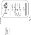
- specific capture probes are used to capture the specific region of interest in the target nucleic acid. The captured regions of the target nucleic acid can be washed at various stringencies and optionally amplified, followed by sequencing.
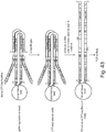
- the capture probe may be biotinylated. The complex of the biotinylated capture probes hybridized to the specific regions of the indexed target nucleic acids can be separated by using streptavidin beads. Exemplary scheme of targeted phasing is shown in Fig. 41 .
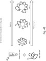
- compositions and methods disclosed herein can be used phasing exomes.
- exons, promoters can be enriched. Markers, for example, heterozygous SNPs between exonic regions, can aid in phasing the exons, especially when the distance between exons is large. Exemplary exome phasing is shown in Fig. 42 .
- indexed linked reads cannot span (cover) heterozygous SNPs of neighboring exons simultaneously. As such, it is challenging to phase the two or more exons.
- the compositions and methods disclosed herein also enriches heterozygous SNPs between exons for example, phasing exons 1 to SNP1 and SNP2 to Exon 2. As such, through the use of SNP 1, exon 1 and exon 2 can be phased as shown in Fig. 42 .
- compositions and methods disclosed herein can be used for phasing and simultaneous methylation detection.
- Methylation detection through bisulfite conversion (BSC) is challenging as the BSC reaction is harsh on DNA, fragmenting the DNA and therefore removing contiguity/phasing information.
- methods disclosed in the present application has an additional advantage because no additional purification steps are required in contrast to those required in traditional BSC approaches, thereby improving the yield.
- compositions and methods disclosed herein can be used to prepare different size libraries in single assay.
- different sizes of clonally indexed beads can be used to prepare different size libraries.
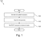
- Figure 1 illustrates a flow diagram of an example of a method 100 of binding transposomes to a bead surface.
- Transposomes may be bound to a bead surface using any chemistry that may be added on the transposon oligonucleotide, transposase, and solid-phase.
- transposomes are bound to a bead surface via a biotin-streptavidin binding complex.
- Method 100 includes, but is not limited to, the following steps.
- transposons may comprise sequencing primer binding sites.
- Exemplary sequences of sequence binding sites include, but are not limited to AATGATACGGCGACCACCGAGATCTACAC (P5 sequence) and CAAGCAGAAGACGGCATACGAGAT (P7 sequence).
- the transposons may be biotinylated.
- the transposons may also include one or more index sequence (unique identifier).
- index sequences include, but are not limited to TAGATCGC, CTCTCTAT, TATCCTCT, AGAGTAGA, GTAAGGAG, ACTGCATA, AAGGAGTA, CTAAGCCT.
- the transposons comprise only the mosaic end (ME) sequences or the ME sequences plus additional sequences that are not P5 and P7 sequences.
- P5 and P7 sequences are added in a subsequent PCR amplification step.
- the transposomes are assembled.
- the assembled transposomes are a mixture of P5 and P7 transposomes.
- a mixture of P5 and P7 transposomes are described in more detail with reference to Figures 11 and 12 .
- P5/P7 transposome mixtures are bound to a bead surface.
- the beads are streptavidin coated beads and the transposomes are bound to the bead surface via a biotin-streptavidin binding complex.
- Beads can be of various sizes. In one example, the beads may be 2.8 ⁇ m beads. In another example, the beads may be 1 ⁇ m beads.
- a suspension (e.g., 1 ⁇ L) of 1 ⁇ m beads provides a large surface area per volume for transposomes binding. Because of the available surface area for transposomes binding, the number of tagmentation products per reaction is increased.
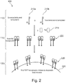
- Figure 2 shows pictorially the steps 110, 115, and 120 of method 100 of Figure 1 .
- the transposons are shown as duplexes.
- another structure such as a hairpin, i.e., a single oligonucleotide with regions of self-complementarity capable of forming a duplex, may be used.
- a plurality of biotinylated P5 transposons 210a and a plurality of P7 transposons 210b are generated.
- P5 transposons 210a and P7 transposons 210b are biotinylated.
- P5 transposons 210a and P7 transposons 210b are mixed with transposase Tn5 215 to form a plurality of assembled transposomes 220.
- transposomes 220 are bound to a bead 225.
- Bead 225 is a streptavidin coated bead.
- Transposomes 220 are bound to bead 225 via a biotin-streptavidin binding complex.
- a mixture of transposomes may be formed on a solid support such as bead surface as shown in Figures 10 , 11 , 12 , and 13 .
- P5 and P7 oligonucleotides are first bound to a bead surface prior to assembly of transposome complexes.
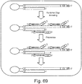
- FIG 3 illustrates a schematic diagram of an example of a tagmentation process 300 on a bead surface. Shown in process 300 is bead 225 of Figure 2 with transposomes 220 bound thereon. A solution of DNA 310 is added to a suspension of beads 225. As DNA 310 contacts transposomes 220, the DNA is tagmented (fragmented and tagged) and is bound to beads 225 via transposomes 220. Bound and tagmented DNA 310 may be PCR amplified to generate a pool of amplicons 315 in solution (bead-free). Amplicons 315 may be transferred to the surface of a flow cell 320.
- a cluster generation protocol (e.g., a bridge amplification protocol or any other amplification protocol that may be used for cluster generation) may be used to generate a plurality of clusters 325 on the surface of flow cell 320.
- Clusters 325 are clonal amplification products of tagmented DNA 310.
- Clusters 325 are now ready for the next step in a sequencing protocol.
- the transposomes may be bound to any solid surface, such as the walls of a microfuge tube.
- oligonucleotides are first bound to a bead surface prior to transposome assembly.
- Figure 10 illustrates a flow diagram of an example of a method 1000 of forming transposome complexes on a bead surface. Method 1000 includes, but is not limited to, the following steps.
- P5 and P7 oligonucleotides are bound to a bead surface.
- the P5 and P7 oligonucleotides are biotinylated and the bead is a streptavidin coated bead.
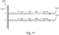
- This step is also shown pictorially in schematic diagram 1100 of Figure 11 . Referring now to Figure 11 , a P5 oligonucleotide 1110 and a P7 oligonucleotide 1115 are bound to the surface of a bead 1120.
- a single P5 oligonucleotide 1110 and a single P7 oligonucleotide 1115 are bound to the surface of bead 1120, but any number of P5 oligonucleotides 1110 and/or P7 oligonucleotides 1115 may be bound to the surface of a plurality of beads 1120.
- P5 oligonucleotide 1110 comprises a P5 primer sequence, an index sequence (unique identifier), a read 1 sequencing primer sequence and a mosaic end (ME) sequence.
- P7 oligonucleotide 1115 comprises a P7 primer sequence, an index sequence (unique identifier), a read 2 sequencing primer sequence and an ME sequence.
- an index sequence is present in only P5 oligonucleotide 1110. In yet another example (not shown), an index sequence is present in only the P7 oligonucleotide 1115. In yet another example (not shown), an index sequence is absent in both P5 oligonucleotide 1110 and P7 oligonucleotide 1115.
- complementary mosaic end (ME') oligonucleotides are hybridized to the bead-bound P5 and P7 oligonucleotides.
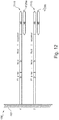
- This step is also shown pictorially in schematic diagram 1200 of Figure 12 .
- complementary ME sequences (ME') 1125 are hybrid to P5 oligonucleotide 1110 and P7 oligonucleotide 1115.
- Complementary ME sequences (ME') 1125 e.g., complementary ME sequences (ME') 1125a and complementary ME sequences (ME') 1125b
- Complementary ME sequence (ME') 1125 is typically about 15 bases in length and phosphorylated at its 5' end.
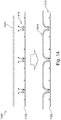
- transposase enzyme is added to the bead-bound oligonucleotides to form a mixture of bead-bound transposome complexes.
- This step is also shown pictorially in schematic diagram 1300 of Figure 13 .
- transposase enzyme is added to form a plurality of transposome complexes 1310.
- transposome complex 1310 is a duplex structure that comprises transposase enzyme, two surface-bound oligonucleotide sequences, and their hybridized complementary ME sequences (ME') 1125.
- transposome complex 1310a comprises P5 oligonucleotide 1110 hybridized to complementary ME sequence (ME') 1125 and P7 oligonucleotide 1115 hybridized to complementary ME sequence (ME') 1125 (i.e., P5:P7);
- transposome complex 1310b comprises two P5 oligonucleotides 1110 hybridized to complementary ME sequences (ME') 1125 (i.e., P5:P5);
- transposome complex 1310c comprises two P7 oligonucleotides 1115 hybridized to complementary ME sequences (ME') 1125 (i.e., P7:P7).
- the ratio of P5:P5, P7:P7, and P5:P7 transposome complexes may be, for example, 25:25:50, respectively.
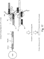
- FIG 14 shows an exemplary schematic diagram 1400 of a tagmentation process using the transposome coated bead 1120 of Figure 13 .
- bead 1120 with transposome complexes 1310 thereon is added to a solution of DNA 1410 in a tagmentation buffer, tagmentation occurs and the DNA is linked to the surface of bead 1120 via transposomes 1310.
- Successive tagmentation of DNA 1410 results in a plurality of bridged molecules 1415 between transposomes 1310.
- the length of bridged molecules 1415 may be dependent on the density of transposome complexes 1310 on the surface of bead 1120.
- the density of transposome complexes 1310 on the surface of bead 1120 may be tuned by varying the amount of P5 and P7 oligonucleotides bound to the surface of bead 1120 in step 1010 of method 100 of Figure 10 .
- the density of transposome complexes 1310 on the surface of bead 1120 may be tuned by varying the amount of complementary ME sequence (ME') hybridized to P5 and P7 oligonucleotides in step 1015 of method 1000 of Figure 10 .
- the density of transposome complexes 1310 on the surface of bead 1120 may be tuned by varying the amount of transposase enzyme added in step 1020 of method 1000 of Figure 1 .
- the length of bridged molecules 1415 is independent of the quantity of beads 1120 with transposome complexes 1310 bound thereon used in a tagmentation reaction. Similarly, adding more or less DNA 1410 in a tagmentation reaction does not alter the size of the final tagmented product, but may affect the yield of the reaction.
- bead 1120 is a paramagnetic bead.
- purification of the tagmentation reaction is readily achieved by immobilizing beads 1120 with a magnet and washing. Therefore, tagmentation and subsequent PCR amplification may be performed in a single reaction compartment ("one-pot") reaction.
- the present invention relate to methods and compositions of transposase-based methods that can derive contiguity information of a target nucleic acid on a solid support.
- the compositions and the methods can derive assembly/phasing information.
- the solid support is a bead.
- the target nucleic acid is DNA.
- the target nucleic acid is genomic DNA.
- the target nucleic acid is RNA.
- the RNA is mRNA.
- the target nucleic acid is complimentary DNA (cDNA).
- transposons may be immobilized as dimers to solid-support such as beads, followed by the binding of transposase to the transposons to form transposomes.
- two transposons may be immobilized in close proximity (preferably fixed distance) to one another in a solid support.
- the two transposons will always be immobilized simultaneously, with preferably an optimum linker length and orientation of the two transposons to form transposomes efficiently.
- transposome formation efficiency will not be a function of transposon density.
- Two transposons will always be available with the right orientation and distance between them to form transposomes.
- transposomes may be prepared on solid support, which can subsequently be used to derive contiguity information through tagmentation and sequencing.
- An exemplary scheme is illustrated in Figure 15 .
- the transposons may be immobilized to the solid support by means other than chemical coupling.
- Exemplary methods of immobilizing transposons on the solid support may include, but are not limited to affinity binding such as streptavidin-biotin, maltose-maltose binding protein, antigen-antibody, DNA-DNA or DNA-RNA hybridization.
- transposomes can be pre-assembled and then immobilized on a solid-support.
- the transposons comprise unique indexes, barcodes, and amplification primer binding sites.
- Transposase can be added in solution comprising transposons to form transposome dimers, which can be immobilized on a solid support.
- multiple bead sets can be generated in which each set has the same index derived from the immobilized transposons thus generating indexed beads.
- Target nucleic acid can be added to each set of indexed beads as shown in Figure 29A .
- target nucleic acid can be added to each set of indexed beads, tagmented and subsequent PCR amplification may be performed separately.
- target nucleic acid, indexed beads, and transposomes can be combined in droplets such that a number of droplets contain a single bead with one or more DNA molecules and adequate transposomes.
- the indexed beads can be pooled, target nucleic acid can be added to the pool, tagmented and subsequent PCR amplification may be performed in a single reaction compartment ("one-pot").
- the present invention relate to methods and compositions to derive contiguity information by means of capturing contiguously-linked, transposed, target nucleic acid onto a solid support.
- contiguity preserving transposition CPT
- CPT-DNA contiguity preserving transposition
- Contiguity information can be preserved by the use of transposase to maintain the association of template nucleic acid fragments adjacent in the target nucleic acid.
- the CPT-DNA can be captured by hybridization of complimentary oligonucleotides having unique indexes or barcodes and immobilized on solid support, e.g., beads ( Figure 29B ).
- the oligonucleotide immobilized on the solid support may further comprise primer binding sites, unique molecular indices (UMI), in addition to barcodes.
- UMI unique molecular indices
- transposomes to maintain physical proximity of fragmented nucleic acids increases the likelihood that fragmented nucleic acids from the same original molecule, e.g., chromosome, will receive the same unique barcode and index information from the oligonucleotides immobilized on a solid support. This will result in a contiguously-linked sequencing library with unique barcodes.
- the contiguously-linked sequencing library can be sequenced to derive contiguous sequence information.
- Figures 16 and 17 show schematic representations of an exemplary embodiment of the above aspect of the invention of making contiguously-linked libraries with unique barcodes or indices.
- the exemplary method leverages on ligation of the CPT-DNA with the immobilized oligonucleotides on the solid support comprising unique indexes and barcodes and strand-replacement PCR to generate a sequencing library.
- clonal indexed beads may be generated with immobilized DNA sequences such as random or specific primer and index.
- Contiguously-linked libraries can be captured onto clonal-indexed beads by hybridization to the immobilized oligonucleotides followed by ligation.
- FIGS. 18 and 19 depict the capture of the CPT-DNA on clonal indexed beads and the preservation of the contiguity information. Strand-replacement PCR can transfer the clonal bead index information to the individual molecule. Thus, each contiguously-linked library will be uniquely indexed.
- the oligonucleotide immobilized on a solid support can comprise a partially double stranded structure such that one strand is immobilized to the solid support and the other strand is partially complementary to the immobilized strand resulting in a Y-adaptor.
- the Y-adaptor immobilized on the solid surface is linked to the contiguously linked tagmented DNA by ligation and gap filling and shown in Figure 20 .
- Y-adaptor is formed through hybridization capture of CPT-DNA with the probe/index on the solid support such as beads.
- Figure 21 shows an exemplary scheme of making such Y-adapters. The use of these Y-adapters ensures that potentially every fragment can become a sequencing library. This increases the coverage per sequencing.
- free transposomes may be separated from CPT-DNA.
- the separation of the free transposomes is by size exclusion chromatography. In one embodiment, the separation may be achieved by MicroSpin S-400 HR Columns (GE Healthcare Life Sciences, Pittsburgh, PA).
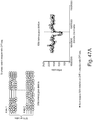
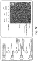
- Figure 22 shows an agarose gel electrophoresis of the separated of CPT-DNA from the free transposomes.
- Capturing contiguously-linked, transposed, target nucleic acid onto a solid support through hybridization has several unique advantages.
- First, the method is based on hybridization and not transposition. Intramolecular hybridization rate >> intermolecular hybridization rate.
- chances of contiguously-transposed libraries on a single target DNA molecule to wrap around a uniquely indexed bead is much higher than having two or more different single target DNA molecule to wrap around a uniquely indexed bead.
- DNA transposition and barcoding of the transposed DNA occur in two separate steps.
- the challenges associated with active transposome assembly on beads and surface density optimization of transposons on solid-surfaces can be avoided.
- Fourth, self-transposition products can be removed by column purification.
- DNA is more flexible and therefore puts less of a burden on transposition density (insert size) compared to immobilizing transposome on bead methods.
- the method can be used with combinatorial barcoding schemes.
- it is easy to covalently-link indexed oligos to the beads. Thus, there is less chance for index exchange.
- the tagmentation and subsequent PCR amplification may be multiplexed and can be performed in a single reaction compartment ("one-pot") reaction eliminating the need to carryout individual reactions for each index sequences.
- each barcode includes a first barcode sequence and a second barcode sequence, having a fragmentation site disposed therebetween.
- the first barcode sequence and second barcode sequence can be identified or designated to be paired with one another.
- the pairing can be informative so that a first barcode is associated with a second barcode.
- the paired barcode sequences can be used to assemble sequencing data from the library of template nucleic acids.
- identifying a first template nucleic acid comprising a first barcode sequence and a second template nucleic acid comprising a second barcode sequence that is paired with the first indicates that the first and second template nucleic acids represent sequences adjacent to one another in a sequence representation of the target nucleic acid.
- Such methods can be used to assemble a sequence representation of a target nucleic acid de novo , without the requirement of a reference genome.
- the present invention relate to methods and compositions to generate shotgun sequence library of a specific DNA fragment.
- clonal indexed beads are generated with immobilized oligonucleotide sequences: random or specific primer and unique indexes.
- Target nucleic acid is added to the clonal indexed beads.
- the target nucleic acid is DNA.
- the target DNA is denatured.
- the target DNA hybridizes with primers comprising unique indexes immobilized on the solid surface (e.g., bead) and subsequently with other primers with the same index.
- the primers on the bead amplify the DNA.
- One or more further rounds of amplification may be carried out.
- the amplification may be carried out by whole genome amplification using bead immobilized primers with a 3' random n-mer sequence.
- the random n-mer contains pseudocomplementary bases (2-thiothymine, 2-amino dA, N4-ethyl cytosine, etc.) to prevent primer-primer interaction during amplification ( Hoshika, S; Chen, F; Leal, NA; Benner, SA , Angew. Chem. Int. Ed.49(32) 5554-5557 (2010 ).
- Figure 23 shows an exemplary scheme of generating shotgun sequence library of a specific DNA fragment. A clonal indexed sequencing library of the amplified product can be generated.
- such library can be generated by transposition. Sequence information of the clonal indexed library can be used to assemble the contiguous information using the index information as a guide.
- Figure 24 shows an exemplary scheme of assembling the sequence information from clonal indexed sequencing library.
- Intra-molecular amplification on a bead is much faster than inter-bead amplification.
- the products on a bead will have the same index.
- a shotgun library of a specific DNA fragment can be created. Random primers amplify the template at random locations and therefore a shotgun library with the same index can be generated from a specific molecule and the sequence information can be assembled using the indexed sequence.
- a significant advantage of the methods of the above embodiments is that the reactions can be multiplexed in a single reaction (one pot reaction) and will not require using many individual wells.
- index clonal beads can be prepared so many different fragments can be uniquely labeled, and discrimination can be made to the parental alleles for same genomic regions. With a high number of indexes, the chance that the DNA copy of the father and copy of the mother will receive the same index for the same genomic region is low.
- the method takes advantage of the fact that intra reactions are much faster than inter, the beads basically generate a virtual partition in a larger physical compartment.
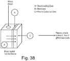
- the method may be used for cell free DNA (cfDNA) in cfDNA assays.
- cfDNA cell free DNA
- the cfDNA is obtained from plasma, placental fluids.
- the plasma can be obtained from undiluted whole blood using membrane based, sedimentation assisted plasma separator ( Liu et al. Anal Chem. 2013 Nov 5;85(21): 10463-70 ).
- the collection zone of the plasma of the plasma separator may comprise solid support comprising transposomes.
- the solid support comprising transposomes may capture the cfDNA from the isolated plasma as it is separated from the whole blood and can concentrate the cfDNA and/or tagment the DNA.
- the tagmentation will further introduce unique barcodes to allow subsequent demultiplexing after sequencing of the pool of libraries.
- the collection zone of the separator may comprise PCR master mix (primers, nucleotides, buffers, metals) and polymerase.
- the master mix can be in dry form such that it will be reconstituted as the plasma comes out of the separator.
- the primers are random primers.
- the primers can be specific primers for a particular gene. PCR amplification of the cfDNA will result in the generation of library directly from the separated plasma.
- the collection zone of the separator may comprise RT-PCR master mix (primers, nucleotides, buffers, metals), reverse transcriptase and polymerase.
- the primers are random primers or oligo dT primers.
- the primers can be specific primers for a particular gene.
- the resulting cDNA can be used for sequencing. Alternatively, the cDNA can be treated with transposomes immobilized on a solid support for sequence library preparation.
- the plasma separator may comprise barcodes (1D or 2D barcodes).
- the separation device may comprise blood collection device. This would result in direct delivery of the blood to the plasma separator and library prep device.
- the device may comprise a downstream sequence analyzer.
- sequence analyzer is a single use sequencer.
- the sequencer is capable of queuing samples before sequencing in a batch. Alternatively, the sequencer may have random access capability, where samples are delivered to their sequencing area.
- the collection zone for plasma may comprise silica substrates, such that the cell free DNA is concentrated
- Inventors of the present application has surprisingly and unexpectedly found that phasing and simultaneous methylation detection is possible using the methods and compositions of the present application.
- the present methods will allow to combine CPT-seq on beads (indexed contiguity linked libraries) with DNA methylation detection. For example, individual libraries generated on beads can be treated with bisulfite, converting non-methylated Cs, but not methylated Cs to Us, allowing the detection of 5-Me-C.
- epi-medication-phasing blocks can be established multi megabase range.
- the size of the DNA analyzed can be about hundred bases to about multi mega bases. In some embodiments, the size of the DNA analyzed can be about 100, 200, 300, 400, 500, 600, 700, 800, 900, 1000, 1200, 1300, 1500, 2000, 3000, 3500, 4000, 4500, 5000, 5500, 6000, 6500, 7000, 7,500, 8000, 8500, 9000, 9500, 10,000, 10,500, 11,000, 11,500, 12,000, 12500, 13000, 14000, 14500, 15000, 15500, 16000, 16500, 17000, 17,500, 18,000, 18,500, 19,000, 19,500, 20,000, 20,500, 21,000, 21,500, 22,000, 22,500, 23,000, 23,500, 24,000, 24,500, 25,000, 25,500, 26,000, 26,500, 27,000, 27,500, 28,000, 28,500, 29,500, 30,000, 30,500, 31,000, 31,500, 32,000, 33,000, 34,000, 35,000, 36,000, 37,000, 3
- DNA is first transformed into indexed-linked libraries on a solid-support.
- Individual indexed libraries much smaller than the original DNA, are less prone to fragmentation since the individual libraries are smaller. Even if a small fraction of indexed libraries are lost, phasing information is still maintained across the long span of the indexed DNA molecule. For example, if a 100kb molecule in traditional bisulfite conversion (BSC) is fragmented in half the contiguity is now restricted to 50kb.
- BSC bisulfite conversion
- a 100kb library is first indexed and even if a fraction of individual libraries are lost, contiguity is still at ⁇ 100kb (except in the unlikely event when all libraries lost are from one end of the DNA molecule.
- methods disclosed in the present application has an additional advantage because no additional purification steps are required in contrast to those required in traditional bisulfite conversion approaches, thereby improving the yield.
- the beads are simply washed after bisulfite conversion.
- buffer exchanges can be readily performed with minimal loss of DNA (indexed libraries) and reduced hands on time.
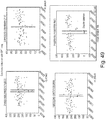
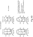
- Fig. 43 Exemplary scheme of simultaneous phasing and methylation detection is shown in Fig. 43 .
- the workflow consists of tagmentation of DNA on beads, gap-fill-ligate the 9-bp repeat regions, removal of Tn5 with SDS, and bisulfite conversion of the individual libraries on the beads.
- the bisulfite conversion is performed under denaturing conditions to ensure that neighboring complementary libraries are not re-annealing, therefore reducing the bisulfite conversion efficiency.
- BCS converts non-methylated C's to U's and methylated C's are not converted.
- Figure 44 shows an alternative exemplary scheme of simultaneous phasing and methylation detection.
- a fraction of gap-filled-ligated libraries are degraded in order to prepare single-stranded templates.
- Single-stranded templates need milder conditions for bisulfite conversion since the templates are already single-stranded which could reduce library loss or improve bisulfite conversion efficiency.
- a mixture of 3' thio-protected transposons (Exo resistant) and non-protected transposons are used on the same bead. Enzymes, for example, Exo I, can be used to digest the non-thio-protected libraries, converting them to single stranded libraries.
- non-protected transposons 50% of the libraries will be converted to single-stranded libraries (50% have one transposon of the library is protected and one, the complement strand, is not protected), 25% will not be converted (both transposons are thio protected), and 25% are both converted removing the whole library. (both transposons not protected).
- the first strategy relies on decreasing library insert size by more densely populating transposome complexes to the streptavidin beads. By decreasing library size, a smaller proportion of library elements are degraded by bisulfite treatment.
- the second strategy to improve DNA yield of the Epi-CPTSeq protocol is enzymatic recovery of broken library elements.
- the purpose of the recovery strategy is to add the 3' common sequence necessary for library amplification back to the bead bound library elements that became digested and lost their 3' portion during bisulfite treatment. After the addition of the 3' common sequence these elements can now be PCR amplified and sequenced.
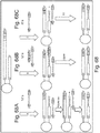
- Figure 67 and 68 shows an exemplary scheme of this strategy. Double stranded CPTSeq library elements have been denatured and bisulfite converted (top panel). During bisulfite conversion, one of DNA strands has been damaged (middle panel), leading to loss of the PCR common sequence on the 3' end.
- Template rescue strategies restore the 3' common sequence (green) necessary for PCR amplification (bottom panel).
- a terminal transferase in a presence of 3' phosphorylated attenuator oligo a sequence containing a sequencing adapter followed by an oligo dT stretch is used ( Figure 68A ).
- TdT adds a stretch of 10 to 15 dAs to the 3' end of a broken library element, which anneals to the oligo dT portion of the attenuator oligo. Formation of this DNA hybrid stops TdT reaction and provides template for consequent extension of the 3'end of a broken library element by DNA polymerase.
- the TdT tailing reaction is performed in the presence of a partially double stranded attenuator oligo, containing a single stranded oligo dT portion and 5' phosphorylated double stranded sequencing adapter portion.
- a partially double stranded attenuator oligo containing a single stranded oligo dT portion and 5' phosphorylated double stranded sequencing adapter portion.
- the nick between last added dA and 5' phosphorylated attenuator oligo is sealed by DNA ligase.
- a common sequencing adapter can also be added to the 3' end of broken library elements by a recently introduced ssDNA template switching activity of MMLV RT.
- MMLV RT and a template switch oligo are added to damaged DNA ( Figure 68C ).
- reverse transcriptase adds a few additional nucleotides to 3' ends of a single-stranded DNA fragment, and these bases pair with an oligo (N) sequence presented at the 3' end of one of the TS_oligos.
- reverse transcriptase template switching activity adds the sequences of the annealed common primers to the 3' end of BSC broken library element, restoring its ability to get amplified in PCR with common sequencing primers.
- an Epicentre's EpiGenome kit "post-bisulfite conversion" library construction method can be used to rescue library elements which lost their common sequences at the 3' end during bisulfite conversion.
- this library rescue method utilizes 3' phosphorylated oligos with common sequences followed by a short stretch of random sequence. These short random sequences hybridize to the bisulfite-treated single-stranded DNA and common sequences are subsequently copied to the broken library strand by DNA polymerase.
- Figure 74 shows the fourth strategy to improve the bisulfite sequencing methods on beads.
- a first common sequence comprising a capture tag is covalently attached to the 5' ends of DNA.
- the first common sequence can be attached to DNA using various methods, including single-sided transposition (as pictured), adapter ligation, or terminal transferase (TdT) adapter ligation as described in US Patent Application Publication 20150087027 .
- DNA is denatured (e.g. incubation at high heat) and bound to a solid support.
- biotin used as a capture tag on CS1
- DNA can be bound using streptavidin magnetic beads (as pictured). Once bound to the solid support buffer exchanges can be readily made.
- PCR is performed to amplify the library and remove it from the solid support.
- PCR primers can be designed to add additional commons sequences, such as sequencing adapters, to the ends of the MethylSeq library.
- library preparation can be achieved in a single assay using differentially sized solid support, for example, beads.
- Each bead size will generate a specific library size or range of sizes, with the physical size of the bead determining the library size.
- the various sized beads all have unique clonal indices that are transferred to the library.
- different sizes libraries are generated with each different library scale-length uniquely indexed.
- the various length-scale libraries are prepared simultaneously in the same physical compartment, reducing cost and improving overall work flow.
- each specific solid support size for example, bead size receives a unique index.
- Fig. 45 shows an exemplary scheme to generate various sized libraries using various sized clonally indexed beads in a single assay.
- the size of the libraries generated are about 50, 75, 100, 150, 200, 250, 300, 350, 400, 500, 600, 700, 800, 900, 1000, 1200, 1300, 1500, 2000, 3000, 3500, 4000, 4500, 5000, 5500, 6000, 6500, 7000, 7,500, 8000, 8500, 9000, 9500, 10,000, 10,500, 11,000, 11,500, 12,000, 12500, 13000, 14000, 14500, 15000, 15500, 16000, 16500, 17000, 17,500, 18,000, 18,500, 19,000, 19,500, 20,000, 20,500, 21,000, 21,500, 22,000, 22,500, 23,000, 23,500, 24,000, 24,500, 25,000, 25,500, 26,000, 26,500, 27,000, 27,500, 28,000, 28,500, 29,500, 30,000, 30,500, 31,000, 31,500, 32,000, 33,000, 34,000, 35,000, 36,000, 37,000, 38,000, 39,000, 40,000, 42,000, 45,000,
- multiple length scale libraries discussed above can be used in the assembly of pseudogenes, paralogs etc. instead of having one large length scale.
- multiple length scale libraries are prepared simultaneously in a single assay. The advantage is that at least one length-scale will link a unique region with only the pseudo-gene and or gene, but not both. As such, variants detected with this length-scale can uniquely assign the variant to either the gene or the pseudo-gene. The same holds true for copy number variants, paralogs etc.
- the strength of assembly is the use of different length scales. Using the methods disclosed herein different length scale indexed linked libraries can be generated in a single assay instead of individual, different library preparations for different length scales.
- Fig. 46 shows an exemplary scheme of determining genetic variants with different length scale libraries.
- compositions and methods disclosed herein relate to analysis of genomic variants.
- Exemplary genomic variants include but are not limited to deletions, inter chromosomal translocations, duplications, paralogs, interchromosomal gene fusions.
- the compositions and methods disclosed herein relate to determining phasing information of the genomic variants.
- the table below shows exemplary interchromosomal gene fusions.
- target nucleic acid can be fragmented prior to exposing it to transposomes.
- Exemplary fragmentation methods include, but are not limited to sonication, mechanical shearing, and restriction digestion. Fragmentation of target nucleic acid prior to tagmentation (fragmentation and tagging) is advantageous for assembly/phasing of pseudogenes (e.g., CYP2D6). Long islands (>30kb) of indexed linked reads will span the pseudogenes A and A' as shown in Figure 64 . Due to high sequence homology, it will be challenging to determine which variant belongs to Gene A and Gene A'. Shorter variants will link one variant of the pseudogenes with unique surrounding sequences. Such shorter islands can be achieved by fragmenting the target nucleic acid prior to tagmentation.
- pseudogenes e.g., CYP2D6
- transposases are multimeric in a transposome complex, e.g., they form dimers, tetramers etc. in a transposome complex.
- Inventors of the present application have surprisingly and unexpectedly found that linking the monomer transposases in multimeric transposome complex or linking the transposon ends of a transposome monomer in multimeric transposome complex has several advantages.
- Second, lower concentrations of transposomes can potentially be used in the fragmentation by transposition reaction.
- the linking leads to lower exchange of the mosaic ends (ME) of transposome complexes, thus less mixing of barcodes or adaptor molecules.
- Such swapping of ME ends are possible if the complexes fall apart and reform, or in case where transposomes are immobilized on solid support by streptavidin/biotin, the streptavidin/biotin interaction can break and reform, or when there is a possible contamination.
- Inventors of the present application noted that there is a significant swap or exchange of ME ends under various reaction conditions. In some embodiments, the exchange can be as high as 15%. The exchange is pronounced in high salt buffer and the exchange is reduced in glutamate buffer.
- Figures 57 and 58 shows some possible mechanisms of ME swapping.
- the transposase subunits in the transposome complex can be linked to each other by covalent and non-covalent means.
- transposase monomers can be linked before making the transposome complex (before addition of the transposons).
- transposase monomers can be linked after transposome formation.
- native amino acid residues may be substituted with Cysteine (Cys) amino acids at the multimeric interface to promote disulfide bond formation.
- Cysteine (Cys) amino acids For example, in Tn5 transposase, Asp468, Tyr407, Asp461, Lys459, Ser458, Gly462, Ala466, Met470 may be substituted with Cys to promote disulfide bond between the monomer subunits and shown in Figures 59 and 60 .
- exemplary amino acids that can be substituted with cysteine include, but are not limited to Leu21, Leu32, Ala35, His20, Phe17, Phe36, Ile16, Thr13, Arg12, Gln10, Glu9 and shown in Fig.
- the modified transposase with amino acid residues substituted with cysteine can chemically cross-linked to each other using a chemical cross-linker using maleimide or pyridyldithiol reactive groups.
- exemplary chemical cross-linkers are available commercially from Pierce Protein Biology/ThermoFisher Scientific (Grand Island, NY, USA).
- transposome multimer complexes can be covalently linked to solid support.
- solid supports include but are not limited to nanoparticles, beads, flow cell surfaces, column matrices.
- solid surfaces may be coated with amine groups.
- Modified transposase with amino acid residues substituted with cysteine can be chemically cross-linked to such amine groups using an amine-to-sulfhydryl crosslinker (i.e., succinimidyl-4-(N-maleimidomethyl)cyclohexane-1-carboxylate (SMCC)).
- SMCC succinimidyl-4-(N-maleimidomethyl)cyclohexane-1-carboxylate
- SMCC succinimidyl-4-(N-maleimidomethyl)cyclohexane-1-carboxylate
- transposase gene can be modified to express multimeric protein in a single polypeptide.
- Tn5 or Mos-1 genes can be modified to express two Tn5 or Mos-1 proteins in a single polypeptide.
- Mu transposase gene can be modified to encode four mu transposase units in a single polypeptide.
- the transposon ends of a transposome monomer unit can be linked to form a linked transposome multimer complex.
- Linking the transposon ends allow insertion of primer sites, sequencing primers, amplification primers or any role DNA can play into gDNA without fragmenting the target DNA. Insertion of such functionality are advantages in haplotyping assays or junction tagging assays in which information needs to be extracted from intact molecules or in which sub-sampling are important.
- transposon ends of Mu transposomes can be linked to a "looped" Mu transposase/transposon configuration.
- Mu is a tetramer
- various configurations are possible but not limited by linking either R2UJ and/or R1UJ with R2J and/or R1J.
- R2UJ and R1UJ can/are not connected with R2J and R1J, respectively.
- Figure 63 shows a Mu transposome complex where transposon ends are linked.
- transposon ends of Tn5 or transposon ends of Mos-1 transposomes can be linked.
- transposon means a double-stranded DNA that exhibits only the nucleotide sequences (the "transposon end sequences") that are necessary to form the complex with the transposase or integrase enzyme that is functional in an in vitro transposition reaction.
- a transposon forms a "complex” or a “synaptic complex” or a “transposome complex” or a “transposome composition with a transposase or integrase that recognizes and binds to the transposon, and which complex is capable of inserting or transposing the transposon into target DNA with which it is incubated in an in vitro transposition reaction.
- a transposon exhibits two complementary sequences consisting of a "transferred transposon sequence” or “transferred strand” and a "non-transferred transposon sequence,” or “non transferred strand”.
- a transposon that forms a complex with a hyperactive Tn5 transposase e.g., EZ-Tn5TM Transposase, EPICENTRE Biotechnologies, Madison, Wis., USA
- EZ-Tn5TM Transposase EZ-Tn5TM Transposase, EPICENTRE Biotechnologies, Madison, Wis., USA
- a transferred strand that exhibits a "transferred transposon sequence” as follows: 5' AGATGTGTATAAGAGACAG 3' and a non-transferred strand that exhibits a "non-transferred transposon sequence” as follows: 5' CTGTCT CTTATACACATCT 3'.
- the 3'-end of a transferred strand is joined or transferred to target DNA in an in vitro transposition reaction.
- the non-transferred strand which exhibits a transposon sequence that is complementary to the transferred transposon end sequence, is not joined or transferred to the target DNA in an in vitro transposition reaction.
- the transposon sequences may comprise one or more of the following: a barcode, an adaptor sequence, a tag sequence, a primer binding sequence, a capture sequence, unique molecular identifier (UMI) sequence.
- adaptor means a nucleic acid sequence that can comprise a barcode, a primer binding sequence, a capture sequence, a sequence complementary to a capture sequence, unique molecular identifier (UMI) sequence, an affinity moiety, restriction site.
- UMI unique molecular identifier
- the term "contiguity information” refers to a spatial relationship between two or more DNA fragments based on shared information.
- the shared aspect of the information can be with respect to adjacent, compartmental and distance spatial relationships. Information regarding these relationships in turn facilitates hierarchical assembly or mapping of sequence reads derived from the DNA fragments. This contiguity information improves the efficiency and accuracy of such assembly or mapping because traditional assembly or mapping methods used in association with conventional shotgun sequencing do not take into account the relative genomic origins or coordinates of the individual sequence reads as they relate to the spatial relationship between the two or more DNA fragments from which the individual sequence reads were derived.
- methods of capturing contiguity information may be accomplished by short range contiguity methods to determine adjacent spatial relationships, mid-range contiguity methods to determine compartmental spatial relationships, or long range contiguity methods to determine distance spatial relationships. These methods facilitate the accuracy and quality of DNA sequence assembly or mapping, and may be used with any sequencing method, such as those described above.
- Contiguity information includes the relative genomic origins or coordinates of the individual sequence reads as they relate to the spatial relationship between the two or more DNA fragments from which the individual sequence reads were derived.
- contiguity information includes sequence information from non-overlapping sequence reads.
- the contiguity information of a target nucleic acid sequence is indicative of haplotype information. In some embodiments, the contiguity information of a target nucleic acid sequence is indicative of genomic variants.
- maintaining the contiguity of the target nucleic acid in the context of fragmenting a nucleic acid means maintaining the order of the nucleic acid sequence of the fragments from the same target nucleic acid.
- the term "at least a portion” and/or grammatical equivalents thereof can refer to any fraction of a whole amount.
- “at least a portion” can refer to at least about 1%, 2%, 3%, 4%, 5%, 6%, 7%, 8%, 9%, 10%, 15%, 20%, 25%, 30%, 35%, 40%, 45%, 50%, 55%, 60%, 65%, 70%, 75%, 80%, 85%, 90%, 95%, 99%, 99.9% or 100% of a whole amount.
- the term "sequencing read” and/or grammatical equivalents thereof can refer to a repetitive process of physical or chemical steps that is carried out to obtain signals indicative of the order of monomers in a polymer.
- the signals can be indicative of an order of monomers at single monomer resolution or lower resolution.
- the steps can be initiated on a nucleic acid target and carried out to obtain signals indicative of the order of bases in the nucleic acid target.
- the process can be carried out to its typical completion, which is usually defined by the point at which signals from the process can no longer distinguish bases of the target with a reasonable level of certainty. If desired, completion can occur earlier, for example, once a desired amount of sequence information has been obtained.
- a sequencing read can be carried out on a single target nucleic acid molecule or simultaneously on a population of target nucleic acid molecules having the same sequence, or simultaneously on a population of target nucleic acids having different sequences.
- a sequencing read is terminated when signals are no longer obtained from one or more target nucleic acid molecules from which signal acquisition was initiated.
- a sequencing read can be initiated for one or more target nucleic acid molecules that are present on a solid phase substrate and terminated upon removal of the one or more target nucleic acid molecules from the substrate. Sequencing can be terminated by otherwise ceasing detection of the target nucleic acids that were present on the substrate when the sequencing run was initiated. Exemplary methods of sequencing are described in U.S. Patent No. 9,029,103 .
- the term "sequencing representation" and/or grammatical equivalents thereof can refer to information that signifies the order and type of monomeric units in the polymer.
- the information can indicate the order and type of nucleotides in a nucleic acid.
- the information can be in any of a variety of formats including, for example, a depiction, image, electronic medium, series of symbols, series of numbers, series of letters, series of colors, etc.
- the information can be at single monomer resolution or at lower resolution.
- An exemplary polymer is a nucleic acid, such as DNA or RNA, having nucleotide units.
- a series of "A,” “T,” “G,” and “C” letters is a well-known sequence representation for DNA that can be correlated, at single nucleotide resolution, with the actual sequence of a DNA molecule.
- Other exemplary polymers are proteins having amino acid units and polysaccharides having saccharide units.
- solid support and solid surface are used interchangeably.
- the solid support or its surface is non-planar, such as the inner or outer surface of a tube or vessel.
- the solid support comprises microspheres or beads.
- microspheres or “beads” or “particles” or grammatical equivalents herein is meant small discrete particles.
- Suitable bead compositions include, but are not limited to, plastics, ceramics, glass, polystyrene, methylstyrene, acrylic polymers, paramagnetic materials, thoria sol, carbon graphite, titanium dioxide, latex or cross-linked dextrans such as Sepharose, cellulose, nylon, cross-linked micelles and Teflon, as well as any other materials outlined herein for solid supports may all be used.
- "Microsphere Detection Guide” from Bangs Laboratories, Fishers Ind. is a helpful guide.
- the microspheres are magnetic microspheres or beads.
- the beads can be color coded.
- MicroPlex® Microspheres from Luminex, Austin, TX may be used.
- the beads need not be spherical; irregular particles may be used. Alternatively or additionally, the beads may be porous.
- the bead sizes range from nanometers, i.e. about 10 nm, to millimeters in diameter, i.e. 1 mm, with beads from about 0.2 micron to about 200 microns being preferred, and from about 0.5 to about 5 micron being particularly preferred, although in some embodiments smaller or larger beads may be used. In some embodiments, beads can be about 0.1, 0.2, 0.3, 0.4, 0.5.
- a “transposome” comprises an integration enzyme such as an integrase or transposase, and a nucleic acid comprising an integration recognition site, such as a transposase recognition site.
- the transposase can form a functional complex with a transposase recognition site that is capable of catalyzing a transposition reaction.
- the transposase may bind to the transposase recognition site and insert the transposase recognition site into a target nucleic acid in a process sometimes termed "tagmentation". In some such insertion events, one strand of the transposase recognition site may be transferred into the target nucleic acid.
- a transposome comprises a dimeric transposase comprising two subunits, and two non-contiguous transposon sequences.
- a transposome comprises a transposase comprises a dimeric transposase comprising two subunits, and a contiguous transposon sequence.
- Some embodiments can include the use of a hyperactive Tn5 transposase and a Tn5-type transposase recognition site ( Goryshin and Reznikoff, J. Biol. Chem., 273:7367 (1998 )), or MuA transposase and a Mu transposase recognition site comprising R1 and R2 end sequences ( Mizuuchi, K., Cell, 35: 785, 1983 ; Savilahti, H, et al., EMBO J., 14: 4893, 1995 ).
- An exemplary transposase recognition site that forms a complex with a hyperactive Tn5 transposase comprises the following 19b transferred strand (sometimes "M” or "ME") and non-transferred strands: 5' AGATGTGTATAAGAGACAG 3', 5' CTGTCT CTTATACACATCT 3', respectively.
- ME sequences can also be used as optimized by a skilled artisan.
- transposition systems that can be used with certain embodiments of the compositions and methods provided herein include Staphylococcus aureus Tn552 (togethero et al., J. Bacteriol., 183: 2384-8, 2001 ; Kirby C et al., Mol. Microbiol., 43: 173-86, 2002 ), Ty1 ( Devine & Boeke, Nucleic Acids Res., 22: 3765-72, 1994 and International Publication WO 95/23875 ), Transposon Tn7 ( Craig, N L, Science.
- More examples include IS5, Tn10, Tn903, IS911, , Sleeping Beauty, SPIN, hAT, PiggyBac, Hermes, TcBuster, AeBuster1, Tol2, and engineered versions of transposase family enzymes ( Zhang et al., (2009) PLoS Genet. 5:e1000689 . Epub 2009 Oct 16; Wilson C. et al (2007) J. Microbiol. Methods 71:332-5 ).
- integrases that may be used with the methods and compositions provided herein include retroviral integrases and integrase recognition sequences for such retroviral integrases, such as integrases from HIV-1, HIV-2, SIV, PFV-1, RSV.
- a barcode can include one or more nucleotide sequences that can be used to identify one or more particular nucleic acids.
- the barcode can be an artificial sequence, or can be a naturally occurring sequence generated during transposition, such as identical flanking genomic DNA sequences (g-codes) at the end of formerly juxtaposed DNA fragments.
- the barcodes are artificial sequences that are absent in the target nucleic acid sequence and can be used to identify one or more target nucleic acid sequences.
- a barcode can comprise at least about 1, 2, 3, 4, 5, 6, 7, 8, 9, 10, 11, 12, 13, 14, 15, 16, 17, 18, 19, 20 or more consecutive nucleotides.
- a barcode comprises at least about 10, 20, 30, 40, 50, 60, 70 80, 90, 100 or more consecutive nucleotides.
- at least a portion of the barcodes in a population of nucleic acids comprising barcodes is different.
- at least about 10%, 20%, 30%, 40%, 50%, 60%, 70%, 80%, 90%, 95%, 99% of the barcodes are different.
- all of the barcodes are different.
- the diversity of different barcodes in a population of nucleic acids comprising barcodes can be randomly generated or non-randomly generated.
- a transposon sequence comprises at least one barcode.
- the first transposon sequence comprises a first barcode
- the second transposon sequence comprises a second barcode.
- a transposon sequence comprises a barcode comprising a first barcode sequence and a second barcode sequence.
- the first barcode sequence can be identified or designated to be paired with the second barcode sequence. For example, a known first barcode sequence can be known to be paired with a known second barcode sequence using a reference table comprising a plurality of first and second bar code sequences known to be paired to one another.
- the first barcode sequence can comprise the same sequence as the second barcode sequence.
- the first barcode sequence can comprise the reverse complement of the second barcode sequence.
- the first barcode sequence and the second barcode sequence are different.
- the first and second barcode sequences may comprise a bi-code.
- barcodes are used in the preparation of template nucleic acids.
- the vast number of available barcodes permits each template nucleic acid molecule to comprise a unique identification.
- Unique identification of each molecule in a mixture of template nucleic acids can be used in several applications. For example, uniquely identified molecules can be applied to identify individual nucleic acid molecules, in samples having multiple chromosomes, in genomes, in cells, in cell types, in cell disease states, and in species, for example, in haplotype sequencing, in parental allele discrimination, in metagenomic sequencing, and in sample sequencing of a genome.
- Exemplary barcode sequences include, but are not limited to TATAGCCT, ATAGAGGC, CCTATCCT, GGCTCTGA, AGGCGAAG, TAATCTTA, CAGGACGT, and GTACTGAC.
- a transposon sequence can include a "sequencing adaptor” or “sequencing adaptor site”, that is to say a region that comprises one or more sites that can hybridize to a primer.
- a transposon sequence can include at least a first primer site useful for amplification, sequencing, and the like. Exemplary sequences of sequence binding sites include, but are not limited to AATGATACGGCGACCACCGAGATCTACAC (P5 sequence) and CAAGCAGAAGACGGCATACGAGAT (P7 sequence).
- a target nucleic acid can include any nucleic acid of interest.
- Target nucleic acids can include DNA, RNA, peptide nucleic acid, morpholino nucleic acid, locked nucleic acid, glycol nucleic acid, threose nucleic acid, mixed samples of nucleic acids, polyploidy DNA (i.e., plant DNA), mixtures thereof, and hybrids thereof.
- genomic DNA or amplified copies thereof are used as the target nucleic acid.
- cDNA, mitochondrial DNA or chloroplast DNA is used.
- the target nucleic acid is mRNA.
- target nucleic acid is from a single cell or from fractions of a single cell.
- the target nucleic acid is from a single organelle. Exemplary single organelle includes but is not limited to single nuclei, single mitochondria, and a single ribosome.
- target nucleic acid is from formalin fixed paraffin embedded (FFPE) sample.
- target nucleic acid is cross-linked nucleic acid.
- the target nucleic acid is cross-linked with protein.
- the target nucleic acid is cross-linked DNA.
- the target nucleic acid is histone protected DNA. In some embodiments, histones are removed from the target nucleic acid.
- target nucleic acid is from nucleosomes. In some embodiments, target nucleic acid is from nucleosomes from which nuclear proteins are removed.
- a target nucleic acid can comprise any nucleotide sequence.
- the target nucleic acid comprises homopolymer sequences.
- a target nucleic acid can also include repeat sequences. Repeat sequences can be any of a variety of lengths including, for example, 2, 5, 10, 20, 30, 40, 50, 100,250, 500 or 1000 nucleotides or more. Repeat sequences can be repeated, either contiguously or non-contiguously, any of a variety of times including, for example, 2, 3, 4, 5, 6, 7, 8, 9, 10, 15 or 20 times or more.
- Some embodiments described herein can utilize a single target nucleic acid.
- Other embodiments can utilize a plurality of target nucleic acids.
- a plurality of target nucleic acids can include a plurality of the same target nucleic acids, a plurality of different target nucleic acids where some target nucleic acids are the same, or a plurality of target nucleic acids where all target nucleic acids are different.
- Embodiments that utilize a plurality of target nucleic acids can be carried out in multiplex formats so that reagents are delivered simultaneously to the target nucleic acids, for example, in one or more chambers or on an array surface.
- the plurality of target nucleic acids can include substantially all of a particular organism's genome.
- the plurality of target nucleic acids can include at least a portion of a particular organism's genome including, for example, at least about 1%, 5%, 10%, 25%, 50%, 75%, 80%, 85%, 90%, 95%, or 99% of the genome.
- the portion can have an upper limit that is at most about 1%, 5%, 10%, 25%, 50%, 75%, 80%, 85%, 90%, 95%, or 99% of the genome
- Target nucleic acids can be obtained from any source.
- target nucleic acids may be prepared from nucleic acid molecules obtained from a single organism or from populations of nucleic acid molecules obtained from natural sources that include one or more organisms.
- Sources of nucleic acid molecules include, but are not limited to, organelles, cells, tissues, organs, or organisms.
- Cells that may be used as sources of target nucleic acid molecules may be prokaryotic (bacterial cells, for example, Escherichia, Bacillus, Serratia, Salmonella, Staphylococcus, Streptococcus, Clostridium, Chlamydia, Neisseria, Treponema, Mycoplasma, Borrelia, Legionella, Pseudomonas, Mycobacterium, Helicobacter, Erwinia, Agrobacterium, Rhizobium, and Streptomyces genera); archeaon, such as crenarchaeota, nanoarchaeota or euryarchaeotia; or eukaryotic such as fungi, (for example, yeasts), plants, protozoans and other parasites, and animals (including insects (for example, Drosophila spp.), nematodes ( e .
- prokaryotic bacterial cells, for example, Escherichia, Bacillus, Serrati
- Target nucleic acids and template nucleic acids can be enriched for certain sequences of interest using various methods well known in the art. Examples of such methods are provided in Int. Pub. No. WO/2012/108864 .
- nucleic acids may be further enriched during methods of preparing template libraries. For example, nucleic acids may be enriched for certain sequences, before insertion of transposomes after insertion of transposomes and/or after amplification of nucleic acids.
- target nucleic acids and/or template nucleic acids can be highly purified, for example, nucleic acids can be at least about 70%, 80%, 90%, 95%, 96%, 97%, 98%, 99%, or 100% free from contaminants before use with the methods provided herein.
- it is beneficial to use methods known in the art that maintain the quality and size of the target nucleic acid for example isolation and/or direct transposition of target DNA may be performed using agarose plugs. Transposition can also be performed directly in cells, with population of cells, lysates, and non-purified DNA.
- target nucleic acid may be obtained from a biological sample or a patient sample.
- biological sample or “patient sample” as used herein includes samples such as tissues and bodily fluids.
- Bodily fluids may include, but are not limited to, blood, serum, plasma, saliva, cerebral spinal fluid, pleural fluid, tears, lactal duct fluid, lymph, sputum, urine, amniotic fluid, and semen.
- a sample may include a bodily fluid that is "acellular.”
- An “acellular bodily fluid” includes less than about 1% (w/w) whole cellular material. Plasma or serum are examples of acellular bodily fluids.
- a sample may include a specimen of natural or synthetic origin (i.e., a cellular sample made to be acellular).
- target nucleic acid can be fragmented (e.g., by sonication, by restriction digestion, other mechanical means) prior to exposing the target nucleic acid to the transposomes.
- Plasma refers to acellular fluid found in blood. “Plasma” may be obtained from blood by removing whole cellular material from blood by methods known in the art (e.g., centrifugation, filtration, and the like).
- Example 1 DNA cluster yield from the bead-based tagmentation process
- DNA cluster yield from the bead-based tagmentation process of Figure 3 were evaluated and shown in the table of Figure 4 .
- 50, 250, and 1000 ng of human NA12878 DNA were tagmented using the same batch of tagmentation beads (2.8 ⁇ m beads).
- a second 50 ng aliquot of NA12878 DNA was tagmented using a second batch of tagmentation beads (full repeat; 2.8 ⁇ m beads).
- the bead-bound tagmented DNA samples were PCR amplified and purified.
- An aliquot (5.4 ⁇ L) of each purified PCR product (unquantified) was diluted 270 fold to make stock sample solutions of about 50 pM. For each sample, the 50 pM stock solution was diluted to 15, 19, 21, and 24 pM.
- the diluted samples were loaded onto a flow cell for cluster generation and sequencing.
- the data show that starting from the same dilution ( ⁇ 50 pM), cluster numbers are between 100 - 114% for the three different input levels (i.e., 50, 250, and 1000 ng) using the same set of beads.
- the cluster number for the 50 ng full repeat (with a different batch of beads) was 81%.
- Different dilutions (15, 19, 21, and 24 pM) yield the same number of clusters within about 10%. The data indicates that the beads are largely controlling the yield and yield is reproducible for different DNA inputs and different repeats.
- FIG. 5 The reproducibility of the bead-based tagmentation process of Figure 3 is shown in Figure 5 .
- six different preparations of indexed beads indexes 1 through 6; 2.8 ⁇ m beads) made at the "same" transposome density were used to prepare tagmented DNA using 50 and 500 ng of input NA12878 DNA.
- the tagmented DNA was PCR amplified and purified.
- the 12 purified PCR products were pooled into two mixtures (pool 1 and pool 2) of six for two HiSeq lanes. Each pool includes 3 - 50 ng and 3 - 500 ng samples per lane.
- Data table 500 shows the median insert size and the mean insert size for each indexed sample.
- Example 3 Insert size of pool 1 and the insert size of pool 2
- the insert size of pool 1 and the insert size of pool 2 are shown in Figure 6A (Plot 600) and Figure 6B (Plot 650), respectively, of the indexed samples of Figure 5 .
- the data also shows that the insert size is uniform between the six different preparations of indexed beads. Bead-based tagmentation provides a mechanism to control the size of the inserts and DNA yield.
- the bead-based tagmentation process may be used in an exome enrichment assay which includes a tagmentation step, e.g., Illumina's Nextera® Rapid Capture Enrichment protocol.
- a tagmentation step e.g., Illumina's Nextera® Rapid Capture Enrichment protocol.
- solution-based tagmentation Nextera
- Gene specific primers are then used to pull down specific gene fragments of interest. Two enrichment cycles are performed and fragments pulled down are then enriched by PCR and sequenced.
- human NA12878 DNA was tagmented using 25, 50, 100, 150, 200, and 500 ng of input DNA.
- a control library (NA00536) was prepared from 50 ng input DNA according to the standard protocol. Each DNA input had a different index (unique identifier).
- EPM enhanced polymerase mastermix
- the amplification protocol was 3 minutes at 72 °C, 30 seconds at 98 °C, followed by 10 cycles of 10 seconds at 98 °C, 30 seconds at 65 °C, and 1 minute at 72 °C. The samples were then held at 10 °C. The samples were then processed through the exome enrichment pulldown process and sequenced.
- Example 5 Insert size in a control and bead-based tagmented library in the exome enrichment assay
- Figures 8A, 8B, and 8C show a plot 800 of insert size in a control library, a plot 820 of insert size in a bead-based tagmented library, and a summary data table 840, respectively, in the exome enrichment assay.
- the data show that the bead-based tagmentation libraries have a wider insert size spread compared to the control library, but the insert size is very similar irrespective of the DNA input for the samples.
- Figures 9A, 9B, and 9C show a bar graph 900 of percent duplicates passing filters (dups PF), a bar graph 920 of PCT selected bases, and bar graph 940 of PCT usable bases on target, respectively, in the exome enrichment assay of Figures 8A, 8B, and 8C .
- the percent dups PF is a measure of how many reads are duplicated elsewhere on the flow cell. This number will ideally be low (as here) to ensure that all clusters are bringing useful data to the results.
- Figure 9B shows PCT selected bases, which is a measure of the ratio of reads that sequence at or near the site of interest which should have been enriched during the enrichment process. Ideally this number will be close to 1 to reflect the success of the enrichment process and show that reads that should not be enriched do not get through the process.
- Figure 9C shows the PCT usable bases on target, which is a measure of the ratio of reads that actually sequence over the particular base of interest within the enriched region. Ideally all enriched reads would sequence over the base of interest within the enriched read, but due to the random nature of the tagmentation and the variable length of the inserts, reads may be enriched that do not end up being sequenced over the area of interest.
- an SPRI clean-up may be used to remove fragments that are too small or too large.
- SPRI clean-up is a process of removing fragments that are larger or smaller than the desired size, by selective DNA precipitation based on size and either retention of the precipitated or non-precipitated DNA as desired (i.e., a first step is to precipitate only DNA that is larger than the desired size and retain the soluble smaller fragments). The smaller fragments are then further precipitated and this time the very small fragments that are not wanted (still in solution) are removed and the precipitated DNA is retained, washed and then resolubilized to give a desired size range of DNA.
- the spacing of active transposomes on the bead surface may be used to control the insert size distribution.
- gaps on the bead surface may be filled with inactive transposomes (e.g., transposomes with inactive transposons).
- Table 3 shows the number of times 0, 1, 2, or 3 reads occur within a 1000 bp windows sharing an index. Beads were generated with 9 different indexed transposomes and used to tagment a small amount of human DNA. Reads were generated, aligned, and analyzed for the number of reads within a 1000 bp or 10 Kb window that shared the same index. Some reads within a small window sharing an index may be generated by chance and a prediction of how many times this is likely to occur is given in the "Random" row of Table 3 and Table 4. The numbers in the "Bead” row show the actual number of 1000 bp (Table 3) or 10 Kb (Table 4) windows that share an index.
- the actual number of times the same index was found within 1000 bp or 10 Kb window is significantly greater than expected in the random case.
- "0" windows show all the times a particular 1000 bp window had no indexed reads mapping to it. The number is largest here because only a very small amount of the human genome was sequence and most windows have no reads aligning to them.
- "1" is the number of times just one read maps to a 1000bp (or 10Kb) window; "2" the number of times 2 reads share an index within a 1000bp (or 10KB) window, etc.
- Table 4 shows the number of reads (up to 5) within a 10 kb windows sharing an index. Table 4. Number of reads in a 10 kb windows sharing an index 0 1 2 3 4 5 Bead 2578669 12683 1267 169 28 3 Random 2577012 15742 64 1 0 0
- reaction mixture comprising CPT-DNA and free transposomes were subjected to column chromatography using Sephacryl S-400 and Sephacryl S-200 size exclusion chromatography and shown in Figure 22 .
- CPT-DNA is indicated as NCP DNA.
- Densities of capture probes A7 and B7 were optimized on 1 ⁇ m beads and the results were shown in Figure 25 .
- the ligation product of the capture probe to the target molecule was evaluated in a agarose gel. Probe density of approximately 10,000-100,000 per bead had better ligation efficiency than those with higher probe densities.
- Transposomes were prepared by mixing transposons having A7' and B7' capture sequences, that are complementary to A7 and B7 capture sequences on beads, with hyperactive Tn5 transposase. High molecular weight genomic DNA is mixed with the transposomes to generate CPT-DNA. Separately, beads are prepared with immobilized oligonucleotides: P5-A7, P7-B7, or P5-A7+ P7-B7, where P5 and P7 are primer binding sequences and A7 and B7 are capture sequences complementary to A7' and B7' sequences respectively.
- Beads comprising P5-A7 alone, P7-B7 alone, P5-A7+ P7-B7, or a mixture of P5-A7 and P7-B7beads are treated with CPT-DNA and ligase was added to the reaction mixture to determine the efficiency of the hybridization of the immobilized oligos to the transposed DNA.
- the results are shown in Figure 26 . Sequencing libraries are only made when P5-A7 & P7-B7 are immobilized together on one bead (lane 4) as shown by high molecular weight bands on an agarose gel. The results indicate a high efficiency of intra-molecular hybridization and prove the feasibility of the preparing indexed sequencing libraries of CPT-DNA on beads by intra-molecular hybridization.
- Example 10 Testing the feasibility of clonal indexing.
- transposomes were prepared.
- hyperactive Tn5 transposase is mixed with transposon sequences Tnp1 with 5' biotin to prepare transposome 1.
- Tnp2 having unique index2 with 5' biotin to prepare a transposome 2.
- hyperactive Tn5 transposase is mixed with transposon sequences Tnp3 with 5' biotin to for transposome 3.
- Tnp4 having unique index 4 and 5'-biotin to prepare a transposome 4.
- Each of transposome 1&2 and transposome 3&4 are mixed separately with streptavidin beads to generate bead set 1 and bead set 2.
- the two set of beads are then mixed together and incubated with genomic DNA and tagmentation buffer to promote tagmentation of the genomic DNA. This is then followed by PCR amplification of the tagmented sequences.
- the amplified DNA is sequenced to analyze the insertion of the index sequences. If tagmentation is confined to the beads, majority of fragments will be coded with Tnp1/Tnp2 and Tnp3/Tnp4 indexes. If there is intra-molecular hybridization, the fragments may be coded with Tnp1/Tnp4, Tnp2/Tnp3, Tnp1/Tnp3, and Tnp2/Tnp4 indexes. Sequencing results after 5 and 10 cycles of PCR were shown in Figure 27 .
- the control has all four transposons mixed together and immobilized on a bead. Results indicate that the majority of the sequences had Tnp1/Tnp2 or Tnp3/Tnp4 indexes indicating that clonal indexing is feasible. The control shows no distinction between the indexes.
- Individual indexed transposomes were prepared by mixing transposon comprising an oligonucleotide comprising a Tn5 mosaic end sequence (ME) at the 5'-end and index sequence. Individually indexed transposomes were immobilized on beads through streptavidin-biotin interaction. Transposomes on beads were washed and all 96 individually indexed transposomes on beads were pooled. Oligonucleotides complimentary to the ME sequence and comprising an index sequence is annealed to the immobilized oligonucleotide creating transposons with unique indexes. The ninety six clonal indexed transposome bead sets are combined and incubated with high molecular weight (HMW) genomic DNA in presence of Nextera tagmentation buffer in a single tube.
- HMW high molecular weight
- the beads are washed and the transposase are removed by treating the reaction mixture with 0.1% SDS.
- the tagmented DNA is amplified with indexed primers and sequenced with PE HiSeq flow cell v2 using TrueSeq v3 cluster kit and sequencing data are analyzed.
- Clusters or islands of reads are observed.
- a plot of the nearest neighbor distances between the reads for each sequence shows essentially to major peaks, one from within the cluster (proximal) and another from between clusters (distal).
- a schematic of the method and the results are shown in Figures 30 and 31 .
- the island sizes ranged from approximately between 3-10 kb. Percent of bases covered are approximately 5% to 10%.
- the insert sizes of the genomic DNA are approximately 200-300 bases.
- Transposomes are first assembled in solution by mixing a first oligonucleotide having ME' sequence, a second oligonucleotide having ME-barcode-P5/P7 sequence, and Tn5 transposase.
- first oligonucleotide having ME' sequence is biotinylated at the 3'-end.
- the oligonucleotide having ME-barcode-P5/P7 sequence is biotinylated at the 5'-end.
- streptavidin beads are added such that the transposomes are immobilized on the streptavidin beads.
- the beads are washed and HMW genomic DNA is added and tagmentation is carried out.
- the tagmented DNA is treated with 0.1% SDS and in other cases the tagmented DNA are untreated.
- the tagmented DNA is PCR amplified for 5-8 cycles and sequenced. The schematic is shown in Figure 32 .
- Figure 34 shows the effect of transposome surface density on the insertion size.
- Transposomes with 5'-biotin shows smaller sized library and more self-insertion by-products.
- Various amounts of target HMW DNA was added to clonally indexed beads with 50mM Tn5: Transposon density and incubated for 15 or 60 min at 37 degree C or for 60 min at room temperature.
- the transposomes comprised oligonucleotides with 3 '-biotin.
- the tagmentation was carried out, the reaction mixture was treated with 0.1% SDS, and PCR amplified.
- the amplified DNA was sequenced.
- Figure 35 shows the effect of input DNA on the size distribution. Reactions with 10 pg of input DNA showed the least signal. Size distribution pattern was similar for DNA inputs ranging from 20, 40, and 200 pg.
- Example 14 Island size and distribution using solution based and bead based methods
- transposomes each with unique index in the transposons are assembled in a 96 well plate.
- the oligonucleotides comprised 3'-end biotin.
- Streptavidin beads are added to each of the 96 well plate and incubated such that the transposomes are immobilized on the streptavidin beads.
- the beads are individually washed and pooled, HMW genomic DNA is added, and the tagmentation reaction is carried out in a single reaction vessel (one pot).
- the reaction product is treated with 0.1% SDS and PCR amplified. The amplified products were sequenced.
- transposon sequences In the negative control, all 96 transposon sequences, each with unique index, are mixed together first.
- the oligonucleotides comprised 3'-end biotin.
- Transposomes are prepared from the individually mixed indexed transposons. Streptavidin beads are added to the mixture. HMW genomic DNA is added, and the tagmentation reaction is carried out. The reaction product is treated with 0.1% SDS and PCR amplified. The amplified products were sequenced.
- the number of intra island reads is plotted versus the island size.
- the results as shown in Figure 36 indicate that islands (proximity reads) are observed with the one-pot clonal indexed beads, similar to the solution based method. When indexed transposons were mixed before transposome formation, no island (proximity reads) were observed. Mixing transposons before transposome formation gives beads with different indexes/transposomes per bead, i.e. not clonal.
- the sequencing data are extracted as fastq files and go through the demultiplexing process to generate individual fastq file for each barcode.
- the fastq files from the CPT sequencing are demultiplexed according to their indexes and aligned to the reference genome with the duplicates removed.
- the chromosomes are scanned by 5kb/1kb window, in which the number of the indexes showing any reads within the scanning window is recorded.
- the NA12878 chr1 60kb heterozygous deletion are shown in Fig. 47A and 47B by scanning in 5kb window from 9216 indexed CPT sequencing data.
- the fastq files from the CPT sequencing are demultiplexed according to their index and aligned to the reference genome with the duplicates removed. Chromosomes are scanned in 2kb window. Each 2kb window is a 36864 vector in which each element records how many reads from a unique index have been found in this 2kb window. For every 2kb window pair (X,Y) across the genome, the weighted-Jaccard index is calculated. This index indicates the de facto distance between (X,Y) in the sample.
- each data point representing a pair of 2kb scanning window is displayed as the heatmap shown in Fig.48 , each data point representing a pair of 2kb scanning window; the top left square is for X,Y both from region 1, bottom right is for X,Y both from region2 and top right is for the X,Y from region 1 cross region2.
- the gene fusion signal is revealed as the horizontal line in the middle in this case.
- the fastq files from the CPT sequencing are demultiplexed according to their index and aligned to the reference genome with the duplicates removed. Chromosomes are scanned in 1kb window. Fig. 49 shows results of detection of genetic deletions.
- Fig. 53 shows an image of agarose gel electrophoresis of indexed-linked libraries after PCR after bisulfite conversion. The expected size-range of 200-500bp libraries was observed. Reaction without DNA does not yield indexed-linked libraries.
- Fig. 54 the bioanalyzer trace of whole-genome indexed linked CPT-seq libraries before enrichment without size-selection.
- Fig. 55 shows the agarose gel analysis of libraries after enrichment.
- Figure 56 shows the results of application of targeted haplotyping to the HLA region in the chromosome. Illustration of enrichment of whole genome indexed linked read library in the left. Each small bar represents an indexed short library. Clusters of indexed libraries are "islands", the region that got clonally indexed on a single bead with the same index, hence the proximity of the reads ("island” character) on a genomic scale. Enrichment (see Selective enrichment of nucleic acids WO 2012108864 A1 ) of libraries in the targeted region are displayed on the right. Reads are enriched for the HLA region. Additionally, when reads are sorted by index and aligned to the genome they again display the "island” structure indicating that contiguity information is maintained from the indexed linked reads.
- Streptavidin magnetic beads were loaded with 1x, 6x, and 12x concentrations of TsTn5 transposome complex.
- the Epi-CPT seq protocol was performed for each bead type.
- the final PCR product was loaded on the Agilent BioAnalyzer for analysis and shown in Figure .
- Epi-CPT seq libraries fragments are smaller and have greater yield when more TsTn5 is loaded on beads.
- Example 20 Fragmentation of DNA library during sodium bisulfate conversion.
- DNA After bisulfite conversion, DNA becomes damaged, resulting in loss of the common sequences (CS2) needed for PCR amplification.
- DNA fragments CPTSeq and Epi-CPTSeq (Me-CPTSeq) libraries were analyzed by BioAnalyzer. Due to DNA damage during bisulfite conversion, the Epi-CPTSeq library has 5-fold lower yield and a smaller library size distribution compared to the CPTSeq library as shown in Figure 70 .
- TdT Terminal transferase
- TdT Terminal transferase
Description
- Embodiments of the present invention relate to sequencing nucleic acids. In particular, embodiments of the methods and compositions provided herein relate to preparing nucleic acid templates and obtaining sequence data therefrom.
- The detection of specific nucleic acid sequences present in a biological sample has been used, for example, as a method for identifying and classifying microorganisms, diagnosing infectious diseases, detecting and characterizing genetic abnormalities, identifying genetic changes associated with cancer, studying genetic susceptibility to disease, and measuring response to various types of treatment. A common technique for detecting specific nucleic acid sequences in a biological sample is nucleic acid sequencing.
- Nucleic acid sequencing methodology has evolved significantly from the chemical degradation methods used by Maxam and Gilbert and the strand elongation methods used by Sanger. Today several sequencing methodologies are in use which allow for the parallel processing of nucleic acids all in a single sequencing run. As such, the information generated from a single sequencing run can be enormous.
-
WO 2014/108810 A2 describes methods and compositions for using immobilized transposase and a transposon end for generating an immobilized library of 5'-tagged double-stranded target DNA on a surface.WO 2012/061832 A1 describes artificial transposon sequences having code tags and target nucleic acids containing such sequences and methods for making artificial transposons and for using their properties to analyze target nucleic acids. - Adey, A. et al. describe in Genome Research, vol. 22, no. 6, 30 March 2012, on pages 1139-1143 low-input, whole-genome bisulfite sequencing.
- Caruccio, N. describes in Methods in Molecular Biology, vol. 733, 1 January 2011, on pages 241-255 a preparation of next-generation sequencing libraries using Nextera(TM) technology based on simultaneous DNA fragmentation and adaptor tagging by in vitro transposition.
-
WO 2012/106546 A2 describes methods for parallel capture of contiguity information at different scales. - Syed Fraz et al. describe in Nature Methods, vol. 6, no. 10, 1 October 2009, on pages I-II a further library preparation method for next generation sequencing.
- Haoyi Wang et al. describe in Genome Research, vol. 21, no. 5, 1 May 2011, on pages 748-755 that so-called calling cards enable multiplexed identification of the genomic targets of DNA-binding proteins.
-
WO 2010/048605 A1 describes methods, compositions, and kits for using a transposase and a transposon end for generating extensive fragmentation and 5'-tagging of double-stranded target DNA in vitro. -
EP 2712931 A1 describes a method for preparing purified transposase complexes that are suitable for fragmenting DNA including forming transposase complexes with oligonucleotide adapters in cell lysate. - The present invention is defined by
independent claim 1. The dependent claims depict additional embodiments of the invention. - In one aspect, described herein are methods of preparing a library of barcoded DNA fragments of a target nucleic acid. The methods include contacting a target nucleic acid with a plurality of transposome complexes, each transposome complex includes: transposons and transposases, in which the transposons comprise transferred strands and non-transferred strands. At least one of the transposons of the transposome complex comprises an adaptor sequence capable of hybridizing to a complementary capture sequence. The target nucleic acid is fragmented into a plurality of fragments and inserting plurality of transferred strands to the 5' end of at least one strand of the fragments while maintaining the contiguity of the target nucleic acid. The plurality of fragments of the target nucleic acid are contacted with a plurality of solid supports, each of the solid supports in the plurality comprising a plurality of immobilized oligonucleotides, each of the oligonucleotides comprising a complementary capture sequence and a first barcode sequence, and wherein the first barcode sequence from each solid support in the plurality of the solid supports differs from the first barcode sequence from other solid supports in the plurality of solid supports. The barcode sequence information is transferred to the target nucleic acid fragments, thereby producing an immobilized library of double-stranded fragments wherein at least one strand is 5'-tagged with the first barcode such that at least two fragments of the same target nucleic acid receives identical barcode information.
- In one aspect, described herein are methods for determining contiguity information of a target nucleic acid sequence The methods include contacting the target nucleic acid with a plurality of transposome complexes, each transposome complex comprising: transposons and transposases, in which the transposons comprise transferred strands and non-transferred strands, in which at least one of the transposons of the transposome complex comprise an adaptor sequence capable of hybridizing to a complementary capture sequence. The target nucleic acid is fragmented into a plurality of fragments and plurality of transferred strands is inserted into the plurality of fragments while maintaining the contiguity of the target nucleic acid. The plurality of fragments of the target nucleic acid is contacted with a plurality of solid supports. Each of the solid supports in the plurality comprising a plurality of immobilized oligonucleotides, each of the oligonucleotides comprising a complementary capture sequence and a first barcode sequence, and wherein the first barcode sequence from each solid support in the plurality of the solid supports differs from the first barcode sequence from other solid supports in the plurality of solid supports. The barcode sequence information is transferred to the target nucleic acid fragments such that at least two fragments of the same target nucleic acid receive identical barcode information. The sequence of the target nucleic acid fragments and the barcode sequences are determined. The contiguity information of the target nucleic acid are determined by identifying the barcode sequences. In some embodiments, the transposases of transposome complexes are removed after transposition and subsequent hybridization of the adaptor sequences of the transposon to the complimentary capture sequence. In some embodiments, the transposases are removed by SDS treatment. In some embodiments, the transposases are removed by proteinase treatment.
- In one aspect, described herein are methods for simultaneously determining phasing information and methylation status of a target nucleic acid sequence. The methods include contacting the target nucleic acid with a plurality of transposome complexes, each transposome complex includes transposons and transposases, in which the transposons comprise transferred strands and non-transferred strands, wherein at least one of the transposons of the transposome complex comprise an adaptor sequence capable of hybridizing to a complementary capture sequence. The target nucleic acid is fragmented into a plurality of fragments and plurality of transferred strands is inserted into the target nucleic acid fragments while maintaining the contiguity of the target nucleic acid. The plurality of fragments of the target nucleic acid are contacted with a plurality of solid supports, each of the solid supports in the plurality comprising a plurality of immobilized oligonucleotides, each of the oligonucleotides comprising a complementary capture sequence and a first barcode sequence, and wherein the first barcode sequence from each solid support in the plurality of the solid supports differs from the first barcode sequence from other solid supports in the plurality of solid supports. The barcode sequence information is transferred to the target nucleic acid fragments such that at least two fragments of the same target nucleic acid receive identical barcode information. The target nucleic acid fragments comprising barcodes are subjected to bisulfite treatment, thereby generating bisulfite treated target nucleic acid fragments comprising barcodes. The sequence of the bisulfite treated target nucleic acid fragments and the barcode sequences are determined. The contiguity information of the target nucleic acid is determined by identifying the barcode sequences.
- In some embodiments, a single barcode sequence is present in the plurality of immobilized oligonucleotides on each individual solid support. In some embodiments, different barcode sequences are present in the plurality of immobilized oligonucleotides on each individual solid support. In some embodiments, the transferring of the barcode sequence information to the target nucleic acid fragments is by ligation. In some embodiments, transferring of the barcode sequence information to the target nucleic acid fragments is by polymerase extension. In some embodiments, the transferring of the barcode sequence information to the target nucleic acid fragments is by both ligation and polymerase extension. In some embodiments, the polymerase extension is by extending the 3'-end of the non-ligated transposon strand with a DNA polymerase using the ligated immobilized oligonucleotide as a template. In some embodiments, at least a portion of the adaptor sequences further comprise a second barcode sequence.
- In some embodiments, the transposome complexes are multimeric, and wherein the adaptor sequences of the transposons of each monomeric unit are different from the other monomeric unit in the same transposome complex. In some embodiments, the adaptor sequence further comprises a first primer binding sequence. In some embodiments, the first primer binding site has no sequence homology to the capture sequence or to the complement of the capture sequence. In some embodiments, the immobilized oligonucleotides on the solid support further comprise a second primer binding sequence.
- In some embodiments, the transposome complexes are multimeric, and the transposome monomeric units are linked to each other in the same transposome complex. In some embodiments, the transposase of a transposome monomeric unit is linked to the transposase of another transposome monomeric unit of the same transposome complex. In some embodiments, the transposons of a transposome monomeric unit are linked to transposons of another transposome monomeric unit of the same transposome complex. In some embodiments, the transposase of a transposome monomeric unit is linked to the transposase of another transposome monomeric unit of the same transposome complex by covalent bond. In some embodiments, the transposases of one monomeric unit is linked to the transposase of another transposome monomeric unit of the same transposome complex by di-sulfide bond. In some embodiments, the transposons of a transposome monomeric unit are linked to transposons of another transposome monomeric unit of the same transposome complex by covalent bond.
- In some embodiments, the contiguity information of a target nucleic acid sequence is indicative of haplotype information. In some embodiments, the contiguity information of a target nucleic acid sequence is indicative of genomic variants. In some embodiments, the genomic variants are selected from the group consisting of deletions, translocations, interchromosomal gene fusions, duplications, and paralogs. In some embodiments, the oligonucleotides immobilized on the solid support comprise a partially double stranded region and a partially single stranded region. In some embodiments, the partially single stranded region of the oligonucleotide comprises the second barcode sequence and the second primer binding sequence. In some embodiments, the target nucleic acid fragments comprising the barcodes are amplified prior to determining the sequence of the target nucleic acid fragments. In some embodiments, subsequent amplification are carried out in a single reaction compartment prior to determining the sequence of the target nucleic acid fragments. In some embodiments, a third barcode sequence is introduced to the target nucleic acid fragments during the amplification.
- In some embodiments, the methods may further include combining the target nucleic acid fragments comprising the barcodes from plurality of first set of reaction compartments into a pool of target nucleic acid fragments comprising the barcodes; redistributing the pool of target nucleic acid fragments comprising the barcodes to a plurality of second set of reaction compartments; and introducing a third barcode in to the target nucleic acid fragments by amplifying the target nucleic acid fragments in the second set of reaction compartments prior to sequencing.
- In some embodiments, the methods may further include pre-fragmenting the target nucleic acid prior to contacting the target nucleic acid with transposome complexes. In some embodiments, the pre-fragmenting the target nucleic acid is by a method selected from the group consisting of sonication and restriction digestion.
-
-
Figure 1 illustrates a flow diagram of an example of a method of binding transposomes to a bead surface. -
Figure 2 shows pictorially the steps of the method ofFigure 1 . -
Figure 3 illustrates a schematic diagram of an example of a tagmentation process on a bead surface. -
Figure 4 shows a data table of an example of the DNA yield in terms of cluster number from the bead-based tagmentation process ofFigure 3 . -
Figure 5 shows a data table of another example of the reproducibility of the bead-based tagmentation process ofFigure 3 in terms of uniform size. -
Figures 6A and 6B show a plot of the insert size ofpool 1 and a plot of the insert size ofpool 2, respectively, of the indexed samples ofFigure 5 . -
Figure 7 shows a bar graph of the reproducibility of total number of reads and percent reads aligned for the experiment described inFigure 5 . -
Figures 8A, 8B, and 8C show a plot of insert size in a control library, a plot of insert size in a bead-based tagmented library, and a summary data table, respectively, in the exome enrichment assay. -
Figures 9A, 9B, and 9C show a bar graph of the fraction of dups PF, a bar graph of the fraction of selected bases, and bar graph of PCT usable bases on target, respectively, in the exome enrichment assay. -
Figure 10 illustrates a flow diagram of an example of a method of forming transposome complexes on a bead surface. -
Figures 11 ,12 , and13 show pictorially the steps of the method ofFigure 10 . -
Figure 14 shows a schematic diagram of a tagmentation process using the transposome coated bead shown inFigure 13 . -
Figure 15 shows an exemplary scheme of forming transposomes on a solid support. -
Figure 16 shows an exemplary scheme of making contiguously-linked libraries with unique indexes. -
Figure 17 shows an exemplary scheme of making contiguously-linked libraries with unique indexes. -
Figures 18 and19 depicts the capture of a single CPT-DNA on a single clonal indexed bead where the CPT-DNA wraps around the bead. -
Figure 20 shows an exemplary scheme of linking a Y-adaptor immobilized on the solid surface to the tagmented DNA by ligation and gap filling. -
Figure 21 shows an exemplary scheme of making such Y-adapters during the ligation of CPT-DNA to the immobilized oligonucleotides on the solid support. -
Figure 22 depicts an agarose gel electrophoresis showing the removal of free transposome from contiguously-linked libraries by size exclusion chromatography. -
Figure 23 shows an exemplary scheme of generating shotgun sequence library of a specific DNA fragment. -
Figure 24 shows an exemplary scheme of assembling the sequence information from clonal indexed sequencing library. -
Figure 25 shows the results of optimization of capture probe density on beads. -
Figure 26 shows the results of testing the feasibility of preparing indexed sequencing libraries of CPT-DNA on beads by intra-molecular hybridization. -
Figure 27 shows the results of testing the feasibility of clonal indexing. -
Figure 28 depicts a graph showing the frequencies of sequencing reads for particular distances within (intra) and also between (intra) neighboring aligned islands of reads for template nucleic acid following tagmentation. -
Figure 29A and 29B show exemplary approaches to derive contiguity information on solid support. -
Figures 30 and31 show the schematics of indexed clonal bead transposition in a single reaction vessel (one pot) and the results of the transposition. -
Figure 32 shows the schematics of creating clonal transposomes on beads using 5'- or 3'-biotinylated oligonucleotides. -
Figure 33 shows the library sizes for transposomes on beads. -
Figure 34 shows the effect of transposome surface density on insertion size. -
Figure 35 shows the effect of input DNA on the size distribution. -
Figure 36 shows the island size and distribution using bead based and solution based tagmentation reactions. -
Figure 37 shows clonal indexing of several individual DNA molecules, each receiving unique indexes. -
Figure 38 shows a diagram of a device for separating plasma from whole blood. -
Figures 39 and40 show a diagram of a device for separating plasma and subsequent use of the separated plasma. -
Figure 41 shows an exemplary scheme of targeted phasing by enriching specific regions of a genome. -
Figure 42 shows an exemplary scheme of exome phasing using the SNPs between the exons. -
Figure 43 shows an exemplary scheme of simultaneous phasing and methylation detection. -
Figure 44 shows an alternative exemplary scheme of simultaneous phasing and methylation detection. -
Fig. 45 shows an exemplary scheme to generate various sized libraries using various sized clonally indexed beads in a single assay. -
Fig. 46 shows an exemplary scheme of determining genetic variants with different length scale libraries. -
Fig. 47 A and B shows the result of detection of 60 kb heterozygous deletion inchromosome 1. -
Fig. 48 shows results of detection of gene fusion using the methods of the present application. -
Fig. 49 shows results of detection of genetic deletions using the methods of the present application. -
Fig. 50 shows ME sequences before and after bisulfite conversion. -
Fig. 51 shows the results of bisulfite conversion efficiency optimization. -
Fig. 52 shows the results after bisulfite conversion in IVC plot (intensity versus cycles per individual base). -
Fig. 53 shows an image of agarose gel electrophoresis of indexed-linked libraries after PCR after BSC. -
Fig. 54 shows the bioanalyzer trace of whole-genome indexed linked CPT-seq libraries before enrichment without size-selection. -
Fig. 55 shows the agarose gel analysis of libraries after enrichment. -
Fig. 56 shows the results of application of targeted haplotyping to the HLA region in the chromosome. -
Fig. 57 shows some possible mechanisms of ME swapping. -
Fig. 58 shows some possible mechanisms of ME swapping. -
Fig. 59 shows a portion of Tn5 transposase with exemplary amino acid residues Asp468, Tyr407, Asp461, Lys459, Ser458, Gly462, Ala466, Met470 that can be substituted with Cys. -
Fig. 60 shows a portion of Tn5 transposase with amino acid substitution of S458C, K459C and A466C, such that cysteine residues can form disulfide bond between two monomeric units. -
Fig. 61 shows an exemplary scheme of making and using a dimer transposase (dTnp)-nanoparticle (NP) bioconjugate (dTnp-NP) using amine coated nanoparticle. -
Fig. 62 shows an exemplary scheme of conjugation of transposome dimer to an amine coated solid support. -
Fig. 63 shows a Mu transposome complex where transposon ends are linked. -
Fig. 64 shows a diagram of indexed linked reads for assembly/phasing of pseudogenes and the advantage of indentifying variants in pseudogene using shorter fragments. -
Fig. 65 shows a plot of index exchange from 4 separate experiments and shown as % of indexes swapped. -
Fig. 66 shows Agilent BioAnalyzer analysis of fragment sizes of Ts-Tn5 titration. -
Fig. 67 shows an exemplary scheme to improve DNA yield of the Epi-CPTSeq protocol using enzymatic methods for recovery of broken library elements after bisulfite treatment. -
Fig. 68 A-C shows several exemplary schemes to improve DNA yield of the Epi-CPTSeq protocol using enzymatic methods for recovery of broken library elements after bisulfite treatment. -
Fig. 69 shows an exemplary scheme for template rescue using random primer extension. -
Fig. 70 shows the Fragmentation of DNA library during sodium bisulfate conversion. Left panel illustrates fragmentation during bisulfate conversion of a portion of DNA tagmented on magnetic beads. Right panel shows the BioAnalyzer traces of CPTSeq and Epi-CPTSeq (Me-CPTSeq) libraries. -
Fig. 71 shows an exemplary scheme and the results of TdT mediated ssDNA ligation reaction. -
Fig. 72 shows a scheme and the results of TdT mediated recovery of sodium bisulfate converted bead bound library. Left panel illustrates the rescue workflow of damaged bisulfite converted DNA library using TdT mediated ligation reaction. Results of DNA library rescue experiment are shown in the right panel. -
Fig. 73 shows the results of Methyl-CPTSeq assay. -
Fig. 74 shows an exemplary scheme of bead based bisulfite conversion of DNA -
Fig. 75 A-B shows the results of bisulfite conversion efficiency optimization. - Embodiments of the present invention relate to sequencing nucleic acids. In particular, embodiments of the methods and compositions provided herein relate to preparing nucleic acid templates and obtaining sequence data therefrom.
- In one aspect, the present invention relate to methods of tagmenting (fragmenting and tagging) target nucleic acid on a solid support for the construction of a tagmented target nucleic acid library. In one embodiment, the solid support is a bead. In one embodiment, the target nucleic acid is DNA.
- In one aspect, the present invention relate to methods and compositions of solid-support, transposase-based methods that can derive contiguity information of a target nucleic acid. In some embodiments, the compositions and the methods can derive assembly/phasing information.
- In one aspect, the present invention relate to methods and compositions to derive contiguity information by means of capturing contiguously-linked, transposed, target nucleic acid onto a solid support.
- In one aspect the compositions and methods disclosed herein relate to analysis of genomic variants. Exemplary genomic variants include but are not limited to deletions, inter chromosomal translocations, duplications, paralogs, interchromosomal gene fusions. In some embodiments, the compositions and methods disclosed herein relate to determining phasing information of the genomic variants.
- In one aspect, the compositions and methods disclosed herein relate to phasing specific regions of the target nucleic acid. In one embodiment, the target nucleic acid is DNA. In one embodiment, the target nucleic acid is genomic DNA. In some embodiments, the target nucleic acid is RNA. In some embodiments, the RNA is mRNA. In some embodiments, the target nucleic acid is complimentary DNA (cDNA). In some embodiments, target nucleic acid is from a single cell. In some embodiments, target nucleic acid is from circulating tumor cells. In some embodiments, target nucleic acid is cell free DNA. In some embodiments, target nucleic acid is cell free tumor DNA. In some embodiments, target nucleic acid is from formalin fixed paraffin embedded tissue samples. In some embodiments, target nucleic acid is cross-linked target nucleic acid. In some embodiments, target nucleic acid is cross-linked to proteins. In some embodiments, target nucleic acid is cross-linked to nucleic acid. In some embodiments, target nucleic acid is histone-protected DNA. In some embodiments, histone-protected DNA is precipitated from a cell lysate using antibodies to histones and the histones are removed.
- In some aspects, indexed libraries are created from the target nucleic acid using the clonally indexed beads. In some embodiments, the tagmented target nucleic acid, while the transposase is still bound to the target DNA can be captured using the clonally indexed beads. In some embodiments, specific capture probes are used to capture the specific region of interest in the target nucleic acid. The captured regions of the target nucleic acid can be washed at various stringencies and optionally amplified, followed by sequencing. In some embodiments, the capture probe may be biotinylated. The complex of the biotinylated capture probes hybridized to the specific regions of the indexed target nucleic acids can be separated by using streptavidin beads. Exemplary scheme of targeted phasing is shown in
Fig. 41 . - In some aspects, the compositions and methods disclosed herein can be used phasing exomes. In some embodiments, exons, promoters can be enriched. Markers, for example, heterozygous SNPs between exonic regions, can aid in phasing the exons, especially when the distance between exons is large. Exemplary exome phasing is shown in
Fig. 42 . In some embodiments, indexed linked reads cannot span (cover) heterozygous SNPs of neighboring exons simultaneously. As such, it is challenging to phase the two or more exons. The compositions and methods disclosed herein also enriches heterozygous SNPs between exons for example, phasingexons 1 to SNP1 and SNP2 toExon 2. As such, through the use ofSNP 1,exon 1 andexon 2 can be phased as shown inFig. 42 . - In one aspect, the compositions and methods disclosed herein can be used for phasing and simultaneous methylation detection. Methylation detection through bisulfite conversion (BSC) is challenging as the BSC reaction is harsh on DNA, fragmenting the DNA and therefore removing contiguity/phasing information. Also, methods disclosed in the present application has an additional advantage because no additional purification steps are required in contrast to those required in traditional BSC approaches, thereby improving the yield.
- In one aspect, the compositions and methods disclosed herein can be used to prepare different size libraries in single assay. In some embodiment, different sizes of clonally indexed beads can be used to prepare different size libraries.
Figure 1 illustrates a flow diagram of an example of amethod 100 of binding transposomes to a bead surface. Transposomes may be bound to a bead surface using any chemistry that may be added on the transposon oligonucleotide, transposase, and solid-phase. In one example, transposomes are bound to a bead surface via a biotin-streptavidin binding complex.Method 100 includes, but is not limited to, the following steps. - In one embodiment, transposons may comprise sequencing primer binding sites. Exemplary sequences of sequence binding sites include, but are not limited to AATGATACGGCGACCACCGAGATCTACAC (P5 sequence) and CAAGCAGAAGACGGCATACGAGAT (P7 sequence). In some embodiments, the transposons may be biotinylated.
- At a
step 110 ofFigure 1 , P5 and P7 biotinylated transposons are generated. The transposons may also include one or more index sequence (unique identifier). Exemplary index sequences include, but are not limited to TAGATCGC, CTCTCTAT, TATCCTCT, AGAGTAGA, GTAAGGAG, ACTGCATA, AAGGAGTA, CTAAGCCT. In another example, only the P5 or only the P7 transposons are biotinylated. In yet another example, the transposons comprise only the mosaic end (ME) sequences or the ME sequences plus additional sequences that are not P5 and P7 sequences. In this example, P5 and P7 sequences are added in a subsequent PCR amplification step. - At a
step 115 ofFigure 1 , the transposomes are assembled. The assembled transposomes are a mixture of P5 and P7 transposomes. A mixture of P5 and P7 transposomes are described in more detail with reference toFigures 11 and12 . - At a
step 120 ofFigure 1 , P5/P7 transposome mixtures are bound to a bead surface. In this example, the beads are streptavidin coated beads and the transposomes are bound to the bead surface via a biotin-streptavidin binding complex. Beads can be of various sizes. In one example, the beads may be 2.8 µm beads. In another example, the beads may be 1 µm beads. A suspension (e.g., 1 µL) of 1 µm beads provides a large surface area per volume for transposomes binding. Because of the available surface area for transposomes binding, the number of tagmentation products per reaction is increased. -
Figure 2 shows pictorially thesteps method 100 ofFigure 1 . In this example, the transposons are shown as duplexes. In another example (not shown), another structure such as a hairpin, i.e., a single oligonucleotide with regions of self-complementarity capable of forming a duplex, may be used. - At
step 110 ofmethod 100, a plurality ofbiotinylated P5 transposons 210a and a plurality ofP7 transposons 210b are generated. P5 transposons 210a andP7 transposons 210b are biotinylated. - At
step 115 ofmethod 100, P5 transposons 210a andP7 transposons 210b are mixed withtransposase Tn5 215 to form a plurality of assembledtransposomes 220. - At
step 120 ofmethod 100,transposomes 220 are bound to abead 225.Bead 225 is a streptavidin coated bead.Transposomes 220 are bound to bead 225 via a biotin-streptavidin binding complex. - In one embodiment, a mixture of transposomes may be formed on a solid support such as bead surface as shown in
Figures 10 ,11 ,12 , and13 . In this example, P5 and P7 oligonucleotides are first bound to a bead surface prior to assembly of transposome complexes. -
Figure 3 illustrates a schematic diagram of an example of atagmentation process 300 on a bead surface. Shown inprocess 300 isbead 225 ofFigure 2 withtransposomes 220 bound thereon. A solution ofDNA 310 is added to a suspension ofbeads 225. AsDNA 310 contacts transposomes 220, the DNA is tagmented (fragmented and tagged) and is bound tobeads 225 viatransposomes 220. Bound andtagmented DNA 310 may be PCR amplified to generate a pool ofamplicons 315 in solution (bead-free).Amplicons 315 may be transferred to the surface of aflow cell 320. A cluster generation protocol (e.g., a bridge amplification protocol or any other amplification protocol that may be used for cluster generation) may be used to generate a plurality ofclusters 325 on the surface offlow cell 320.Clusters 325 are clonal amplification products oftagmented DNA 310.Clusters 325 are now ready for the next step in a sequencing protocol. - In another embodiment, the transposomes may be bound to any solid surface, such as the walls of a microfuge tube.
- In another embodiment of forming a mixture of transposome complexes on a bead surface, oligonucleotides are first bound to a bead surface prior to transposome assembly.
Figure 10 illustrates a flow diagram of an example of amethod 1000 of forming transposome complexes on a bead surface.Method 1000 includes, but is not limited to, the following steps. - At a
step 1010, P5 and P7 oligonucleotides are bound to a bead surface. In one example, the P5 and P7 oligonucleotides are biotinylated and the bead is a streptavidin coated bead. This step is also shown pictorially in schematic diagram 1100 ofFigure 11 . Referring now toFigure 11 , aP5 oligonucleotide 1110 and aP7 oligonucleotide 1115 are bound to the surface of abead 1120. In this example, asingle P5 oligonucleotide 1110 and asingle P7 oligonucleotide 1115 are bound to the surface ofbead 1120, but any number ofP5 oligonucleotides 1110 and/orP7 oligonucleotides 1115 may be bound to the surface of a plurality ofbeads 1120. In one example,P5 oligonucleotide 1110 comprises a P5 primer sequence, an index sequence (unique identifier), aread 1 sequencing primer sequence and a mosaic end (ME) sequence. In this example,P7 oligonucleotide 1115 comprises a P7 primer sequence, an index sequence (unique identifier), aread 2 sequencing primer sequence and an ME sequence. In another example (not shown), an index sequence is present inonly P5 oligonucleotide 1110. In yet another example (not shown), an index sequence is present in only theP7 oligonucleotide 1115. In yet another example (not shown), an index sequence is absent in bothP5 oligonucleotide 1110 andP7 oligonucleotide 1115. - At a
step 1015, complementary mosaic end (ME') oligonucleotides are hybridized to the bead-bound P5 and P7 oligonucleotides. This step is also shown pictorially in schematic diagram 1200 ofFigure 12 . Referring now toFigure 12 , complementary ME sequences (ME') 1125 are hybrid toP5 oligonucleotide 1110 andP7 oligonucleotide 1115. Complementary ME sequences (ME') 1125 (e.g., complementary ME sequences (ME') 1125a and complementary ME sequences (ME') 1125b) hybridize to the ME sequences inP5 oligonucleotide 1110 andP7 oligonucleotide 1115, respectively. Complementary ME sequence (ME') 1125 is typically about 15 bases in length and phosphorylated at its 5' end. - At a
step 1020, transposase enzyme is added to the bead-bound oligonucleotides to form a mixture of bead-bound transposome complexes. This step is also shown pictorially in schematic diagram 1300 ofFigure 13 . Referring now toFigure 13 , transposase enzyme is added to form a plurality oftransposome complexes 1310. In this example,transposome complex 1310 is a duplex structure that comprises transposase enzyme, two surface-bound oligonucleotide sequences, and their hybridized complementary ME sequences (ME') 1125. For example, transposome complex 1310a comprisesP5 oligonucleotide 1110 hybridized to complementary ME sequence (ME') 1125 andP7 oligonucleotide 1115 hybridized to complementary ME sequence (ME') 1125 (i.e., P5:P7); transposome complex 1310b comprises twoP5 oligonucleotides 1110 hybridized to complementary ME sequences (ME') 1125 (i.e., P5:P5); and transposome complex 1310c comprises twoP7 oligonucleotides 1115 hybridized to complementary ME sequences (ME') 1125 (i.e., P7:P7). The ratio of P5:P5, P7:P7, and P5:P7 transposome complexes may be, for example, 25:25:50, respectively. -
Figure 14 shows an exemplary schematic diagram 1400 of a tagmentation process using the transposome coatedbead 1120 ofFigure 13 . In this example, whenbead 1120 withtransposome complexes 1310 thereon is added to a solution ofDNA 1410 in a tagmentation buffer, tagmentation occurs and the DNA is linked to the surface ofbead 1120 viatransposomes 1310. Successive tagmentation ofDNA 1410 results in a plurality of bridgedmolecules 1415 between transposomes 1310. The length of bridgedmolecules 1415 may be dependent on the density oftransposome complexes 1310 on the surface ofbead 1120. In one example, the density oftransposome complexes 1310 on the surface ofbead 1120 may be tuned by varying the amount of P5 and P7 oligonucleotides bound to the surface ofbead 1120 instep 1010 ofmethod 100 ofFigure 10 . In another example, the density oftransposome complexes 1310 on the surface ofbead 1120 may be tuned by varying the amount of complementary ME sequence (ME') hybridized to P5 and P7 oligonucleotides instep 1015 ofmethod 1000 ofFigure 10 . In yet another example, the density oftransposome complexes 1310 on the surface ofbead 1120 may be tuned by varying the amount of transposase enzyme added instep 1020 ofmethod 1000 ofFigure 1 . - The length of bridged
molecules 1415 is independent of the quantity ofbeads 1120 withtransposome complexes 1310 bound thereon used in a tagmentation reaction. Similarly, adding more orless DNA 1410 in a tagmentation reaction does not alter the size of the final tagmented product, but may affect the yield of the reaction. - In one example,
bead 1120 is a paramagnetic bead. In this example, purification of the tagmentation reaction is readily achieved by immobilizingbeads 1120 with a magnet and washing. Therefore, tagmentation and subsequent PCR amplification may be performed in a single reaction compartment ("one-pot") reaction. - In one aspect, the present invention relate to methods and compositions of transposase-based methods that can derive contiguity information of a target nucleic acid on a solid support. In some embodiments, the compositions and the methods can derive assembly/phasing information. In one embodiment, the solid support is a bead. In one embodiment, the target nucleic acid is DNA. In one embodiment, the target nucleic acid is genomic DNA. In some embodiment, the target nucleic acid is RNA. In some embodiments, the RNA is mRNA. In some embodiments, the target nucleic acid is complimentary DNA (cDNA).
- In some embodiments, transposons may be immobilized as dimers to solid-support such as beads, followed by the binding of transposase to the transposons to form transposomes.
- In some embodiments, particularly related to formation of transposomes on solid-phases by solid-phase immobilized transposons and addition of transposase, two transposons may be immobilized in close proximity (preferably fixed distance) to one another in a solid support. There are several advantages to this approach. First, the two transposons will always be immobilized simultaneously, with preferably an optimum linker length and orientation of the two transposons to form transposomes efficiently. Second, transposome formation efficiency will not be a function of transposon density. Two transposons will always be available with the right orientation and distance between them to form transposomes. Third, with random immobilized transposons on surfaces, various distances are created between transposons, therefore only a fraction has the optimum orientation and distance to form transposomes efficiently. As a consequence, not all transposons are converted into transposomes and solid-phase immobilized non-complexed transposons will be present. These transposons are susceptible as a target to transposition as the ME-part is double-stranded DNA. This could result in a reduction of transposition efficiency or creates undesired side products. Thus, transposomes may be prepared on solid support, which can subsequently be used to derive contiguity information through tagmentation and sequencing. An exemplary scheme is illustrated in
Figure 15 . In some embodiments, the transposons may be immobilized to the solid support by means other than chemical coupling. Exemplary methods of immobilizing transposons on the solid support may include, but are not limited to affinity binding such as streptavidin-biotin, maltose-maltose binding protein, antigen-antibody, DNA-DNA or DNA-RNA hybridization. - In some embodiments, transposomes can be pre-assembled and then immobilized on a solid-support. In some embodiments, the transposons comprise unique indexes, barcodes, and amplification primer binding sites. Transposase can be added in solution comprising transposons to form transposome dimers, which can be immobilized on a solid support. In one embodiment, multiple bead sets can be generated in which each set has the same index derived from the immobilized transposons thus generating indexed beads. Target nucleic acid can be added to each set of indexed beads as shown in
Figure 29A . - In some embodiments, target nucleic acid can be added to each set of indexed beads, tagmented and subsequent PCR amplification may be performed separately.
- In some embodiments, target nucleic acid, indexed beads, and transposomes can be combined in droplets such that a number of droplets contain a single bead with one or more DNA molecules and adequate transposomes.
- In some embodiments, the indexed beads can be pooled, target nucleic acid can be added to the pool, tagmented and subsequent PCR amplification may be performed in a single reaction compartment ("one-pot").
- In one aspect, the present invention relate to methods and compositions to derive contiguity information by means of capturing contiguously-linked, transposed, target nucleic acid onto a solid support. In some embodiments, contiguity preserving transposition (CPT) is carried out on the DNA, but the DNA is kept intact (CPT-DNA), thus making contiguously-linked libraries. Contiguity information can be preserved by the use of transposase to maintain the association of template nucleic acid fragments adjacent in the target nucleic acid. The CPT-DNA can be captured by hybridization of complimentary oligonucleotides having unique indexes or barcodes and immobilized on solid support, e.g., beads (
Figure 29B ). In some embodiments, the oligonucleotide immobilized on the solid support may further comprise primer binding sites, unique molecular indices (UMI), in addition to barcodes. - Advantageously, such use of transposomes to maintain physical proximity of fragmented nucleic acids increases the likelihood that fragmented nucleic acids from the same original molecule, e.g., chromosome, will receive the same unique barcode and index information from the oligonucleotides immobilized on a solid support. This will result in a contiguously-linked sequencing library with unique barcodes. The contiguously-linked sequencing library can be sequenced to derive contiguous sequence information.
-
Figures 16 and17 show schematic representations of an exemplary embodiment of the above aspect of the invention of making contiguously-linked libraries with unique barcodes or indices. The exemplary method leverages on ligation of the CPT-DNA with the immobilized oligonucleotides on the solid support comprising unique indexes and barcodes and strand-replacement PCR to generate a sequencing library. In one embodiment, clonal indexed beads may be generated with immobilized DNA sequences such as random or specific primer and index. Contiguously-linked libraries can be captured onto clonal-indexed beads by hybridization to the immobilized oligonucleotides followed by ligation. As intramolecular hybridization capture is much faster than intermolecular hybridization, contiguously-transposed libraries will "wrap" around the bead.Figures 18 and19 depict the capture of the CPT-DNA on clonal indexed beads and the preservation of the contiguity information. Strand-replacement PCR can transfer the clonal bead index information to the individual molecule. Thus, each contiguously-linked library will be uniquely indexed. - In some embodiments, the oligonucleotide immobilized on a solid support can comprise a partially double stranded structure such that one strand is immobilized to the solid support and the other strand is partially complementary to the immobilized strand resulting in a Y-adaptor. In some embodiments, the Y-adaptor immobilized on the solid surface is linked to the contiguously linked tagmented DNA by ligation and gap filling and shown in
Figure 20 . - In some embodiments, Y-adaptor is formed through hybridization capture of CPT-DNA with the probe/index on the solid support such as beads.
Figure 21 shows an exemplary scheme of making such Y-adapters. The use of these Y-adapters ensures that potentially every fragment can become a sequencing library. This increases the coverage per sequencing. - In some embodiments, free transposomes may be separated from CPT-DNA. In some embodiments, the separation of the free transposomes is by size exclusion chromatography. In one embodiment, the separation may be achieved by MicroSpin S-400 HR Columns (GE Healthcare Life Sciences, Pittsburgh, PA).
Figure 22 shows an agarose gel electrophoresis of the separated of CPT-DNA from the free transposomes. - Capturing contiguously-linked, transposed, target nucleic acid onto a solid support through hybridization has several unique advantages. First, the method is based on hybridization and not transposition. Intramolecular hybridization rate >> intermolecular hybridization rate. Thus, chances of contiguously-transposed libraries on a single target DNA molecule to wrap around a uniquely indexed bead is much higher than having two or more different single target DNA molecule to wrap around a uniquely indexed bead. Second, DNA transposition and barcoding of the transposed DNA occur in two separate steps. Third, the challenges associated with active transposome assembly on beads and surface density optimization of transposons on solid-surfaces can be avoided. Fourth, self-transposition products can be removed by column purification. Fifth, as contiguously linked, transposed, DNA contains gaps, the DNA is more flexible and therefore puts less of a burden on transposition density (insert size) compared to immobilizing transposome on bead methods. Sixth, the method can be used with combinatorial barcoding schemes. Seventh, it is easy to covalently-link indexed oligos to the beads. Thus, there is less chance for index exchange. Eight, the tagmentation and subsequent PCR amplification may be multiplexed and can be performed in a single reaction compartment ("one-pot") reaction eliminating the need to carryout individual reactions for each index sequences.
- In some embodiments, a plurality of unique barcodes throughout the target nucleic acid may be inserted during transposition. In some embodiments, each barcode includes a first barcode sequence and a second barcode sequence, having a fragmentation site disposed therebetween. The first barcode sequence and second barcode sequence can be identified or designated to be paired with one another. The pairing can be informative so that a first barcode is associated with a second barcode. Advantageously, the paired barcode sequences can be used to assemble sequencing data from the library of template nucleic acids. For example, identifying a first template nucleic acid comprising a first barcode sequence and a second template nucleic acid comprising a second barcode sequence that is paired with the first indicates that the first and second template nucleic acids represent sequences adjacent to one another in a sequence representation of the target nucleic acid. Such methods can be used to assemble a sequence representation of a target nucleic acid de novo, without the requirement of a reference genome.
- In one aspect, the present invention relate to methods and compositions to generate shotgun sequence library of a specific DNA fragment.
- In one embodiment, clonal indexed beads are generated with immobilized oligonucleotide sequences: random or specific primer and unique indexes. Target nucleic acid is added to the clonal indexed beads. In some embodiments, the target nucleic acid is DNA. In one embodiment, the target DNA is denatured. The target DNA hybridizes with primers comprising unique indexes immobilized on the solid surface (e.g., bead) and subsequently with other primers with the same index. The primers on the bead amplify the DNA. One or more further rounds of amplification may be carried out. In one embodiment, the amplification may be carried out by whole genome amplification using bead immobilized primers with a 3' random n-mer sequence. In a preferred embodiment, the random n-mer contains pseudocomplementary bases (2-thiothymine, 2-amino dA, N4-ethyl cytosine, etc.) to prevent primer-primer interaction during amplification (Hoshika, S; Chen, F; Leal, NA; Benner, SA , Angew. Chem. Int. Ed.49(32) 5554-5557 (2010).
Figure 23 shows an exemplary scheme of generating shotgun sequence library of a specific DNA fragment. A clonal indexed sequencing library of the amplified product can be generated. In one embodiment, such library can be generated by transposition. Sequence information of the clonal indexed library can be used to assemble the contiguous information using the index information as a guide.Figure 24 shows an exemplary scheme of assembling the sequence information from clonal indexed sequencing library. - The methods of the above embodiments have several advantages. Intra-molecular amplification on a bead is much faster than inter-bead amplification. Thus, the products on a bead will have the same index. A shotgun library of a specific DNA fragment can be created. Random primers amplify the template at random locations and therefore a shotgun library with the same index can be generated from a specific molecule and the sequence information can be assembled using the indexed sequence. A significant advantage of the methods of the above embodiments is that the reactions can be multiplexed in a single reaction (one pot reaction) and will not require using many individual wells. Many index clonal beads can be prepared so many different fragments can be uniquely labeled, and discrimination can be made to the parental alleles for same genomic regions. With a high number of indexes, the chance that the DNA copy of the father and copy of the mother will receive the same index for the same genomic region is low. The method takes advantage of the fact that intra reactions are much faster than inter, the beads basically generate a virtual partition in a larger physical compartment.
- In some embodiments of all of the above aspect of the inventions, the method may be used for cell free DNA (cfDNA) in cfDNA assays. In some embodiments, the cfDNA is obtained from plasma, placental fluids.
- In one embodiment, the plasma can be obtained from undiluted whole blood using membrane based, sedimentation assisted plasma separator (Liu et al. Anal Chem. 2013 ). In one embodiment, the collection zone of the plasma of the plasma separator may comprise solid support comprising transposomes. The solid support comprising transposomes may capture the cfDNA from the isolated plasma as it is separated from the whole blood and can concentrate the cfDNA and/or tagment the DNA. In some embodiments, the tagmentation will further introduce unique barcodes to allow subsequent demultiplexing after sequencing of the pool of libraries.
- In some embodiments, the collection zone of the separator may comprise PCR master mix (primers, nucleotides, buffers, metals) and polymerase. In one embodiment, the master mix can be in dry form such that it will be reconstituted as the plasma comes out of the separator. In some embodiments the primers are random primers. In some embodiments, the primers can be specific primers for a particular gene. PCR amplification of the cfDNA will result in the generation of library directly from the separated plasma.
- In some embodiments, the collection zone of the separator may comprise RT-PCR master mix (primers, nucleotides, buffers, metals), reverse transcriptase and polymerase. In some embodiments the primers are random primers or oligo dT primers. In some embodiments, the primers can be specific primers for a particular gene. The resulting cDNA can be used for sequencing. Alternatively, the cDNA can be treated with transposomes immobilized on a solid support for sequence library preparation.
- In some embodiments, the plasma separator may comprise barcodes (1D or 2D barcodes). In some embodiments, the separation device may comprise blood collection device. This would result in direct delivery of the blood to the plasma separator and library prep device. In some embodiments, the device may comprise a downstream sequence analyzer. In some embodiments, sequence analyzer is a single use sequencer. In some embodiments, the sequencer is capable of queuing samples before sequencing in a batch. Alternatively, the sequencer may have random access capability, where samples are delivered to their sequencing area.
- In some embodiments, the collection zone for plasma may comprise silica substrates, such that the cell free DNA is concentrated
- The 5-methyl Cytosine (5-Me-C) and 5-hydroxymethyl Cytosine (5-hydroxy-C), also known as epi modifications play an important role in cellular metabolism, differentiation and cancer development. Inventors of the present application has surprisingly and unexpectedly found that phasing and simultaneous methylation detection is possible using the methods and compositions of the present application. The present methods will allow to combine CPT-seq on beads (indexed contiguity linked libraries) with DNA methylation detection. For example, individual libraries generated on beads can be treated with bisulfite, converting non-methylated Cs, but not methylated Cs to Us, allowing the detection of 5-Me-C. Through additional phasing analysis using heterozygous SNPs, epi-medication-phasing blocks can be established multi megabase range.
- In some embodiments, the size of the DNA analyzed can be about hundred bases to about multi mega bases. In some embodiments, the size of the DNA analyzed can be about 100, 200, 300, 400, 500, 600, 700, 800, 900, 1000, 1200, 1300, 1500, 2000, 3000, 3500, 4000, 4500, 5000, 5500, 6000, 6500, 7000, 7,500, 8000, 8500, 9000, 9500, 10,000, 10,500, 11,000, 11,500, 12,000, 12500, 13000, 14000, 14500, 15000, 15500, 16000, 16500, 17000, 17,500, 18,000, 18,500, 19,000, 19,500, 20,000, 20,500, 21,000, 21,500, 22,000, 22,500, 23,000, 23,500, 24,000, 24,500, 25,000, 25,500, 26,000, 26,500, 27,000, 27,500, 28,000, 28,500, 29,500, 30,000, 30,500, 31,000, 31,500, 32,000, 33,000, 34,000, 35,000, 36,000, 37,000, 38,000, 39,000, 40,000, 42,000, 45,000, 50,000, 55,000, 60,000, 65,000, 70,000, 75,000, 80,000, 85,000, 90,000, 95,000, 100,000, 110,000, 120,000, 130,000, 140,000, 150,000, 160,000, 170,000, 180,000, 200,000, 225,000, 250,000, 300,000, 350,000, 400,000, 450,000, 500,000, 550,000, 600,000, 650,000, 700,000, 750,000, 800,000, 850,000, 900,000, 1,000,000, 1,250,000, 1,500,000, 2,000,000, 2,500,000, 3,000,000, 4,000,000, 5,000,000, 6,000,000, 7,000,000, 8,000,000, 9,000,000, 10,000,000, 15,000,000, 20,000,000, 30,000,000, 40,000,000, 50,000,000, 75,000,000, 100,000,000 or more bases.
- Other epi-modifications like 5-hydroxy-C, DNA oxidation products, DNA alkylation products, histone-foot printing etc. can also be analyzed in the context of phasing using the disclosed methods and compositions of the present application.
- In some embodiments, DNA is first transformed into indexed-linked libraries on a solid-support. Individual indexed libraries, much smaller than the original DNA, are less prone to fragmentation since the individual libraries are smaller. Even if a small fraction of indexed libraries are lost, phasing information is still maintained across the long span of the indexed DNA molecule. For example, if a 100kb molecule in traditional bisulfite conversion (BSC) is fragmented in half the contiguity is now restricted to 50kb. In the methods disclosed herein, a 100kb library is first indexed and even if a fraction of individual libraries are lost, contiguity is still at ∼100kb (except in the unlikely event when all libraries lost are from one end of the DNA molecule. Also, methods disclosed in the present application has an additional advantage because no additional purification steps are required in contrast to those required in traditional bisulfite conversion approaches, thereby improving the yield. In the methods of the present application, the beads are simply washed after bisulfite conversion. Additionally, while DNA is bound to a solid phase, buffer exchanges can be readily performed with minimal loss of DNA (indexed libraries) and reduced hands on time.
- Exemplary scheme of simultaneous phasing and methylation detection is shown in
Fig. 43 . The workflow consists of tagmentation of DNA on beads, gap-fill-ligate the 9-bp repeat regions, removal of Tn5 with SDS, and bisulfite conversion of the individual libraries on the beads. The bisulfite conversion is performed under denaturing conditions to ensure that neighboring complementary libraries are not re-annealing, therefore reducing the bisulfite conversion efficiency. BCS converts non-methylated C's to U's and methylated C's are not converted. -
Figure 44 shows an alternative exemplary scheme of simultaneous phasing and methylation detection. After preparing sequencing libraries after transposition, a fraction of gap-filled-ligated libraries are degraded in order to prepare single-stranded templates. Single-stranded templates need milder conditions for bisulfite conversion since the templates are already single-stranded which could reduce library loss or improve bisulfite conversion efficiency. In one embodiment, a mixture of 3' thio-protected transposons (Exo resistant) and non-protected transposons are used on the same bead. Enzymes, for example, Exo I, can be used to digest the non-thio-protected libraries, converting them to single stranded libraries. Using a mixture of 50:50 of thio-protected transposons: non-protected transposons, 50% of the libraries will be converted to single-stranded libraries (50% have one transposon of the library is protected and one, the complement strand, is not protected), 25% will not be converted (both transposons are thio protected), and 25% are both converted removing the whole library. (both transposons not protected). - One challenge to performing bisulfite conversion of DNA bound to a solid phase, such as streptavidin magnetic beads is that extended treatment of bead bound DNA with sodium bisulfite at high temperatures damages both the DNA and the beads. To help ameliorate DNA damage, carrier DNA (i.e. Lambda DNA) is added to the reaction mixture prior to bisulfite treatment. Even in presence of carrier DNA, it has been estimated that approximately 80% of starting DNA is lost. As a result, CPTSeq contiguity blocks have fewer members than those in the traditional CPTSeq protocol.
- Therefore, several strategies are proposed herein to improve DNA yield of the Epi-CPTSeq protocol. The first strategy relies on decreasing library insert size by more densely populating transposome complexes to the streptavidin beads. By decreasing library size, a smaller proportion of library elements are degraded by bisulfite treatment.
- The second strategy to improve DNA yield of the Epi-CPTSeq protocol is enzymatic recovery of broken library elements. The purpose of the recovery strategy is to add the 3' common sequence necessary for library amplification back to the bead bound library elements that became digested and lost their 3' portion during bisulfite treatment. After the addition of the 3' common sequence these elements can now be PCR amplified and sequenced.
Figure 67 and68 shows an exemplary scheme of this strategy. Double stranded CPTSeq library elements have been denatured and bisulfite converted (top panel). During bisulfite conversion, one of DNA strands has been damaged (middle panel), leading to loss of the PCR common sequence on the 3' end. Template rescue strategies restore the 3' common sequence (green) necessary for PCR amplification (bottom panel). In one example, a terminal transferase in a presence of 3' phosphorylated attenuator oligo, a sequence containing a sequencing adapter followed by an oligo dT stretch is used (Figure 68A ). Briefly, TdT adds a stretch of 10 to 15 dAs to the 3' end of a broken library element, which anneals to the oligo dT portion of the attenuator oligo. Formation of this DNA hybrid stops TdT reaction and provides template for consequent extension of the 3'end of a broken library element by DNA polymerase. - In an alternative workflow (
Figure 68B ), the TdT tailing reaction is performed in the presence of a partially double stranded attenuator oligo, containing a single stranded oligo dT portion and 5' phosphorylated double stranded sequencing adapter portion. Upon termination of TdT reaction, the nick between last added dA and 5' phosphorylated attenuator oligo is sealed by DNA ligase. - Both of the described workflows rely on a controllable TdT tailing reaction recently developed and described in
US Patent Application Publication 20150087027 . A common sequencing adapter can also be added to the 3' end of broken library elements by a recently introduced ssDNA template switching activity of MMLV RT. In short, MMLV RT and a template switch oligo (TS_oligo) are added to damaged DNA (Figure 68C ). In first step of this reaction, reverse transcriptase adds a few additional nucleotides to 3' ends of a single-stranded DNA fragment, and these bases pair with an oligo (N) sequence presented at the 3' end of one of the TS_oligos. Then, reverse transcriptase template switching activity adds the sequences of the annealed common primers to the 3' end of BSC broken library element, restoring its ability to get amplified in PCR with common sequencing primers. - As a part of the third strategy, an Epicentre's EpiGenome kit "post-bisulfite conversion" library construction method can be used to rescue library elements which lost their common sequences at the 3' end during bisulfite conversion. As shown in
Figure 69 , this library rescue method utilizes 3' phosphorylated oligos with common sequences followed by a short stretch of random sequence. These short random sequences hybridize to the bisulfite-treated single-stranded DNA and common sequences are subsequently copied to the broken library strand by DNA polymerase. -
Figure 74 shows the fourth strategy to improve the bisulfite sequencing methods on beads. A first common sequence comprising a capture tag is covalently attached to the 5' ends of DNA. The first common sequence can be attached to DNA using various methods, including single-sided transposition (as pictured), adapter ligation, or terminal transferase (TdT) adapter ligation as described inUS Patent Application Publication 20150087027 . - Next, DNA is denatured (e.g. incubation at high heat) and bound to a solid support. If biotin is used as a capture tag on CS1, for example, DNA can be bound using streptavidin magnetic beads (as pictured). Once bound to the solid support buffer exchanges can be readily made.
- In the next step, bisulfite conversion of ssDNA is performed. In the single stranded form, DNA should be readily accessible for bisulfite conversion; up to 95% conversion efficiencies have been observed using a modified version of Promega's Methyl Edge BSC kit (
Figure 75 ). - After bisulfite conversion, a second common sequence is covalently attached to the 3' end of ssDNA attached to solid support. Several methods have been described above to covalently attach oligos to ssDNA. Using the TdT attenuator/adapter ligation method, ligation efficiencies of >95% have been achieved. As a result, final library yields using the proposed MethylSeq workflow should be greater than existing methods.
- In the final step, PCR is performed to amplify the library and remove it from the solid support. PCR primers can be designed to add additional commons sequences, such as sequencing adapters, to the ends of the MethylSeq library.
- The accuracy of the assembly of genomes is contingent on the use of different length scale technologies. For example, shotgun (100's of bp) - matepair (∼3Kb) to -Hi-C (Mb-scale) are all methods that sequentially improve assemblies and contig lengths. The challenge is that multiple assays are required to accomplish this, making the multi-layered approach cumbersome and costly. The compositions and methods disclosed herein can address multiple length scales in a single assay.
- In some embodiments, library preparation can be achieved in a single assay using differentially sized solid support, for example, beads. Each bead size will generate a specific library size or range of sizes, with the physical size of the bead determining the library size. The various sized beads all have unique clonal indices that are transferred to the library. As such, different sizes libraries are generated with each different library scale-length uniquely indexed. The various length-scale libraries are prepared simultaneously in the same physical compartment, reducing cost and improving overall work flow. In some embodiments, each specific solid support size, for example, bead size receives a unique index. In some other embodiments, multiple different indexes of the same solid support size, for example, bead size are also prepared so multiple DNA molecules can be index partitioned for that size range.
Fig. 45 shows an exemplary scheme to generate various sized libraries using various sized clonally indexed beads in a single assay. - In some embodiments, the size of the libraries generated are about 50, 75, 100, 150, 200, 250, 300, 350, 400, 500, 600, 700, 800, 900, 1000, 1200, 1300, 1500, 2000, 3000, 3500, 4000, 4500, 5000, 5500, 6000, 6500, 7000, 7,500, 8000, 8500, 9000, 9500, 10,000, 10,500, 11,000, 11,500, 12,000, 12500, 13000, 14000, 14500, 15000, 15500, 16000, 16500, 17000, 17,500, 18,000, 18,500, 19,000, 19,500, 20,000, 20,500, 21,000, 21,500, 22,000, 22,500, 23,000, 23,500, 24,000, 24,500, 25,000, 25,500, 26,000, 26,500, 27,000, 27,500, 28,000, 28,500, 29,500, 30,000, 30,500, 31,000, 31,500, 32,000, 33,000, 34,000, 35,000, 36,000, 37,000, 38,000, 39,000, 40,000, 42,000, 45,000, 50,000, 55,000, 60,000, 65,000, 70,000, 75,000, 80,000, 85,000, 90,000, 95,000, 100,000, 110,000, 120,000, 130,000, 140,000, 150,000, 160,000, 170,000, 180,000, 200,000, 225,000, 250,000, 300,000, 350,000, 400,000, 450,000, 500,000, 550,000, 600,000, 650,000, 700,000, 750,000, 800,000, 850,000, 900,000, 1,000,000, 1,250,000, 1,500,000, 2,000,000, 2,500,000, 3,000,000, 4,000,000, 5,000,000, 6,000,000, 7,000,000, 8,000,000, 9,000,000, 10,000,000, 15,000,000, 20,000,000, 30,000,000, 40,000,000, 50,000,000, 75,000,000, 100,000,000 or more bases.
- In some embodiments, multiple length scale libraries discussed above can be used in the assembly of pseudogenes, paralogs etc. instead of having one large length scale. In some embodiments, multiple length scale libraries are prepared simultaneously in a single assay. The advantage is that at least one length-scale will link a unique region with only the pseudo-gene and or gene, but not both. As such, variants detected with this length-scale can uniquely assign the variant to either the gene or the pseudo-gene. The same holds true for copy number variants, paralogs etc. The strength of assembly is the use of different length scales. Using the methods disclosed herein different length scale indexed linked libraries can be generated in a single assay instead of individual, different library preparations for different length scales.
Fig. 46 shows an exemplary scheme of determining genetic variants with different length scale libraries. - The compositions and methods disclosed herein relate to analysis of genomic variants. Exemplary genomic variants include but are not limited to deletions, inter chromosomal translocations, duplications, paralogs, interchromosomal gene fusions. In some embodiments, the compositions and methods disclosed herein relate to determining phasing information of the genomic variants. The table below shows exemplary interchromosomal gene fusions.


- In some embodiments, target nucleic acid can be fragmented prior to exposing it to transposomes. Exemplary fragmentation methods include, but are not limited to sonication, mechanical shearing, and restriction digestion. Fragmentation of target nucleic acid prior to tagmentation (fragmentation and tagging) is advantageous for assembly/phasing of pseudogenes (e.g., CYP2D6). Long islands (>30kb) of indexed linked reads will span the pseudogenes A and A' as shown in
Figure 64 . Due to high sequence homology, it will be challenging to determine which variant belongs to Gene A and Gene A'. Shorter variants will link one variant of the pseudogenes with unique surrounding sequences. Such shorter islands can be achieved by fragmenting the target nucleic acid prior to tagmentation. - In some embodiments, transposases are multimeric in a transposome complex, e.g., they form dimers, tetramers etc. in a transposome complex. Inventors of the present application have surprisingly and unexpectedly found that linking the monomer transposases in multimeric transposome complex or linking the transposon ends of a transposome monomer in multimeric transposome complex has several advantages. First, the linking of the transposases or the transposons leads to the complexes that are more stable and a large fraction is in an active state. Second, lower concentrations of transposomes can potentially be used in the fragmentation by transposition reaction. Third, the linking leads to lower exchange of the mosaic ends (ME) of transposome complexes, thus less mixing of barcodes or adaptor molecules. Such swapping of ME ends are possible if the complexes fall apart and reform, or in case where transposomes are immobilized on solid support by streptavidin/biotin, the streptavidin/biotin interaction can break and reform, or when there is a possible contamination. Inventors of the present application noted that there is a significant swap or exchange of ME ends under various reaction conditions. In some embodiments, the exchange can be as high as 15%. The exchange is pronounced in high salt buffer and the exchange is reduced in glutamate buffer.
Figures 57 and58 shows some possible mechanisms of ME swapping. - In some embodiments, the transposase subunits in the transposome complex can be linked to each other by covalent and non-covalent means. In some embodiments, transposase monomers can be linked before making the transposome complex (before addition of the transposons). In some embodiments, transposase monomers can be linked after transposome formation.
- In some embodiments, native amino acid residues may be substituted with Cysteine (Cys) amino acids at the multimeric interface to promote disulfide bond formation. For example, in Tn5 transposase, Asp468, Tyr407, Asp461, Lys459, Ser458, Gly462, Ala466, Met470 may be substituted with Cys to promote disulfide bond between the monomer subunits and shown in
Figures 59 and60 . For Mos-1 transposase, exemplary amino acids that can be substituted with cysteine include, but are not limited to Leu21, Leu32, Ala35, His20, Phe17, Phe36, Ile16, Thr13, Arg12, Gln10, Glu9 and shown inFig. 61 . In some embodiments, the modified transposase with amino acid residues substituted with cysteine can chemically cross-linked to each other using a chemical cross-linker using maleimide or pyridyldithiol reactive groups. Exemplary chemical cross-linkers are available commercially from Pierce Protein Biology/ThermoFisher Scientific (Grand Island, NY, USA). - In some embodiments, transposome multimer complexes can be covalently linked to solid support. Exemplary solid supports include but are not limited to nanoparticles, beads, flow cell surfaces, column matrices. In some embodiments, solid surfaces may be coated with amine groups. Modified transposase with amino acid residues substituted with cysteine can be chemically cross-linked to such amine groups using an amine-to-sulfhydryl crosslinker (i.e., succinimidyl-4-(N-maleimidomethyl)cyclohexane-1-carboxylate (SMCC)). Exemplary scheme is shown in
Figure 62 . In some embodiments, a maleimide- PEG-biotin crosslinker may be used to couple dTnp to a streptavidin coated solid surface. - In some embodiments, transposase gene can be modified to express multimeric protein in a single polypeptide. For example, Tn5 or Mos-1 genes can be modified to express two Tn5 or Mos-1 proteins in a single polypeptide. Similarly Mu transposase gene can be modified to encode four mu transposase units in a single polypeptide.
- In some embodiments, the transposon ends of a transposome monomer unit can be linked to form a linked transposome multimer complex. Linking the transposon ends allow insertion of primer sites, sequencing primers, amplification primers or any role DNA can play into gDNA without fragmenting the target DNA. Insertion of such functionality are advantages in haplotyping assays or junction tagging assays in which information needs to be extracted from intact molecules or in which sub-sampling are important. In some embodiments, transposon ends of Mu transposomes can be linked to a "looped" Mu transposase/transposon configuration. Since Mu is a tetramer, various configurations are possible but not limited by linking either R2UJ and/or R1UJ with R2J and/or R1J. In these configurations R2UJ and R1UJ can/are not connected with R2J and R1J, respectively.
Figure 63 shows a Mu transposome complex where transposon ends are linked. In some embodiments, transposon ends of Tn5 or transposon ends of Mos-1 transposomes can be linked. - As used herein the term "transposon" means a double-stranded DNA that exhibits only the nucleotide sequences (the "transposon end sequences") that are necessary to form the complex with the transposase or integrase enzyme that is functional in an in vitro transposition reaction. A transposon forms a "complex" or a "synaptic complex" or a "transposome complex" or a "transposome composition with a transposase or integrase that recognizes and binds to the transposon, and which complex is capable of inserting or transposing the transposon into target DNA with which it is incubated in an in vitro transposition reaction. A transposon exhibits two complementary sequences consisting of a "transferred transposon sequence" or "transferred strand" and a "non-transferred transposon sequence," or "non transferred strand". For example, one transposon that forms a complex with a hyperactive Tn5 transposase (e.g., EZ-Tn5™ Transposase, EPICENTRE Biotechnologies, Madison, Wis., USA) that is active in an in vitro transposition reaction comprises a transferred strand that exhibits a "transferred transposon sequence" as follows:
5' AGATGTGTATAAGAGACAG 3'
and a non-transferred strand that exhibits a "non-transferred transposon sequence" as follows:
5' CTGTCT CTTATACACATCT 3'. - The 3'-end of a transferred strand is joined or transferred to target DNA in an in vitro transposition reaction. The non-transferred strand, which exhibits a transposon sequence that is complementary to the transferred transposon end sequence, is not joined or transferred to the target DNA in an in vitro transposition reaction. In some embodiments, the transposon sequences may comprise one or more of the following: a barcode, an adaptor sequence, a tag sequence, a primer binding sequence, a capture sequence, unique molecular identifier (UMI) sequence.
- As used herein the term "adaptor" means a nucleic acid sequence that can comprise a barcode, a primer binding sequence, a capture sequence, a sequence complementary to a capture sequence, unique molecular identifier (UMI) sequence, an affinity moiety, restriction site.
- As used herein the term "contiguity information" refers to a spatial relationship between two or more DNA fragments based on shared information. The shared aspect of the information can be with respect to adjacent, compartmental and distance spatial relationships. Information regarding these relationships in turn facilitates hierarchical assembly or mapping of sequence reads derived from the DNA fragments. This contiguity information improves the efficiency and accuracy of such assembly or mapping because traditional assembly or mapping methods used in association with conventional shotgun sequencing do not take into account the relative genomic origins or coordinates of the individual sequence reads as they relate to the spatial relationship between the two or more DNA fragments from which the individual sequence reads were derived. Therefore, according to the embodiments described herein, methods of capturing contiguity information may be accomplished by short range contiguity methods to determine adjacent spatial relationships, mid-range contiguity methods to determine compartmental spatial relationships, or long range contiguity methods to determine distance spatial relationships. These methods facilitate the accuracy and quality of DNA sequence assembly or mapping, and may be used with any sequencing method, such as those described above.
- Contiguity information includes the relative genomic origins or coordinates of the individual sequence reads as they relate to the spatial relationship between the two or more DNA fragments from which the individual sequence reads were derived. In some embodiments, contiguity information includes sequence information from non-overlapping sequence reads.
- In some embodiments, the contiguity information of a target nucleic acid sequence is indicative of haplotype information. In some embodiments, the contiguity information of a target nucleic acid sequence is indicative of genomic variants.
- As used herein the term "maintaining the contiguity of the target nucleic acid" in the context of fragmenting a nucleic acid means maintaining the order of the nucleic acid sequence of the fragments from the same target nucleic acid.
- As used herein the term "at least a portion" and/or grammatical equivalents thereof can refer to any fraction of a whole amount. For example, "at least a portion" can refer to at least about 1%, 2%, 3%, 4%, 5%, 6%, 7%, 8%, 9%, 10%, 15%, 20%, 25%, 30%, 35%, 40%, 45%, 50%, 55%, 60%, 65%, 70%, 75%, 80%, 85%, 90%, 95%, 99%, 99.9% or 100% of a whole amount.
- As used herein the term "about" means +/- 10%.
- As used herein, the term "sequencing read" and/or grammatical equivalents thereof can refer to a repetitive process of physical or chemical steps that is carried out to obtain signals indicative of the order of monomers in a polymer. The signals can be indicative of an order of monomers at single monomer resolution or lower resolution. In particular embodiments, the steps can be initiated on a nucleic acid target and carried out to obtain signals indicative of the order of bases in the nucleic acid target. The process can be carried out to its typical completion, which is usually defined by the point at which signals from the process can no longer distinguish bases of the target with a reasonable level of certainty. If desired, completion can occur earlier, for example, once a desired amount of sequence information has been obtained. A sequencing read can be carried out on a single target nucleic acid molecule or simultaneously on a population of target nucleic acid molecules having the same sequence, or simultaneously on a population of target nucleic acids having different sequences. In some embodiments, a sequencing read is terminated when signals are no longer obtained from one or more target nucleic acid molecules from which signal acquisition was initiated. For example, a sequencing read can be initiated for one or more target nucleic acid molecules that are present on a solid phase substrate and terminated upon removal of the one or more target nucleic acid molecules from the substrate. Sequencing can be terminated by otherwise ceasing detection of the target nucleic acids that were present on the substrate when the sequencing run was initiated. Exemplary methods of sequencing are described in
U.S. Patent No. 9,029,103 - As used herein, the term "sequencing representation" and/or grammatical equivalents thereof can refer to information that signifies the order and type of monomeric units in the polymer. For example, the information can indicate the order and type of nucleotides in a nucleic acid. The information can be in any of a variety of formats including, for example, a depiction, image, electronic medium, series of symbols, series of numbers, series of letters, series of colors, etc. The information can be at single monomer resolution or at lower resolution. An exemplary polymer is a nucleic acid, such as DNA or RNA, having nucleotide units. A series of "A," "T," "G," and "C" letters is a well-known sequence representation for DNA that can be correlated, at single nucleotide resolution, with the actual sequence of a DNA molecule. Other exemplary polymers are proteins having amino acid units and polysaccharides having saccharide units.
- Throughout this application, solid support and solid surface are used interchangeably. In some embodiments, the solid support or its surface is non-planar, such as the inner or outer surface of a tube or vessel. In some embodiments, the solid support comprises microspheres or beads. By "microspheres" or "beads" or "particles" or grammatical equivalents herein is meant small discrete particles. Suitable bead compositions include, but are not limited to, plastics, ceramics, glass, polystyrene, methylstyrene, acrylic polymers, paramagnetic materials, thoria sol, carbon graphite, titanium dioxide, latex or cross-linked dextrans such as Sepharose, cellulose, nylon, cross-linked micelles and Teflon, as well as any other materials outlined herein for solid supports may all be used. "Microsphere Detection Guide" from Bangs Laboratories, Fishers Ind. is a helpful guide. In certain embodiments, the microspheres are magnetic microspheres or beads. In some embodiments, the beads can be color coded. For example, MicroPlex® Microspheres from Luminex, Austin, TX may be used.
- The beads need not be spherical; irregular particles may be used. Alternatively or additionally, the beads may be porous. The bead sizes range from nanometers, i.e. about 10 nm, to millimeters in diameter, i.e. 1 mm, with beads from about 0.2 micron to about 200 microns being preferred, and from about 0.5 to about 5 micron being particularly preferred, although in some embodiments smaller or larger beads may be used. In some embodiments, beads can be about 0.1, 0.2, 0.3, 0.4, 0.5. 0.6, 0.7, 0.8, 0.9, 1, 1.5, 2, 2.5, 2.8, 3, 3.5, 4, 4.5, 5, 5.5, 6, 6.5, 7, 7.5, 8, 8.5, 9, 9.5, 10, 10.5, 15, 20, 25, 30, 35, 40, 45, 50, 55, 60, 65, 70, 75, 80, 85, 90, 95, 100, 150, or 200 µm in diameter.
- A "transposome" comprises an integration enzyme such as an integrase or transposase, and a nucleic acid comprising an integration recognition site, such as a transposase recognition site. In embodiments provided herein, the transposase can form a functional complex with a transposase recognition site that is capable of catalyzing a transposition reaction. The transposase may bind to the transposase recognition site and insert the transposase recognition site into a target nucleic acid in a process sometimes termed "tagmentation". In some such insertion events, one strand of the transposase recognition site may be transferred into the target nucleic acid. In one example, a transposome comprises a dimeric transposase comprising two subunits, and two non-contiguous transposon sequences. In another example, a transposome comprises a transposase comprises a dimeric transposase comprising two subunits, and a contiguous transposon sequence.
- Some embodiments can include the use of a hyperactive Tn5 transposase and a Tn5-type transposase recognition site (Goryshin and Reznikoff, J. Biol. Chem., 273:7367 (1998)), or MuA transposase and a Mu transposase recognition site comprising R1 and R2 end sequences (Mizuuchi, K., Cell, 35: 785, 1983; Savilahti, H, et al., EMBO J., 14: 4893, 1995). An exemplary transposase recognition site that forms a complex with a hyperactive Tn5 transposase (e.g., EZ-Tn5™ Transposase, Epicentre Biotechnologies, Madison, Wisconsin) comprises the following 19b transferred strand (sometimes "M" or "ME") and non-transferred strands: 5' AGATGTGTATAAGAGACAG 3', 5' CTGTCT CTTATACACATCT 3', respectively. ME sequences can also be used as optimized by a skilled artisan.
- More examples of transposition systems that can be used with certain embodiments of the compositions and methods provided herein include Staphylococcus aureus Tn552 (Colegio et al., J. Bacteriol., 183: 2384-8, 2001; Kirby C et al., Mol. Microbiol., 43: 173-86, 2002), Ty1 (Devine & Boeke, Nucleic Acids Res., 22: 3765-72, 1994 and International Publication
WO 95/23875 - More examples of integrases that may be used with the methods and compositions provided herein include retroviral integrases and integrase recognition sequences for such retroviral integrases, such as integrases from HIV-1, HIV-2, SIV, PFV-1, RSV.
- Generally, a barcode can include one or more nucleotide sequences that can be used to identify one or more particular nucleic acids. The barcode can be an artificial sequence, or can be a naturally occurring sequence generated during transposition, such as identical flanking genomic DNA sequences (g-codes) at the end of formerly juxtaposed DNA fragments. In some embodiments, the barcodes are artificial sequences that are absent in the target nucleic acid sequence and can be used to identify one or more target nucleic acid sequences.
- A barcode can comprise at least about 1, 2, 3, 4, 5, 6, 7, 8, 9, 10, 11, 12, 13, 14, 15, 16, 17, 18, 19, 20 or more consecutive nucleotides. In some embodiments, a barcode comprises at least about 10, 20, 30, 40, 50, 60, 70 80, 90, 100 or more consecutive nucleotides. In some embodiments, at least a portion of the barcodes in a population of nucleic acids comprising barcodes is different. In some embodiments, at least about 10%, 20%, 30%, 40%, 50%, 60%, 70%, 80%, 90%, 95%, 99% of the barcodes are different. In more such embodiments, all of the barcodes are different. The diversity of different barcodes in a population of nucleic acids comprising barcodes can be randomly generated or non-randomly generated.
- In some embodiments, a transposon sequence comprises at least one barcode. In some embodiments, such as transposomes comprising two non-contiguous transposon sequences, the first transposon sequence comprises a first barcode, and the second transposon sequence comprises a second barcode. In some embodiments, a transposon sequence comprises a barcode comprising a first barcode sequence and a second barcode sequence. In some of the foregoing embodiments, the first barcode sequence can be identified or designated to be paired with the second barcode sequence. For example, a known first barcode sequence can be known to be paired with a known second barcode sequence using a reference table comprising a plurality of first and second bar code sequences known to be paired to one another.
- In another example, the first barcode sequence can comprise the same sequence as the second barcode sequence. In another example, the first barcode sequence can comprise the reverse complement of the second barcode sequence. In some embodiments, the first barcode sequence and the second barcode sequence are different. The first and second barcode sequences may comprise a bi-code.
- In some embodiments of compositions and methods described herein, barcodes are used in the preparation of template nucleic acids. As will be understood, the vast number of available barcodes permits each template nucleic acid molecule to comprise a unique identification. Unique identification of each molecule in a mixture of template nucleic acids can be used in several applications. For example, uniquely identified molecules can be applied to identify individual nucleic acid molecules, in samples having multiple chromosomes, in genomes, in cells, in cell types, in cell disease states, and in species, for example, in haplotype sequencing, in parental allele discrimination, in metagenomic sequencing, and in sample sequencing of a genome.
- Exemplary barcode sequences include, but are not limited to TATAGCCT, ATAGAGGC, CCTATCCT, GGCTCTGA, AGGCGAAG, TAATCTTA, CAGGACGT, and GTACTGAC.
- In some embodiments, a transposon sequence can include a "sequencing adaptor" or "sequencing adaptor site", that is to say a region that comprises one or more sites that can hybridize to a primer. In some embodiments, a transposon sequence can include at least a first primer site useful for amplification, sequencing, and the like. Exemplary sequences of sequence binding sites include, but are not limited to AATGATACGGCGACCACCGAGATCTACAC (P5 sequence) and CAAGCAGAAGACGGCATACGAGAT (P7 sequence).
- A target nucleic acid can include any nucleic acid of interest. Target nucleic acids can include DNA, RNA, peptide nucleic acid, morpholino nucleic acid, locked nucleic acid, glycol nucleic acid, threose nucleic acid, mixed samples of nucleic acids, polyploidy DNA (i.e., plant DNA), mixtures thereof, and hybrids thereof. In a preferred embodiment, genomic DNA or amplified copies thereof are used as the target nucleic acid. In another preferred embodiment, cDNA, mitochondrial DNA or chloroplast DNA is used. In some embodiments, the target nucleic acid is mRNA.
- In some embodiments, target nucleic acid is from a single cell or from fractions of a single cell. In some embodiments, the target nucleic acid is from a single organelle. Exemplary single organelle includes but is not limited to single nuclei, single mitochondria, and a single ribosome. In some embodiments, target nucleic acid is from formalin fixed paraffin embedded (FFPE) sample. In some embodiments, target nucleic acid is cross-linked nucleic acid. In some embodiments, the target nucleic acid is cross-linked with protein. In some embodiments, the target nucleic acid is cross-linked DNA. In some embodiments, the target nucleic acid is histone protected DNA. In some embodiments, histones are removed from the target nucleic acid. In some embodiments, target nucleic acid is from nucleosomes. In some embodiments, target nucleic acid is from nucleosomes from which nuclear proteins are removed.
- A target nucleic acid can comprise any nucleotide sequence. In some embodiments, the target nucleic acid comprises homopolymer sequences. A target nucleic acid can also include repeat sequences. Repeat sequences can be any of a variety of lengths including, for example, 2, 5, 10, 20, 30, 40, 50, 100,250, 500 or 1000 nucleotides or more. Repeat sequences can be repeated, either contiguously or non-contiguously, any of a variety of times including, for example, 2, 3, 4, 5, 6, 7, 8, 9, 10, 15 or 20 times or more.
- Some embodiments described herein can utilize a single target nucleic acid. Other embodiments can utilize a plurality of target nucleic acids. In such embodiments, a plurality of target nucleic acids can include a plurality of the same target nucleic acids, a plurality of different target nucleic acids where some target nucleic acids are the same, or a plurality of target nucleic acids where all target nucleic acids are different. Embodiments that utilize a plurality of target nucleic acids can be carried out in multiplex formats so that reagents are delivered simultaneously to the target nucleic acids, for example, in one or more chambers or on an array surface. In some embodiments, the plurality of target nucleic acids can include substantially all of a particular organism's genome. The plurality of target nucleic acids can include at least a portion of a particular organism's genome including, for example, at least about 1%, 5%, 10%, 25%, 50%, 75%, 80%, 85%, 90%, 95%, or 99% of the genome. In particular embodiments the portion can have an upper limit that is at most about 1%, 5%, 10%, 25%, 50%, 75%, 80%, 85%, 90%, 95%, or 99% of the genome
- Target nucleic acids can be obtained from any source. For example, target nucleic acids may be prepared from nucleic acid molecules obtained from a single organism or from populations of nucleic acid molecules obtained from natural sources that include one or more organisms. Sources of nucleic acid molecules include, but are not limited to, organelles, cells, tissues, organs, or organisms. Cells that may be used as sources of target nucleic acid molecules may be prokaryotic (bacterial cells, for example, Escherichia, Bacillus, Serratia, Salmonella, Staphylococcus, Streptococcus, Clostridium, Chlamydia, Neisseria, Treponema, Mycoplasma, Borrelia, Legionella, Pseudomonas, Mycobacterium, Helicobacter, Erwinia, Agrobacterium, Rhizobium, and Streptomyces genera); archeaon, such as crenarchaeota, nanoarchaeota or euryarchaeotia; or eukaryotic such as fungi, (for example, yeasts), plants, protozoans and other parasites, and animals (including insects (for example, Drosophila spp.), nematodes (e.g., Caenorhabditis elegans), and mammals (for example, rat, mouse, monkey, non-human primate and human). Target nucleic acids and template nucleic acids can be enriched for certain sequences of interest using various methods well known in the art. Examples of such methods are provided in Int. Pub. No.
WO/2012/108864 . In some embodiments, nucleic acids may be further enriched during methods of preparing template libraries. For example, nucleic acids may be enriched for certain sequences, before insertion of transposomes after insertion of transposomes and/or after amplification of nucleic acids. - In addition, in some embodiments, target nucleic acids and/or template nucleic acids can be highly purified, for example, nucleic acids can be at least about 70%, 80%, 90%, 95%, 96%, 97%, 98%, 99%, or 100% free from contaminants before use with the methods provided herein. In some embodiments, it is beneficial to use methods known in the art that maintain the quality and size of the target nucleic acid, for example isolation and/or direct transposition of target DNA may be performed using agarose plugs. Transposition can also be performed directly in cells, with population of cells, lysates, and non-purified DNA.
- In some embodiments, target nucleic acid may be obtained from a biological sample or a patient sample. The term "biological sample" or "patient sample" as used herein includes samples such as tissues and bodily fluids. "Bodily fluids" may include, but are not limited to, blood, serum, plasma, saliva, cerebral spinal fluid, pleural fluid, tears, lactal duct fluid, lymph, sputum, urine, amniotic fluid, and semen. A sample may include a bodily fluid that is "acellular." An "acellular bodily fluid" includes less than about 1% (w/w) whole cellular material. Plasma or serum are examples of acellular bodily fluids. A sample may include a specimen of natural or synthetic origin (i.e., a cellular sample made to be acellular).
- In some embodiments of the above disclosed methods, target nucleic acid can be fragmented (e.g., by sonication, by restriction digestion, other mechanical means) prior to exposing the target nucleic acid to the transposomes.
- The term "Plasma" as used herein refers to acellular fluid found in blood. "Plasma" may be obtained from blood by removing whole cellular material from blood by methods known in the art (e.g., centrifugation, filtration, and the like).
- Unless otherwise specified, the terms "a" or "an" mean "one or more" throughout this application.
- When the terms "for example", "e.g.", "such as", "include", "including" or variations thereof are used herein, these terms will not be deemed to be terms of limitation, and will be interpreted to mean "but not limited to" or "without limitation."
- The following Examples provide illustrative embodiments and do not in any way limit the inventions provided herein.
- DNA cluster yield from the bead-based tagmentation process of
Figure 3 were evaluated and shown in the table ofFigure 4 . In this example, 50, 250, and 1000 ng of human NA12878 DNA were tagmented using the same batch of tagmentation beads (2.8 µm beads). A second 50 ng aliquot of NA12878 DNA was tagmented using a second batch of tagmentation beads (full repeat; 2.8 µm beads). The bead-bound tagmented DNA samples were PCR amplified and purified. An aliquot (5.4 µL) of each purified PCR product (unquantified) was diluted 270 fold to make stock sample solutions of about 50 pM. For each sample, the 50 pM stock solution was diluted to 15, 19, 21, and 24 pM. The diluted samples were loaded onto a flow cell for cluster generation and sequencing. The data show that starting from the same dilution (∼50 pM), cluster numbers are between 100 - 114% for the three different input levels (i.e., 50, 250, and 1000 ng) using the same set of beads. The cluster number for the 50 ng full repeat (with a different batch of beads) was 81%. Different dilutions (15, 19, 21, and 24 pM) yield the same number of clusters within about 10%. The data indicates that the beads are largely controlling the yield and yield is reproducible for different DNA inputs and different repeats. - The reproducibility of the bead-based tagmentation process of
Figure 3 is shown inFigure 5 . In this example, six different preparations of indexed beads (indexes 1 through 6; 2.8 µm beads) made at the "same" transposome density were used to prepare tagmented DNA using 50 and 500 ng of input NA12878 DNA. The tagmented DNA was PCR amplified and purified. The 12 purified PCR products were pooled into two mixtures (pool 1 and pool 2) of six for two HiSeq lanes. Each pool includes 3 - 50 ng and 3 - 500 ng samples per lane. Data table 500 shows the median insert size and the mean insert size for each indexed sample. - The insert size of
pool 1 and the insert size ofpool 2 are shown inFigure 6A (Plot 600) andFigure 6B (Plot 650), respectively, of the indexed samples ofFigure 5 . The data also shows that the insert size is uniform between the six different preparations of indexed beads. Bead-based tagmentation provides a mechanism to control the size of the inserts and DNA yield. - The reproducibility of total number of reads and percent reads aligned for the experiment described in
Figure 5 is shown inFigure 7 (Bar graph 700). At both inputs (50 ng and 500 ng) the total number of reads is similar for the same indexed bead preparation. Four of the six indexed bead preparations (index bead preparations - In one application, the bead-based tagmentation process may be used in an exome enrichment assay which includes a tagmentation step, e.g., Illumina's Nextera® Rapid Capture Enrichment protocol. In the current exome enrichment assay (i.e., Illumina's Nextera® Rapid Capture Enrichment protocol), solution-based tagmentation (Nextera) is used to fragment the genomic DNA. Gene specific primers are then used to pull down specific gene fragments of interest. Two enrichment cycles are performed and fragments pulled down are then enriched by PCR and sequenced.
- To evaluate the use of the bead-based tagmentation process in the exome enrichment assay, human NA12878 DNA was tagmented using 25, 50, 100, 150, 200, and 500 ng of input DNA. A control library (NA00536) was prepared from 50 ng input DNA according to the standard protocol. Each DNA input had a different index (unique identifier). Ten cycles of PCR using enhanced polymerase mastermix (EPM) were used to match standard methods and to ensure a sufficient amount of fragments were present for pulldown. The amplification protocol was 3 minutes at 72 °C, 30 seconds at 98 °C, followed by 10 cycles of 10 seconds at 98 °C, 30 seconds at 65 °C, and 1 minute at 72 °C. The samples were then held at 10 °C. The samples were then processed through the exome enrichment pulldown process and sequenced.
-
Figures 8A, 8B, and 8C show aplot 800 of insert size in a control library, aplot 820 of insert size in a bead-based tagmented library, and a summary data table 840, respectively, in the exome enrichment assay. The data show that the bead-based tagmentation libraries have a wider insert size spread compared to the control library, but the insert size is very similar irrespective of the DNA input for the samples. -
Figures 9A, 9B, and 9C show abar graph 900 of percent duplicates passing filters (dups PF), abar graph 920 of PCT selected bases, andbar graph 940 of PCT usable bases on target, respectively, in the exome enrichment assay ofFigures 8A, 8B, and 8C . Referring toFigure 9A , the percent dups PF is a measure of how many reads are duplicated elsewhere on the flow cell. This number will ideally be low (as here) to ensure that all clusters are bringing useful data to the results. -
Figure 9B shows PCT selected bases, which is a measure of the ratio of reads that sequence at or near the site of interest which should have been enriched during the enrichment process. Ideally this number will be close to 1 to reflect the success of the enrichment process and show that reads that should not be enriched do not get through the process. -
Figure 9C shows the PCT usable bases on target, which is a measure of the ratio of reads that actually sequence over the particular base of interest within the enriched region. Ideally all enriched reads would sequence over the base of interest within the enriched read, but due to the random nature of the tagmentation and the variable length of the inserts, reads may be enriched that do not end up being sequenced over the area of interest. - Two techniques may be used to optimize the insert size distribution. In one example, an SPRI clean-up may be used to remove fragments that are too small or too large. SPRI clean-up is a process of removing fragments that are larger or smaller than the desired size, by selective DNA precipitation based on size and either retention of the precipitated or non-precipitated DNA as desired (i.e., a first step is to precipitate only DNA that is larger than the desired size and retain the soluble smaller fragments). The smaller fragments are then further precipitated and this time the very small fragments that are not wanted (still in solution) are removed and the precipitated DNA is retained, washed and then resolubilized to give a desired size range of DNA. In another example, the spacing of active transposomes on the bead surface may be used to control the insert size distribution. For example, gaps on the bead surface may be filled with inactive transposomes (e.g., transposomes with inactive transposons).
- Contiguity of the bead-based tagmentation process was assessed. Table 3 shows the number of
times times 2 reads share an index within a 1000bp (or 10KB) window, etc. This data suggests that in over 1400 cases the same piece of DNA (over 10Kb) is being tagmented by the same bead at least twice and up to 5 times, out of about 15000 tagmentation events. Since the fragments share an index, they are unlikely to be there by chance, but are coming from the same bead.Table 3. Number of reads in a 1000 bp windows sharing an index 0 1 2 3 Bead 25913666 15220 305 7 Random 25913334 15855 9 0 - Table 4 shows the number of reads (up to 5) within a 10 kb windows sharing an index.
Table 4. Number of reads in a 10 kb windows sharing an index 0 1 2 3 4 5 Bead 2578669 12683 1267 169 28 3 Random 2577012 15742 64 1 0 0 - Following transposition, the reaction mixture comprising CPT-DNA and free transposomes were subjected to column chromatography using Sephacryl S-400 and Sephacryl S-200 size exclusion chromatography and shown in
Figure 22 . CPT-DNA is indicated as NCP DNA. - Densities of capture probes A7 and B7 were optimized on 1 µm beads and the results were shown in
Figure 25 . Lanes 1 (A7) and 3 (B7) had higher probe densities and lanes 2 (A7) and 4 (B7) had probe density of estimated 10,000-100,000 per 1um bead. The ligation product of the capture probe to the target molecule was evaluated in a agarose gel. Probe density of approximately 10,000-100,000 per bead had better ligation efficiency than those with higher probe densities. - Transposomes were prepared by mixing transposons having A7' and B7' capture sequences, that are complementary to A7 and B7 capture sequences on beads, with hyperactive Tn5 transposase. High molecular weight genomic DNA is mixed with the transposomes to generate CPT-DNA. Separately, beads are prepared with immobilized oligonucleotides: P5-A7, P7-B7, or P5-A7+ P7-B7, where P5 and P7 are primer binding sequences and A7 and B7 are capture sequences complementary to A7' and B7' sequences respectively. Beads comprising P5-A7 alone, P7-B7 alone, P5-A7+ P7-B7, or a mixture of P5-A7 and P7-B7beads are treated with CPT-DNA and ligase was added to the reaction mixture to determine the efficiency of the hybridization of the immobilized oligos to the transposed DNA. The results are shown in
Figure 26 . Sequencing libraries are only made when P5-A7 & P7-B7 are immobilized together on one bead (lane 4) as shown by high molecular weight bands on an agarose gel. The results indicate a high efficiency of intra-molecular hybridization and prove the feasibility of the preparing indexed sequencing libraries of CPT-DNA on beads by intra-molecular hybridization. - Several sets of transposomes were prepared. In one set, hyperactive Tn5 transposase is mixed with transposon sequences Tnp1 with 5' biotin to prepare
transposome 1. In another set,Tnp2 having unique index2 with 5' biotin to prepare atransposome 2. In another set, hyperactive Tn5 transposase is mixed with transposon sequences Tnp3 with 5' biotin to fortransposome 3. In another Tnp4 havingunique index 4 and 5'-biotin to prepare atransposome 4. Each of transposome 1&2 and transposome 3&4 are mixed separately with streptavidin beads to generate bead set 1 and bead set 2. The two set of beads are then mixed together and incubated with genomic DNA and tagmentation buffer to promote tagmentation of the genomic DNA. This is then followed by PCR amplification of the tagmented sequences. The amplified DNA is sequenced to analyze the insertion of the index sequences. If tagmentation is confined to the beads, majority of fragments will be coded with Tnp1/Tnp2 and Tnp3/Tnp4 indexes. If there is intra-molecular hybridization, the fragments may be coded with Tnp1/Tnp4, Tnp2/Tnp3, Tnp1/Tnp3, and Tnp2/Tnp4 indexes. Sequencing results after 5 and 10 cycles of PCR were shown inFigure 27 . The control has all four transposons mixed together and immobilized on a bead. Results indicate that the majority of the sequences had Tnp1/Tnp2 or Tnp3/Tnp4 indexes indicating that clonal indexing is feasible. The control shows no distinction between the indexes. - Ninety six indexed transposome bead sets are prepared. Individual indexed transposomes were prepared by mixing transposon comprising an oligonucleotide comprising a Tn5 mosaic end sequence (ME) at the 5'-end and index sequence. Individually indexed transposomes were immobilized on beads through streptavidin-biotin interaction. Transposomes on beads were washed and all 96 individually indexed transposomes on beads were pooled. Oligonucleotides complimentary to the ME sequence and comprising an index sequence is annealed to the immobilized oligonucleotide creating transposons with unique indexes. The ninety six clonal indexed transposome bead sets are combined and incubated with high molecular weight (HMW) genomic DNA in presence of Nextera tagmentation buffer in a single tube.
- The beads are washed and the transposase are removed by treating the reaction mixture with 0.1% SDS. The tagmented DNA is amplified with indexed primers and sequenced with PE HiSeq flow cell v2 using TrueSeq v3 cluster kit and sequencing data are analyzed.
- Clusters or islands of reads are observed. A plot of the nearest neighbor distances between the reads for each sequence shows essentially to major peaks, one from within the cluster (proximal) and another from between clusters (distal). A schematic of the method and the results are shown in
Figures 30 and31 . The island sizes ranged from approximately between 3-10 kb. Percent of bases covered are approximately 5% to 10%. The insert sizes of the genomic DNA are approximately 200-300 bases. - Transposomes are first assembled in solution by mixing a first oligonucleotide having ME' sequence, a second oligonucleotide having ME-barcode-P5/P7 sequence, and Tn5 transposase. In first set, the first oligonucleotide having ME' sequence is biotinylated at the 3'-end. In second case the oligonucleotide having ME-barcode-P5/P7 sequence is biotinylated at the 5'-end. To various concentrations (10nM, 50nM, and 200 NM) of each of the resulting transposome sets streptavidin beads are added such that the transposomes are immobilized on the streptavidin beads. The beads are washed and HMW genomic DNA is added and tagmentation is carried out. In some cases, the tagmented DNA is treated with 0.1% SDS and in other cases the tagmented DNA are untreated. The tagmented DNA is PCR amplified for 5-8 cycles and sequenced. The schematic is shown in
Figure 32 . - As shown in
Figure 33 , treatment of SDS improves the amplification efficiency and sequencing quality. Oligonucleotides with 3'-biotin has better library sizes for transposomes. -
Figure 34 shows the effect of transposome surface density on the insertion size. Transposomes with 5'-biotin shows smaller sized library and more self-insertion by-products. - Various amounts of target HMW DNA was added to clonally indexed beads with 50mM Tn5: Transposon density and incubated for 15 or 60 min at 37 degree C or for 60 min at room temperature. The transposomes comprised oligonucleotides with 3 '-biotin. The tagmentation was carried out, the reaction mixture was treated with 0.1% SDS, and PCR amplified. The amplified DNA was sequenced.
Figure 35 shows the effect of input DNA on the size distribution. Reactions with 10 pg of input DNA showed the least signal. Size distribution pattern was similar for DNA inputs ranging from 20, 40, and 200 pg. - Island size and distribution using solution based and bead based methods are compared. In a solution based approach, 96 transposomes each with unique index in the transposons are assembled in a 96 well plate. HMW genomic DNA is added, and the tagmentation reaction is carried out. The reaction product is treated with 0.1% SDS and PCR amplified. The amplified products were sequenced.
- In a bead based approach, 96 transposomes each with unique index in the transposons are assembled in a 96 well plate. The oligonucleotides comprised 3'-end biotin. Streptavidin beads are added to each of the 96 well plate and incubated such that the transposomes are immobilized on the streptavidin beads. The beads are individually washed and pooled, HMW genomic DNA is added, and the tagmentation reaction is carried out in a single reaction vessel (one pot). The reaction product is treated with 0.1% SDS and PCR amplified. The amplified products were sequenced.
- In the negative control, all 96 transposon sequences, each with unique index, are mixed together first. The oligonucleotides comprised 3'-end biotin. Transposomes are prepared from the individually mixed indexed transposons. Streptavidin beads are added to the mixture. HMW genomic DNA is added, and the tagmentation reaction is carried out. The reaction product is treated with 0.1% SDS and PCR amplified. The amplified products were sequenced.
- The number of intra island reads is plotted versus the island size. The results as shown in
Figure 36 indicate that islands (proximity reads) are observed with the one-pot clonal indexed beads, similar to the solution based method. When indexed transposons were mixed before transposome formation, no island (proximity reads) were observed. Mixing transposons before transposome formation gives beads with different indexes/transposomes per bead, i.e. not clonal. - The sequencing data are extracted as fastq files and go through the demultiplexing process to generate individual fastq file for each barcode. The fastq files from the CPT sequencing are demultiplexed according to their indexes and aligned to the reference genome with the duplicates removed. The chromosomes are scanned by 5kb/1kb window, in which the number of the indexes showing any reads within the scanning window is recorded. Statistically for heterozygous deletion region only half amount of DNA is available for the library generation compared to its neighboring regions, therefore the number of indexes should be roughly half as its neighbors' as well. The NA12878 chr1 60kb heterozygous deletion are shown in
Fig. 47A and47B by scanning in 5kb window from 9216 indexed CPT sequencing data. - The fastq files from the CPT sequencing are demultiplexed according to their index and aligned to the reference genome with the duplicates removed. Chromosomes are scanned in 2kb window. Each 2kb window is a 36864 vector in which each element records how many reads from a unique index have been found in this 2kb window. For every 2kb window pair (X,Y) across the genome, the weighted-Jaccard index is calculated. This index indicates the de facto distance between (X,Y) in the sample. Those indexes are displayed as the heatmap shown in
Fig.48 , each data point representing a pair of 2kb scanning window; the top left square is for X,Y both fromregion 1, bottom right is for X,Y both from region2 and top right is for the X,Y fromregion 1 cross region2. The gene fusion signal is revealed as the horizontal line in the middle in this case. - The fastq files from the CPT sequencing are demultiplexed according to their index and aligned to the reference genome with the duplicates removed. Chromosomes are scanned in 1kb window.
Fig. 49 shows results of detection of genetic deletions. - Conversion was assessed at the ME (mosaic element region) and gDNA region for index linked CPT-Seq libraries on beads. Promega's MethylEdge Bisulfite Conversion system was optimized to improve efficiency.

- ME sequences were analyzed to determine efficiency of bisulfite conversion treatments and shown in
Fig. 50 . 95% bisulfite conversion (BSC) of indexed-linked libraries attached to beads. Similar PCR yields observed between bisulfite conditions > Harsher bisulfite treatment did not appear to degrade libraries and shown inFig. 51 . Approximately 95% BSC of indexed linked libraries on beads were observed. Variables investigated to improve BSC (C's->U's) were temperature and NaOH concentration (denaturation). 60°C and 1M NaOH or °C and 0.3 M NaOH performed well. - Expected sequencing read structure after sequencing BSC converted CPT-seq on beads libraries observed. Percent base metrics displayed with the IVC plot in
Fig. 52 . -
Fig. 53 shows an image of agarose gel electrophoresis of indexed-linked libraries after PCR after bisulfite conversion. The expected size-range of 200-500bp libraries was observed. Reaction without DNA does not yield indexed-linked libraries. - Whole-genome indexed linked CPT-seq libraries were enriched.
Fig. 54 the bioanalyzer trace of whole-genome indexed linked CPT-seq libraries before enrichment without size-selection.Fig. 55 shows the agarose gel analysis of libraries after enrichment. - Enrichment statistics for HLA region is shown below:

-
Figure 56 shows the results of application of targeted haplotyping to the HLA region in the chromosome. Illustration of enrichment of whole genome indexed linked read library in the left. Each small bar represents an indexed short library. Clusters of indexed libraries are "islands", the region that got clonally indexed on a single bead with the same index, hence the proximity of the reads ("island" character) on a genomic scale. Enrichment (see Selective enrichment of nucleic acidsWO 2012108864 A1 ) of libraries in the targeted region are displayed on the right. Reads are enriched for the HLA region. Additionally, when reads are sorted by index and aligned to the genome they again display the "island" structure indicating that contiguity information is maintained from the indexed linked reads. - To evaluate the exchange of the mosaic ends (ME) of transposome complexes, bead with different indices were prepared. After mixing, index exchange was determined by sequencing the libraries and reporting the indices for each library. % "swapped" was calculated as (D4+D5+E3+E5+f4)/(sum of all 96) and shown in the
Figure 65 . - Streptavidin magnetic beads were loaded with 1x, 6x, and 12x concentrations of TsTn5 transposome complex. The Epi-CPT seq protocol was performed for each bead type. The final PCR product was loaded on the Agilent BioAnalyzer for analysis and shown in Figure . Epi-CPT seq libraries fragments are smaller and have greater yield when more TsTn5 is loaded on beads.
- After bisulfite conversion, DNA becomes damaged, resulting in loss of the common sequences (CS2) needed for PCR amplification. DNA fragments CPTSeq and Epi-CPTSeq (Me-CPTSeq) libraries were analyzed by BioAnalyzer. Due to DNA damage during bisulfite conversion, the Epi-CPTSeq library has 5-fold lower yield and a smaller library size distribution compared to the CPTSeq library as shown in
Figure 70 . - Feasibility of the DNA end-recovery by Terminal transferase (TdT) mediated ligation was tested. Briefly, 5 pmoles of ssDNA template was incubated with TdT(10/50U), Attenuator/adapter duplex (0/15/25 pmoles) and DNA Ligase (0/10U) were incubated for 15m at 37C. DNA products of extension/ligation were analyzed on a TBE-Urea gel and the results were shown in
Figure 71 . Addition of all reaction components resulted in almost complete ligation of the adapter molecule (Lanes 5-8). - Feasibility of the DNA end-recovery by Terminal transferase (TdT) mediated ligation was tested for sodium bisulfate converted bead bound library and shown in
Figure 72 . Briefly, DNA was tagmented on beads (first two lanes), treated with Promega's MethylEdge bisulfate conversion kit (lanes 3 and 4) and subjected to DNA rescue protocol (lanes 5 and 6). There is an obvious increase in yield and size of DNA library after rescue reaction. There is also an increase in abundance of self inserted transposons (SIs) indicating efficient ligation of the adapter molecule. - Results of Methyl-CPTSeq assay are presented in
Figure 73 .
Claims (12)
- A method of preparing a library of barcoded DNA fragments of a target nucleic acid comprising:a. contacting a target nucleic acid with a plurality of transposome complexes, each transposome complex comprising:
transposons and transposases, wherein the transposons comprise transferred strands and non-transferred strands, wherein at least one of the transposons of the transposome complex comprise an adaptor sequence capable of hybridizing to a complementary capture sequence;b. fragmenting the target nucleic acid into a plurality of fragments and inserting a plurality of transferred strands to the 5' end of at least one strand of the fragments while maintaining the contiguity of the target nucleic acid;c. contacting the plurality of fragments of the target nucleic acid with a plurality of solid supports, each of the solid supports in the plurality comprising a plurality of immobilized oligonucleotides, each of the oligonucleotides comprising a complementary capture sequence and a first barcode sequence, and
wherein the first barcode sequence from each solid support in the plurality of the solid supports differs from the first barcode sequence from other solid supports in the plurality of solid supports;d. transferring the barcode sequence information to the target nucleic acid fragments, thereby producing a library of double-stranded fragments,
wherein at least one strand is 5'- tagged with the first barcode, wherein at least two fragments of the same target nucleic acid receives identical barcode information. - The method of claim 1 further comprising:e. determining the sequence of the target nucleic acid fragments and the barcode sequences; andf. determining the contiguity information of the target nucleic acid by identifying the barcode sequences.
- The method of claim 1 further comprising:e. subjecting the target nucleic acid fragments comprising barcodes to bisulfite treatment, thereby generating bisulfite treated target nucleic acid fragments comprising barcodes;f. determining the sequence of the bisulfite treated target nucleic acid fragments and the barcode sequences; andg. determining the contiguity information of the target nucleic acid by identifying the barcode sequences,wherein the sequence information is indicative of the methylation status of the target nucleic acid and the contiguity information is indicative of the haplotype information.
- The method of any one of claims 1-3, wherein;i) a single barcode sequence is present in the plurality of immobilized oligonucleotides on each individual solid support, orii) different barcode sequences are present in the plurality of immobilized oligonucleotides on each individual solid support.
- The method of any one of claims 1-4, wherein the transferring of the barcode sequence information to the target nucleic acid fragments is by(i) ligation,(ii) polymerase extension or(iii) both ligation and polymerase extension,optionally wherein the polymerase extension is by extending the 3'-end of the non-ligated transposon strand with a DNA polymerase using the ligated immobilized oligonucleotide as a template.
- The method of any one of claims 1-5, wherein at least a portion of the adaptor sequences further comprise a second barcode sequence,
optionally wherein the transposome complexes are multimeric, and wherein the adaptor sequences of the transposons of each monomeric unit are different from the other monomeric unit in the same transposome complex,
optionally wherein the adaptor sequence further comprises a first primer binding sequence,
optionally wherein the first primer binding site has no sequence homology to the capture sequence or to the complement of the capture sequence,
optionally wherein the immobilized oligonucleotides on the solid support further comprise a second primer binding sequence,
optionally wherein the transposome complexes are multimeric, and
wherein(i) the transposome monomeric units are linked to each other in the same transposome complex or(ii) the transposase of a transposome monomeric unit is linked to another transposase of another transposome monomeric unit of the same transposome complex or;(iii) the transposons of a transposome monomeric unit are linked to transposons of another transposome monomeric unit of the same transposome complex. - The method of any one of claims 1-6, wherein the contiguity information of a target nucleic acid sequence is indicative of haplotype information or genomic variants,
optionally wherein the genomic variants are selected from the group consisting of deletions, translocations, interchromosomal gene fusions, duplications, and paralogs. - The method of any one of claims 1-7, wherein the oligonucleotides immobilized on the solid support comprise a partially double stranded region and a partially single stranded region,
optionally wherein the partially single stranded region of the oligonucleotide comprises the second barcode sequence and the second primer binding sequence. - The method of any one of claims 1-8, wherein target nucleic acid fragments comprising the barcodes are amplified prior to determining the sequence of the target nucleic acid fragments, and
optionally wherein the steps (a)-(d) and the subsequent amplification are carried out in a single reaction compartment prior to determining the sequence of the target nucleic acid fragments, or
wherein a third barcode sequence is introduced to the target nucleic acid fragments during the amplification. - The method of any one of claims 1-9, further comprising:combining the target nucleic acid fragments comprising the barcodes of step (d) from a plurality of first set of reaction compartments into a pool of target nucleic acid fragments comprising the barcodes;redistributing the pool of target nucleic acid fragments comprising the barcodes to a plurality of second set of reaction compartments;introducing a third barcode in to the target nucleic acid fragments by amplifying the target nucleic acid fragments in the second set of reaction compartments prior to sequencing.
- The method of any one of claims 1-10, further comprising:
pre-fragmenting the target nucleic acid prior to contacting the target nucleic acid with transposome complexes. - The method of claim 11, wherein the pre-fragmenting of the target nucleic acid is by a method selected from the group consisting of sonication and restriction digestion.
Priority Applications (1)
| Application Number | Priority Date | Filing Date | Title |
|---|---|---|---|
| EP19183798.8A EP3636757A1 (en) | 2014-10-17 | 2015-10-16 | Contiguity preserving transposition |
Applications Claiming Priority (3)
| Application Number | Priority Date | Filing Date | Title |
|---|---|---|---|
| US201462065544P | 2014-10-17 | 2014-10-17 | |
| US201562157396P | 2015-05-05 | 2015-05-05 | |
| PCT/US2015/056040 WO2016061517A2 (en) | 2014-10-17 | 2015-10-16 | Contiguity preserving transposition |
Related Child Applications (1)
| Application Number | Title | Priority Date | Filing Date |
|---|---|---|---|
| EP19183798.8A Division EP3636757A1 (en) | 2014-10-17 | 2015-10-16 | Contiguity preserving transposition |
Publications (2)
| Publication Number | Publication Date |
|---|---|
| EP3207134A2 EP3207134A2 (en) | 2017-08-23 |
| EP3207134B1 true EP3207134B1 (en) | 2019-07-03 |
Family
ID=54601997
Family Applications (2)
| Application Number | Title | Priority Date | Filing Date |
|---|---|---|---|
| EP15797490.8A Active EP3207134B1 (en) | 2014-10-17 | 2015-10-16 | Contiguity preserving transposition |
| EP19183798.8A Pending EP3636757A1 (en) | 2014-10-17 | 2015-10-16 | Contiguity preserving transposition |
Family Applications After (1)
| Application Number | Title | Priority Date | Filing Date |
|---|---|---|---|
| EP19183798.8A Pending EP3636757A1 (en) | 2014-10-17 | 2015-10-16 | Contiguity preserving transposition |
Country Status (6)
| Country | Link |
|---|---|
| EP (2) | EP3207134B1 (en) |
| CN (2) | CN114045282A (en) |
| DK (1) | DK3207134T3 (en) |
| ES (1) | ES2745814T3 (en) |
| LT (1) | LT3207134T (en) |
| PT (1) | PT3207134T (en) |
Cited By (1)
| Publication number | Priority date | Publication date | Assignee | Title |
|---|---|---|---|---|
| US11410750B2 (en) | 2018-09-27 | 2022-08-09 | Grail, Llc | Methylation markers and targeted methylation probe panel |
Families Citing this family (12)
| Publication number | Priority date | Publication date | Assignee | Title |
|---|---|---|---|---|
| CN108728477B (en) * | 2017-04-24 | 2022-02-22 | 华东理工大学 | Efficient transposition mutation system and construction method |
| JP7407128B2 (en) * | 2018-05-03 | 2023-12-28 | ベクトン・ディキンソン・アンド・カンパニー | High-throughput multi-omics sample analysis |
| WO2020061903A1 (en) * | 2018-09-27 | 2020-04-02 | 深圳华大生命科学研究院 | Method for constructing sequencing library, obtained sequencing library and sequencing method |
| SG11202102700TA (en) * | 2018-11-30 | 2021-04-29 | Illumina Inc | Analysis of multiple analytes using a single assay |
| BR112021012755A2 (en) * | 2019-07-12 | 2022-04-26 | Illumina Cambridge Ltd | Compositions and methods for preparing nucleic acid sequencing libraries using crispr/cas9 immobilized on a solid support |
| CA3131580A1 (en) * | 2019-12-02 | 2021-06-10 | Natalie MORRELL | Time-based cluster imaging of amplified contiguity preserved liarary fragments of genomic dna |
| JP2023513314A (en) | 2020-02-13 | 2023-03-30 | ザイマージェン インコーポレイテッド | Metagenome library and natural product discovery platform |
| WO2021232184A1 (en) * | 2020-05-18 | 2021-11-25 | 深圳华大智造科技有限公司 | Tagged transposase complex and application thereof in high-throughput sequencing |
| CN116438319A (en) * | 2020-10-21 | 2023-07-14 | Illumina公司 | Sequencing templates comprising multiple inserts, compositions and methods for improving sequencing throughput |
| CN112795563A (en) * | 2021-03-23 | 2021-05-14 | 上海欣百诺生物科技有限公司 | Use and method of biotinylated transposomes for recovering CUT & Tag or ATAC-seq products |
| WO2023225519A1 (en) | 2022-05-17 | 2023-11-23 | 10X Genomics, Inc. | Modified transposons, compositions and uses thereof |
| CN115747301B (en) * | 2022-08-01 | 2023-12-22 | 深圳赛陆医疗科技有限公司 | Method for constructing sequencing library, kit for constructing sequencing library and gene sequencing method |
Family Cites Families (19)
| Publication number | Priority date | Publication date | Assignee | Title |
|---|---|---|---|---|
| US5677170A (en) | 1994-03-02 | 1997-10-14 | The Johns Hopkins University | In vitro transposition of artificial transposons |
| AU2007249635B2 (en) * | 2005-10-07 | 2012-05-31 | Complete Genomics, Inc. | High throughput genome sequencing on DNA arrays |
| EP3272879B1 (en) * | 2008-10-24 | 2019-08-07 | Epicentre Technologies Corporation | Transposon end compositions and methods for modifying nucleic acids |
| US9029103B2 (en) | 2010-08-27 | 2015-05-12 | Illumina Cambridge Limited | Methods for sequencing polynucleotides |
| JP2013535986A (en) * | 2010-08-27 | 2013-09-19 | ジェネンテック, インコーポレイテッド | Nucleic acid capture and sequencing |
| US9902950B2 (en) * | 2010-10-08 | 2018-02-27 | President And Fellows Of Harvard College | High-throughput single cell barcoding |
| US8829171B2 (en) * | 2011-02-10 | 2014-09-09 | Illumina, Inc. | Linking sequence reads using paired code tags |
| CA2821299C (en) * | 2010-11-05 | 2019-02-12 | Frank J. Steemers | Linking sequence reads using paired code tags |
| ES2762866T3 (en) * | 2011-01-28 | 2020-05-26 | Illumina Inc | Nucleotide replacement by doubly-labeled and directional libraries |
| WO2012106546A2 (en) * | 2011-02-02 | 2012-08-09 | University Of Washington Through Its Center For Commercialization | Massively parallel continguity mapping |
| WO2012108864A1 (en) | 2011-02-08 | 2012-08-16 | Illumina, Inc. | Selective enrichment of nucleic acids |
| NO2694769T3 (en) * | 2012-03-06 | 2018-03-03 | ||
| CN104395480B (en) | 2012-03-13 | 2018-01-30 | 斯威夫特生物科学公司 | For the method and composition of size-controlled homopolymeric tailing to be carried out to substrate polynucleotide by nucleic acid polymerase |
| US9644199B2 (en) * | 2012-10-01 | 2017-05-09 | Agilent Technologies, Inc. | Immobilized transposase complexes for DNA fragmentation and tagging |
| US9683230B2 (en) * | 2013-01-09 | 2017-06-20 | Illumina Cambridge Limited | Sample preparation on a solid support |
| JP6222639B2 (en) * | 2013-03-07 | 2017-11-01 | 積水メディカル株式会社 | Method for detecting methylated DNA |
| ES2887177T3 (en) * | 2013-03-13 | 2021-12-22 | Illumina Inc | Nucleic Acid Sequencing Library Preparation Method |
| KR102536833B1 (en) * | 2013-08-28 | 2023-05-26 | 벡톤 디킨슨 앤드 컴퍼니 | Massively parallel single cell analysis |
| JP6608368B2 (en) * | 2013-12-30 | 2019-11-20 | アトレカ インコーポレイテッド | Method for analyzing nucleic acids associated with single cells using nucleic acid barcodes |
-
2015
- 2015-10-16 ES ES15797490T patent/ES2745814T3/en active Active
- 2015-10-16 DK DK15797490.8T patent/DK3207134T3/en active
- 2015-10-16 EP EP15797490.8A patent/EP3207134B1/en active Active
- 2015-10-16 CN CN202111171107.9A patent/CN114045282A/en active Pending
- 2015-10-16 PT PT15797490T patent/PT3207134T/en unknown
- 2015-10-16 CN CN201580068116.1A patent/CN107969137B/en active Active
- 2015-10-16 EP EP19183798.8A patent/EP3636757A1/en active Pending
- 2015-10-16 LT LTEP15797490.8T patent/LT3207134T/en unknown
Non-Patent Citations (1)
| Title |
|---|
| None * |
Cited By (4)
| Publication number | Priority date | Publication date | Assignee | Title |
|---|---|---|---|---|
| US11410750B2 (en) | 2018-09-27 | 2022-08-09 | Grail, Llc | Methylation markers and targeted methylation probe panel |
| US11685958B2 (en) | 2018-09-27 | 2023-06-27 | Grail, Llc | Methylation markers and targeted methylation probe panel |
| US11725251B2 (en) | 2018-09-27 | 2023-08-15 | Grail, Llc | Methylation markers and targeted methylation probe panel |
| US11795513B2 (en) | 2018-09-27 | 2023-10-24 | Grail, Llc | Methylation markers and targeted methylation probe panel |
Also Published As
| Publication number | Publication date |
|---|---|
| PT3207134T (en) | 2019-09-17 |
| EP3636757A1 (en) | 2020-04-15 |
| EP3207134A2 (en) | 2017-08-23 |
| CN107969137A (en) | 2018-04-27 |
| CN114045282A (en) | 2022-02-15 |
| ES2745814T3 (en) | 2020-03-03 |
| DK3207134T3 (en) | 2019-09-23 |
| CN107969137B (en) | 2021-10-26 |
| LT3207134T (en) | 2019-09-10 |
Similar Documents
| Publication | Publication Date | Title |
|---|---|---|
| US20220282242A1 (en) | Contiguity Preserving Transposition | |
| EP3207134B1 (en) | Contiguity preserving transposition | |
| CN107109485B (en) | Universal blocking oligomer systems for multiple capture reactions and improved methods of hybrid capture | |
| EP2670894B1 (en) | Massively parallel continguity mapping | |
| EP2619329B1 (en) | Direct capture, amplification and sequencing of target dna using immobilized primers | |
| CN111201329A (en) | High throughput single cell sequencing with reduced amplification bias | |
| JP2021176302A (en) | Deep sequencing profiling of tumors | |
| WO2018195217A1 (en) | Compositions and methods for library construction and sequence analysis | |
| US20230383336A1 (en) | Method for nucleic acid detection by oligo hybridization and pcr-based amplification |
Legal Events
| Date | Code | Title | Description |
|---|---|---|---|
| STAA | Information on the status of an ep patent application or granted ep patent |
Free format text: STATUS: THE INTERNATIONAL PUBLICATION HAS BEEN MADE |
|
| PUAI | Public reference made under article 153(3) epc to a published international application that has entered the european phase |
Free format text: ORIGINAL CODE: 0009012 |
|
| STAA | Information on the status of an ep patent application or granted ep patent |
Free format text: STATUS: REQUEST FOR EXAMINATION WAS MADE |
|
| 17P | Request for examination filed |
Effective date: 20170516 |
|
| AK | Designated contracting states |
Kind code of ref document: A2 Designated state(s): AL AT BE BG CH CY CZ DE DK EE ES FI FR GB GR HR HU IE IS IT LI LT LU LV MC MK MT NL NO PL PT RO RS SE SI SK SM TR |
|
| AX | Request for extension of the european patent |
Extension state: BA ME |
|
| RIN1 | Information on inventor provided before grant (corrected) |
Inventor name: HE, MOLLY Inventor name: MORRELL, NATALIE Inventor name: PANTOJA, RIGO Inventor name: JACKSON, ROSAMOND Inventor name: MEULEMAN, WOUTER Inventor name: BETLEY, JASON RICHARD Inventor name: GORMLEY, NIALL ANTHONY Inventor name: GORYSHIN, IGOR Inventor name: NORBERG, STEVEN J. Inventor name: GUNDERSON, KEVIN L. Inventor name: ZHANG, FAN Inventor name: POKHOLOK, DMITRY K. Inventor name: KIA, AMIRALI Inventor name: STEEMERS, FRANK J. Inventor name: IOANNOU, AVGOUSTA Inventor name: WEIR, JACQUELINE Inventor name: JENKINS, GARETH |
|
| DAV | Request for validation of the european patent (deleted) | ||
| DAX | Request for extension of the european patent (deleted) | ||
| REG | Reference to a national code |
Ref country code: HK Ref legal event code: DE Ref document number: 1235082 Country of ref document: HK |
|
| STAA | Information on the status of an ep patent application or granted ep patent |
Free format text: STATUS: EXAMINATION IS IN PROGRESS |
|
| 17Q | First examination report despatched |
Effective date: 20181001 |
|
| GRAP | Despatch of communication of intention to grant a patent |
Free format text: ORIGINAL CODE: EPIDOSNIGR1 |
|
| STAA | Information on the status of an ep patent application or granted ep patent |
Free format text: STATUS: GRANT OF PATENT IS INTENDED |
|
| RIN1 | Information on inventor provided before grant (corrected) |
Inventor name: GORMLEY, NIALL ANTHONY Inventor name: IOANNOU, AVGOUSTA Inventor name: ZHANG, FAN Inventor name: JENKINS, GARETH Inventor name: MORRELL, NATALIE Inventor name: MEULEMAN, WOUTER Inventor name: GORYSHIN, IGOR Inventor name: JACKSON, ROSAMOND Inventor name: PANTOJA, RIGO Inventor name: HE, MOLLY Inventor name: STEEMERS, FRANK J. Inventor name: POKHOLOK, DMITRY K. Inventor name: GUNDERSON, KEVIN L. Inventor name: NORBERG, STEVEN J. Inventor name: KIA, AMIRALI Inventor name: WEIR, JACQUELINE Inventor name: BETLEY, JASON RICHARD |
|
| INTG | Intention to grant announced |
Effective date: 20190319 |
|
| GRAS | Grant fee paid |
Free format text: ORIGINAL CODE: EPIDOSNIGR3 |
|
| GRAA | (expected) grant |
Free format text: ORIGINAL CODE: 0009210 |
|
| STAA | Information on the status of an ep patent application or granted ep patent |
Free format text: STATUS: THE PATENT HAS BEEN GRANTED |
|
| AK | Designated contracting states |
Kind code of ref document: B1 Designated state(s): AL AT BE BG CH CY CZ DE DK EE ES FI FR GB GR HR HU IE IS IT LI LT LU LV MC MK MT NL NO PL PT RO RS SE SI SK SM TR |
|
| REG | Reference to a national code |
Ref country code: GB Ref legal event code: FG4D |
|
| REG | Reference to a national code |
Ref country code: CH Ref legal event code: EP Ref country code: AT Ref legal event code: REF Ref document number: 1151026 Country of ref document: AT Kind code of ref document: T Effective date: 20190715 |
|
| REG | Reference to a national code |
Ref country code: IE Ref legal event code: FG4D |
|
| REG | Reference to a national code |
Ref country code: DE Ref legal event code: R096 Ref document number: 602015033209 Country of ref document: DE |
|
| REG | Reference to a national code |
Ref country code: PT Ref legal event code: SC4A Ref document number: 3207134 Country of ref document: PT Date of ref document: 20190917 Kind code of ref document: T Free format text: AVAILABILITY OF NATIONAL TRANSLATION Effective date: 20190829 |
|
| REG | Reference to a national code |
Ref country code: DK Ref legal event code: T3 Effective date: 20190919 |
|
| REG | Reference to a national code |
Ref country code: NL Ref legal event code: FP |
|
| REG | Reference to a national code |
Ref country code: SE Ref legal event code: TRGR |
|
| REG | Reference to a national code |
Ref country code: NO Ref legal event code: T2 Effective date: 20190703 |
|
| PG25 | Lapsed in a contracting state [announced via postgrant information from national office to epo] |
Ref country code: BG Free format text: LAPSE BECAUSE OF FAILURE TO SUBMIT A TRANSLATION OF THE DESCRIPTION OR TO PAY THE FEE WITHIN THE PRESCRIBED TIME-LIMIT Effective date: 20191003 Ref country code: HR Free format text: LAPSE BECAUSE OF FAILURE TO SUBMIT A TRANSLATION OF THE DESCRIPTION OR TO PAY THE FEE WITHIN THE PRESCRIBED TIME-LIMIT Effective date: 20190703 Ref country code: CZ Free format text: LAPSE BECAUSE OF FAILURE TO SUBMIT A TRANSLATION OF THE DESCRIPTION OR TO PAY THE FEE WITHIN THE PRESCRIBED TIME-LIMIT Effective date: 20190703 |
|
| PG25 | Lapsed in a contracting state [announced via postgrant information from national office to epo] |
Ref country code: GR Free format text: LAPSE BECAUSE OF FAILURE TO SUBMIT A TRANSLATION OF THE DESCRIPTION OR TO PAY THE FEE WITHIN THE PRESCRIBED TIME-LIMIT Effective date: 20191004 Ref country code: RS Free format text: LAPSE BECAUSE OF FAILURE TO SUBMIT A TRANSLATION OF THE DESCRIPTION OR TO PAY THE FEE WITHIN THE PRESCRIBED TIME-LIMIT Effective date: 20190703 Ref country code: AL Free format text: LAPSE BECAUSE OF FAILURE TO SUBMIT A TRANSLATION OF THE DESCRIPTION OR TO PAY THE FEE WITHIN THE PRESCRIBED TIME-LIMIT Effective date: 20190703 |
|
| REG | Reference to a national code |
Ref country code: ES Ref legal event code: FG2A Ref document number: 2745814 Country of ref document: ES Kind code of ref document: T3 Effective date: 20200303 |
|
| PG25 | Lapsed in a contracting state [announced via postgrant information from national office to epo] |
Ref country code: TR Free format text: LAPSE BECAUSE OF FAILURE TO SUBMIT A TRANSLATION OF THE DESCRIPTION OR TO PAY THE FEE WITHIN THE PRESCRIBED TIME-LIMIT Effective date: 20190703 |
|
| PG25 | Lapsed in a contracting state [announced via postgrant information from national office to epo] |
Ref country code: PL Free format text: LAPSE BECAUSE OF FAILURE TO SUBMIT A TRANSLATION OF THE DESCRIPTION OR TO PAY THE FEE WITHIN THE PRESCRIBED TIME-LIMIT Effective date: 20190703 Ref country code: RO Free format text: LAPSE BECAUSE OF FAILURE TO SUBMIT A TRANSLATION OF THE DESCRIPTION OR TO PAY THE FEE WITHIN THE PRESCRIBED TIME-LIMIT Effective date: 20190703 Ref country code: EE Free format text: LAPSE BECAUSE OF FAILURE TO SUBMIT A TRANSLATION OF THE DESCRIPTION OR TO PAY THE FEE WITHIN THE PRESCRIBED TIME-LIMIT Effective date: 20190703 |
|
| PG25 | Lapsed in a contracting state [announced via postgrant information from national office to epo] |
Ref country code: SM Free format text: LAPSE BECAUSE OF FAILURE TO SUBMIT A TRANSLATION OF THE DESCRIPTION OR TO PAY THE FEE WITHIN THE PRESCRIBED TIME-LIMIT Effective date: 20190703 Ref country code: MC Free format text: LAPSE BECAUSE OF FAILURE TO SUBMIT A TRANSLATION OF THE DESCRIPTION OR TO PAY THE FEE WITHIN THE PRESCRIBED TIME-LIMIT Effective date: 20190703 Ref country code: SK Free format text: LAPSE BECAUSE OF FAILURE TO SUBMIT A TRANSLATION OF THE DESCRIPTION OR TO PAY THE FEE WITHIN THE PRESCRIBED TIME-LIMIT Effective date: 20190703 |
|
| REG | Reference to a national code |
Ref country code: DE Ref legal event code: R097 Ref document number: 602015033209 Country of ref document: DE |
|
| PLBE | No opposition filed within time limit |
Free format text: ORIGINAL CODE: 0009261 |
|
| STAA | Information on the status of an ep patent application or granted ep patent |
Free format text: STATUS: NO OPPOSITION FILED WITHIN TIME LIMIT |
|
| REG | Reference to a national code |
Ref country code: AT Ref legal event code: UEP Ref document number: 1151026 Country of ref document: AT Kind code of ref document: T Effective date: 20190703 |
|
| PG25 | Lapsed in a contracting state [announced via postgrant information from national office to epo] |
Ref country code: LU Free format text: LAPSE BECAUSE OF NON-PAYMENT OF DUE FEES Effective date: 20191016 |
|
| 26N | No opposition filed |
Effective date: 20200603 |
|
| PG25 | Lapsed in a contracting state [announced via postgrant information from national office to epo] |
Ref country code: SI Free format text: LAPSE BECAUSE OF FAILURE TO SUBMIT A TRANSLATION OF THE DESCRIPTION OR TO PAY THE FEE WITHIN THE PRESCRIBED TIME-LIMIT Effective date: 20190703 |
|
| PG25 | Lapsed in a contracting state [announced via postgrant information from national office to epo] |
Ref country code: CY Free format text: LAPSE BECAUSE OF FAILURE TO SUBMIT A TRANSLATION OF THE DESCRIPTION OR TO PAY THE FEE WITHIN THE PRESCRIBED TIME-LIMIT Effective date: 20190703 |
|
| PG25 | Lapsed in a contracting state [announced via postgrant information from national office to epo] |
Ref country code: HU Free format text: LAPSE BECAUSE OF FAILURE TO SUBMIT A TRANSLATION OF THE DESCRIPTION OR TO PAY THE FEE WITHIN THE PRESCRIBED TIME-LIMIT; INVALID AB INITIO Effective date: 20151016 Ref country code: MT Free format text: LAPSE BECAUSE OF FAILURE TO SUBMIT A TRANSLATION OF THE DESCRIPTION OR TO PAY THE FEE WITHIN THE PRESCRIBED TIME-LIMIT Effective date: 20190703 |
|
| PG25 | Lapsed in a contracting state [announced via postgrant information from national office to epo] |
Ref country code: MK Free format text: LAPSE BECAUSE OF FAILURE TO SUBMIT A TRANSLATION OF THE DESCRIPTION OR TO PAY THE FEE WITHIN THE PRESCRIBED TIME-LIMIT Effective date: 20190703 |
|
| PGFP | Annual fee paid to national office [announced via postgrant information from national office to epo] |
Ref country code: BE Payment date: 20220916 Year of fee payment: 8 |
|
| P01 | Opt-out of the competence of the unified patent court (upc) registered |
Effective date: 20230505 |
|
| REG | Reference to a national code |
Ref country code: LU Ref legal event code: HC Owner name: ILLUMINA CAMBRIDGE LIMITED; GB Free format text: FORMER OWNER: ILLUMINA CAMBRIDGE LIMITED Effective date: 20230928 |
|
| REG | Reference to a national code |
Ref country code: DE Ref legal event code: R081 Ref document number: 602015033209 Country of ref document: DE Owner name: ILLUMINA CAMBRIDGE LTD., GREAT ABINGTON, GB Free format text: FORMER OWNER: ILLUMINA CAMBRIDGE LTD., SAN DIEGO, CA, US |
|
| REG | Reference to a national code |
Ref country code: NO Ref legal event code: CREP Representative=s name: RWS, EUROPA HOUSE, CHILTERN PARK, CHILTERN HILL, SL99FG Ref country code: NO Ref legal event code: CHAD Owner name: ILLUMINA CAMBRIDGE LIMITED, GB |
|
| PGFP | Annual fee paid to national office [announced via postgrant information from national office to epo] |
Ref country code: NL Payment date: 20231023 Year of fee payment: 9 |
|
| PGFP | Annual fee paid to national office [announced via postgrant information from national office to epo] |
Ref country code: GB Payment date: 20231025 Year of fee payment: 9 |
|
| PGFP | Annual fee paid to national office [announced via postgrant information from national office to epo] |
Ref country code: ES Payment date: 20231117 Year of fee payment: 9 |
|
| PGFP | Annual fee paid to national office [announced via postgrant information from national office to epo] |
Ref country code: IS Payment date: 20231031 Year of fee payment: 9 |
|
| PGFP | Annual fee paid to national office [announced via postgrant information from national office to epo] |
Ref country code: SE Payment date: 20231025 Year of fee payment: 9 Ref country code: PT Payment date: 20231006 Year of fee payment: 9 Ref country code: NO Payment date: 20231023 Year of fee payment: 9 Ref country code: LV Payment date: 20231019 Year of fee payment: 9 Ref country code: LT Payment date: 20230928 Year of fee payment: 9 Ref country code: IT Payment date: 20231031 Year of fee payment: 9 Ref country code: IE Payment date: 20231019 Year of fee payment: 9 Ref country code: FR Payment date: 20231023 Year of fee payment: 9 Ref country code: FI Payment date: 20231023 Year of fee payment: 9 Ref country code: DK Payment date: 20231025 Year of fee payment: 9 Ref country code: DE Payment date: 20231018 Year of fee payment: 9 Ref country code: CH Payment date: 20231102 Year of fee payment: 9 Ref country code: AT Payment date: 20231019 Year of fee payment: 9 |
|
| PGFP | Annual fee paid to national office [announced via postgrant information from national office to epo] |
Ref country code: BE Payment date: 20231023 Year of fee payment: 9 |