EP3204517B1 - Polymerase chain reaction primers and probes for mycobacterium tuberculosis - Google Patents
Polymerase chain reaction primers and probes for mycobacterium tuberculosis Download PDFInfo
- Publication number
- EP3204517B1 EP3204517B1 EP15848780.1A EP15848780A EP3204517B1 EP 3204517 B1 EP3204517 B1 EP 3204517B1 EP 15848780 A EP15848780 A EP 15848780A EP 3204517 B1 EP3204517 B1 EP 3204517B1
- Authority
- EP
- European Patent Office
- Prior art keywords
- gene
- probe
- sequence
- dna
- mutation
- Prior art date
- Legal status (The legal status is an assumption and is not a legal conclusion. Google has not performed a legal analysis and makes no representation as to the accuracy of the status listed.)
- Active
Links
- 239000000523 sample Substances 0.000 title claims description 160
- 241000187479 Mycobacterium tuberculosis Species 0.000 title claims description 19
- 238000003752 polymerase chain reaction Methods 0.000 title description 2
- 230000035772 mutation Effects 0.000 claims description 118
- SBUJHOSQTJFQJX-NOAMYHISSA-N kanamycin Chemical compound O[C@@H]1[C@@H](O)[C@H](O)[C@@H](CN)O[C@@H]1O[C@H]1[C@H](O)[C@@H](O[C@@H]2[C@@H]([C@@H](N)[C@H](O)[C@@H](CO)O2)O)[C@H](N)C[C@@H]1N SBUJHOSQTJFQJX-NOAMYHISSA-N 0.000 claims description 69
- 238000012360 testing method Methods 0.000 claims description 65
- 150000007523 nucleic acids Chemical class 0.000 claims description 58
- 102000039446 nucleic acids Human genes 0.000 claims description 52
- 108020004707 nucleic acids Proteins 0.000 claims description 52
- 108090000623 proteins and genes Proteins 0.000 claims description 49
- 238000000034 method Methods 0.000 claims description 46
- 229940079593 drug Drugs 0.000 claims description 33
- 239000003814 drug Substances 0.000 claims description 33
- 108091093088 Amplicon Proteins 0.000 claims description 30
- 108091034117 Oligonucleotide Proteins 0.000 claims description 25
- 125000003729 nucleotide group Chemical group 0.000 claims description 24
- 239000002773 nucleotide Substances 0.000 claims description 20
- 238000009396 hybridization Methods 0.000 claims description 18
- 101150013736 gyrB gene Proteins 0.000 claims description 9
- 238000002844 melting Methods 0.000 claims description 9
- 230000002441 reversible effect Effects 0.000 claims description 9
- 101150090202 rpoB gene Proteins 0.000 claims description 9
- 101150005343 INHA gene Proteins 0.000 claims description 8
- 238000004458 analytical method Methods 0.000 claims description 8
- 230000008018 melting Effects 0.000 claims description 8
- 101150054168 devR gene Proteins 0.000 claims description 7
- 101150062801 embB gene Proteins 0.000 claims description 7
- 101150013110 katG gene Proteins 0.000 claims description 7
- 230000008569 process Effects 0.000 claims description 7
- 239000000872 buffer Substances 0.000 claims description 6
- 101150070420 gyrA gene Proteins 0.000 claims description 6
- 229930027917 kanamycin Natural products 0.000 claims description 5
- 229960000318 kanamycin Drugs 0.000 claims description 5
- 229930182823 kanamycin A Natural products 0.000 claims description 5
- 102000016928 DNA-directed DNA polymerase Human genes 0.000 claims description 4
- 108010014303 DNA-directed DNA polymerase Proteins 0.000 claims description 4
- 108020004711 Nucleic Acid Probes Proteins 0.000 claims description 4
- 239000002853 nucleic acid probe Substances 0.000 claims description 4
- UDGUGZTYGWUUSG-UHFFFAOYSA-N 4-[4-[[2,5-dimethoxy-4-[(4-nitrophenyl)diazenyl]phenyl]diazenyl]-n-methylanilino]butanoic acid Chemical compound COC=1C=C(N=NC=2C=CC(=CC=2)N(C)CCCC(O)=O)C(OC)=CC=1N=NC1=CC=C([N+]([O-])=O)C=C1 UDGUGZTYGWUUSG-UHFFFAOYSA-N 0.000 claims description 2
- WCKQPPQRFNHPRJ-UHFFFAOYSA-N 4-[[4-(dimethylamino)phenyl]diazenyl]benzoic acid Chemical compound C1=CC(N(C)C)=CC=C1N=NC1=CC=C(C(O)=O)C=C1 WCKQPPQRFNHPRJ-UHFFFAOYSA-N 0.000 claims description 2
- GNBHRKFJIUUOQI-UHFFFAOYSA-N fluorescein Chemical group O1C(=O)C2=CC=CC=C2C21C1=CC=C(O)C=C1OC1=CC(O)=CC=C21 GNBHRKFJIUUOQI-UHFFFAOYSA-N 0.000 claims description 2
- ABZLKHKQJHEPAX-UHFFFAOYSA-N tetramethylrhodamine Chemical compound C=12C=CC(N(C)C)=CC2=[O+]C2=CC(N(C)C)=CC=C2C=1C1=CC=CC=C1C([O-])=O ABZLKHKQJHEPAX-UHFFFAOYSA-N 0.000 claims description 2
- MPLHNVLQVRSVEE-UHFFFAOYSA-N texas red Chemical compound [O-]S(=O)(=O)C1=CC(S(Cl)(=O)=O)=CC=C1C(C1=CC=2CCCN3CCCC(C=23)=C1O1)=C2C1=C(CCC1)C3=[N+]1CCCC3=C2 MPLHNVLQVRSVEE-UHFFFAOYSA-N 0.000 claims description 2
- ANRHNWWPFJCPAZ-UHFFFAOYSA-M thionine Chemical compound [Cl-].C1=CC(N)=CC2=[S+]C3=CC(N)=CC=C3N=C21 ANRHNWWPFJCPAZ-UHFFFAOYSA-M 0.000 claims description 2
- 108020004414 DNA Proteins 0.000 description 117
- 238000003556 assay Methods 0.000 description 80
- LKCWBDHBTVXHDL-RMDFUYIESA-N amikacin Chemical compound O([C@@H]1[C@@H](N)C[C@H]([C@@H]([C@H]1O)O[C@@H]1[C@@H]([C@@H](N)[C@H](O)[C@@H](CO)O1)O)NC(=O)[C@@H](O)CCN)[C@H]1O[C@H](CN)[C@@H](O)[C@H](O)[C@H]1O LKCWBDHBTVXHDL-RMDFUYIESA-N 0.000 description 40
- 239000000203 mixture Substances 0.000 description 27
- 238000006243 chemical reaction Methods 0.000 description 25
- 230000000295 complement effect Effects 0.000 description 24
- 201000008827 tuberculosis Diseases 0.000 description 22
- 230000003321 amplification Effects 0.000 description 21
- 238000003199 nucleic acid amplification method Methods 0.000 description 21
- 238000001514 detection method Methods 0.000 description 20
- 101150060849 whiB7 gene Proteins 0.000 description 17
- 108091033319 polynucleotide Proteins 0.000 description 16
- 102000040430 polynucleotide Human genes 0.000 description 16
- 239000002157 polynucleotide Substances 0.000 description 16
- 108700039691 Genetic Promoter Regions Proteins 0.000 description 15
- 206010059866 Drug resistance Diseases 0.000 description 13
- 108091032973 (ribonucleotides)n+m Proteins 0.000 description 11
- 239000012634 fragment Substances 0.000 description 11
- 238000011282 treatment Methods 0.000 description 10
- 108020003589 5' Untranslated Regions Proteins 0.000 description 9
- 108091028043 Nucleic acid sequence Proteins 0.000 description 9
- 241000894006 Bacteria Species 0.000 description 8
- 229940126575 aminoglycoside Drugs 0.000 description 8
- 241000894007 species Species 0.000 description 8
- 238000007480 sanger sequencing Methods 0.000 description 7
- 230000035945 sensitivity Effects 0.000 description 7
- 108020004705 Codon Proteins 0.000 description 6
- 210000004027 cell Anatomy 0.000 description 6
- 239000003795 chemical substances by application Substances 0.000 description 6
- PCHJSUWPFVWCPO-UHFFFAOYSA-N gold Chemical compound [Au] PCHJSUWPFVWCPO-UHFFFAOYSA-N 0.000 description 6
- 238000003753 real-time PCR Methods 0.000 description 6
- 238000012163 sequencing technique Methods 0.000 description 6
- 238000001668 nucleic acid synthesis Methods 0.000 description 5
- 239000000047 product Substances 0.000 description 5
- 239000011541 reaction mixture Substances 0.000 description 5
- 102000053602 DNA Human genes 0.000 description 4
- 102000004190 Enzymes Human genes 0.000 description 4
- 108090000790 Enzymes Proteins 0.000 description 4
- TWRXJAOTZQYOKJ-UHFFFAOYSA-L Magnesium chloride Chemical compound [Mg+2].[Cl-].[Cl-] TWRXJAOTZQYOKJ-UHFFFAOYSA-L 0.000 description 4
- 239000012472 biological sample Substances 0.000 description 4
- 239000003153 chemical reaction reagent Substances 0.000 description 4
- OPTASPLRGRRNAP-UHFFFAOYSA-N cytosine Chemical compound NC=1C=CNC(=O)N=1 OPTASPLRGRRNAP-UHFFFAOYSA-N 0.000 description 4
- 238000012217 deletion Methods 0.000 description 4
- 230000037430 deletion Effects 0.000 description 4
- 238000004925 denaturation Methods 0.000 description 4
- 230000036425 denaturation Effects 0.000 description 4
- 101150111189 eis gene Proteins 0.000 description 4
- 101150012629 parE gene Proteins 0.000 description 4
- 108700028369 Alleles Proteins 0.000 description 3
- PEDCQBHIVMGVHV-UHFFFAOYSA-N Glycerine Chemical compound OCC(O)CO PEDCQBHIVMGVHV-UHFFFAOYSA-N 0.000 description 3
- 241001465754 Metazoa Species 0.000 description 3
- 238000002944 PCR assay Methods 0.000 description 3
- 206010036790 Productive cough Diseases 0.000 description 3
- 229960004821 amikacin Drugs 0.000 description 3
- 239000012491 analyte Substances 0.000 description 3
- 238000000137 annealing Methods 0.000 description 3
- 230000003247 decreasing effect Effects 0.000 description 3
- 238000010586 diagram Methods 0.000 description 3
- 229940124307 fluoroquinolone Drugs 0.000 description 3
- 230000002068 genetic effect Effects 0.000 description 3
- 229910052739 hydrogen Inorganic materials 0.000 description 3
- 239000001257 hydrogen Substances 0.000 description 3
- 230000001939 inductive effect Effects 0.000 description 3
- 239000000463 material Substances 0.000 description 3
- 201000009671 multidrug-resistant tuberculosis Diseases 0.000 description 3
- 238000012247 phenotypical assay Methods 0.000 description 3
- 239000007787 solid Substances 0.000 description 3
- 210000003802 sputum Anatomy 0.000 description 3
- 208000024794 sputum Diseases 0.000 description 3
- 239000000758 substrate Substances 0.000 description 3
- 210000001519 tissue Anatomy 0.000 description 3
- YBJHBAHKTGYVGT-ZKWXMUAHSA-N (+)-Biotin Chemical compound N1C(=O)N[C@@H]2[C@H](CCCCC(=O)O)SC[C@@H]21 YBJHBAHKTGYVGT-ZKWXMUAHSA-N 0.000 description 2
- 108091026898 Leader sequence (mRNA) Proteins 0.000 description 2
- 101100038261 Methanococcus vannielii (strain ATCC 35089 / DSM 1224 / JCM 13029 / OCM 148 / SB) rpo2C gene Proteins 0.000 description 2
- 101100509674 Mycolicibacterium smegmatis (strain ATCC 700084 / mc(2)155) katG3 gene Proteins 0.000 description 2
- ISAKRJDGNUQOIC-UHFFFAOYSA-N Uracil Chemical compound O=C1C=CNC(=O)N1 ISAKRJDGNUQOIC-UHFFFAOYSA-N 0.000 description 2
- 239000003242 anti bacterial agent Substances 0.000 description 2
- 229940088710 antibiotic agent Drugs 0.000 description 2
- 239000007864 aqueous solution Substances 0.000 description 2
- 230000008901 benefit Effects 0.000 description 2
- 230000015572 biosynthetic process Effects 0.000 description 2
- 210000004369 blood Anatomy 0.000 description 2
- 239000008280 blood Substances 0.000 description 2
- 239000002299 complementary DNA Substances 0.000 description 2
- 239000000470 constituent Substances 0.000 description 2
- 229940104302 cytosine Drugs 0.000 description 2
- 238000003745 diagnosis Methods 0.000 description 2
- 201000010099 disease Diseases 0.000 description 2
- 208000037265 diseases, disorders, signs and symptoms Diseases 0.000 description 2
- 230000009977 dual effect Effects 0.000 description 2
- 238000005516 engineering process Methods 0.000 description 2
- 208000036984 extensively drug-resistant tuberculosis Diseases 0.000 description 2
- 239000012530 fluid Substances 0.000 description 2
- 238000002866 fluorescence resonance energy transfer Methods 0.000 description 2
- UYTPUPDQBNUYGX-UHFFFAOYSA-N guanine Chemical compound O=C1NC(N)=NC2=C1N=CN2 UYTPUPDQBNUYGX-UHFFFAOYSA-N 0.000 description 2
- 208000015181 infectious disease Diseases 0.000 description 2
- 238000011835 investigation Methods 0.000 description 2
- 238000011901 isothermal amplification Methods 0.000 description 2
- 238000002372 labelling Methods 0.000 description 2
- 239000003446 ligand Substances 0.000 description 2
- 239000007788 liquid Substances 0.000 description 2
- 229910001629 magnesium chloride Inorganic materials 0.000 description 2
- 238000002493 microarray Methods 0.000 description 2
- 239000005022 packaging material Substances 0.000 description 2
- 244000052769 pathogen Species 0.000 description 2
- BASFCYQUMIYNBI-UHFFFAOYSA-N platinum Chemical compound [Pt] BASFCYQUMIYNBI-UHFFFAOYSA-N 0.000 description 2
- 102000054765 polymorphisms of proteins Human genes 0.000 description 2
- 238000002360 preparation method Methods 0.000 description 2
- 230000009467 reduction Effects 0.000 description 2
- 101150085857 rpo2 gene Proteins 0.000 description 2
- 239000000243 solution Substances 0.000 description 2
- 238000001228 spectrum Methods 0.000 description 2
- 239000000126 substance Substances 0.000 description 2
- RWQNBRDOKXIBIV-UHFFFAOYSA-N thymine Chemical compound CC1=CNC(=O)NC1=O RWQNBRDOKXIBIV-UHFFFAOYSA-N 0.000 description 2
- 238000011144 upstream manufacturing Methods 0.000 description 2
- XLYOFNOQVPJJNP-UHFFFAOYSA-N water Substances O XLYOFNOQVPJJNP-UHFFFAOYSA-N 0.000 description 2
- VCOPTHOUUNAYKQ-WBTCAYNUSA-N (3s)-3,6-diamino-n-[[(2s,5s,8e,11s,15s)-15-amino-11-[(6r)-2-amino-1,4,5,6-tetrahydropyrimidin-6-yl]-8-[(carbamoylamino)methylidene]-2-(hydroxymethyl)-3,6,9,12,16-pentaoxo-1,4,7,10,13-pentazacyclohexadec-5-yl]methyl]hexanamide;(3s)-3,6-diamino-n-[[(2s,5s,8 Chemical compound N1C(=O)\C(=C/NC(N)=O)NC(=O)[C@H](CNC(=O)C[C@@H](N)CCCN)NC(=O)[C@H](C)NC(=O)[C@@H](N)CNC(=O)[C@@H]1[C@@H]1NC(N)=NCC1.N1C(=O)\C(=C/NC(N)=O)NC(=O)[C@H](CNC(=O)C[C@@H](N)CCCN)NC(=O)[C@H](CO)NC(=O)[C@@H](N)CNC(=O)[C@@H]1[C@@H]1NC(N)=NCC1 VCOPTHOUUNAYKQ-WBTCAYNUSA-N 0.000 description 1
- 229930024421 Adenine Natural products 0.000 description 1
- GFFGJBXGBJISGV-UHFFFAOYSA-N Adenine Chemical compound NC1=NC=NC2=C1N=CN2 GFFGJBXGBJISGV-UHFFFAOYSA-N 0.000 description 1
- 101001007348 Arachis hypogaea Galactose-binding lectin Proteins 0.000 description 1
- 241000283690 Bos taurus Species 0.000 description 1
- 241000282472 Canis lupus familiaris Species 0.000 description 1
- 108010065839 Capreomycin Proteins 0.000 description 1
- 108091026890 Coding region Proteins 0.000 description 1
- 241000283086 Equidae Species 0.000 description 1
- 241000206602 Eukaryota Species 0.000 description 1
- 241000282326 Felis catus Species 0.000 description 1
- 206010064571 Gene mutation Diseases 0.000 description 1
- 241000124008 Mammalia Species 0.000 description 1
- 102000018697 Membrane Proteins Human genes 0.000 description 1
- 108010052285 Membrane Proteins Proteins 0.000 description 1
- 241000186359 Mycobacterium Species 0.000 description 1
- 101100394061 Mycobacterium bovis (strain BCG / Pasteur 1173P2) gyrB2 gene Proteins 0.000 description 1
- 241000187494 Mycobacterium xenopi Species 0.000 description 1
- 108700026244 Open Reading Frames Proteins 0.000 description 1
- 206010034133 Pathogen resistance Diseases 0.000 description 1
- 241001494479 Pecora Species 0.000 description 1
- 108091081021 Sense strand Proteins 0.000 description 1
- FAPWRFPIFSIZLT-UHFFFAOYSA-M Sodium chloride Chemical compound [Na+].[Cl-] FAPWRFPIFSIZLT-UHFFFAOYSA-M 0.000 description 1
- 108010090804 Streptavidin Proteins 0.000 description 1
- 241000282898 Sus scrofa Species 0.000 description 1
- 108700009124 Transcription Initiation Site Proteins 0.000 description 1
- 229920004890 Triton X-100 Polymers 0.000 description 1
- 108091023045 Untranslated Region Proteins 0.000 description 1
- JLCPHMBAVCMARE-UHFFFAOYSA-N [3-[[3-[[3-[[3-[[3-[[3-[[3-[[3-[[3-[[3-[[3-[[5-(2-amino-6-oxo-1H-purin-9-yl)-3-[[3-[[3-[[3-[[3-[[3-[[5-(2-amino-6-oxo-1H-purin-9-yl)-3-[[5-(2-amino-6-oxo-1H-purin-9-yl)-3-hydroxyoxolan-2-yl]methoxy-hydroxyphosphoryl]oxyoxolan-2-yl]methoxy-hydroxyphosphoryl]oxy-5-(5-methyl-2,4-dioxopyrimidin-1-yl)oxolan-2-yl]methoxy-hydroxyphosphoryl]oxy-5-(6-aminopurin-9-yl)oxolan-2-yl]methoxy-hydroxyphosphoryl]oxy-5-(6-aminopurin-9-yl)oxolan-2-yl]methoxy-hydroxyphosphoryl]oxy-5-(6-aminopurin-9-yl)oxolan-2-yl]methoxy-hydroxyphosphoryl]oxy-5-(6-aminopurin-9-yl)oxolan-2-yl]methoxy-hydroxyphosphoryl]oxyoxolan-2-yl]methoxy-hydroxyphosphoryl]oxy-5-(5-methyl-2,4-dioxopyrimidin-1-yl)oxolan-2-yl]methoxy-hydroxyphosphoryl]oxy-5-(4-amino-2-oxopyrimidin-1-yl)oxolan-2-yl]methoxy-hydroxyphosphoryl]oxy-5-(5-methyl-2,4-dioxopyrimidin-1-yl)oxolan-2-yl]methoxy-hydroxyphosphoryl]oxy-5-(5-methyl-2,4-dioxopyrimidin-1-yl)oxolan-2-yl]methoxy-hydroxyphosphoryl]oxy-5-(6-aminopurin-9-yl)oxolan-2-yl]methoxy-hydroxyphosphoryl]oxy-5-(6-aminopurin-9-yl)oxolan-2-yl]methoxy-hydroxyphosphoryl]oxy-5-(4-amino-2-oxopyrimidin-1-yl)oxolan-2-yl]methoxy-hydroxyphosphoryl]oxy-5-(4-amino-2-oxopyrimidin-1-yl)oxolan-2-yl]methoxy-hydroxyphosphoryl]oxy-5-(4-amino-2-oxopyrimidin-1-yl)oxolan-2-yl]methoxy-hydroxyphosphoryl]oxy-5-(6-aminopurin-9-yl)oxolan-2-yl]methoxy-hydroxyphosphoryl]oxy-5-(4-amino-2-oxopyrimidin-1-yl)oxolan-2-yl]methyl [5-(6-aminopurin-9-yl)-2-(hydroxymethyl)oxolan-3-yl] hydrogen phosphate Polymers Cc1cn(C2CC(OP(O)(=O)OCC3OC(CC3OP(O)(=O)OCC3OC(CC3O)n3cnc4c3nc(N)[nH]c4=O)n3cnc4c3nc(N)[nH]c4=O)C(COP(O)(=O)OC3CC(OC3COP(O)(=O)OC3CC(OC3COP(O)(=O)OC3CC(OC3COP(O)(=O)OC3CC(OC3COP(O)(=O)OC3CC(OC3COP(O)(=O)OC3CC(OC3COP(O)(=O)OC3CC(OC3COP(O)(=O)OC3CC(OC3COP(O)(=O)OC3CC(OC3COP(O)(=O)OC3CC(OC3COP(O)(=O)OC3CC(OC3COP(O)(=O)OC3CC(OC3COP(O)(=O)OC3CC(OC3COP(O)(=O)OC3CC(OC3COP(O)(=O)OC3CC(OC3COP(O)(=O)OC3CC(OC3COP(O)(=O)OC3CC(OC3CO)n3cnc4c(N)ncnc34)n3ccc(N)nc3=O)n3cnc4c(N)ncnc34)n3ccc(N)nc3=O)n3ccc(N)nc3=O)n3ccc(N)nc3=O)n3cnc4c(N)ncnc34)n3cnc4c(N)ncnc34)n3cc(C)c(=O)[nH]c3=O)n3cc(C)c(=O)[nH]c3=O)n3ccc(N)nc3=O)n3cc(C)c(=O)[nH]c3=O)n3cnc4c3nc(N)[nH]c4=O)n3cnc4c(N)ncnc34)n3cnc4c(N)ncnc34)n3cnc4c(N)ncnc34)n3cnc4c(N)ncnc34)O2)c(=O)[nH]c1=O JLCPHMBAVCMARE-UHFFFAOYSA-N 0.000 description 1
- 238000000862 absorption spectrum Methods 0.000 description 1
- 230000004913 activation Effects 0.000 description 1
- 229960000643 adenine Drugs 0.000 description 1
- 150000001413 amino acids Chemical class 0.000 description 1
- 210000004381 amniotic fluid Anatomy 0.000 description 1
- 230000000692 anti-sense effect Effects 0.000 description 1
- 238000007846 asymmetric PCR Methods 0.000 description 1
- 239000011324 bead Substances 0.000 description 1
- 235000015278 beef Nutrition 0.000 description 1
- 230000002457 bidirectional effect Effects 0.000 description 1
- 239000011230 binding agent Substances 0.000 description 1
- 239000013060 biological fluid Substances 0.000 description 1
- 239000000090 biomarker Substances 0.000 description 1
- 229960002685 biotin Drugs 0.000 description 1
- 235000020958 biotin Nutrition 0.000 description 1
- 239000011616 biotin Substances 0.000 description 1
- 238000009835 boiling Methods 0.000 description 1
- 238000006664 bond formation reaction Methods 0.000 description 1
- 210000001185 bone marrow Anatomy 0.000 description 1
- 229960004602 capreomycin Drugs 0.000 description 1
- 230000003197 catalytic effect Effects 0.000 description 1
- 238000004113 cell culture Methods 0.000 description 1
- 230000001413 cellular effect Effects 0.000 description 1
- 238000005119 centrifugation Methods 0.000 description 1
- 210000001175 cerebrospinal fluid Anatomy 0.000 description 1
- 210000003756 cervix mucus Anatomy 0.000 description 1
- 239000007795 chemical reaction product Substances 0.000 description 1
- 239000005081 chemiluminescent agent Substances 0.000 description 1
- 239000013611 chromosomal DNA Substances 0.000 description 1
- 239000003086 colorant Substances 0.000 description 1
- 238000001816 cooling Methods 0.000 description 1
- 238000012864 cross contamination Methods 0.000 description 1
- 238000013211 curve analysis Methods 0.000 description 1
- 230000001351 cycling effect Effects 0.000 description 1
- 235000013365 dairy product Nutrition 0.000 description 1
- 230000001419 dependent effect Effects 0.000 description 1
- 238000013461 design Methods 0.000 description 1
- 230000029087 digestion Effects 0.000 description 1
- 239000003085 diluting agent Substances 0.000 description 1
- 229940072185 drug for treatment of tuberculosis Drugs 0.000 description 1
- -1 eis promoter Proteins 0.000 description 1
- 238000000295 emission spectrum Methods 0.000 description 1
- 238000002474 experimental method Methods 0.000 description 1
- 210000000416 exudates and transudate Anatomy 0.000 description 1
- 238000002189 fluorescence spectrum Methods 0.000 description 1
- 239000007850 fluorescent dye Substances 0.000 description 1
- 239000011888 foil Substances 0.000 description 1
- 108020001507 fusion proteins Proteins 0.000 description 1
- 102000037865 fusion proteins Human genes 0.000 description 1
- 238000012120 genotypic test Methods 0.000 description 1
- 239000011521 glass Substances 0.000 description 1
- 230000012010 growth Effects 0.000 description 1
- 239000000383 hazardous chemical Substances 0.000 description 1
- 230000036541 health Effects 0.000 description 1
- 238000010438 heat treatment Methods 0.000 description 1
- 125000005842 heteroatom Chemical group 0.000 description 1
- 229920001519 homopolymer Polymers 0.000 description 1
- 238000000126 in silico method Methods 0.000 description 1
- 230000002458 infectious effect Effects 0.000 description 1
- 239000003112 inhibitor Substances 0.000 description 1
- 230000002401 inhibitory effect Effects 0.000 description 1
- 230000000977 initiatory effect Effects 0.000 description 1
- 230000002147 killing effect Effects 0.000 description 1
- 244000144972 livestock Species 0.000 description 1
- 239000008176 lyophilized powder Substances 0.000 description 1
- 238000007403 mPCR Methods 0.000 description 1
- 239000006249 magnetic particle Substances 0.000 description 1
- 239000011159 matrix material Substances 0.000 description 1
- 238000005259 measurement Methods 0.000 description 1
- 230000010534 mechanism of action Effects 0.000 description 1
- 239000000155 melt Substances 0.000 description 1
- 108020004999 messenger RNA Proteins 0.000 description 1
- 230000000813 microbial effect Effects 0.000 description 1
- 239000011859 microparticle Substances 0.000 description 1
- 238000012544 monitoring process Methods 0.000 description 1
- 230000009670 mycobacterial growth Effects 0.000 description 1
- 239000013642 negative control Substances 0.000 description 1
- 238000007857 nested PCR Methods 0.000 description 1
- 238000007481 next generation sequencing Methods 0.000 description 1
- 230000000065 osmolyte Effects 0.000 description 1
- 239000000123 paper Substances 0.000 description 1
- 230000001717 pathogenic effect Effects 0.000 description 1
- 239000013610 patient sample Substances 0.000 description 1
- 239000008188 pellet Substances 0.000 description 1
- 239000013612 plasmid Substances 0.000 description 1
- 229920003023 plastic Polymers 0.000 description 1
- 239000004033 plastic Substances 0.000 description 1
- 229910052697 platinum Inorganic materials 0.000 description 1
- 238000006116 polymerization reaction Methods 0.000 description 1
- 239000013641 positive control Substances 0.000 description 1
- 244000144977 poultry Species 0.000 description 1
- 235000004252 protein component Nutrition 0.000 description 1
- 235000018102 proteins Nutrition 0.000 description 1
- 102000004169 proteins and genes Human genes 0.000 description 1
- 230000005180 public health Effects 0.000 description 1
- 238000010791 quenching Methods 0.000 description 1
- 230000000171 quenching effect Effects 0.000 description 1
- 230000010076 replication Effects 0.000 description 1
- 239000011347 resin Substances 0.000 description 1
- 229920005989 resin Polymers 0.000 description 1
- 108091008146 restriction endonucleases Proteins 0.000 description 1
- 238000012552 review Methods 0.000 description 1
- 229960001225 rifampicin Drugs 0.000 description 1
- JQXXHWHPUNPDRT-WLSIYKJHSA-N rifampicin Chemical compound O([C@](C1=O)(C)O/C=C/[C@@H]([C@H]([C@@H](OC(C)=O)[C@H](C)[C@H](O)[C@H](C)[C@@H](O)[C@@H](C)\C=C\C=C(C)/C(=O)NC=2C(O)=C3C([O-])=C4C)C)OC)C4=C1C3=C(O)C=2\C=N\N1CC[NH+](C)CC1 JQXXHWHPUNPDRT-WLSIYKJHSA-N 0.000 description 1
- 229940049413 rifampicin and isoniazid Drugs 0.000 description 1
- 101150029016 rpo3 gene Proteins 0.000 description 1
- 101150084116 rpo4 gene Proteins 0.000 description 1
- 210000003296 saliva Anatomy 0.000 description 1
- 230000028327 secretion Effects 0.000 description 1
- 210000000582 semen Anatomy 0.000 description 1
- 230000011664 signaling Effects 0.000 description 1
- 239000011780 sodium chloride Substances 0.000 description 1
- 238000007619 statistical method Methods 0.000 description 1
- 239000008223 sterile water Substances 0.000 description 1
- 239000006228 supernatant Substances 0.000 description 1
- 210000001179 synovial fluid Anatomy 0.000 description 1
- 238000003786 synthesis reaction Methods 0.000 description 1
- 238000010998 test method Methods 0.000 description 1
- 229940113082 thymine Drugs 0.000 description 1
- 239000001226 triphosphate Substances 0.000 description 1
- 235000011178 triphosphate Nutrition 0.000 description 1
- 125000002264 triphosphate group Chemical class [H]OP(=O)(O[H])OP(=O)(O[H])OP(=O)(O[H])O* 0.000 description 1
- 230000003827 upregulation Effects 0.000 description 1
- 229940035893 uracil Drugs 0.000 description 1
- 210000002700 urine Anatomy 0.000 description 1
- 238000010200 validation analysis Methods 0.000 description 1
Images


Classifications
-
- C—CHEMISTRY; METALLURGY
- C12—BIOCHEMISTRY; BEER; SPIRITS; WINE; VINEGAR; MICROBIOLOGY; ENZYMOLOGY; MUTATION OR GENETIC ENGINEERING
- C12Q—MEASURING OR TESTING PROCESSES INVOLVING ENZYMES, NUCLEIC ACIDS OR MICROORGANISMS; COMPOSITIONS OR TEST PAPERS THEREFOR; PROCESSES OF PREPARING SUCH COMPOSITIONS; CONDITION-RESPONSIVE CONTROL IN MICROBIOLOGICAL OR ENZYMOLOGICAL PROCESSES
- C12Q1/00—Measuring or testing processes involving enzymes, nucleic acids or microorganisms; Compositions therefor; Processes of preparing such compositions
- C12Q1/68—Measuring or testing processes involving enzymes, nucleic acids or microorganisms; Compositions therefor; Processes of preparing such compositions involving nucleic acids
- C12Q1/6876—Nucleic acid products used in the analysis of nucleic acids, e.g. primers or probes
- C12Q1/6888—Nucleic acid products used in the analysis of nucleic acids, e.g. primers or probes for detection or identification of organisms
- C12Q1/689—Nucleic acid products used in the analysis of nucleic acids, e.g. primers or probes for detection or identification of organisms for bacteria
-
- C—CHEMISTRY; METALLURGY
- C12—BIOCHEMISTRY; BEER; SPIRITS; WINE; VINEGAR; MICROBIOLOGY; ENZYMOLOGY; MUTATION OR GENETIC ENGINEERING
- C12Q—MEASURING OR TESTING PROCESSES INVOLVING ENZYMES, NUCLEIC ACIDS OR MICROORGANISMS; COMPOSITIONS OR TEST PAPERS THEREFOR; PROCESSES OF PREPARING SUCH COMPOSITIONS; CONDITION-RESPONSIVE CONTROL IN MICROBIOLOGICAL OR ENZYMOLOGICAL PROCESSES
- C12Q1/00—Measuring or testing processes involving enzymes, nucleic acids or microorganisms; Compositions therefor; Processes of preparing such compositions
- C12Q1/68—Measuring or testing processes involving enzymes, nucleic acids or microorganisms; Compositions therefor; Processes of preparing such compositions involving nucleic acids
- C12Q1/6844—Nucleic acid amplification reactions
- C12Q1/686—Polymerase chain reaction [PCR]
-
- C—CHEMISTRY; METALLURGY
- C12—BIOCHEMISTRY; BEER; SPIRITS; WINE; VINEGAR; MICROBIOLOGY; ENZYMOLOGY; MUTATION OR GENETIC ENGINEERING
- C12Q—MEASURING OR TESTING PROCESSES INVOLVING ENZYMES, NUCLEIC ACIDS OR MICROORGANISMS; COMPOSITIONS OR TEST PAPERS THEREFOR; PROCESSES OF PREPARING SUCH COMPOSITIONS; CONDITION-RESPONSIVE CONTROL IN MICROBIOLOGICAL OR ENZYMOLOGICAL PROCESSES
- C12Q1/00—Measuring or testing processes involving enzymes, nucleic acids or microorganisms; Compositions therefor; Processes of preparing such compositions
- C12Q1/68—Measuring or testing processes involving enzymes, nucleic acids or microorganisms; Compositions therefor; Processes of preparing such compositions involving nucleic acids
- C12Q1/6844—Nucleic acid amplification reactions
- C12Q1/6865—Promoter-based amplification, e.g. nucleic acid sequence amplification [NASBA], self-sustained sequence replication [3SR] or transcription-based amplification system [TAS]
-
- G—PHYSICS
- G01—MEASURING; TESTING
- G01N—INVESTIGATING OR ANALYSING MATERIALS BY DETERMINING THEIR CHEMICAL OR PHYSICAL PROPERTIES
- G01N33/00—Investigating or analysing materials by specific methods not covered by groups G01N1/00 - G01N31/00
- G01N33/48—Biological material, e.g. blood, urine; Haemocytometers
- G01N33/50—Chemical analysis of biological material, e.g. blood, urine; Testing involving biospecific ligand binding methods; Immunological testing
- G01N33/5005—Chemical analysis of biological material, e.g. blood, urine; Testing involving biospecific ligand binding methods; Immunological testing involving human or animal cells
- G01N33/5008—Chemical analysis of biological material, e.g. blood, urine; Testing involving biospecific ligand binding methods; Immunological testing involving human or animal cells for testing or evaluating the effect of chemical or biological compounds, e.g. drugs, cosmetics
-
- C—CHEMISTRY; METALLURGY
- C12—BIOCHEMISTRY; BEER; SPIRITS; WINE; VINEGAR; MICROBIOLOGY; ENZYMOLOGY; MUTATION OR GENETIC ENGINEERING
- C12Q—MEASURING OR TESTING PROCESSES INVOLVING ENZYMES, NUCLEIC ACIDS OR MICROORGANISMS; COMPOSITIONS OR TEST PAPERS THEREFOR; PROCESSES OF PREPARING SUCH COMPOSITIONS; CONDITION-RESPONSIVE CONTROL IN MICROBIOLOGICAL OR ENZYMOLOGICAL PROCESSES
- C12Q2525/00—Reactions involving modified oligonucleotides, nucleic acids, or nucleotides
- C12Q2525/10—Modifications characterised by
- C12Q2525/143—Modifications characterised by incorporating a promoter sequence
-
- C—CHEMISTRY; METALLURGY
- C12—BIOCHEMISTRY; BEER; SPIRITS; WINE; VINEGAR; MICROBIOLOGY; ENZYMOLOGY; MUTATION OR GENETIC ENGINEERING
- C12Q—MEASURING OR TESTING PROCESSES INVOLVING ENZYMES, NUCLEIC ACIDS OR MICROORGANISMS; COMPOSITIONS OR TEST PAPERS THEREFOR; PROCESSES OF PREPARING SUCH COMPOSITIONS; CONDITION-RESPONSIVE CONTROL IN MICROBIOLOGICAL OR ENZYMOLOGICAL PROCESSES
- C12Q2525/00—Reactions involving modified oligonucleotides, nucleic acids, or nucleotides
- C12Q2525/10—Modifications characterised by
- C12Q2525/185—Modifications characterised by incorporating bases where the precise position of the bases in the nucleic acid string is important
-
- C—CHEMISTRY; METALLURGY
- C12—BIOCHEMISTRY; BEER; SPIRITS; WINE; VINEGAR; MICROBIOLOGY; ENZYMOLOGY; MUTATION OR GENETIC ENGINEERING
- C12Q—MEASURING OR TESTING PROCESSES INVOLVING ENZYMES, NUCLEIC ACIDS OR MICROORGANISMS; COMPOSITIONS OR TEST PAPERS THEREFOR; PROCESSES OF PREPARING SUCH COMPOSITIONS; CONDITION-RESPONSIVE CONTROL IN MICROBIOLOGICAL OR ENZYMOLOGICAL PROCESSES
- C12Q2600/00—Oligonucleotides characterized by their use
- C12Q2600/112—Disease subtyping, staging or classification
-
- C—CHEMISTRY; METALLURGY
- C12—BIOCHEMISTRY; BEER; SPIRITS; WINE; VINEGAR; MICROBIOLOGY; ENZYMOLOGY; MUTATION OR GENETIC ENGINEERING
- C12Q—MEASURING OR TESTING PROCESSES INVOLVING ENZYMES, NUCLEIC ACIDS OR MICROORGANISMS; COMPOSITIONS OR TEST PAPERS THEREFOR; PROCESSES OF PREPARING SUCH COMPOSITIONS; CONDITION-RESPONSIVE CONTROL IN MICROBIOLOGICAL OR ENZYMOLOGICAL PROCESSES
- C12Q2600/00—Oligonucleotides characterized by their use
- C12Q2600/156—Polymorphic or mutational markers
-
- C—CHEMISTRY; METALLURGY
- C12—BIOCHEMISTRY; BEER; SPIRITS; WINE; VINEGAR; MICROBIOLOGY; ENZYMOLOGY; MUTATION OR GENETIC ENGINEERING
- C12Q—MEASURING OR TESTING PROCESSES INVOLVING ENZYMES, NUCLEIC ACIDS OR MICROORGANISMS; COMPOSITIONS OR TEST PAPERS THEREFOR; PROCESSES OF PREPARING SUCH COMPOSITIONS; CONDITION-RESPONSIVE CONTROL IN MICROBIOLOGICAL OR ENZYMOLOGICAL PROCESSES
- C12Q2600/00—Oligonucleotides characterized by their use
- C12Q2600/158—Expression markers
-
- C—CHEMISTRY; METALLURGY
- C12—BIOCHEMISTRY; BEER; SPIRITS; WINE; VINEGAR; MICROBIOLOGY; ENZYMOLOGY; MUTATION OR GENETIC ENGINEERING
- C12Q—MEASURING OR TESTING PROCESSES INVOLVING ENZYMES, NUCLEIC ACIDS OR MICROORGANISMS; COMPOSITIONS OR TEST PAPERS THEREFOR; PROCESSES OF PREPARING SUCH COMPOSITIONS; CONDITION-RESPONSIVE CONTROL IN MICROBIOLOGICAL OR ENZYMOLOGICAL PROCESSES
- C12Q2600/00—Oligonucleotides characterized by their use
- C12Q2600/16—Primer sets for multiplex assays
Definitions
- This invention relates to kits comprising novel primers and probes for identifying the presence of multi-drug resistant M.tb, and methods for the same purpose.
- Tuberculosis was declared a global public emergency nearly twenty years ago ( WHO Global Tuberculosis Report 2013 ). Although the rate of new cases of TB has been decreasing worldwide, the millennium developmental goal target of 50% disease reduction by 2015 is unlikely to be achieved ( WHO Global Tuberculosis Report 2013 ). An increase in the incidence of multi drug resistant (MDR) and extensively drug resistant (XDR) TB is a serious threat to these reduction goals ( WHO Global Tuberculosis Report 2013 ).
- MDR multi drug resistant
- XDR extensively drug resistant
- MDR TB is defined as TB resistant to treatment with at least Rifampicin and Isoniazid and XDR TB is defined as MDR TB that is additionally resistant to treatment with the Fluoroquinolone class of antibiotics and the injectable drugs Amikacin, Kanamycin and Capreomycin. Patients with drug resistant TB are best identified as rapidly as possible so that appropriate infection control and treatments can be quickly initiated Boehme, C. C., et.al, 2011, Lancet 377:1495-1505 .
- Mtb does not naturally contain drug-resistance plasmids; thus, molecular tests are directed against chromosomal DNA.
- Genotypic assays are relatively easy to design because the Mtb genome has a very high degree of sequence conservation. Virtually all drug-susceptible clinical Mtb isolates have identical DNA sequences in drug resistance targets, except for a few easily identified "natural polymorphisms". It follows that any deviation from wild type sequence in a drug resistance target gene indicates the presence of drug resistance to the corresponding drug.
- Genotypic assays are more rapid and sensitive than phenotypic assays because DNA targets may be amplified by PCR. Biohazards can be minimized by early killing of infectious organisms.
- This invention relates to kits comprising primers and probes, and methods for detecting multi-drug resistant M.tb.
- the invention provides a kit for detecting multi-drug resistant Mycobacterium tuberculosis in a sample, the kit comprising a first pair of forward and reverse primers for amplifying a first portion of a first region, the first region being Mycobacterium tuberculosis rrs gene, wherein each primer for the rrs gene has a sequence that is at least 85% identical to an oligonucleotide sequence selected from the group consisting of SEQ ID Nos: 16, 51 and 52, and a second pair of forward and reverse primers for amplifying a second portion of a second region of a rpoB gene; and an isolated nucleic acid probe comprising a sequence that is at least 85% identical to SEQ ID NO: 59.
- the isolated oligonucleotide set or primer set for amplifying a portion of a M. tuberculosis region is disclosed and relates to but is not part of the invention.
- the isolated oligonucleotide set or primer set may be selected from the group consisting of rpoB gene, gyrA gene, gyrB gene, inhA promoter, rrs gene, eis promoter, embB gene, katG gene, dosR gene, IS6110 gene, IS1081 gene.
- the set includes a pair of forward and reverse primers specific for the portion, where each primer has a sequence that is substantially identical to an oligonucleotide sequence selected from those listed in Tables 1A and 1B below.
- each primer has a sequence that is substantially complementary to the complement of the oligonucleotide sequence selected from those listed in the tables.
- the sequence of the primer may be identical to the oligonucleotide sequence selected from those listed in Tables 1A and 1B.
- nucleic acid having a sequence that is substantially identical to one selected from those listed in Table 2 is disclosed and relates to but is not part of the invention.
- the nucleic acid may include the sequence of one selected from those listed in Table 2.
- the nucleic acid can be labeled with, e.g., a fluorophore and a quencher at its two ends respectively, or a fluorophore linked to an internal nucleotide in the probe.
- fluorophores include fluorescein, cyanine 5, or TexasRed, and TAMRA.
- quenchers include BHQ1, BHQ2, and DABCYL.
- the kit may further include a DNA polymerase, extension nucleotides, and a buffer.
- the invention features a method for detecting multi-drug resistant M . tuberculosis in a sample, comprising amplifying a first nucleic acid target sequence with a first primer pair to generate a first amplicon, the first primer pair being specific for a portion of a region from the rrs gene, wherein each primer for the rrs gene has a sequence that is at least 85% identical to an oligonucleotide sequence selected from the group consisting of SEQ ID Nos: 16, 51 and 52, detecting a first mutation in the first amplicon, determining the presence of drug resistant Mycobacterium tuberculosis in the sample on the basis of the presence of the first mutation, amplifying a second nucleic acid target sequence with a second primer pair to generate a second amplicon, the second primer pair being specific for a second portion of a second region of a rpoB gene, detecting a second mutation in the second amplicon, determining the presence of drug resistant Mycobacterium
- the detecting step is performed by a process comprising (i) contacting the first amplicon with a first probe specific for the mutation under conditions conducive to a hybridization to form a probe-target hybrid; (ii) conducting a melting temperature (Tm) analysis to determine a test Tm value for the probe-target hybrid; and (iii) comparing the test Tm value with a pre-determined reference Tm value.
- Tm melting temperature
- the test Tm value if different from the pre-determined Tm value, indicates the presence of the mutation. For example, a shift in the test Tm value of at least 3 (e.g., 3, 4, or 5) standard deviations away from the reference Tm value indicates the presence of the mutation.
- the pre-determined reference Tm value can be the mean wild type Tm values.
- the test Tm value if lower than the pre-determined reference Tm value by e.g., at least 3 standard deviations, indicates the presence of the mutation. Otherwise, the test Tm value, if not lower than the pre-determined reference Tm value by e.g., 3 standard deviations, indicates the absence of the mutation.
- the method can further include amplifying a further nucleic acid target sequence with a further primer pair to generate a further amplicon, the further primer pair being specific for a portion of a further region selected from the group consisting of gyrA gene, gyrB gene, inhA promoter, eis promoter, embB gene, and katG gene.
- the first region may be the eis promoter.
- the regions can be amplified independently or amplified in the same reaction system using techniques such as nested PCR. In that case, the mutation can be an A1401G or C1402T mutation in the rrs gene. The mutation can be within the eis promoter region queried by the eis primer sequences.
- the above-described method allows one to detect resistance to kanamycin.
- the method may further comprise detecting mutations in the gyrA gene, gyrB gene, inhA promoter, eis promoter, embB gene, katG gene, dosR gene, IS6110 gene and IS 1081 gene.
- the probe can have a sequence that is 80% identical to SEQ ID NO: 57 or 58.
- a further method for detecting presence of M. tuberculosis in a test sample, for example, from a subject includes contacting the test sample with a first primer pair under conditions conducive to an amplifying reaction to yield a first amplicon, and detecting the presence of the amplicon thereby detecting presence of Mycobacterium tuberculosis in the test sample.
- the first primer pair can be an oligonucleotide set for amplifying a portion of a M. tuberculosis region selected from the group consisting of gyrB gene, inhA promoter, eis promoter, embB gene, katG gene, dosR gene, IS6110 gene, IS 1081 gene.
- Each primer of the first primer pair has a sequence that can be substantially identical to an oligonucleotide sequence selected from those listed in Table 1B.
- the method can further include contacting the test sample or the amplicon generated by the first primer pair with a second primer pair under conditions conducive to an amplifying reaction to yield a second amplicon, and detecting the presence of the second amplicon. In that case, presence of both the first amplicon and second amplicon indicates the presence of Mycobacterium tuberculosis in the test sample.
- a method for detecting presence of M. tuberculosis in a test sample includes contacting the test sample with a first molecular beacon probe under conditions conducive to a hybridization reaction to yield a probe-target hybrid, and detecting the presence of the probe-target hybrid thereby detecting presence of Mycobacterium tuberculosis in the test sample.
- the first molecular beacon probe has a sequence that is substantially identical to one selected from those listed in Table 2.
- the first molecular beacon probe is selected from the group consisting of the IS 1081, dosR2, and IS6110 probe (SEQ ID No. 67-69).
- This invention is based, at least in part, on an unexpected discovery of novel primers, SMB probes, and MB probes for amplifying segments from eleven different genes in M.tb for identifying the presence of M.tb DNA and resistance to anti-tuberculosis drugs such as amikacin and kanamycin.
- the primers described here for amplifying the rpoB and the rrs genes allow sensitive amplification of drug resistance inducing mutation hotspots in M.tb.
- the corresponding SMB probes target and identify these mutations which result in drug resistance to the most commonly used first and second line drugs. These primers can be used with very high efficiency in both symmetric and asymmetric PCR assays.
- the primer and probe sequences described here have been used by the inventors to develop rapid and accurate molecular drug susceptibility testing assays for M.tb.
- these primers will also find use for sequencing the target genes to identify the resistance inducing mutations in surveillance assays and any other probe based assays which aims at specific and sensitive identification of the common drug resistance inducing mutations in M.tb.
- Primer sequences amplifying the dosR, IS6110 and IS1081 genes allow highly sensitive and specific identification of M.tb and can be used in any PCR assay format aimed at highly specific and sensitive molecular diagnosis of Tuberculosis. Listed in the tables below are exemplary primers and probes. Table 1A Primer # SEQ ID No.
- One or more of the probe sequences in Table 2 can be made in various detection formats, such as dual labeled probes including liner probes, Taqman probes, molecular beacon probes, and sloppy molecular beacon probes.
- a "sloppy" probe refers to a probe that is mismatch-tolerant. Mismatch-tolerant probes hybridize with and generate detectable signal for more than one target sequence at a detection temperature in an assay, and various hybrids so formed will have different melting points.
- Linear, or random coil, single-stranded probes are generally mismatch tolerant. Examples of such probes are hairpin or linear probes with an internal fluorescent moiety whose level of fluorescence increases upon hybridization to one or another target strand. See, e.g., U.S. Pat. Nos. 7662550 and 5925517 . US 20130095479 .
- the sloppy probes are dual-labeled hairpin probes or molecular beacon probes, described in U.S. Pat. Nos. 7662550 and 5925517 .
- These hairpin probes contain a target binding sequence flanked by a pair of arms complementary to one another. They can be DNA, RNA, or PNA, or a combination of all three nucleic acids. Furthermore, they can contain modified nucleotides and modified internucleotide linkages. They can have a first fluorophore on one arm and a second fluorophore on the other arm, wherein the absorption spectrum of the second fluorophore substantially overlaps the emission spectrum of the first fluorophore.
- hairpin probes are "molecular beacon probes" that have a fluorophore on one arm and a quencher on the other arm such that the probes are dark when free in solution. They can also be wavelength-shifting molecular beacon probes with, for example, multiple fluorophores on one arm that interact by fluorescence resonance energy transfer (FRET), and a quencher on the other arm.
- FRET fluorescence resonance energy transfer
- the target binding sequences can be, for example, 12 to 50, or 25 to 50 nucleotides in length, and the hybridizing arms can be 4 to 10 or 4 to 6 (e.g., 5 or 6) nucleotides in length.
- Molecular beacon probes can be tethered to primers, as described in U.S. Pat. Nos. 7662550 and 5925517 and WO 01/31062 .
- Sloppy molecular beacon probes thus refers to such a class of fluorescently labeled hairpin oligonucleotide hybridization probes.
- Such probes produce a detectable signal in a homogeneous assay, that is, without having to separate probes hybridized to target from unbound probes.
- the probes can be used in assays to detect the presence of one variant of a nucleic acid sequence segment of interest from among a number of possible variants or even to detect the presence of two or more variants.
- the probes can therefore be used in combinations of two or more in the same assay. Because they differ in target binding sequence, their relative avidities for different variants are different.
- a first probe may bind strongly to a wild-type sequence, moderately to a first allele, weakly to a second allele and not at all to a third allele; while a second probe may bind weakly to the wild-type sequence and the first variant, and moderately to the second variant and the third variant.
- Additional sloppy probes will exhibit yet different binding patterns due to their different target binding sequences.
- fluorescence emission spectra from combinations of sloppy probes define different microbial strains or species, as well as allelic variants/mutation of genes.
- nucleic acid amplification reaction assays e.g ., PCR-based assays
- the assays can be performed also on samples suspected of containing directly detectable amounts of unamplified target nucleic acids.
- This identification assay is based on analyzing the spectra of a set of partially hybridizing sloppy signaling probes, such as sloppy molecular beacon probes, each labeled with a fluorophore that emits light with a different wavelength optimum, to generate "signature spectra" of species-specific or variant-specific DNA sequences.
- sloppy signaling probes such as sloppy molecular beacon probes
- multiplexing can be achieved, for example, by designing a different allele-discriminating molecular beacon probe for each target and labeling each probe differentially.
- a different allele-discriminating molecular beacon probe for each target and labeling each probe differentially.
- Mixtures of allele-discriminating probes, each comprising aliquots of multiple colors, extends the number of probe signatures.
- every molecular beacon-target hybrid with a unique melting temperature will have corresponding unique signal intensity at a defined temperature and concentration of probe and amplicon.
- probes can be used as probes to identify many different possible target sequences in a real-time PCR reaction.
- the probes can be added to the amplification reaction mixture before, during, or after the amplification. See US Patent No. 7662550 .
- This kit may contain reagents for performing the above-described methods, including PCR and/or probe-target hybridization reactions.
- one or more of the reaction components e.g. , PCR primers, polymerase, and probes, for the methods disclosed herein can be supplied in the form of a kit for use.
- an appropriate amount of one or more reaction components is provided in one or more containers or held on a substrate.
- the kit may also contain additional materials for practicing the above-described methods.
- the kit may contain some or all of the reagents, materials for performing a method that uses primers and/or probes according to the invention. Some or all of the components of the kits can be provided in containers separate from the container(s) containing the primers and/or probes of the invention. Examples of additional components of the kits include, but are not limited to, one or more different polymerases, one or more control reagents (e.g. , probes or PCR primers or control templates), and buffers for the reactions (in 1X or concentrated forms).
- the kit may also include one or more of the following components: supports, terminating, modifying or digestion reagents, osmolytes, and an apparatus for detection.
- the reaction components used can be provided in a variety of forms.
- the components e.g. , enzymes, probes and/or primers
- the components can be suspended in an aqueous solution or as a freeze-dried or lyophilized powder, pellet, or bead. In the latter case, the components, when reconstituted, form a complete mixture of components for use in an assay.
- the kits of the invention can be provided at any suitable temperature.
- protein components e.g. , an enzyme
- a kit may contain, in an amount sufficient for at least one assay, any combination of the components described herein.
- one or more reaction components may be provided in pre-measured single use amounts in individual, typically disposable, tubes or equivalent containers.
- a PCR reaction can be performed by adding a target nucleic acid or a sample/cell containing the target nucleic acid to the individual tubes directly.
- the amount of a component supplied in the kit can be any appropriate amount, and may depend on the target market to which the product is directed.
- the container(s) in which the components are supplied can be any conventional container that is capable of holding the supplied form, for instance, microfuge tubes, ampoules, bottles, or integral testing devices, such as fluidic devices, cartridges, lateral flow, or other similar devices.
- the kit can also include packaging materials for holding the container or combination of containers.
- packaging materials for such kits and systems include solid matrices (e.g. , glass, plastic, paper, foil, micro-particles and the like) that hold the reaction components or detection probes in any of a variety of configurations ( e.g. , in a vial, microtiter plate well, microarray, and the like).
- the kits may further include instructions recorded in a tangible form for use of the components.
- a nucleic acid refers to a DNA molecule (for example, but not limited to, a cDNA or genomic DNA), an RNA molecule (for example, but not limited to, an mRNA), or a DNA or RNA analog.
- a DNA or RNA analog can be synthesized from nucleotide analogs.
- the nucleic acid molecule can be single-stranded or double-stranded.
- An "isolated" nucleic acid is a nucleic acid, the structure of which is not identical to that of any naturally occurring nucleic acid or to that of any fragment of a naturally occurring genomic nucleic acid.
- the term therefore covers, for example, (a) a DNA which has the sequence of part of a naturally occurring genomic DNA molecule but is not flanked by both of the coding sequences that flank that part of the molecule in the genome of the organism in which it naturally occurs; (b) a nucleic acid incorporated into a vector or into the genomic DNA of a prokaryote or eukaryote in a manner such that the resulting molecule is not identical to any naturally occurring vector or genomic DNA; (c) a separate molecule such as a cDNA, a genomic fragment, a fragment produced by PCR, or a restriction fragment; and (d) a recombinant nucleotide sequence that is part of a hybrid gene, i.e., a gene encoding a fusion protein.
- target nucleic acid refers to a nucleic acid containing a target nucleic acid sequence of interest.
- a target nucleic acid may be single-stranded or double-stranded, and often is double-stranded DNA.
- a “target nucleic acid sequence,” “target sequence” or “target region” means a specific sequence that comprises all or part of the sequence of a single-stranded nucleic acid.
- a target sequence may be within a nucleic acid template or within the genome of a cell, which may be any form of single-stranded or double-stranded nucleic acid.
- a template may be a purified or isolated nucleic acid, or may be non-purified or non-isolated.
- “Complementary” sequences may include, or be formed entirely from, Watson-Crick base pairs (e.g., A-T/U and C-G), non-Watson-Crick base pairs and/or base pairs formed from non-natural and modified nucleotides, and in as far as the above requirements with respect to their ability to hybridize are fulfilled.
- a full complement or fully complementary may mean 100% (completely) complementary or substantially complementary base pairing between nucleotides or nucleotide analogs of nucleic acid molecules.
- Substantially complementary means that a nucleic acid or oligonucleotide has a sequence containing at least 10 contiguous bases that are at least 80%, (e.g., 85%, 90%, 95%, 96%, 97%, 98%, 99%, and 100%) to at least 10 contiguous bases in a target nucleic acid sequence so that the nucleic acid or oligonucleotide can hybridize or anneal to the target nucleic acid sequence under, e.g., the annealing condition of a PCR reaction or probe-target hybridization condition.
- Complementarity between sequences may be expressed a number of base mismatches in each set of at least 10 contiguous bases being compared.
- substantially identical means that a first nucleic acid is at least 80%, (e.g., 85%, 90%, 95%, 96%, 97%, 98%, 99%, and 100%) complementary to a second nucleic acid so that the first nucleic acid is substantially complementary to and is capable of hybridizing to the complement of the second nucleic acid under PCR annealing or probe-target hybridization conditions.
- Hybridization or “hybridizing” or “hybridize” or “anneal” refers to the ability of completely or partially complementary nucleic acid strands to come together under specified hybridization conditions in a parallel or preferably antiparallel orientation to form a stable double-stranded structure or region (sometimes called a “hybrid” or “duplex” or “stem”) in which the two constituent strands are joined by hydrogen bonds.
- nucleic acid duplex refers to a stable nucleic acid structure comprising a double-stranded, hydrogen-bonded region, e.g ., RNA:RNA, RNA:DNA and DNA:DNA duplex molecules and analogs thereof. Such structure may be detected by any known means, e.g ., by using a labeled probe, an optically active probe-coated substrate sensitive to changes in mass at its surface ( U.S. Pat. No. 6,060,237 ), or binding agents ( U.S. Pat. No. 5,994,056 ).
- the term "amplification" and its variants includes any process for producing multiple copies or complements of at least some portion of a polynucleotide, the polynucleotide typically being referred to as a "template.”
- the template polynucleotide can be single stranded or double stranded.
- a template may be a purified or isolated nucleic acid, or may be non-purified or non-isolated.
- Amplification of a given template can result in the generation of a population of polynucleotide amplification products, collectively referred to as an "amplicon.”
- the polynucleotides of the amplicon can be single stranded or double stranded, or a mixture of both.
- the template will include a target sequence
- the resulting amplicon will include polynucleotides having a sequence that is either substantially identical or substantially complementary to the target sequence.
- the polynucleotides of a particular amplicon may be substantially identical, or substantially complementary, to each other; alternatively, the polynucleotides within a given amplicon can have nucleotide sequences that vary from each other.
- Amplification can proceed in linear or exponential fashion, and can involve repeated and consecutive replications of a given template to form two or more amplification products.
- Some typical amplification reactions involve successive and repeated cycles of templatebased nucleic acid synthesis, resulting in the formation of a plurality of daughter polynucleotides containing at least some portion of the nucleotide sequence of the template and sharing at least some degree of nucleotide sequence identity (or complementarity) with the template.
- Each instance of nucleic acid synthesis which can be referred to as a "cycle" of amplification, may include creating free 3' end (e.g., by nicking one strand of a dsDNA) thereby generating a primer and primer extension steps; optionally, an additional denaturation step can also be included wherein the template is partially or completely denatured.
- One round of amplification may include a given number of repetitions of a single cycle of amplification.
- a round of amplification can include 5, 10, 15, 20, 25, 30, 35, 40, 50, or more repetitions of a particular cycle.
- Amplification may include any reaction wherein a particular polynucleotide template is subjected to two consecutive cycles of nucleic acid synthesis.
- the synthesis can include template-dependent nucleic acid synthesis.
- Amplification of this invention may also include isothermal amplification.
- isothermal means conducting a reaction at substantially constant temperature, i.e., without varying the reaction temperature in which a nucleic acid polymerization reaction occurs. Isothermal temperatures for isothermal amplification reactions depend on the strand-displacing nucleic acid polymerase used in the reactions.
- the isothermal temperatures are below the melting temperature (Tm; the temperature at which half of the potentially double-stranded molecules in a mixture are in a single-stranded, denatured state) of the predominant reaction product, i.e., generally 90 °C or below, usually between about 20 °C and 75 °C, and preferably between about 30 °C and 60 °C, or more preferably at about 37 °C.
- Tm melting temperature
- primer or “primer oligonucleotide” refers to a strand of nucleic acid or an oligonucleotide capable of hybridizing to a template nucleic acid and acting as the initiation point for incorporating extension nucleotides according to the composition of the template nucleic acid for nucleic acid synthesis.
- Extension nucleotides refer to any nucleotides ( e.g ., dNTP) and analogs thereof capable of being incorporated into an extension product during amplification, i.e ., DNA, RNA, or a derivative of DNA or RNA, which may include a label.
- oligonucleotide refers to a short polynucleotide, typically less than or equal to 300 nucleotides long (e.g., in the range of 5 and 150, preferably in the range of 10 to 100, more preferably in the range of 15 to 50 nucleotides in length). However, as used herein, the term is also intended to encompass longer or shorter polynucleotide chains.
- An "oligonucleotide” may hybridize to other polynucleotides, therefore serving as a probe for polynucleotide detection, or a primer for polynucleotide chain extension.
- probe refers to an oligonucleotide capable of binding to a target nucleic acid of complementary sequence through one or more types of chemical bonds, usually through complementary base pairing, usually through hydrogen bond formation. Probes may bind target sequences lacking complete complementarity with the probe sequence depending upon the stringency of the hybridization conditions. There may be any number of base pair mismatches which will interfere with hybridization between the target sequence and the single stranded nucleic acids described herein. However, if the number of mutations is so great that no hybridization can occur under even the least stringent of hybridization conditions, the sequence is not a complementary target sequence.
- a probe may be single stranded or partially single and partially double stranded. The strandedness of the probe is dictated by the structure, composition, and properties of the target sequence. Probes may be directly labeled or indirectly labeled with a label such as with biotin to which a streptavidin complex may later bind.
- detection probe refers to an oligonucleotide having a sequence sufficiently complementary to its target sequence to form a probe:target hybrid stable for detection under stringent hybridization conditions.
- a probe is typically a synthetic oligomer that may include bases complementary to sequence outside of the targeted region which do not prevent hybridization under stringent hybridization conditions to the target nucleic acid.
- a sequence non-complementary to the target may be a homopolymer tract (e.g., poly-A or poly-T), promoter sequence, restriction endonuclease recognition sequence, or sequence to confer desired secondary or tertiary structure (e.g., a catalytic site or hairpin structure), or a tag region which may facilitate detection and/or amplification.
- “Stable” or “stable for detection” means that the temperature of a reaction mixture is at least 2 °C below the melting temperature (Tm) of a nucleic acid duplex contained in the mixture, more preferably at least 5 °C below the Tm, and even more preferably at least 10 °C below the Tm.
- a “label” or “reporter molecule” is chemical or biochemical moiety useful for labeling a nucleic acid (including a single nucleotide), polynucleotide, oligonucleotide, or protein ligand, e.g. , amino acid or antibody.
- label or “reporter molecule” is chemical or biochemical moiety useful for labeling a nucleic acid (including a single nucleotide), polynucleotide, oligonucleotide, or protein ligand, e.g. , amino acid or antibody.
- Examples include fluorescent agents, chemiluminescent agents, chromogenic agents, quenching agents, radionucleotides, enzymes, substrates, cofactors, inhibitors, magnetic particles, and other moieties known in the art.
- Labels or reporter molecules are capable of generating a measurable signal and may be covalently or noncovalently joined to an oligonucleotide or nucleotide (e.g., a non-natural nucleotide) or ligand.
- the term "contacting" and its variants when used in reference to any set of components, includes any process whereby the components to be contacted are mixed into same mixture (for example, are added into the same compartment or solution), and does not necessarily require actual physical contact between the recited components.
- the recited components can be contacted in any order or any combination (or subcombination), and can include situations where one or some of the recited components are subsequently removed from the mixture, optionally prior to addition of other recited components.
- contacting A with B and C includes any and all of the following situations: (i) A is mixed with C, then B is added to the mixture; (ii) A and B are mixed into a mixture; B is removed from the mixture, and then C is added to the mixture; and (iii) A is added to a mixture of B and C.
- Contacting a target nucleic acid or a cell with one or more reaction components includes any or all of the following situations: (i) the target or cell is contacted with a first component of a reaction mixture to create a mixture; then other components of the reaction mixture are added in any order or combination to the mixture; and (ii) the reaction mixture is fully formed prior to mixture with the target or cell.
- mixture refers to a combination of elements, that are interspersed and not in any particular order.
- a mixture is heterogeneous and not spatially separable into its different constituents.
- examples of mixtures of elements include a number of different elements that are dissolved in the same aqueous solution, or a number of different elements attached to a solid support at random or in no particular order in which the different elements are not spatially distinct. In other words, a mixture is not addressable.
- the term "subject” refers to any organism having a genome, preferably, a living animal, e.g., a mammal, which has been the object of diagnosis, treatment, observation or experiment.
- a subject can be a human, a livestock animal (beef and dairy cattle, sheep, poultry, swine, etc.), or a companion animal (dogs, cats, horses, etc).
- sample as used herein means any biological fluid or tissue obtained from an organism (e.g., patient) or from components (e.g., blood) of an organism.
- the sample may be of any biological tissue, cell(s) or fluid.
- the sample may be a "clinical sample” which is a sample derived from a subject, such as a human patient or veterinary subject.
- Useful biological samples include, without limitation, whole blood, saliva, urine, synovial fluid, bone marrow, cerebrospinal fluid, vaginal mucus, cervical mucus, nasal secretions, sputum, semen, amniotic fluid, bronchoalveolar lavage fluid, and other cellular exudates from a patient or subject.
- Such samples may further be diluted with saline, buffer or a physiologically acceptable diluent. Alternatively, such samples are concentrated by conventional means.
- Biological samples may also include sections of tissues such as frozen sections taken for histological purposes.
- a biological sample may also be referred to as a "patient sample.”
- a biological sample may also include a substantially purified or isolated protein, membrane preparation, or cell culture.
- determining means determining if a characteristic, trait, or feature is present or not. Assessing may be relative or absolute. Assessing the presence of a target includes determining the amount of the target present, as well as determining whether it is present or absent.
- reference value refers to a value that statistically correlates to a particular outcome when compared to an assay result.
- the reference value can be determined from statistical analysis that examines the mean of wild type values.
- the reference value may be a threshold score value or a cutoff score value. Typically a reference value will be a threshold above (or below) which one outcome is more probable and below which an alternative outcome is more probable.
- the difference of the values is indicative of presence or absence of a pathogen (e.g., Mycobacterium tuberculosis) or a mutation.
- the phrase "difference" of the level or value refers to differences in a variable (e.g ., Tm) of an analyte (e.g., a probe-target hybrid) in a sample as compared to a control or reference level or value.
- a difference of a value or level may be a statistically significant difference between the quantities of an analyte present in a sample as compared to a control. For example, a difference may be statistically significant if the measured level of the analyte falls outside of about 1.0, 2.0, 3.0, 4.0, or 5.0 standard deviations of the mean of any control or reference group.
- the multiplexed SMB PCR and melt assay accurately identified mutations in the rrs gene and eis promoter associated with resistance to AMK and/or KAN.
- the assay did not produce false resistance calls when tested against NTMs, gram positive, and gram negative bacteria. Most cases of hetero-resistance were also detected by the assay, when present.
- the assay can be performed in a closed real-time PCR system, and can easily be adapted to high-throughput testing as all assay steps are performed in 384-well plates.
- the SMB assay also avoids potential problems associated with alternative methods for mutation detection.
- High resolution melt curve analysis requires the ability to detect subtle variations in melt curves ( Yadav, R., S., et al., 2012, J Appl Microbiol 113:856-862 ).
- Other post-PCR melt based molecular assays must detect mutations by decoding complex fluorescence contours ( Rice, J. E., et al., 2012. Nucleic Acids Res 40:e164 .)
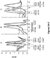
- the SMB assay produces clear and easily distinguishable Tm peaks and definitive Tm shifts to identify the mutations of interest. Individual Tm values can also be used to cluster samples that have the same genotype.
- an assay was tested out on a panel of 603 clinical samples representing both new cases of TB as well as unresolved re-treatment cases, and evaluated the relationship of the targeted mutations with the susceptibility pattern of the clinical isolates. It was observed that 100% of the isolates with rrs A1401G mutations had a strong correlation with high level resistance to both AMK and KAN. However, eis promoter mutations resulted in only moderate to low level KAN resistance and no resistance to AMK, which is consistent with previous studies ( Campbell, P. J., et al, 2011, Antimicrob Agents Chemother 55:2032-2041 ; Zaunbrecher, M.
- the molecular assay served as a better predictor of potentially emerging resistance than the phenotypic assay, as the SMB assay clearly detected the presence of both the wild type and the mutant DNA types.
- a sensitive and specific assay is developed for detection of AMK and KAN resistance in M. tb and validated it in clinical isolates with a high prevalence of MDR and XDR TB.
- the results show that rrs A1401G mutations encode high level cross-resistance to both AMK and KAN, and that eis promoter mutations encode moderate to low level KAN resistance, which is consistent with previous functional genomics studies (Zaunbrecher 2009).
- Comparing the performance of the assay disclosed herein with three different phenotypic susceptibility testing methods in solid and liquid media revealed that low to moderate level KAN resistance caused by eis promoter mutations are largely missed by the LJ based susceptibility tests.
- M.tb test samples consisted of DNA isolated from 603 sequential M.tb isolates cultured from 503 patients enrolled in a natural history study of MDR tuberculosis (NCT00341601 at clinicaltrials.gov) in the National Masan Hospital in Changwon, Republic of Korea. Two cohorts were tested. Cohort A consisted of treatment naive newly suspected TB cases (158 samples) and cohort B consisted of re-treatment TB cases (445 samples). Fresh sputum samples were collected from each patient at the onset of treatment and cultured for M.tb. In a subset of patients, repeat sputum samples were collected at the 1 st , 4 th and 6 th months of treatment and also cultured for M.tb.
- Non-Tuberculosis Mycobacteria (NTM) and Gram-positive and Gram-negative bacteria test samples were taken from the New Jersey Medical School (NJMS) DNA repository as described previously ( Chakravorty, S., 2012. J Clin Microbiol 50:2194-2202 ).
- Phenotypic drug susceptibility testing was performed on all 603 isolates by the absolute concentration method on LJ media to determine the susceptibility to AMK and KAN using a critical concentrations of 40 ⁇ g/ml (the standard concentration used during 2012 when the isolates were tested) for both the antibiotics ( Jnawali, H. N., 2013, Diagn Microbiol Infect Dis 76:187-196 ) at the International Tuberculosis Research Center (ITRC), South Korea.
- ITRC International Tuberculosis Research Center
- MICs to AMK and KAN for 173/603 samples were also evaluated using the TREK Sensititre® MYCOTB MIC plates ("MYCOTB”; TREK Diagnostic Systems, Cleveland, Ohio, USA) as described previously ( Lee, J., 2014, Antimicrob Agents Chemother 58:11-18 ).
- resistance to KAN was also evaluated using the Mycobacterial Growth Indicator Tube (MGIT) system (Becton Dickinson, Franklin Lakes, NJ, USA) at a critical concentration of 2.5 ⁇ g/ml.
- MGIT Mycobacterial Growth Indicator Tube
- the phenotypic susceptibility tests were repeated to confirm the initial findings. In cases where MGIT and LJ susceptibility test results showed discordance, both the assays were repeated to confirm or rectify the initial findings.
- DNA for both SMB assay testing and Sanger sequencing was prepared from cultured isolates by boiling one loopful of culture in 200 ⁇ l of Instagene Matrix resin (Bio-Rad Laboratories, Hercules California, USA) in the presence of 0.1% Triton X100 for 10-15 minutes. The supernatant was recovered after centrifugation and quantified using a Nanodrop microvolume spectrophotometer (Thermo Fisher Scientific, Waltham, Massachusetts, USA).
- the eis promoter region and the rrs gene fragments were amplified as described previously (10, 33).
- a 538bp fragment from the whiB7 gene including 412bp of the 5' untranslated region and 126bp from the ORF was amplified and sequenced using primers whiB7F 5'aaacgcgcaggtcagaaaat 3' and whiB7R 5'cagtgtcttggctacctcga 3' (SEQ ID Nos: 70 and 71).
- a 275bp fragment from the whiB7 gene which included almost the entire whiB7 ORF was also amplified using the primers whiB7-ingene-F 5' GTCGGTACTGACAGTCCCC 3' and whiB7-ingene-R 5'ATGCAACAGCATCCTTGCG 3'(SEQ ID Nos: 72 and 73).
- the PCR products were subjected to bidirectional sequencing using the gene-specific forward and reverse primers in a 3130XL Genetic Analyzer (Applied Bio-systems, Foster City, California, USA) using a BigDye Terminator, version 3.1, cycle sequencing kit (Applied Biosystems) according to the manufacturer's instructions.
- the SMB assays targeted M.tb mutations in codons 1401 and 1402 of the rrs gene and mutations along the promoter region of the eis gene.
- a 113bp fragment (nucleotides 1335-1451) was amplified from the rrs gene using the primers AMG-F (5'-GCTAGTAATCGCAGATCAGCAACGCTGC-3', SEQ ID No: 51) and AMG-R (5'-CCTCCCGAGGGTTAGGCCACT-3', SEQ ID No: 52) and a 98bp fragment encompassing the promoter region and the initial five codons of the eis gene (nucleotides - 81 to 17) was amplified using the primers eis-F (5'-CACAGGGTCACAGTCACAGAATC-3', SEQ ID No: 18) and eis -R (5'-GCATCGCGTGATCCTTTGCCAGAC-3', SEQ ID No: 53).
- the rrs primers were designed to be specific to Mycobacterium genus and the eis primers were designed to be specific to the M.tb complex.
- One SMB probe rrs -1400 (5'-6carboxyfluorescein- cacg accgcccgtcacgtcatgaaagtcggt cgtg -BHQ1-3', SEQ ID No: 59) and two SMB probes eis-1 (5'-Cyanine5- caggcgg tcgtaatattcacgtgcacctggccgc cgcctg -BHQ2-3', SEQ ID No: 16) and eis-2 (5'-TexasRed- ctcgcg gcatatgccacagtcggattctctgac gcgag -BHQ2-3', SEQ ID No: 61) (where underlined sequences represent the stem portion of the SMB
- the rrs probe was designed to be complementary to the antisense strand and the eis probes were designed to be complementary to the sense strand.
- the SMBs were designed using the in silico DNA folding program at http://mfold.rna.albany.edu/?q_mfold/dna-folding-form, and the probe-target hybrid folding program at http://mfold.rna.albany.edu/?q_DINAMelt/Two-state-melting was used to predict the possible probe-target hybrid structures and melting temperatures (Tms).
- the probes were designed to generate a maximum Tm difference between wild-type and mutant sequences in their respective target regions to enable unambiguous mutation identification. Primers were obtained from Sigma Aldrich (St. Louis, Missouri, USA), and SMB probes from Biosearch Technologies (Novato, California, USA).
- reaction volumes containing 100nM forward primer and 1 ⁇ M reverse primer for the rrs gene and 1 ⁇ M forward primer and 50nM reverse primer for the eis promoter region, 1ng/ ⁇ l of rrs-1400 and eis-1 probes and 0.8ng/ ⁇ l of eis-2 probe, 4mM MgCl 2 , 250mM deoxynucleoside triphosphates (dNTPs), 1XPCR buffer, 8% glycerol, 0.06U/ ⁇ l of Platinum® Tfi Exo(-) DNA polymerase (Life Technologies, Grand Island, New York, USA), and 2 to 5ng of sample DNA or an equivalent volume of water.
- dNTPs deoxynucleoside triphosphates
- PCR was carried out with the following steps: activation of the enzyme for 2min at 95°C, followed by 50 cycles of denaturation at 95°C for 10s and combined annealing and extension at 67°C for 30s.
- post-PCR-Tm analysis was performed by denaturation at 95°C for 2min, followed by cooling down to 45°C and then gradual heating to 85°C, with continuous monitoring of fluorescence during the process at a rate of 1 data acquisition per degree centigrade.
- Tm values were identified at the end of the reaction using the Tm calling software (Light Cycler 480 software). However, each Tm was also verified by a trained observer before the final identification of the Tm value was made.
- Samples showing distinct double peaks for any probes corresponding to wild type and mutant Tms were considered to be indicative of hetero-resistance.
- a no template control using sterile water instead of DNA as the template was used as the DNA-negative control, and a DNA-positive control using 1ng of genomic DNA from M.tb H37Rv as the template was also included in each assay plate.
- the SMB-based assay disclosed herein detected resistance to AMK and KAN by looking for mutations in the M.tb rrs gene and eis promoter that have known associations with resistance.
- the assay consisted of a PCR step followed by a Tm analysis in the presence of SMB probes complementary to portions of the rrs and eis target amplicons.
- the inventors first evaluated the capability of the assay to identify the target mutations on artificial oligonucleotides and sequenced DNA templates from selected wild type and mutant M.tb. strains (data not shown). Wild type sequences were identified by the presence of Tm values within 1°C of the known mean values for wild type targets.
- Mutant sequences were identified by a shift in Tm values of at least five standard deviations away from the mean wild type Tm values.
- the ability of the assay to detect the most prevalent mutations associated with AMK and KAN resistance was then evaluated on the clinical DNA samples. Tests were performed on a panel of 603 clinical samples, consisting of 487 samples with wild type sequences and 116 samples with mutations in the assay targets. Five of these samples had mixtures of both wild type and mutant DNA detected on Sanger sequencing.
- the SMB assay correctly identified 115/116 (99%) mutant or mixed (heterogeneous samples containing both mutant and wild type DNA) samples as mutant or mixed and 487/487 (100%) pure wild type samples as wild type.
- a single mixed sample (as indicated by Sanger sequencing) was identified as a wild type sample by the assay disclosed herein.
- the Tm values produced by each SMB probe in the setting of wild type or mutant targets were highly reproducible.
- probes rrs -1400, eis -1 and eis-2 showed mean Tm values of 70.1°C ⁇ 0.15, 63.9°C ⁇ 0.19 and 69°C ⁇ 0.23, respectively (Table 3).
- mutant Tm targets A1401G and C1402T, the mutations resulted in a 3.9°C ( ⁇ 0.17) and 5.6°C ( ⁇ 0.21) decrease in Tm values in probe rrs-1400, respectively (Table 3).
- the eis -1 and the eis-2 probes robustly detected a range of mutations in the eis promoter region as mutant by developing a 4.3°C to 6.5°C decrease in Tm values, compared to the expected wild type Tm values (Table 3).
- the PCR assays performed in the two different laboratories at Rutgers and ITRC were in complete agreement for all the samples detected as wild type and mutant as well as mixtures.
- the assay results enabled us to clearly segregate the 603 samples into wild type and mutant Tm cluster types based on their individual three-point Tm patterns ( Fig 1 ).
- the assay correctly identified mutations in all 75 samples that only contained the A1401G mutation (Table 3, Fig 2 . panel A).
- Three of the four samples containing mixtures of A1401G and wild type sequence were also detected as mixed wild type/mutant based on the presence of a double Tm peak.
- a single sample that contained a mixture of the C1402T mutation and wild type DNA was also identified by the presence of double Tm peaks from the sample with a mutant Tm specific for the C1402T mutation (Table 3, Fig 2 . panel A).
- the 32 samples with eis promoter region mutations included five different polymorphisms (at positions -8, -10, -12, -14 and -37). All of these mutations were successfully detected by either one of the eis SMBs (Table 3, Fig 2 . panels B and C). Four samples that had mutations in both the rrs gene and the eis promoter region were also correctly detected as double mutants (Table 3). Sequencing of the rrs gene did not identify any samples with a codon 1484 mutation, regardless of its drug susceptibility pattern.
- assays were performed to evaluate the performance of the molecular assay relative to phenotypic drug susceptibly test results.
- the apparent performance of a genotypic drug susceptibility test can vary depending on the mutations selected for inclusion in the test and the phenotypic assay that is used as a gold standard ( Kim, S. J. 2005 Eur Respir J 25:564-569 ; Rigouts, L., et al., 2013, J Clin Microbiol 51:2641-2645 ; and Van Deun, A., et al., 2013, J Clin Microbiol 51:2633-2640 ).
- the rrs SMB Tm characteristic of the A1401G mutation classified 82/90 of the AMK resistant samples as resistant, (sensitivity of 91.1%; 95% CI, 82.8% to 96.8%).
- the wild type Tm classified 512/513 of the AMK susceptible samples as susceptible.
- a single isolate among the 513 AMK susceptible isolates was identified as a mixture of wild type and C1402T mutant DNA by the SMB assay disclosed herein due to the presence of a clear double peak generated by the rrs SMB probe, corresponding to the wild type Tm and a specific C1402T mutant Tm ( Figure 2 . panel A).
- the performance of an assay to detect KAN resistance was also evaluated using LJ based drug susceptibly testing as the gold standard for all the 603 samples.
- Tm values generated by the rrs SMB disclosed herein typical for either the A1401G or the C1402T mutation to define resistance the assay detected 82/106 samples as KAN resistant (sensitivity 77.4%; 95% CI 68.0 to 84.7%).
- a rrs SMB Tm characteristic for wild type target to define susceptibility identified 496/497 KAN susceptible samples as susceptible (Table 4) (specificity 99.8%; 95% CI, 98.7 to 100%).
- the discordance between resistance as defined by the assay disclosed herein and resistance as defined by two phenotypic susceptibility test methods disclosed herein principally involved isolates with eis promoter mutations.
- Previous studies have shown that the eis promoter mutations give rise to relatively low levels of KAN resistance, while rrs gene mutations result in high level resistance to AMK, KAN and CAP (Campbell 2011; Du, Q., et al., 2013, Diagn Microbiol Infect Dis 77:138-142 ; Georghiou 2012; and Zaunbrecher 2009).
- An additional finding in the results was the discordance between LJ versus MGIT-based susceptibility test results.
- MIC testing was performed to more carefully explore the relationship between rrs and eis promoter mutations, and their differential susceptibility patterns in the LJ and the MGIT system.
- Samples that were either susceptible to both AMK and KAN (and wild type at both the target regions), or chosen to be representative of the most common mutation types in the two target genes ( rrs A1401G and eis G(-10)A, C(-14)T and G(-37)T) were tested by the MYCOTB method to determine their MIC. Additional isolates known to be wild type in both of the assay targets were also tested as controls.
- the AMK MICs of the isolates that only had eis promoter mutations ranged between 0.25 ⁇ g/ml and 2 ⁇ g/ml, with majority of samples showing MICs of 0.5 ⁇ g/ml to 1 ⁇ g/ml ( Fig. 3 ). Only one eis promoter mutant had an AMK MIC of 4 ⁇ g/ml. Control isolates with no eis promoter or rrs mutations had MICs between 0.25 ⁇ g/ml and 0. 5 ⁇ g/ml range. Thus, the AMK MICs of the isolates with either wild type or mutant eis promoter sequences overlapped substantially.
- the KAN MICs for most of the same eis promoter mutants ranged from 5 ⁇ g/ml to 20 ⁇ g/ml, with one isolate showing an MIC of 40 ⁇ g/ml ( Fig. 3 ). Only two eis promoter mutants had low MICs of 2.5 ⁇ g/ml.
- the isolates with wild type eis promoter sequences showed MICs between 0.6/ ⁇ g/ml to 2.5 ⁇ g/ml which is 2 to 30 fold less than the mean MIC of the eis promoter mutants ( Fig. 3 ).
- the KAN MICs of the wild type isolates overlapped very little with the KAN MICs of the eis promoter mutants.
- the analytical specificity of the assay was tested against a panel of 18 species of non-tuberculous Mycobacteria (NTM) obtained from the ATCC repository (Manassas, Virginia, USA), 121 clinical NTM strains representing 26 species, and 18 species of gram positive and gram negative bacteria.
- NTM non-tuberculous Mycobacteria
- the rrs region targeted in the assay here is highly conserved among different NTM species.
- the rrs assay generated a Tm of 70°C (which is identical to the Tm generated in the presence of wild type M. tb DNA) for all the NTM tested as expected based on sequence homology expect for M. xenopi, which did not generate any Tm.
- the study in this example included 22 samples that were resistant to AMK and/or KAN, but had wild type rrs gene and eis promoter sequences. Recent investigations have suggested that mutations in the 5' untranslated region (UTR) of the whiB7 gene may cause aminoglycoside resistance in M. tb (27). To determine whether whiB7 mutations could be responsible for some of the phenotypically resistant but assay-susceptible isolates, all the 22 samples were sequenced in a 412bp region upstream of the whiB7 gene start site plus a portion of the whiB7 open reading frame. As a control set, 30 randomly picked pansusceptible isolates were also sequenced.
- UTR 5' untranslated region
- LJ and MGIT imply susceptibility testing by the LJ proportions and the MGIT methods respectively.
Description
- This invention relates to kits comprising novel primers and probes for identifying the presence of multi-drug resistant M.tb, and methods for the same purpose.
- Tuberculosis (TB) was declared a global public emergency nearly twenty years ago (WHO Global Tuberculosis Report 2013). Although the rate of new cases of TB has been decreasing worldwide, the millennium developmental goal target of 50% disease reduction by 2015 is unlikely to be achieved (WHO Global Tuberculosis Report 2013). An increase in the incidence of multi drug resistant (MDR) and extensively drug resistant (XDR) TB is a serious threat to these reduction goals (WHO Global Tuberculosis Report 2013). MDR TB is defined as TB resistant to treatment with at least Rifampicin and Isoniazid and XDR TB is defined as MDR TB that is additionally resistant to treatment with the Fluoroquinolone class of antibiotics and the injectable drugs Amikacin, Kanamycin and Capreomycin. Patients with drug resistant TB are best identified as rapidly as possible so that appropriate infection control and treatments can be quickly initiated Boehme, C. C., et.al, 2011, Lancet 377:1495-1505.
- Conventional phenotypic methods can take weeks to months to fully define the drug resistance pattern of Mycobacterium tuberculosis (Mtb) due to the very slow growth of this bacterium (Heifets, L., et al., J Clin Microbiol 38:1227-1230; Kim, S. J., 2005, Eur Respir J 25:564-569; and PT, K., and K. GP., 1985, Public health Mycobacteriology: A guide for level III laboratory. Center for Disease Control, U.S Department of Health and Human Services, Atltanta, Georgia.). Molecular tests offer the promise of more rapid drug resistance detection. Mtb does not naturally contain drug-resistance plasmids; thus, molecular tests are directed against chromosomal DNA. Genotypic assays are relatively easy to design because the Mtb genome has a very high degree of sequence conservation. Virtually all drug-susceptible clinical Mtb isolates have identical DNA sequences in drug resistance targets, except for a few easily identified "natural polymorphisms". It follows that any deviation from wild type sequence in a drug resistance target gene indicates the presence of drug resistance to the corresponding drug. Genotypic assays are more rapid and sensitive than phenotypic assays because DNA targets may be amplified by PCR. Biohazards can be minimized by early killing of infectious organisms.
- The genetic targets which account for most cases of drug resistance in TB are now well established. Real-time PCR remains the most sensitive, rapid, and robust method to detect mutations in bacteria. Virtually all other mutation detection methods including PCR-MS, microarrays, miniarrays and next generation sequencing require nucleic acid amplification as a first step in the detection process. In contrast, real-time PCR enables sample amplification, detection and analysis to all be performed in a single well. Tubes do not have to be opened, complex fluidics are unnecessary. However, no one has been able to develop a broad methodology of drug-resistance testing that is sufficiently simple and robust to be performed outside of reference laboratories. Thus there is a need for novel primers and probes for detecting M.tb and M.tb drug resistance to the most commonly used first and second line drugs
- This invention relates to kits comprising primers and probes, and methods for detecting multi-drug resistant M.tb.
- In one aspect, the invention provides a kit for detecting multi-drug resistant Mycobacterium tuberculosis in a sample, the kit comprising a first pair of forward and reverse primers for amplifying a first portion of a first region, the first region being Mycobacterium tuberculosis rrs gene, wherein each primer for the rrs gene has a sequence that is at least 85% identical to an oligonucleotide sequence selected from the group consisting of SEQ ID Nos: 16, 51 and 52, and a second pair of forward and reverse primers for amplifying a second portion of a second region of a rpoB gene; and an isolated nucleic acid probe comprising a sequence that is at least 85% identical to SEQ ID NO: 59. An isolated oligonucleotide set or primer set for amplifying a portion of a M. tuberculosis region is disclosed and relates to but is not part of the invention. The isolated oligonucleotide set or primer set may be selected from the group consisting of rpoB gene, gyrA gene, gyrB gene, inhA promoter, rrs gene, eis promoter, embB gene, katG gene, dosR gene, IS6110 gene, IS1081 gene. The set includes a pair of forward and reverse primers specific for the portion, where each primer has a sequence that is substantially identical to an oligonucleotide sequence selected from those listed in Tables 1A and 1B below. Accordingly, each primer has a sequence that is substantially complementary to the complement of the oligonucleotide sequence selected from those listed in the tables. The sequence of the primer may be identical to the oligonucleotide sequence selected from those listed in Tables 1A and 1B.
- An isolated nucleic acid having a sequence that is substantially identical to one selected from those listed in Table 2 is disclosed and relates to but is not part of the invention. The nucleic acid may include the sequence of one selected from those listed in Table 2. The nucleic acid can be labeled with, e.g., a fluorophore and a quencher at its two ends respectively, or a fluorophore linked to an internal nucleotide in the probe. Examples of the fluorophores include fluorescein, cyanine 5, or TexasRed, and TAMRA. Examples of the quenchers include BHQ1, BHQ2, and DABCYL.
- The kit may further include a DNA polymerase, extension nucleotides, and a buffer.
- In another aspect of the invention, there is provided a method for detecting multi-drug resistant M. tuberculosis in a sample using the kit according to the invention.
- In another aspect, the invention features a method for detecting multi-drug resistant M. tuberculosis in a sample, comprising amplifying a first nucleic acid target sequence with a first primer pair to generate a first amplicon, the first primer pair being specific for a portion of a region from the rrs gene, wherein each primer for the rrs gene has a sequence that is at least 85% identical to an oligonucleotide sequence selected from the group consisting of SEQ ID Nos: 16, 51 and 52, detecting a first mutation in the first amplicon, determining the presence of drug resistant Mycobacterium tuberculosis in the sample on the basis of the presence of the first mutation, amplifying a second nucleic acid target sequence with a second primer pair to generate a second amplicon, the second primer pair being specific for a second portion of a second region of a rpoB gene, detecting a second mutation in the second amplicon, determining the presence of drug resistant Mycobacterium tuberculosis in the sample on the basis of the presence of the second mutation, whereby the presence of the first mutation and the second mutation is indicative of the presence of multi-drug resistant Mycobacterium tuberculosis in the sample, wherein the step of detecting a first mutation in the first amplicon is performed by a process comprising contacting the first amplicon with a first probe specific for said first mutation under conditions conducive to a hybridization to form a first probe-target hybrid, wherein the first probe comprises a sequence that is at least 85% identical to SEQ ID NO: 59; conducting a first melting temperature (Tm) analysis to determine a first test Tm value for said first probe-target hybrid; and comparing the first test Tm value with a first pre-determined reference Tm value, whereby the first test Tm value, if different from the first pre-determined Tm value, indicates the presence of the first mutation.
- The detecting step is performed by a process comprising (i) contacting the first amplicon with a first probe specific for the mutation under conditions conducive to a hybridization to form a probe-target hybrid; (ii) conducting a melting temperature (Tm) analysis to determine a test Tm value for the probe-target hybrid; and (iii) comparing the test Tm value with a pre-determined reference Tm value. The test Tm value, if different from the pre-determined Tm value, indicates the presence of the mutation. For example, a shift in the test Tm value of at least 3 (e.g., 3, 4, or 5) standard deviations away from the reference Tm value indicates the presence of the mutation. Conversely, a shift in the test Tm value of less than 3 standard deviations away from the reference Tm value indicates the absence of the mutation. As disclosed herein, the pre-determined reference Tm value can be the mean wild type Tm values. In one example, the test Tm value, if lower than the pre-determined reference Tm value by e.g., at least 3 standard deviations, indicates the presence of the mutation. Otherwise, the test Tm value, if not lower than the pre-determined reference Tm value by e.g., 3 standard deviations, indicates the absence of the mutation.
- The method can further include amplifying a further nucleic acid target sequence with a further primer pair to generate a further amplicon, the further primer pair being specific for a portion of a further region selected from the group consisting of gyrA gene, gyrB gene, inhA promoter, eis promoter, embB gene, and katG gene. The first region may be the eis promoter. The regions can be amplified independently or amplified in the same reaction system using techniques such as nested PCR. In that case, the mutation can be an A1401G or C1402T mutation in the rrs gene. The mutation can be within the eis promoter region queried by the eis primer sequences.
- The above-described method allows one to detect resistance to kanamycin. The method may further comprise detecting mutations in the gyrA gene, gyrB gene, inhA promoter, eis promoter, embB gene, katG gene, dosR gene, IS6110 gene and IS 1081 gene. The probe can have a sequence that is 80% identical to SEQ ID NO: 57 or 58.
- A further method for detecting presence of M. tuberculosis in a test sample, for example, from a subject is disclosed and relates to but is not part of the invention. The method includes contacting the test sample with a first primer pair under conditions conducive to an amplifying reaction to yield a first amplicon, and detecting the presence of the amplicon thereby detecting presence of Mycobacterium tuberculosis in the test sample. The first primer pair can be an oligonucleotide set for amplifying a portion of a M. tuberculosis region selected from the group consisting of gyrB gene, inhA promoter, eis promoter, embB gene, katG gene, dosR gene, IS6110 gene, IS 1081 gene. Each primer of the first primer pair has a sequence that can be substantially identical to an oligonucleotide sequence selected from those listed in Table 1B. The method can further include contacting the test sample or the amplicon generated by the first primer pair with a second primer pair under conditions conducive to an amplifying reaction to yield a second amplicon, and detecting the presence of the second amplicon. In that case, presence of both the first amplicon and second amplicon indicates the presence of Mycobacterium tuberculosis in the test sample.
- A method for detecting presence of M. tuberculosis in a test sample is disclosed and relates to but is not part of the invention. The method includes contacting the test sample with a first molecular beacon probe under conditions conducive to a hybridization reaction to yield a probe-target hybrid, and detecting the presence of the probe-target hybrid thereby detecting presence of Mycobacterium tuberculosis in the test sample. In this method, the first molecular beacon probe has a sequence that is substantially identical to one selected from those listed in Table 2. In an example, the first molecular beacon probe is selected from the group consisting of the IS 1081, dosR2, and IS6110 probe (SEQ ID No. 67-69).
- The details of one or more embodiments of the invention are set forth in the description below. Other features, objectives, and advantages of the invention will be apparent from the description and from the claims.
-
-
Figure 1 is a diagram showing detection of AMK and/or KAN resistance in 603 clinical DNA samples using SMB-probe generated three-point Tm profile. Each of the three assay SMBs were tested against all M. tb DNA samples in a multiplex PCR reaction. The results for each sample are shown as a three point Tm plot on the X axis, with the Tm value of each SMB indicated on the Y axis. Isolates are sorted from left to right as phenotypically susceptible and then as resistant. Distinct Tm shifts from at least one of the three probes can be seen in each resistant isolate. -
Figures 2A, 2B and 2C are diagrams showing first derivative melt peak profiles of three SMB probes. The melt peak profiles of wild type, mutant and mixed DNA samples are shown for the rrs-1400 SMB probe (2A), the eis1 SMB probe (2B), and the eis2 SMB probe (2C). Each melt curve represents an individual strain. -
Figure 3 is a diagram showing MIC values of rrs and eis mutant and wild type strains. The average MIC values to AMK and KAN for the rrs and the eis mutants and the wild type strains are shown. Error bars represent the ± one standard deviation of the MIC values. eis-P indicates eis gene promoter. - This invention is based, at least in part, on an unexpected discovery of novel primers, SMB probes, and MB probes for amplifying segments from eleven different genes in M.tb for identifying the presence of M.tb DNA and resistance to anti-tuberculosis drugs such as amikacin and kanamycin.
- The primers described here for amplifying the rpoB and the rrs genes allow sensitive amplification of drug resistance inducing mutation hotspots in M.tb. The corresponding SMB probes target and identify these mutations which result in drug resistance to the most commonly used first and second line drugs. These primers can be used with very high efficiency in both symmetric and asymmetric PCR assays. The primer and probe sequences described here have been used by the inventors to develop rapid and accurate molecular drug susceptibility testing assays for M.tb. Apart from their obvious utility in molecular diagnostic assay for M.tb, these primers will also find use for sequencing the target genes to identify the resistance inducing mutations in surveillance assays and any other probe based assays which aims at specific and sensitive identification of the common drug resistance inducing mutations in M.tb. Primer sequences amplifying the dosR, IS6110 and IS1081 genes allow highly sensitive and specific identification of M.tb and can be used in any PCR assay format aimed at highly specific and sensitive molecular diagnosis of Tuberculosis. Listed in the tables below are exemplary primers and probes.
Table 1A Primer # SEQ ID No. Primer name Primer Sequence Target Gene Portion # 1 1 gyrA-F CCGGTCGGTTGCCGAGACC gyrA # 2 2 gyrA-asym-F CGGTCGGTTGCCGAGACCATGG #3 3 P2-gyrA-nested-F GTCGGTTGCCGAGACCATGGGC #4 4 gyrA-P1-R AGCGGGTAGCGCAGCGACCAG #5 5 P2-gyrA-nested-R CGGGTAGCGCAGCGACCAGGGC #6 6 gyrA-R CCAGCGGGTAGCGCAGCGACCAG #12 12 rpo-R0 CGTCGCGGACCTCCAGCCCGGCA rpo B #13 13 rpo- R2a TCACGTGACAGACCGCCGGGC # 14 14 rpo-R2b GCTCACGTGACAGACCGCCGGGC #49 49 rpoB-iF ATCAACATCCGGCCGGTGGTCGCC #50 50 rpoB-R AGCTCCAGCCCGGCACGCTCACGT #15 15 rrs-F GCTAGTAATCGCAGATCAGCAACGCTGC rrs #16 16 rrs-R CCTCCCGAGGGTTAGGCCACTGG #17 17 P3-AMG- R GGTTAGGCCACTGGCTTCGG # 51 51 AMG-F GCTAGTAATCGCAGATCAGCAACGCTGC #52 52 AMG-R CCTCCCGAGGGTTAGGCCACT Table IB Primer # SEQ ID No. Primer name Primer Sequence Target Gene Portion #7 7 katG-F GCTGGAGCAGATGGGCTTGG katG #8 8 P1-katG-F CCGCTGGAGCAGATGGGCTTGG #9 9 P2-katG-F GGCTGGAAGAGCTCGTATGGCACCG #10 10 katG-asym-R GTCCCATTTCGTCGGGGTGTTCGTCC #11 11 P2-katG-R2 CCATTTCGTCGGGGTGTTCGTCCATAC #18 18 eis-F CACAGGGTCACAGTCACAGAATC eis #19 19 P1-eis-F CGTCCTCGGTCGGGCTACACAGG #20 20 P2-eis-nested-F CGGTCGGGCTACACAGGGTCACAGT #21 21 P3-eis-inner-F CACAGGGTCACAGTCACAGAATC #22 22 eis-R GCATCGCGTGATCCTTTGCCAGACA #53 53 eis-R1 GCATCGCGTGATCCTTTGCCAGAC #23 23 dosR-F CTCGCCGGTGCCAGCGGATATGTC dosR #24 24 dosR-R CGACCGTCCAGCGCCCACATCTTT #25 25 IS6110-F CCGCGAGGGCCCCGATGGTTT IS6110 #26 26 IS6110-R GGCTGGGCTCCCGGTTGATGTGG #27 27 IS-SPADE R GGCTCCCGGTTGATGTGGTCGTAG #28 28 ISBcnSA-iR TGGGGCGATCGGCACACCCAGC #29 29 gyrB1-F ATCGGTGGATTGCCCGGCAAGCTG gyrB #30 30 P2-gyrB1-nested F CGTTCCACGGATCCGCGCAAGTC #31 31 P1-gyrB1-F GCTGGCCGATTGCCGTTCCACG #32 32 P3-gyrB-inner-F GATCATCAATGTGGAGAAAGCGC #33 33 P2-gyrB1-R2 CTGGAACATCGAATCGCGACCGCTT #34 34 gyrB1-R ATCGCGACCGCTTTTTGCAGAA #35 35 embB306-F CTGACCGACGCCGTGGTGATA embB #36 36 embB306-R GGAAATAGTTGGACATGTAGCCGGCGT #37 37 gyrB2-F CGATTCGATGTTCCAGGCGATACTT gyrB #38 38 P2-gyrB2-F GCGCGGCAAGATCATCAATGTGGAG #39 39 P3-gyrB-inner-F GATCATCAATGTGGAGAAAGCGC #40 40 P1-gyrB2-outer-R GTGGATCCCGGTGCCCAGCGCC #41 41 P2-gyrB2-R2 GGTGCCCAGCGCCGTGATGATC #42 42 inhA-F CGTTACGCTCGTGGACATACCGATTTCG inhA #43 43 P2-inhA-F TTACGCTCGTGGACATACCGATTTCGGC #44 44 P1-inhA-R GGACTGAACGGGATACGAATGGGG #45 45 P2-inhA-R GTTTGGCCCCTTCAGTGGCTGTGG #46 46 IS1081-outer-F CAGCCCGACGCCGAATCAGTTGTT IS1081 #47 47 IS1081-outer-R GGTGCGGGCGGTGTCGAGGTG #48 48 IS1081-inner-R GCCACCGCGGGGAGTTTGTCG Table 2 Probe # SEQ ID No. Probe Name Probe Sequence Target Gene Portion # 1 54 gyrA-1 
gyrA # 2 55 gyrA-2 
#3 56 katG 
katG #4 57 rpo3 
rpoB #5 58 rpo4 
#6 59 rrs1400 
rrs #7 60 eisld 
eis promoter #8 61 eis2b 
#16 62 eis 1d 
#9 63 gyrB500 
gyrB # 10 64 inhA RC 
inhA promoter #11 65 gyrB2 
gyrB #12 66 embB306 
embB #13 67 IS1081 CgcgcaCCAATATGATCGGGTACTCGACtgcgcg IS1081 # 14 68 dosR2 
dosR #15 69 IS6110 
IS6110 - One or more of the probe sequences in Table 2 can be made in various detection formats, such as dual labeled probes including liner probes, Taqman probes, molecular beacon probes, and sloppy molecular beacon probes. A "sloppy" probe refers to a probe that is mismatch-tolerant. Mismatch-tolerant probes hybridize with and generate detectable signal for more than one target sequence at a detection temperature in an assay, and various hybrids so formed will have different melting points. Linear, or random coil, single-stranded probes are generally mismatch tolerant. Examples of such probes are hairpin or linear probes with an internal fluorescent moiety whose level of fluorescence increases upon hybridization to one or another target strand. See, e.g.,
U.S. Pat. Nos. 7662550 and5925517 .US 20130095479 . - Preferably, the sloppy probes are dual-labeled hairpin probes or molecular beacon probes, described in
U.S. Pat. Nos. 7662550 and5925517 . These hairpin probes contain a target binding sequence flanked by a pair of arms complementary to one another. They can be DNA, RNA, or PNA, or a combination of all three nucleic acids. Furthermore, they can contain modified nucleotides and modified internucleotide linkages. They can have a first fluorophore on one arm and a second fluorophore on the other arm, wherein the absorption spectrum of the second fluorophore substantially overlaps the emission spectrum of the first fluorophore. Most preferably such hairpin probes are "molecular beacon probes" that have a fluorophore on one arm and a quencher on the other arm such that the probes are dark when free in solution. They can also be wavelength-shifting molecular beacon probes with, for example, multiple fluorophores on one arm that interact by fluorescence resonance energy transfer (FRET), and a quencher on the other arm. The target binding sequences can be, for example, 12 to 50, or 25 to 50 nucleotides in length, and the hybridizing arms can be 4 to 10 or 4 to 6 (e.g., 5 or 6) nucleotides in length. Molecular beacon probes can be tethered to primers, as described inU.S. Pat. Nos. 7662550 and5925517 andWO 01/31062 - Sloppy molecular beacon probes thus refers to such a class of fluorescently labeled hairpin oligonucleotide hybridization probes. Such probes produce a detectable signal in a homogeneous assay, that is, without having to separate probes hybridized to target from unbound probes. By virtue of their ability to bind to more than one variants of a given target sequence, the probes can be used in assays to detect the presence of one variant of a nucleic acid sequence segment of interest from among a number of possible variants or even to detect the presence of two or more variants. The probes can therefore be used in combinations of two or more in the same assay. Because they differ in target binding sequence, their relative avidities for different variants are different. For example, a first probe may bind strongly to a wild-type sequence, moderately to a first allele, weakly to a second allele and not at all to a third allele; while a second probe may bind weakly to the wild-type sequence and the first variant, and moderately to the second variant and the third variant. Additional sloppy probes will exhibit yet different binding patterns due to their different target binding sequences. Thus, fluorescence emission spectra from combinations of sloppy probes define different microbial strains or species, as well as allelic variants/mutation of genes.
- As the sloppy probes reproducibly fluoresce with variable intensities after binding to different DNA sequences, combinations can be used in, for example, simple, rapid, and sensitive nucleic acid amplification reaction assays (e.g., PCR-based assays) that identify multiple pathogens or variants in a single reaction container. It is understood, however, that the assays can be performed also on samples suspected of containing directly detectable amounts of unamplified target nucleic acids. This identification assay is based on analyzing the spectra of a set of partially hybridizing sloppy signaling probes, such as sloppy molecular beacon probes, each labeled with a fluorophore that emits light with a different wavelength optimum, to generate "signature spectra" of species-specific or variant-specific DNA sequences.
- Using the probes, multiplexing can be achieved, for example, by designing a different allele-discriminating molecular beacon probe for each target and labeling each probe differentially. (See, e.g.,
U.S. Pat. Nos. 7662550 and5925517 ,WO 01/31062 US Patent No. 7662550 . - This kit may contain reagents for performing the above-described methods, including PCR and/or probe-target hybridization reactions. To that end, one or more of the reaction components, e.g., PCR primers, polymerase, and probes, for the methods disclosed herein can be supplied in the form of a kit for use. In such a kit, an appropriate amount of one or more reaction components is provided in one or more containers or held on a substrate.
- The kit may also contain additional materials for practicing the above-described methods. The kit may contain some or all of the reagents, materials for performing a method that uses primers and/or probes according to the invention. Some or all of the components of the kits can be provided in containers separate from the container(s) containing the primers and/or probes of the invention. Examples of additional components of the kits include, but are not limited to, one or more different polymerases, one or more control reagents (e.g., probes or PCR primers or control templates), and buffers for the reactions (in 1X or concentrated forms). The kit may also include one or more of the following components: supports, terminating, modifying or digestion reagents, osmolytes, and an apparatus for detection.
- The reaction components used can be provided in a variety of forms. For example, the components (e.g., enzymes, probes and/or primers) can be suspended in an aqueous solution or as a freeze-dried or lyophilized powder, pellet, or bead. In the latter case, the components, when reconstituted, form a complete mixture of components for use in an assay. The kits of the invention can be provided at any suitable temperature. For example, for storage of kits containing protein components (e.g., an enzyme) in a liquid, it is preferred that they are provided and maintained below 0°C, preferably at or below - 20°C, or otherwise in a frozen state.
- A kit may contain, in an amount sufficient for at least one assay, any combination of the components described herein. In some applications, one or more reaction components may be provided in pre-measured single use amounts in individual, typically disposable, tubes or equivalent containers. With such an arrangement, a PCR reaction can be performed by adding a target nucleic acid or a sample/cell containing the target nucleic acid to the individual tubes directly. The amount of a component supplied in the kit can be any appropriate amount, and may depend on the target market to which the product is directed. The container(s) in which the components are supplied can be any conventional container that is capable of holding the supplied form, for instance, microfuge tubes, ampoules, bottles, or integral testing devices, such as fluidic devices, cartridges, lateral flow, or other similar devices.
- The kit can also include packaging materials for holding the container or combination of containers. Typical packaging materials for such kits and systems include solid matrices (e.g., glass, plastic, paper, foil, micro-particles and the like) that hold the reaction components or detection probes in any of a variety of configurations (e.g., in a vial, microtiter plate well, microarray, and the like). The kits may further include instructions recorded in a tangible form for use of the components.
- A nucleic acid refers to a DNA molecule (for example, but not limited to, a cDNA or genomic DNA), an RNA molecule (for example, but not limited to, an mRNA), or a DNA or RNA analog. A DNA or RNA analog can be synthesized from nucleotide analogs. The nucleic acid molecule can be single-stranded or double-stranded. An "isolated" nucleic acid is a nucleic acid, the structure of which is not identical to that of any naturally occurring nucleic acid or to that of any fragment of a naturally occurring genomic nucleic acid. The term therefore covers, for example, (a) a DNA which has the sequence of part of a naturally occurring genomic DNA molecule but is not flanked by both of the coding sequences that flank that part of the molecule in the genome of the organism in which it naturally occurs; (b) a nucleic acid incorporated into a vector or into the genomic DNA of a prokaryote or eukaryote in a manner such that the resulting molecule is not identical to any naturally occurring vector or genomic DNA; (c) a separate molecule such as a cDNA, a genomic fragment, a fragment produced by PCR, or a restriction fragment; and (d) a recombinant nucleotide sequence that is part of a hybrid gene, i.e., a gene encoding a fusion protein.
- As used herein, the term "target nucleic acid" or "target" refers to a nucleic acid containing a target nucleic acid sequence of interest. A target nucleic acid may be single-stranded or double-stranded, and often is double-stranded DNA. A "target nucleic acid sequence," "target sequence" or "target region" means a specific sequence that comprises all or part of the sequence of a single-stranded nucleic acid. A target sequence may be within a nucleic acid template or within the genome of a cell, which may be any form of single-stranded or double-stranded nucleic acid. A template may be a purified or isolated nucleic acid, or may be non-purified or non-isolated.
- "Complementary" sequences, as used herein, may include, or be formed entirely from, Watson-Crick base pairs (e.g., A-T/U and C-G), non-Watson-Crick base pairs and/or base pairs formed from non-natural and modified nucleotides, and in as far as the above requirements with respect to their ability to hybridize are fulfilled. A full complement or fully complementary may mean 100% (completely) complementary or substantially complementary base pairing between nucleotides or nucleotide analogs of nucleic acid molecules.
- "Substantially complementary" means that a nucleic acid or oligonucleotide has a sequence containing at least 10 contiguous bases that are at least 80%, (e.g., 85%, 90%, 95%, 96%, 97%, 98%, 99%, and 100%) to at least 10 contiguous bases in a target nucleic acid sequence so that the nucleic acid or oligonucleotide can hybridize or anneal to the target nucleic acid sequence under, e.g., the annealing condition of a PCR reaction or probe-target hybridization condition. Complementarity between sequences may be expressed a number of base mismatches in each set of at least 10 contiguous bases being compared. The term "substantially identical" means that a first nucleic acid is at least 80%, (e.g., 85%, 90%, 95%, 96%, 97%, 98%, 99%, and 100%) complementary to a second nucleic acid so that the first nucleic acid is substantially complementary to and is capable of hybridizing to the complement of the second nucleic acid under PCR annealing or probe-target hybridization conditions.
- "Hybridization" or "hybridizing" or "hybridize" or "anneal" refers to the ability of completely or partially complementary nucleic acid strands to come together under specified hybridization conditions in a parallel or preferably antiparallel orientation to form a stable double-stranded structure or region (sometimes called a "hybrid" or "duplex" or "stem") in which the two constituent strands are joined by hydrogen bonds. Although hydrogen bonds typically form between adenine and thymine or uracil (A and T or U) or cytosine and guanine (C and G), other base pairs may form (e.g., Adams et al., The Biochemistry of the Nucleic Acids, 11th ed., 1992).
- A "nucleic acid duplex," "duplex," "stem," "nucleic acid hybrid" or "hybrid" refers to a stable nucleic acid structure comprising a double-stranded, hydrogen-bonded region, e.g., RNA:RNA, RNA:DNA and DNA:DNA duplex molecules and analogs thereof. Such structure may be detected by any known means, e.g., by using a labeled probe, an optically active probe-coated substrate sensitive to changes in mass at its surface (
U.S. Pat. No. 6,060,237 ), or binding agents (U.S. Pat. No. 5,994,056 ). - As used herein the term "amplification" and its variants includes any process for producing multiple copies or complements of at least some portion of a polynucleotide, the polynucleotide typically being referred to as a "template." The template polynucleotide can be single stranded or double stranded. A template may be a purified or isolated nucleic acid, or may be non-purified or non-isolated. Amplification of a given template can result in the generation of a population of polynucleotide amplification products, collectively referred to as an "amplicon." The polynucleotides of the amplicon can be single stranded or double stranded, or a mixture of both. Typically, the template will include a target sequence, and the resulting amplicon will include polynucleotides having a sequence that is either substantially identical or substantially complementary to the target sequence. The polynucleotides of a particular amplicon may be substantially identical, or substantially complementary, to each other; alternatively, the polynucleotides within a given amplicon can have nucleotide sequences that vary from each other. Amplification can proceed in linear or exponential fashion, and can involve repeated and consecutive replications of a given template to form two or more amplification products. Some typical amplification reactions involve successive and repeated cycles of templatebased nucleic acid synthesis, resulting in the formation of a plurality of daughter polynucleotides containing at least some portion of the nucleotide sequence of the template and sharing at least some degree of nucleotide sequence identity (or complementarity) with the template. Each instance of nucleic acid synthesis, which can be referred to as a "cycle" of amplification, may include creating free 3' end (e.g., by nicking one strand of a dsDNA) thereby generating a primer and primer extension steps; optionally, an additional denaturation step can also be included wherein the template is partially or completely denatured. One round of amplification may include a given number of repetitions of a single cycle of amplification. For example, a round of amplification can include 5, 10, 15, 20, 25, 30, 35, 40, 50, or more repetitions of a particular cycle. Amplification may include any reaction wherein a particular polynucleotide template is subjected to two consecutive cycles of nucleic acid synthesis. The synthesis can include template-dependent nucleic acid synthesis.
- Amplification of this invention may also include isothermal amplification. The term "isothermal" means conducting a reaction at substantially constant temperature, i.e., without varying the reaction temperature in which a nucleic acid polymerization reaction occurs. Isothermal temperatures for isothermal amplification reactions depend on the strand-displacing nucleic acid polymerase used in the reactions. Generally, the isothermal temperatures are below the melting temperature (Tm; the temperature at which half of the potentially double-stranded molecules in a mixture are in a single-stranded, denatured state) of the predominant reaction product, i.e., generally 90 °C or below, usually between about 20 °C and 75 °C, and preferably between about 30 °C and 60 °C, or more preferably at about 37 °C.
- The term "primer" or "primer oligonucleotide" refers to a strand of nucleic acid or an oligonucleotide capable of hybridizing to a template nucleic acid and acting as the initiation point for incorporating extension nucleotides according to the composition of the template nucleic acid for nucleic acid synthesis. "Extension nucleotides" refer to any nucleotides (e.g., dNTP) and analogs thereof capable of being incorporated into an extension product during amplification, i.e., DNA, RNA, or a derivative of DNA or RNA, which may include a label.
- As used herein, the term "oligonucleotide" refers to a short polynucleotide, typically less than or equal to 300 nucleotides long (e.g., in the range of 5 and 150, preferably in the range of 10 to 100, more preferably in the range of 15 to 50 nucleotides in length). However, as used herein, the term is also intended to encompass longer or shorter polynucleotide chains. An "oligonucleotide" may hybridize to other polynucleotides, therefore serving as a probe for polynucleotide detection, or a primer for polynucleotide chain extension.
- The term "probe" as used herein refers to an oligonucleotide capable of binding to a target nucleic acid of complementary sequence through one or more types of chemical bonds, usually through complementary base pairing, usually through hydrogen bond formation. Probes may bind target sequences lacking complete complementarity with the probe sequence depending upon the stringency of the hybridization conditions. There may be any number of base pair mismatches which will interfere with hybridization between the target sequence and the single stranded nucleic acids described herein. However, if the number of mutations is so great that no hybridization can occur under even the least stringent of hybridization conditions, the sequence is not a complementary target sequence. A probe may be single stranded or partially single and partially double stranded. The strandedness of the probe is dictated by the structure, composition, and properties of the target sequence. Probes may be directly labeled or indirectly labeled with a label such as with biotin to which a streptavidin complex may later bind.
- The term "detection probe" refers to an oligonucleotide having a sequence sufficiently complementary to its target sequence to form a probe:target hybrid stable for detection under stringent hybridization conditions. A probe is typically a synthetic oligomer that may include bases complementary to sequence outside of the targeted region which do not prevent hybridization under stringent hybridization conditions to the target nucleic acid. A sequence non-complementary to the target may be a homopolymer tract (e.g., poly-A or poly-T), promoter sequence, restriction endonuclease recognition sequence, or sequence to confer desired secondary or tertiary structure (e.g., a catalytic site or hairpin structure), or a tag region which may facilitate detection and/or amplification. "Stable" or "stable for detection" means that the temperature of a reaction mixture is at least 2 °C below the melting temperature (Tm) of a nucleic acid duplex contained in the mixture, more preferably at least 5 °C below the Tm, and even more preferably at least 10 °C below the Tm.
- A "label" or "reporter molecule" is chemical or biochemical moiety useful for labeling a nucleic acid (including a single nucleotide), polynucleotide, oligonucleotide, or protein ligand, e.g., amino acid or antibody. Examples include fluorescent agents, chemiluminescent agents, chromogenic agents, quenching agents, radionucleotides, enzymes, substrates, cofactors, inhibitors, magnetic particles, and other moieties known in the art. Labels or reporter molecules are capable of generating a measurable signal and may be covalently or noncovalently joined to an oligonucleotide or nucleotide (e.g., a non-natural nucleotide) or ligand.
- As used herein, the term "contacting" and its variants, when used in reference to any set of components, includes any process whereby the components to be contacted are mixed into same mixture (for example, are added into the same compartment or solution), and does not necessarily require actual physical contact between the recited components. The recited components can be contacted in any order or any combination (or subcombination), and can include situations where one or some of the recited components are subsequently removed from the mixture, optionally prior to addition of other recited components. For example, "contacting A with B and C" includes any and all of the following situations: (i) A is mixed with C, then B is added to the mixture; (ii) A and B are mixed into a mixture; B is removed from the mixture, and then C is added to the mixture; and (iii) A is added to a mixture of B and C. "Contacting" a target nucleic acid or a cell with one or more reaction components, such as a polymerase, a primer set or a probe, includes any or all of the following situations: (i) the target or cell is contacted with a first component of a reaction mixture to create a mixture; then other components of the reaction mixture are added in any order or combination to the mixture; and (ii) the reaction mixture is fully formed prior to mixture with the target or cell.
- The term "mixture" as used herein, refers to a combination of elements, that are interspersed and not in any particular order. A mixture is heterogeneous and not spatially separable into its different constituents. Examples of mixtures of elements include a number of different elements that are dissolved in the same aqueous solution, or a number of different elements attached to a solid support at random or in no particular order in which the different elements are not spatially distinct. In other words, a mixture is not addressable.
- As used herein, the term "subject" refers to any organism having a genome, preferably, a living animal, e.g., a mammal, which has been the object of diagnosis, treatment, observation or experiment. Examples of a subject can be a human, a livestock animal (beef and dairy cattle, sheep, poultry, swine, etc.), or a companion animal (dogs, cats, horses, etc).
- A "sample" as used herein means any biological fluid or tissue obtained from an organism (e.g., patient) or from components (e.g., blood) of an organism. The sample may be of any biological tissue, cell(s) or fluid. The sample may be a "clinical sample" which is a sample derived from a subject, such as a human patient or veterinary subject. Useful biological samples include, without limitation, whole blood, saliva, urine, synovial fluid, bone marrow, cerebrospinal fluid, vaginal mucus, cervical mucus, nasal secretions, sputum, semen, amniotic fluid, bronchoalveolar lavage fluid, and other cellular exudates from a patient or subject. Such samples may further be diluted with saline, buffer or a physiologically acceptable diluent. Alternatively, such samples are concentrated by conventional means. Biological samples may also include sections of tissues such as frozen sections taken for histological purposes. A biological sample may also be referred to as a "patient sample." A biological sample may also include a substantially purified or isolated protein, membrane preparation, or cell culture.
- The terms "determining," "measuring," "assessing," and "assaying" are used interchangeably and include both quantitative and qualitative measurement, and include determining if a characteristic, trait, or feature is present or not. Assessing may be relative or absolute. Assessing the presence of a target includes determining the amount of the target present, as well as determining whether it is present or absent.
- As used herein the term "reference" value refers to a value that statistically correlates to a particular outcome when compared to an assay result. The reference value can be determined from statistical analysis that examines the mean of wild type values. The reference value may be a threshold score value or a cutoff score value. Typically a reference value will be a threshold above (or below) which one outcome is more probable and below which an alternative outcome is more probable.
- As disclosed herein, the difference of the values is indicative of presence or absence of a pathogen (e.g., Mycobacterium tuberculosis) or a mutation. The phrase "difference" of the level or value refers to differences in a variable (e.g., Tm) of an analyte (e.g., a probe-target hybrid) in a sample as compared to a control or reference level or value. A difference of a value or level may be a statistically significant difference between the quantities of an analyte present in a sample as compared to a control. For example, a difference may be statistically significant if the measured level of the analyte falls outside of about 1.0, 2.0, 3.0, 4.0, or 5.0 standard deviations of the mean of any control or reference group.
- As disclosed herein, a number of ranges of values are provided. It is understood that each intervening value, to the tenth of the unit of the lower limit, unless the context clearly dictates otherwise, between the upper and lower limits of that range is also specifically disclosed. Each smaller range between any stated value or intervening value in a stated range and any other stated or intervening value in that stated range is encompassed within the invention. The upper and lower limits of these smaller ranges may independently be included or excluded in the range, and each range where either, neither, or both limits are included in the smaller ranges is also encompassed within the invention, subject to any specifically excluded limit in the stated range. Where the stated range includes one or both of the limits, ranges excluding either or both of those included limits are also included in the invention. The term "about" generally refers to plus or minus 10% of the indicated number. For example, "about 10%" may indicate a range of 9% to 11%, and "about 20" may mean from 18-22. Other meanings of "about" may be apparent from the context, such as rounding off, so, for example "about 1" may also mean from 0.5 to 1.4.
- Two separate SMB assays that rapidly and reliably identify the M.tb mutations that are largely responsible for Rifampin and Fluoroquinolone (FQ) resistance were recently described (Chakravorty, S., B. et al., 2011, J Clin Microbiol 49:932-940; Chakravorty, S., H., et al., 2012, J Clin Microbiol 50:2194-2202). The assays disclosed herein have the advantage of being in real-time PCR format, so that they are easy to use and not subject to amplicon cross contamination. Moreover, mutation detection has been shown to be robust and amenable to high throughput testing. The examples below present a SMB TB drug resistance detection assay system, adding assays that enable detection of resistance to AMK and KAN. The causes of discordance between the assay disclosed herein and phenotypic susceptibility testing methods were also explored. The results show that some of the most commonly used phenotypic methods can miss M.tb isolates with resistance-conferring mutations if these mutations only moderately increase minimal inhibitory concentrations (MICs) to KAN. Novel mutations in whiB7 that are associated with low level KAN resistance were also discovered.
- As disclosed herein, the multiplexed SMB PCR and melt assay accurately identified mutations in the rrs gene and eis promoter associated with resistance to AMK and/or KAN. The assay did not produce false resistance calls when tested against NTMs, gram positive, and gram negative bacteria. Most cases of hetero-resistance were also detected by the assay, when present. Unlike the MTBDRsl platform, the assay can be performed in a closed real-time PCR system, and can easily be adapted to high-throughput testing as all assay steps are performed in 384-well plates. The SMB assay also avoids potential problems associated with alternative methods for mutation detection. High resolution melt curve analysis requires the ability to detect subtle variations in melt curves (Yadav, R., S., et al., 2012, J Appl Microbiol 113:856-862). Other post-PCR melt based molecular assays must detect mutations by decoding complex fluorescence contours (Rice, J. E., et al., 2012. Nucleic Acids Res 40:e164.) In contrast, the SMB assay produces clear and easily distinguishable Tm peaks and definitive Tm shifts to identify the mutations of interest. Individual Tm values can also be used to cluster samples that have the same genotype.
- As disclosed herein, an assay was tested out on a panel of 603 clinical samples representing both new cases of TB as well as unresolved re-treatment cases, and evaluated the relationship of the targeted mutations with the susceptibility pattern of the clinical isolates. It was observed that 100% of the isolates with rrs A1401G mutations had a strong correlation with high level resistance to both AMK and KAN. However, eis promoter mutations resulted in only moderate to low level KAN resistance and no resistance to AMK, which is consistent with previous studies (Campbell, P. J., et al, 2011, Antimicrob Agents Chemother 55:2032-2041; Zaunbrecher, M. A., et al., 2009, Proc Natl Acad Sci U S A 106:20004-20009). The study here also showed that the LJ absolute concentration method for susceptibility testing does not adequately detect moderate to low level KAN resistance. In fact, nearly two-thirds of the samples with eis promoter mutations were detected as KAN susceptible in the LJ media. However, all but one sample with eis promoter mutations were detected as KAN resistant by the MGIT method. Two such isolates contained an eis C(-12)T mutation. These mutants were also resistant to KAN when tested by MYCOTB, which showed a KAN MIC of 5µg/ml. Previous studies have suggested that clinical isolates with C(-12)T mutations do not correlate (Zaunbrecher (2009) or correlate poorly (Campbell, 2011; Hoshide, M., L. et al., 2014, J Clin Microbiol 52:1322-1329.) with KAN resistance. These studies possibly missed the relation between this mutation and low level KAN resistance due to the testing method used to establish phenotypic susceptibility. These results suggest that MGIT or MYCOTB methods should be preferred for testing phenotypic resistance to KAN. They also highlight the power of genotypic resistance tests, such as that disclosed herein, to detect mutations which cause low level resistance and may be missed by phenotypic tests alone (Rigouts, L., M. et al., 2013, J Clin Microbiol 51:2641-2645; Sirgel, F. A., et al., 2012, Microb Drug Resist 18:193-197; and Van Deun, A., et al., 2013, J Clin Microbiol 51:2633-2640).
- The study set here included one sample that was a mixture of rrs wild type and rrs C1402T mutants. This sample was susceptible to both AMK and KAN in LJ media. Isolates with C1402T mutations have been reported to be susceptible to AMK but resistant to KAN (Maus, C. E., et al., 2005, Antimicrob Agents Chemother 49:3192-3197). In this particular case, repeated susceptibility tests using LJ media showed susceptibility to KAN presumably because of the hetero-resistant nature of the sample. Here, the molecular assay served as a better predictor of potentially emerging resistance than the phenotypic assay, as the SMB assay clearly detected the presence of both the wild type and the mutant DNA types.
- The incidence of rrs 1484 mutations in clinical strains with AMK or KAN resistance has been very low (Georghiou, S. B., et al., 2012, PLoS One 7:e33275) making its clinical significance debatable. A separate version of the assay which targeted the rrs 1484 codon, did not detect any mutations in any of the 603 isolates in the study as well as in an additional 259 isolates from the New Jersey -New York area, which included 33 AMK and KAN resistant isolates. The lack of any rrs 1484 mutations in this enlarged study set was confirmed by Sanger sequencing (data not shown). In light of the very low prevalence of rrs 1484 mutations, this codon is unlikely to provide much value in predicting aminoglycoside resistance. Thus, it is recommended that molecular assays for aminoglycoside resistance target only the 1401-1402 codons in the rrs gene.
- It was found that 22 AMK or KAN resistant samples had wild type sequences in the rrs gene and the eis promoter region. A recent study has shown a possible association between mutations in the 5'UTR of the whiB7 and KAN resistance, by identifying a 5'UTR whiB7 mutation in a single clinical strain with unexplained KAN resistance (Reeves, A. Z., et al., 2013, Antimicrob Agents Chemother 57:1857-1865). Also described were several novel 5'UTR whiB7 mutations, as well as a deletion, that appear to be associated with KAN resistance. No suitable universal biomarkers have been identified which can account for KAN and AMK resistance in the remaining 15-20% of clinical strains with wild type rrs, eis promoter region. Samples containing wild type rrs gene and eis promoter region DNA mixed with a trace amount of mutant targets from a KAN or AMK resistant subpopulation could also account for the remaining discordances between phenotypic resistance tests and the SMB assay disclosed herein. However, expensive investigation of heteroresistance was beyond the scope of this study. Some recent studies have suggested that PPE60 and Rv3168 genes might be involved in unexplained KAN resistance (Farhat, M. R., et al., 2013. Nat Genet 45:1183-1189; Zhang, H., et al., 2013, Nat Genet 45:1255-1260) although this remains to be verified in clinical settings.
- In summary, a sensitive and specific assay is developed for detection of AMK and KAN resistance in M. tb and validated it in clinical isolates with a high prevalence of MDR and XDR TB. The results show that rrs A1401G mutations encode high level cross-resistance to both AMK and KAN, and that eis promoter mutations encode moderate to low level KAN resistance, which is consistent with previous functional genomics studies (Zaunbrecher 2009). Comparing the performance of the assay disclosed herein with three different phenotypic susceptibility testing methods in solid and liquid media revealed that low to moderate level KAN resistance caused by eis promoter mutations are largely missed by the LJ based susceptibility tests. These results strongly argue for the value of genotypic tests to detect aminoglycoside resistance, and the results demonstrate the specific utility of the SMB based assay disclosed herein.
- This example describes materials and methods used in EXAMPLES 2-7 below.
- M.tb test samples consisted of DNA isolated from 603 sequential M.tb isolates cultured from 503 patients enrolled in a natural history study of MDR tuberculosis (NCT00341601 at clinicaltrials.gov) in the National Masan Hospital in Changwon, Republic of Korea. Two cohorts were tested. Cohort A consisted of treatment naive newly suspected TB cases (158 samples) and cohort B consisted of re-treatment TB cases (445 samples). Fresh sputum samples were collected from each patient at the onset of treatment and cultured for M.tb. In a subset of patients, repeat sputum samples were collected at the 1st, 4th and 6th months of treatment and also cultured for M.tb. Non-Tuberculosis Mycobacteria (NTM) and Gram-positive and Gram-negative bacteria test samples were taken from the New Jersey Medical School (NJMS) DNA repository as described previously (Chakravorty, S., 2012. J Clin Microbiol 50:2194-2202).
- Phenotypic drug susceptibility testing was performed on all 603 isolates by the absolute concentration method on LJ media to determine the susceptibility to AMK and KAN using a critical concentrations of 40µg/ml (the standard concentration used during 2012 when the isolates were tested) for both the antibiotics (Jnawali, H. N., 2013, Diagn Microbiol Infect Dis 76:187-196) at the International Tuberculosis Research Center (ITRC), South Korea. MICs to AMK and KAN for 173/603 samples were also evaluated using the TREK Sensititre® MYCOTB MIC plates ("MYCOTB"; TREK Diagnostic Systems, Cleveland, Ohio, USA) as described previously (Lee, J., 2014, Antimicrob Agents Chemother 58:11-18). For 560/603 samples, resistance to KAN was also evaluated using the Mycobacterial Growth Indicator Tube (MGIT) system (Becton Dickinson, Franklin Lakes, NJ, USA) at a critical concentration of 2.5µg/ml. For the samples with phenotypic susceptibility test results that were discordant with Sanger sequencing results of the target genes, the phenotypic susceptibility tests were repeated to confirm the initial findings. In cases where MGIT and LJ susceptibility test results showed discordance, both the assays were repeated to confirm or rectify the initial findings.
- DNA for both SMB assay testing and Sanger sequencing was prepared from cultured isolates by boiling one loopful of culture in 200µl of Instagene Matrix resin (Bio-Rad Laboratories, Hercules California, USA) in the presence of 0.1% Triton X100 for 10-15 minutes. The supernatant was recovered after centrifugation and quantified using a Nanodrop microvolume spectrophotometer (Thermo Fisher Scientific, Waltham, Massachusetts, USA). For Sanger sequencing, two different fragments of the rrs gene (nucleotides 420-980 and 1293-1537) and a part of the upstream eis coding region plus the entire eis promoter were amplified using 0.5µM of forward and reverse primers, 1X PCR buffer, 250mM dNTPs, 2.5mM MgCl2 and 0.03U/µl of AmpliTaq Gold DNA polymerase enzyme (Applied Biosystems, Foster City, California, USA) according to the following parameters: initial denaturation at 95°C for 10 min, followed by 40 cycles of 95°C for 10s, 58-60°C for 30s and 72°C for 10-30s depending on the amplicon size. The eis promoter region and the rrs gene fragments were amplified as described previously (10, 33). For a subset of samples, a 538bp fragment from the whiB7 gene including 412bp of the 5' untranslated region and 126bp from the ORF was amplified and sequenced using primers whiB7F 5'aaacgcgcaggtcagaaaat 3' and whiB7R 5'cagtgtcttggctacctcga 3' (SEQ ID Nos: 70 and 71). Additionally, a 275bp fragment from the whiB7 gene, which included almost the entire whiB7 ORF was also amplified using the primers whiB7-ingene-F 5' GTCGGTACTGACAGTCCCC 3' and whiB7-ingene-R 5'ATGCAACAGCATCCTTGCG 3'(SEQ ID Nos: 72 and 73). The PCR products were subjected to bidirectional sequencing using the gene-specific forward and reverse primers in a 3130XL Genetic Analyzer (Applied Bio-systems, Foster City, California, USA) using a BigDye Terminator, version 3.1, cycle sequencing kit (Applied Biosystems) according to the manufacturer's instructions.
- The SMB assays targeted M.tb mutations in codons 1401 and 1402 of the rrs gene and mutations along the promoter region of the eis gene. A 113bp fragment (nucleotides 1335-1451) was amplified from the rrs gene using the primers AMG-F (5'-GCTAGTAATCGCAGATCAGCAACGCTGC-3', SEQ ID No: 51) and AMG-R (5'-CCTCCCGAGGGTTAGGCCACT-3', SEQ ID No: 52) and a 98bp fragment encompassing the promoter region and the initial five codons of the eis gene (nucleotides - 81 to 17) was amplified using the primers eis-F (5'-CACAGGGTCACAGTCACAGAATC-3', SEQ ID No: 18) and eis-R (5'-GCATCGCGTGATCCTTTGCCAGAC-3', SEQ ID No: 53). The rrs primers were designed to be specific to Mycobacterium genus and the eis primers were designed to be specific to the M.tb complex. One SMB probe rrs-1400 (5'-6carboxyfluorescein-cacgaccgcccgtcacgtcatgaaagtcggtcgtg-BHQ1-3', SEQ ID No: 59) and two SMB probes eis-1 (5'-Cyanine5-caggcggtcgtaatattcacgtgcacctggccgccgcctg-BHQ2-3', SEQ ID No: 16) and eis-2 (5'-TexasRed-ctcgcggcatatgccacagtcggattctctgacgcgag-BHQ2-3', SEQ ID No: 61) (where underlined sequences represent the stem portion of the SMB and BHQ represents black hole quencher) were targeted against the rrs gene and the eis promoter region respectively. The rrs probe was designed to be complementary to the antisense strand and the eis probes were designed to be complementary to the sense strand. The SMBs were designed using the in silico DNA folding program at http://mfold.rna.albany.edu/?q_mfold/dna-folding-form, and the probe-target hybrid folding program at http://mfold.rna.albany.edu/?q_DINAMelt/Two-state-melting was used to predict the possible probe-target hybrid structures and melting temperatures (Tms). The probes were designed to generate a maximum Tm difference between wild-type and mutant sequences in their respective target regions to enable unambiguous mutation identification. Primers were obtained from Sigma Aldrich (St. Louis, Missouri, USA), and SMB probes from Biosearch Technologies (Novato, California, USA).
- All the samples were independently coded and randomly distributed to ensure that assay validation was performed in a blinded manner. The assay was tested at both ITRC in Masan, South Korea and New Jersey Medical School (NJMS), Rutgers, Newark, New Jersey. Once testing of the entire 603 sample set was completed, the samples were decoded and the PCR results were compared to the corresponding sequencing, and phenotypic drug susceptibility testing results. The results obtained at each site were also compared. Assay results were not reported to the treating physicians and were not used to guide any treatment decisions. PCR was performed in 384-well plates using a Roche Light Cycler 480 II real-time PCR system (Roche Diagnostics Co. Indianapolis, Indiana, USA) in 20µl reaction volumes containing 100nM forward primer and 1µM reverse primer for the rrs gene and 1µM forward primer and 50nM reverse primer for the eis promoter region, 1ng/µl of rrs-1400 and eis-1 probes and 0.8ng/µl of eis-2 probe, 4mM MgCl2, 250mM deoxynucleoside triphosphates (dNTPs), 1XPCR buffer, 8% glycerol, 0.06U/µl of Platinum® TfiExo(-) DNA polymerase (Life Technologies, Grand Island, New York, USA), and 2 to 5ng of sample DNA or an equivalent volume of water. PCR was carried out with the following steps: activation of the enzyme for 2min at 95°C, followed by 50 cycles of denaturation at 95°C for 10s and combined annealing and extension at 67°C for 30s. Following PCR cycling, post-PCR-Tm analysis was performed by denaturation at 95°C for 2min, followed by cooling down to 45°C and then gradual heating to 85°C, with continuous monitoring of fluorescence during the process at a rate of 1 data acquisition per degree centigrade. Tm values were identified at the end of the reaction using the Tm calling software (Light Cycler 480 software). However, each Tm was also verified by a trained observer before the final identification of the Tm value was made. Samples showing distinct double peaks for any probes corresponding to wild type and mutant Tms were considered to be indicative of hetero-resistance. A no template control using sterile water instead of DNA as the template was used as the DNA-negative control, and a DNA-positive control using 1ng of genomic DNA from M.tb H37Rv as the template was also included in each assay plate.
- This study was approved by the National Masan Hospital, NIAID and Rutgers (formerly UMDNJ) institutional review boards, and all subjects gave written informed consent (Rutgers IRB protocol number 0120090104).
- The SMB-based assay disclosed herein detected resistance to AMK and KAN by looking for mutations in the M.tb rrs gene and eis promoter that have known associations with resistance. The assay consisted of a PCR step followed by a Tm analysis in the presence of SMB probes complementary to portions of the rrs and eis target amplicons. The inventors first evaluated the capability of the assay to identify the target mutations on artificial oligonucleotides and sequenced DNA templates from selected wild type and mutant M.tb. strains (data not shown). Wild type sequences were identified by the presence of Tm values within 1°C of the known mean values for wild type targets. Mutant sequences were identified by a shift in Tm values of at least five standard deviations away from the mean wild type Tm values. The ability of the assay to detect the most prevalent mutations associated with AMK and KAN resistance was then evaluated on the clinical DNA samples. Tests were performed on a panel of 603 clinical samples, consisting of 487 samples with wild type sequences and 116 samples with mutations in the assay targets. Five of these samples had mixtures of both wild type and mutant DNA detected on Sanger sequencing. The SMB assay correctly identified 115/116 (99%) mutant or mixed (heterogeneous samples containing both mutant and wild type DNA) samples as mutant or mixed and 487/487 (100%) pure wild type samples as wild type. A single mixed sample (as indicated by Sanger sequencing) was identified as a wild type sample by the assay disclosed herein. The Tm values produced by each SMB probe in the setting of wild type or mutant targets were highly reproducible. For wild type targets, probes rrs-1400, eis-1 and eis-2 showed mean Tm values of 70.1°C ± 0.15, 63.9°C ± 0.19 and 69°C ± 0.23, respectively (Table 3). For mutant Tm targets, A1401G and C1402T, the mutations resulted in a 3.9°C (± 0.17) and 5.6°C (± 0.21) decrease in Tm values in probe rrs-1400, respectively (Table 3). Similarly, the eis-1 and the eis-2 probes robustly detected a range of mutations in the eis promoter region as mutant by developing a 4.3°C to 6.5°C decrease in Tm values, compared to the expected wild type Tm values (Table 3). The PCR assays performed in the two different laboratories at Rutgers and ITRC were in complete agreement for all the samples detected as wild type and mutant as well as mixtures.
- The assay results enabled us to clearly segregate the 603 samples into wild type and mutant Tm cluster types based on their individual three-point Tm patterns (
Fig 1 ). The assay correctly identified mutations in all 75 samples that only contained the A1401G mutation (Table 3,Fig 2 . panel A). Three of the four samples containing mixtures of A1401G and wild type sequence were also detected as mixed wild type/mutant based on the presence of a double Tm peak. A single sample that contained a mixture of the C1402T mutation and wild type DNA was also identified by the presence of double Tm peaks from the sample with a mutant Tm specific for the C1402T mutation (Table 3,Fig 2 . panel A). The 32 samples with eis promoter region mutations included five different polymorphisms (at positions -8, -10, -12, -14 and -37). All of these mutations were successfully detected by either one of the eis SMBs (Table 3,Fig 2 . panels B and C). Four samples that had mutations in both the rrs gene and the eis promoter region were also correctly detected as double mutants (Table 3). Sequencing of the rrs gene did not identify any samples with a codon 1484 mutation, regardless of its drug susceptibility pattern. - In this example, assays were performed to evaluate the performance of the molecular assay relative to phenotypic drug susceptibly test results. The apparent performance of a genotypic drug susceptibility test can vary depending on the mutations selected for inclusion in the test and the phenotypic assay that is used as a gold standard (Kim, S. J. 2005 Eur Respir J 25:564-569; Rigouts, L., et al., 2013, J Clin Microbiol 51:2641-2645; and Van Deun, A., et al., 2013, J Clin Microbiol 51:2633-2640). Considering the LJ based drug susceptibility testing method as the gold standard (performed for all the 603 study samples), the rrs SMB Tm characteristic of the A1401G mutation, classified 82/90 of the AMK resistant samples as resistant, (sensitivity of 91.1%; 95% CI, 82.8% to 96.8%). The wild type Tm classified 512/513 of the AMK susceptible samples as susceptible. A single isolate among the 513 AMK susceptible isolates was identified as a mixture of wild type and C1402T mutant DNA by the SMB assay disclosed herein due to the presence of a clear double peak generated by the rrs SMB probe, corresponding to the wild type Tm and a specific C1402T mutant Tm (
Figure 2 . panel A). This was also confirmed by Sanger sequencing. Since previous studies have shown that the C1402T mutation does not code for AMK resistance (24), the specific Tm corresponding to the C1402T mutation can be considered as an indicator of AMK susceptibility. This consideration resulted in the assay disclosed herein correctly detecting all the 513/513 AMK susceptible samples resulting in a specificity of 100% (95% CI, 99 to 100%). Including Tm values characteristic for mutations in the eis promoter region in the analysis did not increase the sensitivity for detecting AMK resistance, but decreased the specificity from 100% to 93.8% (95% CI, 91.2 to 95.6%). These results are consistent with previous reports which suggest that the eis promoter mutations are not associated with AMK resistance as defined by the LJ drug susceptibility testing (Campbell 2011 and Zaunbrecher 2009). - The performance of an assay to detect KAN resistance was also evaluated using LJ based drug susceptibly testing as the gold standard for all the 603 samples. Using Tm values generated by the rrs SMB disclosed herein typical for either the A1401G or the C1402T mutation to define resistance, the assay detected 82/106 samples as KAN resistant (sensitivity 77.4%; 95% CI 68.0 to 84.7%). Conversely, using a rrs SMB Tm characteristic for wild type target to define susceptibility, identified 496/497 KAN susceptible samples as susceptible (Table 4) (specificity 99.8%; 95% CI, 98.7 to 100%). Adding Tm values of the two eis SMBs characteristic for mutations in the eis promoter region to the definition of resistance, increased the sensitivity for detecting KAN resistance from 77.4% to 87.7% (95% CI, 79.5 to 93%), as 11 additional KAN resistant samples were classified as resistant. However, specificity decreased from 99.8% to 95.6% (95% CI, 93.3 to 97.1%) as 21 KAN susceptible samples with eis promoter mutations were now "falsely" detected as KAN resistant (Table 4).
- Then a similar analysis was performed using MGIT-based drug susceptibility test results as the gold standard for the 560 of the samples for which a MGIT result was available. This subset included all the samples harboring only eis promoter mutations. Comparison of the assay results to the MGIT based gold standard helped to clarify the eis mutants with discordant KAN resistance in LJ media. Using MGIT as the gold standard and only the rrs SMB Tm values characteristic for A1401G or C1402T mutations to define KAN resistance, only 63/113 KAN resistant samples were identified as resistant by the SMB assay (sensitivity 55.8%; 95% CI, 46.1 to 65%). Conversely, using the rrs SMB Tm characteristic for wild type target to define susceptibility identified 445/447 samples as KAN susceptible (specificity 99.8%; 95% CI, 98.5 to 100%; Table 4). Unlike the case with LJ based susceptibility testing, including the Tm values characteristic for eis promoter mutations in this case, increased sensitivity for resistance testing from 55.8% to 82.3% with the specificity of the assay for KAN resistance still remaining very high at 99.5% (95% CI, 98.2 to 100%). Thus, based on a MGIT-based susceptibility test, the eis assay allowed for the detection of 29 additional KAN resistant samples without affecting specificity (Table 4).
- The discordance between resistance as defined by the assay disclosed herein and resistance as defined by two phenotypic susceptibility test methods disclosed herein principally involved isolates with eis promoter mutations. Previous studies have shown that the eis promoter mutations give rise to relatively low levels of KAN resistance, while rrs gene mutations result in high level resistance to AMK, KAN and CAP (Campbell 2011; Du, Q., et al., 2013, Diagn Microbiol Infect Dis 77:138-142; Georghiou 2012; and Zaunbrecher 2009). An additional finding in the results was the discordance between LJ versus MGIT-based susceptibility test results. MIC testing was performed to more carefully explore the relationship between rrs and eis promoter mutations, and their differential susceptibility patterns in the LJ and the MGIT system. Samples that were either susceptible to both AMK and KAN (and wild type at both the target regions), or chosen to be representative of the most common mutation types in the two target genes (rrs A1401G and eis G(-10)A, C(-14)T and G(-37)T) were tested by the MYCOTB method to determine their MIC. Additional isolates known to be wild type in both of the assay targets were also tested as controls. It was observed that the AMK MICs of the isolates that only had eis promoter mutations (without rrs mutations) ranged between 0.25µg/ml and 2µg/ml, with majority of samples showing MICs of 0.5µg/ml to 1µg/ml (
Fig. 3 ). Only one eis promoter mutant had an AMK MIC of 4µg/ml. Control isolates with no eis promoter or rrs mutations had MICs between 0.25µg/ml and 0. 5µg/ml range. Thus, the AMK MICs of the isolates with either wild type or mutant eis promoter sequences overlapped substantially. In contrast, the KAN MICs for most of the same eis promoter mutants ranged from 5µg/ml to 20µg/ml, with one isolate showing an MIC of 40µg/ml (Fig. 3 ). Only two eis promoter mutants had low MICs of 2.5µg/ml. The isolates with wild type eis promoter sequences showed MICs between 0.6/µg/ml to 2.5µg/ml which is 2 to 30 fold less than the mean MIC of the eis promoter mutants (Fig. 3 ). Thus, in contrast to the situation with AMK, the KAN MICs of the wild type isolates overlapped very little with the KAN MICs of the eis promoter mutants. These results strongly suggest that eis promoter mutants should be considered to have low to moderate level KAN resistance even if resistance is not detected on LJ based or even MGIT-based susceptibility tests. - The analytical specificity of the assay was tested against a panel of 18 species of non-tuberculous Mycobacteria (NTM) obtained from the ATCC repository (Manassas, Virginia, USA), 121 clinical NTM strains representing 26 species, and 18 species of gram positive and gram negative bacteria. The rrs region targeted in the assay here is highly conserved among different NTM species. Thus, the rrs assay generated a Tm of 70°C (which is identical to the Tm generated in the presence of wild type M. tb DNA) for all the NTM tested as expected based on sequence homology expect for M. xenopi, which did not generate any Tm. The NTM species which generate Tm values identical to aminoglycoside susceptible M. tb would not be expected to cause a false-resistance test result. When M. tb DNA from rrs mutant AMK and KAN resistant strains was mixed with 10 to 20 fold excess of NTM DNA, a distinct double Tm peak was produced by the assay, corresponding a mutant Tm value from the M. tb target and a wild type Tm value from the NTM sequence (data not shown) indicating that resistance-associated rrs mutations can be detected in M.tb by the assay here even in presence of a large background of NTM DNA. No visible melt curve was generated by the eis probes in the presence of any NTMs species tested even when 107 genome equivalents of DNA were added to the PCR assay. None of the gram positive or gram negative bacteria produced Tm values to any of the rrs or eis SMBs; thus, they did not cause any false resistance calls to be made by the assay.
- The study in this example included 22 samples that were resistant to AMK and/or KAN, but had wild type rrs gene and eis promoter sequences. Recent investigations have suggested that mutations in the 5' untranslated region (UTR) of the whiB7 gene may cause aminoglycoside resistance in M. tb (27). To determine whether whiB7 mutations could be responsible for some of the phenotypically resistant but assay-susceptible isolates, all the 22 samples were sequenced in a 412bp region upstream of the whiB7 gene start site plus a portion of the whiB7 open reading frame. As a control set, 30 randomly picked pansusceptible isolates were also sequenced. Of the 22 discordant isolates, six isolates from three patients showed mutations in the whiB7 5'UTR region. One sample had a cytosine deletion at the position +138 in the 5'UTR, two samples from one patient contained an A to G mutation in the position +237, and the remaining three samples from a single patient showed an A to C mutation at position +273 (Table 5) considering the transcription start site as +1 (Reeves, A. Z., 2013, 57:1857-1865). Three samples from a single patient failed to generate any amplification from the 5'UTR after repeated PCR attempts despite functioning positive PCR controls. This suggested the presence of a large deletion in the 5'UTR region, since a 275bp fragment could be easily amplified from within the whiB7 ORF for all the three samples. All the samples with whiB7 mutations were resistant only to KAN which is consistent with the presumed whiB7 mechanism of action by upregulation of the eis gene (Reeves 2013). The KAN MICs for these isolates were also low at 5µg/ml, which is similar to that observed for eis promoter mutants. None of the 30 control samples that were susceptible to aminoglycosides had any mutations in the 5'UTR of the whiB7 gene. Further studies are necessary to confirm the relationship of these mutations and the deletion in the 5'UTR of the whiB7 gene with aminoglycoside resistance. However, the absence of such mutations in the susceptible strains implies that they might have some role to play in aminoglycoside resistance and future assays could target these mutations to improve sensitivity for detecting low-level KAN resistance.

- SD represents the +/- standard deviation of the Tm values for each of the probes for the different clinical samples and dTm represents the Tm difference of the mutant sequences from the wild type sequences for each probe.
- SD; standard deviation, dTM; delta Tm
- Probe no 1, 2 and 3 correspond to rrs-1400, eis-1 and eis-2 probes respectively.
- LJ and MGIT imply susceptibility testing by the LJ proportions and the MGIT methods respectively.


- LJ and MGIT imply susceptibility testing by the LJ proportions and the MGIT methods respectively.
- ND; not determined, NM; no mutation, R; resistant, S; susceptible.
-
- <110> Alland, David
Chakravorty, Soumitesh - <120> Polymerase Chain Reaction Primers and Probes for Mycobacterium Tuberculosis
- <130> 096747.00312/ S2015-028
- <150>
US 62/062,351
<151> 2014-10-10 - <160> 73
- <170> PatentIn version 3.5
- <210> 1
<211> 19
<212> DNA
<213> Artificial Sequence - <220>
<223> synthesized oligo - <400> 1
ccggtcggtt gccgagacc 19 - <210> 2
<211> 22
<212> DNA
<213> Artificial Sequence - <220>
<223> synthesized oligo - <400> 2
cggtcggttg ccgagaccat gg 22 - <210> 3
<211> 22
<212> DNA
<213> Artificial Sequence - <220>
<223> synthesized oligo - <400> 3
gtcggttgcc gagaccatgg gc 22 - <210> 4
<211> 21
<212> DNA
<213> Artificial Sequence - <220>
<223> synthesized oligo - <400> 4
agcgggtagc gcagcgacca g 21 - <210> 5
<211> 22
<212> DNA
<213> Artificial Sequence - <220>
<223> synthesized oligo - <400> 5
cgggtagcgc agcgaccagg gc 22 - <210> 6
<211> 23
<212> DNA
<213> Artificial Sequence - <220>
<223> synthesized oligo - <400> 6
ccagcgggta gcgcagcgac cag 23 - <210> 7
<211> 20
<212> DNA
<213> Artificial Sequence - <220>
<223> synthesized oligo - <400> 7
gctggagcag atgggcttgg 20 - <210> 8
<211> 22
<212> DNA
<213> Artificial Sequence - <220>
<223> synthesized oligo - <400> 8
ccgctggagc agatgggctt gg 22 - <210> 9
<211> 25
<212> DNA
<213> Artificial Sequence - <220>
<223> synthesized oligo - <400> 9
ggctggaaga gctcgtatgg caccg 25 - <210> 10
<211> 26
<212> DNA
<213> Artificial Sequence - <220>
<223> synthesized oligo - <400> 10
gtcccatttc gtcggggtgt tcgtcc 26 - <210> 11
<211> 27
<212> DNA
<213> Artificial Sequence - <220>
<223> synthesized oligo - <400> 11
ccatttcgtc ggggtgttcg tccatac 27 - <210> 12
<211> 23
<212> DNA
<213> Artificial Sequence - <220>
<223> synthesized oligo - <400> 12
cgtcgcggac ctccagcccg gca 23 - <210> 13
<211> 21
<212> DNA
<213> Artificial Sequence - <220>
<223> synthesized oligo - <400> 13
tcacgtgaca gaccgccggg c 21 - <210> 14
<211> 23
<212> DNA
<213> Artificial Sequence - <220>
<223> synthesized oligo - <400> 14
gctcacgtga cagaccgccg ggc 23 - <210> 15
<211> 28
<212> DNA
<213> Artificial Sequence - <220>
<223> synthesized oligo - <400> 15
gctagtaatc gcagatcagc aacgctgc 28 - <210> 16
<211> 23
<212> DNA
<213> Artificial Sequence - <220>
<223> synthesized oligo - <400> 16
cctcccgagg gttaggccac tgg 23 - <210> 17
<211> 20
<212> DNA
<213> Artificial Sequence - <220>
<223> synthesized oligo - <400> 17
ggttaggcca ctggcttcgg 20 - <210> 18
<211> 23
<212> DNA
<213> Artificial Sequence - <220>
<223> synthesized oligo - <400> 18
cacagggtca cagtcacaga atc 23 - <210> 19
<211> 23
<212> DNA
<213> Artificial Sequence - <220>
<223> synthesized oligo - <400> 19
cgtcctcggt cgggctacac agg 23 - <210> 20
<211> 25
<212> DNA
<213> Artificial Sequence - <220>
<223> synthesized oligo - <400> 20
cggtcgggct acacagggtc acagt 25 - <210> 21
<211> 23
<212> DNA
<213> Artificial Sequence - <220>
<223> synthesized oligo - <400> 21
cacagggtca cagtcacaga atc 23 - <210> 22
<211> 25
<212> DNA
<213> Artificial Sequence - <220>
<223> synthesized oligo - <400> 22
gcatcgcgtg atcctttgcc agaca 25 - <210> 23
<211> 24
<212> DNA
<213> Artificial Sequence - <220>
<223> synthesized oligo - <400> 23
ctcgccggtg ccagcggata tgtc 24 - <210> 24
<211> 24
<212> DNA
<213> Artificial Sequence - <220>
<223> synthesized oligo - <400> 24
cgaccgtcca gcgcccacat cttt 24 - <210> 25
<211> 21
<212> DNA
<213> Artificial Sequence - <220>
<223> synthesized oligo - <400> 25
ccgcgagggc cccgatggtt t 21 - <210> 26
<211> 23
<212> DNA
<213> Artificial Sequence - <220>
<223> synthesized oligo - <400> 26
ggctgggctc ccggttgatg tgg 23 - <210> 27
<211> 24
<212> DNA
<213> Artificial Sequence - <220>
<223> synthesized oligo - <400> 27
ggctcccggt tgatgtggtc gtag 24 - <210> 28
<211> 22
<212> DNA
<213> Artificial Sequence - <220>
<223> synthesized oligo - <400> 28
tggggcgatc ggcacaccca gc 22 - <210> 29
<211> 24
<212> DNA
<213> Artificial Sequence - <220>
<223> synthesized oligo - <400> 29
atcggtggat tgcccggcaa gctg 24 - <210> 30
<211> 23
<212> DNA
<213> Artificial Sequence - <220>
<223> synthesized oligo - <400> 30
cgttccacgg atccgcgcaa gtc 23 - <210> 31
<211> 22
<212> DNA
<213> Artificial Sequence - <220>
<223> synthesized oligo - <400> 31
gctggccgat tgccgttcca cg 22 - <210> 32
<211> 23
<212> DNA
<213> Artificial Sequence - <220>
<223> synthesized oligo - <400> 32
gatcatcaat gtggagaaag cgc 23 - <210> 33
<211> 25
<212> DNA
<213> Artificial Sequence - <220>
<223> synthesized oligo - <400> 33
ctggaacatc gaatcgcgac cgctt 25 - <210> 34
<211> 22
<212> DNA
<213> Artificial Sequence - <220>
<223> synthesized oligo - <400> 34
atcgcgaccg ctttttgcag aa 22 - <210> 35
<211> 21
<212> DNA
<213> Artificial Sequence - <220>
<223> synthesized oligo - <400> 35
ctgaccgacg ccgtggtgat a 21 - <210> 36
<211> 27
<212> DNA
<213> Artificial Sequence - <220>
<223> synthesized oligo - <400> 36
ggaaatagtt ggacatgtag ccggcgt 27 - <210> 37
<211> 25
<212> DNA
<213> Artificial Sequence - <220>
<223> synthesized oligo - <400> 37
cgattcgatg ttccaggcga tactt 25 - <210> 38
<211> 25
<212> DNA
<213> Artificial Sequence - <220>
<223> synthesized oligo - <400> 38
gcgcggcaag atcatcaatg tggag 25 - <210> 39
<211> 23
<212> DNA
<213> Artificial Sequence - <220>
<223> synthesized oligo - <400> 39
gatcatcaat gtggagaaag cgc 23 - <210> 40
<211> 22
<212> DNA
<213> Artificial Sequence - <220>
<223> synthesized oligo - <400> 40
gtggatcccg gtgcccagcg cc 22 - <210> 41
<211> 22
<212> DNA
<213> Artificial Sequence - <220>
<223> synthesized oligo - <400> 41
ggtgcccagc gccgtgatga tc 22 - <210> 42
<211> 28
<212> DNA
<213> Artificial Sequence - <220>
<223> synthesized oligo - <400> 42
cgttacgctc gtggacatac cgatttcg 28 - <210> 43
<211> 28
<212> DNA
<213> Artificial Sequence - <220>
<223> synthesized oligo - <400> 43
ttacgctcgt ggacataccg atttcggc 28 - <210> 44
<211> 24
<212> DNA
<213> Artificial Sequence - <220>
<223> synthesized oligo - <400> 44
ggactgaacg ggatacgaat gggg 24 - <210> 45
<211> 24
<212> DNA
<213> Artificial Sequence - <220>
<223> synthesized oligo - <400> 45
gtttggcccc ttcagtggct gtgg 24 - <210> 46
<211> 24
<212> DNA
<213> Artificial Sequence - <220>
<223> synthesized oligo - <400> 46
cagcccgacg ccgaatcagt tgtt 24 - <210> 47
<211> 21
<212> DNA
<213> Artificial Sequence - <220>
<223> synthesized oligo - <400> 47
ggtgcgggcg gtgtcgaggt g 21 - <210> 48
<211> 21
<212> DNA
<213> Artificial Sequence - <220>
<223> synthesized oligo - <400> 48
gccaccgcgg ggagtttgtc g 21 - <210> 49
<211> 24
<212> DNA
<213> Artificial Sequence - <220>
<223> synthesized oligo - <400> 49
atcaacatcc ggccggtggt cgcc 24 - <210> 50
<211> 24
<212> DNA
<213> Artificial Sequence - <220>
<223> synthesized oligo - <400> 50
agctccagcc cggcacgctc acgt 24 - <210> 51
<211> 28
<212> DNA
<213> Artificial Sequence - <220>
<223> synthesized oligo - <400> 51
gctagtaatc gcagatcagc aacgctgc 28 - <210> 52
<211> 21
<212> DNA
<213> Artificial Sequence - <220>
<223> synthesized oligo - <400> 52
cctcccgagg gttaggccac t 21 - <210> 53
<211> 24
<212> DNA
<213> Artificial Sequence - <220>
<223> synthesized oligo - <400> 53
gcatcgcgtg atcctttgcc agac 24 - <210> 54
<211> 40
<212> DNA
<213> Artificial Sequence - <220>
<223> synthesized oligo - <400> 54
cctgcgcgca ccagggtgcc ctagatcgac gcgtcgcagg 40 - <210> 55
<211> 38
<212> DNA
<213> Artificial Sequence - <220>
<223> synthesized oligo - <400> 55
ccaggggtgu cgtagatcga cgcgtcgccg cgccctgg 38 - <210> 56
<211> 38
<212> DNA
<213> Artificial Sequence - <220>
<223> synthesized oligo - <400> 56
ccggcgacat caatggtgct ggtgatcgcg tccgccgg 38 - <210> 57
<211> 43
<212> DNA
<213> Artificial Sequence - <220>
<223> synthesized oligo - <400> 57
cgcggccgac agttggcgct tgtgggttaa ccccgacgcc gcg 43 - <210> 58
<211> 41
<212> DNA
<213> Artificial Sequence - <220>
<223> synthesized oligo - <400> 58
cgcgcgccgg gccccagcac caacagtcgg agcttcgcgc g 41 - <210> 59
<211> 35
<212> DNA
<213> Artificial Sequence - <220>
<223> synthesized oligo - <400> 59
cacgaccgcc cgtcacgtca tgaaagtcgg tcgtg 35 - <210> 60
<211> 39
<212> DNA
<213> Artificial Sequence - <220>
<223> synthesized oligo - <400> 60
agcggtcgta atattcacgt gcacctggcc gcggccgct 39 - <210> 61
<211> 38
<212> DNA
<213> Artificial Sequence - <220>
<223> synthesized oligo - <400> 61
ctcgcggcat atgccacagt cggattctct gacgcgag 38 - <210> 62
<211> 40
<212> DNA
<213> Artificial Sequence - <220>
<223> synthesized oligo - <400> 62
caggcggtcg taatattcac gtgcacctgg ccgccgcctg 40 - <210> 63
<211> 38
<212> DNA
<213> Artificial Sequence - <220>
<223> synthesized oligo - <400> 63
cgagcgtatg tagtagaagg tgactcggcc ggcgctcg 38 - <210> 64
<211> 43
<212> DNA
<213> Artificial Sequence - <220>
<223> synthesized oligo - <400> 64
acctgccgcg gcgagacgat aggutgtagg ggtgacggca ggt 43 - <210> 65
<211> 53
<212> DNA
<213> Artificial Sequence - <220>
<223> synthesized oligo - <400> 65
ccgagctgat cguctgaact tcggcgtuct ttagcacccg gtugatagct cgg 53 - <210> 66
<211> 38
<212> DNA
<213> Artificial Sequence - <220>
<223> synthesized oligo - <400> 66
caccggcgac tcgggccacg tccaggatgt agccggtg 38 - <210> 67
<211> 34
<212> DNA
<213> Artificial Sequence - <220>
<223> synthesized oligo - <400> 67
cgcgcaccaa tatgatcggg tactcgactg cgcg 34 - <210> 68
<211> 36
<212> DNA
<213> Artificial Sequence - <220>
<223> synthesized oligo - <400> 68
tcggccatca agggaatgga gttggcgcgc ggccga 36 - <210> 69
<211> 39
<212> DNA
<213> Artificial Sequence - <220>
<223> synthesized oligo - <400> 69
ccgcgtgggt gtcgagtcga tctgcacaca gctacgcgg 39 - <210> 70
<211> 20
<212> DNA
<213> Artificial Sequence - <220>
<223> synthesized oligo - <400> 70
aaacgcgcag gtcagaaaat 20 - <210> 71
<211> 20
<212> DNA
<213> Artificial Sequence - <220>
<223> synthesized oligo - <400> 71
cagtgtcttg gctacctcga 20 - <210> 72
<211> 19
<212> DNA
<213> Artificial Sequence - <220>
<223> synthesized oligo - <400> 72
gtcggtactg acagtcccc 19 - <210> 73
<211> 19
<212> DNA
<213> Artificial Sequence - <220>
<223> synthesized oligo - <400> 73
atgcaacagc atccttgcg 19
| rrs A1401G | rrs C1402T | eis promoter mutations | rrs gene and eis promoter wild type | Total number of isolates | |
| AMK-Resistant (LJ) | 82 | 0 | 1 | 7 | 90 |
| AMK-Susceptible (LJ) | 0 | 1 | 31 | 481 | 513 |
| KAN-Resistant (LJ) | 82 | 0 | 11 | 13 | 106 |
| KAN-Susceptible (LJ) | 0 | 1 | 21 | 475 | 497 |
| KAN-Resistant (MGIT) | 63 | 0 | 30 | 20 | 113 |
| KAN-Susceptible (MGIT) | 1 | 0 | 1 | 445 | 447 |
Claims (13)
- A kit for detecting multi-drug resistant Mycobacterium tuberculosis in a sample, the kit comprising:a first pair of forward and reverse primers for amplifying a first portion of a first region, the first region being Mycobacterium tuberculosis rrs gene, wherein each primer for the rrs gene has a sequence that is at least 85% identical to an oligonucleotide sequence selected from the group consisting of SEQ ID Nos: 16, 51 and 52, anda second pair of forward and reverse primers for amplifying a second portion of a second region of a rpoB gene; andan isolated nucleic acid probe comprising a sequence that is at least 85% identical to SEQ ID NO: 59.
- The kit of claim 1, wherein each primer for the rpoB gene has a sequence that is at least 85% identical to an oligonucleotide sequence selected from the group consisting of SEQ ID NOs: 12, 13, 14, 49 and 50.
- The kit of claim 1, further comprising an isolated nucleic acid probe comprising a sequence that is at least 85% identical SEQ ID NO: 57 or 58.
- The kit of claim 3, wherein the nucleic acid probe is labeled with a fluorophore and a quencher at its two ends respectively.
- The kit of claim 4, wherein the fluorophore is fluorescein, cyanine 5, or TexasRed or TAMRA and wherein the quencher is BHQ1, BHQ2 or DABCYL.
- The kit according to claim 1, wherein the probe is a sloppy molecular beacon probe.
- The kit of any one of claims 1 to 6, further comprising a DNA polymerase, extension nucleotides and a buffer.
- A method for detecting multi-drug resistant Mycobacterium tuberculosis in a sample using a kit according to any one of claims 1 to 7.
- A method for detecting multi-drug resistant Mycobacterium tuberculosis in a sample, comprising:amplifying a first nucleic acid target sequence with a first primer pair to generate a first amplicon, the first primer pair being specific for a portion of a region from the rrs gene, wherein each primer for the rrs gene has a sequence that is at least 85% identical to an oligonucleotide sequence selected from the group consisting of SEQ ID Nos: 16, 51 and 52,detecting a first mutation in the first amplicon,determining the presence of drug resistant Mycobacterium tuberculosis in the sample on the basis of the presence of the first mutation,amplifying a second nucleic acid target sequence with a second primer pair to generate a second amplicon, the second primer pair being specific for a second portion of a second region of a rpoB gene,detecting a second mutation in the second amplicon,determining the presence of drug resistant Mycobacterium tuberculosis in the sample on the basis of the presence of the second mutation,whereby the presence of the first mutation and the second mutation is indicative of the presence of multi-drug resistant Mycobacterium tuberculosis in the sample,wherein the step of detecting a first mutation in the first amplicon is performed by a process comprisingcontacting the first amplicon with a first probe specific for said first mutation under conditions conducive to a hybridization to form a first probe-target hybrid,wherein the first probe comprises a sequence that is at least 85% identical to SEQ ID NO: 59;conducting a first melting temperature (Tm) analysis to determine a first test Tm value for said first probe-target hybrid; andcomparing the first test Tm value with a first pre-determined reference Tm value,whereby the first test Tm value, if different from the first pre-determined Tm value, indicates the presence of the first mutation.
- The method of claim 9, wherein each primer for the rpoB gene has a sequence that is at least 85% identical to an oligonucleotide sequence selected from the group consisting of SEQ ID NOs: 12, 13, 14, 49 and 50.
- The method of claim 9, further comprising detecting mutations in the gyrA gene, gyrB gene, inhA promoter, eis promoter, embB gene, katG gene, dosR gene, IS6110 gene, and IS1081 gene.
- The method of any one of claims 9 to 11 wherein the resistance is to kanamycin.
- The method according to claim 9, wherein the probe is a sloppy molecular beacon probe.
Priority Applications (1)
| Application Number | Priority Date | Filing Date | Title |
|---|---|---|---|
| EP21186118.2A EP3957753A1 (en) | 2014-10-10 | 2015-10-09 | Polymerase chain reaction primers and probes for mycobacterium tuberculosis |
Applications Claiming Priority (2)
| Application Number | Priority Date | Filing Date | Title |
|---|---|---|---|
| US201462062351P | 2014-10-10 | 2014-10-10 | |
| PCT/US2015/054916 WO2016057905A1 (en) | 2014-10-10 | 2015-10-09 | Polymerase chain reaction primers and probes for mycobacterium tuberculosis |
Related Child Applications (1)
| Application Number | Title | Priority Date | Filing Date |
|---|---|---|---|
| EP21186118.2A Division EP3957753A1 (en) | 2014-10-10 | 2015-10-09 | Polymerase chain reaction primers and probes for mycobacterium tuberculosis |
Publications (3)
| Publication Number | Publication Date |
|---|---|
| EP3204517A1 EP3204517A1 (en) | 2017-08-16 |
| EP3204517A4 EP3204517A4 (en) | 2018-07-04 |
| EP3204517B1 true EP3204517B1 (en) | 2021-08-11 |
Family
ID=55653846
Family Applications (2)
| Application Number | Title | Priority Date | Filing Date |
|---|---|---|---|
| EP21186118.2A Pending EP3957753A1 (en) | 2014-10-10 | 2015-10-09 | Polymerase chain reaction primers and probes for mycobacterium tuberculosis |
| EP15848780.1A Active EP3204517B1 (en) | 2014-10-10 | 2015-10-09 | Polymerase chain reaction primers and probes for mycobacterium tuberculosis |
Family Applications Before (1)
| Application Number | Title | Priority Date | Filing Date |
|---|---|---|---|
| EP21186118.2A Pending EP3957753A1 (en) | 2014-10-10 | 2015-10-09 | Polymerase chain reaction primers and probes for mycobacterium tuberculosis |
Country Status (12)
| Country | Link |
|---|---|
| US (2) | US11180816B2 (en) |
| EP (2) | EP3957753A1 (en) |
| JP (3) | JP6835712B2 (en) |
| CN (2) | CN107002148B (en) |
| AU (1) | AU2015330738B2 (en) |
| BR (1) | BR112017007229A2 (en) |
| CA (1) | CA2964265A1 (en) |
| ES (1) | ES2896198T3 (en) |
| LT (1) | LT3204517T (en) |
| PT (1) | PT3204517T (en) |
| RU (1) | RU2717655C2 (en) |
| WO (1) | WO2016057905A1 (en) |
Families Citing this family (11)
| Publication number | Priority date | Publication date | Assignee | Title |
|---|---|---|---|---|
| JOP20160086B1 (en) | 2015-05-08 | 2021-08-17 | 2 Katholieke Univ Leuven Ku Leuven Research And Development | Mono- or di-substituted indole derivatives as dengue viral replication inhibitors |
| CN107557435B (en) * | 2017-10-08 | 2021-04-06 | 福建医科大学 | Preparation method and application of MC-ARMS-mMB triple technology in drug-resistant tubercle bacillus diagnostic kit |
| KR101962831B1 (en) * | 2017-11-13 | 2019-03-27 | 연세대학교 원주산학협력단 | A method for simultaneously detecting Mycobacterium tuberculosis and nontuberculous mycobacteria and resistance for rifampicin, isoniazid, ethambutol, quinolone and aminoglycoside of Mycobacterium tuberculosis, and a kit therefor based QuantaMatrix assay Platform |
| CN108192989A (en) * | 2018-03-28 | 2018-06-22 | 首都医科大学附属北京胸科医院 | For detecting mycobacterium tuberculosis and reagent set lungy and kit |
| KR102030005B1 (en) * | 2018-05-24 | 2019-11-08 | 연세대학교 원주산학협력단 | A method for simultaneously detecting MTB complex and resistance for rifampicin, isoniazid, ethambutol, quinolone, secondary line injectable drugs such as amikacin, kanamycin, capreomycin and streptomycin, based QuantaMatrix assay Platform and a kit therefor |
| WO2021159000A1 (en) * | 2020-02-06 | 2021-08-12 | The Administrators Of The Tulane Educational Fund | Crispr-based assay for detecting tb in bodily fluids |
| CN112481399B (en) * | 2020-12-21 | 2022-08-30 | 上海国际旅行卫生保健中心(上海海关口岸门诊部) | Primer group for mycobacterium tuberculosis MLST typing based on second-generation sequencing technology and library construction method |
| CN112680534B (en) * | 2021-01-21 | 2022-10-28 | 哈尔滨医科大学 | Mycobacterium tuberculosis sRNA fluorescent quantitative PCR standard substance for identifying false positive reaction and application thereof |
| CN112725486A (en) * | 2021-02-03 | 2021-04-30 | 四川大学华西医院 | Mycobacterium tuberculosis complex rapid detection and diagnosis kit and preparation method thereof |
| CN115992272B (en) * | 2022-10-17 | 2023-08-04 | 杭州遂真生物技术有限公司 | Composition for detecting mycobacterium tuberculosis and drug-resistant gene thereof and integrated kit |
| CN117070645A (en) * | 2023-07-25 | 2023-11-17 | 鲲鹏基因(北京)科技有限责任公司 | Composition, kit and method for detecting aminoglycoside drug resistance of mycobacterium tuberculosis |
Family Cites Families (33)
| Publication number | Priority date | Publication date | Assignee | Title |
|---|---|---|---|---|
| US6060237A (en) | 1985-02-26 | 2000-05-09 | Biostar, Inc. | Devices and methods for optical detection of nucleic acid hybridization |
| US5541308A (en) * | 1986-11-24 | 1996-07-30 | Gen-Probe Incorporated | Nucleic acid probes for detection and/or quantitation of non-viral organisms |
| US5994056A (en) | 1991-05-02 | 1999-11-30 | Roche Molecular Systems, Inc. | Homogeneous methods for nucleic acid amplification and detection |
| US5652106A (en) * | 1993-06-04 | 1997-07-29 | The United States Of America As Represented By The Secretary Of The Department Of Health And Human Services | Rapid amplification-based subtyping of mycobacterium tuberculosis |
| US5925517A (en) | 1993-11-12 | 1999-07-20 | The Public Health Research Institute Of The City Of New York, Inc. | Detectably labeled dual conformation oligonucleotide probes, assays and kits |
| DK0771360T3 (en) | 1994-06-09 | 2004-07-12 | Innogenetics Nv | Method for Detecting the Antibiotic Resistance Spectrum for Mycobacterium Species |
| US6235479B1 (en) * | 1999-04-13 | 2001-05-22 | Bio Merieux, Inc. | Methods and devices for performing analysis of a nucleic acid sample |
| US6277607B1 (en) | 1999-05-24 | 2001-08-21 | Sanjay Tyagi | High specificity primers, amplification methods and kits |
| EP1076099A3 (en) | 1999-08-03 | 2003-03-05 | Nisshinbo Industries, Inc. | Kit for diagnosis of tubercle bacilli |
| JP2001103981A (en) * | 1999-08-03 | 2001-04-17 | Nisshinbo Ind Inc | Kit for diagnosing mycobacterium tuberculosis |
| AU781945B2 (en) | 1999-10-22 | 2005-06-23 | Phri Properties, Inc. | Assays for short sequence variants |
| CN1151270C (en) * | 2000-10-31 | 2004-05-26 | 亚能生物技术(深圳)有限公司 | DNA chip for diagnosing tubercle bacillus and its drug tolerance |
| CN1153969C (en) | 2001-04-13 | 2004-06-16 | 中国科学院上海冶金研究所 | Drug tolerance test chip for Mycobacterium tuberculosis and its preparing method and utilization method |
| US7226732B2 (en) * | 2001-07-16 | 2007-06-05 | Cepheid | Methods, apparatus, and computer programs for verifying the integrity of a probe |
| US20110111389A1 (en) | 2001-11-07 | 2011-05-12 | Diagcor Bioscience Incorporation Limited | Rapid genotyping analysis for human papillomavirus and the device thereof |
| US20060121487A1 (en) | 2002-12-27 | 2006-06-08 | David Alland | Method for single nucleotide polymorphism detection |
| GB0403039D0 (en) * | 2004-02-11 | 2004-03-17 | Health Prot Agency | TB resistance assay |
| RU2460804C2 (en) | 2004-10-18 | 2012-09-10 | Брандейс Юнивесити | Method for homogenous detection of one-strand amplification product |
| CN101168767A (en) | 2006-10-25 | 2008-04-30 | 复旦大学附属华山医院 | Kit for fast detecting multiple drug resistance of mycobacteriumtuberculosis |
| CN102149822A (en) | 2008-09-12 | 2011-08-10 | 株式会社Lg生命科学 | Composition for detection of m. tuberculosis complex or mycobacteria genus and simultaneous detection method for m. tuberculosis complex and mycobacteria genus with multiplex real time PCR using the same |
| CN101580879B (en) | 2009-04-30 | 2012-04-11 | 广西医科大学 | Drug-resistance gene film chip for detecting mycobacterium tuberculosis |
| US20130095489A1 (en) | 2010-05-04 | 2013-04-18 | Centers For Disease Control And Prevention | Process for detection of multidrug resistant tuberculosis using real-time pcr and high resolution melt analysis |
| CN101845503B (en) * | 2010-06-10 | 2013-02-06 | 无锡锐奇基因生物科技有限公司 | Kit for detecting drug resistant mycobacterium tuberculosis (MDR-TB) |
| KR101261292B1 (en) * | 2010-06-28 | 2013-05-06 | 주식회사 인트론바이오테크놀로지 | Method and kit for identification of Mycobacterium sp. using real-time PCR |
| CN103025893A (en) * | 2010-07-29 | 2013-04-03 | 彼格泰格私人有限公司 | Probes and primers for detection of dengue |
| EP2638178B1 (en) * | 2010-11-10 | 2016-10-05 | Brandeis University | Methods, and kits for detecting and identifying mycobacteria |
| CN103045716B (en) | 2011-10-11 | 2015-04-08 | 上海市肺科医院 | Method for detecting isoniazid drug resistance of mycobacterium tuberculosis by using pyrosequencing technology |
| CN103184270A (en) * | 2011-12-28 | 2013-07-03 | 北京宏微特斯生物科技有限公司 | Method and kit for detecting drug resistance gene mutant type of tuberculous bacillus (TB) |
| CN102559916B (en) | 2012-02-24 | 2014-04-16 | 孙爱华 | Method for detecting multi-drug resistance of Mycobacterium tuberculosis |
| WO2013155189A1 (en) * | 2012-04-10 | 2013-10-17 | University Of Medicine And Dentistry Of New Jersey | Detection of drug resistant mycobacterium tuberculosis |
| TWM467671U (en) * | 2012-09-14 | 2013-12-11 | Fooyin University Hospital | Rapid diagnosis and efficacy detection structure for tuberculosis |
| RU127074U1 (en) | 2012-12-27 | 2013-04-20 | Государственное бюджетное образовательное учреждение высшего профессионального образования Первый Московский государственный медицинский университет им. И.М. Сеченова Министерства здравоохранения Российской Федерации (ГБОУ ВПО Первый МГМУ им. И.М. Сеченова Минздрава России) | MOLECULAR GENETIC TEST SYSTEM FOR EXPRESS DETERMINATION OF THE STABILITY OF TUBERCULOSIS MYCOBACTERIA TO ANTUBERCULOSIS DRUGS OF THE FLUORKHINOLONES, AMINOGLYCINSAMINSANIUMANIUM ANAMINE |
| WO2014139330A1 (en) * | 2013-03-15 | 2014-09-18 | Diagcor Bioscience Incorporation Limited | Rapid genotyping analysis and kits thereof |
-
2015
- 2015-10-09 BR BR112017007229-7A patent/BR112017007229A2/en active Search and Examination
- 2015-10-09 CN CN201580067512.2A patent/CN107002148B/en active Active
- 2015-10-09 US US15/517,042 patent/US11180816B2/en active Active
- 2015-10-09 AU AU2015330738A patent/AU2015330738B2/en active Active
- 2015-10-09 PT PT158487801T patent/PT3204517T/en unknown
- 2015-10-09 WO PCT/US2015/054916 patent/WO2016057905A1/en active Application Filing
- 2015-10-09 CN CN202310103213.6A patent/CN116042880A/en active Pending
- 2015-10-09 LT LTEPPCT/US2015/054916T patent/LT3204517T/en unknown
- 2015-10-09 RU RU2017113727A patent/RU2717655C2/en active
- 2015-10-09 ES ES15848780T patent/ES2896198T3/en active Active
- 2015-10-09 EP EP21186118.2A patent/EP3957753A1/en active Pending
- 2015-10-09 EP EP15848780.1A patent/EP3204517B1/en active Active
- 2015-10-09 JP JP2017518819A patent/JP6835712B2/en active Active
- 2015-10-09 CA CA2964265A patent/CA2964265A1/en active Pending
-
2020
- 2020-06-09 JP JP2020100009A patent/JP7432237B2/en active Active
-
2021
- 2021-11-16 US US17/527,576 patent/US20220064715A1/en active Pending
-
2023
- 2023-11-29 JP JP2023201360A patent/JP2024026210A/en active Pending
Non-Patent Citations (1)
| Title |
|---|
| None * |
Also Published As
| Publication number | Publication date |
|---|---|
| CN107002148A (en) | 2017-08-01 |
| JP2024026210A (en) | 2024-02-28 |
| EP3204517A4 (en) | 2018-07-04 |
| JP2017530710A (en) | 2017-10-19 |
| US11180816B2 (en) | 2021-11-23 |
| JP2020168007A (en) | 2020-10-15 |
| PT3204517T (en) | 2021-11-16 |
| BR112017007229A2 (en) | 2021-02-09 |
| EP3957753A1 (en) | 2022-02-23 |
| US20170247747A1 (en) | 2017-08-31 |
| JP6835712B2 (en) | 2021-02-24 |
| LT3204517T (en) | 2021-11-25 |
| CA2964265A1 (en) | 2016-04-14 |
| EP3204517A1 (en) | 2017-08-16 |
| WO2016057905A1 (en) | 2016-04-14 |
| AU2015330738A1 (en) | 2017-05-04 |
| JP7432237B2 (en) | 2024-02-16 |
| CN116042880A (en) | 2023-05-02 |
| AU2015330738B2 (en) | 2021-10-14 |
| RU2017113727A3 (en) | 2019-05-20 |
| CN107002148B (en) | 2023-02-17 |
| RU2717655C2 (en) | 2020-03-24 |
| ES2896198T3 (en) | 2022-02-24 |
| US20220064715A1 (en) | 2022-03-03 |
| RU2017113727A (en) | 2018-11-13 |
Similar Documents
| Publication | Publication Date | Title |
|---|---|---|
| US20220064715A1 (en) | Polymerase Chain Reaction Primers and Probes for Mycobacterium Tuberculosis | |
| Chow et al. | Application of isothermal helicase-dependent amplification with a disposable detection device in a simple sensitive stool test for toxigenic Clostridium difficile | |
| JP6574703B2 (en) | Method for detecting Helicobacter pylori DNA in stool samples | |
| CA2892686A1 (en) | Molecular assay for the amplification and detection of kpc genes responsible for high-level resistance to carbapenem in gram negative bacteria | |
| Debruyne et al. | Comparative performance of different PCR assays for the identification of Campylobacter jejuni and Campylobacter coli | |
| JP2013048620A (en) | Primer and probe for detecting methicillin-resistant staphylococcus aureus, and method for detecting methicillin-resistant staphylococcus aureus using the same | |
| US20130157265A1 (en) | Composition, method and kit for detecting bacteria by means of sequencing | |
| Couzinet et al. | High-density DNA probe arrays for identification of staphylococci to the species level | |
| WO2021201091A1 (en) | Primer set and probe for detecting klebsiella bacteria | |
| KR101765677B1 (en) | Primer set for detection of mycobacterium tuberculosis and non-Tuberculosis mycobacteria and use thereof | |
| CA2696433A1 (en) | Methods for detecting drug-resistant microbes | |
| WO2016129249A1 (en) | Method for detecting lactic acid bacteria, lactic acid bacteria detection kit, and lactic acid bacteria detection instrument | |
| US9637800B2 (en) | Diagnostic test for bacterial pathogens using internal control bacterial strain | |
| EP2723891B1 (en) | Diagnostic methods for detecting clostridium difficile | |
| WO2009087688A2 (en) | Method for detection of nucleic acid of mycobacterium and uses thereof | |
| EP2929054A2 (en) | Primers, probes and methods for mycobacterium tuberculosis specific diagnosis | |
| NZ626100B2 (en) | Diagnostic test for bacterial pathogens using internal control bacterial strain | |
| IE20090470U1 (en) | LepA/Guf1 gene sequences as a diagnostic target for the identification of bacterial species. |
Legal Events
| Date | Code | Title | Description |
|---|---|---|---|
| STAA | Information on the status of an ep patent application or granted ep patent |
Free format text: STATUS: THE INTERNATIONAL PUBLICATION HAS BEEN MADE |
|
| PUAI | Public reference made under article 153(3) epc to a published international application that has entered the european phase |
Free format text: ORIGINAL CODE: 0009012 |
|
| STAA | Information on the status of an ep patent application or granted ep patent |
Free format text: STATUS: REQUEST FOR EXAMINATION WAS MADE |
|
| 17P | Request for examination filed |
Effective date: 20170406 |
|
| AK | Designated contracting states |
Kind code of ref document: A1 Designated state(s): AL AT BE BG CH CY CZ DE DK EE ES FI FR GB GR HR HU IE IS IT LI LT LU LV MC MK MT NL NO PL PT RO RS SE SI SK SM TR |
|
| AX | Request for extension of the european patent |
Extension state: BA ME |
|
| DAV | Request for validation of the european patent (deleted) | ||
| DAX | Request for extension of the european patent (deleted) | ||
| RIC1 | Information provided on ipc code assigned before grant |
Ipc: G01N 33/50 20060101ALI20180214BHEP Ipc: C12Q 1/689 20180101AFI20180214BHEP |
|
| REG | Reference to a national code |
Ref country code: DE Ref legal event code: R079 Ref document number: 602015072270 Country of ref document: DE Free format text: PREVIOUS MAIN CLASS: C12Q0001680000 Ipc: C12Q0001689000 |
|
| A4 | Supplementary search report drawn up and despatched |
Effective date: 20180601 |
|
| RIC1 | Information provided on ipc code assigned before grant |
Ipc: G01N 33/50 20060101ALI20180525BHEP Ipc: C12Q 1/689 20180101AFI20180525BHEP |
|
| STAA | Information on the status of an ep patent application or granted ep patent |
Free format text: STATUS: EXAMINATION IS IN PROGRESS |
|
| 17Q | First examination report despatched |
Effective date: 20190703 |
|
| STAA | Information on the status of an ep patent application or granted ep patent |
Free format text: STATUS: EXAMINATION IS IN PROGRESS |
|
| GRAP | Despatch of communication of intention to grant a patent |
Free format text: ORIGINAL CODE: EPIDOSNIGR1 |
|
| STAA | Information on the status of an ep patent application or granted ep patent |
Free format text: STATUS: GRANT OF PATENT IS INTENDED |
|
| INTG | Intention to grant announced |
Effective date: 20210331 |
|
| GRAS | Grant fee paid |
Free format text: ORIGINAL CODE: EPIDOSNIGR3 |
|
| GRAA | (expected) grant |
Free format text: ORIGINAL CODE: 0009210 |
|
| STAA | Information on the status of an ep patent application or granted ep patent |
Free format text: STATUS: THE PATENT HAS BEEN GRANTED |
|
| AK | Designated contracting states |
Kind code of ref document: B1 Designated state(s): AL AT BE BG CH CY CZ DE DK EE ES FI FR GB GR HR HU IE IS IT LI LT LU LV MC MK MT NL NO PL PT RO RS SE SI SK SM TR |
|
| REG | Reference to a national code |
Ref country code: CH Ref legal event code: EP |
|
| REG | Reference to a national code |
Ref country code: DE Ref legal event code: R096 Ref document number: 602015072270 Country of ref document: DE |
|
| REG | Reference to a national code |
Ref country code: IE Ref legal event code: FG4D Ref country code: AT Ref legal event code: REF Ref document number: 1419447 Country of ref document: AT Kind code of ref document: T Effective date: 20210915 |
|
| REG | Reference to a national code |
Ref country code: FI Ref legal event code: FGE |
|
| REG | Reference to a national code |
Ref country code: SE Ref legal event code: TRGR |
|
| REG | Reference to a national code |
Ref country code: PT Ref legal event code: SC4A Ref document number: 3204517 Country of ref document: PT Date of ref document: 20211116 Kind code of ref document: T Free format text: AVAILABILITY OF NATIONAL TRANSLATION Effective date: 20211108 |
|
| RAP4 | Party data changed (patent owner data changed or rights of a patent transferred) |
Owner name: RUTGERS, THE STATE UNIVERSITY OF NEW JERSEY |
|
| REG | Reference to a national code |
Ref country code: NL Ref legal event code: FP |
|
| REG | Reference to a national code |
Ref country code: NO Ref legal event code: T2 Effective date: 20210811 |
|
| PG25 | Lapsed in a contracting state [announced via postgrant information from national office to epo] |
Ref country code: HR Free format text: LAPSE BECAUSE OF FAILURE TO SUBMIT A TRANSLATION OF THE DESCRIPTION OR TO PAY THE FEE WITHIN THE PRESCRIBED TIME-LIMIT Effective date: 20210811 Ref country code: RS Free format text: LAPSE BECAUSE OF FAILURE TO SUBMIT A TRANSLATION OF THE DESCRIPTION OR TO PAY THE FEE WITHIN THE PRESCRIBED TIME-LIMIT Effective date: 20210811 Ref country code: BG Free format text: LAPSE BECAUSE OF FAILURE TO SUBMIT A TRANSLATION OF THE DESCRIPTION OR TO PAY THE FEE WITHIN THE PRESCRIBED TIME-LIMIT Effective date: 20211111 |
|
| REG | Reference to a national code |
Ref country code: ES Ref legal event code: FG2A Ref document number: 2896198 Country of ref document: ES Kind code of ref document: T3 Effective date: 20220224 |
|
| PG25 | Lapsed in a contracting state [announced via postgrant information from national office to epo] |
Ref country code: PL Free format text: LAPSE BECAUSE OF FAILURE TO SUBMIT A TRANSLATION OF THE DESCRIPTION OR TO PAY THE FEE WITHIN THE PRESCRIBED TIME-LIMIT Effective date: 20210811 Ref country code: GR Free format text: LAPSE BECAUSE OF FAILURE TO SUBMIT A TRANSLATION OF THE DESCRIPTION OR TO PAY THE FEE WITHIN THE PRESCRIBED TIME-LIMIT Effective date: 20211112 |
|
| PG25 | Lapsed in a contracting state [announced via postgrant information from national office to epo] |
Ref country code: DK Free format text: LAPSE BECAUSE OF FAILURE TO SUBMIT A TRANSLATION OF THE DESCRIPTION OR TO PAY THE FEE WITHIN THE PRESCRIBED TIME-LIMIT Effective date: 20210811 |
|
| REG | Reference to a national code |
Ref country code: DE Ref legal event code: R097 Ref document number: 602015072270 Country of ref document: DE |
|
| PG25 | Lapsed in a contracting state [announced via postgrant information from national office to epo] |
Ref country code: SM Free format text: LAPSE BECAUSE OF FAILURE TO SUBMIT A TRANSLATION OF THE DESCRIPTION OR TO PAY THE FEE WITHIN THE PRESCRIBED TIME-LIMIT Effective date: 20210811 Ref country code: SK Free format text: LAPSE BECAUSE OF FAILURE TO SUBMIT A TRANSLATION OF THE DESCRIPTION OR TO PAY THE FEE WITHIN THE PRESCRIBED TIME-LIMIT Effective date: 20210811 Ref country code: RO Free format text: LAPSE BECAUSE OF FAILURE TO SUBMIT A TRANSLATION OF THE DESCRIPTION OR TO PAY THE FEE WITHIN THE PRESCRIBED TIME-LIMIT Effective date: 20210811 Ref country code: EE Free format text: LAPSE BECAUSE OF FAILURE TO SUBMIT A TRANSLATION OF THE DESCRIPTION OR TO PAY THE FEE WITHIN THE PRESCRIBED TIME-LIMIT Effective date: 20210811 Ref country code: AL Free format text: LAPSE BECAUSE OF FAILURE TO SUBMIT A TRANSLATION OF THE DESCRIPTION OR TO PAY THE FEE WITHIN THE PRESCRIBED TIME-LIMIT Effective date: 20210811 |
|
| PLBE | No opposition filed within time limit |
Free format text: ORIGINAL CODE: 0009261 |
|
| STAA | Information on the status of an ep patent application or granted ep patent |
Free format text: STATUS: NO OPPOSITION FILED WITHIN TIME LIMIT |
|
| PG25 | Lapsed in a contracting state [announced via postgrant information from national office to epo] |
Ref country code: MC Free format text: LAPSE BECAUSE OF FAILURE TO SUBMIT A TRANSLATION OF THE DESCRIPTION OR TO PAY THE FEE WITHIN THE PRESCRIBED TIME-LIMIT Effective date: 20210811 |
|
| 26N | No opposition filed |
Effective date: 20220512 |
|
| PG25 | Lapsed in a contracting state [announced via postgrant information from national office to epo] |
Ref country code: LU Free format text: LAPSE BECAUSE OF NON-PAYMENT OF DUE FEES Effective date: 20211009 |
|
| PG25 | Lapsed in a contracting state [announced via postgrant information from national office to epo] |
Ref country code: SI Free format text: LAPSE BECAUSE OF FAILURE TO SUBMIT A TRANSLATION OF THE DESCRIPTION OR TO PAY THE FEE WITHIN THE PRESCRIBED TIME-LIMIT Effective date: 20210811 |
|
| PG25 | Lapsed in a contracting state [announced via postgrant information from national office to epo] |
Ref country code: IE Free format text: LAPSE BECAUSE OF NON-PAYMENT OF DUE FEES Effective date: 20211009 |
|
| PGFP | Annual fee paid to national office [announced via postgrant information from national office to epo] |
Ref country code: BE Payment date: 20221027 Year of fee payment: 8 |
|
| REG | Reference to a national code |
Ref country code: AT Ref legal event code: UEP Ref document number: 1419447 Country of ref document: AT Kind code of ref document: T Effective date: 20210811 |
|
| PG25 | Lapsed in a contracting state [announced via postgrant information from national office to epo] |
Ref country code: HU Free format text: LAPSE BECAUSE OF FAILURE TO SUBMIT A TRANSLATION OF THE DESCRIPTION OR TO PAY THE FEE WITHIN THE PRESCRIBED TIME-LIMIT; INVALID AB INITIO Effective date: 20151009 |
|
| PG25 | Lapsed in a contracting state [announced via postgrant information from national office to epo] |
Ref country code: CY Free format text: LAPSE BECAUSE OF FAILURE TO SUBMIT A TRANSLATION OF THE DESCRIPTION OR TO PAY THE FEE WITHIN THE PRESCRIBED TIME-LIMIT Effective date: 20210811 |
|
| P01 | Opt-out of the competence of the unified patent court (upc) registered |
Effective date: 20230601 |
|
| PGFP | Annual fee paid to national office [announced via postgrant information from national office to epo] |
Ref country code: TR Payment date: 20230928 Year of fee payment: 9 Ref country code: CZ Payment date: 20230925 Year of fee payment: 9 |
|
| PGFP | Annual fee paid to national office [announced via postgrant information from national office to epo] |
Ref country code: PT Payment date: 20230925 Year of fee payment: 9 Ref country code: NL Payment date: 20231026 Year of fee payment: 9 |
|
| PGFP | Annual fee paid to national office [announced via postgrant information from national office to epo] |
Ref country code: LV Payment date: 20230919 Year of fee payment: 9 Ref country code: LT Payment date: 20230919 Year of fee payment: 9 |
|
| PGFP | Annual fee paid to national office [announced via postgrant information from national office to epo] |
Ref country code: GB Payment date: 20231027 Year of fee payment: 9 |
|
| PGFP | Annual fee paid to national office [announced via postgrant information from national office to epo] |
Ref country code: ES Payment date: 20231102 Year of fee payment: 9 |
|
| PGFP | Annual fee paid to national office [announced via postgrant information from national office to epo] |
Ref country code: SE Payment date: 20231027 Year of fee payment: 9 Ref country code: NO Payment date: 20231027 Year of fee payment: 9 Ref country code: IT Payment date: 20231023 Year of fee payment: 9 Ref country code: FR Payment date: 20231025 Year of fee payment: 9 Ref country code: FI Payment date: 20231025 Year of fee payment: 9 Ref country code: DE Payment date: 20231027 Year of fee payment: 9 Ref country code: CH Payment date: 20231102 Year of fee payment: 9 Ref country code: AT Payment date: 20230920 Year of fee payment: 9 |
|
| PGFP | Annual fee paid to national office [announced via postgrant information from national office to epo] |
Ref country code: BE Payment date: 20231027 Year of fee payment: 9 |