EP3202784A1 - T-cell receptor sequences for active immunotherapy - Google Patents
T-cell receptor sequences for active immunotherapy Download PDFInfo
- Publication number
- EP3202784A1 EP3202784A1 EP16154713.8A EP16154713A EP3202784A1 EP 3202784 A1 EP3202784 A1 EP 3202784A1 EP 16154713 A EP16154713 A EP 16154713A EP 3202784 A1 EP3202784 A1 EP 3202784A1
- Authority
- EP
- European Patent Office
- Prior art keywords
- seq
- tcr
- cell
- cells
- cancer
- Prior art date
- Legal status (The legal status is an assumption and is not a legal conclusion. Google has not performed a legal analysis and makes no representation as to the accuracy of the status listed.)
- Withdrawn
Links
- 238000009169 immunotherapy Methods 0.000 title claims abstract description 13
- 108091008874 T cell receptors Proteins 0.000 title description 56
- 102000016266 T-Cell Antigen Receptors Human genes 0.000 title description 53
- 210000001744 T-lymphocyte Anatomy 0.000 claims abstract description 85
- 108090000623 proteins and genes Proteins 0.000 claims abstract description 26
- 102000004169 proteins and genes Human genes 0.000 claims abstract description 22
- 238000000034 method Methods 0.000 claims abstract description 21
- 125000003275 alpha amino acid group Chemical group 0.000 claims abstract 3
- 206010028980 Neoplasm Diseases 0.000 claims description 50
- 208000005017 glioblastoma Diseases 0.000 claims description 33
- 210000004027 cell Anatomy 0.000 claims description 32
- 108010002350 Interleukin-2 Proteins 0.000 claims description 23
- 102000000588 Interleukin-2 Human genes 0.000 claims description 23
- 201000011510 cancer Diseases 0.000 claims description 22
- 239000000427 antigen Substances 0.000 claims description 20
- 108091007433 antigens Proteins 0.000 claims description 20
- 102000036639 antigens Human genes 0.000 claims description 20
- 102000003812 Interleukin-15 Human genes 0.000 claims description 18
- 108090000172 Interleukin-15 Proteins 0.000 claims description 18
- 102100030704 Interleukin-21 Human genes 0.000 claims description 17
- 108010074108 interleukin-21 Proteins 0.000 claims description 17
- 206010061902 Pancreatic neoplasm Diseases 0.000 claims description 11
- 208000015486 malignant pancreatic neoplasm Diseases 0.000 claims description 11
- 208000008443 pancreatic carcinoma Diseases 0.000 claims description 11
- 102000004127 Cytokines Human genes 0.000 claims description 6
- 108090000695 Cytokines Proteins 0.000 claims description 6
- 238000000338 in vitro Methods 0.000 claims description 6
- 102000007056 Recombinant Fusion Proteins Human genes 0.000 claims description 5
- 108010008281 Recombinant Fusion Proteins Proteins 0.000 claims description 5
- 238000012258 culturing Methods 0.000 claims description 4
- 238000002560 therapeutic procedure Methods 0.000 claims description 4
- 101500027988 Mus musculus ADGRV1 subunit beta Proteins 0.000 claims description 2
- 210000003171 tumor-infiltrating lymphocyte Anatomy 0.000 description 70
- 210000004881 tumor cell Anatomy 0.000 description 30
- 239000000047 product Substances 0.000 description 27
- 150000001413 amino acids Chemical group 0.000 description 22
- 238000004458 analytical method Methods 0.000 description 14
- 108090000765 processed proteins & peptides Proteins 0.000 description 14
- 101100112922 Candida albicans CDR3 gene Proteins 0.000 description 12
- 238000004519 manufacturing process Methods 0.000 description 9
- 238000006243 chemical reaction Methods 0.000 description 8
- 239000002609 medium Substances 0.000 description 8
- 102000004196 processed proteins & peptides Human genes 0.000 description 8
- 239000012634 fragment Substances 0.000 description 7
- 101000645350 Homo sapiens T cell receptor beta joining 2-1 Proteins 0.000 description 6
- 101000763986 Homo sapiens T cell receptor beta joining 2-7 Proteins 0.000 description 6
- 102100026271 T cell receptor beta joining 2-1 Human genes 0.000 description 6
- 102100026919 T cell receptor beta joining 2-7 Human genes 0.000 description 6
- 108060008682 Tumor Necrosis Factor Proteins 0.000 description 6
- 102000000852 Tumor Necrosis Factor-alpha Human genes 0.000 description 6
- 210000004698 lymphocyte Anatomy 0.000 description 6
- 238000007403 mPCR Methods 0.000 description 6
- 210000001519 tissue Anatomy 0.000 description 6
- FWMNVWWHGCHHJJ-SKKKGAJSSA-N 4-amino-1-[(2r)-6-amino-2-[[(2r)-2-[[(2r)-2-[[(2r)-2-amino-3-phenylpropanoyl]amino]-3-phenylpropanoyl]amino]-4-methylpentanoyl]amino]hexanoyl]piperidine-4-carboxylic acid Chemical compound C([C@H](C(=O)N[C@H](CC(C)C)C(=O)N[C@H](CCCCN)C(=O)N1CCC(N)(CC1)C(O)=O)NC(=O)[C@H](N)CC=1C=CC=CC=1)C1=CC=CC=C1 FWMNVWWHGCHHJJ-SKKKGAJSSA-N 0.000 description 5
- 210000004366 CD4-positive T-lymphocyte Anatomy 0.000 description 5
- 101000658398 Homo sapiens T cell receptor beta variable 19 Proteins 0.000 description 5
- 102100034884 T cell receptor beta variable 19 Human genes 0.000 description 5
- 230000000259 anti-tumor effect Effects 0.000 description 5
- 239000000203 mixture Substances 0.000 description 5
- 201000002528 pancreatic cancer Diseases 0.000 description 5
- UNFWWIHTNXNPBV-WXKVUWSESA-N spectinomycin Chemical compound O([C@@H]1[C@@H](NC)[C@@H](O)[C@H]([C@@H]([C@H]1O1)O)NC)[C@]2(O)[C@H]1O[C@H](C)CC2=O UNFWWIHTNXNPBV-WXKVUWSESA-N 0.000 description 5
- 206010018338 Glioma Diseases 0.000 description 4
- 101000939742 Homo sapiens T cell receptor beta variable 20-1 Proteins 0.000 description 4
- 102100029659 T cell receptor beta variable 20-1 Human genes 0.000 description 4
- 238000005251 capillar electrophoresis Methods 0.000 description 4
- 201000010099 disease Diseases 0.000 description 4
- 208000037265 diseases, disorders, signs and symptoms Diseases 0.000 description 4
- 238000005516 engineering process Methods 0.000 description 4
- 238000000684 flow cytometry Methods 0.000 description 4
- 108020001507 fusion proteins Proteins 0.000 description 4
- 102000037865 fusion proteins Human genes 0.000 description 4
- 229920001184 polypeptide Polymers 0.000 description 4
- 230000009257 reactivity Effects 0.000 description 4
- 230000004044 response Effects 0.000 description 4
- 238000012163 sequencing technique Methods 0.000 description 4
- 210000002966 serum Anatomy 0.000 description 4
- UCSJYZPVAKXKNQ-HZYVHMACSA-N streptomycin Chemical compound CN[C@H]1[C@H](O)[C@@H](O)[C@H](CO)O[C@H]1O[C@@H]1[C@](C=O)(O)[C@H](C)O[C@H]1O[C@@H]1[C@@H](NC(N)=N)[C@H](O)[C@@H](NC(N)=N)[C@H](O)[C@H]1O UCSJYZPVAKXKNQ-HZYVHMACSA-N 0.000 description 4
- 238000001356 surgical procedure Methods 0.000 description 4
- 241000588724 Escherichia coli Species 0.000 description 3
- 208000032612 Glial tumor Diseases 0.000 description 3
- 101000645337 Homo sapiens T cell receptor beta joining 1-1 Proteins 0.000 description 3
- 101000645352 Homo sapiens T cell receptor beta joining 2-3 Proteins 0.000 description 3
- 101000844040 Homo sapiens T cell receptor beta variable 9 Proteins 0.000 description 3
- 102000003735 Mesothelin Human genes 0.000 description 3
- 108090000015 Mesothelin Proteins 0.000 description 3
- 102100026269 T cell receptor beta joining 1-1 Human genes 0.000 description 3
- 102100025770 T cell receptor beta joining 2-3 Human genes 0.000 description 3
- 102100032166 T cell receptor beta variable 9 Human genes 0.000 description 3
- 239000006285 cell suspension Substances 0.000 description 3
- 238000003501 co-culture Methods 0.000 description 3
- 239000012091 fetal bovine serum Substances 0.000 description 3
- 230000006870 function Effects 0.000 description 3
- 210000002865 immune cell Anatomy 0.000 description 3
- 230000006054 immunological memory Effects 0.000 description 3
- 230000007774 longterm Effects 0.000 description 3
- 210000005259 peripheral blood Anatomy 0.000 description 3
- 239000011886 peripheral blood Substances 0.000 description 3
- 239000013641 positive control Substances 0.000 description 3
- 230000002265 prevention Effects 0.000 description 3
- 230000035755 proliferation Effects 0.000 description 3
- 238000010186 staining Methods 0.000 description 3
- 230000001225 therapeutic effect Effects 0.000 description 3
- 230000009258 tissue cross reactivity Effects 0.000 description 3
- 238000012546 transfer Methods 0.000 description 3
- APKFDSVGJQXUKY-KKGHZKTASA-N Amphotericin-B Natural products O[C@H]1[C@@H](N)[C@H](O)[C@@H](C)O[C@H]1O[C@H]1C=CC=CC=CC=CC=CC=CC=C[C@H](C)[C@@H](O)[C@@H](C)[C@H](C)OC(=O)C[C@H](O)C[C@H](O)CC[C@@H](O)[C@H](O)C[C@H](O)C[C@](O)(C[C@H](O)[C@H]2C(O)=O)O[C@H]2C1 APKFDSVGJQXUKY-KKGHZKTASA-N 0.000 description 2
- 102000006354 HLA-DR Antigens Human genes 0.000 description 2
- 108010058597 HLA-DR Antigens Proteins 0.000 description 2
- 101001055157 Homo sapiens Interleukin-15 Proteins 0.000 description 2
- 101001002657 Homo sapiens Interleukin-2 Proteins 0.000 description 2
- 101001010621 Homo sapiens Interleukin-21 Proteins 0.000 description 2
- 101000645339 Homo sapiens T cell receptor beta joining 1-2 Proteins 0.000 description 2
- 101000763896 Homo sapiens T cell receptor beta joining 2-5 Proteins 0.000 description 2
- 101000939859 Homo sapiens T cell receptor beta variable 12-3 Proteins 0.000 description 2
- 101000939858 Homo sapiens T cell receptor beta variable 12-4 Proteins 0.000 description 2
- 101000658386 Homo sapiens T cell receptor beta variable 14 Proteins 0.000 description 2
- 101000658406 Homo sapiens T cell receptor beta variable 28 Proteins 0.000 description 2
- 101000844026 Homo sapiens T cell receptor beta variable 7-2 Proteins 0.000 description 2
- 208000008839 Kidney Neoplasms Diseases 0.000 description 2
- 102000043129 MHC class I family Human genes 0.000 description 2
- 108091054437 MHC class I family Proteins 0.000 description 2
- 206010064912 Malignant transformation Diseases 0.000 description 2
- 206010027476 Metastases Diseases 0.000 description 2
- 229930182555 Penicillin Natural products 0.000 description 2
- JGSARLDLIJGVTE-MBNYWOFBSA-N Penicillin G Chemical compound N([C@H]1[C@H]2SC([C@@H](N2C1=O)C(O)=O)(C)C)C(=O)CC1=CC=CC=C1 JGSARLDLIJGVTE-MBNYWOFBSA-N 0.000 description 2
- 238000010802 RNA extraction kit Methods 0.000 description 2
- 239000012980 RPMI-1640 medium Substances 0.000 description 2
- 206010038389 Renal cancer Diseases 0.000 description 2
- 208000005718 Stomach Neoplasms Diseases 0.000 description 2
- 102000000763 Survivin Human genes 0.000 description 2
- 108010002687 Survivin Proteins 0.000 description 2
- 102100026266 T cell receptor beta joining 1-2 Human genes 0.000 description 2
- 102100026807 T cell receptor beta joining 2-5 Human genes 0.000 description 2
- 102100029696 T cell receptor beta variable 12-3 Human genes 0.000 description 2
- 102100029697 T cell receptor beta variable 12-4 Human genes 0.000 description 2
- 102100034885 T cell receptor beta variable 14 Human genes 0.000 description 2
- 102100034880 T cell receptor beta variable 28 Human genes 0.000 description 2
- 102100032177 T cell receptor beta variable 7-2 Human genes 0.000 description 2
- 230000000735 allogeneic effect Effects 0.000 description 2
- APKFDSVGJQXUKY-INPOYWNPSA-N amphotericin B Chemical compound O[C@H]1[C@@H](N)[C@H](O)[C@@H](C)O[C@H]1O[C@H]1/C=C/C=C/C=C/C=C/C=C/C=C/C=C/[C@H](C)[C@@H](O)[C@@H](C)[C@H](C)OC(=O)C[C@H](O)C[C@H](O)CC[C@@H](O)[C@H](O)C[C@H](O)C[C@](O)(C[C@H](O)[C@H]2C(O)=O)O[C@H]2C1 APKFDSVGJQXUKY-INPOYWNPSA-N 0.000 description 2
- 229960003942 amphotericin b Drugs 0.000 description 2
- 239000003242 anti bacterial agent Substances 0.000 description 2
- 229940088710 antibiotic agent Drugs 0.000 description 2
- 238000003556 assay Methods 0.000 description 2
- 239000011324 bead Substances 0.000 description 2
- 238000001574 biopsy Methods 0.000 description 2
- 230000000903 blocking effect Effects 0.000 description 2
- 238000010805 cDNA synthesis kit Methods 0.000 description 2
- 238000004113 cell culture Methods 0.000 description 2
- 230000001413 cellular effect Effects 0.000 description 2
- 239000002299 complementary DNA Substances 0.000 description 2
- 230000016396 cytokine production Effects 0.000 description 2
- 238000004925 denaturation Methods 0.000 description 2
- 230000036425 denaturation Effects 0.000 description 2
- 238000003745 diagnosis Methods 0.000 description 2
- BFMYDTVEBKDAKJ-UHFFFAOYSA-L disodium;(2',7'-dibromo-3',6'-dioxido-3-oxospiro[2-benzofuran-1,9'-xanthene]-4'-yl)mercury;hydrate Chemical compound O.[Na+].[Na+].O1C(=O)C2=CC=CC=C2C21C1=CC(Br)=C([O-])C([Hg])=C1OC1=C2C=C(Br)C([O-])=C1 BFMYDTVEBKDAKJ-UHFFFAOYSA-L 0.000 description 2
- 238000009826 distribution Methods 0.000 description 2
- 230000001747 exhibiting effect Effects 0.000 description 2
- 238000001943 fluorescence-activated cell sorting Methods 0.000 description 2
- 206010017758 gastric cancer Diseases 0.000 description 2
- 150000004676 glycans Chemical class 0.000 description 2
- 239000001963 growth medium Substances 0.000 description 2
- 102000056003 human IL15 Human genes 0.000 description 2
- 230000028993 immune response Effects 0.000 description 2
- 238000003780 insertion Methods 0.000 description 2
- 230000037431 insertion Effects 0.000 description 2
- 230000024949 interleukin-17 production Effects 0.000 description 2
- 230000021547 interleukin-27 production Effects 0.000 description 2
- 201000010982 kidney cancer Diseases 0.000 description 2
- 230000003902 lesion Effects 0.000 description 2
- 150000002632 lipids Chemical class 0.000 description 2
- 230000007787 long-term memory Effects 0.000 description 2
- 238000002826 magnetic-activated cell sorting Methods 0.000 description 2
- 230000036212 malign transformation Effects 0.000 description 2
- 230000003211 malignant effect Effects 0.000 description 2
- 201000001441 melanoma Diseases 0.000 description 2
- 229940049954 penicillin Drugs 0.000 description 2
- 210000004303 peritoneum Anatomy 0.000 description 2
- 239000013612 plasmid Substances 0.000 description 2
- 229920001282 polysaccharide Polymers 0.000 description 2
- 239000005017 polysaccharide Substances 0.000 description 2
- 238000011160 research Methods 0.000 description 2
- 230000002441 reversible effect Effects 0.000 description 2
- 238000012552 review Methods 0.000 description 2
- 230000000638 stimulation Effects 0.000 description 2
- 201000011549 stomach cancer Diseases 0.000 description 2
- 229960005322 streptomycin Drugs 0.000 description 2
- 239000006228 supernatant Substances 0.000 description 2
- 102000055501 telomere Human genes 0.000 description 2
- 108091035539 telomere Proteins 0.000 description 2
- 210000003411 telomere Anatomy 0.000 description 2
- 238000012360 testing method Methods 0.000 description 2
- 230000009261 transgenic effect Effects 0.000 description 2
- 230000006433 tumor necrosis factor production Effects 0.000 description 2
- 230000003612 virological effect Effects 0.000 description 2
- 108091064702 1 family Proteins 0.000 description 1
- 208000031261 Acute myeloid leukaemia Diseases 0.000 description 1
- 208000007860 Anus Neoplasms Diseases 0.000 description 1
- 208000010839 B-cell chronic lymphocytic leukemia Diseases 0.000 description 1
- 206010005949 Bone cancer Diseases 0.000 description 1
- 208000018084 Bone neoplasm Diseases 0.000 description 1
- 108091003079 Bovine Serum Albumin Proteins 0.000 description 1
- 208000003174 Brain Neoplasms Diseases 0.000 description 1
- 206010006187 Breast cancer Diseases 0.000 description 1
- 208000026310 Breast neoplasm Diseases 0.000 description 1
- 102100025570 Cancer/testis antigen 1 Human genes 0.000 description 1
- 206010007279 Carcinoid tumour of the gastrointestinal tract Diseases 0.000 description 1
- 206010008342 Cervix carcinoma Diseases 0.000 description 1
- VYZAMTAEIAYCRO-BJUDXGSMSA-N Chromium-51 Chemical compound [51Cr] VYZAMTAEIAYCRO-BJUDXGSMSA-N 0.000 description 1
- 206010009944 Colon cancer Diseases 0.000 description 1
- 108020004635 Complementary DNA Proteins 0.000 description 1
- 206010061818 Disease progression Diseases 0.000 description 1
- 102000009024 Epidermal Growth Factor Human genes 0.000 description 1
- 102400001368 Epidermal growth factor Human genes 0.000 description 1
- 101800003838 Epidermal growth factor Proteins 0.000 description 1
- 208000000461 Esophageal Neoplasms Diseases 0.000 description 1
- 208000022072 Gallbladder Neoplasms Diseases 0.000 description 1
- 102000001398 Granzyme Human genes 0.000 description 1
- 108060005986 Granzyme Proteins 0.000 description 1
- 206010073073 Hepatobiliary cancer Diseases 0.000 description 1
- 102000008949 Histocompatibility Antigens Class I Human genes 0.000 description 1
- 108010088652 Histocompatibility Antigens Class I Proteins 0.000 description 1
- 102000018713 Histocompatibility Antigens Class II Human genes 0.000 description 1
- 108010027412 Histocompatibility Antigens Class II Proteins 0.000 description 1
- 208000017604 Hodgkin disease Diseases 0.000 description 1
- 208000021519 Hodgkin lymphoma Diseases 0.000 description 1
- 208000010747 Hodgkins lymphoma Diseases 0.000 description 1
- 101000856237 Homo sapiens Cancer/testis antigen 1 Proteins 0.000 description 1
- 101000844035 Homo sapiens T cell receptor beta variable 10-3 Proteins 0.000 description 1
- 101000658400 Homo sapiens T cell receptor beta variable 27 Proteins 0.000 description 1
- 101000606204 Homo sapiens T cell receptor beta variable 5-1 Proteins 0.000 description 1
- 101000606218 Homo sapiens T cell receptor beta variable 6-1 Proteins 0.000 description 1
- 101000606220 Homo sapiens T cell receptor beta variable 6-5 Proteins 0.000 description 1
- 101000844025 Homo sapiens T cell receptor beta variable 7-6 Proteins 0.000 description 1
- 102000013691 Interleukin-17 Human genes 0.000 description 1
- ZDXPYRJPNDTMRX-VKHMYHEASA-N L-glutamine Chemical compound OC(=O)[C@@H](N)CCC(N)=O ZDXPYRJPNDTMRX-VKHMYHEASA-N 0.000 description 1
- 229930182816 L-glutamine Natural products 0.000 description 1
- 206010023825 Laryngeal cancer Diseases 0.000 description 1
- 206010024291 Leukaemias acute myeloid Diseases 0.000 description 1
- 206010058467 Lung neoplasm malignant Diseases 0.000 description 1
- 208000031422 Lymphocytic Chronic B-Cell Leukemia Diseases 0.000 description 1
- 208000002720 Malnutrition Diseases 0.000 description 1
- 208000034578 Multiple myelomas Diseases 0.000 description 1
- 208000033776 Myeloid Acute Leukemia Diseases 0.000 description 1
- 206010028729 Nasal cavity cancer Diseases 0.000 description 1
- 208000001894 Nasopharyngeal Neoplasms Diseases 0.000 description 1
- 208000015914 Non-Hodgkin lymphomas Diseases 0.000 description 1
- 208000010505 Nose Neoplasms Diseases 0.000 description 1
- 108091028043 Nucleic acid sequence Proteins 0.000 description 1
- 206010030155 Oesophageal carcinoma Diseases 0.000 description 1
- 102000015636 Oligopeptides Human genes 0.000 description 1
- 108010038807 Oligopeptides Proteins 0.000 description 1
- 240000007594 Oryza sativa Species 0.000 description 1
- 235000007164 Oryza sativa Nutrition 0.000 description 1
- 206010033128 Ovarian cancer Diseases 0.000 description 1
- 206010061535 Ovarian neoplasm Diseases 0.000 description 1
- 238000010222 PCR analysis Methods 0.000 description 1
- KHGNFPUMBJSZSM-UHFFFAOYSA-N Perforine Natural products COC1=C2CCC(O)C(CCC(C)(C)O)(OC)C2=NC2=C1C=CO2 KHGNFPUMBJSZSM-UHFFFAOYSA-N 0.000 description 1
- 208000009565 Pharyngeal Neoplasms Diseases 0.000 description 1
- 206010035226 Plasma cell myeloma Diseases 0.000 description 1
- 208000002151 Pleural effusion Diseases 0.000 description 1
- 206010060862 Prostate cancer Diseases 0.000 description 1
- 208000000236 Prostatic Neoplasms Diseases 0.000 description 1
- 208000015634 Rectal Neoplasms Diseases 0.000 description 1
- 208000007660 Residual Neoplasm Diseases 0.000 description 1
- 238000012300 Sequence Analysis Methods 0.000 description 1
- 208000000453 Skin Neoplasms Diseases 0.000 description 1
- 208000032383 Soft tissue cancer Diseases 0.000 description 1
- 102100032172 T cell receptor beta variable 10-3 Human genes 0.000 description 1
- 102100034877 T cell receptor beta variable 27 Human genes 0.000 description 1
- 102100039739 T cell receptor beta variable 5-1 Human genes 0.000 description 1
- 102100039787 T cell receptor beta variable 6-1 Human genes 0.000 description 1
- 102100039786 T cell receptor beta variable 6-5 Human genes 0.000 description 1
- 102100032178 T cell receptor beta variable 7-6 Human genes 0.000 description 1
- 208000024313 Testicular Neoplasms Diseases 0.000 description 1
- 206010057644 Testis cancer Diseases 0.000 description 1
- 208000024770 Thyroid neoplasm Diseases 0.000 description 1
- 208000023915 Ureteral Neoplasms Diseases 0.000 description 1
- 206010046392 Ureteric cancer Diseases 0.000 description 1
- 208000007097 Urinary Bladder Neoplasms Diseases 0.000 description 1
- 208000006105 Uterine Cervical Neoplasms Diseases 0.000 description 1
- 208000004354 Vulvar Neoplasms Diseases 0.000 description 1
- 210000000683 abdominal cavity Anatomy 0.000 description 1
- 238000009825 accumulation Methods 0.000 description 1
- 230000001154 acute effect Effects 0.000 description 1
- 230000033289 adaptive immune response Effects 0.000 description 1
- 206010065867 alveolar rhabdomyosarcoma Diseases 0.000 description 1
- 210000002255 anal canal Anatomy 0.000 description 1
- 201000007696 anal canal cancer Diseases 0.000 description 1
- 230000006023 anti-tumor response Effects 0.000 description 1
- 230000000890 antigenic effect Effects 0.000 description 1
- 238000013459 approach Methods 0.000 description 1
- 210000001185 bone marrow Anatomy 0.000 description 1
- 201000010881 cervical cancer Diseases 0.000 description 1
- 239000003153 chemical reaction reagent Substances 0.000 description 1
- 210000000038 chest Anatomy 0.000 description 1
- 230000001684 chronic effect Effects 0.000 description 1
- 208000032852 chronic lymphocytic leukemia Diseases 0.000 description 1
- 208000029742 colonic neoplasm Diseases 0.000 description 1
- 210000002808 connective tissue Anatomy 0.000 description 1
- 238000004132 cross linking Methods 0.000 description 1
- 230000001461 cytolytic effect Effects 0.000 description 1
- 238000004163 cytometry Methods 0.000 description 1
- 210000001151 cytotoxic T lymphocyte Anatomy 0.000 description 1
- 230000003013 cytotoxicity Effects 0.000 description 1
- 231100000135 cytotoxicity Toxicity 0.000 description 1
- 230000006378 damage Effects 0.000 description 1
- 238000007405 data analysis Methods 0.000 description 1
- 238000012217 deletion Methods 0.000 description 1
- 230000037430 deletion Effects 0.000 description 1
- 238000001514 detection method Methods 0.000 description 1
- 230000005750 disease progression Effects 0.000 description 1
- 210000000959 ear middle Anatomy 0.000 description 1
- 239000012636 effector Substances 0.000 description 1
- 230000000694 effects Effects 0.000 description 1
- 229940116977 epidermal growth factor Drugs 0.000 description 1
- 230000008995 epigenetic change Effects 0.000 description 1
- 201000004101 esophageal cancer Diseases 0.000 description 1
- 238000011156 evaluation Methods 0.000 description 1
- 230000017188 evasion or tolerance of host immune response Effects 0.000 description 1
- 208000024519 eye neoplasm Diseases 0.000 description 1
- 210000004700 fetal blood Anatomy 0.000 description 1
- 239000012530 fluid Substances 0.000 description 1
- 210000000232 gallbladder Anatomy 0.000 description 1
- 201000007487 gallbladder carcinoma Diseases 0.000 description 1
- 210000001035 gastrointestinal tract Anatomy 0.000 description 1
- 238000003306 harvesting Methods 0.000 description 1
- 208000014829 head and neck neoplasm Diseases 0.000 description 1
- 201000006866 hypopharynx cancer Diseases 0.000 description 1
- 230000008629 immune suppression Effects 0.000 description 1
- 210000000987 immune system Anatomy 0.000 description 1
- 239000012535 impurity Substances 0.000 description 1
- 238000001727 in vivo Methods 0.000 description 1
- 230000006698 induction Effects 0.000 description 1
- 239000004615 ingredient Substances 0.000 description 1
- 210000004964 innate lymphoid cell Anatomy 0.000 description 1
- 230000003834 intracellular effect Effects 0.000 description 1
- 210000003228 intrahepatic bile duct Anatomy 0.000 description 1
- 201000010985 invasive ductal carcinoma Diseases 0.000 description 1
- 238000005304 joining Methods 0.000 description 1
- 206010023841 laryngeal neoplasm Diseases 0.000 description 1
- 201000004962 larynx cancer Diseases 0.000 description 1
- 239000007788 liquid Substances 0.000 description 1
- 210000004185 liver Anatomy 0.000 description 1
- 201000007270 liver cancer Diseases 0.000 description 1
- 208000014018 liver neoplasm Diseases 0.000 description 1
- 201000005202 lung cancer Diseases 0.000 description 1
- 208000020816 lung neoplasm Diseases 0.000 description 1
- 210000001165 lymph node Anatomy 0.000 description 1
- 230000000527 lymphocytic effect Effects 0.000 description 1
- 208000006178 malignant mesothelioma Diseases 0.000 description 1
- 208000025848 malignant tumor of nasopharynx Diseases 0.000 description 1
- 208000026037 malignant tumor of neck Diseases 0.000 description 1
- 239000000463 material Substances 0.000 description 1
- 239000011159 matrix material Substances 0.000 description 1
- 230000001404 mediated effect Effects 0.000 description 1
- 230000015654 memory Effects 0.000 description 1
- 210000003071 memory t lymphocyte Anatomy 0.000 description 1
- 210000000713 mesentery Anatomy 0.000 description 1
- 230000009401 metastasis Effects 0.000 description 1
- 229930182817 methionine Natural products 0.000 description 1
- 125000001360 methionine group Chemical group N[C@@H](CCSC)C(=O)* 0.000 description 1
- 201000003956 middle ear cancer Diseases 0.000 description 1
- 210000000214 mouth Anatomy 0.000 description 1
- 230000035772 mutation Effects 0.000 description 1
- 210000003928 nasal cavity Anatomy 0.000 description 1
- 201000007425 nasal cavity carcinoma Diseases 0.000 description 1
- 201000011216 nasopharynx carcinoma Diseases 0.000 description 1
- 210000000822 natural killer cell Anatomy 0.000 description 1
- 239000013642 negative control Substances 0.000 description 1
- 108020004707 nucleic acids Proteins 0.000 description 1
- 102000039446 nucleic acids Human genes 0.000 description 1
- 150000007523 nucleic acids Chemical class 0.000 description 1
- 201000008106 ocular cancer Diseases 0.000 description 1
- 210000002747 omentum Anatomy 0.000 description 1
- 229930192851 perforin Natural products 0.000 description 1
- 230000002688 persistence Effects 0.000 description 1
- 201000008006 pharynx cancer Diseases 0.000 description 1
- 210000004224 pleura Anatomy 0.000 description 1
- 201000003437 pleural cancer Diseases 0.000 description 1
- 108091033319 polynucleotide Proteins 0.000 description 1
- 102000040430 polynucleotide Human genes 0.000 description 1
- 239000002157 polynucleotide Substances 0.000 description 1
- 230000001737 promoting effect Effects 0.000 description 1
- 108020003175 receptors Proteins 0.000 description 1
- 206010038038 rectal cancer Diseases 0.000 description 1
- 201000001275 rectum cancer Diseases 0.000 description 1
- 230000008672 reprogramming Effects 0.000 description 1
- 210000000574 retroperitoneal space Anatomy 0.000 description 1
- 235000009566 rice Nutrition 0.000 description 1
- 239000012679 serum free medium Substances 0.000 description 1
- 201000000849 skin cancer Diseases 0.000 description 1
- 201000002314 small intestine cancer Diseases 0.000 description 1
- 239000000126 substance Substances 0.000 description 1
- 238000006467 substitution reaction Methods 0.000 description 1
- 230000008685 targeting Effects 0.000 description 1
- 201000003120 testicular cancer Diseases 0.000 description 1
- 210000001541 thymus gland Anatomy 0.000 description 1
- 201000002510 thyroid cancer Diseases 0.000 description 1
- 201000011294 ureter cancer Diseases 0.000 description 1
- 201000005112 urinary bladder cancer Diseases 0.000 description 1
- VBEQCZHXXJYVRD-GACYYNSASA-N uroanthelone Chemical compound C([C@@H](C(=O)N[C@H](C(=O)N[C@@H](CS)C(=O)N[C@@H](CC(N)=O)C(=O)N[C@@H](CS)C(=O)N[C@H](C(=O)N[C@@H]([C@@H](C)CC)C(=O)NCC(=O)N[C@@H](CC=1C=CC(O)=CC=1)C(=O)N[C@@H](CO)C(=O)NCC(=O)N[C@@H](CC(O)=O)C(=O)N[C@@H](CCCNC(N)=N)C(=O)N[C@@H](CS)C(=O)N[C@@H](CCC(N)=O)C(=O)N[C@@H]([C@@H](C)O)C(=O)N[C@@H](CCCNC(N)=N)C(=O)N[C@@H](CC(O)=O)C(=O)N[C@@H](CC(C)C)C(=O)N[C@@H](CCCNC(N)=N)C(=O)N[C@@H](CC=1C2=CC=CC=C2NC=1)C(=O)N[C@@H](CC=1C2=CC=CC=C2NC=1)C(=O)N[C@@H](CCC(O)=O)C(=O)N[C@@H](CC(C)C)C(=O)N[C@@H](CCCNC(N)=N)C(O)=O)C(C)C)[C@@H](C)O)NC(=O)[C@H](CO)NC(=O)[C@H](CC(O)=O)NC(=O)[C@H](CC(C)C)NC(=O)[C@H](CO)NC(=O)[C@H](CCC(O)=O)NC(=O)[C@@H](NC(=O)[C@H](CC=1NC=NC=1)NC(=O)[C@H](CCSC)NC(=O)[C@H](CS)NC(=O)[C@@H](NC(=O)CNC(=O)CNC(=O)[C@H](CC(N)=O)NC(=O)[C@H](CC(C)C)NC(=O)[C@H](CS)NC(=O)[C@H](CC=1C=CC(O)=CC=1)NC(=O)CNC(=O)[C@H](CC(O)=O)NC(=O)[C@H](CC=1C=CC(O)=CC=1)NC(=O)[C@H](CO)NC(=O)[C@H](CO)NC(=O)[C@H]1N(CCC1)C(=O)[C@H](CS)NC(=O)CNC(=O)[C@H]1N(CCC1)C(=O)[C@H](CC=1C=CC(O)=CC=1)NC(=O)[C@H](CO)NC(=O)[C@@H](N)CC(N)=O)C(C)C)[C@@H](C)CC)C1=CC=C(O)C=C1 VBEQCZHXXJYVRD-GACYYNSASA-N 0.000 description 1
- 238000005406 washing Methods 0.000 description 1
Images


Classifications
-
- A—HUMAN NECESSITIES
- A61—MEDICAL OR VETERINARY SCIENCE; HYGIENE
- A61K—PREPARATIONS FOR MEDICAL, DENTAL OR TOILETRY PURPOSES
- A61K35/00—Medicinal preparations containing materials or reaction products thereof with undetermined constitution
- A61K35/12—Materials from mammals; Compositions comprising non-specified tissues or cells; Compositions comprising non-embryonic stem cells; Genetically modified cells
- A61K35/14—Blood; Artificial blood
- A61K35/17—Lymphocytes; B-cells; T-cells; Natural killer cells; Interferon-activated or cytokine-activated lymphocytes
-
- C—CHEMISTRY; METALLURGY
- C07—ORGANIC CHEMISTRY
- C07K—PEPTIDES
- C07K14/00—Peptides having more than 20 amino acids; Gastrins; Somatostatins; Melanotropins; Derivatives thereof
- C07K14/435—Peptides having more than 20 amino acids; Gastrins; Somatostatins; Melanotropins; Derivatives thereof from animals; from humans
- C07K14/705—Receptors; Cell surface antigens; Cell surface determinants
- C07K14/70503—Immunoglobulin superfamily
- C07K14/7051—T-cell receptor (TcR)-CD3 complex
-
- A—HUMAN NECESSITIES
- A61—MEDICAL OR VETERINARY SCIENCE; HYGIENE
- A61K—PREPARATIONS FOR MEDICAL, DENTAL OR TOILETRY PURPOSES
- A61K39/00—Medicinal preparations containing antigens or antibodies
- A61K39/46—Cellular immunotherapy
- A61K39/461—Cellular immunotherapy characterised by the cell type used
- A61K39/4611—T-cells, e.g. tumor infiltrating lymphocytes [TIL], lymphokine-activated killer cells [LAK] or regulatory T cells [Treg]
-
- A—HUMAN NECESSITIES
- A61—MEDICAL OR VETERINARY SCIENCE; HYGIENE
- A61K—PREPARATIONS FOR MEDICAL, DENTAL OR TOILETRY PURPOSES
- A61K39/00—Medicinal preparations containing antigens or antibodies
- A61K39/46—Cellular immunotherapy
- A61K39/463—Cellular immunotherapy characterised by recombinant expression
- A61K39/4632—T-cell receptors [TCR]; antibody T-cell receptor constructs
-
- A—HUMAN NECESSITIES
- A61—MEDICAL OR VETERINARY SCIENCE; HYGIENE
- A61K—PREPARATIONS FOR MEDICAL, DENTAL OR TOILETRY PURPOSES
- A61K39/00—Medicinal preparations containing antigens or antibodies
- A61K39/46—Cellular immunotherapy
- A61K39/464—Cellular immunotherapy characterised by the antigen targeted or presented
- A61K39/4643—Vertebrate antigens
- A61K39/4644—Cancer antigens
- A61K39/464499—Undefined tumor antigens, e.g. tumor lysate or antigens targeted by cells isolated from tumor
-
- C—CHEMISTRY; METALLURGY
- C12—BIOCHEMISTRY; BEER; SPIRITS; WINE; VINEGAR; MICROBIOLOGY; ENZYMOLOGY; MUTATION OR GENETIC ENGINEERING
- C12N—MICROORGANISMS OR ENZYMES; COMPOSITIONS THEREOF; PROPAGATING, PRESERVING, OR MAINTAINING MICROORGANISMS; MUTATION OR GENETIC ENGINEERING; CULTURE MEDIA
- C12N5/00—Undifferentiated human, animal or plant cells, e.g. cell lines; Tissues; Cultivation or maintenance thereof; Culture media therefor
- C12N5/06—Animal cells or tissues; Human cells or tissues
- C12N5/0602—Vertebrate cells
- C12N5/0634—Cells from the blood or the immune system
- C12N5/0636—T lymphocytes
-
- A—HUMAN NECESSITIES
- A61—MEDICAL OR VETERINARY SCIENCE; HYGIENE
- A61K—PREPARATIONS FOR MEDICAL, DENTAL OR TOILETRY PURPOSES
- A61K2239/00—Indexing codes associated with cellular immunotherapy of group A61K39/46
- A61K2239/46—Indexing codes associated with cellular immunotherapy of group A61K39/46 characterised by the cancer treated
- A61K2239/47—Brain; Nervous system
-
- A—HUMAN NECESSITIES
- A61—MEDICAL OR VETERINARY SCIENCE; HYGIENE
- A61K—PREPARATIONS FOR MEDICAL, DENTAL OR TOILETRY PURPOSES
- A61K2239/00—Indexing codes associated with cellular immunotherapy of group A61K39/46
- A61K2239/46—Indexing codes associated with cellular immunotherapy of group A61K39/46 characterised by the cancer treated
- A61K2239/54—Pancreas
-
- A—HUMAN NECESSITIES
- A61—MEDICAL OR VETERINARY SCIENCE; HYGIENE
- A61K—PREPARATIONS FOR MEDICAL, DENTAL OR TOILETRY PURPOSES
- A61K38/00—Medicinal preparations containing peptides
-
- C—CHEMISTRY; METALLURGY
- C12—BIOCHEMISTRY; BEER; SPIRITS; WINE; VINEGAR; MICROBIOLOGY; ENZYMOLOGY; MUTATION OR GENETIC ENGINEERING
- C12N—MICROORGANISMS OR ENZYMES; COMPOSITIONS THEREOF; PROPAGATING, PRESERVING, OR MAINTAINING MICROORGANISMS; MUTATION OR GENETIC ENGINEERING; CULTURE MEDIA
- C12N2501/00—Active agents used in cell culture processes, e.g. differentation
- C12N2501/20—Cytokines; Chemokines
- C12N2501/23—Interleukins [IL]
- C12N2501/2302—Interleukin-2 (IL-2)
-
- C—CHEMISTRY; METALLURGY
- C12—BIOCHEMISTRY; BEER; SPIRITS; WINE; VINEGAR; MICROBIOLOGY; ENZYMOLOGY; MUTATION OR GENETIC ENGINEERING
- C12N—MICROORGANISMS OR ENZYMES; COMPOSITIONS THEREOF; PROPAGATING, PRESERVING, OR MAINTAINING MICROORGANISMS; MUTATION OR GENETIC ENGINEERING; CULTURE MEDIA
- C12N2501/00—Active agents used in cell culture processes, e.g. differentation
- C12N2501/20—Cytokines; Chemokines
- C12N2501/23—Interleukins [IL]
- C12N2501/2315—Interleukin-15 (IL-15)
-
- C—CHEMISTRY; METALLURGY
- C12—BIOCHEMISTRY; BEER; SPIRITS; WINE; VINEGAR; MICROBIOLOGY; ENZYMOLOGY; MUTATION OR GENETIC ENGINEERING
- C12N—MICROORGANISMS OR ENZYMES; COMPOSITIONS THEREOF; PROPAGATING, PRESERVING, OR MAINTAINING MICROORGANISMS; MUTATION OR GENETIC ENGINEERING; CULTURE MEDIA
- C12N2501/00—Active agents used in cell culture processes, e.g. differentation
- C12N2501/20—Cytokines; Chemokines
- C12N2501/23—Interleukins [IL]
- C12N2501/2321—Interleukin-21 (IL-21)
Definitions
- the present invention relates to proteins comprising a Vbeta sequence with an amino acid sequence with a sequence identity of at 90 %, to a sequence for use in immunotherapy.
- active cellular immunotherapy One of the most promising advances for the treatment of cancer is a new therapeutic class called active cellular immunotherapy (ACI).
- Active immunotherapies stimulate the patient's immune system with the intent of promoting an antigen specific anti-tumor effect using the body's own immune cells.
- active immunotherapies seek to create durable anti-tumor response that can protect against minimal residual disease and tumor recurrences as well as against potentially pre-malignant tumor lesions.
- WO 2015/189357 A1 the inventors have described a novel cytokine cocktail containing IL-2, IL-15 and IL-21 for the expansion of lymphocytes, in particular T-cells.
- T-cell populations obtained by expansion in the presence of the cytokine cocktail are able to not only recognize autologous tumor calls but also kill such tumor cells in vitro.
- WO 2015/189357 A1 describes a variety of T-cell products obtained from an expansion of T-cells from the tumor, i.e. tumor infiltrating lymphocytes (TILs) or peripheral blood of patients with pancreatic cancer or glioblastoma.
- TILs tumor infiltrating lymphocytes
- peripheral blood i.e. peripheral blood of patients with pancreatic cancer or glioblastoma.
- TILs tumor infiltrating lymphocytes
- cytokine cocktail containing IL-2, IL-15 and IL-21 it is possible to produce several T-cell products in parallel.
- These T-cell products in general show a phenotype distribution advantageous for the active immune therapy.
- the clinician at present does not have further determinants for the selection for a T-cell product for insertion into the patient.
- the invention provides a T-cell comprising a TCR which is a protein according to the first aspect.
- T-cell according to the second aspect may in particular be used in the treatment of cancer.
- the invention provides a method of selecting a T-cell product for use in active immunotherapy in a patient, wherein the method comprises the following steps:
- interleukin 2 refers to human IL-2 as defined by SEQ ID NO: 1 and functional equivalents thereof.
- Functional equivalents of IL-2 include relevant substructures or fusion proteins of IL-2 that remain the functions of IL-2.
- the definition IL-2 comprises any protein with a sequence identity to SEQ ID NO: 1 of at least 80 %, preferably at least 90 %, more preferably at least 95 %, most preferably at least 98 %.
- Recombinant human IL-2 produced in E. coli as a single, non-glycosylated polypeptide chain with 134 amino acids and having a molecular mass of 15 kDa is commercially available in lyophilized form from Prospec as CYT-209.
- interleukin 21 refers to human IL-21 and functional equivalents thereof. Functional equivalents of IL-21 included relevant substructures or fusion proteins of IL-21 that remain the functions of IL-21. Accordingly the definition IL-21 comprises any protein with a sequence identity to SEQ ID NO: 3 of at least 80 %, preferably at least 90 %, more preferably at least 95 %, most preferably at least 98 %.
- Recombinant human IL-21 produced in E. coli as a single, non-glycosylated polypeptide chain with 132 amino acids and having a molecular mass of 15 kDa is commercially available in lyophilized form from Prospec as CYT-408.
- a "peptide” as used herein may be composed of any number of amino acids of any type, preferably naturally occurring amino acids, which, preferably, are linked by peptide bonds.
- a peptide comprises at least 3 amino acids, preferably at least 5, at least 7, at least 9, at least 12, or at least 15 amino acids.
- there is no upper limit for the length of a peptide preferably, a peptide according to the invention does not exceed a length of 500 amino acids, more preferably it does not exceed a length of 300 amino acids; even more preferably it is not longer than 250 amino acids.
- peptide includes “oligopeptides”, which usually refer to peptides with a length of 2 to 10 amino acids, and “polypeptides” which usually refer to peptides with a length of more than 10 amino acids.
- protein refers to a peptide with at least 60, at least 80, preferably at least 100 amino acids.
- fusion protein relates to proteins created through the joining of two or more genes, cDNAs or sequences that originally coded for separate proteins/peptides.
- the genes may be naturally occurring in the same organism or different organisms or may synthetic polynucleotides.
- transitional term "comprising”, which is synonymous with “including,” “containing,” or “characterized by,” is inclusive or open-ended and does not exclude additional, unrecited elements or method steps.
- the transitional phrase “consisting of” excludes any element, step, or ingredient not specified in the claim, except for impurities ordinarily associated therewith.
- the transitional phrase “consisting essentially of” limits the scope of a claim to the specified materials or steps "and those that do not materially affect the basic and novel characteristic(s)" of the claimed invention.
- a 'consisting essentially of' claim occupies a middle ground between closed claims that are written in a 'consisting of' format and fully open claims that are drafted in a 'comprising' format.”
- “Expansion” or “clonal expansion” as used herein means production of daughter cells all arising originally from a single cell. In a clonal expansion of lymphocytes, all progeny share the same antigen specificity.
- T-cell product refers to a population of T-cell for use in immune therapy.
- TIL refers to tumor infiltrating lymphocyte. These are lymphocytes, in particular T-cells predominantly found in a tumor.
- Clinical/biological relevance as used herein relates to the ability of a T-cell to provide at least one of the following: containment of tumor cells, destruction of tumor cells, prevention of metastasis, stop of proliferation, stop of cellular activity, stop of progress of cells to malignant transformation, prevention of metastases and / or tumor relapse, including reprogramming of malignant cells to their non-malignant state; prevention and / or stop of negative clinical factors associated with cancer, such as malnourishment or immune suppression, stop of accumulation of mutations leading to immune escape and disease progression, including epigenetic changes, induction of long-term immune memory preventing spread of the disease or future malignant transformation affecting the target (potential tumor cells), including connective tissue and non-transformed cells that would favor tumor disease.
- the invention provides a protein comprising a V ⁇ amino acid sequence with a sequence identity of at 90 %, to a sequence selected from SEQ ID NO: 1, SEQ ID NO: 2, SEQ ID NO: 3, SEQ ID NO: 4, SEQ ID NO: 5, SEQ ID NO: 6, SEQ ID NO: 7, SEQ ID NO: 8, SEQ ID NO: 9, SEQ ID NO: 10, SEQ ID NO: 11, SEQ ID NO: 12, SEQ ID NO: 13, SEQ ID NO: 14, SEQ ID NO: 15, SEQ ID NO: 16, SEQ ID NO: 17, SEQ ID NO: 18, SEQ ID NO: 19, SEQ ID NO: 20, SEQ ID NO: 21, SEQ ID NO: 22, SEQ ID NO: 23, SEQ ID NO: 24, and SEQ ID NO: 26.
- the sequences SEQ ID NO: 1 to 26 were identified in T-cell products obtained by expansion of T-cells and shown to be responsible for recognition and killing of autologous tumor cells.
- the sequences are shown in Tables 1 and 2.
- Table 1 V ⁇ sequences found in TIL expanded from Glioblastoma GBM A-CD4 (SEQ ID NO: 1) GBM A-CD8 A (SEQ ID NO: 2) GBM A-CD8 B (SEQ ID NO: 3) GBM F-CD4 (SEQ ID NO: 4) GBM G-CD4 A (SEQ ID NO: 5) GBM G-CD4 B (SEQ ID NO: 6) GBM G CD4 C (SEQ ID NO: 7) GBM H-CD8 A (SEQ ID NO: 8) GBM H-CD8 B (SEQ ID NO: 9) GBM H-CD8 C (SEQ ID NO: 10) GBM I-CD4 (SEQ ID NO: 11) GBM I-CD8 (SEQ ID NO: 12) GBM J-
- the amino acid sequence of the protein according to the invention has sequence identity is at least 95 % to any one of SEQ ID NO: 1, SEQ ID NO: 2, SEQ ID NO: 3, SEQ ID NO: 4, SEQ ID NO: 5, SEQ ID NO: 6, SEQ ID NO: 7, SEQ ID NO: 8, SEQ ID NO: 9, SEQ ID NO: 10, SEQ ID NO: 11, SEQ ID NO: 12, SEQ ID NO: 13, SEQ ID NO: 14, SEQ ID NO: 15, SEQ ID NO: 16, SEQ ID NO: 17, SEQ ID NO: 18, SEQ ID NO: 19, SEQ ID NO: 20, SEQ ID NO: 21, SEQ ID NO: 22, SEQ ID NO: 23, SEQ ID NO: 24, and SEQ ID NO: 26.
- sequence identity is at least 98 % According to a preferred embodiment the sequence identity to any one of SEQ ID NO: 1, SEQ ID NO: 2, SEQ ID NO: 3, SEQ ID NO: 4, SEQ ID NO: 5, SEQ ID NO: 6, SEQ ID NO: 7, SEQ ID NO: 8, SEQ ID NO: 9, SEQ ID NO: 10, SEQ ID NO: 11, SEQ ID NO: 12, SEQ ID NO: 13, SEQ ID NO: 14, SEQ ID NO: 15, SEQ ID NO: 16, SEQ ID NO: 17, SEQ ID NO: 18, SEQ ID NO: 19, SEQ ID NO: 20, SEQ ID NO: 21, SEQ ID NO: 22, SEQ ID NO: 23, SEQ ID NO: 24, and SEQ ID NO: 26 is 100 %.
- the protein may be a T-cell receptor (TCR) in a T-cell obtained by expansion of autologous T-cells.
- the protein may be a recombinant protein.
- the methods for producing, i.e. constructing and expressing recombinant proteins are well known in the art (see e.g. (7) and (8).
- the recombinant protein may for example designed for introduction into a cell.
- the cell may be a T-cell., an NK-cell, NK-T-cell or any innate immune cell.
- Innate immune cells include innate lymphoid cells, MAIT cells or immune memory of effector cells suitable for long-term and effective anti-tumor reactivity.
- the recombinant protein may be a transgenic TCR, such as the "anti.NY-ESO-1 TCR" (see e.g. (9)).
- a transgenic TCR may be virally or non-virally transferred into recipient cells. Protocols for the viral and non-viral transfer are known in the art. Examples for the transfer are described in (10), (11) and (12).
- the invention provides a T-cell comprising a TCR of to the first aspect.
- the T-cell comprising the TCR according to the first aspect is in particular suitable for treatment of cancer.
- the cancer preferably selected from acute lymphocytic cancer, acute myeloid leukemia, alveolar rhabdomyosarcoma, bone cancer, brain cancer, breast cancer, cancer of the anus, anal canal, or anorectum, cancer of the eye, cancer of the intrahepatic bile duct, cancer of the joints, cancer of the neck, gallbladder, or pleura, cancer of the nose, nasal cavity, or middle ear, cancer of the vulva, chronic lymphocytic leukemia, chronic myeloid cancer, cervical cancer, glioma, Hodgkin lymphoma, hypopharynx cancer, kidney cancer, larynx cancer, liver cancer, lung cancer, malignant mesothelioma, melanoma, multiple myeloma, nasophary
- T-cell products comprising a TCR with a sequence selected from any of SEQ ID NO: 1 to 27 are able to recognize and kill tumor cells in vitro. Therefore, identification of the TCR in a T-cell product drastically improves the chance that the T-cell product is able to fight the cancer in vivo, in particular that the T-cell product is clinically/biologically relevant.
- the invention provides a method of selecting a T-cell product for use in active immunotherapy in a patient, wherein the method comprises the following steps:
- the TCR further comprises determining the clonality of the V ⁇ -family to which the TCR belongs and selecting the T-cell product in which the TCR is present and the V ⁇ family to which the TCR belongs is monoclonal. Monoclonality of the TCR indicated a directed and specific expansion of the TCR.
- the method further comprises determining the percentage of the TCR and/or the V ⁇ family to which the TCR belongs in the T-cell product and selecting the T-cell product in which the percentage of the TCR is the highest. In order provide clinical/biological relevance to the T-cell product the TCR should be present in a sufficient concentration.
- the method according to third aspect may be used in particular in the treatment of cancer.
- the method is preferably carried out with a patient suffering from cancer.
- the cancer may be for example one of the cancers listed above.
- the cancer is selected from glioblastoma and pancreas cancer.
- the third aspect providing the one or more T-cell products is based on an expansion of T-cells according to the invention. More preferably the method comprises
- the body sample can be taken from any part of the body that contains lymphocytes.
- body samples are peripheral blood, cord blood, bone marrow, lymph nodes, liver pleural effusion, thorax, abdominal cavity, synvial fluid, peritoneum, retroperitoneal space, thymus, and tumor.
- a lymphocyte sample derived from tumor is also referred as tumor infiltrating lymphocytes (TIL).
- TIL tumor infiltrating lymphocytes
- TIL is any kind of lymphocyte that is located in, on or around a tumor.
- the culturing for expansion of the T-cells occurs in the presence of IL-2, IL-15 and IL-21.
- the concentration of IL-2 in the liquid composition is in the range of from 10 to 6000 U/ml.
- the International Unit (U) is the standard measure for an amount or IL-2. It is determined by its ability to induce the proliferation of CTLL-2 cells.
- the concentration of IL-2 is preferably in the range from 500 to 2000 U/ml. More preferably the concentration of IL-2 is in the range from 800 to 1100 U/ml.
- the concentration of IL-15 is in the range of 0.1 to 100 ng/ml.
- the concentration of IL-15 is in the range from 2 to 50 ng/ml, more preferably in the range from 5 to 20 ng/ml.
- the most preferred concentration is about 10 ng/ml.
- the concentration of IL-21 is in the range from 0.1 ng/ml, preferably in the range from 2 to 50 ng/ml, more preferably in the range from 5 to 20 ng/ml.
- the culture medium of the first expansion step comprises at least one expansion antigen.
- the expansion antigen is a known disease associated antigen or a fragment, a mutant or a variant thereof.
- a "mutant" is defined as an amino acid sequence that differs from the reference sequence by an insertion, deletion or replacement of at least one amino acid.
- the expansion antigen is a TAA.
- the body sample is tumor and no expansion antigen is used in culturing.
- the invention is further defined by the following examples.
- Example 1 Expansion of TIL and tumor cell culture from glioma patients
- Glioma tumor tissue was harvested in the course of tumor surgery and dissected into fragments (approximately 1-2mm 3 ) using a sterile scalpel.
- the fragments were washed 2 times with PBS and cultured in 24 well plates in GMP Serum-free DC medium (CellGenix, Freiburg, Germany) plus 5% pooled human AB serum (Innovative Research, Michigan, USA) supplemented with recombinant IL-2 (1000 IU/ml), IL-15 (10 ng/ml), IL-21 (10 ng/ml) (Prospec, Ness-Ziona, Israel).
- the culture medium was changed when necessary.
- TILs were transferred into 6 well plates; as they covered >70% of the 24 well surface, they were further expanded in G-Rex flasks (Wilson Wolf, New Brighton, USA) using 30ng OKT3/mL and irradiated allogeneic feeder cells at the ratio of 1 feeder cell to 10 TILs.
- Tumor single cell suspension were processed using a Medimachine (BD, California, USA Cat:340588).
- the cell suspension was washed 2 times in PBS and then cultured in T25 flasks (Thermo Fisher Scientific Inc., Waltham, MA USA) using RPMI 1640 L-glutamine 2mM (Life Technologies, CA, USA) supplemented with antibiotics plus 20% Fetal Bovine Serum (Gibco, REF: 26140-079).
- Example 2 Exposure of TIL to autologous tumor cell co-cultures.
- expanded TIL recognized the autologous tumor cell line defined by intracellular cytokine production.
- expanded TILs were exposed to autologous tumor cells in 96 well plates. Cells were seeded at 20,000 TILs/well at an E:T ratio of 10:1. Supernatants were harvested at day 3 and tested for IFN ⁇ by Elisa (MABTECH, Sweden).
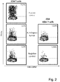
- CD3+CD4+ TIL cytokine production from GBM-D CD3+CD4+ TCRV ⁇ 2 T-cells (representing 33.3% TCRV ⁇ 2 T-cells in CD3+CD4 TIL) ( Fig. 2 ): CD3+CD4+ TIL exhibited TNF ⁇ production of 3.5% in response to autologous tumor cells, its TCRV ⁇ 2 T-cell subpopulation showed a higher frequency (13,6%.) of TNF ⁇ producing CD3+CD4+ cells. Testing of TIL reactivity in ICS also revealed selective IL-2 and IL-17 production of individual TIL lines against the autologous tumor cells.
- TCR V ⁇ families were analyzed by flow cytometry and PCR.
- TCR V ⁇ frequency staining was performed using the Beta Mark TCR V ⁇ Repertoire Kit (Beckman Coulter, CA, USA) along with co-staining with anti-CD3 PE-Cy7 (BD Biosciences, CA, USA), anti-CD4 Krome Orange (Beckman coulter, CA, USA) and anti-CD8a APC-Cy7 (BD Biosciences, CA, USA).
- a FACS Aria flow cytometer (BD Biosciences, Sweden) was used for acquisition and data analysis was performed by FlowJo software.
- TCR CDR3 analysis was performed using the TCR V ⁇ panel as described before (Magalhaes et al., 2008).
- TILs were sorted using CD4+ or CD8+ magnetic beads (MACS Milteny Biotec AB, Lund, Sweden) according to the supplier's instructions.
- RNA from CD4+ and CD8+ positive sorted cells was extracted using a RNeasy plus RNA extraction kit (Qiagen inc. Hilden, Germany) and converted to cDNA using a Oligodt protocol from revertaid premium first strand cDNA synthesis kit (Fermentas, ThermoFisher scientific, MA, USA).
- the multiplex PCR was performed using Qiagen multiplex PCR kit in a 30 ⁇ l reaction volume. Each reaction contained 15 ⁇ l of Qiagen multiplex PCR mastermix, 20pmol of the respective forward primer mix (primer mix 1 to 9), 20 pmol of 6FAM tagged TCRV ⁇ content reverse primer (Ahmed et al., 2014).
- a positive control reaction was performed for each sample using primers covering constant region of TCR V ⁇ gene specific primers tagged with VIC fluorochrome.
- the PCR was initiated with denaturation at 95 °C for 15 min followed by 35 cycles of 94 °C for 30 sec, 61 °C for 90 sec and 72 °C for 90 sec. The reaction was terminated at 72 °C for 10 min.
- the analysis was performed on ABI7500 capillary electrophoresis fragment analyzer (Applied Biosystems, CA, USA) using 400 bp internal control ladder for size reference.
- T-cells Samples exhibiting monoclonal expansion of T-cells were amplified with family specific primers and cloned into a TA (PCR2.1 topo) vector (Invitrogen, CA, USA) according to the suppliers instructions.
- the inserts in the plasmids were sequenced to identify the TCR CDR3 region using BigDye terminator cycle sequencing kit and ABI7500 capillary electrophoresis instrument (Applied Biosystems, CA, USA).
- the analyzed T-cell products showed up to 99% of individual V ⁇ families in TIL from individual patients (see Table 3), e.g. the TCRV ⁇ 2 which represented 99,6 % in CD3+CD4+ T-cells from the GBM-I TIL, the TCRV ⁇ .1 family constituted 97% of GBM-I CD3+CD8+ T-cells; TCRV ⁇ 21,3 constituted 98,4% of the GBM-J CD3+CD4+ T-cells.
- Some of the dominant TCR V ⁇ families were analyzed in greater detail and tested for the CDR3 length, followed by sequencing of the TCR, i.e. some of the expanded TCRV ⁇ (see Table 4) represent T-cell clones.
- Tumor tissues from clinical biopsies or surgery were cut with surgical scissors and a scalpel. Individual single tumor fragments (1-2 mm 3 ) were placed in each well of a 24-well tissue culture plate (Costar, USA) along with 1 ml of TIL medium , i.e.
- Cellgro GMP Serum-free medium (CellGenix, Freiburg, Germany), with 10% human AB serum (Innovative Research, Michigan, USA), supplemented with IL-2 1000IU/ml, IL-15 10ng/ml, IL-21 10ng/ml, (Prospec, Ness-Ziona, Israel), penicillin (100IU/mL), streptomycin (100 ⁇ g/mL) (Life Technologies, Carlsbad, USA) and Amphotericin B (2.5mg/L) (Sigma-Aldrich, MI; USA). Plates were then incubated at 37°C, 5% CO2. On day 4, 1 ml medium was added to wells. On day 7, TIL were split into 2-3 additional 24-wells. Upon 80% expansion of TIL in each culture well, 1 ml of TIL medium was added.
- TIL were further expanded on day 10 using OKT3 (Biolegend, CA, USA) in the presence of a 1:10 ratio of 55 Gry irradiated allogeneic feeder cells from healthy volunteers, resuspended in medium (Cellgro with 10% human AB serum).
- OKT3 was used at 30 ng/ml along with IL-2 1000IU/ml, IL-15 10ng/ml, and IL-21 10ng/ml.
- TIL were then transferred to 6-well plates and tested for anti-tumor (as autologous tumor cells were available) or TAA-reactivity.
- TIL medium Cellgro with 10% human AB serum, IL-2 1000IU/ml, IL-15 10ng/ml, and IL-21 10ng/m.
- irradiated feeder cells ratio of feeder cells: TIL was 1:10; OKT3 was used at 30 ng/ml.
- tumor tissues were cut with surgical scissors and a scalpel.
- Single tumor fragments (1-2 mm3) were placed in 24-well tissue culture plates with 1 ml of tumor medium, i.e. RPMI 1640 with 10% FBS (Life technologies, CA, USA), Epidermal growth factor (20ng/ml, ImmunoTools, Friesoythe, Germany) supplemented with antibiotics (penicillin,100IU/mL and streptomycin, 10mg/mL) (Life Technologies, Carlsbad, USA) and Amphotericin B (2.5mg/L, Sigma-Aldrich, MI; USA). Tumor cell lines were then cultured and passed without EGF.
- Example 5 Pancreas cancer: Exposure of TIL to autologous tumor cell co-cultures.
- the autologous tumor cell line established from Panc 17 was tested for TIL recognition (Table 5 ). IL-2 and IL-17 production tested positive in CD3+CD4+ TIL (1.55%). CD3+CD4-CD8- (DN) T-cells showed IL-2 (3.71%) and IL-17 (9.76%) production in response to autologous tumor cells. The same TIL line showed strong cytotoxicity directed against tumor cells, tested by a standard Cr51 release assay (see Figure 3A ).
- Panc 9 TIL (99.2% CD8+ V ⁇ 13.2, a monoclonal TCR see supplementary Table S2) exhibited IFN-y production against freshly harvested autologous tumor single cell suspension cells, i.e. 2022.81 pg/ml (see Figure 3B ), which could be blocked using the anti-MHC-I antibody (W6/32, 9.91 pg/ml); the anti-HLA-DR antibody (L243) did not affect IFN-y production (1853.45pg/ml).
- TCR V ⁇ frequency staining was performed using the Beta Mark TCR V ⁇ Repertoire Kit (Beckman Coulter, France) along with CD3 PE-Cy7 (BD Biosciences, CA, USA), CD4 Krome orange (Beckman Coulter, France), and CD8 APC-Cy7(BD Biosciences, CA, USA). TIL were washed using PBS-0.1 %FBS and stained using a FACS Aria flow cytometer (BD Biosciences, Sweden).
- TCR CDR3 analysis was performed using the TCR V ⁇ panel as described before [29].
- the TCR V ⁇ CDR3 analysis was performed using the TCR V ⁇ family specific primer set panel previously described [30].
- TILs were sorted using CD4+ or CD8+ magnetic beads (MACS Miltenyibiotec AB, Lund, Sweden) according to the supplier's instructions.
- Total RNA from CD4 and CD8 positive sorted cells was extracted using a RNeasy plus RNA extraction kit (Qiagen inc. Hilden, Germany) and converted to cDNA using a Oligodt protocol from the revertaid premium first strand cDNA synthesis kit (Fermentas, ThermoFisher scientific, MA, USA).
- the multiplex PCR was performed using Qiagen multiplex PCR kit in a 30 ⁇ I reaction volume. Each reaction contained 15 ⁇ I of the Qiagen multiplex PCR mastermix, 20pmol of the respective forward primer mix (primer mix 1 to 9) and 20pmol of 6FAM tagged TCRV ⁇ content reverse primer. A positive control reaction was performed for each sample using primers covering constant region of TCR V ⁇ gene specific primers tagged with VIC fluorochrome. The PCR was initiated with denaturation at 95°C for 15 min followed by 35 cycles of 94°C for 30 seconds, 61°C for 1 minute 30 seconds and 72°C for 1 minute 30 seconds. The reaction was terminated at 72°C for 10 minutes.
- Panc 9 (CD8+) TIL exhibited up to 99.2% V ⁇ 13.2 + T-cells.
- the preferential expansion of TCR V ⁇ families was also observed in other TIL lines (see Table 5), e.g. CD8 + V ⁇ 1 (77.1 %) from Panc1.
- CD4 + V ⁇ 8 (42.7 %) in Panc5 CD8 + V ⁇ 2 (39.8 %) in Panc10
- CD4 + V ⁇ 2 48.9 %) in Panc16.
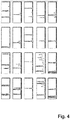
- Some of the expanded V ⁇ families showed clonal TCRs tested by CDR3 TCR length analysis, followed by TCR sequencing (see Tables 6 & 7 and Fig. 4 ).
- Table 6 V ⁇ analysis and clonality in TILs after expansion Patient Cell population Family by per V ⁇ by cytometry Flow Number of clones Panc 1 CD8 TRBV9 V ⁇ 1 2 Panc 5 CD4 TRBV12 V ⁇ 8 1 CD4 TRBV5,1 V ⁇ 5,1 1 Panc 7 CD8 TRBV19 V ⁇ 17 2 TRBV28 V ⁇ 3 2 Panc 8 CD8 TRBV14 V ⁇ 16 1 Panc 9 CD8 TRBV6 V ⁇ 13,2 1 Panc 10 CD8 TRBV20,1 V ⁇ 2 2 TRBV20 V ⁇ 2 1 Panc 11 CD8 TRBV7,3/6/7/9 No antibody 1 TRBV19 V ⁇ 17 1 Panc 13 CD4 TRBV19 V ⁇ 17 1 Panc 14 CD8 TRBV9 V ⁇ 1 2 Panc 15 CD4 TRBV20,1 V ⁇ 2 2 CD8 TRBV6,2 V ⁇ 13,2 2 CD4 TRBV20,1 V ⁇ 2 1 Panc 16 TRBV7,3 No antibody 2 CD8 TRBV7,2,8 No antibody 2 TRBV19 V ⁇ 17 1 Table 7: C
- Example 7 Recognition of known TAAs by the individual T-cell products from pancreas cancer patients
- TIL were exposed to commonly shared antigens for three days, in the presence of the blocking MHC class I (W6/32) or HLA-DR (L243) mAb, followed by IFN ⁇ production analysis in the supernatant. Strong reactivity to Mesothelin that could be blocked with mAbs. Note that Panc 10 is predominantly a CD4+ TIL that could be blocked with the anti-HLA-DR mAb and not with the MHC class I -directed reagent. We have also included an isotype control (anti-C1q, 242G3 mAb) that showed similar results as the antigen stimulation without the blocking mAb. Results are means of triplicates. Increased reactivity upon L243 (e.g.
- Panc16 may reflect T-cell stimulation of crosslinking HLA-DR molecules on activated T-cells. Only positive results are reported.
- Table 8 TAA IFN ⁇ production (pg/mL) in TIL in response to commonly shared TAAs NY-ESO1 NY-ESO1 +W6/32 NY-ESO1 +L243 Survivin Survivin +W6/3 2 Survivin +L243 Mesothelin Mesothelin +W6/32 Mesothelin +L243 Panc 1 6,97 0 0 0 0 262,23 15,26 0 Panc 2 24,20 0 0 0 0 0 291,27 13,50 0 Panc 10 4,13 0 0 66,06 0 0 78,70 72,88 0 Panc 11 0 0 0 0 0 0 131,16 0 0 Panc 14 10,47 0 0 0 0 0 275,15 nd 249,95 Panc 16 173,19 0 0 8,35 0
Abstract
The present invention provides a protein, in particular a TCR comprising a Vβ amino acid sequence with a sequence identity of at 90 %, to a sequence selected from SEQ ID NO: 1 to 27, a T-cell comprising the TCR a method of selecting a T-cell product for use in active immunotherapy based on the identification of an Vβ amino acid sequence with a sequence identity of at 90 %, to a sequence selected from SEQ ID NO: 1 to 27 in a T-cell product.
Description
- The present invention relates to proteins comprising a Vbeta sequence with an amino acid sequence with a sequence identity of at 90 %, to a sequence for use in immunotherapy.
- One of the most promising advances for the treatment of cancer is a new therapeutic class called active cellular immunotherapy (ACI). Active immunotherapies stimulate the patient's immune system with the intent of promoting an antigen specific anti-tumor effect using the body's own immune cells. In addition active immunotherapies seek to create durable anti-tumor response that can protect against minimal residual disease and tumor recurrences as well as against potentially pre-malignant tumor lesions.
- Clinically relevant and long-term remissions have been achieved in patients with melanoma using T-cells directed against tumors (tumor reactive T-cells) (1, 2). These approaches usually rely on the harvesting of tumor infiltrating lymphocytes (TIL) from tumor lesions or T-cells from peripheral blood.
- Clinical (antitumor) efficacy appears to be mediated by CD8+, or by CD4+ T-cells residing preferentially in central memory cells, defined by CD45RA-CCR7+, defined ex vivo from patients responding to T-cell based therapy. The phenotype of such T-cells is determined by the ex vivo expanded T-cell population, as well as by host factors after adoptive transfer. A diverse population of T-cells targeting cancer cells may be advantageous for effective immune responses, including long-term memory T-cells, yet also T-cells that can immediately react to (cancer) target cells and produce anti-tumor directed immune responses, including terminally differentiated T-cells that express cytolytic molecules, such as granzyme and perforin (3, 4). Long term-memory immune memory is in part determined by the increased proliferation potential and half-life that can be measured by the telomere length (5, 6).
- In
WO 2015/189357 A1 the inventors have described a novel cytokine cocktail containing IL-2, IL-15 and IL-21 for the expansion of lymphocytes, in particular T-cells. T-cell populations obtained by expansion in the presence of the cytokine cocktail are able to not only recognize autologous tumor calls but also kill such tumor cells in vitro. -
WO 2015/189357 A1 describes a variety of T-cell products obtained from an expansion of T-cells from the tumor, i.e. tumor infiltrating lymphocytes (TILs) or peripheral blood of patients with pancreatic cancer or glioblastoma. With the expansion protocol using the cytokine cocktail containing IL-2, IL-15 and IL-21 it is possible to produce several T-cell products in parallel. These T-cell products in general show a phenotype distribution advantageous for the active immune therapy. - However, besides the phenotype distribution and the in vitro testing of the T-cell product the clinician at present does not have further determinants for the selection for a T-cell product for insertion into the patient.
- The present invention is inter alia based on the identification TCR sequences that are responsible for recognition and killing of diseased tissue cells. TCRs with the respective TCR sequences target tumor associated antigens and T-cells comprising the TCRs are therefore valuable therapeutic tools and a detection of the TCR sequences can be used for evaluation of T-cell products, i.e. an in vitro expanded T-cell population for therapeutic use.
- Thus, according to a first aspect the invention provides a protein comprising a Vβ amino acid sequence with a sequence identity of at 90 % to a sequence selected from SEQ ID NO: 1 to 27.
- According to a second aspect the invention provides a T-cell comprising a TCR which is a protein according to the first aspect. T-cell according to the second aspect may in particular be used in the treatment of cancer.
- Furthermore according to a third aspect the invention provides a method of selecting a T-cell product for use in active immunotherapy in a patient, wherein the method comprises the following steps:
- a) providing one or more T-cell products;
- b) identifying the presence of a TCR comprising a sequence with a homology of at least 95 % to the sequence selected from SEQ ID NO: 1 to 27 in the one or more T-cell products; and
- c) selecting the T-cell product comprising a TCR identified in step b) for use in therapy.
-
- Figure 1
- shows the frequency of IFNγ and TNFα producing TIL in response to autologous tumor cells. Numbers represent the percentage of T-cells specifically producing IFNγ (black circles) and TNFα (open circles) in the parental CD8+, CD4+, or (CD3+), CD4-CD8- TIL subsets. Each circle represent TIL from an individual patient. Medium values have been subtracted. Right panel: example flow cytometric analysis of CD4+ and CD8+ TIL producing cytokines directed against autologous tumor cells.
- Figure 2
- shows the link of expanded Vβ families in TIL with the recognition of autologous tumor cells, in particular the frequency of TNFalpha producing T-cells in CD3+CD4+ T-cells. Top left: positive control. Middle panel. Exposure to autologous tumor cells. TIL gated on CD3+CD4+ T-cells: 3.5% cells produce TNFalpha; right: 13.6% TNFα producing TIL in DNCD3+CD4+ Vβ2+ T- cells from patient GBM-D. Bottom panel. Left: negative control in CD3+CD4+ TIL. Right: CD3+CD4+Vβ2+ TIL.
- Figure 3
- shows (A) the IFNγ production in TILs after co-culture with autologous tumor cells (Panc 9). CD8+ TILs (Vβ13.2 dominant) recognize the autologous tumor and IFNγ production can be inhibited by the anti-MHC-I antibody (W6/32), yet no with the anti-HLA-DR directed mAb. (B) The Recognition of the autologous tumor cell line in Panc 17 TIL using a standard Chromium 51 release assay.
- Figure 4
- shows the results of a TCR CDR3 analysis of frequent TCR Vβ families after a 4 week expansion using IL-2, IL-15 and IL-21.
- "Disease associated antigens" according to the invention are antigens involved in a disease. Accordingly, clinically relevant antigens can be tumor-associated antigens TAA.
- According to the invention an "antigen" (Ag) is any structural substance which serves as a target for the receptors of an adaptive immune response, TCR or antibody, respectively. Antigens are in particular proteins, polysaccharides, lipids and substructures thereof such as peptides. Lipids and nucleic acids are in particular antigenic when combined with proteins or polysaccharides.
- "Tumor associated antigens" or "TAA" according to the invention are antigens that are presented by MHC I or MHC II molecules or non-classical MHC molecules on the surface of tumor cells. As used herein TAA includes "tumor-specific antigens" which are found only on the surface of tumor cells, but not on the surface of normal cells.
- As used herein, "interleukin 2" or "IL-2" refers to human IL-2 as defined by SEQ ID NO: 1 and functional equivalents thereof. Functional equivalents of IL-2 include relevant substructures or fusion proteins of IL-2 that remain the functions of IL-2. Accordingly, the definition IL-2 comprises any protein with a sequence identity to SEQ ID NO: 1 of at least 80 %, preferably at least 90 %, more preferably at least 95 %, most preferably at least 98 %. Recombinant human IL-2 produced in E. coli as a single, non-glycosylated polypeptide chain with 134 amino acids and having a molecular mass of 15 kDa is commercially available in lyophilized form from Prospec as CYT-209.
- As used herein, "
interleukin 15" or "IL-15" refer to human IL-15 and functional equivalents thereof. Functional equivalents of IL-15 include relevant substructures or fusion proteins of IL-15 that remain the functions of IL-15. Accordingly the definition IL-15 comprises any protein with a sequence identity to SEQ ID NO: 2 of at least 80 %, preferably at least 90 %, more preferably at least 95 %, most preferably at least 98 %. Recombinant human IL-15 produced in E. coli as a single, non-glycosylated polypeptide chain with 114 amino acids (and an N-terminal Methionine) and having a molecular mass of 12.8 kDa is commercially available in lyophilized form from Prospec as CYT-230. - As used herein, "interleukin 21" or "IL-21" refer to human IL-21 and functional equivalents thereof. Functional equivalents of IL-21 included relevant substructures or fusion proteins of IL-21 that remain the functions of IL-21. Accordingly the definition IL-21 comprises any protein with a sequence identity to SEQ ID NO: 3 of at least 80 %, preferably at least 90 %, more preferably at least 95 %, most preferably at least 98 %. Recombinant human IL-21 produced in E. coli as a single, non-glycosylated polypeptide chain with 132 amino acids and having a molecular mass of 15 kDa is commercially available in lyophilized form from Prospec as CYT-408.
- A "peptide" as used herein may be composed of any number of amino acids of any type, preferably naturally occurring amino acids, which, preferably, are linked by peptide bonds. In particular, a peptide comprises at least 3 amino acids, preferably at least 5, at least 7, at least 9, at least 12, or at least 15 amino acids. Furthermore, there is no upper limit for the length of a peptide. However, preferably, a peptide according to the invention does not exceed a length of 500 amino acids, more preferably it does not exceed a length of 300 amino acids; even more preferably it is not longer than 250 amino acids.
- Thus, the term "peptide" includes "oligopeptides", which usually refer to peptides with a length of 2 to 10 amino acids, and "polypeptides" which usually refer to peptides with a length of more than 10 amino acids.
- The term "protein" refers to a peptide with at least 60, at least 80, preferably at least 100 amino acids.
- The term "fusion protein" according to the invention relates to proteins created through the joining of two or more genes, cDNAs or sequences that originally coded for separate proteins/peptides. The genes may be naturally occurring in the same organism or different organisms or may synthetic polynucleotides.
- The relatedness between two amino acid sequences or between two nucleotide sequences is described by the parameter "sequence identity". For purposes of the present invention, the degree of sequence identity between two amino acid sequences is determined using the Needleman-Wunsch algorithm (Needleman and Wunsch, 1970, J. Mol. Biol. 48: 443-453) as implemented in the Needle program of the EMBOSS package (EMBOSS: The European Molecular Biology Open Software Suite, Rice et a/., 2000, Trends Genet. 16: 276-277), preferably version 3.0.0 or later. The optional parameters used are gap open penalty of 10, gap extension penalty of 0.5, and the EBLOSUM62 (EMBOSS version of BLOSUM62) substitution matrix. The output of Needle labeled "longest identity" (obtained using thenobrief option) is used as the percent identity and is calculated as follows:

- The transitional term "comprising", which is synonymous with "including," "containing," or "characterized by," is inclusive or open-ended and does not exclude additional, unrecited elements or method steps. The transitional phrase "consisting of" excludes any element, step, or ingredient not specified in the claim, except for impurities ordinarily associated therewith. When the phrase "consists of" appears in a clause of the body of a claim, rather than immediately following the preamble, it limits only the element set forth in that clause; other elements are not excluded from the claim as a whole. The transitional phrase "consisting essentially of" limits the scope of a claim to the specified materials or steps "and those that do not materially affect the basic and novel characteristic(s)" of the claimed invention. "A 'consisting essentially of' claim occupies a middle ground between closed claims that are written in a 'consisting of' format and fully open claims that are drafted in a 'comprising' format."
- "Expansion" or "clonal expansion" as used herein means production of daughter cells all arising originally from a single cell. In a clonal expansion of lymphocytes, all progeny share the same antigen specificity.
- A "T-cell product" as used herein refers to a population of T-cell for use in immune therapy.
- "TIL" according to the invention refers to tumor infiltrating lymphocyte. These are lymphocytes, in particular T-cells predominantly found in a tumor.
- "Clinical/biological relevance" as used herein relates to the ability of a T-cell to provide at least one of the following: containment of tumor cells, destruction of tumor cells, prevention of metastasis, stop of proliferation, stop of cellular activity, stop of progress of cells to malignant transformation, prevention of metastases and / or tumor relapse, including reprogramming of malignant cells to their non-malignant state; prevention and / or stop of negative clinical factors associated with cancer, such as malnourishment or immune suppression, stop of accumulation of mutations leading to immune escape and disease progression, including epigenetic changes, induction of long-term immune memory preventing spread of the disease or future malignant transformation affecting the target (potential tumor cells), including connective tissue and non-transformed cells that would favor tumor disease.
- According to a first aspect the invention provides a protein comprising a Vβ amino acid sequence with a sequence identity of at 90 %, to a sequence selected from SEQ ID NO: 1, SEQ ID NO: 2, SEQ ID NO: 3, SEQ ID NO: 4, SEQ ID NO: 5, SEQ ID NO: 6, SEQ ID NO: 7, SEQ ID NO: 8, SEQ ID NO: 9, SEQ ID NO: 10, SEQ ID NO: 11, SEQ ID NO: 12, SEQ ID NO: 13, SEQ ID NO: 14, SEQ ID NO: 15, SEQ ID NO: 16, SEQ ID NO: 17, SEQ ID NO: 18, SEQ ID NO: 19, SEQ ID NO: 20, SEQ ID NO: 21, SEQ ID NO: 22, SEQ ID NO: 23, SEQ ID NO: 24, and SEQ ID NO: 26. The sequences SEQ ID NO: 1 to 26 were identified in T-cell products obtained by expansion of T-cells and shown to be responsible for recognition and killing of autologous tumor cells. The sequences are shown in Tables 1 and 2.
Table 1: Vβ sequences found in TIL expanded from Glioblastoma GBM A-CD4 (SEQ ID NO: 1) 
GBM A-CD8 A (SEQ ID NO: 2) 
GBM A-CD8 B (SEQ ID NO: 3) 
GBM F-CD4 (SEQ ID NO: 4) 
GBM G-CD4 A (SEQ ID NO: 5) 
GBM G-CD4 B (SEQ ID NO: 6) 
GBM G CD4 C (SEQ ID NO: 7) 
GBM H-CD8 A (SEQ ID NO: 8) 
GBM H-CD8 B (SEQ ID NO: 9) 
GBM H-CD8 C (SEQ ID NO: 10) 
GBM I-CD4 (SEQ ID NO: 11) 
GBM I-CD8 (SEQ ID NO: 12) 
GBM J-CD4 (SEQ ID NO: 13) 
Table 2: Vβ sequences found in TIL expanded from Pancreas Cancer Panc1 - CD8 (SEQ ID NO: 14) 
Panc5 - CD4 A (SEQ ID NO: 15) 
Panc5 - CD4 B (SEQ ID NO:16) 
Panc7 - CD4 B (SEQ ID NO: 17) 
Panc7 - CD8 B (SEQ ID NO: 18) 
Panc 8 - CD8 (SEQ ID NO: 19) 
Panc 9 - CD8 (SEQ ID NO:20) 
Panc 16 - CD4 A (SEQ ID NO: 21) 
Panc 16 - CD4 B (SEQ ID NO: 22) 
Panc 16 - CD8 A (SEQ ID NO:23) 
Panc 16 - CD8 B (SEQ ID NO:24) 
Panc 17 - CD8 A (SEQ ID NO: 25) 
Panc 17 - CD8 B (SEQ ID NO:26) 
- According to a preferred embodiment the amino acid sequence of the protein according to the invention has sequence identity is at least 95 % to any one of SEQ ID NO: 1, SEQ ID NO: 2, SEQ ID NO: 3, SEQ ID NO: 4, SEQ ID NO: 5, SEQ ID NO: 6, SEQ ID NO: 7, SEQ ID NO: 8, SEQ ID NO: 9, SEQ ID NO: 10, SEQ ID NO: 11, SEQ ID NO: 12, SEQ ID NO: 13, SEQ ID NO: 14, SEQ ID NO: 15, SEQ ID NO: 16, SEQ ID NO: 17, SEQ ID NO: 18, SEQ ID NO: 19, SEQ ID NO: 20, SEQ ID NO: 21, SEQ ID NO: 22, SEQ ID NO: 23, SEQ ID NO: 24, and SEQ ID NO: 26. More preferably the sequence identity is at least 98 % According to a preferred embodiment the sequence identity to any one of SEQ ID NO: 1, SEQ ID NO: 2, SEQ ID NO: 3, SEQ ID NO: 4, SEQ ID NO: 5, SEQ ID NO: 6, SEQ ID NO: 7, SEQ ID NO: 8, SEQ ID NO: 9, SEQ ID NO: 10, SEQ ID NO: 11, SEQ ID NO: 12, SEQ ID NO: 13, SEQ ID NO: 14, SEQ ID NO: 15, SEQ ID NO: 16, SEQ ID NO: 17, SEQ ID NO: 18, SEQ ID NO: 19, SEQ ID NO: 20, SEQ ID NO: 21, SEQ ID NO: 22, SEQ ID NO: 23, SEQ ID NO: 24, and SEQ ID NO: 26 is 100 %.
- The protein may be a T-cell receptor (TCR) in a T-cell obtained by expansion of autologous T-cells. Alternatively the protein may be a recombinant protein. The methods for producing, i.e. constructing and expressing recombinant proteins are well known in the art (see e.g. (7) and (8). The recombinant protein may for example designed for introduction into a cell. The cell may be a T-cell., an NK-cell, NK-T-cell or any innate immune cell. Innate immune cells include innate lymphoid cells, MAIT cells or immune memory of effector cells suitable for long-term and effective anti-tumor reactivity. The recombinant protein may be a transgenic TCR, such as the "anti.NY-ESO-1 TCR" (see e.g. (9)).. Such a transgenic TCR may be virally or non-virally transferred into recipient cells. Protocols for the viral and non-viral transfer are known in the art. Examples for the transfer are described in (10), (11) and (12).
- According to a second aspect the invention provides a T-cell comprising a TCR of to the first aspect. The T-cell comprising the TCR according to the first aspect is in particular suitable for treatment of cancer. The cancer preferably selected from acute lymphocytic cancer, acute myeloid leukemia, alveolar rhabdomyosarcoma, bone cancer, brain cancer, breast cancer, cancer of the anus, anal canal, or anorectum, cancer of the eye, cancer of the intrahepatic bile duct, cancer of the joints, cancer of the neck, gallbladder, or pleura, cancer of the nose, nasal cavity, or middle ear, cancer of the vulva, chronic lymphocytic leukemia, chronic myeloid cancer, cervical cancer, glioma, Hodgkin lymphoma, hypopharynx cancer, kidney cancer, larynx cancer, liver cancer, lung cancer, malignant mesothelioma, melanoma, multiple myeloma, nasopharynx cancer, non-Hodgkin lymphoma, ovarian cancer, peritoneum, omentum, mesentery cancer, pancreatic cancer, pharynx cancer, prostate cancer, rectal cancer, renal cancer, skin cancer, soft tissue cancer, testicular cancer, thyroid cancer, ureter cancer, urinary bladder cancer, and digestive tract cancer such as, e.g., esophageal cancer, gastric cancer, pancreatic cancer, stomach cancer, small intestine cancer, gastrointestinal carcinoid tumor, cancer of the oral cavity, colon cancer, and hepatobiliary cancer. The T-cell may preferably be used in the treatment of glioblastoma or pancreatic cancer.
- As shown in the examples T-cell products comprising a TCR with a sequence selected from any of SEQ ID NO: 1 to 27 are able to recognize and kill tumor cells in vitro. Therefore, identification of the TCR in a T-cell product drastically improves the chance that the T-cell product is able to fight the cancer in vivo, in particular that the T-cell product is clinically/biologically relevant.
- Thus, according to a third aspect the invention provides a method of selecting a T-cell product for use in active immunotherapy in a patient, wherein the method comprises the following steps:
- a) providing at one or more T-cell products;
- b) identifying the presence of a according to the first aspect in the one or more T-cell products; and
- c) selecting the T-cell product comprising a TCR identified in step b) for use in therapy.
- The TCR According to one embodiment the method further comprises determining the clonality of the Vβ-family to which the TCR belongs and selecting the T-cell product in which the TCR is present and the Vβ family to which the TCR belongs is monoclonal. Monoclonality of the TCR indicated a directed and specific expansion of the TCR.
- According to one embodiment the method further comprises determining the percentage of the TCR and/or the Vβ family to which the TCR belongs in the T-cell product and selecting the T-cell product in which the percentage of the TCR is the highest. In order provide clinical/biological relevance to the T-cell product the TCR should be present in a sufficient concentration.
- The method according to third aspect may be used in particular in the treatment of cancer. Thus the method is preferably carried out with a patient suffering from cancer. The cancer may be for example one of the cancers listed above. Preferably the cancer is selected from glioblastoma and pancreas cancer.
- According to one embodiment of the third aspect providing the one or more T-cell products is based on an expansion of T-cells according to the invention. More preferably the method comprises
- providing a body sample containing T-cells from the patient;
- culturing cells of the sample in-vitro in the presence of at least one cytokine,
- The body sample can be taken from any part of the body that contains lymphocytes. Examples of body samples are peripheral blood, cord blood, bone marrow, lymph nodes, liver pleural effusion, thorax, abdominal cavity, synvial fluid, peritoneum, retroperitoneal space, thymus, and tumor. A lymphocyte sample derived from tumor is also referred as tumor infiltrating lymphocytes (TIL). "TIL" as used herein is short for "tumor infiltrating lymphocytes". TIL is any kind of lymphocyte that is located in, on or around a tumor.
- Preferably the culturing for expansion of the T-cells occurs in the presence of IL-2, IL-15 and IL-21. According to a further embodiment, the concentration of IL-2 in the liquid composition is in the range of from 10 to 6000 U/ml. The International Unit (U) is the standard measure for an amount or IL-2. It is determined by its ability to induce the proliferation of CTLL-2 cells. The concentration of IL-2 is preferably in the range from 500 to 2000 U/ml. More preferably the concentration of IL-2 is in the range from 800 to 1100 U/ml. According to one embodiment the concentration of IL-15 is in the range of 0.1 to 100 ng/ml. Preferably, the concentration of IL-15 is in the range from 2 to 50 ng/ml, more preferably in the range from 5 to 20 ng/ml. The most preferred concentration is about 10 ng/ml. In a further embodiment the concentration of IL-21 is in the range from 0.1 ng/ml, preferably in the range from 2 to 50 ng/ml, more preferably in the range from 5 to 20 ng/ml.
- According to one embodiment of the invention the culture medium of the first expansion step comprises at least one expansion antigen. The expansion antigen is a known disease associated antigen or a fragment, a mutant or a variant thereof. As used herein a "mutant" is defined as an amino acid sequence that differs from the reference sequence by an insertion, deletion or replacement of at least one amino acid. The expansion antigen is a TAA.
- According to a preferred embodiment of the method according to the second aspect, the body sample is tumor and no expansion antigen is used in culturing.
- The invention is further defined by the following examples.
- 16 patients (Supplementary Table 1) with gliomas were enrolled. The study was approved by the regional ethical review board at Karolinska Institutet, Stockholm, Sweden (Dnr: 2013/576-31).
- Glioma tumor tissue was harvested in the course of tumor surgery and dissected into fragments (approximately 1-2mm3) using a sterile scalpel. The fragments were washed 2 times with PBS and cultured in 24 well plates in GMP Serum-free DC medium (CellGenix, Freiburg, Germany) plus 5% pooled human AB serum (Innovative Research, Michigan, USA) supplemented with recombinant IL-2 (1000 IU/ml), IL-15 (10 ng/ml), IL-21 (10 ng/ml) (Prospec, Ness-Ziona, Israel). The culture medium was changed when necessary. TILs were transferred into 6 well plates; as they covered >70% of the 24 well surface, they were further expanded in G-Rex flasks (Wilson Wolf, New Brighton, USA) using 30ng OKT3/mL and irradiated allogeneic feeder cells at the ratio of 1 feeder cell to 10 TILs.
- Tumor single cell suspension were processed using a Medimachine (BD, California, USA Cat:340588). The cell suspension was washed 2 times in PBS and then cultured in T25 flasks (Thermo Fisher Scientific Inc., Waltham, MA USA) using RPMI 1640 L-glutamine 2mM (Life Technologies, CA, USA) supplemented with antibiotics plus 20% Fetal Bovine Serum (Gibco, REF: 26140-079).
- It was tested whether the expanded TIL recognized the autologous tumor cell line defined by intracellular cytokine production. For this, expanded TILs were exposed to autologous tumor cells in 96 well plates. Cells were seeded at 20,000 TILs/well at an E:T ratio of 10:1. Supernatants were harvested at day 3 and tested for IFNγ by Elisa (MABTECH, Stockholm, Sweden).
- A IFNγ and TNFα production against the autologous tumor cells in expanded TIL from individual patients up to 7.87% in CD3+CD4+ and up to 48.70% in CD3+CD8+ TIL was observed (see
Fig. 1 ) - The cytokine production from GBM-D CD3+CD4+ TCRVβ2 T-cells (representing 33.3% TCRVβ2 T-cells in CD3+CD4 TIL) (
Fig. 2 ): CD3+CD4+ TIL exhibited TNFα production of 3.5% in response to autologous tumor cells, its TCRVβ2 T-cell subpopulation showed a higher frequency (13,6%.) of TNFα producing CD3+CD4+ cells. Testing of TIL reactivity in ICS also revealed selective IL-2 and IL-17 production of individual TIL lines against the autologous tumor cells. - TCR Vβ families were analyzed by flow cytometry and PCR.
- TCR Vβ frequency staining was performed using the Beta Mark TCR Vβ Repertoire Kit (Beckman Coulter, CA, USA) along with co-staining with anti-CD3 PE-Cy7 (BD Biosciences, CA, USA), anti-CD4 Krome Orange (Beckman coulter, CA, USA) and anti-CD8a APC-Cy7 (BD Biosciences, CA, USA). After washing, a FACS Aria flow cytometer (BD Biosciences, Stockholm, Sweden) was used for acquisition and data analysis was performed by FlowJo software. TCR CDR3 analysis was performed using the TCR Vβ panel as described before (Magalhaes et al., 2008). In brief, TILs were sorted using CD4+ or CD8+ magnetic beads (MACS Milteny Biotec AB, Lund, Sweden) according to the supplier's instructions.
- For the PCR analysis, total RNA from CD4+ and CD8+ positive sorted cells was extracted using a RNeasy plus RNA extraction kit (Qiagen inc. Hilden, Germany) and converted to cDNA using a Oligodt protocol from revertaid premium first strand cDNA synthesis kit (Fermentas, ThermoFisher scientific, MA, USA). The multiplex PCR was performed using Qiagen multiplex PCR kit in a 30 µl reaction volume. Each reaction contained 15µl of Qiagen multiplex PCR mastermix, 20pmol of the respective forward primer mix (
primer mix 1 to 9), 20 pmol of 6FAM tagged TCRVβ content reverse primer (Ahmed et al., 2014). - A positive control reaction was performed for each sample using primers covering constant region of TCR Vβ gene specific primers tagged with VIC fluorochrome. The PCR was initiated with denaturation at 95 °C for 15 min followed by 35 cycles of 94 °C for 30 sec, 61 °C for 90 sec and 72 °C for 90 sec. The reaction was terminated at 72 °C for 10 min. The analysis was performed on ABI7500 capillary electrophoresis fragment analyzer (Applied Biosystems, CA, USA) using 400 bp internal control ladder for size reference.
- Samples exhibiting monoclonal expansion of T-cells were amplified with family specific primers and cloned into a TA (PCR2.1 topo) vector (Invitrogen, CA, USA) according to the suppliers instructions. The inserts in the plasmids were sequenced to identify the TCR CDR3 region using BigDye terminator cycle sequencing kit and ABI7500 capillary electrophoresis instrument (Applied Biosystems, CA, USA).
- The analyzed T-cell products showed up to 99% of individual Vβ families in TIL from individual patients (see Table 3), e.g. the TCRVβ2 which represented 99,6 % in CD3+CD4+ T-cells from the GBM-I TIL, the TCRVβ.1 family constituted 97% of GBM-I CD3+CD8+ T-cells; TCRVβ21,3 constituted 98,4% of the GBM-J CD3+CD4+ T-cells. Some of the dominant TCR Vβ families were analyzed in greater detail and tested for the CDR3 length, followed by sequencing of the TCR, i.e. some of the expanded TCRVβ (see Table 4) represent T-cell clones.


Table 4: Vβ amino acid sequences TIL id Vβ Vβ (-D-) Jβ Jβ Flow antibody GBM A-CD4 TRBV20-1 CSA ATGDRP YEQYF TRBJ2-7 Vβ2 GBM A-CD8 TRBV10-3 CAI RTGSD NEQSF TRBJ2-1 Vβ12 GBM A-CD8 TRV11 CAS RYTGS IEQFF TRBJ2-1 Vβ21.3 GBM F-CD4 TRBV27 CAS SAGTSGVT YEQYF TRBJ2-7 Vβ14 GBM G-CD4 TRBV6-1/5/6 CAS STR FEQYF TRBJ2-7 Vβ13.1 GBM H-CD8 TRBV12-3/4 CAS SSRRDHTY NGQFF TRBJ2-1 Vβ8 GBM H-CD8 TRV27 CAS SLQGAN YGYTF TRBJ1-2 Vβ14 GBM I-CD4 TRBV20-1 CSA RVIPSGGV VQGT DTQYF TRBJ2-3 Vβ2 GBM I-CD8 TRBV5-1 CAS SWDKS YEQYF TRBJ2-7 Vβ5.1 GBM J-CD4 TRBV11 CAS SRLALFS YEQYF TRBJ2-7 Vβ21.3 - 17 patients (Table 1) with pancreatic cancer (13/17 with a ductal adenocarcinoma), recruited at the Pancreatic Surgery Unit of the Karolinska Hospital, Huddinge, Sweden, provided informed consent. The study was approved by the regional ethical review board at Karolinska Institutet, Stockholm, Sweden (Dnr: 2013/576-31).
- Tumor tissues from clinical biopsies or surgery were cut with surgical scissors and a scalpel. Individual single tumor fragments (1-2 mm3) were placed in each well of a 24-well tissue culture plate (Costar, USA) along with 1 ml of TIL medium , i.e. Cellgro GMP Serum-free medium (CellGenix, Freiburg, Germany), with 10% human AB serum (Innovative Research, Michigan, USA), supplemented with IL-2 1000IU/ml, IL-15 10ng/ml, IL-21 10ng/ml, (Prospec, Ness-Ziona, Israel), penicillin (100IU/mL), streptomycin (100·g/mL) (Life Technologies, Carlsbad, USA) and Amphotericin B (2.5mg/L) (Sigma-Aldrich, MI; USA). Plates were then incubated at 37°C, 5% CO2. On
day 4, 1 ml medium was added to wells. On day 7, TIL were split into 2-3 additional 24-wells. Upon 80% expansion of TIL in each culture well, 1 ml of TIL medium was added. - TIL were further expanded on day 10 using OKT3 (Biolegend, CA, USA) in the presence of a 1:10 ratio of 55 Gry irradiated allogeneic feeder cells from healthy volunteers, resuspended in medium (Cellgro with 10% human AB serum). OKT3 was used at 30 ng/ml along with IL-2 1000IU/ml, IL-15 10ng/ml, and IL-21 10ng/ml. TIL were then transferred to 6-well plates and tested for anti-tumor (as autologous tumor cells were available) or TAA-reactivity. If tested positive, defined by IFN-production, they were expanded in GRex flasks (Wilson Wolf, New Brighton, MN, USA) using TIL medium (Cellgro with 10% human AB serum, IL-2 1000IU/ml, IL-15 10ng/ml, and IL-21 10ng/m.) plus irradiated (55Gy) feeder cells (ratio of feeder cells: TIL was 1:10); OKT3 was used at 30 ng/ml.
- After surgery or biopsy, tumor tissues were cut with surgical scissors and a scalpel. Single tumor fragments (1-2 mm3) were placed in 24-well tissue culture plates with 1 ml of tumor medium, i.e. RPMI 1640 with 10% FBS (Life technologies, CA, USA), Epidermal growth factor (20ng/ml, ImmunoTools, Friesoythe, Germany) supplemented with antibiotics (penicillin,100IU/mL and streptomycin, 10mg/mL) (Life Technologies, Carlsbad, USA) and Amphotericin B (2.5mg/L, Sigma-Aldrich, MI; USA). Tumor cell lines were then cultured and passed without EGF.
- The autologous tumor cell line established from
Panc 17 was tested for TIL recognition (Table 5 ). IL-2 and IL-17 production tested positive in CD3+CD4+ TIL (1.55%). CD3+CD4-CD8- (DN) T-cells showed IL-2 (3.71%) and IL-17 (9.76%) production in response to autologous tumor cells. The same TIL line showed strong cytotoxicity directed against tumor cells, tested by a standard Cr51 release assay (seeFigure 3A ). - The Panc 9 TIL (99.2% CD8+ Vβ13.2, a monoclonal TCR see supplementary Table S2) exhibited IFN-y production against freshly harvested autologous tumor single cell suspension cells, i.e. 2022.81 pg/ml (see
Figure 3B ), which could be blocked using the anti-MHC-I antibody (W6/32, 9.91 pg/ml); the anti-HLA-DR antibody (L243) did not affect IFN-y production (1853.45pg/ml). - We analyzed the TCR Vβ families in TIL with a preferential expansion of individual Vβ families.
- TCR Vβ frequency staining was performed using the Beta Mark TCR Vβ Repertoire Kit (Beckman Coulter, France) along with CD3 PE-Cy7 (BD Biosciences, CA, USA), CD4 Krome orange (Beckman Coulter, France), and CD8 APC-Cy7(BD Biosciences, CA, USA). TIL were washed using PBS-0.1 %FBS and stained using a FACS Aria flow cytometer (BD Biosciences, Stockholm, Sweden).
- TCR CDR3 analysis was performed using the TCR Vβ panel as described before [29]. The TCR Vβ CDR3 analysis was performed using the TCR Vβ family specific primer set panel previously described [30]. First, TILs were sorted using CD4+ or CD8+ magnetic beads (MACS Miltenyibiotec AB, Lund, Sweden) according to the supplier's instructions. Total RNA from CD4 and CD8 positive sorted cells was extracted using a RNeasy plus RNA extraction kit (Qiagen inc. Hilden, Germany) and converted to cDNA using a Oligodt protocol from the revertaid premium first strand cDNA synthesis kit (Fermentas, ThermoFisher scientific, MA, USA). The multiplex PCR was performed using Qiagen multiplex PCR kit in a 30· I reaction volume. Each reaction contained 15· I of the Qiagen multiplex PCR mastermix, 20pmol of the respective forward primer mix (
primer mix 1 to 9) and 20pmol of 6FAM tagged TCRVβ content reverse primer. A positive control reaction was performed for each sample using primers covering constant region of TCR Vβ gene specific primers tagged with VIC fluorochrome. The PCR was initiated with denaturation at 95°C for 15 min followed by 35 cycles of 94°C for 30 seconds, 61°C for 1 minute 30 seconds and 72°C for 1 minute 30 seconds. The reaction was terminated at 72°C for 10 minutes. Analysis was performed on ABI7500 capillary electrophoresis fragment analyzer (Applied Biosystems, CA, USA) using an 400bp internal control ladder for size reference. Samples exhibiting strong monoclonal expansion of T-cells were amplified with family specific primers and cloned into a TA (PCR2.1 topo) vector (Invitrogen, CA, USA) according to the suppliers instructions. The inserts in the plasmids were sequenced to identify the TCR CDR3 region using BigDye terminator cycle sequencing kit and the ABI7500 capillary electrophoresis instrument (Applied Biosystems, CA, USA). - Panc 9 (CD8+) TIL exhibited up to 99.2% Vβ13.2 + T-cells. The preferential expansion of TCR Vβ families was also observed in other TIL lines (see Table 5), e.g. CD8+ Vβ1 (77.1 %) from Panc1. CD4+ Vβ8 (42.7 %) in Panc5, CD8+ Vβ2 (39.8 %) in Panc10, CD4+ Vβ2 (48.9 %) in Panc16. Some of the expanded Vβ families showed clonal TCRs tested by CDR3 TCR length analysis, followed by TCR sequencing (see Tables 6 & 7 and
Fig. 4 ).

Table 6: Vβ analysis and clonality in TILs after expansion Patient Cell population Family by per Vβ by cytometry Flow Number of clones Panc 1 CD8 TRBV9 Vβ1 2 Panc 5 CD4 TRBV12 Vβ8 1 CD4 TRBV5,1 Vβ5,1 1 Panc 7 CD8 TRBV19 Vβ17 2 TRBV28 Vβ3 2 Panc 8 CD8 TRBV14 Vβ16 1 Panc 9 CD8 TRBV6 Vβ13,2 1 Panc 10 CD8 TRBV20,1 Vβ2 2 TRBV20 Vβ2 1 Panc 11 CD8 TRBV7,3/6/7/9 No antibody 1 TRBV19 Vβ17 1 Panc 13 CD4 TRBV19 Vβ17 1 Panc 14 CD8 TRBV9 Vβ1 2 Panc 15 CD4 TRBV20,1 Vβ2 2 CD8 TRBV6,2 Vβ13,2 2 CD4 TRBV20,1 Vβ2 1 Panc 16 TRBV7,3 No antibody 2 CD8 TRBV7,2,8 No antibody 2 TRBV19 Vβ17 1 Table 7: CDR3 sequence analysis in TIL TIL ID Cell subset Vβ Vβ (-D-) Jβ Jβ Flow AB Panc 1 CD8 TRBV9 CAS SVQGVSQ ETQYF TRBJ2-5 Vβ1 Panc 5 CD4 TRBV12-3/4 CAS SRRGSG DTQYF TRBJ2-3 Vβ8 Panc 7 CD4 TRBV5.1 CAS STDSN TEAFF TRBJ1-1 Vβ5.1 CD8 TRBV28 CAS SFQTAHT DTQYF TRBJ2-3 Vβ3 Panc 8 CD8 TRBV14 CAS SLRDS DEAFF TRBJ1-1 Vβ16 Panc 9 CD8 TRV6-2/3 CAS SLQGRV DEQFF TRBJ2-1 NA Pane 16 CD4 TRBV20-1 CSA RDGTLN TEAFF TRBJ1-1 Vβ2 TRBV20-1 CSA RPLRDRVA HGYTF TRBJ1-2 Vβ2 CD8 TRBV19 CAS SILAFRAG ETQYF TRBJ2-5 Vβ17 TRBV7-6 CAS SQGPN YEQYF TRBJ2-7 NA Pane 17 CD8 TRBV7-2 CAS SFGLAGA NEQFF TRBJ2-1 NA TRBV7-2 CAS SSRLAGTY NEQFF TRBJ2-1 NA - TIL were exposed to commonly shared antigens for three days, in the presence of the blocking MHC class I (W6/32) or HLA-DR (L243) mAb, followed by IFNγ production analysis in the supernatant. Strong reactivity to Mesothelin that could be blocked with mAbs. Note that Panc 10 is predominantly a CD4+ TIL that could be blocked with the anti-HLA-DR mAb and not with the MHC class I -directed reagent. We have also included an isotype control (anti-C1q, 242G3 mAb) that showed similar results as the antigen stimulation without the blocking mAb. Results are means of triplicates. Increased reactivity upon L243 (e.g. Panc16) may reflect T-cell stimulation of crosslinking HLA-DR molecules on activated T-cells. Only positive results are reported.
Table 8: TAA IFNγ production (pg/mL) in TIL in response to commonly shared TAAs NY-ESO1 NY-ESO1 +W6/32 NY-ESO1 +L243 Survivin Survivin +W6/3 2 Survivin +L243 Mesothelin Mesothelin +W6/32 Mesothelin + L243 Panc 1 6,97 0 0 0 0 0 262,23 15,26 0 Panc 2 24,20 0 0 0 0 0 291,27 13,50 0 Panc 10 4,13 0 0 66,06 0 0 78,70 72,88 0 Panc 11 0 0 0 0 0 0 131,16 0 0 Panc 14 10,47 0 0 0 0 0 275,15 nd 249,95 Panc 16 173,19 0 0 8,35 0 0 19,64 0 60,84 -
- 1. Rosenberg SA, Restifo NP, Yang JC, Morgan RA, Dudley ME. Adoptive cell transfer: a clinical path to effective cancer immunotherapy. Nature reviews Cancer. 2008;8(4):299-308.
- 2. Rosenberg SA, Packard BS, Aebersold PM, Solomon D, Topalian SL, Toy ST, et al. Use of tumor-infiltrating lymphocytes and interleukin-2 in the immunotherapy of patients with metastatic melanoma. A preliminary report. The New England journal of medicine. 1988;319(25):1676-80.
- 3. Schwartzentruber DJ, Hom SS, Dadmarz R, White DE, Yannelli JR, SteinbergSM, et al. In vitro predictors of therapeutic response in melanoma patients receiving tumor- infiltrating lymphocytes and interleukin-2. Journal of clinical oncology : official journal of the American Society of Clinical Oncology. 1994;12(7):1475-83.
- 4. PittetMJ, Speiser DE, Valmori D, Cerottini JC, Romero P. Cutting edge: cytolytic effector function in human circulating CD8+ T cells closely correlates with CD56 surface expression. Journal of immunology. 2000;164(3):1148-52.
- 5. ShenX, Zhou J, Hathcock KS, Robbins P, Powell DJ, Jr., Rosenberg SA, et al.Persistence of tumor infiltrating lymphocytes in adoptive immunotherapy correlates with telomere length. Journal of immunotherapy. 2007;30(1):123-9.
- 6. Zhou J, Shen X, Huang J, Hodes RJ, Rosenberg SA, Robbins PF. Telomere length of transferred lymphocytes correlates with in vivo persistence and tumor regression in melanoma patients receiving cell transfer therapy. Journal of immunology. 2005;175(10):7046-52.
- 7. Green M Sambrook J, Molecular Cloning: A Laboratory Manual, Fourth Edition. Cold Spring Harbor Press. 2012.
- 8. Wingfield, PT. Production of Recombinant Proteins. Current Protocols in Protein Science. 2010.
- 9. Robbins PF, Morgan RA, Feldman SA, Yang JC, Sherry RM, Dudley ME, Wunderlich JR, Nahvi AV, Helman LJ, Mackall CL, Kammula US, Hughes MS, Restifo NP, Raffeld M, Lee CC, Levy CL, Li YF, El-Gamil M, Schwarz SL, Laurencot C, Rosenberg SA. Tumor regression in patients with metastatic synovial cell sarcoma and melanoma using genetically engineered lymphocytes reactive with NY-ESO-1. Journal of Clinical Oncology. 2011; 29(7):917-924.
- 10. Kershaw M, Westwood JA,. Darcy PK. Gene-engineered T cells for cancer therapy. Nature Reviews Cancer, 2013;13:525-541.
- 11. Johnson LA, Heemskerk B, Powell DJ Jr, Cohen CJ, Morgan RA, Dudley ME, Robbins PF, Rosenberg SA. Gene transfer of tumor-reactive TCR confers both high avidity and tumor reactivity to nonreactive peripheral blood mononuclear cells and tumor-infiltrating lymphocytes. Journal of Immunology. 2006; 177(9):6548-6559.
- 12. Morgan RA, Dudley ME, Wunderlich JR, Hughes MS, Yang JC, Sherry RM, Royal RE, Topalian SL, Kammula US, Restifo NP, Zheng Z, Nahvi A, de Vries CR, Rogers-Freezer LJ, Mavroukakis SA, Rosenberg SA. Cancer regression in patients after transfer of genetically engineered lymphocytes. Science. 2006;314(5796):126-129.


Claims (13)
- A protein comprising a Vbeta sequence with an amino acid sequence with a sequence identity of at 90 %, to a sequence selected from SEQ ID NO: 1 to 27.
- The protein according to claim 1, wherein the sequence identity is at least 95 %, more preferably at least 98 %, in particular 100 %.
- A protein according to claim 1 or 2, wherein the protein is a recombinant protein.
- The protein according to any of claims 1 to 3, wherein the protein is a TCR.
- A T-cell comprising a TCR according to claim 4.
- The T-cell according to claim 5, for use in the treatment of cancer.
- A method of selecting a T-cell product for use in active immunotherapy in a patient, wherein the method comprises the following steps:a) providing at one or more T-cell products;b) identifying the presence of a TCR comprising a sequence with a homology of at least 95 % to the sequence selected from SEQ ID NO: 1 to 27in the one or more T-cell products; andc) selecting the T-cell product comprising a TCR identified in step b) for use in therapy.
- The method according to claim 7, wherein the T-cell product is a T-cell population
- The method according to claim 7 or 8, further comprising determining the clonality of the Vβ family to which the TCR belongs and selecting the T-cell product in which the TCR is present and the Vβ family to which the TCR belongs is monoclonal.
- The method according to claim 7 to 9, further comprising determining the percentage of the TCR and/or the Vβ family to which the TCR belongs in the T-cell product and selecting the T-cell product in which the percentage of the TCR is the highest.
- The method according to any of claims 7 to 10, wherein the patient suffers from cancer.
- The method according to any of claims 7 to 11, wherein the cancer is selected from glioblastoma and pancreas cancer.
- The method according to any of claims 7 to 12, wherein providing the one or more T-cell products comprises:- providing a body sample containing T-cells from the patient;- culturing cells of the sample in-vitro in the presence of at least one cytokine, preferably in the presence of interleukin 2 (IL-2), interleukin 15 (IL-15) and interleukin 21 (IL-21), and optionally an expansion antigen to expand the T-cells.
Priority Applications (3)
| Application Number | Priority Date | Filing Date | Title |
|---|---|---|---|
| EP16154713.8A EP3202784A1 (en) | 2016-02-08 | 2016-02-08 | T-cell receptor sequences for active immunotherapy |
| US16/076,063 US20200368277A1 (en) | 2016-02-08 | 2017-02-08 | T-cell receptor sequences for active immunotherapy |
| PCT/EP2017/052785 WO2017137453A1 (en) | 2016-02-08 | 2017-02-08 | T-cell receptor sequences for active immunotherapy |
Applications Claiming Priority (1)
| Application Number | Priority Date | Filing Date | Title |
|---|---|---|---|
| EP16154713.8A EP3202784A1 (en) | 2016-02-08 | 2016-02-08 | T-cell receptor sequences for active immunotherapy |
Publications (1)
| Publication Number | Publication Date |
|---|---|
| EP3202784A1 true EP3202784A1 (en) | 2017-08-09 |
Family
ID=55435962
Family Applications (1)
| Application Number | Title | Priority Date | Filing Date |
|---|---|---|---|
| EP16154713.8A Withdrawn EP3202784A1 (en) | 2016-02-08 | 2016-02-08 | T-cell receptor sequences for active immunotherapy |
Country Status (3)
| Country | Link |
|---|---|
| US (1) | US20200368277A1 (en) |
| EP (1) | EP3202784A1 (en) |
| WO (1) | WO2017137453A1 (en) |
Families Citing this family (3)
| Publication number | Priority date | Publication date | Assignee | Title |
|---|---|---|---|---|
| RU2694412C9 (en) * | 2017-12-25 | 2019-09-18 | Федеральное государственное бюджетное образовательное учреждение высшего образования "Российский национальный исследовательский медицинский университет им. Н.И. Пирогова" Министерства здравоохранения Российской Федерации (ФГБОУ ВО РНИМУ им. Н.И. Пирогова Минздрава России) | Monoclonal antibodies and methods of using them |
| RU2711871C1 (en) * | 2018-12-25 | 2020-01-23 | Федеральное государственное автономное образовательное учреждение высшего образования "Российский национальный исследовательский медицинский университет имени Н.И. Пирогова" Министерства здравоохранения Российской Федерации (ФГАОУ ВО РНИМУ им. Н.И. Пирогова Минздрава России) | Monoclonal antibodies which specifically bind to the beta-chain region of the trbv-9 family of the human t-cell receptor, and methods for use thereof |
| RU2712251C1 (en) * | 2018-12-25 | 2020-01-27 | Федеральное государственное автономное образовательное учреждение высшего образования "Российский национальный исследовательский медицинский университет имени Н.И. Пирогова" Министерства здравоохранения Российской Федерации (ФГАОУ ВО РНИМУ им. Н.И. Пирогова Минздрава России) | Humanised anti-beta 9 chain antibodies of human trbv9 tkp family, and methods of using |
Citations (4)
| Publication number | Priority date | Publication date | Assignee | Title |
|---|---|---|---|---|
| WO2005056595A2 (en) * | 2003-12-06 | 2005-06-23 | Imperial Innovations Limited | T cell receptor specific for wilms tumor antigen |
| WO2012038055A1 (en) * | 2010-09-20 | 2012-03-29 | Unicell Gmbh | Antigen-specific t cell receptors and t cell epitopes |
| WO2014083173A1 (en) * | 2012-11-30 | 2014-06-05 | Max-Delbrück-Centrum Für Molekulare Medizin (Mdc) Berlin-Buch | Tumor specific t-cell receptors |
| WO2015189356A1 (en) * | 2014-06-11 | 2015-12-17 | Polybiocept Ab | Expansion of lymphocytes with a cytokine composition for active cellular immunotherapy |
-
2016
- 2016-02-08 EP EP16154713.8A patent/EP3202784A1/en not_active Withdrawn
-
2017
- 2017-02-08 WO PCT/EP2017/052785 patent/WO2017137453A1/en active Application Filing
- 2017-02-08 US US16/076,063 patent/US20200368277A1/en not_active Abandoned
Patent Citations (5)
| Publication number | Priority date | Publication date | Assignee | Title |
|---|---|---|---|---|
| WO2005056595A2 (en) * | 2003-12-06 | 2005-06-23 | Imperial Innovations Limited | T cell receptor specific for wilms tumor antigen |
| WO2012038055A1 (en) * | 2010-09-20 | 2012-03-29 | Unicell Gmbh | Antigen-specific t cell receptors and t cell epitopes |
| WO2014083173A1 (en) * | 2012-11-30 | 2014-06-05 | Max-Delbrück-Centrum Für Molekulare Medizin (Mdc) Berlin-Buch | Tumor specific t-cell receptors |
| WO2015189356A1 (en) * | 2014-06-11 | 2015-12-17 | Polybiocept Ab | Expansion of lymphocytes with a cytokine composition for active cellular immunotherapy |
| WO2015189357A1 (en) | 2014-06-11 | 2015-12-17 | Polybiocept Ab | Expansion of lymphocytes with a cytokine composition for active cellular immunotherapy |
Non-Patent Citations (19)
| Title |
|---|
| AHMED RAIJA K ET AL: "TCR+CD4-CD8- T cells in antigen-specific MHC class I-restricted T-cell responses after allogeneic hematopoietic stem cell transplantation.", JOURNAL OF IMMUNOTHERAPY (HAGERSTOWN, MD. : 1997) OCT 2014, vol. 37, no. 8, October 2014 (2014-10-01), pages 416 - 425, XP009189604, ISSN: 1537-4513 * |
| DATABASE GSP [online] BLANKENSTEIN T ET AL.: "RAC1 specific T cell receptor beta chain variable domain, SEQ ID 48", Database accession no. BBH06876 * |
| DATABASE GSP [online] STAUSS HJ ET AL.: "Wilm's tumor antigen-1 (WT1) peptide TCR beta chain.", Database accession no. AEA49987 * |
| DATABASE Sequence ID [online] OMOKOKO T ET AL: "Human NY-ESO-1 specific T cell receptor (TCR) beta chain, SEQ ID 143", Database accession no. AZU40714 * |
| GREEN M; SAMBROOK J: "Molecular Cloning: A Laboratory Manual", 2012, COLD SPRING HARBOR PRESS |
| JOHNSON LA; HEEMSKERK B; POWELL DJ JR; COHEN CJ; MORGAN RA; DUDLEY ME; ROBBINS PF; ROSENBERG SA: "Gene transfer of tumor-reactive TCR confers both high avidity and tumor reactivity to nonreactive peripheral blood mononuclear cells and tumor-infiltrating lymphocytes", JOURNAL OF IMMUNOLOGY, vol. 177, no. 9, 2006, pages 6548 - 6559, XP055216481, DOI: doi:10.4049/jimmunol.177.9.6548 |
| KERSHAW M; WESTWOOD JA; DARCY PK: "Gene-engineered T cells for cancer therapy", NATURE REVIEWS CANCER, vol. 13, 2013, pages 525 - 541, XP055185130, DOI: doi:10.1038/nrc3565 |
| MORGAN RA; DUDLEY ME; WUNDERLICH JR; HUGHES MS; YANG JC; SHERRY RM; ROYAL RE; TOPALIAN SL; KAMMULA US; RESTIFO NP: "Cancer regression in patients after transfer of genetically engineered lymphocytes", SCIENCE, vol. 314, no. 5796, 2006, pages 126 - 129, XP002478784, DOI: doi:10.1126/science.1129003 |
| NEEDLEMAN; WUNSCH, J. MOL. BIOL., vol. 48, 1970, pages 443 - 453 |
| PITTETMJ; SPEISER DE; VALMORI D; CEROTTINI JC; ROMERO P: "Cutting edge: cytolytic effector function in human circulating CD8+ T cells closely correlates with CD56 surface expression", JOURNAL OF IMMUNOLOGY, vol. 164, no. 3, 2000, pages 1148 - 52 |
| RICE: "EMBOSS: The European Molecular Biology Open Software Suite", TRENDS GENET, vol. 16, 2000, pages 276 - 277, XP004200114, DOI: doi:10.1016/S0168-9525(00)02024-2 |
| ROBBINS PF; MORGAN RA; FELDMAN SA; YANG JC; SHERRY RM; DUDLEY ME; WUNDERLICH JR; NAHVI AV; HELMAN LJ; MACKALL CL: "Tumor regression in patients with metastatic synovial cell sarcoma and melanoma using genetically engineered lymphocytes reactive with NY-ESO-1", JOURNAL OF CLINICAL ONCOLOGY, vol. 29, no. 7, 2011, pages 917 - 924, XP002705979, DOI: doi:10.1200/JCO.2010.32.2537 |
| ROSENBERG SA; PACKARD BS; AEBERSOLD PM; SOLOMON D; TOPALIAN SL; TOY ST ET AL.: "Use of tumor-infiltrating lymphocytes and interleukin-2 in the immunotherapy of patients with metastatic melanoma. A preliminary report", THE NEW ENGLAND JOURNAL OF MEDICINE, vol. 319, no. 25, 1988, pages 1676 - 80, XP009032797 |
| ROSENBERG SA; RESTIFO NP; YANG JC; MORGAN RA; DUDLEY ME: "Adoptive cell transfer: a clinical path to effective cancer immunotherapy", NATURE REVIEWS CANCER, vol. 8, no. 4, 2008, pages 299 - 308, XP002571413, DOI: doi:10.1038/nrc2355 |
| SCHWARTZENTRUBER DJ; HOM SS; DADMARZ R; WHITE DE; YANNELLI JR; STEINBERGSM ET AL.: "In vitro predictors of therapeutic response in melanoma patients receiving tumor- infiltrating lymphocytes and interleukin-2", JOURNAL OF CLINICAL ONCOLOGY : OFFICIAL JOURNAL OF THE AMERICAN SOCIETY OF CLINICAL ONCOLOGY, vol. 12, no. 7, 1994, pages 1475 - 83, XP009187901 |
| SHENX; ZHOU J; HATHCOCK KS; ROBBINS P; POWELL DJ, JR.; ROSENBERG SA ET AL.: "Persistence of tumor infiltrating lymphocytes in adoptive immunotherapy correlates with telomere length", JOURNAL OF IMMUNOTHERAPY, vol. 30, no. 1, 2007, pages 123 - 9 |
| SHI LI ET AL: "Enhancement of the TCR[zeta] expression, polyclonal expansion, and activation of t cells from patients with acute myeloid leukemia after IL-2, IL-7, and IL-12 induction.", DNA AND CELL BIOLOGY JUL 2015, vol. 34, no. 7, July 2015 (2015-07-01), pages 481 - 488, XP055266345, ISSN: 1557-7430 * |
| WINGFIELD, PT: "Production of Recombinant Proteins", CURRENT PROTOCOLS IN PROTEIN SCIENCE, 2010 |
| ZHOU J; SHEN X; HUANG J; HODES RJ; ROSENBERG SA; ROBBINS PF: "Telomere length of transferred lymphocytes correlates with in vivo persistence and tumor regression in melanoma patients receiving cell transfer therapy", JOURNAL OF IMMUNOLOGY, vol. 175, no. 10, 2005, pages 7046 - 52 |
Also Published As
| Publication number | Publication date |
|---|---|
| US20200368277A1 (en) | 2020-11-26 |
| WO2017137453A1 (en) | 2017-08-17 |
Similar Documents
| Publication | Publication Date | Title |
|---|---|---|
| JP7349365B2 (en) | Expansion of tumor-infiltrating lymphocytes from liquid tumors and their therapeutic use | |
| Shafer et al. | Cancer therapy with TCR-engineered T cells: current strategies, challenges, and prospects | |
| EP3795177B1 (en) | Claudin-18.2-specific immunoreceptors and t cell epitopes | |
| JP2021113200A (en) | Growth of lymphocytes with cytokine composition for active cellular immunotherapy | |
| EP3263595A1 (en) | Fusion protein for use in the treatment of hvg disease | |
| TW202134264A (en) | Chimeric antigen receptors and uses thereof | |
| CN113286811A (en) | Improving the efficacy and safety of adoptive cell therapy | |
| WO2015150327A1 (en) | Claudin-6-specific immunoreceptors and t cell epitopes | |
| KR20200065026A (en) | Methods for isolating T cells with antigen specificity for P53 cancer-specific mutations | |
| KR20200133370A (en) | How to strengthen the persistence of adoptively injected T cells | |
| EP2944652A1 (en) | Glypican-3-specific T-cell receptors and their uses for immunotherapy of hepatocellular carcinoma | |
| JP2022507830A (en) | Modified cells expressing therapeutic agents and their use | |
| Goff et al. | Enhanced receptor expression and in vitro effector function of a murine-human hybrid MART-1-reactive T cell receptor following a rapid expansion | |
| JP2019506154A (en) | Compositions and libraries comprising recombinant T cell receptors and methods of using recombinant T cell receptors | |
| WO2021170666A1 (en) | Chimeric receptors for use in engineered cells | |
| EP3202784A1 (en) | T-cell receptor sequences for active immunotherapy | |
| JP2022503505A (en) | Production and selection of tumor-related reactive immune cells (TURIC) | |
| EP3683229A1 (en) | Specific t cell receptors against epitopes of mutant myd88l265p protein for adoptive t cell therapy | |
| CN116254230A (en) | Method for preparing and amplifying universal humanized anti-CD 19CAR-NK cells and uses thereof | |
| EP4293041A1 (en) | Amplification enhancer and use thereof | |
| Sun et al. | Enhancing anti-gastrointestinal cancer activities of CLDN18. 2 CAR-T armored with novel synthetic NKG2D receptors Containing DAP10 and DAP12 signaling domains. | |
| TW202246511A (en) | Enhanced immune cell therapy targeting ny-eso-1 | |
| Stringhini | Direct and Indirect Harnessing of Cytotoxic CD8+ T Lymphocytes for Cancer Therapy: Adoptive Cell Therapy Versus Immunocytokines | |
| TW202346576A (en) | Therapeutic t cell product | |
| Eruslanov et al. | Tumor Associated Neutrophils in Human Lung Cancer |
Legal Events
| Date | Code | Title | Description |
|---|---|---|---|
| PUAI | Public reference made under article 153(3) epc to a published international application that has entered the european phase |
Free format text: ORIGINAL CODE: 0009012 |
|
| STAA | Information on the status of an ep patent application or granted ep patent |
Free format text: STATUS: THE APPLICATION HAS BEEN PUBLISHED |
|
| AK | Designated contracting states |
Kind code of ref document: A1 Designated state(s): AL AT BE BG CH CY CZ DE DK EE ES FI FR GB GR HR HU IE IS IT LI LT LU LV MC MK MT NL NO PL PT RO RS SE SI SK SM TR |
|
| AX | Request for extension of the european patent |
Extension state: BA ME |
|
| STAA | Information on the status of an ep patent application or granted ep patent |
Free format text: STATUS: THE APPLICATION IS DEEMED TO BE WITHDRAWN |
|
| 18D | Application deemed to be withdrawn |
Effective date: 20180210 |