EP3201632B1 - Spectrométrie de masse pour détermination du point de savoir si un variant muté d'une protéine cible est présent dans un échantillon - Google Patents
Spectrométrie de masse pour détermination du point de savoir si un variant muté d'une protéine cible est présent dans un échantillon Download PDFInfo
- Publication number
- EP3201632B1 EP3201632B1 EP15784434.1A EP15784434A EP3201632B1 EP 3201632 B1 EP3201632 B1 EP 3201632B1 EP 15784434 A EP15784434 A EP 15784434A EP 3201632 B1 EP3201632 B1 EP 3201632B1
- Authority
- EP
- European Patent Office
- Prior art keywords
- ion
- mass
- ions
- fragment
- haemoglobin
- Prior art date
- Legal status (The legal status is an assumption and is not a legal conclusion. Google has not performed a legal analysis and makes no representation as to the accuracy of the status listed.)
- Active
Links
Images

Classifications
-
- G—PHYSICS
- G01—MEASURING; TESTING
- G01N—INVESTIGATING OR ANALYSING MATERIALS BY DETERMINING THEIR CHEMICAL OR PHYSICAL PROPERTIES
- G01N33/00—Investigating or analysing materials by specific methods not covered by groups G01N1/00 - G01N31/00
- G01N33/48—Biological material, e.g. blood, urine; Haemocytometers
- G01N33/50—Chemical analysis of biological material, e.g. blood, urine; Testing involving biospecific ligand binding methods; Immunological testing
- G01N33/68—Chemical analysis of biological material, e.g. blood, urine; Testing involving biospecific ligand binding methods; Immunological testing involving proteins, peptides or amino acids
- G01N33/6803—General methods of protein analysis not limited to specific proteins or families of proteins
- G01N33/6848—Methods of protein analysis involving mass spectrometry
-
- G—PHYSICS
- G01—MEASURING; TESTING
- G01N—INVESTIGATING OR ANALYSING MATERIALS BY DETERMINING THEIR CHEMICAL OR PHYSICAL PROPERTIES
- G01N33/00—Investigating or analysing materials by specific methods not covered by groups G01N1/00 - G01N31/00
- G01N33/48—Biological material, e.g. blood, urine; Haemocytometers
- G01N33/50—Chemical analysis of biological material, e.g. blood, urine; Testing involving biospecific ligand binding methods; Immunological testing
- G01N33/68—Chemical analysis of biological material, e.g. blood, urine; Testing involving biospecific ligand binding methods; Immunological testing involving proteins, peptides or amino acids
-
- G—PHYSICS
- G01—MEASURING; TESTING
- G01N—INVESTIGATING OR ANALYSING MATERIALS BY DETERMINING THEIR CHEMICAL OR PHYSICAL PROPERTIES
- G01N33/00—Investigating or analysing materials by specific methods not covered by groups G01N1/00 - G01N31/00
- G01N33/48—Biological material, e.g. blood, urine; Haemocytometers
- G01N33/50—Chemical analysis of biological material, e.g. blood, urine; Testing involving biospecific ligand binding methods; Immunological testing
- G01N33/72—Chemical analysis of biological material, e.g. blood, urine; Testing involving biospecific ligand binding methods; Immunological testing involving blood pigments, e.g. haemoglobin, bilirubin or other porphyrins; involving occult blood
- G01N33/721—Haemoglobin
-
- H—ELECTRICITY
- H01—ELECTRIC ELEMENTS
- H01J—ELECTRIC DISCHARGE TUBES OR DISCHARGE LAMPS
- H01J49/00—Particle spectrometers or separator tubes
- H01J49/004—Combinations of spectrometers, tandem spectrometers, e.g. MS/MS, MSn
- H01J49/0045—Combinations of spectrometers, tandem spectrometers, e.g. MS/MS, MSn characterised by the fragmentation or other specific reaction
-
- G—PHYSICS
- G01—MEASURING; TESTING
- G01N—INVESTIGATING OR ANALYSING MATERIALS BY DETERMINING THEIR CHEMICAL OR PHYSICAL PROPERTIES
- G01N33/00—Investigating or analysing materials by specific methods not covered by groups G01N1/00 - G01N31/00
- G01N33/48—Biological material, e.g. blood, urine; Haemocytometers
- G01N33/50—Chemical analysis of biological material, e.g. blood, urine; Testing involving biospecific ligand binding methods; Immunological testing
- G01N33/68—Chemical analysis of biological material, e.g. blood, urine; Testing involving biospecific ligand binding methods; Immunological testing involving proteins, peptides or amino acids
- G01N33/6803—General methods of protein analysis not limited to specific proteins or families of proteins
- G01N33/6818—Sequencing of polypeptides
-
- G—PHYSICS
- G01—MEASURING; TESTING
- G01N—INVESTIGATING OR ANALYSING MATERIALS BY DETERMINING THEIR CHEMICAL OR PHYSICAL PROPERTIES
- G01N33/00—Investigating or analysing materials by specific methods not covered by groups G01N1/00 - G01N31/00
- G01N33/48—Biological material, e.g. blood, urine; Haemocytometers
- G01N33/50—Chemical analysis of biological material, e.g. blood, urine; Testing involving biospecific ligand binding methods; Immunological testing
- G01N33/68—Chemical analysis of biological material, e.g. blood, urine; Testing involving biospecific ligand binding methods; Immunological testing involving proteins, peptides or amino acids
- G01N33/6803—General methods of protein analysis not limited to specific proteins or families of proteins
- G01N33/6842—Proteomic analysis of subsets of protein mixtures with reduced complexity, e.g. membrane proteins, phosphoproteins, organelle proteins
-
- H—ELECTRICITY
- H01—ELECTRIC ELEMENTS
- H01J—ELECTRIC DISCHARGE TUBES OR DISCHARGE LAMPS
- H01J49/00—Particle spectrometers or separator tubes
- H01J49/004—Combinations of spectrometers, tandem spectrometers, e.g. MS/MS, MSn
- H01J49/0045—Combinations of spectrometers, tandem spectrometers, e.g. MS/MS, MSn characterised by the fragmentation or other specific reaction
- H01J49/0054—Combinations of spectrometers, tandem spectrometers, e.g. MS/MS, MSn characterised by the fragmentation or other specific reaction by an electron beam, e.g. electron impact dissociation, electron capture dissociation
-
- H—ELECTRICITY
- H01—ELECTRIC ELEMENTS
- H01J—ELECTRIC DISCHARGE TUBES OR DISCHARGE LAMPS
- H01J49/00—Particle spectrometers or separator tubes
- H01J49/004—Combinations of spectrometers, tandem spectrometers, e.g. MS/MS, MSn
- H01J49/0045—Combinations of spectrometers, tandem spectrometers, e.g. MS/MS, MSn characterised by the fragmentation or other specific reaction
- H01J49/0072—Combinations of spectrometers, tandem spectrometers, e.g. MS/MS, MSn characterised by the fragmentation or other specific reaction by ion/ion reaction, e.g. electron transfer dissociation, proton transfer dissociation
Definitions
- the present invention relates generally to a method of mass spectrometry, and in particular to methods for detecting mutated variants of proteins such as mutated haemoglobin proteins.
- US 2013/210050 is concerned with proteases and proteomics, and compositions comprising OmpT, methods of using OmpT, and methods of manufacturing OmpT for proteomics.
- US 2011/049351 is concerned with methods for acquisition and deductive analysis of mixed fragment peptide mass spectra.
- WO 2012/166055 is concerned with a method of detecting a biomarker for a genetic disease in an individual.
- WO 2006/082389 is concerned with a method for screening for variant peptides using mass spectrometry.
- WO 2011/006249 discloses substantially pure nucleic acids encoding mutated SUMO isoforms, polypeptides, vectors, cells and methods of their use to identify and quantify protein SUMOylation in mammalian cells, and a dual affinity method for detecting a mutated SUMOylated protein substrate fragment.
- the present invention provides a method of mass spectrometry as claimed in claim 1.
- the method considers that said ion in the spectral data is from a mutated variant of said target protein.
- This method provides a fast and efficient manner of identifying whether or not a mutated variant of a known target protein is present in the sample. This may be useful, for example, in determining that a sample is from a diseased organism.
- the second fragment ions described herein may be first generation fragment ions of the first fragment ions, i.e. daughter ions of the first fragment ions.
- the second fragment ions may be second or further generation fragment ions of the first fragment ions, i.e. granddaughter ions or further generation fragment ions of the first fragment ions.
- the mutated variant of said target protein may correspond to a normal unmutated version of the target protein in which an amino-acid has mutated or has been substituted by another amino-acid.
- the method may compare the first spectral data to corresponding data for a non-mutated version of the target protein (e.g. library data), in order to determine that the ion in the first spectral data has a mass to charge ratio that differs from the mass to charge ratio of an ion that would be observed if said target protein was a normal unmutated version of said target protein. It may be determined that the mass difference corresponds to a mass difference that would be caused by the target protein being a mutated variant of said target protein, if the mass difference is equivalent to the mass difference that would be caused by a mutation of an amino-acid in the normal target protein to another type of amino-acid.
- a non-mutated version of the target protein e.g. library data
- the sample may be, or may comprise: blood; whole-blood; diluted blood; or diluted whole blood; and/or the target protein may be haemoglobin or a haemoglobin beta chain.
- the blood may be from an organism such as a human or animal.
- the diluted blood may be blood diluted with water or another liquid.
- the diluted blood may be diluted whole blood.
- the target protein may be subjected to ionisation prior to said fragmentation step such that target protein ions are fragmented to form the first fragment ions; and optionally the target protein may be ionised in an ion source to form the target protein ions.
- the sample may be electro-sprayed into the ion source and then ionised.
- Atmospheric Pressure Electron Capture Dissociation ('Ap-ECD'); Atmospheric Pressure Electron Transfer Dissociation ('Ap-ETD'); Electron Capture Dissociation (“ECD”); and Electron Transfer Dissociation (“ETD”) enable rapid and effective analysis of proteins.
- Said step of fragmenting said first fragment ions may comprise isolating one of said first fragment ions from the other fragment ions and then fragmenting the isolated ions so as to form the second fragment ions.
- An ion filter may be used to perform said step of isolating ions, and the ion filter may be set to isolate ions having a mass to charge ratio or ion mobility corresponding to that of a first fragment ion from a non-normal mutated variant of the target protein.
- Said target protein may be a haemoglobin protein; and the first fragment ion that is isolated and fragmented to form said second fragment ions may be a c8 fragment ion derived from a haemoglobin beta chain.
- the ion filter may be set to isolate ions having a mass to charge ratio or ion mobility corresponding to a c8 ion from a non-normal mutated variant of a haemoglobin beta chain (i.e. from a mutated variant of the target protein).
- the ion filter may be set to isolate first fragment ions having a mass difference to that of a c8 ion from normal non-mutated variant of a haemoglobin beta chain.
- the mass difference may be 30 Da, or the mass difference may corresponds to a mutation of Glutamic Acid to Valine. This mass difference may be used to screen for sickle cell disease. However, other amino-acid mutations may be considered for detecting other clinically significant diseases.
- mass differences corresponding to a mutation from Glu to Lys may be screened for, or a mass differences corresponding to a mutation from Glu to Gln (mild anaemia) may be screened for.
- the step of fragmenting said first fragment ions may comprise fragmenting said first fragment ions using Collision Induced Dissociation ("CID"); and/or the isolated ions may be fragmented using CID.
- CID Collision Induced Dissociation
- alternative fragmentation techniques may be used.
- a fragmentation technique other than CID, ECD and ETD may be used.
- the method described herein may comprise determining from the first spectral data if an ion is present at a mass to charge ratio corresponding to the mass to charge ratio that an ion would be detected at only if the target protein (e.g. haemoglobin) in the sample was of a non-normal mutated variant.
- target protein e.g. haemoglobin
- the method may only consider (e.g. search for) mass differences that would be caused by amino-acid mutations/substitutions that are practically and/or theoretically possible in said ion in the first spectral data, i.e. mass differences that would be caused by mutations/substitutions that are possible in the target protein (e.g. haemoglobin).
- target protein e.g. haemoglobin
- the mass difference may be 30 Da; and/or the mass difference may correspond to a mutation of Glutamic Acid to Valine; and/or the mass difference may be a mass difference that would arise if the haemoglobin in the blood sample was from a patient having sickle cell disease.
- other mass differences may be searched for. For example, mass differences corresponding to a mutation from Glu to Lys (mild anaemia, thalassemia minor or mild microcytosis) may be screened for, or mass differences corresponding to a mutation from Glu to Gln (mild anaemia) may be screened for.
- the ion may be considered and/or indicated as being from a non-normal mutated variant of the target protein (e.g. haemoglobin).
- the ion may be considered and/or indicated as being from a non-normal mutated variant haemoglobin.
- the sample may be considered and/or indicated as potentially being from a patient having sickle cell disease.
- the method may comprise using the mass to charge ratio of said ion in the first spectral data to identify the location of the mutation within the protein sequence of the target protein.
- the mass to charge ratio of said ion in the first spectral data may be used to identify the position in the protein sequence at which the mutation has occurred.
- the mass to charge ratio indicates which second fragment ion (e.g. sequence ion) comprises the mutation. This in turn may indicate the position in the parent or grandparent protein that the mutation has occurred at. Any of this positional data may be used to help identify the significance of the mutation.
- the mass to charge ratio of said ion in the first spectral data may indicate that the mutation is present in the b6 sequence ion (second generation fragment ion).
- This location may indicate that the mutation may be of clinical significance in that the sample may be from a patient having sickle cell disease (if the mass difference represents a mutation from Glu to Val).
- the sample may be from a patient having mild anaemia (if the mass difference represents a mutation from Glu to Lys).
- the mass to charge ratio of said ion in the first spectral data may indicate that the mutation is present in the b26 sequence ion (second generation fragment ion).
- This location may indicate that the mutation may be of clinical significance in that the sample may be from a patient having thalassemia minor or microcytosis (if the mass difference represents a mutation from Glu to Lys).
- the mass to charge ratio of said ion in the first spectral data may indicate that the mutation is present in the b121 sequence ion (second generation fragment ion).
- This location may indicate that the mutation may be of clinical significance in that the sample may be from a patient having mild anaemia (if the mass difference represents a mutation from either Glu to Lys, or from Glu to Lys).
- the method may comprise using the value of said mass difference to identify the type of mutation in the sequence of the target protein.
- the mass difference may be equivalent to the mass difference between two different amino-acids.
- the mass difference may correspond to a mass difference that would be caused by different amino-acid mutations.
- the method may only consider amino-acid mutations that would be practically or theoretically possible at the identified location of the mutation.
- the identified location and mass difference may therefore be used to identify the type of mutation that has occurred.
- the method may comprise, for example, identifying, from the location and type of mutation, that the sample was taken from a patient having sickle cell disease.
- the method may comprise identifying the type of variant of the target protein in the sample, or identifying a disease in the sample, from the location and/or type of mutation.
- the method may comprise analysing the target protein in a precursor ion mode without it first being fragmented. If said ion having said mass difference is detected in the first spectral data, a related precursor ion detected in said precursor ion mode may be considered and/or indicated as being a mutated precursor ion from a non-normal mutated variant of the target protein.
- the method may then determine whether or not the precursor ions contain a precursor ion corresponding to an unmutated version of the mutated precursor ion. If the method determines that the precursor ions contain both said mutated precursor ion and said unmutated version of the precursor ion, then the variant may be determined to be a heterozygous variant. Alternatively, if the method determines that the precursor ions contain said mutated precursor ion and not said unmutated version of the precursor ion, then the variant may be determined to be a homozygous variant.
- the method may comprise comparing the intensity of said ion in the first spectral data to a first threshold value, and determining that the variant is a homogeneous variant if the intensity is above the first threshold value; and/or comparing the intensity of said ion in the first spectral data to a second threshold value, and determining that the variant is a heterogeneous variant if the intensity is below the second threshold value.
- the first fragment ion that is fragmented to produce the second spectral data may be considered and/or indicated as being a mutated first fragment ion from a non-normal mutated variant of the target protein; and the method may determine whether or not the first fragment ions contain a fragment ion corresponding to an unmutated version of the mutated first fragment ion. If the method determines that the first fragment ions contain both said mutated first fragment ion and said unmutated version of the mutated first fragment ion, then the variant may be determined to be a heterozygous variant. If the method determines that the first fragment ions contain said mutated first fragment ion and not said unmutated version of the mutated first fragment ion, then the variant may be determined to be a homozygous variant
- the target protein is haemoglobin and the first fragment ions are determined to contain the mutated c8 ion and the unmutated c8 ion from the haemoglobin beta chain, then it may be considered that the variant is heterozygous. If the first fragment ions are determined to contain the mutated c8 ion and not the unmutated c8 ion from the haemoglobin beta chain, then it may be considered that the variant is homozygous
- the unmutated version of the mutated first fragment ion may be determined to be present if a first fragment ion is present or detected that has a mass to charge ratio corresponding to that of the mutated first fragment ion plus or minus said mass difference determined to have been caused by the mutation detected in the second fragment ions.
- the presence or absence of said unmutated first fragment ion may be determined directly by mass analysing the first fragment ions. Additionally, or alternatively, the presence or absence of said unmutated first fragment ion may be determined indirectly by setting an ion filter so as to transmit only ions having a mass to charge ratio or ion mobility of the unmutated first fragment ion into the fragmentation region that generates the second fragment ions. If no second fragment ions are detected from this then the unmutated first fragment ion may be determined not to be present. Conversely, if second fragment ions are detected from this then the unmutated first fragment ion may be determined to be present.
- the method may comprise a further mode that determines if ions in the first spectral data are at mass to charge ratios corresponding to the mass to charge ratios that the ions would be detected at if the target protein (e.g. haemoglobin) in the sample was normal. This may be achieved by comparing the experimental data to a library of corresponding spectral data for the normal target protein.
- the method may further comprise providing an indication that the target protein (e.g. haemoglobin) in the sample is normal and unmutated if the ions in the first spectral data are at mass to charge ratios corresponding to the mass to charge ratios that the ions would be detected at if the target protein (e.g. haemoglobin) in the sample was normal and unmutated.
- the method may comprise a further mode for putative identification of variants, e.g. based on analysis of the first fragment ions .
- the method may comprise mass analysing said first fragment ions to obtain second spectral data; and determining whether or not ions in the second spectral data are at mass to charge ratios corresponding to the mass to charge ratios that these ions would be detected at if the target protein in the sample was a normal, unmutated target protein.
- the method may optionally further comprise providing an indication that the target protein in the sample is a normal unmutated target protein if the ions in the second spectral data are at mass to charge ratios corresponding to the mass to charge ratios that the ions would be detected at if the target protein in the sample was a normal unmutated target protein.
- the method may comprise mass analysing said first fragment ions to obtain second spectral data; and determining from the second spectral data if an ion is present at a mass to charge ratio corresponding to the mass to charge ratio that an ion would be detected at only if the target protein in the sample was of a mutated non-normal variant.
- the method may comprise determining that an ion in the second spectral data has a mass to charge ratio that differs from the mass to charge ratio of an ion that would be observed if a normal unmutated target protein had been analysed, and by an amount that corresponds to a mass difference that would be caused by the analysed target protein being a non-normal mutated variant of the target protein or that would be caused by a mutation/substitution of an amino-acid of the normal target protein for another amino-acid.
- the method may only consider (e.g. search for) mass differences that would be caused by amino-acid mutations/substitutions that are practically and/or theoretically possible in said ion in the second spectral data.
- the target protein described herein may be haemoglobin, or a haemoglobin beta chain.
- the mass difference may be 30 Da and/or the mass difference may correspond to a mutation of Glutamic Acid to Valine. Alternatively, or additionally, the mass difference may be a mass difference that would arise if the haemoglobin in the blood sample was from a patient having sickle cell disease.
- the method may further comprise providing an indication that the target protein in the sample is a non-normal mutated variant.
- the method may determine if a particular first fragment ion derived from the target protein is present at a mass to charge ratio corresponding to that expected for normal unmutated target protein; and/or if a particular first fragment ion derived from a target protein is present at a mass to charge ratio corresponding to that expected for a non-normal mutated variant of the target protein.
- the method may determine if a c8 fragment ion derived from a haemoglobin beta chain has a mass to charge ratio corresponding to that expected for normal unmutated haemoglobin; and/or the method may determine if a c8 fragment ion derived from a haemoglobin beta chain has a mass to charge ratio corresponding to that expected for a non-normal mutated variant of haemoglobin.
- the present invention provides a method of mass spectrometry as claimed in claim 7.
- This method provides a rapid and effective manner of analysing haemoglobin proteins.
- the ECD may be Atmospheric Pressure Electron Capture Dissociation ('Ap-ECD'); and said ETD may be Atmospheric Pressure Electron Transfer Dissociation ('Ap-ETD').
- the blood sample introduced into the mass spectrometer system may be, or may comprise, whole blood; and/or said blood sample may comprise, or may be, a diluted blood sample.
- the diluted blood sample may be a blood sample diluted with water or another liquid.
- the diluted blood sample may be diluted whole blood.
- the step of using ECD or ETD to subject said sample to fragmentation may comprise using said ECD or ETD to subject said sample to in-source fragmentation so as to cause the haemoglobin proteins to fragment to form the first fragment ions.
- the method may comprise delivering the blood sample to an ion source and ionising the blood sample, or components thereof, to form haemoglobin protein ions, and then using said ECD or ETD to fragment said haemoglobin protein ions to form said first fragment ions.
- the haemoglobin protein ions may be fragmented in said ion source by said ECD or ETD.
- Ap-ECD and Ap-ETD are especially effective ways to fragment the proteins for analysis.
- the blood sample may be electro-sprayed into an, or the, ion source and then ionised.
- the method may comprise mass analysing said first fragment ions to obtain first spectral data; and determining whether or not ions in the first spectral data are at mass to charge ratios corresponding to the mass to charge ratios that these ions would be detected at if the haemoglobin in the blood sample was normal, unmutated haemoglobin.
- the method may further comprise providing an indication that the haemoglobin in the blood sample is normal unmutated haemoglobin if the ions in the first spectral data are at mass to charge ratios corresponding to the mass to charge ratios that the ions would be detected at if the haemoglobin in the blood sample was normal unmutated haemoglobin.
- the method may comprise mass analysing said first fragment ions to obtain first spectral data; and determining from the first spectral data if an ion is present at a mass to charge ratio corresponding to the mass to charge ratio that an ion would be detected at only if the haemoglobin in the blood sample was of a mutated non-normal variant.
- the method may further comprise providing an indication that the haemoglobin in the blood sample is a mutated variant of haemoglobin.
- the method may comprise determining that an ion in the first spectral data has a mass to charge ratio that differs from the mass to charge ratio of an ion that would be observed if normal unmutated haemoglobin had been analysed, and by an amount that corresponds to a mass difference that would be caused by the analysed haemoglobin being a non-normal variant or that would be caused by a mutation/substitution of an amino-acid of normal haemoglobin for another amino-acid.
- the method may only consider (e.g. search for) mass differences that would be caused by amino-acid mutations/substitutions that are practically and/or theoretically possible in said ion in the first spectral data (e.g. mass differences that would be caused by mutations/substitutions that are possible in the haemoglobin beta chain.
- the mass difference may be 30 Da; and/or the mass difference may correspond to a mutation of Glutamic Acid to Valine; and/or the mass difference may be a mass difference that would arise if the haemoglobin in the blood sample was from a patient having sickle cell disease.
- the method may further comprise providing an indication that the haemoglobin in the blood sample is a non-normal mutated variant and/or that the blood sample is potentially from a patient having sickle cell disease.
- the method may determine if a particular first fragment ion derived from a haemoglobin beta chain is present at a mass to charge ratio corresponding to that expected for normal unmutated haemoglobin; and/or if a particular first fragment ion derived from a haemoglobin beta chain is present at a mass to charge ratio corresponding to that expected for a non-normal mutated variant of haemoglobin.
- the method may determine if a c8 fragment ion derived from a haemoglobin beta chain has a mass to charge ratio corresponding to that expected for normal unmutated haemoglobin; and/or if a c8 fragment ion derived from a haemoglobin beta chain has a mass to charge ratio corresponding to that expected for a non-normal mutated variant of haemoglobin.
- the step of fragmenting at least some of said first fragment ions may comprise isolating one of said first fragment ions from the other fragment ions and then fragmenting the isolated ions so as to form the second fragment ions.
- An ion filter may be used to perform said step of isolating ions, and the ion filter may be set to isolate ions having a mass to charge ratio or ion mobility corresponding to that of an ECD or ETD fragment ion from a normal unmutated haemoglobin beta chain.
- an ion filter may be used to perform said step of isolating ions, and the ion filter may be set to isolate ions having a mass to charge ratio or ion mobility corresponding to that of an ECD or ETD fragment ion from a non-normal mutated variant of a haemoglobin beta chain.
- the first fragment ion that is isolated and fragmented to form said second fragment ions is a c8 fragment ion derived from a haemoglobin beta chain.
- the ion filter may be set to isolate ions having a mass to charge ratio or ion mobility corresponding to a c8 ion from a non-normal mutated variant of a haemoglobin beta chain.
- the ion filter may be set to isolate ions having mass difference to that of a c8 ion from a normal non-mutated variant of a haemoglobin beta chain; wherein the mass difference is 30 Da, or wherein the mass difference corresponds to a mutation of Glutamic Acid to Valine.
- the ion filter may be set to isolate ions having a mass to charge ratio or ion mobility corresponding to a c8 ion from a normal unmutated haemoglobin beta chain.
- the step of fragmenting at least some of said first fragment ions may comprise fragmenting at least some of said first fragment ions using Collision Induced Dissociation ("CID"); and/or wherein the isolated ions are fragmented using CID.
- CID Collision Induced Dissociation
- alternative fragmentation techniques may be used.
- a fragmentation technique other than CID, ECD and ETD may be used.
- the method may comprise mass analysing the second fragment ions to obtain second spectral data; and determining if ions in the second spectral data are at mass to charge ratios corresponding to the mass to charge ratios that the ions would be detected at if the haemoglobin in the blood sample was normal. This may be achieved by comparing the experimental data to a library of corresponding spectral data for normal haemoglobin.
- the method may further comprise providing an indication that the haemoglobin in the blood sample is normal and unmutated if the ions in the second spectral data are at mass to charge ratios corresponding to the mass to charge ratios that the ions would be detected at if the haemoglobin in the blood sample was normal and unmutated.
- the method may comprise mass analysing the second fragment ions to obtain second spectral data; and determining from the second spectral data if an ion is present at a mass to charge ratio corresponding to the mass to charge ratio that an ion would be detected at only if the haemoglobin in the blood sample was of a non-normal mutated variant.
- the method may comprise determining that an ion in the second spectral data has a mass to charge ratio that differs from the mass to charge ratio of an ion that would be observed if normal unmutated haemoglobin had been analysed, and by an amount that corresponds to a mass difference that would be caused by the analysed haemoglobin being a non-normal variant or that would be caused by a mutation/substitution of an amino-acid of normal haemoglobin for another amino-acid.
- the method may only consider (e.g. search for) mass differences that would be caused by amino-acid mutations/substitutions that are practically and/or theoretically possible in said ion in the second spectral data (e.g. mass differences that would be caused by mutations/substitutions that are possible in the haemoglobin beta chain.
- the mass difference may be 30 Da; and/or the mass difference may correspond to a mutation of Glutamic Acid to Valine; and/or the mass difference may be a mass difference that would arise if the haemoglobin in the blood sample was from a patient having sickle cell disease.
- Other mass differences may be searched for to screen for other diseases, e.g. as described above in relation to the first aspect of the invention.
- the ion may be considered and/or indicated as being from a non-normal mutated variant of haemoglobin.
- the blood sample may be considered and/or indicated as potentially being from a patient having sickle cell disease.
- the method may comprise using the value of said mass difference to identify the type of mutation in the haemoglobin sequence.
- the mass difference may correspond to a mass difference that would be caused by more than one type of amino-acid mutation.
- the method may only consider amino-acid mutations that would be practically or theoretically possible in the protein or at the identified location of the mutation. The identified location and mass difference may therefore be used to identify the type of mutation that has occurred.
- the method may comprise using the mass to charge ratio of said ion in the second spectral data to identify the location of the mutation within the haemoglobin sequence.
- the mass to charge ratio of said ion in the second spectral data may be used to identify the position in the protein sequence at which the mutation has occurred.
- the mass to charge ratio indicates which second generation fragment ion (e.g. sequence ion) comprises the mutation. This in turn may indicate the position in the parent or grandparent protein that the mutation has occurred at. Any of this positional data may be used to help identify the significance of the mutation.
- the mass to charge ratio of said ion in the second spectral data may indicate that the mutation is present in the b6 sequence ion (second generation fragment ion).
- This location may indicate that the mutation may be of clinical significance in that the sample may be from a patient having sickle cell disease (if the mass difference represents a mutation from Glu to Val).
- the sample may be from a patient having mild anaemia (if the mass difference represents a mutation from Glu to Lys).
- the mass to charge ratio of said ion in the first spectral data may indicate that the mutation is present in the b26 sequence ion (second generation fragment ion).
- This location may indicate that the mutation may be of clinical significance in that the sample may be from a patient having thalassemia minor or microcytosis (if the mass difference represents a mutation from Glu to Lys).
- the mass to charge ratio of said ion in the first spectral data may indicate that the mutation is present in the b121 sequence ion (second generation fragment ion).
- This location may indicate that the mutation may be of clinical significance in that the sample may be from a patient having mild anaemia (if the mass difference represents a mutation from either Glu to Lys, or from Glu to Lys).
- the method may comprise identifying a type of haemoglobin variant in the blood sample or a blood disease in the blood sample from the location and/or type of mutation; and/or identifying from the location and type of mutation that the blood sample was taken from a patient having sickle cell disease.
- the method may comprise analysing the blood sample in a precursor ion mode without prior fragmentation. If said ion having said mass difference is detected, a related precursor ion detected in said precursor ion mode may be considered and/or indicated as being a mutated precursor ion from a non-normal mutated variant of haemoglobin. The method may then determine whether or not the precursor ions contain a precursor ion corresponding to an unmutated version of the mutated precursor ion. If the method determines that the precursor ions contain both said mutated precursor ion and said unmutated version of the precursor ion, then the variant is determined to be a heterozygous variant. Alternatively, if the method determines that the precursor ions contain said mutated precursor ion and not said unmutated version of the precursor ion, then the variant is determined to be a homozygous variant.
- the method may comprise comparing the intensity of said ion in the second spectral data to a first threshold value, and determining that the haemoglobin variant is a homogeneous variant if the intensity is above the first threshold value; and/or comparing the intensity of said ion in the second spectral data to a second threshold value, and determining that the haemoglobin variant is a heterogeneous variant if the intensity is below the second threshold value.
- the first fragment ion that is fragmented to produce the second spectral data may be considered and/or indicated as being a mutated first fragment ion from a non-normal mutated variant of haemoglobin; and the method may determine whether or not the first fragment ions contain a fragment ion corresponding to an unmutated version of the mutated first fragment ion. If the method determines that the first fragment ions contain both said mutated first fragment ion and said unmutated version of the mutated first fragment ion, then the haemoglobin variant may be determined to be a heterozygous variant. If the method determines that the first fragment ions contain said mutated first fragment ion and not said unmutated version of the mutated first fragment ion, then the haemoglobin variant may be determined to be a homozygous variant
- the variant is heterozygous. If the first fragment ions are determined to contain the mutated c8 ion and not the unmutated c8 ion from the haemoglobin beta chain, then it may be considered that the variant is homozygous.
- the unmutated version of the mutated first fragment ion may be determined to be present if a first fragment ion is present or detected that has a mass to charge ratio corresponding to that of the mutated first fragment ion plus or minus said mass difference determined to have been caused by the mutation detected in the second fragment ions.
- the presence or absence of said unmutated first fragment ion may be determined directly by mass analysing the first fragment ions. Additionally, or alternatively, the presence or absence of said unmutated first fragment ion may be determined indirectly by setting an ion filter so as to transmit only ions having a mass to charge ratio or ion mobility of the unmutated first fragment ion into the fragmentation region that generates the second fragment ions. If no second fragment ions are detected from this then the unmutated first fragment ion may be determined not to be present. Conversely, if second fragment ions are detected from this then the unmutated first fragment ion may be determined to be present.
- the analysis of the second fragment ions may only be performed if the putative analysis of the first fragment ions in the first spectral data indicates that the haemoglobin is a mutated variant.
- the method may comprise using said first fragment ions and/or said second fragment ions to identify the location of haemoglobin sequence mutations.
- the present invention also provides a method of identifying a type of haemoglobin variant in a blood sample or a blood disease in a blood sample comprising a method as described herein.
- the spectrometer may comprise an ion source selected from the group consisting of: (i) an Electrospray ionisation (“ESI”) ion source; (ii) an Atmospheric Pressure Photo lonisation (“APPI”) ion source; (iii) an Atmospheric Pressure Chemical Ionisation (“APCI”) ion source; (iv) a Matrix Assisted Laser Desorption lonisation (“MALDI”) ion source; (v) a Laser Desorption lonisation (“LDI”) ion source; (vi) an Atmospheric Pressure lonisation (“API”) ion source; (vii) a Desorption lonisation on Silicon (“DIOS”) ion source; (viii) an Electron Impact ("El”) ion source; (ix) a Chemical Ionisation (“CI”) ion source; (x) a Field lonisation (“Fl”) ion
- the spectrometer may comprise one or more continuous or pulsed ion sources.
- the spectrometer may comprise one or more ion guides.
- the spectrometer may comprise one or more ion mobility separation devices and/or one or more Field Asymmetric Ion Mobility Spectrometer devices.
- the spectrometer may comprise one or more ion traps or one or more ion trapping regions.
- the spectrometer may comprise one or more collision, fragmentation or reaction cells selected from the group consisting of: (i) a Collisional Induced Dissociation (“CID”) fragmentation device; (ii) a Surface Induced Dissociation (“SID”) fragmentation device; (iii) an Electron Transfer Dissociation (“ETD”) fragmentation device; (iv) an Electron Capture Dissociation (“ECD”) fragmentation device; (v) an Electron Collision or Impact Dissociation fragmentation device; (vi) a Photo Induced Dissociation (“PID”) fragmentation device; (vii) a Laser Induced Dissociation fragmentation device; (viii) an infrared radiation induced dissociation device; (ix) an ultraviolet radiation induced dissociation device; (x) a nozzle-skimmer interface fragmentation device; (xi) an in-source fragmentation device; (xii) an in-source Collision Induced Dissociation fragmentation device; (xiii) a thermal or temperature source
- the spectrometer may comprise a mass analyser selected from the group consisting of: (i) a quadrupole mass analyser; (ii) a 2D or linear quadrupole mass analyser; (iii) a Paul or 3D quadrupole mass analyser; (iv) a Penning trap mass analyser; (v) an ion trap mass analyser; (vi) a magnetic sector mass analyser; (vii) Ion Cyclotron Resonance ("ICR”) mass analyser; (viii) a Fourier Transform Ion Cyclotron Resonance (“FTICR”) mass analyser; (ix) an electrostatic mass analyser arranged to generate an electrostatic field having a quadro-logarithmic potential distribution; (x) a Fourier Transform electrostatic mass analyser; (xi) a Fourier Transform mass analyser; (xii) a Time of Flight mass analyser; (xiii) an orthogonal acceleration Time of Flight mass analyser; and (xiv) a linear acceleration
- the spectrometer may comprise one or more energy analysers or electrostatic energy analysers.
- the spectrometer may comprise one or more ion detectors.
- the spectrometer may comprise one or more mass filters selected from the group consisting of: (i) a quadrupole mass filter; (ii) a 2D or linear quadrupole ion trap; (iii) a Paul or 3D quadrupole ion trap; (iv) a Penning ion trap; (v) an ion trap; (vi) a magnetic sector mass filter; (vii) a Time of Flight mass filter; and (viii) a Wien filter.
- mass filters selected from the group consisting of: (i) a quadrupole mass filter; (ii) a 2D or linear quadrupole ion trap; (iii) a Paul or 3D quadrupole ion trap; (iv) a Penning ion trap; (v) an ion trap; (vi) a magnetic sector mass filter; (vii) a Time of Flight mass filter; and (viii) a Wien filter.
- the spectrometer may comprise a device or ion gate for pulsing ions; and/or a device for converting a substantially continuous ion beam into a pulsed ion beam.
- the spectrometer may comprise a C-trap and a mass analyser comprising an outer barrel-like electrode and a coaxial inner spindle-like electrode that form an electrostatic field with a quadro-logarithmic potential distribution, wherein in a first mode of operation ions are transmitted to the C-trap and are then injected into the mass analyser and wherein in a second mode of operation ions are transmitted to the C-trap and then to a collision cell or Electron Transfer Dissociation device wherein at least some ions are fragmented into fragment ions, and wherein the fragment ions are then transmitted to the C-trap before being injected into the mass analyser.
- the spectrometer may comprise a stacked ring ion guide comprising a plurality of electrodes each having an aperture through which ions are transmitted in use and wherein the spacing of the electrodes increases along the length of the ion path, and wherein the apertures in the electrodes in an upstream section of the ion guide have a first diameter and wherein the apertures in the electrodes in a downstream section of the ion guide have a second diameter which is smaller than the first diameter, and wherein opposite phases of an AC or RF voltage are applied, in use, to successive electrodes.
- the spectrometer may comprise a device arranged and adapted to supply an AC or RF voltage to the electrodes.
- the AC or RF voltage optionally has an amplitude selected from the group consisting of: (i) about ⁇ 50 V peak to peak; (ii) about 50-100 V peak to peak; (iii) about 100-150 V peak to peak; (iv) about 150-200 V peak to peak; (v) about 200-250 V peak to peak; (vi) about 250-300 V peak to peak; (vii) about 300-350 V peak to peak; (viii) about 350-400 V peak to peak; (ix) about 400-450 V peak to peak; (x) about 450-500 V peak to peak; and (xi) > about 500 V peak to peak.
- the AC or RF voltage may have a frequency selected from the group consisting of: (i) ⁇ about 100 kHz; (ii) about 100-200 kHz; (iii) about 200-300 kHz; (iv) about 300-400 kHz; (v) about 400-500 kHz; (vi) about 0.5-1.0 MHz; (vii) about 1.0-1.5 MHz; (viii) about 1.5-2.0 MHz; (ix) about 2.0-2.5 MHz; (x) about 2.5-3.0 MHz; (xi) about 3.0-3.5 MHz; (xii) about 3.5-4.0 MHz; (xiii) about 4.0-4.5 MHz; (xiv) about 4.5-5.0 MHz; (xv) about 5.0-5.5 MHz; (xvi) about 5.5-6.0 MHz; (xvii) about 6.0-6.5 MHz; (xviii) about 6.5-7.0 MHz; (xix) about 7.0-7.5 MHz; (xx) about 7.5-8.0 MHz
- the spectrometer may comprise a chromatography or other separation device upstream of an ion source.
- the chromatography separation device may comprise a liquid chromatography or gas chromatography device.
- the separation device may comprise: (i) a Capillary Electrophoresis (“CE”) separation device; (ii) a Capillary Electrochromatography (“CEC”) separation device; (iii) a substantially rigid ceramic-based multilayer microfluidic substrate (“ceramic tile”) separation device; or (iv) a supercritical fluid chromatography separation device.
- the ion guide may be maintained at a pressure selected from the group consisting of: (i) ⁇ about 0.0001 mbar; (ii) about 0.0001-0.001 mbar; (iii) about 0.001-0.01 mbar; (iv) about 0.01-0.1 mbar; (v) about 0.1-1 mbar; (vi) about 1-10 mbar; (vii) about 10-100 mbar; (viii) about 100-1000 mbar; and (ix) > about 1000 mbar.
- Analyte ions may be subjected to Electron Transfer Dissociation ("ETD") fragmentation in an Electron Transfer Dissociation fragmentation device.
- ETD Electron Transfer Dissociation
- Analyte ions may be caused to interact with ETD reagent ions within an ion guide or fragmentation device.
- analyte ions are fragmented or are induced to dissociate and form product or fragment ions upon interacting with reagent ions; and/or (b) electrons are transferred from one or more reagent anions or negatively charged ions to one or more multiply charged analyte cations or positively charged ions whereupon at least some of the multiply charged analyte cations or positively charged ions are induced to dissociate and form product or fragment ions; and/or (c) analyte ions are fragmented or are induced to dissociate and form product or fragment ions upon interacting with neutral reagent gas molecules or atoms or a non-ionic reagent gas; and/or (d) electrons are transferred from one or more neutral, non-ionic or uncharged basic gases or vapours to one or more multiply charged analyte cations or positively charged ions whereupon at least some of the multiply charged analy
- the multiply charged analyte cations or positively charged ions may comprise peptides, polypeptides, proteins or biomolecules.
- the reagent anions or negatively charged ions are derived from a polyaromatic hydrocarbon or a substituted polyaromatic hydrocarbon; and/or (b) the reagent anions or negatively charged ions are derived from the group consisting of: (i) anthracene; (ii) 9,10 diphenyl-anthracene; (iii) naphthalene; (iv) fluorine; (v) phenanthrene; (vi) pyrene; (vii) fluoranthene; (viii) chrysene; (ix) triphenylene; (x) perylene; (xi) acridine; (xii) 2,2' dipyridyl; (xiii) 2,2' biquinoline; (xiv) 9-anthracenecarbonitrile; (xv) dibenzothiophene; (xvi) 1,10'-phenanthroline;
- the process of Electron Transfer Dissociation fragmentation may comprise interacting analyte ions with reagent ions, wherein the reagent ions comprise dicyanobenzene, 4-nitrotoluene or azulene.
- a chromatography detector may be provided, wherein the chromatography detector comprises either:
- the spectrometer may be operated in various modes of operation including a mass spectrometry ("MS”) mode of operation; a tandem mass spectrometry (“MS/MS”) mode of operation; a mode of operation in which parent or precursor ions are alternatively fragmented or reacted so as to produce fragment or product ions, and not fragmented or reacted or fragmented or reacted to a lesser degree; a Multiple Reaction Monitoring (“MRM”) mode of operation; a Data Dependent Analysis (“DDA”) mode of operation; a Data Independent Analysis (“DIA”) mode of operation a Quantification mode of operation or an Ion Mobility Spectrometry (“IMS”) mode of operation.
- MRM Multiple Reaction Monitoring
- DDA Data Dependent Analysis
- DIA Data Independent Analysis
- IMS Ion Mobility Spectrometry
- Embodiments of the present invention provide a fast (non-HPLC) and accurate method and/or apparatus for determining haemoglobin variants directly from diluted whole blood samples.
- An atmospheric pressure Electron Capture Dissociation source (“Ap-ECD”) may be used to generate Electron Capture Dissociation (“ECD”) fragments. ECD fragment or daughter ions may then be further subjected to MS/MS by Collision Induced Dissociation (“CID”) in order to confirm sequence of variants.
- Ap-ECD Electron Capture Dissociation source
- ECD Electron Capture Dissociation
- CID Collision Induced Dissociation
- a first fragment ion may be selected for fragmentation by a quadrupole (or ion mobility device), and then fragmented to form the second fragment ions.
- the sequence variant ions determined by MS/MS may be measured by processing full spectrum scanning data or by single ion monitoring and their levels quantified.
- the method described herein may operate as a pseudo-MS 3 method.
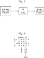
- FIG. 1 A simplified workflow according to an embodiment of the present invention is shown in Fig. 1 .
- the workflow comprises diluting the blood sample taken from a patient and introducing the diluted blood sample directly into a mass spectrometer.
- the ratio of dilution of blood to water in this example is 1:500, although other dilutions may of course be used.
- Components or molecules in the diluted blood sample are then ionised within the mass spectrometer.
- the resulting ions are then subjected to Atmospheric Pressure Electron Capture Dissociation (“Ap-ECD”), or Atmospheric Pressure Electron Transfer Dissociation (“Ap-ETD”), so as to dissociate the ions to form first fragment ions, e.g. to dissociate ions derived from haemoglobin proteins to form first fragment ions.
- the resulting spectra may be used to putatively determine the location of a mutation, as will be described further below in relation to Figs. 3
- MS/MS analysis may be performed by selectively transmitting a first fragment ion having a particular mass to charge ratio (or range of mass to charge ratios). At least some of the first fragment ions are fragmented to form second fragment ions, such as by Collisionally Induced Dissociation (CID) of the first fragment ions that are transmitted.
- CID Collisionally Induced Dissociation
- the resulting spectra may be used to accurately determine the location of a mutation, as will be described further below in relation to Figs. 5A-5C .
- the above-described workflow does not involve the relatively time consuming steps of HPLC and sample digestion, and does not require complex workflows.
- the simplified workflow is relatively fast, only requiring approximately a minute or less per stage.
- the procedure may be performed using a small, low-cost bench-top instrument.
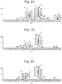
- Figs. 2A-2C show MS spectra for precursor ions from three different blood samples.
- Fig. 2A shows an MS spectra for ions from a normal (AA) blood sample.
- the most intense peak 2 shown in the spectrum is due to ions from the alpha chain (ions having a charge state of 18+ and having an average molecular weight of 15126 Da) and the second most intense peak 4 is due to beta chain ions (ions having a charge state of 19+ and having an average molecular weight of 15867 Da).
- beta chain ions ions having a charge state of 19+ and having an average molecular weight of 15867 Da.
- both beta chains are unmodified and appear at the same mass to charge ratio.
- Fig. 2B shows an MS spectra for ions from a heterozygous (AS) blood sample, i.e. a sample in which one of the beta chains is mutated and one of the beta chains is not mutated.
- the spectrum shows an ion peak 2 due to the alpha chain and an ion peak 4 due to the non-mutated beta chain (beta A) at the same locations as the alpha and beta chain peaks in Fig. 2A .
- the non-mutated beta chain peak 4 in Fig. 2B has a lower intensity than that in Fig. 2A , because the blood sample analysed to produce Fig. 2B does not contain only non-mutated beta chains but also includes mutated beta chains.
- Fig. 1 shows an MS spectra for ions from a heterozygous (AS) blood sample, i.e. a sample in which one of the beta chains is mutated and one of the beta chains is not mutated.
- the spectrum shows an ion peak 2 due to
- FIG. 2B shows an ion peak 6 corresponding to the mutated beta chain (beta S), which has an approximately similar intensity to peak 4 shown in Fig. 2B for the non-mutated beta chain (beta A).
- the ion peak 6 is due to ions having a charge state of 19+ and having an average molecular weight of 15837 Da.
- Fig. 2C shows an MS spectra for ions from a homozygous (SS) blood sample, i.e. a sample in which both of the beta chains are mutated.
- the spectrum shows an ion peak 2 due to the alpha chain at substantially the same location and intensity as the alpha chain peaks in Figs. 2A and 2B .
- the spectrum of Fig. 2C includes only a single beta chain peak 6 that is located at the same location as the mutated beta chain (beta S) peak shown in Fig. 2B .
- the beta chain peak 6 in Fig. 2C has a higher intensity than that in Fig. 2B (and the substantially same intensity as the non-mutated beta chain 4 in Fig.
- Fig. 2C because the blood sample analysed to produce Fig. 2C contains only mutated beta chains and no non-mutated beta chains.
- the ion peak 6 in Fig. 2C is due to ions having a charge state of 19+ and having an average molecular weight of 15837 Da.
- Figs. 2A-2C the mutation in the beta chain in the AS and SS variants causes the beta chain to decrease in mass by 30 Da, as compared to an non-mutated beta chain ( Fig. 2A ).
- the location of the mutation has not been determined precisely and so is not directly indicative of whether the mutation is problematic or not (e.g. whether or not the mutation is symptomatic in the patient). It may therefore be desirable to use the technique described in relation to Fig. 1 to determine the location of the mutation more accurately.
- Figs. 3A-3C show fragment ion spectra obtained for the three different blood samples analysed in Figs. 2A-2C , respectively.
- the fragment ion spectra were obtained by switching on an Atmospheric Pressure Electron Capture Dissociation (“Ap-ECD”) ultraviolet lamp whilst in a MS mode of operation.
- the light from the lamp may generate photoelectrons, e.g. via the ionization of a supplementary acetone flow within the ion source.
- the photoelectrons may then react with the multiply charged analyte ions, thereby generating Electron Capture Dissociation (ECD) type fragment ions.
- ECD Electron Capture Dissociation
- Fig. 3A shows the spectrum for the normal (AA) blood sample.
- the c8 fragment ion has a mass to charge ratio of 952 and is derived from a normal beta chain (beta A).
- a c8 ion exists in only one form for the normal (AA) blood sample, at a mass to charge ratio of 952.
- Fig. 3B shows the spectrum for the heterozygous (AS) blood sample.
- the spectrum shows a c8 fragment ion having a mass to charge ratio of 952, which is derived from a normal beta chain (beta A).
- the spectrum of Fig. 3B also shows a c8 fragment having a mass to charge ratio of 922 and which is derived from a mutated beta chain (beta S).
- Fig. 3C shows the spectrum for the homozygous (SS) blood sample.
- the c8 fragment ion having a mass to charge ratio of 952 that is shown in Figs. 3A and 3B is missing, because such a fragment would be derived from a normal beta chain (beta A), which is not present in the homozygous (SS) blood sample.
- the spectrum of Fig. 3C shows the c8 fragment ion having a mass to charge ratio of 922, which is derived from the mutated beta chain (beta S), and which is also shown in Fig. 3B .
- Figs. 3A-3C the mutation in the beta chain in the AS and SS variants ( Figs. 3B and 3C ) causes c8 fragment ions to be observed at a mass to charge ratio that is 30 Da lower than the c8 fragment ion from a non-mutated beta chain ( Fig. 3A ).
- a mass shift of 30 Da may be attributed to either a Threonine to Alanine mutation, or a Glutamic Acid to Valine mutation in the beta chain sequence.
- Fig. 4 shows the amino-acid sequence for a non-mutated beta chain, together with the above-described three different possible mutations that would give rise to a 30 Da mass shift.
- T-A mutation Threonine to Alanine
- E-V mutation Glutamic Acid to Valine
- Figs. 2B-2C and Figs. 3B-3C show the putative location of the mutation, i.e. at the beta chain.
- c8 fragment ions observed in the experiments of Figs. 3A-3C may be selected and isolated.
- the isolated c8 fragment ion may then be subjected to Collisionally Induced Dissociation (CID) so as to produce sequence ions, as shown in the spectra of Figs. 5A-5C .
- the sequence ions may then be used to determine the location of the mutation, and hence whether the mutation is clinically significant or not.
- CID Collisionally Induced Dissociation
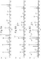
- Fig. 5A shows the spectrum obtained by CID fragmentation of the c8 ion observed in Fig. 3A , i.e. for a normal (AA) blood sample.
- the b6 ion in this spectrum appears at its expected mass for a non-mutated sample.
- the y2 and b4 ions in this spectrum also appear at their expected masses for a non-mutated sample.
- Fig. 5B shows the spectrum obtained by isolating the c8 ions observed at a mass to charge ratio of around 922 in Fig. 3B , and then fragmenting these ions by CID, i.e. fragmenting the mutated c8 ions for the heterozygous (AS) blood sample.
- the b6 ion in this spectrum is located at a mass 30 Da below its expected mass for a non-mutated sample. Referring back to Fig. 4 , this indicates that the Glutamic Acid to Valine (E-V) mutation at position 6 in the beta chain has occurred, indicating that the sample is either homozygous or heterozygous.
- E-V Glutamic Acid to Valine
- the sample is a heterozygous sample by determining that both mutated an unmutated beta chains are present (e.g. from the MS spectrum shown in Fig. 2B ).
- the sample may be determined to be heterozygous by fragmenting the first generation ions and detecting the presence of product ions generated from the fragmentation of unmutated c8 ions, i.e.
- the sample is heterozygous, rather than homozygous, by determining that the mass shift in the b6 ions indicate a mutation and that the intensity of the b6 ion peak is below a threshold value.
- y2 and b4 ions in the spectrum of Fig. 5B appear at their expected masses for a non-mutated sample, indicating that the clinically insignificant Threonine to Alanine (T-A) mutation at position 4 in the beta chain and the clinically insignificant Glutamic Acid to Valine (E-V) mutation at position 7 in the beta chain have not occurred.
- Fig. 5C shows the spectrum obtained by isolating the c8 ions observed at a mass to charge ratio of around 922 in Fig. 3C , and then fragmenting these ions by CID, i.e. fragmenting the mutated c8 ions for the homozygous (AS) blood sample.
- the b6 ion in this spectrum is located at a mass 30 Da below its expected mass for a non-mutated sample. Referring back to Fig. 4 , this indicates that the Glutamic Acid to Valine (E-V) mutation at position 6 in the beta chain has occurred, indicating that the sample is either homozygous or heterozygous.
- E-V Glutamic Acid to Valine
- the sample is a homozygous sample by determining that only the mutated beta chain, and not the unmutated beta chain, is present (e.g. from the MS spectrum shown in Fig. 2C ). Alternatively, or additionally, it may be determined that the sample is a homozygous sample by determining that c8 fragment ions having a mass to charge ratio of around 952 are not generated in the first generation fragment ions (i.e. determining that the non-mutated c8 fragment ions shown in Figs. 3A and 3B are not generated).
- the sample is homozygous, rather than heterozygous, by determining that the mass shift in the b6 ions indicate a mutation and that the intensity of the b6 ion peak is above a threshold value.
- the y2 and b4 ions in this spectrum appear at their expected masses for a non-mutated sample, indicating that the "non-listed" Threonine to Alanine (T-A) mutation at position 4 in the beta chain and the "non-listed" Glutamic Acid to Valine (E-V) mutation at position 7 in the beta chain have not occurred.
- Figs. 5A-5C may be used to identify the location of the mutation and whether the mutation is actually a mutation of clinical significance.
- the approach described above may detect haemoglobin variants by detecting the location of a mass shift that is expected to occur if that variant is present.
- the above-described approach may be applied to detecting other clinically significant variants by detecting the location of a mass shift that is expected to occur if that variant is present.
- the technique may be used to detect Hb Lepores or, for example, to detect any of the conditions listed below: Name Mutation Symptoms (homozygote or heterozygote) Hb S ⁇ 6 (Glu to Val) Sickle Cell Disease or asymptomatic Hb C ⁇ 6 (Glu to Lys) Mild anaemia or asymptomatic Hb E ⁇ 26 (Glu to Lys) Thalassemia minor or mild microcytosis Hb D-Punjab ⁇ 121 (Glu to Gln) Mild anaemia or asymptomatic Hb O-Arab ⁇ 121 (Glu to Lys) Mild anaemia or asymptomatic
- the invention may be used to detect variants of other proteins in blood or other biological samples.
Landscapes
- Life Sciences & Earth Sciences (AREA)
- Health & Medical Sciences (AREA)
- Engineering & Computer Science (AREA)
- Molecular Biology (AREA)
- Chemical & Material Sciences (AREA)
- Hematology (AREA)
- Urology & Nephrology (AREA)
- Biomedical Technology (AREA)
- Physics & Mathematics (AREA)
- Immunology (AREA)
- Analytical Chemistry (AREA)
- Medicinal Chemistry (AREA)
- Bioinformatics & Cheminformatics (AREA)
- Food Science & Technology (AREA)
- Cell Biology (AREA)
- Biotechnology (AREA)
- Microbiology (AREA)
- Biochemistry (AREA)
- General Health & Medical Sciences (AREA)
- General Physics & Mathematics (AREA)
- Pathology (AREA)
- Bioinformatics & Computational Biology (AREA)
- Proteomics, Peptides & Aminoacids (AREA)
- Biophysics (AREA)
- Spectroscopy & Molecular Physics (AREA)
- Chemical Kinetics & Catalysis (AREA)
- Other Investigation Or Analysis Of Materials By Electrical Means (AREA)
- Investigating Or Analysing Biological Materials (AREA)
Claims (14)
- Procédé de spectrométrie de masse pour déterminer si un variant muté d'une protéine cible est présent dans un échantillon, comprenant les étapes consistant à :introduire ledit échantillon comprenant la protéine cible dans un spectromètre de masse, dans lequel l'échantillon n'est pas soumis à une chromatographie liquide ou une digestion avant d'être introduit dans le spectromètre de masse ;soumettre l'échantillon à une fragmentation de manière à amener ladite protéine cible à se fragmenter pour former des premiers ions fragments, dans lequel ladite fragmentation est une Dissociation par capture d'électrons sous pression atmosphérique (« Ap-ECD »), une Dissociation par transfert d'électrons sous pression atmosphérique (« Ap-ETD »), une Dissociation par capture d'électrons (« ECD »), ou une Dissociation par transfert d'électrons (« ETD ») ;fragmenter au moins certains desdits premiers ions fragments pour former des seconds ions fragments ;analyser la masse desdits seconds ions fragments pour obtenir des premières données spectrales ; etdéterminer qu'un ion dans les premières données spectrales présente un rapport masse/charge qui diffère du rapport masse/charge d'un ion qui serait observé si ladite protéine cible était une version non mutée normale de ladite protéine cible, et par une quantité qui correspond à une différence de masse qui serait provoquée par le fait que la protéine cible est un variant muté de ladite protéine cible.
- Procédé selon la revendication 1, dans lequel ledit échantillon est, ou comprend : du sang, du sang total, du sang dilué, ou du sang total dilué ; et/ou
dans lequel la protéine cible est l'hémoglobine ou une chaîne beta d'hémoglobine ; et/ou
dans lequel la protéine cible est soumise à une ionisation avant ladite étape de fragmentation de sorte que des ions de protéine cible soient fragmentés pour former les premiers ions fragments ; et facultativement dans lequel la protéine cible est ionisée dans une source d'ions pour former les ions de protéine cible. - Procédé selon l'une quelconque des revendications précédentes, dans lequel l'étape consistant à fragmenter lesdits premiers ions fragments comprend les étapes consistant à :fragmenter lesdits premiers ions fragments en utilisant une Dissociation induite par collision (« CID ») ; et/ouisoler un desdits premiers ions fragments des autres ions fragments et ensuite fragmenter les ions isolés de manière à former les seconds ions fragments, dans lequel les ions isolés sont fragmentés en utilisant une CID.
- Procédé selon l'une quelconque des revendications précédentes, comprenant en outre :l'utilisation du rapport masse/charge dudit ion dans les premières données spectrales pour identifier l'emplacement de la mutation dans la séquence de protéine de la protéine cible ; et/oul'utilisation de la valeur de ladite différence de masse pour identifier le type de mutation dans la séquence de la protéine cible ; etfacultativement l'identification du type de variant de la protéine cible dans l'échantillon, ou l'identification d'une maladie dans l'échantillon, depuis l'emplacement et/ou le type de mutation.
- Procédé selon l'une quelconque des revendications précédentes, dans lequel la protéine cible est analysée de masse dans un mode d'ion précurseur sans être d'abord fragmentée ; dans lequel, si ledit ion présentant ladite différence de masse est détecté dans les premières données spectrales, un ion précurseur apparenté détecté dans ledit mode d'ion précurseur est considéré et/ou indiqué comme étant un ion précurseur muté d'un variant muté anormal de la protéine cible ; dans lequel le procédé détermine si les ions précurseurs contiennent ou non un ion précurseur correspondant à une version non mutée de l'ion précurseur muté ; et
dans lequel, si le procédé détermine que les ions précurseurs contiennent à la fois ledit ion précurseur muté et ladite version non mutée de l'ion précurseur, alors il est déterminé que le variant est un variant hétérozygote ; ou
dans lequel, si le procédé détermine que les ions précurseurs contiennent ledit ion précurseur muté et pas ladite version non mutée de l'ion précurseur, alors il est déterminé que le variant est un variant homozygote. - Procédé selon l'une quelconque des revendications précédentes, dans lequel, si ledit ion présentant ladite différence de masse est détecté dans les premières données spectrales, le premier ion fragment qui est fragmenté pour produire les secondes données spectrales est considéré et/ou indiqué comme étant un premier ion fragment muté d'un variant muté anormal de la protéine cible; et dans lequel le procédé détermine si les premiers ions fragments contiennent ou non un ion fragment correspondant à une version non mutée du premier ion fragment muté ; et
dans lequel, si le procédé détermine que les premiers ions fragments contiennent à la fois ledit premier ion fragment muté et ladite version non mutée du premier ion fragment muté, alors il est déterminé que le variant est un variant hétérozygote ; et/ou
dans lequel, si le procédé détermine que les premiers ions fragments contiennent ledit premier ion fragment muté et pas ladite version non mutée du premier ion fragment muté, alors il est déterminé que le variant est un variant homozygote. - Procédé de spectrométrie de masse comprenant les étapes consistant à :introduire un échantillon de sang dans un système de spectromètre de masse sans soumettre ledit échantillon de sang à une chromatographie liquide ou une digestion ;utiliser une Dissociation par capture d'électrons (« ECD ») ou une Dissociation par transfert d'électrons (« ETD ») pour soumettre ledit échantillon à une fragmentation de manière à amener des protéines d'hémoglobine à se fragmenter pour former des premiers ions fragments ; etfragmenter au moins certains desdits premiers ions fragments pour former des seconds ions fragments.
- Procédé selon la revendication 7, dans lequel ladite ECD est une Dissociation par capture d'électrons sous pression atmosphérique (« Ap-ECD ») ; et ladite ETD est une Dissociation par transfert d'électrons sous pression atmosphérique (« Ap-ETD »), ; et
facultativement dans lequel l'étape consistant à utiliser une ECD ou ETD pour soumettre ledit échantillon à une fragmentation comprend une étape consistant à utiliser ladite ECD ou ETD pour soumettre ledit échantillon à une fragmentation dans la source de manière à amener les protéines d'hémoglobine à se fragmenter pour former les premiers ions fragments. - Procédé selon l'une quelconque des revendications 7-8, comprenant une étape consistant à analyser la masse desdits premiers ions fragments pour obtenir des premières données spectrales ; et une étape consistant à déterminer si des ions dans les premières données spectrales sont ou non à des rapports masse/charge correspondant aux rapports masse/charge auxquels ces ions seraient détectés si l'hémoglobine dans l'échantillon de sang était de l'hémoglobine non mutée normale ; et
facultativement, comprenant en outre une étape consistant à fournir une indication que l'hémoglobine dans l'échantillon de sang est de l'hémoglobine non mutée normale si les ions dans les premières données spectrales sont à des rapports masse/charge correspondant aux rapports masse/charge auxquels les ions seraient détectés si l'hémoglobine dans l'échantillon de sang était de l'hémoglobine non mutée normale. - Procédé selon l'une quelconque des revendications 7-8, comprenant une étape consistant à analyser la masse desdits premiers ions fragments pour obtenir des premières données spectrales ; et une étape consistant à déterminer, à partir des premières données spectrales, si un ion est présent à un rapport masse/charge correspondant au rapport masse/charge auquel un ion serait détecté uniquement si l'hémoglobine dans l'échantillon de sang était d'un variant anormal muté ; et
facultativement, comprenant en outre une étape consistant à fournir une indication que l'hémoglobine dans l'échantillon de sang est un variant muté d'hémoglobine. - Procédé selon l'une quelconque des revendications 7-10, comprenant une étape consistant à analyser la masse des seconds ions fragments pour obtenir des secondes données spectrales ; et une étape consistant à déterminer, à partir des secondes données spectrales, si un ion est présent à un rapport masse/charge correspondant au rapport masse/charge auquel un ion serait détecté uniquement si l'hémoglobine dans l'échantillon de sang était d'un variant muté anormal.
- Procédé selon la revendication 11, comprenant une étape consistant à déterminer qu'un ion dans les secondes données spectrales présente un rapport masse/charge qui diffère du rapport masse/charge d'un ion qui serait observé si de l'hémoglobine non mutée normale avait été analysée, et par une quantité qui correspond à une différence de masse qui serait provoquée par le fait que l'hémoglobine analysée est un variant anormal ou qui serait provoquée par une mutation/substitution d'un acide aminé d'hémoglobine normale pour un autre acide aminé.
- Procédé selon la revendication 12, dans lequel la différence de masse est 30 Da ; et/ou
dans lequel la différence de masse correspond à une mutation d'Acide glutamique en Valine ; et/ou
dans lequel la différence de masse est une différence de masse qui surviendrait si l'hémoglobine dans l'échantillon de sang était d'un patient atteint de drépanocytose. - Procédé d'identification d'un type de variant d'hémoglobine dans un échantillon de sang ou d'une maladie du sang dans un échantillon de sang comprenant un procédé selon l'une quelconque des revendications précédentes.
Applications Claiming Priority (2)
| Application Number | Priority Date | Filing Date | Title |
|---|---|---|---|
| GBGB1417387.6A GB201417387D0 (en) | 2014-10-01 | 2014-10-01 | Haemoglobin variant analysis using pseudo MS/MS/MS via atmospheric pressures elctron capture dissociation ("AP-ECD") and collision induced dissociation("CID") |
| PCT/GB2015/052881 WO2016051191A1 (fr) | 2014-10-01 | 2015-10-01 | Spectrométrie de masse pour détermination du point de savoir si un variant muté d'une protéine cible est présent dans un échantillon |
Publications (2)
| Publication Number | Publication Date |
|---|---|
| EP3201632A1 EP3201632A1 (fr) | 2017-08-09 |
| EP3201632B1 true EP3201632B1 (fr) | 2019-12-25 |
Family
ID=51901480
Family Applications (1)
| Application Number | Title | Priority Date | Filing Date |
|---|---|---|---|
| EP15784434.1A Active EP3201632B1 (fr) | 2014-10-01 | 2015-10-01 | Spectrométrie de masse pour détermination du point de savoir si un variant muté d'une protéine cible est présent dans un échantillon |
Country Status (4)
| Country | Link |
|---|---|
| US (1) | US11402385B2 (fr) |
| EP (1) | EP3201632B1 (fr) |
| GB (2) | GB201417387D0 (fr) |
| WO (1) | WO2016051191A1 (fr) |
Families Citing this family (1)
| Publication number | Priority date | Publication date | Assignee | Title |
|---|---|---|---|---|
| CN111693597A (zh) * | 2020-06-12 | 2020-09-22 | 南方科技大学 | 一种高覆盖度的神经节糖苷脂鉴定方法及其应用 |
Citations (1)
| Publication number | Priority date | Publication date | Assignee | Title |
|---|---|---|---|---|
| WO2015150785A1 (fr) * | 2014-03-31 | 2015-10-08 | Micromass Uk Limited | Méthode rapide d'analyse d'échantillons de sang pour l'identification de variants d'hémoglobine à l'aide d'une dissociation par transfert d'électrons |
Family Cites Families (16)
| Publication number | Priority date | Publication date | Assignee | Title |
|---|---|---|---|---|
| US5538897A (en) | 1994-03-14 | 1996-07-23 | University Of Washington | Use of mass spectrometry fragmentation patterns of peptides to identify amino acid sequences in databases |
| ATE393910T1 (de) | 2000-04-28 | 2008-05-15 | Univ Duke | Quantitatives screening-verfahren mit hohem durchsatz für die proteinstabilität |
| GB0308202D0 (en) * | 2003-04-09 | 2003-05-14 | King S College London | A screening method using mass spectrometry |
| GB0502068D0 (en) * | 2005-02-01 | 2005-03-09 | King S College London | Screening method |
| US20060255261A1 (en) * | 2005-04-04 | 2006-11-16 | Craig Whitehouse | Atmospheric pressure ion source for mass spectrometry |
| US7838303B2 (en) | 2006-07-27 | 2010-11-23 | Agilent Technologies, Inc. | Peptide derivatization method to increase fragmentation information from MS/MS spectra |
| WO2009073505A2 (fr) * | 2007-11-30 | 2009-06-11 | Wisconsin Alumni Research Foundation | Procédés de traitement de données de spectres de masse en tandem pour une analyse de séquence d'une protéine |
| CA2790835A1 (fr) * | 2009-02-26 | 2010-09-02 | The University Of British Columbia | Procedes et appareil ap-ecd pour une analyse spectrometrique de masse de peptides et de proteines |
| CA2767570C (fr) | 2009-07-13 | 2018-05-01 | Universite De Montreal | Isoformes mutees de la proteine sumo et leurs utilisations |
| US8153961B2 (en) * | 2009-08-31 | 2012-04-10 | Thermo Finnigan Llc | Methods for acquisition and deductive analysis of mixed fragment peptide mass spectra |
| US20150111238A1 (en) | 2011-05-31 | 2015-04-23 | Singapore Health Services Pte. Ltd. | Method for detecting disease biomarkers |
| EP2807669A2 (fr) * | 2012-01-24 | 2014-12-03 | Thermo Finnigan LLC | Isolation par filtre coupe-bande pour analyse de masse ms3 |
| US20130210050A1 (en) * | 2012-02-15 | 2013-08-15 | Northwestern University | Protease for proteomics |
| CA2873806A1 (fr) * | 2012-05-18 | 2013-11-21 | Micromass Uk Limited | Procede de spectrometrie de masse en tandem ms/ms |
| EP3155638B1 (fr) * | 2014-06-13 | 2023-09-20 | Waters Technologies Corporation | Analyse de matrices biologiques complexes par ciblage et alignement avancé d'ions précurseurs et produits |
| US9837255B2 (en) * | 2014-07-18 | 2017-12-05 | Thermo Finnigan Llc | Methods for mass spectrometry of mixtures of protein or polypeptides using proton transfer reaction |
-
2014
- 2014-10-01 GB GBGB1417387.6A patent/GB201417387D0/en not_active Ceased
-
2015
- 2015-10-01 GB GB1517390.9A patent/GB2536322B/en active Active
- 2015-10-01 WO PCT/GB2015/052881 patent/WO2016051191A1/fr not_active Ceased
- 2015-10-01 US US15/516,083 patent/US11402385B2/en active Active
- 2015-10-01 EP EP15784434.1A patent/EP3201632B1/fr active Active
Patent Citations (1)
| Publication number | Priority date | Publication date | Assignee | Title |
|---|---|---|---|---|
| WO2015150785A1 (fr) * | 2014-03-31 | 2015-10-08 | Micromass Uk Limited | Méthode rapide d'analyse d'échantillons de sang pour l'identification de variants d'hémoglobine à l'aide d'une dissociation par transfert d'électrons |
Also Published As
| Publication number | Publication date |
|---|---|
| GB201417387D0 (en) | 2014-11-12 |
| GB201517390D0 (en) | 2015-11-18 |
| US20170292960A1 (en) | 2017-10-12 |
| WO2016051191A1 (fr) | 2016-04-07 |
| US11402385B2 (en) | 2022-08-02 |
| GB2536322B (en) | 2019-02-06 |
| GB2536322A (en) | 2016-09-14 |
| EP3201632A1 (fr) | 2017-08-09 |
Similar Documents
| Publication | Publication Date | Title |
|---|---|---|
| US12217952B2 (en) | Mass correction | |
| EP2850638B1 (fr) | Procédé de spectrométrie de masse en tandem ms/ms | |
| US10495647B2 (en) | Analysis of complex biological matrices through targeting and advanced precursor and product ion alignment | |
| KR102314968B1 (ko) | 최적화된 표적 분석 | |
| US9881781B2 (en) | Optimized multiple reaction monitoring or single ion recording method | |
| US20170236700A1 (en) | Time of Flight Mass Spectrometer | |
| JP4959712B2 (ja) | 質量分析計 | |
| US10041907B2 (en) | Accurate mobility chromatograms | |
| US10043645B2 (en) | Method of localizing lipid double bonds | |
| US11402385B2 (en) | Mass spectrometry for determining if a mutated variant of a target protein is present in a sample | |
| US9881776B2 (en) | Monitoring liquid chromatography elution to determine when to perform a lockmass calibration | |
| US9659760B2 (en) | Automated adjustment of capillary voltage based on the elution conditions to retain optimal ionization conditions | |
| EP3152778B1 (fr) | Procédé d'élimination des interférences ioniques | |
| GB2525709A (en) | Optimized multiple reaction monitoring or single ion recording method | |
| GB2534627A (en) | Accurate mobility chromatograms |
Legal Events
| Date | Code | Title | Description |
|---|---|---|---|
| STAA | Information on the status of an ep patent application or granted ep patent |
Free format text: STATUS: THE INTERNATIONAL PUBLICATION HAS BEEN MADE |
|
| PUAI | Public reference made under article 153(3) epc to a published international application that has entered the european phase |
Free format text: ORIGINAL CODE: 0009012 |
|
| STAA | Information on the status of an ep patent application or granted ep patent |
Free format text: STATUS: REQUEST FOR EXAMINATION WAS MADE |
|
| 17P | Request for examination filed |
Effective date: 20170329 |
|
| AK | Designated contracting states |
Kind code of ref document: A1 Designated state(s): AL AT BE BG CH CY CZ DE DK EE ES FI FR GB GR HR HU IE IS IT LI LT LU LV MC MK MT NL NO PL PT RO RS SE SI SK SM TR |
|
| AX | Request for extension of the european patent |
Extension state: BA ME |
|
| DAV | Request for validation of the european patent (deleted) | ||
| DAX | Request for extension of the european patent (deleted) | ||
| STAA | Information on the status of an ep patent application or granted ep patent |
Free format text: STATUS: EXAMINATION IS IN PROGRESS |
|
| 17Q | First examination report despatched |
Effective date: 20180613 |
|
| RIC1 | Information provided on ipc code assigned before grant |
Ipc: G01N 33/68 20060101AFI20190528BHEP Ipc: G01N 33/72 20060101ALI20190528BHEP Ipc: G01N 33/48 20060101ALI20190528BHEP Ipc: H01J 49/00 20060101ALI20190528BHEP |
|
| GRAP | Despatch of communication of intention to grant a patent |
Free format text: ORIGINAL CODE: EPIDOSNIGR1 |
|
| STAA | Information on the status of an ep patent application or granted ep patent |
Free format text: STATUS: GRANT OF PATENT IS INTENDED |
|
| INTG | Intention to grant announced |
Effective date: 20190718 |
|
| GRAS | Grant fee paid |
Free format text: ORIGINAL CODE: EPIDOSNIGR3 |
|
| GRAA | (expected) grant |
Free format text: ORIGINAL CODE: 0009210 |
|
| STAA | Information on the status of an ep patent application or granted ep patent |
Free format text: STATUS: THE PATENT HAS BEEN GRANTED |
|
| AK | Designated contracting states |
Kind code of ref document: B1 Designated state(s): AL AT BE BG CH CY CZ DE DK EE ES FI FR GB GR HR HU IE IS IT LI LT LU LV MC MK MT NL NO PL PT RO RS SE SI SK SM TR |
|
| REG | Reference to a national code |
Ref country code: GB Ref legal event code: FG4D |
|
| REG | Reference to a national code |
Ref country code: CH Ref legal event code: EP |
|
| REG | Reference to a national code |
Ref country code: AT Ref legal event code: REF Ref document number: 1217697 Country of ref document: AT Kind code of ref document: T Effective date: 20200115 |
|
| REG | Reference to a national code |
Ref country code: DE Ref legal event code: R096 Ref document number: 602015044345 Country of ref document: DE |
|
| REG | Reference to a national code |
Ref country code: IE Ref legal event code: FG4D |
|
| REG | Reference to a national code |
Ref country code: NL Ref legal event code: MP Effective date: 20191225 |
|
| PG25 | Lapsed in a contracting state [announced via postgrant information from national office to epo] |
Ref country code: BG Free format text: LAPSE BECAUSE OF FAILURE TO SUBMIT A TRANSLATION OF THE DESCRIPTION OR TO PAY THE FEE WITHIN THE PRESCRIBED TIME-LIMIT Effective date: 20200325 Ref country code: FI Free format text: LAPSE BECAUSE OF FAILURE TO SUBMIT A TRANSLATION OF THE DESCRIPTION OR TO PAY THE FEE WITHIN THE PRESCRIBED TIME-LIMIT Effective date: 20191225 Ref country code: GR Free format text: LAPSE BECAUSE OF FAILURE TO SUBMIT A TRANSLATION OF THE DESCRIPTION OR TO PAY THE FEE WITHIN THE PRESCRIBED TIME-LIMIT Effective date: 20200326 Ref country code: NO Free format text: LAPSE BECAUSE OF FAILURE TO SUBMIT A TRANSLATION OF THE DESCRIPTION OR TO PAY THE FEE WITHIN THE PRESCRIBED TIME-LIMIT Effective date: 20200325 Ref country code: LV Free format text: LAPSE BECAUSE OF FAILURE TO SUBMIT A TRANSLATION OF THE DESCRIPTION OR TO PAY THE FEE WITHIN THE PRESCRIBED TIME-LIMIT Effective date: 20191225 Ref country code: SE Free format text: LAPSE BECAUSE OF FAILURE TO SUBMIT A TRANSLATION OF THE DESCRIPTION OR TO PAY THE FEE WITHIN THE PRESCRIBED TIME-LIMIT Effective date: 20191225 Ref country code: LT Free format text: LAPSE BECAUSE OF FAILURE TO SUBMIT A TRANSLATION OF THE DESCRIPTION OR TO PAY THE FEE WITHIN THE PRESCRIBED TIME-LIMIT Effective date: 20191225 |
|
| REG | Reference to a national code |
Ref country code: LT Ref legal event code: MG4D |
|
| PG25 | Lapsed in a contracting state [announced via postgrant information from national office to epo] |
Ref country code: HR Free format text: LAPSE BECAUSE OF FAILURE TO SUBMIT A TRANSLATION OF THE DESCRIPTION OR TO PAY THE FEE WITHIN THE PRESCRIBED TIME-LIMIT Effective date: 20191225 Ref country code: RS Free format text: LAPSE BECAUSE OF FAILURE TO SUBMIT A TRANSLATION OF THE DESCRIPTION OR TO PAY THE FEE WITHIN THE PRESCRIBED TIME-LIMIT Effective date: 20191225 |
|
| PG25 | Lapsed in a contracting state [announced via postgrant information from national office to epo] |
Ref country code: AL Free format text: LAPSE BECAUSE OF FAILURE TO SUBMIT A TRANSLATION OF THE DESCRIPTION OR TO PAY THE FEE WITHIN THE PRESCRIBED TIME-LIMIT Effective date: 20191225 |
|
| PG25 | Lapsed in a contracting state [announced via postgrant information from national office to epo] |
Ref country code: RO Free format text: LAPSE BECAUSE OF FAILURE TO SUBMIT A TRANSLATION OF THE DESCRIPTION OR TO PAY THE FEE WITHIN THE PRESCRIBED TIME-LIMIT Effective date: 20191225 Ref country code: CZ Free format text: LAPSE BECAUSE OF FAILURE TO SUBMIT A TRANSLATION OF THE DESCRIPTION OR TO PAY THE FEE WITHIN THE PRESCRIBED TIME-LIMIT Effective date: 20191225 Ref country code: NL Free format text: LAPSE BECAUSE OF FAILURE TO SUBMIT A TRANSLATION OF THE DESCRIPTION OR TO PAY THE FEE WITHIN THE PRESCRIBED TIME-LIMIT Effective date: 20191225 Ref country code: EE Free format text: LAPSE BECAUSE OF FAILURE TO SUBMIT A TRANSLATION OF THE DESCRIPTION OR TO PAY THE FEE WITHIN THE PRESCRIBED TIME-LIMIT Effective date: 20191225 Ref country code: PT Free format text: LAPSE BECAUSE OF FAILURE TO SUBMIT A TRANSLATION OF THE DESCRIPTION OR TO PAY THE FEE WITHIN THE PRESCRIBED TIME-LIMIT Effective date: 20200520 |
|
| PG25 | Lapsed in a contracting state [announced via postgrant information from national office to epo] |
Ref country code: SK Free format text: LAPSE BECAUSE OF FAILURE TO SUBMIT A TRANSLATION OF THE DESCRIPTION OR TO PAY THE FEE WITHIN THE PRESCRIBED TIME-LIMIT Effective date: 20191225 Ref country code: IS Free format text: LAPSE BECAUSE OF FAILURE TO SUBMIT A TRANSLATION OF THE DESCRIPTION OR TO PAY THE FEE WITHIN THE PRESCRIBED TIME-LIMIT Effective date: 20200425 Ref country code: SM Free format text: LAPSE BECAUSE OF FAILURE TO SUBMIT A TRANSLATION OF THE DESCRIPTION OR TO PAY THE FEE WITHIN THE PRESCRIBED TIME-LIMIT Effective date: 20191225 |
|
| REG | Reference to a national code |
Ref country code: DE Ref legal event code: R097 Ref document number: 602015044345 Country of ref document: DE |
|
| PG25 | Lapsed in a contracting state [announced via postgrant information from national office to epo] |
Ref country code: ES Free format text: LAPSE BECAUSE OF FAILURE TO SUBMIT A TRANSLATION OF THE DESCRIPTION OR TO PAY THE FEE WITHIN THE PRESCRIBED TIME-LIMIT Effective date: 20191225 Ref country code: DK Free format text: LAPSE BECAUSE OF FAILURE TO SUBMIT A TRANSLATION OF THE DESCRIPTION OR TO PAY THE FEE WITHIN THE PRESCRIBED TIME-LIMIT Effective date: 20191225 |
|
| PLBE | No opposition filed within time limit |
Free format text: ORIGINAL CODE: 0009261 |
|
| STAA | Information on the status of an ep patent application or granted ep patent |
Free format text: STATUS: NO OPPOSITION FILED WITHIN TIME LIMIT |
|
| REG | Reference to a national code |
Ref country code: AT Ref legal event code: MK05 Ref document number: 1217697 Country of ref document: AT Kind code of ref document: T Effective date: 20191225 |
|
| PLAA | Information modified related to event that no opposition was filed |
Free format text: ORIGINAL CODE: 0009299DELT |
|
| PLBE | No opposition filed within time limit |
Free format text: ORIGINAL CODE: 0009261 |
|
| PG25 | Lapsed in a contracting state [announced via postgrant information from national office to epo] |
Ref country code: SI Free format text: LAPSE BECAUSE OF FAILURE TO SUBMIT A TRANSLATION OF THE DESCRIPTION OR TO PAY THE FEE WITHIN THE PRESCRIBED TIME-LIMIT Effective date: 20191225 |
|
| 26N | No opposition filed |
Effective date: 20200928 |
|
| R26N | No opposition filed (corrected) |
Effective date: 20200928 |
|
| RBV | Designated contracting states (corrected) |
Designated state(s): AL AT BE BG CH CY CZ DE DK EE ES FI FR GR HR HU IE IS IT LI LT LU LV MC MK MT NL NO PL PT RO RS SE SI SK SM TR |
|
| PG25 | Lapsed in a contracting state [announced via postgrant information from national office to epo] |
Ref country code: AT Free format text: LAPSE BECAUSE OF FAILURE TO SUBMIT A TRANSLATION OF THE DESCRIPTION OR TO PAY THE FEE WITHIN THE PRESCRIBED TIME-LIMIT Effective date: 20191225 Ref country code: IT Free format text: LAPSE BECAUSE OF FAILURE TO SUBMIT A TRANSLATION OF THE DESCRIPTION OR TO PAY THE FEE WITHIN THE PRESCRIBED TIME-LIMIT Effective date: 20191225 |
|
| PG25 | Lapsed in a contracting state [announced via postgrant information from national office to epo] |
Ref country code: PL Free format text: LAPSE BECAUSE OF FAILURE TO SUBMIT A TRANSLATION OF THE DESCRIPTION OR TO PAY THE FEE WITHIN THE PRESCRIBED TIME-LIMIT Effective date: 20191225 |
|
| REG | Reference to a national code |
Ref country code: CH Ref legal event code: PL |
|
| PG25 | Lapsed in a contracting state [announced via postgrant information from national office to epo] |
Ref country code: LU Free format text: LAPSE BECAUSE OF NON-PAYMENT OF DUE FEES Effective date: 20201001 Ref country code: MC Free format text: LAPSE BECAUSE OF FAILURE TO SUBMIT A TRANSLATION OF THE DESCRIPTION OR TO PAY THE FEE WITHIN THE PRESCRIBED TIME-LIMIT Effective date: 20191225 |
|
| REG | Reference to a national code |
Ref country code: BE Ref legal event code: MM Effective date: 20201031 |
|
| PG25 | Lapsed in a contracting state [announced via postgrant information from national office to epo] |
Ref country code: FR Free format text: LAPSE BECAUSE OF NON-PAYMENT OF DUE FEES Effective date: 20201031 |
|
| PG25 | Lapsed in a contracting state [announced via postgrant information from national office to epo] |
Ref country code: LI Free format text: LAPSE BECAUSE OF NON-PAYMENT OF DUE FEES Effective date: 20201031 Ref country code: CH Free format text: LAPSE BECAUSE OF NON-PAYMENT OF DUE FEES Effective date: 20201031 Ref country code: BE Free format text: LAPSE BECAUSE OF NON-PAYMENT OF DUE FEES Effective date: 20201031 |
|
| PG25 | Lapsed in a contracting state [announced via postgrant information from national office to epo] |
Ref country code: IE Free format text: LAPSE BECAUSE OF NON-PAYMENT OF DUE FEES Effective date: 20201001 |
|
| PG25 | Lapsed in a contracting state [announced via postgrant information from national office to epo] |
Ref country code: TR Free format text: LAPSE BECAUSE OF FAILURE TO SUBMIT A TRANSLATION OF THE DESCRIPTION OR TO PAY THE FEE WITHIN THE PRESCRIBED TIME-LIMIT Effective date: 20191225 Ref country code: MT Free format text: LAPSE BECAUSE OF FAILURE TO SUBMIT A TRANSLATION OF THE DESCRIPTION OR TO PAY THE FEE WITHIN THE PRESCRIBED TIME-LIMIT Effective date: 20191225 Ref country code: CY Free format text: LAPSE BECAUSE OF FAILURE TO SUBMIT A TRANSLATION OF THE DESCRIPTION OR TO PAY THE FEE WITHIN THE PRESCRIBED TIME-LIMIT Effective date: 20191225 |
|
| PG25 | Lapsed in a contracting state [announced via postgrant information from national office to epo] |
Ref country code: MK Free format text: LAPSE BECAUSE OF FAILURE TO SUBMIT A TRANSLATION OF THE DESCRIPTION OR TO PAY THE FEE WITHIN THE PRESCRIBED TIME-LIMIT Effective date: 20191225 |
|
| P01 | Opt-out of the competence of the unified patent court (upc) registered |
Effective date: 20230506 |
|
| PGFP | Annual fee paid to national office [announced via postgrant information from national office to epo] |
Ref country code: DE Payment date: 20250923 Year of fee payment: 11 |