EP3201335B1 - A modified b-fructofuranosidase for fructooligosaccharide production - Google Patents
A modified b-fructofuranosidase for fructooligosaccharide production Download PDFInfo
- Publication number
- EP3201335B1 EP3201335B1 EP15779025.4A EP15779025A EP3201335B1 EP 3201335 B1 EP3201335 B1 EP 3201335B1 EP 15779025 A EP15779025 A EP 15779025A EP 3201335 B1 EP3201335 B1 EP 3201335B1
- Authority
- EP
- European Patent Office
- Prior art keywords
- amino acid
- seq
- sucrose
- enzyme
- polypeptide
- Prior art date
- Legal status (The legal status is an assumption and is not a legal conclusion. Google has not performed a legal analysis and makes no representation as to the accuracy of the status listed.)
- Active
Links
- FTSSQIKWUOOEGC-RULYVFMPSA-N fructooligosaccharide Chemical compound OC[C@H]1O[C@@](CO)(OC[C@@]2(OC[C@@]3(OC[C@@]4(OC[C@@]5(OC[C@@]6(OC[C@@]7(OC[C@@]8(OC[C@@]9(OC[C@@]%10(OC[C@@]%11(O[C@H]%12O[C@H](CO)[C@@H](O)[C@H](O)[C@H]%12O)O[C@H](CO)[C@@H](O)[C@@H]%11O)O[C@H](CO)[C@@H](O)[C@@H]%10O)O[C@H](CO)[C@@H](O)[C@@H]9O)O[C@H](CO)[C@@H](O)[C@@H]8O)O[C@H](CO)[C@@H](O)[C@@H]7O)O[C@H](CO)[C@@H](O)[C@@H]6O)O[C@H](CO)[C@@H](O)[C@@H]5O)O[C@H](CO)[C@@H](O)[C@@H]4O)O[C@H](CO)[C@@H](O)[C@@H]3O)O[C@H](CO)[C@@H](O)[C@@H]2O)[C@@H](O)[C@@H]1O FTSSQIKWUOOEGC-RULYVFMPSA-N 0.000 title claims description 5
- 229940107187 fructooligosaccharide Drugs 0.000 title claims description 5
- 238000004519 manufacturing process Methods 0.000 title description 12
- 238000006467 substitution reaction Methods 0.000 claims description 64
- CZMRCDWAGMRECN-UGDNZRGBSA-N Sucrose Chemical compound O[C@H]1[C@H](O)[C@@H](CO)O[C@@]1(CO)O[C@@H]1[C@H](O)[C@@H](O)[C@H](O)[C@@H](CO)O1 CZMRCDWAGMRECN-UGDNZRGBSA-N 0.000 claims description 63
- 229930006000 Sucrose Natural products 0.000 claims description 62
- 239000005720 sucrose Substances 0.000 claims description 62
- 230000000694 effects Effects 0.000 claims description 56
- 235000001014 amino acid Nutrition 0.000 claims description 53
- 229920001184 polypeptide Polymers 0.000 claims description 52
- 108090000765 processed proteins & peptides Proteins 0.000 claims description 52
- 102000004196 processed proteins & peptides Human genes 0.000 claims description 52
- RFSUNEUAIZKAJO-ARQDHWQXSA-N Fructose Chemical class OC[C@H]1O[C@](O)(CO)[C@@H](O)[C@@H]1O RFSUNEUAIZKAJO-ARQDHWQXSA-N 0.000 claims description 48
- FLDFNEBHEXLZRX-DLQNOBSRSA-N Nystose Chemical compound O[C@H]1[C@H](O)[C@@H](CO)O[C@@]1(CO)OC[C@]1(OC[C@]2(O[C@@H]3[C@@H]([C@@H](O)[C@H](O)[C@@H](CO)O3)O)[C@H]([C@H](O)[C@@H](CO)O2)O)[C@@H](O)[C@H](O)[C@@H](CO)O1 FLDFNEBHEXLZRX-DLQNOBSRSA-N 0.000 claims description 28
- 238000000034 method Methods 0.000 claims description 28
- FLDFNEBHEXLZRX-UHFFFAOYSA-N nystose Natural products OC1C(O)C(CO)OC1(CO)OCC1(OCC2(OC3C(C(O)C(O)C(CO)O3)O)C(C(O)C(CO)O2)O)C(O)C(O)C(CO)O1 FLDFNEBHEXLZRX-UHFFFAOYSA-N 0.000 claims description 26
- 150000001413 amino acids Chemical group 0.000 claims description 24
- VAWYEUIPHLMNNF-OESPXIITSA-N 1-kestose Chemical compound O[C@H]1[C@H](O)[C@@H](CO)O[C@@]1(CO)OC[C@]1(O[C@@H]2[C@@H]([C@@H](O)[C@H](O)[C@@H](CO)O2)O)[C@@H](O)[C@H](O)[C@@H](CO)O1 VAWYEUIPHLMNNF-OESPXIITSA-N 0.000 claims description 22
- 230000028327 secretion Effects 0.000 claims description 18
- 240000004808 Saccharomyces cerevisiae Species 0.000 claims description 16
- 108091033319 polynucleotide Proteins 0.000 claims description 15
- 102000040430 polynucleotide Human genes 0.000 claims description 15
- 239000002157 polynucleotide Substances 0.000 claims description 15
- 230000008569 process Effects 0.000 claims description 15
- GIUOHBJZYJAZNP-DVZCMHTBSA-N 1-kestose Natural products OC[C@@H]1O[C@](CO)(OC[C@]2(O[C@H]3O[C@H](CO)[C@@H](O)[C@H](O)[C@H]3O)O[C@@H](O)[C@@H](O)[C@@H]2O)[C@@H](O)[C@@H]1O GIUOHBJZYJAZNP-DVZCMHTBSA-N 0.000 claims description 14
- VAWYEUIPHLMNNF-UHFFFAOYSA-N kestotriose Natural products OC1C(O)C(CO)OC1(CO)OCC1(OC2C(C(O)C(O)C(CO)O2)O)C(O)C(O)C(CO)O1 VAWYEUIPHLMNNF-UHFFFAOYSA-N 0.000 claims description 14
- DHMQDGOQFOQNFH-UHFFFAOYSA-N Glycine Chemical compound NCC(O)=O DHMQDGOQFOQNFH-UHFFFAOYSA-N 0.000 claims description 9
- 241000235058 Komagataella pastoris Species 0.000 claims description 9
- 230000004048 modification Effects 0.000 claims description 9
- 238000012986 modification Methods 0.000 claims description 9
- DCXYFEDJOCDNAF-UHFFFAOYSA-N Asparagine Natural products OC(=O)C(N)CC(N)=O DCXYFEDJOCDNAF-UHFFFAOYSA-N 0.000 claims description 8
- MTCFGRXMJLQNBG-UHFFFAOYSA-N Serine Natural products OCC(N)C(O)=O MTCFGRXMJLQNBG-UHFFFAOYSA-N 0.000 claims description 8
- 235000009582 asparagine Nutrition 0.000 claims description 8
- 229960001230 asparagine Drugs 0.000 claims description 8
- 235000002374 tyrosine Nutrition 0.000 claims description 8
- OUYCCCASQSFEME-UHFFFAOYSA-N tyrosine Natural products OC(=O)C(N)CC1=CC=C(O)C=C1 OUYCCCASQSFEME-UHFFFAOYSA-N 0.000 claims description 8
- 239000013598 vector Substances 0.000 claims description 8
- 241000233866 Fungi Species 0.000 claims description 6
- 101100429194 Hypocrea jecorina (strain QM6a) xyn2 gene Proteins 0.000 claims description 6
- ONIBWKKTOPOVIA-UHFFFAOYSA-N Proline Natural products OC(=O)C1CCCN1 ONIBWKKTOPOVIA-UHFFFAOYSA-N 0.000 claims description 6
- 239000002773 nucleotide Substances 0.000 claims description 6
- 125000003729 nucleotide group Chemical group 0.000 claims description 6
- 235000013930 proline Nutrition 0.000 claims description 6
- 239000006228 supernatant Substances 0.000 claims description 6
- FWMNVWWHGCHHJJ-SKKKGAJSSA-N 4-amino-1-[(2r)-6-amino-2-[[(2r)-2-[[(2r)-2-[[(2r)-2-amino-3-phenylpropanoyl]amino]-3-phenylpropanoyl]amino]-4-methylpentanoyl]amino]hexanoyl]piperidine-4-carboxylic acid Chemical compound C([C@H](C(=O)N[C@H](CC(C)C)C(=O)N[C@H](CCCCN)C(=O)N1CCC(N)(CC1)C(O)=O)NC(=O)[C@H](N)CC=1C=CC=CC=1)C1=CC=CC=C1 FWMNVWWHGCHHJJ-SKKKGAJSSA-N 0.000 claims description 5
- 241000894006 Bacteria Species 0.000 claims description 5
- 239000004471 Glycine Substances 0.000 claims description 5
- DCXYFEDJOCDNAF-REOHCLBHSA-N L-asparagine Chemical compound OC(=O)[C@@H](N)CC(N)=O DCXYFEDJOCDNAF-REOHCLBHSA-N 0.000 claims description 5
- CKLJMWTZIZZHCS-REOHCLBHSA-N L-aspartic acid Chemical compound OC(=O)[C@@H](N)CC(O)=O CKLJMWTZIZZHCS-REOHCLBHSA-N 0.000 claims description 5
- COLNVLDHVKWLRT-QMMMGPOBSA-N L-phenylalanine Chemical compound OC(=O)[C@@H](N)CC1=CC=CC=C1 COLNVLDHVKWLRT-QMMMGPOBSA-N 0.000 claims description 5
- OUYCCCASQSFEME-QMMMGPOBSA-N L-tyrosine Chemical compound OC(=O)[C@@H](N)CC1=CC=C(O)C=C1 OUYCCCASQSFEME-QMMMGPOBSA-N 0.000 claims description 5
- 125000000613 asparagine group Chemical group N[C@@H](CC(N)=O)C(=O)* 0.000 claims description 5
- COLNVLDHVKWLRT-UHFFFAOYSA-N phenylalanine Natural products OC(=O)C(N)CC1=CC=CC=C1 COLNVLDHVKWLRT-UHFFFAOYSA-N 0.000 claims description 5
- 125000001500 prolyl group Chemical group [H]N1C([H])(C(=O)[*])C([H])([H])C([H])([H])C1([H])[H] 0.000 claims description 5
- 125000003607 serino group Chemical group [H]N([H])[C@]([H])(C(=O)[*])C(O[H])([H])[H] 0.000 claims description 5
- 125000001493 tyrosinyl group Chemical group [H]OC1=C([H])C([H])=C(C([H])=C1[H])C([H])([H])C([H])(N([H])[H])C(*)=O 0.000 claims description 5
- 108010001817 Endo-1,4-beta Xylanases Proteins 0.000 claims description 4
- KDXKERNSBIXSRK-UHFFFAOYSA-N Lysine Natural products NCCCCC(N)C(O)=O KDXKERNSBIXSRK-UHFFFAOYSA-N 0.000 claims description 4
- 239000004472 Lysine Substances 0.000 claims description 4
- 241000499912 Trichoderma reesei Species 0.000 claims description 4
- 235000004279 alanine Nutrition 0.000 claims description 4
- 235000003704 aspartic acid Nutrition 0.000 claims description 4
- OQFSQFPPLPISGP-UHFFFAOYSA-N beta-carboxyaspartic acid Natural products OC(=O)C(N)C(C(O)=O)C(O)=O OQFSQFPPLPISGP-UHFFFAOYSA-N 0.000 claims description 4
- ZDXPYRJPNDTMRX-UHFFFAOYSA-N glutamine Natural products OC(=O)C(N)CCC(N)=O ZDXPYRJPNDTMRX-UHFFFAOYSA-N 0.000 claims description 4
- WHUUTDBJXJRKMK-UHFFFAOYSA-N Glutamic acid Natural products OC(=O)C(N)CCC(O)=O WHUUTDBJXJRKMK-UHFFFAOYSA-N 0.000 claims description 3
- QNAYBMKLOCPYGJ-REOHCLBHSA-N L-alanine Chemical compound C[C@H](N)C(O)=O QNAYBMKLOCPYGJ-REOHCLBHSA-N 0.000 claims description 3
- 235000013922 glutamic acid Nutrition 0.000 claims description 3
- 239000004220 glutamic acid Substances 0.000 claims description 3
- 230000001131 transforming effect Effects 0.000 claims description 3
- -1 β-fructofuranosyl Chemical group 0.000 claims description 3
- 230000000295 complement effect Effects 0.000 claims description 2
- 102000004190 Enzymes Human genes 0.000 description 99
- 108090000790 Enzymes Proteins 0.000 description 99
- 229940088598 enzyme Drugs 0.000 description 99
- 108090000623 proteins and genes Proteins 0.000 description 52
- 125000003275 alpha amino acid group Chemical group 0.000 description 42
- 102000004169 proteins and genes Human genes 0.000 description 40
- 235000018102 proteins Nutrition 0.000 description 38
- 108010051210 beta-Fructofuranosidase Proteins 0.000 description 37
- 235000011073 invertase Nutrition 0.000 description 30
- WQZGKKKJIJFFOK-GASJEMHNSA-N Glucose Natural products OC[C@H]1OC(O)[C@H](O)[C@@H](O)[C@@H]1O WQZGKKKJIJFFOK-GASJEMHNSA-N 0.000 description 28
- 239000013078 crystal Substances 0.000 description 28
- 239000008103 glucose Substances 0.000 description 27
- 239000000758 substrate Substances 0.000 description 23
- 238000006243 chemical reaction Methods 0.000 description 22
- CHUGKEQJSLOLHL-UHFFFAOYSA-N 2,2-Bis(bromomethyl)propane-1,3-diol Chemical compound OCC(CO)(CBr)CBr CHUGKEQJSLOLHL-UHFFFAOYSA-N 0.000 description 18
- 210000004027 cell Anatomy 0.000 description 17
- 230000001965 increasing effect Effects 0.000 description 16
- PEDCQBHIVMGVHV-UHFFFAOYSA-N Glycerine Chemical compound OCC(O)CO PEDCQBHIVMGVHV-UHFFFAOYSA-N 0.000 description 15
- 235000014680 Saccharomyces cerevisiae Nutrition 0.000 description 15
- 239000000047 product Substances 0.000 description 13
- 239000000243 solution Substances 0.000 description 13
- 230000005764 inhibitory process Effects 0.000 description 12
- 229940024606 amino acid Drugs 0.000 description 10
- 230000015572 biosynthetic process Effects 0.000 description 10
- 230000003197 catalytic effect Effects 0.000 description 10
- 238000002022 differential scanning fluorescence spectroscopy Methods 0.000 description 10
- 238000003786 synthesis reaction Methods 0.000 description 10
- 241001480052 Aspergillus japonicus Species 0.000 description 9
- 238000013461 design Methods 0.000 description 9
- 239000000499 gel Substances 0.000 description 9
- 238000012216 screening Methods 0.000 description 9
- 125000000539 amino acid group Chemical group 0.000 description 8
- 238000003556 assay Methods 0.000 description 8
- 238000004925 denaturation Methods 0.000 description 8
- 230000036425 denaturation Effects 0.000 description 8
- VLTRZXGMWDSKGL-UHFFFAOYSA-N perchloric acid Chemical compound OCl(=O)(=O)=O VLTRZXGMWDSKGL-UHFFFAOYSA-N 0.000 description 8
- 108010042889 Inulosucrase Proteins 0.000 description 7
- WQZGKKKJIJFFOK-VFUOTHLCSA-N beta-D-glucose Chemical compound OC[C@H]1O[C@@H](O)[C@H](O)[C@@H](O)[C@@H]1O WQZGKKKJIJFFOK-VFUOTHLCSA-N 0.000 description 7
- 238000004128 high performance liquid chromatography Methods 0.000 description 7
- 238000004458 analytical method Methods 0.000 description 6
- 238000010367 cloning Methods 0.000 description 6
- 230000003247 decreasing effect Effects 0.000 description 6
- 238000001597 immobilized metal affinity chromatography Methods 0.000 description 6
- 239000000203 mixture Substances 0.000 description 6
- 230000035772 mutation Effects 0.000 description 6
- 239000002904 solvent Substances 0.000 description 6
- 230000007704 transition Effects 0.000 description 6
- GHCZTIFQWKKGSB-UHFFFAOYSA-N 2-hydroxypropane-1,2,3-tricarboxylic acid;phosphoric acid Chemical compound OP(O)(O)=O.OC(=O)CC(O)(C(O)=O)CC(O)=O GHCZTIFQWKKGSB-UHFFFAOYSA-N 0.000 description 5
- OKTJSMMVPCPJKN-UHFFFAOYSA-N Carbon Chemical compound [C] OKTJSMMVPCPJKN-UHFFFAOYSA-N 0.000 description 5
- 229930091371 Fructose Natural products 0.000 description 5
- 239000005715 Fructose Substances 0.000 description 5
- 229910052799 carbon Inorganic materials 0.000 description 5
- 239000002131 composite material Substances 0.000 description 5
- 238000000855 fermentation Methods 0.000 description 5
- 230000004151 fermentation Effects 0.000 description 5
- 239000003446 ligand Substances 0.000 description 5
- 239000002609 medium Substances 0.000 description 5
- 239000008363 phosphate buffer Substances 0.000 description 5
- 235000000346 sugar Nutrition 0.000 description 5
- 108010025188 Alcohol oxidase Proteins 0.000 description 4
- 241000228245 Aspergillus niger Species 0.000 description 4
- IJGRMHOSHXDMSA-UHFFFAOYSA-N Atomic nitrogen Chemical compound N#N IJGRMHOSHXDMSA-UHFFFAOYSA-N 0.000 description 4
- 230000004988 N-glycosylation Effects 0.000 description 4
- 102100028251 Phosphoglycerate kinase 1 Human genes 0.000 description 4
- 101710139464 Phosphoglycerate kinase 1 Proteins 0.000 description 4
- 241000235648 Pichia Species 0.000 description 4
- 230000001010 compromised effect Effects 0.000 description 4
- 239000012228 culture supernatant Substances 0.000 description 4
- 238000001952 enzyme assay Methods 0.000 description 4
- 102000006602 glyceraldehyde-3-phosphate dehydrogenase Human genes 0.000 description 4
- 108020004445 glyceraldehyde-3-phosphate dehydrogenase Proteins 0.000 description 4
- 230000006872 improvement Effects 0.000 description 4
- 238000002887 multiple sequence alignment Methods 0.000 description 4
- 238000002415 sodium dodecyl sulfate polyacrylamide gel electrophoresis Methods 0.000 description 4
- 239000007787 solid Substances 0.000 description 4
- 229930182830 galactose Natural products 0.000 description 3
- 230000007407 health benefit Effects 0.000 description 3
- 230000003301 hydrolyzing effect Effects 0.000 description 3
- RAXXELZNTBOGNW-UHFFFAOYSA-N imidazole Natural products C1=CNC=N1 RAXXELZNTBOGNW-UHFFFAOYSA-N 0.000 description 3
- 238000011534 incubation Methods 0.000 description 3
- 230000002401 inhibitory effect Effects 0.000 description 3
- 230000003993 interaction Effects 0.000 description 3
- 238000002955 isolation Methods 0.000 description 3
- 230000000813 microbial effect Effects 0.000 description 3
- 238000001742 protein purification Methods 0.000 description 3
- 230000004044 response Effects 0.000 description 3
- 108091008146 restriction endonucleases Proteins 0.000 description 3
- 150000003839 salts Chemical class 0.000 description 3
- 238000004611 spectroscopical analysis Methods 0.000 description 3
- 239000000126 substance Substances 0.000 description 3
- 238000012360 testing method Methods 0.000 description 3
- 229920001817 Agar Polymers 0.000 description 2
- 108020004705 Codon Proteins 0.000 description 2
- 241000588724 Escherichia coli Species 0.000 description 2
- 108010031186 Glycoside Hydrolases Proteins 0.000 description 2
- 102000005744 Glycoside Hydrolases Human genes 0.000 description 2
- 240000005499 Sasa Species 0.000 description 2
- BQCADISMDOOEFD-UHFFFAOYSA-N Silver Chemical compound [Ag] BQCADISMDOOEFD-UHFFFAOYSA-N 0.000 description 2
- ISAKRJDGNUQOIC-UHFFFAOYSA-N Uracil Chemical compound O=C1C=CNC(=O)N1 ISAKRJDGNUQOIC-UHFFFAOYSA-N 0.000 description 2
- 239000008272 agar Substances 0.000 description 2
- 238000013019 agitation Methods 0.000 description 2
- 125000003295 alanine group Chemical group N[C@@H](C)C(=O)* 0.000 description 2
- WQZGKKKJIJFFOK-PHYPRBDBSA-N alpha-D-galactose Chemical compound OC[C@H]1O[C@H](O)[C@H](O)[C@@H](O)[C@H]1O WQZGKKKJIJFFOK-PHYPRBDBSA-N 0.000 description 2
- BFNBIHQBYMNNAN-UHFFFAOYSA-N ammonium sulfate Chemical compound N.N.OS(O)(=O)=O BFNBIHQBYMNNAN-UHFFFAOYSA-N 0.000 description 2
- 229910052921 ammonium sulfate Inorganic materials 0.000 description 2
- 239000001166 ammonium sulphate Substances 0.000 description 2
- 235000011130 ammonium sulphate Nutrition 0.000 description 2
- 229960000723 ampicillin Drugs 0.000 description 2
- AVKUERGKIZMTKX-NJBDSQKTSA-N ampicillin Chemical compound C1([C@@H](N)C(=O)N[C@H]2[C@H]3SC([C@@H](N3C2=O)C(O)=O)(C)C)=CC=CC=C1 AVKUERGKIZMTKX-NJBDSQKTSA-N 0.000 description 2
- QVGXLLKOCUKJST-UHFFFAOYSA-N atomic oxygen Chemical compound [O] QVGXLLKOCUKJST-UHFFFAOYSA-N 0.000 description 2
- 239000000872 buffer Chemical group 0.000 description 2
- 239000003054 catalyst Substances 0.000 description 2
- 238000005119 centrifugation Methods 0.000 description 2
- 238000012512 characterization method Methods 0.000 description 2
- 239000000287 crude extract Substances 0.000 description 2
- 230000007423 decrease Effects 0.000 description 2
- 230000001419 dependent effect Effects 0.000 description 2
- 230000003292 diminished effect Effects 0.000 description 2
- 238000001962 electrophoresis Methods 0.000 description 2
- 238000002474 experimental method Methods 0.000 description 2
- 239000013604 expression vector Substances 0.000 description 2
- 235000013305 food Nutrition 0.000 description 2
- 239000012634 fragment Substances 0.000 description 2
- 235000013376 functional food Nutrition 0.000 description 2
- 230000002538 fungal effect Effects 0.000 description 2
- 102000054767 gene variant Human genes 0.000 description 2
- 125000000404 glutamine group Chemical group N[C@@H](CCC(N)=O)C(=O)* 0.000 description 2
- LXJXRIRHZLFYRP-UHFFFAOYSA-N glyceraldehyde 3-phosphate Chemical compound O=CC(O)COP(O)(O)=O LXJXRIRHZLFYRP-UHFFFAOYSA-N 0.000 description 2
- 230000013595 glycosylation Effects 0.000 description 2
- 238000006206 glycosylation reaction Methods 0.000 description 2
- 125000003630 glycyl group Chemical group [H]N([H])C([H])([H])C(*)=O 0.000 description 2
- 230000036541 health Effects 0.000 description 2
- 230000002209 hydrophobic effect Effects 0.000 description 2
- 230000001939 inductive effect Effects 0.000 description 2
- 239000000543 intermediate Substances 0.000 description 2
- 229930027917 kanamycin Natural products 0.000 description 2
- 229960000318 kanamycin Drugs 0.000 description 2
- SBUJHOSQTJFQJX-NOAMYHISSA-N kanamycin Chemical compound O[C@@H]1[C@@H](O)[C@H](O)[C@@H](CN)O[C@@H]1O[C@H]1[C@H](O)[C@@H](O[C@@H]2[C@@H]([C@@H](N)[C@H](O)[C@@H](CO)O2)O)[C@H](N)C[C@@H]1N SBUJHOSQTJFQJX-NOAMYHISSA-N 0.000 description 2
- 229930182823 kanamycin A Natural products 0.000 description 2
- 230000000670 limiting effect Effects 0.000 description 2
- 239000000463 material Substances 0.000 description 2
- 238000002844 melting Methods 0.000 description 2
- 230000008018 melting Effects 0.000 description 2
- 238000002703 mutagenesis Methods 0.000 description 2
- 231100000350 mutagenesis Toxicity 0.000 description 2
- 229910052757 nitrogen Inorganic materials 0.000 description 2
- 239000001048 orange dye Substances 0.000 description 2
- 229910052760 oxygen Inorganic materials 0.000 description 2
- 239000001301 oxygen Substances 0.000 description 2
- 239000013612 plasmid Substances 0.000 description 2
- 235000013406 prebiotics Nutrition 0.000 description 2
- 230000006432 protein unfolding Effects 0.000 description 2
- 230000002829 reductive effect Effects 0.000 description 2
- 238000010993 response surface methodology Methods 0.000 description 2
- 229910052709 silver Inorganic materials 0.000 description 2
- 239000004332 silver Substances 0.000 description 2
- KDYFGRWQOYBRFD-UHFFFAOYSA-N succinic acid Chemical compound OC(=O)CCC(O)=O KDYFGRWQOYBRFD-UHFFFAOYSA-N 0.000 description 2
- 238000011282 treatment Methods 0.000 description 2
- 238000000108 ultra-filtration Methods 0.000 description 2
- XLYOFNOQVPJJNP-UHFFFAOYSA-N water Substances O XLYOFNOQVPJJNP-UHFFFAOYSA-N 0.000 description 2
- MTCFGRXMJLQNBG-REOHCLBHSA-N (2S)-2-Amino-3-hydroxypropansäure Chemical compound OC[C@H](N)C(O)=O MTCFGRXMJLQNBG-REOHCLBHSA-N 0.000 description 1
- HNSDLXPSAYFUHK-UHFFFAOYSA-N 1,4-bis(2-ethylhexyl) sulfosuccinate Chemical compound CCCCC(CC)COC(=O)CC(S(O)(=O)=O)C(=O)OCC(CC)CCCC HNSDLXPSAYFUHK-UHFFFAOYSA-N 0.000 description 1
- DGZSVBBLLGZHSF-UHFFFAOYSA-N 4,4-diethylpiperidine Chemical compound CCC1(CC)CCNCC1 DGZSVBBLLGZHSF-UHFFFAOYSA-N 0.000 description 1
- VHUUQVKOLVNVRT-UHFFFAOYSA-N Ammonium hydroxide Chemical compound [NH4+].[OH-] VHUUQVKOLVNVRT-UHFFFAOYSA-N 0.000 description 1
- 241000228212 Aspergillus Species 0.000 description 1
- 101100049989 Aspergillus niger xlnB gene Proteins 0.000 description 1
- 241000223678 Aureobasidium pullulans Species 0.000 description 1
- 241000186000 Bifidobacterium Species 0.000 description 1
- 239000002028 Biomass Substances 0.000 description 1
- 108091003079 Bovine Serum Albumin Proteins 0.000 description 1
- RFSUNEUAIZKAJO-VRPWFDPXSA-N D-Fructose Natural products OC[C@H]1OC(O)(CO)[C@@H](O)[C@@H]1O RFSUNEUAIZKAJO-VRPWFDPXSA-N 0.000 description 1
- 102000012410 DNA Ligases Human genes 0.000 description 1
- 108010061982 DNA Ligases Proteins 0.000 description 1
- 241000620209 Escherichia coli DH5[alpha] Species 0.000 description 1
- 108010074860 Factor Xa Proteins 0.000 description 1
- 241001318249 Huntiella moniliformis Species 0.000 description 1
- 229920001202 Inulin Polymers 0.000 description 1
- 241001170074 Komagataella pastoris DSMZ 70382 Species 0.000 description 1
- ONIBWKKTOPOVIA-BYPYZUCNSA-N L-Proline Chemical compound OC(=O)[C@@H]1CCCN1 ONIBWKKTOPOVIA-BYPYZUCNSA-N 0.000 description 1
- KDXKERNSBIXSRK-YFKPBYRVSA-N L-lysine Chemical compound NCCCC[C@H](N)C(O)=O KDXKERNSBIXSRK-YFKPBYRVSA-N 0.000 description 1
- 102000004882 Lipase Human genes 0.000 description 1
- 239000004367 Lipase Substances 0.000 description 1
- 108090001060 Lipase Proteins 0.000 description 1
- 239000006142 Luria-Bertani Agar Substances 0.000 description 1
- 239000006137 Luria-Bertani broth Substances 0.000 description 1
- 101100301239 Myxococcus xanthus recA1 gene Proteins 0.000 description 1
- 101100494726 Neurospora crassa (strain ATCC 24698 / 74-OR23-1A / CBS 708.71 / DSM 1257 / FGSC 987) pep-4 gene Proteins 0.000 description 1
- 108700026244 Open Reading Frames Proteins 0.000 description 1
- 102100038551 Peptide-N(4)-(N-acetyl-beta-glucosaminyl)asparagine amidase Human genes 0.000 description 1
- 108010055817 Peptide-N4-(N-acetyl-beta-glucosaminyl) Asparagine Amidase Proteins 0.000 description 1
- 102000000447 Peptide-N4-(N-acetyl-beta-glucosaminyl) Asparagine Amidase Human genes 0.000 description 1
- 239000001888 Peptone Substances 0.000 description 1
- 108010080698 Peptones Proteins 0.000 description 1
- 108010076504 Protein Sorting Signals Proteins 0.000 description 1
- 101100084022 Pseudomonas aeruginosa (strain ATCC 15692 / DSM 22644 / CIP 104116 / JCM 14847 / LMG 12228 / 1C / PRS 101 / PAO1) lapA gene Proteins 0.000 description 1
- MUPFEKGTMRGPLJ-RMMQSMQOSA-N Raffinose Natural products O(C[C@H]1[C@@H](O)[C@H](O)[C@@H](O)[C@@H](O[C@@]2(CO)[C@H](O)[C@@H](O)[C@@H](CO)O2)O1)[C@@H]1[C@H](O)[C@@H](O)[C@@H](O)[C@@H](CO)O1 MUPFEKGTMRGPLJ-RMMQSMQOSA-N 0.000 description 1
- 101150014136 SUC2 gene Proteins 0.000 description 1
- MUPFEKGTMRGPLJ-UHFFFAOYSA-N UNPD196149 Natural products OC1C(O)C(CO)OC1(CO)OC1C(O)C(O)C(O)C(COC2C(C(O)C(O)C(CO)O2)O)O1 MUPFEKGTMRGPLJ-UHFFFAOYSA-N 0.000 description 1
- 238000010521 absorption reaction Methods 0.000 description 1
- 239000002253 acid Substances 0.000 description 1
- 238000005273 aeration Methods 0.000 description 1
- 230000004075 alteration Effects 0.000 description 1
- 239000000908 ammonium hydroxide Substances 0.000 description 1
- 230000003321 amplification Effects 0.000 description 1
- 238000000540 analysis of variance Methods 0.000 description 1
- 239000002518 antifoaming agent Substances 0.000 description 1
- 238000013459 approach Methods 0.000 description 1
- 229940009098 aspartate Drugs 0.000 description 1
- 238000001210 attenuated total reflectance infrared spectroscopy Methods 0.000 description 1
- 238000005102 attenuated total reflection Methods 0.000 description 1
- 239000011324 bead Substances 0.000 description 1
- 238000003236 bicinchoninic acid assay Methods 0.000 description 1
- 230000036983 biotransformation Effects 0.000 description 1
- OWMVSZAMULFTJU-UHFFFAOYSA-N bis-tris Chemical compound OCCN(CCO)C(CO)(CO)CO OWMVSZAMULFTJU-UHFFFAOYSA-N 0.000 description 1
- 229940098773 bovine serum albumin Drugs 0.000 description 1
- UDSAIICHUKSCKT-UHFFFAOYSA-N bromophenol blue Chemical compound C1=C(Br)C(O)=C(Br)C=C1C1(C=2C=C(Br)C(O)=C(Br)C=2)C2=CC=CC=C2S(=O)(=O)O1 UDSAIICHUKSCKT-UHFFFAOYSA-N 0.000 description 1
- 210000004899 c-terminal region Anatomy 0.000 description 1
- 238000004364 calculation method Methods 0.000 description 1
- 229940041514 candida albicans extract Drugs 0.000 description 1
- 150000001720 carbohydrates Chemical class 0.000 description 1
- 238000004113 cell culture Methods 0.000 description 1
- 230000008859 change Effects 0.000 description 1
- 239000003153 chemical reaction reagent Substances 0.000 description 1
- 238000013375 chromatographic separation Methods 0.000 description 1
- 238000003776 cleavage reaction Methods 0.000 description 1
- 239000013599 cloning vector Substances 0.000 description 1
- 230000000112 colonic effect Effects 0.000 description 1
- 230000000052 comparative effect Effects 0.000 description 1
- 239000000470 constituent Substances 0.000 description 1
- 230000001186 cumulative effect Effects 0.000 description 1
- 230000003111 delayed effect Effects 0.000 description 1
- 230000002939 deleterious effect Effects 0.000 description 1
- 239000003599 detergent Substances 0.000 description 1
- 238000011161 development Methods 0.000 description 1
- 238000010790 dilution Methods 0.000 description 1
- 239000012895 dilution Substances 0.000 description 1
- 238000006073 displacement reaction Methods 0.000 description 1
- 238000007323 disproportionation reaction Methods 0.000 description 1
- 239000000975 dye Substances 0.000 description 1
- 238000011156 evaluation Methods 0.000 description 1
- 238000013213 extrapolation Methods 0.000 description 1
- 238000001506 fluorescence spectroscopy Methods 0.000 description 1
- BJHIKXHVCXFQLS-UYFOZJQFSA-N fructose group Chemical group OCC(=O)[C@@H](O)[C@H](O)[C@H](O)CO BJHIKXHVCXFQLS-UYFOZJQFSA-N 0.000 description 1
- 239000011521 glass Substances 0.000 description 1
- WHUUTDBJXJRKMK-VKHMYHEASA-L glutamate group Chemical group N[C@@H](CCC(=O)[O-])C(=O)[O-] WHUUTDBJXJRKMK-VKHMYHEASA-L 0.000 description 1
- 125000000291 glutamic acid group Chemical group N[C@@H](CCC(O)=O)C(=O)* 0.000 description 1
- 239000001963 growth medium Substances 0.000 description 1
- 238000003505 heat denaturation Methods 0.000 description 1
- 125000000487 histidyl group Chemical group [H]N([H])C(C(=O)O*)C([H])([H])C1=C([H])N([H])C([H])=N1 0.000 description 1
- 239000004615 ingredient Substances 0.000 description 1
- 229910052500 inorganic mineral Inorganic materials 0.000 description 1
- JYJIGFIDKWBXDU-MNNPPOADSA-N inulin Chemical compound O[C@H]1[C@H](O)[C@@H](CO)O[C@@]1(CO)OC[C@]1(OC[C@]2(OC[C@]3(OC[C@]4(OC[C@]5(OC[C@]6(OC[C@]7(OC[C@]8(OC[C@]9(OC[C@]%10(OC[C@]%11(OC[C@]%12(OC[C@]%13(OC[C@]%14(OC[C@]%15(OC[C@]%16(OC[C@]%17(OC[C@]%18(OC[C@]%19(OC[C@]%20(OC[C@]%21(OC[C@]%22(OC[C@]%23(OC[C@]%24(OC[C@]%25(OC[C@]%26(OC[C@]%27(OC[C@]%28(OC[C@]%29(OC[C@]%30(OC[C@]%31(OC[C@]%32(OC[C@]%33(OC[C@]%34(OC[C@]%35(OC[C@]%36(O[C@@H]%37[C@@H]([C@@H](O)[C@H](O)[C@@H](CO)O%37)O)[C@H]([C@H](O)[C@@H](CO)O%36)O)[C@H]([C@H](O)[C@@H](CO)O%35)O)[C@H]([C@H](O)[C@@H](CO)O%34)O)[C@H]([C@H](O)[C@@H](CO)O%33)O)[C@H]([C@H](O)[C@@H](CO)O%32)O)[C@H]([C@H](O)[C@@H](CO)O%31)O)[C@H]([C@H](O)[C@@H](CO)O%30)O)[C@H]([C@H](O)[C@@H](CO)O%29)O)[C@H]([C@H](O)[C@@H](CO)O%28)O)[C@H]([C@H](O)[C@@H](CO)O%27)O)[C@H]([C@H](O)[C@@H](CO)O%26)O)[C@H]([C@H](O)[C@@H](CO)O%25)O)[C@H]([C@H](O)[C@@H](CO)O%24)O)[C@H]([C@H](O)[C@@H](CO)O%23)O)[C@H]([C@H](O)[C@@H](CO)O%22)O)[C@H]([C@H](O)[C@@H](CO)O%21)O)[C@H]([C@H](O)[C@@H](CO)O%20)O)[C@H]([C@H](O)[C@@H](CO)O%19)O)[C@H]([C@H](O)[C@@H](CO)O%18)O)[C@H]([C@H](O)[C@@H](CO)O%17)O)[C@H]([C@H](O)[C@@H](CO)O%16)O)[C@H]([C@H](O)[C@@H](CO)O%15)O)[C@H]([C@H](O)[C@@H](CO)O%14)O)[C@H]([C@H](O)[C@@H](CO)O%13)O)[C@H]([C@H](O)[C@@H](CO)O%12)O)[C@H]([C@H](O)[C@@H](CO)O%11)O)[C@H]([C@H](O)[C@@H](CO)O%10)O)[C@H]([C@H](O)[C@@H](CO)O9)O)[C@H]([C@H](O)[C@@H](CO)O8)O)[C@H]([C@H](O)[C@@H](CO)O7)O)[C@H]([C@H](O)[C@@H](CO)O6)O)[C@H]([C@H](O)[C@@H](CO)O5)O)[C@H]([C@H](O)[C@@H](CO)O4)O)[C@H]([C@H](O)[C@@H](CO)O3)O)[C@H]([C@H](O)[C@@H](CO)O2)O)[C@@H](O)[C@H](O)[C@@H](CO)O1 JYJIGFIDKWBXDU-MNNPPOADSA-N 0.000 description 1
- 229940029339 inulin Drugs 0.000 description 1
- 229960004903 invert sugar Drugs 0.000 description 1
- 238000011835 investigation Methods 0.000 description 1
- 101150066555 lacZ gene Proteins 0.000 description 1
- 235000019421 lipase Nutrition 0.000 description 1
- 230000037356 lipid metabolism Effects 0.000 description 1
- 239000007788 liquid Substances 0.000 description 1
- 238000009630 liquid culture Methods 0.000 description 1
- XIXADJRWDQXREU-UHFFFAOYSA-M lithium acetate Chemical compound [Li+].CC([O-])=O XIXADJRWDQXREU-UHFFFAOYSA-M 0.000 description 1
- 238000011068 loading method Methods 0.000 description 1
- 230000007774 longterm Effects 0.000 description 1
- 239000003550 marker Substances 0.000 description 1
- 230000007246 mechanism Effects 0.000 description 1
- 244000005700 microbiome Species 0.000 description 1
- 238000004476 mid-IR spectroscopy Methods 0.000 description 1
- 239000011707 mineral Substances 0.000 description 1
- 238000002156 mixing Methods 0.000 description 1
- 238000012544 monitoring process Methods 0.000 description 1
- 238000000491 multivariate analysis Methods 0.000 description 1
- 239000013642 negative control Substances 0.000 description 1
- 235000013615 non-nutritive sweetener Nutrition 0.000 description 1
- 238000003199 nucleic acid amplification method Methods 0.000 description 1
- 239000012038 nucleophile Substances 0.000 description 1
- 238000001543 one-way ANOVA Methods 0.000 description 1
- 230000003287 optical effect Effects 0.000 description 1
- 230000036961 partial effect Effects 0.000 description 1
- 108040002068 peptide-N4-(N-acetyl-beta-glucosaminyl)asparagine amidase activity proteins Proteins 0.000 description 1
- 235000019319 peptone Nutrition 0.000 description 1
- 101150009573 phoA gene Proteins 0.000 description 1
- 238000013081 phylogenetic analysis Methods 0.000 description 1
- 238000003752 polymerase chain reaction Methods 0.000 description 1
- 230000001737 promoting effect Effects 0.000 description 1
- 238000001273 protein sequence alignment Methods 0.000 description 1
- 238000001303 quality assessment method Methods 0.000 description 1
- 238000011002 quantification Methods 0.000 description 1
- MUPFEKGTMRGPLJ-ZQSKZDJDSA-N raffinose Chemical compound O[C@H]1[C@H](O)[C@@H](CO)O[C@@]1(CO)O[C@@H]1[C@H](O)[C@@H](O)[C@H](O)[C@@H](CO[C@@H]2[C@@H]([C@@H](O)[C@@H](O)[C@@H](CO)O2)O)O1 MUPFEKGTMRGPLJ-ZQSKZDJDSA-N 0.000 description 1
- 230000035484 reaction time Effects 0.000 description 1
- 238000003753 real-time PCR Methods 0.000 description 1
- 230000009467 reduction Effects 0.000 description 1
- 230000007017 scission Effects 0.000 description 1
- 238000011218 seed culture Methods 0.000 description 1
- 230000035945 sensitivity Effects 0.000 description 1
- 238000000926 separation method Methods 0.000 description 1
- 241000894007 species Species 0.000 description 1
- 238000001228 spectrum Methods 0.000 description 1
- 230000006641 stabilisation Effects 0.000 description 1
- 238000010561 standard procedure Methods 0.000 description 1
- 239000001384 succinic acid Substances 0.000 description 1
- 150000008163 sugars Chemical class 0.000 description 1
- 238000005820 transferase reaction Methods 0.000 description 1
- 230000014616 translation Effects 0.000 description 1
- 230000007306 turnover Effects 0.000 description 1
- 208000001072 type 2 diabetes mellitus Diseases 0.000 description 1
- 229940035893 uracil Drugs 0.000 description 1
- 108700026220 vif Genes Proteins 0.000 description 1
- 101150041186 xyn2 gene Proteins 0.000 description 1
- 210000005253 yeast cell Anatomy 0.000 description 1
- 239000012138 yeast extract Substances 0.000 description 1
- DGVVWUTYPXICAM-UHFFFAOYSA-N β‐Mercaptoethanol Chemical compound OCCS DGVVWUTYPXICAM-UHFFFAOYSA-N 0.000 description 1
Images


Classifications
-
- C—CHEMISTRY; METALLURGY
- C12—BIOCHEMISTRY; BEER; SPIRITS; WINE; VINEGAR; MICROBIOLOGY; ENZYMOLOGY; MUTATION OR GENETIC ENGINEERING
- C12N—MICROORGANISMS OR ENZYMES; COMPOSITIONS THEREOF; PROPAGATING, PRESERVING, OR MAINTAINING MICROORGANISMS; MUTATION OR GENETIC ENGINEERING; CULTURE MEDIA
- C12N9/00—Enzymes; Proenzymes; Compositions thereof; Processes for preparing, activating, inhibiting, separating or purifying enzymes
- C12N9/14—Hydrolases (3)
- C12N9/24—Hydrolases (3) acting on glycosyl compounds (3.2)
- C12N9/2402—Hydrolases (3) acting on glycosyl compounds (3.2) hydrolysing O- and S- glycosyl compounds (3.2.1)
- C12N9/2405—Glucanases
- C12N9/2408—Glucanases acting on alpha -1,4-glucosidic bonds
- C12N9/2431—Beta-fructofuranosidase (3.2.1.26), i.e. invertase
-
- A—HUMAN NECESSITIES
- A23—FOODS OR FOODSTUFFS; TREATMENT THEREOF, NOT COVERED BY OTHER CLASSES
- A23L—FOODS, FOODSTUFFS, OR NON-ALCOHOLIC BEVERAGES, NOT COVERED BY SUBCLASSES A21D OR A23B-A23J; THEIR PREPARATION OR TREATMENT, e.g. COOKING, MODIFICATION OF NUTRITIVE QUALITIES, PHYSICAL TREATMENT; PRESERVATION OF FOODS OR FOODSTUFFS, IN GENERAL
- A23L27/00—Spices; Flavouring agents or condiments; Artificial sweetening agents; Table salts; Dietetic salt substitutes; Preparation or treatment thereof
- A23L27/30—Artificial sweetening agents
- A23L27/33—Artificial sweetening agents containing sugars or derivatives
-
- A—HUMAN NECESSITIES
- A23—FOODS OR FOODSTUFFS; TREATMENT THEREOF, NOT COVERED BY OTHER CLASSES
- A23L—FOODS, FOODSTUFFS, OR NON-ALCOHOLIC BEVERAGES, NOT COVERED BY SUBCLASSES A21D OR A23B-A23J; THEIR PREPARATION OR TREATMENT, e.g. COOKING, MODIFICATION OF NUTRITIVE QUALITIES, PHYSICAL TREATMENT; PRESERVATION OF FOODS OR FOODSTUFFS, IN GENERAL
- A23L33/00—Modifying nutritive qualities of foods; Dietetic products; Preparation or treatment thereof
- A23L33/10—Modifying nutritive qualities of foods; Dietetic products; Preparation or treatment thereof using additives
- A23L33/125—Modifying nutritive qualities of foods; Dietetic products; Preparation or treatment thereof using additives containing carbohydrate syrups; containing sugars; containing sugar alcohols; containing starch hydrolysates
-
- C—CHEMISTRY; METALLURGY
- C12—BIOCHEMISTRY; BEER; SPIRITS; WINE; VINEGAR; MICROBIOLOGY; ENZYMOLOGY; MUTATION OR GENETIC ENGINEERING
- C12P—FERMENTATION OR ENZYME-USING PROCESSES TO SYNTHESISE A DESIRED CHEMICAL COMPOUND OR COMPOSITION OR TO SEPARATE OPTICAL ISOMERS FROM A RACEMIC MIXTURE
- C12P19/00—Preparation of compounds containing saccharide radicals
- C12P19/16—Preparation of compounds containing saccharide radicals produced by the action of an alpha-1, 6-glucosidase, e.g. amylose, debranched amylopectin
-
- C—CHEMISTRY; METALLURGY
- C12—BIOCHEMISTRY; BEER; SPIRITS; WINE; VINEGAR; MICROBIOLOGY; ENZYMOLOGY; MUTATION OR GENETIC ENGINEERING
- C12Y—ENZYMES
- C12Y302/00—Hydrolases acting on glycosyl compounds, i.e. glycosylases (3.2)
- C12Y302/01—Glycosidases, i.e. enzymes hydrolysing O- and S-glycosyl compounds (3.2.1)
- C12Y302/01026—Beta-fructofuranosidase (3.2.1.26), i.e. invertase
-
- A—HUMAN NECESSITIES
- A23—FOODS OR FOODSTUFFS; TREATMENT THEREOF, NOT COVERED BY OTHER CLASSES
- A23V—INDEXING SCHEME RELATING TO FOODS, FOODSTUFFS OR NON-ALCOHOLIC BEVERAGES AND LACTIC OR PROPIONIC ACID BACTERIA USED IN FOODSTUFFS OR FOOD PREPARATION
- A23V2002/00—Food compositions, function of food ingredients or processes for food or foodstuffs
Definitions
- the invention related to modified ⁇ -fructofuranosidases which have improved enzyme activity, in particular fructosyltransferase activity, relative to the parent enzyme.
- FOS fructooligosaccharides
- FOS are prebiotics that selectively stimulate the growth of bifidobacteria, thereby promoting colonic health [1,2]. Further claims as to the effect of FOS consumption relate to mineral absorption, lipid metabolism and the control of type II diabetes and have been extensively reviewed [2-4]. Further to their health benefits, FOS are used in the food industry as low calorie sweeteners. They are also added to food products to improve their organoleptic properties and their inclusion allows producers to label their products as 'functional foods' - a claim that resonates with health conscious consumers [2,3].
- ⁇ -fructofuranosidases possess the ability to transform sucrose to FOS.
- ⁇ -fructofuranosidases are family 32 glycoside hydrolase (GH32) enzymes that act on sucrose and related ⁇ -D-fructofuranosides [5]. They are also known as invertases (EC 3.2.1.26) as they hydrolyse sucrose to produce invert sugar - an equimolar mixture of dextrorotatory D-glucose and levorotatory D-fructose [6].
- Crystal structures for GH32 ⁇ -fructofuranosidases reveal that the enzymes display a bimodular arrangement of a N-terminal catalytic domain containing a five-bladed ⁇ -propeller fold linked to a C-terminal ⁇ -sandwich domain [7-10].
- ⁇ -fructofuranosidases hydrolyse ⁇ -glycosidic bonds by a double displacement catalytic mechanism that retains the configuration of the fructose anomeric carbon [11].
- MSAs Multiple sequence alignments identified a highly conserved aspartate close to the N terminus that serves as the catalytic nucleophile and a glutamate residue that acts as a general acid/base catalyst [12].
- the ⁇ -fructofuranosidases which are capable of transforming sucrose to FOS possess fructosyltransferase activity whereby the sugar moiety is transferred from the enzyme-fructosyl intermediate to an acceptor other than water [7,13].
- This reaction forms the basis of FOS synthesis from sucrose.
- Enzymes from Aspergillus spp. [14-16] and Aureobasidium pullulans [17] exhibit good propensities for the synthesis of inulin type FOS from sucrose, with ⁇ -(2 ⁇ 1) linkages between fructose units.
- sucrose to FOS Industrial biotransformation of sucrose to FOS is currently conducted in a batch system using the ⁇ -fructofuranosidase from A. niger ATCC 20611 (subsequently classified as A. japonicus ).
- the enzyme is added to a buffered 50 - 60% (wt/vol) sucrose solution with the reaction proceeding at 50 - 60°C for up to 20 hours [19].
- These severe industrial conditions impose limitations on activity.
- the fructosyltransferase activity of the enzyme has been shown to be non-competitively inhibited by the glucose product, limiting complete sucrose conversion [19].
- long-term enzyme stability is severely compromised at temperatures above 50°C despite immobilisation efforts [20].
- EP 1 726 655 A1 describes a ⁇ -fructofuranosidase variant whose reaction specificity is improved to be suitable for the production of fructooligosaccharides.
- ⁇ -fructofuranosidase variant consisting of a mutated amino acid sequence of SEQ ID NO: 2 or a mutated homologue thereof, which has at least one mutation at positions 62, 122, 128, 165, 221, 395 and 550.
- EP 0 889 134 A1 describes a beta-fructofuranosidase gene and a beta-fructofuranosidase encoded by the gene, a process for isolating a beta-fructofuranosidase gene using the beta-fructofuranosidase gene, and a beta -fructofuranosidase obtained by this isolation process.
- a mold fungus having no beta-fructofuranosidase activity suitable for the production of beta-fructofuranosidase, and a system for producing a recombinant beta-fructofuranosidase using the mold fungus as a host is disclosed.
- a beta-fructofuranosidase variant which selectively and efficiently produces a specific fructooligosaccharide such as I-kestose from sucrose is disclosed.
- Trollope et al. (2013) describes the development of a screening method displaying potential high-throughput capacity for the evaluation of ⁇ -fructofuranosidase libraries using Fourier transform mid-infrared attenuated total reflectance (FT-MIR ATR) spectroscopy and multivariate analysis, and highlights the application of FT-MIR ATR spectroscopy to a variant discovery pipeline in the directed evolution of a ⁇ -fructofuranosidase for enhanced short chain FOS production van Wyk et al. (2013) identifies a ⁇ -fructofuranosidase gene (CmINV) from a Ceratocystis moniliformis genome sequence using protein homology and phylogenetic analysis.
- CmINV ⁇ -fructofuranosidase gene
- This invention provides a modified polypeptide having specific fructofuranosidase activity more particularly as defined in the accompanying claims.
- a modified polypeptide having fructofuranosidase activity wherein the amino acid sequence of the polypeptide is at least 90% or 95% identical to SEQ ID NO: 3 and has at least one of the following amino acid substitutions:
- the polypeptide may differ only from SEQ ID NO: 3 at one or more of positions 121, 159, 302 and/or 471.
- the polypeptide may include a secretion signal peptide at its 5' end, the secretion signal having an amino acid sequence which is at least 90% or 95% identical to SEQ ID NO: 25.
- the modified polypeptide with secretion signal may have an amino acid sequence which is at least 90% or 95% identical to SEQ ID NO: 26 and which has at least one amino acid substitution at position 154, 192, 335 and/or 504 of SEQ ID NO: 26.
- the polypeptide may include any two of the above substitutions, such as at amino acid positions 121 and 302 of SEQ ID NO: 3.
- the polypeptide may include any three of the above substitutions, such as at amino acid positions 121, 159 and 302, or at amino acid positions 159, 302 and 471, or at amino acid positions 121, 159 and 471 of SEQ ID NO: 3.
- the polypeptide may include any four of the above substitutions, such as at amino acid positions 121, 159, 302 and 471 of SEQ ID NO: 3.
- the phenylalanine (F) at amino acid position 154 may be substituted by tyrosine (Y), the alanine (A) at amino acid position 192 may be substituted by proline (P) or serine (S), the glycine (G) at amino acid position 335 may be substituted by asparagine (N), aspartic acid (D), tyrosine (Y) or glutamic acid (E), and/or the glutamine (Q) at amino acid position 504 may be substituted by serine (S), lysine (K) or asparagine (N).
- the substitution at amino acid position 154 is tyrosine (Y)
- the substitution at amino acid position 192 is proline (P)
- the substitution at amino acid position 335 is asparagine (N)
- the substitution at amino acid position 504 is serine (S).
- polypeptide may include the following modifications:
- the polypeptide may include the following four modifications: F121Y, A159P, G302N and Q471S.
- polypeptide may comprise an unsubstituted amino acid residue at positions 62, 122, 128, 165, 221, 395 and/or 550 of SEQ ID NO: 3
- the polypeptide may comprise an amino acid sequence of any one of SEQ ID NOS: 4 - 21, such as SEQ ID NO: 4.
- the polypeptide may have an improved thermal stability, catalytic rate and lower glucose feedback inhibition levels relative to a polypeptide which has not been modified as described above.
- a polynucleotide which encodes a modified polypeptide as described above.
- the polynucleotide may have a nucleotide sequence which is at least 90% identical to the sequence of nucleotides 100 to 2007 of SEQ ID NO: 1 or SEQ ID NO: 2, or a complement thereof, wherein SEQ ID NO: 1 or SEQ ID NO: 2 has been modified so that the polynucleotide encodes a modified polypeptide as described above.
- the polynucleotide may include a T. reesei endoxylanase 2 (xln2) secretion signal.
- the secretion signal may be encoded by nucleotides 1-99 of SEQ ID NO: 1 or 2.
- a vector comprising a polynucleotide encoding a modified fructofuranosidase polypeptide as described above.
- a host cell comprising the vector described above.
- the host cell may be a microbial cell, such as from a yeast, bacterium or fungus.
- a yeast, fungus or bacterium comprising at least one copy of an exogenous gene coding for a modified polypeptide as described above.
- a process for producing a modified fructofuranosidase polypeptide as described above comprising the steps of transforming a host cell with the polynucleotide described above and causing the polypeptide to be expressed.
- the process may also comprise the step of recovering the polypeptide, such as from the transformed host cell or a supernatant into which the polypeptide has been secreted.
- the host cell may be a yeast cell, fungal cell or bacterium.
- polypeptide may be expressed under the control of a constitutive or inducible promoter, such as the S . cerevisiae phosphoglycerate kinase 1 (PGK1), glyceraldehyde-3-phosphate dehydrogenase (GAP) promoter or the alcohol oxidase (AOX1) promoter in Pichia pastoris.
- a constitutive or inducible promoter such as the S . cerevisiae phosphoglycerate kinase 1 (PGK1), glyceraldehyde-3-phosphate dehydrogenase (GAP) promoter or the alcohol oxidase (AOX1) promoter in Pichia pastoris.
- a process for producing fructooligosaccharides comprising the steps of contacting sucrose with a modified polypeptide described above under conditions which cause the sucrose to be converted into fructooligosaccharides.
- the fructooligosaccharides may be short chain fructooligosaccharides.
- the transformed host cell described above may be added to a solution containing sucrose and be caused to express the polypeptide into the solution, or alternatively a purified, partially purified or crude extract of polypeptide may be added directly to the solution containing sucrose.
- the fructooligosaccharides may include 1-kestose (GF2), nystose (GF3) and/or ⁇ -fructofuranosyl nystose (GF4).
- GF2 1-kestose
- GF3 nystose
- GF4 ⁇ -fructofuranosyl nystose
- the process may require lower amounts of the modified polypeptide to produce the FOS or may be performed over a shortened reaction time, relative to an unmodified process.
- Hits from the first round of screening were exhaustively combined to create a second library of combination variants with 2, 3 or 4 mutations, and a second round of screening was conducted to find combination variants with improved activity.
- amino acid substitutions mentioned below will be referred to relative to their position in the crystal structure of the mature polypeptide.
- the corresponding amino acid position in any of SEQ ID NOS: 3 - 21 can be calculated by subtracting 19 from the crystal position, and this position number is shown in parenthesis in some of the substitutions described below.
- the position numbers of the amino acid residues into which substitutions are introduced correspond to the amino acid residues of the amino acid sequences represented by SEQ ID NOs: 3 - 21.
- substitution means that a specific amino acid residue at a specific position is removed and another amino acid residue is inserted into the same position.
- Preferred modifications are substitution of the phenylalanine reside at position 140 (121) with a tyrosine residue, substitution of the alanine residue at position 178 (159) with a proline or serine residue, substitution of the glycine residue at position 321 (302) with an asparagine, aspartic acid, tyrosine or glutamic acid residue, and/or substitution of the glutamine residue at position 490 (471) with a serine, lysine or asparagine residue.
- the substitution at position 140 (121) can be a tyrosine residue
- the substitution at position 178 (159) can be a proline residue
- the substitution at position 321 (302) can be an asparagine residue
- the substitution at position 490 (471) can be a serine residue. Consequently, a variant polypeptide of SEQ ID NO: 3 which has all four of the following amino acid residues does not fall within the scope of the present invention: phenylalanine at position 121, alanine at position 159, glycine at position 303 and glutamine at position 471.
- A178P and G321N were identified as positive contributors to thermostability. Proline substitutions in loops have been linked to improved thermostability due to backbone modifications that increase loop rigidity [58,66] and the data herein supports these findings.
- V1 had a specific activity that was 2-fold higher than that of the parent, and tested under conditions approximating its industrial application, V1 displayed an improved catalytic effectiveness than that of the parent by reducing the time to completion of the reaction by 22%. Extrapolation from the DSF thermostability data would suggest that V1 can be applied under reaction conditions at least 5°C higher than currently employed, and it is probable that time to completion can be further reduced, as it is accepted that reactions kinetics are enhanced at elevated temperatures.
- the polynucleotide encoding the modified polypeptide can be codon-optimised according to the host cell. Methods for codon-optimisation are well known to those skilled in the art.
- the polynucleotide can optionally include a secretion signal, such as that for T. reesei endoxylanase 2 ( xln2 ) or any other suitable secretion signal.
- a vector including the polynucleotide can be used to transform the host cell.
- the polypeptide may be expressed under the control of various constitutive or inducible promoters, such as the S. cerevisiae phosphoglycerate kinase 1 (PGK1) promoter, glyceraldehyde-3-phosphate dehydrogenase (GAP) promoter or the alcohol oxidase (AOX1) promoter in Pichia pastoris.
- PGK1 S. cerevisiae phosphoglycerate kinase 1
- GAP glyceraldehyde-3-phosphate dehydrogenase
- AOX1 alcohol oxidase
- the ⁇ -fructofuranosidase gene ( fopA ) from Aspergillus niger ATCC 20611 (A. japonicus) was codon optimised for expression in Pichia pastoris DSMZ 70382 (purchased from the The Leibniz Institute DSMZ - German Collection of Microorganisms and Cell Cultures GmbH (Deutsche Sammlung von Mikroorganismen und Zellkulturen GmbH)).
- the protein was further engineered for higher specific activity, decreased glucose inhibition and thermostability by introducing the mutations of V1.
- the original enzyme (FopA) produced by the P. pastoris fopA strain was then compared to the protein-engineered enzyme (fopA_V1) produced by the P.
- Actilight® available from Beghin Meiji and Teros Syral, contains scFOS in a ratio of 37%, 53% and 10% for GF2, GF3 and GF4, respectively.
- S. cerevisiae EUROSCARFY02321 [BY4741; Mat a; his3 ⁇ 1; leu2 ⁇ 0; met15 ⁇ 0; ura360; YIL162w(SUC2)::kanMX4] served as host for the variant libraries [25].
- Escherichia coli DH5 ⁇ [ thuA2 ⁇ (argF-lacZ)U169 phoA glnV44 ⁇ 80 ⁇ (lacZ)M15 gyrA96 recA1 relA1 endA1 thi-1 hsdR17 ] (New England Biolabs, Midrand, South Africa) was used for cloning and amplification of plasmids.
- E. coli cells were grown at 37 °C in Luria Bertani broth supplemented with 100 ⁇ g/ml ampicillin or 50 ⁇ g/ml kanamycin, as appropriate.
- a codon-optimised (parent) gene was provided by DNA 2.0 (Menlo Park, CA, USA) as a synthetic construct combining the Trichoderma reesei endoxylanase 2 ( xln2 ) secretion signal [28] and the open reading frame of the fopA ⁇ -fructofuranosidase (GenBank accession number AB046383) (SEQ ID NO: 1 and 3). The native fopA secretion signal was excluded. Thirty-six variants of the ⁇ -fructofuranosidase gene were synthesised. The parent gene sequence was altered to produce gene products harbouring single amino acid substitutions.
- substitutions were distributed throughout the protein sequence but were limited to loop regions as determined from the published crystal structure (3LF7) of Chuankhayan et al. [7].
- the published crystal structures were determined for the A. japonicus ⁇ -fructofuranosidase. There is 99% homology on the DNA (AB046383, GU356596.1) and protein (BAB67771.1, ADK46938.1) levels of the ⁇ -fructofuranosidases of A. niger and A. japonicus, respectively.
- Aspergillus niger ATCC 20611 was reclassified by the curators of the ATCC culture collection (http://www.lgcpromochem-atcc. com) as A. japonicus and it was therefore assumed that the sequences and structures deposited in the databases are for the same gene/enzyme.
- Positions for amino acid substitutions were selected by the strategy provided by DNA2.0. An alignment of homologous sequences to the 3LF7 structure was used from the HSSP database [29]. Positions within the MSA corresponding to secondary structural elements of the 3LF7 chain A were excluded. The solvent accessibility computed for each sequence position was normalised according to values obtained for Ala-X-Ala tripeptides [30,31]. This provided a relative solvent accessibility at each position normalized by the side chain type. The sequence entropy for each position in the multiple sequence alignment was also obtained from the HSSP file for RCSB Protein Data Bank entry 3LF7. Sequence positions with relative solvent accessibility (RSA) greater than 50% and sequence entropy (SE) greater than 1.0 provided a list of positions in the structure-based multiple sequence alignment.
- RSA relative solvent accessibility
- SE sequence entropy
- Cloning vectors (pJ227) containing the synthesised gene variants were digested with Eco RI and X ho I restriction enzymes (Fermentas) and directly ligated with the pJC1 yeast expression vector [32] digested with the same restriction enzymes. Selection of E. coli transformants on LB agar plates supplemented with 100 g/ml ampicillin ensured isolation of clones with the gene variant-pJC1 combination and not re-circularised gene variant-pJ227, as the cloning vector conferred resistance to kanamycin.
- the primer pair 5'GTTTAGTAGAACCTCGTGAAACTTA 3' (SEQ ID NO: 22) and 5'ACTTAAAATACGCTGAACCCGAACAT3' (SEQ ID NO: 23) was used to screen clones by polymerase chain reaction to ensure the presence of the 2000 base pair insert. Positive clones were further confirmed by restriction digest analysis. Yeast was transformed by the lithium acetate method described by Hill et al. [33]. The method was adapted to 96-well format by proportionally scaling down reagents.
- S. cerevisiae was cultivated at 30 °C in YPD (1 % yeast extract, 2 % peptone and 2 % glucose) or in synthetic medium, SC without uracil [2% carbon source, 0.67% yeast nitrogen base without amino acids (with ammonium sulphate; Difco Laboratories, Detroit, Michigan, USA) and 0.13 % amino acid dropout pool [34]].
- Glucose and galactose served as carbon source in solid and liquid SC - ura media, respectively.
- Solid media contained 2% agar (Difco Laboratories).
- Three yeast transformants per variant were manually transferred to individual wells of 2 ml round bottomed 96-deep-well plates (Merck, Modderfontein, South Africa) containing 1.25 ml SC -ura media. Mixing was facilitated by a single 2-mm glass bead (Merck, Modderfontein, SouthAfrica) added to each well. Plates were sealed with sterile, breathable AeraSealTM film (Excel Scientific Inc., Victorville, California, USA) and shaken at 200 rpm for 4 days. Fifty microliters of each culture were transferred to a fresh plate and cultivated for a further 4 days. Master plates were generated using a 96-well replicator (Applikon Biotechnology, Delft, Netherlands). Culture supernatants were used in assays as source of enzyme after cell removal by centrifugation at 3000 rpm.
- Enzyme activity assays were performed in 96-well format by reacting 50 ⁇ l of culture supernatant with 50 ⁇ l of substrate at 55°C for 2 hours.
- the working concentration of substrate was 200 g/l sucrose (Fluka, Sigma-Aldrich, St. Louis, Missouri, USA) in 50 mM citrate phosphate buffer, pH 5.5.
- 54 g/l glucose was added to the substrate solution to test for variants insensitive to product inhibition.
- Saccharides in assay samples were quantified using high performance liquid chromatography and Fourier transform mid-infrared (FT-MIR) spectroscopy. The details of the methods were described previously [22].
- the top five performing variants were purified using immobilised metal affinity chromatography (IMAC).
- the proteins were N-terminal His-tagged by sub-cloning Bgl II -Xho I gene fragments into the same sites in a modified pJC1 yeast expression vector.
- the vector was modified by cloning a synthetic fragment (Geneart, Regensburg, Germany) encoding the xyn2 secretion signal, six histidine residues and a factor Xa protease cleavage site into the Eco RI and Bgl II sites of the multiple cloning site (ATGGTTTCTTTCACATCCTTGTTGGCTGGTGTTGCTGCTATTTCCGGTGTTTTG GCTGCTCCAGCTGCTGAAGTTGAATCCGTTGCTGTTGAGAAGagaCATCACCATCACCATCAC GGATCcGGCTCTGGATCTGGTATCGAGGGAAGA (SEQ ID NO: 24). Tagged gene variants were sequenced to verify integrity of the clones. Plasmids were transformed to S.
- Transformants were cultivated for 72 hours in 50 ml double strength SC -ura buffered with succinic acid at pH 6.0 [(2% glucose, 1.34% yeast nitrogen base without amino acids (with ammonium sulphate; Difco Laboratories, Detroit, Michigan, USA) and 0.26 % amino acid dropout pool [34]].
- Antifoam 204 Sigma-Aldrich, St. Louis, Missouri, USA was added after 48 hours of cultivation to a concentration of 0.025% (v/v). Following cell removal by centrifugation, supernatants were concentrated 50 times by ultrafiltration using Amicon ultra-15 centrifugal filters with 10 kDA MWCO (Millipore, Molsheim, France).
- IMAC protein purification was performed under native conditions using Ni-NTA spin columns supplied by Qaigen (Venlo, Netherlands). For the removal of imidazole, buffer exchange with 10 mM Bis-Tris, pH 6 was performed using the aforementioned ultrafiltration devices. Protein concentration was determined using the bicinchoninic acid assay (Pierce Chemical Company, Rockford, Illinois, USA) with bovine serum albumin as standard.
- a unit of enzyme was defined as the amount of protein that produced 1 mmol 1-kestose per minute from 10% (w/v) sucrose at 40°C in 50 mM citrate phosphate buffer (pH 5.5). The definition approximates that of Hirayama et al. [35].
- Loading dye consisted of 60 mM Tris-HCI (pH 6.8), 25% glycerol, 2% SDS, 14 mM ⁇ -mercaptoethanol and bromophenol blue and gels were run in Tris-glycine buffer (25 mM Tris-HCI, 250 mM glycine, 0.1% SDS). Protein bands were visualized with a silver-stain [36].
- ITD Isothermal denaturation
- DSF differential scanning fluorimetry
- Protein thermal denaturation assays were performed by the method described by Niesen et al. [37].
- SYPRO orange was supplied by Sigma-Aldrich (St. Louis, Missouri, USA) and used at a 5x working concentration. Each reaction utilised 65 ng of protein.
- Samples were incubated in a StepOnePlus Real-Time PCR machine (Applied Biosystems). IDT samples were incubated at 55°C for 10 hours while DSF samples were incubated with temperature increasing by 1 °C per minute from 25 - 95°C. Multicomponent data were exported from the StepOne software to Excel 2010 and ROX filter data were used. Data points beyond the maximum fluorescence +4 were discarded.
- First derivatives were calculated in Statistica version 12 (StatSoft Inc.). In cases where 2 peaks were obtained after application of the derivative, the temperature of the second peak was used.
- One-way analysis of variance (ANOVA) was conducted to test for differences between treatments applied to enzymes.
- SASA Solvent accessible surface area
- the Ligand-Protein Contacts (LPC) server was used to identify amino acid residues in contact with the ligand (first shell residues). Contacts with substituted residues in the variant were identified by the Contacts of Structural Units (CSU) server. Servers were accessed via http://ligin.weizmann.ac.il/cgi-bin/Ipccsu/LpcCsu.cgi (67). Functional site predictions where made using 3LF7 structure as input for the Partial Order Optimal Likelihood (POOL) server (67). The top 8% of ranked residues were taken as active site residues.
- POOL Partial Order Optimal Likelihood
- FOS synthesis was performed with the parent enzyme and the variant displaying the highest specific activity. It was accomplished by reacting 10 U of parent enzyme per gram of sucrose and dosing the same amount of protein for the variant. The reaction was performed at 62°C with shaking at 120 rpm. Working concentrations were 600 g/l sucrose dissolved in 50 mM citrate phosphate buffer, pH 5.5. Samples were taken hourly for 12 hours and analysed by HPLC after appropriate dilution.
- Table 1 and Figure 1 list the amino acid substitutions selected to generate the first round of variants. Although the substitutions were limited to the loop regions, they were otherwise distributed across the entire protein sequence length. Substitutions were made at 13 positions with multiple substitutions at 10 of the positions. Of the 36 variants, two were deemed inactive as evidenced by a lack of growth on solid media supplemented with sucrose as sole carbon source. Active gene products complemented the suc2 knockout in the S. cerevisiae strain enabling growth on sucrose containing solid media. The activities of a further 10 variants were deemed severely compromised as HPLC and FT-MIR spectroscopy indicated negligible amounts of sucrose conversion.
- First round screening data were used to design combination variants of the top five performing round 1 variants.
- the top four nystose producing variants F140Y, A178P, Q490S and G321N and the single variant showing the greatest relief from glucose inhibition (A178S) were combined exhaustively to produce another 18 variants which included combinations of two, three and four amino acid substitutions.
- Table 2 lists the combination variants grouped by number of substitutions and their activity data arranged per group in descending order for nystose.
- the parent (wild type) data is given in bold font. Sixteen of the 18 variants showed improved activity relative to the parent.
- F140Y-A178S-G321N displayed poorer fructosyltransferase activity than the parent.
- all the variants with the A178S substitution performed worse than the variants with the A178P substitution.
- both F140Y and Q490S resulted in the same amount of nystose produced at approximately 44 g/l.
- the 1-kestose values differed with F140Y-G321N levels at 79.3 g/l while G321N-Q490S levels were 72.2 g/l. It is probable that G321N-Q490S was a more efficient enzyme as decreasing 1-kestose levels with similar nystose levels indicate GF4 production. No significant differences in the levels of sucrose, glucose and fructose supported this deduction.
- V1 F140Y-A178P-G321N-Q490S (F121Y-A159P-G321N-Q471S)
- V3 F140Y-A178P-G321N (F121Y-A159P-G302N)
- V4 A178P-G321N-Q490S (A159P-G302N-Q471S)
- V5 F140Y-A178P-Q490S (F121Y-A159P-Q471S)
- V8 A178P-G321N (A159P-G302N).
- Enzymes were purified using IMAC.
- Figure 3 shows the purified enzymes and crude yeast supernatants on a silver stained SDS-PAGE gel. The high degree of enzyme purity following IMAC is evident in gel A. The intense bands between 100 and 130 kDa correspond to the parent and variant ⁇ -fructofuranosidases - the band was absent in the reference (gel B, lane 2). The proteins were larger than the expected 69 kDa as deduced from the protein sequence but the shift was attributed to N-glycosylation of the proteins by yeast. Following the removal of the glycosylation with PNGase F, bands of the expected size were obtained (gel C, lane 4).
- ITD Isothermal denaturation
- ITD a method that quantifies protein stability by measuring protein unfolding caused by heat denaturation in the presence of SYPRO orange dye [42]
- SYPRO orange dye [42] SYPRO orange dye [42]
- This method is used to quantify stability and ligand affinity for a given protein.
- the method was employed in a modified sense in that the proteins were the variable factor. Purified proteins were incubated for 10 hours at 55°C to examine the influence of protracted exposure to temperatures routinely used for industrial FOS synthesis reactions.
- Figure 5 shows that after 2 hours the fluorescence measured for the parent enzyme increased dramatically whereas the variants showed more measured increases. The increased fluorescence can be interpreted as thermal denaturation of the enzymes.
- DFS Differential scanning fluorimetry
- thermostability of the variants was employed.
- the principle is similar to ITD, but instead of maintaining a set temperature, it is increased by 1°C per minute.
- the T m of a protein is the temperature at which half of the protein molecules are unfolded and reflects the transition midpoint of the fluorescence vs temperature curve [37,44,45].
- Factors influencing protein stability include buffers, salts and detergents and also specific interactions with ligands. Factors that promote stability delay the thermally induced unfolding and result in increased T m .
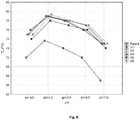
- DSF experiments were conducted for the parent and 5 variants at pHs ranging from 4 to 7.
- Fluorescence intensity curves for the parent displayed the typical two-state unfolding transition (folded to unfolded with no stable intermediates) [37,46] at all pH levels tested. However, all the variant profiles were pH dependent. At low pH the unfolding proceeded via the typical two-state transition while at higher pH unfolding occurred in via multiple-state transitions. This was attributed to a mutation(s) that stabilised a portion of the protein and thus more energy was required for complete unfolding. The maximum fluorescence at pH 4 was in excess of 60000 AU, whereas at pH 7 it was 22000 AU. Hirayama et al. [35] reported the optimum pH for the wild type enzyme in terms of activity and stability to be between 5 and 6 and 6.5, respectively.
- V1 F140Y-A178P-G321N-Q490S
- V3 F140Y-A178P-G321N
- V4 A178P-G321N-Q490S
- V8 was the most thermostable enzyme, it did not display the highest specific activity.
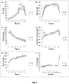
- DSF was used to perform comparative investigations into substrate interactions between the parent and variant proteins. More effective ligand binding to enzymes results in delayed thermal denaturation and higher T m s. Results for ⁇ T m (T m at given substrate concentration minus T m at 0 substrate concentration) at different substrate and product concentrations are shown in Figure 7 . At 0.2 M sucrose increased T m s were observed for all enzymes ( Figure 7A ). Further increased sucrose concentrations resulted in diminished substrate binding for all enzymes as evidenced by lower T m s.
- V1 the most improved variant, displayed increased affinities for sucrose and 1-kestose and decreased affinities for nystose and glucose at the highest substrate concentrations tested.
- the T m of the parent was unaffected by sucrose or 1-kestose while it was increased by nystose and glucose, again at the highest concentrations tested.
- these data indicated that subtle changes to the range of substrate and product affinities as well as improved thermostability were responsible for improved enzyme activities. It is plausible that the amino acid substitutions altered the active site such that the enzymes were relieved to an extent from substrate and product inhibition, conditions that are well documented in the literature for ⁇ -fructofuranosidases, albeit not this specific enzyme. Further kinetic characterisation is required to fully understand the impact of substitutions on the enzymes' activities.
- V1 has a higher turnover number (k cat ) than the parent.
- the fructose data for both enzymes were virtually identical, which indicated that the hydrolytic activity was unchanged in the variant.
- V1 consumed marginally more sucrose than the parent, but it was not reflected in the amount of total FOS produced.
- experimental error could account for the discrepancy (a given amount of sucrose translates into a small amount of FOS which could be missed given a maximum of 10% error of quantification).
- hydrolytic activity of the enzyme dominated after 11 h as reflected by the sharp increase in fructose and corresponding decreases in GF4, nystose, sucrose and to a lesser extent, 1-kestose levels.
- P. pastoris cultivations were performed in 1.3L New Brunswick Bioflo 110 Fermenters/Bioreactors with a working volume of 1L. Biocommand version 3.30 plus software was used for monitoring and feed rate control.
- BMGH buffered minimal glycerol medium
- Fermentation basalt salt medium BSM
- PTM 1 trace salts were used as culture media as described by the Pichia Fermentation protocol (Invitrogen). Culture conditions were maintained as follows: temperature of 30°C and a pH of 5.0 was maintained with 28% ammonium hydroxide, aeration rate of 1.0 volume of oxygen per volume of fermentation culture per minute (vvm), the dissolved oxygen (DO) was maintained at 30% controlled by a cascade effect between agitation (200-1000 rpm) and sparging O 2 when agitation was not sufficient.
- sucrose was used as the substrate and prepared in a 50 mM citrate phosphate buffer (pH 5.5) and used at a working concentration of 100 g.l -1 .
- the substrate solution was equilibrated at 40°C for 10 min where after culture supernatant was added to a final of 25% [v/v] and incubated for 60 min.
- perchloric acid (PCA) was added to a final concentration of 2.14% followed by the addition of 7 N KOH to precipitate the proteins prior to chemical analysis.
- Negative control reactions containing all the assay constituents except for either sucrose or enzyme were included.
- the samples were diluted appropriately and subjected to HPLC analysis using an external glucose standard calibration. The method has been decribed previously [69]. The concentration of glucose liberated during the assays was indicative of global fructofuranosidase activity.
- a unit of enzyme was defined as the amount of enzyme required to produce 1 ⁇ mol glucose per minute under the described conditions [14].
- scFOS a 60% sucrose solution (w/v) was prepared in a 50 mM citrate phosphate buffer (pH 5.0).
- the substrate solution was equilibrated at the required temperature in a Gyrotory Water Bath Shaker (New Brunswick Scientific Co. Inc., Edison N.J., USA) for 2 min while shaking at 120 rpm where after culture supernatant was added at a predetermined dosage according to central composite design.
- Samples were taken every 2 hrs and the reaction stopped by adding perchloric acid (PCA) to a final concentration of 2.14% followed by the addition of 7 N KOH to precipitate the proteins prior to chemical analysis in a Dionex UltiMate 3000 (Thermo Fisher Scientific, Waltham Mass., USA).
- PCA perchloric acid
- scFOS was optimised as a function of temperature and enzyme dosage using response surface methodology (RSM) with a two-factor central composite design using Design Expert® software (Stat-Ease Inc., Minneapolis, USA).
- the input factors in the design were selected in the ranges of 57°C ⁇ A ⁇ 67°C and 8 U/g sucrose ⁇ B ⁇ 12 U/g sucrose, where A represents the temperature and B the enzyme dosage.
- This design gave a total of 11 experiments for each enzyme (Tables 3 and 4).
- Table 3 Central composite design of temperature (A) and enzyme dosage (B) for scFOS production by the enzyme fopA_V1 using 60% (w/v) sucrose solution for 8 hrs.
- the culture volume in the bioreactors was harvested after 94 hrs and the biomass separated from the volume.
- the enzyme assays of the two strains yielded enzyme activities of 1202 U/ml for P. pastoris fopA and 1124 U/ml for G250.2.
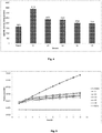
- fopA_V1 was able to produce sugar compositions similar to Actilight® at 6 hrs decreasing the incubation time by 2 hrs compared to fopA ( Figure 10 and Table 3).
- the fopA_V1 enzyme produced higher percentage GF4 or similar amounts in shorter time periods than the fopA enzyme at all the conditions tested. It was also able to tolerate higher temperatures and decrease incubation times at the higher enzyme dosages.
Landscapes
- Life Sciences & Earth Sciences (AREA)
- Chemical & Material Sciences (AREA)
- Health & Medical Sciences (AREA)
- Organic Chemistry (AREA)
- Engineering & Computer Science (AREA)
- Wood Science & Technology (AREA)
- Zoology (AREA)
- Bioinformatics & Cheminformatics (AREA)
- Genetics & Genomics (AREA)
- General Health & Medical Sciences (AREA)
- Biochemistry (AREA)
- General Engineering & Computer Science (AREA)
- Molecular Biology (AREA)
- Microbiology (AREA)
- Biotechnology (AREA)
- Polymers & Plastics (AREA)
- Nutrition Science (AREA)
- Food Science & Technology (AREA)
- Medicinal Chemistry (AREA)
- Biomedical Technology (AREA)
- Proteomics, Peptides & Aminoacids (AREA)
- Chemical Kinetics & Catalysis (AREA)
- General Chemical & Material Sciences (AREA)
- Mycology (AREA)
- Enzymes And Modification Thereof (AREA)
- Micro-Organisms Or Cultivation Processes Thereof (AREA)
Description
- The invention related to modified β-fructofuranosidases which have improved enzyme activity, in particular fructosyltransferase activity, relative to the parent enzyme.
- The global demand for fructooligosaccharides (FOS) is growing due to human health benefits associated with their consumption. FOS are prebiotics that selectively stimulate the growth of bifidobacteria, thereby promoting colonic health [1,2]. Further claims as to the effect of FOS consumption relate to mineral absorption, lipid metabolism and the control of type II diabetes and have been extensively reviewed [2-4]. Further to their health benefits, FOS are used in the food industry as low calorie sweeteners. They are also added to food products to improve their organoleptic properties and their inclusion allows producers to label their products as 'functional foods' - a claim that resonates with health conscious consumers [2,3].
- It is well known that some β-fructofuranosidases possess the ability to transform sucrose to FOS. β-fructofuranosidases are family 32 glycoside hydrolase (GH32) enzymes that act on sucrose and related β-D-fructofuranosides [5]. They are also known as invertases (EC 3.2.1.26) as they hydrolyse sucrose to produce invert sugar - an equimolar mixture of dextrorotatory D-glucose and levorotatory D-fructose [6]. Crystal structures for GH32 β-fructofuranosidases reveal that the enzymes display a bimodular arrangement of a N-terminal catalytic domain containing a five-bladed β-propeller fold linked to a C-terminal β-sandwich domain [7-10]. β-fructofuranosidases hydrolyse β-glycosidic bonds by a double displacement catalytic mechanism that retains the configuration of the fructose anomeric carbon [11]. Multiple sequence alignments (MSAs) identified a highly conserved aspartate close to the N terminus that serves as the catalytic nucleophile and a glutamate residue that acts as a general acid/base catalyst [12]. The β-fructofuranosidases which are capable of transforming sucrose to FOS possess fructosyltransferase activity whereby the sugar moiety is transferred from the enzyme-fructosyl intermediate to an acceptor other than water [7,13]. This reaction forms the basis of FOS synthesis from sucrose. Enzymes from Aspergillus spp. [14-16] and Aureobasidium pullulans [17] exhibit good propensities for the synthesis of inulin type FOS from sucrose, with β-(2 → 1) linkages between fructose units.
- Synthesis of FOS (GFn) from sucrose (GF) occurs via a disproportionation reaction with the reaction generalised as GFn + GFn → GFn-1 + GFn+1 [18,19]. In a batch reaction the initial products are glucose and 1-kestose (GF2), and as the reaction progresses, nystose (GF3) and β-fructofuranosyl nystose (GF4) levels increase. Reaction conditions influence the dominance of hydrolytic or transferase reactions with high substrate concentrations favouring the latter [14].
- Industrial biotransformation of sucrose to FOS is currently conducted in a batch system using the β-fructofuranosidase from A. niger ATCC 20611 (subsequently classified as A. japonicus). The enzyme is added to a buffered 50 - 60% (wt/vol) sucrose solution with the reaction proceeding at 50 - 60°C for up to 20 hours [19]. These severe industrial conditions impose limitations on activity. The fructosyltransferase activity of the enzyme has been shown to be non-competitively inhibited by the glucose product, limiting complete sucrose conversion [19]. Furthermore, long-term enzyme stability is severely compromised at temperatures above 50°C despite immobilisation efforts [20].
- There is thus still a need for alternative enzymes which are able to efficiently convert sucrose to FOS on an industrial scale.
-
EP 1 726 655 A1positions 62, 122, 128, 165, 221, 395 and 550. - Chuankhayan et al. (2010) describes the crystal structures of recombinant fructosyltransferase (AjFT) from Aspergillus japonicus CB05 and its mutant D191A complexes with various donor/acceptor substrates, including sucrose, 1-kestose, nystose, and raffinose.
-
EP 0 889 134 A1 describes a beta-fructofuranosidase gene and a beta-fructofuranosidase encoded by the gene, a process for isolating a beta-fructofuranosidase gene using the beta-fructofuranosidase gene, and a beta -fructofuranosidase obtained by this isolation process. A mold fungus having no beta-fructofuranosidase activity suitable for the production of beta-fructofuranosidase, and a system for producing a recombinant beta-fructofuranosidase using the mold fungus as a host is disclosed. Further, a beta-fructofuranosidase variant which selectively and efficiently produces a specific fructooligosaccharide such as I-kestose from sucrose is disclosed.
Trollope et al. (2013) describes the development of a screening method displaying potential high-throughput capacity for the evaluation of β-fructofuranosidase libraries using Fourier transform mid-infrared attenuated total reflectance (FT-MIR ATR) spectroscopy and multivariate analysis, and highlights the application of FT-MIR ATR spectroscopy to a variant discovery pipeline in the directed evolution of a β-fructofuranosidase for enhanced short chain FOS production
van Wyk et al. (2013) identifies a β-fructofuranosidase gene (CmINV) from a Ceratocystis moniliformis genome sequence using protein homology and phylogenetic analysis. - This invention provides a modified polypeptide having specific fructofuranosidase activity more particularly as defined in the accompanying claims.
- According to a first embodiment of the invention, there is provided a modified polypeptide having fructofuranosidase activity, wherein the amino acid sequence of the polypeptide is at least 90% or 95% identical to SEQ ID NO: 3 and has at least one of the following amino acid substitutions:
- the phenylalanine (F) at amino acid position 121 is substituted by tyrosine (Y);
- the alanine (A) at amino acid position 159 is substituted by proline (P) or serine (S);
- the glycine (G) at amino acid position 302 is substituted by asparagine (N), aspartic acid (D), tyrosine (Y) or glutamic acid (E); and/or
- the glutamine (Q) at amino acid position 471 is substituted by serine (S), lysine (K) or asparagine (N).
- The polypeptide may differ only from SEQ ID NO: 3 at one or more of positions 121, 159, 302 and/or 471.
- The polypeptide may include a secretion signal peptide at its 5' end, the secretion signal having an amino acid sequence which is at least 90% or 95% identical to SEQ ID NO: 25. The modified polypeptide with secretion signal may have an amino acid sequence which is at least 90% or 95% identical to SEQ ID NO: 26 and which has at least one amino acid substitution at position 154, 192, 335 and/or 504 of SEQ ID NO: 26.
- The polypeptide may include any two of the above substitutions, such as at amino acid positions 121 and 302 of SEQ ID NO: 3.
- The polypeptide may include any three of the above substitutions, such as at amino acid positions 121, 159 and 302, or at amino acid positions 159, 302 and 471, or at amino acid positions 121, 159 and 471 of SEQ ID NO: 3.
- The polypeptide may include any four of the above substitutions, such as at amino acid positions 121, 159, 302 and 471 of SEQ ID NO: 3.
- Also disclosed herein, the phenylalanine (F) at amino acid position 154 may be substituted by tyrosine (Y), the alanine (A) at amino acid position 192 may be substituted by proline (P) or serine (S), the glycine (G) at amino acid position 335 may be substituted by asparagine (N), aspartic acid (D), tyrosine (Y) or glutamic acid (E), and/or the glutamine (Q) at amino acid position 504 may be substituted by serine (S), lysine (K) or asparagine (N).
- Also disclosed herein, more preferably, the substitution at amino acid position 154 is tyrosine (Y), the substitution at amino acid position 192 is proline (P), the substitution at amino acid position 335 is asparagine (N), and/or the substitution at amino acid position 504 is serine (S).
- Even more preferably, the polypeptide may include the following modifications:
- F121Y-A159P-G302N-Q471S;
- F121Y-A159P-G302N;
- A159P-G302N-Q471S;
- F121Y-A159P-Q471S; or
- A159P-G302N.
- Even more preferably, the polypeptide may include the following four modifications: F121Y, A159P, G302N and Q471S.
- Also disclosed herein, the polypeptide may comprise an unsubstituted amino acid residue at
positions 62, 122, 128, 165, 221, 395 and/or 550 of SEQ ID NO: 3
The polypeptide may comprise an amino acid sequence of any one of SEQ ID NOS: 4 - 21, such as SEQ ID NO: 4. - Also disclosed herein, the polypeptide may have an improved thermal stability, catalytic rate and lower glucose feedback inhibition levels relative to a polypeptide which has not been modified as described above.
- According to a second embodiment of the invention, there is provided a polynucleotide which encodes a modified polypeptide as described above.
- The polynucleotide may have a nucleotide sequence which is at least 90% identical to the sequence of
nucleotides 100 to 2007 of SEQ ID NO: 1 or SEQ ID NO: 2, or a complement thereof, wherein SEQ ID NO: 1 or SEQ ID NO: 2 has been modified so that the polynucleotide encodes a modified polypeptide as described above. - The polynucleotide may include a T. reesei endoxylanase 2 (xln2) secretion signal. The secretion signal may be encoded by nucleotides 1-99 of SEQ ID NO: 1 or 2.
- According to a third embodiment of the invention, there is provided a vector comprising a polynucleotide encoding a modified fructofuranosidase polypeptide as described above.
- According to a fourth embodiment of the invention, there is provided a host cell comprising the vector described above.
- The host cell may be a microbial cell, such as from a yeast, bacterium or fungus.
- Also disclosed herein, according to a fifth embodiment, there is provided a yeast, fungus or bacterium comprising at least one copy of an exogenous gene coding for a modified polypeptide as described above.
- According to a sixth embodiment of the invention, there is provided a process for producing a modified fructofuranosidase polypeptide as described above, the process comprising the steps of transforming a host cell with the polynucleotide described above and causing the polypeptide to be expressed.
- The process may also comprise the step of recovering the polypeptide, such as from the transformed host cell or a supernatant into which the polypeptide has been secreted.
- The host cell may be a yeast cell, fungal cell or bacterium.
- Also disclosed herein, the polypeptide may be expressed under the control of a constitutive or inducible promoter, such as the S. cerevisiae phosphoglycerate kinase 1 (PGK1), glyceraldehyde-3-phosphate
dehydrogenase (GAP) promoter or the alcohol oxidase (AOX1) promoter in Pichia pastoris. - According to a seventh embodiment of the invention, there is provided a process for producing fructooligosaccharides (FOS), the process comprising the steps of contacting sucrose with a modified polypeptide described above under conditions which cause the sucrose to be converted into fructooligosaccharides. The fructooligosaccharides may be short chain fructooligosaccharides.
- The transformed host cell described above may be added to a solution containing sucrose and be caused to express the polypeptide into the solution, or alternatively a purified, partially purified or crude extract of polypeptide may be added directly to the solution containing sucrose.
- The fructooligosaccharides may include 1-kestose (GF2), nystose (GF3) and/or β-fructofuranosyl nystose (GF4).
- Also disclosed herein, the process may require lower amounts of the modified polypeptide to produce the FOS or may be performed over a shortened reaction time, relative to an unmodified process.
-
-
Figure 1 : Amino acid sequence of FopA (SEQ ID NO: 3) (numbered according to crystal structure) showing substitutions which were selected for generating a first round of FopA variants. -
Figure 2 : Screening data for the active first round variants harbouring single amino acid substitutions. The parent enzyme is indicated by the filled circle. Error bars denote standard error (n = 3). Nystose data were generated by HPLC from assays performed under glucose inhibiting conditions. Sucrose consumption under normal and glucose inhibiting conditions was quantified by Fourier transform mid-infrared spectroscopy. Relative inhibition was expressed as the difference in sucrose consumption between the two conditions divided by uninhibited activity. Data were normalised to the parental activity. The most improved variants in terms of nystose production and/or relative inhibition are labelled. -
Figure 3 : SDS-PAGE gels of purified (A) and crude (B, C) parent and five most improved combination variant β-fructofuranosidases. All gels were 8% and silver stained. B shows crude supernatants of S. cerevisiae NI-C-D4[fopA].Lane 2 is a reference as yeast was transformed with empty vector pJC1.Lanes 3 to 8 show the parent and the five most improved variants. A shows the IMAC purified enzymes.Lanes 2 to 7 show the parent and variant enzymes. C shows the result of PNGase treatment of the crude supernatant (parent).Lane 2 control,lane 3 untreated andlane 4 treated sample. The Spectra multicolor high range protein ladder (Thermo Scientific) was loaded inlane 1 of all the gels and served as molecular weight marker. -
Figure 4 : Specific activity data for the purified parent and five most improved combination variants. Values above each bar indicate the average 1-kestose units per milligram purified enzyme. Error bars denote standard error (n = 3). -
Figure 5 : Isothermal denaturation data for the parent and five most improved combination variants. Purified proteins were incubated with SYPRO orange at 55°C and fluorescence was monitored for 10 hours. Increased fluorescence indicates thermal denaturation of the protein as SYPRO orange binds to newly exposed hydrophobic amino acids. Error bars denote standard error (n = 5). -
Figure 6 : Differential scanning fluorimetry-derived melting temperatures for purified parent and five most improved combination β-fructofuranosidase variants at pHs ranging from 4 - 7. Error bars denote 0.95 confidence intervals (n = 3). -
Figure 7 : ΔTm values for the parent and 5 most improved combination variants in the presence of substrates sucrose (A), 1-kestose (B) and nystose (C). Differences between the given concentrations and the zero substrate Tms are shown. Error bars denote 0.95 confidence intervals (n = 3). -
Figure 8 : Time course FOS synthesis by the purified parent and most improved variant (V1) enzymes. The enzyme dosage was 10 KU per gram sucrose with a starting concentration of 600 g/l sucrose. The reaction was conducted at 62 °C, pH 5.5 with shaking at 120 rpm. Error bars denote standard error (n = 3). -
Figure 9 : Response surface plots of GF4 percentage of total scFOS as a function of temperature (A) and enzyme dosage (B) for the enzyme fopA_V1 after 6 hrs. -
Figure 10 : Response surface plots of GF4 percentage of total scFOS as a function of temperature (A) and enzyme dosage (B) for the enzyme fopA_V1 after 8 hrs. -
Figure 11 : Response surface plots of GF4 percentage of total scFOS as a function of temperature (A) and enzyme dosage (B) for the enzyme fopA after 8 hrs. - Modifications to the Aspergillus japonicus β-fructofuranosidase enzyme which result in improved synthesis of inulin-type fructooligosaccharides (FOS) from sucrose are described herein.
- Semi-rational directed evolution of the A. japonicus β-fructofuranosidase using a combination of strategies was performed and is described below. Only loop regions were selected for engineering using a crystal structure-guided approach, and amino acid substitutions were selected based on scoring positions in homologous protein sequence alignments using sequence entropies and solvent accessibilities. 36 variants of the β-fructofuranosidase, each with a single amino acid substitution, were engineered and this library was screened in Saccharomyces cerevisiae for variants that produced higher levels of FOS than the parent (wild type) enzyme. Enzymes were further screened for those producing more FOS than the parent under glucose inhibiting conditions. This strategic combination likely resulted in the enriched functionality of the library - 58% of the first round library was active (these results are in contrast to the results of a similar strategy which focused on engineering loops, where site saturation mutagenesis at 90 loop residues in a lipase only yielded 10% of active mutants that were improved relative to the parent [65]).
- Hits from the first round of screening were exhaustively combined to create a second library of combination variants with 2, 3 or 4 mutations, and a second round of screening was conducted to find combination variants with improved activity.
- The amino acid substitutions mentioned below will be referred to relative to their position in the crystal structure of the mature polypeptide. The corresponding amino acid position in any of SEQ ID NOS: 3 - 21 can be calculated by subtracting 19 from the crystal position, and this position number is shown in parenthesis in some of the substitutions described below. In the claims, the position numbers of the amino acid residues into which substitutions are introduced correspond to the amino acid residues of the amino acid sequences represented by SEQ ID NOs: 3 - 21.
- In the present invention, "substitution" means that a specific amino acid residue at a specific position is removed and another amino acid residue is inserted into the same position.
- Modification to the A. japonicus β-fructofuranosidase polypeptide at
positions 140, 178, 321 and/or 490 corresponding to the crystal structure of the mature polypeptide (corresponding to amino acids 121, 159, 302 and/or 471 of SEQ ID NOs: 3-21)) showed improved enzyme activity. Preferred modifications are substitution of the phenylalanine reside at position 140 (121) with a tyrosine residue, substitution of the alanine residue at position 178 (159) with a proline or serine residue, substitution of the glycine residue at position 321 (302) with an asparagine, aspartic acid, tyrosine or glutamic acid residue, and/or substitution of the glutamine residue at position 490 (471) with a serine, lysine or asparagine residue. More particularly, the substitution at position 140 (121) can be a tyrosine residue, the substitution at position 178 (159) can be a proline residue, the substitution at position 321 (302) can be an asparagine residue, and/or the substitution at position 490 (471) can be a serine residue. Consequently, a variant polypeptide of SEQ ID NO: 3 which has all four of the following amino acid residues does not fall within the scope of the present invention: phenylalanine at position 121, alanine at position 159, glycine at position 303 and glutamine at position 471. A178P and G321N were identified as positive contributors to thermostability. Proline substitutions in loops have been linked to improved thermostability due to backbone modifications that increase loop rigidity [58,66] and the data herein supports these findings. - 71% of active mutants (15 variants) were improved over the parent. Data showed that the effect of combining the top 4 first round substitutions was cumulative and delivered the best variant instead of a 3 or 2 combination or even single substitution variant. One particular combination variant with 4 amino acid substitutions displayed a combination of improved thermostability and catalytic activity. This variant was designated "V1" and had the following modifications: F140Y-A178P-G321 N-Q490S (F121Y-A159P-G302N-Q471S).
- V1 had a specific activity that was 2-fold higher than that of the parent, and tested under conditions approximating its industrial application, V1 displayed an improved catalytic effectiveness than that of the parent by reducing the time to completion of the reaction by 22%. Extrapolation from the DSF thermostability data would suggest that V1 can be applied under reaction conditions at least 5°C higher than currently employed, and it is probable that time to completion can be further reduced, as it is accepted that reactions kinetics are enhanced at elevated temperatures.
- Previous improvements to fructosyltransferase activity of fungal β-fructofuranosidases have been achieved by altering amino acids in the active site pocket [23,24] and in the non-catalytic β-sandwich domain [21]. Although catalytic pocket residues seem the obvious choice for amino acid substitutions, the applicant has demonstrated that substitutions in solvent exposed loops mediate long range interactions which alter active site geometry and in turn modify enzyme activity.
- In order to optimise expression of the modified polypeptide in a host cell, such as a microbial cell from a yeast (e.g. Pichia pastoris), fungus or bacterium, the polynucleotide encoding the modified polypeptide can be codon-optimised according to the host cell. Methods for codon-optimisation are well known to those skilled in the art. The polynucleotide can optionally include a secretion signal, such as that for T. reesei endoxylanase 2 (xln2) or any other suitable secretion signal. A vector including the polynucleotide can be used to transform the host cell. The polypeptide may be expressed under the control of various constitutive or inducible promoters, such as the S. cerevisiae phosphoglycerate kinase 1 (PGK1) promoter, glyceraldehyde-3-phosphate dehydrogenase (GAP) promoter or the alcohol oxidase (AOX1) promoter in Pichia pastoris. The transformed host cell can be added to a solution containing sucrose and be caused to express the polypeptide into the solution, possibly as whole cell catalysts, or alternatively a purified, partially purified or crude extract of the modified polypeptide can be added directly to the solution containing sucrose.
- As described in more detail below, the β-fructofuranosidase gene (fopA) from Aspergillus niger ATCC 20611 (A. japonicus) was codon optimised for expression in Pichia pastoris DSMZ 70382 (purchased from the The Leibniz Institute DSMZ - German Collection of Microorganisms and Cell Cultures GmbH (Deutsche Sammlung von Mikroorganismen und Zellkulturen GmbH)). The protein was further engineered for higher specific activity, decreased glucose inhibition and thermostability by introducing the mutations of V1. The original enzyme (FopA) produced by the P. pastoris fopA strain was then compared to the protein-engineered enzyme (fopA_V1) produced by the P. pastoris G250.2 strain for its ability to produce a similar sugar composition from sucrose to Actilight®, a prebiotic ingredient which has been proven to have multiple health benefits and is extensively used in a wide variety of functional foods. Actilight®, available from Beghin Meiji and Teros Syral, contains scFOS in a ratio of 37%, 53% and 10% for GF2, GF3 and GF4, respectively.
- The invention will now be described in more detail by way of the following non-limiting examples.
- S. cerevisiae EUROSCARFY02321 [BY4741;Mat a; his3Δ1; leu2Δ0; met15Δ0; ura360; YIL162w(SUC2)::kanMX4] served as host for the variant libraries [25]. The S. cerevisiae NI-C-D4 [Matα; trp1; ura3; pep4] oversecretion phenotype strain was used when heterologous proteins were to be purified [26]. Escherichia coli DH5α [thuA2Δ(argF-lacZ)U169 phoA glnV44 Φ80Δ (lacZ)M15 gyrA96 recA1 relA1 endA1 thi-1 hsdR17] (New England Biolabs, Midrand, South Africa) was used for cloning and amplification of plasmids. E. coli cells were grown at 37 °C in Luria Bertani broth supplemented with 100 µg/ml ampicillin or 50 µg/ml kanamycin, as appropriate.
- All DNA manipulations were performed according to standard methods [27]. Restriction enzymes and T4 DNA ligase were used according the specifications of the supplier (ThermoScientific, Waltham, Massachusetts, USA). CLC Main Workbench version 6.8.1 (Qaigen) was used for sequence analyses.
- A codon-optimised (parent) gene was provided by DNA 2.0 (Menlo Park, CA, USA) as a synthetic construct combining the Trichoderma reesei endoxylanase 2 (xln2) secretion signal [28] and the open reading frame of the fopA β-fructofuranosidase (GenBank accession number AB046383) (SEQ ID NO: 1 and 3). The native fopA secretion signal was excluded. Thirty-six variants of the β-fructofuranosidase gene were synthesised. The parent gene sequence was altered to produce gene products harbouring single amino acid substitutions. The substitutions were distributed throughout the protein sequence but were limited to loop regions as determined from the published crystal structure (3LF7) of Chuankhayan et al. [7]. The published crystal structures were determined for the A. japonicus β-fructofuranosidase. There is 99% homology on the DNA (AB046383, GU356596.1) and protein (BAB67771.1, ADK46938.1) levels of the β-fructofuranosidases of A. niger and A. japonicus, respectively. Aspergillus niger ATCC 20611 was reclassified by the curators of the ATCC culture collection (http://www.lgcpromochem-atcc. com) as A. japonicus and it was therefore assumed that the sequences and structures deposited in the databases are for the same gene/enzyme.
- Positions for amino acid substitutions were selected by the strategy provided by DNA2.0. An alignment of homologous sequences to the 3LF7 structure was used from the HSSP database [29]. Positions within the MSA corresponding to secondary structural elements of the 3LF7 chain A were excluded. The solvent accessibility computed for each sequence position was normalised according to values obtained for Ala-X-Ala tripeptides [30,31]. This provided a relative solvent accessibility at each position normalized by the side chain type. The sequence entropy for each position in the multiple sequence alignment was also obtained from the HSSP file for RCSB Protein Data Bank entry 3LF7. Sequence positions with relative solvent accessibility (RSA) greater than 50% and sequence entropy (SE) greater than 1.0 provided a list of positions in the structure-based multiple sequence alignment. From these alignment data, the most commonly observed substitutions were selected for inclusion in a 36 variant first round library. The mutations are listed in Table 1. A further 18 genes were synthesised containing exhaustive combinations of 5 single mutations that were determined to improve enzyme performance during first round screening (Table 2). The amino acid sequences of these variants are shown in SEQ D NOS: 4 - 21.


- Cloning vectors (pJ227) containing the synthesised gene variants were digested with EcoRI and XhoI restriction enzymes (Fermentas) and directly ligated with the pJC1 yeast expression vector [32] digested with the same restriction enzymes. Selection of E. coli transformants on LB agar plates supplemented with 100g/ml ampicillin ensured isolation of clones with the gene variant-pJC1 combination and not re-circularised gene variant-pJ227, as the cloning vector conferred resistance to kanamycin. The primer pair 5'GTTTAGTAGAACCTCGTGAAACTTA 3' (SEQ ID NO: 22) and 5'ACTTAAAATACGCTGAACCCGAACAT3' (SEQ ID NO: 23) was used to screen clones by polymerase chain reaction to ensure the presence of the 2000 base pair insert. Positive clones were further confirmed by restriction digest analysis. Yeast was transformed by the lithium acetate method described by Hill et al. [33]. The method was adapted to 96-well format by proportionally scaling down reagents.

- S. cerevisiae was cultivated at 30 °C in YPD (1 % yeast extract, 2 % peptone and 2 % glucose) or in synthetic medium, SC without uracil [2% carbon source, 0.67% yeast nitrogen base without amino acids (with ammonium sulphate; Difco Laboratories, Detroit, Michigan, USA) and 0.13 % amino acid dropout pool [34]]. Glucose and galactose served as carbon source in solid and liquid SC-ura media, respectively. Solid media contained 2% agar (Difco Laboratories).
- Three yeast transformants per variant were manually transferred to individual wells of 2 ml round bottomed 96-deep-well plates (Merck, Modderfontein, South Africa) containing 1.25 ml SC-ura media. Mixing was facilitated by a single 2-mm glass bead (Merck, Modderfontein, SouthAfrica) added to each well. Plates were sealed with sterile, breathable AeraSealTM film (Excel Scientific Inc., Victorville, California, USA) and shaken at 200 rpm for 4 days. Fifty microliters of each culture were transferred to a fresh plate and cultivated for a further 4 days. Master plates were generated using a 96-well replicator (Applikon Biotechnology, Delft, Netherlands). Culture supernatants were used in assays as source of enzyme after cell removal by centrifugation at 3000 rpm.


- Enzyme activity assays were performed in 96-well format by reacting 50 µl of culture supernatant with 50 µl of substrate at 55°C for 2 hours. The working concentration of substrate was 200 g/l sucrose (Fluka, Sigma-Aldrich, St. Louis, Missouri, USA) in 50 mM citrate phosphate buffer, pH 5.5. As determined previously [22], 54 g/l glucose was added to the substrate solution to test for variants insensitive to product inhibition. Saccharides in assay samples were quantified using high performance liquid chromatography and Fourier transform mid-infrared (FT-MIR) spectroscopy. The details of the methods were described previously [22]. The rationale for supplying galactose as carbon source in liquid cultures was the separation of glucose and galactose by HPLC and hence any glucose present in assay samples was attributed to enzyme activity. Inhibition was calculated as the difference between uninhibited variant activity and inhibited activity divided by uninhibited activity. Data were normalised to the parental activity. The parent and all variants were was cultivated and assayed with triplicate repeats. The cultivation and assay procedure was validated previously [22].
- The top five performing variants were purified using immobilised metal affinity chromatography (IMAC). The proteins were N-terminal His-tagged by sub-cloning BglII-XhoI gene fragments into the same sites in a modified pJC1 yeast expression vector. The vector was modified by cloning a synthetic fragment (Geneart, Regensburg, Germany) encoding the xyn2 secretion signal, six histidine residues and a factor Xa protease cleavage site into the EcoRI and BglII sites of the multiple cloning site (ATGGTTTCTTTCACATCCTTGTTGGCTGGTGTTGCTGCTATTTCCGGTGTTTTG GCTGCTCCAGCTGCTGAAGTTGAATCCGTTGCTGTTGAGAAGagaCATCACCATCACCATCAC GGATCcGGCTCTGGATCTGGTATCGAGGGAAGA (SEQ ID NO: 24). Tagged gene variants were sequenced to verify integrity of the clones. Plasmids were transformed to S. cerevisiae NI-C-D4. Transformants were cultivated for 72 hours in 50 ml double strength SC-ura buffered with succinic acid at pH 6.0 [(2% glucose, 1.34% yeast nitrogen base without amino acids (with ammonium sulphate; Difco Laboratories, Detroit, Michigan, USA) and 0.26 % amino acid dropout pool [34]]. Antifoam 204 (Sigma-Aldrich, St. Louis, Missouri, USA) was added after 48 hours of cultivation to a concentration of 0.025% (v/v). Following cell removal by centrifugation, supernatants were concentrated 50 times by ultrafiltration using Amicon ultra-15 centrifugal filters with 10 kDA MWCO (Millipore, Molsheim, France). IMAC protein purification was performed under native conditions using Ni-NTA spin columns supplied by Qaigen (Venlo, Netherlands). For the removal of imidazole, buffer exchange with 10 mM Bis-Tris,
pH 6 was performed using the aforementioned ultrafiltration devices. Protein concentration was determined using the bicinchoninic acid assay (Pierce Chemical Company, Rockford, Illinois, USA) with bovine serum albumin as standard. - A unit of enzyme was defined as the amount of protein that produced 1 mmol 1-kestose per minute from 10% (w/v) sucrose at 40°C in 50 mM citrate phosphate buffer (pH 5.5). The definition approximates that of Hirayama et al. [35].
- Samples were analysed by SDS-PAGE on an 8% resolving gel. Loading dye consisted of 60 mM Tris-HCI (pH 6.8), 25% glycerol, 2% SDS, 14 mM β-mercaptoethanol and bromophenol blue and gels were run in Tris-glycine buffer (25 mM Tris-HCI, 250 mM glycine, 0.1% SDS). Protein bands were visualized with a silver-stain [36].
- Protein thermal denaturation assays were performed by the method described by Niesen et al. [37]. SYPRO orange was supplied by Sigma-Aldrich (St. Louis, Missouri, USA) and used at a 5x working concentration. Each reaction utilised 65 ng of protein. Samples were incubated in a StepOnePlus Real-Time PCR machine (Applied Biosystems). IDT samples were incubated at 55°C for 10 hours while DSF samples were incubated with temperature increasing by 1 °C per minute from 25 - 95°C. Multicomponent data were exported from the StepOne software to Excel 2010 and ROX filter data were used. Data points beyond the maximum fluorescence +4 were discarded. First derivatives were calculated in Statistica version 12 (StatSoft Inc.). In cases where 2 peaks were obtained after application of the derivative, the temperature of the second peak was used. One-way analysis of variance (ANOVA) was conducted to test for differences between treatments applied to enzymes.
- Homology models for the single amino acid variants at the 4 positions yielding the most improved variants and the 5 best combination variants were generated by the SWISS-MODEL web server [38]. The crystal structure 3LF7 [7] served as template for the automated modelling mode. Template and target sequences shared 99% identity. Quality assessment of the models was performed by the QMEAN server (47).
- Solvent accessible surface area (SASA) data for the most improved combination variant were generated from the homology model. SASAs were computed for the folded (from the homology models) and unfolded states (sequence specific theoretical calculations) by the ProtSA web server (51). Differences between the folded and mean unfolded ensembles were determined. To examine the influence of the amino acid substitutions on SASAs, differences between the parent and the variant were further calculated.
- The Ligand-Protein Contacts (LPC) server was used to identify amino acid residues in contact with the ligand (first shell residues). Contacts with substituted residues in the variant were identified by the Contacts of Structural Units (CSU) server. Servers were accessed via http://ligin.weizmann.ac.il/cgi-bin/Ipccsu/LpcCsu.cgi (67). Functional site predictions where made using 3LF7 structure as input for the Partial Order Optimal Likelihood (POOL) server (67). The top 8% of ranked residues were taken as active site residues.
- FOS synthesis was performed with the parent enzyme and the variant displaying the highest specific activity. It was accomplished by reacting 10 U of parent enzyme per gram of sucrose and dosing the same amount of protein for the variant. The reaction was performed at 62°C with shaking at 120 rpm. Working concentrations were 600 g/l sucrose dissolved in 50 mM citrate phosphate buffer, pH 5.5. Samples were taken hourly for 12 hours and analysed by HPLC after appropriate dilution.
- Table 1 and
Figure 1 list the amino acid substitutions selected to generate the first round of variants. Although the substitutions were limited to the loop regions, they were otherwise distributed across the entire protein sequence length. Substitutions were made at 13 positions with multiple substitutions at 10 of the positions. Of the 36 variants, two were deemed inactive as evidenced by a lack of growth on solid media supplemented with sucrose as sole carbon source. Active gene products complemented the suc2 knockout in the S. cerevisiae strain enabling growth on sucrose containing solid media. The activities of a further 10 variants were deemed severely compromised as HPLC and FT-MIR spectroscopy indicated negligible amounts of sucrose conversion. SDS-PAGE analyses showed consistent protein expression levels for the variants, barring three where protein production was abolished - expected bands were absent. The activities of these three variants remained unknown (data not shown). The sensitivity of the catalytic β propeller domain of FopA to structural alterations was highlighted by 48% of substitutions resulting in abolished or severely compromised activities as opposed to the β sandwich domain where all variants remained active bar one (9%), the activity of which remains unknown.Figure 2 shows the nystose and relative inhibition data for the 21 variants retaining good activity levels. The smaller the relative inhibition value the less sensitive the enzyme to glucose inhibition. Variant A178S appeared to tolerate glucose better than other variants but at the expense of its fructosyltransferase activity. First round screening data were used to design combination variants of the top five performinground 1 variants. The top four nystose producing variants F140Y, A178P, Q490S and G321N and the single variant showing the greatest relief from glucose inhibition (A178S) were combined exhaustively to produce another 18 variants which included combinations of two, three and four amino acid substitutions. - Table 2 lists the combination variants grouped by number of substitutions and their activity data arranged per group in descending order for nystose. The parent (wild type) data is given in bold font. Sixteen of the 18 variants showed improved activity relative to the parent. Besides the single substitution variant A178S, only F140Y-A178S-G321N displayed poorer fructosyltransferase activity than the parent. Furthermore, all the variants with the A178S substitution performed worse than the variants with the A178P substitution. Although combining A178S with other substitutions did recover activity relative to the parent, it was generally assumed to be a deleterious substitution and did not prove permissive in a combinatorial context. No trend emerged for relative inhibition of combination variants and the A178S substitution (data not shown). Single substitutions ranked in decreasing order of nystose production A178P, G321N, Q490S, F140Y and A178S. A178P and G321N always proved to be good substitutions be it in isolation, in combination with each other and with either Q490S and/or F140Y. The contributions of Q490S and F140Y to enzyme activity were combination dependent. Together in a combination of 2 they were the poorest performers of the combination mutants. When combined with A178P, F140Y improved enzyme activity to a greater degree than Q490S - 37.6 g/l nystose produced as opposed to 34.7 g/l. In a combination of 2 with G321N, both F140Y and Q490S resulted in the same amount of nystose produced at approximately 44 g/l. However, the 1-kestose values differed with F140Y-G321N levels at 79.3 g/l while G321N-Q490S levels were 72.2 g/l. It is probable that G321N-Q490S was a more efficient enzyme as decreasing 1-kestose levels with similar nystose levels indicate GF4 production. No significant differences in the levels of sucrose, glucose and fructose supported this deduction. For a 3 combination variant the opposite for Q490S was true - A178P-G321N-Q490S produced more nystose than F140Y- A178P-G321N. The 4 combination variant F140Y-A178P-G321N-Q490S proved to be the most improved variant with
nystose levels 65% higher than the parent - 53.8 g/l versus 32.6 g/l. - This section applies to the parent and the five most improved combination variants. For simplicity the combination variants were named V1: F140Y-A178P-G321N-Q490S (F121Y-A159P-G321N-Q471S), V3: F140Y-A178P-G321N (F121Y-A159P-G302N), V4: A178P-G321N-Q490S (A159P-G302N-Q471S), V5: F140Y-A178P-Q490S (F121Y-A159P-Q471S) and V8: A178P-G321N (A159P-G302N).
- Enzymes were purified using IMAC.
Figure 3 shows the purified enzymes and crude yeast supernatants on a silver stained SDS-PAGE gel. The high degree of enzyme purity following IMAC is evident in gel A. The intense bands between 100 and 130 kDa correspond to the parent and variant β-fructofuranosidases - the band was absent in the reference (gel B, lane 2). The proteins were larger than the expected 69 kDa as deduced from the protein sequence but the shift was attributed to N-glycosylation of the proteins by yeast. Following the removal of the glycosylation with PNGase F, bands of the expected size were obtained (gel C, lane 4). All variants had 8 putative N-glycosylation sites which was unchanged from the glycosylation pattern of the parent [40]. Chuankhayan et al. [7] proposed that altered N-glycosylation patterns from heterologous hosts may influence enzyme stability. As the parent and all variants were expressed in the same host this was irrelevant but altered stability was ruled out due to altered N-glycosylation patterns resulting from different primary protein structures of the variants. - Specific activities for the purified enzymes were determined. Results are shown in
Figure 4 . The specific activity of the parent enzyme was 1652 U/mg protein which was lower than the 2650 U/mg protein reported by Nishizawa et al. [19]. Heterologous enzyme production and experimental disparities could account for this. Differences in activity between the native enzyme and the heterologous fopA gene product were also reported by Yanai et al. [41]. All the variants had significantly (p = 0.0023 for V8) higher specific activities than the parent, with V1 as much as double. - To investigate the stability of the improved variants, ITD (a method that quantifies protein stability by measuring protein unfolding caused by heat denaturation in the presence of SYPRO orange dye [42]) was employed. Typically this method is used to quantify stability and ligand affinity for a given protein. The method was employed in a modified sense in that the proteins were the variable factor. Purified proteins were incubated for 10 hours at 55°C to examine the influence of protracted exposure to temperatures routinely used for industrial FOS synthesis reactions.
Figure 5 shows that after 2 hours the fluorescence measured for the parent enzyme increased dramatically whereas the variants showed more measured increases. The increased fluorescence can be interpreted as thermal denaturation of the enzymes. Upon protein unfolding, binding of the SYPRO orange dye to newly exposed hydrophobic amino acid residues results in increased fluorescence [43]. It thus appeared that the variant enzymes were more thermostable than the parent. Slight differences in stability were observed between the variants, which could be attributed to the differing combinations of amino acid substitutions. - To further investigate the thermostability of the variants, DSF [37] was employed. The principle is similar to ITD, but instead of maintaining a set temperature, it is increased by 1°C per minute. The Tm of a protein is the temperature at which half of the protein molecules are unfolded and reflects the transition midpoint of the fluorescence vs temperature curve [37,44,45]. Factors influencing protein stability include buffers, salts and detergents and also specific interactions with ligands. Factors that promote stability delay the thermally induced unfolding and result in increased Tm. DSF experiments were conducted for the parent and 5 variants at pHs ranging from 4 to 7. Fluorescence intensity curves for the parent displayed the typical two-state unfolding transition (folded to unfolded with no stable intermediates) [37,46] at all pH levels tested. However, all the variant profiles were pH dependent. At low pH the unfolding proceeded via the typical two-state transition while at higher pH unfolding occurred in via multiple-state transitions. This was attributed to a mutation(s) that stabilised a portion of the protein and thus more energy was required for complete unfolding. The maximum fluorescence at
pH 4 was in excess of 60000 AU, whereas atpH 7 it was 22000 AU. Hirayama et al. [35] reported the optimum pH for the wild type enzyme in terms of activity and stability to be between 5 and 6 and 6.5, respectively. At pHs below 4.5 the stability and activity of the enzyme were severely compromised. The high fluorescence and two-state transition of the variant proteins at low pH were therefore attributed to the severely unfavourable conditions imposed on the enzyme atpH 4. The ionization states of amino acids were likely altered and hence contributed to extensive protein denaturation. Tms were calculated from the first derivatives applied to the fluorescence data [37]. For samples presenting multiple transitions the maximum of the second peak was used to determine the Tm. The results at each pH for the enzymes are shown inFigure 6 . Similar to the ITD results, it was clear that the variants were more thermostable than the parent. The pH optima profiles did not change due to amino acid substitution - the optima for all enzymes were at pH 5.0. At optimum pH V8 displayed a 5.7 °C increased Tm relative to the parent (73.7°C), making it the most thermostable variant. V1, V3 and V4 all displayed improvements of 5.3 °C, while V5 was improved by 4.3 °C. The differences between the three Tm groups were all statistically significant (p<0.05). Considering the combinations of substitutions in the variants, A178P and G321N appeared to be responsible for the improved thermostability. All variants harboured the A178P substitution and they were all more stable than the parent. As V1 (F140Y-A178P-G321N-Q490S), V3 (F140Y-A178P-G321N) and V4 (A178P-G321N-Q490S) all displayed the same improvement, it appeared that F140Y and Q490S were marginally destabilising substitutions in terms of thermostability - inclusion of either one or both of them resulted in a decreased Tm relative to V8. As V8 (A178P-G321N) was the most improved and V5 (F140Y-A178P-Q490S) the least which was concluded was that G321N contributed positively to stability. - Although V8 was the most thermostable enzyme, it did not display the highest specific activity. DSF was used to perform comparative investigations into substrate interactions between the parent and variant proteins. More effective ligand binding to enzymes results in delayed thermal denaturation and higher Tms. Results for ΔTm (Tm at given substrate concentration minus Tm at 0 substrate concentration) at different substrate and product concentrations are shown in
Figure 7 . At 0.2 M sucrose increased Tms were observed for all enzymes (Figure 7A ). Further increased sucrose concentrations resulted in diminished substrate binding for all enzymes as evidenced by lower Tms. At 0.5 M sucrose the parent, V5 and V8 displayed the same Tm as in the absence of substrate, whereas variants V1, V3 and V4 displayed increased Tms - differences were small, ⊗Tm of 1°C or less, but significant (p<0.05 for V1, V3 and V4). It was apparent that the substrate binding at 0.5 M sucrose was altered for enzymes with higher specific activities, namely V1, V3 and V4 (Figure 4 ). In the case of 1-kestose, all variants bound the substrate with higher affinity than the parent (Figure 7B ). Increased 1-kestose concentrations increased the stability of all variants except V4 which did not display an altered Tm at all concentrations tested. Enzyme stabilisation as a result of nystose binding was not evident at concentrations up to 0.2 M - no significant changes in Tm were observed (Figure 7C ). However, at 0.4 M the parent and V4 were stabilised significantly by nystose binding. The other variants did not show significant improvements in Tm as a result of substrate binding. Finally, binding of glucose (product) to enzymes was also examined and results indicated that the two most improved variants, V1 and V3, appeared to have diminished affinities for glucose - Tms were not affected at glucose concentrations up to 0.3 M while the parent, V3, V4 and V5 were stabilised by 1°C. In summary, the most improved variant, V1, displayed increased affinities for sucrose and 1-kestose and decreased affinities for nystose and glucose at the highest substrate concentrations tested. The Tm of the parent was unaffected by sucrose or 1-kestose while it was increased by nystose and glucose, again at the highest concentrations tested. Taken in combination, these data indicated that subtle changes to the range of substrate and product affinities as well as improved thermostability were responsible for improved enzyme activities. It is plausible that the amino acid substitutions altered the active site such that the enzymes were relieved to an extent from substrate and product inhibition, conditions that are well documented in the literature for β-fructofuranosidases, albeit not this specific enzyme. Further kinetic characterisation is required to fully understand the impact of substitutions on the enzymes' activities. - As an ultimate test of variant performance, equal amounts of purified parent enzyme and V1 were used to produce FOS under conditions similar to those used in industry. In commercial FOS products the ratio of GF2:GF3:GF4 is approximately 42:47:10 and depending on the product, compromise up to 95% of the dry mass after chromatographic separation of glucose, fructose and sucrose [19,60-62].
Figure 8 shows HPLC data for all the relevant sugars. It was evident that V1 has a higher catalytic activity relative to the parent, as the levels of nystose and GF4 were higher. Higher 1-kestose levels for the parent highlighted the difference in activities as this is the initial FOS species produced from sucrose. It in turn serves as substrate for the formation of nystose and GF4. As equal amounts of protein were dosed, it can be deduced that V1 has a higher turnover number (kcat) than the parent. The fructose data for both enzymes were virtually identical, which indicated that the hydrolytic activity was unchanged in the variant. V1 consumed marginally more sucrose than the parent, but it was not reflected in the amount of total FOS produced. As the differences in molecular weights between sucrose and FOS are large, experimental error could account for the discrepancy (a given amount of sucrose translates into a small amount of FOS which could be missed given a maximum of 10% error of quantification). Similarly for glucose, which is an indicator of global enzyme activity, clear differences at all the time points were not evident. Although the reaction was followed for 12 hours, the endpoint for a typical industrial reaction was regarded as the time when total FOS composition comprised 10% GF4. V1 reached 10% GF4 in 4.6 h while it took the parent 6.2 h to produce FOS of similar composition. This difference represented a 26% reduction in time required to complete the reaction. Otherwise stated, enzyme dosage could be reduced to achieve the same result in 6.2 h. - The hydrolytic activity of the enzyme dominated after 11 h as reflected by the sharp increase in fructose and corresponding decreases in GF4, nystose, sucrose and to a lesser extent, 1-kestose levels.
- P. pastoris cultivations were performed in 1.3L New Brunswick Bioflo 110 Fermenters/Bioreactors with a working volume of 1L. Biocommand version 3.30 plus software was used for monitoring and feed rate control. Pre-cultures were prepared by inoculating a streak of colonies from YPD agar plates into 4 ml of buffered minimal glycerol medium (BMGH) as described in the Pichia Expression Kit (Invitrogen) with the exception of a final YNB concentration of 0.17 %. Following overnight incubation at 30°C cultures were diluted to an optical density (OD)600 = 0.1 in 40 ml of fresh BMGH medium. Subsequently, these seed-cultures were grown overnight at 200 rpm to an OD600 = 10-15, before inoculating the entire volume to the medium to a final volume of 400 ml in the bioreactor, obtaining a starting OD600 = 1.0-1.5.
- Fermentation basalt salt medium (BSM), supplemented with 4% glycerol and PTM1 trace salts, was used as culture media as described by the Pichia Fermentation protocol (Invitrogen). Culture conditions were maintained as follows: temperature of 30°C and a pH of 5.0 was maintained with 28% ammonium hydroxide, aeration rate of 1.0 volume of oxygen per volume of fermentation culture per minute (vvm), the dissolved oxygen (DO) was maintained at 30% controlled by a cascade effect between agitation (200-1000 rpm) and sparging O2 when agitation was not sufficient. Fermentations were performed as per the Pichia Fermentation protocol (Invitrogen) with the following exceptions: During the glycerol fed-batch phase the glycerol was fed via a DO-stat feeding strategy (feeding started when DO >= 30 % and stopped when it was < 30 %). This was continued for 72 hrs after the batch phase at which point the bioreactor volume was harvested, centrifuged for 3 min at 3000 rpm and then filtered through 22 µm filter and stored at 4°C for determining the enzyme activity.
- To determine the fructofuranosidase activity, sucrose was used as the substrate and prepared in a 50 mM citrate phosphate buffer (pH 5.5) and used at a working concentration of 100 g.l-1. The substrate solution was equilibrated at 40°C for 10 min where after culture supernatant was added to a final of 25% [v/v] and incubated for 60 min. To stop the reaction, perchloric acid (PCA) was added to a final concentration of 2.14% followed by the addition of 7 N KOH to precipitate the proteins prior to chemical analysis. Negative control reactions containing all the assay constituents except for either sucrose or enzyme were included. The samples were diluted appropriately and subjected to HPLC analysis using an external glucose standard calibration. The method has been decribed previously [69]. The concentration of glucose liberated during the assays was indicative of global fructofuranosidase activity. A unit of enzyme was defined as the amount of enzyme required to produce 1 µmol glucose per minute under the described conditions [14].
- To produce scFOS, a 60% sucrose solution (w/v) was prepared in a 50 mM citrate phosphate buffer (pH 5.0). The substrate solution was equilibrated at the required temperature in a Gyrotory Water Bath Shaker (New Brunswick Scientific Co. Inc., Edison N.J., USA) for 2 min while shaking at 120 rpm where after culture supernatant was added at a predetermined dosage according to central composite design. Samples were taken every 2 hrs and the reaction stopped by adding perchloric acid (PCA) to a final concentration of 2.14% followed by the addition of 7 N KOH to precipitate the proteins prior to chemical analysis in a Dionex UltiMate 3000 (Thermo Fisher Scientific, Waltham Mass., USA).
- The production of scFOS was optimised as a function of temperature and enzyme dosage using response surface methodology (RSM) with a two-factor central composite design using Design Expert® software (Stat-Ease Inc., Minneapolis, USA). The input factors in the design were selected in the ranges of 57°C ≤ A ≤ 67°C and 8 U/g sucrose ≤ B ≤ 12 U/g sucrose, where A represents the temperature and B the enzyme dosage. This design gave a total of 11 experiments for each enzyme (Tables 3 and 4).
Table 3: Central composite design of temperature (A) and enzyme dosage (B) for scFOS production by the enzyme fopA_V1 using 60% (w/v) sucrose solution for 8 hrs. Only time points where -10% GF4 was produced are shown or where conditions did not achieve this the values for the final two time points are shown. Run no. Factors Time (hrs) % of total scFOS A (°C) B (U/g sucrose) GF2 GF3 GF4 1 62.00 12.83 4 44.05 47.50 8.44 6 35.34 51.16 13.50 2 62.00 10.00 6 41.88 48.93 9.19 8 36.63 50.97 12.40 3 67.00 8.00 6 47.45 45.40 7.15 8 42.00 48.60 9.40 4 57.00 8.00 6 50.20 45.01 4.79 8 43.07 51.52 5.42 5 54.93 10.00 6 58.33 35.23 6.44 8 52.17 38.10 9.73 6 62.00 10.00 6 40.74 49.51 9.74 8 34.30 53.70 12.00 7 69.07 10.00 6 41.83 50.56 7.60 8 38.34 51.60 10.06 8 62.00 7.17 6 49.26 44.64 6.10 8 42.72 48.79 8.50 9 57.00 12.00 6 40.29 51.69 8.03 8 34.05 54.49 11.46 10 67.00 12.00 4 44.90 47.46 7.64 6 36.94 51.02 12.03 11 62.00 10.00 6 41.51 48.00 10.50 8 35.18 50.44 14.38 Table 4: Central composite design of temperature (A) and enzyme dosage (B) for scFOS production by the enzyme fopA using 60% (w/v) sucrose solution for 8 hrs. Only time points where ∼10% GF4 was produced are shown or where conditions did not achieve this the values for the final two time points are shown. Run no. Factors Time (hrs) % of total scFOS A (°C) B (U/g sucrose) GF2 GF3 GF4 1 62.00 12.83 6 40.43 49.60 9.97 8 35.05 51.54 13.41 2 62.00 10.00 6 45.60 45.44 8.96 8 39.81 49.41 10.78 3 67.00 8.00 6 54.89 40.25 4.86 8 50.81 43.18 6.02 4 57.00 8.00 6 56.22 38.94 4.83 8 49.40 45.05 5.55 5 54.93 10.00 6 60.02 34.31 5.67 8 51.25 42.41 6.33 6 62.00 10.00 6 45.43 47.77 6.80 8 39.39 51.07 9.54 7 69.07 10.00 6 53.55 42.39 4.06 8 50.69 44.47 4.84 8 62.00 7.17 6 54.71 41.10 4.19 8 48.24 45.70 6.06 9 57.00 12.00 6 43.51 49.91 6.58 8 36.72 53.63 9.65 10 67.00 12.00 6 43.79 48.42 7.79 8 40.80 50.06 9.14 11 62.00 10.00 6 44.25 47.46 8.29 8 38.54 50.99 10.47 - The culture volume in the bioreactors was harvested after 94 hrs and the biomass separated from the volume. The enzyme assays of the two strains yielded enzyme activities of 1202 U/ml for P. pastoris fopA and 1124 U/ml for G250.2.
- The optimum temperature for both enzymes is at ∼62°C (
Figures 9 to 11 ), but fopA_V1 was still able to produce 10.06% GF4 after 8 hrs at 69.07°C. This is more than double that for the fopA enzyme, which only produced 4.84% GF4 after the same amount of time. This would indicate that the fopA_V1 enzyme is more thermotolerant compared to the fopA enzyme. Both these enzymes struggled to produce 10% GF4 at the lower temperatures and required an increased enzyme dosage to achieve this (Tables 3 and 4). At enzyme dosages less than 10.00 U/g sucrose, both these enzymes were unable to produce a sugar composition similar to that of Actilight®. At the higher enzyme dosages of 12.83 U/g sucrose (62°C) and 12.00 U/g sucrose (67°C), fopA_V1 was able to produce sugar compositions similar to Actilight® at 6 hrs decreasing the incubation time by 2 hrs compared to fopA (Figure 10 and Table 3). - Generally, the fopA_V1 enzyme produced higher percentage GF4 or similar amounts in shorter time periods than the fopA enzyme at all the conditions tested. It was also able to tolerate higher temperatures and decrease incubation times at the higher enzyme dosages.
-
- 1. Hidaka H, Eida T, Takizawa T, Tokunahga T, Tashiro Y (1986) Effects of fructo-oligosaccharides on intestinal flora and human health. Bifidobact Microflora 5: 37-50.
- 2. Roberfroid MB (2007) Inulin-type fructans: functional food ingredients. J Nutr 137: 2493S-2502S. Available: http://www.ncbi.nlm.nih.gov/entrez/query.fcgi?cmd=Retrieve&db=PubMed&dopt=Citation&lis t_uids=17951492. Accessed 4 February 2014.
- 3. Sangeetha PT, Ramesh MN, Prapulla SG (2005) Recent trends in the microbial production, analysis and application of Fructooligosaccharides. Trends Food Sci Technol 16: 442-457. Available: http://www.sciencedirect.com/science/article/B6VHY-4GHSGHC-1/1/e35b9e9a9d27ffbbaf7197d399b05bff.
- 4. Singh R, Singh R (2010) Production of fructooligosaccharides from inulin by endoinulinases and their prebiotic potential. Food Technol Biotechnol 48: 435-450. Available: http://www.researchgate.net/publication/215601814_Production_of_Fructooligosaccharides _from_lnulin_by_Endoinulinases_and_Their_Prebiotic_Potential/file/3deec52662ab8ad548. pdf. Accessed 21 July 2014.
- 5. Cantarel BL, Coutinho PM, Rancurel C, Bernard T, Lombard V, et al. (2009) The Carbohydrate-Active EnZymes database (CAZy): an expert resource for Glycogenomics. Nucleic Acids Res 37: D233-8. Available: http://www.pubmedcentral.nih.gov/articlerender.fcgi?artid=2686590&tool=pmcentrez&render type=abstract. Accessed 22 May 2013.
- 6. O'Sullivan C, Tompson FW (1890) LX. - Invertase: a contribution to the history of an enzyme or unorganised ferment. J Chem Soc Trans 57: 834. Available: http://xlink.rsc.org/?DOI=ct8905700834. Accessed 12 March 2014.
- 7. Chuankhayan P, Hsieh C-Y, Huang Y-C, Hsieh Y-Y, Guan H-H, et al. (2010) Crystal structures of Aspergillus japonicus fructosyltransferase complex with donor/acceptor substrates reveal complete subsites in the active site for catalysis. J Biol Chem 285: 23251-23264. Available: http://www.jbc.org/content/early/2010/05/13/jbc.M110.113027.abstract. Accessed 20 February 2014.
- 8. Alberto F, Bignon C, Sulzenbacher G, Henrissat B, Czjzek M (2004) The three-dimensional structure of invertase (beta-fructosidase) from Thermotoga maritima reveals a bimodular arrangement and an evolutionary relationship between retaining and inverting glycosidases. J Biol Chem 279: 18903-18910. Available: http://www.jbc.org/content/279/18/18903.abstract. Accessed 21 May 2013.
- 9. Alvaro-Benito M, Polo A, Gonzalez B, Fernandez-Lobato M, Sanz-Aparicio J (2010) Structural and kinetic analysis of Schwanniomyces occidentalis invertase reveals a new oligomerization pattern and the role of its supplementary domain in substrate binding. J Biol Chem 285: 13930-13941. Available: http://www.pubmedcentral.nih.gov/articlerender.fcgi?artid=2859555&tool=pmcentrez&render type=abstract. Accessed 26 April 2013.
- 10. Sainz-Polo MA, Ramírez-Escudero M, Lafraya A, Gonzalez B, Marín-Navarro J, et al. (2013) Three-dimensional structure of Saccharomyces invertase: role of a non-catalytic domain in oligomerization and substrate specificity. J Biol Chem 288: 9755-9766. Available: http://www.ncbi.nlm.nih.gov/pubmed/23430743. Accessed 1 April 2014.
- 11. Koshland DE, Stein SS (1954) Correlation of bond breaking with enzyme specificity; cleavage point of invertase. J Biol Chem 208: 139-148. Available: http://www.ncbi.nlm.nih.gov/pubmed/13174523. Accessed 12 March 2014.
- 12. Pons T, Naumoff DG, Martínez-Fleites C, Hernández L (2004) Three acidic residues are at the active site of a beta-propeller architecture in .
- 13. Edelman J (1956) The formation of oligosaccharides by enzymic transglycosylation. In: Nord FF, editor. Advances in Enzymology. New York: Interscience Publishers, Inc., Vol. XVII. p. 189.
- 14. Hidaka H, Hirayama M, Sumi N (1988) A fructooligosaccharide-producing enzyme from Aspergillus niger ATCC 20611. Agric Biol Chem 52: 1181-1187.
- 15. Zuccaro A, Götze S, Kneip S, Dersch P, Seibel J (2008) Tailor-made fructooligosaccharides by a combination of substrate and genetic engineering. Chembiochem 9: 143-149. Available: http://www.ncbi.nlm.nih.gov/entrez/query.fcgi?cmd=Retrieve&db=PubMed&dopt=Citation&lis t_uids=18058889. Accessed 17 June 2014.
- 16. Cruz R, Cruz VD, Belini MZ, Belote JG, Vieira CR (1998) Production of fructooligosaccharides by the mycelia of Aspergillus japonicus immobilized in calcium alginate. Bioresour Technol 65: 139-143. Available: http://www.sciencedirect.com/science/article/pii/S0960852498000054. Accessed 17 June 2014.
- 17. Yoshikawa J, Amachi S, Shinoyama H, Fujii T (2006) Multiple beta-fructofuranosidases by Aureobasidium pullulans DSM2404 and their roles in fructooligosaccharide production. FEMS Microbiol Lett 265: 159-163. Available: http://www.ncbi.nlm.nih.gov/entrez/query.fcgi?cmd=Retrieve&db=PubMed&dopt=Citation&lis t_uids=17052265.
- 18. Jung KH, Yun JW, Kang KR, Lim JY, Lee JH (1989) Mathematical model for enzymatic production of fructo-oligosaccharides from sucrose. Enzyme Microb Technol 11: 491-494. Available: http://linkinghub.elsevier.com/retrieve/pii/014102298990029X.
- 19. Nishizawa K, Nakajima M, Nabetani H (2001) Kinetic Study on Transfructosylation by β-Fructofuranosidase from Aspergillus niger ATCC 20611 and Availability of a Membrane Reactor for Fructooligosaccharide Production. Food Sci Technol Res 7: 39-44. Available: http://joi.jlc.jst.go.jp/JST.JSTAGE/fstr/7.39?from=CrossRef. Accessed 28 May 2013.
- 20. Yun JW, Song SK. 1999. Enzymatic production of fructooligosaccharides from sucrose, p 141-151. In Bucke C (ed), Carbohydrate biotechnology protocols. Humana Press, Totowa, NJ.
- 21. De Abreu MA, Alvaro-Benito M, Plou FJ, Fernandez-Lobato M, Alcalde M (2011) Screening β-fructofuranosidases mutant libraries to enhance the transglycosylation rates of β-(2→6) fructooligosaccharides. Comb Chem High Throughput Screen 14: 730-738. Available: http://www.ncbi.nlm.nih.gov/pubmed/21599626.
- 22. Trollope KM, Nieuwoudt HH, Görgens JF, Volschenk H (2014) Screening a random mutagenesis library of a fungal β-fructofuranosidase using FT-MIR ATR spectroscopy and multivariate analysis. Appl Microbiol Biotechnol 98: 4063-4073. Available: http://www.ncbi.nlm.nih.gov/pubmed/24323289. Accessed 21 July 2014.
- 23. Lafraya Á, Sanz-Aparicio J, Polaina J, Marín-Navarro J (2011) Fructo-oligosaccharide synthesis by mutant versions of Saccharomyces cerevisiae invertase. Appl Environ Microbiol 77: 6148-6157. Available: http://www.pubmedcentral.nih.gov/articlerender.fcgi?artid=3165384&tool=pmcentrez&render type=abstract. Accessed 26 April 2013.
- 24. Alvaro-Benito M, de Abreu M, Portillo F, Sanz-Aparicio J, Fernandez-Lobato M (2010) New insights into the fructosyltransferase activity of Schwanniomyces occidentalis β-fructofuranosidase, emerging from nonconventional codon usage and directed mutation. Appl Environ Microbiol 76: 7491-7499. Available: http://www.pubmedcentral.nih.gov/articlerender.fcgi?artid=2976189&tool=pmcentrez&render type=abstract. Accessed 17 June 2014.
- 25. Brachmann CB, Davies A, Cost GJ, Caputo E, Li J, et al. (1998) Designer deletion strains derived from Saccharomyces cerevisiae S288C: a useful set of strains and plasmids for PCR-mediated gene disruption and other applications. Yeast 14: 115-132. Available: http://www.ncbi.nlm.nih.gov/pubmed/9483801. Accessed 25 March 2014.
- 26. Wang B-D, Chen D-C, Kuo T-T (2001) Characterization of a Saccharomyces cerevisiae mutant with oversecretion phenotype. Appl Microbiol Biotechnol 55: 712-720. Available: http://link.springer.com/10.1007/s002530100594. Accessed 25 March 2014.
- 27. Sambrook J, Fritsch E, Maniatis T, editors (1989) Molecular Cloning: A Laboratory Manual. 2nd ed. Cold Spring Harbor, NY: Cold Spring Harbor Laboratory Press.
- 28. Saarelainen R, Paloheimo M, Fagerström R, Suominen P, Nevalainen K (1993) Cloning, sequencing and enhanced expression of the Trichoderma reesei endoxylanase II (pl 9) gene xln2. Mol Gen Genet 241: 497-503.
- 29. Sander C, Schneider R (1994) The HSSP database of protein structure-sequence alignments. Nucleic Acids Res 22: 3597-3599. Available: http://ukpmc.ac.uk/abstract/MED/7937066. Accessed 7 March 2014.
- 30. Shrake A, Rupley JA (1973) Environment and exposure to solvent of protein atoms. Lysozyme and insulin. J Mol Biol 79: 351-371. Available: http://www.sciencedirect.com/science/article/pii/0022283673900119. Accessed 10 April 2014.
- 31. Zielenkiewicz P, Saenger W (1992) Residue solvent accessibilities in the unfolded polypeptide chain. Biophys J 63: 1483-1486. Available: http://www.pubmedcentral.nih.gov/articlerender.fcgi?artid=1262262&tool=pmcentrez&render type=abstract.
- 32. Crous JM, Pretorius IS, van Zyl WH (1995) Cloning and expression of an Aspergillus kawachii endo-1,4-beta-xylanase gene in Saccharomyces cerevisiae. Curr Genet 28: 467-473. Available: http://www.ncbi.nlm.nih.gov/pubmed/8575021. Accessed 26 April 2013.
- 33. Hill J, Donald K, Griffiths D (1991) DMSO-enhanced whole cell yeast transformation. Nucleic Acids Res 19: 5791. Available: http://nar.oxfordjournals.org/content/19/20/5791.full.pdf. Accessed 31 July 2013.
- 34. Sherman F, Fink G, Lawrence C (1979) Methods in Yeast Genetics. Cold Spring Harbor, N.Y.: Cold Spring Harbor Laboratory Press.
- 35. Hirayama M, Sumi N, Hidaka H (1989) Purification and properties of a Fructoooligosaccharide-producing beta-fructofuranosidase from Aspergillus niger ATCC 20611. Agric Biol Chem 53: 667-673.
- 36. Gallagher SR, Sasse J (2012) Staining Proteins in Gels. Current Protocols Essential Laboratory Techniques. John Wiley & Sons, Inc. pp. 6:7.4:7.4.1-7.4.14. Available: http://dx.doi.org/10.1002/9780470089941.et0704s06.
- 37. Niesen FH, Berglund H, Vedadi M (2007) The use of differential scanning fluorimetry to detect ligand interactions that promote protein stability. Nat Protoc 2: 2212-2221. Available: http://www.ncbi.nlm.nih.gov/pubmed/17853878. Accessed 11 August 2013.
- 38. Arnold K, Bordoli L, Kopp J, Schwede T (2006) The SWISS-MODEL workspace: a web-based environment for protein structure homology modelling. Bioinforma 22 : 195-201. Available: http://bioinformatics.oxfordjournals.org/content/22/2/195.abstract.
- 39. Chen MMY, Snow CD, Vizcarra CL, Mayo SL, Arnold FH (2012) Comparison of random mutagenesis and semi-rational designed libraries for improved cytochrome P450 BM3-catalyzed hydroxylation of small alkanes. Protein Eng Des Sel 25: 171-178. Available: http://peds.oxfordjournals.org/content/25/4/171.abstract. Accessed 19 March 2014.
- 40. Blom N, Sicheritz-Pontén T, Gupta R, Gammeltoft S, Brunak S (2004) Prediction of post-translational glycosylation and phosphorylation of proteins from the amino acid sequence. Proteomics 4: 1633-1649. Available: http://dx.doi.org/10.1002/pmic.200300771.
- 41. Yanai K, Nakane A, Kawate A, Hirayama M (2001) Molecular cloning and characterization of the fructooligosaccharide-producing beta-fructofuranosidase gene from Aspergillus niger ATCC 20611. Biosci Biotechnol Biochem 65: 766-773. Available: http://www.ncbi.nlm.nih.gov/entrez/query.fcgi?cmd=Retrieve&db=PubMed&dopt=Citation&lis t_uids=11388451.
- 42. Epps DE, Sarver RW, Rogers JM, Herberg JT, Tomich PK (2001) The ligand affinity of proteins measured by isothermal denaturation kinetics. Anal Biochem 292: 40-50. Available: http://www.sciencedirect.com/science/article/pii/S000326970195047X. Accessed 10 March 2014.
- 43. Lo M-C, Aulabaugh A, Jin G, Cowling R, Bard J, et al. (2004) Evaluation of fluorescence-based thermal shift assays for hit identification in drug discovery. Anal Biochem 332: 153-159. Available: http://www.sciencedirect.com/science/article/pii/S0003269704003756. Accessed 30 January 2014.
- 44. Schellman JA (1997) Temperature, stability, and the hydrophobic interaction. Biophys J 73: 2960-2964. Available: http://www.pubmedcentral.nih.gov/articlerender.fcgi?artid=1181201&tool=pmcentrez&render type=abstract. Accessed 17 April 2014.
- 45. Privalov PL (1979) Stability of proteins: small globular proteins. Adv Protein Chem 33: 167-241. Available: http://www.ncbi.nlm.nih.gov/pubmed/44431. Accessed 17 April 2014.
- 46. Zucker FH, Stewart C, dela Rosa J, Kim J, Zhang L, et al. (2010) Prediction of protein crystallization outcome using a hybrid method. J Struct Biol 171: 64-73. Available: http://www.pubmedcentral.nih.gov/articlerender.fcgi?artid=2957526&tool=pmcentrez&render type=abstract. Accessed 27 March 2014.
- 47. Benkert P, Tosatto SCE, Schomburg D (2008) QMEAN: A comprehensive scoring function for model quality assessment. Proteins 71: 261-277. Available: http://www.ncbi.nlm.nih.gov/pubmed/17932912. Accessed 15 April 2014.
- 48. Baldwin RL (2007) Energetics of protein folding. J Mol Biol 371: 283-301. Available: http://www.sciencedirect.com/science/article/pii/S0022283607007371. Accessed 28 March 2014.
- 49. Pace CN, Shirley BA, McNutt M, Gajiwala K (1996) Forces contributing to the conformational stability of proteins. FASEB J 10: 75-83. Available: http://www.ncbi.nlm.nih.gov/pubmed/8566551. Accessed 14 April 2014.
- 50. Wesson L, Eisenberg D (1992) Atomic solvation parameters applied to molecular dynamics of proteins in solution. Protein Sci 1: 227-235. Available: http://dx.doi.org/10.1002/pro.5560010204. Accessed 16 April 2014.
- 51. Estrada J, Bernadó P, Blackledge M, Sancho J (2009) ProtSA: a web application for calculating sequence specific protein solvent accessibilities in the unfolded ensemble. BMC Bioinformatics 10: 104-112. Available: http://www.biomedcentral.com/1471-2105/10/104. Accessed 11 September 2014.
- 52. Betts MJ, Russell RB (2003) Amino acid properties and consequences of subsitutions. In: Barnes MI, Gray I., editors. Bioinformatics for geneticists. New York: Wiley. Available: http://www.russelllab.org/aas/Val.html.
- 53. Stites WE, Pranata J (1995) Empirical evaluation of the influence of side chains on the conformational entropy of the polypeptide backbone. Proteins 22: 132-140. Available: http://www.ncbi.nlm.nih.gov/pubmed/7567961. Accessed 4 April 2014.
- 54. Wallach J (1993) Protein stability and stabilization through protein engineering. Biochem Educ 21: 111. Available: http://linkinghub.elsevier.com/retrieve/pii/0307441293900728. Accessed 4 April 2014.
- 55. Watanabe K, Masuda T, Ohashi H, Mihara H, Suzuki Y (1994) Multiple Proline Substitutions Cumulatively Thermostabilize Bacillus Cereus ATCC7064 Oligo-1,6-Glucosidase. Eur J Biochem 226: 277-283. Available: http://dx.doi.org/10.1111/j.1432-1033.1994.tb20051.x.
- 56. Wu I, Arnold FH (2013) Engineered thermostable fungal Cel6A and Cel7A cellobiohydrolases hydrolyze cellulose efficiently at elevated temperatures. Biotechnol Bioeng 110: 1874-1883. Available: http://dx.doi.org/10.1002/bit.24864. Accessed 8 April 2014.
- 57. Hardy F, Vriend G, Veltman OR, van der Vinne B, Venema G, et al. (1993) Stabilization of Bacillus stearothermophilus neutral protease by introduction of prolines. FEBS Lett 317: 89-92. Available: http://www.sciencedirect.com/science/article/pii/001457939381497N. Accessed 8 April 2014.
- 58. Schallmey M, Floor RJ, Hauer B, Breuer M, Jekel P a, et al. (2013) Biocatalytic and structural properties of a highly engineered halohydrin dehalogenase. Chembiochem 14: 870-881. Available: http://www.ncbi.nlm.nih.gov/pubmed/23585096. Accessed 28 May 2013.
- 59. Zhang H, Zhang T, Chen K, Shen S, Ruan J, et al. (2009) On the relation between residue flexibility and local solvent accessibility in proteins. Proteins 76: 617-636. Available: http://www.ncbi.nlm.nih.gov/pubmed/19274736. Accessed 8 April 2014.
- 60. Bujacz A, Jedrzejczak-Krzepkowska M, Bielecki S, Redzynia I, Bujacz G (2011) Crystal structures of the apo form of β-fructofuranosidase from Bifidobacterium longum and its complex with fructose. FEBS J 278: 1728-1744. Available: http://dx.doi.org/10.1111/j.1742-4658.2011.08098.x. Accessed 27 March 2014.
- 61. Saulnier DM a, Molenaar D, de Vos WM, Gibson GR, Kolida S (2007) Identification of prebiotic fructooligosaccharide metabolism in Lactobacillus plantarum WCFS1 through microarrays. Appl Environ Microbiol 73: 1753-1765. Available: http://www.pubmedcentral.nih.gov/articlerender.fcgi?artid=1828832&tool=pmcentrez&render type=abstract. Accessed 5 March 2013.
- 62. Molis C, Flourie B, Ouarne F, Gailing MF, Lartigue S, et al. (1996) Digestion, excretion, and energy value of fructooligosaccharides in healthy humans. Am J Clin Nutr 64: 324-328. Available: http://www.ncbi.nlm.nih.gov/entrez/query.fcgi?cmd=Retrieve&db=PubMed&dopt=Citation&lis t_uids=8780341.
- 63. Fox RJ, Clay MD (2009) Catalytic effectiveness, a measure of enzyme proficiency for industrial applications. Trends Biotechnol 27: 137-140. Available: http://www.ncbi.nlm.nih.gov/pubmed/19193465. Accessed 29 April 2014.
- 64. Eisenthal R, Danson MJ, Hough DW (2007) Catalytic efficiency and kcat/KM: a useful comparator? Trends Biotechnol 25: 247-249. Available: http://www.ncbi.nlm.nih.gov/pubmed/17433847. Accessed 5 March 2013.
- 65. Yedavalli P, Rao NM (2013) Engineering the loops in a lipase for stability in DMSO. Protein Eng Des Sel 26: 317-324. Available: http://www.ncbi.nlm.nih.gov/pubmed/23404771. Accessed 18 March 2014.
- 66. Goihberg E, Dym O, Tel-Or S (2007) A single proline substitution is critical for the thermostabilization of Clostridium beijerinckii alcohol dehydrogenase. Proteins Struct Funct Bioinforma 204: 196-204. Available: http://onlinelibrary.wiley.com/doi/10.1002/prot.21170/full. Accessed 2 May 2014.
- 67. Sobolev V, Sorokine A, Prilusky J, Abola EE, Edelman M. 1999. Automated analysis of interatomic contacts in proteins. Bioinformatics 15: 327-332. http://dx.doi.org/10.1093/bioinformatics/15.4.327.
- 68. Tong W, Wei Y, Murga LF, Ondrechen MJ, Williams RJ. 2009. Partial Order Optimum Likelihood (POOL): maximum likelihood prediction of protein active site residues using 3D structure and sequence properties. PLoS Comput Biol 5:e1000266. http://dx.doi.org/10.1371/journal.pcbi.1000266.
- 69. Van Wyk N, Trollope KM, Steenkamp ET, Wingfield BD, Volschenk H. 2013. Identification of the gene for β-fructofuranosidase from Ceratocystis moniliformis CMW 10134 and characterization of the enzyme expressed in Saccharomyces cerevisiae. BMC Biotechnol. 13:100.
-
-
SEQ ID No. 1: DNA codon optimised bv DNA 2.0 for parent enzyme fopA. including xln2 secretion signal

-
SEQ ID No. 2: DNA for parent enzyme fopA (codon-optimised by DNA 2.0), with Geneart® codon-optimised xln2 secretion signal and his-tagging sequence


-
SEQ ID No. 3: Protein sequence for mature parent enzyme FopA

- SEQ ID No. 4: Variant 1 - F140Y-A178P-G321N-Q490S, crystal structure numbering (F121Y-A159P-G302N-Q471S)

-
SEQ ID No. 5: Variant 2 - F140Y-G321N-Q490S, crystal structure numbering (F121Y-G302N-Q471S)

-
SEQ ID No. 6: Variant 3 - G321N-F140Y-A178P, crystal structure numbering (G302N-F121Y-A159P)

-
SEQ ID No. 7: Variant 4 - G321N-Q490S-A178P. crystal structure numbering (G302N-Q471S-A159P)

-
SEQ ID No. 8: Variant 5 - F140Y-Q490S-A178P, crystal structure numbering (F121Y-Q471 S-A159P)

-
SEQ ID No. 9: Variant 6 - G321N-F140Y, crystal structure numbering (G302N-F121 Y)

-
SEQ ID No. 1 0: Variant 7 - G321N-Q490S, crystal structure numbering (G302N-Q471S)

-
SEQ ID No. 11: Variant 8 - G321N-A178P, crystal structure numbering (G302N-A159P)

-
SEQ ID No. 12: Variant 9 - F140Y-Q490S, crystal structure numbering (F121Y-Q471S)

-
SEQ ID No. 13: Variant 10 - F140Y-A178P, crystal structure numbering (F121Y-A159P)

-
SEQ ID No. 14: Variant 11 - Q490S-A178P, crystal structure numbering (Q471S-A159P)

-
SEQ ID No. 15: Variant 12 - G321N-F140Y-Q490S-A178S, crystal structure numbering (G302N-F121Y-Q471S-A159S)

-
SEQ ID No. 16: Variant 14 - G321N-F140Y-A178S, crystal structure numbering (G302N-F121Y-A159S)

-
SEQ ID No. 17: Variant 15 - G321N-Q490S-A178S, crystal structure numbering (G302N-Q471S-A159S)


-
SEQ ID No. 18: Variant 16 - F140Y-Q490S-A178S, crystal structure numbering (F121Y-Q471S-A159S)

-
SEQ ID No. 19: Variant19 - G321N-A178S, crystal structure numbering (G302N-A159S)

-
SEQ ID No. 20: Variant 21 - F140Y-A178S, crystal structure numbering (F121Y-A159S)

-
SEQ ID No. 21: Variant 22 - Q490S-A178S, crystal structure numbering (Q471S-A159S)

- SEQ ID No. 25: Xln2 secretion signal
MVSFTSLLAGVAAISGVLAAPAAEVESVAVEKR -
SEQ ID No. 26: Protein sequence for parent enzyme FopA including synthetic Xln2 secretion signal

Claims (15)
- A modified polypeptide having fructofuranosidase activity, wherein the modified polypeptide comprises an amino acid sequence which is at least 90% identical to SEQ ID NO: 3 and has at least one of the following amino acid substitutions:the phenylalanine (F) at amino acid position 121 is substituted by tyrosine (Y);the alanine (A) at amino acid position 159 is substituted by proline (P) or serine (S);the glycine (G) at amino acid position 302 is substituted by asparagine (N), aspartic acid (D), tyrosine (Y) or glutamic acid (E); and/orthe glutamine (Q) at amino acid position 471 is substituted by serine (S), lysine (K) or asparagine (N).
- The modified polypeptide according to claim 1, which further includes a secretion signal at its 5' end, the secretion signal having an amino acid sequence which is at least 90% identical to SEQ ID NO: 25, and which optionally further comprises
an amino acid sequence which is at least 90% identical to SEQ ID NO: 26 and which has at least one amino acid substitution at position 154, 192, 335 and/or 504 of SEQ ID NO: 26. - The modified polypeptide according to either one of claims 1 or 2, which has at least two of said amino acid substitutions at any of amino acid positions 121, 159, 302 and/or 471 of SEQ ID NO: 3, more preferably at least three of said amino acid substitutions at positions 121, 159, 302 and/or 471 of SEQ ID NO: 3, or more preferably amino acid substitutions at all of positions 121, 159, 302 and 471 of SEQ ID NO: 3.
- The modified polypeptide according to any one of claims 1 to 3, which includes the following modifications:a. F121Y-A159P-G302N-Q471S;b. F121Y-A159P-G302N;c. A159P-G302N-Q471S;d. F121Y-A159P-Q471S; ore. A159P-G302N.
- The modified polypeptide according to any one of claims 1 to 4, which comprises an amino acid sequence of any one of SEQ ID NOS: 4 - 21.
- The modified polypeptide according to any one of claims 1 to 5, which comprises amino acid sequence SEQ ID NO: 4.
- A polynucleotide which encodes a modified polypeptide as claimed in any one of claims 1 to 6.
- The polynucleotide according to claim 7, which comprises a nucleotide sequence which is at least 90% identical to the sequence of nucleotides 100 to 2007 of SEQ ID NO: 1 or SEQ ID NO: 2, or a complement thereof, wherein SEQ ID NO: 1 or SEQ ID NO: 2 has been modified so that the polynucleotide encodes a modified polypeptide as claimed in any one of claims 1 to 6.
- The polynucleotide according to claim 7 or 8, which includes a T. reesei endoxylanase 2 (xln2) secretion signal,
optionally wherein the secretion signal is encoded by nucleotides 1-99 of SEQ ID NO: 1 or 2. - A vector comprising a polynucleotide of any one of claims 7 to 9.
- A transformed host cell comprising a vector of claim 10, wherein the transformed host cell is preferably a yeast, bacterium or fungus, such as Pichia pastoris.
- A process for producing a modified polypeptide of any one of claims 1 to 6, the process comprising the steps of transforming a host cell with a polynucleotide of any one of claims 7 to 9 and causing the polypeptide to be expressed,
optionally further comprising the step of recovering the polypeptide from the transformed host cell or a supernatant into which the polypeptide has been secreted. - The process according to claim 12, wherein the host cell is a Pichia pastoris cell.
- The process for producing a fructooligosaccharide (FOS), the process comprising the steps of contacting sucrose with a modified polypeptide of any one of claims 1 to 6 under conditions which cause the sucrose to be converted into one or more fructooligosaccharides, optionally
wherein the transformed host cell of claim 11 is brought into contact with the sucrose and the polypeptide is caused to be expressed. - The process according to any one of claims 12 to 14, wherein the fructooligosaccharides include 1-kestose (GF2), nystose (GF3) and/or β-fructofuranosyl nystose (GF4).
Applications Claiming Priority (2)
| Application Number | Priority Date | Filing Date | Title |
|---|---|---|---|
| ZA201407138 | 2014-10-02 | ||
| PCT/IB2015/057554 WO2016051386A1 (en) | 2014-10-02 | 2015-10-02 | A MODIFIED β-FRUCTOFURANOSIDASE FOR FRUCTOOLIGOSACCHARIDE PRODUCTION |
Publications (2)
| Publication Number | Publication Date |
|---|---|
| EP3201335A1 EP3201335A1 (en) | 2017-08-09 |
| EP3201335B1 true EP3201335B1 (en) | 2019-09-04 |
Family
ID=54293292
Family Applications (1)
| Application Number | Title | Priority Date | Filing Date |
|---|---|---|---|
| EP15779025.4A Active EP3201335B1 (en) | 2014-10-02 | 2015-10-02 | A modified b-fructofuranosidase for fructooligosaccharide production |
Country Status (3)
| Country | Link |
|---|---|
| US (1) | US10167459B2 (en) |
| EP (1) | EP3201335B1 (en) |
| WO (1) | WO2016051386A1 (en) |
Families Citing this family (4)
| Publication number | Priority date | Publication date | Assignee | Title |
|---|---|---|---|---|
| WO2019100676A1 (en) * | 2018-05-25 | 2019-05-31 | 邦泰生物工程(深圳)有限公司 | Preparation method for enzyme-modified stevioside, enzyme for said preparation method, and application thereof |
| BR112022008663A2 (en) * | 2019-11-27 | 2022-07-19 | Revelations Biotech Pvt Ltd | NUCLEIC ACIDS, VECTORS, HOST CELLS AND METHODS FOR THE PRODUCTION OF BETA-FRUCTOFURANOSIDASE FROM ASPERGILLUS NIGER |
| KR20220108113A (en) * | 2019-11-27 | 2022-08-02 | 레벨레이션즈 바이오테크 프라이빗 리미티드 | Nucleic acids, vectors, host cells and methods for the production of fructosyltransferases from Aspergillus japonicus |
| WO2023089639A1 (en) * | 2021-11-22 | 2023-05-25 | Revelations Biotech Private Limited | Mutant ftases having efficient transfructosylation activity |
Family Cites Families (3)
| Publication number | Priority date | Publication date | Assignee | Title |
|---|---|---|---|---|
| EP0889134A4 (en) | 1996-03-11 | 2001-02-21 | Meiji Seika Kaisha | beta-FRUCTOFURANOSIDASE AND ITS GENE, METHOD OF ISOLATING beta-FRUCTOFURANOSIDASE GENE, SYSTEM FOR PRODUCING beta-FRUCTOFURANOSIDASE, AND beta-FRUCTOFURANOSIDASE VARIANT |
| JPWO2005085447A1 (en) | 2004-03-04 | 2008-01-24 | 明治製菓株式会社 | β-fructofuranosidase mutant |
| WO2009149940A1 (en) | 2008-06-12 | 2009-12-17 | Tiense Suikerraffinaderij N.V. | Fructooligosaccharide composition, process for its production and use |
-
2015
- 2015-10-02 EP EP15779025.4A patent/EP3201335B1/en active Active
- 2015-10-02 US US15/516,372 patent/US10167459B2/en active Active
- 2015-10-02 WO PCT/IB2015/057554 patent/WO2016051386A1/en active Application Filing
Non-Patent Citations (1)
| Title |
|---|
| None * |
Also Published As
| Publication number | Publication date |
|---|---|
| US10167459B2 (en) | 2019-01-01 |
| EP3201335A1 (en) | 2017-08-09 |
| US20170298332A1 (en) | 2017-10-19 |
| WO2016051386A1 (en) | 2016-04-07 |
Similar Documents
| Publication | Publication Date | Title |
|---|---|---|
| Cartmell et al. | The structure and function of an arabinan-specific α-1, 2-arabinofuranosidase identified from screening the activities of bacterial GH43 glycoside hydrolases | |
| Zhang et al. | Isomaltulose synthase from Klebsiella sp. strain LX3: gene cloning and characterization and engineering of thermostability | |
| Hidaka et al. | Trimeric crystal structure of the glycoside hydrolase family 42 β-galactosidase from Thermus thermophilus A4 and the structure of its complex with galactose | |
| Wu et al. | Characterization of the highly efficient sucrose isomerase from Pantoea dispersa UQ68J and cloning of the sucrose isomerase gene | |
| EP3201335B1 (en) | A modified b-fructofuranosidase for fructooligosaccharide production | |
| Tabachnikov et al. | Functional characterization of the galactan utilization system of G eobacillus stearothermophilus | |
| Jordan et al. | Highly active β-xylosidases of glycoside hydrolase family 43 operating on natural and artificial substrates | |
| Bolam et al. | Mannanase A from Pseudomonas fluorescens ssp. cellulosa is a retaining glycosyl hydrolase in which E212 and E320 are the putative catalytic residues | |
| Trollope et al. | Semirational directed evolution of loop regions in Aspergillus japonicus β-fructofuranosidase for improved fructooligosaccharide production | |
| Florindo et al. | Structural insights into β-glucosidase transglycosylation based on biochemical, structural and computational analysis of two GH1 enzymes from Trichoderma harzianum | |
| Hekmat et al. | Rational engineering of mannosyl binding in the distal glycone subsites of Cellulomonas fimi endo-β-1, 4-mannanase: mannosyl binding promoted at subsite− 2 and demoted at subsite− 3 | |
| Zheng et al. | Improvement of alkali stability and thermostability of Paenibacillus campinasensis Family-11 xylanase by directed evolution and site-directed mutagenesis | |
| Shulami et al. | Multiple regulatory mechanisms control the expression of the Geobacillus stearothermophilus gene for extracellular xylanase | |
| Lee et al. | Divalent metal activation of a GH43 β-xylosidase | |
| Plaza-Vinuesa et al. | Unravelling the diversity of glycoside hydrolase family 13 α-amylases from Lactobacillus plantarum WCFS1 | |
| Contesini et al. | Structural and functional characterization of a highly secreted α-L-arabinofuranosidase (GH62) from Aspergillus nidulans grown on sugarcane bagasse | |
| Lee et al. | Comparison of starch hydrolysis activity and thermal stability of two Bacillus licheniformis α-amylases and insights into engineering α-amylase variants active under acidic conditions | |
| Duan et al. | Enhancing the thermostability of Serratia plymuthica sucrose isomerase using B-factor-directed mutagenesis | |
| Nimpiboon et al. | Mutagenesis for improvement of activity and thermostability of amylomaltase from Corynebacterium glutamicum | |
| Watanabe et al. | Construction of thermophilic xylanase and its structural analysis | |
| Vacilotto et al. | Paludibacter propionicigenes GH10 xylanase as a tool for enzymatic xylooligosaccharides production from heteroxylans | |
| Viigand et al. | Maltase protein of Ogataea (Hansenula) polymorpha is a counterpart to the resurrected ancestor protein ancMALS of yeast maltases and isomaltases | |
| Kaewpathomsri et al. | Characterization of amylomaltase from Thermus filiformis and the increase in alkaline and thermo-stability by E27R substitution | |
| Tsutsumi et al. | Structural insights into polysaccharide recognition by Flavobacterium johnsoniae dextranase, a member of glycoside hydrolase family 31 | |
| Laksmi et al. | Improved substrate specificity for D-galactose of L-arabinose isomerase for industrial application |
Legal Events
| Date | Code | Title | Description |
|---|---|---|---|
| STAA | Information on the status of an ep patent application or granted ep patent |
Free format text: STATUS: THE INTERNATIONAL PUBLICATION HAS BEEN MADE |
|
| PUAI | Public reference made under article 153(3) epc to a published international application that has entered the european phase |
Free format text: ORIGINAL CODE: 0009012 |
|
| STAA | Information on the status of an ep patent application or granted ep patent |
Free format text: STATUS: REQUEST FOR EXAMINATION WAS MADE |
|
| 17P | Request for examination filed |
Effective date: 20170427 |
|
| AK | Designated contracting states |
Kind code of ref document: A1 Designated state(s): AL AT BE BG CH CY CZ DE DK EE ES FI FR GB GR HR HU IE IS IT LI LT LU LV MC MK MT NL NO PL PT RO RS SE SI SK SM TR |
|
| AX | Request for extension of the european patent |
Extension state: BA ME |
|
| DAV | Request for validation of the european patent (deleted) | ||
| DAX | Request for extension of the european patent (deleted) | ||
| STAA | Information on the status of an ep patent application or granted ep patent |
Free format text: STATUS: EXAMINATION IS IN PROGRESS |
|
| 17Q | First examination report despatched |
Effective date: 20181026 |
|
| GRAP | Despatch of communication of intention to grant a patent |
Free format text: ORIGINAL CODE: EPIDOSNIGR1 |
|
| STAA | Information on the status of an ep patent application or granted ep patent |
Free format text: STATUS: GRANT OF PATENT IS INTENDED |
|
| RIC1 | Information provided on ipc code assigned before grant |
Ipc: C12P 19/16 20060101ALI20190221BHEP Ipc: C12N 9/26 20060101AFI20190221BHEP |
|
| INTG | Intention to grant announced |
Effective date: 20190318 |
|
| GRAS | Grant fee paid |
Free format text: ORIGINAL CODE: EPIDOSNIGR3 |
|
| GRAA | (expected) grant |
Free format text: ORIGINAL CODE: 0009210 |
|
| STAA | Information on the status of an ep patent application or granted ep patent |
Free format text: STATUS: THE PATENT HAS BEEN GRANTED |
|
| AK | Designated contracting states |
Kind code of ref document: B1 Designated state(s): AL AT BE BG CH CY CZ DE DK EE ES FI FR GB GR HR HU IE IS IT LI LT LU LV MC MK MT NL NO PL PT RO RS SE SI SK SM TR |
|
| REG | Reference to a national code |
Ref country code: GB Ref legal event code: FG4D |
|
| REG | Reference to a national code |
Ref country code: CH Ref legal event code: EP |
|
| REG | Reference to a national code |
Ref country code: AT Ref legal event code: REF Ref document number: 1175383 Country of ref document: AT Kind code of ref document: T Effective date: 20190915 |
|
| REG | Reference to a national code |
Ref country code: DE Ref legal event code: R096 Ref document number: 602015037268 Country of ref document: DE Ref country code: IE Ref legal event code: FG4D |
|
| REG | Reference to a national code |
Ref country code: NL Ref legal event code: MP Effective date: 20190904 |
|
| REG | Reference to a national code |
Ref country code: LT Ref legal event code: MG4D |
|
| PG25 | Lapsed in a contracting state [announced via postgrant information from national office to epo] |
Ref country code: NO Free format text: LAPSE BECAUSE OF FAILURE TO SUBMIT A TRANSLATION OF THE DESCRIPTION OR TO PAY THE FEE WITHIN THE PRESCRIBED TIME-LIMIT Effective date: 20191204 Ref country code: BG Free format text: LAPSE BECAUSE OF FAILURE TO SUBMIT A TRANSLATION OF THE DESCRIPTION OR TO PAY THE FEE WITHIN THE PRESCRIBED TIME-LIMIT Effective date: 20191204 Ref country code: LT Free format text: LAPSE BECAUSE OF FAILURE TO SUBMIT A TRANSLATION OF THE DESCRIPTION OR TO PAY THE FEE WITHIN THE PRESCRIBED TIME-LIMIT Effective date: 20190904 Ref country code: HR Free format text: LAPSE BECAUSE OF FAILURE TO SUBMIT A TRANSLATION OF THE DESCRIPTION OR TO PAY THE FEE WITHIN THE PRESCRIBED TIME-LIMIT Effective date: 20190904 Ref country code: SE Free format text: LAPSE BECAUSE OF FAILURE TO SUBMIT A TRANSLATION OF THE DESCRIPTION OR TO PAY THE FEE WITHIN THE PRESCRIBED TIME-LIMIT Effective date: 20190904 Ref country code: FI Free format text: LAPSE BECAUSE OF FAILURE TO SUBMIT A TRANSLATION OF THE DESCRIPTION OR TO PAY THE FEE WITHIN THE PRESCRIBED TIME-LIMIT Effective date: 20190904 |
|
| PG25 | Lapsed in a contracting state [announced via postgrant information from national office to epo] |
Ref country code: AL Free format text: LAPSE BECAUSE OF FAILURE TO SUBMIT A TRANSLATION OF THE DESCRIPTION OR TO PAY THE FEE WITHIN THE PRESCRIBED TIME-LIMIT Effective date: 20190904 Ref country code: ES Free format text: LAPSE BECAUSE OF FAILURE TO SUBMIT A TRANSLATION OF THE DESCRIPTION OR TO PAY THE FEE WITHIN THE PRESCRIBED TIME-LIMIT Effective date: 20190904 Ref country code: GR Free format text: LAPSE BECAUSE OF FAILURE TO SUBMIT A TRANSLATION OF THE DESCRIPTION OR TO PAY THE FEE WITHIN THE PRESCRIBED TIME-LIMIT Effective date: 20191205 Ref country code: RS Free format text: LAPSE BECAUSE OF FAILURE TO SUBMIT A TRANSLATION OF THE DESCRIPTION OR TO PAY THE FEE WITHIN THE PRESCRIBED TIME-LIMIT Effective date: 20190904 Ref country code: LV Free format text: LAPSE BECAUSE OF FAILURE TO SUBMIT A TRANSLATION OF THE DESCRIPTION OR TO PAY THE FEE WITHIN THE PRESCRIBED TIME-LIMIT Effective date: 20190904 |
|
| REG | Reference to a national code |
Ref country code: AT Ref legal event code: MK05 Ref document number: 1175383 Country of ref document: AT Kind code of ref document: T Effective date: 20190904 |
|
| PG25 | Lapsed in a contracting state [announced via postgrant information from national office to epo] |
Ref country code: NL Free format text: LAPSE BECAUSE OF FAILURE TO SUBMIT A TRANSLATION OF THE DESCRIPTION OR TO PAY THE FEE WITHIN THE PRESCRIBED TIME-LIMIT Effective date: 20190904 Ref country code: AT Free format text: LAPSE BECAUSE OF FAILURE TO SUBMIT A TRANSLATION OF THE DESCRIPTION OR TO PAY THE FEE WITHIN THE PRESCRIBED TIME-LIMIT Effective date: 20190904 Ref country code: PL Free format text: LAPSE BECAUSE OF FAILURE TO SUBMIT A TRANSLATION OF THE DESCRIPTION OR TO PAY THE FEE WITHIN THE PRESCRIBED TIME-LIMIT Effective date: 20190904 Ref country code: PT Free format text: LAPSE BECAUSE OF FAILURE TO SUBMIT A TRANSLATION OF THE DESCRIPTION OR TO PAY THE FEE WITHIN THE PRESCRIBED TIME-LIMIT Effective date: 20200106 Ref country code: RO Free format text: LAPSE BECAUSE OF FAILURE TO SUBMIT A TRANSLATION OF THE DESCRIPTION OR TO PAY THE FEE WITHIN THE PRESCRIBED TIME-LIMIT Effective date: 20190904 Ref country code: EE Free format text: LAPSE BECAUSE OF FAILURE TO SUBMIT A TRANSLATION OF THE DESCRIPTION OR TO PAY THE FEE WITHIN THE PRESCRIBED TIME-LIMIT Effective date: 20190904 Ref country code: IT Free format text: LAPSE BECAUSE OF FAILURE TO SUBMIT A TRANSLATION OF THE DESCRIPTION OR TO PAY THE FEE WITHIN THE PRESCRIBED TIME-LIMIT Effective date: 20190904 |
|
| PG25 | Lapsed in a contracting state [announced via postgrant information from national office to epo] |
Ref country code: CZ Free format text: LAPSE BECAUSE OF FAILURE TO SUBMIT A TRANSLATION OF THE DESCRIPTION OR TO PAY THE FEE WITHIN THE PRESCRIBED TIME-LIMIT Effective date: 20190904 Ref country code: SK Free format text: LAPSE BECAUSE OF FAILURE TO SUBMIT A TRANSLATION OF THE DESCRIPTION OR TO PAY THE FEE WITHIN THE PRESCRIBED TIME-LIMIT Effective date: 20190904 Ref country code: SM Free format text: LAPSE BECAUSE OF FAILURE TO SUBMIT A TRANSLATION OF THE DESCRIPTION OR TO PAY THE FEE WITHIN THE PRESCRIBED TIME-LIMIT Effective date: 20190904 Ref country code: IS Free format text: LAPSE BECAUSE OF FAILURE TO SUBMIT A TRANSLATION OF THE DESCRIPTION OR TO PAY THE FEE WITHIN THE PRESCRIBED TIME-LIMIT Effective date: 20200224 |
|
| REG | Reference to a national code |
Ref country code: CH Ref legal event code: PL |
|
| REG | Reference to a national code |
Ref country code: DE Ref legal event code: R097 Ref document number: 602015037268 Country of ref document: DE |
|
| PLBE | No opposition filed within time limit |
Free format text: ORIGINAL CODE: 0009261 |
|
| STAA | Information on the status of an ep patent application or granted ep patent |
Free format text: STATUS: NO OPPOSITION FILED WITHIN TIME LIMIT |
|
| PG2D | Information on lapse in contracting state deleted |
Ref country code: IS |
|
| PG25 | Lapsed in a contracting state [announced via postgrant information from national office to epo] |
Ref country code: DK Free format text: LAPSE BECAUSE OF FAILURE TO SUBMIT A TRANSLATION OF THE DESCRIPTION OR TO PAY THE FEE WITHIN THE PRESCRIBED TIME-LIMIT Effective date: 20190904 Ref country code: CH Free format text: LAPSE BECAUSE OF NON-PAYMENT OF DUE FEES Effective date: 20191031 Ref country code: LI Free format text: LAPSE BECAUSE OF NON-PAYMENT OF DUE FEES Effective date: 20191031 Ref country code: LU Free format text: LAPSE BECAUSE OF NON-PAYMENT OF DUE FEES Effective date: 20191002 Ref country code: IS Free format text: LAPSE BECAUSE OF FAILURE TO SUBMIT A TRANSLATION OF THE DESCRIPTION OR TO PAY THE FEE WITHIN THE PRESCRIBED TIME-LIMIT Effective date: 20200105 |
|
| 26N | No opposition filed |
Effective date: 20200605 |
|
| REG | Reference to a national code |
Ref country code: BE Ref legal event code: MM Effective date: 20191031 |
|
| PG25 | Lapsed in a contracting state [announced via postgrant information from national office to epo] |
Ref country code: BE Free format text: LAPSE BECAUSE OF NON-PAYMENT OF DUE FEES Effective date: 20191031 Ref country code: SI Free format text: LAPSE BECAUSE OF FAILURE TO SUBMIT A TRANSLATION OF THE DESCRIPTION OR TO PAY THE FEE WITHIN THE PRESCRIBED TIME-LIMIT Effective date: 20190904 Ref country code: MC Free format text: LAPSE BECAUSE OF FAILURE TO SUBMIT A TRANSLATION OF THE DESCRIPTION OR TO PAY THE FEE WITHIN THE PRESCRIBED TIME-LIMIT Effective date: 20190904 |
|
| PG25 | Lapsed in a contracting state [announced via postgrant information from national office to epo] |
Ref country code: IE Free format text: LAPSE BECAUSE OF NON-PAYMENT OF DUE FEES Effective date: 20191002 |
|
| PG25 | Lapsed in a contracting state [announced via postgrant information from national office to epo] |
Ref country code: CY Free format text: LAPSE BECAUSE OF FAILURE TO SUBMIT A TRANSLATION OF THE DESCRIPTION OR TO PAY THE FEE WITHIN THE PRESCRIBED TIME-LIMIT Effective date: 20190904 |
|
| PG25 | Lapsed in a contracting state [announced via postgrant information from national office to epo] |
Ref country code: MT Free format text: LAPSE BECAUSE OF FAILURE TO SUBMIT A TRANSLATION OF THE DESCRIPTION OR TO PAY THE FEE WITHIN THE PRESCRIBED TIME-LIMIT Effective date: 20190904 Ref country code: HU Free format text: LAPSE BECAUSE OF FAILURE TO SUBMIT A TRANSLATION OF THE DESCRIPTION OR TO PAY THE FEE WITHIN THE PRESCRIBED TIME-LIMIT; INVALID AB INITIO Effective date: 20151002 |
|
| PG25 | Lapsed in a contracting state [announced via postgrant information from national office to epo] |
Ref country code: TR Free format text: LAPSE BECAUSE OF FAILURE TO SUBMIT A TRANSLATION OF THE DESCRIPTION OR TO PAY THE FEE WITHIN THE PRESCRIBED TIME-LIMIT Effective date: 20190904 |
|
| PG25 | Lapsed in a contracting state [announced via postgrant information from national office to epo] |
Ref country code: MK Free format text: LAPSE BECAUSE OF FAILURE TO SUBMIT A TRANSLATION OF THE DESCRIPTION OR TO PAY THE FEE WITHIN THE PRESCRIBED TIME-LIMIT Effective date: 20190904 |
|
| P01 | Opt-out of the competence of the unified patent court (upc) registered |
Effective date: 20230516 |
|
| PGFP | Annual fee paid to national office [announced via postgrant information from national office to epo] |
Ref country code: GB Payment date: 20231025 Year of fee payment: 9 |
|
| PGFP | Annual fee paid to national office [announced via postgrant information from national office to epo] |
Ref country code: FR Payment date: 20231025 Year of fee payment: 9 Ref country code: DE Payment date: 20231027 Year of fee payment: 9 |