EP3185779B1 - Concurrent acquisition of harmonic and fundamental images for screening applications - Google Patents
Concurrent acquisition of harmonic and fundamental images for screening applications Download PDFInfo
- Publication number
- EP3185779B1 EP3185779B1 EP15767297.3A EP15767297A EP3185779B1 EP 3185779 B1 EP3185779 B1 EP 3185779B1 EP 15767297 A EP15767297 A EP 15767297A EP 3185779 B1 EP3185779 B1 EP 3185779B1
- Authority
- EP
- European Patent Office
- Prior art keywords
- modes
- mode
- imaging
- ultrasound
- frames
- Prior art date
- Legal status (The legal status is an assumption and is not a legal conclusion. Google has not performed a legal analysis and makes no representation as to the accuracy of the status listed.)
- Active
Links
- 238000012216 screening Methods 0.000 title claims description 21
- 238000003384 imaging method Methods 0.000 claims description 83
- 238000012552 review Methods 0.000 claims description 34
- 238000002604 ultrasonography Methods 0.000 claims description 30
- 238000000034 method Methods 0.000 claims description 27
- 238000013329 compounding Methods 0.000 claims description 21
- 238000012285 ultrasound imaging Methods 0.000 claims description 16
- 230000003902 lesion Effects 0.000 claims description 15
- 150000001875 compounds Chemical class 0.000 claims description 9
- 238000002399 angioplasty Methods 0.000 claims description 4
- 238000002091 elastography Methods 0.000 claims description 4
- 206010006187 Breast cancer Diseases 0.000 claims description 3
- 208000026310 Breast neoplasm Diseases 0.000 claims description 3
- 230000006870 function Effects 0.000 description 11
- 238000010586 diagram Methods 0.000 description 9
- 230000008569 process Effects 0.000 description 8
- 210000000481 breast Anatomy 0.000 description 7
- 238000009877 rendering Methods 0.000 description 7
- 238000012545 processing Methods 0.000 description 6
- 238000001914 filtration Methods 0.000 description 5
- 238000003491 array Methods 0.000 description 4
- 239000000523 sample Substances 0.000 description 4
- 206010028980 Neoplasm Diseases 0.000 description 3
- 230000005540 biological transmission Effects 0.000 description 3
- 201000011510 cancer Diseases 0.000 description 3
- 230000003287 optical effect Effects 0.000 description 3
- 238000012805 post-processing Methods 0.000 description 3
- 230000008901 benefit Effects 0.000 description 2
- 208000031513 cyst Diseases 0.000 description 2
- 238000002592 echocardiography Methods 0.000 description 2
- 210000003041 ligament Anatomy 0.000 description 2
- 201000007270 liver cancer Diseases 0.000 description 2
- 208000014018 liver neoplasm Diseases 0.000 description 2
- 210000000056 organ Anatomy 0.000 description 2
- 230000009467 reduction Effects 0.000 description 2
- 239000004065 semiconductor Substances 0.000 description 2
- 238000000926 separation method Methods 0.000 description 2
- 239000007787 solid Substances 0.000 description 2
- 208000019901 Anxiety disease Diseases 0.000 description 1
- RYGMFSIKBFXOCR-UHFFFAOYSA-N Copper Chemical compound [Cu] RYGMFSIKBFXOCR-UHFFFAOYSA-N 0.000 description 1
- 206010011732 Cyst Diseases 0.000 description 1
- 206010073713 Musculoskeletal injury Diseases 0.000 description 1
- 230000036506 anxiety Effects 0.000 description 1
- 230000009286 beneficial effect Effects 0.000 description 1
- 210000004204 blood vessel Anatomy 0.000 description 1
- 230000003139 buffering effect Effects 0.000 description 1
- 230000001010 compromised effect Effects 0.000 description 1
- 238000004590 computer program Methods 0.000 description 1
- 229910052802 copper Inorganic materials 0.000 description 1
- 239000010949 copper Substances 0.000 description 1
- 238000013480 data collection Methods 0.000 description 1
- 230000001419 dependent effect Effects 0.000 description 1
- 238000013461 design Methods 0.000 description 1
- 238000001514 detection method Methods 0.000 description 1
- 238000002059 diagnostic imaging Methods 0.000 description 1
- 230000004069 differentiation Effects 0.000 description 1
- 230000008030 elimination Effects 0.000 description 1
- 238000003379 elimination reaction Methods 0.000 description 1
- 210000001035 gastrointestinal tract Anatomy 0.000 description 1
- 230000003993 interaction Effects 0.000 description 1
- 210000004185 liver Anatomy 0.000 description 1
- 210000004072 lung Anatomy 0.000 description 1
- 230000036210 malignancy Effects 0.000 description 1
- 238000009607 mammography Methods 0.000 description 1
- 238000013507 mapping Methods 0.000 description 1
- 238000012986 modification Methods 0.000 description 1
- 230000004048 modification Effects 0.000 description 1
- 238000005192 partition Methods 0.000 description 1
- 210000005259 peripheral blood Anatomy 0.000 description 1
- 239000011886 peripheral blood Substances 0.000 description 1
- 230000002093 peripheral effect Effects 0.000 description 1
Images

Classifications
-
- A—HUMAN NECESSITIES
- A61—MEDICAL OR VETERINARY SCIENCE; HYGIENE
- A61B—DIAGNOSIS; SURGERY; IDENTIFICATION
- A61B8/00—Diagnosis using ultrasonic, sonic or infrasonic waves
- A61B8/08—Detecting organic movements or changes, e.g. tumours, cysts, swellings
- A61B8/0825—Detecting organic movements or changes, e.g. tumours, cysts, swellings for diagnosis of the breast, e.g. mammography
-
- A—HUMAN NECESSITIES
- A61—MEDICAL OR VETERINARY SCIENCE; HYGIENE
- A61B—DIAGNOSIS; SURGERY; IDENTIFICATION
- A61B8/00—Diagnosis using ultrasonic, sonic or infrasonic waves
- A61B8/08—Detecting organic movements or changes, e.g. tumours, cysts, swellings
- A61B8/0833—Detecting organic movements or changes, e.g. tumours, cysts, swellings involving detecting or locating foreign bodies or organic structures
- A61B8/085—Detecting organic movements or changes, e.g. tumours, cysts, swellings involving detecting or locating foreign bodies or organic structures for locating body or organic structures, e.g. tumours, calculi, blood vessels, nodules
-
- A—HUMAN NECESSITIES
- A61—MEDICAL OR VETERINARY SCIENCE; HYGIENE
- A61B—DIAGNOSIS; SURGERY; IDENTIFICATION
- A61B8/00—Diagnosis using ultrasonic, sonic or infrasonic waves
- A61B8/46—Ultrasonic, sonic or infrasonic diagnostic devices with special arrangements for interfacing with the operator or the patient
- A61B8/461—Displaying means of special interest
- A61B8/463—Displaying means of special interest characterised by displaying multiple images or images and diagnostic data on one display
-
- A—HUMAN NECESSITIES
- A61—MEDICAL OR VETERINARY SCIENCE; HYGIENE
- A61B—DIAGNOSIS; SURGERY; IDENTIFICATION
- A61B8/00—Diagnosis using ultrasonic, sonic or infrasonic waves
- A61B8/46—Ultrasonic, sonic or infrasonic diagnostic devices with special arrangements for interfacing with the operator or the patient
- A61B8/461—Displaying means of special interest
- A61B8/465—Displaying means of special interest adapted to display user selection data, e.g. icons or menus
-
- A—HUMAN NECESSITIES
- A61—MEDICAL OR VETERINARY SCIENCE; HYGIENE
- A61B—DIAGNOSIS; SURGERY; IDENTIFICATION
- A61B8/00—Diagnosis using ultrasonic, sonic or infrasonic waves
- A61B8/48—Diagnostic techniques
-
- A—HUMAN NECESSITIES
- A61—MEDICAL OR VETERINARY SCIENCE; HYGIENE
- A61B—DIAGNOSIS; SURGERY; IDENTIFICATION
- A61B8/00—Diagnosis using ultrasonic, sonic or infrasonic waves
- A61B8/48—Diagnostic techniques
- A61B8/485—Diagnostic techniques involving measuring strain or elastic properties
-
- G—PHYSICS
- G16—INFORMATION AND COMMUNICATION TECHNOLOGY [ICT] SPECIALLY ADAPTED FOR SPECIFIC APPLICATION FIELDS
- G16H—HEALTHCARE INFORMATICS, i.e. INFORMATION AND COMMUNICATION TECHNOLOGY [ICT] SPECIALLY ADAPTED FOR THE HANDLING OR PROCESSING OF MEDICAL OR HEALTHCARE DATA
- G16H30/00—ICT specially adapted for the handling or processing of medical images
- G16H30/20—ICT specially adapted for the handling or processing of medical images for handling medical images, e.g. DICOM, HL7 or PACS
-
- G—PHYSICS
- G16—INFORMATION AND COMMUNICATION TECHNOLOGY [ICT] SPECIALLY ADAPTED FOR SPECIFIC APPLICATION FIELDS
- G16H—HEALTHCARE INFORMATICS, i.e. INFORMATION AND COMMUNICATION TECHNOLOGY [ICT] SPECIALLY ADAPTED FOR THE HANDLING OR PROCESSING OF MEDICAL OR HEALTHCARE DATA
- G16H30/00—ICT specially adapted for the handling or processing of medical images
- G16H30/40—ICT specially adapted for the handling or processing of medical images for processing medical images, e.g. editing
-
- G—PHYSICS
- G16—INFORMATION AND COMMUNICATION TECHNOLOGY [ICT] SPECIALLY ADAPTED FOR SPECIFIC APPLICATION FIELDS
- G16H—HEALTHCARE INFORMATICS, i.e. INFORMATION AND COMMUNICATION TECHNOLOGY [ICT] SPECIALLY ADAPTED FOR THE HANDLING OR PROCESSING OF MEDICAL OR HEALTHCARE DATA
- G16H40/00—ICT specially adapted for the management or administration of healthcare resources or facilities; ICT specially adapted for the management or operation of medical equipment or devices
- G16H40/60—ICT specially adapted for the management or administration of healthcare resources or facilities; ICT specially adapted for the management or operation of medical equipment or devices for the operation of medical equipment or devices
- G16H40/63—ICT specially adapted for the management or administration of healthcare resources or facilities; ICT specially adapted for the management or operation of medical equipment or devices for the operation of medical equipment or devices for local operation
-
- A—HUMAN NECESSITIES
- A61—MEDICAL OR VETERINARY SCIENCE; HYGIENE
- A61B—DIAGNOSIS; SURGERY; IDENTIFICATION
- A61B8/00—Diagnosis using ultrasonic, sonic or infrasonic waves
- A61B8/46—Ultrasonic, sonic or infrasonic diagnostic devices with special arrangements for interfacing with the operator or the patient
- A61B8/467—Ultrasonic, sonic or infrasonic diagnostic devices with special arrangements for interfacing with the operator or the patient characterised by special input means
-
- A—HUMAN NECESSITIES
- A61—MEDICAL OR VETERINARY SCIENCE; HYGIENE
- A61B—DIAGNOSIS; SURGERY; IDENTIFICATION
- A61B8/00—Diagnosis using ultrasonic, sonic or infrasonic waves
- A61B8/52—Devices using data or image processing specially adapted for diagnosis using ultrasonic, sonic or infrasonic waves
- A61B8/5207—Devices using data or image processing specially adapted for diagnosis using ultrasonic, sonic or infrasonic waves involving processing of raw data to produce diagnostic data, e.g. for generating an image
-
- A—HUMAN NECESSITIES
- A61—MEDICAL OR VETERINARY SCIENCE; HYGIENE
- A61B—DIAGNOSIS; SURGERY; IDENTIFICATION
- A61B8/00—Diagnosis using ultrasonic, sonic or infrasonic waves
- A61B8/52—Devices using data or image processing specially adapted for diagnosis using ultrasonic, sonic or infrasonic waves
- A61B8/5215—Devices using data or image processing specially adapted for diagnosis using ultrasonic, sonic or infrasonic waves involving processing of medical diagnostic data
- A61B8/5238—Devices using data or image processing specially adapted for diagnosis using ultrasonic, sonic or infrasonic waves involving processing of medical diagnostic data for combining image data of patient, e.g. merging several images from different acquisition modes into one image
- A61B8/5246—Devices using data or image processing specially adapted for diagnosis using ultrasonic, sonic or infrasonic waves involving processing of medical diagnostic data for combining image data of patient, e.g. merging several images from different acquisition modes into one image combining images from the same or different imaging techniques, e.g. color Doppler and B-mode
- A61B8/5253—Devices using data or image processing specially adapted for diagnosis using ultrasonic, sonic or infrasonic waves involving processing of medical diagnostic data for combining image data of patient, e.g. merging several images from different acquisition modes into one image combining images from the same or different imaging techniques, e.g. color Doppler and B-mode combining overlapping images, e.g. spatial compounding
-
- A—HUMAN NECESSITIES
- A61—MEDICAL OR VETERINARY SCIENCE; HYGIENE
- A61B—DIAGNOSIS; SURGERY; IDENTIFICATION
- A61B8/00—Diagnosis using ultrasonic, sonic or infrasonic waves
- A61B8/54—Control of the diagnostic device
-
- G—PHYSICS
- G01—MEASURING; TESTING
- G01S—RADIO DIRECTION-FINDING; RADIO NAVIGATION; DETERMINING DISTANCE OR VELOCITY BY USE OF RADIO WAVES; LOCATING OR PRESENCE-DETECTING BY USE OF THE REFLECTION OR RERADIATION OF RADIO WAVES; ANALOGOUS ARRANGEMENTS USING OTHER WAVES
- G01S7/00—Details of systems according to groups G01S13/00, G01S15/00, G01S17/00
- G01S7/52—Details of systems according to groups G01S13/00, G01S15/00, G01S17/00 of systems according to group G01S15/00
- G01S7/52017—Details of systems according to groups G01S13/00, G01S15/00, G01S17/00 of systems according to group G01S15/00 particularly adapted to short-range imaging
- G01S7/52098—Details of systems according to groups G01S13/00, G01S15/00, G01S17/00 of systems according to group G01S15/00 particularly adapted to short-range imaging related to workflow protocols
Description
- This disclosure relates to medical instruments and more particularly to diagnostic ultrasound where different imaging modes (e.g., fundamental and harmonic images) are acquired during a single screening acquisition, so that any mode can later be selectively reviewed.
- Breast density is one of the strongest predictors of a failure of mammography to detect cancer and is also a well-established predictor of breast cancer risk. A basic concept of breast ultrasound screening is that an operator, who may or may not be skilled in interpreting ultrasound images, acquires ultrasound images covering all of the breast tissue as efficiently as possible. The acquisition may be performed free-hand or free-hand with electromagnetic (EM) tracking. The acquisition may also be semi-automated or fully automated. In all of these cases, the reading and interpretation of these images is performed very efficiently off-line by a radiologist. The radiologist reviews a complete set of images on a workstation and may also review images that are rendered from the images collected, such as "C" plane images.
- Patients with suspicious findings are called back for diagnostic ultrasound. Since the radiologist is making important decisions about the need to call back the patient, with the associated costs and patient anxiety, it is important that the radiologist has as much information as possible to decide if a lesion is truly suspicious, and not just a simple cyst, for example.
- In conventional diagnostic ultrasound, where a suspicious lesion is being characterized, it is typical clinical practice to use many different ultrasound imaging modes to decide whether a lesion has the characteristics of cancer or is more likely benign. For example, fundamental imaging is typically best for visualizing the internal structure of a solid lesion, whereas harmonics may be best for identifying cysts since clutter artifacts are reduced. Spatial compounding (e.g., SonoCT) is best for visualizing the borders of a lesion, the irregularity of which is an important indicator for malignancy. Spatial compounding has the downside of suppressing shadowing, which is itself an important indicator.
- One of the limitations with existing breast ultrasound screening solutions, regardless of whether the images are acquired manually or with automation, is that the operator must decide on the best ultrasound imaging mode to use for acquisition, e.g., either fundamental or harmonic, spatial compounding or no compounding, color, color power angioplasty, elastography, etc. This is because there is insufficient time to re-acquire sweeps in every mode, and there is insufficient time for a radiologist reading the exam to review all the data sets individually. In practice, screening is typically performed in fundamental mode since, in harmonic mode, shadows from lesions are stronger and may also be caused by multiple other targets such as Cooper's Ligaments. These shadows make the subsequent reading of the images much harder for the radiologist. For example, strong shadows can look like hypo-echoic lesions in "C" plane rendered images, and shadows from Copper's Ligaments can be very distracting. Thus, in a typical screening workflow, the radiologist is limited to the fundamental images (with or without spatial compounding), and is not able to use harmonics or enable/disable compounding to assist in deciding whether a lesion is suspicious.
- Each of the documents
WO2013/183651 andUS5873829 discloses a method and a system for acquiring ultrasound frames of multiple modes in a single acquisition scan. - The invention is defined by the independent claims. Advantageous embodiments are provided in the dependent claims.
- A method for providing multiple review modes in a single acquisition scan is disclosed in claim 1.
- A system for providing multiple review modes in a single acquisition scan is disclosed in claim 7.
- These and other objects, features and advantages of the present disclosure will become apparent from the following detailed description of illustrative embodiments thereof, which is to be read in connection with the accompanying drawings.
- This disclosure will present in detail the following description of preferred embodiments with reference to the following figures wherein:
-
FIG. 1 is a block/flow diagram showing an ultrasonic system configured to collect multiple imaging modes in a single acquisition sequence in accordance with one embodiment; -
FIG. 2A is a diagram showing an alternating mode pattern for acquiring images for two imaging modes in accordance with one embodiment; -
FIG. 2B is a diagram showing an alternating mode pattern for acquiring images where images are taken with a set number of sequential frames for one or more imaging modes in accordance with other embodiments; -
FIG. 3 is a block/flow diagram showing a review workstation for reviewing stored ultrasonic images from a single acquisition sequence in accordance with one embodiment; and -
FIG. 4 is a block/flow diagram showing methods for providing multiple review modes in a single acquisition scan in accordance with illustrative embodiments. - In accordance with the present principles, ultrasound screening methods and systems are provided where ultrasound data acquired during a single scan can be employed for generating multiple imaging modes, e.g., fundamental imaging, harmonic imaging, color imaging, color power angioplasty imaging, elastography imaging, etc. Conventional scan and review processes often employ an automated scanning of tissue where the scan mode is typically for a single imaging mode. In accordance with the present principles, a single scan sequentially provides frames for a plurality of different imaging modes. The frames are stored and can be employed later to generate a display for review for each of the plurality of imaging modes. In other words, as a result of a single scan, fundamental images, harmonic images, etc. may be reviewed without the requirement for multiple particular mode scans (e.g., fundamental, harmonic, fundamental with compounding, etc.). In diagnostic ultrasound (e.g., real-time scanning), it is common to use both fundamental and harmonic imaging to characterize a lesion, since each provides some unique information. During diagnostic ultrasound, different imaging modes may be employed. However, for screening with stored ultrasound images, the mode was previously selected during the scan, and employing different imaging modes was not available for a reviewer. In accordance with the present principles, a plurality of imaging modes are collected including fundamental and harmonic images during screening acquisition, so that the clinician can look at either mode later in review. This may be achieved by collecting sufficiently raw data and applying software post-processing to generate the desired modes this way does not fall under the scope of the present invention, or during acquisition, automatically switching between the modes for a set duration to acquire data for multiple modes.
- It should be understood that the present invention will be described in terms of medical instruments; however, the teachings of the present invention are much broader and are applicable to any ultrasonic imaging system and method. In some embodiments, the present principles are employed in tracking or analyzing complex biological or mechanical systems. In particular, the present principles are applicable to internal tracking procedures of biological systems, procedures in all areas of the body such as the breasts, lungs, gastro-intestinal tract, excretory organs, blood vessels, liver, etc. The elements depicted in the FIGS. may be implemented in various combinations of hardware and software and provide functions which may be combined in a single element or multiple elements.
- The functions of the various elements shown in the FIGS. can be provided through the use of dedicated hardware as well as hardware capable of executing software in association with appropriate software. When provided by a processor, the functions can be provided by a single dedicated processor, by a single shared processor, or by a plurality of individual processors, some of which can be shared. Moreover, explicit use of the term "processor" or "controller" should not be construed to refer exclusively to hardware capable of executing software, and can implicitly include, without limitation, digital signal processor ("DSP") hardware, read-only memory ("ROM") for storing software, random access memory ("RAM"), non-volatile storage, etc.
- Moreover, all statements herein reciting principles, aspects, and embodiments of the invention, as well as specific examples thereof, are intended to encompass both structural and functional equivalents thereof. Additionally, it is intended that such equivalents include both currently known equivalents as well as equivalents developed in the future (i.e., any elements developed that perform the same function, regardless of structure). Thus, for example, it will be appreciated by those skilled in the art that the block diagrams presented herein represent conceptual views of illustrative system components and/or circuitry embodying the principles of the invention. Similarly, it will be appreciated that any flow charts, flow diagrams and the like represent various processes which may be substantially represented in computer readable storage media and so executed by a computer or processor, whether or not such computer or processor is explicitly shown.
- Furthermore, embodiments of the present invention can take the form of a computer program product accessible from a computer-usable or computer-readable storage medium providing program code for use by or in connection with a computer or any instruction execution system. For the purposes of this description, a computer-usable or computer readable storage medium can be any apparatus that may include, store, communicate, propagate, or transport the program for use by or in connection with the instruction execution system, apparatus, or device. The medium can be an electronic, magnetic, optical, electromagnetic, infrared, or semiconductor system (or apparatus or device) or a propagation medium. Examples of a computer-readable medium include a semiconductor or solid state memory, magnetic tape, a removable computer diskette, a random access memory (RAM), a read-only memory (ROM), a rigid magnetic disk and an optical disk. Current examples of optical disks include compact disk - read only memory (CD-ROM), compact disk - read/write (CD-R/W), Blu-Ray™ and DVD.
- Referring now to the drawings in which like numerals represent the same or similar elements and initially to
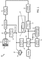
FIG. 1 , anultrasound imaging system 10 constructed in accordance with the present principles is shown in block diagram form. In the ultrasonic diagnostic imaging system ofFIG. 1 , anultrasound system 10 includes aprobe 12 having a transducer ortransducer array 14 for transmitting ultrasonic waves and receiving echo information. A variety of transducer arrays are well known in the art, e.g., linear arrays, convex arrays or phased arrays. Thetransducer array 14, for example, can include a two dimensional array (as shown) of transducer elements capable of scanning in both elevation and azimuth dimensions for 2D and/or 3D imaging. Thetransducer array 14 is coupled to amicrobeamformer 16 in theprobe 12, which controls transmission and reception of signals by the transducer elements in the array. In this example, themicrobeamformer 16 is coupled by the probe cable to a transmit/receive (T/R)switch 18, which switches between transmission and reception and protects amain beamformer 22 from high energy transmit signals. In some embodiments, the T/R switch 18 and other elements in the system can be included in the transducer probe rather than in a separate ultrasound system base. The transmission of ultrasonic beams from thetransducer array 14 under control of themicrobeamformer 16 is directed by a transmitcontroller 20 coupled to the T/R switch 18 and thebeamformer 22, which may receive input from the user's operation of a user interface orcontrol panel 24. - In accordance with one embodiment, the transmit
controller 20 automatically switches the imaging modes for one or more frames to concurrently acquire (receive) echoes/images in multiple imaging modes. A user may adjust the relative frame rate between the imaging modes using theinterface 24, and, in particular, aframe rate control 48. Another function controlled by the transmitcontroller 20 is the direction in which beams are steered. Beams may be steered straight ahead from (orthogonal to) the transducer array, or at different angles for a wider field of view. The partially beamformed signals produced by themicrobeamformer 16 are coupled to amain beamformer 22 where partially beamformed signals from individual patches of transducer elements are combined into a fully beamformed signal. - The beamformed signals are coupled to a
signal processor 26. Thesignal processor 26 can process the received echo signals in various ways, such as bandpass filtering, decimation, I and Q component separation, and harmonic signal separation. Thesignal processor 26 is also configured to perform additional signal enhancement such as signal compounding and optionally speckle reduction and noise elimination. The processed signals are coupled to aB mode processor 28, which can employ amplitude detection for the imaging of structures in the body. The signals produced by the B mode processor are coupled to ascan converter 30 and amultiplanar reformatter 32. Thescan converter 30 arranges the echo signals in the spatial relationship from which they were received in a desired image format. For instance, thescan converter 30 may arrange the echo signal into a two dimensional (2D) sector-shaped format, or a pyramidal three dimensional (3D) image. Themultiplanar reformatter 32 can convert echoes which are received from points in a common plane in a volumetric region of the body into an ultrasonic image of that plane, as described inU.S. Pat. No. 6,443,896 (Detmer ). Avolume renderer 34 converts the echo signals of a 3D data set into a projected 3D image as viewed from a given reference point, e.g., as described inU.S. Pat. No. 6,530,885 (Entrekin et al. ). The 2D or 3D images are coupled from thescan converter 30,multiplanar reformatter 32, andvolume renderer 34 to animage processor 36 for further enhancement, buffering and temporary storage for display on animage display 38. Agraphics processor 40 can generate graphic overlays for display with the ultrasound images. These graphic overlays or parameter blocks can contain, e.g., standard identifying information such as patient name, date and time of the image, imaging parameters, frame indices and the like. For these purposes, thegraphics processor 40 receives input from theuser interface 24, such as a typed patient name. Theuser interface 24 can also be coupled to themultiplanar reformatter 32 for selection and control of a display of multiple multiplanar reformatted (MPR) images. - In accordance with the present principles, screening ultrasound data is acquired and stored in memory 42 in a format that allows an offline reader/reviewer to still be able to access multiple imaging modes, e.g., to help characterize a suspicious lesion, etc., but without significantly complicating or extending the workflow for either acquisition or review. In one embodiment, the
ultrasound system 10 is programmed to acquire images that alternate between modes, e.g., fundamental, harmonic, etc. In one example, to make acquisition easier for the operator, only the fundamental (or harmonic) images are shown on thedisplay 38, the other images are acquired but not shown. The memory 42 stores frames in memory structures or logically connects (e.g., points to or indexes) frames of a same mode. This may include organizing frames of a stream by the use of indexing, multiplexing or other techniques to designate which frames are associated with each imaging mode. In one embodiment, the frames may be stored inseparate data structures scan converter 30; however, the memory 42 may store data at any position in the signal path. In particularly useful embodiments, not falling under the scope of the present invention, the data stored may be sufficiently raw data that needs to be stored earlier in the signal path to permit the raw data to be available for rendering in multiple modes. In such a case, the switching between modes is not needed as the data will be stored to recreate these modes in post-processing to generate the desired modes during or for review. - The resolution of one mode may be adjusted using a
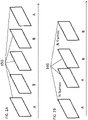
frame rate control 48. Theframe rate control 48 may be employed to adjust the number of frames for one mode versus the other modes. Theframe rate control 48 may be implemented in software, hardware or a combination of both. This will be further described with reference toFIGS. 2A and 2B . - Referring to
FIGS. 2A and 2B , diagrams illustratively show imaging mode frame collection.FIG. 2A shows acquiredframes 150, which alternate between a first mode (mode A) and a second mode (mode B). In one embodiment, mode A and mode B may include fundamental and harmonic imaging modes. For each mode, the frame rate will be reduced by a factor of 2, but since ultrasound systems are now able to acquire images at high frame rates that accuracy/resolution is not compromised. For example, frame rate settings (speed) are preferably over 100 Hz. - The imaging mode streams may be separated and both streams of, e.g., fundamental and harmonic imaging may be stored and exported as separate but linked loops. The linking may include the use of time stamps, frame numbers, indexes, etc. When a radiologist initially reviews the data, the radiologist may only see the fundamental images since these have the least artifactual shadowing and are thus most efficient for finding suspicious lesions. However, once a suspicious lesion has been identified the radiologist can switch to the equivalent harmonic image obtained from the same location, and thus obtain diagnostic information from both modes. Note that this description refers to screening the images, which is typically performed in a separate process than scanning. The scanned images are often done with a set imaging mode. In conventional systems, a reviewer is often stuck with the selected imaging mode. In accordance with the present principles, the same workflow provides access to multiple imaging modes. These imaging modes are compiled at a later time (not necessarily during scanning), and the reviewer can select the imaging mode to display with minimal loss of accuracy and no time lost during the scanning process. The screening process includes the review of the images at a separate time and/or location, e.g., at a workstation configured for reviewing.
-
FIG. 2B shows a more general frame collection scheme where one ormultiple frames 160 are collected for each imaging mode. In one embodiment, a single mode A frame may be collected followed by N (an integer number) mode B frames, and then repeated. In another embodiment, N mode A frames may be taken followed by a single mode B frame and then repeated. In yet another embodiment, N mode A frames may be collected followed by N mode B frames, and then repeated. In other embodiments, a greater number of modes may be employed and different combinations of frame numbers may be employed. The additional frame numbers for a particular imaging mode may be selected to increase resolution for that imaging mode relative the other imaging mode or modes. These adjustments in frame numbers may be selected for the scanning based on experience, desired results or other criteria. - Spatial compound imaging or spatial compounding (SonoCT) is an ultrasound technique that uses electronic beam steering of a transducer array to rapidly acquire several (e.g., three to nine) overlapping scans of an object from different view angles. These single-angle scans are averaged to form a multiangle compound image that is updated with each subsequent scan. Compound imaging shows improved image quality compared with conventional ultrasound, primarily because of reduction of speckle, clutter, and other acoustic artifacts, and provides improved contrast resolution and tissue differentiation, which are beneficial for imaging the breast, peripheral blood vessels, and musculoskeletal injuries.
- Spatial compounding being switched on or off can also be accommodated at the screening stage by a user in accordance with the present principles. Ultrasound data would be acquired in SonoCT (and if desired fundamental and harmonic modes), and the operator or scanner would see a SonoCT image, but the component frames and not the compounded frames would be stored in memory 42 (e.g., as a separate mode). During review, workstation software would perform the compounding step needed to generate a SonoCT image to present to the radiologist, or the radiologist could choose to view the non-SonoCT image (i.e., to asses lesion shadowing) in which case only the non-steered component image would be presented. The post-processing of the image stream may be performed with or without the mode switching process step - the later not being part of the invention.
- For example, the switching mode data collection can be post-processed to switch compounding on or off. Likewise, the sufficiently raw data can be collected and be post-processed to switch compounding on or off.
- Referring to
FIG. 3 , asystem 100 for review of ultrasound images is illustratively shown in accordance with one embodiment.System 100 may include a workstation or console 112 from which images are reviewed and modes selected.Workstation 112 preferably includes one ormore processors 114 andmemory 116 for storing programs, applications and data.Memory 116 may store animage rendering module 115 configured to collect and render image frames for the display of one or more imaging modes. - The
image rendering module 115 is configured to receive image data and link or process image modes for display. Animage 134 can be generated fromframes 140 stored inmemory 116 and can be displayed on adisplay device 118.Workstation 112 includes thedisplay 118 for reviewing internal images of a subject (e.g., a patient).Display 118 may also permit a user to interact with theworkstation 112 and its components and functions, or any other element within thesystem 100. This is further facilitated by aninterface 120 which may include a keyboard, mouse, a joystick, a haptic device, or any other peripheral or control to permit user feedback from and interaction with theworkstation 112. - In one embodiment, each imaging mode is acquired sequentially and so each mode can be fully optimized without compromise, for example, in terms of the acquisition design (e.g., line density, focal zones), signal and image processing, display parameters, etc. For example, for SonoCT (spatial compounding), the non-steered frame may be designated differently from the other component frames, since this non-steered frame will be visualized on its own without the benefit of compounding. That is, it may have a different line density, more focal zones, more frequency compounding, or a different display map.
- In another embodiment, the review and interpretation of the images may be performed either on the system 10 (
FIG. 1 ) or off the system on theworkstation 100. In either case, review software ofimage rendering module 115 needs to be able to support the capability to correctly process the separate modes being presented. For fundamental versus harmonic imaging, this may only involve pulling images from the appropriate data stream and applying a suitable display map. For SonoCT versus non-SonoCT, the review software ofimage rendering module 115 needs to be able to extract only the non-steered frames and apply appropriate display maps for non-SonoCT, or extract all the frames and apply a compounding algorithm to them, plus appropriate display mapping for SonoCT. - In accordance with an alternate embodiment - not being part of the invention, instead of acquiring images by switching modes, the acquisition may be obtained as a sufficiently raw data stream. In such an embodiment, the acquisition (initial scan) is more efficient because all of the imaging acquired can be utilized to display in more than one mode (i.e., there are no alternating modes). For example, both the fundamental and harmonic images that are displayed in review may be extracted from the same acquisition and the same data. The modes extracted from the data may be based on software operations rather than mode switching. For example, a single kind of data acquisition is stored which includes all mode components, and these are separated through additional processing by
image rendering module 115, such as, e.g., band-pass filtering of IQ data, applying different summation weights to radio frequency (RF) data acquired with opposite polarity transmit waveforms, etc. The stored data is stored in a format that is sufficiently raw (i.e., minimally processed) so that the components of all modes (e.g., fundamental and harmonic) can be extracted with sufficient quality. - For example, both the fundamental and harmonic images that are displayed in review may be extracted from the same acquisition. Only one kind of acquisition is defined and stored during scanning, which includes both fundamental and harmonic components (and other modes), and these are separated through additional processing at the
workstation 112. The additional processing may be performed by theimage rendering module 115 and may include functions such as image filtering, band-pass filtering of IQ data, applying different summation weights to RF data acquired with opposite polarity transmit waveforms, etc. - The present principles are particularly useful in medical procedures such as for breast screening or other screening procedures. The present principles may also be applicable to any clinical application that involves the need for rapid acquisition with minimal interpretation during acquisition, followed by careful review and interpretation post-acquisition. One example may include screening for liver cancer with ultrasound. Target platforms for the present principles include any ultrasound systems and workstations designed for screening purposes.
- Referring to
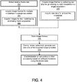
FIG. 4 , methods for providing multiple review modes in a single acquisition scan are illustratively shown. Inblock 202, a relative frame rate may be selected for each of the plurality of imaging modes in the acquisition sequence. The frame rate may be programmed into the system (e.g., by controlling the transmitter) to toggle between different imaging modes for different consecutive durations to control the number of frames received for each imaging mode. The relative frame rate between imaging modes may include an integer multiple of successive frames for at least one of the imaging modes in the acquisition sequence. - In
block 204, image frames are acquired. This may include acquiring image frames for a plurality of imaging modes by switching image acquisition modes during a single acquisition sequence. In block 206, the imaging frames may be acquired from at least two imaging modes (e.g., two) and the image acquisition modes are switched such that frames are acquired in an alternating manner for each of the two modes. Alternately, acquiring image frames includes acquiring data that is sufficiently raw to generate images in multiple modes by soft processing inblock 205. This means that the collected data includes sufficient information for generating each of the desired imaging modalities. In one embodiment, raw data is acquired for generating at least two imaging modes inblock 207. - The imaging modes include ultrasonic imaging modes, and the ultrasonic imaging modes include fundamental, harmonic, and optionally one or more of compound fundamental, compound harmonic, color, color power angioplasty, elastography, etc. During the acquisition, the image frames may be acquired by scan and displayed to an operator in a single imaging mode during the single acquisition.
- In
block 208, the image frames are stored in non-transitory memory for each acquisition mode for subsequent review. The storage may include storing images of a particular mode together (in a separate device, memory partition etc.), logically linking the images of a particular mode, employing indexing of images and providing a lookup table, generating the mode images from sufficiently raw data, etc. - In
block 210, during review (e.g., post scan, not live), a display is selectively generated for each of the plurality of imaging modes from stored images for a selected imaging mode such that each of the plurality of image modes is available for review from the single acquisition sequence. The generation of images may include post-processing of the sufficiently raw data, provide grouping of collected frames from a single imaging mode, etc. - A reviewer can switch between different imaging modes, e.g., fundamental, harmonic, compound fundamental, compound harmonic, etc. to obtain a more accurate result by employing the strength of each imaging mode. Since the desired imaging modes are all available, there is a significantly reduced need to rescan the patient in other imaging modes as in conventional workflows. The plurality of imaging modes may include multiple imaging modes for discovering different diagnostic information employed for identifying a lesion in an organ, or other applications. Such applications may include breast cancer screening, liver cancer screening, etc.
- In block 212, enabling/disabling compounding using the stored images may be performed using software functions. Other image processing functions may also be performed using software functions, e.g., filtering, contrast enhancement, generating modes from raw data, etc.
- In interpreting the appended claims, it should be understood that:
- a) the word "comprising" does not exclude the presence of other elements or acts than those listed in a given claim;
- b) the word "a" or "an" preceding an element does not exclude the presence of a plurality of such elements;
- c) any reference signs in the claims do not limit their scope;
- d) several "means" may be represented by the same item or hardware or software implemented structure or function; and
- e) no specific sequence of acts is intended to be required unless specifically indicated.
- Having described preferred embodiments for concurrent acquisition of harmonic and fundamental images for screening applications (which are intended to be illustrative and not limiting), it is noted that modifications and variations can be made by persons skilled in the art in light of the above teachings. It is therefore to be understood that changes may be made in the particular embodiments of the disclosure disclosed which are within the scope of the embodiments disclosed herein as outlined by the appended claims.
Claims (12)
- A method for providing multiple review modes in a single acquisition scan, comprising:acquiring (204) image frames for a plurality of ultrasound imaging modes including at least a fundamental mode and a harmonic mode by switching between ultrasound image acquisition modes including at least a fundamental acquisition mode and a harmonic acquisition mode in real-time during a single acquisition scan;storing (208) the image frames in non-transitory memory for each of the ultrasound image acquisition modes for subsequent review; andduring review, selectively generating (210) a display for each of the plurality of ultrasound imaging modes from the stored image frames for an imaging mode selected by a reviewer such that each of the plurality of ultrasound imaging modes is available for review from the single acquisition scan,wherein the method further comprises enabling or disabling (212) compounding using the stored images.
- The method as recited in claim 1, further comprising selecting (202) a relative frame rate for each of the plurality of ultrasound imaging modes in the single acquisition scan.
- The method as recited in claim 1, wherein acquiring (204) imaging frames includes acquiring imaging frames (206) from two imaging modes and the ultrasound image acquisition modes are switched such that frames are acquired in an alternating manner for each of the two imaging modes.
- The method as recited in claim 1, wherein acquiring image frames (204) includes displaying a single imaging mode during the single acquisition scan.
- The method as recited in claim 1, wherein the plurality of ultrasound imaging modes includes a first imaging mode for discovering first diagnostic information and a second imaging mode for discovering second diagnostic information for identifying a lesion in an
- A non-transitory computer readable storage medium comprising a computer readable program for providing multiple review modes in a single acquisition scan, wherein the computer readable program when executed on a computer causes the computer to perform the steps of claim 1.
- A system for providing multiple review modes in a single acquisition scan, comprising:an ultrasound imaging device (10) configured to acquire image frames for a plurality of ultrasound imaging modes including at least a fundamental mode and a harmonic mode by automated switching between ultrasound image acquisition modes including at least a fundamental acquisition mode and a harmonic acquisition mode during a single acquisition scan;a memory device (116) configured to store the image frames in non-transitory memory for each of the ultrasound image acquisition modes for subsequent review;a signal processor (26) adapted to perform signal compounding; anda review workstation (112) having a display (118) for viewing one of the plurality of ultrasound imaging modes from the stored image frames for an imaging mode selected by a reviewer such that each of the plurality of ultrasound imaging modes is available for review from the single acquisition scan, wherein compounding using the stored images can be enabled and disabled.
- The system as recited in claim 7, further comprising a relative frame rate control (48) for setting a number of sequential frames for each of the plurality of ultrasound imaging modes in the single acquisition scan.
- The system as recited in claim 8, wherein the relative frame rate includes an integer multiple of successive frames for at least one of the plurality of ultrasound imaging modes in the single acquisition scan.
- The system as recited in claim 7, wherein the plurality of ultrasound imaging modes includes two imaging modes and the imaging modes are switched such that frames are acquired in an alternating manner for each of the two imaging modes.
- The system as recited in claim 7, wherein the plurality of ultrasound imaging modes further include one or more of a compound fundamental mode, a compound harmonic mode, a color mode, a color power angioplasty mode and/or an elastography mode.
- The system as recited in claim 7, wherein the review workstation (112) is configured for breast cancer screening.
Applications Claiming Priority (2)
| Application Number | Priority Date | Filing Date | Title |
|---|---|---|---|
| US201462042980P | 2014-08-28 | 2014-08-28 | |
| PCT/IB2015/056064 WO2016030785A1 (en) | 2014-08-28 | 2015-08-10 | Concurrent acquisition of harmonic and fundamental images for screening applications |
Publications (2)
| Publication Number | Publication Date |
|---|---|
| EP3185779A1 EP3185779A1 (en) | 2017-07-05 |
| EP3185779B1 true EP3185779B1 (en) | 2022-12-21 |
Family
ID=54151338
Family Applications (1)
| Application Number | Title | Priority Date | Filing Date |
|---|---|---|---|
| EP15767297.3A Active EP3185779B1 (en) | 2014-08-28 | 2015-08-10 | Concurrent acquisition of harmonic and fundamental images for screening applications |
Country Status (5)
| Country | Link |
|---|---|
| US (1) | US20170231599A1 (en) |
| EP (1) | EP3185779B1 (en) |
| JP (1) | JP6665167B2 (en) |
| CN (1) | CN106604682A (en) |
| WO (1) | WO2016030785A1 (en) |
Families Citing this family (3)
| Publication number | Priority date | Publication date | Assignee | Title |
|---|---|---|---|---|
| EP3549528A1 (en) * | 2018-04-05 | 2019-10-09 | Koninklijke Philips N.V. | Ultrasound imaging system and method |
| CN111820945A (en) * | 2019-04-16 | 2020-10-27 | 深圳迈瑞生物医疗电子股份有限公司 | System and method for performing ultrasound imaging |
| US11710229B2 (en) | 2020-03-23 | 2023-07-25 | GE Precision Healthcare LLC | Methods and systems for shear wave elastography |
Family Cites Families (24)
| Publication number | Priority date | Publication date | Assignee | Title |
|---|---|---|---|---|
| JP3580627B2 (en) * | 1996-01-29 | 2004-10-27 | 株式会社東芝 | Ultrasound diagnostic equipment |
| JP4116143B2 (en) * | 1998-04-10 | 2008-07-09 | 株式会社東芝 | Ultrasonic diagnostic equipment |
| US6511426B1 (en) * | 1998-06-02 | 2003-01-28 | Acuson Corporation | Medical diagnostic ultrasound system and method for versatile processing |
| US6224552B1 (en) * | 1998-10-01 | 2001-05-01 | Atl Ultrasound | Ultrasonic diagnostic imaging system with reduced spatial compounding seam artifacts |
| US6117079A (en) * | 1999-04-28 | 2000-09-12 | General Electric Company | Method and apparatus for handling image data after unsuccessful transfer to remotely located device |
| US20030013959A1 (en) * | 1999-08-20 | 2003-01-16 | Sorin Grunwald | User interface for handheld imaging devices |
| US6530885B1 (en) | 2000-03-17 | 2003-03-11 | Atl Ultrasound, Inc. | Spatially compounded three dimensional ultrasonic images |
| US6443896B1 (en) | 2000-08-17 | 2002-09-03 | Koninklijke Philips Electronics N.V. | Method for creating multiplanar ultrasonic images of a three dimensional object |
| US7615008B2 (en) * | 2000-11-24 | 2009-11-10 | U-Systems, Inc. | Processing and displaying breast ultrasound information |
| US6540683B1 (en) * | 2001-09-14 | 2003-04-01 | Gregory Sharat Lin | Dual-frequency ultrasonic array transducer and method of harmonic imaging |
| JP3908555B2 (en) * | 2002-02-08 | 2007-04-25 | 株式会社東芝 | Ultrasonic diagnostic equipment |
| JP2004154567A (en) * | 2002-10-15 | 2004-06-03 | Matsushita Electric Ind Co Ltd | Image processing apparatus, method, and program |
| US7530356B2 (en) * | 2004-10-06 | 2009-05-12 | Guided Therapy Systems, Inc. | Method and system for noninvasive mastopexy |
| US20070010747A1 (en) * | 2005-05-26 | 2007-01-11 | Sabourin Thomas J | Methods and systems for acquiring ultrasound image data |
| US9420991B2 (en) * | 2005-09-01 | 2016-08-23 | Shih-Ping Wang | Breast ultrasound scanning device |
| US20070055159A1 (en) * | 2005-09-01 | 2007-03-08 | Shih-Ping Wang | Breast ultrasound scanning template |
| US20070086632A1 (en) * | 2005-09-30 | 2007-04-19 | Siemens Medical Solutions Usa, Inc. | Medical data storage or review with interactive features of a video format |
| US20070161898A1 (en) * | 2006-01-10 | 2007-07-12 | Siemens Medical Solutions Usa, Inc. | Raw data reprocessing in ultrasound diagnostic imaging |
| US20080119733A1 (en) * | 2006-11-22 | 2008-05-22 | Wei Zhang | Selectably compounding and displaying breast ultrasound images |
| WO2009020617A1 (en) * | 2007-08-06 | 2009-02-12 | Orison Corporation | System and method for three-dimensional ultrasound imaging |
| CN103415258B (en) * | 2010-12-06 | 2015-05-20 | 索那利姆医疗有限公司 | System and method for ultrasound examination of the breast |
| EP2788078B1 (en) * | 2011-12-09 | 2020-09-02 | Metavention, Inc. | Therapeutic neuromodulation of the hepatic system |
| CN103648398B (en) * | 2012-06-05 | 2016-05-04 | 株式会社东芝 | Diagnostic ultrasound equipment and image processing apparatus |
| US9980629B2 (en) * | 2013-03-11 | 2018-05-29 | Karl Storz Imaging, Inc. | Video capture and streaming diagnostics metadata |
-
2015
- 2015-08-10 US US15/504,408 patent/US20170231599A1/en not_active Abandoned
- 2015-08-10 JP JP2017510874A patent/JP6665167B2/en active Active
- 2015-08-10 WO PCT/IB2015/056064 patent/WO2016030785A1/en active Application Filing
- 2015-08-10 CN CN201580046564.1A patent/CN106604682A/en active Pending
- 2015-08-10 EP EP15767297.3A patent/EP3185779B1/en active Active
Also Published As
| Publication number | Publication date |
|---|---|
| WO2016030785A1 (en) | 2016-03-03 |
| EP3185779A1 (en) | 2017-07-05 |
| JP2017525491A (en) | 2017-09-07 |
| CN106604682A (en) | 2017-04-26 |
| US20170231599A1 (en) | 2017-08-17 |
| JP6665167B2 (en) | 2020-03-13 |
Similar Documents
| Publication | Publication Date | Title |
|---|---|---|
| US9386964B2 (en) | 3D view of 2D ultrasound images | |
| US11653897B2 (en) | Ultrasonic diagnostic apparatus, scan support method, and medical image processing apparatus | |
| JP7150800B2 (en) | Motion-adaptive visualization in medical 4D imaging | |
| CN101066210A (en) | User interface and method for displaying information in an ultrasound system | |
| JP6097452B2 (en) | Ultrasonic imaging system and ultrasonic imaging method | |
| JP7177870B2 (en) | Ultrasound Imaging System with Simplified 3D Imaging Controls | |
| EP3185779B1 (en) | Concurrent acquisition of harmonic and fundamental images for screening applications | |
| JP7175613B2 (en) | Analysis device and control program | |
| CN112867444A (en) | System and method for guiding acquisition of ultrasound images | |
| JP2011115456A (en) | Ultrasonic diagnostic apparatus and control program for image data display | |
| EP2644102A1 (en) | Method and apparatus for indicating medical equipment on ultrasound image | |
| US20220160333A1 (en) | Optimal ultrasound-based organ segmentation | |
| CN112672696A (en) | System and method for tracking tools in ultrasound images | |
| JP7216738B2 (en) | Provision of 3D ultrasound images | |
| EP4270411A1 (en) | Analysing an ultrasound image feed | |
| EP3681403B1 (en) | Ultrasound image processing | |
| WO2023208877A1 (en) | Analysing an ultrasound image feed | |
| KR20160056164A (en) | Untrasound dianognosis apparatus, operating method thereof and computer-readable storage medium | |
| JP2013236863A (en) | Ultrasonic diagnostic apparatus and image processor |
Legal Events
| Date | Code | Title | Description |
|---|---|---|---|
| STAA | Information on the status of an ep patent application or granted ep patent |
Free format text: STATUS: THE INTERNATIONAL PUBLICATION HAS BEEN MADE |
|
| PUAI | Public reference made under article 153(3) epc to a published international application that has entered the european phase |
Free format text: ORIGINAL CODE: 0009012 |
|
| STAA | Information on the status of an ep patent application or granted ep patent |
Free format text: STATUS: REQUEST FOR EXAMINATION WAS MADE |
|
| 17P | Request for examination filed |
Effective date: 20170328 |
|
| AK | Designated contracting states |
Kind code of ref document: A1 Designated state(s): AL AT BE BG CH CY CZ DE DK EE ES FI FR GB GR HR HU IE IS IT LI LT LU LV MC MK MT NL NO PL PT RO RS SE SI SK SM TR |
|
| AX | Request for extension of the european patent |
Extension state: BA ME |
|
| DAV | Request for validation of the european patent (deleted) | ||
| DAX | Request for extension of the european patent (deleted) | ||
| RAP1 | Party data changed (applicant data changed or rights of an application transferred) |
Owner name: KONINKLIJKE PHILIPS N.V. |
|
| STAA | Information on the status of an ep patent application or granted ep patent |
Free format text: STATUS: EXAMINATION IS IN PROGRESS |
|
| STAA | Information on the status of an ep patent application or granted ep patent |
Free format text: STATUS: EXAMINATION IS IN PROGRESS |
|
| 17Q | First examination report despatched |
Effective date: 20201127 |
|
| STAA | Information on the status of an ep patent application or granted ep patent |
Free format text: STATUS: EXAMINATION IS IN PROGRESS |
|
| GRAP | Despatch of communication of intention to grant a patent |
Free format text: ORIGINAL CODE: EPIDOSNIGR1 |
|
| STAA | Information on the status of an ep patent application or granted ep patent |
Free format text: STATUS: GRANT OF PATENT IS INTENDED |
|
| INTG | Intention to grant announced |
Effective date: 20220715 |
|
| GRAS | Grant fee paid |
Free format text: ORIGINAL CODE: EPIDOSNIGR3 |
|
| GRAA | (expected) grant |
Free format text: ORIGINAL CODE: 0009210 |
|
| STAA | Information on the status of an ep patent application or granted ep patent |
Free format text: STATUS: THE PATENT HAS BEEN GRANTED |
|
| AK | Designated contracting states |
Kind code of ref document: B1 Designated state(s): AL AT BE BG CH CY CZ DE DK EE ES FI FR GB GR HR HU IE IS IT LI LT LU LV MC MK MT NL NO PL PT RO RS SE SI SK SM TR |
|
| REG | Reference to a national code |
Ref country code: GB Ref legal event code: FG4D |
|
| REG | Reference to a national code |
Ref country code: DE Ref legal event code: R096 Ref document number: 602015082015 Country of ref document: DE |
|
| REG | Reference to a national code |
Ref country code: CH Ref legal event code: EP |
|
| REG | Reference to a national code |
Ref country code: AT Ref legal event code: REF Ref document number: 1538557 Country of ref document: AT Kind code of ref document: T Effective date: 20230115 |
|
| REG | Reference to a national code |
Ref country code: IE Ref legal event code: FG4D |
|
| REG | Reference to a national code |
Ref country code: DE Ref legal event code: R084 Ref document number: 602015082015 Country of ref document: DE |
|
| REG | Reference to a national code |
Ref country code: LT Ref legal event code: MG9D |
|
| REG | Reference to a national code |
Ref country code: NL Ref legal event code: MP Effective date: 20221221 |
|
| PG25 | Lapsed in a contracting state [announced via postgrant information from national office to epo] |
Ref country code: SE Free format text: LAPSE BECAUSE OF FAILURE TO SUBMIT A TRANSLATION OF THE DESCRIPTION OR TO PAY THE FEE WITHIN THE PRESCRIBED TIME-LIMIT Effective date: 20221221 Ref country code: NO Free format text: LAPSE BECAUSE OF FAILURE TO SUBMIT A TRANSLATION OF THE DESCRIPTION OR TO PAY THE FEE WITHIN THE PRESCRIBED TIME-LIMIT Effective date: 20230321 Ref country code: LT Free format text: LAPSE BECAUSE OF FAILURE TO SUBMIT A TRANSLATION OF THE DESCRIPTION OR TO PAY THE FEE WITHIN THE PRESCRIBED TIME-LIMIT Effective date: 20221221 Ref country code: FI Free format text: LAPSE BECAUSE OF FAILURE TO SUBMIT A TRANSLATION OF THE DESCRIPTION OR TO PAY THE FEE WITHIN THE PRESCRIBED TIME-LIMIT Effective date: 20221221 |
|
| REG | Reference to a national code |
Ref country code: AT Ref legal event code: MK05 Ref document number: 1538557 Country of ref document: AT Kind code of ref document: T Effective date: 20221221 |
|
| PG25 | Lapsed in a contracting state [announced via postgrant information from national office to epo] |
Ref country code: RS Free format text: LAPSE BECAUSE OF FAILURE TO SUBMIT A TRANSLATION OF THE DESCRIPTION OR TO PAY THE FEE WITHIN THE PRESCRIBED TIME-LIMIT Effective date: 20221221 Ref country code: LV Free format text: LAPSE BECAUSE OF FAILURE TO SUBMIT A TRANSLATION OF THE DESCRIPTION OR TO PAY THE FEE WITHIN THE PRESCRIBED TIME-LIMIT Effective date: 20221221 Ref country code: HR Free format text: LAPSE BECAUSE OF FAILURE TO SUBMIT A TRANSLATION OF THE DESCRIPTION OR TO PAY THE FEE WITHIN THE PRESCRIBED TIME-LIMIT Effective date: 20221221 Ref country code: GR Free format text: LAPSE BECAUSE OF FAILURE TO SUBMIT A TRANSLATION OF THE DESCRIPTION OR TO PAY THE FEE WITHIN THE PRESCRIBED TIME-LIMIT Effective date: 20230322 |
|
| PG25 | Lapsed in a contracting state [announced via postgrant information from national office to epo] |
Ref country code: NL Free format text: LAPSE BECAUSE OF FAILURE TO SUBMIT A TRANSLATION OF THE DESCRIPTION OR TO PAY THE FEE WITHIN THE PRESCRIBED TIME-LIMIT Effective date: 20221221 |
|
| PG25 | Lapsed in a contracting state [announced via postgrant information from national office to epo] |
Ref country code: SM Free format text: LAPSE BECAUSE OF FAILURE TO SUBMIT A TRANSLATION OF THE DESCRIPTION OR TO PAY THE FEE WITHIN THE PRESCRIBED TIME-LIMIT Effective date: 20221221 Ref country code: RO Free format text: LAPSE BECAUSE OF FAILURE TO SUBMIT A TRANSLATION OF THE DESCRIPTION OR TO PAY THE FEE WITHIN THE PRESCRIBED TIME-LIMIT Effective date: 20221221 Ref country code: PT Free format text: LAPSE BECAUSE OF FAILURE TO SUBMIT A TRANSLATION OF THE DESCRIPTION OR TO PAY THE FEE WITHIN THE PRESCRIBED TIME-LIMIT Effective date: 20230421 Ref country code: ES Free format text: LAPSE BECAUSE OF FAILURE TO SUBMIT A TRANSLATION OF THE DESCRIPTION OR TO PAY THE FEE WITHIN THE PRESCRIBED TIME-LIMIT Effective date: 20221221 Ref country code: EE Free format text: LAPSE BECAUSE OF FAILURE TO SUBMIT A TRANSLATION OF THE DESCRIPTION OR TO PAY THE FEE WITHIN THE PRESCRIBED TIME-LIMIT Effective date: 20221221 Ref country code: CZ Free format text: LAPSE BECAUSE OF FAILURE TO SUBMIT A TRANSLATION OF THE DESCRIPTION OR TO PAY THE FEE WITHIN THE PRESCRIBED TIME-LIMIT Effective date: 20221221 Ref country code: AT Free format text: LAPSE BECAUSE OF FAILURE TO SUBMIT A TRANSLATION OF THE DESCRIPTION OR TO PAY THE FEE WITHIN THE PRESCRIBED TIME-LIMIT Effective date: 20221221 |
|
| PG25 | Lapsed in a contracting state [announced via postgrant information from national office to epo] |
Ref country code: SK Free format text: LAPSE BECAUSE OF FAILURE TO SUBMIT A TRANSLATION OF THE DESCRIPTION OR TO PAY THE FEE WITHIN THE PRESCRIBED TIME-LIMIT Effective date: 20221221 Ref country code: PL Free format text: LAPSE BECAUSE OF FAILURE TO SUBMIT A TRANSLATION OF THE DESCRIPTION OR TO PAY THE FEE WITHIN THE PRESCRIBED TIME-LIMIT Effective date: 20221221 Ref country code: IS Free format text: LAPSE BECAUSE OF FAILURE TO SUBMIT A TRANSLATION OF THE DESCRIPTION OR TO PAY THE FEE WITHIN THE PRESCRIBED TIME-LIMIT Effective date: 20230421 Ref country code: AL Free format text: LAPSE BECAUSE OF FAILURE TO SUBMIT A TRANSLATION OF THE DESCRIPTION OR TO PAY THE FEE WITHIN THE PRESCRIBED TIME-LIMIT Effective date: 20221221 |
|
| REG | Reference to a national code |
Ref country code: DE Ref legal event code: R097 Ref document number: 602015082015 Country of ref document: DE |
|
| PLBE | No opposition filed within time limit |
Free format text: ORIGINAL CODE: 0009261 |
|
| STAA | Information on the status of an ep patent application or granted ep patent |
Free format text: STATUS: NO OPPOSITION FILED WITHIN TIME LIMIT |
|
| PG25 | Lapsed in a contracting state [announced via postgrant information from national office to epo] |
Ref country code: DK Free format text: LAPSE BECAUSE OF FAILURE TO SUBMIT A TRANSLATION OF THE DESCRIPTION OR TO PAY THE FEE WITHIN THE PRESCRIBED TIME-LIMIT Effective date: 20221221 |
|
| 26N | No opposition filed |
Effective date: 20230922 |
|
| PGFP | Annual fee paid to national office [announced via postgrant information from national office to epo] |
Ref country code: DE Payment date: 20230828 Year of fee payment: 9 |
|
| PG25 | Lapsed in a contracting state [announced via postgrant information from national office to epo] |
Ref country code: SI Free format text: LAPSE BECAUSE OF FAILURE TO SUBMIT A TRANSLATION OF THE DESCRIPTION OR TO PAY THE FEE WITHIN THE PRESCRIBED TIME-LIMIT Effective date: 20221221 |
|
| PG25 | Lapsed in a contracting state [announced via postgrant information from national office to epo] |
Ref country code: MC Free format text: LAPSE BECAUSE OF FAILURE TO SUBMIT A TRANSLATION OF THE DESCRIPTION OR TO PAY THE FEE WITHIN THE PRESCRIBED TIME-LIMIT Effective date: 20221221 |
|
| REG | Reference to a national code |
Ref country code: CH Ref legal event code: PL |
|
| PG25 | Lapsed in a contracting state [announced via postgrant information from national office to epo] |
Ref country code: MC Free format text: LAPSE BECAUSE OF FAILURE TO SUBMIT A TRANSLATION OF THE DESCRIPTION OR TO PAY THE FEE WITHIN THE PRESCRIBED TIME-LIMIT Effective date: 20221221 |
|
| PG25 | Lapsed in a contracting state [announced via postgrant information from national office to epo] |
Ref country code: LU Free format text: LAPSE BECAUSE OF NON-PAYMENT OF DUE FEES Effective date: 20230810 |
|
| GBPC | Gb: european patent ceased through non-payment of renewal fee |
Effective date: 20230810 |