EP3176580A1 - Method for reduction of interferences in immunoassays - Google Patents
Method for reduction of interferences in immunoassays Download PDFInfo
- Publication number
- EP3176580A1 EP3176580A1 EP15197192.6A EP15197192A EP3176580A1 EP 3176580 A1 EP3176580 A1 EP 3176580A1 EP 15197192 A EP15197192 A EP 15197192A EP 3176580 A1 EP3176580 A1 EP 3176580A1
- Authority
- EP
- European Patent Office
- Prior art keywords
- binding partner
- analyte
- label
- detectable label
- binding
- Prior art date
- Legal status (The legal status is an assumption and is not a legal conclusion. Google has not performed a legal analysis and makes no representation as to the accuracy of the status listed.)
- Ceased
Links
Images




Classifications
-
- G—PHYSICS
- G01—MEASURING; TESTING
- G01N—INVESTIGATING OR ANALYSING MATERIALS BY DETERMINING THEIR CHEMICAL OR PHYSICAL PROPERTIES
- G01N33/00—Investigating or analysing materials by specific methods not covered by groups G01N1/00 - G01N31/00
- G01N33/48—Biological material, e.g. blood, urine; Haemocytometers
- G01N33/50—Chemical analysis of biological material, e.g. blood, urine; Testing involving biospecific ligand binding methods; Immunological testing
- G01N33/53—Immunoassay; Biospecific binding assay; Materials therefor
- G01N33/543—Immunoassay; Biospecific binding assay; Materials therefor with an insoluble carrier for immobilising immunochemicals
- G01N33/54306—Solid-phase reaction mechanisms
-
- G—PHYSICS
- G01—MEASURING; TESTING
- G01N—INVESTIGATING OR ANALYSING MATERIALS BY DETERMINING THEIR CHEMICAL OR PHYSICAL PROPERTIES
- G01N33/00—Investigating or analysing materials by specific methods not covered by groups G01N1/00 - G01N31/00
- G01N33/48—Biological material, e.g. blood, urine; Haemocytometers
- G01N33/50—Chemical analysis of biological material, e.g. blood, urine; Testing involving biospecific ligand binding methods; Immunological testing
- G01N33/53—Immunoassay; Biospecific binding assay; Materials therefor
- G01N33/536—Immunoassay; Biospecific binding assay; Materials therefor with immune complex formed in liquid phase
- G01N33/537—Immunoassay; Biospecific binding assay; Materials therefor with immune complex formed in liquid phase with separation of immune complex from unbound antigen or antibody
- G01N33/539—Immunoassay; Biospecific binding assay; Materials therefor with immune complex formed in liquid phase with separation of immune complex from unbound antigen or antibody involving precipitating reagent, e.g. ammonium sulfate
- G01N33/541—Double or second antibody, i.e. precipitating antibody
-
- G—PHYSICS
- G01—MEASURING; TESTING
- G01N—INVESTIGATING OR ANALYSING MATERIALS BY DETERMINING THEIR CHEMICAL OR PHYSICAL PROPERTIES
- G01N21/00—Investigating or analysing materials by the use of optical means, i.e. using sub-millimetre waves, infrared, visible or ultraviolet light
- G01N21/62—Systems in which the material investigated is excited whereby it emits light or causes a change in wavelength of the incident light
- G01N21/63—Systems in which the material investigated is excited whereby it emits light or causes a change in wavelength of the incident light optically excited
- G01N21/64—Fluorescence; Phosphorescence
-
- G—PHYSICS
- G01—MEASURING; TESTING
- G01N—INVESTIGATING OR ANALYSING MATERIALS BY DETERMINING THEIR CHEMICAL OR PHYSICAL PROPERTIES
- G01N21/00—Investigating or analysing materials by the use of optical means, i.e. using sub-millimetre waves, infrared, visible or ultraviolet light
- G01N21/75—Systems in which material is subjected to a chemical reaction, the progress or the result of the reaction being investigated
- G01N21/77—Systems in which material is subjected to a chemical reaction, the progress or the result of the reaction being investigated by observing the effect on a chemical indicator
- G01N21/78—Systems in which material is subjected to a chemical reaction, the progress or the result of the reaction being investigated by observing the effect on a chemical indicator producing a change of colour
-
- G—PHYSICS
- G01—MEASURING; TESTING
- G01N—INVESTIGATING OR ANALYSING MATERIALS BY DETERMINING THEIR CHEMICAL OR PHYSICAL PROPERTIES
- G01N33/00—Investigating or analysing materials by specific methods not covered by groups G01N1/00 - G01N31/00
- G01N33/48—Biological material, e.g. blood, urine; Haemocytometers
- G01N33/50—Chemical analysis of biological material, e.g. blood, urine; Testing involving biospecific ligand binding methods; Immunological testing
- G01N33/53—Immunoassay; Biospecific binding assay; Materials therefor
- G01N33/5306—Improving reaction conditions, e.g. reduction of non-specific binding, promotion of specific binding
-
- G—PHYSICS
- G01—MEASURING; TESTING
- G01N—INVESTIGATING OR ANALYSING MATERIALS BY DETERMINING THEIR CHEMICAL OR PHYSICAL PROPERTIES
- G01N33/00—Investigating or analysing materials by specific methods not covered by groups G01N1/00 - G01N31/00
- G01N33/48—Biological material, e.g. blood, urine; Haemocytometers
- G01N33/50—Chemical analysis of biological material, e.g. blood, urine; Testing involving biospecific ligand binding methods; Immunological testing
- G01N33/53—Immunoassay; Biospecific binding assay; Materials therefor
- G01N33/531—Production of immunochemical test materials
-
- G—PHYSICS
- G01—MEASURING; TESTING
- G01N—INVESTIGATING OR ANALYSING MATERIALS BY DETERMINING THEIR CHEMICAL OR PHYSICAL PROPERTIES
- G01N33/00—Investigating or analysing materials by specific methods not covered by groups G01N1/00 - G01N31/00
- G01N33/48—Biological material, e.g. blood, urine; Haemocytometers
- G01N33/50—Chemical analysis of biological material, e.g. blood, urine; Testing involving biospecific ligand binding methods; Immunological testing
- G01N33/53—Immunoassay; Biospecific binding assay; Materials therefor
- G01N33/536—Immunoassay; Biospecific binding assay; Materials therefor with immune complex formed in liquid phase
- G01N33/537—Immunoassay; Biospecific binding assay; Materials therefor with immune complex formed in liquid phase with separation of immune complex from unbound antigen or antibody
- G01N33/538—Immunoassay; Biospecific binding assay; Materials therefor with immune complex formed in liquid phase with separation of immune complex from unbound antigen or antibody by sorbent column, particles or resin strip, i.e. sorbent materials
-
- G—PHYSICS
- G01—MEASURING; TESTING
- G01N—INVESTIGATING OR ANALYSING MATERIALS BY DETERMINING THEIR CHEMICAL OR PHYSICAL PROPERTIES
- G01N33/00—Investigating or analysing materials by specific methods not covered by groups G01N1/00 - G01N31/00
- G01N33/48—Biological material, e.g. blood, urine; Haemocytometers
- G01N33/50—Chemical analysis of biological material, e.g. blood, urine; Testing involving biospecific ligand binding methods; Immunological testing
- G01N33/53—Immunoassay; Biospecific binding assay; Materials therefor
- G01N33/543—Immunoassay; Biospecific binding assay; Materials therefor with an insoluble carrier for immobilising immunochemicals
- G01N33/54393—Improving reaction conditions or stability, e.g. by coating or irradiation of surface, by reduction of non-specific binding, by promotion of specific binding
-
- G—PHYSICS
- G01—MEASURING; TESTING
- G01N—INVESTIGATING OR ANALYSING MATERIALS BY DETERMINING THEIR CHEMICAL OR PHYSICAL PROPERTIES
- G01N33/00—Investigating or analysing materials by specific methods not covered by groups G01N1/00 - G01N31/00
- G01N33/48—Biological material, e.g. blood, urine; Haemocytometers
- G01N33/50—Chemical analysis of biological material, e.g. blood, urine; Testing involving biospecific ligand binding methods; Immunological testing
- G01N33/53—Immunoassay; Biospecific binding assay; Materials therefor
- G01N33/569—Immunoassay; Biospecific binding assay; Materials therefor for microorganisms, e.g. protozoa, bacteria, viruses
- G01N33/56911—Bacteria
-
- G—PHYSICS
- G01—MEASURING; TESTING
- G01N—INVESTIGATING OR ANALYSING MATERIALS BY DETERMINING THEIR CHEMICAL OR PHYSICAL PROPERTIES
- G01N33/00—Investigating or analysing materials by specific methods not covered by groups G01N1/00 - G01N31/00
- G01N33/48—Biological material, e.g. blood, urine; Haemocytometers
- G01N33/50—Chemical analysis of biological material, e.g. blood, urine; Testing involving biospecific ligand binding methods; Immunological testing
- G01N33/53—Immunoassay; Biospecific binding assay; Materials therefor
- G01N33/569—Immunoassay; Biospecific binding assay; Materials therefor for microorganisms, e.g. protozoa, bacteria, viruses
- G01N33/56983—Viruses
-
- G—PHYSICS
- G01—MEASURING; TESTING
- G01N—INVESTIGATING OR ANALYSING MATERIALS BY DETERMINING THEIR CHEMICAL OR PHYSICAL PROPERTIES
- G01N33/00—Investigating or analysing materials by specific methods not covered by groups G01N1/00 - G01N31/00
- G01N33/48—Biological material, e.g. blood, urine; Haemocytometers
- G01N33/50—Chemical analysis of biological material, e.g. blood, urine; Testing involving biospecific ligand binding methods; Immunological testing
- G01N33/53—Immunoassay; Biospecific binding assay; Materials therefor
- G01N33/569—Immunoassay; Biospecific binding assay; Materials therefor for microorganisms, e.g. protozoa, bacteria, viruses
- G01N33/56983—Viruses
- G01N33/56994—Herpetoviridae, e.g. cytomegalovirus, Epstein-Barr virus
-
- G—PHYSICS
- G01—MEASURING; TESTING
- G01N—INVESTIGATING OR ANALYSING MATERIALS BY DETERMINING THEIR CHEMICAL OR PHYSICAL PROPERTIES
- G01N33/00—Investigating or analysing materials by specific methods not covered by groups G01N1/00 - G01N31/00
- G01N33/48—Biological material, e.g. blood, urine; Haemocytometers
- G01N33/50—Chemical analysis of biological material, e.g. blood, urine; Testing involving biospecific ligand binding methods; Immunological testing
- G01N33/53—Immunoassay; Biospecific binding assay; Materials therefor
- G01N33/576—Immunoassay; Biospecific binding assay; Materials therefor for hepatitis
- G01N33/5761—Hepatitis B
-
- G—PHYSICS
- G01—MEASURING; TESTING
- G01N—INVESTIGATING OR ANALYSING MATERIALS BY DETERMINING THEIR CHEMICAL OR PHYSICAL PROPERTIES
- G01N33/00—Investigating or analysing materials by specific methods not covered by groups G01N1/00 - G01N31/00
- G01N33/48—Biological material, e.g. blood, urine; Haemocytometers
- G01N33/50—Chemical analysis of biological material, e.g. blood, urine; Testing involving biospecific ligand binding methods; Immunological testing
- G01N33/53—Immunoassay; Biospecific binding assay; Materials therefor
- G01N33/576—Immunoassay; Biospecific binding assay; Materials therefor for hepatitis
- G01N33/5768—Hepatitis A
-
- G—PHYSICS
- G01—MEASURING; TESTING
- G01N—INVESTIGATING OR ANALYSING MATERIALS BY DETERMINING THEIR CHEMICAL OR PHYSICAL PROPERTIES
- G01N33/00—Investigating or analysing materials by specific methods not covered by groups G01N1/00 - G01N31/00
- G01N33/48—Biological material, e.g. blood, urine; Haemocytometers
- G01N33/50—Chemical analysis of biological material, e.g. blood, urine; Testing involving biospecific ligand binding methods; Immunological testing
- G01N33/58—Chemical analysis of biological material, e.g. blood, urine; Testing involving biospecific ligand binding methods; Immunological testing involving labelled substances
Definitions
- the invention concerns a method for reduction of interferences in immunoassays that are caused by interfering factors that bind to a detectable label that is used for direct or indirect detection of an analyte. More specifically the invention concerns an immunoassay method for detecting an analyte such as an antibody or an antigen in an isolated sample by incubating said sample with a plurality of binding partners, one of which carries a detectable label, wherein a label-specific binding partner is added that does not carry a label but binds to said detectable label.
- an analyte such as an antibody or an antigen in an isolated sample by incubating said sample with a plurality of binding partners, one of which carries a detectable label, wherein a label-specific binding partner is added that does not carry a label but binds to said detectable label.
- signal-generating component that is used in an immunoassay.
- signal-generating compounds are for example enzymes or labels emitting light based on fluorescent, colorimetric, chemiluminescent or electrochemiluminescent principles.
- electrochemiluminescent compounds interference due to non-specific binding to the signal-generating compound has been described in prior art ( Ando et al. Intern Med 2007, 46(15): 1225-1229 ; Sapin et al., Clin Chem Lab Med 2007; 45(3):416-418 ).
- the problem to be solved can therefore be seen in increasing the specificity of an immunoassay based on emission of a detectable signal.
- the invention is based on the finding that a signal-generating compound or detectable label, in particular a light-generating label that is attached to a binding partner, is masked and shielded from its environment by adding a label-specific binding partner.
- the invention concerns an immunoassay method for detecting an analyte such as an antigen or an antibody in an isolated sample suspected to contain said analyte by incubating said sample with a plurality of binding partners, one of which carries a detectable label, wherein a label-specific binding partner is added that does not carry a label but binds to said detectable label.
- the method is applicable for a large variety of analytes and has proven particularly useful for analyte antibodies of the IgG and IgM class present in samples due to infections by pathogens.
- the invention further concerns a reagent kit useful for said method comprising a plurality of binding partners, at least one of which binds to the analyte, and one of which carries a detectable label and one of which is a label-specific binding partner that binds to said detectable label but itself does not carry a detectable label.
- the invention covers the use of a label-specific binding partner that does not carry a detectable label in an in vitro diagnostic test for eliminating interferences caused by anti-label antibodies present in a sample.
- the third binding partner is added which binds to the label that is carried by either the first binding partner (b)) or the specifier (c)).
- the invention concerns an immunoassay method for detecting an analyte in an isolated sample suspected to contain said analyte by incubating said sample with a plurality of binding partners, one of which carries a detectable label, wherein a label-specific binding partner is added that does not carry a label itself but binds to said detectable label.
- immunoassays are widely known in the art, such as homogeneous and heterogeneous immunoassays.
- homogeneous immunoassays reagents and samples are mixed and directly measured for example by turbidimetric methods.
- Heterogeneous immunoassays are run in multiple steps where reagents such as analyte-specific binding partners that carry a detectable label are added to a sample to be investigated. These assays are called “heterogeneous” because two phases are needed, a liquid and a solid phase. Before, during or after completion of the immunoreaction in solution between the analyte and a specific binding partner - making up an immunocomplex - said immunocomplex is attached to a solid phase such as a microtiter plate latex or polystyrol beads. This results in two “heterogeneous” phases, a liquid and a solid phase. After separation of liquid and solid phases the signal emitted from a label attached to an analyte-specific binding partner is detected in either the solid phase or the liquid phase or in both phases.
- a solid phase such as a microtiter plate latex or polystyrol beads
- the immunoassay can be carried out either in a competitive or a non-competitive format.
- the competitive format the analyte contained in a sample competes with a labeled binding partner which is similar or identical to the analyte for binding to a capture compound, which is frequently an antibody, or, in case the presence of antibodies to e.g. a pathogenic agent to be tested in a serum sample, an antigen of said pathogenic agent.
- the immunoassay method has got a non-competitive format (for illustration of various specific formats, see Fig. 4 ).
- the analyte is detected by contacting the analyte with a compound binding to the analyte, i.e. an analyte-specific binding partner.
- This analyte-specific binding partner either carries a detectable label itself or is the target of another binding partner carrying a detectable label.
- the term "carries a detectable label” includes both a directly attached label and a label that is conferred indirectly by adding another labeled binding partner that binds to the analyte-specific binding partner (for illustration see e.g. Fig. 4 b )).
- the amount of analyte is determined by determining the amount of complexes formed between the analyte and a detector compound carrying a label.
- Well-known non-competitive immunoassay formats are the classic sandwich format (analyte captured between two analyte-specific antibodies), the double-antigen sandwich format (analyte is an antibody captured between two specific antigens) and the ⁇ -capture format wherein IgM-antibodies are captured by an antibody specific for human-IgM antibodies.
- ⁇ -capture format usually the analyte IgM-antibody is detected using a labeled antigen that binds to the ⁇ -captured IgM analyte.
- said immunoassay method carried out in a non-competitive format comprises the following steps:
- an embodiment of the non-competitive format is further illustrated in Fig. 4 .
- a plurality of binding partners is applied, at least one of which is an analyte-specific binding partner.
- An analyte-specific binding partner is a biological molecule that binds to the analyte.
- the analyte-specific binding partner is applied as first binding partner.
- Such an analyte specific binding partner can be an antibody or a fragment thereof binding to the analyte.
- An antibody or antibody fragment is particularly useful as analyte specific binding partner if the analyte is an antigen. If the analyte is an antibody then typically an antigen binding to the analyte antibody is applied as analyte-specific binding partner.
- the first binding partner analyte-specific binding partner
- the first binding partner carries a label.
- the antigen Toxo p30 antigen is applied as an analyte specific binding partner that binds to Toxoplasma gondii antibodies present in the sample.
- the Toxo p30 antigen carries a detectable label.
- An embodiment of the invention is an immunoassay method according to the non-competitive format described above wherein the analyte is an antibody against Toxoplasma gondii.
- the designations i., ii., and iii. are merely descriptive terms and shall not be understood in such a way that a timewise or stepwise order is associated with the addition of the individual binding partners to the immunoreaction admixture.
- the third binding partner does not need to be added last but could also be added first or all binding partners listed under i., ii., and iii. can be added simultaneously etc.
- Binding of the first and second binding partners to the analyte as well as the binding of the third binding partner to the label (that is carried by the first binding partner) take place in a specific manner that can also be described as exclusive binding with high affinity.
- the first binding partner specifically binds to the analyte
- the second binding partner also specifically binds to the analyte
- the third binding partner specifically binds to the label attached to the first binding partner.
- the first binding partner is an antigen that is recognized by and binds to the analyte IgM such as Toxo p30 antigen.
- the second binding partner that can be bound to a solid phase is an antibody against the Fc part of IgM antibodies ( ⁇ -chain).
- the second binding partner specifically binds to the Fc-part of IgM-molecules and the first binding partner (Toxo p30 antigen) that carries a label specifically binds to the paratope of the analyte IgM antibody.
- binding also indicates that other compounds, typically biomolecules, that are present in a sample do not significantly bind to the analyte, binding partners and the label (summarized as target molecules). This does not exclude binding of other chemical compounds, to regions of the target molecules not involved in interaction with the analyte, binding partners, and the label.
- a second binding partner is applied that is capable of being bound to a solid phase.
- This binding partner can be an antibody or a fragment thereof.
- DGS double-antigen sandwich
- the second binding partner is an antigen binding to the analyte antibody.
- This antigen can be bound to a solid phase for example by conjugating the antigen with biotin and attaching it to a streptavidin-coated solid phase.
- second binding partner an antibody binding to human IgM is applied.
- This second binding partner binds IgM molecules independently of their antigen-specificity reflected in the IgM analyte's paratope (it would bind to all IgM molecules in the sample).
- the second binding partner can be bound to a solid phase. Binding mechanisms of biomolecules to a solid phase are well-known in the art, the easiest way being direct coating of proteins to a plastic surface, e.g. to a microtiter plate.
- the binding to a solid phase takes place via biotin-streptavidin interaction, wherein the second binding partner carries a biotin moiety and the solid phase is coated with streptavidin.
- a label-specific binding partner is added as third binding partner to an incubation admixture containing the sample to be analyzed and a plurality of binding partners.
- This label-specific binding partner is a biological molecule that binds to the label applied for detection that is carried by one of the binding partners.
- the label is usually attached directly or indirectly to one of the analyte-specific binding partners. In an embodiment it is attached to the first binding partner.
- the label is attached to a specifier that competes with the analyte for binding to the other binding partner present in the incubation admixture.
- said label-specific binding partner is an antibody or a fragment thereof binding to the label. It is important that the label-specific binding partner itself does not carry a label and does not interact or does not significantly interact with other assay components except for the label. In other words, detection concepts known in the art that are based on a method wherein the label of a specific binding partner is recognized by a label-specific binding partner which itself also carries a label (such as e.g. the digoxin/ digoxigenin and anti-digoxin/digoxigenin antibodies) are not encompassed as those concepts are not applicable for reducing the interference arising from anti-label antibodies.
- the exact stoichiometry for the amount of label-specific binding partner acting as third binding partner compared to the amount of label is not critical so that absolute maximal and minimal concentration ranges are not essential to make the invention work.
- the only condition that needs to be fulfilled is that the assay reagents stay functional.
- a third binding partner label-specific binding partner
- Fab fragment has got one paratope and therefore counts as one binding site whereas e.g. a complete IgG antibody or Fab 2 fragment has got two paratopes (binding sites) and thus counts as two binding sites.
- the situation may be reversed and it may be helpful to add a molar excess of label compared to the amount of label-specific binding partner.
- a molar excess of label compared to the amount of label-specific binding partner.
- 1:10 proportion of binding sites of a label-specific binding partner to label
- the molar excess of label to be shielded in comparison to the binding sites of a label-specific binding partner is 10-fold, which means that every tenth label molecule could be bound and covered by a binding site of the label-specific binding partner.
- the situation would be reversed as e.g. 10:1, i.e.
- the binding sites of a label-specific binding partner would be present in 10-fold excess.
- the molar the proportions of label-specific binding partner binding sites to label are at least 1:20, in another embodiment 1:10, in another embodiment 1:5, in another embodiment 1:3, in another embodiment 1:2, in another embodiment 1:1.5, in another embodiment 1:1, in another embodiment 2:1, in another embodiment 3:1, in another embodiment 5:1, in another embodiment 10:1, in another embodiment 20:1, in another embodiment 50:1 and in yet another embodiment at least 100:1.
- the label-specific binding partner When the label-specific binding partner is added to an immunoassay admixture it binds to the label, thereby covering and shielding the label from interaction with interfering compounds present in the sample. At the same instance and because of its size and three-dimensional structure the label-specific binding partner can sterically also shield or block the linker attached to the label from interaction with interfering sample compounds. As a result also interferences caused by non-specific binding by sample ingredients to a linker sequence can also be avoided. By adding a label-specific binding partner that itself does not carry a label the interfering component, i.e. the label is masked on purpose thereby preventing binding of further anti-label component that might be present in the sample to the label.
- Detectable labels are also widely known to the expert.
- a detectable label is an enzyme, or a label emitting light such as fluorescence, luminescence, chemiluminescence, electrochemiluminescence or radioactivity.
- the label is an electrochemiluminescent label such as Tris(2,2'-bipyridyl)ruthenium(II)-complex (Ru(bpy)).
- detectable label comprises also a linker sequence that is attached to said label connecting the label with an analyte-specific binding partner.
- a linker sequence is based on a peptidic backbone of 1-100 natural or synthetic amino acids.
- the immunoassay method can be carried out in a competitive format, comprising the following steps:
- a competitive immunoassay format An embodiment of the competitive immunoassay format is illustrated in Fig. 5 .
- the interference problem can be explained as follows: When no label-specific binding partner according to the invention is present the binding of the interfering compound to the label leads to a reduction of the measured signal. For the interpretation of a signal in a competitive format this means that a reduced signal could be understood and interpreted as a (false) positive result as also competition with a real analyte leads to reduction in signal.
- a label-specific binding partner according to the invention is added to the immunoassay admixture so that the potential binding sites for an interfering compound are already occupied and blocked. As a consequence the overall signal range is reduced.
- the analyte present in true positive samples still competes with the assay compounds so that this true positive sample can still be reliably detected.
- the properties of the plurality of binding partners used in a competitive immunoassay format differ from those used in a non-competitive format in some aspects.
- the first binding partner that binds to the analyte does not carry a label in said competitive format.
- the detectable label is attached instead to a specifier that is applied.
- a specifier binds to the first binding partner and is typically structurally similar to the analyte. Said specifier therefore competes with the analyte for binding to the first binding partner.
- the second binding partner is capable of being bound to a solid phase, in so far corresponding to the properties of the second binding partner described for the non-competitive format further above.
- the second binding partner binds to the first binding partner (not to the analyte) and competes with the analyte and the specifier for binding to the first binding partner.
- the properties of the third binding partner that does not carry a label itself and that binds to the label of the specifier are the same as those for the non-competitive format.
- the designations i., ii., iii. and iv. are merely descriptive terms and shall not be understood in such a way that a timewise or stepwise order is associated with the addition of the individual binding partners to the immunoreaction admixture.
- the third binding partner does not need to be added last but could also be added first or all binding partners listed under i., ii., iii. and iv. can be added simultaneously etc.
- a competitive immunoassay is an assay for detection of anti-hepatitis B core antibodies, see example 4.
- the method for detecting an analyte in an isolated sample can be carried out in a competitive format as illustrated in Fig. 6 embodiment b), wherein no specifier is added and the first binding partner carries a label (instead of the specifier that is not present in this case).
- Said method comprises the following steps:
- Said method comprises the following steps:
- An analyte for both competitive and non-competitive immunoassays can be any chemical or biological molecule that is large enough to be detected by an immunoassay such as antigens, antibodies and hormones.
- Some analytes appear in a sample as a consequence of an infection, an inflammatory, septic, metabolic, cardiac or oncologic event or disease.
- Other analytes are drugs a patient takes and the concentration of this drug needs to be monitored such as drugs for immunosuppression after organ transplantation (therapeutic drug monitoring).
- Some analytes, mostly hormones, are determined in a fertility check.
- analyte is an enzyme, cytokine, growth factor, hormone, vaccine, antibody and the like. More particularly, an analyte can be a thyroid hormone such as triiodothyronine (T3) and its prohormone thyroxine (T4), thyroid-stimulating hormone (TSH) and TSH receptor autoantibodies, erythropoietin, insulin, somatotropin, growth hormone releasing factor, growth factors such as platelet derived growth factor, epidermal growth factor, transforming growth factor ⁇ , transforming growth factor, epidermal growth factor, fibroblast growth factor, nerve growth factor, insulin-like growth factor I, insulin-like growth factor II, clotting Factor VIII, superoxide dismutase, interferon, y-interferon, interleukin-1, interleukin-2, interleukin-3, interleukin-4, interleukin-5, interleukin-6, granulocyte colony stimulating factor, multi-lineage colony
- T3
- the analyte is a polypeptide such as an antigen or an antibody present in an isolated sample.
- the analyte is a pathogen and detectable proteins and antigens present due to the infection by said pathogen.
- the analyte may be derived from any viral, bacterial or protozoic pathogen, which is capable of causing a detectable immune reaction, i.e. a generation of antibodies, particularly IgM antibodies as a result of an infection.
- the pathogen is selected from the group consisting of
- the analyte is an antibody of the IgM or IgG class present as a result of an infection by any of the pathogens listed above.
- the analyte is an IgM or IgG antibody the analyte binds to an epitope derived from the pathogen's own structural or non-structural proteins such as e.g. capsid/core antigen or envelope antigen or soluble antigens present within the pathogen.
- the analyte is an antibody of the IgM class present as a reaction to an infection with a pathogen selected from the group consisting of Toxoplasma gondii, cytomegalovirus (CMV), Rubella, hepatitis A and hepatitis B.
- a pathogen selected from the group consisting of Toxoplasma gondii, cytomegalovirus (CMV), Rubella, hepatitis A and hepatitis B.
- samples can be used as isolated samples for the described immunoassay method.
- the samples usually used are bodily liquids like whole blood, blood sera, blood plasma, urine or saliva.
- sample any biological liquid isolated from a vertebrate can be used.
- samples originate from mammalians, in particular from humans.
- Yet another aspect of the invention is a reagent kit for detecting of an analyte by an immunoassay comprising a plurality of binding partners, at least one of which binds to the analyte and one of which carries a detectable label and a label-specific binding partner that binds to said detectable label but does not carry a detectable label itself.
- the analyte-specific binding partner carries said label.
- An additional subject matter of the invention is a reagent kit for the detection of IgM antibodies against Toxoplasma gondii, comprising a plurality of binding partners one of which is an anti-human IgM antibody that can be bound to a solid phase and one of which is a Toxoplasma gondii specific antigen that carries a detectable label, and a label-specific binding partner that binds to said detectable label but itself does not carry a detectable label.
- reagent kits defined above contain controls and standard solutions as well as reagents in one or more solutions with the common additives, buffers, salts, detergents etc. as used by the average man skilled in the art along with instructions for use.
- a further embodiment of the invention is the use of in a label-specific binding partner that does not carry a detectable label in an in vitro diagnostic test for eliminating interferences caused by anti-label antibodies present in a sample.
- the general procedure for producing monoclonal antibodies is well-known in prior art.
- the pre-formulated fusion polypeptide immunogen is administered to an experimental animal, such as mouse, rat, rabbit, sheep, or hamster, intraperitoneally at different dosages.
- an experimental animal such as mouse, rat, rabbit, sheep, or hamster
- B-cell hybridomas can be obtained according to the method of Koehler and Milstein ( Koehler, G. and Milstein, C., Nature 256 (1975) 495-497 ).
- the hybridomas obtained are deposited as single clones or cells in the wells of a multi well plate. Primary culture supernatants are tested by ELISA for reactivity against the immunogen.
- Monoclonal mouse antibodies with specificity for the electrochemiluminescent label Ruthenium (II) tris-bipyridine N-hydroxysuccinimide were produced by administering mice with the immunogen Ruthenium (II) tris-bipyridine N-hydroxysuccinimide conjugated to keyhole limpet hemocyanin (BPRu-KLH) according to procedures described in prior art.
- the cells obtained from immunization were analyzed using an ELISA for their specificity for Ruthenium (II) tris-bipyridine N-hydroxysuccinimide.
- mice 8-12 weeks old Balb/c mice, respectively, were subjected to repeated intraperitoneal immunizations with 100 ⁇ g of immunogen Ruthenium (II) tris-bipyridine N-hydroxysuccinimide conjugated to keyhole limpet hemocyanin (BPRu-KLH).
- II immunogen Ruthenium
- BPRu-KLH keyhole limpet hemocyanin
- the mice were immunized three times, i.e. also at the time points of 6 weeks and 10 weeks after the initial immunization.
- the first immunization was performed using complete Freund's adjuvant, the second and third immunizations were done using incomplete Freund's adjuvant.
- the mice serum titers were tested after 12 weeks by ELISA methods as described in the following. The ELISA was performed on a microplate reader.
- ELISA plates were coated with the immunogen Ruthenium (II) tris-bipyridine N-hydroxysuccinimide conjugated to keyhole limpet hemocyanin (BPRu-KLH) by applying a solution comprising 0.5 ⁇ g antigen per ml. Thereafter free binding sites were blocked by applying a solution comprising 1 % RPLA in PBS for one hour at room temperature. The wells were washed three times with a solution comprising 0.9 % (w/v) sodium chloride and 0.05 % (w/v) Tween 20. Mouse serum was diluted 1:50 with PBS and used as sample. The incubation time was one hour at room temperature.
- the wells were washed three times with a solution comprising 0.9 % (w/v) sodium chloride and 0.05 % (w/v) Tween 20.
- detection antibody a polyclonal antibody against the constant domain of the target antibodies conjugated to a peroxidase was used (PAK ⁇ M-Fc ⁇ >S-F(ab') 2 -POD).
- the detection antibody was applied at a concentration of 80 ng/ml in PBS comprising 1 % (w/v) RSA.
- the incubation time was one hour at room temperature.
- the wells were washed three times with a solution comprising 0.9 % (w/v) sodium chloride and 0.05 % (w/v) Tween 20. Afterwards, the wells were incubated with an ABTS solution for 15 minutes at room temperature. The intensity of the developed color was photometrically determined.
- the final booster immunization was performed by i.v. injection of 100 ⁇ g of immunogen Ruthenium (II) tris-bipyridine N-hydroxysuccinimide conjugated to keyhole limpet hemocyanin (BPRu-KLH).
- II immunogen Ruthenium
- BPRu-KLH keyhole limpet hemocyanin
- the wells were washed three times with a solution comprising 0.9 % (w/v) sodium chloride and 0.05 % (w/v) Tween 20. Undiluted hybridoma supernatants were used as samples. The incubation time was one hour at room temperature. The wells were washed three times with a solution comprising 0.9 % (w/v) sodium chloride and 0.05 % (w/v) Tween 20.
- detection antibody a polyclonal antibody against the constant domain of the target antibodies conjugated to a peroxidase was used (PAK ⁇ M-Fc ⁇ >S-F(ab') 2 -POD).
- the detection antibody was applied at a concentration of 80 ng/ml in PBS comprising 1 % (w/v) RSA.
- the incubation time was one hour at room temperature.
- the wells were washed three times with a solution comprising 0.9 % (w/v) sodium chloride and 0.05 % (w/v) Tween 20. Afterwards the wells were incubated with an ABTS solution for 15 minutes at room temperature.
- the intensity of the developed color was determined photometrically at 405 nm.
- the reference wavelength was 492 nm.
- Elecsys ® 2010 analyzer (Roche Diagnostics GmbH). Elecsys ® is a registered trademark of the Roche group.
- Signal detection in Elecsys ® 2010 is based on electrochemoluminescence.
- the biotin-conjugate i.e. the capture-antigen
- the detection-antigen bears a complexed Ruthenium cation (switching between the redox states 2+ and 3+) as the signaling moiety.
- the chromogenic ruthenium complex is bridged to the solid phase and emits light at 620 nm after excitation at a platinum electrode.
- the signal output is in arbitrary light units.
- the assay is based on the so-called ⁇ -capture principle wherein a sample suspected to contain anti-T. gondii IgM antibodies is incubated with ruthenylated T. gondii-specific recombinant antigen and biotinylated anti-human IgM mouse monoclonal antibody. If T. gondii-specific IgM antibodies are present in said sample the anti-IgM capture these antibodies. After addition of a streptavidin-coated solid phase a removal of excess liquid the electrochemiluminescence signal originating from the ruthenium complex label can be detected.
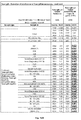
- Fig. 2 shows the results of the interference eliminating effect at different concentrations of the label-specific binding partner.
- Commercially available native samples and in-house samples that do not contain any T. gondii antibodies were tested for the positive results in an Elecsys Anti-Toxo IgM assay.
- the reference column shows that all of the eight samples tested provide a positive signal above the cut-off value falsely indicating that these samples contain Toxo-IgM antibodies.
- the molar excess or proportion of label specific binding partner (that has two paratopes) compared to the label is about 1:10, i.e. the label is present in about 10-fold access so that every tenth label is covered by a label-specific binding partner.
- Elecsys ® 2010 analyzer Roche Diagnostics GmbH
- Elecsys ® is a registered trademark of the Roche group.
- the general detection principle on the analyzer works in an analogous way as described in Example 1 (Anti-Toxo IgM).
- anti-HBc assay assay works according to the competition principle where the analyte (anti-HBc antibody) competes with biotinylated and ruthenium-labeled antibodies for binding to HBc antigen in the reaction mixture. Total duration of the assay was 27 minutes.
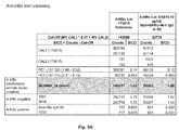
- Fig. 3 shows the elimination of interference by addition of a label-specific binding partner in an immunoassay for detection of antibodies against hepatitis B core antigen.
- This assay is performed in a competitive format which means that high signals and a signal to cut-off ratio of > 1.0 indicate a non-reactive (negative) sample whereas a low signal and a signal to cut-off ratio of ⁇ 1.0 are indicative for a reactive (positive) sample.
- Sample SE-0064_18 Trina A-HBc negative serum
- S/CO 1.80
Landscapes
- Health & Medical Sciences (AREA)
- Life Sciences & Earth Sciences (AREA)
- Immunology (AREA)
- Engineering & Computer Science (AREA)
- Chemical & Material Sciences (AREA)
- Molecular Biology (AREA)
- Biomedical Technology (AREA)
- Hematology (AREA)
- Urology & Nephrology (AREA)
- Physics & Mathematics (AREA)
- General Health & Medical Sciences (AREA)
- Analytical Chemistry (AREA)
- Pathology (AREA)
- General Physics & Mathematics (AREA)
- Biochemistry (AREA)
- Biotechnology (AREA)
- Microbiology (AREA)
- Medicinal Chemistry (AREA)
- Food Science & Technology (AREA)
- Cell Biology (AREA)
- Chemical Kinetics & Catalysis (AREA)
- Virology (AREA)
- Tropical Medicine & Parasitology (AREA)
- Communicable Diseases (AREA)
- Plasma & Fusion (AREA)
- Nuclear Medicine, Radiotherapy & Molecular Imaging (AREA)
- Peptides Or Proteins (AREA)
Abstract
The invention concerns an immunoassay method for detecting an analyte such as an antigen or an antibody in an isolated sample suspected to contain said analyte by incubating said sample with a plurality of binding partners, one of which carries a detectable label, wherein a label-specific binding partner is added that does not carry a label but binds to said detectable label. The method is applicable for a large variety of analytes and has proven particularly useful for analyte antibodies of the IgG and IgM class present in samples due to infections by pathogens. The invention further concerns a reagent kit useful for said method comprising at least two analyte-specific binding partners one of which carries a detectable label and a label-specific binding partner that binds to said detectable label but itself does not carry a detectable label. In addition, the invention covers the use of a label-specific binding partner that does not carry a detectable label in an in vitro diagnostic test for eliminating interferences caused by anti-label antibodies present in a sample.
Description
- The invention concerns a method for reduction of interferences in immunoassays that are caused by interfering factors that bind to a detectable label that is used for direct or indirect detection of an analyte. More specifically the invention concerns an immunoassay method for detecting an analyte such as an antibody or an antigen in an isolated sample by incubating said sample with a plurality of binding partners, one of which carries a detectable label, wherein a label-specific binding partner is added that does not carry a label but binds to said detectable label.
- Methods for eliminating interferences in immunodiagnostic assays are well-known in the art and have been described in detail. For review, see for example Park and Kricka (The Immunoassay Handbook Elsevier Ltd. 2013, Chapter 5.3, pages 403-415). In particular solid-phase-based immunoassays, so-called heterogeneous assays, are subject to various interferences that can lead to false-positive or false-negative assay results, high background signal, decreased assay sensitivity and dynamics, lack of reagent stability and shortened shelf-life of these reagents. Non-specific interference is often caused by nonspecific binding of assay components to solid phase material independently of the presence of an analyte. Another source of interference is the presence of rheumatoid factors, anti-IgM antibodies or anti-animal-antibodies (heterophilic antibodies) such as human anti-mouse antibodies (HAMA).
- Another frequent source for interference is the signal-generating component that is used in an immunoassay. Known signal-generating compounds are for example enzymes or labels emitting light based on fluorescent, colorimetric, chemiluminescent or electrochemiluminescent principles. In particular for electrochemiluminescent compounds interference due to non-specific binding to the signal-generating compound has been described in prior art (Ando et al. Intern Med 2007, 46(15): 1225-1229; Sapin et al., Clin Chem Lab Med 2007; 45(3):416-418).
- In order to avoid interferences based on the signal-generating component often an excess amount of carrier protein linked to said signal-generating component is added to an immunoassay mixture. As a result the interfering compound or factor binds to said signal-generating component attached to the free carrier protein. In case this measure is not sufficient to eliminate the interference also a substance can be added to the immunoassay mixture that is structurally similar to the real signal-generating component but does not provide any signal by its own. As a consequence, interfering factors present in the sample bind to the excess similar compound in a quantitative way and do not bind to the real target, i.e. the signal-generating compound. Thus the interference can be suppressed. Such a procedure has been described for example in
US patent no. 5,888,745 and its equivalentDE 195 19 973 . In these documents a method for determining an analyte is disclosed wherein an analyte-specific binding substance is attached to a metal-containing complex capable of providing a luminescence signal. In said method a second metal-containing complex that is structurally related to the first metal-containing complex but does not bind to the analyte is added to the sample to be investigated. This second metal-containing complex helps in reducing the quenching of the first metal-containing complex's signal. - However, although measures for eliminating interferences are available immunoassays tend to show interference that cannot be eliminated by simply adding an additional amount of competing compounds similar to the signal-generating compound to the assay admixture.
- The problem to be solved can therefore be seen in increasing the specificity of an immunoassay based on emission of a detectable signal.
- When we added a compound to an immunoassay that binds to the label itself that caused the problem the interference was reduced. This was surprising because our expectation was that the label itself would be masked and shielded so that no or only an insufficient detectable light signal could be generated anymore by the shielded label.
- The invention is based on the finding that a signal-generating compound or detectable label, in particular a light-generating label that is attached to a binding partner, is masked and shielded from its environment by adding a label-specific binding partner.
- The invention concerns an immunoassay method for detecting an analyte such as an antigen or an antibody in an isolated sample suspected to contain said analyte by incubating said sample with a plurality of binding partners, one of which carries a detectable label, wherein a label-specific binding partner is added that does not carry a label but binds to said detectable label.
- The method is applicable for a large variety of analytes and has proven particularly useful for analyte antibodies of the IgG and IgM class present in samples due to infections by pathogens.
- The invention further concerns a reagent kit useful for said method comprising a plurality of binding partners, at least one of which binds to the analyte, and one of which carries a detectable label and one of which is a label-specific binding partner that binds to said detectable label but itself does not carry a detectable label.
- In addition, the invention covers the use of a label-specific binding partner that does not carry a detectable label in an in vitro diagnostic test for eliminating interferences caused by anti-label antibodies present in a sample.
-
-
Figure 1 shows the results of a Toxoplasma gondii IgM immunoassay as described in Examples 2 and 3. -
Figure 2 shows the results of a Toxoplasma IgM immunoassay with varying concentrations of a label-specific binding partner as anti-interference component (see Example 3). -
Figure 3 shows the results of an Anti-hepatitis B core immunoassay with optimized concentration of a label-specific binding partner as anti-interference component. For details refer to Example 4. -
Figure 4 shows a schematic drawing of a non-competitive assay principle. In this format the detected signal is directly proportional to the amount of the analyte, i.e. a small concentration of analyte results in a low measured signal whereas a high concentration of analyte leads to an increased signal. If no analyte is present no signal above a predetermined background or cut-off value can be detected.- a) format for detecting a specific class of antibodies such as IgM antibodies (µ capture); the first binding partner is an analyte-specific antigen that carries a detectable label;
- b) as is a) but the first binding partner is indirectly labeled by adding an additional labeled binding partner that binds the first binding partner;
- c) double-antigen sandwich format (DAGS) for detecting antibodies; first and second binding partners both bind the analyte. The first binding partner carries a detectable label itself or it can also be indirectly labeled by adding an additional binding partner that carries a label and that binds to the first binding partner as shown in b).
-
Figure 5 (embodiment a)) shows a schematic drawing of a competitive assay format. In competitive formats the relation between a detected signal and presence of analyte is reversed, i.e. for embodiment a) in the absence of the analyte the specifier is able to bind to the analyte-specific binding partner and provides a measurable signal. On the other hand, in the presence of analyte binding of the specifier to the analyte-specific binding partner is suppressed due to competition with the analyte. Also the binding of the second binding partner to the analyte-specific binding partner is reduced if the analyte is present. As a consequence no or lower signal shows that the analyte is present in the investigated sample. -
Figure 6 shows two additional embodiments b) and c) of a competitive assay format according to the invention. Also here the relation between a detected signal and presence of analyte is reversed, i.e. in the absence of the analyte the detectable signal emitted by the label is high, i.e. higher than in the presence of the analyte where the measurable signal is decreased.- b) In this embodiment the first binding partner carries a label and no specifier is used. The second binding partner competes with the analyte for binding to the labeled first binding partner.
- c) In this embodiment the first binding partner does not carry a label and is capable of being bound to a solid phase. No second binding partner is used for attachment of the immunocomplex to the solid phase. The specifier carries a label. The analyte competes with the specifier for binding to the first binding partner.
- In both embodiments b) and c) the third binding partner is added which binds to the label that is carried by either the first binding partner (b)) or the specifier (c)).
- The invention concerns an immunoassay method for detecting an analyte in an isolated sample suspected to contain said analyte by incubating said sample with a plurality of binding partners, one of which carries a detectable label, wherein a label-specific binding partner is added that does not carry a label itself but binds to said detectable label.
- Different formats and variants of immunoassays are widely known in the art, such as homogeneous and heterogeneous immunoassays. In homogeneous immunoassays reagents and samples are mixed and directly measured for example by turbidimetric methods.
- This invention concerns heterogeneous immunoassays. Heterogeneous immunoassays are run in multiple steps where reagents such as analyte-specific binding partners that carry a detectable label are added to a sample to be investigated. These assays are called "heterogeneous" because two phases are needed, a liquid and a solid phase. Before, during or after completion of the immunoreaction in solution between the analyte and a specific binding partner - making up an immunocomplex - said immunocomplex is attached to a solid phase such as a microtiter plate latex or polystyrol beads. This results in two "heterogeneous" phases, a liquid and a solid phase. After separation of liquid and solid phases the signal emitted from a label attached to an analyte-specific binding partner is detected in either the solid phase or the liquid phase or in both phases.
- The immunoassay can be carried out either in a competitive or a non-competitive format. In the competitive format, the analyte contained in a sample competes with a labeled binding partner which is similar or identical to the analyte for binding to a capture compound, which is frequently an antibody, or, in case the presence of antibodies to e.g. a pathogenic agent to be tested in a serum sample, an antigen of said pathogenic agent.
- In an embodiment of the invention the immunoassay method has got a non-competitive format (for illustration of various specific formats, see
Fig. 4 ). In this case the analyte is detected by contacting the analyte with a compound binding to the analyte, i.e. an analyte-specific binding partner. This analyte-specific binding partner either carries a detectable label itself or is the target of another binding partner carrying a detectable label. The term "carries a detectable label" includes both a directly attached label and a label that is conferred indirectly by adding another labeled binding partner that binds to the analyte-specific binding partner (for illustration see e.g.Fig. 4 b )). Thus, in non-competitive immunoassays, the amount of analyte is determined by determining the amount of complexes formed between the analyte and a detector compound carrying a label. Well-known non-competitive immunoassay formats are the classic sandwich format (analyte captured between two analyte-specific antibodies), the double-antigen sandwich format (analyte is an antibody captured between two specific antigens) and the µ-capture format wherein IgM-antibodies are captured by an antibody specific for human-IgM antibodies. In this so-called µ-capture format usually the analyte IgM-antibody is detected using a labeled antigen that binds to the µ-captured IgM analyte. - In more detail, said immunoassay method carried out in a non-competitive format comprises the following steps:
- a. incubating said sample with
- i. a first binding partner that binds to the analyte and that carries a detectable label
- ii. a second binding partner that is capable of being bound to a solid phase and that binds to the analyte
- iii. a third binding partner that binds to said detectable label of said first binding partner, wherein said third binding partner does not carry a label
- b. forming an immunoreaction admixture by admixing said sample with said first, second and third binding partners
- c. maintaining said immunoreaction admixture for a time period sufficient for allowing said analyte if present in the body fluid sample to immunoreact with said first and second binding partners to form an immunoreaction product; and allowing said third binding partner to immunoreact with said detectable label of said first binding partner
- d. detecting the presence and/or the concentration of any of said immunoreaction product.
- An embodiment of the non-competitive format is further illustrated in
Fig. 4 . According to the invention a plurality of binding partners is applied, at least one of which is an analyte-specific binding partner. An analyte-specific binding partner is a biological molecule that binds to the analyte. The analyte-specific binding partner is applied as first binding partner. Such an analyte specific binding partner can be an antibody or a fragment thereof binding to the analyte. An antibody or antibody fragment is particularly useful as analyte specific binding partner if the analyte is an antigen. If the analyte is an antibody then typically an antigen binding to the analyte antibody is applied as analyte-specific binding partner. In a non-competitive assay format the first binding partner (analyte-specific binding partner) carries a label. - For illustration, in an assay for detecting IgM antibodies against Toxoplasma gondii the antigen Toxo p30 antigen is applied as an analyte specific binding partner that binds to Toxoplasma gondii antibodies present in the sample. The Toxo p30 antigen carries a detectable label. An embodiment of the invention is an immunoassay method according to the non-competitive format described above wherein the analyte is an antibody against Toxoplasma gondii.
- The designations i., ii., and iii. are merely descriptive terms and shall not be understood in such a way that a timewise or stepwise order is associated with the addition of the individual binding partners to the immunoreaction admixture. For example, the third binding partner (see step iii) does not need to be added last but could also be added first or all binding partners listed under i., ii., and iii. can be added simultaneously etc.
- Binding of the first and second binding partners to the analyte as well as the binding of the third binding partner to the label (that is carried by the first binding partner) take place in a specific manner that can also be described as exclusive binding with high affinity. This means that the first binding partner specifically binds to the analyte, the second binding partner also specifically binds to the analyte and also the third binding partner specifically binds to the label attached to the first binding partner. This means for example that in an embodiment where the analyte is an IgM molecule against Toxoplasma gondii the first binding partner is an antigen that is recognized by and binds to the analyte IgM such as Toxo p30 antigen. In this embodiment the second binding partner that can be bound to a solid phase is an antibody against the Fc part of IgM antibodies (µ-chain). As a result, the second binding partner specifically binds to the Fc-part of IgM-molecules and the first binding partner (Toxo p30 antigen) that carries a label specifically binds to the paratope of the analyte IgM antibody.
- The term "specific" or "specific(ally) binding" also indicates that other compounds, typically biomolecules, that are present in a sample do not significantly bind to the analyte, binding partners and the label (summarized as target molecules). This does not exclude binding of other chemical compounds, to regions of the target molecules not involved in interaction with the analyte, binding partners, and the label.
- According to the invention a second binding partner is applied that is capable of being bound to a solid phase. This binding partner can be an antibody or a fragment thereof. In a so-called double-antigen sandwich (DAGS) format which can be used for detecting analyte antibodies the second binding partner is an antigen binding to the analyte antibody. This antigen can be bound to a solid phase for example by conjugating the antigen with biotin and attaching it to a streptavidin-coated solid phase.
- As explained above, in assays according to the µ-capture format as additional binding partner (second binding partner) an antibody binding to human IgM is applied. This second binding partner binds IgM molecules independently of their antigen-specificity reflected in the IgM analyte's paratope (it would bind to all IgM molecules in the sample). The second binding partner can be bound to a solid phase. Binding mechanisms of biomolecules to a solid phase are well-known in the art, the easiest way being direct coating of proteins to a plastic surface, e.g. to a microtiter plate. In an embodiment the binding to a solid phase takes place via biotin-streptavidin interaction, wherein the second binding partner carries a biotin moiety and the solid phase is coated with streptavidin.
- According to the invention a label-specific binding partner is added as third binding partner to an incubation admixture containing the sample to be analyzed and a plurality of binding partners. This label-specific binding partner is a biological molecule that binds to the label applied for detection that is carried by one of the binding partners. In the non-competitive format the label is usually attached directly or indirectly to one of the analyte-specific binding partners. In an embodiment it is attached to the first binding partner. In an embodiment of a competitive assay format (see further below) the label is attached to a specifier that competes with the analyte for binding to the other binding partner present in the incubation admixture.
- Preferably, said label-specific binding partner is an antibody or a fragment thereof binding to the label. It is important that the label-specific binding partner itself does not carry a label and does not interact or does not significantly interact with other assay components except for the label. In other words, detection concepts known in the art that are based on a method wherein the label of a specific binding partner is recognized by a label-specific binding partner which itself also carries a label (such as e.g. the digoxin/ digoxigenin and anti-digoxin/digoxigenin antibodies) are not encompassed as those concepts are not applicable for reducing the interference arising from anti-label antibodies.
- The exact stoichiometry for the amount of label-specific binding partner acting as third binding partner compared to the amount of label is not critical so that absolute maximal and minimal concentration ranges are not essential to make the invention work. The only condition that needs to be fulfilled is that the assay reagents stay functional.
- For comparison and counting of the molar proportions of a third binding partner (label-specific binding partner) one has to determine the actual binding sites that are available for masking the label. This means e.g. that a Fab fragment has got one paratope and therefore counts as one binding site whereas e.g. a complete IgG antibody or Fab2 fragment has got two paratopes (binding sites) and thus counts as two binding sites.
- For certain assays it may be helpful to provide an excess of label-specific binding partner (=third binding partner) compared to the molar amount of label. In other assays the situation may be reversed and it may be helpful to add a molar excess of label compared to the amount of label-specific binding partner. For example, in an embodiment of 1:10 (proportion of binding sites of a label-specific binding partner to label) the molar excess of label to be shielded in comparison to the binding sites of a label-specific binding partner is 10-fold, which means that every tenth label molecule could be bound and covered by a binding site of the label-specific binding partner. In another embodiment, the situation would be reversed as e.g. 10:1, i.e. the binding sites of a label-specific binding partner would be present in 10-fold excess. In yet another embodiment the molar the proportions of label-specific binding partner binding sites to label are at least 1:20, in another embodiment 1:10, in another embodiment 1:5, in another embodiment 1:3, in another embodiment 1:2, in another embodiment 1:1.5, in another embodiment 1:1, in another embodiment 2:1, in another embodiment 3:1, in another embodiment 5:1, in another embodiment 10:1, in another embodiment 20:1, in another embodiment 50:1 and in yet another embodiment at least 100:1.
- When the label-specific binding partner is added to an immunoassay admixture it binds to the label, thereby covering and shielding the label from interaction with interfering compounds present in the sample. At the same instance and because of its size and three-dimensional structure the label-specific binding partner can sterically also shield or block the linker attached to the label from interaction with interfering sample compounds. As a result also interferences caused by non-specific binding by sample ingredients to a linker sequence can also be avoided. By adding a label-specific binding partner that itself does not carry a label the interfering component, i.e. the label is masked on purpose thereby preventing binding of further anti-label component that might be present in the sample to the label.
- If no label-specific binding partner is added then quite often false-positive results can be observed. For example, in an assay format based on the µ capture format these interferences are often caused by the presence of non-specific IgM class antibodies. In an assay design based on the µ-capture format these interfering anti-label-IgMs are captured to the solid phase. Since the anti-label-IgMs also bind to the labeled specific compound the labeled analyte-specific binding binding partner binds to the solid phase although no real analyte is present in the sample. As a result, a false positive signal is observed.
- Detectable labels are also widely known to the expert. According to an embodiment of the current invention a detectable label is an enzyme, or a label emitting light such as fluorescence, luminescence, chemiluminescence, electrochemiluminescence or radioactivity. In a preferred embodiment the label is an electrochemiluminescent label such as Tris(2,2'-bipyridyl)ruthenium(II)-complex (Ru(bpy)).
- The term detectable label comprises also a linker sequence that is attached to said label connecting the label with an analyte-specific binding partner. Usually a linker sequence is based on a peptidic backbone of 1-100 natural or synthetic amino acids.
- In another embodiment the immunoassay method can be carried out in a competitive format, comprising the following steps:
- a. Incubating said sample with
- i. a first binding partner that binds to the analyte and that does not carry a detectable label
- ii. a specifier which binds to the first binding partner and which competes with the analyte for binding to the first binding partner wherein said specifier carries a detectable label
- iii. a second binding partner that is capable of being bound to a solid phase and that binds to said first binding partner and which competes with said analyte and said specifier for binding to said first binding partner
- iv. a third binding partner that binds to said detectable label of said specifier, wherein said third binding partner does not carry a label
- b. forming an immunoreaction admixture by admixing said sample with said first, second and third binding partners and said specifier
- c. maintaining said immunoreaction admixture for a time period sufficient for allowing said analyte if present in said body fluid sample to compete with said specifier and second binding partner for binding to said first binding partner, and allowing said third binding partner to immunoreact with said detectable label of said specifier, thereby forming an immunoreaction product;
- d. detecting the presence and/or the concentration of any of said immunoreaction product.
- An embodiment of the competitive immunoassay format is illustrated in
Fig. 5 . In a competitive immunoassay format the interference problem can be explained as follows: When no label-specific binding partner according to the invention is present the binding of the interfering compound to the label leads to a reduction of the measured signal. For the interpretation of a signal in a competitive format this means that a reduced signal could be understood and interpreted as a (false) positive result as also competition with a real analyte leads to reduction in signal. In order to avoid this interference a label-specific binding partner according to the invention is added to the immunoassay admixture so that the potential binding sites for an interfering compound are already occupied and blocked. As a consequence the overall signal range is reduced. However and surprisingly, the analyte present in true positive samples still competes with the assay compounds so that this true positive sample can still be reliably detected. - The properties of the plurality of binding partners used in a competitive immunoassay format differ from those used in a non-competitive format in some aspects. The first binding partner that binds to the analyte does not carry a label in said competitive format. The detectable label is attached instead to a specifier that is applied. A specifier binds to the first binding partner and is typically structurally similar to the analyte. Said specifier therefore competes with the analyte for binding to the first binding partner. The second binding partner is capable of being bound to a solid phase, in so far corresponding to the properties of the second binding partner described for the non-competitive format further above. However, in the competitive format the second binding partner binds to the first binding partner (not to the analyte) and competes with the analyte and the specifier for binding to the first binding partner.
- The properties of the third binding partner that does not carry a label itself and that binds to the label of the specifier are the same as those for the non-competitive format. The designations i., ii., iii. and iv. are merely descriptive terms and shall not be understood in such a way that a timewise or stepwise order is associated with the addition of the individual binding partners to the immunoreaction admixture. For example, the third binding partner (see step iv) does not need to be added last but could also be added first or all binding partners listed under i., ii., iii. and iv. can be added simultaneously etc.
- An example of a competitive immunoassay is an assay for detection of anti-hepatitis B core antibodies, see example 4.
- In another embodiment the method for detecting an analyte in an isolated sample can be carried out in a competitive format as illustrated in
Fig. 6 embodiment b), wherein no specifier is added and the first binding partner carries a label (instead of the specifier that is not present in this case). Said method comprises the following steps: - a. Incubating an isolated sample with
- i. a first binding partner that binds to the analyte that carries a detectable label
- ii. a second binding partner that is capable of being bound to a solid phase and that binds to said first binding partner and which competes with said analyte for binding to said first binding partner
- iii. a third binding partner that binds to said detectable label of said first binding partner wherein said third binding partner does not carry a label
- b. forming an immunoreaction admixture by admixing said sample with said first, second and third binding partners
- c. maintaining said immunoreaction admixture for a time period sufficient for allowing said analyte if present in said body fluid sample to compete with said second binding partner for binding to said first binding partner, and allowing said third binding partner to immunoreact with said detectable label of said first binding partner, thereby forming an immunoreaction product;
- d. detecting the presence and/or the concentration of any of said immunoreaction product.
- In yet another embodiment the can be carried out in a competitive format as illustrated in
Fig. 6 embodiment c), wherein no second binding partner is added and the first binding partner is capable of being bound to a solid phase (instead of using a second binding partner that is not present in this case). Said method comprises the following steps: - a. Incubating an isolated sample with
- i. a first binding partner that binds to the analyte that is capable of being bound to a solid phase and that does not carry a detectable label
- ii. a specifier which binds to the first binding partner and which competes with the analyte for binding to the first binding partner wherein said specifier carries a detectable label
- iii. a third binding partner that binds to said detectable label of said first binding partner wherein said third binding partner does not carry a label
- b. forming an immunoreaction admixture by admixing said sample with said first and second binding partners and said specifier
- c. maintaining said immunoreaction admixture for a time period sufficient for allowing said analyte if present in said body fluid sample to compete with said specifier for binding to said first binding partner, and allowing said third binding partner to immunoreact with said detectable label of said specifier, thereby forming an immunoreaction product;
- d. detecting the presence and/or the concentration of any of said immunoreaction product.
- An analyte for both competitive and non-competitive immunoassays can be any chemical or biological molecule that is large enough to be detected by an immunoassay such as antigens, antibodies and hormones. Some analytes appear in a sample as a consequence of an infection, an inflammatory, septic, metabolic, cardiac or oncologic event or disease. Other analytes are drugs a patient takes and the concentration of this drug needs to be monitored such as drugs for immunosuppression after organ transplantation (therapeutic drug monitoring). Some analytes, mostly hormones, are determined in a fertility check.
- In yet another embodiment the analyte is an enzyme, cytokine, growth factor, hormone, vaccine, antibody and the like. More particularly, an analyte can be a thyroid hormone such as triiodothyronine (T3) and its prohormone thyroxine (T4), thyroid-stimulating hormone (TSH) and TSH receptor autoantibodies, erythropoietin, insulin, somatotropin, growth hormone releasing factor, growth factors such as platelet derived growth factor, epidermal growth factor, transforming growth factor α, transforming growth factor, epidermal growth factor, fibroblast growth factor, nerve growth factor, insulin-like growth factor I, insulin-like growth factor II, clotting Factor VIII, superoxide dismutase, interferon, y-interferon, interleukin-1, interleukin-2, interleukin-3, interleukin-4, interleukin-5, interleukin-6, granulocyte colony stimulating factor, multi-lineage colony stimulating factor, granulocyte-macrophage stimulating factor, macrophage colony stimulating factor, T cell growth factor, lymphotoxin and the like. Also cardiac markers such as Troponin T and I, NT-proBNP, BNP as well as sepsis markers.
- In an embodiment the analyte is a polypeptide such as an antigen or an antibody present in an isolated sample. In an embodiment the analyte is a pathogen and detectable proteins and antigens present due to the infection by said pathogen. The analyte may be derived from any viral, bacterial or protozoic pathogen, which is capable of causing a detectable immune reaction, i.e. a generation of antibodies, particularly IgM antibodies as a result of an infection. For example, the pathogen is selected from the group consisting of
- i a Toxoplasma organism such as Toxoplasma gondii,
- ii hepatitis viruses such as hepatitis A virus (HAV), hepatitis B virus (HBV) and hepatitis C virus (HCV);
- iii herpes viruses such as human
herpes simplex virus 1 and 2 (HHV1 and HHV2), varicella zoster virus (HHV3), Epstein-Barr virus (HHV4/EBV) or human cytomegalovirus (HHV5) andhuman herpes viruses 6, 7, and 8; - iv rubella virus;
- v a retrovirus such as
HIV 1 and 2 andHTLV 1 and 2; - vi paramyxoviruses such as measles virus and mumps virus;
- vii a Borrelia organism.
- In another embodiment the analyte is an antibody of the IgM or IgG class present as a result of an infection by any of the pathogens listed above. In case the analyte is an IgM or IgG antibody the analyte binds to an epitope derived from the pathogen's own structural or non-structural proteins such as e.g. capsid/core antigen or envelope antigen or soluble antigens present within the pathogen.
- In an embodiment the analyte is an antibody of the IgM class present as a reaction to an infection with a pathogen selected from the group consisting of Toxoplasma gondii, cytomegalovirus (CMV), Rubella, hepatitis A and hepatitis B.
- All biological liquids known to the person skilled in the art can be used as isolated samples for the described immunoassay method. The samples usually used are bodily liquids like whole blood, blood sera, blood plasma, urine or saliva. As sample any biological liquid isolated from a vertebrate can be used. In an embodiment samples originate from mammalians, in particular from humans.
- Yet another aspect of the invention is a reagent kit for detecting of an analyte by an immunoassay comprising a plurality of binding partners, at least one of which binds to the analyte and one of which carries a detectable label and a label-specific binding partner that binds to said detectable label but does not carry a detectable label itself. In an embodiment the analyte-specific binding partner carries said label. An additional subject matter of the invention is a reagent kit for the detection of IgM antibodies against Toxoplasma gondii, comprising a plurality of binding partners one of which is an anti-human IgM antibody that can be bound to a solid phase and one of which is a Toxoplasma gondii specific antigen that carries a detectable label, and a label-specific binding partner that binds to said detectable label but itself does not carry a detectable label.
- In addition, the reagent kits defined above contain controls and standard solutions as well as reagents in one or more solutions with the common additives, buffers, salts, detergents etc. as used by the average man skilled in the art along with instructions for use.
- A further embodiment of the invention is the use of in a label-specific binding partner that does not carry a detectable label in an in vitro diagnostic test for eliminating interferences caused by anti-label antibodies present in a sample.
- The following examples illustrate the invention.
- The general procedure for producing monoclonal antibodies is well-known in prior art. The pre-formulated fusion polypeptide immunogen is administered to an experimental animal, such as mouse, rat, rabbit, sheep, or hamster, intraperitoneally at different dosages. Prior to collection of the B-cells a boost immunization is performed. B-cell hybridomas can be obtained according to the method of Koehler and Milstein (Koehler, G. and Milstein, C., Nature 256 (1975) 495-497). The hybridomas obtained are deposited as single clones or cells in the wells of a multi well plate. Primary culture supernatants are tested by ELISA for reactivity against the immunogen.
- Monoclonal mouse antibodies with specificity for the electrochemiluminescent label Ruthenium (II) tris-bipyridine N-hydroxysuccinimide were produced by administering mice with the immunogen Ruthenium (II) tris-bipyridine N-hydroxysuccinimide conjugated to keyhole limpet hemocyanin (BPRu-KLH) according to procedures described in prior art. The cells obtained from immunization were analyzed using an ELISA for their specificity for Ruthenium (II) tris-bipyridine N-hydroxysuccinimide.
- 8-12 weeks old Balb/c mice, respectively, were subjected to repeated intraperitoneal immunizations with 100 µg of immunogen Ruthenium (II) tris-bipyridine N-hydroxysuccinimide conjugated to keyhole limpet hemocyanin (BPRu-KLH). The mice were immunized three times, i.e. also at the time points of 6 weeks and 10 weeks after the initial immunization. The first immunization was performed using complete Freund's adjuvant, the second and third immunizations were done using incomplete Freund's adjuvant. The mice serum titers) were tested after 12 weeks by ELISA methods as described in the following. The ELISA was performed on a microplate reader. ELISA plates were coated with the immunogen Ruthenium (II) tris-bipyridine N-hydroxysuccinimide conjugated to keyhole limpet hemocyanin (BPRu-KLH) by applying a solution comprising 0.5 µg antigen per ml. Thereafter free binding sites were blocked by applying a solution comprising 1 % RPLA in PBS for one hour at room temperature. The wells were washed three times with a solution comprising 0.9 % (w/v) sodium chloride and 0.05 % (w/v) Tween 20. Mouse serum was diluted 1:50 with PBS and used as sample. The incubation time was one hour at room temperature. The wells were washed three times with a solution comprising 0.9 % (w/v) sodium chloride and 0.05 % (w/v) Tween 20. As detection antibody a polyclonal antibody against the constant domain of the target antibodies conjugated to a peroxidase was used (PAK<M-Fcγ>S-F(ab')2-POD). The detection antibody was applied at a concentration of 80 ng/ml in PBS comprising 1 % (w/v) RSA. The incubation time was one hour at room temperature. The wells were washed three times with a solution comprising 0.9 % (w/v) sodium chloride and 0.05 % (w/v) Tween 20. Afterwards, the wells were incubated with an ABTS solution for 15 minutes at room temperature. The intensity of the developed color was photometrically determined.
- Three days before preparation of spleen cells and fusion with a myeloma cell line, the final booster immunization was performed by i.v. injection of 100 µg of immunogen Ruthenium (II) tris-bipyridine N-hydroxysuccinimide conjugated to keyhole limpet hemocyanin (BPRu-KLH).
- Primary culture supernatants were tested by ELISA for reactivity against the immunogen immunogen Ruthenium (II) tris-bipyridine N-hydroxysuccinimide conjugated to keyhole limpet hemocyanin (BPRu-KLH) and a blank plate, respectively. ELISA plates were coated with 0.5 µg/ml immunogen Ruthenium (II) tris-bipyridine N-hydroxysuccinimide conjugated to keyhole limpet hemocyanin (BPRu-KLH). Thereafter, free binding sites were blocked by 1 % RPLA in PBS for one hour at room temperature. The wells were washed three times with a solution comprising 0.9 % (w/v) sodium chloride and 0.05 % (w/v) Tween 20. Undiluted hybridoma supernatants were used as samples. The incubation time was one hour at room temperature. The wells were washed three times with a solution comprising 0.9 % (w/v) sodium chloride and 0.05 % (w/v) Tween 20. As detection antibody a polyclonal antibody against the constant domain of the target antibodies conjugated to a peroxidase was used (PAK<M-Fcγ>S-F(ab')2-POD). The detection antibody was applied at a concentration of 80 ng/ml in PBS comprising 1 % (w/v) RSA. The incubation time was one hour at room temperature. The wells were washed three times with a solution comprising 0.9 % (w/v) sodium chloride and 0.05 % (w/v) Tween 20. Afterwards the wells were incubated with an ABTS solution for 15 minutes at room temperature. The intensity of the developed color was determined photometrically at 405 nm. The reference wavelength was 492 nm. Primary hybridoma supernatants showing fast and strong color formation in ELISA upon binding to Ruthenium (II) tris-bipyridine N-hydroxysuccinimide conjugated to keyhole limpet hemocyanin (BPRu-KLH) were selected. Using this procedure Mab<BPRu>M-1 IgG as applied in Examples 3 and 4 was identified.
- The immunoassay for the in vitro qualitative determination of IgM antibodies to Toxoplasma gondii was carried out according to the manufacturer's instructions on an automated Elecsys® 2010 analyzer (Roche Diagnostics GmbH). Elecsys® is a registered trademark of the Roche group.
- Signal detection in Elecsys® 2010 is based on electrochemoluminescence. The biotin-conjugate (i.e. the capture-antigen) is immobilized on the surface of a streptavidin coated magnetic bead whereas the detection-antigen bears a complexed Ruthenium cation (switching between the redox states 2+ and 3+) as the signaling moiety. In the presence of a specific immunoglobulin analyte, the chromogenic ruthenium complex is bridged to the solid phase and emits light at 620 nm after excitation at a platinum electrode. The signal output is in arbitrary light units.
- The assay is based on the so-called µ-capture principle wherein a sample suspected to contain anti-T. gondii IgM antibodies is incubated with ruthenylated T. gondii-specific recombinant antigen and biotinylated anti-human IgM mouse monoclonal antibody. If T. gondii-specific IgM antibodies are present in said sample the anti-IgM capture these antibodies. After addition of a streptavidin-coated solid phase a removal of excess liquid the electrochemiluminescence signal originating from the ruthenium complex label can be detected.
- In detail, the following steps were carried out; total duration of the assay was 18 minutes:
- 1st incubation: 10 µL of sample were automatically prediluted 1:20 with Diluent Universal. T. gondii-specific recombinant antigen labeled with a ruthenium complex (R1; Ru complex: Tris(2,2'-bipyridyl)ruthenium(II)-complex (Ru(bpy))) was added. Anti-T. gondii IgM antibodies present in the sample reacted with the ruthenium-labeled T. gondii-specific recombinant antigen.
- 2nd incubation: Biotinylated monoclonal h-IgM-specific antibodies (R2) and streptavidin-coated microparticles (M) were added. The complex became bound to the solid phase via interaction of biotin and streptavidin.
- The reaction mixture was aspirated into the measuring cell where the microparticles were magnetically captured onto the surface of the electrode. Unbound substances were then removed with ProCell. Application of a voltage to the electrode then induced chemiluminescent emission which was measured by a photomultiplier.
- Results were determined automatically by the software by comparing the electrochemiluminescence signal obtained from the reaction product of the sample with the signal of the cutoff value previously obtained by calibration.
- As negative calibrator Call human serum, negative for anti-Toxo IgM, was used. As positive calibrator Cal2 Anti-Toxo IgM (human) approx. 130 U/mL (Roche units) in human serum was applied.
- Commercially available human serum samples were purchased from in.vent Diagnostica GmbH (Hennigsdorf /Berlin, Germany) and screened for interfering properties. These samples were tested along with in-house false-positive samples as well as with true Toxo IgM and IgG positive samples. Among the Toxo IgM and IgG positive samples we also tested two subgroups of samples, i.e. one group with IgG of high avidity (late infection) and one group with IgG of low avidity (early or primary infection). All samples were tested with Elecsys Toxo IgM (Roche Diagnostics GmbH Germany) according to the procedure described above. As a negative control PC1 [human serum free of anti-Toxo antibodies] was used; as positive control PC2 [Toxo-IgM positive human serum]. A signal to cutoff ratio (S/CO) < 0.8 was regarded as non-reactive (negative); a signal to cutoff ratio (S/CO) ≥ 0.8 < 1.0 was classified as undetermined and a signal to cutoff ratio (S/CO) ≥ 1.0 was classified as reactive (positive). Results are shown in
Fig. 1 . In the absence of a label-specific binding partner to eliminate interference sera classified as interference samples beginning with "C21" to "SN947 (16)" these samples show up as falsely positive. When a label-specific binding partner (Mab<BPRu>M-1 IgG) is added that binds to the label the overall signal decreases for all samples including real positives. However, the cut-off index remains constant. Real positive samples are still correctly detected as positive. Interestingly and surprisingly the signal for the interference samples goes down to the negative calibration value. In other words, when a label-specific binding partner is added false positives can be avoided resulting in higher assay specificity. -
Fig. 2 shows the results of the interference eliminating effect at different concentrations of the label-specific binding partner. Commercially available native samples and in-house samples that do not contain any T. gondii antibodies were tested for the positive results in an Elecsys Anti-Toxo IgM assay. The reference column shows that all of the eight samples tested provide a positive signal above the cut-off value falsely indicating that these samples contain Toxo-IgM antibodies. - Increasing amounts (0.25, 0.5 and 10 µg/ml) of an interference-eliminating compound were added to the assay mixture (R1), i.e. in this case an anti-ruthenium complex monoclonal antibody Mab<BPRu>M-1 IgG which binds to the signal-generating ruthenium complex attached to the recombinant Toxo antigen. As expected, the absolute signal intensity is quenched by adding said label-specific binding partner. However, surprisingly when sufficient anti-label-specific binding partner is added the interference can be suppressed providing correct results below the cutoff and below the cut-off index (see column 10µg/ml in R1). In this example the molar excess or proportion of label specific binding partner (that has two paratopes) compared to the label is about 1:10, i.e. the label is present in about 10-fold access so that every tenth label is covered by a label-specific binding partner.
- Even at a concentration of 1µg/ml in R1, which means that even a smaller proportion of label is shielded, a satisfying elimination of interference can be observed (data not shown). In other words, addition of a label-specific binding partner that binds to a label meant to indicate whether an analyte is present or not suppresses interferences effectively so that false-positive assay results can be avoided. At the same time, a positive control (see row PC1, negative control and PC2, positive control containing Toxo-IgM) still provides correct negative or positive results.
- The immunoassay for the in vitro qualitative determination of IgG and IgM antibodies to hepatitis B core antigen was carried out according to the manufacturer's instructions on an automated Elecsys® 2010 analyzer (Roche Diagnostics GmbH). Elecsys® is a registered trademark of the Roche group. The general detection principle on the analyzer works in an analogous way as described in Example 1 (Anti-Toxo IgM).
- In detail, the anti-HBc assay assay works according to the competition principle where the analyte (anti-HBc antibody) competes with biotinylated and ruthenium-labeled antibodies for binding to HBc antigen in the reaction mixture. Total duration of the assay was 27 minutes.
- 1sit incubation: Pretreatment of 40 µL of sample was carried out with a reducing agent (R0)
- 2nd incubation: After addition of HBcAg recombinant antigen (R1), a complex was formed with anti-HBc antibodies in the sample.
- 3rd incubation: After addition of biotinylated antibodies and ruthenium complex-labeled antibodies specific for HBcAg (R2; Ru complex: Tris(2,2'-bipyridyl)ruthenium(II)-complex (Ru(bpy)), together with streptavidin-coated microparticles (M), the still-free binding sites on the HBc-antigens became occupied. The entire complex was bound to the solid phase via interaction of biotin and streptavidin.
- The reaction mixture was aspirated into the measuring cell where the microparticles were magnetically captured onto the surface of the electrode. Unbound substances were then removed with ProCell. Application of a voltage to the electrode then induced chemiluminescent emission which was measured by a photomultiplier.
- Results were determined automatically by the software by comparing the electrochemiluminescence signal obtained from the reaction product of the sample with the signal of the cutoff value previously obtained by calibration.
- As negative calibrator Call human serum (not infected by hepatitis B) was used. As positive calibrator Cal2 Anti-HBc (human) > 8 WHO IU/mLb) in human serum was used. WHO IU means WHO international units.
-
Fig. 3 shows the elimination of interference by addition of a label-specific binding partner in an immunoassay for detection of antibodies against hepatitis B core antigen. This assay is performed in a competitive format which means that high signals and a signal to cut-off ratio of > 1.0 indicate a non-reactive (negative) sample whereas a low signal and a signal to cut-off ratio of ≤ 1.0 are indicative for a reactive (positive) sample. - Sample SE-0064_18 (Trina A-HBc negative serum) provides a signal very close to the cut-off with a signal to cut-off value of 1.02. When a label-specific binding partner is added, the sample provides a measuring result (S/CO = 1.80) indicating that no anti-HBc antibodies are present in said sample. Also in this case the addition of a label-specific binding partner leads to an increased specificity of the assay.
Claims (11)
- An immunoassay method for detecting an analyte in an isolated sample suspected to contain said analyte by incubating said sample with a plurality of binding partners, one of which carries a detectable label,
wherein a label-specific binding partner is added that does not carry a label but binds to said detectable label. - An immunoassay method according to claim 1, comprisinga. incubating said sample withi. a first binding partner that binds to the analyte and that carries a detectable labelii. a second binding partner that is capable of being bound to a solid phase and that binds to the analyteiii. a third binding partner that binds to said detectable label of said first binding partner, wherein said third binding partner does not carry a labelb. forming an immunoreaction admixture by admixing said sample with said first, second and third binding partnersc. maintaining said immunoreaction admixture for a time period sufficient for allowing said analyte if present in the body fluid sample to immunoreact with said first and second binding partners to form an immunoreaction product; and allowing said third binding partner to immunoreact with said detectable label of said first binding partnerd. detecting the presence and/or the concentration of any of said immunoreaction product.
- An immunoassay method according to claim 1, comprisinga. incubating said sample withi. a first binding partner that binds to the analyte and that does not carry a detectable labelii. a specifier which binds to the first binding partner and which competes with the analyte for binding to the first binding partner wherein said specifier carries a detectable labeliii. a second binding partner that is capable of being bound to a solid phase and that binds to said first binding partner and which competes with said analyte and said specifier for binding to said first binding partneriv. a third binding partner that binds to said detectable label of said specifier, wherein said third binding partner does not carry a labelb. forming an immunoreaction admixture by admixing said sample with said first, second and third binding partners and said specifierc. maintaining said immunoreaction admixture for a time period sufficient for allowing said analyte if present in said body fluid sample to compete with said specifier and second binding partner for binding to said first binding partner, and allowing said third binding partner to immunoreact with said detectable label of said specifier, thereby forming an immunoreaction product;d. detecting the presence and/or the concentration of any of said immunoreaction product.
- An immunoassay method according to any of claims 1 to 3 wherein said analyte is a biological or chemical molecule associated with an infection, an inflammatory, septic, metabolic, cardiac or oncologic event or disease.
- An immunoassay method according to claim 4 wherein said analyte is a hormone, an antigen or an antibody.
- An immunoassay method according to claim 5 wherein said analyte is an antibody against a pathogen selected from the group consisting ofa. a Toxoplasma organism such as Toxoplasma gondii,b. hepatitis viruses such as hepatitis A virus (HAV), hepatitis B virus (HBV) and hepatitis C virus (HCV);c. herpes viruses such as human herpes simplex virus 1 and 2 (HHV1 and HHV2), varicella zoster virus (HHV3), Epstein-Barr virus (HHV4/EBV) or human cytomegalovirus (HHV5) and human herpes viruses 6, 7, and 8;d. rubella virus;e. a retrovirus such as HIV 1 and 2 and HTLV 1 and 2;f. paramyxoviruses such as measles virus and mumps virus;g. a Borrelia organism.
- An immunoassay method according to claim 6 wherein said analyte is an antibody of the IgM class.
- An immunoassay method according to claim 7 wherein said analyte is an antibody of the IgM class present as a reaction to an infection with a pathogen selected from the group consisting of Toxoplasma gondii, cytomegalovirus (CMV), Rubella, hepatitis A and hepatitis B.
- An immunoassay method according to any of the preceding claims wherein said detectable label is an enzymatic, radioactive, luminescent, chemiluminescent or electrochemiluminescent label.
- A reagent kit for detecting an analyte by an immunoassay comprising a plurality of binding partners wherein at least one of the binding partners binds to the analyte, and one of the binding partners carries a detectable label and one of the binding partners is label-specific and binds to said detectable label but does not carry a detectable label.
- Use of in a label-specific binding partner that does not carry a detectable label in an in vitro diagnostic test for eliminating interferences caused by anti-label antibodies present in a sample.
Priority Applications (9)
| Application Number | Priority Date | Filing Date | Title |
|---|---|---|---|
| EP15197192.6A EP3176580A1 (en) | 2015-12-01 | 2015-12-01 | Method for reduction of interferences in immunoassays |
| BR112018005812-2A BR112018005812B1 (en) | 2015-12-01 | 2016-11-30 | METHOD FOR REDUCING INTERFERENCE IN IMMUNOASSAYS, REAGENT KIT AND USE OF A SPECIFIC MARKER BINDING PARTNER |
| CN201680070249.7A CN108291912B (en) | 2015-12-01 | 2016-11-30 | Methods for reducing interference in immunoassays |
| KR1020187013476A KR102164266B1 (en) | 2015-12-01 | 2016-11-30 | Methods for reducing interference in immunoassays |
| PCT/EP2016/079170 WO2017093271A1 (en) | 2015-12-01 | 2016-11-30 | Method for reduction of interferences in immunoassays |
| JP2018528341A JP6779293B2 (en) | 2015-12-01 | 2016-11-30 | Methods for reducing interference in immunoassays |
| ES16805762T ES2972639T3 (en) | 2015-12-01 | 2016-11-30 | Procedure for reducing interferences in immunoassay |
| EP16805762.8A EP3384288B1 (en) | 2015-12-01 | 2016-11-30 | Method for reduction of interferences in immunoassays |
| US15/995,572 US11320423B2 (en) | 2015-12-01 | 2018-06-01 | Method for reduction of interferences in immunoassays |
Applications Claiming Priority (1)
| Application Number | Priority Date | Filing Date | Title |
|---|---|---|---|
| EP15197192.6A EP3176580A1 (en) | 2015-12-01 | 2015-12-01 | Method for reduction of interferences in immunoassays |
Publications (1)
| Publication Number | Publication Date |
|---|---|
| EP3176580A1 true EP3176580A1 (en) | 2017-06-07 |
Family
ID=54754553
Family Applications (2)
| Application Number | Title | Priority Date | Filing Date |
|---|---|---|---|
| EP15197192.6A Ceased EP3176580A1 (en) | 2015-12-01 | 2015-12-01 | Method for reduction of interferences in immunoassays |
| EP16805762.8A Active EP3384288B1 (en) | 2015-12-01 | 2016-11-30 | Method for reduction of interferences in immunoassays |
Family Applications After (1)
| Application Number | Title | Priority Date | Filing Date |
|---|---|---|---|
| EP16805762.8A Active EP3384288B1 (en) | 2015-12-01 | 2016-11-30 | Method for reduction of interferences in immunoassays |
Country Status (7)
| Country | Link |
|---|---|
| US (1) | US11320423B2 (en) |
| EP (2) | EP3176580A1 (en) |
| JP (1) | JP6779293B2 (en) |
| KR (1) | KR102164266B1 (en) |
| CN (1) | CN108291912B (en) |
| ES (1) | ES2972639T3 (en) |
| WO (1) | WO2017093271A1 (en) |
Cited By (1)
| Publication number | Priority date | Publication date | Assignee | Title |
|---|---|---|---|---|
| CN111122852A (en) * | 2018-10-31 | 2020-05-08 | 博阳生物科技(上海)有限公司 | Homogeneous phase chemiluminescence analysis method and application thereof |
Families Citing this family (9)
| Publication number | Priority date | Publication date | Assignee | Title |
|---|---|---|---|---|
| CN111868526A (en) | 2018-03-13 | 2020-10-30 | 豪夫迈·罗氏有限公司 | Reduce interference in immunoassays |
| EP3853612A4 (en) * | 2018-09-18 | 2021-10-27 | Siemens Healthcare Diagnostics, Inc. | ZIKA VIRUS IMMUNOLOGICAL ASSAY METHODS AND REAGENTS |
| CN109444426A (en) * | 2018-12-12 | 2019-03-08 | 郑州安图生物工程股份有限公司 | The method for eliminating HAMA interference in prize law IgM antibody detection system |
| WO2020229378A1 (en) * | 2019-05-13 | 2020-11-19 | F. Hoffmann-La Roche Ag | Interference-suppressed pharmacokinetic immunoassay |
| AU2020308574A1 (en) * | 2019-06-25 | 2022-03-24 | Veravas, Inc. | Compositions and methods for detecting and depleting sample interferences |
| CN114258491A (en) * | 2019-08-16 | 2022-03-29 | 深圳迈瑞生物医疗电子股份有限公司 | Immunoassay format for a fully novel total antibody assay |
| CN111089961A (en) * | 2019-12-26 | 2020-05-01 | 迪瑞医疗科技股份有限公司 | Toxoplasma IgM antibody detection kit and preparation method thereof |
| CN111089975A (en) * | 2019-12-26 | 2020-05-01 | 迪瑞医疗科技股份有限公司 | Chemiluminescence immunoassay determination kit for detecting Toxoplasma gondii IgG antibody and preparation method thereof |
| CN115327101B (en) * | 2022-08-19 | 2025-05-13 | 中国人民解放军南部战区总医院 | A gamma interferon electrochemiluminescence test strip and its application |
Citations (4)
| Publication number | Priority date | Publication date | Assignee | Title |
|---|---|---|---|---|
| DE19519973A1 (en) | 1995-05-31 | 1996-12-05 | Boehringer Mannheim Gmbh | Interference suppression reagent for the determination of an analyte with a metal complex capable of luminescence |
| WO1998034109A1 (en) * | 1997-01-31 | 1998-08-06 | Bayer Corporation | Improving performance of binding assays by use of more than one label |
| WO2009111729A1 (en) * | 2008-03-06 | 2009-09-11 | Ventana Medical Systems, Inc. | Anti-dnp antibodies and methods of use |
| WO2014144325A1 (en) * | 2013-03-15 | 2014-09-18 | President And Fellows Of Harvard College | Methods and compositions for improving detection and/or capture of a target entity |
Family Cites Families (14)
| Publication number | Priority date | Publication date | Assignee | Title |
|---|---|---|---|---|
| DE4407423A1 (en) | 1994-03-05 | 1995-09-07 | Boehringer Mannheim Gmbh | Anti-interference agent for use in immunoassays |
| DE19649389A1 (en) * | 1996-11-29 | 1998-06-04 | Boehringer Mannheim Gmbh | Antigen-specific IgM detection |
| CN1629309A (en) * | 2004-08-24 | 2005-06-22 | 中国水产科学研究院黄海水产研究所 | Method for screening acceptor blocker for controlling prawn virus infection |
| US20070059682A1 (en) * | 2005-09-13 | 2007-03-15 | Rapid Pathogen Screening Inc. | Method to increase specificity and/or accuracy of lateral flow immunoassays |
| US7732153B2 (en) * | 2006-05-09 | 2010-06-08 | Beckman Coulter, Inc. | Nonseparation assay methods |
| TWI434855B (en) * | 2006-11-21 | 2014-04-21 | Hoffmann La Roche | Conjugate and its use as a standard in an immunoassay |
| CA2686206A1 (en) | 2007-05-07 | 2008-11-13 | Wyeth | Elimination of interference in immunoassays caused by anti-carbohydrate antibodies |
| EP2108957A1 (en) | 2008-04-07 | 2009-10-14 | Koninklijke Philips Electronics N.V. | Compounds and methods for the labelling and affinity-selection of proteins |
| US8614297B2 (en) * | 2008-12-22 | 2013-12-24 | Hoffmann-La Roche Inc. | Anti-idiotype antibody against an antibody against the amyloid β peptide |
| JP2010172324A (en) * | 2009-02-02 | 2010-08-12 | Tdk Corp | Method and kit for detecting microorganism using aptamer |
| US8476079B2 (en) | 2010-04-30 | 2013-07-02 | Abbott Point Of Care Inc. | Reagents for reducing leukocyte interference in immunoassays |
| CN103068852B (en) * | 2010-08-17 | 2016-04-20 | 弗·哈夫曼-拉罗切有限公司 | anti-human IgG1 antibody |
| WO2013132338A2 (en) * | 2012-03-06 | 2013-09-12 | Calpro As | Competitive immunoassay for calprotectin |
| JP6850254B2 (en) * | 2014-12-18 | 2021-03-31 | エフ.ホフマン−ラ ロシュ アーゲーF. Hoffmann−La Roche Aktiengesellschaft | Ways to reduce interference |
-
2015
- 2015-12-01 EP EP15197192.6A patent/EP3176580A1/en not_active Ceased
-
2016
- 2016-11-30 WO PCT/EP2016/079170 patent/WO2017093271A1/en not_active Ceased
- 2016-11-30 ES ES16805762T patent/ES2972639T3/en active Active
- 2016-11-30 JP JP2018528341A patent/JP6779293B2/en active Active
- 2016-11-30 KR KR1020187013476A patent/KR102164266B1/en active Active
- 2016-11-30 EP EP16805762.8A patent/EP3384288B1/en active Active
- 2016-11-30 CN CN201680070249.7A patent/CN108291912B/en active Active
-
2018
- 2018-06-01 US US15/995,572 patent/US11320423B2/en active Active
Patent Citations (5)
| Publication number | Priority date | Publication date | Assignee | Title |
|---|---|---|---|---|
| DE19519973A1 (en) | 1995-05-31 | 1996-12-05 | Boehringer Mannheim Gmbh | Interference suppression reagent for the determination of an analyte with a metal complex capable of luminescence |
| US5888745A (en) | 1995-05-31 | 1999-03-30 | Boehringer Mannheim Gmbh | Interference elimination reagent for the determination of an analyte using a metal complex capable of luminescence |
| WO1998034109A1 (en) * | 1997-01-31 | 1998-08-06 | Bayer Corporation | Improving performance of binding assays by use of more than one label |
| WO2009111729A1 (en) * | 2008-03-06 | 2009-09-11 | Ventana Medical Systems, Inc. | Anti-dnp antibodies and methods of use |
| WO2014144325A1 (en) * | 2013-03-15 | 2014-09-18 | President And Fellows Of Harvard College | Methods and compositions for improving detection and/or capture of a target entity |
Non-Patent Citations (5)
| Title |
|---|
| ANDO ET AL., INTERN MED, vol. 46, no. 15, 2007, pages 1225 - 1229 |
| GESSEL ET AL: "Anti-ruthenium antibodies mimic macro-TSH in electrochemiluminescent immunoassay", CLIN.CHEM.LAB.MED., vol. 52, no. 11, 15 May 2014 (2014-05-15), pages 1589 - 1594, XP002756179, DOI: 10.1515/cclm-2014-0067 * |
| KOEHLER, G.; MILSTEIN, C., NATURE, vol. 256, 1975, pages 495 - 497 |
| PARK; KRICKA: "The Immunoassay Handbook", 2013, ELSEVIER LTD., pages: 403 - 415 |
| SAPIN ET AL., CLIN CHEM LAB MED, vol. 45, no. 3, 2007, pages 416 - 418 |
Cited By (1)
| Publication number | Priority date | Publication date | Assignee | Title |
|---|---|---|---|---|
| CN111122852A (en) * | 2018-10-31 | 2020-05-08 | 博阳生物科技(上海)有限公司 | Homogeneous phase chemiluminescence analysis method and application thereof |
Also Published As
| Publication number | Publication date |
|---|---|
| JP2019502110A (en) | 2019-01-24 |
| EP3384288B1 (en) | 2024-01-10 |
| BR112018005812A2 (en) | 2018-10-16 |
| CN108291912B (en) | 2021-07-27 |
| US11320423B2 (en) | 2022-05-03 |
| JP6779293B2 (en) | 2020-11-04 |
| EP3384288A1 (en) | 2018-10-10 |
| KR20180063313A (en) | 2018-06-11 |
| WO2017093271A1 (en) | 2017-06-08 |
| KR102164266B1 (en) | 2020-10-13 |
| ES2972639T3 (en) | 2024-06-13 |
| US20180348211A1 (en) | 2018-12-06 |
| CN108291912A (en) | 2018-07-17 |
Similar Documents
| Publication | Publication Date | Title |
|---|---|---|
| US11320423B2 (en) | Method for reduction of interferences in immunoassays | |
| Koivunen et al. | Principles of immunochemical techniques used in clinical laboratories | |
| WO1998030908A1 (en) | Luminescent specific binding assay | |
| US6489131B1 (en) | Interference reduction by rheumatoid factors | |
| JP2021505906A (en) | Kits and methods for quantitative detection of HBsAg | |
| US12265083B2 (en) | Immunoassay method for free AIM in biological sample, and assay kit | |
| JP7315966B2 (en) | Immunological analysis method for free AIM in biological samples | |
| US20260009793A1 (en) | Immunoassay method, non-specific reaction suppression method, immunoassay reagent, immunoassay reagent kit, composition, non-specific reaction suppressing agent, and use | |
| WO2009084369A1 (en) | Reagent for detecting hiv-1 antigen and detection method | |
| Ronald et al. | The evolution of immunoassay technology | |
| JP7315965B2 (en) | Method for detecting viral liver cancer | |
| CN117616277A (en) | Immunoassay of thyroglobulin and kits therefor | |
| BR112018005812B1 (en) | METHOD FOR REDUCING INTERFERENCE IN IMMUNOASSAYS, REAGENT KIT AND USE OF A SPECIFIC MARKER BINDING PARTNER | |
| US20250224393A1 (en) | Method and kit for reducing interferences in immunoassays | |
| US12411129B2 (en) | Reduction of interferences in immunoassays | |
| JPH10132818A (en) | Immunological analysis method | |
| JP2004012170A (en) | Method for immunological measurement and measuring kit |
Legal Events
| Date | Code | Title | Description |
|---|---|---|---|
| AK | Designated contracting states |
Kind code of ref document: A1 Designated state(s): AL AT BE BG CH CY CZ DE DK EE ES FI FR GB GR HR HU IE IS IT LI LT LU LV MC MK MT NL NO PL PT RO RS SE SI SK SM TR |
|
| AX | Request for extension of the european patent |
Extension state: BA ME |
|
| PUAI | Public reference made under article 153(3) epc to a published international application that has entered the european phase |
Free format text: ORIGINAL CODE: 0009012 |
|
| STAA | Information on the status of an ep patent application or granted ep patent |
Free format text: STATUS: THE APPLICATION HAS BEEN PUBLISHED |
|
| STAA | Information on the status of an ep patent application or granted ep patent |
Free format text: STATUS: THE APPLICATION HAS BEEN REFUSED |
|
| 18R | Application refused |
Effective date: 20170624 |