EP3173102B1 - Fgfr-fc fusion protein and use thereof - Google Patents
Fgfr-fc fusion protein and use thereof Download PDFInfo
- Publication number
- EP3173102B1 EP3173102B1 EP16205128.8A EP16205128A EP3173102B1 EP 3173102 B1 EP3173102 B1 EP 3173102B1 EP 16205128 A EP16205128 A EP 16205128A EP 3173102 B1 EP3173102 B1 EP 3173102B1
- Authority
- EP
- European Patent Office
- Prior art keywords
- fusion protein
- fgfr
- seq
- fgf
- protein
- Prior art date
- Legal status (The legal status is an assumption and is not a legal conclusion. Google has not performed a legal analysis and makes no representation as to the accuracy of the status listed.)
- Active
Links
- 108020001507 fusion proteins Proteins 0.000 title claims description 206
- 102000037865 fusion proteins Human genes 0.000 title claims description 205
- 108091008794 FGF receptors Proteins 0.000 claims description 79
- 102000044168 Fibroblast Growth Factor Receptor Human genes 0.000 claims description 77
- 125000003275 alpha amino acid group Chemical group 0.000 claims description 59
- 108090000623 proteins and genes Proteins 0.000 claims description 52
- 102000004169 proteins and genes Human genes 0.000 claims description 51
- 206010028980 Neoplasm Diseases 0.000 claims description 45
- 210000004027 cell Anatomy 0.000 claims description 44
- 239000002773 nucleotide Substances 0.000 claims description 31
- 125000003729 nucleotide group Chemical group 0.000 claims description 31
- 150000007523 nucleic acids Chemical class 0.000 claims description 30
- 108020004707 nucleic acids Proteins 0.000 claims description 29
- 102000039446 nucleic acids Human genes 0.000 claims description 29
- 238000011282 treatment Methods 0.000 claims description 29
- 108060003951 Immunoglobulin Proteins 0.000 claims description 28
- 102000018358 immunoglobulin Human genes 0.000 claims description 28
- 239000013598 vector Substances 0.000 claims description 18
- 241000124008 Mammalia Species 0.000 claims description 12
- 210000004978 chinese hamster ovary cell Anatomy 0.000 claims description 11
- 239000008194 pharmaceutical composition Substances 0.000 claims description 11
- 230000002265 prevention Effects 0.000 claims description 10
- 239000003937 drug carrier Substances 0.000 claims description 4
- 230000014399 negative regulation of angiogenesis Effects 0.000 claims description 2
- 102000003974 Fibroblast growth factor 2 Human genes 0.000 description 65
- 108090000379 Fibroblast growth factor 2 Proteins 0.000 description 65
- 235000018102 proteins Nutrition 0.000 description 48
- 230000033115 angiogenesis Effects 0.000 description 46
- 239000000243 solution Substances 0.000 description 46
- 108050007372 Fibroblast Growth Factor Proteins 0.000 description 44
- 229940126864 fibroblast growth factor Drugs 0.000 description 44
- 102000018233 Fibroblast Growth Factor Human genes 0.000 description 43
- 230000027455 binding Effects 0.000 description 40
- 238000000034 method Methods 0.000 description 37
- 201000010099 disease Diseases 0.000 description 33
- 208000037265 diseases, disorders, signs and symptoms Diseases 0.000 description 33
- 241000282414 Homo sapiens Species 0.000 description 32
- 108020004414 DNA Proteins 0.000 description 26
- 239000003814 drug Substances 0.000 description 25
- 238000002965 ELISA Methods 0.000 description 22
- 238000002474 experimental method Methods 0.000 description 19
- 230000014509 gene expression Effects 0.000 description 17
- 238000005406 washing Methods 0.000 description 17
- 235000001014 amino acid Nutrition 0.000 description 16
- UQLDLKMNUJERMK-UHFFFAOYSA-L di(octadecanoyloxy)lead Chemical compound [Pb+2].CCCCCCCCCCCCCCCCCC([O-])=O.CCCCCCCCCCCCCCCCCC([O-])=O UQLDLKMNUJERMK-UHFFFAOYSA-L 0.000 description 16
- 239000000203 mixture Substances 0.000 description 16
- ZEOWTGPWHLSLOG-UHFFFAOYSA-N Cc1ccc(cc1-c1ccc2c(n[nH]c2c1)-c1cnn(c1)C1CC1)C(=O)Nc1cccc(c1)C(F)(F)F Chemical compound Cc1ccc(cc1-c1ccc2c(n[nH]c2c1)-c1cnn(c1)C1CC1)C(=O)Nc1cccc(c1)C(F)(F)F ZEOWTGPWHLSLOG-UHFFFAOYSA-N 0.000 description 15
- 150000001413 amino acids Chemical class 0.000 description 15
- 230000002378 acidificating effect Effects 0.000 description 13
- 239000002299 complementary DNA Substances 0.000 description 13
- 239000012634 fragment Substances 0.000 description 13
- 230000005764 inhibitory process Effects 0.000 description 13
- 239000013612 plasmid Substances 0.000 description 13
- 102000005789 Vascular Endothelial Growth Factors Human genes 0.000 description 12
- 108010019530 Vascular Endothelial Growth Factors Proteins 0.000 description 12
- 230000000903 blocking effect Effects 0.000 description 12
- 210000004881 tumor cell Anatomy 0.000 description 12
- 108010073929 Vascular Endothelial Growth Factor A Proteins 0.000 description 11
- 230000003321 amplification Effects 0.000 description 11
- 238000003199 nucleic acid amplification method Methods 0.000 description 11
- 102100023593 Fibroblast growth factor receptor 1 Human genes 0.000 description 10
- 101710182386 Fibroblast growth factor receptor 1 Proteins 0.000 description 10
- 102000018071 Immunoglobulin Fc Fragments Human genes 0.000 description 10
- 108010091135 Immunoglobulin Fc Fragments Proteins 0.000 description 10
- 238000009396 hybridization Methods 0.000 description 10
- 230000003527 anti-angiogenesis Effects 0.000 description 9
- 230000000694 effects Effects 0.000 description 9
- 210000004962 mammalian cell Anatomy 0.000 description 9
- 102000005962 receptors Human genes 0.000 description 9
- 108020003175 receptors Proteins 0.000 description 9
- 241001465754 Metazoa Species 0.000 description 8
- 238000002835 absorbance Methods 0.000 description 8
- 229940120638 avastin Drugs 0.000 description 8
- 238000011161 development Methods 0.000 description 8
- 230000018109 developmental process Effects 0.000 description 8
- 230000002401 inhibitory effect Effects 0.000 description 8
- 238000012827 research and development Methods 0.000 description 8
- 230000032823 cell division Effects 0.000 description 7
- 239000001963 growth medium Substances 0.000 description 7
- 239000007788 liquid Substances 0.000 description 7
- 230000005012 migration Effects 0.000 description 7
- 238000013508 migration Methods 0.000 description 7
- 230000008569 process Effects 0.000 description 7
- 230000004614 tumor growth Effects 0.000 description 7
- 230000002792 vascular Effects 0.000 description 7
- 101000827746 Homo sapiens Fibroblast growth factor receptor 1 Proteins 0.000 description 6
- 230000008827 biological function Effects 0.000 description 6
- 239000000872 buffer Substances 0.000 description 6
- 238000006243 chemical reaction Methods 0.000 description 6
- 239000013613 expression plasmid Substances 0.000 description 6
- 102000055705 human FGFR1 Human genes 0.000 description 6
- 238000004519 manufacturing process Methods 0.000 description 6
- 239000012528 membrane Substances 0.000 description 6
- 239000000047 product Substances 0.000 description 6
- 238000000746 purification Methods 0.000 description 6
- 239000000523 sample Substances 0.000 description 6
- 238000012360 testing method Methods 0.000 description 6
- UZOVYGYOLBIAJR-UHFFFAOYSA-N 4-isocyanato-4'-methyldiphenylmethane Chemical compound C1=CC(C)=CC=C1CC1=CC=C(N=C=O)C=C1 UZOVYGYOLBIAJR-UHFFFAOYSA-N 0.000 description 5
- 102100031780 Endonuclease Human genes 0.000 description 5
- 108010076504 Protein Sorting Signals Proteins 0.000 description 5
- 238000007792 addition Methods 0.000 description 5
- 230000000259 anti-tumor effect Effects 0.000 description 5
- 238000012217 deletion Methods 0.000 description 5
- 230000037430 deletion Effects 0.000 description 5
- 230000001419 dependent effect Effects 0.000 description 5
- 210000003725 endotheliocyte Anatomy 0.000 description 5
- 230000002018 overexpression Effects 0.000 description 5
- 108091033319 polynucleotide Proteins 0.000 description 5
- 102000040430 polynucleotide Human genes 0.000 description 5
- 239000002157 polynucleotide Substances 0.000 description 5
- 238000002360 preparation method Methods 0.000 description 5
- 108090000765 processed proteins & peptides Proteins 0.000 description 5
- 238000006467 substitution reaction Methods 0.000 description 5
- 230000004083 survival effect Effects 0.000 description 5
- 210000001519 tissue Anatomy 0.000 description 5
- HTTJABKRGRZYRN-UHFFFAOYSA-N Heparin Chemical compound OC1C(NC(=O)C)C(O)OC(COS(O)(=O)=O)C1OC1C(OS(O)(=O)=O)C(O)C(OC2C(C(OS(O)(=O)=O)C(OC3C(C(O)C(O)C(O3)C(O)=O)OS(O)(=O)=O)C(CO)O2)NS(O)(=O)=O)C(C(O)=O)O1 HTTJABKRGRZYRN-UHFFFAOYSA-N 0.000 description 4
- 241000699666 Mus <mouse, genus> Species 0.000 description 4
- 238000012181 QIAquick gel extraction kit Methods 0.000 description 4
- 206010038933 Retinopathy of prematurity Diseases 0.000 description 4
- 102000004142 Trypsin Human genes 0.000 description 4
- 108090000631 Trypsin Proteins 0.000 description 4
- 230000033228 biological regulation Effects 0.000 description 4
- 210000004204 blood vessel Anatomy 0.000 description 4
- 201000011510 cancer Diseases 0.000 description 4
- 230000001413 cellular effect Effects 0.000 description 4
- 230000000295 complement effect Effects 0.000 description 4
- 229940079593 drug Drugs 0.000 description 4
- 229920000669 heparin Polymers 0.000 description 4
- 229960002897 heparin Drugs 0.000 description 4
- 208000015181 infectious disease Diseases 0.000 description 4
- 239000002502 liposome Substances 0.000 description 4
- 230000004048 modification Effects 0.000 description 4
- 238000012986 modification Methods 0.000 description 4
- 102000004196 processed proteins & peptides Human genes 0.000 description 4
- 238000011002 quantification Methods 0.000 description 4
- 238000002415 sodium dodecyl sulfate polyacrylamide gel electrophoresis Methods 0.000 description 4
- 230000004936 stimulating effect Effects 0.000 description 4
- 239000012588 trypsin Substances 0.000 description 4
- 239000012224 working solution Substances 0.000 description 4
- 108091032973 (ribonucleotides)n+m Proteins 0.000 description 3
- 208000005623 Carcinogenesis Diseases 0.000 description 3
- 206010009944 Colon cancer Diseases 0.000 description 3
- 108010042407 Endonucleases Proteins 0.000 description 3
- LYCAIKOWRPUZTN-UHFFFAOYSA-N Ethylene glycol Chemical compound OCCO LYCAIKOWRPUZTN-UHFFFAOYSA-N 0.000 description 3
- 108091006020 Fc-tagged proteins Proteins 0.000 description 3
- 206010058467 Lung neoplasm malignant Diseases 0.000 description 3
- 206010027476 Metastases Diseases 0.000 description 3
- DNIAPMSPPWPWGF-UHFFFAOYSA-N Propylene glycol Chemical compound CC(O)CO DNIAPMSPPWPWGF-UHFFFAOYSA-N 0.000 description 3
- 108010081667 aflibercept Proteins 0.000 description 3
- 206010064930 age-related macular degeneration Diseases 0.000 description 3
- 230000002491 angiogenic effect Effects 0.000 description 3
- 238000000137 annealing Methods 0.000 description 3
- 239000007640 basal medium Substances 0.000 description 3
- 239000008280 blood Substances 0.000 description 3
- 210000004369 blood Anatomy 0.000 description 3
- 230000036952 cancer formation Effects 0.000 description 3
- 231100000504 carcinogenesis Toxicity 0.000 description 3
- 230000012292 cell migration Effects 0.000 description 3
- KRKNYBCHXYNGOX-UHFFFAOYSA-N citric acid Chemical compound OC(=O)CC(O)(C(O)=O)CC(O)=O KRKNYBCHXYNGOX-UHFFFAOYSA-N 0.000 description 3
- 238000010367 cloning Methods 0.000 description 3
- 208000029742 colonic neoplasm Diseases 0.000 description 3
- 239000012228 culture supernatant Substances 0.000 description 3
- 230000003247 decreasing effect Effects 0.000 description 3
- 230000004069 differentiation Effects 0.000 description 3
- 230000029087 digestion Effects 0.000 description 3
- 210000002889 endothelial cell Anatomy 0.000 description 3
- 230000006870 function Effects 0.000 description 3
- ZDXPYRJPNDTMRX-UHFFFAOYSA-N glutamine Natural products OC(=O)C(N)CCC(N)=O ZDXPYRJPNDTMRX-UHFFFAOYSA-N 0.000 description 3
- 239000003102 growth factor Substances 0.000 description 3
- 238000001727 in vivo Methods 0.000 description 3
- 230000003834 intracellular effect Effects 0.000 description 3
- 208000032839 leukemia Diseases 0.000 description 3
- 239000003446 ligand Substances 0.000 description 3
- 201000005202 lung cancer Diseases 0.000 description 3
- 208000020816 lung neoplasm Diseases 0.000 description 3
- 208000002780 macular degeneration Diseases 0.000 description 3
- 230000009401 metastasis Effects 0.000 description 3
- -1 more preferably Proteins 0.000 description 3
- 238000011275 oncology therapy Methods 0.000 description 3
- 230000037361 pathway Effects 0.000 description 3
- 238000010647 peptide synthesis reaction Methods 0.000 description 3
- 238000009522 phase III clinical trial Methods 0.000 description 3
- 229920001184 polypeptide Polymers 0.000 description 3
- 230000004224 protection Effects 0.000 description 3
- 238000011160 research Methods 0.000 description 3
- 108091008146 restriction endonucleases Proteins 0.000 description 3
- 239000011550 stock solution Substances 0.000 description 3
- 239000000126 substance Substances 0.000 description 3
- 239000006228 supernatant Substances 0.000 description 3
- 238000001890 transfection Methods 0.000 description 3
- 210000003606 umbilical vein Anatomy 0.000 description 3
- 210000003556 vascular endothelial cell Anatomy 0.000 description 3
- YBJHBAHKTGYVGT-ZKWXMUAHSA-N (+)-Biotin Chemical compound N1C(=O)N[C@@H]2[C@H](CCCCC(=O)O)SC[C@@H]21 YBJHBAHKTGYVGT-ZKWXMUAHSA-N 0.000 description 2
- 206010006187 Breast cancer Diseases 0.000 description 2
- 208000026310 Breast neoplasm Diseases 0.000 description 2
- 241000282693 Cercopithecidae Species 0.000 description 2
- 108020004635 Complementary DNA Proteins 0.000 description 2
- 238000001712 DNA sequencing Methods 0.000 description 2
- 238000008157 ELISA kit Methods 0.000 description 2
- 241000588724 Escherichia coli Species 0.000 description 2
- 102000003745 Hepatocyte Growth Factor Human genes 0.000 description 2
- 108090000100 Hepatocyte Growth Factor Proteins 0.000 description 2
- 101000851018 Homo sapiens Vascular endothelial growth factor receptor 1 Proteins 0.000 description 2
- FBOZXECLQNJBKD-ZDUSSCGKSA-N L-methotrexate Chemical compound C=1N=C2N=C(N)N=C(N)C2=NC=1CN(C)C1=CC=C(C(=O)N[C@@H](CCC(O)=O)C(O)=O)C=C1 FBOZXECLQNJBKD-ZDUSSCGKSA-N 0.000 description 2
- NBIIXXVUZAFLBC-UHFFFAOYSA-N Phosphoric acid Chemical compound OP(O)(O)=O NBIIXXVUZAFLBC-UHFFFAOYSA-N 0.000 description 2
- 108091000080 Phosphotransferase Proteins 0.000 description 2
- 239000002202 Polyethylene glycol Substances 0.000 description 2
- 108010092799 RNA-directed DNA polymerase Proteins 0.000 description 2
- 208000015634 Rectal Neoplasms Diseases 0.000 description 2
- 239000008156 Ringer's lactate solution Substances 0.000 description 2
- 240000004808 Saccharomyces cerevisiae Species 0.000 description 2
- 108010087230 Sincalide Proteins 0.000 description 2
- 208000025865 Ulcer Diseases 0.000 description 2
- 206010046851 Uveitis Diseases 0.000 description 2
- 102100033178 Vascular endothelial growth factor receptor 1 Human genes 0.000 description 2
- 241000700605 Viruses Species 0.000 description 2
- 238000001042 affinity chromatography Methods 0.000 description 2
- 238000000246 agarose gel electrophoresis Methods 0.000 description 2
- AVKUERGKIZMTKX-NJBDSQKTSA-N ampicillin Chemical compound C1([C@@H](N)C(=O)N[C@H]2[C@H]3SC([C@@H](N3C2=O)C(O)=O)(C)C)=CC=CC=C1 AVKUERGKIZMTKX-NJBDSQKTSA-N 0.000 description 2
- 229960000723 ampicillin Drugs 0.000 description 2
- 229940121369 angiogenesis inhibitor Drugs 0.000 description 2
- 239000004037 angiogenesis inhibitor Substances 0.000 description 2
- 239000007864 aqueous solution Substances 0.000 description 2
- 229960000397 bevacizumab Drugs 0.000 description 2
- 230000004071 biological effect Effects 0.000 description 2
- 238000010609 cell counting kit-8 assay Methods 0.000 description 2
- 238000002512 chemotherapy Methods 0.000 description 2
- 238000004587 chromatography analysis Methods 0.000 description 2
- 230000001684 chronic effect Effects 0.000 description 2
- 238000002648 combination therapy Methods 0.000 description 2
- 238000010276 construction Methods 0.000 description 2
- 238000012258 culturing Methods 0.000 description 2
- 230000003412 degenerative effect Effects 0.000 description 2
- 238000001514 detection method Methods 0.000 description 2
- 239000003480 eluent Substances 0.000 description 2
- 210000003527 eukaryotic cell Anatomy 0.000 description 2
- 230000012010 growth Effects 0.000 description 2
- 238000000338 in vitro Methods 0.000 description 2
- 238000002347 injection Methods 0.000 description 2
- 239000007924 injection Substances 0.000 description 2
- 208000014674 injury Diseases 0.000 description 2
- 238000011081 inoculation Methods 0.000 description 2
- 206010023332 keratitis Diseases 0.000 description 2
- 150000002632 lipids Chemical class 0.000 description 2
- 230000007246 mechanism Effects 0.000 description 2
- 239000002609 medium Substances 0.000 description 2
- 239000002808 molecular sieve Substances 0.000 description 2
- 238000006386 neutralization reaction Methods 0.000 description 2
- 239000000546 pharmaceutical excipient Substances 0.000 description 2
- 239000012071 phase Substances 0.000 description 2
- 102000020233 phosphotransferase Human genes 0.000 description 2
- 229920001223 polyethylene glycol Polymers 0.000 description 2
- 230000001323 posttranslational effect Effects 0.000 description 2
- 239000002243 precursor Substances 0.000 description 2
- 238000012545 processing Methods 0.000 description 2
- 210000001236 prokaryotic cell Anatomy 0.000 description 2
- 238000002731 protein assay Methods 0.000 description 2
- 238000003259 recombinant expression Methods 0.000 description 2
- 206010038038 rectal cancer Diseases 0.000 description 2
- 201000001275 rectum cancer Diseases 0.000 description 2
- 238000010839 reverse transcription Methods 0.000 description 2
- 238000003757 reverse transcription PCR Methods 0.000 description 2
- 206010039073 rheumatoid arthritis Diseases 0.000 description 2
- 150000003839 salts Chemical class 0.000 description 2
- 201000000306 sarcoidosis Diseases 0.000 description 2
- 230000003248 secreting effect Effects 0.000 description 2
- 238000002864 sequence alignment Methods 0.000 description 2
- 230000019491 signal transduction Effects 0.000 description 2
- 230000011664 signaling Effects 0.000 description 2
- IZTQOLKUZKXIRV-YRVFCXMDSA-N sincalide Chemical compound C([C@@H](C(=O)N[C@@H](CCSC)C(=O)NCC(=O)N[C@@H](CC=1C2=CC=CC=C2NC=1)C(=O)N[C@@H](CCSC)C(=O)N[C@@H](CC(O)=O)C(=O)N[C@@H](CC=1C=CC=CC=1)C(N)=O)NC(=O)[C@@H](N)CC(O)=O)C1=CC=C(OS(O)(=O)=O)C=C1 IZTQOLKUZKXIRV-YRVFCXMDSA-N 0.000 description 2
- URGAHOPLAPQHLN-UHFFFAOYSA-N sodium aluminosilicate Chemical compound [Na+].[Al+3].[O-][Si]([O-])=O.[O-][Si]([O-])=O URGAHOPLAPQHLN-UHFFFAOYSA-N 0.000 description 2
- 239000007790 solid phase Substances 0.000 description 2
- 238000007920 subcutaneous administration Methods 0.000 description 2
- 238000004114 suspension culture Methods 0.000 description 2
- 239000012096 transfection reagent Substances 0.000 description 2
- 230000010474 transient expression Effects 0.000 description 2
- 230000008733 trauma Effects 0.000 description 2
- 230000005747 tumor angiogenesis Effects 0.000 description 2
- 231100000397 ulcer Toxicity 0.000 description 2
- 238000000108 ultra-filtration Methods 0.000 description 2
- 238000000870 ultraviolet spectroscopy Methods 0.000 description 2
- 238000011144 upstream manufacturing Methods 0.000 description 2
- VEEGZPWAAPPXRB-BJMVGYQFSA-N (3e)-3-(1h-imidazol-5-ylmethylidene)-1h-indol-2-one Chemical compound O=C1NC2=CC=CC=C2\C1=C/C1=CN=CN1 VEEGZPWAAPPXRB-BJMVGYQFSA-N 0.000 description 1
- FWMNVWWHGCHHJJ-SKKKGAJSSA-N 4-amino-1-[(2r)-6-amino-2-[[(2r)-2-[[(2r)-2-[[(2r)-2-amino-3-phenylpropanoyl]amino]-3-phenylpropanoyl]amino]-4-methylpentanoyl]amino]hexanoyl]piperidine-4-carboxylic acid Chemical compound C([C@H](C(=O)N[C@H](CC(C)C)C(=O)N[C@H](CCCCN)C(=O)N1CCC(N)(CC1)C(O)=O)NC(=O)[C@H](N)CC=1C=CC=CC=1)C1=CC=CC=C1 FWMNVWWHGCHHJJ-SKKKGAJSSA-N 0.000 description 1
- 102100028187 ATP-binding cassette sub-family C member 6 Human genes 0.000 description 1
- 206010001257 Adenoviral conjunctivitis Diseases 0.000 description 1
- 208000003120 Angiofibroma Diseases 0.000 description 1
- 206010003645 Atopy Diseases 0.000 description 1
- 241000894006 Bacteria Species 0.000 description 1
- 206010005003 Bladder cancer Diseases 0.000 description 1
- 241000283690 Bos taurus Species 0.000 description 1
- 201000009030 Carcinoma Diseases 0.000 description 1
- 241000700199 Cavia porcellus Species 0.000 description 1
- 102000000844 Cell Surface Receptors Human genes 0.000 description 1
- 108010001857 Cell Surface Receptors Proteins 0.000 description 1
- 208000009043 Chemical Burns Diseases 0.000 description 1
- 208000018380 Chemical injury Diseases 0.000 description 1
- 206010008723 Chondrodystrophy Diseases 0.000 description 1
- 208000002691 Choroiditis Diseases 0.000 description 1
- 108020004705 Codon Proteins 0.000 description 1
- 208000009283 Craniosynostoses Diseases 0.000 description 1
- 206010049889 Craniosynostosis Diseases 0.000 description 1
- 241000699800 Cricetinae Species 0.000 description 1
- 241000699802 Cricetulus griseus Species 0.000 description 1
- 102000004127 Cytokines Human genes 0.000 description 1
- 108090000695 Cytokines Proteins 0.000 description 1
- 206010012689 Diabetic retinopathy Diseases 0.000 description 1
- 206010013774 Dry eye Diseases 0.000 description 1
- 206010013883 Dwarfism Diseases 0.000 description 1
- 208000019878 Eales disease Diseases 0.000 description 1
- 241000283073 Equus caballus Species 0.000 description 1
- 208000001382 Experimental Melanoma Diseases 0.000 description 1
- 241000282326 Felis catus Species 0.000 description 1
- 102100023600 Fibroblast growth factor receptor 2 Human genes 0.000 description 1
- 101710182389 Fibroblast growth factor receptor 2 Proteins 0.000 description 1
- 102100027842 Fibroblast growth factor receptor 3 Human genes 0.000 description 1
- 101710182396 Fibroblast growth factor receptor 3 Proteins 0.000 description 1
- 102100027844 Fibroblast growth factor receptor 4 Human genes 0.000 description 1
- 108010010803 Gelatin Proteins 0.000 description 1
- 208000010412 Glaucoma Diseases 0.000 description 1
- WQZGKKKJIJFFOK-GASJEMHNSA-N Glucose Natural products OC[C@H]1OC(O)[C@H](O)[C@@H](O)[C@@H]1O WQZGKKKJIJFFOK-GASJEMHNSA-N 0.000 description 1
- 102100031000 Hepatoma-derived growth factor Human genes 0.000 description 1
- 208000009889 Herpes Simplex Diseases 0.000 description 1
- 208000007514 Herpes zoster Diseases 0.000 description 1
- 241000238631 Hexapoda Species 0.000 description 1
- 201000002563 Histoplasmosis Diseases 0.000 description 1
- 101000917134 Homo sapiens Fibroblast growth factor receptor 4 Proteins 0.000 description 1
- 101001083798 Homo sapiens Hepatoma-derived growth factor Proteins 0.000 description 1
- 101000851007 Homo sapiens Vascular endothelial growth factor receptor 2 Proteins 0.000 description 1
- 206010051151 Hyperviscosity syndrome Diseases 0.000 description 1
- 206010021143 Hypoxia Diseases 0.000 description 1
- 102000009786 Immunoglobulin Constant Regions Human genes 0.000 description 1
- 108010009817 Immunoglobulin Constant Regions Proteins 0.000 description 1
- 102000016844 Immunoglobulin-like domains Human genes 0.000 description 1
- 108050006430 Immunoglobulin-like domains Proteins 0.000 description 1
- 208000007766 Kaposi sarcoma Diseases 0.000 description 1
- 208000008839 Kidney Neoplasms Diseases 0.000 description 1
- XUJNEKJLAYXESH-REOHCLBHSA-N L-Cysteine Chemical compound SC[C@H](N)C(O)=O XUJNEKJLAYXESH-REOHCLBHSA-N 0.000 description 1
- 150000008575 L-amino acids Chemical class 0.000 description 1
- 102000003960 Ligases Human genes 0.000 description 1
- 108090000364 Ligases Proteins 0.000 description 1
- 208000016604 Lyme disease Diseases 0.000 description 1
- 206010025412 Macular dystrophy congenital Diseases 0.000 description 1
- 208000024599 Mooren ulcer Diseases 0.000 description 1
- 241000699670 Mus sp. Species 0.000 description 1
- 206010062207 Mycobacterial infection Diseases 0.000 description 1
- 206010029113 Neovascularisation Diseases 0.000 description 1
- 201000004404 Neurofibroma Diseases 0.000 description 1
- 108091028043 Nucleic acid sequence Proteins 0.000 description 1
- 241000283973 Oryctolagus cuniculus Species 0.000 description 1
- 208000010191 Osteitis Deformans Diseases 0.000 description 1
- 208000027868 Paget disease Diseases 0.000 description 1
- 206010061902 Pancreatic neoplasm Diseases 0.000 description 1
- 208000004788 Pars Planitis Diseases 0.000 description 1
- 208000037273 Pathologic Processes Diseases 0.000 description 1
- 241001494479 Pecora Species 0.000 description 1
- 206010035226 Plasma cell myeloma Diseases 0.000 description 1
- 208000003971 Posterior uveitis Diseases 0.000 description 1
- 208000002158 Proliferative Vitreoretinopathy Diseases 0.000 description 1
- 206010060862 Prostate cancer Diseases 0.000 description 1
- 208000010362 Protozoan Infections Diseases 0.000 description 1
- 201000004613 Pseudoxanthoma elasticum Diseases 0.000 description 1
- 201000004681 Psoriasis Diseases 0.000 description 1
- 201000002154 Pterygium Diseases 0.000 description 1
- 206010037649 Pyogenic granuloma Diseases 0.000 description 1
- 238000010802 RNA extraction kit Methods 0.000 description 1
- 241000700159 Rattus Species 0.000 description 1
- 108010008281 Recombinant Fusion Proteins Proteins 0.000 description 1
- 102000007056 Recombinant Fusion Proteins Human genes 0.000 description 1
- 208000001647 Renal Insufficiency Diseases 0.000 description 1
- 206010038389 Renal cancer Diseases 0.000 description 1
- 206010038848 Retinal detachment Diseases 0.000 description 1
- 206010038910 Retinitis Diseases 0.000 description 1
- 206010038934 Retinopathy proliferative Diseases 0.000 description 1
- 206010039705 Scleritis Diseases 0.000 description 1
- 208000021386 Sjogren Syndrome Diseases 0.000 description 1
- 108020004459 Small interfering RNA Proteins 0.000 description 1
- FKNQFGJONOIPTF-UHFFFAOYSA-N Sodium cation Chemical compound [Na+] FKNQFGJONOIPTF-UHFFFAOYSA-N 0.000 description 1
- FAPWRFPIFSIZLT-UHFFFAOYSA-M Sodium chloride Chemical compound [Na+].[Cl-] FAPWRFPIFSIZLT-UHFFFAOYSA-M 0.000 description 1
- 208000005718 Stomach Neoplasms Diseases 0.000 description 1
- 108010090804 Streptavidin Proteins 0.000 description 1
- 239000012505 Superdex™ Substances 0.000 description 1
- 241000282898 Sus scrofa Species 0.000 description 1
- 206010043189 Telangiectasia Diseases 0.000 description 1
- 208000018656 Terrien marginal degeneration Diseases 0.000 description 1
- 201000005485 Toxoplasmosis Diseases 0.000 description 1
- 206010064996 Ulcerative keratitis Diseases 0.000 description 1
- 208000007097 Urinary Bladder Neoplasms Diseases 0.000 description 1
- 102100033177 Vascular endothelial growth factor receptor 2 Human genes 0.000 description 1
- 206010047141 Vasodilatation Diseases 0.000 description 1
- 208000014070 Vestibular schwannoma Diseases 0.000 description 1
- 208000010011 Vitamin A Deficiency Diseases 0.000 description 1
- 206010047663 Vitritis Diseases 0.000 description 1
- 230000002159 abnormal effect Effects 0.000 description 1
- 201000010272 acanthosis nigricans Diseases 0.000 description 1
- 230000021736 acetylation Effects 0.000 description 1
- 238000006640 acetylation reaction Methods 0.000 description 1
- 208000008919 achondroplasia Diseases 0.000 description 1
- 208000004064 acoustic neuroma Diseases 0.000 description 1
- 230000003213 activating effect Effects 0.000 description 1
- 239000004480 active ingredient Substances 0.000 description 1
- 230000010933 acylation Effects 0.000 description 1
- 238000005917 acylation reaction Methods 0.000 description 1
- 239000000654 additive Substances 0.000 description 1
- 239000002671 adjuvant Substances 0.000 description 1
- 229960002833 aflibercept Drugs 0.000 description 1
- 229910000147 aluminium phosphate Inorganic materials 0.000 description 1
- 125000000539 amino acid group Chemical group 0.000 description 1
- 230000003698 anagen phase Effects 0.000 description 1
- 230000019552 anatomical structure morphogenesis Effects 0.000 description 1
- 239000002870 angiogenesis inducing agent Substances 0.000 description 1
- 238000010171 animal model Methods 0.000 description 1
- 239000010775 animal oil Substances 0.000 description 1
- 230000003042 antagnostic effect Effects 0.000 description 1
- 229940124650 anti-cancer therapies Drugs 0.000 description 1
- 238000011319 anticancer therapy Methods 0.000 description 1
- 239000003963 antioxidant agent Substances 0.000 description 1
- 230000003078 antioxidant effect Effects 0.000 description 1
- 235000006708 antioxidants Nutrition 0.000 description 1
- 210000001367 artery Anatomy 0.000 description 1
- 210000004507 artificial chromosome Anatomy 0.000 description 1
- QVGXLLKOCUKJST-UHFFFAOYSA-N atomic oxygen Chemical compound [O] QVGXLLKOCUKJST-UHFFFAOYSA-N 0.000 description 1
- 230000001580 bacterial effect Effects 0.000 description 1
- 230000008901 benefit Effects 0.000 description 1
- 235000020958 biotin Nutrition 0.000 description 1
- 229960002685 biotin Drugs 0.000 description 1
- 239000011616 biotin Substances 0.000 description 1
- 239000006172 buffering agent Substances 0.000 description 1
- BMLSTPRTEKLIPM-UHFFFAOYSA-I calcium;potassium;disodium;hydrogen carbonate;dichloride;dihydroxide;hydrate Chemical compound O.[OH-].[OH-].[Na+].[Na+].[Cl-].[Cl-].[K+].[Ca+2].OC([O-])=O BMLSTPRTEKLIPM-UHFFFAOYSA-I 0.000 description 1
- ZEWYCNBZMPELPF-UHFFFAOYSA-J calcium;potassium;sodium;2-hydroxypropanoic acid;sodium;tetrachloride Chemical compound [Na].[Na+].[Cl-].[Cl-].[Cl-].[Cl-].[K+].[Ca+2].CC(O)C(O)=O ZEWYCNBZMPELPF-UHFFFAOYSA-J 0.000 description 1
- 239000012830 cancer therapeutic Substances 0.000 description 1
- 239000002775 capsule Substances 0.000 description 1
- 230000021523 carboxylation Effects 0.000 description 1
- 238000006473 carboxylation reaction Methods 0.000 description 1
- 238000004113 cell culture Methods 0.000 description 1
- 230000004663 cell proliferation Effects 0.000 description 1
- 230000033383 cell-cell recognition Effects 0.000 description 1
- 238000005119 centrifugation Methods 0.000 description 1
- 208000019065 cervical carcinoma Diseases 0.000 description 1
- 230000008859 change Effects 0.000 description 1
- 210000000349 chromosome Anatomy 0.000 description 1
- 210000001728 clone cell Anatomy 0.000 description 1
- 238000013373 clone screening Methods 0.000 description 1
- 239000011248 coating agent Substances 0.000 description 1
- 238000000576 coating method Methods 0.000 description 1
- 230000000052 comparative effect Effects 0.000 description 1
- 230000001447 compensatory effect Effects 0.000 description 1
- 238000004132 cross linking Methods 0.000 description 1
- 235000018417 cysteine Nutrition 0.000 description 1
- XUJNEKJLAYXESH-UHFFFAOYSA-N cysteine Natural products SCC(N)C(O)=O XUJNEKJLAYXESH-UHFFFAOYSA-N 0.000 description 1
- 125000000151 cysteine group Chemical group N[C@@H](CS)C(=O)* 0.000 description 1
- 230000009089 cytolysis Effects 0.000 description 1
- 230000007850 degeneration Effects 0.000 description 1
- 238000010586 diagram Methods 0.000 description 1
- 238000000502 dialysis Methods 0.000 description 1
- 238000010790 dilution Methods 0.000 description 1
- 239000012895 dilution Substances 0.000 description 1
- 231100000673 dose–response relationship Toxicity 0.000 description 1
- 230000003828 downregulation Effects 0.000 description 1
- 239000012636 effector Substances 0.000 description 1
- 238000001962 electrophoresis Methods 0.000 description 1
- 230000013020 embryo development Effects 0.000 description 1
- 208000021373 epidemic keratoconjunctivitis Diseases 0.000 description 1
- 230000032050 esterification Effects 0.000 description 1
- 238000005886 esterification reaction Methods 0.000 description 1
- 239000013604 expression vector Substances 0.000 description 1
- 206010016629 fibroma Diseases 0.000 description 1
- 230000002538 fungal effect Effects 0.000 description 1
- 206010017758 gastric cancer Diseases 0.000 description 1
- 239000008273 gelatin Substances 0.000 description 1
- 229920000159 gelatin Polymers 0.000 description 1
- 235000019322 gelatine Nutrition 0.000 description 1
- 235000011852 gelatine desserts Nutrition 0.000 description 1
- 238000010353 genetic engineering Methods 0.000 description 1
- 239000008103 glucose Substances 0.000 description 1
- 150000004676 glycans Chemical class 0.000 description 1
- 150000002334 glycols Chemical class 0.000 description 1
- 230000013595 glycosylation Effects 0.000 description 1
- 238000006206 glycosylation reaction Methods 0.000 description 1
- 201000010536 head and neck cancer Diseases 0.000 description 1
- 208000014829 head and neck neoplasm Diseases 0.000 description 1
- 230000036541 health Effects 0.000 description 1
- 230000002324 hematogenic effect Effects 0.000 description 1
- 230000002440 hepatic effect Effects 0.000 description 1
- 210000005260 human cell Anatomy 0.000 description 1
- 208000013653 hyalitis Diseases 0.000 description 1
- 206010020718 hyperplasia Diseases 0.000 description 1
- 230000001146 hypoxic effect Effects 0.000 description 1
- 230000036737 immune function Effects 0.000 description 1
- 238000003364 immunohistochemistry Methods 0.000 description 1
- 238000009169 immunotherapy Methods 0.000 description 1
- 230000002779 inactivation Effects 0.000 description 1
- 201000001371 inclusion conjunctivitis Diseases 0.000 description 1
- 238000011534 incubation Methods 0.000 description 1
- 238000007918 intramuscular administration Methods 0.000 description 1
- 238000007913 intrathecal administration Methods 0.000 description 1
- 238000001990 intravenous administration Methods 0.000 description 1
- 238000010253 intravenous injection Methods 0.000 description 1
- 208000028867 ischemia Diseases 0.000 description 1
- 230000035984 keratolysis Effects 0.000 description 1
- 201000010982 kidney cancer Diseases 0.000 description 1
- 210000003292 kidney cell Anatomy 0.000 description 1
- 201000006370 kidney failure Diseases 0.000 description 1
- 230000002197 limbic effect Effects 0.000 description 1
- 201000007270 liver cancer Diseases 0.000 description 1
- 208000014018 liver neoplasm Diseases 0.000 description 1
- 244000144972 livestock Species 0.000 description 1
- 230000007774 longterm Effects 0.000 description 1
- 206010025135 lupus erythematosus Diseases 0.000 description 1
- 208000016847 malignant urinary system neoplasm Diseases 0.000 description 1
- 208000027202 mammary Paget disease Diseases 0.000 description 1
- 238000005259 measurement Methods 0.000 description 1
- 230000001404 mediated effect Effects 0.000 description 1
- 201000001441 melanoma Diseases 0.000 description 1
- 229960000485 methotrexate Drugs 0.000 description 1
- 239000002480 mineral oil Substances 0.000 description 1
- 235000010446 mineral oil Nutrition 0.000 description 1
- 238000010369 molecular cloning Methods 0.000 description 1
- 229940125645 monoclonal antibody drug Drugs 0.000 description 1
- 150000002772 monosaccharides Chemical class 0.000 description 1
- 208000027531 mycobacterial infectious disease Diseases 0.000 description 1
- 201000000050 myeloid neoplasm Diseases 0.000 description 1
- 230000002107 myocardial effect Effects 0.000 description 1
- 208000001491 myopia Diseases 0.000 description 1
- 230000004379 myopia Effects 0.000 description 1
- 201000003142 neovascular glaucoma Diseases 0.000 description 1
- 208000021971 neovascular inflammatory vitreoretinopathy Diseases 0.000 description 1
- 239000002547 new drug Substances 0.000 description 1
- 238000007899 nucleic acid hybridization Methods 0.000 description 1
- 230000000414 obstructive effect Effects 0.000 description 1
- 239000003921 oil Substances 0.000 description 1
- 235000019198 oils Nutrition 0.000 description 1
- 238000005457 optimization Methods 0.000 description 1
- 239000003960 organic solvent Substances 0.000 description 1
- 230000003204 osmotic effect Effects 0.000 description 1
- 210000001672 ovary Anatomy 0.000 description 1
- 239000001301 oxygen Substances 0.000 description 1
- 229910052760 oxygen Inorganic materials 0.000 description 1
- 201000002528 pancreatic cancer Diseases 0.000 description 1
- 230000009054 pathological process Effects 0.000 description 1
- 239000003208 petroleum Substances 0.000 description 1
- 239000008024 pharmaceutical diluent Substances 0.000 description 1
- 229940124531 pharmaceutical excipient Drugs 0.000 description 1
- 230000026731 phosphorylation Effects 0.000 description 1
- 238000006366 phosphorylation reaction Methods 0.000 description 1
- 238000001126 phototherapy Methods 0.000 description 1
- 230000035790 physiological processes and functions Effects 0.000 description 1
- 235000010482 polyoxyethylene sorbitan monooleate Nutrition 0.000 description 1
- 229920001282 polysaccharide Polymers 0.000 description 1
- 239000005017 polysaccharide Substances 0.000 description 1
- 229920000053 polysorbate 80 Polymers 0.000 description 1
- 230000004481 post-translational protein modification Effects 0.000 description 1
- 230000002980 postoperative effect Effects 0.000 description 1
- 239000000843 powder Substances 0.000 description 1
- 239000003755 preservative agent Substances 0.000 description 1
- 230000002335 preservative effect Effects 0.000 description 1
- 230000002062 proliferating effect Effects 0.000 description 1
- 230000035755 proliferation Effects 0.000 description 1
- 230000006785 proliferative vitreoretinopathy Effects 0.000 description 1
- 201000001514 prostate carcinoma Diseases 0.000 description 1
- 230000004853 protein function Effects 0.000 description 1
- 208000023558 pseudoxanthoma elasticum (inherited or acquired) Diseases 0.000 description 1
- 239000002510 pyrogen Substances 0.000 description 1
- 238000001959 radiotherapy Methods 0.000 description 1
- 229960002633 ramucirumab Drugs 0.000 description 1
- 239000011535 reaction buffer Substances 0.000 description 1
- 230000007115 recruitment Effects 0.000 description 1
- 230000002829 reductive effect Effects 0.000 description 1
- 230000006884 regulation of angiogenesis Effects 0.000 description 1
- 230000001105 regulatory effect Effects 0.000 description 1
- 230000008844 regulatory mechanism Effects 0.000 description 1
- 230000004044 response Effects 0.000 description 1
- 230000004264 retinal detachment Effects 0.000 description 1
- 239000003161 ribonuclease inhibitor Substances 0.000 description 1
- 201000004700 rosacea Diseases 0.000 description 1
- 239000004017 serum-free culture medium Substances 0.000 description 1
- 201000006476 shipyard eye Diseases 0.000 description 1
- 208000007056 sickle cell anemia Diseases 0.000 description 1
- 208000011865 skeletal system disease Diseases 0.000 description 1
- 239000008354 sodium chloride injection Substances 0.000 description 1
- 229910001415 sodium ion Inorganic materials 0.000 description 1
- 239000007787 solid Substances 0.000 description 1
- 239000003381 stabilizer Substances 0.000 description 1
- 230000010473 stable expression Effects 0.000 description 1
- 238000010561 standard procedure Methods 0.000 description 1
- 230000000638 stimulation Effects 0.000 description 1
- 201000011549 stomach cancer Diseases 0.000 description 1
- 239000013589 supplement Substances 0.000 description 1
- 208000011580 syndromic disease Diseases 0.000 description 1
- 238000003786 synthesis reaction Methods 0.000 description 1
- 208000006379 syphilis Diseases 0.000 description 1
- 230000009885 systemic effect Effects 0.000 description 1
- 201000000596 systemic lupus erythematosus Diseases 0.000 description 1
- 239000003826 tablet Substances 0.000 description 1
- 208000009056 telangiectasis Diseases 0.000 description 1
- 229940124597 therapeutic agent Drugs 0.000 description 1
- 238000002560 therapeutic procedure Methods 0.000 description 1
- 230000000699 topical effect Effects 0.000 description 1
- 206010044325 trachoma Diseases 0.000 description 1
- 238000002054 transplantation Methods 0.000 description 1
- STCOOQWBFONSKY-UHFFFAOYSA-N tributyl phosphate Chemical compound CCCCOP(=O)(OCCCC)OCCCC STCOOQWBFONSKY-UHFFFAOYSA-N 0.000 description 1
- 241000701161 unidentified adenovirus Species 0.000 description 1
- 241000701447 unidentified baculovirus Species 0.000 description 1
- 241001515965 unidentified phage Species 0.000 description 1
- 201000005112 urinary bladder cancer Diseases 0.000 description 1
- 201000004435 urinary system cancer Diseases 0.000 description 1
- 238000010200 validation analysis Methods 0.000 description 1
- 230000004862 vasculogenesis Effects 0.000 description 1
- 230000024883 vasodilation Effects 0.000 description 1
- 235000015112 vegetable and seed oil Nutrition 0.000 description 1
- 239000008158 vegetable oil Substances 0.000 description 1
- 210000003462 vein Anatomy 0.000 description 1
- 201000007790 vitelliform macular dystrophy Diseases 0.000 description 1
- XLYOFNOQVPJJNP-UHFFFAOYSA-N water Substances O XLYOFNOQVPJJNP-UHFFFAOYSA-N 0.000 description 1
- 230000029663 wound healing Effects 0.000 description 1
Images


Classifications
-
- C—CHEMISTRY; METALLURGY
- C07—ORGANIC CHEMISTRY
- C07K—PEPTIDES
- C07K19/00—Hybrid peptides, i.e. peptides covalently bound to nucleic acids, or non-covalently bound protein-protein complexes
-
- C—CHEMISTRY; METALLURGY
- C07—ORGANIC CHEMISTRY
- C07K—PEPTIDES
- C07K14/00—Peptides having more than 20 amino acids; Gastrins; Somatostatins; Melanotropins; Derivatives thereof
- C07K14/435—Peptides having more than 20 amino acids; Gastrins; Somatostatins; Melanotropins; Derivatives thereof from animals; from humans
- C07K14/705—Receptors; Cell surface antigens; Cell surface determinants
- C07K14/71—Receptors; Cell surface antigens; Cell surface determinants for growth factors; for growth regulators
-
- A—HUMAN NECESSITIES
- A61—MEDICAL OR VETERINARY SCIENCE; HYGIENE
- A61K—PREPARATIONS FOR MEDICAL, DENTAL OR TOILETRY PURPOSES
- A61K47/00—Medicinal preparations characterised by the non-active ingredients used, e.g. carriers or inert additives; Targeting or modifying agents chemically bound to the active ingredient
- A61K47/50—Medicinal preparations characterised by the non-active ingredients used, e.g. carriers or inert additives; Targeting or modifying agents chemically bound to the active ingredient the non-active ingredient being chemically bound to the active ingredient, e.g. polymer-drug conjugates
- A61K47/51—Medicinal preparations characterised by the non-active ingredients used, e.g. carriers or inert additives; Targeting or modifying agents chemically bound to the active ingredient the non-active ingredient being chemically bound to the active ingredient, e.g. polymer-drug conjugates the non-active ingredient being a modifying agent
- A61K47/68—Medicinal preparations characterised by the non-active ingredients used, e.g. carriers or inert additives; Targeting or modifying agents chemically bound to the active ingredient the non-active ingredient being chemically bound to the active ingredient, e.g. polymer-drug conjugates the non-active ingredient being a modifying agent the modifying agent being an antibody, an immunoglobulin or a fragment thereof, e.g. an Fc-fragment
- A61K47/6801—Drug-antibody or immunoglobulin conjugates defined by the pharmacologically or therapeutically active agent
- A61K47/6803—Drugs conjugated to an antibody or immunoglobulin, e.g. cisplatin-antibody conjugates
- A61K47/6811—Drugs conjugated to an antibody or immunoglobulin, e.g. cisplatin-antibody conjugates the drug being a protein or peptide, e.g. transferrin or bleomycin
- A61K47/6813—Drugs conjugated to an antibody or immunoglobulin, e.g. cisplatin-antibody conjugates the drug being a protein or peptide, e.g. transferrin or bleomycin the drug being a peptidic cytokine, e.g. an interleukin or interferon
-
- A—HUMAN NECESSITIES
- A61—MEDICAL OR VETERINARY SCIENCE; HYGIENE
- A61P—SPECIFIC THERAPEUTIC ACTIVITY OF CHEMICAL COMPOUNDS OR MEDICINAL PREPARATIONS
- A61P35/00—Antineoplastic agents
-
- A—HUMAN NECESSITIES
- A61—MEDICAL OR VETERINARY SCIENCE; HYGIENE
- A61P—SPECIFIC THERAPEUTIC ACTIVITY OF CHEMICAL COMPOUNDS OR MEDICINAL PREPARATIONS
- A61P9/00—Drugs for disorders of the cardiovascular system
-
- A—HUMAN NECESSITIES
- A61—MEDICAL OR VETERINARY SCIENCE; HYGIENE
- A61P—SPECIFIC THERAPEUTIC ACTIVITY OF CHEMICAL COMPOUNDS OR MEDICINAL PREPARATIONS
- A61P9/00—Drugs for disorders of the cardiovascular system
- A61P9/14—Vasoprotectives; Antihaemorrhoidals; Drugs for varicose therapy; Capillary stabilisers
-
- C—CHEMISTRY; METALLURGY
- C07—ORGANIC CHEMISTRY
- C07K—PEPTIDES
- C07K16/00—Immunoglobulins [IGs], e.g. monoclonal or polyclonal antibodies
- C07K16/18—Immunoglobulins [IGs], e.g. monoclonal or polyclonal antibodies against material from animals or humans
- C07K16/28—Immunoglobulins [IGs], e.g. monoclonal or polyclonal antibodies against material from animals or humans against receptors, cell surface antigens or cell surface determinants
- C07K16/2863—Immunoglobulins [IGs], e.g. monoclonal or polyclonal antibodies against material from animals or humans against receptors, cell surface antigens or cell surface determinants against receptors for growth factors, growth regulators
-
- A—HUMAN NECESSITIES
- A61—MEDICAL OR VETERINARY SCIENCE; HYGIENE
- A61K—PREPARATIONS FOR MEDICAL, DENTAL OR TOILETRY PURPOSES
- A61K39/00—Medicinal preparations containing antigens or antibodies
- A61K2039/505—Medicinal preparations containing antigens or antibodies comprising antibodies
-
- A—HUMAN NECESSITIES
- A61—MEDICAL OR VETERINARY SCIENCE; HYGIENE
- A61K—PREPARATIONS FOR MEDICAL, DENTAL OR TOILETRY PURPOSES
- A61K38/00—Medicinal preparations containing peptides
-
- C—CHEMISTRY; METALLURGY
- C07—ORGANIC CHEMISTRY
- C07K—PEPTIDES
- C07K2319/00—Fusion polypeptide
- C07K2319/30—Non-immunoglobulin-derived peptide or protein having an immunoglobulin constant or Fc region, or a fragment thereof, attached thereto
Description
- The present invention belongs to the field of biotechnology and relates to the treatment of diseases, especially the treatment of FGF overexpression-related diseases. Particularly, the present invention relates to FGFR-Fc fusion proteins and the use thereof in the treatment of angiogenesis regulation-related diseases. More particularly, the present invention relates to isolated soluble FGFR-Fc fusion proteins and their applications in manufacture of the medicament for the treatment of angiogenesis regulation-related diseases.
- Angiogenesis is one of the primary factors resulting in the growth and metastasis of malignant tumor [1]. The process of angiogenesis is regulated by many factors, among which some factors promote angiogenesis, while some factors inhibit angiogenesis, and as a result, the regulation of angiogenesis is a very complicated dynamic equilibrium process [2]. The anti-angiogenesis treatment is intended to control the growth of tumor by blocking angiogenic stimulating factors or preventing angiogenesis in the tumor using angiogenesis inhibitors. At present, a large amount of angiogenic stimulating factors are known, such as, for example, vascular endothelial growth factor (VEGF), fibroblast growth factor (FGF), hepatocyte growth factor (HGF) etc., which may stimulate the division and differentiation of vascular endothelial cells and the morphogenesis of blood vessels. Among these factors mentioned above, it is now known that VEGF is the most angiogenesis-specific and the most effective growth factor [3, 4].
- In a hypoxic environment inside the tumor tissue, a plenty of VEGFs are secreted by the tumor cells, which induce the division and migration of vascular endotheliocytes, finally resulting in the establishment of tumor vascular network. It has been demonstrated by a lot of animal experiments that the inhibition of VEGF may prevent angiogenesis, and further inhibit the growth of tumor. For this reason, VEGF and its receptors become important targets for anti-angiogenesis medicaments. At present, anti-angiogenesis medicaments demonstrated in clinical trials to have remarkable efficacy include Bevacizumab (under the trade name of Avastin), which is able to block VEGF directly and inhibit the tumor angiogenesis. Bevacizumab was approved for marketing by FDA in 2004, and as a first-line drug for rectal cancer, it is the first marketing-approved drug that plays a role in anticarcinogenesis by inhibiting angiogenesis. Avastin is a humanized anti-VEGF monoclonal antibody, which is produced by a famous US biotechnology company Genentech. In a large-scale Phase III clinical trial, the combined therapy by Avastin and chemotherapy may significantly extend the survival time of the patients suffered from many kinds of cancers, including rectal cancer, lung cancer, breast cancer and renal cancer, etc. [5, 6] The clinical success of Avastin is a landmark, demonstrating that the anti-angiogenesis treatment using tumor vascular system as the target is a clinically effective measure and provide a new path for the tumor treatment. In western countries, Avastin has already been widely used for tumor therapy and is one of the drugs holding the largest global market share.
- Besides Avastin, several drugs for anti-VEGF signaling are also under the late phase of human clinical trial in foreign countries, which are expected for clinical application in the next several years. Among others, Aflibercept (also called as VEGF-Trap), developed by the cooperation between US biotechnology company Regeneron and Sanofi-Aventis, is now under the large-scale Phase III clinical trial [7]. An anti-VEGF receptor II (VEGFR2) monoclonal antibody drug IMC-1121B (Imclone) is also under the Phase III clinical trial [8]. In China, the development of new medicaments now also focuses on the anti-tumor medicament using anti-angiogenesis as the target. Medicaments using VEGF and its receptor, or other angiogenic targets are under the development by several Chinese companies or research institutions. These new drugs will definitely provide new choices for cancer therapy and new hope for the patients.
- Great progress has been achieved in the clinical treatment of tumor using anti-VEGF medicament, however, it has also been shown by the clinical trial that the anti-VEGF treatment are also considerably limited. From the point of the effect of tumor treatment, Avastin may extend the half survival time of the colon cancer patient for about 3-4 months [9, 10], and extend the half survival time of the breast cancer patient for about 7-8 months [11], and thus, Avastin cannot effectively inhibit the growth of tumor blood vessel in a long term. Therefore, the problem how to further improve the effect of clinical treatment using anti-angiogenesis method need to be solved by tumor investigators and is also the main point of the research and development of the next generation anti-angiogenesis medicament.
- The primary causes resulting in the failure of anti-VEGF treatment or the appearance of resistance may depend on the regulation of tumor angiogenesis by a plurality of factors. Although VEGF plays an important role in angiogenesis, it is not the only angiogenesis stimulating factor. Meanwhile, owing to the heterogeneity of tumor cells, the complexity of tumor microenvironment and the compensatory response mechanism of body, when the activity of VEGF is inhibited for a long period of time, other angiogenesis stimulating factors would be expressed [12], and thus the growth of tumor blood vessel is no longer dependent on VEGF signaling path. The variation of angiogenesis factors expressed by the tumor was studied during anti-VEGFR2 treatment for pancreatic tumor by Prof. Hanahan's group (University of California, San Francisco, US), indicating that the expression of several genes changed during anti-VEGF treatment, in which the expression of FGF-2 significantly increased. It has been shown that the expression of FGF, especially FGF-2, increased significantly in the tumor resistant to anti-VEGF treatment so that angiogenesis was activated again and the tumor repopulation was inhibited after blocking FGF signal pathway [13]. It may be seen that the over-expression of FGF-2 is closely related to the ability of tumor to escape from anti-VEGF treatment. Therefore, we believe the angiogenesis of tumor may be efficiently prevented and the tumor growth may be inhibited by blocking FGF pathway, so that angiogenesis-related diseases can be treated alone by anti-FGF treatment or by a combination therapy of anti-FGF and anti-VEGF treatment.
- Fibroblast growth factor (FGF) is a growth factor family for heparin-binding, and there are 22 family members (FGF 1-14, 16-23) in the mammals. FGF plays an important role in many biological functions, for example, cell proliferation, differentiation, migration, angiogenesis and tumorigenesis etc. Fibroblast growth factor receptor (FGFR) is the receptor which binds the family members of fibroblast growth factor. FGF may bind FGFR and activate the downstream signal pathway, which plays an important role in a physiological and pathological process, such as embryogenesis, development, vasculogenesis, vasodilatation, neuroregulation, ischemia protection, wound healing and tumorigenesis etc. [14, 15]It has already been demonstrated that overexpression of FGF/FGFR in vivo is closely related to many diseases including tumors (such as fibroma, neuroglioma, melanoma, prostate carcinoma, lymphomata, leukaemia, urinary system cancer etc.), skeletal system diseases (dwarfism, craniosynostosis, achondroplasia, acanthosis nigricans) and renal failure etc. It has already been reported that increased expression level of FGF and its receptor may directly promote the survival and proliferation of tumor cells, and the survival of hepatic carcinoma cells is significantly reduced by down-regulation of FGF by siRNA [22]. Therefore, it is expected to block FGF pathway by constructing an FGFR-Fc fusion protein that is able to antagonize FGF, which is of the potential for treating FGF overexpression-related diseases.
- At present, few researches focus on the development of new anti-angiogenesis medicament using FGF and its receptor as the target in clinical trials. For example, FP-1039, a fusion protein composed of whole extracellular domain of human FGFR1 and human IgG1 Fc fragment, is developed by a US company Five Prime and now in volunteer recruitment stage of Phase I clinical trail. However, it has been suggested by researches of Wang and Olsen that the first Ig-like domain of the extracellular domain of human FGFR1 and the linking fragment between the first and the second Ig-like domain of the extracellular domain of human FGFR1 may inhibit binding of FGFR1 and FGF [20, 21].
- Therefore, it is expected to block FGF pathway by constructing an FGFR-Fc fusion protein that is able to antagonize FGF so that angiogenesis may be efficiently inhibited or it may act on tumor cells directly and inhibit their growth, and it is of the potential for treating FGF overexpression-related diseases to cure angiogenesis-related diseases such as tumors.
-
WO 2007/014123 A2 ,WO 00/46380 A2 WO 2011/060333 A1 , Zhang et al. (2007) Mol. Cancer Therapeutics 6(12): 3449S-3450S, Long et al. (2009) Proc. Amer. Assoc. Cancer Res. andWO 2008/065543 A2 disclose FGFR-Fc fusion proteins.WO 2010/017198 A2 discloses muteins of the extracellular domain of FGFR. - The space structure of protein is closely related to its biological function. The FGF binding capacity is directly influenced by difference among the conformations of each Ig-like domain of the extracellular domain of FGFR and the linking fragment. Different fusion proteins, composed of the FGFR extracellular domain fragments of various lengths and IgG Fc, are constructed by means of genetic engineering to obtain fusion proteins with different conformations, so that the fusion protein with high efficiency of FGF binding and biological activity can be screened.
- There are four FGFR genes in the mammals: fgfR1-fgfR4. Fibroblast growth factor receptor is composed of the extracellular domain, transmembrane domain and intracellular domain. There are many members in FGFR family, which have similar extracellular domain but vary in the ligand binding property and kinase domain. Their extracellular domains include three immunoglobulin-like (Ig-like) domains: the first Ig-like domain, the second Ig-like domain and the third Ig-like domain, and there is a sequence between the first and the second Ig-like domain, which is referred as the intermediate functional sequence of the Ig-like domain of FGFR (IFS for short herein) in this specification. The intermediate functional sequence may comprise one acidic amino acid segment, which is referred as acidic box (AB).
- The present disclosure relates to an isolated soluble fusion protein of fibroblast growth factor receptor (FGFR), which comprises: the part derived from the intermediate functional sequence (also referred as IFS herein) of the Ig-like domain of FGFR, the second Ig-like domain (also referred as D2 herein) of FGFR, the third Ig-like domain (also referred as D3 herein) of FGFR and immunoglobulin Fc region.
- The present disclosure relates to a fusion protein, which comprises or consists of: the part derived from the intermediate functional sequence region of the Ig-like domain of FGFR, the second Ig-like domain of FGFR, the third Ig-like domain of FGFR and immunoglobulin Fc region. In some embodiments, the part derived from IFS contains no acidic box. In some other embodiments, the part of IFS has the amino acid sequence of position 134 to position 162, position 145 to position 162 or position 151 to position 162 of SEQ ID NO: 1, or has the amino acid sequence sharing at least 90% identity, preferably 93%, 95%, 97%, 98% or 99% identity, with the amino acid sequence of position 134 to position 162, position 145 to position 162 or position 151 to position 162 of SEQ ID NO: 1.
- The present disclosure further relates to a fusion protein, which comprises or consists of: the first Ig-like domain (also referred as D1 herein) of FGFR or a moiety thereof, the part derived from the intermediate functional sequence region of the Ig-like domain of FGFR, the second Ig-like domain of FGFR, the third Ig-like domain of FGFR and immunoglobulin Fc region. Preferably, said D1 domain or a moiety thereof possesses:
- the amino acid sequence corresponding to position 40 to position 118 of SEQ ID NO: 1, or the amino acid sequence sharing at least 70% identity, preferably at least 80%, 90%, 93%, 95%, 97%, 98% or 99% identity with the sequence of position 40 to position 118 of SEQ ID NO: 1; or
- the amino acid sequence corresponding to position 77 to position 118 of SEQ ID NO: 1, or the amino acid sequence sharing at least 70% identity, preferably at least 80%, 90%, 93%, 95%, 97%, 98% or 99% identity with the amino acid sequence of position 77 to position 118 of SEQ ID NO:1.
- In one aspect, the present disclosure relates to a fusion protein, which comprises or consists of: the intermediate functional sequence region of the Ig-like domain of FGFR or a moiety thereof, the second Ig-like domain of FGFR, the third Ig-like domain of FGFR and immunoglobulin Fc region, wherein:
- the second Ig-like domain of FGFR has the amino acid sequence corresponding to position 163 to position 247 of SEQ ID NO: 1, or the amino acid sequence sharing at least 70% identity, preferably at least 80%, 90%, 93%, 95%, 97%, 98% or 99% identity with the amino acid sequence of position 163 to position 247 of SEQ ID NO: 1; and/or
- the third Ig-like domain of FGFR has the amino acid sequence corresponding to position 270 to position 359 of SEQ ID NO: 1, or the amino acid sequence sharing at least 70% identity, preferably at least 80%, 90%, 93%, 95%, 97%, 98% or 99% identity with the amino acid sequence of position 270 to position 359 of SEQ ID NO: 1.
- The present invention relates to an isolated soluble fusion protein, which consists of a part derived from the extracellular domain of FGFR and immunoglobulin Fc region, wherein the region derived from the extracellular domain of FGFR consists of the amino acid sequence indicated as positions 1-241 of SEQ ID NO: 12 or the positions 1-224 of SEQ ID NO: 14, or encoded by the nucleotide sequence indicated as position 1-723 of SEQ ID NO: 19 or position 1-672 of SEQ ID NO: 21
- The present invention further relates to an isolated soluble fusion protein, said protein consists of the amino acid sequence indicated by any one of SEQ ID NO: 12 or 14, or the amino acid sequence encoded by the nucleotide sequence indicated by any one of SEQ ID NO: 19 or 21
- Preferably, in the fusion protein of the present invention, the immunoglobulin Fc region is human IgG1 Fc region, and more preferably, it comprises:
- the amino acid sequence corresponding to SEQ ID NO: 7, or the amino acid sequence sharing at least 70% identity, preferably at least 80%, 90%, 93%, 95%, 97%, 98% or 99% identity, with the amino acid sequence of SEQ ID NO: 7; or
- the amino acid sequence encoded by the nucleotide sequence corresponding to SEQ ID NO: 8, or the amino acid sequence encoded by the nucleotide sequence sharing at least 70% identity, preferably at least 80%, 90%, 93%, 95%, 97%, 98% or 99% identity, with the nucleotide sequence of SEQ ID NO: 8.
- In one embodiment of the present invention, the immunoglobulin Fc region is located at the C-terminus of the fusion protein.
- Preferably, the present disclosure further relates to a fusion protein precursor comprising a secretory signal peptide region, for example, VEGFR1 signal peptide region, and preferably, the secretory signal peptide region has the amino acid sequence of
position 1 to position 26 of SEQ ID NO: 2 or the amino acid sequence encoded by the nucleotide sequence of SEQ ID NO: 23. Preferably, the signal peptide region is located at the N-terminus of the precursor. - In another aspect, the present disclosure relates to a fusion protein which sequentially comprises from the N-terminus to the C-terminus: the part derived from IFR, D2, D3 and immunoglobulin Fc region.
- In another aspect, the domains and/or regions involved in the fusion protein of the present disclosure are linked directly and/or by a linker. In one embodiment, the region derived from the extracellular domain of FGFR and immunoglobulin Fc region are linked directly. In another embodiment, the region derived from the extracellular domain of FGFR and immunoglobulin Fc region are linked by a linker.
- In one aspect, the fusion protein of the present invention inhibits angiogenesis. In another aspect, the fusion protein of the present invention binds FGF, preferably FGF2, in vivo and/or in vitro. In another aspect, the fusion protein of the present invention inhibits tumor cells directly.
- The present disclosure further relates to an FGFR-Fc fusion protein, which comprises a part derived from the extracellular domain of FGFR and a part derived from immunoglobulin Fc region. Particularly, the part derived from the extracellular domain of FGFR is the part derived from the extracellular domain of FGFR1. Preferably, the immunoglobulin Fc region is human immunoglobulin Fc region, for example, human IgG1 Fc region. In one aspect of the present invention, the FGFR-Fc fusion protein of the present invention has the capacity of binding and/or antagonizing FGF, and thus, may inhibit angiogenesis.
- In the FGFR-Fc fusion protein of the present disclosure, the part derived from the extracellular domain of FGFR may comprise one or more selected from the group consisting of: D1 domain or a moiety thereof, the part derived from IFS, D2 domain or a moiety thereof and D3 domain or a moiety thereof. In one embodiment, the part derived from the extracellular domain of FGFR may comprise D1 or a moiety thereof, the part derived from IFS, D2 domain and D3 domain. In another embodiment, the part derived from the extracellular domain of FGFR may comprise the part derived from IFS, D2 domain and D3 domain, and preferably, the part derived from IFS has the amino acid sequence corresponding to position 134 to position 162, position 145 to position 162 or position 151 to position 162 of SEQ ID NO: 1. In some preferable embodiments, the FGFR-Fc fusion protein of the present disclosure contains no D1 or a moiety thereof. In some other preferable embodiments, the FGFR-Fc fusion protein of the present disclosure contains no part from IFS other than the amino acid sequence corresponding to position 134 to position 162, position 145 to position 162 or position 151 to position 162 of SEQ ID NO: 1.
- In some embodiments of the present disclosure, the order from the N-terminus to the C-terminus of each region and/or each domain involved in the FGFR-Fc fusion protein may be any order. In some other embodiments, said order can be as shown in
Fig. 1 . In some other embodiments, said order may be different from the order shown inFig. 1 . - In some embodiments, the FGFR-Fc fusion protein of the present invention further comprises one or more intrachain disulfide bonds, and preferably, comprises one or more intra-chain disulfide bonds in the Ig-like domain.
- In one aspect, the FGFR-Fc fusion protein can be produced by expression of the nucleic acid comprising the nucleotide sequence indicated by any one of SEQ ID NOs: 16-22 in a mammalian cell line. Particularly, the mammalian cell line is CHO cell line.
- Additionally, a FGFR-Fc fusion protein is also provided, in which domains and/or regions involved in the fusion protein are operatively linked and/or by a linker.
- In another aspect of the present invention, an isolated nucleic acid molecule which encodes the fusion protein of the present invention is provided. Preferably, the nucleic acid molecule comprises the nucleotide sequence indicated by any one of SEQ ID NOs: 19 or 21.
- In another aspect of the present invention, a vector comprising the nucleic acid molecule of the present invention is provided.
- In another aspect of the present invention, cells, preferably CHO cells, transfected by the vector are provided.
- In the present invention, a composition comprising the fusion protein of the present invention, which is mixed with a pharmaceutically acceptable carrier, is provided.
- In the present invention, a pharmaceutical composition, which comprises the fusion protein, the nucleic acid molecule, the vector or the cells of the present invention, as well as a pharmaceutically acceptable carrier, is also provided.
- In another aspect, a method for producing the angiogenesis-inhibitory fusion protein, which is carried out by expressing the fusion protein of the present invention in prokaryotic cells or eukaryotic cells, especially, in mammalian cell lines, is provided.
- The present disclosure further relates to a method for producing the angiogenesis-inhibitory fusion protein, which is carried out by expressing the nucleic acid molecule of the present invention in mammalian cell lines. Preferably, the mammalian cell line is CHO cell line.
- In another aspect, a method for inhibition of angiogenesis is provided, which comprises administrating angiogenesis-inhibiting effective amount of the FGFR-Fc fusion protein, the nucleic acid molecule encoding the protein, the vector comprising the nucleic acid molecule and/or a pharmaceutical composition comprising any one mentioned above according to the present invention to the subject in need thereof. Preferably, the method is carried out in the mammals.
- In another aspect, a method for the treatment or prevention of tumors in the mammals is provided, which comprises administrating therapeutically or preventively effective amount of the FGFR-Fc fusion protein, the nucleic acid molecule encoding the protein, the vector comprising the nucleic acid molecule and/or a pharmaceutical composition comprising any one mentioned above according to the present invention to the subject in need thereof, and preferably, the tumor is a solid tumor.
- In another aspect, a method for the treatment or prevention of ophthalmic angiogenesis-related diseases in the mammals is provided, which comprises administrating therapeutically or preventively effective amount of the FGFR-Fc fusion protein, the nucleic acid molecule encoding the protein, the vector comprising the nucleic acid molecule and/or a pharmaceutical composition comprising any one mentioned above according to the present invention to the subject in need thereof, and preferably, the ophthalmic angiogenesis-related disease is age-related macular degeneration.
- The present invention further relates to use of the FGFR-Fc fusion protein, the nucleic acid molecule encoding the protein, the vector comprising the nucleic acid molecule and/or a pharmaceutical composition comprising any one mentioned above according to the present invention in manufacture of a medicament for inhibiting angiogenesis. Furthermore, the present disclosure further relates to the use of the FGFR-Fc fusion protein, the nucleic acid molecule encoding the protein, the vector comprising the nucleic acid molecule and/or the pharmaceutical composition comprising any one mentioned above according to the present invention in the manufacture of a medicament for the treatment or prevention of angiogenesis-related diseases, and preferably, the angiogenesis-related disease is a tumor or ophthalmic angiogenesis-related disease.
- With reference to the accompanying figures and the description in more detail below, the present invention will be illustrated by way of example only. It should be understood that the description below is only illustrated by way of example for the technical solutions claimed for protection by the present invention, and not regarded as any limitation on these technical solutions. The protection scope of the present invention shall be defined by the claims as appended.
-
-
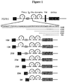
Fig. 1 is a structural representation of FGFR1-Fc fusion protein. FGFR1-Fc fusion protein is represented by a solid line, and the deleted amino acid is represented by a dash line; the antibody-like domain is represented by a circle; different antibody-like domains are represented by number 1-3; the disulfide bond is represented by s s; human IgG1 Fc is represented by a grey box; VEGFR1 signal peptide is represented by SP; the acidic box sequence is represented by a box with letter AB. -
Fig. 2 shows the comparison of FGF-2 binding among various FGFR1-Fc fusion proteins. Binding of heparin (100 ng/mL) containing FGF-2 (50 ng/mL) or FGF-2 (50 ng/mL) alone to each FGFR1-Fc fusion protein (20 ng/mL) is detected by ELISA. -
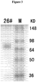
Fig. 3 shows SDS-PAGE of 26# FGFR1-Fc fusion protein. -
Fig. 4 shows the binding of FGF-2 to a gradient concentration of 26# FGFR1-Fc fusion protein. -
Fig. 5 shows the affinity between 26# FGFR1-Fc fusion protein and FGF-2. -
Fig. 6 shows the effect of 26# FGFR1-Fc fusion protein on the HUVEC cell division induced by FGF-2. - Unless otherwise defined, all scientific terms used herein have the same meaning as commonly understood by those skilled in the art. With regard to the definitions and terms in the art, reference may be made to Current Protocols in Molecular Biology (Ausubel) by the skilled one. Standard three- and/or one-letter code used for expressing one of 20 common L-amino acids in the art are adopted as the abbreviation of amino acid residues.
- Although the number ranges and approximate parameter values are given in a broad range in the present disclosure, all numbers in the specific examples are described as precise as possible. However, certain errors exist in any numerical values essentially, which may be resulted from the standard deviation during the measurement for each of them. Additionally, it should be understood that all ranges disclosed herein encompass any and all possible subranges contained therein. For example, it should be understood that the range "from 1 to 10" as described herein encompasses any and all possible subranges between the minimum 1 and the maximum 10 (including the endpoints); i.e., all subranges started from the
minimum 1 or more, for example 1 to 6.1, and all subranges ended at the maximum 10 or less, for example 5.5 to 10. Additionally, it should be noted that unless otherwise clearly and explicitly stated, the singular form includes the plural referent, as used in the present disclosure. The term "or" and the term "and/or" are used interchangeably, unless otherwise clearly indicated in the context. - As used herein, the term "Fc", "Fc region", "Fc fragment" or "immunoglobulin Fc region" refers to the crystallizable fragment of immunoglobulin, and in the present invention, said Fc region is preferably the human IgG1 Fc region.
- The term "Fc fusion protein" refers to the antibody-like molecule which incorporates the binding specificity of a heterologous protein and the effector function of a constant region of an immunoglobulin. In the term of molecular structure, a Fc fusion protein comprises the amino acid sequence having the required binding specificity and the sequence of a constant region of an immunoglobulin. A Fc fusion protein molecule generally comprises a binding site of a receptor or a ligand. The sequence of immunoglobulin constant region may be derived from any immunoglobulin, for example, IgG-1, IgG-2, IgG-3 or IgG-4 subtype, IgA (including IgA-1 and IgA-2), IgE, IgD or IgM.
- The term "soluble" protein as used herein refers to the protein which may be dissolved in an aqueous solution at a biologically relevant temperature, pH level and osmotic pressure. The "soluble fusion protein" as used herein is intended to mean that the fusion protein does not contain a transmembrane region or an intracellular region.
- As used herein, the term "isolated" refers to the following substance and/or entity: (1) which is isolated from at least some components which is present when initially produced (in natural environment and/or in a experiment device) and related thereto and/or (2) which is produced, prepared and/or manufactured artificially. The isolated substance and/or entity may be isolated from at least about 10%, about 20%, about 30%, about 40%, about 50%, about 60%, about 70%, about 80%, about 90%, about 95%, about 98%, about 99%, substantially 100% or 100% other components related to it initially.
- The term "part" and "fragment" interchangeably refer to a part of polypeptide, nucleic acid or other molecular constructs.
- The term "Ig-like domain" as used herein refers to immunoglobulin-like domain, which may be found in a plurality of protein families and involved in many biological functions, including cell-cell recognition, cell surface receptor, immune function and the like.
- Fibroblast growth factor (FGF) is a heparin-binding growth factor family, which has 22 family members in the mammals (FGF 1-14, 16-23). FGF has many important biological functions, such as cell multiplication, differentiation, migration, angiogenesis and tumorigenesis. FGF exerts many biological functions by binding and activating the cell surface FGF receptor (FGFR). (See, for example, Eswarakumar et al. Cytokine Growth Factor Rev. 16: 139-149, 2005). Fibroblast growth factor receptor (FGFR) is the receptor that binds the family members of fibroblast growth factor. A part of fibroblast growth factor receptor is involved in the disease process. In the mammals, there are 4 FGFR genes: fgfR1-fgfR4. The fibroblast growth factor receptor is composed of extracellular domain, transmembrane domain and intracellular domain. There are many members in FGFR family, which are different from each other in the term of ligand binding properties and kinase domains. However, the extracellular domains thereof are similar. There are three immunoglobulin-like (Ig-like) domains contained in their extracellular domains: the first Ig-like domain, the second Ig-like domain and the third Ig-like domain, and there is also a sequence contained between the first and the second Ig-like domain. Said sequence contained between the first and the second Ig-like domain is referred herein as the intermediate functional sequence region of the Ig-like domain of FGFR. Said intermediate regulation sequence comprises a region of acidic amino acids, referred as acidic box (AB).
- As used herein, the term "the first Ig-like domain of FGFR" or "the first Ig-like domain" refers to the first Ig-like domain in the protein FGFR from the N-terminus, which has for example the amino acid sequence corresponding to position 40 to position 118 of SEQ ID NO: 1. Similarly, the term "the second Ig-like domain of FGFR" or "the second Ig-like domain" refers to the second Ig-like domain in the protein FGFR from the N-terminus, which has for example the amino acid sequence corresponding to position 163 to position 247 of SEQ ID NO: 1; the term "the third Ig-like domain of FGFR" or "the third Ig-like domain" refers to the first Ig-like domain in the protein FGFR from the N-terminus, which has for example the amino acid sequence corresponding to position 270 to position 359 of SEQ ID NO: 1. Preferably, the FGFR is FGFR1, and the first Ig-like domain of FGFR is the first Ig-like domain of FGFR1, and the second Ig-like domain of FGFR is the second Ig-like domain of FGFR1, and the third Ig-like domain of FGFR is the third Ig-like domain of FGFR1.
- A part of sequence of hFGFR1 is given as follows, in which each Ig-like domain is shown in shaded area sequentially, see http://www.ncbi.nlm.nih.gov/protein/AAH15035.1

- The amino acid sequence of FGFR1 may be found in SEQ ID NO: 1, and its encoding nucleotide sequence may be found in SEQ ID NO: 4.
- As used herein, the term "the intermediate functional sequence region of the Ig-like domain of FGFR" or "the intermediate functional sequence of the Ig-like domain of FGFR" or "IFS" refers to the sequence between the first Ig-like domain and the second Ig-like domain in the protein FGFR, and preferably, IFS sequence has the amino acid sequence corresponding to position 118 to position 162 of SEQ ID NO: 1. Unexpectedly, it has been found by the present inventor that there is a significant effect of the intermediate functional sequence region on the function of the Ig-like domain. In some embodiments a FGFR fusion protein, which comprises a plurality of parts of various lengths derived from the intermediate functional sequence region, and particularly preferably, the part derived from the intermediate functional sequence region contains no acidic box. More preferably, the part derived from IFS has the amino acid sequence corresponding to position 134 to position 162, position 145 to position 162 or position 151 to position 162 of SEQ ID NO: 1. The protein FGFR is preferably FGFR1 (SEQ ID NO: 1), especially the protein human FGFR1. The amino acid sequence of the protein human FGFR1 may be found in SEQ ID NO: 1, and its cDNA sequence may be found in SEQ ID NO: 4.
- The term "FGFR" as used herein refers to fibroblast growth factor receptor, which may be FGFR1, FGFR2, FGFR3 and/or FGFR4. Preferably, the FGFR in the present invention is FGFR1, more preferably, human FGFR1.
- As used herein, the term "degenerate variant" is intended to mean that the degenerate variant comprises a degenerate change at the third position of the amino acid codon so that the degenerate variants encode the same amino acid, for example the wobble position of a triplet code comprising one or more changed variants (also referred as synonymous variant).
- As used herein, the term "subject" refers to mammals, such as human. However, it may also be other animals, such as domesticated animals (such as dog and cat etc.), livestocks (such as cattle, sheep, pig and horse etc.) or experimental animals (such as monkey, rat, mouse, rabbit and guinea pig etc.).
- As used herein, the term "percentage identity", "homology" or "identity" refers to the sequence identity between two amino acid sequences or nucleic acid sequences. The percentage identity may be determined by alignment between two sequences, and the percentage identity refers to the amount of the same residue (i.e., amino acid or nucleotide) at the same position in the sequence aligned. Sequence alignment and comparison may be performed using standard algorithms in the art (for example Smith and Waterman, 1981, Adv. Appl. Math. 2: 482; Needleman and Wunsch, 1970, J. Mol. Biol. 48: 443; Pearson and Lipman, 1988, Proc. Natl. Acad. Sci., USA, 85: 2444) or by the computerized versions of these algorithms (Wisconsin Genetics Software Package Release 7.0, Genetics Computer Group, 575 Science Drive, Madison, WI). Said computerized versions publicly available are BLAST and FASTA. Additionally, ENTREZ available through National Institutes of Health (Bethesda MD) may be used for sequence alignment. When BLAST and GAP-BLAST are used, default parameters for each program (for example, BLASTN, available on the website of National Center for Biotechnology Information) may be used. In one embodiment, the percentage identity between two sequences may be determined using GCG with a gap-weight of 1 so that the giving weight of each amino acid gap seems as if it is a single amino acid mismatch between two sequences. Alternatively, ALIGN (version 2.0), which is a part of GCG (Accelrys, San Diego, CA) Sequence Alignment Software Package, may be used.
- As used herein, the term "hybridization" refers to the process by which a stable double-stranded polynucleotide is formed by non-covalent bonding between two single stranded polynucleotides. The term "hybridization" also may refer to triple-stranded hybridization. The double stranded polynucleotide (generally) produced is the "hybrid" or "duplex". "The condition for hybridization" generally includes a salt concentration lower than about 1 M, and more generally, lower than about 500 mM, and lower than about 200 mM. The hybridization temperature may be as low as 5°C, but it usually higher than about 22°C, and more usually higher than about 30°C, and preferably higher than about 37°C. Hybridization is usually carried out under strict conditions (i.e., the conditions under which the probe will hybridize to its target sequence). Strict hybridization conditions are dependent on the sequence and will be varied under different conditions. Higher hybridization temperature will be probably required by longer segments for specific hybridization. Since the hybridization stringency may be influenced by other factors (including base composition and length of the complementary strand, the presence of organic solvent and the degree of base mismatch), the combination of parameters is more important than the absolute value of any single parameter. Generally, the strict condition is selected as 5°C lower than the Tm of the sequence under certain ionic strength and pH. Exemplary strict conditions include pH 7.0 to 8.3, sodium ion (or other salts) concentration of at least 0.01 M to no more than 1 M and temperature of at least 25°C. For strict conditions, see, for example Sambrook, Fritsche and Maniatis. "Molecular Cloning A laboratory Manual", 2nd edition, Cold Spring Harbor Press (1989) and Anderson "Nucleic Acid Hybridization", 1st edition, BIOS Scientific Publishers Limited (1999).
- As used herein, the term "linker", "peptide linker", "linking sequence" or "linker sequence" refers to a short amino acid sequence by which individual domain and/or region involved in the present fusion protein are linked together, and the length of the short amino acid sequence is generally 0-20 amino acids, and preferably, 2-10 amino acids.
- As used herein, the term of "the amino acid sequence corresponding to SEQ ID NO: N" in a fusion protein or part or domain is intended to mean said fusion protein or part or domain has the amino acid sequence substantially as indicated by SEQ ID NO: N, and preferably, containing no more than 1, 2, 3, 4, 5, 10 or 20 substitutions, additions and deletions of amino acids, and yet preferably, said fusion protein or part or domain shares at least 80%, 90%, 93%, 95%, 97%, 98% or 99% identity with the amino acid sequence of SEQ ID NO: N, and more preferably, said fusion protein or part or domain has the amino acid sequence as indicated by SEQ ID NO: N.
- As used herein, the term "FGFR-Fc fusion protein" refers to a fusion protein which comprises the part derived from the extracellular domain of FGFR and the part derived from the immunoglobulin Fc region, wherein the part derived from the extracellular domain of FGFR may: (1) comprise the amino acid sequence sharing at least 70% identity, preferably at least 80%, 90%, 93%, 95%, 97%, 98% or 99% identity, with the amino acid sequence indicated by any one of SEQ ID NOs: 9-15 or composed thereof; (2) comprise the amino acid sequence encoded by the nucleotide sequence sharing at least 70% identity, preferably at least 80%, 90%, 93%, 95%, 97%, 98% or 99% identity, with the nucleotide sequence indicated by any one of SEQ ID NOs: 16-22 or composed thereof; or (3) possess the amino acid sequence indicated by any one of SEQ ID NOs: 9-15, or the amino acid sequence encoded by the nucleotide sequence indicated by any one of SEQ ID NOs: 16-22.
- In some preferable embodiments, the FGFR-Fc fusion protein may be encoded by the nucleic acid, in which the nucleotide sequence encoding the part derived from the extracellular domain of FGFR comprises the sequence of which the complementary sequence is hybridized with the nucleotide sequence as indicated by any one of SEQ ID NOs: 16-22 under stringent conditions, or comprises the degenerative variant of the nucleotide sequence as indicated by any one of SEQ ID NOs: 16-22. In some preferable embodiments, the nucleotide sequence encoding the immunoglobulin Fc region comprises the sequence of which the complementary sequence is hybridized with the nucleotide sequence indicated by SEQ ID NO: 8 under stringent conditions, or comprises the degenerative variant of the nucleotide sequence indicated by SEQ ID NO: 8.
- In other preferable embodiments, the FGFR-Fc fusion protein includes the FGFR-Fc fusion protein variant. In one embodiment, the variant includes the variant which contains no more than 2, 3, 4, 5 or 10 substitutions, additions or deletions of amino acid in the part derived from IFS corresponding to the amino acid sequence indicated by position 134 to position 162, position 145 to position 162 or position 151 to position 162 of SEQ ID NO: 1, and preferably, the variant retains the angiogenesis-inhibitory capacity. In another embodiment, the variant includes the variant which contains no more than 2, 3, 4, 5, 10 or 20 substitutions, additions or deletions of amino acid in D2 domain corresponding to the amino acid sequence indicated by position 163 to position 247 of SEQ ID NO: 1, and preferably, the variant retains the angiogenesis-inhibitory capacity. In another embodiment, the variant includes the variant which contains no more than 2, 3, 4, 5, 10 or 20 substitutions, additions or deletions of amino acid in D3 domain corresponding to the amino acid sequence indicated by position 270 to position 359 of SEQ ID NO: 1, and preferably, the variant retains the angiogenesis-inhibitory capacity. In another embodiment, the substitution, addition or deletion is located at the linker or the linking part.
- In addition to the naturally occurring modifications in the part derived from the extracellular domain of FGFR and the part derived from immunoglobulin Fc region, other post-translational modifications may also be comprised in the FGFR-Fc fusion protein. Such modifications include, but are not limited to, acetylation, carboxylation, glycosylation, phosphorylation, esterification and acylation. As a result, non-amino acid component may be comprised in the modified FGFR-Fc fusion protein, for example polyethylene glycol, lipid, polysaccharide or monosaccharide, and phosphoric acid. Effect of such non-amino acid components on the function of the FGFR-Fc fusion protein may be tested as described for other FGFR-Fc fusion protein variants herein. When FGFR-Fc fusion protein is produced in a cell, post-translational processing is also possibly important for correct folding and/or protein function. Special cell machines and unique mechanisms exist in different cells (for example CHO, HeLa, MDCK, 293, WI38, NIH-3T3 or HEK293) for these post-translational activities, and different cells may be selected to make sure correct modification and processing of FGFR-Fc fusion protein.
- The fusion protein as described herein may be produced by any method known in the art. For example, it may be produced by chemical synthesis or from nucleic acid expression. The peptides used in the present invention may be easily prepared according to the established standard liquid, or preferably, solid phase peptide synthesis method known in the art (see, for example J. M. Stewart and J. D. Young, Solid Phase Peptide Synthesis, 2nd edition, Pierce Chemical Company, Rockford, Illinois (1984), in M. Bodanzsky, and A. Bodanzsky, The Practice of Peptide Synthesis, Springer Verlag, New York (1984)). The fusion protein may be produced by the techniques known in the art so that one or more intramolecular crosslinkings may be formed between the cysteine residues located in the polypeptide sequence expected to be comprised in the protein (see, for example
US patent No. 5478925 ). In addition, general modifications may be performed to the protein described herein by adding cysteine or biotin to the C-terminus or N-terminus of the protein. - As used herein, "therapeutically effective amount" or "effective amount" refers to the dosage which is sufficient to show the benefit to the subject administrated. The actually administrated dosage, the rate and the time course of administration are dependent on the condition of the patient and the severity of the disease. Finally, the physician is responsible for the prescription (for example decision on the dosage etc.) and will make a decision for the treatment, usually by considering the disease treated, individual condition of the patient, position of delivery, the method for administration and other factors known to the physician.
- A series of isolated soluble FGFR-Fc fusion proteins are constructed by the present inventor, which may bind FGF and effectively inhibit the cell division induced by FGF. The fusion protein preferably comprises: the part derived from IFS, D2, D3 and immunoglobulin Fc region.
- Unexpectedly, it has also been found by the present inventor that the binding of FGF by the fusion protein is significantly influenced by the length of the part derived from IFS. Therefore, in some embodiments fusion proteins comprise the parts derived from IFS with various lengths. Preferably, the part derived from IFS comprises no acidic box, and more preferably, it has the amino acid sequence corresponding to position 134 to position 162, position 145 to position 162 or position 151 to position 162 of SEQ ID NO: 1. In some preferable embodiments, the part derived from IFS comprises the fusion protein corresponding to the amino acid sequence indicated by position 145 to position 162 of SEQ ID NO: 1, which has extremely high FGF affinity and may particularly effectively inhibit the cell division induced by FGF.
- In some embodiments, a soluble FGFR-Fc fusion protein is provided, which comprises: D1, a part derived from IFS, D2, D3 and immunoglobulin Fc region. Preferably, the part derived from IFS comprises no acidic box, and more preferably, it has the amino acid sequence corresponding to position 134 to position 162, position 145 to position 162 or position 151 to position 162 of SEQ ID NO: 1.
- In some other embodiments, a soluble FGFR-Fc fusion protein is provided, which comprises: a part of D1, a part derived from IFS, D2, D3 and immunoglobulin Fc region. Preferably, the part derived from IFS comprises no acidic box, and more preferably, it has the amino acid sequence corresponding to position 134 to position 162, position 145 to position 162 or position 151 to position 162 of SEQ ID NO: 1.
- In some other embodiments, a soluble FGFR-Fc fusion protein is provided, which is composed of: a part derived from IFS, D2, D3 and immunoglobulin Fc region. Preferably, the part derived from IFS comprises no acidic box, and more preferably, it has the amino acid sequence corresponding to position 134 to position 162, position 145 to position 162 or position 151 to position 162 of SEQ ID NO: 1.
- In some other embodiments, a soluble FGFR-Fc fusion protein is provided, which is sequentially composed of, from the N-terminus to the C-terminus, a part derived from IFS, D2, D3 and immunoglobulin Fc region. Preferably, the part derived from IFS comprises no acidic box, and more preferably, it has the amino acid sequence corresponding to position 134 to position 162, position 145 to position 162 or position 151 to position 162 of SEQ ID NO: 1.
- In some other embodiments of the present invention, an FGFR-Fc fusion protein is provided, which may inhibit tumor cells directly or indirectly. Preferably, the FGFR-Fc fusion protein of the present invention inhibits tumor cells directly. More preferably, the growth of tumor cells is inhibited by the FGFR-Fc fusion protein of the present invention by at least 10%, 20%, 30%, 40%, 50%, 80%, 90% and 95% etc. The tumor cells may be any tumor cells, for example, leukaemia, lung cancer, liver cancer, head and neck cancer, stomach cancer, bladder cancer, carcinoma of uterine cervix etc. Particularly, the inhibition is achieved by direct binding to tumor cells.
- In some embodiments, the present invention includes the use of (i) FGFR-Fc fusion protein, or (ii) the polynucleotide encoding such fusion protein, in the preparation of the compositions or medicaments for the treatment of diseases mediated by or related to angiogenesis. For example, in one embodiment, the present invention includes use of (i) FGFR-Fc fusion protein, or (ii) the polynucleotide encoding such fusion protein in the preparation of the medicaments as an angiogenesis inhibitor.
- In some embodiments, the FGFR-Fc fusion protein may be produced by the expression of the nucleotide sequence as indicated by any one of SEQ ID NOs: 16-22 in a mammalian cell line. In particular, the mammalian cell line is CHO cell line.
- Additionally, the FGFR-Fc fusion protein as described below is provided, in which a part derived from the extracellular domain of FGFR may be fused with the immunoglobulin Fc region with or without a linker.
- In some other embodiments, the present invention includes the isolated nucleic acid molecules encoding the FGFR-Fc fusion protein, and the present invention also includes use of these molecules in manufacture of a medicament. The nucleic acid may be recombinant, synthetic or produced by any available methods in the art, and the method includes cloning by means of using a standard technique.
- In some other embodiments, the present invention includes a vector comprising the isolated nucleic acid molecule of the present invention. The vector may be an expression vector, in which the nucleic acid is operatively linked to a control sequence which is able to provide the expression of the nucleic acid in a host cell. A plurality of vectors may be used. For example, suitable vectors may include virus (for example poxvirus, adenovirus, baculovirus etc.); yeast vector, bacteriophage, chromosome, artificial chromosome, plasmid, cosmid.
- In some embodiments, the present invention further includes the cells transfected by these vectors so that the FGFR-Fc fusion protein is expressed. The host cell suitable for the present invention may be prokaryotic cell or eukaryotic cell. They include bacteria, for example E. coli, yeast, insect cell and mammalian cell. The mammalian cell lines that may be used include, but are not limited to, Chinese Hamster Ovary (CHO) cell, baby hamster kidney cell, NS0 mouse myeloma cell, monkey and human cell lines, and derivate cell lines thereof, etc.
- In another aspect, a method for angiogenesis inhibition is provided, comprising administrating the FGFR-Fc fusion protein of the present invention to the subject in need thereof. Preferably, the method is carried out in the mammals.
- In another aspect, a method for binding FGF in vitro or in vivo is provided, which comprises contacting FGF to the fusion protein according to the present invention.
- In another aspect, a method for the treatment or prevention of tumors in the mammals is provided, which comprises administrating the FGFR-Fc fusion protein of the present invention to the subject in need thereof, and preferably, the tumor is a solid tumor.
- In another aspect, a method for the treatment or prevention of ophthalmic angiogenesis-related diseases in the mammals is provided, which comprises administrating the FGFR-Fc fusion protein of the present invention to the subject in need thereof, and preferably, the ophthalmic angiogenesis-related disease is age-related macular degeneration.
- The present invention also relates to use of the FGFR-Fc fusion protein in the preparation of medicaments for angiogenesis inhibition. Additionally, the present invention also relates to use of the FGFR-Fc fusion protein in the preparation of medicaments for the treatment or prevention of angiogenesis-related diseases, and preferably, angiogenesis-related diseases are tumors or ophthalmic angiogenesis-related disease.
- The angiogenesis-related diseases as described herein include, but are not limited to, angiogenesis-dependent cancers, comprising, for example, solid tumor, hematogenic tumor (for example leukaemia) and tumor metastasis; benign tumor, for example, angioma, acoustic neuroma, neurofibroma, trachoma and pyogenic granuloma; rheumatoid arthritis; psoriasis; rubeosis; Osler-Webber Syndrome; myocardial angiogenesis; plaque neovascularization; telangiectasia; hemophiliac joint and angiofibroma.
- In some embodiments of the methods described, one or more FGFR-Fc fusion proteins may be administrated together (simultaneously) or at a different time (sequentially). Additionally, the fusion protein may be administrated together with additional medicament used for cancer treatment or angiogenesis inhibition.
- In some embodiments, the method disclosed may be used alone. Alternatively, the subject method may be combined with other conventional anticancer therapies for the treatment or prevention of proliferative diseases (for example tumor). For example, these methods may be used for the prevention of cancers, the prevention of cancer relapse and postoperative metastasis, and may be used as a supplement for other cancer therapies. As disclosed herein, the effectiveness of conventional cancer therapies (for example, chemotherapy, radiotherapy, phototherapy, immunotherapy and operation) may be enhanced by using target polypeptide therapeutic agents.
- In ophthalmology, angiogenesis is related to, for example, diabetic retinopathy, retinopathy of prematurity, age-related macular degeneration, corneal transplantation rejection, neovascular glaucoma and RLF (retrolental fibroplasia). The FGFR-Fc fusion protein disclosed herein may be administrated inside the eye or by other routes. Other diseases related to angiogenesis in ophthalmology include, but not limited to, epidemic keratoconjunctivitis, Vitamin A deficiency, contact lens overwear, atopic keratitis, superior limbic keratitis, pterygium keratitis sicca, sjogren, acne rosacea, phlyctenosis, syphilis, Mycobacteria infection, lipid degeneration, chemical burn, bacterial ulcer, fungal ulcer, Herpes simplex infection, Herpes zoster infection, protozoan infection, Kaposi sarcoma, Mooren ulcer, Terrien's marginal degeneration, mariginal keratolysis, rheumatoid arthritis, systemic lupus, polyarteritis, trauma, Wegeners sarcoidosis, Scleritis, Steven's Johnson disease, periphigoid radial keratotomy and comeal graph rejection, sickle cell anemia, sarcoid, pseudoxanthoma elasticum, Pagets disease, vein occlusion, artery occlusion, carotid obstructive disease, chronic uveitis/vitritis, mycobacterial infections, Lyme's disease, systemic lupus erythematosis, retinopathy of prematurity, Eales disease, Bechets disease, infection resulting in retinitis or choroiditis, presumed ocular histoplasmosis, Bests disease, myopia, optic pit, Stargarts disease, pars planitis, chronic retinal detachment, hyperviscosity syndromes, toxoplasmosis, trauma and post-laser complication.Other diseases include, but not limited to, rubeosis (neovasculariation of the angle) related diseases and diseases induced by abnormal hyperplasia of the fibrous blood vessel or fibrous tissue, including all kinds of proliferative vitreoretinopathy.
- The fusion protein of the present invention may be administrated alone, but preferably, as a pharmaceutical composition which usually comprises a suitable pharmaceutical excipient, diluent or carrier selected according to the intended administration route. The fusion protein may be administrated to the patient in need thereof by any suitable route. A precise dosage will be dependent on many factors, including exact properties of the fusion protein.
- Some suitable administration routes include (but are not limited to) oral, rectal, nasal, topical (including buccal and sublingual), subcutaneous, vaginal or parenteral (including subcutaneous, intramuscular, intravenous, intracutaneous, intrathecal and extradural) administration.
- For intravenous injection and injection at the focal site, active ingredients are present in the form of a parenterally-acceptable aqueous solution, which is free of pyrogen and has appropriate pH value, isotonicity and stability.
- A suitable solution may be well formulated by the skilled one in the art using, for example, isotonic excipients such as sodium chloride injection, Ringer's injection, Ringer's lactate injection. As required, preservative, stabilizer, buffering agent, antioxidant and/or some other additives may be added. The pharmaceutical composition orally administrated may be in a form of tablet, capsule, powder or oral liquid etc. Solid carrier, such as gelatin or adjuvant, may be comprised in a tablet. Liquid pharmaceutical composition usually comprises liquid carrier, such as water, petroleum, animal or vegetable oil, mineral oil or synthetic oil. Also included may be normal saline solution, glucose or other sugar solutions or glycols such as ethylene glycol, propylene glycol or polyethylene glycol.
- Examples of the techniques and schemes as mentioned above and other techniques and schemes as used according to the present invention may be found in Remington's Pharmaceutical Sciences, 16th edition, Oslo, A. (ed), 1980.
- The FGF receptor fragment are obtained from the amplification of the cDNA template of corresponding receptor through PCR, and IgG1 Fc fragment is obtained from the cDNA amplification of the human-derived IgG1 through PCR. When PCR primers are designed, linking sequences are introduced between different fragments so that these different fragments may be finally linked by overlap PCR to form reading frames for different fusion proteins, and endonuclease BspE I and Pst I site are added to both ends of the cDNA. The cDNAs for different fusion proteins may be cloned to the expression plasmid after digestion by BspE I and Pst I. The plasmid after cloning may be determined by endonuclease digestion, electrophoresis and finally DNA sequencing.
- The present fusion protein may be expressed and purified by techniques commonly used in the art. DNA from corresponding fusion protein plasmid was purified using plasmid purification kit (MAX) available from Qiagen, and the concentration of plasmid DNA was determined using UV spectrophotometry, and the plasmid was transfected to CHO cell using FUGENE 6 liposome (Roche). Specific method for transfection was performed according to the specification of the product. Based on the expression amount required for the proteins, two methods were employed in the present invention for protein expression: (1) transient expression, in which the fusion protein contained culture supernatant was usually harvested 48-72 h after transfection, and the relative content of the fusion protein was then determined using human IgG ELISA so that the fusion protein may be rapidly and efficiently obtained; (2) establishing a stable cell line and producing the common DHFR-defective CHO cell expression system using the recombinant protein medicament expression, the basic process of which includes cell transfection, selection of stably transfected cell, clone screening, stress amplification, culture medium and process optimization etc., and finally realizing a large-scale suspension culture of CHO engineering cell strain in a serum free culture medium. The culture product was collected and the fusion protein was purified using Protein A affinity column. The purified protein was analyzed by sodium dodecyl sulfate polyacrylamide gel electrophoresis (SDS-PAGE), and subsequently all eluates in which the required expression product was contained were combined and filtered using a 0.22 µm filter, and then protein quantification was carried out according to a plurality of methods such as Lowry protein assay. The volume of CHO cell culture in the present invention was at a level of 10 L bioreactor, through which the fusion protein obtained after purification could satisfy the protein amount required in the animal experiments, and also a basis was established for future scaling-up.
- After the fusion protein expressed by CHO was obtained, the binding capacity of the fusion protein to FGF is evaluated in the present invention at a level of protein. Binding experiment and affinity experiment were performed for validation in the present invention, in which steps of the binding experiment included: after initially coated by FGF-2 on a 96-well ELISA plate, the coated well was blocked by BSA followed by adding each fusion protein at the same concentration, and then a secondary antibody to human IgG Fc-HRP was added after washing, and the samples were developed, stopped and read at 450nm on a ELISA plate, and finally the fusion protein which had binding capacity to FGF-2 was screened based on the signal strength. The affinity experiment was performed in order to determine the affinity of the fusion protein to FGF-2 in the solution system, which comprised the following steps: FGF-2 was initially coated on a 96-well ELISA plate to capture the antibody, and then the coated well was blocked by BSA, and subsequently a mxiture of the fusion protein and FGF-2 which was previously prepared and incubated were added with a gradient of diluted standards, and after incubation, an HRP-labeled detection antibody was added (using
antibody 2 which specifically detected free VEGF or FGF-2), and subsequently the samples were developed, stopped and read at 450nm on a ELISA plate, and finally the relative concentration of free FGF-2 was detected in the mixture of the fusion protein and FGF-2. Through the experiments above, the fusion protein having a blocking effect on FGF-2 was screened. - After the binding capacity of the fusion protein to FGF-2 was determined at a level of protein, its angiogenesis-inhibiting effect will be further validated at a cellular level in the present invention. The inhibition capacity of the fusion protein on the division and migration of the vascular endotheliocyte is examined by the division test using human umbilical vein endothelial cell (HUVEC) and the HUEVC cell migration test. The inhibition capacity of the fusion protein on the division of HUVEC cell can be examined by the HUVEC cell division test, which comprises the following steps during the experiment: 3000 HUVEC cells/well were inoculated to a 96-well plate and cultured at 37°C in an incubator supplemented with 5% CO2, and then FGF-2 as well as a mixture of the fusion protein at different concentrations with FGF-2 are added respectively, and after culturing for another 3-4 days, 10% CCK-8 is added and cultured for 2 h before the sample is read at 450 nm on a ELISA plate. The inhibition capacity of the fusion protein on the division of vascular endotheliocyte induced by FGF-2 was evaluated based on the difference of absorbance, and the median effective concentration of the fusion protein was obtained for FGF-2 inhibition. The inhibition capacity of the fusion protein on HUVEC cell migration was examined by the HUVEC cell migration test, which comprises the following steps during the experiment: 50000 HUVEC cells as well as the fusion protein at various concentrations were initially inoculated in the upper chamber, while 600 µL FGF-2 containing culture liquid was added into the lower chamber, and subsequently, the sample was cultured at 37°C in an incubator supplemented with 5% CO2 for 20-24 h before cells on the face side of the membrane of the upper chamber were removed, and then cells on the back side of the membrane were fixed, stained and washed with PBS before observed and counted under an inverted microscope. The migration of HUVEC cells induced by the stimulation of FGF-2 was demonstrated by counting the HUVEC cells on the back side of the membrane, and the inhibition capacity of the fusion protein on the migration of the vascular endotheliocyte was tested by adding the fusion protein at various concentrations into the culture liquid. Through the experiments mentioned above, the inhibition capacity of the new fusion protein constructed in the present invention was validated on the division and migration of the vascular endotheliocyte induced by FGF-2, which also provided a basis for future animal experiments.
- After the blocking effect of the new fusion protein in the present invention on FGF-2 signal was demonstrated by experiments at a protein level and a cellular level, its anti-tumor capacity would be tested in animal tumor models in the present invention. In the present invention, the anti-angiogenesis and anti-tumor effect of the fusion protein would be validated by models commonly used in searching medicaments for angiogenesis and tumor, for example, LLC mouse lung cancer, U87 gliocytoma, B 16 melanoma and so on. In animal experiments, in addition to conventional control groups, control medicaments, such as VEGF-Trap, FP-1039, would also be included so as to obtain comparative data for anti-tumor capacity. During experiments, 100 µL tumor cell liquid with appropriate amount was subcutaneously injected into C57 mouse on one side of the back, and the tumor volume was measured with a vernier caliper twice a week. Upon the tumor grew to about 200 mm3, the fusion protein at various concentrations was subcutaneously injected and the mice were sacrificed after 2-3 weeks. Subsequently, the tumor volume was measured with a vernier caliper, and the anti-tumor effect of the fusion protein was validated by the size of the tumor. Furthermore, individual tumor tissue was analyzed using methods such as immunohistochemistry to investigate the regulation mechanism of angiogenesis.
- The FGF receptor fragment is obtained from the amplification of the cDNA templet of FGF receptor through PCR, and IgG1 Fc fragment is obtained from the cDNA amplification of the human-derived IgG1 through PCR. A commercially available cDNA (PCR Ready First Strand cDNA, derived from human adult colon cancer tissue, BioChain) was used as the template for FGFR1 fragment. Total RNA was extracted from the blood of healthy human subjects using human blood RNA extraction kit (QIAGEN). According to the manufacturer's instruction of reverse transcription kit (Promega), RT-PCR was performed using M-MLV reverse transcriptase (Promega) so that RNA was reversely transcripted to cDNA which was used as the template for IgG1 Fc fragment. RT-PCR was performed according to the manufacturer's instruction of reverse transcription kit, which has the following steps: Oligo dT, dNTP, total RNA and DEPC H2O were mixed homogeneously and reacted at 70°C for 10 min before placed on ice for 5 min, and subsequently RNase inhibitor, M-MLV reverse transcriptase and reaction buffer were added. The mixture was reacted at 42°C for 1 h and subsequently at 70°C for 15 min, and the cDNA obtained may be used as the template.
- Various FGFR1 fragments were individually amplified by PCR using the cDNA from human adult colon cancer tissue as the template (the primers were listed in table 1), and IgG1 Fc fragment was amplified by PCR using human blood cDNA as the template (the primers were listed in table 1 and 2). The reaction conditions for the PCR were as follows: 5 min of pre-denaturalization at 98°C, total 30 cycles of 30 s of denaturalization at 98°C, 45 s of annealing at 56°C and 2 min of extension at 72°C, and finally another 10 min of extension. When PCR primers were designed, 20 or more complementary base sequences were introduced as the linking sequence between FGFR1 fragment and IgG1 Fc fragment so that the FGFR1 fragment and IgG1 Fc fragment may be subsequently linked by overlap PCR to form reading frames for different fusion proteins, and at the same time, restriction endonuclease BspE I and Pst I site were added at both ends of the PCR product.
- Subsequently, overlap PCR was carried out to obtain each FGFR1-Fc fusion protein fragment by amplification. The process of the overlap PCR reaction may be divided into two rounds, in which the fragment required for linking and containing no primer was included in the first round with reaction conditions as follows: 5 min of pre-denaturalization at 98°C, 6 cycles of 30 s of denaturalization at 98°C, 45 s of annealing at 56°C and 5 min of extension at 72°C, and finally another 10 min of extension at 72°C; after the first round, the second round of PCR was carried out by adding the primers for both ends with reaction conditions as follows: 5 min of pre-denaturalization at 98°C, 30 cycles of 30 s of denaturalization at 98°C, 45 s of annealing at 56°C and 2 min of extension at 72°C, and finally another 10 min of extension at 72°C; through the process above, reading frames for different fusion proteins were spliced, and at the same time, restriction endonuclease BspE I and Pst I site were added at both ends of the cDNA.
- After amplification, the fragments amplified by PCR were purified using QIAquick PCR purification kit (QIAGEN). cDNAs of various fusion proteins and the eucaryotic expression plasmid pSV2-dhfr (ATCC) were digested by BspE I and Pst I, respectively. Subsequently, 1% agarose gel electrophoresis was performed on the digested samples under a voltage of 90 V. Target fragments were recovered using QIAquick gel extraction kit (QIAGEN) before ligating at 16°C for 1 h using a ligase (NEB). The mixture for ligation reaction was transformed to the competent Top10 E. coli under the conditions of 90 s of reaction at 42°C followed by 3 min of standing on ice. After the sterile LB culture broth (free of antibody) added, the mixture was shaken at 250 rpm in a shaker at 37°C for 1 h before coating on a LB plate supplemented with ampicillin. The plate was cultured overnight in a thermostated incubator at 37°C, and then single colonies were picked out and transferred to an ampicillin-containing LB culture broth. The inoculated culture broth was shaken at 250 rpm in a shaker at 37°C overnight before the plasmid was extracted using alkaline lysis. Subsequently, the sample was digested by restriction endonuclease before evaluated by 1% agarose gel electrophoresis under a voltage of 90 V. The recombinant plasmid with correct endonuclease digestion was confirmed by DNA sequencing. Based on the steps above, 19#, 13#, 22#, 23#, 26#, 29# and 8# expression plasmid for FGFR1-Fc fusion protein were constructed. The protein sequence of FGFR1-Fc in each fusion protein and its encoding nucleotide sequence were listed in Table 3. The schematic diagram of the fusion protein structure was shown in
Fig. 1 .Table 1: Primers used for amplification of FGFR1 fragment Fusion protein Upstream primer Downstream primer 19# 19#-FGFR1 For (SEQ ID NO: 24) FGFR1Rev (SEQ ID NO: 31) TAGTTCCGGAAGGCCGTCCCCGACCTTGCCTG 
13# 13#-FGFR1For (SEQ ID NO: 25) FGFR1Rev TAGTTCCGGAAAAAATCGCACCCGCATCACAG 22# 22#-FGFR1For (SEQ ID NO: 26) FGFR1Rev TAGTTCCGGAGTAACCAGCAGCCCCTCGGGC 23# 23#-FGFR1For (SEQ ID NO: 27) FGFR1Rev TAGTTCCGGATCCTCTTCAGAGGAGAAAGAAAC 26# 26#-FGFR1For (SEQ ID NO: 28) FGFR1Rev TAGTTCCGGAAAACCTAACCCCGTAGCTCCAT 29# 29#-FGFR1For (SEQ ID NO: 29) FGFR1Rev TAGTTCCGGACCATATTGGACATCCCCAGAAAAG 8# 8#-FGFR1For (SEQ ID NO: 30) FGFR1Rev CTAGCTCCGGACCAGAAAAGATGGAAAAGAAATTGC Table 2: Primers used for amplification of IgG1 Fc fragment Upstream primer Downstream primer IgG1 Fc fragment FcFor (SEQ ID NO: 32) FcRev (SEQ ID NO: 33) CTGTACCTGGAGGACAAAACTCACACATGC 
Table 3: Protein sequences and nucleotide sequences for FGFR1-Fc fusion proteins Fusion protein Protein sequence Nucleotide sequence 19# SEQ ID NO: 9 SEQ ID NO: 16 13# SEQ ID NO: 10 SEQ ID NO: 17 22# SEQ ID NO: 11 SEQ ID NO: 18 23# SEQ ID NO: 12 SEQ ID NO: 19 26# SEQ ID NO: 13 SEQ ID NO: 20 29# SEQ ID NO: 14 SEQ ID NO: 21 8# SEQ ID NO: 15 SEQ ID NO: 22 - The DNA of individual fusion protein plasmid was purified using MAX Plasmid Purification Kit (Qiagen). The concentration of the plasmid DNA was determined by UV spectrophotometry. 1 µg recombinant plasmid and 6 µL liposome (FuGENE 6 Transfection Reagent, Roche) were homogeneously mixed into 100 µL fresh IMDM culture broth (GIBCO); after standing for 15 min, the mixture was added to the CHO cells (ATCC) cultured overnight after inoculation at a cell density of 3 × 10⁵/mL into a 6-well plate; the mixture was cultured at 37°C in an incubator supplemented with 5% CO2 for 48 h with a cell complete culture broth (IMDM medium containing 10% FBS, 1% HT and 1% glutamine, all supplied by GIBCO); subsequently, the supernatant was collected and determined for the relative content of the fusion protein using human IgG ELISA kit for protein quantification (BETHYL). The relative content of the fusion protein expressed and secreted by CHO was determined with the following steps: 100 µL anti-human IgG-Fc protein (10 µg/mL) purified by affinity was coated to a 96-well ELISA plate (IMMULON) and subsequently washed for 5 times using 300 µL PBST washing solution; each coated well was blocked with 200 µL freshly prepared blocking working solution (blocking stock solution : PBS=1: 19) and incubated at 37°C for 1 h; after washed in 300 µL PBST washing solution for 5 times, 100 µL IgG solution diluted in a gradient (200 ng/mL original concentration and diluted by PBS in the multiple proportion of 1: 2) as a standard and 100 µL culture supernatant of each fusion protein diluted in a gradient (starting with the concentration of each culture supernatant, and diluted by PBS in the multiple proportion of 1: 5) were added to each well and incubated at 37°C for 2h; after washed in 300 µL PBST washing solution for 5 times, 100 µL anti-human IgG Fc-HRP secondary antibodies diluted with PBS in a ratio of 1: 10000 was added and incubated at 37°C for 1h; after washed, the well was developed by adding 100 µL developing solution (KPL); finally, after the development was stopped by adding 100 µL stopping solution (KPL), the absorbance of the ELISA plate was read at a wavelength of 450 nm on a ELISA reader. The concentrations of various fusion proteins may thereby be determined according to the standard curve.
- The binding capacity of 19#, 13#, 22#, 23#, 26#, 29# and 8# fusion protein constructed above to FGF-2 was detected by ELISA. Initially, a 96-well ELISA plate (IMMULON Company) was coated by 100 µL solution containing 50 ng/mL FGF-2 (R&D Systems) as well as containing 100 ng/mL heparin (Sigma Company) and 50 ng/mL FGF-2. Subsequently, the plate was washed by 300 µL PBST washing solution for 5 times before each coated well was blocked by 200 µL freshly prepared blocking working solution (KPL Company) (blocking stock solution : PBS = 1:19) and incubated at 37°C for 1 h. After washed in 300 µL PBST washing solution for 5 times, 100 µL solutions of various fusion proteins (dissolve in PBS, pH=7.2, concentration of 20 ng/ml) were added and incubated at 37°C for 2 h. After washed in 300 µL PBST washing solution for 5 times, 100 µL secondary antibody to human IgG Fc-HRP (BETHYL Company) diluted with PBS in a ratio of 1:10000 was added and incubated at 37°C for 1h. After washed in 300 µL PBST washing solution for 5 times, the well was developed to the presence of color at room temperature in a dark place by adding 100 µL developing solution (KPL Company), and finally the development was stopped by adding 100 µL stopping solution (KPL Company) before the absorbance of the ELISA plate was read at a wavelength of 450 nm on a ELISA reader. The higher the binding capacity of the fusion protein to FGF2 was, the larger the absorbance was and the stronger the signal was. Based on the strength of the signal, 26# fusion protein was determined to have the highest binding capacity to FGF-2. Comparison of FGF-2 binding among various fusion proteins was shown in
Fig. 2 . It can be seen fromFig. 2 that 19#, 13#, 22#, 23#, 26# and 29# fusion protein bound to FGF at different extents in the presence of heparin, and particularly, the binding extent of 23#, 26# and 29# was extremely higher than control, and higher than that of 19#, 13# and 22#, indicating that the fusion proteins containing no acidic box had excellent effect. Among others, especially high binding extent was demonstrated by 26#, which presented us a clue that the fusion protein of the present invention had significantly better binding effect when comprises a part of certain length derived from the intermediate functional sequence of the Ig-like domain of FGFR. - DHFR-defective CHO cells (ATCC) were transfected by the recombinant expression plasmid of 26# fusion protein (possessing a high FGF-2 binding capacity) through a liposome (Roche). Particularly, 5 µg recombinant plasmid and 30 µL liposome (FuGENE 6 Transfection Reagent, Roche) were homogeneously mixed into 100 µL fresh IMDM culture broth (GIBCO); after standing for 15 min, the mixture was added to the DHFR-defective CHO cells (ATCC) cultured overnight after inoculation at a cell density of 3×10⁵/mL in a 10 cm culture dish (Corning); the mixture was cultured at 37°C in an incubator supplemented with 5% CO2 for 2-3 days with a cell complete culture broth containing 10% FBS, 1% HT and 1% glutamine in a IMDM culture medium (all supplied by GIBCO); subsequently, the cells were digested by trypsin (GIBCO), inoculated at a cell density of 3×10⁵/mL in 30 mL serum-free 302 culture medium (SAFC) in a flask, and selectively cultured at 37°C in an incubator supplemented with 5% CO2 at 100 rpm to a cell density of 106/mL. Subsequently, 3000 cells were inoculated into a 10 cm culture dish (Corning) (the culture broth containing 10% FBS and 1% glutamine in an IMDM culture medium) and cultured at 37°C in an incubator supplemented with 5% CO2 to form single clones. These single clones were picked out and cultured in a 96-well plate (Corning). The relative content of the fusion protein expressed and secreted by each individual single clone was determined using a human IgG ELISA kit for protein quantification (BETHYL) under the same conditions and steps as described in Example 2 for the determination of the relative content of the fusion protein. The clone with the highest expression amount was screened out and transferred to a 6-well plate for culturing to a confluence rate of about 70%. The cells were digested by trypsin and transferred to a 10 cm culture dish. Subsequently, gradual stress amplification was carried out by adding methotrexate (MTX, Sigma) with various concentrations (10 nM, 20 nM, 50 nM, 100 nM, 200 nM and 500 nM). After stress amplification, the cells were digested by trypsin and inoculated at a cell density of 3×10⁵/mL in a flask. The expression amount of a single cell was determined so that genetically engineered stains of CHO were obtained for expressing a particular fusion protein. Finally, large-scale suspension culture (volume of 10L) of the genetically engineered stain of CHO was carried out at 37°C, 5% CO2, 40% dissolved oxygen and 80 rpm in a serum-free 302 culture medium (pH 7.0, SAFC). The culture product was collected by centrifugation. After the supernatant was filtered using 0.45 µm filter membrane (Millipore), affinity chromatography was performed according to the instruction manual of Protein A affinity column (GE) with the specific steps as follows: initially, a protein A affinity column was equilibrated by a PBS buffer (pH 7.0); subsequently, the supernatant was loaded on the column and washed again with the PBS buffer; finally, the column was eluted with a citric acid buffer (pH 3.0), and the eluent was collected and filtered by a 0.45 µm filter membrane. After virus inactivation by adding S/D (0.3% tributyl phosphate/1% Tween 80) at 24°C for 6 h, the target protein was further purified by a molecular sieve chromatography with the following steps: first, the eluent obtained from the Protein A affinity chromatography was dialyzed in a dialysis bag against a PBS buffer; subsequently, the sample was concentrated in a 10 KD ultrafiltration cup (Millipore); the sample concentrated using the ultrafiltration cup was then loaded on a molecular sieve chromatography column Superdex 200 (GE) equilibrated by a PBS buffer, and subsequently the column was eluted with a PBS buffer and the eluting peak was collected. The purified protein was analyzed by SDS-PAGE (
Fig. 3 ); and subsequently, the eluates containing the required expression product was combined and filtered with a 0.22 µm filter membrane (Millipore) before the protein content was determined using many methods such Lowry protein assay. - The binding capacities of the fusion proteins as constructed above to FGF-2 were detected by ELISA, similarly as in Example 3. Initially, a 96-well ELISA plate was coated by 100 µL solution containing 50 ng/mL FGF-2 (R&D Systems). Subsequently, the plate was washed in 300 µL PBST washing solution for 5 times before each coated well was blocked by 200 µL freshly prepared blocking working solution (KPL) (blocking stock solution : PBS = 1: 19) and incubated at 37°C for 1 h. After washed in 300 µL PBST washing solution for 5 times, 100 µL solutions containing various fusion proteins at different concentrations (the starting content of protein was 16000 pM, and was diluted in a ratio of 1: 3) were added and incubated at 37°C for 2 h. After washed in 300 µL PBST washing solution for 5 times, 100 µL anti-human IgG Fc-HRP secondary antibody (BETHYL) diluted with PBS in a ratio of 1: 10000 was added and incubated at 37°C for 1 h. After washed in 300 µL PBST washing solution for 5 times, the well was developed by adding 100 µL developing solution (KPL), and finally the development was stopped by adding 100 µL stopping solution (KPL) before the absorbance of the ELISA plate was read at a wavelength of 450 nm on a ELISA reader. Based on the intensity of the signal, the gradient binding capacities of the fusion proteins to FGF-2 were determined. In the experiment procedure mentioned above, specific conditions and steps may be found in Example 3. Gradient binding of 26# fusion protein to FGF-2 was compared in
Fig. 4 . It can be seen that the binding capacity of 26# fusion protein to FGF-2 was dose-dependent. It has been suggested by this example that the binding capacity to FGF-2 increased with an enhanced molar concentration of 26# fusion protein, manifested by a stronger signal at a wavelength of 450 nm; while the binding capacity to FGF-2 decreased correspondingly with a gradient dilution of the molar concentration of 26# fusion protein. - The affinity of the fusion protein to FGF-2 in a solution system was determined by an affinity experiment. Initially, a 96-well ELISA plate was coated by 100 µL solution containing 2.0 µg/mL FGF-2 capture antibody (R&D Systems). Subsequently, the plate was washed in 300 µL PBST washing solution for 5 times before each coated well was blocked by a blocking working solution (KPL) (as seen in Example 3) and incubated at 37°C for 1 h. After washed in 300 µL PBST washing solution for 5 times, previously prepared and incubated (4 °C overnight) mixture of the fusion proteins and FGF-2 as well as the standard (R&D Systems) diluted in a gradient were added, in which the specific preparation procedure was as follows: the starting concentration of 26# fusion protein was 400 pM (dissolved in PBS) and diluted in a gradient ratio of 2-fold, and the solutions of the fusion protein were 1: 1 mixed with 20 pM FGF-2 solution (dissolved in PBS), and that is, the starting final concentration of each fusion protein was 200pM, and the final concentration of FGF-2 was 10 pM in the mixture solution prepared. The plate was incubated at 37°C for 2 h and washed in 300 µL PBST washing solution for 5 times before 100 µL FGF-2 detection antibody solution (250 ng/mL) was added (R&D systems, which may specifically detect free antibodies against FGF-2). The plate was incubated at 37°C for 2 h and washed in 300 µL PBST washing solution for 5 times, and subsequently, HRP labeled streptavidin (R&D systems) was added (diluted by PBS in 1: 200). The plate was incubated at 37°C for 2 h and washed in 300 µL PBST washing solution for 5 times before the well was developed at room temperature in a dark place for an appropriate duration (about 15-30 min) by adding 100 µL developing solution (KPL). Finally, after the development was stopped by adding 100 µL stopping solution (KPL), the absorbance of the ELISA plate was read at a wavelength of 450 nm on a ELISA reader. The relative concentration of free FGF-2 in the mixture of the fusion protein and FGF-2 was determined. The affinity between 26# fusion protein and FGF-2 in a solution system can be seen in
Fig. 5 . As demonstrated in this Example, 26# fusion protein had high affinity to FGF-2 in a solution system. The affinity increased with an enhanced concentration, which is manifested as a decreased amount of free FGF-2 with an enhanced concentration of the fusion protein. The affinity between 26# fusion protein and FGF-2 in a solution system can be seen inFig. 5 . As demonstrated in this Example, 26# fusion protein had affinity to FGF-2 in a solution system. The affinity increased with an enhanced concentration, which is manifested as a decreased amount of free FGF-2. - The inhibitory ability of the fusion proteins on the division of vascular endothelial cells was examined in a division test for human umbilical vein endothelial cell (HUVEC). HUVEC cells (AllCells) were cultured to the exponential growth phase in an HUVEC complete medium (AllCells) at 37°C in an incubator supplemented with 5% CO2. HUVEC cells were counted after digested by trypsin. 3000 HUVEC cells were inoculated per well in an HUVEC basal medium containing 1% FBS (AllCells) in a 96-well plate. The plate was cultured overnight at 37°C in an incubator supplemented with 5% CO2.
- 100 µL FGF-2 (R&D Systems) solution (final concentration of 5 ng/mL) diluted by an HUVEC basal medium containing 1% FBS, as well as 100 µL mixture of various amount of 26# fusion protein and FGF-2 (in which the final concentration of the fusion protein was 40 pM, diluted in an HUVEC basal medium containing 1% FBS with a ratio of 1:10, and the final concentration of FGF-2 was 5 ng/mL) were added and cultured for another 3-4 days. Subsequently, the culture medium was taken out and a culture medium containing 10% CCK-8 (DOJINDO) was added for another 2 h of culture before the absorbance of the 96-well plate was read directly at a wavelength of 450 nm on an ELISA reader. Based on the difference of the absorbance, the inhibitory ability of the fusion protein on the division of vascular endothelial cells induced by FGF-2 was determined. The effect of the fusion protein on HUVEC cell division induced by FGF-2 was shown in
Fig. 6 . As demonstrated in this Example, 26# fusion protein has biological activity and function at the cellular level, which can inhibit HUVEC cell division induced by FGF-2, and has the binding capacity to FGF-2. Such binding capacity increases as the molar concentration of 26# fusion protein increases, which is indicated by the inhibition of HUVEC cell division induced by FGF-2. - The present invention has already been illustrated by specific examples. However, it will be appreciated by a person of ordinary skill in the art that the present invention is not limited to each specific embodiments.
-
- [1] Hanahan D, Weinberg RA. The hallmarks of cancer. Cell, 2000, 100(1):57-70.
- [2] Hanahan D, Folkman J. Patterns and emerging mechanisms of the angiogenic switch during tumorigenesis. Cell. 1996, 86:353-64.
- [3] Ferrara N, Gerber HP, LeCouter J. The biology of VEGF and its receptors. Nat Med. 2003, 9:669-76.
- [4] Ferrara N. Vascular endothelial growth factor as a target for anticancer therapy. Oncologist. 2004, 1:2-10.
- [5] Jenab-Wolcott J, Giantonio BJ. Bevacizumab: current indications and future development for management of solid tumors. Expert Opin Biol Ther. 2009, 9(4):507-17.
- [6] Summers J, Cohen MH, Keegan P, Pazdur R. FDA drug approval summary: bevacizumab plus interferon for advanced renal cell carcinoma. Oncologist. 2010, 15(1):104-11.
- [7] Hsu JY, Wakelee HA. Monoclonal antibodies targeting vascular endothelial growth factor: current status and future challenges in cancer therapy. BioDrugs. 2009, 23(5):289-304.
- [8] Krupitskaya Y, Wakelee HA. Ramucirumab, a fully human mAb to the transmembrane signaling tyrosine kinase VEGFR-2 for the potential treatment of cancer. Curr Opin Investig Drugs. 2009, 10(6):597-605.
- [9] Hurwitz H, Fehrenbacher L, Novotny W, Cartwright T, Hainsworth J, Heim W, Berlin J, Baron A, Griffing S, Holmgren E, Ferrara N, Fyfe G, Rogers B, Ross R, Kabbinavar F. Bevacizumab plus irinotecan, fluorouracil, and leucovorin for metastatic colorectal cancer. N Engl J Med. 2004, 350(23):2335-42.
- [10] Sandler A, Gray R, Perry MC, Brahmer J, Schiller JH, Dowlati A, Lilenbaum R, Johnson DH. Paclitaxel-carboplatin alone or with bevacizumab for non-small-cell lung cancer. N Engl J Med. 2006, 355(24):2542-50.
- [11] Jenab-Wolcott J, Giantonio BJ. Bevacizumab: current indications and future development for management of solid tumors. Expert Opin Biol Ther, 2009, 9(4):507-17.
- [12] Dorrell MI, Aguilar E, Scheppke L, Barnett FH, Friedlander M. Combination angiostatic therapy completely inhibits ocular and tumor angiogenesis. Proc Natl Acad Sci USA. 2007, 104(3):967-72.
- [13] Casanovas O, Hicklin DJ, Bergers G, Hanahan D. Drug resistance by evasion of antiangiogenic targeting of VEGF signaling in late-stage pancreatic islet tumors. Cancer Cell. 2005, 8(4):299-309.
- [14] A Beenken, M Mohammadi. Nature Rev., 2009, 8(3): 235-53.
- [15] M Mohammadi, S K Olsen, O A Ibrahimi. Cytokine, 2005, 16: 107-137.
- [16] M Presta, P D Era, S Mitola et al. Cytokine, 2005, 16(2): 159-178.
- [17] R Grose, C Dickson. Cytokine, 2005, 16(2): 179-186.
- [18] Y H Cao, R H Cao, E M Hedlund. J. Mol. Med., 2008, 86(7): 785-789.
- [19] M J Cross, L C Welsh. Trends Pharmacol. Sci., 2001, 22(4): 201-207.
- [20] Wang F. et al., J Biol Chem. 1995, 270:10231-10235
- [21] Olsen SK. et al., Proc Natl Acad Sci USA. 2004, 101:935-940
- [22]Gauglhofer C, Sagmeister S, Schrottmaier W, Fischer C, Rodgarkia-Dara C, Mohr T, Stättner S, Bichler C, Kandioler D, Wrba F, Schulte-Hermann R, Holzmann K, Grusch M, Marian B, Berger W, Grasl-Kraupp B. Hepatology. 2011 Mar;53(3):854-64.
-
- <110> YANTAI RC BIOTECHNOLOGIES, LTD.
- <120> FGFR-FC FUSION PROTEIN AND USE THEREOF
- <130> Y07020WOEPD1/RN
- <140>
<141> 18.05.2012 - <150>
CN 201110132218.9
<151> 20.05.2011 - <160> 33
- <170> PatentIn version 3.2
- <210> 1
<211> 820
<212> PRT
<213> Homo sapiens - <220>
<221> Ig1_FGFR
<222> (40)..(118) - <220>
<221> Ig2_FGFR
<222> (163)..(247) - <220>
<221> Ig3_FGFR
<222> (270)..(359) - <400> 1


- <210> 2
<211> 1338
<212> PRT
<213> Homo sapiens - <400> 2


- <210> 3
<211> 1356
<212> PRT
<213> Homo sapiens - <400> 3


- <210> 4
<211> 5911
<212> DNA
<213> Homo sapiens - <400> 4


- <210> 5
<211> 7123
<212> DNA
<213> Homo sapiens - <400> 5


- <210> 6
<211> 6055
<212> DNA
<213> Homo sapiens - <400> 6


- <210> 7
<211> 223
<212> PRT
<213> Homo sapiens - <400> 7


- <210> 8
<211> 669
<212> DNA
<213> Homo sapiens - <400> 8

- <210> 9
<211> 580
<212> PRT
<213> Artificial sequence - <220>
<223> 19# fused protein - <400> 9


- <210> 10
<211> 526
<212> PRT
<213> artificial sequence - <220>
<223> 13# fused protein - <400> 10


- <210> 11
<211> 500
<212> PRT
<213> artificial sequence - <220>
<223> 22# fused protein - <400> 11


- <210> 12
<211> 468
<212> PRT
<213> artificial sequence - <220>
<223> 23# fused protein - <400> 12


- <210> 13
<211> 457
<212> PRT
<213> artificial sequence - <220>
<223> 26# fused protein - <400> 13


- <210> 14
<211> 451
<212> PRT
<213> artificial sequence - <220>
<223> 29# fused protein - <400> 14


- <210> 15
<211> 446
<212> PRT
<213> artificial sequence - <220>
<223> 8# fused protein - <400> 15


- <210> 16
<211> 1743
<212> DNA
<213> Artificial sequence - <220>
<223> Nucleotide sequence of 19# fused protein - <400> 16


- <210> 17
<211> 1581
<212> DNA
<213> Artificial sequence - <220>
<223> Nucleotide sequence of 13# fused protein - <400> 17


- <210> 18
<211> 1503
<212> DNA
<213> Artificial sequence - <220>
<223> Nucleotide sequence of 22# fused protein - <400> 18


- <210> 19
<211> 1407
<212> DNA
<213> Artificial sequence - <220>
<223> Nucleotide sequence of 23# fused protein - <400> 19

- <210> 20
<211> 1374
<212> DNA
<213> artificial sequence - <220>
<223> Nucleotide sequence of 26# fused protein - <400> 20

- <210> 21
<211> 1356
<212> DNA
<213> Artificial sequence - <220>
<223> nucleotide sequence of 29# fused protein - <400> 21

- <210> 22
<211> 1341
<212> DNA
<213> artificial sequence - <220>
<223> Nucleotide sequence of 8# fused protein - <400> 22


- <210> 23
<211> 78
<212> DNA
<213> Homo sapiens - <400> 23

- <210> 24
<211> 32
<212> DNA
<213> Artificial sequence - <220>
<223> 19# Fw primer - <400> 24
tagttccgga aggccgtccc cgaccttgcc tg 32 - <210> 25
<211> 32
<212> DNA
<213> Artificial sequence - <220>
<223> 13# Fw primer - <400> 25
tagttccgga aaaaatcgca cccgcatcac ag 32 - <210> 26
<211> 31
<212> DNA
<213> Artificial sequence - <220>
<223> 22# Fw primer - <400> 26
tagttccgga gtaaccagca gcccctcggg c 31 - <210> 27
<211> 33
<212> DNA
<213> Artificial sequence - <220>
<223> 23# Fw primer - <400> 27
tagttccgga tcctcttcag aggagaaaga aac 33 - <210> 28
<211> 32
<212> DNA
<213> Artificial sequence - <220>
<223> 26# Fw primer - <400> 28
tagttccgga aaacctaacc ccgtagctcc at 32 - <210> 29
<211> 34
<212> DNA
<213> Artificial sequence - <220>
<223> 29# Fw primer - <400> 29
tagttccgga ccatattgga catccccaga aaag 34 - <210> 30
<211> 36
<212> DNA
<213> Artificial sequence - <220>
<223> 8# Fw primer - <400> 30
ctagctccgg accagaaaag atggaaaaga aattgc 36 - <210> 31
<211> 30
<212> DNA
<213> Artificial sequence - <220>
<223> FGFR1Rev primer - <400> 31
gttttgtcct ccaggtacag gggcgaggtc 30 - <210> 32
<211> 30
<212> DNA
<213> Artificial sequence - <220>
<223> FcFor primer - <400> 32
ctgtacctgg aggacaaaac tcacacatgc 30 - <210> 33
<211> 30
<212> DNA
<213> Artificial sequence - <220>
<223> FcRev primer - <400> 33
gatatctgca gtcatttacc cggagacagg 30
Claims (7)
- An isolated soluble fusion protein of fibroblast growth factor receptor (FGFR), consisting of: a part derived from the extracellular domain of FGFR and immunoglobulin Fc region, wherein the part derived from the extracellular domain of FGFR consists of the amino acid sequence indicated as positions 1-241 of SEQ ID NO: 12, or the positions 1-224 of SEQ ID NO: 14 or encoded by the nucleotide sequence indicated as position 1-723 of SEQ ID NO: 19, or position 1-672 of SEQ ID NO: 21.
- An isolated soluble fusion protein of fibroblast growth factor receptor (FGFR), wherein said protein consists of the amino acid sequence indicated by any one of SEQ ID NO: 12 or 14, or the amino acid sequence encoded by the nucleic acid molecule of the nucleotide sequence indicated by any one of SEQ ID NO: 19 or 21.
- An isolated nucleic acid molecule encoding the fusion protein of any one of the preceding claims.
- A vector containing the nucleic acid molecule of claim 3.
- A cell, preferably CHO cell, transfected by the vector of claim 4.
- A pharmaceutical composition, comprising the fusion protein of any one of the claims 1 or 2, the nucleic acid molecule of claim 3, the vector of claim 4 or the cell of claim 5, as well as a pharmaceutically acceptable carrier.
- The fusion proteins of any one of claims 1 or 2, the nucleic acid molecule of claim 3, the vector of claim 4, the cells of claim 5 and/or the pharmaceutical composition of claim 6, for use in the inhibition of angiogenesis in the mammals and/or in the treatment or prevention of tumors in a subject.
Applications Claiming Priority (3)
| Application Number | Priority Date | Filing Date | Title |
|---|---|---|---|
| CN201110132218A CN102219860B (en) | 2011-05-20 | 2011-05-20 | FGFR-Fc fusion protein and applications thereof |
| EP12788998.8A EP2711376B1 (en) | 2011-05-20 | 2012-05-18 | Fgfr-fc fusion protein and use thereof |
| PCT/CN2012/075706 WO2012159550A1 (en) | 2011-05-20 | 2012-05-18 | Fgfr-fc fusion protein and use thereof |
Related Parent Applications (2)
| Application Number | Title | Priority Date | Filing Date |
|---|---|---|---|
| EP12788998.8A Division EP2711376B1 (en) | 2011-05-20 | 2012-05-18 | Fgfr-fc fusion protein and use thereof |
| EP12788998.8A Division-Into EP2711376B1 (en) | 2011-05-20 | 2012-05-18 | Fgfr-fc fusion protein and use thereof |
Publications (2)
| Publication Number | Publication Date |
|---|---|
| EP3173102A1 EP3173102A1 (en) | 2017-05-31 |
| EP3173102B1 true EP3173102B1 (en) | 2019-11-20 |
Family
ID=44776585
Family Applications (2)
| Application Number | Title | Priority Date | Filing Date |
|---|---|---|---|
| EP12788998.8A Active EP2711376B1 (en) | 2011-05-20 | 2012-05-18 | Fgfr-fc fusion protein and use thereof |
| EP16205128.8A Active EP3173102B1 (en) | 2011-05-20 | 2012-05-18 | Fgfr-fc fusion protein and use thereof |
Family Applications Before (1)
| Application Number | Title | Priority Date | Filing Date |
|---|---|---|---|
| EP12788998.8A Active EP2711376B1 (en) | 2011-05-20 | 2012-05-18 | Fgfr-fc fusion protein and use thereof |
Country Status (11)
| Country | Link |
|---|---|
| US (2) | US9273137B2 (en) |
| EP (2) | EP2711376B1 (en) |
| JP (1) | JP2014513941A (en) |
| KR (2) | KR20130119978A (en) |
| CN (1) | CN102219860B (en) |
| AU (1) | AU2012261412B2 (en) |
| BR (1) | BR112013022285B1 (en) |
| CA (1) | CA2834584C (en) |
| DK (1) | DK3173102T3 (en) |
| RU (1) | RU2560573C2 (en) |
| WO (1) | WO2012159550A1 (en) |
Families Citing this family (10)
| Publication number | Priority date | Publication date | Assignee | Title |
|---|---|---|---|---|
| CN102219860B (en) * | 2011-05-20 | 2012-09-12 | 烟台荣昌生物工程有限公司 | FGFR-Fc fusion protein and applications thereof |
| KR102132149B1 (en) * | 2012-07-24 | 2020-07-09 | 더 트러스티스 오브 콜롬비아 유니버시티 인 더 시티 오브 뉴욕 | Fusion proteins and methods thereof |
| KR20160002681A (en) | 2013-01-16 | 2016-01-08 | 인썸(인스티튜트 내셔날 드 라 싼테 에 드 라 리셰르셰메디칼르) | A soluble fibroblast growth factor receptor 3 (fgr3) polypeptide for use in the prevention or treatment of skeletal growth retardation disorders |
| US9957313B2 (en) | 2013-03-15 | 2018-05-01 | Remegen, Ltd. | FGFR-FC fusion proteins and the use thereof |
| CA2909689A1 (en) * | 2013-05-23 | 2014-11-27 | Five Prime Therapeutics, Inc. | Methods of treating cancer |
| CN105983093A (en) * | 2015-02-11 | 2016-10-05 | 烟台荣昌生物工程有限公司 | Applications of angiogenic factor fusion protein in preparing medicines for treating eye diseases relevant to angiogenesis |
| MX2018016257A (en) | 2016-07-07 | 2019-11-21 | Therachon Sas | Soluble fibroblast growth factor receptor 3 (sfgfr3) polypeptides and uses thereof. |
| BR112019019251A2 (en) * | 2017-03-17 | 2020-04-28 | Vaximm Ag | attenuated salmonella strain |
| CN111328336B (en) | 2018-12-07 | 2024-01-30 | 荣昌生物制药(烟台)股份有限公司 | Dual-function angiogenesis inhibitor and application thereof |
| CN116143941A (en) * | 2022-09-08 | 2023-05-23 | 浙江省血液中心 | KIR3DL1-IgG-Fc fusion recombinant protein, application and kit |
Family Cites Families (14)
| Publication number | Priority date | Publication date | Assignee | Title |
|---|---|---|---|---|
| IL99120A0 (en) | 1991-08-07 | 1992-07-15 | Yeda Res & Dev | Multimers of the soluble forms of tnf receptors,their preparation and pharmaceutical compositions containing them |
| WO2000046380A2 (en) * | 1999-02-08 | 2000-08-10 | Chiron Corporation | Fibroblast growth factor receptor-immunoglobulin fusion |
| DE102005007665A1 (en) * | 2005-02-19 | 2006-08-31 | Degussa Ag | Foil based on a polyamide blend |
| NZ565511A (en) * | 2005-07-22 | 2011-03-31 | Five Prime Therapeutics Inc | Compositions and methods of treating disease with FGFR fusion proteins |
| NZ574093A (en) * | 2006-06-12 | 2011-03-31 | Symphogen As | Pan-cell surface receptor- specific therapeutics |
| RU2341198C2 (en) * | 2006-11-27 | 2008-12-20 | ФГУ Ростовский научно-исследовательский онкологический институт Росздрава | Method of estimation of angiogenic factors at chemotherapy of cancer of mammary gland |
| CL2007003411A1 (en) * | 2006-11-28 | 2008-07-04 | Centelion | PROTEIN FUSION THAT CONSISTS IN AN FC REGION OF AN IMMUNOGLOBULIN WITH A FRAGMENT OR SOLUBLE DOMAIN OF A RECEIVER FOR FGF; POLINUCLEOTIDO THAT CODIFIES AND VECTOR AND CELL THAT UNDERSTAND IT; PHARMACEUTICAL COMPOSITION INCLUDING PROTEIN FU |
| ES2366160T3 (en) * | 2006-11-28 | 2011-10-17 | Aventis Pharma S.A. | FC FUSIONS WITH RECEIVER FOR FGF SOLUBLE MODIFIED WITH IMPROVED BIOLOGICAL ACTIVITY. |
| WO2008133873A2 (en) * | 2007-04-25 | 2008-11-06 | Aveo Pharmaceuticals, Inc. | Fgf-binding fusion proteins |
| FR2933702A1 (en) * | 2008-07-08 | 2010-01-15 | Sanofi Aventis | SPECIFIC ANTAGONISTS OF FGF-R4 RECEPTOR |
| BRPI0916904A2 (en) * | 2008-08-04 | 2016-10-11 | Fiveprime Therapeutics Inc | polypeptides, ecd fgfr4 fusion molecules, pharmaceutical compositions, polynucleotide and methods of treating angiogenic disorder, patient cancer and patient macular degeneration and their uses |
| US8614183B2 (en) * | 2009-11-13 | 2013-12-24 | Five Prime Therapeutics, Inc. | Use of FGFR1 extra cellular domain proteins to treat cancers characterized by ligand-dependent activating mutations in FGFR2 |
| CN102219859B (en) * | 2011-05-20 | 2012-09-12 | 烟台荣昌生物工程有限公司 | Fusion protein for antagonizing angiogenesis inducible factor and application thereof |
| CN102219860B (en) * | 2011-05-20 | 2012-09-12 | 烟台荣昌生物工程有限公司 | FGFR-Fc fusion protein and applications thereof |
-
2011
- 2011-05-20 CN CN201110132218A patent/CN102219860B/en active Active
-
2012
- 2012-05-18 KR KR1020137023245A patent/KR20130119978A/en active Application Filing
- 2012-05-18 EP EP12788998.8A patent/EP2711376B1/en active Active
- 2012-05-18 KR KR1020167001261A patent/KR101660336B1/en active IP Right Grant
- 2012-05-18 WO PCT/CN2012/075706 patent/WO2012159550A1/en active Application Filing
- 2012-05-18 BR BR112013022285-9A patent/BR112013022285B1/en active IP Right Grant
- 2012-05-18 EP EP16205128.8A patent/EP3173102B1/en active Active
- 2012-05-18 AU AU2012261412A patent/AU2012261412B2/en active Active
- 2012-05-18 CA CA2834584A patent/CA2834584C/en active Active
- 2012-05-18 JP JP2014505502A patent/JP2014513941A/en active Pending
- 2012-05-18 RU RU2013140184/10A patent/RU2560573C2/en active
- 2012-05-18 DK DK16205128.8T patent/DK3173102T3/en active
-
2013
- 2013-03-15 US US13/842,345 patent/US9273137B2/en active Active
-
2016
- 2016-02-29 US US15/056,207 patent/US20160176945A1/en not_active Abandoned
Non-Patent Citations (1)
| Title |
|---|
| None * |
Also Published As
| Publication number | Publication date |
|---|---|
| BR112013022285A2 (en) | 2016-12-06 |
| BR112013022285B1 (en) | 2020-05-12 |
| RU2013140184A (en) | 2015-07-10 |
| US20130195864A1 (en) | 2013-08-01 |
| WO2012159550A1 (en) | 2012-11-29 |
| US20160176945A1 (en) | 2016-06-23 |
| CA2834584C (en) | 2019-05-14 |
| CN102219860A (en) | 2011-10-19 |
| KR20130119978A (en) | 2013-11-01 |
| EP3173102A1 (en) | 2017-05-31 |
| RU2560573C2 (en) | 2015-08-20 |
| AU2012261412B2 (en) | 2016-02-25 |
| CA2834584A1 (en) | 2012-11-29 |
| KR20160011241A (en) | 2016-01-29 |
| EP2711376A1 (en) | 2014-03-26 |
| EP2711376A4 (en) | 2015-06-03 |
| US9273137B2 (en) | 2016-03-01 |
| EP2711376B1 (en) | 2018-07-25 |
| JP2014513941A (en) | 2014-06-19 |
| KR101660336B1 (en) | 2016-09-27 |
| DK3173102T3 (en) | 2020-02-24 |
| AU2012261412A1 (en) | 2013-08-29 |
| CN102219860B (en) | 2012-09-12 |
Similar Documents
| Publication | Publication Date | Title |
|---|---|---|
| AU2012261410B2 (en) | Fusion protein for antagonizing angiogenesis inducible factors and uses thereof | |
| EP3173102B1 (en) | Fgfr-fc fusion protein and use thereof | |
| JP4680997B2 (en) | Chimeric protein inhibiting angiogenesis and use thereof | |
| JP6519090B2 (en) | Vascular endothelial growth factor fusion protein | |
| US9957313B2 (en) | FGFR-FC fusion proteins and the use thereof | |
| US20190048063A1 (en) | Methods and compositions for modulating angiogenesis and pericyte composition | |
| WO2016191765A1 (en) | Fgfr-fc fusion proteins and the use thereof | |
| JP6138304B2 (en) | FGFR-Fc fusion protein and use thereof | |
| EP3486254A1 (en) | Cancer treatment composition for inhibiting tumor angiogenesis, containing vegf deep blocker, and preparation method therefor |
Legal Events
| Date | Code | Title | Description |
|---|---|---|---|
| PUAI | Public reference made under article 153(3) epc to a published international application that has entered the european phase |
Free format text: ORIGINAL CODE: 0009012 |
|
| STAA | Information on the status of an ep patent application or granted ep patent |
Free format text: STATUS: THE APPLICATION HAS BEEN PUBLISHED |
|
| AC | Divisional application: reference to earlier application |
Ref document number: 2711376 Country of ref document: EP Kind code of ref document: P |
|
| AK | Designated contracting states |
Kind code of ref document: A1 Designated state(s): AL AT BE BG CH CY CZ DE DK EE ES FI FR GB GR HR HU IE IS IT LI LT LU LV MC MK MT NL NO PL PT RO RS SE SI SK SM TR |
|
| RAP1 | Party data changed (applicant data changed or rights of an application transferred) |
Owner name: REMEGEN, LTD. |
|
| STAA | Information on the status of an ep patent application or granted ep patent |
Free format text: STATUS: REQUEST FOR EXAMINATION WAS MADE |
|
| 17P | Request for examination filed |
Effective date: 20171128 |
|
| RBV | Designated contracting states (corrected) |
Designated state(s): AL AT BE BG CH CY CZ DE DK EE ES FI FR GB GR HR HU IE IS IT LI LT LU LV MC MK MT NL NO PL PT RO RS SE SI SK SM TR |
|
| STAA | Information on the status of an ep patent application or granted ep patent |
Free format text: STATUS: EXAMINATION IS IN PROGRESS |
|
| 17Q | First examination report despatched |
Effective date: 20180123 |
|
| REG | Reference to a national code |
Ref country code: DE Ref legal event code: R079 Ref document number: 602012065938 Country of ref document: DE Free format text: PREVIOUS MAIN CLASS: A61K0047500000 Ipc: A61K0047680000 |
|
| GRAP | Despatch of communication of intention to grant a patent |
Free format text: ORIGINAL CODE: EPIDOSNIGR1 |
|
| STAA | Information on the status of an ep patent application or granted ep patent |
Free format text: STATUS: GRANT OF PATENT IS INTENDED |
|
| RIC1 | Information provided on ipc code assigned before grant |
Ipc: A61K 47/68 20170101AFI20190515BHEP Ipc: C07K 14/71 20060101ALI20190515BHEP |
|
| INTG | Intention to grant announced |
Effective date: 20190605 |
|
| GRAA | (expected) grant |
Free format text: ORIGINAL CODE: 0009210 |
|
| STAA | Information on the status of an ep patent application or granted ep patent |
Free format text: STATUS: THE PATENT HAS BEEN GRANTED |
|
| GRAS | Grant fee paid |
Free format text: ORIGINAL CODE: EPIDOSNIGR3 |
|
| AC | Divisional application: reference to earlier application |
Ref document number: 2711376 Country of ref document: EP Kind code of ref document: P |
|
| AK | Designated contracting states |
Kind code of ref document: B1 Designated state(s): AL AT BE BG CH CY CZ DE DK EE ES FI FR GB GR HR HU IE IS IT LI LT LU LV MC MK MT NL NO PL PT RO RS SE SI SK SM TR |
|
| REG | Reference to a national code |
Ref country code: GB Ref legal event code: FG4D |
|
| REG | Reference to a national code |
Ref country code: CH Ref legal event code: EP |
|
| REG | Reference to a national code |
Ref country code: IE Ref legal event code: FG4D |
|
| REG | Reference to a national code |
Ref country code: AT Ref legal event code: REF Ref document number: 1203443 Country of ref document: AT Kind code of ref document: T Effective date: 20191215 |
|
| REG | Reference to a national code |
Ref country code: DE Ref legal event code: R096 Ref document number: 602012065938 Country of ref document: DE |
|
| REG | Reference to a national code |
Ref country code: CH Ref legal event code: NV Representative=s name: ISLER AND PEDRAZZINI AG, CH |
|
| REG | Reference to a national code |
Ref country code: DK Ref legal event code: T3 Effective date: 20200217 |
|
| REG | Reference to a national code |
Ref country code: NL Ref legal event code: FP |
|
| REG | Reference to a national code |
Ref country code: LT Ref legal event code: MG4D |
|
| PG25 | Lapsed in a contracting state [announced via postgrant information from national office to epo] |
Ref country code: SE Free format text: LAPSE BECAUSE OF FAILURE TO SUBMIT A TRANSLATION OF THE DESCRIPTION OR TO PAY THE FEE WITHIN THE PRESCRIBED TIME-LIMIT Effective date: 20191120 Ref country code: LT Free format text: LAPSE BECAUSE OF FAILURE TO SUBMIT A TRANSLATION OF THE DESCRIPTION OR TO PAY THE FEE WITHIN THE PRESCRIBED TIME-LIMIT Effective date: 20191120 Ref country code: BG Free format text: LAPSE BECAUSE OF FAILURE TO SUBMIT A TRANSLATION OF THE DESCRIPTION OR TO PAY THE FEE WITHIN THE PRESCRIBED TIME-LIMIT Effective date: 20200220 Ref country code: FI Free format text: LAPSE BECAUSE OF FAILURE TO SUBMIT A TRANSLATION OF THE DESCRIPTION OR TO PAY THE FEE WITHIN THE PRESCRIBED TIME-LIMIT Effective date: 20191120 Ref country code: GR Free format text: LAPSE BECAUSE OF FAILURE TO SUBMIT A TRANSLATION OF THE DESCRIPTION OR TO PAY THE FEE WITHIN THE PRESCRIBED TIME-LIMIT Effective date: 20200221 Ref country code: LV Free format text: LAPSE BECAUSE OF FAILURE TO SUBMIT A TRANSLATION OF THE DESCRIPTION OR TO PAY THE FEE WITHIN THE PRESCRIBED TIME-LIMIT Effective date: 20191120 Ref country code: NO Free format text: LAPSE BECAUSE OF FAILURE TO SUBMIT A TRANSLATION OF THE DESCRIPTION OR TO PAY THE FEE WITHIN THE PRESCRIBED TIME-LIMIT Effective date: 20200220 |
|
| PG25 | Lapsed in a contracting state [announced via postgrant information from national office to epo] |
Ref country code: IS Free format text: LAPSE BECAUSE OF FAILURE TO SUBMIT A TRANSLATION OF THE DESCRIPTION OR TO PAY THE FEE WITHIN THE PRESCRIBED TIME-LIMIT Effective date: 20200320 Ref country code: HR Free format text: LAPSE BECAUSE OF FAILURE TO SUBMIT A TRANSLATION OF THE DESCRIPTION OR TO PAY THE FEE WITHIN THE PRESCRIBED TIME-LIMIT Effective date: 20191120 Ref country code: RS Free format text: LAPSE BECAUSE OF FAILURE TO SUBMIT A TRANSLATION OF THE DESCRIPTION OR TO PAY THE FEE WITHIN THE PRESCRIBED TIME-LIMIT Effective date: 20191120 |
|
| PG25 | Lapsed in a contracting state [announced via postgrant information from national office to epo] |
Ref country code: AL Free format text: LAPSE BECAUSE OF FAILURE TO SUBMIT A TRANSLATION OF THE DESCRIPTION OR TO PAY THE FEE WITHIN THE PRESCRIBED TIME-LIMIT Effective date: 20191120 |
|
| PG25 | Lapsed in a contracting state [announced via postgrant information from national office to epo] |
Ref country code: EE Free format text: LAPSE BECAUSE OF FAILURE TO SUBMIT A TRANSLATION OF THE DESCRIPTION OR TO PAY THE FEE WITHIN THE PRESCRIBED TIME-LIMIT Effective date: 20191120 Ref country code: PT Free format text: LAPSE BECAUSE OF FAILURE TO SUBMIT A TRANSLATION OF THE DESCRIPTION OR TO PAY THE FEE WITHIN THE PRESCRIBED TIME-LIMIT Effective date: 20200412 Ref country code: ES Free format text: LAPSE BECAUSE OF FAILURE TO SUBMIT A TRANSLATION OF THE DESCRIPTION OR TO PAY THE FEE WITHIN THE PRESCRIBED TIME-LIMIT Effective date: 20191120 Ref country code: CZ Free format text: LAPSE BECAUSE OF FAILURE TO SUBMIT A TRANSLATION OF THE DESCRIPTION OR TO PAY THE FEE WITHIN THE PRESCRIBED TIME-LIMIT Effective date: 20191120 Ref country code: RO Free format text: LAPSE BECAUSE OF FAILURE TO SUBMIT A TRANSLATION OF THE DESCRIPTION OR TO PAY THE FEE WITHIN THE PRESCRIBED TIME-LIMIT Effective date: 20191120 |
|
| REG | Reference to a national code |
Ref country code: AT Ref legal event code: MK05 Ref document number: 1203443 Country of ref document: AT Kind code of ref document: T Effective date: 20191120 |
|
| REG | Reference to a national code |
Ref country code: DE Ref legal event code: R097 Ref document number: 602012065938 Country of ref document: DE |
|
| PG25 | Lapsed in a contracting state [announced via postgrant information from national office to epo] |
Ref country code: SK Free format text: LAPSE BECAUSE OF FAILURE TO SUBMIT A TRANSLATION OF THE DESCRIPTION OR TO PAY THE FEE WITHIN THE PRESCRIBED TIME-LIMIT Effective date: 20191120 Ref country code: SM Free format text: LAPSE BECAUSE OF FAILURE TO SUBMIT A TRANSLATION OF THE DESCRIPTION OR TO PAY THE FEE WITHIN THE PRESCRIBED TIME-LIMIT Effective date: 20191120 |
|
| PLBE | No opposition filed within time limit |
Free format text: ORIGINAL CODE: 0009261 |
|
| STAA | Information on the status of an ep patent application or granted ep patent |
Free format text: STATUS: NO OPPOSITION FILED WITHIN TIME LIMIT |
|
| 26N | No opposition filed |
Effective date: 20200821 |
|
| PG25 | Lapsed in a contracting state [announced via postgrant information from national office to epo] |
Ref country code: PL Free format text: LAPSE BECAUSE OF FAILURE TO SUBMIT A TRANSLATION OF THE DESCRIPTION OR TO PAY THE FEE WITHIN THE PRESCRIBED TIME-LIMIT Effective date: 20191120 Ref country code: SI Free format text: LAPSE BECAUSE OF FAILURE TO SUBMIT A TRANSLATION OF THE DESCRIPTION OR TO PAY THE FEE WITHIN THE PRESCRIBED TIME-LIMIT Effective date: 20191120 Ref country code: AT Free format text: LAPSE BECAUSE OF FAILURE TO SUBMIT A TRANSLATION OF THE DESCRIPTION OR TO PAY THE FEE WITHIN THE PRESCRIBED TIME-LIMIT Effective date: 20191120 |
|
| PG25 | Lapsed in a contracting state [announced via postgrant information from national office to epo] |
Ref country code: MC Free format text: LAPSE BECAUSE OF FAILURE TO SUBMIT A TRANSLATION OF THE DESCRIPTION OR TO PAY THE FEE WITHIN THE PRESCRIBED TIME-LIMIT Effective date: 20191120 Ref country code: IT Free format text: LAPSE BECAUSE OF FAILURE TO SUBMIT A TRANSLATION OF THE DESCRIPTION OR TO PAY THE FEE WITHIN THE PRESCRIBED TIME-LIMIT Effective date: 20191120 |
|
| REG | Reference to a national code |
Ref country code: BE Ref legal event code: MM Effective date: 20200531 |
|
| PG25 | Lapsed in a contracting state [announced via postgrant information from national office to epo] |
Ref country code: LU Free format text: LAPSE BECAUSE OF NON-PAYMENT OF DUE FEES Effective date: 20200518 |
|
| PG25 | Lapsed in a contracting state [announced via postgrant information from national office to epo] |
Ref country code: BE Free format text: LAPSE BECAUSE OF NON-PAYMENT OF DUE FEES Effective date: 20200531 |
|
| PG25 | Lapsed in a contracting state [announced via postgrant information from national office to epo] |
Ref country code: TR Free format text: LAPSE BECAUSE OF FAILURE TO SUBMIT A TRANSLATION OF THE DESCRIPTION OR TO PAY THE FEE WITHIN THE PRESCRIBED TIME-LIMIT Effective date: 20191120 Ref country code: MT Free format text: LAPSE BECAUSE OF FAILURE TO SUBMIT A TRANSLATION OF THE DESCRIPTION OR TO PAY THE FEE WITHIN THE PRESCRIBED TIME-LIMIT Effective date: 20191120 Ref country code: CY Free format text: LAPSE BECAUSE OF FAILURE TO SUBMIT A TRANSLATION OF THE DESCRIPTION OR TO PAY THE FEE WITHIN THE PRESCRIBED TIME-LIMIT Effective date: 20191120 |
|
| PG25 | Lapsed in a contracting state [announced via postgrant information from national office to epo] |
Ref country code: MK Free format text: LAPSE BECAUSE OF FAILURE TO SUBMIT A TRANSLATION OF THE DESCRIPTION OR TO PAY THE FEE WITHIN THE PRESCRIBED TIME-LIMIT Effective date: 20191120 |
|
| REG | Reference to a national code |
Ref country code: FR Ref legal event code: PLFP Year of fee payment: 12 |
|
| P01 | Opt-out of the competence of the unified patent court (upc) registered |
Effective date: 20230426 |
|
| PGFP | Annual fee paid to national office [announced via postgrant information from national office to epo] |
Ref country code: NL Payment date: 20230404 Year of fee payment: 12 |
|
| PGFP | Annual fee paid to national office [announced via postgrant information from national office to epo] |
Ref country code: IE Payment date: 20230413 Year of fee payment: 12 Ref country code: FR Payment date: 20230413 Year of fee payment: 12 Ref country code: DK Payment date: 20230413 Year of fee payment: 12 Ref country code: DE Payment date: 20230403 Year of fee payment: 12 Ref country code: CH Payment date: 20230602 Year of fee payment: 12 |
|
| PGFP | Annual fee paid to national office [announced via postgrant information from national office to epo] |
Ref country code: GB Payment date: 20230413 Year of fee payment: 12 |
|
| PGFP | Annual fee paid to national office [announced via postgrant information from national office to epo] |
Ref country code: IE Payment date: 20240327 Year of fee payment: 13 |