EP3168236B1 - Anti-canine pd-1 antibody or anti-canine pd-l1 antibody - Google Patents
Anti-canine pd-1 antibody or anti-canine pd-l1 antibody Download PDFInfo
- Publication number
- EP3168236B1 EP3168236B1 EP15819249.2A EP15819249A EP3168236B1 EP 3168236 B1 EP3168236 B1 EP 3168236B1 EP 15819249 A EP15819249 A EP 15819249A EP 3168236 B1 EP3168236 B1 EP 3168236B1
- Authority
- EP
- European Patent Office
- Prior art keywords
- seq
- amino acid
- acid sequence
- canine
- set forth
- Prior art date
- Legal status (The legal status is an assumption and is not a legal conclusion. Google has not performed a legal analysis and makes no representation as to the accuracy of the status listed.)
- Active
Links
Images



Classifications
-
- C—CHEMISTRY; METALLURGY
- C07—ORGANIC CHEMISTRY
- C07K—PEPTIDES
- C07K16/00—Immunoglobulins [IG], e.g. monoclonal or polyclonal antibodies
- C07K16/18—Immunoglobulins [IG], e.g. monoclonal or polyclonal antibodies against material from animals or humans
- C07K16/28—Immunoglobulins [IG], e.g. monoclonal or polyclonal antibodies against material from animals or humans against receptors, cell surface antigens or cell surface determinants
- C07K16/2803—Immunoglobulins [IG], e.g. monoclonal or polyclonal antibodies against material from animals or humans against receptors, cell surface antigens or cell surface determinants against the immunoglobulin superfamily
-
- A—HUMAN NECESSITIES
- A61—MEDICAL OR VETERINARY SCIENCE; HYGIENE
- A61K—PREPARATIONS FOR MEDICAL, DENTAL OR TOILETRY PURPOSES
- A61K39/00—Medicinal preparations containing antigens or antibodies
- A61K39/395—Antibodies; Immunoglobulins; Immune serum, e.g. antilymphocytic serum
-
- A—HUMAN NECESSITIES
- A61—MEDICAL OR VETERINARY SCIENCE; HYGIENE
- A61P—SPECIFIC THERAPEUTIC ACTIVITY OF CHEMICAL COMPOUNDS OR MEDICINAL PREPARATIONS
- A61P35/00—Antineoplastic agents
-
- C—CHEMISTRY; METALLURGY
- C07—ORGANIC CHEMISTRY
- C07K—PEPTIDES
- C07K16/00—Immunoglobulins [IG], e.g. monoclonal or polyclonal antibodies
- C07K16/18—Immunoglobulins [IG], e.g. monoclonal or polyclonal antibodies against material from animals or humans
- C07K16/28—Immunoglobulins [IG], e.g. monoclonal or polyclonal antibodies against material from animals or humans against receptors, cell surface antigens or cell surface determinants
- C07K16/2803—Immunoglobulins [IG], e.g. monoclonal or polyclonal antibodies against material from animals or humans against receptors, cell surface antigens or cell surface determinants against the immunoglobulin superfamily
- C07K16/2818—Immunoglobulins [IG], e.g. monoclonal or polyclonal antibodies against material from animals or humans against receptors, cell surface antigens or cell surface determinants against the immunoglobulin superfamily against CD28 or CD152
-
- C—CHEMISTRY; METALLURGY
- C07—ORGANIC CHEMISTRY
- C07K—PEPTIDES
- C07K16/00—Immunoglobulins [IG], e.g. monoclonal or polyclonal antibodies
- C07K16/18—Immunoglobulins [IG], e.g. monoclonal or polyclonal antibodies against material from animals or humans
- C07K16/28—Immunoglobulins [IG], e.g. monoclonal or polyclonal antibodies against material from animals or humans against receptors, cell surface antigens or cell surface determinants
- C07K16/2803—Immunoglobulins [IG], e.g. monoclonal or polyclonal antibodies against material from animals or humans against receptors, cell surface antigens or cell surface determinants against the immunoglobulin superfamily
- C07K16/2827—Immunoglobulins [IG], e.g. monoclonal or polyclonal antibodies against material from animals or humans against receptors, cell surface antigens or cell surface determinants against the immunoglobulin superfamily against B7 molecules, e.g. CD80, CD86
-
- C—CHEMISTRY; METALLURGY
- C12—BIOCHEMISTRY; BEER; SPIRITS; WINE; VINEGAR; MICROBIOLOGY; ENZYMOLOGY; MUTATION OR GENETIC ENGINEERING
- C12N—MICROORGANISMS OR ENZYMES; COMPOSITIONS THEREOF; PROPAGATING, PRESERVING, OR MAINTAINING MICROORGANISMS; MUTATION OR GENETIC ENGINEERING; CULTURE MEDIA
- C12N15/00—Mutation or genetic engineering; DNA or RNA concerning genetic engineering, vectors, e.g. plasmids, or their isolation, preparation or purification; Use of hosts therefor
- C12N15/09—Recombinant DNA-technology
-
- C—CHEMISTRY; METALLURGY
- C07—ORGANIC CHEMISTRY
- C07K—PEPTIDES
- C07K2317/00—Immunoglobulins specific features
- C07K2317/20—Immunoglobulins specific features characterized by taxonomic origin
-
- C—CHEMISTRY; METALLURGY
- C07—ORGANIC CHEMISTRY
- C07K—PEPTIDES
- C07K2317/00—Immunoglobulins specific features
- C07K2317/30—Immunoglobulins specific features characterized by aspects of specificity or valency
- C07K2317/34—Identification of a linear epitope shorter than 20 amino acid residues or of a conformational epitope defined by amino acid residues
-
- C—CHEMISTRY; METALLURGY
- C07—ORGANIC CHEMISTRY
- C07K—PEPTIDES
- C07K2317/00—Immunoglobulins specific features
- C07K2317/70—Immunoglobulins specific features characterized by effect upon binding to a cell or to an antigen
- C07K2317/76—Antagonist effect on antigen, e.g. neutralization or inhibition of binding
Definitions
- the present invention relates to an anti-canine PD-1 antibody, an agent suitable for inhibiting binding between a canine PD-1 and a canine PD-L1 containing such an antibody, an antibody for use in treating cancer in a dog expressing PD-L1, and a nucleic acid encoding such an antibody.
- PD-1 is a receptor on the surface of T cells. It has the function of suppressing activation of T cells and shown to have the function of suppressing the immune response against the self, etc. Such suppression of the immune response is achieved by binding of PD-L1, a ligand of PD-1, to PD-1. Cancer cells express PD-L1 and gain the ability to suppress the activation of T cells and evade immune response by binding of expressed PD-L1 to PD-1. Therefore, inhibiting binding between PD-1 and PD-L1 is considered to be effective in treating cancer.
- a cancer therapeutic agent comprising an anti-PD-Ll antibody as an active ingredient and having the effect of suppressing the proliferation of cancer cells in vivo (see Patent Document 1) and an anticancer agent comprising an anti-PD-1 antibody or an anti-PD-Ll antibody, functioning to restore the responsiveness of iNKT cells in which allergy has been induced by administration with an iNKT cell ligand (see Patent Document 2) have been proposed.
- an anticancer agent comprising an anti-PD-1 antibody or an anti-PD-Ll antibody, functioning to restore the responsiveness of iNKT cells in which allergy has been induced by administration with an iNKT cell ligand
- Patent Document 3 the development of anti-PD-1 antibodies as therapeutic agents for melanoma, non-small cell lung cancer, and renal cell cancer is in progress (see Non Patent Document 3).
- Objects of the present invention are to provide an anti-canine PD-1 antibody, an agent suitable for inhibiting binding between a canine PD-1 and a canine PD-L1 containing such an antibody, an antibody for use in treating cancer in a dog expressing PD-L1, and a nucleic acid encoding such an antibody.
- the inventors first succeeded in cloning of cDNAs of canine PD-1 and canine PD-L1 and determined their nucleotide and amino acid sequences.
- Canine PD-1 or canine PD-L1 expressing cell lines were then produced using the canine PD-1 or canine PD-L1 cDNA.
- the inventors then succeeded in producing rat monoclonal antibodies against canine PD-1 or canine PD-L1 using the cell lines. Furthermore, the inventors have found that the obtained rat monoclonal antibodies have the ability to inhibit binding between canine PD-1 and canine PD-L1.
- the present invention provides:
- Antibodies according to the present invention can recognize and bind to canine PD-1 and can be therefore used in expression and functional analyses of canine PD-1 and as an agent for inhibiting binding between canine PD-1 and canine PD-L1.
- An anti-canine PD-1 antibody according to the present invention is not particularly limited as long as it is an anti-Canine PD-1 antibody that specifically binds to a canine PD-1 consisting of the amino acid sequence set forth in SEQ ID NO: 1, the anti-canine PD-1 antibody comprising: a heavy chain variable region comprising a CDR1 consisting of the amino acid sequence set forth in SEQ ID NO: 11, a CDR2 consisting of the amino acid sequence set forth in SEQ ID NO: 12, and a CDR3 consisting of the amino acid sequence set forth in SEQ ID NO: 13; and a light chain variable region comprising a CDR1 consisting of the amino acid sequence set forth in SEQ ID NO: 14, a CDR2 consisting of the amino acid sequence set forth in SEQ ID NO: 15, and a CDR3 consisting of the amino acid sequence set forth in SEQ ID NO: 16.
- the anti-canine PD-1 antibody comprises: a heavy chain variable region having the amino acid sequence set forth in SEQ ID NO: 2 or an amino acid sequence having an identity of 95% or more with the amino acid sequence set forth in SEQ ID NO: 2; and a light chain variable region having the amino acid sequence set forth in SEQ ID NO: 3 or an amino acid sequence having an identity of 95% or more with the amino acid sequence set forth in SEQ ID NO: 3.
- a heavy chain variable region having the amino acid sequence set forth in SEQ ID NO: 2 or an amino acid sequence having an identity of 95% or more with the amino acid sequence set forth in SEQ ID NO: 2
- a light chain variable region having the amino acid sequence set forth in SEQ ID NO: 3 or an amino acid sequence having an identity of 95% or more with the amino acid sequence set forth in SEQ ID NO: 3.
- anti-canine PD-1 antibody that specifically binds to a canine PD-1 consisting of the amino acid sequence set forth in SEQ ID NO: 1, the anti-canine PD-1 antibody comprising: a heavy chain variable region comprising a CDR1 consisting of the amino acid sequence set forth in SEQ ID NO: 17, a CDR2 consisting of the amino acid sequence set forth in SEQ ID NO: 18, and a CDR3 consisting of the amino acid sequence set forth in SEQ ID NO: 19; and a light chain variable region comprising a CDR1 consisting of the amino acid sequence set forth in SEQ ID NO: 20, a CDR2 consisting of the amino acid sequence set forth in SEQ ID NO: 21, and a CDR3 consisting of the amino acid sequence set forth in SEQ ID NO: 22.
- the anti-canine PD-1 antibody comprises: a heavy chain variable region consisting of the amino acid sequence set forth in SEQ ID NO: 4 or an amino acid sequence having an identity of 95% or more with the amino acid sequence set forth in SEQ ID NO: 4; and a light chain variable region consisting of the amino acid sequence set forth in SEQ ID NO: 5 or an amino acid sequence having an identity of 95% or more with the amino acid sequence set forth in SEQ ID NO: 5.
- Such an anti-canine PD-1 antibody can recognize and bind to canine PD-1 and can be therefore used in expression and functional analyses of canine PD-1 and in inhibition of binding between canine PD-1 and canine PD-L1.
- the type of the aforementioned anti-canine PD-1 antibodies (hereinafter, these are also collectively referred to as "the present anti-canine antibodies”) is not particularly limited and includes a monoclonal antibody; a chimeric or caninized antibody produced as a recombinant protein from the sequence of a monoclonal antibody; and an antibody fragment consisting of a part(s) of antibody, such as F (ab')2, obtained by digesting antibody with pepsin, Fab, obtained by digesting antibody with papain, ScFv, in which a heavy chain variable region and a light chain variable region are connected by amino acid cross-linking, diabody, in which ScFvs are dimerized.
- F (ab')2 obtained by digesting antibody with pepsin
- Fab obtained by digesting antibody with papain
- ScFv in which a heavy chain variable region and a light chain variable region are connected by amino acid cross-linking, diabody, in which ScFvs are dimerized.
- an identity of 90% or more means that the identity is 90% or more, preferably 93% or more, more preferably 95% or more, and further preferably 98% or more.
- nucleic acids encoding the present anti-canine antibodies for example, a nucleic acid encoding the aforementioned anti-canine PD-1 antibody include a nucleic acid comprising a nucleic acid encoding an amino acid sequence set forth in SEQ ID NO: 2 or an amino acid sequence having an identity of 95% or more with the amino acid sequence set forth in SEQ ID NO: 2 and a nucleic acid encoding an amino acid sequence set forth in SEQ ID NO: 3 or an amino acid sequence having an identity of 95% or more with the amino acid sequence set forth in SEQ ID NO: 3; wherein the antibody produced from the nucleic acid specifically binds to a canine PD-1 consisting of the amino acid sequence set forth in SEQ ID NO: 1.
- nucleic acid comprising the nucleotide sequence set forth in SEQ ID NO: 35 and the nucleotide sequence set forth in SEQ ID NO: 36.
- Other examples include a nucleic acid comprising a nucleic acid encoding the amino acid sequences of CDR1-3 of the heavy chain variable region set forth in SEQ ID NOs: 11-13 and a nucleic acid encoding the amino acid sequences of CDR1-3 of the light chain variable region set forth in SEQ ID NOs: 14-16
- nucleic acid encoding the aforementioned anti-canine PD-1 antibody include a nucleic acid comprising a nucleic acid encoding the amino acid sequence set forth in SEQ ID NO: 4 or an amino acid sequence having an identity of 95% or more with the amino acid sequence set forth in SEQ ID NO: 4 and a nucleic acid encoding an amino acid sequence set forth in SEQ ID NO: 5 or an amino acid sequence having an identity of 95% or more with the amino acid sequence set forth in SEQ ID NO: 5; wherein the antibody produced from the nucleic acid specifically binds to a canine PD-1 consisting of the amino acid sequence set forth in SEQ ID NO: 1.
- nucleic acid comprising the nucleotide sequence set forth in SEQ ID NO: 37 and the nucleotide sequence set forth in SEQ ID NO: 38.
- Other examples include a nucleic acid comprising a nucleic acid encoding the amino acid sequences of CDR1-3 of the heavy chain variable region set forth in SEQ ID NOs: 17-19 and a nucleic acid encoding the amino acid sequences of CDR1-3 of the light chain variable region set forth in SEQ ID NOs: 20-22.
- An expression vector of a nucleic acid encoding the aforementioned anti-canine PD-1 antibody can be produced by incorporating a nucleic acid encoding the aforementioned anti-canine PD-1 antibody into a vector.
- the vector to be used as the expression vector is a vector capable of expressing a nucleic acid encoding the aforementioned anti-canine PD-1 and can be a plasmid vector or a phage vector.
- the aforementioned monoclonal antibody can be produced according to a conventional protocol.
- it can be produced as a recombinant antibody by expressing a gene encoding one of the present anti-canine antibodies by the gene recombination technique.
- Methods for producing a recombination antibody include, for example, a method involving incorporating a gene encoding one of the present anti-canine antibodies into an expression vector; introducing the expression vector into host cells such as a mammalian cell line such as Chinese hamster ovary (CHO) cells, Escherichia coli, fungal cells, insect cells, and plant cells; and producing the recombinant antibody in the host cells (see Peter J.
- nucleotide sequence of the gene encoding one of the present anti-canine antibodies to be incorporated into an expression vector can be optimized in codon sequence for the host cells to be used for the expression.
- a transgenic animal such as a mouse, a cow, a goat, a sheep, a chicken, or a pig, in which a gene encoding one of the present anti-canine antibodies is incorporated can be created by a transgenic animal-creation technique and the antibody can be produced in a large amount in blood, milk, or the like of the transgenic animal.
- the aforementioned monoclonal antibody can be produced by administering cells expressing a protein consisting of the amino acid sequence set forth in SEQ ID NO: 1 or 6 as an antigen to a non-human animal such as a mouse or a rat and screening for cellular clones that produce one of the present anti-canine antibodies by a cell fusion technique (the hybridoma technique (see Kohler G, et al., Nature 256: 495-497, 1975 )), the trioma technique, the human B-cell hybridoma technique (see Danuta Kozbor, et al., Immunology Today 4, 72-79, 1983 ), and the EBV-hybridoma technique (see Cole et al., MONOCLONAL ANTIBODIES AND CANCER THERAPY: 77-96, Alan R.Liss, Inc., 1985 )).
- a cell fusion technique see Kohler G, et al., Nature 256: 495-497, 1975
- the aforementioned caninized antibody can be produced, for example, by replacing the constant regions of an antibody having heavy chain and light chain variable regions with constant regions of a canine antibody.
- the constant regions of the canine antibody can be those of known canine antibodies.
- the produced present anti-canine antibody can be purified by Protein A or Protein G column chromatography, ion exchange chromatography, hydrophobic chromatography, ammonium sulfate precipitation, gel filtration, affinity chromatography, or the like.
- An agent for inhibiting binding between a canine PD-1 and a canine PD-L1 containing one of the anti-canine antibodies is not particularly limited as long as it contains one of the present anti-canine antibodies and can comprise a plurality of types of anti-canine PD-1 antibodies.
- Such an agent for inhibiting binding inhibits binding between a canine PD-1 and a canine PD-L1 and can be therefore used as an agent for treating cancer in a dog expressing PD-L1.
- the inhibition of binding according to the present invention includes inhibition of binding both in vivo and in vitro.
- the aforementioned agent for inhibiting binding can comprise a pharmaceutically acceptable additive usually used in the formulation, such as an excipient, a binder, a lubricant, a disintegrator, a preservative, a tonicity adjusting agent, a stabilizer, a dispersant, an antioxidant, a colorant, a flavor agent, or a buffer.
- a pharmaceutically acceptable additive usually used in the formulation, such as an excipient, a binder, a lubricant, a disintegrator, a preservative, a tonicity adjusting agent, a stabilizer, a dispersant, an antioxidant, a colorant, a flavor agent, or a buffer.
- the aforementioned agent for inhibiting binding can be administered into a dog in a mode of administration, such as the oral administration as well as the parenteral administration by injection or drip infusion, application, suppository, or intranasal spray, and can be formulated into any form as appropriate.
- Examples of the amount of a present antibody comprised in the aforementioned agent for inhibiting binding include 0.0001-50% by weight and preferable examples include 0.001-5% by weight.
- the method for inhibiting binding between a canine PD-1 and a canine PD-L1 comprising using one of the present anti-canine antibodies is not particularly limited as long as one of the present anti-canine antibodies is used and a plurality of types of anti-canine PD-1 antibodies can be used.
- the NRK cells, a rat kidney cell line, and the HEK293T cells, a human kidney cell line, used in the following Examples were maintained in the DMEM medium supplemented with 10% FBS, 100 units/ml penicillin, 100 ⁇ g/ml streptomycin, and 55 ⁇ M 2-mercaptoethanol. These cell lines were used after culturing at a CO 2 gas concentration of 5%, and 37°C in a humidified incubator.
- the forward primer (YTM1142: SEQ ID NO: 43) and the reverse primer (YTM1143: SEQ ID NO: 44) for amplifying cDNA of cPD1 are designed based on the sequence of NCBI (http://www.ncbi.nlm.nih.gov/guide/) Accession No. XM_543338 and the forward primer (YTM1144: SEQ ID NO: 45) and the reverse primer (YTM1145: SEQ ID NO: 46) for amplifying cDNAs of cPDL1 were designed based on the sequence of NCBI Accession No. XM_005615936.1. None of the sequences of Accession No. XM_543338 and Accession No. XM_005615936.1 were found in the genes whose expression is confirmed in a dog.
- cPD1 and cPDL1 genes were amplified using the aforementioned primer pairs, thymic cDNA in a normal dog as template, KOD-Plus-Neo (Toyobo Co. Ltd.), according to the attached protocol.
- the PCR was conducted with pre-denaturation at 94°C for 2 minutes; then 35 cycles of denaturation at 98°C for 10 seconds, annealing at 58°C for 30 seconds, and extension 68°C for 60 seconds; and a final extension at 68°C for 10 minutes.
- the PCR products prepared as described above were gel-purified and inserted at the SmaI site in the pBluescript SK (-) vector to produce constructed vectors comprising cPD1 or cPDL1 gene as the insert (pBS-cPD1, pBS-cPDL1).
- the nucleotide sequences of the constructed vectors were determined using a forward primer (M13(-20): SEQ ID NO: 47) and a reverse primer (M13 reverse: SEQ ID NO: 48), BigDye (registered trademark) Termination v3.1 Cycle Sequencing Kit (Perkin-Elmer), and ABI Prism (registered trademark) 377 automatic DNA sequencer (Applied Biosystems).
- the nucleotide sequence of the obtained cPD1 and the amino acid sequence encoded by the nucleotide sequence are shown in Figure 1 .
- the nucleotide sequence of cPDL1 and the amino acid sequence encoded by the nucleotide sequence is shown in Figure 2 .
- the nucleotide sequence of cPD1 is shown SEQ ID NO: 49, the amino acid sequence thereof in SEQ ID NO: 1, the nucleotide sequence of cPDL1 in SEQ ID NO: 50, and the amino acid sequence thereof in SEQ ID NO: 6.
- the retroviral vectors that overexpress cPD1 or cPDL1 protein were constructed.
- primary PCR was first conducted using pBS-cPD1 as template, a forward primer (YTM1142: SEQ ID NO: 43), and a reverse primer (YTM1167: SEQ ID NO: 51) comprising a FLAG tag sequence at the C-terminus of the cPD1 sequence for the amplification of cPD1, or pBS-cPDL1 as template, a forward primer (YTM1144: SEQ ID NO: 45), and a reverse primer (YTM1168: SEQ ID NO: 52) comprising a FLAG tag sequence at the C-terminus of the cPDL1 sequence for the amplification of cPDL1.
- the second PCR was then conducted using each 1st PCR product as template, a forward primer same as that in the primary PCR, and a reverse primer (YTM838: SEQ ID NO: 53) comprising a double FLAG tag sequence that anneals with the 1st FLAG tag sequence.
- a reverse primer YTM838: SEQ ID NO: 53
- Each obtained PCR product was inserted at EcoRI and the NotI sites in the pMxs-IP vector to produce pMx-IP-cPD1-FL and pMx-IP-cPDL1-FL#9.
- NRK cells are transfected with pMx-IP-cPD1-FL or pMx-IP-cPDL1-FL#9 by a common transfection technique to produce the NRK cells that stability express cPD1 and the NRK cells that stability express cPDL1.
- NRK cells (3.5 ⁇ 10 5 cells) were first plated in a 6 well dish 1 day before the transfection. The transfection of cells was conducted using Lipofectamine 2000 (Invitrogen) according to the attached protocol.
- the NRK/cPD1 or NRK/cPDL1 in 500 ⁇ l of the DMEM medium, 1 ⁇ 10 7 cells
- the NRK/cPD1 or NRK/cPDL1 in 500 ⁇ l of the DMEM medium, 1 ⁇ 10 7 cells
- lymph node monocytes (1 ⁇ 10 8 cells) and P3U1 cells (2 ⁇ 10 7 cells) were mixed and washed twice with the serum-free RPMI1640 medium. The supernatant was removed and cells were then incubated at 37°C for 2 minutes.
- the supernatant was removed and the cells were then suspended in 36 ml of the GIT medium (Wako Pure Chemical Industries) comprising 10% FBS and hypoxanthine-aminopterin-thymidine (HAT: Life Technologies) and cultured in a humidified incubator at CO 2 gas concentration of 5% and 37°C for 1 hour.
- the BM-Condimed H1 medium (Roche Diagnostics) was added and the cells were plated and cultured in four 96 well tissue culture plates (100 ⁇ l/well).
- NRK cell-positive hybridomas expressing cPD1 or cPDL1 were identified by ELISA or flow cytometry and the hybridomas were cloned by limiting dilution.
- the hybridomas 3B7-D9 and 4F12-E6 were obtained as NRK cell-positive hybridomas expressing cPD1 and the hybridomas H7-9 and G11-6 as NRK cell-positive hybridomas expressing cPDL1.
- the antibody produced by the hybridoma 3B7-D9 is also referred to as "3B7-D9," the antibody produced by the hybridoma 4F12-E6 as “4F12-E6,” the antibody produced by the hybridoma H7-9 as “H7-9,” and the antibody produced by the hybridoma G11-6 as “G11-6.”
- the hybridomas 3B7-D9, 4F12-E6, H7-9, and G11-6 are kept in the Joint Faculty of Veterinary Medicine at Yamaguchi University and available under a certain condition.
- the staining of the cell lines to be used in the flow cytometry analysis was conducted according to the method described in the following documents ( Mizuno et al., J Vet Med Sci, 71(12): 1561-1568, 2009 ).
- the respective purified rat monoclonal antibodies against cPD1 or cPDL1 were used as the primary antibodies and anti-rat IgG-PE (Southern biotech) was used as the secondary antibody.
- the samples were analyzed using BD AccuriC6 (BD Bioscience) and the obtained results were analyzed by FlowJo software (Treestar).
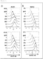
- Figure 3(A) illustrates results of the examination of binding between rat monoclonal antibodies against cPD1 (anti-cPD1 mAb) and NRK/cPD1
- Figure 3(B) illustrates results of examination of binding between rat monoclonal antibodies against cPDL1 (anti-cPD-Ll mAb) and NRK/cPDL1.
- the abscissa indicates the fluorescence intensity of the fluorescent labeled secondary antibody against the produced antibody and the ordinate indicates the concentration of each rat monoclonal antibody.
- any of the rat monoclonal antibodies increased the fluorescence intensity, the rat monoclonal antibodies against cPD1, 3B7-D9 and 4F12-E6 bound with NRK/cPD1, and the rat monoclonal antibodies against cPDL1, H7-9 and G11-6 bound with NRK/cPDL1, as shown in Figure 3 . Accordingly, it was demonstrated that 3B7-D9 and 4F12-E6 are anti-cPD1 antibodies and H7-9 and G11-6 are anti-cPDL1 antibodies.
- hPDL1 was amplified using cDNA derived from the human Burkitt lymphoma cell line (Raji) as template, a forward primer (YTM1150: SEQ ID NO: 54) and a reverse primer (YTM1151: SEQ ID NO: 55).
- the amplification by the PCR was conducted in a way similar to that described for Example 1.
- the amplified PCR product was introduced at the SmaI site in the pBluescript SK (-) vector.
- This plasmid was digested with EcoRI and NotI, the obtained fragments were ligated at the EcoRI and NotI sites in the pMxs-IP vector to produce the hPDL1 expression plasmid, pMx-IP-hPDL1#21.
- a vector expressing a fusion protein between the extracellular region of cPD1 or hPDL1 and a human IgG2 Fc region was constructed.
- the extracellular region of cPD1 was amplified by conducting PCR using pMx-IP-cPD1-FL produced in Example 1 as template, a forward primer (YTM1153: SEQ ID NO: 56), and a reverse primer (YTM1154: SEQ ID NO: 57).
- hPDL1 The extracellular region of hPDL1 was amplified by conducting PCR using pMx-IP-hPDL1#21 produced as described above as template, a forward primer (YTM1157: SEQ ID NO: 58), and a reverse primer (YTM1158: SEQ ID NO: 59).
- PCR products cPD1 were respectively digested with BamHI for cPD1 and BglII for hPDL1 and cloned at the EcoRV and BgIII sites in the pFUSE-hIgG2-Fc2 vector (Invivogen) to produce a vector expressing a fusion protein with the extracellular region of cPD1 and the human IgG2 Fc region (pFUSE-cPD1-hIg#2) and a vector expressing a fusion protein of the extracellular region of hPDL1 and the human IgG2 Fc region (pFUSE-hPDL1-hIg#9).
- the HEK293T cell line was transfected with the vector pFUSE-cPD1-hIg#2, pFUSE-hPDL1-hIg#9, produced as described above, or the empty vector pFUSE-hIgG2-Fc2. 2 ⁇ 10 6 HEK293T cells were first plated in four 10 cm dishes 1 day before the transfection. 7.5 ⁇ g of each vector and 375 ⁇ l of OPTI-MEM comprising 30 ⁇ l of 1 mg/ml PEI Max were then mixed and incubated at room temperature for 15 minutes, and added to the cells. 24 hours after the transfection, medium was replaced into the serum-free GIT medium (Wako Pure Chemical Industries) and the cells were further cultured for 48 hours.
- Figure 4(A) illustrates a result of examination of binding between hPDL1-hIg and NRK/cPD1
- Figure 4(B) illustrates a result of examination of binding between cPD1-hIg and NRK/cPDL1.
- the abscissa indicates the fluorescence intensity of the fluorescent labeled secondary antibody produced against the fusion protein, and the ordinate indicates the concentration of hPDL1-hIg or cPD1-hIg. It was demonstrated that hPDL1-hIg binds to NRK/cPD1 and cPD1-hIg binds to NRK/cPDL1 as shown in Figure 4 .
- Figure 5(A) illustrates results of examination of inhibition of binding between hPDL1-hIg and NRK/cPD1 by rat monoclonal antibodies against cPD1
- Figure 5(B) illustrates results of examination of inhibition of binding between cPD1-hIg and NRK/cPDL1 by rat monoclonal antibodies against cPDL1
- Figure 6 illustrates results of examination of inhibition of binding between cPD1-hIg and NRK/cPDL1 by rat monoclonal antibodies against cPD1.
- the abscissa indicates the fluorescence intensity of the fluorescent labeled secondary antibody against the produced antibody
- the left ordinate indicates the concentration of each rat monoclonal antibody
- the right ordinate indicates the presence (+) or the absence (-) of hPDL1-hIg or cPD1-hIg.
- RNA was isolated from each hybridoma cell line obtained in Example 1.
- the nucleotide sequence of the immunoglobulin heavy chain (IgG2a) and the nucleotide sequence of the ⁇ light chain comprising all variable regions and 5 'region of the constant regions were obtained by the 5 ' RACE PCR technique.
- the isolated RNA was first reverse-transcribed using the reverse transcriptase Superscript (registered trademark) III.
- YTM171 SEQ ID NO: 60
- YTM172 SEQ ID NO: 61
- polyC was added by TdT enzymatic reaction and then the primary PCR was conducted using a forward primer (YTM166: SEQ ID NO: 62) and a reverse primer (YTM171: SEQ ID NO: 60) for the amplification of the light chain and a forward primer (YTM166: SEQ ID NO: 62) and a reverse primer (YTM172: SEQ ID NO: 61) for the amplification of the heavy chain.
- the PCR products were gel-purified and the nested PCR was conducted using a forward primer (YTM170: SEQ ID NO: 63) and a reverse primer (YTM173: SEQ ID NO: 64), or a forward primer (YTM170: SEQ ID NO: 63) and a reverse primer (YTM174: SEQ ID NO: 65) to amplify all variable regions of the light chain or the heavy chain.
- YTM1224 SEQ ID NO: 42
- the nested PCR products were cloned into the pBluescript SK (-) vector and their nucleotide sequences and the amino acid sequences encoded by the nucleotide sequence were determined.
- the nucleotide sequence of the heavy chain variable region of 3B7-D9 is set forth in SEQ ID NO: 35, the amino acid sequence in SEQ ID NO: 2, the nucleotide sequence of the light chain variable region in SEQ ID NO: 36, and the amino acid sequence in SEQ ID NO: 3.
- the nucleotide sequence of the heavy chain variable region of 4F12-E6 is set forth in SEQ ID NO: 37, the amino acid sequence in SEQ ID NO: 4, the nucleotide sequence of the light chain variable region in SEQ ID NO: 38, and the amino acid sequence in SEQ ID NO: 5.
- the nucleotide sequence of the heavy chain variable region of H7-9 is set forth in SEQ ID NO: 39, the amino acid sequence in SEQ ID NO: 7, the nucleotide sequence of the light chain variable region in SEQ ID NO: 40, and the amino acid sequence in SEQ ID NO: 8.
- the nucleotide sequence of the heavy chain variable region of G11-6 is set forth in SEQ ID NO: 41, the amino acid sequence in SEQ ID NO: 9, the nucleotide sequence of the light chain variable region in SEQ ID NO: 42, and the amino acid sequence in SEQ ID NO: 10.
- amino acid sequences of CDRs of each heavy chain variable region and light chain variable region were identified by comparing the sequence with the amino acid sequences of known antibodies.
- the amino acid sequences of the CDR1-3 of the heavy chain variable region of 3B7-D9 are set forth in SEQ ID NOs: 11-13 and the amino acid sequences of the CDR1-3 of the light chain variable region in SEQ ID NOs: 14-16, respectively.
- the amino acid sequences of the CDR1-3 of the heavy chain variable region of 4F12-E6 are set forth in SEQ ID NOs: 17-19 and the amino acid sequences of the CDR1-3 of the light chain variable region in SEQ ID NOs: 20-22, respectively.
- amino acid sequences of the CDR1-3 of the heavy chain variable region of H7-9 are set forth in SEQ ID NOs: 23-25 and the amino acid sequences of the CDR1-3 of the light chain variable region in SEQ ID NOs: 26-28, respectively.
- amino acid sequences of the CDR1-3 of the heavy chain variable region of G11-6 are set forth in SEQ ID NOs: 29-31 and the amino acid sequences of the CDR1-3 of the light chain variable region in SEQ ID NOs: 32-34, respectively.
- PBMC Peripheral blood mononuclear cells
- PBMCs were transferred into 96 well round bottom plates at 2 ⁇ 10 5 per well, isotype (rat IgG2a: eBioscience), a rat monoclonal antibody against PD-1 (anti-PD-1: 4F12-E6), or rat monoclonal antibody against cPDL1 (anti-PD-Ll: G11-6) was added to a concentration of 10 ⁇ g/ml, and concanavalin A (ConA) was added to 5 ⁇ g/ml.
- the cells were cultured with stimulation for 3 days and canine IFN- ⁇ in the obtained culture supernatant was measured by the canine IFN-gamma DuoSet ELISA (R & D). The results are shown in Figure 7 .
- IFN ⁇ produced by ConA stimulation exhibited a tendency of produced and enhanced in the presence of the anti-PD-1, 4F12-E6 and the anti-PD-Ll, G11-6, as shown in Figure 7 .
- Canine PBMCs were cultured in the presence of PMA/ionomycin or ConA for 3 days, PBMCs were collected, and PD-1 and PD-L1 expression in a CD3-positive fraction (T cells) was then analyzed by flow cytometry using a rat monoclonal antibody against cPD1 (4F12-E6) or a rat monoclonal antibody against cPDL1 (G11-6). The results are shown in Figure 8 .
- activated canine T cells are induced to express both PD-1 and PD-L1 expression, as shown in Figure 8 . It was shown that the PD-1 or PD-L1 expression in T cells can be thus examined by using an antibody according to the present invention and that, in other words, expression of PD-1 and/or PD-L1 expressed on the surface of canine cells can be examined.
- Immature dendritic cells were induced by culturing CD14-positive monocytes separated from canine PBMCs using magnetic beads in the presence of IL-4 and GM-CSF for 6 days. The cells were confirmed to be immature dendritic cells by the simultaneous staining of MHC Class II antibodies (eBioscience).
- Figure 9 illustrates results of analyzing PD-1 and PD-L1 expression in the obtained immature dendritic cells by flow cytometry using a rat monoclonal antibody against cPD1 (4F12-E6) or a rat monoclonal antibody against cPDL1 (G11-6).
- Rat monoclonal antibodies obtained according to the present invention can recognize and bind to canine PD-1 and be therefore used in expression and functional analyses of canine PD-1 and as an agent for inhibiting binding between canine PD-1 and canine PD-L1.
Landscapes
- Health & Medical Sciences (AREA)
- Chemical & Material Sciences (AREA)
- Immunology (AREA)
- Life Sciences & Earth Sciences (AREA)
- Organic Chemistry (AREA)
- Genetics & Genomics (AREA)
- General Health & Medical Sciences (AREA)
- Medicinal Chemistry (AREA)
- Engineering & Computer Science (AREA)
- Biophysics (AREA)
- Molecular Biology (AREA)
- Biochemistry (AREA)
- Bioinformatics & Cheminformatics (AREA)
- Proteomics, Peptides & Aminoacids (AREA)
- Biomedical Technology (AREA)
- Wood Science & Technology (AREA)
- Zoology (AREA)
- Biotechnology (AREA)
- General Engineering & Computer Science (AREA)
- Microbiology (AREA)
- Pharmacology & Pharmacy (AREA)
- Animal Behavior & Ethology (AREA)
- Public Health (AREA)
- Veterinary Medicine (AREA)
- Plant Pathology (AREA)
- Physics & Mathematics (AREA)
- Epidemiology (AREA)
- Mycology (AREA)
- General Chemical & Material Sciences (AREA)
- Chemical Kinetics & Catalysis (AREA)
- Nuclear Medicine, Radiotherapy & Molecular Imaging (AREA)
- Peptides Or Proteins (AREA)
- Medicines Containing Antibodies Or Antigens For Use As Internal Diagnostic Agents (AREA)
Description
- The present invention relates to an anti-canine PD-1 antibody, an agent suitable for inhibiting binding between a canine PD-1 and a canine PD-L1 containing such an antibody, an antibody for use in treating cancer in a dog expressing PD-L1, and a nucleic acid encoding such an antibody.
- In human, antibody medicines targeting immune checkpoint molecules, which are immunosuppressive molecules has lately drawn attention and many clinical trials thereof have been conducted. In particular, clinical trials for human cancer that target PD-1 (Programmed cell death 1) and PD-L1 (programmed
cell death 1 ligand-1) have achieved excellent results and drawn attention as a next-generation cancer therapy (see non-PatentDocuments 1 and 2). - PD-1 is a receptor on the surface of T cells. It has the function of suppressing activation of T cells and shown to have the function of suppressing the immune response against the self, etc. Such suppression of the immune response is achieved by binding of PD-L1, a ligand of PD-1, to PD-1. Cancer cells express PD-L1 and gain the ability to suppress the activation of T cells and evade immune response by binding of expressed PD-L1 to PD-1. Therefore, inhibiting binding between PD-1 and PD-L1 is considered to be effective in treating cancer.
- So far, a cancer therapeutic agent comprising an anti-PD-Ll antibody as an active ingredient and having the effect of suppressing the proliferation of cancer cells in vivo (see Patent Document 1) and an anticancer agent comprising an anti-PD-1 antibody or an anti-PD-Ll antibody, functioning to restore the responsiveness of iNKT cells in which allergy has been induced by administration with an iNKT cell ligand (see Patent Document 2) have been proposed. Moreover, the development of anti-PD-1 antibodies as therapeutic agents for melanoma, non-small cell lung cancer, and renal cell cancer is in progress (see Non Patent Document 3).
- The number of cancer cases of pets has been rapidly increased in these days due to the longevity of pets. Cancer therapies for pets, as well as those for humans, have progressed and the three major therapies of surgical, radiation, and chemical (anticancer agent) therapies have been performed positively.
- In Japan, dogs are the most common household pets and approximately 13 million dogs are kept. Therefore, the number of cancer cases in dogs is increasing. For treating cancer of dogs, the aforementioned three major therapies employed conventionally have a limit and therapies targeting an immune checkpoint molecule such as PD-1 or PD-L1 are expected to be a new therapy that can be used instead of or along with the three major therapies. However, there has been a problem that antibody medicines for human PD-1 and PD-L1 are not effective in dogs. The development of antibodies against canine PD-1 and canine PD-L1 has been therefore demanded. However, studies on PD-1 and PD-L1 in dogs had not progressed: the expression of PD-1 and PD-L1 cDNAs in dogs was not confirmed and the production of antibodies recognizing canine PD-1 and PD-L1 was difficult.
-
-
Patent Document 1
Japanese unexamined Patent Application Publication No.2014-65748 -
Patent Document 2
Japanese unexamined Patent Application Publication No.2010-83863 -
- Non
Patent Document 1
YOKOYAMA, Isao, "Activated development of antibody medicines related to PD-1 (in Japanese)" March 6, 2013, Nikkei Medical online, Internet URL: http://medical.nikkeibp.co.jp/leaf/all/search/cancer/news/ 201303/529345.html (searched in June 23, 2014) - Non
Patent Document 2
NONAKA, Nozomi, "Marked and sustained tumor response with combination of ipilimumab and anti-PD-1 antibody medicine nivolumab in half of advanced malignant melanoma (in Japanese)" May 17, 2013, Nikkei Medical online, Internet URL: http://medical.nikkeibp.co.jp/leaf/all/search/cancer/news/ 201305/530569.html (searched in June 23, 2014) - Non Patent Document 3
Suzanne L. et al., The New England Journal of Medicine Vol.366, No. 26: 2443-2454, 2012
Maekawa et al, PLOS One (June 2014), Vol 9, disclose the effective blocking of the binding of recombinant PD-1 with PD-L1-expressing cells by an antibovine PD-L1 monoclonal antibody. - Objects of the present invention are to provide an anti-canine PD-1 antibody, an agent suitable for inhibiting binding between a canine PD-1 and a canine PD-L1 containing such an antibody, an antibody for use in treating cancer in a dog expressing PD-L1, and a nucleic acid encoding such an antibody.
- The inventors first succeeded in cloning of cDNAs of canine PD-1 and canine PD-L1 and determined their nucleotide and amino acid sequences. Canine PD-1 or canine PD-L1 expressing cell lines were then produced using the canine PD-1 or canine PD-L1 cDNA. The inventors then succeeded in producing rat monoclonal antibodies against canine PD-1 or canine PD-L1 using the cell lines. Furthermore, the inventors have found that the obtained rat monoclonal antibodies have the ability to inhibit binding between canine PD-1 and canine PD-L1.
- Accordingly, the present invention provides:
- (1) An anti-canine PD-1 antibody that specifically binds to a canine PD-1 consisting of the amino acid sequence set forth in SEQ ID NO: 1, the anti-canine PD-1 antibody comprising:
- a heavy chain variable region comprising a CDR1 consisting of the amino acid sequence set forth in SEQ ID NO: 11, a CDR2 consisting of the amino acid sequence set forth in SEQ ID NO: 12, and a CDR3 consisting of the amino acid sequence set forth in SEQ ID NO: 13; and
- a light chain variable region comprising a CDR1 consisting of the amino acid sequence set forth in SEQ ID NO: 14, a CDR2 consisting of the amino acid sequence set forth in SEQ ID NO: 15, and a CDR3 consisting of the amino acid sequence set forth in SEQ ID NO: 16.
- (2) The anti-canine PD-1 antibody according to (1), comprising: a heavy chain variable region having the amino acid sequence set forth in SEQ ID NO: 2 or an amino acid sequence having an identity of 95% or more with the amino acid sequence set forth in SEQ ID NO: 2; and a light chain variable region having the amino acid sequence set forth in SEQ ID NO: 3 or an amino acid sequence having an identity of 95% or more with the amino acid sequence set forth in SEQ ID NO: 3.
- (3) An anti-canine PD-1 antibody that specifically binds to a canine PD-1 consisting of the amino acid sequence set forth in SEQ ID NO: 1, the anti-canine PD-1 antibody comprising: a heavy chain variable region comprising a CDR1 consisting of the amino acid sequence set forth in SEQ ID NO: 17, a CDR2 consisting of the amino acid sequence set forth in SEQ ID NO: 18, and a CDR3 consisting of the amino acid sequence set forth in SEQ ID NO: 19; and a light chain variable region comprising a CDR1 consisting of the amino acid sequence set forth in SEQ ID NO: 20, a CDR2 consisting of the amino acid sequence set forth in SEQ ID NO: 21, and a CDR3 consisting of the amino acid sequence set forth in SEQ ID NO: 22.
- (4) The anti-canine PD-1 antibody according to (3) comprising: a heavy chain variable region having the amino acid sequence set forth in SEQ ID NO: 4 or an amino acid sequence having an identity of 95% or more with the amino acid sequence set forth in SEQ ID NO: 4; and a light chain variable region having the amino acid sequence set forth in SEQ ID NO: 5 or an amino acid sequence having an identity of 95% or more with the amino acid sequence set forth in SEQ ID NO: 5.
- (5) An agent suitable for inhibiting binding between a canine PD-1 and a canine PD-L1 containing the antibody according to any one of (1) to (4) above.
- (6) The antibody according to any one of (1) to (4) above, for use in treating cancer in a dog expressing PD-L1.
- (7) A nucleic acid encoding the antibody according to any one of (1) to (4) above.
- Antibodies according to the present invention can recognize and bind to canine PD-1 and can be therefore used in expression and functional analyses of canine PD-1 and as an agent for inhibiting binding between canine PD-1 and canine PD-L1.
-
-
Figure 1 illustrates the nucleotide sequence of canine PD-1 (hereinafter, also referred to as "cPD1") and the amino acid sequence that the nucleotide sequence encodes. -
Figure 2 illustrates the nucleotide sequence of canine PD-L1 (hereinafter, also referred to as "cPDL1") and the amino acid sequence that the nucleotide sequence encodes. -
Figure 3 (A) illustrates results of examination of binding between rat monoclonal antibodies against cPD1 and NRK/cPD1.Figure 3 (B) illustrates results of examination of binding between rat monoclonal antibodies against cPDL1 and NRK/cPDL1. -
Figure 4 (A) illustrates a result of examination of binding between human PD-L1 (hPDL1) -hIg and NRK/cPD1.Figure 4 (B) illustrates a result of examination of binding between cPD1-hIg and NRK/cPDL1. -
Figure 5 (A) illustrates results of examination of inhibition of binding between hPDL1-hIg and NRK/cPD1 by rat monoclonal antibodies against cPD1.Figure 5 (B) illustrates results of examination of inhibition of binding between cPD1-hIg and NRK/cPDL1 by rat monoclonal antibodies against cPDL1. -
Figure 6 illustrates results of examination of inhibition of binding between cPD1-hIg and NRK/cPDL1 by rat monoclonal antibodies against cPD1. -
Figure 7 illustrates results of adding a rat monoclonal antibody against PD-1 (anti-PD-1: 4F12-E6 or a rat monoclonal antibody against cPD-L1 (anti-PD-Ll: G11-6) to peripheral blood mononuclear cells; further adding concanavalin A (ConA) for stimulated culturing; and measuring canine IFN-γ in the obtained culture supernatant by ELISA. -
Figure 8 illustrates results of culturing canine PBMCs in the presence of PMA/ionomycin or ConA; collecting the PBMCs; then analyzing PD-1 and PD-L1 expression in a CD3-positive fraction (T cells) by flow cytometry using a rat monoclonal antibody against cPD1 (4F12-E6) or a rat monoclonal antibody against cPDL1 (G11-6). -
Figure 9 illustrates results of culturing CD14-positive monocytes separated from canine PBMCs using magnetic beads in the presence of IL-4 and GM-CSF for 6 days to induce immature dendritic cells; and analyzing PD-1 or PD-L1 expression in the immature dendritic cells (MHC Class II antibody positive) by flow cytometry using a rat monoclonal antibody against cPD1 (4F12-E6) or a rat monoclonal antibody against cPDL1 (G11-6). - An anti-canine PD-1 antibody according to the present invention is not particularly limited as long as it is an anti-Canine PD-1 antibody that specifically binds to a canine PD-1 consisting of the amino acid sequence set forth in SEQ ID NO: 1, the anti-canine PD-1 antibody comprising: a heavy chain variable region comprising a CDR1 consisting of the amino acid sequence set forth in SEQ ID NO: 11, a CDR2 consisting of the amino acid sequence set forth in SEQ ID NO: 12, and a CDR3 consisting of the amino acid sequence set forth in SEQ ID NO: 13; and a light chain variable region comprising a CDR1 consisting of the amino acid sequence set forth in SEQ ID NO: 14, a CDR2 consisting of the amino acid sequence set forth in SEQ ID NO: 15, and a CDR3 consisting of the amino acid sequence set forth in SEQ ID NO: 16.
- In some embodiments, the anti-canine PD-1 antibody comprises: a heavy chain variable region having the amino acid sequence set forth in SEQ ID NO: 2 or an amino acid sequence having an identity of 95% or more with the amino acid sequence set forth in SEQ ID NO: 2; and a light chain variable region having the amino acid sequence set forth in SEQ ID NO: 3 or an amino acid sequence having an identity of 95% or more with the amino acid sequence set forth in SEQ ID NO: 3. Such an anti-canine PD-1 antibody can recognize and bind to canine PD-1 and can be therefore used in expression and functional analyses of canine PD-1 and in inhibition of binding between canine PD-1 and canine PD-L1.
- Other aspects of the anti-canine PD-1 antibody according to the present invention include an anti-canine PD-1 antibody that specifically binds to a canine PD-1 consisting of the amino acid sequence set forth in SEQ ID NO: 1, the anti-canine PD-1 antibody comprising: a heavy chain variable region comprising a CDR1 consisting of the amino acid sequence set forth in SEQ ID NO: 17, a CDR2 consisting of the amino acid sequence set forth in SEQ ID NO: 18, and a CDR3 consisting of the amino acid sequence set forth in SEQ ID NO: 19; and a light chain variable region comprising a CDR1 consisting of the amino acid sequence set forth in SEQ ID NO: 20, a CDR2 consisting of the amino acid sequence set forth in SEQ ID NO: 21, and a CDR3 consisting of the amino acid sequence set forth in SEQ ID NO: 22.
- In some embodiments, the anti-canine PD-1 antibody comprises: a heavy chain variable region consisting of the amino acid sequence set forth in SEQ ID NO: 4 or an amino acid sequence having an identity of 95% or more with the amino acid sequence set forth in SEQ ID NO: 4; and a light chain variable region consisting of the amino acid sequence set forth in SEQ ID NO: 5 or an amino acid sequence having an identity of 95% or more with the amino acid sequence set forth in SEQ ID NO: 5. Such an anti-canine PD-1 antibody can recognize and bind to canine PD-1 and can be therefore used in expression and functional analyses of canine PD-1 and in inhibition of binding between canine PD-1 and canine PD-L1.
- The type of the aforementioned anti-canine PD-1 antibodies (hereinafter, these are also collectively referred to as "the present anti-canine antibodies") is not particularly limited and includes a monoclonal antibody; a chimeric or caninized antibody produced as a recombinant protein from the sequence of a monoclonal antibody; and an antibody fragment consisting of a part(s) of antibody, such as F (ab')2, obtained by digesting antibody with pepsin, Fab, obtained by digesting antibody with papain, ScFv, in which a heavy chain variable region and a light chain variable region are connected by amino acid cross-linking, diabody, in which ScFvs are dimerized.
- In the present invention, "an identity of 90% or more" means that the identity is 90% or more, preferably 93% or more, more preferably 95% or more, and further preferably 98% or more.
- Examples of nucleic acids encoding the present anti-canine antibodies, for example, a nucleic acid encoding the aforementioned anti-canine PD-1 antibody include a nucleic acid comprising a nucleic acid encoding an amino acid sequence set forth in SEQ ID NO: 2 or an amino acid sequence having an identity of 95% or more with the amino acid sequence set forth in SEQ ID NO: 2 and a nucleic acid encoding an amino acid sequence set forth in SEQ ID NO: 3 or an amino acid sequence having an identity of 95% or more with the amino acid sequence set forth in SEQ ID NO: 3; wherein the antibody produced from the nucleic acid specifically binds to a canine PD-1 consisting of the amino acid sequence set forth in SEQ ID NO: 1. Specific examples include a nucleic acid comprising the nucleotide sequence set forth in SEQ ID NO: 35 and the nucleotide sequence set forth in SEQ ID NO: 36. Other examples include a nucleic acid comprising a nucleic acid encoding the amino acid sequences of CDR1-3 of the heavy chain variable region set forth in SEQ ID NOs: 11-13 and a nucleic acid encoding the amino acid sequences of CDR1-3 of the light chain variable region set forth in SEQ ID NOs: 14-16
- Other aspects of the nucleic acid encoding the aforementioned anti-canine PD-1 antibody include a nucleic acid comprising a nucleic acid encoding the amino acid sequence set forth in SEQ ID NO: 4 or an amino acid sequence having an identity of 95% or more with the amino acid sequence set forth in SEQ ID NO: 4 and a nucleic acid encoding an amino acid sequence set forth in SEQ ID NO: 5 or an amino acid sequence having an identity of 95% or more with the amino acid sequence set forth in SEQ ID NO: 5; wherein the antibody produced from the nucleic acid specifically binds to a canine PD-1 consisting of the amino acid sequence set forth in SEQ ID NO: 1. Specific examples include a nucleic acid comprising the nucleotide sequence set forth in SEQ ID NO: 37 and the nucleotide sequence set forth in SEQ ID NO: 38. Other examples include a nucleic acid comprising a nucleic acid encoding the amino acid sequences of CDR1-3 of the heavy chain variable region set forth in SEQ ID NOs: 17-19 and a nucleic acid encoding the amino acid sequences of CDR1-3 of the light chain variable region set forth in SEQ ID NOs: 20-22.
- An expression vector of a nucleic acid encoding the aforementioned anti-canine PD-1 antibody can be produced by incorporating a nucleic acid encoding the aforementioned anti-canine PD-1 antibody into a vector. The vector to be used as the expression vector is a vector capable of expressing a nucleic acid encoding the aforementioned anti-canine PD-1 and can be a plasmid vector or a phage vector.
- The aforementioned monoclonal antibody can be produced according to a conventional protocol. For example, it can be produced as a recombinant antibody by expressing a gene encoding one of the present anti-canine antibodies by the gene recombination technique. Methods for producing a recombination antibody include, for example, a method involving incorporating a gene encoding one of the present anti-canine antibodies into an expression vector; introducing the expression vector into host cells such as a mammalian cell line such as Chinese hamster ovary (CHO) cells, Escherichia coli, fungal cells, insect cells, and plant cells; and producing the recombinant antibody in the host cells (see Peter J. Delves, ANTIBODY PRODUCTION ESSENTIAL TECHNIQUES, WILEY, 1997). The nucleotide sequence of the gene encoding one of the present anti-canine antibodies to be incorporated into an expression vector can be optimized in codon sequence for the host cells to be used for the expression.
- Moreover, a transgenic animal, such as a mouse, a cow, a goat, a sheep, a chicken, or a pig, in which a gene encoding one of the present anti-canine antibodies is incorporated can be created by a transgenic animal-creation technique and the antibody can be produced in a large amount in blood, milk, or the like of the transgenic animal.
- Furthermore, the aforementioned monoclonal antibody can be produced by administering cells expressing a protein consisting of the amino acid sequence set forth in SEQ ID NO: 1 or 6 as an antigen to a non-human animal such as a mouse or a rat and screening for cellular clones that produce one of the present anti-canine antibodies by a cell fusion technique (the hybridoma technique (see Kohler G, et al., Nature 256: 495-497, 1975)), the trioma technique, the human B-cell hybridoma technique (see Danuta Kozbor, et al., Immunology Today 4, 72-79, 1983), and the EBV-hybridoma technique (see Cole et al., MONOCLONAL ANTIBODIES AND CANCER THERAPY: 77-96, Alan R.Liss, Inc., 1985)).
- The aforementioned caninized antibody can be produced, for example, by replacing the constant regions of an antibody having heavy chain and light chain variable regions with constant regions of a canine antibody. The constant regions of the canine antibody can be those of known canine antibodies.
- The produced present anti-canine antibody can be purified by Protein A or Protein G column chromatography, ion exchange chromatography, hydrophobic chromatography, ammonium sulfate precipitation, gel filtration, affinity chromatography, or the like.
- An agent for inhibiting binding between a canine PD-1 and a canine PD-L1 containing one of the anti-canine antibodies is not particularly limited as long as it contains one of the present anti-canine antibodies and can comprise a plurality of types of anti-canine PD-1 antibodies. Such an agent for inhibiting binding inhibits binding between a canine PD-1 and a canine PD-L1 and can be therefore used as an agent for treating cancer in a dog expressing PD-L1. The inhibition of binding according to the present invention includes inhibition of binding both in vivo and in vitro.
- The aforementioned agent for inhibiting binding can comprise a pharmaceutically acceptable additive usually used in the formulation, such as an excipient, a binder, a lubricant, a disintegrator, a preservative, a tonicity adjusting agent, a stabilizer, a dispersant, an antioxidant, a colorant, a flavor agent, or a buffer. The aforementioned agent for inhibiting binding can be administered into a dog in a mode of administration, such as the oral administration as well as the parenteral administration by injection or drip infusion, application, suppository, or intranasal spray, and can be formulated into any form as appropriate.
- Examples of the amount of a present antibody comprised in the aforementioned agent for inhibiting binding include 0.0001-50% by weight and preferable examples include 0.001-5% by weight.
- The method for inhibiting binding between a canine PD-1 and a canine PD-L1 comprising using one of the present anti-canine antibodies is not particularly limited as long as one of the present anti-canine antibodies is used and a plurality of types of anti-canine PD-1 antibodies can be used.
- The present invention will be described more specifically with Examples below. However, the technical scope of the present invention is not limited to these illustrations.
- Primers to be used in PCR in the following Examples are listed in Table 1.
[Table 1] Primer name Nucleotide sequence (5' to 3') SEQ ID No: Direction Purpose YTM1142 CCTTTCCGCTCACACCTCT 43 F to amplify the full length of cPD1 YTM1143 GGCTCCTGGTGGACAGTCT 44 R YTM1144 CGGTGGTCACTTCAGAACG 45 F to amplify the full length of cPDL1 YTM1145 TGCTGAACTCAAACCACAGG 46 R M13(-20) GTAAAACGACGGCCAG 47 F sequence reaction M13 reverse CAGGAAACAGCTATGAC 48 R sequence reaction YTM1167 GTCGATGTCATGATCTTTATAATCAGGGGGCCACAGGCC 51 R Addition of 1st FLAG tag to cPD1 YTM1168 GTCGATGTCATGATCTTTATAATCTGTCTCTTCAAATTG 52 R Addition of 1st FLAG tag to cPDL1 YTM838 GTCGATGTCATGATCTTTATAATCGTCGATGTCATG 53 R Addition of 2nd FLAG tag YTM1150 GGGCATTCCAGAAAGATGAG 54 F to amplify the full length of hPDL1 YTM1151 CCCATTCCTTCCTCTTGTCA 55 R YTM1153 ACTAGACTCCCCTGACAGG 56 F to amplify the extracellular domain of cPD1 YTM1154 CGGGATCCCTGCAACTGGCCGCT 57 R YTM1157 ATTTACTGTCACGGTTCCC 58 F to amplify the extracellular domain of hPDL1 YTM1158 CGAGATCTAGTCCTTTCATTTGGAGG 59 R YTM166 GGCCACGCGTCGACTAGTACGGGGGGGGGGGGGGGG 62 F to amplify the V region of rat heavy and light chains YTM170 GGCCACGCGTCGACTAC 63 F YTM171 TGCCATCAATCTTCCACTTGACA 60 R YTM172 AAYTTTCTTGTCCACCTTGG 61 R YTM173 GTTGTTCAWGARGCACACGACTGAGGCA 64 R YTM174 AATAGCCCTTGACCAGGCAT 65 R YTM1224 AGGATGATGTCTTATGAACAA 66 R F, forward; R, reverse; Bold types indicate restriction enzyme sites and underlines indicate FLAG coding region. - The NRK cells, a rat kidney cell line, and the HEK293T cells, a human kidney cell line, used in the following Examples were maintained in the DMEM medium supplemented with 10% FBS, 100 units/ml penicillin, 100 µg/ml streptomycin, and 55 µM 2-mercaptoethanol. These cell lines were used after culturing at a CO2 gas concentration of 5%, and 37°C in a humidified incubator.
- The forward primer (YTM1142: SEQ ID NO: 43) and the reverse primer (YTM1143: SEQ ID NO: 44) for amplifying cDNA of cPD1 are designed based on the sequence of NCBI (http://www.ncbi.nlm.nih.gov/guide/) Accession No. XM_543338 and the forward primer (YTM1144: SEQ ID NO: 45) and the reverse primer (YTM1145: SEQ ID NO: 46) for amplifying cDNAs of cPDL1 were designed based on the sequence of NCBI Accession No. XM_005615936.1. None of the sequences of Accession No. XM_543338 and Accession No. XM_005615936.1 were found in the genes whose expression is confirmed in a dog.
- cPD1 and cPDL1 genes were amplified using the aforementioned primer pairs, thymic cDNA in a normal dog as template, KOD-Plus-Neo (Toyobo Co. Ltd.), according to the attached protocol. The PCR was conducted with pre-denaturation at 94°C for 2 minutes; then 35 cycles of denaturation at 98°C for 10 seconds, annealing at 58°C for 30 seconds, and extension 68°C for 60 seconds; and a final extension at 68°C for 10 minutes.
- The PCR products prepared as described above were gel-purified and inserted at the SmaI site in the pBluescript SK (-) vector to produce constructed vectors comprising cPD1 or cPDL1 gene as the insert (pBS-cPD1, pBS-cPDL1). The nucleotide sequences of the constructed vectors were determined using a forward primer (M13(-20): SEQ ID NO: 47) and a reverse primer (M13 reverse: SEQ ID NO: 48), BigDye (registered trademark) Termination v3.1 Cycle Sequencing Kit (Perkin-Elmer), and ABI Prism (registered trademark) 377 automatic DNA sequencer (Applied Biosystems). The nucleotide sequence of the obtained cPD1 and the amino acid sequence encoded by the nucleotide sequence are shown in
Figure 1 . The nucleotide sequence of cPDL1 and the amino acid sequence encoded by the nucleotide sequence is shown inFigure 2 . The nucleotide sequence of cPD1 is shown SEQ ID NO: 49, the amino acid sequence thereof in SEQ ID NO: 1, the nucleotide sequence of cPDL1 in SEQ ID NO: 50, and the amino acid sequence thereof in SEQ ID NO: 6. - The retroviral vectors that overexpress cPD1 or cPDL1 protein were constructed. To add two FLAG tag sequences at the C-terminus of cPD1 and cPDL1 sequences, primary PCR was first conducted using pBS-cPD1 as template, a forward primer (YTM1142: SEQ ID NO: 43), and a reverse primer (YTM1167: SEQ ID NO: 51) comprising a FLAG tag sequence at the C-terminus of the cPD1 sequence for the amplification of cPD1, or pBS-cPDL1 as template, a forward primer (YTM1144: SEQ ID NO: 45), and a reverse primer (YTM1168: SEQ ID NO: 52) comprising a FLAG tag sequence at the C-terminus of the cPDL1 sequence for the amplification of cPDL1.
- The second PCR was then conducted using each 1st PCR product as template, a forward primer same as that in the primary PCR, and a reverse primer (YTM838: SEQ ID NO: 53) comprising a double FLAG tag sequence that anneals with the 1st FLAG tag sequence. Each obtained PCR product was inserted at EcoRI and the NotI sites in the pMxs-IP vector to produce pMx-IP-cPD1-FL and pMx-IP-cPDL1-FL#9.
- NRK cells are transfected with pMx-IP-cPD1-FL or pMx-IP-cPDL1-FL#9 by a common transfection technique to produce the NRK cells that stability express cPD1 and the NRK cells that stability express cPDL1. NRK cells (3.5 × 10⁵ cells) were first plated in a 6
well dish 1 day before the transfection. The transfection of cells was conducted using Lipofectamine 2000 (Invitrogen) according to the attached protocol. Cells after the transfection were incubated for 48 hours and then cultured in the presence of 7.5 µg/ml puromycin (Sigma-Aldrich) to obtain stable transfected cells to produce the NRK cell that stability expressing cPD1 (NRK/cPD1) and the NRK cells stability expressing cPDL1 (NRK/cPDL1). - Stable expression of cPD1 and cPDL1 in the NRK/cPD1 and NRK/cPDL1 was confirmed by immunofluorescence using the anti-FLAG M2 antibody (1:1,000 dilution, Sigma-Aldrich) as the primary antibody and IgG-Dylight (registered trademark) 488-labelled anti-mouse (Biolegend) as the secondary antibody.
- To produce rat monoclonal antibodies against cPD1 or cPDL1, the NRK/cPD1 or NRK/cPDL1 (in 500 µl of the DMEM medium, 1 × 10⁷ cells) produced as described above was emulsified with an equal amount of Titer Max (registered trademark) Gold (CytRx) and administered subcutaneously to the footpad of hindleg in 7 week-old Sprague-Dawley rat (Kudo Co. Ltd.). Lymph node cells in the pople were isolated 2 weeks after administration and fused with P3U1 cells. More specifically, lymph node monocytes (1 × 10⁸ cells) and P3U1 cells (2 × 10⁷ cells) were mixed and washed twice with the serum-free RPMI1640 medium. The supernatant was removed and cells were then incubated at 37°C for 2 minutes. 0.5 ml of polyethyleneglycol 1500 (Roche Diagnostics) at 37°C and then 9 ml of the serum-free RPMI1640 medium at 37°C were added and the suspension was centrifuged at room temperature at 900 rpm for 5 minutes. The supernatant was removed and the cells were then suspended in 36 ml of the GIT medium (Wako Pure Chemical Industries) comprising 10% FBS and hypoxanthine-aminopterin-thymidine (HAT: Life Technologies) and cultured in a humidified incubator at CO2 gas concentration of 5% and 37°C for 1 hour. 4 ml of the BM-Condimed H1 medium (Roche Diagnostics) was added and the cells were plated and cultured in four 96 well tissue culture plates (100 µl/well). After culturing to obtain colonies, NRK cell-positive hybridomas expressing cPD1 or cPDL1 were identified by ELISA or flow cytometry and the hybridomas were cloned by limiting dilution. The hybridomas 3B7-D9 and 4F12-E6 were obtained as NRK cell-positive hybridomas expressing cPD1 and the hybridomas H7-9 and G11-6 as NRK cell-positive hybridomas expressing cPDL1. Hereinafter, the antibody produced by the hybridoma 3B7-D9 is also referred to as "3B7-D9," the antibody produced by the hybridoma 4F12-E6 as "4F12-E6," the antibody produced by the hybridoma H7-9 as "H7-9," and the antibody produced by the hybridoma G11-6 as "G11-6." The hybridomas 3B7-D9, 4F12-E6, H7-9, and G11-6 are kept in the Joint Faculty of Veterinary Medicine at Yamaguchi University and available under a certain condition.
- The binding between a rat monoclonal antibody against cPD1, 3B7-D9 or 4F12-E6, and NRK/cPD1 produced as described above and the binding of a rat monoclonal antibody against cPDL1, H7-9 or G11-6 and NRK/cPDL1 produced as described above were examined by the flow cytometry analysis described below. Concentrations of each rat monoclonal antibody were 0, 0.04, 0.156, 0.625, 2.5, and 10 µg/ml and NRK/cPD1 (2 × 10⁵ cells) or NRK/cPDL1 (2 × 10⁵ cells) were used.
- The staining of the cell lines to be used in the flow cytometry analysis was conducted according to the method described in the following documents (Mizuno et al., J Vet Med Sci, 71(12): 1561-1568, 2009). The respective purified rat monoclonal antibodies against cPD1 or cPDL1 were used as the primary antibodies and anti-rat IgG-PE (Southern biotech) was used as the secondary antibody. The samples were analyzed using BD AccuriC6 (BD Bioscience) and the obtained results were analyzed by FlowJo software (Treestar).
- The results are shown in
Figure 3. Figure 3(A) illustrates results of the examination of binding between rat monoclonal antibodies against cPD1 (anti-cPD1 mAb) and NRK/cPD1 andFigure 3(B) illustrates results of examination of binding between rat monoclonal antibodies against cPDL1 (anti-cPD-Ll mAb) and NRK/cPDL1. The abscissa indicates the fluorescence intensity of the fluorescent labeled secondary antibody against the produced antibody and the ordinate indicates the concentration of each rat monoclonal antibody. It was confirmed that any of the rat monoclonal antibodies increased the fluorescence intensity, the rat monoclonal antibodies against cPD1, 3B7-D9 and 4F12-E6 bound with NRK/cPD1, and the rat monoclonal antibodies against cPDL1, H7-9 and G11-6 bound with NRK/cPDL1, as shown inFigure 3 . Accordingly, it was demonstrated that 3B7-D9 and 4F12-E6 are anti-cPD1 antibodies and H7-9 and G11-6 are anti-cPDL1 antibodies. - To construct hPDL1 expression plasmid, hPDL1 was amplified using cDNA derived from the human Burkitt lymphoma cell line (Raji) as template, a forward primer (YTM1150: SEQ ID NO: 54) and a reverse primer (YTM1151: SEQ ID NO: 55). The amplification by the PCR was conducted in a way similar to that described for Example 1. The amplified PCR product was introduced at the SmaI site in the pBluescript SK (-) vector. This plasmid was digested with EcoRI and NotI, the obtained fragments were ligated at the EcoRI and NotI sites in the pMxs-IP vector to produce the hPDL1 expression plasmid, pMx-IP-
hPDL1# 21. - A vector expressing a fusion protein between the extracellular region of cPD1 or hPDL1 and a human IgG2 Fc region was constructed. To produce such an expression vector, the extracellular region of cPD1 was amplified by conducting PCR using pMx-IP-cPD1-FL produced in Example 1 as template, a forward primer (YTM1153: SEQ ID NO: 56), and a reverse primer (YTM1154: SEQ ID NO: 57). The extracellular region of hPDL1 was amplified by conducting PCR using pMx-IP-
hPDL1# 21 produced as described above as template, a forward primer (YTM1157: SEQ ID NO: 58), and a reverse primer (YTM1158: SEQ ID NO: 59). The PCR products cPD1 were respectively digested with BamHI for cPD1 and BglII for hPDL1 and cloned at the EcoRV and BgIII sites in the pFUSE-hIgG2-Fc2 vector (Invivogen) to produce a vector expressing a fusion protein with the extracellular region of cPD1 and the human IgG2 Fc region (pFUSE-cPD1-hIg#2) and a vector expressing a fusion protein of the extracellular region of hPDL1 and the human IgG2 Fc region (pFUSE-hPDL1-hIg#9). - The HEK293T cell line was transfected with the vector pFUSE-cPD1-
hIg# 2, pFUSE-hPDL1-hIg#9, produced as described above, or the empty vector pFUSE-hIgG2-Fc2. 2 × 10⁶ HEK293T cells were first plated in four 10cm dishes 1 day before the transfection. 7.5 µg of each vector and 375 µl of OPTI-MEM comprising 30 µl of 1 mg/ml PEI Max were then mixed and incubated at room temperature for 15 minutes, and added to the cells. 24 hours after the transfection, medium was replaced into the serum-free GIT medium (Wako Pure Chemical Industries) and the cells were further cultured for 48 hours. Supernatant was collected from each of the transfected cells on Day 4 andDay 8 and purified with rProtein A agarose (GE healthcare). Salts were then removed from the soluble protein by dialysis to produce the fusion protein of the extracellular region of cPD1 and the hIgG2 Fc region (cPD1-hIg) and the fusion protein of the extracellular region of hPDL1 and the hIgG2 Fc region (hPDL1-hIg). The purity of the fusion proteins was checked by SDS - PAGE and Western blotting. - As a preliminary examination of the tests of inhibition of PD-1 and PD-L1 with rat monoclonal antibodies described later, the binding between hPDL1-hIg and NRK/cPD1 or the binding between cPD1-hIg and NRK/cPDL1 was examined in a way similar to the aforementioned flow cytometry analysis. Concentrations of hPDL1-hIg and cPD1-hIg were 0, 0.04, 0.156, 0.625, 2.5, 10, and 40 µg/ml and NRK/cPD1 (2 × 10⁵ cells) or NRK/cPDL1 (2 × 10⁵ cells) was used. The results are shown in
Figure 4. Figure 4(A) illustrates a result of examination of binding between hPDL1-hIg and NRK/cPD1 andFigure 4(B) illustrates a result of examination of binding between cPD1-hIg and NRK/cPDL1. The abscissa indicates the fluorescence intensity of the fluorescent labeled secondary antibody produced against the fusion protein, and the ordinate indicates the concentration of hPDL1-hIg or cPD1-hIg. It was demonstrated that hPDL1-hIg binds to NRK/cPD1 and cPD1-hIg binds to NRK/cPDL1 as shown inFigure 4 . - The following (1) to (3) were examined: (1) whether the rat monoclonal antibodies against cPD1 inhibit binding between hPDL1-hIg and NRK/cPD1 by having NRK/cPD1 interacted with a rat monoclonal antibody against cPD1 (3B7-D9, 4F12-E6) and then having hPDL1-hIg interacted with them; (2) whether the rat monoclonal antibodies against cPDL1 inhibit binding between cPD1-hIg and NRK/cPDL1 by having NRK/cPDL1 interacted with a rat monoclonal antibody against cPDL1 (H7-9, G11-6) and then having cPD1-hIg interacted with them; or (3) Whether the rat monoclonal antibodies against cPD1 inhibit binding between cPD1-hIg and NRK/cPDL1 by having a rat monoclonal antibody against cPD1 (3B7-D9, 4F12-E6) interacted with cPD1-hIg and then having NRK/cPDL1 interacted with the mixed reactant. The inhibition of binding between PD-1 and PD-L1 with each antibody was examined in a way similar to the aforementioned flow cytometry analysis. Concentrations of each rat monoclonal antibody were 0, 0.04, 0.156, 0.625, 2.5, and 10 µg/ml, the concentration of hPDL1-hIg and cPD1-hIg was 40 µg/ml, and NRK/cPD1 (2 × 10⁵ cells) or NRK/cPDL1 (2 × 10⁵ cells) was used.
- The results are shown in
Figures 5 and6 .Figure 5(A) illustrates results of examination of inhibition of binding between hPDL1-hIg and NRK/cPD1 by rat monoclonal antibodies against cPD1;Figure 5(B) illustrates results of examination of inhibition of binding between cPD1-hIg and NRK/cPDL1 by rat monoclonal antibodies against cPDL1; andFigure 6 illustrates results of examination of inhibition of binding between cPD1-hIg and NRK/cPDL1 by rat monoclonal antibodies against cPD1. The abscissa indicates the fluorescence intensity of the fluorescent labeled secondary antibody against the produced antibody, the left ordinate indicates the concentration of each rat monoclonal antibody, and the right ordinate indicates the presence (+) or the absence (-) of hPDL1-hIg or cPD1-hIg. It was demonstrated that the rat monoclonal antibodies against cPD1, 3B7-D9, 4F12-E6 inhibit the binding between hPDL1-hIg and NRK/cPD1 and the binding between cPD1-hIg and NRK/cPDL1 and the rat monoclonal antibodies against cPDL1, H7-9 and G11-6 inhibit the binding between cPD1-hIg and NRK/cPDL1, as shownFigures 5(A), (B) , and6 . - Total RNA was isolated from each hybridoma cell line obtained in Example 1. The nucleotide sequence of the immunoglobulin heavy chain (IgG2a) and the nucleotide sequence of the κ light chain comprising all variable regions and 5 'region of the constant regions were obtained by the 5 ' RACE PCR technique.
- The isolated RNA was first reverse-transcribed using the reverse transcriptase Superscript (registered trademark) III. As a primer, YTM171 (SEQ ID NO: 60) was used for the amplification of the light chain and YTM172 (SEQ ID NO: 61) for the amplification of the heavy chain. After the reverse transcription reaction, polyC was added by TdT enzymatic reaction and then the primary PCR was conducted using a forward primer (YTM166: SEQ ID NO: 62) and a reverse primer (YTM171: SEQ ID NO: 60) for the amplification of the light chain and a forward primer (YTM166: SEQ ID NO: 62) and a reverse primer (YTM172: SEQ ID NO: 61) for the amplification of the heavy chain. The PCR products were gel-purified and the nested PCR was conducted using a forward primer (YTM170: SEQ ID NO: 63) and a reverse primer (YTM173: SEQ ID NO: 64), or a forward primer (YTM170: SEQ ID NO: 63) and a reverse primer (YTM174: SEQ ID NO: 65) to amplify all variable regions of the light chain or the heavy chain. In hybridomas 3B7-D9 and G11-6, YTM1224 (SEQ ID NO: 42) was used, instead of YTM171 as a primer in 5' RACE and the primary PCR. The nested PCR products were cloned into the pBluescript SK (-) vector and their nucleotide sequences and the amino acid sequences encoded by the nucleotide sequence were determined.
- The nucleotide sequence of the heavy chain variable region of 3B7-D9 is set forth in SEQ ID NO: 35, the amino acid sequence in SEQ ID NO: 2, the nucleotide sequence of the light chain variable region in SEQ ID NO: 36, and the amino acid sequence in SEQ ID NO: 3. The nucleotide sequence of the heavy chain variable region of 4F12-E6 is set forth in SEQ ID NO: 37, the amino acid sequence in SEQ ID NO: 4, the nucleotide sequence of the light chain variable region in SEQ ID NO: 38, and the amino acid sequence in SEQ ID NO: 5. The nucleotide sequence of the heavy chain variable region of H7-9 is set forth in SEQ ID NO: 39, the amino acid sequence in SEQ ID NO: 7, the nucleotide sequence of the light chain variable region in SEQ ID NO: 40, and the amino acid sequence in SEQ ID NO: 8. The nucleotide sequence of the heavy chain variable region of G11-6 is set forth in SEQ ID NO: 41, the amino acid sequence in SEQ ID NO: 9, the nucleotide sequence of the light chain variable region in SEQ ID NO: 42, and the amino acid sequence in SEQ ID NO: 10.
- Furthermore, the amino acid sequences of CDRs of each heavy chain variable region and light chain variable region were identified by comparing the sequence with the amino acid sequences of known antibodies. The amino acid sequences of the CDR1-3 of the heavy chain variable region of 3B7-D9 are set forth in SEQ ID NOs: 11-13 and the amino acid sequences of the CDR1-3 of the light chain variable region in SEQ ID NOs: 14-16, respectively. The amino acid sequences of the CDR1-3 of the heavy chain variable region of 4F12-E6 are set forth in SEQ ID NOs: 17-19 and the amino acid sequences of the CDR1-3 of the light chain variable region in SEQ ID NOs: 20-22, respectively. The amino acid sequences of the CDR1-3 of the heavy chain variable region of H7-9 are set forth in SEQ ID NOs: 23-25 and the amino acid sequences of the CDR1-3 of the light chain variable region in SEQ ID NOs: 26-28, respectively. The amino acid sequences of the CDR1-3 of the heavy chain variable region of G11-6 are set forth in SEQ ID NOs: 29-31 and the amino acid sequences of the CDR1-3 of the light chain variable region in SEQ ID NOs: 32-34, respectively.
- Peripheral blood mononuclear cells (PBMC) were collected from 3 dogs (A, B, and C), PBMCs were transferred into 96 well round bottom plates at 2 × 10⁵ per well, isotype (rat IgG2a: eBioscience), a rat monoclonal antibody against PD-1 (anti-PD-1: 4F12-E6), or rat monoclonal antibody against cPDL1 (anti-PD-Ll: G11-6) was added to a concentration of 10 µg/ml, and concanavalin A (ConA) was added to 5 µg/ml. The cells were cultured with stimulation for 3 days and canine IFN-γ in the obtained culture supernatant was measured by the canine IFN-gamma DuoSet ELISA (R & D). The results are shown in
Figure 7 . - IFNγ produced by ConA stimulation exhibited a tendency of produced and enhanced in the presence of the anti-PD-1, 4F12-E6 and the anti-PD-Ll, G11-6, as shown in
Figure 7 . This suggested the possibility that expression of PD-1 and PD-L1 is enhanced by ConA stimulation in canine T cells and binding of the PD-L1 to the PD-1 prevents excessive activation of T cells and also suggested that the obtained PD-1 and PD-L1 antibodies can inhibit the interaction between PD-1 and PD-L1 molecules expressed on canine cells. - Canine PBMCs were cultured in the presence of PMA/ionomycin or ConA for 3 days, PBMCs were collected, and PD-1 and PD-L1 expression in a CD3-positive fraction (T cells) was then analyzed by flow cytometry using a rat monoclonal antibody against cPD1 (4F12-E6) or a rat monoclonal antibody against cPDL1 (G11-6). The results are shown in
Figure 8 . - It was demonstrated that activated canine T cells are induced to express both PD-1 and PD-L1 expression, as shown in
Figure 8 . It was shown that the PD-1 or PD-L1 expression in T cells can be thus examined by using an antibody according to the present invention and that, in other words, expression of PD-1 and/or PD-L1 expressed on the surface of canine cells can be examined. - Immature dendritic cells were induced by culturing CD14-positive monocytes separated from canine PBMCs using magnetic beads in the presence of IL-4 and GM-CSF for 6 days. The cells were confirmed to be immature dendritic cells by the simultaneous staining of MHC Class II antibodies (eBioscience).
Figure 9 illustrates results of analyzing PD-1 and PD-L1 expression in the obtained immature dendritic cells by flow cytometry using a rat monoclonal antibody against cPD1 (4F12-E6) or a rat monoclonal antibody against cPDL1 (G11-6). - It was confirmed that PD-1 is not expressed, but PDL-1 is expressed, as shown in
Figure 9 . It was demonstrated that PD-1 or PD-L1 expression in immature dendritic cells can be thus examined using an antibody according to the present invention. - Rat monoclonal antibodies obtained according to the present invention can recognize and bind to canine PD-1 and be therefore used in expression and functional analyses of canine PD-1 and as an agent for inhibiting binding between canine PD-1 and canine PD-L1.
-
- <110> NIPPON ZENYAKU KOGYO CO., LTD.
- <120> Antibody against caninePD-1 or caninePD-L1
- <130> ZQ1507-PCT
- <150>
JP2014-140982
<151> 2014-07-09 - <160> 66
- <170> PatentIn version 3.1
- <210> 1
<211> 288
<212> PRT
<213> artificial - <220>
<223> cPD1 aa - <220>
<223> Inventor: Mizuno, Takuya Inventor: Shosu, Kazuha - <400> 1


- <210> 2
<211> 139
<212> PRT
<213> artificial - <220>
<223> 3B7-D9 IgH V region aa - <400> 2


- <210> 3
<211> 133
<212> PRT
<213> artificial - <220>
<223> 3B7-D9 Igkappa V region aa - <400> 3


- <210> 4
<211> 139
<212> PRT
<213> artificial - <220>
<223> 4F12-E6 IgH V region aa - <400> 4

- <210> 5
<211> 133
<212> PRT
<213> artificial - <220>
<223> 4F12-E6 Igkappa V region aa - <400> 5


- <210> 6
<211> 289
<212> PRT
<213> artificial - <220>
<223> cPDL1 aa - <400> 6
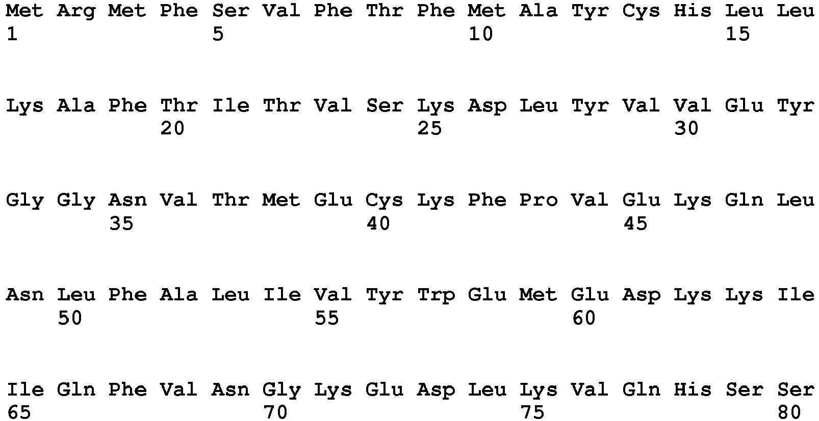

- <210> 7
<211> 137
<212> PRT
<213> artificial - <220>
<223> H7-9 IgH V region aa - <400> 7

- <210> 8
<211> 127
<212> PRT
<213> artificial - <220>
<223> H7-9 Igkappa V region aa - <400> 8


- <210> 9
<211> 139
<212> PRT
<213> artificial - <220>
<223> Gll-6 IgH V region aa - <400> 9
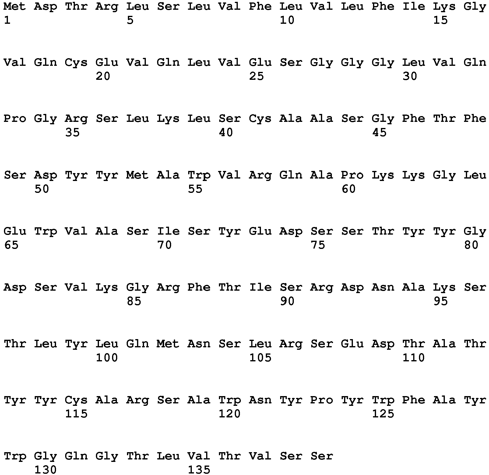
- <210> 10
<211> 127
<212> PRT
<213> artificial - <220>
<223> Gll-6 Igkappa V region aa - <400> 10

- <210> 11
<211> 8
<212> PRT
<213> artificial - <220>
<223> 3B7-D9 IgH V region CDR1 aa - <400> 11

- <210> 12
<211> 8
<212> PRT
<213> artificial - <220>
<223> 3B7-D9 IgH V region CDR2 aa - <400> 12

- <210> 13
<211> 13
<212> PRT
<213> artificial - <220>
<223> 3B7-D9 IgH V region CDR3 aa - <400> 13

- <210> 14
<211> 12
<212> PRT
<213> artificial - <220>
<223> 3B7-D9 Igkappa V region CDR1 aa - <400> 14

- <210> 15
<211> 3
<212> PRT
<213> artificial - <220>
<223> 3B7-D9 Igkappa V region CDR2 aa - <400> 15

- <210> 16
<211> 6
<212> PRT
<213> artificial - <220>
<223> 3B7-D9 Igkappa V region CDR3 aa - <400> 16

- <210> 17
<211> 8
<212> PRT
<213> artificial - <220>
<223> 4F12-E6 IgH V region CDR1 aa - <400> 17

- <210> 18
<211> 8
<212> PRT
<213> artificial - <220>
<223> 4F12-E6 IgH V region CDR2 aa - <400> 18

- <210> 19
<211> 13
<212> PRT
<213> artificial - <220>
<223> 4F12-E6 IgH V region CDR3 aa - <400> 19

- <210> 20
<211> 10
<212> PRT
<213> artificial - <220>
<223> 4F12-E6 Igkappa V region CDR1 aa - <400> 20

- <210> 21
<211> 3
<212> PRT
<213> artificial - <220>
<223> 4F12-E6 Igkappa V region CDR2 aa - <400> 21

- <210> 22
<211> 6
<212> PRT
<213> artificial - <220>
<223> 4F12-E6 Igkappa V region CDR3 aa - <400> 22

- <210> 23
<211> 8
<212> PRT
<213> artificial - <220>
<223> H7-9 IgH V region CDR1 aa - <400> 23

- <210> 24
<211> 7
<212> PRT
<213> artificial - <220>
<223> H7-9 IgH V region CDR2 aa - <400> 24

- <210> 25
<211> 12
<212> PRT
<213> artificial - <220>
<223> H7-9 IgH V region CDR3 aa - <400> 25

- <210> 26
<211> 6
<212> PRT
<213> artificial - <220>
<223> H7-9 Igkappa V region CDR1 aa - <400> 26

- <210> 27
<211> 3
<212> PRT
<213> artificial - <220>
<223> H7-9 Igkappa V region CDR2 aa - <400> 27

- <210> 28
<211> 6
<212> PRT
<213> artificial - <220>
<223> H7-9 Igkappa V region CDR3 aa - <400> 28

- <210> 29
<211> 8
<212> PRT
<213> artificial - <220>
<223> Gll-6 IgH V region CDR1 aa - <400> 29

- <210> 30
<211> 8
<212> PRT
<213> artificial - <220>
<223> Gll-6 IgH V region CDR2 aa - <400> 30

- <210> 31
<211> 13
<212> PRT
<213> artificial - <220>
<223> G11-6 IgH V region CDR3 aa - <400> 31

- <210> 32
<211> 6
<212> PRT
<213> artificial - <220>
<223> Gll-6 Igkappa V region CDR1 aa - <400> 32

- <210> 33
<211> 3
<212> PRT
<213> artificial - <220>
<223> Gll-6 Igkappa V region CDR2 aa - <400> 33

- <210> 34
<211> 6
<212> PRT
<213> artificial - <220>
<223> Gll-6 Igkappa V region CDR3 aa - <400> 34

- <210> 35
<211> 417
<212> DNA
<213> artificial - <220>
<223> 3B7-D9 IgH V region nt - <400> 35

- <210> 36
<211> 399
<212> DNA
<213> artificial - <220>
<223> 3B7-D9 Igkappa V region nt - <400> 36

- <210> 37
<211> 417
<212> DNA
<213> artificial - <220>
<223> 4F12-E6 IgH V region nt - <400> 37


- <210> 38
<211> 399
<212> DNA
<213> artificial - <220>
<223> 4F12-E6 Igkappa V region nt - <400> 38

- <210> 39
<211> 411
<212> DNA
<213> artificial - <220>
<223> H7-9 IgH V region nt - <400> 39

- <210> 40
<211> 381
<212> DNA
<213> artificial - <220>
<223> H7-9 Igkappa V region nt - <400> 40

- <210> 41
<211> 417
<212> DNA
<213> artificial - <220>
<223> Gll-6 IgH V region nt - <400> 41

- <210> 42
<211> 381
<212> DNA
<213> artificial - <220>
<223> Gll-6 Igkappa V region nt - <400> 42
ggcaccaagc tggaattgaa a 381

- <210> 43
<211> 19
<212> DNA
<213> artificial - <220>
<223> YTM1142 primer - <400> 43
cctttccgct cacacctct 19 - <210> 44
<211> 19
<212> DNA
<213> artificial - <220>
<223> YTM1143 primer - <400> 44
ggctcctggt ggacagtct 19 - <210> 45
<211> 19
<212> DNA
<213> artificial - <220>
<223> YTM1144 primer - <400> 45
cggtggtcac ttcagaacg 19 - <210> 46
<211> 20
<212> DNA
<213> artificial - <220>
<223> YTM1145 primer - <400> 46
tgctgaactc aaaccacagg 20 - <210> 47
<211> 16
<212> DNA
<213> artificial - <220>
<223> M13(-20) primer - <400> 47
gtaaaacgac ggccag 16 - <210> 48
<211> 17
<212> DNA
<213> artificial - <220>
<223> M13 reverse primer - <400> 48
caggaaacag ctatgac 17 - <210> 49
<211> 864
<212> DNA
<213> artificial - <220>
<223> cPD1 nt - <400> 49

- <210> 50
<211> 867
<212> DNA
<213> artificial - <220>
<223> cPLD1 nt - <400> 50


- <210> 51
<211> 39
<212> DNA
<213> artificial - <220>
<223> YTM1167 primer - <400> 51
gtcgatgtca tgatctttat aatcaggggg ccacaggcc 39 - <210> 52
<211> 39
<212> DNA
<213> artificial - <220>
<223> YTM1168 primer - <400> 52
gtcgatgtca tgatctttat aatctgtctc ttcaaattg 39 - <210> 53
<211> 36
<212> DNA
<213> artificial - <220>
<223> YTM838 primer - <400> 53
gtcgatgtca tgatctttat aatcgtcgat gtcatg 36 - <210> 54
<211> 20
<212> DNA
<213> artificial - <220>
<223> YTM1150 primer - <400> 54
gggcattcca gaaagatgag 20 - <210> 55
<211> 20
<212> DNA
<213> artificial - <220>
<223> YTM1151 primer - <400> 55
cccattcctt cctcttgtca 20 - <210> 56
<211> 19
<212> DNA
<213> artificial - <220>
<223> YTM1153 primer - <400> 56
actagactcc cctgacagg 19 - <210> 57
<211> 23
<212> DNA
<213> artificial - <220>
<223> YTM1154 primer - <400> 57
cgggatccct gcaactggcc gct 23 - <210> 58
<211> 19
<212> DNA
<213> artificial - <220>
<223> YTM1157 primer - <400> 58
atttactgtc acggttccc 19 - <210> 59
<211> 26
<212> DNA
<213> artificial - <220>
<223> YTM1158 primer - <400> 59
cgagatctag tcctttcatt tggagg 26 - <210> 60
<211> 23
<212> DNA
<213> artificial - <220>
<223> YTM171 primer - <400> 60
tgccatcaat cttccacttg aca 23 - <210> 61
<211> 20
<212> DNA
<213> artificial - <220>
<223> YTM172 primer - <400> 61
aaytttcttg tccaccttgg 20 - <210> 62
<211> 36
<212> DNA
<213> artificial - <220>
<223> YTM166 primer - <400> 62
ggccacgcgt cgactagtac gggggggggg gggggg 36 - <210> 63
<211> 17
<212> DNA
<213> artificial - <220>
<223> YTM170 primer - <400> 63
ggccacgcgt cgactac 17 - <210> 64
<211> 28
<212> DNA
<213> artificial - <220>
<223> YTM173 primer - <400> 64
gttgttcawg argcacacga ctgaggca 28 - <210> 65
<211> 20
<212> DNA
<213> artificial - <220>
<223> YTM174 primer - <400> 65
aatagccctt gaccaggcat 20 - <210> 66
<211> 21
<212> DNA
<213> artificial - <220>
<223> YTM1224 primer - <400> 66
aggatgatgt cttatgaaca a 21
Claims (7)
- An anti-canine PD-1 antibody that specifically binds to a canine PD-1 consisting of the amino acid sequence set forth in SEQ ID NO: 1, the anti-canine PD-1 antibody comprising:a heavy chain variable region comprising a CDR1 consisting of the amino acid sequence set forth in SEQ ID NO: 11, a CDR2 consisting of the amino acid sequence set forth in SEQ ID NO: 12, and a CDR3 consisting of the amino acid sequence set forth in SEQ ID NO: 13; anda light chain variable region comprising a CDR1 consisting of the amino acid sequence set forth in SEQ ID NO: 14, a CDR2 consisting of the amino acid sequence set forth in SEQ ID NO: 15, and a CDR3 consisting of the amino acid sequence set forth in SEQ ID NO: 16.
- The anti-canine PD-1 antibody according to claim 1, wherein the anti-canine PD-1 antibody comprises a heavy chain variable region having the amino acid sequence set forth in SEQ ID NO: 2 or an amino acid sequence having an identity of 95% or more with the amino acid sequence set forth in SEQ ID NO: 2; and a light chain variable region having the amino acid sequence set forth in SEQ ID NO: 3 or an amino acid sequence having an identity of 95% or more with the amino acid sequence set forth in SEQ ID NO: 3.
- An anti-canine PD-1 antibody that specifically binds to a canine PD-1 consisting of the amino acid sequence set forth in SEQ ID NO: 1, the anti-canine PD-1 antibody comprising:a heavy chain variable region comprising a CDR1 consisting of the amino acid sequence set forth in SEQ ID NO: 17, a CDR2 consisting of the amino acid sequence set forth in SEQ ID NO: 18, and a CDR3 consisting of the amino acid sequence set forth in SEQ ID NO: 19; anda light chain variable region comprising a CDR1 consisting of the amino acid sequence set forth in SEQ ID NO: 20, a CDR2 consisting of the amino acid sequence set forth in SEQ ID NO: 21, and a CDR3 consisting of the amino acid sequence set forth in SEQ ID NO: 22.
- The anti-canine PD-1 antibody according to claim 3, wherein the anti-canine PD-1 antibody comprises a heavy chain variable region having the amino acid sequence set forth in SEQ ID NO: 4 or an amino acid sequence having an identity of 95% or more with the amino acid sequence set forth in SEQ ID NO: 4; and a light chain variable region having the amino acid sequence set forth in SEQ ID NO: 5 or an amino acid sequence having an identity of 95% or more with the amino acid sequence set forth in SEQ ID NO: 5.
- An agent suitable for inhibiting binding between a canine PD-1 and a canine PD-L1 containing the antibody according to any one of claims 1 to 4.
- The antibody according to any one of claims 1 to 4, for use in treating cancer in a dog expressing PD-L1.
- A nucleic acid encoding the antibody according to any one of claims 1 to 4.
Applications Claiming Priority (2)
| Application Number | Priority Date | Filing Date | Title |
|---|---|---|---|
| JP2014140982 | 2014-07-09 | ||
| PCT/JP2015/003444 WO2016006241A1 (en) | 2014-07-09 | 2015-07-08 | Anti-canine pd-1 antibody or anti-canine pd-l1 antibody |
Publications (3)
| Publication Number | Publication Date |
|---|---|
| EP3168236A1 EP3168236A1 (en) | 2017-05-17 |
| EP3168236A4 EP3168236A4 (en) | 2018-01-10 |
| EP3168236B1 true EP3168236B1 (en) | 2019-09-04 |
Family
ID=55063893
Family Applications (1)
| Application Number | Title | Priority Date | Filing Date |
|---|---|---|---|
| EP15819249.2A Active EP3168236B1 (en) | 2014-07-09 | 2015-07-08 | Anti-canine pd-1 antibody or anti-canine pd-l1 antibody |
Country Status (4)
| Country | Link |
|---|---|
| US (1) | US10280223B2 (en) |
| EP (1) | EP3168236B1 (en) |
| JP (1) | JP6279733B2 (en) |
| WO (1) | WO2016006241A1 (en) |
Cited By (1)
| Publication number | Priority date | Publication date | Assignee | Title |
|---|---|---|---|---|
| WO2020244562A1 (en) * | 2019-06-03 | 2020-12-10 | 北京希诺谷生物科技有限公司 | Anti-canine pd-1 antibody binding with canine pd-1 |
Families Citing this family (26)
| Publication number | Priority date | Publication date | Assignee | Title |
|---|---|---|---|---|
| EP4209510B1 (en) | 2008-12-09 | 2024-01-31 | F. Hoffmann-La Roche AG | Anti-pd-l1 antibodies and their use to enhance t-cell function |
| US10348432B2 (en) | 2013-06-11 | 2019-07-09 | Texas Instruments Incorporated | Network signaling for network-assisted interference cancellation and suppression |
| EP4124624A3 (en) | 2013-12-20 | 2023-05-03 | Intervet International B.V. | Antibodies against canine pd-1 |
| CA2961987A1 (en) | 2014-09-30 | 2016-04-07 | Intervet International B.V. | Pd-l1 antibodies binding canine pd-l1 |
| CN108112254B (en) | 2015-03-13 | 2022-01-28 | 西托姆克斯治疗公司 | anti-PDL 1 antibodies, activatable anti-PDL 1 antibodies, and methods of use thereof |
| CA2991976A1 (en) | 2015-07-13 | 2017-01-19 | Cytomx Therapeutics, Inc. | Anti-pd-1 antibodies, activatable anti-pd-1 antibodies, and methods of use thereof |
| KR20250099459A (en) | 2015-11-03 | 2025-07-01 | 얀센 바이오테크 인코포레이티드 | Antibodies specifically binding pd-1 and their uses |
| CA3005696A1 (en) | 2015-12-18 | 2017-06-22 | Intervet International B.V. | Caninized human antibodies to human and canine il-4r alpha |
| US11091556B2 (en) | 2015-12-18 | 2021-08-17 | Intervet Inc. | Caninized human antibodies to human IL-4R alpha |
| US10059769B2 (en) * | 2016-06-13 | 2018-08-28 | I-Mab | Anti-PD-L1 antibodies and uses thereof |
| CN106632676B (en) * | 2017-01-20 | 2020-05-29 | 新乡学院 | Mouse-anti-pig PD-L1 monoclonal antibody and application thereof |
| WO2018181064A1 (en) * | 2017-03-27 | 2018-10-04 | 国立大学法人北海道大学 | Anti-pd-l1 antibody for detecting pd-l1 |
| BR112019025188A2 (en) | 2017-06-01 | 2020-06-23 | Cytomx Therapeutics, Inc. | ACTIVABLE ANTI-PDL1 ANTIBODIES AND METHODS OF USE OF THE SAME |
| US12398209B2 (en) | 2018-01-22 | 2025-08-26 | Janssen Biotech, Inc. | Methods of treating cancers with antagonistic anti-PD-1 antibodies |
| CN109452229B (en) * | 2018-11-19 | 2021-10-22 | 百奥赛图(北京)医药科技股份有限公司 | Preparation method and application of caninized PD-1 gene modified animal model |
| EP3883969A4 (en) | 2018-11-19 | 2022-11-16 | Biocytogen Pharmaceuticals (Beijing) Co., Ltd. | ANTI-PD-1 ANTIBODIES AND THEIR USES |
| WO2021022067A2 (en) * | 2019-07-30 | 2021-02-04 | Kindred Biosciences, Inc. | Parvovirus antibodies for veterinary use |
| MX2022013840A (en) * | 2020-05-04 | 2023-02-09 | Inhibrx Inc | Canine pd-1-binding polypeptides and uses thereof. |
| AU2022239051A1 (en) * | 2021-03-19 | 2023-10-12 | Mie University | Pharmaceutical composition for ameliorating acute lung injury and acute worsening of pulmonary fibrosis |
| WO2024150525A1 (en) | 2023-01-12 | 2024-07-18 | 国立大学法人山口大学 | Feline anti-pd-1 antibody |
| EP4652197A1 (en) | 2023-01-20 | 2025-11-26 | Petmedix Ltd. | Therapeutic antibodies |
| CN116425877A (en) * | 2023-05-30 | 2023-07-14 | 武汉百英诺生物科技有限公司 | PD-L1 protein antibody, kit and use |
| WO2025106547A1 (en) * | 2023-11-14 | 2025-05-22 | The Trustees Of Dartmouth College | Anti-canine pd-1 antibodies, compositions containing, and therapeutic or diagnostic use thereof |
| WO2025113570A1 (en) * | 2023-11-29 | 2025-06-05 | Vetigenics, Inc. | Fully canine antibodies and uses thereof |
| CN120137035A (en) * | 2023-12-06 | 2025-06-13 | 北京伟杰信生物科技有限公司 | An anti-canine PD-1 antibody and its use |
| KR20250091367A (en) * | 2023-12-13 | 2025-06-23 | 주식회사 노드큐어 | Canine PDL-1 antibody and uses thereof |
Family Cites Families (3)
| Publication number | Priority date | Publication date | Assignee | Title |
|---|---|---|---|---|
| PT1537878E (en) | 2002-07-03 | 2010-11-18 | Ono Pharmaceutical Co | IMMUNOPOTENTIAL COMPOSITIONS |
| KR101050829B1 (en) | 2008-10-02 | 2011-07-20 | 서울대학교산학협력단 | Anticancer agents comprising an anti-PD-1 antibody or an anti-PD-L1 antibody |
| AU2013263076B2 (en) * | 2012-05-15 | 2017-08-31 | Bristol-Myers Squibb Company | Cancer immunotherapy by disrupting PD-1/PD-L1 signaling |
-
2015
- 2015-07-08 WO PCT/JP2015/003444 patent/WO2016006241A1/en not_active Ceased
- 2015-07-08 JP JP2016532441A patent/JP6279733B2/en active Active
- 2015-07-08 US US15/320,412 patent/US10280223B2/en active Active
- 2015-07-08 EP EP15819249.2A patent/EP3168236B1/en active Active
Non-Patent Citations (1)
| Title |
|---|
| None * |
Cited By (1)
| Publication number | Priority date | Publication date | Assignee | Title |
|---|---|---|---|---|
| WO2020244562A1 (en) * | 2019-06-03 | 2020-12-10 | 北京希诺谷生物科技有限公司 | Anti-canine pd-1 antibody binding with canine pd-1 |
Also Published As
| Publication number | Publication date |
|---|---|
| JPWO2016006241A1 (en) | 2017-04-27 |
| WO2016006241A1 (en) | 2016-01-14 |
| JP6279733B2 (en) | 2018-02-14 |
| US20170158764A1 (en) | 2017-06-08 |
| EP3168236A1 (en) | 2017-05-17 |
| US10280223B2 (en) | 2019-05-07 |
| EP3168236A4 (en) | 2018-01-10 |
Similar Documents
| Publication | Publication Date | Title |
|---|---|---|
| US10280223B2 (en) | Anti-canine PD-1 antibody or anti-canine PD-L1 antibody | |
| JP6041842B2 (en) | Antibodies against human CD39 and their use for inhibiting regulatory T cell activity | |
| CN112867735B (en) | Bispecific antigen binding proteins and uses thereof | |
| US20190321466A1 (en) | Anti-pd1 monoclonal antibody, pharmaceutical composition thereof and use thereof | |
| JP2019193652A (en) | Antibodies against prostate-specific stem cell antigen and uses thereof | |
| WO2007142277A1 (en) | Monoclonal antibody capable of binding to heparin-binding epidermal growth factor-like growth factor | |
| KR102412805B1 (en) | Compositions and methods for cellular immunotherapy | |
| AU2015360642B2 (en) | Soluble universal ADCC-enhancing synthetic fusion gene and peptide technology and its use thereof | |
| CN113045661B (en) | Novel anti-CD 4 antibodies | |
| WO2021004480A1 (en) | Anti-cd47/anti-pd-1 bispecific antibody, preparation method and use thereof | |
| CN120917051A (en) | Multispecific antigen-binding proteins and uses thereof | |
| WO2020259535A1 (en) | Anti-cd47/anti-tigit bispecific antibody, preparation method therefor and application thereof | |
| JP2024542618A (en) | Fibrosis treatment with anti-TREM2 antibody | |
| US20170101469A1 (en) | Human-mouse chimeric anti-cd147 antibody with non-fucosylated glycosylation | |
| EP4647445A1 (en) | Antigen-binding proteins targeting ror1 | |
| CN121002069A (en) | antigen-binding molecules | |
| KR20230114745A (en) | Novel Anti-CD47 Antibodies and Uses Thereof | |
| WO2020078149A1 (en) | Anti-cd137 antibody and uses thereof | |
| US20240226298A1 (en) | Chimeric antigen receptors specific for baff-r and cd19 and methods and uses thereof | |
| JP7209284B2 (en) | Antibody that binds to TMEM132A, anticancer agent, and method for testing cancer | |
| TW202542189A (en) | Cfc1 binding molecule | |
| CA2908331C (en) | Human-mouse chimeric anti-cd147 antibody with non-fucosylated glycosylation | |
| HK40055449A (en) | Novel anti-cd4 antibodies | |
| HK40055449B (en) | Novel anti-cd4 antibodies | |
| HK40091237A (en) | Novel anti-cd47 antibodies and uses thereof |
Legal Events
| Date | Code | Title | Description |
|---|---|---|---|
| STAA | Information on the status of an ep patent application or granted ep patent |
Free format text: STATUS: THE INTERNATIONAL PUBLICATION HAS BEEN MADE |
|
| PUAI | Public reference made under article 153(3) epc to a published international application that has entered the european phase |
Free format text: ORIGINAL CODE: 0009012 |
|
| STAA | Information on the status of an ep patent application or granted ep patent |
Free format text: STATUS: REQUEST FOR EXAMINATION WAS MADE |
|
| 17P | Request for examination filed |
Effective date: 20161207 |
|
| AK | Designated contracting states |
Kind code of ref document: A1 Designated state(s): AL AT BE BG CH CY CZ DE DK EE ES FI FR GB GR HR HU IE IS IT LI LT LU LV MC MK MT NL NO PL PT RO RS SE SI SK SM TR |
|
| AX | Request for extension of the european patent |
Extension state: BA ME |
|
| DAV | Request for validation of the european patent (deleted) | ||
| DAX | Request for extension of the european patent (deleted) | ||
| A4 | Supplementary search report drawn up and despatched |
Effective date: 20171207 |
|
| RIC1 | Information provided on ipc code assigned before grant |
Ipc: C07K 16/18 20060101ALI20171201BHEP Ipc: A61P 35/00 20060101ALI20171201BHEP Ipc: C12N 15/09 20060101ALI20171201BHEP Ipc: C07K 16/28 20060101AFI20171201BHEP Ipc: A61K 39/395 20060101ALI20171201BHEP |
|
| STAA | Information on the status of an ep patent application or granted ep patent |
Free format text: STATUS: EXAMINATION IS IN PROGRESS |
|
| 17Q | First examination report despatched |
Effective date: 20190221 |
|
| GRAP | Despatch of communication of intention to grant a patent |
Free format text: ORIGINAL CODE: EPIDOSNIGR1 |
|
| STAA | Information on the status of an ep patent application or granted ep patent |
Free format text: STATUS: GRANT OF PATENT IS INTENDED |
|
| INTG | Intention to grant announced |
Effective date: 20190425 |
|
| GRAS | Grant fee paid |
Free format text: ORIGINAL CODE: EPIDOSNIGR3 |
|
| GRAA | (expected) grant |
Free format text: ORIGINAL CODE: 0009210 |
|
| STAA | Information on the status of an ep patent application or granted ep patent |
Free format text: STATUS: THE PATENT HAS BEEN GRANTED |
|
| AK | Designated contracting states |
Kind code of ref document: B1 Designated state(s): AL AT BE BG CH CY CZ DE DK EE ES FI FR GB GR HR HU IE IS IT LI LT LU LV MC MK MT NL NO PL PT RO RS SE SI SK SM TR |
|
| REG | Reference to a national code |
Ref country code: GB Ref legal event code: FG4D |
|
| REG | Reference to a national code |
Ref country code: CH Ref legal event code: EP |
|
| REG | Reference to a national code |
Ref country code: AT Ref legal event code: REF Ref document number: 1175209 Country of ref document: AT Kind code of ref document: T Effective date: 20190915 |
|
| REG | Reference to a national code |
Ref country code: DE Ref legal event code: R096 Ref document number: 602015037369 Country of ref document: DE |
|
| REG | Reference to a national code |
Ref country code: IE Ref legal event code: FG4D |
|
| REG | Reference to a national code |
Ref country code: NL Ref legal event code: MP Effective date: 20190904 |
|
| REG | Reference to a national code |
Ref country code: LT Ref legal event code: MG4D |
|
| PG25 | Lapsed in a contracting state [announced via postgrant information from national office to epo] |
Ref country code: BG Free format text: LAPSE BECAUSE OF FAILURE TO SUBMIT A TRANSLATION OF THE DESCRIPTION OR TO PAY THE FEE WITHIN THE PRESCRIBED TIME-LIMIT Effective date: 20191204 Ref country code: SE Free format text: LAPSE BECAUSE OF FAILURE TO SUBMIT A TRANSLATION OF THE DESCRIPTION OR TO PAY THE FEE WITHIN THE PRESCRIBED TIME-LIMIT Effective date: 20190904 Ref country code: NO Free format text: LAPSE BECAUSE OF FAILURE TO SUBMIT A TRANSLATION OF THE DESCRIPTION OR TO PAY THE FEE WITHIN THE PRESCRIBED TIME-LIMIT Effective date: 20191204 Ref country code: FI Free format text: LAPSE BECAUSE OF FAILURE TO SUBMIT A TRANSLATION OF THE DESCRIPTION OR TO PAY THE FEE WITHIN THE PRESCRIBED TIME-LIMIT Effective date: 20190904 Ref country code: LT Free format text: LAPSE BECAUSE OF FAILURE TO SUBMIT A TRANSLATION OF THE DESCRIPTION OR TO PAY THE FEE WITHIN THE PRESCRIBED TIME-LIMIT Effective date: 20190904 Ref country code: HR Free format text: LAPSE BECAUSE OF FAILURE TO SUBMIT A TRANSLATION OF THE DESCRIPTION OR TO PAY THE FEE WITHIN THE PRESCRIBED TIME-LIMIT Effective date: 20190904 |
|
| PG25 | Lapsed in a contracting state [announced via postgrant information from national office to epo] |
Ref country code: GR Free format text: LAPSE BECAUSE OF FAILURE TO SUBMIT A TRANSLATION OF THE DESCRIPTION OR TO PAY THE FEE WITHIN THE PRESCRIBED TIME-LIMIT Effective date: 20191205 Ref country code: LV Free format text: LAPSE BECAUSE OF FAILURE TO SUBMIT A TRANSLATION OF THE DESCRIPTION OR TO PAY THE FEE WITHIN THE PRESCRIBED TIME-LIMIT Effective date: 20190904 Ref country code: RS Free format text: LAPSE BECAUSE OF FAILURE TO SUBMIT A TRANSLATION OF THE DESCRIPTION OR TO PAY THE FEE WITHIN THE PRESCRIBED TIME-LIMIT Effective date: 20190904 Ref country code: AL Free format text: LAPSE BECAUSE OF FAILURE TO SUBMIT A TRANSLATION OF THE DESCRIPTION OR TO PAY THE FEE WITHIN THE PRESCRIBED TIME-LIMIT Effective date: 20190904 Ref country code: ES Free format text: LAPSE BECAUSE OF FAILURE TO SUBMIT A TRANSLATION OF THE DESCRIPTION OR TO PAY THE FEE WITHIN THE PRESCRIBED TIME-LIMIT Effective date: 20190904 |
|
| REG | Reference to a national code |
Ref country code: AT Ref legal event code: MK05 Ref document number: 1175209 Country of ref document: AT Kind code of ref document: T Effective date: 20190904 |
|
| PG25 | Lapsed in a contracting state [announced via postgrant information from national office to epo] |
Ref country code: PT Free format text: LAPSE BECAUSE OF FAILURE TO SUBMIT A TRANSLATION OF THE DESCRIPTION OR TO PAY THE FEE WITHIN THE PRESCRIBED TIME-LIMIT Effective date: 20200106 Ref country code: PL Free format text: LAPSE BECAUSE OF FAILURE TO SUBMIT A TRANSLATION OF THE DESCRIPTION OR TO PAY THE FEE WITHIN THE PRESCRIBED TIME-LIMIT Effective date: 20190904 Ref country code: AT Free format text: LAPSE BECAUSE OF FAILURE TO SUBMIT A TRANSLATION OF THE DESCRIPTION OR TO PAY THE FEE WITHIN THE PRESCRIBED TIME-LIMIT Effective date: 20190904 Ref country code: IT Free format text: LAPSE BECAUSE OF FAILURE TO SUBMIT A TRANSLATION OF THE DESCRIPTION OR TO PAY THE FEE WITHIN THE PRESCRIBED TIME-LIMIT Effective date: 20190904 Ref country code: NL Free format text: LAPSE BECAUSE OF FAILURE TO SUBMIT A TRANSLATION OF THE DESCRIPTION OR TO PAY THE FEE WITHIN THE PRESCRIBED TIME-LIMIT Effective date: 20190904 Ref country code: EE Free format text: LAPSE BECAUSE OF FAILURE TO SUBMIT A TRANSLATION OF THE DESCRIPTION OR TO PAY THE FEE WITHIN THE PRESCRIBED TIME-LIMIT Effective date: 20190904 Ref country code: RO Free format text: LAPSE BECAUSE OF FAILURE TO SUBMIT A TRANSLATION OF THE DESCRIPTION OR TO PAY THE FEE WITHIN THE PRESCRIBED TIME-LIMIT Effective date: 20190904 |
|
| PG25 | Lapsed in a contracting state [announced via postgrant information from national office to epo] |
Ref country code: IS Free format text: LAPSE BECAUSE OF FAILURE TO SUBMIT A TRANSLATION OF THE DESCRIPTION OR TO PAY THE FEE WITHIN THE PRESCRIBED TIME-LIMIT Effective date: 20200224 Ref country code: CZ Free format text: LAPSE BECAUSE OF FAILURE TO SUBMIT A TRANSLATION OF THE DESCRIPTION OR TO PAY THE FEE WITHIN THE PRESCRIBED TIME-LIMIT Effective date: 20190904 Ref country code: SK Free format text: LAPSE BECAUSE OF FAILURE TO SUBMIT A TRANSLATION OF THE DESCRIPTION OR TO PAY THE FEE WITHIN THE PRESCRIBED TIME-LIMIT Effective date: 20190904 Ref country code: SM Free format text: LAPSE BECAUSE OF FAILURE TO SUBMIT A TRANSLATION OF THE DESCRIPTION OR TO PAY THE FEE WITHIN THE PRESCRIBED TIME-LIMIT Effective date: 20190904 |
|
| REG | Reference to a national code |
Ref country code: DE Ref legal event code: R097 Ref document number: 602015037369 Country of ref document: DE |
|
| PLBE | No opposition filed within time limit |
Free format text: ORIGINAL CODE: 0009261 |
|
| STAA | Information on the status of an ep patent application or granted ep patent |
Free format text: STATUS: NO OPPOSITION FILED WITHIN TIME LIMIT |
|
| PG2D | Information on lapse in contracting state deleted |
Ref country code: IS |
|
| PG25 | Lapsed in a contracting state [announced via postgrant information from national office to epo] |
Ref country code: DK Free format text: LAPSE BECAUSE OF FAILURE TO SUBMIT A TRANSLATION OF THE DESCRIPTION OR TO PAY THE FEE WITHIN THE PRESCRIBED TIME-LIMIT Effective date: 20190904 Ref country code: IS Free format text: LAPSE BECAUSE OF FAILURE TO SUBMIT A TRANSLATION OF THE DESCRIPTION OR TO PAY THE FEE WITHIN THE PRESCRIBED TIME-LIMIT Effective date: 20200105 |
|
| 26N | No opposition filed |
Effective date: 20200605 |
|
| PG25 | Lapsed in a contracting state [announced via postgrant information from national office to epo] |
Ref country code: SI Free format text: LAPSE BECAUSE OF FAILURE TO SUBMIT A TRANSLATION OF THE DESCRIPTION OR TO PAY THE FEE WITHIN THE PRESCRIBED TIME-LIMIT Effective date: 20190904 |
|
| REG | Reference to a national code |
Ref country code: DE Ref legal event code: R082 Ref document number: 602015037369 Country of ref document: DE Representative=s name: VENNER SHIPLEY GERMANY LLP, DE Ref country code: DE Ref legal event code: R082 Ref document number: 602015037369 Country of ref document: DE Representative=s name: VENNER SHIPLEY LLP, DE |
|
| PG25 | Lapsed in a contracting state [announced via postgrant information from national office to epo] |
Ref country code: MC Free format text: LAPSE BECAUSE OF FAILURE TO SUBMIT A TRANSLATION OF THE DESCRIPTION OR TO PAY THE FEE WITHIN THE PRESCRIBED TIME-LIMIT Effective date: 20190904 |
|
| REG | Reference to a national code |
Ref country code: CH Ref legal event code: PL |
|
| REG | Reference to a national code |
Ref country code: BE Ref legal event code: MM Effective date: 20200731 |
|
| PG25 | Lapsed in a contracting state [announced via postgrant information from national office to epo] |
Ref country code: CH Free format text: LAPSE BECAUSE OF NON-PAYMENT OF DUE FEES Effective date: 20200731 Ref country code: LU Free format text: LAPSE BECAUSE OF NON-PAYMENT OF DUE FEES Effective date: 20200708 Ref country code: LI Free format text: LAPSE BECAUSE OF NON-PAYMENT OF DUE FEES Effective date: 20200731 |
|
| PG25 | Lapsed in a contracting state [announced via postgrant information from national office to epo] |
Ref country code: BE Free format text: LAPSE BECAUSE OF NON-PAYMENT OF DUE FEES Effective date: 20200731 |
|
| PG25 | Lapsed in a contracting state [announced via postgrant information from national office to epo] |
Ref country code: IE Free format text: LAPSE BECAUSE OF NON-PAYMENT OF DUE FEES Effective date: 20200708 |
|
| PG25 | Lapsed in a contracting state [announced via postgrant information from national office to epo] |
Ref country code: TR Free format text: LAPSE BECAUSE OF FAILURE TO SUBMIT A TRANSLATION OF THE DESCRIPTION OR TO PAY THE FEE WITHIN THE PRESCRIBED TIME-LIMIT Effective date: 20190904 Ref country code: MT Free format text: LAPSE BECAUSE OF FAILURE TO SUBMIT A TRANSLATION OF THE DESCRIPTION OR TO PAY THE FEE WITHIN THE PRESCRIBED TIME-LIMIT Effective date: 20190904 Ref country code: CY Free format text: LAPSE BECAUSE OF FAILURE TO SUBMIT A TRANSLATION OF THE DESCRIPTION OR TO PAY THE FEE WITHIN THE PRESCRIBED TIME-LIMIT Effective date: 20190904 |
|
| PG25 | Lapsed in a contracting state [announced via postgrant information from national office to epo] |
Ref country code: MK Free format text: LAPSE BECAUSE OF FAILURE TO SUBMIT A TRANSLATION OF THE DESCRIPTION OR TO PAY THE FEE WITHIN THE PRESCRIBED TIME-LIMIT Effective date: 20190904 |
|
| P01 | Opt-out of the competence of the unified patent court (upc) registered |
Effective date: 20230513 |
|
| PGFP | Annual fee paid to national office [announced via postgrant information from national office to epo] |
Ref country code: DE Payment date: 20250722 Year of fee payment: 11 |
|
| PGFP | Annual fee paid to national office [announced via postgrant information from national office to epo] |
Ref country code: GB Payment date: 20250722 Year of fee payment: 11 |
|
| PGFP | Annual fee paid to national office [announced via postgrant information from national office to epo] |
Ref country code: FR Payment date: 20250725 Year of fee payment: 11 |