EP3163289A2 - Multiphoton luminescence imaging of protein crystals - Google Patents
Multiphoton luminescence imaging of protein crystals Download PDFInfo
- Publication number
- EP3163289A2 EP3163289A2 EP16193716.4A EP16193716A EP3163289A2 EP 3163289 A2 EP3163289 A2 EP 3163289A2 EP 16193716 A EP16193716 A EP 16193716A EP 3163289 A2 EP3163289 A2 EP 3163289A2
- Authority
- EP
- European Patent Office
- Prior art keywords
- protein
- image
- sample
- laser
- crystals
- Prior art date
- Legal status (The legal status is an assumption and is not a legal conclusion. Google has not performed a legal analysis and makes no representation as to the accuracy of the status listed.)
- Withdrawn
Links
Images




Classifications
-
- G—PHYSICS
- G01—MEASURING; TESTING
- G01N—INVESTIGATING OR ANALYSING MATERIALS BY DETERMINING THEIR CHEMICAL OR PHYSICAL PROPERTIES
- G01N21/00—Investigating or analysing materials by the use of optical means, i.e. using sub-millimetre waves, infrared, visible or ultraviolet light
- G01N21/62—Systems in which the material investigated is excited whereby it emits light or causes a change in wavelength of the incident light
- G01N21/63—Systems in which the material investigated is excited whereby it emits light or causes a change in wavelength of the incident light optically excited
- G01N21/64—Fluorescence; Phosphorescence
- G01N21/6486—Measuring fluorescence of biological material, e.g. DNA, RNA, cells
-
- G—PHYSICS
- G01—MEASURING; TESTING
- G01N—INVESTIGATING OR ANALYSING MATERIALS BY DETERMINING THEIR CHEMICAL OR PHYSICAL PROPERTIES
- G01N21/00—Investigating or analysing materials by the use of optical means, i.e. using sub-millimetre waves, infrared, visible or ultraviolet light
- G01N21/62—Systems in which the material investigated is excited whereby it emits light or causes a change in wavelength of the incident light
- G01N21/63—Systems in which the material investigated is excited whereby it emits light or causes a change in wavelength of the incident light optically excited
- G01N21/64—Fluorescence; Phosphorescence
- G01N21/6402—Atomic fluorescence; Laser induced fluorescence
-
- G—PHYSICS
- G01—MEASURING; TESTING
- G01N—INVESTIGATING OR ANALYSING MATERIALS BY DETERMINING THEIR CHEMICAL OR PHYSICAL PROPERTIES
- G01N21/00—Investigating or analysing materials by the use of optical means, i.e. using sub-millimetre waves, infrared, visible or ultraviolet light
- G01N21/62—Systems in which the material investigated is excited whereby it emits light or causes a change in wavelength of the incident light
- G01N21/63—Systems in which the material investigated is excited whereby it emits light or causes a change in wavelength of the incident light optically excited
- G01N21/64—Fluorescence; Phosphorescence
- G01N21/645—Specially adapted constructive features of fluorimeters
- G01N21/6456—Spatial resolved fluorescence measurements; Imaging
- G01N21/6458—Fluorescence microscopy
Definitions
- the present disclosure relates to a protein crystal detection method, more specifically to a method of multiphoton excited luminescence imaging of protein crystals.
- Protein structure determination is a key step in developing molecular-level understandings of the role of proteins in cell signaling pathways, which in turn can guide understanding of diseases and the rational design of potential drugs for treatment.
- High-resolution structures of relatively large proteins are generally obtained by X-ray diffraction from protein crystals. Consequently, identification of the conditions amenable to the formation of diffraction-quality protein crystals remains a major bottleneck in the sequence to structure pipeline. Because of the large chemical space associated with finding the appropriate crystallization conditions, it is routine to perform hundreds of crystallization trials for a given protein target.
- High throughput approaches for rapidly preparing and screening large numbers of crystallization trials have improved the pace of structure-discovery, increasingly placing the bottleneck for protein structure determination on the development of reliable and automated methods for protein crystal detection for efficiently mapping and sampling chemical space.
- UV fluorescence of intrinsic aromatic residues is also widely used, offering improved image contrast and facile discrimination between protein crystals and small-molecule crystals of salts or other additives in the mother liquor.
- the deep uv excitation light used for intrinsic UV fluorescence exhibits poor transparency in conventional optical elements and most polymeric materials used for crystallization screenings, posing practical limitations on its general applicability.
- UV fluorescence cannot easily discriminate between disordered aggregates and microcrystalline conglomerates.
- the high-energy photons used for UV-imaging can induce photochemical damage to proteins during long or repeated exposures through the breakage of disulfide bonds and polymerization of neighboring residues within the crystalline lattice.
- the use of attenuated total reflection FT-IR spectroscopic imaging of protein crystallizations has recently been demonstrated as a technique that can distinguish between protein and precipitant crystals. Although this technique has the ability to selectively image protein crystals through the protein-specific amide bond at 1550 cm -1 , it is difficult to implement on traditional crystallization screening platforms, such as 96 well plates, thus making it a limited and specialized technique.
- one alternative approach to shift the fluorescence of protein crystals to the visible region of the spectrum is to incorporate organic fluorophores into protein crystals at low doping densities by addition to the mother liquor.
- the degree of incorporation within the crystalline lattice can vary substantially depending on the nature of the fluorophore and the protein, the resulting fluorescence intensity of the crystal will often be higher than the surrounding mother liquor by nature of the higher local density.
- low doping densities have the advantage of suppressing autoquenching, which can substantially reduce the overall quantum efficiency for emission of intrinsic chromophores (e.g., tryptophan).
- a major disadvantage of fluorophore doping is that protein crystals can routinely exhibit low or moderate doping efficiencies, with the fluorescence from solvated dye increasing the background and reducing image contrast.
- Intrinsic one-photon excited emission of visible light has been reported in previous studies of seemingly innocuous organic assemblies lacking obvious chromophores, including proteinacious material, triethylamine, poly(amido amine) dendrimers, aliphatic polyamides, poly(ether amide)s, poly(propylene imine), and poly(amine-amide)s. In those studies, a combination of fluorescence and phosphorescence was observed, with oxygen implicated as a critical component, possibly through oxygen exiplex formation with amino groups. This mechanism suggests that visible emission could potentially be observed in broad classes of organic species.
- a method for detecting protein crystals comprises: illuminating a sample with a laser to produce multiphoton excitation; collecting an emission spectrum; and determining whether the sample comprises protein crystals.
- a method for detecting protein crystals comprising: illuminating a sample with a laser to produce multiphoton excitation; obtaining an image of the sample; and determining whether the sample comprises protein crystals.
- a method for imaging a macromolecule comprising: illuminating a sample with a laser to produce multiphoton excitation; obtaining an image of the sample.
- the present disclosure provides a method of detection and imaging protein crystals.
- multiphoton excited luminescence is employed for selective detection and imaging of protein crystals.
- the method includes illuminating a sample with a laser; collecting an emission spectrum, evaluating the spectrum, and determining whether the sample contains protein crystals.
- the sample is excited by simultaneous absorption of two or more photons. More preferably, the sample is excited by simultaneous absorption of two or three photons.
- the known spectra of protein crystals are used to evaluate the observed spectrum, and therefore identify the presence of protein crystals.
- the present method can detect any suitable protein crystals, such as super-oxide dismutases (SODs), lysozyme, insulin, myoglobin, deubiquitinating enzymes, and combinations thereof.
- SODs super-oxide dismutases
- the method also can be used to image polymers, such as polyamides, amino-dendrimers, etc.
- the instrument of the present disclosure can be any suitable microscope that can produce two or more photons excitation.
- a confocal microscope is used.
- the laser is generated from a femtosecond laser source. More preferably, the laser source is a Ti:Sapphire laser source.
- the laser can be in the ultraviolet, visible, or infrared range.
- the laser has a wavelength in the visible or infrared range. More preferably, the laser has a wavelength of about 515 nm, or about 800 nm.
- the emission spectrum is in the visible range.
- the spectrum is a luminescence emission spectrum.
- the spectrum is a fluorescence emission spectrum.
- a three-dimensional excitation and emission spectrum is collected.
- the images of the sample are captured and observed to determine whether the sample contains protein crystals.
- the image is a luminescence image. In some embodiments, the image is a fluorescence image.
- UCHL1, SOD and thaumatin protein crystals were prepared via standard vapor diffusion protocols. Wild type UCHL1 was expressed as a glutathione S-transferase (GST) fusion protein and was purified on a glutathione-Sepharose column (GE Biosciences) using manufacturer's instructions. Crystals were grown using the hanging drop vapor diffusion method from a solution that contained 2.4 M ammonium sulfate and 0.1 M HEPES (pH 7.0). The S134N mutant of human SOD1 (hereafter referred to as SOD1S134N) was subcloned into the pGEX-6P1 vector (GE Biosciences) using standard cloning protocols.
- GST glutathione S-transferase
- GE Biosciences glutathione-Sepharose column
- the resulting GST-tagged construct was expressed in Rosetta E. coli strain and was purified on a glutathione-Sepharose column (GE Biosciences) using manufacturer's instructions. The protein was further purified by size exclusion chromatography using a Superdex S75 column (GE Biosciences). Crystals of SOD1S134N were grown using the hanging drop vapor diffusion method from a solution that contained 2.0 M ammonium sulfate, 0.1 M tri-sodium citrate dihydrate (pH 5.6) and 0.2 M potassium sodium tartrate tetrahydrate. Thaumatin purchased from Sigma (Lot # T7638) was crystallized via vapor diffusion in a hanging drop. A 30 mg/mL solution of thaumatin dissolved in dionized water was mixed in 1:1 ratio with 0.5 M sodium potassium tartrate in dionized water and crystallized in a hanging drop.
- Two-photon excited luminescence images were acquired using a custom built confocal microscope performing beam scanning with a resonant vibrating mirror (7.8 kHz) directing the laser beam along the fast scan axis and a mirror galvanometer for sample-scanning along the slow axis.
- the incident beam was generated from a Spectra-Physics Mai Tai, approximately 100 femtosecond, 80 MHz, 100 mW average power at 800 nm focused onto the sample with a 10 x (0.3 N.A.) objective.
- Multialkali PMTs (Burle, XP29290PC) were used to collect the visible luminescence generated in the epi direction with a band-pass filter from 450-570 nm (Omega, 3rd 450-570) and a dichroic mirror to reject the incident beam. Emission spectra were also acquired in the epi-generated direction through a monochromator (Acton Research Spectra Pro 150, 1200 groves/mm, blaze angle for 500 nm) and detected with a photomultiplier tube (PMT).
- a monochromator Acton Research Spectra Pro 150, 1200 groves/mm, blaze angle for 500 nm
- Olympus BX51 microscope was used for conventional one-photon excitation with a 10 x (0.3 numerical aperture) objective. Images were acquired with blue excitation from 460-490 nm and detection in the green from 520-800 nm with a CCD camera (Olympus DP71).
- Two-photon excited intrinsic fluorescence images were acquired using similar instrumentation as two-photon excited luminescence images, where a Nikon TE2000 microscope was used as a base. Beam scanning was completed with a resonant vibrating mirror (8.8 kHz) and a mirror galvanometer for fast and slow scan axis. The incident beam was generated by doubling the incident light to 515 nm, approximately 300 femtosecond, 5 mW, generated by a Polaronyz Uranus laser, approximately 200 femtosecond, 50 mW average power, 76 MHz, focused onto the sample with a 10x (0.3N.A.) objective.
- the signal With UV excitation occurring at 257.5 nm at the sample, the signal, at around 350 nm, was collected in the trans-direction by a bialkali PMT (Hamamatsu, R1924), through a bandpass filter from 350 to 370 nm (Chroma, HQ360-20m), a 514 nm notch filter (Chroma), and a dichoric to reject the fundamental beam.
- a bialkali PMT Hamamatsu, R1924
- a bandpass filter from 350 to 370 nm (Chroma, HQ360-20m)
- Chroma 514 nm notch filter
- dichoric dichoric
- Two-photon excited visible luminescence images for crystals of thaumatin and a super-oxide dismutase (SOD) mutant are shown in Figs. 1b and 1f , along with the corresponding emission spectra Figs. 1c and 1g .
- conventional brightfield images of thaumatin and SOD are shown in Figs. 1a and 1e .
- Two-photon excited luminescence images were acquired with a 80 MHz femtosecond Ti:Sapphire laser operating at 800 nm with the epi-generated luminescence collected through a filter (450 nm to 570 nm). Emission spectra were acquired in the epi-generated direction through a monochromator.
- Contrast for protein crystal detection can also be observed using conventional one-photon excitation for UCH-L1 ( Figs. 3a and 3b ), a deubiquitinating enzyme of the ubiquitin C-terminal hydrolase (UCH) family, and thaumatin ( Fig. 3c ) by wide-field fluorescence imaging. Images were acquired using blue excitation from 460-490 nm and detection in the green from 520-800 nm.
- Figs. 4a-4d One-photon excited visible luminescence images of SOD crystals acquired using a laser scanning confocal microscope equipped with a 32-channel multi-anode photomultiplier tube array (Nikon A1) are shown in Figs. 4a-4d .
- Fig. 4a Although little detectable fluorescence was observed from the bulk solution, visible luminescence was readily observed from both crystals and aggregates, with the crystals producing a significantly greater red-shifted emission compared to the aggregates.
- FIG. 5a Conventional brightfield image of a lysozyme crystal is shown in Fig. 5a .
- a two-photon excited image of the same sample is shown in Fig. 5b .
- the protein crystal was excited with a 515 nm femtosecond laser and the fluorescence emission collected in the transmission direction at 350 - 370 nm corresponding to the emission of tryptophan and tyrosine.
- Two-photon excitation of the aromatic amino acid residues is advantageous in that conventional optics can be utilized and also that there is no out of plane damage to the proteins.
Landscapes
- Health & Medical Sciences (AREA)
- Life Sciences & Earth Sciences (AREA)
- Physics & Mathematics (AREA)
- General Health & Medical Sciences (AREA)
- Immunology (AREA)
- Chemical & Material Sciences (AREA)
- Analytical Chemistry (AREA)
- Biochemistry (AREA)
- Pathology (AREA)
- General Physics & Mathematics (AREA)
- Nuclear Medicine, Radiotherapy & Molecular Imaging (AREA)
- Optics & Photonics (AREA)
- Engineering & Computer Science (AREA)
- Biomedical Technology (AREA)
- Molecular Biology (AREA)
- Investigating, Analyzing Materials By Fluorescence Or Luminescence (AREA)
- Investigating Or Analysing Biological Materials (AREA)
Abstract
Description
- The present application claims priority to
U.S. Provisional Patent Application No. 61/246,334, filed September 28, 2009 - All listed references are hereby incorporated by reference.
- The present disclosure relates to a protein crystal detection method, more specifically to a method of multiphoton excited luminescence imaging of protein crystals.
- Protein structure determination is a key step in developing molecular-level understandings of the role of proteins in cell signaling pathways, which in turn can guide understanding of diseases and the rational design of potential drugs for treatment. High-resolution structures of relatively large proteins are generally obtained by X-ray diffraction from protein crystals. Consequently, identification of the conditions amenable to the formation of diffraction-quality protein crystals remains a major bottleneck in the sequence to structure pipeline. Because of the large chemical space associated with finding the appropriate crystallization conditions, it is routine to perform hundreds of crystallization trials for a given protein target. High throughput approaches for rapidly preparing and screening large numbers of crystallization trials have improved the pace of structure-discovery, increasingly placing the bottleneck for protein structure determination on the development of reliable and automated methods for protein crystal detection for efficiently mapping and sampling chemical space.
- Numerous strategies have been adopted for selectively and rapidly identifying protein crystals. The simplest experimental approaches rely on visual inspection or algorithmic analysis of bright-field images, which can be error-prone (high false-positive / false negative rates) and particularly challenging for small (<5 µm) crystals. UV fluorescence of intrinsic aromatic residues is also widely used, offering improved image contrast and facile discrimination between protein crystals and small-molecule crystals of salts or other additives in the mother liquor. However, the deep uv excitation light used for intrinsic UV fluorescence exhibits poor transparency in conventional optical elements and most polymeric materials used for crystallization screenings, posing practical limitations on its general applicability. Furthermore, UV fluorescence cannot easily discriminate between disordered aggregates and microcrystalline conglomerates. Moreover, the high-energy photons used for UV-imaging (<280 nm) can induce photochemical damage to proteins during long or repeated exposures through the breakage of disulfide bonds and polymerization of neighboring residues within the crystalline lattice. The use of attenuated total reflection FT-IR spectroscopic imaging of protein crystallizations has recently been demonstrated as a technique that can distinguish between protein and precipitant crystals. Although this technique has the ability to selectively image protein crystals through the protein-specific amide bond at 1550 cm-1, it is difficult to implement on traditional crystallization screening platforms, such as 96 well plates, thus making it a limited and specialized technique. Other techniques for discriminating between protein and precipitant protein crystals are the "crush test" where the crystals are deemed protein if they disintegrate when touched with a needle and the staining of crystals with Coommasie Brilliant Blue dye, both of which are destructive techniques. More recently, second order nonlinear optical imaging of chiral crystals (SONICC) based on second harmonic generation (SHG) microscopy has been shown to be highly selective for protein crystal detection. Coherent SHG only arises from assemblies with long-range order, allowing selective identification of protein crystals with negligible contributions from solvated proteins or amorphous aggregates.
- Although SONICC remains an attractive option, one alternative approach to shift the fluorescence of protein crystals to the visible region of the spectrum is to incorporate organic fluorophores into protein crystals at low doping densities by addition to the mother liquor. Although the degree of incorporation within the crystalline lattice can vary substantially depending on the nature of the fluorophore and the protein, the resulting fluorescence intensity of the crystal will often be higher than the surrounding mother liquor by nature of the higher local density. Furthermore, low doping densities have the advantage of suppressing autoquenching, which can substantially reduce the overall quantum efficiency for emission of intrinsic chromophores (e.g., tryptophan). A major disadvantage of fluorophore doping is that protein crystals can routinely exhibit low or moderate doping efficiencies, with the fluorescence from solvated dye increasing the background and reducing image contrast.
- Detection of intrinsic visible light emission from native, unlabeled protein crystals can reduce complications associated with incorporation of large organic chromophores while still maintaining compatibility with conventional optical components and detectors. Intrinsic one-photon excited emission of visible light has been reported in previous studies of seemingly innocuous organic assemblies lacking obvious chromophores, including proteinacious material, triethylamine, poly(amido amine) dendrimers, aliphatic polyamides, poly(ether amide)s, poly(propylene imine), and poly(amine-amide)s. In those studies, a combination of fluorescence and phosphorescence was observed, with oxygen implicated as a critical component, possibly through oxygen exiplex formation with amino groups. This mechanism suggests that visible emission could potentially be observed in broad classes of organic species.
- In one aspect, a method for detecting protein crystals comprises: illuminating a sample with a laser to produce multiphoton excitation; collecting an emission spectrum; and determining whether the sample comprises protein crystals.
- In another aspect, a method for detecting protein crystals, comprising: illuminating a sample with a laser to produce multiphoton excitation; obtaining an image of the sample; and determining whether the sample comprises protein crystals.
- In yet another aspect, a method for imaging a macromolecule, comprising: illuminating a sample with a laser to produce multiphoton excitation; obtaining an image of the sample.
-
-
Fig. 1a is a brightfield image of thaumatin in a nylon loop. -
Fig. 1b is a two-photon excited image of the sample ofFig. 1 a, according to one embodiment of the present disclosure. -
Fig. 1c is a graph of the two-photon excited emission spectra of thaumatin according to one embodiment of the present disclosure. -
Fig. 1d is a graph of the emission spectra of the Nylon loop according to one embodiment of the present disclosure. -
Fig. 1e is a brightfield image of SOD protein crystals. -
Fig. 1f is a two-photon excited image of the sample ofFig. 1e according to one embodiment of the present disclosure. -
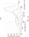
Fig. 1g is a graph of the emission spectra of SOD protein crystals according to one embodiment of the present disclosure. -
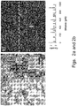
Fig. 2a is a brightfield image of ammonium sulfate crystals. -
Fig. 2b is a corresponding transmission generated SHG and line trace. -
Fig. 3a is a one photon laser-scanned excited confocal emission image of SOD protein aggregates and crystals at the wavelengths of 408-710 nm with corresponding emission profiles from the aggregates and crystal. -
Fig. 3b is a one photon laser-scanned excited confocal emission image of SOD protein crystals at the wavelengths of 408-710 nm with corresponding crystal emission profile. -
Fig. 3c is a one photon laser-scanned excited confocal emission image of SOD protein aggregate at the wavelengths of 408-710 nm with corresponding aggregate emission profile. -
Fig. 4a is a brightfield image of UCH-L1 protein crystals. -
Fig. 4b is a one-photon excited visible emission image of the sample ofFig 4a . -
Fig. 4c is a brightfield image of thaumatin crystals. -
Fig. 4d is a one-photon excited visible emission image of the sample ofFig 4c . -
Fig. 5a is a brightfield image of a lysozyme crystal. -
Fig. 5b is a two-photon excited visible emission (350 - 370 nm) image of a lysozyme protein crystal when excited at 515 nm according to one embodiment of the present disclosure. - The present disclosure provides a method of detection and imaging protein crystals. In particular, multiphoton excited luminescence is employed for selective detection and imaging of protein crystals. The method includes illuminating a sample with a laser; collecting an emission spectrum, evaluating the spectrum, and determining whether the sample contains protein crystals. Preferably, the sample is excited by simultaneous absorption of two or more photons. More preferably, the sample is excited by simultaneous absorption of two or three photons. Preferably, the known spectra of protein crystals are used to evaluate the observed spectrum, and therefore identify the presence of protein crystals.
- The present method can detect any suitable protein crystals, such as super-oxide dismutases (SODs), lysozyme, insulin, myoglobin, deubiquitinating enzymes, and combinations thereof. The method also can be used to image polymers, such as polyamides, amino-dendrimers, etc.
- The instrument of the present disclosure can be any suitable microscope that can produce two or more photons excitation. Preferably, a confocal microscope is used. Preferably, the laser is generated from a femtosecond laser source. More preferably, the laser source is a Ti:Sapphire laser source.
- The laser can be in the ultraviolet, visible, or infrared range. Preferably, the laser has a wavelength in the visible or infrared range. More preferably, the laser has a wavelength of about 515 nm, or about 800 nm.
- In some embodiments of the present disclosure, the emission spectrum is in the visible range. The spectrum is a luminescence emission spectrum. In some embodiments, the spectrum is a fluorescence emission spectrum. Preferably, a three-dimensional excitation and emission spectrum is collected.
- In some embodiments, the images of the sample are captured and observed to determine whether the sample contains protein crystals. The image is a luminescence image. In some embodiments, the image is a fluorescence image.
- Although the ubiquitous observation of visible light emission preferentially from protein crystals has notable practical benefits as a complement to existing protein crystal detection methods, the molecular nature of the emitting species is not immediately obvious when excited outside of the wavelength range for aromatic amino acid absorption. Resolving the molecular origin of the emission can provide a predictive framework for interpreting imaging contrast. The absorption and emission wavelengths are well outside of the range for accessing aromatic amino acid residues (e.g., tryptophan, tyrosine, etc.). As such, it is difficult to explain the observed ubiquitous visible emission in terms of conventional conjugated aromatic groups attached to the main protein chain.
- Without wishing to be bound by any theory, several emission mechanisms have been postulated. Visible emission could potentially arise from the incorporation and concentration of trace impurities into the crystalline lattice. Impurities have been shown to substantially impact nucleation, growth rates, and crystal quality. However, the observation of remarkably similar luminescence spectra from crystals for different proteins is not directly consistent with trace impurities, unless such impurities are likely to appear ubiquitously in protein crystal preparations. Furthermore, the emission spectra from protein crystals are nearly identical to those from the polyamide nylon loops, despite the fundamental differences in structure, processing, and materials properties. Similar emission spectra have also been reported for amino-dendrites, and triethylamine, suggesting a common origin for the relatively bright visible luminescence from diverse organic molecules and macromolecules. Given the radically different synthetic pathways used to generate each species, it is highly unlikely that similar impurities are present in each of these different samples.
- Several hypotheses have been forwarded for the origin of the intrinsic photoemission observed primarily in molecular constructs containing sterically hindered amino-groups. In previous studies with poly(amido amine) dendrimers, oxygen has been shown to be important for promoting visible-light excited luminescence. This finding is initially somewhat counter-intuitive, as oxygen is also a well-known fluorescence and phosphorescence quenching agent through energy transfer to the triplet state. The fact that bright, long-lived luminescence with similar emission spectra has been observed in polyamides, amino-dendrimers, and molecules as simple as triethylamine suggests that the effect is not limited to particular proteins or substituents (e.g., aromatic amino acid residues), and may well be responsible for luminescence in broad classes of amino-containing species, including the amide backbones of proteins. Consequently, it may well serve as a general and selective method for protein crystal detection.
- The absence of luminescence from the closely packed salt crystals supports the hypothesis of oxygen driven luminescent mechanism where tightly packed crystalline arrays such as salts will not luminescence due to the inability of oxygen to diffuse through the crystalline lattice.
- Other features of the present disclosure will become apparent from the following discussion of the preferred embodiments, which is illustrated in the accompanying drawings. The components in the figures are not necessarily to scale, emphasis instead being placed upon illustrating the principles of the invention. Moreover, in the figures, referenced numerals designate corresponding parts throughout the different views.
- UCHL1, SOD and thaumatin protein crystals were prepared via standard vapor diffusion protocols. Wild type UCHL1 was expressed as a glutathione S-transferase (GST) fusion protein and was purified on a glutathione-Sepharose column (GE Biosciences) using manufacturer's instructions. Crystals were grown using the hanging drop vapor diffusion method from a solution that contained 2.4 M ammonium sulfate and 0.1 M HEPES (pH 7.0). The S134N mutant of human SOD1 (hereafter referred to as SOD1S134N) was subcloned into the pGEX-6P1 vector (GE Biosciences) using standard cloning protocols. The resulting GST-tagged construct was expressed in Rosetta E. coli strain and was purified on a glutathione-Sepharose column (GE Biosciences) using manufacturer's instructions. The protein was further purified by size exclusion chromatography using a Superdex S75 column (GE Biosciences). Crystals of SOD1S134N were grown using the hanging drop vapor diffusion method from a solution that contained 2.0 M ammonium sulfate, 0.1 M tri-sodium citrate dihydrate (pH 5.6) and 0.2 M potassium sodium tartrate tetrahydrate. Thaumatin purchased from Sigma (Lot # T7638) was crystallized via vapor diffusion in a hanging drop. A 30 mg/mL solution of thaumatin dissolved in dionized water was mixed in 1:1 ratio with 0.5 M sodium potassium tartrate in dionized water and crystallized in a hanging drop.
- Two-photon excited luminescence images were acquired using a custom built confocal microscope performing beam scanning with a resonant vibrating mirror (7.8 kHz) directing the laser beam along the fast scan axis and a mirror galvanometer for sample-scanning along the slow axis. The incident beam was generated from a Spectra-Physics Mai Tai, approximately 100 femtosecond, 80 MHz, 100 mW average power at 800 nm focused onto the sample with a 10 x (0.3 N.A.) objective. Multialkali PMTs (Burle, XP29290PC) were used to collect the visible luminescence generated in the epi direction with a band-pass filter from 450-570 nm (Omega, 3rd 450-570) and a dichroic mirror to reject the incident beam. Emission spectra were also acquired in the epi-generated direction through a monochromator (Acton
Research Spectra Pro 150, 1200 groves/mm, blaze angle for 500 nm) and detected with a photomultiplier tube (PMT). - A laser scanning confocal microscope equipped with a 32-channel multi-anode photomultiplier tube array (Nikon A1) was used to acquire one-photon excited visible emission images and spectra. The 405 nm incident light was focused onto the sample with a 10 x objective (0.3 numerical aperture) and emission from 408 - 710 nm was collected on the multi-anode PMT array.
- An Olympus BX51 microscope was used for conventional one-photon excitation with a 10 x (0.3 numerical aperture) objective. Images were acquired with blue excitation from 460-490 nm and detection in the green from 520-800 nm with a CCD camera (Olympus DP71).
- Two-photon excited intrinsic fluorescence images were acquired using similar instrumentation as two-photon excited luminescence images, where a Nikon TE2000 microscope was used as a base. Beam scanning was completed with a resonant vibrating mirror (8.8 kHz) and a mirror galvanometer for fast and slow scan axis. The incident beam was generated by doubling the incident light to 515 nm, approximately 300 femtosecond, 5 mW, generated by a Polaronyz Uranus laser, approximately 200 femtosecond, 50 mW average power, 76 MHz, focused onto the sample with a 10x (0.3N.A.) objective. With UV excitation occurring at 257.5 nm at the sample, the signal, at around 350 nm, was collected in the trans-direction by a bialkali PMT (Hamamatsu, R1924), through a bandpass filter from 350 to 370 nm (Chroma, HQ360-20m), a 514 nm notch filter (Chroma), and a dichoric to reject the fundamental beam.
- Two-photon excited visible luminescence images for crystals of thaumatin and a super-oxide dismutase (SOD) mutant are shown in
Figs. 1b and 1f , along with the corresponding emission spectraFigs. 1c and1g . As a comparison, conventional brightfield images of thaumatin and SOD are shown inFigs. 1a and 1e . Two-photon excited luminescence images were acquired with a 80 MHz femtosecond Ti:Sapphire laser operating at 800 nm with the epi-generated luminescence collected through a filter (450 nm to 570 nm). Emission spectra were acquired in the epi-generated direction through a monochromator. Similar broad-band emission spectra were observed in both cases, extending from the two-photon energy of 400 nm out to 600 nm (Figs. 1c and1g ). Visible luminescence has also been observed in other protein crystals including lysozyme, insulin, and myoglobin. The emission spectra from the polyamide nylon loop (Fig. 1d ) coincide with the spectra from the protein crystals. In contrast, no detectable luminescence was observed for ammonium sulfate crystals (Fig. 2 ) under similar imaging conditions. - Contrast for protein crystal detection can also be observed using conventional one-photon excitation for UCH-L1 (
Figs. 3a and 3b ), a deubiquitinating enzyme of the ubiquitin C-terminal hydrolase (UCH) family, and thaumatin (Fig. 3c ) by wide-field fluorescence imaging. Images were acquired using blue excitation from 460-490 nm and detection in the green from 520-800 nm. - One-photon excited visible luminescence images of SOD crystals acquired using a laser scanning confocal microscope equipped with a 32-channel multi-anode photomultiplier tube array (Nikon A1) are shown in
Figs. 4a-4d . The integrated intensity image of the combined luminescence detected from 408 nm to 705 nm (405 nm excitation) is shown inFig. 4a . Although little detectable fluorescence was observed from the bulk solution, visible luminescence was readily observed from both crystals and aggregates, with the crystals producing a significantly greater red-shifted emission compared to the aggregates. Numerical separation based on a proprietary unmixing algorithm employed in the Nikon A1 software utilizing the least squares method with linear regression allowed selective identification of the protein crystal from the emission spectra. Unmixing of the luminescence spectra at each pixel generated two images corresponding to the crystals (Fig. 4b ) and aggregates. A line trace of the SOD crystals inFig. 4b shows the ability to detect protein crystals with high selectivity and without significant interference from aggregates. Comparison to deep UV excited images of SOD crystals (Fig. 4c ) shows similar contrast but with a much higher background, presumably arising from fluorescence generated by solvated and/or aggregated protein. - Conventional brightfield image of a lysozyme crystal is shown in
Fig. 5a . A two-photon excited image of the same sample is shown inFig. 5b . The protein crystal was excited with a 515 nm femtosecond laser and the fluorescence emission collected in the transmission direction at 350 - 370 nm corresponding to the emission of tryptophan and tyrosine. Two-photon excitation of the aromatic amino acid residues is advantageous in that conventional optics can be utilized and also that there is no out of plane damage to the proteins. - Many modifications and other embodiments of the present disclosure will come to mind to one skilled in the art to which the present disclosure pertains having the benefit of the teachings presented in the foregoing description. It will be apparent to those skilled in the art that variations and modifications of the present disclosure may be made without departing from the scope or spirit of the present disclosure. Therefore, it is to be understood that the invention is not to be limited to the specific embodiments disclosed and that modifications and other embodiments are intended to be included within the scope of the appended claims. Although specific terms are employed herein, they are used in a generic and descriptive sense only and not for purposes of limitation.
-
- 1. Asanov, A. N., H. M. McDonald, P. B. Oldham, M. J. Jedrzejas, and W. W. Wilson. Intrinsic fluorescence as a potential rapid scoring tool for protein crystals; 2000 May 14-19; Sandestin, Florida. p 603-609.
- 2. Vernede, X., B. Lavault, J. Ohana, D. Nurizzo, J. Joly, L. Jacquamet, F. Felisaz, F. Cipriani, and D. Bourgeois. 2006. UV laser-excited fluorescence as a tool for the visualization of protein crystals mounted in loops. Acta Crystallographica Section D-Biological Crystallography 62:253-261.
- 3. Judge, R. A., K. Swift, and C. Gonzalez. 2005. An ultraviolet fluorescence-based method for identifying and distinguishing protein crystals. Acta Crystallographica Section D-Biological Crystallography 61:60-66.
- 4. Pohl, E., U. Ristau, T. Gehrmann, D. Jahn, B. Robrahn, D. Malthan, H. Dobler, and C. Hermes. 2004. Automation of the EMBL Hamburg protein crystallography beamline BW7B. Journal of Synchrotron Radiation 11:372-377.
- 5. Chan, K. L. A., L. Govada, R. M. Bill, N. E. Chayen, and S. G. Kazarian. 2009. Attenuated Total Reflection-FT-IR Spectroscopic Imaging of Protein Crystallization. Analytical Chemistry 81:3769-3775.
- 6. Wampler, R. D., D. J. Kissick, C. J. Dehen, E. J. Gualtieri, J. L. Grey, H.-F. Wang, D. H. Thompson, J.-X. Cheng, and G. Simpson, J. 2008. Nonlinear optical imaging of protein crsytallization. J. Amer. Chem. Soc. 130:14076-14077.
- 7. Groves, M. R., I. B. Muller, X. Kreplin, and J. Muller-Dieckmann. 2007. A method for the general identification of protein crystals in crystallization experiments using a noncovalent fluorescent dye. Acta Crystallographica Section D-Biological Crystallography 63:526-535.
- 8. del Mercato, L. L., P. P. Pompa, G. Maruccio, A. Della Torre, S. Sabella, A. M. Tamburro, R. Cingolani, and R. Rinaldi. 2007. Charge transport and intrinsic fluorescence in amyloid-like fibrils. Proc. Natl. Acad. Sci. U. S. A. 104:18019-18024.
- 9. Guptasarma, P. 2008. Solution-state characteristics of the ultraviolet A-induced visible fluorescence from proteins. Archives of Biochemistry and Biophysics 478:127-129.
- 10. Shukla, A., S. Mukherjee, S. Sharma, V. Agrawal, K. V. Radha Kishan, and P. Guptasarma. 2004. A novel UV laser-induced visible blue radiation from protein crystals and aggregates: scattering artifacts or fluorescence transitions of peptide electrons delocalized through hydrogen bonding? Archives of Biochemistry and Biophysics 428:144-153.
- 11. Chu, C. C. and T. Imae. 2009. Fluorescence Investigations of Oxygen-Doped Simple Amine Compared with Fluorescent PAMAM Dendrimer. Macromolecular Rapid Communications 30:89-93.
- 12. Lee, W. I., Y. J. Bae, and A. J. Bard. 2004. Strong blue photoluminescence and ECL from OH-terminated PAMAM dendrimers in the absence of gold nanoparticles. Journal of the American Chemical Society 126:8358-8359.
- 13. Wang, D. J. and T. Imae. 2004. Fluorescence emission from dendrimers and its pH dependence. Journal of the American Chemical Society 126:13204-13205.
- 14. Wu, D. C., Y. Liu, C. B. He, and S. H. Goh. 2005. Blue photoluminescence from hyperbranched poly(amino ester)s. Macromolecules 38:9906-9909.
- 15. Onoshima, D. and T. Imae. 2006. Dendritic nano- and microhydrogels fabricated by triethoxysilyl focal dendrons. Soft Matter 2:141-148.
- 16. Antharjanam, P. K. S., M. Jaseer, K. N. Ragi, and E. Prasad. 2009. Intrinsic luminescence properties of ionic liquid crystals based on PAMAM and PPI dendrimers. Journal of Photochemistry and Photobiology a-Chemistry 203:50-55.
- 17. Jeong, S., G. Kwak, A. Takagi, M. Fujiki, D. H. Lee, L. S. Park, and K. B. Yoon. 2008. Luminous, fully aliphatic polyamides: Multicolor photoluminescence, their pH and solvent dependency. European Polymer Journal 44:1149-1156.
- 18. Lin, Y., J. W. Gao, H. W. Liu, and Y. S. Li. 2009. Synthesis and Characterization of Hyperbranched Poly(ether amide)s with Thermoresponsive Property and Unexpected Strong Blue Photoluminescence. Macromolecules 42:3237-3246.
- 19. Tamano, K. and T. Imae. 2008. Investigation of Luminescent Poly(propylene imine) Dendrimer. Journal of Nanoscience and Nanotechnology 8:4329-4334.
- 20. Mohamed, N. A. and M. M. Fahmy. 2009. Synthesis and Characterization of Novel Wholly Para-Oriented Aromatic Polyamide-Hydrazides Containing Sulfone-Ether Linkages. Journal of Applied Polymer Science 113:767-776.
- 21. Larson, J. M. 2006. The Nikon C1si combines high spectral resolution, high sensitivity, and high acquisition speed. Cytometry Part A 69A:825-834.
- 22. Rosenberger, F., P. G. Vekilov, M. Muschol, and B. R. Thomas. Nucleation and crystallization of globular proteins - What we know and what is missing; 1995 Nov 12-17; Hiroshima, Japan. Elsevier Science Bv. p 1-27.
- 23. Durbin, S. D. and G. Feher. 1996. Protein crystallization. Annual Review of Physical Chemistry 47:171-204.
- 24. Skouri, M., B. Lorber, R. Giege, J. P. Munch, and J. S. Candau. 1995. Effect of Macromolecular Impurities on Lysozyme Solubility and Crystallizability - Dynamic Light-Scattering, Phase-diagram, And Crystal-growth Studies. Journal of Crystal Growth 152:209-220.
- 25. Judge, R. A., R. S. Jacobs, T. Frazier, E. H. Snell, and M. L. Pusey. 1999. The effect of temperature and solution pH on the nucleation of tetragonal lysozyme crystals. Biophysical Journal 77:1585-1593.
- 26. Sangwal, K. 1996. Effects of impurities on crystal growth processes. Progress in Crystal Growth and Characterization of Materials 32:3-43.
- 27. Vekilov, P. G. and F. Rosenberger. 1998. Protein crystal growth under forced solution flow: experimental setup and general response of lysozyme. Journal of Crystal Growth 186:251-261.
- 28. Hirschler, J. and J. C. FontecillaCamps. 1997. Protein crystal growth rates are face-specifically modified by structurally related contaminants. Journal of Crystal Growth 171:559-565.
- 29. Judge, R. A., E. L. Forsythe, and M. L. Pusey. 1998. The effect of protein impurities on lysozyme crystal growth. Biotechnology and Bioengineering 59:776-785.
- 30. Thomas, B. R., D. Carter, and F. Rosenberger. 1998. Effect of microheterogeneity on horse spleen apoferritin crystallization. Journal of Crystal Growth 187:499-510.
- 31. Matsui, T., G. Sazaki, H. Hondoh, Y. Matsuura, T. Nakada, and K. Nakajima. 2006. Impurity effects of lysozyme molecules specifically labeled with a fluorescent reagent on the crystallization of tetragonal and monoclinic lysozyme crystals. Journal of Crystal Growth 293:415-422.
- 32. Burke, M. W., R. Leardi, R. A. Judge, and M. L. Pusey. 2001. Quantifying main trends in lysozyme nucleation: The effect of precipitant concentration, supersaturation, and impurities. Crystal Growth & Design 1:333-337.
- 33. Asherie, N., C. Ginsberg, A. Greenbaum, S. Blass, and S. Knafo. Effects of Protein Purity and Precipitant Stereochemistry on the Crystallization of Thaumatin; 2008 May 06-08; Cancun City, MEXICO. Amer Chemical Soc. p 4200-4207.
- 34. Dobrianov, I., C. Caylor, S. G. Lemay, K. D. Finkelstein, and R. E. Thorne. X-ray diffraction studies of protein crystal disorder; 1998 May 03-08; Granada, Spain. Elsevier Science Bv. p 511-523.
- 35. Van Driessche, A. E. S., G. Sazaki, G. L. Dai, F. Otalora, J. A. Gavira, T. Matsui, I. Yoshizaki, K. Tsukamoto, and K. Nakajima. 2009. Direct Observation of Adsorption Sites of Protein Impurities and Their Effects on Step Advancement of Protein Crystals. Crystal Growth & Design 9:3062-3071.
- 36. Carter, D. C., K. Lim, J. X. Ho, B. S. Wright, P. D. Twigg, T. Y. Miller, J. Chapman, K. Keeling, J. Ruble, P. G. Vekilov, B. R. Thomas, F. Rosenberger, and A. A. Chernov. Lower dimer impurity incorporation may result in higher perfection of HEWL crystals grown in microgravity - A case study; 1998 May 03-08; Granada, Spain. Elsevier Science Bv. p 623-637.
- 37. Robert, M. C., B. Capelle, B. Lorber, and R. Giege. Influence of impurities on protein crystal perfection; 2000 May 14-19; Sandestin, Florida. Elsevier Science Bv. p 489-497.
- 38. Yoshizaki, I., S. Fukuyama, H. Koizumi, M. Tachibana, K. Kojima, Y. Matsuura, M. Tanakae, N. Igarashi, A. Kadowaki, L. Rong, S. Adachi, S. Yoda, and H. Komatsu. 2006. Impurity-induced defect and its effect on protein crystal perfection. Journal of Crystal Growth 290:185-191.
- 39. Caylor, C. L., I. Dobrianov, S. G. Lemay, C. Kimmer, S. Kriminski, K. D. Finkelstein, W. Zipfel, W. W. Webb, B. R. Thomas, A. A. Chernov, and R. E. Thorne. 1999. Macromolecular impurities and disorder in protein crystals. Proteins-Structure Function and Genetics 36:270-281.
Claims (15)
- A method for detecting protein crystals, the method comprising:illuminating a sample with a laser to produce multiphoton excitation;obtaining an image of the sample; anddetermining whether the sample comprises protein crystals.
- The method of claim 1, wherein the multiphoton excitation is a two-photon excitation.
- The method of claim 1, wherein the laser has a wavelength in the infrared range.
- The method of claim 1, wherein the laser has a wavelength in the visible range.
- The method of claim 1, wherein the laser has a wavelength in the ultraviolet range.
- The method of claim 1, wherein the image is a luminescence image.
- The method of claim 1, wherein the image is a fluorescence image.
- The method of claim 1, wherein the sample is illuminated with incident light in the visible range, and wherein the image is a fluorescence image.
- The method of any preceding claim, wherein obtaining the image comprises exciting native amino acids.
- The method of any preceding claim, wherein the laser is generated from a femtosecond laser source.
- The method of any preceding claim, wherein the laser source is a Ti:sapphire laser source.
- The method of claim 1, wherein the multiphoton excitation is a three-photon excitation.
- The method of any preceding claim, wherein the sample comprises unlabeled proteins.
- A method for imaging a polymer, the method comprising:illuminating a sample with a laser to produce multiphoton excitation; andobtaining an image of the sample.
- The method of claim 14, wherein the polymer comprises at least one of a polyamide and an amino-dendrimer.
Applications Claiming Priority (2)
| Application Number | Priority Date | Filing Date | Title |
|---|---|---|---|
| US24633409P | 2009-09-28 | 2009-09-28 | |
| EP10819608.0A EP2483665B1 (en) | 2009-09-28 | 2010-09-27 | Multiphoton luminescence imaging of protein crystals |
Related Parent Applications (2)
| Application Number | Title | Priority Date | Filing Date |
|---|---|---|---|
| EP10819608.0A Division-Into EP2483665B1 (en) | 2009-09-28 | 2010-09-27 | Multiphoton luminescence imaging of protein crystals |
| EP10819608.0A Division EP2483665B1 (en) | 2009-09-28 | 2010-09-27 | Multiphoton luminescence imaging of protein crystals |
Publications (2)
| Publication Number | Publication Date |
|---|---|
| EP3163289A2 true EP3163289A2 (en) | 2017-05-03 |
| EP3163289A3 EP3163289A3 (en) | 2017-07-19 |
Family
ID=43796257
Family Applications (2)
| Application Number | Title | Priority Date | Filing Date |
|---|---|---|---|
| EP10819608.0A Not-in-force EP2483665B1 (en) | 2009-09-28 | 2010-09-27 | Multiphoton luminescence imaging of protein crystals |
| EP16193716.4A Withdrawn EP3163289A3 (en) | 2009-09-28 | 2010-09-27 | Multiphoton luminescence imaging of protein crystals |
Family Applications Before (1)
| Application Number | Title | Priority Date | Filing Date |
|---|---|---|---|
| EP10819608.0A Not-in-force EP2483665B1 (en) | 2009-09-28 | 2010-09-27 | Multiphoton luminescence imaging of protein crystals |
Country Status (3)
| Country | Link |
|---|---|
| US (1) | US8946655B2 (en) |
| EP (2) | EP2483665B1 (en) |
| WO (1) | WO2011038349A1 (en) |
Families Citing this family (1)
| Publication number | Priority date | Publication date | Assignee | Title |
|---|---|---|---|---|
| US10113965B2 (en) * | 2015-03-06 | 2018-10-30 | Purdue Research Foundation | Methods for second harmonic generation imaging of protein crystals |
Family Cites Families (10)
| Publication number | Priority date | Publication date | Assignee | Title |
|---|---|---|---|---|
| US4162851A (en) * | 1978-03-29 | 1979-07-31 | Akiyoshi Wada | Simultaneous photometering method and assembly for multi-dimensional measurements concerning biologically related materials |
| JPH0735687A (en) * | 1993-07-22 | 1995-02-07 | Fujitsu Ltd | Search method for biopolymer crystals |
| JP4113063B2 (en) * | 2003-08-18 | 2008-07-02 | 株式会社リガク | Method for detecting specific polymer crystals |
| JP4458513B2 (en) * | 2003-08-18 | 2010-04-28 | 株式会社リガク | Equipment for evaluating specific polymer crystals |
| US7256894B2 (en) * | 2003-10-20 | 2007-08-14 | The Regents Of The University Of California | Method and apparatus for performing second harmonic optical coherence tomography |
| US20050237522A1 (en) * | 2004-04-27 | 2005-10-27 | Swift Kerry M | Methods for visualizing crystals and distinguishing crystals from other matter within a biological sample |
| US7545495B2 (en) * | 2004-04-27 | 2009-06-09 | Abbott Laboratories | Methods for visualizing crystals and distinguishing crystals from other matter within a biological sample |
| JP4869734B2 (en) | 2005-04-25 | 2012-02-08 | オリンパス株式会社 | Multi-photon excitation scanning laser microscope |
| US7663750B2 (en) * | 2005-06-30 | 2010-02-16 | Applied Biosystems, Llc | Two-dimensional spectral imaging system |
| JP2007254415A (en) * | 2006-03-24 | 2007-10-04 | Gunma Univ | Macromolecular crystal, production method thereof, and production apparatus used therefor |
-
2010
- 2010-09-27 EP EP10819608.0A patent/EP2483665B1/en not_active Not-in-force
- 2010-09-27 US US13/498,523 patent/US8946655B2/en active Active
- 2010-09-27 EP EP16193716.4A patent/EP3163289A3/en not_active Withdrawn
- 2010-09-27 WO PCT/US2010/050409 patent/WO2011038349A1/en not_active Ceased
Non-Patent Citations (39)
| Title |
|---|
| ANTHARJANAM; P. K. S.; M. JASEER; K. N. RAGI; E. PRASAD.: "Intrinsic luminescence properties of ionic liquid crystals based on PAMAM and PPI dendrimers", JOURNAL OF PHOTOCHEMISTRY AND PHOTOBIOLOGY A-CHEMISTRY, vol. 203, 2009, pages 50 - 55, XP025994614, DOI: doi:10.1016/j.jphotochem.2008.12.023 |
| ASANOV, A. N.; H. M. MCDONALD; P. B. OLDHAM; M. J. JEDRZEJAS; W. W. WILSON., INTRINSIC FLUORESCENCE AS A POTENTIAL RAPID SCORING TOOL FOR PROTEIN CRYSTALS, vol. 14-19, May 2000 (2000-05-01), pages 603 - 609 |
| ASHERIE, N.; C. GINSBERG; A. GREENBAUM; S. BLASS; S. KNAFO.: "Effects of Protein Purity and Precipitant Stereochemistry on the Crystallization of Thaumatin", 6 May 2008, MEXICO. AMER CHEMICAL SOC., pages: 4200 - 4207 |
| BURKE, M. W.; R. LEARDI; R. A. JUDGE; M. L. PUSEY.: "Quantifying main trends in lysozyme nucleation: The effect of precipitant concentration, supersaturation, and impurities", CRYSTAL GROWTH & DESIGN, vol. 1, 2001, pages 333 - 337 |
| CARTER, D. C.; K. LIM; J. X. HO; B. S. WRIGHT; P. D. TWIGG; T. Y. MILLER; J. CHAPMAN; K. KEELING; J. RUBLE; P. G. VEKILOV: "A case study", 3 May 1998, ELSEVIER SCIENCE, article "Lower dimer impurity incorporation may result in higher perfection of HEWL crystals grown in microgravity" |
| CAYLOR; C. L.; I. DOBRIANOV; S. G. LEMAY; C. KIMMER; S. KRIMINSKI; K. D. FINKELSTEIN; W. ZIPFEL; W. W. WEBB; B. R. THOMAS: "Macromolecular impurities and disorder in protein crystals", PROTEINS-STRUCTURE FUNCTION AND GENETICS, vol. 36, 1999, pages 270 - 281 |
| CHAN, K. L. A.; L. GOVADA; R. M. BILL; N. E. CHAYEN; S. G. KAZARIAN.: "Attenuated Total Reflection-FT-IR Spectroscopic Imaging of Protein Crystallization.", ANALYTICAL CHEMISTRY, vol. 81, 2009, pages 3769 - 3775 |
| CHU, C. C.; T. IMAE.: "Fluorescence Investigations of Oxygen-Doped Simple Amine Compared with Fluorescent PAMAM Dendrimer.", MACROMOLECULAR RAPID COMMUNICATIONS, vol. 30, 2009, pages 89 - 93 |
| DEL MERCATO; L. L.; P. P. POMPA; G. MARUCCIO; A. DELIA TORRE; S. SABELLA; A. M. TAMBURRO; R. CINGOLANI; R. RINALDI.: "Charge transport and intrinsic fluorescence in amyloid-like fibrils.", PROC. NATL. ACAD. SCI. U. S. A., vol. 104, 2007, pages 18019 - 18024, XP055012138, DOI: doi:10.1073/pnas.0702843104 |
| DOBRIANOV, I.; C. CAYLOR; S. G. LEMAY; K. D. FINKELSTEIN; R. E. THORNE: "X-ray diffraction studies of protein crystal disorder", 3 March 1998, GRANADA, SPAIN. ELSEVIER SCIENCE, pages: 511 - 523 |
| DURBIN; S. D.; G. FEHER., PROTEIN CRYSTALLIZATION. ANNUAL REVIEW OF PHYSICAL CHEMISTRY, vol. 47, 1996, pages 171 - 204 |
| GROVES, M. R.; I. B. MULLER; X. KREPLIN; J. MULLER-DIECKMANN.: "A method for the general identification of protein crystals in crystallization experiments using a noncovalent fluorescent dye.", ACTA CRYSTALLOGRAPHICA SECTION D-BIOLOGICAL CRYSTALLOGRAPHY, vol. 63, 2007, pages 526 - 535 |
| GUPTASARMA, P.: "Solution-state characteristics of the ultraviolet A-induced visible fluorescence from proteins.", ARCHIVES OF BIOCHEMISTRY AND BIOPHYSICS, vol. 478, 2008, pages 127 - 129, XP025434420, DOI: doi:10.1016/j.abb.2008.08.002 |
| HIRSCHLER, J.; J. C. FONTECILLACAMPS.: "Protein crystal growth rates are face-specifically modified by structurally related contaminants", JOURNAL OF CRYSTAL GROWTH, vol. 171, 1997, pages 559 - 565, XP004117287, DOI: doi:10.1016/S0022-0248(96)00688-4 |
| JEONG, S.; G. KWAK; A. TAKAGI; M. FUJIKI; D. H. LEE; L. S. PARK; K. B. YOON.: "Luminous, fully aliphatic polyamides: Multicolor photoluminescence, their pH and solvent dependency", EUROPEAN POLYMER JOURNAL, vol. 44, 2008, pages 1149 - 1156, XP022561565 |
| JUDGE, R. A.; E. L. FORSYTHE; M. L. PUSEY.: "The effect of protein impurities on lysozyme crystal growth", BIOTECHNOLOGY AND BIOENGINEERING, vol. 59, 1998, pages 776 - 785 |
| JUDGE, R. A.; K. SWIFT; C. GONZALEZ.: "An ultraviolet fluorescence-based method for identifying and distinguishing protein crystals", ACTA CRYSTALLOGRAPHICA SECTION D-BIOLOGICAL CRYSTALLOGRAPHY, vol. 61, 2005, pages 60 - 66, XP002348216, DOI: doi:10.1107/S0907444904026538 |
| JUDGE, R. A.; R. S. JACOBS; T. FRAZIER; E. H. SNELL; M. L. PUSEY.: "The effect of temperature and solution pH on the nucleation of tetragonal lysozyme crystals", BIOPHYSICAL JOURNAL, vol. 77, 1999, pages 1585 - 1593 |
| LARSON, J. M.: "The Nikon C1si combines high spectral resolution, high sensitivity, and high acquisition speed", CYTOMETRY, vol. 69A, 2006, pages 825 - 834 |
| LEE, W. I.; Y. J. BAE; A. J. BARD.: "Strong blue photoluminescence and ECL from OH-terminated PAMAM dendrimers in the absence of gold nanoparticles", JOURNAL OF THE AMERICAN CHEMICAL SOCIETY, vol. 126, 2004, pages 8358 - 8359 |
| LIN, Y.; J. W. GAO; H. W. LIU; Y. S. LI.: "Synthesis and Characterization of Hyperbranched Poly(ether amide)s with Thermoresponsive Property and Unexpected Strong Blue Photoluminescence", MACROMOLECULES, vol. 42, 2009, pages 3237 - 3246 |
| MATSUI, T.; G. SAZAKI; H. HONDOH; Y. MATSUURA; T. NAKADA; K. NAKAJIMA: "Impurity effects of lysozyme molecules specifically labeled with a fluorescent reagent on the crystallization of tetragonal and monoclinic lysozyme crystals", JOURNAL OF CRYSTAL GROWTH, vol. 293, 2006, pages 415 - 422, XP028016348, DOI: doi:10.1016/j.jcrysgro.2006.05.061 |
| MOHAMED, N. A.; M. M. FAHMY.: "Synthesis and Characterization of Novel Wholly Para-Oriented Aromatic Polyamide-Hydrazides Containing Sulfone-Ether Linkages", JOURNAL OF APPLIED POLYMER SCIENCE, vol. 113, 2009, pages 767 - 776 |
| ONOSHIMA, D.; T. IMAE.: "Dendritic nano- and microhydrogels fabricated by triethoxysilyl focal dendrons", SOFT MATTER, vol. 2, 2006, pages 141 - 148 |
| POHL, E.; U. RISTAU; T. GEHRMANN; D. JAHN; B. ROBRAHN; D. MALTHAN; H. DOBLER; C. HERMES: "utomation of the EMBL Hamburg protein crystallography beamline BW7B", JOURNAL OF SYNCHROTRON RADIATION, vol. 11, 2004, pages 372 - 377 |
| ROBERT, M. C.; B. CAPELLE; B. LORBER; R. GIEGE.: "Influence of impurities on protein crystal perfection", 14 May 2000, ELSEVIER SCIENCE, pages: 489 - 497 |
| ROSENBERGER, F.; P. G. VEKILOV; M. MUSCHOL; B. R. THOMAS.: "Nucleation and crystallization of globular proteins - What we know and what is missing", 12 November 1995, ELSEVIER SCIENCE, pages: 1 - 27 |
| SANGWAL, K.: "Effects of impurities on crystal growth processes", PROGRESS IN CRYSTAL GROWTH AND CHARACTERIZATION OF MATERIALS, vol. 32, 1996, pages 3 - 43, XP005538437, DOI: doi:10.1016/0960-8974(96)00008-3 |
| SHUKLA, A.; S. MUKHERJEE; S. SHARMA; V. AGRAWAL; K. V. RADHA KISHAN; P. GUPTASARMA.: "A novel UV laser-induced visible blue radiation from protein crystals and aggregates: scattering artifacts or fluorescence transitions of peptide electrons delocalized through hydrogen bonding", ARCHIVES OF BIOCHEMISTRY AND BIOPHYSICS, vol. 428, 2004, pages 144 - 153, XP004520178, DOI: doi:10.1016/j.abb.2004.05.007 |
| SKOURI, M.; B. LORBER; R. GIEGE; J. P. MUNCH; J. S. CANDAU.: "Effect of Macromolecular Impurities on Lysozyme Solubility and Crystallizability - Dynamic Light-Scattering, Phase-diagram, And Crystal-growth Studies", JOURNAL OF CRYSTAL GROWTH, vol. 152, 1995, pages 209 - 220 |
| TAMANO, K.; T. IMAE.: "Investigation of Luminescent Poly(propylene imine) Dendrimer", JOURNAL OF NANOSCIENCE AND NANOTECHNOLOGY, vol. 8, 2008, pages 4329 - 4334 |
| THOMAS, B. R.; D. CARTER; F. ROSENBERGER.: "Effect of microheterogeneity on horse spleen apoferritin crystallization", JOURNAL OF CRYSTAL GROWTH, vol. 187, 1998, pages 499 - 510, XP004148301, DOI: doi:10.1016/S0022-0248(98)00033-5 |
| VAN DRIESSCHE; A. E. S.; G. SAZAKI; G. L. DAI; F. OTALORA; J. A. GAVIRA; T. MATSUI; I. YOSHIZAKI; K. TSUKAMOTO; K. NAKAJIMA.: "Direct Observation of Adsorption Sites of Protein Impurities and Their Effects on Step Advancement of Protein Crystals", CRYSTAL GROWTH & DESIGN, vol. 9, 2009, pages 3062 - 3071 |
| VEKILOV; P. G.; F. ROSENBERGER: "Protein crystal growth under forced solution flow: experimental setup and general response of lysozyme", JOURNAL OF CRYSTAL GROWTH, vol. 186, 1998, pages 251 - 261, XP004117980, DOI: doi:10.1016/S0022-0248(97)00521-6 |
| VERNEDE, X.; B. LAVAULT; J. OHANA; D. NURIZZO; J. JOLY; L. JACQUAMET; F. FELISAZ; F. CIPRIANI; D. BOURGEOIS.: "UV laser-excited fluorescence as a tool for the visualization of protein crystals mounted in loops", ACTA CRYSTALLOGRAPHICA SECTION D-BIOLOGICAL CRYSTALLOGRAPHY, vol. 62, 2006, pages 253 - 261 |
| WAMPLER, R. D.; D. J. KISSICK; C. J. DEHEN; E. J. GUALTIERI; J. L. GREY; H.-F. WANG; D. H. THOMPSON; J.-X. CHENG; G. SIMPSON, J.: "Nonlinear optical imaging of protein crsytallization.", J. AMER. CHEM. SOC., vol. 130, 2008, pages 14076 - 14077 |
| WANG, D. J.; T. IMAE.: "Fluorescence emission from dendrimers and its pH dependence", JOURNAL OF THE AMERICAN CHEMICAL SOCIETY, vol. 126, 2004, pages 13204 - 13205 |
| WU, D. C.; Y. LIU; C. B. HE; S. H. GOH.: "Blue photoluminescence from hyperbranched poly(amino ester)s.", MACROMOLECULES, vol. 38, 2005, pages 9906 - 9909 |
| YOSHIZAKI, I.; S. FUKUYAMA; H. KOIZUMI; M. TACHIBANA; K. KOJIMA; Y. MATSUURA; M. TANAKAE; N. IGARASHI; A. KADOWAKI; L. RONG: "Impurity-induced defect and its effect on protein crystal perfection", JOURNAL OF CRYSTAL GROWTH, vol. 290, 2006, pages 185 - 191, XP028016723, DOI: doi:10.1016/j.jcrysgro.2005.12.094 |
Also Published As
| Publication number | Publication date |
|---|---|
| EP2483665A4 (en) | 2013-08-21 |
| WO2011038349A1 (en) | 2011-03-31 |
| US8946655B2 (en) | 2015-02-03 |
| EP2483665B1 (en) | 2017-04-05 |
| EP2483665A1 (en) | 2012-08-08 |
| US20120241647A1 (en) | 2012-09-27 |
| EP3163289A3 (en) | 2017-07-19 |
Similar Documents
| Publication | Publication Date | Title |
|---|---|---|
| JP5776992B2 (en) | STED microscopy, STED fluorescence correlation spectroscopy, and STED fluorescence microscope for pulse excitation, continuous deexcitation, and gate recording of spontaneous emission fluorescence | |
| US7113275B2 (en) | Method for detection of pathogenic microorganisms | |
| CN1842696A (en) | Wide field method for detecting pathogenic microorganisms | |
| Calero et al. | Identifying, studying and making good use of macromolecular crystals | |
| US11982676B2 (en) | Near-infrared fluorescent nanoprobe | |
| Madden et al. | Two-photon excited UV fluorescence for protein crystal detection | |
| JP4488259B2 (en) | Quantum dot observation system | |
| EP2483665B1 (en) | Multiphoton luminescence imaging of protein crystals | |
| US20080106733A1 (en) | Methods for visualizing crystals and distinguishing crystals from other matter within a biological sample | |
| WO2006014576A2 (en) | Fluorescent lifetime biological detection and imaging using water-stable semiconductor nanocrystals | |
| JP4431549B2 (en) | Fluorescence analyzer | |
| JP2005098808A (en) | Biological information detection device | |
| WO2025046069A1 (en) | Method for analysis of plastic particles | |
| Orthaus et al. | Crossing the Limit Towards Deep UV: Time‐resolved microscopy of native fluorophores | |
| JP2012145344A (en) | Fluorescence detection device | |
| US7545495B2 (en) | Methods for visualizing crystals and distinguishing crystals from other matter within a biological sample | |
| US9970873B1 (en) | System and method for luminescent tag based wafer inspection | |
| WO2022065744A1 (en) | Macrocyclic host molecule-based core-shell nanoparticles and method for synthesizing same | |
| RU125458U1 (en) | DEVICE FOR DETERMINING THE LOCALIZATION OF ATYPIC CELLS BY LUMINESCENCE OF NANOCRYSTALLINE TAGS IN BIOLOGICAL TISSUES | |
| RU2498298C1 (en) | Apparatus for imaging biological objects with nano-labels | |
| Ritz et al. | Characterization of unexpected blue-shifted single molecule fluorescence of JF646 and JF669 for SMLM | |
| Fröch et al. | DETERMINISTIC SINGLE-PHOTON EMITTER ARRAYS FROM GIANT SILICA-SHELLED QDS | |
| Varghese et al. | L-Tyrosine functionalized ZnO for the fluorescence detection of phenol | |
| JPH05268993A (en) | Discrimination of lipid-containing algae | |
| TW202506958A (en) | Fluorescent probe and observation method using fluorescent probe |
Legal Events
| Date | Code | Title | Description |
|---|---|---|---|
| PUAI | Public reference made under article 153(3) epc to a published international application that has entered the european phase |
Free format text: ORIGINAL CODE: 0009012 |
|
| AC | Divisional application: reference to earlier application |
Ref document number: 2483665 Country of ref document: EP Kind code of ref document: P |
|
| AK | Designated contracting states |
Kind code of ref document: A2 Designated state(s): AL AT BE BG CH CY CZ DE DK EE ES FI FR GB GR HR HU IE IS IT LI LT LU LV MC MK MT NL NO PL PT RO SE SI SK SM TR |
|
| RIN1 | Information on inventor provided before grant (corrected) |
Inventor name: GUALTIERI, ELLEN J. Inventor name: SIMPSON, GARTH J. Inventor name: MADDEN, JEREMY Inventor name: KISSICK, DAVID J. |
|
| PUAL | Search report despatched |
Free format text: ORIGINAL CODE: 0009013 |
|
| RIN1 | Information on inventor provided before grant (corrected) |
Inventor name: GUALTIERI, ELLEN J. Inventor name: SIMPSON, GARTH J. Inventor name: MADDEN, JEREMY Inventor name: KISSICK, DAVID J. |
|
| AK | Designated contracting states |
Kind code of ref document: A3 Designated state(s): AL AT BE BG CH CY CZ DE DK EE ES FI FR GB GR HR HU IE IS IT LI LT LU LV MC MK MT NL NO PL PT RO SE SI SK SM TR |
|
| RIC1 | Information provided on ipc code assigned before grant |
Ipc: G01N 21/64 20060101ALI20170609BHEP Ipc: G01N 21/31 20060101AFI20170609BHEP Ipc: G01N 23/20 20060101ALI20170609BHEP |
|
| 17P | Request for examination filed |
Effective date: 20180115 |
|
| RBV | Designated contracting states (corrected) |
Designated state(s): AL AT BE BG CH CY CZ DE DK EE ES FI FR GB GR HR HU IE IS IT LI LT LU LV MC MK MT NL NO PL PT RO SE SI SK SM TR |
|
| 17Q | First examination report despatched |
Effective date: 20180814 |
|
| STAA | Information on the status of an ep patent application or granted ep patent |
Free format text: STATUS: THE APPLICATION IS DEEMED TO BE WITHDRAWN |
|
| 18D | Application deemed to be withdrawn |
Effective date: 20190103 |