EP3158077B1 - Cell culture methods and media comprising n-acetylcysteine - Google Patents
Cell culture methods and media comprising n-acetylcysteine Download PDFInfo
- Publication number
- EP3158077B1 EP3158077B1 EP15809573.7A EP15809573A EP3158077B1 EP 3158077 B1 EP3158077 B1 EP 3158077B1 EP 15809573 A EP15809573 A EP 15809573A EP 3158077 B1 EP3158077 B1 EP 3158077B1
- Authority
- EP
- European Patent Office
- Prior art keywords
- cell
- cell culture
- acetylcysteine
- cells
- culture medium
- Prior art date
- Legal status (The legal status is an assumption and is not a legal conclusion. Google has not performed a legal analysis and makes no representation as to the accuracy of the status listed.)
- Active
Links
- PWKSKIMOESPYIA-BYPYZUCNSA-N L-N-acetyl-Cysteine Chemical compound CC(=O)N[C@@H](CS)C(O)=O PWKSKIMOESPYIA-BYPYZUCNSA-N 0.000 title claims description 218
- 229960004308 acetylcysteine Drugs 0.000 title claims description 215
- 238000004113 cell culture Methods 0.000 title claims description 95
- HVYWMOMLDIMFJA-DPAQBDIFSA-N cholesterol Chemical compound C1C=C2C[C@@H](O)CC[C@]2(C)[C@@H]2[C@@H]1[C@@H]1CC[C@H]([C@H](C)CCCC(C)C)[C@@]1(C)CC2 HVYWMOMLDIMFJA-DPAQBDIFSA-N 0.000 claims description 190
- 239000006143 cell culture medium Substances 0.000 claims description 148
- 239000002609 medium Substances 0.000 claims description 133
- 235000012000 cholesterol Nutrition 0.000 claims description 99
- 230000010261 cell growth Effects 0.000 claims description 59
- 238000000034 method Methods 0.000 claims description 49
- 230000001965 increasing effect Effects 0.000 claims description 45
- 230000003833 cell viability Effects 0.000 claims description 41
- 108090000623 proteins and genes Proteins 0.000 claims description 41
- 239000012634 fragment Substances 0.000 claims description 36
- 230000002829 reductive effect Effects 0.000 claims description 36
- 102000004169 proteins and genes Human genes 0.000 claims description 35
- 235000021120 animal protein Nutrition 0.000 claims description 32
- 235000018102 proteins Nutrition 0.000 claims description 32
- 150000001413 amino acids Chemical class 0.000 claims description 27
- 239000000427 antigen Substances 0.000 claims description 26
- 108091007433 antigens Proteins 0.000 claims description 26
- 102000036639 antigens Human genes 0.000 claims description 26
- 150000007523 nucleic acids Chemical class 0.000 claims description 25
- 108020004707 nucleic acids Proteins 0.000 claims description 24
- 102000039446 nucleic acids Human genes 0.000 claims description 24
- 150000002632 lipids Chemical class 0.000 claims description 20
- 229940088594 vitamin Drugs 0.000 claims description 17
- 235000013343 vitamin Nutrition 0.000 claims description 17
- 239000011782 vitamin Substances 0.000 claims description 17
- 229930003231 vitamin Natural products 0.000 claims description 17
- 150000001720 carbohydrates Chemical class 0.000 claims description 15
- 108090000176 Interleukin-13 Proteins 0.000 claims description 13
- 230000035772 mutation Effects 0.000 claims description 13
- 238000012258 culturing Methods 0.000 claims description 12
- 150000003839 salts Chemical class 0.000 claims description 12
- 210000002966 serum Anatomy 0.000 claims description 9
- 239000004094 surface-active agent Substances 0.000 claims description 8
- OKTJSMMVPCPJKN-UHFFFAOYSA-N Carbon Chemical compound [C] OKTJSMMVPCPJKN-UHFFFAOYSA-N 0.000 claims description 5
- 229910052799 carbon Inorganic materials 0.000 claims description 5
- 239000003797 essential amino acid Substances 0.000 claims description 5
- 235000020776 essential amino acid Nutrition 0.000 claims description 5
- 239000013598 vector Substances 0.000 claims description 5
- 239000002299 complementary DNA Substances 0.000 claims description 4
- 229910052751 metal Inorganic materials 0.000 claims description 4
- 239000002184 metal Substances 0.000 claims description 4
- 150000002739 metals Chemical class 0.000 claims description 4
- 239000000203 mixture Substances 0.000 claims description 4
- 239000013612 plasmid Substances 0.000 claims description 4
- 239000003963 antioxidant agent Substances 0.000 claims description 3
- 239000006174 pH buffer Substances 0.000 claims description 3
- 210000004027 cell Anatomy 0.000 description 430
- 210000004408 hybridoma Anatomy 0.000 description 70
- 206010035226 Plasma cell myeloma Diseases 0.000 description 52
- 201000000050 myeloid neoplasm Diseases 0.000 description 52
- 238000011219 vial thaw Methods 0.000 description 30
- 235000001014 amino acid Nutrition 0.000 description 25
- 229940024606 amino acid Drugs 0.000 description 25
- 230000003698 anagen phase Effects 0.000 description 20
- 210000004287 null lymphocyte Anatomy 0.000 description 17
- 235000014633 carbohydrates Nutrition 0.000 description 14
- 239000004615 ingredient Substances 0.000 description 13
- 235000002639 sodium chloride Nutrition 0.000 description 13
- -1 Cholesterol Lipid Chemical class 0.000 description 11
- 230000012010 growth Effects 0.000 description 11
- 238000003556 assay Methods 0.000 description 10
- 239000012141 concentrate Substances 0.000 description 10
- 241000282414 Homo sapiens Species 0.000 description 8
- 238000005516 engineering process Methods 0.000 description 8
- 229940088597 hormone Drugs 0.000 description 8
- 108090000765 processed proteins & peptides Proteins 0.000 description 8
- 108020004414 DNA Proteins 0.000 description 7
- 241000196324 Embryophyta Species 0.000 description 7
- 241001465754 Metazoa Species 0.000 description 7
- 239000000306 component Substances 0.000 description 7
- 238000004519 manufacturing process Methods 0.000 description 7
- WTJKGGKOPKCXLL-RRHRGVEJSA-N phosphatidylcholine Chemical compound CCCCCCCCCCCCCCCC(=O)OC[C@H](COP([O-])(=O)OCC[N+](C)(C)C)OC(=O)CCCCCCCC=CCCCCCCCC WTJKGGKOPKCXLL-RRHRGVEJSA-N 0.000 description 7
- 102000004196 processed proteins & peptides Human genes 0.000 description 7
- 239000012679 serum free medium Substances 0.000 description 7
- 108010002335 Interleukin-9 Proteins 0.000 description 6
- 102000007056 Recombinant Fusion Proteins Human genes 0.000 description 6
- 108010008281 Recombinant Fusion Proteins Proteins 0.000 description 6
- 230000008901 benefit Effects 0.000 description 6
- 239000003814 drug Substances 0.000 description 6
- 239000003102 growth factor Substances 0.000 description 6
- 239000005556 hormone Substances 0.000 description 6
- 238000011534 incubation Methods 0.000 description 6
- 230000009467 reduction Effects 0.000 description 6
- 108091026890 Coding region Proteins 0.000 description 5
- GLNADSQYFUSGOU-GPTZEZBUSA-J Trypan blue Chemical compound [Na+].[Na+].[Na+].[Na+].C1=C(S([O-])(=O)=O)C=C2C=C(S([O-])(=O)=O)C(/N=N/C3=CC=C(C=C3C)C=3C=C(C(=CC=3)\N=N\C=3C(=CC4=CC(=CC(N)=C4C=3O)S([O-])(=O)=O)S([O-])(=O)=O)C)=C(O)C2=C1N GLNADSQYFUSGOU-GPTZEZBUSA-J 0.000 description 5
- 238000002474 experimental method Methods 0.000 description 5
- 239000000126 substance Substances 0.000 description 5
- 239000011573 trace mineral Substances 0.000 description 5
- 235000013619 trace mineral Nutrition 0.000 description 5
- 108091032973 (ribonucleotides)n+m Proteins 0.000 description 4
- IJGRMHOSHXDMSA-UHFFFAOYSA-N Atomic nitrogen Chemical compound N#N IJGRMHOSHXDMSA-UHFFFAOYSA-N 0.000 description 4
- TWRXJAOTZQYOKJ-UHFFFAOYSA-L Magnesium chloride Chemical compound [Mg+2].[Cl-].[Cl-] TWRXJAOTZQYOKJ-UHFFFAOYSA-L 0.000 description 4
- FAPWRFPIFSIZLT-UHFFFAOYSA-M Sodium chloride Chemical compound [Na+].[Cl-] FAPWRFPIFSIZLT-UHFFFAOYSA-M 0.000 description 4
- 210000004102 animal cell Anatomy 0.000 description 4
- 239000007850 fluorescent dye Substances 0.000 description 4
- 239000001963 growth medium Substances 0.000 description 4
- NOESYZHRGYRDHS-UHFFFAOYSA-N insulin Chemical compound N1C(=O)C(NC(=O)C(CCC(N)=O)NC(=O)C(CCC(O)=O)NC(=O)C(C(C)C)NC(=O)C(NC(=O)CN)C(C)CC)CSSCC(C(NC(CO)C(=O)NC(CC(C)C)C(=O)NC(CC=2C=CC(O)=CC=2)C(=O)NC(CCC(N)=O)C(=O)NC(CC(C)C)C(=O)NC(CCC(O)=O)C(=O)NC(CC(N)=O)C(=O)NC(CC=2C=CC(O)=CC=2)C(=O)NC(CSSCC(NC(=O)C(C(C)C)NC(=O)C(CC(C)C)NC(=O)C(CC=2C=CC(O)=CC=2)NC(=O)C(CC(C)C)NC(=O)C(C)NC(=O)C(CCC(O)=O)NC(=O)C(C(C)C)NC(=O)C(CC(C)C)NC(=O)C(CC=2NC=NC=2)NC(=O)C(CO)NC(=O)CNC2=O)C(=O)NCC(=O)NC(CCC(O)=O)C(=O)NC(CCCNC(N)=N)C(=O)NCC(=O)NC(CC=3C=CC=CC=3)C(=O)NC(CC=3C=CC=CC=3)C(=O)NC(CC=3C=CC(O)=CC=3)C(=O)NC(C(C)O)C(=O)N3C(CCC3)C(=O)NC(CCCCN)C(=O)NC(C)C(O)=O)C(=O)NC(CC(N)=O)C(O)=O)=O)NC(=O)C(C(C)CC)NC(=O)C(CO)NC(=O)C(C(C)O)NC(=O)C1CSSCC2NC(=O)C(CC(C)C)NC(=O)C(NC(=O)C(CCC(N)=O)NC(=O)C(CC(N)=O)NC(=O)C(NC(=O)C(N)CC=1C=CC=CC=1)C(C)C)CC1=CN=CN1 NOESYZHRGYRDHS-UHFFFAOYSA-N 0.000 description 4
- 108091033319 polynucleotide Proteins 0.000 description 4
- 102000040430 polynucleotide Human genes 0.000 description 4
- 239000002157 polynucleotide Substances 0.000 description 4
- 239000000047 product Substances 0.000 description 4
- 238000011084 recovery Methods 0.000 description 4
- 230000001105 regulatory effect Effects 0.000 description 4
- UXVMQQNJUSDDNG-UHFFFAOYSA-L Calcium chloride Chemical compound [Cl-].[Cl-].[Ca+2] UXVMQQNJUSDDNG-UHFFFAOYSA-L 0.000 description 3
- 229920000858 Cyclodextrin Polymers 0.000 description 3
- 102000053602 DNA Human genes 0.000 description 3
- WQZGKKKJIJFFOK-GASJEMHNSA-N Glucose Natural products OC[C@H]1OC(O)[C@H](O)[C@@H](O)[C@@H]1O WQZGKKKJIJFFOK-GASJEMHNSA-N 0.000 description 3
- ZDXPYRJPNDTMRX-VKHMYHEASA-N L-glutamine Chemical compound OC(=O)[C@@H](N)CCC(N)=O ZDXPYRJPNDTMRX-VKHMYHEASA-N 0.000 description 3
- 108091028043 Nucleic acid sequence Proteins 0.000 description 3
- 240000004808 Saccharomyces cerevisiae Species 0.000 description 3
- 229940124691 antibody therapeutics Drugs 0.000 description 3
- 210000003719 b-lymphocyte Anatomy 0.000 description 3
- 239000001110 calcium chloride Substances 0.000 description 3
- 229910001628 calcium chloride Inorganic materials 0.000 description 3
- 230000004663 cell proliferation Effects 0.000 description 3
- 239000003638 chemical reducing agent Substances 0.000 description 3
- KRKNYBCHXYNGOX-UHFFFAOYSA-N citric acid Chemical compound OC(=O)CC(O)(C(O)=O)CC(O)=O KRKNYBCHXYNGOX-UHFFFAOYSA-N 0.000 description 3
- 239000008103 glucose Substances 0.000 description 3
- 229910052500 inorganic mineral Inorganic materials 0.000 description 3
- 239000012528 membrane Substances 0.000 description 3
- 235000010755 mineral Nutrition 0.000 description 3
- 239000011707 mineral Substances 0.000 description 3
- 229910052757 nitrogen Inorganic materials 0.000 description 3
- 150000003904 phospholipids Chemical class 0.000 description 3
- 229920001184 polypeptide Polymers 0.000 description 3
- 238000002360 preparation method Methods 0.000 description 3
- 239000004017 serum-free culture medium Substances 0.000 description 3
- 230000035899 viability Effects 0.000 description 3
- 150000003722 vitamin derivatives Chemical class 0.000 description 3
- XLYOFNOQVPJJNP-UHFFFAOYSA-N water Chemical compound O XLYOFNOQVPJJNP-UHFFFAOYSA-N 0.000 description 3
- GHOKWGTUZJEAQD-ZETCQYMHSA-N (D)-(+)-Pantothenic acid Chemical compound OCC(C)(C)[C@@H](O)C(=O)NCCC(O)=O GHOKWGTUZJEAQD-ZETCQYMHSA-N 0.000 description 2
- HZAXFHJVJLSVMW-UHFFFAOYSA-N 2-Aminoethan-1-ol Chemical compound NCCO HZAXFHJVJLSVMW-UHFFFAOYSA-N 0.000 description 2
- 108091003079 Bovine Serum Albumin Proteins 0.000 description 2
- 241000282693 Cercopithecidae Species 0.000 description 2
- 241000699800 Cricetinae Species 0.000 description 2
- 239000006144 Dulbecco’s modified Eagle's medium Substances 0.000 description 2
- 102000003951 Erythropoietin Human genes 0.000 description 2
- 108090000394 Erythropoietin Proteins 0.000 description 2
- 241000206602 Eukaryota Species 0.000 description 2
- DHMQDGOQFOQNFH-UHFFFAOYSA-N Glycine Chemical compound NCC(O)=O DHMQDGOQFOQNFH-UHFFFAOYSA-N 0.000 description 2
- 108060003951 Immunoglobulin Proteins 0.000 description 2
- 108090001061 Insulin Proteins 0.000 description 2
- 102000004877 Insulin Human genes 0.000 description 2
- 102000010789 Interleukin-2 Receptors Human genes 0.000 description 2
- 108010038453 Interleukin-2 Receptors Proteins 0.000 description 2
- XEEYBQQBJWHFJM-UHFFFAOYSA-N Iron Chemical compound [Fe] XEEYBQQBJWHFJM-UHFFFAOYSA-N 0.000 description 2
- 239000007760 Iscove's Modified Dulbecco's Medium Substances 0.000 description 2
- XUJNEKJLAYXESH-REOHCLBHSA-N L-Cysteine Chemical compound SC[C@H](N)C(O)=O XUJNEKJLAYXESH-REOHCLBHSA-N 0.000 description 2
- FYYHWMGAXLPEAU-UHFFFAOYSA-N Magnesium Chemical compound [Mg] FYYHWMGAXLPEAU-UHFFFAOYSA-N 0.000 description 2
- 241000124008 Mammalia Species 0.000 description 2
- 241001529936 Murinae Species 0.000 description 2
- PXHVJJICTQNCMI-UHFFFAOYSA-N Nickel Chemical compound [Ni] PXHVJJICTQNCMI-UHFFFAOYSA-N 0.000 description 2
- DFPAKSUCGFBDDF-UHFFFAOYSA-N Nicotinamide Chemical compound NC(=O)C1=CC=CN=C1 DFPAKSUCGFBDDF-UHFFFAOYSA-N 0.000 description 2
- WCUXLLCKKVVCTQ-UHFFFAOYSA-M Potassium chloride Chemical compound [Cl-].[K+] WCUXLLCKKVVCTQ-UHFFFAOYSA-M 0.000 description 2
- RJKFOVLPORLFTN-LEKSSAKUSA-N Progesterone Chemical compound C1CC2=CC(=O)CC[C@]2(C)[C@@H]2[C@@H]1[C@@H]1CC[C@H](C(=O)C)[C@@]1(C)CC2 RJKFOVLPORLFTN-LEKSSAKUSA-N 0.000 description 2
- RADKZDMFGJYCBB-UHFFFAOYSA-N Pyridoxal Chemical compound CC1=NC=C(CO)C(C=O)=C1O RADKZDMFGJYCBB-UHFFFAOYSA-N 0.000 description 2
- 241000700159 Rattus Species 0.000 description 2
- AUNGANRZJHBGPY-SCRDCRAPSA-N Riboflavin Chemical compound OC[C@@H](O)[C@@H](O)[C@@H](O)CN1C=2C=C(C)C(C)=CC=2N=C2C1=NC(=O)NC2=O AUNGANRZJHBGPY-SCRDCRAPSA-N 0.000 description 2
- UIIMBOGNXHQVGW-DEQYMQKBSA-M Sodium bicarbonate-14C Chemical compound [Na+].O[14C]([O-])=O UIIMBOGNXHQVGW-DEQYMQKBSA-M 0.000 description 2
- WQZGKKKJIJFFOK-PHYPRBDBSA-N alpha-D-galactose Chemical compound OC[C@H]1O[C@H](O)[C@H](O)[C@@H](O)[C@H]1O WQZGKKKJIJFFOK-PHYPRBDBSA-N 0.000 description 2
- 239000003146 anticoagulant agent Substances 0.000 description 2
- 235000006708 antioxidants Nutrition 0.000 description 2
- 239000007640 basal medium Substances 0.000 description 2
- WQZGKKKJIJFFOK-VFUOTHLCSA-N beta-D-glucose Chemical compound OC[C@H]1O[C@@H](O)[C@H](O)[C@@H](O)[C@@H]1O WQZGKKKJIJFFOK-VFUOTHLCSA-N 0.000 description 2
- 239000000872 buffer Substances 0.000 description 2
- 239000006172 buffering agent Substances 0.000 description 2
- 239000012578 cell culture reagent Substances 0.000 description 2
- 230000003915 cell function Effects 0.000 description 2
- XUJNEKJLAYXESH-UHFFFAOYSA-N cysteine Natural products SCC(N)C(O)=O XUJNEKJLAYXESH-UHFFFAOYSA-N 0.000 description 2
- 235000018417 cysteine Nutrition 0.000 description 2
- 238000011161 development Methods 0.000 description 2
- 230000018109 developmental process Effects 0.000 description 2
- 230000000694 effects Effects 0.000 description 2
- 230000007613 environmental effect Effects 0.000 description 2
- 229940105423 erythropoietin Drugs 0.000 description 2
- 230000007717 exclusion Effects 0.000 description 2
- OVBPIULPVIDEAO-LBPRGKRZSA-N folic acid Chemical compound C=1N=C2NC(N)=NC(=O)C2=NC=1CNC1=CC=C(C(=O)N[C@@H](CCC(O)=O)C(O)=O)C=C1 OVBPIULPVIDEAO-LBPRGKRZSA-N 0.000 description 2
- 230000006870 function Effects 0.000 description 2
- 229930182830 galactose Natural products 0.000 description 2
- ZDXPYRJPNDTMRX-UHFFFAOYSA-N glutamine Natural products OC(=O)C(N)CCC(N)=O ZDXPYRJPNDTMRX-UHFFFAOYSA-N 0.000 description 2
- 235000004554 glutamine Nutrition 0.000 description 2
- 102000005396 glutamine synthetase Human genes 0.000 description 2
- 108020002326 glutamine synthetase Proteins 0.000 description 2
- 239000000122 growth hormone Substances 0.000 description 2
- IPCSVZSSVZVIGE-UHFFFAOYSA-N hexadecanoic acid Chemical compound CCCCCCCCCCCCCCCC(O)=O IPCSVZSSVZVIGE-UHFFFAOYSA-N 0.000 description 2
- BHEPBYXIRTUNPN-UHFFFAOYSA-N hydridophosphorus(.) (triplet) Chemical compound [PH] BHEPBYXIRTUNPN-UHFFFAOYSA-N 0.000 description 2
- JYGXADMDTFJGBT-VWUMJDOOSA-N hydrocortisone Chemical compound O=C1CC[C@]2(C)[C@H]3[C@@H](O)C[C@](C)([C@@](CC4)(O)C(=O)CO)[C@@H]4[C@@H]3CCC2=C1 JYGXADMDTFJGBT-VWUMJDOOSA-N 0.000 description 2
- 239000000413 hydrolysate Substances 0.000 description 2
- 102000018358 immunoglobulin Human genes 0.000 description 2
- 229940125396 insulin Drugs 0.000 description 2
- 238000002372 labelling Methods 0.000 description 2
- 210000004698 lymphocyte Anatomy 0.000 description 2
- 239000011777 magnesium Substances 0.000 description 2
- 229910052749 magnesium Inorganic materials 0.000 description 2
- 229910001629 magnesium chloride Inorganic materials 0.000 description 2
- 210000004962 mammalian cell Anatomy 0.000 description 2
- 239000012092 media component Substances 0.000 description 2
- 108020004999 messenger RNA Proteins 0.000 description 2
- 238000010369 molecular cloning Methods 0.000 description 2
- 239000003068 molecular probe Substances 0.000 description 2
- 239000002105 nanoparticle Substances 0.000 description 2
- 230000001537 neural effect Effects 0.000 description 2
- 239000002777 nucleoside Substances 0.000 description 2
- 125000003835 nucleoside group Chemical group 0.000 description 2
- 239000002773 nucleotide Substances 0.000 description 2
- 125000003729 nucleotide group Chemical group 0.000 description 2
- 235000015097 nutrients Nutrition 0.000 description 2
- 239000006179 pH buffering agent Substances 0.000 description 2
- SECPZKHBENQXJG-FPLPWBNLSA-N palmitoleic acid Chemical compound CCCCCC\C=C/CCCCCCCC(O)=O SECPZKHBENQXJG-FPLPWBNLSA-N 0.000 description 2
- 229920001993 poloxamer 188 Polymers 0.000 description 2
- FGIUAXJPYTZDNR-UHFFFAOYSA-N potassium nitrate Chemical compound [K+].[O-][N+]([O-])=O FGIUAXJPYTZDNR-UHFFFAOYSA-N 0.000 description 2
- OXCMYAYHXIHQOA-UHFFFAOYSA-N potassium;[2-butyl-5-chloro-3-[[4-[2-(1,2,4-triaza-3-azanidacyclopenta-1,4-dien-5-yl)phenyl]phenyl]methyl]imidazol-4-yl]methanol Chemical compound [K+].CCCCC1=NC(Cl)=C(CO)N1CC1=CC=C(C=2C(=CC=CC=2)C2=N[N-]N=N2)C=C1 OXCMYAYHXIHQOA-UHFFFAOYSA-N 0.000 description 2
- 239000002243 precursor Substances 0.000 description 2
- 230000035755 proliferation Effects 0.000 description 2
- 235000004252 protein component Nutrition 0.000 description 2
- NHZMQXZHNVQTQA-UHFFFAOYSA-N pyridoxamine Chemical compound CC1=NC=C(CO)C(CN)=C1O NHZMQXZHNVQTQA-UHFFFAOYSA-N 0.000 description 2
- LXNHXLLTXMVWPM-UHFFFAOYSA-N pyridoxine Chemical compound CC1=NC=C(CO)C(CO)=C1O LXNHXLLTXMVWPM-UHFFFAOYSA-N 0.000 description 2
- 239000000523 sample Substances 0.000 description 2
- 238000013341 scale-up Methods 0.000 description 2
- 239000011780 sodium chloride Substances 0.000 description 2
- 239000007787 solid Substances 0.000 description 2
- 239000000243 solution Substances 0.000 description 2
- 210000000130 stem cell Anatomy 0.000 description 2
- 230000035882 stress Effects 0.000 description 2
- 239000000758 substrate Substances 0.000 description 2
- 235000000346 sugar Nutrition 0.000 description 2
- 239000013589 supplement Substances 0.000 description 2
- 230000004083 survival effect Effects 0.000 description 2
- 210000001519 tissue Anatomy 0.000 description 2
- LWIHDJKSTIGBAC-UHFFFAOYSA-K tripotassium phosphate Chemical compound [K+].[K+].[K+].[O-]P([O-])([O-])=O LWIHDJKSTIGBAC-UHFFFAOYSA-K 0.000 description 2
- MDYOLVRUBBJPFM-UHFFFAOYSA-N tropolone Chemical compound OC1=CC=CC=CC1=O MDYOLVRUBBJPFM-UHFFFAOYSA-N 0.000 description 2
- HDTRYLNUVZCQOY-UHFFFAOYSA-N α-D-glucopyranosyl-α-D-glucopyranoside Natural products OC1C(O)C(O)C(CO)OC1OC1C(O)C(O)C(O)C(CO)O1 HDTRYLNUVZCQOY-UHFFFAOYSA-N 0.000 description 1
- MTCFGRXMJLQNBG-REOHCLBHSA-N (2S)-2-Amino-3-hydroxypropansäure Chemical compound OC[C@H](N)C(O)=O MTCFGRXMJLQNBG-REOHCLBHSA-N 0.000 description 1
- MZOFCQQQCNRIBI-VMXHOPILSA-N (3s)-4-[[(2s)-1-[[(2s)-1-[[(1s)-1-carboxy-2-hydroxyethyl]amino]-4-methyl-1-oxopentan-2-yl]amino]-5-(diaminomethylideneamino)-1-oxopentan-2-yl]amino]-3-[[2-[[(2s)-2,6-diaminohexanoyl]amino]acetyl]amino]-4-oxobutanoic acid Chemical compound OC[C@@H](C(O)=O)NC(=O)[C@H](CC(C)C)NC(=O)[C@H](CCCN=C(N)N)NC(=O)[C@H](CC(O)=O)NC(=O)CNC(=O)[C@@H](N)CCCCN MZOFCQQQCNRIBI-VMXHOPILSA-N 0.000 description 1
- OYHQOLUKZRVURQ-NTGFUMLPSA-N (9Z,12Z)-9,10,12,13-tetratritiooctadeca-9,12-dienoic acid Chemical compound C(CCCCCCC\C(=C(/C\C(=C(/CCCCC)\[3H])\[3H])\[3H])\[3H])(=O)O OYHQOLUKZRVURQ-NTGFUMLPSA-N 0.000 description 1
- WRIDQFICGBMAFQ-UHFFFAOYSA-N (E)-8-Octadecenoic acid Natural products CCCCCCCCCC=CCCCCCCC(O)=O WRIDQFICGBMAFQ-UHFFFAOYSA-N 0.000 description 1
- OWEGMIWEEQEYGQ-UHFFFAOYSA-N 100676-05-9 Natural products OC1C(O)C(O)C(CO)OC1OCC1C(O)C(O)C(O)C(OC2C(OC(O)C(O)C2O)CO)O1 OWEGMIWEEQEYGQ-UHFFFAOYSA-N 0.000 description 1
- VOXZDWNPVJITMN-ZBRFXRBCSA-N 17β-estradiol Chemical compound OC1=CC=C2[C@H]3CC[C@](C)([C@H](CC4)O)[C@@H]4[C@@H]3CCC2=C1 VOXZDWNPVJITMN-ZBRFXRBCSA-N 0.000 description 1
- JKMHFZQWWAIEOD-UHFFFAOYSA-N 2-[4-(2-hydroxyethyl)piperazin-1-yl]ethanesulfonic acid Chemical compound OCC[NH+]1CCN(CCS([O-])(=O)=O)CC1 JKMHFZQWWAIEOD-UHFFFAOYSA-N 0.000 description 1
- MSWZFWKMSRAUBD-IVMDWMLBSA-N 2-amino-2-deoxy-D-glucopyranose Chemical compound N[C@H]1C(O)O[C@H](CO)[C@@H](O)[C@@H]1O MSWZFWKMSRAUBD-IVMDWMLBSA-N 0.000 description 1
- LQJBNNIYVWPHFW-UHFFFAOYSA-N 20:1omega9c fatty acid Natural products CCCCCCCCCCC=CCCCCCCCC(O)=O LQJBNNIYVWPHFW-UHFFFAOYSA-N 0.000 description 1
- DVLFYONBTKHTER-UHFFFAOYSA-N 3-(N-morpholino)propanesulfonic acid Chemical compound OS(=O)(=O)CCCN1CCOCC1 DVLFYONBTKHTER-UHFFFAOYSA-N 0.000 description 1
- 108010068327 4-hydroxyphenylpyruvate dioxygenase Proteins 0.000 description 1
- MJZJYWCQPMNPRM-UHFFFAOYSA-N 6,6-dimethyl-1-[3-(2,4,5-trichlorophenoxy)propoxy]-1,6-dihydro-1,3,5-triazine-2,4-diamine Chemical compound CC1(C)N=C(N)N=C(N)N1OCCCOC1=CC(Cl)=C(Cl)C=C1Cl MJZJYWCQPMNPRM-UHFFFAOYSA-N 0.000 description 1
- YXHLJMWYDTXDHS-IRFLANFNSA-N 7-aminoactinomycin D Chemical compound C[C@H]1OC(=O)[C@H](C(C)C)N(C)C(=O)CN(C)C(=O)[C@@H]2CCCN2C(=O)[C@@H](C(C)C)NC(=O)[C@H]1NC(=O)C1=C(N)C(=O)C(C)=C2OC(C(C)=C(N)C=C3C(=O)N[C@@H]4C(=O)N[C@@H](C(N5CCC[C@H]5C(=O)N(C)CC(=O)N(C)[C@@H](C(C)C)C(=O)O[C@@H]4C)=O)C(C)C)=C3N=C21 YXHLJMWYDTXDHS-IRFLANFNSA-N 0.000 description 1
- 108700012813 7-aminoactinomycin D Proteins 0.000 description 1
- QSBYPNXLFMSGKH-UHFFFAOYSA-N 9-Heptadecensaeure Natural products CCCCCCCC=CCCCCCCCC(O)=O QSBYPNXLFMSGKH-UHFFFAOYSA-N 0.000 description 1
- 108010059616 Activins Proteins 0.000 description 1
- 108010088751 Albumins Proteins 0.000 description 1
- 102000009027 Albumins Human genes 0.000 description 1
- GUBGYTABKSRVRQ-XLOQQCSPSA-N Alpha-Lactose Chemical compound O[C@@H]1[C@@H](O)[C@@H](O)[C@@H](CO)O[C@H]1O[C@@H]1[C@@H](CO)O[C@H](O)[C@H](O)[C@H]1O GUBGYTABKSRVRQ-XLOQQCSPSA-N 0.000 description 1
- 108020005544 Antisense RNA Proteins 0.000 description 1
- 239000004475 Arginine Substances 0.000 description 1
- DCXYFEDJOCDNAF-UHFFFAOYSA-N Asparagine Natural products OC(=O)C(N)CC(N)=O DCXYFEDJOCDNAF-UHFFFAOYSA-N 0.000 description 1
- 238000012935 Averaging Methods 0.000 description 1
- 102100024505 Bone morphogenetic protein 4 Human genes 0.000 description 1
- 102100022544 Bone morphogenetic protein 7 Human genes 0.000 description 1
- 102100025423 Bone morphogenetic protein receptor type-1A Human genes 0.000 description 1
- OYPRJOBELJOOCE-UHFFFAOYSA-N Calcium Chemical compound [Ca] OYPRJOBELJOOCE-UHFFFAOYSA-N 0.000 description 1
- 102000011727 Caspases Human genes 0.000 description 1
- 108010076667 Caspases Proteins 0.000 description 1
- 206010057248 Cell death Diseases 0.000 description 1
- GHOKWGTUZJEAQD-UHFFFAOYSA-N Chick antidermatitis factor Natural products OCC(C)(C)C(O)C(=O)NCCC(O)=O GHOKWGTUZJEAQD-UHFFFAOYSA-N 0.000 description 1
- RYGMFSIKBFXOCR-UHFFFAOYSA-N Copper Chemical compound [Cu] RYGMFSIKBFXOCR-UHFFFAOYSA-N 0.000 description 1
- AUNGANRZJHBGPY-UHFFFAOYSA-N D-Lyxoflavin Natural products OCC(O)C(O)C(O)CN1C=2C=C(C)C(C)=CC=2N=C2C1=NC(=O)NC2=O AUNGANRZJHBGPY-UHFFFAOYSA-N 0.000 description 1
- ZAKOWWREFLAJOT-CEFNRUSXSA-N D-alpha-tocopherylacetate Chemical compound CC(=O)OC1=C(C)C(C)=C2O[C@@](CCC[C@H](C)CCC[C@H](C)CCCC(C)C)(C)CCC2=C1C ZAKOWWREFLAJOT-CEFNRUSXSA-N 0.000 description 1
- AEMOLEFTQBMNLQ-AQKNRBDQSA-N D-glucopyranuronic acid Chemical compound OC1O[C@H](C(O)=O)[C@@H](O)[C@H](O)[C@H]1O AEMOLEFTQBMNLQ-AQKNRBDQSA-N 0.000 description 1
- WQZGKKKJIJFFOK-QTVWNMPRSA-N D-mannopyranose Chemical compound OC[C@H]1OC(O)[C@@H](O)[C@@H](O)[C@@H]1O WQZGKKKJIJFFOK-QTVWNMPRSA-N 0.000 description 1
- HMFHBZSHGGEWLO-SOOFDHNKSA-N D-ribofuranose Chemical compound OC[C@H]1OC(O)[C@H](O)[C@@H]1O HMFHBZSHGGEWLO-SOOFDHNKSA-N 0.000 description 1
- XUIIKFGFIJCVMT-GFCCVEGCSA-N D-thyroxine Chemical compound IC1=CC(C[C@@H](N)C(O)=O)=CC(I)=C1OC1=CC(I)=C(O)C(I)=C1 XUIIKFGFIJCVMT-GFCCVEGCSA-N 0.000 description 1
- BWGNESOTFCXPMA-UHFFFAOYSA-N Dihydrogen disulfide Chemical compound SS BWGNESOTFCXPMA-UHFFFAOYSA-N 0.000 description 1
- 102000004190 Enzymes Human genes 0.000 description 1
- 108090000790 Enzymes Proteins 0.000 description 1
- 101150021185 FGF gene Proteins 0.000 description 1
- 229930091371 Fructose Natural products 0.000 description 1
- 239000005715 Fructose Substances 0.000 description 1
- RFSUNEUAIZKAJO-ARQDHWQXSA-N Fructose Chemical compound OC[C@H]1O[C@](O)(CO)[C@@H](O)[C@@H]1O RFSUNEUAIZKAJO-ARQDHWQXSA-N 0.000 description 1
- IAJILQKETJEXLJ-UHFFFAOYSA-N Galacturonsaeure Natural products O=CC(O)C(O)C(O)C(O)C(O)=O IAJILQKETJEXLJ-UHFFFAOYSA-N 0.000 description 1
- 108700028146 Genetic Enhancer Elements Proteins 0.000 description 1
- 108700007698 Genetic Terminator Regions Proteins 0.000 description 1
- WHUUTDBJXJRKMK-UHFFFAOYSA-N Glutamic acid Natural products OC(=O)C(N)CCC(O)=O WHUUTDBJXJRKMK-UHFFFAOYSA-N 0.000 description 1
- 239000004471 Glycine Substances 0.000 description 1
- 102000003886 Glycoproteins Human genes 0.000 description 1
- 108090000288 Glycoproteins Proteins 0.000 description 1
- 239000007995 HEPES buffer Substances 0.000 description 1
- SQUHHTBVTRBESD-UHFFFAOYSA-N Hexa-Ac-myo-Inositol Natural products CC(=O)OC1C(OC(C)=O)C(OC(C)=O)C(OC(C)=O)C(OC(C)=O)C1OC(C)=O SQUHHTBVTRBESD-UHFFFAOYSA-N 0.000 description 1
- 101000762379 Homo sapiens Bone morphogenetic protein 4 Proteins 0.000 description 1
- 101000899361 Homo sapiens Bone morphogenetic protein 7 Proteins 0.000 description 1
- 101000934638 Homo sapiens Bone morphogenetic protein receptor type-1A Proteins 0.000 description 1
- 101000898034 Homo sapiens Hepatocyte growth factor Proteins 0.000 description 1
- 101000599951 Homo sapiens Insulin-like growth factor I Proteins 0.000 description 1
- 101000840572 Homo sapiens Insulin-like growth factor-binding protein 4 Proteins 0.000 description 1
- 101001076408 Homo sapiens Interleukin-6 Proteins 0.000 description 1
- 101001091610 Homo sapiens Krev interaction trapped protein 1 Proteins 0.000 description 1
- 101000934338 Homo sapiens Myeloid cell surface antigen CD33 Proteins 0.000 description 1
- 101000868152 Homo sapiens Son of sevenless homolog 1 Proteins 0.000 description 1
- 108091006905 Human Serum Albumin Proteins 0.000 description 1
- 102000008100 Human Serum Albumin Human genes 0.000 description 1
- 102100026818 Inhibin beta E chain Human genes 0.000 description 1
- 102100029224 Insulin-like growth factor-binding protein 4 Human genes 0.000 description 1
- 102000014150 Interferons Human genes 0.000 description 1
- 108010050904 Interferons Proteins 0.000 description 1
- 108010002352 Interleukin-1 Proteins 0.000 description 1
- 108010065805 Interleukin-12 Proteins 0.000 description 1
- 108010065637 Interleukin-23 Proteins 0.000 description 1
- 102100035878 Krev interaction trapped protein 1 Human genes 0.000 description 1
- ONIBWKKTOPOVIA-BYPYZUCNSA-N L-Proline Chemical compound OC(=O)[C@@H]1CCCN1 ONIBWKKTOPOVIA-BYPYZUCNSA-N 0.000 description 1
- QNAYBMKLOCPYGJ-REOHCLBHSA-N L-alanine Chemical compound C[C@H](N)C(O)=O QNAYBMKLOCPYGJ-REOHCLBHSA-N 0.000 description 1
- 150000008575 L-amino acids Chemical class 0.000 description 1
- ODKSFYDXXFIFQN-BYPYZUCNSA-P L-argininium(2+) Chemical compound NC(=[NH2+])NCCC[C@H]([NH3+])C(O)=O ODKSFYDXXFIFQN-BYPYZUCNSA-P 0.000 description 1
- DCXYFEDJOCDNAF-REOHCLBHSA-N L-asparagine Chemical compound OC(=O)[C@@H](N)CC(N)=O DCXYFEDJOCDNAF-REOHCLBHSA-N 0.000 description 1
- CKLJMWTZIZZHCS-REOHCLBHSA-N L-aspartic acid Chemical compound OC(=O)[C@@H](N)CC(O)=O CKLJMWTZIZZHCS-REOHCLBHSA-N 0.000 description 1
- WHUUTDBJXJRKMK-VKHMYHEASA-N L-glutamic acid Chemical compound OC(=O)[C@@H](N)CCC(O)=O WHUUTDBJXJRKMK-VKHMYHEASA-N 0.000 description 1
- 229930182816 L-glutamine Natural products 0.000 description 1
- HNDVDQJCIGZPNO-YFKPBYRVSA-N L-histidine Chemical compound OC(=O)[C@@H](N)CC1=CN=CN1 HNDVDQJCIGZPNO-YFKPBYRVSA-N 0.000 description 1
- AGPKZVBTJJNPAG-WHFBIAKZSA-N L-isoleucine Chemical compound CC[C@H](C)[C@H](N)C(O)=O AGPKZVBTJJNPAG-WHFBIAKZSA-N 0.000 description 1
- ROHFNLRQFUQHCH-YFKPBYRVSA-N L-leucine Chemical compound CC(C)C[C@H](N)C(O)=O ROHFNLRQFUQHCH-YFKPBYRVSA-N 0.000 description 1
- KDXKERNSBIXSRK-YFKPBYRVSA-N L-lysine Chemical compound NCCCC[C@H](N)C(O)=O KDXKERNSBIXSRK-YFKPBYRVSA-N 0.000 description 1
- FFEARJCKVFRZRR-BYPYZUCNSA-N L-methionine Chemical compound CSCC[C@H](N)C(O)=O FFEARJCKVFRZRR-BYPYZUCNSA-N 0.000 description 1
- COLNVLDHVKWLRT-QMMMGPOBSA-N L-phenylalanine Chemical compound OC(=O)[C@@H](N)CC1=CC=CC=C1 COLNVLDHVKWLRT-QMMMGPOBSA-N 0.000 description 1
- AYFVYJQAPQTCCC-GBXIJSLDSA-N L-threonine Chemical compound C[C@@H](O)[C@H](N)C(O)=O AYFVYJQAPQTCCC-GBXIJSLDSA-N 0.000 description 1
- QIVBCDIJIAJPQS-VIFPVBQESA-N L-tryptophane Chemical compound C1=CC=C2C(C[C@H](N)C(O)=O)=CNC2=C1 QIVBCDIJIAJPQS-VIFPVBQESA-N 0.000 description 1
- OUYCCCASQSFEME-QMMMGPOBSA-N L-tyrosine Chemical compound OC(=O)[C@@H](N)CC1=CC=C(O)C=C1 OUYCCCASQSFEME-QMMMGPOBSA-N 0.000 description 1
- KZSNJWFQEVHDMF-BYPYZUCNSA-N L-valine Chemical compound CC(C)[C@H](N)C(O)=O KZSNJWFQEVHDMF-BYPYZUCNSA-N 0.000 description 1
- GUBGYTABKSRVRQ-QKKXKWKRSA-N Lactose Natural products OC[C@H]1O[C@@H](O[C@H]2[C@H](O)[C@@H](O)C(O)O[C@@H]2CO)[C@H](O)[C@@H](O)[C@H]1O GUBGYTABKSRVRQ-QKKXKWKRSA-N 0.000 description 1
- ROHFNLRQFUQHCH-UHFFFAOYSA-N Leucine Natural products CC(C)CC(N)C(O)=O ROHFNLRQFUQHCH-UHFFFAOYSA-N 0.000 description 1
- KDXKERNSBIXSRK-UHFFFAOYSA-N Lysine Natural products NCCCCC(N)C(O)=O KDXKERNSBIXSRK-UHFFFAOYSA-N 0.000 description 1
- 239000004472 Lysine Substances 0.000 description 1
- 239000007993 MOPS buffer Substances 0.000 description 1
- GUBGYTABKSRVRQ-PICCSMPSSA-N Maltose Natural products O[C@@H]1[C@@H](O)[C@H](O)[C@@H](CO)O[C@@H]1O[C@@H]1[C@@H](CO)OC(O)[C@H](O)[C@H]1O GUBGYTABKSRVRQ-PICCSMPSSA-N 0.000 description 1
- ZOKXTWBITQBERF-UHFFFAOYSA-N Molybdenum Chemical compound [Mo] ZOKXTWBITQBERF-UHFFFAOYSA-N 0.000 description 1
- 241000699666 Mus <mouse, genus> Species 0.000 description 1
- 101100369076 Mus musculus Tdgf1 gene Proteins 0.000 description 1
- 102100025243 Myeloid cell surface antigen CD33 Human genes 0.000 description 1
- 101001055320 Myxine glutinosa Insulin-like growth factor Proteins 0.000 description 1
- OVBPIULPVIDEAO-UHFFFAOYSA-N N-Pteroyl-L-glutaminsaeure Natural products C=1N=C2NC(N)=NC(=O)C2=NC=1CNC1=CC=C(C(=O)NC(CCC(O)=O)C(O)=O)C=C1 OVBPIULPVIDEAO-UHFFFAOYSA-N 0.000 description 1
- 102000015336 Nerve Growth Factor Human genes 0.000 description 1
- 108010025020 Nerve Growth Factor Proteins 0.000 description 1
- 239000005642 Oleic acid Substances 0.000 description 1
- ZQPPMHVWECSIRJ-UHFFFAOYSA-N Oleic acid Natural products CCCCCCCCC=CCCCCCCCC(O)=O ZQPPMHVWECSIRJ-UHFFFAOYSA-N 0.000 description 1
- 102000015636 Oligopeptides Human genes 0.000 description 1
- 108010038807 Oligopeptides Proteins 0.000 description 1
- 241000283973 Oryctolagus cuniculus Species 0.000 description 1
- 229910019142 PO4 Inorganic materials 0.000 description 1
- 235000021314 Palmitic acid Nutrition 0.000 description 1
- 235000021319 Palmitoleic acid Nutrition 0.000 description 1
- 241000276498 Pollachius virens Species 0.000 description 1
- RVGRUAULSDPKGF-UHFFFAOYSA-N Poloxamer Chemical compound C1CO1.CC1CO1 RVGRUAULSDPKGF-UHFFFAOYSA-N 0.000 description 1
- 101710098940 Pro-epidermal growth factor Proteins 0.000 description 1
- ONIBWKKTOPOVIA-UHFFFAOYSA-N Proline Natural products OC(=O)C1CCCN1 ONIBWKKTOPOVIA-UHFFFAOYSA-N 0.000 description 1
- 101710132633 Protein C5 Proteins 0.000 description 1
- 108010009736 Protein Hydrolysates Proteins 0.000 description 1
- LCTONWCANYUPML-UHFFFAOYSA-M Pyruvate Chemical compound CC(=O)C([O-])=O LCTONWCANYUPML-UHFFFAOYSA-M 0.000 description 1
- 239000012980 RPMI-1640 medium Substances 0.000 description 1
- MUPFEKGTMRGPLJ-RMMQSMQOSA-N Raffinose Natural products O(C[C@H]1[C@@H](O)[C@H](O)[C@@H](O)[C@@H](O[C@@]2(CO)[C@H](O)[C@@H](O)[C@@H](CO)O2)O1)[C@@H]1[C@H](O)[C@@H](O)[C@@H](O)[C@@H](CO)O1 MUPFEKGTMRGPLJ-RMMQSMQOSA-N 0.000 description 1
- 108020004511 Recombinant DNA Proteins 0.000 description 1
- PLXBWHJQWKZRKG-UHFFFAOYSA-N Resazurin Chemical compound C1=CC(=O)C=C2OC3=CC(O)=CC=C3[N+]([O-])=C21 PLXBWHJQWKZRKG-UHFFFAOYSA-N 0.000 description 1
- 101710089766 Ribonuclease P protein component Proteins 0.000 description 1
- PYMYPHUHKUWMLA-LMVFSUKVSA-N Ribose Natural products OC[C@@H](O)[C@@H](O)[C@@H](O)C=O PYMYPHUHKUWMLA-LMVFSUKVSA-N 0.000 description 1
- 239000006146 Roswell Park Memorial Institute medium Substances 0.000 description 1
- BUGBHKTXTAQXES-UHFFFAOYSA-N Selenium Chemical compound [Se] BUGBHKTXTAQXES-UHFFFAOYSA-N 0.000 description 1
- MTCFGRXMJLQNBG-UHFFFAOYSA-N Serine Natural products OCC(N)C(O)=O MTCFGRXMJLQNBG-UHFFFAOYSA-N 0.000 description 1
- 108020004682 Single-Stranded DNA Proteins 0.000 description 1
- VMHLLURERBWHNL-UHFFFAOYSA-M Sodium acetate Chemical compound [Na+].CC([O-])=O VMHLLURERBWHNL-UHFFFAOYSA-M 0.000 description 1
- 102000005157 Somatostatin Human genes 0.000 description 1
- 108010056088 Somatostatin Proteins 0.000 description 1
- 229920002472 Starch Polymers 0.000 description 1
- 235000021355 Stearic acid Nutrition 0.000 description 1
- 229930006000 Sucrose Natural products 0.000 description 1
- CZMRCDWAGMRECN-UGDNZRGBSA-N Sucrose Chemical compound O[C@H]1[C@H](O)[C@@H](CO)O[C@@]1(CO)O[C@@H]1[C@H](O)[C@@H](O)[C@H](O)[C@@H](CO)O1 CZMRCDWAGMRECN-UGDNZRGBSA-N 0.000 description 1
- 102000016266 T-Cell Antigen Receptors Human genes 0.000 description 1
- 108010092262 T-Cell Antigen Receptors Proteins 0.000 description 1
- 210000001744 T-lymphocyte Anatomy 0.000 description 1
- JZRWCGZRTZMZEH-UHFFFAOYSA-N Thiamine Natural products CC1=C(CCO)SC=[N+]1CC1=CN=C(C)N=C1N JZRWCGZRTZMZEH-UHFFFAOYSA-N 0.000 description 1
- AYFVYJQAPQTCCC-UHFFFAOYSA-N Threonine Natural products CC(O)C(N)C(O)=O AYFVYJQAPQTCCC-UHFFFAOYSA-N 0.000 description 1
- 239000004473 Threonine Substances 0.000 description 1
- AUYYCJSJGJYCDS-LBPRGKRZSA-N Thyrolar Chemical compound IC1=CC(C[C@H](N)C(O)=O)=CC(I)=C1OC1=CC=C(O)C(I)=C1 AUYYCJSJGJYCDS-LBPRGKRZSA-N 0.000 description 1
- ATJFFYVFTNAWJD-UHFFFAOYSA-N Tin Chemical compound [Sn] ATJFFYVFTNAWJD-UHFFFAOYSA-N 0.000 description 1
- 108090000901 Transferrin Proteins 0.000 description 1
- 102000004338 Transferrin Human genes 0.000 description 1
- 108090001012 Transforming Growth Factor beta Proteins 0.000 description 1
- 102100030742 Transforming growth factor beta-1 proprotein Human genes 0.000 description 1
- HDTRYLNUVZCQOY-WSWWMNSNSA-N Trehalose Natural products O[C@@H]1[C@@H](O)[C@@H](O)[C@@H](CO)O[C@@H]1O[C@@H]1[C@H](O)[C@@H](O)[C@@H](O)[C@@H](CO)O1 HDTRYLNUVZCQOY-WSWWMNSNSA-N 0.000 description 1
- 241000209140 Triticum Species 0.000 description 1
- 235000021307 Triticum Nutrition 0.000 description 1
- QIVBCDIJIAJPQS-UHFFFAOYSA-N Tryptophan Natural products C1=CC=C2C(CC(N)C(O)=O)=CNC2=C1 QIVBCDIJIAJPQS-UHFFFAOYSA-N 0.000 description 1
- 108060008682 Tumor Necrosis Factor Proteins 0.000 description 1
- 102000000852 Tumor Necrosis Factor-alpha Human genes 0.000 description 1
- DRQXUCVJDCRJDB-UHFFFAOYSA-N Turanose Natural products OC1C(CO)OC(O)(CO)C1OC1C(O)C(O)C(O)C(CO)O1 DRQXUCVJDCRJDB-UHFFFAOYSA-N 0.000 description 1
- MUPFEKGTMRGPLJ-UHFFFAOYSA-N UNPD196149 Natural products OC1C(O)C(CO)OC1(CO)OC1C(O)C(O)C(O)C(COC2C(C(O)C(O)C(CO)O2)O)O1 MUPFEKGTMRGPLJ-UHFFFAOYSA-N 0.000 description 1
- KZSNJWFQEVHDMF-UHFFFAOYSA-N Valine Natural products CC(C)C(N)C(O)=O KZSNJWFQEVHDMF-UHFFFAOYSA-N 0.000 description 1
- 102000005789 Vascular Endothelial Growth Factors Human genes 0.000 description 1
- 108010019530 Vascular Endothelial Growth Factors Proteins 0.000 description 1
- 241000251539 Vertebrata <Metazoa> Species 0.000 description 1
- HCHKCACWOHOZIP-UHFFFAOYSA-N Zinc Chemical compound [Zn] HCHKCACWOHOZIP-UHFFFAOYSA-N 0.000 description 1
- 229960000446 abciximab Drugs 0.000 description 1
- 238000010521 absorption reaction Methods 0.000 description 1
- 238000009825 accumulation Methods 0.000 description 1
- 125000002777 acetyl group Chemical group [H]C([H])([H])C(*)=O 0.000 description 1
- 239000002253 acid Substances 0.000 description 1
- 239000000488 activin Substances 0.000 description 1
- 238000004115 adherent culture Methods 0.000 description 1
- 230000001464 adherent effect Effects 0.000 description 1
- 238000013019 agitation Methods 0.000 description 1
- 235000004279 alanine Nutrition 0.000 description 1
- HDTRYLNUVZCQOY-LIZSDCNHSA-N alpha,alpha-trehalose Chemical compound O[C@@H]1[C@@H](O)[C@H](O)[C@@H](CO)O[C@@H]1O[C@@H]1[C@H](O)[C@@H](O)[C@H](O)[C@@H](CO)O1 HDTRYLNUVZCQOY-LIZSDCNHSA-N 0.000 description 1
- HMFHBZSHGGEWLO-UHFFFAOYSA-N alpha-D-Furanose-Ribose Natural products OCC1OC(O)C(O)C1O HMFHBZSHGGEWLO-UHFFFAOYSA-N 0.000 description 1
- AWUCVROLDVIAJX-UHFFFAOYSA-N alpha-glycerophosphate Natural products OCC(O)COP(O)(O)=O AWUCVROLDVIAJX-UHFFFAOYSA-N 0.000 description 1
- DTOSIQBPPRVQHS-PDBXOOCHSA-N alpha-linolenic acid Chemical compound CC\C=C/C\C=C/C\C=C/CCCCCCCC(O)=O DTOSIQBPPRVQHS-PDBXOOCHSA-N 0.000 description 1
- 235000020661 alpha-linolenic acid Nutrition 0.000 description 1
- 150000001412 amines Chemical class 0.000 description 1
- 238000004458 analytical method Methods 0.000 description 1
- 239000003242 anti bacterial agent Substances 0.000 description 1
- 230000000692 anti-sense effect Effects 0.000 description 1
- 229940088710 antibiotic agent Drugs 0.000 description 1
- 229940127219 anticoagulant drug Drugs 0.000 description 1
- 239000002518 antifoaming agent Substances 0.000 description 1
- ODKSFYDXXFIFQN-UHFFFAOYSA-N arginine Natural products OC(=O)C(N)CCCNC(N)=N ODKSFYDXXFIFQN-UHFFFAOYSA-N 0.000 description 1
- 235000009697 arginine Nutrition 0.000 description 1
- 235000009582 asparagine Nutrition 0.000 description 1
- 229960001230 asparagine Drugs 0.000 description 1
- 235000003704 aspartic acid Nutrition 0.000 description 1
- 229960004669 basiliximab Drugs 0.000 description 1
- 229960003270 belimumab Drugs 0.000 description 1
- MSWZFWKMSRAUBD-UHFFFAOYSA-N beta-D-galactosamine Natural products NC1C(O)OC(CO)C(O)C1O MSWZFWKMSRAUBD-UHFFFAOYSA-N 0.000 description 1
- OQFSQFPPLPISGP-UHFFFAOYSA-N beta-carboxyaspartic acid Natural products OC(=O)C(N)C(C(O)=O)C(O)=O OQFSQFPPLPISGP-UHFFFAOYSA-N 0.000 description 1
- GUBGYTABKSRVRQ-QUYVBRFLSA-N beta-maltose Chemical compound OC[C@H]1O[C@H](O[C@H]2[C@H](O)[C@@H](O)[C@H](O)O[C@@H]2CO)[C@H](O)[C@@H](O)[C@@H]1O GUBGYTABKSRVRQ-QUYVBRFLSA-N 0.000 description 1
- 210000004369 blood Anatomy 0.000 description 1
- 239000008280 blood Substances 0.000 description 1
- 229960000182 blood factors Drugs 0.000 description 1
- 239000001045 blue dye Substances 0.000 description 1
- 210000002798 bone marrow cell Anatomy 0.000 description 1
- KGBXLFKZBHKPEV-UHFFFAOYSA-N boric acid Chemical compound OB(O)O KGBXLFKZBHKPEV-UHFFFAOYSA-N 0.000 description 1
- 239000004327 boric acid Substances 0.000 description 1
- 229940098773 bovine serum albumin Drugs 0.000 description 1
- 239000008366 buffered solution Substances 0.000 description 1
- 239000011575 calcium Substances 0.000 description 1
- 229910052791 calcium Inorganic materials 0.000 description 1
- 235000011148 calcium chloride Nutrition 0.000 description 1
- 229960001838 canakinumab Drugs 0.000 description 1
- 229940077731 carbohydrate nutrients Drugs 0.000 description 1
- 125000003178 carboxy group Chemical group [H]OC(*)=O 0.000 description 1
- 150000001732 carboxylic acid derivatives Chemical class 0.000 description 1
- 230000015556 catabolic process Effects 0.000 description 1
- 230000021164 cell adhesion Effects 0.000 description 1
- 239000012534 cell culture medium component Substances 0.000 description 1
- 230000005779 cell damage Effects 0.000 description 1
- 230000030833 cell death Effects 0.000 description 1
- 210000000170 cell membrane Anatomy 0.000 description 1
- 239000006285 cell suspension Substances 0.000 description 1
- 238000003570 cell viability assay Methods 0.000 description 1
- 230000001413 cellular effect Effects 0.000 description 1
- 230000004640 cellular pathway Effects 0.000 description 1
- 229960005395 cetuximab Drugs 0.000 description 1
- 239000003153 chemical reaction reagent Substances 0.000 description 1
- SECPZKHBENQXJG-UHFFFAOYSA-N cis-palmitoleic acid Natural products CCCCCCC=CCCCCCCCC(O)=O SECPZKHBENQXJG-UHFFFAOYSA-N 0.000 description 1
- 230000000295 complement effect Effects 0.000 description 1
- 102000006834 complement receptors Human genes 0.000 description 1
- 108010047295 complement receptors Proteins 0.000 description 1
- 239000003184 complementary RNA Substances 0.000 description 1
- 150000001875 compounds Chemical class 0.000 description 1
- 238000010276 construction Methods 0.000 description 1
- 230000001276 controlling effect Effects 0.000 description 1
- 229910052802 copper Inorganic materials 0.000 description 1
- 239000010949 copper Substances 0.000 description 1
- 210000004748 cultured cell Anatomy 0.000 description 1
- 229940097362 cyclodextrins Drugs 0.000 description 1
- 230000009089 cytolysis Effects 0.000 description 1
- 238000002784 cytotoxicity assay Methods 0.000 description 1
- 231100000263 cytotoxicity test Toxicity 0.000 description 1
- 229960002806 daclizumab Drugs 0.000 description 1
- 239000008367 deionised water Substances 0.000 description 1
- 229910021641 deionized water Inorganic materials 0.000 description 1
- 230000001419 dependent effect Effects 0.000 description 1
- UREBDLICKHMUKA-CXSFZGCWSA-N dexamethasone Chemical compound C1CC2=CC(=O)C=C[C@]2(C)[C@]2(F)[C@@H]1[C@@H]1C[C@@H](C)[C@@](C(=O)CO)(O)[C@@]1(C)C[C@@H]2O UREBDLICKHMUKA-CXSFZGCWSA-N 0.000 description 1
- 229960003957 dexamethasone Drugs 0.000 description 1
- 238000002405 diagnostic procedure Methods 0.000 description 1
- 235000015872 dietary supplement Nutrition 0.000 description 1
- 229940042399 direct acting antivirals protease inhibitors Drugs 0.000 description 1
- 238000010494 dissociation reaction Methods 0.000 description 1
- 230000005593 dissociations Effects 0.000 description 1
- VHJLVAABSRFDPM-QWWZWVQMSA-N dithiothreitol Chemical compound SC[C@@H](O)[C@H](O)CS VHJLVAABSRFDPM-QWWZWVQMSA-N 0.000 description 1
- 238000012377 drug delivery Methods 0.000 description 1
- 239000000975 dye Substances 0.000 description 1
- 229960002224 eculizumab Drugs 0.000 description 1
- 229940088598 enzyme Drugs 0.000 description 1
- 229960005309 estradiol Drugs 0.000 description 1
- 229930182833 estradiol Natural products 0.000 description 1
- HQPMKSGTIOYHJT-UHFFFAOYSA-N ethane-1,2-diol;propane-1,2-diol Chemical compound OCCO.CC(O)CO HQPMKSGTIOYHJT-UHFFFAOYSA-N 0.000 description 1
- 239000012091 fetal bovine serum Substances 0.000 description 1
- 238000007667 floating Methods 0.000 description 1
- 238000000684 flow cytometry Methods 0.000 description 1
- 108700014844 flt3 ligand Proteins 0.000 description 1
- 229960000304 folic acid Drugs 0.000 description 1
- 235000019152 folic acid Nutrition 0.000 description 1
- 239000011724 folic acid Substances 0.000 description 1
- 238000002825 functional assay Methods 0.000 description 1
- 125000000524 functional group Chemical group 0.000 description 1
- 230000004927 fusion Effects 0.000 description 1
- 108020001507 fusion proteins Proteins 0.000 description 1
- 102000037865 fusion proteins Human genes 0.000 description 1
- 229960003297 gemtuzumab ozogamicin Drugs 0.000 description 1
- 229960002442 glucosamine Drugs 0.000 description 1
- 229940097043 glucuronic acid Drugs 0.000 description 1
- 235000013922 glutamic acid Nutrition 0.000 description 1
- 239000004220 glutamic acid Substances 0.000 description 1
- HNDVDQJCIGZPNO-UHFFFAOYSA-N histidine Natural products OC(=O)C(N)CC1=CN=CN1 HNDVDQJCIGZPNO-UHFFFAOYSA-N 0.000 description 1
- 210000005260 human cell Anatomy 0.000 description 1
- 229960000890 hydrocortisone Drugs 0.000 description 1
- 230000016784 immunoglobulin production Effects 0.000 description 1
- 230000001939 inductive effect Effects 0.000 description 1
- 229960000598 infliximab Drugs 0.000 description 1
- 229910017053 inorganic salt Inorganic materials 0.000 description 1
- 229960000367 inositol Drugs 0.000 description 1
- CDAISMWEOUEBRE-GPIVLXJGSA-N inositol Chemical compound O[C@H]1[C@H](O)[C@@H](O)[C@H](O)[C@H](O)[C@@H]1O CDAISMWEOUEBRE-GPIVLXJGSA-N 0.000 description 1
- 229940079322 interferon Drugs 0.000 description 1
- 229910052742 iron Inorganic materials 0.000 description 1
- 229960000310 isoleucine Drugs 0.000 description 1
- AGPKZVBTJJNPAG-UHFFFAOYSA-N isoleucine Natural products CCC(C)C(N)C(O)=O AGPKZVBTJJNPAG-UHFFFAOYSA-N 0.000 description 1
- QXJSBBXBKPUZAA-UHFFFAOYSA-N isooleic acid Natural products CCCCCCCC=CCCCCCCCCC(O)=O QXJSBBXBKPUZAA-UHFFFAOYSA-N 0.000 description 1
- 239000008101 lactose Substances 0.000 description 1
- 229950002183 lebrikizumab Drugs 0.000 description 1
- 229960004488 linolenic acid Drugs 0.000 description 1
- KQQKGWQCNNTQJW-UHFFFAOYSA-N linolenic acid Natural products CC=CCCC=CCC=CCCCCCCCC(O)=O KQQKGWQCNNTQJW-UHFFFAOYSA-N 0.000 description 1
- 239000007788 liquid Substances 0.000 description 1
- 235000011147 magnesium chloride Nutrition 0.000 description 1
- WPBNNNQJVZRUHP-UHFFFAOYSA-L manganese(2+);methyl n-[[2-(methoxycarbonylcarbamothioylamino)phenyl]carbamothioyl]carbamate;n-[2-(sulfidocarbothioylamino)ethyl]carbamodithioate Chemical compound [Mn+2].[S-]C(=S)NCCNC([S-])=S.COC(=O)NC(=S)NC1=CC=CC=C1NC(=S)NC(=O)OC WPBNNNQJVZRUHP-UHFFFAOYSA-L 0.000 description 1
- 239000003550 marker Substances 0.000 description 1
- 230000002503 metabolic effect Effects 0.000 description 1
- 230000037353 metabolic pathway Effects 0.000 description 1
- 229930182817 methionine Natural products 0.000 description 1
- 238000000386 microscopy Methods 0.000 description 1
- 230000002438 mitochondrial effect Effects 0.000 description 1
- 229910052750 molybdenum Inorganic materials 0.000 description 1
- 239000011733 molybdenum Substances 0.000 description 1
- 238000004264 monolayer culture Methods 0.000 description 1
- 230000004899 motility Effects 0.000 description 1
- 210000003205 muscle Anatomy 0.000 description 1
- 210000003887 myelocyte Anatomy 0.000 description 1
- VMGAPWLDMVPYIA-HIDZBRGKSA-N n'-amino-n-iminomethanimidamide Chemical compound N\N=C\N=N VMGAPWLDMVPYIA-HIDZBRGKSA-N 0.000 description 1
- WQEPLUUGTLDZJY-UHFFFAOYSA-N n-Pentadecanoic acid Natural products CCCCCCCCCCCCCCC(O)=O WQEPLUUGTLDZJY-UHFFFAOYSA-N 0.000 description 1
- 229960005027 natalizumab Drugs 0.000 description 1
- 210000002569 neuron Anatomy 0.000 description 1
- 229910052759 nickel Inorganic materials 0.000 description 1
- 229960003966 nicotinamide Drugs 0.000 description 1
- 235000005152 nicotinamide Nutrition 0.000 description 1
- 239000011570 nicotinamide Substances 0.000 description 1
- 125000004433 nitrogen atom Chemical group N* 0.000 description 1
- 108091027963 non-coding RNA Proteins 0.000 description 1
- 102000042567 non-coding RNA Human genes 0.000 description 1
- 239000002736 nonionic surfactant Substances 0.000 description 1
- QIQXTHQIDYTFRH-UHFFFAOYSA-N octadecanoic acid Chemical compound CCCCCCCCCCCCCCCCCC(O)=O QIQXTHQIDYTFRH-UHFFFAOYSA-N 0.000 description 1
- OQCDKBAXFALNLD-UHFFFAOYSA-N octadecanoic acid Natural products CCCCCCCC(C)CCCCCCCCC(O)=O OQCDKBAXFALNLD-UHFFFAOYSA-N 0.000 description 1
- 229960002450 ofatumumab Drugs 0.000 description 1
- ZQPPMHVWECSIRJ-KTKRTIGZSA-N oleic acid Chemical compound CCCCCCCC\C=C/CCCCCCCC(O)=O ZQPPMHVWECSIRJ-KTKRTIGZSA-N 0.000 description 1
- 235000021313 oleic acid Nutrition 0.000 description 1
- 210000000056 organ Anatomy 0.000 description 1
- 150000002894 organic compounds Chemical class 0.000 description 1
- 230000003204 osmotic effect Effects 0.000 description 1
- 230000003647 oxidation Effects 0.000 description 1
- 238000007254 oxidation reaction Methods 0.000 description 1
- 229960000402 palivizumab Drugs 0.000 description 1
- 229940055726 pantothenic acid Drugs 0.000 description 1
- 235000019161 pantothenic acid Nutrition 0.000 description 1
- 239000011713 pantothenic acid Substances 0.000 description 1
- 239000000137 peptide hydrolase inhibitor Substances 0.000 description 1
- 230000010412 perfusion Effects 0.000 description 1
- COLNVLDHVKWLRT-UHFFFAOYSA-N phenylalanine Natural products OC(=O)C(N)CC1=CC=CC=C1 COLNVLDHVKWLRT-UHFFFAOYSA-N 0.000 description 1
- NBIIXXVUZAFLBC-UHFFFAOYSA-K phosphate Chemical compound [O-]P([O-])([O-])=O NBIIXXVUZAFLBC-UHFFFAOYSA-K 0.000 description 1
- 239000010452 phosphate Substances 0.000 description 1
- 230000004962 physiological condition Effects 0.000 description 1
- 210000004180 plasmocyte Anatomy 0.000 description 1
- 230000008488 polyadenylation Effects 0.000 description 1
- 229920001223 polyethylene glycol Polymers 0.000 description 1
- 235000010482 polyoxyethylene sorbitan monooleate Nutrition 0.000 description 1
- 229920001451 polypropylene glycol Polymers 0.000 description 1
- 229920000053 polysorbate 80 Polymers 0.000 description 1
- 239000001103 potassium chloride Substances 0.000 description 1
- 235000011164 potassium chloride Nutrition 0.000 description 1
- 239000001508 potassium citrate Substances 0.000 description 1
- 229960002635 potassium citrate Drugs 0.000 description 1
- QEEAPRPFLLJWCF-UHFFFAOYSA-K potassium citrate (anhydrous) Chemical compound [K+].[K+].[K+].[O-]C(=O)CC(O)(CC([O-])=O)C([O-])=O QEEAPRPFLLJWCF-UHFFFAOYSA-K 0.000 description 1
- 235000011082 potassium citrates Nutrition 0.000 description 1
- 239000004323 potassium nitrate Substances 0.000 description 1
- 235000010333 potassium nitrate Nutrition 0.000 description 1
- 229910000160 potassium phosphate Inorganic materials 0.000 description 1
- 235000011009 potassium phosphates Nutrition 0.000 description 1
- 125000002924 primary amino group Chemical group [H]N([H])* 0.000 description 1
- 230000008569 process Effects 0.000 description 1
- 229960003387 progesterone Drugs 0.000 description 1
- 239000000186 progesterone Substances 0.000 description 1
- XJMOSONTPMZWPB-UHFFFAOYSA-M propidium iodide Chemical compound [I-].[I-].C12=CC(N)=CC=C2C2=CC=C(N)C=C2[N+](CCC[N+](C)(CC)CC)=C1C1=CC=CC=C1 XJMOSONTPMZWPB-UHFFFAOYSA-M 0.000 description 1
- 238000000746 purification Methods 0.000 description 1
- 229960003581 pyridoxal Drugs 0.000 description 1
- 235000008164 pyridoxal Nutrition 0.000 description 1
- 239000011674 pyridoxal Substances 0.000 description 1
- 235000008151 pyridoxamine Nutrition 0.000 description 1
- 239000011699 pyridoxamine Substances 0.000 description 1
- 235000008160 pyridoxine Nutrition 0.000 description 1
- 239000011677 pyridoxine Substances 0.000 description 1
- MUPFEKGTMRGPLJ-ZQSKZDJDSA-N raffinose Chemical compound O[C@H]1[C@H](O)[C@@H](CO)O[C@@]1(CO)O[C@@H]1[C@H](O)[C@@H](O)[C@H](O)[C@@H](CO[C@@H]2[C@@H]([C@@H](O)[C@@H](O)[C@@H](CO)O2)O)O1 MUPFEKGTMRGPLJ-ZQSKZDJDSA-N 0.000 description 1
- 230000022532 regulation of transcription, DNA-dependent Effects 0.000 description 1
- BOLDJAUMGUJJKM-LSDHHAIUSA-N renifolin D Natural products CC(=C)[C@@H]1Cc2c(O)c(O)ccc2[C@H]1CC(=O)c3ccc(O)cc3O BOLDJAUMGUJJKM-LSDHHAIUSA-N 0.000 description 1
- 230000003362 replicative effect Effects 0.000 description 1
- 230000004044 response Effects 0.000 description 1
- 238000012552 review Methods 0.000 description 1
- 229960002477 riboflavin Drugs 0.000 description 1
- 235000019192 riboflavin Nutrition 0.000 description 1
- 239000002151 riboflavin Substances 0.000 description 1
- HFHDHCJBZVLPGP-UHFFFAOYSA-N schardinger α-dextrin Chemical compound O1C(C(C2O)O)C(CO)OC2OC(C(C2O)O)C(CO)OC2OC(C(C2O)O)C(CO)OC2OC(C(O)C2O)C(CO)OC2OC(C(C2O)O)C(CO)OC2OC2C(O)C(O)C1OC2CO HFHDHCJBZVLPGP-UHFFFAOYSA-N 0.000 description 1
- CDAISMWEOUEBRE-UHFFFAOYSA-N scyllo-inosotol Natural products OC1C(O)C(O)C(O)C(O)C1O CDAISMWEOUEBRE-UHFFFAOYSA-N 0.000 description 1
- 239000011669 selenium Substances 0.000 description 1
- 229910052711 selenium Inorganic materials 0.000 description 1
- 239000011492 sheep wool Substances 0.000 description 1
- 150000003384 small molecules Chemical class 0.000 description 1
- AWUCVROLDVIAJX-GSVOUGTGSA-N sn-glycerol 3-phosphate Chemical compound OC[C@@H](O)COP(O)(O)=O AWUCVROLDVIAJX-GSVOUGTGSA-N 0.000 description 1
- 239000001632 sodium acetate Substances 0.000 description 1
- 235000017281 sodium acetate Nutrition 0.000 description 1
- 239000001488 sodium phosphate Substances 0.000 description 1
- 229910000162 sodium phosphate Inorganic materials 0.000 description 1
- 235000011008 sodium phosphates Nutrition 0.000 description 1
- NHXLMOGPVYXJNR-ATOGVRKGSA-N somatostatin Chemical compound C([C@H]1C(=O)N[C@H](C(N[C@@H](CO)C(=O)N[C@@H](CSSC[C@@H](C(=O)N[C@@H](CCCCN)C(=O)N[C@@H](CC(N)=O)C(=O)N[C@@H](CC=2C=CC=CC=2)C(=O)N[C@@H](CC=2C=CC=CC=2)C(=O)N[C@@H](CC=2C3=CC=CC=C3NC=2)C(=O)N[C@@H](CCCCN)C(=O)N[C@H](C(=O)N1)[C@@H](C)O)NC(=O)CNC(=O)[C@H](C)N)C(O)=O)=O)[C@H](O)C)C1=CC=CC=C1 NHXLMOGPVYXJNR-ATOGVRKGSA-N 0.000 description 1
- 229960000553 somatostatin Drugs 0.000 description 1
- 239000003381 stabilizer Substances 0.000 description 1
- 238000010186 staining Methods 0.000 description 1
- 239000008107 starch Substances 0.000 description 1
- 235000019698 starch Nutrition 0.000 description 1
- 239000008117 stearic acid Substances 0.000 description 1
- 239000005720 sucrose Substances 0.000 description 1
- 230000009469 supplementation Effects 0.000 description 1
- 238000001356 surgical procedure Methods 0.000 description 1
- 238000004114 suspension culture Methods 0.000 description 1
- 230000002194 synthesizing effect Effects 0.000 description 1
- TUNFSRHWOTWDNC-HKGQFRNVSA-N tetradecanoic acid Chemical compound CCCCCCCCCCCCC[14C](O)=O TUNFSRHWOTWDNC-HKGQFRNVSA-N 0.000 description 1
- 238000010257 thawing Methods 0.000 description 1
- 230000001225 therapeutic effect Effects 0.000 description 1
- 238000002560 therapeutic procedure Methods 0.000 description 1
- 235000019157 thiamine Nutrition 0.000 description 1
- KYMBYSLLVAOCFI-UHFFFAOYSA-N thiamine Chemical compound CC1=C(CCO)SCN1CC1=CN=C(C)N=C1N KYMBYSLLVAOCFI-UHFFFAOYSA-N 0.000 description 1
- 229960003495 thiamine Drugs 0.000 description 1
- 239000011721 thiamine Substances 0.000 description 1
- 230000002537 thrombolytic effect Effects 0.000 description 1
- 229940034208 thyroxine Drugs 0.000 description 1
- XUIIKFGFIJCVMT-UHFFFAOYSA-N thyroxine-binding globulin Natural products IC1=CC(CC([NH3+])C([O-])=O)=CC(I)=C1OC1=CC(I)=C(O)C(I)=C1 XUIIKFGFIJCVMT-UHFFFAOYSA-N 0.000 description 1
- 229910052718 tin Inorganic materials 0.000 description 1
- 239000011135 tin Substances 0.000 description 1
- 229940042585 tocopherol acetate Drugs 0.000 description 1
- 229910021654 trace metal Inorganic materials 0.000 description 1
- 238000013518 transcription Methods 0.000 description 1
- 230000035897 transcription Effects 0.000 description 1
- 238000012546 transfer Methods 0.000 description 1
- 239000012581 transferrin Substances 0.000 description 1
- 230000009466 transformation Effects 0.000 description 1
- 230000009261 transgenic effect Effects 0.000 description 1
- 238000013519 translation Methods 0.000 description 1
- RYFMWSXOAZQYPI-UHFFFAOYSA-K trisodium phosphate Chemical compound [Na+].[Na+].[Na+].[O-]P([O-])([O-])=O RYFMWSXOAZQYPI-UHFFFAOYSA-K 0.000 description 1
- RULSWEULPANCDV-PIXUTMIVSA-N turanose Chemical compound OC[C@@H](O)[C@@H](O)[C@@H](C(=O)CO)O[C@H]1O[C@H](CO)[C@@H](O)[C@H](O)[C@H]1O RULSWEULPANCDV-PIXUTMIVSA-N 0.000 description 1
- OUYCCCASQSFEME-UHFFFAOYSA-N tyrosine Natural products OC(=O)C(N)CC1=CC=C(O)C=C1 OUYCCCASQSFEME-UHFFFAOYSA-N 0.000 description 1
- 229960003824 ustekinumab Drugs 0.000 description 1
- 229960005486 vaccine Drugs 0.000 description 1
- 239000004474 valine Substances 0.000 description 1
- 231100000747 viability assay Toxicity 0.000 description 1
- 238000003026 viability measurement method Methods 0.000 description 1
- 239000011719 vitamin A Substances 0.000 description 1
- 235000019155 vitamin A Nutrition 0.000 description 1
- 229940011671 vitamin b6 Drugs 0.000 description 1
- 239000011701 zinc Substances 0.000 description 1
- 229910052725 zinc Inorganic materials 0.000 description 1
- DGVVWUTYPXICAM-UHFFFAOYSA-N β‐Mercaptoethanol Chemical compound OCCS DGVVWUTYPXICAM-UHFFFAOYSA-N 0.000 description 1
Images


Classifications
-
- C—CHEMISTRY; METALLURGY
- C12—BIOCHEMISTRY; BEER; SPIRITS; WINE; VINEGAR; MICROBIOLOGY; ENZYMOLOGY; MUTATION OR GENETIC ENGINEERING
- C12N—MICROORGANISMS OR ENZYMES; COMPOSITIONS THEREOF; PROPAGATING, PRESERVING, OR MAINTAINING MICROORGANISMS; MUTATION OR GENETIC ENGINEERING; CULTURE MEDIA
- C12N5/00—Undifferentiated human, animal or plant cells, e.g. cell lines; Tissues; Cultivation or maintenance thereof; Culture media therefor
- C12N5/0018—Culture media for cell or tissue culture
-
- C—CHEMISTRY; METALLURGY
- C07—ORGANIC CHEMISTRY
- C07K—PEPTIDES
- C07K16/00—Immunoglobulins [IGs], e.g. monoclonal or polyclonal antibodies
-
- C—CHEMISTRY; METALLURGY
- C07—ORGANIC CHEMISTRY
- C07K—PEPTIDES
- C07K16/00—Immunoglobulins [IGs], e.g. monoclonal or polyclonal antibodies
- C07K16/18—Immunoglobulins [IGs], e.g. monoclonal or polyclonal antibodies against material from animals or humans
- C07K16/24—Immunoglobulins [IGs], e.g. monoclonal or polyclonal antibodies against material from animals or humans against cytokines, lymphokines or interferons
- C07K16/244—Interleukins [IL]
-
- C—CHEMISTRY; METALLURGY
- C12—BIOCHEMISTRY; BEER; SPIRITS; WINE; VINEGAR; MICROBIOLOGY; ENZYMOLOGY; MUTATION OR GENETIC ENGINEERING
- C12N—MICROORGANISMS OR ENZYMES; COMPOSITIONS THEREOF; PROPAGATING, PRESERVING, OR MAINTAINING MICROORGANISMS; MUTATION OR GENETIC ENGINEERING; CULTURE MEDIA
- C12N5/00—Undifferentiated human, animal or plant cells, e.g. cell lines; Tissues; Cultivation or maintenance thereof; Culture media therefor
- C12N5/06—Animal cells or tissues; Human cells or tissues
- C12N5/0602—Vertebrate cells
-
- C—CHEMISTRY; METALLURGY
- C12—BIOCHEMISTRY; BEER; SPIRITS; WINE; VINEGAR; MICROBIOLOGY; ENZYMOLOGY; MUTATION OR GENETIC ENGINEERING
- C12P—FERMENTATION OR ENZYME-USING PROCESSES TO SYNTHESISE A DESIRED CHEMICAL COMPOUND OR COMPOSITION OR TO SEPARATE OPTICAL ISOMERS FROM A RACEMIC MIXTURE
- C12P21/00—Preparation of peptides or proteins
- C12P21/02—Preparation of peptides or proteins having a known sequence of two or more amino acids, e.g. glutathione
-
- C—CHEMISTRY; METALLURGY
- C07—ORGANIC CHEMISTRY
- C07K—PEPTIDES
- C07K2317/00—Immunoglobulins specific features
- C07K2317/10—Immunoglobulins specific features characterized by their source of isolation or production
- C07K2317/14—Specific host cells or culture conditions, e.g. components, pH or temperature
-
- C—CHEMISTRY; METALLURGY
- C12—BIOCHEMISTRY; BEER; SPIRITS; WINE; VINEGAR; MICROBIOLOGY; ENZYMOLOGY; MUTATION OR GENETIC ENGINEERING
- C12N—MICROORGANISMS OR ENZYMES; COMPOSITIONS THEREOF; PROPAGATING, PRESERVING, OR MAINTAINING MICROORGANISMS; MUTATION OR GENETIC ENGINEERING; CULTURE MEDIA
- C12N2500/00—Specific components of cell culture medium
- C12N2500/30—Organic components
- C12N2500/32—Amino acids
-
- C—CHEMISTRY; METALLURGY
- C12—BIOCHEMISTRY; BEER; SPIRITS; WINE; VINEGAR; MICROBIOLOGY; ENZYMOLOGY; MUTATION OR GENETIC ENGINEERING
- C12N—MICROORGANISMS OR ENZYMES; COMPOSITIONS THEREOF; PROPAGATING, PRESERVING, OR MAINTAINING MICROORGANISMS; MUTATION OR GENETIC ENGINEERING; CULTURE MEDIA
- C12N2500/00—Specific components of cell culture medium
- C12N2500/30—Organic components
- C12N2500/34—Sugars
-
- C—CHEMISTRY; METALLURGY
- C12—BIOCHEMISTRY; BEER; SPIRITS; WINE; VINEGAR; MICROBIOLOGY; ENZYMOLOGY; MUTATION OR GENETIC ENGINEERING
- C12N—MICROORGANISMS OR ENZYMES; COMPOSITIONS THEREOF; PROPAGATING, PRESERVING, OR MAINTAINING MICROORGANISMS; MUTATION OR GENETIC ENGINEERING; CULTURE MEDIA
- C12N2500/00—Specific components of cell culture medium
- C12N2500/30—Organic components
- C12N2500/36—Lipids
-
- C—CHEMISTRY; METALLURGY
- C12—BIOCHEMISTRY; BEER; SPIRITS; WINE; VINEGAR; MICROBIOLOGY; ENZYMOLOGY; MUTATION OR GENETIC ENGINEERING
- C12N—MICROORGANISMS OR ENZYMES; COMPOSITIONS THEREOF; PROPAGATING, PRESERVING, OR MAINTAINING MICROORGANISMS; MUTATION OR GENETIC ENGINEERING; CULTURE MEDIA
- C12N2500/00—Specific components of cell culture medium
- C12N2500/30—Organic components
- C12N2500/44—Thiols, e.g. mercaptoethanol
-
- C—CHEMISTRY; METALLURGY
- C12—BIOCHEMISTRY; BEER; SPIRITS; WINE; VINEGAR; MICROBIOLOGY; ENZYMOLOGY; MUTATION OR GENETIC ENGINEERING
- C12N—MICROORGANISMS OR ENZYMES; COMPOSITIONS THEREOF; PROPAGATING, PRESERVING, OR MAINTAINING MICROORGANISMS; MUTATION OR GENETIC ENGINEERING; CULTURE MEDIA
- C12N2501/00—Active agents used in cell culture processes, e.g. differentation
- C12N2501/999—Small molecules not provided for elsewhere
-
- C—CHEMISTRY; METALLURGY
- C12—BIOCHEMISTRY; BEER; SPIRITS; WINE; VINEGAR; MICROBIOLOGY; ENZYMOLOGY; MUTATION OR GENETIC ENGINEERING
- C12N—MICROORGANISMS OR ENZYMES; COMPOSITIONS THEREOF; PROPAGATING, PRESERVING, OR MAINTAINING MICROORGANISMS; MUTATION OR GENETIC ENGINEERING; CULTURE MEDIA
- C12N2511/00—Cells for large scale production
Definitions
- Serum or other animal-protein ingredients are often used to enhance the ability of cells to grow in a laboratory setting.
- cell culture media and reagents often do not contain serum or other animal-protein ingredients when manufacturing biologic therapeutics. Removal of animal-protein components makes it more difficult for cells to grow in culture and more difficult for cells to thaw and start growing from a frozen stock, thereby reducing product yields and increasing plant occupancy, utilization rates, and costs. Therefore, precisely when production efficiency becomes most important, cell culture medium ingredients are restricted.
- the art faces challenges in optimizing cell culture ingredients for cell lines used to produce protein-based biologic therapeutics such as heterologous proteins and antibody therapeutics.
- cell culture media and cell culture methods comprising N-acetylcysteine (NAC), which when added to cell culture media and/or used in cell culture methods involving a cholesterol auxotroph, a myeloma, or a hybridoma surprisingly increases cell viability, cellular growth rate and reduces cell doubling time.
- NAC N-acetylcysteine
- N-acetyl cysteine to cell culture media as a generic amino acid source (see, e.g., EP 2351827 ; at amounts orders of magnitude lower than used herein) or as a generic reducing agent ( see, e.g., EP1434856 , WO2012095731 , US20060258003 ) to support the growth of T cells, neuronal progenitor / stem cells, or muscle progenitor / stems cells, respectively.
- cell culture media and cell culture methods comprising N-acetylcysteine that increase cell viability, cellular growth rate and reduce cell doubling time of cholesterol auxotrophs, myeloma, or hybridoma cells.
- N-acetylcysteine when added to cell culture media already containing amino acids and reducing agents, surprisingly increased cell viability, cellular growth rate, and reduced cell doubling time of NSO cells.
- one embodiment provides a cell culture method comprising: (a) providing a cell culture medium sufficient to support cell growth, wherein the cell culture medium comprises N-acetylcysteine; and (b) culturing a cell in the cell culture medium, wherein the cell is a cholesterol auxotroph, a myeloma, or a hybridoma.
- a method of increasing cell viability comprises: (a) providing a cell culture medium sufficient to support cell growth, wherein the cell culture medium comprises N-acetylcysteine; and (b) culturing a cell in the cell culture medium, wherein the cell is a cholesterol auxotroph, a myeloma, or a hybridoma.
- a method of increasing cell growth rate comprises: (a) providing a cell culture medium sufficient to support cell growth, wherein the cell culture medium comprises N-acetylcysteine; and (b) culturing a cell in the cell culture medium, wherein the cell is a cholesterol auxotroph, a myeloma, or a hybridoma.
- a method of reducing cell doubling time comprises: (a) providing a cell culture medium sufficient to support cell growth, wherein the cell culture medium comprises N-acetylcysteine; and (b) culturing a cell in the cell culture medium, wherein the cell is a cholesterol auxotroph, a myeloma, or a hybridoma.
- the cell is a cholesterol auxotroph.
- the cell is a myeloma.
- the cell is a hybridoma.
- the cells are being thawed from a frozen stock. In another embodiment, the cells are in an expansion phase.
- the cell culture medium is a serum free and animal-protein free medium. In another embodiment, the cell culture medium is a chemically-defined medium. In another embodiment, the medium comprises lipids.
- the cells are derived from a mammal.
- the mammalian cells are murine, hamster, rat, monkey, or human.
- the cells are cholesterol auxotrophs.
- a cholesterol auxotroph may comprise NS0, NS1, U937, M19, SRD-12B, SRD-13A, CHO-215, X63 cells, cell lines derived from these cells lines, or any other cell engineered to be a cholesterol auxotroph.
- the cells are NS0 cells.
- the cells are a myeloma or a hybridoma.
- a cell culture medium comprises N-acetylcysteine, a carbohydrate source, an amino acid source, and a cholesterol source.
- the carbohydrate source and the amino acid source are different.
- the medium further comprises lipids.
- the cell culture medium comprises a carbohydrate source, an amino acid source, a cholesterol source, vitamins, inorganic salts, trace metals, surfactants, and a pH buffer.
- the cell culture medium comprises N-acetylcysteine at a concentration of from about 0.25 mM to about 3 mM. In another embodiment, the cell culture medium comprises N-acetylcysteine at a concentration of from about 0.5 to about 2.5 mM. In another embodiment, the cell culture medium comprises N-acetylcysteine at a concentration of from about 1.0 to about 1.5 mM. In another embodiment, the cell culture medium comprises N-acetylcysteine at a concentration of about 1 mM. In another embodiment, the cell culture medium comprises N-acetylcysteine at a concentration of about 1.5 mM.
- the cell culture medium comprises N-acetylcysteine at a concentration of at least about 0.5 mM, at least about 1.0 mM, at least about 1.5 mM or at least about 2.0 mM.
- the cell culture medium comprises yeastolate.
- the cell culture medium comprises 1 g/L of yeastolate.
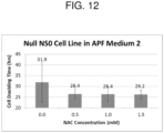
- the average doubling time is shorter than in a cell culture with a control medium excluding N-acetylcysteine. In another embodiment, the average doubling time is reduced by at least 10% compared to a cell culture medium without N-acetylcysteine. In another embodiment, the average doubling time is reduced by at least 15% compared to a cell culture medium without N-acetylcysteine. In another embodiment, the average doubling time is reduced by at least 20% compared to a cell culture medium without N-acetylcysteine. In another embodiment, the average doubling time is reduced by at least 25% compared to a cell culture medium without N-acetylcysteine.
- the average doubling time is reduced by at least 50% compared to a cell culture medium without N-acetylcysteine. In another embodiment, the average doubling time is 60 hours or less in a cell culture medium containing N-acetylcysteine. In another embodiment, the average doubling time is 42 hours or less in a cell culture medium containing N-acetylcysteine. In another embodiment, the average doubling time is 34 hours or less in a cell culture medium containing N-acetylcysteine. In another embodiment, the average doubling time is 30 hours or less in a cell culture medium containing N-acetylcysteine. In another embodiment, the average doubling time is 29 hours or less in a cell culture medium containing N-acetylcysteine.
- the cell viability is increased over a cell culture with a control medium excluding N-acetylcysteine. In another embodiment, the cell viability is increased by at least 5% compared to a cell culture medium without N-acetylcysteine. In another embodiment, the cell viability is increased by at least 7% compared to a cell culture medium without N-acetylcysteine. In another embodiment, the cell viability is increased by at least 10% compared to a cell culture medium without N-acetylcysteine. In another embodiment, the cell viability is at least 90%. In another embodiment, the cell viability is at least 92%. In another embodiment, the cell viability is at least 93%.
- the cells do not express a heterologous protein.
- the cells express a heterologous protein.
- the cells are transformed with a heterologous nucleic acid.
- the heterologous nucleic acid is cDNA, a vector, a plasmid, a nucleic acid operably linked to a promoter, and/or a nucleic acid that incorporates into the genome.
- the heterologous protein is transiently expressed.
- the heterologous protein is stably expressed.
- the heterologous protein is an antibody or antigen-binding fragment thereof.
- the antibody or antigen-binding fragment thereof is an IL-13 antibody.
- the antibody is BAK502G9 (as represented by the VH and VL domains of SEQ ID NOs 1-2 and/or the heavy and light chain CDRs of SEQ ID NOs 3-8), BAK278D6 (as represented by the VH and VL domains of SEQ ID NOs 9-10 and/or the heavy and light chain CDRs of SEQ ID NOs 11-16), BAK1183H4 (as represented by the VH and VL domains of SEQ ID NOs 17-18 and/or the heavy and light chain CDRs of SEQ ID NOs 19-24), or BAK1167F2 (as represented by the VH and VL domains of SEQ ID NOs 25-26 and/or the heavy and light chain CDRs of SEQ ID NOs 27-32).
- Table 1 provides a listing of certain sequences referenced in present embodiments.
- Table 1 Description Sequence SEQ ID NO BAK502G9 VH 1 BAK502G9 VL 2 BAK502G9 HC CDR1 NYGLS 3 BAK502G9 HC CDR2 WISANNGDTNYGQEFQG 4 BAK502G9 HC CDR3 DSSSSWARWFFDL 5 BAK502G9 LC CDR1 GGNIIGSKLVH 6 BAK502G9 LC CDR2 DDGDRPS 7 BAK502G9 LC CDR3 QVWDTGSDPVV 8 BAK278D6 VH 9 BAK278D6 VL 10 BAK278D6 HC CDR1 NYGLS 11 BAK278D6 HC CDR2 WISANNGDTNYGQEFQG 12 BAK278D6 HC CDR3 DSSSNWARWFFDL 13 BAK278D6 LC CDR1 GGNNIGSKLVH 14 BAK
- a cell may refer to a single cell or a population of cells.
- N-acetylcysteine refers to a compound derived from cysteine having an acetyl group attached to the nitrogen atom.
- N-acetylcysteine is also referred to as (2R)-2-acetamido-3-sulfanylpropanoic acid (IUPAC) and has a Chemical Abstracts Service (CAS) Registry Number of 616-91-1 .
- N-acetylcysteine is available from various commercial vendors including Sigma-Aldrich.
- cholesterol auxotroph refers to a cell or cell line that requires cholesterol for growth but is unable to synthesize it.
- exemplary cholesterol auxotroph include, but are not limited to, NS0, NS1, U937, M19, SRD-12B, SRD-13A, CHO-215, X63 cells, cell lines derived from these cells lines, or any other cell engineered to be a cholesterol auxotroph.
- Methods of identifying and/or culturing cholesterol auxotrophs are well known in the art. See, e.g., Keen et al., Cytotechnology. 17(3):203-11 (1995 ); Gorfien et al., Biotechnol Prog.
- myeloma and “myeloma cells” refer to an immortalized cell line derived from bone marrow cells such as myelocytes, plasma cells or B cells.
- exemplary myeloma cells include, but are not limited to, X63Ag8, Sp2/0, NS1, NS0, J558L, U266, U937, P3U1, XG-1, XG-2, XG-3, XG-4, XG-5, XG-6, XG-7, XG-8, XG-9, U266, RPM1-8226, LP1, L363, OPM1, OPM2, and NCLH929 cells or cell lines derived from these cells lines.
- hybridomas refer to an immortalized cell line formed by fusing a B cell with an immortalized cell (e.g. a myeloma cell).
- Methods of generating and/or culturing hybridomas are well known in the art. See, e.g., Kohler and Milstein, Nature 256:495 (1975 ); Galfrè and Milstein. Methods Enzymol. 73(Pt B):3-46 (1981 ); and Goding, Monoclonal Antibodies: Principles and Practice, Academic Press (1986 ).
- cell culture medium refers to a liquid or substrate designed to support the growth of cells derived from multi-cellular eukaryotes, especially animal cells.
- Exemplary cell culture media and methods of culturing cells are described in Doyle et al., "Mammalian cell culture: essential techniques” Wiley, (1997 ); Freshney, “Culture of Animal Cells: A Manual of Basic Technique and Specialized Applications” John Wiley & Sons, (2011 ); and Meenakshi, "Cell Culture Media: A Review” Mater Methods. 3:175 (2013 ).
- the term "serum free medium” refers to a cell culture medium that does not contain animal serum such as fetal bovine serum, bovine serum albumin or human serum albumin.
- animal-protein free medium refers to a cell culture medium that does not contain proteins and/or protein components from higher multicellular non-plant eukaryotes such as albumin, transferrin, insulin or growth factors. Animal proteins and protein components are to be distinguished from non-animal proteins, small peptides and oligopeptides obtainable from plants (usually 10-30 amino acids in length) or lower eukaryotes, such as yeast, which may be included into the animal-protein free cell culture medium.
- Serum free and animal-protein free medium may be based on any basal medium such as DMEM, Ham's F12, Medium 199, McCoy or RPMI generally known to the skilled worker.
- the basal medium may comprise a number of ingredients, including amino acids, vitamins, organic and inorganic salts, and sources of carbohydrate, each ingredient being present in an amount which supports the cultivation of a cell which is generally known to the person skilled in the art.
- the medium may contain auxiliary substances, such as buffer substances like sodium bicarbonate, antioxidants, stabilizers to counteract mechanical stress, or protease inhibitors.
- a non-ionic surfactant such as mixtures of polyethylene glycols and polypropylene glycols (e.g.
- Pluronic F68 ® , SERVA can be added as a defoaming agent.
- serum free and animal-protein free medium are well known in the art as described in Mariani et al., "Commercial serum-free media: hybridoma growth and monoclonal antibody production.” J Immunol Methods. 145:175-83 (1991 ); Barnes et al., “Methods for growth of cultured cells in serum-free medium.” Anal Biochem. 102:255-70 (1980 ); Waymouth, "Preparation and use of serum-free culture media.” In: Barnes DW, Sirbasku DA, Sato GH, editors.
- chemically-defined medium is a cell growth medium suitable for the cell culture of human or animal cells in which all of the chemical components are known.
- a “cell culture medium sufficient to support cell growth” refers to a cell culture medium capable of supporting the growth, survival and/or proliferation of a cell.
- a “cell culture medium sufficient to support cell growth” comprises an appropriate energy source and a complement of amino acids, vitamins, salts, and/or nutrients generally known to the skilled person.
- Exemplary cell culture media sufficient to support cell growth include commercially available media, chemically-defined media, serum-free medium, and animal-protein free media, as generally known to the skilled worker.
- cell viability refers to the ability of a cell to live or develop. In general, determining “cell viability' requires measuring the ability of a cell or cell population to live or develop (including, e.g., making a determination of the number of living or dead cells, based on a total cell sample), as generally known to the skilled worker.
- Cell viability assays are well known in the art and include: cytolysis or membrane leakage assays (e.g. using Propidium iodide, Trypan blue and/or 7-Aminoactinomycin D), Mitochondrial activity or caspase assays (e.g. using Resazurin and/or Formazan), cell functional assays (e.g.
- motility assays cell proliferation or growth assays
- Genomic and/or proteomic assays e.g. measuring the expression of various genes or proteins associated with cell death, damage or stress
- Cytotoxicity assays and vital staining. See Chapter 15, “Assays for Cell Viability, Proliferation and Function” In: “The Molecular Probes Handbook. A Guide to Fluorescent Probes and Labeling Technologies” (I. Johnson and M. Spence (eds.) 11th Edition, Life Technologies (2010 ).
- cell doubling time refers to the period of time required for a cell or population of cells to double in number.
- cell doubling time is measured when the relative growth rate of a cell or cell population is constant (e.g. in the exponential growth or log phase).
- Cell counting assays are well known in the art and include counting cells; using a counting chamber (e.g.
- heterologous nucleic acid refers to a nucleic acid molecule (e.g. a polynucleotide, cDNA, DNA, RNA, or fragment thereof) introduced into a cell using standard recombinant DNA and molecular cloning techniques including, but not limited to, those described in Sambrook et al., "Molecular Cloning: A Laboratory Manual” Second Edition, Cold Spring Harbor Laboratory Press, Cold Spring Harbor, N.Y. (1989 ); Silhavy et al., “Experiments with Gene Fusions” Cold Spring Harbor Laboratory Press, Cold Spring Harbor, N.Y. (1984 ); and Ausubel, F. M. et al., "Current Protocols in Molecular Biology” published by Greene Publishing Assoc. and Wiley-Interscience (1987 ). Nucleic acids may comprise DNA or RNA and may be wholly or partially synthetic.
- transformation refers to the transfer of a nucleic acid molecule or fragment thereof into a host cell, resulting in inheritance of the nucleic acid molecule or fragment thereof to daughter cells of the host cell.
- Host cells containing the transformed nucleic acids or fragments thereof are referred to herein as “transgenic” or “recombinant” or “transformed” cells.
- promoter refers to a polynucleotide sequence capable of controlling the expression of a coding sequence or functional RNA.
- a coding sequence is located 3' to a promoter sequence. Promoters may be derived in their entirety from a native gene, or be composed of different elements derived from different promoters found in nature, or even comprise synthetic DNA segments. It is understood by those skilled in the art that different promoters may direct the expression of a nucleic acid in different tissues or cell types, or at different stages of development, or in response to different environmental or physiological conditions. Promoters which cause a gene to be expressed in most cell types at most times are commonly referred to as "constitutive promoters". It is further recognized that since in most cases the exact boundaries of regulatory sequences have not been completely defined, DNA fragments of different lengths may have identical promoter activity.
- operably linked refers to the association of nucleic acid sequences on a single nucleic acid molecule so that the function of one is affected by the other.
- a promoter is operably linked with a coding sequence when it is capable of effecting the expression of that coding sequence (i.e., that the coding sequence is under the transcriptional control of the promoter).
- Coding sequences can be operably linked to regulatory sequences in sense or antisense orientation.
- Plasmid and vector refer to a nucleic acid element usually in the form of circular double-stranded DNA fragments. Such elements may be autonomously replicating sequences, genome integrating sequences, phage or nucleotide sequences, linear or circular, of a single- or double-stranded DNA or RNA, derived from any source, in which a number of nucleotide sequences have been joined or recombined into a unique construction which is capable of introducing a promoter fragment and DNA sequence for a selected gene product along with appropriate 3' untranslated sequence into a cell.
- Suitable vectors can be chosen or constructed, containing appropriate regulatory sequences, including promoter sequences, terminator sequences, polyadenylation sequences, enhancer sequences, marker genes and other sequences as appropriate.
- expression refers to the transcription and stable accumulation of sense (mRNA) or antisense RNA derived from nucleic acid molecules. Expression may also refer to translation of mRNA into a polypeptide.
- heterologous protein refers to a protein (e.g. a polypeptide, peptide or fragment thereof) that is encoded by a heterologous nucleic acid and expressed in a host cell.
- a heterologous protein may be expressed transiently (e.g. where the polynucleotide encoding the heterologous protein does not incorporate into the host cell genome) or stably (e.g. where the polynucleotide encoding the heterologous protein incorporates into the host cell genome).
- the term "antibody” refers to at least the minimal portion of an antibody which is capable of binding to antigen, e.g., at least the variable domain of a heavy chain (VH) and the variable domain of a light chain (VL) in the context of a typical antibody produced by a B cell.
- VH variable domain of a heavy chain
- VL variable domain of a light chain
- Basic antibody structures in vertebrate systems are relatively well understood. See, e.g., Harlow et al., Antibodies: A Laboratory Manual, (Cold Spring Harbor Laboratory Press, 2nd ed. 1988 ).
- Antibodies or antigen-binding fragments, variants, or derivatives thereof include, but are not limited to, polyclonal, monoclonal, human, humanized, or chimeric antibodies, single chain antibodies, epitope-binding fragments, e.g ., Fab, Fab' and F(ab')2, Fd, Fvs, single-chain Fvs (scFv), single-chain antibodies, disulfide-linked Fvs (sdFv), fragments comprising either a VL or VH domain, fragments produced by a Fab expression library.
- ScFv molecules are known in the art and are described, e . g ., in US patent 5,892,019 .
- Immunoglobulin or antibody molecules encompassed by this disclosure can be of any type (e.g., IgG, IgE, IgM, IgD, IgA, and IgY), class (e.g., IgG1, IgG2, IgG3, IgG4, IgA1 and IgA2) or subclass of immunoglobulin molecule.
- type e.g., IgG, IgE, IgM, IgD, IgA, and IgY
- class e.g., IgG1, IgG2, IgG3, IgG4, IgA1 and IgA2
- subclass of immunoglobulin molecule e.g., IgG1, IgG2, IgG3, IgG4, IgA1 and IgA2
- Antibodies or antigen-binding fragments, variants, or derivatives thereof disclosed herein can be described or specified in terms of the epitope(s) or portion(s) of an antigen, e.g., a target polypeptide that they recognize or specifically bind.
- an IL-13 or anti-IL-13 antibody is an antibody that binds to an IL-13 polypeptide or a portion thereof.
- the anti-IL-13 antibody is BAK502G9 (e.g. an anti-IL-13 antibody comprising SEQ ID NOs: 1 and 2).
- the antibody is BAK502G9 (as represented by the VH and VL domains of SEQ ID NOs 1-2 and/or the heavy and light chain CDRs of SEQ ID NOs 3-8), BAK278D6 (as represented by the VH and VL domains of SEQ ID NOs 9-10 and/or the heavy and light chain CDRs of SEQ ID NOs 11-16), BAK1183H4 (as represented by the VH and VL domains of SEQ ID NOs 17-18 and/or the heavy and light chain CDRs of SEQ ID NOs 19-24), or BAK1167F2 (as represented by the VH and VL domains of SEQ ID NOs 25-26 and/or the heavy and light chain CDRs of SEQ ID NOs 27-32).
- anti-IL-13 monoclonal antibodies that can be used include those described in U.S. Pat. Appl. Publ. No. 2012-0052060, published March 1, 2012 .
- Other IL-13 antibodies include, without limitation, anti-human-IL-13 antibodies, for example, Lebrikizumab (MILR1444A / RG3637, Roche / Genentech), ABT-308 (Abbott), GSK679586 (GlaxoSmithKline) or QAX576 (Novartis).
- antibodies, including anti-IL13 antibodies may be produced in cells using various techniques known in the art. See, e .
- Cell culture is the process of placing cells, tissues, or organs removed from an animal into an artificial environment that promotes their survival, growth, and/or proliferation.
- Basic environmental requirements for cells to grow optimally include: a suitable vessel, a cell culture / growth medium to supply nutrients (including, but not limited to, at least one of amino acids, carbohydrates, vitamins, minerals, growth factors, hormones, etc.), and a controlled physicochemical environment (to control, e.g., pH, osmotic pressure, temperature, O 2 , CO 2 , etc.).
- nutrients including, but not limited to, at least one of amino acids, carbohydrates, vitamins, minerals, growth factors, hormones, etc.
- a controlled physicochemical environment to control, e.g., pH, osmotic pressure, temperature, O 2 , CO 2 , etc.
- Some cells are anchorage-dependent and must be cultured while attached to a solid or semi-solid substrate (adherent or monolayer culture), while others can be grown floating in the culture medium (
- the cell culture medium or cell culture method includes a cell culture medium sufficient to support cell growth comprising N-acetylcysteine.
- the cell culture medium is a serum free and animal-protein free medium.
- the cell culture medium is a chemically-defined medium.
- N-acetylcysteine is added to commercially-available cell culture media.
- the commercially-available cell culture medium is EX-CELL ® NS0 serum-free medium for NS0 cells (available from Sigma-Aldrich, catalog number H4281), EX-CELL ® CD hybridoma medium (Sigma-Aldrich, catalog number H4409), Ex-Cell 620-HSF serum-free medium for hybridoma cells (Sigma-Aldrich, catalog number 14621C), Ex-Cell NS- serum-free medium for NS0 (Sigma-Aldrich, catalog number 14650C), DMEM (Sigma-Aldrich, catalog number D567), Iscove's Modified Dulbecco's Medium (IMDM) (available from Sigma-Aldrich, catalog number I3390), RPMI-1640 Medium (Sigma-Aldrich, catalog number R8005), Hybridoma-SFM (Life Technologies, catalog number 12045076), CD Hybridoma AGT medium (Life Technologies, catalog number 12372025), CD Hybridom
- N-acetylcysteine is added to the cell culture medium or the cell culture methods described herein to increase cell viability, cell growth rate, and/or reduce cell doubling time. While not being limited by any particular theory, it is believed that N-acetylcysteine provides benefits to cells growing in culture by protecting them from free radicals, preventing cell membrane breakdown, and/or preventing oxidation of other cell culture medium ingredients, including, but not limited to, lipids (such as cholesterol).
- a cell culture medium or a cell culture method as described herein comprises N-acetylcysteine at a concentration of from about 0.25 mM to about 3 mM, from about 0.5 mM to about 2.5 mM, from about 0.5 mM to about 2.0 mM, from about 0.5 mM to about 1.5 mM, from about 0.5 mM to about 1.0 mM, from about 1.0 mM to about 2.5 mM, from about 1.0 mM to about 2.0 mM, from about 1.0 mM to about 1.5 mM, from about 1.5 mM to about 2.5 mM, or from about 1.5 mM to about 2.0 mM.
- a cell culture medium or a cell culture method as described herein comprises N-acetylcysteine at a concentration of about 1 mM. In another embodiment, a cell culture medium or a cell culture method as described herein comprises N-acetylcysteine at a concentration of about 1.5 mM. In another embodiment, a cell culture medium or a cell culture method as described herein comprises N-acetylcysteine at a concentration of about 2.0 mM. In another embodiment, a cell culture medium or a cell culture method as described herein comprises N-acetylcysteine at a concentration of about 2.5 mM.
- a cell culture medium or a cell culture method as described herein comprises N-acetylcysteine at a concentration of about 0.5 mM. In another embodiment, the cell culture medium or a cell culture method as described herein comprises N-acetylcysteine at a concentration of at least about 0.5 mM, at least about 1.0 mM, at least about 1.5 mM, at least about 2.0 mM, or at least about 2.5 mM.
- the cell culture medium or cell culture method described herein further comprises a carbon source, a nitrogen source, and/or a phosphorous source. These can be provided by the same ingredient or different ingredients.
- the cell culture medium or cell culture methods described herein further comprise a carbon source, a nitrogen source, a phosphorous source, and/or mineral salts.
- a carbon source suitable for use in the cell culture medium or cell culture methods described herein includes a carbohydrate (such as a sugar) or an amino acid, such as L-glutamine and/or pyruvate or any combination thereof.
- the cell culture medium or cell culture method described herein further comprises carbohydrates and amino acids.
- the cell culture medium or cell culture method described herein further comprises at least one of salts, vitamins, metabolic precursors, growth factors, hormones, and trace elements.
- the cell culture medium or cell culture methods described herein further comprise basal media, containing amino acids, vitamins, inorganic salts, and a carbon source such as glucose.
- Exemplary carbohydrates suitable for use in the cell culture medium or cell culture methods described herein include glucose, galactose, trehalose, glucosamine, mannose, raffinose, fructose, ribose, glucuronic acid, lactose, maltose, sucrose, turanose, any other carbohydrate suitable for cell culture generally known in the art, or any combinations thereof.
- the carbohydrate may be glucose or galactose.
- amino acids suitable for use in the cell culture medium or cell culture methods described herein include one or more essential amino acids (i.e. histidine, isoleucine, leucine, lysine, methionine, phenylalanine, threonine, tryptophan, and/or valine), and/or one or more nonessential amino acids (i.e. alanine, arginine, aspartic acid, cysteine, glutamic acid, glutamine, glycine, proline, serine, tyrosine, and asparagine), and/or any combination of essential and non-essential amino acids thereof.
- essential amino acids i.e. histidine, isoleucine, leucine, lysine, methionine, phenylalanine, threonine, tryptophan, and/or valine
- nonessential amino acids i.e. alanine, arginine, aspartic acid, cysteine, glutamic acid, glutamine
- NS0 cells lack or contain very low levels of the endogenous glutamine synthetase enzyme and, as such, glutamine is an essential amino acid for NS0 cells unless glutamine synthetase is included in the expression system for a heterologous protein.
- amino acids this disclosure includes any amino acids, including, but not limited to, D- or L-amino acids and non-standard amino acids.
- amino acid encompasses any organic compound with an amine (-NH 2 ) and a carboxylic acid (-COOH) functional group.
- the cell culture medium or cell culture methods described herein further comprise a lipid.
- lipids suitable for use in the cell culture medium or cell culture methods described herein include one or more of cholesterol, arachadonic acid, tocopherol acetate, linoleic acid, linolenic acid, myristic acid, oleic acid, palmitic acid, palmitoleic acid, stearic acid, a phospholipid (such as phosphatidylcholine) any other lipid suitable for cell culture generally known in the art, or any combinations thereof.
- Inositol as a component of membrane phospholipids, may also be optionally included.
- Synthetic or plant-derived lipids may also be optionally used in applications where the medium is desired to be kept free of animal-derived components.
- Lipids may be added in a cyclodextrin-based lipid supplement. Cyclodextrins may be used to solubilize lipids and/or other ingredients such as fat-soluble vitamins and hormones.
- Cholesterol may be synthetically produced or it may be animal derived. For instance, cholesterol may be isolated from sheep wool. An animal-protein free medium may contain cholesterol derived from an animal source. In some embodiments, cholesterol is obtained from a commercial source. See, e.g., Chemically Defined Lipid Concentrate from Gibco; Lipid Concentrate from SAFC.
- Cholesterol may be added to the medium in a cholesterol-cyclodextrin solution, as a synthetic cholesterol (such as but not limited to SyntheChol TM ), as cholesterol nanoparticles (see, e.g., Wu et al., Enhanced Productivity of NS0 cells in fed-batch culture with cholesterol nanoparticle supplementation, Biotechnology Progress 27(3):796-802 (2011 )) or by any means suitable for cell culture generally known in the art.
- a synthetic cholesterol such as but not limited to SyntheChol TM
- cholesterol nanoparticles see, e.g., Wu et al., Enhanced Productivity of NS0 cells in fed-batch culture with cholesterol nanoparticle supplementation, Biotechnology Progress 27(3):796-802 (2011 ) or by any means suitable for cell culture generally known in the art.
- the medium may contain from about 1 to about 10 g/L of cholesterol. In another embodiment, the medium may contain from about 1 to about 5 g/l of cholesterol. In another embodiment, the medium may contain from about 1.5 to about 4 g/l of cholesterol. In another embodiment, the medium may contain from about 2 to about 3 g/l of cholesterol. In another embodiment, the medium may contain about 2.5 g/l of cholesterol. In another embodiment, the medium may contain at least about 1 g/L of cholesterol, at least about 2.5 g/L of cholesterol or at least about 5 g/L of cholesterol.
- the medium may contain a lipid other than cholesterol. In one embodiment, the medium may contain a phospholipid. In another embodiment, the medium may contain phosphatidylcholine. In one embodiment, the medium may contain from about 1 to about 10 g/L of phosphatidylcholine. In another embodiment, the medium may contain from about 1 to about 5 g/l of phosphatidylcholine. In another embodiment, the medium may contain from about 1.5 to about 4 g/l of phosphatidylcholine. In another embodiment, the medium may contain from about 2 to about 3 g/l of phosphatidylcholine. In another embodiment, the medium may contain about 2.5 g/l of phosphatidylcholine.
- the cell culture medium or cell culture methods described herein further comprise a salt.
- exemplary salts suitable for use in the cell culture medium or cell culture methods described herein include at least one of calcium chloride, magnesium chloride, potassium chloride, sodium chloride, potassium nitrate, any other salt suitable for cell culture generally known in the art, or any combinations thereof.
- the cell culture medium or cell culture methods described herein do not include calcium chloride or magnesium chloride. This embodiment has advantages when cell dissociation or release is desired as calcium and magnesium promote cell adhesion.
- the cell culture medium or cell culture methods described herein further comprise a vitamin.
- exemplary vitamins suitable for use in the cell culture medium or cell culture methods described herein include fat-soluble vitamins, vitamins A, D, E, K, B1 (thiamine), B2 (riboflavin), B3 (nicotinamide), B5 (pantothenic acid), B6 (pyridoxal, pyridoxamine, and/or pyridoxine), B9 (folic acid), any other vitamin suitable for cell culture generally known in the art, or any combinations thereof.
- At least one hormone may be added to the cell culture medium or cell culture methods described herein.
- the hormone may be chosen from at least one of dexamethasone, erythropoietin, estradiol, hydrocortisone, insulin, progesterone, somatostatin, thyroxine (T4), triiodothyronine (T3), activin, BMP4, BMP7, BMPR1A, Cripto, FLT3 ligand, HGF, IGF, EGF, FGF, PDGF, IGFBP4, kallekrein, LEFTY-A, NGF, TGF ⁇ , VEGF, or any other hormone or growth factor suitable for cell culture generally known in the art.
- At least one trace element may be added to the cell culture medium or cell culture methods described herein.
- the trace element may be at least one of zinc, iron, copper, selenium, magnesium, manganese, molybdenum, tin, nickel, or any other trace element suitable for cell culture generally known in the art.
- the cell culture medium or cell culture methods described herein further comprise at least one surfactant.
- exemplary surfactants suitable for use in the cell culture medium or cell culture methods described herein include Tween-80, pluronic F-68, or any other surfactant suitable for cell culture generally known in the art.
- the cell culture medium or cell culture methods described herein further comprise at least one pH buffering agent.
- buffering agents suitable for use in the cell culture medium or cell culture methods described herein include sodium bicarbonate, boric acid, citric acid, dithiothreitol, ethanolamine, glycerophosphate, potassium citrate, potassium phosphate, sodium acetate, sodium chloride, sodium phosphate, starch from wheat, HEPES, calcium chloride, MOPS, or any other buffering agent cell suitable for cell culture generally known in the art, or any combination thereof.
- the cell culture medium or cell culture methods described herein further comprise a non-animal-sourced hydrolysate.
- a plant or yeast hydrolysate provides protein digests comprising amino acids, short peptides, carbohydrates, vitamins, nucleosides, and minerals, providing a variety of nutritional supplements to media.
- yeastolate a yeast hydrolysate
- Yeastolate is a mixture of peptides, amino acids, carbohydrates, lipids, metals and vitamins. It may be added in addition to or in lieu of those ingredients provided separately.
- the cell culture medium comprises 1 g/L of yeastolate.
- tropolone may also be added to the cell culture medium or cell culture methods described herein.
- nucleosides may be added to the cell culture medium or cell culture methods described herein.
- ⁇ -mercaptoethanol may be added to the cell culture medium or cell culture methods described herein.
- antibiotics may be added to the cell culture media or cell culture methods described herein.
- the cell culture medium or cell culture methods described herein contain NAC, an amino acid, a vitamin, a lipid, a carbohydrate, a pH buffering agent, a trace metal, an inorganic salt, and a surfactant.
- the lipid is cholesterol.
- a variety of cell types such as cholesterol auxotrophic cells, myeloma cells, and hybridoma cells, may be cultured with the presently-disclosed cell culture medium comprising N-acetylcysteine.
- the cells being cultured are derived from a mammal, including, but not limited to, cells derived from a mouse, rat, human, monkey, hamster, rabbit, etc.
- cholesterol auxotroph refers to a cell or cell line that requires cholesterol for growth but is unable to synthesize it.
- a cholesterol auxotroph is NS0, NS1, U937, M19, SRD-12B, SRD-13A, CHO-215, X63 cells, cell lines derived from these cells lines, or any other cell engineered to be a cholesterol auxotroph.
- the cells are NS0 cells.
- the cells suitable to be cultured with the presently-disclosed cell culture medium comprising N-acetylcysteine are cells that are cholesterol auxotrophs. In one embodiment, the cells suitable to be cultured with the presently-disclosed cell culture medium comprising N-acetylcysteine are NS0 cells. In another embodiment, the cells are NS1, U937, M19, SRD-12B, SRD-13A, CHO-215, X63 cells, cells derived from these cell lines, or any other cell engineered to be a cholesterol auxotroph.
- the cells suitable to be cultured with the presently-disclosed cell culture medium comprising N-acetylcysteine are murine myeloma cells that are cholesterol auxotrophs. In one embodiment, the cells are mammalian myeloma cells that are cholesterol auxotrophs. In one embodiment the cells suitable to be cultured with the presently-disclosed cell culture medium comprising N-acetylcysteine are human myeloma cells that are cholesterol auxotrophs.
- the cells suitable to be cultured with the presently-disclosed cell culture medium comprising N-acetylcysteine are myeloma cells.
- the cells are X63Ag8, Sp2/0, NS1, NS0, J558L, U266, U937, P3U1, XG-1, XG-2, XG-3, XG-4, XG-5, XG-6, XG-7, XG-8, XG-9, U266, RPM1-8226, LP1, L363, OPM1, OPM2, and NCLH929 cells or cell lines derived from these cells lines.
- the cells suitable to be cultured with the presently-disclosed cell culture medium comprising N-acetylcysteine are hybridoma cells.
- the hybridoma cells suitable to be cultured with the presently-disclosed cell culture medium comprising N-acetylcysteine express and/or secrete an antibody.
- the cells suitable to be cultured with the presently-disclosed cell culture medium comprising N-acetylcysteine are cultured without inducing and/or expressing a recombinant or heterologous protein.
- the cells suitable to be cultured with the presently-disclosed cell culture medium comprising N-acetylcysteine e.g.
- a heterologous nucleic acid including, but not limited to cDNA, a plasmid, a vector, nucleic acids operably linked to a promoter, and/or nucleic acids that transiently express a heterologous nucleic acid or incorporate the heterologous nucleic acid into the genome of the cell line.
- the cells express a recombinant or heterologous protein.
- the cells overexpress the recombinant or heterologous protein.
- Cell lines expressing a wide variety of heterologous sequences may benefit from the present cell culture medium and method.
- the heterologous protein is transiently expressed. In another embodiment, the heterologous protein is stably expressed.
- the heterologous protein is not an antibody or antigen-binding fragment thereof.
- the heterologous protein is a blood factor, anticoagulant, thrombolytic, erythropoietin, interferon, hormone, enzyme, vaccine, growth factor, and/or a fusion protein.
- the cells suitable to be cultured with the presently-disclosed cell culture medium comprising N-acetylcysteine express a heterologous protein, wherein the heterologous protein is an antibody or antigen-binding fragment thereof.
- the antibody or antigen-binding fragment thereof specifically binds IL-13 or specifically binds IL-9.
- the antibody or antigen-binding fragment is an antibody or antigen-binding fragment disclosed in US 7,947,273 , US 7,354,584 or US 7,371,383 .
- the antibody is BAK502G9 (comprising SEQ ID NOs 1 and 2).
- the antibody or antigen-binding fragment thereof has the same CDRs as BAK502G9 (comprising heavy chain CDRS (SEQ ID NOs: 3-5) and light chain CDRs (SEQ ID NOs: 6-8)).
- the antibody or antigen binding fragment has a heavy chain variable region comprising any one of SEQ ID NOs: 1, 9, 17, or 25 and a light chain variable region comprising any one of SEQ ID NOs: 2, 10, 18, or 26.
- the antibody or antigen binding fragment has a heavy chain variable region comprising: (a) a HC CDR1 chosen from SEQ ID NOs: 3, 11, 19, and 27; (b) a HC CDR2 chosen from SEQ ID NOs: 4, 12, 20, and 28; and (c) a HC CDR chosen from SEQ ID NOs: 5, 13, 21, and 29; and a light chain variable region comprising: (a) a LC CDR1 chosen from SEQ ID NOs: 6, 14, 22, and 30; (b) a LC CDR2 chosen from SEQ ID NOs: 7, 15, 23, and 31; and (c) a LC CDR3 chosen from SEQ ID NOs: 8, 16, 24, and 32.
- the antibody or antigen binding fragment comprises a heavy chain variable region (VH) and/or a light chain variable region (VL) described in Table 1; or comprises a set of 6 of the CDRs described in Table 1.
- the antibody or antigen binding fragment has a heavy chain variable region comprising a sequence that is about 70%, about 75%, about 80%, about 85%, about 90%, about 92%, about 93%, about 94%, about 98%, or about 99% identical to any one of SEQ ID NOs: 1, 9, 17, or 25 and a light chain variable region comprising a sequence that is about 70%, about 75%, about 80%, about 85%, about 90%, about 92%, about 93%, about 94%, about 98%, or about 99% identical to any one of SEQ ID NOs: 2, 10, 18, or 26.
- the antibody or antigen binding fragment has a heavy chain variable region comprising: (a) a HC CDR1 that has one mutation compared to a sequence chosen from SEQ ID NOs: 3, 11, 19, or 27; (b) a HC CDR2 that has one or two mutations compared to a sequence chosen from SEQ ID NOs: 4, 12, 20, and 28; and (c) a HC CDR3 that has one or two mutations compared to a sequence chosen from SEQ ID NOs: 5, 13, 21, and 29; and a light chain variable region comprising: (a) a LC CDR1 that has one mutation compared to a sequence chosen from SEQ ID NOs: 6, 14, 22, and 30; (b) a LC CDR2 that has one or two mutations compared to a sequence chosen from SEQ ID NOs: 7, 15, 23, and 31; and (c) a LC CDR3 that has one or two mutations compared to a sequence chosen from SEQ ID NOs: 8, 16, 24, and 32.
- the antibody or antigen binding fragment has a heavy chain variable region (VH) and/or a light chain variable region (VL) comprising a sequence that is about 70%, about 75%, about 80%, about 85%, about 90%, about 92%, about 93%, about 94%, about 98%, or about 99% identical to any one of the VH and/or VL sequences described in Table 1.
- the antibody or antigen binding fragment has one or two mutations in a CDR compared to any one of the CDRs described in Table 1.
- the antibody or antigen binding fragment thereof specifically binds glycoprotein IIb/IIIa, IL-2 receptor (such as the IL-2 receptor a), TNF- ⁇ , RSV, F protein epitope of RSV, CD33, epidermal GF receptor, T-cell VLA4 receptor, complement protein C5, IL-1, IL-9, IL-12, IL-13, IL-23, CD-20, and/or BAFF.
- IL-2 receptor such as the IL-2 receptor a
- TNF- ⁇ such as the IL-2 receptor a
- RSV F protein epitope of RSV
- CD33 epidermal GF receptor
- T-cell VLA4 receptor T-cell VLA4 receptor
- the antibody or antigen binding fragment thereof is ofatumumab, belimumab, gemtuzumab ozogamicin, palivizumab, natalizumab, cetuximab, canakinumab, infliximab, abciximab, basiliximab, daclizumab, eculizumab, or ustekinumab.
- a cell culture method comprises providing a cell culture medium sufficient to support cell growth, wherein the cell culture medium comprises N-acetylcysteine and culturing a cell in the cell culture medium, wherein the cell is a cholesterol auxotroph, a myeloma, or a hybridoma.
- the method is a method of increasing cell viability, increasing cell growth rate, and/or reducing cell doubling time of a cell by providing a cell culture medium sufficient to support cell growth comprising N-acetylcysteine; and culturing the cell in the cell culture medium, wherein the cell is a cholesterol auxotroph, a myeloma, or a hybridoma.
- the cells are cultured at 37°C at 5% CO 2 and 85% relative humidity.
- the pH may be from 6.8 to 7.4.
- Other acceptable conditions generally known in the art may also be used.
- the cell culture occurs while scaling up production from a frozen stock to a large bioreactor.
- the improved medium allows for at least a four-fold split ratio when passaging the cells (namely 1X of cell culture medium containing the cells to be passaged is mixed with 3X of fresh cell culture medium not containing the cells). In another aspect, it allows for a five-fold split ratio, six-fold split ratio, or a seven-fold split ratio.
- the cells are cultured in a 100 L bioreactor, then transferred to a 500 L bioreactor, and then transferred to a 2500 L bioreactor.
- the cells are thawed from a frozen stock.
- the cells e.g. cholesterol auxotrophs, myelomas, or hybridomas
- the cells are in an expansion phase.
- the cells e.g. cholesterol auxotrophs, myelomas, or hybridomas
- the present cell culture method and/or media may provide multiple advantages.
- it reduces the time required for cells (e.g. cholesterol auxotrophs, myelomas, or hybridomas such as NS0 cells) to grow in a vessel (such as a bioreactor) by at least about 10% to at least about 50% (e.g. at least about 10%, about 15%, about 20%, about 25%, about 30%, about 35%, about 40%, about 45%, or about 50%) compared to a cell culture medium without N-acetylcysteine. In one embodiment, this is determined by calculating the period of time necessary to get to a desired cell count per volume.
- cells e.g. cholesterol auxotrophs, myelomas, or hybridomas such as NS0 cells
- a vessel such as a bioreactor
- this is determined by calculating the period of time necessary to get to a desired cell count per volume.
- the cell culture method and/or media reduce the average cell doubling time of a cholesterol auxotroph, a myeloma, or a hybridoma (e.g. NS0 cells) cultured using a cell culture method and/or media comprising N-acetylcysteine compared to a cholesterol auxotroph, a myeloma, or a hybridoma (e.g. NS0 cells) cultured in a cell culture with a control medium excluding N-acetylcysteine.
- the average doubling time of a cholesterol auxotroph, a myeloma, or a hybridoma e.g.
- NS0 cells cultured using a cell culture method and/or media comprising N-acetylcysteine is shorter than a cholesterol auxotroph, a myeloma, or a hybridoma (e.g. NS0 cells) cultured in a cell culture with a control medium excluding N-acetylcysteine.
- the average and/or median cell doubling time of a cholesterol auxotroph, a myeloma, or a hybridoma (e.g. NS0 cells) cultured using a cell culture method and/or media comprising N-acetylcysteine is reduced by at least about 10% to at least about 50% (e.g.
- the average and/or median cell doubling time of a cholesterol auxotroph, a myeloma, or a hybridoma (e.g. NS0 cells) cultured using a cell culture method and/or media comprising N-acetylcysteine is less than or equal to 60 hours to less than or equal to about 29 hours (e.g. less than or equal to about 60 hours, about 42 hours, about 34 hours, about 30 hours, or about 29 hours) or about less than or about equal to any of the average cell doubling times reported in Figures 1-12 .
- the cell culture method and/or media increase the cell growth rate of a cholesterol auxotroph, a myeloma, or a hybridoma (e.g. NS0 cells) cultured using a cell culture method and/or media comprising N-acetylcysteine compared to a cholesterol auxotroph, a myeloma, or a hybridoma (e.g. NS0 cells) cultured in a cell culture with a control medium excluding N-acetylcysteine.
- the cell growth rate of a cholesterol auxotroph, a myeloma, or a hybridoma e.g.
- NS0 cells cultured using a cell culture method and/or media comprising N-acetylcysteine is higher than a cholesterol auxotroph, a myeloma, or a hybridoma (e.g. NS0 cells) cultured in a cell culture with a control medium excluding N-acetylcysteine.
- cell growth rate of a cholesterol auxotroph, a myeloma, or a hybridoma (e.g. NS0 cells) cultured using a cell culture method and/or media comprising N-acetylcysteine is increased by at least about 10% to at least about 50% (e.g.
- a cholesterol auxotroph e.g. a myeloma, or a hybridoma (e.g. NS0 cells) cultured in a cell culture medium without N-acetylcysteine.
- cell growth may be determined using a cell counting method.
- a sample volume of cell culture medium is obtained and the cells counted in that volume.
- Cell counting may be done in a hemocytometer or with a Coulter Counter. Another method plots the number of cells against time, with the slope of the graph stepper for cultures showing improved growth rates.
- the cell culture method and/or media disclosed herein increase the cell viability of a cholesterol auxotroph, a myeloma, or a hybridoma (e.g. NS0 cells) over a cholesterol auxotroph, a myeloma, or a hybridoma (e.g. NS0 cells) cultured in a cell culture with a control medium excluding N-acetylcysteine.
- cell viability of a cholesterol auxotroph, a myeloma, or a hybridoma e.g.
- NS0 cells cultured using a cell culture method and/or media comprising N-acetylcysteine is higher compared to a cholesterol auxotroph, a myeloma, or a hybridoma (e.g. NS0 cells) cultured in a cell culture with a control medium excluding N-acetylcysteine.
- cell viability of a cholesterol auxotroph, a myeloma, or a hybridoma (e.g. NS0 cells) cultured using a cell culture method and/or media comprising N-acetylcysteine is increased by at least about 5% to at least about 15% (e.g.
- the cell viability of a cholesterol auxotroph, a myeloma, or a hybridoma (e.g. NS0 cells) cultured using a cell culture method and/or media comprising N-acetylcysteine is at least about 85% to at least about 95% (e.g. at least about 85%, about 88%, about 90%, about 92%, about 93%, about 94%, or about 95%).
- cell viability may be determined by a trypan blue viability exclusion assay.
- a cell suspension may be mixed with 0.4% trypan blue in phosphate buffered solution and cells counted using a hemocytometer. Live cells appear round and refractile without any blue-dye coloration, while dead cells absorb the dye and appear blue. Viability may be expressed as a percentage of viable cells over total cells counted, with a viable cell being a cell whose membrane integrity is still able to prevent the absorption of the trypan blue in a trypan blue exclusion viability assay.
- increased protein yield is obtained using the cell culture method and/or media disclosed herein.
- recombinant or heterologous protein expression is higher in a cholesterol auxotroph, a myeloma, or a hybridoma (e.g. NS0 cells) cultured using a cell culture method and/or media comprising N-acetylcysteine compared to a cholesterol auxotroph, a myeloma, or a hybridoma (e.g. NS0 cells) cultured in a cell culture with a control medium excluding N-acetylcysteine.
- the cholesterol auxotroph, myeloma, or hybridoma (e.g. NS0 cells) cultured using a cell culture method and/or media comprising N-acetylcysteine shows at least 10% to at least 200% (e.g. at least 10%, 20%, 30%, 40%, 50%, 60%, 70%, 80% 90%, 100%, 150%, 200%) higher protein expression compared to a cholesterol auxotroph, a myeloma, or a hybridoma (e.g. NS0 cells) cultured in a cell culture medium without N-acetylcysteine.
- Example 1 Cell Doubling Time of NS0 Cell Line 1 in APF Medium 1 and 2 at Vial Thaw
- N-Acetylcysteine N-acetylcysteine
- APF Animal Protein Free
- One of the cell lines was a NS0 null host cell that had not been transformed to express a recombinant protein (NS0 Null Cell Line).
- NS0 Cell Line 1 (expressing an anti-IL-9 antibody) and NS0 Cell Line 2 (expressing BAK502G9, an anti-IL-13 antibody).
- NS0 Cell Line 1 was thawed and expanded in two different APF media (APF Medium 1 or APF Medium 2) supplemented with various concentrations of NAC ranging from 1.5 mM to 2.5 mM.
- NS0 Cell Line 2 was thawed and expanded in three different APF media (APF Medium 1, APF Medium 2 or a commercially available NS0 cell culture media (CD Hybridoma + cholesterol from Invitrogen/Gibco)) supplemented with NAC ranging from 0.5 mM to 2.5 mM, while the untransformed host cell line (NS0 Null Cell Line) was thawed in APF medium 2 supplemented with NAC ranging from 0.5 mM to 1.5 mM.
- NAC was obtained from Sigma and was either added to the medium directly or dissolved in water at 100 mM concentration before adding to the media at the appropriate concentration.
- APF Medium 1 and APF Medium 2 contain standard cell culture components including: amino acids, vitamins, lipids, sugar, small peptides, pH buffer, trace metals, inorganic salts, nucleotides, nucleotide precursors, surfactants, reducing agents, cholesterol, lipids and antioxidants.
- Viable cell densities during the first 3 to 4 days (i.e. exponential growth phase) at each passage were used to calculate population doubling times during both the first few days after the vial thaw while the cells were recovering from the thaw and during subsequent cell passages when the cells have fully recovered and reached a consistent doubling time from passage to passage.
- NS0 cells i.e. NS0 Cell Line 1 expressing an anti-IL-9 monoclonal antibody were in a frozen stock.
- Frozen NS0 Cell Line 1 cells were thawed in animal-protein free (APF) medium (i.e. APF media 1 or APF media 2) supplemented with various concentrations of N-acetylcysteine (NAC) and viable cell density during exponential growth phase was used to calculate average cell doubling time as described above.
- APF animal-protein free
- NAC N-acetylcysteine
- Figure 1 shows NS0 Cell Line 1 cell doubling time in control medium (APF media 1 without NAC) and APF media 1 supplemented with three increasing concentrations of N-acetylcysteine for cells thawed from a frozen stock.
- Addition of 1.5 mM to 2.5 mM NAC (1.5 mM, 2.0 mM or 2.5 mM NAC) reduced average cell doubling time at vial thaw (55.6 hours, 48.4 hours, and 53.9 hours, respectively) compared to NS0 Cell Line 1 cells thawed in control medium (57.6 hours).
- the slight increase in doubling time between 2 mM and 2.5 mM may be due to an increase in osmolality of the solution, especially as NS0 cells can be sensitive to osmolality during thawing.
- Figure 2 shows NS0 Cell Line 1 cell doubling time in control medium (APF media 2 without NAC) and APF media 2 supplemented with three increasing concentrations of N-acetylcysteine for cells thawed from a frozen stock. Similar to the results observed with APF Media 1, adding 1.5 mM to 2.5 mM NAC (1.5 mM, 2.0 mM or 2.5 mM NAC) to APF Media 2 also reduced cell doubling time at vial thaw (47.5 hours, 56.9 hours, and 47.7 hours, respectively) compared to NS0 Cell Line 1 cells thawed in control medium (109.8 hours).
- NS0 cells i.e. NS0 Cell Line 2 expressing a monoclonal antibody (i.e. BAK502G9, an anti-IL-13 antibody) were in a frozen stock.
- Frozen NS0 Cell Line 2 cells were thawed in animal-protein free (APF) medium (i.e. APF media 1, APF media 2 or CD Hybridoma medium (Gibco) supplemented with cholesterol (1X Invitrogen Cholesterol Lipid Concentrate)) supplemented with various concentrations of N-acetylcysteine (NAC) and viable cell density during exponential growth phase was used to calculate average cell doubling time as described in Example 1.
- APF animal-protein free
- Figure 3 shows NS0 Cell Line 2 cell doubling time in control medium (APF media 1 without NAC) and APF media 1 supplemented with three increasing concentrations of N-acetylcysteine for cells thawed from a frozen stock.
- Addition of 0.5 mM to 2.5 mM NAC (0.5 mM, 1.0 mM or 2.5 mM NAC) reduced average cell doubling time at vial thaw (39.6 hours, 41.6 hours, and 37.5 hours, respectively) compared to NS0 Cell Line 2 cells thawed in control medium (44.6 hours), with 2.5 mM NAC showing the greatest reduction of cell doubling time at vial thaw.
- Figure 4 shows NS0 Cell Line 2 cell doubling time in control medium (APF media 2 without NAC) and APF media 2 supplemented with three increasing concentrations of N-acetylcysteine for cells thawed from a frozen stock. Similar to the results observed with APF media 1, 0.5 mM to 2.0 mM NAC (0.5 mM, 1.0 mM or 2.0 mM NAC) reduced average cell doubling time at vial thaw (57.7 hours, 37.0 hours, and 39.7 hours, respectively) compared to NS0 Cell Line 2 cells thawed in control medium (77.0 hours), with 1.0 mM and 2.0 mM showing the greatest reduction of cell doubling time at vial thaw.
- control medium APF media 2 without NAC
- Figure 5 shows NS0 Cell Line 2 cell doubling time in control medium (CD Hybridoma medium (Gibco) supplemented with 1X Invitrogen Cholesterol Lipid Concentrate without NAC) and control medium supplemented with three increasing concentrations of N-acetylcysteine for cells thawed from a frozen stock.
- control medium CD Hybridoma medium (Gibco) supplemented with 1X Invitrogen Cholesterol Lipid Concentrate without NAC)
- control medium supplemented with three increasing concentrations of N-acetylcysteine for cells thawed from a frozen stock.
- NS0 cells i.e. NS0 Null Cell Line
- Frozen NS0 Null Cell Line cells were thawed in animal-protein free (APF) medium (i.e. APF media 2) supplemented with various concentrations of N-acetylcysteine (NAC) and viable cell density during exponential growth phase was used to calculate average cell doubling time as described in Example 1.
- APF animal-protein free
- NAC N-acetylcysteine
- Figure 6 shows NS0 Null Cell Line cell doubling time in control medium (APF media 2 without NAC) and APF media supplemented with three increasing concentrations of N-acetylcysteine for cells thawed from a frozen stock. Similar to the results reported in Examples 1 and 2, addition of 0.5 mM to 1.5 mM NAC (0.5 mM, 1.0 mM or 1.5 mM NAC) reduced cell doubling time at vial thaw (36.8 hours, 38.4 hours and 57.9 hours, respectively) compared to NS0 Null Cell Line cells thawed in control medium (67.8 hours), with 0.5 mM and 1.0 mM showing the greatest reduction of cell doubling time at vial thaw.
- control medium APF media 2 without NAC
- NS0 cells i.e. NS0 Cell Line 1 expressing an anti-IL-9 monoclonal antibody were cultured in animal-protein free (APF) medium (i.e. APF media 1 or APF media 2).
- APF media 1 or APF media 2 supplemented with various concentrations of N-acetylcysteine
- Figure 7 and Table 2 show NS0 Cell Line 1 cell doubling time in control medium (APF media 1 without NAC) and APF media 1 supplemented with three increasing concentrations of N-acetylcysteine during cell expansion.
- Addition of 1.5 mM to 2.0 mM NAC (1.5 mM or 2.0 mM) reduced average cell doubling time during cell expansion compared to NS0 Cell Line 1 cells cultured in control medium. Error bars represent 1 standard deviation (1 S.D.) of average doubling time.
- Table 2 Population Doubling Time of NS0 Cell Line 1 in APF Medium 1 During Expansion NAC Concentration (mM) Average Doubling Time (hrs) 1 S.D. (hrs) 0.0 47.0 1.0 1.5 42.3 0.4 2.0 46.1 1.1 2.5 63.6 3.2
- Figure 8 and Table 3 show NS0 Cell Line 1 cell doubling time in control medium (APF media 2 without NAC) and APF media 2 supplemented with three increasing concentrations of N-acetylcysteine during expansion. Similar to the results observed with APF media 1, addition of 1.5 mM to 2.5 mM NAC (1.5 mM, 2.0 mM or 2.5 mM NAC) reduced average cell doubling time during expansion compared to NS0 Cell Line 1 cells cultured in control medium. Error bars represent 1 standard deviation (1 S.D.) of average doubling time.
- Table 3 Population Doubling Time of NS0 Cell Line 1 in APF Medium 2 During Expansion NAC Concentration (mM) Average Doubling Time (hrs) 1 S.D. (hrs) 0.0 44.3 0.7 1.5 34.8 0.8 2.0 34.6 0.8 2.5 34.8 0.8
- N-acetylcysteine (about 1.5 mM to about 2.5 mM; or about 1.5 mM, about 2.0 mM or about 2.5 mM NAC) while NS0 cells are expanding increases cell viability, cell growth and reduces cell doubling time.
- N-acetylcysteine concentrations of 1.5 mM and 2.0 mM consistently increased cell viability, cell growth and reduced cell doubling time of NS0 cell line 1 cells undergoing cell expansion in two different media.
- Example 5 Cell Doubling Time of NS0 Cell Line 2 in APF Medium 1, APF Medium 2 and CD Hybridoma Media During Cell Expansion
- NS0 cells i.e. NS0 Cell Line 2 expressing a monoclonal antibody (i.e. BAK502G9, an anti-IL-13 antibody) were cultured in animal-protein free (APF) medium (i.e. APF media 1, APF media 2 or CD Hybridoma medium (Gibco) supplemented with cholesterol (1X Invitrogen Cholesterol Lipid Concentrate)).
- APF media 1 APF media 2 or CD Hybridoma medium (Gibco) supplemented with cholesterol (1X Invitrogen Cholesterol Lipid Concentrate)
- the cells used in the vial thaw studies described in Example 2 were split into the same media (APF media 1, APF media 2 or CD Hybridoma medium + cholesterol) supplemented with various concentrations of N-acetylcysteine and allowed to recover in subsequent passages until a consistent doubling time from passage to passage was achieved.
- Viable cell density during exponential growth phase after recovery was used to calculate average cell doubling time as described in Example
- Figure 9 and Table 4 show NS0 Cell Line 2 cell doubling time in control medium (APF media 1 without NAC) and APF media 1 supplemented with three increasing concentrations of N-acetylcysteine during expansion.
- Addition of 0.5 mM to 2.5 mM NAC (0.5 mM, 1.0 mM or 2.5 mM) reduced average cell doubling time during expansion compared to NS0 Cell Line 2 cells cultured in control medium, with 1.0 mM and 2.5 mM NAC showing the greatest reduction of cell doubling time during expansion.
- Error bars represent 1 standard deviation (1 S.D.) of average doubling time.
- Table 4 Population Doubling Time of NS0 Cell Line 2 in APF Medium 1 During Expansion NAC Concentration (mM) Average Doubling Time (hrs) 1 S.D. (hrs) 0.0 41.7 1.5 0.5 35.4 2.3 1.0 31.2 1.0 2.5 31.2 0.5
- Figure 10 and Table 5 show NS0 Cell Line 2 cell doubling time in control medium (APF media 2 without NAC) and APF media 2 supplemented with three increasing concentrations of N-acetylcysteine during expansion. Similar to the results observed with APF media 1, addition of 0.5 mM to 2.0 mM NAC (0.5 mM, 1.0 mM, or 2.0 mM) reduced average cell doubling time during expansion compared to NS0 Cell Line 2 cells cultured in control medium. Error bars represent 1 standard deviation (1 S.D.) of average doubling time.
- Table 5 Population Doubling Time of NS0 Cell Line 2 in APF Medium 2 During Expansion NAC Concentration (mM) Average Doubling Time (hrs) 1 S.D. (hrs) 0.0 38.1 1.2 0.5 30.2 0.2 1.0 28.8 0.6 2.0 31.5 0.2
- Figure 11 and Table 6 show NS0 Cell Line 2 cell doubling time in control medium (CD Hybridoma medium (Gibco) supplemented with 1X Invitrogen Cholesterol Lipid Concentrate without NAC) and control medium containing three increasing concentrations of N-acetylcysteine during cell expansion. Similar to the results observed with APF media 1 and APF media 2, addition of 1.0 mM to 2.0 mM NAC reduced average cell doubling time compared to NS0 Cell Line 2 cells cultured in control medium. Error bars represent 1 standard deviation (1 S.D.) of average doubling time.
- Table 6 Population Doubling Time of NS0 Cell Line 2 in Invitrogen CD Hybridoma Medium Supplemented with Cholesterol During Expansion NAC Concentration (mM) Average Doubling Time (hrs) 1 S.D. (hrs) 0.0 40.7 14.3 0.5 39.7 11.0 1.0 37.5 9.3 2.0 35.7 4.3
- N-acetylcysteine (about 0.5 mM to about 2.5 mM; or about 0.5 mM, about 1.0 mM, about 2.0 mM or about 2.5 mM NAC) to the cell culture media of NS0 cells during expansion increases cell viability, cell growth and reduces cell doubling time.
- NS0 cells i.e. NS0 Null Cell Line
- APF media 2 animal-protein free medium
- Viable cell density during exponential growth phase after recovery was used to calculate average cell doubling time as described in Example 1.
- Figure 12 and Table 7 show NS0 Null Cell Line cell doubling time in control medium (APF medium 2 without NAC) and APF media 2 supplemented with three increasing concentrations of N-acetylcysteine during expansion.
- Addition of 0.5 mM to 1.5 mM NAC 0.5 mM. 1.0 mM or 1.5 mM NAC
- Error bars represent 1 standard deviation (1 S.D.) of average doubling time.
- the term about refers to a numeric value, including, for example, whole numbers, fractions, and percentages, whether or not explicitly indicated.
- the term about generally refers to a range of numerical values (e.g., +/-5% of the recited value) that one of ordinary skill in the art would consider equivalent to the recited value (e.g., having the same function or result).
- the term about may include numerical values that are rounded to the nearest significant figure.
Description
- Many important protein-based biologic therapeutics are produced in cell culture. These include, but are not limited to, both recombinant proteins and antibody therapeutics. Producing biologic therapeutics in cell culture increases the cost of manufacturing both commercial products and clinical candidates as compared to traditional small-molecule therapeutics. One of the significant limiting factors is the time it takes to scale up a production run in a manufacturing facility from a frozen stock of cells, as well as, high plant occupancy and utilization rates which result from the relatively slow growth of cells used to produce recombinant proteins and antibody therapeutics. As a result, there is a need to develop optimized cell culture methods and reagents which increase cell viability and/or cell growth rates (resulting in reduced cell doubling time) to reduce production scale-up timelines, plant occupancy or utilization rates, and reduce costs. Serum or other animal-protein ingredients are often used to enhance the ability of cells to grow in a laboratory setting. However, due to regulatory or potential safety concerns, cell culture media and reagents often do not contain serum or other animal-protein ingredients when manufacturing biologic therapeutics. Removal of animal-protein components makes it more difficult for cells to grow in culture and more difficult for cells to thaw and start growing from a frozen stock, thereby reducing product yields and increasing plant occupancy, utilization rates, and costs. Therefore, precisely when production efficiency becomes most important, cell culture medium ingredients are restricted. Thus, the art faces challenges in optimizing cell culture ingredients for cell lines used to produce protein-based biologic therapeutics such as heterologous proteins and antibody therapeutics. Indeed, the involvement of large number of media components, the complexity of cellular metabolic pathways and the interdependence between the various media components and complex cellular pathways, often makes it is very difficult to optimize cell culture reagents or methods. Against this backdrop, provided herein are cell culture media and cell culture methods comprising N-acetylcysteine (NAC), which when added to cell culture media and/or used in cell culture methods involving a cholesterol auxotroph, a myeloma, or a hybridoma surprisingly increases cell viability, cellular growth rate and reduces cell doubling time.
- Others have suggested adding N-acetyl cysteine to cell culture media as a generic amino acid source (see, e.g.,
EP 2351827 ; at amounts orders of magnitude lower than used herein) or as a generic reducing agent (see, e.g.,EP1434856 ,WO2012095731 ,US20060258003 ) to support the growth of T cells, neuronal progenitor / stem cells, or muscle progenitor / stems cells, respectively. In contrast, provided herein are cell culture media and cell culture methods comprising N-acetylcysteine that increase cell viability, cellular growth rate and reduce cell doubling time of cholesterol auxotrophs, myeloma, or hybridoma cells. As described herein (see, e.g. Examples 1-6) N-acetylcysteine, when added to cell culture media already containing amino acids and reducing agents, surprisingly increased cell viability, cellular growth rate, and reduced cell doubling time of NSO cells. - The invention is defined in the appended claims. The specification and claims provide a variety of cell culture media and methods comprising N-acetylcysteine (NAC), with the following providing a summary of some of those media and methods. In accordance with the description, one embodiment provides a cell culture method comprising: (a) providing a cell culture medium sufficient to support cell growth, wherein the cell culture medium comprises N-acetylcysteine; and (b) culturing a cell in the cell culture medium, wherein the cell is a cholesterol auxotroph, a myeloma, or a hybridoma. In another embodiment, a method of increasing cell viability comprises: (a) providing a cell culture medium sufficient to support cell growth, wherein the cell culture medium comprises N-acetylcysteine; and (b) culturing a cell in the cell culture medium, wherein the cell is a cholesterol auxotroph, a myeloma, or a hybridoma. In a further aspect, a method of increasing cell growth rate comprises: (a) providing a cell culture medium sufficient to support cell growth, wherein the cell culture medium comprises N-acetylcysteine; and (b) culturing a cell in the cell culture medium, wherein the cell is a cholesterol auxotroph, a myeloma, or a hybridoma. In another embodiment, a method of reducing cell doubling time comprises: (a) providing a cell culture medium sufficient to support cell growth, wherein the cell culture medium comprises N-acetylcysteine; and (b) culturing a cell in the cell culture medium, wherein the cell is a cholesterol auxotroph, a myeloma, or a hybridoma. In one embodiment of the methods disclosed herein, the cell is a cholesterol auxotroph. In another embodiment of the methods disclosed herein, the cell is a myeloma. In a further embodiment of the methods disclosed herein the cell is a hybridoma.
- In one embodiment, the cells are being thawed from a frozen stock. In another embodiment, the cells are in an expansion phase. In another embodiment, the cell culture medium is a serum free and animal-protein free medium. In another embodiment, the cell culture medium is a chemically-defined medium. In another embodiment, the medium comprises lipids.
- In another embodiment, the cells are derived from a mammal. In another embodiment, the mammalian cells are murine, hamster, rat, monkey, or human. In another embodiment, the cells are cholesterol auxotrophs. In one embodiment, a cholesterol auxotroph may comprise NS0, NS1, U937, M19, SRD-12B, SRD-13A, CHO-215, X63 cells, cell lines derived from these cells lines, or any other cell engineered to be a cholesterol auxotroph. In another embodiment, the cells are NS0 cells. In another embodiment, the cells are a myeloma or a hybridoma.
- In another aspect a cell culture medium comprises N-acetylcysteine, a carbohydrate source, an amino acid source, and a cholesterol source. In another embodiment, the carbohydrate source and the amino acid source are different. In another embodiment, the medium further comprises lipids. In another embodiment, the cell culture medium comprises a carbohydrate source, an amino acid source, a cholesterol source, vitamins, inorganic salts, trace metals, surfactants, and a pH buffer.
- In another embodiment, the cell culture medium comprises N-acetylcysteine at a concentration of from about 0.25 mM to about 3 mM. In another embodiment, the cell culture medium comprises N-acetylcysteine at a concentration of from about 0.5 to about 2.5 mM. In another embodiment, the cell culture medium comprises N-acetylcysteine at a concentration of from about 1.0 to about 1.5 mM. In another embodiment, the cell culture medium comprises N-acetylcysteine at a concentration of about 1 mM. In another embodiment, the cell culture medium comprises N-acetylcysteine at a concentration of about 1.5 mM. In another embodiment, the cell culture medium comprises N-acetylcysteine at a concentration of at least about 0.5 mM, at least about 1.0 mM, at least about 1.5 mM or at least about 2.0 mM. In another embodiment, the cell culture medium comprises yeastolate. In another embodiment, the cell culture medium comprises 1 g/L of yeastolate.
- In another embodiment, the average doubling time is shorter than in a cell culture with a control medium excluding N-acetylcysteine. In another embodiment, the average doubling time is reduced by at least 10% compared to a cell culture medium without N-acetylcysteine. In another embodiment, the average doubling time is reduced by at least 15% compared to a cell culture medium without N-acetylcysteine. In another embodiment, the average doubling time is reduced by at least 20% compared to a cell culture medium without N-acetylcysteine. In another embodiment, the average doubling time is reduced by at least 25% compared to a cell culture medium without N-acetylcysteine. In another embodiment, the average doubling time is reduced by at least 50% compared to a cell culture medium without N-acetylcysteine. In another embodiment, the average doubling time is 60 hours or less in a cell culture medium containing N-acetylcysteine. In another embodiment, the average doubling time is 42 hours or less in a cell culture medium containing N-acetylcysteine. In another embodiment, the average doubling time is 34 hours or less in a cell culture medium containing N-acetylcysteine. In another embodiment, the average doubling time is 30 hours or less in a cell culture medium containing N-acetylcysteine. In another embodiment, the average doubling time is 29 hours or less in a cell culture medium containing N-acetylcysteine.
- In another embodiment, the cell viability is increased over a cell culture with a control medium excluding N-acetylcysteine. In another embodiment, the cell viability is increased by at least 5% compared to a cell culture medium without N-acetylcysteine. In another embodiment, the cell viability is increased by at least 7% compared to a cell culture medium without N-acetylcysteine. In another embodiment, the cell viability is increased by at least 10% compared to a cell culture medium without N-acetylcysteine. In another embodiment, the cell viability is at least 90%. In another embodiment, the cell viability is at least 92%. In another embodiment, the cell viability is at least 93%.
- In another embodiment, the cells do not express a heterologous protein. In another embodiment, the cells express a heterologous protein. In another embodiment, the cells are transformed with a heterologous nucleic acid. In another embodiment, the heterologous nucleic acid is cDNA, a vector, a plasmid, a nucleic acid operably linked to a promoter, and/or a nucleic acid that incorporates into the genome. In another embodiment, the heterologous protein is transiently expressed. In another embodiment, the heterologous protein is stably expressed. In another embodiment, the heterologous protein is an antibody or antigen-binding fragment thereof. In another embodiment, the antibody or antigen-binding fragment thereof is an IL-13 antibody. In another embodiment, the antibody is BAK502G9 (as represented by the VH and VL domains of SEQ ID NOs 1-2 and/or the heavy and light chain CDRs of SEQ ID NOs 3-8), BAK278D6 (as represented by the VH and VL domains of SEQ ID NOs 9-10 and/or the heavy and light chain CDRs of SEQ ID NOs 11-16), BAK1183H4 (as represented by the VH and VL domains of SEQ ID NOs 17-18 and/or the heavy and light chain CDRs of SEQ ID NOs 19-24), or BAK1167F2 (as represented by the VH and VL domains of SEQ ID NOs 25-26 and/or the heavy and light chain CDRs of SEQ ID NOs 27-32).
- Additional objects and advantages will be set forth in part in the description which follows, and in part will be obvious from the description, or may be learned by practice. The objects and advantages will be realized and attained by means of the elements and combinations particularly pointed out in the appended claims.
- It is to be understood that both the foregoing general description and the following detailed description are exemplary and explanatory only and are not restrictive of the claims.
- The accompanying drawings, which are incorporated in and constitute a part of this specification, illustrate one (several) embodiment(s) and together with the description, serve to explain the principles described herein.
-
-
Figure 1 shows population doubling time ofNS0 Cell Line 1 expressing a monoclonal antibody ("mAb") against IL-9 in animal-protein free (APF) medium 1 at vial thaw. FrozenNS0 Cell Line 1 cells thawed in animal-protein free (APF) medium 1 were supplemented with N-acetylcysteine (NAC) (1.5 mM, 2.0 mM, or 2.5 mM) and viable cell density during exponential growth phase was used to calculate average doubling time. Addition of 1.5 mM to 2.5 mM NAC improved cell viability, increased cell growth and reduced average cell doubling time from vial thaw ofNS0 Cell Line 1. hrs = hours; mM = millimolar. -
Figure 2 shows population doubling time ofNS0 Cell Line 1 expressing a monoclonal antibody ("mAb") against IL-9 inAPF medium 2 at vial thaw. FrozenNS0 Cell Line 1 cells thawed in animal-protein free (APF) medium 2 were supplemented with N-acetylcysteine (NAC) (1.5 mM, 2.0 mM, or 2.5 mM) and viable cell density during exponential growth phase was used to calculate average doubling time. Addition of 1.5 mM to 2.5 mM NAC improved cell viability, increased cell growth and reduced average cell doubling time from vial thaw ofNS0 Cell Line 1. hrs = hours; mM = millimolar. -
Figure 3 shows population doubling time ofNS0 Cell Line 2 expressing a monoclonal antibody ("mAb") against IL-13 inAPF medium 1 at vial thaw. FrozenNS0 Cell Line 2 cells thawed in animal-protein free (APF) medium 1 were supplemented with N-acetylcysteine (NAC) (0.5 mM, 1.0 mM or 2.5 mM) and viable cell density during exponential growth phase was used to calculate average doubling time. Addition of 0.5 mM to 2.5 mM NAC improved cell viability, increased cell growth and reduced average cell doubling time from vial thaw ofNS0 Cell Line 2. hrs = hours; mM = millimolar. -
Figure 2 shows population doubling time ofNS0 Cell Line 2 expressing a monoclonal antibody ("mAb") against IL-13 inAPF medium 2 at vial thaw. FrozenNS0 Cell Line 2 cells thawed in animal-protein free (APF) medium 2 were supplemented with N-acetylcysteine (NAC) (0.5 mM, 1.0 mM or 2.0 mM) and viable cell density during exponential growth phase was used to calculate average doubling time. Addition of 0.5 mM to 2.0 mM NAC improved cell viability, increased cell growth and reduced average cell doubling time from vial thaw ofNS0 Cell Line 2. hrs = hours; mM = millimolar. -
Figure 5 shows population doubling time ofNS0 Cell Line 2 expressing a monoclonal antibody against IL-13 in commercially available NS0 cell culture media (CD Hybridoma, Gibco) supplemented with cholesterol (1X Invitrogen Cholesterol Lipid Concentrate) at vial thaw. FrozenNS0 Cell Line 2 cells thawed in CD Hybridoma medium with 1X Cholesterol Lipid Concentrate were supplemented with N-acetylcysteine (NAC) (0.5 mM, 1.0 mM or 2.0 mM) and viable cell density during exponential growth phase was used to calculate average doubling time. Addition of 0.5 mM to 2.0 mM NAC improved cell viability, increased cell growth and reduced average cell doubling time ofNS0 Cell Line 2. hrs = hours; mM = millimolar. -
Figure 6 shows population doubling time of an NS0 null cell line (an untransfected NS0 cell line not expressing a recombinant protein) inAPF medium 2 at vial thaw. Frozen NS0 Null Cell Line cells thawed in animal-protein free (APF) medium 2 were supplemented with N-acetylcysteine (NAC) (0.5 mM, 1.0 mM or 1.5 mM) and viable cell density during exponential growth phase was used to calculate average doubling time. Addition of 0.5 mM to 1.5 mM NAC improved cell viability, increased cell growth and reduced average cell doubling time at vial thaw. hrs = hours; mM = millimolar. -
Figure 7 shows population doubling time ofNS0 Cell Line 1 expressing a monoclonal antibody ("mAb") against IL-9 inAPF medium 1 during expansion.NS0 Cell Line 1 cells were cultured in animal-protein free (APF) medium 1 supplemented with N-acetylcysteine (NAC) (1.5 mM, 2.0 mM or 2.5 mM) and viable cell density during exponential growth phase was used to calculate average doubling time. Addition of 1.5 mM to 2.0 mM NAC increased cell growth and reduced average cell doubling time ofNS0 Cell Line 1. hrs = hours; mM = millimolar; error bars represent 1 standard deviation of average doubling time. -
Figure 8 shows population doubling time ofNS0 Cell Line 1 expressing a monoclonal antibody ("mAb") against IL-9 inAPF medium 2 during expansion.NS0 Cell Line 1 cells were cultured in animal-protein free (APF) medium 2 supplemented with various levels of N-acetylcysteine (NAC) (1.5 mM, 2.0 mM or 2.5 mM) and viable cell density during exponential growth phase was used to calculate average doubling time. Addition of 1.5 mM to 2.5 mM NAC increased cell growth and reduced average cell doubling time ofNS0 Cell Line 1. hrs = hours; mM = millimolar; error bars represent 1 standard deviation of average doubling time. -
Figure 9 shows population doubling time ofNS0 Cell Line 2 expressing a monoclonal antibody ("mAb") against IL-13 inAPF medium 1 during expansion.NS0 Cell Line 2 cells were cultured in animal-protein free (APF) medium 1 supplemented with N-acetylcysteine (NAC) (0.5 mM, 1.0 mM or 2.5 mM) and viable cell density during exponential growth phase was used to calculate average doubling time. Addition of 0.5 mM to 2.5 mM NAC increased cell growth and reduced average cell doubling time ofNS0 Cell Line 2. hrs = hours; mM = millimolar; error bars represent 1 standard deviation of average doubling time. -
Figure 10 shows population doubling time ofNS0 Cell Line 2 expressing a monoclonal antibody ("mAb") against IL-13 inAPF medium 2 during expansion.NS0 Cell Line 2 cells were cultured in animal-protein free (APF) medium 2 supplemented with N-acetylcysteine (NAC) (0.5 mM, 1.0 mM or 2.0 mM) and viable cell density during exponential growth phase was used to calculate average doubling time. Addition of 0.5 mM to 2.0 mM NAC increased cell growth and reduced average cell doubling time ofNS0 Cell Line 2. hrs = hours; mM = millimolar; error bars represent 1 standard deviation of average doubling time. -
Figure 11 illustrates population doubling time ofNS0 Cell Line 2 expressing a monoclonal antibody against IL-13 in commercially available NS0 cell culture media (CD Hybridoma, Gibco) supplemented with cholesterol (1X Invitrogen Cholesterol Lipid Concentrate) during expansion.NS0 Cell Line 2 cells were cultured in CD Hybridoma medium with 1X Cholesterol Lipid Concentrate supplemented with N-acetylcysteine (NAC) (0.5 mM, 1.0 mM or 2.0 mM) and viable cell density during exponential growth phase was used to calculate average doubling time. Addition of 1.0 mM to 2.0 mM NAC increased cell growth and reduced average cell doubling time ofNS0 Cell Line 2. Reduction of error bars also demonstrates that the cell growth is more robust. hrs = hours; mM = millimolar; error bars represent 1 standard deviation of average doubling time. -
Figure 12 shows population doubling time of an NS0 null cell line (an untransfected NS0 cell line not expressing a recombinant protein) inAPF medium 2 during expansion. NS0 Null Cell Line cells were cultured in animal-protein free (APF) medium 2 supplemented with N-acetylcysteine (NAC) (0.5 mM, 1.0 mM or 1.5 mM) and viable cell density during exponential growth phase was used to calculate the average doubling time. Addition of 0.5 mM to 1.5 mM NAC increased cell growth and reduced average cell doubling time of the NS0 Null Cell Line. hrs = hours; mM = millimolar; error bars represent 1 standard deviation of average doubling time. - Table 1 provides a listing of certain sequences referenced in present embodiments.
Table 1 Description Sequence SEQ ID NO BAK502G9 VH 
1 BAK502G9 VL 
2 BAK502G9 HC CDR1 NYGLS 3 BAK502G9 HC CDR2 WISANNGDTNYGQEFQG 4 BAK502G9 HC CDR3 DSSSSWARWFFDL 5 BAK502G9 LC CDR1 GGNIIGSKLVH 6 BAK502G9 LC CDR2 DDGDRPS 7 BAK502G9 LC CDR3 QVWDTGSDPVV 8 BAK278D6 VH 
9 BAK278D6 VL 
10 BAK278D6 HC CDR1 NYGLS 11 BAK278D6 HC CDR2 WISANNGDTNYGQEFQG 12 BAK278D6 HC CDR3 DSSSNWARWFFDL 13 BAK278D6 LC CDR1 GGNNIGSKLVH 14 BAK278D6 LC CDR2 DDGDRPS 15 BAK278D6 LC CDR3 QVWDTGSDPVV 16 BAK1183H4 VH 
17 BAK1183H4 VL 
18 BAK1183H4 HC CDR1 NYGLS 19 BAK1183H4 HC CDR2 WINYDGGNTQYGQEFQG 20 BAK1183H4 HC CDR3 DSSSSWARWFFDL 21 BAK1183H4 LC CDR1 GGNIIGSKLVH 22 BAK1183H4 LC CDR2 DDGDRPS 23 BAK1183H4 LC CDR3 QVWDTGSDPVV 24 BAK1167F2 VH 
25 BAK1167F2 VL 
26 BAK1167F2 HC CDR1 QTGVS 27 BAK1167F2 HC CDR2 WISANNGDTNYGQEFQG 28 BAK1167F2 HC CDR3 DSSSSWARWFFDL 29 BAK1167F2 LC CDR1 GGNIIGSKLVH 30 BAK1167F2 LC CDR2 DDGDRPS 31 BAK1167F2 LC CDR3 QVWDTGSDPVV 32 - In order that the present disclosure can be more readily understood, certain terms are first defined. Additional definitions are set forth throughout the detailed description.
- In this specification and the appended claims, the singular forms "a", "an" and "the" include plural referents unless the context clearly dictates otherwise. The terms "a" (or "an"), as well as the terms "one or more," and "at least one" can be used interchangeably herein. For example, a cell may refer to a single cell or a population of cells.
- Furthermore, "and/or" where used herein is to be taken as specific disclosure of each of the two specified features or components with or without the other. Thus, the term "and/or" as used in a phrase such as "A and/or B" herein is intended to include "A and B," "A or B," "A" (alone), and "B" (alone). Likewise, the term "and/or" as used in a phrase such as "A, B, and/or C" is intended to encompass each of the following aspects: A, B, and C; A, B, or C; A or C; A or B; B or C; A and C; A and B; B and C; A (alone); B (alone); and C (alone).
- The term "about" or "approximately" as used in connection with a numerical value throughout the specification and the claims denotes an interval of accuracy, familiar and acceptable to a person skilled in the art. In general, such interval of accuracy is ± 5 %.
- Unless defined otherwise, all technical and scientific terms used herein have the same meaning as commonly understood by one of ordinary skill in the art to which this disclosure is related. For example, the Concise Dictionary of Biomedicine and Molecular Biology, Juo, Pei-Show, 2nd ed., 2002, CRC Press; The Dictionary of Cell and Molecular Biology, 3rd ed., 1999, Academic Press; and the Oxford Dictionary Of Biochemistry And Molecular Biology, Revised, 2000, Oxford University Press, provide one of skill with a general dictionary of many of the terms used in this disclosure.
- Units, prefixes, and symbols are denoted in their Système International de Unites (SI) accepted form. Numeric ranges are inclusive of the numbers defining the range. Unless otherwise indicated, amino acid sequences are written left to right in amino to carboxy orientation. The headings provided herein are not limitations of the various aspects or aspects of the disclosure, which can be had by reference to the specification as a whole. Accordingly, the terms defined immediately below are more fully defined by reference to the specification in its entirety.
- As used herein, the term "N-acetylcysteine", "N-acetylcysteine", "N-acetyl-L-cysteine", or "Acetylcysteine" (abbreviated "NAC") refers to a compound derived from cysteine having an acetyl group attached to the nitrogen atom. N-acetylcysteine is also referred to as (2R)-2-acetamido-3-sulfanylpropanoic acid (IUPAC) and has a Chemical Abstracts Service (CAS) Registry Number of 616-91-1. N-acetylcysteine is available from various commercial vendors including Sigma-Aldrich.
- As used herein, the term "cholesterol auxotroph" refers to a cell or cell line that requires cholesterol for growth but is unable to synthesize it. Exemplary cholesterol auxotroph include, but are not limited to, NS0, NS1, U937, M19, SRD-12B, SRD-13A, CHO-215, X63 cells, cell lines derived from these cells lines, or any other cell engineered to be a cholesterol auxotroph. Methods of identifying and/or culturing cholesterol auxotrophs are well known in the art. See, e.g., Keen et al., Cytotechnology. 17(3):203-11 (1995); Gorfien et al., Biotechnol Prog. 16(5):682-7 (2000); Fu, et al., Proc Natl Acad Sci USA. 102(41):14551-6 (2005); Birch et al., Adv Drug Delivery Rev. 58:671-685 (2006); Feng et al., MAbs. 2(5): 466-477 (2010).
- As used herein, the terms "myeloma" and "myeloma cells" refer to an immortalized cell line derived from bone marrow cells such as myelocytes, plasma cells or B cells. Exemplary myeloma cells include, but are not limited to, X63Ag8, Sp2/0, NS1, NS0, J558L, U266, U937, P3U1, XG-1, XG-2, XG-3, XG-4, XG-5, XG-6, XG-7, XG-8, XG-9, U266, RPM1-8226, LP1, L363, OPM1, OPM2, and NCLH929 cells or cell lines derived from these cells lines. Methods of identifying and/or culturing myeloma cells are well known in the art. See, e.g., Fuller, et al. Preparation of Myeloma Cells. Current Protocols in Molecular Biology. 18:11.5.1-11.5.3 (2001); Zhang et al., Blood, 83(12):3654-3663 (1994); and Tai et al., J. Immunol. Methods. 235:11-19 (2000).
- As used herein, the terms "hybridoma" and "hybridoma cells" refer to an immortalized cell line formed by fusing a B cell with an immortalized cell (e.g. a myeloma cell). Methods of generating and/or culturing hybridomas are well known in the art. See, e.g., Kohler and Milstein, Nature 256:495 (1975); Galfrè and Milstein. Methods Enzymol. 73(Pt B):3-46 (1981); and Goding, Monoclonal Antibodies: Principles and Practice, Academic Press (1986).
- As used herein, the term "cell culture medium" refers to a liquid or substrate designed to support the growth of cells derived from multi-cellular eukaryotes, especially animal cells. Exemplary cell culture media and methods of culturing cells are described in Doyle et al., "Mammalian cell culture: essential techniques" Wiley, (1997); Freshney, "Culture of Animal Cells: A Manual of Basic Technique and Specialized Applications" John Wiley & Sons, (2011); and Meenakshi, "Cell Culture Media: A Review" Mater Methods. 3:175 (2013).
- As used herein, the term "serum free medium" refers to a cell culture medium that does not contain animal serum such as fetal bovine serum, bovine serum albumin or human serum albumin. As used herein, the term "animal-protein free medium" refers to a cell culture medium that does not contain proteins and/or protein components from higher multicellular non-plant eukaryotes such as albumin, transferrin, insulin or growth factors. Animal proteins and protein components are to be distinguished from non-animal proteins, small peptides and oligopeptides obtainable from plants (usually 10-30 amino acids in length) or lower eukaryotes, such as yeast, which may be included into the animal-protein free cell culture medium. Serum free and animal-protein free medium according to the methods disclosed herein may be based on any basal medium such as DMEM, Ham's F12, Medium 199, McCoy or RPMI generally known to the skilled worker. The basal medium may comprise a number of ingredients, including amino acids, vitamins, organic and inorganic salts, and sources of carbohydrate, each ingredient being present in an amount which supports the cultivation of a cell which is generally known to the person skilled in the art. The medium may contain auxiliary substances, such as buffer substances like sodium bicarbonate, antioxidants, stabilizers to counteract mechanical stress, or protease inhibitors. If required, a non-ionic surfactant such as mixtures of polyethylene glycols and polypropylene glycols (e.g. Pluronic F68®, SERVA) can be added as a defoaming agent. Examples of serum free and animal-protein free medium are well known in the art as described in Mariani et al., "Commercial serum-free media: hybridoma growth and monoclonal antibody production." J Immunol Methods. 145:175-83 (1991); Barnes et al., "Methods for growth of cultured cells in serum-free medium." Anal Biochem. 102:255-70 (1980); Waymouth, "Preparation and use of serum-free culture media." In: Barnes DW, Sirbasku DA, Sato GH, editors. "Methods for preparation of media, supplements and substrata for serum-free animal cell culture." New York: Liss; (1984); and Mendelson et al., "Culture of human lymphocytes in serum-free medium." In: Barnes DW, Sirbasku DA, Sato GH, editors. "Methods for serum-free culture of neuronal and lymphoid cells." New York: Liss; (1984).
- As used herein, the term "chemically-defined medium" is a cell growth medium suitable for the cell culture of human or animal cells in which all of the chemical components are known.
- As used herein, a "cell culture medium sufficient to support cell growth" refers to a cell culture medium capable of supporting the growth, survival and/or proliferation of a cell. In general, a "cell culture medium sufficient to support cell growth" comprises an appropriate energy source and a complement of amino acids, vitamins, salts, and/or nutrients generally known to the skilled person. Exemplary cell culture media sufficient to support cell growth include commercially available media, chemically-defined media, serum-free medium, and animal-protein free media, as generally known to the skilled worker.
- As used herein, the term "cell viability" refers to the ability of a cell to live or develop. In general, determining "cell viability' requires measuring the ability of a cell or cell population to live or develop (including, e.g., making a determination of the number of living or dead cells, based on a total cell sample), as generally known to the skilled worker. Cell viability assays are well known in the art and include: cytolysis or membrane leakage assays (e.g. using Propidium iodide, Trypan blue and/or 7-Aminoactinomycin D), Mitochondrial activity or caspase assays (e.g. using Resazurin and/or Formazan), cell functional assays (e.g. motility assays, cell proliferation or growth assays), Genomic and/or proteomic assays (e.g. measuring the expression of various genes or proteins associated with cell death, damage or stress), Cytotoxicity assays, and vital staining. See Chapter 15, "Assays for Cell Viability, Proliferation and Function" In: "The Molecular Probes Handbook. A Guide to Fluorescent Probes and Labeling Technologies" (I. Johnson and M. Spence (eds.) 11th Edition, Life Technologies (2010).
- As used herein, the term "cell doubling time" or "doubling time" refers to the period of time required for a cell or population of cells to double in number. The doubling time of a cell or cell population can be determined using the following formula: DT=T ln2/ln(X2/X1), where DT = doubling time; T is the incubation time in any units; X1 is the cell number at the beginning of the incubation time; and X2 is the cell number at the end of the incubation time. As used herein, cell doubling time is measured when the relative growth rate of a cell or cell population is constant (e.g. in the exponential growth or log phase). Cell counting assays are well known in the art and include counting cells; using a counting chamber (e.g. a hemocytometer); using a spectrophotometer; using a Coulter counter; using Flow cytometry; or using microscopy. See Chapter 15, "Assays for Cell Viability, Proliferation and Function" In: "The Molecular Probes Handbook. A Guide to Fluorescent Probes and Labeling Technologies" (I. Johnson and M. Spence (eds.) 11th Edition, Life Technologies (2010).
- As used herein, the term "heterologous nucleic acid" refers to a nucleic acid molecule (e.g. a polynucleotide, cDNA, DNA, RNA, or fragment thereof) introduced into a cell using standard recombinant DNA and molecular cloning techniques including, but not limited to, those described in Sambrook et al., "Molecular Cloning: A Laboratory Manual" Second Edition, Cold Spring Harbor Laboratory Press, Cold Spring Harbor, N.Y. (1989); Silhavy et al., "Experiments with Gene Fusions" Cold Spring Harbor Laboratory Press, Cold Spring Harbor, N.Y. (1984); and Ausubel, F. M. et al., "Current Protocols in Molecular Biology" published by Greene Publishing Assoc. and Wiley-Interscience (1987). Nucleic acids may comprise DNA or RNA and may be wholly or partially synthetic.
- As used herein the term "transformation" or "transformed" refers to the transfer of a nucleic acid molecule or fragment thereof into a host cell, resulting in inheritance of the nucleic acid molecule or fragment thereof to daughter cells of the host cell. Host cells containing the transformed nucleic acids or fragments thereof are referred to herein as "transgenic" or "recombinant" or "transformed" cells.
- The term "promoter" refers to a polynucleotide sequence capable of controlling the expression of a coding sequence or functional RNA. In general, a coding sequence is located 3' to a promoter sequence. Promoters may be derived in their entirety from a native gene, or be composed of different elements derived from different promoters found in nature, or even comprise synthetic DNA segments. It is understood by those skilled in the art that different promoters may direct the expression of a nucleic acid in different tissues or cell types, or at different stages of development, or in response to different environmental or physiological conditions. Promoters which cause a gene to be expressed in most cell types at most times are commonly referred to as "constitutive promoters". It is further recognized that since in most cases the exact boundaries of regulatory sequences have not been completely defined, DNA fragments of different lengths may have identical promoter activity.
- The term "operably linked" refers to the association of nucleic acid sequences on a single nucleic acid molecule so that the function of one is affected by the other. For example, a promoter is operably linked with a coding sequence when it is capable of effecting the expression of that coding sequence (i.e., that the coding sequence is under the transcriptional control of the promoter). Coding sequences can be operably linked to regulatory sequences in sense or antisense orientation.
- The terms "plasmid" and "vector" refer to a nucleic acid element usually in the form of circular double-stranded DNA fragments. Such elements may be autonomously replicating sequences, genome integrating sequences, phage or nucleotide sequences, linear or circular, of a single- or double-stranded DNA or RNA, derived from any source, in which a number of nucleotide sequences have been joined or recombined into a unique construction which is capable of introducing a promoter fragment and DNA sequence for a selected gene product along with appropriate 3' untranslated sequence into a cell. Suitable vectors can be chosen or constructed, containing appropriate regulatory sequences, including promoter sequences, terminator sequences, polyadenylation sequences, enhancer sequences, marker genes and other sequences as appropriate.
- The term "expression", as used herein, refers to the transcription and stable accumulation of sense (mRNA) or antisense RNA derived from nucleic acid molecules. Expression may also refer to translation of mRNA into a polypeptide.
- As used herein, the term a "heterologous protein" refers to a protein (e.g. a polypeptide, peptide or fragment thereof) that is encoded by a heterologous nucleic acid and expressed in a host cell. A heterologous protein may be expressed transiently (e.g. where the polynucleotide encoding the heterologous protein does not incorporate into the host cell genome) or stably (e.g. where the polynucleotide encoding the heterologous protein incorporates into the host cell genome).
- As used herein, the term "antibody" (or a fragment, variant, or derivative thereof) refers to at least the minimal portion of an antibody which is capable of binding to antigen, e.g., at least the variable domain of a heavy chain (VH) and the variable domain of a light chain (VL) in the context of a typical antibody produced by a B cell. Basic antibody structures in vertebrate systems are relatively well understood. See, e.g., Harlow et al., Antibodies: A Laboratory Manual, (Cold Spring Harbor Laboratory Press, 2nd ed. 1988).
- Antibodies or antigen-binding fragments, variants, or derivatives thereof include, but are not limited to, polyclonal, monoclonal, human, humanized, or chimeric antibodies, single chain antibodies, epitope-binding fragments, e.g., Fab, Fab' and F(ab')2, Fd, Fvs, single-chain Fvs (scFv), single-chain antibodies, disulfide-linked Fvs (sdFv), fragments comprising either a VL or VH domain, fragments produced by a Fab expression library. ScFv molecules are known in the art and are described, e.g., in
US patent 5,892,019 . Immunoglobulin or antibody molecules encompassed by this disclosure can be of any type (e.g., IgG, IgE, IgM, IgD, IgA, and IgY), class (e.g., IgG1, IgG2, IgG3, IgG4, IgA1 and IgA2) or subclass of immunoglobulin molecule. - Antibodies or antigen-binding fragments, variants, or derivatives thereof disclosed herein can be described or specified in terms of the epitope(s) or portion(s) of an antigen, e.g., a target polypeptide that they recognize or specifically bind. For example, an IL-13 or anti-IL-13 antibody is an antibody that binds to an IL-13 polypeptide or a portion thereof. In some aspects, the anti-IL-13 antibody is BAK502G9 (e.g. an anti-IL-13 antibody comprising SEQ ID NOs: 1 and 2). In one embodiment, the antibody is BAK502G9 (as represented by the VH and VL domains of SEQ ID NOs 1-2 and/or the heavy and light chain CDRs of SEQ ID NOs 3-8), BAK278D6 (as represented by the VH and VL domains of SEQ ID NOs 9-10 and/or the heavy and light chain CDRs of SEQ ID NOs 11-16), BAK1183H4 (as represented by the VH and VL domains of SEQ ID NOs 17-18 and/or the heavy and light chain CDRs of SEQ ID NOs 19-24), or BAK1167F2 (as represented by the VH and VL domains of SEQ ID NOs 25-26 and/or the heavy and light chain CDRs of SEQ ID NOs 27-32).
- Other anti-IL-13 monoclonal antibodies that can be used include those described in
U.S. Pat. Appl. Publ. No. 2012-0052060, published March 1, 2012 . Other IL-13 antibodies include, without limitation, anti-human-IL-13 antibodies, for example, Lebrikizumab (MILR1444A / RG3637, Roche / Genentech), ABT-308 (Abbott), GSK679586 (GlaxoSmithKline) or QAX576 (Novartis). As is well known in the art, antibodies, including anti-IL13 antibodies, may be produced in cells using various techniques known in the art. See, e.g., Monoclonal Antibodies, Hybridomas: A New Dimension in Biological Analyses, Kennet et al.(eds.) Plenum Press, New York (1980); and Antibodies: A Laboratory Manual, Harlow and Lane (eds.), Cold Spring Harbor Laboratory Press, Cold Spring Harbor, N.Y. (1988). - Cell culture is the process of placing cells, tissues, or organs removed from an animal into an artificial environment that promotes their survival, growth, and/or proliferation. Basic environmental requirements for cells to grow optimally include: a suitable vessel, a cell culture / growth medium to supply nutrients (including, but not limited to, at least one of amino acids, carbohydrates, vitamins, minerals, growth factors, hormones, etc.), and a controlled physicochemical environment (to control, e.g., pH, osmotic pressure, temperature, O2, CO2, etc.). Some cells are anchorage-dependent and must be cultured while attached to a solid or semi-solid substrate (adherent or monolayer culture), while others can be grown floating in the culture medium (suspension culture). One step in cell culture is selecting an appropriate growth medium. The cell culture medium or cell culture method according to the embodiments disclosed herein includes a cell culture medium sufficient to support cell growth comprising N-acetylcysteine. In one embodiment, the cell culture medium is a serum free and animal-protein free medium. In one embodiment, the cell culture medium is a chemically-defined medium. In another embodiment, N-acetylcysteine is added to commercially-available cell culture media. In one embodiment, the commercially-available cell culture medium is EX-CELL® NS0 serum-free medium for NS0 cells (available from Sigma-Aldrich, catalog number H4281), EX-CELL® CD hybridoma medium (Sigma-Aldrich, catalog number H4409), Ex-Cell 620-HSF serum-free medium for hybridoma cells (Sigma-Aldrich, catalog number 14621C), Ex-Cell NS- serum-free medium for NS0 (Sigma-Aldrich, catalog number 14650C), DMEM (Sigma-Aldrich, catalog number D567), Iscove's Modified Dulbecco's Medium (IMDM) (available from Sigma-Aldrich, catalog number I3390), RPMI-1640 Medium (Sigma-Aldrich, catalog number R8005), Hybridoma-SFM (Life Technologies, catalog number 12045076), CD Hybridoma AGT medium (Life Technologies, catalog number 12372025), CD Hybridoma medium (available from Life Technologies, catalog number 11278023), PFHM-II protein-free hybridoma medium (Life Technologies, catalog number 12040077), Nutridoma-SP (Roche, catalog number 11011374001), UltraDOMA-PF hybridoma medium (Lonza, catalog number 12-727F), UltradDOMA serum free hybridoma media (Lonza, catalog number 12-723B), Hyclone PF-Mab media (GE Life Sciences, SH30138.05), Hyclone SFM4MAb media (GE Life Sciences SH30391.02), Hyclone SFM4Mab-utility media (GE Life Sciences, catalog no SH30382.02), Hyclone ADCF-Mab media (GE Life Sciences, catalog number SH30349.02), Hyclone CCM1 media (GE Life Sciences, SH30043.03), HyClone CCM4MAb media (GE Life Sciences, SH30800.06), Hyclone CDM4NS0 media (GE Life Sciences, SH30478.06), and the like. In another embodiment, N-acetylcysteine is added to cell culture medium prepared from component ingredients with sterile deionized water as the basis for the medium.
- N-acetylcysteine is added to the cell culture medium or the cell culture methods described herein to increase cell viability, cell growth rate, and/or reduce cell doubling time. While not being limited by any particular theory, it is believed that N-acetylcysteine provides benefits to cells growing in culture by protecting them from free radicals, preventing cell membrane breakdown, and/or preventing oxidation of other cell culture medium ingredients, including, but not limited to, lipids (such as cholesterol).
- In one embodiment, a cell culture medium or a cell culture method as described herein comprises N-acetylcysteine at a concentration of from about 0.25 mM to about 3 mM, from about 0.5 mM to about 2.5 mM, from about 0.5 mM to about 2.0 mM, from about 0.5 mM to about 1.5 mM, from about 0.5 mM to about 1.0 mM, from about 1.0 mM to about 2.5 mM, from about 1.0 mM to about 2.0 mM, from about 1.0 mM to about 1.5 mM, from about 1.5 mM to about 2.5 mM, or from about 1.5 mM to about 2.0 mM. In one embodiment, a cell culture medium or a cell culture method as described herein comprises N-acetylcysteine at a concentration of about 1 mM. In another embodiment, a cell culture medium or a cell culture method as described herein comprises N-acetylcysteine at a concentration of about 1.5 mM. In another embodiment, a cell culture medium or a cell culture method as described herein comprises N-acetylcysteine at a concentration of about 2.0 mM. In another embodiment, a cell culture medium or a cell culture method as described herein comprises N-acetylcysteine at a concentration of about 2.5 mM. In another embodiment, a cell culture medium or a cell culture method as described herein comprises N-acetylcysteine at a concentration of about 0.5 mM. In another embodiment, the cell culture medium or a cell culture method as described herein comprises N-acetylcysteine at a concentration of at least about 0.5 mM, at least about 1.0 mM, at least about 1.5 mM, at least about 2.0 mM, or at least about 2.5 mM.
- In some embodiments, the cell culture medium or cell culture method described herein further comprises a carbon source, a nitrogen source, and/or a phosphorous source. These can be provided by the same ingredient or different ingredients. In one embodiment, the cell culture medium or cell culture methods described herein further comprise a carbon source, a nitrogen source, a phosphorous source, and/or mineral salts. A carbon source suitable for use in the cell culture medium or cell culture methods described herein includes a carbohydrate (such as a sugar) or an amino acid, such as L-glutamine and/or pyruvate or any combination thereof. In one embodiment, the cell culture medium or cell culture method described herein further comprises carbohydrates and amino acids. In one embodiment, the cell culture medium or cell culture method described herein further comprises at least one of salts, vitamins, metabolic precursors, growth factors, hormones, and trace elements. In some embodiments, the cell culture medium or cell culture methods described herein further comprise basal media, containing amino acids, vitamins, inorganic salts, and a carbon source such as glucose.
- Exemplary carbohydrates suitable for use in the cell culture medium or cell culture methods described herein include glucose, galactose, trehalose, glucosamine, mannose, raffinose, fructose, ribose, glucuronic acid, lactose, maltose, sucrose, turanose, any other carbohydrate suitable for cell culture generally known in the art, or any combinations thereof. In one aspect, the carbohydrate may be glucose or galactose.
- Exemplary amino acids suitable for use in the cell culture medium or cell culture methods described herein include one or more essential amino acids (i.e. histidine, isoleucine, leucine, lysine, methionine, phenylalanine, threonine, tryptophan, and/or valine), and/or one or more nonessential amino acids (i.e. alanine, arginine, aspartic acid, cysteine, glutamic acid, glutamine, glycine, proline, serine, tyrosine, and asparagine), and/or any combination of essential and non-essential amino acids thereof. For certain cells, some nonessential amino acids are essential amino acids because the cell does not have the capability of synthesizing that amino acid. For example, NS0 cells lack or contain very low levels of the endogenous glutamine synthetase enzyme and, as such, glutamine is an essential amino acid for NS0 cells unless glutamine synthetase is included in the expression system for a heterologous protein.
- By amino acids, this disclosure includes any amino acids, including, but not limited to, D- or L-amino acids and non-standard amino acids. Thus, the term amino acid encompasses any organic compound with an amine (-NH2) and a carboxylic acid (-COOH) functional group.
- In one embodiment, the cell culture medium or cell culture methods described herein further comprise a lipid. Exemplary lipids suitable for use in the cell culture medium or cell culture methods described herein include one or more of cholesterol, arachadonic acid, tocopherol acetate, linoleic acid, linolenic acid, myristic acid, oleic acid, palmitic acid, palmitoleic acid, stearic acid, a phospholipid (such as phosphatidylcholine) any other lipid suitable for cell culture generally known in the art, or any combinations thereof. Inositol, as a component of membrane phospholipids, may also be optionally included. Synthetic or plant-derived lipids may also be optionally used in applications where the medium is desired to be kept free of animal-derived components. Lipids may be added in a cyclodextrin-based lipid supplement. Cyclodextrins may be used to solubilize lipids and/or other ingredients such as fat-soluble vitamins and hormones.
- Cholesterol may be synthetically produced or it may be animal derived. For instance, cholesterol may be isolated from sheep wool. An animal-protein free medium may contain cholesterol derived from an animal source. In some embodiments, cholesterol is obtained from a commercial source. See, e.g., Chemically Defined Lipid Concentrate from Gibco; Lipid Concentrate from SAFC. Cholesterol may be added to the medium in a cholesterol-cyclodextrin solution, as a synthetic cholesterol (such as but not limited to SyntheChol™), as cholesterol nanoparticles (see, e.g., Wu et al., Enhanced Productivity of NS0 cells in fed-batch culture with cholesterol nanoparticle supplementation, Biotechnology Progress 27(3):796-802 (2011)) or by any means suitable for cell culture generally known in the art.
- In one embodiment, the medium may contain from about 1 to about 10 g/L of cholesterol. In another embodiment, the medium may contain from about 1 to about 5 g/l of cholesterol. In another embodiment, the medium may contain from about 1.5 to about 4 g/l of cholesterol. In another embodiment, the medium may contain from about 2 to about 3 g/l of cholesterol. In another embodiment, the medium may contain about 2.5 g/l of cholesterol. In another embodiment, the medium may contain at least about 1 g/L of cholesterol, at least about 2.5 g/L of cholesterol or at least about 5 g/L of cholesterol.
- In another embodiment, the medium may contain a lipid other than cholesterol. In one embodiment, the medium may contain a phospholipid. In another embodiment, the medium may contain phosphatidylcholine. In one embodiment, the medium may contain from about 1 to about 10 g/L of phosphatidylcholine. In another embodiment, the medium may contain from about 1 to about 5 g/l of phosphatidylcholine. In another embodiment, the medium may contain from about 1.5 to about 4 g/l of phosphatidylcholine. In another embodiment, the medium may contain from about 2 to about 3 g/l of phosphatidylcholine. In another embodiment, the medium may contain about 2.5 g/l of phosphatidylcholine.
- In one embodiment, the cell culture medium or cell culture methods described herein further comprise a salt. Exemplary salts suitable for use in the cell culture medium or cell culture methods described herein include at least one of calcium chloride, magnesium chloride, potassium chloride, sodium chloride, potassium nitrate, any other salt suitable for cell culture generally known in the art, or any combinations thereof.
- In another embodiment, the cell culture medium or cell culture methods described herein do not include calcium chloride or magnesium chloride. This embodiment has advantages when cell dissociation or release is desired as calcium and magnesium promote cell adhesion.
- In one embodiment, the cell culture medium or cell culture methods described herein further comprise a vitamin. Exemplary vitamins suitable for use in the cell culture medium or cell culture methods described herein include fat-soluble vitamins, vitamins A, D, E, K, B1 (thiamine), B2 (riboflavin), B3 (nicotinamide), B5 (pantothenic acid), B6 (pyridoxal, pyridoxamine, and/or pyridoxine), B9 (folic acid), any other vitamin suitable for cell culture generally known in the art, or any combinations thereof.
- In one embodiment, at least one hormone may be added to the cell culture medium or cell culture methods described herein. In one embodiment, the hormone may be chosen from at least one of dexamethasone, erythropoietin, estradiol, hydrocortisone, insulin, progesterone, somatostatin, thyroxine (T4), triiodothyronine (T3), activin, BMP4, BMP7, BMPR1A, Cripto, FLT3 ligand, HGF, IGF, EGF, FGF, PDGF, IGFBP4, kallekrein, LEFTY-A, NGF, TGFβ, VEGF, or any other hormone or growth factor suitable for cell culture generally known in the art.
- In one embodiment, at least one trace element may be added to the cell culture medium or cell culture methods described herein. In one embodiment, the trace element may be at least one of zinc, iron, copper, selenium, magnesium, manganese, molybdenum, tin, nickel, or any other trace element suitable for cell culture generally known in the art.
- In another embodiment, the cell culture medium or cell culture methods described herein further comprise at least one surfactant. Exemplary surfactants suitable for use in the cell culture medium or cell culture methods described herein include Tween-80, pluronic F-68, or any other surfactant suitable for cell culture generally known in the art.
- In another embodiment, the cell culture medium or cell culture methods described herein further comprise at least one pH buffering agent. Exemplary buffering agents suitable for use in the cell culture medium or cell culture methods described herein include sodium bicarbonate, boric acid, citric acid, dithiothreitol, ethanolamine, glycerophosphate, potassium citrate, potassium phosphate, sodium acetate, sodium chloride, sodium phosphate, starch from wheat, HEPES, calcium chloride, MOPS, or any other buffering agent cell suitable for cell culture generally known in the art, or any combination thereof.
- In one embodiment, the cell culture medium or cell culture methods described herein further comprise a non-animal-sourced hydrolysate. For example a plant or yeast hydrolysate provides protein digests comprising amino acids, short peptides, carbohydrates, vitamins, nucleosides, and minerals, providing a variety of nutritional supplements to media. For example, yeastolate, a yeast hydrolysate, may be employed. Yeastolate is a mixture of peptides, amino acids, carbohydrates, lipids, metals and vitamins. It may be added in addition to or in lieu of those ingredients provided separately. In one embodiment, the cell culture medium comprises 1 g/L of yeastolate.
- In one embodiment, tropolone may also be added to the cell culture medium or cell culture methods described herein. In another mode, nucleosides may be added to the cell culture medium or cell culture methods described herein. In another embodiment, β-mercaptoethanol may be added to the cell culture medium or cell culture methods described herein. In another embodiment, antibiotics may be added to the cell culture media or cell culture methods described herein.
- In some embodiments, the cell culture medium or cell culture methods described herein contain NAC, an amino acid, a vitamin, a lipid, a carbohydrate, a pH buffering agent, a trace metal, an inorganic salt, and a surfactant. In one embodiment, the lipid is cholesterol.
- A variety of cell types, such as cholesterol auxotrophic cells, myeloma cells, and hybridoma cells, may be cultured with the presently-disclosed cell culture medium comprising N-acetylcysteine. In one embodiment, the cells being cultured are derived from a mammal, including, but not limited to, cells derived from a mouse, rat, human, monkey, hamster, rabbit, etc.
- As used herein, the term "cholesterol auxotroph" refers to a cell or cell line that requires cholesterol for growth but is unable to synthesize it. In one embodiment, a cholesterol auxotroph is NS0, NS1, U937, M19, SRD-12B, SRD-13A, CHO-215, X63 cells, cell lines derived from these cells lines, or any other cell engineered to be a cholesterol auxotroph. In another embodiment, the cells are NS0 cells.
- In one embodiment, the cells suitable to be cultured with the presently-disclosed cell culture medium comprising N-acetylcysteine are cells that are cholesterol auxotrophs. In one embodiment, the cells suitable to be cultured with the presently-disclosed cell culture medium comprising N-acetylcysteine are NS0 cells. In another embodiment, the cells are NS1, U937, M19, SRD-12B, SRD-13A, CHO-215, X63 cells, cells derived from these cell lines, or any other cell engineered to be a cholesterol auxotroph. In another embodiment, the cells suitable to be cultured with the presently-disclosed cell culture medium comprising N-acetylcysteine are murine myeloma cells that are cholesterol auxotrophs. In one embodiment, the cells are mammalian myeloma cells that are cholesterol auxotrophs. In one embodiment the cells suitable to be cultured with the presently-disclosed cell culture medium comprising N-acetylcysteine are human myeloma cells that are cholesterol auxotrophs.
- In one embodiment, the cells suitable to be cultured with the presently-disclosed cell culture medium comprising N-acetylcysteine are myeloma cells. In another embodiment, the cells are X63Ag8, Sp2/0, NS1, NS0, J558L, U266, U937, P3U1, XG-1, XG-2, XG-3, XG-4, XG-5, XG-6, XG-7, XG-8, XG-9, U266, RPM1-8226, LP1, L363, OPM1, OPM2, and NCLH929 cells or cell lines derived from these cells lines. In another embodiment, the cells suitable to be cultured with the presently-disclosed cell culture medium comprising N-acetylcysteine are hybridoma cells. In some aspects, the hybridoma cells suitable to be cultured with the presently-disclosed cell culture medium comprising N-acetylcysteine express and/or secrete an antibody.
- In one embodiment, the cells suitable to be cultured with the presently-disclosed cell culture medium comprising N-acetylcysteine (e.g. cholesterol auxotrophs, myeloma cells, or hybridomas, including NS0 cells) are cultured without inducing and/or expressing a recombinant or heterologous protein. In one embodiment, the cells suitable to be cultured with the presently-disclosed cell culture medium comprising N-acetylcysteine (e.g. cholesterol auxotrophs, myeloma cells, or hybridomas, including NS0 cells) are transformed with a heterologous nucleic acid (including, but not limited to cDNA, a plasmid, a vector, nucleic acids operably linked to a promoter, and/or nucleic acids that transiently express a heterologous nucleic acid or incorporate the heterologous nucleic acid into the genome of the cell line). In another method, the cells express a recombinant or heterologous protein. In one method, the cells overexpress the recombinant or heterologous protein. Cell lines expressing a wide variety of heterologous sequences may benefit from the present cell culture medium and method.
- In one embodiment, the heterologous protein is transiently expressed. In another embodiment, the heterologous protein is stably expressed.
- In one embodiment, the heterologous protein is not an antibody or antigen-binding fragment thereof. In one embodiment, the heterologous protein is a blood factor, anticoagulant, thrombolytic, erythropoietin, interferon, hormone, enzyme, vaccine, growth factor, and/or a fusion protein.
- In another embodiment, the cells suitable to be cultured with the presently-disclosed cell culture medium comprising N-acetylcysteine (e.g. cholesterol auxotrophs, myeloma cells, or hybridomas, including NS0 cells) express a heterologous protein, wherein the heterologous protein is an antibody or antigen-binding fragment thereof. In one embodiment, the antibody or antigen-binding fragment thereof specifically binds IL-13 or specifically binds IL-9. In another embodiment, the antibody or antigen-binding fragment is an antibody or antigen-binding fragment disclosed in
US 7,947,273 ,US 7,354,584 orUS 7,371,383 . In another embodiment, the antibody is BAK502G9 (comprisingSEQ ID NOs 1 and 2). In another embodiment, the antibody or antigen-binding fragment thereof has the same CDRs as BAK502G9 (comprising heavy chain CDRS (SEQ ID NOs: 3-5) and light chain CDRs (SEQ ID NOs: 6-8)). In another embodiment, the antibody or antigen binding fragment has a heavy chain variable region comprising any one of SEQ ID NOs: 1, 9, 17, or 25 and a light chain variable region comprising any one of SEQ ID NOs: 2, 10, 18, or 26. In another embodiment, the antibody or antigen binding fragment has a heavy chain variable region comprising: (a) a HC CDR1 chosen from SEQ ID NOs: 3, 11, 19, and 27; (b) a HC CDR2 chosen from SEQ ID NOs: 4, 12, 20, and 28; and (c) a HC CDR chosen from SEQ ID NOs: 5, 13, 21, and 29; and a light chain variable region comprising: (a) a LC CDR1 chosen from SEQ ID NOs: 6, 14, 22, and 30; (b) a LC CDR2 chosen from SEQ ID NOs: 7, 15, 23, and 31; and (c) a LC CDR3 chosen from SEQ ID NOs: 8, 16, 24, and 32. In another embodiment, the antibody or antigen binding fragment comprises a heavy chain variable region (VH) and/or a light chain variable region (VL) described in Table 1; or comprises a set of 6 of the CDRs described in Table 1. - In another embodiment, the antibody or antigen binding fragment has a heavy chain variable region comprising a sequence that is about 70%, about 75%, about 80%, about 85%, about 90%, about 92%, about 93%, about 94%, about 98%, or about 99% identical to any one of SEQ ID NOs: 1, 9, 17, or 25 and a light chain variable region comprising a sequence that is about 70%, about 75%, about 80%, about 85%, about 90%, about 92%, about 93%, about 94%, about 98%, or about 99% identical to any one of SEQ ID NOs: 2, 10, 18, or 26. In another embodiment, the antibody or antigen binding fragment has a heavy chain variable region comprising: (a) a HC CDR1 that has one mutation compared to a sequence chosen from SEQ ID NOs: 3, 11, 19, or 27; (b) a HC CDR2 that has one or two mutations compared to a sequence chosen from SEQ ID NOs: 4, 12, 20, and 28; and (c) a HC CDR3 that has one or two mutations compared to a sequence chosen from SEQ ID NOs: 5, 13, 21, and 29; and a light chain variable region comprising: (a) a LC CDR1 that has one mutation compared to a sequence chosen from SEQ ID NOs: 6, 14, 22, and 30; (b) a LC CDR2 that has one or two mutations compared to a sequence chosen from SEQ ID NOs: 7, 15, 23, and 31; and (c) a LC CDR3 that has one or two mutations compared to a sequence chosen from SEQ ID NOs: 8, 16, 24, and 32. In another embodiment, the antibody or antigen binding fragment has a heavy chain variable region (VH) and/or a light chain variable region (VL) comprising a sequence that is about 70%, about 75%, about 80%, about 85%, about 90%, about 92%, about 93%, about 94%, about 98%, or about 99% identical to any one of the VH and/or VL sequences described in Table 1. In some embodiments, the antibody or antigen binding fragment has one or two mutations in a CDR compared to any one of the CDRs described in Table 1.
- In another embodiment, the antibody or antigen binding fragment thereof specifically binds glycoprotein IIb/IIIa, IL-2 receptor (such as the IL-2 receptor a), TNF-α, RSV, F protein epitope of RSV, CD33, epidermal GF receptor, T-cell VLA4 receptor, complement protein C5, IL-1, IL-9, IL-12, IL-13, IL-23, CD-20, and/or BAFF.
- In a further embodiment, the antibody or antigen binding fragment thereof is ofatumumab, belimumab, gemtuzumab ozogamicin, palivizumab, natalizumab, cetuximab, canakinumab, infliximab, abciximab, basiliximab, daclizumab, eculizumab, or ustekinumab.
- In one embodiment, a cell culture method comprises providing a cell culture medium sufficient to support cell growth, wherein the cell culture medium comprises N-acetylcysteine and culturing a cell in the cell culture medium, wherein the cell is a cholesterol auxotroph, a myeloma, or a hybridoma. In one embodiment, the method is a method of increasing cell viability, increasing cell growth rate, and/or reducing cell doubling time of a cell by providing a cell culture medium sufficient to support cell growth comprising N-acetylcysteine; and culturing the cell in the cell culture medium, wherein the cell is a cholesterol auxotroph, a myeloma, or a hybridoma.
- In one embodiment, the cells are cultured at 37°C at 5% CO2 and 85% relative humidity. In one embodiment the pH may be from 6.8 to 7.4. Other acceptable conditions generally known in the art may also be used.
- In one aspect, the cell culture occurs while scaling up production from a frozen stock to a large bioreactor. In one aspect, the improved medium allows for at least a four-fold split ratio when passaging the cells (namely 1X of cell culture medium containing the cells to be passaged is mixed with 3X of fresh cell culture medium not containing the cells). In another aspect, it allows for a five-fold split ratio, six-fold split ratio, or a seven-fold split ratio.
- In one embodiment, the cells are cultured in a 100 L bioreactor, then transferred to a 500 L bioreactor, and then transferred to a 2500 L bioreactor.
- In one embodiment, the cells (e.g. cholesterol auxotrophs, myelomas, or hybridomas) are thawed from a frozen stock. In one embodiment, the cells (e.g. cholesterol auxotrophs, myelomas, or hybridomas) are in an expansion phase. In another embodiment, the cells (e.g. cholesterol auxotrophs, myelomas, or hybridomas) are grown in a batch mode, a fed-batch mode, continuous culture, perfusion, or in an integrated bioreactor-purification unit.
- The present cell culture method and/or media may provide multiple advantages. In one instance, it reduces the time required for cells (e.g. cholesterol auxotrophs, myelomas, or hybridomas such as NS0 cells) to grow in a vessel (such as a bioreactor) by at least about 10% to at least about 50% (e.g. at least about 10%, about 15%, about 20%, about 25%, about 30%, about 35%, about 40%, about 45%, or about 50%) compared to a cell culture medium without N-acetylcysteine. In one embodiment, this is determined by calculating the period of time necessary to get to a desired cell count per volume.
- In another embodiment, the cell culture method and/or media reduce the average cell doubling time of a cholesterol auxotroph, a myeloma, or a hybridoma (e.g. NS0 cells) cultured using a cell culture method and/or media comprising N-acetylcysteine compared to a cholesterol auxotroph, a myeloma, or a hybridoma (e.g. NS0 cells) cultured in a cell culture with a control medium excluding N-acetylcysteine. In one embodiment, the average doubling time of a cholesterol auxotroph, a myeloma, or a hybridoma (e.g. NS0 cells) cultured using a cell culture method and/or media comprising N-acetylcysteine is shorter than a cholesterol auxotroph, a myeloma, or a hybridoma (e.g. NS0 cells) cultured in a cell culture with a control medium excluding N-acetylcysteine. In another embodiment, the average and/or median cell doubling time of a cholesterol auxotroph, a myeloma, or a hybridoma (e.g. NS0 cells) cultured using a cell culture method and/or media comprising N-acetylcysteine is reduced by at least about 10% to at least about 50% (e.g. at least about 10%, about 15%, about 20%, about 25%, about 30%, about 35%, about 40%, about 45%, or about 50%) compared to a cholesterol auxotroph, a myeloma, or a hybridoma (e.g. NS0 cells) cultured in a cell culture medium without N-acetylcysteine. In one aspect, the average and/or median cell doubling time of a cholesterol auxotroph, a myeloma, or a hybridoma (e.g. NS0 cells) cultured using a cell culture method and/or media comprising N-acetylcysteine is less than or equal to 60 hours to less than or equal to about 29 hours (e.g. less than or equal to about 60 hours, about 42 hours, about 34 hours, about 30 hours, or about 29 hours) or about less than or about equal to any of the average cell doubling times reported in
Figures 1-12 . - In one embodiment, the cell doubling time may be determined by counting the cells in a given cell culture medium at multiple time intervals and plotting the data on a graph. Average doubling time can be calculated by averaging the doubling time values in the exponential growth phase in multiple replicate cultures using the equation DT=T ln2/ln (X2/X1), where DT=doubling time, T is the incubation time in any units; X1 is the cell number at the beginning of the incubation time, and X2 is the cell number at the end of the incubation time.
- In an additional embodiment, the cell culture method and/or media increase the cell growth rate of a cholesterol auxotroph, a myeloma, or a hybridoma (e.g. NS0 cells) cultured using a cell culture method and/or media comprising N-acetylcysteine compared to a cholesterol auxotroph, a myeloma, or a hybridoma (e.g. NS0 cells) cultured in a cell culture with a control medium excluding N-acetylcysteine. In one embodiment, the cell growth rate of a cholesterol auxotroph, a myeloma, or a hybridoma (e.g. NS0 cells) cultured using a cell culture method and/or media comprising N-acetylcysteine is higher than a cholesterol auxotroph, a myeloma, or a hybridoma (e.g. NS0 cells) cultured in a cell culture with a control medium excluding N-acetylcysteine. In another embodiment, cell growth rate of a cholesterol auxotroph, a myeloma, or a hybridoma (e.g. NS0 cells) cultured using a cell culture method and/or media comprising N-acetylcysteine is increased by at least about 10% to at least about 50% (e.g. at least about 10%, about 15%, about 20%, about 25%, about 30%, about 35%, about 40%, about 45%, or about 50%) compared to a cholesterol auxotroph, a myeloma, or a hybridoma (e.g. NS0 cells) cultured in a cell culture medium without N-acetylcysteine.
- In one embodiment, cell growth may be determined using a cell counting method. In one embodiment, a sample volume of cell culture medium is obtained and the cells counted in that volume. Cell counting may be done in a hemocytometer or with a Coulter Counter. Another method plots the number of cells against time, with the slope of the graph stepper for cultures showing improved growth rates.
- In another embodiment, the cell culture method and/or media disclosed herein increase the cell viability of a cholesterol auxotroph, a myeloma, or a hybridoma (e.g. NS0 cells) over a cholesterol auxotroph, a myeloma, or a hybridoma (e.g. NS0 cells) cultured in a cell culture with a control medium excluding N-acetylcysteine. In one embodiment, cell viability of a cholesterol auxotroph, a myeloma, or a hybridoma (e.g. NS0 cells) cultured using a cell culture method and/or media comprising N-acetylcysteine is higher compared to a cholesterol auxotroph, a myeloma, or a hybridoma (e.g. NS0 cells) cultured in a cell culture with a control medium excluding N-acetylcysteine. In one embodiment, cell viability of a cholesterol auxotroph, a myeloma, or a hybridoma (e.g. NS0 cells) cultured using a cell culture method and/or media comprising N-acetylcysteine is increased by at least about 5% to at least about 15% (e.g. at least about 5%, about 7%, about 10%, about 12%, or about 15%) compared to a cholesterol auxotroph, a myeloma, or a hybridoma (e.g. NS0 cells) cultured in a cell culture medium without N-acetylcysteine. In one aspect, the cell viability of a cholesterol auxotroph, a myeloma, or a hybridoma (e.g. NS0 cells) cultured using a cell culture method and/or media comprising N-acetylcysteine is at least about 85% to at least about 95% (e.g. at least about 85%, about 88%, about 90%, about 92%, about 93%, about 94%, or about 95%).
- In one embodiment, cell viability may be determined by a trypan blue viability exclusion assay. In such an assay, a cell suspension may be mixed with 0.4% trypan blue in phosphate buffered solution and cells counted using a hemocytometer. Live cells appear round and refractile without any blue-dye coloration, while dead cells absorb the dye and appear blue. Viability may be expressed as a percentage of viable cells over total cells counted, with a viable cell being a cell whose membrane integrity is still able to prevent the absorption of the trypan blue in a trypan blue exclusion viability assay.
- In another embodiment, increased protein yield, such as increased heterologous protein expression, is obtained using the cell culture method and/or media disclosed herein. In one embodiment, recombinant or heterologous protein expression is higher in a cholesterol auxotroph, a myeloma, or a hybridoma (e.g. NS0 cells) cultured using a cell culture method and/or media comprising N-acetylcysteine compared to a cholesterol auxotroph, a myeloma, or a hybridoma (e.g. NS0 cells) cultured in a cell culture with a control medium excluding N-acetylcysteine. In one embodiment, the cholesterol auxotroph, myeloma, or hybridoma (e.g. NS0 cells) cultured using a cell culture method and/or media comprising N-acetylcysteine shows at least 10% to at least 200% (e.g. at least 10%, 20%, 30%, 40%, 50%, 60%, 70%, 80% 90%, 100%, 150%, 200%) higher protein expression compared to a cholesterol auxotroph, a myeloma, or a hybridoma (e.g. NS0 cells) cultured in a cell culture medium without N-acetylcysteine.
- Reference will now be made in detail to the present exemplary embodiments, examples of which are illustrated in the accompanying drawings. Wherever possible, the same reference numbers will be used throughout the drawings to refer to the same or like parts. Other embodiments will be apparent to those skilled in the art from consideration of the specification and practice disclosed herein. The embodiments are further explained in the following examples. These examples do not limit the scope of the claims, but merely serve to clarify certain embodiments. It is intended that the specification and examples be considered as exemplary only, with a true scope and spirit being indicated by the following claims.
- To investigate the role of N-Acetylcysteine on thaw recovery, cell viability, cell growth and cell doubling time on subsequent passaging, the following methods were used. Three different NS0 cell lines cultured in one to three different Animal Protein Free ("APF") media were investigated to study the effect of N-acetylcysteine (NAC) on cell growth at vial thaw and during routine cell expansion. One of the cell lines was a NS0 null host cell that had not been transformed to express a recombinant protein (NS0 Null Cell Line). The other two NS0 cell lines were engineered to express a therapeutic recombinant protein: NS0 Cell Line 1 (expressing an anti-IL-9 antibody) and NS0 Cell Line 2 (expressing BAK502G9, an anti-IL-13 antibody).
NS0 Cell Line 1 was thawed and expanded in two different APF media (APF Medium 1 or APF Medium 2) supplemented with various concentrations of NAC ranging from 1.5 mM to 2.5 mM.NS0 Cell Line 2 was thawed and expanded in three different APF media (APF Medium 1,APF Medium 2 or a commercially available NS0 cell culture media (CD Hybridoma + cholesterol from Invitrogen/Gibco)) supplemented with NAC ranging from 0.5 mM to 2.5 mM, while the untransformed host cell line (NS0 Null Cell Line) was thawed inAPF medium 2 supplemented with NAC ranging from 0.5 mM to 1.5 mM. NAC was obtained from Sigma and was either added to the medium directly or dissolved in water at 100 mM concentration before adding to the media at the appropriate concentration. All three media (APF Medium 1,APF Medium 2, and CD Hybridoma + cholesterol from Invitrogen/Gibco) supported growth of NS0 cells, as illustrated inFigures 1-12 .APF Medium 1 andAPF Medium 2 contain standard cell culture components including: amino acids, vitamins, lipids, sugar, small peptides, pH buffer, trace metals, inorganic salts, nucleotides, nucleotide precursors, surfactants, reducing agents, cholesterol, lipids and antioxidants. - Prior to vial thaw, media were temperature and pH equilibrated for a minimum of 1 hour in a 6% CO2 incubator at 37°C with agitation at 120 rpm on a shaker. Vials were thawed using a 37°C water bath and the entire contents were transferred equilibrated media. A cell count using Beckman Vi-Cell (an image-based cell viability analyzer) was obtained to measure the viable cell density and viability.
- Viable cell densities during the first 3 to 4 days (i.e. exponential growth phase) at each passage were used to calculate population doubling times during both the first few days after the vial thaw while the cells were recovering from the thaw and during subsequent cell passages when the cells have fully recovered and reached a consistent doubling time from passage to passage.
- NS0 cells (i.e. NS0 Cell Line 1) expressing an anti-IL-9 monoclonal antibody were in a frozen stock. Frozen
NS0 Cell Line 1 cells were thawed in animal-protein free (APF) medium (i.e.APF media 1 or APF media 2) supplemented with various concentrations of N-acetylcysteine (NAC) and viable cell density during exponential growth phase was used to calculate average cell doubling time as described above. -
Figure 1 showsNS0 Cell Line 1 cell doubling time in control medium (APF media 1 without NAC) andAPF media 1 supplemented with three increasing concentrations of N-acetylcysteine for cells thawed from a frozen stock. Addition of 1.5 mM to 2.5 mM NAC (1.5 mM, 2.0 mM or 2.5 mM NAC) reduced average cell doubling time at vial thaw (55.6 hours, 48.4 hours, and 53.9 hours, respectively) compared toNS0 Cell Line 1 cells thawed in control medium (57.6 hours). While not being bound by theory, the slight increase in doubling time between 2 mM and 2.5 mM may be due to an increase in osmolality of the solution, especially as NS0 cells can be sensitive to osmolality during thawing. -
Figure 2 showsNS0 Cell Line 1 cell doubling time in control medium (APF media 2 without NAC) andAPF media 2 supplemented with three increasing concentrations of N-acetylcysteine for cells thawed from a frozen stock. Similar to the results observed withAPF Media 1, adding 1.5 mM to 2.5 mM NAC (1.5 mM, 2.0 mM or 2.5 mM NAC) toAPF Media 2 also reduced cell doubling time at vial thaw (47.5 hours, 56.9 hours, and 47.7 hours, respectively) compared toNS0 Cell Line 1 cells thawed in control medium (109.8 hours). - These experiments show that addition of N-acetylcysteine (about 1.5 mM to about 2.5 mM; or about 1.5 mM, about 2.0 mM or about 2.5 mM) to the cell culture media of NS0 cells during vial thaw increases cell viability, cell growth and reduces cell doubling time.
- NS0 cells (i.e. NS0 Cell Line 2) expressing a monoclonal antibody (i.e. BAK502G9, an anti-IL-13 antibody) were in a frozen stock. Frozen
NS0 Cell Line 2 cells were thawed in animal-protein free (APF) medium (i.e.APF media 1,APF media 2 or CD Hybridoma medium (Gibco) supplemented with cholesterol (1X Invitrogen Cholesterol Lipid Concentrate)) supplemented with various concentrations of N-acetylcysteine (NAC) and viable cell density during exponential growth phase was used to calculate average cell doubling time as described in Example 1. -
Figure 3 showsNS0 Cell Line 2 cell doubling time in control medium (APF media 1 without NAC) andAPF media 1 supplemented with three increasing concentrations of N-acetylcysteine for cells thawed from a frozen stock. Addition of 0.5 mM to 2.5 mM NAC (0.5 mM, 1.0 mM or 2.5 mM NAC) reduced average cell doubling time at vial thaw (39.6 hours, 41.6 hours, and 37.5 hours, respectively) compared toNS0 Cell Line 2 cells thawed in control medium (44.6 hours), with 2.5 mM NAC showing the greatest reduction of cell doubling time at vial thaw. -
Figure 4 showsNS0 Cell Line 2 cell doubling time in control medium (APF media 2 without NAC) andAPF media 2 supplemented with three increasing concentrations of N-acetylcysteine for cells thawed from a frozen stock. Similar to the results observed withAPF media 1, 0.5 mM to 2.0 mM NAC (0.5 mM, 1.0 mM or 2.0 mM NAC) reduced average cell doubling time at vial thaw (57.7 hours, 37.0 hours, and 39.7 hours, respectively) compared toNS0 Cell Line 2 cells thawed in control medium (77.0 hours), with 1.0 mM and 2.0 mM showing the greatest reduction of cell doubling time at vial thaw. -
Figure 5 showsNS0 Cell Line 2 cell doubling time in control medium (CD Hybridoma medium (Gibco) supplemented with 1X Invitrogen Cholesterol Lipid Concentrate without NAC) and control medium supplemented with three increasing concentrations of N-acetylcysteine for cells thawed from a frozen stock. Similar to the results observed withAPF media 1 andAPF media 2, addition of 1.0 mM to 2.0 mM NAC reduced average cell doubling time at vial thaw (34.7 hours, 30.6 hours, and 30.9 hours, respectively) compared toNS0 Cell Line 2 cells thawed in control medium (35.5 hours), with 1.0 mM and 2.0 mM showing the greatest reduction of cell doubling time at vial thaw. - These experiments show that addition of N-acetylcysteine (about 0.5 mM to about 2.5 mM; or about 0.5 mM, about 1.0 mM, about 2.0 mM or about 2.5 mM) to the cell culture media of NS0 cells at vial thaw increases cell viability, cell growth and reduces cell doubling time.
- NS0 cells (i.e. NS0 Null Cell Line) not transfected with a heterologous protein were in a frozen stock. Frozen NS0 Null Cell Line cells were thawed in animal-protein free (APF) medium (i.e. APF media 2) supplemented with various concentrations of N-acetylcysteine (NAC) and viable cell density during exponential growth phase was used to calculate average cell doubling time as described in Example 1.
-
Figure 6 shows NS0 Null Cell Line cell doubling time in control medium (APF media 2 without NAC) and APF media supplemented with three increasing concentrations of N-acetylcysteine for cells thawed from a frozen stock. Similar to the results reported in Examples 1 and 2, addition of 0.5 mM to 1.5 mM NAC (0.5 mM, 1.0 mM or 1.5 mM NAC) reduced cell doubling time at vial thaw (36.8 hours, 38.4 hours and 57.9 hours, respectively) compared to NS0 Null Cell Line cells thawed in control medium (67.8 hours), with 0.5 mM and 1.0 mM showing the greatest reduction of cell doubling time at vial thaw. - These results, when taken together with the results summarized in Examples 1 and 2, reveal that three NS0 cell lines thawed in three different media supplemented with N-acetylcysteine at concentrations of 0.5 mM, 1.0 mM, 1.5 mM, 2.0 mM, and 2.5 mM (e.g. about 0.5 mM to about 2.5 mM) consistently showed increased cell viability, cell growth and reduced cell doubling time at vial thaw compared to NS0 cells thawed in control media.
- NS0 cells (i.e. NS0 Cell Line 1) expressing an anti-IL-9 monoclonal antibody were cultured in animal-protein free (APF) medium (i.e.
APF media 1 or APF media 2). The cells used in the vial thaw studies described in Example 1 were split into the same media (APF media 1 orAPF media 2 supplemented with various concentrations of N-acetylcysteine) and allowed to recover in subsequent passages until a consistent doubling time from passage to passage was achieved. Viable cell density during exponential growth phase after recovery was used to calculate average cell doubling time as described in Example 1. -
Figure 7 and Table 2 showNS0 Cell Line 1 cell doubling time in control medium (APF media 1 without NAC) andAPF media 1 supplemented with three increasing concentrations of N-acetylcysteine during cell expansion. Addition of 1.5 mM to 2.0 mM NAC (1.5 mM or 2.0 mM) reduced average cell doubling time during cell expansion compared toNS0 Cell Line 1 cells cultured in control medium. Error bars represent 1 standard deviation (1 S.D.) of average doubling time.Table 2: Population Doubling Time of NS0 Cell Line 1 inAPF Medium 1 During ExpansionNAC Concentration (mM) Average Doubling Time (hrs) 1 S.D. (hrs) 0.0 47.0 1.0 1.5 42.3 0.4 2.0 46.1 1.1 2.5 63.6 3.2 -
Figure 8 and Table 3 showNS0 Cell Line 1 cell doubling time in control medium (APF media 2 without NAC) andAPF media 2 supplemented with three increasing concentrations of N-acetylcysteine during expansion. Similar to the results observed withAPF media 1, addition of 1.5 mM to 2.5 mM NAC (1.5 mM, 2.0 mM or 2.5 mM NAC) reduced average cell doubling time during expansion compared toNS0 Cell Line 1 cells cultured in control medium. Error bars represent 1 standard deviation (1 S.D.) of average doubling time.Table 3: Population Doubling Time of NS0 Cell Line 1 inAPF Medium 2 During ExpansionNAC Concentration (mM) Average Doubling Time (hrs) 1 S.D. (hrs) 0.0 44.3 0.7 1.5 34.8 0.8 2.0 34.6 0.8 2.5 34.8 0.8 - These experiments show that addition of N-acetylcysteine (about 1.5 mM to about 2.5 mM; or about 1.5 mM, about 2.0 mM or about 2.5 mM NAC) while NS0 cells are expanding increases cell viability, cell growth and reduces cell doubling time. In addition, N-acetylcysteine concentrations of 1.5 mM and 2.0 mM consistently increased cell viability, cell growth and reduced cell doubling time of
NS0 cell line 1 cells undergoing cell expansion in two different media. - NS0 cells (i.e. NS0 Cell Line 2) expressing a monoclonal antibody (i.e. BAK502G9, an anti-IL-13 antibody) were cultured in animal-protein free (APF) medium (i.e.
APF media 1,APF media 2 or CD Hybridoma medium (Gibco) supplemented with cholesterol (1X Invitrogen Cholesterol Lipid Concentrate)). The cells used in the vial thaw studies described in Example 2 were split into the same media (APF media 1,APF media 2 or CD Hybridoma medium + cholesterol) supplemented with various concentrations of N-acetylcysteine and allowed to recover in subsequent passages until a consistent doubling time from passage to passage was achieved. Viable cell density during exponential growth phase after recovery was used to calculate average cell doubling time as described in Example 1. -
Figure 9 and Table 4 showNS0 Cell Line 2 cell doubling time in control medium (APF media 1 without NAC) andAPF media 1 supplemented with three increasing concentrations of N-acetylcysteine during expansion. Addition of 0.5 mM to 2.5 mM NAC (0.5 mM, 1.0 mM or 2.5 mM) reduced average cell doubling time during expansion compared toNS0 Cell Line 2 cells cultured in control medium, with 1.0 mM and 2.5 mM NAC showing the greatest reduction of cell doubling time during expansion. Error bars represent 1 standard deviation (1 S.D.) of average doubling time.Table 4: Population Doubling Time of NS0 Cell Line 2 inAPF Medium 1 During ExpansionNAC Concentration (mM) Average Doubling Time (hrs) 1 S.D. (hrs) 0.0 41.7 1.5 0.5 35.4 2.3 1.0 31.2 1.0 2.5 31.2 0.5 -
Figure 10 and Table 5 showNS0 Cell Line 2 cell doubling time in control medium (APF media 2 without NAC) andAPF media 2 supplemented with three increasing concentrations of N-acetylcysteine during expansion. Similar to the results observed withAPF media 1, addition of 0.5 mM to 2.0 mM NAC (0.5 mM, 1.0 mM, or 2.0 mM) reduced average cell doubling time during expansion compared toNS0 Cell Line 2 cells cultured in control medium. Error bars represent 1 standard deviation (1 S.D.) of average doubling time.Table 5: Population Doubling Time of NS0 Cell Line 2 inAPF Medium 2 During ExpansionNAC Concentration (mM) Average Doubling Time (hrs) 1 S.D. (hrs) 0.0 38.1 1.2 0.5 30.2 0.2 1.0 28.8 0.6 2.0 31.5 0.2 -
Figure 11 and Table 6 showNS0 Cell Line 2 cell doubling time in control medium (CD Hybridoma medium (Gibco) supplemented with 1X Invitrogen Cholesterol Lipid Concentrate without NAC) and control medium containing three increasing concentrations of N-acetylcysteine during cell expansion. Similar to the results observed withAPF media 1 andAPF media 2, addition of 1.0 mM to 2.0 mM NAC reduced average cell doubling time compared toNS0 Cell Line 2 cells cultured in control medium. Error bars represent 1 standard deviation (1 S.D.) of average doubling time.Table 6: Population Doubling Time of NS0 Cell Line 2 in Invitrogen CD Hybridoma Medium Supplemented with Cholesterol During ExpansionNAC Concentration (mM) Average Doubling Time (hrs) 1 S.D. (hrs) 0.0 40.7 14.3 0.5 39.7 11.0 1.0 37.5 9.3 2.0 35.7 4.3 - These experiments show that addition of N-acetylcysteine (about 0.5 mM to about 2.5 mM; or about 0.5 mM, about 1.0 mM, about 2.0 mM or about 2.5 mM NAC) to the cell culture media of NS0 cells during expansion increases cell viability, cell growth and reduces cell doubling time.
- NS0 cells (i.e. NS0 Null Cell Line) not transfected with a heterologous protein were cultured in animal-protein free (APF) medium (i.e. APF media 2). The cells used in the vial thaw studies described in Example 3 were split into the same media (
APF media 2 supplemented with various concentrations of N-acetylcysteine) and allowed to recover in subsequent passages until a consistent doubling time from passage to passage was achieved. Viable cell density during exponential growth phase after recovery was used to calculate average cell doubling time as described in Example 1. -
Figure 12 and Table 7 show NS0 Null Cell Line cell doubling time in control medium (APF medium 2 without NAC) andAPF media 2 supplemented with three increasing concentrations of N-acetylcysteine during expansion. Addition of 0.5 mM to 1.5 mM NAC (0.5 mM. 1.0 mM or 1.5 mM NAC) reduced cell doubling time during expansion compared to Null Cell Line cells cultured in control medium. Error bars represent 1 standard deviation (1 S.D.) of average doubling time. - These results, when taken together with the results summarized in Examples 4 and 5, show that three NS0 cell lines cultured in three different NS0 media supplemented with N-acetylcysteine at concentrations of 0.5 mM, 1.0 mM, 1.5 mM, and 2.0 mM (e.g. about 0.5 mM to about 2.0 mM) consistently had increased cell viability, cell growth and reduced cell doubling time of cells during cell expansion compared to NS0 cells thawed in control media.
Table 7: Population Doubling Time of NS0 Null Cell Line in APF Medium 2 During ExpansionNAC Concentration (mM) Average Doubling Time (hrs) 1 S.D. (hrs) 0.0 31.9 9.2 0.5 26.4 2.5 1.0 26.4 3.6 1.5 26.2 2.1 - The foregoing written specification is considered to be sufficient to enable one skilled in the art to practice the embodiments. The foregoing description and Examples detail certain embodiments and describes the best mode contemplated by the inventors. It will be appreciated, however, that no matter how detailed the foregoing may appear in text, the embodiments may be practiced in many ways and the claims include any equivalents thereof.
- As used herein, the term about refers to a numeric value, including, for example, whole numbers, fractions, and percentages, whether or not explicitly indicated. The term about generally refers to a range of numerical values (e.g., +/-5% of the recited value) that one of ordinary skill in the art would consider equivalent to the recited value (e.g., having the same function or result). In some instances, the term about may include numerical values that are rounded to the nearest significant figure.
- The technical information set out herein may in some respect go beyond the scope of the invention as defined in the claims. The additional technical information is provided to place the actual invention in a broader technical context and to illustrate possible related technical developments. Such information has been clearly indicated as for the purpose of illustration.
- Incidental references to methods for treatment of the human or animal body by surgery or therapy and diagnostic methods practiced on the human or animal body are not to be construed as claiming protection for such methods as such, but are instead to be construed as claims to products, in particular substances or compositions, for use in any of these methods.
Claims (14)
- A cell culture method comprising:a. providing a cell culture medium sufficient to support cell growth, wherein the cell culture medium comprises N-acetylcysteine; andb. culturing a cell in the cell culture medium, wherein the cell is an NS0 cell,wherein the N-acetylcysteine is at a concentration of from about 0.25 mM to about 3 mM.
- A method of increasing cell viability, increasing cell growth rate or reducing cell doubling time comprising:a. providing a cell culture medium sufficient to support cell growth, wherein the cell culture medium comprises N-acetylcysteine; andb. culturing a cell in the cell culture medium, wherein the cell is an NS0 cell,wherein the N-acetylcysteine is at a concentration of from about 0.25 mM to about 3 mM.
- The method of any one of claims 1 or 2, wherein the cell culture medium is a serum free and animal-protein free medium.
- The method of any one of claims 1-3, wherein the cell culture medium is a chemically defined medium.
- The method of any one of claims 1-4, wherein the cell culture medium comprises: a carbon source, essential and non-essential amino acid sources, vitamins, inorganic salts, trace metals, pH buffers, surfactants, antioxidants, lipids, and cholesterol.
- The method of any one of claims 1-5, wherein the cell culture medium comprises N-acetylcysteine at a concentration of from about 0.5 mM to about 2.5 mM, or from about 1.0 mM to about 1.5 mM.
- The method of any one of claims 1-6, wherein the cell culture medium comprises N-acetylcysteine at a concentration of about 1 mM or about 1.5 mM.
- The method of any one of claims 1-7, wherein the average doubling time is shorter than in a cell culture with a control medium excluding N-acetylcysteine or is reduced by at least about 10%, at least 15%, at least 20%, at least 25% or at least 50% compared to a cell culture medium without N-acetylcysteine, optionally wherein the average cell doubling time is 60 hours or less, 42 hours or less, 34 hours or less, 30 hours or less, or about 29 hours or less.
- The method of any one of claims 1-8, wherein the cell viability is increased over a cell culture with a control medium excluding N-acetylcysteine or is increased by at least about 5%, at least 7%, or at least 10% compared to a cell culture medium without N-acetylcysteine or wherein cell viability is at least about 90%, at least 92% or at least 93%.
- The method of any one of claims 1-9, wherein the cells express a heterologous protein, optionally wherein the heterologous protein is transiently expressed or stably expressed.
- The method of any one of claims 1-10, wherein the cells are transformed with a heterologous nucleic acid, optionally wherein the heterologous nucleic acid is cDNA, a vector, a plasmid, a nucleic acid operably linked to a promoter, and/or a nucleic acid that incorporates into the genome.
- The method of claim 10, wherein the heterologous protein is an antibody or antigen-binding fragment thereof, optionally wherein the antibody or antigen-binding fragment thereof is an IL-13 antibody.
- The method of claim 12, wherein the antibody is BAK502G9 or an antigen-binding fragment thereof; or wherein the antibody or antigen binding fragment has a heavy chain variable region comprising a sequence that is at least about 80% identical to any one of SEQ ID NOs: 1, 9, 17, or 25 and a light chain variable region comprising a sequence that is at least about 80% identical to any one of SEQ ID NOs: 2, 10, 18, or 26; or wherein the antibody or antigen binding fragment comprises:a. a heavy chain variable region comprising:i. a HC CDR1 that has one mutation compared to a sequence chosen from SEQ ID NOs: 3, 11, 19, or 27;ii. ii. a HC CDR2 that has one or two mutations compared to a sequence chosen from SEQ ID NOs: 4, 12, 20, and 28; andiii. a HC CDR3 that has one or two mutations compared to a sequence chosen from SEQ ID NOs: 5, 13, 21, and 29; andb. a light chain variable region comprising:i. a LC CDR1 that has one mutation compared to a sequence chosen from SEQ ID NOs: 6, 14, 22, and 30;ii. ii. a LC CDR2 that has one or two mutations compared to a sequence chosen from SEQ ID NOs: 7, 15, 23, and 31; andiii. a LC CDR3 that has one or two mutations compared to a sequence chosen from SEQ ID NOs: 8, 16, 24, and 32.
- A composition comprisinga. cell culture medium comprising N-acetylcysteine, a carbohydrate source, an amino acid source, and a cholesterol source, wherein the cell culture medium comprises N-acetylcysteine at a concentration of from about 0.25 mM to about 3 mM; and optionally further comprises yeastolate, andb. NSO cells.
Applications Claiming Priority (2)
| Application Number | Priority Date | Filing Date | Title |
|---|---|---|---|
| US201462013699P | 2014-06-18 | 2014-06-18 | |
| PCT/US2015/036169 WO2015195758A1 (en) | 2014-06-18 | 2015-06-17 | Cell culture methods and media comprising n-acetylcysteine |
Publications (3)
| Publication Number | Publication Date |
|---|---|
| EP3158077A1 EP3158077A1 (en) | 2017-04-26 |
| EP3158077A4 EP3158077A4 (en) | 2018-01-17 |
| EP3158077B1 true EP3158077B1 (en) | 2023-04-05 |
Family
ID=54936063
Family Applications (1)
| Application Number | Title | Priority Date | Filing Date |
|---|---|---|---|
| EP15809573.7A Active EP3158077B1 (en) | 2014-06-18 | 2015-06-17 | Cell culture methods and media comprising n-acetylcysteine |
Country Status (14)
| Country | Link |
|---|---|
| US (3) | US20170204362A1 (en) |
| EP (1) | EP3158077B1 (en) |
| JP (4) | JP2017518064A (en) |
| KR (2) | KR102434588B1 (en) |
| CN (1) | CN106715707A (en) |
| AU (3) | AU2015277230A1 (en) |
| BR (1) | BR112016027797A2 (en) |
| CA (1) | CA2952241A1 (en) |
| ES (1) | ES2947286T3 (en) |
| IL (1) | IL249202A0 (en) |
| MX (1) | MX2016015785A (en) |
| RU (1) | RU2017101195A (en) |
| SG (2) | SG11201609980TA (en) |
| WO (1) | WO2015195758A1 (en) |
Families Citing this family (3)
| Publication number | Priority date | Publication date | Assignee | Title |
|---|---|---|---|---|
| US11549000B2 (en) | 2015-12-18 | 2023-01-10 | Tega Therapeutics, Inc. | Cellular glycosaminoglycan compositions and methods of making and using |
| EP3492582A1 (en) | 2017-12-01 | 2019-06-05 | UCB Biopharma SPRL | Cell culture methods |
| WO2021041711A1 (en) * | 2019-08-27 | 2021-03-04 | Tega Therapeutics, Inc. | Heparin and heparan sulfate from modified mst cells and methods of making and using |
Family Cites Families (18)
| Publication number | Priority date | Publication date | Assignee | Title |
|---|---|---|---|---|
| US4927762A (en) * | 1986-04-01 | 1990-05-22 | Cell Enterprises, Inc. | Cell culture medium with antioxidant |
| US5892019A (en) | 1987-07-15 | 1999-04-06 | The United States Of America, As Represented By The Department Of Health And Human Services | Production of a single-gene-encoded immunoglobulin |
| US5859205A (en) * | 1989-12-21 | 1999-01-12 | Celltech Limited | Humanised antibodies |
| CN1096540A (en) * | 1993-06-15 | 1994-12-21 | 中国科学院化工冶金研究所 | Culture of wall dependent cells using air-lift suspended micro-carrier |
| US20030119185A1 (en) | 2000-02-24 | 2003-06-26 | Xcyte Therapies, Inc. | Activation and expansion of cells |
| EP1370134B1 (en) * | 2001-03-23 | 2016-03-23 | University Of Ottawa | Methods and compositions for cryopreservation of dissociated primary animal cells |
| US20030096414A1 (en) | 2001-03-27 | 2003-05-22 | Invitrogen Corporation | Culture medium for cell growth and transfection |
| WO2003086458A1 (en) | 2002-04-12 | 2003-10-23 | Medimmune, Inc. | Recombinant anti-interleukin-9 antibodies |
| NZ538575A (en) * | 2002-09-05 | 2007-03-30 | Bavarian Nordic As | Method for the amplification of a virus wherein primary avian cells are cultivated in a serum-free medium comprising growth factors and attachment factors |
| EP1572988A1 (en) | 2002-12-13 | 2005-09-14 | Celogos | Culture medium composition, culture method, and myoblasts obtained, and their uses |
| BR0316882A (en) * | 2002-12-23 | 2005-10-25 | Bristol Myers Squibb Co | Improved product quality in mammalian cell culture processes for protein production |
| KR101224235B1 (en) | 2003-04-11 | 2013-01-25 | 메디뮨 엘엘씨 | Recombinant IL-9 Antibodies and Uses Thereof |
| KR101073590B1 (en) * | 2003-07-15 | 2011-10-14 | 메디뮨 리미티드 | -13 human antibody molecules for il-13 |
| GB0407315D0 (en) | 2003-07-15 | 2004-05-05 | Cambridge Antibody Tech | Human antibody molecules |
| PL1711528T3 (en) * | 2003-12-23 | 2012-11-30 | Genentech Inc | Treatment of cancer with novel anti-il 13 monoclonal antibodies |
| SG174804A1 (en) * | 2006-09-13 | 2011-10-28 | Abbott Lab | |
| US20110262965A1 (en) * | 2010-04-23 | 2011-10-27 | Life Technologies Corporation | Cell culture medium comprising small peptides |
| IE20110008A1 (en) | 2011-01-12 | 2012-08-01 | Patrick Thomas Prendergast | Compositions for alleviating:- stress, anxiety, depression and aiding with weight loss |
-
2015
- 2015-06-17 CA CA2952241A patent/CA2952241A1/en not_active Abandoned
- 2015-06-17 RU RU2017101195A patent/RU2017101195A/en not_active Application Discontinuation
- 2015-06-17 BR BR112016027797A patent/BR112016027797A2/en not_active Application Discontinuation
- 2015-06-17 MX MX2016015785A patent/MX2016015785A/en unknown
- 2015-06-17 KR KR1020177001566A patent/KR102434588B1/en active IP Right Grant
- 2015-06-17 CN CN201580032159.4A patent/CN106715707A/en active Pending
- 2015-06-17 JP JP2016573817A patent/JP2017518064A/en active Pending
- 2015-06-17 WO PCT/US2015/036169 patent/WO2015195758A1/en active Application Filing
- 2015-06-17 ES ES15809573T patent/ES2947286T3/en active Active
- 2015-06-17 SG SG11201609980TA patent/SG11201609980TA/en unknown
- 2015-06-17 US US15/318,798 patent/US20170204362A1/en not_active Abandoned
- 2015-06-17 AU AU2015277230A patent/AU2015277230A1/en not_active Abandoned
- 2015-06-17 KR KR1020227028044A patent/KR20220119179A/en not_active Application Discontinuation
- 2015-06-17 EP EP15809573.7A patent/EP3158077B1/en active Active
- 2015-06-17 SG SG10201811177SA patent/SG10201811177SA/en unknown
-
2016
- 2016-11-24 IL IL249202A patent/IL249202A0/en unknown
-
2019
- 2019-07-09 AU AU2019204934A patent/AU2019204934B2/en active Active
- 2019-11-11 US US16/679,713 patent/US11555175B2/en active Active
-
2020
- 2020-05-27 JP JP2020091887A patent/JP2020146051A/en active Pending
- 2020-10-27 AU AU2020260408A patent/AU2020260408A1/en not_active Abandoned
-
2021
- 2021-12-22 JP JP2021207929A patent/JP2022046609A/en active Pending
-
2022
- 2022-06-23 US US17/808,410 patent/US20230057856A1/en active Pending
-
2023
- 2023-12-08 JP JP2023207563A patent/JP2024035242A/en active Pending
Also Published As
| Publication number | Publication date |
|---|---|
| US20170204362A1 (en) | 2017-07-20 |
| AU2019204934A1 (en) | 2019-07-25 |
| JP2024035242A (en) | 2024-03-13 |
| EP3158077A4 (en) | 2018-01-17 |
| SG10201811177SA (en) | 2019-01-30 |
| AU2020260408A1 (en) | 2020-11-26 |
| US20200208100A1 (en) | 2020-07-02 |
| JP2017518064A (en) | 2017-07-06 |
| AU2019204934B2 (en) | 2020-11-12 |
| KR20170020482A (en) | 2017-02-22 |
| KR20220119179A (en) | 2022-08-26 |
| SG11201609980TA (en) | 2016-12-29 |
| AU2015277230A1 (en) | 2017-01-05 |
| CA2952241A1 (en) | 2015-12-23 |
| IL249202A0 (en) | 2017-01-31 |
| JP2022046609A (en) | 2022-03-23 |
| US20230057856A1 (en) | 2023-02-23 |
| RU2017101195A3 (en) | 2019-02-19 |
| MX2016015785A (en) | 2017-04-25 |
| WO2015195758A1 (en) | 2015-12-23 |
| CN106715707A (en) | 2017-05-24 |
| ES2947286T3 (en) | 2023-08-04 |
| RU2017101195A (en) | 2018-07-20 |
| JP2020146051A (en) | 2020-09-17 |
| EP3158077A1 (en) | 2017-04-26 |
| US11555175B2 (en) | 2023-01-17 |
| KR102434588B1 (en) | 2022-08-22 |
| BR112016027797A2 (en) | 2017-10-31 |
Similar Documents
| Publication | Publication Date | Title |
|---|---|---|
| US20230057856A1 (en) | Cell culture methods and media comprising n-acetylcysteine | |
| AU2021205088B2 (en) | Serum-free cell culture medium | |
| CN107922920B (en) | Cell culture media supplemented with taurine and methods of use | |
| JP2020054388A (en) | Use of perfusion seed cultures to improve biopharmaceutical fed-batch production capacity and product quality | |
| JP2022550315A (en) | Concentrated perfusion medium | |
| EP2379710A1 (en) | Feed supplement for mammalian cell culture and methods of use | |
| WO2008013809A1 (en) | Cell culture methods | |
| NZ711935B2 (en) | Serum-free cell culture medium | |
| KR20240065185A (en) | Serum-free cell culture medium |
Legal Events
| Date | Code | Title | Description |
|---|---|---|---|
| STAA | Information on the status of an ep patent application or granted ep patent |
Free format text: STATUS: THE INTERNATIONAL PUBLICATION HAS BEEN MADE |
|
| PUAI | Public reference made under article 153(3) epc to a published international application that has entered the european phase |
Free format text: ORIGINAL CODE: 0009012 |
|
| STAA | Information on the status of an ep patent application or granted ep patent |
Free format text: STATUS: REQUEST FOR EXAMINATION WAS MADE |
|
| 17P | Request for examination filed |
Effective date: 20170113 |
|
| AK | Designated contracting states |
Kind code of ref document: A1 Designated state(s): AL AT BE BG CH CY CZ DE DK EE ES FI FR GB GR HR HU IE IS IT LI LT LU LV MC MK MT NL NO PL PT RO RS SE SI SK SM TR |
|
| AX | Request for extension of the european patent |
Extension state: BA ME |
|
| DAV | Request for validation of the european patent (deleted) | ||
| DAX | Request for extension of the european patent (deleted) | ||
| REG | Reference to a national code |
Ref country code: HK Ref legal event code: DE Ref document number: 1232565 Country of ref document: HK |
|
| A4 | Supplementary search report drawn up and despatched |
Effective date: 20171214 |
|
| RIC1 | Information provided on ipc code assigned before grant |
Ipc: C12N 5/02 20060101ALI20171208BHEP Ipc: C12P 21/02 20060101AFI20171208BHEP Ipc: C12N 5/00 20060101ALI20171208BHEP |
|
| STAA | Information on the status of an ep patent application or granted ep patent |
Free format text: STATUS: EXAMINATION IS IN PROGRESS |
|
| 17Q | First examination report despatched |
Effective date: 20190430 |
|
| STAA | Information on the status of an ep patent application or granted ep patent |
Free format text: STATUS: EXAMINATION IS IN PROGRESS |
|
| STAA | Information on the status of an ep patent application or granted ep patent |
Free format text: STATUS: EXAMINATION IS IN PROGRESS |
|
| GRAP | Despatch of communication of intention to grant a patent |
Free format text: ORIGINAL CODE: EPIDOSNIGR1 |
|
| STAA | Information on the status of an ep patent application or granted ep patent |
Free format text: STATUS: GRANT OF PATENT IS INTENDED |
|
| INTG | Intention to grant announced |
Effective date: 20221019 |
|
| GRAS | Grant fee paid |
Free format text: ORIGINAL CODE: EPIDOSNIGR3 |
|
| GRAA | (expected) grant |
Free format text: ORIGINAL CODE: 0009210 |
|
| STAA | Information on the status of an ep patent application or granted ep patent |
Free format text: STATUS: THE PATENT HAS BEEN GRANTED |
|
| AK | Designated contracting states |
Kind code of ref document: B1 Designated state(s): AL AT BE BG CH CY CZ DE DK EE ES FI FR GB GR HR HU IE IS IT LI LT LU LV MC MK MT NL NO PL PT RO RS SE SI SK SM TR |
|
| REG | Reference to a national code |
Ref country code: GB Ref legal event code: FG4D |
|
| REG | Reference to a national code |
Ref country code: CH Ref legal event code: EP |
|
| REG | Reference to a national code |
Ref country code: AT Ref legal event code: REF Ref document number: 1558280 Country of ref document: AT Kind code of ref document: T Effective date: 20230415 |
|
| REG | Reference to a national code |
Ref country code: DE Ref legal event code: R096 Ref document number: 602015083067 Country of ref document: DE |
|
| REG | Reference to a national code |
Ref country code: IE Ref legal event code: FG4D |
|
| P01 | Opt-out of the competence of the unified patent court (upc) registered |
Effective date: 20230329 |
|
| REG | Reference to a national code |
Ref country code: NL Ref legal event code: FP |
|
| REG | Reference to a national code |
Ref country code: SE Ref legal event code: TRGR |
|
| REG | Reference to a national code |
Ref country code: LT Ref legal event code: MG9D |
|
| PGFP | Annual fee paid to national office [announced via postgrant information from national office to epo] |
Ref country code: NL Payment date: 20230626 Year of fee payment: 9 Ref country code: FR Payment date: 20230626 Year of fee payment: 9 Ref country code: DE Payment date: 20230628 Year of fee payment: 9 |
|
| REG | Reference to a national code |
Ref country code: ES Ref legal event code: FG2A Ref document number: 2947286 Country of ref document: ES Kind code of ref document: T3 Effective date: 20230804 |
|
| PGFP | Annual fee paid to national office [announced via postgrant information from national office to epo] |
Ref country code: TR Payment date: 20230622 Year of fee payment: 9 Ref country code: SE Payment date: 20230623 Year of fee payment: 9 |
|
| REG | Reference to a national code |
Ref country code: AT Ref legal event code: MK05 Ref document number: 1558280 Country of ref document: AT Kind code of ref document: T Effective date: 20230405 |
|
| PG25 | Lapsed in a contracting state [announced via postgrant information from national office to epo] |
Ref country code: PT Free format text: LAPSE BECAUSE OF FAILURE TO SUBMIT A TRANSLATION OF THE DESCRIPTION OR TO PAY THE FEE WITHIN THE PRESCRIBED TIME-LIMIT Effective date: 20230807 Ref country code: NO Free format text: LAPSE BECAUSE OF FAILURE TO SUBMIT A TRANSLATION OF THE DESCRIPTION OR TO PAY THE FEE WITHIN THE PRESCRIBED TIME-LIMIT Effective date: 20230705 Ref country code: AT Free format text: LAPSE BECAUSE OF FAILURE TO SUBMIT A TRANSLATION OF THE DESCRIPTION OR TO PAY THE FEE WITHIN THE PRESCRIBED TIME-LIMIT Effective date: 20230405 |
|
| PGFP | Annual fee paid to national office [announced via postgrant information from national office to epo] |
Ref country code: IT Payment date: 20230727 Year of fee payment: 9 Ref country code: GB Payment date: 20230626 Year of fee payment: 9 Ref country code: ES Payment date: 20230704 Year of fee payment: 9 Ref country code: CH Payment date: 20230702 Year of fee payment: 9 |
|
| PG25 | Lapsed in a contracting state [announced via postgrant information from national office to epo] |
Ref country code: RS Free format text: LAPSE BECAUSE OF FAILURE TO SUBMIT A TRANSLATION OF THE DESCRIPTION OR TO PAY THE FEE WITHIN THE PRESCRIBED TIME-LIMIT Effective date: 20230405 Ref country code: PL Free format text: LAPSE BECAUSE OF FAILURE TO SUBMIT A TRANSLATION OF THE DESCRIPTION OR TO PAY THE FEE WITHIN THE PRESCRIBED TIME-LIMIT Effective date: 20230405 Ref country code: LV Free format text: LAPSE BECAUSE OF FAILURE TO SUBMIT A TRANSLATION OF THE DESCRIPTION OR TO PAY THE FEE WITHIN THE PRESCRIBED TIME-LIMIT Effective date: 20230405 Ref country code: LT Free format text: LAPSE BECAUSE OF FAILURE TO SUBMIT A TRANSLATION OF THE DESCRIPTION OR TO PAY THE FEE WITHIN THE PRESCRIBED TIME-LIMIT Effective date: 20230405 Ref country code: IS Free format text: LAPSE BECAUSE OF FAILURE TO SUBMIT A TRANSLATION OF THE DESCRIPTION OR TO PAY THE FEE WITHIN THE PRESCRIBED TIME-LIMIT Effective date: 20230805 Ref country code: HR Free format text: LAPSE BECAUSE OF FAILURE TO SUBMIT A TRANSLATION OF THE DESCRIPTION OR TO PAY THE FEE WITHIN THE PRESCRIBED TIME-LIMIT Effective date: 20230405 Ref country code: GR Free format text: LAPSE BECAUSE OF FAILURE TO SUBMIT A TRANSLATION OF THE DESCRIPTION OR TO PAY THE FEE WITHIN THE PRESCRIBED TIME-LIMIT Effective date: 20230706 |
|
| PG25 | Lapsed in a contracting state [announced via postgrant information from national office to epo] |
Ref country code: FI Free format text: LAPSE BECAUSE OF FAILURE TO SUBMIT A TRANSLATION OF THE DESCRIPTION OR TO PAY THE FEE WITHIN THE PRESCRIBED TIME-LIMIT Effective date: 20230405 |
|
| REG | Reference to a national code |
Ref country code: DE Ref legal event code: R097 Ref document number: 602015083067 Country of ref document: DE |
|
| PG25 | Lapsed in a contracting state [announced via postgrant information from national office to epo] |
Ref country code: SK Free format text: LAPSE BECAUSE OF FAILURE TO SUBMIT A TRANSLATION OF THE DESCRIPTION OR TO PAY THE FEE WITHIN THE PRESCRIBED TIME-LIMIT Effective date: 20230405 |
|
| PG25 | Lapsed in a contracting state [announced via postgrant information from national office to epo] |
Ref country code: MC Free format text: LAPSE BECAUSE OF FAILURE TO SUBMIT A TRANSLATION OF THE DESCRIPTION OR TO PAY THE FEE WITHIN THE PRESCRIBED TIME-LIMIT Effective date: 20230405 |
|
| PG25 | Lapsed in a contracting state [announced via postgrant information from national office to epo] |
Ref country code: SM Free format text: LAPSE BECAUSE OF FAILURE TO SUBMIT A TRANSLATION OF THE DESCRIPTION OR TO PAY THE FEE WITHIN THE PRESCRIBED TIME-LIMIT Effective date: 20230405 Ref country code: SK Free format text: LAPSE BECAUSE OF FAILURE TO SUBMIT A TRANSLATION OF THE DESCRIPTION OR TO PAY THE FEE WITHIN THE PRESCRIBED TIME-LIMIT Effective date: 20230405 Ref country code: RO Free format text: LAPSE BECAUSE OF FAILURE TO SUBMIT A TRANSLATION OF THE DESCRIPTION OR TO PAY THE FEE WITHIN THE PRESCRIBED TIME-LIMIT Effective date: 20230405 Ref country code: MC Free format text: LAPSE BECAUSE OF FAILURE TO SUBMIT A TRANSLATION OF THE DESCRIPTION OR TO PAY THE FEE WITHIN THE PRESCRIBED TIME-LIMIT Effective date: 20230405 Ref country code: EE Free format text: LAPSE BECAUSE OF FAILURE TO SUBMIT A TRANSLATION OF THE DESCRIPTION OR TO PAY THE FEE WITHIN THE PRESCRIBED TIME-LIMIT Effective date: 20230405 Ref country code: DK Free format text: LAPSE BECAUSE OF FAILURE TO SUBMIT A TRANSLATION OF THE DESCRIPTION OR TO PAY THE FEE WITHIN THE PRESCRIBED TIME-LIMIT Effective date: 20230405 Ref country code: CZ Free format text: LAPSE BECAUSE OF FAILURE TO SUBMIT A TRANSLATION OF THE DESCRIPTION OR TO PAY THE FEE WITHIN THE PRESCRIBED TIME-LIMIT Effective date: 20230405 |
|
| PLBE | No opposition filed within time limit |
Free format text: ORIGINAL CODE: 0009261 |
|
| STAA | Information on the status of an ep patent application or granted ep patent |
Free format text: STATUS: NO OPPOSITION FILED WITHIN TIME LIMIT |
|
| REG | Reference to a national code |
Ref country code: BE Ref legal event code: MM Effective date: 20230630 |
|
| PG25 | Lapsed in a contracting state [announced via postgrant information from national office to epo] |
Ref country code: LU Free format text: LAPSE BECAUSE OF NON-PAYMENT OF DUE FEES Effective date: 20230617 |
|
| 26N | No opposition filed |
Effective date: 20240108 |
|
| REG | Reference to a national code |
Ref country code: IE Ref legal event code: MM4A |
|
| PG25 | Lapsed in a contracting state [announced via postgrant information from national office to epo] |
Ref country code: LU Free format text: LAPSE BECAUSE OF NON-PAYMENT OF DUE FEES Effective date: 20230617 |
|
| PG25 | Lapsed in a contracting state [announced via postgrant information from national office to epo] |
Ref country code: IE Free format text: LAPSE BECAUSE OF NON-PAYMENT OF DUE FEES Effective date: 20230617 |
|
| PG25 | Lapsed in a contracting state [announced via postgrant information from national office to epo] |
Ref country code: IE Free format text: LAPSE BECAUSE OF NON-PAYMENT OF DUE FEES Effective date: 20230617 |
|
| PG25 | Lapsed in a contracting state [announced via postgrant information from national office to epo] |
Ref country code: SI Free format text: LAPSE BECAUSE OF FAILURE TO SUBMIT A TRANSLATION OF THE DESCRIPTION OR TO PAY THE FEE WITHIN THE PRESCRIBED TIME-LIMIT Effective date: 20230405 |