EP3142664B1 - Compositions and methods for treating and diagnosing ocular disorders - Google Patents
Compositions and methods for treating and diagnosing ocular disorders Download PDFInfo
- Publication number
- EP3142664B1 EP3142664B1 EP15792756.7A EP15792756A EP3142664B1 EP 3142664 B1 EP3142664 B1 EP 3142664B1 EP 15792756 A EP15792756 A EP 15792756A EP 3142664 B1 EP3142664 B1 EP 3142664B1
- Authority
- EP
- European Patent Office
- Prior art keywords
- dnira
- faf
- rpe
- shows
- bindarit
- Prior art date
- Legal status (The legal status is an assumption and is not a legal conclusion. Google has not performed a legal analysis and makes no representation as to the accuracy of the status listed.)
- Active
Links
Images


Classifications
-
- A—HUMAN NECESSITIES
- A61—MEDICAL OR VETERINARY SCIENCE; HYGIENE
- A61K—PREPARATIONS FOR MEDICAL, DENTAL OR TOILETRY PURPOSES
- A61K31/00—Medicinal preparations containing organic active ingredients
- A61K31/33—Heterocyclic compounds
- A61K31/395—Heterocyclic compounds having nitrogen as a ring hetero atom, e.g. guanethidine or rifamycins
- A61K31/41—Heterocyclic compounds having nitrogen as a ring hetero atom, e.g. guanethidine or rifamycins having five-membered rings with two or more ring hetero atoms, at least one of which being nitrogen, e.g. tetrazole
- A61K31/415—1,2-Diazoles
- A61K31/416—1,2-Diazoles condensed with carbocyclic ring systems, e.g. indazole
-
- A—HUMAN NECESSITIES
- A61—MEDICAL OR VETERINARY SCIENCE; HYGIENE
- A61K—PREPARATIONS FOR MEDICAL, DENTAL OR TOILETRY PURPOSES
- A61K39/00—Medicinal preparations containing antigens or antibodies
- A61K39/395—Antibodies; Immunoglobulins; Immune serum, e.g. antilymphocytic serum
- A61K39/39533—Antibodies; Immunoglobulins; Immune serum, e.g. antilymphocytic serum against materials from animals
- A61K39/3955—Antibodies; Immunoglobulins; Immune serum, e.g. antilymphocytic serum against materials from animals against proteinaceous materials, e.g. enzymes, hormones, lymphokines
-
- A—HUMAN NECESSITIES
- A61—MEDICAL OR VETERINARY SCIENCE; HYGIENE
- A61K—PREPARATIONS FOR MEDICAL, DENTAL OR TOILETRY PURPOSES
- A61K49/00—Preparations for testing in vivo
- A61K49/0004—Screening or testing of compounds for diagnosis of disorders, assessment of conditions, e.g. renal clearance, gastric emptying, testing for diabetes, allergy, rheuma, pancreas functions
-
- A—HUMAN NECESSITIES
- A61—MEDICAL OR VETERINARY SCIENCE; HYGIENE
- A61K—PREPARATIONS FOR MEDICAL, DENTAL OR TOILETRY PURPOSES
- A61K49/00—Preparations for testing in vivo
- A61K49/001—Preparation for luminescence or biological staining
- A61K49/0013—Luminescence
- A61K49/0017—Fluorescence in vivo
- A61K49/0019—Fluorescence in vivo characterised by the fluorescent group, e.g. oligomeric, polymeric or dendritic molecules
- A61K49/0021—Fluorescence in vivo characterised by the fluorescent group, e.g. oligomeric, polymeric or dendritic molecules the fluorescent group being a small organic molecule
- A61K49/0032—Methine dyes, e.g. cyanine dyes
- A61K49/0034—Indocyanine green, i.e. ICG, cardiogreen
-
- A—HUMAN NECESSITIES
- A61—MEDICAL OR VETERINARY SCIENCE; HYGIENE
- A61K—PREPARATIONS FOR MEDICAL, DENTAL OR TOILETRY PURPOSES
- A61K9/00—Medicinal preparations characterised by special physical form
- A61K9/0012—Galenical forms characterised by the site of application
- A61K9/0048—Eye, e.g. artificial tears
-
- A—HUMAN NECESSITIES
- A61—MEDICAL OR VETERINARY SCIENCE; HYGIENE
- A61P—SPECIFIC THERAPEUTIC ACTIVITY OF CHEMICAL COMPOUNDS OR MEDICINAL PREPARATIONS
- A61P27/00—Drugs for disorders of the senses
- A61P27/02—Ophthalmic agents
-
- A—HUMAN NECESSITIES
- A61—MEDICAL OR VETERINARY SCIENCE; HYGIENE
- A61P—SPECIFIC THERAPEUTIC ACTIVITY OF CHEMICAL COMPOUNDS OR MEDICINAL PREPARATIONS
- A61P27/00—Drugs for disorders of the senses
- A61P27/02—Ophthalmic agents
- A61P27/06—Antiglaucoma agents or miotics
-
- A—HUMAN NECESSITIES
- A61—MEDICAL OR VETERINARY SCIENCE; HYGIENE
- A61P—SPECIFIC THERAPEUTIC ACTIVITY OF CHEMICAL COMPOUNDS OR MEDICINAL PREPARATIONS
- A61P27/00—Drugs for disorders of the senses
- A61P27/02—Ophthalmic agents
- A61P27/12—Ophthalmic agents for cataracts
-
- A—HUMAN NECESSITIES
- A61—MEDICAL OR VETERINARY SCIENCE; HYGIENE
- A61P—SPECIFIC THERAPEUTIC ACTIVITY OF CHEMICAL COMPOUNDS OR MEDICINAL PREPARATIONS
- A61P3/00—Drugs for disorders of the metabolism
- A61P3/08—Drugs for disorders of the metabolism for glucose homeostasis
- A61P3/10—Drugs for disorders of the metabolism for glucose homeostasis for hyperglycaemia, e.g. antidiabetics
-
- C—CHEMISTRY; METALLURGY
- C07—ORGANIC CHEMISTRY
- C07K—PEPTIDES
- C07K16/00—Immunoglobulins [IG], e.g. monoclonal or polyclonal antibodies
- C07K16/40—Immunoglobulins [IG], e.g. monoclonal or polyclonal antibodies against enzymes
-
- C—CHEMISTRY; METALLURGY
- C12—BIOCHEMISTRY; BEER; SPIRITS; WINE; VINEGAR; MICROBIOLOGY; ENZYMOLOGY; MUTATION OR GENETIC ENGINEERING
- C12Y—ENZYMES
- C12Y304/00—Hydrolases acting on peptide bonds, i.e. peptidases (3.4)
- C12Y304/21—Serine endopeptidases (3.4.21)
- C12Y304/21046—Complement factor D (3.4.21.46)
-
- G—PHYSICS
- G01—MEASURING; TESTING
- G01N—INVESTIGATING OR ANALYSING MATERIALS BY DETERMINING THEIR CHEMICAL OR PHYSICAL PROPERTIES
- G01N33/00—Investigating or analysing materials by specific methods not covered by groups G01N1/00 - G01N31/00
- G01N33/48—Biological material, e.g. blood, urine; Haemocytometers
- G01N33/50—Chemical analysis of biological material, e.g. blood, urine; Testing involving biospecific ligand binding methods; Immunological testing
- G01N33/5005—Chemical analysis of biological material, e.g. blood, urine; Testing involving biospecific ligand binding methods; Immunological testing involving human or animal cells
- G01N33/5008—Chemical analysis of biological material, e.g. blood, urine; Testing involving biospecific ligand binding methods; Immunological testing involving human or animal cells for testing or evaluating the effect of chemical or biological compounds, e.g. drugs, cosmetics
- G01N33/5082—Supracellular entities, e.g. tissue, organisms
- G01N33/5088—Supracellular entities, e.g. tissue, organisms of vertebrates
-
- G—PHYSICS
- G01—MEASURING; TESTING
- G01N—INVESTIGATING OR ANALYSING MATERIALS BY DETERMINING THEIR CHEMICAL OR PHYSICAL PROPERTIES
- G01N33/00—Investigating or analysing materials by specific methods not covered by groups G01N1/00 - G01N31/00
- G01N33/48—Biological material, e.g. blood, urine; Haemocytometers
- G01N33/50—Chemical analysis of biological material, e.g. blood, urine; Testing involving biospecific ligand binding methods; Immunological testing
- G01N33/58—Chemical analysis of biological material, e.g. blood, urine; Testing involving biospecific ligand binding methods; Immunological testing involving labelled substances
- G01N33/582—Chemical analysis of biological material, e.g. blood, urine; Testing involving biospecific ligand binding methods; Immunological testing involving labelled substances with fluorescent label
-
- A—HUMAN NECESSITIES
- A61—MEDICAL OR VETERINARY SCIENCE; HYGIENE
- A61K—PREPARATIONS FOR MEDICAL, DENTAL OR TOILETRY PURPOSES
- A61K39/00—Medicinal preparations containing antigens or antibodies
- A61K2039/505—Medicinal preparations containing antigens or antibodies comprising antibodies
-
- C—CHEMISTRY; METALLURGY
- C07—ORGANIC CHEMISTRY
- C07K—PEPTIDES
- C07K2317/00—Immunoglobulins specific features
- C07K2317/20—Immunoglobulins specific features characterized by taxonomic origin
- C07K2317/24—Immunoglobulins specific features characterized by taxonomic origin containing regions, domains or residues from different species, e.g. chimeric, humanized or veneered
-
- C—CHEMISTRY; METALLURGY
- C07—ORGANIC CHEMISTRY
- C07K—PEPTIDES
- C07K2317/00—Immunoglobulins specific features
- C07K2317/70—Immunoglobulins specific features characterized by effect upon binding to a cell or to an antigen
- C07K2317/76—Antagonist effect on antigen, e.g. neutralization or inhibition of binding
-
- G—PHYSICS
- G01—MEASURING; TESTING
- G01N—INVESTIGATING OR ANALYSING MATERIALS BY DETERMINING THEIR CHEMICAL OR PHYSICAL PROPERTIES
- G01N2333/00—Assays involving biological materials from specific organisms or of a specific nature
- G01N2333/435—Assays involving biological materials from specific organisms or of a specific nature from animals; from humans
- G01N2333/52—Assays involving cytokines
- G01N2333/521—Chemokines
-
- G—PHYSICS
- G01—MEASURING; TESTING
- G01N—INVESTIGATING OR ANALYSING MATERIALS BY DETERMINING THEIR CHEMICAL OR PHYSICAL PROPERTIES
- G01N2800/00—Detection or diagnosis of diseases
- G01N2800/16—Ophthalmology
Definitions
- This invention relates to, in part, compounds for use and compositions that are useful for the diagnosis, treatment, or prevention of an ocular disorder, including the discovery of agents, including immunomodulatory agents, that are efficacious against these disorders.
- Ocular disorders which may reduce or eliminate ones sight, among other effects, are a major medical concern.
- age-related macular degeneration is a leading cause of ocular dysfunction, including irreversible blindness, especially in patients over 50 years old. All patients start with early so-called “dry” AMD. This is characterized by deposits that can be seen clinically in the posterior pole of the eye known as the macula. Advanced AMD can take two forms - late "wet” or late “dry”. The primary pharmaceutical option in advanced "wet” AMD is regular intra-ocular injections of antiangiogenic drugs. These injections are given after pathological new blood vessels grow beneath or into the central retina, where they leak, bleed or cause tissue damage and scarring. By contrast, there are no treatments for late or early dry AMD. Late dry AMD is characterized by the death or atrophy of the photoreceptors and their nurse cells, the retinal pigment epithelium (RPE). Patches of tissue loss are known as "geographic atrophy" GA.
- RPE retinal pigment epithelium
- WO 2013/163758 A1 discloses the use of bindarit for the treatment of age-related macular degeneration.
- WO 2008/061671 A2 discloses the use of bindarit in the reduction of blood triglyceride, cholesterol and glucose levels.
- WO 2009/109613 A2 discloses the use of bindarit in the treatment of a variety of diseases.
- the present invention relates to a composition
- a composition comprising a compound of Formula I for use in treating a diabetic eye disease (for instance, diabetic retinopathy and DME), including, without limitation, a compound of Formula I such as bindarit.
- a compound of Formula I such as bindarit for use in treating diabetic retinopathy in a patient in need thereof.
- the present invention relates to, in part, the surprising finding that bindarit has immunomodulatory effects, specifically on M1/M2 macrophage polarization, that makes it an effective agent for ocular disorders.
- the invention provides an agent for use in a method of treating an ocular disorder.
- the agent is a compound of Formula I as shown herein, by way of limiting example, the compound bindarit.
- the ocular disorder is a diabetic eye disease (for instance, diabetic retinopathy and DME)
- a diabetic eye disease for instance, diabetic retinopathy and DME
- the subject is not undergoing treatment with and/or would benefit from therapy with and/or is unresponsive to one or more of an anti-factor D antibody (e.g . lampalizumab (Genentech)), an anti- ⁇ -amyloid (anti-A ⁇ ) antibody (e.g . GSK933776 (GSK)), a corticosteroid ( e.g . fluocinolone acetonide), MC-1101 (MacuCLEAR), a CD34+ stem cell therapy, an anti-VEGF antibody (e.g . Ranibizumab), brimonidine (Alphagan), an anti-C5 complement antibody (e.g .
- an anti-factor D antibody e.g . lampalizumab (Genentech)
- an anti- ⁇ -amyloid (anti-A ⁇ ) antibody e.g . GSK933776 (GSK)
- a corticosteroid e.g . fluocinolone acetonide
- MC-1101 Macu
- the subject has evidence of AMD as confirmed by the presence of at least 1 druse greater than about 125 ⁇ m in diameter.
- the subject may or may not have evidence of prior or concurrent active choroidal neovascularization (CNV).
- the subject has one or more well-demarcated GA lesions of a total area of about 2 to about 20 mm 2 in one or more eye and/or 0.5 to 10 disc areas ( e.g .
- the subject has a best-corrected visual acuity score of greater than about 35 letters or a Snellen VA equivalent of about 20/200 or better.
- the method further comprises administering an additional therapeutic agent.
- the additional therapeutic agent may be, in various embodiments, an anti-factor D antibody (e.g . lampalizumab (Genentech)), an anti- ⁇ -amyloid (anti-A ⁇ ) antibody (e.g . GSK933776 (GSK)), a corticosteroid ( e.g . fluocinolone acetonide), MC-1101 (MacuCLEAR), a CD34+ stem cell therapy, an anti-VEGF antibody (e.g . Ranibizumab), brimonidine (Alphagan), an anti-C5 complement antibody (e.g .
- an anti-factor D antibody e.g . lampalizumab (Genentech)
- an anti- ⁇ -amyloid (anti-A ⁇ ) antibody e.g . GSK933776 (GSK)
- a corticosteroid e.g . fluocinolone acetonide
- MC-1101 MacuCLEAR
- the additional therapeutic agent may also be, in various embodiments, an anti-vascular endothelial growth factor (VEGF) agent, an angiotensin-converting enzyme (ACE) inhibitor, a peroxisome proliferator-activated receptor (PPAR)-gamma agonist, a renin inhibitor, a steroid, and an agent that modulates autophagy.
- VEGF anti-vascular endothelial growth factor
- ACE angiotensin-converting enzyme
- PPAR peroxisome proliferator-activated receptor
- the additional therapeutic agent is a modulator of the complement cascade (e.g . a modulator of C3, C5, complement factor D, or complement factor B).
- the additional therapeutic agent is a nucleoside reverse transcriptase inhibitor (NRTIs), by way of non-limiting example zidovudine, didanosine, zalcitabine, stavudine, lamivudine, abacavir, emtricitabine, and entecavir.
- NRTIs nucleoside reverse transcriptase inhibitor
- zidovudine didanosine
- zalcitabine stavudine
- lamivudine lamivudine
- abacavir emtricitabine
- entecavir emtricitabine
- the additional therapeutic agent is acyclovir.
- the agent is a compound of Formula I : or a pharmaceutically acceptable salt thereof, wherein each of R 1 and R 2 is independently H or a C 1 -C 6 alkyl and R 3 is H or a C 1 -C 6 alkyl.
- the agent is a compound of the structure: or a pharmaceutically acceptable salt thereof.
- the patient has type 1 or type 2 diabetes.
- the patient has a blood glucose level that exceeds normal (about 70-130 milligrams per deciliter or mg/dL before meals, and less than 180 mg/dL one to two hours after a meal).
- the present invention pertains to a compound of Formula I, or a pharmaceutically acceptable salt thereof for use in treatment of a cataract or glaucoma, comprising administering to a patient in need thereof, where R 1 , R 2 and R 3 are defined above.
- the compound of Formula I is bindarit.
- the methods further comprise administering an additional therapeutic agent.
- the additional therapeutic agent is one or more of an anti-vascular endothelial growth factor (VEGF).
- VEGF anti-vascular endothelial growth factor
- the subject is a human.
- the administering is effected orally or intra-vascularly, or intraocularly, or periocularly, or to the ocular surface.
- an "effective amount,” when used in connection with a test compound and/or candidate compound is an amount that is effective for providing a measurable treatment, prevention, or reduction in the rate of pathogenesis of an ocular disorder such as, for example, a diabetic eye disease (for instance, diabetic retinopathy and DME), Vogt-Kayanagi-Harada disease (VKH); Sarcoid uveitis; Ocular histoplasmosis and/or Presumed Ocular Histoplasmosis Syndrome; Autoimmune uveitis; Uveitis associated with systemic diseases, e.g.
- a diabetic eye disease for instance, diabetic retinopathy and DME
- VKH Vogt-Kayanagi-Harada disease
- Sarcoid uveitis Ocular histoplasmosis and/or Presumed Ocular Histoplasmosis Syndrome
- Autoimmune uveitis Uveitis associated with systemic diseases, e.g.
- lupus Crohn's disease, rheumatoid arthritis, and other diseases of known immune origin
- Posterior uvieits including that which may not yet be diagnosed
- Anterior uveitis e.g . ulceris
- Bechet's disease Polyarteritis nodosa
- Wegener granulomatosis AMD and/or RPD.
- an “effective amount,” when used in connection with a fluorescent compound is an amount that allows optical detection.
- an "effective amount,” when used in connection with an agent effective for the treatment of an ocular disorder, a compound of Formula I , methotrexate, or a pharmaceutically acceptable salt thereof, is an amount that is effective for treating or preventing an ocular disorder such as, for example, a diabetic eye disease (for instance, diabetic retinopathy and DME).
- an ocular disorder such as, for example, a diabetic eye disease (for instance, diabetic retinopathy and DME).
- an “effective amount,” when used in connection with another therapeutic agent is an amount that is effective for treating or preventing an ocular disorder such as, for example, a diabetic eye disease (for instance, diabetic retinopathy and DME) alone or in combination with a compound of Formula I , methotrexate, or a pharmaceutically acceptable salt thereof.
- an ocular disorder such as, for example, a diabetic eye disease (for instance, diabetic retinopathy and DME) alone or in combination with a compound of Formula I , methotrexate, or a pharmaceutically acceptable salt thereof.
- “In combination with” includes administration within the same composition and via separate compositions; in the latter instance, the other therapeutic agent is effective for treating or preventing a condition during a time when the agent a compound of Formula I , methotrexate, or a pharmaceutically acceptable salt thereof, exerts its prophylactic or therapeutic effect, or vice versa.
- an “effective amount,” when used in connection with a toxin, for example, sodium iodate, is an amount that is effective for inducing measurable atrophy of ocular tissue as described herein.
- An agent is "useful for the treatment of an ocular disorder" if the agent provides a measurable treatment, prevention, or reduction in the rate of pathogenesis of an ocular disorder.
- ocular disorder refers to one of various ophthalmic diseases and includes diseases of the eye and the ocular adnexa.
- ocular disorder includes, for example, disorders described herein (for instance, by way of non-limiting example, an ocular disorder such as, for example, a diabetic eye disease (for instance, diabetic retinopathy and DME), Vogt-Kayanagi-Harada disease (VKH); Sarcoid uveitis; Ocular histoplasmosis and/or Presumed Ocular Histoplasmosis Syndrome; Autoimmune uveitis; Uveitis associated with systemic diseases, e.g.
- lupus Crohn's disease, rheumatoid arthritis, and other diseases of known immune origin
- Posterior uvieits including that which may not yet be diagnosed
- Anterior uveitis e.g . ulceris
- Bechet's disease Polyarteritis nodosa
- Wegener granulomatosis wet AMD, dry AMD, RPD, white-dot syndromes ( e.g .
- serpiginous chorioretinopathy serpiginous retinopathy
- serpiginous retinopathy acute posterior multifocal placoid pigment epitheliopathy (APMPPE)
- APMPPE acute posterior multifocal placoid pigment epitheliopathy
- MEWDS multiple evanescent white dot syndrome
- AZOOR acute zonal occult outer retinopathy
- PIC punctate inner choroidopathy
- DSF diffuse subretinal fibrosis
- LORDs and central serous retinopathy
- neovascularization refers to new blood vessel formation in abnormal tissue or in abnormal positions.
- VEGF refers to a vascular endothelial growth factor that induces angiogenesis or an angiogenic process, including, but not limited to, increased permeability.
- VEGF includes the various subtypes of VEGF (also known as vascular permeability factor (VPF) and VEGF-A) that arise by, e.g ., alternative splicing of the VEGF-A/VPF gene including VEGF 121 , VEGF 165 and VEGF 189 .
- VEGF includes VEGF-related angiogenic factors such as PIGF (placental growth factor), VEGF-B, VEGF-C, VEGF-D and VEGF-E, which act through a cognate VEFG receptor (i.e ., VEGFR) to induce angiogenesis or an angiogenic process.
- VEGF includes any member of the class of growth factors that binds to a VEGF receptor such as VEGFR-1 (Flt-1), VEGFR-2 (KDR/Flk-1), or VEGFR-3 (FLT-4).
- VEGF can be used to refer to a "VEGF” polypeptide or a "VEGF” encoding gene or nucleic acid.
- anti-VEGF agent refers to an agent that reduces, or inhibits, either partially or fully, the activity or production of a VEGF.
- An anti-VEGF agent can directly or indirectly reduce or inhibit the activity or production of a specific VEGF such as VEGF 165 .
- anti-VEGF agents include agents that act on either a VEGF ligand or its cognate receptor so as to reduce or inhibit a VEGF-associated receptor signal.
- anti-VEGF agents include antisense molecules, ribozymes or RNAi that target a VEGF nucleic acid; anti-VEGF aptamers, anti-VEGF antibodies to VEGF itself or its receptor, or soluble VEGF receptor decoys that prevent binding of a VEGF to its cognate receptor; antisense molecules, ribozymes, or RNAi that target a cognate VEGF receptor (VEGFR) nucleic acid; anti-VEGFR aptamers or anti-VEGFR antibodies that bind to a cognate VEGFR receptor; and VEGFR tyrosine kinase inhibitors.
- VEGFR tyrosine kinase inhibitors include antisense molecules, ribozymes or RNAi that target a VEGF nucleic acid; anti-VEGF aptamers, anti-VEGF antibodies to VEGF itself or its receptor, or soluble VEGF receptor decoys that prevent binding of a VEGF to its cognate receptor
- anti-RAS agent or "anti-Renin Angiotensin System agent” refers to refers to an agent that reduces, or inhibits, either partially or fully, the activity or production of a molecule of the renin angiotensin system (RAS).
- RAS renin angiotensin system
- Non-limiting examples of "anti-RAS” or “anti-Renin Angiotensin System” molecules are one or more of an angiotensin-converting enzyme (ACE) inhibitor, an angiotensin-receptor blocker, and a renin inhibitor.
- ACE angiotensin-converting enzyme
- steroids refers to compounds belonging to or related to the following illustrative families of compounds: corticosteroids, mineralicosteroids, and sex steroids (including, for example, potentially androgenic or estrogenic or anti-andogenic and anti-estrogenic molecules). Included among these are, for example, prednisone, prednisolone, methyl-prednisolone, triamcinolone, fluocinolone, aldosterone, spironolactone, danazol (otherwise known as OPTINA), and others.
- peroxisome proliferator-activated receptor gamma agent or "PPAR- ⁇ agent,” or “PPARG agent,” or “PPAR-gamma agent” refers to agents which directly or indirectly act upon the peroxisome proliferator-activated receptor. This agent may also influence PPAR-alpha, "PPARA" activity.
- an agent that modulates autophagy refers to a modulator of cell survival, cell death, survival, autophagy, proliferation, regeneration, and the like.
- MCP monoocyte chemotactic protein
- SIG small inducible gene
- alkyl refers to a straight or branched saturated group derived from the removal of a hydrogen atom from an alkane.
- Representative straight chain alkyl groups include, but are not limited to, -methyl, -ethyl, -n-propyl, -n-butyl, -n-pentyl, and n-hexyl.
- Representative branched alkyl groups include, but are not limited to, isopropyl, -sec-butyl, -isobutyl, -tert-butyl, -isopentyl, -neopentyl, 1-methylbutyl, 2-methylbutyl, 3-methylbutyl, 1,1-dimethylpropyl and 1,2-dimethylpropyl.
- a can mean one or more than one.
- the term “about” when used in connection with a referenced numeric indication means the referenced numeric indication plus or minus up to 10% of that referenced numeric indication. For example, the language “about 50” covers the range of 45 to 55.
- compositional percentages are by weight of the total composition, unless otherwise specified.
- the word "include,” and its variants is intended to be non-limiting, such that recitation of items in a list is not to the exclusion of other like items that may also be useful in the materials, compositions, devices, and methods of this technology.
- the terms “can” and “may” and their variants are intended to be non-limiting, such that recitation that an embodiment can or may comprise certain elements or features does not exclude other embodiments of the present technology that do not contain those elements or features.
- the present invention involves optical imaging, using various techniques that are known in the art.
- such techniques include, but are not limited to cSLO, FAF, angiography, OCT, including three dimensional reconstructions of such.
- exposing an eye to light comprises performing cSLO, FAF, DNIRA, angiography or OCT.
- the imaging is DNIRA.
- combinations of any of the above techniques may be used.
- a compound suitable for fluorescence detection including a near-infrared (NIR) dye such as, ICG when given at non-toxic doses, can label the RPE and therefore make it visible when viewed using the ICG excitation/emission filters in the days or weeks thereafter.
- NIR near-infrared
- a central component of the DNIRA technique lies in its timing. This is distinct from the present usage of ICG or other angiographic dyes that are viewed immediately after injection, during the transit phase, or in the immediate minutes to hours following injection, to determine the intra-vascular localization of dye and its immediate extravasation.
- DNIRA is used in a laboratory animal.
- DNIRA may involve administration of a compound suitable for fluorescence detection, by way of non-limiting example, ICG (and, optionally, angiography) at about one or more days prior to administration with a toxin or other agent that causes patchy geographic areas of RPE loss (e.g . NaIO 3 ) and optionally followed by, at about 1 or more days (or about one week, or about one month, or about three months), an additional amount of NaIO 3 or another agent that causes expansion of the areas of patchy RPE loss.
- the other challenge that causes geographic atrophy expansion e.g . as an initial, or second, or third, or fourth administration
- the DNIRA technique is used in a human patient.
- DNIRA in a human patient may not comprise the use of a toxin.
- DNIRA in a human patient may comprise the evaluation of normal or disease-associated changes in the eye, using a fluorescent dye, with the excitation/emission filters in place but no angiography.
- Expansion of geographic atrophy is a U.S. Food and Drug Administration (FDA) approved primary outcome for clinical trial design, and, accordingly, this invention makes possible observation of geographic atrophy, in particularly the expansion of geographic atrophy, in an animal model, thus permitting correlation between pre-clinical disease models and clinical trial design.
- FDA Food and Drug Administration
- This invention makes possible observation of geographic atrophy, in particularly the expansion of geographic atrophy, in an animal model, thus permitting correlation between pre-clinical disease models and clinical trial design.
- the inability to clearly identify the geographic atrophy, or expansion of geographic atrophy, in an eye of an animal prior to the present invention has precluded direct correlation between pre-clinical studies and clinical observation.
- the present invention allows for clinical evaluation of geographic atrophy, including the expansion of geographic atrophy, in a human patient.
- the compound suitable for fluorescence detection is suitable for imaging with various wavelengths of fluorescence. In some embodiments, these wavelengths range from visible light to infrared, e.g., 390 nm to 1 mm, including, for example, blue light, white light, and near-infrared.
- the dye is a near-infrared dye. In some embodiments, the dye is ICG.
- DNIRA is performed (and/or delayed near infrared fluorescence (DNIRF) is observed) at about 1 day, or about 2 days, or about 3 days, or about 4 days, or about 5 days, or about 6 days, or about 7 days, or about 10 days, or about 14 days, or about 21 day after the administration. In some embodiments, the DNIRA is performed at least 1 day after the administration, or at least 2 days, or at least 3 days, or at least 4 days, or at least 5 days, or at least 6 days, or at least 7 days, or at least 10 days, or at least 14 days, or at least 21 days after the administration.
- DNIRF delayed near infrared fluorescence
- the DNIRA is performed at least about 24 hours, or at least about 7 days, or at least about 30 days, or at least 60 days, or at least 90 days after administering. In other embodiments, the DNIRA is performed at least about 2 months, or about 3 months, or about 4 months, or about 5 months, or at a maximum about 6 months after administering. In some embodiments, the DNIRA is not performed during the transit stage ( i.e . the first passage of dye as it flows through the ocular blood vessels and into the ocular tissue) or minutes thereafter.
- the visualization is effected using a cSLO. In some embodiments, the visualization is effected using white light and appropriate filters. In some embodiments, the ICG excitation/emission filters are 795 nm (excitation)/810 nm (emission) filters.
- the RPE is a critical epithelial monolayer that serves a "nurse-cell" function for an eye's specialized photoreceptors, the rods and cones.
- Blinding eye diseases such as, for example, AMD and RPD, are, without wishing to be bound by theory, causally linked in part to abnormalities of the RPE.
- DNIRA makes it possible to clearly identify the RPE layer in vivo in an eye of an animal.
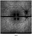
- the leading technique used to detect the RPE in the human eye, FAF is ineffective or poorly effective in the rodent eye (by way of non-limiting example), possibly owing to a relative paucity of fluorophores such as lipofuscin.
- FAF imaging in the human eye is performed using the blue spectrum of non-coherent light in the presence of stimulation/emission filters, or coherent blue light, and can identify areas of absent RPE (e.g . hypo-fluorescent signal) or abnormal RPE (e.g . hyper-fluorescent signal).
- the inability to clearly identify the RPE in an eye of an animal, in the absence of DNIRA has precluded direct correlation between pre-clinical studies and clinical observation.
- RPE layer such as, for example, DNIRA
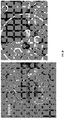
- DNIRA or variations thereof, allow for visualization of fluorescent immune cells in the eyes of an animal.
- DNIRA may optionally not comprise toxin administration.
- DNIRA is performed in a human patient and a toxin is not applied.
- DNIRA or variations thereof, allow for visualization of fluorescent immune cells in the eyes of human subject.
- DNIRA may not comprise toxin administration.
- DNIRA is used in the identification of an agent that is effective for treating an ocular disorder.
- DNIRA is used as a method to evaluate a patient that has, or may have, an ocular disorder (including, without limitation AMD and RPD).
- DNIRA is a surrogate biomarker for diagnosis and/or prognosis and/or progression of an ocular disorder (including, without limitation AMD and RPD).
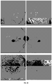
- DNIRA may be used to identify patterns, including lacy, reticular or leopard-like pattern of alternating hyper- and hypo-fluorescent DNIRA that is not seen in other imaging modalities, that are indicative of a ocular disease state (without limitation AMD and RPD).
- DNIRA may also be used to identify, and quantify, areas of hyper- and hypo-fluroescent DNIRA.
- DNIRA is used to identify hypofluorescent features of an eye. For instance, these areas appear black when imaged and therefore allow for easy quantitation (in contrast to ICG imaging, or in contrast to hyperfluroescent signal, which is grey-scale rather than black/white). Detection of hypofluroescent DNIRA, in some embodiments, is predictive of damaged or dead RPE.
- hypofluroescent DNIRA may indicate one or more of an absence of RPE, abnormal/unhealthy RPE (which is unable to uptake ICG dye), RPE that does not lie in contact with Bruch's Membrane (and so are no longer in a position to take up ICG dye from the choroidal vasculature), and the presence of lipid that could be located either between the RPE and BM (thus blocking ICG uptake), or could be internal to the RPE (thus blocking the RPE signal).
- DNIRA is used to identify hyperfluorescent features of an eye. For instance, these areas appear light when imaged and therefore allow for easy quantitation. Detection of hyperfluroescent DNIRA, in some embodiments, is predictive of macrophages, including M1 and/or M2 macrophages
- DNIRA is used biomarker for diagnosis of an ocular disease state (without limitation AMD and RPD) and prompts further evaluation and/or treatment with one of more agents, including without limitation those described herein.
- DNIRA is used as a biomarker for prognosis of an ocular disease state (without limitation AMD and RPD) and prompts further evaluation and/or treatment with one of more agents, including without limitation those described herein.
- DNIRA is used to improve identification of suitable patients for study recruitment and to evaluate progression of disease. In various embodiments, DNIRA is used to monitor disease progression.
- DNIRA is used as a companion diagnostic to any of the agents described herein.
- DNIRA is used to evaluate patient response to any of the agents described herein (including evaluating the effectiveness of any of the agents described herein and/or the likelihood of response to any of the agents described herein).
- the present invention further relates to the use of DNIRA as entrance inclusion or exclusion criteria, or endpoint analysis for clinical trial design.
- an ocular disorder is evaluated and/or identifiable using DNIRA or a technique known in the art.
- Illustrative techniques include: cSLO, FAF, OCT (including with cross-sectional, three-dimensional and en face viewing), SD-OCT (with cross-sectional, three-dimensional and en face viewing), angiography, or other imaging modalities including other wavelengths of fluorescence.
- these wavelengths range from blue to infrared, e.g ., 390 nm to 1 mm, including, for example, blue light, white light, red-free, near infra-red, infrared.
- an ocular disorder is evaluated and/or identifiable by patterns of FAF within patches of RPE damage or loss or outer retinal loss.
- the patterns are one or more of curvilinear, ribbon-like, reticular, oval, circular, scalloped, halo, and target-like lesions.
- an ocular disorder evaluated and/or identifiable by patterns of FAF within a border of patches of RPE damage or loss or outer retinal loss are one or more of curvilinear, ribbon-like, reticular, oval, circular, scalloped, halo, and target-like lesions.
- an ocular disorder is evaluated and/or identifiable by cross-sectional patterns or transverse patterns.
- the patterns are observed with OCT.
- the patterns are RPE and/or outer retinal loss or mounds, triangles, peaks or spikes found in the sub-retinal space.
- an ocular disorder is evaluated and/or identifiable by the presence of areas of hyper-fluorescent FAF in an eye of the subject and/or the presence of one or more areas of abnormally fluorescent FAF in an eye of the subject and/or by changes in one or more of blue spectrum fundus imaging, white-light fundus imaging, red-free fundus imaging, and OCT in an eye of the subject and/or by an increase (e.g .
- Illustrative changes include differences in an imaging pattern in the same of different subject and/or animal at two different time points and/or experimental or clinical conditions.
- changes can encompass changes in imaging data between two evaluations of the same subject and/or animal and/or changes in imaging data between a first subject and/or animal and the imaging data of a second subject and/or animal.
- an ocular disorder is evaluated and/or identifiable by the presence of one or more areas of distinct patterns of retinal imaging in the eye of a subject, wherein the retinal imaging is one or more of white light, red-free light, blue light, FAF, near infra-red (NIR), infra-red (IR), angiography, and DNIRA and/or the presence of one or more areas of abnormally-fluorescent FAF in the eye of a subject and/or an increase (e.g.
- the ocular disorder is a diabetic eye disease, by way non-limiting example, diabetic retinopathy and DME.
- the diabetic eye disease is found in a patient with type 1 or type 2 diabetes.
- a diabetic eye disease patient to be treated may have a blood glucose level that exceeds normal (about 70-130 milligrams per deciliter or mg/dL before meals, and less than 180 mg/dL one to two hours after a meal).
- the diabetic eye disease is diabetic retinopathy in one or more of the following stages: Mild Nonproliferative Retinopathy (in which microaneurysms may occur); Moderate Nonproliferative Retinopathy (in which some blood vessels that nourish the retina may be blocked); and Severe Nonproliferative Retinopathy (in which more blood vessels may be blocked, depriving several areas of the retina with their blood supply); and Proliferative Retinopathy (in which signals sent by the retina for nourishment trigger the growth of new blood vessels.
- Mild Nonproliferative Retinopathy in which microaneurysms may occur
- Moderate Nonproliferative Retinopathy in which some blood vessels that nourish the retina may be blocked
- Severe Nonproliferative Retinopathy in which more blood vessels may be blocked, depriving several areas of the retina with their blood supply
- Proliferative Retinopathy in which signals sent by the retina for nourishment trigger the growth of new blood vessels.
- the present methods for treatment or prevention of a diabetic eye disease further comprise performing laser surgery (e.g . scatter laser treatment, focal laser treatment, photocoagulation) and/or vitrectomy (e.g . in which blood is removed from the center of a patient's eye) and/or administering a steroid and/or administering an anti-VEGF agent such as Ranibizumb (LUCENTIS), Bevizumab (AVASTIN) or Aflibercept (EYLEA).
- laser surgery e.g . scatter laser treatment, focal laser treatment, photocoagulation
- vitrectomy e.g . in which blood is removed from the center of a patient's eye
- an anti-VEGF agent such as Ranibizumb (LUCENTIS), Bevizumab (AVASTIN) or Aflibercept (EYLEA).
- the diabetic eye disease is a cataract or glaucoma (e.g ., neovascular glaucoma).
- disorders associate with imaging (FAF) patterns such as "diffuse trickling” may be evaluated, treated or prevented using methods disclosed herein.
- Diffuse trickling patterns are known in the art (see, e.g., Schmitz-Valckenberg et al. Survey of Ophthalmology. 2009 Jan-Feb; 54(1):96-117 ; Bindewald et al. British Journal of Ophthalmology. 2005 Jul; 89(7):874-8 ; Holz et al. Am. J Ophthalmology. 2007 Mar; 143(3):463-72 ).
- DNIRA is used to identify and/or diagnoses any of the ocular disorder described herein.
- RPE toxicity, RPE loss, and the dry AMD are identifiable using DNIRA, e.g. with sodium iodate (NaIO 3 ) or in the presence of disease. Areas analogous to geographic atrophy can be detected by, for example, tissue analysis (for example, by observing the loss of an RPE cell marker such as RPE65 and/or the loss of binucleate cell staining), and/or in vivo, using DNIRA, of NaIO 3 .treated- eyes.
- RPE toxicity, RPE loss and the dry AMD are identifiable using FAF which shows a hyperfluorescent FAF signal, which may be complex, within the area of imminent tissue loss and/or at its margins.
- This complex hyperfluorescent FAF signal develops, in the days or weeks after NaIO 3 treatment, into a pattern of FAF, which can be complex and can include, but is not limited to ribbon-like, curvilinear, pisciform, scalloped, interlacing, branching, or reticular hyperfluorescence. Such patterns mimic those found in clinical dry AMD.
- Such FAF may also be hypofluorescent.
- angiography performed immediately prior to, or coincident with, the emergence of the areas of altered FAF may demonstrate an increase (including a transient increase) in permeability across the epithelial barrier between choroid and retina (such as, for example, observed by leakage of dyes that may be injected prior to imaging; such dyes include ICG).
- This barrier normally includes the inner choroid, Bruch's Membrane, the RPE cells, and, possibly, the configuration of the outer photoreceptors in conjunction with the RPE and the outer limiting membrane.
- a transient breakdown of this specialized epithelial barrier may underlie a sequence of events thereafter including folding, undulation, or deformation of the outer retina, photoreceptor layer, and/or movement/migration of inflammatory cells from the choroid to the subretinal space.
- this phase may be transient and resolved, or subclinical in its extent.
- one or more of the ocular disorder are identifiable by the presence of areas of hyper- and/or abnormal-fluorescent FAF, or other wavelengths of light from 350 nm to 1,000 nm. In some embodiments, one or more of the ocular disorders are identifiable using DNIRA.
- one or more of the ocular disorders are identifiable by, for example, cSLO, FAF, OCT (including with cross-sectional, three-dimensional and en face viewing), SD-OCT (with cross-sectional, three-dimensional and en face viewing), or other imaging modalities including other wavelengths of fluorescence (e.g. wavelengths ranging from blue to infrared, e.g., 390 nm to 1 mm, including, for example, blue light, white light, red-free, near infra-red, or infrared).
- cSLO e.g. wavelengths ranging from blue to infrared, e.g., 390 nm to 1 mm, including, for example, blue light, white light, red-free, near infra-red, or infrared.
- the ocular disorder may be of a certain stage or progression. In some embodiments, the ocular disorder may be incipient, emerging, quiescent, advancing or active.
- a compound of Formula I is also administered with an additional therapeutic agent.
- the additional therapeutic agent may be used in a combination therapy with the agents effective for treating an ocular disorder described herein.
- the agents effective for treating an ocular disorder described herein is administered to a patient undergoing treatment with one or more additional therapeutic agents.
- Additional therapeutic agents include, for example, one or more of an anti-VEGF agent, an ACE inhibitor, a PPAR-gamma agonist or partial agonist, a renin inhibitor, a steroid, and an agent that modulates autophagy, as well as a semapimod, a MIF inhibitor, a CCR2 inhibitor, CKR-2B, a 2-thioimidazole, CAS 445479-97-0, CCX140, clodronate, a clodonate-liposome preparation or gadolinium chloride.
- An agent of the invention is a compound of Formula I : or a pharmaceutically acceptable salts thereof, wherein each of R 1 and R 2 is independently H or a C 1 -C 6 alkyl and R 3 is H or a C 1 -C 6 alkyl.
- R 1 and R 2 are independently hydrogen, methyl or ethyl.
- R 1 and R 2 are independently methyl or ethyl.
- R 1 and R 2 are methyl.
- R 3 is hydrogen, methyl or ethyl.
- R 3 is hydrogen
- the compounds of Formula I are in the form of a pharmaceutically acceptable salt.
- R 1 and R 2 are methyl and R 3 is hydrogen.
- the compound of Formula I is 2-((1-benzylindazol-3-yl) methoxy)-2-methyl propionic acid, also known as 2-methyl-2-[[1-(phenylmethyl)-1H-indazol-3yl]methoxy] propanoic acid.
- Such a compound is commonly known as bindarit and has the following structure:
- Bindarit is an inhibitor of MCP-1 production in vitro and in vivo and, without wishing to be bound by theory, its beneficial effects in animal models of inflammation may be related to this anti-MCP-1 activity (see Mirolo, et al. Eur Cytokine Netw 2008;19:119-122 ). Bindarit has also been shown to selectively inhibit the production of MCP-2 and MCP-3 (see Mirolo, et al. Eur Cytokine Netw 2008;19:119-122 ).
- the fluorescent compound is suitable for imaging with various wavelengths of fluorescence. In some embodiments, these wavelengths range from visible light to infrared, e.g ., 390 nm to 1 mm, including, for example, blue light, white light, and near-infrared.
- the dye is a near-infrared dye. In some embodiments, the dye is ICG.
- the fluorescent compound is suitable for imaging with various wavelengths of fluorescence. In some embodiments, these wavelengths range from visible light to infrared, e.g ., about 390 nm to about 1 mm, including, for example, blue light, white light, and near-infrared. In some embodiments, the absorption is from about 390 nm to about 1 mm. In some embodiments, the emission is from about 390 nm to about 1 mm.
- the fluorescent compound absorbs light at a wavelength of about 600 nm to about 900 nm and/or emits light at a wavelength of about 750 nm to about 950 nm.
- the dye is a near-infrared dye. In some embodiments, the dye is ICG. In some embodiments, the ICG excitation/emission filters are 795 nm (excitation)/810 nm (emission).
- the dose of the fluorescent compound e.g . a dye (including ICG)
- the dose of ICG is from about 0.1 to about 10 mg/kg of an animal.
- the dose is about 0.1, or about 0.3, or about 0.5, or about 1.0, or about 2.0, or about 3.0, or about 4.0, or about 5.0, or about 6.0, or about 7.0, or about 8.0, or about 9.0, or about 10.0 mg/kg of an animal.
- the fluorescent compounds, or metabolites thereof cause a fluorescence which occurs in RPE cells and/or immune cells.
- the methods described herein do not comprise administering (i) an additional amount of fluorescent compound to the animal or (ii) a second fluorescent compound to the animal.
- a toxin known to affect ocular tissue including but not limited to, the RPE or retina, is provided.
- the toxin is one or more of aluminium, aminophenoxyalkanes (a non-limiting example includes, but is not limited to, 1,4,-bis(4-aminophenoxy)-2-phenylbenzene to rats), cationic amphophilic drugs /tricyclic antidepressants (non-limiting examples include but are not limited to amiodarone, chloroamitriptyline, chlorphentermine, clomipramine, imipramine, iprindole, various aminoglycosides, and other cationic amphophilic compounds), desferrioxamine, dl-(p-trifluoromethylphenyl) isopropylamine hydrochloride, fluoride (e.g.
- iodate non-limiting examples include but are not limited to, sodium or potassium iodate
- iodoacetate lead, methanol and formic acid
- 4,4'-Methylenedianiline N-methyl-N-nitrosurea, naphthalene, napthol, nitroaniline (N-3-pyridylmethyl-N'-p-nitrophenylurea/nitroanilin/pyriminil)
- organophosphates non-limiting examples include but are not limited to ethylthiometon, fenthion, and fenitrothion
- oxalate a non-limiting example includes but are not limited to dibutyl oxalate
- phenothiazines non-limiting examples include but are not limited to piperidylchlorophenothiazine, thioridazine, and chlorpromazine
- quinolines a non-limiting example includes, but is not limited to, chloroquine and hydroxy
- the toxin is an iodate. In some embodiments, the toxin is sodium or potassium iodate.
- the toxin induces atrophy of ocular tissue.
- the atrophy comprises necrosis and/or apoptosis.
- the atrophy comprising necrosis and/or apoptosis is of RPE cells.
- the toxin reduces or modifies autophagy.
- the toxin induces geographic atrophy and/or the expansion of geographic atrophy.
- the toxin induces one or more of the above-mentioned effects.
- the toxin is administered one time, or two times, or three times. In some embodiments, the toxin is administered one time, or two times, or three times, or four times, or five times. In some embodiments, a second, or third, or fourth, or fifth administration is within about a day, or 1 week, or 1 month of the first.
- the toxin administered may be may be one or more of the agents described herein.
- the second, or third, or fourth, or fifth pulse is a modulator of autophagy, cell survival, cell death, proliferation, regeneration, and the like, as described herein and as known in the art.
- the methods described herein comprise administering (i) an additional amount of a first toxin to the animal and/or (ii) a second toxin to the animal. In some embodiments, the methods described herein comprise further comprise observing a reduction in the rate of formation, growth or expansion of patches of ocular tissue atrophy or patches of tissue loss.
- a suitable dosage may be in a range of about 0.1 mg/kg to about 100 mg/kg of body weight of the subject, for example, about 0.1 mg/kg, about 0.2 mg/kg, about 0.3 mg/kg, about 0.4 mg/kg, about 0.5 mg/kg, about 0.6 mg/kg, about 0.7 mg/kg, about 0.8 mg/kg, about 0.9 mg/kg, about 1 mg/kg, about 1.1 mg/kg, about 1.2 mg/kg, about 1.3 mg/kg, about 1.4 mg/kg, about 1.5 mg/kg, about 1.6 mg/kg, about 1.7 mg/kg, about 1.8 mg/kg, about 1.9 mg/kg, about 2 mg/kg, about 3 mg/kg, about 4 mg/kg, about 5 mg/kg, about 6 mg/kg, about 7 mg/kg, about 8 mg/kg, about 9 mg/kg, about 10 mg/kg, about 11 mg/kg, about 12 mg/kg, about 13 mg
- the toxin is NaIO 3 and the dose is about 50 mg/kg of body weight. In one embodiment, the toxin is NaIO 3 and the dose is about 45 mg/kg of body weight. In one embodiment, the toxin is NaIO 3 and the dose is about 30 mg/kg of body weight.
- Any agent described herein can possess a sufficiently basic functional group, which can react with an inorganic or organic acid, or a carboxyl group, which can react with an inorganic or organic base, to form a pharmaceutically acceptable salt.
- a pharmaceutically acceptable acid addition salt is formed from a pharmaceutically acceptable acid, as is well known in the art.
- Such salts include the pharmaceutically acceptable salts listed in Journal of Pharmaceutical Science, 66, 2-19 (1977 ) and The Handbook of Pharmaceutical Salts; Properties, Selection, and Use. P. H. Stahl and C. G. Wermuth (eds.), Verlag, Zurich (Switzerland) 2002 .
- salts include, by way of non-limiting example, sulfate, citrate, acetate, oxalate, chloride, bromide, iodide, nitrate, bisulfate, phosphate, acid phosphate, isonicotinate, lactate, salicylate, acid citrate, tartrate, oleate, tannate, pantothenate, bitartrate, ascorbate, succinate, maleate, gentisinate, fumarate, gluconate, glucaronate, saccharate, formate, benzoate, glutamate, methanesulfonate, ethanesulfonate, benzenesulfonate, p-toluenesulfonate, camphorsulfonate, pamoate, phenylacetate, trifluoroacetate, acrylate, chlorobenzoate, dinitrobenzoate, hydroxybenzoate, methoxybenzoate, methylbenzo
- Suitable bases include, but are not limited to, hydroxides of alkali metals such as sodium, potassium, and lithium; hydroxides of alkaline earth metal such as calcium and magnesium; hydroxides of other metals, such as aluminum and zinc; ammonia, and organic amines, such as unsubstituted or hydroxy-substituted mono-, di-, or tri-alkylamines, dicyclohexylamine; tributyl amine; pyridine; N-methyl, N-ethylamine; diethylamine; triethylamine; mono-, bis-, or tris-(2-OH-lower alkylamines), such as mono-; bis-, or tris-(2-hydroxyethyl)amine, 2-hydroxy-tert-butylamine, or tris-(hydroxymethyl
- an agent of the invention is in the form or a pharmaceutically acceptable salt.
- the pharmaceutically acceptable salt is a sodium salt.
- R 3 of the compounds of Formula I is hydrogen
- the compounds of Formula I are in the form of a pharmaceutically acceptable salt.
- the compound of Formula I is a pharmaceutically acceptable salt of bindarit.
- methotrexate is in the form or a pharmaceutically acceptable salt.
- the pharmaceutically acceptable salt of methotrexate is methotrexate sodium.
- any agent described herein can be administered to a subject as a component of a composition that comprises a pharmaceutically acceptable carrier or vehicle.
- Such compositions can optionally comprise a suitable amount of a pharmaceutically acceptable excipient so as to provide the form for proper administration.
- Pharmaceutical excipients can be liquids, such as water and oils, including those of petroleum, animal, vegetable, or synthetic origin, such as peanut oil, soybean oil, mineral oil, sesame oil and the like.
- the pharmaceutical excipients can be saline, gum acacia, gelatin, starch paste, talc, keratin, colloidal silica, urea and the like.
- auxiliary, stabilizing, thickening, lubricating, and coloring agents can be used.
- the pharmaceutically acceptable excipients are sterile when administered to a subject. Water is a useful excipient when any agent described herein is administered intravenously.
- Saline solutions and aqueous dextrose and glycerol solutions can also be employed as liquid excipients, specifically for injectable solutions.
- suitable pharmaceutical excipients also include starch, glucose, lactose, sucrose, gelatin, malt, rice, flour, chalk, silica gel, sodium stearate, glycerol monostearate, talc, sodium chloride, dried skim milk, glycerol, propylene, glycol, water, ethanol and the like.
- Any agent described herein, if desired, can also comprise minor amounts of wetting or emulsifying agents, or pH buffering agents.
- any agent described herein can take the form of solutions, suspensions, emulsion, intra-ocular injection, intra-vitreal injection, topical ophthalmic drops, sub-conjunctival injection, sub-Tenon's injection, trans-scleral formulations, tablets, pills, pellets, capsules, capsules containing liquids, powders, sustained-release formulations, suppositories, emulsions, aerosols, sprays, suspensions, or any other form suitable for use.
- the composition is suitable for an intra-vitreal injection ( see, e.g., ILUVIEN or similar forms) or implantation ( see, e.g., RETISERT or similar forms).
- the composition is in the form of a capsule ( see, e.g., U.S. Patent No. 5,698,155 ).
- suitable pharmaceutical excipients are described in Remington's Pharmaceutical Sciences 1447-1676 (Alfonso R. Gennaro eds., 19th ed. 1995 ).
- any agent described herein is formulated for ophthalmic administration, including, for example, intravitreal or intraocular administration, topical, and/or intravenous administration.
- compositions for administration comprise sterile isotonic aqueous buffer. Where necessary, the compositions can also include a solubilizing agent.
- the agents can be delivered with a suitable vehicle or delivery device as known in the art. Combination therapies outlined herein can be co-delivered in a single delivery vehicle or delivery device.
- Compositions for administration can optionally include a local anesthetic such as lignocaine to lessen pain at the site of the injection.
- any agent described herein is formulated in accordance with routine procedures as a composition adapted for intra-ocular administration.
- compositions for oral delivery can be in the form of tablets, lozenges, aqueous or oily suspensions, granules, powders, emulsions, capsules, syrups, or elixirs, for example.
- Orally administered compositions can comprise one or more agents, for example, sweetening agents such as fructose, aspartame or saccharin; flavoring agents such as peppermint, oil of wintergreen, or cherry; coloring agents; and preserving agents, to provide a pharmaceutically palatable preparation.
- compositions can be coated to delay disintegration and absorption in the gastrointestinal tract thereby providing a sustained action over an extended period of time.
- Selectively permeable membranes surrounding an osmotically active driving any agent described herein are also suitable for orally administered compositions.
- fluid from the environment surrounding the capsule is imbibed by the driving compound, which swells to displace the agent or agent composition through an aperture.
- delivery platforms can provide an essentially zero order delivery profile as opposed to the spiked profiles of immediate release formulations.
- a time-delay material such as glycerol monostearate or glycerol stearate can also be useful.
- Oral compositions can include standard excipients such as mannitol, lactose, starch, magnesium stearate, sodium saccharin, cellulose, and magnesium carbonate. In one embodiment, the excipients are of pharmaceutical grade.
- the ingredients may be supplied either separately or mixed together in unit dosage form, for example, as a pre-mixed solution, dry lyophilized-powder, or water-free concentrate in a hermetically sealed container such as an ampule, pre-filled syringe, or sachette indicating the quantity of active agent.
- a hermetically sealed container such as an ampule, pre-filled syringe, or sachette indicating the quantity of active agent.
- any agent described herein is to be administered by intra-vitreal or intra-ocular delivery, it can be dispensed, for example, with a pre-filled syringe or injector, or in an ampule for withdrawal into a suitable syringe.
- agent described herein is to be administered by infusion, it can be dispensed, for example, with an infusion bottle containing sterile pharmaceutical grade water or saline.
- an ampule of sterile water for injection or saline can be provided so that the ingredients
- Any agent described herein can be administered by controlled-release or sustained-release means or by delivery devices that are well known to those of ordinary skill in the art. Examples include, but are not limited to, those described in U.S. Patent Nos. 3,845,770 ; 3,916,899 ; 3,536,809 ; 3,598,123 ; 4,008,719 ; 5,674,533 ; 5,059,595 ; 5,591,767 ; 5,120,548 ; 5,073,543 ; 5,639,476 ; 5,354,556 ; and 5,733,556 .
- Such dosage forms can be useful for providing controlled- or sustained-release of one or more active ingredients using, for example, hydropropylmethyl cellulose, other polymer matrices, gels, permeable membranes, osmotic systems, multilayer coatings, microparticles, nanoparticles, liposomes, microspheres, or a combination thereof to provide the desired release profile in varying proportions.
- Suitable controlled- or sustained-release formulations known to those skilled in the art, including those described herein can be readily selected for use with the active ingredients of the agents described herein.
- the invention thus provides single unit dosage forms suitable for oral administration such as, but not limited to, tablets, capsules, gelcaps, and caplets that are adapted for controlled- or sustained-release.
- Controlled- or sustained-release of an active ingredient can be stimulated by various conditions, including but not limited to, changes in pH, changes in temperature, stimulation by an appropriate wavelength of light, concentration or availability of enzymes, concentration or availability of water, or other physiological conditions or compounds.
- compositions can be prepared according to conventional mixing, granulating, coating or polymerization methods, respectively, and the present compositions can comprise, in one embodiment, from about 0.1% to about 99%; and in another embodiment from about 1% to about 70% of any agent described herein by weight or volume.
- any agent described herein acts synergistically when co-administered with another agent and is administered at doses that are lower than the doses commonly employed when such agents are used as monotherapy.
- the dosage any agent described herein as well as the dosing schedule can depend on various parameters, including, but not limited to, the ocular disorder being treated, the subject's general health, and the administering physician's discretion.
- Any agent described herein can be administered prior to (e.g., 5 minutes, 15 minutes, 30 minutes, 45 minutes, 1 hour, 2 hours, 4 hours, 6 hours, 12 hours, 24 hours, 48 hours, 72 hours, 96 hours, 1 week, 2 weeks, 3 weeks, 4 weeks, 5 weeks, 6 weeks, 8 weeks, or 12 weeks before), concurrently with, or subsequent to (e.g., 5 minutes, 15 minutes, 30 minutes, 45 minutes, 1 hour, 2 hours, 4 hours, 6 hours, 12 hours, 24 hours, 48 hours, 72 hours, 96 hours, 1 week, 2 weeks, 3 weeks, 4 weeks, 5 weeks, 6 weeks, 8 weeks, or 12 weeks after) the administration of an additional therapeutic agent, to a subject in need thereof.
- any agent described herein is administered 1 minute apart, 10 minutes apart, 30 minutes apart, less than 1 hour apart, 1 hour apart, 1 hour to 2 hours apart, 2 hours to 3 hours apart, 3 hours to 4 hours apart, 4 hours to 5 hours apart, 5 hours to 6 hours apart, 6 hours to 7 hours apart, 7 hours to 8 hours apart, 8 hours to 9 hours apart, 9 hours to 10 hours apart, 10 hours to 11 hours apart, 11 hours to 12 hours apart, no more than 24 hours apart or no more than 48 hours apart.
- an agent of the invention, including compounds of Formula I , methotrexate or their pharmaceutically acceptable salts, and one or more additional therapeutic agents are administered within 3 hours.
- an agent of the invention, including compounds of Formula I , methotrexate or their pharmaceutically acceptable salts, and one or more additional therapeutic agents are administered at 1 minute to 24 hours apart.
- any agent described herein that is admixed with the carrier materials to produce a single dosage can vary depending upon the subject being treated and the particular mode of administration. In vitro or in vivo assays can be employed to help identify optimal dosage ranges.
- doses that are useful are known to those in the art.
- doses may be determined with reference Physicians' Desk Reference, 66th Edition, PDR Network; 2012 Edition (December 27, 2011 ).
- the dosage of any agent described herein can depend on several factors including the severity of the condition, whether the condition is to be treated or prevented, and the age, weight, and health of the subject to be treated. Additionally, pharmacogenomic (the effect of genotype on the pharmacokinetic, pharmacodynamic or efficacy profile of a therapeutic) information about a particular subject may affect dosage used. Furthermore, the exact individual dosages can be adjusted somewhat depending on a variety of factors, including the specific combination of the agents being administered, the time of administration, the route of administration, the nature of the formulation, the rate of excretion, the particular ocular disorder being treated, the severity of the disorder, and the anatomical location of the disorder. Some variations in the dosage can be expected.
- the administering is effected orally or intra-vascularly, or intraocularly, or periocularly, or to the ocular surface
- the dosage of an agent of the invention including, for example, Formula I , methotrexate or a pharmaceutically acceptable salt thereof and/or additional therapeutic agent is normally 0.003 mg to 5.0 mg per eye per administration, or 0.03 mg to 3.0 mg per eye per administration, or 0.1 mg to 1.0 mg per eye per administration.
- the dosage is 0.03 mg, 0.3 mg, 1.5 mg or 3.0 mg per eye.
- the dosage is 0.5 mg per eye.
- the dosage can range from 0.01 mL to 0.2 mL administered per eye, or 0.03 mL to 0.15 mL administered per eye, or 0.05 mL to 0.10 mL administered per eye.
- the administration is 400 ⁇ g of compound, monthly for at least three months.
- the dosage of any agent described herein may be 0.001 mg/kg/day to 100 mg/kg/day, 0.01 mg/kg/day to 50 mg/kg/day, or 0.1 mg/kg/day to 10 mg/kg/day.
- the dosage of any agent described herein is normally 0.001 mg to 1000 mg per day, 1 mg to 600 mg per day, or 5 mg to 30 mg per day.
- oral dosage is 600 mg per day.
- the oral dosage is two 300 mg doses per day.
- oral dosage is 7.5 mg per week to 15 mg per week.
- the dosage is normally 0.1 mg to 250 mg per day, 1 mg to 20 mg per day, or 3 mg to 5 mg per day. Injections may be given up to four times daily.
- the dosage of any agent described herein is normally 0.1 mg to 1500 mg per day, or 0.5 mg to 10 mg per day, or 0.5 mg to 5 mg per day. A dosage of up to 3000 mg per day can be administered.
- Administration may be, by way of non-limiting example, intra-ocular, intra-vitreal, topical (including, but not limited to, drops and ointment), sub-conjunctival, sub-Tenon's, trans-scleral, suprachoroidal, subretinal, and via iontophoresis.
- routes of administration may also be used, such as, for example: intradermal, intramuscular, intraperitoneal, intravenous, subcutaneous, intranasal, epidural, oral, sublingual, intranasal, intracerebral, intravaginal, transdermal, rectally, by inhalation, or topically, particularly to the ears, nose, eyes, or skin.
- the mode of administration can be left to the discretion of the practitioner, and depends in-part upon the site of the medical condition. In most instances, administration results in the release of any agent described herein into the bloodstream.
- Any agent described herein can be administered orally.
- Such agents can also be administered by any other convenient route, for example, by intravenous infusion or bolus injection, by absorption through epithelial or mucocutaneous linings ( e.g ., oral mucosa, rectal and intestinal mucosa, etc .) and can be administered together with another biologically active agent. Administration can be systemic or local.
- Various delivery systems are known, e.g ., encapsulation in liposomes, microparticles, microcapsules, capsules, etc., and can be used to administer.
- Further methods of administration include but are not limited to intra-ocular, intra-vitreal, topical ocular (including but not limited to drops, ointments and inserts), sub-conjunctival, sub-Tenon's, suprachoroidal, trans-scleral, intradermal, intramuscular, intraperitoneal, intravenous, subcutaneous, intranasal, epidural, oral, sublingual, intranasal, intracerebral, intravaginal, transdermal, rectally, by inhalation, or topically, particularly to the ears, nose, eyes, or skin. In some embodiments, more than one of any agent described herein is administered to the eye.
- Administration may be, by way of non-limiting example, intra-ocular, intra-vitreal, topical (including, but not limited to, drops and ointment), sub-conjunctival, sub-Tenon's, trans-scleral, and iontophoresis.
- the mode of administration can be left to the discretion of the practitioner, and depends in-part upon the site of the medical condition. In most instances, administration results in the release into the bloodstream.
- delivery can be in a vesicle, in particular a liposome (see Langer, 1990, Science 249:1527-1533 ; Treat et al., in Liposomes in the Therapy of Infectious Disease and Cancer, Lopez-Berestein and Fidler (eds.), Liss, New York, pp. 353-365 (1989 ).
- delivery can be in a controlled release system.
- a slow release intra-ocular device may be used. In some embodiments, this device consists of a locally delivered erodible or non-erodable liquid, gel, polymer, etc.
- polymeric materials can be used (see Medical Applications of Controlled Release, Langer and Wise (eds.), CRC Pres., Boca Raton, Florida (1974 ); Controlled Drug Bioavailability, Drug Product Design and Performance, Smolen and Ball (eds.), Wiley, New York (1984 ); Ranger and Peppas, 1983, J. Macromol. Sci. Rev. Macromol. Chem. 23:61 ; see also Levy et al., 1985, Science 228:190 ; During et al., 1989, Ann. Neurol. 25:351 ; Howard et al., 1989, J. Neurosurg. 71:105 ).
- a controlled-release system can be placed in proximity of the target area to be treated, e.g., the retina, thus requiring only a fraction of the systemic dose ( see , e.g ., Goodson, in Medical Applications of Controlled Release, supra, vol. 2, pp. 115-138 (1984 )).
- Other controlled-release systems discussed in the review by Langer, 1990, Science 249:1527-1533 ) may be used.
- Administration of any agent described herein can, independently, be one to four times daily or one to four times per month or one to six times per year or once every two, three, four or five years. Administration can be for the duration of one day or one month, two months, three months, six months, one year, two years, three years, and may even be for the life of the subject. Chronic, long-term administration will be indicated in many cases.
- the dosage may be administered as a single dose or divided into multiple doses. In general, the desired dosage should be administered at set intervals for a prolonged period, usually at least over several weeks or months, although longer periods of administration of several months or years or more may be needed.
- the dosage regimen utilizing any agent described herein can be selected in accordance with a variety of factors including type, species, age, weight, sex and medical condition of the subject; the severity of the condition to be treated; the route of administration; the renal or hepatic function of the subject; the pharmacogenomic makeup of the individual; and the specific compound of the invention employed. Any agent described herein can be administered in a single daily dose, or the total daily dosage can be administered in divided doses of two, three or four times daily. Furthermore, any agent described herein can be administered continuously rather than intermittently throughout the dosage regimen.
- the present invention provides an agent of the invention for use in a method for treating an ocular disorder described herein.
- the "agent of the invention” comprises compounds useful for both monotherapy and combination therapy (e.g. as an additional therapeutic agent).
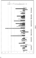
- the invention relates to an agent of the invention for use in a method for treating an ocular disorder in a subject in need thereof, wherein the subject has an increased or decreased expression or activity of one or more of CD64, IDO, SOCS1, CXCL10 Marco, Socs3, Nos2, Il12b, Ptgs2 (Cox2), Il23 ⁇ (Il23p19), and Ido1.
- the subject has an increased or decreased expression or activity of one or more of MRC1, TGM2, CD23, CCL22 Relma (Fizz1, Retnla), Socs2, Irf4, Chia (Amcase), Chi3l1 (Gp39, Ykl40), Chi3l2 (Ykl39), Chi3l3(Ym1), Cxcl13, Ccl12, Ccl24, and Klf4.
- the subject has an increased or decreased expression or activity of one or more of Ccl5, CD163, Cx3Cr1, Faslg, Gfap, Csf2, Icam1, Ifng, Il10, Il12b, Il13, Il17a, Il18, Il1b, Il22, Il4, Il6, Klf4, Mrc1, Myd88, Nlrp3, Nos2, Ppary, Tgfb1, Tlr4, Tnf, Vcam1, Ccl2, Ccl5, Ccl7, Ccr2, Socs1, Socs3, Stat1, Stat3, and Stat6.
- the subject has an increased or decreased expression or activity of one or more of Myd88, Klf4, Nlrp3, Ccl2, Ccl5, Ccl7, Socs1, Socs3, Stat1, Stat3, and Stat6.
- bindarit leads to differential gene expression of any of the following, directly or indirectly Ccl5, CD163, Cx3Cr1, Faslg, Gfap, Csf2, Icam1, Ifng, Il10, Il12b, 1113, Il17a, 1118, Il1b, 1122, 114, 116, Klf4, Mrc1, Myd88, Nlrp3, Nos2, Ppary, Tgfb1, Tlr4, Tnf, Vcam1, Ccl2, Ccl5, Ccl7, Ccr2, Socs1, Socs3, Stat1, Stat3, and Stat6, optionally in macrophages or RPE directly.
- bindarit modulates NF ⁇ B.
- the subject is not undergoing treatment with and/or is unresponsive to one or more of an anti-factor D antibody (e.g. lampalizumab (Genentech)), an anti- ⁇ -amyloid (anti-A ⁇ ) antibody (e.g. GSK933776 (GSK)), a corticosteroid (e.g. fluocinolone acetonide), MC-1101 (MacuCLEAR), a CD34+ stem cell therapy, an anti-VEGF antibody (e.g. Ranibizumab), brimonidine (Alphagan), an anti-C5 complement antibody (e.g.
- an anti-factor D antibody e.g. lampalizumab (Genentech)
- an anti- ⁇ -amyloid (anti-A ⁇ ) antibody e.g. GSK933776 (GSK)
- a corticosteroid e.g. fluocinolone acetonide
- MC-1101 MacuCLEAR
- CD34+ stem cell therapy e.g. Ranibi
- the subject has evidence of AMD as confirmed by the presence of at least 1 druse greater than about 125 ⁇ m in diameter. In some embodiments, the subject has no evidence of prior or active choroidal neovascularization (CNV). In some embodiments, the subject has one or more well-demarcated GA lesions of a total area of about 2 to about 20 mm 2 in one or more eye. In some embodiments, the subject has a best-corrected visual acuity score of greater than about 35 letters or a Snellen VA equivalent of about 20/200 or better.
- An evaluation of any of the treatments disclosed herein can comprise optical imaging, including, by way of non-limiting example, cSLO, FAF, OCT (including with cross-sectional, three-dimensional and en face viewing), SD-OCT (with cross-sectional, three-dimensional and en face viewing), or other imaging modalities including other wavelengths of fluorescence (e.g. wavelengths ranging from blue to infrared, e.g., 390 nm to 1 mm, including, for example, blue light, white light, red-free, near infra-red, or infrared).
- optical imaging including, by way of non-limiting example, cSLO, FAF, OCT (including with cross-sectional, three-dimensional and en face viewing), SD-OCT (with cross-sectional, three-dimensional and en face viewing), or other imaging modalities including other wavelengths of fluorescence (e.g. wavelengths ranging from blue to infrared, e.g., 390 nm to 1
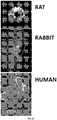
- the subject and/or animal is a mammal, e.g., a human, mouse, rat, guinea pig, dog, cat, horse, cow, pig, rabbit, sheep, or non-human primate, such as a monkey, chimpanzee, or baboon.
- the subject and/or animal is a non-mammal, such, for example, a zebrafish.
- the subject and/or animal may comprise fluorescently-tagged cells (with e.g. GFP).
- the subject and/or animal is a transgenic animal comprising a fluorescent cell, such as, for example, an RPE cell and/or an immune cell.
- the subject and/or animal is a human.
- the human is a pediatric human.
- the human is an adult human.
- the human is a geriatric human.
- the human may be referred to as a patient.
- the human has an age in a range of from about 0 months to about 6 months old, from about 6 to about 12 months old, from about 6 to about 18 months old, from about 18 to about 36 months old, from about 1 to about 5 years old, from about 5 to about 10 years old, from about 10 to about 15 years old, from about 15 to about 20 years old, from about 20 to about 25 years old, from about 25 to about 30 years old, from about 30 to about 35 years old, from about 35 to about 40 years old, from about 40 to about 45 years old, from about 45 to about 50 years old, from about 50 to about 55 years old, from about 55 to about 60 years old, from about 60 to about 65 years old, from about 65 to about 70 years old, from about 70 to about 75 years old, from about 75 to about 80 years old, from about 80 to about 85 years old, from about 85 to about 90 years old, from about 90 to about 95 years old or from about 95 to about 100 years old.
- the subject is a non-human animal, and therefore the invention pertains to veterinary use.
- the non-human animal is a household pet.
- the non-human animal is a livestock animal.
- a subject's and/or an animal's eye comprises (i) a fluorescent compound in an amount effective to indicate the presence of an ocular disorder in the subject and/or animal and (ii) a toxin in an amount effective to induce atrophy of ocular tissue.
- a subject and/or animal is administered an agent of the invention or is not administered an agent of the invention.
- immune cells include cells of a subject's and/or animal's innate immune system. In some embodiments, such cells include, but are not limited to, macrophage, monocyte, and microglial cells. In various embodiments, the invention provides for detecting a presence, detecting an absence, or measuring an amount of immune cells in a subject's and/or animal's eye
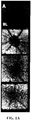
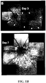
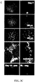
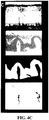
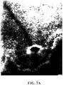
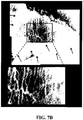
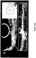
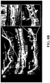
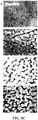
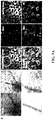
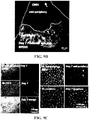
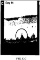
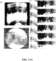
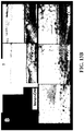
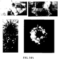
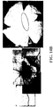
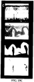
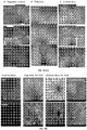
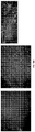
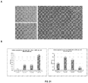
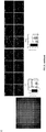
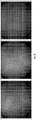
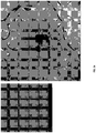
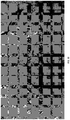
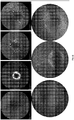
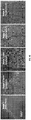
- Example 1 Systemic Injection of the RPE Toxin, NaIO 3 , Induces Complex Patterns of FAF Similar to Those of AMD and/or RPD
- the RPE toxin, sodium iodate (NaIO 3 ) generated patchy loss of the RPE and hypofluorescent DNIRA, in the rat eye similar to geographic atrophy (GA).
- G geographic atrophy
- ICG Cardiogreen
- sodium iodate Harris haematoxylin and eosin
- Tropicamide 0.8%, in 5% phenylephrine hydrochloride solution (Diophenyl-T) was from Sandoz Canada Inc (Boucherville, QC, Canada), and GenTeal lubricating eye drops were from Novartis Pharmaceuticals Canada Inc (Dorval, QC, Canada).
- Mouse anti-rat CD68 antibody was from AbD Serotec (Oxford, UK), and mouse IgG was from Santa Cruz Biotechnology (Santa Cruz, CA, USA). Rabbit anti-lba-1 was from Wako Pure Chemical Industries Ltd (Osaka, Japan). Alexa-labeled fluorescent goat antimouse secondary antibody, Isolectin IB 4 Conjugates and TO-PRO-3 nucleic acid stain were from Invitrogen (Camarillo, CA, USA). Dako fluorescent mounting medium was from Dako North America (Burlington, ON, Canada).
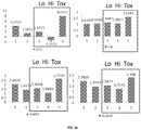
- Sodium iodate (45% solution), was prepared fresh for each set of experiments in 0.9% sodium chloride and injected to a final concentration of 45 mg/kg body weight. A total of 192 eyes were evaluated.
- Fundus imaging was performed using a commercially available confocal scanning laser ophthalmoscope, (cSLO, Spectralis, Heidelberg Retinal Angiography, HRA-II; Heidelberg Engineering GmbH, Germany), and the Eye Explorer Image Capture system, version 1.1.10. Images were captured at a wavelength of 488 nm excitation with 500 nm barrier filter for fluorescein angiography without the barrier filter for red free (RF, green dominant) imaging, at 795/810 nm excitation/emission for ICG angiography, and with a 830 nm laser for infrared reflectance imaging.
- cSLO Spectralis, Heidelberg Retinal Angiography, HRA-II; Heidelberg Engineering GmbH, Germany
- the Eye Explorer Image Capture system version 1.1.10. Images were captured at a wavelength of 488 nm excitation with 500 nm barrier filter for fluorescein angiography without the barrier filter for red free (RF, green dominant) imaging, at 795/810 nm
- cSLO imaging was performed in the absence of fluorescein dye. Fluorescein was not given at baseline or at any time during each study. In cases when it was necessary to simultaneously image the vasculature and provide landmarks for FAF image analysis, ICG was injected via tail vein, at 2 mg/kg or 5 mg/kg, into a previously inserted 23 gauge tail vein catheter. In a small number of clearly indicated situations, such as initial characterization of the model, fluorescein dextran (200 kD) was provided intravenously. Results from these latter studies using fluorescein angiography were performed in a subset of animals and not included in FAF analysis.
- Fundus images were routinely obtained at baseline (day 0, i.e. prior to systemic NaIO 3 injection), and at days 3 or 4 (3/4), days 7 or 8 (7/8) and days 13 or 14 (13/14) after NaIO 3 .
- images were acquired at 24 hours post- NaIO 3 , at 21 to 28 days and out to 88 days (3 months).
- composite images consisting of at least nine images were taken in the following order: optic nerve head, upper right, right, lower right, inferior, lower left, left, upper left, and the superior fields.
- a second or third complete or partial ring of images was obtained more peripherally, typically by realigning the animal relative to the cSLO as needed.
- the full nine images could not be captured, and a partial composite image was generated.
- High resolution spectral domain OCT images were obtained (Envisu R2200 VHR Animal SDOIS System, Bioptogen, USA). A scans were acquired at a rate of 36,000 per second, with a lateral resolution of approximately 2.8 ⁇ m at the level of the retina.
- H& E hemotoxylin and eosin histology of paraffin-embedded tissue was performed as known in the art after fixation with Davidson's fixative. Paraffin-embedded sections were viewed with a Nikon Upright E800 Microscope. Deparaffinization and rehydration procedure was followed as in H&E tissue processing.
- FIGs. 1A-D FAF and angiographic imaging in vivo of the normal Sprague Dawley rat eye produced a faint, homogenous ground glass glow interrupted by radially-arranged retinal blood vessels that block RPE fluorescence and so appear dark, and by a central hypofluorescent circle at the optic nerve head (ONH), where RPE is absent ( FIGs. 1A-D ).
- OHP optic nerve head
- FIGs. 1A-D Three days after systemic NaIO 3 injection, a single area of iso- or slightly hyperfluorescent FAF with hyperfluorescent borders appeared, and took the shape of a discrete island, sector, or 360° ring. Islands or sectors are consistently located inferior to the ONH or in the inferior hemiretina ( FIGs. 1A-D ).
- a 360° ring When a 360° ring is present, it typically encompasses much of the fundus, with one border adjacent to, surrounding, the ONH and the other in the mid- to far- retinal periphery. Large 360° rings were the most commonly observed shape, occurring in 102/110 (93%) of eyes treated with sodium iodate ( FIG. 2A-D ). In all cases, the islands, sectors or rings approached the ONH but were not contiguous with it ( FIGS. 1A -D-3A-C ). These and the following observations were confirmed in over 160 eyes.
- FIGS. 1A-D a marked reticular or curvilinear pattern of alternating hyper/hypo-fluorescent FAF appeared within the islands, sectors or rings.
- this pattern was pronounced. In small lesions, it completely filled the island or sector and emerged rapidly. In large 360° rings, it was at first pronounced at the proximal and distal borders and continued to emerge over the next weeks, whereby its curvilinear components contributed to partial loops, ovals, circles, rosettes, and scalloped or "paw-print" patterns of FAF ( FIGS. 2A-D ).
- curvilinear, round or oval shapes coalesce and appeared darker than the surrounding areas, appearing as homogenously dark grey patches of hypofluorescent FAF.
- the hyperfluorescent reticular pattern matured in situ, becoming more granular and less smoothly curvilinear ( FIGS. 2A-D ).
- the borders retained their hyperfluorescent reticular pattern and continued to creep slightly toward the retinal periphery.
- H&E staining confirms that fluid accumulates in the subretinal space. This was also observed by ultra-high resolution OCT in vivo.
- excised retinal wholemounts were viewed at low magnification under white light and by epifluorescent microscopy at 488 nm - the same wavelength used for in vivo imaging, with no fluorescent IHC labeling.
- two microscopes were used for this purpose: an epifluorescent device with a narrow excitation/emission window (488/520nm), and the Inverted Leica DM IRE2 viewing system of a confocal microscope with a mercury arc lamp and long-pass (rather than band-pass) filter providing fluorescence from greater than 515 nm following excitation with a 450-490 nm band.
- FIG. 4A-C shows a comparison of the clinical features of RPD with tissue findings in the NaIO 3 model.
- color and red-free fundus images were obtained ( FIG. 4A-C ).
- Red free images showed small dark spots that constitute individual pseudodrusen of RPD ( FIG. 4A , top image). These are identified using dark grey overlay.
- Example 2 DNIRA of a Rat Eye After Systemic ICG Administration Identifies the Retinal Pigment Epithelial (RPE) Layer in Vivo
- ICG near infra-red dye
- ICG dye or PBS
- ICG dye was injected systemically in 46 Sprague Dawley rats at doses of 0.35 and 5.0 mg/kg, and the fundus was evaluated in vivo using confocal scanning laser ophthalmoscopy (cSLO), with 795/810 nm excitation/emission filters in place, prior to injection, and at days 2, 7, and 21 thereafter.
- Electroretinography (ERG) was performed to evaluate potential toxicity. In a subset of animals, an RPE toxin was injected to induce RPE damage or loss.
- CSLO in vivo images were acquired using a commercially available cSLO (Heidelberg Retinal Angiography, HRA-2, Heidelberg Engineering, Germany). Images were obtained in the red-free, FAF (488/500 nm excitation/emission), NIR channel (830 nm) and ICG channel (795/810 nm excitation/emission).
- ICG and Fluorescein Angiography ICG dye (Cardiogreen, Sigma, Cat # I2633) was freshly prepared prior to experimentation to a final stock concentration of 5.0 mg/ml. A 23-gauge catheter was inserted into the tail vein, and ICG dye infused at doses of 0.35, or 5.0 mg/kg. Images were taken prior to injection (baseline), during dye circulation, and at various intervals thereafter out to 20 minutes. In a subset of animals, fluorescein-dextran (200 kD) (Sigma, Cat # FD2000S) solution, at 5.0 mg/ml, was injected IV via tail vein catheter to yield a final dose of 5.0 mg/kg.
- ICG and fluorescein angiography was performed simultaneously in a subset of animals only, otherwise fluorescein was not injected. Angiographic images were obtained in the fluorescein and ICG channels with excitation and emission filters of 488/500 nm and 795/810 nm respectively. Control animals received PBS.
- DNIRA images were obtained in the days and weeks after ICG injection using the ICG angiography settings, i.e. with excitation/emission filters, but without re-injection of dye at 24 hours, 48 hours, 3 days, 7 days, 21 days and 28 days after angiography. Angiography was not performed again during the time-course of the study.
- Electroretinography animals receiving ICG or PBS were evaluated using the Espion (DiagnosysLLC, USA) mini-Ganzfeld ERG system. Following anesthesia, animals were placed on an electrically silent heating pad and gold coil electrodes placed on the corneal surface after application of GenTeal lubricating drops. Following a short train of dim flashes (0.01 candela s/m 2 , 1.0 Hz), a scotopic b-wave amplitude was evaluated using a single bright flash (3 candela s/m 2 ) that we previously determined consistently produces a response close to the maximum b-wave amplitude. Student t-test was used to compare post-injection amplitudes against baseline (prior to injection).
- FIGs. 5A-H Representative baseline (pre-ICG) cSLO findings in the normal Sprague Dawley rat eye are shown ( FIGs. 5A-H ), and served along with PBS-treated animals ( FIG. 6A ), as normal controls for all experimentation.
- Red-free (i.e., green dominant) imaging identified the optic nerve head (ONH) and radial blood vessel of the retina ( FIG. 5A ).
- Low levels of endogenous signal were also seen at longer wavelengths, in the NIR channel (830 nm), and provided a similar image of the ONH and vasculature with some potential imaging of the deeper choroidal vessels that arises due to deeper light penetration ( FIG. 5B ).
- FAF appeared as a faint ground-glass glow that is lacking in detail, and is obscured by the radial retinal blood vessels and is absent at the ONH ( FIG. 5C ). This diffuse glow however is very faint.
- FIG. 6A-D The observations made using DNIRA after ICG injection were dose-dependent ( FIG. 6A-D ). The higher the initial ICG dosage used for angiography, the brighter the NIR signal. The high dose, 5 mg/kg, required that the gain (sensitivity) of the cSLO be reduced to obtain suitable quality images, while the low dose (0.35mg/kg) was less bright, and required that the gain be increased to obtain suitable quality images. Comparative images ( FIG. 6A, FIG. 6B and FIG. 6C ), taken with the gain set at the same level, demonstrated a dose-responsive increase in brightness. In the same experiment, it was also determined that by day 28 the speckled pattern faded in animals receiving low-dose ICG ( FIG. 6B ). By contrast, in animals receiving high dose ICG, there was little appreciable difference between days 3 and 28 post-injection ( FIG. 6C ).
- DNIRA identifies the RPE and/or outer retinal complex
- DNIRA could be used to identify structural abnormalities of the RPE.
- DNIRA was used in combination with systemic injection of the RPE toxin, NaIO 3 .
- NaIO 3 is directly toxic to RPE cells and leads to their loss; apoptosis of the overlying photoreceptors occurs thereafter.
- DNIRA in the days and weeks after NaIO 3 injection identified geographic (spatial) patches of profound hypofluorescence, evident as large black patches within an otherwise continuous background of speckled fluorescence. These hypofluorescent patches were bounded by a defined border.
- FIGs. 7A-B Choroidal vessels were identified by their complexity, variable size, and non-radial pattern with respect to the ONH. By contrast, the speckled layer obscured this view but permitted ongoing visualization of the overlying retinal vessels.
- test compound is administered to such an animal.
- the presentation of the ocular disorder is determined using in vivo imaging including fluorescence detection.
- a candidate compound is identified by observing fluorescence in the eye at or after a time sufficient to present characteristics of an ocular disorder.
- the candidate compound is evaluated for its ability to reduce or eliminate the characteristics of the ocular disorder as described herein.
- a candidate compound is administered to such an animal.
- the presentation of the ocular disorder disease is determined using in vivo imaging including fluorescence detection.
- the activity of a candidate compound is assessed by observing fluorescence in the eye at or after a time sufficient to present characteristics of an ocular disorder disease and the activity of a candidate compound.
- the candidate compound may be assessed for its ability to reduce or eliminate the characteristics of the ocular disorder as described herein.
- the subjects are administered bindarit in two 300 mg oral doses (or up to 1200 mg) once a day for 12 weeks. After an initial twelve-week treatment period, the subjects are evaluated for clinical outcomes. Alternatively, patients receive intravitreal injection of a vehicle containing bindarit, with or without a drug delivery vehicle.
- a first clinical outcome is determined using a standard visual acuity test, as is well known in the art.
- the subjects are assessed for the ability to clearly see symbols and objects on a Snellen eye chart from a distance.
- a second clinical outcome assesses the rate of progression of geographic atrophy.
- the subjects' pupils are dilated with 1.0% tropicamide and 2.5% phenylephrine before retinal imaging.
- Imaging is carried out with an instrument (e.g., Spectralis HRA+OCT; Heidelberg Engineering, Heidelberg, Germany) that allows for simultaneous recording of cSLO and spectral-domain optical coherence tomography (SD-OCT) with two independent scanning mirrors, as described in Helb, et al. Acta Ophthalmol. 2010 Dec; 88(8):842-9 .
- Five modes of operation are employed: white light, red-free light, near infrared, FAF and OCT.
- a third clinical outcome assesses the rate of change of hypofluorescent DNIRA (quantified by the area of hyporfluorescence). To do so, the subject's pupils are dilated with 1.0% and 2.5% phylephrine before retina imaging. Imaging is carried out with an instrument (that may utilize confocal scanning methodology, or a modified fundus camera with the appropriate excitation and emission filters in place, with or without the capacity to perform angiography) and occur prior to ICG injection (baseline), and again in the days after dye injection without further injection of dye but with the excitation/emission filters in place.
- an instrument that may utilize confocal scanning methodology, or a modified fundus camera with the appropriate excitation and emission filters in place, with or without the capacity to perform angiography
- a fourth clinical outcome assesses the change in the number and location of hyperfluorescent DNIRA dots.
- the subject's pupils are dilated with 1.0% and 2.5% phylephrine before retina imaging.
- Imaging is carried out with an instrument (that may utilize confocal scanning methodology, or a modified fundus camera with the appropriate excitation and emission filters in place, with or without the capacity to perform angiography) and occur prior to ICG injection (baseline), and again in the days after dye injection without further injection of dye but with the excitation/emission filters in place.
- Two SD-OCT scans one vertical and one horizontal, per eye are performed through the approximate foveal center, or in the case of RPD, in proximity to the vascular arcades of the macula.
- Color fundus photographs are obtained with a fundus camera (e.g. FF 450 Visupac ZK5; Carl Zeiss Meditec AG, Jena, Germany).
- Example 6 Treatment of Dry AMD with a Combination Therapy
- the subjects are administered bindarit, by way of non-limiting example, in two 300 mg oral doses (or up to 1200 mg) once a day for 12 weeks.
- the subjects are also administered ranibizumab injection once per month (roughly 28 days) in a dose of 0.5 mg per affected eye. After an initial twelve-week treatment period, the subjects are evaluated for clinical outcomes.
- Bindarit may also be administered via any of the ocular routes described herein (e.g. intravitreally).
- subjects are administered bindarit, by way of non-limiting example, in two 300 mg oral doses (or up to 1200 mg) once a day for 12 weeks.
- the subjects are also administered lampalizumab injection once per 4-6 weeks in a dose of about 10 mg per affected eye. After an initial twelve-week treatment period, the subjects are evaluated for clinical outcomes.
- Bindarit may also be administered via any of the ocular routes described herein (e.g. intravitreally).
- a first clinical outcome is determined using a standard visual acuity test, as is well known in the art.
- the subjects are assessed for the ability to clearly see symbols and objects on a Snellen eye chart from a distance.
- a second clinical outcome assesses the rate of progression of geographic atrophy.
- the subjects' pupils are dilated with 1.0% tropicamide and 2.5% phenylephrine or a comparable agent before retinal imaging.
- Imaging is carried out with an instrument (e.g., Spectralis HRA+OCT; Heidelberg Engineering, Heidelberg, Germany) that allows for simultaneous recording of cSLO and SD-OCT, as described in Helb, et al. Acta Ophthalmol. 2010 Dec; 88(8):842-9 .
- Multiple modes of operation can be employed: white light, red-free light, blue light, near infrared, and OCT. Similar analysis can be performed with a modified fundus camera.
- a third clinical outcome assesses the rate of change of hypofluorescent DNIRA (quantified by the area of hyporfluorescence). To do so, the subject's pupils are dilated with 1.0% and 2.5% phylephrine before retina imaging. Imaging is carried out with an instrument (that may utilize confocal scanning methodology, or a modified fundus camera with the appropriate excitation and emission filters in place, with or without the capacity to perform angiography) and occur prior to ICG injection (baseline), and again in the days after dye injection without further injection of dye but with the excitation/emission filters in place.
- an instrument that may utilize confocal scanning methodology, or a modified fundus camera with the appropriate excitation and emission filters in place, with or without the capacity to perform angiography
- a fourth clinical outcome assesses the change in the number and location of hyperfluorescent DNIRA dots.
- the subject's pupils are dilated with 1.0% and 2.5% phylephrine before retina imaging.
- Imaging is carried out with an instrument (that may utilize confocal scanning methodology, or a modified fundus camera with the appropriate excitation and emission filters in place, with or without the capacity to perform angiography) and occur prior to ICG injection (baseline), and again in the days after dye injection without further injection of dye but with the excitation/emission filters in place.
- Two SD-OCT scans are performed through the approximate foveal center, or in the case of RPD, in proximity to the vascular arcades of the macula.
- Color fundus photographs are obtained with a fundus camera ( e.g. FF 450 Visupac ZK5; Carl Zeiss Meditec AG, Jena, Germany).
- DNIRA images are obtained according to protocols described here that may include the acquisition of baseline images the ICG excitation/emission filters in place but before injection of ICG dye.
- ICG dye up to 5mg
- angiography may or may not be performed.
- DNIRA images are again obtained with the ICG excitation/emission filterse in place but with no further injection of dye. Regions of hypofluorescent (dark) DNIRA are measured and compared to similar measurements over time.
- Interpretation of clinical outcome data informs a decision for further treatment, if any.
- Illustrative data analysis includes macular cube analysis and 5 line raster.
- Example 7 Detection and/or Prediction of an Ocular Disorder Subject Response to an Agent
- Multi-modal and Angiographic Imaging were performed using a commercially available confocal scanning laser ophthalmoscope, (cSLO, Spectralis, Heidelberg Retinal Angiography, HRA-II; Heidelberg Engineering GmbH, Germany), and the Eye Explorer Image Capture system, version 1.1.10. Images were captured at a wavelength of 488 nm excitation with 500 nm barrier filter for fluorescein angiography without the barrier filter for red free (RF, green dominant) imaging, at 795/810 nm excitation/emission for ICG angiography, and with a 830 nm laser for infrared reflectance imaging.
- cSLO imaging was performed in the absence of fluorescein dye.
- ICG was injected via tail vein, at 2 mg/kg or 5 mg/kg, into a previously inserted 23 gauge tail vein catheter.
- FAF imaging of normal Sprague Dawley rat eye produced a faint, homogenous ground glass glow interrupted by radially-arranged retinal blood vessels that block RPE fluorescence and therefore appear dark. Such imaging was also characterized by a central hypofluorescent circle at the optic nerve head (ONH), where RPE is absent.
- OHP optic nerve head
- Systemic injections of NaIO 3 45% solution, prepared fresh for each set of experiments in 0.9% sodium chloride and injected to a final concentration of 45 mg/kg body weight
- Three days after injection a single area of iso- or slightly hyperfluorescent FAF with hyperfluorescent borders appeared and took the shape of a discrete island, sector, or 360° ring. In all cases, the islands, sectors or rings approached the ONH but were not contiguous with it.
- Fluorescein angiography performed in a subset of animals confirmed that the reticular pattern does not correspond with the retinal or choroidal vasculature while ICG angiography revealed a permeability change that occurred at the chorio-retinal interface, across the RPE and BM, approximately two to three days after NaIO 3 that coincides with, both spatially and temporally, the emergence of the islands, sectors of rings of FAF. This increase in permeability is transient and resolved by day 7, the next time point routinely evaluated. The retinal vessels do not leak.
- Hemotoxylin and eosin H & E histology of paraffin-embedded tissue and immunohistochemistry (IHC) (known in the art; performed after fixation with, e.g ., Davidson's fixative and paraffin-embedded sections, viewed with a Nikon Upright E800 Microscope) confirmed that fluid accumulated in the subretinal space. This was also observed by ultra-high resolution OCT in vivo (high resolution 160 nm spectral domain OCT images were obtained using the Envisu R2200 VHR Animal SDOIS System, Bioptogen, USA; scans were acquired at a rate of 36,000 per second, with a lateral resolution of approximately 2.8 ⁇ m at the level of the retina).
- IHC immunohistochemistry
- the Inverted Leica DM IRE2 viewing system of a confocal microscope with a mercury arc lamp and long-pass (rather than band-pass) filter providing fluorescence from greater than 515 nm following excitation with a 450-490 nm band.
- the normal rat retina appeared fairly homogenous throughout, with some artifactuous deformation.
- a subtle off-white, fine reticular pattern became apparent in samples obtained 7 days after NaIO 3 injection. By day 14, this pattern was more apparent and was observed to correspond with small, outer retinal folds best illustrated at the cut edge of the retina.
- Iba1 is a pan-microglial/macrophage marker and in the rat retina; antibodies against it identify the major phagocytic cell populations. Using this marker, it was found that Iba1 + cells are arranged in a lacy, curvilinear pattern when viewed en face in whole mount retina, i.e. in the z-plane. Further, when compared against photoreceptor nuclear staining, this curvilinear pattern is seen to lie interposed between folds of the deformed ONL.
- This pattern of Iba1 + cells corresponds with the pattern of hyperfluorescence observed in vivo by FAF imaging, and with the pattern of autofluorescent cells in excised retina.
- Corresponding patterns of in vivo FAF and Iba1 + staining were observed at both 7 days and 12 weeks after NaIO 3 injection.
- all RPE cells are absent from the posterior eye cup, confirming that these cells cannot be responsible for the observed FAF.
- identification of the autofluorescent cells as phagocytic was confirmed by comparison of imaging to preparations in which monocyte/macrophage depletion has been undertaken. Such depletion was achieved by treatment with gadolinium chloride (GAD).
- GAD gadolinium chloride
- Such identification of phagocytic cells is indicative of dry AMD and/or RPD and predictive of a subject's response to an agent. Specifically, the observation of such phagocytic cells makes it more likely than not that a subject will respond to treatment with an agent.
- Identification of a transient increase in permeability across a subject's epithelial barrier between a choroid and a retina relative to an undiseased state is indicative of dry AMD and/or RPD and predictive of a subject's response to an agent. Specifically, the observation of a transient increase in permeability across a subject's epithelial barrier between a choroid and a retina relative to an undiseased state makes it more likely than not that a subject will respond to treatment with an agent.
- DNIRA Delayed Near InfraRed Analysis
- a toxin such as NaIO 3 (Sigma, cat #424064) is injected systemically via a 23G catheter at a dosage of 45 mg/kg body weight.
- ICG angiography at 0.35mg/kg is performed immediately prior to NaIO 3 injection.
- DNIRA is performed 7 days after ICG and NaIO 3 injection.
- excised retinal wholemounts were viewed at low magnification under white light and by epifluorescent microscopy at 488 nm - the same wavelength used for in vivo imaging, with no fluorescent IHC labeling.
- two microscopes were used for this purpose: an epifluorescent device with a narrow excitation/emission window (488/520 nm), and the Inverted Leica DM IRE2 viewing system of a confocal microscope with a mercury arc lamp and long-pass (rather than band-pass) filter providing fluorescence from greater than 515 nm following excitation with a 450-490 nm band.
- Iba1 is a pan-microglial/macrophage marker and in the rat retina; antibodies against it identify the major phagocytic cell populations.
- Iba1 + cells are arranged in a lacy, curvilinear pattern when viewed en face in wholemount retina, namely, in the z-plane ( FIGs. 5A-H ). Further, when compared against photoreceptor nuclear staining, this curvilinear pattern was seen to lie interposed between folds of the deformed ONL. As shown in FIGS.
- this pattern of Iba1 + cells corresponded with the pattern of hyperfluorescence observed in vivo by FAF imaging ( FIGs 1A-D and 2A-D ), and with the pattern of autofluorescent cells in excised retina ( FIGs. 3A-C ).
- Corresponding patterns of in vivo FAF and Iba1 + staining are shown at both 7 days and 12 weeks after NaIO 3 injection ( FIGs. 5A and 5B ). By 7 days after NaIO 3 administration, all RPE cells were absent from the posterior eye cup, confirming that these cells were not responsible for the observed FAF ( FIGs 5C and 5D ).
- the 3-dimensional (3D) relationship between the Iba1 + cells and the outer retina was next evaluated by en face serial confocal microscopy of wholemount retina (i.e., in the z-axis) moving step-wise from the inner plexiform layer (INL) through the outer plexiform layer (OPL), to the inner-, mid- and outer-ONL.
- Short stacks of confocal images (segments) were projected (flattened) at these three steps ( FIGS. 6A-D ).
- the co-distribution of activated CD68 + macrophages of the monocyte lineage was also evaluated.
- Iba1 + cells were seen in the IPL only. No CD68 + cells were identified ( FIG. 6A ).
- Iba1 + cells increased in number and were increased in the IPL and also throughout the outer retina, including the outer-ONL ( FIG. 6B ).
- a complex distribution of Iba1 + and CD68 + cells was seen interlaced between curvilinear folds of the ONL.
- optically-sectioned ectopic circles or ovals of photoreceptor nuclei were observed as far internally as the OPL, the mid-retinal layer of blood vessels, thereby bringing the deformed photoreceptor nuclear layer and vascular layer, two normally separate tissue compartments, in contact.
- ovoid cross-sections of the deformed nuclear layer appeared more curvilinear or pisciform, and large Iba1 + and CD68 + cells were found within their central void.
- optical cross-sections remain curvilinear and were largely contiguous with many bridging segments.
- the subretinal space normally a potential space only, was expanded by outer retinal deformation and shows Iba1 + and/or CD68 + inflammatory cells and photoreceptor nuclei together immediately internal to Bruch's membrane (in the space where the lost photoreceptor outer segments would normally reside).
- FIG. 7A Confocal scanning microscopy and three-dimensional reconstruction of the excised wholemount retina was undertaken. These data showed that Iba1 + cells formed a reticular or lacy pattern that is interposed between photoreceptor nuclei. Further, projection of the z-stack, divided into the same three planes ( FIG. 7B right) showed that the inner-ONL is characterized by circular or oval cross-sections of the ONL. By contrast, the mid-ONL is characterized by looping curved cross-sections of the nuclear layer, and the outer ONL is formed of larger curvilinear segments with multiple bridging elements.
- HR-OCT provides non-invasive, in vivo cross-sections of the retina previously observed only in post-enucleation histological specimens.
- OCT imaging is dependent on the signal generated at the interface between layers of differing optical density, and in the normal rat retina confirms its multi-laminar structure.
- the three major outermost OCT boundaries of the retina are, from internal to external, the external limiting membrane (ELM), the junction of photoreceptor inner and outer segments (IS/OS), and the RPE/BM ( FIG . 8A , left).
- Standard techniques both in the clinic and in these investigations arbitrarily set the nuclear layers, including the ONL, as dark ( i.e. a signal void).
- FIG . 8A middle
- optical cross sections in the inferior retina showed slight deformation of the photoreceptor IS/OS junction and ELM such that bright (hyper-echoic) shallow mounds become visible ( FIG . 8A , right). This signal was found external to the ONL and internal to BM. The fine dark line representing that the RPE was no longer present.
- VIPs sub-retinal space volume-intensity projections
- VIPs in the z-axis were then analyzed ( FIG . 9B ).
- VIPs through the inner-ONL showed a series of punctate spots, regularly spaced throughout the retina.
- VIPs from the mid-ONL showed less regularly spaced pisciform or curved short lines, some of which are bridging.
- VIPs from the outer-ONL showed contiguous curvilinear segments with many bridging elements.
- the circulating macrophage/monocyte population was depleted using gadolinium chloride (GAD or GdCl 3 ), a rare earth metal salt which depresses macrophage activity.
- GAD depletion of immune cells led to profound alteration of the NaIO 3 -induced pattern of FAF and DNIRA.
- GAD depletion of the monocyte/macrophage populations led to profound alteration of the NaIO 3 -induced pattern of FAF. This was evident qualitatively both within the geographic areas of damage, and at its border ( FIGs. 8A-C ).
- DNIRA performed in a repeated manner, a method we term Pulsed DNIRA, identifies small hyperfluorescent dots in DNIRA that appear to migrate over time ( FIG 32 ). RPE cells that also can be labeled with DNIRA, do not migrate. This along with the above co-localization with hyperfluorescent FAF suggests that macrophages colocalize and contribute to disease progression.
- FIGs. 10A-C images and interpretive diagrams that directly compare the clinical features of RPD against tissue findings in the NaIO 3 model are provided.
- color and red-free fundus images were also obtained ( FIGs. 10A-C ).
- Red free images showed small dark spots that constitute individual pseudodrusen of RPD ( FIG . 10A , top image). These were identified using dark grey overlay. This further highlights, circumscribed by circles, the presence of so-called "target lesions” that characterize RPD. These appear as dark spots with lighter centers.
- a schematized illustration of folded outer rat retina is presented, both en face and in cross-section that, when falsely colored and presented as black and white images-both "positive” and “negative”-correlates directly with targets and halos of RPD.
- This example shows, inter alia, that bindarit protects the retina and RPE against toxin-induced damage in a model of non-exudative "dry” AMD and RPD and that bindarit protects against the development of patches of retinal pigment epithelium (RPE) and photoreceptor loss known as Geographic Atrophy (GA).
- RPE retinal pigment epithelium
- GA Geographic Atrophy
- In vivo images were acquired using a commercially available confocal scanning laser ophthalmoscope (cSLO) (Heidelberg Retinal Angiography, HRA-2, Heidelberg Engineering, Germany). Images were obtained in the red-free, FAF (488/500 nm excitation/emission), IR reflectance channel (830 nm) and ICG fluorescence channel (795/810 nm excitation/emission). Animals were pre-screened using cSLO, and those with spontaneous defects eliminated from the study. The surface of the cornea was kept moist with repeated application of Genteal lubricating drops (Alcon, Mississauga, Canada) during all in vivo imaging.
- cSLO confocal scanning laser ophthalmoscope
- ICG dye (Cardiogreen, Sigma) was freshly prepared prior to experimentation to a final stock concentration of 5.0 mg/ml in sterile water. A 24-gauge catheter was inserted into the tail vein, and ICG dye infused at doses of 0.35, or 5.0 mg/kg. Images were taken prior to injection (baseline), during dye circulation, and at various intervals thereafter out to 20 minutes.
- DNIRA Delayed Near Infra Red Analysis
- Electroretinography The bright-flash ERG response was evaluated in animals using the Espion (DiagnosysLLC, USA) mini-Ganzfeld system following high (5.0 mg/kg) and low (0.35 mg/kg) dose administration of ICG, or saline control. Following anesthesia, animals were placed on an electrically silent heating pad and gold coil electrodes placed at the edge of the cornea after application of GenTeal lubricating drops. Following a short train of dim flashes (0.01 candela s/m2, 1.0 Hz), the photopic b-wave response was evaluated using a single bright flash (3 candela s/m2) that we previously determined consistently approximates the maximum b-wave amplitude.
- Sodium iodate The RPE toxin sodium iodate (NaIO 3 , Sigma) was prepared fresh weekly at a stock solution of 45 mg/ml in saline (Baxter, Mississauga, Canada). This solution was diluted to final concentrations of 5 mg/ml, 15 mg/ml, 30 mg/ml and 45 mg/ml, such that all animals received the same volume to achieve final dosages of 5 mg/kg to 45 mg/kg.
- NaIO 3 was injected systemically via a 24-gauge catheter inserted into the tail vein. ICG injection (0.35 mg/kg) and angiography were performed at day 0 immediately prior to NaIO 3 injection. To test the effects of bindarit, a standardized dose was used.
- Intra-ocular injection of bindarit intra-vitreal injection of bindarit was performed under a dissecting microscope (SMZ800; Nikon Instruments, Melville, USA) with a Hamilton syringe (Cat# 7634-01, Hamilton Company, Reno, USA). A small incision was made behind the limbus using a beveled 33-gauge needle (BD, Mississauga, Canada). A 30-gauge blunt needle was then inserted and at an angle of approximately 45° to the pupillary axis. Two (2) ⁇ L of bindarit solution or control solution (saline) was manually injected.
- Immunofluorescence microscopy Immunohistochemistry was performed using standard protocols. Briefly, cells were fixed with 4% PFA/PBS for 15 minutes, and blocked with 1.25% BSA in TBS for 30 minutes at room temperature. Primary rabbit anti-ZO-1 was used at 2.5 ⁇ g/ml in 1.25% BSA/TBS and incubated for 1 hour at room temperature. Cells were washed in TBS and incubated with goat anti-rabbit 488 (Invitrogen) at 5 ⁇ g/ml for 1 hour at room temperature, washed, and counterstained with To-Pro-3 (Invitrogen).
- goat anti-rabbit 488 Invitrogen
- RPE monolayers were stained with 647 nm conjugated phalloidin (Invitrogen) for 20 minutes at room temperature and counterstained with Sytox green nuclear stain (Invitrogen). Slides were mounted with fluorescent mounting medium (Dako) and images acquired using a Leica TCS SL confocal fluorescent microscope (Leica Microsystems, GmbH, Wetzlar, Germany), with Leica Confocal Software V 2.61.
- Treatment with bindarit provides secondary prevention against the late blinding complication of dry AMD and RPD known as Geographic Atrophy (GA).
- GA Geographic Atrophy
- the purpose of secondary prevention is to maintain central visual acuity and central visual field prior to loss.
- FIGs. 16A-F show that bindarit (TMi-018) reduces the size of the patch of RPE loss in the rat eye.
- FIGs. 16A-F further show that, when evaluated over time using Fundus Autofluorescence (FAF), intra-vitreal bindarit reduces the development of complex patterns of hyperfluorescent FAF.
- FAF Fundus Autofluorescence
- FIG. 17A-B shows that bindarit dose-dependently protects retinal function (electrophysiology), and demonstrates a minimally-effective dose.
- FIG. 18A-C shows that bindarit protects against tissue damage, RPE loss and disruption of the outer retina.
- FIGs. 19A-J shows that bindarit reduces Monocyte Chemoattractant protein (MCP)-1 expression and reduces the M1 macrophage response, tipping the balance in favor of M2.
- MCP Monocyte Chemoattractant protein
- FIG. 19D shows that during toxin-induced damage to the RPE, a model for ocular disease (e.g. dry AMD and/or RPD) gene expression shifts towards M1 polarization. Also, gene expression shifts back towards neutral, with relative decreases in M1 mRNA expression and increased levels of M2 expression.
- a model for ocular disease e.g. dry AMD and/or RPD
- FIG. 20A shows a negative control (saline) treatment of an existing patch, which expands, while, in contrast, FIG. 20B shows bindarit treatment of an existing patch, which does not expand.
- MCP-1 is reported to be expressed by RPE, and is a potent Danger Associate Molecular Protein (DAMP) that activates M1 macrophages.
- DAMP Danger Associate Molecular Protein
- Bindarit appears to have the potential to modulate the innate immune response both directly through its influence on macrophage polarization and indirectly by reducing the level of RPE signaling that initiates the local response.
- FIG. 21 shows, in panels A and B, that hTERT cells are confirmed to express markers of RPE differentiation.
- panel C it is shown that TMi-018 reduces NFkb expression and reduces MCP-1 in human RPE (hTERT).
- Panel D shows that TMi-018 increases RPE survival against toxin challenge.
- FIG. 22 shows mRNA species of the complement cascade components change following toxin-induced RPE damage and that bindarit alters complement gene expression.
- the genes studied include: C1qb, C1qtnf5, C2, C3, C4b, C5, C6, C7, C8a, C9, Cfb, Cfd, Cfh, and Cfi.
- CFD complement factor D
- mRNA levels of many complement genes are seen to increase or decrease by more than 2 or 3 fold ( FIG. 22 ).
- Major changes are identified in C1, C3, C5 and C9, and of note, CFH and CFI both decrease. This latter finding may correlate with the finding that the CFD inhibitor, Lampilizumab, was most efficacious in patients with polymorphisms in these genes.
- CFD mRNA did not change much, less than 3-fold, over much of the time-course.
- Bindarit dose-dependently up- or downregulated the mRNA of multiple complement genes was explored.
- Example 12 Clinical Evaluation of Drug Efficacy and/or Disease Progression with DNIRA
- DNIRA is useful as a diagnostic or prognostic method for ocular disorders.
- the present data show that DNIRA is useful as a biomarker for the therapeutic efficacy of an agent against one or more ocular disorders and/or a diagnostic for the onset of one or more ocular disorders and/or a diagnostic for progression of one or more ocular disorders.
- the data present findings in human subjects afflicted with ocular disorders.
- DNIRA is a novel and previously undescribed biomarker for diagnosis, disease progression, and treatment response. Its application, qualitative description and quantification provides important uses to these ends.
- the instruments used to perform ICG angiography require real-time imaging of the dye as it passes through, for example, the ocular (retinal and choroidal) blood vessels.
- imaging typically starts in the seconds and minutes prior to injection of the dye in the veins of the arm or hand, and continues for typically 10 to 30 minutes thereafter.
- Late phase angiography typically occurs up to 45 minutes later. Images are thus acquired in the transit phase (as dye passes through the blood vessels) and in the re-circulation phase (as it leaks from the vessels into the tissue).
- the devices required to image ICG dye thus must capture sequential images originally in the form of film, video, or more recently digital angiography. These systems are expensive and require sustained imaging.
- the present example provides use of ICG excitation/emission filters without the use of angiographic imaging.
- Baseline DNIRA images are obtained with the ICG ex/em filters in place prior to systemic injection of ICG dye. In the days and weeks after injection, individual images are obtained. The use of ICG filters without angiography has not been previously described.
- DNIRA DNIRA to clinical disease
- Baseline investigation included spectacle-corrected visual acuity, ophthalmic examination with slit-lamp microscopy and fundoscopy, along with cSLO imaging that included FAF and NIR imaging with the ICG excitation/emission filters in place but no infusion of dye.
- Angiography was performed less than one week thereafter, simultaneous with administration of a single dose of ICG dye (Cardiogreen, 25mg/5ml) and fluorescein sodium.
- FAF and DNIRA imaging was performed twice in the following 7 days, with a minimum of 2 and maximum of 4 days between procedures.
- DNIRA is compared against ICG angiography. Other than overlying blockage of the central signal (barely detectable) little abnormality is observed with ICG angiography ( FIG . 23 ). This is in stark contrast to DNIRA, in which areas of marked hyperfluorescence in the central macula ( FIG . 24 ). In addition, a leopard-like pattern is observed in the peripheral macula that has not been identified by any imaging technique to date. Further, DNIRA is compared to FAF and infra-red (IR) as is also superior against these techniques ( FIG . 25 ). DNIRA images of the same patient as shown in the left (IR) and center (FAF) panels of FIG. 25 contrast markedly.
- DNIRA reveals a region of profound hypofluorescence (dark) in the image's center. Smaller patches are noted at the margin of central lesion and in proximity to the optic nerve head.
- composite images of the same patient shows a lacy, reticular or leopard-like pattern of alternating hyper- and hypo-fluorescent DNIRA that is not seen in other imaging modalities, and not described previously ( FIG . 26 ).
- an age-matched non-AMD patient who is not known to have RPE damage, does not show such lacy, reticular or leopard-like pattern of alternating hyper- and hypo-fluorescent DNIRA (data not shown).
- hypofluorescent FAF areas of chorioretinal damage are seen as hypofluorescent (dark, black or absent) signal. Such areas are readily quantified owing to the stark contrast between normal or hyper-fluorescent background.
- Algorithms for the quantification of hypofluorescent FAF are now included with commercially available imaging devices (for example the confocal scanning laser ophthalmoscopy system, or modified fundus camera). Quantification of hypofluorescent FAF is now incorporated into clinical trial design as both inclusion criteria and outcome measure.
- the Mahalo study (Genetech) requires that patients have a minimum and maximum size region of hypofluorescent FAF to enter the study. The size of this area is measured repeatedly during the study and the rates of growth, i.e . expansion, is compared between treatment arms.
- DNIRA makes clear regions of hypofluorescence not seen in current imaging modalities. Accordingly, inter alia, DNIRA can be used to improve identification of suitable patients for study recruitment and to evaluate progression of disease.
- DNIRA images change over time. This enables serial quantification of the areas of hypofluorescent or hyperfluorescent signal and provides a strategy for analysis of disease progression (e.g . as a surrogate biomarker).
- disease progression e.g . as a surrogate biomarker
- the DNIRA images of FIG. 27 show hypofluroescence expansion in a late dry AMD patient.
- hyperfluorescent DNIRA in addition to hypofluorescent DNIRA, described herein is the presence of hyperfluorescent DNIRA in the clinical setting. Bright punctate dots or spots can be identified in the rodent eye in the days and weeks after ICG injection without further injection of dye, but only with the ICG excitation/emission filters in place ( see, e.g. Examples herein, such as Example 1, 2, and 8).
- Pulsed DNIRA in which ICG dye is provided repeatedly intravascularly and evaluated intra-ocularly in the living eye in the days after injection
- ImmunoD it is shown that macrophages can be labeled in vitro and in vivo with ICG and so become visible as small, motile, hyperfluorescent dots.
- FIG. 28 shows that discrete dots of hyperfluorescent DNIRA are identified scattered throughout the macula of patients with AMD. Some are in proximity to the optic nerve head, and other immediately adjacent to regions of geographic atrophy, both at the border and in the so-called junctional zone.
- FIG. 29 shows the eye of a human patient with RPD. Having characterized the patient as having RPD using IR and FAF imaging, DNIRA imaging demonstrates multiple regions of hypofluorescent FAF not seen in any other modality (including other modalities not shown in FIG. 29 ). These regions are distributed throughout the macula. The leopard-like spots are again seen in the macular periphery.
- RPD have been shown to mature over time, starting as shallow mounds anterior to the RPE, and later maturing into base-down pyramids or spike superiorly.
- RPD are seen to mature in a centripetal direction, being newer or younger peripherally (in this case superiorly), and older centrally (inferiorly).
- the hyperfluorescent dots seen superiorly would coincide with the presence of labeled macrophages that have infiltrated the tissue.
- areas of tissue loss are suggested by the small patches of hypofluorescent DNIRA.
- FIG. 30 shows a gradient of hypofluorescent and hyperfluorescent signal in a different RPD patient than above. The lower arrow shows region of discrete hypofluorescent FAF immediately superior to the central macula. Superior to this, there is an increasing accumulation of hyperfluorescent FAF dots
- FIG. 31 shows a correspondence between hyperfluorescent FAF signal and hyperfluorescent DNIRA signal in the rabbit eye.
- DNIRA labels macrophages the migration of hyperfluorescent DNIRA dots over time was evaluated. Following systemic toxin administration, a wave of bright dots is observed to migrate, or be labeled centrifugally, from the rabbit optic nerve head.
- FIG. 32 shows that hyperfluorescent DNIRA dots, likely macrophages, change position over time.
- the upper row of images in FIG. 32 shows DNIRA imaging after systemic toxin injection in the rat and shows punctate hyperfluorescent dots encircling the optic nerve head. Over time, these dots appear to migrate centrifugally.
- FIG. 32 shows DNIRA imaging in the rabbit eye and shows a similar apparent migration of individually labeled dots that by immunohistochemistry stain positively for macrophage markers.
- the left-most image is baseline DNIRA (i.e ., pre-ICG injection with ICG filters in place).
- the human optic nerve head was evaluated for similar findings. Similarities between rat, rabbit and human DNIRA permit clinical-pathological correlation and suggest that macrophages account for hyperfluorescence in all three species.
- FIG. 33 shows that brightly labeled small DNIRA-positive dots are observed in proximity to the optic nerve head in rat, rabbit and human images.
- Inflammation is suggested to play a central role in diabetic eye disease, including both diabetic retinopathy and diabetic macular edema.
- Cytokines, chemokines and other pro-inflammatory or pro-permeability molecules are described in human ocular tissues such as vitreous or aqueous, and also in serum. Animal models confirm an inflammatory response in pre-clinical studies.
- MCP-1/ccl2 and RANTES are also implicated in diabetic eye disease, and can be directly activated in the face of obesity and excess free fatty acids. Increased IKKb phosphorylation leads to NFkb translocation to the nucleus and activation of the inflammatory transcriptome. Further, serum levels of MCP-1/ccl2 and CRP are elevated in patients with severe diabetic retinopathy, potentially arising from systemic release of eye-derived mediators or, concurrent advanced systemic disease. Agents that can reduce these systemic factors, either locally or systemically might therefore be efficacious in the treatment of diabetic eye disease.
- the present example shows, among others, that bindarit reduces inflammatory mediators demonstrated to be involved in diabetic eye disease.
- Sodium iodate, at a high dose of 45 mg/kg was used to induce an inflammatory response that was treated with high (20 ⁇ g/ ⁇ l, 2 ⁇ l volume) and low (2 ⁇ g/ ⁇ l, 2 ⁇ l volume) dose intra-vitreal bindarit.
- the effect of bindarit was assessed at day 16 after toxin injection.
- bindarit reduces inflammatory mediators known to be involved in diabetic retinopathy.
- FIG. 36 shows that bindarit had no significant effect on other interleukins IL-10 and IL-3 or tumor necrosis factor alpha (TNFa) and ccl5 (MCP-3).
- Fold mRNA changes were compared against baseline (normal) conditions, under the following conditions: 1. Intra-vitreal saline (vehicle), 2. High-dose bindarit, 2ug/ul, (no toxin), 3. Toxin + low dose 2ug/ul bindarit, 4. Toxin + high dose 20ug/ul bindarit + NaIO 3 , and 5. Toxin alone.
- bindarit had no significant effect on vascular endotheiial growth factor A (VEGF-A) mRNA ( FIG . 37 ).
- VEGF-A vascular endotheiial growth factor A
- FIG . 37 vascular endotheiial growth factor A
- VEGF-A vascular endotheiial growth factor A
- VEGF-A vascular endotheiial growth factor A
- All changes are approximately 1.0 fold up or down.
- Fold mRNA changes were compared against baseline (normal) conditions, under the following conditions: 1. Intra-vitreal saline (vehicle), 2. High-dose bindarit, 2ug/ul, (no toxin), 3. Toxin + low dose 2ug/ul bindarit, 4. Toxin + high dose 20 ug/ul bindarit + NaIO 3 , and 5. Toxin alone.
- FIG. 38 shows that, in an immortalized human RPE cell line, bindarit reduces NFkb translocation to the nucleus.
- hyperglycemia i.e ., elevated serum glucose
- most cells become hypoglycemic. This is also true for cells that are non-responsive or resistant to insulin.
- cells of the brain and retina uptake high levels of glucose in the absence of insulin and so become hyperglycemic. At low levels this can stimulate cell activity, however at higher levels of glucose become toxic, inciting inflammation and leading to the formation of advanced glycated endproducts (AGEs).
- the immortalized human cell line, hTERT was incubated with normal, medium-high and very high concentrations of D-glucose.
- FIG. 39 shows that bindarit normalizes cellular stress in hTERT cells incubated with high glucose. This example, inter alia, points to bindarit as an agent for the treatment of diabetic retinopathy.
Landscapes
- Health & Medical Sciences (AREA)
- Life Sciences & Earth Sciences (AREA)
- Engineering & Computer Science (AREA)
- Chemical & Material Sciences (AREA)
- General Health & Medical Sciences (AREA)
- Biomedical Technology (AREA)
- Medicinal Chemistry (AREA)
- Animal Behavior & Ethology (AREA)
- Veterinary Medicine (AREA)
- Public Health (AREA)
- Immunology (AREA)
- Organic Chemistry (AREA)
- Urology & Nephrology (AREA)
- Molecular Biology (AREA)
- Bioinformatics & Cheminformatics (AREA)
- Hematology (AREA)
- Epidemiology (AREA)
- Pharmacology & Pharmacy (AREA)
- Biochemistry (AREA)
- Pathology (AREA)
- Cell Biology (AREA)
- Ophthalmology & Optometry (AREA)
- Microbiology (AREA)
- Diabetes (AREA)
- Endocrinology (AREA)
- Toxicology (AREA)
- Physics & Mathematics (AREA)
- Food Science & Technology (AREA)
- Analytical Chemistry (AREA)
- General Physics & Mathematics (AREA)
- Biotechnology (AREA)
- Nuclear Medicine, Radiotherapy & Molecular Imaging (AREA)
- Chemical Kinetics & Catalysis (AREA)
- General Chemical & Material Sciences (AREA)
- Genetics & Genomics (AREA)
- Rheumatology (AREA)
- Gastroenterology & Hepatology (AREA)
- Zoology (AREA)
- General Engineering & Computer Science (AREA)
- Wood Science & Technology (AREA)
Applications Claiming Priority (5)
| Application Number | Priority Date | Filing Date | Title |
|---|---|---|---|
| US201461993915P | 2014-05-15 | 2014-05-15 | |
| US201462064344P | 2014-10-15 | 2014-10-15 | |
| US201462076162P | 2014-11-06 | 2014-11-06 | |
| US201562157773P | 2015-05-06 | 2015-05-06 | |
| PCT/IB2015/053609 WO2015173786A1 (en) | 2014-05-15 | 2015-05-15 | Compositions and methods for treating and diagnosing ocular disorders |
Publications (3)
| Publication Number | Publication Date |
|---|---|
| EP3142664A1 EP3142664A1 (en) | 2017-03-22 |
| EP3142664A4 EP3142664A4 (en) | 2017-12-13 |
| EP3142664B1 true EP3142664B1 (en) | 2019-12-04 |
Family
ID=54479391
Family Applications (1)
| Application Number | Title | Priority Date | Filing Date |
|---|---|---|---|
| EP15792756.7A Active EP3142664B1 (en) | 2014-05-15 | 2015-05-15 | Compositions and methods for treating and diagnosing ocular disorders |
Country Status (7)
| Country | Link |
|---|---|
| US (2) | US20170079955A1 (enExample) |
| EP (1) | EP3142664B1 (enExample) |
| JP (1) | JP6625064B2 (enExample) |
| DK (1) | DK3142664T3 (enExample) |
| ES (1) | ES2789849T3 (enExample) |
| HU (1) | HUE049518T2 (enExample) |
| WO (1) | WO2015173786A1 (enExample) |
Families Citing this family (13)
| Publication number | Priority date | Publication date | Assignee | Title |
|---|---|---|---|---|
| US9840553B2 (en) | 2014-06-28 | 2017-12-12 | Kodiak Sciences Inc. | Dual PDGF/VEGF antagonists |
| KR20250057128A (ko) | 2015-12-30 | 2025-04-28 | 코디악 사이언시스 인코포레이티드 | 항체 및 이의 접합체 |
| EP3525659A4 (en) * | 2016-10-13 | 2020-06-17 | Translatum Medicus, Inc. | SYSTEMS AND METHODS FOR DETECTING EYE DISEASES |
| WO2018119202A1 (en) * | 2016-12-23 | 2018-06-28 | StemBios Technologies, Inc. | Use of somatic stem cells for increasing level of peroxisome proliferator-activated receptor gamma |
| EP3758737A4 (en) | 2018-03-02 | 2022-10-12 | Kodiak Sciences Inc. | IL-6 ANTIBODIES AND FUSION CONSTRUCTS AND CONJUGATES THEREOF |
| RU2683758C1 (ru) * | 2018-06-27 | 2019-04-01 | Федеральное государственное учреждение "Федеральный исследовательский центр "Информатика и управление" Российской академии наук" (ФИЦ ИУ РАН) | Автоматизированная система анализа ангиографических изображений глазного дна человека |
| EP3706136B1 (en) * | 2019-03-05 | 2026-02-18 | Novartis AG | Computerized systems for prediction of geographic atrophy progression using deep learning applied to clinical imaging |
| WO2020261158A1 (en) | 2019-06-25 | 2020-12-30 | Translatum Medicus Inc. | Processes of making 2-((1-benzyl-1h-indazol-3-yl)methoxy)-2-methylpropanoic acid and its derivatives |
| CA3157509A1 (en) | 2019-10-10 | 2021-04-15 | Kodiak Sciences Inc. | Methods of treating an eye disorder |
| RU196414U1 (ru) * | 2019-12-23 | 2020-02-28 | Федеральное государственное учреждение "Федеральный исследовательский центр "Информатика и управление" Российской Академии Наук" | Анализатор ангиографических изображений глазного дна человека |
| GB202001336D0 (en) * | 2020-01-31 | 2020-03-18 | Imp College Innovations Ltd | Methods for measuring gut permeability and gastric emptying rate |
| US20250241902A1 (en) * | 2022-04-14 | 2025-07-31 | Belite Bio, Inc | Methods, compositions and systems for evaluation of visual function |
| WO2024165925A1 (en) * | 2023-02-10 | 2024-08-15 | Translatum Medicus Inc. | Bindarit conjugates, compositions and methods for treating ocular diseases or disorders |
Family Cites Families (5)
| Publication number | Priority date | Publication date | Assignee | Title |
|---|---|---|---|---|
| IT1293795B1 (it) * | 1997-07-28 | 1999-03-10 | Angelini Ricerche Spa | Farmaco attivo nel ridurre la produzione di proteina mcp-1 |
| WO2005108431A1 (en) * | 2004-05-04 | 2005-11-17 | University Of Kentucky Research Foundation | Methods and compositions for the treatment of ocular neovascularization |
| ITMI20062254A1 (it) * | 2006-11-24 | 2008-05-25 | Acraf | Uso di un acido metossi-alcanoico dell'indazolo per preparare una composizione farmaceutca |
| WO2009109613A2 (en) * | 2008-03-07 | 2009-09-11 | Aziende Chimiche Riunite Angelini Francesco A.C.R.A.F. S.P.A. | Novel 1-benzyl-3-hydroxymethylindazole derivatives and use thereof in the treatment of diseases based on the expression of mcp-1, cx3cr1 and p40 |
| AU2013255050B2 (en) * | 2012-05-01 | 2016-07-28 | Translatum Medicus Inc. | Methods for treating and diagnosing blinding eye diseases |
-
2015
- 2015-05-15 ES ES15792756T patent/ES2789849T3/es active Active
- 2015-05-15 JP JP2016567824A patent/JP6625064B2/ja active Active
- 2015-05-15 DK DK15792756.7T patent/DK3142664T3/da active
- 2015-05-15 WO PCT/IB2015/053609 patent/WO2015173786A1/en not_active Ceased
- 2015-05-15 EP EP15792756.7A patent/EP3142664B1/en active Active
- 2015-05-15 HU HUE15792756A patent/HUE049518T2/hu unknown
- 2015-05-15 US US15/311,083 patent/US20170079955A1/en not_active Abandoned
-
2018
- 2018-05-30 US US15/993,183 patent/US20180338952A1/en not_active Abandoned
Non-Patent Citations (1)
| Title |
|---|
| None * |
Also Published As
| Publication number | Publication date |
|---|---|
| HUE049518T2 (hu) | 2020-09-28 |
| JP2017520527A (ja) | 2017-07-27 |
| US20180338952A1 (en) | 2018-11-29 |
| EP3142664A4 (en) | 2017-12-13 |
| DK3142664T3 (da) | 2020-03-09 |
| JP6625064B2 (ja) | 2019-12-25 |
| WO2015173786A1 (en) | 2015-11-19 |
| ES2789849T3 (es) | 2020-10-26 |
| US20170079955A1 (en) | 2017-03-23 |
| EP3142664A1 (en) | 2017-03-22 |
Similar Documents
| Publication | Publication Date | Title |
|---|---|---|
| EP3142664B1 (en) | Compositions and methods for treating and diagnosing ocular disorders | |
| US12377171B2 (en) | Methods for treating and diagnosing blinding eye diseases | |
| EP2844250B1 (en) | Methods for treating and diagnosing blinding eye diseases | |
| US20190336482A1 (en) | Compositions and methods for treating and diagnosing ocular disorders | |
| CA2870353C (en) | Compositions and methods for treating and diagnosing ocular disorders |
Legal Events
| Date | Code | Title | Description |
|---|---|---|---|
| STAA | Information on the status of an ep patent application or granted ep patent |
Free format text: STATUS: THE INTERNATIONAL PUBLICATION HAS BEEN MADE |
|
| PUAI | Public reference made under article 153(3) epc to a published international application that has entered the european phase |
Free format text: ORIGINAL CODE: 0009012 |
|
| STAA | Information on the status of an ep patent application or granted ep patent |
Free format text: STATUS: REQUEST FOR EXAMINATION WAS MADE |
|
| 17P | Request for examination filed |
Effective date: 20161207 |
|
| AK | Designated contracting states |
Kind code of ref document: A1 Designated state(s): AL AT BE BG CH CY CZ DE DK EE ES FI FR GB GR HR HU IE IS IT LI LT LU LV MC MK MT NL NO PL PT RO RS SE SI SK SM TR |
|
| AX | Request for extension of the european patent |
Extension state: BA ME |
|
| DAV | Request for validation of the european patent (deleted) | ||
| DAX | Request for extension of the european patent (deleted) | ||
| A4 | Supplementary search report drawn up and despatched |
Effective date: 20171114 |
|
| RIC1 | Information provided on ipc code assigned before grant |
Ipc: A61K 31/416 20060101AFI20171108BHEP Ipc: A61K 49/00 20060101ALI20171108BHEP Ipc: A61P 27/02 20060101ALI20171108BHEP Ipc: A61P 27/12 20060101ALI20171108BHEP Ipc: C12Q 1/00 20060101ALI20171108BHEP |
|
| STAA | Information on the status of an ep patent application or granted ep patent |
Free format text: STATUS: EXAMINATION IS IN PROGRESS |
|
| 17Q | First examination report despatched |
Effective date: 20181207 |
|
| GRAP | Despatch of communication of intention to grant a patent |
Free format text: ORIGINAL CODE: EPIDOSNIGR1 |
|
| STAA | Information on the status of an ep patent application or granted ep patent |
Free format text: STATUS: GRANT OF PATENT IS INTENDED |
|
| INTG | Intention to grant announced |
Effective date: 20190618 |
|
| GRAS | Grant fee paid |
Free format text: ORIGINAL CODE: EPIDOSNIGR3 |
|
| GRAA | (expected) grant |
Free format text: ORIGINAL CODE: 0009210 |
|
| STAA | Information on the status of an ep patent application or granted ep patent |
Free format text: STATUS: THE PATENT HAS BEEN GRANTED |
|
| AK | Designated contracting states |
Kind code of ref document: B1 Designated state(s): AL AT BE BG CH CY CZ DE DK EE ES FI FR GB GR HR HU IE IS IT LI LT LU LV MC MK MT NL NO PL PT RO RS SE SI SK SM TR |
|
| REG | Reference to a national code |
Ref country code: GB Ref legal event code: FG4D |
|
| REG | Reference to a national code |
Ref country code: CH Ref legal event code: EP |
|
| REG | Reference to a national code |
Ref country code: AT Ref legal event code: REF Ref document number: 1208534 Country of ref document: AT Kind code of ref document: T Effective date: 20191215 |
|
| REG | Reference to a national code |
Ref country code: DE Ref legal event code: R096 Ref document number: 602015043089 Country of ref document: DE |
|
| REG | Reference to a national code |
Ref country code: IE Ref legal event code: FG4D |
|
| GRAT | Correction requested after decision to grant or after decision to maintain patent in amended form |
Free format text: ORIGINAL CODE: EPIDOSNCDEC |
|
| REG | Reference to a national code |
Ref country code: DK Ref legal event code: T3 Effective date: 20200302 |
|
| REG | Reference to a national code |
Ref country code: CH Ref legal event code: NV Representative=s name: PATENTANWALT MATTHIAS W. SCHAEFER, CH Ref country code: CH Ref legal event code: PK Free format text: BERICHTIGUNG INHABER |
|
| REG | Reference to a national code |
Ref country code: NL Ref legal event code: FP |
|
| REG | Reference to a national code |
Ref country code: LT Ref legal event code: MG4D Ref country code: NO Ref legal event code: T2 Effective date: 20191204 |
|
| REG | Reference to a national code |
Ref country code: CH Ref legal event code: PCAR Free format text: NEW ADDRESS: SPLUEGENSTRASSE 10, 8002 ZUERICH (CH) |
|
| PG25 | Lapsed in a contracting state [announced via postgrant information from national office to epo] |
Ref country code: LV Free format text: LAPSE BECAUSE OF FAILURE TO SUBMIT A TRANSLATION OF THE DESCRIPTION OR TO PAY THE FEE WITHIN THE PRESCRIBED TIME-LIMIT Effective date: 20191204 Ref country code: SE Free format text: LAPSE BECAUSE OF FAILURE TO SUBMIT A TRANSLATION OF THE DESCRIPTION OR TO PAY THE FEE WITHIN THE PRESCRIBED TIME-LIMIT Effective date: 20191204 Ref country code: BG Free format text: LAPSE BECAUSE OF FAILURE TO SUBMIT A TRANSLATION OF THE DESCRIPTION OR TO PAY THE FEE WITHIN THE PRESCRIBED TIME-LIMIT Effective date: 20200304 Ref country code: FI Free format text: LAPSE BECAUSE OF FAILURE TO SUBMIT A TRANSLATION OF THE DESCRIPTION OR TO PAY THE FEE WITHIN THE PRESCRIBED TIME-LIMIT Effective date: 20191204 Ref country code: GR Free format text: LAPSE BECAUSE OF FAILURE TO SUBMIT A TRANSLATION OF THE DESCRIPTION OR TO PAY THE FEE WITHIN THE PRESCRIBED TIME-LIMIT Effective date: 20200305 Ref country code: LT Free format text: LAPSE BECAUSE OF FAILURE TO SUBMIT A TRANSLATION OF THE DESCRIPTION OR TO PAY THE FEE WITHIN THE PRESCRIBED TIME-LIMIT Effective date: 20191204 |
|
| PG25 | Lapsed in a contracting state [announced via postgrant information from national office to epo] |
Ref country code: RS Free format text: LAPSE BECAUSE OF FAILURE TO SUBMIT A TRANSLATION OF THE DESCRIPTION OR TO PAY THE FEE WITHIN THE PRESCRIBED TIME-LIMIT Effective date: 20191204 Ref country code: HR Free format text: LAPSE BECAUSE OF FAILURE TO SUBMIT A TRANSLATION OF THE DESCRIPTION OR TO PAY THE FEE WITHIN THE PRESCRIBED TIME-LIMIT Effective date: 20191204 |
|
| PG25 | Lapsed in a contracting state [announced via postgrant information from national office to epo] |
Ref country code: AL Free format text: LAPSE BECAUSE OF FAILURE TO SUBMIT A TRANSLATION OF THE DESCRIPTION OR TO PAY THE FEE WITHIN THE PRESCRIBED TIME-LIMIT Effective date: 20191204 |
|
| PG25 | Lapsed in a contracting state [announced via postgrant information from national office to epo] |
Ref country code: PT Free format text: LAPSE BECAUSE OF FAILURE TO SUBMIT A TRANSLATION OF THE DESCRIPTION OR TO PAY THE FEE WITHIN THE PRESCRIBED TIME-LIMIT Effective date: 20200429 Ref country code: RO Free format text: LAPSE BECAUSE OF FAILURE TO SUBMIT A TRANSLATION OF THE DESCRIPTION OR TO PAY THE FEE WITHIN THE PRESCRIBED TIME-LIMIT Effective date: 20191204 Ref country code: EE Free format text: LAPSE BECAUSE OF FAILURE TO SUBMIT A TRANSLATION OF THE DESCRIPTION OR TO PAY THE FEE WITHIN THE PRESCRIBED TIME-LIMIT Effective date: 20191204 Ref country code: CZ Free format text: LAPSE BECAUSE OF FAILURE TO SUBMIT A TRANSLATION OF THE DESCRIPTION OR TO PAY THE FEE WITHIN THE PRESCRIBED TIME-LIMIT Effective date: 20191204 |
|
| PG25 | Lapsed in a contracting state [announced via postgrant information from national office to epo] |
Ref country code: SK Free format text: LAPSE BECAUSE OF FAILURE TO SUBMIT A TRANSLATION OF THE DESCRIPTION OR TO PAY THE FEE WITHIN THE PRESCRIBED TIME-LIMIT Effective date: 20191204 Ref country code: IS Free format text: LAPSE BECAUSE OF FAILURE TO SUBMIT A TRANSLATION OF THE DESCRIPTION OR TO PAY THE FEE WITHIN THE PRESCRIBED TIME-LIMIT Effective date: 20200404 Ref country code: SM Free format text: LAPSE BECAUSE OF FAILURE TO SUBMIT A TRANSLATION OF THE DESCRIPTION OR TO PAY THE FEE WITHIN THE PRESCRIBED TIME-LIMIT Effective date: 20191204 |
|
| REG | Reference to a national code |
Ref country code: DE Ref legal event code: R097 Ref document number: 602015043089 Country of ref document: DE |
|
| REG | Reference to a national code |
Ref country code: AT Ref legal event code: MK05 Ref document number: 1208534 Country of ref document: AT Kind code of ref document: T Effective date: 20191204 |
|
| REG | Reference to a national code |
Ref country code: HU Ref legal event code: AG4A Ref document number: E049518 Country of ref document: HU |
|
| PLBE | No opposition filed within time limit |
Free format text: ORIGINAL CODE: 0009261 |
|
| STAA | Information on the status of an ep patent application or granted ep patent |
Free format text: STATUS: NO OPPOSITION FILED WITHIN TIME LIMIT |
|
| REG | Reference to a national code |
Ref country code: ES Ref legal event code: FG2A Ref document number: 2789849 Country of ref document: ES Kind code of ref document: T3 Effective date: 20201026 |
|
| 26N | No opposition filed |
Effective date: 20200907 |
|
| PG25 | Lapsed in a contracting state [announced via postgrant information from national office to epo] |
Ref country code: PL Free format text: LAPSE BECAUSE OF FAILURE TO SUBMIT A TRANSLATION OF THE DESCRIPTION OR TO PAY THE FEE WITHIN THE PRESCRIBED TIME-LIMIT Effective date: 20191204 Ref country code: SI Free format text: LAPSE BECAUSE OF FAILURE TO SUBMIT A TRANSLATION OF THE DESCRIPTION OR TO PAY THE FEE WITHIN THE PRESCRIBED TIME-LIMIT Effective date: 20191204 Ref country code: AT Free format text: LAPSE BECAUSE OF FAILURE TO SUBMIT A TRANSLATION OF THE DESCRIPTION OR TO PAY THE FEE WITHIN THE PRESCRIBED TIME-LIMIT Effective date: 20191204 |
|
| REG | Reference to a national code |
Ref country code: HU Ref legal event code: HC9C Owner name: TRANSLATUM MEDICUS INC., CA |
|
| PG25 | Lapsed in a contracting state [announced via postgrant information from national office to epo] |
Ref country code: MC Free format text: LAPSE BECAUSE OF FAILURE TO SUBMIT A TRANSLATION OF THE DESCRIPTION OR TO PAY THE FEE WITHIN THE PRESCRIBED TIME-LIMIT Effective date: 20191204 |
|
| PG25 | Lapsed in a contracting state [announced via postgrant information from national office to epo] |
Ref country code: LU Free format text: LAPSE BECAUSE OF NON-PAYMENT OF DUE FEES Effective date: 20200515 |
|
| PGFP | Annual fee paid to national office [announced via postgrant information from national office to epo] |
Ref country code: HU Payment date: 20210715 Year of fee payment: 7 |
|
| PG25 | Lapsed in a contracting state [announced via postgrant information from national office to epo] |
Ref country code: TR Free format text: LAPSE BECAUSE OF FAILURE TO SUBMIT A TRANSLATION OF THE DESCRIPTION OR TO PAY THE FEE WITHIN THE PRESCRIBED TIME-LIMIT Effective date: 20191204 Ref country code: MT Free format text: LAPSE BECAUSE OF FAILURE TO SUBMIT A TRANSLATION OF THE DESCRIPTION OR TO PAY THE FEE WITHIN THE PRESCRIBED TIME-LIMIT Effective date: 20191204 Ref country code: CY Free format text: LAPSE BECAUSE OF FAILURE TO SUBMIT A TRANSLATION OF THE DESCRIPTION OR TO PAY THE FEE WITHIN THE PRESCRIBED TIME-LIMIT Effective date: 20191204 |
|
| PG25 | Lapsed in a contracting state [announced via postgrant information from national office to epo] |
Ref country code: MK Free format text: LAPSE BECAUSE OF FAILURE TO SUBMIT A TRANSLATION OF THE DESCRIPTION OR TO PAY THE FEE WITHIN THE PRESCRIBED TIME-LIMIT Effective date: 20191204 |
|
| REG | Reference to a national code |
Ref country code: HU Ref legal event code: HC9C Owner name: TRANSLATUM MEDICUS INC., CA |
|
| PG25 | Lapsed in a contracting state [announced via postgrant information from national office to epo] |
Ref country code: HU Free format text: LAPSE BECAUSE OF NON-PAYMENT OF DUE FEES Effective date: 20220516 |
|
| P01 | Opt-out of the competence of the unified patent court (upc) registered |
Effective date: 20230516 |
|
| PGFP | Annual fee paid to national office [announced via postgrant information from national office to epo] |
Ref country code: NL Payment date: 20250521 Year of fee payment: 11 |
|
| PGFP | Annual fee paid to national office [announced via postgrant information from national office to epo] |
Ref country code: DE Payment date: 20250521 Year of fee payment: 11 |
|
| PGFP | Annual fee paid to national office [announced via postgrant information from national office to epo] |
Ref country code: GB Payment date: 20250521 Year of fee payment: 11 Ref country code: ES Payment date: 20250627 Year of fee payment: 11 Ref country code: DK Payment date: 20250526 Year of fee payment: 11 |
|
| PGFP | Annual fee paid to national office [announced via postgrant information from national office to epo] |
Ref country code: NO Payment date: 20250523 Year of fee payment: 11 |
|
| PGFP | Annual fee paid to national office [announced via postgrant information from national office to epo] |
Ref country code: BE Payment date: 20250521 Year of fee payment: 11 Ref country code: IT Payment date: 20250527 Year of fee payment: 11 |
|
| PGFP | Annual fee paid to national office [announced via postgrant information from national office to epo] |
Ref country code: FR Payment date: 20250528 Year of fee payment: 11 |
|
| PGFP | Annual fee paid to national office [announced via postgrant information from national office to epo] |
Ref country code: CH Payment date: 20250601 Year of fee payment: 11 |
|
| PGFP | Annual fee paid to national office [announced via postgrant information from national office to epo] |
Ref country code: IE Payment date: 20250521 Year of fee payment: 11 |