EP3141888A1 - Plasmonic chip, and cancer disease diagnosis methods respectively employing fluorescent image and raman spectroscopy and each using same - Google Patents
Plasmonic chip, and cancer disease diagnosis methods respectively employing fluorescent image and raman spectroscopy and each using same Download PDFInfo
- Publication number
- EP3141888A1 EP3141888A1 EP15789355.3A EP15789355A EP3141888A1 EP 3141888 A1 EP3141888 A1 EP 3141888A1 EP 15789355 A EP15789355 A EP 15789355A EP 3141888 A1 EP3141888 A1 EP 3141888A1
- Authority
- EP
- European Patent Office
- Prior art keywords
- cancer
- crystal
- silver
- fluorescence
- plasmonic
- Prior art date
- Legal status (The legal status is an assumption and is not a legal conclusion. Google has not performed a legal analysis and makes no representation as to the accuracy of the status listed.)
- Granted
Links
Images







Classifications
-
- G—PHYSICS
- G01—MEASURING; TESTING
- G01N—INVESTIGATING OR ANALYSING MATERIALS BY DETERMINING THEIR CHEMICAL OR PHYSICAL PROPERTIES
- G01N21/00—Investigating or analysing materials by the use of optical means, i.e. using sub-millimetre waves, infrared, visible or ultraviolet light
- G01N21/17—Systems in which incident light is modified in accordance with the properties of the material investigated
- G01N21/55—Specular reflectivity
- G01N21/552—Attenuated total reflection
- G01N21/553—Attenuated total reflection and using surface plasmons
- G01N21/554—Attenuated total reflection and using surface plasmons detecting the surface plasmon resonance of nanostructured metals, e.g. localised surface plasmon resonance
-
- G—PHYSICS
- G01—MEASURING; TESTING
- G01N—INVESTIGATING OR ANALYSING MATERIALS BY DETERMINING THEIR CHEMICAL OR PHYSICAL PROPERTIES
- G01N21/00—Investigating or analysing materials by the use of optical means, i.e. using sub-millimetre waves, infrared, visible or ultraviolet light
- G01N21/62—Systems in which the material investigated is excited whereby it emits light or causes a change in wavelength of the incident light
- G01N21/63—Systems in which the material investigated is excited whereby it emits light or causes a change in wavelength of the incident light optically excited
- G01N21/64—Fluorescence; Phosphorescence
-
- G—PHYSICS
- G01—MEASURING; TESTING
- G01N—INVESTIGATING OR ANALYSING MATERIALS BY DETERMINING THEIR CHEMICAL OR PHYSICAL PROPERTIES
- G01N21/00—Investigating or analysing materials by the use of optical means, i.e. using sub-millimetre waves, infrared, visible or ultraviolet light
- G01N21/62—Systems in which the material investigated is excited whereby it emits light or causes a change in wavelength of the incident light
- G01N21/63—Systems in which the material investigated is excited whereby it emits light or causes a change in wavelength of the incident light optically excited
- G01N21/64—Fluorescence; Phosphorescence
- G01N21/645—Specially adapted constructive features of fluorimeters
- G01N21/6456—Spatial resolved fluorescence measurements; Imaging
- G01N21/6458—Fluorescence microscopy
-
- G—PHYSICS
- G01—MEASURING; TESTING
- G01N—INVESTIGATING OR ANALYSING MATERIALS BY DETERMINING THEIR CHEMICAL OR PHYSICAL PROPERTIES
- G01N21/00—Investigating or analysing materials by the use of optical means, i.e. using sub-millimetre waves, infrared, visible or ultraviolet light
- G01N21/62—Systems in which the material investigated is excited whereby it emits light or causes a change in wavelength of the incident light
- G01N21/63—Systems in which the material investigated is excited whereby it emits light or causes a change in wavelength of the incident light optically excited
- G01N21/65—Raman scattering
- G01N21/658—Raman scattering enhancement Raman, e.g. surface plasmons
-
- G—PHYSICS
- G01—MEASURING; TESTING
- G01N—INVESTIGATING OR ANALYSING MATERIALS BY DETERMINING THEIR CHEMICAL OR PHYSICAL PROPERTIES
- G01N21/00—Investigating or analysing materials by the use of optical means, i.e. using sub-millimetre waves, infrared, visible or ultraviolet light
- G01N21/62—Systems in which the material investigated is excited whereby it emits light or causes a change in wavelength of the incident light
- G01N21/63—Systems in which the material investigated is excited whereby it emits light or causes a change in wavelength of the incident light optically excited
- G01N21/64—Fluorescence; Phosphorescence
- G01N21/645—Specially adapted constructive features of fluorimeters
- G01N21/648—Specially adapted constructive features of fluorimeters using evanescent coupling or surface plasmon coupling for the excitation of fluorescence
-
- G—PHYSICS
- G01—MEASURING; TESTING
- G01N—INVESTIGATING OR ANALYSING MATERIALS BY DETERMINING THEIR CHEMICAL OR PHYSICAL PROPERTIES
- G01N21/00—Investigating or analysing materials by the use of optical means, i.e. using sub-millimetre waves, infrared, visible or ultraviolet light
- G01N21/62—Systems in which the material investigated is excited whereby it emits light or causes a change in wavelength of the incident light
- G01N21/63—Systems in which the material investigated is excited whereby it emits light or causes a change in wavelength of the incident light optically excited
- G01N21/64—Fluorescence; Phosphorescence
- G01N21/6486—Measuring fluorescence of biological material, e.g. DNA, RNA, cells
-
- G—PHYSICS
- G01—MEASURING; TESTING
- G01N—INVESTIGATING OR ANALYSING MATERIALS BY DETERMINING THEIR CHEMICAL OR PHYSICAL PROPERTIES
- G01N21/00—Investigating or analysing materials by the use of optical means, i.e. using sub-millimetre waves, infrared, visible or ultraviolet light
- G01N21/62—Systems in which the material investigated is excited whereby it emits light or causes a change in wavelength of the incident light
- G01N21/63—Systems in which the material investigated is excited whereby it emits light or causes a change in wavelength of the incident light optically excited
- G01N21/65—Raman scattering
-
- G—PHYSICS
- G01—MEASURING; TESTING
- G01N—INVESTIGATING OR ANALYSING MATERIALS BY DETERMINING THEIR CHEMICAL OR PHYSICAL PROPERTIES
- G01N33/00—Investigating or analysing materials by specific methods not covered by groups G01N1/00 - G01N31/00
- G01N33/48—Biological material, e.g. blood, urine; Haemocytometers
- G01N33/483—Physical analysis of biological material
- G01N33/487—Physical analysis of biological material of liquid biological material
- G01N33/49—Blood
-
- G—PHYSICS
- G01—MEASURING; TESTING
- G01N—INVESTIGATING OR ANALYSING MATERIALS BY DETERMINING THEIR CHEMICAL OR PHYSICAL PROPERTIES
- G01N33/00—Investigating or analysing materials by specific methods not covered by groups G01N1/00 - G01N31/00
- G01N33/48—Biological material, e.g. blood, urine; Haemocytometers
- G01N33/50—Chemical analysis of biological material, e.g. blood, urine; Testing involving biospecific ligand binding methods; Immunological testing
- G01N33/53—Immunoassay; Biospecific binding assay; Materials therefor
- G01N33/575—Immunoassay; Biospecific binding assay; Materials therefor for cancer
- G01N33/57565—Immunoassay; Biospecific binding assay; Materials therefor for cancer involving carcinoembryonic antigen [CEA]
-
- G—PHYSICS
- G01—MEASURING; TESTING
- G01N—INVESTIGATING OR ANALYSING MATERIALS BY DETERMINING THEIR CHEMICAL OR PHYSICAL PROPERTIES
- G01N33/00—Investigating or analysing materials by specific methods not covered by groups G01N1/00 - G01N31/00
- G01N33/48—Biological material, e.g. blood, urine; Haemocytometers
- G01N33/50—Chemical analysis of biological material, e.g. blood, urine; Testing involving biospecific ligand binding methods; Immunological testing
- G01N33/53—Immunoassay; Biospecific binding assay; Materials therefor
- G01N33/575—Immunoassay; Biospecific binding assay; Materials therefor for cancer
- G01N33/5758—Immunoassay; Biospecific binding assay; Materials therefor for cancer involving compounds serving as markers for tumours, cancers or neoplasias, e.g. cellular determinants, receptors, heat shock/stress proteins, A-protein, oligosaccharides or metabolites
Definitions
- the present invention relates to a fluorescence image observation chip (plasmonic chips) with a fluorescence microscope and a method of diagnostic analysis for cancer diseases by observation of auto fluorescence and Raman spectroscopy for body fluids, in particular cancer-related substances and tumor makers absorbed with the plasmonic chip and enhanced by localized surface plasmon resonance (SPR).
- plasmonic chips fluorescence image observation chip
- SPR localized surface plasmon resonance
- the DNA is composed of a unique sequence of the four bases, adenine (A), guanine (G), thymine (T) and cytosine (C). These bases are pairings between A and T as well as G and C on the two strands to form a DNA double helix. Chains of these pairs, stores all the information into regions called genes to make grouped specific molecules.
- A adenine
- G guanine
- T thymine
- C cytosine
- DNA methylation is said to play an important role in determining whether a part of the gene is expressed, where turning off of not required gene, makes DNA methylation. It is an essential control mechanism for normal development of the organism and the function. Further, aberrant DNA methylation, is said to be one of the mechanisms underlying the changes which is observed with the aging and the generation of a number of cancers.
- Cancers heretofore, had been associated with the genetic changes caused by chromosomal mutations in the DNA. Mutation, regardless of hereditary or acquired hereditary, may lead to loss of expression of critical genes in order to maintain a healthy state. Now, it has been corroborated that a relatively large number of cancers, in many cases, are caused by improper DNA methylation close to the DNA mutation. In many cases, hyper methylation of DNA, for example, would mistakenly turn off the critical gene such as a tumor suppressor gene or DNA repair gene, which would allow the development and progression of cancer. The un-mutated process to control gene expression is called as Epigenetics.
- Non-Patent Document 1 DNA methylation, is a chemical modification of DNA performed by enzymes called methyltransferases, where a methyl group (m) is added to a specific cytosine (C) in DNA.
- the non-mutated (epigenetic) process (mC) was said to be a decisive factor in the regulation of gene expression (Non-Patent Document 1). Therefore, from the focused sight on detection of DNA methylation, early detection method for colon cancer has been proposed (Patent Document 1). However, it is too complex and difficult to detect methylated DNA, so it is not suitable for simple and rapid detection because of taking much time and cost.
- SPR surface plasmon resonance
- Patent Document 2 Patent Document 2
- ELISA enzyme-linked immunosorbent assay
- Patent Document 3 a method of using a plasmonic chip to realize the rapidity and sensitivity of fluorescence advanced detection is proposed by making a thin film of silver and zinc oxide deposited on the substrate with a periodic structure of pitch 350nm labeled marker (Patent Document 3) and a method of using localized plasmon fluorescence enhancement sheet is proposed where the sheet is made from metal nanoparticles dispersed in an organic solvent by evaporating the solvent and self-assembling the metal nanoparticles into two-dimensional directions.(Patent Document 4).
- Non-Patent Document 1 JG Herman, Seminars in Cancer Biology, 9: 359-67, 1999
- the present invention is to provide a plasmonic chips which is easy to be mass produced, whereby cancer-related substances or fluorescent labeling marker of adsorbent material such as antibody and so on, can be absorbed onto the plasmonic chips, so that plasmons on plasmonic chips can enhance the emission of fluorescence intensity from several times to several tens of times so as to realize a diagnostic method for cancer diseases by using a diagnosed fluorescence image on the plasmonic chip.
- firstly substrates of 1) silver complex quantum crystals and 2) silver peroxide meso crystals prepared by alkali treatment of the silver complex quantum crystals which realize not only a physical property of localized surface plasmon resonance (LSPR) by light irradiation including UV, and infrared light, but also a chemical property which enables to absorb an antibody, fluorescent label markers and ligands with fluorescent label markers as a kind of ligands when silver complex quantum crystals are prepared.
- LSPR localized surface plasmon resonance
- silver peroxide meso crystal has an another chemical property to absorb the cancer-related substances, such as nucleosome or chromatin including the DNA in the serum, particularly a histone wrapped around with DNA, Therefore, the present invention can be made on the findings that a plasmonic chip can be used for observing a fluorescence image under a fluorescence microscope, to judge or detect the presence or absence of cancer by selective absorption of cancer makers or nucleosome and clomatine as cancer-related substance, According to the present invention, there can be provided a plasmonic chip where a region of quantum crystal of plasmon metal complexes is formed on the substrate to selectively absorp or capture tumor makers with cancer related substance and to be enhanced by the localized surface plasmon resonance function.
- a plasmonic chip where a region of meso-crystal of nanocrystal of silver oxide or oxides containing silver peroxides made from quantum crystal of silver complex, is formed on a metal substrate by alkali treatment wherein the meso-crystal is able to absorb or capture the cancer-related substance and to be enhanced by localized surface plasmon resonance function by light irradiation in the crystal area so as to observe the .auto fluorescence image.
- a method of performing imaging of cancer diseases using the plasmonic chips which comprises a step of providing a plasmonic chip which has a meso-crystal region is prepared by alkali treatment from the silver complex quantum crystals and contains silver oxide nanocrystals containing silver peroxides; a step of adding dropwise a serum or biological liquid sample on the crystalline regions of the plasmonic chips, to selectively absorb capture cancer-related substances having a positively charged protein molecules in the sample; a step of irradiating the agglomerated crystals on the plasmonic chip with light to observe the auto fluorescence image for diagnosis of the presence or absence of cancer.
- another method which comprises a step of providing a silver complex quantum crystal on the metal substrate having an electrode potential less noble than that of a reduction potential of silver complex, where the tumor marker having a fluorescent function with ligand of the metal complex quantum crystal or as one of the ligand was added to place the tumor markers in the crystalline region in the silver complex quantum crystals, and a step of light irradiating the silver complex quantum crystal region to enhance the fluorescence emission intensity of the tumor markers by localized surface plasmon resonance and a step of observing the fluorescence image of a cancer disease.
- a cancer diagnostic method which is characterized in that determination of the location of its crystalline region is made by distinct vision image with a microscope under a laser irradiation and then analyzation of chemical modification of histone tails, and remodeling factors can be made ( Fig.
- a cancer diagnostic method of Raman spectroscopy which comprises a step of observing a fluorescence image of the cancer-related substances captured on the plasmonic chip as a mark for a laser irradiation, and a step of irradiating the fluorescence image with various laser lights of 514nm, 532nm, 633nm, and 785nm for the precise analysis.
- the present invention relates to a method of diagnosing a body fluid, in particular selectively captured cancer-related substances in the blood, as well as a method of analyzing the structure of a histone or chromatin, by analyzing the factors that chemical modification of histones tails, existence of cancers.
- the method is intended to enable early stage diagnosis of cancer as well as the presence or absence of the cancer.
- the plasmonic chips make it possible to selectively adsorb the cancer-related substances or tumor markers, so that localized surface plasmon resonance makes fluorescence intensity of the fluorescence image enhanced, whereby it is possible to facilitate the determination of the cancer diagnosis from the fluorescence images.
- less noble electrode potential (larger ionization tendency) of the metal substrate than the metal complex causes a potential difference to make aggregation of quantum crystals (nano-sized metal complex crystals) from the metal complex solution by means of chemical reduction.
- quantum crystals of silver complex can be formed by the chemical reduction where aggregation of quantum crystals of silver complex on the metal substrate from silver thiosulfate aqueous solution is caused by noble electrode potential (higher ionization tendency) of silver than that of copper or copper alloy.
- the concentration of the metal complex in the aqueous solution should be mainly determined in the light of the size of the quantum crystals to be formed, so the concentration of a dispersing agent, if used, has to be considered.
- 100 to 5000ppm can be used, 500 to 2000ppm is preferred for preparation of nano-sized crystals to say the nanocluster depending on the functionality of the ligand.
- Metal complex to be formed as a quantum crystal is selected in the light of a complex stability constant (log ⁇ ) according to the following formula (I) to correlate the electrode potential E of the supported metal.
- E° RT / Z F ln ⁇ i
- E ° is the standard electrode potential, R is the gas constant, T is absolute temperature, Z is ion valence, F represents the Faraday constant.
- a plasmon metal selected from the group consisting of Au, Ag, Pt and Pd
- an enhancement effect caused by localized surface plasmon resonance can be obtained by irradiation of the Raman light.
- the silver complexes may be formed by a reaction which may be preferably carried out between a silver complexing agent having a stability constant (formation constant) (log ⁇ i) of 8 or more and silver halide, preferably silver chloride.
- the complexing agent can be selected from the group consisting of thiosulfate, thiocyanate, sulfite, thiourea, potassium iodide, and thiosalicylic acid salt,
- the silver complexes become quantum crystals of 100 ⁇ 200nm provided with quantum dots made of nanoclusters having average diameter of 5 ⁇ 20nm.
- the acicular nanocrystals of silver oxides containing silver peroxides can be reduced by means of irradiation with a laser beam and further changed into the metallic silver, so as to show the surface plasmon enhancement effect by means of laser beam irradiation, which makes it possible to detect histones by the auto fluorescence of cancer-related substances.
- the needle-like nanocrystals of the silver oxide composite containing silver peroxides can be formed by a self-organizing action or phenomenon as a neuron-like three-dimensional super-structure (so called meso crystals, referring to FIGS. 8 and 9 ).
- the silver oxide composite may be prepared by means of potentiostatic deposition using an Ag / AgCl electrode in silver ions aqueous solution and can be also prepared by oxidation of silver quantum crystals with an alkali treatment.
- the silver complex quantum crystals can be easily prepared by the alkali treatment (with an aqueous solution of sodium hypochlorite).
- the method comprises a step of providing a plasmonic chip with a needle-like nanocrystal composites of the silver oxides containing silver halides or halogens, that is, a step of providing a plasmonic chip having a meso crystal region of the silver oxides containing silver peroxides ( FIGS. 8 and 9 ), a step of dropping a plasma serum or biological sample solution onto the needle-like nano-crystal area on the plasmonic chip, so as to selectively absorb or capture the cancer-related material having a positive charge in the sample.
- the absorbed cancer-related substances can be observed by irradiation of a laser of UV, red or green on with a fluorescence microscope (magnification 50 times).
- the fluorescence images of the cancer-related substances are shown in Figs. 1 (a), (b) and (c) in case of gastric cancer and in Figs. 2 (a), (b) and (c) in case of colorectal cancer while no fluorescence images of the samples can be shown in case of benign disease.
- the geometry, brightness and dimension of the fluorescence images can be quantified and the resulting numerical values are subjected to histogram processing.
- the crystal shapes between the benign disease and the gastric cancer or the colon cancer can show some differences. Therefore, it is understood that use of plasmonic chips provided with meso crystals containing silver peroxide or peroxides, makes it possible to selectively absorb or capture the cancer-related substances in blood and to determine the presence or absence of cancer by fluorescence image observation...
- the cancer-related substances in serum may include, a histone wound around with DNA (so called nucleosomes), and also a chromatin (fiber) where histones gather and form a string-like structure
- nucleosomes a histone wound around with DNA
- chromatin fiber
- the substances to be detected in the present invention are deemed to be the cancer-related substances in the light of their increasing volumes because although globulin is positively charged, the globlin increases up to 2 times at most, while the cancer-related substance increases up to 100 times in association with progression of the cancer..
- DNA left from the normal cells, DNA dissociated from histones by acetylation and albumin account for approximately 60% of serum, but owing to their negative charge, they will not be absorbed in the present invention. Therefore, it is advantageous for fluorescence imaging of cancer-related substances. In order to confirm this phenomenon, the following tests were carried out.
- Fig.15 shows photographs of 100-fold image, 3000-fold image, and 3D image in case of 10-fold and 100-fold dilution samples of benign disease, Gastric cancer, and Colon cancer.
- Fig.16 show photographs of 100-fold image, 3000-fold image with undulation, and 3D image in case of Kato-III (gastric signet ring cell carcinoma). Then, Kato-III was cultured and from the cancer cell a double-stranded DNA, RNA and Protein were extracted.
- Kato-III gastric signet ring cell carcinoma
- the (a, b, c) are photographs of 100-fold surface image, 3000-fold surface image, and 3D surface image of the Kato-III (gastric signet ring cell carcinoma), while the (d, e, f) are photographs of 100-fold image, 3000-fold image, and 3D image of the DNA extracted therefrom.
- the (g, h, l) are also photographs of 100-fold image, 3000-fold image, and 3D image of the RNA.
- the (j, k, l) show photographs of 100-fold image, 3000-fold (images and undulations) and the 3D image of the Protein, while the (m, n, o) are photographs of 100-fold image, 3000-fold image, and 3D image showing a surface structure of physically grounded cancer cells
- Fig. 17 shows Raman scattering spectra on the meso-crystal plasmonic chips where they are chemical cultured cancer cells (a), DNA extracted therefrom (b), RNA (c), Protein (d), physically crushed cancer cells (e), serum (f) from gastric cancer patients, serum (g) from colorectal cancer patients and serum (h) from benign disease patients.
- Kato-III is gastric signet ring cell carcinoma, MKN-45 (poorly differentiated gastric cancer line), CW-2 (Japanese derived colon cancer cell line), PK45-P (human pancreatic cancer cell line), and NHDF-Neo (skin fibroblasts). Accordingly, it is understood from these results that the cancer-related substances are deemed to be selectively absorbed or captured and detected by the plasmonic chips of the present invention.
- needle-like nanocrystals (so called meso crystals of the silver oxide or oxides containing silver peroxide or peroxides) is provided with silver oxides containing silver peroxides which are easily negatively charged in an aqueous solution so as to form a charge transfer complex in contact with the sample (target molecules) .
- the silver oxide can be reduced by a light energy to become metallic silver, which will have an enhancing effect by localized surface plasmon resonance with the metallic nanoparticles arranged regularly.
- the needle-like nano-crystals (meso crystal) of the present invention are provided with an ionization property together with a metal property, which becomes a plasmonic chip suitable for measurement of surface-enhanced Raman scattering (SERS), as a cancer diagnostic chip.
- SERS surface-enhanced Raman scattering
- quantum crystals formed on a metal substrate or metal particles tend to have a positive polarity in an aqueous solution as a metal complex crystal, so their subjection to alkali treatment in the presence of halide ions is preferable to adjust the polarity of the chips by sodium hypochlorite solution having more than pH11 in order to fixedly adsorb the protein in a biological sample,
- recrystallization of the quantum crystals makes it to have not only a negative polarity in an aqueous solution, but also makes it changed to needle-like composite of nanocrystal of silver oxides containing silver peroxides, so as to promote the histones having a positive charge in cancer-related substances absorbed on the chips.
- Fig. 5 is a photograph showing various SEM images of the nano-particle aggregate prepared in Example 1 (quantum crystal), and Fig. 6 shows an enlarged SEM image of nano-particles where there were thin hexagonal columnar crystals of 100 nm more or less and having an unevenness surface of several nm order. We could not find out any specific facets of metal nano-crystals in the quantum crystals.
- Figure 7 is a photograph showing the relationship between quantum crystal shapes and the standing time after dropping onto the phosphor bronze substrate, where it is recognized that firstly, a hexagonal quantum crystal is produced and then growing while maintaining the crystal shape.
- Fig. 8 is a graph showing a result of EDS spectra (elemental analysis).of the quantum crystals where not only silver but also elements derived from complex ligands can be detected in case of the quantum crystal on the phosphor bronze substrate, while only silver can be detected in the case of the quantum crystals formed on a copper plate by using 1000ppm of silver thiosulfate in aqueous solution and keeping it for the standing time of 3 minutes after dropping onto the copper substrates.
- hexagonal column crystals of 100nm more or less, are formed for the standing time of 3 minutes after dropping it onto a phosphor bronze plate, where it is confirmed that irregularities of several nm order are found on the hexagonal column quantum crystals from the SEM images ( Fig. 4 , 5 and 6 ).and any specific facets derived from a metal nano-crystals are not found, while the EDS elemental analysis shows silver and elements derived from the complexing ligand.
- the whole particles show nano-crystals of silver complex and also the unevenness appearance on the surface may be caused by the formation of spread quantum dots made of silver clusters in the complexes.
- the silver complex quantum crystals of the present invention can be formed on a phosphor bronze plate, while silver nano-particles alone can be deposited on the copper substrate, it is estimated that, as the equilibrium potential of the silver thiosulfate complexes is 0.33 which is equivalent to the copper electrode potential with 0.34, there is deposited only silvers with 0.80 on the copper substrate.
- the concentration of the silver complex in the aqueous solution should be in a dilute region of 500 ⁇ 2000ppm, 2) the electrode potential of the metal substrate with respect to the equilibrium potential of the metal complex solution is slightly less noble, 3) the metal complex should be deposited by the electrode potential difference between the metal substrate and the metal complex. Further, in case of 1000ppm of thiourea silver complex in aqueous solution, the same function can be observed.
- the quantum crystal substrate is subjected to a treatment of dropping 5% sodium hypochlorite solution thereon and the dropped solution is removed off 2 minutes later to obtain crystals having structures shown in Fig. 12 , where needle-shaped crystals and large clumps such as rugby ball-like mass are observed and the respective compositions are subjected to analyzation at EDS spectra (elemental analysis),.
- the needle-like crystals are both considered to consist of a composite crystal of silver oxide and silver chloride, from the following reaction formulas and the result of Figure 12 does not show any chlorine and shows that the silver and oxygen is dominant.
- the aqueous sodium hypochlorite was added dropwise for 2 minutes to the quantum crystal substrate prepared as the above, in order to make a re-crystal substrate, which is subjected to a XPS analysis (using models: ULVAC-PHI (Ltd.) / PHI5000 Versa Probe II (scanning X-ray photoelectron spectroscopy) without etching for Ag and O by XPS measurement.
- XPS analysis using models: ULVAC-PHI (Ltd.) / PHI5000 Versa Probe II (scanning X-ray photoelectron spectroscopy) without etching for Ag and O by XPS measurement.
- Ag in the powder of silver chloride and the powder of silver oxide were measured.
- the recrystallized substrate was subjected to XPS measurement of Ag and O after etching for 5 minutes with an argon gas cluster ion gun. If the XPS measurement results of FIGS.
- the peak in the vicinity of 529eV is the peak derived from silver peroxide (AgO), while the peak in the vicinity of 530eV is the peak derived from silver oxide (Ag 2 O). Further, If it is etched, the oxygen content decreases, while the O peak derived from the silver peroxide (AgO) in the vicinity of 529eV is still greater than the peak derived from the silver oxide in the vicinity of 530eV in case of etching, so that it is recognized that the silver peroxide was produced in the vicinity of the substrate. It is assumed that the electrode potential of the substrate and the catalytic action are affected.to the meso -crystal formation
- the EDS measurement was carried out on the above-mentioned re-crystal substrate by using a JEOL Ltd. / JSM-7001F (field emission scanning electron microscope analysis).
- any result similar to be treated with sodium hypochlorite was not obtained.
- the formation of the needle-like crystals are caused by the above reaction in the presence of silver ions and thiosulfate ions.
- the silver oxide is induced into negatively charged in an aqueous solution, it is reduced by the light to deposit metallic silver. Since silver peroxide shows more remarkable in the above tendency than silver oxide, it is possible to adsorb cancer related substances having a positive charge, resulting in occurrence of the surface plasmon enhancement effect between the trapped cancers related substance and the silver particles.
- the plasmonic chip makes it possible to detect the presence and absence of cancer-related substances and analyze chemical modification factors that control chromatin remodeling by a diagnosis imaging and Raman analysis of biological samples including the blood,. That is, these detection and analyzation can be carried out by the following steps which comprises a step of providing the plasmonic chip having a meso crystal region of the silver oxide containing silver peroxide ( FIGS.
- the adsorbed cancer-related substances were observed by a reflection microscope (magnification 500-fold).
- the crystal images of the cancer-related substances in case of a benign disease, a gastric cancer, and a colon cancer are shown ( Fig. 1 (a), (b) and (c) ).
- the image information such as the area of the crystal of the image, the volume, the length, the width and so on can be digitized and these numbers are used for the histogram.
- Observation of images with 15,000 fold by a laser microscope VK-X250 shape analysis Laser microscope manufactured by Keyence Corporation
- Fig. 2A , 2B, and 2C where there can be observed differences between the benign disease and the stomach cancer or the colorectal cancer.
- Graphing of R, G, and B in the histogram makes the peak deference clear relating to the position and the shape between the benign disease, the stomach cancer, and the colon cancer. Therefore, it is understood that use of the plasmonic chips provided with meso crystals containing silver peroxide makes it possible to identify the presence of cancer by selectively capturing the cancer-related substances in blood. Further, use of a confocal laser microscope is preferable, since the slice image and then the internal information are obtained.
- FIG. 4 is an enlarged view of the Raman spectrum in the case of gastric cancer, where the chemical modification of the histone tail can appear as peaks and can be determined in so called fingerprint region from 1300cm -1 less than 625cm -1
- the cancer-related substances in the serum may include a DNA that wraps around the histone derived from cancer cells, a histone wounded around with DNA (so called nucleosomes) and also a chromatin (fiber) where histones gather and form a string-like structure.
- nucleosomes a DNA that wraps around the histone derived from cancer cells
- chromatin fiber
- the substances to be detected in the present invention are deemed to be the cancer-related substances in the light of their increasing volumes because although globulin is positively charged, the globlin increases up to 2 times at most, while the cancer-related substance increases up to 100 times in association with progression of the cancer...
- DNA left from the normal cells DNA dissociated from histones by acetylation and albumin account for approximately 60% of serum, but owing to their negative charge, they will not be absorbed or captured in the present invention. Therefore, it is advantageous for fluorescence imaging of cancer-related substances and it is convenient to analyze the chemical modification of histone tails by Raman spectroscopy.
- needle-like nanocrystals of the present invention (meso crystals of the silver oxide containing silver peroxide) is easily negatively charged in aqueous solution and it is believed to form a charge transfer complex in contact with the sample (target molecules). Furthermore, the silver oxide is reduced by receiving a light energy to be changed to metallic silver, and will make a localized surface plasmon resonance enhancement effect with the metal nanoparticles arranged regularly. Therefore, needle-like nano-crystals of the present invention (meso crystal), although it is a non-metal, are provided with the metal property as well as the ionization property, which is suitable for a plasmonic chip used for measurement of surface-enhanced Raman scattering (SERS), as a cancer diagnostic chip.
- SERS surface-enhanced Raman scattering
- quantum crystals formed on a metal substrate or metal particles are believed to likely have a positive polarity in an aqueous solution as a metal complex crystals, so that in order to adsorb proteins in a biological sample, an alkali treatment in the presence of halide ions is preferable to adjust the polarity by dropping sodium hypochlorite solution having pH11 or more,.
- quantum crystals can be recrystallized not only to have a negative polarity in an aqueous solution, but also to make a needle-like composite of nanocrystal silver oxide including silver peroxide, so that immobilization of histones having a positive charge can be promoted as cancer-related substance.
- Nucleosomes is a basic building block of chromatin, where DNA is wound to histone octamer consisting of four histone (H2A, H2B, H3, H4) to have a role of packaging the DNA and also a role of regulating the accessibility of the DNA to play an important role in gene regulation.
- the histone post-translational modifications controls interactions with DNA and other nuclear proteins to affect the reversible gene expression.
- types of histone modification there are mainly known methylation, acetylation, phosphorylation, ubiquitination, SUMO reduction, citrullination, and ADP-riposylation.
- histone code associated antibodies Genetex Inc. anti-histone antibody
- Raman spectroscopy as well as observation in the fluorescence images modified state of histone code that is captured by the plasmonic chip of the present invention and then verification of the histone code hypothesis can be carried out.
- the histone code hypothesis is now described as follows.
- Histone code hypothesis has been proposed in relation to the chromatin remodeling by Dr. Alice group of Rockefeller University. That is, the chromatin remodeling is defined as the following phenomenon, where associated with molecular mechanism through changes in chromatin structure which modulates the expression levels of genes, and with the induction, 1) change in the histone modification, 2) change in the methylation status of genomic DNA, then associated therewith 3) phenomenon in which changes in the high sensitivity region occur against DNase.
- the present inventors have, based on the histone code hypothesis, in order to elucidate the mechanisms of cancer development, an intensive research has been carried out, where as a cancer-related substances, the nucleosomes and chromatin relating to histones firstly is selectively captured and then tried to verify this.
- chromatin remodeling in a broad sense is to control the nuclear phenomenon in reverse.
- transcriptional activity varies within a scope up to about 2500-fold, depending on the chromatin structure. Since the range of the activation of the transcription factor in the naked DNA is a 10-fold, most of the transcriptional control is done in chromatin remodeling.
- histone modification factor such as acetylation, phosphorylation, methylation, ubiquitination and their removal performance, make histone H3, H4 amino terminal side to form a chemical modified pattern.
- This chemical modified pattern acts as a recognition code and then the factors that further involved in remodeling is recruited. This is proposed as "histone code hypothesis”.
- the structural biology is said to have played a large role for the verification of the "histone code hypothesis" where a histone tail received this chemical modification serves as a recognition code such as remodeling factors.
- a histone tail received this chemical modification serves as a recognition code such as remodeling factors.
- most of HAT, ATP-dependent remodeling factors, coactivators, TFD subunit TAF are provided with a low-bromo-domain homology consisting of about 110 amino acids.
- the bromo domain structure of the coactivator P / CAF is determined by NMR, so distinctive hydrophobic pockets have been found. Results of the binding experiments with NMR, showed that the pockets were bound to peptides of histone H3 tail acetylated.
- the crystal structure provided with two-bromo-domains arranged in tandem of TAF 250 is the largest subunit of the general transcription factor TFD.
- This structure has an activity of binding to the tail of histone H4, which is modified with acetylation.
- chromo domains of heterochromatin protein 1 has been shown to bind to histone H3 tails methylated.
- NMR structure of the chromo domain of mouse chromatin modifier protein 1 has been reported.
- This chromatin remodeling factors may be accessed directly to specific nucleosomes sites by recognizing codes of histone tail with bromo domain. This chromatin remodeling factors are often indirectly recruited by a DNA binding transcription factor.
- the transcriptional repressor Mad having bHLH (basic helix-loop helix) -Zip as the DNA-binding domain, recruits Sin3 complex containing histone deacetylase as a sub-unit into the chromatin.
- Complex of PAH2 domain and transcriptional repressor domain of the Mad the Sin3 is analyzed by NMR and the recruitment mechanism for the chromatin region of deacetylation complex has been discussed.
- genomic DNA is a body of genetic material is a 2 m long thing in humans, which is housed in the small space of 100 femto-liters of a so called cell nucleus. Storage of such genomic DNA can be accomplished by molecular complex called "chromatin".
- the foundation structure of chromatin is a "nucleosomes" where histone protain is wound around with DNA and those nucleosomes are beaded, and further folded highly bound to proteins and RNA to form a "higher-order chromatin".
- Unravelling of the DNA from chromatin requires a large energy, so chromatin is inhibitory to the functional expression of the DNA, such as replication, transcription, and recombination.
- the basic amino acids are electrostatic binding to the DNA of a phosphate group (negative charge).
- HAT histone acetyltransferase
- the presence or absence of cancer symptoms can be judged by the fluorescence imaging depending on volumes of the crystal. Then, the cancer symptoms come from any organ, and how the cancer progresses can be determined by analyzing the remodeling factors and chemical modifications of histone tails. And all the information relating to how the cancer occurs and how the cancer progresses through this chromatin remodeling events, make it possible for doctor to predict accurately specific chemotherapeutic agents, and also to rationally design chemotherapy based on knowledge of tumor chemo sensitivity.
- a protein of cancer-related substances of interest to be detected is a histone wrapped around with DNA
- the unit structure is called a nucleosome
- the string form structure made of nucleosome gathered is called a chromatin (fibers).
- methylation modification makes DNA to wrap around histone not to come out from bad genes (tumor suppressor gene).
- histones show (+) charged
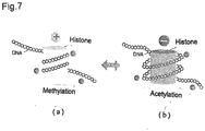
- DNA show (-) charged, so that histone and DNA stick each other as a magnet and methylation improves untied, where DNAs winding around the histones are charged (+) (see Fig. 8 (a)).
- the substrate to be adsorb is preferable to be (-) charged since the histones wound around with DNA is (+) charged.
- the cancer-related substances in blood and biological samples can be selectively detected through auto fluorescence by localized plasmon enhancement effect. Further, it is also possible to determine the existence of cancer simply through fluorescence microscopy because the cancer-related substance with a variety of fluorescent labels can be selectively captured. Furthermore, since chemical modification status of histone tails can be detec6ted from the Raman spectrum of the crystals with or without labels, it is also possible to carry out early detection of cancer, and determination as to progression of the cancer. Therefore, summarization of the usage way according to the present invention is as follows.
- the plasmonic chip provided with a silver quantum crystal
- the plasmonic chip provided with a silver meso crystal
- the Raman spectroscopic analysis is carried out through Raman scattering spectra by easily irradiating a laser onto the crystal.
Landscapes
- Health & Medical Sciences (AREA)
- Life Sciences & Earth Sciences (AREA)
- Chemical & Material Sciences (AREA)
- Immunology (AREA)
- Physics & Mathematics (AREA)
- General Physics & Mathematics (AREA)
- Biochemistry (AREA)
- General Health & Medical Sciences (AREA)
- Analytical Chemistry (AREA)
- Pathology (AREA)
- Engineering & Computer Science (AREA)
- Nuclear Medicine, Radiotherapy & Molecular Imaging (AREA)
- Biomedical Technology (AREA)
- Hematology (AREA)
- Molecular Biology (AREA)
- Urology & Nephrology (AREA)
- Medicinal Chemistry (AREA)
- Food Science & Technology (AREA)
- Cell Biology (AREA)
- Ecology (AREA)
- Biophysics (AREA)
- Microbiology (AREA)
- Biotechnology (AREA)
- Nanotechnology (AREA)
- Investigating, Analyzing Materials By Fluorescence Or Luminescence (AREA)
- Microscoopes, Condenser (AREA)
- Oncology (AREA)
- Measuring Or Testing Involving Enzymes Or Micro-Organisms (AREA)
Abstract
Description
- A plasmonic chip and a diagnostic method for observation of a fluorescence image and Raman spectroscopy of cancer-related substance on the plasmonic chips
- The present invention relates to a fluorescence image observation chip (plasmonic chips) with a fluorescence microscope and a method of diagnostic analysis for cancer diseases by observation of auto fluorescence and Raman spectroscopy for body fluids, in particular cancer-related substances and tumor makers absorbed with the plasmonic chip and enhanced by localized surface plasmon resonance (SPR).
- In the field of cancer diagnosis and therapeutics, as cancer-related substance, methylated DNAs and their proteins bound to histones (nucleosomes and chromatin) has drawn much attention. It is important for a simple diagnosis and early detection of the presence of cancer .to absorb or capture them and analyze their crystallization or aggregation state by fluorescence image diagnosis and SERS analyzation where the chemical modification of the DNA methylation status and the histone code state can be detected by means of laser irradiation on the crystal.
- Information for making all the organism cells are included in the DNA. The DNA, is composed of a unique sequence of the four bases, adenine (A), guanine (G), thymine (T) and cytosine (C). These bases are pairings between A and T as well as G and C on the two strands to form a DNA double helix. Chains of these pairs, stores all the information into regions called genes to make grouped specific molecules. Within each cell, there is a process of defining the cell-specific features by control genes mechanism to be turned on, that is, controls of genes expression, one of these control mechanisms is provided by adding a methyl group to a cytosine (C). C labeled with a methyl group may be represented as mC.
- DNA methylation, is said to play an important role in determining whether a part of the gene is expressed, where turning off of not required gene, makes DNA methylation. It is an essential control mechanism for normal development of the organism and the function. Further, aberrant DNA methylation, is said to be one of the mechanisms underlying the changes which is observed with the aging and the generation of a number of cancers.
- Cancers, heretofore, had been associated with the genetic changes caused by chromosomal mutations in the DNA. Mutation, regardless of hereditary or acquired hereditary, may lead to loss of expression of critical genes in order to maintain a healthy state. Now, it has been corroborated that a relatively large number of cancers, in many cases, are caused by improper DNA methylation close to the DNA mutation. In many cases, hyper methylation of DNA, for example, would mistakenly turn off the critical gene such as a tumor suppressor gene or DNA repair gene, which would allow the development and progression of cancer. The un-mutated process to control gene expression is called as Epigenetics.
- DNA methylation, is a chemical modification of DNA performed by enzymes called methyltransferases, where a methyl group (m) is added to a specific cytosine (C) in DNA. The non-mutated (epigenetic) process (mC), was said to be a decisive factor in the regulation of gene expression (Non-Patent Document 1). Therefore, from the focused sight on detection of DNA methylation, early detection method for colon cancer has been proposed (Patent Document 1). However, it is too complex and difficult to detect methylated DNA, so it is not suitable for simple and rapid detection because of taking much time and cost.
- Therefore, it is desirable to provide a method for measuring the fluorescence of markers precisely and up to a low density with a simple operation on a commercial fluorescence microscope by using a metallic nanocrystal sheet, which is suitable for the fluorescence observation substrate without crossed special optical system.
- As a marker detection method, it has been proposed currently, a method of surface plasmon resonance (SPR) and its apparatus (Patent Document 2), and a method of enzyme-linked immunosorbent assay (ELISA). The former SPR although operation is simple and rapidity, has a problem with the price of devices and chips as well as lack of sensitivity, while the latter ELISA has problems with not good rapidity and operability for the spot diagnostic methods although the price of the device and the chip are inexpensive with high sensitivity. Therefore, instead of this, a method of using a plasmonic chip to realize the rapidity and sensitivity of fluorescence advanced detection is proposed by making a thin film of silver and zinc oxide deposited on the substrate with a periodic structure of pitch 350nm labeled marker (Patent Document 3) and a method of using localized plasmon fluorescence enhancement sheet is proposed where the sheet is made from metal nanoparticles dispersed in an organic solvent by evaporating the solvent and self-assembling the metal nanoparticles into two-dimensional directions.(Patent Document 4).
-
- (Patent Document 1) Tokkyo
No.2010-517582 JP - (Patent Document 2)
WO2011-11472 - (Patent Document 3) Tokkai No.
2011-158369 JP - (Patent Document 4)
WO2013-039180 JP - (Non-Patent Document 1) JG Herman, Seminars in Cancer Biology, 9: 359-67, 1999
- However, mass production of a plasmonic chips is not easy and also adsorption of cancer-related substance or fluorescence-labeled markers onto the mass-produced plasmonic chip is not easy, so that it is difficult to exert the fluorescence enhancement effect on the present plasmonic chips. Accordingly, the present invention is to provide a plasmonic chips which is easy to be mass produced, whereby cancer-related substances or fluorescent labeling marker of adsorbent material such as antibody and so on, can be absorbed onto the plasmonic chips, so that plasmons on plasmonic chips can enhance the emission of fluorescence intensity from several times to several tens of times so as to realize a diagnostic method for cancer diseases by using a diagnosed fluorescence image on the plasmonic chip.
- According to the present invention, there can be provided firstly substrates of 1) silver complex quantum crystals and 2) silver peroxide meso crystals prepared by alkali treatment of the silver complex quantum crystals, which realize not only a physical property of localized surface plasmon resonance (LSPR) by light irradiation including UV, and infrared light, but also a chemical property which enables to absorb an antibody, fluorescent label markers and ligands with fluorescent label markers as a kind of ligands when silver complex quantum crystals are prepared. On the other hand, silver peroxide meso crystal has an another chemical property to absorb the cancer-related substances, such as nucleosome or chromatin including the DNA in the serum, particularly a histone wrapped around with DNA, Therefore, the present invention can be made on the findings that a plasmonic chip can be used for observing a fluorescence image under a fluorescence microscope, to judge or detect the presence or absence of cancer by selective absorption of cancer makers or nucleosome and clomatine as cancer-related substance, According to the present invention, there can be provided a plasmonic chip where a region of quantum crystal of plasmon metal complexes is formed on the substrate to selectively absorp or capture tumor makers with cancer related substance and to be enhanced by the localized surface plasmon resonance function. Further, according to the present invention, there can be provided a plasmonic chip where a region of meso-crystal of nanocrystal of silver oxide or oxides containing silver peroxides made from quantum crystal of silver complex, is formed on a metal substrate by alkali treatment wherein the meso-crystal is able to absorb or capture the cancer-related substance and to be enhanced by localized surface plasmon resonance function by light irradiation in the crystal area so as to observe the .auto fluorescence image.
- According to the present invention, there can be secondly provided a method of performing imaging of cancer diseases using the plasmonic chips, which comprises a step of providing a plasmonic chip which has a meso-crystal region is prepared by alkali treatment from the silver complex quantum crystals and contains silver oxide nanocrystals containing silver peroxides; a step of adding dropwise a serum or biological liquid sample on the crystalline regions of the plasmonic chips, to selectively absorb capture cancer-related substances having a positively charged protein molecules in the sample; a step of irradiating the agglomerated crystals on the plasmonic chip with light to observe the auto fluorescence image for diagnosis of the presence or absence of cancer. On the other hand, according to the present invention, there can be provided another method, which comprises a step of providing a silver complex quantum crystal on the metal substrate having an electrode potential less noble than that of a reduction potential of silver complex, where the tumor marker having a fluorescent function with ligand of the metal complex quantum crystal or as one of the ligand was added to place the tumor markers in the crystalline region in the silver complex quantum crystals, and a step of light irradiating the silver complex quantum crystal region to enhance the fluorescence emission intensity of the tumor markers by localized surface plasmon resonance and a step of observing the fluorescence image of a cancer disease..
- In addition, the position of the cancer-related substances aggregated on the substrate cannot be determined visually. According to the present invention, there can be provided a cancer diagnostic method which is characterized in that determination of the location of its crystalline region is made by distinct vision image with a microscope under a laser irradiation and then analyzation of chemical modification of histone tails, and remodeling factors can be made (
Fig. 19 ), Thirdly according to present invention, there can be provided a cancer diagnostic method of Raman spectroscopy, which comprises a step of observing a fluorescence image of the cancer-related substances captured on the plasmonic chip as a mark for a laser irradiation, and a step of irradiating the fluorescence image with various laser lights of 514nm, 532nm, 633nm, and 785nm for the precise analysis.. Accordingly, the present invention relates to a method of diagnosing a body fluid, in particular selectively captured cancer-related substances in the blood, as well as a method of analyzing the structure of a histone or chromatin, by analyzing the factors that chemical modification of histones tails, existence of cancers. The method is intended to enable early stage diagnosis of cancer as well as the presence or absence of the cancer. - According to the present invention, the plasmonic chips, make it possible to selectively adsorb the cancer-related substances or tumor markers, so that localized surface plasmon resonance makes fluorescence intensity of the fluorescence image enhanced, whereby it is possible to facilitate the determination of the cancer diagnosis from the fluorescence images. By the way, less noble electrode potential (larger ionization tendency) of the metal substrate than the metal complex causes a potential difference to make aggregation of quantum crystals (nano-sized metal complex crystals) from the metal complex solution by means of chemical reduction. In case of silver complex, quantum crystals of silver complex can be formed by the chemical reduction where aggregation of quantum crystals of silver complex on the metal substrate from silver thiosulfate aqueous solution is caused by noble electrode potential (higher ionization tendency) of silver than that of copper or copper alloy. In detail, the concentration of the metal complex in the aqueous solution should be mainly determined in the light of the size of the quantum crystals to be formed, so the concentration of a dispersing agent, if used, has to be considered. Although 100 to 5000ppm can be used, 500 to 2000ppm is preferred for preparation of nano-sized crystals to say the nanocluster depending on the functionality of the ligand.
- Metal complex to be formed as a quantum crystal is selected in the light of a complex stability constant (log β) according to the following formula (I) to correlate the electrode potential E of the supported metal.

- (Where E ° is the standard electrode potential, R is the gas constant, T is absolute temperature, Z is ion valence, F represents the Faraday constant.). In case of complexes of a plasmon metal selected from the group consisting of Au, Ag, Pt and Pd, an enhancement effect caused by localized surface plasmon resonance can be obtained by irradiation of the Raman light. In particular, in case of silver complexes the silver complexes may be formed by a reaction which may be preferably carried out between a silver complexing agent having a stability constant (formation constant) (log β i) of 8 or more and silver halide, preferably silver chloride. The complexing agent can be selected from the group consisting of thiosulfate, thiocyanate, sulfite, thiourea, potassium iodide, and thiosalicylic acid salt, The silver complexes become quantum crystals of 100 ∼ 200nm provided with quantum dots made of nanoclusters having average diameter of 5 ∼ 20nm.
- . It is believed that alkaline treatment (with an aqueous solution of sodium hypochlorite) makes silver complex quantum crystals to be changed into needle-like or acicular nanocrystals of silver oxide composite, containing a silver peroxide as a core of silver halide by the following reaction (
Fig. 5 ). The silver oxide composite shows a minus (-) charge in water while a histone wound around with DNA shows a plus (+) charge (Fig. 7 (a) ), whereby it is found that the positively charged cancer related substances including free nucleosomes are selectively adsorbed. Moreover it is found that the acicular nanocrystals of silver oxides containing silver peroxides can be reduced by means of irradiation with a laser beam and further changed into the metallic silver, so as to show the surface plasmon enhancement effect by means of laser beam irradiation, which makes it possible to detect histones by the auto fluorescence of cancer-related substances.
Na2S2O3 + 4NaClO + H2O → Na2SO4 + H2SO4(2NaHSO4) + 4NaCl Ag + + NaCl → AgCl + Na +
Ag + + 3NaOCl → 2AgCl + NaClO3 + 2Na+
Ag + + OH- → AgOH
2Ag+ + 2OH- → Ag2O + H2O
- According to the present invention, the needle-like nanocrystals of the silver oxide composite containing silver peroxides, can be formed by a self-organizing action or phenomenon as a neuron-like three-dimensional super-structure (so called meso crystals, referring to
FIGS. 8 and9 ). The silver oxide composite may be prepared by means of potentiostatic deposition using an Ag / AgCl electrode in silver ions aqueous solution and can be also prepared by oxidation of silver quantum crystals with an alkali treatment. For example, in case of silver thiosulfate, the silver complex quantum crystals can be easily prepared by the alkali treatment (with an aqueous solution of sodium hypochlorite). - Moreover, according to the present invention, use of the plasmonic chip, makes it possible to detect the presence of cancer-related substances in a biological sample including a blood and also to determine the chemical modification state where chromatin remodeling is controlled, through the fluorescence imaging diagnosis. That is, according to the present invention, the method comprises a step of providing a plasmonic chip with a needle-like nanocrystal composites of the silver oxides containing silver halides or halogens, that is, a step of providing a plasmonic chip having a meso crystal region of the silver oxides containing silver peroxides (
FIGS. 8 and9 ), a step of dropping a plasma serum or biological sample solution onto the needle-like nano-crystal area on the plasmonic chip, so as to selectively absorb or capture the cancer-related material having a positive charge in the sample. - The absorbed cancer-related substances can be observed by irradiation of a laser of UV, red or green on with a fluorescence microscope (
magnification 50 times). The fluorescence images of the cancer-related substances are shown inFigs. 1 (a), (b) and (c) in case of gastric cancer and inFigs. 2 (a), (b) and (c) in case of colorectal cancer while no fluorescence images of the samples can be shown in case of benign disease. The geometry, brightness and dimension of the fluorescence images can be quantified and the resulting numerical values are subjected to histogram processing. When observed with a fluorescence microscope, the crystal shapes between the benign disease and the gastric cancer or the colon cancer can show some differences. Therefore, it is understood that use of plasmonic chips provided with meso crystals containing silver peroxide or peroxides, makes it possible to selectively absorb or capture the cancer-related substances in blood and to determine the presence or absence of cancer by fluorescence image observation... - Although crystal of adsorbed tumor marker on the plasmonic chips) cannot be visually observed, irradiation of ultraviolet, red, or green laser makes it possible to observe the crystals with a fluorescence microscope (50 times), where point-like dispersed crystalline masses can be observed. Detection of the Raman spectra therefrom by irradiating of various laser beams makes it possible to determine the cancer disease with the intensity of surface-enhanced Raman scattering (SERS).
- It is understood in the present invention that the cancer-related substances in serum may include, a histone wound around with DNA (so called nucleosomes), and also a chromatin (fiber) where histones gather and form a string-like structure It is indicated that the substances to be detected in the present invention are deemed to be the cancer-related substances in the light of their increasing volumes because although globulin is positively charged, the globlin increases up to 2 times at most, while the cancer-related substance increases up to 100 times in association with progression of the cancer.. Moreover, DNA left from the normal cells, DNA dissociated from histones by acetylation and albumin account for approximately 60% of serum, but owing to their negative charge, they will not be absorbed in the present invention. Therefore, it is advantageous for fluorescence imaging of cancer-related substances. In order to confirm this phenomenon, the following tests were carried out.
-
Fig.15 shows photographs of 100-fold image, 3000-fold image, and 3D image in case of 10-fold and 100-fold dilution samples of benign disease, Gastric cancer, and Colon cancer.Fig.16 show photographs of 100-fold image, 3000-fold image with undulation, and 3D image in case of Kato-III (gastric signet ring cell carcinoma). Then, Kato-III was cultured and from the cancer cell a double-stranded DNA, RNA and Protein were extracted. The (a, b, c) are photographs of 100-fold surface image, 3000-fold surface image, and 3D surface image of the Kato-III (gastric signet ring cell carcinoma), while the (d, e, f) are photographs of 100-fold image, 3000-fold image, and 3D image of the DNA extracted therefrom. The (g, h, l) are also photographs of 100-fold image, 3000-fold image, and 3D image of the RNA. The (j, k, l) show photographs of 100-fold image, 3000-fold (images and undulations) and the 3D image of the Protein, while the (m, n, o) are photographs of 100-fold image, 3000-fold image, and 3D image showing a surface structure of physically grounded cancer cellsFig. 17 shows Raman scattering spectra on the meso-crystal plasmonic chips where they are chemical cultured cancer cells (a), DNA extracted therefrom (b), RNA (c), Protein (d), physically crushed cancer cells (e), serum (f) from gastric cancer patients, serum (g) from colorectal cancer patients and serum (h) from benign disease patients. Kato-III is gastric signet ring cell carcinoma, MKN-45 (poorly differentiated gastric cancer line), CW-2 (Japanese derived colon cancer cell line), PK45-P (human pancreatic cancer cell line), and NHDF-Neo (skin fibroblasts). Accordingly, it is understood from these results that the cancer-related substances are deemed to be selectively absorbed or captured and detected by the plasmonic chips of the present invention. - Moreover, according to the present invention, it is believed that needle-like nanocrystals (so called meso crystals of the silver oxide or oxides containing silver peroxide or peroxides) is provided with silver oxides containing silver peroxides which are easily negatively charged in an aqueous solution so as to form a charge transfer complex in contact with the sample (target molecules) . Furthermore, the silver oxide can be reduced by a light energy to become metallic silver, which will have an enhancing effect by localized surface plasmon resonance with the metallic nanoparticles arranged regularly. Therefore, the needle-like nano-crystals (meso crystal) of the present invention ,although it is a non-metal, are provided with an ionization property together with a metal property, which becomes a plasmonic chip suitable for measurement of surface-enhanced Raman scattering (SERS), as a cancer diagnostic chip.
- It is believed that quantum crystals formed on a metal substrate or metal particles tend to have a positive polarity in an aqueous solution as a metal complex crystal, so their subjection to alkali treatment in the presence of halide ions is preferable to adjust the polarity of the chips by sodium hypochlorite solution having more than pH11 in order to fixedly adsorb the protein in a biological sample, According to the present invention, recrystallization of the quantum crystals makes it to have not only a negative polarity in an aqueous solution, but also makes it changed to needle-like composite of nanocrystal of silver oxides containing silver peroxides, so as to promote the histones having a positive charge in cancer-related substances absorbed on the chips.
-
-
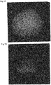
Fig.1A shows fluorescence micrographs of the cancer-related substance of gastric cancer that adsorbed on plasmonic chip crystal by irradiation of UV (380nm). -
Fig.1 B shows fluorescence micrographs of the cancer-related substance of gastric cancer that adsorbed on plasmonic chips by irradiation of a red laser. -
Fig. 1C shows the fluorescence micrographs of the cancer-related substance of gastric cancer that adsorbed on plasmonic chips by irradiation of a green laser. -
Fig.2 shows the fluorescence micrographs of the cancer-related substance of colon cancer that adsorbed on plasmonic chips by irradiation of (A) UV (380nm), (b) a red laser, (c) a green laser. -
Fig.3 is a photograph showing the relationship between quantum crystal shapes and standing times after dropping on the phosphor bronze substrate. -
Fig.4 is a graph showing a result of EDS spectra analysis of quantum crystals (elemental analysis). -
Fig.5 is a photograph showing SEM image of quantum crystals alkali-treated in the presence of a halogen ion (Sodium hypochlorite treatment). -
Fig.6A is a photograph showing needle-like crystals of the alkali-treated quantum crystals. -
Fig.6B is a photograph showing a rugby ball-shaped mass in the needle-like crystals. -
Fig.6C is a graph showing a result of EDS spectra of large mass (elemental analysis). -
Fig.7 is functional illustration views showing a state of the methylated free DNA (a) and a state of acetylated DNA (b). -
Fig.8 is a view (top) of SEM image showing a re-crystallized substrate which is the quantum crystal substrate alkali treated in the presence of a halogen ion (Sodium hypochlorite treatment) (top view) and a graph (down view) showing a result (elemental analysis) of the EDS spectra of the re-crystallized substrate. -
Fig.9 is a graph showing a result of XPS measurement of the alkali-treated .recrystallization substrate. -
Fig.10 is a graph showing a result of XPS measurements after etching the surface of the recrystallization substrate. -
Fig.11 shows fluorescence micrographs of the cancer-related substance adsorbed on the plasmonic chips in case of a benign disease, (b) a gastric cancer, (c) a colon cancer. -
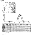
Fig.12A shows a laser micrograph of a sample obtained by adjusting the serum obtained from benign disease patients and a histogram of the RGB. -
Fig.12B shows a laser micrograph of a sample obtained by adjusting the sera obtained from gastric cancer patients and a histogram of the RGB. -
Fig.12C shows a laser micrograph of a sample obtained by adjusting the serum obtained from colorectal cancer patients and a histograms of RGB. -
Fig.13 shows plasmonic micrographs of distinct vision crystal display image and a method of measuring the Raman scattering spectra (unadjusted).of benign disease, gastric cancer, and colon cancer by aiming at the center of the crystals. -
Fig 14 shows Raman spectrum showing a state of chemical modification of histone tails of a gastric cancer-related substances. -
Fig.15 shows 3D laser scanning micrographs (100-fold image, 3000-fold image and 3D image) of 10-fold dilution and 100-fold dilution samples of Benign disease (a, b, c; d, e, f), Gastric cancer (g, h, i; j, k, l), Colon cancer (m, n, o, p, q, r), 3000 times, it will show the 3D image. In addition,Fig.15 shows RGB color histograms of 3000-fold image obtained by 3D laser scanning microscope in case of 10-fold dilution samples of Benign disease (s, t), Gastric cancer (u, v), and Colon cancer (w, x). -
Fig.16 shows photographs of 100-fold image, 3000-fold image with undulation, and 3D image in case of Kato-III (gastric signet ring cell carcinoma). Then, Kato-III was cultured and from the cancer cell double-stranded DNA, RNA and Protein were extracted, where the (a, b, c) are photographs of 100-fold surface image, 3000-fold surface image, and 3D surface image of the Kato-III (gastric signet ring cell carcinoma), while the (d, e, f) are photographs of 100-fold image, 3000-fold image, and 3D image of the DNA extracted therefrom. The (g, h, i) are also photographs of 100-fold image, 3000-fold image, and 3D image of the RNA. The (j, k, l) show photographs of 100-fold image, 3000-fold (images and undulations) and the 3D image of the Protein, while the (m, n, o) are photographs of 100-fold image, 3000-fold image, and 3D image showing a surface structure of physically grounded cancer cells -
Fig. 17 shows Raman scattering spectra on the meso-crystal plasmonic chips where they are chemical cultured cancer cells (a), DNA extracted therefrom (b), RNA (c), Protein (d), physically crushed cancer cells (e), serum (f) from gastric cancer patients, serum (g) from colorectal cancer patients and serum (h) from benign disease patients, wherein Kato-III is gastric signet ring cell carcinoma, MKN-45 (poorly differentiated gastric cancer line), CW-2 (Japanese derived colon cancer cell line), PK45-P (human pancreatic cancer cell line), and NHDF-Neo (skin fibroblasts). -
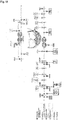
Fig.18 shows histone-associated antibodies diagram for identifying a chemical modification of histone tails (referring to www.genetex.com). -
Fig.19 shows a fluorescence image showing the auto fluorescence state of benign disease and cancer, which is observed on plasmonic chip of the present invention. - Hereinafter, embodiments of the present invention will be explained, referring to the attached drawings,
- As shown in
Figure 4 , an aqueous solution containing 1000ppm of silver thiosulfate was prepared and the 1 drop was added dropwise on a phosphor bronze plate. After standing for about 3 minutes, the solution on the plate was blown off. On the plate, quantum crystals were obtained as shown in the SEM image at the right side ofFig.4 ... -
Fig. 5 is a photograph showing various SEM images of the nano-particle aggregate prepared in Example 1 (quantum crystal), andFig. 6 shows an enlarged SEM image of nano-particles where there were thin hexagonal columnar crystals of 100 nm more or less and having an unevenness surface of several nm order. We could not find out any specific facets of metal nano-crystals in the quantum crystals.Figure 7 is a photograph showing the relationship between quantum crystal shapes and the standing time after dropping onto the phosphor bronze substrate, where it is recognized that firstly, a hexagonal quantum crystal is produced and then growing while maintaining the crystal shape. -
Fig. 8 is a graph showing a result of EDS spectra (elemental analysis).of the quantum crystals where not only silver but also elements derived from complex ligands can be detected in case of the quantum crystal on the phosphor bronze substrate, while only silver can be detected in the case of the quantum crystals formed on a copper plate by using 1000ppm of silver thiosulfate in aqueous solution and keeping it for the standing time of 3 minutes after dropping onto the copper substrates. - In case of 1000ppm of silver thiosulfate complex in an aqueous solution, hexagonal column crystals of 100nm more or less, are formed for the standing time of 3 minutes after dropping it onto a phosphor bronze plate, where it is confirmed that irregularities of several nm order are found on the hexagonal column quantum crystals from the SEM images (
Fig. 4 ,5 and6 ).and any specific facets derived from a metal nano-crystals are not found, while the EDS elemental analysis shows silver and elements derived from the complexing ligand. Accordingly, it can be estimated from the above analysis that the whole particles show nano-crystals of silver complex and also the unevenness appearance on the surface may be caused by the formation of spread quantum dots made of silver clusters in the complexes. From the aspect of phenomenon that the silver complex quantum crystals of the present invention can be formed on a phosphor bronze plate, while silver nano-particles alone can be deposited on the copper substrate, it is estimated that, as the equilibrium potential of the silver thiosulfate complexes is 0.33 which is equivalent to the copper electrode potential with 0.34, there is deposited only silvers with 0.80 on the copper substrate. On the other hand, in case of a phosphor bronze plate with the electrode potential of 0.22, which is slightly less noble than that of the copper so that silver complex crystals seem able to be precipitated. The concentration of the silver complex in the aqueous solution should be in a dilute region of 500 ∼ 2000ppm, 2) the electrode potential of the metal substrate with respect to the equilibrium potential of the metal complex solution is slightly less noble, 3) the metal complex should be deposited by the electrode potential difference between the metal substrate and the metal complex. Further, in case of 1000ppm of thiourea silver complex in aqueous solution, the same function can be observed. - The quantum crystal substrate is subjected to a treatment of dropping 5% sodium hypochlorite solution thereon and the dropped solution is removed off 2 minutes later to obtain crystals having structures shown in
Fig. 12 , where needle-shaped crystals and large clumps such as rugby ball-like mass are observed and the respective compositions are subjected to analyzation at EDS spectra (elemental analysis),. After a result of the analysis, the needle-like crystals are both considered to consist of a composite crystal of silver oxide and silver chloride, from the following reaction formulas and the result ofFigure 12 does not show any chlorine and shows that the silver and oxygen is dominant.
Na2S2O3 + 4NaClO → H2O → Na2SO4 + H2SO4 + 4NaCl (1)
Ag+ + NaCl → AgCl + Na + (2)
Ag++ 3NaOCl → 2AgCl + NaClO3 + 2Na+ (3)
Ag+ + OH- → AgOH (4)
2Ag+ + 2OH → Ag2O + H2O (5)
- Thus, although it is considered that silver ions and thiosulfate ions are important in the formation of meso-crystal according to the present invention by alkaline oxidation reaction in the presence of chloride ions and ,although the silver oxide is formed according to a conventional reaction, it is surprised that silver peroxide are predominantly formed from the following XPS measurement.
- The aqueous sodium hypochlorite was added dropwise for 2 minutes to the quantum crystal substrate prepared as the above, in order to make a re-crystal substrate, which is subjected to a XPS analysis (using models: ULVAC-PHI (Ltd.) / PHI5000 Versa Probe II (scanning X-ray photoelectron spectroscopy) without etching for Ag and O by XPS measurement. In addition, for comparison, Ag in the powder of silver chloride and the powder of silver oxide were measured. On the other hand, the recrystallized substrate was subjected to XPS measurement of Ag and O after etching for 5 minutes with an argon gas cluster ion gun. If the XPS measurement results of
FIGS. 13 and14 will be combined with the results of EDS according toFIG. 12 , the peak in the vicinity of 529eV is the peak derived from silver peroxide (AgO), while the peak in the vicinity of 530eV is the peak derived from silver oxide (Ag2O). Further, If it is etched, the oxygen content decreases, while the O peak derived from the silver peroxide (AgO) in the vicinity of 529eV is still greater than the peak derived from the silver oxide in the vicinity of 530eV in case of etching, so that it is recognized that the silver peroxide was produced in the vicinity of the substrate. It is assumed that the electrode potential of the substrate and the catalytic action are affected.to the meso -crystal formation - The EDS measurement was carried out on the above-mentioned re-crystal substrate by using a JEOL Ltd. / JSM-7001F (field emission scanning electron microscope analysis).
- In addition, even if the aqueous solution selected from the group consisting of hypochlorous acid, 0.01 N sodium hydroxide, 0.01 N hydrochloric acid and 0.1 molar sodium carbonate would be used, any result similar to be treated with sodium hypochlorite was not obtained. Thus, it is believed that the formation of the needle-like crystals are caused by the above reaction in the presence of silver ions and thiosulfate ions. While the silver oxide is induced into negatively charged in an aqueous solution, it is reduced by the light to deposit metallic silver. Since silver peroxide shows more remarkable in the above tendency than silver oxide, it is possible to adsorb cancer related substances having a positive charge, resulting in occurrence of the surface plasmon enhancement effect between the trapped cancers related substance and the silver particles.
- Moreover, according to the present invention, the plasmonic chip makes it possible to detect the presence and absence of cancer-related substances and analyze chemical modification factors that control chromatin remodeling by a diagnosis imaging and Raman analysis of biological samples including the blood,. That is, these detection and analyzation can be carried out by the following steps which comprises a step of providing the plasmonic chip having a meso crystal region of the silver oxide containing silver peroxide (
FIGS. 9 and10 ) as the needle-like nanocrystal composites of the silver oxide containing silver halide or halogen, and a step of dropping the serum or biological sample solution onto the needle-like nano-crystal area of the plasmonic chip, whereby selectively adsorption of the cancer-related substances having a positive charge in the sample can be carried out. - The adsorbed cancer-related substances were observed by a reflection microscope (magnification 500-fold). The crystal images of the cancer-related substances in case of a benign disease, a gastric cancer, and a colon cancer are shown (
Fig. 1 (a), (b) and (c) ). The image information such as the area of the crystal of the image, the volume, the length, the width and so on can be digitized and these numbers are used for the histogram. Observation of images with 15,000 fold by a laser microscope (VK-X250 shape analysis Laser microscope manufactured by Keyence Corporation) are shown inFig. 2A ,2B, and 2C , where there can be observed differences between the benign disease and the stomach cancer or the colorectal cancer. Graphing of R, G, and B in the histogram, as shown inFig. 2A ,Fig. 2B and Fig. 2C , makes the peak deference clear relating to the position and the shape between the benign disease, the stomach cancer, and the colon cancer. Therefore, it is understood that use of the plasmonic chips provided with meso crystals containing silver peroxide makes it possible to identify the presence of cancer by selectively capturing the cancer-related substances in blood. Further, use of a confocal laser microscope is preferable, since the slice image and then the internal information are obtained. - In addition, sera obtained from benign disease patients (esophageal achalasia, gastric mesenchymal tumor, duodenum benign tumor) and sera obtained from cancer patients (stomach cancer, colon cancer, pancreatic cancer) were subjected to auto-fluorescence imaging diagnosis on the plasmonic chips with a fluorescence microscope. It is understood that clear identification can be made between the benign disease patients and the cancer patients, depending on whether auto fluorescence can be observed or not as shown in Fig. 20.
- Crystals of adsorbed cancer-related substances on the plasmonic chips cannot be observed with naked eyes. Under a microscope (500-fold), crystalline mass are observed like distributed points (
Fig. 18 ). Laser Irradiation to one of the crystalline mass (wavelength 532nm) makes Raman spectra from there, where it is possible to determine the cancer disease with the intensity of surface-enhanced Raman scattering (SERS). Fig.3 (a), (b), and (c) show the Raman spectrum obtained therefrom by irradiating laser (wavelength 532nm) to in case of benign disease, gastric cancer, and colon cancer. In particular,FIG. 4 is an enlarged view of the Raman spectrum in the case of gastric cancer, where the chemical modification of the histone tail can appear as peaks and can be determined in so called fingerprint region from 1300cm -1 less than 625cm -1 - . The cancer-related substances in the serum, may include a DNA that wraps around the histone derived from cancer cells, a histone wounded around with DNA (so called nucleosomes) and also a chromatin (fiber) where histones gather and form a string-like structure. It is indicated that the substances to be detected in the present invention are deemed to be the cancer-related substances in the light of their increasing volumes because although globulin is positively charged, the globlin increases up to 2 times at most, while the cancer-related substance increases up to 100 times in association with progression of the cancer... Moreover, DNA left from the normal cells, DNA dissociated from histones by acetylation and albumin account for approximately 60% of serum, but owing to their negative charge, they will not be absorbed or captured in the present invention. Therefore, it is advantageous for fluorescence imaging of cancer-related substances and it is convenient to analyze the chemical modification of histone tails by Raman spectroscopy.
- Moreover, needle-like nanocrystals of the present invention (meso crystals of the silver oxide containing silver peroxide) is easily negatively charged in aqueous solution and it is believed to form a charge transfer complex in contact with the sample (target molecules). Furthermore, the silver oxide is reduced by receiving a light energy to be changed to metallic silver, and will make a localized surface plasmon resonance enhancement effect with the metal nanoparticles arranged regularly. Therefore, needle-like nano-crystals of the present invention (meso crystal), although it is a non-metal, are provided with the metal property as well as the ionization property, which is suitable for a plasmonic chip used for measurement of surface-enhanced Raman scattering (SERS), as a cancer diagnostic chip.
- On the other hand, quantum crystals formed on a metal substrate or metal particles are believed to likely have a positive polarity in an aqueous solution as a metal complex crystals, so that in order to adsorb proteins in a biological sample, an alkali treatment in the presence of halide ions is preferable to adjust the polarity by dropping sodium hypochlorite solution having pH11 or more,. quantum crystals can be recrystallized not only to have a negative polarity in an aqueous solution, but also to make a needle-like composite of nanocrystal silver oxide including silver peroxide, so that immobilization of histones having a positive charge can be promoted as cancer-related substance.
- Nucleosomes is a basic building block of chromatin, where DNA is wound to histone octamer consisting of four histone (H2A, H2B, H3, H4) to have a role of packaging the DNA and also a role of regulating the accessibility of the DNA to play an important role in gene regulation. The histone post-translational modifications, controls interactions with DNA and other nuclear proteins to affect the reversible gene expression. As types of histone modification, there are mainly known methylation, acetylation, phosphorylation, ubiquitination, SUMO reduction, citrullination, and ADP-riposylation. In the sequence of histone, depending on which part is subjected to these modifications, the periphery of the gene expression is activated or inhibited. The relation between combination of post-translational modification sites of histones and gene expression can be tested and verified with histone code associated antibodies (Genetex Inc. anti-histone antibody), and by Raman spectroscopy as well as observation in the fluorescence images modified state of histone code that is captured by the plasmonic chip of the present invention and then verification of the histone code hypothesis can be carried out. The histone code hypothesis is now described as follows.
- Histone code hypothesis has been proposed in relation to the chromatin remodeling by Dr. Alice group of Rockefeller University. That is, the chromatin remodeling is defined as the following phenomenon, where associated with molecular mechanism through changes in chromatin structure which modulates the expression levels of genes, and with the induction, 1) change in the histone modification, 2) change in the methylation status of genomic DNA, then associated therewith 3) phenomenon in which changes in the high sensitivity region occur against DNase. The present inventors have, based on the histone code hypothesis, in order to elucidate the mechanisms of cancer development, an intensive research has been carried out, where as a cancer-related substances, the nucleosomes and chromatin relating to histones firstly is selectively captured and then tried to verify this. Most of genomic DNA are wound in core histone, and is stored as chromatin. Therefore nuclear phenomena which directly encourage to genomic DNA such as transcription, replication, repair and so on, is accompanied by chromatin structural changes in some way. This chromatin remodeling in a broad sense is to control the nuclear phenomenon in reverse. For example, transcriptional activity varies within a scope up to about 2500-fold, depending on the chromatin structure. Since the range of the activation of the transcription factor in the naked DNA is a 10-fold, most of the transcriptional control is done in chromatin remodeling. Among the factors involved in the chromatin remodeling, histone modification factor such as acetylation, phosphorylation, methylation, ubiquitination and their removal performance, make histone H3, H4 amino terminal side to form a chemical modified pattern. This chemical modified pattern acts as a recognition code and then the factors that further involved in remodeling is recruited. This is proposed as "histone code hypothesis".
- The structural biology is said to have played a large role for the verification of the "histone code hypothesis" where a histone tail received this chemical modification serves as a recognition code such as remodeling factors. For example, most of HAT, ATP-dependent remodeling factors, coactivators, TFD subunit TAF, are provided with a low-bromo-domain homology consisting of about 110 amino acids. The bromo domain structure of the coactivator P / CAF is determined by NMR, so distinctive hydrophobic pockets have been found. Results of the binding experiments with NMR, showed that the pockets were bound to peptides of histone H3 tail acetylated. Also, the crystal structure provided with two-bromo-domains arranged in tandem of TAF 250, is the largest subunit of the general transcription factor TFD. This structure has an activity of binding to the tail of histone H4, which is modified with acetylation. Also chromo domains of
heterochromatin protein 1 has been shown to bind to histone H3 tails methylated. NMR structure of the chromo domain of mousechromatin modifier protein 1 has been reported. This chromatin remodeling factors may be accessed directly to specific nucleosomes sites by recognizing codes of histone tail with bromo domain. This chromatin remodeling factors are often indirectly recruited by a DNA binding transcription factor. The transcriptional repressor Mad having bHLH (basic helix-loop helix) -Zip as the DNA-binding domain, recruits Sin3 complex containing histone deacetylase as a sub-unit into the chromatin. Complex of PAH2 domain and transcriptional repressor domain of the Mad the Sin3 is analyzed by NMR and the recruitment mechanism for the chromatin region of deacetylation complex has been discussed. - It has been believed that DNA methylation is involved deeply in this chromatin structure control. In the genomic DNA sites that DNA methylation is seen at a high density, generally the chromatin structure becomes strong, reduction of transcriptional repression and DNA mutation rate is observed. Also it has been reported that methylation patterns and genomic imprinting of genomic DNA, the X chromosome inactivation and cell canceration have a clear correlation each other (referring to non-patent document 1). Therefore, it can be said that structural analysis of histones and chromatin holds the key to explain the context of cancer, so that analyzation of the chemical modification factors of histone tails has a great significance. Since histones and chromatin can selectively absorbed or captured according to the present invention, not only a means of fluorescently labeled chemical modification status of histone code but also a means of Surface enhanced Raman spectrometry (SERS).can be provided.
- Meanwhile, the genomic DNA is a body of genetic material is a 2 m long thing in humans, which is housed in the small space of 100 femto-liters of a so called cell nucleus. Storage of such genomic DNA can be accomplished by molecular complex called "chromatin". The foundation structure of chromatin is a "nucleosomes" where histone protain is wound around with DNA and those nucleosomes are beaded, and further folded highly bound to proteins and RNA to form a "higher-order chromatin". However, Unravelling of the DNA from chromatin, requires a large energy, so chromatin is inhibitory to the functional expression of the DNA, such as replication, transcription, and recombination. But organisms, through the dynamic variation of chromatin, very easily makes replication, transcription, and recombination. It is said that this "dynamic chromatin structure" is being produced by a variety of the nucleosome structure by histone variants and modifications, the diversity of its arrangement, and the interaction of the complex between protein and RNA molecular, while it has been controlled by the interaction of cell nuclear structure, nuclear membrane, and nuclear pore complex. To explain the phenomenon, DNA of eukaryotic organisms, was tightly bound to the histone protein synthesized in the body to form a nucleosome and the helix of nucleosomes form a strong chromatin structure. In case of the histone and DNA tightly combined, it is difficult to RNA polymerase to bind to DNA. On the other hand, when the transfer is thriving, the chromatin structure becomes loose and histone from nucleosomes is released out to let DNA naked. When the transfer is suppressed, on the contrary, the nucleosome structure is solid while the chromatin is gathered. Restructuring or reconfiguration of the chromatin structure means such a chromatin remodeling described above but the promoter and enhancer promote transcription and enhance gene expression. One of these roles is to loosen the binding of histone and DNA, sometimes to destroy the nucleosome structure, which promote the RNA synthesis by RNA polymerase. In detail, on the histone tails at the end of the translated histone protein, a lot of basic amino acids (positive charge) is present... The basic amino acids are electrostatic binding to the DNA of a phosphate group (negative charge). To loosen the binding of histones and DNA may lower basicity of histones into weak Transcriptional regulation by chromatin remodeling is actually made by a complex reaction in which many kinds of enzymes and proteins involved in transcription factors bind to promoters and enhancers, bond to histone acetylation enzyme (HAT: histone acetyltransferase) which enzyme makes acetylation of the amino group of histone. The acetylated amino group lowers the basic which causes weak binding to the DNA. After this, a dissociation of histones with DNA further progresses and further the acetylation of histones progresses, resulting in reveal of the promoter which makes it easy to combine with RNA polymerase. Therefore, it is important how to trap the cancer-related substances in the body fluid and how to analyze it in order to elucidate the mechanism of chromatin remodeling based on histone code hypothesis.
- According to the plasmonic chip of the present invention, trapping of the histones and chromatin as a cancer-related substance, the presence or absence of cancer symptoms can be judged by the fluorescence imaging depending on volumes of the crystal. Then, the cancer symptoms come from any organ, and how the cancer progresses can be determined by analyzing the remodeling factors and chemical modifications of histone tails. And all the information relating to how the cancer occurs and how the cancer progresses through this chromatin remodeling events, make it possible for doctor to predict accurately specific chemotherapeutic agents, and also to rationally design chemotherapy based on knowledge of tumor chemo sensitivity.
- Here, a protein of cancer-related substances of interest to be detected is a histone wrapped around with DNA The unit structure is called a nucleosome, and the string form structure made of nucleosome gathered is called a chromatin (fibers). And, in time of repeat of dividing cancerous cells, methylation modification makes DNA to wrap around histone not to come out from bad genes (tumor suppressor gene). Usually histones show (+) charged , while DNA show (-) charged, so that histone and DNA stick each other as a magnet and methylation improves untied, where DNAs winding around the histones are charged (+) (see Fig. 8 (a)). On the other hand, acetylation makes it (-) charged, so that histone acetylation makes DNA of (-) and histone (-) repelled each other. Then, the 'thread' of DNA has a mechanism for expression of the gene unwound from the histones (see Fig. 8 (b)). Therefore, in order to selectively adsorb the cancer-related substances derived from cancer cells, the substrate to be adsorb is preferable to be (-) charged since the histones wound around with DNA is (+) charged.
- . Thus, according to the present invention, the cancer-related substances in blood and biological samples can be selectively detected through auto fluorescence by localized plasmon enhancement effect. Further, it is also possible to determine the existence of cancer simply through fluorescence microscopy because the cancer-related substance with a variety of fluorescent labels can be selectively captured. Furthermore, since chemical modification status of histone tails can be detec6ted from the Raman spectrum of the crystals with or without labels, it is also possible to carry out early detection of cancer, and determination as to progression of the cancer. Therefore, summarization of the usage way according to the present invention is as follows.
- Auto fluorescence does not occur on the plasmonic chips by dropping antibody or antigen. Observation of the fluorescence image can be detected by attaching a fluorescent dye or a fluorescent protein to the target substance. In case of the endotoxin, binding of the fluorescence to the endotoxin is confirmed by Raman scattering spectra, so it can be more clearly distinguished by attaching a fluorescent label. Thus, 1) when the fluorescent material is attached to the antibody side, addition of a mixed solution of the metal complex and the antibody was added dropwise on the plasmonic chip of the present invention makes it possible to observe the light emission with the antigen bound to the antibody. On the other hand, 2) when the fluorescent material is attached to the antigen side, dropwise addition of antibody makes quantum crystals into a solid phase, which can be measured by emitting of fluorescence due to antigen-antibody reaction.
-
- 1) when a fluorescent material is attached to the antibody side of a silver meso crystal, immobilization of the antibody (with labeled) on the meso crystal and dropwise addition of the antigen make it possible to measure the fluorescence due to binding the antigen with the antibody. 2)when the fluorescent material is attached to the antigen side, immobilization of the antigen (with labeled) on the meso crystal and dropwise addition of the antibody make it possible to measure the fluorescence due to binding the antibody with the antigen. 3) Measurement by auto fluorescence. It is also possible to attach a fluorescent label in this case.
- The Raman spectroscopic analysis is carried out through Raman scattering spectra by easily irradiating a laser onto the crystal.
Claims (6)
- A plasmonic chip used for observing a fluorescence image under a fluorescence microscope, which is provided with a region of quantum crystal of plasmon metal complexes on the metal substrate wherein the plasmon metal has an electrode potential lower or more basic than that of the metal substrate which is close to a reducing electrode potential of the plasmon metal complex so as to have a difference between both electrode potentials which make a deposition of plasmon metal complexes on the metal substrate from an aqueous solution of the plasmon metal complex which contains an absorbent material such as a tumor marker or an antibody which is allowed to coordinate together with quantum crystals of the plasmon metal and which is able to selectively captures the substances such as cancer related substance or antigens, and to be enhanced by the localized surface plasmon resonance function.
- A plasmonic chip used for observing an auto fluorescence image under a fluorescence microscope, which is provided with a region of meso-crystal consisting of nanocrystal of silver oxide containing silver peroxide made from quantum crystal of silver complex formed on a metal substrate by alkali treatment wherein the meso-crystal is able to capture the cancer-related substance and to be enhanced by localized surface plasmon resonance function by light irradiation in the crystal area.
- A method of observing a fluorescence image for diagnosis of cancer which comprises providing the plasmon chip according to claim 1 or 2, dropping a serum or biological liquid sample on the crystal region of the plasmonic chips, to selectively adsorb cancer-related substances or adsorbent substances such as antigens in a sample, and light irradiating on the crystal region to observe the auto fluorescence or cancer markers of cancer-related material by localized surface plasmon resonance with a fluorescence microscope to observe the fluorescence image to diagnose the presence or absence of cancer..
- An analysis method of Roman spectroscopy for diagnosis of cancer which comprises providing a plasmon chip according to claim 1 or 2; dropping a serum or biological liquid sample on the crystal region of the plasmonic chips to selectively adsorb cancer-related substance or adsorbent substances such as antigens in a sample, light irradiating on the crystal region to observe an auto fluorescence or cancer markers of cancer-related substance by localized surface plasmon resonance with a fluorescence microscope to observe the fluorescence image to diagnose the presence or absence of cancer.by irradiating a laser beam on the fluorescence image of the cancer related substance or cancer marker by using the fluorescence image as a mark for irradiating.
- An analysis method of Raman spectroscopy according to claim 3, wherein the cancer-related substance is a nucleosome or chromatin including the histone wrapped around with DNA.
- An analysis method of Raman spectroscopy according to claim 4, wherein the cancer-related substance is a nucleosome or chromatin including the histone wrapped around with DNA.
Priority Applications (1)
| Application Number | Priority Date | Filing Date | Title |
|---|---|---|---|
| SM20220128T SMT202200128T1 (en) | 2014-05-08 | 2015-05-07 | Plasmonic chip, and cancer disease diagnosis methods respectively employing fluorescent image and raman spectroscopy |
Applications Claiming Priority (3)
| Application Number | Priority Date | Filing Date | Title |
|---|---|---|---|
| PCT/JP2014/062318 WO2014181816A1 (en) | 2013-05-08 | 2014-05-08 | Raman quantification method of cancer-related substance |
| JP2015036645 | 2015-02-26 | ||
| PCT/JP2015/063219 WO2015170711A1 (en) | 2014-05-08 | 2015-05-07 | Plasmonic chip, and cancer disease diagnosis methods respectively employing fluorescent image and raman spectroscopy and each using same |
Publications (3)
| Publication Number | Publication Date |
|---|---|
| EP3141888A1 true EP3141888A1 (en) | 2017-03-15 |
| EP3141888A4 EP3141888A4 (en) | 2018-03-14 |
| EP3141888B1 EP3141888B1 (en) | 2021-11-03 |
Family
ID=54392561
Family Applications (1)
| Application Number | Title | Priority Date | Filing Date |
|---|---|---|---|
| EP15789355.3A Active EP3141888B1 (en) | 2014-05-08 | 2015-05-07 | Plasmonic chip, and cancer disease diagnosis methods respectively employing fluorescent image and raman spectroscopy |
Country Status (15)
| Country | Link |
|---|---|
| US (1) | US10215700B2 (en) |
| EP (1) | EP3141888B1 (en) |
| JP (2) | JP6492066B2 (en) |
| KR (1) | KR102232197B1 (en) |
| CN (1) | CN106461556B (en) |
| AU (1) | AU2015256932B2 (en) |
| CA (1) | CA2948353C (en) |
| ES (1) | ES2905092T3 (en) |
| HU (1) | HUE057218T2 (en) |
| IL (1) | IL248760B (en) |
| MY (1) | MY193573A (en) |
| NZ (1) | NZ726748A (en) |
| SG (1) | SG11201609331XA (en) |
| SM (1) | SMT202200128T1 (en) |
| WO (1) | WO2015170711A1 (en) |
Cited By (2)
| Publication number | Priority date | Publication date | Assignee | Title |
|---|---|---|---|---|
| EP4143572A4 (en) * | 2020-04-30 | 2023-10-18 | MYTECH Co., Ltd. | Fluorescence counting system for quantifying viruses or antibodies on an immobilized metal substrate by using an antigen-antibody reaction |
| EP4083223A4 (en) * | 2019-12-25 | 2024-01-17 | MYTECH Co., Ltd. | AUTOFLUORESCENCE-BASED LIQUID BIOPSY METHOD TARGETING NUCLEOSOMES FRAGMENTED BY APOPTOSIS |
Families Citing this family (11)
| Publication number | Priority date | Publication date | Assignee | Title |
|---|---|---|---|---|
| JP2017156104A (en) * | 2016-02-29 | 2017-09-07 | 西松建設株式会社 | Light enhancement element, manufacturing method of the same, and spectroanalysis kit and spectroanalysis method |
| GB201704128D0 (en) * | 2017-03-15 | 2017-04-26 | Univ Swansea | Method and apparatus for use in diagnosis and monitoring of colorectal cancer |
| CN108169184B (en) * | 2017-09-05 | 2021-01-05 | 哈尔滨工业大学 | Establishment method and application of tumor classification identification model |
| CN108469429B (en) * | 2018-02-06 | 2020-11-24 | 西安电子科技大学 | A dual-modality Raman-optical projection tomography system |
| JP2021156576A (en) * | 2018-06-21 | 2021-10-07 | 有限会社マイテック | Liquid biopsy method by autofluorescence targeting disease-related protein conjugate |
| WO2021210189A1 (en) * | 2020-04-18 | 2021-10-21 | 有限会社マイテック | Pathogen quantum crystal solid-phase substrate and quantum crystal solid-phase fluorescence counting method |
| CN111624186B (en) * | 2020-06-24 | 2021-03-16 | 江南大学 | A Spectral Analysis Method of Enterotoxin Based on Fluorescence and Raman Dual Signal Enhancement |
| TWI818278B (en) * | 2020-11-20 | 2023-10-11 | 日商米特奇有限公司 | Autofluorescent liquid biopsy method targeting nucleosome fragmented by apoptosis |
| WO2022139465A1 (en) * | 2020-12-22 | 2022-06-30 | 두에이아이(주) | Method for early diagnosis of pancreatic cancer using artificial intelligence raman analysis technique |
| KR102530499B1 (en) * | 2021-08-26 | 2023-05-10 | 한밭대학교 산학협력단 | Method of detecting protein within a nucleus using plasmonic metal nanoparticle |
| CN114550816B (en) * | 2022-03-01 | 2022-11-04 | 上海图灵智算量子科技有限公司 | Prediction method of virus mutation probability based on photonic chip |
Family Cites Families (17)
| Publication number | Priority date | Publication date | Assignee | Title |
|---|---|---|---|---|
| US5255067A (en) * | 1990-11-30 | 1993-10-19 | Eic Laboratories, Inc. | Substrate and apparatus for surface enhanced Raman spectroscopy |
| WO2008100913A2 (en) | 2007-02-12 | 2008-08-21 | The Johns Hopkins University | Early detection and prognosis of colon cancers |
| CA2681523C (en) * | 2007-03-20 | 2017-11-28 | Kristin Weidemaier | Assays using surface-enhanced raman spectroscopy (sers)-active particles |
| US8243271B2 (en) * | 2009-02-26 | 2012-08-14 | Pchem Associates, Inc. | Methods for fabricating analytical substrates using metallic nanoparticles |
| US9658163B2 (en) | 2009-03-04 | 2017-05-23 | Mytech Co., Ltd. | Assaying substrate with surface-enhanced raman scattering activity |
| RU2485800C1 (en) | 2009-07-21 | 2013-06-27 | Хилл'С Пет Ньютришн, Инк. | System for domestic animals feeding |
| CN102072891B (en) * | 2009-11-20 | 2012-04-25 | 中国科学院化学研究所 | Metal-modified photonic crystal biodetection film and its preparation method and application |
| JP5544653B2 (en) | 2010-02-02 | 2014-07-09 | 独立行政法人産業技術総合研究所 | Method for detecting antigen-antibody reaction |
| US9139907B2 (en) | 2010-09-06 | 2015-09-22 | Mytech Co., Ltd | Method for producing metal complex quantum crystals |
| WO2013039180A1 (en) | 2011-09-13 | 2013-03-21 | 国立大学法人九州大学 | Colored film using metal nano-particles, and coloring method |
| WO2013065747A1 (en) * | 2011-10-31 | 2013-05-10 | 有限会社マイテック | Metal complex quantum crystals and surface enhanced raman scattering (sers) analysis method of biochemical substance using same |
| US8810789B2 (en) * | 2011-11-07 | 2014-08-19 | University Of Georgia Research Foundation, Inc. | Thin layer chromatography-surfaced enhanced Raman spectroscopy chips and methods of use |
| US9134248B2 (en) * | 2011-11-29 | 2015-09-15 | Ondavia, Inc. | Systems for analyte detection |
| JP6054030B2 (en) * | 2011-12-23 | 2016-12-27 | 有限会社マイテック | Method for making metal complex quantum crystals |
| JP5975480B2 (en) * | 2012-01-31 | 2016-08-23 | 国立研究開発法人産業技術総合研究所 | Biochip, bioassay kit, and bioassay method |
| JP6294614B2 (en) * | 2013-05-08 | 2018-03-14 | 有限会社マイテック | Methods for quantifying cancer-related substances |
| JP2014219623A (en) | 2013-05-10 | 2014-11-20 | ソニー株式会社 | Observation system, observation program, and observation method |
-
2015
- 2015-05-07 HU HUE15789355A patent/HUE057218T2/en unknown
- 2015-05-07 CA CA2948353A patent/CA2948353C/en active Active
- 2015-05-07 EP EP15789355.3A patent/EP3141888B1/en active Active
- 2015-05-07 JP JP2016517920A patent/JP6492066B2/en active Active
- 2015-05-07 SM SM20220128T patent/SMT202200128T1/en unknown
- 2015-05-07 MY MYPI2016704066A patent/MY193573A/en unknown
- 2015-05-07 AU AU2015256932A patent/AU2015256932B2/en not_active Ceased
- 2015-05-07 CN CN201580023965.5A patent/CN106461556B/en not_active Expired - Fee Related
- 2015-05-07 KR KR1020167034270A patent/KR102232197B1/en not_active Expired - Fee Related
- 2015-05-07 SG SG11201609331XA patent/SG11201609331XA/en unknown
- 2015-05-07 ES ES15789355T patent/ES2905092T3/en active Active
- 2015-05-07 WO PCT/JP2015/063219 patent/WO2015170711A1/en not_active Ceased
- 2015-05-07 NZ NZ726748A patent/NZ726748A/en not_active IP Right Cessation
- 2015-07-09 JP JP2015137686A patent/JP2016208959A/en active Pending
-
2016
- 2016-11-06 IL IL248760A patent/IL248760B/en active IP Right Grant
- 2016-11-08 US US15/346,078 patent/US10215700B2/en active Active
Cited By (2)
| Publication number | Priority date | Publication date | Assignee | Title |
|---|---|---|---|---|
| EP4083223A4 (en) * | 2019-12-25 | 2024-01-17 | MYTECH Co., Ltd. | AUTOFLUORESCENCE-BASED LIQUID BIOPSY METHOD TARGETING NUCLEOSOMES FRAGMENTED BY APOPTOSIS |
| EP4143572A4 (en) * | 2020-04-30 | 2023-10-18 | MYTECH Co., Ltd. | Fluorescence counting system for quantifying viruses or antibodies on an immobilized metal substrate by using an antigen-antibody reaction |
Also Published As
| Publication number | Publication date |
|---|---|
| US10215700B2 (en) | 2019-02-26 |
| JPWO2015170711A1 (en) | 2017-04-20 |
| JP2016208959A (en) | 2016-12-15 |
| JP6492066B2 (en) | 2019-03-27 |
| EP3141888B1 (en) | 2021-11-03 |
| CN106461556B (en) | 2019-12-27 |
| IL248760B (en) | 2021-01-31 |
| AU2015256932B2 (en) | 2019-11-07 |
| EP3141888A4 (en) | 2018-03-14 |
| KR102232197B1 (en) | 2021-03-25 |
| US20170115216A1 (en) | 2017-04-27 |
| CA2948353C (en) | 2021-12-14 |
| MY193573A (en) | 2022-10-19 |
| KR20170007774A (en) | 2017-01-20 |
| SMT202200128T1 (en) | 2022-05-12 |
| HUE057218T2 (en) | 2022-04-28 |
| NZ726748A (en) | 2020-05-29 |
| CA2948353A1 (en) | 2015-11-12 |
| WO2015170711A1 (en) | 2015-11-12 |
| ES2905092T3 (en) | 2022-04-07 |
| AU2015256932A1 (en) | 2016-12-08 |
| IL248760A0 (en) | 2017-01-31 |
| CN106461556A (en) | 2017-02-22 |
| SG11201609331XA (en) | 2016-12-29 |
Similar Documents
| Publication | Publication Date | Title |
|---|---|---|
| EP3141888B1 (en) | Plasmonic chip, and cancer disease diagnosis methods respectively employing fluorescent image and raman spectroscopy | |
| US10365222B2 (en) | Biochip for Raman quantitative analysis of biological samples | |
| Gunsolus et al. | Analytical aspects of nanotoxicology | |
| JP2024116149A (en) | Nucleosome quantification | |
| WO2019245020A1 (en) | Autofluorescence-based liquid biopsy method targeting disease-associated protein conjugates | |
| JP2016044993A (en) | Cancer diagnostic method by determination of histone chemical modification | |
| WO2022138983A1 (en) | Method for spectroscopically analyzing autofluorescence of nucleosome as target in cancer disease |
Legal Events
| Date | Code | Title | Description |
|---|---|---|---|
| STAA | Information on the status of an ep patent application or granted ep patent |
Free format text: STATUS: THE INTERNATIONAL PUBLICATION HAS BEEN MADE |
|
| PUAI | Public reference made under article 153(3) epc to a published international application that has entered the european phase |
Free format text: ORIGINAL CODE: 0009012 |
|
| STAA | Information on the status of an ep patent application or granted ep patent |
Free format text: STATUS: REQUEST FOR EXAMINATION WAS MADE |
|
| 17P | Request for examination filed |
Effective date: 20161207 |
|
| AK | Designated contracting states |
Kind code of ref document: A1 Designated state(s): AL AT BE BG CH CY CZ DE DK EE ES FI FR GB GR HR HU IE IS IT LI LT LU LV MC MK MT NL NO PL PT RO RS SE SI SK SM TR |
|
| AX | Request for extension of the european patent |
Extension state: BA ME |
|
| DAV | Request for validation of the european patent (deleted) | ||
| DAX | Request for extension of the european patent (deleted) | ||
| A4 | Supplementary search report drawn up and despatched |
Effective date: 20180212 |
|
| RIC1 | Information provided on ipc code assigned before grant |
Ipc: G01N 21/64 20060101AFI20180206BHEP Ipc: G01N 33/49 20060101ALI20180206BHEP Ipc: G01N 21/65 20060101ALI20180206BHEP |
|
| GRAP | Despatch of communication of intention to grant a patent |
Free format text: ORIGINAL CODE: EPIDOSNIGR1 |
|
| STAA | Information on the status of an ep patent application or granted ep patent |
Free format text: STATUS: GRANT OF PATENT IS INTENDED |
|
| INTG | Intention to grant announced |
Effective date: 20210528 |
|
| GRAJ | Information related to disapproval of communication of intention to grant by the applicant or resumption of examination proceedings by the epo deleted |
Free format text: ORIGINAL CODE: EPIDOSDIGR1 |
|
| STAA | Information on the status of an ep patent application or granted ep patent |
Free format text: STATUS: REQUEST FOR EXAMINATION WAS MADE |
|
| INTC | Intention to grant announced (deleted) | ||
| GRAS | Grant fee paid |
Free format text: ORIGINAL CODE: EPIDOSNIGR3 |
|
| STAA | Information on the status of an ep patent application or granted ep patent |
Free format text: STATUS: GRANT OF PATENT IS INTENDED |
|
| GRAP | Despatch of communication of intention to grant a patent |
Free format text: ORIGINAL CODE: EPIDOSNIGR1 |
|
| GRAA | (expected) grant |
Free format text: ORIGINAL CODE: 0009210 |
|
| STAA | Information on the status of an ep patent application or granted ep patent |
Free format text: STATUS: THE PATENT HAS BEEN GRANTED |
|
| INTG | Intention to grant announced |
Effective date: 20210910 |
|
| AK | Designated contracting states |
Kind code of ref document: B1 Designated state(s): AL AT BE BG CH CY CZ DE DK EE ES FI FR GB GR HR HU IE IS IT LI LT LU LV MC MK MT NL NO PL PT RO RS SE SI SK SM TR |
|
| REG | Reference to a national code |
Ref country code: GB Ref legal event code: FG4D |
|
| REG | Reference to a national code |
Ref country code: AT Ref legal event code: REF Ref document number: 1444397 Country of ref document: AT Kind code of ref document: T Effective date: 20211115 Ref country code: CH Ref legal event code: EP |
|
| REG | Reference to a national code |
Ref country code: IE Ref legal event code: FG4D |
|
| REG | Reference to a national code |
Ref country code: DE Ref legal event code: R096 Ref document number: 602015074713 Country of ref document: DE |
|
| REG | Reference to a national code |
Ref country code: SE Ref legal event code: TRGR |
|
| REG | Reference to a national code |
Ref country code: LT Ref legal event code: MG9D |
|
| REG | Reference to a national code |
Ref country code: NL Ref legal event code: MP Effective date: 20211103 |
|
| REG | Reference to a national code |
Ref country code: AT Ref legal event code: MK05 Ref document number: 1444397 Country of ref document: AT Kind code of ref document: T Effective date: 20211103 |
|
| REG | Reference to a national code |
Ref country code: ES Ref legal event code: FG2A Ref document number: 2905092 Country of ref document: ES Kind code of ref document: T3 Effective date: 20220407 |
|
| REG | Reference to a national code |
Ref country code: HU Ref legal event code: AG4A Ref document number: E057218 Country of ref document: HU |
|
| PG25 | Lapsed in a contracting state [announced via postgrant information from national office to epo] |
Ref country code: RS Free format text: LAPSE BECAUSE OF FAILURE TO SUBMIT A TRANSLATION OF THE DESCRIPTION OR TO PAY THE FEE WITHIN THE PRESCRIBED TIME-LIMIT Effective date: 20211103 Ref country code: LT Free format text: LAPSE BECAUSE OF FAILURE TO SUBMIT A TRANSLATION OF THE DESCRIPTION OR TO PAY THE FEE WITHIN THE PRESCRIBED TIME-LIMIT Effective date: 20211103 Ref country code: FI Free format text: LAPSE BECAUSE OF FAILURE TO SUBMIT A TRANSLATION OF THE DESCRIPTION OR TO PAY THE FEE WITHIN THE PRESCRIBED TIME-LIMIT Effective date: 20211103 Ref country code: BG Free format text: LAPSE BECAUSE OF FAILURE TO SUBMIT A TRANSLATION OF THE DESCRIPTION OR TO PAY THE FEE WITHIN THE PRESCRIBED TIME-LIMIT Effective date: 20220203 Ref country code: AT Free format text: LAPSE BECAUSE OF FAILURE TO SUBMIT A TRANSLATION OF THE DESCRIPTION OR TO PAY THE FEE WITHIN THE PRESCRIBED TIME-LIMIT Effective date: 20211103 |
|
| PG25 | Lapsed in a contracting state [announced via postgrant information from national office to epo] |
Ref country code: IS Free format text: LAPSE BECAUSE OF FAILURE TO SUBMIT A TRANSLATION OF THE DESCRIPTION OR TO PAY THE FEE WITHIN THE PRESCRIBED TIME-LIMIT Effective date: 20220303 Ref country code: PL Free format text: LAPSE BECAUSE OF FAILURE TO SUBMIT A TRANSLATION OF THE DESCRIPTION OR TO PAY THE FEE WITHIN THE PRESCRIBED TIME-LIMIT Effective date: 20211103 Ref country code: NO Free format text: LAPSE BECAUSE OF FAILURE TO SUBMIT A TRANSLATION OF THE DESCRIPTION OR TO PAY THE FEE WITHIN THE PRESCRIBED TIME-LIMIT Effective date: 20220203 Ref country code: NL Free format text: LAPSE BECAUSE OF FAILURE TO SUBMIT A TRANSLATION OF THE DESCRIPTION OR TO PAY THE FEE WITHIN THE PRESCRIBED TIME-LIMIT Effective date: 20211103 Ref country code: LV Free format text: LAPSE BECAUSE OF FAILURE TO SUBMIT A TRANSLATION OF THE DESCRIPTION OR TO PAY THE FEE WITHIN THE PRESCRIBED TIME-LIMIT Effective date: 20211103 Ref country code: HR Free format text: LAPSE BECAUSE OF FAILURE TO SUBMIT A TRANSLATION OF THE DESCRIPTION OR TO PAY THE FEE WITHIN THE PRESCRIBED TIME-LIMIT Effective date: 20211103 Ref country code: GR Free format text: LAPSE BECAUSE OF FAILURE TO SUBMIT A TRANSLATION OF THE DESCRIPTION OR TO PAY THE FEE WITHIN THE PRESCRIBED TIME-LIMIT Effective date: 20220204 Ref country code: PT Free format text: LAPSE BECAUSE OF FAILURE TO SUBMIT A TRANSLATION OF THE DESCRIPTION OR TO PAY THE FEE WITHIN THE PRESCRIBED TIME-LIMIT Effective date: 20220303 |
|
| PG25 | Lapsed in a contracting state [announced via postgrant information from national office to epo] |
Ref country code: SK Free format text: LAPSE BECAUSE OF FAILURE TO SUBMIT A TRANSLATION OF THE DESCRIPTION OR TO PAY THE FEE WITHIN THE PRESCRIBED TIME-LIMIT Effective date: 20211103 Ref country code: RO Free format text: LAPSE BECAUSE OF FAILURE TO SUBMIT A TRANSLATION OF THE DESCRIPTION OR TO PAY THE FEE WITHIN THE PRESCRIBED TIME-LIMIT Effective date: 20211103 Ref country code: EE Free format text: LAPSE BECAUSE OF FAILURE TO SUBMIT A TRANSLATION OF THE DESCRIPTION OR TO PAY THE FEE WITHIN THE PRESCRIBED TIME-LIMIT Effective date: 20211103 Ref country code: DK Free format text: LAPSE BECAUSE OF FAILURE TO SUBMIT A TRANSLATION OF THE DESCRIPTION OR TO PAY THE FEE WITHIN THE PRESCRIBED TIME-LIMIT Effective date: 20211103 Ref country code: CZ Free format text: LAPSE BECAUSE OF FAILURE TO SUBMIT A TRANSLATION OF THE DESCRIPTION OR TO PAY THE FEE WITHIN THE PRESCRIBED TIME-LIMIT Effective date: 20211103 |
|
| REG | Reference to a national code |
Ref country code: DE Ref legal event code: R097 Ref document number: 602015074713 Country of ref document: DE |
|
| PLBE | No opposition filed within time limit |
Free format text: ORIGINAL CODE: 0009261 |
|
| STAA | Information on the status of an ep patent application or granted ep patent |
Free format text: STATUS: NO OPPOSITION FILED WITHIN TIME LIMIT |
|
| 26N | No opposition filed |
Effective date: 20220804 |
|
| PG25 | Lapsed in a contracting state [announced via postgrant information from national office to epo] |
Ref country code: AL Free format text: LAPSE BECAUSE OF FAILURE TO SUBMIT A TRANSLATION OF THE DESCRIPTION OR TO PAY THE FEE WITHIN THE PRESCRIBED TIME-LIMIT Effective date: 20211103 |
|
| PG25 | Lapsed in a contracting state [announced via postgrant information from national office to epo] |
Ref country code: SI Free format text: LAPSE BECAUSE OF FAILURE TO SUBMIT A TRANSLATION OF THE DESCRIPTION OR TO PAY THE FEE WITHIN THE PRESCRIBED TIME-LIMIT Effective date: 20211103 |
|
| PGFP | Annual fee paid to national office [announced via postgrant information from national office to epo] |
Ref country code: DE Payment date: 20230323 Year of fee payment: 9 |
|
| PG25 | Lapsed in a contracting state [announced via postgrant information from national office to epo] |
Ref country code: MK Free format text: LAPSE BECAUSE OF FAILURE TO SUBMIT A TRANSLATION OF THE DESCRIPTION OR TO PAY THE FEE WITHIN THE PRESCRIBED TIME-LIMIT Effective date: 20211103 Ref country code: CY Free format text: LAPSE BECAUSE OF FAILURE TO SUBMIT A TRANSLATION OF THE DESCRIPTION OR TO PAY THE FEE WITHIN THE PRESCRIBED TIME-LIMIT Effective date: 20211103 |
|
| PGFP | Annual fee paid to national office [announced via postgrant information from national office to epo] |
Ref country code: LU Payment date: 20240429 Year of fee payment: 10 |
|
| PGFP | Annual fee paid to national office [announced via postgrant information from national office to epo] |
Ref country code: IE Payment date: 20240429 Year of fee payment: 10 |
|
| PGFP | Annual fee paid to national office [announced via postgrant information from national office to epo] |
Ref country code: GB Payment date: 20240426 Year of fee payment: 10 |
|
| PGFP | Annual fee paid to national office [announced via postgrant information from national office to epo] |
Ref country code: MC Payment date: 20240426 Year of fee payment: 10 |
|
| PGFP | Annual fee paid to national office [announced via postgrant information from national office to epo] |
Ref country code: CH Payment date: 20240602 Year of fee payment: 10 |
|
| PGFP | Annual fee paid to national office [announced via postgrant information from national office to epo] |
Ref country code: ES Payment date: 20240603 Year of fee payment: 10 |
|
| PGFP | Annual fee paid to national office [announced via postgrant information from national office to epo] |
Ref country code: SM Payment date: 20240426 Year of fee payment: 10 Ref country code: IT Payment date: 20240509 Year of fee payment: 10 Ref country code: FR Payment date: 20240426 Year of fee payment: 10 |
|
| PGFP | Annual fee paid to national office [announced via postgrant information from national office to epo] |
Ref country code: TR Payment date: 20240424 Year of fee payment: 10 Ref country code: SE Payment date: 20240426 Year of fee payment: 10 Ref country code: HU Payment date: 20240426 Year of fee payment: 10 Ref country code: BE Payment date: 20240429 Year of fee payment: 10 |
|
| PG25 | Lapsed in a contracting state [announced via postgrant information from national office to epo] |
Ref country code: MT Free format text: LAPSE BECAUSE OF FAILURE TO SUBMIT A TRANSLATION OF THE DESCRIPTION OR TO PAY THE FEE WITHIN THE PRESCRIBED TIME-LIMIT Effective date: 20211103 |
|
| REG | Reference to a national code |
Ref country code: DE Ref legal event code: R119 Ref document number: 602015074713 Country of ref document: DE |
|
| REG | Reference to a national code |
Ref country code: CH Ref legal event code: H13 Free format text: ST27 STATUS EVENT CODE: U-0-0-H10-H13 (AS PROVIDED BY THE NATIONAL OFFICE) Effective date: 20251223 |
|
| PG25 | Lapsed in a contracting state [announced via postgrant information from national office to epo] |
Ref country code: SM Free format text: LAPSE BECAUSE OF NON-PAYMENT OF DUE FEES Effective date: 20251217 |
|
| PG25 | Lapsed in a contracting state [announced via postgrant information from national office to epo] |
Ref country code: HU Free format text: LAPSE BECAUSE OF NON-PAYMENT OF DUE FEES Effective date: 20250508 Ref country code: LU Free format text: LAPSE BECAUSE OF NON-PAYMENT OF DUE FEES Effective date: 20250507 |
|
| PG25 | Lapsed in a contracting state [announced via postgrant information from national office to epo] |
Ref country code: CH Free format text: LAPSE BECAUSE OF NON-PAYMENT OF DUE FEES Effective date: 20250531 |
|
| PG25 | Lapsed in a contracting state [announced via postgrant information from national office to epo] |
Ref country code: SE Free format text: LAPSE BECAUSE OF NON-PAYMENT OF DUE FEES Effective date: 20250508 |
|
| REG | Reference to a national code |
Ref country code: SE Ref legal event code: EUG |
|
| GBPC | Gb: european patent ceased through non-payment of renewal fee |
Effective date: 20250507 |
|
| REG | Reference to a national code |
Ref country code: BE Ref legal event code: MM Effective date: 20250531 |
|
| PG25 | Lapsed in a contracting state [announced via postgrant information from national office to epo] |
Ref country code: MC Free format text: LAPSE BECAUSE OF NON-PAYMENT OF DUE FEES Effective date: 20250602 |
|
| PG25 | Lapsed in a contracting state [announced via postgrant information from national office to epo] |
Ref country code: GB Free format text: LAPSE BECAUSE OF NON-PAYMENT OF DUE FEES Effective date: 20250507 |
|
| PG25 | Lapsed in a contracting state [announced via postgrant information from national office to epo] |
Ref country code: DE Free format text: LAPSE BECAUSE OF NON-PAYMENT OF DUE FEES Effective date: 20251202 Ref country code: IE Free format text: LAPSE BECAUSE OF NON-PAYMENT OF DUE FEES Effective date: 20250507 |
|
| PG25 | Lapsed in a contracting state [announced via postgrant information from national office to epo] |
Ref country code: BE Free format text: LAPSE BECAUSE OF NON-PAYMENT OF DUE FEES Effective date: 20250531 Ref country code: IT Free format text: LAPSE BECAUSE OF NON-PAYMENT OF DUE FEES Effective date: 20250507 |
|
| PG25 | Lapsed in a contracting state [announced via postgrant information from national office to epo] |
Ref country code: FR Free format text: LAPSE BECAUSE OF NON-PAYMENT OF DUE FEES Effective date: 20250531 |
|
| REG | Reference to a national code |
Ref country code: ES Ref legal event code: FD2A Effective date: 20260626 |
|
| PG25 | Lapsed in a contracting state [announced via postgrant information from national office to epo] |
Ref country code: ES Free format text: LAPSE BECAUSE OF NON-PAYMENT OF DUE FEES Effective date: 20250508 |