EP3141227A1 - An ophthalmic surgical device - Google Patents
An ophthalmic surgical device Download PDFInfo
- Publication number
- EP3141227A1 EP3141227A1 EP16002126.7A EP16002126A EP3141227A1 EP 3141227 A1 EP3141227 A1 EP 3141227A1 EP 16002126 A EP16002126 A EP 16002126A EP 3141227 A1 EP3141227 A1 EP 3141227A1
- Authority
- EP
- European Patent Office
- Prior art keywords
- fiber
- canal
- distal end
- surgical device
- ophthalmic surgical
- Prior art date
- Legal status (The legal status is an assumption and is not a legal conclusion. Google has not performed a legal analysis and makes no representation as to the accuracy of the status listed.)
- Granted
Links
- 239000000835 fiber Substances 0.000 claims abstract description 63
- 208000010412 Glaucoma Diseases 0.000 claims abstract description 11
- 238000011282 treatment Methods 0.000 claims abstract description 8
- 239000011247 coating layer Substances 0.000 claims description 5
- 238000000034 method Methods 0.000 description 24
- 210000001508 eye Anatomy 0.000 description 19
- 238000001356 surgical procedure Methods 0.000 description 16
- 210000001585 trabecular meshwork Anatomy 0.000 description 10
- 206010030348 Open-Angle Glaucoma Diseases 0.000 description 8
- 210000002159 anterior chamber Anatomy 0.000 description 8
- 239000012530 fluid Substances 0.000 description 6
- 230000008569 process Effects 0.000 description 6
- 238000001914 filtration Methods 0.000 description 5
- 210000001519 tissue Anatomy 0.000 description 5
- 210000004087 cornea Anatomy 0.000 description 4
- 208000014674 injury Diseases 0.000 description 4
- 230000006378 damage Effects 0.000 description 3
- 208000015181 infectious disease Diseases 0.000 description 3
- 230000004410 intraocular pressure Effects 0.000 description 3
- 210000001328 optic nerve Anatomy 0.000 description 3
- 210000003786 sclera Anatomy 0.000 description 3
- 230000008733 trauma Effects 0.000 description 3
- 230000004393 visual impairment Effects 0.000 description 3
- 201000002862 Angle-Closure Glaucoma Diseases 0.000 description 2
- 201000004569 Blindness Diseases 0.000 description 2
- 206010008786 Choroidal haemorrhage Diseases 0.000 description 2
- 208000032843 Hemorrhage Diseases 0.000 description 2
- 210000004240 ciliary body Anatomy 0.000 description 2
- 238000002651 drug therapy Methods 0.000 description 2
- 230000000694 effects Effects 0.000 description 2
- 230000035876 healing Effects 0.000 description 2
- 239000007943 implant Substances 0.000 description 2
- 238000011084 recovery Methods 0.000 description 2
- 230000008263 repair mechanism Effects 0.000 description 2
- 238000002560 therapeutic procedure Methods 0.000 description 2
- 210000003462 vein Anatomy 0.000 description 2
- 230000000007 visual effect Effects 0.000 description 2
- 206010002091 Anaesthesia Diseases 0.000 description 1
- 206010003694 Atrophy Diseases 0.000 description 1
- 208000002177 Cataract Diseases 0.000 description 1
- 208000032544 Cicatrix Diseases 0.000 description 1
- 208000003164 Diplopia Diseases 0.000 description 1
- GHASVSINZRGABV-UHFFFAOYSA-N Fluorouracil Chemical compound FC1=CNC(=O)NC1=O GHASVSINZRGABV-UHFFFAOYSA-N 0.000 description 1
- 206010019233 Headaches Diseases 0.000 description 1
- 206010020751 Hypersensitivity Diseases 0.000 description 1
- 208000035719 Maculopathy Diseases 0.000 description 1
- NWIBSHFKIJFRCO-WUDYKRTCSA-N Mytomycin Chemical compound C1N2C(C(C(C)=C(N)C3=O)=O)=C3[C@@H](COC(N)=O)[C@@]2(OC)[C@@H]2[C@H]1N2 NWIBSHFKIJFRCO-WUDYKRTCSA-N 0.000 description 1
- 206010047513 Vision blurred Diseases 0.000 description 1
- 208000027418 Wounds and injury Diseases 0.000 description 1
- 238000001949 anaesthesia Methods 0.000 description 1
- 230000037005 anaesthesia Effects 0.000 description 1
- 239000002246 antineoplastic agent Substances 0.000 description 1
- 229940041181 antineoplastic drug Drugs 0.000 description 1
- 238000013459 approach Methods 0.000 description 1
- 230000037444 atrophy Effects 0.000 description 1
- 230000015572 biosynthetic process Effects 0.000 description 1
- 239000008280 blood Substances 0.000 description 1
- 210000004369 blood Anatomy 0.000 description 1
- 230000036770 blood supply Effects 0.000 description 1
- 210000005252 bulbus oculi Anatomy 0.000 description 1
- 230000002612 cardiopulmonary effect Effects 0.000 description 1
- 210000000795 conjunctiva Anatomy 0.000 description 1
- 238000011461 current therapy Methods 0.000 description 1
- 230000001066 destructive effect Effects 0.000 description 1
- 230000001627 detrimental effect Effects 0.000 description 1
- 230000003292 diminished effect Effects 0.000 description 1
- 238000002224 dissection Methods 0.000 description 1
- 208000029444 double vision Diseases 0.000 description 1
- 239000003814 drug Substances 0.000 description 1
- 229940079593 drug Drugs 0.000 description 1
- 210000001723 extracellular space Anatomy 0.000 description 1
- 208000030533 eye disease Diseases 0.000 description 1
- 230000002349 favourable effect Effects 0.000 description 1
- 229960002949 fluorouracil Drugs 0.000 description 1
- 229910052736 halogen Inorganic materials 0.000 description 1
- 150000002367 halogens Chemical class 0.000 description 1
- 231100000869 headache Toxicity 0.000 description 1
- 230000003993 interaction Effects 0.000 description 1
- 239000010410 layer Substances 0.000 description 1
- 230000007774 longterm Effects 0.000 description 1
- 208000018769 loss of vision Diseases 0.000 description 1
- 231100000864 loss of vision Toxicity 0.000 description 1
- 208000002780 macular degeneration Diseases 0.000 description 1
- 238000004519 manufacturing process Methods 0.000 description 1
- 239000000463 material Substances 0.000 description 1
- 230000007246 mechanism Effects 0.000 description 1
- 238000013160 medical therapy Methods 0.000 description 1
- 235000015097 nutrients Nutrition 0.000 description 1
- 230000037361 pathway Effects 0.000 description 1
- 230000000149 penetrating effect Effects 0.000 description 1
- 239000006187 pill Substances 0.000 description 1
- 229920001296 polysiloxane Polymers 0.000 description 1
- 230000002980 postoperative effect Effects 0.000 description 1
- 230000001902 propagating effect Effects 0.000 description 1
- 210000001747 pupil Anatomy 0.000 description 1
- 210000001525 retina Anatomy 0.000 description 1
- 230000002441 reversible effect Effects 0.000 description 1
- 231100000241 scar Toxicity 0.000 description 1
- 230000037390 scarring Effects 0.000 description 1
- 230000037387 scars Effects 0.000 description 1
- 230000011664 signaling Effects 0.000 description 1
- 230000007704 transition Effects 0.000 description 1
- 229940006076 viscoelastic substance Drugs 0.000 description 1
- 239000003190 viscoelastic substance Substances 0.000 description 1
Images


Classifications
-
- A—HUMAN NECESSITIES
- A61—MEDICAL OR VETERINARY SCIENCE; HYGIENE
- A61F—FILTERS IMPLANTABLE INTO BLOOD VESSELS; PROSTHESES; DEVICES PROVIDING PATENCY TO, OR PREVENTING COLLAPSING OF, TUBULAR STRUCTURES OF THE BODY, e.g. STENTS; ORTHOPAEDIC, NURSING OR CONTRACEPTIVE DEVICES; FOMENTATION; TREATMENT OR PROTECTION OF EYES OR EARS; BANDAGES, DRESSINGS OR ABSORBENT PADS; FIRST-AID KITS
- A61F9/00—Methods or devices for treatment of the eyes; Devices for putting-in contact lenses; Devices to correct squinting; Apparatus to guide the blind; Protective devices for the eyes, carried on the body or in the hand
- A61F9/007—Methods or devices for eye surgery
- A61F9/00781—Apparatus for modifying intraocular pressure, e.g. for glaucoma treatment
-
- A—HUMAN NECESSITIES
- A61—MEDICAL OR VETERINARY SCIENCE; HYGIENE
- A61F—FILTERS IMPLANTABLE INTO BLOOD VESSELS; PROSTHESES; DEVICES PROVIDING PATENCY TO, OR PREVENTING COLLAPSING OF, TUBULAR STRUCTURES OF THE BODY, e.g. STENTS; ORTHOPAEDIC, NURSING OR CONTRACEPTIVE DEVICES; FOMENTATION; TREATMENT OR PROTECTION OF EYES OR EARS; BANDAGES, DRESSINGS OR ABSORBENT PADS; FIRST-AID KITS
- A61F9/00—Methods or devices for treatment of the eyes; Devices for putting-in contact lenses; Devices to correct squinting; Apparatus to guide the blind; Protective devices for the eyes, carried on the body or in the hand
- A61F9/007—Methods or devices for eye surgery
- A61F9/008—Methods or devices for eye surgery using laser
- A61F2009/00861—Methods or devices for eye surgery using laser adapted for treatment at a particular location
- A61F2009/00868—Ciliary muscles or trabecular meshwork
-
- A—HUMAN NECESSITIES
- A61—MEDICAL OR VETERINARY SCIENCE; HYGIENE
- A61F—FILTERS IMPLANTABLE INTO BLOOD VESSELS; PROSTHESES; DEVICES PROVIDING PATENCY TO, OR PREVENTING COLLAPSING OF, TUBULAR STRUCTURES OF THE BODY, e.g. STENTS; ORTHOPAEDIC, NURSING OR CONTRACEPTIVE DEVICES; FOMENTATION; TREATMENT OR PROTECTION OF EYES OR EARS; BANDAGES, DRESSINGS OR ABSORBENT PADS; FIRST-AID KITS
- A61F9/00—Methods or devices for treatment of the eyes; Devices for putting-in contact lenses; Devices to correct squinting; Apparatus to guide the blind; Protective devices for the eyes, carried on the body or in the hand
- A61F9/007—Methods or devices for eye surgery
- A61F9/008—Methods or devices for eye surgery using laser
- A61F2009/00885—Methods or devices for eye surgery using laser for treating a particular disease
- A61F2009/00891—Glaucoma
Definitions
- the invention relates to an ophthalmic surgical device for the treatment of open angle glaucoma through direct surgery.
- Glaucoma is a group of eye diseases that can damage the eye's optic nerve and can cause corresponding visual field loss resulting in blindness if left untreated.
- the eyeball is basically a rigid sphere filled with fluids.
- the first two chambers are filled with a clear fluid called aqueous humour whereas the vitreous chamber is filled with a more viscous fluid, the vitreous humour.
- the aqueous humour or "aqueous" carries nutrients to the lens and cornea, both of which have no blood supply.
- aqueous is constantly secreted by the ciliary body (which is located behind the iris and around the lens) and flows from the posterior chamber through the pupil into the anterior chamber and drains out of the eye through a spongy tissue called trabecular meshwork (TM).
- TM trabecular meshwork
- TM is located in the drainage angle of the anterior chamber, between the internal periphery of the cornea and the outer rim of the iris at the location where the iris meets the external wall of the eye (sclera).
- the fluid drains from the TM into a small canal (the Schlemm canal), then into aqueous collector channels and into aqueous veins.
- Aqueous is produced by the ciliary body and removed from the eye at a constant rate to maintain a constant pressure in the anterior chamber of the eye. If the resistance to fluid flow increases, pressure inside the eye increases and the circulation of blood to the optic nerve is restricted. If the ocular pressure remains elevated for prolonged periods of time, the fibres of the optic nerve may cause atrophy resulting in loss of vision in the afflicted eye.
- Glaucoma is roughly classified into two categories: closed angle glaucoma and open angle glaucoma.
- the closed angle glaucoma is caused by closure of angle of the anterior chamber by contact between the iris and the inner surface of the TM. Closure of this anatomical angle prevents normal drainage of aqueous from the anterior chamber.
- open angle glaucoma the angle of the anterior chamber remains open, but the exit of aqueous through the TM is diminished.
- the source of resistance to outflow is in the TM.
- Surgical therapy for open angle glaucoma includes laser and surgery methods.
- the surgery methods can be classified as follows.
- Goniotomy/trabeculotomy are simple and direct microsurgical dissection techniques with mechanical destruction of the TM. Initially this surgery provided favourable responses, however long term results showed only limited success in adults. These procedures probably failed later following repair mechanisms and "filling" processes. The "filling in” effect is the result of the healing process forming scars, which have the detrimental effect of collapsing and closing the opening that has been created in the TM. Once this opening is closed the pressure builds up inside once more and the surgery fails.
- Goniocurettage This is an ab-interno (performed from the inside) mechanical destructive technique. An instrument similar to a cyclodialysis spatula is used with a microcurette on the tip. Initial results are similar to trabeculotomy with subsequential failure following repair mechanisms and the filling in process.
- Trabeculectomy This is the most commonly performed filtering surgery. It involves creating a tiny filtering valve in the sclera. This procedure controls pressure by creating a new drainage channel through the angle structures to the extracellular space beneath the conjunctiva.
- Trabeculectomy is a major surgery and is aided with locally applied anticancer drugs such as 5-flurouracil or mitomycin-c to reduce scarring and increase surgical success.
- Current mortality associated with trabeculectomy consists of failure (10-15%), infections (a life long risk about 2-5%), choroidal haemorrhage (1%, a severe internal haemorrhage from insufficient pressure resulting in visual loss), cataract formation, and hypotony maculopathy (potentially reversible visual loss from insufficient pressure).
- Another disadvantage of this procedure is that the body's natural healing process may gradually close the filter, causing the pressure to rise again.
- Viscocanulostomy (VC) and Non Penetrating Trabeculectomy (NPT) are two new variations of filtering surgery. These are both major surgery procedures in which the Schlemm canal is surgically exposed by making a large and very deep scleral flap.
- the Schlemm canal is canulated and a viscoelastic substance injected (which dilates the Schlemm canal and aqueous collector channels).
- the inner wall of the Schlemm canal is stripped away after the canal has been surgically exposed.
- Trabeculectomy, VC and NPT are performed under a conjunctival and scleral flap so that the aqueous is drained onto the surface of the eye or into the tissues located near the lateral wall of the eye. Normal physiological outflows are not used.
- Drainage devices When Trabeculectomy, VC and NPT are not considered to have good probabilities of success, a number of implantable drainage devices are used to ensure that the desired filtration and outflow of aqueous through the surgical opening can continue. Placing glaucoma drainage implants also increases the risk of haemorrhage, infection and postoperative double vision that is a complication unique to drainage implants.
- TM and juxtacanalicular tissues both create the main resistance to aqueous outflow and for this reason they are the logical targets for surgical treatment of open angle glaucoma.
- Trabecular surgery has a much lower potential risk of choroidal haemorrhage, infections and furthermore, it aims at restoring physiologic outflow mechanisms. This surgery can be performed under local anaesthesia with rapid visual recovery.
- WO-A-2005/070490 discloses a microcannula having a rounded distal tip.
- the rounded tip is used in conjunction with a light emitting signaling beacon such that the light is delivered proximal to the rounded tip, in order to disperse the light.
- the aim of the present invention is to eliminate at least one of the disadvantages of the prior art. More specifically, the invention aims at providing an ophthalmic surgical device for the efficacious treatment of open angle glaucoma.
- the device according to the invention comprises a fiber having a proximal end and a distal end, wherein the fiber is provided, near its proximal end, with a light source, whereby the distal end of the fiber forms a rounded tip, wherein the fiber has a substantially constant diameter that is smaller than the diameter of the rounded tip.
- the disclosure also relates to a method of performing ophthalmic surgery, comprising the steps of forming an incision in the eye's Schlemm's canal, inserting a fiber's distal end through the incision into the Schlemm's canal, advancing the fiber's distal end around in the Schlemm's canal, attaching a suture to the fiber's distal end, retracting the fiber backwardly through the Schlemm's canal, and connecting the suture ends.
- the canal can be opened re-establishing a natural outflow and reducing the intraocular eye over pressure.
- an efficacious treatment of open angle glaucoma is obtained.
- the suture can be drawn through the Schlemms's canal by attaching the suture to the notch a the fiber's distal end, thereby counteracting additional injury during the process of retracting the fiber backwardly.
- Figure 1 shows a schematic cross sectional view of a first embodiment of an ophthalmic surgical device 1 according to the invention.
- the surgical device 1 is intended for use in a surgical method for treatment of glaucoma, as explained in more detail below.
- the device 1 comprises a fiber connector 2 and a fiber 3.
- the fiber 3 has a proximal end 4 and a distal end 5.
- the proximal end 4 is connected to the fiber connector 2, while the fiber 3 is further provided, near its distal end, with a notch 6.
- the distal end 5 of the fiber forms a rounded tip 7 to minimize tissue trauma and allow the fiber to advance in small tissue spaces, such as the eye's Schlemm's channel.
- light propagating through the fiber 3 is dispersed by the rounded tip 7, thereby visualizing the channel's interior under a range of incidence angles.
- the fiber 3 has a substantially constant diameter, advantageously less than circa 0.2 mm, e.g. circa 0.15 mm.
- the fiber diameter is smaller than the diameter of the rounded tip 7, thus further facilitating movement of the fiber through Schlemm's channel.
- the rounded tip diameter is circa 0.2 mm.
- the notch 6 is located close to the rounded tip 7, just behind the tip 7.
- the notch 6 can also be realized at a location offset from the tip 7.
- the offset between the tip 7 and the notch 6 is relatively small.
- the device 1 further comprises an optional coating layer 8 covering a proximal end section 9 the fiber 3.
- the coating layer 8 provides mechanical protection to the fiber 3.
- a distal end section 10 of the fiber 3 is free of a coating layer 8 to minimize its radial thickness.
- the distance of the free distal end section 10 is circa 40 cm.
- other distances can be applied, e.g. more than 40 cm such as circa 50 cm, or less than 40 cm such as circa 30 cm.
- the device 1 according to the invention may have a total length in a range between circa 50 cm and circa 250 cm, e.g. 2 m, 1 m or 50 cm.
- the total length of the fiber may be shorter, e.g. between circa 10 cm and 100 cm, e.g. circa 30 cm.
- a silicone layer 11 is provided on top of the connector exterior surface and the coating layer 8.
- Figure 2 shows a schematic partial cross sectional view of the human eye 20.
- the eye contains a so-called canal of Schlemm 21 for flowing aqueous humour from the anterior chamber 22 into aqueous collector channels 23 and into aqueous veins.
- treatment of open angle glaucoma is performed through direct surgery, using the ophthalmic surgical device 1 shown in Fig. 1 .
- Figures 3a,b,c show a schematic view of the device 1 in a first, second and third state, respectively.
- an incision is formed in the eye's Schlemm's canal 30.
- a trabeculectomy flap 31 is created for opening the canal 30.
- the fiber's distal end 5 is inserted through the incision 31 into the canal 30, as shown in Fig. 3a .
- the fiber's distal end 5 is advanced in the Schlemm's canal 30, all around, and protrudes through the same incision 31, as shown in Fig. 3b .
- a suture 32 is attached to the neck of the distal end, formed by the rounded end tip 7 and the notch 6 behind the tip 7. Then, the fiber 3 is retracted backwardly through the Schlemm's canal 30 until the fiber 3 is removed from the canal 30 and the suture 32 is thus drawn all around in the canal 30. The suture 32 can be released from the fiber 3 so that the suture ends 33, 34 can now be connected, preferably with a little tension to stretch and open the Schlemm's canal 30.
- the incision 31 can be used both for inserting the tip 7 into and from the canal 30, thereby reducing additional trauma.
- the suture 32 can be manufactured from a variety of materials, e.g. from 10,0 prolyne.
- the fiber 3 is illuminated while advancing the fiber's distal end 5 in the Schlemm's canal 30, thereby facilitating the advancing process since the person operating the ophthalmic surgical device 1 can easily keep track of progress of the fiber 3 in the canal 30.
- the fiber connector 2 is connected to a light source generating light that propagates through the fiber 3.
- Figure 4 shows a schematic partial cross sectional view of a second embodiment of an ophthalmic surgical device 1 according to the invention.
- the fiber 3 in the device 1 according to the second embodiment is further provided, near its proximal end, with a light source 40.
- the light source is integrated with the fiber's distal end.
- the light source comprises a LED 41 and an energy source, such as a battery 42, for feeding the light source.
- another light source can be used, such as a halogen light source.
- the light source can be implemented as a disposable unit, e.g. by designing the light source as a separate unit that can be mounted to the fiber 3, e.g. using a snap connection.
- the device can advantageously be used without an external light source, thereby increasing the ease of use of the device.
- the fiber may have a shorter length, thereby further increasing the ease of use of the device, e.g. in providing the user of the device with an overview including less complicated structures.
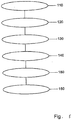
- FIG. 5 shows a flow chart of method steps .
- the method includes performing ophthalmic surgery.
- the method comprises the step 110 of forming an incision in the eye's Schlemm's canal, a step 120 of inserting a fiber's distal end through the incision into the Schlemm's canal, a step 130 of advancing the fiber's distal end around (360 graden, all around) in the Schlemm's canal, a step 140 of attaching a suture to the fiber's distal end, a step 150 of retracting the fiber backwardly through the Schlemm's canal, and a step 160 of connecting the suture ends.
Abstract
Description
- The invention relates to an ophthalmic surgical device for the treatment of open angle glaucoma through direct surgery.
- About two percent of people in Europe and in the United States have glaucoma. Glaucoma is a group of eye diseases that can damage the eye's optic nerve and can cause corresponding visual field loss resulting in blindness if left untreated.
- A considerable rise in intraocular pressure appears to be a major etiologic factor in glaucoma.
- The eyeball is basically a rigid sphere filled with fluids. There are three chambers of fluid in the eye: Anterior Chamber (between the cornea and the iris), Posterior Chamber (between iris, zonule fiber and lens) and the Vitreous Chamber (between the lens and the retina). The first two chambers are filled with a clear fluid called aqueous humour whereas the vitreous chamber is filled with a more viscous fluid, the vitreous humour. The aqueous humour or "aqueous" carries nutrients to the lens and cornea, both of which have no blood supply. The aqueous is constantly secreted by the ciliary body (which is located behind the iris and around the lens) and flows from the posterior chamber through the pupil into the anterior chamber and drains out of the eye through a spongy tissue called trabecular meshwork (TM).
- TM is located in the drainage angle of the anterior chamber, between the internal periphery of the cornea and the outer rim of the iris at the location where the iris meets the external wall of the eye (sclera). The fluid drains from the TM into a small canal (the Schlemm canal), then into aqueous collector channels and into aqueous veins.
- Aqueous is produced by the ciliary body and removed from the eye at a constant rate to maintain a constant pressure in the anterior chamber of the eye. If the resistance to fluid flow increases, pressure inside the eye increases and the circulation of blood to the optic nerve is restricted. If the ocular pressure remains elevated for prolonged periods of time, the fibres of the optic nerve may cause atrophy resulting in loss of vision in the afflicted eye.
- Glaucoma is roughly classified into two categories: closed angle glaucoma and open angle glaucoma. The closed angle glaucoma is caused by closure of angle of the anterior chamber by contact between the iris and the inner surface of the TM. Closure of this anatomical angle prevents normal drainage of aqueous from the anterior chamber. In open angle glaucoma the angle of the anterior chamber remains open, but the exit of aqueous through the TM is diminished. The source of resistance to outflow is in the TM.
- Many of the current therapies for glaucoma are directed at reducing intraocular pressure. This is initially treated using medical therapy with drops or pills that reduce the production of aqueous or increase the outflow of aqueous. However, these various drug therapies for glaucoma are sometimes associated with considerable side effects, such as headaches, blurred vision, allergic reactions, cardiopulmonary complications and potential interactions with other drugs.
- When the drug therapy fails, surgical therapy is used.
- Surgical therapy for open angle glaucoma includes laser and surgery methods. The surgery methods can be classified as follows.
- Goniotomy/trabeculotomy: These are simple and direct microsurgical dissection techniques with mechanical destruction of the TM. Initially this surgery provided favourable responses, however long term results showed only limited success in adults. These procedures probably failed later following repair mechanisms and "filling" processes. The "filling in" effect is the result of the healing process forming scars, which have the detrimental effect of collapsing and closing the opening that has been created in the TM. Once this opening is closed the pressure builds up inside once more and the surgery fails.
- Goniocurettage: This is an ab-interno (performed from the inside) mechanical destructive technique. An instrument similar to a cyclodialysis spatula is used with a microcurette on the tip. Initial results are similar to trabeculotomy with subsequential failure following repair mechanisms and the filling in process.
- Trabeculectomy: This is the most commonly performed filtering surgery. It involves creating a tiny filtering valve in the sclera. This procedure controls pressure by creating a new drainage channel through the angle structures to the extracellular space beneath the conjunctiva.
- Trabeculectomy is a major surgery and is aided with locally applied anticancer drugs such as 5-flurouracil or mitomycin-c to reduce scarring and increase surgical success. Current mortality associated with trabeculectomy consists of failure (10-15%), infections (a life long risk about 2-5%), choroidal haemorrhage (1%, a severe internal haemorrhage from insufficient pressure resulting in visual loss), cataract formation, and hypotony maculopathy (potentially reversible visual loss from insufficient pressure). Another disadvantage of this procedure is that the body's natural healing process may gradually close the filter, causing the pressure to rise again.
- Viscocanulostomy (VC) and Non Penetrating Trabeculectomy (NPT) are two new variations of filtering surgery. These are both major surgery procedures in which the Schlemm canal is surgically exposed by making a large and very deep scleral flap. In the VC procedure, the Schlemm canal is canulated and a viscoelastic substance injected (which dilates the Schlemm canal and aqueous collector channels). In the NPT procedure, the inner wall of the Schlemm canal is stripped away after the canal has been surgically exposed.
- Trabeculectomy, VC and NPT are performed under a conjunctival and scleral flap so that the aqueous is drained onto the surface of the eye or into the tissues located near the lateral wall of the eye. Normal physiological outflows are not used.
- Drainage devices: When Trabeculectomy, VC and NPT are not considered to have good probabilities of success, a number of implantable drainage devices are used to ensure that the desired filtration and outflow of aqueous through the surgical opening can continue. Placing glaucoma drainage implants also increases the risk of haemorrhage, infection and postoperative double vision that is a complication unique to drainage implants.
- The treatment procedures and variations described above have numerous disadvantages and are generally only moderately successful. They involve considerable trauma to the eye and require great surgical skill to create a hole in the total thickness of the sclera/cornea in the sub-conjunctival space. Furthermore, normal physiological outflow pathways are not used. Procedure is lengthy and requires an operating theatre and the presence of an anaesthesiologist with all the costs involved, and the vision recovery time is also a long process.
- The complications of filtration surgery have induced ophthalmic surgeons to look into alternative approaches for lowering intraocular pressure. The TM and the juxtacanalicular tissues both create the main resistance to aqueous outflow and for this reason they are the logical targets for surgical treatment of open angle glaucoma. Trabecular surgery has a much lower potential risk of choroidal haemorrhage, infections and furthermore, it aims at restoring physiologic outflow mechanisms. This surgery can be performed under local anaesthesia with rapid visual recovery.
-
WO-A-2005/070490 discloses a microcannula having a rounded distal tip. The rounded tip is used in conjunction with a light emitting signaling beacon such that the light is delivered proximal to the rounded tip, in order to disperse the light. - The aim of the present invention is to eliminate at least one of the disadvantages of the prior art. More specifically, the invention aims at providing an ophthalmic surgical device for the efficacious treatment of open angle glaucoma. Thereto, the device according to the invention comprises a fiber having a proximal end and a distal end, wherein the fiber is provided, near its proximal end, with a light source, whereby the distal end of the fiber forms a rounded tip, wherein the fiber has a substantially constant diameter that is smaller than the diameter of the rounded tip.
- The disclosure also relates to a method of performing ophthalmic surgery, comprising the steps of forming an incision in the eye's Schlemm's canal, inserting a fiber's distal end through the incision into the Schlemm's canal, advancing the fiber's distal end around in the Schlemm's canal, attaching a suture to the fiber's distal end, retracting the fiber backwardly through the Schlemm's canal, and connecting the suture ends.
- By drawing a suture through the Schlemm's canal and connecting the suture ends, in an elegant way, the canal can be opened re-establishing a natural outflow and reducing the intraocular eye over pressure. As a result, an efficacious treatment of open angle glaucoma is obtained. In an advantageous way, the suture can be drawn through the Schlemms's canal by attaching the suture to the notch a the fiber's distal end, thereby counteracting additional injury during the process of retracting the fiber backwardly.
- Further advantageous embodiments according to the invention are described in the following claims.
- By way of example only, embodiments of the present invention will now be described with reference to the accompanying figures in which
-
Fig. 1 shows a schematic cross sectional view of a first embodiment of an ophthalmic surgical device according to the invention; -
Fig. 2 shows a schematic partial cross sectional view of the human eye; -
Fig. 3a shows a schematic view of the device in a first state; -
Fig. 3b shows a schematic view of the device in a second state; -
Fig. 3c shows a schematic view of the device in a third state; -
Fig. 4 shows a schematic partial cross sectional view of a second embodiment of an ophthalmic surgical device according to the invention; and -
Fig. 5 shows a flow chart of steps of a method according to the invention. - It is noted that the figures show merely a preferred embodiment according to the invention. In the figures, the same reference numbers refer to equal or corresponding parts.
-
Figure 1 shows a schematic cross sectional view of a first embodiment of an ophthalmicsurgical device 1 according to the invention. Thesurgical device 1 is intended for use in a surgical method for treatment of glaucoma, as explained in more detail below. - The
device 1 comprises afiber connector 2 and afiber 3. Thefiber 3 has a proximal end 4 and adistal end 5. The proximal end 4 is connected to thefiber connector 2, while thefiber 3 is further provided, near its distal end, with anotch 6. Thedistal end 5 of the fiber forms arounded tip 7 to minimize tissue trauma and allow the fiber to advance in small tissue spaces, such as the eye's Schlemm's channel. In addition, light propagating through thefiber 3 is dispersed by the roundedtip 7, thereby visualizing the channel's interior under a range of incidence angles. - The
fiber 3 has a substantially constant diameter, advantageously less than circa 0.2 mm, e.g. circa 0.15 mm. Preferably, the fiber diameter is smaller than the diameter of the roundedtip 7, thus further facilitating movement of the fiber through Schlemm's channel. As an example, the rounded tip diameter is circa 0.2 mm. - As shown in
Fig. 1 , thenotch 6 is located close to therounded tip 7, just behind thetip 7. In principle, thenotch 6 can also be realized at a location offset from thetip 7. However, in order to draw a suture through the Schlemm's channel properly, the offset between thetip 7 and thenotch 6 is relatively small. - The
device 1 further comprises anoptional coating layer 8 covering aproximal end section 9 thefiber 3. Thecoating layer 8 provides mechanical protection to thefiber 3. Adistal end section 10 of thefiber 3 is free of acoating layer 8 to minimize its radial thickness. As an example, the distance of the freedistal end section 10 is circa 40 cm. However, also other distances can be applied, e.g. more than 40 cm such as circa 50 cm, or less than 40 cm such as circa 30 cm. Thedevice 1 according to the invention may have a total length in a range between circa 50 cm and circa 250 cm, e.g. 2 m, 1 m or 50 cm. The total length of the fiber may be shorter, e.g. between circa 10 cm and 100 cm, e.g. circa 30 cm. - In the transition area between the fiber proximal end 4 and the
fiber connector 2, asilicone layer 11 is provided on top of the connector exterior surface and thecoating layer 8. -
Figure 2 shows a schematic partial cross sectional view of thehuman eye 20. The eye contains a so-called canal ofSchlemm 21 for flowing aqueous humour from theanterior chamber 22 intoaqueous collector channels 23 and into aqueous veins. - According to an aspect of the disclosure treatment of open angle glaucoma is performed through direct surgery, using the ophthalmic
surgical device 1 shown inFig. 1 . -
Figures 3a,b,c show a schematic view of thedevice 1 in a first, second and third state, respectively. In the process of treating the glaucoma, an incision is formed in the eye's Schlemm'scanal 30. By making the incision, atrabeculectomy flap 31 is created for opening thecanal 30. Then, the fiber'sdistal end 5 is inserted through theincision 31 into thecanal 30, as shown inFig. 3a . The fiber'sdistal end 5 is advanced in the Schlemm'scanal 30, all around, and protrudes through thesame incision 31, as shown inFig. 3b . Subsequently, asuture 32 is attached to the neck of the distal end, formed by therounded end tip 7 and thenotch 6 behind thetip 7. Then, thefiber 3 is retracted backwardly through the Schlemm'scanal 30 until thefiber 3 is removed from thecanal 30 and thesuture 32 is thus drawn all around in thecanal 30. Thesuture 32 can be released from thefiber 3 so that the suture ends 33, 34 can now be connected, preferably with a little tension to stretch and open the Schlemm'scanal 30. - By advancing the fiber's
distal end 5 over 360°, theincision 31 can be used both for inserting thetip 7 into and from thecanal 30, thereby reducing additional trauma. Thesuture 32 can be manufactured from a variety of materials, e.g. from 10,0 prolyne. - According to an aspect of the invention, the
fiber 3 is illuminated while advancing the fiber'sdistal end 5 in the Schlemm'scanal 30, thereby facilitating the advancing process since the person operating the ophthalmicsurgical device 1 can easily keep track of progress of thefiber 3 in thecanal 30. Thereto, thefiber connector 2 is connected to a light source generating light that propagates through thefiber 3. -
Figure 4 shows a schematic partial cross sectional view of a second embodiment of an ophthalmicsurgical device 1 according to the invention. Here, instead of thefiber connector 2 shown inFig. 1 , thefiber 3 in thedevice 1 according to the second embodiment is further provided, near its proximal end, with a light source 40. In the shown embodiment, the light source is integrated with the fiber's distal end. Further, the light source comprises a LED 41 and an energy source, such as abattery 42, for feeding the light source. In principle, also another light source can be used, such as a halogen light source. Alternatively, the light source can be implemented as a disposable unit, e.g. by designing the light source as a separate unit that can be mounted to thefiber 3, e.g. using a snap connection. - By applying a light source on the proximal end of the fiber, the device can advantageously be used without an external light source, thereby increasing the ease of use of the device. Also, the fiber may have a shorter length, thereby further increasing the ease of use of the device, e.g. in providing the user of the device with an overview including less complicated structures.
-
Figure 5 shows a flow chart of method steps . The method includes performing ophthalmic surgery. The method comprises thestep 110 of forming an incision in the eye's Schlemm's canal, astep 120 of inserting a fiber's distal end through the incision into the Schlemm's canal, astep 130 of advancing the fiber's distal end around (360 graden, all around) in the Schlemm's canal, astep 140 of attaching a suture to the fiber's distal end, astep 150 of retracting the fiber backwardly through the Schlemm's canal, and astep 160 of connecting the suture ends. - The invention is not restricted to the embodiments described herein. It will be understood that many variants are possible.
- These and other embodiments will be apparent for the person skilled in the art and are considered to lie within the scope of the invention as defined in the following claims.
Claims (6)
- An ophthalmic surgical device for the treatment of glaucoma, comprising a fiber connector and a fiber having a proximal end connected to the fiber connector, wherein the fiber is provided, near its distal end, with a notch.
- An ophthalmic surgical device according to claim 1, wherein the distal end of the fiber forms a rounded tip.
- An ophthalmic surgical device according to claim 1, wherein the fiber has a substantially constant diameter that is smaller than the diameter of the rounded tip.
- An ophthalmic surgical device according to any of the preceding claims, wherein the notch is located close to the rounded tip.
- An ophthalmic surgical device according to any of the preceding claims, wherein the substantially constant diameter of the fiber is smaller than circa 0.2 mm.
- An ophthalmic surgical device according to any of the preceding claims, wherein a distal end section of the fiber is free of a coating layer.
Applications Claiming Priority (3)
| Application Number | Priority Date | Filing Date | Title |
|---|---|---|---|
| NL2004047A NL2004047C2 (en) | 2010-01-04 | 2010-01-04 | An ophthalmic surgical device and a method of performing ophthalmic surgery. |
| PCT/NL2011/050002 WO2011081525A1 (en) | 2010-01-04 | 2011-01-04 | An ophthalmic surgical device and a method of performing ophthalmic surgery |
| EP11701869.7A EP2521517B1 (en) | 2010-01-04 | 2011-01-04 | An ophthalmic surgical device |
Related Parent Applications (1)
| Application Number | Title | Priority Date | Filing Date |
|---|---|---|---|
| EP11701869.7A Division EP2521517B1 (en) | 2010-01-04 | 2011-01-04 | An ophthalmic surgical device |
Publications (2)
| Publication Number | Publication Date |
|---|---|
| EP3141227A1 true EP3141227A1 (en) | 2017-03-15 |
| EP3141227B1 EP3141227B1 (en) | 2020-03-11 |
Family
ID=42455373
Family Applications (2)
| Application Number | Title | Priority Date | Filing Date |
|---|---|---|---|
| EP16002126.7A Active EP3141227B1 (en) | 2010-01-04 | 2011-01-04 | An ophthalmic surgical device |
| EP11701869.7A Active EP2521517B1 (en) | 2010-01-04 | 2011-01-04 | An ophthalmic surgical device |
Family Applications After (1)
| Application Number | Title | Priority Date | Filing Date |
|---|---|---|---|
| EP11701869.7A Active EP2521517B1 (en) | 2010-01-04 | 2011-01-04 | An ophthalmic surgical device |
Country Status (16)
| Country | Link |
|---|---|
| US (1) | US20120303010A1 (en) |
| EP (2) | EP3141227B1 (en) |
| JP (1) | JP2013516215A (en) |
| CY (1) | CY1118384T1 (en) |
| DK (2) | DK2521517T3 (en) |
| ES (2) | ES2611953T3 (en) |
| HR (1) | HRP20161721T1 (en) |
| HU (1) | HUE032485T2 (en) |
| LT (1) | LT2521517T (en) |
| NL (1) | NL2004047C2 (en) |
| PL (1) | PL2521517T3 (en) |
| PT (1) | PT2521517T (en) |
| RS (1) | RS55501B1 (en) |
| SI (1) | SI2521517T1 (en) |
| SM (1) | SMT201700001B (en) |
| WO (1) | WO2011081525A1 (en) |
Families Citing this family (14)
| Publication number | Priority date | Publication date | Assignee | Title |
|---|---|---|---|---|
| US8889193B2 (en) | 2010-02-25 | 2014-11-18 | The Johns Hopkins University | Sustained delivery of therapeutic agents to an eye compartment |
| US20120022424A1 (en) * | 2010-05-27 | 2012-01-26 | Iscience Interventional Corporation | Device for placing circumferential implant in schlemm's canal |
| US9327037B2 (en) | 2011-02-08 | 2016-05-03 | The Johns Hopkins University | Mucus penetrating gene carriers |
| AU2012368274B2 (en) | 2012-02-03 | 2015-10-29 | Nova Eye, Inc. | Method and apparatus for treating an ocular disorder |
| US8962577B2 (en) | 2012-03-16 | 2015-02-24 | The Johns Hopkins University | Controlled release formulations for the delivery of HIF-1 inhibitors |
| EA030318B1 (en) | 2012-03-16 | 2018-07-31 | Дзе Джонс Хопкинс Юниверсити | Non-linear multiblock copolymer-drug conjugates for the delivery of active agents |
| US9533068B2 (en) | 2012-05-04 | 2017-01-03 | The Johns Hopkins University | Drug loaded microfiber sutures for ophthalmic application |
| JP2015532884A (en) | 2012-10-25 | 2015-11-16 | ザ レゲンツ オブ ザ ユニバーシティ オブ コロラド, ア ボディ コーポレート | Adjustable loop fiber optic illuminator for surgery |
| US10568975B2 (en) | 2013-02-05 | 2020-02-25 | The Johns Hopkins University | Nanoparticles for magnetic resonance imaging tracking and methods of making and using thereof |
| AU2016211696B2 (en) | 2015-01-27 | 2018-05-10 | The Johns Hopkins University | Hypotonic hydrogel formulations for enhanced transport of active agents at mucosal surfaces |
| CN105943186A (en) * | 2016-04-21 | 2016-09-21 | 温州眼视光发展有限公司 | Establishment method for chronic high intraocular pressure animal model |
| CN108685638A (en) * | 2018-05-08 | 2018-10-23 | 天津优视眼科技术有限公司 | A kind of ophthalmology fibre-optic catheter head end |
| DE202018106650U1 (en) * | 2018-11-22 | 2018-11-30 | Lisa Laser Products Gmbh | Handpiece for handling an optical fiber in a laser surgery |
| US11779490B1 (en) * | 2022-10-11 | 2023-10-10 | Beijing Institute of Ophthalmology, Beijing Tongren Hospital, Capital Medical University | Minimally invasive ab interno triple surgery for open-angle glaucoma |
Citations (5)
| Publication number | Priority date | Publication date | Assignee | Title |
|---|---|---|---|---|
| US5514125A (en) * | 1994-06-17 | 1996-05-07 | Carl-Zeiss-Stiftung | Applicator for the treatment of an elevated internal ocular pressure by means of laser radiation |
| US6241721B1 (en) * | 1998-10-09 | 2001-06-05 | Colette Cozean | Laser surgical procedures for treatment of glaucoma |
| US20020111608A1 (en) * | 2001-01-18 | 2002-08-15 | George Baerveldt | Minimally invasive glaucoma surgical instrument and method |
| WO2005070490A2 (en) * | 2004-01-23 | 2005-08-04 | Iscience Surgical Corporation | Composite ophthalmic microcannula |
| WO2006066103A2 (en) * | 2004-12-16 | 2006-06-22 | Iscience Interventional Corporation | Ophthalmic implant for treatment of glaucoma |
Family Cites Families (5)
| Publication number | Priority date | Publication date | Assignee | Title |
|---|---|---|---|---|
| US7419467B2 (en) * | 1998-11-25 | 2008-09-02 | M3 Electronics, Inc. | Medical inspection device |
| GB2377763B (en) * | 2001-06-21 | 2005-01-19 | Pbx Ltd | Acoustic intraocular pressure sensor |
| US7704246B2 (en) * | 2004-04-30 | 2010-04-27 | Connor Christopher S | Shielded intraocular probe for improved illumination or therapeutic application of light |
| US7668450B2 (en) * | 2005-01-28 | 2010-02-23 | Stryker Corporation | Endoscope with integrated light source |
| US8535333B2 (en) * | 2009-07-29 | 2013-09-17 | Transcend Medical, Inc. | Ocular implant applier and methods of use |
-
2010
- 2010-01-04 NL NL2004047A patent/NL2004047C2/en not_active IP Right Cessation
-
2011
- 2011-01-04 LT LTEP11701869.7T patent/LT2521517T/en unknown
- 2011-01-04 US US13/519,046 patent/US20120303010A1/en not_active Abandoned
- 2011-01-04 ES ES11701869.7T patent/ES2611953T3/en active Active
- 2011-01-04 RS RS20161124A patent/RS55501B1/en unknown
- 2011-01-04 SI SI201131044A patent/SI2521517T1/en unknown
- 2011-01-04 JP JP2012547046A patent/JP2013516215A/en active Pending
- 2011-01-04 DK DK11701869.7T patent/DK2521517T3/en active
- 2011-01-04 EP EP16002126.7A patent/EP3141227B1/en active Active
- 2011-01-04 DK DK16002126.7T patent/DK3141227T3/en active
- 2011-01-04 WO PCT/NL2011/050002 patent/WO2011081525A1/en active Application Filing
- 2011-01-04 ES ES16002126T patent/ES2797776T3/en active Active
- 2011-01-04 PL PL11701869T patent/PL2521517T3/en unknown
- 2011-01-04 PT PT117018697T patent/PT2521517T/en unknown
- 2011-01-04 HU HUE11701869A patent/HUE032485T2/en unknown
- 2011-01-04 EP EP11701869.7A patent/EP2521517B1/en active Active
-
2016
- 2016-12-14 HR HRP20161721TT patent/HRP20161721T1/en unknown
- 2016-12-15 CY CY20161101302T patent/CY1118384T1/en unknown
-
2017
- 2017-01-02 SM SM201700001T patent/SMT201700001B/en unknown
Patent Citations (5)
| Publication number | Priority date | Publication date | Assignee | Title |
|---|---|---|---|---|
| US5514125A (en) * | 1994-06-17 | 1996-05-07 | Carl-Zeiss-Stiftung | Applicator for the treatment of an elevated internal ocular pressure by means of laser radiation |
| US6241721B1 (en) * | 1998-10-09 | 2001-06-05 | Colette Cozean | Laser surgical procedures for treatment of glaucoma |
| US20020111608A1 (en) * | 2001-01-18 | 2002-08-15 | George Baerveldt | Minimally invasive glaucoma surgical instrument and method |
| WO2005070490A2 (en) * | 2004-01-23 | 2005-08-04 | Iscience Surgical Corporation | Composite ophthalmic microcannula |
| WO2006066103A2 (en) * | 2004-12-16 | 2006-06-22 | Iscience Interventional Corporation | Ophthalmic implant for treatment of glaucoma |
Also Published As
| Publication number | Publication date |
|---|---|
| DK3141227T3 (en) | 2020-06-15 |
| EP2521517B1 (en) | 2016-10-05 |
| NL2004047C2 (en) | 2011-07-05 |
| EP2521517A1 (en) | 2012-11-14 |
| PL2521517T3 (en) | 2017-06-30 |
| WO2011081525A1 (en) | 2011-07-07 |
| CY1118384T1 (en) | 2017-06-28 |
| ES2797776T3 (en) | 2020-12-03 |
| SMT201700001B (en) | 2017-03-08 |
| DK2521517T3 (en) | 2017-01-09 |
| EP3141227B1 (en) | 2020-03-11 |
| HRP20161721T1 (en) | 2017-03-10 |
| US20120303010A1 (en) | 2012-11-29 |
| RS55501B1 (en) | 2017-05-31 |
| LT2521517T (en) | 2017-01-10 |
| SI2521517T1 (en) | 2017-03-31 |
| JP2013516215A (en) | 2013-05-13 |
| PT2521517T (en) | 2016-12-27 |
| HUE032485T2 (en) | 2017-09-28 |
| ES2611953T3 (en) | 2017-05-11 |
Similar Documents
| Publication | Publication Date | Title |
|---|---|---|
| EP2521517B1 (en) | An ophthalmic surgical device | |
| CA2791154C (en) | Apparatus and method for treating an ocular disorder | |
| US9220632B2 (en) | Fluid infusion methods for ocular disorder treatment | |
| US20170252212A1 (en) | Methods and apparatus for treating glaucoma | |
| US6533768B1 (en) | Device for glaucoma treatment and methods thereof | |
| US20130281910A1 (en) | Ocular implant system | |
| US20040024345A1 (en) | Glaucoma implant with valveless flow bias | |
| AU2001245698A1 (en) | Device for glaucoma treatment and methods thereof | |
| JP2017519592A (en) | Drainage device for glaucoma intraocular pressure control | |
| CA2863608C (en) | Method and apparatus for treating an ocular disorder | |
| WO2022112844A1 (en) | Multi-lumen glaucoma stent | |
| WO2007006466A1 (en) | Device for the treatment of glaucoma | |
| WO2014131423A1 (en) | El saadani's glaucoma tube implant (egti) | |
| Figueiredo | Gonioscopic Surgery: Trabecular Micro-Bypass Stent Implantation |
Legal Events
| Date | Code | Title | Description |
|---|---|---|---|
| PUAI | Public reference made under article 153(3) epc to a published international application that has entered the european phase |
Free format text: ORIGINAL CODE: 0009012 |
|
| STAA | Information on the status of an ep patent application or granted ep patent |
Free format text: STATUS: THE APPLICATION HAS BEEN PUBLISHED |
|
| AC | Divisional application: reference to earlier application |
Ref document number: 2521517 Country of ref document: EP Kind code of ref document: P |
|
| AK | Designated contracting states |
Kind code of ref document: A1 Designated state(s): AL AT BE BG CH CY CZ DE DK EE ES FI FR GB GR HR HU IE IS IT LI LT LU LV MC MK MT NL NO PL PT RO RS SE SI SK SM TR |
|
| STAA | Information on the status of an ep patent application or granted ep patent |
Free format text: STATUS: REQUEST FOR EXAMINATION WAS MADE |
|
| 17P | Request for examination filed |
Effective date: 20170914 |
|
| RBV | Designated contracting states (corrected) |
Designated state(s): AL AT BE BG CH CY CZ DE DK EE ES FI FR GB GR HR HU IE IS IT LI LT LU LV MC MK MT NL NO PL PT RO RS SE SI SK SM TR |
|
| GRAP | Despatch of communication of intention to grant a patent |
Free format text: ORIGINAL CODE: EPIDOSNIGR1 |
|
| STAA | Information on the status of an ep patent application or granted ep patent |
Free format text: STATUS: GRANT OF PATENT IS INTENDED |
|
| INTG | Intention to grant announced |
Effective date: 20190409 |
|
| GRAJ | Information related to disapproval of communication of intention to grant by the applicant or resumption of examination proceedings by the epo deleted |
Free format text: ORIGINAL CODE: EPIDOSDIGR1 |
|
| STAA | Information on the status of an ep patent application or granted ep patent |
Free format text: STATUS: REQUEST FOR EXAMINATION WAS MADE |
|
| GRAP | Despatch of communication of intention to grant a patent |
Free format text: ORIGINAL CODE: EPIDOSNIGR1 |
|
| STAA | Information on the status of an ep patent application or granted ep patent |
Free format text: STATUS: GRANT OF PATENT IS INTENDED |
|
| INTC | Intention to grant announced (deleted) | ||
| INTG | Intention to grant announced |
Effective date: 20190916 |
|
| GRAS | Grant fee paid |
Free format text: ORIGINAL CODE: EPIDOSNIGR3 |
|
| GRAA | (expected) grant |
Free format text: ORIGINAL CODE: 0009210 |
|
| STAA | Information on the status of an ep patent application or granted ep patent |
Free format text: STATUS: THE PATENT HAS BEEN GRANTED |
|
| AC | Divisional application: reference to earlier application |
Ref document number: 2521517 Country of ref document: EP Kind code of ref document: P |
|
| AK | Designated contracting states |
Kind code of ref document: B1 Designated state(s): AL AT BE BG CH CY CZ DE DK EE ES FI FR GB GR HR HU IE IS IT LI LT LU LV MC MK MT NL NO PL PT RO RS SE SI SK SM TR |
|
| REG | Reference to a national code |
Ref country code: GB Ref legal event code: FG4D |
|
| REG | Reference to a national code |
Ref country code: CH Ref legal event code: EP |
|
| REG | Reference to a national code |
Ref country code: AT Ref legal event code: REF Ref document number: 1242230 Country of ref document: AT Kind code of ref document: T Effective date: 20200315 |
|
| REG | Reference to a national code |
Ref country code: IE Ref legal event code: FG4D |
|
| REG | Reference to a national code |
Ref country code: DE Ref legal event code: R096 Ref document number: 602011065608 Country of ref document: DE |
|
| REG | Reference to a national code |
Ref country code: DK Ref legal event code: T3 Effective date: 20200611 |
|
| REG | Reference to a national code |
Ref country code: NL Ref legal event code: FP |
|
| REG | Reference to a national code |
Ref country code: SE Ref legal event code: TRGR |
|
| REG | Reference to a national code |
Ref country code: CH Ref legal event code: NV Representative=s name: TR-IP CONSULTING LLC, CH |
|
| REG | Reference to a national code |
Ref country code: NO Ref legal event code: T2 Effective date: 20200311 |
|
| PG25 | Lapsed in a contracting state [announced via postgrant information from national office to epo] |
Ref country code: FI Free format text: LAPSE BECAUSE OF FAILURE TO SUBMIT A TRANSLATION OF THE DESCRIPTION OR TO PAY THE FEE WITHIN THE PRESCRIBED TIME-LIMIT Effective date: 20200311 Ref country code: RS Free format text: LAPSE BECAUSE OF FAILURE TO SUBMIT A TRANSLATION OF THE DESCRIPTION OR TO PAY THE FEE WITHIN THE PRESCRIBED TIME-LIMIT Effective date: 20200311 |
|
| PG25 | Lapsed in a contracting state [announced via postgrant information from national office to epo] |
Ref country code: BG Free format text: LAPSE BECAUSE OF FAILURE TO SUBMIT A TRANSLATION OF THE DESCRIPTION OR TO PAY THE FEE WITHIN THE PRESCRIBED TIME-LIMIT Effective date: 20200611 Ref country code: LV Free format text: LAPSE BECAUSE OF FAILURE TO SUBMIT A TRANSLATION OF THE DESCRIPTION OR TO PAY THE FEE WITHIN THE PRESCRIBED TIME-LIMIT Effective date: 20200311 Ref country code: HR Free format text: LAPSE BECAUSE OF FAILURE TO SUBMIT A TRANSLATION OF THE DESCRIPTION OR TO PAY THE FEE WITHIN THE PRESCRIBED TIME-LIMIT Effective date: 20200311 Ref country code: GR Free format text: LAPSE BECAUSE OF FAILURE TO SUBMIT A TRANSLATION OF THE DESCRIPTION OR TO PAY THE FEE WITHIN THE PRESCRIBED TIME-LIMIT Effective date: 20200612 |
|
| REG | Reference to a national code |
Ref country code: LT Ref legal event code: MG4D |
|
| PG25 | Lapsed in a contracting state [announced via postgrant information from national office to epo] |
Ref country code: PT Free format text: LAPSE BECAUSE OF FAILURE TO SUBMIT A TRANSLATION OF THE DESCRIPTION OR TO PAY THE FEE WITHIN THE PRESCRIBED TIME-LIMIT Effective date: 20200805 Ref country code: SK Free format text: LAPSE BECAUSE OF FAILURE TO SUBMIT A TRANSLATION OF THE DESCRIPTION OR TO PAY THE FEE WITHIN THE PRESCRIBED TIME-LIMIT Effective date: 20200311 Ref country code: RO Free format text: LAPSE BECAUSE OF FAILURE TO SUBMIT A TRANSLATION OF THE DESCRIPTION OR TO PAY THE FEE WITHIN THE PRESCRIBED TIME-LIMIT Effective date: 20200311 Ref country code: IS Free format text: LAPSE BECAUSE OF FAILURE TO SUBMIT A TRANSLATION OF THE DESCRIPTION OR TO PAY THE FEE WITHIN THE PRESCRIBED TIME-LIMIT Effective date: 20200711 Ref country code: CZ Free format text: LAPSE BECAUSE OF FAILURE TO SUBMIT A TRANSLATION OF THE DESCRIPTION OR TO PAY THE FEE WITHIN THE PRESCRIBED TIME-LIMIT Effective date: 20200311 Ref country code: EE Free format text: LAPSE BECAUSE OF FAILURE TO SUBMIT A TRANSLATION OF THE DESCRIPTION OR TO PAY THE FEE WITHIN THE PRESCRIBED TIME-LIMIT Effective date: 20200311 Ref country code: SM Free format text: LAPSE BECAUSE OF FAILURE TO SUBMIT A TRANSLATION OF THE DESCRIPTION OR TO PAY THE FEE WITHIN THE PRESCRIBED TIME-LIMIT Effective date: 20200311 Ref country code: LT Free format text: LAPSE BECAUSE OF FAILURE TO SUBMIT A TRANSLATION OF THE DESCRIPTION OR TO PAY THE FEE WITHIN THE PRESCRIBED TIME-LIMIT Effective date: 20200311 |
|
| REG | Reference to a national code |
Ref country code: ES Ref legal event code: FG2A Ref document number: 2797776 Country of ref document: ES Kind code of ref document: T3 Effective date: 20201203 |
|
| REG | Reference to a national code |
Ref country code: DE Ref legal event code: R097 Ref document number: 602011065608 Country of ref document: DE |
|
| REG | Reference to a national code |
Ref country code: NL Ref legal event code: HC Owner name: NOVA EYE, INC.; US Free format text: DETAILS ASSIGNMENT: CHANGE OF OWNER(S), CHANGE OF OWNER(S) NAME; FORMER OWNER NAME: ELLEX ISCIENCE, INC. Effective date: 20201201 |
|
| REG | Reference to a national code |
Ref country code: DE Ref legal event code: R082 Ref document number: 602011065608 Country of ref document: DE Representative=s name: PATENTANWAELTE EINSEL & KOLLEGEN, DE |
|
| PLBE | No opposition filed within time limit |
Free format text: ORIGINAL CODE: 0009261 |
|
| STAA | Information on the status of an ep patent application or granted ep patent |
Free format text: STATUS: NO OPPOSITION FILED WITHIN TIME LIMIT |
|
| 26N | No opposition filed |
Effective date: 20201214 |
|
| PG25 | Lapsed in a contracting state [announced via postgrant information from national office to epo] |
Ref country code: SI Free format text: LAPSE BECAUSE OF FAILURE TO SUBMIT A TRANSLATION OF THE DESCRIPTION OR TO PAY THE FEE WITHIN THE PRESCRIBED TIME-LIMIT Effective date: 20200311 Ref country code: PL Free format text: LAPSE BECAUSE OF FAILURE TO SUBMIT A TRANSLATION OF THE DESCRIPTION OR TO PAY THE FEE WITHIN THE PRESCRIBED TIME-LIMIT Effective date: 20200311 |
|
| REG | Reference to a national code |
Ref country code: CH Ref legal event code: PFA Owner name: NOVA EYE, INC., US Free format text: FORMER OWNER: ELLEX ISCIENCE, INC., US Ref country code: CH Ref legal event code: NV Representative=s name: ING. MARCO ZARDI C/O M. ZARDI AND CO. S.A., CH |
|
| REG | Reference to a national code |
Ref country code: DE Ref legal event code: R082 Ref document number: 602011065608 Country of ref document: DE Representative=s name: PATENTANWAELTE EINSEL & KOLLEGEN, DE Ref country code: DE Ref legal event code: R081 Ref document number: 602011065608 Country of ref document: DE Owner name: NOVA EYE INC., FREEMONT, US Free format text: FORMER OWNER: ELLEX ISCIENCE, INC., FREEMONT, CALIF., US |
|
| REG | Reference to a national code |
Ref country code: ES Ref legal event code: PC2A Owner name: NOVA EYE, INC. Effective date: 20210412 |
|
| REG | Reference to a national code |
Ref country code: DK Ref legal event code: EBP Effective date: 20210131 |
|
| REG | Reference to a national code |
Ref country code: NO Ref legal event code: MMEP |
|
| PG25 | Lapsed in a contracting state [announced via postgrant information from national office to epo] |
Ref country code: MC Free format text: LAPSE BECAUSE OF FAILURE TO SUBMIT A TRANSLATION OF THE DESCRIPTION OR TO PAY THE FEE WITHIN THE PRESCRIBED TIME-LIMIT Effective date: 20200311 |
|
| PG25 | Lapsed in a contracting state [announced via postgrant information from national office to epo] |
Ref country code: LU Free format text: LAPSE BECAUSE OF NON-PAYMENT OF DUE FEES Effective date: 20210104 |
|
| REG | Reference to a national code |
Ref country code: BE Ref legal event code: MM Effective date: 20210131 |
|
| PG25 | Lapsed in a contracting state [announced via postgrant information from national office to epo] |
Ref country code: NO Free format text: LAPSE BECAUSE OF NON-PAYMENT OF DUE FEES Effective date: 20210131 |
|
| REG | Reference to a national code |
Ref country code: AT Ref legal event code: UEP Ref document number: 1242230 Country of ref document: AT Kind code of ref document: T Effective date: 20200311 |
|
| PG25 | Lapsed in a contracting state [announced via postgrant information from national office to epo] |
Ref country code: DK Free format text: LAPSE BECAUSE OF NON-PAYMENT OF DUE FEES Effective date: 20210131 Ref country code: IE Free format text: LAPSE BECAUSE OF NON-PAYMENT OF DUE FEES Effective date: 20210104 |
|
| REG | Reference to a national code |
Ref country code: AT Ref legal event code: MM01 Ref document number: 1242230 Country of ref document: AT Kind code of ref document: T Effective date: 20210104 |
|
| PG25 | Lapsed in a contracting state [announced via postgrant information from national office to epo] |
Ref country code: AT Free format text: LAPSE BECAUSE OF NON-PAYMENT OF DUE FEES Effective date: 20210104 |
|
| PG25 | Lapsed in a contracting state [announced via postgrant information from national office to epo] |
Ref country code: BE Free format text: LAPSE BECAUSE OF NON-PAYMENT OF DUE FEES Effective date: 20210131 |
|
| PGFP | Annual fee paid to national office [announced via postgrant information from national office to epo] |
Ref country code: FR Payment date: 20221229 Year of fee payment: 13 |
|
| PGFP | Annual fee paid to national office [announced via postgrant information from national office to epo] |
Ref country code: ES Payment date: 20230206 Year of fee payment: 13 Ref country code: CH Payment date: 20230201 Year of fee payment: 13 |
|
| PG25 | Lapsed in a contracting state [announced via postgrant information from national office to epo] |
Ref country code: HU Free format text: LAPSE BECAUSE OF FAILURE TO SUBMIT A TRANSLATION OF THE DESCRIPTION OR TO PAY THE FEE WITHIN THE PRESCRIBED TIME-LIMIT; INVALID AB INITIO Effective date: 20110104 Ref country code: CY Free format text: LAPSE BECAUSE OF FAILURE TO SUBMIT A TRANSLATION OF THE DESCRIPTION OR TO PAY THE FEE WITHIN THE PRESCRIBED TIME-LIMIT Effective date: 20200311 |
|
| PGFP | Annual fee paid to national office [announced via postgrant information from national office to epo] |
Ref country code: SE Payment date: 20230113 Year of fee payment: 13 Ref country code: IT Payment date: 20221129 Year of fee payment: 13 Ref country code: DE Payment date: 20230127 Year of fee payment: 13 |
|
| PGFP | Annual fee paid to national office [announced via postgrant information from national office to epo] |
Ref country code: NL Payment date: 20230127 Year of fee payment: 13 |
|
| P01 | Opt-out of the competence of the unified patent court (upc) registered |
Effective date: 20230711 |
|
| PGFP | Annual fee paid to national office [announced via postgrant information from national office to epo] |
Ref country code: GB Payment date: 20231215 Year of fee payment: 14 |
|
| PGFP | Annual fee paid to national office [announced via postgrant information from national office to epo] |
Ref country code: NL Payment date: 20240124 Year of fee payment: 14 |
|
| PGFP | Annual fee paid to national office [announced via postgrant information from national office to epo] |
Ref country code: ES Payment date: 20240205 Year of fee payment: 14 |