EP3140419B1 - Biomarkers and combinations thereof for diagnosing active tuberculosis - Google Patents
Biomarkers and combinations thereof for diagnosing active tuberculosis Download PDFInfo
- Publication number
- EP3140419B1 EP3140419B1 EP15722572.3A EP15722572A EP3140419B1 EP 3140419 B1 EP3140419 B1 EP 3140419B1 EP 15722572 A EP15722572 A EP 15722572A EP 3140419 B1 EP3140419 B1 EP 3140419B1
- Authority
- EP
- European Patent Office
- Prior art keywords
- nucleic acid
- acid sequence
- tuberculosis
- biomarker
- seq
- Prior art date
- Legal status (The legal status is an assumption and is not a legal conclusion. Google has not performed a legal analysis and makes no representation as to the accuracy of the status listed.)
- Active
Links
Images


Classifications
-
- C—CHEMISTRY; METALLURGY
- C12—BIOCHEMISTRY; BEER; SPIRITS; WINE; VINEGAR; MICROBIOLOGY; ENZYMOLOGY; MUTATION OR GENETIC ENGINEERING
- C12Q—MEASURING OR TESTING PROCESSES INVOLVING ENZYMES, NUCLEIC ACIDS OR MICROORGANISMS; COMPOSITIONS OR TEST PAPERS THEREFOR; PROCESSES OF PREPARING SUCH COMPOSITIONS; CONDITION-RESPONSIVE CONTROL IN MICROBIOLOGICAL OR ENZYMOLOGICAL PROCESSES
- C12Q1/00—Measuring or testing processes involving enzymes, nucleic acids or microorganisms; Compositions therefor; Processes of preparing such compositions
- C12Q1/68—Measuring or testing processes involving enzymes, nucleic acids or microorganisms; Compositions therefor; Processes of preparing such compositions involving nucleic acids
- C12Q1/6876—Nucleic acid products used in the analysis of nucleic acids, e.g. primers or probes
- C12Q1/6883—Nucleic acid products used in the analysis of nucleic acids, e.g. primers or probes for diseases caused by alterations of genetic material
-
- C—CHEMISTRY; METALLURGY
- C12—BIOCHEMISTRY; BEER; SPIRITS; WINE; VINEGAR; MICROBIOLOGY; ENZYMOLOGY; MUTATION OR GENETIC ENGINEERING
- C12Q—MEASURING OR TESTING PROCESSES INVOLVING ENZYMES, NUCLEIC ACIDS OR MICROORGANISMS; COMPOSITIONS OR TEST PAPERS THEREFOR; PROCESSES OF PREPARING SUCH COMPOSITIONS; CONDITION-RESPONSIVE CONTROL IN MICROBIOLOGICAL OR ENZYMOLOGICAL PROCESSES
- C12Q1/00—Measuring or testing processes involving enzymes, nucleic acids or microorganisms; Compositions therefor; Processes of preparing such compositions
- C12Q1/68—Measuring or testing processes involving enzymes, nucleic acids or microorganisms; Compositions therefor; Processes of preparing such compositions involving nucleic acids
- C12Q1/6876—Nucleic acid products used in the analysis of nucleic acids, e.g. primers or probes
- C12Q1/6888—Nucleic acid products used in the analysis of nucleic acids, e.g. primers or probes for detection or identification of organisms
- C12Q1/689—Nucleic acid products used in the analysis of nucleic acids, e.g. primers or probes for detection or identification of organisms for bacteria
-
- C—CHEMISTRY; METALLURGY
- C12—BIOCHEMISTRY; BEER; SPIRITS; WINE; VINEGAR; MICROBIOLOGY; ENZYMOLOGY; MUTATION OR GENETIC ENGINEERING
- C12Q—MEASURING OR TESTING PROCESSES INVOLVING ENZYMES, NUCLEIC ACIDS OR MICROORGANISMS; COMPOSITIONS OR TEST PAPERS THEREFOR; PROCESSES OF PREPARING SUCH COMPOSITIONS; CONDITION-RESPONSIVE CONTROL IN MICROBIOLOGICAL OR ENZYMOLOGICAL PROCESSES
- C12Q2600/00—Oligonucleotides characterized by their use
- C12Q2600/158—Expression markers
Definitions
- This disclosure relates to the detection and diagnosis of tuberculosis. More specifically, the disclosure relates to new biomarkers and combinations thereof that enable the accurate detection and diagnosis of tuberculosis.
- Tuberculosis is a progressive, often fatal, infectious disease, caused by the bacterial pathogen Mycobacterium tuberculosis ( M. tuberculosis , MTB). This is a significant cause of mortality worldwide, being the eighth largest leading cause of death globally, and is primarily a disease of poverty, particularly in developing countries. Latent TB infection is believed to affect as much as one third of the world's population.
- Tuberculosis is a notifiable disease and is a major concern for many governmental and other health bodies including the World Health Organisation (WHO), who have initiated numerous control and treatment programmes like the "Stop TB Partnership".
- WHO World Health Organization
- TB infection is not limited to the developing world: the UK has seen a resurgence of tuberculosis since the late 1980s and there are currently over 8000 new cases each year - a rate of 14.0 per 100,000 population. About 40% of these new cases occur in the London region, where the rate of infection is 44.8 per 100,000 population.
- M. tuberculosis is capable of forming intracellular infections. These infections may be exclusively intracellular, or may contain both intracellular and extracellular components. Generally, M. tuberculosis bacilli do not circulate freely in the body, for example, in the bloodstream, and as such are often difficult to detect. They are also less amenable to drug treatment regimes. Intracellular survival and multiplication of mycobacteria is suspected to be a main contributory factor for mycobacterial disease progression.
- latency is synonymous with " persistence " , and describes a reversible state of low metabolic activity in which mycobacterial cells can survive for extended periods with limited or no cell division. During latency (i.e. latent infection), the clinical symptoms associated with a mycobacterial infection do not become manifest.
- diagnosis would be made by a technique that accurately, rapidly, and simultaneously measures a plurality of biomarkers at a single point in time, thereby minimizing disease progression during the time required for diagnosis.
- WO 2009/158521 discloses methods and kits for the identification of latent versus active tuberculosis (TB) patients, as compared to healthy controls. Microarray analysis of blood of a distinct and reciprocal immune signature is used to determine, diagnose, track and treat latent versus active TB patients.
- Table 7-I discloses a list of genes over-expressed in active TB versus control, among them GBP1, and Table 7-K discloses another list of genes over-expressed in active TB versus control, among them SNX10.
- WO 2014/019977 discloses a method of distinguishing active TB in the presence of a complicating factor.
- the methods described employ gene signatures comprising at least 27 genes.
- WO 2013/190321 discloses biomarkers for determining the Mycobacterium tuberculosis infection status of an individual.
- WO 2013/177502 discloses methods and devices for diagnosing TB in a subject based on particular biomarker profiles.
- Operon Biotechnologies Gmbh "Operon Microarray Slides -OpArraysTM” describes the features of OpArraysTM, full genome pre-spotted microarray slides for human, mouse and rat.
- the present inventors have conducted a temporal differential gene expression study in peripheral blood leukocytes (PBLs) in an aerosol Macaca fascicularis non-human primate model of TB. Using this method, the inventors have identified host biomarkers associated with early exposure to TB. Microarray hybridisation analyses to human whole genome arrays have revealed many significant gene expression changes, showing substantial temporal changes in PBL gene expression in response to M. tuberculosis challenge across the time-course of the study. Using parametric and non-parametric tools for data analysis, including artificial neural network analysis, the inventors have identified highly-significant host biomarkers associated with TB and M. tuberculosis infections. The biomarkers identified by the present disclosure have improved specificity for TB across different subgroups, such as different ethnic groups.
- the present disclosure allows for accurate, rapid, and sensitive prediction and diagnosis of TB through a measurement of one or more biomarker taken from a biological sample at a single point in time.
- the present disclosure provides the use of one or more of SNX10, CPVL, PF4V1, HERC2, CD52, LYN, LGALS3BP, BAZ1A, KLRAP1, WSB1, BST1, SERPINB1, MVP, APBB1IP, MB21D1/C6orf150, TICAM2, DEFB128 and IL8 as a biomarker for tuberculosis.
- the disclosure also provides a method for diagnosing tuberculosis in an individual comprising determining the presence and/or amount of SNX10 and GBP1 as biomarkers for tuberculosis in a sample obtained from the individual, wherein the one or more biomarker for tuberculosis is selected from SNX10, CPVL, PF4V1, HERC2, CD52, LYN, LGALS3BP, BAZ1A, KLRAP1, WSB1, BST1, SERPINB1, MVP, APBB1IP, MB21D1/C6orf150, TICAM2, DEFB128 and IL8.
- the tuberculosis detected and/or diagnosed by the method or use of the present disclosure may be an active tuberculosis infection and the one or more biomarker a biomarker for an active tuberculosis infection.
- the one or more biomarker is selected from SNX10, CPVL, PF4V1 and HERC2, or any combination thereof.
- the one or more biomarker is selected from: (i) SNX10 and CREG1; and/or (ii) PF4V1 and HERC2.
- the tuberculosis detected and/or diagnosed by the method or use of the present disclosure may be a latent tuberculosis infection and the one or more biomarker a biomarker for a latent tuberculosis infection.
- the one or more biomarker for a latent tuberculosis infection is selected from PF4V1, LYN, CD52, HERC2, KLRAP1, DEFB128, LGALS3BP and IL8.
- a use of the disclosure may comprise determining the presence and/or amount of the one or more biomarker for tuberculosis in a sample obtained from an individual.
- the present disclosure also provides a use or method as defined herein, wherein said one or more biomarker is able to identify an individual with an active tuberculosis infection and/or an individual with a latent tuberculosis infection.
- the present disclosure also provides a use or method as defined herein, wherein said one or more biomarker is able to identify an individual with an active tuberculosis infection and/or an individual with a latent tuberculosis infection and/or an individual uninfected with tuberculosis.
- the one or more additional biomarker may be used in the method or use of the disclosure.
- the one or more additional biomarker may be (a) a biomarker for an active tuberculosis infection selected from: (i) LOC400759/GBP1P1, SNX10, CPVL, CREG1, PF4V1, PSMB9, LGALS3BP, BST1, BAZ1A, LYN, TAPBP, SERPINB1, WSB1, MVP, APBB1IP, FYB, MB21D1/C6orf150, TICAM2, CD52, KLRAP1, DEFB128 and IL8; and/or (ii) a biomarker listed in Table 3; and/or (b) a biomarker for a latent tuberculosis infection selected from: (i) a biomarker listed in Table 4; and/or (ii) a biomarker listed in Table 5.
- the one or more additional biomarker for an active tuberculosis infection is selected from LOC400759/GBP1P1, CREG1, PSMB9, ALPK1, GBP1, IRF1, HLA-B, IFITM3, S100A11, MMP9 and CD96.
- the one or more biomarkers for tuberculosis are SNX10 and CPVL and the one or more additional biomarkers for tuberculosis are LOC400759/GBP1P1 and CREG1; and/or the one or more biomarkers for tuberculosis are PF4V1 and HERC2 and the one or more additional biomarkers for tuberculosis are LOC400759/GBP1P1 and ALPK1.
- One or more further additional biomarkers may be used in the methods and/or uses of the disclosure.
- the one or more further additional biomarker is PSMB9 and/or PF4V1.
- the one or more additional biomarker for an active tuberculosis infection, or the one or more further additional biomarker is: (i) GBP1, IRF1 and HLA-B; (ii) GBP1, IRF1, IFITM3 and S100A11; and/or (iii) GBP1, IRF1, MMP9 and CD96.
- the presence and/or amount of the one or more biomarker for tuberculosis may be compared with the presence and/or amount of the one or more biomarker for tuberculosis in a control sample.
- the specificity of the comparison of the presence and/or amount of the one or more biomarker for tuberculosis in the sample and the presence and/or absence of the one or more biomarker for tuberculosis in the control diagnoses tuberculosis may be at least about 80%.
- the presence and/or amount of the one or more biomarker for tuberculosis may be determined using an antibody and/or an oligonucleotide specific for said one or more biomarker. Typically, an oligonucleotide specific for said one or more biomarker is used.
- the one or more biomarker for tuberculosis is LOC400759/GBP1P1 and the oligonucleotide comprises at least one nucleic acid sequence having at least 90% sequence identity to the nucleic acid sequence of SEQ ID NOs: 1, 2 or 3;
- the one or more biomarker for tuberculosis is PF4V1 and the oligonucleotide comprises at least one nucleic acid sequence having at least 90% sequence identity to the nucleic acid sequence of SEQ ID NOs: 4 or 5;
- the one or more biomarker for tuberculosis is ALPK1 and the oligonucleotide comprises at least one nucleic acid sequence having at least 90% sequence identity to the nucleic acid sequence of SEQ ID NOs: 6 or 7;
- the one or more biomarker for tuberculosis is HERC2 and the oligonucleotide comprises at least one nucleic acid sequence having at least
- the presence and/or absence of the at least one biomarker for tuberculosis in the individual may be determined at least twice using a separate sample taken each time the presence and/or absence of the at least one biomarker for tuberculosis is determined.
- the samples from the individual may be taken prior to, during and/or after treatment initiation.
- the disclosure further provides a device for carrying out the use of the disclosure, or for use in a method of the disclosure, which comprises (i) one or more antibody specific for the one or more biomarker for tuberculosis; or (ii) one or more oligonucleotide specific for the one or more biomarker for tuberculosis.
- the one or more oligonucleotide specific for the one or more biomarker for tuberculosis comprised in the device is an oligonucleotide of the disclosure as defined herein.
- the present disclosure allows for the rapid, sensitive, and accurate diagnosis or prediction of TB using one or more biological samples obtained from an individual at a single time point (" snapshot ") or during the course of disease progression.
- TB may be diagnosed or predicted prior to the onset of clinical symptoms, and/or as subsequent confirmation after the onset of clinical symptoms. Accordingly, the present disclosure allows for more effective therapeutic intervention and/or diagnosis in the pre-symptomatic stage of the disease.
- Tuberculosis and Mycobacterium tuberculosis are Tuberculosis and Mycobacterium tuberculosis
- Tuberculosis is a progressive, often fatal, infectious disease, caused by the bacterial pathogen Mycobacterium tuberculosis ( M. tuberculosis , MTB).
- Pulmonary symptoms of TB include a productive, prolonged cough of three or more weeks, chest pain, and hemoptysis.
- Systemic symptoms include low grade remittent fever, chills, night sweats, appetite loss, weight loss, easy fatigability, and production of sputum that starts out mucoid but changes to purulent.
- a reference herein to the detection or diagnosis of TB is equivalent to the detection or diagnosis of M. tuberculosis infection. When the M. tuberculosis cells are metabolically active and/or undergoing cell division, this results in the symptoms of TB becoming overt, and is described as an active TB/M. tuberculosis infection.
- latent TB an individual is infected with M. tuberculosis, but the individual does not display the symptoms of active TB disease.
- latent TB the mycobacterial cells survive for extended periods in a state of low metabolic activity and with limited or no cell division. Thus, during latency (i.e. latent infection), the clinical symptoms associated with a mycobacterial infection do not become manifest. This can make it difficult to distinguish between a latent TB infection and the absence of a TB infection using conventional methods and techniques.
- a reference herein to the detection or diagnosis of latent TB is equivalent to the detection or diagnosis of latent M. tuberculosis infection.
- biomarkers for active TB are expressed at relatively low levels at an early stage in active TB, but become expressed at higher levels as the active stage of the infection progresses.
- the term "low level of expression" is relative.
- the expression of these active TB biomarkers during the early active phase may be low relative to the expression level later in the active phase, and similar to (or slightly greater than) the expression level of the same biomarkers in an uninfected individual and/or an individual with latent TB.
- the expression of these active TB biomarkers during the early active phase is low relative to the expression level later in the active phase, but still higher than the expression level of the same biomarkers in an uninfected individual and/or an individual with latent TB.
- the present disclosure provides biomarkers for the detection and/or diagnosis of TB infection.
- the present disclosure provides biomarkers for the detection and/or diagnosis of an active TB infection, including an early stage active TB infection and/or a later stage active TB infection.
- the present disclosure also provides biomarkers for the detection and/or diagnosis of a latent TB infection.
- the present disclosure further provides biomarkers for distinguishing between active and latent TB infections.
- the present disclosure also provides biomarkers for distinguishing between a latent TB infection and an absence/lack of TB infection (active or latent).
- the present disclosure also provides biomarkers for distinguishing between early stage active TB and later stage active TB.
- the present disclosure also provides biomarkers for distinguishing between an individual who has no symptomatic TB infection (active or latent) and has not been exposed to TB (e.g. because they are from a non/low-TB endemic region) and an individual who has no symptomatic TB infection (active or latent) but has been exposed to TB (e.g. because they are from a high-TB endemic region).
- any appropriate technique may be used to confirm the diagnosis of active and/or latent TB according to the present disclosure.
- Standard techniques are known in the art. For example, chest x-ray, microbiological culture of M. tuberculosis in a sample (sputum, pus, cerebrospinal fluid, biopsied tissue, etc.) from the individual, CT scan, MMR, antibodies from lymphocyte secretion (ALS) assay, IFN ⁇ assay and tuberculin skin tests (e.g. Mantoux and Heaf tests).
- a “biomarker” is virtually any biological compound, such as a protein and a fragment thereof, a peptide, a polypeptide, a proteoglycan, a glycoprotein, a lipoprotein, a carbohydrate, a lipid, a nucleic acid, an organic on inorganic chemical, a natural polymer, and a small molecule, that is present in the biological sample and that may be isolated from, or measured in, the biological sample.
- a biomarker can be the entire intact molecule, or it can be a portion thereof that may be partially functional or recognized, for example, by an antibody or other specific binding protein.
- a biomarker is considered to be informative if a measurable aspect or characteristic of the biomarker is associated with a given state of an individual, such as infection with TB.
- a measurable aspect or characteristic may include, for example, the presence, absence, or concentration of the biomarker in the biological sample from the individual and/or its presence as part of a profile of biomarkers.
- Such a measurable aspect of a biomarker is defined herein as a " feature. "
- the presence of a biomarker may be a feature.
- the amount of a biomarker in a sample, or the amount of a biomarker in a sample compared with a control or reference sample may be a feature.
- a feature may also be a ratio of two or more measurable aspects of biomarkers, which biomarkers may or may not be of known identity, for example.
- a " biomarker profile" comprises at least two such features, where the features can correspond to the same or different classes of biomarkers such as, for example, two nucleic acids or a nucleic acid and a carbohydrate.
- a biomarker profile may also comprise at least three, four, five, 10, 20, 30 or more features. In one instance, a biomarker profile comprises hundreds, or even thousands, of features. In another instance, the biomarker profile comprises at least one measurable aspect of at least one internal standard.
- the present inventors have conducted a temporal differential gene expression study in peripheral blood leukocytes (PBLs) in an aerosol Macaca fascicularis non-human primate model of TB. Using this method, the inventors have identified host biomarkers associated with early exposure to TB.
- PBLs peripheral blood leukocytes
- the new biomarkers for TB identified by the present inventors are listed in Table 2 herein (together with corresponding sequence identifiers (SEQ ID NOs).
- the present inventors have identified LOC400759/GBP1P1, SNX10, CPVL, CREG1, PF4V1, PSMB9, ALPK1, HERC2, LGALS3BP, BST1, BAZ1A, LYN, TAPBP, SERPINB1, WSB1, MVP, APBB1IP, FYB, MB21D1/C6orf150, TICAM2, CD52, KLRAP1, DEFB128 and IL8 as biomarkers for TB.
- the present disclosure provides the use of one or more of LOC400759/GBP1P1, SNX10, CPVL, CREG1, PF4V1, PSMB9, ALPK1, HERC2, LGALS3BP, BST1, BAZ1A, LYN, TAPBP, SERPINB1, WSB1, MVP, APBB1IP, FYB, MB21D1/C6orf150, TICAM2, CD52, KLRAP1, DEFB128 and IL8 as a biomarker for tuberculosis.
- Each of these biomarkers may be used alone, in combination with any of the other biomarkers, and/or in combination with one or more additional biomarker for tuberculosis as disclosed herein.
- the disclosure may relate to the use of LOC400759/GBP1P1, SNX10, CPVL and/or CREG1 (alone or in any combination thereof), optionally in combination with PF4V1 and/or PSMB9 and/or in combination with any of the other biomarkers disclosed herein.
- the present disclosure provides the use of one or more of SNX10, CPVL, PF4V1, HERC2, CD52, LYN, LGALS3BP, BAZ1A, KLRAP1, WSB1, BST1, SERPINB1, MVP, APBB1IP, MB21D1/C6orf150, TICAM2,DEFB128 and IL8 as a biomarker for tuberculosis.
- biomarkers may be used according to the present disclosure. For example, any two or more, three or more, four or more, five or more, six or more, seven or more, eight or more, nine or more, ten or more, up to and including all of these biomarkers may be used to diagnose TB according to the present disclosure.
- the one or more biomarker of the disclosure may be a hormone, a growth factor, a transcription factor, a cell surface marker or a soluble protein derived from cells.
- the one or more biomarker of the disclosure may be a nucleic acid encoding for one of said proteins.
- the one or more biomarker of the disclosure may be used in the detection and/or diagnosis of an active TB infection.
- the one or more biomarker of the disclosure may be used in the detection and/or diagnosis of a latent TB infection.
- the one or more biomarker of the disclosure may be used to diagnose the absence of a TB infection (active or latent).
- the one or more biomarker of the disclosure may be used to identify an individual with an active TB infection and/or an individual with a latent TB infection.
- the one or more biomarker of the disclosure may be used to identify an individual with an active TB infection and/or an individual with a latent TB infection and/or an individual uninfected with TB.
- the one or more biomarker of the disclosure may be used in the detection and/or diagnosis of an early stage active TB infection or a late/later stage active TB infection.
- the one or more biomarker of the disclosure may be used to determine exposure of an individual to TB, even in the absence of a symptomatic active or asymptomatic latent TB infection.
- the one or more biomarker of the disclosure may be used to distinguish between one or more individual with an active (early or later stage) TB infection and/or one or more individual with a latent TB infection, and/or one or more individual uninfected with TB.
- the one or more biomarker of the disclosure may also be used to distinguish between one or more individual with an early stage active TB infection and one or more individual with a late/later stage active TB infection.
- the present disclosure relates to the use of one or more of SNX10, CPVL, PF4V1,HERC2, CD52 and LYN as a biomarker for TB.
- biomarkers may be used in the detection and/or diagnosis of an active TB infection (early or late/later stage), or to distinguish between an early stage active TB infection and a late/later stage active TB infection.
- one or more of these biomarkers may be used in the detection and/or diagnosis of a latent TB infection, or to diagnose the absence of a TB infection (active or latent).
- any combination of SNX10, CPVL, PF4V1, HERC2 CD52 and LYN may be used as biomarkers for TB according to the present disclosure.
- CD52 and/or LYN may be used in combination with one or more of SNX10, CPVL, PF4V1 and HERC2, or with any combination of SNX10, CPVL, PF4V1 and HERC2.
- the disclosure relates to the use of SNX10, CPVL, PF4V1, HERC2, CD52 and LYN
- the disclosure relates to the use of (i) SNX10 and CPVL; and/or (ii) PF4V1 and HERC2 as biomarkers for tuberculosis.
- SNX10 and CPVL are used in combination with LOC400759/GBP1P1 and/or CREG1 as biomarkers in the diagnosis of TB according to the present disclosure.
- PF4V1 and HERC2 are used in combination with LOC400759/GBP1P1 and/or ALPK1 as biomarkers in the diagnosis of TB according to the present disclosure. Any of these combinations may be used with CD52 and/or LYN
- One or more additional biomarker for TB may also be used in the detection and/or diagnosis of TB according to the present disclosure. Any combination of the one or more additional biomarker (or further additional biomarker) may be used in combination with the one or more biomarker of the disclosure. For example at least two, at least three, at least four, at least five, at least six, at least seven, at least eight, at least nine, at least ten or more additional biomakers for TB may be used in combination with the one or more biomarker of the disclosure.
- the one or more additional biomarker may be selected from LOC400759/GBP1P1, CREG1, PF4V1, PSMB9, ALPK1, HERC2, LGALS3BP, BST1, BAZ1A, LYN, TAPBP, SERPINB1,WSB1, MVP, APBB1IP, FYB, MB21D1/C6orf150, TICAM2, CD52, KLRAP1, DEFB128, HERC2 and IL8.
- the one or more additional biomarker may be selected from LOC400759/GBP1P1, SNX10, CPVL, CREG1, PSMB9, LGALS3BP, BST1, BAZ1A, LYN, TAPBP, SERPINB1, WSB1, MVP, APBB1IP, FYB, MB21D1/C6orf150, CPVL, TICAM2, CD52, KLRAP1, DEFB128 and IL8.
- any of these combinations may be used with CD52 and/or LYN
- the one or more additional biomarker is selected from the biomarkers listed in Tables 2, 3, 4 and/or 5 herein (corresponding sequence identifiers (SEQ ID NOs) are also given in Tables 2 to 5).
- the one or more biomarker of the disclosure is selected from SNX10 and CPVL and the one or more additional biomarker is selected from the biomarkers in Tables 2 and 3 or 5.
- the one or more biomarker of the disclosure is selected from SNX10 and CPVL and the one or more additional biomarker is selected from LOC400759/GBP1P1, CREG1, PF4V1, PSMB9, GBP1, IRF1, HLA-B, IFITM3 and S100A11.
- the present disclosure provides the use SNX10 and CPVL in combination with PF4V1 and/or PSMB9, and optionally in combination with one or more additional biomarker for TB as disclosed herein.
- Said one or more additional biomarker is preferably selected from LOC400759/GBP1P1, CREG1, GBP1, IRF1, HLA-B, IFITM3 and S100A11. Any of these combinations may be used with CD52 and/or LYN.
- the present disclosure relates to the use of SNX10, CPVL, LOC400759/GBP1P1 and CREG1, the combination of SNX10, CPVL, LOC400759/GBP1P1, CREG1, PSMB9, the combination of SNX10, CPVL, LOC400759/GBP1P1, CREG1, PF4V1, the combination of SNX10, CPVL, LOC400759/GBP1P1, CREG1, PSMB9, GBP1, IRF1 and HLA-B or the combination of SNX10, CPVL, LOC400759/GBP1P1, CREG1, PF4V1, GBP1, IRF1, IFITM3 and S100A11 as biomarkers for TB.
- SNX10 preferably the combination of SNX10, CPVL, LOC400759/GBP1P1, CREG1, PSMB9, GBP1, IRF1 and HLA-B or the combination of SNX10, CPVL, LOC400759/GBP1P1, CREG1, PF4V1, GBP1, IRF1, IFITM3 and S100A11 is used. Any of these combinations may be used with CD52 and/or LYN.
- the one or more biomarker of the disclosure is selected from PF4V1 and HERC2 and the one or more additional biomarker is selected from the biomarkers in Tables 2 and 3 or 5.
- the one or more biomarker of the disclosure is selected from PF4V1 and HERC2 and the one or more additional biomarker is selected from LOC400759/GBP1P1, CREG1, PF4V1, PSMB9, GBP1, IRF1, HLA-B, IFITM3 and S100A11, MMP9 and CD96.
- the disclosure relates to the use of PF4V1 and HERC2 in combination with one or more additional biomarker for TB as disclosed herein.
- Said one or more additional biomarker is preferably selected from LOC400759/GBP1P1, CREG1, GBP1, IRF1, HLA-B, IFITM3, S100A11, MMP9, KLRA1, DEFB128 and IL8 and CD96.
- the present disclosure provides the use of PF4V1 and HERC2 in combination with one or more additional biomarker selected from LOC400759/GBP1P1, ALPK1, GBP1, IRF1, MMP9 and CD96; or in combination with one or more additional biomarker selected from of LOC400759/GBP1P1, ALPK1, GBP1, IRF1, MMP9, CD96, KLRA1, DEFB128 and IL8.
- the present disclosure provides the use of the combination of PF4V1, HERC2, LOC400759/GBP1P1, ALPK1, GBP1, IRF1, MMP9 and CD96, or the combination of PF4V1, HERC2, LOC400759/GBP1P1, ALPK1, GBP1, IRF1, MMP9, CD96, KLRA1, DEFB128 and IL8 as biomarkers for TB. Any of these combinations may be used with CD52 and/or LYN.
- LOC400759/GBP1P1, SNX10, CPVL and CREG1 are particularly preferred. Such combinations include: (i) LOC400759/GBP1P1 and SNX10; (ii) LOC400759/GBP1P1 and CPVL; (iii) LOC400759/GBP1P1 and CREG1; (iv) SNX10 and CPVL; (v) SNX10 and CREG1; (vi) CPVL and CREG1; (vii) LOC400759/GBP1P1, SNX10 and CPVL; (viii) LOC400759/GBP1P1, SNX10 and CREG1; (ix) LOC400759/GBP1P, CPVL and CREG1; (x) SNX10, CPVL and CREG1; and/or (xi) LOC400759/GBP1P1, SNX10; CPVL
- combinations may be used in combination with one or more further additional biomarker as disclosed herein, with one or more of GBP1, IRF1, HLA-B, IFITM3 and/or S100A11 being particularly preferred as disclosed herein. Any of these combinations may be used with CD52 and/or LYN.
- combinations of one or more of LOC400759/GBP1P1, PF4V1, ALPK1 and HERC2 are preferred. Such combinations include: (i) LOC400759/GBP1P1 and PF4V1; (ii) LOC400759/GBP1P1 and ALPK1; (iii) LOC400759/GBP1P1 and HERC2; (iv) PF4V1 and ALPK1; (v) PF4V1 and HERC2; (vi) ALPK1 and HERC2; (vii) LOC400759/GBP1P1, PF4V1 and ALPK1; (viii) LOC400759/GBP1P1, PF4V1 and HERC2; (ix) LOC400759/GBP1P1, ALPK1 and HERC2; (x) PF4V1, ALPK1 and HERC2; and (xi) LOC400759/GBP1P1, PF4V1; and (
- combinations may be used in combination with one or more further additional biomarker as disclosed herein, with one or more of GBP1, IRF1, MMP9, CD96, KLRA1, DEFB128 and IL8 being particularly preferred as disclosed herein. Any of these combinations may be used with CD52 and/or LYN.
- biomarkers for latent TB have also identified biomarkers for latent TB, and which can be used to distinguish between latent and active forms of TB, i.e. between latent and active forms of M. tuberculosis infection. These biomarkers for latent TB can also be used according to the present disclosure to distinguish between latent TB infection and the absence of TB infection.
- the present inventors have identified PF4V1, LYN, CD52, HERC2, KLRA1, DEFB128, LGALS3BP and IL8 as biomarkers for latent TB. These biomarkers may be used to distinguish between active TB and/or latent TB and/or the absence of TB. In a preferred instance, these biomarkers are used to distinguish between latent TB and the absence of TB infection, i.e. to identify one or more individual with a latent TB infection and/or one or more individual uninfected with TB.
- the present disclosure provides the use of one or more of the biomarkers selected from PF4V1, LYN, CD52, HERC2, KLRA1, DEFB128, LGALS3BP and IL8 for distinguishing between latent and active M. tuberculosis infection, and hence latent and active TB.
- the present disclosure also provides the use of one or more of the biomarkers selected from PF4V1, LYN, CD52, HERC2, KLRA1, DEFB128, LGALS3BP and IL8 for distinguishing between active TB and/or latent TB and/or the absence of TB.
- the present disclosure provides the use of one or more of the biomarkers selected from PF4V1, LYN, CD52, HERC2, KLRA1, DEFB128, LGALS3BP and IL8 for distinguishing between one or more individual with a latent TB infection, and one or more individual uninfected with TB.
- biomarkers may be used according to the present disclosure.
- any two, three or four, or all five of these biomarkers may be used to distinguish between latent TB and/or active TB and/or the absence of TB according to the present disclosure.
- the combination of the biomarkers PF4V1, LYN, CD52, HERC2, KLRA1, DEFB128, LGALS3BP and IL8 is used to distiguish between latent TB and/or active TB and/or the absence of TB according to the present disclosure.
- the combination of the biomarkers PF4V1, LYN, CD52, HERC2 is used to distiguish between latent TB and the absence of TB, and/or to identify one or more individual with a latent TB infection and/or one or more individual uninfected with TB.
- the combination of the biomarkers PF4V1, LYN, CD52, HERC2, KLRA1, DEFB128, LGALS3BP and IL8 is used to distiguish between latent TB and the absence of TB.
- the combination of the biomarkers PF4V1, LYN, CD52, HERC2, KLRA1, DEFB128 and IL8 may be used to identify an individual with a latent TB infection and/or an individual uninfected with TB.
- the combination of biomarkers PF4V1, LYN, CD52, HERC2, the combination of biomakers, HERC2, KLRAP1, PF4V1, DEFB128, IL8 or the combination of biomarkers PF4V1, LYN, CD52, HERC2, KLRA1, DEFB128, LGALS3BP and IL8 is used to distinguish between one or more individual with a latent TB infection, and one or more individual uninfected with TB.
- One or more additional biomarker for latent TB may also be used in combination with the one or more biomarker selected from PF4V1, LYN, CD52, HERC2, KLRA1, DEFB128, LGALS3BP and IL8.
- the one or more additional biomaker is selected from the biomarkers listed in Tables 4 and 5.
- One or more additional biomarker for TB may also be used to distinguish between latent TB and/or active TB and/or the absence of TB according to the present disclosure. Any combination of the one or more additional biomarker may be used in combination with the one or more biomarker of the disclosure. For example at least two, at least three, at least four, at least five, at least six, at least seven, at least eight, at least nine, at least ten or more additional biomakers for TB may be used in combination with the one or more biomarker of the disclosure.
- the one or more additional biomarker for use in distinguishing between latent TB and/or active TB and/or the absence of TB can be any biomarker disclosed herein.
- biomarkers for distinguishing between latent TB and/or active TB and/or the absence of TB include HLA-B, NCF1C, ABCF2, FNBP1L, TBC1D3B, SLC14A1, CALCOCO2, GTF2B, HLA-F, MGST2, SPAST and WAC. These biomarkers are listed in Tables 4 and 5 herein.
- the present inventors have also identified biomarkers which can be used to distinguish between early stage active TB and late/later stage active TB, i.e. between early stage active and late/later stage active forms of M. tuberculosis infection.
- the present inventors have identified GBP1 as such a biomarker.
- the GBP1 biomarker may be used to distinguish between early stage active TB and late/later stage active TB.
- the term "early stage active TB” refers to patients on first presentation with low to moderate symptoms, such as persistant cough and/or fever, and/or suspected pulmonary tuberculosis which is subsequently confirmed using conventional methods such as smear positivity (graded 1-4 in terms of severity of bacterial load), M.
- tuberculosis culture or M. tuberculosis PCR positivity (such as using the Cepheid GeneXpertTM),
- late or later stage active TB refers to patients with fully symptomatic active pulmonary tuberculosis, such as persistent cough of some duration, prolonged fever, weight loss, subsequently conirmed using conventional methods as above.
- the present disclosure provides the use of the GBP1 biomarker for distinguishing between early stage active TB and late/later stage active TB.
- the present disclosure also provides the use of the GBP1 biomarker for distinguishing between active (early or late active stage) TB and/or latent TB and/or the absence of TB.
- One or more additional biomarker for TB may also be used to distinguish between early stage active TB and late/later stage active TB according to the present disclosure. Any combination of the one or more additional biomarkers may be used in combination with the GBP1 biomarker of the disclosure. For example at least two, at least three, at least four, at least five, at least six, at least seven, at least eight, at least nine, at least ten or more additional biomakers for TB may be used in combination with the GBP1 biomarker of the disclosure.
- the one or more additional biomarker for use in distinguishing between early stage active TB and late/later stage active TB can be any biomarker disclosed herein.
- the present inventors have also identified biomarkers which can be used to determine exposure of an individual to TB, even in the absence of an active or latent TB infection.
- the present inventors have identified IRF1, S100A11, CD52, LYN, IFITM3, NCF1C and HLA-B as such biomarkers for exposure to TB.
- One or more of the IRF1, S100A11,CD52, LYN, IFITM3, NCF1C and HLA-B biomarkers, or any combination thereof, may be used to determine exposure to TB.
- the term "exposure to TB" is defined by comparision to non-exposed controls from regions of non/low-TB endemic regions.
- the Caucasian control group used in Example 2 below are an example of non-exposed individuals.
- the present disclosure provides the use of one or more of the IRF1, S100A11, CD52, LYN, IFITM3, NCF1C and HLA-B biomarkers for determining exposure to TB. Any combination of these biomarkers may be used according to the present disclosure. For example, any one, two, or all three of these biomarkers may be used to determine exposure to TB according to the present disclosure. Typically, the combination of the biomarkers IRF1, S100A11, CD52, LYN, IFITM3, NCF1C and HLA-B or the combination of IRF1, S100A11, CD52, LYN, IFITM3 and NCF1C is used to determine exposure to TB according to the present disclosure.
- One or more additional biomarker for TB may also be used to determine exposure to TB according to the present disclosure. Any combination of the one or more additional biomarkers may be used in combination with one or more of the IRF1, S100A11, CD52, LYN, IFITM3, NCF1C and HLA-B biomarkers of the disclosure. For example at least two, at least three, at least four, at least five, at least six, at least seven, at least eight, at least nine, at least ten or more additional biomakers for TB may be used in combination with one or more of the IRF1, S100A11, CD52, LYN, IFITM3, NCF1C and HLA-B biomarkers of the disclosure.
- the one or more additional biomarker for use in determining exposure to TB can be any biomarker disclosed herein.
- the one or more biomarker of the disclosure as described herein may have a nucleic acid sequence as shown in the sequences in the Sequence Information section herein.
- the relevant sequence identifiers are also shown in Tables 2 to 5.
- the one or more biomarker of the disclosure may have a sequence identity of at least 80% with the corresponding nucleic acid sequence shown in the Sequence Information section. Sequence identity may be calculated as described herein.
- a sequence identity of at least 80% includes at least 82%, at least 84%, at least 86%, at least 88%, at least 90%, at least 91%, at least 92%, at least 93%, at least 94%, at least 95%, at least 96%, at least 97%, at least 98%, at least 99%, and 100% sequence identity (to each and every nucleic acid sequence presented herein and/ or to each and every SEQ ID NO presented herein).
- biomarkers for TB that are mutually compatible (i.e. retain accurate binding specificity) within a single set of assay conditions (i.e. a singleplex format).
- the present inventors have also identified robust sets of mutually compatible biomarkers for distinguishing between latent and active TB, for distinguishing between early active and late/later active TB and for determining exposure to TB. Combinations of biomarkers for use according to the present disclosure are discussed in detail herein.
- the present disclosure provides the use of the combination of: (i) LOC400759/GBP1P1, SNX10, CPVL, CREG1, PSMB9, GBP1, IRF1 and HLA-B; (ii) LOC400759/GBP1P1, SNX10, CPVL, CREG1, PF4V1, GBP1, IRF1, IFITM3 and S100A11; and/or (iii) LOC400759/GBP1P1, PF4V1, ALPK1, HERC2, GBP1, IRF1, MMP9, CD96, KLRA1, DEFB128 and IL8 as biomarkers for TB.
- biomarkers may be used not only as biomarkers for TB, but also to distinguish between latent TB and/or active TB and/or the absence of TB, to distinguish between early active and late/later stage active TB and/or to determine exposure to TB.
- the one or more biomarkers of the disclosure may be used in a decision tree process.
- the present disclosure may first provide one or more biomarkers for the detection and/or diagnosis of active TB (an active TB infection) in an individual.
- Any suitable biomarker or combination of biomakers disclosed herein may be used for the detection and/or diagnosis of active TB.
- the one or more biomarker for the detection and/or diagnosis of active TB is selected from (i) LOC400759/GBP1P1, SNX10, CPVL, CREG1, PSMB9, GBP1, IRF1 and HLA-B; (ii) LOC400759/GBP1P1, SNX10, CPVL, CREG1, PF4V1, GBP1, IRF1, IFITM3 and S100A11; and/or (iii) LOC400759/GBP1P1, PF4V1, ALPK1, HERC2, GBP1, IRF1, MMP9 and CD96; optionally in combination with one or more additional biomarker as disclosed herein. If the individual tests positive for active TB using this method, they may be treated appropriately.
- the individual tests negative for active TB they may then be tested for latent TB (a latent TB infection) according to the present disclosure. This is the next " branch " of the decision tree.
- Any suitable biomarker or combination of biomakers disclosed herein may be used for the detection and/or diagnosis of latent TB.
- the one or more biomarker for the detection and/or diagnosis of latent TB is selected from PF4V1, LYN, CD52, HERC2, KLRA1, DEFB128 and IL8, optionally in combination with one or more additional biomarker as disclosed herein.
- the present disclosure enables the rapid detection of TB, and also to rapidly distinguish between latent TB and/or active TB and/or the absence of TB.
- the method of the disclosure is typically completed within 2.5 hours, preferably within 2 or 1.5 hours.
- existing multiplex assays typically take at least 4-5 hours, typically at least 5 hours.
- a " phenotypic change" is a detectable change in a parameter associated with a given state of the individual.
- a phenotypic change may include an increase or decrease of a biomarker in a bodily fluid, where the change is associated with TB or distinguishing between active and latent TB.
- the presence and/or amount of each of the one or more biomarkers of the disclosure is a feature or phenotypic change according to the present disclosure.
- a phenotypic change may further include a change in a detectable aspect of a given state of the individual that is not a change in a measurable aspect of a biomarker.
- a change in phenotype may include a detectable change in body temperature, weight loss, fatigue, respiration rate or other physiological parameter.
- a “ decision rule " or a “decision tree” is a method used to classify individuals. This rule can take on one or more forms that are known in the art, as exemplified in Hastie et al., in “The Elements of Statistical Learning” Springer-Nerlag (Springer, New York (2001 )). Analysis of biomarkers in the complex mixture of molecules within the sample generates features in a data set. A decision rule or a decision tree may be used to act on a data set of features to detect and/or diagnose, or to distinguish between active TB and/or latent TB and/or the absence of TB (for example uninfected control(s)).
- a classification may be made with at least about 90% certainty, or even more, in one instance. In other instances, the certainty is at least about 80%, at least about 70%, or at least about 60%. The useful degree of certainty may vary, depending on the particular method of the present disclosure. " Certainty " is defined as the total number of accurately classified individuals divided by the total number of individuals subjected to classification. As used herein, “ certainty " means " accuracy ".
- Classification may also be characterized by its " sensitivity " .
- the " sensitivity " of classification relates to the percentage of individuals with TB who were correctly identified as having TB, or in the case of distinguishing between active and latent TB, the percentage of individuals correctly identified as having active TB, or latent TB, or as uninfected with TB.
- Sensitivity is defined in the art as the number of true positives divided by the sum of true positives and false negatives.
- the "specificity" of a method is defined as the percentage of patients who were correctly identified as not having TB, or in the case of distinguishing between active and latent TB, the percentage of individuals correctly identified as not having active or latent TB compared with an uninfected control(s). That is, “specificity " relates to the number of true negatives divided by the sum of true negatives and false positives.
- the accuracy, sensitivity and/or specificity is at least about 90%, at least about 80%, at least about 70% or at least about 60%.
- Diagnosing TB in an individual means to identify or detect TB in the individual.
- Distinguishing between active and latent TB in an individual means to identify or detect TB in the individual and to determine whether the TB is active or latent as described herein.
- Distinguishing between early stage active and late/later stage active TB in an individual means to identify or detect TB in the individual and to determine whether the TB is early stage active or late/later stage active as described herein.
- the diagnosis, identification or detection of TB includes the detection of the onset of TB, as defined above.
- TB may be diagnosed or detected, or active and latent TB distinguished, by obtaining a profile of biomarkers from a sample obtained from an individual.
- obtain means “ to come into possession of ".
- the present disclosure is particularly useful in predicting and diagnosing TB in an individual, who is suspected of having TB, or who is at risk of TB infection.
- the present disclosure may be used to distinguish between active TB and/or latent TB and/or the absence of TB in an individual. That is, the present disclosure may be used to confirm a clinical suspicion of TB.
- the presence and/or amount of the one or more biomarker of the disclosure in an individual or the profile of biomarkers in an individual may be measured relative to a control or reference population, for example relative to the corresponding biomarker profile of a reference population.
- the biomarker profile of an individual may be measured relative to a biomarker profile from a control or reference population.
- the terms " contro l " and "reference population” are used interchangeably.
- the actual amount of the one or more biomarkers, such as the mass, molar amount, concentration or molarity of the one or more biomarker of the disclosure may be assessed and compared with the corresponding value from the control or reference population.
- the amount of one or more biomarker of the disclosure may be compared with that of the control or reference population without quantifying the mass, molar amount, concentration or molarity of the one or more biomarker.
- the control or reference biomarker profile can be generated from one individual or a population of two or more individuals.
- the control or reference population may comprise three, four, five, ten, 15, 20, 30, 40, 50 or more individuals.
- the control or reference biomarker profile and the individual's (test) biomarker profile that are compared in the methods of the present disclosure may be generated from the same individual, provided that the test and reference biomarker profiles are generated from biological samples taken at different time points and compared to one another. For example, a sample may be obtained from an individual at the start of a study period. A control or reference biomarker profile taken from that sample may then be compared to biomarker profiles generated from subsequent samples from the same individual. Such a comparison may be used, for example, to determine the progression of TB in the individual by repeated classifications over time.
- the control or reference may be obtained, for example, from a population of TB-negative individuals, TB-positive individuals, individuals with active TB and individuals with latent TB.
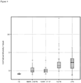
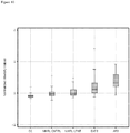
- the Caucasian control group consists of professional individuals recruited locally to the project team who constitute a low risk group, coming from non/low-TB endemic regions, such that their risk of having been exposed to TB is extremely low. Typically this is the preferred control group.
- the second control group in the Examples consists of individuals of Asian descent who tested negative for TB using the standard Mantoux skin test and IFN ⁇ test and who come from regions where TB is endemic. The likelihood is that these individuals have been exposed to TB, even if they are not themselves (currently) infected. Thus, without being bound by theory, any differences in the detection of biomarkers of the disclosure between this control group and the Caucasian controls may result from the likely exposure of this Asian control group to TB.
- control or reference population does not have TB and/or is not infected with M. tuberculosis (i.e. is TB-negative).
- the control or reference population may be TB-positive and are then subsequently diagnosed with TB using conventional techniques.
- a population of TB-positive individuals used to generate the reference profile may be diagnosed with TB about 24, 48, 72, 96 or more hours after biological samples were taken from them for the purposes of generating a reference biomarker profile.
- the population of TB-positive individuals is diagnosed with TB using conventional techniques about 0-36 hours, about 36-60 hours, about 60-84 hours, or about 84-108 hours after the biological samples were taken. If the biomarker profile is indicative of TB, a clinician may begin treatment prior to the manifestation of clinical symptoms of TB.
- the amount of the one or more biomarker of the disclosure may differ by at least 10%, at least 20%, at least 30%, at least 40%, at least 50%, at least 60, at least 70%, at least 80%, at least 90%, at least 100%, at least 150%, at least 200% or more compared with a control or reference population.
- the expression may be reduced partially or totally compared with the control or reference population.
- the amount is reduced by at least 10%, at least 20%, at least 30%, at least 40%, at least 50%, at least 60, at least 70%, at least 80%, at least 90%, at least 95%, at least 99%, up to total elimination of the one or more biomarker.
- the amount of one or more biomarker of the disclosure, typically in a biomarker profile, is increased compared with a control or reference population, the amount may be increased by at least 10%, at least 20%, at least 30%, at least 40%, at least 50%, at least 60, at least 70%, at least 80%, at least 90&, at least 100%, at least 150%, at least 200% compared with the control or reference population.
- the amount of the one or more biomarker of the disclosure may be increased or decreased compared with a control or reference population as shown in Tables 2 to 5 herein (where ⁇ means the one or more biomarker is upregulated/an increased amount of the one or more biomarker and ⁇ means the one or more biomarker is downregulated/a decreased amount of the one or more biomarker).
- ⁇ means the one or more biomarker is upregulated/an increased amount of the one or more biomarker
- ⁇ means the one or more biomarker is downregulated/a decreased amount of the one or more biomarker.
- Table 2 discloses that ALPK1is increased in monocytes, neutrophils and CD4 positive T cells compared with a control or reference population.
- the amount of ALPK1 may be increased in CD4 positive T cells, preferably increased in neutrophils and most preferably increased in monocytes.
- the amount of ALPK1 may be increased in CD4 positive T cells, neutrophils and monocytes, and may also be increased in other cell types not listed in Tables 2 to 5.
- the amount of the one or more biomarker may be increased in some cell types and/or decreased in other cell types.
- PF4V1 is upregulated (increased amount) in monocytes of individuals with TB
- PF4V1 is downregulated (decreased amount) in neutrophils of individuals with TB.
- the presence and/or amount of the one or more biomarker of the disclosure may be determined by quantitative and/or qualitative analysis.
- the amount of the one or more biomarker of the disclosure encompasses the mass of the one or more biomarker, the molar amount of the one or more biomarker, the concentration of the one or biomarker and the molarity of the one or more biomarker. This amount may be given in any appropriate units.
- the concentration of the one or more biomarker may be given in pg/ml, ng/ml or ⁇ g/ml.
- the presence and/or amount of the one or more biomarker of the disclosure may be measured directly or indirectly.
- the relative presence and/or amount of the one or more biomarker of the disclosure relative to a control or reference population may be determined using any appropriate technique. Suitable standard techniques are known in the art, for example Western blotting and enzyme-linked immunosorbent assays (ELISAs). Preferred methods include microarray analysis (as used in Example 1) and quantitative real-time PCR (qPCR) (as used in Example 2). Different one or more biomarkers may be used with different detection methods according to the present disclosure.
- the one or more biomarker is selected from PF4V1 /or HERC2, preferably in combination with LOC400759/GBP1P1 and/or ALPK1 as disclosed herein, for use with microarray analysis.
- the one or more biomarker is selected from SNX10 and/or CPVL, preferably in combination with LOC400759/GBP1P1 and/or CREG1, for use with qPCR analysis.
- additional one or more biomarkers as disclosed herein can be selected dependent on the preferred detection method.
- comparison includes any means to discern at least one difference in the presence and/or amount of the one or more biomarker in the individual and the control or reference population, or at least one difference in the individual's and the control or reference profiles.
- a comparison may include a visual inspection of chromatographic spectra, and a comparison may include arithmetical or statistical comparisons of values assigned to the features of the profiles. Such statistical comparisons include, but are not limited to, applying a decision rule.
- the biomarker profiles comprise at least one internal standard
- the comparison to discern a difference in the biomarker profiles may also include features of these internal standards, such that features of the biomarker are correlated to features of the internal standards.
- the comparison can confirm the presence or absence of TB, and thus to detect or diagnose TB; or the comparison can distinguish between active and latent TB.
- the presence and/or amount level of the one or more biomarker may be altered compared with a control or reference population for at least 12 hours, at least 24 hours, at least 30 hours, at least 48 hours, at least 72 hours, at least 96 hours, at least 120 hours, at least 144 hours, at least 1 week, at least 2 weeks, at least 3 weeks, at least 4 weeks, at least 5 weeks, at least 6 weeks, at least 7 weeks, at least 8 weeks, at least 9 weeks, at least 10 weeks, at least 11 weeks, at least 12 weeks, at least 13 weeks, at least 14 weeks, at least 15 weeks or more.
- an " individual” is an animal, preferably a mammal, more preferably a human or non-human primate.
- the terms " individual ,” “ subject “ and “patient” are used interchangeably herein.
- the individual can be normal, suspected of having TB or at risk of a TB infection.
- the disclosure relates to the detection and/or diagnosis of TB in adult humans (over the age of 16 years).
- the progression of an individual from normalcy i.e., a condition characterized by not having TB
- latent or active TB will be characterized by changes in biomarker profiles, as certain biomarkers are expressed at increasingly higher levels and the expression of other biomarkers becomes down regulated.
- These changes in biomarker profiles may reflect the progressive establishment of a physiological response in the reference population to infection.
- the biomarker profile of the control or reference population also will change as a physiological response subsides.
- one of the advantages of the present is the capability of classifying an individual, using a biomarker profile from a single biological sample, as having membership in a particular population.
- the determination of whether a particular physiological response is becoming established or is subsiding may be facilitated by a subsequent classification of the individual.
- the present disclosure provides numerous biomarkers that both increase and decrease in level of expression as a physiological response to TB is established or subsides. For example, a feature of an individual's biomarker profile that is known to change in intensity as a physiological response to TB becomes established may be selected. A comparison of the same feature in a profile from a subsequent biological sample from the individual can establish whether the individual is progressing toward more severe TB or is progressing toward normalcy.
- a feature as defined herein for the diagnosis of TB, a TB infection and/or a M. tuberculosis infection may be detected, quantified or determined by any appropriate means.
- the one or more biomarker of the disclosure, a measurable aspect or characteristic of the one or more biomarker or a biomarker profile of the disclosure may be detected by any appropriate means.
- the presence and/or amount of the one or more biomarkers of the disclosure may be considered together as a " biomarker profile " of the disclosure.
- the presence and/or amount of the individual biomarkers within any of the biomarker combinations disclosed herein may be considered together as a " biomarker profile " of the disclosure.
- biomarkers (i) SNX10 and CPVL; (ii) LOC400759/GBP1P1, SNX10, CPVL and CREG1; (iii) LOC400759/GBP1P1, SNX10, CPVL, CREG1, PSMB9, GBP1, IRF1 and HLA-B; (iv) LOC400759/GBP1P1, SNX10, CPVL, CREG1, PF4V1, GBP1, IRF1, IFITM3 and S100A11; (v) PF4V1 and HERC2; (vi) SNX10, CPVL, PF4V1 and HERC2; (vii) SNX10, CPVL, PF4V1, HERC2, CD52, LYN, LGALS3BP, BAZ1A, KLRA1 and WSB1 and/or (viii) LOC4007
- a biomarker profile of the disclosure may comprise: (i) SNX10 and CPVL; (ii) LOC400759/GBP1P1, SNX10, CPVL and CREG1; (iii) LOC400759/GBP1P1, SNX10, CPVL, CREG1, PSMB9, GBP1, IRF1 and HLA-B; (iv) LOC400759/GBP1P1, SNX10, CPVL, CREG1, PF4V1, GBP1, IRF1, IFITM3 and S100A11; (v) PF4V1 and HERC2; (vi) SNX10, CPVL, PF4V1 and HERC2; (vii) SNX10, CPVL, PF4V1, HERC2, CD52, LYN, LGALS3BP, BAZ
- the presence and/or amount of the one or more biomarker of the disclosure may be determined in a sample obtained from an individual.
- the sample may be any suitable biological material, for example blood, plasma, saliva, serum, sputum, urine, cerebral spinal fluid, cells, a cellular extract, a tissue sample, a tissue biopsy, a stool sample and the like.
- the sample is blood sample.
- the precise biological sample that is taken from the individual may vary, but the sampling preferably is minimally invasive and is easily performed by conventional techniques.
- the sample is a whole blood sample, a purified peripheral blood leukocyte sample or a cell type sorted leukocyte sample, such as a sample of the individual's neutrophils.
- the biological sample may be taken from the individual before, during, and/or after treatment for TB infection. In one instance, the sample is taken after treatment for TB infection has been initiated.
- Measurement of a phenotypic change may be carried out by any conventional technique. Measurement of body temperature, respiration rate, pulse, blood pressure, or other physiological parameters can be achieved via clinical observation and measurement. Measurements of biomarker molecules may include, for example, measurements that indicate the presence, concentration, expression level, or any other value associated with a biomarker molecule. The form of detection of biomarker molecules typically depends on the method used to form a profile of these biomarkers from a biological sample. For instance, biomarkers separated by 2D-PAGE are detected by Coomassie Blue staining or by silver staining, which are well-established in the art.
- the biomarkers of the disclosure may be detected at the nucleic acid or protein level.
- the biomarkers of the disclosure may be DNA, RNA or protein and may be detected using any appropriate technique.
- the presence and/or amount of the one or more biomarker of the disclosure may be measured directly or indirectly. Any appropriate agent may be used to determine the presence and/or amount of the one or more biomarker of the disclosure.
- the presence and/or amount of the one or more biomarker of the disclosure may be determined using an agent selected from peptides and peptidomimetics, antibodies, small molecules and single-stranded DNA or RNA molecules, as described herein.
- the relative presence and/or amount of the one or more biomarker of the disclosure relative to a control or reference population may be determined using any appropriate technique. Suitable standard techniques are known in the art.
- the one or more biomarker when the one or more biomarker is detected at the nucleic acid level this may be carried out using: (i) biomarker-specific oligonucleotide DNA or RNA or any other nucleic acid derivative probes bound to a solid surface; (ii) purified RNA (labelled by any method, for example using reverse transcription and amplification) hybridised to probes; (iii) whole lysed blood, from which the RNA is labelled by any method and hybridised to probes; (iv) purified RNA hybridised to probes and a second probe (labelled by any method) hybridised to the purified RNA; (v) whole lysed blood from which the RNA is hybridised to probes, and a second probe (labelled by any method) which is hybridised to the RNA; (vi) purified peripheral blood leukocytes, obtaining purified RNA (labelled by any method), and hybridising the purified labelled RNA to probes; (vii) purified peripheral blood leukocytes, obtaining purified
- RNA from a sample is labelled via any method (typically amplification) and used to interrogate one or more probe immobilised on a surface.
- the one or more probes are 50 to 100 nucleotides in length.
- one or more probe is immobilised on a surface and the RNA from a sample is hybridised to one or more second probe (labelled by any method). The RNA hybridised with the second (labelled) probe is then used to interrogate the one or more probe immobilised on the surface.
- Examples of such methodology are known in the art, including the VantixTM system.
- biomarker-specific primary antibodies or antibody fragments bound to a solid surface there may be carried out using: (i) biomarker-specific primary antibodies or antibody fragments bound to a solid surface; (ii) whole lysed blood biomarker antigen bound to antibodies or antibody fragments; (iii) secondary biomarker-specific antibodies or antibody fragments used to detect biomarker antigen bound to primary antibody (labelled using any method); (iv) biomarker-specific primary aptamers bound to a solid surface; (v) whole lysed blood - biomarker antigen bound to aptamers; (vi) secondary biomarker-specific aptamer used to detect biomarker antigen bound to primary aptamer (labelled using any method); (vii) any antibody derivative i.e.
- phage display etc. used as above; (viii) lateral flow devices/methodology; (ix) chromatography; (x) mass spectrometry; (xi) nuclear magnetic resonance (NMR); (xii) protein gels/transfers to filter; and/or (xiii) immunoprecipitation.
- Any agent for the detection of or for the determination of the amount of the one or more biomarker of the disclosure may be used to determine the presence of and/or amount of the one or more biomarker.
- any method that allows for the detecting of the one or more biomarker, the quantification, or relative quantification of the one or more biomarker may be used.
- Agents for the detection of or for the determination of the amount of one or more biomarker may be used to determine the amount of the one or more biomarker in a sample obtained from the individual. Such agents typically bind to the one or more biomarker. Such agents may bind specifically to the one or more biomarker.
- the agent for the detection of or for the determination of the amount of the one or more biomarker may be an antibody or other binding agent specific for the one or more biomarker. By specific, it will be understood that the agent or antibody binds to the molecule of interest, in this case the one or more biomarker, with no significant cross-reactivity to any other molecule, particularly any other protein.
- an agent or antibody that is specific for LOC400759/GBP1P1 will show no significant cross-reactivity with human neutrophil elastase.
- Cross-reactivity may be assessed by any suitable method.
- Cross-reactivity of an agent or antibody for the one or more biomarker with a molecule other than the one or more biomarker may be considered significant if the agent or antibody binds to the other molecule at least 5%, 10%, 15%, 20%, 25%, 30%, 35%, 40%, 45%, 50%, 55%, 60%, 65%, 70%, 75%, 80%, 85%, 90% or 100% as strongly as it binds to the one or more biomarker.
- An agent or antibody that is specific for the one or more biomarker may bind to another molecule such as human neutrophil elastase at less than 90%, 85%, 80%, 75%, 70%, 65%, 60%, 55%, 50%, 45%, 40%, 35%, 30%, 25% or 20% the strength that it binds to the one or more biomarker.
- the agent or antibody binds to the other molecule at less than 20%, less than 15%, less than 10% or less than 5%, less than 2% or less than 1% the strength that it binds to the one or more biomarker.
- the presence and/or amount of the one or more biomarker, and hence the biomarker profile may be determined immunologically by reacting antibodies, or functional fragments thereof, specific to the biomarkers.
- a functional fragment of an antibody is a portion of an antibody that retains at least some ability to bind to the antigen to which the complete antibody binds.
- the fragments which include, but are not limited to, scFv fragments, Fab fragments, F(ab) fragments and F(ab)2 fragments, can be recombinantly produced or enzymatically produced.
- Specific binding molecules other than antibodies, such as aptamers may be used to bind the biomarkers.
- the antibody may be monoclonal or polyclonal.
- the antibody may be produced by any suitable method known in the art.
- polyclonal antibodies may be obtained by immunising a mammal, typically a rabbit or a mouse, with the one or more biomarker under suitable conditions and isolating antibody molecules from, for example, the serum of said mammal.
- Monoclonal antibodies may be obtained by hybridoma or recombinant methods.
- Hybridoma methods may involve immunising a mammal, typically a rabbit or a mouse, with the one or more biomarker under suitable conditions, then harvesting the spleen cells of said mammal and fusing them with myeloma cells. The mixture of fused cells is then diluted and clones are grown from single parent cells. The antibodies secreted by the different clones are then tested for their ability to bind to the one or more biomarker, and the most productive and stable clone is then grown in culture medium to a high volume. The secreted antibody is collected and purified.
- Recombinant methods may involve the cloning into phage or yeast of different immunoglobulin gene segments to create libraries of antibodies with slightly different amino acid sequences. Those sequences which give rise to antibodies which bind to the one or more biomarker may be selected and the sequences cloned into, for example, a bacterial cell line, for production.
- the antibody is a mammalian antibody, such as a primate, human, rodent (e.g. mouse or rat), rabbit, ovine, porcine, equine or camel antibody.
- the antibody may be a camelid antibody or shark antibody.
- the antibody may be a nanobody.
- the antibody can be any class or isotype of antibody, for example IgM, but is preferably IgG.
- the antibody may be a humanised antibody.
- the antibody or fragment may be associated with other moieties, such as linkers which may be used to join together 2 or more fragments or antibodies.
- linkers may be chemical linkers or can be present in the form of a fusion protein with the fragment or whole antibody.
- the linkers may thus be used to join together whole antibodies or fragments which have the same or different binding specificities, e.g. that can bind the same or different polymorphisms.
- the antibody may be a bispecific antibody which is able to bind to two different antigens, typically any two of the polymorphisms mentioned herein.
- the antibody may be a ' diabody ' formed by joining two variable domains back to back.
- the antibodies used in the method are present in any of the above forms which have different antigen binding sites of different specificities then these different specificities are typically to polymorphisms at different positions or on different proteins.
- the antibody is a chimeric antibody comprising sequence from different natural antibodies, for example a humanised antibody.
- Methods to assess an amount of the one or more biomarker may involve contacting a sample with an agent or antibody capable of binding specifically to the one or more biomarker. Such methods may include dipstick assays and Enzyme-linked Immunosorbant Assay (ELISA), or similar assays, such as those using a lateral flow device. Other immunoassay types may also be used to assess the one or more biomarker amounts.
- dipsticks comprise one or more antibodies or proteins that specifically bind to the one or more biomarker. If more than one antibody is present, the antibodies preferably have different non-overlapping determinants such that they may bind to the one or more biomarker simultaneously.
- ELISA is a heterogeneous, solid phase assay that requires the separation of reagents.
- ELISA is typically carried out using the sandwich technique or the competitive technique.
- the sandwich technique requires two antibodies. The first specifically binds the one or more biomarker and is bound to a solid support. The second antibody is bound to a marker, typically an enzyme conjugate.
- a substrate for the enzyme is used to quantify the one or more biomarker -antibody complex and hence the amount of the one or more biomarker in a sample.
- the antigen competitive inhibition assay also typically requires a one or more biomarker -specific antibody bound to a support.
- a biomarker -enzyme conjugate is added to the sample (containing the one or more biomarker) to be assayed. Competitive inhibition between the biomarker -enzyme conjugate and unlabelled biomarker allows quantification of the amount of the one or more biomarker in a sample.
- the solid supports for ELISA reactions preferably contain wells.
- Antibodies capable of binding specifically to the one or more biomarker may be used in methods of immunofluorescence to detect the presence of the one or more biomarker and hence in methods of diagnosing TB, a TB infection, infection with M. tuberculosis, or to distinguish between active and latent TB according to the present disclosure.
- the present disclosure may also employ methods of determining the amount of the one or more biomarker that do not comprise antibodies.
- High Performance Liquid Chromatography (HPLC) separation and fluorescence detection is preferably used as a method of determining the amount of the one or more biomarker.
- HPLC apparatus and methods as described previously may be used ( Tsikas D et al. J Chromatogr B Biomed Sci Appl 1998; 705: 174-6 ) Separation during HPLC is typically carried out on the basis of size or charge.
- endogenous amino acids and an internal standard L-homoarginine Prior to HPLC, endogenous amino acids and an internal standard L-homoarginine are typically added to assay samples and these are phase extracted on CBA cartridges (Varian, Harbor City, CA).
- Amino acids within the samples are preferably derivatized with o-phthalaldehyde (OPA).
- OPA o-phthalaldehyde
- Mass spectrometric methods may include, for example, matrix-assisted laser desorption/ionization mass spectrometry (MALDI MS), surface-enhanced laser desorption/ionization mass spectrometry (SELDI MS), time of flight mass spectrometry (TOF MS) and liquid chromatography mass spectrometry (LC MS).
- MALDI MS matrix-assisted laser desorption/ionization mass spectrometry
- SELDI MS surface-enhanced laser desorption/ionization mass spectrometry
- TOF MS time of flight mass spectrometry
- LC MS liquid chromatography mass spectrometry
- a separation method may be used to determine the presence and/or amount of the one or more biomarker and hence to create a profile of biomarkers, such that only a subset of biomarkers within the sample is analysed.
- the biomarkers that are analysed in a sample may consist of mRNA species from a cellular extract, which has been fractionated to obtain only the nucleic acid biomarkers within the sample, or the biomarkers may consist of a fraction of the total complement of proteins within the sample, which have been fractionated by chromatographic techniques.
- One or more, two or more, three or more, four or more, or five or more separation methods may be used according to the present disclosure.
- Determination of the presence and/or amount of the one or more biomarker, and hence the creation of a profile of biomarkers may be carried out without employing a separation method.
- a biological sample may be interrogated with a labelled compound that forms a specific complex with a biomarker in the sample, where the intensity of the label in the specific complex is a measurable characteristic of the biomarker.
- a suitable compound for forming such a specific complex is a labelled antibody.
- a biomarker may be measured using an antibody with an amplifiable nucleic acid as a label. The nucleic acid label may become amplifiable when two antibodies, each conjugated to one strand of a nucleic acid label, interact with the biomarker, such that the two nucleic acid strands form an amplifiable nucleic acid.
- the presence and/or amount of the one or more biomarker, and hence the biomarker profile may be derived from an assay, such as an array, of nucleic acids, where the biomarkers are the nucleic acids or complements thereof.
- the biomarkers may be ribonucleic acids.
- the presence and/or amount of the one or more biomarker, and hence the biomarker profile may be obtained using a method selected from nuclear magnetic resonance, nucleic acid arrays, dot blotting, slot blotting, reverse transcription amplification and Northern analysis.
- the biomarker profile may comprise any measurable aspect of M. tuberculosis or a component thereof.
- the biomarker profile may comprise measurable aspects of small molecules, which may include fragments of proteins or nucleic acids, or which may include metabolites.
- suitable separation methods may include a mass spectrometry method, such as electrospray ionization mass spectrometry (ESI-MS), ESI-MS/MS, ESI- MS/(MS)n (n is an integer greater than zero), matrix-assisted laser desorption ionization time- of-flight mass spectrometry (MALDI-TOF-MS), surface-enhanced laser desorption/ionization time-of-flight mass spectrometry (SELDI-TOF-MS), desorption/ionization on silicon (DIOS), secondary ion mass spectrometry (SLMS), quadrupole time-of-flight (Q-TOF), atmospheric pressure chemical ionization mass spectrometry (APCI-MS), APCI-MS/MS, APCI-(MS)n, atmospheric pressure photoionization mass spectrometry
- ESI-MS electrospray ionization mass spectrometry
- MALDI-TOF-MS matrix-assisted laser de
- mass spectrometry methods may include, inter alia, quadrupole, fourier transform mass spectrometry (FTMS) and ion trap.
- suitable separation methods may include chemical extraction partitioning, column chromatography, ion exchange chromatography, hydrophobic (reverse phase) liquid chromatography, isoelectric focusing, one-dimensional polyacrylamide gel electrophoresis (PAGE), two-dimensional polyacrylamide gel electrophoresis (2D-PAGE) or other chromatography, such as thin-layer, gas or liquid chromatography, or any combination thereof.
- the sample may be fractionated prior to application of the separation method.
- the determination of the presence and/or amount of the one or more biomarker, and hence a biomarker profile may be generated by methods that do not require physical separation of the biomarkers themselves.
- nuclear magnetic resonance (NMR) spectroscopy may be used to resolve a profile of biomarkers from a complex mixture of molecules.
- NMR nuclear magnetic resonance
- Additional procedures include nucleic acid amplification technologies, which may be used to generate a profile of biomarkers without physical separation of individual biomarkers. (See Stordeur et al, J. Immunol. Methods 259: 55-64 (2002 ) and Tan et al, Proc. Nat'lAcad. Sci. USA 99: 11387-11392 (2002 ), for example.)
- laser desorption/ionization time-of-flight mass spectrometry is used to determine the presence and/or amount of the one or more biomarker, and hence create a biomarker profile where the biomarkers are proteins or protein fragments that have been ionized and vaporized off an immobilizing support by incident laser radiation. A profile is then created by the characteristic time-of-flight for each protein, which depends on its mass-to-charge (“m/z”) ratio.
- m/z mass-to-charge
- Laser desorption/ionization time-of-flight mass spectrometry allows the generation of large amounts of information in a relatively short period of time.
- a sample is applied to one of several varieties of a support that binds all of the biomarkers, or a subset thereof, in the sample.
- Cell lysates or samples are directly applied to these surfaces in volumes as small as 0.5 ⁇ L, with or without prior purification or fractionation.
- the lysates or sample can be concentrated or diluted prior to application onto the support surface.
- Laser desorption/ionization is then used to generate mass spectra of the sample, or samples, in as little as three hours.
- the total mRNA from a cellular extract of the individual is assayed, and the various mRNA species that are obtained from the sample are used as biomarkers.
- Biomarker profiles may be obtained, for example, by hybridizing these mRNAs to an array of probes, which may comprise oligonucleotides or cDNAs, using standard methods known in the art.
- the mRNAs may be subjected to gel electrophoresis or blotting methods such as dot blots, slot blots or Northern analysis, all of which are known in the art. (See, e.g., Sambrook et al.
- mRNA profiles also may be obtained by reverse transcription followed by amplification and detection of the resulting cDNAs, as disclosed by Stordeur et al , supra, for example.
- the profile may be obtained by using a combination of methods, such as a nucleic acid array combined with mass spectroscopy.
- qPCR using probe/quencher hydrolysis probes as described herein is highly specific and stringent.
- microarray analysis can resolve subtle differences in expression of transcript variants, which may be important in disease pathology and diagnosis.
- Any appropriate detection means can be used to detect or quantify the one or more biomarker of the disclosure, as described herein.
- the presence of the one or more biomarker may be detected, and/or the amount of the one or more biomarker determined using an oligonucleotide probe.
- An oligonucleotide probe of the disclosure may have at least 80% sequence identity to the one or more biomarker of the disclosure, or a target region within said biomarker, measured over any appropriate length of sequence. Typically the % sequence identity is determined over a length of contiguous nucleic acid residues.
- An oligonucleotide probe of the disclosure may, for example, have at least 80% sequence identity to the one or more biomarker of the disclosure, or target region thereof, measured over at least 10, at least 20, at least 30, at least 40, at least 50, at least 60, at least 70, at least 80, at least 90, or more nucleic acid residues, up to the oligonucleotide probe having at least 80% sequence identity with the one or more biomarker of the disclosure, or target region thereof, over the entire length of the oligonucleotide probe.
- An oligonucleotide probe of the disclosure may be complementary to the one or more nucleic acid biomarker of the disclosure, or a target region thereof. Typically the oligonucleotide probe of the disclosure is complementary over a length of contiguous nucleic acid residues.
- An oligonucleotide probe of the disclosure may, for example, be complementary to the one or more biomarker of the disclosure, or target region thereof, measured over at least 10, at least 20, at least 30, at least 40, at least 50, at least 60, at least 70, at least 80, at least 90, or more nucleic acid residues, up to the oligonucleotide probe having being complementary to the one or more biomarker of the disclosure, or target region thereof, over the entire length of the oligonucleotide probe.
- An oligonucleotide probe of the disclosure may be complementary to a variant of the one or more biomarker of the disclosure, or a variant of a target region of said biomarker.
- the oligonucleotide probe is complementary to a variant having at least 80% sequence identity to the one or more biomarker of the disclosure, or a variant having at least 80% sequence identity to the target region of said biomarker.
- the % sequence identity of the variant to the one or more biomarker of the disclosure, or a variant of a target region of said biomarker may be calculated over any appropriate length of sequence in the one or more biomarker, as described herein.
- a sequence identity of at least 80% includes at least 82%, at least 84%, at least 86%, at least 88%, at least 90%, at least 91%, at least 92%, at least 93%, at least 94%, at least 95%, at least 96%, at least 97%, at least 98%, at least 99%, and 100% sequence identity (to each and every nucleic acid sequence presented herein and/ or to each and every SEQ ID NO presented herein).
- sequence alignment methods can be used to determine percent identity, including, without limitation, global methods, local methods and hybrid methods, such as, e.g., segment approach methods. Protocols to determine percent identity are routine procedures within the scope of one skilled in the art. Global methods align sequences from the beginning to the end of the molecule and determine the best alignment by adding up scores of individual residue pairs and by imposing gap penalties. Non-limiting methods include, e.g., CLUSTAL W, see, e.g., Julie D.
- Non-limiting methods include, e.g., Match-box, see, e.g., Eric Depiereux and Ernest Feytmans, Match-Box: A Fundamentally New Algorithm for the Simultaneous Alignment of Several Protein Sequences, 8(5) CABIOS 501 -509 (1992 ); Gibbs sampling, see, e.g., C. E.
- variants of the specific sequences provided above may alternatively be defined by reciting the number of nucleotides that differ between the variant sequences and the specific reference sequences provided above.
- the sequence may comprise (or consist of) a nucleotide sequence that differs from the specific sequences provided above at no more than 2 nucleotide positions, for example at no more than 1 nucleotide position. Conservative substitutions are preferred.
- variants as defined herein also encompasses splice variants.
- An oligonucleotide probe of the disclosure may be at least 30, at least 40, at least 50, at least 60, at least 70, at least 80, at least 90, at least 100, or more nucleotides in length.
- the oligonucleotide probe is 40 to 100 nucleotides in length, more preferably 50 to 100 nucleotides in length, even more preferably 50 to 80 nucleotides in length and most preferably 50 to 70 nucleotides in length.
- the probes of the disclosure are typically designed to hybridise to their target nucleic acid sequence present in the one or more biomarker of the disclosure.
- a probe may comprise or be complementary to a nucleic acid sequence within a target nucleic acid sequence from the one or more biomarker of the disclosure, or to a nucleic acid sequence having at least 80% identity to said target nucleic acid sequence. Any suitable probe which comprises or is complementary (as defined herein) to a nucleic acid sequence within a target nucleic acid sequence of one or more biomarker of the disclosure may be used. Preferred target sequences within the one or more biomarkers of the disclosure are underlined in the nucleic acid sequences shown in the Sequence Information section.
- a target nucleic acid sequence may comprise bases 91 to 640 of SEQ ID NO: 112 or bases 13751 to 13950 of SEQ ID NO: 113, and a probe typically comprises or is complementary to a nucleic acid sequence having at least 80% sequence identity to a nucleic acid sequence from this target sequence.
- a target nucleic acid sequence may comprise bases 21 to 450 of SEQ ID NO: 134, and a probe typically comprises or is complementary to a nucleic acid sequence having at least 80% sequence identity to a nucleic acid sequence from this target sequence.
- a target nucleic acid sequence may comprise bases 511 to 3220 of SEQ ID NO: 117, and a probe typically comprises or is complementary to a nucleic acid sequence having at least 80% sequence identity to a nucleic acid sequence from this target sequence.
- a target nucleic acid sequence may comprise bases 2411 to 5641, 8141 to 9630 and/or 13651 to 14930 of SEQ ID NO: 132, and a probe typically comprises or is complementary to a nucleic acid sequence having at least 80% sequence identity to a nucleic acid sequence from this target sequence.
- a target nucleic acid sequence may comprise bases 1431 to 1850 of SEQ ID NO: 114, and a probe typically comprises or is complementary to a nucleic acid sequence having at least 80% sequence identity to a nucleic acid sequence from this target sequence.
- a target nucleic acid sequence may comprise bases 361 to 840 of SEQ ID NO: 115, and a probe typically comprises or is complementary to a nucleic acid sequence having at least 80% sequence identity to a nucleic acid sequence from this target sequence.
- a target nucleic acid sequence may comprise bases 1901 to 2480 of SEQ ID NO: 116, and a probe typically comprises or is complementary to at least one nucleic acid sequence having at least 80% sequence identity to a nucleic acid sequence from this target sequence.
- a target nucleic acid sequence may comprise bases 961 to 1620 of SEQ ID NO: 118, and a probe typically comprises or is complementary to at least one nucleic acid sequence having at least 80% sequence identity to a nucleic acid sequence from this target sequence.
- a target nucleic acid sequence may comprise bases 4561 to 5270 of SEQ ID NO: 119, and a probe typically comprises or is complementary to at least one nucleic acid sequence having at least 80% sequence identity to a nucleic acid sequence from this target sequence.
- a target nucleic acid sequence may comprise bases 1681 to 2520 of SEQ ID NO: 120, and a probe typically comprises or is complementary to at least one nucleic acid sequence having at least 80% sequence identity to a nucleic acid sequence from this target sequence.
- a target nucleic acid sequence may comprise bases 171 to 1820 of SEQ ID NO: 121, and a probe typically comprises or is complementary to at least one nucleic acid sequence having at least 80% sequence identity to a nucleic acid sequence from this target sequence.
- a target nucleic acid sequence may comprise bases 1201 to 2050 of SEQ ID NO: 122, and a probe typically comprises or is complementary to at least one nucleic acid sequence having at least 80% sequence identity to a nucleic acid sequence from this target sequence.
- a target nucleic acid sequence may comprise bases 241 to 870 of SEQ ID NO: 123, and a probe typically comprises or is complementary to at least one nucleic acid sequence having at least 80% sequence identity to a nucleic acid sequence from this target sequence.
- a target nucleic acid sequence may comprise bases 851 to 2250 of SEQ ID NO: 124, and a probe typically comprises or is complementary to at least one nucleic acid sequence having at least 80% sequence identity to a nucleic acid sequence from this target sequence.
- a target nucleic acid sequence may comprise bases 1901 to 2880 of SEQ ID NO: 125, and a probe typically comprises or is complementary to at least one nucleic acid sequence having at least 80% sequence identity to a nucleic acid sequence from this target sequence.
- a target nucleic acid sequence may comprise bases 301 to 1830 of SEQ ID NO: 126, and a probe typically comprises or is complementary to at least one nucleic acid sequence having at least 80% sequence identity to a nucleic acid sequence from this target sequence.
- a target nucleic acid sequence may comprise bases 1621 to 2690 of SEQ ID NO: 127, and a probe typically comprises or is complementary to at least one nucleic acid sequence having at least 80% sequence identity to a nucleic acid sequence from this target sequence.
- a target nucleic acid sequence may comprise bases 1051 to 1570 of SEQ ID NO: 128, and a probe typically comprises or is complementary to at least one nucleic acid sequence having at least 80% sequence identity to a nucleic acid sequence from this target sequence.
- a target nucleic acid sequence may comprise bases 381 to 1140 of SEQ ID NO: 129, and a probe typically comprises or is complementary to at least one nucleic acid sequence having at least 80% sequence identity to a nucleic acid sequence from this target sequence.
- a target nucleic acid sequence may comprise bases 2671 to 3020 of SEQ ID NO: 130, and a probe typically comprises or is complementary to at least one nucleic acid sequence having at least 80% sequence identity to a nucleic acid sequence from this target sequence.
- a target nucleic acid sequence may comprise bases 51 to 450 of SEQ ID NO: 131, and a probe typically comprises or is complementary to at least one nucleic acid sequence having at least 80% sequence identity to a nucleic acid sequence from this target sequence.
- a target nucleic acid sequence may comprise bases 801 to 1310 of SEQ ID NO: 133, and a probe typically comprises or is complementary to at least one nucleic acid sequence having at least 80% sequence identity to a nucleic acid sequence from this target sequence.
- a target nucleic acid sequence may comprise bases 11 to 270 of SEQ ID NO: 135, and a probe typically comprises or is complementary to at least one nucleic acid sequence having at least 80% sequence identity to a nucleic acid sequence from this target sequence.
- a target nucleic acid sequence may comprise bases 241 to 1460 of SEQ ID NO: 136, and a probe typically comprises or is complementary to at least one nucleic acid sequence having at least 80% sequence identity to a nucleic acid sequence from this target sequence.
- a target nucleic acid sequence may comprise bases 2171 to 2800 of SEQ ID NO: 142, and a probe typically comprises or is complementary to at least one nucleic acid sequence having at least 80% sequence identity to a nucleic acid sequence from this target sequence.
- a target nucleic acid sequence may comprise bases 1411 to 2050 of SEQ ID NO: 141, and a probe typically comprises or is complementary to at least one nucleic acid sequence having at least 80% sequence identity to a nucleic acid sequence from this target sequence.
- a target nucleic acid sequence may comprise bases 1091 to 2190 of SEQ ID NO: 152, and a probe typically comprises or is complementary to at least one nucleic acid sequence having at least 80% sequence identity to a nucleic acid sequence from this target sequence.
- a target nucleic acid sequence may comprise bases 641 to 3760 of SEQ ID NO: 138, and a probe typically comprises or is complementary to at least one nucleic acid sequence having at least 80% sequence identity to a nucleic acid sequence from this target sequence.
- a target nucleic acid sequence may comprise bases 541 to 1060 of SEQ ID NO: 137, and a probe typically comprises or is complementary to at least one nucleic acid sequence having at least 80% sequence identity to a nucleic acid sequence from this target sequence.
- a target nucleic acid sequence may comprise bases 541 to 1930 of SEQ ID NO: 138, and a probe typically comprises or is complementary to at least one nucleic acid sequence having at least 80% sequence identity to a nucleic acid sequence from this target sequence.
- a target nucleic acid sequence may comprise bases 9681 to 10990 of SEQ ID NO: 140, and a probe typically comprises or is complementary to at least one nucleic acid sequence having at least 80% sequence identity to a nucleic acid sequence from this target sequence.
- a target nucleic acid sequence may comprise bases 1041 to 1830 of SEQ ID NO: 143, and a probe typically comprises or is complementary to at least one nucleic acid sequence having at least 80% sequence identity to a nucleic acid sequence from this target sequence.
- a target nucleic acid sequence may comprise bases 211 to 580 of SEQ ID NO: 144, and a probe typically comprises or is complementary to at least one nucleic acid sequence having at least 80% sequence identity to a nucleic acid sequence from this target sequence.
- a target nucleic acid sequence may comprise bases 1251 to 1970 of SEQ ID NO: 145, and a probe typically comprises or is complementary to at least one nucleic acid sequence having at least 80% sequence identity to a nucleic acid sequence from this target sequence.
- a target nucleic acid sequence may comprise bases 2401 to 3110 of SEQ ID NO: 146, and a probe typically comprises or is complementary to at least one nucleic acid sequence having at least 80% sequence identity to a nucleic acid sequence from this target sequence.
- a target nucleic acid sequence may comprise bases 291 to 580 of SEQ ID NO: 147, and a probe typically comprises or is complementary to at least one nucleic acid sequence having at least 80% sequence identity to a nucleic acid sequence from this target sequence.
- a target nucleic acid sequence may comprise bases 461 to 3260 of SEQ ID NO: 148, and a probe typically comprises or is complementary to at least one nucleic acid sequence having at least 80% sequence identity to a nucleic acid sequence from this target sequence.
- a target nucleic acid sequence may comprise bases 2261 to 3170 of SEQ ID NO: 149, and a probe typically comprises or is complementary to at least one nucleic acid sequence having at least 80% sequence identity to a nucleic acid sequence from this target sequence.
- a target nucleic acid sequence may comprise bases 1751 to 2430 of SEQ ID NO: 150, and a probe typically comprises or is complementary to at least one nucleic acid sequence having at least 80% sequence identity to a nucleic acid sequence from this target sequence.
- a target nucleic acid sequence may comprise bases 1801 to 2860 of SEQ ID NO: 151, and a probe typically comprises or is complementary to at least one nucleic acid sequence having at least 80% sequence identity to a nucleic acid sequence from this target sequence.
- a target nucleic acid sequence may comprise bases 5791 to 6460 of SEQ ID NO: 153, and a probe typically comprises or is complementary to at least one nucleic acid sequence having at least 80% sequence identity to a nucleic acid sequence from this target sequence.
- a target nucleic acid sequence may comprise bases 741 to 1950 of SEQ ID NO: 154, and a probe typically comprises or is complementary to at least one nucleic acid sequence having at least 80% sequence identity to a nucleic acid sequence from this target sequence.
- a target nucleic acid sequence may comprise bases 981 to 1320 of SEQ ID NO: 155, and a probe typically comprises or is complementary to at least one nucleic acid sequence having at least 80% sequence identity to a nucleic acid sequence from this target sequence.
- a target nucleic acid sequence may comprise bases 1741 to 2370 of SEQ ID NO: 156, and a probe typically comprises or is complementary to at least one nucleic acid sequence having at least 80% sequence identity to a nucleic acid sequence from this target sequence.
- a target nucleic acid sequence may comprise bases 4591 to 5220 of SEQ ID NO: 157, and a probe typically comprises or is complementary to at least one nucleic acid sequence having at least 80% sequence identity to a nucleic acid sequence from this target sequence.
- a target nucleic acid sequence may comprise bases 461 to 940 of SEQ ID NO: 158, and a probe typically comprises or is complementary to at least one nucleic acid sequence having at least 80% sequence identity to a nucleic acid sequence from this target sequence.
- a target nucleic acid sequence may comprise bases 1421 to 2090 of SEQ ID NO: 159, and a probe typically comprises or is complementary to at least one nucleic acid sequence having at least 80% sequence identity to a nucleic acid sequence from this target sequence.
- a target nucleic acid sequence may comprise bases 2031 to 2950 of SEQ ID NO: 160, and a probe typically comprises or is complementary to at least one nucleic acid sequence having at least 80% sequence identity to a nucleic acid sequence from this target sequence.
- a target nucleic acid sequence may comprise bases 2601 to 3600 of SEQ ID NO: 161
- a probe typically comprises or is complementary to at least one nucleic acid sequence having at least 80% sequence identity to a nucleic acid sequence from this target sequence.
- a target nucleic acid sequence may comprise bases 661 to 1160 of SEQ ID NO: 162, and a probe typically comprises or is complementary to at least one nucleic acid sequence having at least 80% sequence identity to a nucleic acid sequence from this target sequence.
- a target nucleic acid sequence may comprise bases 961 to 1430 of SEQ ID NO: 163, and a probe typically comprises or is complementary to at least one nucleic acid sequence having at least 80% sequence identity to a nucleic acid sequence from this target sequence.
- a target nucleic acid sequence may comprise bases 461 to 1520 of SEQ ID NO: 164, and a probe typically comprises or is complementary to at least one nucleic acid sequence having at least 80% sequence identity to a nucleic acid sequence from this target sequence.
- a target nucleic acid sequence may comprise bases 161 to 760 of SEQ ID NO: 165, and a probe typically comprises or is complementary to at least one nucleic acid sequence having at least 80% sequence identity to a nucleic acid sequence from this target sequence.
- a target nucleic acid sequence may comprise bases 701 to 1770 of SEQ ID NO: 166, and a probe typically comprises or is complementary to at least one nucleic acid sequence having at least 80% sequence identity to a nucleic acid sequence from this target sequence.
- a target nucleic acid sequence may comprise bases 2011 to 3590 of SEQ ID NO: 167, and a probe typically comprises or is complementary to at least one nucleic acid sequence having at least 80% sequence identity to a nucleic acid sequence from this target sequence.
- the binding conditions for a probe hybridising to its target sequence are such that a high level of specificity is provided - i.e. hybridisation of the probe occurs under "stringent conditions " .
- stringent conditions are selected to be about 5°C lower than the thermal melting point (Tm) for the specific sequence at a defined ionic strength and pH.
- Tm is the temperature (under defined ionic strength and pH) at which 50% of the target (or complement) sequence hybridises to a perfectly matched probe.
- the Tm of probes of the present disclosure at a salt concentration of about 0.02M or less at pH 7, is for example above 60°C, such as about 70°C.
- Premixed buffer solutions are commercially available (e.g. EXPRESSHYB Hybridisation Solution from CLONTECH Laboratories, Inc.), and hybridisation can be performed according to the manufacturer's instructions.
- Probes of the present disclosure may be screened to minimise self-complementarity and dimer formation (probe-probe binding).
- any of the probes described herein may comprise a tag and/ or label.
- the tag and/ or label may, for example, be located (independently of one another) towards the middle or towards or at the 5' or 3' end of the herein described probes, for example at the 5' end.
- the tag/ label is associated with the target nucleic acid in the one or more biomarker.
- the probes may act as primers during the method of the disclosure and the tag/ label may therefore become incorporated into the amplification product as the primer is extended.
- suitable labels include detectable labels such as radiolabels or fluorescent or coloured molecules, enzymatic markers or chromogenic markers - e.g. dyes that produce a visible colour change upon hybridisation of the probe.
- the label may be digoxygenin, fluorescein-isothiocyanate (FITC), R-phycoerythrin, Alexa 532 or Cy3.
- the probes preferably contain a Fam label (e.g. a 5' Fam label), and/ or a minor groove binder (MGB).
- the label may be a reporter molecule, which is detected directly, such as by exposure to photographic or X-ray film.
- the label is not directly detectable, but may be detected indirectly, for example, in a two-phase system.
- An example of indirect label detection is binding of an antibody to the label.
- tags include " complement / anti-complement pairs".
- complement / anti-complement pair denotes non-identical moieties that form a non-covalently associated, stable pair under appropriate conditions.
- suitable tags include biotin and streptavidin (or avidin).
- a biotin tag may be captured using streptavidin, which may be coated onto a substrate or support such as a bead (for example a magnetic bead) or membrane.
- a streptavidin tag may be captured using biotin, which may be coated onto a substrate or support such as a bead (for example a magnetic bead) or membrane.
- complement/ anti-complement pairs include receptor/ ligand pairs, antibody/ antigen (or hapten or epitope) pairs, and the like.
- Another example is a nucleic acid sequence tag that binds to a complementary sequence. The latter may itself be pre-labelled, or may be attached to a surface (e.g. a bead) which is separately labelled. An example of the latter instances is the well-known LuminexR bead system.
- Other exemplary pairs of tags and capture molecules include receptor/ ligand pairs and antibody/ antigen (or hapten or epitope) pairs. Where subsequent dissociation of the complement/ anti-complement pair is desirable, the complement/ anti-complement pair has a binding affinity of, for example, less than 10 9 M -1 .
- One exemplary tagged probe is a biotin-labelled probe, which may be detected using horse-radish peroxidase conjugated streptavidin.
- the probes of the disclosure may be labelled with different labels or tags, thereby allowing separate identification of each probe when used in the method of the present disclosure.
- nucleic acid tags Any conventional method may be employed to attach nucleic acid tags to a probe of the present disclosure (e.g. to the 5' end of the defined binding region of the probe).
- nucleic acid probes of the disclosure may be constructed by commercial providers.
- an amplification step may be carried out using methods and platforms known in the art, for example PCR (for example, with the use of "Fast DNA Polymerase", Life Technologies), such as real-time PCR, block-based PCR, ligase chain reaction, glass capillaries, isothermal amplification methods including loop-mediated isothermal amplification, rolling circle amplification transcription mediated amplification, nucleic acid sequence-based amplification, signal mediated amplification of RNA technology, strand displacement amplification, isothermal multiple displacement amplification, helicase-dependent amplification, single primer isothermal amplification, and circular helicase-dependent amplification. If employed, amplification may be carried using any amplification platform.
- PCR for example, with the use of "Fast DNA Polymerase", Life Technologies
- isothermal amplification methods including loop-mediated isothermal amplification, rolling circle amplification transcription mediated amplification, nucleic acid sequence-based amplification, signal mediated amplification
- a general amplification step may be employed to increase the amount of the one or more biomarker of the disclosure present in the sample.
- PCR amplification primers are typically employed to amplify approximately 100-400 base pair regions of the target/ complementary nucleic acid that contain the nucleotide targets of the present disclosure.
- a suitable polymerase and DNA precursors dATP, dCTP, dGTP and dTTP
- forward and reverse primers are extended in a 5' to 3' direction, thereby initiating the synthesis of new nucleic acid strands that are complementary to the individual strands of the target nucleic acid.
- the primers thereby drive amplification of target nucleic acid sequences in the one or more biomarker, thereby generating amplification products comprising said target nucleic acid sequences.
- An amplification step may be employed in which the probes of the present disclosure act as primers.
- the probes act as primers
- Such an amplification step may be employed in conjunction with a general amplification step, such as the one described above.
- the detection step may be carried out by any known means.
- the probe or amplification product may be tagged and/ or labelled, and the detection method may therefore comprise detecting said tag and/ or label.
- the probe(s) may comprise a tag and/ or label.
- the tag/ label becomes associated with the target nucleic acid.
- the assay may comprise detecting the tag/ label and correlating presence of tag/ label with presence of the one or more nucleic acid biomarker of the disclosure.
- tag and/ or label may be incorporated during extension of the probe(s).
- the amplification product(s) become tagged/ labelled, and the assay may therefore comprise detecting the tag/ label and correlating presence of tag/ label with presence of amplification product, and hence the presence of one or more nucleic acid biomarker of the disclosure.
- the amplification product may incorporate a tag/ label (e.g. via a tagged/ labelled dNTP such as biotin-dNTP) as part of the amplification process, and the assay may further comprise the use of a binding partner complementary to said tag (e.g. streptavidin) that includes a detectable tag/ label (e.g. a fluorescent label, such as R-phycoerythrin).
- a detectable tag/ label e.g. a fluorescent label, such as R-phycoerythrin.
- the probe(s) and/ or the amplification product(s) may include a further tag/ label (as the complement component) to allow capture of the amplification product(s).
- a "complement / anti-complement” pairing may be employed in which an anti-complement capture component binds to said further tag/ label (complement component) and thereby permits capture of the probe(s) and/ or amplification product(s).
- suitable "complement/ anti-complement” partners have been described earlier in this specification, such as a complementary pair of nucleic acid sequences, a complementary antibody-antigen pair, etc.
- the anti-complement capture component may be attached (e.g. coated) on to a substrate or solid support - examples of suitable substrates/ supports include membranes and/ or beads (e.g. a magnetic or fluorescent bead). Capture methods are well known in the art.
- LuminexR beads may be employed.
- the use of magnetic beads may be advantageous because the beads (plus captured, tagged/ labelled amplification product) can easily be concentrated and separated from the sample, using conventional techniques known in the art.
- Immobilisation provides a physical location for the anti-complement capture component (or probes), and may serve to fix the capture component/ probe at a desired location and/ or facilitate recovery or separation of probe.
- the support may be a rigid solid support made from, for example, glass, plastic or silica, such as a bead (for example a fluorescent or magnetic bead).
- the support may be a membrane, such as nylon or nitrocellulose membrane.
- 3D matrices are also suitable supports for use with the present dislcosure - e.g. polyacrylamide or PEG gels.
- Immobilisation to a support/ platform may be achieved by a variety of conventional means. By way of example, immobilisation onto a support such as a nylon membrane may be achieved by UV cross-linking.
- biotin-labelled molecules may be bound to streptavidin-coated substrates (and vice-versa), and molecules prepared with amino linkers may be immobilised on to silanised surfaces.
- Another means of immobilisation is via a poly-T tail or a poly-C tail, for example at the 3' or 5' end. Said immobilisation techniques apply equally to the probe component (and primer pair component, if present) of the present disclosure.
- the probes of the disclosure comprise a nucleic acid sequence tag/ label (e.g. attached to each probe at the 5' end of the defined sequence of the probe that binds to target/ complement nucleic acid).
- each of the probes is provided with a different nucleic acid sequence tag/ label, wherein each of said tags/ labels (specifically) binds to a complementary nucleic acid sequence present on the surface of a bead.
- Each of the different tags/ labels binds to its complementary sequence counterpart (and not to any of the complementary sequence counterparts of the other tags), which is located on a uniquely identifiable bead.
- the beads are uniquely identifiable, for example by means of fluorescence at a specific wavelength.
- probes of the disclosure bind to target nucleic acid (if present in the sample). Thereafter, (only) the bound probes may be extended (in the 3' direction) in the presence of one or more labelled dNTP (e.g. biotin labelled dNTPs, such as biotin-dCTPs).
- dNTP e.g. biotin labelled dNTPs, such as biotin-dCTPs
- the extended primers may be contacted with a binding partner counterpart to the labelled dNTPs (e.g. a streptavidin labelled fluorophore, such as streptavidin labelled R-phycoerythrin), which binds to those labelled dNTPs that have become incorporated into the extended primers.
- a binding partner counterpart to the labelled dNTPs e.g. a streptavidin labelled fluorophore, such as streptavidin labelled R-phycoerythrin
- the labelled extended primers may be identified by allowing them to bind to their nucleic acid counterparts present on the uniquely identifiable beads. The latter may then be "called” (e.g. to determine the type of bead present by wavelength emission) and the nature of the primer extension (and thus the type of target/ complement nucleic acid present) may be determined.
- probes of the disclosure are oligonucleotides having sequence identity with a region of the one or more biomarker of the disclosure as disclosed herein.
- One or more probe may be immobilised on a solid support, and used to interrogate mRNA obtained from a test sample. If the mRNA from the test sample contains the one or more biomarker targeted by the immobilised probe, it will bind to the probe, and may then be detected.
- the biomarkers of the disclosure may also be detected using PCR, such as real time PCR.
- Any oligonucleotide with the appropriate level of sequence identity with the one or more biomarker of the disclosure, or with one or more target sequences within said one or more biomarker of the disclosure may be used as a probe as described herein.
- Any oligonucleotide with the appropriate level of complementarity with the one or more biomarker of the disclosure, or with one or more target sequences within said one or more biomarker of the disclosure may be used as a probe as described herein.
- Exemplary sequences of the one or more biomarkers of the disclosure are given in SEQ ID NOs: 112 to 167 (see Tables 2 to 5 herein).
- Sequences of exemplary target regions within the one or more biomarkers of the disclosure are shown as underlined in the sequences of the Sequence Information section (as discussed herein).
- Exemplary probe nucleic acid sequences for the biomarkers disclosed herein are set out in Table 6 (SEQ ID NOs: 1 to 111 and 168 to 171) and are shown as double-underlined in the sequences of the Sequence Information section.
- the oligonucleotide probe typically comprises or is complementary to a nucleic acid sequence having at least 80% sequence identity to the nucleic acid sequence of SEQ ID NO: 1, 2 or 3.
- the oligonucleotide probe typically comprises or is complementary to at least one nucleic acid sequence having at least 80% sequence identity to the nucleic acid sequence of SEQ ID NOs: 4 or 5.
- the oligonucleotide probe typically comprises or is complementary to at least one nucleic acid sequence having at least 80% sequence identity to the nucleic acid sequence of SEQ ID NOs: 6 or 7.
- the oligonucleotide probe typically comprises or is complementary to at least one nucleic acid sequence having at least 80% sequence identity to the nucleic acid sequence of SEQ ID NOs: 8, 9 or 168 to 171.
- the oligonucleotide probe typically comprises or is complementary to at least one nucleic acid sequence having at least 80% sequence identity to the nucleic acid sequence of SEQ ID NOs: 10 or 11.
- the oligonucleotide probe typically comprises or is complementary to at least one nucleic acid sequence having at least 80% sequence identity to the nucleic acid sequence of SEQ ID NOs: 12 or 13.
- the oligonucleotide probe typically comprises or is complementary to at least one nucleic acid sequence having at least 80% sequence identity to the nucleic acid sequence of SEQ ID NOs: 14 or 15.
- the oligonucleotide probe typically comprises or is complementary to at least one nucleic acid sequence having at least 80% sequence identity to the nucleic acid sequence of SEQ ID NOs: 16 or 17.
- the oligonucleotide probe typically comprises or is complementary to at least one nucleic acid sequence having at least 80% sequence identity to the nucleic acid sequence of SEQ ID NOs: 18 or 19.
- the oligonucleotide probe typically comprises or is complementary to at least one nucleic acid sequence having at least 80% sequence identity to the nucleic acid sequence of SEQ ID NOs: 20 or 21.
- the oligonucleotide probe typically comprises or is complementary to at least one nucleic acid sequence having at least 80% sequence identity to the nucleic acid sequence of SEQ ID NOs: 22 or 23.
- the oligonucleotide probe typically comprises or is complementary to at least one nucleic acid sequence having at least 80% sequence identity to the nucleic acid sequence of SEQ ID NOs: 24 or 25.
- the oligonucleotide probe typically comprises or is complementary to at least one nucleic acid sequence having at least 80% sequence identity to the nucleic acid sequence of SEQ ID NOs: 26 or 27.
- the oligonucleotide probe typically comprises or is complementary to at least one nucleic acid sequence having at least 80% sequence identity to the nucleic acid sequence of SEQ ID NOs: 28 or 29.
- the oligonucleotide probe typically comprises or is complementary to at least one nucleic acid sequence having at least 80% sequence identity to the nucleic acid sequence of SEQ ID NOs: 30 or 31.
- the oligonucleotide probe typically comprises or is complementary to at least one nucleic acid sequence having at least 80% sequence identity to the nucleic acid sequence of SEQ ID NOs: 32 or 33.
- the oligonucleotide probe typically comprises or is complementary to at least one nucleic acid sequence having at least 80% sequence identity to the nucleic acid sequence of SEQ ID NOs: 34 or 35.
- the oligonucleotide probe typically comprises or is complementary to at least one nucleic acid sequence having at least 80% sequence identity to the nucleic acid sequence of SEQ ID NOs: 36 or 37.
- the oligonucleotide probe typically comprises or is complementary to at least one nucleic acid sequence having at least 80% sequence identity to the nucleic acid sequence of SEQ ID NOs: 38 or 39.
- the oligonucleotide probe typically comprises or is complementary to at least one nucleic acid sequence having at least 80% sequence identity to the nucleic acid sequence of SEQ ID NOs: 40 or 41.
- the oligonucleotide probe typically comprises or is complementary to at least one nucleic acid sequence having at least 80% sequence identity to the nucleic acid sequence of SEQ ID NOs: 42 or 43.
- the oligonucleotide probe typically comprises or is complementary to at least one nucleic acid sequence having at least 80% sequence identity to the nucleic acid sequence of SEQ ID NOs: 44 or 45.
- the oligonucleotide probe typically comprises or is complementary to at least one nucleic acid sequence having at least 80% sequence identity to the nucleic acid sequence of SEQ ID NOs: 46 or 47.
- the oligonucleotide probe typically comprises or is complementary to at least one nucleic acid sequence having at least 80% sequence identity to the nucleic acid sequence of SEQ ID NOs: 48 or 49.
- the oligonucleotide probe typically comprises or is complementary to at least one nucleic acid sequence having at least 80% sequence identity to the nucleic acid sequence of SEQ ID NOs: 50 or 51.
- the oligonucleotide probe typically comprises or is complementary to at least one nucleic acid sequence having at least 80% sequence identity to the nucleic acid sequence of SEQ ID NOs: 52 or 53.
- the oligonucleotide probe typically comprises or is complementary to at least one nucleic acid sequence having at least 80% sequence identity to the nucleic acid sequence of SEQ ID NOs: 54 or 55.
- the oligonucleotide probe typically comprises or is complementary to at least one nucleic acid sequence having at least 80% sequence identity to the nucleic acid sequence of SEQ ID NOs: 56 or 57.
- the oligonucleotide probe typically comprises or is complementary to at least one nucleic acid sequence having at least 80% sequence identity to the nucleic acid sequence of SEQ ID NOs: 58 or 59.
- the oligonucleotide probe typically comprises or is complementary to at least one nucleic acid sequence having at least 80% sequence identity to the nucleic acid sequence of SEQ ID NOs: 60 or 61.
- the oligonucleotide probe typically comprises or is complementary to at least one nucleic acid sequence having at least 80% sequence identity to the nucleic acid sequence of SEQ ID NOs: 62 or 63.
- the oligonucleotide probe typically comprises or is complementary to at least one nucleic acid sequence having at least 80% sequence identity to the nucleic acid sequence of SEQ ID NOs: 64 or 65.
- the oligonucleotide probe typically comprises or is complementary to at least one nucleic acid sequence having at least 80% sequence identity to the nucleic acid sequence of SEQ ID NOs: 66 or 67.
- the oligonucleotide probe typically comprises or is complementary to at least one nucleic acid sequence having at least 80% sequence identity to the nucleic acid sequence of SEQ ID NOs: 68 or 69.
- the oligonucleotide probe typically comprises or is complementary to at least one nucleic acid sequence having at least 80% sequence identity to the nucleic acid sequence of SEQ ID NOs: 70 or 71.
- the oligonucleotide probe typically comprises or is complementary to at least one nucleic acid sequence having at least 80% sequence identity to the nucleic acid sequence of SEQ ID NOs: 72 or 73.
- the oligonucleotide probe typically comprises or is complementary to at least one nucleic acid sequence having at least 80% sequence identity to the nucleic acid sequence of SEQ ID NOs: 74 or 75.
- the oligonucleotide probe typically comprises or is complementary to at least one nucleic acid sequence having at least 80% sequence identity to the nucleic acid sequence of SEQ ID NOs: 76 or 77.
- the oligonucleotide probe typically comprises or is complementary to at least one nucleic acid sequence having at least 80% sequence identity to the nucleic acid sequence of SEQ ID NOs: 78 or 79.
- the oligonucleotide probe typically comprises or is complementary to at least one nucleic acid sequence having at least 80% sequence identity to the nucleic acid sequence of SEQ ID NOs: 80 or 81.
- the oligonucleotide probe typically comprises or is complementary to at least one nucleic acid sequence having at least 80% sequence identity to the nucleic acid sequence of SEQ ID NOs: 82 or 83.
- the oligonucleotide probe typically comprises or is complementary to at least one nucleic acid sequence having at least 80% sequence identity to the nucleic acid sequence of SEQ ID NOs: 84 or 85.
- the oligonucleotide probe typically comprises or is complementary to at least one nucleic acid sequence having at least 80% sequence identity to the nucleic acid sequence of SEQ ID NOs: 86 or 87.
- the oligonucleotide probe typically comprises or is complementary to at least one nucleic acid sequence having at least 80% sequence identity to the nucleic acid sequence of SEQ ID NOs: 88 or 89.
- the oligonucleotide probe typically comprises or is complementary to at least one nucleic acid sequence having at least 80% sequence identity to the nucleic acid sequence of SEQ ID NOs: 90 or 91.
- the oligonucleotide probe typically comprises or is complementary to at least one nucleic acid sequence having at least 80% sequence identity to the nucleic acid sequence of SEQ ID NOs: 92 or 93.
- the oligonucleotide probe typically comprises or is complementary to at least one nucleic acid sequence having at least 80% sequence identity to the nucleic acid sequence of SEQ ID NOs: 94 or 95.
- the oligonucleotide probe typically comprises or is complementary to at least one nucleic acid sequence having at least 80% sequence identity to the nucleic acid sequence of SEQ ID NOs: 96 or 97.
- the oligonucleotide probe typically comprises or is complementary to at least one nucleic acid sequence having at least 80% sequence identity to the nucleic acid sequence of SEQ ID NOs: 98 or 99.
- the oligonucleotide probe typically comprises or is complementary to at least one nucleic acid sequence having at least 80% sequence identity to the nucleic acid sequence of SEQ ID NOs: 100 or 101.
- the oligonucleotide probe typically comprises or is complementary to at least one nucleic acid sequence having at least 80% sequence identity to the nucleic acid sequence of SEQ ID NOs: 102 or 103.
- the oligonucleotide probe typically comprises or is complementary to at least one nucleic acid sequence having at least 80% sequence identity to the nucleic acid sequence of SEQ ID NOs: 104 or 105.
- the oligonucleotide probe typically comprises or is complementary to at least one nucleic acid sequence having at least 80% sequence identity to the nucleic acid sequence of SEQ ID NOs: 106 or 107.
- the oligonucleotide probe typically comprises or is complementary to at least one nucleic acid sequence having at least 80% sequence identity to the nucleic acid sequence of SEQ ID NOs: 108 or 109.
- the oligonucleotide probe typically comprises or is complementary to at least one nucleic acid sequence having at least 80% sequence identity to the nucleic acid sequence of SEQ ID NOs: 110 or 111.
- comparison of the one or more biomarker or the biomarker profile to a reference or control comprises applying a decision rule, or using a decision tree, as described herein.
- the decision rule or decision tree can comprise a data analysis algorithm, such as a computer pattern recognition algorithm.
- Other suitable algorithms include, but are not limited to, logistic regression or a nonparametric algorithm that detects differences in the distribution of feature values (e.g., a Wilcoxon Signed Rank Test).
- the decision rule may be based upon one, two, three, four, five, 10, 20 or more features. In one instance, the decision rule or decision tree is based on hundreds or more of features. Applying the decision rule or decision tree may also comprise using a classification tree algorithm.
- control or reference biomarker profile may comprise at least three features or biomarkers, where the features are predictors in a classification tree algorithm.
- the data analysis algorithm predicts membership within a population (or class) with an accuracy of at least about 60%, at least about 70%, at least about 80% and at least about 90%.
- Suitable algorithms are known in the art, some of which are reviewed in Hastie et al, supra. Such algorithms classify complex spectra from biological materials, such as a blood sample, to distinguish individuals as normal or as possessing biomarker expression levels characteristic of a particular disease state. While such algorithms may be used to increase the speed and efficiency of the application of the decision rule and to avoid investigator bias, one of ordinary skill in the art will realize that computer-based algorithms are not required to carry out the methods of the present disclosure.
- Algorithms may be applied to the comparison of the one or more biomarker or the biomarker profiles, regardless of the method that was used to generate the data for the one or more biomarker or the biomarker profile.
- suitable algorithms can be applied to biomarker profiles generated using gas chromatography, as discussed in Harper, "Pyrolysis and GC in Polymer Analysis” Dekker, New York (1985 ).
- Wagner et al, Anal Chem 74: 1824-35 (2002 ) disclose an algorithm that improves the ability to classify individuals based on spectra obtained by static time-of-flight secondary ion mass spectrometry (TOF-SIMS). Additionally, Bright et al, J.
- Microbiol Methods 48: 127-38 disclose a method of distinguishing between bacterial strains with high certainty (79- 89% correct classification rates) by analysis of MALDI-TOF-MS spectra. Dalluge, Fresenius J. Anal. Chem. 366: 701-11 (2000 ) discusses the use of MALDI-TOF-MS and liquid chromatography-electrospray ionization mass spectrometry (LC/ESI-MS) to classify profiles of biomarkers in complex biological samples.
- LC/ESI-MS liquid chromatography-electrospray ionization mass spectrometry
- the present disclosure provides a method for diagnosing TB in an individual, comprising determining the presence and/or amount of one or more biomarker for TB in a sample obtained from the individual, wherein the one or more biomarkers is selected from SNX10, CPVL, PF4V1, HERC2, LGALS3BP, BST1, BAZ1A, LYN, SERPINB1, WSB1, MVP, APBB1IP, MB21D1/C6orf150, TICAM2, CD52, KLRA1, DEFB128 and IL8. Any combination of biomarkers as disclosed herein may be used ina method according to the present disclosure.
- the method may comprising obtaining a first biomarker profile from a first sample taken from the individual at a single initial point in time and multiple time points thereafter to monitor the efficacy of treatment and disease resolution; and comparing said individual's first biomarker profile to a reference or control biomarker profile, wherein said comparison determines the status of TB infection in the individual with an accuracy, sensitivity and/or specificity of at least about 90%, at least about 80%, at least about 70% or at least about 60%; and wherein the biomarker profiles comprise determining the presence and/or amount of one or more biomarker of the disclosure, Typically the accuracy, sensitivity and/or specificity is of at least about 80% or at least about 90%.
- the method may comprise obtaining a first biomarker profile from a first sample from the individual; and comparing the individual's first biomarker profile to a reference or control biomarker profile obtained from a reference or control population, said comparison being capable of classifying the individual as belonging to or not belonging to the reference or control population, wherein the comparison determines the status of TB infection in the individual, and wherein the biomarker profiles comprise determining the presence and/or amount of one or more biomarker of the disclosure.
- the method may comprise comparing a measurable characteristic of at least three biomarkers of the disclosure between (i) a first biomarker profile obtained from a first sample from the individual and (ii) a biomarker profile obtained from samples from a control or reference population; and classifying the individual as belonging to or not belonging to the control or reference population, wherein the comparison determines the status of TB infection in the individual, and wherein the measurable characteristic optionally comprises the presence and/or amount of the biomarker.
- the method may comprise selecting at least two features from a set of biomarkers of the disclosure in a first biomarker profile generated from a first sample of the individual; and comparing the at least two features to a set of the same biomarkers in a biomarker profile generated from samples from a control or reference population, wherein the comparison is capable of classifying the individual as belonging to or not belonging to the control reference population with an accuracy, sensitivity and/or specificity of at least about 90%, at least about 80%, at least about 70% or at least about 60%, wherein the comparison determines the status of TB in the individual, and wherein the feature optionally comprises the presence and/or amount of the biomarker.
- the accuracy, sensitivity and/or specificity is of at least about 80% or at least about 90%.
- the method may comprise determining an abundance or a change in an abundance of at least three biomarkers contained in a first biomarker profile obtained from a first biological sample of the individual; and (b) comparing the abundance or the change in the abundance to an abundance or change in an abundance of said at least three biomarkers contained in biological samples from a control or reference population, wherein the comparison is capable of classifying the individual as belonging to or not belonging to the control or reference population; and wherein the comparison determines the status of TB in the individual.
- the method may further comprise obtaining a second biomarker profile from a second sample taken from the individual; and comparing the individual's second biomarker profile to the control or reference biomarker profile; wherein the individual's second biomarker profile and the control or reference biomarker profile comprise features that are measurable characteristics of a biomarker of the disclosure, wherein the second comparison is capable of classifying the individual as belonging to or not belonging to the control or reference population, and wherein the second comparison determines the status TB infection in the individual.
- the biomarker profiles optionally comprise one or more of the biomarkers of the present disclosure, and the measurable characteristic optionally comprises the presence and/or amount of one or more biomarker of the disclosure.
- the methods of the disclosure may be repeated at least once, at least twice, at least three times, at least four times, at least five times, or more.
- a separate biomarker profile can be obtained from the individual from a separate sample taken each time the method is repeated.
- the methods of the disclosure may be used to diagnose, detect and/or predict TB, TB infection and/or infection with M. tuberculosis.
- the methods of the disclosure may be used to distinguish between active and latent TB, a TB infection and/or infection with M . tuberculosis.
- the methods of the disclosure may be used to distinguish between latent TB and the absence of TB.
- the methods of the disclosure may be used to identify an individual with an active TB infection and/or a latent TB infection.
- the methods of the disclosure may be used to identify an individual with an active TB infection and/or a latent TB infection and/or an individual uninfected with TB.
- the methods of the disclosure may be used to identify an individual with an early stage active TB infection and/or a late/later stage active TB infection.
- the methods of the disclosure may be used to distinguish between an early stage active TB infection and/or a late/later stage active TB infection.
- Determining the status of TB, a TB infection and/or infection with M. tuberculosis in an individual may comprise determining the presence of active or latent TB, a TB infection and/or infection with M. tuberculosis in an individual.
- the methods of the disclosure may be used to determine whether an individual has been exposed to TB.
- the methods of the disclosure may comprise applying a decision rule as described herein. Applying the decision rule may comprise using a data analysis algorithm, also as described herein.
- the data analysis algorithm may comprise at least two, at least three, at least four, at least five, at least six, at least seven, at least eight, at least nine, at least ten, at least 15, at least 20, at least 25, at least 50 or more input parameters.
- the data analysis algorithm may use any of the biomarkers of the disclosure, or combination of biomarkers of the disclosure as input parameters.
- the data analysis algorithm uses at least two, at least three, at least four, at least five, at least six, at least seven, at least eight, at least nine, at least ten, at least 15, at least 20, at least 25, at least 50 of the biomarkers of the disclosure (e.g. as listed in any one of Tables 2 to 5) as input parameters.
- the features and/or biomarkers profile used in the methods of the disclosure are the one or more biomarkers of the present disclosure, as described herein, and preferably the methods relate to determining the presence and/or amount of the one or more biomarker.
- the "measurable characteristic" in a method of the disclosure may be any quantitative or qualitative characteristic associated with one or more biomarker of the disclosure, and is preferably the presence and/or amount of said biomarker.
- the one or more biomarker of the disclosure is nucleic acids, selected from DNA or RNA, typically mRNA.
- the biomarker profile may comprise any measurable aspect of said nucleic acid biomarker, and is typically a measurable characteristic of an mRNA biomarker, such as the presence and/or amount of said mRNA biomarker.
- the one or more biomarker of the disclosure and/or the biomarker may comprise a measurable aspect of a nucleic acid biomarker that encodes a protein that is informative of the state of the immune system in response to TB, a TB infection and/or infection with M . tuberculosis in an individual.
- a method of the disclosure may comprise fractionation of the sample prior to determining the presence and/or amount of the one or more biomarker of the disclosure, or obtaining a biomarker profile.
- the method comprises at least one, at least two, at least three, at least four, at least five, or more separation methods as described herein.
- the at least one separation method may be selected from inflammatory cell separation, chemical extraction partitioning, ion exchange chromatography, gel electrophoresis, and any combination thereof.
- the disclosure also provides the use of one or more biomarker for TB as defined herein in the manufacture of a diagnostic for TB.
- Said diagnostic may be for diagnosing active TB and/or latent TB and/or the absence of TB.
- kits and devices that are useful in determining the status of TB, diagnosing or detecting TB, distinguishing between active and latent TB in an individual, distinguishing between early stage active TB and late/later stage active TB and/or to determine whether an individual has been exposed to TB.
- the kits and devices of the present disclosure comprise at least one biomarker of the disclosure and/or one or more agent for the detection of or for the determination of the amount of the one or more biomarker of the disclosure. Specific biomarkers and agents for the detection of said biomarkers useful in the present disclosure are set forth herein.
- the biomarkers of the kit or device can be used to generate biomarker profiles according to the present disclosure.
- the biomarkers of the kit or biomarker will bind, with at least some specificity, to the biomarker molecules contained in the sample from which the biomarker profile is generated.
- classes of compounds of the kit or device include, but are not to, proteins (including antibodies of the disclosure), and fragments thereof, peptides, polypeptides, proteoglycans, glycoproteins, lipoproteins, carbohydrates, lipids, nucleic acids, organic and inorganic chemicals, and natural and synthetic polymers.
- the biomarker(s) and/or agent(s) for the detection of the one or more biomarker may be part of an array, or the biomarker(s) and/or agent(s) may be packaged separately and/or individually.
- the biomarker(s) and/or agent(s) may be immobilised on an inert support.
- the kit or device may also comprise at least one internal standard to be used in generating the biomarker profiles of the present disclosure.
- the internal standards can be any of the classes of compounds described above.
- kits and devices of the present disclosure also may contain reagents that can be used to detectably label biomarkers contained in the biological samples from which the biomarker profiles are generated.
- the kit or device may comprise a set of antibodies or functional fragments thereof that specifically bind at least two, three, four, five, 10, 20, 30, 40, 50 or more, up to all 55 of the biomarkers set forth in any one of Tables 2 to 6 that list biomarkers for use in the disclosure.
- the antibodies themselves may be detectably labelled.
- the kit or device also may comprise a specific biomarker binding component, such as an aptamer.
- a kit or device of the disclosure comprises (i) one or more antibody specific for the one or more biomarker for tuberculosis; or (ii) one or more oligonucleotide specific for the one or more biomarker for tuberculosis.
- the one or more oligonucleotide specific for the one or more biomarker for tuberculosis is an oligonucleotide is an oligonucleotide of the disclosure, more preferably one or more of SEQ ID NOs: 1 to 111 or 168 to 171.
- the kit or device may provide one or more oligonucleotide probe that is capable of forming a duplex with the one or more biomarker or with a complementary strand of said one or more biomarker.
- the one or more oligonucleotide probe may be detectably labelled.
- the one or more oligonucleotide probe used in the methods of the disclosure is selected from one or more of the oligonucleotide described herein.
- the one or more oligonucleotide probe is selected from an oligonucleotide probe that comprises or is complementary to at least one nucleic acid sequence having at least 80% sequence identity to the nucleic acid sequence of any one or more of SEQ ID NOs: 1 to 111 or 168 to 171.
- kits and devices of the present disclosure may also include pharmaceutical excipients, diluents and/or adjuvants when the biomarker is to be used to raise an antibody.
- pharmaceutical adjuvants include, but are not limited to, preservatives, wetting agents, emulsifying agents, and dispersing agents. Prevention of the action of microorganisms can be ensured by the inclusion of various antibacterial and antifungal agents, for example, paraben, chlorobutanol, phenol sorbic acid, and the like. It may also be desirable to include isotonic agents such as sugars, sodium chloride, and the like.
- Naive Cynomologous macaques ( Macaca fascicularis ) aged between 2 - 4 yrs from two separate breeding colonies i.e. of Mauritian or Chinese origin, from established United Kingdom or Chinese breeding facilities were challenged with were challenged with live M . tuberculosis using aerosol challenge Erdman strain K 01. They were confirmed as na ⁇ ve in terms of prior exposure to mycobacterial antigens ( M. tuberculosis infection or environmental mycobacteria), by negative tuberculin test while in their original breeding colony and prior to the start of the study using the gamma interferon (IFN- ⁇ )-based Primagam test kit (Biocor; CSL). All procedures involving animals were approved by the Ethical Review Committee of the Centre for Emergency Preparedness and Response, Salisbury, United Kingdom.
- IFN- ⁇ gamma interferon
- Mono-dispersed bacteria in particles were generated using a three-jet Collison nebulizer (BGI) and in conjunction with a modified Henderson apparatus, delivered to the nose of each sedated primate via a modified veterinary anaesthesia mask.
- BGI three-jet Collison nebulizer
- the challenge was performed on sedated animals placed within a "head-out" plethysmography chamber (Buxco, Wilmington, NC), to enable the aerosol to be delivered simultaneously with the measurement of the respiration rate. None of the animals had been used previously for experimental procedures.
- heparinised blood was obtained at three independent time points prior to challenge and at one, two, four and six weeks post M . tuberculosis challenge.
- 1 ml of blood from each animal was mixed with 5 ml of Erythrocyte Lysis (EL) Buffer (Qiagen) followed by incubation on ice for 10-15 min.
- EL Erythrocyte Lysis
- Peripheral blood leukocytes (PBLs) were recovered from erythrocyte-lysed blood by centrifugation at 400 x g for 10 min at 4 °C and re-suspended in a further 2 ml of EL buffer.
- PBLS were again recovered by centrifugation as described above and processed for recovery of total RNA.
- RNA pellets were re-suspended in 10 ⁇ l of diethylpyrocarbonate (DECP) water (Invitrogen), then concentration and purity (A260/A280 ratio ⁇ 1.8) assessed by spectrophotometry using a NanoDrop ND-1000 spectrophotometer (Thermo Scientific). Genomic DNA was removed prior to its use in further procedures using the DNase I kit (Qiagen), according to the manufacturer's instructions.
- DECP diethylpyrocarbonate
- concentration and purity A260/A280 ratio ⁇ 1.8
- GeniSphere SenseAmp RNA amplification kit according to manufactures instructions (http://genisphere.com/ ).
- the resulting amplified cRNA was purified using RNeasy Min-Elute Cleanup kit (Qiagen), again according to the manufacturer's protocol.
- the cRNA concentration and purity was then assessed by spectrophotometry using a NanoDrop ND-1000 spectrophotometer.
- the slides were air-dried and scanned using an Affymetrix 480 microarray scanner, at a gain threshold of 65.
- Feature extraction was then conducted using the microarray quantification package BlueFuse (BlueGnome ltd.).
- Raw data were then exported and hybridisation fluorescence intensities quantified using the software analysis program BluefuseTM, using default background subtraction and normalisation methods, to remove data generated from poor-quality spots, hybridisation artefacts. All raw data were then processed further using the microarray analysis package Genespring 12.5.
- ANN Artificial Neural Network Analysis
- Normalised expression data was analysed using ANN based data mining approach ( Lancashire LJ et al (2010), Breast Cancer Res Treat. Feb; 120(1):83-93 ).
- This approach comprised a supervised learning approach where the data for a given single probe was used to classify a known sample.
- the classifier consisted of a multi-layer perceptron ANN, where weights were updated by a back propagation algorithm ( Rumelhart DE et al (1986) Nature 323: 533 - 536 ).
- the ANN architecture utilised a constrained architecture to prevent over-fitting, having only 2 hidden nodes in the hidden layer.
- ANN training incorporated Monte Carlo Cross Validation, where, the data was randomly divided into three subsets; 60% for training the classifier, 20% for testing (to assess model performance during the training process) and 20% for validation (to independently test the model on data completely blind to the model).
- This process of random sample cross validation was utilised to prevent over-fitting of the data and assess how well the model would perform on a blind data set.
- This random re-sampling and training process was repeated 50 times to generate predictions and associated error values for each sample with respect to the validation (blind) data. Probes were ranked in ascending order based on predictive error for test data from the Monte Carlo Cross validation.
- Table 2 lists the genes newly identified as biomarkers for TB using the above methods. Also given are the sequence identifiers for the identified genes and an indication of whether expression of the various genes are up or down regulated in TB compared with a control/reference population, and in what cell types (all white blood cells in a sample, monocytes, neutrophils, CD4 positive T cells, CD8 positive T cells, etc.) the change was observed. Where more than one indication is given, the first listed is the most preferred.
- Table 3 lists further biomarkers for TB. Also given are the sequence identifiers for the identified genes and an indication of whether expression of the various genes are up or down regulated in TB compared with a control/reference population, and in what cell types the change was observed. Where more than one indication is given, the first listed is the most preferred.
- Table 4 lists the genes identified as biomarkers for latent TB using the above methods. Also given are the sequence identifiers for the identified genes and an indication of whether expression of the various genes are up or down regulated in TB compared with a control/reference population, and in what cell types the change was observed. Where more than one indication is given, the first listed is the most preferred.
- Table 5 lists further biomarkers for latent TB. Also given are the sequence identifiers for the identified genes and an indication of whether expression of the various genes are up or down regulated in TB compared with a control/reference population, and in what cell types the change was observed. Where more than one indication is given, the first listed is the most preferred.
- Probes 1a & 1b SEQ ID NO: Probe 2 SEQ ID NO: AA1 LOC40075/ GBP1P1 1 3 2 AA2 LGALS3BP 10 11 AB1 BST1 12 13 AB2 SNX10 14 15 AC1 ALPK1 6 7 AC2 CREG1 16 17 AD1 BAZ1A 18 19 AD2 LYN 20 21 AD3 TAPBP 22 23 AE1 SERPINB 1 24 25 AE2 PSMB9 26 27 AE3 WSB1 28 29 AF1 MVP 30 31 AF2 APBB1IP 32 33 AF3 FYB 34 35 AG1 MB21D1/ C6orf150 36 37 AG2 CPVL 38 39 AG3 TICAM2 40 41 AH1 CD52 42 43 AI1 HERC2 8 9 168 169 170 171 AJ1 KLRAP1 (KLRA1) 44 45 AK1
- Quantitative real-time PCR analyses were performed using the Roche Lightcycler (LC) 480 in 384 well plate format.
- the LC 480 is a plate-based, highly adaptable and versatile real-time PCR system used for gene expression analysis and has been designed for enhanced throughput and efficiency without compromising sensitivity and specificity.
- Roche provide an online 'target to assay' design and build system, the 'Realtime Ready configurator', which can be used to generate quantitative real-time PCR (qPCR) assays in a number of plate formats.
- Assays for the biomarker targets of interest were designed using this system and the assay plates configured, tested and quality assured by Roche.
- Data output files from BlueFuse were imported into GX 12.5 for differential gene expression and statistical analysis. Averaged data were imported without further normalisation and then baseline transformed to the median of all samples. Data were assessed for quality, then filtered on gene expression where entities in at least 100 percent of samples and in any one out of one conditions had normalised expression values within the cut-off 0 to 329.0 where at least 1 feature out of all samples had values within range. Statistically significant features were identified using one-way ANOVA analysis across all entities and time points at a cut-off p ⁇ 0.05. To identify differentially expressed entities between the groups T-tests (unpaired, unequal variance) were performed on the samples arranged by group at a p ⁇ 0.05 using the cut-off fold-change setting > 1.2. The outputs were visualised using the boxplot graphical output facility.
- Table 7 lists the genes newly identified as biomarkers for TB in Example 1 (see Table 2 above). Also given are the results of t-tests comparing the expression of a given marker between different test/control groups. The final two columns give ANOVA p values illustrating the significance of the biomarkers as determined using the qPCR technique.
- Table 8 provides the results of the qPCR analysis of the human cohort samples using the further biomarkers for TB listed in Table 3 above. Also given are the results of t-tests comparing the expression of a given marker between different test/control groups. The final two columns give ANOVA p values illustrating the significance of the biomarkers as determined using the qPCR technique.
- Table 9 provides the results of the qPCR analysis of the human cohort samples using the genes identified as biomarkers for latent TB in Example 1 (see Table 4 above). Also given are the results of t-tests comparing the expression of a given marker between different test/control groups. The final two columns give ANOVA p values illustrating the significance of the biomarkers as determined using the qPCR technique.
- Table 10 provides the results of the qPCR analysis of the human cohort samples using the further biomarkers for latent TB listed in Table 5 above. Also given are the results of t-tests comparing the expression of a given marker between different test/control groups. The final two columns give ANOVA p values illustrating the significance of the biomarkers as determined using the qPCR technique.
- nucleotide sequences of the TB biomarkers disclosed herein are underlined, and exemplary probe sequences are double underlined.
Description
- This disclosure relates to the detection and diagnosis of tuberculosis. More specifically, the disclosure relates to new biomarkers and combinations thereof that enable the accurate detection and diagnosis of tuberculosis.
- Tuberculosis (TB) is a progressive, often fatal, infectious disease, caused by the bacterial pathogen Mycobacterium tuberculosis (M. tuberculosis, MTB). This is a significant cause of mortality worldwide, being the eighth largest leading cause of death globally, and is primarily a disease of poverty, particularly in developing countries. Latent TB infection is believed to affect as much as one third of the world's population.
- Tuberculosis is a notifiable disease and is a major concern for many governmental and other health bodies including the World Health Organisation (WHO), who have initiated numerous control and treatment programmes like the "Stop TB Partnership".
- The WHO estimates that nearly nine million new cases of TB, and nearly two million deaths, occur globally each year. The largest number of new TB cases in 2005 occurred in South-East Asia (34% of incident cases globally), and the estimated incidence rate in sub-Saharan Africa is nearly 350 cases per 100,000 population. However, TB infection is not limited to the developing world: the UK has seen a resurgence of tuberculosis since the late 1980s and there are currently over 8000 new cases each year - a rate of 14.0 per 100,000 population. About 40% of these new cases occur in the London region, where the rate of infection is 44.8 per 100,000 population.
- M. tuberculosis is capable of forming intracellular infections. These infections may be exclusively intracellular, or may contain both intracellular and extracellular components. Generally, M. tuberculosis bacilli do not circulate freely in the body, for example, in the bloodstream, and as such are often difficult to detect. They are also less amenable to drug treatment regimes. Intracellular survival and multiplication of mycobacteria is suspected to be a main contributory factor for mycobacterial disease progression.
- The term "latency" is synonymous with "persistence", and describes a reversible state of low metabolic activity in which mycobacterial cells can survive for extended periods with limited or no cell division. During latency (i.e. latent infection), the clinical symptoms associated with a mycobacterial infection do not become manifest.
- The presence of a large reservoir of asymptomatic individuals latently-infected with mycobacteria is a major problem for the control of M. tuberculosis infections. In addition, conventional methods for the detection of a latent mycobacterial infection by skin testing may be compromised by BCG vaccination and by exposure to environmental mycobacteria.
- Timely, accurate and sensitive diagnosis is imperative for disease control. This is a key priority for many health and immigration authorities, particularly at "point of entry" for developed countries where the majority of TB cases are imported. Optimal patient management requires early initiation of drug therapy and isolation of infectious individuals as soon as possible. Left untreated, each person with active TB disease will infect on average between 10 and 15 people every year. TB infection can normally be treated by a 6 month course of antibiotics; however, patient compliance to long-term drug treatment is varied, with patients often stopping therapy when their symptoms cease. Failure to complete the treatment regime can promote the development of multiple drug-resistant mycobacteria.
- Despite considerable investment in surveillance, control and treatment programmes, as well as in research and development for new diagnostics and therapeutics, TB control and eradication has proved challenging. The standard methods used for TB diagnosis have not changed significantly in recent years in many routine diagnostic laboratories, and there is substantial evidence that TB diagnosis is subject to significant error, with up to 52% under-diagnosis reported in some studies using comparative indices between TB diagnosis methods as measured against autopsy observations.
- Early detection of a disease condition typically allows for a more effective therapeutic treatment with a correspondingly more favourable clinical outcome. In view of the increasing threat and global prevalence of TB, new strategies are required for more effective prevention, treatment, and diagnosis of TB and M. tuberculosis infection. Ideally, diagnosis would be made by a technique that accurately, rapidly, and simultaneously measures a plurality of biomarkers at a single point in time, thereby minimizing disease progression during the time required for diagnosis.
-
WO 2009/158521 discloses methods and kits for the identification of latent versus active tuberculosis (TB) patients, as compared to healthy controls. Microarray analysis of blood of a distinct and reciprocal immune signature is used to determine, diagnose, track and treat latent versus active TB patients. Table 7-I discloses a list of genes over-expressed in active TB versus control, among them GBP1, and Table 7-K discloses another list of genes over-expressed in active TB versus control, among them SNX10. -
WO 2014/019977 discloses a method of distinguishing active TB in the presence of a complicating factor. The methods described employ gene signatures comprising at least 27 genes. -
WO 2013/190321 discloses biomarkers for determining the Mycobacterium tuberculosis infection status of an individual. -
WO 2013/177502 discloses methods and devices for diagnosing TB in a subject based on particular biomarker profiles. - Stern et al. (2009), Immunologic Research, 45, , identifies molecular signatures which distinguish active from latent tuberculosis in peripheral blood mononuclear cells.
- Fu and Xie (2011), Critical Reviews in Eukaryotic Gene Expression, 21(4), pages 379 to 391, provides an overview of the use of biomarkers in the identification and monitoring of tuberculosis infection.
- Stanton et al. (2003), International Journal of Experimental Pathology, 84, pages 289 to 304, describes the immunophenotype of macrophages from patients with pulmonary tuberculosis and sarcoidosis.
- Operon Biotechnologies Gmbh: "Operon Microarray Slides -OpArrays™" describes the features of OpArrays™, full genome pre-spotted microarray slides for human, mouse and rat.
- The invention is defined by the claims. Those aspects/instances of the present disclosure which constitute the invention are defined by the claims.
- Previous attempts to develop new diagnostic methods for TB have proved problematic. In particular, earlier work attempting to enable the accurate and timely diagnosis of early stage or latent infection TB, where symptoms may not be apparent and where detection of M. tuberculosis by culture or specific polymerase chain reaction (PCR) is not achieved, has faced challenges.
- Other groups have investigated host biomarkers in active and latent TB. However, these methods were unable to maintain the required level of specificity for TB across different subgroups, such as different ethnic groups.
- The present inventors have conducted a temporal differential gene expression study in peripheral blood leukocytes (PBLs) in an aerosol Macaca fascicularis non-human primate model of TB. Using this method, the inventors have identified host biomarkers associated with early exposure to TB. Microarray hybridisation analyses to human whole genome arrays have revealed many significant gene expression changes, showing substantial temporal changes in PBL gene expression in response to M. tuberculosis challenge across the time-course of the study. Using parametric and non-parametric tools for data analysis, including artificial neural network analysis, the inventors have identified highly-significant host biomarkers associated with TB and M. tuberculosis infections. The biomarkers identified by the present disclosure have improved specificity for TB across different subgroups, such as different ethnic groups.
- Therefore, the present disclosure allows for accurate, rapid, and sensitive prediction and diagnosis of TB through a measurement of one or more biomarker taken from a biological sample at a single point in time.
- Accordingly, the present disclosure provides the use of one or more of SNX10, CPVL, PF4V1, HERC2, CD52, LYN, LGALS3BP, BAZ1A, KLRAP1, WSB1, BST1, SERPINB1, MVP, APBB1IP, MB21D1/C6orf150, TICAM2, DEFB128 and IL8 as a biomarker for tuberculosis.
- The disclosure also provides a method for diagnosing tuberculosis in an individual comprising determining the presence and/or amount of SNX10 and GBP1 as biomarkers for tuberculosis in a sample obtained from the individual, wherein the one or more biomarker for tuberculosis is selected from SNX10, CPVL, PF4V1, HERC2, CD52, LYN, LGALS3BP, BAZ1A, KLRAP1, WSB1, BST1, SERPINB1, MVP, APBB1IP, MB21D1/C6orf150, TICAM2, DEFB128 and IL8.
- The tuberculosis detected and/or diagnosed by the method or use of the present disclosure may be an active tuberculosis infection and the one or more biomarker a biomarker for an active tuberculosis infection.
- Typically the one or more biomarker is selected from SNX10, CPVL, PF4V1 and HERC2, or any combination thereof. In a preferred instance, the one or more biomarker is selected from: (i) SNX10 and CREG1; and/or (ii) PF4V1 and HERC2.
- The tuberculosis detected and/or diagnosed by the method or use of the present disclosure may be a latent tuberculosis infection and the one or more biomarker a biomarker for a latent tuberculosis infection. Typically the one or more biomarker for a latent tuberculosis infection is selected from PF4V1, LYN, CD52, HERC2, KLRAP1, DEFB128, LGALS3BP and IL8.
- A use of the disclosure may comprise determining the presence and/or amount of the one or more biomarker for tuberculosis in a sample obtained from an individual.
- The present disclosure also provides a use or method as defined herein, wherein said one or more biomarker is able to identify an individual with an active tuberculosis infection and/or an individual with a latent tuberculosis infection.
- The present disclosure also provides a use or method as defined herein, wherein said one or more biomarker is able to identify an individual with an active tuberculosis infection and/or an individual with a latent tuberculosis infection and/or an individual uninfected with tuberculosis.
- One or more additional biomarker for tuberculosis may be used in the method or use of the disclosure. The one or more additional biomarker may be (a) a biomarker for an active tuberculosis infection selected from: (i) LOC400759/GBP1P1, SNX10, CPVL, CREG1, PF4V1, PSMB9, LGALS3BP, BST1, BAZ1A, LYN, TAPBP, SERPINB1, WSB1, MVP, APBB1IP, FYB, MB21D1/C6orf150, TICAM2, CD52, KLRAP1, DEFB128 and IL8; and/or (ii) a biomarker listed in Table 3; and/or (b) a biomarker for a latent tuberculosis infection selected from: (i) a biomarker listed in Table 4; and/or (ii) a biomarker listed in Table 5. In a preferred instance, the one or more additional biomarker for an active tuberculosis infection is selected from LOC400759/GBP1P1, CREG1, PSMB9, ALPK1, GBP1, IRF1, HLA-B, IFITM3, S100A11, MMP9 and CD96. In a more preferred instance, the one or more biomarkers for tuberculosis are SNX10 and CPVL and the one or more additional biomarkers for tuberculosis are LOC400759/GBP1P1 and CREG1; and/or the one or more biomarkers for tuberculosis are PF4V1 and HERC2 and the one or more additional biomarkers for tuberculosis are LOC400759/GBP1P1 and ALPK1.
- One or more further additional biomarkers may be used in the methods and/or uses of the disclosure. In one instance, the one or more further additional biomarker is PSMB9 and/or PF4V1. Alternatively and/or in addition, the one or more additional biomarker for an active tuberculosis infection, or the one or more further additional biomarker is: (i) GBP1, IRF1 and HLA-B; (ii) GBP1, IRF1, IFITM3 and S100A11; and/or (iii) GBP1, IRF1, MMP9 and CD96.
- The presence and/or amount of the one or more biomarker for tuberculosis may be compared with the presence and/or amount of the one or more biomarker for tuberculosis in a control sample. The specificity of the comparison of the presence and/or amount of the one or more biomarker for tuberculosis in the sample and the presence and/or absence of the one or more biomarker for tuberculosis in the control diagnoses tuberculosis may be at least about 80%.
- The presence and/or amount of the one or more biomarker for tuberculosis may be determined using an antibody and/or an oligonucleotide specific for said one or more biomarker. Typically, an oligonucleotide specific for said one or more biomarker is used. Preferably: (i) the one or more biomarker for tuberculosis is LOC400759/GBP1P1 and the oligonucleotide comprises at least one nucleic acid sequence having at least 90% sequence identity to the nucleic acid sequence of SEQ ID NOs: 1, 2 or 3; (ii) the one or more biomarker for tuberculosis is PF4V1 and the oligonucleotide comprises at least one nucleic acid sequence having at least 90% sequence identity to the nucleic acid sequence of SEQ ID NOs: 4 or 5; (iii) the one or more biomarker for tuberculosis is ALPK1 and the oligonucleotide comprises at least one nucleic acid sequence having at least 90% sequence identity to the nucleic acid sequence of SEQ ID NOs: 6 or 7; (iv) the one or more biomarker for tuberculosis is HERC2 and the oligonucleotide comprises at least one nucleic acid sequence having at least 90% sequence identity to the nucleic acid sequence of SEQ ID NOs: 8, 9 or 168 to 171; (v) the one or more biomarker for tuberculosis is LGALS3BP and the oligonucleotide comprises at least one nucleic acid sequence having at least 90% sequence identity to the nucleic acid sequence of SEQ ID NOs: 10 or 11; (vi) the one or more biomarker for tuberculosis is BST1 and the oligonucleotide comprises at least one nucleic acid sequence having at least 90% sequence identity to the nucleic acid sequence of SEQ ID NOs: 12 or 13; (vii) the one or more biomarker for tuberculosis is SNX10 and the oligonucleotide comprises at least one nucleic acid sequence having at least 90% sequence identity to the nucleic acid sequence of SEQ ID NOs: 14 or 15; (viii) the one or more biomarker for tuberculosis is CREG1 and the oligonucleotide comprises at least one nucleic acid sequence having at least 90% sequence identity to the nucleic acid sequence of SEQ ID NOs: 16 or 17; (ix) the one or more biomarker for tuberculosis is BAZ1A and the oligonucleotide comprises at least one nucleic acid sequence having at least 90% sequence identity to the nucleic acid sequence of SEQ ID NOs: 18 or 19; (x) the one or more biomarker for tuberculosis is LYN and the oligonucleotide comprises at least one nucleic acid sequence having at least 90% sequence identity to the nucleic acid sequence of SEQ ID NOs: 20 or 21; (xi) the one or more biomarker for tuberculosis is TAPBP and the oligonucleotide comprises at least one nucleic acid sequence having at least 90% sequence identity to the nucleic acid sequence of SEQ ID NOs: 22 or 23; (xii) the one or more biomarker for tuberculosis is SERPINB1 and the oligonucleotide comprises at least one nucleic acid sequence having at least 90% sequence identity to the nucleic acid sequence of SEQ ID NOs: 24 or 25; (xiii) the one or more biomarker for tuberculosis is PSMB9 and the oligonucleotide comprises at least one nucleic acid sequence having at least 90% sequence identity to the nucleic acid sequence of SEQ ID NOs: 26 or 27; (xiv) the one or more biomarker for tuberculosis is WSB1 and the oligonucleotide comprises at least one nucleic acid sequence having at least 90% sequence identity to the nucleic acid sequence of SEQ ID NOs: 28 or 29; (xv) the one or more biomarker for tuberculosis is MVP and the oligonucleotide comprises at least one nucleic acid sequence having at least 90% sequence identity to the nucleic acid sequence of SEQ ID NOs: 30 or 31; (xvi) the one or more biomarker for tuberculosis is APBB1IP and the oligonucleotide comprises at least one nucleic acid sequence having at least 90% sequence identity to the nucleic acid sequence of SEQ ID NOs: 32 or 33; (xvii) the one or more biomarker for tuberculosis is FYB and the oligonucleotide comprises at least one nucleic acid sequence having at least 90% sequence identity to the nucleic acid sequence of SEQ ID NOs: 34 or 35; (xviii) the one or more biomarker for tuberculosis is MB21D1/C6orf150 and the oligonucleotide comprises at least one nucleic acid sequence having at least 90% sequence identity to the nucleic acid sequence of SEQ ID NOs: 36 or 37; (xix) the one or more biomarker for tuberculosis is CPVL and the oligonucleotide comprises at least one nucleic acid sequence having at least 90% sequence identity to the nucleic acid sequence of SEQ ID NOs: 38 or 39; (xx) the one or more biomarker for tuberculosis is TICAM2 and the oligonucleotide comprises at least one nucleic acid sequence having at least 90% sequence identity to the nucleic acid sequence of SEQ ID NOs: 40 or 41; (xxi) the one or more biomarker for tuberculosis is CD52 and the oligonucleotide comprises at least one nucleic acid sequence having at least 90% sequence identity to the nucleic acid sequence of SEQ ID NOs: 42 or 43; (xxii) the one or more biomarker for tuberculosis is KLRAP1 and the oligonucleotide comprises at least one nucleic acid sequence having at least 90% sequence identity to the nucleic acid sequence of SEQ ID NOs: 44 or 45; (xxiii) the one or more biomarker for tuberculosis is DEFB128 and the oligonucleotide comprises at least one nucleic acid sequence having at least 90% sequence identity to the nucleic acid sequence of SEQ ID NOs: 46 or 47; (xxiv) the one or more biomarker for tuberculosis is IL8 and the oligonucleotide comprises at least one nucleic acid sequence having at least 90% sequence identity to the nucleic acid sequence of SEQ ID NOs: 48 or 49; (xxv) the one or more biomarker for tuberculosis is GBP1 and the oligonucleotide comprises at least one nucleic acid sequence having at least 90% sequence identity to the nucleic acid sequence of SEQ ID NOs: 50 or 51; (xxvi) the one or more biomarker for tuberculosis is IRF1 and the oligonucleotide comprises at least one nucleic acid sequence having at least 90% sequence identity to the nucleic acid sequence of SEQ ID NOs: 52 or 53; (xxvii) the one or more biomarker for tuberculosis is MMP9 and the oligonucleotide comprises at least one nucleic acid sequence having at least 90% sequence identity to the nucleic acid sequence of SEQ ID NOs: 54 or 55; (xxviii) the one or more biomarker for tuberculosis is CD96 and the oligonucleotide comprises at least one nucleic acid sequence having at least 90% sequence identity to the nucleic acid sequence of SEQ ID NOs: 56 or 57; (xxix) the one or more biomarker for tuberculosis is AIM2 and the oligonucleotide comprises at least one nucleic acid sequence having at least 90% sequence identity to the nucleic acid sequence of SEQ ID NOs: 58 or 59; (xxx) the one or more biomarker for tuberculosis is CD274 and the oligonucleotide comprises at least one nucleic acid sequence having at least 90% sequence identity to the nucleic acid sequence of SEQ ID NOs: 60 or 61; (xxxi) the one or more biomarker for tuberculosis is CDH23 and the oligonucleotide comprises at least one nucleic acid sequence having at least 90% sequence identity to the nucleic acid sequence of SEQ ID NOs: 62 or 63; (xxxii) the one or more biomarker for tuberculosis is IFIT3 and the oligonucleotide comprises at least one nucleic acid sequence having at least 90% sequence identity to the nucleic acid sequence of SEQ ID NOs: 64 or 65; (xxxiii) the one or more biomarker for tuberculosis is IFITM3 and the oligonucleotide comprises at least one nucleic acid sequence having at least 90% sequence identity to the nucleic acid sequence of SEQ ID NOs: 66 or 67; (xxxiv) the one or more biomarker for tuberculosis is GK and the oligonucleotide comprises at least one nucleic acid sequence having at least 90% sequence identity to the nucleic acid sequence of SEQ ID NOs: 68 or 69; (xxxv) the one or more biomarker for tuberculosis is NELL2 and the oligonucleotide comprises at least one nucleic acid sequence having at least 90% sequence identity to the nucleic acid sequence of SEQ ID NOs: 70 or 71; (xxxvi) the one or more biomarker for tuberculosis is S100A11 and the oligonucleotide comprises at least one nucleic acid sequence having at least 90% sequence identity to the nucleic acid sequence of SEQ ID NOs: 72 or 73; (xxxvii) the one or more biomarker for tuberculosis is SAMD9L and the oligonucleotide comprises at least one nucleic acid sequence having at least 90% sequence identity to the nucleic acid sequence of SEQ ID NOs: 74 or 75; (xxxviii) the one or more biomarker for tuberculosis is STAT1 and the oligonucleotide comprises at least one nucleic acid sequence having at least 90% sequence identity to the nucleic acid sequence of SEQ ID NOs: 76 or 77; (xxxix) the one or more biomarker for tuberculosis is TLR6 and the oligonucleotide comprises at least one nucleic acid sequence having at least 90% sequence identity to the nucleic acid sequence of SEQ ID NOs: 78 or 79; (xl) the one or more biomarker for tuberculosis is WARS and the oligonucleotide comprises at least one nucleic acid sequence having at least 90% sequence identity to the nucleic acid sequence of SEQ ID NOs: 80 or 81; (xli) the one or more biomarker for tuberculosis is DOCK9 and the oligonucleotide comprises at least one nucleic acid sequence having at least 90% sequence identity to the nucleic acid sequence of SEQ ID NOs: 82 or 83; (xlii) the one or more biomarker for tuberculosis is SIRPB2 and the oligonucleotide comprises at least one nucleic acid sequence having at least 90% sequence identity to the nucleic acid sequence of SEQ ID NOs: 84 or 85; (xliii) the one or more biomarker for tuberculosis is ANKRD22 and the oligonucleotide comprises at least one nucleic acid sequence having at least 90% sequence identity to the nucleic acid sequence of SEQ ID NOs: 86 or 87; (xliv) the one or more biomarker for tuberculosis is ABCF2 (NM_005692.3 and the oligonucleotide comprises at least one nucleic acid sequence having at least 90% sequence identity to the nucleic acid sequence of SEQ ID NOs: 88 or 89; (xlv) the one or more biomarker for tuberculosis is FNBP1L and the oligonucleotide comprises at least one nucleic acid sequence having at least 90% sequence identity to the nucleic acid sequence of SEQ ID NOs: 90 or 91; (xlvi) the one or more biomarker for tuberculosis is NCF1C and the oligonucleotide comprises at least one nucleic acid sequence having at least 90% sequence identity to the nucleic acid sequence of SEQ ID NOs: 92 or 93; (xlvii) the one or more biomarker for tuberculosis is TBC1D3B and the oligonucleotide comprises at least one nucleic acid sequence having at least 90% sequence identity to the nucleic acid sequence of SEQ ID NOs: 94 or 95; (xlviii) the one or more biomarker for tuberculosis is SLC14A1 and the oligonucleotide comprises at least one nucleic acid sequence having at least 90% sequence identity to the nucleic acid sequence of SEQ ID NOs: 96 or 97; (xlix) the one or more biomarker for tuberculosis is CALCOCO2 and the oligonucleotide comprises at least one nucleic acid sequence having at least 90% sequence identity to the nucleic acid sequence of SEQ ID NOs: 98 or 99; (1) the one or more biomarker for tuberculosis is GTF2B and the oligonucleotide comprises at least one nucleic acid sequence having at least 90% sequence identity to the nucleic acid sequence of SEQ ID NOs: 100 or 101; (li) the one or more biomarker for tuberculosis is HLA-B and the oligonucleotide comprises at least one nucleic acid sequence having at least 90% sequence identity to the nucleic acid sequence of SEQ ID NOs: 102 or 103; (lii) the one or more biomarker for tuberculosis is HLA-F and the oligonucleotide comprises at least one nucleic acid sequence having at least 90% sequence identity to the nucleic acid sequence of SEQ ID NOs: 104 or 105; (liii) the one or more biomarker for tuberculosis is MGST2 and the oligonucleotide comprises at least one nucleic acid sequence having at least 90% sequence identity to the nucleic acid sequence of SEQ ID NOs: 106 or 107; (liv) the one or more biomarker for tuberculosis is SPAST and the oligonucleotide comprises at least one nucleic acid sequence having at least 90% sequence identity to the nucleic acid sequence of SEQ ID NOs: 108 or 109; and/or (lv) the one or more biomarker for tuberculosis is WAC and the oligonucleotide comprises at least one nucleic acid sequence having at least 90% sequence identity to the nucleic acid sequence of SEQ ID NOs: 110 or 111 or 168 to 171.
- The presence and/or absence of the at least one biomarker for tuberculosis in the individual may be determined at least twice using a separate sample taken each time the presence and/or absence of the at least one biomarker for tuberculosis is determined. The samples from the individual may be taken prior to, during and/or after treatment initiation.
- The disclosure further provides a device for carrying out the use of the disclosure, or for use in a method of the disclosure, which comprises (i) one or more antibody specific for the one or more biomarker for tuberculosis; or (ii) one or more oligonucleotide specific for the one or more biomarker for tuberculosis. In a preferred instance, the one or more oligonucleotide specific for the one or more biomarker for tuberculosis comprised in the device is an oligonucleotide of the disclosure as defined herein.
-
-
Figure 1 : shows a box plot of LOC400759 normalised gene expression in Caucasian controls (CC); Controls of Asian descent recruited from Hindu temples in London who tested negative for TB in skin and/or IFNγ tests and originate from high-incidence areas of TB (NMRL CNTRL); individuals of Asian descent recruited from Hindu temples in London and test positive for TB in Mantoux skin and/or IFNγ tests and diagnosed with latent TB (NMRL LTNT); individuals with early stage active TB recruited at St. Thomas's and Royal Free hospitals in London (EATB); and individuals of Asian descent recruited at the Jawaharlal Institute of Postgraduate Medical Education and Research (JIPMER), India, diagnosed with active TB (ATB). The box represents highest and lowest gene expression interquartile range and median gene expression. The error bars represent minimum and maximum values. Grey bars represent outlier values. -
Figure 2 : shows a box plot of GBP1 normalised gene expression in CC, NMRL CNTRL, NMRL LTNT, EATB and ATB. The box represents highest and lowest gene expression interquartile range and median gene expression. The error bars represent minimum and maximum values. Grey bars represent outlier values. -
Figure 3 : shows a box plot of IRF1 normalised gene expression in CC, NMRL CNTRL, NMRL LTNT, EATB and ATB. The box represents highest and lowest gene expression interquartile range and median gene expression. The error bars represent minimum and maximum values. Grey bars represent outlier values. -
Figure 4 : shows a box plot of S100A11 normalised gene expression in CC, NMRL CNTRL, NMRL LTNT, EATB and ATB. The box represents highest and lowest gene expression interquartile range and median gene expression. The error bars represent minimum and maximum values. Grey bars represent outlier values. -
Figure 5 : shows a box plot of CPVL normalised gene expression in CC, NMRL CNTRL, NMRL LTNT, EATB and ATB. The box represents highest and lowest gene expression interquartile range and median gene expression. The error bars represent minimum and maximum values. Grey bars represent outlier values. -
Figure 6 : shows a box plot of IFITM3 normalised gene expression in CC, NMRL CNTRL, NMRL LTNT, EATB and ATB. The box represents highest and lowest gene expression interquartile range and median gene expression. The error bars represent minimum and maximum values. Grey bars represent outlier values. -
Figure 7 : shows a box plot of NCF1C normalised gene expression in CC, NMRL CNTRL, NMRL LTNT, EATB and ATB. The box represents highest and lowest gene expression interquartile range and median gene expression. The error bars represent minimum and maximum values. Grey bars represent outlier values. -
Figure 8 : shows a box plot of SNX10 normalised gene expression in CC, NMRL CNTRL, NMRL LTNT, EATB and ATB. The box represents highest and lowest gene expression interquartile range and median gene expression. The error bars represent minimum and maximum values. Grey bars represent outlier values. -
Figure 9 : shows a box plot of CREG1 normalised gene expression in CC, NMRL CNTRL, NMRL LTNT, EATB and ATB. The box represents highest and lowest gene expression interquartile range and median gene expression. The error bars represent minimum and maximum values. Grey bars represent outlier values. -
Figure 10 : shows a box plot of PSMB9 normalised gene expression in CC, NMRL CNTRL, NMRL LTNT, EATB and ATB. The box represents highest and lowest gene expression interquartile range and median gene expression. The error bars represent minimum and maximum values. Grey bars represent outlier values. -
Figure 11 : shows a box plot of PF4V1 normalised gene expression in CC, NMRL CNTRL, NMRL LTNT, EATB and ATB. The box represents highest and lowest gene expression interquartile range and median gene expression. The error bars represent minimum and maximum values. Grey bars represent outlier values. -
Figure 12 : shows a box plot of ALPK1 normalised gene expression in CC, NMRL CNTRL, NMRL LTNT, EATB and ATB. The box represents highest and lowest gene expression interquartile range and median gene expression. The error bars represent minimum and maximum values. Grey bars represent outlier values. - The present disclosure allows for the rapid, sensitive, and accurate diagnosis or prediction of TB using one or more biological samples obtained from an individual at a single time point ("snapshot") or during the course of disease progression. TB may be diagnosed or predicted prior to the onset of clinical symptoms, and/or as subsequent confirmation after the onset of clinical symptoms. Accordingly, the present disclosure allows for more effective therapeutic intervention and/or diagnosis in the pre-symptomatic stage of the disease.
- Tuberculosis (TB) is a progressive, often fatal, infectious disease, caused by the bacterial pathogen Mycobacterium tuberculosis (M. tuberculosis, MTB). Pulmonary symptoms of TB include a productive, prolonged cough of three or more weeks, chest pain, and hemoptysis. Systemic symptoms include low grade remittent fever, chills, night sweats, appetite loss, weight loss, easy fatigability, and production of sputum that starts out mucoid but changes to purulent. A reference herein to the detection or diagnosis of TB is equivalent to the detection or diagnosis of M. tuberculosis infection. When the M. tuberculosis cells are metabolically active and/or undergoing cell division, this results in the symptoms of TB becoming overt, and is described as an active TB/M. tuberculosis infection.
- In latent TB, an individual is infected with M. tuberculosis, but the individual does not display the symptoms of active TB disease. In latent TB, the mycobacterial cells survive for extended periods in a state of low metabolic activity and with limited or no cell division. Thus, during latency (i.e. latent infection), the clinical symptoms associated with a mycobacterial infection do not become manifest. This can make it difficult to distinguish between a latent TB infection and the absence of a TB infection using conventional methods and techniques. A reference herein to the detection or diagnosis of latent TB is equivalent to the detection or diagnosis of latent M. tuberculosis infection.
- The present inventors have also found that there is a temporal aspect to the expression of some biomarkers for TB during the active phase of an infection. Specifically, some biomarkers for active TB are expressed at relatively low levels at an early stage in active TB, but become expressed at higher levels as the active stage of the infection progresses. In this context, the term "low level of expression" is relative. For example, the expression of these active TB biomarkers during the early active phase may be low relative to the expression level later in the active phase, and similar to (or slightly greater than) the expression level of the same biomarkers in an uninfected individual and/or an individual with latent TB. Typically the expression of these active TB biomarkers during the early active phase is low relative to the expression level later in the active phase, but still higher than the expression level of the same biomarkers in an uninfected individual and/or an individual with latent TB.
- The present disclosure provides biomarkers for the detection and/or diagnosis of TB infection. In particular, the present disclosure provides biomarkers for the detection and/or diagnosis of an active TB infection, including an early stage active TB infection and/or a later stage active TB infection. The present disclosure also provides biomarkers for the detection and/or diagnosis of a latent TB infection. The present disclosure further provides biomarkers for distinguishing between active and latent TB infections. The present disclosure also provides biomarkers for distinguishing between a latent TB infection and an absence/lack of TB infection (active or latent). The present disclosure also provides biomarkers for distinguishing between early stage active TB and later stage active TB. The present disclosure also provides biomarkers for distinguishing between an individual who has no symptomatic TB infection (active or latent) and has not been exposed to TB (e.g. because they are from a non/low-TB endemic region) and an individual who has no symptomatic TB infection (active or latent) but has been exposed to TB (e.g. because they are from a high-TB endemic region).
- Any appropriate technique may be used to confirm the diagnosis of active and/or latent TB according to the present disclosure. Standard techniques are known in the art. For example, chest x-ray, microbiological culture of M. tuberculosis in a sample (sputum, pus, cerebrospinal fluid, biopsied tissue, etc.) from the individual, CT scan, MMR, antibodies from lymphocyte secretion (ALS) assay, IFNγ assay and tuberculin skin tests (e.g. Mantoux and Heaf tests).
- A "biomarker" is virtually any biological compound, such as a protein and a fragment thereof, a peptide, a polypeptide, a proteoglycan, a glycoprotein, a lipoprotein, a carbohydrate, a lipid, a nucleic acid, an organic on inorganic chemical, a natural polymer, and a small molecule, that is present in the biological sample and that may be isolated from, or measured in, the biological sample. Furthermore, a biomarker can be the entire intact molecule, or it can be a portion thereof that may be partially functional or recognized, for example, by an antibody or other specific binding protein. A biomarker is considered to be informative if a measurable aspect or characteristic of the biomarker is associated with a given state of an individual, such as infection with TB. Such a measurable aspect or characteristic may include, for example, the presence, absence, or concentration of the biomarker in the biological sample from the individual and/or its presence as part of a profile of biomarkers. Such a measurable aspect of a biomarker is defined herein as a "feature." For example, the presence of a biomarker may be a feature. As another example, the amount of a biomarker in a sample, or the amount of a biomarker in a sample compared with a control or reference sample may be a feature. A feature may also be a ratio of two or more measurable aspects of biomarkers, which biomarkers may or may not be of known identity, for example. A "biomarker profile" comprises at least two such features, where the features can correspond to the same or different classes of biomarkers such as, for example, two nucleic acids or a nucleic acid and a carbohydrate. A biomarker profile may also comprise at least three, four, five, 10, 20, 30 or more features. In one instance, a biomarker profile comprises hundreds, or even thousands, of features. In another instance, the biomarker profile comprises at least one measurable aspect of at least one internal standard.
- The present inventors have conducted a temporal differential gene expression study in peripheral blood leukocytes (PBLs) in an aerosol Macaca fascicularis non-human primate model of TB. Using this method, the inventors have identified host biomarkers associated with early exposure to TB.
- The new biomarkers for TB identified by the present inventors are listed in Table 2 herein (together with corresponding sequence identifiers (SEQ ID NOs). In particular, the present inventors have identified LOC400759/GBP1P1, SNX10, CPVL, CREG1, PF4V1, PSMB9, ALPK1, HERC2, LGALS3BP, BST1, BAZ1A, LYN, TAPBP, SERPINB1, WSB1, MVP, APBB1IP, FYB, MB21D1/C6orf150, TICAM2, CD52, KLRAP1, DEFB128 and IL8 as biomarkers for TB. Therefore, the present disclosure provides the use of one or more of LOC400759/GBP1P1, SNX10, CPVL, CREG1, PF4V1, PSMB9, ALPK1, HERC2, LGALS3BP, BST1, BAZ1A, LYN, TAPBP, SERPINB1, WSB1, MVP, APBB1IP, FYB, MB21D1/C6orf150, TICAM2, CD52, KLRAP1, DEFB128 and IL8 as a biomarker for tuberculosis. Each of these biomarkers may be used alone, in combination with any of the other biomarkers, and/or in combination with one or more additional biomarker for tuberculosis as disclosed herein. For example, the disclosure may relate to the use of LOC400759/GBP1P1, SNX10, CPVL and/or CREG1 (alone or in any combination thereof), optionally in combination with PF4V1 and/or PSMB9 and/or in combination with any of the other biomarkers disclosed herein.
- Typically the present disclosure provides the use of one or more of SNX10, CPVL, PF4V1, HERC2, CD52, LYN, LGALS3BP, BAZ1A, KLRAP1, WSB1, BST1, SERPINB1, MVP, APBB1IP, MB21D1/C6orf150, TICAM2,DEFB128 and IL8 as a biomarker for tuberculosis.
- Any combination of these biomarkers may be used according to the present disclosure. For example, any two or more, three or more, four or more, five or more, six or more, seven or more, eight or more, nine or more, ten or more, up to and including all of these biomarkers may be used to diagnose TB according to the present disclosure.
- The one or more biomarker of the disclosure may be a hormone, a growth factor, a transcription factor, a cell surface marker or a soluble protein derived from cells. The one or more biomarker of the disclosure may be a nucleic acid encoding for one of said proteins.
- The one or more biomarker of the disclosure may be used in the detection and/or diagnosis of an active TB infection. The one or more biomarker of the disclosure may be used in the detection and/or diagnosis of a latent TB infection. The one or more biomarker of the disclosure may be used to diagnose the absence of a TB infection (active or latent). The one or more biomarker of the disclosure may be used to identify an individual with an active TB infection and/or an individual with a latent TB infection. The one or more biomarker of the disclosure may be used to identify an individual with an active TB infection and/or an individual with a latent TB infection and/or an individual uninfected with TB. The one or more biomarker of the disclosure may be used in the detection and/or diagnosis of an early stage active TB infection or a late/later stage active TB infection. The one or more biomarker of the disclosure may be used to determine exposure of an individual to TB, even in the absence of a symptomatic active or asymptomatic latent TB infection. Thus, the one or more biomarker of the disclosure may be used to distinguish between one or more individual with an active (early or later stage) TB infection and/or one or more individual with a latent TB infection, and/or one or more individual uninfected with TB. The one or more biomarker of the disclosure may also be used to distinguish between one or more individual with an early stage active TB infection and one or more individual with a late/later stage active TB infection.
- Typically, the present disclosure relates to the use of one or more of SNX10, CPVL, PF4V1,HERC2, CD52 and LYN as a biomarker for TB. One or more of these biomarkers may be used in the detection and/or diagnosis of an active TB infection (early or late/later stage), or to distinguish between an early stage active TB infection and a late/later stage active TB infection. Alternatively, one or more of these biomarkers may be used in the detection and/or diagnosis of a latent TB infection, or to diagnose the absence of a TB infection (active or latent). Any combination of SNX10, CPVL, PF4V1, HERC2 CD52 and LYN may be used as biomarkers for TB according to the present disclosure. As a non-limiting example: (i) SNX10 and CPVL; (ii) SNX10 and PF4V1; (iii) SNX10 and HERC2; (iv) CPVL and PF4V1; (v) CPVL and HERC2; (vi) PF4V1 and HERC2; (vii) SNX10,CPVL and PF4V1; (viii) SNX10, CPVL and HERC2; (ix) SNX10, PF4V1 and HERC2; (x) CPVL, PF4V1 and HERC2; and/or (xi) SNX10, CPVL, PF4V1 and HERC2 may be used in combination as biomarkers in the detection and/or diagnosis of TB according to the present disclosure. Any of these combinations may be used with CD52 and/or LYN. Similarly, CD52 and/or LYN may be used in combination with one or more of SNX10, CPVL, PF4V1 and HERC2, or with any combination of SNX10, CPVL, PF4V1 and HERC2. Thus, in one instance, the disclosure relates to the use of SNX10, CPVL, PF4V1, HERC2, CD52 and LYN
- Typically the disclosure relates to the use of (i) SNX10 and CPVL; and/or (ii) PF4V1 and HERC2 as biomarkers for tuberculosis. In a preferred instance, SNX10 and CPVL are used in combination with LOC400759/GBP1P1 and/or CREG1 as biomarkers in the diagnosis of TB according to the present disclosure. In another preferred instance, PF4V1 and HERC2 are used in combination with LOC400759/GBP1P1 and/or ALPK1 as biomarkers in the diagnosis of TB according to the present disclosure. Any of these combinations may be used with CD52 and/or LYN
- One or more additional biomarker for TB (or further additional biomarker for TB) may also be used in the detection and/or diagnosis of TB according to the present disclosure. Any combination of the one or more additional biomarker (or further additional biomarker) may be used in combination with the one or more biomarker of the disclosure. For example at least two, at least three, at least four, at least five, at least six, at least seven, at least eight, at least nine, at least ten or more additional biomakers for TB may be used in combination with the one or more biomarker of the disclosure. As a non-limiting example, in the cases where the one or more biomarker is selected from SNX10 and/or CPVL, the one or more additional biomarker may be selected from LOC400759/GBP1P1, CREG1, PF4V1, PSMB9, ALPK1, HERC2, LGALS3BP, BST1, BAZ1A, LYN, TAPBP, SERPINB1,WSB1, MVP, APBB1IP, FYB, MB21D1/C6orf150, TICAM2, CD52, KLRAP1, DEFB128, HERC2 and IL8. As another non-limiting example, in the case where the one or more biomarker is selected from PF4V1 and/or HERC2, the one or more additional biomarker may be selected from
LOC400759/GBP1P1, SNX10, CPVL, CREG1, PSMB9, LGALS3BP, BST1, BAZ1A, LYN, TAPBP, SERPINB1, WSB1, MVP, APBB1IP, FYB, MB21D1/C6orf150, CPVL, TICAM2, CD52, KLRAP1, DEFB128 and IL8. Again, any of these combinations may be used with CD52 and/or LYN - Typically, the one or more additional biomarker is selected from the biomarkers listed in Tables 2, 3, 4 and/or 5 herein (corresponding sequence identifiers (SEQ ID NOs) are also given in Tables 2 to 5).
- In a preferred instance, the one or more biomarker of the disclosure is selected from SNX10 and CPVL and the one or more additional biomarker is selected from the biomarkers in Tables 2 and 3 or 5. In a more preferred instance, the one or more biomarker of the disclosure is selected from SNX10 and CPVL and the one or more additional biomarker is selected from LOC400759/GBP1P1, CREG1, PF4V1, PSMB9, GBP1, IRF1, HLA-B, IFITM3 and S100A11. In a more preferred instance, the present disclosure provides the use SNX10 and CPVL in combination with PF4V1 and/or PSMB9, and optionally in combination with one or more additional biomarker for TB as disclosed herein. Said one or more additional biomarker is preferably selected from LOC400759/GBP1P1, CREG1, GBP1, IRF1, HLA-B, IFITM3 and S100A11. Any of these combinations may be used with CD52 and/or LYN.
- In a particularly preferred instance, the present disclosure relates to the use of SNX10, CPVL, LOC400759/GBP1P1 and CREG1, the combination of SNX10, CPVL, LOC400759/GBP1P1, CREG1, PSMB9, the combination of SNX10, CPVL, LOC400759/GBP1P1, CREG1, PF4V1, the combination of SNX10, CPVL, LOC400759/GBP1P1, CREG1, PSMB9, GBP1, IRF1 and HLA-B or the combination of SNX10, CPVL, LOC400759/GBP1P1, CREG1, PF4V1, GBP1, IRF1, IFITM3 and S100A11 as biomarkers for TB. Most preferably the combination of SNX10, CPVL, LOC400759/GBP1P1, CREG1, PSMB9, GBP1, IRF1 and HLA-B or the combination of SNX10, CPVL, LOC400759/GBP1P1, CREG1, PF4V1, GBP1, IRF1, IFITM3 and S100A11 is used. Any of these combinations may be used with CD52 and/or LYN.
- In another preferred instance, the one or more biomarker of the disclosure is selected from PF4V1 and HERC2 and the one or more additional biomarker is selected from the biomarkers in Tables 2 and 3 or 5. In a preferred instance, the one or more biomarker of the disclosure is selected from PF4V1 and HERC2 and the one or more additional biomarker is selected from LOC400759/GBP1P1, CREG1, PF4V1, PSMB9, GBP1, IRF1, HLA-B, IFITM3 and S100A11, MMP9 and CD96. In a more preferred instance, the disclosure relates to the use of PF4V1 and HERC2 in combination with one or more additional biomarker for TB as disclosed herein. Said one or more additional biomarker is preferably selected from LOC400759/GBP1P1, CREG1, GBP1, IRF1, HLA-B, IFITM3, S100A11, MMP9, KLRA1, DEFB128 and IL8 and CD96. Thus, in one preferred instance, the present disclosure provides the use of PF4V1 and HERC2 in combination with one or more additional biomarker selected from LOC400759/GBP1P1, ALPK1, GBP1, IRF1, MMP9 and CD96; or in combination with one or more additional biomarker selected from of LOC400759/GBP1P1, ALPK1, GBP1, IRF1, MMP9, CD96, KLRA1, DEFB128 and IL8. In a more preferred instance, the present disclosure provides the use of the combination of PF4V1, HERC2, LOC400759/GBP1P1, ALPK1, GBP1, IRF1, MMP9 and CD96, or the combination of PF4V1, HERC2, LOC400759/GBP1P1, ALPK1, GBP1, IRF1, MMP9, CD96, KLRA1, DEFB128 and IL8 as biomarkers for TB. Any of these combinations may be used with CD52 and/or LYN.
- Combinations of one or more of LOC400759/GBP1P1, SNX10, CPVL and CREG1 are particularly preferred. Such combinations include: (i) LOC400759/GBP1P1 and SNX10; (ii) LOC400759/GBP1P1 and CPVL; (iii) LOC400759/GBP1P1 and CREG1; (iv) SNX10 and CPVL; (v) SNX10 and CREG1; (vi) CPVL and CREG1; (vii) LOC400759/GBP1P1, SNX10 and CPVL; (viii) LOC400759/GBP1P1, SNX10 and CREG1; (ix) LOC400759/GBP1P, CPVL and CREG1; (x) SNX10, CPVL and CREG1; and/or (xi) LOC400759/GBP1P1, SNX10, CPVL and CREG1. These combinations may be used in combination with one or more further additional biomarker as disclosed herein, with one or more of GBP1, IRF1, HLA-B, IFITM3 and/or S100A11 being particularly preferred as disclosed herein. Any of these combinations may be used with CD52 and/or LYN.
- Alternatively or in addition, combinations of one or more of LOC400759/GBP1P1, PF4V1, ALPK1 and HERC2 are preferred. Such combinations include: (i) LOC400759/GBP1P1 and PF4V1; (ii) LOC400759/GBP1P1 and ALPK1; (iii) LOC400759/GBP1P1 and HERC2; (iv) PF4V1 and ALPK1; (v) PF4V1 and HERC2; (vi) ALPK1 and HERC2; (vii) LOC400759/GBP1P1, PF4V1 and ALPK1; (viii) LOC400759/GBP1P1, PF4V1 and HERC2; (ix) LOC400759/GBP1P1, ALPK1 and HERC2; (x) PF4V1, ALPK1 and HERC2; and (xi) LOC400759/GBP1P1, PF4V1, ALPK1 and HERC2. These combinations may be used in combination with one or more further additional biomarker as disclosed herein, with one or more of GBP1, IRF1, MMP9, CD96, KLRA1, DEFB128 and IL8 being particularly preferred as disclosed herein. Any of these combinations may be used with CD52 and/or LYN.
- The combination of SNX10, CPVL, PF4V1, HERC2, CD52 and LYN, optionally including one or more additional biomarker for TB, preferably selected from CREG1, PSMB9, LOC400759/GBP1P1, ALPK1, GBP1, IRF1, HLA-B, IFITM3, S100A11, MMP9, CD96, KLRA1, DEFB128 and/or IL8, or any combination thereof, is also preferred. Similarly, the combination of SNX10, CPVL, PF4V1, HERC2, CD52, LYN, LGALS3BP, BAZ1A , KLRA1 and WSB1, optionally including one or more additional biomarker for TB, preferably selected from CREG1, PSMB9, LOC400759/GBP1P1, ALPK1, GBP1, IRF1, HLA-B, IFITM3, S100A11, MMP9, CD96, KLRA1, DEFB128 and/or IL8, or any combination thereof, is also preferred.
- The present inventors have also identified biomarkers for latent TB, and which can be used to distinguish between latent and active forms of TB, i.e. between latent and active forms of M. tuberculosis infection. These biomarkers for latent TB can also be used according to the present disclosure to distinguish between latent TB infection and the absence of TB infection. In particular, the present inventors have identified PF4V1, LYN, CD52, HERC2, KLRA1, DEFB128, LGALS3BP and IL8 as biomarkers for latent TB. These biomarkers may be used to distinguish between active TB and/or latent TB and/or the absence of TB. In a preferred instance, these biomarkers are used to distinguish between latent TB and the absence of TB infection, i.e. to identify one or more individual with a latent TB infection and/or one or more individual uninfected with TB.
- Accordingly, the present disclosure provides the use of one or more of the biomarkers selected from PF4V1, LYN, CD52, HERC2, KLRA1, DEFB128, LGALS3BP and IL8 for distinguishing between latent and active M. tuberculosis infection, and hence latent and active TB. The present disclosure also provides the use of one or more of the biomarkers selected from PF4V1, LYN, CD52, HERC2, KLRA1, DEFB128, LGALS3BP and IL8 for distinguishing between active TB and/or latent TB and/or the absence of TB. In a preferred instance, the present disclosure provides the use of one or more of the biomarkers selected from PF4V1, LYN, CD52, HERC2, KLRA1, DEFB128, LGALS3BP and IL8 for distinguishing between one or more individual with a latent TB infection, and one or more individual uninfected with TB.
- Any combination of these biomarkers may be used according to the present disclosure. For example, any two, three or four, or all five of these biomarkers may be used to distinguish between latent TB and/or active TB and/or the absence of TB according to the present disclosure. For example, the combination of the biomarkers PF4V1, LYN, CD52, HERC2, KLRA1, DEFB128, LGALS3BP and IL8 is used to distiguish between latent TB and/or active TB and/or the absence of TB according to the present disclosure. In a preferred instance, the combination of the biomarkers PF4V1, LYN, CD52, HERC2 is used to distiguish between latent TB and the absence of TB, and/or to identify one or more individual with a latent TB infection and/or one or more individual uninfected with TB. In another preferred instance, the combination of the biomarkers PF4V1, LYN, CD52, HERC2, KLRA1, DEFB128, LGALS3BP and IL8 is used to distiguish between latent TB and the absence of TB. Thus, the combination of the biomarkers PF4V1, LYN, CD52, HERC2, KLRA1, DEFB128 and IL8 may be used to identify an individual with a latent TB infection and/or an individual uninfected with TB. In a preferred instance, the combination of biomarkers PF4V1, LYN, CD52, HERC2, the combination of biomakers, HERC2, KLRAP1, PF4V1, DEFB128, IL8 or the combination of biomarkers PF4V1, LYN, CD52, HERC2, KLRA1, DEFB128, LGALS3BP and IL8 is used to distinguish between one or more individual with a latent TB infection, and one or more individual uninfected with TB.
- One or more additional biomarker for latent TB may also be used in combination with the one or more biomarker selected from PF4V1, LYN, CD52, HERC2, KLRA1, DEFB128, LGALS3BP and IL8. In a preferred instance, the one or more additional biomaker is selected from the biomarkers listed in Tables 4 and 5.
- One or more additional biomarker for TB may also be used to distinguish between latent TB and/or active TB and/or the absence of TB according to the present disclosure. Any combination of the one or more additional biomarker may be used in combination with the one or more biomarker of the disclosure. For example at least two, at least three, at least four, at least five, at least six, at least seven, at least eight, at least nine, at least ten or more additional biomakers for TB may be used in combination with the one or more biomarker of the disclosure. The one or more additional biomarker for use in distinguishing between latent TB and/or active TB and/or the absence of TB can be any biomarker disclosed herein.
- Other biomarkers for distinguishing between latent TB and/or active TB and/or the absence of TB, particularly for distinguishing between latent TB and the absence of TB (i.e. to identify one or more individual with a latent TB infection and/or one or more individual uninfected with TB) include HLA-B, NCF1C, ABCF2, FNBP1L, TBC1D3B, SLC14A1, CALCOCO2, GTF2B, HLA-F, MGST2, SPAST and WAC. These biomarkers are listed in Tables 4 and 5 herein.
- The present inventors have also identified biomarkers which can be used to distinguish between early stage active TB and late/later stage active TB, i.e. between early stage active and late/later stage active forms of M. tuberculosis infection. In particular, the present inventors have identified GBP1 as such a biomarker. The GBP1 biomarker may be used to distinguish between early stage active TB and late/later stage active TB. As used herein, the term "early stage active TB" refers to patients on first presentation with low to moderate symptoms, such as persistant cough and/or fever, and/or suspected pulmonary tuberculosis which is subsequently confirmed using conventional methods such as smear positivity (graded 1-4 in terms of severity of bacterial load), M. tuberculosis culture or M. tuberculosis PCR positivity (such as using the Cepheid GeneXpert™), As used herein, the term" late or later stage active TB" refers to patients with fully symptomatic active pulmonary tuberculosis, such as persistent cough of some duration, prolonged fever, weight loss, subsequently conirmed using conventional methods as above.
- Accordingly, the present disclosure provides the use of the GBP1 biomarker for distinguishing between early stage active TB and late/later stage active TB. The present disclosure also provides the use of the GBP1 biomarker for distinguishing between active (early or late active stage) TB and/or latent TB and/or the absence of TB.
- One or more additional biomarker for TB may also be used to distinguish between early stage active TB and late/later stage active TB according to the present disclosure. Any combination of the one or more additional biomarkers may be used in combination with the GBP1 biomarker of the disclosure. For example at least two, at least three, at least four, at least five, at least six, at least seven, at least eight, at least nine, at least ten or more additional biomakers for TB may be used in combination with the GBP1 biomarker of the disclosure. The one or more additional biomarker for use in distinguishing between early stage active TB and late/later stage active TB can be any biomarker disclosed herein.
- The present inventors have also identified biomarkers which can be used to determine exposure of an individual to TB, even in the absence of an active or latent TB infection. In particular, the present inventors have identified IRF1, S100A11, CD52, LYN, IFITM3, NCF1C and HLA-B as such biomarkers for exposure to TB. One or more of the IRF1, S100A11,CD52, LYN, IFITM3, NCF1C and HLA-B biomarkers, or any combination thereof, may be used to determine exposure to TB. As used herein, the term "exposure to TB" is defined by comparision to non-exposed controls from regions of non/low-TB endemic regions. As an example, the Caucasian control group used in Example 2 below are an example of non-exposed individuals.
- Accordingly, the present disclosure provides the use of one or more of the IRF1, S100A11, CD52, LYN, IFITM3, NCF1C and HLA-B biomarkers for determining exposure to TB. Any combination of these biomarkers may be used according to the present disclosure. For example, any one, two, or all three of these biomarkers may be used to determine exposure to TB according to the present disclosure. Typically, the combination of the biomarkers IRF1, S100A11, CD52, LYN, IFITM3, NCF1C and HLA-B or the combination of IRF1, S100A11, CD52, LYN, IFITM3 and NCF1C is used to determine exposure to TB according to the present disclosure.
- One or more additional biomarker for TB may also be used to determine exposure to TB according to the present disclosure. Any combination of the one or more additional biomarkers may be used in combination with one or more of the IRF1, S100A11, CD52, LYN, IFITM3, NCF1C and HLA-B biomarkers of the disclosure. For example at least two, at least three, at least four, at least five, at least six, at least seven, at least eight, at least nine, at least ten or more additional biomakers for TB may be used in combination with one or more of the IRF1, S100A11, CD52, LYN, IFITM3, NCF1C and HLA-B biomarkers of the disclosure. The one or more additional biomarker for use in determining exposure to TB can be any biomarker disclosed herein.
- The one or more biomarker of the disclosure as described herein may have a nucleic acid sequence as shown in the sequences in the Sequence Information section herein. The relevant sequence identifiers are also shown in Tables 2 to 5. The one or more biomarker of the disclosure may have a sequence identity of at least 80% with the corresponding nucleic acid sequence shown in the Sequence Information section. Sequence identity may be calculated as described herein. A sequence identity of at least 80% includes at least 82%, at least 84%, at least 86%, at least 88%, at least 90%, at least 91%, at least 92%, at least 93%, at least 94%, at least 95%, at least 96%, at least 97%, at least 98%, at least 99%, and 100% sequence identity (to each and every nucleic acid sequence presented herein and/ or to each and every SEQ ID NO presented herein).
- Thus, as described herein, by studying both human and non-human primate biomarkers for TB, the present inventors have identified a robust set of biomarkers for TB that are mutually compatible (i.e. retain accurate binding specificity) within a single set of assay conditions (i.e. a singleplex format). Similarly, the present inventors have also identified robust sets of mutually compatible biomarkers for distinguishing between latent and active TB, for distinguishing between early active and late/later active TB and for determining exposure to TB. Combinations of biomarkers for use according to the present disclosure are discussed in detail herein. As discussed above, preferably, the present disclosure provides the use of the combination of: (i) LOC400759/GBP1P1, SNX10, CPVL, CREG1, PSMB9, GBP1, IRF1 and HLA-B; (ii) LOC400759/GBP1P1, SNX10, CPVL, CREG1, PF4V1, GBP1, IRF1, IFITM3 and S100A11; and/or (iii) LOC400759/GBP1P1, PF4V1, ALPK1, HERC2, GBP1, IRF1, MMP9, CD96, KLRA1, DEFB128 and IL8 as biomarkers for TB. These combinations (and the other combinations of biomarkers disclosed herein) may be used not only as biomarkers for TB, but also to distinguish between latent TB and/or active TB and/or the absence of TB, to distinguish between early active and late/later stage active TB and/or to determine exposure to TB.
- The one or more biomarkers of the disclosure may be used in a decision tree process. For example, the present disclosure may first provide one or more biomarkers for the detection and/or diagnosis of active TB (an active TB infection) in an individual. Any suitable biomarker or combination of biomakers disclosed herein may be used for the detection and/or diagnosis of active TB. In a preferred instance, the one or more biomarker for the detection and/or diagnosis of active TB is selected from (i) LOC400759/GBP1P1, SNX10, CPVL, CREG1, PSMB9, GBP1, IRF1 and HLA-B; (ii) LOC400759/GBP1P1, SNX10, CPVL, CREG1, PF4V1, GBP1, IRF1, IFITM3 and S100A11; and/or (iii) LOC400759/GBP1P1, PF4V1, ALPK1, HERC2, GBP1, IRF1, MMP9 and CD96; optionally in combination with one or more additional biomarker as disclosed herein. If the individual tests positive for active TB using this method, they may be treated appropriately.
- If, however, the individual tests negative for active TB, they may then be tested for latent TB (a latent TB infection) according to the present disclosure. This is the next "branch" of the decision tree. Any suitable biomarker or combination of biomakers disclosed herein may be used for the detection and/or diagnosis of latent TB. In a preferred instance, the one or more biomarker for the detection and/or diagnosis of latent TB is selected from PF4V1, LYN, CD52, HERC2, KLRA1, DEFB128 and IL8, optionally in combination with one or more additional biomarker as disclosed herein.
- The present disclosure enables the rapid detection of TB, and also to rapidly distinguish between latent TB and/or active TB and/or the absence of TB. By way of example, the method of the disclosure is typically completed within 2.5 hours, preferably within 2 or 1.5 hours. In contrast, existing multiplex assays typically take at least 4-5 hours, typically at least 5 hours.
- A "phenotypic change" is a detectable change in a parameter associated with a given state of the individual. For instance, a phenotypic change may include an increase or decrease of a biomarker in a bodily fluid, where the change is associated with TB or distinguishing between active and latent TB. The presence and/or amount of each of the one or more biomarkers of the disclosure is a feature or phenotypic change according to the present disclosure.
- A phenotypic change may further include a change in a detectable aspect of a given state of the individual that is not a change in a measurable aspect of a biomarker. For example, a change in phenotype may include a detectable change in body temperature, weight loss, fatigue, respiration rate or other physiological parameter. Such changes can be determined via clinical observation and measurement using conventional techniques that are well-known to the skilled artisan. As used herein, "conventional techniques" are those techniques that classify an individual based on phenotypic changes without obtaining a biomarker profile according to the present disclosure .
- A "decision rule" or a "decision tree" is a method used to classify individuals. This rule can take on one or more forms that are known in the art, as exemplified in Hastie et al., in "The Elements of Statistical Learning" Springer-Nerlag (Springer, New York (2001)). Analysis of biomarkers in the complex mixture of molecules within the sample generates features in a data set. A decision rule or a decision tree may be used to act on a data set of features to detect and/or diagnose, or to distinguish between active TB and/or latent TB and/or the absence of TB (for example uninfected control(s)).
- The application of the decision rule or the decision tree does not require perfect classification. A classification may be made with at least about 90% certainty, or even more, in one instance. In other instances, the certainty is at least about 80%, at least about 70%, or at least about 60%. The useful degree of certainty may vary, depending on the particular method of the present disclosure. "Certainty" is defined as the total number of accurately classified individuals divided by the total number of individuals subjected to classification. As used herein, "certainty" means "accuracy".
- Classification may also be characterized by its "sensitivity". The "sensitivity" of classification relates to the percentage of individuals with TB who were correctly identified as having TB, or in the case of distinguishing between active and latent TB, the percentage of individuals correctly identified as having active TB, or latent TB, or as uninfected with TB. "Sensitivity" is defined in the art as the number of true positives divided by the sum of true positives and false negatives.
- The "specificity" of a method is defined as the percentage of patients who were correctly identified as not having TB, or in the case of distinguishing between active and latent TB, the percentage of individuals correctly identified as not having active or latent TB compared with an uninfected control(s). That is, "specificity" relates to the number of true negatives divided by the sum of true negatives and false positives.
- Typically, the accuracy, sensitivity and/or specificity is at least about 90%, at least about 80%, at least about 70% or at least about 60%.
- Diagnosing TB in an individual means to identify or detect TB in the individual. Distinguishing between active and latent TB in an individual means to identify or detect TB in the individual and to determine whether the TB is active or latent as described herein. Distinguishing between early stage active and late/later stage active TB in an individual means to identify or detect TB in the individual and to determine whether the TB is early stage active or late/later stage active as described herein. Distinguishing between latent TB and the absence of TB in an individual means to identify or detect latent TB in the individual compared with an uninfected control. Determining exposure of an individual to TB means to determine whether an individual has been exposed to TB, but is not themselves infected with active or latent TB.
- Because of the sensitivity of the present disclosure to detect TB before an overtly observable clinical manifestation, the diagnosis, identification or detection of TB includes the detection of the onset of TB, as defined above.
- According to the present disclosure, TB may be diagnosed or detected, or active and latent TB distinguished, by obtaining a profile of biomarkers from a sample obtained from an individual. As used herein, "obtain" means "to come into possession of". The present disclosure is particularly useful in predicting and diagnosing TB in an individual, who is suspected of having TB, or who is at risk of TB infection. In the same manner, the present disclosure may be used to distinguish between active TB and/or latent TB and/or the absence of TB in an individual. That is, the present disclosure may be used to confirm a clinical suspicion of TB.
- The presence and/or amount of the one or more biomarker of the disclosure in an individual or the profile of biomarkers in an individual may be measured relative to a control or reference population, for example relative to the corresponding biomarker profile of a reference population. Similarly, the biomarker profile of an individual may be measured relative to a biomarker profile from a control or reference population. Herein the terms "control" and "reference population" are used interchangeably. The actual amount of the one or more biomarkers, such as the mass, molar amount, concentration or molarity of the one or more biomarker of the disclosure may be assessed and compared with the corresponding value from the control or reference population. Alternatively, the amount of one or more biomarker of the disclosure may be compared with that of the control or reference population without quantifying the mass, molar amount, concentration or molarity of the one or more biomarker.
- The control or reference biomarker profile can be generated from one individual or a population of two or more individuals. The control or reference population, for example, may comprise three, four, five, ten, 15, 20, 30, 40, 50 or more individuals. Furthermore, the control or reference biomarker profile and the individual's (test) biomarker profile that are compared in the methods of the present disclosure may be generated from the same individual, provided that the test and reference biomarker profiles are generated from biological samples taken at different time points and compared to one another. For example, a sample may be obtained from an individual at the start of a study period. A control or reference biomarker profile taken from that sample may then be compared to biomarker profiles generated from subsequent samples from the same individual. Such a comparison may be used, for example, to determine the progression of TB in the individual by repeated classifications over time.
- The control or reference may be obtained, for example, from a population of TB-negative individuals, TB-positive individuals, individuals with active TB and individuals with latent TB. In the Examples herein, the Caucasian control group consists of professional individuals recruited locally to the project team who constitute a low risk group, coming from non/low-TB endemic regions, such that their risk of having been exposed to TB is extremely low. Typically this is the preferred control group. The second control group in the Examples consists of individuals of Asian descent who tested negative for TB using the standard Mantoux skin test and IFNγ test and who come from regions where TB is endemic. The likelihood is that these individuals have been exposed to TB, even if they are not themselves (currently) infected. Thus, without being bound by theory, any differences in the detection of biomarkers of the disclosure between this control group and the Caucasian controls may result from the likely exposure of this Asian control group to TB.
- Typically the control or reference population does not have TB and/or is not infected with M. tuberculosis (i.e. is TB-negative). The control or reference population may be TB-positive and are then subsequently diagnosed with TB using conventional techniques. For example, a population of TB-positive individuals used to generate the reference profile may be diagnosed with TB about 24, 48, 72, 96 or more hours after biological samples were taken from them for the purposes of generating a reference biomarker profile. In one instance, the population of TB-positive individuals is diagnosed with TB using conventional techniques about 0-36 hours, about 36-60 hours, about 60-84 hours, or about 84-108 hours after the biological samples were taken. If the biomarker profile is indicative of TB, a clinician may begin treatment prior to the manifestation of clinical symptoms of TB.
- The amount of the one or more biomarker of the disclosure, for example in a biomarker profile, may differ by at least 10%, at least 20%, at least 30%, at least 40%, at least 50%, at least 60, at least 70%, at least 80%, at least 90%, at least 100%, at least 150%, at least 200% or more compared with a control or reference population.
- For example, if the amount of the one or more biomarker of the disclosure, typically in a biomarker profile, is reduced compared with a control or reference population, the expression may be reduced partially or totally compared with the control or reference population. Typically the amount is reduced by at least 10%, at least 20%, at least 30%, at least 40%, at least 50%, at least 60, at least 70%, at least 80%, at least 90%, at least 95%, at least 99%, up to total elimination of the one or more biomarker.
- If the amount of one or more biomarker of the disclosure, typically in a biomarker profile, is increased compared with a control or reference population, the amount may be increased by at least 10%, at least 20%, at least 30%, at least 40%, at least 50%, at least 60, at least 70%, at least 80%, at least 90&, at least 100%, at least 150%, at least 200% compared with the control or reference population.
- The amount of the one or more biomarker of the disclosure may be increased or decreased compared with a control or reference population as shown in Tables 2 to 5 herein (where ↑ means the one or more biomarker is upregulated/an increased amount of the one or more biomarker and ↓ means the one or more biomarker is downregulated/a decreased amount of the one or more biomarker). In instances where more than one indication of up or downregulation is given in Tables 2 to 5, the first recited statement is preferred. For example, Table 2 discloses that ALPK1is increased in monocytes, neutrophils and CD4 positive T cells compared with a control or reference population. In this example, the amount of ALPK1 may be increased in CD4 positive T cells, preferably increased in neutrophils and most preferably increased in monocytes. The amount of ALPK1 may be increased in CD4 positive T cells, neutrophils and monocytes, and may also be increased in other cell types not listed in Tables 2 to 5.
- The amount of the one or more biomarker may be increased in some cell types and/or decreased in other cell types. For example, as shown in Table 2 herein, PF4V1 is upregulated (increased amount) in monocytes of individuals with TB, whereas PF4V1 is downregulated (decreased amount) in neutrophils of individuals with TB.
- The presence and/or amount of the one or more biomarker of the disclosure may be determined by quantitative and/or qualitative analysis. The amount of the one or more biomarker of the disclosure encompasses the mass of the one or more biomarker, the molar amount of the one or more biomarker, the concentration of the one or biomarker and the molarity of the one or more biomarker. This amount may be given in any appropriate units. For example, the concentration of the one or more biomarker may be given in pg/ml, ng/ml or µg/ml.
- The presence and/or amount of the one or more biomarker of the disclosure may be measured directly or indirectly. The relative presence and/or amount of the one or more biomarker of the disclosure relative to a control or reference population may be determined using any appropriate technique. Suitable standard techniques are known in the art, for example Western blotting and enzyme-linked immunosorbent assays (ELISAs). Preferred methods include microarray analysis (as used in Example 1) and quantitative real-time PCR (qPCR) (as used in Example 2). Different one or more biomarkers may be used with different detection methods according to the present disclosure. For example, in one instance, the one or more biomarker is selected from PF4V1 /or HERC2, preferably in combination with LOC400759/GBP1P1 and/or ALPK1 as disclosed herein, for use with microarray analysis. Typically, the one or more biomarker is selected from SNX10 and/or CPVL, preferably in combination with LOC400759/GBP1P1 and/or CREG1, for use with qPCR analysis. Again, additional one or more biomarkers as disclosed herein can be selected dependent on the preferred detection method.
- As used herein, "comparison" includes any means to discern at least one difference in the presence and/or amount of the one or more biomarker in the individual and the control or reference population, or at least one difference in the individual's and the control or reference profiles. Thus, a comparison may include a visual inspection of chromatographic spectra, and a comparison may include arithmetical or statistical comparisons of values assigned to the features of the profiles. Such statistical comparisons include, but are not limited to, applying a decision rule. If the biomarker profiles comprise at least one internal standard, the comparison to discern a difference in the biomarker profiles may also include features of these internal standards, such that features of the biomarker are correlated to features of the internal standards. The comparison can confirm the presence or absence of TB, and thus to detect or diagnose TB; or the comparison can distinguish between active and latent TB.
- The presence and/or amount level of the one or more biomarker may be altered compared with a control or reference population for at least 12 hours, at least 24 hours, at least 30 hours, at least 48 hours, at least 72 hours, at least 96 hours, at least 120 hours, at least 144 hours, at least 1 week, at least 2 weeks, at least 3 weeks, at least 4 weeks, at least 5 weeks, at least 6 weeks, at least 7 weeks, at least 8 weeks, at least 9 weeks, at least 10 weeks, at least 11 weeks, at least 12 weeks, at least 13 weeks, at least 14 weeks, at least 15 weeks or more.
- Although the disclosure does not require a monitoring period to classify an individual, it will be understood that repeated classifications of the individual, i.e., repeated snapshots, may be taken over time until the individual is no longer at risk. Alternatively, a profile of biomarkers obtained from the individual may be compared to one or more profiles of biomarkers obtained from the same individual at different points in time.
- As used herein, an "individual" is an animal, preferably a mammal, more preferably a human or non-human primate. The terms "individual," "subject" and "patient" are used interchangeably herein. The individual can be normal, suspected of having TB or at risk of a TB infection. In a preferred instance, the disclosure relates to the detection and/or diagnosis of TB in adult humans (over the age of 16 years).
- The progression of an individual from normalcy (i.e., a condition characterized by not having TB) to latent or active TB, and vice versa, will be characterized by changes in biomarker profiles, as certain biomarkers are expressed at increasingly higher levels and the expression of other biomarkers becomes down regulated. These changes in biomarker profiles may reflect the progressive establishment of a physiological response in the reference population to infection. The biomarker profile of the control or reference population also will change as a physiological response subsides. As stated above, one of the advantages of the present is the capability of classifying an individual, using a biomarker profile from a single biological sample, as having membership in a particular population. The determination of whether a particular physiological response is becoming established or is subsiding may be facilitated by a subsequent classification of the individual. To this end, the present disclosure provides numerous biomarkers that both increase and decrease in level of expression as a physiological response to TB is established or subsides. For example, a feature of an individual's biomarker profile that is known to change in intensity as a physiological response to TB becomes established may be selected. A comparison of the same feature in a profile from a subsequent biological sample from the individual can establish whether the individual is progressing toward more severe TB or is progressing toward normalcy.
- A feature as defined herein for the diagnosis of TB, a TB infection and/or a M. tuberculosis infection may be detected, quantified or determined by any appropriate means. For example, the one or more biomarker of the disclosure, a measurable aspect or characteristic of the one or more biomarker or a biomarker profile of the disclosure may be detected by any appropriate means. The presence and/or amount of the one or more biomarkers of the disclosure may be considered together as a "biomarker profile" of the disclosure. The presence and/or amount of the individual biomarkers within any of the biomarker combinations disclosed herein may be considered together as a "biomarker profile" of the disclosure. For example, in a preferred instance of the disclosure, the combination of biomarkers: (i) SNX10 and CPVL; (ii) LOC400759/GBP1P1, SNX10, CPVL and CREG1; (iii) LOC400759/GBP1P1, SNX10, CPVL, CREG1, PSMB9, GBP1, IRF1 and HLA-B; (iv) LOC400759/GBP1P1, SNX10, CPVL, CREG1, PF4V1, GBP1, IRF1, IFITM3 and S100A11; (v) PF4V1 and HERC2; (vi) SNX10, CPVL, PF4V1 and HERC2; (vii) SNX10, CPVL, PF4V1, HERC2, CD52, LYN, LGALS3BP, BAZ1A, KLRA1 and WSB1 and/or (viii) LOC400759/GBP1P1, PF4V1, ALPK1, HERC2, GBP1, IRF1, MMP9 and CD96 is used to detect and or diagnose TB. Thus, the presence and/or amount of: (i) SNX10 and CPVL; (ii) LOC400759/GBP1P1, SNX10, CPVL and CREG1; (iii) LOC400759/GBP1P1, SNX10, CPVL, CREG1, PSMB9, GBP1, IRF1 and HLA-B; (iv) LOC400759/GBP1P1, SNX10, CPVL, CREG1, PF4V1, GBP1, IRF1, IFITM3 and S100A11; (v) PF4V1 and HERC2; (vi) SNX10, CPVL, PF4V1 and HERC2; (vii) SNX10, CPVL, PF4V1, HERC2, CD52, LYN, LGALS3BP, BAZ1A , KLRA1 and WSB1 and/or (viii) LOC400759/GBP1P1, PF4V1, ALPK1, HERC2, GBP1, IRF1, MMP9 and CD96 may be considered as a biomarker profile according to the present disclosure. The presence and/or amount of any other combination of biomarkers according to the present disclosure may also be considered as a biomarker profile. A biomarker profile of the disclosure may comprise: (i) SNX10 and CPVL; (ii) LOC400759/GBP1P1, SNX10, CPVL and CREG1; (iii) LOC400759/GBP1P1, SNX10, CPVL, CREG1, PSMB9, GBP1, IRF1 and HLA-B; (iv) LOC400759/GBP1P1, SNX10, CPVL, CREG1, PF4V1, GBP1, IRF1, IFITM3 and S100A11; (v) PF4V1 and HERC2; (vi) SNX10, CPVL, PF4V1 and HERC2; (vii) SNX10, CPVL, PF4V1, HERC2, CD52, LYN, LGALS3BP, BAZ1A, KLRA1 and WSB1 and/or (viii) LOC400759/GBP1P1, PF4V1, ALPK1, HERC2, GBP1, IRF1, MMP9, CD96, KLRA1, DEFB128 and IL8.
- The presence and/or amount of the one or more biomarker of the disclosure may be determined in a sample obtained from an individual. The sample may be any suitable biological material, for example blood, plasma, saliva, serum, sputum, urine, cerebral spinal fluid, cells, a cellular extract, a tissue sample, a tissue biopsy, a stool sample and the like. Typically the sample is blood sample. The precise biological sample that is taken from the individual may vary, but the sampling preferably is minimally invasive and is easily performed by conventional techniques. In a preferred instance, the sample is a whole blood sample, a purified peripheral blood leukocyte sample or a cell type sorted leukocyte sample, such as a sample of the individual's neutrophils. The biological sample may be taken from the individual before, during, and/or after treatment for TB infection. In one instance, the sample is taken after treatment for TB infection has been initiated.
- Measurement of a phenotypic change may be carried out by any conventional technique. Measurement of body temperature, respiration rate, pulse, blood pressure, or other physiological parameters can be achieved via clinical observation and measurement. Measurements of biomarker molecules may include, for example, measurements that indicate the presence, concentration, expression level, or any other value associated with a biomarker molecule. The form of detection of biomarker molecules typically depends on the method used to form a profile of these biomarkers from a biological sample. For instance, biomarkers separated by 2D-PAGE are detected by Coomassie Blue staining or by silver staining, which are well-established in the art.
- The biomarkers of the disclosure may be detected at the nucleic acid or protein level. Thus, the biomarkers of the disclosure may be DNA, RNA or protein and may be detected using any appropriate technique. The presence and/or amount of the one or more biomarker of the disclosure may be measured directly or indirectly. Any appropriate agent may be used to determine the presence and/or amount of the one or more biomarker of the disclosure. For example, the presence and/or amount of the one or more biomarker of the disclosure may be determined using an agent selected from peptides and peptidomimetics, antibodies, small molecules and single-stranded DNA or RNA molecules, as described herein. The relative presence and/or amount of the one or more biomarker of the disclosure relative to a control or reference population (see above) may be determined using any appropriate technique. Suitable standard techniques are known in the art.
- For example, when the one or more biomarker is detected at the nucleic acid level this may be carried out using: (i) biomarker-specific oligonucleotide DNA or RNA or any other nucleic acid derivative probes bound to a solid surface; (ii) purified RNA (labelled by any method, for example using reverse transcription and amplification) hybridised to probes; (iii) whole lysed blood, from which the RNA is labelled by any method and hybridised to probes; (iv) purified RNA hybridised to probes and a second probe (labelled by any method) hybridised to the purified RNA; (v) whole lysed blood from which the RNA is hybridised to probes, and a second probe (labelled by any method) which is hybridised to the RNA; (vi) purified peripheral blood leukocytes, obtaining purified RNA (labelled by any method), and hybridising the purified labelled RNA to probes; (vii) purified peripheral blood leukocytes, obtaining purified RNA and hybridising the RNA to probes, then using a second probe (labelled by any method) which hybridises to the RNA; (viii) RT-PCR using any primer/probe combination or inter-chelating fluorescent label, for example SyberGreen; (ix) end-point PCR; (x) digital PCT; (xi) sequencing; (xii) array cards (RT-PCT); (xiii) lateral flow devices/methodology; and/or (xiv) digital microfluidics.
- In a preferred instance, RNA from a sample (either purified or unpurified) is labelled via any method (typically amplification) and used to interrogate one or more probe immobilised on a surface. Typically the one or more probes are 50 to 100 nucleotides in length.
- In another preferred instance, one or more probe is immobilised on a surface and the RNA from a sample is hybridised to one or more second probe (labelled by any method). The RNA hybridised with the second (labelled) probe is then used to interrogate the one or more probe immobilised on the surface. Examples of such methodology are known in the art, including the Vantix™ system.
- For example, when the one or more biomarker is detected at the protein acid level this may be carried out using: (i) biomarker-specific primary antibodies or antibody fragments bound to a solid surface; (ii) whole lysed blood biomarker antigen bound to antibodies or antibody fragments; (iii) secondary biomarker-specific antibodies or antibody fragments used to detect biomarker antigen bound to primary antibody (labelled using any method); (iv) biomarker-specific primary aptamers bound to a solid surface; (v) whole lysed blood - biomarker antigen bound to aptamers; (vi) secondary biomarker-specific aptamer used to detect biomarker antigen bound to primary aptamer (labelled using any method); (vii) any antibody derivative i.e. phage display etc. used as above; (viii) lateral flow devices/methodology; (ix) chromatography; (x) mass spectrometry; (xi) nuclear magnetic resonance (NMR); (xii) protein gels/transfers to filter; and/or (xiii) immunoprecipitation.
- Any agent for the detection of or for the determination of the amount of the one or more biomarker of the disclosure may be used to determine the presence of and/or amount of the one or more biomarker. Similarly, any method that allows for the detecting of the one or more biomarker, the quantification, or relative quantification of the one or more biomarker may be used.
- Agents for the detection of or for the determination of the amount of one or more biomarker may be used to determine the amount of the one or more biomarker in a sample obtained from the individual. Such agents typically bind to the one or more biomarker. Such agents may bind specifically to the one or more biomarker. The agent for the detection of or for the determination of the amount of the one or more biomarker may be an antibody or other binding agent specific for the one or more biomarker. By specific, it will be understood that the agent or antibody binds to the molecule of interest, in this case the one or more biomarker, with no significant cross-reactivity to any other molecule, particularly any other protein. For example, an agent or antibody that is specific for LOC400759/GBP1P1 will show no significant cross-reactivity with human neutrophil elastase. Cross-reactivity may be assessed by any suitable method. Cross-reactivity of an agent or antibody for the one or more biomarker with a molecule other than the one or more biomarker may be considered significant if the agent or antibody binds to the other molecule at least 5%, 10%, 15%, 20%, 25%, 30%, 35%, 40%, 45%, 50%, 55%, 60%, 65%, 70%, 75%, 80%, 85%, 90% or 100% as strongly as it binds to the one or more biomarker. An agent or antibody that is specific for the one or more biomarker may bind to another molecule such as human neutrophil elastase at less than 90%, 85%, 80%, 75%, 70%, 65%, 60%, 55%, 50%, 45%, 40%, 35%, 30%, 25% or 20% the strength that it binds to the one or more biomarker. Preferably, the agent or antibody binds to the other molecule at less than 20%, less than 15%, less than 10% or less than 5%, less than 2% or less than 1% the strength that it binds to the one or more biomarker.
- As described herein, the presence and/or amount of the one or more biomarker, and hence the biomarker profile may be determined immunologically by reacting antibodies, or functional fragments thereof, specific to the biomarkers. A functional fragment of an antibody is a portion of an antibody that retains at least some ability to bind to the antigen to which the complete antibody binds. The fragments, which include, but are not limited to, scFv fragments, Fab fragments, F(ab) fragments and F(ab)2 fragments, can be recombinantly produced or enzymatically produced. Specific binding molecules other than antibodies, such as aptamers, may be used to bind the biomarkers.
- The antibody may be monoclonal or polyclonal. The antibody may be produced by any suitable method known in the art. For example, polyclonal antibodies may be obtained by immunising a mammal, typically a rabbit or a mouse, with the one or more biomarker under suitable conditions and isolating antibody molecules from, for example, the serum of said mammal. Monoclonal antibodies may be obtained by hybridoma or recombinant methods.
- Hybridoma methods may involve immunising a mammal, typically a rabbit or a mouse, with the one or more biomarker under suitable conditions, then harvesting the spleen cells of said mammal and fusing them with myeloma cells. The mixture of fused cells is then diluted and clones are grown from single parent cells. The antibodies secreted by the different clones are then tested for their ability to bind to the one or more biomarker, and the most productive and stable clone is then grown in culture medium to a high volume. The secreted antibody is collected and purified.
- Recombinant methods may involve the cloning into phage or yeast of different immunoglobulin gene segments to create libraries of antibodies with slightly different amino acid sequences. Those sequences which give rise to antibodies which bind to the one or more biomarker may be selected and the sequences cloned into, for example, a bacterial cell line, for production.
- Typically the antibody is a mammalian antibody, such as a primate, human, rodent (e.g. mouse or rat), rabbit, ovine, porcine, equine or camel antibody. The antibody may be a camelid antibody or shark antibody. The antibody may be a nanobody. The antibody can be any class or isotype of antibody, for example IgM, but is preferably IgG. The antibody may be a humanised antibody.
- The antibody or fragment may be associated with other moieties, such as linkers which may be used to join together 2 or more fragments or antibodies. Such linkers may be chemical linkers or can be present in the form of a fusion protein with the fragment or whole antibody. The linkers may thus be used to join together whole antibodies or fragments which have the same or different binding specificities, e.g. that can bind the same or different polymorphisms. The antibody may be a bispecific antibody which is able to bind to two different antigens, typically any two of the polymorphisms mentioned herein. The antibody may be a 'diabody' formed by joining two variable domains back to back. In the case where the antibodies used in the method are present in any of the above forms which have different antigen binding sites of different specificities then these different specificities are typically to polymorphisms at different positions or on different proteins. In one instance, the antibody is a chimeric antibody comprising sequence from different natural antibodies, for example a humanised antibody.
- Methods to assess an amount of the one or more biomarker may involve contacting a sample with an agent or antibody capable of binding specifically to the one or more biomarker. Such methods may include dipstick assays and Enzyme-linked Immunosorbant Assay (ELISA), or similar assays, such as those using a lateral flow device. Other immunoassay types may also be used to assess the one or more biomarker amounts. Typically dipsticks comprise one or more antibodies or proteins that specifically bind to the one or more biomarker. If more than one antibody is present, the antibodies preferably have different non-overlapping determinants such that they may bind to the one or more biomarker simultaneously.
- ELISA is a heterogeneous, solid phase assay that requires the separation of reagents. ELISA is typically carried out using the sandwich technique or the competitive technique. The sandwich technique requires two antibodies. The first specifically binds the one or more biomarker and is bound to a solid support. The second antibody is bound to a marker, typically an enzyme conjugate. A substrate for the enzyme is used to quantify the one or more biomarker -antibody complex and hence the amount of the one or more biomarker in a sample. The antigen competitive inhibition assay also typically requires a one or more biomarker -specific antibody bound to a support. A biomarker -enzyme conjugate is added to the sample (containing the one or more biomarker) to be assayed. Competitive inhibition between the biomarker -enzyme conjugate and unlabelled biomarker allows quantification of the amount of the one or more biomarker in a sample. The solid supports for ELISA reactions preferably contain wells.
- Antibodies capable of binding specifically to the one or more biomarker may be used in methods of immunofluorescence to detect the presence of the one or more biomarker and hence in methods of diagnosing TB, a TB infection, infection with M. tuberculosis, or to distinguish between active and latent TB according to the present disclosure.
- The present disclosure may also employ methods of determining the amount of the one or more biomarker that do not comprise antibodies. High Performance Liquid Chromatography (HPLC) separation and fluorescence detection is preferably used as a method of determining the amount of the one or more biomarker. HPLC apparatus and methods as described previously may be used (Tsikas D et al. J Chromatogr B Biomed Sci Appl 1998; 705: 174-6) Separation during HPLC is typically carried out on the basis of size or charge. Prior to HPLC, endogenous amino acids and an internal standard L-homoarginine are typically added to assay samples and these are phase extracted on CBA cartridges (Varian, Harbor City, CA). Amino acids within the samples are preferably derivatized with o-phthalaldehyde (OPA). The accuracy and precision of the assay is preferably determined within quality control samples for all amino acids.
- Other methods of determining the amount the one or more biomarker that do not comprise antibodies include mass spectrometry. Mass spectrometric methods may include, for example, matrix-assisted laser desorption/ionization mass spectrometry (MALDI MS), surface-enhanced laser desorption/ionization mass spectrometry (SELDI MS), time of flight mass spectrometry (TOF MS) and liquid chromatography mass spectrometry (LC MS).
- A separation method may be used to determine the presence and/or amount of the one or more biomarker and hence to create a profile of biomarkers, such that only a subset of biomarkers within the sample is analysed. For example, the biomarkers that are analysed in a sample may consist of mRNA species from a cellular extract, which has been fractionated to obtain only the nucleic acid biomarkers within the sample, or the biomarkers may consist of a fraction of the total complement of proteins within the sample, which have been fractionated by chromatographic techniques. One or more, two or more, three or more, four or more, or five or more separation methods may be used according to the present disclosure.
- Determination of the presence and/or amount of the one or more biomarker, and hence the creation of a profile of biomarkers may be carried out without employing a separation method. For example, a biological sample may be interrogated with a labelled compound that forms a specific complex with a biomarker in the sample, where the intensity of the label in the specific complex is a measurable characteristic of the biomarker. A suitable compound for forming such a specific complex is a labelled antibody. A biomarker may be measured using an antibody with an amplifiable nucleic acid as a label. The nucleic acid label may become amplifiable when two antibodies, each conjugated to one strand of a nucleic acid label, interact with the biomarker, such that the two nucleic acid strands form an amplifiable nucleic acid.
- The presence and/or amount of the one or more biomarker, and hence the biomarker profile may be derived from an assay, such as an array, of nucleic acids, where the biomarkers are the nucleic acids or complements thereof. For example, the biomarkers may be ribonucleic acids. The presence and/or amount of the one or more biomarker, and hence the biomarker profile may be obtained using a method selected from nuclear magnetic resonance, nucleic acid arrays, dot blotting, slot blotting, reverse transcription amplification and Northern analysis.
- The biomarker profile may comprise any measurable aspect of M. tuberculosis or a component thereof. For example, the biomarker profile may comprise measurable aspects of small molecules, which may include fragments of proteins or nucleic acids, or which may include metabolites.
- The determination of the presence and/or amount of the one or more biomarker, and hence a biomarker profile may be generated by the use of one or more separation methods. For example, suitable separation methods may include a mass spectrometry method, such as electrospray ionization mass spectrometry (ESI-MS), ESI-MS/MS, ESI- MS/(MS)n (n is an integer greater than zero), matrix-assisted laser desorption ionization time- of-flight mass spectrometry (MALDI-TOF-MS), surface-enhanced laser desorption/ionization time-of-flight mass spectrometry (SELDI-TOF-MS), desorption/ionization on silicon (DIOS), secondary ion mass spectrometry (SLMS), quadrupole time-of-flight (Q-TOF), atmospheric pressure chemical ionization mass spectrometry (APCI-MS), APCI-MS/MS, APCI-(MS)n, atmospheric pressure photoionization mass spectrometry (APPI-MS), APPI-MS/MS, and APPI-(MS)n. Other mass spectrometry methods may include, inter alia, quadrupole, fourier transform mass spectrometry (FTMS) and ion trap. Other suitable separation methods may include chemical extraction partitioning, column chromatography, ion exchange chromatography, hydrophobic (reverse phase) liquid chromatography, isoelectric focusing, one-dimensional polyacrylamide gel electrophoresis (PAGE), two-dimensional polyacrylamide gel electrophoresis (2D-PAGE) or other chromatography, such as thin-layer, gas or liquid chromatography, or any combination thereof. The sample may be fractionated prior to application of the separation method.
- The determination of the presence and/or amount of the one or more biomarker, and hence a biomarker profile may be generated by methods that do not require physical separation of the biomarkers themselves. For example, nuclear magnetic resonance (NMR) spectroscopy may be used to resolve a profile of biomarkers from a complex mixture of molecules. An analogous use of NMR to classify tumours is disclosed in Hagberg, NMR Biomed. 11: 148-56 (1998), for example. Additional procedures include nucleic acid amplification technologies, which may be used to generate a profile of biomarkers without physical separation of individual biomarkers. (See Stordeur et al, J. Immunol. Methods 259: 55-64 (2002) and Tan et al, Proc. Nat'lAcad. Sci. USA 99: 11387-11392 (2002), for example.)
- In one instance, laser desorption/ionization time-of-flight mass spectrometry is used to determine the presence and/or amount of the one or more biomarker, and hence create a biomarker profile where the biomarkers are proteins or protein fragments that have been ionized and vaporized off an immobilizing support by incident laser radiation. A profile is then created by the characteristic time-of-flight for each protein, which depends on its mass-to-charge ("m/z") ratio. A variety of laser desorption/ionization techniques are known in the art. (See, e.g., Guttman et al, Anal Chem. 73: 1252-62 (2001) and Wei et al, Nature 399: 243-46 (1999).)
- Laser desorption/ionization time-of-flight mass spectrometry allows the generation of large amounts of information in a relatively short period of time. A sample is applied to one of several varieties of a support that binds all of the biomarkers, or a subset thereof, in the sample. Cell lysates or samples are directly applied to these surfaces in volumes as small as 0.5 µL, with or without prior purification or fractionation. The lysates or sample can be concentrated or diluted prior to application onto the support surface. Laser desorption/ionization is then used to generate mass spectra of the sample, or samples, in as little as three hours.
- In a preferred instance, the total mRNA from a cellular extract of the individual is assayed, and the various mRNA species that are obtained from the sample are used as biomarkers. Biomarker profiles may be obtained, for example, by hybridizing these mRNAs to an array of probes, which may comprise oligonucleotides or cDNAs, using standard methods known in the art. Alternatively, the mRNAs may be subjected to gel electrophoresis or blotting methods such as dot blots, slot blots or Northern analysis, all of which are known in the art. (See, e.g., Sambrook et al. in "Molecular Cloning, 3rd ed.," Cold Spring Harbor Laboratory Press, Cold Spring Harbor, New York (2001).) mRNA profiles also may be obtained by reverse transcription followed by amplification and detection of the resulting cDNAs, as disclosed by Stordeur et al, supra, for example. In another instance, the profile may be obtained by using a combination of methods, such as a nucleic acid array combined with mass spectroscopy.
- Different methods have different advantages and may be preferred depending on numerous factors, such as the particular circumstances of the individuals to be tested and/or the availability of reagents/equipment in the diagnostics laboratory. For example, qPCR using probe/quencher hydrolysis probes as described herein is highly specific and stringent. As another example, microarray analysis can resolve subtle differences in expression of transcript variants, which may be important in disease pathology and diagnosis.
- Any appropriate detection means can be used to detect or quantify the one or more biomarker of the disclosure, as described herein.
- Typically when the one or more biomarker of the disclosure is a nucleic acid, the presence of the one or more biomarker may be detected, and/or the amount of the one or more biomarker determined using an oligonucleotide probe.
- An oligonucleotide probe of the disclosure may have at least 80% sequence identity to the one or more biomarker of the disclosure, or a target region within said biomarker, measured over any appropriate length of sequence. Typically the % sequence identity is determined over a length of contiguous nucleic acid residues. An oligonucleotide probe of the disclosure may, for example, have at least 80% sequence identity to the one or more biomarker of the disclosure, or target region thereof, measured over at least 10, at least 20, at least 30, at least 40, at least 50, at least 60, at least 70, at least 80, at least 90, or more nucleic acid residues, up to the oligonucleotide probe having at least 80% sequence identity with the one or more biomarker of the disclosure, or target region thereof, over the entire length of the oligonucleotide probe.
- An oligonucleotide probe of the disclosure may be complementary to the one or more nucleic acid biomarker of the disclosure, or a target region thereof. Typically the oligonucleotide probe of the disclosure is complementary over a length of contiguous nucleic acid residues. An oligonucleotide probe of the disclosure may, for example, be complementary to the one or more biomarker of the disclosure, or target region thereof, measured over at least 10, at least 20, at least 30, at least 40, at least 50, at least 60, at least 70, at least 80, at least 90, or more nucleic acid residues, up to the oligonucleotide probe having being complementary to the one or more biomarker of the disclosure, or target region thereof, over the entire length of the oligonucleotide probe.
- An oligonucleotide probe of the disclosure may be complementary to a variant of the one or more biomarker of the disclosure, or a variant of a target region of said biomarker. Typically the oligonucleotide probe is complementary to a variant having at least 80% sequence identity to the one or more biomarker of the disclosure, or a variant having at least 80% sequence identity to the target region of said biomarker. The % sequence identity of the variant to the one or more biomarker of the disclosure, or a variant of a target region of said biomarker may be calculated over any appropriate length of sequence in the one or more biomarker, as described herein.
- A sequence identity of at least 80% includes at least 82%, at least 84%, at least 86%, at least 88%, at least 90%, at least 91%, at least 92%, at least 93%, at least 94%, at least 95%, at least 96%, at least 97%, at least 98%, at least 99%, and 100% sequence identity (to each and every nucleic acid sequence presented herein and/ or to each and every SEQ ID NO presented herein).
- Any of a variety of sequence alignment methods can be used to determine percent identity, including, without limitation, global methods, local methods and hybrid methods, such as, e.g., segment approach methods. Protocols to determine percent identity are routine procedures within the scope of one skilled in the art. Global methods align sequences from the beginning to the end of the molecule and determine the best alignment by adding up scores of individual residue pairs and by imposing gap penalties. Non-limiting methods include, e.g., CLUSTAL W, see, e.g., Julie D. Thompson et al., CLUSTAL W: Improving the Sensitivity of Progressive Multiple Sequence Alignment Through Sequence Weighting, Position- Specific Gap Penalties and Weight Matrix Choice, 22 (22) Nucleic Acids Research 4673-4680 (1994); and iterative refinement, see, e.g., Osamu Gotoh, Significant Improvement in Accuracy of Multiple Protein. Sequence Alignments by Iterative Refinement as Assessed by Reference to Structural Alignments, 264(4) J. Mol. Biol. 823-838 (1996). Local methods align sequences by identifying one or more conserved motifs shared by all of the input sequences. Non-limiting methods include, e.g., Match-box, see, e.g., Eric Depiereux and Ernest Feytmans, Match-Box: A Fundamentally New Algorithm for the Simultaneous Alignment of Several Protein Sequences, 8(5) CABIOS 501 -509 (1992); Gibbs sampling, see, e.g., C. E. Lawrence et al., Detecting Subtle Sequence Signals: A Gibbs Sampling Strategy for Multiple Alignment, 262 (5131) Science 208-214 (1993); Align-M, see, e.g., Ivo Van WaIIe et al., Align-M - A New Algorithm for Multiple Alignment of Highly Divergent Sequences, 20 (9) Bioinformatics: 1428-1435 (2004). Thus, percent sequence identity is determined by conventional methods. See, for example, Altschul et al., Bull. Math. Bio. 48: 603-16, 1986 and Henikoff and Henikoff, Proc. Natl. Acad. Sci. USA 89:10915-19, 1992.
- Variants of the specific sequences provided above may alternatively be defined by reciting the number of nucleotides that differ between the variant sequences and the specific reference sequences provided above. Thus, in one instance, the sequence may comprise (or consist of) a nucleotide sequence that differs from the specific sequences provided above at no more than 2 nucleotide positions, for example at no more than 1 nucleotide position. Conservative substitutions are preferred. The term variants as defined herein also encompasses splice variants.
- An oligonucleotide probe of the disclosure may be at least 30, at least 40, at least 50, at least 60, at least 70, at least 80, at least 90, at least 100, or more nucleotides in length. In a preferred instance, the oligonucleotide probe is 40 to 100 nucleotides in length, more preferably 50 to 100 nucleotides in length, even more preferably 50 to 80 nucleotides in length and most preferably 50 to 70 nucleotides in length.
- The probes of the disclosure are typically designed to hybridise to their target nucleic acid sequence present in the one or more biomarker of the disclosure.
- A probe may comprise or be complementary to a nucleic acid sequence within a target nucleic acid sequence from the one or more biomarker of the disclosure, or to a nucleic acid sequence having at least 80% identity to said target nucleic acid sequence. Any suitable probe which comprises or is complementary (as defined herein) to a nucleic acid sequence within a target nucleic acid sequence of one or more biomarker of the disclosure may be used. Preferred target sequences within the one or more biomarkers of the disclosure are underlined in the nucleic acid sequences shown in the Sequence Information section.
- In instances wherein the one or more biomarker for TB is LOC400759/GBP1P1, a target nucleic acid sequence may comprise bases 91 to 640 of SEQ ID NO: 112 or bases 13751 to 13950 of SEQ ID NO: 113, and a probe typically comprises or is complementary to a nucleic acid sequence having at least 80% sequence identity to a nucleic acid sequence from this target sequence.
- In instances wherein the one or more biomarker for TB is PF4V1, a target nucleic acid sequence may comprise bases 21 to 450 of SEQ ID NO: 134, and a probe typically comprises or is complementary to a nucleic acid sequence having at least 80% sequence identity to a nucleic acid sequence from this target sequence.
- In instances wherein the one or more biomarker for TB is ALPK1, a target nucleic acid sequence may comprise bases 511 to 3220 of SEQ ID NO: 117, and a probe typically comprises or is complementary to a nucleic acid sequence having at least 80% sequence identity to a nucleic acid sequence from this target sequence.
- In instances wherein the one or more biomarker for TB is HERC2, a target nucleic acid sequence may comprise bases 2411 to 5641, 8141 to 9630 and/or 13651 to 14930 of SEQ ID NO: 132, and a probe typically comprises or is complementary to a nucleic acid sequence having at least 80% sequence identity to a nucleic acid sequence from this target sequence.
- In instances wherein the one or more biomarker for TB is LGALS3BP, a target nucleic acid sequence may comprise bases 1431 to 1850 of SEQ ID NO: 114, and a probe typically comprises or is complementary to a nucleic acid sequence having at least 80% sequence identity to a nucleic acid sequence from this target sequence.
- In instances wherein the one or more biomarker for TB is BST1, a target nucleic acid sequence may comprise bases 361 to 840 of SEQ ID NO: 115, and a probe typically comprises or is complementary to a nucleic acid sequence having at least 80% sequence identity to a nucleic acid sequence from this target sequence.
- In instances wherein the one or more biomarker for TB is SNX10, a target nucleic acid sequence may comprise bases 1901 to 2480 of SEQ ID NO: 116, and a probe typically comprises or is complementary to at least one nucleic acid sequence having at least 80% sequence identity to a nucleic acid sequence from this target sequence.
- In instances wherein the one or more biomarker for TB is CREG1, a target nucleic acid sequence may comprise bases 961 to 1620 of SEQ ID NO: 118, and a probe typically comprises or is complementary to at least one nucleic acid sequence having at least 80% sequence identity to a nucleic acid sequence from this target sequence.
- In instances wherein the one or more biomarker for TB is BAZ1A, a target nucleic acid sequence may comprise bases 4561 to 5270 of SEQ ID NO: 119, and a probe typically comprises or is complementary to at least one nucleic acid sequence having at least 80% sequence identity to a nucleic acid sequence from this target sequence.
- In instances wherein the one or more biomarker for TB is LYN, a target nucleic acid sequence may comprise bases 1681 to 2520 of SEQ ID NO: 120, and a probe typically comprises or is complementary to at least one nucleic acid sequence having at least 80% sequence identity to a nucleic acid sequence from this target sequence.
- In instances wherein the one or more biomarker for TB is TAPBP, a target nucleic acid sequence may comprise bases 171 to 1820 of SEQ ID NO: 121, and a probe typically comprises or is complementary to at least one nucleic acid sequence having at least 80% sequence identity to a nucleic acid sequence from this target sequence.
- In instances wherein the one or more biomarker for TB is SERPINB1, a target nucleic acid sequence may comprise bases 1201 to 2050 of SEQ ID NO: 122, and a probe typically comprises or is complementary to at least one nucleic acid sequence having at least 80% sequence identity to a nucleic acid sequence from this target sequence.
- In instances wherein the one or more biomarker for TB is PSMB9, a target nucleic acid sequence may comprise bases 241 to 870 of SEQ ID NO: 123, and a probe typically comprises or is complementary to at least one nucleic acid sequence having at least 80% sequence identity to a nucleic acid sequence from this target sequence.
- In instances wherein the one or more biomarker for TB is WSB1, a target nucleic acid sequence may comprise bases 851 to 2250 of SEQ ID NO: 124, and a probe typically comprises or is complementary to at least one nucleic acid sequence having at least 80% sequence identity to a nucleic acid sequence from this target sequence.
- In instances wherein the one or more biomarker for TB is MVP, a target nucleic acid sequence may comprise bases 1901 to 2880 of SEQ ID NO: 125, and a probe typically comprises or is complementary to at least one nucleic acid sequence having at least 80% sequence identity to a nucleic acid sequence from this target sequence.
- In instances wherein the one or more biomarker for TB is APBB1IP, a target nucleic acid sequence may comprise bases 301 to 1830 of SEQ ID NO: 126, and a probe typically comprises or is complementary to at least one nucleic acid sequence having at least 80% sequence identity to a nucleic acid sequence from this target sequence.
- In instances wherein the one or more biomarker for TB is FYB, a target nucleic acid sequence may comprise bases 1621 to 2690 of SEQ ID NO: 127, and a probe typically comprises or is complementary to at least one nucleic acid sequence having at least 80% sequence identity to a nucleic acid sequence from this target sequence.
- In instances wherein the one or more biomarker for TB is MB21D1/C6orf150, a target nucleic acid sequence may comprise bases 1051 to 1570 of SEQ ID NO: 128, and a probe typically comprises or is complementary to at least one nucleic acid sequence having at least 80% sequence identity to a nucleic acid sequence from this target sequence.
- In instances wherein the one or more biomarker for TB is CPVL, a target nucleic acid sequence may comprise bases 381 to 1140 of SEQ ID NO: 129, and a probe typically comprises or is complementary to at least one nucleic acid sequence having at least 80% sequence identity to a nucleic acid sequence from this target sequence.
- In instances wherein the one or more biomarker for TB is TICAM2, a target nucleic acid sequence may comprise bases 2671 to 3020 of SEQ ID NO: 130, and a probe typically comprises or is complementary to at least one nucleic acid sequence having at least 80% sequence identity to a nucleic acid sequence from this target sequence.
- In instances wherein the one or more biomarker for TB is CD52, a target nucleic acid sequence may comprise bases 51 to 450 of SEQ ID NO: 131, and a probe typically comprises or is complementary to at least one nucleic acid sequence having at least 80% sequence identity to a nucleic acid sequence from this target sequence.
- In instances wherein the one or more biomarker for TB is KLRA1, a target nucleic acid sequence may comprise bases 801 to 1310 of SEQ ID NO: 133, and a probe typically comprises or is complementary to at least one nucleic acid sequence having at least 80% sequence identity to a nucleic acid sequence from this target sequence.
- In instances wherein the one or more biomarker for TB is DEFB128, a target nucleic acid sequence may comprise bases 11 to 270 of SEQ ID NO: 135, and a probe typically comprises or is complementary to at least one nucleic acid sequence having at least 80% sequence identity to a nucleic acid sequence from this target sequence.
- In instances wherein the one or more biomarker for TB is IL8, a target nucleic acid sequence may comprise bases 241 to 1460 of SEQ ID NO: 136, and a probe typically comprises or is complementary to at least one nucleic acid sequence having at least 80% sequence identity to a nucleic acid sequence from this target sequence.
- In instances wherein the one or more biomarker for TB is GBP1, a target nucleic acid sequence may comprise bases 2171 to 2800 of SEQ ID NO: 142, and a probe typically comprises or is complementary to at least one nucleic acid sequence having at least 80% sequence identity to a nucleic acid sequence from this target sequence.
- In instances wherein the one or more biomarker for TB is IRF1, a target nucleic acid sequence may comprise bases 1411 to 2050 of SEQ ID NO: 141, and a probe typically comprises or is complementary to at least one nucleic acid sequence having at least 80% sequence identity to a nucleic acid sequence from this target sequence.
- In instances wherein the one or more biomarker for TB is MMP9, a target nucleic acid sequence may comprise bases 1091 to 2190 of SEQ ID NO: 152, and a probe typically comprises or is complementary to at least one nucleic acid sequence having at least 80% sequence identity to a nucleic acid sequence from this target sequence.
- In instances wherein the one or more biomarker for TB is CD96, a target nucleic acid sequence may comprise bases 641 to 3760 of SEQ ID NO: 138, and a probe typically comprises or is complementary to at least one nucleic acid sequence having at least 80% sequence identity to a nucleic acid sequence from this target sequence.
- In instances wherein the one or more biomarker for TB is AIM2, a target nucleic acid sequence may comprise bases 541 to 1060 of SEQ ID NO: 137, and a probe typically comprises or is complementary to at least one nucleic acid sequence having at least 80% sequence identity to a nucleic acid sequence from this target sequence.
- In instances wherein the one or more biomarker for TB is CD274, a target nucleic acid sequence may comprise bases 541 to 1930 of SEQ ID NO: 138, and a probe typically comprises or is complementary to at least one nucleic acid sequence having at least 80% sequence identity to a nucleic acid sequence from this target sequence.
- In instances wherein the one or more biomarker for TB is CDH23, a target nucleic acid sequence may comprise bases 9681 to 10990 of SEQ ID NO: 140, and a probe typically comprises or is complementary to at least one nucleic acid sequence having at least 80% sequence identity to a nucleic acid sequence from this target sequence.
- In instances wherein the one or more biomarker for TB is IFIT3, a target nucleic acid sequence may comprise bases 1041 to 1830 of SEQ ID NO: 143, and a probe typically comprises or is complementary to at least one nucleic acid sequence having at least 80% sequence identity to a nucleic acid sequence from this target sequence.
- In instances wherein the one or more biomarker for TB is IFITM3, a target nucleic acid sequence may comprise bases 211 to 580 of SEQ ID NO: 144, and a probe typically comprises or is complementary to at least one nucleic acid sequence having at least 80% sequence identity to a nucleic acid sequence from this target sequence.
- In instances wherein the one or more biomarker for TB is GK, a target nucleic acid sequence may comprise bases 1251 to 1970 of SEQ ID NO: 145, and a probe typically comprises or is complementary to at least one nucleic acid sequence having at least 80% sequence identity to a nucleic acid sequence from this target sequence.
- In instances wherein the one or more biomarker for TB is NELL2, a target nucleic acid sequence may comprise bases 2401 to 3110 of SEQ ID NO: 146, and a probe typically comprises or is complementary to at least one nucleic acid sequence having at least 80% sequence identity to a nucleic acid sequence from this target sequence.
- In instances wherein the one or more biomarker for TB is S100A11, a target nucleic acid sequence may comprise bases 291 to 580 of SEQ ID NO: 147, and a probe typically comprises or is complementary to at least one nucleic acid sequence having at least 80% sequence identity to a nucleic acid sequence from this target sequence.
- In instances wherein the one or more biomarker for TB is SAMD9L, a target nucleic acid sequence may comprise bases 461 to 3260 of SEQ ID NO: 148, and a probe typically comprises or is complementary to at least one nucleic acid sequence having at least 80% sequence identity to a nucleic acid sequence from this target sequence.
- In instances wherein the one or more biomarker for TB is STAT1, a target nucleic acid sequence may comprise bases 2261 to 3170 of SEQ ID NO: 149, and a probe typically comprises or is complementary to at least one nucleic acid sequence having at least 80% sequence identity to a nucleic acid sequence from this target sequence.
- In instances wherein the one or more biomarker for TB is TLR6, a target nucleic acid sequence may comprise bases 1751 to 2430 of SEQ ID NO: 150, and a probe typically comprises or is complementary to at least one nucleic acid sequence having at least 80% sequence identity to a nucleic acid sequence from this target sequence.
- In instances wherein the one or more biomarker for TB is WARS, a target nucleic acid sequence may comprise bases 1801 to 2860 of SEQ ID NO: 151, and a probe typically comprises or is complementary to at least one nucleic acid sequence having at least 80% sequence identity to a nucleic acid sequence from this target sequence.
- In instances wherein the one or more biomarker for TB is DOCK9, a target nucleic acid sequence may comprise bases 5791 to 6460 of SEQ ID NO: 153, and a probe typically comprises or is complementary to at least one nucleic acid sequence having at least 80% sequence identity to a nucleic acid sequence from this target sequence.
- In instances wherein the one or more biomarker for TB is SIRPB2, a target nucleic acid sequence may comprise bases 741 to 1950 of SEQ ID NO: 154, and a probe typically comprises or is complementary to at least one nucleic acid sequence having at least 80% sequence identity to a nucleic acid sequence from this target sequence.
- In instances wherein the one or more biomarker for TB is ANKRD22, a target nucleic acid sequence may comprise bases 981 to 1320 of SEQ ID NO: 155, and a probe typically comprises or is complementary to at least one nucleic acid sequence having at least 80% sequence identity to a nucleic acid sequence from this target sequence.
- In instances wherein the one or more biomarker for TB is ABCF2, a target nucleic acid sequence may comprise bases 1741 to 2370 of SEQ ID NO: 156, and a probe typically comprises or is complementary to at least one nucleic acid sequence having at least 80% sequence identity to a nucleic acid sequence from this target sequence.
- In instances wherein the one or more biomarker for TB is FNBPIL, a target nucleic acid sequence may comprise bases 4591 to 5220 of SEQ ID NO: 157, and a probe typically comprises or is complementary to at least one nucleic acid sequence having at least 80% sequence identity to a nucleic acid sequence from this target sequence.
- In instances wherein the one or more biomarker for TB is NCF1C, a target nucleic acid sequence may comprise bases 461 to 940 of SEQ ID NO: 158, and a probe typically comprises or is complementary to at least one nucleic acid sequence having at least 80% sequence identity to a nucleic acid sequence from this target sequence.
- In instances wherein the one or more biomarker for TB is TBC1D3B, a target nucleic acid sequence may comprise bases 1421 to 2090 of SEQ ID NO: 159, and a probe typically comprises or is complementary to at least one nucleic acid sequence having at least 80% sequence identity to a nucleic acid sequence from this target sequence.
- In instances wherein the one or more biomarker for TB is SLC14A1, a target nucleic acid sequence may comprise bases 2031 to 2950 of SEQ ID NO: 160, and a probe typically comprises or is complementary to at least one nucleic acid sequence having at least 80% sequence identity to a nucleic acid sequence from this target sequence.
- In instances wherein the one or more biomarker for TB is CALCOC02, a target nucleic acid sequence may comprise bases 2601 to 3600 of SEQ ID NO: 161, and a probe typically comprises or is complementary to at least one nucleic acid sequence having at least 80% sequence identity to a nucleic acid sequence from this target sequence.
- In instances wherein the one or more biomarker for TB is GTF2B, a target nucleic acid sequence may comprise bases 661 to 1160 of SEQ ID NO: 162, and a probe typically comprises or is complementary to at least one nucleic acid sequence having at least 80% sequence identity to a nucleic acid sequence from this target sequence.
- In instances wherein the one or more biomarker for TB is HLA-B, a target nucleic acid sequence may comprise bases 961 to 1430 of SEQ ID NO: 163, and a probe typically comprises or is complementary to at least one nucleic acid sequence having at least 80% sequence identity to a nucleic acid sequence from this target sequence.
- In instances wherein the one or more biomarker for TB is HLA-F, a target nucleic acid sequence may comprise bases 461 to 1520 of SEQ ID NO: 164, and a probe typically comprises or is complementary to at least one nucleic acid sequence having at least 80% sequence identity to a nucleic acid sequence from this target sequence.
- In instances wherein the one or more biomarker for TB is MGST2, a target nucleic acid sequence may comprise bases 161 to 760 of SEQ ID NO: 165, and a probe typically comprises or is complementary to at least one nucleic acid sequence having at least 80% sequence identity to a nucleic acid sequence from this target sequence.
- In instances wherein the one or more biomarker for TB is SPAST, a target nucleic acid sequence may comprise bases 701 to 1770 of SEQ ID NO: 166, and a probe typically comprises or is complementary to at least one nucleic acid sequence having at least 80% sequence identity to a nucleic acid sequence from this target sequence.
- In instances wherein the one or more biomarker for TB is WAC, a target nucleic acid sequence may comprise bases 2011 to 3590 of SEQ ID NO: 167, and a probe typically comprises or is complementary to at least one nucleic acid sequence having at least 80% sequence identity to a nucleic acid sequence from this target sequence.
- It is preferred that the binding conditions for a probe hybridising to its target sequence are such that a high level of specificity is provided - i.e. hybridisation of the probe occurs under "stringent conditions". In general, stringent conditions are selected to be about 5°C lower than the thermal melting point (Tm) for the specific sequence at a defined ionic strength and pH. The Tm is the temperature (under defined ionic strength and pH) at which 50% of the target (or complement) sequence hybridises to a perfectly matched probe. In this regard, the Tm of probes of the present disclosure, at a salt concentration of about 0.02M or less at pH 7, is for example above 60°C, such as about 70°C.
- Premixed buffer solutions are commercially available (e.g. EXPRESSHYB Hybridisation Solution from CLONTECH Laboratories, Inc.), and hybridisation can be performed according to the manufacturer's instructions.
- Probes of the present disclosure may be screened to minimise self-complementarity and dimer formation (probe-probe binding).
- Any of the probes described herein may comprise a tag and/ or label. The tag and/ or label may, for example, be located (independently of one another) towards the middle or towards or at the 5' or 3' end of the herein described probes, for example at the 5' end.
- Hence, following hybridisation of tagged/ labelled probe to target nucleic acid, the tag/ label is associated with the target nucleic acid in the one or more biomarker. Alternatively, if an amplification step is employed, the probes may act as primers during the method of the disclosure and the tag/ label may therefore become incorporated into the amplification product as the primer is extended.
- Examples of suitable labels include detectable labels such as radiolabels or fluorescent or coloured molecules, enzymatic markers or chromogenic markers - e.g. dyes that produce a visible colour change upon hybridisation of the probe. By way of example, the label may be digoxygenin, fluorescein-isothiocyanate (FITC), R-phycoerythrin, Alexa 532 or Cy3. The probes preferably contain a Fam label (e.g. a 5' Fam label), and/ or a minor groove binder (MGB). The label may be a reporter molecule, which is detected directly, such as by exposure to photographic or X-ray film. Alternatively, the label is not directly detectable, but may be detected indirectly, for example, in a two-phase system. An example of indirect label detection is binding of an antibody to the label.
- Examples of suitable tags include "complement/anti-complement pairs". The term "complement/ anti-complement pair" denotes non-identical moieties that form a non-covalently associated, stable pair under appropriate conditions. Examples of suitable tags include biotin and streptavidin (or avidin). By way of example, a biotin tag may be captured using streptavidin, which may be coated onto a substrate or support such as a bead (for example a magnetic bead) or membrane. Likewise, a streptavidin tag may be captured using biotin, which may be coated onto a substrate or support such as a bead (for example a magnetic bead) or membrane. Other exemplary complement/ anti-complement pairs include receptor/ ligand pairs, antibody/ antigen (or hapten or epitope) pairs, and the like. Another example is a nucleic acid sequence tag that binds to a complementary sequence. The latter may itself be pre-labelled, or may be attached to a surface (e.g. a bead) which is separately labelled. An example of the latter instances is the well-known LuminexR bead system. Other exemplary pairs of tags and capture molecules include receptor/ ligand pairs and antibody/ antigen (or hapten or epitope) pairs. Where subsequent dissociation of the complement/ anti-complement pair is desirable, the complement/ anti-complement pair has a binding affinity of, for example, less than 109 M-1. One exemplary tagged probe is a biotin-labelled probe, which may be detected using horse-radish peroxidase conjugated streptavidin.
- The probes of the disclosure may be labelled with different labels or tags, thereby allowing separate identification of each probe when used in the method of the present disclosure.
- Any conventional method may be employed to attach nucleic acid tags to a probe of the present disclosure (e.g. to the 5' end of the defined binding region of the probe). Alternatively, nucleic acid probes of the disclosure (with pre-attached nucleic acid tags) may be constructed by commercial providers.
- If an amplification step is employed, this step may be carried out using methods and platforms known in the art, for example PCR (for example, with the use of "Fast DNA Polymerase", Life Technologies), such as real-time PCR, block-based PCR, ligase chain reaction, glass capillaries, isothermal amplification methods including loop-mediated isothermal amplification, rolling circle amplification transcription mediated amplification, nucleic acid sequence-based amplification, signal mediated amplification of RNA technology, strand displacement amplification, isothermal multiple displacement amplification, helicase-dependent amplification, single primer isothermal amplification, and circular helicase-dependent amplification. If employed, amplification may be carried using any amplification platform.
- A general amplification step (e.g. pre-detection) may be employed to increase the amount of the one or more biomarker of the disclosure present in the sample. PCR amplification primers are typically employed to amplify approximately 100-400 base pair regions of the target/ complementary nucleic acid that contain the nucleotide targets of the present disclosure. In the presence of a suitable polymerase and DNA precursors (dATP, dCTP, dGTP and dTTP), forward and reverse primers are extended in a 5' to 3' direction, thereby initiating the synthesis of new nucleic acid strands that are complementary to the individual strands of the target nucleic acid. The primers thereby drive amplification of target nucleic acid sequences in the one or more biomarker, thereby generating amplification products comprising said target nucleic acid sequences.
- An amplification step may be employed in which the probes of the present disclosure act as primers. In this instance, the probes (acting as primers) are extended from their 3' ends (i.e. in a 5'-to-'3') direction. Such an amplification step may be employed in conjunction with a general amplification step, such as the one described above.
- The detection step may be carried out by any known means. In this regard, the probe or amplification product may be tagged and/ or labelled, and the detection method may therefore comprise detecting said tag and/ or label.
- In one instance, the probe(s) may comprise a tag and/ or label. Thus, in one instance, following hybridisation of tagged/ labelled probe to target nucleic acid in the one or more biomarker, the tag/ label becomes associated with the target nucleic acid. Thus, in one instance, the assay may comprise detecting the tag/ label and correlating presence of tag/ label with presence of the one or more nucleic acid biomarker of the disclosure.
- In one instance, tag and/ or label may be incorporated during extension of the probe(s). In doing so, the amplification product(s) become tagged/ labelled, and the assay may therefore comprise detecting the tag/ label and correlating presence of tag/ label with presence of amplification product, and hence the presence of one or more nucleic acid biomarker of the disclosure.
- By way of example, in one instance, the amplification product may incorporate a tag/ label (e.g. via a tagged/ labelled dNTP such as biotin-dNTP) as part of the amplification process, and the assay may further comprise the use of a binding partner complementary to said tag (e.g. streptavidin) that includes a detectable tag/ label (e.g. a fluorescent label, such as R-phycoerythrin). In this way, the amplified product incorporates a detectable tag/ label (e.g. a fluorescent label, such as R-phycoerythrin).
- In one instance, the probe(s) and/ or the amplification product(s) may include a further tag/ label (as the complement component) to allow capture of the amplification product(s).
- By way of example, a "complement/ anti-complement" pairing may be employed in which an anti-complement capture component binds to said further tag/ label (complement component) and thereby permits capture of the probe(s) and/ or amplification product(s). Examples of suitable "complement/ anti-complement" partners have been described earlier in this specification, such as a complementary pair of nucleic acid sequences, a complementary antibody-antigen pair, etc. The anti-complement capture component may be attached (e.g. coated) on to a substrate or solid support - examples of suitable substrates/ supports include membranes and/ or beads (e.g. a magnetic or fluorescent bead). Capture methods are well known in the art. For example, LuminexR beads may be employed. Alternatively, the use of magnetic beads may be advantageous because the beads (plus captured, tagged/ labelled amplification product) can easily be concentrated and separated from the sample, using conventional techniques known in the art.
- Immobilisation provides a physical location for the anti-complement capture component (or probes), and may serve to fix the capture component/ probe at a desired location and/ or facilitate recovery or separation of probe. The support may be a rigid solid support made from, for example, glass, plastic or silica, such as a bead (for example a fluorescent or magnetic bead). Alternatively, the support may be a membrane, such as nylon or nitrocellulose membrane. 3D matrices are also suitable supports for use with the present dislcosure - e.g. polyacrylamide or PEG gels. Immobilisation to a support/ platform may be achieved by a variety of conventional means. By way of example, immobilisation onto a support such as a nylon membrane may be achieved by UV cross-linking. Alternatively, biotin-labelled molecules may be bound to streptavidin-coated substrates (and vice-versa), and molecules prepared with amino linkers may be immobilised on to silanised surfaces. Another means of immobilisation is via a poly-T tail or a poly-C tail, for example at the 3' or 5' end. Said immobilisation techniques apply equally to the probe component (and primer pair component, if present) of the present disclosure.
- In one instance, the probes of the disclosure comprise a nucleic acid sequence tag/ label (e.g. attached to each probe at the 5' end of the defined sequence of the probe that binds to target/ complement nucleic acid). In more detail, each of the probes is provided with a different nucleic acid sequence tag/ label, wherein each of said tags/ labels (specifically) binds to a complementary nucleic acid sequence present on the surface of a bead. Each of the different tags/ labels binds to its complementary sequence counterpart (and not to any of the complementary sequence counterparts of the other tags), which is located on a uniquely identifiable bead. In this regard, the beads are uniquely identifiable, for example by means of fluorescence at a specific wavelength. Thus, in use, probes of the disclosure bind to target nucleic acid (if present in the sample). Thereafter, (only) the bound probes may be extended (in the 3' direction) in the presence of one or more labelled dNTP (e.g. biotin labelled dNTPs, such as biotin-dCTPs).
- The extended primers may be contacted with a binding partner counterpart to the labelled dNTPs (e.g. a streptavidin labelled fluorophore, such as streptavidin labelled R-phycoerythrin), which binds to those labelled dNTPs that have become incorporated into the extended primers. Thereafter, the labelled extended primers may be identified by allowing them to bind to their nucleic acid counterparts present on the uniquely identifiable beads. The latter may then be "called" (e.g. to determine the type of bead present by wavelength emission) and the nature of the primer extension (and thus the type of target/ complement nucleic acid present) may be determined.
- Typically, probes of the disclosure are oligonucleotides having sequence identity with a region of the one or more biomarker of the disclosure as disclosed herein. One or more probe may be immobilised on a solid support, and used to interrogate mRNA obtained from a test sample. If the mRNA from the test sample contains the one or more biomarker targeted by the immobilised probe, it will bind to the probe, and may then be detected. The biomarkers of the disclosure may also be detected using PCR, such as real time PCR.
- Any oligonucleotide with the appropriate level of sequence identity with the one or more biomarker of the disclosure, or with one or more target sequences within said one or more biomarker of the disclosure may be used as a probe as described herein. Any oligonucleotide with the appropriate level of complementarity with the one or more biomarker of the disclosure, or with one or more target sequences within said one or more biomarker of the disclosure may be used as a probe as described herein. Exemplary sequences of the one or more biomarkers of the disclosure are given in SEQ ID NOs: 112 to 167 (see Tables 2 to 5 herein). Sequences of exemplary target regions within the one or more biomarkers of the disclosure are shown as underlined in the sequences of the Sequence Information section (as discussed herein). Exemplary probe nucleic acid sequences for the biomarkers disclosed herein are set out in Table 6 (SEQ ID NOs: 1 to 111 and 168 to 171) and are shown as double-underlined in the sequences of the Sequence Information section.
- In instances wherein the one or more biomarker for TB is LOC400759/GBP1P1, the oligonucleotide probe typically comprises or is complementary to a nucleic acid sequence having at least 80% sequence identity to the nucleic acid sequence of SEQ ID NO: 1, 2 or 3.
- In instances wherein the one or more biomarker for TB is PF4V1, the oligonucleotide probe typically comprises or is complementary to at least one nucleic acid sequence having at least 80% sequence identity to the nucleic acid sequence of SEQ ID NOs: 4 or 5.
- In instances wherein the one or more biomarker for TB is ALPK1, the oligonucleotide probe typically comprises or is complementary to at least one nucleic acid sequence having at least 80% sequence identity to the nucleic acid sequence of SEQ ID NOs: 6 or 7.
- In instances wherein the one or more biomarker for TB is HERC2, the oligonucleotide probe typically comprises or is complementary to at least one nucleic acid sequence having at least 80% sequence identity to the nucleic acid sequence of SEQ ID NOs: 8, 9 or 168 to 171.
- In instances wherein the one or more biomarker for TB is LGALS3BP, the oligonucleotide probe typically comprises or is complementary to at least one nucleic acid sequence having at least 80% sequence identity to the nucleic acid sequence of SEQ ID NOs: 10 or 11.
- In instances wherein the one or more biomarker for TB is BST1, the oligonucleotide probe typically comprises or is complementary to at least one nucleic acid sequence having at least 80% sequence identity to the nucleic acid sequence of SEQ ID NOs: 12 or 13.
- In instances wherein the one or more biomarker for TB is SNX10, the oligonucleotide probe typically comprises or is complementary to at least one nucleic acid sequence having at least 80% sequence identity to the nucleic acid sequence of SEQ ID NOs: 14 or 15.
- In instances wherein the one or more biomarker for TB is CREG1, the oligonucleotide probe typically comprises or is complementary to at least one nucleic acid sequence having at least 80% sequence identity to the nucleic acid sequence of SEQ ID NOs: 16 or 17.
- In instances wherein the one or more biomarker for TB is BAZ1A, the oligonucleotide probe typically comprises or is complementary to at least one nucleic acid sequence having at least 80% sequence identity to the nucleic acid sequence of SEQ ID NOs: 18 or 19.
- In instances wherein the one or more biomarker for TB is LYN, the oligonucleotide probe typically comprises or is complementary to at least one nucleic acid sequence having at least 80% sequence identity to the nucleic acid sequence of SEQ ID NOs: 20 or 21.
- In instances wherein the one or more biomarker for TB is TAPBP, the oligonucleotide probe typically comprises or is complementary to at least one nucleic acid sequence having at least 80% sequence identity to the nucleic acid sequence of SEQ ID NOs: 22 or 23.
- In instances wherein the one or more biomarker for TB is
SERPINB 1, the oligonucleotide probe typically comprises or is complementary to at least one nucleic acid sequence having at least 80% sequence identity to the nucleic acid sequence of SEQ ID NOs: 24 or 25. - In instances wherein the one or more biomarker for TB is PSMB9, the oligonucleotide probe typically comprises or is complementary to at least one nucleic acid sequence having at least 80% sequence identity to the nucleic acid sequence of SEQ ID NOs: 26 or 27.
- In instances wherein the one or more biomarker for TB is WSB1, the oligonucleotide probe typically comprises or is complementary to at least one nucleic acid sequence having at least 80% sequence identity to the nucleic acid sequence of SEQ ID NOs: 28 or 29.
- In instances wherein the one or more biomarker for TB is MVP, the oligonucleotide probe typically comprises or is complementary to at least one nucleic acid sequence having at least 80% sequence identity to the nucleic acid sequence of SEQ ID NOs: 30 or 31.
- In instances wherein the one or more biomarker for TB is APBB1IP, the oligonucleotide probe typically comprises or is complementary to at least one nucleic acid sequence having at least 80% sequence identity to the nucleic acid sequence of SEQ ID NOs: 32 or 33.
- In instances wherein the one or more biomarker for TB is FYB, the oligonucleotide probe typically comprises or is complementary to at least one nucleic acid sequence having at least 80% sequence identity to the nucleic acid sequence of SEQ ID NOs: 34 or 35.
- In instances wherein the one or more biomarker for TB is MB21D1/C6orf150, the oligonucleotide probe typically comprises or is complementary to at least one nucleic acid sequence having at least 80% sequence identity to the nucleic acid sequence of SEQ ID NOs: 36 or 37.
- In instances wherein the one or more biomarker for TB is CPVL, the oligonucleotide probe typically comprises or is complementary to at least one nucleic acid sequence having at least 80% sequence identity to the nucleic acid sequence of SEQ ID NOs: 38 or 39.
- In instances wherein the one or more biomarker for TB is TICAM2, the oligonucleotide probe typically comprises or is complementary to at least one nucleic acid sequence having at least 80% sequence identity to the nucleic acid sequence of SEQ ID NOs: 40 or 41.
- In instances wherein the one or more biomarker for TB is CD52, the oligonucleotide probe typically comprises or is complementary to at least one nucleic acid sequence having at least 80% sequence identity to the nucleic acid sequence of SEQ ID NOs: 42 or 43.
- In instances wherein the one or more biomarker for TB is KLRA1, the oligonucleotide probe typically comprises or is complementary to at least one nucleic acid sequence having at least 80% sequence identity to the nucleic acid sequence of SEQ ID NOs: 44 or 45.
- In instances wherein the one or more biomarker for TB is DEFB128, the oligonucleotide probe typically comprises or is complementary to at least one nucleic acid sequence having at least 80% sequence identity to the nucleic acid sequence of SEQ ID NOs: 46 or 47.
- In instances wherein the one or more biomarker for TB is IL8, the oligonucleotide probe typically comprises or is complementary to at least one nucleic acid sequence having at least 80% sequence identity to the nucleic acid sequence of SEQ ID NOs: 48 or 49.
- In instances wherein the one or more biomarker for TB is GBP1, the oligonucleotide probe typically comprises or is complementary to at least one nucleic acid sequence having at least 80% sequence identity to the nucleic acid sequence of SEQ ID NOs: 50 or 51.
- In instances wherein the one or more biomarker for TB is IRF1, the oligonucleotide probe typically comprises or is complementary to at least one nucleic acid sequence having at least 80% sequence identity to the nucleic acid sequence of SEQ ID NOs: 52 or 53.
- In instances wherein the one or more biomarker for TB is MMP9, the oligonucleotide probe typically comprises or is complementary to at least one nucleic acid sequence having at least 80% sequence identity to the nucleic acid sequence of SEQ ID NOs: 54 or 55.
- In instances wherein the one or more biomarker for TB is CD96, the oligonucleotide probe typically comprises or is complementary to at least one nucleic acid sequence having at least 80% sequence identity to the nucleic acid sequence of SEQ ID NOs: 56 or 57.
- In instances wherein the one or more biomarker for TB is AIM2, the oligonucleotide probe typically comprises or is complementary to at least one nucleic acid sequence having at least 80% sequence identity to the nucleic acid sequence of SEQ ID NOs: 58 or 59.
- In instances wherein the one or more biomarker for TB is CD274, the oligonucleotide probe typically comprises or is complementary to at least one nucleic acid sequence having at least 80% sequence identity to the nucleic acid sequence of SEQ ID NOs: 60 or 61.
- In instances wherein the one or more biomarker for TB is CDH23, the oligonucleotide probe typically comprises or is complementary to at least one nucleic acid sequence having at least 80% sequence identity to the nucleic acid sequence of SEQ ID NOs: 62 or 63.
- In instances wherein the one or more biomarker for TB is IFIT3, the oligonucleotide probe typically comprises or is complementary to at least one nucleic acid sequence having at least 80% sequence identity to the nucleic acid sequence of SEQ ID NOs: 64 or 65.
- In instances wherein the one or more biomarker for TB is IFITM3, the oligonucleotide probe typically comprises or is complementary to at least one nucleic acid sequence having at least 80% sequence identity to the nucleic acid sequence of SEQ ID NOs: 66 or 67.
- In instances wherein the one or more biomarker for TB is GK, the oligonucleotide probe typically comprises or is complementary to at least one nucleic acid sequence having at least 80% sequence identity to the nucleic acid sequence of SEQ ID NOs: 68 or 69.
- In instances wherein the one or more biomarker for TB is NELL2, the oligonucleotide probe typically comprises or is complementary to at least one nucleic acid sequence having at least 80% sequence identity to the nucleic acid sequence of SEQ ID NOs: 70 or 71.
- In instances wherein the one or more biomarker for TB is S 100A11, the oligonucleotide probe typically comprises or is complementary to at least one nucleic acid sequence having at least 80% sequence identity to the nucleic acid sequence of SEQ ID NOs: 72 or 73.
- In instances wherein the one or more biomarker for TB is SAMD9L, the oligonucleotide probe typically comprises or is complementary to at least one nucleic acid sequence having at least 80% sequence identity to the nucleic acid sequence of SEQ ID NOs: 74 or 75.
- In instances wherein the one or more biomarker for TB is STAT1, the oligonucleotide probe typically comprises or is complementary to at least one nucleic acid sequence having at least 80% sequence identity to the nucleic acid sequence of SEQ ID NOs: 76 or 77.
- In instances wherein the one or more biomarker for TB is TLR6, the oligonucleotide probe typically comprises or is complementary to at least one nucleic acid sequence having at least 80% sequence identity to the nucleic acid sequence of SEQ ID NOs: 78 or 79.
- In instances wherein the one or more biomarker for TB is WARS, the oligonucleotide probe typically comprises or is complementary to at least one nucleic acid sequence having at least 80% sequence identity to the nucleic acid sequence of SEQ ID NOs: 80 or 81.
- In instances wherein the one or more biomarker for TB is DOCK9, the oligonucleotide probe typically comprises or is complementary to at least one nucleic acid sequence having at least 80% sequence identity to the nucleic acid sequence of SEQ ID NOs: 82 or 83.
- In instances wherein the one or more biomarker for TB is SIRPB2, the oligonucleotide probe typically comprises or is complementary to at least one nucleic acid sequence having at least 80% sequence identity to the nucleic acid sequence of SEQ ID NOs: 84 or 85.
- In instances wherein the one or more biomarker for TB is ANKRD22, the oligonucleotide probe typically comprises or is complementary to at least one nucleic acid sequence having at least 80% sequence identity to the nucleic acid sequence of SEQ ID NOs: 86 or 87.
- In instances wherein the one or more biomarker for TB is ABCF2 (NM_005692.3), the oligonucleotide probe typically comprises or is complementary to at least one nucleic acid sequence having at least 80% sequence identity to the nucleic acid sequence of SEQ ID NOs: 88 or 89.
- In instances wherein the one or more biomarker for TB is FNBP1L, the oligonucleotide probe typically comprises or is complementary to at least one nucleic acid sequence having at least 80% sequence identity to the nucleic acid sequence of SEQ ID NOs: 90 or 91.
- In instances wherein the one or more biomarker for TB is NCF1C, the oligonucleotide probe typically comprises or is complementary to at least one nucleic acid sequence having at least 80% sequence identity to the nucleic acid sequence of SEQ ID NOs: 92 or 93.
- In instances wherein the one or more biomarker for TB is TBC1D3B, the oligonucleotide probe typically comprises or is complementary to at least one nucleic acid sequence having at least 80% sequence identity to the nucleic acid sequence of SEQ ID NOs: 94 or 95.
- In instances wherein the one or more biomarker for TB is SLC14A1, the oligonucleotide probe typically comprises or is complementary to at least one nucleic acid sequence having at least 80% sequence identity to the nucleic acid sequence of SEQ ID NOs: 96 or 97.
- In instances wherein the one or more biomarker for TB is CALCOCO2, the oligonucleotide probe typically comprises or is complementary to at least one nucleic acid sequence having at least 80% sequence identity to the nucleic acid sequence of SEQ ID NOs: 98 or 99.
- In instances wherein the one or more biomarker for TB is GTF2B, the oligonucleotide probe typically comprises or is complementary to at least one nucleic acid sequence having at least 80% sequence identity to the nucleic acid sequence of SEQ ID NOs: 100 or 101.
- In instances wherein the one or more biomarker for TB is HLA-B, the oligonucleotide probe typically comprises or is complementary to at least one nucleic acid sequence having at least 80% sequence identity to the nucleic acid sequence of SEQ ID NOs: 102 or 103.
- In instances wherein the one or more biomarker for TB is HLA-F, the oligonucleotide probe typically comprises or is complementary to at least one nucleic acid sequence having at least 80% sequence identity to the nucleic acid sequence of SEQ ID NOs: 104 or 105.
- In instances wherein the one or more biomarker for TB is MGST2, the oligonucleotide probe typically comprises or is complementary to at least one nucleic acid sequence having at least 80% sequence identity to the nucleic acid sequence of SEQ ID NOs: 106 or 107.
- In instances wherein the one or more biomarker for TB is SPAST, the oligonucleotide probe typically comprises or is complementary to at least one nucleic acid sequence having at least 80% sequence identity to the nucleic acid sequence of SEQ ID NOs: 108 or 109.
- In instances wherein the one or more biomarker for TB is WAC, the oligonucleotide probe typically comprises or is complementary to at least one nucleic acid sequence having at least 80% sequence identity to the nucleic acid sequence of SEQ ID NOs: 110 or 111.
- In one instance, comparison of the one or more biomarker or the biomarker profile to a reference or control comprises applying a decision rule, or using a decision tree, as described herein. The decision rule or decision tree can comprise a data analysis algorithm, such as a computer pattern recognition algorithm. Other suitable algorithms include, but are not limited to, logistic regression or a nonparametric algorithm that detects differences in the distribution of feature values (e.g., a Wilcoxon Signed Rank Test). The decision rule may be based upon one, two, three, four, five, 10, 20 or more features. In one instance, the decision rule or decision tree is based on hundreds or more of features. Applying the decision rule or decision tree may also comprise using a classification tree algorithm. For example, the control or reference biomarker profile may comprise at least three features or biomarkers, where the features are predictors in a classification tree algorithm. The data analysis algorithm predicts membership within a population (or class) with an accuracy of at least about 60%, at least about 70%, at least about 80% and at least about 90%.
- Suitable algorithms are known in the art, some of which are reviewed in Hastie et al, supra. Such algorithms classify complex spectra from biological materials, such as a blood sample, to distinguish individuals as normal or as possessing biomarker expression levels characteristic of a particular disease state. While such algorithms may be used to increase the speed and efficiency of the application of the decision rule and to avoid investigator bias, one of ordinary skill in the art will realize that computer-based algorithms are not required to carry out the methods of the present disclosure.
- Algorithms may be applied to the comparison of the one or more biomarker or the biomarker profiles, regardless of the method that was used to generate the data for the one or more biomarker or the biomarker profile. For example, suitable algorithms can be applied to biomarker profiles generated using gas chromatography, as discussed in Harper, "Pyrolysis and GC in Polymer Analysis" Dekker, New York (1985). Further, Wagner et al, Anal Chem 74: 1824-35 (2002) disclose an algorithm that improves the ability to classify individuals based on spectra obtained by static time-of-flight secondary ion mass spectrometry (TOF-SIMS). Additionally, Bright et al, J. Microbiol Methods 48: 127-38 (2002) disclose a method of distinguishing between bacterial strains with high certainty (79- 89% correct classification rates) by analysis of MALDI-TOF-MS spectra. Dalluge, Fresenius J. Anal. Chem. 366: 701-11 (2000) discusses the use of MALDI-TOF-MS and liquid chromatography-electrospray ionization mass spectrometry (LC/ESI-MS) to classify profiles of biomarkers in complex biological samples.
- As described herein, the present disclosure provides a method for diagnosing TB in an individual, comprising determining the presence and/or amount of one or more biomarker for TB in a sample obtained from the individual, wherein the one or more biomarkers is selected from SNX10, CPVL, PF4V1, HERC2, LGALS3BP, BST1, BAZ1A, LYN, SERPINB1, WSB1, MVP, APBB1IP, MB21D1/C6orf150, TICAM2, CD52, KLRA1, DEFB128 and IL8. Any combination of biomarkers as disclosed herein may be used ina method according to the present disclosure.
- The method may comprising obtaining a first biomarker profile from a first sample taken from the individual at a single initial point in time and multiple time points thereafter to monitor the efficacy of treatment and disease resolution; and comparing said individual's first biomarker profile to a reference or control biomarker profile, wherein said comparison determines the status of TB infection in the individual with an accuracy, sensitivity and/or specificity of at least about 90%, at least about 80%, at least about 70% or at least about 60%; and wherein the biomarker profiles comprise determining the presence and/or amount of one or more biomarker of the disclosure, Typically the accuracy, sensitivity and/or specificity is of at least about 80% or at least about 90%.
- The method may comprise obtaining a first biomarker profile from a first sample from the individual; and comparing the individual's first biomarker profile to a reference or control biomarker profile obtained from a reference or control population, said comparison being capable of classifying the individual as belonging to or not belonging to the reference or control population, wherein the comparison determines the status of TB infection in the individual, and wherein the biomarker profiles comprise determining the presence and/or amount of one or more biomarker of the disclosure.
- The method may comprise comparing a measurable characteristic of at least three biomarkers of the disclosure between (i) a first biomarker profile obtained from a first sample from the individual and (ii) a biomarker profile obtained from samples from a control or reference population; and classifying the individual as belonging to or not belonging to the control or reference population, wherein the comparison determines the status of TB infection in the individual, and wherein the measurable characteristic optionally comprises the presence and/or amount of the biomarker.
- The method may comprise selecting at least two features from a set of biomarkers of the disclosure in a first biomarker profile generated from a first sample of the individual; and comparing the at least two features to a set of the same biomarkers in a biomarker profile generated from samples from a control or reference population, wherein the comparison is capable of classifying the individual as belonging to or not belonging to the control reference population with an accuracy, sensitivity and/or specificity of at least about 90%, at least about 80%, at least about 70% or at least about 60%, wherein the comparison determines the status of TB in the individual, and wherein the feature optionally comprises the presence and/or amount of the biomarker. Typically the accuracy, sensitivity and/or specificity is of at least about 80% or at least about 90%.
- The method may comprise determining an abundance or a change in an abundance of at least three biomarkers contained in a first biomarker profile obtained from a first biological sample of the individual; and (b) comparing the abundance or the change in the abundance to an abundance or change in an abundance of said at least three biomarkers contained in biological samples from a control or reference population, wherein the comparison is capable of classifying the individual as belonging to or not belonging to the control or reference population; and wherein the comparison determines the status of TB in the individual.
- The method may further comprise obtaining a second biomarker profile from a second sample taken from the individual; and comparing the individual's second biomarker profile to the control or reference biomarker profile; wherein the individual's second biomarker profile and the control or reference biomarker profile comprise features that are measurable characteristics of a biomarker of the disclosure, wherein the second comparison is capable of classifying the individual as belonging to or not belonging to the control or reference population, and wherein the second comparison determines the status TB infection in the individual. The biomarker profiles optionally comprise one or more of the biomarkers of the present disclosure, and the measurable characteristic optionally comprises the presence and/or amount of one or more biomarker of the disclosure.
- The methods of the disclosure may be repeated at least once, at least twice, at least three times, at least four times, at least five times, or more. A separate biomarker profile can be obtained from the individual from a separate sample taken each time the method is repeated.
- The methods of the disclosure may be used to diagnose, detect and/or predict TB, TB infection and/or infection with M. tuberculosis. The methods of the disclosure may be used to distinguish between active and latent TB, a TB infection and/or infection with M. tuberculosis. The methods of the disclosure may be used to distinguish between latent TB and the absence of TB. The methods of the disclosure may be used to identify an individual with an active TB infection and/or a latent TB infection. The methods of the disclosure may be used to identify an individual with an active TB infection and/or a latent TB infection and/or an individual uninfected with TB. The methods of the disclosure may be used to identify an individual with an early stage active TB infection and/or a late/later stage active TB infection. The methods of the disclosure may be used to distinguish between an early stage active TB infection and/or a late/later stage active TB infection. The methods of the disclosure may also be used to determine the status of TB, a TB infection and/or infection with M. tuberculosis in an individual. Determining the status of TB, a TB infection and/or infection with M. tuberculosis in an individual may comprise determining the progression or resolution of TB, a TB infection and/or infection with M. tuberculosis in the individual. Determining the status of TB, a TB infection and/or infection with M. tuberculosis in an individual may comprise determining the presence of active or latent TB, a TB infection and/or infection with M. tuberculosis in an individual. The methods of the disclosure may be used to determine whether an individual has been exposed to TB.
- The methods of the disclosure may comprise applying a decision rule as described herein. Applying the decision rule may comprise using a data analysis algorithm, also as described herein. The data analysis algorithm may comprise at least two, at least three, at least four, at least five, at least six, at least seven, at least eight, at least nine, at least ten, at least 15, at least 20, at least 25, at least 50 or more input parameters. The data analysis algorithm may use any of the biomarkers of the disclosure, or combination of biomarkers of the disclosure as input parameters. Typically, the data analysis algorithm uses at least two, at least three, at least four, at least five, at least six, at least seven, at least eight, at least nine, at least ten, at least 15, at least 20, at least 25, at least 50 of the biomarkers of the disclosure (e.g. as listed in any one of Tables 2 to 5) as input parameters.
- In a preferred instance, the features and/or biomarkers profile used in the methods of the disclosure are the one or more biomarkers of the present disclosure, as described herein, and preferably the methods relate to determining the presence and/or amount of the one or more biomarker. Similarly, the "measurable characteristic" in a method of the disclosure may be any quantitative or qualitative characteristic associated with one or more biomarker of the disclosure, and is preferably the presence and/or amount of said biomarker.
- In a more preferred instance, the one or more biomarker of the disclosure is nucleic acids, selected from DNA or RNA, typically mRNA. The biomarker profile may comprise any measurable aspect of said nucleic acid biomarker, and is typically a measurable characteristic of an mRNA biomarker, such as the presence and/or amount of said mRNA biomarker. The one or more biomarker of the disclosure and/or the biomarker may comprise a measurable aspect of a nucleic acid biomarker that encodes a protein that is informative of the state of the immune system in response to TB, a TB infection and/or infection with M. tuberculosis in an individual.
- As described herein, a method of the disclosure may comprise fractionation of the sample prior to determining the presence and/or amount of the one or more biomarker of the disclosure, or obtaining a biomarker profile. Typically, the method comprises at least one, at least two, at least three, at least four, at least five, or more separation methods as described herein. The at least one separation method may be selected from inflammatory cell separation, chemical extraction partitioning, ion exchange chromatography, gel electrophoresis, and any combination thereof.
- The disclosure also provides the use of one or more biomarker for TB as defined herein in the manufacture of a diagnostic for TB. Said diagnostic may be for diagnosing active TB and/or latent TB and/or the absence of TB.
- The disclosure also provides kits and devices that are useful in determining the status of TB, diagnosing or detecting TB, distinguishing between active and latent TB in an individual, distinguishing between early stage active TB and late/later stage active TB and/or to determine whether an individual has been exposed to TB. The kits and devices of the present disclosure comprise at least one biomarker of the disclosure and/or one or more agent for the detection of or for the determination of the amount of the one or more biomarker of the disclosure. Specific biomarkers and agents for the detection of said biomarkers useful in the present disclosure are set forth herein. The biomarkers of the kit or device can be used to generate biomarker profiles according to the present disclosure.
- Generally, the biomarkers of the kit or biomarker will bind, with at least some specificity, to the biomarker molecules contained in the sample from which the biomarker profile is generated. Examples of classes of compounds of the kit or device include, but are not to, proteins (including antibodies of the disclosure), and fragments thereof, peptides, polypeptides, proteoglycans, glycoproteins, lipoproteins, carbohydrates, lipids, nucleic acids, organic and inorganic chemicals, and natural and synthetic polymers. The biomarker(s) and/or agent(s) for the detection of the one or more biomarker may be part of an array, or the biomarker(s) and/or agent(s) may be packaged separately and/or individually. The biomarker(s) and/or agent(s) may be immobilised on an inert support.
- The kit or device may also comprise at least one internal standard to be used in generating the biomarker profiles of the present disclosure. Likewise, the internal standards can be any of the classes of compounds described above.
- The kits and devices of the present disclosure also may contain reagents that can be used to detectably label biomarkers contained in the biological samples from which the biomarker profiles are generated. For this purpose, the kit or device may comprise a set of antibodies or functional fragments thereof that specifically bind at least two, three, four, five, 10, 20, 30, 40, 50 or more, up to all 55 of the biomarkers set forth in any one of Tables 2 to 6 that list biomarkers for use in the disclosure. The antibodies themselves may be detectably labelled. The kit or device also may comprise a specific biomarker binding component, such as an aptamer.
- In a preferred instance, a kit or device of the disclosure comprises (i) one or more antibody specific for the one or more biomarker for tuberculosis; or (ii) one or more oligonucleotide specific for the one or more biomarker for tuberculosis. In a more preferred instance, the one or more oligonucleotide specific for the one or more biomarker for tuberculosis is an oligonucleotide is an oligonucleotide of the disclosure, more preferably one or more of SEQ ID NOs: 1 to 111 or 168 to 171.
- If the biomarkers comprise a nucleic acid, the kit or device may provide one or more oligonucleotide probe that is capable of forming a duplex with the one or more biomarker or with a complementary strand of said one or more biomarker. The one or more oligonucleotide probe may be detectably labelled. Typically, the one or more oligonucleotide probe used in the methods of the disclosure is selected from one or more of the oligonucleotide described herein. In a preferred instance, the one or more oligonucleotide probe is selected from an oligonucleotide probe that comprises or is complementary to at least one nucleic acid sequence having at least 80% sequence identity to the nucleic acid sequence of any one or more of SEQ ID NOs: 1 to 111 or 168 to 171.
- The kits and devices of the present disclosure may also include pharmaceutical excipients, diluents and/or adjuvants when the biomarker is to be used to raise an antibody. Examples of pharmaceutical adjuvants include, but are not limited to, preservatives, wetting agents, emulsifying agents, and dispersing agents. Prevention of the action of microorganisms can be ensured by the inclusion of various antibacterial and antifungal agents, for example, paraben, chlorobutanol, phenol sorbic acid, and the like. It may also be desirable to include isotonic agents such as sugars, sodium chloride, and the like.
- The following Examples illustrate the disclosure.
- Naive Cynomologous macaques (Macaca fascicularis) aged between 2 - 4 yrs from two separate breeding colonies i.e. of Mauritian or Chinese origin, from established United Kingdom or Chinese breeding facilities were challenged with were challenged with live M. tuberculosis using aerosol challenge Erdman strain K 01. They were confirmed as naïve in terms of prior exposure to mycobacterial antigens (M. tuberculosis infection or environmental mycobacteria), by negative tuberculin test while in their original breeding colony and prior to the start of the study using the gamma interferon (IFN-γ)-based Primagam test kit (Biocor; CSL). All procedures involving animals were approved by the Ethical Review Committee of the Centre for Emergency Preparedness and Response, Salisbury, United Kingdom.
- Mono-dispersed bacteria in particles were generated using a three-jet Collison nebulizer (BGI) and in conjunction with a modified Henderson apparatus, delivered to the nose of each sedated primate via a modified veterinary anaesthesia mask. The challenge was performed on sedated animals placed within a "head-out" plethysmography chamber (Buxco, Wilmington, NC), to enable the aerosol to be delivered simultaneously with the measurement of the respiration rate. None of the animals had been used previously for experimental procedures.
- Whole heparinised blood was obtained at three independent time points prior to challenge and at one, two, four and six weeks post M. tuberculosis challenge. Within 1 hour of collection, 1 ml of blood from each animal was mixed with 5 ml of Erythrocyte Lysis (EL) Buffer (Qiagen) followed by incubation on ice for 10-15 min. Peripheral blood leukocytes (PBLs) were recovered from erythrocyte-lysed blood by centrifugation at 400 x g for 10 min at 4 °C and re-suspended in a further 2 ml of EL buffer. PBLS were again recovered by centrifugation as described above and processed for recovery of total RNA.
- One ml of TRIzol® was added to the PBL pellet and then total RNA was extracted from the lysed PBL pellet according to the manufacturer's instructions, using aqueous-phase separation with chloroform isoamyl alcohol and the precipitation using 2-isopropanol. Recovered, dried RNA pellets were re-suspended in 10 µl of diethylpyrocarbonate (DECP) water (Invitrogen), then concentration and purity (A260/A280 ratio ≥ 1.8) assessed by spectrophotometry using a NanoDrop ND-1000 spectrophotometer (Thermo Scientific). Genomic DNA was removed prior to its use in further procedures using the DNase I kit (Qiagen), according to the manufacturer's instructions. The GeniSphere SenseAmp RNA amplification kit according to manufactures instructions (http://genisphere.com/). The resulting amplified cRNA was purified using RNeasy Min-Elute Cleanup kit (Qiagen), again according to the manufacturer's protocol. The cRNA concentration and purity (A260/A280 ratio ≥ 1.8) was then assessed by spectrophotometry using a NanoDrop ND-1000 spectrophotometer.
- Total amplified cRNAs were then labelled with Cy3 and hybridised to replicate Operon Human Genome AROS V4.0 slides (n = 3/ sample/time point (http://www.microarrays.com/dna-arrays.php)), using established protocols. The slides were air-dried and scanned using an Affymetrix 480 microarray scanner, at a gain threshold of 65. Feature extraction was then conducted using the microarray quantification package BlueFuse (BlueGnome ltd.). Raw data were then exported and hybridisation fluorescence intensities quantified using the software analysis program BluefuseTM, using default background subtraction and normalisation methods, to remove data generated from poor-quality spots, hybridisation artefacts. All raw data were then processed further using the microarray analysis package Genespring 12.5.
- Data output files from BlueFuse were imported into GeneSpring 12.5 (GX12) for differential gene expression and statistical analysis. Raw data was normalized to the 50th percentile followed by median baseline transformed to the corresponding animal pre-bleed. This was conducted to normalise data across all time points and assess differential gene expression of each gene entity, relative to a baseline i.e. pre-bleed level of expression prior to M. tuberculosis challenge. The mean value across three replicate samples slides for each feature was used for further analysis. Data were assessed for quality, then filtered on gene expression where entities in at least 100 percent of samples and in any one out of one conditions had normalised expression values within the cut-off -10.699 to 7.037. Statistically significant features were identified using one-way ANOVA analysis across all entities and time points, using the Benjamini-Hochberg False Discovery Rate (BH-FDR) at a cut-off p < 0.05. To identify temporally, differentially expressed entities between time-points post-infection, fold-change cut-off analysis was conducted, all against the pre-bleed condition and where the minimum number of pairs was equal to 1 out of the 4 condition pairs i.e.
weeks - Data outputs were also analysed using Artificial Neural Network Analysis (ANN). Normalised expression data was analysed using ANN based data mining approach (Lancashire LJ et al (2010), Breast Cancer Res Treat. Feb; 120(1):83-93). This approach comprised a supervised learning approach where the data for a given single probe was used to classify a known sample. The classifier consisted of a multi-layer perceptron ANN, where weights were updated by a back propagation algorithm (Rumelhart DE et al (1986) Nature 323: 533 - 536). The ANN architecture utilised a constrained architecture to prevent over-fitting, having only 2 hidden nodes in the hidden layer. ANN training incorporated Monte Carlo Cross Validation, where, the data was randomly divided into three subsets; 60% for training the classifier, 20% for testing (to assess model performance during the training process) and 20% for validation (to independently test the model on data completely blind to the model). This process of random sample cross validation was utilised to prevent over-fitting of the data and assess how well the model would perform on a blind data set. This random re-sampling and training process was repeated 50 times to generate predictions and associated error values for each sample with respect to the validation (blind) data. Probes were ranked in ascending order based on predictive error for test data from the Monte Carlo Cross validation. Significant hits were identified by cross-comparison between ANOVA p value-based (lowest to highest) and ANN test error-based ranked order lists (lowest to highest) and further filtered using the heat map and cluster functions in Genespring 12.0, using default settings. Highly significant biomarker datasets were refined by cross comparison of entity lists obtained using either one way ANOVA (P ≥ 0.05) or ANN analysis (top one thousand entities ranked on average test error). Fifty-five biomarkers were selected for further progression from these gene lists.
- All fifty-five biomarkers and individual smaller panels of up to ten biomarkers each were used to interrogate previously published human datasets using the cluster algorithm of GeneSpring 12.5, using the unsupervised hierarchical Euclidean clustering setting on conditions and entities. Small, select panels of biomarkers more amenable to use on point of care diagnostic platforms were identified which exhibited the best sensitivity and specificity in discriminating active Tuberculosis patients from Latent Tuberculosis and controls in one analysis and also in discrimination Latent Tuberculosis from uninfected controls in a second tier analysis. These are given below in Table 1.
- All TB 55 Panel; all Biomarkers combined from Tables 2-5
- Active TB 8 Panel; LOC400759, PF4V1, ALPK1, HERC2, IRF1, MMP9, GBP1, CD96
- Latent TB 5 Panel; HERC2, KLRAP1, PF4V1, DEFB128, IL8
- Table 2 lists the genes newly identified as biomarkers for TB using the above methods. Also given are the sequence identifiers for the identified genes and an indication of whether expression of the various genes are up or down regulated in TB compared with a control/reference population, and in what cell types (all white blood cells in a sample, monocytes, neutrophils, CD4 positive T cells, CD8 positive T cells, etc.) the change was observed. Where more than one indication is given, the first listed is the most preferred.
Table 2: Biomarkers for TB Probe Number Gene Symbol SEQ ID NO: Corrected P value NHP Dataset BH FDR Cell Type AA1 LOC400759/GBP1P1 (RP4-644F6.3) 112/113 1.95E-20 Mainly Monocytes↑/ Neutrophils↑ AA2 LGALS3BP 114 7.46E-09 All↑ AB1 BST1 115 2.05E-09 Monocytes↑/Neutrophils↑/ CD8 positive T cells (CD8↑ AB2 SNX10 116 8.02E-12 Monocytes↑/Neutrophils↑/CD4 positive T cells(CD4)↑ AC1 ALPK1 117 1.33E-03 Monocytes ↑/Neutrophils↑/CD4↑ AC2 CREG1 118 1.52E-09 Monocytes ↑/Neutrophils↑/CD4↑ AD1 BAZ1A 119 5.46E-04 Monocytes ↑/Neutrophils↑ AD2 LYN 120 3.14E-11 Monocytes ↑/Neutrophils↑ AD3 TAPBP 121 1.63E-04 Monocytes ↑/Neutrophils↑ AE1 SERPINB1 122 1.61E-14 Monocytes↑/Neutrophils↑ AE2 PSMB9 123 9.98E-17 All↑ AE3 WSB1 124 3.82E-04 Monocytes↑/ Neutrophils↑ AF1 MVP 125 3.49E-11 Monocytes ↑/Neutrophils↑ AF2 APBB1IP 126 1.21E-08 Monocytes↑/ Neutrophils↑ AF3 FYB 127 3.09E-10 All↑ AG1 MB21D1/C6orf150 128 1.76E-02 All↑ AG2 CPVL 129 2.30E-15 Monocytes↑/CD4↑ AG3 TICAM2 130 5.98E-10 Neutrophils↑ AH1 CD52 131 9.66E-04 Monocytes↓/Neutrophils↓ AI1 HERC2 132 2.07E-09 Neutrophils↓ AJ2 KLRAP1 (KLRA1) 133 7.81E-07 CD4 & CD8 positive T cells (CD4, CD8)↑ AK1 PF4V1 134 4.37E-02 Monocytes↑/Neutrophils↓ AL1 DEFB128 135 7.22E-03 CD8↑/Monocytes↑ AM1 IL8 136 Neutrophils↓ - Table 3 lists further biomarkers for TB. Also given are the sequence identifiers for the identified genes and an indication of whether expression of the various genes are up or down regulated in TB compared with a control/reference population, and in what cell types the change was observed. Where more than one indication is given, the first listed is the most preferred.
Table 3: Further Biomarkers for TB Probe Number Gene Symbol SEQ ID NO: Corrected P value NHP Dataset BH FDR B1 AIM2 137 4.63E-02 Monocytes↑/Neutrophilt/CD4↑ B2 CD274 138 8.31E-04 All↑ B3 CD96 139 4.02E-04 CD4 & CD8 positive T cells (CD4, CD8) ↑ B4 CDH23 140 8.26E-10 CD8↑/Neutrophils↓ B5 IRF1 141 2.36E-19 Monocytes ↑/Neutrophil↑ B6 GBP1 142 7.95E-06 All↑ B7 IFIT3 143 7.13E-04 All ↑ B8 IFITM3 144 8.38E-12 Monocytes↑/Neutrophil↑/CD4↑ B9 GK 145 3.50E-02 Monocytes ↑/Neutrophils↑/CD4↑ B10 NELL2 146 4.63E-04 CD8 positive T cells (CD8)↓ B11 S100A11 147 7.31E-03 CD4↑/CD8↑ B12 SAMD9L 148 1.60E-04 All↑ B13 STAT1 149 3.42E-05 All↑ B14 TLR6 150 2.13E-03 Monocytes ↑/Neutrophils↑ B15 WARS 151 2.43E-06 Monocytes ↑/Neutrophils↑/CD4↑ B16 MMP9 152 3.39E-01 Monocytes↓/Neutrophils↓/ CD8 positive T cells (CD8)↓ B17 DOCK9 153 1.43E-04 CD4 & CD8 positive T cells (CD4, CD8)↓/ Neutrophils↑ B18 SIRPB2 154 3.03E-01 Monocytes↑ B19 ANKRD22 155 Monocytes ↑/Neutrophils↑/CD4↑ - Table 4 lists the genes identified as biomarkers for latent TB using the above methods. Also given are the sequence identifiers for the identified genes and an indication of whether expression of the various genes are up or down regulated in TB compared with a control/reference population, and in what cell types the change was observed. Where more than one indication is given, the first listed is the most preferred.
Table 4: Biomarkers for Latent TB Probe Number Gene Symbol SEQ ID NO: Corrected P value NHP Dataset BH FDR C1 ABCF2 (NM 005692.3) 156 5.10E-03 CD4 & CD8 positive T cells (CD4, CD8)↑/ Monocytes↓/Neutrophils↓ C2 FNBP1L 157 1.92E-04 Neutrophils↑ C3 NCF1C 158 3.60E-04 Monocytes ↑/Neutrophils↑/CD4↑ C4 TBC1D3B 159 1.83E-03 Neutrophils↓↑(differing splice variants) C5 SLC14A1 160 Neutrophils↑ - Table 5 lists further biomarkers for latent TB. Also given are the sequence identifiers for the identified genes and an indication of whether expression of the various genes are up or down regulated in TB compared with a control/reference population, and in what cell types the change was observed. Where more than one indication is given, the first listed is the most preferred.
Table 5: Further Biomarkers for TB Probe Number Gene Symbol SEQ ID NO: Corrected P value NHP Dataset BH FDR D1 CALCOCO2 161 1.77E-03 Monocytes↑/Neutrophils ↑ D2 GTF2B 162 2.68E-09 Neutrophils↑ D3 HLA-B 163 4.02E-11 Neutrophils↑ D4 HLA-F 164 3.23E-09 Neutrophils↑ D5 MGST2 165 4.59E-08 Neutrophils↑ D6 SPAST 166 1.28E-02 CD8 positive T cells (CD8)↓/ Neutrophils↑ D7 WAC 167 6.57E-10 All↑/Neutrophils↑ - Table 6 lists the various probes used to detect the various biomarkers of the disclosure
Table 6: Oligonucleotide probes Probe No. Gene Symbol Probes 1a & 1b SEQ ID NO: Probe 2 SEQ ID NO: AA1 LOC40075/ GBP1P1 
1 
3 
2 AA2 LGALS3BP 
10 
11 AB1 BST1 
12 
13 AB2 SNX10 
14 
15 AC1 ALPK1 
6 
7 AC2 CREG1 
16 
17 AD1 BAZ1A 
18 
19 AD2 LYN 
20 
21 AD3 TAPBP 
22 
23 AE1 SERPINB 1 
24 
25 AE2 PSMB9 
26 
27 AE3 WSB1 
28 
29 AF1 MVP 
30 
31 AF2 APBB1IP 
32 
33 AF3 FYB 
34 
35 AG1 MB21D1/ C6orf150 
36 
37 AG2 CPVL 
38 
39 AG3 TICAM2 
40 
41 AH1 CD52 
42 
43 AI1 HERC2 
8 
9 
168 
169 
170 
171 AJ1 KLRAP1 (KLRA1) 
44 
45 AK1 PF4V1 
4 
5 AL1 DEFB128 
46 
47 AM1 IL8 
48 
49 B1 AIM2 
58 
59 B2 CD274 
60 
61 B3 CD96 
56 
57 B4 CDH23 
62 
63 B5 IRF1 
52 
53 B6 GBP1 
50 
51 B7 IFIT3 
64 
65 B8 IFITM3 
66 
67 B9 GK 
68 
69 B10 NELL2 
70 
71 B11 S100A11 
72 
73 B12 SAMD9L 
74 
75 B13 STAT1 
76 
77 B14 TLR6 
78 
79 B15 WARS 
80 
81 B16 MMP9 
54 
55 B17 DOCK9 
82 
83 B18 SIRPB2 
84 
85 B19 ANKRD22 
86 
87 C1 ABCF2 
88 
89 C2 FNBP1L 
90 
91 

C3 NCF1C 
92 
93 C4 TBC1D3B 
94 
95 C5 SLC14A1 
96 
97 D1 CALCOCO2 
98 
99 D2 GTF2B 
100 
101 D3 HLA-B 
102 
103 D4 HLA-F 
104 
105 D5 MGST2 
106 
107 D6 SPAST 
108 
109 D7 WAC 
110 
111 - Whole blood samples were obtained from the following cohorts: (1) Caucasian controls - professional individuals recruited locally to the project team who constitute a low risk group, coming from non/low-TB endemic regions, such that their risk of having been exposed to TB is extremely low (CC); (2) Controls of Asian descent recruited from Hindu temples in London who tested negative for TB in skin and/or IFNγ tests and originate from high-incidence areas of TB (LC or NMRL CNTRL); (3) individuals of Asian descent recruited from Hindu temples in London and test positive for TB in Mantoux skin and/or IFNγ tests and diagnosed with latent TB (LTB or NMRL LTNT); (4) individuals with early stage active TB recruited at St. Thomas's and Royal Free hospitals in London (EATB); and (5) individuals of Asian descent recruited at the Jawaharlal Institute of Postgraduate Medical Education and Research (JIPMER), India, diagnosed with active TB (ATB).
- Whole blood was obtained at a single time point in PaxGene or Tempus RNA stablilisation tubes. Control and Latent TB Blood were collected using PaxGene tubes. Early Active and Active TB blood were collected using Tempus tubes. Blood collected in PaxGene tubes was mixed by inversion and incubated at Room Temperature (~25 °C) for 2 hours before storing at -80 °C. Blood collected in Tempus tubes was vortexed at full speed for 10 seconds and then stored at -20/-80 °C. RNA was extracted from these respective tubes according to the manufacturer's instructions. Concentration and purity (A260/A280 ratio ≥ 1.8) were assessed by spectrophotometry using a NanoDrop ND-1000 spectrophotometer. The purity of the extracted RNA was analysed using Agilent Bioanalyzer according to the manufacturer's instructions. cDNA was synthesized from the extracted RNA using the Roche Transcriptor First Strand cDNA Synthesis Kit according to the manufacturer's instructions.
- Quantitative real-time PCR analyses were performed using the Roche Lightcycler (LC) 480 in 384 well plate format. The LC 480 is a plate-based, highly adaptable and versatile real-time PCR system used for gene expression analysis and has been designed for enhanced throughput and efficiency without compromising sensitivity and specificity. Roche provide an online 'target to assay' design and build system, the 'Realtime Ready configurator', which can be used to generate quantitative real-time PCR (qPCR) assays in a number of plate formats. Assays for the biomarker targets of interest were designed using this system and the assay plates configured, tested and quality assured by Roche. It uses a dual-colour assay, 165-FAM labelled Universal Probe Library (UPL) probe system and Roche provide all platform-specific dedicated reagents. Assay plates are dispatched in desiccated format, each well containing a target-specific primer pair and assay-specific human LC 480 Universal Probe Library (UPL) probe, coated in the bottom of each of the 384 wells.
- All assays were performed using default protocols according to the manufacturer's instructions. Four human reference genes were used throughout, for quantifying the expression of genes of interest. In short, synthesized cDNA was mixed with Roche LC480 probes master mix at a constant dilution and pipetted into the 384 well assay plates using a Qiagility™ robotic pipetter. This reduced manual handling minimised pipetting errors and ensured reagent distribution uniformity throughout the plate wells. Data outputs were quantified using the Lightcycler 480 software and then expressed as a numeric figure of the ratio of the fold-change difference of the target vs the mean of all four reference genes. All raw data were then exported and processed further using the microarray analysis package Genespring GX 12.5 ((GX 12.5).
- Data output files from BlueFuse were imported into GX 12.5 for differential gene expression and statistical analysis. Averaged data were imported without further normalisation and then baseline transformed to the median of all samples. Data were assessed for quality, then filtered on gene expression where entities in at least 100 percent of samples and in any one out of one conditions had normalised expression values within the cut-off 0 to 329.0 where at least 1 feature out of all samples had values within range. Statistically significant features were identified using one-way ANOVA analysis across all entities and time points at a cut-off p < 0.05. To identify differentially expressed entities between the groups T-tests (unpaired, unequal variance) were performed on the samples arranged by group at a p < 0.05 using the cut-off fold-change setting > 1.2. The outputs were visualised using the boxplot graphical output facility.
- Table 7 lists the genes newly identified as biomarkers for TB in Example 1 (see Table 2 above). Also given are the results of t-tests comparing the expression of a given marker between different test/control groups. The final two columns give ANOVA p values illustrating the significance of the biomarkers as determined using the qPCR technique.
- Table 8 provides the results of the qPCR analysis of the human cohort samples using the further biomarkers for TB listed in Table 3 above. Also given are the results of t-tests comparing the expression of a given marker between different test/control groups. The final two columns give ANOVA p values illustrating the significance of the biomarkers as determined using the qPCR technique.
- Table 9 provides the results of the qPCR analysis of the human cohort samples using the genes identified as biomarkers for latent TB in Example 1 (see Table 4 above). Also given are the results of t-tests comparing the expression of a given marker between different test/control groups. The final two columns give ANOVA p values illustrating the significance of the biomarkers as determined using the qPCR technique.
- Table 10 provides the results of the qPCR analysis of the human cohort samples using the further biomarkers for latent TB listed in Table 5 above. Also given are the results of t-tests comparing the expression of a given marker between different test/control groups. The final two columns give ANOVA p values illustrating the significance of the biomarkers as determined using the qPCR technique.
- In Tables 7 to 10 and
Figures 1 to 12 , the terms CC, LC (or NMRL CNTRL), LTB (or NMRL LTNT), EATB and ATB are as defined above. The term ND stands for not detected.


- Set out below are the nucleotide sequences of the TB biomarkers disclosed herein. Exemplary target regions within the biomarker sequences are underlined, and exemplary probe sequences are double underlined.
- A1 LOC400759 GBP1P1 -
guanylate binding protein 1, interferon-inducible pseudogene 1 GBP1P1, mRNA - NR 003133.2 (SEQ ID NO: 112)
- GBP1P1 Genomic Sequence AL691464 (SEQ ID NO: 113)


- AA2 LGALS3BP - galectin-3-binding protein precursor, mRNA- NM 005567.3 (SEQ ID NO: 114)

- AB1 BST1 - bone marrow
stromal cell antigen 1, mRNA NM_004334.2 (SEQ ID NO: 115)
- AB2 SNX10 - sorting nexin-10, mRNA NM_001199835.1 (SEQ ID NO: 116)


- AC1 ALPK1 - alpha-
kinase 1, mRNA - NM_025144.3 (SEQ ID NO: 117)


- AC2 CREG1 - cellular repressor of E1A-stimulated
genes 1, mRNA NM_303851.2 (SEQ ID NO: 118)

- AD1 BAZ1A - bromodomain adjacent to zinc finger domain protein 1A, mRNA NM_013448.2 (SEQ ID NO: 119)


- AD2 LYN - v-yes-1 Yamaguchi sarcoma viral related oncogene homolog, mRNA NM 001111097.2 (SEQ ID NO: 120)


- AD3 TAPBP -
tapasin isoform 1 precursor, mRNA NM_003190.4 (SEQ ID NO: 121)

- AE1 SERPINB1 - serpin peptidase inhibitor, clade B (ovalbumin)
member 1, mRNA - NM_030666.3 (SEQ ID NO: 122)

- AE2 PSMB9 - proteasome (prosome, macropain) subunit, beta type 9, mRNA NM_002800.4 (SEQ ID NO: 123)


- AE3 WSB1 - WD repeat and SOCS box containing 1 mRNA - NM_015626.8 (SEQ ID NO: 124)


- AF1 MVP - major vault protein, mRNA - NM_005115.4 (SEQ ID NO: 125)


- AF2 APBB1IP - amyloid beta (A4) precursor protein-binding, family B,
member 1 interacting protein, mRNA NM_019043.3 (SEQ ID NO: 126)

- AF3 FYB - FYN binding protein, mRNA NM 001243093.1 (SEQ ID NO: 127)


- AG1 MB21D1/C6orf150 - Mab-21 domain containing 1, mRNA NM_138441.2 (SEQ ID NO: 128)


- AG2 CPVL - carboxypeptidase, vitellogenic-like, mRNA NM 019029.2 (SEQ ID NO: 129)


- AG3 TICAM2 - toll-like
receptor adaptor molecule 2, mRNA NM 021649.6 (SEQ ID NO: 130)

- AH1 CD52 - CD52 molecule/CAMPATH1, mRNA NM 001803.2 (SEQ ID NO: 131)

- All HERC2 Homo sapiens HECT and RLD domain containing E3
ubiquitin protein ligase 2, mRNA NM_004667.5 (SEQ ID NO: 132)


- AI2 KLRAP1 (KLRA1) - KLRAP1 killer cell lectin-like receptor
subfamily A pseudogene 1, mRNA NR_028045.1 (SEQ ID NO: 133)

- AJ1 Homo sapiens
platelet factor 4 variant 1 (PF4V1/ CXCL4L1), mRNA NM 002620.2 (SEQ ID NO: 134)
- AL1 DEFB128 defensin, beta 128, mRNA NM_001037732.1 (SEQ ID NO: 135)

- AM1 IL8 interleukin 8, mRNA NM_000584.3 (SEQ ID NO: 136)


- B1 AIM2 - interferon-inducible protein AIM2/absent in
melanoma 2 mRNA NM_004833.1 (SEQ ID NO: 137)

- B2 CD274 - CD274 molecule/ B7-H, mRNA NM_014143.3 (SEQ ID NO: 138)


- B3 CD96 - CD96 antigen; T cell activation mRNA NM_198196.2 (SEQ ID NO: 139)


- B4 CDH23 - cadherin-related 23 mRNA NM_022124.5 (SEQ ID NO: 140)


- B5 IRF1 - interferon
regulatory factor 1 mRNA - NM_002198.2 (SEQ ID NO: 141)

- B6 GBP1 - interferon-induced guanylate-binding
protein 1 mRNA NM_002053.2 (SEQ ID NO: 142)

- B7 IFIT3 - interferon-induced protein with tetratricopeptide repeats 3 mRNA NM_001549.4 (SEQ ID NO: 143)


- B8 IFITM3 - interferon induced
transmembrane protein 3 mRNA - NM_021034.2 (SEQ ID NO: 144)
- B9 GK - glycerol kinase mRNA NM_203391.3 (SEQ ID NO: 145)


- B10 NELL2 - NEL-like 2 (chicken) mRNA NM_001145107.1 (SEQ ID NO: 146)


- B11 S100A11 - S100 calcium binding protein All mRNA NM_005620.1 (SEQ ID NO: 147)

- B12 SAMD9L - sterile alpha motif domain containing 9-like mRNA NM_152703.2 (SEQ ID NO: 148)


- B13 STAT1 - signal transducer and activator of transcription 1-alpha/beta isoform alpha mRNA NM_007315.3 (SEQ ID NO: 149)


- B14 TLR6 - toll-
like receptor 6 mRNA NM_006068.4 (SEQ ID NO: 150)


- B15 WARS - tryptophanyl-tRNA synthetase, cytoplasmic isoform a mRNA NM_004184.3 (SEQ ID NO: 151)


- B16 MMP9 - matrix metallopeptidase 9, mRNA NM_004994.2 (SEQ ID NO: 152)

- B17 DOCK9 - dedicator of cytokinesis 9 mRNA NM_015296.2 (SEQ ID NO: 153)


- B18 SIRPB2 signal-
regulatory protein beta 2, mRNA NM_001122962.1 (SEQ ID NO: 154)

- B19 ANKRD22 ankyrin repeat domain 22, mRNA NM_144590.2 (SEQ ID NO: 155)


- C1 ABCF2 - ATP-binding cassette, sub-family F (GCN20),
member 2 mRNA NM_005692.4 (SEQ ID NO: 156)

- C2 FNBP1L formin binding protein 1-like, mRNA NM_001024948.2 (SEQ ID NO: 157)


- C3 NCF1C neutrophil cytosolic factor 1C pseudogene, mRNA NR_003187.3 (SEQ ID NO: 158)


- C4 TBC1D3B TBC1 domain family, member 3B, mRNA NM_001001417.5 (SEQ ID NO: 159)

- C5 SLC14A1 solute carrier family 14 (urea transporter),
member 1, mRNA NM_001128588.3 (SEQ ID NO: 160)

- D1 CALCOCO2 - calcium binding and coiled-
coil domain 2 mRNA NM_001261390.1 (SEQ ID NO: 161)

- D2GTF2B - general transcription factor IIB mRNA NM_001514.5 (SEQ ID NO: 162)


- D3HLA-B - major histocompatibility complex, class I, B mRNA NM_005514.6 (SEQ ID NO: 163)

- D4HLA-F - major histocompatibility complex, class I, F mRNA NM_001098479.1 (SEQ ID NO: 164)

- D5 MGST2 - microsomal glutathione S-
transferase 2 mRNA NM_002413.4 (SEQ ID NO: 165)
- D6SPAST - spastin mRNA NM_014946.3 (SEQ ID NO: 166)


- D7 WAC - WW domain containing adaptor with coiled-coil mRNA NM_016628.4 (SEQ ID NO: 167)


-
- <110> The Secretary of State for Health
- <120> Detection Method
- <130> P40501WO
- <150>
GB1408100.4
<151> 2014-05-07 - <160> 171
- <170> PatentIn version 3.5
- <210> 1
<211> 70
<212> DNA
<213> Artificial Sequence - <220>
<223> probe - <400> 1

- <210> 2
<211> 70
<212> DNA
<213> Artificial Sequence - <220>
<223> probe - <400> 2

- <210> 3
<211> 71
<212> DNA
<213> Artificial Sequence - <220>
<223> probe - <400> 3

- <210> 4
<211> 70
<212> DNA
<213> Artificial Sequence - <220>
<223> probe - <400> 4

- <210> 5
<211> 70
<212> DNA
<213> Artificial Sequence - <220>
<223> probe - <400> 5

- <210> 6
<211> 70
<212> DNA
<213> Artificial Sequence - <220>
<223> probe - <400> 6

- <210> 7
<211> 70
<212> DNA
<213> Artificial Sequence - <220>
<223> probe - <400> 7

- <210> 8
<211> 70
<212> DNA
<213> Artificial Sequence - <220>
<223> probe - <400> 8

- <210> 9
<211> 70
<212> DNA
<213> Artificial Sequence - <220>
<223> probe - <400> 9

- <210> 10
<211> 70
<212> DNA
<213> Artificial Sequence - <220>
<223> probe - <400> 10

- <210> 11
<211> 70
<212> DNA
<213> Artificial Sequence - <220>
<223> probe - <400> 11

- <210> 12
<211> 70
<212> DNA
<213> Artificial Sequence - <220>
<223> probe - <400> 12

- <210> 13
<211> 70
<212> DNA
<213> Artificial Sequence - <220>
<223> probe - <400> 13

- <210> 14
<211> 69
<212> DNA
<213> Artificial Sequence - <220>
<223> probe - <400> 14

- <210> 15
<211> 70
<212> DNA
<213> Artificial Sequence - <220>
<223> probe - <400> 15

- <210> 16
<211> 69
<212> DNA
<213> Artificial Sequence - <220>
<223> probe - <400> 16

- <210> 17
<211> 70
<212> DNA
<213> Artificial Sequence - <220>
<223> probe - <400> 17

- <210> 18
<211> 70
<212> DNA
<213> Artificial Sequence - <220>
<223> probe - <400> 18

- <210> 19
<211> 70
<212> DNA
<213> Artificial Sequence - <220>
<223> probe - <400> 19

- <210> 20
<211> 70
<212> DNA
<213> Artificial Sequence - <220>
<223> probe - <400> 20

- <210> 21
<211> 70
<212> DNA
<213> Artificial Sequence - <220>
<223> probe - <400> 21

- <210> 22
<211> 70
<212> DNA
<213> Artificial Sequence - <220>
<223> probe - <400> 22

- <210> 23
<211> 70
<212> DNA
<213> Artificial Sequence - <220>
<223> probe - <400> 23

- <210> 24
<211> 70
<212> DNA
<213> Artificial Sequence - <220>
<223> probe - <400> 24

- <210> 25
<211> 70
<212> DNA
<213> Artificial Sequence - <220>
<223> probe - <400> 25

- <210> 26
<211> 70
<212> DNA
<213> Artificial Sequence - <220>
<223> probe - <400> 26

- <210> 27
<211> 70
<212> DNA
<213> Artificial Sequence - <220>
<223> probe - <400> 27

- <210> 28
<211> 69
<212> DNA
<213> Artificial Sequence - <220>
<223> probe - <400> 28

- <210> 29
<211> 70
<212> DNA
<213> Artificial Sequence - <220>
<223> probe - <400> 29

- <210> 30
<211> 70
<212> DNA
<213> Artificial Sequence - <220>
<223> probe - <400> 30

- <210> 31
<211> 70
<212> DNA
<213> Artificial Sequence - <220>
<223> probe - <400> 31

- <210> 32
<211> 70
<212> DNA
<213> Artificial Sequence - <220>
<223> probe - <400> 32

- <210> 33
<211> 70
<212> DNA
<213> Artificial Sequence - <220>
<223> probe - <400> 33

- <210> 34
<211> 70
<212> DNA
<213> Artificial Sequence - <220>
<223> probe - <400> 34

- <210> 35
<211> 70
<212> DNA
<213> Artificial Sequence - <220>
<223> probe - <400> 35

- <210> 36
<211> 70
<212> DNA
<213> Artificial Sequence - <220>
<223> probe - <400> 36

- <210> 37
<211> 70
<212> DNA
<213> Artificial Sequence - <220>
<223> probe - <400> 37

- <210> 38
<211> 70
<212> DNA
<213> Artificial Sequence - <220>
<223> probe - <400> 38

- <210> 39
<211> 70
<212> DNA
<213> Artificial Sequence - <220>
<223> probe - <400> 39

- <210> 40
<211> 70
<212> DNA
<213> Artificial Sequence - <220>
<223> probe - <400> 40

- <210> 41
<211> 70
<212> DNA
<213> Artificial Sequence - <220>
<223> probe - <400> 41

- <210> 42
<211> 70
<212> DNA
<213> Artificial Sequence - <220>
<223> probe - <400> 42

- <210> 43
<211> 70
<212> DNA
<213> Artificial Sequence - <220>
<223> probe - <400> 43

- <210> 44
<211> 70
<212> DNA
<213> Artificial Sequence - <220>
<223> probe - <400> 44

- <210> 45
<211> 70
<212> DNA
<213> Artificial Sequence - <220>
<223> probe - <400> 45

- <210> 46
<211> 70
<212> DNA
<213> Artificial Sequence - <220>
<223> probe - <400> 46

- <210> 47
<211> 70
<212> DNA
<213> Artificial Sequence - <220>
<223> probe - <400> 47

- <210> 48
<211> 70
<212> DNA
<213> Artificial Sequence - <220>
<223> probe - <400> 48

- <210> 49
<211> 70
<212> DNA
<213> Artificial Sequence - <220>
<223> probe - <400> 49

- <210> 50
<211> 70
<212> DNA
<213> Artificial Sequence - <220>
<223> probe - <400> 50

- <210> 51
<211> 70
<212> DNA
<213> Artificial Sequence - <220>
<223> probe - <400> 51

- <210> 52
<211> 70
<212> DNA
<213> Artificial Sequence - <220>
<223> probe - <400> 52

- <210> 53
<211> 70
<212> DNA
<213> Artificial Sequence - <220>
<223> probe - <400> 53

- <210> 54
<211> 70
<212> DNA
<213> Artificial Sequence - <220>
<223> probe - <400> 54

- <210> 55
<211> 70
<212> DNA
<213> Artificial Sequence - <220>
<223> probe - <400> 55

- <210> 56
<211> 70
<212> DNA
<213> Artificial Sequence - <220>
<223> probe - <400> 56

- <210> 57
<211> 70
<212> DNA
<213> Artificial Sequence - <220>
<223> probe - <400> 57

- <210> 58
<211> 70
<212> DNA
<213> Artificial Sequence - <220>
<223> probe - <400> 58

- <210> 59
<211> 70
<212> DNA
<213> Artificial Sequence - <220>
<223> probe - <400> 59

- <210> 60
<211> 70
<212> DNA
<213> Artificial Sequence - <220>
<223> probe - <400> 60

- <210> 61
<211> 70
<212> DNA
<213> Artificial Sequence - <220>
<223> probe - <400> 61

- <210> 62
<211> 70
<212> DNA
<213> Artificial Sequence - <220>
<223> probe - <400> 62

- <210> 63
<211> 70
<212> DNA
<213> Artificial Sequence - <220>
<223> probe - <400> 63

- <210> 64
<211> 70
<212> DNA
<213> Artificial Sequence - <220>
<223> probe - <400> 64

- <210> 65
<211> 70
<212> DNA
<213> Artificial Sequence - <220>
<223> probe - <400> 65

- <210> 66
<211> 70
<212> DNA
<213> Artificial Sequence - <220>
<223> probe - <400> 66

- <210> 67
<211> 70
<212> DNA
<213> Artificial Sequence - <220>
<223> probe - <400> 67

- <210> 68
<211> 70
<212> DNA
<213> Artificial Sequence - <220>
<223> probe - <400> 68

- <210> 69
<211> 70
<212> DNA
<213> Artificial Sequence - <220>
<223> probe - <400> 69

- <210> 70
<211> 70
<212> DNA
<213> Artificial Sequence - <220>
<223> probe - <400> 70

- <210> 71
<211> 70
<212> DNA
<213> Artificial Sequence - <220>
<223> probe - <400> 71

- <210> 72
<211> 70
<212> DNA
<213> Artificial Sequence - <220>
<223> probe - <400> 72

- <210> 73
<211> 70
<212> DNA
<213> Artificial Sequence - <220>
<223> probe - <400> 73

- <210> 74
<211> 70
<212> DNA
<213> Artificial Sequence - <220>
<223> probe - <400> 74

- <210> 75
<211> 70
<212> DNA
<213> Artificial Sequence - <220>
<223> probe - <400> 75

- <210> 76
<211> 70
<212> DNA
<213> Artificial Sequence - <220>
<223> probe - <400> 76

- <210> 77
<211> 70
<212> DNA
<213> Artificial Sequence - <220>
<223> probe - <400> 77

- <210> 78
<211> 70
<212> DNA
<213> Artificial Sequence - <220>
<223> probe - <400> 78

- <210> 79
<211> 70
<212> DNA
<213> Artificial Sequence - <220>
<223> probe - <400> 79

- <210> 80
<211> 70
<212> DNA
<213> Artificial Sequence - <220>
<223> probe - <400> 80

- <210> 81
<211> 70
<212> DNA
<213> Artificial Sequence - <220>
<223> probe - <400> 81

- <210> 82
<211> 70
<212> DNA
<213> Artificial Sequence - <220>
<223> probe - <400> 82

- <210> 83
<211> 70
<212> DNA
<213> Artificial Sequence - <220>
<223> probe - <400> 83

- <210> 84
<211> 70
<212> DNA
<213> Artificial Sequence - <220>
<223> probe - <400> 84

- <210> 85
<211> 70
<212> DNA
<213> Artificial Sequence - <220>
<223> probe - <400> 85

- <210> 86
<211> 70
<212> DNA
<213> Artificial Sequence - <220>
<223> probe - <400> 86

- <210> 87
<211> 70
<212> DNA
<213> Artificial Sequence - <220>
<223> probe - <400> 87

- <210> 88
<211> 70
<212> DNA
<213> Artificial Sequence - <220>
<223> probe - <400> 88

- <210> 89
<211> 70
<212> DNA
<213> Artificial Sequence - <220>
<223> probe - <400> 89

- <210> 90
<211> 70
<212> DNA
<213> Artificial Sequence - <220>
<223> probe - <400> 90

- <210> 91
<211> 70
<212> DNA
<213> Artificial Sequence - <220>
<223> probe - <400> 91

- <210> 92
<211> 70
<212> DNA
<213> Artificial Sequence - <220>
<223> probe - <400> 92

- <210> 93
<211> 70
<212> DNA
<213> Artificial Sequence - <220>
<223> probe - <400> 93

- <210> 94
<211> 70
<212> DNA
<213> Artificial Sequence - <220>
<223> probe - <400> 94

- <210> 95
<211> 70
<212> DNA
<213> Artificial Sequence - <220>
<223> probe - <400> 95

- <210> 96
<211> 70
<212> DNA
<213> Artificial Sequence - <220>
<223> probe - <400> 96

- <210> 97
<211> 70
<212> DNA
<213> Artificial Sequence - <220>
<223> probe - <400> 97

- <210> 98
<211> 70
<212> DNA
<213> Artificial Sequence - <220>
<223> probe - <400> 98

- <210> 99
<211> 70
<212> DNA
<213> Artificial Sequence - <220>
<223> probe - <400> 99

- <210> 100
<211> 70
<212> DNA
<213> Artificial Sequence - <220>
<223> probe - <400> 100

- <210> 101
<211> 70
<212> DNA
<213> Artificial Sequence - <220>
<223> probe - <400> 101

- <210> 102
<211> 70
<212> DNA
<213> Artificial Sequence - <220>
<223> probe - <400> 102

- <210> 103
<211> 70
<212> DNA
<213> Artificial Sequence - <220>
<223> probe - <400> 103

- <210> 104
<211> 70
<212> DNA
<213> Artificial Sequence - <220>
<223> probe - <400> 104

- <210> 105
<211> 70
<212> DNA
<213> Artificial Sequence - <220>
<223> probe - <400> 105

- <210> 106
<211> 70
<212> DNA
<213> Artificial Sequence - <220>
<223> probe - <400> 106

- <210> 107
<211> 70
<212> DNA
<213> Artificial Sequence - <220>
<223> probe - <400> 107

- <210> 108
<211> 70
<212> DNA
<213> Artificial Sequence - <220>
<223> probe - <400> 108

- <210> 109
<211> 70
<212> DNA
<213> Artificial Sequence - <220>
<223> probe - <400> 109

- <210> 110
<211> 70
<212> DNA
<213> Artificial Sequence - <220>
<223> probe - <400> 110

- <210> 111
<211> 70
<212> DNA
<213> Artificial Sequence - <220>
<223> probe - <400> 111

- <210> 112
<211> 1234
<212> DNA
<213> Homo sapiens - <400> 112

- <210> 113
<211> 17257
<212> DNA
<213> Homo sapiens - <400> 113


- <210> 114
<211> 2277
<212> DNA
<213> Homo sapiens - <400> 114


- <210> 115
<211> 1497
<212> DNA
<213> Homo sapiens - <400> 115


- <210> 116
<211> 2638
<212> DNA
<213> Homo sapiens - <400> 116


- <210> 117
<211> 5441
<212> DNA
<213> Homo sapiens - <400> 117


- <210> 118
<211> 2048
<212> DNA
<213> Homo sapiens - <400> 118


- <210> 119
<211> 6031
<212> DNA
<213> Homo sapiens - <400> 119


- <210> 120
<211> 4095
<212> DNA
<213> Homo sapiens - <400> 120


- <210> 121
<211> 3779
<212> DNA
<213> Homo sapiens - <400> 121


- <210> 122
<211> 2678
<212> DNA
<213> Homo sapiens - <400> 122


- <210> 123
<211> 1048
<212> DNA
<213> Homo sapiens - <400> 123

- <210> 124
<211> 2849
<212> DNA
<213> Homo sapiens - <400> 124


- <210> 125
<211> 2902
<212> DNA
<213> Homo sapiens - <400> 125


- <210> 126
<211> 2659
<212> DNA
<213> Homo sapiens - <400> 126


- <210> 127
<211> 4811
<212> DNA
<213> Homo sapiens - <400> 127


- <210> 128
<211> 1802
<212> DNA
<213> Homo sapiens - <400> 128


- <210> 129
<211> 1778
<212> DNA
<213> Homo sapiens - <400> 129


- <210> 130
<211> 3074
<212> DNA
<213> Homo sapiens - <400> 130


- <210> 131
<211> 523
<212> DNA
<213> Homo sapiens - <400> 131

- <210> 132
<211> 15340
<212> DNA
<213> Homo sapiens - <400> 132


- <210> 133
<211> 2208
<212> DNA
<213> Homo sapiens - <400> 133


- <210> 134
<211> 741
<212> DNA
<213> Homo sapiens - <400> 134

- <210> 135
<211> 282
<212> DNA
<213> Homo sapiens - <400> 135

- <210> 136
<211> 1718
<212> DNA
<213> Homo sapiens - <400> 136

- <210> 137
<211> 1485
<212> DNA
<213> Homo sapiens - <400> 137

- <210> 138
<211> 3691
<212> DNA
<213> Homo sapiens - <400> 138


- <210> 139
<211> 4481
<212> DNA
<213> Homo sapiens - <400> 139


- <210> 140
<211> 11134
<212> DNA
<213> Homo sapiens - <400> 140


- <210> 141
<211> 3567
<212> DNA
<213> Homo sapiens - <400> 141


- <210> 142
<211> 3050
<212> DNA
<213> Homo sapiens - <400> 142


- <210> 143
<211> 2552
<212> DNA
<213> Homo sapiens - <400> 143


- <210> 144
<211> 678
<212> DNA
<213> Homo sapiens - <400> 144


- <210> 145
<211> 4503
<212> DNA
<213> Homo sapiens - <400> 145


- <210> 146
<211> 3385
<212> DNA
<213> Homo sapiens - <400> 146


- <210> 147
<211> 595
<212> DNA
<213> Homo sapiens - <400> 147


- <210> 148
<211> 7133
<212> DNA
<213> Homo sapiens - <400> 148


- <210> 149
<211> 4326
<212> DNA
<213> Homo sapiens - <400> 149


- <210> 150
<211> 5891
<212> DNA
<213> Homo sapiens - <400> 150


- <210> 151
<211> 2884
<212> DNA
<213> Homo sapiens - <400> 151


- <210> 152
<211> 2387
<212> DNA
<213> Homo sapiens - <400> 152


- <210> 153
<211> 7544
<212> DNA
<213> Homo sapiens - <400> 153


- <210> 154
<211> 2833
<212> DNA
<213> Homo sapiens - <400> 154


- <210> 155
<211> 3993
<212> DNA
<213> Homo sapiens - <400> 155


- <210> 156
<211> 2405
<212> DNA
<213> Homo sapiens - <400> 156


- <210> 157
<211> 5371
<212> DNA
<213> Homo sapiens - <400> 157


- <210> 158
<211> 1459
<212> DNA
<213> Homo sapiens - <400> 158

- <210> 159
<211> 2114
<212> DNA
<213> Homo sapiens - <400> 159


- <210> 160
<211> 4129
<212> DNA
<213> Homo sapiens - <400> 160


- <210> 161
<211> 3754
<212> DNA
<213> Homo sapiens - <400> 161


- <210> 162
<211> 1651
<212> DNA
<213> Homo sapiens - <400> 162


- <210> 163
<211> 1578
<212> DNA
<213> Homo sapiens - <400> 163


- <210> 164
<211> 1591
<212> DNA
<213> Homo sapiens - <400> 164


- <210> 165
<211> 817
<212> DNA
<213> Homo sapiens - <400> 165


- <210> 166
<211> 5221
<212> DNA
<213> Homo sapiens - <400> 166


- <210> 167
<211> 5537
<212> DNA
<213> Homo sapiens - <400> 167


- <210> 168
<211> 70
<212> DNA
<213> Artificial Sequence - <220>
<223> probe - <400> 168

- <210> 169
<211> 70
<212> DNA
<213> Artificial Sequence - <220>
<223> probe - <400> 169

- <210> 170
<211> 70
<212> DNA
<213> Artificial Sequence - <220>
<223> probe - <400> 170

- <210> 171
<211> 70
<212> DNA
<213> Artificial Sequence - <220>
<223> probe - <400> 171

| Data Set | Biomarker Panel | No. True Negatives | No. False Negatives | No. True Positives | No. False Positives | Sensitivity (%) | Specificity (%) |
| | All TB 55 panel | 82 | 21 | 53 | 10 (9 Latent TB, 1 uninfected) | 84.1 | 79.6 |
| | All TB 55 panel | 60 | 14 | 32 | 4 (all Latent TB) | 88.9 | 81.1 |
| | Active TB 8 panel | 88 | 18 | 56 | 10 (9 Latent TB, 1 uninfected) | 84.8 | 83 |
| | Active TB 8 panel | 62 | 16 | 30 | 2 (all Latent TB) | 93.75 | 79.5 |
| | Latent TB 5 panel | 20 | 17 | 52 | 3 | 94.5 | 54.1 |
| a - Berry MPR et al (2010) Nature 466(7309): 973-977 b - Maertzdorf J et al (2011) PLoS One 6(10): e26938 |
Claims (15)
- Use of the combination of SNX10 and GBP1 as biomarkers for tuberculosis.
- A method for diagnosing tuberculosis in an individual comprising determining the presence and/or amount of SNX10 and GBP1 as biomarkers for tuberculosis in a sample obtained from the individual.
- The use of Claim 1, or the method of Claim 2, wherein the tuberculosis is an active tuberculosis infection and the SNX10 and/or GBP1 biomarker is a biomarker for an active tuberculosis infection, wherein optionally said use comprises determining the presence and/or amount of the SNX10 and GBP1 biomarkers for tuberculosis in a sample obtained from an individual, and wherein:a) said combination of SNX10 and GBP1 biomarkers is able to identify an individual with an active tuberculosis infection; and/orb) said combination of SNX10 and GBP1 biomarkers is able to identify an individual with an active tuberculosis infection and/or an individual uninfected with tuberculosis.
- The use of Claim 1 or 3, or the method of Claim 2 or 3, wherein one or more additional biomarker for tuberculosis is used wherein optionally:a) the one or more additional biomarker for tuberculosis is a biomarker for an active tuberculosis infection selected from:(i) LOC400759/GBP1P1, CPVL, CREG1, PF4V1, PSMB9, ALPK1, HERC2, LGALS3BP, BST1, BAZ1A, LYN, TAPBP, SERPINB1, WSB1, MVP, APBB1IP, FYB, MB21D1/C6orf150, TICAM2, CD52, KLRA1, DEFB128 and IL8; and/or(ii) a biomarker listed in Table 3; and/orb) the one or more additional biomarker for tuberculosis is a biomarker for a latent tuberculosis infection selected from:(i) a biomarker listed in Table 4; and/or(ii) a biomarker listed in Table 5.
- The use or method of Claim 4(a), wherein the one or more additional biomarker for an active tuberculosis infection is selected from LOC400759/GBP1P1, CREG1, PSMB9, ALPK1, IRF1, HLA-B, IFITM3, S100A11, MMP9 and CD96.
- The use or method of Claim 4 or 5, wherein the biomarkers SNX10, GBP1 and CPVL are used.
- The use or method of any one of the preceding claims, wherein one or more biomarker for latent tuberculosis is also used; wherein preferably the one or more biomarker for latent tuberculosis is selected from KLRA1 and CD52 and optionally:(i) said combination of SNX10 and GBP1 biomarkers and one or more biomarker for latent tuberculosis is able to identify an individual with an active tuberculosis infection and/or an individual with a latent tuberculosis infection; or(ii) said combination of SNX10 and GBP1 biomarkers and one or more biomarker for latent tuberculosis is able to identify an individual with an active tuberculosis infection and/or an individual with a latent tuberculosis infection and/or an individual uninfected with tuberculosis.
- The use of any one of Claims 1 or 3 to 7, or the method of any one of Claims 2 to 7, wherein the presence and/or amount of the SNX10 and GBP1 biomarkers for tuberculosis is compared with the presence and/or amount of the SNX10 and GBP1 biomarkers for tuberculosis in a control sample; preferably wherein said comparison diagnoses tuberculosis with a specificity of at least about 80%.
- The use of any one of Claims 1 or 3 to 8, or the method of any one of Claims 2 to 8, wherein the presence and/or amount of the SNX10 and GBP1 biomarkers for tuberculosis is determined using an antibody and/or an oligonucleotide specific for said SNX10 and GBP1 biomarkers, wherein optionally:(i) the presence and/or amount of the SNX10 biomarker for tuberculosis is determined using an oligonucleotide which comprises at least one nucleic acid sequence having at least 90% sequence identity to the nucleic acid sequence of SEQ ID NOs: 14 or 15; and/or(ii) the presence and/or amount of the GBP1 biomarker for tuberculosis is determined using an oligonucleotide which comprises at least one nucleic acid sequence having at least 90% sequence identity to the nucleic acid sequence of SEQ ID NOs: 50 or 51.
- The use or method of Claim 9, wherein the presence and/or amount of one or more additional biomarker for tuberculosis is determined using an antibody and/or an oligonucleotide specific for said one or more additional biomarker, wherein:(i) the one or more additional biomarker for tuberculosis is LOC400759/GBP1P1 and the oligonucleotide comprises at least one nucleic acid sequence having at least 90% sequence identity to the nucleic acid sequence of SEQ ID NOs: 1,2 or 3;(ii) the one or more additional biomarker for tuberculosis is PF4V1 and the oligonucleotide comprises at least one nucleic acid sequence having at least 90% sequence identity to the nucleic acid sequence of SEQ ID NOs: 4 or 5;(iii) the one or more additional biomarker for tuberculosis is ALPK1 and the oligonucleotide comprises at least one nucleic acid sequence having at least 90% sequence identity to the nucleic acid sequence of SEQ ID NOs: 6 or 7;(iv) the one or more additional biomarker for tuberculosis is HERC2 and the oligonucleotide comprises at least one nucleic acid sequence having at least 90% sequence identity to the nucleic acid sequence of SEQ ID NOs: 8, 9 or 168 to 171;(v) the one or more additional biomarker for tuberculosis is LGALS3BP and the oligonucleotide comprises at least one nucleic acid sequence having at least 90% sequence identity to the nucleic acid sequence of SEQ ID NOs: 10 or 11;(vi) the one or more additional biomarker for tuberculosis is BST1 and the oligonucleotide comprises at least one nucleic acid sequence having at least 90% sequence identity to the nucleic acid sequence of SEQ ID NOs: 12 or 13;(vii) the one or more additional biomarker for tuberculosis is CREG1 and the oligonucleotide comprises at least one nucleic acid sequence having at least 90% sequence identity to the nucleic acid sequence of SEQ ID NOs: 16 or 17;(viii) the one or more additional biomarker for tuberculosis is BAZ1A and the oligonucleotide comprises at least one nucleic acid sequence having at least 90% sequence identity to the nucleic acid sequence of SEQ ID NOs: 18 or 19;(ix) the one or more additional biomarker for tuberculosis is LYN and the oligonucleotide comprises at least one nucleic acid sequence having at least 90% sequence identity to the nucleic acid sequence of SEQ ID NOs: 20 or 21;(x) the one or more additional biomarker for tuberculosis is TAPBP and the oligonucleotide comprises at least one nucleic acid sequence having at least 90% sequence identity to the nucleic acid sequence of SEQ ID NOs: 22 or 23;(xi) the one or more additional biomarker for tuberculosis is SERPINB1 and the oligonucleotide comprises at least one nucleic acid sequence having at least 90% sequence identity to the nucleic acid sequence of SEQ ID NOs: 24 or 25;(xii) the one or more additional biomarker for tuberculosis is PSMB9 and the oligonucleotide comprises at least one nucleic acid sequence having at least 90% sequence identity to the nucleic acid sequence of SEQ ID NOs: 26 or 27;(xiii) the one or more additional biomarker for tuberculosis is WSB1 and the oligonucleotide comprises at least one nucleic acid sequence having at least 90% sequence identity to the nucleic acid sequence of SEQ ID NOs: 28 or 29;(xiv) the one or more additional biomarker for tuberculosis is MVP and the oligonucleotide comprises at least one nucleic acid sequence having at least 90% sequence identity to the nucleic acid sequence of SEQ ID NOs: 30 or 31;(xv) the one or more additional biomarker for tuberculosis is APBB1IP and the oligonucleotide comprises at least one nucleic acid sequence having at least 90% sequence identity to the nucleic acid sequence of SEQ ID NOs: 32 or 33;(xvi) the one or more additional biomarker for tuberculosis is FYB and the oligonucleotide comprises at least one nucleic acid sequence having at least 90% sequence identity to the nucleic acid sequence of SEQ ID NOs: 34 or 35;(xvii) the one or more additional biomarker for tuberculosis is MB21D1/C6orf150 and the oligonucleotide comprises at least one nucleic acid sequence having at least 90% sequence identity to the nucleic acid sequence of SEQ ID NOs: 36 or 37;(xviii) the one or more additional biomarker for tuberculosis is CPVL and the oligonucleotide comprises at least one nucleic acid sequence having at least 90% sequence identity to the nucleic acid sequence of SEQ ID NOs: 38 or 39;(xix) the one or more additional biomarker for tuberculosis is TICAM2 and the oligonucleotide comprises at least one nucleic acid sequence having at least 90% sequence identity to the nucleic acid sequence of SEQ ID NOs: 40 or 41;(xx) the one or more additional biomarker for tuberculosis is CD52 and the oligonucleotide comprises at least one nucleic acid sequence having at least 90% sequence identity to the nucleic acid sequence of SEQ ID NOs: 42 or 43;(xxi) the one or more additional biomarker for tuberculosis is KLRA1 and the oligonucleotide comprises at least one nucleic acid sequence having at least 90% sequence identity to the nucleic acid sequence of SEQ ID NOs: 44 or 45;(xxii) the one or more additional biomarker for tuberculosis is DEFB128 and the oligonucleotide comprises at least one nucleic acid sequence having at least 90% sequence identity to the nucleic acid sequence of SEQ ID NOs: 46 or 47;(xxiii) the one or more additional biomarker for tuberculosis is IL8 and the oligonucleotide comprises at least one nucleic acid sequence having at least 90% sequence identity to the nucleic acid sequence of SEQ ID NOs: 48 or 49;(xxiv) the one or more additional biomarker for tuberculosis is IRF1 and the oligonucleotide comprises at least one nucleic acid sequence having at least 90% sequence identity to the nucleic acid sequence of SEQ ID NOs: 52 or 53;(xxv) the one or more additional biomarker for tuberculosis is MMP9 and the oligonucleotide comprises at least one nucleic acid sequence having at least 90% sequence identity to the nucleic acid sequence of SEQ ID NOs: 54 or 55;(xxvi) the one or more additional biomarker for tuberculosis is CD96 and the oligonucleotide comprises at least one nucleic acid sequence having at least 90% sequence identity to the nucleic acid sequence of SEQ ID NOs: 56 or 57;(xxvii) the one or more additional biomarker for tuberculosis is AIM2 and the oligonucleotide comprises at least one nucleic acid sequence having at least 90% sequence identity to the nucleic acid sequence of SEQ ID NOs: 58 or 59;(xxviii)the one or more additional biomarker for tuberculosis is CD274 and the oligonucleotide comprises at least one nucleic acid sequence having at least 90% sequence identity to the nucleic acid sequence of SEQ ID NOs: 60 or 61;(xxix) the one or more additional biomarker for tuberculosis is CDH23 and the oligonucleotide comprises at least one nucleic acid sequence having at least 90% sequence identity to the nucleic acid sequence of SEQ ID NOs: 62 or 63;(xxx) the one or more additional biomarker for tuberculosis is IFIT3 and the oligonucleotide comprises at least one nucleic acid sequence having at least 90% sequence identity to the nucleic acid sequence of SEQ ID NOs: 64 or 65;(xxxi) the one or more additional biomarker for tuberculosis is IFITM3 and the oligonucleotide comprises at least one nucleic acid sequence having at least 90% sequence identity to the nucleic acid sequence of SEQ ID NOs: 66 or 67;(xxxii) the one or more additional biomarker for tuberculosis is GK and the oligonucleotide comprises at least one nucleic acid sequence having at least 90% sequence identity to the nucleic acid sequence of SEQ ID NOs: 68 or 69;(xxxiii)the one or more additional biomarker for tuberculosis is NELL2 and the oligonucleotide comprises at least one nucleic acid sequence having at least 90% sequence identity to the nucleic acid sequence of SEQ ID NOs: 70 or 71;(xxxiv)the one or more additional biomarker for tuberculosis is S100A11 and the oligonucleotide comprises at least one nucleic acid sequence having at least 90% sequence identity to the nucleic acid sequence of SEQ ID NOs: 72 or 73;(xxxv) the one or more additional biomarker for tuberculosis is SAMD9L and the oligonucleotide comprises at least one nucleic acid sequence having at least 90% sequence identity to the nucleic acid sequence of SEQ ID NOs: 74 or 75;(xxxvi)the one or more additional biomarker for tuberculosis is STAT1 and the oligonucleotide comprises at least one nucleic acid sequence having at least 90% sequence identity to the nucleic acid sequence of SEQ ID NOs: 76 or 77;(xxxvii) the one or more additional biomarker for tuberculosis is TLR6 and the oligonucleotide comprises at least one nucleic acid sequence having at least 90% sequence identity to the nucleic acid sequence of SEQ ID NOs: 78 or 79;(xxxviii) the one or more additional biomarker for tuberculosis is WARS and the oligonucleotide comprises at least one nucleic acid sequence having at least 90% sequence identity to the nucleic acid sequence of SEQ ID NOs: 80 or 81;(xxxix)the one or more additional biomarker for tuberculosis is DOCK9 and the oligonucleotide comprises at least one nucleic acid sequence having at least 90% sequence identity to the nucleic acid sequence of SEQ ID NOs: 82 or 83;(xl) the one or more additional biomarker for tuberculosis is SIRPB2 and the oligonucleotide comprises at least one nucleic acid sequence having at least 90% sequence identity to the nucleic acid sequence of SEQ ID NOs: 84 or 85;(xli) the one or more additional biomarker for tuberculosis is ANKRD22 and the oligonucleotide comprises at least one nucleic acid sequence having at least 90% sequence identity to the nucleic acid sequence of SEQ ID NOs: 86 or 87;(xlii) the one or more additional biomarker for tuberculosis is ABCF2 (NM_005692.3 and the oligonucleotide comprises at least one nucleic acid sequence having at least 90% sequence identity to the nucleic acid sequence of SEQ ID NOs: 88 or 89;(xliii) the one or more additional biomarker for tuberculosis is FNBP1L and the oligonucleotide comprises at least one nucleic acid sequence having at least 90% sequence identity to the nucleic acid sequence of SEQ ID NOs: 90 or 91;(xliv) the one or more additional biomarker for tuberculosis is NCF1C and the oligonucleotide comprises at least one nucleic acid sequence having at least 90% sequence identity to the nucleic acid sequence of SEQ ID NOs: 92 or 93;(xlv) the one or more additional biomarker for tuberculosis is TBC1D3B and the oligonucleotide comprises at least one nucleic acid sequence having at least 90% sequence identity to the nucleic acid sequence of SEQ ID NOs: 94 or 95;(xlvi) the one or more additional biomarker for tuberculosis is SLC14A1 and the oligonucleotide comprises at least one nucleic acid sequence having at least 90% sequence identity to the nucleic acid sequence of SEQ ID NOs: 96 or 97;(xlvii) the one or more additional biomarker for tuberculosis is CALCOCO2 and the oligonucleotide comprises at least one nucleic acid sequence having at least 90% sequence identity to the nucleic acid sequence of SEQ ID NOs: 98 or 99;(xlviii) the one or more additional biomarker for tuberculosis is GTF2B and the oligonucleotide comprises at least one nucleic acid sequence having at least 90% sequence identity to the nucleic acid sequence of SEQ ID NOs: 100 or 101;(xlix) the one or more additional biomarker for tuberculosis is HLA-B and the oligonucleotide comprises at least one nucleic acid sequence having at least 90% sequence identity to the nucleic acid sequence of SEQ ID NOs: 102 or 103;(l) the one or more additional biomarker for tuberculosis is HLA-F and the oligonucleotide comprises at least one nucleic acid sequence having at least 90% sequence identity to the nucleic acid sequence of SEQ ID NOs: 104 or 105;(li) the one or more additional biomarker for tuberculosis is MGST2 and the oligonucleotide comprises at least one nucleic acid sequence having at least 90% sequence identity to the nucleic acid sequence of SEQ ID NOs: 106 or 107;(lii) the one or more additional biomarker for tuberculosis is SPAST and the oligonucleotide comprises at least one nucleic acid sequence having at least 90% sequence identity to the nucleic acid sequence of SEQ ID NOs: 108 or 109; and/or(liii) the one or more additional biomarker for tuberculosis is WAC and the oligonucleotide comprises at least one nucleic acid sequence having at least 90% sequence identity to the nucleic acid sequence of SEQ ID NOs: 110 or 111.
- The use of any one of Claims 1 or 3 to 10, or the method of any one of Claims 2 to 10, wherein the sample is a sample of blood, cerebral spinal fluid, cells, a cellular extract, a tissue sample or a tissue biopsy; wherein optionally the blood sample is a sample of whole blood, purified peripheral blood leukocytes or cell type sorted leukocytes.
- The use of any one of Claims 1 or 3 to 11, or the method of any one of Claims 2 to 11, wherein:a) the presence and/or absence of the SNX10 and GBP1 biomarkers for tuberculosis in the individual is determined at least twice using a separate sample taken each time the presence and/or absence of the SNX10 and GBP1 biomarkers for tuberculosis is determined; and/orb) the samples from the individual are taken after treatment initiation.
- A device for carrying out the use of any one of Claims 1 or 3 to 12 or for use in the method of any one of Claims 2 to 12, which comprises one or more antibody specific for the SNX10 and GBP1 biomarkers for tuberculosis.
- The device of claim 13, which further comprises one or more antibody specific for one or more additional biomarker for tuberculosis, wherein optionally the one or more additional biomarker for tuberculosis is as defined in any one of Claims 4 to 6.
- A device for carrying out the use of any one of claims 1 or 3 to 12 or for use in the method of any one of Claims 2 to 12, which comprises oligonucleotides; said oligonucleotides consisting of:a) one or more oligonucleotide specific for each of the SNX10 and GBP1 biomarkers for tuberculosis, wherein preferably the one or more oligonucleotide specific for the SNX10 and GBP1 biomarkers is an oligonucleotide as defined in Claim 9; and optionallyb) one or more oligonucleotide specific for one or more additional biomarker for tuberculosis, wherein the one or more additional biomarker for tuberculosis is as defined in any one of Claims 4 to 6, and wherein preferably said one or more oligonucleotide specific for said one or more additional biomarker is an oligonucleotide as defined in Claim 10;wherein the device optionally further comprises at least one internal standard.
Priority Applications (1)
| Application Number | Priority Date | Filing Date | Title |
|---|---|---|---|
| EP20170712.2A EP3712278A1 (en) | 2014-05-07 | 2015-05-07 | Biomarkers and combinations thereof for diagnosing active tuberculosis |
Applications Claiming Priority (2)
| Application Number | Priority Date | Filing Date | Title |
|---|---|---|---|
| GBGB1408100.4A GB201408100D0 (en) | 2014-05-07 | 2014-05-07 | Detection method |
| PCT/GB2015/051349 WO2015170108A1 (en) | 2014-05-07 | 2015-05-07 | Biomarkers and combinations thereof for diagnosising tuberculosis |
Related Child Applications (2)
| Application Number | Title | Priority Date | Filing Date |
|---|---|---|---|
| EP20170712.2A Division-Into EP3712278A1 (en) | 2014-05-07 | 2015-05-07 | Biomarkers and combinations thereof for diagnosing active tuberculosis |
| EP20170712.2A Division EP3712278A1 (en) | 2014-05-07 | 2015-05-07 | Biomarkers and combinations thereof for diagnosing active tuberculosis |
Publications (2)
| Publication Number | Publication Date |
|---|---|
| EP3140419A1 EP3140419A1 (en) | 2017-03-15 |
| EP3140419B1 true EP3140419B1 (en) | 2020-06-03 |
Family
ID=50980763
Family Applications (2)
| Application Number | Title | Priority Date | Filing Date |
|---|---|---|---|
| EP15722572.3A Active EP3140419B1 (en) | 2014-05-07 | 2015-05-07 | Biomarkers and combinations thereof for diagnosing active tuberculosis |
| EP20170712.2A Pending EP3712278A1 (en) | 2014-05-07 | 2015-05-07 | Biomarkers and combinations thereof for diagnosing active tuberculosis |
Family Applications After (1)
| Application Number | Title | Priority Date | Filing Date |
|---|---|---|---|
| EP20170712.2A Pending EP3712278A1 (en) | 2014-05-07 | 2015-05-07 | Biomarkers and combinations thereof for diagnosing active tuberculosis |
Country Status (9)
| Country | Link |
|---|---|
| US (1) | US11674188B2 (en) |
| EP (2) | EP3140419B1 (en) |
| CN (1) | CN107075569B (en) |
| AU (1) | AU2015257483B2 (en) |
| BR (1) | BR112016025627B1 (en) |
| CA (1) | CA2947109A1 (en) |
| GB (1) | GB201408100D0 (en) |
| WO (1) | WO2015170108A1 (en) |
| ZA (1) | ZA201607663B (en) |
Families Citing this family (13)
| Publication number | Priority date | Publication date | Assignee | Title |
|---|---|---|---|---|
| GB201408100D0 (en) | 2014-05-07 | 2014-06-18 | Sec Dep For Health The | Detection method |
| US10435685B2 (en) | 2014-08-19 | 2019-10-08 | Pacific Biosciences Of California, Inc. | Compositions and methods for enrichment of nucleic acids |
| GB201603367D0 (en) * | 2016-02-26 | 2016-04-13 | Ucl Business Plc | Method |
| CA3042298A1 (en) * | 2016-11-08 | 2018-05-17 | Qvella Corporation | Methods of performing nucleic acid stabilization and separation |
| MA47111A (en) * | 2016-12-22 | 2019-10-30 | Univ Wake Forest Health Sciences | SIRPGAMMA TARGETING AGENTS FOR USE IN THE TREATMENT OF CANCER |
| CN110891606A (en) * | 2017-07-06 | 2020-03-17 | 上海药苑生物科技有限公司 | Methods of treating inflammation and related diseases and disorders by inhibiting α protein kinase 1 |
| WO2019080898A1 (en) | 2017-10-27 | 2019-05-02 | Shanghai Yao Yuan Biotechnology Co., Ltd. | Compositions and methods of modulating the immune response by activating alpha protein kinase 1 |
| AR117327A1 (en) | 2018-12-20 | 2021-07-28 | 23Andme Inc | ANTI-CD96 ANTIBODIES AND METHODS OF USE OF THEM |
| CN112114146A (en) * | 2019-06-19 | 2020-12-22 | 中国人民解放军总医院第八医学中心 | Kit for diagnosing active tuberculosis |
| CN112481370A (en) * | 2020-12-03 | 2021-03-12 | 中国医学科学院病原生物学研究所 | Application of BST1 as tuberculosis diagnosis molecular marker |
| CN114250310B (en) * | 2021-11-15 | 2023-12-08 | 四川大学华西医院 | DNA methylation marker, diagnosis model, methylation probe and kit for detecting tuberculosis |
| CN114854736B (en) * | 2022-06-23 | 2023-12-15 | 香港中文大学(深圳) | Circular nucleic acid molecule, method for producing same, nucleic acid probe, and method for detecting same |
| CN116994646A (en) * | 2023-08-01 | 2023-11-03 | 东莞市滨海湾中心医院(东莞市太平人民医院、东莞市第五人民医院) | Construction method and application of fungus yang active tuberculosis risk assessment model |
Family Cites Families (21)
| Publication number | Priority date | Publication date | Assignee | Title |
|---|---|---|---|---|
| WO2003042661A2 (en) * | 2001-11-13 | 2003-05-22 | Protein Design Labs, Inc. | Methods of diagnosis of cancer, compositions and methods of screening for modulators of cancer |
| WO2003051388A2 (en) * | 2001-12-18 | 2003-06-26 | Mondobiotech Laboratories Anstalt | Pharmaceutical composition of interferon gamma or pirfenidone with molecular diagnostics for the improved treatment of interstitial lung diseases |
| EP1723257B1 (en) * | 2004-02-23 | 2019-01-09 | Erasmus University Medical Center Rotterdam | Classification, diagnosis and prognosis of acute myeloid leukemia by gene expression profiling |
| US7598080B2 (en) * | 2004-08-20 | 2009-10-06 | Carl Deirmengian | Diagnostic assay for source of inflammation |
| NZ554895A (en) * | 2004-11-03 | 2009-06-26 | Almac Diagnostics Ltd | Transcriptome microarray technology and methods of using the same |
| US7666596B2 (en) * | 2005-05-23 | 2010-02-23 | University Of Alberta | Tissue rejection |
| CN1970789A (en) * | 2005-11-21 | 2007-05-30 | 林远 | Flow cytometry and micro-carrier gene chip |
| SG174826A1 (en) * | 2006-09-19 | 2011-10-28 | Novartis Ag | Biomarkers of target modulation, efficacy, diagnosis and/or prognosis for raf inhibitors |
| WO2008104608A1 (en) * | 2007-03-01 | 2008-09-04 | Universite Catholique De Louvain | Method for the determination and the classification of rheumatic conditions |
| WO2009158521A2 (en) | 2008-06-25 | 2009-12-30 | Baylor Research Institute | Blood transcriptional signature of mycobacterium tuberculosis infection |
| WO2010141546A1 (en) * | 2009-06-02 | 2010-12-09 | University Of Miami | Diagnostic transcriptomic biomakers in inflammatory cardiomyopathies |
| US20130316933A1 (en) * | 2010-12-09 | 2013-11-28 | Universidad De Zaragoza | Method for the diagnosis, prognosis and monitoring of muscular degeneration |
| WO2013177502A1 (en) * | 2012-05-24 | 2013-11-28 | The Broad Institute, Inc. | Methods and devices for tuberculosis diagnosis using biomarker profiles |
| GB201211158D0 (en) * | 2012-06-22 | 2012-08-08 | Univ Nottingham Trent | Biomarkers and uses thereof |
| GB201213636D0 (en) * | 2012-07-31 | 2012-09-12 | Imp Innovations Ltd | Method |
| WO2014067943A1 (en) | 2012-10-30 | 2014-05-08 | Imperial Innovations Limited | Method of detecting active tuberculosis in children in the presence of a|co-morbidity |
| US20150315643A1 (en) | 2012-12-13 | 2015-11-05 | Baylor Research Institute | Blood transcriptional signatures of active pulmonary tuberculosis and sarcoidosis |
| WO2015084862A1 (en) * | 2013-12-02 | 2015-06-11 | Fu-Shin Yu | Compositions and methods to diagnose diabetes and/or to treat negative effects of diabetes |
| GB201408100D0 (en) | 2014-05-07 | 2014-06-18 | Sec Dep For Health The | Detection method |
| JP6716585B2 (en) * | 2014-11-07 | 2020-07-01 | エイアイ・セラピューティクス・インコーポレーテッド | Apilimod for use in the treatment of colorectal cancer |
| SG10202006117QA (en) * | 2016-04-21 | 2020-08-28 | Immatics Biotechnologies Gmbh | Immunotherapy against melanoma and other cancers |
-
2014
- 2014-05-07 GB GBGB1408100.4A patent/GB201408100D0/en not_active Ceased
-
2015
- 2015-05-07 CA CA2947109A patent/CA2947109A1/en active Pending
- 2015-05-07 AU AU2015257483A patent/AU2015257483B2/en active Active
- 2015-05-07 EP EP15722572.3A patent/EP3140419B1/en active Active
- 2015-05-07 BR BR112016025627-1A patent/BR112016025627B1/en active IP Right Grant
- 2015-05-07 US US15/309,348 patent/US11674188B2/en active Active
- 2015-05-07 CN CN201580030469.2A patent/CN107075569B/en active Active
- 2015-05-07 EP EP20170712.2A patent/EP3712278A1/en active Pending
- 2015-05-07 WO PCT/GB2015/051349 patent/WO2015170108A1/en active Application Filing
-
2016
- 2016-11-07 ZA ZA2016/07663A patent/ZA201607663B/en unknown
Non-Patent Citations (1)
| Title |
|---|
| None * |
Also Published As
| Publication number | Publication date |
|---|---|
| US11674188B2 (en) | 2023-06-13 |
| EP3140419A1 (en) | 2017-03-15 |
| US20170073737A1 (en) | 2017-03-16 |
| CN107075569B (en) | 2022-01-07 |
| ZA201607663B (en) | 2023-03-29 |
| AU2015257483B2 (en) | 2021-07-15 |
| BR112016025627B1 (en) | 2022-09-13 |
| CN107075569A (en) | 2017-08-18 |
| EP3712278A1 (en) | 2020-09-23 |
| BR112016025627A2 (en) | 2017-10-17 |
| AU2015257483A1 (en) | 2016-11-10 |
| CA2947109A1 (en) | 2015-11-12 |
| WO2015170108A1 (en) | 2015-11-12 |
| GB201408100D0 (en) | 2014-06-18 |
Similar Documents
| Publication | Publication Date | Title |
|---|---|---|
| EP3140419B1 (en) | Biomarkers and combinations thereof for diagnosing active tuberculosis | |
| EP3362579B1 (en) | Methods for diagnosis of tuberculosis | |
| AU2011334548B2 (en) | Diagnostic and/or screening agents and uses therefor | |
| AU2012261820B2 (en) | Molecular diagnostic test for cancer | |
| EP3356558B1 (en) | Sirs pathogen biomarkers and uses therefor | |
| EP3519594B1 (en) | Assay for distinguishing between sepsis and systemic inflammatory response syndrome | |
| US20160244834A1 (en) | Sepsis biomarkers and uses thereof | |
| US20190055604A1 (en) | Biological methods for diagnosing active tuberculosis or for detemining the risk of a latent tuberculosis infection progressing to active tuberculosis and materials for use therein | |
| JP2008538007A (en) | Diagnosis of sepsis | |
| AU2012261820A1 (en) | Molecular diagnostic test for cancer | |
| US20190367984A1 (en) | Methods for predicting response to anti-tnf therapy | |
| US20150133469A1 (en) | Early detection of tuberculosis treatment response | |
| US20190194728A1 (en) | Systemic inflammatory and pathogen biomarkers and uses therefor | |
| EP3420101B1 (en) | Method for detecting active tuberculosis | |
| WO2013071410A1 (en) | Biomarkers for t cell malignancies and uses thereof | |
| JP6585175B2 (en) | Host DNA as a biomarker for Crohn's disease | |
| US20170327872A1 (en) | Detection method | |
| US20180142297A1 (en) | Systems and methods for characterizing granulomatous diseases | |
| Morgun et al. | UltraRapid Communication | |
| KR20160037137A (en) | Sepsis biomarkers and uses thereof |
Legal Events
| Date | Code | Title | Description |
|---|---|---|---|
| STAA | Information on the status of an ep patent application or granted ep patent |
Free format text: STATUS: THE INTERNATIONAL PUBLICATION HAS BEEN MADE |
|
| PUAI | Public reference made under article 153(3) epc to a published international application that has entered the european phase |
Free format text: ORIGINAL CODE: 0009012 |
|
| STAA | Information on the status of an ep patent application or granted ep patent |
Free format text: STATUS: REQUEST FOR EXAMINATION WAS MADE |
|
| 17P | Request for examination filed |
Effective date: 20161123 |
|
| AK | Designated contracting states |
Kind code of ref document: A1 Designated state(s): AL AT BE BG CH CY CZ DE DK EE ES FI FR GB GR HR HU IE IS IT LI LT LU LV MC MK MT NL NO PL PT RO RS SE SI SK SM TR |
|
| AX | Request for extension of the european patent |
Extension state: BA ME |
|
| DAV | Request for validation of the european patent (deleted) | ||
| DAX | Request for extension of the european patent (deleted) | ||
| REG | Reference to a national code |
Ref country code: HK Ref legal event code: DE Ref document number: 1229387 Country of ref document: HK |
|
| STAA | Information on the status of an ep patent application or granted ep patent |
Free format text: STATUS: EXAMINATION IS IN PROGRESS |
|
| 17Q | First examination report despatched |
Effective date: 20180327 |
|
| REG | Reference to a national code |
Ref country code: DE Ref legal event code: R079 Ref document number: 602015053726 Country of ref document: DE Free format text: PREVIOUS MAIN CLASS: C12Q0001680000 Ipc: C12Q0001688300 |
|
| GRAP | Despatch of communication of intention to grant a patent |
Free format text: ORIGINAL CODE: EPIDOSNIGR1 |
|
| STAA | Information on the status of an ep patent application or granted ep patent |
Free format text: STATUS: GRANT OF PATENT IS INTENDED |
|
| RIC1 | Information provided on ipc code assigned before grant |
Ipc: C12Q 1/6883 20180101AFI20190118BHEP Ipc: C12Q 1/689 20180101ALI20190118BHEP |
|
| INTG | Intention to grant announced |
Effective date: 20190226 |
|
| RIN1 | Information on inventor provided before grant (corrected) |
Inventor name: WILLIAMS, ANN Inventor name: SILMAN, NIGEL Inventor name: SHARPE, SALLY Inventor name: VIPOND, RICHARD Inventor name: KEMPSELL, KAREN Inventor name: JAVED, SAJID |
|
| GRAJ | Information related to disapproval of communication of intention to grant by the applicant or resumption of examination proceedings by the epo deleted |
Free format text: ORIGINAL CODE: EPIDOSDIGR1 |
|
| STAA | Information on the status of an ep patent application or granted ep patent |
Free format text: STATUS: EXAMINATION IS IN PROGRESS |
|
| GRAP | Despatch of communication of intention to grant a patent |
Free format text: ORIGINAL CODE: EPIDOSNIGR1 |
|
| STAA | Information on the status of an ep patent application or granted ep patent |
Free format text: STATUS: GRANT OF PATENT IS INTENDED |
|
| INTC | Intention to grant announced (deleted) | ||
| INTG | Intention to grant announced |
Effective date: 20190724 |
|
| GRAJ | Information related to disapproval of communication of intention to grant by the applicant or resumption of examination proceedings by the epo deleted |
Free format text: ORIGINAL CODE: EPIDOSDIGR1 |
|
| STAA | Information on the status of an ep patent application or granted ep patent |
Free format text: STATUS: EXAMINATION IS IN PROGRESS |
|
| GRAP | Despatch of communication of intention to grant a patent |
Free format text: ORIGINAL CODE: EPIDOSNIGR1 |
|
| STAA | Information on the status of an ep patent application or granted ep patent |
Free format text: STATUS: GRANT OF PATENT IS INTENDED |
|
| INTG | Intention to grant announced |
Effective date: 20191213 |
|
| GRAS | Grant fee paid |
Free format text: ORIGINAL CODE: EPIDOSNIGR3 |
|
| GRAA | (expected) grant |
Free format text: ORIGINAL CODE: 0009210 |
|
| STAA | Information on the status of an ep patent application or granted ep patent |
Free format text: STATUS: THE PATENT HAS BEEN GRANTED |
|
| RAP1 | Party data changed (applicant data changed or rights of an application transferred) |
Owner name: SECRETARY OF STATE FOR HEALTH AND SOCIAL CARE |
|
| AK | Designated contracting states |
Kind code of ref document: B1 Designated state(s): AL AT BE BG CH CY CZ DE DK EE ES FI FR GB GR HR HU IE IS IT LI LT LU LV MC MK MT NL NO PL PT RO RS SE SI SK SM TR |
|
| REG | Reference to a national code |
Ref country code: GB Ref legal event code: FG4D |
|
| REG | Reference to a national code |
Ref country code: CH Ref legal event code: EP Ref country code: AT Ref legal event code: REF Ref document number: 1277072 Country of ref document: AT Kind code of ref document: T Effective date: 20200615 |
|
| REG | Reference to a national code |
Ref country code: DE Ref legal event code: R096 Ref document number: 602015053726 Country of ref document: DE |
|
| REG | Reference to a national code |
Ref country code: CH Ref legal event code: NV Representative=s name: SERVOPATENT GMBH, CH |
|
| REG | Reference to a national code |
Ref country code: NL Ref legal event code: FP |
|
| REG | Reference to a national code |
Ref country code: LT Ref legal event code: MG4D |
|
| PG25 | Lapsed in a contracting state [announced via postgrant information from national office to epo] |
Ref country code: NO Free format text: LAPSE BECAUSE OF FAILURE TO SUBMIT A TRANSLATION OF THE DESCRIPTION OR TO PAY THE FEE WITHIN THE PRESCRIBED TIME-LIMIT Effective date: 20200903 Ref country code: SE Free format text: LAPSE BECAUSE OF FAILURE TO SUBMIT A TRANSLATION OF THE DESCRIPTION OR TO PAY THE FEE WITHIN THE PRESCRIBED TIME-LIMIT Effective date: 20200603 Ref country code: FI Free format text: LAPSE BECAUSE OF FAILURE TO SUBMIT A TRANSLATION OF THE DESCRIPTION OR TO PAY THE FEE WITHIN THE PRESCRIBED TIME-LIMIT Effective date: 20200603 Ref country code: GR Free format text: LAPSE BECAUSE OF FAILURE TO SUBMIT A TRANSLATION OF THE DESCRIPTION OR TO PAY THE FEE WITHIN THE PRESCRIBED TIME-LIMIT Effective date: 20200904 Ref country code: LT Free format text: LAPSE BECAUSE OF FAILURE TO SUBMIT A TRANSLATION OF THE DESCRIPTION OR TO PAY THE FEE WITHIN THE PRESCRIBED TIME-LIMIT Effective date: 20200603 |
|
| PG25 | Lapsed in a contracting state [announced via postgrant information from national office to epo] |
Ref country code: BG Free format text: LAPSE BECAUSE OF FAILURE TO SUBMIT A TRANSLATION OF THE DESCRIPTION OR TO PAY THE FEE WITHIN THE PRESCRIBED TIME-LIMIT Effective date: 20200903 Ref country code: RS Free format text: LAPSE BECAUSE OF FAILURE TO SUBMIT A TRANSLATION OF THE DESCRIPTION OR TO PAY THE FEE WITHIN THE PRESCRIBED TIME-LIMIT Effective date: 20200603 Ref country code: LV Free format text: LAPSE BECAUSE OF FAILURE TO SUBMIT A TRANSLATION OF THE DESCRIPTION OR TO PAY THE FEE WITHIN THE PRESCRIBED TIME-LIMIT Effective date: 20200603 Ref country code: HR Free format text: LAPSE BECAUSE OF FAILURE TO SUBMIT A TRANSLATION OF THE DESCRIPTION OR TO PAY THE FEE WITHIN THE PRESCRIBED TIME-LIMIT Effective date: 20200603 |
|
| REG | Reference to a national code |
Ref country code: AT Ref legal event code: MK05 Ref document number: 1277072 Country of ref document: AT Kind code of ref document: T Effective date: 20200603 |
|
| PG25 | Lapsed in a contracting state [announced via postgrant information from national office to epo] |
Ref country code: AL Free format text: LAPSE BECAUSE OF FAILURE TO SUBMIT A TRANSLATION OF THE DESCRIPTION OR TO PAY THE FEE WITHIN THE PRESCRIBED TIME-LIMIT Effective date: 20200603 |
|
| PG25 | Lapsed in a contracting state [announced via postgrant information from national office to epo] |
Ref country code: SM Free format text: LAPSE BECAUSE OF FAILURE TO SUBMIT A TRANSLATION OF THE DESCRIPTION OR TO PAY THE FEE WITHIN THE PRESCRIBED TIME-LIMIT Effective date: 20200603 Ref country code: EE Free format text: LAPSE BECAUSE OF FAILURE TO SUBMIT A TRANSLATION OF THE DESCRIPTION OR TO PAY THE FEE WITHIN THE PRESCRIBED TIME-LIMIT Effective date: 20200603 Ref country code: AT Free format text: LAPSE BECAUSE OF FAILURE TO SUBMIT A TRANSLATION OF THE DESCRIPTION OR TO PAY THE FEE WITHIN THE PRESCRIBED TIME-LIMIT Effective date: 20200603 Ref country code: PT Free format text: LAPSE BECAUSE OF FAILURE TO SUBMIT A TRANSLATION OF THE DESCRIPTION OR TO PAY THE FEE WITHIN THE PRESCRIBED TIME-LIMIT Effective date: 20201006 Ref country code: IT Free format text: LAPSE BECAUSE OF FAILURE TO SUBMIT A TRANSLATION OF THE DESCRIPTION OR TO PAY THE FEE WITHIN THE PRESCRIBED TIME-LIMIT Effective date: 20200603 Ref country code: CZ Free format text: LAPSE BECAUSE OF FAILURE TO SUBMIT A TRANSLATION OF THE DESCRIPTION OR TO PAY THE FEE WITHIN THE PRESCRIBED TIME-LIMIT Effective date: 20200603 Ref country code: RO Free format text: LAPSE BECAUSE OF FAILURE TO SUBMIT A TRANSLATION OF THE DESCRIPTION OR TO PAY THE FEE WITHIN THE PRESCRIBED TIME-LIMIT Effective date: 20200603 Ref country code: ES Free format text: LAPSE BECAUSE OF FAILURE TO SUBMIT A TRANSLATION OF THE DESCRIPTION OR TO PAY THE FEE WITHIN THE PRESCRIBED TIME-LIMIT Effective date: 20200603 |
|
| PG25 | Lapsed in a contracting state [announced via postgrant information from national office to epo] |
Ref country code: PL Free format text: LAPSE BECAUSE OF FAILURE TO SUBMIT A TRANSLATION OF THE DESCRIPTION OR TO PAY THE FEE WITHIN THE PRESCRIBED TIME-LIMIT Effective date: 20200603 Ref country code: SK Free format text: LAPSE BECAUSE OF FAILURE TO SUBMIT A TRANSLATION OF THE DESCRIPTION OR TO PAY THE FEE WITHIN THE PRESCRIBED TIME-LIMIT Effective date: 20200603 Ref country code: IS Free format text: LAPSE BECAUSE OF FAILURE TO SUBMIT A TRANSLATION OF THE DESCRIPTION OR TO PAY THE FEE WITHIN THE PRESCRIBED TIME-LIMIT Effective date: 20201003 |
|
| REG | Reference to a national code |
Ref country code: DE Ref legal event code: R097 Ref document number: 602015053726 Country of ref document: DE |
|
| PLBE | No opposition filed within time limit |
Free format text: ORIGINAL CODE: 0009261 |
|
| STAA | Information on the status of an ep patent application or granted ep patent |
Free format text: STATUS: NO OPPOSITION FILED WITHIN TIME LIMIT |
|
| PG25 | Lapsed in a contracting state [announced via postgrant information from national office to epo] |
Ref country code: DK Free format text: LAPSE BECAUSE OF FAILURE TO SUBMIT A TRANSLATION OF THE DESCRIPTION OR TO PAY THE FEE WITHIN THE PRESCRIBED TIME-LIMIT Effective date: 20200603 |
|
| 26N | No opposition filed |
Effective date: 20210304 |
|
| PG25 | Lapsed in a contracting state [announced via postgrant information from national office to epo] |
Ref country code: SI Free format text: LAPSE BECAUSE OF FAILURE TO SUBMIT A TRANSLATION OF THE DESCRIPTION OR TO PAY THE FEE WITHIN THE PRESCRIBED TIME-LIMIT Effective date: 20200603 |
|
| PG25 | Lapsed in a contracting state [announced via postgrant information from national office to epo] |
Ref country code: MC Free format text: LAPSE BECAUSE OF FAILURE TO SUBMIT A TRANSLATION OF THE DESCRIPTION OR TO PAY THE FEE WITHIN THE PRESCRIBED TIME-LIMIT Effective date: 20200603 Ref country code: LU Free format text: LAPSE BECAUSE OF NON-PAYMENT OF DUE FEES Effective date: 20210507 |
|
| REG | Reference to a national code |
Ref country code: BE Ref legal event code: MM Effective date: 20210531 |
|
| PG25 | Lapsed in a contracting state [announced via postgrant information from national office to epo] |
Ref country code: BE Free format text: LAPSE BECAUSE OF NON-PAYMENT OF DUE FEES Effective date: 20210531 |
|
| PG25 | Lapsed in a contracting state [announced via postgrant information from national office to epo] |
Ref country code: HU Free format text: LAPSE BECAUSE OF FAILURE TO SUBMIT A TRANSLATION OF THE DESCRIPTION OR TO PAY THE FEE WITHIN THE PRESCRIBED TIME-LIMIT; INVALID AB INITIO Effective date: 20150507 |
|
| PG25 | Lapsed in a contracting state [announced via postgrant information from national office to epo] |
Ref country code: CY Free format text: LAPSE BECAUSE OF FAILURE TO SUBMIT A TRANSLATION OF THE DESCRIPTION OR TO PAY THE FEE WITHIN THE PRESCRIBED TIME-LIMIT Effective date: 20200603 |
|
| P01 | Opt-out of the competence of the unified patent court (upc) registered |
Effective date: 20230526 |
|
| PGFP | Annual fee paid to national office [announced via postgrant information from national office to epo] |
Ref country code: NL Payment date: 20230519 Year of fee payment: 9 Ref country code: IE Payment date: 20230526 Year of fee payment: 9 Ref country code: FR Payment date: 20230526 Year of fee payment: 9 Ref country code: DE Payment date: 20230516 Year of fee payment: 9 Ref country code: CH Payment date: 20230602 Year of fee payment: 9 |
|
| PGFP | Annual fee paid to national office [announced via postgrant information from national office to epo] |
Ref country code: GB Payment date: 20230526 Year of fee payment: 9 |