EP3134120B1 - Compositions and methods for treating cytokine-related disorders - Google Patents
Compositions and methods for treating cytokine-related disorders Download PDFInfo
- Publication number
- EP3134120B1 EP3134120B1 EP15782489.7A EP15782489A EP3134120B1 EP 3134120 B1 EP3134120 B1 EP 3134120B1 EP 15782489 A EP15782489 A EP 15782489A EP 3134120 B1 EP3134120 B1 EP 3134120B1
- Authority
- EP
- European Patent Office
- Prior art keywords
- mice
- cells
- antibody
- lcmv
- ifny
- Prior art date
- Legal status (The legal status is an assumption and is not a legal conclusion. Google has not performed a legal analysis and makes no representation as to the accuracy of the status listed.)
- Active
Links
- 238000000034 method Methods 0.000 title claims description 19
- 239000000203 mixture Substances 0.000 title description 19
- 102000004127 Cytokines Human genes 0.000 title description 18
- 108090000695 Cytokines Proteins 0.000 title description 18
- 108010067003 Interleukin-33 Proteins 0.000 claims description 87
- 102000017761 Interleukin-33 Human genes 0.000 claims description 87
- 208000036066 Hemophagocytic Lymphohistiocytosis Diseases 0.000 claims description 66
- 208000032672 Histiocytosis haematophagic Diseases 0.000 claims description 35
- 208000014752 hemophagocytic syndrome Diseases 0.000 claims description 34
- 230000037361 pathway Effects 0.000 claims description 23
- 239000003112 inhibitor Substances 0.000 claims description 21
- 239000012634 fragment Substances 0.000 claims description 15
- 230000002401 inhibitory effect Effects 0.000 claims description 6
- 208000014673 secondary hemophagocytic lymphohistiocytosis Diseases 0.000 claims 2
- 241000699670 Mus sp. Species 0.000 description 116
- 210000001744 T-lymphocyte Anatomy 0.000 description 38
- 241000712899 Lymphocytic choriomeningitis mammarenavirus Species 0.000 description 34
- 208000037265 diseases, disorders, signs and symptoms Diseases 0.000 description 33
- 208000035366 Familial hemophagocytic lymphohistiocytosis Diseases 0.000 description 31
- 238000011282 treatment Methods 0.000 description 31
- 210000004027 cell Anatomy 0.000 description 25
- 102100036706 Interleukin-1 receptor-like 1 Human genes 0.000 description 24
- 230000011664 signaling Effects 0.000 description 24
- 201000010099 disease Diseases 0.000 description 23
- 208000015181 infectious disease Diseases 0.000 description 23
- 230000000694 effects Effects 0.000 description 22
- 230000009467 reduction Effects 0.000 description 18
- 238000007912 intraperitoneal administration Methods 0.000 description 16
- 102000004503 Perforin Human genes 0.000 description 13
- 108010056995 Perforin Proteins 0.000 description 13
- KHGNFPUMBJSZSM-UHFFFAOYSA-N Perforine Natural products COC1=C2CCC(O)C(CCC(C)(C)O)(OC)C2=NC2=C1C=CO2 KHGNFPUMBJSZSM-UHFFFAOYSA-N 0.000 description 13
- 229930192851 perforin Natural products 0.000 description 13
- 210000002966 serum Anatomy 0.000 description 13
- 230000004083 survival effect Effects 0.000 description 13
- 210000004369 blood Anatomy 0.000 description 12
- 239000008280 blood Substances 0.000 description 12
- 102000005962 receptors Human genes 0.000 description 11
- 108020003175 receptors Proteins 0.000 description 11
- 230000002829 reductive effect Effects 0.000 description 11
- 238000002474 experimental method Methods 0.000 description 10
- 210000004185 liver Anatomy 0.000 description 10
- 101800001466 Envelope glycoprotein E1 Proteins 0.000 description 9
- 102000008394 Immunoglobulin Fragments Human genes 0.000 description 9
- 108010021625 Immunoglobulin Fragments Proteins 0.000 description 9
- 206010061218 Inflammation Diseases 0.000 description 9
- 241001465754 Metazoa Species 0.000 description 9
- 102000010168 Myeloid Differentiation Factor 88 Human genes 0.000 description 9
- 108010077432 Myeloid Differentiation Factor 88 Proteins 0.000 description 9
- 238000004458 analytical method Methods 0.000 description 9
- 239000000427 antigen Substances 0.000 description 9
- 102000036639 antigens Human genes 0.000 description 9
- 108091007433 antigens Proteins 0.000 description 9
- 239000003795 chemical substances by application Substances 0.000 description 9
- 208000035475 disorder Diseases 0.000 description 9
- 230000004054 inflammatory process Effects 0.000 description 9
- 230000003993 interaction Effects 0.000 description 9
- 238000004519 manufacturing process Methods 0.000 description 9
- 210000001151 cytotoxic T lymphocyte Anatomy 0.000 description 8
- 239000012636 effector Substances 0.000 description 8
- 230000009021 linear effect Effects 0.000 description 8
- 230000001404 mediated effect Effects 0.000 description 8
- 108090000623 proteins and genes Proteins 0.000 description 8
- 230000001225 therapeutic effect Effects 0.000 description 8
- 230000004580 weight loss Effects 0.000 description 8
- 238000000540 analysis of variance Methods 0.000 description 7
- 239000003937 drug carrier Substances 0.000 description 7
- 230000014509 gene expression Effects 0.000 description 7
- 238000010172 mouse model Methods 0.000 description 7
- 239000000825 pharmaceutical preparation Substances 0.000 description 7
- 108090000765 processed proteins & peptides Proteins 0.000 description 7
- 230000003393 splenic effect Effects 0.000 description 7
- 238000002560 therapeutic procedure Methods 0.000 description 7
- WVDDGKGOMKODPV-UHFFFAOYSA-N Benzyl alcohol Chemical compound OCC1=CC=CC=C1 WVDDGKGOMKODPV-UHFFFAOYSA-N 0.000 description 6
- 101150085515 IL33 gene Proteins 0.000 description 6
- 230000000903 blocking effect Effects 0.000 description 6
- 230000003247 decreasing effect Effects 0.000 description 6
- 230000002950 deficient Effects 0.000 description 6
- 230000028993 immune response Effects 0.000 description 6
- 238000001727 in vivo Methods 0.000 description 6
- 101150107865 prf1 gene Proteins 0.000 description 6
- 102000004169 proteins and genes Human genes 0.000 description 6
- 208000024891 symptom Diseases 0.000 description 6
- 210000001519 tissue Anatomy 0.000 description 6
- 210000001266 CD8-positive T-lymphocyte Anatomy 0.000 description 5
- 108060003951 Immunoglobulin Proteins 0.000 description 5
- 108091008874 T cell receptors Proteins 0.000 description 5
- 102000016266 T-Cell Antigen Receptors Human genes 0.000 description 5
- 230000008901 benefit Effects 0.000 description 5
- 230000037396 body weight Effects 0.000 description 5
- 230000006378 damage Effects 0.000 description 5
- 102000018358 immunoglobulin Human genes 0.000 description 5
- 230000001976 improved effect Effects 0.000 description 5
- 238000011813 knockout mouse model Methods 0.000 description 5
- 230000001717 pathogenic effect Effects 0.000 description 5
- 230000004044 response Effects 0.000 description 5
- 210000000952 spleen Anatomy 0.000 description 5
- 230000009885 systemic effect Effects 0.000 description 5
- 230000000451 tissue damage Effects 0.000 description 5
- 231100000827 tissue damage Toxicity 0.000 description 5
- CIWBSHSKHKDKBQ-JLAZNSOCSA-N Ascorbic acid Chemical compound OC[C@H](O)[C@H]1OC(=O)C(O)=C1O CIWBSHSKHKDKBQ-JLAZNSOCSA-N 0.000 description 4
- 238000000692 Student's t-test Methods 0.000 description 4
- 206010052015 cytokine release syndrome Diseases 0.000 description 4
- 230000036039 immunity Effects 0.000 description 4
- 238000000338 in vitro Methods 0.000 description 4
- 230000002757 inflammatory effect Effects 0.000 description 4
- 230000003834 intracellular effect Effects 0.000 description 4
- 210000000822 natural killer cell Anatomy 0.000 description 4
- 108020004707 nucleic acids Proteins 0.000 description 4
- 102000039446 nucleic acids Human genes 0.000 description 4
- 150000007523 nucleic acids Chemical class 0.000 description 4
- 230000007310 pathophysiology Effects 0.000 description 4
- 230000002265 prevention Effects 0.000 description 4
- 230000001737 promoting effect Effects 0.000 description 4
- 210000004988 splenocyte Anatomy 0.000 description 4
- 238000010186 staining Methods 0.000 description 4
- 238000012353 t test Methods 0.000 description 4
- 238000012360 testing method Methods 0.000 description 4
- 230000003612 virological effect Effects 0.000 description 4
- 238000011740 C57BL/6 mouse Methods 0.000 description 3
- 210000004366 CD4-positive T-lymphocyte Anatomy 0.000 description 3
- 206010050685 Cytokine storm Diseases 0.000 description 3
- 238000002965 ELISA Methods 0.000 description 3
- PEDCQBHIVMGVHV-UHFFFAOYSA-N Glycerine Chemical compound OCC(O)CO PEDCQBHIVMGVHV-UHFFFAOYSA-N 0.000 description 3
- 101150018215 Il1rl1 gene Proteins 0.000 description 3
- 102000003810 Interleukin-18 Human genes 0.000 description 3
- 108090000171 Interleukin-18 Proteins 0.000 description 3
- 241001529936 Murinae Species 0.000 description 3
- 206010033661 Pancytopenia Diseases 0.000 description 3
- 230000000840 anti-viral effect Effects 0.000 description 3
- 206010003246 arthritis Diseases 0.000 description 3
- 102000023732 binding proteins Human genes 0.000 description 3
- 108091008324 binding proteins Proteins 0.000 description 3
- 239000000872 buffer Substances 0.000 description 3
- 150000001875 compounds Chemical class 0.000 description 3
- 238000001514 detection method Methods 0.000 description 3
- 239000003085 diluting agent Substances 0.000 description 3
- 238000005516 engineering process Methods 0.000 description 3
- 210000000987 immune system Anatomy 0.000 description 3
- 230000006872 improvement Effects 0.000 description 3
- 238000001325 log-rank test Methods 0.000 description 3
- 230000017074 necrotic cell death Effects 0.000 description 3
- 230000001575 pathological effect Effects 0.000 description 3
- 239000008194 pharmaceutical composition Substances 0.000 description 3
- 239000003755 preservative agent Substances 0.000 description 3
- 102000004196 processed proteins & peptides Human genes 0.000 description 3
- 239000000243 solution Substances 0.000 description 3
- 239000002904 solvent Substances 0.000 description 3
- 230000007863 steatosis Effects 0.000 description 3
- 231100000240 steatosis hepatitis Toxicity 0.000 description 3
- 239000000126 substance Substances 0.000 description 3
- 238000011144 upstream manufacturing Methods 0.000 description 3
- QKNYBSVHEMOAJP-UHFFFAOYSA-N 2-amino-2-(hydroxymethyl)propane-1,3-diol;hydron;chloride Chemical compound Cl.OCC(N)(CO)CO QKNYBSVHEMOAJP-UHFFFAOYSA-N 0.000 description 2
- QTBSBXVTEAMEQO-UHFFFAOYSA-M Acetate Chemical compound CC([O-])=O QTBSBXVTEAMEQO-UHFFFAOYSA-M 0.000 description 2
- GUBGYTABKSRVRQ-XLOQQCSPSA-N Alpha-Lactose Chemical compound O[C@@H]1[C@@H](O)[C@@H](O)[C@@H](CO)O[C@H]1O[C@@H]1[C@@H](CO)O[C@H](O)[C@H](O)[C@H]1O GUBGYTABKSRVRQ-XLOQQCSPSA-N 0.000 description 2
- 108010074051 C-Reactive Protein Proteins 0.000 description 2
- 102100032752 C-reactive protein Human genes 0.000 description 2
- 206010053567 Coagulopathies Diseases 0.000 description 2
- PMATZTZNYRCHOR-CGLBZJNRSA-N Cyclosporin A Chemical compound CC[C@@H]1NC(=O)[C@H]([C@H](O)[C@H](C)C\C=C\C)N(C)C(=O)[C@H](C(C)C)N(C)C(=O)[C@H](CC(C)C)N(C)C(=O)[C@H](CC(C)C)N(C)C(=O)[C@@H](C)NC(=O)[C@H](C)NC(=O)[C@H](CC(C)C)N(C)C(=O)[C@H](C(C)C)NC(=O)[C@H](CC(C)C)N(C)C(=O)CN(C)C1=O PMATZTZNYRCHOR-CGLBZJNRSA-N 0.000 description 2
- 229930105110 Cyclosporin A Natural products 0.000 description 2
- 108010036949 Cyclosporine Proteins 0.000 description 2
- FBPFZTCFMRRESA-KVTDHHQDSA-N D-Mannitol Chemical compound OC[C@@H](O)[C@@H](O)[C@H](O)[C@H](O)CO FBPFZTCFMRRESA-KVTDHHQDSA-N 0.000 description 2
- 208000010201 Exanthema Diseases 0.000 description 2
- 102000008857 Ferritin Human genes 0.000 description 2
- 108050000784 Ferritin Proteins 0.000 description 2
- 238000008416 Ferritin Methods 0.000 description 2
- 108010049003 Fibrinogen Proteins 0.000 description 2
- 102000008946 Fibrinogen Human genes 0.000 description 2
- 241000282412 Homo Species 0.000 description 2
- 206010020751 Hypersensitivity Diseases 0.000 description 2
- 102000000589 Interleukin-1 Human genes 0.000 description 2
- 108010002352 Interleukin-1 Proteins 0.000 description 2
- 108700003107 Interleukin-1 Receptor-Like 1 Proteins 0.000 description 2
- 102000051628 Interleukin-1 receptor antagonist Human genes 0.000 description 2
- 108700021006 Interleukin-1 receptor antagonist Proteins 0.000 description 2
- 102000013462 Interleukin-12 Human genes 0.000 description 2
- 108010065805 Interleukin-12 Proteins 0.000 description 2
- 102000010789 Interleukin-2 Receptors Human genes 0.000 description 2
- 108010038453 Interleukin-2 Receptors Proteins 0.000 description 2
- GUBGYTABKSRVRQ-QKKXKWKRSA-N Lactose Natural products OC[C@H]1O[C@@H](O[C@H]2[C@H](O)[C@@H](O)C(O)O[C@@H]2CO)[C@H](O)[C@@H](O)[C@H]1O GUBGYTABKSRVRQ-QKKXKWKRSA-N 0.000 description 2
- 241000124008 Mammalia Species 0.000 description 2
- 229930195725 Mannitol Natural products 0.000 description 2
- 241000699666 Mus <mouse, genus> Species 0.000 description 2
- 229910019142 PO4 Inorganic materials 0.000 description 2
- 102000001708 Protein Isoforms Human genes 0.000 description 2
- 108010029485 Protein Isoforms Proteins 0.000 description 2
- 206010037660 Pyrexia Diseases 0.000 description 2
- 238000011529 RT qPCR Methods 0.000 description 2
- 206010041660 Splenomegaly Diseases 0.000 description 2
- 230000005867 T cell response Effects 0.000 description 2
- -1 Thimersol Substances 0.000 description 2
- 208000036142 Viral infection Diseases 0.000 description 2
- 241000700605 Viruses Species 0.000 description 2
- 230000005856 abnormality Effects 0.000 description 2
- 239000004480 active ingredient Substances 0.000 description 2
- 239000002671 adjuvant Substances 0.000 description 2
- 208000026935 allergic disease Diseases 0.000 description 2
- 230000007815 allergy Effects 0.000 description 2
- 125000003275 alpha amino acid group Chemical group 0.000 description 2
- 208000007502 anemia Diseases 0.000 description 2
- 238000010171 animal model Methods 0.000 description 2
- 210000000612 antigen-presenting cell Anatomy 0.000 description 2
- 230000000890 antigenic effect Effects 0.000 description 2
- 239000003963 antioxidant agent Substances 0.000 description 2
- 230000003078 antioxidant effect Effects 0.000 description 2
- 235000006708 antioxidants Nutrition 0.000 description 2
- 235000010323 ascorbic acid Nutrition 0.000 description 2
- 229960005070 ascorbic acid Drugs 0.000 description 2
- 239000011668 ascorbic acid Substances 0.000 description 2
- 238000003556 assay Methods 0.000 description 2
- 239000011324 bead Substances 0.000 description 2
- 235000019445 benzyl alcohol Nutrition 0.000 description 2
- 239000000090 biomarker Substances 0.000 description 2
- 208000015294 blood coagulation disease Diseases 0.000 description 2
- 238000004820 blood count Methods 0.000 description 2
- KQNZDYYTLMIZCT-KQPMLPITSA-N brefeldin A Chemical compound O[C@@H]1\C=C\C(=O)O[C@@H](C)CCC\C=C\[C@@H]2C[C@H](O)C[C@H]21 KQNZDYYTLMIZCT-KQPMLPITSA-N 0.000 description 2
- JUMGSHROWPPKFX-UHFFFAOYSA-N brefeldin-A Natural products CC1CCCC=CC2(C)CC(O)CC2(C)C(O)C=CC(=O)O1 JUMGSHROWPPKFX-UHFFFAOYSA-N 0.000 description 2
- 239000000969 carrier Substances 0.000 description 2
- 230000034994 death Effects 0.000 description 2
- 230000001419 dependent effect Effects 0.000 description 2
- 238000011161 development Methods 0.000 description 2
- 230000018109 developmental process Effects 0.000 description 2
- 230000004069 differentiation Effects 0.000 description 2
- 239000003814 drug Substances 0.000 description 2
- 239000003995 emulsifying agent Substances 0.000 description 2
- 210000003743 erythrocyte Anatomy 0.000 description 2
- VJJPUSNTGOMMGY-MRVIYFEKSA-N etoposide Chemical compound COC1=C(O)C(OC)=CC([C@@H]2C3=CC=4OCOC=4C=C3[C@@H](O[C@H]3[C@@H]([C@@H](O)[C@@H]4O[C@H](C)OC[C@H]4O3)O)[C@@H]3[C@@H]2C(OC3)=O)=C1 VJJPUSNTGOMMGY-MRVIYFEKSA-N 0.000 description 2
- 229960005420 etoposide Drugs 0.000 description 2
- 201000005884 exanthem Diseases 0.000 description 2
- 229940012952 fibrinogen Drugs 0.000 description 2
- 230000006870 function Effects 0.000 description 2
- 230000004927 fusion Effects 0.000 description 2
- 239000003862 glucocorticoid Substances 0.000 description 2
- 239000003102 growth factor Substances 0.000 description 2
- 230000002440 hepatic effect Effects 0.000 description 2
- 238000001597 immobilized metal affinity chromatography Methods 0.000 description 2
- 210000002865 immune cell Anatomy 0.000 description 2
- 230000008938 immune dysregulation Effects 0.000 description 2
- 238000003364 immunohistochemistry Methods 0.000 description 2
- 238000007918 intramuscular administration Methods 0.000 description 2
- 239000008101 lactose Substances 0.000 description 2
- 239000007788 liquid Substances 0.000 description 2
- 210000002540 macrophage Anatomy 0.000 description 2
- 239000000594 mannitol Substances 0.000 description 2
- 235000010355 mannitol Nutrition 0.000 description 2
- 230000007246 mechanism Effects 0.000 description 2
- 210000000056 organ Anatomy 0.000 description 2
- 210000005259 peripheral blood Anatomy 0.000 description 2
- 239000011886 peripheral blood Substances 0.000 description 2
- 230000002688 persistence Effects 0.000 description 2
- 239000000546 pharmaceutical excipient Substances 0.000 description 2
- NBIIXXVUZAFLBC-UHFFFAOYSA-K phosphate Chemical compound [O-]P([O-])([O-])=O NBIIXXVUZAFLBC-UHFFFAOYSA-K 0.000 description 2
- 239000010452 phosphate Substances 0.000 description 2
- 229920000747 poly(lactic acid) Polymers 0.000 description 2
- 239000000244 polyoxyethylene sorbitan monooleate Substances 0.000 description 2
- 235000010482 polyoxyethylene sorbitan monooleate Nutrition 0.000 description 2
- 229920001184 polypeptide Polymers 0.000 description 2
- 229920000053 polysorbate 80 Polymers 0.000 description 2
- 229940068968 polysorbate 80 Drugs 0.000 description 2
- 238000000746 purification Methods 0.000 description 2
- 206010037844 rash Diseases 0.000 description 2
- 230000007115 recruitment Effects 0.000 description 2
- 230000001105 regulatory effect Effects 0.000 description 2
- 239000000523 sample Substances 0.000 description 2
- 230000019491 signal transduction Effects 0.000 description 2
- 150000003384 small molecules Chemical class 0.000 description 2
- HRZFUMHJMZEROT-UHFFFAOYSA-L sodium disulfite Chemical compound [Na+].[Na+].[O-]S(=O)S([O-])(=O)=O HRZFUMHJMZEROT-UHFFFAOYSA-L 0.000 description 2
- 229940001584 sodium metabisulfite Drugs 0.000 description 2
- 235000010262 sodium metabisulphite Nutrition 0.000 description 2
- 230000000638 stimulation Effects 0.000 description 2
- 229940037128 systemic glucocorticoids Drugs 0.000 description 2
- 230000008685 targeting Effects 0.000 description 2
- 206010043554 thrombocytopenia Diseases 0.000 description 2
- 230000000699 topical effect Effects 0.000 description 2
- 239000013598 vector Substances 0.000 description 2
- 230000009385 viral infection Effects 0.000 description 2
- XLYOFNOQVPJJNP-UHFFFAOYSA-N water Substances O XLYOFNOQVPJJNP-UHFFFAOYSA-N 0.000 description 2
- HMLGSIZOMSVISS-ONJSNURVSA-N (7r)-7-[[(2z)-2-(2-amino-1,3-thiazol-4-yl)-2-(2,2-dimethylpropanoyloxymethoxyimino)acetyl]amino]-3-ethenyl-8-oxo-5-thia-1-azabicyclo[4.2.0]oct-2-ene-2-carboxylic acid Chemical compound N([C@@H]1C(N2C(=C(C=C)CSC21)C(O)=O)=O)C(=O)\C(=N/OCOC(=O)C(C)(C)C)C1=CSC(N)=N1 HMLGSIZOMSVISS-ONJSNURVSA-N 0.000 description 1
- 108091032973 (ribonucleotides)n+m Proteins 0.000 description 1
- 101150087690 ACTB gene Proteins 0.000 description 1
- 102000007469 Actins Human genes 0.000 description 1
- 108010085238 Actins Proteins 0.000 description 1
- 241000894006 Bacteria Species 0.000 description 1
- 208000019838 Blood disease Diseases 0.000 description 1
- 102100032912 CD44 antigen Human genes 0.000 description 1
- 108010019670 Chimeric Antigen Receptors Proteins 0.000 description 1
- 108700022150 Designed Ankyrin Repeat Proteins Proteins 0.000 description 1
- 108010016626 Dipeptides Proteins 0.000 description 1
- 101100024560 Drosophila melanogaster Mettl3 gene Proteins 0.000 description 1
- 206010014486 Elevated triglycerides Diseases 0.000 description 1
- 206010014824 Endotoxic shock Diseases 0.000 description 1
- 208000004930 Fatty Liver Diseases 0.000 description 1
- WQZGKKKJIJFFOK-GASJEMHNSA-N Glucose Natural products OC[C@H]1OC(O)[C@H](O)[C@@H](O)[C@@H]1O WQZGKKKJIJFFOK-GASJEMHNSA-N 0.000 description 1
- 108010017213 Granulocyte-Macrophage Colony-Stimulating Factor Proteins 0.000 description 1
- 102100039620 Granulocyte-macrophage colony-stimulating factor Human genes 0.000 description 1
- 208000008913 Hantavirus Infections Diseases 0.000 description 1
- 102000001554 Hemoglobins Human genes 0.000 description 1
- 108010054147 Hemoglobins Proteins 0.000 description 1
- 101000868273 Homo sapiens CD44 antigen Proteins 0.000 description 1
- 101000960952 Homo sapiens Interleukin-1 receptor accessory protein Proteins 0.000 description 1
- 101001018097 Homo sapiens L-selectin Proteins 0.000 description 1
- 102000039996 IL-1 family Human genes 0.000 description 1
- 108091069196 IL-1 family Proteins 0.000 description 1
- 102000003777 Interleukin-1 beta Human genes 0.000 description 1
- 108090000193 Interleukin-1 beta Proteins 0.000 description 1
- 102100039880 Interleukin-1 receptor accessory protein Human genes 0.000 description 1
- 102000003814 Interleukin-10 Human genes 0.000 description 1
- 108090000174 Interleukin-10 Proteins 0.000 description 1
- 108010002386 Interleukin-3 Proteins 0.000 description 1
- 206010023126 Jaundice Diseases 0.000 description 1
- FBOZXECLQNJBKD-ZDUSSCGKSA-N L-methotrexate Chemical compound C=1N=C2N=C(N)N=C(N)C2=NC=1CN(C)C1=CC=C(C(=O)N[C@@H](CCC(O)=O)C(O)=O)C=C1 FBOZXECLQNJBKD-ZDUSSCGKSA-N 0.000 description 1
- 102100033467 L-selectin Human genes 0.000 description 1
- 238000003657 Likelihood-ratio test Methods 0.000 description 1
- 206010025280 Lymphocytosis Diseases 0.000 description 1
- 102000043129 MHC class I family Human genes 0.000 description 1
- 108091054437 MHC class I family Proteins 0.000 description 1
- 102000043131 MHC class II family Human genes 0.000 description 1
- 108091054438 MHC class II family Proteins 0.000 description 1
- 108010046938 Macrophage Colony-Stimulating Factor Proteins 0.000 description 1
- 102000007651 Macrophage Colony-Stimulating Factor Human genes 0.000 description 1
- 208000034486 Multi-organ failure Diseases 0.000 description 1
- 101000998136 Mus musculus Interleukin-33 Proteins 0.000 description 1
- 229930040373 Paraformaldehyde Natural products 0.000 description 1
- 229920000954 Polyglycolide Polymers 0.000 description 1
- 101150095505 ST2 gene Proteins 0.000 description 1
- 206010040070 Septic Shock Diseases 0.000 description 1
- FAPWRFPIFSIZLT-UHFFFAOYSA-M Sodium chloride Chemical compound [Na+].[Cl-] FAPWRFPIFSIZLT-UHFFFAOYSA-M 0.000 description 1
- 108060008682 Tumor Necrosis Factor Proteins 0.000 description 1
- 102000000852 Tumor Necrosis Factor-alpha Human genes 0.000 description 1
- 206010058874 Viraemia Diseases 0.000 description 1
- 208000027418 Wounds and injury Diseases 0.000 description 1
- PVNJLUVGTFULAE-UHFFFAOYSA-N [NH4+].[Cl-].[K] Chemical compound [NH4+].[Cl-].[K] PVNJLUVGTFULAE-UHFFFAOYSA-N 0.000 description 1
- 239000013543 active substance Substances 0.000 description 1
- 230000001154 acute effect Effects 0.000 description 1
- 239000000654 additive Substances 0.000 description 1
- 230000000145 adjuvantlike effect Effects 0.000 description 1
- 238000001042 affinity chromatography Methods 0.000 description 1
- 230000004075 alteration Effects 0.000 description 1
- 150000001413 amino acids Chemical class 0.000 description 1
- 229960004238 anakinra Drugs 0.000 description 1
- 230000000845 anti-microbial effect Effects 0.000 description 1
- 230000007416 antiviral immune response Effects 0.000 description 1
- 238000013459 approach Methods 0.000 description 1
- 239000012752 auxiliary agent Substances 0.000 description 1
- WQZGKKKJIJFFOK-VFUOTHLCSA-N beta-D-glucose Chemical compound OC[C@H]1O[C@@H](O)[C@H](O)[C@@H](O)[C@@H]1O WQZGKKKJIJFFOK-VFUOTHLCSA-N 0.000 description 1
- 230000000975 bioactive effect Effects 0.000 description 1
- 230000004071 biological effect Effects 0.000 description 1
- 230000015572 biosynthetic process Effects 0.000 description 1
- 239000002981 blocking agent Substances 0.000 description 1
- 230000036772 blood pressure Effects 0.000 description 1
- 210000001185 bone marrow Anatomy 0.000 description 1
- 238000004364 calculation method Methods 0.000 description 1
- 229960001838 canakinumab Drugs 0.000 description 1
- 238000004113 cell culture Methods 0.000 description 1
- 230000024245 cell differentiation Effects 0.000 description 1
- 230000003915 cell function Effects 0.000 description 1
- 230000009134 cell regulation Effects 0.000 description 1
- 239000006285 cell suspension Substances 0.000 description 1
- 230000001413 cellular effect Effects 0.000 description 1
- 230000004637 cellular stress Effects 0.000 description 1
- 230000008859 change Effects 0.000 description 1
- 238000010382 chemical cross-linking Methods 0.000 description 1
- 239000003153 chemical reaction reagent Substances 0.000 description 1
- 208000035850 clinical syndrome Diseases 0.000 description 1
- 239000002299 complementary DNA Substances 0.000 description 1
- 229920001577 copolymer Polymers 0.000 description 1
- 238000011461 current therapy Methods 0.000 description 1
- 230000016396 cytokine production Effects 0.000 description 1
- 230000000120 cytopathologic effect Effects 0.000 description 1
- 208000024389 cytopenia Diseases 0.000 description 1
- 231100000135 cytotoxicity Toxicity 0.000 description 1
- 230000003013 cytotoxicity Effects 0.000 description 1
- 230000007812 deficiency Effects 0.000 description 1
- 230000006735 deficit Effects 0.000 description 1
- 210000004443 dendritic cell Anatomy 0.000 description 1
- 239000003599 detergent Substances 0.000 description 1
- 239000008121 dextrose Substances 0.000 description 1
- 238000003745 diagnosis Methods 0.000 description 1
- 230000009699 differential effect Effects 0.000 description 1
- 230000003467 diminishing effect Effects 0.000 description 1
- 238000009826 distribution Methods 0.000 description 1
- 239000002552 dosage form Substances 0.000 description 1
- 210000003162 effector t lymphocyte Anatomy 0.000 description 1
- 210000002889 endothelial cell Anatomy 0.000 description 1
- 210000002919 epithelial cell Anatomy 0.000 description 1
- 230000028023 exocytosis Effects 0.000 description 1
- 239000013604 expression vector Substances 0.000 description 1
- 210000002950 fibroblast Anatomy 0.000 description 1
- 238000005206 flow analysis Methods 0.000 description 1
- 238000000684 flow cytometry Methods 0.000 description 1
- 108020001507 fusion proteins Proteins 0.000 description 1
- 102000037865 fusion proteins Human genes 0.000 description 1
- 230000009395 genetic defect Effects 0.000 description 1
- 229950003717 gevokizumab Drugs 0.000 description 1
- 239000008187 granular material Substances 0.000 description 1
- 208000029629 hantavirus infectious disease Diseases 0.000 description 1
- 210000002216 heart Anatomy 0.000 description 1
- 208000014951 hematologic disease Diseases 0.000 description 1
- 230000002489 hematologic effect Effects 0.000 description 1
- 230000009716 hepatic expression Effects 0.000 description 1
- 231100000753 hepatic injury Toxicity 0.000 description 1
- 201000008298 histiocytosis Diseases 0.000 description 1
- 210000004408 hybridoma Anatomy 0.000 description 1
- 239000000017 hydrogel Substances 0.000 description 1
- 238000002991 immunohistochemical analysis Methods 0.000 description 1
- 238000011532 immunohistochemical staining Methods 0.000 description 1
- 230000002134 immunopathologic effect Effects 0.000 description 1
- 229960003444 immunosuppressant agent Drugs 0.000 description 1
- 239000003018 immunosuppressive agent Substances 0.000 description 1
- 238000002650 immunosuppressive therapy Methods 0.000 description 1
- 230000006698 induction Effects 0.000 description 1
- 230000001939 inductive effect Effects 0.000 description 1
- 230000002458 infectious effect Effects 0.000 description 1
- 230000028709 inflammatory response Effects 0.000 description 1
- 238000002347 injection Methods 0.000 description 1
- 239000007924 injection Substances 0.000 description 1
- 208000014674 injury Diseases 0.000 description 1
- 238000001361 intraarterial administration Methods 0.000 description 1
- 238000001990 intravenous administration Methods 0.000 description 1
- 238000004255 ion exchange chromatography Methods 0.000 description 1
- PGHMRUGBZOYCAA-ADZNBVRBSA-N ionomycin Chemical compound O1[C@H](C[C@H](O)[C@H](C)[C@H](O)[C@H](C)/C=C/C[C@@H](C)C[C@@H](C)C(/O)=C/C(=O)[C@@H](C)C[C@@H](C)C[C@@H](CCC(O)=O)C)CC[C@@]1(C)[C@@H]1O[C@](C)([C@@H](C)O)CC1 PGHMRUGBZOYCAA-ADZNBVRBSA-N 0.000 description 1
- PGHMRUGBZOYCAA-UHFFFAOYSA-N ionomycin Natural products O1C(CC(O)C(C)C(O)C(C)C=CCC(C)CC(C)C(O)=CC(=O)C(C)CC(C)CC(CCC(O)=O)C)CCC1(C)C1OC(C)(C(C)O)CC1 PGHMRUGBZOYCAA-UHFFFAOYSA-N 0.000 description 1
- 238000002955 isolation Methods 0.000 description 1
- 230000003907 kidney function Effects 0.000 description 1
- 229940054136 kineret Drugs 0.000 description 1
- 231100001231 less toxic Toxicity 0.000 description 1
- 239000002502 liposome Substances 0.000 description 1
- 230000003908 liver function Effects 0.000 description 1
- 238000007449 liver function test Methods 0.000 description 1
- 210000004072 lung Anatomy 0.000 description 1
- 210000004698 lymphocyte Anatomy 0.000 description 1
- 230000002934 lysing effect Effects 0.000 description 1
- 239000012139 lysis buffer Substances 0.000 description 1
- 238000012423 maintenance Methods 0.000 description 1
- 238000004949 mass spectrometry Methods 0.000 description 1
- 108020004999 messenger RNA Proteins 0.000 description 1
- 230000002503 metabolic effect Effects 0.000 description 1
- 229960000485 methotrexate Drugs 0.000 description 1
- 238000012544 monitoring process Methods 0.000 description 1
- 208000029744 multiple organ dysfunction syndrome Diseases 0.000 description 1
- 230000001338 necrotic effect Effects 0.000 description 1
- 208000037971 neglected tropical disease Diseases 0.000 description 1
- 230000003472 neutralizing effect Effects 0.000 description 1
- 210000004967 non-hematopoietic stem cell Anatomy 0.000 description 1
- 231100001160 nonlethal Toxicity 0.000 description 1
- 230000030648 nucleus localization Effects 0.000 description 1
- 239000003921 oil Substances 0.000 description 1
- 238000012261 overproduction Methods 0.000 description 1
- 239000012188 paraffin wax Substances 0.000 description 1
- 229920002866 paraformaldehyde Polymers 0.000 description 1
- 244000052769 pathogen Species 0.000 description 1
- 230000008506 pathogenesis Effects 0.000 description 1
- 239000003208 petroleum Substances 0.000 description 1
- 229940124531 pharmaceutical excipient Drugs 0.000 description 1
- 229920001308 poly(aminoacid) Polymers 0.000 description 1
- 229920001200 poly(ethylene-vinyl acetate) Polymers 0.000 description 1
- 238000002264 polyacrylamide gel electrophoresis Methods 0.000 description 1
- 229920000728 polyester Polymers 0.000 description 1
- 239000004633 polyglycolic acid Substances 0.000 description 1
- 239000004626 polylactic acid Substances 0.000 description 1
- 229920000642 polymer Polymers 0.000 description 1
- 239000000843 powder Substances 0.000 description 1
- 238000002360 preparation method Methods 0.000 description 1
- 230000002335 preservative effect Effects 0.000 description 1
- 230000008569 process Effects 0.000 description 1
- 238000011321 prophylaxis Methods 0.000 description 1
- 230000002685 pulmonary effect Effects 0.000 description 1
- 208000019539 pyogenic bacterial infections due to MyD88 deficiency Diseases 0.000 description 1
- 239000003642 reactive oxygen metabolite Substances 0.000 description 1
- 238000003753 real-time PCR Methods 0.000 description 1
- 238000009877 rendering Methods 0.000 description 1
- 229960001886 rilonacept Drugs 0.000 description 1
- 108010046141 rilonacept Proteins 0.000 description 1
- 230000028327 secretion Effects 0.000 description 1
- 238000004062 sedimentation Methods 0.000 description 1
- 238000000926 separation method Methods 0.000 description 1
- 238000001542 size-exclusion chromatography Methods 0.000 description 1
- 239000011780 sodium chloride Substances 0.000 description 1
- 238000010561 standard procedure Methods 0.000 description 1
- 238000007619 statistical method Methods 0.000 description 1
- 238000007920 subcutaneous administration Methods 0.000 description 1
- 238000002198 surface plasmon resonance spectroscopy Methods 0.000 description 1
- 230000002459 sustained effect Effects 0.000 description 1
- 238000003786 synthesis reaction Methods 0.000 description 1
- 238000007910 systemic administration Methods 0.000 description 1
- 229940038570 terrell Drugs 0.000 description 1
- 229940124597 therapeutic agent Drugs 0.000 description 1
- 231100000419 toxicity Toxicity 0.000 description 1
- 230000001988 toxicity Effects 0.000 description 1
- 238000011269 treatment regimen Methods 0.000 description 1
- 230000001960 triggered effect Effects 0.000 description 1
- 238000007492 two-way ANOVA Methods 0.000 description 1
- 238000012762 unpaired Student’s t-test Methods 0.000 description 1
- 238000011870 unpaired t-test Methods 0.000 description 1
- 235000013311 vegetables Nutrition 0.000 description 1
- 239000003981 vehicle Substances 0.000 description 1
- 230000035899 viability Effects 0.000 description 1
- 238000011179 visual inspection Methods 0.000 description 1
- 238000001262 western blot Methods 0.000 description 1
- 210000005253 yeast cell Anatomy 0.000 description 1
Images


Classifications
-
- C—CHEMISTRY; METALLURGY
- C07—ORGANIC CHEMISTRY
- C07K—PEPTIDES
- C07K16/00—Immunoglobulins [IGs], e.g. monoclonal or polyclonal antibodies
- C07K16/18—Immunoglobulins [IGs], e.g. monoclonal or polyclonal antibodies against material from animals or humans
- C07K16/28—Immunoglobulins [IGs], e.g. monoclonal or polyclonal antibodies against material from animals or humans against receptors, cell surface antigens or cell surface determinants
- C07K16/2866—Immunoglobulins [IGs], e.g. monoclonal or polyclonal antibodies against material from animals or humans against receptors, cell surface antigens or cell surface determinants against receptors for cytokines, lymphokines, interferons
-
- A—HUMAN NECESSITIES
- A61—MEDICAL OR VETERINARY SCIENCE; HYGIENE
- A61K—PREPARATIONS FOR MEDICAL, DENTAL OR TOILETRY PURPOSES
- A61K38/00—Medicinal preparations containing peptides
- A61K38/16—Peptides having more than 20 amino acids; Gastrins; Somatostatins; Melanotropins; Derivatives thereof
- A61K38/17—Peptides having more than 20 amino acids; Gastrins; Somatostatins; Melanotropins; Derivatives thereof from animals; from humans
- A61K38/177—Receptors; Cell surface antigens; Cell surface determinants
- A61K38/1793—Receptors; Cell surface antigens; Cell surface determinants for cytokines; for lymphokines; for interferons
-
- A—HUMAN NECESSITIES
- A61—MEDICAL OR VETERINARY SCIENCE; HYGIENE
- A61K—PREPARATIONS FOR MEDICAL, DENTAL OR TOILETRY PURPOSES
- A61K39/00—Medicinal preparations containing antigens or antibodies
- A61K39/395—Antibodies; Immunoglobulins; Immune serum, e.g. antilymphocytic serum
- A61K39/39533—Antibodies; Immunoglobulins; Immune serum, e.g. antilymphocytic serum against materials from animals
- A61K39/3955—Antibodies; Immunoglobulins; Immune serum, e.g. antilymphocytic serum against materials from animals against proteinaceous materials, e.g. enzymes, hormones, lymphokines
-
- C—CHEMISTRY; METALLURGY
- C07—ORGANIC CHEMISTRY
- C07K—PEPTIDES
- C07K14/00—Peptides having more than 20 amino acids; Gastrins; Somatostatins; Melanotropins; Derivatives thereof
- C07K14/435—Peptides having more than 20 amino acids; Gastrins; Somatostatins; Melanotropins; Derivatives thereof from animals; from humans
- C07K14/52—Cytokines; Lymphokines; Interferons
- C07K14/54—Interleukins [IL]
-
- C—CHEMISTRY; METALLURGY
- C07—ORGANIC CHEMISTRY
- C07K—PEPTIDES
- C07K14/00—Peptides having more than 20 amino acids; Gastrins; Somatostatins; Melanotropins; Derivatives thereof
- C07K14/435—Peptides having more than 20 amino acids; Gastrins; Somatostatins; Melanotropins; Derivatives thereof from animals; from humans
- C07K14/705—Receptors; Cell surface antigens; Cell surface determinants
- C07K14/715—Receptors; Cell surface antigens; Cell surface determinants for cytokines; for lymphokines; for interferons
- C07K14/7155—Receptors; Cell surface antigens; Cell surface determinants for cytokines; for lymphokines; for interferons for interleukins [IL]
-
- A—HUMAN NECESSITIES
- A61—MEDICAL OR VETERINARY SCIENCE; HYGIENE
- A61K—PREPARATIONS FOR MEDICAL, DENTAL OR TOILETRY PURPOSES
- A61K39/00—Medicinal preparations containing antigens or antibodies
- A61K2039/505—Medicinal preparations containing antigens or antibodies comprising antibodies
-
- C—CHEMISTRY; METALLURGY
- C07—ORGANIC CHEMISTRY
- C07K—PEPTIDES
- C07K2317/00—Immunoglobulins specific features
- C07K2317/70—Immunoglobulins specific features characterized by effect upon binding to a cell or to an antigen
- C07K2317/72—Increased effector function due to an Fc-modification
-
- C—CHEMISTRY; METALLURGY
- C07—ORGANIC CHEMISTRY
- C07K—PEPTIDES
- C07K2317/00—Immunoglobulins specific features
- C07K2317/70—Immunoglobulins specific features characterized by effect upon binding to a cell or to an antigen
- C07K2317/76—Antagonist effect on antigen, e.g. neutralization or inhibition of binding
Definitions
- the present invention relates to the field of cytokine-related diseases or disorders. Specifically, compositions and methods for inhibiting, treating, and/or preventing hemophagocytic lymphohistiocytosis are disclosed.
- Familial hemophagocytic lymphohistiocytosis is a life-threatening disorder caused by genetic defects in perforin (FHL type 2, or FHL2) or other proteins in the granule exocytosis pathway ( Stepp et al. (1999) Science 286(5446):1957-1959 ; Brisse et al. (2014) Cytokine Growth Factor Rev., S1359-6101(14)00129-4 ). Due to the absence of immune-mediated cytotoxicity in these patients, viral infections and other inflammatory stimuli trigger an ineffective but hyperactive immune response that rapidly leads to fatal immunopathology, described clinically as hemophagocytic syndrome ( Janka et al. (2014) Blood Rev., 28(4):135-142 ).
- Hemophagocytic lymphohistiocytosis manifests as a "cytokine storm" with elevated inflammatory cytokines, particularly IFNy, followed by multi-organ failure, pan-cytopenia, and ultimately death.
- the difficulty in treating FHL and related hemophagocytic syndromes stems from both the lack of effective therapies and incomplete understanding of the underlying pathophysiology.
- compositions or agents for use in methods of treating, inhibiting, and/or preventing disease in a subject, as defined in the claims.
- a composition for use in a method for inhibiting, treating, and/or preventing hemophagocytic lymphohistiocytosis comprises administering an inhibitor of IL-33 signaling to the subject, which inhibitor is selected from an antibody or fragment thereof immunologically specific for the IL-33 receptor (IL-33R) or IL-33 and a soluble form of IL-3 3R.
- an inhibitor of IL-33 signaling to the subject, which inhibitor is selected from an antibody or fragment thereof immunologically specific for the IL-33 receptor (IL-33R) or IL-33 and a soluble form of IL-3 3R.
- IL33 is an inflammatory cytokine that is released by normal tissues upon damage. IL33 receptors are classically thought to be on CD4+ T-cells. IL-33 has also been identified as a critical cytokine for the promotion of anti-viral CD8+ T-cell responses, acting as an "alarmin" to communicate tissue damage to the immune system and, thus, the need for a response ( Bonilla et al. (2012) Science 335:984-9 ).
- Blocking IL33 signaling is a novel therapeutic target in cytokine-related diseases or disorders, providing a completely different means of treatment than any therapeutic currently available or in development for the disease. Further, the targeting of IL33 will not have the side effect profile observed with current therapies such as glucocorticoids, cyclosporine A, and etoposide because it is more specific and will not inhibit the entire immune system, thereby reducing the potential for infectious side effects.
- the present disclosure encompasses a composition for use in a method of inhibiting, treating, and/or preventing a cytokine-related disease or disorder, specifically hemophagocytic lymphohistiocytosis, in a subject in need thereof.
- a cytokine-related disease or disorder specifically hemophagocytic lymphohistiocytosis
- the subject is undergoing an active flare (e.g., cytokine storm) of hemophagocytic lymphohistiocytosis.
- the compositions of the disclosure can be co-administered (sequentially and/or simultaneously) with at least one other therapeutic for the treatment of hemophagocytic lymphohistiocytosis.
- hemophagocytic lymphohistiocytosis include, without limitation, immunosuppressants, glucocorticoids, cyclosporine A, etoposide, methotrexate, IL-1 (e.g., IL-1 beta) blocking agent or blockade (e.g., anakinra (Kineret ® ), rilonacept, canakinumab, and gevokizumab), and bone marrow transplant.
- IL-1 e.g., IL-1 beta
- blockade e.g., anakinra (Kineret ® ), rilonacept, canakinumab, and gevokizumab
- hemophagocytic lymphohistiocytosis is characterized by elevated IFNy levels.
- Hemophagocytic lymphohistiocytosis also known as hemophagocytic syndrome
- FHL familial (primary) hemophagocytic lymphohistiocytosis
- SHLH secondary HLH
- HLH is a hematologic disorder that is typically characterized with fever, splenomegaly, cytopenia, elevated ferritin, elevated triglycerides or decreased fibrinogen, hemophagocytosis, low NK cell activity, and/or elevated soluble IL-2 receptor. The presence of at least about five of these symptoms is typically required for diagnosis.
- HLH may also be characterized by other symptoms such as liver function test abnormalities, jaundice, rash, coagulopathy, lymphocytosis, and/or histiocytosis. HLH may also be screened for through detection of at least one biomarker such as, without limitation: ferritin, soluble IL-2 receptor, IFNy, complete blood count, fibrinogen, erythrocyte sedimentation rate (ESR), and/or C-reactive protein (CRP). HLH may be monitored (e.g., before, during and/or after therapy with an IL-33 pathway inhibitor) by measuring at least one of the above biomarkers or symptoms and/or monitoring at least one of blood pressure, liver function, renal function, fever, rash, and splenomegaly.
- biomarker such as, without limitation: ferritin, soluble IL-2 receptor, IFNy, complete blood count, fibrinogen, erythrocyte sedimentation rate (ESR), and/or C-reactive protein (CRP).
- ESR ery
- the methods of the disclosure comprise administering an IL-33 pathway inhibitor to a subject.
- the inhibitor is an antibody or fragment thereof immunologically specific for IL-33R (also known as Interleukin 1 receptor-like 1 (IL1RL1) or ST2; PubMed Gene ID: 9173 provides examples of nucleic acid and amino acid sequences of IL-33R, including isoforms and variants) or IL-33 (PubMed Gene ID: 90865 provides examples of nucleic acid and amino acid sequences of IL-33, including isoforms and variants).
- the inhibitor is a soluble form of IL-33R (e.g., the extracellular domain; see, e.g., Iwahana et al. (1999) Eur. J. Biochem., 264:397-406 ; Palmer et al. (2009) Arthritis Rheumat., 60:738-749 ; Hayakawa et al. (2007) J. Biol. Chem., 282:26369-26380 ).
- IL-33R e.g., the extracellular domain; see, e.g., Iwahana et al. (1999) Eur. J. Biochem., 264:397-406 ; Palmer et al. (2009) Arthritis Rheumat., 60:738-749 ; Hayakawa et al. (2007) J. Biol. Chem., 282:26369-26380 ).
- the IL-33 pathway inhibitor is an antibody or fragment thereof, immunologically specific for IL-33R. It is contemplated that such antibodies or fragments thereof may bind IL-33R and block binding of the receptor to IL-33 or such antibodies or fragments thereof may bind the IL-33R-IL-33 complex and block the recruitment of the signaling co-receptor AcP.
- IL-33R antibodies include but are not limited to those described in U.S. Patents 7,087,396 , 7,452,980 , and 8,444,987 ; WO 2013/165894 ; WO 99/34217 ; WO 2013/173761 ; and U.S. Patent Application Publication Nos.
- the IL-33R binding protein is an antibody described in U.S. Patent Application Publication No. 20130287777 (e.g., an antibody or fragment thereof that comprises the HCDR1, the HCDR2, the HCDR3, the LCDR1, the LCDR2, and the LCDR3 of what is set forth therein as SEQ ID NOs: 97, 114, 84, 130, 90, and 134, respectively).
- the IL-33R binding protein is an antibody described in U.S. Patent Application Publication No. 20140004107 (e.g., Ab1, Ab2, Ab3, Ab4, Ab5, Ab6, Ab7, Ab8, Ab9, Ab10, Ab11, Ab30, Ab32, or Ab33 or a functional fragment or derivative thereof).
- the IL-33 pathway inhibitor is an antibody or fragment thereof, immunologically specific for IL-33. It is contemplated that such antibodies or fragments thereof may bind IL-33 and block binding of the cytokine to IL-33R or such antibodies or fragments thereof may bind the IL-33r-IL-33 complex and block recruitment of the signaling co-receptor AcP.
- IL-33 binding proteins include but are not limited to those described in U.S Patent Application Publication No. 20140271658 , WO 2014/152195 , and WO 2014/164959 .
- the antibody may be a naturally occurring antibody or may be a synthetic or modified antibody (e.g., a recombinantly generated antibody; a chimeric antibody; a bispecific antibody; a humanized antibody; a camelid antibody; and the like).
- the antibody may comprise at least one purification tag.
- the framework antibody is an antibody fragment.
- Antibody fragments include, without limitation, immunoglobulin fragments including, without limitation: single domain (Dab; e.g., single variable light or heavy chain domain), Fab, Fab', F(ab') 2 , and F(v); and fusions (e.g., via a linker) of these immunoglobulin fragments including, without limitation: scFv, scFv 2 , scFv-Fc, minibody, diabody, triabody, and tetrabody.
- the antibody may also be a protein (e.g., a fusion protein) comprising at least one antibody or antibody fragment.
- the antibody is a monoclonal antibody or fragment thereof.
- the antibody may also be a synthetic protein which mimics an immunoglobulin.
- synthetic protein which mimics an immunoglobulin. Examples include, without limitation, Affibody ® molecules (Affibody, Bromma, Sweden), darpins (designed ankyrin repeat proteins; Kawe et al. (2006) J. Biol. Chem., 281:40252-40263 ), and peptabodies ( Terskikh et al. (1997) PNAS 94:1663-1668 ).
- the antibodies of the invention may be further modified.
- the antibodies may be humanized.
- the hybrid antibodies (or a portion thereof) are inserted into the backbone of an antibody or antibody fragment construct.
- the variable light domain and/or variable heavy domain of the antibodies of the instant invention may be inserted into another antibody construct.
- Methods for recombinantly producing antibodies are well-known in the art. Indeed, commercial vectors for certain antibody and antibody fragment constructs are available.
- the antibody molecules of the invention may be prepared using a variety of methods known in the art. Polyclonal and monoclonal antibodies may be prepared as described in Current Protocols in Molecular Biology, Ausubel et al. eds . Antibodies may be prepared by chemical cross-linking, hybrid hybridoma techniques and by expression of recombinant antibody fragments expressed in host cells, such as bacteria or yeast cells. In one instance of the disclosure, the antibody molecules are produced by expression of recombinant antibody or antibody fragments in host cells. The nucleic acid molecules encoding the antibody may be inserted into expression vectors and introduced into host cells. The resulting antibody molecules are then isolated and purified from the expression system. The antibodies optionally comprise a purification tag by which the antibody can be purified.
- the purity of the antibody molecules of the invention may be assessed using standard methods known to those of skill in the art, including, but not limited to, ELISA, immunohistochemistry, ion-exchange chromatography, affinity chromatography, immobilized metal affinity chromatography (IMAC), size exclusion chromatography, polyacrylamide gel electrophoresis (PAGE), western blotting, surface plasmon resonance and mass spectroscopy.
- compositions comprising an inhibitor of the IL-33 pathway for use of the invention are also encompassed by the disclosure.
- the composition may further comprise a pharmaceutically acceptable carrier.
- the compositions may also comprise at least one other therapeutic as described hereinabove.
- the IL-33 pathway inhibitor(s) can be administered by any suitable route, for example, by injection (e.g., for local, direct, or systemic administration), oral, pulmonary, topical, nasal or other modes of administration.
- the composition may be administered by any suitable means, including parenteral, intramuscular, intravenous, intravascular, intraarterial, intraperitoneal, subcutaneous, topical, inhalatory, transdermal, intrapulmonary, intraareterial, intrarectal, intramuscular, and intranasal administration. In a particular instance, the composition is administered intraperitoneally.
- the pharmaceutically acceptable carrier of the composition is selected from the group of diluents, preservatives, solubilizers, emulsifiers, adjuvants and/or carriers.
- the compositions can include diluents of various buffer content (e.g., Tris HCl, acetate, phosphate), pH and ionic strength; and additives such as detergents and solubilizing agents (e.g., polysorbate 80), anti oxidants (e.g., ascorbic acid, sodium metabisulfite), preservatives (e.g., Thimersol, benzyl alcohol) and bulking substances (e.g., lactose, mannitol).
- buffer content e.g., Tris HCl, acetate, phosphate
- pH and ionic strength e.g., Tris HCl, acetate, phosphate
- additives e.g., polysorbate 80
- anti oxidants e.g.
- compositions can also be incorporated into particulate preparations of polymeric compounds such as polyesters, polyamino acids, hydrogels, polylactide/glycolide copolymers, ethylenevinylacetate copolymers, polylactic acid, polyglycolic acid, etc., or into liposomes.
- polymeric compounds such as polyesters, polyamino acids, hydrogels, polylactide/glycolide copolymers, ethylenevinylacetate copolymers, polylactic acid, polyglycolic acid, etc., or into liposomes.
- Such compositions may influence the physical state, stability, rate of in vivo release, and rate of in vivo clearance of components of a pharmaceutical composition of the present invention (see, e.g., Remington's Pharmaceutical Sciences and Remington: The Science and Practice of Pharmacy).
- the pharmaceutical composition of the disclosure can be prepared, for example, in liquid form, or can be in dried powder form (e.g., lyophilized for later reconstitution).
- the therapeutic agents described herein will generally be administered to a patient as a pharmaceutical preparation.
- patient refers to human or animal subjects.
- compositions of the disclosure may be employed therapeutically or prophylactically, under the guidance of a physician.
- compositions comprising the agent of the invention may be conveniently formulated for administration with any pharmaceutically acceptable carrier(s).
- concentration of agent in the chosen medium may be varied and the medium may be chosen based on the desired route of administration of the pharmaceutical preparation. Except insofar as any conventional media or agent is incompatible with the agent to be administered, its use in the pharmaceutical preparation is contemplated.
- the dose and dosage regimen of the agent according to the invention that is suitable for administration to a particular patient may be determined by a physician considering the patient's age, sex, weight, general medical condition, and the specific condition for which the agent is being administered to be treated or prevented and the severity thereof.
- the physician may also take into account the route of administration, the pharmaceutical carrier, and the agent's biological activity. Selection of a suitable pharmaceutical preparation will also depend upon the mode of administration chosen.
- a pharmaceutical preparation of the disclosure may be formulated in dosage unit form for ease of administration and uniformity of dosage.
- Dosage unit form refers to a physically discrete unit of the pharmaceutical preparation appropriate for the patient undergoing treatment or prevention therapy. Each dosage should contain a quantity of active ingredient calculated to produce the desired effect in association with the selected pharmaceutical carrier. Procedures for determining the appropriate dosage unit are well known to those skilled in the art.
- Dosage units may be proportionately increased or decreased based on the weight of the patient. Appropriate concentrations for alleviation or prevention of a particular condition may be determined by dosage concentration curve calculations, as known in the art.
- the pharmaceutical preparation comprising the agent may be administered at appropriate intervals until the pathological symptoms are reduced or alleviated, after which the dosage may be reduced to a maintenance level.
- the appropriate interval in a particular case would normally depend on the condition of the patient.
- Toxicity and efficacy (e.g., therapeutic, preventative) of the particular formulas described herein can be determined by standard pharmaceutical procedures such as, without limitation, in vitro, in cell cultures, ex vivo, or on experimental animals. The data obtained from these studies can be used in formulating a range of dosage for use in human.
- the dosage may vary depending upon form and route of administration. Dosage amount and interval may be adjusted individually to levels of the active ingredient which are sufficient to deliver a therapeutically or prophylactically effective amount.
- the terms "host,” “subject,” and “patient” refer to any animal, particularly mammals including humans.
- “Pharmaceutically acceptable” indicates approval by a regulatory agency of the Federal or a state government or listed in the U.S. Pharmacopeia or other generally recognized pharmacopeia for use in animals, and more particularly in humans.
- a “carrier” refers to, for example, a diluent, adjuvant, preservative (e.g., Thimersol, benzyl alcohol), anti-oxidant (e.g., ascorbic acid, sodium metabisulfite), solubilizer (e.g., polysorbate 80), emulsifier, buffer (e.g., Tris HCl, acetate, phosphate), antimicrobial, bulking substance (e.g., lactose, mannitol), excipient, auxiliary agent or vehicle with which an active agent of the present invention is administered.
- Pharmaceutically acceptable carriers can be sterile liquids, such as water and oils, including those of petroleum, animal, vegetable or synthetic origin.
- Water or aqueous saline solutions and aqueous dextrose and glycerol solutions may be employed as carriers, particularly for injectable solutions.
- Suitable pharmaceutical carriers are described in " Remington's Pharmaceutical Sciences” by E.W. Martin (Mack Publishing Co., Easton, PA ); Gennaro, A. R., Remington: The Science and Practice of Pharmacy, (Lippincott, Williams and Wilkins ); Liberman, et al., Eds., Pharmaceutical Dosage Forms, Marcel Decker, New York, N.Y .; and Kibbe, et al., Eds., Handbook of Pharmaceutical Excipients, American Pharmaceutical Association, Washingt on.
- treat refers to any type of treatment that imparts a benefit to a patient afflicted with a disease, including improvement in the condition of the patient (e.g., in one or more symptoms), delay in the progression of the condition, etc.
- the term "prevent” refers to the prophylactic treatment of a subject who is at risk of developing a condition (e.g., hemophagocytic lymphohistiocytosis or chimeric antigen receptor T-cell cytokine release syndrome) resulting in a decrease in the probability that the subject will develop the condition.
- a condition e.g., hemophagocytic lymphohistiocytosis or chimeric antigen receptor T-cell cytokine release syndrome
- a “therapeutically effective amount” of a compound or a pharmaceutical composition refers to an amount effective to prevent, inhibit, or treat a particular disorder or disease and/or the symptoms thereof.
- “therapeutically effective amount” may refer to an amount sufficient to modulate a cytokine related disease or disorder in a subject.
- the term "subject" refers to an animal, particularly a mammal, particularly a human.
- antibody or “antibody molecule” is any immunoglobulin, including antibodies and fragments thereof, that binds to a specific antigen.
- antibody or antibody molecule contemplates intact immunoglobulin molecules, immunologically active portions of an immunoglobulin molecule, and fusions of immunologically active portions of an immunoglobulin molecule.
- immunologically specific refers to proteins/polypeptides, particularly antibodies, that bind to one or more epitopes of a protein or compound of interest, but which do not substantially recognize and bind other molecules in a sample containing a mixed population of antigenic biological molecules.
- small molecule refers to a substance or compound that has a relatively low molecular weight (e.g., less than 4,000, less than 2,000, particularly less than 1 kDa or 800 Da).
- small molecules are organic, but are not proteins, polypeptides, or nucleic acids, though they may be amino acids or dipeptides.
- IL-33 signaling may be playing a heretofore unrecognized role in perpetuating disease and, therefore, be a novel target for therapy.
- the LCMV infected perforin deficient mouse model of HLH was used to test the role of IL-33.
- MyD88 and perforin double deficient mice were found to be protected from disease. Since IL-33 is known to signal through MyD88, this result supports a role for IL-33 in HLH.
- mice were infected with 2 ⁇ 10 5 PFU of LCMV. Starting on day 3 following infection, mice were either treated with an IL-33 receptor neutralizing antibody (IL-33RB) (an antibody that was generated against the extracellular domain of ST2; see, e.g., Palmer et al. (2009) Arthritis Rheumat., 60:738-749 ) or isotype control antibody every other day (150 ⁇ g/dose i.p.).
- IL-33RB an antibody that was generated against the extracellular domain of ST2; see, e.g., Palmer et al. (2009) Arthritis Rheumat., 60:738-749
- isotype control antibody every other day (150 ⁇ g/dose i.p.).
- Immunosuppressive therapy is already in use for patients with HLH.
- treatment protocols present treatment associated morbidity and mortality, particularly because the immune system is being suppressed in patients likely suffering from an active infection that triggered the HLH episode in the first place.
- IL-33RB treatment results in a loss of antigen specific effector CD8+ T-cells ( Bonilla et al. (2012) Science 335:984-9 ). This would not necessarily be desirable in patients trying to fight off an infection.
- IL-33RB blockade on the numbers of anti-LCMV CD8+ T-cells in the context of HLH was assessed using a tetramer specific for the LCMV immunodominant epitope gp33.
- IL-33RB treatment did not cause a loss of gp33 specific CD8+ T-cells ( Figure 5A ).
- IL-33RB did cause a reduction in the numbers of IFNy producing CD8+ and CD4+ T-cells ( Figure 5B-C ), consistent with the reduction of IFNy in the serum.
- IL-33RB treatment may not eliminate all protection from infection, but rather reduce the IFNy response of effector cells to a less toxic level to the organism.
- IL-33R blockade reduces serum IFNy without abrogating the antigen-specific immune response.
- perforin KO mice treated with IL-33R blocking antibody have equivalent splenic titers of LCMV, compared to isotype-treated controls. This is consistent with the similar frequencies of LCMV-specific CD8+ T cells between the two treatment groups and shows that IL-33R blockade preserves the anti-viral immune response while reducing excessive inflammation.
- mice Seven- to nine-week old male and female mice were used for these studies.
- MyD88 -/- mice were obtained from The Children's Hospital of Philadelphia, Philadelphia, PA.
- MyD88 -/- mice were crossed to Prf1 - / - mice ( Adachi et al. (2000) Immunity 9(1): 143-150 ). All mice were bred on a C57BL/6 background and housed in an animal facility certified by the Association for Assessment and Accreditation of Laboratory Animal Care.
- mice were infected i.p. with 2 ⁇ 10 5 PFU LCMV-Armstrong on day 0. Mice were weighted and assessed daily for signs of morbidity. Mice were euthanized when moribund, when >20% starting body weight had been lost acutely (survival experiment), or when >30% starting body weight had been lost over several weeks (withdrawal experiment), according to our IACUC protocol. Peripheral blood was obtained by cheek bleed and complete blood cell counts were performed on a Hemavet ® analyzer (Drew Scientific). Serum IFNy levels were measured by ELISA (BD Biosciences). Viral titers 8 days post-infection (p.i.) were measured by plaque assay ( Ahmed et al. (1984) J. Exp. Med., 160(2):521-540 ).
- RNAlater ® Organ sections from uninfected or LCMV-infected (7 days p.i.) WT and Prf1 - / - mice were preserved in RNAlater ® (Qiagen) and stored at -20°C. Tissues were homogenized in RLT lysis buffer (Qiagen) using a stator-rotor tissue homogenizer and RNA was isolated using RNeasy ® Mini kit (Qiagen). cDNA was made using the SuperScript ® III First-Strand Synthesis System (Life Technologies) according to the manufacturer's instructions.
- qRT-PCR was performed using QuantiTect ® primers for Actb, Il33, and Il1rl1 (Qiagen) and Power SYBR ® Green master mix (LifeTechnologies) on a StepOnePlus TM Real Time PCR System (Applied Biosystems). Results were normalized to ⁇ -actin using the ⁇ CT method.
- Unperfused liver sections were fixed overnight in 4% paraformaldehyde and embedded in paraffin. Liver sections were stained with H&E and were read by a pediatric pathologist (PAK) blinded to treatment protocols.
- Microvesicular steatosis was assessed using a standardized scoring system as follows: 0 - absent, 1 - 1-20% of area per 20X objective high power field, 2 - 21-40%, 3 - 41-60%, 4 - 61-80%, 5- 81-100%.
- Lobular inflammation was scored as number of foci of lobular inflammation per 20X objective high power field in the most inflamed area.
- IL-33 immunohistochemistry was performed on slides after antigen retrieval with a pressure cooker.
- Mouse IL-33 Affinity Purified Polyclonal Ab was obtained from R&D Systems. Detection was performed using the Vectastain ® ABC (rat) Kit (Vector Laboratories), followed by DAB reagent (Dako).
- Rat IgG1 anti-mouse ST2-blocking antibody (referred to as ⁇ -ST2 antibody) and murine IgG1 isotype control peptibody (referred to as Control antibody) were obtained from Amgen ( Palmer et al. (2009) Arthritis Rheumatism 60(3):738-749 ).
- LCMV-infected mice were injected i.p. with 150 ⁇ g ⁇ -ST2 antibody or 150 ⁇ g control antibody on days 3, 5, and 7 p.i. Mice were euthanized on day 8 for analysis.
- mice were injected with ⁇ -ST2 or control antibody beginning on day 3 and continuing every other day thereafter.
- mice that had been treated with ⁇ -ST2 antibody until day 17 either continued to receive ⁇ -ST2 antibody every other day until day 29 or were switched to control antibody treatment at day 19.
- Spleens were harvested, homogenized, and passed through a 70- ⁇ m filter to generate single-cell suspensions.
- Red blood cells were lysed using ACK (ammoniumchloride-potassium) Lysing Buffer (Lonza). Cell concentrations were assessed using a hemocytometer or a Countess ® Automated Cell Counter (Life Technologies).
- splenocytes were cultured in the absence or presence of LCMV GP33 peptide (0.2 ⁇ g/ml, GenScript) or LCMV GP61 peptide (1.0 ⁇ g/ml, Anaspec) and brefeldin A (Sigma) for 5 hours at 37°C. Following staining with LIVE/DEAD ® and for surface antigens as described above, cells were stained for IFNy (clone XMG1.2) using the Cytofix/Cytoperm TM kit (BD Bioscience) according to the manufacturer's instructions.
- T cells purified from LCMV-infected splenocytes by negative magnetic bead selection were plated with 0.7 ⁇ 10 6 uninfected C57BL/6.SJL splenocytes depleted of T cells by magnetic bead separation (Miltenyi Biotec). CD45.1 + cells were excluded from subsequent flow analysis. Results were comparable with or without this additional T cell isolation step.
- Prf1 -/- MyD88 -/- mice had significantly decreased levels of serum IFNy ( Figure 7A ) and frequencies of CD8 + T cells specific for the immunodominant LCMV epitope gp33. These results indicate that non-TCR signaling pathways such as MyD88 are important for promoting disease in the FHL2 murine model.
- IL-33 is constitutively expressed in the nuclei of epithelial cells, endothelial cells, fibroblasts, and other non-hematopoietic cells, and is released upon cellular stress or necrosis ( Kakkar et al. (2012) J. Biol. Chem., 287(9):6941-6948 ; Moussion et al. (2008) PloS one 3(10):e3331 ; Pichery et al. (2012) J. Immunol., 188(7):3488-3495 ). Extracellular IL-33 subsequently signals by binding to its receptor (the ST2/IL1RAP complex), which is expressed by a diverse range of immune cells ( Schmitz et al.
- IL-33 is thus classified as an alarmin, in that it activates an inflammatory response in the context of tissue damage ( Moussion et al. (2008) PloS one 3(10):e3331 ; Luthi et al. (2009) Immunity 31(1):84-98 ; Cayrol et al. (2009) PNAS 106(22):9021-9026 ). Furthermore, IL-33 promotes anti-viral CD8+ T cell differentiation and function ( Yang et al. (2011) Eur. J.
- FHL2 mice LCMV-infected Prf1 - / - mice
- splenic Il33 was greatly upregulated in these mice after viral infection ( Figure 7B ).
- Il33 expression was highest in the spleens of FHL2 mice; given the rough correlation demonstrated between splenic Il33 mRNA and LCMV titers in WT mice ( Bonilla et al.
- IL-33 signaling was disrupted in FHL2 mice by administration of an ST2-blocking antibody ( ⁇ -ST2) and compared their survival to that of FHL2 mice receiving an isotype control antibody (Control).
- ⁇ -ST2-treated mice were significantly protected from mortality and severe weight loss compared to Control mice ( Figures 8A, 8B ).
- Continual ST2 blockade enabled mice to survive at least 30 days post-infection (p.i.) and limited weight loss even later in the course of disease ( Figure 8C ).
- ⁇ -ST2-treated mice showed less severe hematologic abnormalities, as anemia and thrombocytopenia were mitigated in these mice compared to Controls ( Figure 8D ).
- IL-33 in combination with IL-12, can induce IFNy production by T cells and NK cells, and IL-33 is crucial for promoting CD8+ T cell effector functions such as cytokine production ( Yang et al. (2011) Eur. J. Immunol., 41(11):3351-3360 ; Bonilla et al. (2012) Science 335(6071):984-989 ; Bourgeois et al. (2009) Eur. J. Immunol., 39(4): 1046-1055 ).
- ST2 blockade may reduce disease severity in FHL2 mice by diminishing systemic levels of IFNy.
- the data provided herein show a role for IL-33 signaling in FHL pathophysiology. These data demonstrate that adjuvant-like TCR-independent signals critically contribute to the hyperinflammation of FHL and indicate a revised model for this disease, in which excessive antigen is not the only stimulus driving immune dysregulation.
- This pathogenic IL-33/IFN ⁇ axis provides an important point of manipulation for the treatment of FHL. While in vivo modulation of numerous pathways (including IL-1 ⁇ , IL-1 ⁇ , IL-10, IL-12, IL-18, TNF ⁇ , M-CSF, and GM-CSF signaling) has failed to demonstrate any therapeutic benefit in FHL mice, IFNy and now IL-33 stand out as the only cytokines whose blockade significantly improves survival and disease severity. The data provide evidence for ST2 blockade as a viable therapeutic strategy in FHL.
- IL-33 may also be an important driver of inflammation in other cytokine storm disorders.
- IL-33 may play a role in endotoxic shock ( Oboki et al. (2010) PNAS 107(43):18581-18586 ) and hantavirus infection ( Zhang et al. (2015) PLoS neglected tropical diseases 9(2):e0003514 ).
- endotoxic shock Oboki et al. (2010) PNAS 107(43):18581-18586
- hantavirus infection Zhang et al. (2015) PLoS neglected tropical diseases 9(2):e0003514 .
- systemic release of IL-33 may provide an additional amplifying signal that contributes to the feed-forward mechanism of immune dysregulation.
- ⁇ -ST2 therapy would benefit a broader range of immune-mediated diseases than the Th2-mediated disorders with which IL-33 is classically identified.
- disruption of IL-33 signaling in the murine model of FHL reduces T cell-mediated production of IFNy, leading to improved morbidity and mortality, and indicates blockade of this pathway as a viable treatment strategy for FHL.
Description
- This invention was made with government support under Grant No. HL112836-01A1 awarded by the National Heart, Lung, and Blood Institute. The Government has certain rights in this invention.
- The present invention relates to the field of cytokine-related diseases or disorders. Specifically, compositions and methods for inhibiting, treating, and/or preventing hemophagocytic lymphohistiocytosis are disclosed.
- Several publications and patent documents are cited throughout the specification in order to describe the state of the art to which this invention pertains.
- Familial hemophagocytic lymphohistiocytosis (FHL) is a life-threatening disorder caused by genetic defects in perforin (
FHL type 2, or FHL2) or other proteins in the granule exocytosis pathway (Stepp et al. (1999) Science 286(5446):1957-1959; Brisse et al. (2014) Cytokine Growth Factor Rev., S1359-6101(14)00129-4). Due to the absence of immune-mediated cytotoxicity in these patients, viral infections and other inflammatory stimuli trigger an ineffective but hyperactive immune response that rapidly leads to fatal immunopathology, described clinically as hemophagocytic syndrome (Janka et al. (2014) Blood Rev., 28(4):135-142). Hemophagocytic lymphohistiocytosis (HLH) manifests as a "cytokine storm" with elevated inflammatory cytokines, particularly IFNy, followed by multi-organ failure, pan-cytopenia, and ultimately death. The difficulty in treating FHL and related hemophagocytic syndromes stems from both the lack of effective therapies and incomplete understanding of the underlying pathophysiology. - Studies of the FHL2 murine model, in which lymphocytic choriomeningitis virus (LCMV) infection of perforin-deficient (Prf1 -/- ) mice induces disease, demonstrate that pathologic inflammation is driven by an excess of IFNy-producing LCMV-specific CD8+ T cells (Jordan et al. (2004) Blood 104(3):735-743; Lykens et al. (2011) Blood118(3):618-626; Matloubian et al. (1999) J. Virol., 73(3):2527-2536). It is thought that the inability of these CD8+ T-cells to kill their targets leads to an over stimulation of these cells by antigen-presenting cells (APCs), and the resultant elevated IFNy and clinical syndrome. While this overactive T cell response is attributed to excess antigen stimulation through the T cell receptor (TCR) (Terrell et al. (2013) Blood 121(26):5184-5191), data has also shown that non-TCR, MyD88-dependent signaling pathways may be equally important (Krebs et al. (2011) Blood 117(24):6582-6588). Furthermore, the majority of FHL patients do not develop hemophagocytic syndrome until after several months of age (Jessen et al. (2013) Front. Immunol., 4:448), long after they have encountered antigenic pathogens in the environment, consistent with a requirement for additional signals beyond antigen to induce the hyperinflammatory immune response. Improved methods of treating and/or preventing HLH are needed.
- Reference herein to "methods" should be understood as pertaining to compositions or agents for use in methods of treating, inhibiting, and/or preventing disease in a subject, as defined in the claims.
- The invention is as defined in the claims.
- In accordance with the present disclosure, a composition for use in a method for inhibiting, treating, and/or preventing hemophagocytic lymphohistiocytosis is provided. The method comprises administering an inhibitor of IL-33 signaling to the subject, which inhibitor is selected from an antibody or fragment thereof immunologically specific for the IL-33 receptor (IL-33R) or IL-33 and a soluble form of IL-3 3R.
-
-
Figure 1 provides a graph of the survival of perforin knockout (perforin-/-) mice after intraperitoneal (i.p.) infection with 2×10⁵ plaque-forming units (PFU) LCMV-Armstrong. Mice received 150 µg i.p. of either IL-33RB (n = 12) or isotype control antibody (n = 11) every other day, beginning onday 3 post-infection. Some mice were sacrificed onday 11 for further analysis and were censored from the survival analysis on that day. Survival was compared using the log rank test. -
Figure 2 provides a graph of the weight loss of perforin knockout mice after i.p. infection with 2×10⁵ PFU LCMV-Armstrong. Mice received 150 µg i.p. of either IL-33RB (n = 4) or isotype control antibody (n = 4) ondays day 11 for further analysis when one mouse in the Control group died. Curves were analyzed for significant interaction by 2-way ANOVA. -
Figures 3A and 3B provide images of liver parenchymal damage of perforin knockout mice after i.p. infection with 2×10⁵ PFU LCMV-Armstrong and treatment with either isotype control antibody or IL-33RB treated mice. Mice received 150 µg i.p. of either IL-33RB (Figure 3B ) or isotype control antibody (Figure 3A ) ondays day 11 and livers examined by H&E stain. Representative pictures are shown. Magnification 200x. -
Figure 4 provides a graph of serum IFNy levels in LCMV-infected perforin knockout mice treated with either IL-33RB (n=4) or isotype control (n=4). Serum IFNy levels were determined by ELISA onday 11 post-infection. -
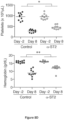
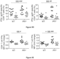
Figures 5A-5C demonstrate that IL-33R blockade reduces the frequency of T cells capable of producing IFNy. LCMV-infected perforin knockout mice were treated with either IL-33R blocking antibody (n=3) or isotype control (n=4). Onday 11, splenocytes were harvested. An aliquot was stained for T-cell markers and gp33 tetramer to look for LCMV reactive CD8+ T-cells (Figure 5A ). The rest of the cells were stimulated with PMA and ionomycin in vitro for 5 hours in the presence of brefeldin A. Cells were then fixed and stained for intracellular IFNy. The frequency of CD4+ T cells (Fig. 5B ) and CD8+ T cells (Fig. 5C ) expressing IFNy were determined. Results of unpaired student's t test are indicated. -
Figure 6 provides a graph of splenic LCMV titers, 11 days post-infection, in perforin knockout mice treated with IL-33R blocking antibody or isotype control. -
Figures 7A-7D show that MyD88-dependent signals, including IL-33, are important in the murine model of FHL2. Prf1-/-, Prf1-/-MyD88-/-, or WT mice were infected with 2 ×10⁵ PFU LCMV-Armstrong i.p. onday 0. Symbols represent individual mice.Figure 7A : Serum IFNy levels in Prf1-/- and Prf1-/-MyD88-/- mice 8 days p.i. N=3. Analyzed by 2-tailed t-test.Figure 7B : Expression of Il33 at days 0 (Uninf) and 7 p.i. (Inf). AU, arbitrary units. Analyzed by 2-way ANOVA. Significance of terms is denoted as follows: #, Genotype (WT vs. Prf1-/-); ‡,LCMV (Uninf vs. Inf); *, Interaction between Genotype and LCMV. N=1.Figure 7C : Immunohistochemical staining of IL-33 in Prf1-/- mice, shown at 200x magnification. Representative of 4 mice/group.Figure 7D : Expression of Il1rl1 (ST2 gene), analyzed as inFigure 7B . N=1. -
Figures 8A-8F show that the ST2 blockade reduces morbidity and mortality in FHL2 mice despite the persistence of LCMV. Prf1-/- mice were infected with 2 ×10⁵ PFU LCMV-Armstrong i.p. onday 0 to induce FHL. Onday 3 p.i. and every other day thereafter, mice were injected with 150 µg i.p. of either α-ST2 or Control antibody.Figure 8A : Survival of α-ST2-treated (n=9) and Control (n=10) mice. Moribund mice were euthanized according to IACUC protocol. Analyzed by log-rank test.Figure 8B : Body weight of α-ST2-treated and Control mice. Symbols represent mean ± SEM of 9-10 mice. † indicates timepoints at which Control mice died and were excluded from subsequent weight analysis. N=2. Analyzed by mixed linear effects model; significance of Interaction term (α-ST2 vs. Control over time) is indicated.Figure 8C : Body weight of mice withdrawn from α-ST2 treatment atday 18 p.i. (Withdrawal) or receiving continued α-ST2 treatment. Symbols represent mean ± SEM of 4-5 mice. † indicates timepoints at which Withdrawal mice died and were excluded from subsequent weight analysis. N=1. Analyzed by mixed linear effects model; significance of Interaction term (α-ST2 vs. Withdrawal over time) is indicated.Figure 8D : Platelet and hemoglobin levels from peripheral blood of α-ST2-treated andControl mice 2 days prior to infection and 8 days p.i. N=2. Analyzed by repeated measures 2-way ANOVA; significance of Interaction term (α-ST2 vs. Control over time) is indicated.Figure 8E : Representative H&E-stained liver sections from Control and α-ST2-treated mice, 200x magnification. Severity of microvesicular steatosis was scored onday 8 p.i. and analyzed by student's 2-tailed t-test. N=2.Figure 8F :Splenic LCMV titer 8 days p.i. in Control and α-ST2-treated mice. LCMV-infected WT mice were included for comparison. Dotted line indicates limit of detection of plaque assay. N=2. No significant differences between Control and α-ST2-treated mice for either genotype by unpaired t-test. -
Figures 9A-9E show that ST2 blockade reduces IFNy production by T cells. WT and Prf1-/- mice were infected with 2 ×10⁵ PFU LCMV-Armstrong i.p. onday 0, injected with 150 µg i.p. of either α-ST2 or Control antibody ondays day 8.Figure 9A : Serum IFNy level. N=3.Figure 9B : Numbers of splenic CD44hiCD62LloCD127lo effector T cells.Figure 9C : Numbers of splenic LCMV-specific T cells staining with gp33 MHC Class I tetramer or gp66 MHC Class II tetramer.Figure 9D : Frequency of T cells producing IFNy in response to in vitro restimulation with LCMV peptides (above background).Figure 9E : Median IFNy fluorescence intensity of IFNγ+ T cells after in vitro peptide restimulation, normalized to WT Control mean for each experiment. Analyzed by 2-way ANOVA (Fig. 9A ) or by mixed linear effects modeling of data pooled from 2-4 experiments (Figs. 9B-9E ). Significance of terms is denoted as follows: #, Genotype (WT vs. Prf1-/-); §,Treatment (Control vs. α-ST2); *, Interaction between Genotype and Treatment. - IL33 is an inflammatory cytokine that is released by normal tissues upon damage. IL33 receptors are classically thought to be on CD4+ T-cells. IL-33 has also been identified as a critical cytokine for the promotion of anti-viral CD8+ T-cell responses, acting as an "alarmin" to communicate tissue damage to the immune system and, thus, the need for a response (Bonilla et al. (2012) Science 335:984-9).
- Herein, it is shown that blocking IL33 signaling, particularly the IL33 receptor, in a murine model of HLH (perforin deficient (pfn -/- ) mice infected with LCMV) results in improvement in weight loss, survival, and a dramatic decrease in the production of the pathogenic IFNy to almost normal levels. This is accompanied by a reduction in the numbers of IFNy producing CD4+ and CD8+ T-cells. These results indicate that inhibiting or blocking the IL33 receptor and/or IL33 results in the reduction and/or prevention of morbidity and mortality in subjects with a cytokine-related disease or disorder. Blocking IL33 signaling is a novel therapeutic target in cytokine-related diseases or disorders, providing a completely different means of treatment than any therapeutic currently available or in development for the disease. Further, the targeting of IL33 will not have the side effect profile observed with current therapies such as glucocorticoids, cyclosporine A, and etoposide because it is more specific and will not inhibit the entire immune system, thereby reducing the potential for infectious side effects.
- The present disclosure encompasses a composition for use in a method of inhibiting, treating, and/or preventing a cytokine-related disease or disorder, specifically hemophagocytic lymphohistiocytosis, in a subject in need thereof. In a particular instance, the subject is undergoing an active flare (e.g., cytokine storm) of hemophagocytic lymphohistiocytosis. In some instances, the compositions of the disclosure can be co-administered (sequentially and/or simultaneously) with at least one other therapeutic for the treatment of hemophagocytic lymphohistiocytosis. For example, current therapeutic modalities for hemophagocytic lymphohistiocytosis include, without limitation, immunosuppressants, glucocorticoids, cyclosporine A, etoposide, methotrexate, IL-1 (e.g., IL-1 beta) blocking agent or blockade (e.g., anakinra (Kineret®), rilonacept, canakinumab, and gevokizumab), and bone marrow transplant.
- In a particular instance, the hemophagocytic lymphohistiocytosis is characterized by elevated IFNy levels.
- Hemophagocytic lymphohistiocytosis (also known as hemophagocytic syndrome) comprises familial (primary) hemophagocytic lymphohistiocytosis (FHL) and secondary HLH (SHLH). HLH is a hematologic disorder that is typically characterized with fever, splenomegaly, cytopenia, elevated ferritin, elevated triglycerides or decreased fibrinogen, hemophagocytosis, low NK cell activity, and/or elevated soluble IL-2 receptor. The presence of at least about five of these symptoms is typically required for diagnosis. HLH may also be characterized by other symptoms such as liver function test abnormalities, jaundice, rash, coagulopathy, lymphocytosis, and/or histiocytosis. HLH may also be screened for through detection of at least one biomarker such as, without limitation: ferritin, soluble IL-2 receptor, IFNy, complete blood count, fibrinogen, erythrocyte sedimentation rate (ESR), and/or C-reactive protein (CRP). HLH may be monitored (e.g., before, during and/or after therapy with an IL-33 pathway inhibitor) by measuring at least one of the above biomarkers or symptoms and/or monitoring at least one of blood pressure, liver function, renal function, fever, rash, and splenomegaly.
- The methods of the disclosure comprise administering an IL-33 pathway inhibitor to a subject. In a particular instance, the inhibitor is an antibody or fragment thereof immunologically specific for IL-33R (also known as
Interleukin 1 receptor-like 1 (IL1RL1) or ST2; PubMed Gene ID: 9173 provides examples of nucleic acid and amino acid sequences of IL-33R, including isoforms and variants) or IL-33 (PubMed Gene ID: 90865 provides examples of nucleic acid and amino acid sequences of IL-33, including isoforms and variants). In a particular instance, the inhibitor is a soluble form of IL-33R (e.g., the extracellular domain; see, e.g., Iwahana et al. (1999) Eur. J. Biochem., 264:397-406; Palmer et al. (2009) Arthritis Rheumat., 60:738-749; Hayakawa et al. (2007) J. Biol. Chem., 282:26369-26380). - In certain embodiments, the IL-33 pathway inhibitor is an antibody or fragment thereof, immunologically specific for IL-33R. It is contemplated that such antibodies or fragments thereof may bind IL-33R and block binding of the receptor to IL-33 or such antibodies or fragments thereof may bind the IL-33R-IL-33 complex and block the recruitment of the signaling co-receptor AcP. Examples of IL-33R antibodies include but are not limited to those described in
U.S. Patents 7,087,396 ,7,452,980 , and8,444,987 ;WO 2013/165894 ;WO 99/34217 WO 2013/173761 ; andU.S. Patent Application Publication Nos. 20130287777 ,20130336980 , and20140004107 . In a particular embodiment, the IL-33R binding protein is an antibody described inU.S. Patent Application Publication No. 20130287777 (e.g., an antibody or fragment thereof that comprises the HCDR1, the HCDR2, the HCDR3, the LCDR1, the LCDR2, and the LCDR3 of what is set forth therein as SEQ ID NOs: 97, 114, 84, 130, 90, and 134, respectively). In a particular embodiment, the IL-33R binding protein is an antibody described inU.S. Patent Application Publication No. 20140004107 (e.g., Ab1, Ab2, Ab3, Ab4, Ab5, Ab6, Ab7, Ab8, Ab9, Ab10, Ab11, Ab30, Ab32, or Ab33 or a functional fragment or derivative thereof). - In a particular instance, the IL-33 pathway inhibitor is an antibody or fragment thereof, immunologically specific for IL-33. It is contemplated that such antibodies or fragments thereof may bind IL-33 and block binding of the cytokine to IL-33R or such antibodies or fragments thereof may bind the IL-33r-IL-33 complex and block recruitment of the signaling co-receptor AcP. Examples of IL-33 binding proteins include but are not limited to those described in
U.S Patent Application Publication No. 20140271658 ,WO 2014/152195 , andWO 2014/164959 . - The antibody may be a naturally occurring antibody or may be a synthetic or modified antibody (e.g., a recombinantly generated antibody; a chimeric antibody; a bispecific antibody; a humanized antibody; a camelid antibody; and the like). The antibody may comprise at least one purification tag. In a particular embodiment, the framework antibody is an antibody fragment. Antibody fragments include, without limitation, immunoglobulin fragments including, without limitation: single domain (Dab; e.g., single variable light or heavy chain domain), Fab, Fab', F(ab')2, and F(v); and fusions (e.g., via a linker) of these immunoglobulin fragments including, without limitation: scFv, scFv2, scFv-Fc, minibody, diabody, triabody, and tetrabody. The antibody may also be a protein (e.g., a fusion protein) comprising at least one antibody or antibody fragment. In a particular embodiment of the invention, the antibody is a monoclonal antibody or fragment thereof. The antibody may also be a synthetic protein which mimics an immunoglobulin. Examples include, without limitation, Affibody® molecules (Affibody, Bromma, Sweden), darpins (designed ankyrin repeat proteins; Kawe et al. (2006) J. Biol. Chem., 281:40252-40263), and peptabodies (Terskikh et al. (1997) PNAS 94:1663-1668).
- The antibodies of the invention may be further modified. For example, the antibodies may be humanized. In a particular embodiment, the hybrid antibodies (or a portion thereof) are inserted into the backbone of an antibody or antibody fragment construct. For example, the variable light domain and/or variable heavy domain of the antibodies of the instant invention may be inserted into another antibody construct. Methods for recombinantly producing antibodies are well-known in the art. Indeed, commercial vectors for certain antibody and antibody fragment constructs are available.
- The antibody molecules of the invention may be prepared using a variety of methods known in the art. Polyclonal and monoclonal antibodies may be prepared as described in Current Protocols in Molecular Biology, Ausubel et al. eds. Antibodies may be prepared by chemical cross-linking, hybrid hybridoma techniques and by expression of recombinant antibody fragments expressed in host cells, such as bacteria or yeast cells. In one instance of the disclosure, the antibody molecules are produced by expression of recombinant antibody or antibody fragments in host cells. The nucleic acid molecules encoding the antibody may be inserted into expression vectors and introduced into host cells. The resulting antibody molecules are then isolated and purified from the expression system. The antibodies optionally comprise a purification tag by which the antibody can be purified. The purity of the antibody molecules of the invention may be assessed using standard methods known to those of skill in the art, including, but not limited to, ELISA, immunohistochemistry, ion-exchange chromatography, affinity chromatography, immobilized metal affinity chromatography (IMAC), size exclusion chromatography, polyacrylamide gel electrophoresis (PAGE), western blotting, surface plasmon resonance and mass spectroscopy.
- Compositions comprising an inhibitor of the IL-33 pathway for use of the invention are also encompassed by the disclosure. The composition may further comprise a pharmaceutically acceptable carrier. The compositions may also comprise at least one other therapeutic as described hereinabove.
- The IL-33 pathway inhibitor(s) (or composition(s) comprising the same) can be administered by any suitable route, for example, by injection (e.g., for local, direct, or systemic administration), oral, pulmonary, topical, nasal or other modes of administration. The composition may be administered by any suitable means, including parenteral, intramuscular, intravenous, intravascular, intraarterial, intraperitoneal, subcutaneous, topical, inhalatory, transdermal, intrapulmonary, intraareterial, intrarectal, intramuscular, and intranasal administration. In a particular instance, the composition is administered intraperitoneally. In general, the pharmaceutically acceptable carrier of the composition is selected from the group of diluents, preservatives, solubilizers, emulsifiers, adjuvants and/or carriers. The compositions can include diluents of various buffer content (e.g., Tris HCl, acetate, phosphate), pH and ionic strength; and additives such as detergents and solubilizing agents (e.g., polysorbate 80), anti oxidants (e.g., ascorbic acid, sodium metabisulfite), preservatives (e.g., Thimersol, benzyl alcohol) and bulking substances (e.g., lactose, mannitol). The compositions can also be incorporated into particulate preparations of polymeric compounds such as polyesters, polyamino acids, hydrogels, polylactide/glycolide copolymers, ethylenevinylacetate copolymers, polylactic acid, polyglycolic acid, etc., or into liposomes. Such compositions may influence the physical state, stability, rate of in vivo release, and rate of in vivo clearance of components of a pharmaceutical composition of the present invention (see, e.g., Remington's Pharmaceutical Sciences and Remington: The Science and Practice of Pharmacy). The pharmaceutical composition of the disclosure can be prepared, for example, in liquid form, or can be in dried powder form (e.g., lyophilized for later reconstitution).
- The therapeutic agents described herein will generally be administered to a patient as a pharmaceutical preparation. The term "patient" as used herein refers to human or animal subjects. The compositions of the disclosure may be employed therapeutically or prophylactically, under the guidance of a physician.
- The compositions comprising the agent of the invention may be conveniently formulated for administration with any pharmaceutically acceptable carrier(s). The concentration of agent in the chosen medium may be varied and the medium may be chosen based on the desired route of administration of the pharmaceutical preparation. Except insofar as any conventional media or agent is incompatible with the agent to be administered, its use in the pharmaceutical preparation is contemplated.
- The dose and dosage regimen of the agent according to the invention that is suitable for administration to a particular patient may be determined by a physician considering the patient's age, sex, weight, general medical condition, and the specific condition for which the agent is being administered to be treated or prevented and the severity thereof. The physician may also take into account the route of administration, the pharmaceutical carrier, and the agent's biological activity. Selection of a suitable pharmaceutical preparation will also depend upon the mode of administration chosen.
- A pharmaceutical preparation of the disclosure may be formulated in dosage unit form for ease of administration and uniformity of dosage. Dosage unit form, as used herein, refers to a physically discrete unit of the pharmaceutical preparation appropriate for the patient undergoing treatment or prevention therapy. Each dosage should contain a quantity of active ingredient calculated to produce the desired effect in association with the selected pharmaceutical carrier. Procedures for determining the appropriate dosage unit are well known to those skilled in the art.
- Dosage units may be proportionately increased or decreased based on the weight of the patient. Appropriate concentrations for alleviation or prevention of a particular condition may be determined by dosage concentration curve calculations, as known in the art.
- The pharmaceutical preparation comprising the agent may be administered at appropriate intervals until the pathological symptoms are reduced or alleviated, after which the dosage may be reduced to a maintenance level. The appropriate interval in a particular case would normally depend on the condition of the patient. Toxicity and efficacy (e.g., therapeutic, preventative) of the particular formulas described herein can be determined by standard pharmaceutical procedures such as, without limitation, in vitro, in cell cultures, ex vivo, or on experimental animals. The data obtained from these studies can be used in formulating a range of dosage for use in human. The dosage may vary depending upon form and route of administration. Dosage amount and interval may be adjusted individually to levels of the active ingredient which are sufficient to deliver a therapeutically or prophylactically effective amount.
- The following definitions are provided to facilitate an understanding of the present invention:
The singular forms "a," "an," and "the" include plural referents unless the context clearly dictates otherwise. - As used herein, the terms "host," "subject," and "patient" refer to any animal, particularly mammals including humans.
- "Pharmaceutically acceptable" indicates approval by a regulatory agency of the Federal or a state government or listed in the U.S. Pharmacopeia or other generally recognized pharmacopeia for use in animals, and more particularly in humans.
- A "carrier" refers to, for example, a diluent, adjuvant, preservative (e.g., Thimersol, benzyl alcohol), anti-oxidant (e.g., ascorbic acid, sodium metabisulfite), solubilizer (e.g., polysorbate 80), emulsifier, buffer (e.g., Tris HCl, acetate, phosphate), antimicrobial, bulking substance (e.g., lactose, mannitol), excipient, auxiliary agent or vehicle with which an active agent of the present invention is administered. Pharmaceutically acceptable carriers can be sterile liquids, such as water and oils, including those of petroleum, animal, vegetable or synthetic origin. Water or aqueous saline solutions and aqueous dextrose and glycerol solutions may be employed as carriers, particularly for injectable solutions. Suitable pharmaceutical carriers are described in "Remington's Pharmaceutical Sciences" by E.W. Martin (Mack Publishing Co., Easton, PA); Gennaro, A. R., Remington: The Science and Practice of Pharmacy, (Lippincott, Williams and Wilkins); Liberman, et al., Eds., Pharmaceutical Dosage Forms, Marcel Decker, New York, N.Y.; and Kibbe, et al., Eds., Handbook of Pharmaceutical Excipients, American Pharmaceutical Association, Washington.
- The term "treat" as used herein refers to any type of treatment that imparts a benefit to a patient afflicted with a disease, including improvement in the condition of the patient (e.g., in one or more symptoms), delay in the progression of the condition, etc.
- As used herein, the term "prevent" refers to the prophylactic treatment of a subject who is at risk of developing a condition (e.g., hemophagocytic lymphohistiocytosis or chimeric antigen receptor T-cell cytokine release syndrome) resulting in a decrease in the probability that the subject will develop the condition.
- A "therapeutically effective amount" of a compound or a pharmaceutical composition refers to an amount effective to prevent, inhibit, or treat a particular disorder or disease and/or the symptoms thereof. For example, "therapeutically effective amount" may refer to an amount sufficient to modulate a cytokine related disease or disorder in a subject.
- As used herein, the term "subject" refers to an animal, particularly a mammal, particularly a human.
- An "antibody" or "antibody molecule" is any immunoglobulin, including antibodies and fragments thereof, that binds to a specific antigen. As used herein, antibody or antibody molecule contemplates intact immunoglobulin molecules, immunologically active portions of an immunoglobulin molecule, and fusions of immunologically active portions of an immunoglobulin molecule.
- As used herein, the term "immunologically specific" refers to proteins/polypeptides, particularly antibodies, that bind to one or more epitopes of a protein or compound of interest, but which do not substantially recognize and bind other molecules in a sample containing a mixed population of antigenic biological molecules.
- As used herein, the term "small molecule" refers to a substance or compound that has a relatively low molecular weight (e.g., less than 4,000, less than 2,000, particularly less than 1 kDa or 800 Da). Typically, small molecules are organic, but are not proteins, polypeptides, or nucleic acids, though they may be amino acids or dipeptides.
- The following examples are illustrative of the practice of the instant invention.
- Given the extensive tissue damage in HLH and the known role of CD8+ T-cells in the disease process, IL-33 signaling may be playing a heretofore unrecognized role in perpetuating disease and, therefore, be a novel target for therapy. The LCMV infected perforin deficient mouse model of HLH was used to test the role of IL-33. Notably, MyD88 and perforin double deficient mice were found to be protected from disease. Since IL-33 is known to signal through MyD88, this result supports a role for IL-33 in HLH.
- In all experiments, mice were infected with 2 × 10⁵ PFU of LCMV. Starting on
day 3 following infection, mice were either treated with an IL-33 receptor neutralizing antibody (IL-33RB) (an antibody that was generated against the extracellular domain of ST2; see, e.g., Palmer et al. (2009) Arthritis Rheumat., 60:738-749) or isotype control antibody every other day (150 µg/dose i.p.). - Mice receiving IL-33RB treatment showed a survival advantage with a hazard ratio of 4 (p=0.06, log-rank test) (
Figure 1 ). Consistent with improved survival, mice treated with IL-33RB therapy showed a significant benefit in weight loss reduction (p=0.01, repeated measures 2-way ANOVA) in the initial phases of the disease (Figure 2 ). Furthermore, IL-33RB treated mice showed a reduction in liver parenchyma damage (Figure 3 ), but no reduction in inflammatory infiltrate. Il-33RB treated mice also showed a reduction/prevention of liver steatosis. - Given the known role of IFNγ in the pathogenesis of HLH, serum IFNy levels were checked at
day 11 post-infection of these mice. Serum IFNy was highly significantly reduced over 16 fold in mice receiving IL-33RB treatment (p=0.0005, t-test) (Figure 4 ). Thus, IL-33RB treatment shows an improvement in survival, weight loss, and hepatic injury, all concordant with a dramatic decrease in pathogenic serum IFNy levels. Interestingly, despite improved survival, IL-33RB therapy did not affect the degree of the pan-cytopenia seen in these mice. - Immunosuppressive therapy is already in use for patients with HLH. However, such treatment protocols present treatment associated morbidity and mortality, particularly because the immune system is being suppressed in patients likely suffering from an active infection that triggered the HLH episode in the first place. In the case of wild type mice infected with LCMV, IL-33RB treatment results in a loss of antigen specific effector CD8+ T-cells (Bonilla et al. (2012) Science 335:984-9). This would not necessarily be desirable in patients trying to fight off an infection. Therefore, the effect of the IL-33RB blockade on the numbers of anti-LCMV CD8+ T-cells in the context of HLH was assessed using a tetramer specific for the LCMV immunodominant epitope gp33. Surprisingly, IL-33RB treatment did not cause a loss of gp33 specific CD8+ T-cells (
Figure 5A ). However, IL-33RB did cause a reduction in the numbers of IFNy producing CD8+ and CD4+ T-cells (Figure 5B-C ), consistent with the reduction of IFNy in the serum. Significantly, these levels of IFNy producing cells and the level of IFNy in the serum are not reduced to zero, indicating that IL-33RB treatment may not eliminate all protection from infection, but rather reduce the IFNy response of effector cells to a less toxic level to the organism. Thus, IL-33R blockade reduces serum IFNy without abrogating the antigen-specific immune response. - As seen in
Figure 6 , at 11 days post-infection, perforin KO mice treated with IL-33R blocking antibody have equivalent splenic titers of LCMV, compared to isotype-treated controls. This is consistent with the similar frequencies of LCMV-specific CD8+ T cells between the two treatment groups and shows that IL-33R blockade preserves the anti-viral immune response while reducing excessive inflammation. - Seven- to nine-week old male and female mice were used for these studies. C57BL/6 (WT) and perforin-deficient (C57BL/6-Prf1tm1Sdz/J, referred to as Prf1 -/- ) mice were purchased from The Jackson Laboratory. MyD88-/- mice were obtained from The Children's Hospital of Philadelphia, Philadelphia, PA. MyD88-/- mice were crossed to Prf1 -/- mice (Adachi et al. (2000) Immunity 9(1): 143-150). All mice were bred on a C57BL/6 background and housed in an animal facility certified by the Association for Assessment and Accreditation of Laboratory Animal Care.
- Mice were infected i.p. with 2×10⁵ PFU LCMV-Armstrong on
day 0. Mice were weighted and assessed daily for signs of morbidity. Mice were euthanized when moribund, when >20% starting body weight had been lost acutely (survival experiment), or when >30% starting body weight had been lost over several weeks (withdrawal experiment), according to our IACUC protocol. Peripheral blood was obtained by cheek bleed and complete blood cell counts were performed on a Hemavet® analyzer (Drew Scientific). Serum IFNy levels were measured by ELISA (BD Biosciences).Viral titers 8 days post-infection (p.i.) were measured by plaque assay (Ahmed et al. (1984) J. Exp. Med., 160(2):521-540). - Organ sections from uninfected or LCMV-infected (7 days p.i.) WT and Prf1 -/- mice were preserved in RNAlater® (Qiagen) and stored at -20°C. Tissues were homogenized in RLT lysis buffer (Qiagen) using a stator-rotor tissue homogenizer and RNA was isolated using RNeasy® Mini kit (Qiagen). cDNA was made using the SuperScript® III First-Strand Synthesis System (Life Technologies) according to the manufacturer's instructions. qRT-PCR was performed using QuantiTect® primers for Actb, Il33, and Il1rl1 (Qiagen) and Power SYBR® Green master mix (LifeTechnologies) on a StepOnePlus™ Real Time PCR System (Applied Biosystems). Results were normalized to β-actin using the ΔΔCT method.
- Unperfused liver sections were fixed overnight in 4% paraformaldehyde and embedded in paraffin. Liver sections were stained with H&E and were read by a pediatric pathologist (PAK) blinded to treatment protocols. Microvesicular steatosis was assessed using a standardized scoring system as follows: 0 - absent, 1 - 1-20% of area per 20X objective high power field, 2 - 21-40%, 3 - 41-60%, 4 - 61-80%, 5- 81-100%. Lobular inflammation was scored as number of foci of lobular inflammation per 20X objective high power field in the most inflamed area. IL-33 immunohistochemistry was performed on slides after antigen retrieval with a pressure cooker. Mouse IL-33 Affinity Purified Polyclonal Ab was obtained from R&D Systems. Detection was performed using the Vectastain® ABC (rat) Kit (Vector Laboratories), followed by DAB reagent (Dako).
- Rat IgG1 anti-mouse ST2-blocking antibody (referred to as α-ST2 antibody) and murine IgG1 isotype control peptibody (referred to as Control antibody) were obtained from Amgen (Palmer et al. (2009) Arthritis Rheumatism 60(3):738-749). LCMV-infected mice were injected i.p. with 150 µg α-ST2 antibody or 150 µg control antibody on
days day 8 for analysis. For survival experiments, mice were injected with α-ST2 or control antibody beginning onday 3 and continuing every other day thereafter. For the withdrawal experiment, mice that had been treated with α-ST2 antibody until day 17 either continued to receive α-ST2 antibody every other day until day 29 or were switched to control antibody treatment at day 19. - Spleens were harvested, homogenized, and passed through a 70-µm filter to generate single-cell suspensions. Red blood cells were lysed using ACK (ammoniumchloride-potassium) Lysing Buffer (Lonza). Cell concentrations were assessed using a hemocytometer or a Countess® Automated Cell Counter (Life Technologies). Cells were stained with LIVE/DEAD® fixable viability dye (Life Technologies) in PBS for 30 minutes at 4°C, washed twice, and stained with CD4, CD8α, CD44, CD45.1, CD62L, CD90.2, and/or CD127 fluorochrome-tagged antibodies (BD Pharmingen, eBioscience, BioLegend, and Miltenyi Biotec) in
PBS 2% FBS for 30 minutes at 4°C. H-2DbGP33-41 and I-AbGP66-77 MHC-peptide complexes were provided as fluorophore-conjugated tetramers by the NIH Tetramer Core Facility. All samples were acquired on a MACSQuant® flow cytometer (Miltenyi Biotec) and analyzed using FlowJo software version 9.6 (Tree Star). - For intracellular cytokine analysis, 106 splenocytes were cultured in the absence or presence of LCMV GP33 peptide (0.2 µg/ml, GenScript) or LCMV GP61 peptide (1.0 µg/ml, Anaspec) and brefeldin A (Sigma) for 5 hours at 37°C. Following staining with LIVE/DEAD® and for surface antigens as described above, cells were stained for IFNy (clone XMG1.2) using the Cytofix/Cytoperm™ kit (BD Bioscience) according to the manufacturer's instructions. To minimize carry-over of viral antigen in 2 of 3 experiment replicates, 0.3×10⁶ T cells purified from LCMV-infected splenocytes by negative magnetic bead selection were plated with 0.7×10⁶ uninfected C57BL/6.SJL splenocytes depleted of T cells by magnetic bead separation (Miltenyi Biotec). CD45.1+ cells were excluded from subsequent flow analysis. Results were comparable with or without this additional T cell isolation step.
- Data were plotted and analyzed using Prism 5.0 (GraphPad Software). Symbols in figures represent individual mice, unless otherwise stated. Statistical significance was tested using two-way analysis of variance (ANOVA), Student's unpaired 2-tailed t-test, repeated-measures 2-way ANOVA, log-rank (Mantel-Cox) test, or linear mixed effects model, as appropriate. Weight loss data were analyzed by linear mixed effects models to allow for missing data due to mouse mortality. T cell data were also analyzed by linear mixed effects models to account for baseline variability between experimental replicates. For these analyses, R (R Core Team, 2014) and Ime4 (Bates et al. (2014) lme4: Linear mixed-effects models using Eigen and S4, arXiv: 1406. 5823v 1) were used to perform a linear mixed effects analysis. For weight loss experiments, treatment and body weight were modeled as fixed effects, and individual mice were treated as a random effect to account for baseline variability between animals (e.g., intercept only). For T cell data, treatment and genotype were modeled as fixed effects, and experiment was treated as a random effect to account for baseline variability between experimental replicates (e.g., intercept only). Visual inspection of residual plots did not reveal any obvious deviations from homoscedasticity or normality. P-values were obtained by likelihood ratio tests of the full model with the effect in question against the model without the effect in question. The method of Levy (Levy R. Using R formulae to test for main effects in the presence of higher-order interactions. 2014; arXiv:stat.ME/1405.2094v1) was used to obtain tests of main effects while modeling an interaction effect. Unless otherwise specified, P values are represented in figures by number of symbols (e.g. * P<0.05, ** P<0.01, *** P<0.001).
- All animal studies were performed with the approval of The Children's Hospital of Philadelphia IACUC.
- Work in the related Jinx model of
FHL type 3 has implicated a role for MyD88 signaling in FHL pathophysiology (Krebs et al. (2011) Blood 117(24):6582-6588). To confirm the importance of MyD88 signaling in the murine model of FHL2, the response of Prf1-/- and Prf1-/-MyD88-/- mice to LCMV infection were compared. Similar to findings in Jinx mice, MyD88 deficiency protected Prf1-/- mice from death, anemia, thrombocytopenia, and hepatic inflammation. Consistent with these reduced FHL disease parameters, Prf1-/-MyD88-/- mice had significantly decreased levels of serum IFNy (Figure 7A ) and frequencies of CD8+ T cells specific for the immunodominant LCMV epitope gp33. These results indicate that non-TCR signaling pathways such as MyD88 are important for promoting disease in the FHL2 murine model. - To further delineate the signaling mediators upstream of MyD88 that contribute to the development of FHL, signaling through receptors of the IL-1 family, which includes IL-1α, IL-1β, IL-18, and IL-33, were studied. While in vivo blockade of either IL-1 or IL-18 signaling has no effect on FHL mortality (Jordan et al. (2004) Blood 104(3):735-743; Jessen et al. (2013) Front. Immunol., 4:448), the role of IL-33 in FHL has not previously been investigated. IL-33 is constitutively expressed in the nuclei of epithelial cells, endothelial cells, fibroblasts, and other non-hematopoietic cells, and is released upon cellular stress or necrosis (Kakkar et al. (2012) J. Biol. Chem., 287(9):6941-6948; Moussion et al. (2008) PloS one 3(10):e3331; Pichery et al. (2012) J. Immunol., 188(7):3488-3495). Extracellular IL-33 subsequently signals by binding to its receptor (the ST2/IL1RAP complex), which is expressed by a diverse range of immune cells (Schmitz et al. (2005) Immunity 23(5):479-490; Chackerian et al. (2007) J. Immunol., 179(4):2551-2555; Mirchandani et al. (2012) Trends Immunol., 33(8):389-396). IL-33 is thus classified as an alarmin, in that it activates an inflammatory response in the context of tissue damage (Moussion et al. (2008) PloS one 3(10):e3331; Luthi et al. (2009) Immunity 31(1):84-98; Cayrol et al. (2009) PNAS 106(22):9021-9026). Furthermore, IL-33 promotes anti-viral CD8+ T cell differentiation and function (Yang et al. (2011) Eur. J. Immunol., 41(11):3351-3360; Bonilla et al. (2012) Science 335(6071):984-989). These facts led us to the conclusion that IL-33 signaling upstream of MyD88 contributes to FHL inflammation.
- It was first determined whether IL-33 and its receptor are expressed in the organs most affected by FHL in LCMV-infected Prf1 -/- mice (referred to as FHL2 mice). While hepatic expression of Il33 remained stable in both WT and Prf1-/- mice following LCMV infection, splenic Il33 was greatly upregulated in these mice after viral infection (
Figure 7B ). Notably, Il33 expression was highest in the spleens of FHL2 mice; given the rough correlation demonstrated between splenic Il33 mRNA and LCMV titers in WT mice (Bonilla et al. (2012) Science 335(6071):984-989), this elevation may reflects the sustained viremia in FHL2 mice (Lykens et al. (2011) Blood 118(3):618-626; Matloubian et al. (1999) J. Virol., 73(3):2527-2536). Immunohistochemical analysis confirmed nuclear localization of IL-33 in cells of both the livers and spleens of WT and Prf1 -/- mice (Figure 7C ). Although the spatial distribution and number of IL-33+ cells did not change substantially after infection, IL-33-expressing cells in LCMV-infected tissue exhibited larger, rounder nuclei, consistent with a more activated status (Figure 7C ). Strikingly, expression of the specific component of the IL-33 receptor, ST2 (encoded by Il1rl1), was greatly increased in both the spleens and livers of FHL2 mice (Figure 7D ). Together, these results show that IL-33 and ST2 are highly expressed in FHL2 mice. - To probe the role of IL-33 in murine FHL, IL-33 signaling was disrupted in FHL2 mice by administration of an ST2-blocking antibody (α-ST2) and compared their survival to that of FHL2 mice receiving an isotype control antibody (Control). Remarkably, α-ST2-treated mice were significantly protected from mortality and severe weight loss compared to Control mice (
Figures 8A, 8B ). Continual ST2 blockade enabled mice to survive at least 30 days post-infection (p.i.) and limited weight loss even later in the course of disease (Figure 8C ). Additionally, α-ST2-treated mice showed less severe hematologic abnormalities, as anemia and thrombocytopenia were mitigated in these mice compared to Controls (Figure 8D ). Althoughα-ST2-treated and Control mice demonstrated similar levels of lobular inflammation in the liver, ST2 blockade significantly reduced hepatic parenchymal damage (Figure 8E ). The microvesicular steatosis evident in livers of Control but not α-ST2-treated mice indicates severe, acute metabolic impairment (Jaeschke et al. (2002) Toxicolog. Sci., 65(2):166-176) and indicates worsened disease in Control mice. Despite multiple improved disease parameters in α-ST2-treated FHL2 mice, both treatment groups showed equivalently high LCMV titers in thespleen 8 days p.i. (Figure 8F ). Thus, disruption of IL-33 signaling reduces morbidity and mortality in murine FHL despite the persistence of virus at high titers. These results indicate that rather than modulating the infection, IL-33 instead modulates the pathologic immune response. - IL-33, in combination with IL-12, can induce IFNy production by T cells and NK cells, and IL-33 is crucial for promoting CD8+ T cell effector functions such as cytokine production (Yang et al. (2011) Eur. J. Immunol., 41(11):3351-3360; Bonilla et al. (2012) Science 335(6071):984-989; Bourgeois et al. (2009) Eur. J. Immunol., 39(4): 1046-1055). Given the central role of IFNγ in FHL pathophysiology, ST2 blockade may reduce disease severity in FHL2 mice by diminishing systemic levels of IFNy. α-ST2 treatment significantly decreased serum IFNy in LCMV-infected WT mice, but its effect on FHL2 mice was far more striking, with a 16-fold reduction in IFNy levels in these mice (
Figure 9A ). These data demonstrate that ST2 blockade critically regulates a major pathogenic factor in FHL and indicate that IL-33 signaling is upstream of IFNγ production in FHL. - To determine how disruption of IL-33 signaling reduces systemic levels of IFNy, the frequency and IFNy production capacity of the lymphocyte populations was examined in this model. ST2 blockade decreased the number of CD44hiCD62LloCD127lo effector CD8+T cells in FHL2mice (2.4-fold reduction), although not to the degree seen in LCMV-infected WT mice (5.4-fold reduction) (
Figure 9B ). Effector CD4+ T cells were similarly reduced by α-ST2 treatment (1.7-fold reduction in FHL2 mice, 2.2-fold in LCMV-infected WT mice) (Figure 9B ). Numbers of CD44hiCD62LhiCD127hi memory-phenotype cells were only slightly altered by α-ST2 treatment in FHL2 mice (1.2-fold reduction in CD8+ T cells) or not at all (1.01-fold increase in CD4+ T cells). Numbers of NK cells were unaffected by ST2 blockade. Among effector-phenotype T cells, lower numbers of LCMV gp33-specific CD8+ T cells were found in α-ST2-treated FHL2 and LCMV-infected WT mice (Figure 9C ). Although the interaction between genotype and treatment did not reach statistical significance for numbers of gp33-specific CD8+ T cells (p=0.054), there was a trend towards a greater α-ST2-mediated reduction of these cells in LCMV-infected WT mice (5.9-fold) than in FHL2 mice (2.1-fold) (Figure 9C ). Interestingly, LCMV gp61-specific CD4+ T cells were reduced 3.5-fold by α-ST2 treatment in FHL2 mice only (Figure 9C ). Together, these findings indicate that IL-33 promotes the expansion and/or differentiation of LCMV-specific effector cells, which are known to be pathogenic in FHL2 mice. Furthermore, the magnitude of the suppressive effect mediated by ST2 blockade on effector CD4+ and CD8+ T cells is different between LCMV-infected WT and FHL2 mice, highlighting the aberrations in T cell regulation characteristic of FHL. - It was then investigated whether IL-33 signaling affects the ability of LCMV-specific effector cells to produce IFNy by performing intracellular cytokine staining on T cells from LCMV-infected WT and FHL2 mice receiving either α-ST2 or Control antibody. Control FHL2 mice demonstrated higher frequencies of IFNγ+ gp33-specific CD8+ T cells than Control LCMV-infected WT mice (
Figure 9D ) (Jordan et al. (2004) Blood 104(3):735-743; Lykens et al. (2011) Blood 118(3):618-626). Remarkably, in vivo ST2 blockade significantly reduced the frequency of IFNγ+ gp33-specific CD8+ T cells in both FHL2 and LCMV-infected WT mice to a similar degree (1.7- and 2.7-fold reductions, respectively) (Figure 9D ). Similar results were seen for CD4+ T cells, with α-ST2 treatment inducing a 2.7-fold reduction in IFNγ+ gp61-specific CD4+ T cells in FHL2 mice and a 2.2-fold reduction in LCMV-infected WT mice (Figure 9D ). Furthermore, ST2 blockade reduced IFNy responses byCD8+ and CD4+ T cells from FHL2 mice across a range of LCMV peptide concentrations. Additionally, significant reductions in IFNy median fluorescence intensity (MFI) mediated by α-ST2 treatment in both IFNy+ LCMV-specific CD8+ and CD4+ T cells were found (Figure 9E ), indicating that in addition to reducing the frequency of IFNy-producing cells, blockade of IL-33 signaling reduces the average per-cell production of this cytokine. Together, the decreased frequency of IFNγ+ LCMV-specific CD4+ and CD8+ T cells and the reduced per-cell IFNy production by these cells accounts for the low systemic IFNy in α-ST2-treated FHL2 mice. - Thus, the data provided herein show a role for IL-33 signaling in FHL pathophysiology. These data demonstrate that adjuvant-like TCR-independent signals critically contribute to the hyperinflammation of FHL and indicate a revised model for this disease, in which excessive antigen is not the only stimulus driving immune dysregulation. The data point to an additional pathway of IL-33, likely released from damaged tissue, promoting T cell-mediated production of IFNy, which continues to drive the feed-forward loop of immunopathologic inflammation in FHL.
- This pathogenic IL-33/IFNγ axis provides an important point of manipulation for the treatment of FHL. While in vivo modulation of numerous pathways (including IL-1α, IL-1β, IL-10, IL-12, IL-18, TNFα, M-CSF, and GM-CSF signaling) has failed to demonstrate any therapeutic benefit in FHL mice, IFNy and now IL-33 stand out as the only cytokines whose blockade significantly improves survival and disease severity. The data provide evidence for ST2 blockade as a viable therapeutic strategy in FHL. Disruption of IL-33 signaling reduces systemic IFNy to sub-lethal levels without rendering FHL2 mice completely deficient in this key anti-viral cytokine (
Figure 9A ); accordingly, viral control is not worsened in these mice (Figure 8F ). This approach may be safer than targeting IFNy directly, as patients with disrupted IFNy signaling show increased susceptibility to infection (Browne et al. (2010) Curr. Opin. Allergy Clin. Immunol., 10(6):534-541; Susan et al. (2000) Cytokine Growth Factor Rev., 11(4):321333). Furthermore, recently described cases of FHL occurring in patients with IFNy receptor deficiency highlight the need for therapeutics designed to target a diverse range of pathways in FHL (Tesi et al. (2015) J. Allergy Clinical Immunol., 10.1016/j.jaci.2014.11.030). - Given the large degree of tissue damage observed in FHL2 mice (Jordan et al. (2004) Blood 104(3):735-743), it is likely that bioactive IL-33 is released by necrotic cells, consistent with its described role as an alarmin (Moussion et al. (2008) PloS one 3(10):e3331). However, LCMV is a non-cytopathic virus (Kägi et al. (1996) Curr. Opin. Immunol., 8(4):472-477), suggesting that direct viral effects are not responsible for the tissue necrosis observed in this model. Rather, it is likely that injury to cells results from the hyperactivated immune response, potentially via reactive oxygen species released by the activated macrophages that feature prominently in FHL2 mice (Jordan et al. (2004) Blood 104(3):735-743). Another possibility is that the coagulopathy present in FHL2 mice gives rise to thromboembolicischemia, leading to cellular necrosis. Recent reports have also highlighted the ability of non-lethal cellular stressors to induce secretion of IL-33 (Kakkar et al. (2012) J. Biol. Chem., 287(9):6941-6948; Kouzaki et al. (2011) J. Immunol., 186(7):4375-4387), an additional mechanism that may account for IL-33 release.
- The findings reveal an ST2 signaling requirement of CD8+ and CD4+ T cells for IFNy overproduction in FHL2 mice (
Figures 9D, 9E ). Although WT CD8+ T cells upregulate ST2 after LCMV infection and their differentiation and function is regulated by IL-33 in a cell-intrinsic manner (Bonilla et al. (2012) Science 335(6071):984-989), the data presented here demonstrate differential effects of ST2 blockade in FHL2 mice. A wide range of immune cell subsets - including NK cells, macrophages, and dendritic cells - expresses ST2 (Garlanda et al. (2013) Immunity 39(6): 1003-1018), suggesting the possibility of an intermediate IL-33-responsive cell in this pathway. - Based on the decisive role that IL-33 plays in promoting dysregulated immune responses in FHL2, IL-33 may also be an important driver of inflammation in other cytokine storm disorders. IL-33 may play a role in endotoxic shock (Oboki et al. (2010) PNAS 107(43):18581-18586) and hantavirus infection (Zhang et al. (2015) PLoS neglected tropical diseases 9(2):e0003514). Given the high degree of tissue damage evident in such hyperinflammatory diseases, systemic release of IL-33 may provide an additional amplifying signal that contributes to the feed-forward mechanism of immune dysregulation. Thus, α-ST2 therapy would benefit a broader range of immune-mediated diseases than the Th2-mediated disorders with which IL-33 is classically identified. In summary, it has been shown that disruption of IL-33 signaling in the murine model of FHL reduces T cell-mediated production of IFNy, leading to improved morbidity and mortality, and indicates blockade of this pathway as a viable treatment strategy for FHL.
Claims (7)
- An IL-33 pathway inhibitor for use in a method of inhibiting, treating, and/or preventing hemophagocytic lymphohistiocytosis in a subject,
wherein said IL-33 pathway inhibitor is selected from an antibody or fragment thereof immunologically specific for IL-33 receptor (IL-33R) or IL-33 and a soluble form of IL-33R. - An IL-33 pathway inhibitor for use according to claim 1, wherein said IL-33 pathway inhibitor is an antibody or fragment thereof immunologically specific for IL-33 receptor (IL-33R) or IL-33.
- An IL-33 pathway inhibitor for use according to claim 2, wherein said IL-33 pathway inhibitor is an antibody or fragment thereof immunologically specific for IL-33R.
- An IL-33 pathway inhibitor for use according to any one of claims 1-3, wherein said hemophagocytic lymphohistiocytosis has elevated levels of IFNγ.
- An IL-33 pathway inhibitor for use according to claim 1, wherein said IL-33 pathway inhibitor is a soluble form of IL-33R.
- An IL-33 pathway inhibitor for use according to claim 1, wherein said hemophagocytic lymphohistiocytosis is familial (primary) hemophagocytic lymphohistiocytosis (FHL).
- An IL-33 pathway inhibitor for use according to claim 1, wherein said hemophagocytic lymphohistiocytosis is secondary hemophagocytic lymphohistiocytosis (SHLH).
Applications Claiming Priority (4)
| Application Number | Priority Date | Filing Date | Title |
|---|---|---|---|
| US201461982026P | 2014-04-21 | 2014-04-21 | |
| US201461993495P | 2014-05-15 | 2014-05-15 | |
| US201562146684P | 2015-04-13 | 2015-04-13 | |
| PCT/US2015/026840 WO2015164354A1 (en) | 2014-04-21 | 2015-04-21 | Compositions and methods for treating cytokine-related disorders |
Publications (3)
| Publication Number | Publication Date |
|---|---|
| EP3134120A1 EP3134120A1 (en) | 2017-03-01 |
| EP3134120A4 EP3134120A4 (en) | 2017-08-30 |
| EP3134120B1 true EP3134120B1 (en) | 2024-01-24 |
Family
ID=54333080
Family Applications (1)
| Application Number | Title | Priority Date | Filing Date |
|---|---|---|---|
| EP15782489.7A Active EP3134120B1 (en) | 2014-04-21 | 2015-04-21 | Compositions and methods for treating cytokine-related disorders |
Country Status (4)
| Country | Link |
|---|---|
| US (1) | US10040859B2 (en) |
| EP (1) | EP3134120B1 (en) |
| CA (1) | CA2946511C (en) |
| WO (1) | WO2015164354A1 (en) |
Families Citing this family (13)
| Publication number | Priority date | Publication date | Assignee | Title |
|---|---|---|---|---|
| SG10201805934RA (en) | 2014-01-10 | 2018-08-30 | Anaptysbio Inc | Antibodies directed against interleukin-33 (il-33) |
| JP6929771B2 (en) | 2014-11-10 | 2021-09-01 | ジェネンテック, インコーポレイテッド | Anti-interleukin-33 antibody and its use |
| CN107109494B (en) | 2014-11-10 | 2023-10-27 | 豪夫迈·罗氏有限公司 | Methods of treatment and diagnosis of IL-33 mediated diseases |
| US11318163B2 (en) | 2015-02-18 | 2022-05-03 | Enlivex Therapeutics Ltd | Combination immune therapy and cytokine control therapy for cancer treatment |
| IL284985B2 (en) | 2015-02-18 | 2023-03-01 | Enlivex Therapeutics R& D Ltd | Combination immune therapy and cytokine control therapy for cancer treatment |
| US11497767B2 (en) | 2015-02-18 | 2022-11-15 | Enlivex Therapeutics R&D Ltd | Combination immune therapy and cytokine control therapy for cancer treatment |
| US11596652B2 (en) | 2015-02-18 | 2023-03-07 | Enlivex Therapeutics R&D Ltd | Early apoptotic cells for use in treating sepsis |
| EP3416661A4 (en) * | 2016-02-18 | 2020-03-04 | Enlivex Therapeutics Ltd. | Combination immune therapy and cytokine control therapy for cancer treatment |
| BR112018071276A2 (en) | 2016-04-27 | 2019-02-12 | Pfizer Inc. | anti-il-33 antibodies, compositions, methods and uses thereof |
| JOP20190093A1 (en) * | 2016-10-28 | 2019-04-25 | Lilly Co Eli | Anti-il-33 antibodies and uses thereof |
| EP3860634A4 (en) * | 2018-10-01 | 2022-08-03 | Emory University | Compositions and methods for treatment of hemophagocytic lymphohistiocytosis |
| US11739156B2 (en) * | 2019-01-06 | 2023-08-29 | The Broad Institute, Inc. Massachusetts Institute of Technology | Methods and compositions for overcoming immunosuppression |
| CN115605507A (en) | 2020-03-13 | 2023-01-13 | 基因泰克公司(Us) | Anti-interleukin-33 antibodies and uses thereof |
Family Cites Families (9)
| Publication number | Priority date | Publication date | Assignee | Title |
|---|---|---|---|---|
| GB9727172D0 (en) | 1997-12-24 | 1998-02-25 | Univ Glasgow | Reagents specific for st2l and uses therefor |
| US6323334B1 (en) | 1999-09-24 | 2001-11-27 | Millennium Pharmaceuticals, Inc. | Nucleic acid molecules encoding a 103 gene product and uses therefor |
| WO2001070817A1 (en) | 2000-03-21 | 2001-09-27 | Medical & Biological Laboratories Co., Ltd. | Monoclonal antibody and method and kit for the immunoassay of soluble human st2 with the use of the same |
| CA2686683A1 (en) | 2007-05-18 | 2008-11-27 | Medimmune, Llc | Il-33 in inflammatory disease |
| US9090694B2 (en) | 2012-04-30 | 2015-07-28 | Janssen Biotech, Inc. | ST2L antibody antagonists |
| AR091069A1 (en) | 2012-05-18 | 2014-12-30 | Amgen Inc | PROTEINS OF UNION TO ANTIGEN DIRECTED AGAINST THE ST2 RECEIVER |
| LT2872171T (en) * | 2012-07-13 | 2021-04-26 | The Trustees Of The University Of Pennsylvania | Toxicity management for anti-tumor activity of cars |
| JO3532B1 (en) | 2013-03-13 | 2020-07-05 | Regeneron Pharma | Anti-il-33 antibodies and uses thereof |
| CN105007929B (en) | 2013-03-15 | 2019-05-10 | 瑞泽恩制药公司 | IL-33 antagonist and its purposes |
-
2015
- 2015-04-21 US US15/305,272 patent/US10040859B2/en active Active
- 2015-04-21 EP EP15782489.7A patent/EP3134120B1/en active Active
- 2015-04-21 CA CA2946511A patent/CA2946511C/en active Active
- 2015-04-21 WO PCT/US2015/026840 patent/WO2015164354A1/en active Application Filing
Also Published As
| Publication number | Publication date |
|---|---|
| WO2015164354A1 (en) | 2015-10-29 |
| EP3134120A4 (en) | 2017-08-30 |
| EP3134120A1 (en) | 2017-03-01 |
| CA2946511C (en) | 2023-08-22 |
| US10040859B2 (en) | 2018-08-07 |
| US20170037134A1 (en) | 2017-02-09 |
| CA2946511A1 (en) | 2015-10-29 |
Similar Documents
| Publication | Publication Date | Title |
|---|---|---|
| EP3134120B1 (en) | Compositions and methods for treating cytokine-related disorders | |
| JP6722261B2 (en) | Therapeutic apoptotic cell preparation, process for its production and its use | |
| Kim et al. | Innate lymphoid cells responding to IL-33 mediate airway hyperreactivity independently of adaptive immunity | |
| JP6803339B2 (en) | Therapeutic pooled blood apoptotic cell preparations and their use | |
| Jang et al. | Effect of preemptive treatment with human umbilical cord blood-derived mesenchymal stem cells on the development of renal ischemia-reperfusion injury in mice | |
| US20210121500A1 (en) | Compositions comprising regulatory t cells and methods of making and using the same | |
| Young et al. | Erosion of transplantation tolerance after infection | |
| US20230340100A1 (en) | Ligands to gm-csf or gm-csf-receptor for use in leukemia in a patient having undergone allo-hct | |
| JP2022523680A (en) | B cell immunotherapy | |
| JP2021521121A (en) | Methods for treating autoimmune diseases | |
| US20230066340A1 (en) | Populations of enriched regulatory t cells and methods for producing same | |
| US20170320951A1 (en) | Methods for diagnosing and treating cd1d restricted gamma/delta t cell lymphomas | |
| EP3912996A1 (en) | Treg expressing tnf receptor type 2 play a major role in cd4+ t cell impairment during acute post-stress immunodepression | |
| Liu et al. | Cytosine-Phosphorothionate-Guanine Oligodeoxynucleotides Exacerbates Hemophagocytosis by Inducing Tumor Necrosis Factor–Alpha Production in Mice after Bone Marrow Transplantation | |
| Cabrera-Pérez | Examining helper T-cell recovery after sepsis | |
| Shivshankar | Modulation of bacterial pathogenesis by oppressive aging factors: insights into host-pneumococcal interaction strategies | |
| Trent | Pulmonary vascular malfunction and inflammation during Orientia tsutsugamushi infection | |
| JP2024510649A (en) | Compositions containing regulatory T cells and methods of using the same | |
| Goncalves | Defining the mechanisms underlying reduced immunity to Streptococcus pneumonia with age | |
| Cromvik | Graft-versus-Host Disease: Eosinophils, Chimerism and Clinical Features in Patients Undergoing Allogeneic Hematopoietic Stem Cell or Multivisceral Transplantation | |
| CN113226341A (en) | Early apoptotic cells for the treatment of sepsis | |
| Parlet | Mechanisms by which chronic ethanol consumption impairs cutaneous immunity | |
| Hongo | Role of nitric oxide in graft versus host disease associated idiopathic pneumonia syndrome | |
| Mauermann | The opposing roles of interferon-[gamma] and IL-17 in inflammation | |
| Kim | Evaluation of local and systemic bovine host defense protein profiles following intramammary challenge with various strains of Staphylcoccus aureus |
Legal Events
| Date | Code | Title | Description |
|---|---|---|---|
| STAA | Information on the status of an ep patent application or granted ep patent |
Free format text: STATUS: THE INTERNATIONAL PUBLICATION HAS BEEN MADE |
|
| PUAI | Public reference made under article 153(3) epc to a published international application that has entered the european phase |
Free format text: ORIGINAL CODE: 0009012 |
|
| STAA | Information on the status of an ep patent application or granted ep patent |
Free format text: STATUS: REQUEST FOR EXAMINATION WAS MADE |
|
| 17P | Request for examination filed |
Effective date: 20161117 |
|
| AK | Designated contracting states |
Kind code of ref document: A1 Designated state(s): AL AT BE BG CH CY CZ DE DK EE ES FI FR GB GR HR HU IE IS IT LI LT LU LV MC MK MT NL NO PL PT RO RS SE SI SK SM TR |
|
| AX | Request for extension of the european patent |
Extension state: BA ME |
|
| DAV | Request for validation of the european patent (deleted) | ||
| DAX | Request for extension of the european patent (deleted) | ||
| A4 | Supplementary search report drawn up and despatched |
Effective date: 20170802 |
|
| RIC1 | Information provided on ipc code assigned before grant |
Ipc: C07K 14/54 20060101ALI20170727BHEP Ipc: A61K 39/395 20060101AFI20170727BHEP |
|
| RAP1 | Party data changed (applicant data changed or rights of an application transferred) |
Owner name: THE TRUSTEES OF THE UNIVERSITY OF PENNSYLVANIA Owner name: THE CHILDREN'S HOSPITAL OF PHILADELPHIA |
|
| STAA | Information on the status of an ep patent application or granted ep patent |
Free format text: STATUS: EXAMINATION IS IN PROGRESS |
|
| 17Q | First examination report despatched |
Effective date: 20190628 |
|
| TPAC | Observations filed by third parties |
Free format text: ORIGINAL CODE: EPIDOSNTIPA |
|
| STAA | Information on the status of an ep patent application or granted ep patent |
Free format text: STATUS: EXAMINATION IS IN PROGRESS |
|
| STAA | Information on the status of an ep patent application or granted ep patent |
Free format text: STATUS: EXAMINATION IS IN PROGRESS |
|
| TPAC | Observations filed by third parties |
Free format text: ORIGINAL CODE: EPIDOSNTIPA |
|
| GRAP | Despatch of communication of intention to grant a patent |
Free format text: ORIGINAL CODE: EPIDOSNIGR1 |
|
| STAA | Information on the status of an ep patent application or granted ep patent |
Free format text: STATUS: GRANT OF PATENT IS INTENDED |
|
| INTG | Intention to grant announced |
Effective date: 20231013 |
|
| GRAS | Grant fee paid |
Free format text: ORIGINAL CODE: EPIDOSNIGR3 |
|
| GRAA | (expected) grant |
Free format text: ORIGINAL CODE: 0009210 |
|
| STAA | Information on the status of an ep patent application or granted ep patent |
Free format text: STATUS: THE PATENT HAS BEEN GRANTED |
|
| RAP3 | Party data changed (applicant data changed or rights of an application transferred) |
Owner name: THE CHILDREN'S HOSPITAL OF PHILADELPHIA Owner name: THE TRUSTEES OF THE UNIVERSITY OF PENNSYLVANIA |
|
| AK | Designated contracting states |
Kind code of ref document: B1 Designated state(s): AL AT BE BG CH CY CZ DE DK EE ES FI FR GB GR HR HU IE IS IT LI LT LU LV MC MK MT NL NO PL PT RO RS SE SI SK SM TR |
|
| P01 | Opt-out of the competence of the unified patent court (upc) registered |
Effective date: 20231219 |
|
| REG | Reference to a national code |
Ref country code: GB Ref legal event code: FG4D |
|
| REG | Reference to a national code |
Ref country code: CH Ref legal event code: EP |
|
| REG | Reference to a national code |
Ref country code: IE Ref legal event code: FG4D |
|
| REG | Reference to a national code |
Ref country code: DE Ref legal event code: R096 Ref document number: 602015087368 Country of ref document: DE |