EP3118623A1 - Method and device for stabilizing of proteins - Google Patents
Method and device for stabilizing of proteins Download PDFInfo
- Publication number
- EP3118623A1 EP3118623A1 EP16179753.5A EP16179753A EP3118623A1 EP 3118623 A1 EP3118623 A1 EP 3118623A1 EP 16179753 A EP16179753 A EP 16179753A EP 3118623 A1 EP3118623 A1 EP 3118623A1
- Authority
- EP
- European Patent Office
- Prior art keywords
- protective agent
- contacted
- blood
- analysis
- sample
- Prior art date
- Legal status (The legal status is an assumption and is not a legal conclusion. Google has not performed a legal analysis and makes no representation as to the accuracy of the status listed.)
- Granted
Links
- 238000000034 method Methods 0.000 title claims abstract description 47
- 102000004169 proteins and genes Human genes 0.000 title claims abstract description 35
- 108090000623 proteins and genes Proteins 0.000 title claims abstract description 35
- 230000000087 stabilizing effect Effects 0.000 title 1
- 210000004369 blood Anatomy 0.000 claims abstract description 78
- 239000008280 blood Substances 0.000 claims abstract description 78
- WSFSSNUMVMOOMR-UHFFFAOYSA-N Formaldehyde Chemical compound O=C WSFSSNUMVMOOMR-UHFFFAOYSA-N 0.000 claims abstract description 69
- 239000003223 protective agent Substances 0.000 claims abstract description 55
- 239000000523 sample Substances 0.000 claims abstract description 37
- 238000004458 analytical method Methods 0.000 claims abstract description 26
- 239000012472 biological sample Substances 0.000 claims abstract description 12
- 210000000265 leukocyte Anatomy 0.000 claims abstract description 7
- 101100268066 Mus musculus Zap70 gene Proteins 0.000 claims description 26
- 210000004027 cell Anatomy 0.000 claims description 20
- 230000003834 intracellular effect Effects 0.000 claims description 16
- 238000010186 staining Methods 0.000 claims description 16
- 239000003755 preservative agent Substances 0.000 claims description 15
- 229930040373 Paraformaldehyde Natural products 0.000 claims description 8
- 238000010212 intracellular staining Methods 0.000 claims description 8
- 229920002866 paraformaldehyde Polymers 0.000 claims description 8
- 239000003146 anticoagulant agent Substances 0.000 claims description 7
- 229940127219 anticoagulant drug Drugs 0.000 claims description 7
- SOROIESOUPGGFO-UHFFFAOYSA-N diazolidinylurea Chemical compound OCNC(=O)N(CO)C1N(CO)C(=O)N(CO)C1=O SOROIESOUPGGFO-UHFFFAOYSA-N 0.000 claims description 7
- 229960001083 diazolidinylurea Drugs 0.000 claims description 7
- ZCTXEAQXZGPWFG-UHFFFAOYSA-N imidurea Chemical group O=C1NC(=O)N(CO)C1NC(=O)NCNC(=O)NC1C(=O)NC(=O)N1CO ZCTXEAQXZGPWFG-UHFFFAOYSA-N 0.000 claims description 7
- 102100022005 B-lymphocyte antigen CD20 Human genes 0.000 claims description 6
- 101000897405 Homo sapiens B-lymphocyte antigen CD20 Proteins 0.000 claims description 6
- 101000917858 Homo sapiens Low affinity immunoglobulin gamma Fc region receptor III-A Proteins 0.000 claims description 6
- 101000917839 Homo sapiens Low affinity immunoglobulin gamma Fc region receptor III-B Proteins 0.000 claims description 6
- 102100029185 Low affinity immunoglobulin gamma Fc region receptor III-B Human genes 0.000 claims description 6
- 230000001413 cellular effect Effects 0.000 claims description 5
- 239000007788 liquid Substances 0.000 claims description 5
- 239000000203 mixture Substances 0.000 claims description 5
- 102100021569 Apoptosis regulator Bcl-2 Human genes 0.000 claims description 4
- LFQSCWFLJHTTHZ-UHFFFAOYSA-N Ethanol Chemical compound CCO LFQSCWFLJHTTHZ-UHFFFAOYSA-N 0.000 claims description 4
- 101000971171 Homo sapiens Apoptosis regulator Bcl-2 Proteins 0.000 claims description 4
- 206010028980 Neoplasm Diseases 0.000 claims description 4
- 201000011510 cancer Diseases 0.000 claims description 4
- 238000012216 screening Methods 0.000 claims description 4
- ZMGMDXCADSRNCX-UHFFFAOYSA-N 5,6-dihydroxy-1,3-diazepan-2-one Chemical compound OC1CNC(=O)NCC1O ZMGMDXCADSRNCX-UHFFFAOYSA-N 0.000 claims description 3
- 102100031585 ADP-ribosyl cyclase/cyclic ADP-ribose hydrolase 1 Human genes 0.000 claims description 3
- 102100035248 Alpha-(1,3)-fucosyltransferase 4 Human genes 0.000 claims description 3
- 102100022749 Aminopeptidase N Human genes 0.000 claims description 3
- 102100038080 B-cell receptor CD22 Human genes 0.000 claims description 3
- 102100024222 B-lymphocyte antigen CD19 Human genes 0.000 claims description 3
- 102000017420 CD3 protein, epsilon/gamma/delta subunit Human genes 0.000 claims description 3
- 101100115215 Caenorhabditis elegans cul-2 gene Proteins 0.000 claims description 3
- 102100025012 Dipeptidyl peptidase 4 Human genes 0.000 claims description 3
- 102100021260 Galactosylgalactosylxylosylprotein 3-beta-glucuronosyltransferase 1 Human genes 0.000 claims description 3
- 102000006354 HLA-DR Antigens Human genes 0.000 claims description 3
- 108010058597 HLA-DR Antigens Proteins 0.000 claims description 3
- 102100031573 Hematopoietic progenitor cell antigen CD34 Human genes 0.000 claims description 3
- 102100026122 High affinity immunoglobulin gamma Fc receptor I Human genes 0.000 claims description 3
- 101000777636 Homo sapiens ADP-ribosyl cyclase/cyclic ADP-ribose hydrolase 1 Proteins 0.000 claims description 3
- 101001022185 Homo sapiens Alpha-(1,3)-fucosyltransferase 4 Proteins 0.000 claims description 3
- 101000757160 Homo sapiens Aminopeptidase N Proteins 0.000 claims description 3
- 101000884305 Homo sapiens B-cell receptor CD22 Proteins 0.000 claims description 3
- 101000980825 Homo sapiens B-lymphocyte antigen CD19 Proteins 0.000 claims description 3
- 101000908391 Homo sapiens Dipeptidyl peptidase 4 Proteins 0.000 claims description 3
- 101000894906 Homo sapiens Galactosylgalactosylxylosylprotein 3-beta-glucuronosyltransferase 1 Proteins 0.000 claims description 3
- 101000777663 Homo sapiens Hematopoietic progenitor cell antigen CD34 Proteins 0.000 claims description 3
- 101000913074 Homo sapiens High affinity immunoglobulin gamma Fc receptor I Proteins 0.000 claims description 3
- 101001046687 Homo sapiens Integrin alpha-E Proteins 0.000 claims description 3
- 101001078143 Homo sapiens Integrin alpha-IIb Proteins 0.000 claims description 3
- 101001046686 Homo sapiens Integrin alpha-M Proteins 0.000 claims description 3
- 101001015004 Homo sapiens Integrin beta-3 Proteins 0.000 claims description 3
- 101001057504 Homo sapiens Interferon-stimulated gene 20 kDa protein Proteins 0.000 claims description 3
- 101001055144 Homo sapiens Interleukin-2 receptor subunit alpha Proteins 0.000 claims description 3
- 101000998120 Homo sapiens Interleukin-3 receptor subunit alpha Proteins 0.000 claims description 3
- 101000878605 Homo sapiens Low affinity immunoglobulin epsilon Fc receptor Proteins 0.000 claims description 3
- 101001063392 Homo sapiens Lymphocyte function-associated antigen 3 Proteins 0.000 claims description 3
- 101001008874 Homo sapiens Mast/stem cell growth factor receptor Kit Proteins 0.000 claims description 3
- 101000946889 Homo sapiens Monocyte differentiation antigen CD14 Proteins 0.000 claims description 3
- 101000934338 Homo sapiens Myeloid cell surface antigen CD33 Proteins 0.000 claims description 3
- 101000581981 Homo sapiens Neural cell adhesion molecule 1 Proteins 0.000 claims description 3
- 101000738771 Homo sapiens Receptor-type tyrosine-protein phosphatase C Proteins 0.000 claims description 3
- 101000874179 Homo sapiens Syndecan-1 Proteins 0.000 claims description 3
- 101000835093 Homo sapiens Transferrin receptor protein 1 Proteins 0.000 claims description 3
- 101000851376 Homo sapiens Tumor necrosis factor receptor superfamily member 8 Proteins 0.000 claims description 3
- 102100022341 Integrin alpha-E Human genes 0.000 claims description 3
- 102100025306 Integrin alpha-IIb Human genes 0.000 claims description 3
- 102100022338 Integrin alpha-M Human genes 0.000 claims description 3
- 102100022297 Integrin alpha-X Human genes 0.000 claims description 3
- 102100032999 Integrin beta-3 Human genes 0.000 claims description 3
- 102100027268 Interferon-stimulated gene 20 kDa protein Human genes 0.000 claims description 3
- 102100033493 Interleukin-3 receptor subunit alpha Human genes 0.000 claims description 3
- 102100038007 Low affinity immunoglobulin epsilon Fc receptor Human genes 0.000 claims description 3
- 102100030984 Lymphocyte function-associated antigen 3 Human genes 0.000 claims description 3
- 102100027754 Mast/stem cell growth factor receptor Kit Human genes 0.000 claims description 3
- 102100035877 Monocyte differentiation antigen CD14 Human genes 0.000 claims description 3
- 102100025243 Myeloid cell surface antigen CD33 Human genes 0.000 claims description 3
- 102000003729 Neprilysin Human genes 0.000 claims description 3
- 108090000028 Neprilysin Proteins 0.000 claims description 3
- 102100027347 Neural cell adhesion molecule 1 Human genes 0.000 claims description 3
- WYNCHZVNFNFDNH-UHFFFAOYSA-N Oxazolidine Chemical compound C1COCN1 WYNCHZVNFNFDNH-UHFFFAOYSA-N 0.000 claims description 3
- 108090000412 Protein-Tyrosine Kinases Proteins 0.000 claims description 3
- 102000004022 Protein-Tyrosine Kinases Human genes 0.000 claims description 3
- 102100037422 Receptor-type tyrosine-protein phosphatase C Human genes 0.000 claims description 3
- 102100035721 Syndecan-1 Human genes 0.000 claims description 3
- 102100027208 T-cell antigen CD7 Human genes 0.000 claims description 3
- 102100025237 T-cell surface antigen CD2 Human genes 0.000 claims description 3
- 102100036011 T-cell surface glycoprotein CD4 Human genes 0.000 claims description 3
- 102100025244 T-cell surface glycoprotein CD5 Human genes 0.000 claims description 3
- 102100034922 T-cell surface glycoprotein CD8 alpha chain Human genes 0.000 claims description 3
- 102100026144 Transferrin receptor protein 1 Human genes 0.000 claims description 3
- 102100036857 Tumor necrosis factor receptor superfamily member 8 Human genes 0.000 claims description 3
- 101150007302 dntt gene Proteins 0.000 claims description 3
- 125000002485 formyl group Chemical group [H]C(*)=O 0.000 claims description 3
- 101001098352 Homo sapiens OX-2 membrane glycoprotein Proteins 0.000 claims description 2
- 102100037589 OX-2 membrane glycoprotein Human genes 0.000 claims description 2
- 238000000684 flow cytometry Methods 0.000 abstract description 29
- 238000002360 preparation method Methods 0.000 abstract description 3
- 239000004615 ingredient Substances 0.000 description 10
- 208000010839 B-cell chronic lymphocytic leukemia Diseases 0.000 description 7
- 208000031422 Lymphocytic Chronic B-Cell Leukemia Diseases 0.000 description 7
- 208000032852 chronic lymphocytic leukemia Diseases 0.000 description 7
- KCXVZYZYPLLWCC-UHFFFAOYSA-N EDTA Chemical compound OC(=O)CN(CC(O)=O)CCN(CC(O)=O)CC(O)=O KCXVZYZYPLLWCC-UHFFFAOYSA-N 0.000 description 6
- 238000001514 detection method Methods 0.000 description 5
- DHMQDGOQFOQNFH-UHFFFAOYSA-N Glycine Chemical compound NCC(O)=O DHMQDGOQFOQNFH-UHFFFAOYSA-N 0.000 description 4
- HTTJABKRGRZYRN-UHFFFAOYSA-N Heparin Chemical compound OC1C(NC(=O)C)C(O)OC(COS(O)(=O)=O)C1OC1C(OS(O)(=O)=O)C(O)C(OC2C(C(OS(O)(=O)=O)C(OC3C(C(O)C(O)C(O3)C(O)=O)OS(O)(=O)=O)C(CO)O2)NS(O)(=O)=O)C(C(O)=O)O1 HTTJABKRGRZYRN-UHFFFAOYSA-N 0.000 description 4
- 239000003153 chemical reaction reagent Substances 0.000 description 4
- 229960002897 heparin Drugs 0.000 description 4
- 229920000669 heparin Polymers 0.000 description 4
- 230000006641 stabilisation Effects 0.000 description 4
- 238000011105 stabilization Methods 0.000 description 4
- 238000003556 assay Methods 0.000 description 3
- 210000000170 cell membrane Anatomy 0.000 description 3
- 230000008823 permeabilization Effects 0.000 description 3
- 230000002335 preservative effect Effects 0.000 description 3
- 238000011084 recovery Methods 0.000 description 3
- 238000012360 testing method Methods 0.000 description 3
- IAZDPXIOMUYVGZ-UHFFFAOYSA-N Dimethylsulphoxide Chemical compound CS(C)=O IAZDPXIOMUYVGZ-UHFFFAOYSA-N 0.000 description 2
- 239000004471 Glycine Substances 0.000 description 2
- 210000001744 T-lymphocyte Anatomy 0.000 description 2
- 210000003719 b-lymphocyte Anatomy 0.000 description 2
- 239000003599 detergent Substances 0.000 description 2
- 230000002934 lysing effect Effects 0.000 description 2
- 239000012528 membrane Substances 0.000 description 2
- 210000000822 natural killer cell Anatomy 0.000 description 2
- 231100000252 nontoxic Toxicity 0.000 description 2
- 230000003000 nontoxic effect Effects 0.000 description 2
- 238000004393 prognosis Methods 0.000 description 2
- 239000000243 solution Substances 0.000 description 2
- 206010025323 Lymphomas Diseases 0.000 description 1
- 108091000080 Phosphotransferase Proteins 0.000 description 1
- 102000001253 Protein Kinase Human genes 0.000 description 1
- FAPWRFPIFSIZLT-UHFFFAOYSA-M Sodium chloride Chemical compound [Na+].[Cl-] FAPWRFPIFSIZLT-UHFFFAOYSA-M 0.000 description 1
- 102000007624 ZAP-70 Protein-Tyrosine Kinase Human genes 0.000 description 1
- 108010046882 ZAP-70 Protein-Tyrosine Kinase Proteins 0.000 description 1
- 230000001594 aberrant effect Effects 0.000 description 1
- 239000013543 active substance Substances 0.000 description 1
- 239000000427 antigen Substances 0.000 description 1
- 230000000890 antigenic effect Effects 0.000 description 1
- 102000036639 antigens Human genes 0.000 description 1
- 108091007433 antigens Proteins 0.000 description 1
- -1 bicyclic oxazolidines Chemical class 0.000 description 1
- 239000000090 biomarker Substances 0.000 description 1
- 230000002596 correlated effect Effects 0.000 description 1
- 230000003247 decreasing effect Effects 0.000 description 1
- 230000006866 deterioration Effects 0.000 description 1
- 238000003745 diagnosis Methods 0.000 description 1
- 201000010099 disease Diseases 0.000 description 1
- 208000037265 diseases, disorders, signs and symptoms Diseases 0.000 description 1
- WSDISUOETYTPRL-UHFFFAOYSA-N dmdm hydantoin Chemical compound CC1(C)N(CO)C(=O)N(CO)C1=O WSDISUOETYTPRL-UHFFFAOYSA-N 0.000 description 1
- 230000000694 effects Effects 0.000 description 1
- 238000005516 engineering process Methods 0.000 description 1
- 210000003743 erythrocyte Anatomy 0.000 description 1
- 238000002474 experimental method Methods 0.000 description 1
- 239000000834 fixative Substances 0.000 description 1
- 230000008014 freezing Effects 0.000 description 1
- 238000007710 freezing Methods 0.000 description 1
- 208000032839 leukemia Diseases 0.000 description 1
- 238000012544 monitoring process Methods 0.000 description 1
- 102000020233 phosphotransferase Human genes 0.000 description 1
- 108060006633 protein kinase Proteins 0.000 description 1
- 239000001397 quillaja saponaria molina bark Substances 0.000 description 1
- 239000012266 salt solution Substances 0.000 description 1
- 229930182490 saponin Natural products 0.000 description 1
- 150000007949 saponins Chemical class 0.000 description 1
- 238000000926 separation method Methods 0.000 description 1
- 239000011780 sodium chloride Substances 0.000 description 1
- 239000002904 solvent Substances 0.000 description 1
- 239000000126 substance Substances 0.000 description 1
- 230000004083 survival effect Effects 0.000 description 1
- 231100000167 toxic agent Toxicity 0.000 description 1
- 239000003440 toxic substance Substances 0.000 description 1
- 238000005406 washing Methods 0.000 description 1
- XLYOFNOQVPJJNP-UHFFFAOYSA-N water Substances O XLYOFNOQVPJJNP-UHFFFAOYSA-N 0.000 description 1
Images


Classifications
-
- G—PHYSICS
- G01—MEASURING; TESTING
- G01N—INVESTIGATING OR ANALYSING MATERIALS BY DETERMINING THEIR CHEMICAL OR PHYSICAL PROPERTIES
- G01N33/00—Investigating or analysing materials by specific methods not covered by groups G01N1/00 - G01N31/00
- G01N33/48—Biological material, e.g. blood, urine; Haemocytometers
- G01N33/50—Chemical analysis of biological material, e.g. blood, urine; Testing involving biospecific ligand binding methods; Immunological testing
- G01N33/68—Chemical analysis of biological material, e.g. blood, urine; Testing involving biospecific ligand binding methods; Immunological testing involving proteins, peptides or amino acids
- G01N33/6878—Chemical analysis of biological material, e.g. blood, urine; Testing involving biospecific ligand binding methods; Immunological testing involving proteins, peptides or amino acids in eptitope analysis
-
- G—PHYSICS
- G01—MEASURING; TESTING
- G01N—INVESTIGATING OR ANALYSING MATERIALS BY DETERMINING THEIR CHEMICAL OR PHYSICAL PROPERTIES
- G01N1/00—Sampling; Preparing specimens for investigation
- G01N1/28—Preparing specimens for investigation including physical details of (bio-)chemical methods covered elsewhere, e.g. G01N33/50, C12Q
- G01N1/30—Staining; Impregnating ; Fixation; Dehydration; Multistep processes for preparing samples of tissue, cell or nucleic acid material and the like for analysis
-
- G—PHYSICS
- G01—MEASURING; TESTING
- G01N—INVESTIGATING OR ANALYSING MATERIALS BY DETERMINING THEIR CHEMICAL OR PHYSICAL PROPERTIES
- G01N33/00—Investigating or analysing materials by specific methods not covered by groups G01N1/00 - G01N31/00
- G01N33/48—Biological material, e.g. blood, urine; Haemocytometers
- G01N33/50—Chemical analysis of biological material, e.g. blood, urine; Testing involving biospecific ligand binding methods; Immunological testing
- G01N33/53—Immunoassay; Biospecific binding assay; Materials therefor
- G01N33/569—Immunoassay; Biospecific binding assay; Materials therefor for microorganisms, e.g. protozoa, bacteria, viruses
- G01N33/56966—Animal cells
- G01N33/56972—White blood cells
-
- G—PHYSICS
- G01—MEASURING; TESTING
- G01N—INVESTIGATING OR ANALYSING MATERIALS BY DETERMINING THEIR CHEMICAL OR PHYSICAL PROPERTIES
- G01N33/00—Investigating or analysing materials by specific methods not covered by groups G01N1/00 - G01N31/00
- G01N33/48—Biological material, e.g. blood, urine; Haemocytometers
- G01N33/50—Chemical analysis of biological material, e.g. blood, urine; Testing involving biospecific ligand binding methods; Immunological testing
- G01N33/53—Immunoassay; Biospecific binding assay; Materials therefor
- G01N33/574—Immunoassay; Biospecific binding assay; Materials therefor for cancer
-
- G—PHYSICS
- G01—MEASURING; TESTING
- G01N—INVESTIGATING OR ANALYSING MATERIALS BY DETERMINING THEIR CHEMICAL OR PHYSICAL PROPERTIES
- G01N33/00—Investigating or analysing materials by specific methods not covered by groups G01N1/00 - G01N31/00
- G01N33/48—Biological material, e.g. blood, urine; Haemocytometers
- G01N33/50—Chemical analysis of biological material, e.g. blood, urine; Testing involving biospecific ligand binding methods; Immunological testing
- G01N33/68—Chemical analysis of biological material, e.g. blood, urine; Testing involving biospecific ligand binding methods; Immunological testing involving proteins, peptides or amino acids
- G01N33/6872—Intracellular protein regulatory factors and their receptors, e.g. including ion channels
-
- G—PHYSICS
- G01—MEASURING; TESTING
- G01N—INVESTIGATING OR ANALYSING MATERIALS BY DETERMINING THEIR CHEMICAL OR PHYSICAL PROPERTIES
- G01N1/00—Sampling; Preparing specimens for investigation
- G01N1/28—Preparing specimens for investigation including physical details of (bio-)chemical methods covered elsewhere, e.g. G01N33/50, C12Q
- G01N1/30—Staining; Impregnating ; Fixation; Dehydration; Multistep processes for preparing samples of tissue, cell or nucleic acid material and the like for analysis
- G01N2001/305—Fixative compositions
- G01N2001/307—Fixative compositions non-toxic, no Hg, no formaldehyde
-
- G—PHYSICS
- G01—MEASURING; TESTING
- G01N—INVESTIGATING OR ANALYSING MATERIALS BY DETERMINING THEIR CHEMICAL OR PHYSICAL PROPERTIES
- G01N2333/00—Assays involving biological materials from specific organisms or of a specific nature
- G01N2333/435—Assays involving biological materials from specific organisms or of a specific nature from animals; from humans
- G01N2333/705—Assays involving receptors, cell surface antigens or cell surface determinants
- G01N2333/70503—Immunoglobulin superfamily, e.g. VCAMs, PECAM, LFA-3
- G01N2333/7051—T-cell receptor (TcR)-CD3 complex
-
- G—PHYSICS
- G01—MEASURING; TESTING
- G01N—INVESTIGATING OR ANALYSING MATERIALS BY DETERMINING THEIR CHEMICAL OR PHYSICAL PROPERTIES
- G01N2333/00—Assays involving biological materials from specific organisms or of a specific nature
- G01N2333/435—Assays involving biological materials from specific organisms or of a specific nature from animals; from humans
- G01N2333/705—Assays involving receptors, cell surface antigens or cell surface determinants
- G01N2333/70589—CD45
-
- G—PHYSICS
- G01—MEASURING; TESTING
- G01N—INVESTIGATING OR ANALYSING MATERIALS BY DETERMINING THEIR CHEMICAL OR PHYSICAL PROPERTIES
- G01N2333/00—Assays involving biological materials from specific organisms or of a specific nature
- G01N2333/90—Enzymes; Proenzymes
- G01N2333/91—Transferases (2.)
- G01N2333/912—Transferases (2.) transferring phosphorus containing groups, e.g. kinases (2.7)
Abstract
Description
- This invention relates to an improved method for preparation of a biological sample for analysis of protein epitopes by flow cytometry.
- Flow cytometry is a technology used to analyze the characteristics of cells. Preparation of biological samples for analysis by flow cytometry often involves the fixation of cells. Flow cytometry may be utilized in cell counting, cell sorting, biomarker detection and protein engineering. Flow cytometry may be used in the diagnosis of various diseases such as cancer.
- Cell surface markers or intracellular markers may be detected with flow cytometry. Flow cytometry uses fluorophore-conjugated antibodies that recognize specific epitopes on proteins to differentiate types of cells. Analysis of intracellular markers involves staining protocols which require fixation of cells. Fixation of cells is typically achieved by formaldehyde or paraformaldehyde. See for example,
US Publication 2015/0010923 incorporated by reference herein. - Clinical flow cytometry utilizes antibodies to recognize protein epitopes exposed on the surface of the cells through extracellular or surface staining. The antibodies are added to whole blood, allowed to bind, and then the excess unbound antibodies are removed by washing in a buffer. The red blood cells are removed via a chemical lysing step and the remaining white blood cells are then run on a flow cytometer. A fixation step is often added after the staining and lysing as this stabilizes the samples to be tested at a later time. Fixation of the cells is always after the extracellular staining is complete as it is well known that formaldehyde alters protein structure and disrupts some antibody epitopes. Staining after formaldehyde fixation is generally dimmer due to decreased epitope availability.
- Detection of intracellular proteins by flow cytometry through intracellular staining requires that the fluorophore-conjugated antibody pass through the cellular membrane in order to bind to the epitope on the protein of interest. An intact cellular membrane on a live cell will not allow this, therefore a permeabilization step is used to disrupt the cell membrane. Various types of detergents can be used to permeabilize the cell membrane. However, the use of detergents without prior fixation destroys the cell membrane and releases the intracellular proteins.
- Some commercially available staining kits include IntraPrep™ Permeabilization Reagent [P/N IM2388] by Beckman Coulter (5-7% by weight formaldehyde), IC Fixation Buffer [FB001] by Invitrogen (1-5% by weight paraformaldehyde), PerFix-
nc Buffer 1, Fixative Reagent [P/N B31167] by Beckman Coulter (less than 10% by weight formaldehyde), Flow Cytometry Fixation Buffer (1X) [FC004] by R&D Systems (formaldehyde), Intracellular Fixation Buffer [00-8222] by eBioscience (4% formaldehyde), Fixation and Permeabilization Solution [554722] by BD Biosciences (4.2% formaldehyde) and Lyse/Fix Buffer 5X [558049] by BD Biosciences (20.35% formaldehyde). - Although, methods for preparing biological samples for flow cytometry analysis have been employed reliably for many years, see for example
U.S. Patent No. 6,977,156 andU.S. Patent No. 6,794,152 incorporated by reference herein for all purposes, there remains a need for continued improvements. Biological samples, such as blood are subject to rapid deterioration. Formaldehyde is well known as a toxic substance. Some antigenic determinants can be sensitive to formaldehyde. - Certain protein epitopes have been found to be particularly challenging. Zap-70 is an intracellular kinase that is naturally expressed in T cells and NK cells and abnormally expressed in B cells in B-cell Chronic Lymphocytic Leukemia (B-CLL). Zap-70 analysis is used in the prognosis and monitoring of B-cell Chronic Lymphocytic Leukemia (B-CLL). The amount of Zap-70 expressed in B-CLL is correlated with survival. However, Zap-70 expression is not stable in drawn blood. Zap-70 expression may decrease by 10% per day in blood collection tubes. There is no consensus among clinical laboratories for the type of blood collection tube used for the Zap-70 assay. Attempts to harmonize Zap-70 detection have suffered from the instability of Zap-70 expression and low population resolution due to dim staining.
- Accordingly, there is a need for an improved method of preserving a biological sample for analysis by flow cytometry. There is a need for a method which provides increased stability of samples both before and after staining protocols. There is a need for a method which allows for samples which are stable for shipping. There is a need for a method which utilizes a relatively non-toxic preservative. There is a need for a method which does not interfere with the analysis of certain cell characteristics. There is a need for a method which provides increased stability of cell surface markers or intracellular markers for detection by flow cytometry. There is a need for clinically relevant assays of protein epitopes, such as Zap-70.
- The present invention addresses one or more of the above needs by providing a method of contacting a drawn blood sample with a protective agent that stabilizes the sample prior to any staining process but does not compromise flow cytometry test results and analyzing the white blood cells of the contacted sample for protein epitopes after at least about twenty-four hours after contacting.
- The present teachings contemplate a method comprising contacting a drawn blood sample with a protective agent that is substantially free of formaldehyde and analyzing the white blood cells of the contacted sample for protein epitopes after at least about twenty-four hours after contacting.
- The contacted blood sample may be stable for analysis for at least four days at about 22°C. The contacted blood sample may be stable for analysis for at least seven days at about 22°C.
- The protective agent may be present in the amount of from about 25 µl to about 125 µl. The protective agent may be present in the amount of about 25 µl to about 125 µl per 5ml of liquid biological sample. The protective agent may be present in the amount of about 75 µl per 5ml of liquid biological sample. The protective agent may include a preserving agent and an anticoagulant. The preserving agent may be selected from aldehyde, oxazolidine, alcohol, cyclic urea, and mixtures thereof. The preserving agent may be imidazolidinyl urea or diazolidinyl urea. The anticoagulant may be K3EDTA.
- The contacted blood sample may be analyzed for protein epitopes of intracellular markers. The contacted blood sample may be analyzed for protein epitopes of cell surface markers. The surface markers for analysis may be selected from the group consisting of: CD1 a, CD2, CD3, CD4, CD5, CD7, CD8, CD10, CD11b, CD11c, CD13, CD14, CD15, CD16, CD16+56, CD19, CD20, CD22, CD23, CD25, CD26, CD30, CD33, CD34, CD38, CD41, CD45, CD56, CD57, CD58, CD61, CD64, CD71, CD103, CD117, CD123, CD138, CD200, FMC7, HLA-DR, Kappa, Lambda, TCR αβ, and TCR γδ. The intracellular markers for analysis may be selected from the group consisting of: cCD3, CD79a, cLambda, cKappa, TdT, BCL-2, and Zap70. The contacted blood sample may be analyzed for the expression of a protein tyrosine kinase.
- The method may include the use of fluorophore conjugated antibodies. The method may include a step of intracellular staining. The method may be free of addition of formaldehyde or paraformaldehyde prior to or post a cellular staining process. The analysis may be accomplished by flow cytometry. The recoverable protein expression of the contacted blood sample after about four days may be about 90% of recoverable protein expression at the time the blood is drawn. The recoverable protein expression of the contacted blood sample after about four days may be about 100% of recoverable protein expression at the time the blood is drawn. The recoverable protein expression of the contacted blood sample stored at 22°C may occur within about 24 hours to about 96 hours after contact. The analysis may include screening for cancer.
- The present teachings provide an improved method of preserving a biological sample for analysis by flow cytometry. The present teachings provide increased stability of samples. The present teachings provide samples which are stable for shipping. The present teachings provide a relatively non-toxic preservative. The present teachings provide a preservative that does not interfere with the analysis of certain cell characteristics. The present teachings provide increased stability of cell surface markers or intracellular markers for detection by flow cytometry. The present teachings provide a clinically relevant assay by the stabilization of Zap-70 in whole blood.
-
-
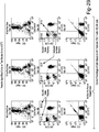
FIG. 1 depicts flow cytometry results of Protective Agent blood collection tube samples with and without a secondary fixation step. -
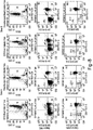
FIG. 2 depicts data of Protective Agent blood collection tube samples stored at 22°C. -
FIG. 3 depicts data of EDTA blood collection tube samples stored at 22°C and 6°C. -
FIG. 4 depicts data of ACD Sol B blood collection tube samples stored at 22°C and 6°C. -
FIG. 5 depicts data of Na Heparin blood collection tube samples stored at 22°C and 6°C. -
FIG. 6 depicts a comparison of EDTA, ACD Sol B, Na Heparin, and Protective Agent blood collection tube samples from 2 hours to 96 hours. -
FIG. 7 depicts the temperature variation of samples subjected to two day shipping. -
FIG. 8 depicts a comparison of flow cytometry results of Zap-70 48 hours after shipping in Protective Agent blood collection tube and EDTA blood collection tube. - The explanations and illustrations presented herein are intended to acquaint others skilled in the art with the teachings, its principles, and its practical application. Those skilled in the art may adapt and apply the teachings in its numerous forms, as may be best suited to the requirements of a particular use. Accordingly, the specific embodiments of the present teachings as set forth are not intended as being exhaustive or limiting of the teachings. The scope of the teachings should, therefore, be determined not with reference to the above description, but should instead be determined with reference to the appended claims, along with the full scope of equivalents to which such claims are entitled. The disclosures of all articles and references, including patent applications and publications, are incorporated by reference for all purposes. Other combinations are also possible as will be gleaned from the following claims, which are also hereby incorporated by reference into this written description.
- This application claims the benefit of the filing date of
U.S. Provisional Application No. 62/192,645, filed July 15, 2015 - The present teachings contemplate a method comprising contacting a drawn blood sample with a protective agent that is substantially free of formaldehyde and analyzing the white blood cells of the contacted sample for protein epitopes after at least about twenty-four hours after contacting. The protective agent may be included in a blood collection tube. The protective agent may be included as part of a kit.
- The present teachings allow for increased antigen stability of intracellular markers such as Zap-70. Zap-70 is an intracellular kinase, whose level of aberrant expression in B-cell Chronic Lymphocytic Leukemia (B-CLL) is used for prognosis and treatment decisions. The varying expression, dim staining, and labile nature makes measuring Zap-70 challenging and has prevented it from becoming more widely used in Leukemia/ Lymphoma screening. The present teachings provide for stabilization of Zap-70, thereby allowing for a longer analysis window and more precise results. The present teachings provide increased fluorescence and population resolution of Zap-70. The present teachings allow for enhanced recovery of Zap-70 percentages up to 96 hours after blood draw.
- The present teachings contemplate a method comprising contacting a drawn blood sample with a protective agent that is substantially free of formaldehyde and analyzing the white blood cells of the contacted sample for protein epitopes after at least about twenty-four hours after contacting. The protective agent may stabilize the sample prior to any staining process but does not compromise flow cytometry test results.
- The contacted blood sample may be stable for analysis for at least four days at about 22°C. The contacted blood sample may be stable for analysis for at least seven days at about 22°C.
- The protective agent may be present in the amount of about 75 µl. The protective agent may be present in the amount of about 75 µl per 5ml of liquid biological sample. The protective agent may be present in an amount of about 200 µl. The protective agent may be present in the amount of about 200 µl per 10 ml of liquid biological sample. The protective agent may be within a tube. The protective agent may be within in a blood collection tube. The tube containing the protective agent may receive about 2 ml to about 10 ml of patient blood. The tube containing the protective agent may receive about 2 ml of patient blood. The tube containing the protective agent may receive about 5 ml of patient blood. The tube containing the protective agent may receive about 10 ml of patient blood. The patient blood may be drawn directly into the tube containing the protective agent.
- The protective agent may include a preserving agent and an anticoagulant. The preserving agent may be selected from aldehyde, oxazolidine, alcohol, cyclic urea, and mixtures thereof. The preserving agent may be a mixture of bicyclic oxazolidines. The preserving agent may be DMDM hydantoin. The preserving agent may be imidazolidinyl urea or diazolidinyl urea. The imidazolidinyl urea may be present in an amount of about 300 g/l to about 700 g/l of the protective agent. The imidazolidinyl urea may be about 1% to about 3% of the protective agent. The diazolidinyl urea may be present in an amount of about 125 g/l to about 525 g/l of the protective agent. The diazolidinyl urea may be about .05 % to about 2.5% of the protective agent. The anticoagulant may be K3EDTA. The K3EDTA may be present in an amount of about 60 g/l to about 100 g/l of the protective agent. The K3EDTA may be present in an amount of about 80 g/l to about 120 g/l of the protective agent. The protective agent may include a preserving agent, an anticoagulant and glycine. The glycine may be present in an amount of about 20 g/l to about 60 g/l of the protective agent. The protective agent may comprise an active agent in solution. Suitable solvents may include water, saline, dimethylsulfoxide, alcohol and mixtures thereof. The protective agent may comprise diazolidinyl urea (DU) and/or imidazolidinyl urea (IDU) in a buffered salt solution.
- The contacted blood sample maybe analyzed for protein epitopes of intracellular markers. The contacted blood sample may be analyzed for protein epitopes of cell surface markers. The markers may be stable for at least 4 days in whole blood samples stored in a protective agent blood collection tube at 22°C (e.g., room temperature (free of any freezing step)). The markers may be stable for at least 7 days in whole blood samples stored in a protective agent blood collection tube at 22°C. The markers may be stable for about 4 to about 7 days in whole blood samples stored in a protective agent blood collection tube at 22°C.
- The surface markers for analysis may be selected from the group consisting of: CD1 a, CD2, CD3, CD4, CD5, CD7, CD8, CD10, CD11b, CD11c, CD13, CD14, CD15, CD16, CD16+56, CD19, CD20, CD22, CD23, CD25, CD26, CD30, CD33, CD34, CD38, CD41, CD45, CD56, CD57, CD58, CD61, CD64, CD71, CD103, CD117, CD123, CD138, FMC7, HLA-DR, Kappa, Lambda, TCR αβ, and TCR γδ. The intracellular markers for analysis may be selected from the group consisting of: cCD3, CD79a, cLambda, cKappa, TdT, BCL-2, and Zap70. The contacted blood sample may be analyzed for protein epitopes of the group consisting of: CD79a, TdT, and BCL-2. The contacted blood sample may be analyzed for the expression of a protein tyrosine kinase. The contacted blood sample may be analyzed for
Zap 70 protein expression. - The method may include the use of fluorophore conjugated antibodies. The method may include a step of intracellular staining. The method may include intracellular staining without fixation of cells by formaldehyde or paraformaldehyde. The method may be free of addition of formaldehyde or paraformaldehyde prior to or post a cellular staining process. The analysis may be accomplished by flow cytometry. The recoverable protein expression of the contacted blood sample after about four days may be about 90% of recoverable protein expression at the time the blood is drawn. The recoverable protein expression of the contacted blood sample after about four days may be about 100% of recoverable protein expression at the time the blood is drawn. The recoverable protein expression of the contacted blood sample stored at 22°C may occur within about 24 hours to about 96 hours after contact. The analysis may include screening for cancer.
- Human blood samples were prepared for flow cytometry analysis. 50 µLs of whole blood drawn into the indicated blood collection tubes were stained with 5 µL of α-CD3-FITC, α-CD45-PerCP, and α-CD19-APC for 20 minutes at 22°C. The samples were fixed or not as indicated with a formaldehyde based commercial fixation reagent for 20 minutes at 22°C and then washed in PBS with BSA. The samples were permeabilized with a saponin based commercial perm/lyse reagent and stained with 20 µL of α-Zap-70-PE for 20 minutes at 22°C. The samples were washed twice in PBS with BSA and fixed in 1% paraformaldehyde in PBS for 30 minutes at 22°C before being run on a BD FACSCalibur. Percentages of T, NK, and B cells were obtained by the BD Multitest™ HIV panel. All antibodies and blood collection tubes other than those containing the protective agent described herein were from BD Biosciences. Flow cytometry was performed on a BD FACSCalibur. External flow cytometry was performed by Flow Contract Site Laboratory on samples sent from Omaha, NE to Kirkland, WA and run on a BD FACSCanto. Data are representative from multiple experiments with multiple donors.
-
FIG. 1 illustrates flow cytometry results of samples that underwent intracellular staining with and without a fixation step, demonstrating that the Protective Agent described herein allows for Zap-70 intracellular staining and analysis without fixation of cells.FIG. 2 depicts data of Protective Agent blood collection tube samples stored at 22°C.FIG. 3 depicts data of EDTA blood collection tube samples stored at 22°C and 6°C.FIG. 4 depicts data of ACD Sol B blood collection tube samples stored at 22°C and 6°C.FIG. 5 depicts data of Na Heparin blood collection tube samples stored at 22°C and 6°C.FIGS. 2-5 illustrate that the Protective Agent blood collection tube provides superior stabilization of Zap-70 and population resolution in comparison to other blood collection tubes. The other tested blood collection tubes lost accurate recovery ≤24 hours.FIG. 6 depicts a comparison of EDTA, ACD Sol B, Na Heparin, and Protective Agent blood collection tube samples from 2 hours to 96 hours. The findings indicate that the Protective Agent blood collection tube allows for accurate recovery of Zap-70 from blood samples stored at 22°C from 24 hours to 96 hours. The shipped samples were shipped via two day shipping in order to examine the effects of temperature variation and stability on the shipped samples.FIG. 7 illustrates the range of temperature variations the samples were subjected to in a 48 hour period.FIG. 8 depicts a comparison of flow cytometry results of the shipped samples in Protective Agent blood collection and EDTA blood collection tube.FIG. 8 illustrates Zap-70 is stable 48 hours after shipping in Protective Agent blood collection tube. - The present teachings provide stabilization of Zap-70 up to 96 hours after draw. The present teachings show increased Zap-70 fluorescence and population resolution after intracellular staining without formaldehyde or paraformaldehyde fixation using samples contacted with the protective agent of the present teachings. The protective agent may stabilize the sample prior to any staining process but does not compromise flow cytometry test results. The protective agent may allow for accurate flow cytometry results utilizing a staining process that is substantially free of any secondary fixation step (e.g., a formaldehyde fixation step).
- Any numerical values recited herein include all values from the lower value to the upper value in increments of one unit provided that there is a separation of at least 2 units between any lower value and any higher value. As an example, if it is stated that the amount of a component or a value of a process variable such as, for example, temperature, pressure, time and the like is, for example, from 1 to 90, preferably from 20 to 80, more preferably from 30 to 70, it is intended that values such as 15 to 85, 22 to 68, 43 to 51, 30 to 32 etc. are expressly enumerated in this specification. For values which are less than one, one unit is considered to be 0.0001, 0.001, 0.01 or 0.1 as appropriate. These are only examples of what is specifically intended and all possible combinations of numerical values between the lowest value and the highest value enumerated are to be considered to be expressly stated in this application in a similar manner.
- Unless otherwise stated, all ranges include both endpoints and all numbers between the endpoints. The use of "about" or "approximately" in connection with a range applies to both ends of the range. Thus, "about 20 to 30" is intended to cover "about 20 to about 30", inclusive of at least the specified endpoints.
- The disclosures of all articles and references, including patent applications and publications, are incorporated by reference for all purposes. The term "consisting essentially of" to describe a combination shall include the elements, ingredients, components or steps identified, and such other elements ingredients, components or steps that do not materially affect the basic and novel characteristics of the combination. The use of the terms "comprising" or "including" to describe combinations of elements, ingredients, components or steps herein also contemplates embodiments that consist essentially of or even consists of the elements, ingredients, components or steps. Plural elements, ingredients, components or steps can be provided by a single integrated element, ingredient, component or step. Alternatively, a single integrated element, ingredient, component or step might be divided into separate plural elements, ingredients, components or steps. The disclosure of "a" or "one" to describe an element, ingredient, component or step is not intended to foreclose additional elements, ingredients, components or steps.
- It is understood that the above description is intended to be illustrative and not restrictive. Many embodiments as well as many applications besides the examples provided will be apparent to those of skill in the art upon reading the above description. The scope of the invention should, therefore, be determined not with reference to the above description, but should instead be determined with reference to the appended claims, along with the full scope of equivalents to which such claims are entitled. The disclosures of all articles and references, including patent applications and publications, are incorporated by reference for all purposes. The omission in the following claims of any aspect of subject matter that is disclosed herein is not a disclaimer of such subject matter, nor should it be regarded that the inventors did not consider such subject matter to be part of the disclosed inventive subject matter.
Claims (15)
- A method comprising the steps of:contacting a drawn blood sample with a protective agent that is substantially free of formaldehyde; andanalyzing the white blood cells of the contacted sample for protein epitopes after at least about twenty-four hours after contacting.
- The method of claim 1, wherein the contacted blood sample is stable for analysis for at least four days at about 22°C.
- The method of claim 1 or claim 2, wherein the protective agent is present in the amount of from about 25 µl to about 125 µl or even about 75 µl per 5 ml of liquid biological sample.
- The method of any of claims 1 through 3, wherein the protective agent includes a preserving agent and an anticoagulant.
- The method of any of claims 1 through 4, wherein the preserving agent is selected from aldehyde, oxazolidine, alcohol, cyclic urea, and mixtures thereof.
- The method of any of claims 1 through 5, wherein the preserving agent is imidazolidinyl urea or diazolidinyl urea.
- The method of any of claims 1 through 6, wherein the anticoagulant is K3EDTA.
- The method of any of claims 1 through 7, wherein the contacted blood sample is analyzed for protein epitopes of intracellular markers or cell surface markers.
- The method of any of claims 1 through 8, wherein surface markers for analysis are selected from the group consisting of: CD1 a, CD2, CD3, CD4, CD5, CD7, CD8, CD10, CD11b, CD11c, CD13, CD14, CD15, CD16, CD16+56, CD19, CD20, CD22, CD23, CD25, CD26, CD30, CD33, CD34, CD38, CD41, CD45, CD56, CD57, CD58, CD61, CD64, CD71, CD103, CD117, CD123, CD138, CD200, FMC7, HLA-DR, Kappa, Lambda, TCR αβ, and TCR γδ.
- The method of any of claims 1 through 9, wherein intracellular markers for analysis are selected from the group consisting of: cCD3, CD79a, cLambda, cKappa, TdT, BCL-2, and Zap70.
- The method of any of claims 1 through 10, wherein the contacted blood sample is analyzed for the expression of a protein tyrosine kinase.
- The method of any of claims 1 through 11, wherein the method includes a step of intracellular staining.
- The method of any of claims 1 through 12, wherein the method is free of addition of formaldehyde or paraformaldehyde prior to or post a cellular staining process.
- The method of any of claims 1 through 13, wherein recoverable protein expression of the contacted blood sample after about four days is about 90% of recoverable protein expression at the time the blood is drawn.
- Use of the method of any of claims 1 through 14 in screening for cancer.
Applications Claiming Priority (2)
| Application Number | Priority Date | Filing Date | Title |
|---|---|---|---|
| US201562192645P | 2015-07-15 | 2015-07-15 | |
| US15/209,855 US20170097361A1 (en) | 2015-07-15 | 2016-07-14 | Method and device for stabilizing of proteins |
Publications (2)
| Publication Number | Publication Date |
|---|---|
| EP3118623A1 true EP3118623A1 (en) | 2017-01-18 |
| EP3118623B1 EP3118623B1 (en) | 2020-05-27 |
Family
ID=56497562
Family Applications (1)
| Application Number | Title | Priority Date | Filing Date |
|---|---|---|---|
| EP16179753.5A Active EP3118623B1 (en) | 2015-07-15 | 2016-07-15 | Method and device for stabilizing of proteins |
Country Status (3)
| Country | Link |
|---|---|
| US (1) | US20170097361A1 (en) |
| EP (1) | EP3118623B1 (en) |
| CA (1) | CA2936167A1 (en) |
Cited By (5)
| Publication number | Priority date | Publication date | Assignee | Title |
|---|---|---|---|---|
| WO2020140035A1 (en) * | 2018-12-27 | 2020-07-02 | Streck, Inc. | Methods of preparing samples for proteomic analysis |
| US11168351B2 (en) | 2015-03-05 | 2021-11-09 | Streck, Inc. | Stabilization of nucleic acids in urine |
| US11299764B2 (en) | 2015-11-20 | 2022-04-12 | Streck, Inc. | Single spin process for blood plasma separation and plasma composition including preservative |
| US11506655B2 (en) | 2016-07-29 | 2022-11-22 | Streck, Inc. | Suspension composition for hematology analysis control |
| US11882823B2 (en) | 2017-01-22 | 2024-01-30 | Chryos, Llc | Composition and method of use of the same for preserving cells for analysis |
Families Citing this family (2)
| Publication number | Priority date | Publication date | Assignee | Title |
|---|---|---|---|---|
| CN106771205B (en) * | 2017-01-18 | 2017-11-21 | 浙江博真生物科技有限公司 | Ten color antibody compositions and its application in leukemia-lymphoma parting |
| CN112462065A (en) * | 2020-11-16 | 2021-03-09 | 浙江博真生物科技有限公司 | Antibody composition, kit and detection method for detecting solid tumor |
Citations (4)
| Publication number | Priority date | Publication date | Assignee | Title |
|---|---|---|---|---|
| EP1217372A1 (en) * | 2000-12-22 | 2002-06-26 | Streck Laboratories, Inc. | Improved flow cytometry reagent and system |
| WO2003018757A2 (en) * | 2001-08-23 | 2003-03-06 | Immunivest Corporation | Stabilization of cells and biological specimens for analysis |
| US20100167271A1 (en) * | 2008-12-30 | 2010-07-01 | Streck, Inc. | Method for screening blood using a preservative that may be in a substantially solid state form |
| US20150010923A1 (en) | 2011-12-21 | 2015-01-08 | Beckman Coulter, Inc. | Method for labeling intracellular and extracellular targets of leukocytes |
-
2016
- 2016-07-14 CA CA2936167A patent/CA2936167A1/en active Pending
- 2016-07-14 US US15/209,855 patent/US20170097361A1/en not_active Abandoned
- 2016-07-15 EP EP16179753.5A patent/EP3118623B1/en active Active
Patent Citations (6)
| Publication number | Priority date | Publication date | Assignee | Title |
|---|---|---|---|---|
| EP1217372A1 (en) * | 2000-12-22 | 2002-06-26 | Streck Laboratories, Inc. | Improved flow cytometry reagent and system |
| US6794152B2 (en) | 2000-12-22 | 2004-09-21 | Streck Laboratories Inc. | Flow cytometry reagent and system |
| US6977156B2 (en) | 2000-12-22 | 2005-12-20 | Streck Laboratories, Inc. | Flow cytometry reagent and system |
| WO2003018757A2 (en) * | 2001-08-23 | 2003-03-06 | Immunivest Corporation | Stabilization of cells and biological specimens for analysis |
| US20100167271A1 (en) * | 2008-12-30 | 2010-07-01 | Streck, Inc. | Method for screening blood using a preservative that may be in a substantially solid state form |
| US20150010923A1 (en) | 2011-12-21 | 2015-01-08 | Beckman Coulter, Inc. | Method for labeling intracellular and extracellular targets of leukocytes |
Cited By (6)
| Publication number | Priority date | Publication date | Assignee | Title |
|---|---|---|---|---|
| US11168351B2 (en) | 2015-03-05 | 2021-11-09 | Streck, Inc. | Stabilization of nucleic acids in urine |
| US11299764B2 (en) | 2015-11-20 | 2022-04-12 | Streck, Inc. | Single spin process for blood plasma separation and plasma composition including preservative |
| US11506655B2 (en) | 2016-07-29 | 2022-11-22 | Streck, Inc. | Suspension composition for hematology analysis control |
| US11882823B2 (en) | 2017-01-22 | 2024-01-30 | Chryos, Llc | Composition and method of use of the same for preserving cells for analysis |
| WO2020140035A1 (en) * | 2018-12-27 | 2020-07-02 | Streck, Inc. | Methods of preparing samples for proteomic analysis |
| US20220074949A1 (en) * | 2018-12-27 | 2022-03-10 | Streck, Inc. | Methods of preparing samples for proteomic analysis |
Also Published As
| Publication number | Publication date |
|---|---|
| CA2936167A1 (en) | 2017-01-15 |
| US20170097361A1 (en) | 2017-04-06 |
| EP3118623B1 (en) | 2020-05-27 |
Similar Documents
| Publication | Publication Date | Title |
|---|---|---|
| EP3118623B1 (en) | Method and device for stabilizing of proteins | |
| Chow et al. | Whole blood fixation and permeabilization protocol with red blood cell lysis for flow cytometry of intracellular phosphorylated epitopes in leukocyte subpopulations | |
| Nilsson et al. | Simplified assays of hemolytic activity of the classical and alternative complement pathways | |
| US7803523B2 (en) | Whole blood preparation for cytometric analysis of cell signaling pathways | |
| US7749757B1 (en) | Stabilizing solution for cells and tissues | |
| Terstappen et al. | A rapid sample preparation technique for flow cytometric analysis of immunofluorescence allowing absolute enumeration of cell subpopulations | |
| EP0685994A1 (en) | Multipurpose reagent system for rapid lysis of whole blood samples | |
| Ting et al. | B-lymphocyte alloantigens in Caucasians. | |
| Suni et al. | Flow cytometric analysis of cell signaling proteins | |
| EP0754301A1 (en) | Preparation and stabilisation of cell suspensions | |
| JPH09508975A (en) | Analytical cuvette, quantification method and diagnostic test kit used for quantification of components to be analyzed present in whole blood sample | |
| Riond et al. | Prevalence of dog erythrocyte antigen 1.1 in dogs in Switzerland evaluated with the gel column technique | |
| Keeney et al. | Paroxysmal nocturnal hemoglobinuria assessment by flow cytometric analysis | |
| CN106802351B (en) | Evaluation is suitable for removing the method for the quality of the medium of anti-A or anti-B antibody | |
| CN105334329B (en) | The assay method of calpain phosphorylation level | |
| Rovati et al. | An eight-colour flow cytometric method for the detection of reference values of lymphocyte subsets in selected healthy donors | |
| Lippi et al. | Identification of spurious hemolysis in anticoagulated blood with Sysmex XE-2100 and Siemens Advia 2120 | |
| Smith et al. | Cytometric analysis of BAL T cells labeled with a standardized antibody cocktail correlates with immunohistochemical staining | |
| Weiler et al. | The inhibition of labeled antigen precipitation as a measure of serum γ-globulin | |
| da Costa et al. | Harmonization of light scatter and fluorescence flow cytometry profiles obtained after staining peripheral blood leucocytes for cell surface‐only versus intracellular antigens with the Fix & Perm™ reagent | |
| US6110730A (en) | Whole blood cell staining device | |
| Haukenes | A rubella haemagglutination inhibitor simulating antibody | |
| Wang et al. | Interference of hemolysis on the postmortem biochemical analysis of IgE by ECLIA | |
| Pavone et al. | Demonstration of anti‐Wrb in a second serum containing anti‐Ena | |
| Moore et al. | Reappraisal of an Auto Analyzer Haemagglutination System for Anti‐D Quantification |
Legal Events
| Date | Code | Title | Description |
|---|---|---|---|
| PUAI | Public reference made under article 153(3) epc to a published international application that has entered the european phase |
Free format text: ORIGINAL CODE: 0009012 |
|
| STAA | Information on the status of an ep patent application or granted ep patent |
Free format text: STATUS: THE APPLICATION HAS BEEN PUBLISHED |
|
| AK | Designated contracting states |
Kind code of ref document: A1 Designated state(s): AL AT BE BG CH CY CZ DE DK EE ES FI FR GB GR HR HU IE IS IT LI LT LU LV MC MK MT NL NO PL PT RO RS SE SI SK SM TR |
|
| AX | Request for extension of the european patent |
Extension state: BA ME |
|
| STAA | Information on the status of an ep patent application or granted ep patent |
Free format text: STATUS: REQUEST FOR EXAMINATION WAS MADE |
|
| 17P | Request for examination filed |
Effective date: 20170718 |
|
| RBV | Designated contracting states (corrected) |
Designated state(s): AL AT BE BG CH CY CZ DE DK EE ES FI FR GB GR HR HU IE IS IT LI LT LU LV MC MK MT NL NO PL PT RO RS SE SI SK SM TR |
|
| STAA | Information on the status of an ep patent application or granted ep patent |
Free format text: STATUS: EXAMINATION IS IN PROGRESS |
|
| 17Q | First examination report despatched |
Effective date: 20170906 |
|
| GRAP | Despatch of communication of intention to grant a patent |
Free format text: ORIGINAL CODE: EPIDOSNIGR1 |
|
| STAA | Information on the status of an ep patent application or granted ep patent |
Free format text: STATUS: GRANT OF PATENT IS INTENDED |
|
| INTG | Intention to grant announced |
Effective date: 20191126 |
|
| GRAJ | Information related to disapproval of communication of intention to grant by the applicant or resumption of examination proceedings by the epo deleted |
Free format text: ORIGINAL CODE: EPIDOSDIGR1 |
|
| STAA | Information on the status of an ep patent application or granted ep patent |
Free format text: STATUS: EXAMINATION IS IN PROGRESS |
|
| GRAR | Information related to intention to grant a patent recorded |
Free format text: ORIGINAL CODE: EPIDOSNIGR71 |
|
| GRAS | Grant fee paid |
Free format text: ORIGINAL CODE: EPIDOSNIGR3 |
|
| STAA | Information on the status of an ep patent application or granted ep patent |
Free format text: STATUS: GRANT OF PATENT IS INTENDED |
|
| INTC | Intention to grant announced (deleted) | ||
| GRAA | (expected) grant |
Free format text: ORIGINAL CODE: 0009210 |
|
| STAA | Information on the status of an ep patent application or granted ep patent |
Free format text: STATUS: THE PATENT HAS BEEN GRANTED |
|
| AK | Designated contracting states |
Kind code of ref document: B1 Designated state(s): AL AT BE BG CH CY CZ DE DK EE ES FI FR GB GR HR HU IE IS IT LI LT LU LV MC MK MT NL NO PL PT RO RS SE SI SK SM TR |
|
| INTG | Intention to grant announced |
Effective date: 20200420 |
|
| REG | Reference to a national code |
Ref country code: GB Ref legal event code: FG4D |
|
| REG | Reference to a national code |
Ref country code: CH Ref legal event code: EP |
|
| REG | Reference to a national code |
Ref country code: AT Ref legal event code: REF Ref document number: 1275070 Country of ref document: AT Kind code of ref document: T Effective date: 20200615 |
|
| REG | Reference to a national code |
Ref country code: DE Ref legal event code: R096 Ref document number: 602016036928 Country of ref document: DE |
|
| REG | Reference to a national code |
Ref country code: CH Ref legal event code: NV Representative=s name: VALIPAT S.A. C/O BOVARD SA NEUCHATEL, CH |
|
| REG | Reference to a national code |
Ref country code: NL Ref legal event code: FP |
|
| REG | Reference to a national code |
Ref country code: LT Ref legal event code: MG4D |
|
| PG25 | Lapsed in a contracting state [announced via postgrant information from national office to epo] |
Ref country code: PT Free format text: LAPSE BECAUSE OF FAILURE TO SUBMIT A TRANSLATION OF THE DESCRIPTION OR TO PAY THE FEE WITHIN THE PRESCRIBED TIME-LIMIT Effective date: 20200928 Ref country code: IS Free format text: LAPSE BECAUSE OF FAILURE TO SUBMIT A TRANSLATION OF THE DESCRIPTION OR TO PAY THE FEE WITHIN THE PRESCRIBED TIME-LIMIT Effective date: 20200927 Ref country code: NO Free format text: LAPSE BECAUSE OF FAILURE TO SUBMIT A TRANSLATION OF THE DESCRIPTION OR TO PAY THE FEE WITHIN THE PRESCRIBED TIME-LIMIT Effective date: 20200827 Ref country code: GR Free format text: LAPSE BECAUSE OF FAILURE TO SUBMIT A TRANSLATION OF THE DESCRIPTION OR TO PAY THE FEE WITHIN THE PRESCRIBED TIME-LIMIT Effective date: 20200828 Ref country code: LT Free format text: LAPSE BECAUSE OF FAILURE TO SUBMIT A TRANSLATION OF THE DESCRIPTION OR TO PAY THE FEE WITHIN THE PRESCRIBED TIME-LIMIT Effective date: 20200527 Ref country code: SE Free format text: LAPSE BECAUSE OF FAILURE TO SUBMIT A TRANSLATION OF THE DESCRIPTION OR TO PAY THE FEE WITHIN THE PRESCRIBED TIME-LIMIT Effective date: 20200527 Ref country code: FI Free format text: LAPSE BECAUSE OF FAILURE TO SUBMIT A TRANSLATION OF THE DESCRIPTION OR TO PAY THE FEE WITHIN THE PRESCRIBED TIME-LIMIT Effective date: 20200527 |
|
| PG25 | Lapsed in a contracting state [announced via postgrant information from national office to epo] |
Ref country code: BG Free format text: LAPSE BECAUSE OF FAILURE TO SUBMIT A TRANSLATION OF THE DESCRIPTION OR TO PAY THE FEE WITHIN THE PRESCRIBED TIME-LIMIT Effective date: 20200827 Ref country code: HR Free format text: LAPSE BECAUSE OF FAILURE TO SUBMIT A TRANSLATION OF THE DESCRIPTION OR TO PAY THE FEE WITHIN THE PRESCRIBED TIME-LIMIT Effective date: 20200527 Ref country code: LV Free format text: LAPSE BECAUSE OF FAILURE TO SUBMIT A TRANSLATION OF THE DESCRIPTION OR TO PAY THE FEE WITHIN THE PRESCRIBED TIME-LIMIT Effective date: 20200527 Ref country code: RS Free format text: LAPSE BECAUSE OF FAILURE TO SUBMIT A TRANSLATION OF THE DESCRIPTION OR TO PAY THE FEE WITHIN THE PRESCRIBED TIME-LIMIT Effective date: 20200527 |
|
| REG | Reference to a national code |
Ref country code: AT Ref legal event code: MK05 Ref document number: 1275070 Country of ref document: AT Kind code of ref document: T Effective date: 20200527 |
|
| PG25 | Lapsed in a contracting state [announced via postgrant information from national office to epo] |
Ref country code: AL Free format text: LAPSE BECAUSE OF FAILURE TO SUBMIT A TRANSLATION OF THE DESCRIPTION OR TO PAY THE FEE WITHIN THE PRESCRIBED TIME-LIMIT Effective date: 20200527 |
|
| PG25 | Lapsed in a contracting state [announced via postgrant information from national office to epo] |
Ref country code: AT Free format text: LAPSE BECAUSE OF FAILURE TO SUBMIT A TRANSLATION OF THE DESCRIPTION OR TO PAY THE FEE WITHIN THE PRESCRIBED TIME-LIMIT Effective date: 20200527 Ref country code: CZ Free format text: LAPSE BECAUSE OF FAILURE TO SUBMIT A TRANSLATION OF THE DESCRIPTION OR TO PAY THE FEE WITHIN THE PRESCRIBED TIME-LIMIT Effective date: 20200527 Ref country code: ES Free format text: LAPSE BECAUSE OF FAILURE TO SUBMIT A TRANSLATION OF THE DESCRIPTION OR TO PAY THE FEE WITHIN THE PRESCRIBED TIME-LIMIT Effective date: 20200527 Ref country code: RO Free format text: LAPSE BECAUSE OF FAILURE TO SUBMIT A TRANSLATION OF THE DESCRIPTION OR TO PAY THE FEE WITHIN THE PRESCRIBED TIME-LIMIT Effective date: 20200527 Ref country code: IT Free format text: LAPSE BECAUSE OF FAILURE TO SUBMIT A TRANSLATION OF THE DESCRIPTION OR TO PAY THE FEE WITHIN THE PRESCRIBED TIME-LIMIT Effective date: 20200527 Ref country code: EE Free format text: LAPSE BECAUSE OF FAILURE TO SUBMIT A TRANSLATION OF THE DESCRIPTION OR TO PAY THE FEE WITHIN THE PRESCRIBED TIME-LIMIT Effective date: 20200527 Ref country code: SM Free format text: LAPSE BECAUSE OF FAILURE TO SUBMIT A TRANSLATION OF THE DESCRIPTION OR TO PAY THE FEE WITHIN THE PRESCRIBED TIME-LIMIT Effective date: 20200527 Ref country code: DK Free format text: LAPSE BECAUSE OF FAILURE TO SUBMIT A TRANSLATION OF THE DESCRIPTION OR TO PAY THE FEE WITHIN THE PRESCRIBED TIME-LIMIT Effective date: 20200527 |
|
| PG25 | Lapsed in a contracting state [announced via postgrant information from national office to epo] |
Ref country code: MC Free format text: LAPSE BECAUSE OF FAILURE TO SUBMIT A TRANSLATION OF THE DESCRIPTION OR TO PAY THE FEE WITHIN THE PRESCRIBED TIME-LIMIT Effective date: 20200527 Ref country code: SK Free format text: LAPSE BECAUSE OF FAILURE TO SUBMIT A TRANSLATION OF THE DESCRIPTION OR TO PAY THE FEE WITHIN THE PRESCRIBED TIME-LIMIT Effective date: 20200527 Ref country code: PL Free format text: LAPSE BECAUSE OF FAILURE TO SUBMIT A TRANSLATION OF THE DESCRIPTION OR TO PAY THE FEE WITHIN THE PRESCRIBED TIME-LIMIT Effective date: 20200527 |
|
| REG | Reference to a national code |
Ref country code: DE Ref legal event code: R097 Ref document number: 602016036928 Country of ref document: DE |
|
| PLBE | No opposition filed within time limit |
Free format text: ORIGINAL CODE: 0009261 |
|
| STAA | Information on the status of an ep patent application or granted ep patent |
Free format text: STATUS: NO OPPOSITION FILED WITHIN TIME LIMIT |
|
| PG25 | Lapsed in a contracting state [announced via postgrant information from national office to epo] |
Ref country code: FR Free format text: LAPSE BECAUSE OF NON-PAYMENT OF DUE FEES Effective date: 20200727 |
|
| 26N | No opposition filed |
Effective date: 20210302 |
|
| PG25 | Lapsed in a contracting state [announced via postgrant information from national office to epo] |
Ref country code: SI Free format text: LAPSE BECAUSE OF FAILURE TO SUBMIT A TRANSLATION OF THE DESCRIPTION OR TO PAY THE FEE WITHIN THE PRESCRIBED TIME-LIMIT Effective date: 20200527 |
|
| PG25 | Lapsed in a contracting state [announced via postgrant information from national office to epo] |
Ref country code: IE Free format text: LAPSE BECAUSE OF NON-PAYMENT OF DUE FEES Effective date: 20200715 |
|
| PG25 | Lapsed in a contracting state [announced via postgrant information from national office to epo] |
Ref country code: TR Free format text: LAPSE BECAUSE OF FAILURE TO SUBMIT A TRANSLATION OF THE DESCRIPTION OR TO PAY THE FEE WITHIN THE PRESCRIBED TIME-LIMIT Effective date: 20200527 Ref country code: MT Free format text: LAPSE BECAUSE OF FAILURE TO SUBMIT A TRANSLATION OF THE DESCRIPTION OR TO PAY THE FEE WITHIN THE PRESCRIBED TIME-LIMIT Effective date: 20200527 Ref country code: CY Free format text: LAPSE BECAUSE OF FAILURE TO SUBMIT A TRANSLATION OF THE DESCRIPTION OR TO PAY THE FEE WITHIN THE PRESCRIBED TIME-LIMIT Effective date: 20200527 |
|
| PG25 | Lapsed in a contracting state [announced via postgrant information from national office to epo] |
Ref country code: MK Free format text: LAPSE BECAUSE OF FAILURE TO SUBMIT A TRANSLATION OF THE DESCRIPTION OR TO PAY THE FEE WITHIN THE PRESCRIBED TIME-LIMIT Effective date: 20200527 |
|
| P01 | Opt-out of the competence of the unified patent court (upc) registered |
Effective date: 20230523 |
|
| PGFP | Annual fee paid to national office [announced via postgrant information from national office to epo] |
Ref country code: NL Payment date: 20230614 Year of fee payment: 8 |
|
| PGFP | Annual fee paid to national office [announced via postgrant information from national office to epo] |
Ref country code: LU Payment date: 20230628 Year of fee payment: 8 |
|
| PGFP | Annual fee paid to national office [announced via postgrant information from national office to epo] |
Ref country code: BE Payment date: 20230616 Year of fee payment: 8 |
|
| PGFP | Annual fee paid to national office [announced via postgrant information from national office to epo] |
Ref country code: GB Payment date: 20230608 Year of fee payment: 8 Ref country code: CH Payment date: 20230801 Year of fee payment: 8 |
|
| PGFP | Annual fee paid to national office [announced via postgrant information from national office to epo] |
Ref country code: DE Payment date: 20230613 Year of fee payment: 8 |