EP3114485B1 - Nephropathy biomarker - Google Patents
Nephropathy biomarker Download PDFInfo
- Publication number
- EP3114485B1 EP3114485B1 EP15709334.5A EP15709334A EP3114485B1 EP 3114485 B1 EP3114485 B1 EP 3114485B1 EP 15709334 A EP15709334 A EP 15709334A EP 3114485 B1 EP3114485 B1 EP 3114485B1
- Authority
- EP
- European Patent Office
- Prior art keywords
- cadherin
- ckd
- urine
- diabetic
- patients
- Prior art date
- Legal status (The legal status is an assumption and is not a legal conclusion. Google has not performed a legal analysis and makes no representation as to the accuracy of the status listed.)
- Active
Links
Images







Classifications
-
- G—PHYSICS
- G01—MEASURING; TESTING
- G01N—INVESTIGATING OR ANALYSING MATERIALS BY DETERMINING THEIR CHEMICAL OR PHYSICAL PROPERTIES
- G01N33/00—Investigating or analysing materials by specific methods not covered by groups G01N1/00 - G01N31/00
- G01N33/48—Biological material, e.g. blood, urine; Haemocytometers
- G01N33/50—Chemical analysis of biological material, e.g. blood, urine; Testing involving biospecific ligand binding methods; Immunological testing
- G01N33/68—Chemical analysis of biological material, e.g. blood, urine; Testing involving biospecific ligand binding methods; Immunological testing involving proteins, peptides or amino acids
- G01N33/6893—Chemical analysis of biological material, e.g. blood, urine; Testing involving biospecific ligand binding methods; Immunological testing involving proteins, peptides or amino acids related to diseases not provided for elsewhere
-
- C—CHEMISTRY; METALLURGY
- C07—ORGANIC CHEMISTRY
- C07K—PEPTIDES
- C07K16/00—Immunoglobulins [IG], e.g. monoclonal or polyclonal antibodies
- C07K16/18—Immunoglobulins [IG], e.g. monoclonal or polyclonal antibodies against material from animals or humans
-
- G—PHYSICS
- G01—MEASURING; TESTING
- G01N—INVESTIGATING OR ANALYSING MATERIALS BY DETERMINING THEIR CHEMICAL OR PHYSICAL PROPERTIES
- G01N2333/00—Assays involving biological materials from specific organisms or of a specific nature
- G01N2333/435—Assays involving biological materials from specific organisms or of a specific nature from animals; from humans
- G01N2333/705—Assays involving receptors, cell surface antigens or cell surface determinants
-
- G—PHYSICS
- G01—MEASURING; TESTING
- G01N—INVESTIGATING OR ANALYSING MATERIALS BY DETERMINING THEIR CHEMICAL OR PHYSICAL PROPERTIES
- G01N2800/00—Detection or diagnosis of diseases
- G01N2800/34—Genitourinary disorders
- G01N2800/347—Renal failures; Glomerular diseases; Tubulointerstitial diseases, e.g. nephritic syndrome, glomerulonephritis; Renovascular diseases, e.g. renal artery occlusion, nephropathy
-
- G—PHYSICS
- G01—MEASURING; TESTING
- G01N—INVESTIGATING OR ANALYSING MATERIALS BY DETERMINING THEIR CHEMICAL OR PHYSICAL PROPERTIES
- G01N2800/00—Detection or diagnosis of diseases
- G01N2800/56—Staging of a disease; Further complications associated with the disease
Definitions
- the invention relates to a biomarker useful for the diagnosis of kidney disease and for determining patients whose disease severity level is likely to progress. Accordingly, the invention relates to a method for determining the likelihood of kidney disease progression, particularly of chronic kidney disease in a diabetic patient.
- CKD Chronic kidney disease
- end stage renal failure are conditions associated with significant morbidity and mortality.
- the incidence of CKD is significant and, with aging and more diabetes-prone populations, is rising internationally.
- CKD is a condition of varying severity which frequently progresses along a 'severity spectrum' over time. Stages 1 and 2 may be considered mild CKD, stage 3 moderate and stages 4 and 5 more severe. Stage 5 may be considered end-stage/established renal failure (ERF).
- ERP end-stage/established renal failure
- kidney function which might be relatively asymptomatic for mild forms
- the CKD is associated with an increased risk of heart disease and stroke.
- ERF advanced renal damage
- CKD has a range of causes, including high blood pressure, diabetes, general aging, glomeruli inflammation, polycystic kidney disease, repeated infection, renal artery stenosis and drug/toxin damage.
- diabetes is considered one of the most significant, with diabetic nephropathy (diabetes-related kidney disease) accounting, in the West, for around 30% of patients requiring RRT. This impact is most likely only to concomitantly rise with the increasing obesity rates of many countries.
- Tubulo-interstitial fibrosis contributes to the irreversible deterioration of renal function and is an accurate predictor of long-term prognosis, irrespective of the type of disease or the compartment of origin. It is assessed by renal biopsy, however, a procedure which can incur complications and risks and so is neither desirable nor feasible for all patients.
- GFR glomerular filtration rate
- the current "gold standard" indicator for (diabetic) CKD may be considered to be a person's urinary albumin (or other/general) protein level.
- microalbuminuria when the kidneys 'leak' albumin into the urine and which may be related to failure of proximal-tube endocytosis, can be assessed by a non-invasive urine test.
- Albuminuria is a sub-type of, and may precede, proteinuria.
- PCR/ACR protein/albumin to creatinine ratio
- urine albumin/protein per se is considered to have limited sensitivity and specificity with respect to CKD progression.
- diabetic nephropathy for example, not all patients with microalbuminuria will progress to ERF and a significant proportion of type-2 diabetic patients do not present with incremental proteinuria or have non-proteinuric renal disease (yet, of these, a substantial proportion progress over time).
- a method for determining the propensity for a person's kidney disease (CKD) level to progress in severity comprising testing for the presence/level of K-cadherin in the person's urine.
- detection e.g. by Western immunoblot as described below
- the relative level of urinary K-cadherin may be related to the rate of progression.
- the relative K-cadherin level is pertinent with respect to more sensitive systems (such as an ELISA (enzyme-linked immunosorbent assay) that has proven able to detect its presence in non-progressors' urine, for example.
- the invention therefore provides for an assay useful for determining whether a person's CKD is likely to progress, and thus whether, for example, closer monitoring/intervention treatment may be advisable.
- K-cadherin relates to 'normal' expressed, un-fragmented protein (since, as mentioned below, fragments which react with K-cadherin antibodies have been found in the urine of healthy control subjects). Accordingly, the K-cadherin may be thought of/described as full length (as judged by electrophoresis separation, for example) though, as would be apparent to a skilled person, there could naturally be a level of variation.
- K-cadherin which is also known as cadherin-6 (CDH6), is a member of the cadherin protein family, a family, having more than 100 members, of transmembrane glycoproteins important in the maintenance of cell-cell adhesion, cell phenotype regulation, tissue organisation and development.
- K-cadherin is a 120kDa type II classical cadherin of 790 amino acids, coded by the CDH6 gene on chromosome 5.
- full length K-cadherin (as measured by Western Immunoblotting) was not detected in the urine of healthy control subjects or diabetics without CKD but was detectable initially (and subsequently) in the urine of every patient who exhibited progressive CKD over the study period. Further, none of the diabetic CKD subjects who were negative for urinary K-cadherin at baseline progressed over the follow-up period.
- the method may comprise testing for elevated K-cadherin.
- the elevated K-cadherin may be with respect to the normal (healthy) population and/or relative to a non-progressive standard model (control).
- control There may be different non-progressive standard levels applicable to different CKD disease stages; stages may include mild disease (CKD stages 1/2) and/or moderate/severe disease (CKD stages 3/4).
- Elevation may also optionally be assessed more specifically, e.g. with respect to a control non-progressive standard for particular variable characteristics (used to calculate estimated renal function) common to a patient.
- variable characteristics used to calculate estimated renal function
- Such characteristics may include any one or more of age, ethnicity, gender and traceable IDMS (isotope-dilution mass spectrometry).
- the degree of elevation for indicating likelihood of progression may correspond to at least an approximate doubling of the relevant non-progressive level.
- Other possibilities include instances where a non-progressive standard may correspond to an undetectable K-cadherin concentration.
- K-cadherin was, however, not echoed for N-cadherin (cadherin-2).
- full-length N-cadherin protein was present in the urine of all subjects tested (including healthy controls) and did not correlate with CKD, even though it was found (as for K-cadherin) to be downregulated in proximal tubule epithelial cells in kidney biopsies from patients with diabetic CKD. This may relate to its widespread expression, particularly in the lower urinary tract and the prostate.
- the person(s) may be diabetics.
- the kidney disease may be diabetic chronic kidney disease (diabetic nephropathy).
- the kidney disease may also be, for example, IgA nephropathy or chronic allograft nephropathy (whether or not the person is diabetic).
- IgA nephropathy or chronic allograft nephropathy whether or not the person is diabetic.
- some of the relevant diabetic patients in the study described below may not have had diabetic nephropathy, they may coincidentally have had another form of CKD (though efforts were made to avoid this and it is expected that for most/all cases the CKD cause would have been diabetes).
- K-cadherin loss similar to that seen from proximal tubule epithelia in early diabetic CKD has also been observed for advancing IgA nephropathy and chronic allograft nephropathy (CAN). Furthermore, loss of K-cadherin was observed also in an in vitro model of fibrosis using primary human proximal tubule cells.
- a person for whom the invention may be used may have more than cause/type of kidney disease.
- the person may be a type I or type II diabetic.
- Testing for K-cadherin may conveniently involve Western Immunoblotting as a detection system.
- Western Immunoblotting it has been shown that K-cadherin was not detected by Western Immunoblotting in the urine of healthy control subjects or diabetics without CKD but was detectable initially (and subsequently) in the urine of every patient who exhibited progressive diabetic CKD over the study period.
- Other techniques which may be used/exploited include an antibody system such as an ELISA (enzyme-linked immunosorbent assay), NMR (nuclear magnetic resonance), colorimetric and mass spectrometry.
- a point of care system e.g. one exploiting a membrane-based lateral flow strip (similar to the approach used in pregnancy tests), could be used to facilitate result determination.
- a test system may optionally exploit one or more antibodies as discussed below.
- the method comprises Western Immunoblotting and/or an ELISA for assessing the presence and/or level of K-cadherin.
- Western immunoblotting the sample should first be subject to a process such as gel electrophoresis, e.g. SDS-PAGE.
- the ELISA system used preferably comprises two sets of antibodies.
- one set is reactive towards the N-terminal end of K-cadherin and the other towards the C-terminal end.
- the target protein may be immobilised from one end and detected by antibodies which interact with the other end and, accordingly, the assay effectively tests for the presence of essentially full-length (non-degraded) K-cadherin.
- cellular degradation and death might lead to cleavage of K-cadherin and excretion of the extra-cellular domain (with the cytoplasmic domain being endocytosed and recycled/degraded in the proteasome).
- K-cadherin as judged on a Western Immunoblot was found in the urine of diabetic CKD patients, unlike for healthy controls. This implied the release of intact, essentially full length K-cadherin from proximal tubular epithelial cells exposed to fibrotic stimuli. Protein fragments recognised by K-cadherin antibodies have also been found in healthy control urine, indicating metabolism/degradation and excretion of K-cadherin may be a normal pathway.
- an antibody-based detection system such as an ELISA may be to use an antibody specific to the relevant K-cadherin cleavage point, e.g. as the capture antibody. By recognising and binding only protein un-broken at the relevant point, the presence of full-length K-cadherin may specifically be detected.
- the urine sample may be concentrated before the presence (and level) of K-cadherin is assessed.
- a sample is concentrated six-fold.
- concentrating a sample can facilitate subsequent testing (e.g. by reducing the sample volume to be applied to a gel).
- Whether or not, and to what extent, it is desirable to concentrate a sample may vary and depend on the testing method and the starting urine concentration. For example, as discussed below, urinary K-cadherin has been detected by ELISA in samples that were initially concentrated but then re-diluted (to the corresponding extent) to decrease urine matrix interference.
- the method further comprises testing for albumin and/or other protein in the person's urine.
- (micro)albuminuria in the early stage, and subsequently proteinuria, are the current favoured options as markers for the diagnosis and progression of diabetic nephropathy (and may also be applied to other forms of kidney disease).
- CKD ⁇ 3 eGFR ⁇ 60 ml/min
- urinary K-cadherin as a marker for determining the propensity for a person's kidney disease level to progress in severity.
- kits for testing for the presence/level of K-cadherin in a person's urine are provided. Also, according to another aspect of the present invention there is provided a point-of-care test for testing for the presence/level of K-cadherin in a person's urine.
- the point-of-care test and/or the kit may comprise a K-cadherin cleavage-point specific antibody.
- the point-of-care test and/or the kit may comprise two sets of K-cadherin antibodies.
- one of the antibody sets is reactive towards the N-terminal end of K-cadherin and the other is reactive towards the C-terminal end.
- the kit may conveniently be for a Western blotting assay (e.g. one involving immunoprecipitation) or an ELISA assay.
- the antibodies are commercially available, e.g.
- Example 1 Immuno-histochemistry of archived human kidney biopsy.
- Blocking to remove endogenous tissue peroxidase was performed by oxidizing and using freshly prepared peroxidase block buffer (3% H 2 O 2 in PBS) . Antigen retrieval was performed using Tris/EDTA (pH9.0) at boiling temperature for 20 min.
- FITC AlexaFluor 488
- TSA Tetra methyl rhodamine system
- Ftc TSA fluorescein system
- N-cadherin is up-regulated by the powerful fibrotic growth factor TGF ⁇ 1
- TGF ⁇ 1 fibrotic growth factor 1
- N-cadherin down-regulation in kidney biopsies from patients with diabetic CKD was found.
- This study demonstrates for the first time that expression of K- and N-cadherin are altered in the proximal tubule epithelia in early diabetic CKD and that the loss is progressive with advancing tubulo-interstitial fibrosis. Further data suggests that this phenomenon is true irrespective of the etiology of the fibrotic process, e.g.
- K-cadherin loss was observed with advancing IgA nephropathy and chronic allograft nephropathy (CAN) ( Figure 10 shows results for CAN). Moreover, loss of K-cadherin was observed also in an in vitro model of fibrosis using primary human proximal tubule cells.
- This Example also demonstrates that the predominant cadherin in human proximal tubules is K-cadherin and not E-cadherin.
- Example 2 Urine collection and concentration and subject (patient/control) data
- Urine from a UK cohort of diabetic patients with and without CKD was analysed for K and N-Cadherin; local ethics committee approval was obtained.
- Table 1 The clinical and demographic data for subjects whose urine was analysed for K and N-cadherin.
- the clinical characteristics among various groups; diabetic patients without CKD, diabetics with CKD stage ⁇ 3 and diabetic with CKD stage ⁇ 3 is indicated in tabulated form.
- the clinical characteristics are shown as mean ⁇ SD among various groups; diabetic patients with CKD stage ⁇ 3 (eGFR ⁇ 60 ml/min) and diabetic patients with CKD stage ⁇ 3 (eGFR ⁇ 60 ml/min).
- the non-diabetic healthy controls were volunteers with an age range of 20-50. Diabetes control (blood sugar level control) was fair and not different between diabetic patients with CKD stage ⁇ 3 and diabetic patients with CKD stage ⁇ 3. As one might predict, diabetic patients with CKD stage ⁇ 3 had higher urinary albumin, RBP and NAG.
- Example 3 Detection of urinary K and N-cadherin by Western blotting
- Urinary K-cadherin was detected by Western Immunoblotting.
- the samples were prepared for gel electrophoresis by taking 65 ⁇ l of concentrated urine and adding 25 ⁇ l of Western sample buffer(x4) and 10 ⁇ l of reducing agent(x10) (Invitrogen; NP0008, NP0004) and denaturing at 70°C for 10 min. 40 ⁇ l of samples prepared were loaded on to a 4-12% Bis-Tris gel for electrophoresis followed by immunoblotting.
- the blot was incubated with mouse monoclonal antibodies against extracellular domain of K-cadherin (diluted 1:1000 in Tris-buffered saline with 0.1% triton and 5% Bovine Serum Albumin, Fischer Thermo-scientific Ltd, UK, Cadherin K Antibody 2B6) and N-cadherin (1:1000, Santa Cruz Biotechnologies, USA).
- the protein was detected using a rabbit polyclonal anti-mouse IgG (Whole molecule) HRP labelled antibody (Sigma-Aldrich, UK) and Amersham ECL PlusTM Western Blotting Detection Reagents (GE Healthcare Life Sciences, UK) according to the manufacturer's recommendations.
- the quantification of the band was performed by ImageQuant (GE Healthcare Life Sciences, UK) .
- FIG. 6 An example Western blot probed with an antibody to the extracellular domain of K-cadherin is shown in Figure 6 , with primary human proximal tubule whole cell lysate as a positive control.
- the whole molecule of K-cadherin is detectable ( ⁇ 120 kDa) in the urine from diabetic patients (as judged by gel migration with respect to known standards), with an increasing rate of detection correlating with severity of CKD stages ( Figure 6 ).
- 75% (9 out of 12) of diabetic subjects with CKD stages ⁇ 3 had detectable K-cadherin in the urine as compared to 29% (7 out of 24) of subjects with CKD ⁇ 3 and this difference was statistically different ( Figure 8 ).
- N-cadherin protein was present in urine of all subjects and did not correlate with CKD, which contrasts with the N-cadherin down-regulation in kidney biopsies from patients with diabetic CKD shown in Example 1. This might relate to its wide-spread expression, particularly in the lower urinary tract and in the prostate.
- Example 4 Association of urinary K-cadherin with progression of CKD.
- the level of urine albumin was also assessed for all subjects at the baseline and subsequent time points.
- Diabetic patients with and without various stages of CKD were further stratified into groups based on Progressors and non-Progressors (change of eGFR during 3 years of follow up) and presence or absence of detectable K-cadherin and albumin in the urine at baseline. The difference between groups was evaluated using Chi-Square test in a 2x2 contingency table. All tests were two tailed and p ⁇ 0.05 was considered significant.
- Kaplan-Meier event free survival curves were plotted with progression of CKD as an event and presence or absence of K-cadherin or albumin in the urine using Graphpad Prism software. Comparison of these curves was done using log-rank (Mantel-Cox) test. p ⁇ 0.05 was considered significant. Hazard ratio was calculated at 95% confidence interval. Sensitivity and specificity of urinary K-cadherin and albumin in predicting progression of kidney disease were calculated using standard statistical equations.
- Presence or absence of K-cadherin or albumin in the urine of these patients was plotted using Kaplan-Meier survival curve where an event was defined as a progression of CKD ( Figure 9 ).
- Patients with K-cadherin in the urine had a statistically significant risk of progression of CKD over this follow up period (p ⁇ 0.0001, HR-75.18 (95% CI of ratio 9.289 to 608.5)), similar to patients with albumin in the urine over this follow up period (p ⁇ 0.05, HR 16.36 (95% CI of ratio 1.675 to 159.9)) ( Figure 9 ).
- HR stands for hazard ratio
- CI stands for confidence interval.
- Presence or absence of K-cadherin or albumin in the urine of these patients was plotted using Kaplan-Meier survival curve where an event was defined as a progression of CKD ( Figure 9 ).
- type II diabetes subjects at both 'early' and 'advanced' disease stages, progressed and had urinary K-cadherin as a marker at baseline, suggests that it may be particularly applicable within the type II diabetic patient population.
- Example 5 ELISA assessment of K-cadherin and association with CKD progression.
- An ELISA system as indicated in Figure 12 was devised using commercial reagents. Accordingly, a sandwich ELISA with a capture antibody and a detection antibody, one that targets the near C-terminus and one that targets the N-terminus was established so as to detect the full length molecule.
- the C-terminal antibody was adhered to the plate wells and used as the capture antibody.
- the C-terminal antibody was ab64917 purchased from Abcam and the N-terminal antibody was MA1-06305 purchased from Thermo Scientific.
- a standard curve of K-Cadherin concentration as in Figure 13 was obtained for the ELISA system.
- Substrate reagent kit RnD (mix equal parts of A and B when needed) Sulphuric acid (0.125M) 1. Coat plate with Aabcam Anti-K-cadherin. Dilute 1:2000 in coating buffer. 100ul/well. Incubate at 4°C overnight. 2. Make standard curve. Label 7 tubes S1-S7, Label 1 tube Blank. S1 (10,000 ng/ml) 70ul of K-cadherin stock (100ug/ml) in to 630ul of assay buffer S2 (5000 ng/ml) 350ul of S1 in to 350ul of assay buffer S3 (2500 ng/ml) 350ul of S2 in to 350ul of assay buffer.
- S4 (1250 ng/ml) 350ul of S3 in to 350ul of assay buffer
- S5 (625 ng/ml) 350ul of S4 in to 350ul of assay buffer.
- S6 (3125ng/ml) 350ul of S5 in to 350ul of assay buffer.
- S7 (156 ng/ml) 350ul of S6 in to 350ul of assay buffer.
- S8 (78 ng/ml) 350ul of S7 in to 350ul of assay buffer.
- S9 39 ng/ml) 350ul of S8 in to 350ul of assay buffer.
- S10 (19.5 ng/ml) 350ul of S9 in to 350ul of assay buffer.
- 172 were classified as initially having minor renal impairment and 150 as having moderate/severe renal impairment. 49% of patients with mild renal disease exhibited progression for some or all of the 3 to 5 year follow-up period and 34% of patients with moderate/severe disease exhibited progression. The majority of non-progressors in the mild renal impairment group had K-Cad values below the level of detection, a much smaller number of progressors also had values below the level of detection; all such values were classified as 0 K-Cadherin.
Landscapes
- Life Sciences & Earth Sciences (AREA)
- Health & Medical Sciences (AREA)
- Chemical & Material Sciences (AREA)
- Engineering & Computer Science (AREA)
- Molecular Biology (AREA)
- Immunology (AREA)
- Urology & Nephrology (AREA)
- Hematology (AREA)
- Biomedical Technology (AREA)
- Medicinal Chemistry (AREA)
- General Health & Medical Sciences (AREA)
- Proteomics, Peptides & Aminoacids (AREA)
- Biochemistry (AREA)
- Organic Chemistry (AREA)
- Cell Biology (AREA)
- Biotechnology (AREA)
- Microbiology (AREA)
- Food Science & Technology (AREA)
- Physics & Mathematics (AREA)
- Analytical Chemistry (AREA)
- General Physics & Mathematics (AREA)
- Pathology (AREA)
- Genetics & Genomics (AREA)
- Biophysics (AREA)
- Investigating Or Analysing Biological Materials (AREA)
Description
- The invention relates to a biomarker useful for the diagnosis of kidney disease and for determining patients whose disease severity level is likely to progress. Accordingly, the invention relates to a method for determining the likelihood of kidney disease progression, particularly of chronic kidney disease in a diabetic patient.
- Chronic kidney disease (CKD) and, in its extreme form, end stage renal failure are conditions associated with significant morbidity and mortality. The incidence of CKD is significant and, with aging and more diabetes-prone populations, is rising internationally.
- CKD is a condition of varying severity which frequently progresses along a 'severity spectrum' over time.
Stages stage 3 moderate andstages Stage 5 may be considered end-stage/established renal failure (ERF). - In addition to the direct effects on kidney function (which might be relatively asymptomatic for mild forms), the CKD is associated with an increased risk of heart disease and stroke.
- In the UK, the cost to the National Health Service of renal replacement therapy (RRT), for patients with ERF, is significant. Further, such costs are expected only to increase with the rising rates of ERF; according to UK renal registry data the incidence of ERF requiring RRT increased by approximately 60% between 1991-1992 and 2008.
- In addition to cost implications, the quality-of-life impact of dialysis and transplantation renal replacement therapies on patients is significant, and so they are desirable to avoid wherever possible.
- Not everyone with CKD will progress to advanced forms of the disease, however. Accordingly, early identification of patients at risk of developing advanced renal damage (e.g. ERF) could be highly advantageous, by, for example, appropriately prompting for intervention to be taken to slow/arrest disease progression.
- CKD has a range of causes, including high blood pressure, diabetes, general aging, glomeruli inflammation, polycystic kidney disease, repeated infection, renal artery stenosis and drug/toxin damage. Of these, diabetes is considered one of the most significant, with diabetic nephropathy (diabetes-related kidney disease) accounting, in the West, for around 30% of patients requiring RRT. This impact is most likely only to concomitantly rise with the increasing obesity rates of many countries.
- Though there may be distinct underlying CKD causes, however, there are common indicators used to diagnose and assess kidney disease status.
- Tubulo-interstitial fibrosis (deposition of excess connective tissue on the kidney parenchyma), for example, contributes to the irreversible deterioration of renal function and is an accurate predictor of long-term prognosis, irrespective of the type of disease or the compartment of origin. It is assessed by renal biopsy, however, a procedure which can incur complications and risks and so is neither desirable nor feasible for all patients.
- Another indicator, a decrease in glomerular filtration rate (GFR), is measured by using a radioactive tracer and is considered invasive and not cost-effective. Use of estimated GFR (eGFR) lacks sensitivity and specificity for diagnosis and prognosis of diabetic nephropathy.
- The current "gold standard" indicator for (diabetic) CKD, therefore, may be considered to be a person's urinary albumin (or other/general) protein level. For example, microalbuminuria, when the kidneys 'leak' albumin into the urine and which may be related to failure of proximal-tube endocytosis, can be assessed by a non-invasive urine test. (Albuminuria is a sub-type of, and may precede, proteinuria.) Furthermore, to compensate for possible gross protein variation between samples, a person's protein/albumin to creatinine ratio (PCR/ACR) may be determined.
- Though an increasing urine albumin/protein level is an indicator of kidney decline, urine albumin/protein per se is considered to have limited sensitivity and specificity with respect to CKD progression. For diabetic nephropathy, for example, not all patients with microalbuminuria will progress to ERF and a significant proportion of type-2 diabetic patients do not present with incremental proteinuria or have non-proteinuric renal disease (yet, of these, a substantial proportion progress over time).
- In view of matters such as those discussed above, there is an ever-increasing demand for a convenient and reliable biomarker which allows for early identification of subjects with CKD that is likely to progress. A predictive assay could be highly advantageous by allowing for preventative patient care strategies to be pre-emptively adopted.
- According to an aspect of the present invention there is provided a method for determining the propensity for a person's kidney disease (CKD) level to progress in severity, the method comprising testing for the presence/level of K-cadherin in the person's urine. In this regard, it has been found that detection (e.g. by Western immunoblot as described below) of urinary K-cadherin indicates a person is likely to progress along the severity spectrum. It is also considered that the relative level of urinary K-cadherin may be related to the rate of progression. Further, as discussed further below, the relative K-cadherin level is pertinent with respect to more sensitive systems (such as an ELISA (enzyme-linked immunosorbent assay) that has proven able to detect its presence in non-progressors' urine, for example. The invention therefore provides for an assay useful for determining whether a person's CKD is likely to progress, and thus whether, for example, closer monitoring/intervention treatment may be advisable.
- It will be understood that K-cadherin relates to 'normal' expressed, un-fragmented protein (since, as mentioned below, fragments which react with K-cadherin antibodies have been found in the urine of healthy control subjects). Accordingly, the K-cadherin may be thought of/described as full length (as judged by electrophoresis separation, for example) though, as would be apparent to a skilled person, there could naturally be a level of variation.
- In this way, if a person is found to have a greater likelihood of progressing towards ERF then clinicians could plan and implement management and prevention strategies, as appropriate for the patient, in order to try and slow/arrest disease progression. Testing a person's urine is also significantly less intrusive than available methods for assessing kidney health.
- K-cadherin, which is also known as cadherin-6 (CDH6), is a member of the cadherin protein family, a family, having more than 100 members, of transmembrane glycoproteins important in the maintenance of cell-cell adhesion, cell phenotype regulation, tissue organisation and development. K-cadherin is a 120kDa type II classical cadherin of 790 amino acids, coded by the CDH6 gene on
chromosome 5. As described below, full length K-cadherin (as measured by Western Immunoblotting) was not detected in the urine of healthy control subjects or diabetics without CKD but was detectable initially (and subsequently) in the urine of every patient who exhibited progressive CKD over the study period. Further, none of the diabetic CKD subjects who were negative for urinary K-cadherin at baseline progressed over the follow-up period. - Additionally, as measured by a specifically developed ELISA disclosed below (that allows for greater quantification in comparison to Western blotting), while non-progressors were found to exhibit full-length urinary K-cadherin, the mean levels were approximately half those seen for progressors at corresponding CKD stages.
- Accordingly, the method may comprise testing for elevated K-cadherin. The elevated K-cadherin may be with respect to the normal (healthy) population and/or relative to a non-progressive standard model (control). There may be different non-progressive standard levels applicable to different CKD disease stages; stages may include mild disease (
CKD stages 1/2) and/or moderate/severe disease (CKD stages 3/4). - Elevation may also optionally be assessed more specifically, e.g. with respect to a control non-progressive standard for particular variable characteristics (used to calculate estimated renal function) common to a patient. Such characteristics may include any one or more of age, ethnicity, gender and traceable IDMS (isotope-dilution mass spectrometry).
- In some instances the degree of elevation for indicating likelihood of progression may correspond to at least an approximate doubling of the relevant non-progressive level. Other possibilities include instances where a non-progressive standard may correspond to an undetectable K-cadherin concentration.
- Further, it has been shown that normal human K-cadherin expression is (along with N-cadherin/cadherin-2) localised to the proximal renal tubules, and that in diabetic kidney biopsies there is a general reduction in proximal tubule cadherin expression which seemingly correlates with the degree of tubular interstitial fibrosis.
- Above characteristics of K-cadherin were, however, not echoed for N-cadherin (cadherin-2). In this regard, full-length N-cadherin protein was present in the urine of all subjects tested (including healthy controls) and did not correlate with CKD, even though it was found (as for K-cadherin) to be downregulated in proximal tubule epithelial cells in kidney biopsies from patients with diabetic CKD. This may relate to its widespread expression, particularly in the lower urinary tract and the prostate.
- The person(s) (patients) may be diabetics. Further, the kidney disease may be diabetic chronic kidney disease (diabetic nephropathy). The kidney disease may also be, for example, IgA nephropathy or chronic allograft nephropathy (whether or not the person is diabetic). In this regard, some of the relevant diabetic patients in the study described below may not have had diabetic nephropathy, they may coincidentally have had another form of CKD (though efforts were made to avoid this and it is expected that for most/all cases the CKD cause would have been diabetes). Moreover, K-cadherin loss similar to that seen from proximal tubule epithelia in early diabetic CKD has also been observed for advancing IgA nephropathy and chronic allograft nephropathy (CAN). Furthermore, loss of K-cadherin was observed also in an in vitro model of fibrosis using primary human proximal tubule cells.
- A person for whom the invention may be used may have more than cause/type of kidney disease.
- The person may be a type I or type II diabetic.
- Testing for K-cadherin may conveniently involve Western Immunoblotting as a detection system. In this regard, it has been shown that K-cadherin was not detected by Western Immunoblotting in the urine of healthy control subjects or diabetics without CKD but was detectable initially (and subsequently) in the urine of every patient who exhibited progressive diabetic CKD over the study period. Other techniques which may be used/exploited include an antibody system such as an ELISA (enzyme-linked immunosorbent assay), NMR (nuclear magnetic resonance), colorimetric and mass spectrometry.
- A point of care system, e.g. one exploiting a membrane-based lateral flow strip (similar to the approach used in pregnancy tests), could be used to facilitate result determination. Such a test system may optionally exploit one or more antibodies as discussed below.
- Preferably, the method comprises Western Immunoblotting and/or an ELISA for assessing the presence and/or level of K-cadherin. It will be apparent to a person skilled in the art that for embodiments using Western immunoblotting the sample should first be subject to a process such as gel electrophoresis, e.g. SDS-PAGE.
- For embodiments comprising an ELISA, the ELISA system used preferably comprises two sets of antibodies. Preferably, one set is reactive towards the N-terminal end of K-cadherin and the other towards the C-terminal end. In this way, the target protein may be immobilised from one end and detected by antibodies which interact with the other end and, accordingly, the assay effectively tests for the presence of essentially full-length (non-degraded) K-cadherin. In this regard, it was hypothesised that cellular degradation and death might lead to cleavage of K-cadherin and excretion of the extra-cellular domain (with the cytoplasmic domain being endocytosed and recycled/degraded in the proteasome). Surprisingly, however, full-length K-cadherin (as judged on a Western Immunoblot) was found in the urine of diabetic CKD patients, unlike for healthy controls. This implied the release of intact, essentially full length K-cadherin from proximal tubular epithelial cells exposed to fibrotic stimuli. Protein fragments recognised by K-cadherin antibodies have also been found in healthy control urine, indicating metabolism/degradation and excretion of K-cadherin may be a normal pathway.
- Another option for an antibody-based detection system such as an ELISA may be to use an antibody specific to the relevant K-cadherin cleavage point, e.g. as the capture antibody. By recognising and binding only protein un-broken at the relevant point, the presence of full-length K-cadherin may specifically be detected.
- Conveniently the urine sample may be concentrated before the presence (and level) of K-cadherin is assessed. Preferably, a sample is concentrated six-fold. In this regard, it has been found that concentrating a sample can facilitate subsequent testing (e.g. by reducing the sample volume to be applied to a gel). Whether or not, and to what extent, it is desirable to concentrate a sample may vary and depend on the testing method and the starting urine concentration. For example, as discussed below, urinary K-cadherin has been detected by ELISA in samples that were initially concentrated but then re-diluted (to the corresponding extent) to decrease urine matrix interference.
- Optionally, the method further comprises testing for albumin and/or other protein in the person's urine. In this regard, (micro)albuminuria, in the early stage, and subsequently proteinuria, are the current favoured options as markers for the diagnosis and progression of diabetic nephropathy (and may also be applied to other forms of kidney disease). Furthermore, it was found that for diabetics with CKD≥3 (eGFR <60 ml/min), all the subjects that progressed (whose CKD progressed) over a three year period had both detectable urinary K-cadherin and albuminuria at baseline.
- According to another aspect of the present invention there is provided use of urinary K-cadherin as a marker for determining the propensity for a person's kidney disease level to progress in severity. Furthermore, it will be apparent to a person skilled in the art that optional features discussed with respect to the first aspect may equally apply with respect to the use herein disclosed.
- According to another aspect of the present invention there is provided a kit for testing for the presence/level of K-cadherin in a person's urine. Also, according to another aspect of the present invention there is provided a point-of-care test for testing for the presence/level of K-cadherin in a person's urine.
- The point-of-care test and/or the kit may comprise a K-cadherin cleavage-point specific antibody. The point-of-care test and/or the kit may comprise two sets of K-cadherin antibodies. Preferably, one of the antibody sets is reactive towards the N-terminal end of K-cadherin and the other is reactive towards the C-terminal end. The kit may conveniently be for a Western blotting assay (e.g. one involving immunoprecipitation) or an ELISA assay. Conveniently the antibodies are commercially available, e.g. Abcam Anti-K-cadherin antibody ab64917, Thermo Scientific Anti K-cadherin monoclonal antibody MA1-06305, Cadherin K antibody (2B6) from ThermoScientific and Anti-K Cadherin antibody (ab68342) from Abcam.
- An example embodiment(s) of the invention will now be described with reference to the accompanying Drawings, of which:
-
Figure 1 shows K-cadherin (CDH6) immunofluorescence staining of normal adult kidney biopsy with markers of proximal tubule (megalin) and distal tubule (Calbindin D28K): (A) Co-localisation of K-cadherin with megalin; (B) Absence of co-localisation of K-cadherin and calbindin D28K. This suggests K-cadherin is expressed by proximal tubule epithelia in normal adult kidney at the baso-lateral surface.Figure 1(C) shows a confocal microscopy image revealing baso-lateral expression of CDH6 (Fitc). In part A; the Trite-stained image is at the top left when the Figure is orientated upright, the Fitc-stained image is at the top right, the bottom left image shows nucleus staining and the bottom right image is a composite of the others. In part B; the Trite-stained image is at the top right when the Figure is orientated upright, the Fitc-stained image is at the bottom left, the top left image shows nucleus staining and the bottom right image is a composite of the others. -
Figure 2 shows N-cadherin (CDH2) immunofluorescence staining of normal adult kidney biopsy with markers of proximal tubule (megalin) and distal tubule (Calbindin D28K): (A) Co-localisation of N-cadherin with megalin; (B) Absence of co-localisation of N-cadherin and calbindin D28K. This suggests N-cadherin is expressed by proximal tubule epithelia in normal adult kidney. -
Figure 3 shows E-cadherin (CDH1) immunofluorescence staining of normal adult kidney biopsy with markers of proximal tubule (megalin) and distal tubule (Calbindin D28K): (A) Co-localisation of E-cadherin with calbindin D28K; (B) Absence of co-localisation of E-cadherin and megalin. This suggests E-cadherin is expressed by the distal tubule epithelia in normal adult kidney. -
Figure 4 shows K-cadherin (CDH6) & N-cadherin (CDH2) immunofluorescence staining in human kidney biopsy of diabetic nephropathy (Early lesion) with markers of proximal tubule (megalin) and distal tubule (Calbindin D28K) respectively. K-cadherin staining (megalin- PT) in human proximal tubule of diabetic nephropathy (early lesion) became hazy and discontinuous (A) as compared to proximal tubule of healthy adult human kidney (B). Similar findings were observed for N-cadherin in early lesions of proximal tubule of diabetic nephropathy as compared to healthy adult human kidney (data not shown). -
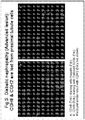
Figure 5 shows K-cadherin (CDH6) immunofluorescence staining in human kidney biopsy of diabetic nephropathy (Advanced lesion) with markers of proximal tubule (megalin) and distal tubule (Calbindin D28K) respectively. K-cadherin (megalin-PT) (A) and K-cadherin (calbindin D28K) (B) staining in human proximal tubule of diabetic nephropathy with advanced lesion was completely lost from the proximal tubule. Similar findings were observed for N-cadherin in advanced lesions of proximal tubule of diabetic nephropathy (data not shown).
For each ofFigures 2 to 5 , in both parts A and B, the Tritc-stained image is at the top right when the Figure is orientated upright, the Fitc-stained image is at the bottom left, the top left image shows nucleus staining and the bottom right image is a composite of the others. -
Figure 6 shows a Western Immuno-blot of a subset of urine samples showing urinary full length K-cadherin in patients with diabetic CKD. The table and graph show the number and percentage of patients in each group with detectable K-cadherin in the urine on enrolment in the study. Around 30% of diabetic patients with an eGFR≥60mls/min (Early CKD) had detectable urinary K-cadherin as compared to almost 70% patients with an eGFR<60 mls/min (Advanced CKD) as compared to none in healthy controls. -
Figure 7 shows a Western Immuno-blot for urinary N-cadherin. In contrast to urinary full length K-cadherin, full length N-cadherin was almost universally present in diabetic patients with an eGFR≥60mls/min (Early CKD), with an eGFR<60 mls/min (Advanced CKD) as well as in urine of healthy controls. -
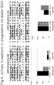
Figure 8 shows urinary K-cadherin and albumin in patients with progressive CKD as compared to stable CKD in early CKD (eGFR≥60mls/min) versus advanced CKD (eGFR<60mls/min). Presence of both K-cadherin and albumin in urine predict progression of CKD over 3 years follow up. -
Figure 9 shows the presence or absence of K-cadherin or albumin in the urine of patients with an eGFR≥ 60mls/min (early CKD) or eGFR<60mls/min (advanced CKD) plotted using a Kaplan-Meier survival curve, where an event was defined as a progression of CKD. For each of the graphs the 'positive' (K-cad/albumin) line was the lower line. Early CKD patients with K-cadherin in the urine had a statistically significant risk of progression of CKD over the follow up period (p<0.0001, HR-75.18 (95% CI of ratio 9.289 to 608.5)), similar to early CKD patients with albumin in the urine over this follow up period (p<0.05, HR 16.36 (95% CI of ratio 1.675 to 159.9)). In contrast to the early CKD group, neither advanced CKD patients with K-cadherin nor advanced CKD patients with albumin in the urine had a statistically significant risk of progression of CKD over this follow up period [(p=0.1047, HR 6.19 95% (95% CI of ratio 0.6846 to 55.97)) and (p=0.1567, HR 4.542 (95% CI of ratio 0.5594 to 36.87)) respectively]. This may be due to small the sample size of this group. -
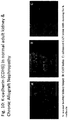
Figure 10 shows data demonstrating the loss of K-cadherin in CAN-affected kidney. -
Figure 11 shows a further representation of data demonstrating the progressive loss of K-cadherin in diabetic lesions by comparing expression in healthy kidneys and kidney tissue at various stages of disease. -
Figure 12 indicates schematically how an example ELISA assay according to the invention may operate. -
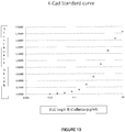
Figure 13 shows a standard curve achieved for an example ELISA assay. - Paraffin blocks of archived human kidney biopsy tissue samples were selected based on the tissue diagnosis report. Cadherin expression was investigated in the context of tubulo-interstitial damage and compared to biopsies reported as histologically normal with no evidence of tubulo-interstitial fibrosis (n=5). This work was carried in the department of Pathology, University Medical Centre, Utrecht, with local ethical approval. Briefly, 3µm thick serial sections were made from each paraffin block using a sliding microtome and the sections mounted on super frost plus slides (Thermo Scientific). The sections were fixed on a slide warmer. Deparaffinization and rehydration of the sections were performed. Blocking to remove endogenous tissue peroxidase was performed by oxidizing and using freshly prepared peroxidase block buffer (3% H2O2 in PBS) . Antigen retrieval was performed using Tris/EDTA (pH9.0) at boiling temperature for 20 min. The sections were incubated over night at 4°C with monoclonal antibodies against extracellular domain of K-cadherin (diluted 1:40 in Phosphate Buffered Saline, Fischer Thermo-scientific, UK, Cadherin K Antibody 2B6), N-cadherin (1:400, Sigma-Aldrich, UK) and E-cadherin (1:40, Novocastra Laboratories, UK) along with polyclonal antibodies against markers of proximal tubule cells(megalin (1:50, Santa Cruz Biotechnologies, USA)) and distal tubule cells (Calbindin D28K (1:20, Santa Cruz Biotechnologies, USA)). For calbindin D28K, donkey anti-goat AlexaFluor 488 (FITC) (1:200, Invitrogen, USA) was used as secondary antibody. All other primary antibodies were detected using poly-HRP-anti-rabbit/mouse/goat IgG Power Vision (Immunologic, Duiven, The Netherlands), according to manufacturer's instructions, for an hour at room temperature and amplified using the TSA (Tyramide signal amplification) tetra methyl rhodamine system (Tritc) or TSA fluorescein system (Fitc) (Perkin Elmer Life Sciences; Waltham, MA) diluted 1:50 with the amplification buffer. All sections were analyzed using a digital high resolution epi-fluorescence microscope (Leica Microsystems; Rijswijk, The Netherlands).
- Expression profile of cadherins in human kidney.
- Immuno-staining of normal human biopsy tissue revealed expression of K and N-cadherin in the proximal tubule; as determined by co-localisation with the large endocytosis receptor megalin (
Figures 1 and2 ). E-cadherin was expressed in the distal tubule; determined by co-localisation with calbindin D28K (a marker of the distal tubule) (Figure 3 ). Confocal laser scanning microscopy indicated baso-lateral staining for K- and N-cadherins using antibodies targeting the extracellular domain (ECD) (Figure 1C ). This highlights the species specificity of cadherin expression as E-cadherin is expressed in the proximal tubule of rodents (data not shown) but not in humans. - In early diabetic nephropathy lesions, expression of K- and N-cadherin on the basal side was hazy and discontinuous (
Figure 4 ). In advanced lesions with marked tubulo-interstitial fibrosis, K- and N-cadherin expression was lost (Figure 5 ).Figure 11 brings such data together and shows the progressive nature of K-cadherin loss. E-cadherin expression was also lost but only in sections exhibiting a phenotype characteristic of later stages of tubulo-interstitial fibrosis (data not shown). - Based on unpublished in vitro findings (from previous work and work by others) that N-cadherin is up-regulated by the powerful fibrotic growth factor TGFβ1, it was also expected that N-cadherin would be up regulated in patients with advanced tubulo-interstitial fibrosis. Instead, N-cadherin down-regulation in kidney biopsies from patients with diabetic CKD was found.
This study demonstrates for the first time that expression of K- and N-cadherin are altered in the proximal tubule epithelia in early diabetic CKD and that the loss is progressive with advancing tubulo-interstitial fibrosis. Further data suggests that this phenomenon is true irrespective of the etiology of the fibrotic process, e.g. similar K-cadherin loss was observed with advancing IgA nephropathy and chronic allograft nephropathy (CAN) (Figure 10 shows results for CAN). Moreover, loss of K-cadherin was observed also in an in vitro model of fibrosis using primary human proximal tubule cells. - This Example also demonstrates that the predominant cadherin in human proximal tubules is K-cadherin and not E-cadherin.
- Urine from a UK cohort of diabetic patients with and without CKD was analysed for K and N-Cadherin; local ethics committee approval was obtained.
- Healthy non-diabetic controls, age range of 20-50, were asked to complete the "Diabetes Risk Score" (Lindstrom & Tuomilehto Diabetes Care 2003); only subjects scoring less than 9 were asked to supply a urine sample. Subjects scoring more than 9 were advised of the relevance and advised to contact their general practitioner.
- Approximately 30 mls of urine was collected in a sterile universal container, divided immediately into 2 mls aliquots and stored at -80°C until further analysis. Urine from 24 diabetic patients with eGFR ≥60 mls/min (CKD stages < 3 UK Renal Association guideline), 12 diabetic patients with eGFR<60mls/min (CKD stages ≥3 UK Renal Association guideline) and 12 healthy controls was analysed. The eGFR levels were calculated at the time of urine collection. Anonymised demographic data along with renal functions (creatinine and urine PCR) and HbAlC was tabulated (see Table 1 below). 3-years follow up data was collected for these subjects to assess progression of CKD. Progressors were defined according to the Renal Association guideline; i.e. those in which eGFR as calculated by the MDRD method (abbreviated Modification of Diet in Renal Disease equation: 186 x (Creat / 88.4)-1.114 x (Age)-0.203 x (0.742 if female) x (1.210 if black)) dropped more than 2mls/min/6months or 5mls/min/year.
Table 1: Demographic & clinical data for the subject groups studied. mean eGFR≥60ml/min mean ± SD eGFR<60ml/min mean ± SD Control Age Range (Y) 32-80 31-80 25-60 Male: Female 13:11 7:5 7:3 Diabetes Type 2 : Type 121:3 9:3 None HbAlC 8.44 (1.75) 8.02 (1.43) N/A Mean (STD) Urine ACR (mg/mmol) 5.1 (9.88) 109.33 (177.98) <2.5 - Table 1: The clinical and demographic data for subjects whose urine was analysed for K and N-cadherin. The clinical characteristics among various groups; diabetic patients without CKD, diabetics with CKD stage <3 and diabetic with CKD stage ≥3 is indicated in tabulated form. The clinical characteristics are shown as mean ± SD among various groups; diabetic patients with CKD stage <3 (eGFR≥60 ml/min) and diabetic patients with CKD stage ≥3 (eGFR <60 ml/min). The non-diabetic healthy controls were volunteers with an age range of 20-50. Diabetes control (blood sugar level control) was fair and not different between diabetic patients with CKD stage <3 and diabetic patients with CKD stage ≥3. As one might predict, diabetic patients with CKD stage ≥3 had higher urinary albumin, RBP and NAG.
- An aliquot of urine was defrosted, briefly vortexed and spun at 10,000 rpm to remove any particulate matter. 1.5 ml of spun urine supernatant was then transferred to an Amicon® Ultra-4 10K centrifugal filter (10,000 Nominal Molecular Weight Limit, Millipore Corporation, USA) and centrifuged at 3000g for 20-25 min to concentrate the
sample 6 fold. - Urinary K-cadherin was detected by Western Immunoblotting. The samples were prepared for gel electrophoresis by taking 65µl of concentrated urine and adding 25µl of Western sample buffer(x4) and 10µl of reducing agent(x10) (Invitrogen; NP0008, NP0004) and denaturing at 70°C for 10 min. 40µl of samples prepared were loaded on to a 4-12% Bis-Tris gel for electrophoresis followed by immunoblotting. The blot was incubated with mouse monoclonal antibodies against extracellular domain of K-cadherin (diluted 1:1000 in Tris-buffered saline with 0.1% triton and 5% Bovine Serum Albumin, Fischer Thermo-scientific Ltd, UK, Cadherin K Antibody 2B6) and N-cadherin (1:1000, Santa Cruz Biotechnologies, USA). The protein was detected using a rabbit polyclonal anti-mouse IgG (Whole molecule) HRP labelled antibody (Sigma-Aldrich, UK) and Amersham ECL Plus™ Western Blotting Detection Reagents (GE Healthcare Life Sciences, UK) according to the manufacturer's recommendations. The quantification of the band was performed by ImageQuant (GE Healthcare Life Sciences, UK) .
- Western Immunoblot analysis of urinary full length K and N-cadherin.
- An example Western blot probed with an antibody to the extracellular domain of K-cadherin is shown in
Figure 6 , with primary human proximal tubule whole cell lysate as a positive control. The whole molecule of K-cadherin is detectable (∼120 kDa) in the urine from diabetic patients (as judged by gel migration with respect to known standards), with an increasing rate of detection correlating with severity of CKD stages (Figure 6 ). None of the non-diabetic healthy controls or diabetics with no CKD (data not shown) had detectable K-cadherin in the urine. 75% (9 out of 12) of diabetic subjects with CKD stages ≥3 had detectable K-cadherin in the urine as compared to 29% (7 out of 24) of subjects with CKD<3 and this difference was statistically different (Figure 8 ). - Western blotting for N-cadherin detected full length protein (∼140 kDa) in the urine from all subjects, patients with diabetes and controls (
Figure 7 ). This data was not analysed further. - This study, using a separate cohort of patients with respect to Example 1, demonstrates that K-cadherin can be found in the urine of diabetic patients with chronic kidney disease, consistent with the loss of expression in the proximal tubule described in Example 1. K-cadherin was not detectable in the urine of any of the healthy controls.
- It was hypothesised that cellular degradation and death might lead to cleavage of the K-cadherin molecule and excretion of only the extra-cellular domain in the urine, the cytoplasmic domain being endocytosed and recycled or degraded in the proteasome. Surprisingly, however, full length K-cadherin (as judged by electrophoresis migration) was found in the urine of patients with diabetic CKD, implying release of intact full length K-cadherin from proximal tubular epithelial cells exposed to fibrotic stimuli. Moreover, full length N-cadherin protein was present in urine of all subjects and did not correlate with CKD, which contrasts with the N-cadherin down-regulation in kidney biopsies from patients with diabetic CKD shown in Example 1. This might relate to its wide-spread expression, particularly in the lower urinary tract and in the prostate.
- Patients were followed up for up to three years to assess disease progression; one subject was lost from the study during this period.
- The level of urine albumin was also assessed for all subjects at the baseline and subsequent time points.
- Values were expressed as means ± SD. Diabetic patients with and without various stages of CKD were further stratified into groups based on Progressors and non-Progressors (change of eGFR during 3 years of follow up) and presence or absence of detectable K-cadherin and albumin in the urine at baseline. The difference between groups was evaluated using Chi-Square test in a 2x2 contingency table. All tests were two tailed and p<0.05 was considered significant. Kaplan-Meier event free survival curves were plotted with progression of CKD as an event and presence or absence of K-cadherin or albumin in the urine using Graphpad Prism software. Comparison of these curves was done using log-rank (Mantel-Cox) test. p<0.05 was considered significant. Hazard ratio was calculated at 95% confidence interval. Sensitivity and specificity of urinary K-cadherin and albumin in predicting progression of kidney disease were calculated using standard statistical equations.
- Approximately 17% (4 out of 24) diabetic subjects with CKD<3 had progression of their CKD over the 3 year follow up period. All 4 patients had detectable K-cadherin in the urine at the start of the study while only 2 out of these 4 patients had albuminuria (ACR >3). Of those who had stable renal function over this period, 15% (3 out of 20) subjects had detectable K-cadherin in the urine and albuminuria.
- Presence or absence of K-cadherin or albumin in the urine of these patients was plotted using Kaplan-Meier survival curve where an event was defined as a progression of CKD (
Figure 9 ). Patients with K-cadherin in the urine had a statistically significant risk of progression of CKD over this follow up period (p<0.0001, HR-75.18 (95% CI of ratio 9.289 to 608.5)), similar to patients with albumin in the urine over this follow up period (p<0.05, HR 16.36 (95% CI of ratio 1.675 to 159.9)) (Figure 9 ). (HR stands for hazard ratio, CI stands for confidence interval.) - None of the subjects who were negative for urinary K-cadherin at baseline in this group progressed over this follow up period.
- In this pilot study, the sensitivity and specificity of urinary K-cadherin for predicting progression of CKD in the CKD <3 group was 100% and 85% respectively whereas that of albuminuria was 50% and 85%. (Sensitivity relates to the ratio of patients who had K-cadherin in urine and progressed to all patients that progressed; specificity relates to the ratio of patients who did not have K-cadherin and did not progress to all patients who did not progress. These are usually expressed as percentages.)
- 41.7% subjects (5 out of 12) from this group had progression of their CKD. All 5 subjects had detectable urinary K-cadherin and albuminuria at baseline. In those with stable CKD (58.3%, 7 out of 12), 42.85% (3 out of 7) and 71.43% (5 out of 7) subjects had detectable urinary K-cadherin and albuminuria respectively (
Figure 8 ). - Presence or absence of K-cadherin or albumin in the urine of these patients was plotted using Kaplan-Meier survival curve where an event was defined as a progression of CKD (
Figure 9 ). Neither patients with K-cadherin in the urine nor patients with albumin in the urine had a statistically significant risk of progression of CKD over this follow up period [(p=0.1047, HR 6.19 95% (95% CI of ratio 0.6846 to 55.97)) and (p=0.1567, HR 4.542 (95% CI of ratio 0.5594 to 36.87)) respectively] (Figure 9 ). This may be due to small sample size in this group. - None of the subjects who were negative for urinary K-cadherin at the start of the study in this group progressed over this follow up period.
- In this pilot study, the sensitivity and specificity of urinary K-cadherin for predicting progression of CKD in the CKD ≥3 group was 100% and 57% respectively, against that of albuminuria which was 100% and 29%.
- This Example shows that the presence of urinary K-cadherin correlates with the stage of chronic kidney disease and an increased proportion of patients with detectable urinary k-cadherin had progression of disease over follow-up. Moreover, the prevalence of K-cadherin has a better predictive value than does albuminuria for progression of chronic kidney disease in patients with 'early' diabetic CKD. A similar trend was also noted for patients with advanced CKD, however it is believed that the small sample size prevented this from being found to be statistically significant. Accordingly, K-cadherin is considered to have great value as a marker for separating kidney disease subjects at risk of progression from non-progressors.
- Furthermore, the consideration that within the study it can definitely be said that type II diabetes subjects, at both 'early' and 'advanced' disease stages, progressed and had urinary K-cadherin as a marker at baseline, suggests that it may be particularly applicable within the type II diabetic patient population.
- An ELISA system as indicated in
Figure 12 was devised using commercial reagents. Accordingly, a sandwich ELISA with a capture antibody and a detection antibody, one that targets the near C-terminus and one that targets the N-terminus was established so as to detect the full length molecule. In this example the C-terminal antibody was adhered to the plate wells and used as the capture antibody. The C-terminal antibody was ab64917 purchased from Abcam and the N-terminal antibody was MA1-06305 purchased from Thermo Scientific. A standard curve of K-Cadherin concentration as inFigure 13 was obtained for the ELISA system. - The ELISA protocol is indicated below:
- Abcam Anti K-cadherin antibody (ab64917), Rabbit polyclonal Thermo Scientific Anti K-cadherin monoclonal antibody (MA1-06305), mouse
Sigma Anti mouse - Coating buffer - 500mls deionised water add 445mg NA2CO3, 1035mg NaHCO3
Assay buffer - Natural PBS with 0.1% BSA
Wash buffer - Natural PBS with 0.05% Tween 20 - Substrate reagent kit RnD (mix equal parts of A and B when needed)
Sulphuric acid (0.125M)
1. Coat plate with Aabcam Anti-K-cadherin. Dilute 1:2000 in coating buffer. 100ul/well. Incubate at 4°C overnight.
2. Make standard curve.Label 7 tubes S1-S7,Label 1 tube Blank.S1 (10,000 ng/ml) 70ul of K-cadherin stock (100ug/ml) in to 630ul of assay buffer S2 (5000 ng/ml) 350ul of S1 in to 350ul of assay buffer S3 (2500 ng/ml) 350ul of S2 in to 350ul of assay buffer. S4 (1250 ng/ml) 350ul of S3 in to 350ul of assay buffer S5 (625 ng/ml) 350ul of S4 in to 350ul of assay buffer. S6 (3125ng/ml) 350ul of S5 in to 350ul of assay buffer. S7 (156 ng/ml) 350ul of S6 in to 350ul of assay buffer. S8 (78 ng/ml) 350ul of S7 in to 350ul of assay buffer. S9 (39 ng/ml) 350ul of S8 in to 350ul of assay buffer. S10 (19.5 ng/ml) 350ul of S9 in to 350ul of assay buffer. - Use assay buffer alone as a blank
- 3. Wash
plate 3 times. 200ul/well. Blot upside down on paper towel to remove excess wash buffer. - 4. Add 100ul of standard or samples in duplicate. Incubate on plate shaker for 1 hour at room temperature.
- 5. Repeat wash,
step 2. - 6. Add Thermo scientific anti-K-cadherin antibody (mouse monoclonal). Dilute 1:2000 in assay buffer. 100ul/well. Incubate on plate shaker for 1 hour at room temperature.
- 7. Repeat wash,
step 2. - 8. Anti-mouse secondary antibody. Dilute 1:40,000 in assay buffer. 100ul/well. Incubate on plate shaker for 1 hour at room temperature.
- 9. Repeat wash,
step 2. - 10. Substrate - mix equal parts of A and B. Add 50ul/well of substrate reagent to each well. Incubate for 30-40mins.
- 11. Stop reaction with 50ul/well of sulphuric acid.
- 12. Read plate on tecan at 450nm, ref 650nm
- Using the ELISA, archived samples (collected with patients' consent to investigate novel biomarkers) from 322 diabetic CKD patients with varying degrees of renal function were analysed. The samples were concentrated 6 fold using PD mini-trap G10 columns from GE Healthcare, then diluted 6 fold in assay buffer to mitigate assay interference by urine matrix components such as organic compounds, pH and electrolytes. In order to identify progressors, national guidelines of a loss in GFR of 5ml/min over 1 year or a loss of GFR of 10ml/min in 5 years were followed.
- Of the 322 subjects, 172 were classified as initially having minor renal impairment and 150 as having moderate/severe renal impairment. 49% of patients with mild renal disease exhibited progression for some or all of the 3 to 5 year follow-up period and 34% of patients with moderate/severe disease exhibited progression. The majority of non-progressors in the mild renal impairment group had K-Cad values below the level of detection, a much smaller number of progressors also had values below the level of detection; all such values were classified as 0 K-Cadherin.
- In patients with moderate/severe disease, progressors had a mean level of 29.9 ng/ml urinary K-Cadherin while non-progressors had 15.5 ng/ml (p<0.01). Further, in patients with mild disease non-progressors had a mean urinary K-Cadherin of 6.3 ng/ml and the mean concentration in progressors was 12.2 ng/ml (p<0.05).
- Though K-cadherin was not detected in all samples, these results indicate that the ELISA is more sensitive than the Western blotting protocols described in previous examples and elevated K-cadherin is strongly associated with disease progression. Furthermore, it seems to be the case that, as measured by the ELISA, urinary concentrations in progressors are, on average, approximately double those of non-progressors for corresponding disease severity states.
Claims (12)
- An in vitro method for determining the propensity for a person's diabetic CKD (chronic kidney disease) level to progress in severity, the method comprising testing for the presence/level of K-cadherin in the person's urine to determine if urinary K-cadherin is elevated.
- A method according to claim 1 wherein the person is a type II diabetic.
- A method according to claim 1 or 2 wherein elevated K-cadherin is determined with respect to a non-progressive standard which may optionally be for a specific CKD stage, which stage may optionally be mild disease (stages 1/2).
- A method according to any preceding claim comprising using a K-cadherin cleavage-point specific antibody.
- A method according to any preceding claim comprising using western immunoblotting or an ELISA (enzyme-linked immunosorbent assay) system for testing for K-cadherin.
- A method according to claim 5 wherein the ELISA system comprises two sets of antibody samples, one immobilised for capturing K-cadherin and the other for subsequently detecting the protein.
- A method according to claim 6 wherein one antibody sample is reactive towards the N-terminal end of K-cadherin and the other is reactive towards the C-terminal end.
- A method according to any preceding claim wherein a urine sample is concentrated before the presence/level of K-cadherin is assessed.
- Use of a point of care test or kit in a method according to any preceding claim, the test or kit comprising a K-cadherin cleavage-point specific antibody or two sets of K-cadherin antibodies.
- Use according to claim 9 wherein the test or kit comprises two sets of antibodies and wherein one of the antibody sets is reactive towards the N-terminal end of K-cadherin and the other is reactive towards the C-terminal end.
- Use according to any of claims 9 to 10 wherein the kit is a kit for an immuno-precipitation assay, such as an ELISA assay.
- Use of urinary K-cadherin in an in vitro method for determining the propensity for a person's diabetic CKD (chronic kidney disease) level to progress in severity.
Applications Claiming Priority (2)
| Application Number | Priority Date | Filing Date | Title |
|---|---|---|---|
| GB201404061A GB201404061D0 (en) | 2014-03-07 | 2014-03-07 | Nephropathy biomarker |
| PCT/GB2015/050669 WO2015132612A1 (en) | 2014-03-07 | 2015-03-06 | Nephropathy biomarker |
Publications (2)
| Publication Number | Publication Date |
|---|---|
| EP3114485A1 EP3114485A1 (en) | 2017-01-11 |
| EP3114485B1 true EP3114485B1 (en) | 2020-11-18 |
Family
ID=50554700
Family Applications (1)
| Application Number | Title | Priority Date | Filing Date |
|---|---|---|---|
| EP15709334.5A Active EP3114485B1 (en) | 2014-03-07 | 2015-03-06 | Nephropathy biomarker |
Country Status (5)
| Country | Link |
|---|---|
| US (1) | US20170016914A1 (en) |
| EP (1) | EP3114485B1 (en) |
| ES (1) | ES2842110T3 (en) |
| GB (1) | GB201404061D0 (en) |
| WO (1) | WO2015132612A1 (en) |
Families Citing this family (2)
| Publication number | Priority date | Publication date | Assignee | Title |
|---|---|---|---|---|
| CN117577321A (en) | 2017-08-08 | 2024-02-20 | 费森尤斯医疗保健控股公司 | Systems and methods for treating and assessing the progression of chronic kidney disease |
| WO2025122579A1 (en) * | 2023-12-04 | 2025-06-12 | Cedars-Sinai Medical Center | Cadherin 6 expression status in determination of renal fibrosis and related uses thereof |
Citations (1)
| Publication number | Priority date | Publication date | Assignee | Title |
|---|---|---|---|---|
| WO2009055937A1 (en) * | 2007-11-01 | 2009-05-07 | The Royal Institution For The Advancement Of Learning/Mcgill University | Altered n-cadherin processing in tumor cells by furln and proproteln convertase 5a (pc5a) |
Family Cites Families (2)
| Publication number | Priority date | Publication date | Assignee | Title |
|---|---|---|---|---|
| US20110177613A1 (en) * | 2009-10-13 | 2011-07-21 | Los Angeles Biomedical Research Institute at Harbor-UCLA- Medical Center | Use of periostin as a novel biomarker |
| US20130338021A1 (en) * | 2012-06-14 | 2013-12-19 | Wisconsin Alumni Research Foundation | Methods and compositions for the detection of complications of diabetes |
-
2014
- 2014-03-07 GB GB201404061A patent/GB201404061D0/en not_active Ceased
-
2015
- 2015-03-06 EP EP15709334.5A patent/EP3114485B1/en active Active
- 2015-03-06 US US15/124,244 patent/US20170016914A1/en not_active Abandoned
- 2015-03-06 ES ES15709334T patent/ES2842110T3/en active Active
- 2015-03-06 WO PCT/GB2015/050669 patent/WO2015132612A1/en not_active Ceased
Patent Citations (1)
| Publication number | Priority date | Publication date | Assignee | Title |
|---|---|---|---|---|
| WO2009055937A1 (en) * | 2007-11-01 | 2009-05-07 | The Royal Institution For The Advancement Of Learning/Mcgill University | Altered n-cadherin processing in tumor cells by furln and proproteln convertase 5a (pc5a) |
Also Published As
| Publication number | Publication date |
|---|---|
| EP3114485A1 (en) | 2017-01-11 |
| US20170016914A1 (en) | 2017-01-19 |
| WO2015132612A1 (en) | 2015-09-11 |
| GB201404061D0 (en) | 2014-04-23 |
| ES2842110T3 (en) | 2021-07-12 |
Similar Documents
| Publication | Publication Date | Title |
|---|---|---|
| RU2688168C2 (en) | Prediction of clinical outcomes in patients with chronic obstructive pulmonary disease | |
| Fu et al. | Urinary tubular biomarkers in short-term type 2 diabetes mellitus patients: a cross-sectional study | |
| Bhensdadia et al. | Urine haptoglobin levels predict early renal functional decline in patients with type 2 diabetes | |
| CN102105794B (en) | A marker for graft failure and mortality | |
| Duan et al. | Assessment of urinary NGAL for differential diagnosis and progression of diabetic kidney disease | |
| JP5931200B2 (en) | Diagnosis of renal injury after acute event or surgical intervention based on L-FABP | |
| Rodrigues et al. | Definitions, phenotypes, and subphenotypes in acute kidney injury—Moving towards precision medicine | |
| US20090081713A1 (en) | Peptide biomarkers predictive of renal function decline and kidney disease | |
| DK2623517T3 (en) | Urine and serum biomarkers associated with diabetic nephropathy | |
| EP2442106B1 (en) | Method for test on diabetic nephropathy | |
| CN111065925B (en) | The use of AGRIN in the diagnosis of endometrial cancer | |
| Munir et al. | Rapid detection of acute kidney injury by urinary neutrophil gelatinase-associated lipocalin after cardiopulmonary bypass surgery | |
| KR20150096728A (en) | Acute kidney injury | |
| EP2960655B1 (en) | Method for estimating glomerular filtration rate from measurement value of megalin in urine | |
| US20120115747A1 (en) | Methods Of Renal Cancer Detection | |
| US20170016914A1 (en) | Nephropathy biomarker | |
| JP7162104B2 (en) | Examination method enabling specific diagnosis of early pathology of diabetic nephropathy | |
| EP2391653B1 (en) | Biomarkers associated with nephropathy | |
| Reznichenko et al. | Uromodulin in renal transplant recipients: elevated urinary levels and bimodal association with graft failure | |
| Patel et al. | Urinary periostin as an early predictor of renal injury in diabetic kidney disease: a tertiary centre experience. | |
| Funayama et al. | Renal tubulointerstitial damage is associated with short-term cardiovascular events in patients with myocardial infarction | |
| US20260043813A1 (en) | Chronic kidney disease detection | |
| HR et al. | UTILITY OF SERUM KIDNEY INJURY MOLECULE-1 (KIM-1) AS A DIAGNOSTIC TOOL FOR EARLY MARKER FOR DIABETIC NEPHROPATHY IN PATIENTS WITH TYPE-2 DIABETES MELLITUS | |
| JP2020020755A (en) | Method of diagnosing liver cirrhosis, method of diagnosing complications between non-alcoholic steatohepatitis and hepatocellular cancer, and method of diagnosing complications between non-alcoholic steatohepatitis and esophagogastric varices | |
| KR102065545B1 (en) | Composition for predicting renal fibrosis and the decline in kidney function in patients with diabetic nephropathy |
Legal Events
| Date | Code | Title | Description |
|---|---|---|---|
| STAA | Information on the status of an ep patent application or granted ep patent |
Free format text: STATUS: THE INTERNATIONAL PUBLICATION HAS BEEN MADE |
|
| PUAI | Public reference made under article 153(3) epc to a published international application that has entered the european phase |
Free format text: ORIGINAL CODE: 0009012 |
|
| STAA | Information on the status of an ep patent application or granted ep patent |
Free format text: STATUS: REQUEST FOR EXAMINATION WAS MADE |
|
| 17P | Request for examination filed |
Effective date: 20161006 |
|
| AK | Designated contracting states |
Kind code of ref document: A1 Designated state(s): AL AT BE BG CH CY CZ DE DK EE ES FI FR GB GR HR HU IE IS IT LI LT LU LV MC MK MT NL NO PL PT RO RS SE SI SK SM TR |
|
| AX | Request for extension of the european patent |
Extension state: BA ME |
|
| DAV | Request for validation of the european patent (deleted) | ||
| DAX | Request for extension of the european patent (deleted) | ||
| STAA | Information on the status of an ep patent application or granted ep patent |
Free format text: STATUS: EXAMINATION IS IN PROGRESS |
|
| 17Q | First examination report despatched |
Effective date: 20170728 |
|
| GRAP | Despatch of communication of intention to grant a patent |
Free format text: ORIGINAL CODE: EPIDOSNIGR1 |
|
| STAA | Information on the status of an ep patent application or granted ep patent |
Free format text: STATUS: GRANT OF PATENT IS INTENDED |
|
| INTG | Intention to grant announced |
Effective date: 20200528 |
|
| GRAS | Grant fee paid |
Free format text: ORIGINAL CODE: EPIDOSNIGR3 |
|
| GRAA | (expected) grant |
Free format text: ORIGINAL CODE: 0009210 |
|
| STAA | Information on the status of an ep patent application or granted ep patent |
Free format text: STATUS: THE PATENT HAS BEEN GRANTED |
|
| AK | Designated contracting states |
Kind code of ref document: B1 Designated state(s): AL AT BE BG CH CY CZ DE DK EE ES FI FR GB GR HR HU IE IS IT LI LT LU LV MC MK MT NL NO PL PT RO RS SE SI SK SM TR |
|
| REG | Reference to a national code |
Ref country code: GB Ref legal event code: FG4D |
|
| REG | Reference to a national code |
Ref country code: CH Ref legal event code: EP |
|
| REG | Reference to a national code |
Ref country code: DE Ref legal event code: R096 Ref document number: 602015062109 Country of ref document: DE |
|
| REG | Reference to a national code |
Ref country code: IE Ref legal event code: FG4D |
|
| REG | Reference to a national code |
Ref country code: AT Ref legal event code: REF Ref document number: 1336331 Country of ref document: AT Kind code of ref document: T Effective date: 20201215 |
|
| REG | Reference to a national code |
Ref country code: AT Ref legal event code: MK05 Ref document number: 1336331 Country of ref document: AT Kind code of ref document: T Effective date: 20201118 |
|
| PG25 | Lapsed in a contracting state [announced via postgrant information from national office to epo] |
Ref country code: GR Free format text: LAPSE BECAUSE OF FAILURE TO SUBMIT A TRANSLATION OF THE DESCRIPTION OR TO PAY THE FEE WITHIN THE PRESCRIBED TIME-LIMIT Effective date: 20210219 Ref country code: RS Free format text: LAPSE BECAUSE OF FAILURE TO SUBMIT A TRANSLATION OF THE DESCRIPTION OR TO PAY THE FEE WITHIN THE PRESCRIBED TIME-LIMIT Effective date: 20201118 Ref country code: FI Free format text: LAPSE BECAUSE OF FAILURE TO SUBMIT A TRANSLATION OF THE DESCRIPTION OR TO PAY THE FEE WITHIN THE PRESCRIBED TIME-LIMIT Effective date: 20201118 Ref country code: NO Free format text: LAPSE BECAUSE OF FAILURE TO SUBMIT A TRANSLATION OF THE DESCRIPTION OR TO PAY THE FEE WITHIN THE PRESCRIBED TIME-LIMIT Effective date: 20210218 Ref country code: PT Free format text: LAPSE BECAUSE OF FAILURE TO SUBMIT A TRANSLATION OF THE DESCRIPTION OR TO PAY THE FEE WITHIN THE PRESCRIBED TIME-LIMIT Effective date: 20210318 |
|
| PG25 | Lapsed in a contracting state [announced via postgrant information from national office to epo] |
Ref country code: AT Free format text: LAPSE BECAUSE OF FAILURE TO SUBMIT A TRANSLATION OF THE DESCRIPTION OR TO PAY THE FEE WITHIN THE PRESCRIBED TIME-LIMIT Effective date: 20201118 Ref country code: BG Free format text: LAPSE BECAUSE OF FAILURE TO SUBMIT A TRANSLATION OF THE DESCRIPTION OR TO PAY THE FEE WITHIN THE PRESCRIBED TIME-LIMIT Effective date: 20210218 Ref country code: IS Free format text: LAPSE BECAUSE OF FAILURE TO SUBMIT A TRANSLATION OF THE DESCRIPTION OR TO PAY THE FEE WITHIN THE PRESCRIBED TIME-LIMIT Effective date: 20210318 Ref country code: LV Free format text: LAPSE BECAUSE OF FAILURE TO SUBMIT A TRANSLATION OF THE DESCRIPTION OR TO PAY THE FEE WITHIN THE PRESCRIBED TIME-LIMIT Effective date: 20201118 Ref country code: SE Free format text: LAPSE BECAUSE OF FAILURE TO SUBMIT A TRANSLATION OF THE DESCRIPTION OR TO PAY THE FEE WITHIN THE PRESCRIBED TIME-LIMIT Effective date: 20201118 Ref country code: PL Free format text: LAPSE BECAUSE OF FAILURE TO SUBMIT A TRANSLATION OF THE DESCRIPTION OR TO PAY THE FEE WITHIN THE PRESCRIBED TIME-LIMIT Effective date: 20201118 |
|
| REG | Reference to a national code |
Ref country code: LT Ref legal event code: MG9D |
|
| PG25 | Lapsed in a contracting state [announced via postgrant information from national office to epo] |
Ref country code: HR Free format text: LAPSE BECAUSE OF FAILURE TO SUBMIT A TRANSLATION OF THE DESCRIPTION OR TO PAY THE FEE WITHIN THE PRESCRIBED TIME-LIMIT Effective date: 20201118 |
|
| REG | Reference to a national code |
Ref country code: ES Ref legal event code: FG2A Ref document number: 2842110 Country of ref document: ES Kind code of ref document: T3 Effective date: 20210712 |
|
| RAP2 | Party data changed (patent owner data changed or rights of a patent transferred) |
Owner name: SOUTH WEST THAMES INSTITUTE FOR RENAL RESEARCH |
|
| PG25 | Lapsed in a contracting state [announced via postgrant information from national office to epo] |
Ref country code: LT Free format text: LAPSE BECAUSE OF FAILURE TO SUBMIT A TRANSLATION OF THE DESCRIPTION OR TO PAY THE FEE WITHIN THE PRESCRIBED TIME-LIMIT Effective date: 20201118 Ref country code: SK Free format text: LAPSE BECAUSE OF FAILURE TO SUBMIT A TRANSLATION OF THE DESCRIPTION OR TO PAY THE FEE WITHIN THE PRESCRIBED TIME-LIMIT Effective date: 20201118 Ref country code: RO Free format text: LAPSE BECAUSE OF FAILURE TO SUBMIT A TRANSLATION OF THE DESCRIPTION OR TO PAY THE FEE WITHIN THE PRESCRIBED TIME-LIMIT Effective date: 20201118 Ref country code: SM Free format text: LAPSE BECAUSE OF FAILURE TO SUBMIT A TRANSLATION OF THE DESCRIPTION OR TO PAY THE FEE WITHIN THE PRESCRIBED TIME-LIMIT Effective date: 20201118 Ref country code: CZ Free format text: LAPSE BECAUSE OF FAILURE TO SUBMIT A TRANSLATION OF THE DESCRIPTION OR TO PAY THE FEE WITHIN THE PRESCRIBED TIME-LIMIT Effective date: 20201118 Ref country code: EE Free format text: LAPSE BECAUSE OF FAILURE TO SUBMIT A TRANSLATION OF THE DESCRIPTION OR TO PAY THE FEE WITHIN THE PRESCRIBED TIME-LIMIT Effective date: 20201118 |
|
| REG | Reference to a national code |
Ref country code: DE Ref legal event code: R097 Ref document number: 602015062109 Country of ref document: DE |
|
| REG | Reference to a national code |
Ref country code: DE Ref legal event code: R081 Ref document number: 602015062109 Country of ref document: DE Owner name: SOUTH WEST THAMES INSTITUTE FOR RENAL RESEARCH, GB Free format text: FORMER OWNER: EPSOM AND ST HELIER UNIVERSITY HOSPITALS NHS TRUST, CARSHALTON, GB |
|
| PG25 | Lapsed in a contracting state [announced via postgrant information from national office to epo] |
Ref country code: DK Free format text: LAPSE BECAUSE OF FAILURE TO SUBMIT A TRANSLATION OF THE DESCRIPTION OR TO PAY THE FEE WITHIN THE PRESCRIBED TIME-LIMIT Effective date: 20201118 |
|
| REG | Reference to a national code |
Ref country code: NL Ref legal event code: PD Owner name: SOUTH WEST THAMES INSTITUTE FOR RENAL RESEARCH; GB Free format text: DETAILS ASSIGNMENT: CHANGE OF OWNER(S), ASSIGNMENT; FORMER OWNER NAME: EPSOM AND ST HELIER UNIVERSITY HOSPITALS NHS TRUST Effective date: 20210806 Ref country code: NL Ref legal event code: FP |
|
| REG | Reference to a national code |
Ref country code: GB Ref legal event code: 732E Free format text: REGISTERED BETWEEN 20210819 AND 20210825 |
|
| PLBE | No opposition filed within time limit |
Free format text: ORIGINAL CODE: 0009261 |
|
| STAA | Information on the status of an ep patent application or granted ep patent |
Free format text: STATUS: NO OPPOSITION FILED WITHIN TIME LIMIT |
|
| REG | Reference to a national code |
Ref country code: BE Ref legal event code: PD Owner name: SOUTH WEST THAMES INSTITUTE FOR RENAL RESEARCH; GB Free format text: DETAILS ASSIGNMENT: CHANGE OF OWNER(S), ASSIGNMENT; FORMER OWNER NAME: EPSOM AND ST HELIER UNIVERSITY HOSPITALS NHS TRUST Effective date: 20210812 |
|
| 26N | No opposition filed |
Effective date: 20210819 |
|
| PG25 | Lapsed in a contracting state [announced via postgrant information from national office to epo] |
Ref country code: MC Free format text: LAPSE BECAUSE OF FAILURE TO SUBMIT A TRANSLATION OF THE DESCRIPTION OR TO PAY THE FEE WITHIN THE PRESCRIBED TIME-LIMIT Effective date: 20201118 Ref country code: AL Free format text: LAPSE BECAUSE OF FAILURE TO SUBMIT A TRANSLATION OF THE DESCRIPTION OR TO PAY THE FEE WITHIN THE PRESCRIBED TIME-LIMIT Effective date: 20201118 |
|
| REG | Reference to a national code |
Ref country code: CH Ref legal event code: PL |
|
| PG25 | Lapsed in a contracting state [announced via postgrant information from national office to epo] |
Ref country code: SI Free format text: LAPSE BECAUSE OF FAILURE TO SUBMIT A TRANSLATION OF THE DESCRIPTION OR TO PAY THE FEE WITHIN THE PRESCRIBED TIME-LIMIT Effective date: 20201118 |
|
| PG25 | Lapsed in a contracting state [announced via postgrant information from national office to epo] |
Ref country code: IE Free format text: LAPSE BECAUSE OF NON-PAYMENT OF DUE FEES Effective date: 20210306 Ref country code: CH Free format text: LAPSE BECAUSE OF NON-PAYMENT OF DUE FEES Effective date: 20210331 Ref country code: LU Free format text: LAPSE BECAUSE OF NON-PAYMENT OF DUE FEES Effective date: 20210306 Ref country code: LI Free format text: LAPSE BECAUSE OF NON-PAYMENT OF DUE FEES Effective date: 20210331 |
|
| PG25 | Lapsed in a contracting state [announced via postgrant information from national office to epo] |
Ref country code: IS Free format text: LAPSE BECAUSE OF FAILURE TO SUBMIT A TRANSLATION OF THE DESCRIPTION OR TO PAY THE FEE WITHIN THE PRESCRIBED TIME-LIMIT Effective date: 20210318 |
|
| PG25 | Lapsed in a contracting state [announced via postgrant information from national office to epo] |
Ref country code: HU Free format text: LAPSE BECAUSE OF FAILURE TO SUBMIT A TRANSLATION OF THE DESCRIPTION OR TO PAY THE FEE WITHIN THE PRESCRIBED TIME-LIMIT; INVALID AB INITIO Effective date: 20150306 |
|
| PG25 | Lapsed in a contracting state [announced via postgrant information from national office to epo] |
Ref country code: CY Free format text: LAPSE BECAUSE OF FAILURE TO SUBMIT A TRANSLATION OF THE DESCRIPTION OR TO PAY THE FEE WITHIN THE PRESCRIBED TIME-LIMIT Effective date: 20201118 |
|
| PG25 | Lapsed in a contracting state [announced via postgrant information from national office to epo] |
Ref country code: MK Free format text: LAPSE BECAUSE OF FAILURE TO SUBMIT A TRANSLATION OF THE DESCRIPTION OR TO PAY THE FEE WITHIN THE PRESCRIBED TIME-LIMIT Effective date: 20201118 |
|
| PG25 | Lapsed in a contracting state [announced via postgrant information from national office to epo] |
Ref country code: TR Free format text: LAPSE BECAUSE OF FAILURE TO SUBMIT A TRANSLATION OF THE DESCRIPTION OR TO PAY THE FEE WITHIN THE PRESCRIBED TIME-LIMIT Effective date: 20201118 |
|
| PG25 | Lapsed in a contracting state [announced via postgrant information from national office to epo] |
Ref country code: MT Free format text: LAPSE BECAUSE OF FAILURE TO SUBMIT A TRANSLATION OF THE DESCRIPTION OR TO PAY THE FEE WITHIN THE PRESCRIBED TIME-LIMIT Effective date: 20201118 |
|
| PGFP | Annual fee paid to national office [announced via postgrant information from national office to epo] |
Ref country code: DE Payment date: 20250328 Year of fee payment: 11 |
|
| PGFP | Annual fee paid to national office [announced via postgrant information from national office to epo] |
Ref country code: IT Payment date: 20250321 Year of fee payment: 11 |
|
| PGFP | Annual fee paid to national office [announced via postgrant information from national office to epo] |
Ref country code: ES Payment date: 20250401 Year of fee payment: 11 |
|
| PGFP | Annual fee paid to national office [announced via postgrant information from national office to epo] |
Ref country code: GB Payment date: 20260330 Year of fee payment: 12 |
|
| PGFP | Annual fee paid to national office [announced via postgrant information from national office to epo] |
Ref country code: BE Payment date: 20260330 Year of fee payment: 12 |
|
| PGFP | Annual fee paid to national office [announced via postgrant information from national office to epo] |
Ref country code: NL Payment date: 20260330 Year of fee payment: 12 |
|
| PGFP | Annual fee paid to national office [announced via postgrant information from national office to epo] |
Ref country code: FR Payment date: 20260331 Year of fee payment: 12 |