EP3107066B1 - Method of quantitative magnetic resonance lung imaging - Google Patents
Method of quantitative magnetic resonance lung imaging Download PDFInfo
- Publication number
- EP3107066B1 EP3107066B1 EP15001782.0A EP15001782A EP3107066B1 EP 3107066 B1 EP3107066 B1 EP 3107066B1 EP 15001782 A EP15001782 A EP 15001782A EP 3107066 B1 EP3107066 B1 EP 3107066B1
- Authority
- EP
- European Patent Office
- Prior art keywords
- ventilation
- lung
- images
- quantitative
- image
- Prior art date
- Legal status (The legal status is an assumption and is not a legal conclusion. Google has not performed a legal analysis and makes no representation as to the accuracy of the status listed.)
- Active
Links
Images

Classifications
-
- A—HUMAN NECESSITIES
- A61—MEDICAL OR VETERINARY SCIENCE; HYGIENE
- A61B—DIAGNOSIS; SURGERY; IDENTIFICATION
- A61B5/00—Measuring for diagnostic purposes; Identification of persons
- A61B5/08—Detecting, measuring or recording devices for evaluating the respiratory organs
-
- A—HUMAN NECESSITIES
- A61—MEDICAL OR VETERINARY SCIENCE; HYGIENE
- A61B—DIAGNOSIS; SURGERY; IDENTIFICATION
- A61B5/00—Measuring for diagnostic purposes; Identification of persons
- A61B5/72—Signal processing specially adapted for physiological signals or for diagnostic purposes
- A61B5/7271—Specific aspects of physiological measurement analysis
- A61B5/7282—Event detection, e.g. detecting unique waveforms indicative of a medical condition
-
- A—HUMAN NECESSITIES
- A61—MEDICAL OR VETERINARY SCIENCE; HYGIENE
- A61B—DIAGNOSIS; SURGERY; IDENTIFICATION
- A61B5/00—Measuring for diagnostic purposes; Identification of persons
- A61B5/0033—Features or image-related aspects of imaging apparatus classified in A61B5/00, e.g. for MRI, optical tomography or impedance tomography apparatus; arrangements of imaging apparatus in a room
- A61B5/004—Features or image-related aspects of imaging apparatus classified in A61B5/00, e.g. for MRI, optical tomography or impedance tomography apparatus; arrangements of imaging apparatus in a room adapted for image acquisition of a particular organ or body part
-
- A—HUMAN NECESSITIES
- A61—MEDICAL OR VETERINARY SCIENCE; HYGIENE
- A61B—DIAGNOSIS; SURGERY; IDENTIFICATION
- A61B5/00—Measuring for diagnostic purposes; Identification of persons
- A61B5/05—Detecting, measuring or recording for diagnosis by means of electric currents or magnetic fields; Measuring using microwaves or radio waves
- A61B5/055—Detecting, measuring or recording for diagnosis by means of electric currents or magnetic fields; Measuring using microwaves or radio waves involving electronic [EMR] or nuclear [NMR] magnetic resonance, e.g. magnetic resonance imaging
-
- G—PHYSICS
- G06—COMPUTING; CALCULATING OR COUNTING
- G06T—IMAGE DATA PROCESSING OR GENERATION, IN GENERAL
- G06T5/00—Image enhancement or restoration
- G06T5/50—Image enhancement or restoration by the use of more than one image, e.g. averaging, subtraction
-
- G—PHYSICS
- G06—COMPUTING; CALCULATING OR COUNTING
- G06T—IMAGE DATA PROCESSING OR GENERATION, IN GENERAL
- G06T7/00—Image analysis
- G06T7/0002—Inspection of images, e.g. flaw detection
- G06T7/0012—Biomedical image inspection
- G06T7/0014—Biomedical image inspection using an image reference approach
- G06T7/0016—Biomedical image inspection using an image reference approach involving temporal comparison
-
- A—HUMAN NECESSITIES
- A61—MEDICAL OR VETERINARY SCIENCE; HYGIENE
- A61B—DIAGNOSIS; SURGERY; IDENTIFICATION
- A61B2576/00—Medical imaging apparatus involving image processing or analysis
- A61B2576/02—Medical imaging apparatus involving image processing or analysis specially adapted for a particular organ or body part
-
- G—PHYSICS
- G06—COMPUTING; CALCULATING OR COUNTING
- G06T—IMAGE DATA PROCESSING OR GENERATION, IN GENERAL
- G06T2207/00—Indexing scheme for image analysis or image enhancement
- G06T2207/10—Image acquisition modality
- G06T2207/10016—Video; Image sequence
-
- G—PHYSICS
- G06—COMPUTING; CALCULATING OR COUNTING
- G06T—IMAGE DATA PROCESSING OR GENERATION, IN GENERAL
- G06T2207/00—Indexing scheme for image analysis or image enhancement
- G06T2207/10—Image acquisition modality
- G06T2207/10072—Tomographic images
- G06T2207/10076—4D tomography; Time-sequential 3D tomography
-
- G—PHYSICS
- G06—COMPUTING; CALCULATING OR COUNTING
- G06T—IMAGE DATA PROCESSING OR GENERATION, IN GENERAL
- G06T2207/00—Indexing scheme for image analysis or image enhancement
- G06T2207/10—Image acquisition modality
- G06T2207/10072—Tomographic images
- G06T2207/10088—Magnetic resonance imaging [MRI]
-
- G—PHYSICS
- G06—COMPUTING; CALCULATING OR COUNTING
- G06T—IMAGE DATA PROCESSING OR GENERATION, IN GENERAL
- G06T2207/00—Indexing scheme for image analysis or image enhancement
- G06T2207/20—Special algorithmic details
- G06T2207/20048—Transform domain processing
- G06T2207/20056—Discrete and fast Fourier transform, [DFT, FFT]
-
- G—PHYSICS
- G06—COMPUTING; CALCULATING OR COUNTING
- G06T—IMAGE DATA PROCESSING OR GENERATION, IN GENERAL
- G06T2207/00—Indexing scheme for image analysis or image enhancement
- G06T2207/20—Special algorithmic details
- G06T2207/20212—Image combination
- G06T2207/20216—Image averaging
-
- G—PHYSICS
- G06—COMPUTING; CALCULATING OR COUNTING
- G06T—IMAGE DATA PROCESSING OR GENERATION, IN GENERAL
- G06T2207/00—Indexing scheme for image analysis or image enhancement
- G06T2207/30—Subject of image; Context of image processing
- G06T2207/30004—Biomedical image processing
- G06T2207/30061—Lung
-
- G—PHYSICS
- G16—INFORMATION AND COMMUNICATION TECHNOLOGY [ICT] SPECIALLY ADAPTED FOR SPECIFIC APPLICATION FIELDS
- G16H—HEALTHCARE INFORMATICS, i.e. INFORMATION AND COMMUNICATION TECHNOLOGY [ICT] SPECIALLY ADAPTED FOR THE HANDLING OR PROCESSING OF MEDICAL OR HEALTHCARE DATA
- G16H30/00—ICT specially adapted for the handling or processing of medical images
- G16H30/40—ICT specially adapted for the handling or processing of medical images for processing medical images, e.g. editing
Definitions
- the invention relates to a method of image processing of magnetic resonance (MR) images of a lung (MR lung images) of a subject under investigation for creating a quantitative ventilation map of the lung.
- the invention relates to magnetic resonance tomography (MRT) based lung imaging using Fourier decomposition.
- MR lung images are processed which provide a map of local lung ventilation, without using ionising radiation and without using contrast agents.
- the invention relates to multiple sub-processes of image processing of the MR lung images, which result in an improved image quality, stability of image calculation and quantification of local distribution of ventilation in the lung. Both, the sub-processes alone and combinations thereof provide subjects of the invention.
- Applications of the invention are available in medical MR imaging, in particular in quantitative MR lung imaging.
- Magnetic resonance tomography (MRT) based lung imaging using Fourier decomposition is described e. g. in [3, 4, 10].
- a quantitative ventilation map of the lung can be created by processing MR lung images along the following procedure.
- a series of two-dimensional (2D) MR lung images is collected with an MRI scanner, e. g. with a temporal resolution of 0.3 s per image for each slice position for a period of 1 min of free ventilation.
- the MR lung images are subjected to a registration into one image representing a medium ventilation position, using a non-rigid transformation [1, 5] with transformation fields.
- the registration results in a deformation of the lung images so that all images correspond to the same ventilation position (or: ventilation status). Accordingly, a characteristic temporal signal intensity can be assigned to each lung image voxel.
- the registration process as conventionally used in the Fourier decomposition context has the following disadvantages. Images of the expiration ventilation position and the inspiration ventilation position are directly registered into the image representing the medium ventilation position. This requires a strong deformation of lung images resulting in a risk of misregistrations. Furthermore, due to the required temporal resolution, the signal to noise ratio (SNR) of the single images is relatively low. Local lung structures which are important for estimating the transformation fields are superimposed by noise, thus further impeding the registration.
- SNR signal to noise ratio
- a frequency determination and a separation of ventilation and perfusion contributions in the images is conducted.
- the signal intensity oscillates with the ventilation frequency anti-proportional to the volume change of the lung during the ventilation.
- the signal intensity is modulated by blood perfusion with a heart perfusion frequency, which is higher than the ventilation frequency.
- the ventilation and perfusion frequencies can be determined and separated by a Fourier decomposition of an averaged temporal signal intensity in frequency space [4, 9].
- the averaged temporal signal intensity reliably can be used with healthy subjects only.
- abnormal ventilation may occur. This may impede or even exclude an appropriate frequency determination.
- the Fourier decomposition requires a periodic ventilation and perfusion. With an irregular ventilation, the spectrum of ventilation is broadened in frequency space. Accordingly, the integration limits for calculating the amplitude of ventilation have to be extended, resulting in an increased noise contribution.
- Ventilation can be quantified using the parameter fractional ventilation (FV), which is calculated by a ratio of a lung tidal volume (difference of inspiration and expiration volumes) and the inspiration volume.
- FV parameter fractional ventilation
- these volumes are measured e. g. by spirome-try. Due to the relationship of signal intensity and volume, it could be considered that this ratio correspondingly can be calculated from the MR lung signal intensities.
- the integrated ventilation peak would provide the signal change caused by the tidal volume [2].
- this integration value could be scaled with the expiration signal [6].
- this approach using MR lung signal intensities does not allow an appropriate quantification process as the fractional ventilation depends on the ventilation deepness, which can vary within the series of MR lung images collected [8]. As the varying ventilation deepness cannot be compensated with the conventional techniques, the calculation of the fractional ventilation based on conventionally processed images cannot yield assessable and reproducible quantitative results.
- the conventional methods mainly suffer from limitations in SNR of registered, filtered and quantified images, so that the conventional quantitative ventilation maps have a limited information contents.
- the objective of the invention is to provide an improved method of image processing of MR lung images, which avoids disadvantages of conventional techniques.
- the objective of the invention is to provide the method of image processing of MR lung images with increased sensitivity and reproducibility, improved SNR and/or reduced loss of information contributions included in the MR lung images.
- the above objective is solved by a method of processing MR lung images of a subject under investigation, comprising the steps of providing a series of the MR lung images collected over a time interval covering multiple ventilation periods, registering the MR lung images for creating a series of registered MR lung images, determining a ventilation frequency from the registered MR lung images, creating a series of ventilation images by applying a ventilation frequency based low-pass frequency filter on the registered MR lung images, wherein the ventilation images preferably are independent of (not influenced by) the perfusion of the subject under investigation, and creating a quantitative ventilation map of the lung, which is calculated from the ventilation images.
- the step of determining the ventilation frequency includes a frequency analysis of a time series of a quantitative lung dimension parameter (area parameter), the value of which being characteristic for the dimension of the lung (size of the lung) at the time of collecting the corresponding MR lung image.
- a quantitative lung dimension parameter area parameter
- the invention is based on a construction and analysis of the time series of the quantitative lung dimension parameter, i. e. a provision of an oscillating lung area quantity in time space.
- analysing the time series of the quantitative lung dimension parameter allows determining the ventilation frequency with increased reliability and stability, compared with conventional analysing of signal intensity oscillations.
- the step of creating the quantitative ventilation map includes selecting a first group of ventilation images, which consists of expiration ventilation images collected at regular expiration phases of the multiple ventilation periods, selecting a second group of ventilation images, which consists of inspiration ventilation images collected at regular inspiration phases of the multiple ventilation periods, and calculating the quantitative ventilation map from the first and second groups of ventilation images.
- multiple ventilation periods of healthy or sick subjects include expiration and inspiration phases (or: expiration and inspiration events), which can be assigned to a regular ventilation (or: periodic, continuous ventilation).
- the regular expiration and inspiration phases can be identified in particular on the basis of the improved ventilation frequency determination of the invention.
- Expiration ventilation images collected at the regular expiration phases and inspiration ventilation images collected at the regular inspiration phases are selected for creating the quantitative ventilation map, while ventilation images of irregular ventilation phases are discarded.
- the quantitative ventilation map is adjusted with reference to the tidal volume of the lung.
- this avoids any dependency of the quantification process on the ventilation deepness.
- the SNR of the ventilation images is improved due to the improved determination of the ventilation frequency and the selection of images representing the regular ventilation.
- the quantitative ventilation map obtained with the invention includes more reliable information compared with conventional techniques.
- the quantitative ventilation map is an image wherein the intensity (brightness of the image pixels) is a direct measure for the local ventilation.
- the quantitative ventilation map created according to the invention can be evaluated by a physician in a separate step, in particular for characterizing the health status of the subject or finding a diagnosis.
- the first and second groups of ventilation images are selected by retrospective gating of the time series of the quantitative lung dimension parameter.
- the retrospective gating comprises an assignment of the values of the measured quantitative lung dimension parameter to predetermined intervals of the quantitative lung dimension parameter (gating) and a determination of regular ventilation events by counting the number of assigned values in each interval. The intervals with maximum numbers of assigned values and the ventilation images associated with the values assigned to those maximum intervals are provided as regular ventilation phases.
- the retrospective gating allows a targeted selection of ventilation images with similar ventilation deepness and a maximization of the SNR.
- the retrospective gating uses the same quantitative lung dimension parameter like the ventilation frequency determination, in particular the lung area value or the averaged ROI signal value.
- the retrospective gating and the ventilation frequency determination use different quantitative lung dimension parameters.
- a method of magnetic resonance imaging of a lung comprising the steps of collecting a series of MR image raw data for obtaining multiple MR lung images, and subjecting the MR lung images to an image processing according to the above first aspect of the invention.
- the quantitative lung dimension parameter used with the inventive method of processing MR lung images is at least one of a lung area value of the registered MR lung images and an averaged ROI signal value of a region of interest covering a part of the lung and a part of the adjacent thoracic diaphragm of the subject under investigation.
- lung area values are obtained by applying inverse transformation fields from the registered image series to one initial ROI outlining the lung parenchyma.
- ROIs for each time point of the registered image series can be obtained and the lung area value time series can be determined using the inverse transformation fields.
- This information can be used not only for the stable determination of the ventilation frequency, but also for the determination of the inspiration and expiration events and for the scaling of the FV as outlined below.
- using the averaged ROI signal value is based on the following consideration. Due to different proton densities, the lung and the thoracic diaphragm provide substantially different MRI signals.
- the ROI is arranged such that the portion of the lung within the ROI is changing in dependency on the ventilation position. Accordingly, the averaged MRI signal of the ROI directly follows the ventilation.
- the step of calculating the quantitative ventilation map includes calculating a fractional or specific ventilation image obtained from a difference between an averaged expiration image from the first group of ventilation images and an averaged inspiration image from the second group of ventilation images, divided by the averaged inspiration or expiration ventilation image.
- the quantitative ventilation map is calculated by adjusting (or: scaling) the fractional ventilation image with reference to the tidal volume of the lung, using a global fractional ventilation parameter, wherein the global fractional ventilation parameter is calculated from the lung area values of the averaged inspiration and expiration images or from measured ventilation flow values.
- the step of registering the MR lung images comprises assigning the collected MR lung images into groups (or: classes), wherein all collected MR lung images within each one of the groups have the quantitative lung dimension parameter, in particular the averaged ROI signal value, within a common predetermined parameter interval, group-wise registering the collected MR lung images within the groups of collected MR lung images, and step-wise registering the group-wise registered MR lung images or an averaged MR lung image thereof, starting from parameter intervals corresponding to expiration and inspiration ventilation positions towards a parameter interval corresponding to an intermediate ventilation position.
- the intermediate ventilation position is a medium ventilation position between expiration and inspiration.
- the problems of the conventional registration are solved by the present group- and step-wise image registration into reference images from neighbouring ventilation phases. Strong deformations of the images and misregistrations are avoided.
- the group- and step-wise image registration avoids artefacts which otherwise would impair the subsequent steps of ventilation frequency determination and quantification. While the conventional registration may result in wrong conclusions or undiscovered pathologic findings, the registration reduces this risk and even accelerates the registration process.
- the step-wise registration of dynamic time series of MR images within groups can be applied not only in combination with the method of processing MR lung images but rather generally for registering time series of MR images.
- the proposed registration process provides an independent subject of the invention wherein the registering the MR images comprises providing groups of a time series of subsequent MR images, the MR images within each group having a quantitative dimension parameter within a common predetermined parameter interval, group-wise registering the collected MR images within the groups of collected MR images, and registering the group-wise registered MR images or an averaged MR image thereof, starting from parameter intervals corresponding to extrema image positions towards a parameter interval corresponding to an intermediate image position.
- an MRI device which is configured for creating a sequence of MR images of an object under investigation.
- the MRI device comprises an MRI scanner including a main magnetic field device, at least one radiofrequency excitation coil, magnetic field gradient coils and at least one radiofrequency receiver coil, and a control device being adapted for controlling the MRI scanner for collecting the series of sets of image data and for processing the sequence of MR images with the method according to the above first aspect of the invention.
- Perfusion images which are independent of ventilation of the subject under investigation can be created by applying a high-pass filter on the registered MR lung images in frequency space. Selected perfusion images can be found and analysed which represent a regular heartbeat, for imaging systole and diastole in time space.
- Embodiments are described in the following with reference to the successive sub-processes of image processing of dynamic 2D MR lung images collected e. g. with coronal or sagittal slices of the lung, resulting in a two-dimensional quantitative ventilation map of the lung.
- the processing is repeated with further time series of 2D MR lung images representing further coronal or sagittal slices of the lung.
- three-dimensional (3D) MR lung images can be collected and subjected to the sub-processes as outlined in the following.
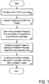
- the method comprises the steps of providing the series of MR lung images (S0), registration (S1), frequency determination (S2), separation of ventilation and perfusion (S3), and quantification (S4), which are described in the following with further details.
- Step S0 of providing the series of MR lung images preferably comprises collecting the MR lung images with an MRI scanner (see e. g. Figure 10 ), so that the image data can be directly processed for an output of the quantitative ventilation map of the lung by the MRI scanner.
- MRI scanner see e. g. Figure 10
- Details of collecting the MR lung images, operating an MRI scanner, the numerical implementation of the mathematical formulation using available software tools and optional further image pre- or post-processing steps are not described as far as they are known from conventional MRI techniques.
- usual MRI pulse sequences can be applied for collecting the images, without changes of the commercially available sequences.
- the number of images, temporal resolution and collecting period can be selected in dependency on the application of the invention. As an example, about 200 images are collected with a temporal resolution of 0.3 s per image and slice position for a collecting period of about 1 min of free ventilation.
- the series of MR lung images can be provided separately from collecting the MR lung image data.
- Previously collected images can be stored and provided, e. g. via a data communication network to a distant processor for implementing the image processing according to the invention.
- FIG. 2 illustrates the effect of image registration.
- Figure 2A shows an MR thorax image wherein the vertical black line 1 represents a cross-section line through the thoracic diaphragm.
- Figure 2B shows a periodic diaphragm oscillation resulting from the ventilation.
- Figure 2C shows the diaphragm oscillation is no longer detectable, as shown in Figure 2C .
- This deformation can be obtained by a conventional registration, as described e. g. in [2, 4]. However, preferably, the registration is implemented with the following inventive procedure, described with reference to Figures 3 and 4 .
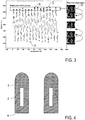
- Figure 3 shows a ventilation position curve formed by a time dependency of a quantitative parameter representing the current ventilation position for each image acquisition.
- the quantitative parameter is an averaged signal of a region of interest (ROI) 2 covering a part of the lung 3 and a part of the adjacent thoracic diaphragm 4 of the subject under investigation, as schematically shown in Figure 4 .
- ROI region of interest
- the image signals of the lung parenchyma are substantially lower compared with the thoracic diaphragm. Due to the movement of the thoracic diaphragm 4, the averaged signal increases during exhalation and decreases during inhalation. Accordingly, the averaged signal of the ROI 2 covering both tissues has a minimum value at inspiration (e. g. A in Figure 3 ) and a maximum value at expiration (e. g. B in Figure 3 ).
- the registration comprises the following registration steps. Firstly, the ventilation position of each image acquisition is obtained from the averaged ROI signal value of ROI 2.
- a plurality of N groups is defined each representing a similar ventilation position.
- Each group is defined by a specific interval of the averaged ROI signal value of ROI 2, or alternatively by a predetermined number of images with similar ventilation positions.
- the groups including the ventilation positions of maximum inspiration and expiration are correspondingly indicated as inspiration and expiration groups.
- Each of the collected MR lung images is assigned to one of the N groups.
- the value N e. g. in a range from 5 to 20, is selected in dependency on the particular application conditions such that a number of images is included in each group.
- the value N is selected e. g. in consideration of the SNR of the single images (depending on the pulse sequence used for collecting the MR images), the temporal resolution (also depending on the pulse sequence) and the deepness and frequency of ventilation (depending on the subject).
- the images within each group are registered into one selected image of the respective group as schematically illustrated in the right part of Figure 3 .
- the selected image can be found by calculating a similarity parameter for each image relative to the remaining images within the group.
- the similarity parameter can be calculated with the same metric as used for the registration.
- the image having the best similarity with the remaining images of the group is used as the selected target image for the registration.
- the registered images within each group are averaged, so that one averaged group image is assigned to each group. N averaged group images are obtained, one of them representing an intermediate ventilation position.
- each of the averaged group images is registered into the averaged group image of the neighbouring group, starting from the inspiration and expiration groups towards the intermediate ventilation position.
- the intermediate ventilation position is a medium ventilation position between maximum expiration and inspiration.
- the averaged group image of group C in Figure 3 is registered into the averaged group image of the neighbouring group, see arrow D in Figure 3 .
- the averaged group images are used for a step-wise registration towards the intermediate ventilation position.
- the registration can be done into a selected ventilation position other than the medium ventilation position.
- this procedure minimizes misregistrations.
- Combining images into groups allows the averaging of the images, so that the SNR of the images to be registered is increased and the reliability of the registration is improved.
- the registration within the group can be omitted.
- the ventilation frequency is determined from a time series of a quantitative lung dimension parameter as outlined in the following with reference to two exemplary embodiments.
- the averaged ROI signal value of the ROI 2 covering a part of the lung 3 and a part of the adjacent thoracic diaphragm 4 of the subject under investigation provides the quantitative lung dimension parameter, as schematically shown in Figure 4 and described above.
- the ventilation frequency is directly determined from the ventilation position curve of Figure 3 .
- a time series of lung area values obtained on the basis of the registered MR lung images provides the quantitative lung dimension parameters.
- the left and right lung of the medium ventilation position image obtained after the registration are segmented. Segmentation of the lung parenchyma is done manually or automatically by excluding vessels, in particular large vessels impairing a lung area estimation.
- the lung area is measured e. g. with the number of pixels covered by the segmented lung.
- the time series of the lung area is obtained by applying the inverse transformations fields of the registration on the lung ROI for all image acquisitions.
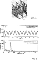
- Figure 5 shows an example of lung images for calculating the lung area time series, including an expiration image (A), a medium ventilation position image (B) and an inspiration image (C).
- a complete lung area time series is obtained by applying the inverse transformations fields on the segmented lung ROI.
- the ventilation frequency is directly determined from the time series of the lung area values.
- Determining the ventilation frequency according to the second variant has particular advantages as the change of the lung area has better stability and reliability compared with the analysis of the averaged ROI signal value of the ROI 2, which can be influenced by irregular ventilation.
- the second variant uses morphologic information rather than the anti-proportionality of the MRI signal and lung volume.
- Step S3 of Figure 1 includes two sub-steps, wherein ventilation images are created by applying a ventilation frequency based low-pass frequency filter in a first sub-step and ventilation images representing regular ventilation are selected from the collected MR lung images in a second sub-step.
- the series of ventilation images is created by applying the ventilation frequency based low-pass frequency filter on the registered MR lung images in frequency space.
- the low-pass frequency filter is provided based on the ventilation frequency determined in step S2.
- the ventilation images obtained are independent of perfusion of the subject under investigation.
- the low-pass frequency filter is provided such that frequency contributions at the ventilation frequency are passed, while perfusion contributions, in particular with frequencies near the heart beat frequency, e. g. around 1 Hz are blocked.
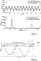
- Figure 6A illustrates the effect of the low-pass filter applied on a time series of the lung area values.
- the filtered lung area values are smoothed compared with the non-filtered lung area values.
- Figure 6B shows a spectrum before and after applying the low pass filter on the area time series.
- the ventilation frequency can be found with a peak near 0.3 Hz.
- a perfusion contribution does not occur in Figure 6B as the lung area time series is used instead of the MR image signal.
- Figure 7A illustrates the effect of the low-pass filter applied on a time series of the lung parenchyma signal (exemplary signal of one lung voxel).
- the filtered lung parenchyma signal does not contain the perfusion signal variations.
- Figure 7B shows a spectrum before and after applying the low pass filter on the image data.
- the ventilation frequency can be found with a peak near 0.3 Hz.
- the perfusion frequency can be found with a peak near 1.1 Hz.
- a series of perfusion images can be created by applying a high-pass filter on the registered MR lung images in frequency space.
- the high-pass frequency filter is provided based on a perfusion frequency determined by an analysis of a time series of a vessel ROI in analogy to step S2.
- the ventilation images are screened for selecting exclusively those ventilation images representing regular expiration and inspiration phases. This is obtained by retrospective gating as illustrated in Figure 8 .
- the ventilation images of the regular expiration and inspiration phases can be processed with optimized SNR, resulting in an improved quantification result in step S4.
- the dynamic change of the (lowpassed) quantitative lung dimension parameter (see Figure 6 ), like the lung area or the averaged signal of the ROI 2 (see Figure 4 ) is presented in a ventilation position curve. It is noted that the retrospective gating is not restricted to the analysis of the lung area time series, but rather also possible with a time series of another quantitative lung dimension parameter.
- Figure 8 shows an example of implementing the retrospective gating on the basis of a lung area time series as obtained in step S2.
- the ventilation position curve includes the inverted lung area values in dependency on time (or number of acquisition).
- a sliding window is defined having a window width, which is determined by the ratio b/n of bandwidth b and a free parameter n.
- the free parameter n is selected in a range of e. g. 5 to 20, and it is selected in dependency on the particular application conditions as mentioned above with reference to the selection of N in step S1.
- the first window 5 (first expiration window, see Figure 8 ) extends from max(A'(t)) to [max(A'(t)) - b/n]. Each point in A'(t) corresponds to one MR lung image of the dynamic series of MR lung images. All images within a common window are marked as associated expiration images. Subsequently, the expiration window 5 is shifted towards the medium ventilation position. The step width of the sliding window is selected in dependency on the particular application conditions. With each position of the sliding window, the associated expiration images are marked. The window, e. g. window 6 in Figure 8 , having the maximum number of expiration images (see counter) is considered as the expiration window representing a regular ventilation.
- the same process including shifting the sliding window starting from the maximum inspiration towards the medium ventilation position and counting the number of associated inspiration images, is conducted for finding the inspiration window representing the regular ventilation.
- shifting the sliding window towards the medium ventilation position is restricted by a predetermined first threshold value above or below the inverted lung area corresponding to the medium ventilation position.
- a predetermined first threshold value above or below the inverted lung area corresponding to the medium ventilation position.
- the second threshold value h ensures that the expiration and inspiration images of regular expiration and inspiration phases are physiologic real conditions.
- a first group of selected expiration ventilation images and a second group of inspiration ventilation images are obtained, which represent expiration and inspiration at regular ventilation. Intermediate ventilation positions or extreme ventilation positions having particularly deep or shallow ventilation are discarded.
- the selected ventilation images comprise about 10 % to 15 % of the registered, low-pass filtered MR lung images.
- Step S4 includes the calculation of the quantitative ventilation map using the selected expiration and inspiration ventilation images obtained with step S3.
- a fractional ventilation image or FV map is calculated as a quotient of a difference between the averaged expiration and inspiration images and the averaged inspiration or averaged expiration image.
- the fractional ventilation image represents a quantitative ventilation map already, which however still depends on the deepness of ventilation.
- a further adjustment is introduced by calculating a scaled fractional ventilation image or FV A map, which is normed relative to the lung area.
- a global fractional ventilation FV A value is calculated using the time series of the quantitative lung dimension parameter, in particular the lung area values, of the selected expiration and inspiration ventilation images at regular ventilation.
- FV A is calculated from the difference between the averaged lung area at expiration and the averaged lung area at inspiration divided by the averaged lung area at inspiration or expiration.
- FV A can be measured, e. g. by collecting 3D MR lung images, optionally combined with spirometer measurements and analysing lung volume changes.
- the scaled fractional ventilation image or FV A map is calculated by dividing the fractional ventilation image by the global fractional ventilation FV A value.
- the scaled fractional ventilation image is a dimensionless quantity which advantageously is independent on ventilation deepness or tidal volume.
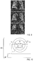
- Figure 9 illustrates a scaled fractional ventilation image obtained in an exemplary manner.
- Figures 9A, 9B and 9C represent three coronal slices of the lung, wherein the vessels can be seen with black images sections and the intensity if ventilation is represented by the brightness of the pixels.
- the images provide an information on the local ventilation, which was not available with conventional techniques.
- Figure 10 schematically shows an example of an MRT device 100 with an MRI scanner 10 including a main magnetic field device 11, at least one radiofrequency excitation coil 12, at least two magnetic field gradient coils 13 and radiofrequency receiver coils 14. Furthermore, the MRI device 100 includes a control device 20, like a computer circuit, being adapted for controlling the MRI scanner 10 for collecting the series of sets of image raw data and processing the image data with the method according to the embodiments.
- a control device 20 like a computer circuit, being adapted for controlling the MRI scanner 10 for collecting the series of sets of image raw data and processing the image data with the method according to the embodiments.
Description
- The invention relates to a method of image processing of magnetic resonance (MR) images of a lung (MR lung images) of a subject under investigation for creating a quantitative ventilation map of the lung. In particular, the invention relates to magnetic resonance tomography (MRT) based lung imaging using Fourier decomposition. MR lung images are processed which provide a map of local lung ventilation, without using ionising radiation and without using contrast agents. The invention relates to multiple sub-processes of image processing of the MR lung images, which result in an improved image quality, stability of image calculation and quantification of local distribution of ventilation in the lung. Both, the sub-processes alone and combinations thereof provide subjects of the invention. Applications of the invention are available in medical MR imaging, in particular in quantitative MR lung imaging.
- For illustrating the background of the invention, particular reference is made to the following publications:
- [1] C. Chefd'hotel et al. "Flows of Diffeomorphisms for Multimodal Image Matching", Proceedings of the IEEE International Symposium on Biomedical Imaging, Washington, USA (2002);
- [2] M. Zapke et al. in "Respiratory Research" vol. 7, 2006, p. 106;
- [3] M. Deimling et al. in "Proc. Inti. Soc. Mag. Reson. Med." vol. 16, 2008, p. 2639;
- [4] G. Bauman et al. in "Magnetic Resonance in Medicine" vol. 62, 2009, p. 656, and in "Radiology" vol. 260, 2011, p. 551-559 titled "Pulmonary Functional Imaging: Qualitative Comparison of Fourier Decomposition MR Imaging with SPECT/CT in Porcine Lung";
- [5] B. B. Avants et al. "Neuroimage" vol. 54, 2011, p. 2033;
- [6] A. Kjørstad et al. in "Magn. Reson. Mater. Phy." 2014, p. 1-10 (DOI 10.1007/s10334-014-0432-9);
- [7] C. Schonfeld et al. in "Journal of Magnetic Resonance Imaging", electronic publication on September 17, 2014;
- [8] A. Voskrebenzev et al. "Introduction of global specific ventilation as a reference for specific ventilation derived by Fourier Decomposition in "ISMRM Proceedings", Milan, 2014;
- [9] A. Voskrebenzev et al. "Detection of Chronic Allograft Dysfunction using Ventilation-Weighted Fourier Decomposition Lung MRI" in "ISMRM Proceedings" Toronto, 2015; and
- [10]
US 8660634 B2 . - Magnetic resonance tomography (MRT) based lung imaging using Fourier decomposition is described e. g. in [3, 4, 10]. A quantitative ventilation map of the lung can be created by processing MR lung images along the following procedure. A series of two-dimensional (2D) MR lung images is collected with an MRI scanner, e. g. with a temporal resolution of 0.3 s per image for each slice position for a period of 1 min of free ventilation.
- Firstly, the MR lung images are subjected to a registration into one image representing a medium ventilation position, using a non-rigid transformation [1, 5] with transformation fields. The registration results in a deformation of the lung images so that all images correspond to the same ventilation position (or: ventilation status). Accordingly, a characteristic temporal signal intensity can be assigned to each lung image voxel.
- The registration process as conventionally used in the Fourier decomposition context has the following disadvantages. Images of the expiration ventilation position and the inspiration ventilation position are directly registered into the image representing the medium ventilation position. This requires a strong deformation of lung images resulting in a risk of misregistrations. Furthermore, due to the required temporal resolution, the signal to noise ratio (SNR) of the single images is relatively low. Local lung structures which are important for estimating the transformation fields are superimposed by noise, thus further impeding the registration.
- Subsequently, a frequency determination and a separation of ventilation and perfusion contributions in the images is conducted. As the MRI signal is proportional to the proton density, the signal intensity oscillates with the ventilation frequency anti-proportional to the volume change of the lung during the ventilation. Simultaneously, the signal intensity is modulated by blood perfusion with a heart perfusion frequency, which is higher than the ventilation frequency. The ventilation and perfusion frequencies can be determined and separated by a Fourier decomposition of an averaged temporal signal intensity in frequency space [4, 9].
- As a limitation of the conventional frequency determination, the averaged temporal signal intensity reliably can be used with healthy subjects only. However, with subjects having a lung disease, abnormal ventilation may occur. This may impede or even exclude an appropriate frequency determination. Furthermore, the Fourier decomposition requires a periodic ventilation and perfusion. With an irregular ventilation, the spectrum of ventilation is broadened in frequency space. Accordingly, the integration limits for calculating the amplitude of ventilation have to be extended, resulting in an increased noise contribution.
- Finally, a quantification is obtained by voxel-based integration of image signals at the ventilation and perfusion peaks, resulting in a ventilation or perfusion weighted map of the lung as described in [3] or [4]. Ventilation can be quantified using the parameter fractional ventilation (FV), which is calculated by a ratio of a lung tidal volume (difference of inspiration and expiration volumes) and the inspiration volume.
- Conventionally, these volumes are measured e. g. by spirome-try. Due to the relationship of signal intensity and volume, it could be considered that this ratio correspondingly can be calculated from the MR lung signal intensities. The integrated ventilation peak would provide the signal change caused by the tidal volume [2]. For calculating the FV, this integration value could be scaled with the expiration signal [6]. However, this approach using MR lung signal intensities does not allow an appropriate quantification process as the fractional ventilation depends on the ventilation deepness, which can vary within the series of MR lung images collected [8]. As the varying ventilation deepness cannot be compensated with the conventional techniques, the calculation of the fractional ventilation based on conventionally processed images cannot yield assessable and reproducible quantitative results.
- In summary, the conventional methods mainly suffer from limitations in SNR of registered, filtered and quantified images, so that the conventional quantitative ventilation maps have a limited information contents.
- The objective of the invention is to provide an improved method of image processing of MR lung images, which avoids disadvantages of conventional techniques. In particular, the objective of the invention is to provide the method of image processing of MR lung images with increased sensitivity and reproducibility, improved SNR and/or reduced loss of information contributions included in the MR lung images.
- This objective is solved with a method of image processing of MR lung images according to
claim 1, a computer program according to claim 8 and an apparatus comprising a computer-readable storage medium according to claim 9. - According to a first general aspect of the invention, the above objective is solved by a method of processing MR lung images of a subject under investigation, comprising the steps of providing a series of the MR lung images collected over a time interval covering multiple ventilation periods, registering the MR lung images for creating a series of registered MR lung images, determining a ventilation frequency from the registered MR lung images, creating a series of ventilation images by applying a ventilation frequency based low-pass frequency filter on the registered MR lung images, wherein the ventilation images preferably are independent of (not influenced by) the perfusion of the subject under investigation, and creating a quantitative ventilation map of the lung, which is calculated from the ventilation images.
- According to the invention, the step of determining the ventilation frequency includes a frequency analysis of a time series of a quantitative lung dimension parameter (area parameter), the value of which being characteristic for the dimension of the lung (size of the lung) at the time of collecting the corresponding MR lung image. Contrary to the conventional Fourier decomposition of an averaged temporal signal intensity and frequency detection in frequency space, the invention is based on a construction and analysis of the time series of the quantitative lung dimension parameter, i. e. a provision of an oscillating lung area quantity in time space. Advantageously, analysing the time series of the quantitative lung dimension parameter allows determining the ventilation frequency with increased reliability and stability, compared with conventional analysing of signal intensity oscillations.
- Furthermore, according to the invention, the step of creating the quantitative ventilation map includes selecting a first group of ventilation images, which consists of expiration ventilation images collected at regular expiration phases of the multiple ventilation periods, selecting a second group of ventilation images, which consists of inspiration ventilation images collected at regular inspiration phases of the multiple ventilation periods, and calculating the quantitative ventilation map from the first and second groups of ventilation images.
- The inventors have found, that multiple ventilation periods of healthy or sick subjects include expiration and inspiration phases (or: expiration and inspiration events), which can be assigned to a regular ventilation (or: periodic, continuous ventilation). The regular expiration and inspiration phases can be identified in particular on the basis of the improved ventilation frequency determination of the invention. Expiration ventilation images collected at the regular expiration phases and inspiration ventilation images collected at the regular inspiration phases are selected for creating the quantitative ventilation map, while ventilation images of irregular ventilation phases are discarded. Furthermore, the quantitative ventilation map is adjusted with reference to the tidal volume of the lung. Advantageously, this avoids any dependency of the quantification process on the ventilation deepness.
- Advantageously, the SNR of the ventilation images is improved due to the improved determination of the ventilation frequency and the selection of images representing the regular ventilation. The quantitative ventilation map obtained with the invention includes more reliable information compared with conventional techniques. In particular, the quantitative ventilation map is an image wherein the intensity (brightness of the image pixels) is a direct measure for the local ventilation. On the basis of the local ventilation information, the quantitative ventilation map created according to the invention can be evaluated by a physician in a separate step, in particular for characterizing the health status of the subject or finding a diagnosis.
- Furthermore, according to the invention, the first and second groups of ventilation images are selected by retrospective gating of the time series of the quantitative lung dimension parameter. The retrospective gating comprises an assignment of the values of the measured quantitative lung dimension parameter to predetermined intervals of the quantitative lung dimension parameter (gating) and a determination of regular ventilation events by counting the number of assigned values in each interval. The intervals with maximum numbers of assigned values and the ventilation images associated with the values assigned to those maximum intervals are provided as regular ventilation phases. Advantageously, the retrospective gating (navigator) allows a targeted selection of ventilation images with similar ventilation deepness and a maximization of the SNR. Preferably, the retrospective gating uses the same quantitative lung dimension parameter like the ventilation frequency determination, in particular the lung area value or the averaged ROI signal value. Thus, the processing complexity is reduced. Alternatively, the retrospective gating and the ventilation frequency determination use different quantitative lung dimension parameters.
- According to a second general aspect, a method of magnetic resonance imaging of a lung is disclosed, comprising the steps of collecting a series of MR image raw data for obtaining multiple MR lung images, and subjecting the MR lung images to an image processing according to the above first aspect of the invention.
- According to preferred embodiments, the quantitative lung dimension parameter used with the inventive method of processing MR lung images is at least one of a lung area value of the registered MR lung images and an averaged ROI signal value of a region of interest covering a part of the lung and a part of the adjacent thoracic diaphragm of the subject under investigation.
- According to the first variant, lung area values are obtained by applying inverse transformation fields from the registered image series to one initial ROI outlining the lung parenchyma. Thus, ROIs for each time point of the registered image series can be obtained and the lung area value time series can be determined using the inverse transformation fields. This information can be used not only for the stable determination of the ventilation frequency, but also for the determination of the inspiration and expiration events and for the scaling of the FV as outlined below.
- According to the second variant, using the averaged ROI signal value is based on the following consideration. Due to different proton densities, the lung and the thoracic diaphragm provide substantially different MRI signals. The ROI is arranged such that the portion of the lung within the ROI is changing in dependency on the ventilation position. Accordingly, the averaged MRI signal of the ROI directly follows the ventilation.
- According to a further preferred embodiment, the step of calculating the quantitative ventilation map includes calculating a fractional or specific ventilation image obtained from a difference between an averaged expiration image from the first group of ventilation images and an averaged inspiration image from the second group of ventilation images, divided by the averaged inspiration or expiration ventilation image. Particularly preferred, the quantitative ventilation map is calculated by adjusting (or: scaling) the fractional ventilation image with reference to the tidal volume of the lung, using a global fractional ventilation parameter, wherein the global fractional ventilation parameter is calculated from the lung area values of the averaged inspiration and expiration images or from measured ventilation flow values.
- According to a further particularly preferred embodiment, the step of registering the MR lung images comprises assigning the collected MR lung images into groups (or: classes), wherein all collected MR lung images within each one of the groups have the quantitative lung dimension parameter, in particular the averaged ROI signal value, within a common predetermined parameter interval, group-wise registering the collected MR lung images within the groups of collected MR lung images, and step-wise registering the group-wise registered MR lung images or an averaged MR lung image thereof, starting from parameter intervals corresponding to expiration and inspiration ventilation positions towards a parameter interval corresponding to an intermediate ventilation position. Preferably, the intermediate ventilation position is a medium ventilation position between expiration and inspiration.
- Advantageously, the problems of the conventional registration are solved by the present group- and step-wise image registration into reference images from neighbouring ventilation phases. Strong deformations of the images and misregistrations are avoided. As a particular advantage, the group- and step-wise image registration avoids artefacts which otherwise would impair the subsequent steps of ventilation frequency determination and quantification. While the conventional registration may result in wrong conclusions or undiscovered pathologic findings, the registration reduces this risk and even accelerates the registration process.
- The step-wise registration of dynamic time series of MR images within groups can be applied not only in combination with the method of processing MR lung images but rather generally for registering time series of MR images. Thus, it is emphasized that the proposed registration process provides an independent subject of the invention wherein the registering the MR images comprises providing groups of a time series of subsequent MR images, the MR images within each group having a quantitative dimension parameter within a common predetermined parameter interval, group-wise registering the collected MR images within the groups of collected MR images, and registering the group-wise registered MR images or an averaged MR image thereof, starting from parameter intervals corresponding to extrema image positions towards a parameter interval corresponding to an intermediate image position.
- According to a further general aspect not covered by the claim, an MRI device is disclosed, which is configured for creating a sequence of MR images of an object under investigation. The MRI device comprises an MRI scanner including a main magnetic field device, at least one radiofrequency excitation coil, magnetic field gradient coils and at least one radiofrequency receiver coil, and a control device being adapted for controlling the MRI scanner for collecting the series of sets of image data and for processing the sequence of MR images with the method according to the above first aspect of the invention.
- Further general subjects of the invention are a computer program residing on a computer-readable medium, with a program code which, when executed on a computer, carries out the method(s) according to the above first and/or second aspect of the invention, and/or an apparatus comprising a computer-readable storage medium containing program instructions which, when executed on a computer, carry out the method(s) according to the above first and/or second aspect of the invention.
- It is noted that the concept correspondingly can be used for calculating a quantitative perfusion image or perfusion map. Perfusion images which are independent of ventilation of the subject under investigation can be created by applying a high-pass filter on the registered MR lung images in frequency space. Selected perfusion images can be found and analysed which represent a regular heartbeat, for imaging systole and diastole in time space.
- Further details and advantages of the embodiments are described in the following with reference to the attached drawings, which show in:
- Figure 1:
- a flowchart illustrating features of preferred embodiments of the image processing method;
- Figures 2 and 3:
- schematic illustrations of an image registration;
- Figures 4 and 5:
- schematic illustrations of measuring quantitative lung dimension parameters;
- Figure 6:
- a schematic illustration of determining a ventilation frequency;
- Figure 7:
- a schematic illustration of low-pass filtering of a lung parenchyma signal;
- Figure 8:
- a schematic illustration of retrospective gating;
- Figure 9:
- experimental results obtained with the invention; and
- Figure 10:
- a schematic illustration of an MRI device according to an embodiment not covered by the claims.
- Embodiments are described in the following with reference to the successive sub-processes of image processing of dynamic 2D MR lung images collected e. g. with coronal or sagittal slices of the lung, resulting in a two-dimensional quantitative ventilation map of the lung. For obtaining a three-dimensional quantitative ventilation map, in particular covering the whole lung, the processing is repeated with further time series of 2D MR lung images representing further coronal or sagittal slices of the lung.
- Alternatively, three-dimensional (3D) MR lung images can be collected and subjected to the sub-processes as outlined in the following.
- According to
Figure 1 , the method comprises the steps of providing the series of MR lung images (S0), registration (S1), frequency determination (S2), separation of ventilation and perfusion (S3), and quantification (S4), which are described in the following with further details. - Step S0 of providing the series of MR lung images preferably comprises collecting the MR lung images with an MRI scanner (see e. g.
Figure 10 ), so that the image data can be directly processed for an output of the quantitative ventilation map of the lung by the MRI scanner. Details of collecting the MR lung images, operating an MRI scanner, the numerical implementation of the mathematical formulation using available software tools and optional further image pre- or post-processing steps are not described as far as they are known from conventional MRI techniques. In particular, usual MRI pulse sequences can be applied for collecting the images, without changes of the commercially available sequences. The number of images, temporal resolution and collecting period can be selected in dependency on the application of the invention. As an example, about 200 images are collected with a temporal resolution of 0.3 s per image and slice position for a collecting period of about 1 min of free ventilation. - Alternatively, the series of MR lung images can be provided separately from collecting the MR lung image data. Previously collected images can be stored and provided, e. g. via a data communication network to a distant processor for implementing the image processing according to the invention.
- The collected lung images are subjected to the registration for deforming the lung images such that they correspond to the same ventilation position.
Figure 2 illustrates the effect of image registration.Figure 2A shows an MR thorax image wherein the verticalblack line 1 represents a cross-section line through the thoracic diaphragm. Before the registration, a periodic diaphragm oscillation is obtained resulting from the ventilation, as shown inFigure 2B . After the registration, the diaphragm oscillation is no longer detectable, as shown inFigure 2C . This deformation can be obtained by a conventional registration, as described e. g. in [2, 4]. However, preferably, the registration is implemented with the following inventive procedure, described with reference toFigures 3 and 4 . -
Figure 3 shows a ventilation position curve formed by a time dependency of a quantitative parameter representing the current ventilation position for each image acquisition. As an example, the quantitative parameter is an averaged signal of a region of interest (ROI) 2 covering a part of thelung 3 and a part of the adjacentthoracic diaphragm 4 of the subject under investigation, as schematically shown inFigure 4 . - As the lung parenchyma has a low proton density, the image signals of the lung parenchyma are substantially lower compared with the thoracic diaphragm. Due to the movement of the
thoracic diaphragm 4, the averaged signal increases during exhalation and decreases during inhalation. Accordingly, the averaged signal of theROI 2 covering both tissues has a minimum value at inspiration (e. g. A inFigure 3 ) and a maximum value at expiration (e. g. B inFigure 3 ). - Based on the ventilation position curve of
Figure 3 , the registration comprises the following registration steps. Firstly, the ventilation position of each image acquisition is obtained from the averaged ROI signal value ofROI 2. - Subsequently, a plurality of N groups is defined each representing a similar ventilation position. Each group is defined by a specific interval of the averaged ROI signal value of
ROI 2, or alternatively by a predetermined number of images with similar ventilation positions. The groups including the ventilation positions of maximum inspiration and expiration are correspondingly indicated as inspiration and expiration groups. Each of the collected MR lung images is assigned to one of the N groups. The value N, e. g. in a range from 5 to 20, is selected in dependency on the particular application conditions such that a number of images is included in each group. In particular, the value N is selected e. g. in consideration of the SNR of the single images (depending on the pulse sequence used for collecting the MR images), the temporal resolution (also depending on the pulse sequence) and the deepness and frequency of ventilation (depending on the subject). - Subsequently, the images within each group are registered into one selected image of the respective group as schematically illustrated in the right part of
Figure 3 . The selected image can be found by calculating a similarity parameter for each image relative to the remaining images within the group. The similarity parameter can be calculated with the same metric as used for the registration. The image having the best similarity with the remaining images of the group is used as the selected target image for the registration. The registered images within each group are averaged, so that one averaged group image is assigned to each group. N averaged group images are obtained, one of them representing an intermediate ventilation position. - Finally, each of the averaged group images is registered into the averaged group image of the neighbouring group, starting from the inspiration and expiration groups towards the intermediate ventilation position. Preferably, the intermediate ventilation position is a medium ventilation position between maximum expiration and inspiration. As an example, the averaged group image of group C in
Figure 3 is registered into the averaged group image of the neighbouring group, see arrow D inFigure 3 . In other words, the averaged group images are used for a step-wise registration towards the intermediate ventilation position. As an alternative, the registration can be done into a selected ventilation position other than the medium ventilation position. - Advantageously, this procedure minimizes misregistrations. Combining images into groups allows the averaging of the images, so that the SNR of the images to be registered is increased and the reliability of the registration is improved.
- If the images within the groups are sufficiently similar, the registration within the group can be omitted.
- As a result of the registration (S1), a series of N transformations fields (and corresponding inverse transformation fields) and N associated registered MR lung images is obtained, which are independent on ventilation movements of the lung.
- With step S2 of
Figure 1 , the ventilation frequency is determined from a time series of a quantitative lung dimension parameter as outlined in the following with reference to two exemplary embodiments. - According to a first variant, the averaged ROI signal value of the
ROI 2 covering a part of thelung 3 and a part of the adjacentthoracic diaphragm 4 of the subject under investigation provides the quantitative lung dimension parameter, as schematically shown inFigure 4 and described above. The ventilation frequency is directly determined from the ventilation position curve ofFigure 3 . - According to a second variant, a time series of lung area values obtained on the basis of the registered MR lung images provides the quantitative lung dimension parameters. Firstly, the left and right lung of the medium ventilation position image obtained after the registration are segmented. Segmentation of the lung parenchyma is done manually or automatically by excluding vessels, in particular large vessels impairing a lung area estimation. The lung area is measured e. g. with the number of pixels covered by the segmented lung.
- Subsequently, the time series of the lung area is obtained by applying the inverse transformations fields of the registration on the lung ROI for all image acquisitions.
Figure 5 shows an example of lung images for calculating the lung area time series, including an expiration image (A), a medium ventilation position image (B) and an inspiration image (C). A complete lung area time series is obtained by applying the inverse transformations fields on the segmented lung ROI. The ventilation frequency is directly determined from the time series of the lung area values. - Determining the ventilation frequency according to the second variant has particular advantages as the change of the lung area has better stability and reliability compared with the analysis of the averaged ROI signal value of the
ROI 2, which can be influenced by irregular ventilation. In particular, the second variant uses morphologic information rather than the anti-proportionality of the MRI signal and lung volume. - Step S3 of
Figure 1 includes two sub-steps, wherein ventilation images are created by applying a ventilation frequency based low-pass frequency filter in a first sub-step and ventilation images representing regular ventilation are selected from the collected MR lung images in a second sub-step. - The series of ventilation images is created by applying the ventilation frequency based low-pass frequency filter on the registered MR lung images in frequency space. The low-pass frequency filter is provided based on the ventilation frequency determined in step S2. The ventilation images obtained are independent of perfusion of the subject under investigation. The low-pass frequency filter is provided such that frequency contributions at the ventilation frequency are passed, while perfusion contributions, in particular with frequencies near the heart beat frequency, e. g. around 1 Hz are blocked.
-
Figure 6A illustrates the effect of the low-pass filter applied on a time series of the lung area values. The filtered lung area values are smoothed compared with the non-filtered lung area values.Figure 6B shows a spectrum before and after applying the low pass filter on the area time series. The ventilation frequency can be found with a peak near 0.3 Hz. A perfusion contribution does not occur inFigure 6B as the lung area time series is used instead of the MR image signal. -
Figure 7A illustrates the effect of the low-pass filter applied on a time series of the lung parenchyma signal (exemplary signal of one lung voxel). The filtered lung parenchyma signal does not contain the perfusion signal variations.Figure 7B shows a spectrum before and after applying the low pass filter on the image data. The ventilation frequency can be found with a peak near 0.3 Hz. The perfusion frequency can be found with a peak near 1.1 Hz. - Additionally or alternatively, a series of perfusion images can be created by applying a high-pass filter on the registered MR lung images in frequency space. The high-pass frequency filter is provided based on a perfusion frequency determined by an analysis of a time series of a vessel ROI in analogy to step S2.
- Subsequently, the ventilation images are screened for selecting exclusively those ventilation images representing regular expiration and inspiration phases. This is obtained by retrospective gating as illustrated in
Figure 8 . The ventilation images of the regular expiration and inspiration phases can be processed with optimized SNR, resulting in an improved quantification result in step S4. - The dynamic change of the (lowpassed) quantitative lung dimension parameter (see
Figure 6 ), like the lung area or the averaged signal of the ROI 2 (seeFigure 4 ) is presented in a ventilation position curve. It is noted that the retrospective gating is not restricted to the analysis of the lung area time series, but rather also possible with a time series of another quantitative lung dimension parameter. -
Figure 8 shows an example of implementing the retrospective gating on the basis of a lung area time series as obtained in step S2. As the MR image signal is invers relative to the volume, the lung area time series A(t) is converted into a time series of inverted lung areas A'(t) = 1/A(t). Thus, the ventilation position curve includes the inverted lung area values in dependency on time (or number of acquisition). The bandwidth b of inverted lung areas is b = max (A'(t)) - min(A'(t)). - A sliding window is defined having a window width, which is determined by the ratio b/n of bandwidth b and a free parameter n. The free parameter n is selected in a range of e. g. 5 to 20, and it is selected in dependency on the particular application conditions as mentioned above with reference to the selection of N in step S1.
- The first window 5 (first expiration window, see
Figure 8 ) extends from max(A'(t)) to [max(A'(t)) - b/n]. Each point in A'(t) corresponds to one MR lung image of the dynamic series of MR lung images. All images within a common window are marked as associated expiration images. Subsequently, theexpiration window 5 is shifted towards the medium ventilation position. The step width of the sliding window is selected in dependency on the particular application conditions. With each position of the sliding window, the associated expiration images are marked. The window,e. g. window 6 inFigure 8 , having the maximum number of expiration images (see counter) is considered as the expiration window representing a regular ventilation. - The same process, including shifting the sliding window starting from the maximum inspiration towards the medium ventilation position and counting the number of associated inspiration images, is conducted for finding the inspiration window representing the regular ventilation.
- Preferably, shifting the sliding window towards the medium ventilation position is restricted by a predetermined first threshold value above or below the inverted lung area corresponding to the medium ventilation position. Advantageously, assignments of expiration images to inspiration windows or vice versa is avoided by applying the threshold value.
- As the medium ventilation position can drift during ventilation, a second threshold value h can be introduced as a condition for assigning data points of inverted lung area to the current sliding window. Data points are discarded if the second threshold value h = dM/d (measured in units of A'(t)) goes below a predetermined value. As illustrated in
Figure 7 , d is the distance between the adjacent extrema of a considered data point, and dM is the distance between the considered data point and the local medium ventilation position. As an example, the condition is h ≥ 0.3. Advantageously, the second threshold value h ensures that the expiration and inspiration images of regular expiration and inspiration phases are physiologic real conditions. - As a result of step S3, a first group of selected expiration ventilation images and a second group of inspiration ventilation images are obtained, which represent expiration and inspiration at regular ventilation. Intermediate ventilation positions or extreme ventilation positions having particularly deep or shallow ventilation are discarded. With an example, the selected ventilation images comprise about 10 % to 15 % of the registered, low-pass filtered MR lung images.
- Step S4 includes the calculation of the quantitative ventilation map using the selected expiration and inspiration ventilation images obtained with step S3.
- Firstly, all images of the first group of selected expiration ventilation images are averaged, and all images of the second group of inspiration ventilation images are averaged, thus providing one averaged expiration ventilation image and one averaged inspiration ventilation image. The signal intensities of the individual pixels of the averaged expiration and inspiration images are determined by the local ventilation. Accordingly, a fractional ventilation image or FV map is calculated as a quotient of a difference between the averaged expiration and inspiration images and the averaged inspiration or averaged expiration image.
- The fractional ventilation image represents a quantitative ventilation map already, which however still depends on the deepness of ventilation. According to a preferred embodiment, a further adjustment is introduced by calculating a scaled fractional ventilation image or FVA map, which is normed relative to the lung area. To this end, a global fractional ventilation FVA value is calculated using the time series of the quantitative lung dimension parameter, in particular the lung area values, of the selected expiration and inspiration ventilation images at regular ventilation. As an example, FVA is calculated from the difference between the averaged lung area at expiration and the averaged lung area at inspiration divided by the averaged lung area at inspiration or expiration. As an alternative, FVA can be measured, e. g. by collecting 3D MR lung images, optionally combined with spirometer measurements and analysing lung volume changes.
- The scaled fractional ventilation image or FVA map is calculated by dividing the fractional ventilation image by the global fractional ventilation FVA value. The scaled fractional ventilation image is a dimensionless quantity which advantageously is independent on ventilation deepness or tidal volume.
-
Figure 9 illustrates a scaled fractional ventilation image obtained in an exemplary manner.Figures 9A, 9B and 9C represent three coronal slices of the lung, wherein the vessels can be seen with black images sections and the intensity if ventilation is represented by the brightness of the pixels. As an essential advantage, the images provide an information on the local ventilation, which was not available with conventional techniques. -
Figure 10 schematically shows an example of anMRT device 100 with anMRI scanner 10 including a mainmagnetic field device 11, at least oneradiofrequency excitation coil 12, at least two magnetic field gradient coils 13 and radiofrequency receiver coils 14. Furthermore, theMRI device 100 includes acontrol device 20, like a computer circuit, being adapted for controlling theMRI scanner 10 for collecting the series of sets of image raw data and processing the image data with the method according to the embodiments.
Claims (9)
- Computer-implemented method of processing magnetic resonance (MR) lung images of a subject under investigation, comprising the steps of- providing a series of the MR lung images collected during multiple ventilation periods (S0),- registering the MR lung images for creating a series of registered MR lung images (S1),- determining a ventilation frequency from the registered MR lung images (S2),- creating a series of ventilation images by applying a ventilation frequency based low-pass frequency filter on the registered MR lung images (S3), and- creating a quantitative ventilation map of the lung (S4), which is calculated from the ventilation images,
characterized in that- the step (S2) of determining the ventilation frequency includes a frequency analysis of a time series of a quantitative lung dimension parameter which is characteristic for the dimension of the lung (3) at the time of collecting the corresponding MR lung image, and- the step (S4) of creating the quantitative ventilation map includes- selecting a first group of ventilation images, which consists of expiration ventilation images collected at regular expiration phases of the multiple ventilation periods, wherein the first group of ventilation images is selected by retrospective gating of the time series of the quantitative lung dimension parameter,- selecting a second group of ventilation images, which consists of inspiration ventilation images collected at regular inspiration phases of the multiple ventilation periods, wherein the second group of ventilation images is selected by retrospective gating of the time series of the quantitative lung dimension parameter, wherein the retrospective gating comprises an assignment of values of the quantitative lung dimension parameter to predetermined intervals of the quantitative lung dimension parameter and a determination of regular ventilation events by counting the number of assigned values in each interval, wherein the intervals with maximum numbers of assigned values and the ventilation images associated with the values assigned to those intervals are provided as regular ventilation phases, and- calculating the quantitative ventilation map from the first and second groups of ventilation images, wherein the quantitative ventilation map is adjusted with reference to the tidal volume of the lung (3). - Method of processing MR lung images according to claim 1, wherein- the quantitative lung dimension parameter is at least one of a lung area value of the registered MR lung images and an averaged ROI signal value of a region of interest covering a part of the lung (3) and a part of the adjacent thoracic diaphragm (4) of the subject under investigation.
- Method of processing MR lung images according to one of the foregoing claims, wherein the step of calculating the quantitative ventilation map includes- calculating a fractional ventilation image obtained from a difference between an averaged expiration image from the first group of ventilation images and an averaged inspiration image from the second group of ventilation images, divided by the averaged inspiration or expiration ventilation image.
- Method of processing MR lung images according to claim 3, wherein- the quantitative ventilation map is calculated by adjusting the fractional ventilation image using a global fractional ventilation parameter, wherein- the global fractional ventilation parameter is calculated from the lung area values of the averaged inspiration and expiration images or from measured 3D volume images and/or measured ventilation flow values.
- Method of processing MR lung images according to one of the claims 2 to 4, wherein the step of registering the MR lung images comprises- providing groups of collected MR lung images, wherein the collected MR lung images within each group have the averaged ROI signal value within a common predetermined signal interval,- group-wise registering the collected MR lung images within the groups of collected MR lung images, and- registering the group-wise registered MR lung images or an averaged MR lung image thereof, starting from signal intervals corresponding to expiration and inspiration ventilation positions towards a signal interval corresponding to an intermediate ventilation position.
- Method of processing MR lung images according to claim 5, wherein- the intermediate ventilation position is a medium ventilation position between expiration and inspiration.
- Method of MR imaging the lung (3) of a subject under investigation, comprising the steps of- MR image acquisition for collecting MR lung images, and- subjecting the MR lung images to a method of processing MR lung images according to one of the foregoing claims.
- Computer program residing on a computer-readable medium, with a program code which, when executed on a computer (20), carries out the method according to at least one of the claims 1 to 6.
- Apparatus comprising a computer-readable storage medium containing program instructions which, when executed on a computer (20), carry out the method according to at least one of the claims 1 to 6.
Priority Applications (2)
| Application Number | Priority Date | Filing Date | Title |
|---|---|---|---|
| EP15001782.0A EP3107066B1 (en) | 2015-06-16 | 2015-06-16 | Method of quantitative magnetic resonance lung imaging |
| US15/178,894 US10010293B2 (en) | 2015-06-16 | 2016-06-10 | Method of quantitative magnetic resonance lung imaging |
Applications Claiming Priority (1)
| Application Number | Priority Date | Filing Date | Title |
|---|---|---|---|
| EP15001782.0A EP3107066B1 (en) | 2015-06-16 | 2015-06-16 | Method of quantitative magnetic resonance lung imaging |
Publications (2)
| Publication Number | Publication Date |
|---|---|
| EP3107066A1 EP3107066A1 (en) | 2016-12-21 |
| EP3107066B1 true EP3107066B1 (en) | 2020-11-11 |
Family
ID=53524534
Family Applications (1)
| Application Number | Title | Priority Date | Filing Date |
|---|---|---|---|
| EP15001782.0A Active EP3107066B1 (en) | 2015-06-16 | 2015-06-16 | Method of quantitative magnetic resonance lung imaging |
Country Status (2)
| Country | Link |
|---|---|
| US (1) | US10010293B2 (en) |
| EP (1) | EP3107066B1 (en) |
Families Citing this family (5)
| Publication number | Priority date | Publication date | Assignee | Title |
|---|---|---|---|---|
| JP2018110637A (en) * | 2017-01-10 | 2018-07-19 | コニカミノルタ株式会社 | Dynamic image processing system |
| DE102018214325A1 (en) * | 2018-08-24 | 2020-02-27 | Siemens Healthcare Gmbh | Method and provision unit for the provision of a virtual tomographic stroke follow-up examination image |
| JP7255329B2 (en) * | 2019-04-09 | 2023-04-11 | コニカミノルタ株式会社 | Dynamic image analysis device, dynamic image analysis system and program |
| DE102020216557B4 (en) * | 2020-12-23 | 2022-06-23 | Siemens Healthcare Gmbh | Method and data processing system for providing respiratory information |
| CN113592804A (en) * | 2021-07-27 | 2021-11-02 | 东软医疗系统股份有限公司 | Image processing method, device and equipment |
Family Cites Families (11)
| Publication number | Priority date | Publication date | Assignee | Title |
|---|---|---|---|---|
| US7130457B2 (en) * | 2001-07-17 | 2006-10-31 | Accuimage Diagnostics Corp. | Systems and graphical user interface for analyzing body images |
| GB0919269D0 (en) * | 2009-11-03 | 2009-12-16 | Univ Manchester | Lung function analysis method and apparatus |
| WO2011104605A2 (en) * | 2010-02-23 | 2011-09-01 | University Of Cape Town | A method for compensating for respiratory motion in magnetic resonance imaging |
| US9750427B2 (en) * | 2010-12-07 | 2017-09-05 | The Regents Of The University Of California | Magnetic resonance imaging of ventilation and perfusion in the lung |
| DK2806791T3 (en) * | 2012-01-29 | 2023-01-09 | Sensible Medical Innovations Ltd | DERIVATION OF INDIVIDUAL THORAX PARAMETERS FOR AN INDIVIDUAL |
| US8660634B2 (en) | 2012-04-23 | 2014-02-25 | The Brigham and Woman's Hospital | System and method for improved lung fourier decomposition magnetic resonance imaging |
| WO2013166430A2 (en) * | 2012-05-04 | 2013-11-07 | Trustees Of The University Of Pennsylvania | System for delivery of gaseous imaging contrast agents and methods for using same |
| US20130303899A1 (en) * | 2012-05-14 | 2013-11-14 | University Of Maryland, Baltimore | Quantitative evaluation of fractional regional ventilation using four-dimensional computed tomography |
| DE102012213551A1 (en) * | 2012-08-01 | 2014-02-06 | Siemens Aktiengesellschaft | Method for motion-induced attenuation correction and magnetic resonance system |
| WO2015153946A1 (en) * | 2014-04-04 | 2015-10-08 | Bayer Medical Care Inc. | Combined radiopharmaceutical imaging system |
| US10085703B2 (en) * | 2015-01-27 | 2018-10-02 | Septimiu Edmund Salcudean | Dynamic computed tomography imaging of elasticity |
-
2015
- 2015-06-16 EP EP15001782.0A patent/EP3107066B1/en active Active
-
2016
- 2016-06-10 US US15/178,894 patent/US10010293B2/en active Active
Non-Patent Citations (1)
| Title |
|---|
| None * |
Also Published As
| Publication number | Publication date |
|---|---|
| US10010293B2 (en) | 2018-07-03 |
| US20160367200A1 (en) | 2016-12-22 |
| EP3107066A1 (en) | 2016-12-21 |
Similar Documents
| Publication | Publication Date | Title |
|---|---|---|
| US10010293B2 (en) | Method of quantitative magnetic resonance lung imaging | |
| JP5944645B2 (en) | Magnetic resonance imaging system | |
| US8280128B2 (en) | Method of generating an enhanced perfusion image | |
| JP4193168B2 (en) | Apparatus and method for analyzing blood flow dynamics | |
| JP6261871B2 (en) | Image processing apparatus and magnetic resonance imaging apparatus | |
| US10898143B2 (en) | Quantitative mapping of cerebrovascular reactivity using resting-state functional magnetic resonance imaging | |
| US20130281828A1 (en) | Magnetic resonance method and apparatus for obtaining a set of measured data relating to a breathing object of interest | |
| JP2013063272A (en) | Method for automatic three-dimensional segmentation of magnetic resonance images | |
| US9345437B2 (en) | Method for obtaining a set of measured data relating to a breathing object of interest by using magnetic resonance technology, magnetic resonance system, computer program, and electronically readable data storage medium | |
| Kang et al. | Application of cardiac gating to improve the reproducibility of intravoxel incoherent motion measurements in the head and neck | |
| US20130281830A1 (en) | System and Method for Improved Lung Fourier Decomposition Magnetic Resonance Imaging | |
| JP2020049237A (en) | Method of assessing data quality of fine structure analysis data | |
| US10718838B2 (en) | Systems and methods for calibrated multi-spectral magnetic resonance imaging | |
| US20190107594A1 (en) | Magnetic resonance imaging apparatus and image processing apparatus | |
| Sundaram et al. | A dynamic model of average lung deformation using capacity-based reparameterization and shape averaging of lung MR images | |
| Hurd et al. | Comparison of prospective and retrospective gated 4D flow cardiac MR image acquisitions in the carotid bifurcation | |
| CN109696648B (en) | Method of determining a motion state of a subject and method of simultaneously imaging a subject | |
| JP6230882B2 (en) | Magnetic resonance imaging apparatus and echo time setting method | |
| JP2022543016A (en) | Methods, systems for generating biomarkers | |
| US11497412B2 (en) | Combined oxygen utilization, strain, and anatomic imaging with magnetic resonance imaging | |
| Rahni et al. | Extracting respiratory motion from 4D MRI using organ-wise registration | |
| EP3995081A1 (en) | Diagnosis assisting program | |
| JP6423066B2 (en) | Image processing apparatus and magnetic resonance imaging apparatus | |
| Khandelwal et al. | Gray Matter Segmentation in Ultra High Resolution 7 Tesla ex vivo T2w MRI of Human Brain Hemispheres | |
| WO2010045478A2 (en) | System and method for differential perfusion analysis of tissues with multiple vascular supplies |
Legal Events
| Date | Code | Title | Description |
|---|---|---|---|
| PUAI | Public reference made under article 153(3) epc to a published international application that has entered the european phase |
Free format text: ORIGINAL CODE: 0009012 |
|
| STAA | Information on the status of an ep patent application or granted ep patent |
Free format text: STATUS: THE APPLICATION HAS BEEN PUBLISHED |
|
| AK | Designated contracting states |
Kind code of ref document: A1 Designated state(s): AL AT BE BG CH CY CZ DE DK EE ES FI FR GB GR HR HU IE IS IT LI LT LU LV MC MK MT NL NO PL PT RO RS SE SI SK SM TR |
|
| AX | Request for extension of the european patent |
Extension state: BA ME |
|
| STAA | Information on the status of an ep patent application or granted ep patent |
Free format text: STATUS: REQUEST FOR EXAMINATION WAS MADE |
|
| 17P | Request for examination filed |
Effective date: 20170530 |
|
| RBV | Designated contracting states (corrected) |
Designated state(s): AL AT BE BG CH CY CZ DE DK EE ES FI FR GB GR HR HU IE IS IT LI LT LU LV MC MK MT NL NO PL PT RO RS SE SI SK SM TR |
|
| STAA | Information on the status of an ep patent application or granted ep patent |
Free format text: STATUS: EXAMINATION IS IN PROGRESS |
|
| 17Q | First examination report despatched |
Effective date: 20190625 |
|
| GRAP | Despatch of communication of intention to grant a patent |
Free format text: ORIGINAL CODE: EPIDOSNIGR1 |
|
| STAA | Information on the status of an ep patent application or granted ep patent |
Free format text: STATUS: GRANT OF PATENT IS INTENDED |
|
| INTG | Intention to grant announced |
Effective date: 20200603 |
|
| GRAS | Grant fee paid |
Free format text: ORIGINAL CODE: EPIDOSNIGR3 |
|
| GRAA | (expected) grant |
Free format text: ORIGINAL CODE: 0009210 |
|
| STAA | Information on the status of an ep patent application or granted ep patent |
Free format text: STATUS: THE PATENT HAS BEEN GRANTED |
|
| AK | Designated contracting states |
Kind code of ref document: B1 Designated state(s): AL AT BE BG CH CY CZ DE DK EE ES FI FR GB GR HR HU IE IS IT LI LT LU LV MC MK MT NL NO PL PT RO RS SE SI SK SM TR |
|
| REG | Reference to a national code |
Ref country code: GB Ref legal event code: FG4D |
|
| REG | Reference to a national code |
Ref country code: CH Ref legal event code: EP |
|
| REG | Reference to a national code |
Ref country code: AT Ref legal event code: REF Ref document number: 1334200 Country of ref document: AT Kind code of ref document: T Effective date: 20201115 |
|
| REG | Reference to a national code |
Ref country code: DE Ref legal event code: R096 Ref document number: 602015061758 Country of ref document: DE |
|
| REG | Reference to a national code |
Ref country code: IE Ref legal event code: FG4D |
|
| REG | Reference to a national code |
Ref country code: NL Ref legal event code: MP Effective date: 20201111 |
|
| REG | Reference to a national code |
Ref country code: AT Ref legal event code: MK05 Ref document number: 1334200 Country of ref document: AT Kind code of ref document: T Effective date: 20201111 |
|
| PG25 | Lapsed in a contracting state [announced via postgrant information from national office to epo] |
Ref country code: GR Free format text: LAPSE BECAUSE OF FAILURE TO SUBMIT A TRANSLATION OF THE DESCRIPTION OR TO PAY THE FEE WITHIN THE PRESCRIBED TIME-LIMIT Effective date: 20210212 Ref country code: RS Free format text: LAPSE BECAUSE OF FAILURE TO SUBMIT A TRANSLATION OF THE DESCRIPTION OR TO PAY THE FEE WITHIN THE PRESCRIBED TIME-LIMIT Effective date: 20201111 Ref country code: FI Free format text: LAPSE BECAUSE OF FAILURE TO SUBMIT A TRANSLATION OF THE DESCRIPTION OR TO PAY THE FEE WITHIN THE PRESCRIBED TIME-LIMIT Effective date: 20201111 Ref country code: PT Free format text: LAPSE BECAUSE OF FAILURE TO SUBMIT A TRANSLATION OF THE DESCRIPTION OR TO PAY THE FEE WITHIN THE PRESCRIBED TIME-LIMIT Effective date: 20210311 Ref country code: NO Free format text: LAPSE BECAUSE OF FAILURE TO SUBMIT A TRANSLATION OF THE DESCRIPTION OR TO PAY THE FEE WITHIN THE PRESCRIBED TIME-LIMIT Effective date: 20210211 |
|
| PG25 | Lapsed in a contracting state [announced via postgrant information from national office to epo] |
Ref country code: LV Free format text: LAPSE BECAUSE OF FAILURE TO SUBMIT A TRANSLATION OF THE DESCRIPTION OR TO PAY THE FEE WITHIN THE PRESCRIBED TIME-LIMIT Effective date: 20201111 Ref country code: IS Free format text: LAPSE BECAUSE OF FAILURE TO SUBMIT A TRANSLATION OF THE DESCRIPTION OR TO PAY THE FEE WITHIN THE PRESCRIBED TIME-LIMIT Effective date: 20210311 Ref country code: SE Free format text: LAPSE BECAUSE OF FAILURE TO SUBMIT A TRANSLATION OF THE DESCRIPTION OR TO PAY THE FEE WITHIN THE PRESCRIBED TIME-LIMIT Effective date: 20201111 Ref country code: PL Free format text: LAPSE BECAUSE OF FAILURE TO SUBMIT A TRANSLATION OF THE DESCRIPTION OR TO PAY THE FEE WITHIN THE PRESCRIBED TIME-LIMIT Effective date: 20201111 Ref country code: BG Free format text: LAPSE BECAUSE OF FAILURE TO SUBMIT A TRANSLATION OF THE DESCRIPTION OR TO PAY THE FEE WITHIN THE PRESCRIBED TIME-LIMIT Effective date: 20210211 Ref country code: AT Free format text: LAPSE BECAUSE OF FAILURE TO SUBMIT A TRANSLATION OF THE DESCRIPTION OR TO PAY THE FEE WITHIN THE PRESCRIBED TIME-LIMIT Effective date: 20201111 |
|
| REG | Reference to a national code |
Ref country code: LT Ref legal event code: MG9D |
|
| PG25 | Lapsed in a contracting state [announced via postgrant information from national office to epo] |
Ref country code: HR Free format text: LAPSE BECAUSE OF FAILURE TO SUBMIT A TRANSLATION OF THE DESCRIPTION OR TO PAY THE FEE WITHIN THE PRESCRIBED TIME-LIMIT Effective date: 20201111 |
|
| PG25 | Lapsed in a contracting state [announced via postgrant information from national office to epo] |
Ref country code: SK Free format text: LAPSE BECAUSE OF FAILURE TO SUBMIT A TRANSLATION OF THE DESCRIPTION OR TO PAY THE FEE WITHIN THE PRESCRIBED TIME-LIMIT Effective date: 20201111 Ref country code: RO Free format text: LAPSE BECAUSE OF FAILURE TO SUBMIT A TRANSLATION OF THE DESCRIPTION OR TO PAY THE FEE WITHIN THE PRESCRIBED TIME-LIMIT Effective date: 20201111 Ref country code: LT Free format text: LAPSE BECAUSE OF FAILURE TO SUBMIT A TRANSLATION OF THE DESCRIPTION OR TO PAY THE FEE WITHIN THE PRESCRIBED TIME-LIMIT Effective date: 20201111 Ref country code: CZ Free format text: LAPSE BECAUSE OF FAILURE TO SUBMIT A TRANSLATION OF THE DESCRIPTION OR TO PAY THE FEE WITHIN THE PRESCRIBED TIME-LIMIT Effective date: 20201111 Ref country code: EE Free format text: LAPSE BECAUSE OF FAILURE TO SUBMIT A TRANSLATION OF THE DESCRIPTION OR TO PAY THE FEE WITHIN THE PRESCRIBED TIME-LIMIT Effective date: 20201111 Ref country code: SM Free format text: LAPSE BECAUSE OF FAILURE TO SUBMIT A TRANSLATION OF THE DESCRIPTION OR TO PAY THE FEE WITHIN THE PRESCRIBED TIME-LIMIT Effective date: 20201111 |
|
| REG | Reference to a national code |
Ref country code: DE Ref legal event code: R097 Ref document number: 602015061758 Country of ref document: DE |
|
| PG25 | Lapsed in a contracting state [announced via postgrant information from national office to epo] |
Ref country code: DK Free format text: LAPSE BECAUSE OF FAILURE TO SUBMIT A TRANSLATION OF THE DESCRIPTION OR TO PAY THE FEE WITHIN THE PRESCRIBED TIME-LIMIT Effective date: 20201111 |
|
| PLBE | No opposition filed within time limit |
Free format text: ORIGINAL CODE: 0009261 |
|
| STAA | Information on the status of an ep patent application or granted ep patent |
Free format text: STATUS: NO OPPOSITION FILED WITHIN TIME LIMIT |
|
| 26N | No opposition filed |
Effective date: 20210812 |
|
| PG25 | Lapsed in a contracting state [announced via postgrant information from national office to epo] |
Ref country code: NL Free format text: LAPSE BECAUSE OF FAILURE TO SUBMIT A TRANSLATION OF THE DESCRIPTION OR TO PAY THE FEE WITHIN THE PRESCRIBED TIME-LIMIT Effective date: 20201111 Ref country code: AL Free format text: LAPSE BECAUSE OF FAILURE TO SUBMIT A TRANSLATION OF THE DESCRIPTION OR TO PAY THE FEE WITHIN THE PRESCRIBED TIME-LIMIT Effective date: 20201111 Ref country code: IT Free format text: LAPSE BECAUSE OF FAILURE TO SUBMIT A TRANSLATION OF THE DESCRIPTION OR TO PAY THE FEE WITHIN THE PRESCRIBED TIME-LIMIT Effective date: 20201111 |
|
| PG25 | Lapsed in a contracting state [announced via postgrant information from national office to epo] |
Ref country code: ES Free format text: LAPSE BECAUSE OF FAILURE TO SUBMIT A TRANSLATION OF THE DESCRIPTION OR TO PAY THE FEE WITHIN THE PRESCRIBED TIME-LIMIT Effective date: 20201111 Ref country code: SI Free format text: LAPSE BECAUSE OF FAILURE TO SUBMIT A TRANSLATION OF THE DESCRIPTION OR TO PAY THE FEE WITHIN THE PRESCRIBED TIME-LIMIT Effective date: 20201111 |
|
| PG25 | Lapsed in a contracting state [announced via postgrant information from national office to epo] |
Ref country code: MC Free format text: LAPSE BECAUSE OF FAILURE TO SUBMIT A TRANSLATION OF THE DESCRIPTION OR TO PAY THE FEE WITHIN THE PRESCRIBED TIME-LIMIT Effective date: 20201111 |
|
| REG | Reference to a national code |
Ref country code: CH Ref legal event code: PL |
|
| GBPC | Gb: european patent ceased through non-payment of renewal fee |
Effective date: 20210616 |
|
| REG | Reference to a national code |
Ref country code: BE Ref legal event code: MM Effective date: 20210630 |
|
| PG25 | Lapsed in a contracting state [announced via postgrant information from national office to epo] |
Ref country code: LU Free format text: LAPSE BECAUSE OF NON-PAYMENT OF DUE FEES Effective date: 20210616 |
|
| PG25 | Lapsed in a contracting state [announced via postgrant information from national office to epo] |
Ref country code: LI Free format text: LAPSE BECAUSE OF NON-PAYMENT OF DUE FEES Effective date: 20210630 Ref country code: IE Free format text: LAPSE BECAUSE OF NON-PAYMENT OF DUE FEES Effective date: 20210616 Ref country code: GB Free format text: LAPSE BECAUSE OF NON-PAYMENT OF DUE FEES Effective date: 20210616 Ref country code: CH Free format text: LAPSE BECAUSE OF NON-PAYMENT OF DUE FEES Effective date: 20210630 |
|
| PG25 | Lapsed in a contracting state [announced via postgrant information from national office to epo] |
Ref country code: IS Free format text: LAPSE BECAUSE OF FAILURE TO SUBMIT A TRANSLATION OF THE DESCRIPTION OR TO PAY THE FEE WITHIN THE PRESCRIBED TIME-LIMIT Effective date: 20210311 Ref country code: FR Free format text: LAPSE BECAUSE OF NON-PAYMENT OF DUE FEES Effective date: 20210630 |
|
| PG25 | Lapsed in a contracting state [announced via postgrant information from national office to epo] |
Ref country code: BE Free format text: LAPSE BECAUSE OF NON-PAYMENT OF DUE FEES Effective date: 20210630 |
|
| PG25 | Lapsed in a contracting state [announced via postgrant information from national office to epo] |
Ref country code: HU Free format text: LAPSE BECAUSE OF FAILURE TO SUBMIT A TRANSLATION OF THE DESCRIPTION OR TO PAY THE FEE WITHIN THE PRESCRIBED TIME-LIMIT; INVALID AB INITIO Effective date: 20150616 |
|
| PG25 | Lapsed in a contracting state [announced via postgrant information from national office to epo] |
Ref country code: CY Free format text: LAPSE BECAUSE OF FAILURE TO SUBMIT A TRANSLATION OF THE DESCRIPTION OR TO PAY THE FEE WITHIN THE PRESCRIBED TIME-LIMIT Effective date: 20201111 |
|
| PGFP | Annual fee paid to national office [announced via postgrant information from national office to epo] |
Ref country code: DE Payment date: 20230607 Year of fee payment: 9 |