EP3099707B1 - Opioid receptor binding agents and uses thereof - Google Patents
Opioid receptor binding agents and uses thereof Download PDFInfo
- Publication number
- EP3099707B1 EP3099707B1 EP15701821.9A EP15701821A EP3099707B1 EP 3099707 B1 EP3099707 B1 EP 3099707B1 EP 15701821 A EP15701821 A EP 15701821A EP 3099707 B1 EP3099707 B1 EP 3099707B1
- Authority
- EP
- European Patent Office
- Prior art keywords
- seq
- receptor
- opioid receptor
- binding
- protein
- Prior art date
- Legal status (The legal status is an assumption and is not a legal conclusion. Google has not performed a legal analysis and makes no representation as to the accuracy of the status listed.)
- Active
Links
- 102000003840 Opioid Receptors Human genes 0.000 title claims description 207
- 108090000137 Opioid Receptors Proteins 0.000 title claims description 207
- 239000011230 binding agent Substances 0.000 title description 136
- 108010003723 Single-Domain Antibodies Proteins 0.000 claims description 171
- 102000005962 receptors Human genes 0.000 claims description 165
- 108020003175 receptors Proteins 0.000 claims description 165
- 230000027455 binding Effects 0.000 claims description 141
- 238000009739 binding Methods 0.000 claims description 140
- 239000003446 ligand Substances 0.000 claims description 131
- 238000000034 method Methods 0.000 claims description 111
- 150000001875 compounds Chemical class 0.000 claims description 93
- 125000003275 alpha amino acid group Chemical group 0.000 claims description 60
- 108020001612 μ-opioid receptors Proteins 0.000 claims description 43
- 239000012528 membrane Substances 0.000 claims description 42
- 108091006027 G proteins Proteins 0.000 claims description 41
- 102000030782 GTP binding Human genes 0.000 claims description 41
- 108091000058 GTP-Binding Proteins 0.000 claims description 41
- 241000282414 Homo sapiens Species 0.000 claims description 40
- 150000007523 nucleic acids Chemical class 0.000 claims description 39
- 238000012216 screening Methods 0.000 claims description 35
- 230000003834 intracellular effect Effects 0.000 claims description 33
- 239000000203 mixture Substances 0.000 claims description 33
- 108010047041 Complementarity Determining Regions Proteins 0.000 claims description 32
- 102000039446 nucleic acids Human genes 0.000 claims description 31
- 108020004707 nucleic acids Proteins 0.000 claims description 31
- 102000051367 mu Opioid Receptors Human genes 0.000 claims description 29
- 238000012360 testing method Methods 0.000 claims description 29
- 238000002425 crystallisation Methods 0.000 claims description 15
- 108091028043 Nucleic acid sequence Proteins 0.000 claims description 10
- 230000008025 crystallization Effects 0.000 claims description 10
- 238000007876 drug discovery Methods 0.000 claims description 9
- 238000000746 purification Methods 0.000 claims description 9
- 230000001413 cellular effect Effects 0.000 claims description 7
- 230000008685 targeting Effects 0.000 claims description 7
- 230000000295 complement effect Effects 0.000 claims description 6
- 150000003384 small molecules Chemical class 0.000 claims description 6
- 101710122576 Isocitrate lyase 2 Proteins 0.000 claims description 2
- 102000016979 Other receptors Human genes 0.000 claims description 2
- 210000004027 cell Anatomy 0.000 description 196
- 108090000623 proteins and genes Proteins 0.000 description 145
- 102000004169 proteins and genes Human genes 0.000 description 138
- 235000018102 proteins Nutrition 0.000 description 133
- 102000003688 G-Protein-Coupled Receptors Human genes 0.000 description 101
- 108090000045 G-Protein-Coupled Receptors Proteins 0.000 description 101
- 101150019925 MDH2 gene Proteins 0.000 description 101
- 239000000556 agonist Substances 0.000 description 96
- 108060003951 Immunoglobulin Proteins 0.000 description 76
- 102000018358 immunoglobulin Human genes 0.000 description 76
- 241000282842 Lama glama Species 0.000 description 72
- 150000001413 amino acids Chemical class 0.000 description 71
- 235000001014 amino acid Nutrition 0.000 description 62
- 108090000765 processed proteins & peptides Proteins 0.000 description 56
- 102000004196 processed proteins & peptides Human genes 0.000 description 45
- 230000014509 gene expression Effects 0.000 description 43
- 239000012634 fragment Substances 0.000 description 40
- 230000000694 effects Effects 0.000 description 35
- 229920001184 polypeptide Polymers 0.000 description 35
- 239000002502 liposome Substances 0.000 description 34
- 239000005557 antagonist Substances 0.000 description 29
- 230000003281 allosteric effect Effects 0.000 description 26
- 238000003556 assay Methods 0.000 description 26
- 239000000427 antigen Substances 0.000 description 25
- 108091007433 antigens Proteins 0.000 description 25
- 102000036639 antigens Human genes 0.000 description 25
- 239000013078 crystal Substances 0.000 description 25
- 230000001965 increasing effect Effects 0.000 description 25
- 210000002966 serum Anatomy 0.000 description 23
- 210000004978 chinese hamster ovary cell Anatomy 0.000 description 22
- 150000002632 lipids Chemical class 0.000 description 21
- BQJCRHHNABKAKU-KBQPJGBKSA-N morphine Chemical compound O([C@H]1[C@H](C=C[C@H]23)O)C4=C5[C@@]12CCN(C)[C@@H]3CC5=CC=C4O BQJCRHHNABKAKU-KBQPJGBKSA-N 0.000 description 21
- 239000013598 vector Substances 0.000 description 21
- 238000000684 flow cytometry Methods 0.000 description 20
- 125000005647 linker group Chemical group 0.000 description 20
- 238000006467 substitution reaction Methods 0.000 description 20
- 241000282326 Felis catus Species 0.000 description 19
- 208000037265 diseases, disorders, signs and symptoms Diseases 0.000 description 19
- 230000003993 interaction Effects 0.000 description 19
- 125000003729 nucleotide group Chemical group 0.000 description 19
- 239000002287 radioligand Substances 0.000 description 19
- 239000000243 solution Substances 0.000 description 19
- 235000002198 Annona diversifolia Nutrition 0.000 description 18
- 241000238631 Hexapoda Species 0.000 description 18
- 239000000872 buffer Substances 0.000 description 18
- 230000003053 immunization Effects 0.000 description 18
- 239000002773 nucleotide Substances 0.000 description 18
- 238000002360 preparation method Methods 0.000 description 18
- 230000011664 signaling Effects 0.000 description 18
- 230000004048 modification Effects 0.000 description 17
- 238000012986 modification Methods 0.000 description 17
- 238000002965 ELISA Methods 0.000 description 16
- 238000002649 immunization Methods 0.000 description 16
- 239000007787 solid Substances 0.000 description 16
- 108010021625 Immunoglobulin Fragments Proteins 0.000 description 14
- 102000008394 Immunoglobulin Fragments Human genes 0.000 description 14
- 241000699666 Mus <mouse, genus> Species 0.000 description 14
- 238000012217 deletion Methods 0.000 description 14
- 230000037430 deletion Effects 0.000 description 14
- 239000003599 detergent Substances 0.000 description 14
- 239000003814 drug Substances 0.000 description 14
- 230000000670 limiting effect Effects 0.000 description 14
- 239000013642 negative control Substances 0.000 description 14
- 239000000523 sample Substances 0.000 description 14
- 230000000087 stabilizing effect Effects 0.000 description 14
- 239000000126 substance Substances 0.000 description 14
- 238000006243 chemical reaction Methods 0.000 description 13
- 238000001943 fluorescence-activated cell sorting Methods 0.000 description 13
- 230000006870 function Effects 0.000 description 13
- XLYOFNOQVPJJNP-UHFFFAOYSA-N water Substances O XLYOFNOQVPJJNP-UHFFFAOYSA-N 0.000 description 13
- 101100076259 Mus musculus Mdh2 gene Proteins 0.000 description 12
- -1 such as an antibody Proteins 0.000 description 12
- XOFLBQFBSOEHOG-UUOKFMHZSA-N γS-GTP Chemical compound C1=2NC(N)=NC(=O)C=2N=CN1[C@@H]1O[C@H](COP(O)(=O)OP(O)(=O)OP(O)(O)=S)[C@@H](O)[C@H]1O XOFLBQFBSOEHOG-UUOKFMHZSA-N 0.000 description 12
- 125000000539 amino acid group Chemical group 0.000 description 11
- 238000003776 cleavage reaction Methods 0.000 description 11
- 238000003780 insertion Methods 0.000 description 11
- 230000037431 insertion Effects 0.000 description 11
- 239000002245 particle Substances 0.000 description 11
- 230000007017 scission Effects 0.000 description 11
- 239000011534 wash buffer Substances 0.000 description 11
- 241000124008 Mammalia Species 0.000 description 10
- 102100028646 Nociceptin receptor Human genes 0.000 description 10
- 241000700605 Viruses Species 0.000 description 10
- 208000035475 disorder Diseases 0.000 description 10
- 108010020615 nociceptin receptor Proteins 0.000 description 10
- 238000002823 phage display Methods 0.000 description 10
- 239000008194 pharmaceutical composition Substances 0.000 description 10
- 230000000144 pharmacologic effect Effects 0.000 description 10
- 230000001225 therapeutic effect Effects 0.000 description 10
- 241000283707 Capra Species 0.000 description 9
- 241001465754 Metazoa Species 0.000 description 9
- 239000004698 Polyethylene Substances 0.000 description 9
- 101100244562 Pseudomonas aeruginosa (strain ATCC 15692 / DSM 22644 / CIP 104116 / JCM 14847 / LMG 12228 / 1C / PRS 101 / PAO1) oprD gene Proteins 0.000 description 9
- 240000004808 Saccharomyces cerevisiae Species 0.000 description 9
- 235000014680 Saccharomyces cerevisiae Nutrition 0.000 description 9
- 238000004458 analytical method Methods 0.000 description 9
- 239000003795 chemical substances by application Substances 0.000 description 9
- 201000010099 disease Diseases 0.000 description 9
- 238000002474 experimental method Methods 0.000 description 9
- MHMNJMPURVTYEJ-UHFFFAOYSA-N fluorescein-5-isothiocyanate Chemical compound O1C(=O)C2=CC(N=C=S)=CC=C2C21C1=CC=C(O)C=C1OC1=CC(O)=CC=C21 MHMNJMPURVTYEJ-UHFFFAOYSA-N 0.000 description 9
- 125000000524 functional group Chemical group 0.000 description 9
- RAXXELZNTBOGNW-UHFFFAOYSA-N imidazole Natural products C1=CNC=N1 RAXXELZNTBOGNW-UHFFFAOYSA-N 0.000 description 9
- 229940125425 inverse agonist Drugs 0.000 description 9
- UZHSEJADLWPNLE-GRGSLBFTSA-N naloxone Chemical compound O=C([C@@H]1O2)CC[C@@]3(O)[C@H]4CC5=CC=C(O)C2=C5[C@@]13CCN4CC=C UZHSEJADLWPNLE-GRGSLBFTSA-N 0.000 description 9
- 229960004127 naloxone Drugs 0.000 description 9
- 239000004031 partial agonist Substances 0.000 description 9
- 108091033319 polynucleotide Proteins 0.000 description 9
- 102000040430 polynucleotide Human genes 0.000 description 9
- 230000009870 specific binding Effects 0.000 description 9
- 238000002424 x-ray crystallography Methods 0.000 description 9
- 108020001588 κ-opioid receptors Proteins 0.000 description 9
- 108020004414 DNA Proteins 0.000 description 8
- 230000000875 corresponding effect Effects 0.000 description 8
- 229940079593 drug Drugs 0.000 description 8
- 239000000284 extract Substances 0.000 description 8
- 230000004927 fusion Effects 0.000 description 8
- 230000035772 mutation Effects 0.000 description 8
- 239000002157 polynucleotide Substances 0.000 description 8
- 230000008569 process Effects 0.000 description 8
- 241000701447 unidentified baculovirus Species 0.000 description 8
- 241000251730 Chondrichthyes Species 0.000 description 7
- KCXVZYZYPLLWCC-UHFFFAOYSA-N EDTA Chemical compound OC(=O)CN(CC(O)=O)CCN(CC(O)=O)CC(O)=O KCXVZYZYPLLWCC-UHFFFAOYSA-N 0.000 description 7
- 208000002193 Pain Diseases 0.000 description 7
- 241000700159 Rattus Species 0.000 description 7
- FAPWRFPIFSIZLT-UHFFFAOYSA-M Sodium chloride Chemical compound [Na+].[Cl-] FAPWRFPIFSIZLT-UHFFFAOYSA-M 0.000 description 7
- OIJXLIIMXHRJJH-ZXJLXYCOSA-N [3h]diprenorphine Chemical compound N1([C@@H]2CC=3C4=C(C(=CC=3)O)O[C@@H]3[C@]4([C@]22C[C@@H]([C@]3(OC)CC2)C(C)(C)O)C([3H])C1[3H])CC1CC1 OIJXLIIMXHRJJH-ZXJLXYCOSA-N 0.000 description 7
- 238000002835 absorbance Methods 0.000 description 7
- 230000000890 antigenic effect Effects 0.000 description 7
- 102000000072 beta-Arrestins Human genes 0.000 description 7
- 108010080367 beta-Arrestins Proteins 0.000 description 7
- 210000004369 blood Anatomy 0.000 description 7
- 239000008280 blood Substances 0.000 description 7
- 238000004113 cell culture Methods 0.000 description 7
- 239000003153 chemical reaction reagent Substances 0.000 description 7
- 102000048124 delta Opioid Receptors Human genes 0.000 description 7
- 108700023159 delta Opioid Receptors Proteins 0.000 description 7
- 230000013595 glycosylation Effects 0.000 description 7
- 238000006206 glycosylation reaction Methods 0.000 description 7
- 102000048260 kappa Opioid Receptors Human genes 0.000 description 7
- 210000004962 mammalian cell Anatomy 0.000 description 7
- 230000001404 mediated effect Effects 0.000 description 7
- 230000036407 pain Effects 0.000 description 7
- 210000001322 periplasm Anatomy 0.000 description 7
- 150000003904 phospholipids Chemical class 0.000 description 7
- 239000000843 powder Substances 0.000 description 7
- 230000001105 regulatory effect Effects 0.000 description 7
- 239000003981 vehicle Substances 0.000 description 7
- 241000196324 Embryophyta Species 0.000 description 6
- 102000004190 Enzymes Human genes 0.000 description 6
- 108090000790 Enzymes Proteins 0.000 description 6
- 241000588724 Escherichia coli Species 0.000 description 6
- TWRXJAOTZQYOKJ-UHFFFAOYSA-L Magnesium chloride Chemical compound [Mg+2].[Cl-].[Cl-] TWRXJAOTZQYOKJ-UHFFFAOYSA-L 0.000 description 6
- 108010004729 Phycoerythrin Proteins 0.000 description 6
- 239000002671 adjuvant Substances 0.000 description 6
- 238000013459 approach Methods 0.000 description 6
- 239000011324 bead Substances 0.000 description 6
- 230000008901 benefit Effects 0.000 description 6
- 230000004071 biological effect Effects 0.000 description 6
- 210000004899 c-terminal region Anatomy 0.000 description 6
- 238000001514 detection method Methods 0.000 description 6
- 238000011161 development Methods 0.000 description 6
- 230000018109 developmental process Effects 0.000 description 6
- 238000006073 displacement reaction Methods 0.000 description 6
- 231100000673 dose–response relationship Toxicity 0.000 description 6
- 229940088598 enzyme Drugs 0.000 description 6
- 239000007850 fluorescent dye Substances 0.000 description 6
- 238000001727 in vivo Methods 0.000 description 6
- 238000011534 incubation Methods 0.000 description 6
- 229910052751 metal Inorganic materials 0.000 description 6
- 239000002184 metal Substances 0.000 description 6
- 150000002739 metals Chemical class 0.000 description 6
- 238000004091 panning Methods 0.000 description 6
- 229920001223 polyethylene glycol Polymers 0.000 description 6
- 238000010186 staining Methods 0.000 description 6
- 238000005406 washing Methods 0.000 description 6
- 210000005253 yeast cell Anatomy 0.000 description 6
- PEDCQBHIVMGVHV-UHFFFAOYSA-N Glycerine Chemical compound OCC(O)CO PEDCQBHIVMGVHV-UHFFFAOYSA-N 0.000 description 5
- 241000282838 Lama Species 0.000 description 5
- 241000235648 Pichia Species 0.000 description 5
- 238000012300 Sequence Analysis Methods 0.000 description 5
- VYPSYNLAJGMNEJ-UHFFFAOYSA-N Silicium dioxide Chemical compound O=[Si]=O VYPSYNLAJGMNEJ-UHFFFAOYSA-N 0.000 description 5
- 230000004913 activation Effects 0.000 description 5
- 239000012148 binding buffer Substances 0.000 description 5
- 230000015572 biosynthetic process Effects 0.000 description 5
- 238000000423 cell based assay Methods 0.000 description 5
- 238000012512 characterization method Methods 0.000 description 5
- 210000003527 eukaryotic cell Anatomy 0.000 description 5
- 239000013604 expression vector Substances 0.000 description 5
- 210000003292 kidney cell Anatomy 0.000 description 5
- 230000007935 neutral effect Effects 0.000 description 5
- 230000037361 pathway Effects 0.000 description 5
- 230000004952 protein activity Effects 0.000 description 5
- 238000007423 screening assay Methods 0.000 description 5
- 230000019491 signal transduction Effects 0.000 description 5
- 102000027257 transmembrane receptors Human genes 0.000 description 5
- 108091008578 transmembrane receptors Proteins 0.000 description 5
- YBJHBAHKTGYVGT-ZKWXMUAHSA-N (+)-Biotin Chemical compound N1C(=O)N[C@@H]2[C@H](CCCCC(=O)O)SC[C@@H]21 YBJHBAHKTGYVGT-ZKWXMUAHSA-N 0.000 description 4
- 108091032973 (ribonucleotides)n+m Proteins 0.000 description 4
- NCYCYZXNIZJOKI-IOUUIBBYSA-N 11-cis-retinal Chemical compound O=C/C=C(\C)/C=C\C=C(/C)\C=C\C1=C(C)CCCC1(C)C NCYCYZXNIZJOKI-IOUUIBBYSA-N 0.000 description 4
- JKMHFZQWWAIEOD-UHFFFAOYSA-N 2-[4-(2-hydroxyethyl)piperazin-1-yl]ethanesulfonic acid Chemical compound OCC[NH+]1CCN(CCS([O-])(=O)=O)CC1 JKMHFZQWWAIEOD-UHFFFAOYSA-N 0.000 description 4
- RZVAJINKPMORJF-UHFFFAOYSA-N Acetaminophen Chemical compound CC(=O)NC1=CC=C(O)C=C1 RZVAJINKPMORJF-UHFFFAOYSA-N 0.000 description 4
- 108090001008 Avidin Proteins 0.000 description 4
- 241000282693 Cercopithecidae Species 0.000 description 4
- 108091026890 Coding region Proteins 0.000 description 4
- 102000015779 HDL Lipoproteins Human genes 0.000 description 4
- 108010010234 HDL Lipoproteins Proteins 0.000 description 4
- 239000007995 HEPES buffer Substances 0.000 description 4
- 101001122476 Homo sapiens Mu-type opioid receptor Proteins 0.000 description 4
- KDXKERNSBIXSRK-UHFFFAOYSA-N Lysine Natural products NCCCCC(N)C(O)=O KDXKERNSBIXSRK-UHFFFAOYSA-N 0.000 description 4
- 108010052285 Membrane Proteins Proteins 0.000 description 4
- 238000005481 NMR spectroscopy Methods 0.000 description 4
- PXHVJJICTQNCMI-UHFFFAOYSA-N Nickel Chemical compound [Ni] PXHVJJICTQNCMI-UHFFFAOYSA-N 0.000 description 4
- 108091005461 Nucleic proteins Proteins 0.000 description 4
- 241000283973 Oryctolagus cuniculus Species 0.000 description 4
- 108010029485 Protein Isoforms Proteins 0.000 description 4
- 102000001708 Protein Isoforms Human genes 0.000 description 4
- 102100040756 Rhodopsin Human genes 0.000 description 4
- 108090000820 Rhodopsin Proteins 0.000 description 4
- 244000000188 Vaccinium ovalifolium Species 0.000 description 4
- 210000003719 b-lymphocyte Anatomy 0.000 description 4
- 238000005119 centrifugation Methods 0.000 description 4
- 230000008859 change Effects 0.000 description 4
- 238000010367 cloning Methods 0.000 description 4
- 239000002299 complementary DNA Substances 0.000 description 4
- 125000000151 cysteine group Chemical group N[C@@H](CS)C(=O)* 0.000 description 4
- 238000010494 dissociation reaction Methods 0.000 description 4
- 230000005593 dissociations Effects 0.000 description 4
- 239000006274 endogenous ligand Substances 0.000 description 4
- 235000013861 fat-free Nutrition 0.000 description 4
- 238000009472 formulation Methods 0.000 description 4
- 239000011521 glass Substances 0.000 description 4
- 102000034345 heterotrimeric G proteins Human genes 0.000 description 4
- 108091006093 heterotrimeric G proteins Proteins 0.000 description 4
- 238000001597 immobilized metal affinity chromatography Methods 0.000 description 4
- 230000028993 immune response Effects 0.000 description 4
- 150000002611 lead compounds Chemical class 0.000 description 4
- 239000007788 liquid Substances 0.000 description 4
- 230000033001 locomotion Effects 0.000 description 4
- 238000004519 manufacturing process Methods 0.000 description 4
- 239000000463 material Substances 0.000 description 4
- 235000013336 milk Nutrition 0.000 description 4
- 239000008267 milk Substances 0.000 description 4
- 210000004080 milk Anatomy 0.000 description 4
- 238000010369 molecular cloning Methods 0.000 description 4
- 229960005181 morphine Drugs 0.000 description 4
- 229930014626 natural product Natural products 0.000 description 4
- 229940005483 opioid analgesics Drugs 0.000 description 4
- 230000006320 pegylation Effects 0.000 description 4
- 239000000546 pharmaceutical excipient Substances 0.000 description 4
- 239000012071 phase Substances 0.000 description 4
- 238000003653 radioligand binding assay Methods 0.000 description 4
- 238000003259 recombinant expression Methods 0.000 description 4
- 238000002864 sequence alignment Methods 0.000 description 4
- 238000013207 serial dilution Methods 0.000 description 4
- 230000007781 signaling event Effects 0.000 description 4
- 238000001542 size-exclusion chromatography Methods 0.000 description 4
- 239000011780 sodium chloride Substances 0.000 description 4
- 239000007790 solid phase Substances 0.000 description 4
- 241000894007 species Species 0.000 description 4
- 230000006641 stabilisation Effects 0.000 description 4
- 238000011105 stabilization Methods 0.000 description 4
- 230000000638 stimulation Effects 0.000 description 4
- 239000006228 supernatant Substances 0.000 description 4
- 238000003786 synthesis reaction Methods 0.000 description 4
- 239000003826 tablet Substances 0.000 description 4
- 230000014616 translation Effects 0.000 description 4
- 102100039358 3-hydroxyacyl-CoA dehydrogenase type-2 Human genes 0.000 description 3
- 108700028369 Alleles Proteins 0.000 description 3
- 108010083359 Antigen Receptors Proteins 0.000 description 3
- 102000006306 Antigen Receptors Human genes 0.000 description 3
- 241000283690 Bos taurus Species 0.000 description 3
- 102000017926 CHRM2 Human genes 0.000 description 3
- 241000282836 Camelus dromedarius Species 0.000 description 3
- OIJXLIIMXHRJJH-KNLIIKEYSA-N Diprenorphine Chemical compound C([C@]12[C@H]3OC=4C(O)=CC=C(C2=4)C[C@@H]2[C@]11CC[C@]3([C@H](C1)C(C)(C)O)OC)CN2CC1CC1 OIJXLIIMXHRJJH-KNLIIKEYSA-N 0.000 description 3
- 238000001327 Förster resonance energy transfer Methods 0.000 description 3
- 241000287828 Gallus gallus Species 0.000 description 3
- 102000034354 Gi proteins Human genes 0.000 description 3
- 108091006101 Gi proteins Proteins 0.000 description 3
- WQZGKKKJIJFFOK-GASJEMHNSA-N Glucose Chemical compound OC[C@H]1OC(O)[C@H](O)[C@@H](O)[C@@H]1O WQZGKKKJIJFFOK-GASJEMHNSA-N 0.000 description 3
- 101001035740 Homo sapiens 3-hydroxyacyl-CoA dehydrogenase type-2 Proteins 0.000 description 3
- 241000235058 Komagataella pastoris Species 0.000 description 3
- 102100028647 Mu-type opioid receptor Human genes 0.000 description 3
- 108010067902 Peptide Library Proteins 0.000 description 3
- DNIAPMSPPWPWGF-UHFFFAOYSA-N Propylene glycol Chemical compound CC(O)CO DNIAPMSPPWPWGF-UHFFFAOYSA-N 0.000 description 3
- 108010090804 Streptavidin Proteins 0.000 description 3
- 241000282898 Sus scrofa Species 0.000 description 3
- 102000004338 Transferrin Human genes 0.000 description 3
- 108090000901 Transferrin Proteins 0.000 description 3
- 239000000654 additive Substances 0.000 description 3
- 238000001042 affinity chromatography Methods 0.000 description 3
- 230000001580 bacterial effect Effects 0.000 description 3
- 239000012472 biological sample Substances 0.000 description 3
- 239000000969 carrier Substances 0.000 description 3
- 230000037011 constitutive activity Effects 0.000 description 3
- 238000007796 conventional method Methods 0.000 description 3
- 230000008878 coupling Effects 0.000 description 3
- 238000010168 coupling process Methods 0.000 description 3
- 238000005859 coupling reaction Methods 0.000 description 3
- 239000003085 diluting agent Substances 0.000 description 3
- 238000010790 dilution Methods 0.000 description 3
- 239000012895 dilution Substances 0.000 description 3
- 229950002494 diprenorphine Drugs 0.000 description 3
- 230000007783 downstream signaling Effects 0.000 description 3
- 229940000406 drug candidate Drugs 0.000 description 3
- 239000003937 drug carrier Substances 0.000 description 3
- 238000009510 drug design Methods 0.000 description 3
- 229940088679 drug related substance Drugs 0.000 description 3
- 239000012636 effector Substances 0.000 description 3
- 238000005516 engineering process Methods 0.000 description 3
- 239000000945 filler Substances 0.000 description 3
- 238000011049 filling Methods 0.000 description 3
- 239000012530 fluid Substances 0.000 description 3
- 239000000499 gel Substances 0.000 description 3
- 230000002068 genetic effect Effects 0.000 description 3
- 239000008187 granular material Substances 0.000 description 3
- 239000005090 green fluorescent protein Substances 0.000 description 3
- PYDQMXRFUVDCHC-XAGHGKQISA-N herkinorin Chemical compound O([C@H]1C[C@H]([C@@]2(C)[C@@H]([C@]3(C[C@H](OC(=O)[C@@H]3CC2)C2=COC=C2)C)C1=O)C(=O)OC)C(=O)C1=CC=CC=C1 PYDQMXRFUVDCHC-XAGHGKQISA-N 0.000 description 3
- 238000013537 high throughput screening Methods 0.000 description 3
- HNDVDQJCIGZPNO-UHFFFAOYSA-N histidine Natural products OC(=O)C(N)CC1=CN=CN1 HNDVDQJCIGZPNO-UHFFFAOYSA-N 0.000 description 3
- 230000028996 humoral immune response Effects 0.000 description 3
- 210000004201 immune sera Anatomy 0.000 description 3
- 229940042743 immune sera Drugs 0.000 description 3
- 229940072221 immunoglobulins Drugs 0.000 description 3
- 230000001976 improved effect Effects 0.000 description 3
- 238000000338 in vitro Methods 0.000 description 3
- 239000003112 inhibitor Substances 0.000 description 3
- 230000005764 inhibitory process Effects 0.000 description 3
- 230000002427 irreversible effect Effects 0.000 description 3
- 239000010410 layer Substances 0.000 description 3
- 229920002521 macromolecule Polymers 0.000 description 3
- 229910001629 magnesium chloride Inorganic materials 0.000 description 3
- 108020004999 messenger RNA Proteins 0.000 description 3
- 150000001457 metallic cations Chemical class 0.000 description 3
- DQCKKXVULJGBQN-XFWGSAIBSA-N naltrexone Chemical compound N1([C@@H]2CC3=CC=C(C=4O[C@@H]5[C@](C3=4)([C@]2(CCC5=O)O)CC1)O)CC1CC1 DQCKKXVULJGBQN-XFWGSAIBSA-N 0.000 description 3
- 229960003086 naltrexone Drugs 0.000 description 3
- 230000002018 overexpression Effects 0.000 description 3
- 238000012856 packing Methods 0.000 description 3
- 238000007911 parenteral administration Methods 0.000 description 3
- 238000000053 physical method Methods 0.000 description 3
- 230000008488 polyadenylation Effects 0.000 description 3
- 230000004481 post-translational protein modification Effects 0.000 description 3
- 239000003755 preservative agent Substances 0.000 description 3
- 238000000159 protein binding assay Methods 0.000 description 3
- 230000000717 retained effect Effects 0.000 description 3
- 238000001228 spectrum Methods 0.000 description 3
- 238000002198 surface plasmon resonance spectroscopy Methods 0.000 description 3
- 239000000725 suspension Substances 0.000 description 3
- 210000001519 tissue Anatomy 0.000 description 3
- 238000013518 transcription Methods 0.000 description 3
- 230000035897 transcription Effects 0.000 description 3
- 230000005026 transcription initiation Effects 0.000 description 3
- 238000010361 transduction Methods 0.000 description 3
- 230000026683 transduction Effects 0.000 description 3
- 238000013519 translation Methods 0.000 description 3
- 239000000080 wetting agent Substances 0.000 description 3
- KWGRBVOPPLSCSI-WPRPVWTQSA-N (-)-ephedrine Chemical compound CN[C@@H](C)[C@H](O)C1=CC=CC=C1 KWGRBVOPPLSCSI-WPRPVWTQSA-N 0.000 description 2
- NFGXHKASABOEEW-UHFFFAOYSA-N 1-methylethyl 11-methoxy-3,7,11-trimethyl-2,4-dodecadienoate Chemical compound COC(C)(C)CCCC(C)CC=CC(C)=CC(=O)OC(C)C NFGXHKASABOEEW-UHFFFAOYSA-N 0.000 description 2
- RZRNAYUHWVFMIP-KTKRTIGZSA-N 1-oleoylglycerol Chemical compound CCCCCCCC\C=C/CCCCCCCC(=O)OCC(O)CO RZRNAYUHWVFMIP-KTKRTIGZSA-N 0.000 description 2
- 108010088751 Albumins Proteins 0.000 description 2
- 102000009027 Albumins Human genes 0.000 description 2
- 108020004774 Alkaline Phosphatase Proteins 0.000 description 2
- 102000002260 Alkaline Phosphatase Human genes 0.000 description 2
- 102000003916 Arrestin Human genes 0.000 description 2
- 108090000328 Arrestin Proteins 0.000 description 2
- IJGRMHOSHXDMSA-UHFFFAOYSA-N Atomic nitrogen Chemical compound N#N IJGRMHOSHXDMSA-UHFFFAOYSA-N 0.000 description 2
- 241000894006 Bacteria Species 0.000 description 2
- 108010082845 Bacteriorhodopsins Proteins 0.000 description 2
- 102400000748 Beta-endorphin Human genes 0.000 description 2
- 101800005049 Beta-endorphin Proteins 0.000 description 2
- 102000004506 Blood Proteins Human genes 0.000 description 2
- 108010017384 Blood Proteins Proteins 0.000 description 2
- 108091003079 Bovine Serum Albumin Proteins 0.000 description 2
- 241000282832 Camelidae Species 0.000 description 2
- 241000282472 Canis lupus familiaris Species 0.000 description 2
- 241000699800 Cricetinae Species 0.000 description 2
- 108010025905 Cystine-Knot Miniproteins Proteins 0.000 description 2
- 102000004163 DNA-directed RNA polymerases Human genes 0.000 description 2
- 108090000626 DNA-directed RNA polymerases Proteins 0.000 description 2
- BWGNESOTFCXPMA-UHFFFAOYSA-N Dihydrogen disulfide Chemical compound SS BWGNESOTFCXPMA-UHFFFAOYSA-N 0.000 description 2
- 108010013369 Enteropeptidase Proteins 0.000 description 2
- 102100029727 Enteropeptidase Human genes 0.000 description 2
- 241000283074 Equus asinus Species 0.000 description 2
- LFQSCWFLJHTTHZ-UHFFFAOYSA-N Ethanol Chemical compound CCO LFQSCWFLJHTTHZ-UHFFFAOYSA-N 0.000 description 2
- 108010074860 Factor Xa Proteins 0.000 description 2
- 102000002090 Fibronectin type III Human genes 0.000 description 2
- 108050009401 Fibronectin type III Proteins 0.000 description 2
- WSFSSNUMVMOOMR-UHFFFAOYSA-N Formaldehyde Chemical compound O=C WSFSSNUMVMOOMR-UHFFFAOYSA-N 0.000 description 2
- 108700039691 Genetic Promoter Regions Proteins 0.000 description 2
- 241000282412 Homo Species 0.000 description 2
- 101000720986 Homo sapiens Melanopsin Proteins 0.000 description 2
- 108010001336 Horseradish Peroxidase Proteins 0.000 description 2
- 108700005091 Immunoglobulin Genes Proteins 0.000 description 2
- 102100031819 Kappa-type opioid receptor Human genes 0.000 description 2
- GUBGYTABKSRVRQ-QKKXKWKRSA-N Lactose Natural products OC[C@H]1O[C@@H](O[C@H]2[C@H](O)[C@@H](O)C(O)O[C@@H]2CO)[C@H](O)[C@@H](O)[C@H]1O GUBGYTABKSRVRQ-QKKXKWKRSA-N 0.000 description 2
- 239000000232 Lipid Bilayer Substances 0.000 description 2
- 108010028921 Lipopeptides Proteins 0.000 description 2
- 108010038049 Mating Factor Proteins 0.000 description 2
- 241000699660 Mus musculus Species 0.000 description 2
- 241000699670 Mus sp. Species 0.000 description 2
- QPCDCPDFJACHGM-UHFFFAOYSA-N N,N-bis{2-[bis(carboxymethyl)amino]ethyl}glycine Chemical compound OC(=O)CN(CC(O)=O)CCN(CC(=O)O)CCN(CC(O)=O)CC(O)=O QPCDCPDFJACHGM-UHFFFAOYSA-N 0.000 description 2
- 230000004988 N-glycosylation Effects 0.000 description 2
- WJBLNOPPDWQMCH-MBPVOVBZSA-N Nalmefene Chemical compound N1([C@@H]2CC3=CC=C(C=4O[C@@H]5[C@](C3=4)([C@]2(CCC5=C)O)CC1)O)CC1CC1 WJBLNOPPDWQMCH-MBPVOVBZSA-N 0.000 description 2
- 230000004989 O-glycosylation Effects 0.000 description 2
- 241001494479 Pecora Species 0.000 description 2
- 108091005804 Peptidases Proteins 0.000 description 2
- 239000004743 Polypropylene Substances 0.000 description 2
- 239000004793 Polystyrene Substances 0.000 description 2
- 239000004365 Protease Substances 0.000 description 2
- 108020004511 Recombinant DNA Proteins 0.000 description 2
- 241000283984 Rodentia Species 0.000 description 2
- 241000235070 Saccharomyces Species 0.000 description 2
- 108090000190 Thrombin Proteins 0.000 description 2
- 101710120037 Toxin CcdB Proteins 0.000 description 2
- 239000007983 Tris buffer Substances 0.000 description 2
- 241000235013 Yarrowia Species 0.000 description 2
- 230000003213 activating effect Effects 0.000 description 2
- 239000008186 active pharmaceutical agent Substances 0.000 description 2
- 239000013543 active substance Substances 0.000 description 2
- 210000005006 adaptive immune system Anatomy 0.000 description 2
- 230000002411 adverse Effects 0.000 description 2
- 239000000443 aerosol Substances 0.000 description 2
- AVKUERGKIZMTKX-NJBDSQKTSA-N ampicillin Chemical compound C1([C@@H](N)C(=O)N[C@H]2[C@H]3SC([C@@H](N3C2=O)C(O)=O)(C)C)=CC=CC=C1 AVKUERGKIZMTKX-NJBDSQKTSA-N 0.000 description 2
- 229960000723 ampicillin Drugs 0.000 description 2
- 230000003321 amplification Effects 0.000 description 2
- 238000010171 animal model Methods 0.000 description 2
- 230000003042 antagnostic effect Effects 0.000 description 2
- 230000003502 anti-nociceptive effect Effects 0.000 description 2
- WOPZMFQRCBYPJU-NTXHZHDSSA-N beta-endorphin Chemical compound C([C@@H](C(=O)N[C@@H](CCCCN)C(=O)N[C@@H](CC(N)=O)C(=O)N[C@@H](C)C(=O)N[C@@H]([C@@H](C)CC)C(=O)N[C@@H]([C@@H](C)CC)C(=O)N[C@@H](CCCCN)C(=O)N[C@@H](CC(N)=O)C(=O)N[C@@H](C)C(=O)N[C@@H](CC=1C=CC(O)=CC=1)C(=O)N[C@@H](CCCCN)C(=O)N[C@@H](CCCCN)C(=O)NCC(=O)N[C@@H](CCC(N)=O)C(O)=O)NC(=O)[C@H](CC(C)C)NC(=O)[C@@H](NC(=O)[C@@H](NC(=O)[C@H](CC(C)C)NC(=O)[C@H]1N(CCC1)C(=O)[C@@H](NC(=O)[C@H](CCC(N)=O)NC(=O)[C@H](CO)NC(=O)[C@H](CCCCN)NC(=O)[C@H](CCC(N)=O)NC(=O)[C@H](CO)NC(=O)[C@@H](NC(=O)[C@H](CCSC)NC(=O)[C@H](CC=1C=CC=CC=1)NC(=O)CNC(=O)CNC(=O)[C@@H](N)CC=1C=CC(O)=CC=1)[C@@H](C)O)[C@@H](C)O)C(C)C)[C@@H](C)O)C1=CC=CC=C1 WOPZMFQRCBYPJU-NTXHZHDSSA-N 0.000 description 2
- 238000000225 bioluminescence resonance energy transfer Methods 0.000 description 2
- 238000005460 biophysical method Methods 0.000 description 2
- 229960002685 biotin Drugs 0.000 description 2
- 235000020958 biotin Nutrition 0.000 description 2
- 239000011616 biotin Substances 0.000 description 2
- 239000002775 capsule Substances 0.000 description 2
- 150000001720 carbohydrates Chemical class 0.000 description 2
- 235000014633 carbohydrates Nutrition 0.000 description 2
- 210000000170 cell membrane Anatomy 0.000 description 2
- HVYWMOMLDIMFJA-DPAQBDIFSA-N cholesterol Chemical compound C1C=C2C[C@@H](O)CC[C@]2(C)[C@@H]2[C@@H]1[C@@H]1CC[C@H]([C@H](C)CCCC(C)C)[C@@]1(C)CC2 HVYWMOMLDIMFJA-DPAQBDIFSA-N 0.000 description 2
- WLNARFZDISHUGS-MIXBDBMTSA-N cholesteryl hemisuccinate Chemical compound C1C=C2C[C@@H](OC(=O)CCC(O)=O)CC[C@]2(C)[C@@H]2[C@@H]1[C@@H]1CC[C@H]([C@H](C)CCCC(C)C)[C@@]1(C)CC2 WLNARFZDISHUGS-MIXBDBMTSA-N 0.000 description 2
- 230000035071 co-translational protein modification Effects 0.000 description 2
- 238000002288 cocrystallisation Methods 0.000 description 2
- 230000001086 cytosolic effect Effects 0.000 description 2
- 230000003247 decreasing effect Effects 0.000 description 2
- 239000003398 denaturant Substances 0.000 description 2
- 238000004925 denaturation Methods 0.000 description 2
- 230000036425 denaturation Effects 0.000 description 2
- 230000001419 dependent effect Effects 0.000 description 2
- 238000003745 diagnosis Methods 0.000 description 2
- 238000002050 diffraction method Methods 0.000 description 2
- 229940042399 direct acting antivirals protease inhibitors Drugs 0.000 description 2
- 239000007884 disintegrant Substances 0.000 description 2
- NLEBIOOXCVAHBD-QKMCSOCLSA-N dodecyl beta-D-maltoside Chemical compound O[C@@H]1[C@@H](O)[C@H](OCCCCCCCCCCCC)O[C@H](CO)[C@H]1O[C@@H]1[C@H](O)[C@@H](O)[C@H](O)[C@@H](CO)O1 NLEBIOOXCVAHBD-QKMCSOCLSA-N 0.000 description 2
- 238000012377 drug delivery Methods 0.000 description 2
- 239000003596 drug target Substances 0.000 description 2
- 230000007613 environmental effect Effects 0.000 description 2
- 230000002255 enzymatic effect Effects 0.000 description 2
- 150000002148 esters Chemical class 0.000 description 2
- 239000012894 fetal calf serum Substances 0.000 description 2
- 239000000796 flavoring agent Substances 0.000 description 2
- 238000002825 functional assay Methods 0.000 description 2
- 235000011187 glycerol Nutrition 0.000 description 2
- RZRNAYUHWVFMIP-HXUWFJFHSA-N glycerol monolinoleate Natural products CCCCCCCCC=CCCCCCCCC(=O)OC[C@H](O)CO RZRNAYUHWVFMIP-HXUWFJFHSA-N 0.000 description 2
- 230000012010 growth Effects 0.000 description 2
- 229910052739 hydrogen Inorganic materials 0.000 description 2
- 239000001257 hydrogen Substances 0.000 description 2
- 230000002209 hydrophobic effect Effects 0.000 description 2
- 238000003384 imaging method Methods 0.000 description 2
- 238000003018 immunoassay Methods 0.000 description 2
- 230000002163 immunogen Effects 0.000 description 2
- 238000000099 in vitro assay Methods 0.000 description 2
- 238000005462 in vivo assay Methods 0.000 description 2
- 238000011065 in-situ storage Methods 0.000 description 2
- 230000001939 inductive effect Effects 0.000 description 2
- 210000005007 innate immune system Anatomy 0.000 description 2
- 230000004068 intracellular signaling Effects 0.000 description 2
- 238000002372 labelling Methods 0.000 description 2
- 239000008101 lactose Substances 0.000 description 2
- 239000000314 lubricant Substances 0.000 description 2
- 229920001427 mPEG Polymers 0.000 description 2
- HQKMJHAJHXVSDF-UHFFFAOYSA-L magnesium stearate Chemical compound [Mg+2].CCCCCCCCCCCCCCCCCC([O-])=O.CCCCCCCCCCCCCCCCCC([O-])=O HQKMJHAJHXVSDF-UHFFFAOYSA-L 0.000 description 2
- 238000007726 management method Methods 0.000 description 2
- SKEFKEOTNIPLCQ-LWIQTABASA-N mating hormone Chemical compound C([C@@H](C(=O)NC(CC=1C2=CC=CC=C2NC=1)C(=O)N[C@@H](CC(C)C)C(=O)N[C@@H](CCC(N)=O)C(=O)N[C@@H](CC(C)C)C(=O)N[C@@H](CCCCN)C(=O)N1[C@@H](CCC1)C(=O)NCC(=O)N[C@@H](CCC(N)=O)C(=O)N1[C@@H](CCC1)C(=O)N[C@@H](CCS(C)=O)C(=O)NC(CC=1C=CC(O)=CC=1)C(O)=O)NC(=O)[C@@H](N)CC=1C2=CC=CC=C2NC=1)C1=CN=CN1 SKEFKEOTNIPLCQ-LWIQTABASA-N 0.000 description 2
- 238000005259 measurement Methods 0.000 description 2
- 102000006240 membrane receptors Human genes 0.000 description 2
- 238000002156 mixing Methods 0.000 description 2
- 239000003068 molecular probe Substances 0.000 description 2
- 229940074096 monoolein Drugs 0.000 description 2
- 229960005297 nalmefene Drugs 0.000 description 2
- DKJCUVXSBOMWAV-PCWWUVHHSA-N naltrindole Chemical compound N1([C@H]2CC3=CC=C(C=4O[C@@H]5[C@](C3=4)([C@]2(CC2=C3[CH]C=CC=C3N=C25)O)CC1)O)CC1CC1 DKJCUVXSBOMWAV-PCWWUVHHSA-N 0.000 description 2
- 239000002547 new drug Substances 0.000 description 2
- 229910052759 nickel Inorganic materials 0.000 description 2
- 231100000252 nontoxic Toxicity 0.000 description 2
- 230000003000 nontoxic effect Effects 0.000 description 2
- 238000003199 nucleic acid amplification method Methods 0.000 description 2
- 238000001668 nucleic acid synthesis Methods 0.000 description 2
- 210000003463 organelle Anatomy 0.000 description 2
- 239000008188 pellet Substances 0.000 description 2
- 229960003330 pentetic acid Drugs 0.000 description 2
- 239000000137 peptide hydrolase inhibitor Substances 0.000 description 2
- 239000000816 peptidomimetic Substances 0.000 description 2
- YBYRMVIVWMBXKQ-UHFFFAOYSA-N phenylmethanesulfonyl fluoride Chemical compound FS(=O)(=O)CC1=CC=CC=C1 YBYRMVIVWMBXKQ-UHFFFAOYSA-N 0.000 description 2
- 239000013612 plasmid Substances 0.000 description 2
- 239000004033 plastic Substances 0.000 description 2
- 229920003023 plastic Polymers 0.000 description 2
- 229920000642 polymer Polymers 0.000 description 2
- 229920002223 polystyrene Polymers 0.000 description 2
- 125000002924 primary amino group Chemical group [H]N([H])* 0.000 description 2
- 239000000047 product Substances 0.000 description 2
- 108020001580 protein domains Proteins 0.000 description 2
- 239000011541 reaction mixture Substances 0.000 description 2
- 238000000611 regression analysis Methods 0.000 description 2
- 230000010076 replication Effects 0.000 description 2
- 239000011347 resin Substances 0.000 description 2
- 229920005989 resin Polymers 0.000 description 2
- 230000004044 response Effects 0.000 description 2
- 108091008146 restriction endonucleases Proteins 0.000 description 2
- PYWVYCXTNDRMGF-UHFFFAOYSA-N rhodamine B Chemical compound [Cl-].C=12C=CC(=[N+](CC)CC)C=C2OC2=CC(N(CC)CC)=CC=C2C=1C1=CC=CC=C1C(O)=O PYWVYCXTNDRMGF-UHFFFAOYSA-N 0.000 description 2
- 150000003839 salts Chemical class 0.000 description 2
- 238000009738 saturating Methods 0.000 description 2
- 102000034285 signal transducing proteins Human genes 0.000 description 2
- 108091006024 signal transducing proteins Proteins 0.000 description 2
- 239000000377 silicon dioxide Substances 0.000 description 2
- 125000006850 spacer group Chemical group 0.000 description 2
- 238000010561 standard procedure Methods 0.000 description 2
- 238000012916 structural analysis Methods 0.000 description 2
- 239000000758 substrate Substances 0.000 description 2
- 239000006188 syrup Substances 0.000 description 2
- 235000020357 syrup Nutrition 0.000 description 2
- ZFXYFBGIUFBOJW-UHFFFAOYSA-N theophylline Chemical compound O=C1N(C)C(=O)N(C)C2=C1NC=N2 ZFXYFBGIUFBOJW-UHFFFAOYSA-N 0.000 description 2
- 229960004072 thrombin Drugs 0.000 description 2
- 230000002103 transcriptional effect Effects 0.000 description 2
- 239000012581 transferrin Substances 0.000 description 2
- 230000009261 transgenic effect Effects 0.000 description 2
- 230000032258 transport Effects 0.000 description 2
- LENZDBCJOHFCAS-UHFFFAOYSA-N tris Chemical compound OCC(N)(CO)CO LENZDBCJOHFCAS-UHFFFAOYSA-N 0.000 description 2
- 241001515965 unidentified phage Species 0.000 description 2
- 238000001086 yeast two-hybrid system Methods 0.000 description 2
- JWZZKOKVBUJMES-UHFFFAOYSA-N (+-)-Isoprenaline Chemical compound CC(C)NCC(O)C1=CC=C(O)C(O)=C1 JWZZKOKVBUJMES-UHFFFAOYSA-N 0.000 description 1
- YQYVFVRQLZMJKJ-OTLVQASYSA-N (-)-cyclazocine Chemical compound C([C@@]1(C)C2=CC(O)=CC=C2C[C@H]2C1C)CN2CC1CC1 YQYVFVRQLZMJKJ-OTLVQASYSA-N 0.000 description 1
- JAKBYSTWCHUQOK-NDBXHCKUSA-N (2s)-1-[(2s)-2-amino-3-(4-hydroxyphenyl)propanoyl]-n-[(2s)-1-[(2r)-2-carbamoylpyrrolidin-1-yl]-1-oxo-3-phenylpropan-2-yl]-n-methylpyrrolidine-2-carboxamide Chemical compound C([C@H](N(C)C(=O)[C@H]1N(CCC1)C(=O)[C@@H](N)CC=1C=CC(O)=CC=1)C(=O)N1[C@H](CCC1)C(N)=O)C1=CC=CC=C1 JAKBYSTWCHUQOK-NDBXHCKUSA-N 0.000 description 1
- YROQVQIJRORFAZ-SKGSPYGFSA-N (2s)-2-[[(2s)-2-[[(2s)-2-[[(2s)-2-[[(2s)-2-[[(2s)-2-acetamido-5-(diaminomethylideneamino)pentanoyl]amino]-3-(4-hydroxyphenyl)propanoyl]amino]-3-(4-hydroxyphenyl)propanoyl]amino]-5-(diaminomethylideneamino)pentanoyl]amino]-3-(1h-indol-3-yl)propanoyl]amino] Chemical compound C([C@H](NC(=O)[C@H](CCCNC(N)=N)NC(=O)C)C(=O)N[C@@H](CC=1C=CC(O)=CC=1)C(=O)N[C@@H](CCCNC(N)=N)C(=O)N[C@@H](CC=1C2=CC=CC=C2NC=1)C(=O)N[C@@H](CCCCN)C(N)=O)C1=CC=C(O)C=C1 YROQVQIJRORFAZ-SKGSPYGFSA-N 0.000 description 1
- RAVVEEJGALCVIN-AGVBWZICSA-N (2s)-2-[[(2s)-2-[[(2s)-2-[[(2s)-5-amino-2-[[(2s)-2-[[(2s)-2-[[(2s)-6-amino-2-[[(2s)-6-amino-2-[[(2s)-2-[[2-[[(2s)-2-amino-3-(4-hydroxyphenyl)propanoyl]amino]acetyl]amino]-5-(diaminomethylideneamino)pentanoyl]amino]hexanoyl]amino]hexanoyl]amino]-5-(diamino Chemical compound NC(N)=NCCC[C@@H](C(O)=O)NC(=O)[C@H](CCCN=C(N)N)NC(=O)[C@H](CCCN=C(N)N)NC(=O)[C@H](CCC(N)=O)NC(=O)[C@H](CCCN=C(N)N)NC(=O)[C@H](CCCN=C(N)N)NC(=O)[C@H](CCCCN)NC(=O)[C@H](CCCCN)NC(=O)[C@H](CCCN=C(N)N)NC(=O)CNC(=O)[C@@H](N)CC1=CC=C(O)C=C1 RAVVEEJGALCVIN-AGVBWZICSA-N 0.000 description 1
- TVMPRXGFXMXJRH-DLYPAOLNSA-N (3s,6r)-6-[[(2s)-2-[[(2s)-2-acetamido-5-(diaminomethylideneamino)pentanoyl]amino]-3-cyclohexylpropanoyl]amino]-n-[(2r)-1-[[(2r)-1-amino-3-(4-chlorophenyl)-1-oxopropan-2-yl]amino]-5-(diaminomethylideneamino)-1-oxopentan-2-yl]-5-oxo-2,3,6,7,8,8a-hexahydro-[ Chemical compound C([C@H](NC(=O)[C@H](CCCN=C(N)N)NC(=O)C)C(=O)N[C@H]1C(N2[C@H](CSC2CC1)C(=O)N[C@H](CCCN=C(N)N)C(=O)N[C@H](CC=1C=CC(Cl)=CC=1)C(N)=O)=O)C1CCCCC1 TVMPRXGFXMXJRH-DLYPAOLNSA-N 0.000 description 1
- OFMQLVRLOGHAJI-FGHAYEPSSA-N (4r,7s,10s,13r,16s,19r)-n-[(2s,3r)-1-amino-3-hydroxy-1-oxobutan-2-yl]-19-[[(2r)-2-amino-3-phenylpropanoyl]amino]-10-[3-(diaminomethylideneamino)propyl]-7-[(1r)-1-hydroxyethyl]-16-[(4-hydroxyphenyl)methyl]-13-(1h-indol-3-ylmethyl)-3,3-dimethyl-6,9,12,15,18 Chemical compound C([C@H]1C(=O)N[C@H](CC=2C3=CC=CC=C3NC=2)C(=O)N[C@@H](CCCNC(N)=N)C(=O)N[C@H](C(=O)N[C@@H](C(SSC[C@@H](C(=O)N1)NC(=O)[C@H](N)CC=1C=CC=CC=1)(C)C)C(=O)N[C@@H]([C@H](O)C)C(N)=O)[C@@H](C)O)C1=CC=C(O)C=C1 OFMQLVRLOGHAJI-FGHAYEPSSA-N 0.000 description 1
- HPZJMUBDEAMBFI-WTNAPCKOSA-N (D-Ala(2)-mephe(4)-gly-ol(5))enkephalin Chemical compound C([C@H](N)C(=O)N[C@H](C)C(=O)NCC(=O)N(C)[C@@H](CC=1C=CC=CC=1)C(=O)NCCO)C1=CC=C(O)C=C1 HPZJMUBDEAMBFI-WTNAPCKOSA-N 0.000 description 1
- ZORQXIQZAOLNGE-UHFFFAOYSA-N 1,1-difluorocyclohexane Chemical compound FC1(F)CCCCC1 ZORQXIQZAOLNGE-UHFFFAOYSA-N 0.000 description 1
- SNKAWJBJQDLSFF-NVKMUCNASA-N 1,2-dioleoyl-sn-glycero-3-phosphocholine Chemical compound CCCCCCCC\C=C/CCCCCCCC(=O)OC[C@H](COP([O-])(=O)OCC[N+](C)(C)C)OC(=O)CCCCCCC\C=C/CCCCCCCC SNKAWJBJQDLSFF-NVKMUCNASA-N 0.000 description 1
- IIZPXYDJLKNOIY-JXPKJXOSSA-N 1-palmitoyl-2-arachidonoyl-sn-glycero-3-phosphocholine Chemical compound CCCCCCCCCCCCCCCC(=O)OC[C@H](COP([O-])(=O)OCC[N+](C)(C)C)OC(=O)CCC\C=C/C\C=C/C\C=C/C\C=C/CCCCC IIZPXYDJLKNOIY-JXPKJXOSSA-N 0.000 description 1
- PAZGBAOHGQRCBP-DDDNOICHSA-N 1-palmitoyl-2-oleoyl-sn-glycero-3-phosphoglycerol Chemical compound CCCCCCCCCCCCCCCC(=O)OC[C@H](COP(O)(=O)OCC(O)CO)OC(=O)CCCCCCC\C=C/CCCCCCCC PAZGBAOHGQRCBP-DDDNOICHSA-N 0.000 description 1
- OWEGMIWEEQEYGQ-UHFFFAOYSA-N 100676-05-9 Natural products OC1C(O)C(O)C(CO)OC1OCC1C(O)C(O)C(O)C(OC2C(OC(O)C(O)C2O)CO)O1 OWEGMIWEEQEYGQ-UHFFFAOYSA-N 0.000 description 1
- HBYHYLBZPLCIEE-UHFFFAOYSA-N 2,4-dinitro-6-propan-2-ylphenol Chemical compound CC(C)C1=CC([N+]([O-])=O)=CC([N+]([O-])=O)=C1O HBYHYLBZPLCIEE-UHFFFAOYSA-N 0.000 description 1
- YMHOBZXQZVXHBM-UHFFFAOYSA-N 2,5-dimethoxy-4-bromophenethylamine Chemical compound COC1=CC(CCN)=C(OC)C=C1Br YMHOBZXQZVXHBM-UHFFFAOYSA-N 0.000 description 1
- XSVWFLQICKPQAA-UHFFFAOYSA-N 2-[4,10-bis(carboxymethyl)-7-[2-(2,5-dioxopyrrolidin-1-yl)oxy-2-oxoethyl]-1,4,7,10-tetrazacyclododec-1-yl]acetic acid Chemical compound C1CN(CC(O)=O)CCN(CC(=O)O)CCN(CC(O)=O)CCN1CC(=O)ON1C(=O)CCC1=O XSVWFLQICKPQAA-UHFFFAOYSA-N 0.000 description 1
- JHALWMSZGCVVEM-UHFFFAOYSA-N 2-[4,7-bis(carboxymethyl)-1,4,7-triazonan-1-yl]acetic acid Chemical compound OC(=O)CN1CCN(CC(O)=O)CCN(CC(O)=O)CC1 JHALWMSZGCVVEM-UHFFFAOYSA-N 0.000 description 1
- UYDHNYKGXODDAV-UHFFFAOYSA-N 2-[4-(carboxymethyl)-7-[2-(2,5-dioxopyrrolidin-1-yl)oxy-2-oxoethyl]-1,4,7-triazonan-1-yl]acetic acid Chemical compound C1CN(CC(=O)O)CCN(CC(O)=O)CCN1CC(=O)ON1C(=O)CCC1=O UYDHNYKGXODDAV-UHFFFAOYSA-N 0.000 description 1
- HUDPLKWXRLNSPC-UHFFFAOYSA-N 4-aminophthalhydrazide Chemical compound O=C1NNC(=O)C=2C1=CC(N)=CC=2 HUDPLKWXRLNSPC-UHFFFAOYSA-N 0.000 description 1
- JLFMYEAXZNPWBK-REWPJTCUSA-N 8-[(1s,3as)-2,3,3a,4,5,6-hexahydro-1h-phenalen-1-yl]-1-phenyl-1,3,8-triazaspiro[4.5]decan-4-one Chemical compound C1CN([C@@H]2C=3C=CC=C4CCC[C@H](C=34)CC2)CCC11C(=O)NCN1C1=CC=CC=C1 JLFMYEAXZNPWBK-REWPJTCUSA-N 0.000 description 1
- LRFVTYWOQMYALW-UHFFFAOYSA-N 9H-xanthine Chemical class O=C1NC(=O)NC2=C1NC=N2 LRFVTYWOQMYALW-UHFFFAOYSA-N 0.000 description 1
- 241000251468 Actinopterygii Species 0.000 description 1
- 229920000936 Agarose Polymers 0.000 description 1
- 108700022183 Ala(2)-MePhe(4)-Gly(5)- Enkephalin Proteins 0.000 description 1
- 102000007698 Alcohol dehydrogenase Human genes 0.000 description 1
- 108010021809 Alcohol dehydrogenase Proteins 0.000 description 1
- 239000012099 Alexa Fluor family Substances 0.000 description 1
- 235000019489 Almond oil Nutrition 0.000 description 1
- 102100022622 Alpha-1,3-mannosyl-glycoprotein 2-beta-N-acetylglucosaminyltransferase Human genes 0.000 description 1
- GUBGYTABKSRVRQ-XLOQQCSPSA-N Alpha-Lactose Chemical compound O[C@@H]1[C@@H](O)[C@@H](O)[C@@H](CO)O[C@H]1O[C@@H]1[C@@H](CO)O[C@H](O)[C@H](O)[C@H]1O GUBGYTABKSRVRQ-XLOQQCSPSA-N 0.000 description 1
- 102400000237 Alpha-neoendorphin Human genes 0.000 description 1
- 101800001617 Alpha-neoendorphin Proteins 0.000 description 1
- 108091093088 Amplicon Proteins 0.000 description 1
- 102100034278 Annexin A6 Human genes 0.000 description 1
- 108090000656 Annexin A6 Proteins 0.000 description 1
- 102000005666 Apolipoprotein A-I Human genes 0.000 description 1
- 108010059886 Apolipoprotein A-I Proteins 0.000 description 1
- 108010014223 Armadillo Domain Proteins Proteins 0.000 description 1
- 102000016904 Armadillo Domain Proteins Human genes 0.000 description 1
- 108010024976 Asparaginase Proteins 0.000 description 1
- 102000015790 Asparaginase Human genes 0.000 description 1
- 238000009020 BCA Protein Assay Kit Methods 0.000 description 1
- 102100029649 Beta-arrestin-1 Human genes 0.000 description 1
- 102100029648 Beta-arrestin-2 Human genes 0.000 description 1
- 102100026189 Beta-galactosidase Human genes 0.000 description 1
- 206010006187 Breast cancer Diseases 0.000 description 1
- 108010022602 CAM 6369 Proteins 0.000 description 1
- 102000000905 Cadherin Human genes 0.000 description 1
- 108050007957 Cadherin Proteins 0.000 description 1
- 241000282828 Camelus bactrianus Species 0.000 description 1
- 241000282465 Canis Species 0.000 description 1
- 229920002134 Carboxymethyl cellulose Polymers 0.000 description 1
- 102000014914 Carrier Proteins Human genes 0.000 description 1
- 102100035882 Catalase Human genes 0.000 description 1
- 108010053835 Catalase Proteins 0.000 description 1
- 108090000994 Catalytic RNA Proteins 0.000 description 1
- 102000053642 Catalytic RNA Human genes 0.000 description 1
- 241000700198 Cavia Species 0.000 description 1
- 108010001857 Cell Surface Receptors Proteins 0.000 description 1
- 241000283153 Cetacea Species 0.000 description 1
- 241000282552 Chlorocebus aethiops Species 0.000 description 1
- 108020004705 Codon Proteins 0.000 description 1
- 102000008186 Collagen Human genes 0.000 description 1
- 108010035532 Collagen Proteins 0.000 description 1
- 108020004635 Complementary DNA Proteins 0.000 description 1
- 239000000055 Corticotropin-Releasing Hormone Substances 0.000 description 1
- 241000195493 Cryptophyta Species 0.000 description 1
- 102100025287 Cytochrome b Human genes 0.000 description 1
- 108010075028 Cytochromes b Proteins 0.000 description 1
- FBPFZTCFMRRESA-FSIIMWSLSA-N D-Glucitol Natural products OC[C@H](O)[C@H](O)[C@@H](O)[C@H](O)CO FBPFZTCFMRRESA-FSIIMWSLSA-N 0.000 description 1
- FBPFZTCFMRRESA-KVTDHHQDSA-N D-Mannitol Chemical compound OC[C@@H](O)[C@@H](O)[C@H](O)[C@H](O)CO FBPFZTCFMRRESA-KVTDHHQDSA-N 0.000 description 1
- 150000008574 D-amino acids Chemical class 0.000 description 1
- 238000011238 DNA vaccination Methods 0.000 description 1
- 241000289632 Dasypodidae Species 0.000 description 1
- 101710088194 Dehydrogenase Proteins 0.000 description 1
- 102100031817 Delta-type opioid receptor Human genes 0.000 description 1
- 101710121791 Delta-type opioid receptor Proteins 0.000 description 1
- 206010012335 Dependence Diseases 0.000 description 1
- 229920002307 Dextran Polymers 0.000 description 1
- 235000017274 Diospyros sandwicensis Nutrition 0.000 description 1
- BVTJGGGYKAMDBN-UHFFFAOYSA-N Dioxetane Chemical compound C1COO1 BVTJGGGYKAMDBN-UHFFFAOYSA-N 0.000 description 1
- 231100000491 EC50 Toxicity 0.000 description 1
- 239000004150 EU approved colour Substances 0.000 description 1
- LVGKNOAMLMIIKO-UHFFFAOYSA-N Elaidinsaeure-aethylester Natural products CCCCCCCCC=CCCCCCCCC(=O)OCC LVGKNOAMLMIIKO-UHFFFAOYSA-N 0.000 description 1
- 101710121765 Endo-1,4-beta-xylanase Proteins 0.000 description 1
- 108010092674 Enkephalins Proteins 0.000 description 1
- 101000925646 Enterobacteria phage T4 Endolysin Proteins 0.000 description 1
- 241000283073 Equus caballus Species 0.000 description 1
- 108090000371 Esterases Proteins 0.000 description 1
- IAYPIBMASNFSPL-UHFFFAOYSA-N Ethylene oxide Chemical compound C1CO1 IAYPIBMASNFSPL-UHFFFAOYSA-N 0.000 description 1
- 108700024394 Exon Proteins 0.000 description 1
- 102000016359 Fibronectins Human genes 0.000 description 1
- 108010067306 Fibronectins Proteins 0.000 description 1
- 241000724791 Filamentous phage Species 0.000 description 1
- 108010057366 Flavodoxin Proteins 0.000 description 1
- 241000233866 Fungi Species 0.000 description 1
- 108010010803 Gelatin Proteins 0.000 description 1
- 108700028146 Genetic Enhancer Elements Proteins 0.000 description 1
- 108700007698 Genetic Terminator Regions Proteins 0.000 description 1
- 241000699694 Gerbillinae Species 0.000 description 1
- 108010073178 Glucan 1,4-alpha-Glucosidase Proteins 0.000 description 1
- 102100022624 Glucoamylase Human genes 0.000 description 1
- 239000004366 Glucose oxidase Substances 0.000 description 1
- 108010015776 Glucose oxidase Proteins 0.000 description 1
- 102000000587 Glycerolphosphate Dehydrogenase Human genes 0.000 description 1
- 108010041921 Glycerolphosphate Dehydrogenase Proteins 0.000 description 1
- 108010043121 Green Fluorescent Proteins Proteins 0.000 description 1
- 102000004144 Green Fluorescent Proteins Human genes 0.000 description 1
- 239000012981 Hank's balanced salt solution Substances 0.000 description 1
- 241000520223 Helice Species 0.000 description 1
- 101000972916 Homo sapiens Alpha-1,3-mannosyl-glycoprotein 2-beta-N-acetylglucosaminyltransferase Proteins 0.000 description 1
- 101001122499 Homo sapiens Nociceptin receptor Proteins 0.000 description 1
- 101000836173 Homo sapiens Tumor protein p53-inducible nuclear protein 2 Proteins 0.000 description 1
- 108700000788 Human immunodeficiency virus 1 tat peptide (47-57) Proteins 0.000 description 1
- 239000004354 Hydroxyethyl cellulose Substances 0.000 description 1
- 229920000663 Hydroxyethyl cellulose Polymers 0.000 description 1
- 108010058683 Immobilized Proteins Proteins 0.000 description 1
- 102000016844 Immunoglobulin-like domains Human genes 0.000 description 1
- 108050006430 Immunoglobulin-like domains Proteins 0.000 description 1
- 206010065390 Inflammatory pain Diseases 0.000 description 1
- 108091092195 Intron Proteins 0.000 description 1
- 102000004310 Ion Channels Human genes 0.000 description 1
- HUYWAWARQUIQLE-UHFFFAOYSA-N Isoetharine Chemical compound CC(C)NC(CC)C(O)C1=CC=C(O)C(O)=C1 HUYWAWARQUIQLE-UHFFFAOYSA-N 0.000 description 1
- 108090000769 Isomerases Proteins 0.000 description 1
- 102000004195 Isomerases Human genes 0.000 description 1
- 241000235649 Kluyveromyces Species 0.000 description 1
- 241001138401 Kluyveromyces lactis Species 0.000 description 1
- 241001099157 Komagataella Species 0.000 description 1
- ZEXLJFNSKAHNFH-SYKYGTKKSA-N L-Phenylalaninamide, L-tyrosyl-L-prolyl-L-tryptophyl- Chemical compound C([C@H](N)C(=O)N1[C@@H](CCC1)C(=O)N[C@@H](CC=1C2=CC=CC=C2NC=1)C(=O)N[C@@H](CC=1C=CC=CC=1)C(N)=O)C1=CC=C(O)C=C1 ZEXLJFNSKAHNFH-SYKYGTKKSA-N 0.000 description 1
- 241000282852 Lama guanicoe Species 0.000 description 1
- 241000713666 Lentivirus Species 0.000 description 1
- 241000270322 Lepidosauria Species 0.000 description 1
- URLZCHNOLZSCCA-VABKMULXSA-N Leu-enkephalin Chemical class C([C@@H](C(=O)N[C@@H](CC(C)C)C(O)=O)NC(=O)CNC(=O)CNC(=O)[C@@H](N)CC=1C=CC(O)=CC=1)C1=CC=CC=C1 URLZCHNOLZSCCA-VABKMULXSA-N 0.000 description 1
- 235000010643 Leucaena leucocephala Nutrition 0.000 description 1
- 240000007472 Leucaena leucocephala Species 0.000 description 1
- 108010006444 Leucine-Rich Repeat Proteins Proteins 0.000 description 1
- 102000019298 Lipocalin Human genes 0.000 description 1
- 108050006654 Lipocalin Proteins 0.000 description 1
- 235000007688 Lycopersicon esculentum Nutrition 0.000 description 1
- 239000004472 Lysine Substances 0.000 description 1
- 102000043136 MAP kinase family Human genes 0.000 description 1
- 108091054455 MAP kinase family Proteins 0.000 description 1
- 102000013460 Malate Dehydrogenase Human genes 0.000 description 1
- 108010026217 Malate Dehydrogenase Proteins 0.000 description 1
- GUBGYTABKSRVRQ-PICCSMPSSA-N Maltose Natural products O[C@@H]1[C@@H](O)[C@H](O)[C@@H](CO)O[C@@H]1O[C@@H]1[C@@H](CO)OC(O)[C@H](O)[C@H]1O GUBGYTABKSRVRQ-PICCSMPSSA-N 0.000 description 1
- 229930195725 Mannitol Natural products 0.000 description 1
- 102000018697 Membrane Proteins Human genes 0.000 description 1
- CERQOIWHTDAKMF-UHFFFAOYSA-M Methacrylate Chemical compound CC(=C)C([O-])=O CERQOIWHTDAKMF-UHFFFAOYSA-M 0.000 description 1
- 108010059724 Micrococcal Nuclease Proteins 0.000 description 1
- 229920000881 Modified starch Polymers 0.000 description 1
- 101710135898 Myc proto-oncogene protein Proteins 0.000 description 1
- 102100038895 Myc proto-oncogene protein Human genes 0.000 description 1
- 238000012565 NMR experiment Methods 0.000 description 1
- UIQMVEYFGZJHCZ-SSTWWWIQSA-N Nalorphine Chemical compound C([C@@H](N(CC1)CC=C)[C@@H]2C=C[C@@H]3O)C4=CC=C(O)C5=C4[C@@]21[C@H]3O5 UIQMVEYFGZJHCZ-SSTWWWIQSA-N 0.000 description 1
- 241000244206 Nematoda Species 0.000 description 1
- 244000061176 Nicotiana tabacum Species 0.000 description 1
- 235000002637 Nicotiana tabacum Nutrition 0.000 description 1
- 102400001111 Nociceptin Human genes 0.000 description 1
- 108090000622 Nociceptin Proteins 0.000 description 1
- APSUXPSYBJVPPS-YAUKWVCOSA-N Norbinaltorphimine Chemical compound N1([C@@H]2CC3=CC=C(C=4O[C@@H]5[C@](C3=4)([C@]2(CC=2C=3C[C@]4(O)[C@]67CCN(CC8CC8)[C@@H]4CC=4C7=C(C(=CC=4)O)O[C@H]6C=3NC=25)O)CC1)O)CC1CC1 APSUXPSYBJVPPS-YAUKWVCOSA-N 0.000 description 1
- 108700014504 Nphe(1)-Phe(4)-Aib(7)-Arg(14)-Lys(15)- nociceptin-amide Proteins 0.000 description 1
- 108020004711 Nucleic Acid Probes Proteins 0.000 description 1
- 241000320412 Ogataea angusta Species 0.000 description 1
- 229940127450 Opioid Agonists Drugs 0.000 description 1
- 239000012807 PCR reagent Substances 0.000 description 1
- 102000000470 PDZ domains Human genes 0.000 description 1
- 108050008994 PDZ domains Proteins 0.000 description 1
- 229910019142 PO4 Inorganic materials 0.000 description 1
- 241000282579 Pan Species 0.000 description 1
- 102000035195 Peptidases Human genes 0.000 description 1
- 102000003992 Peroxidases Human genes 0.000 description 1
- 108010053210 Phycocyanin Proteins 0.000 description 1
- 102000010995 Pleckstrin homology domains Human genes 0.000 description 1
- 108050001185 Pleckstrin homology domains Proteins 0.000 description 1
- 229920002556 Polyethylene Glycol 300 Polymers 0.000 description 1
- 229920002873 Polyethylenimine Polymers 0.000 description 1
- 229920000954 Polyglycolide Polymers 0.000 description 1
- 241000288906 Primates Species 0.000 description 1
- 102100027467 Pro-opiomelanocortin Human genes 0.000 description 1
- 108010076504 Protein Sorting Signals Proteins 0.000 description 1
- 108010001267 Protein Subunits Proteins 0.000 description 1
- 102000002067 Protein Subunits Human genes 0.000 description 1
- 241000700157 Rattus norvegicus Species 0.000 description 1
- 102100037486 Reverse transcriptase/ribonuclease H Human genes 0.000 description 1
- 102000006382 Ribonucleases Human genes 0.000 description 1
- 108010083644 Ribonucleases Proteins 0.000 description 1
- 108091028664 Ribonucleotide Proteins 0.000 description 1
- 101800001440 Rimorphin Proteins 0.000 description 1
- 102000000395 SH3 domains Human genes 0.000 description 1
- 108050008861 SH3 domains Proteins 0.000 description 1
- 241000235346 Schizosaccharomyces Species 0.000 description 1
- 241000235347 Schizosaccharomyces pombe Species 0.000 description 1
- 241000720795 Schizosaccharomyces sp. Species 0.000 description 1
- 241000710961 Semliki Forest virus Species 0.000 description 1
- 229920002684 Sepharose Polymers 0.000 description 1
- 101150002444 Slc11a2 gene Proteins 0.000 description 1
- DBMJMQXJHONAFJ-UHFFFAOYSA-M Sodium laurylsulphate Chemical compound [Na+].CCCCCCCCCCCCOS([O-])(=O)=O DBMJMQXJHONAFJ-UHFFFAOYSA-M 0.000 description 1
- 239000004141 Sodium laurylsulphate Substances 0.000 description 1
- 240000003768 Solanum lycopersicum Species 0.000 description 1
- 229920002472 Starch Polymers 0.000 description 1
- 229930006000 Sucrose Natural products 0.000 description 1
- CZMRCDWAGMRECN-UGDNZRGBSA-N Sucrose Chemical compound O[C@H]1[C@H](O)[C@@H](CO)O[C@@]1(CO)O[C@@H]1[C@H](O)[C@@H](O)[C@H](O)[C@@H](CO)O1 CZMRCDWAGMRECN-UGDNZRGBSA-N 0.000 description 1
- QAOWNCQODCNURD-UHFFFAOYSA-N Sulfuric acid Chemical compound OS(O)(=O)=O QAOWNCQODCNURD-UHFFFAOYSA-N 0.000 description 1
- 239000000150 Sympathomimetic Substances 0.000 description 1
- WDLRUFUQRNWCPK-UHFFFAOYSA-N Tetraxetan Chemical compound OC(=O)CN1CCN(CC(O)=O)CCN(CC(O)=O)CCN(CC(O)=O)CC1 WDLRUFUQRNWCPK-UHFFFAOYSA-N 0.000 description 1
- 108010022394 Threonine synthase Proteins 0.000 description 1
- RTAQQCXQSZGOHL-UHFFFAOYSA-N Titanium Chemical compound [Ti] RTAQQCXQSZGOHL-UHFFFAOYSA-N 0.000 description 1
- 108700009124 Transcription Initiation Site Proteins 0.000 description 1
- 101710150448 Transcriptional regulator Myc Proteins 0.000 description 1
- 108020004566 Transfer RNA Proteins 0.000 description 1
- 102000007238 Transferrin Receptors Human genes 0.000 description 1
- 108010033576 Transferrin Receptors Proteins 0.000 description 1
- 102000005924 Triose-Phosphate Isomerase Human genes 0.000 description 1
- 108700015934 Triose-phosphate isomerases Proteins 0.000 description 1
- 108090000631 Trypsin Proteins 0.000 description 1
- 102000004142 Trypsin Human genes 0.000 description 1
- 108010046334 Urease Proteins 0.000 description 1
- 241000545067 Venus Species 0.000 description 1
- 241000282840 Vicugna vicugna Species 0.000 description 1
- 241000235015 Yarrowia lipolytica Species 0.000 description 1
- 240000008042 Zea mays Species 0.000 description 1
- 235000016383 Zea mays subsp huehuetenangensis Nutrition 0.000 description 1
- 235000002017 Zea mays subsp mays Nutrition 0.000 description 1
- OIPILFWXSMYKGL-UHFFFAOYSA-N acetylcholine Chemical compound CC(=O)OCC[N+](C)(C)C OIPILFWXSMYKGL-UHFFFAOYSA-N 0.000 description 1
- 229960004373 acetylcholine Drugs 0.000 description 1
- DZBUGLKDJFMEHC-UHFFFAOYSA-N acridine Chemical class C1=CC=CC2=CC3=CC=CC=C3N=C21 DZBUGLKDJFMEHC-UHFFFAOYSA-N 0.000 description 1
- 230000009471 action Effects 0.000 description 1
- 239000012190 activator Substances 0.000 description 1
- 102000030621 adenylate cyclase Human genes 0.000 description 1
- 108060000200 adenylate cyclase Proteins 0.000 description 1
- 230000001919 adrenal effect Effects 0.000 description 1
- 230000009824 affinity maturation Effects 0.000 description 1
- 238000001261 affinity purification Methods 0.000 description 1
- 230000002776 aggregation Effects 0.000 description 1
- 238000004220 aggregation Methods 0.000 description 1
- 230000001270 agonistic effect Effects 0.000 description 1
- NDAUXUAQIAJITI-UHFFFAOYSA-N albuterol Chemical compound CC(C)(C)NCC(O)C1=CC=C(O)C(CO)=C1 NDAUXUAQIAJITI-UHFFFAOYSA-N 0.000 description 1
- 125000001931 aliphatic group Chemical group 0.000 description 1
- 108010004469 allophycocyanin Proteins 0.000 description 1
- 229940125516 allosteric modulator Drugs 0.000 description 1
- 239000008168 almond oil Substances 0.000 description 1
- 230000004075 alteration Effects 0.000 description 1
- 239000004411 aluminium Substances 0.000 description 1
- 229910052782 aluminium Inorganic materials 0.000 description 1
- XAGFODPZIPBFFR-UHFFFAOYSA-N aluminium Chemical compound [Al] XAGFODPZIPBFFR-UHFFFAOYSA-N 0.000 description 1
- CEGOLXSVJUTHNZ-UHFFFAOYSA-K aluminium tristearate Chemical compound [Al+3].CCCCCCCCCCCCCCCCCC([O-])=O.CCCCCCCCCCCCCCCCCC([O-])=O.CCCCCCCCCCCCCCCCCC([O-])=O CEGOLXSVJUTHNZ-UHFFFAOYSA-K 0.000 description 1
- FQPFAHBPWDRTLU-UHFFFAOYSA-N aminophylline Chemical compound NCCN.O=C1N(C)C(=O)N(C)C2=C1NC=N2.O=C1N(C)C(=O)N(C)C2=C1NC=N2 FQPFAHBPWDRTLU-UHFFFAOYSA-N 0.000 description 1
- 229960003556 aminophylline Drugs 0.000 description 1
- 239000003708 ampul Substances 0.000 description 1
- 230000003444 anaesthetic effect Effects 0.000 description 1
- 230000036592 analgesia Effects 0.000 description 1
- 229940035676 analgesics Drugs 0.000 description 1
- 239000000730 antalgic agent Substances 0.000 description 1
- 230000003466 anti-cipated effect Effects 0.000 description 1
- 238000000149 argon plasma sintering Methods 0.000 description 1
- 210000004507 artificial chromosome Anatomy 0.000 description 1
- 125000003118 aryl group Chemical group 0.000 description 1
- 229960003272 asparaginase Drugs 0.000 description 1
- DCXYFEDJOCDNAF-UHFFFAOYSA-M asparaginate Chemical compound [O-]C(=O)C(N)CC(N)=O DCXYFEDJOCDNAF-UHFFFAOYSA-M 0.000 description 1
- 230000004888 barrier function Effects 0.000 description 1
- 102000012740 beta Adrenergic Receptors Human genes 0.000 description 1
- 108010079452 beta Adrenergic Receptors Proteins 0.000 description 1
- 108010032969 beta-Arrestin 1 Proteins 0.000 description 1
- 108010032967 beta-Arrestin 2 Proteins 0.000 description 1
- 108010005774 beta-Galactosidase Proteins 0.000 description 1
- 230000001588 bifunctional effect Effects 0.000 description 1
- 108091008324 binding proteins Proteins 0.000 description 1
- 230000008827 biological function Effects 0.000 description 1
- 229960000074 biopharmaceutical Drugs 0.000 description 1
- 230000000903 blocking effect Effects 0.000 description 1
- 210000001124 body fluid Anatomy 0.000 description 1
- 239000010839 body fluid Substances 0.000 description 1
- 210000004556 brain Anatomy 0.000 description 1
- ZDXGFIXMPOUDFF-XLIONFOSSA-N bremazocine Chemical compound C([C@]1(C2=CC(O)=CC=C2C[C@@H]2C1(C)C)CC)CN2CC1(O)CC1 ZDXGFIXMPOUDFF-XLIONFOSSA-N 0.000 description 1
- 229940124630 bronchodilator Drugs 0.000 description 1
- 239000000168 bronchodilator agent Substances 0.000 description 1
- 239000006172 buffering agent Substances 0.000 description 1
- 235000010410 calcium alginate Nutrition 0.000 description 1
- 239000000648 calcium alginate Substances 0.000 description 1
- 229960002681 calcium alginate Drugs 0.000 description 1
- OKHHGHGGPDJQHR-YMOPUZKJSA-L calcium;(2s,3s,4s,5s,6r)-6-[(2r,3s,4r,5s,6r)-2-carboxy-6-[(2r,3s,4r,5s,6r)-2-carboxylato-4,5,6-trihydroxyoxan-3-yl]oxy-4,5-dihydroxyoxan-3-yl]oxy-3,4,5-trihydroxyoxane-2-carboxylate Chemical compound [Ca+2].O[C@@H]1[C@H](O)[C@H](O)O[C@@H](C([O-])=O)[C@H]1O[C@H]1[C@@H](O)[C@@H](O)[C@H](O[C@H]2[C@H]([C@@H](O)[C@H](O)[C@H](O2)C([O-])=O)O)[C@H](C(O)=O)O1 OKHHGHGGPDJQHR-YMOPUZKJSA-L 0.000 description 1
- 229930003827 cannabinoid Natural products 0.000 description 1
- 239000003557 cannabinoid Substances 0.000 description 1
- 229940065144 cannabinoids Drugs 0.000 description 1
- 125000003178 carboxy group Chemical group [H]OC(*)=O 0.000 description 1
- 239000001768 carboxy methyl cellulose Substances 0.000 description 1
- 235000010948 carboxy methyl cellulose Nutrition 0.000 description 1
- 239000008112 carboxymethyl-cellulose Substances 0.000 description 1
- 239000006285 cell suspension Substances 0.000 description 1
- 230000004640 cellular pathway Effects 0.000 description 1
- 230000004700 cellular uptake Effects 0.000 description 1
- 239000001913 cellulose Substances 0.000 description 1
- 235000010980 cellulose Nutrition 0.000 description 1
- 229920002678 cellulose Polymers 0.000 description 1
- 239000000919 ceramic Substances 0.000 description 1
- 208000019065 cervical carcinoma Diseases 0.000 description 1
- 230000003196 chaotropic effect Effects 0.000 description 1
- 239000013522 chelant Substances 0.000 description 1
- 238000012412 chemical coupling Methods 0.000 description 1
- 238000010382 chemical cross-linking Methods 0.000 description 1
- 150000005829 chemical entities Chemical class 0.000 description 1
- 238000007385 chemical modification Methods 0.000 description 1
- 239000007795 chemical reaction product Substances 0.000 description 1
- 239000013626 chemical specie Substances 0.000 description 1
- 235000012000 cholesterol Nutrition 0.000 description 1
- 239000011248 coating agent Substances 0.000 description 1
- 238000000576 coating method Methods 0.000 description 1
- 239000003240 coconut oil Substances 0.000 description 1
- 235000019864 coconut oil Nutrition 0.000 description 1
- 229920001436 collagen Polymers 0.000 description 1
- 230000000052 comparative effect Effects 0.000 description 1
- 108010062428 complexin II Proteins 0.000 description 1
- 238000004590 computer program Methods 0.000 description 1
- 238000012790 confirmation Methods 0.000 description 1
- 230000021615 conjugation Effects 0.000 description 1
- 239000005289 controlled pore glass Substances 0.000 description 1
- 230000001276 controlling effect Effects 0.000 description 1
- 238000001816 cooling Methods 0.000 description 1
- 230000002079 cooperative effect Effects 0.000 description 1
- 229920001577 copolymer Polymers 0.000 description 1
- 239000003246 corticosteroid Substances 0.000 description 1
- 229960001334 corticosteroids Drugs 0.000 description 1
- IDLFZVILOHSSID-OVLDLUHVSA-N corticotropin Chemical compound C([C@@H](C(=O)N[C@@H](CO)C(=O)N[C@@H](CCSC)C(=O)N[C@@H](CCC(O)=O)C(=O)N[C@@H](CC=1NC=NC=1)C(=O)N[C@@H](CC=1C=CC=CC=1)C(=O)N[C@@H](CCCNC(N)=N)C(=O)N[C@@H](CC=1C2=CC=CC=C2NC=1)C(=O)NCC(=O)N[C@@H](CCCCN)C(=O)N1[C@@H](CCC1)C(=O)N[C@@H](C(C)C)C(=O)NCC(=O)N[C@@H](CCCCN)C(=O)N[C@@H](CCCCN)C(=O)N[C@@H](CCCNC(N)=N)C(=O)N[C@@H](CCCNC(N)=N)C(=O)N1[C@@H](CCC1)C(=O)N[C@@H](C(C)C)C(=O)N[C@@H](CCCCN)C(=O)N[C@@H](C(C)C)C(=O)N[C@@H](CC=1C=CC(O)=CC=1)C(=O)N1[C@@H](CCC1)C(=O)N[C@@H](CC(N)=O)C(=O)NCC(=O)N[C@@H](C)C(=O)N[C@@H](CCC(O)=O)C(=O)N[C@@H](CC(O)=O)C(=O)N[C@@H](CCC(O)=O)C(=O)N[C@@H](CO)C(=O)N[C@@H](C)C(=O)N[C@@H](CCC(O)=O)C(=O)N[C@@H](C)C(=O)N[C@@H](CC=1C=CC=CC=1)C(=O)N1[C@@H](CCC1)C(=O)N[C@@H](CC(C)C)C(=O)N[C@@H](CCC(O)=O)C(=O)N[C@@H](CC=1C=CC=CC=1)C(O)=O)NC(=O)[C@@H](N)CO)C1=CC=C(O)C=C1 IDLFZVILOHSSID-OVLDLUHVSA-N 0.000 description 1
- 229960000258 corticotropin Drugs 0.000 description 1
- 238000004132 cross linking Methods 0.000 description 1
- 230000009260 cross reactivity Effects 0.000 description 1
- 238000002447 crystallographic data Methods 0.000 description 1
- 239000012228 culture supernatant Substances 0.000 description 1
- XUJNEKJLAYXESH-UHFFFAOYSA-N cysteine Natural products SCC(N)C(O)=O XUJNEKJLAYXESH-UHFFFAOYSA-N 0.000 description 1
- 235000018417 cysteine Nutrition 0.000 description 1
- KWGRBVOPPLSCSI-UHFFFAOYSA-N d-ephedrine Natural products CNC(C)C(O)C1=CC=CC=C1 KWGRBVOPPLSCSI-UHFFFAOYSA-N 0.000 description 1
- 238000000432 density-gradient centrifugation Methods 0.000 description 1
- 239000005547 deoxyribonucleotide Substances 0.000 description 1
- 125000002637 deoxyribonucleotide group Chemical group 0.000 description 1
- 238000013461 design Methods 0.000 description 1
- 239000008121 dextrose Substances 0.000 description 1
- MNNHAPBLZZVQHP-UHFFFAOYSA-N diammonium hydrogen phosphate Chemical compound [NH4+].[NH4+].OP([O-])([O-])=O MNNHAPBLZZVQHP-UHFFFAOYSA-N 0.000 description 1
- 229910000388 diammonium phosphate Inorganic materials 0.000 description 1
- 230000029087 digestion Effects 0.000 description 1
- 102000004419 dihydrofolate reductase Human genes 0.000 description 1
- 239000000539 dimer Substances 0.000 description 1
- 238000009826 distribution Methods 0.000 description 1
- 125000003438 dodecyl group Chemical group [H]C([H])([H])C([H])([H])C([H])([H])C([H])([H])C([H])([H])C([H])([H])C([H])([H])C([H])([H])C([H])([H])C([H])([H])C([H])([H])C([H])([H])* 0.000 description 1
- 230000036267 drug metabolism Effects 0.000 description 1
- 239000000975 dye Substances 0.000 description 1
- 239000008157 edible vegetable oil Substances 0.000 description 1
- 238000000804 electron spin resonance spectroscopy Methods 0.000 description 1
- 238000004520 electroporation Methods 0.000 description 1
- 238000010828 elution Methods 0.000 description 1
- 239000003995 emulsifying agent Substances 0.000 description 1
- 239000000839 emulsion Substances 0.000 description 1
- 230000008290 endocytic mechanism Effects 0.000 description 1
- 108010015205 endomorphin 1 Proteins 0.000 description 1
- 108010015198 endomorphin 2 Proteins 0.000 description 1
- XIJHWXXXIMEHKW-LJWNLINESA-N endomorphin-2 Chemical compound C([C@H](N)C(=O)N1[C@@H](CCC1)C(=O)N[C@@H](CC=1C=CC=CC=1)C(=O)N[C@@H](CC=1C=CC=CC=1)C(N)=O)C1=CC=C(O)C=C1 XIJHWXXXIMEHKW-LJWNLINESA-N 0.000 description 1
- RDYMFSUJUZBWLH-UHFFFAOYSA-N endosulfan Chemical compound C12COS(=O)OCC2C2(Cl)C(Cl)=C(Cl)C1(Cl)C2(Cl)Cl RDYMFSUJUZBWLH-UHFFFAOYSA-N 0.000 description 1
- 239000003623 enhancer Substances 0.000 description 1
- 239000002702 enteric coating Substances 0.000 description 1
- 238000009505 enteric coating Methods 0.000 description 1
- 229960002179 ephedrine Drugs 0.000 description 1
- 235000019441 ethanol Nutrition 0.000 description 1
- BEFDCLMNVWHSGT-UHFFFAOYSA-N ethenylcyclopentane Chemical compound C=CC1CCCC1 BEFDCLMNVWHSGT-UHFFFAOYSA-N 0.000 description 1
- LVGKNOAMLMIIKO-QXMHVHEDSA-N ethyl oleate Chemical compound CCCCCCCC\C=C/CCCCCCCC(=O)OCC LVGKNOAMLMIIKO-QXMHVHEDSA-N 0.000 description 1
- 229940093471 ethyl oleate Drugs 0.000 description 1
- PXDBZSCGSQSKST-UHFFFAOYSA-N etonitazene Chemical compound C1=CC(OCC)=CC=C1CC1=NC2=CC([N+]([O-])=O)=CC=C2N1CCN(CC)CC PXDBZSCGSQSKST-UHFFFAOYSA-N 0.000 description 1
- 229950004538 etonitazene Drugs 0.000 description 1
- CAHCBJPUTCKATP-FAWZKKEFSA-N etorphine Chemical compound O([C@H]1[C@@]2(OC)C=C[C@@]34C[C@@H]2[C@](C)(O)CCC)C2=C5[C@]41CCN(C)[C@@H]3CC5=CC=C2O CAHCBJPUTCKATP-FAWZKKEFSA-N 0.000 description 1
- 229950004155 etorphine Drugs 0.000 description 1
- 210000001723 extracellular space Anatomy 0.000 description 1
- 238000000605 extraction Methods 0.000 description 1
- 239000003925 fat Substances 0.000 description 1
- 235000019197 fats Nutrition 0.000 description 1
- 229960002428 fentanyl Drugs 0.000 description 1
- PJMPHNIQZUBGLI-UHFFFAOYSA-N fentanyl Chemical compound C=1C=CC=CC=1N(C(=O)CC)C(CC1)CCN1CCC1=CC=CC=C1 PJMPHNIQZUBGLI-UHFFFAOYSA-N 0.000 description 1
- ZFKJVJIDPQDDFY-UHFFFAOYSA-N fluorescamine Chemical compound C12=CC=CC=C2C(=O)OC1(C1=O)OC=C1C1=CC=CC=C1 ZFKJVJIDPQDDFY-UHFFFAOYSA-N 0.000 description 1
- GNBHRKFJIUUOQI-UHFFFAOYSA-N fluorescein Chemical compound O1C(=O)C2=CC=CC=C2C21C1=CC=C(O)C=C1OC1=CC(O)=CC=C21 GNBHRKFJIUUOQI-UHFFFAOYSA-N 0.000 description 1
- 238000001506 fluorescence spectroscopy Methods 0.000 description 1
- 238000007710 freezing Methods 0.000 description 1
- 230000008014 freezing Effects 0.000 description 1
- 230000002538 fungal effect Effects 0.000 description 1
- 239000008273 gelatin Substances 0.000 description 1
- 229920000159 gelatin Polymers 0.000 description 1
- 235000019322 gelatine Nutrition 0.000 description 1
- 235000011852 gelatine desserts Nutrition 0.000 description 1
- 238000003209 gene knockout Methods 0.000 description 1
- 239000008103 glucose Substances 0.000 description 1
- 229940116332 glucose oxidase Drugs 0.000 description 1
- 235000019420 glucose oxidase Nutrition 0.000 description 1
- 150000004676 glycans Chemical class 0.000 description 1
- PCHJSUWPFVWCPO-UHFFFAOYSA-N gold Chemical compound [Au] PCHJSUWPFVWCPO-UHFFFAOYSA-N 0.000 description 1
- 238000010438 heat treatment Methods 0.000 description 1
- 206010073071 hepatocellular carcinoma Diseases 0.000 description 1
- HXYCHJFUBNTKQR-UHFFFAOYSA-N heptane-1,2,3-triol Chemical compound CCCCC(O)C(O)CO HXYCHJFUBNTKQR-UHFFFAOYSA-N 0.000 description 1
- 238000004128 high performance liquid chromatography Methods 0.000 description 1
- 238000012203 high throughput assay Methods 0.000 description 1
- 238000012188 high-throughput screening assay Methods 0.000 description 1
- 239000008240 homogeneous mixture Substances 0.000 description 1
- 239000003668 hormone analog Substances 0.000 description 1
- 125000001165 hydrophobic group Chemical group 0.000 description 1
- 235000019447 hydroxyethyl cellulose Nutrition 0.000 description 1
- 238000005286 illumination Methods 0.000 description 1
- 239000012216 imaging agent Substances 0.000 description 1
- 210000000987 immune system Anatomy 0.000 description 1
- 230000000984 immunochemical effect Effects 0.000 description 1
- 229940127121 immunoconjugate Drugs 0.000 description 1
- 230000005847 immunogenicity Effects 0.000 description 1
- 238000001114 immunoprecipitation Methods 0.000 description 1
- 230000006872 improvement Effects 0.000 description 1
- 238000000126 in silico method Methods 0.000 description 1
- 230000006698 induction Effects 0.000 description 1
- 208000015181 infectious disease Diseases 0.000 description 1
- 238000001802 infusion Methods 0.000 description 1
- 230000002401 inhibitory effect Effects 0.000 description 1
- 150000002484 inorganic compounds Chemical class 0.000 description 1
- 229910010272 inorganic material Inorganic materials 0.000 description 1
- 230000002452 interceptive effect Effects 0.000 description 1
- 230000009878 intermolecular interaction Effects 0.000 description 1
- 210000003093 intracellular space Anatomy 0.000 description 1
- 238000000185 intracerebroventricular administration Methods 0.000 description 1
- 238000007913 intrathecal administration Methods 0.000 description 1
- 238000004255 ion exchange chromatography Methods 0.000 description 1
- 238000001038 ionspray mass spectrometry Methods 0.000 description 1
- 230000001788 irregular Effects 0.000 description 1
- 229960001268 isoetarine Drugs 0.000 description 1
- 238000002955 isolation Methods 0.000 description 1
- 229960001317 isoprenaline Drugs 0.000 description 1
- BPHPUYQFMNQIOC-NXRLNHOXSA-N isopropyl beta-D-thiogalactopyranoside Chemical compound CC(C)S[C@@H]1O[C@H](CO)[C@H](O)[C@H](O)[C@H]1O BPHPUYQFMNQIOC-NXRLNHOXSA-N 0.000 description 1
- 150000002540 isothiocyanates Chemical class 0.000 description 1
- 210000003734 kidney Anatomy 0.000 description 1
- 238000011813 knockout mouse model Methods 0.000 description 1
- 229910052747 lanthanoid Inorganic materials 0.000 description 1
- 150000002602 lanthanoids Chemical class 0.000 description 1
- 239000004816 latex Substances 0.000 description 1
- 229920000126 latex Polymers 0.000 description 1
- 235000010445 lecithin Nutrition 0.000 description 1
- 239000000787 lecithin Substances 0.000 description 1
- 229940067606 lecithin Drugs 0.000 description 1
- 210000004901 leucine-rich repeat Anatomy 0.000 description 1
- USSIQXCVUWKGNF-QGZVFWFLSA-N levomethadone Chemical compound C=1C=CC=CC=1C(C[C@@H](C)N(C)C)(C(=O)CC)C1=CC=CC=C1 USSIQXCVUWKGNF-QGZVFWFLSA-N 0.000 description 1
- GZQKNULLWNGMCW-PWQABINMSA-N lipid A (E. coli) Chemical compound O1[C@H](CO)[C@@H](OP(O)(O)=O)[C@H](OC(=O)C[C@@H](CCCCCCCCCCC)OC(=O)CCCCCCCCCCCCC)[C@@H](NC(=O)C[C@@H](CCCCCCCCCCC)OC(=O)CCCCCCCCCCC)[C@@H]1OC[C@@H]1[C@@H](O)[C@H](OC(=O)C[C@H](O)CCCCCCCCCCC)[C@@H](NC(=O)C[C@H](O)CCCCCCCCCCC)[C@@H](OP(O)(O)=O)O1 GZQKNULLWNGMCW-PWQABINMSA-N 0.000 description 1
- 239000013554 lipid monolayer Substances 0.000 description 1
- 239000004973 liquid crystal related substance Substances 0.000 description 1
- 238000009630 liquid culture Methods 0.000 description 1
- 238000005567 liquid scintillation counting Methods 0.000 description 1
- 210000005229 liver cell Anatomy 0.000 description 1
- 229940083747 low-ceiling diuretics xanthine derivative Drugs 0.000 description 1
- 239000007937 lozenge Substances 0.000 description 1
- 210000005265 lung cell Anatomy 0.000 description 1
- 210000004698 lymphocyte Anatomy 0.000 description 1
- 235000019359 magnesium stearate Nutrition 0.000 description 1
- 235000009973 maize Nutrition 0.000 description 1
- 239000000594 mannitol Substances 0.000 description 1
- 235000010355 mannitol Nutrition 0.000 description 1
- 239000011159 matrix material Substances 0.000 description 1
- 230000007246 mechanism Effects 0.000 description 1
- 230000010534 mechanism of action Effects 0.000 description 1
- 108020004084 membrane receptors Proteins 0.000 description 1
- 229910021645 metal ion Inorganic materials 0.000 description 1
- 108010005264 metarhodopsins Proteins 0.000 description 1
- 229920000609 methyl cellulose Polymers 0.000 description 1
- 125000002496 methyl group Chemical group [H]C([H])([H])* 0.000 description 1
- 235000010270 methyl p-hydroxybenzoate Nutrition 0.000 description 1
- 239000004292 methyl p-hydroxybenzoate Substances 0.000 description 1
- 239000001923 methylcellulose Substances 0.000 description 1
- 235000010981 methylcellulose Nutrition 0.000 description 1
- 239000000693 micelle Substances 0.000 description 1
- 230000000813 microbial effect Effects 0.000 description 1
- 230000003278 mimic effect Effects 0.000 description 1
- 108091005601 modified peptides Proteins 0.000 description 1
- 235000019426 modified starch Nutrition 0.000 description 1
- 238000012544 monitoring process Methods 0.000 description 1
- 239000002756 mu opiate receptor agonist Substances 0.000 description 1
- 229940126487 mu opioid receptor agonist Drugs 0.000 description 1
- 229960000938 nalorphine Drugs 0.000 description 1
- 239000002539 nanocarrier Substances 0.000 description 1
- 239000002105 nanoparticle Substances 0.000 description 1
- 239000006199 nebulizer Substances 0.000 description 1
- 229940126662 negative allosteric modulator Drugs 0.000 description 1
- SLCVBVWXLSEKPL-UHFFFAOYSA-N neopentyl glycol Chemical compound OCC(C)(C)CO SLCVBVWXLSEKPL-UHFFFAOYSA-N 0.000 description 1
- 238000007857 nested PCR Methods 0.000 description 1
- 230000001722 neurochemical effect Effects 0.000 description 1
- 210000002569 neuron Anatomy 0.000 description 1
- 229910052757 nitrogen Inorganic materials 0.000 description 1
- PULGYDLMFSFVBL-SMFNREODSA-N nociceptin Chemical compound C([C@@H](C(=O)N[C@H](C(=O)NCC(=O)N[C@@H](C)C(=O)N[C@@H](CCCNC(N)=N)C(=O)N[C@@H](CCCCN)C(=O)N[C@@H](CO)C(=O)N[C@@H](C)C(=O)N[C@@H](CCCNC(N)=N)C(=O)N[C@@H](CCCCN)C(=O)N[C@@H](CC(C)C)C(=O)N[C@@H](C)C(=O)N[C@@H](CC(N)=O)C(=O)N[C@@H](CCC(N)=O)C(O)=O)[C@@H](C)O)NC(=O)CNC(=O)CNC(=O)[C@@H](N)CC=1C=CC=CC=1)C1=CC=CC=C1 PULGYDLMFSFVBL-SMFNREODSA-N 0.000 description 1
- 239000012457 nonaqueous media Substances 0.000 description 1
- 239000002687 nonaqueous vehicle Substances 0.000 description 1
- 230000036963 noncompetitive effect Effects 0.000 description 1
- 239000000346 nonvolatile oil Substances 0.000 description 1
- 239000002853 nucleic acid probe Substances 0.000 description 1
- 229940054441 o-phthalaldehyde Drugs 0.000 description 1
- 239000003401 opiate antagonist Substances 0.000 description 1
- 229940006093 opthalmologic coloring agent diagnostic Drugs 0.000 description 1
- 230000003287 optical effect Effects 0.000 description 1
- 238000005457 optimization Methods 0.000 description 1
- 150000002894 organic compounds Chemical class 0.000 description 1
- 230000010355 oscillation Effects 0.000 description 1
- 238000007500 overflow downdraw method Methods 0.000 description 1
- 239000003182 parenteral nutrition solution Substances 0.000 description 1
- 230000001991 pathophysiological effect Effects 0.000 description 1
- 238000010647 peptide synthesis reaction Methods 0.000 description 1
- 108040007629 peroxidase activity proteins Proteins 0.000 description 1
- 230000002688 persistence Effects 0.000 description 1
- 238000004634 pharmacological analysis method Methods 0.000 description 1
- RXNXLAHQOVLMIE-UHFFFAOYSA-N phenyl 10-methylacridin-10-ium-9-carboxylate Chemical compound C12=CC=CC=C2[N+](C)=C2C=CC=CC2=C1C(=O)OC1=CC=CC=C1 RXNXLAHQOVLMIE-UHFFFAOYSA-N 0.000 description 1
- 229960001802 phenylephrine Drugs 0.000 description 1
- SONNWYBIRXJNDC-VIFPVBQESA-N phenylephrine Chemical compound CNC[C@H](O)C1=CC=CC(O)=C1 SONNWYBIRXJNDC-VIFPVBQESA-N 0.000 description 1
- 239000010452 phosphate Substances 0.000 description 1
- WTJKGGKOPKCXLL-RRHRGVEJSA-N phosphatidylcholine Chemical compound CCCCCCCCCCCCCCCC(=O)OC[C@H](COP([O-])(=O)OCC[N+](C)(C)C)OC(=O)CCCCCCCC=CCCCCCCCC WTJKGGKOPKCXLL-RRHRGVEJSA-N 0.000 description 1
- 230000026731 phosphorylation Effects 0.000 description 1
- 238000006366 phosphorylation reaction Methods 0.000 description 1
- DCWXELXMIBXGTH-UHFFFAOYSA-N phosphotyrosine Chemical compound OC(=O)C(N)CC1=CC=C(OP(O)(O)=O)C=C1 DCWXELXMIBXGTH-UHFFFAOYSA-N 0.000 description 1
- ZWLUXSQADUDCSB-UHFFFAOYSA-N phthalaldehyde Chemical compound O=CC1=CC=CC=C1C=O ZWLUXSQADUDCSB-UHFFFAOYSA-N 0.000 description 1
- 230000004962 physiological condition Effects 0.000 description 1
- 230000001766 physiological effect Effects 0.000 description 1
- 210000002381 plasma Anatomy 0.000 description 1
- 238000005498 polishing Methods 0.000 description 1
- 229920000747 poly(lactic acid) Polymers 0.000 description 1
- 229920002401 polyacrylamide Polymers 0.000 description 1
- 229940068918 polyethylene glycol 400 Drugs 0.000 description 1
- 229920001155 polypropylene Polymers 0.000 description 1
- 229920001282 polysaccharide Polymers 0.000 description 1
- 239000005017 polysaccharide Substances 0.000 description 1
- 239000001267 polyvinylpyrrolidone Substances 0.000 description 1
- 229920000036 polyvinylpyrrolidone Polymers 0.000 description 1
- 235000013855 polyvinylpyrrolidone Nutrition 0.000 description 1
- 239000013641 positive control Substances 0.000 description 1
- 230000001124 posttranscriptional effect Effects 0.000 description 1
- 230000001323 posttranslational effect Effects 0.000 description 1
- 244000062645 predators Species 0.000 description 1
- 229960005205 prednisolone Drugs 0.000 description 1
- OIGNJSKKLXVSLS-VWUMJDOOSA-N prednisolone Chemical compound O=C1C=C[C@]2(C)[C@H]3[C@@H](O)C[C@](C)([C@@](CC4)(O)C(=O)CO)[C@@H]4[C@@H]3CCC2=C1 OIGNJSKKLXVSLS-VWUMJDOOSA-N 0.000 description 1
- 238000012545 processing Methods 0.000 description 1
- 238000004393 prognosis Methods 0.000 description 1
- 210000001236 prokaryotic cell Anatomy 0.000 description 1
- 235000010232 propyl p-hydroxybenzoate Nutrition 0.000 description 1
- 239000004405 propyl p-hydroxybenzoate Substances 0.000 description 1
- 235000013772 propylene glycol Nutrition 0.000 description 1
- QELSKZZBTMNZEB-UHFFFAOYSA-N propylparaben Chemical compound CCCOC(=O)C1=CC=C(O)C=C1 QELSKZZBTMNZEB-UHFFFAOYSA-N 0.000 description 1
- 238000002331 protein detection Methods 0.000 description 1
- 230000026447 protein localization Effects 0.000 description 1
- 230000009145 protein modification Effects 0.000 description 1
- 239000012460 protein solution Substances 0.000 description 1
- 230000029983 protein stabilization Effects 0.000 description 1
- 229930182852 proteinogenic amino acid Natural products 0.000 description 1
- 230000017854 proteolysis Effects 0.000 description 1
- 230000006337 proteolytic cleavage Effects 0.000 description 1
- 230000002285 radioactive effect Effects 0.000 description 1
- 102000016914 ras Proteins Human genes 0.000 description 1
- 229940044601 receptor agonist Drugs 0.000 description 1
- 239000000018 receptor agonist Substances 0.000 description 1
- 239000002464 receptor antagonist Substances 0.000 description 1
- 239000002469 receptor inverse agonist Substances 0.000 description 1
- 230000007115 recruitment Effects 0.000 description 1
- 230000009467 reduction Effects 0.000 description 1
- 230000002829 reductive effect Effects 0.000 description 1
- 239000013074 reference sample Substances 0.000 description 1
- 238000011160 research Methods 0.000 description 1
- 210000002345 respiratory system Anatomy 0.000 description 1
- 238000003757 reverse transcription PCR Methods 0.000 description 1
- 238000012552 review Methods 0.000 description 1
- 239000002336 ribonucleotide Substances 0.000 description 1
- 125000002652 ribonucleotide group Chemical group 0.000 description 1
- 108020004418 ribosomal RNA Proteins 0.000 description 1
- 210000003705 ribosome Anatomy 0.000 description 1
- 238000002702 ribosome display Methods 0.000 description 1
- 210000004708 ribosome subunit Anatomy 0.000 description 1
- 108091092562 ribozyme Proteins 0.000 description 1
- 108060007223 rubredoxin Proteins 0.000 description 1
- 229960002052 salbutamol Drugs 0.000 description 1
- 238000007789 sealing Methods 0.000 description 1
- 230000028327 secretion Effects 0.000 description 1
- 238000010187 selection method Methods 0.000 description 1
- 230000020341 sensory perception of pain Effects 0.000 description 1
- 238000000926 separation method Methods 0.000 description 1
- 210000000717 sertoli cell Anatomy 0.000 description 1
- 238000002741 site-directed mutagenesis Methods 0.000 description 1
- 238000002415 sodium dodecyl sulfate polyacrylamide gel electrophoresis Methods 0.000 description 1
- 235000019333 sodium laurylsulphate Nutrition 0.000 description 1
- 229940080313 sodium starch Drugs 0.000 description 1
- 235000010199 sorbic acid Nutrition 0.000 description 1
- 239000004334 sorbic acid Substances 0.000 description 1
- 229940075582 sorbic acid Drugs 0.000 description 1
- 239000001593 sorbitan monooleate Substances 0.000 description 1
- 235000011069 sorbitan monooleate Nutrition 0.000 description 1
- 229940035049 sorbitan monooleate Drugs 0.000 description 1
- 239000000600 sorbitol Substances 0.000 description 1
- 235000010356 sorbitol Nutrition 0.000 description 1
- 238000001179 sorption measurement Methods 0.000 description 1
- 238000004611 spectroscopical analysis Methods 0.000 description 1
- 210000000278 spinal cord Anatomy 0.000 description 1
- 230000002269 spontaneous effect Effects 0.000 description 1
- 239000008107 starch Substances 0.000 description 1
- 229940032147 starch Drugs 0.000 description 1
- 235000019698 starch Nutrition 0.000 description 1
- 230000001954 sterilising effect Effects 0.000 description 1
- 239000000021 stimulant Substances 0.000 description 1
- 230000004936 stimulating effect Effects 0.000 description 1
- 239000005720 sucrose Substances 0.000 description 1
- GGCSSNBKKAUURC-UHFFFAOYSA-N sufentanil Chemical compound C1CN(CCC=2SC=CC=2)CCC1(COC)N(C(=O)CC)C1=CC=CC=C1 GGCSSNBKKAUURC-UHFFFAOYSA-N 0.000 description 1
- 229960004739 sufentanil Drugs 0.000 description 1
- 235000000346 sugar Nutrition 0.000 description 1
- 230000019635 sulfation Effects 0.000 description 1
- 238000005670 sulfation reaction Methods 0.000 description 1
- 239000013589 supplement Substances 0.000 description 1
- 239000000829 suppository Substances 0.000 description 1
- 239000004094 surface-active agent Substances 0.000 description 1
- 239000000375 suspending agent Substances 0.000 description 1
- 238000004114 suspension culture Methods 0.000 description 1
- 238000013268 sustained release Methods 0.000 description 1
- 239000012730 sustained-release form Substances 0.000 description 1
- MPLHNVLQVRSVEE-UHFFFAOYSA-N texas red Chemical compound [O-]S(=O)(=O)C1=CC(S(Cl)(=O)=O)=CC=C1C(C1=CC=2CCCN3CCCC(C=23)=C1O1)=C2C1=C(CCC1)C3=[N+]1CCCC3=C2 MPLHNVLQVRSVEE-UHFFFAOYSA-N 0.000 description 1
- 229960000278 theophylline Drugs 0.000 description 1
- 229940124597 therapeutic agent Drugs 0.000 description 1
- 229940126585 therapeutic drug Drugs 0.000 description 1
- 230000001550 time effect Effects 0.000 description 1
- 239000010936 titanium Substances 0.000 description 1
- 229910052719 titanium Inorganic materials 0.000 description 1
- 238000004448 titration Methods 0.000 description 1
- 231100000419 toxicity Toxicity 0.000 description 1
- 230000001988 toxicity Effects 0.000 description 1
- 239000003053 toxin Substances 0.000 description 1
- 231100000765 toxin Toxicity 0.000 description 1
- 108700012359 toxins Proteins 0.000 description 1
- 230000005029 transcription elongation Effects 0.000 description 1
- 238000001890 transfection Methods 0.000 description 1
- 238000012546 transfer Methods 0.000 description 1
- 230000009466 transformation Effects 0.000 description 1
- 238000000844 transformation Methods 0.000 description 1
- 230000001052 transient effect Effects 0.000 description 1
- 238000003146 transient transfection Methods 0.000 description 1
- 230000005945 translocation Effects 0.000 description 1
- 239000012588 trypsin Substances 0.000 description 1
- 210000004881 tumor cell Anatomy 0.000 description 1
- GVIRJSRJRJCCJE-HCYVKCTOSA-N ufp-112 Chemical compound C([C@@H](C(=O)N[C@@H]([C@H](O)C)C(=O)NCC(=O)NC(C)(C)C(=O)N[C@@H](CCCNC(N)=N)C(=O)N[C@@H](CCCCN)C(=O)N[C@@H](CO)C(=O)N[C@@H](C)C(=O)N[C@@H](CCCNC(N)=N)C(=O)N[C@@H](CCCCN)C(=O)N[C@@H](CCCNC(N)=N)C(=O)N[C@@H](CCCCN)C(=O)N[C@@H](CC(N)=O)C(=O)N[C@@H](CCC(N)=O)C(N)=O)NC(=O)CNC(=O)CNC(=O)[C@@H](N)CC=1C=CC=CC=1)C1=CC=C(F)C=C1 GVIRJSRJRJCCJE-HCYVKCTOSA-N 0.000 description 1
- 238000000108 ultra-filtration Methods 0.000 description 1
- 238000009827 uniform distribution Methods 0.000 description 1
- 238000002255 vaccination Methods 0.000 description 1
- 108700026220 vif Genes Proteins 0.000 description 1
- 239000013603 viral vector Substances 0.000 description 1
- 230000003612 virological effect Effects 0.000 description 1
- KZTDMJBCZSGHOG-XJIZABAQSA-N α-neoendorphin Chemical compound C([C@@H](C(=O)N[C@@H](CC(C)C)C(=O)N[C@@H](CCCNC(N)=N)C(=O)N[C@@H](CCCCN)C(=O)N[C@@H](CC=1C=CC(O)=CC=1)C(=O)N1[C@@H](CCC1)C(=O)N[C@@H](CCCCN)C(O)=O)NC(=O)CNC(=O)CNC(=O)[C@@H](N)CC=1C=CC(O)=CC=1)C1=CC=CC=C1 KZTDMJBCZSGHOG-XJIZABAQSA-N 0.000 description 1
Images


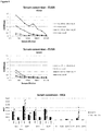
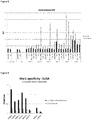

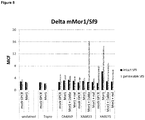
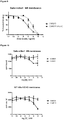


Classifications
-
- C—CHEMISTRY; METALLURGY
- C07—ORGANIC CHEMISTRY
- C07K—PEPTIDES
- C07K16/00—Immunoglobulins [IGs], e.g. monoclonal or polyclonal antibodies
- C07K16/18—Immunoglobulins [IGs], e.g. monoclonal or polyclonal antibodies against material from animals or humans
- C07K16/28—Immunoglobulins [IGs], e.g. monoclonal or polyclonal antibodies against material from animals or humans against receptors, cell surface antigens or cell surface determinants
-
- A—HUMAN NECESSITIES
- A61—MEDICAL OR VETERINARY SCIENCE; HYGIENE
- A61P—SPECIFIC THERAPEUTIC ACTIVITY OF CHEMICAL COMPOUNDS OR MEDICINAL PREPARATIONS
- A61P25/00—Drugs for disorders of the nervous system
- A61P25/04—Centrally acting analgesics, e.g. opioids
-
- A—HUMAN NECESSITIES
- A61—MEDICAL OR VETERINARY SCIENCE; HYGIENE
- A61P—SPECIFIC THERAPEUTIC ACTIVITY OF CHEMICAL COMPOUNDS OR MEDICINAL PREPARATIONS
- A61P43/00—Drugs for specific purposes, not provided for in groups A61P1/00-A61P41/00
-
- C—CHEMISTRY; METALLURGY
- C07—ORGANIC CHEMISTRY
- C07K—PEPTIDES
- C07K14/00—Peptides having more than 20 amino acids; Gastrins; Somatostatins; Melanotropins; Derivatives thereof
- C07K14/435—Peptides having more than 20 amino acids; Gastrins; Somatostatins; Melanotropins; Derivatives thereof from animals; from humans
- C07K14/705—Receptors; Cell surface antigens; Cell surface determinants
- C07K14/72—Receptors; Cell surface antigens; Cell surface determinants for hormones
- C07K14/723—G protein coupled receptor, e.g. TSHR-thyrotropin-receptor, LH/hCG receptor, FSH receptor
-
- G—PHYSICS
- G01—MEASURING; TESTING
- G01N—INVESTIGATING OR ANALYSING MATERIALS BY DETERMINING THEIR CHEMICAL OR PHYSICAL PROPERTIES
- G01N33/00—Investigating or analysing materials by specific methods not covered by groups G01N1/00 - G01N31/00
- G01N33/48—Biological material, e.g. blood, urine; Haemocytometers
- G01N33/50—Chemical analysis of biological material, e.g. blood, urine; Testing involving biospecific ligand binding methods; Immunological testing
- G01N33/5005—Chemical analysis of biological material, e.g. blood, urine; Testing involving biospecific ligand binding methods; Immunological testing involving human or animal cells
- G01N33/5008—Chemical analysis of biological material, e.g. blood, urine; Testing involving biospecific ligand binding methods; Immunological testing involving human or animal cells for testing or evaluating the effect of chemical or biological compounds, e.g. drugs, cosmetics
- G01N33/502—Chemical analysis of biological material, e.g. blood, urine; Testing involving biospecific ligand binding methods; Immunological testing involving human or animal cells for testing or evaluating the effect of chemical or biological compounds, e.g. drugs, cosmetics for testing non-proliferative effects
- G01N33/5041—Chemical analysis of biological material, e.g. blood, urine; Testing involving biospecific ligand binding methods; Immunological testing involving human or animal cells for testing or evaluating the effect of chemical or biological compounds, e.g. drugs, cosmetics for testing non-proliferative effects involving analysis of members of signalling pathways
-
- G—PHYSICS
- G01—MEASURING; TESTING
- G01N—INVESTIGATING OR ANALYSING MATERIALS BY DETERMINING THEIR CHEMICAL OR PHYSICAL PROPERTIES
- G01N33/00—Investigating or analysing materials by specific methods not covered by groups G01N1/00 - G01N31/00
- G01N33/48—Biological material, e.g. blood, urine; Haemocytometers
- G01N33/50—Chemical analysis of biological material, e.g. blood, urine; Testing involving biospecific ligand binding methods; Immunological testing
- G01N33/68—Chemical analysis of biological material, e.g. blood, urine; Testing involving biospecific ligand binding methods; Immunological testing involving proteins, peptides or amino acids
- G01N33/6854—Immunoglobulins
-
- C—CHEMISTRY; METALLURGY
- C07—ORGANIC CHEMISTRY
- C07K—PEPTIDES
- C07K2317/00—Immunoglobulins specific features
- C07K2317/20—Immunoglobulins specific features characterized by taxonomic origin
- C07K2317/22—Immunoglobulins specific features characterized by taxonomic origin from camelids, e.g. camel, llama or dromedary
-
- C—CHEMISTRY; METALLURGY
- C07—ORGANIC CHEMISTRY
- C07K—PEPTIDES
- C07K2317/00—Immunoglobulins specific features
- C07K2317/30—Immunoglobulins specific features characterized by aspects of specificity or valency
- C07K2317/33—Crossreactivity, e.g. for species or epitope, or lack of said crossreactivity
-
- C—CHEMISTRY; METALLURGY
- C07—ORGANIC CHEMISTRY
- C07K—PEPTIDES
- C07K2317/00—Immunoglobulins specific features
- C07K2317/50—Immunoglobulins specific features characterized by immunoglobulin fragments
- C07K2317/56—Immunoglobulins specific features characterized by immunoglobulin fragments variable (Fv) region, i.e. VH and/or VL
- C07K2317/565—Complementarity determining region [CDR]
-
- C—CHEMISTRY; METALLURGY
- C07—ORGANIC CHEMISTRY
- C07K—PEPTIDES
- C07K2317/00—Immunoglobulins specific features
- C07K2317/50—Immunoglobulins specific features characterized by immunoglobulin fragments
- C07K2317/56—Immunoglobulins specific features characterized by immunoglobulin fragments variable (Fv) region, i.e. VH and/or VL
- C07K2317/567—Framework region [FR]
-
- C—CHEMISTRY; METALLURGY
- C07—ORGANIC CHEMISTRY
- C07K—PEPTIDES
- C07K2317/00—Immunoglobulins specific features
- C07K2317/50—Immunoglobulins specific features characterized by immunoglobulin fragments
- C07K2317/56—Immunoglobulins specific features characterized by immunoglobulin fragments variable (Fv) region, i.e. VH and/or VL
- C07K2317/569—Single domain, e.g. dAb, sdAb, VHH, VNAR or nanobody®
-
- G—PHYSICS
- G01—MEASURING; TESTING
- G01N—INVESTIGATING OR ANALYSING MATERIALS BY DETERMINING THEIR CHEMICAL OR PHYSICAL PROPERTIES
- G01N2333/00—Assays involving biological materials from specific organisms or of a specific nature
- G01N2333/435—Assays involving biological materials from specific organisms or of a specific nature from animals; from humans
- G01N2333/705—Assays involving receptors, cell surface antigens or cell surface determinants
- G01N2333/72—Assays involving receptors, cell surface antigens or cell surface determinants for hormones
- G01N2333/726—G protein coupled receptor, e.g. TSHR-thyrotropin-receptor, LH/hCG receptor, FSH
Definitions
- GPCRs G protein-coupled receptors
- agents that specifically bind to a transmembrane receptor in a conformationally-specific way can be used to induce a conformational change in the transmembrane receptor.
- agents have therapeutic applications and can be used in X-ray crystallography studies of the transmembrane receptor.
- agents can also be used to improve drug discovery via compound screening and/or structure based drug design.
- Opioids and cannabinoids are two classes of analgesics that have been used to treat pain for centuries and are arguably the oldest of "pharmacological" interventions used by man. Unfortunately, they also produce several adverse side effects that can complicate pain management.
- Opioids produce their pharmacological effects through activation of G protein-coupled receptors (GPCRs).
- GPCRs G protein-coupled receptors
- opioid receptors There are four distinct genes coding for opioid receptors: the mu-, kappa-, and delta-opioid receptors (MOR, KOR, and DOR, respectively) and the opioid-like receptor1 [ORL-1 or the nociceptin receptor (NOP)] (Cox 2012; Pasternak 2013).
- MOR mu-, kappa-, and delta-opioid receptors
- NOP nociceptin receptor
- the generation of genetic knockout mice has demonstrated that the majority of clinically used opioids including morphine produce their pharmacological effects primarily by activating the MOR (Matthes et al. 1996; Sora et al. 1997; Roy et al. 1998; Kieffer 1999; Kieffer and Gaveriaux-Ruff 2002).
- the MOR is widely distributed and expressed in neurons in the brain,
- herkinorin reduces formalin-induced flinching to the same degree as morphine when administered at the same dose (10 mg/kg, i.pl.), an effect that is reversed by the opioid antagonist naloxone (Lamb et al. 2012).
- opioid antagonist naloxone the opioid antagonist naloxone
- antinociceptive tolerance to herkinorin does not develop to repeated treatment over a 5-day period and it produces antinociception in morphine-tolerant rats (Lamb et al. 2012).
- the initial studies with these MOR "biased" agonists lend further support to the idea that developing a MOR agonist that does not engage ⁇ -arrestins but fully activates G protein signaling may provide a novel therapeutic avenue to improve pain treatment with opioids.
- Antibodies that target an extracellular region of an opioid receptor are described in Gupta et al. 2007; Gupta et al. 2008; and Mace et al. 2001.
- polypeptide As used herein, the terms “polypeptide”, “protein”, “peptide” are used interchangeably herein, and refer to a polymeric form of amino acids of any length, which can include coded and non-coded amino acids, chemically or biochemically modified or derivatized amino acids, and polypeptides having modified peptide backbones. Throughout the application, the standard one letter notation of amino acids will be used. Typically, the term “amino acid” will refer to "proteinogenic amino acid”, i.e. those amino acids that are naturally present in proteins. Most particularly, the amino acids are in the L isomeric form, but D amino acids are also envisaged.
- nucleic acid molecule As used herein, the terms “nucleic acid molecule”, “polynucleotide”, “polynucleic acid”, “nucleic acid” are used interchangeably and refer to a polymeric form of nucleotides of any length, either deoxyribonucleotides or ribonucleotides, or analogs thereof. Polynucleotides may have any three-dimensional structure, and may perform any function, known or unknown.
- Non-limiting examples of polynucleotides include a gene, a gene fragment, exons, introns, messenger RNA (mRNA), transfer RNA, ribosomal RNA, ribozymes, cDNA, recombinant polynucleotides, branched polynucleotides, plasmids, vectors, isolated DNA of any sequence, control regions, isolated RNA of any sequence, nucleic acid probes, and primers.
- the nucleic acid molecule may be linear or circular.
- any of the peptides, polypeptides, nucleic acids, compound, etc., disclosed herein may be “isolated” or “purified”. "Isolated” is used herein to indicate that the material referred to is (i) separated from one or more substances with which it exists in nature (e.g., is separated from at least some cellular material, separated from other polypeptides, separated from its natural sequence context), and/or (ii) is produced by a process that involves the hand of man such as recombinant DNA technology, chemical synthesis, etc.; and/or (iii) has a sequence, structure, or chemical composition not found in nature.
- isolated is meant to include compounds that are within samples that are substantially enriched for the compound of interest and/or in which the compound of interest is partially or substantially purified.
- Purified denote that the material referred to is removed from its natural environment and is at least 60% free, at least 75% free, or at least 90% free from other components with which it is naturally associated, also referred to as being “substantially pure”.
- sequence identity refers to the extent that sequences are identical on a nucleotide-by-nucleotide basis or an amino acid-by-amino acid basis over a window of comparison.
- a “percentage of sequence identity” is calculated by comparing two optimally aligned sequences over the window of comparison, determining the number of positions at which the identical nucleic acid base (e.g., A, T, C, G, I) or the identical amino acid residue (e.g., Ala, Pro, Ser, Thr, Gly, Val, Leu, Ile, Phe, Tyr, Trp, Lys, Arg, His, Asp, Glu, Asn, Gln, Cys and Met) occurs in both sequences to yield the number of matched positions, dividing the number of matched positions by the total number of positions in the window of comparison (i.e., the window size), and multiplying the result by 100 to yield the percentage of sequence identity.
- the identical nucleic acid base e.g., A, T
- Determining the percentage of sequence identity can be done manually, or by making use of computer programs that are available in the art. Examples of useful algorithms are PILEUP ( Higgins & Sharp, CABIOS 5:151 (1989 ), BLAST and BLAST 2.0 ( Altschul et al. J. Mol. Biol. 215: 403 (1990 ). Software for performing BLAST analyses is publicly available through the National Center for Biotechnology Information (http://www.ncbi.nlm.nih.gov/).
- Similarity refers to the percentage number of amino acids that are identical or constitute conservative substitutions. Similarity may be determined using sequence comparison programs such as GAP (Deveraux et al. 1984). In this way, sequences of a similar or substantially different length to those cited herein might be compared by insertion of gaps into the alignment, such gaps being determined, for example, by the comparison algorithm used by GAP.
- conservative substitution is the substitution of amino acids with other amino acids whose side chains have similar biochemical properties (e.g. are aliphatic, are aromatic, are positively charged, %) and is well known to the skilled person. Non-conservative substitution is then the substitution of amino acids with other amino acids whose side chains do not have similar biochemical properties (e.g.
- Conservative substitutions will typically yield sequences which are not identical anymore, but still highly similar. By conservative substitutions is intended combinations such as Gly, Ala; Val, Ile, Leu, Met; Asp, Glu; Asn, Gln; Ser, Thr; Lys, Arg; Cys, Met; and Phe, Tyr, Trp.
- a “deletion” is defined here as a change in either amino acid or nucleotide sequence in which one or more amino acid or nucleotide residues, respectively, are absent as compared to an amino acid sequence or nucleotide sequence of a parental polypeptide or nucleic acid.
- a deletion can involve deletion of about 2, about 5, about 10, up to about 20, up to about 30 or up to about 50 or more amino acids.
- a protein or a fragment thereof may contain more than one deletion.
- a deletion may also be a loop deletion, or an N- and/or C-terminal deletion, or a combination thereof.
- an N- and/or C-terminal deletion of a GPCR is also referred to as a truncation of the amino acid sequence of the GPCR or a truncated GPCR.
- an “insertion” or “addition” is that change in an amino acid or nucleotide sequences which has resulted in the addition of one or more amino acid or nucleotide residues, respectively, as compared to an amino acid sequence or nucleotide sequence of a parental protein.
- “Insertion” generally refers to addition to one or more amino acid residues within an amino acid sequence of a polypeptide, while “addition” can be an insertion or refer to amino acid residues added at an N- or C-terminus, or both termini.
- an insertion or addition is usually of about 1, about 3, about 5, about 10, up to about 20, up to about 30 or up to about 50 or more amino acids.
- a protein or fragment thereof may contain more than one insertion.
- substitution results from the replacement of one or more amino acids or nucleotides by different amino acids or nucleotides, respectively as compared to an amino acid sequence or nucleotide sequence of a parental protein or a fragment thereof. It is understood that a protein or a fragment thereof may have conservative amino acid substitutions which have substantially no effect on the protein's activity. By conservative substitutions is intended combinations such as Gly, Ala; Val, Ile, Leu, Met; Asp, Glu; Asn, Gln; Ser, Thr; Lys, Arg; Cys, Met; and Phe, Tyr, Trp.
- recombinant when used in reference to a cell, nucleic acid, protein or vector, indicates that the cell, nucleic acid, protein or vector, has been modified by the introduction of a heterologous nucleic acid or protein or the alteration of a native nucleic acid or protein, or that the cell is derived from a cell so modified.
- recombinant cells express nucleic acids or polypeptides that are not found within the native (non-recombinant) form of the cell or express native genes that are otherwise abnormally expressed, under expressed, over expressed or not expressed at all.
- expression refers to the process by which a polypeptide is produced based on the nucleic acid sequence of a gene.
- the process includes both transcription and translation.
- operably linked refers to a linkage in which the regulatory sequence is contiguous with the gene of interest to control the gene of interest, as well as regulatory sequences that act in trans or at a distance to control the gene of interest.
- a DNA sequence is operably linked to a promoter when it is ligated to the promoter downstream with respect to the transcription initiation site of the promoter and allows transcription elongation to proceed through the DNA sequence.
- a DNA for a signal sequence is operably linked to DNA coding for a polypeptide if it is expressed as a pre-protein that participates in the transport of the polypeptide.
- Linkage of DNA sequences to regulatory sequences is typically accomplished by ligation at suitable restriction sites or adapters or linkers inserted in lieu thereof using restriction endonucleases known to one of skill in the art.
- control sequences refers to polynucleotide sequences which are necessary to affect the expression of coding sequences to which they are operably linked.
- Expression control sequences are sequences which control the transcription, post-transcriptional events and translation of nucleic acid sequences. Expression control sequences include appropriate transcription initiation, termination, promoter and enhancer sequences; efficient RNA processing signals such as splicing and polyadenylation signals; sequences that stabilize cytoplasmic mRNA; sequences that enhance translation efficiency (e.g., ribosome binding sites); sequences that enhance protein stability; and when desired, sequences that enhance protein secretion. The nature of such control sequences differs depending upon the host organism.
- control sequences is intended to include, at a minimum, all components whose presence is essential for expression, and can also include additional components whose presence is advantageous, for example, leader sequences and fusion partner sequences.
- vector as used herein is intended to refer to a nucleic acid molecule capable of transporting another nucleic acid molecule to which it has been linked.
- the vector may be of any suitable type including, but not limited to, a phage, virus, plasmid, phagemid, cosmid, bacmid or even an artificial chromosome.
- Certain vectors are capable of autonomous replication in a host cell into which they are introduced (e.g., vectors having an origin of replication which functions in the host cell).
- Other vectors can be integrated into the genome of a host cell upon introduction into the host cell, and are thereby replicated along with the host genome.
- certain preferred vectors are capable of directing the expression of certain genes of interest.
- recombinant expression vectors Such vectors are referred to herein as “recombinant expression vectors” (or simply, “expression vectors”). Suitable vectors have regulatory sequences, such as promoters, enhancers, terminator sequences, and the like as desired and according to a particular host organism (e.g. bacterial cell, yeast cell). Typically, a recombinant vector according to the present invention comprises at least one "chimeric gene" or "expression cassette".
- Expression cassettes are generally DNA constructs preferably including (5' to 3' in the direction of transcription): a promoter region, a polynucleotide sequence, homologue, variant or fragment thereof of the present invention operably linked with the transcription initiation region, and a termination sequence including a stop signal for RNA polymerase and a polyadenylation signal. It is understood that all of these regions should be capable of operating in biological cells, such as prokaryotic or eukaryotic cells, to be transformed.
- the promoter region comprising the transcription initiation region, which preferably includes the RNA polymerase binding site, and the polyadenylation signal may be native to the biological cell to be transformed or may be derived from an alternative source, where the region is functional in the biological cell.
- host cell is intended to refer to a cell into which a recombinant vector has been introduced. It should be understood that such terms are intended to refer not only to the particular subject cell but to the progeny of such a cell. Because certain modifications may occur in succeeding generations due to either mutation or environmental influences, such progeny may not, in fact, be identical to the parent cell, but are still included within the scope of the term "host cell” as used herein.
- a host cell may be an isolated cell or cell line grown in culture or may be a cell which resides in a living tissue or organism. In particular, host cells are of bacterial or fungal origin, but may also be of plant or mammalian origin.
- the wordings "host cell”, “recombinant host cell”, “expression host cell”, “expression host system”, “expression system”, are intended to have the same meaning and are used interchangeably herein.
- G-protein coupled receptors or “GPCRs” are polypeptides that share a common structural motif, having seven regions of between 22 to 24 hydrophobic amino acids that form seven alpha helices, each of which spans a membrane. Each span is identified by number, i.e., transmembrane-1 (TM1), transmembrane-2 (TM2), etc.
- transmembrane helices are joined by regions of amino acids between transmembrane-2 and transmembrane-3, transmembrane-4 and transmembrane-5, and transmembrane-6 and transmembrane-7 on the exterior, or "extracellular" side, of the cell membrane, referred to as "extracellular" regions 1, 2 and 3 (EC1, EC2 and EC3), respectively.
- transmembrane helices are also joined by regions of amino acids between transmembrane-1 and transmembrane-2, transmembrane-3 and transmembrane-4, and transmembrane-5 and transmembrane-6 on the interior, or "intracellular” side, of the cell membrane, referred to as "intracellular” regions 1, 2 and 3 (IC1, IC2 and IC3), respectively.
- the "carboxy" (“C”) terminus of the receptor lies in the intracellular space within the cell, and the "amino" (“N”) terminus of the receptor lies in the extracellular space outside of the cell.
- GPCR structure and classification is generally well known in the art, and further discussion of GPCRs may be found in Probst, DNA Cell Biol.
- biologically active refers to a GPCR having a biochemical function (e.g., a binding function, a signal transduction function, or an ability to change conformation as a result of ligand binding) of a naturally occurring GPCR.
- a biochemical function e.g., a binding function, a signal transduction function, or an ability to change conformation as a result of ligand binding
- naturally-occurring in reference to a GPCR means a GPCR that is naturally produced (e.g., by a wild type mammal such as a human). Such GPCRs are found in nature.
- non-naturally occurring in reference to a GPCR means a GPCR that is not naturally-occurring.
- Naturally-occurring GPCRs that have been made constitutively active through mutation, and variants of naturally-occurring transmembrane receptors, e.g., epitope-tagged GPCRs and GPCRs lacking their native N-terminus are examples of non-naturally occurring GPCRs.
- Non-naturally occurring versions of a naturally occurring GPCR are often activated by the same ligand as the naturally-occurring GPCR.
- Non-limiting examples of either naturally-occurring or non-naturally occurring GPCRs within the context of the present invention are provided further herein, in particular for opioid receptors.
- an “epitope”, as used herein, refers to an antigenic determinant of a polypeptide.
- An epitope could comprise 3 amino acids in a spatial conformation, which is unique to the epitope.
- an epitope consists of at least 4, 5, 6, 7 such amino acids, and more usually, consists of at least 8, 9, 10 such amino acids.
- Methods of determining the spatial conformation of amino acids are known in the art, and include, for example, x-ray crystallography and multi-dimensional nuclear magnetic resonance.
- a “conformational epitope”, as used herein, refers to an epitope comprising amino acids in a spacial conformation that is unique to a folded 3-dimensional conformation of the polypeptide.
- a conformational epitope consists of amino acids that are discontinuous in the linear sequence that come together in the folded structure of the protein.
- a conformational epitope may also consist of a linear sequence of amino acids that adopts a conformation that is unique to a folded 3-dimensional conformation of the polypeptide (and not present in a denatured state).
- conformation or conformational state of a protein refers generally to the range of structures that a protein may adopt at any instant in time.
- determinants of conformation or conformational state include a protein's primary structure as reflected in a protein's amino acid sequence (including modified amino acids) and the environment surrounding the protein.
- the conformation or conformational state of a protein also relates to structural features such as protein secondary structures (e.g., ⁇ -helix, ⁇ -sheet, among others), tertiary structure (e.g., the three dimensional folding of a polypeptide chain), and quaternary structure (e.g., interactions of a polypeptide chain with other protein subunits).
- Post-translational and other modifications to a polypeptide chain such as ligand binding, phosphorylation, sulfation, glycosylation, or attachments of hydrophobic groups, among others, can influence the conformation of a protein.
- environmental factors such as pH, salt concentration, ionic strength, and osmolality of the surrounding solution, and interaction with other proteins and co-factors, among others, can affect protein conformation.
- the conformational state of a protein may be determined by either functional assay for activity or binding to another molecule or by means of physical methods such as X-ray crystallography, NMR, or spin labeling, among other methods.
- a “functional conformation” or a “functional conformational state”, as used herein, refers to the fact that proteins possess different conformational states having a dynamic range of activity, in particular ranging from no activity to maximal activity. It should be clear that "a functional conformational state” is meant to cover any conformational state of a protein, having any activity, including no activity, and is not meant to cover the denatured states of proteins. Non-limiting examples of functional conformations include active conformations or inactive conformations (as defined further herein). A particular class of functional conformations is defined as "druggable conformation" and generally refers to a unique therapeutically relevant conformational state of a target protein.
- the agonist-bound active conformation of the mu-opioid receptor corresponds to the druggable conformation of this receptor relating to pain. It will thus be understood that druggability is confined to particular conformations depending on the therapeutic indication. More details are provided further herein.
- active conformation and “active form” refer to a GPCR, particularly an opioid receptor, that is folded in a way so as to be active.
- a GPCR can be placed into an active conformation using an agonist of the receptor.
- a GPCR in its active conformation binds to heterotrimeric G protein and catalyzes nucleotide exchange of the G-protein to activate downstream signaling pathways.
- Activated GPCRs bind to the inactive, GDP-bound form of heterotrimeric G-proteins and cause the G-proteins to release their GDP so GTP can bind. There is a transient 'nucleotide-free' state that results from this process that enables GTP to bind.
- inactive conformation and “inactive form” refer to a GPCR, particularly opioid receptor, that is folded in a way so as to be inactive.
- a GPCR can be placed into an inactive conformation using an inverse agonist of the receptor. For example, a GPCR in its inactive conformation does not bind to heterotrimeric G protein with high affinity.
- active conformation and “inactive conformation” will be illustrated further herein.
- stabilizing refers to the retaining or holding of a GPCR protein in a subset of the possible conformations that it could otherwise assume, due to the effects of the interaction of the GPCR with the binding agent according to the invention.
- a binding agent that selectively binds to a specific conformation or conformational state of a protein refers to a binding agent that binds with a higher affinity to a protein in a subset of conformations or conformational states than to other conformations or conformational states that the protein may assume.
- binding agents that specifically or selectively bind to a specific conformation or conformational state of a protein will stabilize this specific conformation or conformational state.
- affinity refers to the degree to which a ligand binds to an antigen on a receptor so as to shift the equilibrium of receptor and ligand toward the presence of a complex formed by their binding.
- an antibody (fragment) of high affinity will bind to the available antigen so as to shift the equilibrium toward high concentration of the resulting complex.
- the dissociation constant is commonly used to describe the affinity between the binding domain and the antigenic target.
- the dissociation constant is lower than 10-5 M.
- the dissociation constant is lower than 10-6 M, more preferably, lower than 10-7 M.
- the dissociation constant is lower than 10-8 M.
- the ligand may be a binding agent, preferably an immunoglobulin, such as an antibody, or an immunoglobulin fragment, such as a VHH or Nanobody, that binds a conformational epitope on a GPCR.
- binding agent refers to the ability of a binding agent, in particular an immunoglobulin or an immunoglobulin fragment, such as a VHH or Nanobody, to bind preferentially to one antigen, versus a different antigen, and does not necessarily imply high affinity.
- telomere binding agent generally refers to the ability of a binding agent, in particular an immunoglobulin, such as an antibody, or an immunoglobulin fragment, such as a VHH or Nanobody, to preferentially bind to a particular antigen that is present in a homogeneous mixture of different antigens.
- a specific binding interaction will discriminate between desirable and undesirable antigens in a sample, in some embodiments more than about 10 to 100-fold or more (e.g., more than about 1000- or 10,000-fold).
- the terms particularly refer to the ability of a binding agent (as defined herein) to preferentially recognize and/or bind to a particular conformational state of a GPCR as compared to another conformational state.
- the term "conformation-selective binding agent” in the context of the present invention refers to a binding agent that binds to a target protein in a conformation-selective manner.
- a binding agent that selectively binds to a particular conformation or conformational state of a protein refers to a binding agent that binds with a higher affinity to a protein in a subset of conformations or conformational states than to other conformations or conformational states that the protein may assume.
- binding agents that selectively bind to a specific conformation or conformational state of a protein will stabilize or retain the protein it this particular conformation or conformational state.
- an active state conformation-selective binding agent will preferentially bind to a GPCR in an active conformational state and will not or to a lesser degree bind to a GPCR in an inactive conformational state, and will thus have a higher affinity for said active conformational state; or vice versa.
- the terms “specifically bind”, “selectively bind”, “preferentially bind”, and grammatical equivalents thereof, are used interchangeably herein.
- the terms “conformational specific” or “conformational selective” are also used interchangeably herein.
- test compound or “test compound” or “candidate compound” or “drug candidate compound” as used herein describes any molecule, either naturally occurring or synthetic that is tested in an assay, such as a screening assay or drug discovery assay.
- these compounds comprise organic or inorganic compounds.
- the compounds include polynucleotides, lipids or hormone analogs that are characterized by low molecular weights.
- Other biopolymeric organic test compounds include small peptides or peptide-like molecules (peptidomimetics) comprising from about 2 to about 40 amino acids and larger polypeptides comprising from about 40 to about 500 amino acids, such as antibodies, antibody fragments or antibody conjugates.
- Test compounds can also be protein scaffolds.
- test compound libraries may be used, such as combinatorial or randomized libraries that provide a sufficient range of diversity. Examples include, but are not limited to, natural compound libraries, allosteric compound libraries, peptide libraries, antibody fragment libraries, synthetic compound libraries, fragment-based libraries, phage-display libraries, and the like. A more detailed description can be found further in the specification.
- ligand means a molecule that specifically binds to a GPCR, in particular an opioid receptor.
- a ligand may be, without the purpose of being limitative, a polypeptide, a lipid, a small molecule, an antibody, an antibody fragment, a nucleic acid, a carbohydrate.
- a ligand may be synthetic or naturally occurring.
- a ligand also includes a "native ligand” which is a ligand that is an endogenous, natural ligand for a native GPCR. Within the context of the present invention, a ligand may bind to a GPCR, either intracellularly or extracellularly.
- a ligand may be an agonist, a partial agonist, an inverse agonist, an antagonist, an allosteric modulator, and may bind at either the orthosteric site or at an allosteric site.
- a ligand may be a "conformation-selective ligand” or “conformation-specific ligand”, meaning that such a ligand binds the GPCR in a conformation-selective manner.
- a conformation-selective ligand binds with a higher affinity to a particular conformation of the GPCR than to other conformations the GPCR may adopt.
- a neutral antagonist is not considered as a conformation-selective ligand, since a neutral antagonist does not distinguish between the different conformations of a GPCR.
- an "orthosteric ligand”, as used herein, refers to a ligand (both natural and synthetic), that binds to the active site of a GPCR, in particular opioid receptor, and are further classified according to their efficacy or in other words to the effect they have on signaling through a specific pathway.
- an "agonist” refers to a ligand that, by binding a receptor protein, increases the receptor's signaling activity. Full agonists are capable of maximal protein stimulation; partial agonists are unable to elicit full activity even at saturating concentrations. Partial agonists can also function as "blockers" by preventing the binding of more robust agonists.
- an “antagonist”, also referred to as a “neutral antagonist”, refers to a ligand that binds a receptor without stimulating any activity.
- An “antagonist” is also known as a “blocker” because of its ability to prevent binding of other ligands and, therefore, block agonist-induced activity.
- an “inverse agonist” refers to an antagonist that, in addition to blocking agonist effects, reduces a receptor's basal or constitutive activity below that of the unliganded protein.
- Ligands as used herein may also be “biased ligands” with the ability to selectively stimulate a subset of a receptor's signaling activities, for example in the case of GPCRs the selective activation of G-protein or ⁇ -arrestin function.
- Such ligands are known as “biased ligands", “biased agonists” or “functionally selective agonists”. More particularly, ligand bias can be an imperfect bias characterized by a ligand stimulation of multiple receptor activities with different relative efficacies for different signals (non-absolute selectivity) or can be a perfect bias characterized by a ligand stimulation of one receptor protein activity without any stimulation of another known receptor protein activity.
- allosteric regulators Another kind of ligands is known as allosteric regulators.
- Allosteric regulators or otherwise “allosteric modulators”, “allosteric ligands” or “effector molecules”, as used herein, refer to ligands that bind at an allosteric site (that is, a regulatory site physically distinct from the protein's active site) of a GPCR, in particular opioid receptor.
- allosteric modulators are non-competitive because they bind receptor proteins at a different site and modify their function even if the endogenous ligand also is binding.
- Allosteric regulators that enhance the protein's activity are referred to herein as “allosteric activators” or “positive allosteric modulators” (PAMs), whereas those that decrease the protein's activity are referred to herein as “allosteric inhibitors” or otherwise “negative allosteric modulators” (NAMs).
- PAMs positive allosteric modulators
- NAMs negative allosteric modulators
- determining As used herein, the terms “determining”, “measuring”, “assessing”, “assaying” are used interchangeably and include both quantitative and qualitative determinations.
- antibody is intended to mean an immunoglobulin or any fragment thereof that is capable of antigen binding.
- antibody also refers to single chain antibodies and antibodies with only one binding domain.
- CDR complementarity determining region
- H and VL light chains
- CDR regions account for the basic specificity of the antibody for a particular antigenic determinant structure. Such regions are also referred to as “hypervariable regions.”
- the CDRs represent non-contiguous stretches of amino acids within the variable regions but, regardless of species, the positional locations of these critical amino acid sequences within the variable heavy and light chain regions have been found to have similar locations within the amino acid sequences of the variable chains.
- variable heavy and light chains of all canonical antibodies each have 3 CDR regions, each non- contiguous with the others (termed L1, L2, L3, H1, H2, H3) for the respective light (L) and heavy (H) chains.
- Immunoglobulin single variable domains in particular Nanobodies, generally comprise a single amino acid chain that can be considered to comprise 4 "framework sequences or regions" or FRs and 3 "complementary determining regions" or CDRs.
- the nanobodies have 3 CDR regions, each non-contiguous with the others (termed CDR1, CDR2, CDR3).
- the delineation of the FR and CDR sequences can, for example, be based on the IMGT unique numbering system for V-domains and V-like domains ( Lefranc et al. 2003, Developmental and Comparative Immunology 27:55 ).
- a first aspect of the invention relates to a binding agent that is directed against and/or capable of specifically binding to a GPCR of the opioid receptor family (OR or OP receptors), wherein the binding agent of the present invention is an active conformation-selective VHH antibody comprising an amino acid sequence that comprises 4 framework regions (FR1 to FR4) and 3 complementary determining regions (CDR1 to CDR3) as defined by the IMGT numbering system that is directed against and/or capable of specifically binding to an active conformation of an opioid receptor, wherein the VHH antibody binds to an intracellular conformational epitope of the receptor, wherein FR1 is SEQ ID NO: 7, CDR1 is SEQ ID NO: 13, FR2 is SEQ ID NO: 19, CDR2 is SEQ ID NO: 25, FR3 is SEQ ID NO: 31, CDR3 is SEQ ID NO: 37 and FR4 is SEQ ID NO: 43; or wherein FR1 is SEQ ID NO: 8, CDR1 is SEQ ID NO: 14, FR2 is SEQ ID
- a first aspect of the disclosure relates to a binding agent that is directed against and/or capable of specifically binding to a GPCR of the opioid receptor family (OR or OP receptors).
- the invention relates to a conformation-selective binding agent that is directed against and/or capable of specifically binding to a GPCR of the opioid receptor family (OR or OP receptors).
- the "opioid receptor” refers to a receptor belonging to the superfamily of GPCRs (as defined herein), more particularly to the family A GPCRs, and include four types, designated ⁇ (mu or MOP or Mor1), ⁇ (delta or DOP), ⁇ (kappa or KOP) opioid receptors, and finally NOP; see also Table 1.
- ⁇ mi or MOP or Mor1
- ⁇ delta or DOP
- ⁇ kappa or KOP
- NOP see also Table 1.
- the amino acid sequences (and the nucleotide sequences of the cDNAs which encode them) of the opioid receptors are readily available, for example by reference to GenBank (http://www.ncbi.nlm.nih.gov/entrez).
- HGNC standardized nomenclature to human genes; accession numbers of different isoforms from different organisms are available from Uniprot (www.uniprot.org), see also Table 4. Moreover, a comprehensive overview of receptor nomenclature, pharmacological, functional and pathophysiological information on opioid receptors can be retrieved from the IUPHAR database (http://www.iuphar-db.org/). Table 1.
- Opioid receptor type classification Current recommended nomenclature Presumed endogenous ligands ⁇ , mu, MOP, MOR1 ⁇ -endorphin enkephalins endomorphin-1 endomorphin-2 ⁇ , delta, DOP enkephalines ⁇ -endorphin ⁇ , kappa, KOP dynorphin A dynorphin B ⁇ -neoendorphin NOP nociception/orphanin FQ
- the VHH antibody of the present invention is directed against and/or specifically binds to an opioid receptor.
- the nature of the opioid receptor is not critical to the invention and can be from any organism including a fungus (including yeast), nematode, virus, insect, plant, bird (e.g. chicken, turkey), reptile or mammal (e.g., a mouse, rat, rabbit, hamster, gerbil, dog, cat, goat, pig, cow, horse, whale, monkey, camelid, or human).
- the opioid receptor is of mammalian origin, even more preferably of human origin.
- the VHH antibody of the present invention is directed against and/or specifically binds to mu opioid receptor, such as the human mu opioid receptor (SEQ ID NO: 50). In another specific embodiment, the VHH antibody of the present invention is directed against and/or specifically binds to delta opioid receptor, such as the human delta opioid receptor (SEQ ID NO: 54). In yet another specific embodiment, the VHH antibody of the present invention is directed against and/or specifically binds to kappa opioid receptor, such as the human kappa opioid receptor (SEQ ID NO: 53). In yet another specific embodiment, the VHH antibody of the present invention is directed against and/or specifically binds to nociceptin receptor, such as the human nociceptin receptor (SEQ ID NO: 55).
- the VHH antibody of the present invention is directed against and/or specifically binds to the mu opioid receptor, in particular the human mu opioid receptor (SEQ ID NO: 50).
- the VHH antibody that is directed against and/or specifically binds to the human mu opioid receptor is cross-reactive with the delta opioid receptor and/or with the kappa opioid receptor and/or with the nociceptin receptor.
- the VHH antibody that is directed against and/or specifically binds to the human mu opioid receptor is not cross-reactive with the delta opioid receptor nor with the kappa opioid receptor nor with the nociceptin receptor.
- VHH antibody binding agent according to the disclosure
- a prerequisite of the VHH antibody (binding agent according to the disclosure) is its capability to specifically bind (as defined herein) to the opioid receptor, particularly the mu opioid receptor.
- the VHH antibody is directed against an intracellular conformational epitope of the opioid receptor.
- a VHH antibody (binding agent according to the disclosure) that specifically binds to a "conformational epitope” specifically binds to a tertiary (i.e., three dimensional) structure of a folded protein, and binds at much reduced (i.e., by a factor of at least 2, 5, 10, 50 or 100) affinity to the linear (i.e., unfolded, denatured) form of the protein.
- said conformational epitope can be part of an intracellular or extracellular region, or an intramembraneous region, or a domain or loop structure of the opioid receptor.
- the binding agent may be directed against an extracellular region, domain, loop or other extracellular conformational epitope of the opioid receptor, but is preferably directed against the extracellular parts of the transmembrane domains or more preferably against the extracellular loops that link the transmembrane domains.
- the VHH antibody according to the invention may be directed against an intracellular region, domain, loop or other intracellular conformational epitope of the opioid receptor, but is preferably directed against the intracellular parts of the transmembrane domains or more preferably against the intracellular loops that link the transmembrane domains.
- the VHH antibody may be directed against a conformational epitope that forms part of the binding site of a natural ligand, including but limited to an endogenous orthosteric agonist.
- the binding agent may be directed against a conformational epitope, in particular an intracellular epitope, that is comprised in a binding site for a downstream signaling protein, including but not-limited to a G protein binding site or a ⁇ -arrestin binding site.
- the binding agent directed against and/or specifically binding to an opioid receptor binds to an extracellular conformational epitope of the receptor.
- the binding agent directed against and/or specifically binding to an opioid receptor is capable of displacing an orthosteric ligand bound to the opioid receptor or prevents binding of an orthosteric ligand to the opioid receptor.
- the binding agent of the present invention can specifically displace an orthosteric ligand on the opioid receptor, with an average displacement of ligand binding signal of at least 40%, at least 50%, at least 60%, at least 70%, at least 80% or more. Percentages of average displacement can be determined in several ways, e.g. by a ligand displacement assay known in the art (see also Example section). Orthosteric ligands of opioid receptors are described further herein, and are well-known in the art.
- the VHH antibody directed against and/or specifically binding to an opioid receptor binds to an intracellular conformational epitope of the receptor.
- the binding agent preferably is capable of stabilizing the opioid receptor in a particular conformation.
- stabilizing or grammatically equivalent terms, as defined hereinbefore, is meant an increased stability of an opioid receptor with respect to the structure (e.g. conformational state) and/or particular biological activity (e.g. intracellular signaling activity, ligand binding affinity, ). In relation to increased stability with respect to structure and/or biological activity, this may be readily determined by either a functional assay for activity (e.g. Ca2+ release, cAMP generation or transcriptional activity, ⁇ -arrestin recruitment, ...) or ligand binding or by means of physical methods such as X-ray crystallography, NMR, or spin labeling, among other methods.
- stabilize also includes increased thermostability of the receptor under non-physiological conditions induced by denaturants or denaturing conditions.
- thermal stabilize refers to the functional rather than to the thermodynamic properties of a receptor and to the protein's resistance to irreversible denaturation induced by thermal and/or chemical approaches including but not limited to heating, cooling, freezing, chemical denaturants, pH, detergents, salts, additives, proteases or temperature. Irreversible denaturation leads to the irreversible unfolding of the functional conformations of the protein, loss of biological activity and aggregation of the denaturated protein.
- the binding agent is capable of increasing the stability as measured by an increase in the thermal stability of a opioid receptor in a functional conformational state with at least 2°C, at least 5°C, at least 8°C, and more preferably at least 10°C or 15°C or 20°C.
- the opioid receptor is incubated for a defined time in the presence of a test detergent or a test chaotropic agent and the stability is determined using, for example, ligand binding or a spectroscoptic method, optionally at increasing temperatures as discussed above. Otherwise, the binding agent is capable of increasing the stability to extreme pH of a functional conformational state of a opioid receptor.
- a typical test pH would be chosen for example in the range 6 to 8, the range 5.5 to 8.5, the range 5 to 9, the range 4.5 to 9.5, more specifically in the range 4.5 to 5.5 (low pH) or in the range 8.5 to 9.5 (high pH).
- thermo)stabilize applies to opioid receptors embedded in lipid particles or lipid layers (for example, lipid monolayers, lipid bilayers, and the like) and to opioid receptors that have been solubilized in detergent.
- the VHH antibody of the invention stabilizes a particular conformation of the opioid receptor upon binding of the binding agent.
- the opioid receptor is stabilized in an active conformation upon binding of a VHH antibody that is conformation-selective for an active conformation.
- active conformation refers to a spectrum of receptor conformations that allows signal transduction towards an intracellular effector system, including G protein dependent signaling and G protein-independent signaling (e.g. ⁇ -arrestin signaling).
- An "active conformation” thus encompasses a range of ligand-specific conformations, including an agonist conformation, a partial agonist conformation or a biased agonist conformation.
- the opioid receptor is stabilized in an inactive conformation upon binding of a binding agent of the disclosure that is conformation-selective for an inactive conformation.
- inactive conformation refers to a spectrum of receptor conformations that does not allow or blocks signal transduction towards an intracellular effector system.
- An "inactive conformation” thus encompasses a range of ligand-specific conformations, including an inverse agonist conformation. It will be understood that the site of binding of the ligand is not critical for obtaining an active or inactive conformation.
- orthosteric ligands as well as allosteric modulators may equally be capable of stabilizing an opioid receptor in an active or inactive conformation.
- the VHH antibody that is capable of stabilizing the opioid receptor may bind at the orthosteric site or an allosteric site.
- the binding agent that is capable of stabilizing the opioid receptor may be an agonist conformation-selective ligand, or a partial agonist conformation-selective ligand or a biased agonist conformation-selective ligand, or an inverse agonist conformation-selective ligand, either by binding at the orthosteric site or at an allosteric site.
- a binding agent such as a VHH antibody according to the invention that stabilizes an active conformation of an opioid receptor will increase or enhance the affinity of the receptor for an agonist, more particularly for a full agonist, a partial agonist or a biased agonist, as compared to the receptor in the absence of the binding agent (or in the presence of a mock binding agent).
- a binding agent that stabilizes an active conformation of an opioid receptor will decrease the affinity of the receptor for an inverse agonist, as compared to the receptor in the absence of the binding agent (or in the presence of a mock binding agent).
- a binding agent of the disclosure that stabilizes an inactive conformation of an opioid receptor will enhance the affinity of the receptor for an inverse agonist and will decrease the affinity of the receptor for an agonist, particularly for a full agonist, a partial agonist or a biased agonist, as compared to the receptor in the absence of the binding agent (or in the presence of a mock binding agent).
- An increase or decrease in affinity for a ligand may be measured by a decrease or increase, respectively in EC 50 , IC 50 , K d , K i or any other measure of affinity or potency known to one of skill in the art.
- the binding agent (such as a VHH antibody according to the invention) that stabilizes a particular conformation of a opioid receptor is capable of increasing or decreasing the affinity for a conformation-selective ligand at least 2 fold, at least 5 fold, at least 10 fold, at least 50 fold, and more preferably at least 100 fold, even more preferably at least 1000 fold or more, upon binding to the receptor.
- affinity measurements for conformation-selective ligands that trigger/inhibit particular signaling pathways may be carried out with any type of ligand, including natural ligands, small molecules, as well as biologicals; with orthosteric ligands as well as allosteric modulators; with single compounds as well as compound libraries; with lead compounds or fragments; etc.
- the VHH antibody of the present invention that is directed against and/or specifically binding to an opioid receptor is a G protein mimetic.
- G protein mimetic refers to a binding agent that, upon binding to an opioid receptor, enhances the affinity of the receptor for orthosteric or allosteric agonists, to a similar extend as upon binding of the natural G protein to the opioid receptor.
- a binding agent that is a G protein mimetic will occupy the G protein binding site of an opioid receptor.
- the natural G protein of an opioid receptor is the Gi protein.
- the VHH antibody of the present invention that is directed against and/or specifically binding to a opioid receptor is a Gi protein mimetic (as illustrated also in Example section).
- the opioid receptor to which the VHH antibodies of the invention will bind can be a naturally occurring or non-naturally occurring (i.e., altered by man) receptor (as defined herein).
- wild type polymorphic variants and isoforms of the opioid receptor, as well as orthologs across different species are examples of naturally occurring proteins, and are found for example, and without limitation, in a mammal, more specifically in a human, or in a virus, or in a plant, or in an insect, amongst others).
- Such receptors are found in nature.
- a "human mu opioid receptor” has an amino acid sequence that is at least 95% identical to (e.g., at least 95% or at least 98% identical to) the naturally occurring "human mu opioid receptor” of Genbank accession number NM_000914. Wild-type opioid receptors that have been mutated and variants of naturally-occurring opioid receptors are examples of non-naturally occurring proteins.
- Non-limiting examples of non-naturally occurring opioid receptors include, without limitation, opioid receptors that have been made constitutively active through mutation, opioid receptors with a loop deletion, opioid receptors with an N- and/or C-terminal deletion, opioid receptors with a substitution, an insertion or addition, or any combination thereof, in relation to their amino acid or nucleotide sequence, or other variants of naturally-occurring opioid receptors.
- target opioid receptors comprising a chimeric or hybrid structure, for example a chimeric opioid receptor with an N- and/or C-terminus from one opioid receptor and loops of a second opioid receptor, or comprising an opioid receptor fused to a moiety, such as T4 lysozyme, Flavodoxin, Xylanase, Rubredoxin or cytochrome b as an utility in GPCR crystallization ( Chun et al. 2012 and also described in patent applications WO2012/158555 , WO2012/030735 , WO2012/148586 ).
- a non-naturally occurring opioid receptor may have an amino acid sequence that is at least 80% identical to, at least 90% identical to, at least 95% identical to or at least 99% identical to, a corresponding naturally-occurring opioid receptor.
- the VHH antibody is capable of recognizing both a naturally-occurring as well as a non-naturally occurring opioid receptor. This may be particularly advantageous in certain circumstances, and depending on the purpose or application. For example, and for illustration purposes only, to increase the probability of obtaining crystals of a opioid receptor stabilized in a particular conformation enabled by the VHH antibodies of the present invention, it might be desired to perform some protein engineering without or only minimally affecting the conformation (e.g. active conformation with increased affinity for agonists). Or, alternatively or additionally, to increase cellular expression levels of an opioid receptor, or to increase the stability, one might also consider introducing certain mutations in the receptor of interest.
- binding agent means the whole or part of a proteinaceous (protein, protein-like or protein containing) molecule that is capable of binding using specific intermolecular interactions to an opioid receptor.
- binding agent is not meant to include a naturally-occurring binding partner of the opioid receptor, such as a G protein, an arrestin, an endogenous ligand; or variants or derivatives (including fragments) thereof.
- binding agent refers to a polypeptide, more particularly a protein domain.
- a suitable protein domain is an element of overall protein structure that is self-stabilizing and that folds independently of the rest of the protein chain and is often referred to as "binding domain".
- binding domains vary in length from between about 25 amino acids up to 500 amino acids and more. Many binding domains can be classified into folds and are recognizable, identifiable, 3-D structures. Some folds are so common in many different proteins that they are given special names. Non-limiting examples are binding domains selected from a 3- or 4-helix bundle, an armadillo repeat domain, a leucine-rich repeat domain, a PDZ domain, a SUMO or SUMO-like domain, a cadherin domain, an immunoglobulin-like domain, phosphotyrosine-binding domain, pleckstrin homology domain, src homology 2 domain, amongst others.
- a binding domain can thus be derived from a naturally occurring molecule, e.g. from components of the innate or adaptive immune system, or it can be entirely artificially designed.
- a binding domain can be immunoglobulin-based or it can be based on domains present in proteins, including but limited to microbial proteins, protease inhibitors, toxins, fibronectin, lipocalins, single chain antiparallel coiled coil proteins or repeat motif proteins.
- binding domains which are known in the art include, but are not limited to: antibodies, heavy chain antibodies (hcAb), single domain antibodies (sdAb), minibodies, the variable domain derived from camelid heavy chain antibodies (VHH or nanobodies), the variable domain of the new antigen receptors derived from shark antibodies (VNAR), alphabodies, protein A, protein G, designed ankyrin-repeat domains (DARPins), fibronectin type III repeats, anticalins, knottins, engineered CH2 domains (nanoantibodies), engineered SH3 domains, affibodies, peptides and proteins, lipopeptides (e.g.
- the binding domain of the invention is the variable domain derived from camelid heavy chain antibodies (VHH or antibodies). Frequently, when generating a particular type of binding domain using selection methods, combinatorial libraries comprising a consensus or framework sequence containing randomized potential interaction residues are used to screen for binding to a molecule of interest, such as a protein.
- the binding agent of the invention is derived from an innate or adaptive immune system.
- said binding agent is derived from an immunoglobulin.
- the binding agent according to the invention is derived from an antibody or an antibody fragment.
- antibody refers generally to a polypeptide encoded by an immunoglobulin gene, or a functional fragment thereof, that specifically binds and recognizes an antigen, and is known to the person skilled in the art.
- An antibody is meant to include a conventional four-chain immunoglobulin, comprising two identical pairs of polypeptide chains, each pair having one "light” (about 25 kDa) and one "heavy” chain (about 50 kDa).
- VH heavy chain variable domain
- VL light chain variable domain
- antibody is meant to include whole antibodies, including single-chain whole antibodies, and antigen-binding fragments.
- antigen-binding fragments may be antigen-binding antibody fragments that include, but are not limited to, Fab, Fab' and F(ab')2, Fd, single-chain Fvs (scFv), single-chain antibodies, disulfide-linked Fvs (dsFv) and fragments comprising or consisting of either a VL or VH domain, and any combination of those or any other functional portion of an immunoglobulin peptide capable of binding to the target antigen.
- the term "antibodies” is also meant to include heavy chain antibodies, or fragments thereof, including immunoglobulin single variable domains, as defined further herein.
- immunoglobulin single variable domain defines molecules wherein the antigen binding site is present on, and formed by, a single immunoglobulin domain (which is different from conventional immunoglobulins or their fragments, wherein typically two immunoglobulin variable domains interact to form an antigen binding site). It should however be clear that the term “immunoglobulin single variable domain” does comprise fragments of conventional immunoglobulins wherein the antigen binding site is formed by a single variable domain.
- the binding agent within the scope of the present invention is an immunoglobulin single variable domain.
- an immunoglobulin single variable domain will be an amino acid sequence comprising 4 framework regions (FR1 to FR4) and 3 complementary determining regions (CDR1 to CDR3), preferably according to the following formula (1): FR1-CDR1-FR2-CDR2-FR3-CDR3-FR4 (1) or, according to the disclosure, any suitable fragment thereof (which will then usually contain at least some of the amino acid residues that form at least one of the complementarity determining regions).
- Immunoglobulin single variable domains comprising 4 FRs and 3 CDRs are known to the person skilled in the art and have been described, as a non-limiting example, in Wesolowski et al. 2009.
- immunoglobulin single variable domains include light chain variable domain sequences (e.g. a VL domain sequence) or a suitable fragment thereof, or heavy chain variable domain sequences (e.g. a VH domain sequence or VHH domain sequence) or a suitable fragment thereof, as long as it is capable of forming a single antigen binding unit.
- the binding agent is an immunoglobulin single variable domain that is a light chain variable domain sequence (e.g. a VL domain sequence).
- the binding agent is an immunoglobulin single variable domain that is a heavy chain variable domain sequence (e.g.
- the immunoglobulin single variable domain is a heavy chain variable domain sequence that is derived from a conventional four-chain antibody or a heavy chain variable domain sequence that is derived from a heavy chain antibody.
- the immunoglobulin single variable domain may be a domain antibody, or a single domain antibody, or a "dAB” or dAb, or a Nanobody (as defined herein), or another immunoglobulin single variable domain, or, according to the disclosure, any suitable fragment of any one thereof.
- the immunoglobulin single variable domains generally comprise a single amino acid chain that can be considered to comprise 4 "framework sequences" or FR's and 3 "complementary determining regions" or CDR's (as defined hereinbefore). It should be clear that framework regions of immunoglobulin single variable domains may also contribute to the binding of their antigens (Desmyter et al 2002; Korotkov et al. 2009).
- the delineation of the CDR sequences (and thus also the FR sequences) can be based on the IMGT unique numbering system for V-domains and V-like domains (Lefranc et al. 2003).
- the delineation of the FR and CDR sequences can be done by using the Kabat numbering system as applied to VHH domains from Camelids in the article of Riechmann and Muyldermans (2000).
- the immunoglobulin single variable domains as binding agent in their broadest sense are not limited to a specific biological source or to a specific method of preparation.
- the term "immunoglobulin single variable domain” encompasses variable domains of different origin, comprising mouse, rat, rabbit, donkey, human, shark, camelid variable domains.
- the immunoglobulin single variable domains are derived from shark antibodies (the so-called immunoglobulin new antigen receptors or IgNARs), more specifically from naturally occurring heavy chain shark antibodies, devoid of light chains, and are known as VNAR domain sequences.
- the immunoglobulin single variable domains are derived from camelid antibodies. More preferably, the immunoglobulin single variable domains are derived from naturally occurring heavy chain camelid antibodies, devoid of light chains, and are known as VHH domain sequences or Nanobodies.
- the binding agent of the invention is an immunoglobulin single variable domain that is a Nanobody (as defined further herein, and including but not limited to a VHH).
- a Nanobody as used herein, is a single domain antigen binding fragment. It particularly refers to a single variable domain derived from naturally occurring heavy chain antibodies and is known to the person skilled in the art. Nanobodies are usually derived from heavy chain only antibodies (devoid of light chains) seen in camelids (Hamers-Casterman et al. 1993; Desmyter et al. 1996) and consequently are often referred to as VHH antibody or VHH sequence.
- Camelids comprise old world camelids ( Camelus bactrianus and Camelus dromedarius ) and new world camelids (for example Lama paccos, Lama glama, Lama guanicoe and Lama vicugna ).
- Nanobody ® and Nanobodies ® are registered trademarks of Ablynx NV (Belgium).
- Single domain antibodies Methods in Molecular Biology, Eds.
- the Nanobodies are particularly characterized by the presence of one or more Camelidae "hallmark residues" in one or more of the framework sequences (according to Kabat numbering), as described for example in WO 08/020079 , on page 75, Table A-3). It should be noted that the Nanobodies, of the invention in their broadest sense are not limited to a specific biological source or to a specific method of preparation.
- Nanobodies can generally be obtained: (1) by isolating the VHH domain of a naturally occurring heavy chain antibody; (2) by expression of a nucleotide sequence encoding a naturally occurring VHH domain; (3) by "humanization” of a naturally occurring VHH domain or by expression of a nucleic acid encoding a such humanized VHH domain; (4) by "camelization” of a naturally occurring VH domain from any animal species, and in particular from a mammalian species, such as from a human being, or by expression of a nucleic acid encoding such a camelized VH domain; (5) by "camelisation” of a "domain antibody” or “Dab” as described in the art, or by expression of a nucleic acid encoding such a camelized VH domain; (6) by using synthetic or semi-synthetic techniques for preparing proteins, polypeptides or other amino acid sequences known per se; (7) by preparing a nucleic acid encoding a Nanobody using
- Nanobodies including humanization and/or camelization of Nanobodies, can be found e.g. in WO08/101985 and WO08/142164 , as well as further herein.
- a particular class of Nanobodies binding conformational epitopes of native targets is called Xaperones and is particularly envisaged here.
- Xaperone TM is a trademark of VIB and VUB (Belgium).
- a Xaperone TM is a camelid single domain antibody that constrains a drug target into a unique, conformation
- variable domains that are “humanized” or “camelized”, in particular Nanobodies that are “humanized” or “camelized”.
- both “humanization” and “camelization” can be performed by providing a nucleotide sequence that encodes a naturally occurring VHH domain or VH domain, respectively, and then changing, in a manner known per se, one or more codons in said nucleotide sequence in such a way that the new nucleotide sequence encodes a "humanized” or “camelized” immunoglobulin single variable domains of the invention, respectively.
- This nucleic acid can then be expressed in a manner known per se, so as to provide the desired immunoglobulin single variable domains of the invention.
- the amino acid sequence of the desired humanized or camelized immunoglobulin single variable domains of the invention can be designed and then synthesized de novo using techniques for peptide synthesis known per se.
- a nucleotide sequence encoding the desired humanized or camelized immunoglobulin single variable domains of the invention, respectively can be designed and then synthesized de novo using techniques for nucleic acid synthesis known per se, after which the nucleic acid thus obtained can be expressed in a manner known per se, so as to provide the desired immunoglobulin single variable domains of the invention.
- suitable methods and techniques for obtaining the immunoglobulin single variable domains of the invention and/or nucleic acids encoding the same, starting from naturally occurring VH sequences or preferably VHH sequences will be clear from the skilled person, and may for example comprise combining one or more parts of one or more naturally occurring VH sequences (such as one or more FR sequences and/or CDR sequences), one or more parts of one or more naturally occurring VHH sequences (such as one or more FR sequences or CDR sequences), and/or one or more synthetic or semi-synthetic sequences, in a suitable manner, so as to provide a Nanobody of the invention or a nucleotide sequence or nucleic acid encoding the same.
- VH sequences such as one or more FR sequences and/or CDR sequences
- synthetic or semi-synthetic sequences such as one or more synthetic or semi-synthetic sequences
- the present disclosure encompasses conformational-selective binding agents, in particular conformational-selective immunoglobulin single variable domains, targeting the mu opioid receptor, comprising an amino acid sequence that comprises 4 framework regions (FR1 to FR4) and 3 complementarity determining regions (CDR1 to CDR3), according to the following formula (1): FR1-CDR1-FR2-CDR2-FR3-CDR3-FR4 (1) and wherein CDR1 is chosen from the group consisting of:
- the immunoglobulin single variable domain directed against and/or specifically binding to the mu opioid receptor is a Nanobody or VHH, wherein the Nanobody has an amino acid sequence selected from the group consisting of SEQ ID NOs: 5,6,74 or variants thereof.
- the present invention provides for an immunoglobulin single variable domain comprising an amino acid sequence that comprises 4 framework regions (FR1 to FR4) and 3 complementarity determining regions (CDR1 to CDR3), according to the following formula (1): FR1-CDR1-FR2-CDR2-FR3-CDR3-FR4 (1); wherein CDR1 is SEQ ID NO: 17, and CDR2 is SEQ ID NO: 29, and CDR3 is SEQ ID NO: 41; or wherein CDR1 is SEQ ID NO: 18, and CDR2 is SEQ ID NO: 30, and CDR3 is SEQ ID NO: 42; or wherein CDR1 is SEQ ID NO: 76, and CDR2 is SEQ ID NO: 78, and CDR3 is SEQ ID NO: 80.
- FR1-CDR1-FR2-CDR2-FR3-CDR3-FR4 (1); wherein CDR1 is SEQ ID NO: 17, and CDR2 is SEQ ID NO: 29, and CDR3 is SEQ ID NO: 41; or wherein
- the binding agents of the disclosure in particular immunoglobulin single variable domains, directed against and/or specifically binding to mu opioid receptor has an amino acid sequence chosen from the group consisting of SEQ ID NOs: 5,6,74.
- the binding agents of the present invention are defined by SEQ ID NOs: 5,6,74.
- the VHH antibody of the invention comprises an amino acid sequence that comprises 4 framework regions (FR1 to FR4) and 3 complementary determining regions (CDR1 to CDR3) as defined by the IMGT numbering system, wherein FR1 is SEQ ID NO: 7, CDR1 is SEQ ID NO: 13, FR2 is SEQ ID NO: 19, CDR2 is SEQ ID NO: 25, FR3 is SEQ ID NO: 31, CDR3 is SEQ ID NO: 37 and FR4 is SEQ ID NO: 43; or wherein FR1 is SEQ ID NO: 8, CDR1 is SEQ ID NO: 14, FR2 is SEQ ID NO: 20, CDR2 is SEQ ID NO: 26, FR3 is SEQ ID NO: 32, CDR3 is SEQ ID NO: 38 and FR4 is SEQ ID NO: 44; or wherein FR1 is SEQ ID NO: 9, CDR1 is SEQ ID NO: 15, FR2 is SEQ ID NO: 21, CDR2 is SEQ ID NO: 27, FR3 is SEQ ID NO: 33, C
- the immunoglobulin single variable domain directed against and/or specifically binding to the mu opioid receptor is a Nanobody or VHH, wherein the Nanobody has an amino acid sequence selected from the group consisting of SEQ ID NOs: 1-4 or variants thereof.
- the present invention provides for an immunoglobulin single variable domain comprising an amino acid sequence that comprises 4 framework regions (FR1 to FR4) and 3 complementarity determining regions (CDR1 to CDR3), according to the following formula (1): FR1-CDR1-FR2-CDR2-FR3-CDR3-FR4 (1); wherein CDR1 is SEQ ID NO: 13, and CDR2 is SEQ ID NO: 25, and CDR3 is SEQ ID NO: 37; or wherein CDR1 is SEQ ID NO: 14, and CDR2 is SEQ ID NO: 26, and CDR3 is SEQ ID NO: 38; or wherein CDR1 is SEQ ID NO: 15, and CDR2 is SEQ ID NO: 27, and CDR3 is SEQ ID NO: 39; or wherein CDR1 is SEQ ID NO: 16, and CDR2 is SEQ ID NO: 28, and CDR3 is SEQ ID NO: 40.
- CDR1 is SEQ ID NO: 13
- CDR2 is SEQ ID NO: 25
- CDR3 is SEQ
- the immunoglobulin single variable domains, directed against and/or specifically binding to mu opioid receptor has an amino acid sequence chosen from the group consisting of SEQ ID NOs: 1-4 (invention) or 5, 6, 74 (disclosure).
- the binding agents are defined by SEQ ID NOs: 1-4 (invention) or 5, 6, 74 (disclosure).
- binding agents of the invention directed against and/or specifically binding to mu opioid receptor that are specifically characterized as G protein mimetics (as defined hereinbefore) are immunoglobulin single variable domains that have an amino acid sequence chosen from the group consisting of SEQ ID NOs: 1-4.
- binding agents of the disclosure directed against and/or specifically binding to mu opioid receptor, that are specifically characterized as binding to an extracellular conformational epitope are immunoglobulin single variable domains that have an amino acid sequence chosen from the group consisting of SEQ ID NOs: 5.
- Non-limiting examples of binding agents directed against and/or specifically binding to mu opioid receptor that are specifically characterized as binding to an intracellular conformational epitope, are immunoglobulin single variable domains that have an amino acid sequence chosen from the group consisting of SEQ ID NOs: 6, 74 according to the disclosure.
- variants are natural or synthetic analogs, mutants, variants, alleles, parts or fragments (herein collectively referred to as "variants") of the immunoglobulin single variable domains, in particular the nanobodies, as defined herein, and in particular variants of the immunoglobulin single variable domains of SEQ ID NOs: 1-4 (see Tables 2-3).
- the term "immunoglobulin single variable domain of the invention” or “Nanobody of the invention” in its broadest sense also covers such variants.
- one or more amino acid residues may have been replaced, deleted and/or added, compared to the immunoglobulin single variable domains of the invention as defined herein.
- variants of the FR's and CDR's of the immunoglobulin single variable domains of SEQ ID NOs: 1-4 are sequences wherein each or any framework region and each or any complementarity determining region shows at least 80% identity, preferably at least 85% identity, more preferably 90% identity, even more preferably 95% identity or, still even more preferably 99% identity with the corresponding region in the reference sequence (i.e.
- Variants of the VHH antibody according to the invention are hence those wherein each framework region (FR) and each complementarity determining region (CDR) show at least 80% identity with the corresponding region in the reference sequence of SEQ ID NO: 1, 2, 3 or 4. It will be understood that for determining the degree of amino acid identity of the amino acid sequences of the CDRs of one or more sequences of the immunoglobulin single variable domains, the amino acid residues that form the framework regions are disregarded. Similarly, for determining the degree of amino acid identity of the amino acid sequences of the FRs of one or more sequences of the immunoglobulin single variable domains of the invention, the amino acid residues that form the complementarity regions are disregarded. Such variants of immunoglobulin single variable domains may be of particular advantage since they may have improved potency/affinity.
- a substitution may for example be a conservative substitution (as described herein) and/or an amino acid residue may be replaced by another amino acid residue that naturally occurs at the same position in another VHH domain.
- any one or more substitutions, deletions or insertions, or any combination thereof, that either improve the properties of the immunoglobulin single variable domains or that do not detract from the desired properties or from the balance or combination of desired properties of the immunoglobulin single variable domain are included within the scope of the invention.
- a skilled person will generally be able to determine and select suitable substitutions, deletions or insertions, or suitable combinations of thereof, based on the disclosure herein and optionally after a limited degree of routine experimentation, which may for example involve introducing a limited number of possible substitutions and determining their influence on the properties of the immunoglobulin single variable domains thus obtained.
- immunoglobulin single variable domains that are in a "multivalent” form and are formed by bonding, chemically or by recombinant DNA techniques, together two or more monovalent immunoglobulin single variable domains.
- multivalent constructs include “bivalent” constructs, “trivalent” constructs, “tetravalent” constructs, and so on.
- the immunoglobulin single variable domains comprised within a multivalent construct may be identical or different.
- the immunoglobulin single variable domains of the invention are in a "multispecific" form and are formed by bonding together two or more immunoglobulin single variable domains, of which at least one with a different specificity.
- any multivalent or multispecific (as defined herein) immunoglobulin single variable domain of the invention may be suitably directed against two or more different epitopes on the same antigen, for example against two or more different epitopes of the opioid receptor; or may be directed against two or more different antigens, for example against an epitope of opioid receptor and an epitope of a natural binding partner of the opioid receptor (e.g. G protein, ⁇ -arrestin).
- a natural binding partner of the opioid receptor e.g. G protein, ⁇ -arrestin
- a monovalent immunoglobulin single variable domain of the invention is such that it will bind to the target receptor with an affinity less than 500 nM, preferably less than 200 nM, more preferably less than 10 nM, such as less than 500 pM.
- Multivalent or multispecific immunoglobulin single variable domains of the invention may also have (or be engineered and/or selected for) increased avidity and/or improved selectivity for the desired receptor, and/or for any other desired property or combination of desired properties that may be obtained by the use of such multivalent or multispecific immunoglobulin single variable domains.
- such multivalent or multispecific binding domains of the invention may also have (or be engineered and/or selected for) improved efficacy in modulating signaling activity of a GPCR (see also further herein).
- deletions and/or substitutions within the binding agent may be designed in such a way that e.g. one or more sites for post-translational modification (such as one or more glycosylation sites) are removed, as will be within the ability of the person skilled in the art.
- substitutions or insertions may be designed so as to introduce one or more sites for attachment of functional groups (as described further herein).
- the binding agent will generally be capable of binding to all naturally occurring or synthetic analogs, variants, mutants, alleles, parts, fragments, and isoforms of a opioid receptor; or at least to those analogs, variants, mutants, alleles, parts, fragments, and isoforms of a opioid receptor that contain one or more antigenic determinants or epitopes that are essentially the same as the antigenic determinant(s) or epitope(s) to which the binding agents of the invention bind to a opioid receptor.
- the present invention also provides a complex comprising an opioid receptor (as described hereinbefore), and a binding agent (i.e. VHH antibody as described hereinbefore) that is directed against and/or specifically binds to the opioid receptor.
- a stable complex may be purified by size exclusion chromatography.
- the complex as described above further comprises at least one other receptor ligand, preferably a conformation-selective ligand (as defined herein).
- ligands include full agonists, partial agonists, antagonists, inverse agonists, natural binding partners, allosteric modulators, and the like.
- agonists of mu opioid receptor include sufentanil, fentanyl, etonitazene, DAMGO, PL017, (-)-methadone, morphine, amongst others.
- Antagonists of mu opioid receptor are known in the art and include naltrexone, nalmefene, diprenorphine, CTAP, amongst others.
- agonists of kappa opioid receptor are known in the art and include (-)-bremazocine, (-)-cyclazocine, etorphine, amongst others.
- Antagonists of kappa opioid receptor are known in the art and include naltrindole, naltrexone , nor-binaltorphimine amongst others.
- agonists of delta opioid receptor are known in the art and include diprenorphine, nalmefene, nalorphine, morphine, amongst others.
- Antagonists of delta opioid receptor are known in the art and include naltrindole, naltrexone, naloxone, amongst others.
- agonists of NOP receptor are known in the art and include Ro64-6198, Ac-RYYRWK-NH2, UFP-112, amongst others.
- Antagonists of NOP receptor are known in the art and include SB 612111, peptide III-BTD, amongst others. Further examples can be found in the IUPHAR database ( http://www.iuphar-db.org/ ). Presumed endogenous ligands are listed in Table 1 (see above).
- the VHH antibody and/or the complex according to the invention is in a solubilized form, such as in a detergent.
- the VHH antibody and/or the complex according to the invention is immobilized to a solid support.
- solid supports as well as methods and techniques for immobilization are described further in the detailed description.
- the VHH antibody and/or complex according to the invention is in a cellular composition, including an organism, a tissue, a cell, a cell line, or in a membrane composition or liposomal composition derived from said organism, tissue, cell or cell line.
- membrane or liposomal compositions include, but are not limited to organelles, membrane preparations, viruses, Virus Like Lipoparticles, and the like. It will be appreciated that a cellular composition, or a membrane-like or liposomal composition may comprise natural or synthetic lipids.
- the complex is crystalline. So, a crystal of the complex is also provided, as well as methods of making said crystal, which are described in greater detail below.
- a crystalline form of a complex according to the invention and a receptor ligand is envisaged.
- Binding agents in particular immunoglobulin single variable domains, can be identified in several ways, and will be illustrated hereafter in a non-limiting way for VHHs.
- naive or synthetic libraries of VHHs may contain binders against an opioid receptor in a functional conformation
- a preferred embodiment of this invention includes the immunization of a Camelidae with an opioid receptor in a functional conformation, optionally bound to a receptor ligand, to expose the immune system of the animal with the conformational epitopes that are unique to the receptor in that particular conformation (for example, agonist-bound opioid receptor so as to raise antibodies directed against the receptor in its active conformational state).
- VHH sequences can preferably be generated or obtained by suitably immunizing a species of Camelid with a opioid receptor, preferably a receptor in a functional conformational state (i.e. so as to raise an immune response and/or heavy chain antibodies directed against said receptor), by obtaining a suitable biological sample from said Camelid (such as a blood sample, or any sample of B-cells), and by generating VHH sequences directed against said receptor, starting from said sample.
- a suitable biological sample from said Camelid such as a blood sample, or any sample of B-cells
- Yet another technique for obtaining the desired VHH sequences involves suitably immunizing a transgenic mammal that is capable of expressing heavy chain antibodies (i.e. so as to raise an immune response and/or heavy chain antibodies directed against a opioid receptor in a functional conformational state), obtaining a suitable biological sample from said transgenic mammal (such as a blood sample, or any sample of B-cells), and then generating VHH sequences directed against said receptor starting from said sample, using any suitable technique known per se.
- a suitable biological sample such as a blood sample, or any sample of B-cells
- VHH sequences directed against said receptor starting from said sample, using any suitable technique known per se.
- the heavy chain antibody-expressing mice and the further methods and techniques described in WO02085945 and in WO04049794 can be used.
- the receptor may be produced and purified using conventional methods that may employ expressing a recombinant form of the protein in a host cell, and purifying the protein using affinity chromatography and/or antibody-based methods.
- the baculovirus/Sf-9 system may be employed for expression, although other expression systems (e.g., bacterial, yeast or mammalian cell systems) may also be used.
- Exemplary methods for expressing and purifying GPCRs like the opioid receptor are described in, for example, Kobilka (1995), Eroglu et al (2002), Chelikani et al (2006) and the book "Identification and Expression of G Protein-Coupled Receptors" (Kevin R.
- a GPCR such as an opioid receptor may also be reconstituted in phospholipid vesicles.
- methods for reconstituting an active GPCR in phospholipid vesicles are known, and are described in: Luca et al (2003), Mansoor et al (2006), Niu et al. (2005), Shimada et al. (2002), and Eroglu et al. (2003), among others.
- the GPCR and phospholipids may be reconstituted in phospholipids at high density (e.g., 1 mg receptor per mg of phospholipid).
- the phospholipids vesicles may be tested to confirm that the GPCR is active.
- a GPCR may be present in the phospholipid vesicle in both orientations (in the normal orientation, and in the "upside down" orientation in which the intracellular loops are on the outside of the vesicle).
- Other immunization methods include, without limitation, the use of complete cells expressing an opioid receptor or fractions thereof, vaccination with a nucleic acid sequence encoding an opioid receptor (e.g. DNA vaccination), immunization with viruses or virus like particles expressing an opioid receptor, amongst others (e.g. as described in WO2010070145 , WO2011083141 ).
- Any suitable animal in particular a mammal such as a rabbit, mouse, rat, camel, sheep, cow, shark, pig, amongst others, or a bird such as a chicken or turkey, may be immunized using any of the techniques well known in the art suitable for generating an immune response.
- VHHs or Nanobodies specifically binding to a conformational epitope of a functional conformational state of a opioid receptor may for example be performed by screening a set, collection or library of cells that express heavy chain antibodies on their surface (e.g.
- ELISA enzyme linked immunosorbent assays
- flow cytometry radioligand binding assays
- radioligand binding assays surface plasmon resonance assays
- phage display and the like, which are common practice in the art, for example, in discussed in Sambrook et al. (2001), Molecular Cloning, A Laboratory Manual. Third Edition. Cold Spring Harbor Laboratory Press, Cold Spring Harbor, NY , and are further illustrated in the Example section.
- a unique label or tag will be used, such as a peptide label, a nucleic acid label, a chemical label, a fluorescent label, or a radio frequency tag, as described further herein.
- selection for conformation-selective binding agents can also be performed by using cell sorting to select, from a population of cells comprising a library of cell-surface tethered extracellular binding agents, cells that are specifically bound to either the opioid receptor in its active conformation or the opioid receptor in its inactive conformation, but not both.
- selection for (conformation-selective) binding agents is also further illustrated in the Example section.
- VHH antibodies of the invention or the binding agents of the disclosure may be further modified and/or may comprise (or can be fused to) other moieties, as described further herein.
- modifications, as well as examples of amino acid residues within the VHH antibody of the invention or the binding agent of the disclosure that can be modified i.e. either on the protein backbone but preferably on a side chain
- methods and techniques that can be used to introduce such modifications and the potential uses and advantages of such modifications will be clear to the skilled person.
- such a modification may involve the introduction (e.g. by covalent linking or in another suitable manner) of one or more functional groups, residues or moieties into or onto the binding agent.
- Such functional groups can generally comprise all functional groups and techniques mentioned in the art as well as the functional groups and techniques known per se for the modification of pharmaceutical proteins, and in particular for the modification of antibodies or antibody fragments (including ScFv's and single domain antibodies), for which reference is for example made to Remington's Pharmaceutical Sciences, 16th ed., Mack Publishing Co., Easton, PA (1980 ).
- Such functional groups may for example be linked directly (for example covalently) to the binding agent, or optionally via a suitable linker or spacer, as will again be clear to the skilled person.
- One of the most widely used techniques for increasing the half-life and/or reducing immunogenicity of pharmaceutical proteins comprises attachment of a suitable pharmacologically acceptable polymer, such as poly(ethyleneglycol) (PEG) or derivatives thereof (such as methoxypoly(ethyleneglycol) or mPEG).
- PEG poly(ethyleneglycol)
- derivatives thereof such as methoxypoly(ethyleneglycol) or mPEG.
- pegylation can be used, such as the pegylation used in the art for antibodies and antibody fragments (including but not limited to (single) domain antibodies and ScFv's); reference is made to for example Chapman, Nat. Biotechnol., 54, 531-545 (2002 ); by Veronese and Harris, Adv. Drug Deliv. Rev.
- PEG may be attached to a cysteine residue that naturally occurs in an binding agent, or the binding agent may be modified so as to suitably introduce one or more cysteine residues for attachment of PEG, or an amino acid sequence comprising one or more cysteine residues for attachment of PEG may be fused to the N- and/or C-terminus of an binding agent, all using techniques of protein engineering known per se to the skilled person.
- a PEG is used with a molecular weight of more than 5000, such as more than 10,000 and less than 200,000, such as less than 100,000; for example in the range of 20,000-80,000.
- Another, usually less preferred modification comprises N-linked or O-linked glycosylation, usually as part of co-translational and/or post-translational modification, depending on the host cell used for expressing the immunoglobulin single variable domain or polypeptide of the invention.
- Another technique for increasing the half-life of a binding agent may comprise the engineering into bifunctional constructs (for example, one Nanobody against the target opioid receptor and one against a serum protein such as albumin) or into fusions of binding agents with peptides (for example, a peptide against a serum protein such as albumin).
- a usually less preferred modification comprises N-linked or O-linked glycosylation, usually as part of co-translational and/or post-translational modification, depending on the host cell used for expressing the binding agent of the invention.
- Yet another modification may comprise the introduction of one or more detectable labels or other signal-generating groups or moieties, depending on the intended use of the labeled binding agent.
- Suitable labels and techniques for attaching, using and detecting them will be clear to the skilled person, and for example include, but are not limited to, fluorescent labels, (such as IRDye800, VivoTag800, fluorescein, isothiocyanate, rhodamine, phycoerythrin, phycocyanin, allophycocyanin, o-phthalaldehyde, and fluorescamine and fluorescent metals such as Eu or others metals from the lanthanide series), phosphorescent labels, chemiluminescent labels or bioluminescent labels (such as luminal, isoluminol, theromatic acridinium ester, imidazole, acridinium salts, oxalate ester, dioxetane or GFP and its analogs ), radio-isotopes, metals
- Suitable labels will be clear to the skilled person, and for example include moieties that can be detected using NMR or ESR spectroscopy.
- Such labelled binding agents of the invention may for example be used for in vitro, in vivo or in situ assays (including immunoassays known per se such as ELISA, RIA, EIA and other "sandwich assays", etc.) as well as in vivo diagnostic and imaging purposes, depending on the choice of the specific label.
- another modification may involve the introduction of a chelating group, for example to chelate one of the metals or metallic cations referred to above.
- Suitable chelating groups for example include, without limitation, 2,2',2"-(10-(2-((2,5-dioxopyrrolidin-1-yl)oxy)-2-oxoethyl)-1,4,7,10-tetraazacyclododecane-1,4,7-triyl)triacetic acid (DOTA), 2,2'-(7-(2-((2,5-dioxopyrrolidin-1-yl)oxy)-2-oxoethyl)-1,4,7-triazonane-1,4-diyl)diacetic acid (NOTA), diethyl- enetriaminepentaacetic acid (DTPA) or ethylenediaminetetraacetic acid (EDTA).
- DOTA 2,2',2"-(10-(2-((2,5-dioxopyrrolidin-1-yl)oxy)-2-oxoethyl)-1,4,7,10-tetraazacycl
- Yet another modification may comprise the introduction of a functional group that is one part of a specific binding pair, such as the biotin-(strept)avidin binding pair.
- a functional group may be used to link the binding agent to another protein, polypeptide or chemical compound that is bound to the other half of the binding pair, i.e. through formation of the binding pair.
- a binding agent of the invention may be conjugated to biotin, and linked to another protein, polypeptide, compound or carrier conjugated to avidin or streptavidin.
- such a conjugated binding agent may be used as a reporter, for example in a diagnostic system where a detectable signal-producing agent is conjugated to avidin or streptavidin.
- binding pairs may for example also be used to bind the binding agent of the invention to a carrier, including carriers suitable for pharmaceutical purposes.
- a carrier including carriers suitable for pharmaceutical purposes.
- One non-limiting example are the liposomal formulations described by Cao and Suresh, Journal of Drug Targetting, 8, 4, 257 (2000 ).
- Such binding pairs may also be used to link a therapeutically active agent to the binding agent of the invention.
- linker molecules are peptides of 1 to 200 amino acids length, and are typically, but not necessarily, chosen or designed to be unstructured and flexible. For instance, one can choose amino acids that form no particular secondary structure.
- amino acids can be chosen so that they do not form a stable tertiary structure.
- the amino acid linkers may form a random coil.
- Such linkers include, but are not limited to, synthetic peptides rich in Gly, Ser, Thr, Gin, Glu or further amino acids that are frequently associated with unstructured regions in natural proteins (Dosztányi, Z., Csizmok, V., Tompa, P., & Simon, I. (2005). IUPred: web server for the prediction of intrinsically unstructured regions of proteins based on estimated energy content. Bioinformatics (Oxford, England), 21(16), 3433-4.).
- Non-limiting examples of suitable linker sequences include (GS)5 (GSGSGSGSGS; SEQ ID NO: 56), (GS)10 (GSGSGSGSGSGSGSGSGSGSGS; SEQ ID NO: 57), (G4S)3 (GGGGSGGGGSGGGGS; SEQ ID NO: 58), llama IgG2 hinge (AHHSEDPSSKAPKAPMA; SEQ ID NO: 59) or human IgA hinge (SPSTPPTPSPSTPPAS; SEQ ID NO: 60) linkers.
- the amino acid (AA) linker sequence is a peptide of between 0 and 200 AA, between 0 and 150 AA, between 0 and 100 AA, between 0 and 90 AA, between 0 and 80 AA, between 0 and 70 AA, between 0 and 60 AA, between 0 and 50 AA, between 0 and 40 AA, between 0 and 30 amino acids, between 0 and 20 AA, between 0 and 10 amino acids, between 0 and 5 amino acids.
- sequences of short linkers include, but are not limited to, PPP, PP or GS.
- the linker molecule comprises or consists of one or more particular sequence motifs.
- a proteolytic cleavage site can be introduced into the linker molecule such that detectable label or moiety can be released.
- Useful cleavage sites are known in the art, and include a protease cleavage site such as Factor Xa cleavage site having the sequence IEGR (SEQ ID NO: 61), the thrombin cleavage site having the sequence LVPR (SEQ ID NO: 62), the enterokinase cleaving site having the sequence DDDDK (SEQ ID NO: 63), or the PreScission -or 3C- cleavage site LEVLFQGP (SEQ ID NO: 64).
- the linker moiety may exist of different chemical entities, depending on the enzymes or the synthetic chemistry that is used to produce the covalently coupled molecule in vivo or in vitro (reviewed in: Rabuka 2010, Curr Opin Chem Biol 14: 790-796 ).
- the invention relates to a nucleic acid molecule comprising a nucleic acid sequence encoding any of the VHH antibodies of the invention as described hereinbefore.
- the present invention also envisages expression vectors comprising nucleic acid sequences encoding any of the VHH antibodies of the invention, as well as host cells expressing such expression vectors.
- Suitable expression systems include constitutive and inducible expression systems in bacteria or yeasts, virus expression systems, such as baculovirus, semliki forest virus and lentiviruses, or transient transfection in insect or mammalian cells.
- the cloning and/or expression of the VHH antibodies of the invention can be done according to techniques known by the skilled person in the art.
- the "host cell” according to the present invention can be of any prokaryotic or eukaryotic organism.
- the host cell is a eukaryotic cell and can be of any eukaryotic organism, but in particular embodiments yeast, plant, mammalian and insect cells are envisaged.
- yeast, plant, mammalian and insect cells are envisaged.
- the nature of the cells used will typically depend on the ease and cost of producing the binding agent, the desired glycosylation properties, the origin of the binding agent, the intended application, or any combination thereof.
- Mammalian cells may for instance be used for achieving complex glycosylation, but it may not be cost-effective to produce proteins in mammalian cell systems.
- yeast Plant and insect cells, as well as yeast typically achieve high production levels and are more cost-effective, but additional modifications may be needed to mimic the complex glycosylation patterns of mammalian proteins.
- Yeast cells are often used for expression of proteins because they can be economically cultured, give high yields of protein, and when appropriately modified are capable of producing proteins having suitable glycosylation patterns. Further, yeast offers established genetics allowing for rapid transformations, tested protein localization strategies, and facile gene knock-out techniques. Insect cells are also an attractive system to express GPCRs including opioid receptors because insect cells offer an expression system without interfering with mammalian GPCR signaling.
- Eukaryotic cell or cell lines for protein production are well known in the art, including cell lines with modified glycosylation pathways, and non-limiting examples will be provided hereafter.
- Animal or mammalian host cells suitable for harboring, expressing, and producing proteins for subsequent isolation and/or purification include Chinese hamster ovary cells (CHO), such as CHO-K1 (ATCC CCL-61), DG44 (Chasin et al., 1986; Kolkekar et al., 1997), CHO-K1 Tet-On cell line (Clontech), CHO designated ECACC 85050302 (CAMR, Salisbury, Wiltshire, UK), CHO clone 13 (GEIMG, Genova, IT), CHO clone B (GEIMG, Genova, IT), CHO-K1/SF designated ECACC 93061607 (CAMR, Salisbury, Wiltshire, UK), RR-CHOK1 designated ECACC 92052129 (CAMR, Salisbury, Wiltshire, UK), dihydrofolate reductase negative CHO cells (CHO/-DHFR, Urlaub and Chasin, 1980), and dp12.CHO cells ( U.
- baby hamster kidney cells BHK, ATCC CCL-10
- monkey kidney cells CV1, ATCC CCL-70
- African green monkey kidney cells VERO-76, ATCC CRL-1587; VERO, ATCC CCL-81
- mouse sertoli cells TM4, Mather, 1980, Biol.
- human cervical carcinoma cells HELA, ATCC CCL-2
- canine kidney cells MDCK, ATCC CCL-34
- human lung cells W138, ATCC CCL-75
- human hepatoma cells HEP-G2, HB 8065
- mouse mammary tumor cells MMT 060562, ATCC CCL-51
- buffalo rat liver cells BRL 3A, ATCC CRL-1442
- TRI cells Mather, 1982
- MCR 5 cells FS4 cells.
- the cells are mammalian cells selected from Hek293 cells or COS cells.
- Exemplary non-mammalian cell lines include, but are not limited to, insect cells, such as Sf9 cells/baculovirus expression systems (e.g. review Jarvis, Virology Volume 310, Issue 1, 25 May 2003, Pages 1-7 ), plant cells such as tobacco cells, tomato cells, maize cells, algae cells, or yeasts such as Saccharomyces species, Schizosaccharomyces species, Hansenula species, Yarrowia species or Pichia species.
- the eukaryotic cells are yeast cells from a Saccharomyces species (e.g. Saccharomyces cerevisiae), Schizosaccharomyces sp.
- the eukaryotic cells are Pichia cells, and in a most particular embodiment Pichia pastoris cells.
- Target cells e.g. mammalian cells
- target cells e.g. mammalian cells
- viral transduction can also be performed using reagents such as adenoviral vectors. Selection of the appropriate viral vector system, regulatory regions and host cell is common knowledge within the level of ordinary skill in the art. The resulting transfected cells are maintained in culture or frozen for later use according to standard practices.
- Another aspect of the invention relates to a method for producing a VHH antibody according to the invention, the method comprising at least the steps of:
- VHH antibodies and binding agents as well as the complexes comprising the same are particularly useful for screening and drug discovery (in its broadest sense), all of which is now detailed further herein.
- binding agents such as VHH antibodies according to the invention
- VHH antibodies can be used in a variety of contexts and applications, for example and without limitation, (1) for capturing and/or purification of a opioid receptor whereby upon binding, the binding agent maintains the receptor in a particular conformation; (2) for co-crystallization studies and high-resolution structural analysis of a opioid receptor in complex with the binding agent, and optionally additionally bound to another conformation-selective receptor ligand; (3) for ligand characterization, compound screening, and (structure-based) drug discovery; (4) as therapeutics or for diagnostic applications; (5) as biosensor, all of which will be described into further detail below.
- the invention and disclosure provide a method for capturing and/or purifying a opioid receptor in a functional conformation, preferably an active (according to the invention) or inactive conformation (according to the disclosure), by making use of any of the above described binding agents. Capturing and/or purifying a receptor in a particular conformation will allow subsequent crystallization, ligand characterization and compound screening, immunizations, amongst others.
- the invention and disclosure relate to the use of a binding agent to capture an opioid receptor in an active (according to the invention) or inactive conformation (according to the disclosure).
- capturing of a receptor in a particular conformation as described above may include capturing a receptor in complex with another conformation-selective receptor ligand (e.g. an orthosteric ligand, an allosteric ligand, a natural binding partner such as a G protein or an arrestin, and the like).
- another conformation-selective receptor ligand e.g. an orthosteric ligand, an allosteric ligand, a natural binding partner such as a G protein or an arrestin, and the like.
- the disclosure also provides a method of capturing an opioid receptor in a functional conformation, said method comprising the steps of:
- the disclosure also envisages a method of capturing an opioid receptor in a functional conformation, said method comprising the steps of:
- the invention also provides a method of capturing an opioid receptor in an active conformation, said method comprising the steps of:
- the invention also envisages a method of capturing an opioid receptor in an active conformation, said method comprising the steps of:
- any of the methods as described above may further comprise the step of isolating the complex formed in step (ii) of the above described methods, said complex comprising the binding agent and the opioid receptor in a particular conformation.
- the above methods for isolating/purifying opioid receptors include, without limitation, affinity-based methods such as affinity chromatography, affinity purification, immunoprecipitation, protein detection, immunochemistry, surface-display, size exclusion chromatography, ion exchange chromatography, amongst others, and are all well-known in the art.
- affinity-based methods such as affinity chromatography, affinity purification, immunoprecipitation, protein detection, immunochemistry, surface-display, size exclusion chromatography, ion exchange chromatography, amongst others, and are all well-known in the art.
- One aspect of the present invention relates to the usefulness of the binding agents of the invention in X-ray crystallography of opioid receptors and its applications in structure-based drug design.
- pharmaceutical chemists With the inactive-state structure of the mu opioid receptor that is available in the art, pharmaceutical chemists now have experimental data to guide the development of conformation-selective ligands, such as ligands that selectively bind active state and will activate opioid receptors.
- conformation-selective ligands such as ligands that selectively bind active state and will activate opioid receptors.
- agonist-bound receptor crystals may provide three-dimensional representations of the active states of opioid receptors.
- VHH antibody of the invention is particularly suited for co-crystallization of receptor:VHH antibody with lead compounds that are selective for the druggable conformation induced by the VHH antibody because this VHH antibody is able to substantially increase the affinity for conformation-selective receptor ligands.
- VHH antibodies of the invention binds a conformational epitope on the receptor, thus stabilizing the receptor in that particular conformation, reducing its conformational flexibility and increasing its polar surface, facilitating the crystallization of a receptor:VHH antibody complex.
- the VHH antibodies of the present invention are unique tools to increase the probability of obtaining well-ordered crystals by minimizing the conformational heterogeneity in the target opioid receptor.
- crystals can be formed of a complex of a VHH antibody and the opioid receptor, wherein the receptor is trapped in a particular receptor conformation, more particularly a therapeutically relevant receptor conformation (e.g. an active conformation), as ensured by the choice of a VHH antibody.
- the VHH antibody will also reduce the flexibility of extracellular regions upon binding the receptor to grow well-ordered crystals.
- immunoglobulin single variable domains including Nanobodies, are especially suitable binding agents for this purpose because they bind conformational epitopes and are composed of one single rigid globular domain, devoid of flexible linker regions unlike conventional antibodies or fragments derived such as Fab's.
- the present invention provides for VHH antibodies useful as tools for crystallizing a complex of a binding agent and a opioid receptor to which the VHH antibody will specifically bind, and eventually to solve the structure of the complex.
- the present invention also envisages to crystallize a complex of VHH antibody, a opioid receptor to which the binding agent will specifically bind, and another conformation-selective receptor ligand (as defined hereinbefore).
- the complex comprising the VHH antibody according to the invention and the opioid receptor maintained in a particular conformation, may be crystallized using any of a variety of specialized crystallization methods for membrane proteins, many of which are reviewed in Caffrey (2003 & 2009).
- the methods are lipid-based methods that include adding lipid to the complex prior to crystallization. Such methods have previously been used to crystallize other membrane proteins. Many of these methods, including the lipidic cubic phase crystallization method and the bicelle crystallization method, exploit the spontaneous self-assembling properties of lipids and detergent as vesicles (vesicle-fusion method), discoidal micelles (bicelle method), and liquid crystals or mesophases (in meso or cubic-phase method).
- Lipidic cubic phases crystallization methods are described in, for example: Landau et al. 1996; Gouaux 1998; Rummel et al. 1998; Nollert et al. 2004, Rasmussen et al. 2011a and b.
- Bicelle crystallization methods are described in, for example: Faham et al. 2005; Faham et al. 2002.
- the invention relates to the use of a VHH antibody as described herein to solve the structure of an opioid receptor in complex with a VHH antibody, and optionally in complex with another receptor ligand.
- “Solving the structure” as used herein refers to determining the arrangement of atoms or the atomic coordinates of a protein, and is often done by a biophysical method, such as X-ray crystallography.
- the herein described VHH antibodies can be used to improve the diffraction quality of the crystals so that the crystal structure of the receptor:VHH antibody complex can be solved.
- the present invention and disclosure encompass a method of determining the crystal structure of an opioid receptor in a functional conformation (active conformation according to the invention), the method comprising the steps of:
- Said determining of the crystal structure may be done by a biophysical method such as X-ray crystallography.
- the method may further comprise a step for obtaining the atomic coordinates of the crystal (as defined hereinbefore).
- VHH antibodies of the invention include compound screening and immunizations, which will be described further herein.
- the binding agents (such as the VHH antibodies according to the invention), the complexes comprising the same, the host cells comprising the same, as well as host cell cultures or membrane preparations derived thereof are provided, for which specific preferences have been described hereinbefore, are particularly suitable for this purpose, and can then be used as immunogens or selection reagents for screening in a variety of contexts.
- VHH antibodies according to the invention that recognize the active conformation of an opioid receptor will preferably be used in screening assays to screen for agonists because they increase the affinity of the receptor for agonists, relative to inverse agonists or antagonists.
- binding agents of the disclosure that stabilize the inactive state conformation of an opioid receptor will increase the affinity for an inverse agonist, relative to agonists or antagonists.
- binding agents will preferably be used to screen for inverse agonists.
- the present invention encompasses the use of the binding agents, complexes comprising the same, host cells comprising the same, host cell cultures, or membrane preparations derived thereof, according to the invention and as described hereinbefore, in screening and/or identification programs for binding partners of an opioid receptor which ultimately might lead to potential new drug candidates.
- the invention envisages a method of identifying conformation-selective compounds, the method comprising the steps of
- VHH antibodies complexes, host cells, host cell cultures and membrane preparations thereof are as defined above with respect to earlier aspects of the invention.
- the VHH antibody, the opioid receptor or the complex comprising the VHH antibody and the opioid receptor are provided as whole cells, or cell (organelle) extracts such as membrane extracts or fractions thereof, or may be incorporated in lipid layers or vesicles (comprising natural and/or synthetic lipids), high-density lipoparticles, or any nanoparticle, such as nanodisks, or are provided as virus or virus-like particles (VLPs), so that sufficient functionality of the respective proteins is retained.
- VLPs virus or virus-like particles
- VHH antibody according to the invention the opioid receptor or the complex comprising the VHH antibody and the opioid receptor
- suitable solid supports include beads, columns, slides, chips or plates.
- the solid supports may be particulate (e. g. beads or granules, generally used in extraction columns) or in sheet form (e. g. membranes or filters, glass or plastic slides, microtitre assay plates, dipstick, capillary fill devices or such like) which can be flat, pleated, or hollow fibres or tubes.
- particulate e. g. beads or granules, generally used in extraction columns
- sheet form e. g. membranes or filters, glass or plastic slides, microtitre assay plates, dipstick, capillary fill devices or such like
- matrices are given as examples and are not exhaustive, such examples could include silica (porous amorphous silica), i. e.
- macroporous polymers such as the pressure-stable Affi-Prep supports as supplied by Bio-Rad.
- Other supports that could be utilised include; dextran, collagen, polystyrene, methacrylate, calcium alginate, controlled pore glass, aluminium, titanium and porous ceramics.
- the solid surface may comprise part of a mass dependent sensor, for example, a surface plasmon resonance detector.
- a mass dependent sensor for example, a surface plasmon resonance detector.
- Immobilization may be either non-covalent or covalent.
- non-covalent immobilization or adsorption on a solid surface of the VHH antibody of the invention, the opioid receptor or the complex comprising the VHH antibody and the opioid receptor may occur via a surface coating with any of an antibody, or streptavidin or avidin, or a metal ion, recognizing a molecular tag attached to the VHH antibody, according to standard techniques known by the skilled person (e.g. biotin tag, Histidine tag, etc.).
- the VHH antibody of the invention may be attached to a solid surface by covalent cross-linking using conventional coupling chemistries.
- a solid surface may naturally comprise cross-linkable residues suitable for covalent attachment or it may be coated or derivatised to introduce suitable cross-linkable groups according to methods well known in the art.
- sufficient functionality of the immobilised protein is retained following direct covalent coupling to the desired matrix via a reactive moiety that does not contain a chemical spacer arm.
- the mutation of a particular amino acid (in a protein with known or inferred structure) to a lysine or cysteine (or other desired amino acid) can provide a specific site for covalent coupling, for example. It is also possible to reengineer a specific protein to alter the distribution of surface available amino acids involved in the chemical coupling (Kallwass et al, 1993), in effect controlling the orientation of the coupled protein.
- VHH antibodies according to the invention can be applied to the VHH antibodies according to the invention, as well as to the conformationally-stabilized opioid receptors, whether or not comprised in the complex, so providing a means of oriented immobilisation without the addition of other peptide tails or domains containing either natural or unnatural amino acids.
- introduction of mutations in the framework region is preferred, minimising disruption to the antigen-binding activity of the antibody (fragment).
- the immobilised proteins may be used in immunoadsorption processes such as immunoassays, for example ELISA, or immunoaffinity purification processes by contacting the immobilised proteins according to the invention with a test sample according to standard methods conventional in the art.
- the immobilized proteins can be arrayed or otherwise multiplexed.
- the immobilised proteins according to the invention are used for the screening and selection of compounds that selectively bind to a particular conformation of an opioid receptor.
- VHH antibody or the target opioid receptor may be immobilized, depending on the type of application or the type of screening that needs to be done.
- the choice of the binding agent targeting a particular conformational epitope of the receptor, will determine the orientation of the receptor and accordingly, the desired outcome of the compound identification, e.g. compounds specifically binding to extracellular parts, intramembranal parts or intracellular parts of said conformationally stabilized receptor.
- test compound (or a library of test compounds) may be immobilized on a solid surface, such as a chip surface, whereas the VHH antibody and opioid receptor are provided, for example, in a detergent solution or in a membrane-like preparation.
- a solid support to which is immobilized a VHH antibody according to the invention is provided for use in any of the above screening methods.
- neither the VHH antibody, nor the opioid receptor, nor the test compound is immobilized, for example in phage-display selection protocols in solution, or radioligand binding assays.
- Screening assays for drug discovery can be solid phase (e.g. beads, columns, slides, chips or plates) or solution phase assays, e.g. a binding assay, such as radioligand binding assays.
- a binding assay such as radioligand binding assays.
- each well of a microtiter plate can be used to run a separate assay against a selected potential modulator, or, if concentration or incubation time effects are to be observed, every 5-10 wells can test a single modulator.
- a single standard microtiter plate can assay about 96 modulators.
- a screening for opioid receptor conformation-selective compounds will be performed starting from host cells, or host cell cultures, or membrane preparations derived thereof.
- Various methods may be used to determine binding between the stabilized opioid receptor and a test compound, including for example, flow cytometry, radioligand binding assays, enzyme linked immunosorbent assays (ELISA), surface plasmon resonance assays, chip-based assays, immunocytofluorescence, yeast two-hybrid technology and phage display which are common practice in the art, for example, in Sambrook et al. (2001), Molecular Cloning, A Laboratory Manual. Third Edition. Cold Spring Harbor Laboratory Press, Cold Spring Harbor, NY .
- Other methods of detecting binding between a test compound and a membrane protein include ultrafiltration with ion spray mass spectroscopy/HPLC methods or other (bio)physical and analytical methods.
- FRET Fluorescence Energy Resonance Transfer
- the compounds to be tested can be any small chemical compound, or a macromolecule, such as a protein, a sugar, nucleic acid or lipid.
- test compounds will be small chemical compounds, peptides, antibodies or fragments thereof.
- the test compound may be a library of test compounds.
- high-throughput screening assays for therapeutic compounds such as agonists, antagonists or inverse agonists and/or modulators form part of the invention.
- compound libraries or combinatorial libraries may be used such as allosteric compound libraries, peptide libraries, antibody libraries, fragment-based libraries, synthetic compound libraries, natural compound libraries, phage-display libraries and the like. Methodologies for preparing and screening such libraries are known to those of skill in the art.
- the test compound may optionally be covalently or non-covalently linked to a detectable label.
- detectable labels and techniques for attaching, using and detecting them will be clear to the skilled person, and include, but are not limited to, any composition detectable by spectroscopic, photochemical, biochemical, immunochemical, electrical, optical or chemical means.
- Useful labels include magnetic beads (e.g. dynabeads), fluorescent dyes (e.g. all Alexa Fluor dyes, fluorescein isothiocyanate, Texas red, rhodamine, green fluorescent protein and the like), radiolabels (e.g. 3 H, 125 I, 35 S, 14 C, or 32 P), enzymes (e.g.
- horse radish peroxidase, alkaline phosphatase), and colorimetric labels such as colloidal gold or colored glass or plastic (e.g. polystyrene, polypropylene, latex, etc.) beads.
- Means of detecting such labels are well known to those of skill in the art.
- radiolabels may be detected using photographic film or scintillation counters
- fluorescent markers may be detected using a photodetector to detect emitted illumination.
- Enzymatic labels are typically detected by providing the enzyme with a substrate and detecting the reaction product produced by the action of the enzyme on the substrate, and colorimetric labels are detected by simply visualizing the colored label.
- Other suitable detectable labels were described earlier within the context of the first aspect of the invention relating to a binding agent.
- the test compound as used in any of the above screening methods is selected from the group comprising a polypeptide, a peptide, a small molecule, a natural product, a peptidomimetic, a nucleic acid, a lipid, lipopeptide, a carbohydrate, an antibody or any fragment derived thereof, such as Fab, Fab' and F(ab')2, Fd, single-chain Fvs (scFv), single-chain antibodies, disulfide-linked Fvs (dsFv) and fragments comprising either a VL or VH domain, a heavy chain antibody (hcAb), a single domain antibody (sdAb), a minibody, the variable domain derived from camelid heavy chain antibodies (VHH or Nanobody), the variable domain of the new antigen receptors derived from shark antibodies (VNAR), a protein scaffold including an alphabody, protein A, protein G, designed ankyrin-repeat domains (DARPins), fibro
- DARPins
- high throughput screening methods involve providing a combinatorial chemical or peptide library containing a large number of potential therapeutic ligands. Such “combinatorial libraries” or “compound libraries” are then screened in one or more assays, as described herein, to identify those library members (particular chemical species or subclasses) that display a desired characteristic activity.
- a “compound library” is a collection of stored chemicals usually used ultimately in high-throughput screening
- a “combinatorial library” is a collection of diverse chemical compounds generated by either chemical synthesis or biological synthesis, by combining a number of chemical "building blocks” such as reagents. Preparation and screening of combinatorial libraries are well known to those of skill in the art.
- the screening methods as described herein above further comprises modifying a test compound which has been shown to selectively bind to a opioid receptor in a particular conformation, and determining whether the modified test compound binds to the receptor when residing in the particular conformation.
- the compound may decrease the binding between the receptor and its ligand, or may increase the binding between the receptor and its ligand, for example by a factor of at least 2 fold, 3 fold, 4 fold, 5 fold, 10 fold, 20 fold, 30 fold, 50 fold, 100 fold.
- a complex comprising a VHH antibody of the invention, a opioid receptor and a receptor ligand may be used in any of the above screening methods.
- the receptor ligand is chosen from the group comprising a small molecule, a polypeptide, an antibody or any fragment derived thereof, a natural product, and the like. More preferably, the receptor ligand is a full agonist, or a partial agonist, a biased agonist, an antagonist, or an inverse agonist, as described hereinbefore.
- the test compound as used in any of the above screening methods is provided as a biological sample.
- the sample can be any suitable sample taken from an individual.
- the sample may be a body fluid sample such as blood, serum, plasma, spinal fluid.
- the compounds may bind to the opioid receptor resulting in the modulation (activation or inhibition) of the biological function of the receptor, in particular the downstream receptor signaling. This modulation of intracellular signaling can occur ortho- or allosterically.
- the compounds may bind to the opioid receptor so as to activate or increase receptor signaling; or alternatively so as to decrease or inhibit receptor signaling.
- the compounds may also bind to the opioid receptor in such a way that they block off the constitutive activity of the receptor.
- the compounds may also bind to the opioid receptor in such a way that they mediate allosteric modulation (e.g.
- the compounds may modulate the receptor function by binding to different regions in the receptor (e.g. at allosteric sites).
- the compounds of the invention may also bind to the opioid receptor in such a way that they prolong the duration of the receptor-mediated signaling or that they enhance receptor signaling by increasing receptor-ligand affinity. Further, the compounds may also bind to the opioid receptor in such a way that they inhibit or enhance the assembly of receptor functional homomers or heteromers.
- the efficacy of the compounds and/or compositions comprising the same can be tested using any suitable in vitro assay, cell-based assay, in vivo assay and/or animal model known per se, or any combination thereof, depending on the specific disease or disorder involved.
- VHH antibodies, complexes, host cells and derivatives thereof, according to the present invention may be further engineered and are thus particularly useful tools for the development or improvement of cell-based assays.
- Cell-based assays are critical for assessing the mechanism of action of new biological targets and biological activity of chemical compounds.
- current cell-based assays for GPCRs include measures of pathway activation (Ca 2+ release, cAMP generation or transcriptional activity); measurements of protein trafficking by tagging GPCRs and downstream elements with GFP; and direct measures of interactions between proteins using Forster resonance energy transfer (FRET), bioluminescence resonance energy transfer (BRET) or yeast two-hybrid approaches.
- FRET Forster resonance energy transfer
- BRET bioluminescence resonance energy transfer
- yeast two-hybrid approaches yeast two-hybrid approaches.
- an animal may be particularly advantageous to immunize an animal with a complex comprising an opioid receptor and a VHH antibody that is directed against and/or specifically binds to said receptor, or with a host cell comprising said complex, or derivative thereof, in order to raise antibodies, preferably conformation-selective antibodies against the opioid receptor.
- immunization methods are also envisaged here. Methods for raising antibodies in vivo are known in the art, and are also described hereinbefore.
- Any suitable animal for example a mammal such as a rabbit, mouse, rat, camel, sheep, cow, pig, or a bird such as a chicken or turkey, or a fish such as a shark, may be immunized using any of the techniques well known in the art suitable for generating an immune response.
- expression libraries encoding immunoglobulin genes, or portions thereof, expressed in bacteria, yeast, filamentous phage, ribosomes or ribosomal subunits or other display systems, can be made according to well-known techniques in the art.
- the antibody libraries that are generated comprise a collection of suitable test compounds for use in any of the screening methods as described hereinbefore.
- the antibodies that have been raised as described herein above may also be useful diagnostic tools to specifically detect opioid receptors in a particular conformation, and thus also form part of the present invention.
- the complex comprising the opioid receptor and the VHH antibody that is directed against and/or specifically binds to the opioid receptor may be used for the selection of binding agents including antibodies or antibody fragments that bind the receptor by any of the screening methods as described above.
- VHH antibodies as a non-limiting example, can be selected by screening a set, collection or library of cells that express binding agents on their surface, or bacteriophages that display a fusion of genlll and binding agent at their surface, or yeast cells that display a fusion of the mating factor protein Aga2p, or by ribosome display amongst others.
- a further aspect of the invention relates to a pharmaceutical composition
- a pharmaceutical composition comprising a therapeutically effective amount of a VHH antibody according to the invention and at least one of a pharmaceutically acceptable carrier, adjuvant or diluents.
- a 'carrier', or 'adjuvant', in particular a 'pharmaceutically acceptable carrier' or 'pharmaceutically acceptable adjuvant' is any suitable excipient, diluent, carrier and/or adjuvant which, by themselves, do not induce the production of antibodies harmful to the individual receiving the composition nor do they elicit protection.
- pharmaceutically acceptable carriers are inherently non-toxic and nontherapeutic, and they are known to the person skilled in the art.
- Suitable carriers or adjuvantia typically comprise one or more of the compounds included in the following non-exhaustive list: large slowly metabolized macromolecules such as proteins, polysaccharides, polylactic acids, polyglycolic acids, polymeric amino acids, amino acid copolymers and inactive virus particles.
- Carriers or adjuvants may be, as a non-limiting example, Ringer's solution, dextrose solution or Hank's solution. Non aqueous solutions such as fixed oils and ethyl oleate may also be used.
- a preferred excipient is 5% dextrose in saline. The excipient may contain minor amounts of additives such as substances that enhance isotonicity and chemical stability, including buffers and preservatives.
- a VHH antibody according to the invention or a pharmaceutical composition thereof may be by way of oral, inhaled or parenteral administration.
- the binding agent is delivered through intrathecal or intracerebroventricular administration.
- the active compound may be administered alone or preferably formulated as a pharmaceutical composition.
- An amount effective to treat a certain disease or disorder that express the antigen recognized by the protein binding domain depends on the usual factors such as the nature and severity of the disorder being treated and the weight of the mammal. However, a unit dose will normally be in the range of 0.1 mg to 1 g, for example, to 0.1 to 500 mg, for example 0.1 to 50 mg, or 0.1 to 2 mg of protein binding domain or a pharmaceutical composition thereof.
- Unit doses will normally be administered once a month, once a week, bi-weekly, once or more than once a day, for example 2, 3, or 4 times a day, more usually 1 to 3 times a day. It is greatly preferred that the binding agent or a pharmaceutical composition thereof is administered in the form of a unit-dose composition, such as a unit dose oral, parenteral, or inhaled composition.
- a unit-dose composition such as a unit dose oral, parenteral, or inhaled composition.
- Such compositions are prepared by admixture and are suitably adapted for oral, inhaled or parenteral administration, and as such may be in the form of tablets, capsules, oral liquid preparations, powders, granules, lozenges, reconstitutable powders, injectable and infusable solutions or suspensions or suppositories or aerosols.
- Tablets and capsules for oral administration are usually presented in a unit dose, and contain conventional excipients such as binding agents, fillers, diluents, tabletting agents, lubricants, disintegrants, colourants, flavourings, and wetting agents.
- the tablets may be coated according to well-known methods in the art.
- Suitable fillers for use include cellulose, mannitol, lactose and other similar agents.
- Suitable disintegrants include starch, polyvinylpyrrolidone and starch derivatives such as sodium starch glycollate.
- Suitable lubricants include, for example, magnesium stearate.
- Suitable pharmaceutically acceptable wetting agents include sodium lauryl sulphate.
- solid oral compositions may be prepared by conventional methods of blending, filling, tabletting or the like. Repeated blending operations may be used to distribute the active agent throughout those compositions employing large quantities of fillers. Such operations are, of course, conventional in the art.
- Oral liquid preparations may be in the form of, for example, aqueous or oily suspensions, solutions, emulsions, syrups, or elixirs, or may be presented as a dry product for reconstitution with water or other suitable vehicle before use.
- Such liquid preparations may contain conventional additives such as suspending agents, for example sorbitol, syrup, methyl cellulose, gelatin, hydroxyethylcellulose, carboxymethyl cellulose, aluminium stearate gel or hydrogenated edible fats, emulsifying agents, for example lecithin, sorbitan monooleate, or acacia; non-aqueous vehicles (which may include edible oils), for example, almond oil, fractionated coconut oil, oily esters such as esters of glycerine, propylene glycol, or ethyl alcohol; preservatives, for example methyl or propyl p-hydroxybenzoate or sorbic acid, and if desired conventional flavouring or colouring agents.
- suspending agents for example sorbitol, syrup, methyl cellulose, gelatin, hydroxyethylcellulose, carboxymethyl cellulose, aluminium stearate gel or hydrogenated edible fats, emulsifying agents, for example lecithin, sorbitan monooleate
- Oral formulations also include conventional sustained release formulations, such as tablets or granules having an enteric coating.
- compositions for inhalation are presented for administration to the respiratory tract as a snuff or an aerosol or solution for a nebulizer, or as a microfine powder for insufflation, alone or in combination with an inert carrier such as lactose.
- the particles of active compound suitably have diameters of less than 50 microns, preferably less than 10 microns, for example between 1 and 5 microns, such as between 2 and 5 microns.
- a favored inhaled dose will be in the range of 0.05 to 2 mg, for example 0.05 to 0.5 mg, 0.1 to 1 mg or 0.5 to 2 mg.
- fluid unit dose forms are prepared containing a compound of the present invention and a sterile vehicle.
- the active compound depending on the vehicle and the concentration, can be either suspended or dissolved.
- Parenteral solutions are normally prepared by dissolving the compound in a vehicle and filter sterilising before filling into a suitable vial or ampoule and sealing.
- adjuvants such as a local anaesthetic, preservatives and buffering agents are also dissolved in the vehicle.
- the composition can be frozen after filling into the vial and the water removed under vacuum.
- Parenteral suspensions are prepared in substantially the same manner except that the compound is suspended in the vehicle instead of being dissolved and sterilised by exposure to ethylene oxide before suspending in the sterile vehicle.
- a surfactant or wetting agent is included in the composition to facilitate uniform distribution of the active compound.
- small amounts of bronchodilators for example sympathomimetic amines such as isoprenaline, isoetharine, salbutamol, phenylephrine and ephedrine; xanthine derivatives such as theophylline and aminophylline and corticosteroids such as prednisolone and adrenal stimulants such as ACTH may be included.
- the compositions will usually be accompanied by written or printed directions for use in the medical treatment concerned.
- binding agents e.g. VHH antibodies according to the invention
- delivery of binding agents into cells may be performed as described for peptides, polypeptides and proteins.
- the binding agent of the disclosure may exert its function by binding to the extracellular part, without need for intracellular delivery.
- the VHH antibodies of the present invention as described herein may target intracellular conformational epitopes of the opioid receptor.
- intracellular delivery may be enhanced by protein transduction or delivery systems know in the art.
- Protein transduction domains PTDs
- the PTDs are relatively short (1-35 amino acid) sequences that confer this apparent translocation activity to proteins and other macromolecular cargo to which they are conjugated, complexed or fused (Sawant and Torchilin 2010).
- the HIV-derived TAT peptide (YGRKKRRQRRR), for example, has been used widely for intracellular delivery of various agents ranging from small molecules to proteins, peptides, range of pharmaceutical nanocarriers and imaging agents.
- receptor-mediated endocytic mechanisms can also be used for intracellular drug delivery.
- the transferrin receptor-mediated internalization pathway is an efficient cellular uptake pathway that has been exploited for site-specific delivery of drugs and proteins (Qian et al. 2002).
- transferrin This is achieved either chemically by conjugation of transferrin with therapeutic drugs or proteins or genetically by infusion of therapeutic peptides or proteins into the structure of transferrin.
- Naturally existing proteins such as the iron-binding protein transferrin
- these proteins are biodegradable, nontoxic, and non-immunogenic.
- they can achieve site-specific targeting due to the high amounts of their receptors present on the cell surface.
- Still other delivery systems include, without the purpose of being limitative, polymer- and liposome-based delivery systems.
- VHH antibodies of the invention can be tested using any suitable in vitro assay, cell-based assay, in vivo assay and/or animal model known per se, or any combination thereof, depending on the specific disease or disorder involved.
- Another aspect of the invention relates to the use of the VHH antibody or the pharmaceutical composition as described hereinbefore to modulate opioid receptor signaling activity.
- the VHH antibodies of the invention as described herein may bind to the opioid receptor so as to activate or increase receptor signaling; or alternatively so as to decrease or inhibit receptor signaling.
- the VHH antibodies of the invention may also bind to the receptor in such a way that they block off the constitutive activity of the receptor.
- the VHH antibody of the invention may also bind to the receptor in such a way that they mediate allosteric modulation (e.g. bind to the receptor at an allosteric site). In this way, the VHH antibody of the invention may modulate the receptor function by binding to different regions in the receptor (e.g. at allosteric sites).
- the VHH antibody of the invention may also bind to the receptor in such a way that they prolong the duration of the receptor-mediated signaling or that they enhance receptor signaling by increasing receptor-ligand affinity. Further, the VHH antibody of the invention may also bind to the receptor in such a way that they inhibit or enhance the assembly of receptor functional homomers or heteromers.
- the VHH antibody or the pharmaceutical composition as described hereinbefore blocks G-protein mediated signaling.
- the invention also envisages the VHH antibody or the pharmaceutical composition as described hereinbefore for use in the treatment of an opioid receptor-related disease, in particular an opioid receptor-related disease indication.
- VHH antibody may have therapeutic utility and may be administered to a subject having a condition in order to treat the subject for the condition.
- the therapeutic utility for a VHH antibody may be determined by the opioid receptor to which the binds in that signaling via that receptor is linked to the condition.
- a VHH antibody may be employed for the treatment of a opioid receptor-mediated condition, such as pain, amongst others.
- opioid receptor-related conditions at the On-line Mendelian Inheritance in Man database found at the world wide website of the NCBI. So, a particular embodiment of the present invention also envisions the use of a VHH antibody or of a pharmaceutical composition for the treatment of an opioid receptor-related disease or disorder.
- the VHH antibodies may be employed as co-therapeutic agents for use in combination with other drug substances, for example as potentiators of therapeutic activity of such drugs or as a means of reducing required dosaging or potential side effects of such drugs.
- a VHH antibody may be mixed with the other drug substance in a fixed pharmaceutical composition or it may be administered separately, before, simultaneously with or after the other drug substance.
- these protocols involve administering to an individual suffering from an opioid receptor-related disease or disorder, an effective amount of a VHH antibody that modulates a opioid receptor to modulate the receptor in the host and treat the individual for the disorder.
- one or more compounds that decrease the activity of the receptor may be administered, whereas when an increase in activity of an opioid receptor is desired, one or more compounds that increase the activity of the receptor activity may be administered.
- a variety of individuals are treatable according to the subject methods.
- Such individuals are mammals or mammalian, where these terms are used broadly to describe organisms which are within the class mammalia, including the orders carnivore (e.g., dogs and cats), rodentia (e.g., mice, guinea pigs, and rats), and primates (e.g., humans, chimpanzees, and monkeys).
- the individuals will be humans.
- Subject treatment methods are typically performed on individuals with such disorders or on individuals with a desire to avoid such disorders.
- the VHH antibodies may also be useful for the diagnosis or prognosis of an opioid receptor-related disease indication, as described hereinbefore.
- kits comprising a VHH antibody targeting a opioid receptor or a kit comprising a host cell or a host cell culture or a membrane preparation comprising a VHH antibody targeting a opioid receptor according to the invention.
- the kit may further comprise a combination of reagents such as buffers, molecular tags, vector constructs, reference sample material, as well as a suitable solid supports, and the like.
- reagents such as buffers, molecular tags, vector constructs, reference sample material, as well as a suitable solid supports, and the like.
- Such a kit may be useful for any of the applications of the present invention as described herein.
- the kit may comprise (a library of) test compounds useful for compound screening applications.
- a TEV cleavage site 'ENLYFQ' (SEQ ID NO: 71) was introduced after G51 and a 3C site 'LEVLFQGP' (SEQ ID NO: 72) immediately after 1358.
- a histidine tag coding sequence DIHHHHHH (SEQ ID NO: 73) was introduced.
- the complete coding sequence of the recombinant mouse Mor1 variant used for recombinant expression in insect cells (Delta mMor1) is provided in Table 4 and an AA sequence alignment of WT mouse Mor1, Delta mMor1 and WT human Mor1 is depicted in Figure 1 .
- Delta mMor1 is expressed in Sf9 insect cells using the BestBac expression system (Expression Systems) according to the manufacturer's instructions. Recombinant expression of N-terminally Flag tagged Delta mMor1 was accomplished by infecting cultures of freshly grown Sf9 cells at a density of 1-4E6/ml with 200 fold diluted P2 Delta mMor1 baculovirus stocks. Infected cells were cultured for 48-60h at 27°C (130rpm) and were harvested by centrifugation for 5 minutes at 500 x g. Cells overexpressing Delta mMor1 were washed twice with ice cold PBS, pelleted again by centrifugation and stored at -80°C or immediately processed.
- Delta mMor1 is solubilized from these pellets using detergents and purified by affinity chromatography steps using nickel and anti-Flag antibody (Manglik et al 2012) using Mor1 ligand to stabilize the folded receptor. N- and C-termini are removed by enzymatic proteolysis, resulting in truncDelta mMor1. Finally, truncDelta mMor1 is purified to homogeneity by a subsequent polishing step via size exclusion chromatography.
- the detergent in the buffer was changed from 0.1% dodecyl maltoside (DDM) to 0.01% lauryl maltose neopentyl glycol (MNG; Anatrace) through a gradual exchange as previously described (Kruse et al 2013).
- DDM dodecyl maltoside
- MNG lauryl maltose neopentyl glycol
- the receptor is washed with 20 mL ligand free buffer (0.01% MNG, 0.001% cholesterol hemisuccinate, 25 mM HEPES pH 7.5, 100 mM NaCI). Receptor was eluted with addition of flag peptide.
- Agonist (Dmt1-Dalda) doped purified truncDelta mMor1 was reconstituted into lipid vesicles, consisting of synthetic lipids (DOPC, CHS Avanti Polar Lipids) and containing lipid A (Sigma) to stabilize truncDelta mMor1 in a membrane mimicking environment as previously described for ⁇ 2AR (Rasmussen 2011b).
- the liposome reconstituted truncDelta mMor1 was stored in aliquots at -80°C prior to be used for immunization (Example 3), identification of Mor1 specific VHHs via phage display (Example 4) or confirmation of Mor1 specificity of these VHHs via ELISA (Example 5).
- Membranes containing a functional Mor1 receptor can be prepared from Sf9 cells that express recombinant N-terminally Flag tagged Delta mMor1 (Example 1). To prepare these membranes, freshly grown Sf9 cell cultures are infected at a density of 3E6 cells/ml with 200 fold diluted P2 baculovirus stocks expressing the target gene. Infected cells were cultured for 48h or 72h at 27°C (130rpm) and harvested by centrifugation for 5 minutes at 500 x g and room temperature (RT). Cells overexpressing Delta mMor1 were washed twice with ice cold PBS pH7.4 supplemented with 1,5mM EDTA, pelleted again by centrifugation and stored at -80°C.
- N-terminally Flag tagged mouse Mor1 (Delta mMor1) on the surface of intact Sf9 cells (48h incubation) was monitored via flow cytometry by measuring the specific binding of a FITC labeled Flag-tag specific mAb to cells that do not accumulate Topro3.
- Cells were subsequently incubated for minimally 30 minutes at 4°C with ice cold FACS buffer containing 1 ⁇ g of a goat anti-mouse FITC conjugated secondary detection mAb (eBiosciences, cat nr 11-4011-85). Cells were washed twice with 250 ⁇ l of ice cold FACS buffer and supernatant was removed. Immediately before analysis on the flow cytometer, cells were resuspended in 200 ⁇ l of ice cold FACS buffer supplemented with 1 ⁇ M of Topro3 (Molecular Probes cat nr T3605) to discriminate the intact from the permeable cell population.
- Topro3 Molecular Probes cat nr T3605
- the mean cell fluorescence intensity (MCF) due to FITC fluorescence was independently gated for the permeable and the intact cell population following Topro3 staining (approximately 50-50 ratio of intact and permeable cells).
- the cell fluorescence consequent to anti Flag tag detection was calculated for each sample using the FlowJo software (TreeStar Inc., OR, USA) and is depicted in Figure 2 .
- the MCF of intact or permeable Sf9 cells transfected with Delta mMor1 was significantly higher than the MCF of cells transfected with the mock receptor or of Delta mMor1 infected cells stained only with the secondary antibody.
- the robust specific signal on intact, non-permeable Delta mMor1 cells confirms the recombinant overexpression of Mor1 at the surface of baculovirus infected Sf9 cells ( Fig. 2 ).
- the supernatant was discarded and the membrane pellet was resuspended in 75mM Tris HCI pH 7.4, 1mM EDTA, 5mM MgCl 2 , 10% sucrose and stored at -80°C until further use.
- the total protein content of the membrane prep was determined using the BCA protein assay kit (Thermo Scientific Pierce, cat. nr 23225) according to the manufacturer's instructions.
- the functionality of the recombinant Delta mMor1, expressed in Sf9 cells (72h incubation) was assessed via a radioligand competition assay to verify the native Mor1 folding by measuring specific binding of selective ligands to Mor1.
- Delta mMor1/Sf9 membranes aliquots containing 7 ⁇ g of total protein are diluted in 75 ⁇ L TBS (75mM Tris-HCI pH 7.4, 1mM EDTA, 5mM MgCI2, 100mM NaCI) containing 1% BSA as the binding buffer and transferred to a 96 well plate. Subsequently, a serial dilution of cold competitor agonist Dmt1-Dalda or antagonist Naloxone diluted in 25 ⁇ l TBS containing 1% BSA is added. Following, 25 ⁇ l TBS containing 1% BSA and 5 nM of [ 3 H]-diprenorphine (final concentration of 1nM) is added and the reaction mix is incubated for 1hr at room temperature.
- TBS 75mM Tris-HCI pH 7.4, 1mM EDTA, 5mM MgCI2, 100mM NaCI
- Membrane bound [ 3 H]-diprenorphine is separated from unbound radioligand on a 96 well FilterMate harvester (Perkin Elmer) by passing all samples over a Whatman GF/C filter (Perkin Elmer, cat nr 6005174) (presoaked in TBS with 1% BSA). Filters are then washed with cold TBS and subsequently dried for 1 hour at 50°C. After adding 35 ⁇ l of a scintillation fluid (MicroScint TM -O, Perkin Elmer), the radioactivity (cpm) retained on the filters was measured using a Wallac MicroBeta TriLux scintillation counter.
- a scintillation fluid MicroScint TM -O, Perkin Elmer
- Figure 3 gives the residual radioligand bound (in %) to the receptor in function of increasing concentrations of the cold competing ligand. Each value in the graph represents the average of at least 4 data points.
- the IC50 values were determined by nonlinear regression analysis, with the log(agonist) vs response -- variable slope (four parameters) equation using Prism (GraphPad Software, San Diego, CA). The IC50s of the agonist and antagonist were calculated as 511nM and 49nM, respectively, indicating that Dmt1-Dalda and Naloxone specifically interact with membranes containing recombinant Delta mMor1.
- Example 3 Induction of a mu-opioid receptor specific humoral immune response in llama and cloning of the VHH repertoire.
- All llamas were subcutaneously injected with the respective immunogen in intervals of two weeks according to the immunization schedule provided in Table 5. Prior to injection the immunogens were pre-incubated for minimally 1 hour on ice with the high affinity Mor1 agonist Dmt1-Dalda (20-200 ⁇ M final) and GTP ⁇ S (100 ⁇ M final). For llamas Blanco and Jumper, immunogen was separately injected with the adjuvant GERBU LQ at an adjacent but non-overlapping spot near the same draining lymph node. No adjuvant was used for immunization of llama Venus. Pre-immune (collected immediately before the first immunogen administration; day 0) and immune (day 47) serum samples were collected to monitor the serum conversion.
- truncDelta mMor1 liposomes or mock liposomes containing an irrelevant GPCR 100 ⁇ l of a 2 ⁇ g/ml solution
- truncDelta mMor1 liposomes or mock liposomes containing an irrelevant GPCR 100 ⁇ l of a 2 ⁇ g/ml solution
- Unc 96-well Maxisorp plate
- llama IgGs bound to the immobilized samples were detected using a goat anti-Ilama-HRP conjugate (100 ⁇ l of 1 ⁇ g/ml in 0.1% PBSM and 1 ⁇ M Dmt1-Dalda).
- the goat anti-Ilama_HRP conjugate (Bethyl Laboratories, cat nr A160-100P) binds to the conventional and the heavy chain only IgGs of llama.
- TMB one solution Promega, cat nr G7431 was used to develop the colorimetric signal. After 10 minutes of development, the reaction was stopped by adding 100 ⁇ l of 1M H 2 SO 4 and absorbance was measured at OD 405nm .
- the Mor1 specific serum conversion was also assessed via flow cytometry by comparing the capacity of the pre-immune (day 0) and the immune (day 47) sera to stain Sf9 cells transfected with Delta mMor1. Mock cells, transfected with a mock GPCR were used as a negative control. Sf9 cells were stained with 100 and 1600-fold dilutions of the sera as described in Example 2 (except that bound llama IgG was detected).
- Bound llama IgG was detected using 1 ⁇ g of goat anti-llama IgG (Bethyl Laboratories, cat nr A160-100A-5) in combination with a secondary donkey anti-goat IgG PE conjugate (Jackson ImmunoResearch Labs, cat nr 705-116-147).
- mean fluorescence only the fluorescence of intact cells that do not accumulate Topro3 was measured
- day 47 was significantly higher compared to the signals obtained with the pre-immune serum or on cells expressing a mock ( Fig. 4 , lowest panel).
- the fluorescence increase is dose dependent and fluorescence on Delta mMor1 >> mock GPCR infected Sf9 cells, confirming that a humoral immune response is induced to Mor1. No detectable serum conversion is measured in llama Jumper via flow cytometry.
- the display vector allows -after helper phage infection- the production of recombinant phage particles displaying a VHH as a protein III fusion at the tip of the phage but also allows the expression of soluble VHH in the periplasm of E. coli.
- cDNA was synthesized starting from 40-60 ⁇ g of total RNA and dN6 random primers using the SuperScript III First-Strand Synthesis System kit (Life Technologies, cat nr 18080-051) following the manufacturer's instructions.
- the coding sequences of the VHH repertoire is amplified by RT-PCR via nested PCR as described by Pardon et al (2014) with minor modifications.
- the amplified VHH repertoire was ligated as Sfi I- Bst EII fragments in phage display vector pXAP100.
- pXAP100 is similar to pMES4 (genbank GQ907248) but contains a C-terminal His6-cMyc tag and allows cloning of the VHH repertoire via Sfi I -Bst EII .
- three separate libraries were generated containing 4E8 (Blanco), 4E8 (Jumper) or 1.2E9 (Venus) clones.
- the minimal percentage of insert corresponding to the size of a VHH was > 91% (evaluated for minimally 23 randomly picked clones per library). Libraries were rescued as described by Pardon et al (2014) and recombinant phage particles were stored in PBS + 20% glycerol at -80°C until further use.
- Example 4 Parallel panning approaches to enrich Mor1 specific VHHs with distinct receptor modulating behavior.
- Mor1 specific VHHs are enriched from the immune library by the process of phage display and panning as described by Verheesen and Laeremans (2012).
- Non-bound liposomes were removed by 3 wash steps with 250 ⁇ l of wash buffer (PBS containing 0.1-1 ⁇ M Dmt1-Dalda agonist). Wells were blocked for 2h at RT with 250 ⁇ l of 2% PBSM (PBS containing 2% non-fat commercial milk powder; Nestle) supplemented with 1-10 ⁇ M of Dmt1-Dalda. After five washes in wash buffer, 1E11 to 1E12 phage particles, diluted in 100 ⁇ l of 0.1% PBSM and 1-10 ⁇ M Dmt1-Dalda were incubated during 2h at RT.
- wash buffer PBS containing 0.1-1 ⁇ M Dmt1-Dalda agonist
- Mor1 liposome coated wells were also incubated with phage solution containing an excess (30 ⁇ g/ml) of a mock GPCR reconstituted in liposomes. For both panning conditions, non-bound phage was removed by 20 wash steps in wash buffer. Bound phage was eluted by incubating the wells for 20 minutes at RT with 100 ⁇ l of 0.25mg/ml trypsin solution. To identify VHHs that interact with any Mor1 epitope (independent of an orthosteric ligand), parallel selections were performed under conditions where no Mor1 agonist was added.
- the Mor1 enriched phage subrepertoire is re-infected into E. coli TG1 and titrations are performed to calculate the amount of eluted phage in a particular selection well (allowing to determine Mor1 enrichments; see below).
- phage is recovered from the culture supernatant, concentrated and used for a second round of panning following the same enrichment strategies as described above for round1.
- 468 individual TG1 clones expressing recombinant VHH were picked for further analysis including sequence analysis of the VHH encoding gene.
- TG1 clones were only picked from those conditions that show robust enrichment of recombinant phage to the Mor1 liposomes (>10). Mor1 enrichments were calculated as the ratio of the number of eluted phage from a Mor1 containing selection well compared to a parallel well coated with mock liposomes.
- Example 5 Screening for active state stabilizing VHHs: a FACS based binding assay to identify panels of VHHs with distinct Mor1 ligand modulating behavior.
- VHHs that allosterically stabilize the active state of mouse Mor1 by interacting with intracellular Mor1 conformational epitopes -similar to those G-protein mimicking VHHs described by Rasmussen et al (2011b) and Krüse et al (2013).
- Such Nanobodies are expected to increase the affinity of the receptor for agonists and reciprocally, agonists that bind to the receptor increase the affinity for the Nanobody.
- VHH binding as crude periplasmic extracts
- Sf9 cells expressing Delta mMor1 cells in presence or absence of Dmt1-Dalda agonist by flow cytometry, including cells expressing a mock receptor as the negative control.
- VHHs are grown separately in 96 deepwell plates containing 1ml of selective liquid culture (2xTY containing 100 ⁇ g/ml ampiciline) and VHH expression was induced as described in Pardon et al (2014). VHHs are expressed as a soluble His6-cMyc-tagged protein in the periplasm of the TG1 cells.
- 2.5E5 of freshly harvested Delta mMor1/Sf9 or mock GPCR overexpressing Sf9 cells are transferred to 96 well V-bottom culture plates (Greiner, cat nr 651101) and are washed with 250 ⁇ l of FACS buffer (PBS + 10% fetal calf serum).
- VHH binding cells are incubated for 1hr at 4°C with 100 ⁇ l of FACS buffer containing five-fold diluted periplasm -premixed for 30 minutes at 4°C with 1 ⁇ g of anti c-Myc tag detection mouse mAb clone 9E10 (Roche, cat nr 11667 203 001)- and 20 ⁇ M of Mor1 ligand.
- cells were subsequently incubated for minimally 30 minutes at 4°C with ice cold FACS buffer containing 1 ⁇ g of a goat anti-mouse FITC conjugated secondary detection mAb in presence/absence of 20 ⁇ M of the appropriate Mor1 ligand.
- Cells were washed twice with 250 ⁇ l of the appropriate wash buffer (FACS buffer +/- 2 ⁇ M of corresponding ligand).
- FACS buffer +/- 2 ⁇ M of corresponding ligand Immediately before analysis of VHH binding in the flow cytometer, cells were resuspended in 200 ⁇ l of ice cold FACS buffer containing 1 ⁇ M of Topro3 (Molecular Probes cat nr T3605) in presence/absence of 20 ⁇ M of the appropriate Mor1 ligand.
- MCF mean cell fluorescence intensity
- VHHs The first group of VHHs ('Profile I') only show significant binding to the Mor1 expressing insect cells (and showing low -background- fluorescence signal on mock GPCR expressing Sf9) in excess of agonist. These are candidate active state stabilizing VHHs (XA8633, XA8639, XA8641 and XA8643). The typical MCF profile for two of these VHHs (XA8633 and XA8643) is shown in Fig. 5 .
- VHHs interact with intracellular epitopes: only a Mor1 specific signal is detected on permeable cells (grey bars; in presence of agonist) but not on intact cells (black bars; in presence of agonist).
- VHH sequence family is defined as a group of antibody fragments with a highly similar CDR3 AA sequence (identical length and
- VHHs belonging to the same sequence family derive from the same B-cell lineage and are anticipated to interact with a similar epitope. All these 'Profile I'-VHHs are candidate G-protein mimicking VHHs since they i) show only robust Mor1 binding in presence of agonist and ii) interact with intracellular Mor1 epitopes.
- An AA sequence alignment of all unique Mor1 VHHs of Profile I is provided in Figure 7 .
- this screening assay enables the identification of a highly diverse panel of Mor1 specific VHHs with other ligand modulating effects or even simple binders that do not affect receptor function or conformation.
- 'Profile II' is exemplified by XA8635 ( Fig. 5 ): this VHH interacts with the extracellular part of the receptor and is displaced by both an agonistic or antagonistic ligand. After sequence analysis of all Mor1 binders, several clones belonging to 'Profile II' were identified, all with the same unique AA sequence ( Fig. 7 ).
- Clones with 'Profile III' like XA8727 interact with the intracellular part of Mor1 (no Mor1 specific signal on intact Mor1 cells) and still show significant binding in presence of agonist or antagonist ( Fig. 5 ). Following sequence analysis, clones belonging to 'Profile III' all showed the same unique AA sequence ( Fig. 7 ).
- periplasmic extracts of VHHs were assessed for binding to truncDelta mMor1 reconstituted in liposomes via ELISA. All VHH AA sequence variants that were identified as Mor1 specific clones via flow cytometry were tested and confirmed to bind Mor1 in ELISA: XA8633, XA8639, XA8641 and XA8643 (Profile I), XA8635 (Profile II) and XA8727 (Profile III).
- truncDelta mMor1 liposomes or mock liposomes are solid phase immobilized overnight in a 96-well Maxisorp plate at 4°C. Plates were washed 3 times with wash buffer (PBS supplemented with 1 ⁇ M Dmt1-Dalda). Wells were blocked for 2h at RT with 250 ⁇ l of 2% PBSM (PBS containing 2% non-fat commercial milk powder; Nestlé) supplemented with 10 ⁇ M of Dmt1-Dalda.
- Display vector pXAP100 permits the inducible periplasmic expression of VHHs as a soluble C-terminally His6-cMyc tagged protein in the periplasm of E. coli strain WK6 without the need for recloning.
- VHHs are purified by IMAC, using Talon beads (Clontech) according to the manufacturer's protocol. Next, purified proteins are dialysed against PBS and spin concentrated. For all VHHs, the purity was ⁇ 90% as judged by SDS-PAGE and subsequent Coomassie staining.
- VHHs that allosterically stabilize the active state of mouse Mor1 by interacting with intracellular Mor1 conformational epitopes -similar to those G-protein mimicking VHHs as described by Rasmussen et al (2011b) and Krüse et al (2013) are expected to increase the affinity of the receptor for agonists.
- VHH In a volume of 75 ⁇ l, 8.33 ⁇ M of VHH is pre-incubated with 7 ⁇ g of Sf9/mouse deltamMor1 membranes for 30min at RT in TBS (75mM Tris-HCI pH 7.4, 1mM EDTA, 5mM MgCl2, 100mM NaCl) containing 1% BSA as the binding buffer.
- TBS 75mM Tris-HCI pH 7.4, 1mM EDTA, 5mM MgCl2, 100mM NaCl
- 25 ⁇ l binding buffer containing a serial dilution of cold agonist competitor Dmt1-Dalda ranging from 10 -12 to 10 -5 M
- five nM [ 3 H]-diprenorphine of radioligand in 25 ⁇ l binding buffer (1nM final concentration) is added and the reaction mix is incubated for 1hr at room temperature.
- Receptor bound [ 3 H]-diprenorphine is separated from unbound radioligand on 96 well FilterMate harvester by passing all samples over a Whatman GF/C filter (presoaked in TBS with 1% BSA) and washed in cold TBS. Filters were prepared and cpms were measured as described in Example 2. The percentage of residual radioligand binding in presence of the candidate active state stabilizing Nanobody XA8633 vs a mock VHH is calculated and represented in Figure 9 . Data on the graph represent the mean ⁇ s.e. of each value (average of three datapoints).
- IC50 values were determined by nonlinear regression analysis, with the log(agonist) vs response -- variable slope (four parameters) equation using Prism (GraphPad Software, San Diego, CA).
- the calculated IC50s are 5.7E-8 and 5.5E-7M, respectively.
- the IC50 of Mor1 agonist Dmt1-Dalda is ⁇ 10 fold decreased (leftward shifted) compared to a mock VHH CA4910, demonstrating that XA8633 increases the affinity of Dmt1-Dalda agonist for its receptor.
- XA8633 thus stabilizes the agonist-bound active state of the Mor1 receptor.
- Example 7 'Profile II'-VHHs displace orthosteric ligands bound to mu-opioid receptor.
- a radioligand competition assay was performed as described in Example 6 with minor modifications: 5 ⁇ g of WT human hMor1/CHO membranes or 7 ⁇ g of Delta mMor1/Sf9 membranes were incubated with a concentration range of purified XA8635 and 1nM of the radioligand (no agonist was added). The mixture was incubated for 1hr at RT and after washing, filter bound radioligand was measured. A dose dependent increase in radioligand displacement is demonstrated for XA8635 on both membrane types ( Fig. 10 ), indicating that this VHH interacts both with the human and mouse Mor1 orthologue and confirms once more that this VHH prevents binding of ligand to the orthosteric pocket.
- a bivalent XA8635 expression construct was generated by making a genetic head to tail fusion of XA8635, separated by a 9GS linker (Table 2).
- XA8635 was reamplified using the display construct pXAP100 harboring XA8635 cDNA as template.
- Primer set XAP1-FW-MfeI and VHH1-Rv-BamHI was used to amplify the N-terminal VHH while the combination VHH2-FW-BamHI and VHH2-Rv-BstEII for C-terminal VHH amplification (Table 6).
- Appropriate restriction enzyme recognition sequences are introduced into the individual primers to clone the N- and C-terminal VHH as an Mfe I -Bom HI and Bam HI- Bst EII fragment, respectively in Mfe I- Bst EII digested expression vector.
- the primers VHH1-Rv- Bam HI and VHH2-FW- Bam HI incorporate the coding sequence for the GS linker after Bam HI digestion.
- bivalent XA8635 was expressed and purified as described in Example 5, resulting in 9.9mg of purified protein with purity > 95% purity (Coomassie staining) from a 3L culture after single IMAC purification step. Binding of mono and bivalent XA8635 was assessed via flow cytometry on Mor overexpressing CHO cells vs parental non transfected CHO cells, similarly as described in example 5 (but in absence of any Mor1 ligand).
- Example 9 The 'profile I' VHH XA8639 is a G-protein mimetic and enables to solve the high resolution structure of the agonist bound active state mu-opioid receptor-XA8639-BU72 complex by X-ray crystallography.
- Active states of many ligand-activated GPCRs are relatively unstable, even when bound to full agonists. Active states can be stabilized by interactions of the GPCR with its cognate G protein. This stabilization is reflected in a higher affinity for agonists when GPCRs are in complex with their cognate G protein. In the case of Mor1, agonist affinity is enhanced by 220 fold when coupled to the G protein Gi ( Figure 12 upper panel), indicating a stabilization of active state Mor1 receptor conformation. We examined the ability of XA8639 to stabilize the high affinity state for Mor1 agonists.
- Purified Mor1 was reconstituted into high-density lipoprotein (HDL) particles comprised of apolipoprotein A1 and POPC/POPG (Avanti Polar Lipids) lipid mixture in a 3:2 molar ratio and agonist competition assays were performed in the presence or absence of nanobody.
- HRL high-density lipoprotein
- apolipoprotein A1 and POPC/POPG vanti Polar Lipids
- lipid mixture in a 3:2 molar ratio
- agonist competition assays were performed in the presence or absence of nanobody.
- 0.02 nM functional receptor and 2.3 nM [ 3 H] Diprenorphine (DPN) were mixed in binding buffer (100 mM NaCl, 25 mM HEPES 7.4, 0.1% BSA) with indicated concentration of BU72 and XA8639 or G protein.
- Heterotrimeric Gi was prepared by co-expressing human G ⁇ i1, human G ⁇ 1 and Gy2 subunits in HighFive insect cells using baculovirus system (Expression System).
- the G protein was purified as previously described (Rasmussen 2011b). Reactions were incubated for 4 hours at room temperature before filtered by a 48-well harvester (Brandel) on to a filter paper that was treated with 0.1% polyethylenimine. Radioligand activity was measured by liquid scintillation counting. Data was processed and analyzed by Prism (GraphPad Software).
- Functional Mor1-XA8635 complexes were eluted with buffer containing 250 mM imidazole, 0.01% MNG, 0.001% cholesterol hemisuccinate, 25 mM HEPES pH 7.5 and 100 mM NaCl.
- BU72 and XA8639 were then added in excess.
- BU72 displaced XA8635 and a BU72-MOR-XA8639 complex was formed.
- the mixture was concentrated and subjected to size exclusion chromatography to separate the BU72-MOR-XA8639 complex from free XA8635 and XA8639.
- the purified complex was concentrated to approximately 50 mg/mL before crystallization trials.
- G protein mimetic nanobodies camelid variable domain antibody fragments.
- Purified Mor1 was reconstituted into 10:1 mixture of monoolein and cholesterol (w:w, Sigma) by combining the protein solution (50 mg/ml) and lipid in a 1:1.5 ratio (w:w). Protein and lipid were loaded into glass syringes (Art Robbins Instruments) and mixed until homogeneous. 30 nL of samples was dispensed onto 96-well glass plates and overlaid with 500 nL precipitant solution using a Gryphon LCP robot (Art Robbins Instruments).
- Crystals grew in a mixture of 15-25% PEG300, 0.1 M HEPES pH 7.0-7.5, 1% 1,2,3-heptanetriol, 0.5-1.0% Polyethylene glycol 400 (Hampton Research) and 0.1-0.3 M (NH 4 ) 2 HPO 4 . Crystals were observed after 2 days and reached full size in 1 week. Crystals were harvested with mesh grid loops (MiTeGen) and flash frozen in liquid nitrogen. Loops were screened at Advanced Photon Source GM/CA beamlines 23ID-B and 23ID-D. Data were collected using a 10 ⁇ m beam with 5% attenuation and exposed for 0.5-1s. An oscillation width of 0.1-0.5 degree was used and diffraction images from 4 crystals were merged to create the final data sets.
- Crystals of the Mor1 bound to Bu72 and Nb39 were obtained in a monoolein mesophase, a lipid medium that mimics a lipid bilayer.
- the structure was determined by molecular replacement with the structure of inactive MOR (Protein Data Bank accession 4DKL) and Nb80 (Protein Data Bank accession 3P0G) as searching models in Phaser (McCoy et al 2007).
- the model was rebuilt in Coot (Emsley et al. 2004) and refined using Phenix (Afonine 2012) to a resolution of 2.1A and the figures were prepared using PyMol.
- XA8639 binds to the intracellular surface ( Figure 13 ). In the crystal lattice, XA8639 mediates the majority of packing interactions between layers and adjacent XA8639-Mor1 complexes, there are no packing interactions involving the extracellular surface (data not shown).
- TM first transmembrane
- TM2 first transmembrane
- ECL1 first extracellular loop
- XA8639 does not penetrate as deeply into the core of the Mor1 when compared with the Nb80- ⁇ 2AR complex and the Nb9-8-M2R complex (Kruse et al. 2013; Rasmussen et al. 2011b). XA8639 interacts primarily through hydrogen bonds with residues from ICL2, ICL3 and ICL4 of the Mor1. Unlike Nb80 and Nb9-8, which stabilize the active state primarily through interactions with their complementarity determining regions (CDRs), there are more extensive interactions of the Mor1 with the framework of XA8639, with only a small contribution from CDR2 and CDR3 ( Figure 13 ).
- CDRs complementarity determining regions
- Structural differences between inactive and active Mor1 on the extracellular surface are relatively small, with the exception of the proximal N-terminus. Conformational changes at the cytoplasmic surface of the Mor1 observed upon activation are similar to those observed for the ⁇ 2AR, M2R and rhodopsin, with large outward movement of TM6, a smaller outward movement of TM5 and an inward movement of TM7.
- the conserved DRY motif at the intracellular end of TM3 plays a role in maintaining GPCRs in the inactive state. Interactions with these conserved amino acids in the active state are less well conserved than are interactions with NPxxY.
- ERY maintains rhodopsin in an inactive conformation through an ionic interaction with E247 6.30 in TM6.
- R165 3.50 forms a hydrogen bond with Y252 5.58 , stabilizing the inward movement of TM5, similar to what is observed for the M2R, the ⁇ 2AR-Gs complex and metarhodopsin II.
- TruncDelta mMor1 liposomes (100 ⁇ g total protein) Dmt1-Dalda (20 ⁇ M) Gerbu hMor1/CHO membranes (1000 ⁇ g total protein) GTP ⁇ S (10 ⁇ M) Dmt1-Dalda (20 ⁇ M) Gerbu Delta mMOR1/Sf9 membranes (2000 ⁇ g total protein) GTP ⁇ S (10 ⁇ M) Dmt1-Dalda (200 ⁇ M) Pre-immune blood 14 TruncDelta mMor1 liposomes (100 ⁇ g) Dmt1-Dalda (20 ⁇ M) Gerbu hMor1/CHO membranes (1000 ⁇ g total protein) GTP ⁇ S (10 ⁇ M) Dmt1-Dalda (20 ⁇ M) Gerbu Delta mMOR1/Sf9 membranes 1000 ⁇ g total protein) GTP ⁇ S (10 ⁇ M) Dmt1-Dalda (200
Description
- Many transmembrane receptors such as G protein-coupled receptors (GPCRs) exist in many interconvertible three-dimensional conformations depending on their activity or ligand-binding state. Agents that specifically bind to a transmembrane receptor in a conformationally-specific way can be used to induce a conformational change in the transmembrane receptor. Such agents have therapeutic applications and can be used in X-ray crystallography studies of the transmembrane receptor. Such agents can also be used to improve drug discovery via compound screening and/or structure based drug design.
- Pain is a complex disorder with neurochemical and psychological components contributing to the severity, the persistence, and the difficulty in adequately treating the condition. Opioids and cannabinoids are two classes of analgesics that have been used to treat pain for centuries and are arguably the oldest of "pharmacological" interventions used by man. Unfortunately, they also produce several adverse side effects that can complicate pain management.
- Opioids produce their pharmacological effects through activation of G protein-coupled receptors (GPCRs). There are four distinct genes coding for opioid receptors: the mu-, kappa-, and delta-opioid receptors (MOR, KOR, and DOR, respectively) and the opioid-like receptor1 [ORL-1 or the nociceptin receptor (NOP)] (Cox 2012; Pasternak 2013). The generation of genetic knockout mice has demonstrated that the majority of clinically used opioids including morphine produce their pharmacological effects primarily by activating the MOR (Matthes et al. 1996; Sora et al. 1997; Roy et al. 1998; Kieffer 1999; Kieffer and Gaveriaux-Ruff 2002). The MOR is widely distributed and expressed in neurons in the brain, spinal cord, and the periphery (Gutstein and Akil 2001).
- The pharmacological and genetic studies of rodents to date suggests that the development of opioid agonists that bias MOR toward G protein signaling cascades and away from β-arrestin interactions may provide a novel mechanism by which to produce analgesia with less severe adverse effects. Presently, there have been a few reports of "biased" MOR agonists and their effects in vivo. One such compound herkinorin is a selective MOR agonist that does not recruit β-arrestin1 or β-arrestin2 in cell culture assays (Groer et al. 2007). In an inflammatory pain model in rat, herkinorin reduces formalin-induced flinching to the same degree as morphine when administered at the same dose (10 mg/kg, i.pl.), an effect that is reversed by the opioid antagonist naloxone (Lamb et al. 2012). Moreover, antinociceptive tolerance to herkinorin does not develop to repeated treatment over a 5-day period and it produces antinociception in morphine-tolerant rats (Lamb et al. 2012). The initial studies with these MOR "biased" agonists lend further support to the idea that developing a MOR agonist that does not engage β-arrestins but fully activates G protein signaling may provide a novel therapeutic avenue to improve pain treatment with opioids.
- The binding of an activating ligand (agonist) to the extracellular side of a GPCR results in conformational changes that enable the receptor to activate heterotrimeric G proteins. Despite the importance of this process, only the β-adrenergic receptor, the M2 muscarinic receptor and rhodopsin have been crystallized and their structures solved in agonist-bound active-state conformations (Choe et al. 2011; Rasmussen et al. 2011a; Rasmussen et al. 2011b; Deupi et al. 2012; Scheerer et al. 2008; Kruse et al. 2013). Crystallization of agonist-bound active-state GPCRs has been extremely challenging due to their inherent conformational flexibility. Fluorescence and NMR experiments have shown that the conformational stabilization of the agonist-bound active-state conformation requires that the receptor must form a complex with an agonist and its G protein, or some other binding protein that stabilizes the active conformation (Yao et al., 2009, Nygaard et al. 2013).
- Antibodies that target an extracellular region of an opioid receptor are described in Gupta et al. 2007; Gupta et al. 2008; and Mace et al. 2001.
- The crystal structure of MOR available today will enable the application of structure-based approaches to design better drugs for the management of pain, addiction and other human diseases, where MORs play a key role. Though crystal structures were obtained for the µ-opioid receptor bound to an antagonist (Manglik et al. 2012), experimental data that reveal the agonist-bound active state opioid receptor protein structure have not been reported. Such information could greatly facilitate the development of novel agents, not only with increased potency and selectivity, but eventually also biased agonists.
- The development of new straightforward tools for structural and pharmacological analysis of GPCR drug targets is therefore needed.
- The skilled artisan will understand that the drawings, described below, are for illustration purposes only. The drawings are not intended to limit the scope of the present teachings in any way.
-
Figure 1 . AA sequence alignment of WT human Mor1 (Uniprot code P35372), WT mouse Mor1 (Uniprot code P42866) and recombinant Delta mMor1 used for insect cell expression. The introduced TEV and 3C cleavage sequences are not depicted in the Figure but are introduced between the AA residues of the single underlined residues. The theoretical topological domains according to the Uniprot database are depicted: the N-terminus and the three extracellular loops in grey shade, the intracellular loops and the C-terminus are double underlined. Flag and His tags are indicated in bold. AA residues that are not conserved are indicated with an asterisk. -
Figure 2 . Expression of recombinant Delta mMor1 in baculovirus infected Sf9 cells. Mean cell fluorescence values (MCF) are depicted for cells stained with an anti-Flag mouse mAb (aFlag) followed by a secondary anti mouse-FITC conjugate (aM-FITC). Insect cells expressing an untagged (without FLAG) class B GPCR (mock) were stained as negative control. As additional negative controls, background fluorescence detected in the FITC channel was also determined on cells after staining with the secondary mAb alone (aM-FITC) or with Topro3 (Topro) alone. -
Figure 3 . Functional characterization of recombinant Delta mMor1 in Sf9 insect cells. Dose dependent binding of Dmt1-Dalda (agonist) or naloxone (antagonist) to membranes derived from Sf9 cells expressing recombinant Delta mMor1 as determined via a radioligand competition assay. -
Figure 4 . Mor1 specific serum conversion in llamas Blanco and Jumper upon immunization with TruncDelta mMor1 reconstituted in liposomes or membranes prepared of hMor1/CHO cells, respectively. Serum conversions were analyzed by comparing the mMor1 specific titers of the pre-immune sera (day 0) and immune sera (day 47) by ELISA (top and middle panel) and flow cytometry (bottom panel). As negative control in the ELISA, the absorbance on an irrelevant recombinant GPCR reconstituted in liposomes (mock) was determined. As a positive control for mMor1 expression in flow cytometry, the Flag tagged Delta mMor1/Sf9 cells were stained with an anti Flag mouse mAb followed by a goat anti-mouse-PE conjugate (aFI-PE). As negative control in flow cytometry, the MCF of an untagged GPCR expressed in Sf9 (Mock/Sf9) was determined. As additional negative controls, background fluorescence detected in the phycoerythrin (PE) compatible channel was determined on non-stained cells (no) and on cells not incubated with llama serum but only stained with the distinct detection reagents: Topro3 (Topro), goat anti-llama IgG followed by anti-goat-PE (aLl-PE), anti-goat-PE (aG-PE) or an anti-mouse-PE conjugate (aM-PE). -
Figure 5 . Screening for Mor1 specific agonist-bound active-state stabilizing Nanobodies by flow cytometry. Delta mMor1 specific Nanobodies that selectively bind the agonist-bound active state of the receptor were identified by comparing the specific binding to Mor1 transfected Sf9 cells in the absence of ligand (Mor1), in the presence of excess Dmt1-Dalda agonist (Mor1 + Dalda) or in the presence of excess naloxone antagonist (Mor1 + nal). Sf9 cells expressing an irrelevant GPCR were used as negative controls (mock GPCR). To determine whether Nanobodies bind intracellular or extracellular epitopes on Mor1, we compared the mean cell fluorescence values (MCF) caused by FITC fluorescence on intact (intact Sf9) and permeable (permeable Sf9) cell populations. Intact and permeable Sf9 cell populations were determined by Topro3 staining: intact cells do not bind the fluorescent dye Topro3 while permeable cells are stained by Topro3 . CA4910 is a (mock) VHH against a non-related GPCR. As additional negative controls, background fluorescence detected in the FITC channel was also determined on cells after staining with Topro3 (Topro) alone or on non-stained cells. -
Figure 6 . Screening for Mor1 specific Nanobodies. Mor1 specific Nanobodies were identified by ELISA by comparing the binding of the antibodies to recombinant mMOR1 reconstituted in liposomes (truncDelta mMor1 liposome) and to an irrelevant recombinant GPCR reconstituted in liposomes (mock liposome). CA4910 is a Nanobody that binds an unrelated GPCR (see as inFig. 5 ) and was used as a negative control. The absorbance values of the VHHs tested were assembled into one graph but originate from two different experiments each with a slightly different absorbance value for the negative control CA4910: XA8633, XA8639, XA8641, XA8643 and XA8635 (tested in experiment 1) and XA8727 (tested in experiment 2). The absorbance value for CA4910 corresponding to experiment 1 and 2 is represented immediately right of the VHHs tested in that particular experiment. -
Figure 7 . AA sequence alignment of the Mor1 specific Nanobodies. Delineation of complementarity determining (CDR) and framework (FR) regions is according to IMGT numbering (Lefranc et al 2003). Point mutations are indicated in grey shade. -
Figure 8 . Binding properties of the purified Mor1 specific VHHs XA8633 and XA8635 in flow cytometry. The specificities of purified XA8633 and XA8635 were confirmed in flow cytometry by comparing their binding to Delta mMor1 transfected sf9 cells in the absence of ligand (Mor1), in the presence of excess Dmt1 Dalda agonist (Mor1+Dalda) or in the presence of excess naloxone antagonist (Mor1+nal). To identify if they bind intracellular or extracellular epitopes, we compared the mean cell fluorescence values (MCF) of intact Sf9 cells that do not accumulate fluorescent dye Topro (intact cells) with the population of cells that accumulate Topro (permeable Sf9). CA4910 is a (mock) Nanobody against a non-related GPCR. To determine the ratio of intact versus permeable cells, transfected Sf9 cells were stained separately with Topro3 dye alone (Topro). -
Figure 9 . Similar to G proteins, Nanobody XA8633 causes a substantial enhancement of Dmt1-Dalda affinity to Mor1 in radioligand competition binding assays. The effect of XA8633 on the affinity of agonists for recombinant Delta mMor1 was measured by monitoring the residual binding of the radiolabeled antagonist at increasing Dmt1-Dalda concentrations. CA4910, a Nanobody that binds an unrelated GPCR was used as a negative control. IC50 values obtained with VHH XA8633 and mock VHH CA4910 are 57 and 550nM respectively under the conditions tested. -
Figure 10 . Radioligand displacement of dose dependent XA8635 (or mock VHH CA4910) on membranes derived of mouse or human Mor1 overexpressing Sf9 cells. -
Figure 11 . Binding properties of monovalent and bivalent XA8635 in flow cytometry. The mean cell fluorescence values of monovalent XA8635 (mono) and bivalent XA8635 (bi 9GS) were determined on human Mor1 transfected CHO cells (Mor1/CHO) and on non-transfected parental CHO cells (CHO). As negative controls, background fluorescence detected in the phycoerythrin (PE) compatible channel was determined on non-stained cells (unstained) and on cells stained with the distinct detection reagents: Topro3 (topro), anti-cMyc tag mAb followed by anti-mouse-PE (a cMyc_PE), anti-Flag mab followed by anti-mouse-PE (aFlag_PE) or an anti-mouse-PE conjugate (aMousePE). -
Figure 12 . Binding properties of mu-opioid receptor agonist BU72 in presence of Gi or XA8639. Dose dependent radioligand displacement curves are generated using cold BU72 in presence of Gi protein (upper panel) or G-protein mimetic XA8639 (indicated as Nb39; lower panel). The opioid receptor is reconstituted in high density lipoprotein particles (HDLs). -
Figure 13 . XA8639 enables to solve the active state structure of the mu-opioid receptor bound to agonist BU72 by X-ray crystallography. - The present invention will be described with respect to particular embodiments and with reference to certain drawings but the invention is not limited thereto but only by the claims. Any reference signs in the claims shall not be construed as limiting the scope. The drawings described are only schematic and are non-limiting. In the drawings, the size of some of the elements may be exaggerated and not drawn on scale for illustrative purposes. Where the term "comprising" is used in the present description and claims, it does not exclude other elements or steps. Where an indefinite or definite article is used when referring to a singular noun e.g. "a" or "an", "the", this includes a plural of that noun unless something else is specifically stated. Furthermore, the terms first, second, third and the like in the description and in the claims, are used for distinguishing between similar elements and not necessarily for describing a sequential or chronological order. It is to be understood that the terms so used are interchangeable under appropriate circumstances and that the embodiments of the invention described herein are capable of operation in other sequences than described or illustrated herein.
- Unless otherwise defined herein, scientific and technical terms and phrases used in connection with the present invention shall have the meanings that are commonly understood by those of ordinary skill in the art. Generally, nomenclatures used in connection with, and techniques of molecular and cellular biology, structural biology, biophysics, pharmacology, genetics and protein and nucleic acid chemistry described herein are those well-known and commonly used in the art. Singleton, et al., Dictionary of Microbiology and Molecular Biology, 2D ED., John Wiley and Sons, New York (1994), and Hale & Marham, The Harper Collins Dictionary of Biology, Harper Perennial, NY (1991) provide one of skill with general dictionaries of many of the terms used in this disclosure. The methods and techniques of the present invention are generally performed according to conventional methods well known in the art and as described in various general and more specific references that are cited and discussed throughout the present specification unless otherwise indicated. See, for example, Sambrook et al. Molecular Cloning: A Laboratory Manual, 3th ed., Cold Spring Harbor Laboratory Press, Cold Spring Harbor, N.Y. (2001); Ausubel et al., Current Protocols in Molecular Biology, Greene Publishing Associates (1992, and Supplements to 2002); Rup, Biomolecular crystallography: principles, Practice and Applications to Structural Biology, 1st edition, Garland Science, Taylor & Francis Group, LLC, an informa Business, N.Y. (2009); Limbird, Cell Surface Receptors, 3d ed., Springer (2004).
- As used herein, the terms "polypeptide", "protein", "peptide" are used interchangeably herein, and refer to a polymeric form of amino acids of any length, which can include coded and non-coded amino acids, chemically or biochemically modified or derivatized amino acids, and polypeptides having modified peptide backbones. Throughout the application, the standard one letter notation of amino acids will be used. Typically, the term "amino acid" will refer to "proteinogenic amino acid", i.e. those amino acids that are naturally present in proteins. Most particularly, the amino acids are in the L isomeric form, but D amino acids are also envisaged.
- As used herein, the terms "nucleic acid molecule", "polynucleotide", "polynucleic acid", "nucleic acid" are used interchangeably and refer to a polymeric form of nucleotides of any length, either deoxyribonucleotides or ribonucleotides, or analogs thereof. Polynucleotides may have any three-dimensional structure, and may perform any function, known or unknown. Non-limiting examples of polynucleotides include a gene, a gene fragment, exons, introns, messenger RNA (mRNA), transfer RNA, ribosomal RNA, ribozymes, cDNA, recombinant polynucleotides, branched polynucleotides, plasmids, vectors, isolated DNA of any sequence, control regions, isolated RNA of any sequence, nucleic acid probes, and primers. The nucleic acid molecule may be linear or circular.
- Any of the peptides, polypeptides, nucleic acids, compound, etc., disclosed herein may be "isolated" or "purified". "Isolated" is used herein to indicate that the material referred to is (i) separated from one or more substances with which it exists in nature (e.g., is separated from at least some cellular material, separated from other polypeptides, separated from its natural sequence context), and/or (ii) is produced by a process that involves the hand of man such as recombinant DNA technology, chemical synthesis, etc.; and/or (iii) has a sequence, structure, or chemical composition not found in nature. "Isolated" is meant to include compounds that are within samples that are substantially enriched for the compound of interest and/or in which the compound of interest is partially or substantially purified. "Purified" as used herein denote that the material referred to is removed from its natural environment and is at least 60% free, at least 75% free, or at least 90% free from other components with which it is naturally associated, also referred to as being "substantially pure".
- The term "sequence identity" as used herein refers to the extent that sequences are identical on a nucleotide-by-nucleotide basis or an amino acid-by-amino acid basis over a window of comparison. Thus, a "percentage of sequence identity" is calculated by comparing two optimally aligned sequences over the window of comparison, determining the number of positions at which the identical nucleic acid base (e.g., A, T, C, G, I) or the identical amino acid residue (e.g., Ala, Pro, Ser, Thr, Gly, Val, Leu, Ile, Phe, Tyr, Trp, Lys, Arg, His, Asp, Glu, Asn, Gln, Cys and Met) occurs in both sequences to yield the number of matched positions, dividing the number of matched positions by the total number of positions in the window of comparison (i.e., the window size), and multiplying the result by 100 to yield the percentage of sequence identity. Determining the percentage of sequence identity can be done manually, or by making use of computer programs that are available in the art. Examples of useful algorithms are PILEUP (Higgins & Sharp, CABIOS 5:151 (1989), BLAST and BLAST 2.0 (Altschul et al. J. Mol. Biol. 215: 403 (1990). Software for performing BLAST analyses is publicly available through the National Center for Biotechnology Information (http://www.ncbi.nlm.nih.gov/).
- "Similarity" refers to the percentage number of amino acids that are identical or constitute conservative substitutions. Similarity may be determined using sequence comparison programs such as GAP (Deveraux et al. 1984). In this way, sequences of a similar or substantially different length to those cited herein might be compared by insertion of gaps into the alignment, such gaps being determined, for example, by the comparison algorithm used by GAP. As used herein, "conservative substitution" is the substitution of amino acids with other amino acids whose side chains have similar biochemical properties (e.g. are aliphatic, are aromatic, are positively charged, ...) and is well known to the skilled person. Non-conservative substitution is then the substitution of amino acids with other amino acids whose side chains do not have similar biochemical properties (e.g. replacement of a hydrophobic with a polar residue). Conservative substitutions will typically yield sequences which are not identical anymore, but still highly similar. By conservative substitutions is intended combinations such as Gly, Ala; Val, Ile, Leu, Met; Asp, Glu; Asn, Gln; Ser, Thr; Lys, Arg; Cys, Met; and Phe, Tyr, Trp.
- A "deletion" is defined here as a change in either amino acid or nucleotide sequence in which one or more amino acid or nucleotide residues, respectively, are absent as compared to an amino acid sequence or nucleotide sequence of a parental polypeptide or nucleic acid. Within the context of a protein, a deletion can involve deletion of about 2, about 5, about 10, up to about 20, up to about 30 or up to about 50 or more amino acids. A protein or a fragment thereof may contain more than one deletion. Within the context of a GPCR, a deletion may also be a loop deletion, or an N- and/or C-terminal deletion, or a combination thereof. As will be clear to the skilled person, an N- and/or C-terminal deletion of a GPCR is also referred to as a truncation of the amino acid sequence of the GPCR or a truncated GPCR.
- An "insertion" or "addition" is that change in an amino acid or nucleotide sequences which has resulted in the addition of one or more amino acid or nucleotide residues, respectively, as compared to an amino acid sequence or nucleotide sequence of a parental protein. "Insertion" generally refers to addition to one or more amino acid residues within an amino acid sequence of a polypeptide, while "addition" can be an insertion or refer to amino acid residues added at an N- or C-terminus, or both termini. Within the context of a protein or a fragment thereof, an insertion or addition is usually of about 1, about 3, about 5, about 10, up to about 20, up to about 30 or up to about 50 or more amino acids. A protein or fragment thereof may contain more than one insertion.
- A "substitution", as used herein, results from the replacement of one or more amino acids or nucleotides by different amino acids or nucleotides, respectively as compared to an amino acid sequence or nucleotide sequence of a parental protein or a fragment thereof. It is understood that a protein or a fragment thereof may have conservative amino acid substitutions which have substantially no effect on the protein's activity. By conservative substitutions is intended combinations such as Gly, Ala; Val, Ile, Leu, Met; Asp, Glu; Asn, Gln; Ser, Thr; Lys, Arg; Cys, Met; and Phe, Tyr, Trp.
The term "recombinant" when used in reference to a cell, nucleic acid, protein or vector, indicates that the cell, nucleic acid, protein or vector, has been modified by the introduction of a heterologous nucleic acid or protein or the alteration of a native nucleic acid or protein, or that the cell is derived from a cell so modified. Thus, for example, recombinant cells express nucleic acids or polypeptides that are not found within the native (non-recombinant) form of the cell or express native genes that are otherwise abnormally expressed, under expressed, over expressed or not expressed at all. - As used herein, the term "expression" refers to the process by which a polypeptide is produced based on the nucleic acid sequence of a gene. The process includes both transcription and translation.
- The term "operably linked" as used herein refers to a linkage in which the regulatory sequence is contiguous with the gene of interest to control the gene of interest, as well as regulatory sequences that act in trans or at a distance to control the gene of interest. For example, a DNA sequence is operably linked to a promoter when it is ligated to the promoter downstream with respect to the transcription initiation site of the promoter and allows transcription elongation to proceed through the DNA sequence. A DNA for a signal sequence is operably linked to DNA coding for a polypeptide if it is expressed as a pre-protein that participates in the transport of the polypeptide. Linkage of DNA sequences to regulatory sequences is typically accomplished by ligation at suitable restriction sites or adapters or linkers inserted in lieu thereof using restriction endonucleases known to one of skill in the art.
- The term "regulatory sequence" as used herein refers to polynucleotide sequences which are necessary to affect the expression of coding sequences to which they are operably linked. Expression control sequences are sequences which control the transcription, post-transcriptional events and translation of nucleic acid sequences. Expression control sequences include appropriate transcription initiation, termination, promoter and enhancer sequences; efficient RNA processing signals such as splicing and polyadenylation signals; sequences that stabilize cytoplasmic mRNA; sequences that enhance translation efficiency (e.g., ribosome binding sites); sequences that enhance protein stability; and when desired, sequences that enhance protein secretion. The nature of such control sequences differs depending upon the host organism. The term "control sequences" is intended to include, at a minimum, all components whose presence is essential for expression, and can also include additional components whose presence is advantageous, for example, leader sequences and fusion partner sequences.
- The term "vector" as used herein is intended to refer to a nucleic acid molecule capable of transporting another nucleic acid molecule to which it has been linked. The vector may be of any suitable type including, but not limited to, a phage, virus, plasmid, phagemid, cosmid, bacmid or even an artificial chromosome. Certain vectors are capable of autonomous replication in a host cell into which they are introduced (e.g., vectors having an origin of replication which functions in the host cell). Other vectors can be integrated into the genome of a host cell upon introduction into the host cell, and are thereby replicated along with the host genome. Moreover, certain preferred vectors are capable of directing the expression of certain genes of interest. Such vectors are referred to herein as "recombinant expression vectors" (or simply, "expression vectors"). Suitable vectors have regulatory sequences, such as promoters, enhancers, terminator sequences, and the like as desired and according to a particular host organism (e.g. bacterial cell, yeast cell). Typically, a recombinant vector according to the present invention comprises at least one "chimeric gene" or "expression cassette". Expression cassettes are generally DNA constructs preferably including (5' to 3' in the direction of transcription): a promoter region, a polynucleotide sequence, homologue, variant or fragment thereof of the present invention operably linked with the transcription initiation region, and a termination sequence including a stop signal for RNA polymerase and a polyadenylation signal. It is understood that all of these regions should be capable of operating in biological cells, such as prokaryotic or eukaryotic cells, to be transformed. The promoter region comprising the transcription initiation region, which preferably includes the RNA polymerase binding site, and the polyadenylation signal may be native to the biological cell to be transformed or may be derived from an alternative source, where the region is functional in the biological cell.
- The term "host cell", as used herein, is intended to refer to a cell into which a recombinant vector has been introduced. It should be understood that such terms are intended to refer not only to the particular subject cell but to the progeny of such a cell. Because certain modifications may occur in succeeding generations due to either mutation or environmental influences, such progeny may not, in fact, be identical to the parent cell, but are still included within the scope of the term "host cell" as used herein. A host cell may be an isolated cell or cell line grown in culture or may be a cell which resides in a living tissue or organism. In particular, host cells are of bacterial or fungal origin, but may also be of plant or mammalian origin. The wordings "host cell", "recombinant host cell", "expression host cell", "expression host system", "expression system", are intended to have the same meaning and are used interchangeably herein.
- "G-protein coupled receptors" or "GPCRs" are polypeptides that share a common structural motif, having seven regions of between 22 to 24 hydrophobic amino acids that form seven alpha helices, each of which spans a membrane. Each span is identified by number, i.e., transmembrane-1 (TM1), transmembrane-2 (TM2), etc. The transmembrane helices are joined by regions of amino acids between transmembrane-2 and transmembrane-3, transmembrane-4 and transmembrane-5, and transmembrane-6 and transmembrane-7 on the exterior, or "extracellular" side, of the cell membrane, referred to as "extracellular"
regions 1, 2 and 3 (EC1, EC2 and EC3), respectively. The transmembrane helices are also joined by regions of amino acids between transmembrane-1 and transmembrane-2, transmembrane-3 and transmembrane-4, and transmembrane-5 and transmembrane-6 on the interior, or "intracellular" side, of the cell membrane, referred to as "intracellular"regions 1, 2 and 3 (IC1, IC2 and IC3), respectively. The "carboxy" ("C") terminus of the receptor lies in the intracellular space within the cell, and the "amino" ("N") terminus of the receptor lies in the extracellular space outside of the cell. GPCR structure and classification is generally well known in the art, and further discussion of GPCRs may be found in Probst, DNA Cell Biol. 1992 11:1-20; Marchese et al Genomics 23: 609-618, 1994; and the following books: Jürgen Wess (Ed) Structure-Function Analysis of G Protein-Coupled Receptors published by Wiley Liss (1st edition; October 15, 1999); Kevin R. Lynch (Ed) Identification and Expression of G Protein-Coupled Receptors published by John Wiley & Sons (March 1998) and Tatsuya Haga (Ed), G Protein-Coupled Receptors, published by CRC Press (September 24, 1999); and Steve Watson (Ed) G-Protein Linked Receptor Factsbook, published by Academic Press (1st edition; 1994). - The term "biologically active", with respect to a GPCR, refers to a GPCR having a biochemical function (e.g., a binding function, a signal transduction function, or an ability to change conformation as a result of ligand binding) of a naturally occurring GPCR.
- In general, the term "naturally-occurring" in reference to a GPCR means a GPCR that is naturally produced (e.g., by a wild type mammal such as a human). Such GPCRs are found in nature. The term "non-naturally occurring" in reference to a GPCR means a GPCR that is not naturally-occurring. Naturally-occurring GPCRs that have been made constitutively active through mutation, and variants of naturally-occurring transmembrane receptors, e.g., epitope-tagged GPCRs and GPCRs lacking their native N-terminus are examples of non-naturally occurring GPCRs. Non-naturally occurring versions of a naturally occurring GPCR are often activated by the same ligand as the naturally-occurring GPCR. Non-limiting examples of either naturally-occurring or non-naturally occurring GPCRs within the context of the present invention are provided further herein, in particular for opioid receptors.
- An "epitope", as used herein, refers to an antigenic determinant of a polypeptide. An epitope could comprise 3 amino acids in a spatial conformation, which is unique to the epitope. Generally an epitope consists of at least 4, 5, 6, 7 such amino acids, and more usually, consists of at least 8, 9, 10 such amino acids. Methods of determining the spatial conformation of amino acids are known in the art, and include, for example, x-ray crystallography and multi-dimensional nuclear magnetic resonance. A "conformational epitope", as used herein, refers to an epitope comprising amino acids in a spacial conformation that is unique to a folded 3-dimensional conformation of the polypeptide. Generally, a conformational epitope consists of amino acids that are discontinuous in the linear sequence that come together in the folded structure of the protein. However, a conformational epitope may also consist of a linear sequence of amino acids that adopts a conformation that is unique to a folded 3-dimensional conformation of the polypeptide (and not present in a denatured state).
- The term "conformation" or "conformational state" of a protein refers generally to the range of structures that a protein may adopt at any instant in time. One of skill in the art will recognize that determinants of conformation or conformational state include a protein's primary structure as reflected in a protein's amino acid sequence (including modified amino acids) and the environment surrounding the protein. The conformation or conformational state of a protein also relates to structural features such as protein secondary structures (e.g., α-helix, β-sheet, among others), tertiary structure (e.g., the three dimensional folding of a polypeptide chain), and quaternary structure (e.g., interactions of a polypeptide chain with other protein subunits). Post-translational and other modifications to a polypeptide chain such as ligand binding, phosphorylation, sulfation, glycosylation, or attachments of hydrophobic groups, among others, can influence the conformation of a protein. Furthermore, environmental factors, such as pH, salt concentration, ionic strength, and osmolality of the surrounding solution, and interaction with other proteins and co-factors, among others, can affect protein conformation. The conformational state of a protein may be determined by either functional assay for activity or binding to another molecule or by means of physical methods such as X-ray crystallography, NMR, or spin labeling, among other methods. For a general discussion of protein conformation and conformational states, one is referred to Cantor and Schimmel, Biophysical Chemistry, Part I: The Conformation of Biological. Macromolecules,.W.H. Freeman and Company, 1980, and Creighton, Proteins: Structures and Molecular Properties, W.H. Freeman and Company, 1993.
- A "functional conformation" or a "functional conformational state", as used herein, refers to the fact that proteins possess different conformational states having a dynamic range of activity, in particular ranging from no activity to maximal activity. It should be clear that "a functional conformational state" is meant to cover any conformational state of a protein, having any activity, including no activity, and is not meant to cover the denatured states of proteins. Non-limiting examples of functional conformations include active conformations or inactive conformations (as defined further herein). A particular class of functional conformations is defined as "druggable conformation" and generally refers to a unique therapeutically relevant conformational state of a target protein. As an illustration, the agonist-bound active conformation of the mu-opioid receptor corresponds to the druggable conformation of this receptor relating to pain. It will thus be understood that druggability is confined to particular conformations depending on the therapeutic indication. More details are provided further herein.
- As used herein, the terms "active conformation" and "active form" refer to a GPCR, particularly an opioid receptor, that is folded in a way so as to be active. A GPCR can be placed into an active conformation using an agonist of the receptor. For example, a GPCR in its active conformation binds to heterotrimeric G protein and catalyzes nucleotide exchange of the G-protein to activate downstream signaling pathways. Activated GPCRs bind to the inactive, GDP-bound form of heterotrimeric G-proteins and cause the G-proteins to release their GDP so GTP can bind. There is a transient 'nucleotide-free' state that results from this process that enables GTP to bind. Once GTP is bound, the receptor and G-protein dissociate, allowing the GTP-bound G protein to activate downstream signaling pathways such as adenylyl cyclase, ion channels, RAS/MAPK, etc. The terms "inactive conformation" and "inactive form" refer to a GPCR, particularly opioid receptor, that is folded in a way so as to be inactive. A GPCR can be placed into an inactive conformation using an inverse agonist of the receptor. For example, a GPCR in its inactive conformation does not bind to heterotrimeric G protein with high affinity. The terms "active conformation" and "inactive conformation" will be illustrated further herein.
- The term "stabilizing" or "stabilized", with respect to a functional conformational state of a GPCR, as used herein, refers to the retaining or holding of a GPCR protein in a subset of the possible conformations that it could otherwise assume, due to the effects of the interaction of the GPCR with the binding agent according to the invention. Within this context, a binding agent that selectively binds to a specific conformation or conformational state of a protein refers to a binding agent that binds with a higher affinity to a protein in a subset of conformations or conformational states than to other conformations or conformational states that the protein may assume. One of skill in the art will recognize that binding agents that specifically or selectively bind to a specific conformation or conformational state of a protein will stabilize this specific conformation or conformational state.
- The term "affinity", as used herein, refers to the degree to which a ligand binds to an antigen on a receptor so as to shift the equilibrium of receptor and ligand toward the presence of a complex formed by their binding. Thus, for example, where an antigenic target and antibody (fragment) are combined in relatively equal concentration, an antibody (fragment) of high affinity will bind to the available antigen so as to shift the equilibrium toward high concentration of the resulting complex. The dissociation constant is commonly used to describe the affinity between the binding domain and the antigenic target. Typically, the dissociation constant is lower than 10-5 M. Preferably, the dissociation constant is lower than 10-6 M, more preferably, lower than 10-7 M. Most preferably, the dissociation constant is lower than 10-8 M. Other ways of describing the affinity between a ligand and its target protein are the association constant (Ka), the inhibition constant (Ki), or indirectly by evaluating the potency of ligands by measuring the half maximal inhibitory concentration (IC50) or half maximal effective concentration (EC50). Within the scope of the present invention, the ligand may be a binding agent, preferably an immunoglobulin, such as an antibody, or an immunoglobulin fragment, such as a VHH or Nanobody, that binds a conformational epitope on a GPCR.
- The term "specificity", as used herein, refers to the ability of a binding agent, in particular an immunoglobulin or an immunoglobulin fragment, such as a VHH or Nanobody, to bind preferentially to one antigen, versus a different antigen, and does not necessarily imply high affinity.
- The terms "specifically bind" and "specific binding", as used herein, generally refers to the ability of a binding agent, in particular an immunoglobulin, such as an antibody, or an immunoglobulin fragment, such as a VHH or Nanobody, to preferentially bind to a particular antigen that is present in a homogeneous mixture of different antigens. In certain embodiments, a specific binding interaction will discriminate between desirable and undesirable antigens in a sample, in some embodiments more than about 10 to 100-fold or more (e.g., more than about 1000- or 10,000-fold). Within the context of the spectrum of conformational states of GPCRs, in particular opioid receptor, the terms particularly refer to the ability of a binding agent (as defined herein) to preferentially recognize and/or bind to a particular conformational state of a GPCR as compared to another conformational state.
- As used herein, the term "conformation-selective binding agent" in the context of the present invention refers to a binding agent that binds to a target protein in a conformation-selective manner. A binding agent that selectively binds to a particular conformation or conformational state of a protein refers to a binding agent that binds with a higher affinity to a protein in a subset of conformations or conformational states than to other conformations or conformational states that the protein may assume. One of skill in the art will recognize that binding agents that selectively bind to a specific conformation or conformational state of a protein will stabilize or retain the protein it this particular conformation or conformational state. For example, an active state conformation-selective binding agent will preferentially bind to a GPCR in an active conformational state and will not or to a lesser degree bind to a GPCR in an inactive conformational state, and will thus have a higher affinity for said active conformational state; or vice versa. The terms "specifically bind", "selectively bind", "preferentially bind", and grammatical equivalents thereof, are used interchangeably herein. The terms "conformational specific" or "conformational selective" are also used interchangeably herein.
- The term "compound" or "test compound" or "candidate compound" or "drug candidate compound" as used herein describes any molecule, either naturally occurring or synthetic that is tested in an assay, such as a screening assay or drug discovery assay. As such, these compounds comprise organic or inorganic compounds. The compounds include polynucleotides, lipids or hormone analogs that are characterized by low molecular weights. Other biopolymeric organic test compounds include small peptides or peptide-like molecules (peptidomimetics) comprising from about 2 to about 40 amino acids and larger polypeptides comprising from about 40 to about 500 amino acids, such as antibodies, antibody fragments or antibody conjugates. Test compounds can also be protein scaffolds. For high-throughput purposes, test compound libraries may be used, such as combinatorial or randomized libraries that provide a sufficient range of diversity. Examples include, but are not limited to, natural compound libraries, allosteric compound libraries, peptide libraries, antibody fragment libraries, synthetic compound libraries, fragment-based libraries, phage-display libraries, and the like. A more detailed description can be found further in the specification.
- As used herein, the term "ligand" means a molecule that specifically binds to a GPCR, in particular an opioid receptor. A ligand may be, without the purpose of being limitative, a polypeptide, a lipid, a small molecule, an antibody, an antibody fragment, a nucleic acid, a carbohydrate. A ligand may be synthetic or naturally occurring. A ligand also includes a "native ligand" which is a ligand that is an endogenous, natural ligand for a native GPCR. Within the context of the present invention, a ligand may bind to a GPCR, either intracellularly or extracellularly. A ligand may be an agonist, a partial agonist, an inverse agonist, an antagonist, an allosteric modulator, and may bind at either the orthosteric site or at an allosteric site. In particular embodiments, a ligand may be a "conformation-selective ligand" or "conformation-specific ligand", meaning that such a ligand binds the GPCR in a conformation-selective manner. A conformation-selective ligand binds with a higher affinity to a particular conformation of the GPCR than to other conformations the GPCR may adopt. For the sake of clarity, a neutral antagonist is not considered as a conformation-selective ligand, since a neutral antagonist does not distinguish between the different conformations of a GPCR.
- An "orthosteric ligand", as used herein, refers to a ligand (both natural and synthetic), that binds to the active site of a GPCR, in particular opioid receptor, and are further classified according to their efficacy or in other words to the effect they have on signaling through a specific pathway. As used herein, an "agonist" refers to a ligand that, by binding a receptor protein, increases the receptor's signaling activity. Full agonists are capable of maximal protein stimulation; partial agonists are unable to elicit full activity even at saturating concentrations. Partial agonists can also function as "blockers" by preventing the binding of more robust agonists. An "antagonist", also referred to as a "neutral antagonist", refers to a ligand that binds a receptor without stimulating any activity. An "antagonist" is also known as a "blocker" because of its ability to prevent binding of other ligands and, therefore, block agonist-induced activity. Further, an "inverse agonist" refers to an antagonist that, in addition to blocking agonist effects, reduces a receptor's basal or constitutive activity below that of the unliganded protein.
- Ligands as used herein may also be "biased ligands" with the ability to selectively stimulate a subset of a receptor's signaling activities, for example in the case of GPCRs the selective activation of G-protein or β-arrestin function. Such ligands are known as "biased ligands", "biased agonists" or "functionally selective agonists". More particularly, ligand bias can be an imperfect bias characterized by a ligand stimulation of multiple receptor activities with different relative efficacies for different signals (non-absolute selectivity) or can be a perfect bias characterized by a ligand stimulation of one receptor protein activity without any stimulation of another known receptor protein activity.
- Another kind of ligands is known as allosteric regulators. "Allosteric regulators" or otherwise "allosteric modulators", "allosteric ligands" or "effector molecules", as used herein, refer to ligands that bind at an allosteric site (that is, a regulatory site physically distinct from the protein's active site) of a GPCR, in particular opioid receptor. In contrast to orthosteric ligands, allosteric modulators are non-competitive because they bind receptor proteins at a different site and modify their function even if the endogenous ligand also is binding. Allosteric regulators that enhance the protein's activity are referred to herein as "allosteric activators" or "positive allosteric modulators" (PAMs), whereas those that decrease the protein's activity are referred to herein as "allosteric inhibitors" or otherwise "negative allosteric modulators" (NAMs).
- As used herein, the terms "determining", "measuring", "assessing", "assaying" are used interchangeably and include both quantitative and qualitative determinations.
- The term "antibody" is intended to mean an immunoglobulin or any fragment thereof that is capable of antigen binding. The term "antibody" also refers to single chain antibodies and antibodies with only one binding domain.
- As used herein, the terms "complementarity determining region" or "CDR" within the context of antibodies refer to variable regions of either H (heavy) or L (light) chains (also abbreviated as VH and VL, respectively) and contains the amino acid sequences capable of specifically binding to antigenic targets. These CDR regions account for the basic specificity of the antibody for a particular antigenic determinant structure. Such regions are also referred to as "hypervariable regions." The CDRs represent non-contiguous stretches of amino acids within the variable regions but, regardless of species, the positional locations of these critical amino acid sequences within the variable heavy and light chain regions have been found to have similar locations within the amino acid sequences of the variable chains. The variable heavy and light chains of all canonical antibodies each have 3 CDR regions, each non- contiguous with the others (termed L1, L2, L3, H1, H2, H3) for the respective light (L) and heavy (H) chains. Immunoglobulin single variable domains, in particular Nanobodies, generally comprise a single amino acid chain that can be considered to comprise 4 "framework sequences or regions" or FRs and 3 "complementary determining regions" or CDRs. The nanobodies have 3 CDR regions, each non-contiguous with the others (termed CDR1, CDR2, CDR3). The delineation of the FR and CDR sequences can, for example, be based on the IMGT unique numbering system for V-domains and V-like domains (Lefranc et al. 2003, Developmental and Comparative Immunology 27:55).
- A first aspect of the invention relates to a binding agent that is directed against and/or capable of specifically binding to a GPCR of the opioid receptor family (OR or OP receptors), wherein the binding agent of the present invention is an active conformation-selective VHH antibody comprising an amino acid sequence that comprises 4 framework regions (FR1 to FR4) and 3 complementary determining regions (CDR1 to CDR3) as defined by the IMGT numbering system that is directed against and/or capable of specifically binding to an active conformation of an opioid receptor, wherein the VHH antibody binds to an intracellular conformational epitope of the receptor, wherein FR1 is SEQ ID NO: 7, CDR1 is SEQ ID NO: 13, FR2 is SEQ ID NO: 19, CDR2 is SEQ ID NO: 25, FR3 is SEQ ID NO: 31, CDR3 is SEQ ID NO: 37 and FR4 is SEQ ID NO: 43; or wherein FR1 is SEQ ID NO: 8, CDR1 is SEQ ID NO: 14, FR2 is SEQ ID NO: 20, CDR2 is SEQ ID NO: 26, FR3 is SEQ ID NO: 32, CDR3 is SEQ ID NO: 38 and FR4 is SEQ ID NO: 44; or wherein FR1 is SEQ ID NO: 9, CDR1 is SEQ ID NO: 15, FR2 is SEQ ID NO: 21, CDR2 is SEQ ID NO: 27, FR3 is SEQ ID NO: 33, CDR3 is SEQ ID NO: 39 and FR4 is SEQ ID NO: 45; or wherein FR1 is SEQ ID NO: 10, CDR1 is SEQ ID NO: 16, FR2 is SEQ ID NO: 22, CDR2 is SEQ ID NO: 28, FR3 is SEQ ID NO: 34, CDR3 is SEQ ID NO: 40 and FR4 is SEQ ID NO: 46; or variants thereof in which each framework region (FR) and each complementarity determining region (CDR) show at least 80% identity with the corresponding region in the reference sequence of SEQ ID NO: 1, 2, 3 or 4.
- A first aspect of the disclosure relates to a binding agent that is directed against and/or capable of specifically binding to a GPCR of the opioid receptor family (OR or OP receptors). According to a preferred embodiment of the disclosure, the invention relates to a conformation-selective binding agent that is directed against and/or capable of specifically binding to a GPCR of the opioid receptor family (OR or OP receptors).
- As used herein, the "opioid receptor" refers to a receptor belonging to the superfamily of GPCRs (as defined herein), more particularly to the family A GPCRs, and include four types, designated µ (mu or MOP or Mor1), δ (delta or DOP), κ (kappa or KOP) opioid receptors, and finally NOP; see also Table 1. The amino acid sequences (and the nucleotide sequences of the cDNAs which encode them) of the opioid receptors are readily available, for example by reference to GenBank (http://www.ncbi.nlm.nih.gov/entrez). HGNC standardized nomenclature to human genes; accession numbers of different isoforms from different organisms are available from Uniprot (www.uniprot.org), see also Table 4. Moreover, a comprehensive overview of receptor nomenclature, pharmacological, functional and pathophysiological information on opioid receptors can be retrieved from the IUPHAR database (http://www.iuphar-db.org/).
Table 1. Opioid receptor type classification Current recommended nomenclature Presumed endogenous ligands µ, mu, MOP, MOR1 β-endorphin enkephalins endomorphin-1 endomorphin-2 δ, delta, DOP enkephalines β-endorphin κ, kappa, KOP dynorphin A dynorphin B α-neoendorphin NOP nociception/orphanin FQ - According to a preferred embodiment, the VHH antibody of the present invention is directed against and/or specifically binds to an opioid receptor. The nature of the opioid receptor is not critical to the invention and can be from any organism including a fungus (including yeast), nematode, virus, insect, plant, bird (e.g. chicken, turkey), reptile or mammal (e.g., a mouse, rat, rabbit, hamster, gerbil, dog, cat, goat, pig, cow, horse, whale, monkey, camelid, or human). Preferably, the opioid receptor is of mammalian origin, even more preferably of human origin.
- In a specific embodiment, the VHH antibody of the present invention is directed against and/or specifically binds to mu opioid receptor, such as the human mu opioid receptor (SEQ ID NO: 50). In another specific embodiment, the VHH antibody of the present invention is directed against and/or specifically binds to delta opioid receptor, such as the human delta opioid receptor (SEQ ID NO: 54). In yet another specific embodiment, the VHH antibody of the present invention is directed against and/or specifically binds to kappa opioid receptor, such as the human kappa opioid receptor (SEQ ID NO: 53). In yet another specific embodiment, the VHH antibody of the present invention is directed against and/or specifically binds to nociceptin receptor, such as the human nociceptin receptor (SEQ ID NO: 55). Preferably, the VHH antibody of the present invention is directed against and/or specifically binds to the mu opioid receptor, in particular the human mu opioid receptor (SEQ ID NO: 50). According to a particular embodiment, the VHH antibody that is directed against and/or specifically binds to the human mu opioid receptor is cross-reactive with the delta opioid receptor and/or with the kappa opioid receptor and/or with the nociceptin receptor. According to another particular embodiment, the VHH antibody that is directed against and/or specifically binds to the human mu opioid receptor is not cross-reactive with the delta opioid receptor nor with the kappa opioid receptor nor with the nociceptin receptor.
- A prerequisite of the VHH antibody (binding agent according to the disclosure) is its capability to specifically bind (as defined herein) to the opioid receptor, particularly the mu opioid receptor. Thus, the VHH antibody is directed against an intracellular conformational epitope of the opioid receptor. A VHH antibody (binding agent according to the disclosure) that specifically binds to a "conformational epitope" specifically binds to a tertiary (i.e., three dimensional) structure of a folded protein, and binds at much reduced (i.e., by a factor of at least 2, 5, 10, 50 or 100) affinity to the linear (i.e., unfolded, denatured) form of the protein. In particular, said conformational epitope can be part of an intracellular or extracellular region, or an intramembraneous region, or a domain or loop structure of the opioid receptor. Thus, according to particular embodiments of the disclosure, the binding agent may be directed against an extracellular region, domain, loop or other extracellular conformational epitope of the opioid receptor, but is preferably directed against the extracellular parts of the transmembrane domains or more preferably against the extracellular loops that link the transmembrane domains. Alternatively, the VHH antibody according to the invention may be directed against an intracellular region, domain, loop or other intracellular conformational epitope of the opioid receptor, but is preferably directed against the intracellular parts of the transmembrane domains or more preferably against the intracellular loops that link the transmembrane domains. In other specific embodiments, the VHH antibody may be directed against a conformational epitope that forms part of the binding site of a natural ligand, including but limited to an endogenous orthosteric agonist. In still other embodiments of the invention, the binding agent may be directed against a conformational epitope, in particular an intracellular epitope, that is comprised in a binding site for a downstream signaling protein, including but not-limited to a G protein binding site or a β-arrestin binding site.
- In one specific embodiment of the disclosure, the binding agent directed against and/or specifically binding to an opioid receptor binds to an extracellular conformational epitope of the receptor. In a more specific embodiment of the disclosure, the binding agent directed against and/or specifically binding to an opioid receptor is capable of displacing an orthosteric ligand bound to the opioid receptor or prevents binding of an orthosteric ligand to the opioid receptor. According to preferred embodiments of the disclosure, the binding agent of the present invention can specifically displace an orthosteric ligand on the opioid receptor, with an average displacement of ligand binding signal of at least 40%, at least 50%, at least 60%, at least 70%, at least 80% or more. Percentages of average displacement can be determined in several ways, e.g. by a ligand displacement assay known in the art (see also Example section). Orthosteric ligands of opioid receptors are described further herein, and are well-known in the art.
- In another specific embodiment, the VHH antibody directed against and/or specifically binding to an opioid receptor binds to an intracellular conformational epitope of the receptor.
- It will be understood that the binding agent preferably is capable of stabilizing the opioid receptor in a particular conformation. With the term "stabilizing", or grammatically equivalent terms, as defined hereinbefore, is meant an increased stability of an opioid receptor with respect to the structure (e.g. conformational state) and/or particular biological activity (e.g. intracellular signaling activity, ligand binding affinity, ...). In relation to increased stability with respect to structure and/or biological activity, this may be readily determined by either a functional assay for activity (e.g. Ca2+ release, cAMP generation or transcriptional activity, β-arrestin recruitment, ...) or ligand binding or by means of physical methods such as X-ray crystallography, NMR, or spin labeling, among other methods. The term "stabilize" also includes increased thermostability of the receptor under non-physiological conditions induced by denaturants or denaturing conditions. The term "thermostabilize", "thermostabilizing", "increasing the thermostability of", as used herein, refers to the functional rather than to the thermodynamic properties of a receptor and to the protein's resistance to irreversible denaturation induced by thermal and/or chemical approaches including but not limited to heating, cooling, freezing, chemical denaturants, pH, detergents, salts, additives, proteases or temperature. Irreversible denaturation leads to the irreversible unfolding of the functional conformations of the protein, loss of biological activity and aggregation of the denaturated protein. In relation to an increased stability to heat, this can be readily determined by measuring ligand binding or by using spectroscopic methods such as fluorescence, CD or light scattering that are sensitive to unfolding at increasing temperatures. It is preferred that the binding agent is capable of increasing the stability as measured by an increase in the thermal stability of a opioid receptor in a functional conformational state with at least 2°C, at least 5°C, at least 8°C, and more preferably at least 10°C or 15°C or 20°C. In relation to an increased stability to a detergent or to a chaotrope, typically the opioid receptor is incubated for a defined time in the presence of a test detergent or a test chaotropic agent and the stability is determined using, for example, ligand binding or a spectroscoptic method, optionally at increasing temperatures as discussed above. Otherwise, the binding agent is capable of increasing the stability to extreme pH of a functional conformational state of a opioid receptor. In relation to an extreme of pH, a typical test pH would be chosen for example in the
range 6 to 8, the range 5.5 to 8.5, therange 5 to 9, the range 4.5 to 9.5, more specifically in the range 4.5 to 5.5 (low pH) or in the range 8.5 to 9.5 (high pH). The term "(thermo)stabilize", "(thermo)stabilizing", "increasing the (thermo)stability of", as used herein, applies to opioid receptors embedded in lipid particles or lipid layers (for example, lipid monolayers, lipid bilayers, and the like) and to opioid receptors that have been solubilized in detergent. - It is thus particularly envisaged that the VHH antibody of the invention stabilizes a particular conformation of the opioid receptor upon binding of the binding agent. According to one specific embodiment of the invention, the opioid receptor is stabilized in an active conformation upon binding of a VHH antibody that is conformation-selective for an active conformation. The term "active conformation", as used herein, refers to a spectrum of receptor conformations that allows signal transduction towards an intracellular effector system, including G protein dependent signaling and G protein-independent signaling (e.g. β-arrestin signaling). An "active conformation" thus encompasses a range of ligand-specific conformations, including an agonist conformation, a partial agonist conformation or a biased agonist conformation. Alternatively, the opioid receptor is stabilized in an inactive conformation upon binding of a binding agent of the disclosure that is conformation-selective for an inactive conformation. The term "inactive conformation", as used herein, refers to a spectrum of receptor conformations that does not allow or blocks signal transduction towards an intracellular effector system. An "inactive conformation" thus encompasses a range of ligand-specific conformations, including an inverse agonist conformation. It will be understood that the site of binding of the ligand is not critical for obtaining an active or inactive conformation. Hence, orthosteric ligands as well as allosteric modulators may equally be capable of stabilizing an opioid receptor in an active or inactive conformation. According to a particular embodiment of the present invention, the VHH antibody that is capable of stabilizing the opioid receptor may bind at the orthosteric site or an allosteric site. In other specific embodiments, the binding agent that is capable of stabilizing the opioid receptor may be an agonist conformation-selective ligand, or a partial agonist conformation-selective ligand or a biased agonist conformation-selective ligand, or an inverse agonist conformation-selective ligand, either by binding at the orthosteric site or at an allosteric site.
- In general, a binding agent (such as a VHH antibody according to the invention) that stabilizes an active conformation of an opioid receptor will increase or enhance the affinity of the receptor for an agonist, more particularly for a full agonist, a partial agonist or a biased agonist, as compared to the receptor in the absence of the binding agent (or in the presence of a mock binding agent). Also, a binding agent that stabilizes an active conformation of an opioid receptor will decrease the affinity of the receptor for an inverse agonist, as compared to the receptor in the absence of the binding agent (or in the presence of a mock binding agent). In contrast, a binding agent of the disclosure that stabilizes an inactive conformation of an opioid receptor will enhance the affinity of the receptor for an inverse agonist and will decrease the affinity of the receptor for an agonist, particularly for a full agonist, a partial agonist or a biased agonist, as compared to the receptor in the absence of the binding agent (or in the presence of a mock binding agent). An increase or decrease in affinity for a ligand may be measured by a decrease or increase, respectively in EC50, IC50, Kd, Ki or any other measure of affinity or potency known to one of skill in the art. It is particularly preferred that the binding agent (such as a VHH antibody according to the invention) that stabilizes a particular conformation of a opioid receptor is capable of increasing or decreasing the affinity for a conformation-selective ligand at least 2 fold, at least 5 fold, at least 10 fold, at least 50 fold, and more preferably at least 100 fold, even more preferably at least 1000 fold or more, upon binding to the receptor. It will be appreciated that affinity measurements for conformation-selective ligands that trigger/inhibit particular signaling pathways may be carried out with any type of ligand, including natural ligands, small molecules, as well as biologicals; with orthosteric ligands as well as allosteric modulators; with single compounds as well as compound libraries; with lead compounds or fragments; etc.
- According to a particularly preferred embodiment, the VHH antibody of the present invention that is directed against and/or specifically binding to an opioid receptor is a G protein mimetic. The term "G protein mimetic", as used herein, refers to a binding agent that, upon binding to an opioid receptor, enhances the affinity of the receptor for orthosteric or allosteric agonists, to a similar extend as upon binding of the natural G protein to the opioid receptor. Preferably, a binding agent that is a G protein mimetic will occupy the G protein binding site of an opioid receptor. It will be understood that the natural G protein of an opioid receptor is the Gi protein. In a preferred embodiment, the VHH antibody of the present invention that is directed against and/or specifically binding to a opioid receptor is a Gi protein mimetic (as illustrated also in Example section).
- It will also be understood that the opioid receptor to which the VHH antibodies of the invention will bind, can be a naturally occurring or non-naturally occurring (i.e., altered by man) receptor (as defined herein). In particular, wild type polymorphic variants and isoforms of the opioid receptor, as well as orthologs across different species are examples of naturally occurring proteins, and are found for example, and without limitation, in a mammal, more specifically in a human, or in a virus, or in a plant, or in an insect, amongst others). Such receptors are found in nature. For example, a "human mu opioid receptor" has an amino acid sequence that is at least 95% identical to (e.g., at least 95% or at least 98% identical to) the naturally occurring "human mu opioid receptor" of Genbank accession number NM_000914. Wild-type opioid receptors that have been mutated and variants of naturally-occurring opioid receptors are examples of non-naturally occurring proteins. Non-limiting examples of non-naturally occurring opioid receptors include, without limitation, opioid receptors that have been made constitutively active through mutation, opioid receptors with a loop deletion, opioid receptors with an N- and/or C-terminal deletion, opioid receptors with a substitution, an insertion or addition, or any combination thereof, in relation to their amino acid or nucleotide sequence, or other variants of naturally-occurring opioid receptors. Also comprised within the scope of the present invention are target opioid receptors comprising a chimeric or hybrid structure, for example a chimeric opioid receptor with an N- and/or C-terminus from one opioid receptor and loops of a second opioid receptor, or comprising an opioid receptor fused to a moiety, such as T4 lysozyme, Flavodoxin, Xylanase, Rubredoxin or cytochrome b as an utility in GPCR crystallization (
Chun et al. 2012 and also described in patent applications WO2012/158555 ,WO2012/030735 ,WO2012/148586 ). According to specific embodiments within the scope of the present invention, a non-naturally occurring opioid receptor may have an amino acid sequence that is at least 80% identical to, at least 90% identical to, at least 95% identical to or at least 99% identical to, a corresponding naturally-occurring opioid receptor. - Thus, according to a preferred embodiment, the VHH antibody is capable of recognizing both a naturally-occurring as well as a non-naturally occurring opioid receptor. This may be particularly advantageous in certain circumstances, and depending on the purpose or application. For example, and for illustration purposes only, to increase the probability of obtaining crystals of a opioid receptor stabilized in a particular conformation enabled by the VHH antibodies of the present invention, it might be desired to perform some protein engineering without or only minimally affecting the conformation (e.g. active conformation with increased affinity for agonists). Or, alternatively or additionally, to increase cellular expression levels of an opioid receptor, or to increase the stability, one might also consider introducing certain mutations in the receptor of interest.
- The term "binding agent", as used herein, means the whole or part of a proteinaceous (protein, protein-like or protein containing) molecule that is capable of binding using specific intermolecular interactions to an opioid receptor. In particular, the term "binding agent" is not meant to include a naturally-occurring binding partner of the opioid receptor, such as a G protein, an arrestin, an endogenous ligand; or variants or derivatives (including fragments) thereof. More specifically, the term "binding agent" refers to a polypeptide, more particularly a protein domain. A suitable protein domain is an element of overall protein structure that is self-stabilizing and that folds independently of the rest of the protein chain and is often referred to as "binding domain". Such binding domains vary in length from between about 25 amino acids up to 500 amino acids and more. Many binding domains can be classified into folds and are recognizable, identifiable, 3-D structures. Some folds are so common in many different proteins that they are given special names. Non-limiting examples are binding domains selected from a 3- or 4-helix bundle, an armadillo repeat domain, a leucine-rich repeat domain, a PDZ domain, a SUMO or SUMO-like domain, a cadherin domain, an immunoglobulin-like domain, phosphotyrosine-binding domain, pleckstrin homology domain, src homology 2 domain, amongst others. A binding domain can thus be derived from a naturally occurring molecule, e.g. from components of the innate or adaptive immune system, or it can be entirely artificially designed.
- In general, a binding domain can be immunoglobulin-based or it can be based on domains present in proteins, including but limited to microbial proteins, protease inhibitors, toxins, fibronectin, lipocalins, single chain antiparallel coiled coil proteins or repeat motif proteins. Particular examples of binding domains which are known in the art include, but are not limited to: antibodies, heavy chain antibodies (hcAb), single domain antibodies (sdAb), minibodies, the variable domain derived from camelid heavy chain antibodies (VHH or nanobodies), the variable domain of the new antigen receptors derived from shark antibodies (VNAR), alphabodies, protein A, protein G, designed ankyrin-repeat domains (DARPins), fibronectin type III repeats, anticalins, knottins, engineered CH2 domains (nanoantibodies), engineered SH3 domains, affibodies, peptides and proteins, lipopeptides (e.g. pepducins) (see, e.g., Gebauer & Skerra, 2009; Skerra, 2000; Starovasnik et al., 1997; Binz et al., 2004; Koide et al., 1998; Dimitrov, 2009; Nygren et al. 2008;
WO2010066740 ). The binding domain of the invention is the variable domain derived from camelid heavy chain antibodies (VHH or antibodies). Frequently, when generating a particular type of binding domain using selection methods, combinatorial libraries comprising a consensus or framework sequence containing randomized potential interaction residues are used to screen for binding to a molecule of interest, such as a protein. - According to a preferred embodiment, it is particularly envisaged that the binding agent of the invention is derived from an innate or adaptive immune system. Preferably, said binding agent is derived from an immunoglobulin. Preferably, the binding agent according to the invention is derived from an antibody or an antibody fragment. The term "antibody" (Ab) refers generally to a polypeptide encoded by an immunoglobulin gene, or a functional fragment thereof, that specifically binds and recognizes an antigen, and is known to the person skilled in the art. An antibody is meant to include a conventional four-chain immunoglobulin, comprising two identical pairs of polypeptide chains, each pair having one "light" (about 25 kDa) and one "heavy" chain (about 50 kDa). Typically, in conventional immunoglobulins, a heavy chain variable domain (VH) and a light chain variable domain (VL) interact to form an antigen binding site. The term "antibody" is meant to include whole antibodies, including single-chain whole antibodies, and antigen-binding fragments. In some embodiments, antigen-binding fragments may be antigen-binding antibody fragments that include, but are not limited to, Fab, Fab' and F(ab')2, Fd, single-chain Fvs (scFv), single-chain antibodies, disulfide-linked Fvs (dsFv) and fragments comprising or consisting of either a VL or VH domain, and any combination of those or any other functional portion of an immunoglobulin peptide capable of binding to the target antigen. The term "antibodies" is also meant to include heavy chain antibodies, or fragments thereof, including immunoglobulin single variable domains, as defined further herein.
- The term "immunoglobulin single variable domain" defines molecules wherein the antigen binding site is present on, and formed by, a single immunoglobulin domain (which is different from conventional immunoglobulins or their fragments, wherein typically two immunoglobulin variable domains interact to form an antigen binding site). It should however be clear that the term "immunoglobulin single variable domain" does comprise fragments of conventional immunoglobulins wherein the antigen binding site is formed by a single variable domain. Preferably, the binding agent within the scope of the present invention is an immunoglobulin single variable domain.
- Generally, an immunoglobulin single variable domain will be an amino acid sequence comprising 4 framework regions (FR1 to FR4) and 3 complementary determining regions (CDR1 to CDR3), preferably according to the following formula (1):
FR1-CDR1-FR2-CDR2-FR3-CDR3-FR4 (1)
or, according to the disclosure, any suitable fragment thereof (which will then usually contain at least some of the amino acid residues that form at least one of the complementarity determining regions). Immunoglobulin single variable domains comprising 4 FRs and 3 CDRs are known to the person skilled in the art and have been described, as a non-limiting example, in Wesolowski et al. 2009. Typical, but non-limiting, examples of immunoglobulin single variable domains include light chain variable domain sequences (e.g. a VL domain sequence) or a suitable fragment thereof, or heavy chain variable domain sequences (e.g. a VH domain sequence or VHH domain sequence) or a suitable fragment thereof, as long as it is capable of forming a single antigen binding unit. Thus, according to a preferred embodiment of the disclosure, the binding agent is an immunoglobulin single variable domain that is a light chain variable domain sequence (e.g. a VL domain sequence). According to a preferred embodiment of the invention, the binding agent is an immunoglobulin single variable domain that is a heavy chain variable domain sequence (e.g. a VH domain sequence); more specifically, the immunoglobulin single variable domain is a heavy chain variable domain sequence that is derived from a conventional four-chain antibody or a heavy chain variable domain sequence that is derived from a heavy chain antibody. The immunoglobulin single variable domain may be a domain antibody, or a single domain antibody, or a "dAB" or dAb, or a Nanobody (as defined herein), or another immunoglobulin single variable domain, or, according to the disclosure, any suitable fragment of any one thereof. For a general description of single domain antibodies, reference is made to the following book: "Single domain antibodies", Methods in Molecular Biology, Eds. Saerens and Muyldermans, 2012, Vol 911. The immunoglobulin single variable domains, generally comprise a single amino acid chain that can be considered to comprise 4 "framework sequences" or FR's and 3 "complementary determining regions" or CDR's (as defined hereinbefore). It should be clear that framework regions of immunoglobulin single variable domains may also contribute to the binding of their antigens (Desmyter et al 2002; Korotkov et al. 2009). The delineation of the CDR sequences (and thus also the FR sequences) can be based on the IMGT unique numbering system for V-domains and V-like domains (Lefranc et al. 2003). Alternatively, according to the disclosure, the delineation of the FR and CDR sequences can be done by using the Kabat numbering system as applied to VHH domains from Camelids in the article of Riechmann and Muyldermans (2000). - It should be noted that the immunoglobulin single variable domains as binding agent in their broadest sense are not limited to a specific biological source or to a specific method of preparation. The term "immunoglobulin single variable domain" encompasses variable domains of different origin, comprising mouse, rat, rabbit, donkey, human, shark, camelid variable domains. According to specific embodiments, the immunoglobulin single variable domains are derived from shark antibodies (the so-called immunoglobulin new antigen receptors or IgNARs), more specifically from naturally occurring heavy chain shark antibodies, devoid of light chains, and are known as VNAR domain sequences. Preferably, the immunoglobulin single variable domains are derived from camelid antibodies. More preferably, the immunoglobulin single variable domains are derived from naturally occurring heavy chain camelid antibodies, devoid of light chains, and are known as VHH domain sequences or Nanobodies.
- According to a particularly preferred embodiment, the binding agent of the invention is an immunoglobulin single variable domain that is a Nanobody (as defined further herein, and including but not limited to a VHH). The term "Nanobody" (Nb), as used herein, is a single domain antigen binding fragment. It particularly refers to a single variable domain derived from naturally occurring heavy chain antibodies and is known to the person skilled in the art. Nanobodies are usually derived from heavy chain only antibodies (devoid of light chains) seen in camelids (Hamers-Casterman et al. 1993; Desmyter et al. 1996) and consequently are often referred to as VHH antibody or VHH sequence. Camelids comprise old world camelids (Camelus bactrianus and Camelus dromedarius) and new world camelids (for example Lama paccos, Lama glama, Lama guanicoe and Lama vicugna). Nanobody® and Nanobodies® are registered trademarks of Ablynx NV (Belgium). For a further description of VHH's or Nanobodies, reference is made to the book "Single domain antibodies", Methods in Molecular Biology, Eds. Saerens and Muyldermans, 2012, Vol 911, in particular to the Chapter by Vincke and Muyldermans (2012), as well as to a non-limiting list of patent applications, which are mentioned as general background art, and include:
WO 94/04678 WO 95/04079 WO 96/34103 WO 94/25591 WO 99/37681 WO 00/40968 WO 00/43507 WO 00/65057 WO 01/40310 WO 01/44301 EP 1134 231 andWO 02/48193 WO 97/49805 WO 01/21817 WO 03/035694 WO 03/054016 WO 03/055527 WO 04/041867 WO 04/041862 WO 04/041865 WO 04/041863 WO 04/062551 WO 05/044858 WO 06/40153 WO 06/079372 WO 06/122786 WO 06/122787 WO 06/122825 WO 08/020079 WO08/101985 WO08/142164 - Within the scope of the present invention, the term "immunoglobulin single variable domain" also encompasses variable domains that are "humanized" or "camelized", in particular Nanobodies that are "humanized" or "camelized". For example both "humanization" and "camelization" can be performed by providing a nucleotide sequence that encodes a naturally occurring VHH domain or VH domain, respectively, and then changing, in a manner known per se, one or more codons in said nucleotide sequence in such a way that the new nucleotide sequence encodes a "humanized" or "camelized" immunoglobulin single variable domains of the invention, respectively. This nucleic acid can then be expressed in a manner known per se, so as to provide the desired immunoglobulin single variable domains of the invention. Alternatively, based on the amino acid sequence of a naturally occurring VHH domain or VH domain, respectively, the amino acid sequence of the desired humanized or camelized immunoglobulin single variable domains of the invention, respectively, can be designed and then synthesized de novo using techniques for peptide synthesis known per se. Also, based on the amino acid sequence or nucleotide sequence of a naturally occurring VHH domain or VH domain, respectively, a nucleotide sequence encoding the desired humanized or camelized immunoglobulin single variable domains of the invention, respectively, can be designed and then synthesized de novo using techniques for nucleic acid synthesis known per se, after which the nucleic acid thus obtained can be expressed in a manner known per se, so as to provide the desired immunoglobulin single variable domains of the invention. Other suitable methods and techniques for obtaining the immunoglobulin single variable domains of the invention and/or nucleic acids encoding the same, starting from naturally occurring VH sequences or preferably VHH sequences, will be clear from the skilled person, and may for example comprise combining one or more parts of one or more naturally occurring VH sequences (such as one or more FR sequences and/or CDR sequences), one or more parts of one or more naturally occurring VHH sequences (such as one or more FR sequences or CDR sequences), and/or one or more synthetic or semi-synthetic sequences, in a suitable manner, so as to provide a Nanobody of the invention or a nucleotide sequence or nucleic acid encoding the same.
- According to further specific embodiments, the present disclosure encompasses conformational-selective binding agents, in particular conformational-selective immunoglobulin single variable domains, targeting the mu opioid receptor, comprising an amino acid sequence that comprises 4 framework regions (FR1 to FR4) and 3 complementarity determining regions (CDR1 to CDR3), according to the following formula (1):
FR1-CDR1-FR2-CDR2-FR3-CDR3-FR4 (1)
and wherein CDR1 is chosen from the group consisting of: - a) SEQ ID NOs: 17, 18,76
- b) A polypeptide that has at least 80% amino acid identity with SEQ ID NOs: 17,18,76
- c) A polypeptide that has 3, 2 or 1 amino acid difference with SEQ ID NOs: 17,18,76
- a) SEQ ID NOs: 29,30,78
- b) A polypeptide that has at least 80% amino acid identity with SEQ ID NOs: 29,30,78
- c) A polypeptide that has 3, 2 or 1 amino acid difference with SEQ ID NOs: 29,30,78
- a) SEQ ID NOs: 41,42,80
- b) A polypeptide that has at least 80% amino acid identity with SEQ ID NOs: 41,42,80
- c) A polypeptide that has 3, 2 or 1 amino acid difference with SEQ ID NOs: 41,42,80.
- In a particular embodiment of the present disclosure, the immunoglobulin single variable domain directed against and/or specifically binding to the mu opioid receptor is a Nanobody or VHH, wherein the Nanobody has an amino acid sequence selected from the group consisting of SEQ ID NOs: 5,6,74 or variants thereof. In a particularly preferred embodiment, the present invention provides for an immunoglobulin single variable domain comprising an amino acid sequence that comprises 4 framework regions (FR1 to FR4) and 3 complementarity determining regions (CDR1 to CDR3), according to the following formula (1):
FR1-CDR1-FR2-CDR2-FR3-CDR3-FR4 (1);
wherein CDR1 is SEQ ID NO: 17, and CDR2 is SEQ ID NO: 29, and CDR3 is SEQ ID NO: 41; or wherein CDR1 is SEQ ID NO: 18, and CDR2 is SEQ ID NO: 30, and CDR3 is SEQ ID NO: 42; or wherein CDR1 is SEQ ID NO: 76, and CDR2 is SEQ ID NO: 78, and CDR3 is SEQ ID NO: 80. - More preferably, the binding agents of the disclosure, in particular immunoglobulin single variable domains, directed against and/or specifically binding to mu opioid receptor has an amino acid sequence chosen from the group consisting of SEQ ID NOs: 5,6,74. In one particular embodiment, the binding agents of the present invention are defined by SEQ ID NOs: 5,6,74.
- The VHH antibody of the invention comprises an amino acid sequence that comprises 4 framework regions (FR1 to FR4) and 3 complementary determining regions (CDR1 to CDR3) as defined by the IMGT numbering system, wherein FR1 is SEQ ID NO: 7, CDR1 is SEQ ID NO: 13, FR2 is SEQ ID NO: 19, CDR2 is SEQ ID NO: 25, FR3 is SEQ ID NO: 31, CDR3 is SEQ ID NO: 37 and FR4 is SEQ ID NO: 43; or wherein FR1 is SEQ ID NO: 8, CDR1 is SEQ ID NO: 14, FR2 is SEQ ID NO: 20, CDR2 is SEQ ID NO: 26, FR3 is SEQ ID NO: 32, CDR3 is SEQ ID NO: 38 and FR4 is SEQ ID NO: 44; or wherein FR1 is SEQ ID NO: 9, CDR1 is SEQ ID NO: 15, FR2 is SEQ ID NO: 21, CDR2 is SEQ ID NO: 27, FR3 is SEQ ID NO: 33, CDR3 is SEQ ID NO: 39 and FR4 is SEQ ID NO: 45; or wherein FR1 is SEQ ID NO: 10, CDR1 is SEQ ID NO: 16, FR2 is SEQ ID NO: 22, CDR2 is SEQ ID NO: 28, FR3 is SEQ ID NO: 34, CDR3 is SEQ ID NO: 40 and FR4 is SEQ ID NO: 46.
- In a particular embodiment of the present invention, the immunoglobulin single variable domain directed against and/or specifically binding to the mu opioid receptor is a Nanobody or VHH, wherein the Nanobody has an amino acid sequence selected from the group consisting of SEQ ID NOs: 1-4 or variants thereof. In a particularly preferred embodiment, the present invention provides for an immunoglobulin single variable domain comprising an amino acid sequence that comprises 4 framework regions (FR1 to FR4) and 3 complementarity determining regions (CDR1 to CDR3), according to the following formula (1):
FR1-CDR1-FR2-CDR2-FR3-CDR3-FR4 (1);
wherein CDR1 is SEQ ID NO: 13, and CDR2 is SEQ ID NO: 25, and CDR3 is SEQ ID NO: 37; or wherein CDR1 is SEQ ID NO: 14, and CDR2 is SEQ ID NO: 26, and CDR3 is SEQ ID NO: 38; or wherein CDR1 is SEQ ID NO: 15, and CDR2 is SEQ ID NO: 27, and CDR3 is SEQ ID NO: 39; or wherein CDR1 is SEQ ID NO: 16, and CDR2 is SEQ ID NO: 28, and CDR3 is SEQ ID NO: 40. - More preferably, the immunoglobulin single variable domains, directed against and/or specifically binding to mu opioid receptor has an amino acid sequence chosen from the group consisting of SEQ ID NOs: 1-4 (invention) or 5, 6, 74 (disclosure). In one particular embodiment, the binding agents are defined by SEQ ID NOs: 1-4 (invention) or 5, 6, 74 (disclosure).
- In particular, non-limiting examples of binding agents of the invention directed against and/or specifically binding to mu opioid receptor, that are specifically characterized as G protein mimetics (as defined hereinbefore) are immunoglobulin single variable domains that have an amino acid sequence chosen from the group consisting of SEQ ID NOs: 1-4. Non-limiting examples of binding agents of the disclosure directed against and/or specifically binding to mu opioid receptor, that are specifically characterized as binding to an extracellular conformational epitope are immunoglobulin single variable domains that have an amino acid sequence chosen from the group consisting of SEQ ID NOs: 5. Non-limiting examples of binding agents directed against and/or specifically binding to mu opioid receptor, that are specifically characterized as binding to an intracellular conformational epitope, are immunoglobulin single variable domains that have an amino acid sequence chosen from the group consisting of SEQ ID NOs: 6, 74 according to the disclosure.
- Also within the scope of the invention are natural or synthetic analogs, mutants, variants, alleles, parts or fragments (herein collectively referred to as "variants") of the immunoglobulin single variable domains, in particular the nanobodies, as defined herein, and in particular variants of the immunoglobulin single variable domains of SEQ ID NOs: 1-4 (see Tables 2-3). Thus, according to one embodiment of the invention, the term "immunoglobulin single variable domain of the invention" or "Nanobody of the invention" in its broadest sense also covers such variants. Generally, in such variants, one or more amino acid residues may have been replaced, deleted and/or added, compared to the immunoglobulin single variable domains of the invention as defined herein. Such substitutions, insertions or deletions may be made in one or more of the FR's and/or in one or more of the CDR's, and in particular variants of the FR's and CDR's of the immunoglobulin single variable domains of SEQ ID NOs: 1-4 (see Tables 2-3). Variants, as used herein, are sequences wherein each or any framework region and each or any complementarity determining region shows at least 80% identity, preferably at least 85% identity, more preferably 90% identity, even more preferably 95% identity or, still even more preferably 99% identity with the corresponding region in the reference sequence (i.e. FR1_variant versus FR1_reference, CDR1_variant versus CDR1_reference, FR2_variant versus FR2_reference, CDR2_variant versus CDR2_reference, FR3_variant versus FR3_reference, CDR3_variant versus CDR3_reference, FR4_variant versus FR4_reference), as can be measured electronically by making use of algorithms such as PILEUP and BLAST. Software for performing BLAST analyses is publicly available through the National Center for Biotechnology Information (http://www/ncbi.nlm.nih.gov/). Variants of the VHH antibody according to the invention are hence those wherein each framework region (FR) and each complementarity determining region (CDR) show at least 80% identity with the corresponding region in the reference sequence of SEQ ID NO: 1, 2, 3 or 4. It will be understood that for determining the degree of amino acid identity of the amino acid sequences of the CDRs of one or more sequences of the immunoglobulin single variable domains, the amino acid residues that form the framework regions are disregarded. Similarly, for determining the degree of amino acid identity of the amino acid sequences of the FRs of one or more sequences of the immunoglobulin single variable domains of the invention, the amino acid residues that form the complementarity regions are disregarded. Such variants of immunoglobulin single variable domains may be of particular advantage since they may have improved potency/affinity.
- By means of non-limiting examples, a substitution may for example be a conservative substitution (as described herein) and/or an amino acid residue may be replaced by another amino acid residue that naturally occurs at the same position in another VHH domain. Thus, any one or more substitutions, deletions or insertions, or any combination thereof, that either improve the properties of the immunoglobulin single variable domains or that do not detract from the desired properties or from the balance or combination of desired properties of the immunoglobulin single variable domain (i.e. to the extent that the immunoglobulin single variable domains is no longer suited for its intended use) are included within the scope of the invention. A skilled person will generally be able to determine and select suitable substitutions, deletions or insertions, or suitable combinations of thereof, based on the disclosure herein and optionally after a limited degree of routine experimentation, which may for example involve introducing a limited number of possible substitutions and determining their influence on the properties of the immunoglobulin single variable domains thus obtained.
- Also encompassed within the scope of the present invention are immunoglobulin single variable domains that are in a "multivalent" form and are formed by bonding, chemically or by recombinant DNA techniques, together two or more monovalent immunoglobulin single variable domains. Non-limiting examples of multivalent constructs include "bivalent" constructs, "trivalent" constructs, "tetravalent" constructs, and so on. The immunoglobulin single variable domains comprised within a multivalent construct may be identical or different. In another particular embodiment, the immunoglobulin single variable domains of the invention are in a "multispecific" form and are formed by bonding together two or more immunoglobulin single variable domains, of which at least one with a different specificity. Non-limiting examples of multi-specific constructs include "bi-specific" constructs, "tri-specific" constructs, "tetra-specific" constructs, and so on. To illustrate this further, any multivalent or multispecific (as defined herein) immunoglobulin single variable domain of the invention may be suitably directed against two or more different epitopes on the same antigen, for example against two or more different epitopes of the opioid receptor; or may be directed against two or more different antigens, for example against an epitope of opioid receptor and an epitope of a natural binding partner of the opioid receptor (e.g. G protein, β-arrestin). In particular, a monovalent immunoglobulin single variable domain of the invention is such that it will bind to the target receptor with an affinity less than 500 nM, preferably less than 200 nM, more preferably less than 10 nM, such as less than 500 pM. Multivalent or multispecific immunoglobulin single variable domains of the invention may also have (or be engineered and/or selected for) increased avidity and/or improved selectivity for the desired receptor, and/or for any other desired property or combination of desired properties that may be obtained by the use of such multivalent or multispecific immunoglobulin single variable domains. In a particular embodiment, such multivalent or multispecific binding domains of the invention may also have (or be engineered and/or selected for) improved efficacy in modulating signaling activity of a GPCR (see also further herein).
- Further, and depending on the host organism used to express the binding agent of the invention, deletions and/or substitutions within the binding agent may be designed in such a way that e.g. one or more sites for post-translational modification (such as one or more glycosylation sites) are removed, as will be within the ability of the person skilled in the art. Alternatively, substitutions or insertions may be designed so as to introduce one or more sites for attachment of functional groups (as described further herein).
- It is also expected that the binding agent will generally be capable of binding to all naturally occurring or synthetic analogs, variants, mutants, alleles, parts, fragments, and isoforms of a opioid receptor; or at least to those analogs, variants, mutants, alleles, parts, fragments, and isoforms of a opioid receptor that contain one or more antigenic determinants or epitopes that are essentially the same as the antigenic determinant(s) or epitope(s) to which the binding agents of the invention bind to a opioid receptor.
- In another aspect, the present invention also provides a complex comprising an opioid receptor (as described hereinbefore), and a binding agent (i.e. VHH antibody as described hereinbefore) that is directed against and/or specifically binds to the opioid receptor. As a non-limiting example, a stable complex may be purified by size exclusion chromatography. According to one embodiment, the complex as described above further comprises at least one other receptor ligand, preferably a conformation-selective ligand (as defined herein). Non-limiting examples of ligands include full agonists, partial agonists, antagonists, inverse agonists, natural binding partners, allosteric modulators, and the like. To illustrate this further, without the purpose of being limitative, agonists of mu opioid receptor are known in the art and include sufentanil, fentanyl, etonitazene, DAMGO, PL017, (-)-methadone, morphine, amongst others. Antagonists of mu opioid receptor are known in the art and include naltrexone, nalmefene, diprenorphine, CTAP, amongst others. Also, without the purpose of being limitative, agonists of kappa opioid receptor are known in the art and include (-)-bremazocine, (-)-cyclazocine, etorphine, amongst others. Antagonists of kappa opioid receptor are known in the art and include naltrindole, naltrexone , nor-binaltorphimine amongst others. Also, agonists of delta opioid receptor are known in the art and include diprenorphine, nalmefene, nalorphine, morphine, amongst others. Antagonists of delta opioid receptor are known in the art and include naltrindole, naltrexone, naloxone, amongst others. Also, without the purpose of being limitative, agonists of NOP receptor are known in the art and include Ro64-6198, Ac-RYYRWK-NH2, UFP-112, amongst others. Antagonists of NOP receptor are known in the art and include SB 612111, peptide III-BTD, amongst others. Further examples can be found in the IUPHAR database (http://www.iuphar-db.org/). Presumed endogenous ligands are listed in Table 1 (see above).
- In a preferred embodiment, the VHH antibody and/or the complex according to the invention is in a solubilized form, such as in a detergent. In an alternative preferred embodiment, the VHH antibody and/or the complex according to the invention is immobilized to a solid support. Non-limiting examples of solid supports as well as methods and techniques for immobilization are described further in the detailed description. In still another embodiment, the VHH antibody and/or complex according to the invention is in a cellular composition, including an organism, a tissue, a cell, a cell line, or in a membrane composition or liposomal composition derived from said organism, tissue, cell or cell line. Examples of membrane or liposomal compositions include, but are not limited to organelles, membrane preparations, viruses, Virus Like Lipoparticles, and the like. It will be appreciated that a cellular composition, or a membrane-like or liposomal composition may comprise natural or synthetic lipids. In yet another preferred embodiment, the complex is crystalline. So, a crystal of the complex is also provided, as well as methods of making said crystal, which are described in greater detail below. Preferably, a crystalline form of a complex according to the invention and a receptor ligand is envisaged.
- Binding agents, in particular immunoglobulin single variable domains, can be identified in several ways, and will be illustrated hereafter in a non-limiting way for VHHs. Although naive or synthetic libraries of VHHs (for examples of such libraries, see
WO9937681 WO0043507 WO0190190 WO03025020 WO03035694 WO02085945 WO04049794 - For the immunization of an animal with an opioid receptor, the receptor may be produced and purified using conventional methods that may employ expressing a recombinant form of the protein in a host cell, and purifying the protein using affinity chromatography and/or antibody-based methods. In particular embodiments, the baculovirus/Sf-9 system may be employed for expression, although other expression systems (e.g., bacterial, yeast or mammalian cell systems) may also be used. Exemplary methods for expressing and purifying GPCRs like the opioid receptor are described in, for example, Kobilka (1995), Eroglu et al (2002), Chelikani et al (2006) and the book "Identification and Expression of G Protein-Coupled Receptors" (Kevin R. Lynch (Ed.), 1998), among many others. A GPCR such as an opioid receptor may also be reconstituted in phospholipid vesicles. Likewise, methods for reconstituting an active GPCR in phospholipid vesicles are known, and are described in: Luca et al (2003), Mansoor et al (2006), Niu et al. (2005), Shimada et al. (2002), and Eroglu et al. (2003), among others. In certain cases, the GPCR and phospholipids may be reconstituted in phospholipids at high density (e.g., 1 mg receptor per mg of phospholipid). In particular embodiments, the phospholipids vesicles may be tested to confirm that the GPCR is active. In many cases, a GPCR may be present in the phospholipid vesicle in both orientations (in the normal orientation, and in the "upside down" orientation in which the intracellular loops are on the outside of the vesicle). Other immunization methods include, without limitation, the use of complete cells expressing an opioid receptor or fractions thereof, vaccination with a nucleic acid sequence encoding an opioid receptor (e.g. DNA vaccination), immunization with viruses or virus like particles expressing an opioid receptor, amongst others (e.g. as described in
WO2010070145 ,WO2011083141 ). - Any suitable animal, in particular a mammal such as a rabbit, mouse, rat, camel, sheep, cow, shark, pig, amongst others, or a bird such as a chicken or turkey, may be immunized using any of the techniques well known in the art suitable for generating an immune response.
- The selection for VHHs or Nanobodies, as a non-limiting example, specifically binding to a conformational epitope of a functional conformational state of a opioid receptor may for example be performed by screening a set, collection or library of cells that express heavy chain antibodies on their surface (e.g. B-cells obtained from a suitably immunized Camelid), or bacteriophages that display a fusion of genlll and Nanobody at their surface, or yeast cells that display a fusion of the mating factor protein Aga2p, by screening of a (naive or immune) library of VHH sequences or Nanobody sequences, or by screening of a (naive or immune) library of nucleic acid sequences that encode VHH sequences or Nanobody sequences, which may all be performed in a manner known per se, and which method may optionally further comprise one or more other suitable steps, such as, for example and without limitation, a step of affinity maturation, a step of expressing the desired amino acid sequence, a step of screening for binding and/or for activity against the desired antigen (in this case, the opioid receptor in a particular conformation), a step of determining the desired amino acid sequence or nucleotide sequence, a step of introducing one or more humanizing substitutions, a step of formatting in a suitable multivalent and/or multispecific format, a step of screening for the desired biological and/or physiological properties (i.e. using a suitable assay known in the art), and/or any combination of one or more of such steps, in any suitable order.
- Various methods may be used to determine specific binding (as defined hereinbefore) between the binding agent and a target opioid receptor, including for example, enzyme linked immunosorbent assays (ELISA), flow cytometry, radioligand binding assays, surface plasmon resonance assays, phage display, and the like, which are common practice in the art, for example, in discussed in Sambrook et al. (2001), Molecular Cloning, A Laboratory Manual. Third Edition. Cold Spring Harbor Laboratory Press, Cold Spring Harbor, NY, and are further illustrated in the Example section. It will be appreciated that for this purpose often a unique label or tag will be used, such as a peptide label, a nucleic acid label, a chemical label, a fluorescent label, or a radio frequency tag, as described further herein.
- A particularly preferred way of selecting for binding agents is as described in, for example,
WO 2012/007593 . In an alternative preferred embodiment, selection for conformation-selective binding agents can also be performed by using cell sorting to select, from a population of cells comprising a library of cell-surface tethered extracellular binding agents, cells that are specifically bound to either the opioid receptor in its active conformation or the opioid receptor in its inactive conformation, but not both. Without the purpose of being limitative, selection for (conformation-selective) binding agents is also further illustrated in the Example section. - The VHH antibodies of the invention or the binding agents of the disclosure may be further modified and/or may comprise (or can be fused to) other moieties, as described further herein. Examples of modifications, as well as examples of amino acid residues within the VHH antibody of the invention or the binding agent of the disclosure that can be modified (i.e. either on the protein backbone but preferably on a side chain), methods and techniques that can be used to introduce such modifications and the potential uses and advantages of such modifications will be clear to the skilled person. For example, such a modification may involve the introduction (e.g. by covalent linking or in another suitable manner) of one or more functional groups, residues or moieties into or onto the binding agent. Examples of such functional groups and of techniques for introducing them will be clear to the skilled person, and can generally comprise all functional groups and techniques mentioned in the art as well as the functional groups and techniques known per se for the modification of pharmaceutical proteins, and in particular for the modification of antibodies or antibody fragments (including ScFv's and single domain antibodies), for which reference is for example made to Remington's Pharmaceutical Sciences, 16th ed., Mack Publishing Co., Easton, PA (1980). Such functional groups may for example be linked directly (for example covalently) to the binding agent, or optionally via a suitable linker or spacer, as will again be clear to the skilled person.
- One of the most widely used techniques for increasing the half-life and/or reducing immunogenicity of pharmaceutical proteins comprises attachment of a suitable pharmacologically acceptable polymer, such as poly(ethyleneglycol) (PEG) or derivatives thereof (such as methoxypoly(ethyleneglycol) or mPEG). Generally, any suitable form of pegylation can be used, such as the pegylation used in the art for antibodies and antibody fragments (including but not limited to (single) domain antibodies and ScFv's); reference is made to for example Chapman, Nat. Biotechnol., 54, 531-545 (2002); by Veronese and Harris, Adv. Drug Deliv. Rev. 54, 453-456 (2003), by Harris and Chess, Nat. Rev. Drug. Discov., 2, (2003) and in
WO04060965 - A usually less preferred modification comprises N-linked or O-linked glycosylation, usually as part of co-translational and/or post-translational modification, depending on the host cell used for expressing the binding agent of the invention.
- Yet another modification may comprise the introduction of one or more detectable labels or other signal-generating groups or moieties, depending on the intended use of the labeled binding agent. Suitable labels and techniques for attaching, using and detecting them will be clear to the skilled person, and for example include, but are not limited to, fluorescent labels, (such as IRDye800, VivoTag800, fluorescein, isothiocyanate, rhodamine, phycoerythrin, phycocyanin, allophycocyanin, o-phthalaldehyde, and fluorescamine and fluorescent metals such as Eu or others metals from the lanthanide series), phosphorescent labels, chemiluminescent labels or bioluminescent labels (such as luminal, isoluminol, theromatic acridinium ester, imidazole, acridinium salts, oxalate ester, dioxetane or GFP and its analogs ), radio-isotopes, metals, metals chelates or metallic cations or other metals or metallic cations that are particularly suited for use in in vivo, in vitro or in situ diagnosis and imaging, as well as chromophores and enzymes (such as malate dehydrogenase, staphylococcal nuclease, delta- V- steroid isomerase, yeast alcohol dehydrogenase, alpha-glycerophosphate dehydrogenase, triose phosphate isomerase, biotinavidin peroxidase, horseradish peroxidase, alkaline phosphatase, asparaginase, glucose oxidase, beta-galactosidase, ribonuclease, urease, catalase, glucose-VI-phosphate dehydrogenase, glucoamylase and acetylcholine esterase). Other suitable labels will be clear to the skilled person, and for example include moieties that can be detected using NMR or ESR spectroscopy. Such labelled binding agents of the invention may for example be used for in vitro, in vivo or in situ assays (including immunoassays known per se such as ELISA, RIA, EIA and other "sandwich assays", etc.) as well as in vivo diagnostic and imaging purposes, depending on the choice of the specific label. As will be clear to the skilled person, another modification may involve the introduction of a chelating group, for example to chelate one of the metals or metallic cations referred to above. Suitable chelating groups for example include, without limitation, 2,2',2"-(10-(2-((2,5-dioxopyrrolidin-1-yl)oxy)-2-oxoethyl)-1,4,7,10-tetraazacyclododecane-1,4,7-triyl)triacetic acid (DOTA), 2,2'-(7-(2-((2,5-dioxopyrrolidin-1-yl)oxy)-2-oxoethyl)-1,4,7-triazonane-1,4-diyl)diacetic acid (NOTA), diethyl- enetriaminepentaacetic acid (DTPA) or ethylenediaminetetraacetic acid (EDTA). Yet another modification may comprise the introduction of a functional group that is one part of a specific binding pair, such as the biotin-(strept)avidin binding pair. Such a functional group may be used to link the binding agent to another protein, polypeptide or chemical compound that is bound to the other half of the binding pair, i.e. through formation of the binding pair. For example, a binding agent of the invention may be conjugated to biotin, and linked to another protein, polypeptide, compound or carrier conjugated to avidin or streptavidin. For example, such a conjugated binding agent may be used as a reporter, for example in a diagnostic system where a detectable signal-producing agent is conjugated to avidin or streptavidin. Such binding pairs may for example also be used to bind the binding agent of the invention to a carrier, including carriers suitable for pharmaceutical purposes. One non-limiting example are the liposomal formulations described by Cao and Suresh, Journal of Drug Targetting, 8, 4, 257 (2000). Such binding pairs may also be used to link a therapeutically active agent to the binding agent of the invention.
In case binding agents are modified by linking particular functional groups, residues or moieties (as described hereinabove) to the binding agent, then often linker molecules will be used. Preferred "linker molecules" or "linkers" are peptides of 1 to 200 amino acids length, and are typically, but not necessarily, chosen or designed to be unstructured and flexible. For instance, one can choose amino acids that form no particular secondary structure. Or, amino acids can be chosen so that they do not form a stable tertiary structure. Or, the amino acid linkers may form a random coil. Such linkers include, but are not limited to, synthetic peptides rich in Gly, Ser, Thr, Gin, Glu or further amino acids that are frequently associated with unstructured regions in natural proteins (Dosztányi, Z., Csizmok, V., Tompa, P., & Simon, I. (2005). IUPred: web server for the prediction of intrinsically unstructured regions of proteins based on estimated energy content. Bioinformatics (Oxford, England), 21(16), 3433-4.). Non-limiting examples of suitable linker sequences include (GS)5 (GSGSGSGSGS; SEQ ID NO: 56), (GS)10 (GSGSGSGSGSGSGSGSGSGS; SEQ ID NO: 57), (G4S)3 (GGGGSGGGGSGGGGS; SEQ ID NO: 58), llama IgG2 hinge (AHHSEDPSSKAPKAPMA; SEQ ID NO: 59) or human IgA hinge (SPSTPPTPSPSTPPAS; SEQ ID NO: 60) linkers. - Thus, according to specific embodiments, the amino acid (AA) linker sequence is a peptide of between 0 and 200 AA, between 0 and 150 AA, between 0 and 100 AA, between 0 and 90 AA, between 0 and 80 AA, between 0 and 70 AA, between 0 and 60 AA, between 0 and 50 AA, between 0 and 40 AA, between 0 and 30 amino acids, between 0 and 20 AA, between 0 and 10 amino acids, between 0 and 5 amino acids. Examples of sequences of short linkers include, but are not limited to, PPP, PP or GS.
- For certain applications, it may be advantageous that the linker molecule comprises or consists of one or more particular sequence motifs. For example, a proteolytic cleavage site can be introduced into the linker molecule such that detectable label or moiety can be released. Useful cleavage sites are known in the art, and include a protease cleavage site such as Factor Xa cleavage site having the sequence IEGR (SEQ ID NO: 61), the thrombin cleavage site having the sequence LVPR (SEQ ID NO: 62), the enterokinase cleaving site having the sequence DDDDK (SEQ ID NO: 63), or the PreScission -or 3C- cleavage site LEVLFQGP (SEQ ID NO: 64).
- Alternatively, in case the binding agent is linked to a detectable label or moiety using chemoenzymatic methods for protein modification, the linker moiety may exist of different chemical entities, depending on the enzymes or the synthetic chemistry that is used to produce the covalently coupled molecule in vivo or in vitro (reviewed in: Rabuka 2010, Curr Opin Chem Biol 14: 790-796).
- In one other aspect, the invention relates to a nucleic acid molecule comprising a nucleic acid sequence encoding any of the VHH antibodies of the invention as described hereinbefore. Further, the present invention also envisages expression vectors comprising nucleic acid sequences encoding any of the VHH antibodies of the invention, as well as host cells expressing such expression vectors. Suitable expression systems include constitutive and inducible expression systems in bacteria or yeasts, virus expression systems, such as baculovirus, semliki forest virus and lentiviruses, or transient transfection in insect or mammalian cells. The cloning and/or expression of the VHH antibodies of the invention can be done according to techniques known by the skilled person in the art.
- The "host cell" according to the present invention can be of any prokaryotic or eukaryotic organism. According to a preferred embodiment, the host cell is a eukaryotic cell and can be of any eukaryotic organism, but in particular embodiments yeast, plant, mammalian and insect cells are envisaged. The nature of the cells used will typically depend on the ease and cost of producing the binding agent, the desired glycosylation properties, the origin of the binding agent, the intended application, or any combination thereof. Mammalian cells may for instance be used for achieving complex glycosylation, but it may not be cost-effective to produce proteins in mammalian cell systems. Plant and insect cells, as well as yeast typically achieve high production levels and are more cost-effective, but additional modifications may be needed to mimic the complex glycosylation patterns of mammalian proteins. Yeast cells are often used for expression of proteins because they can be economically cultured, give high yields of protein, and when appropriately modified are capable of producing proteins having suitable glycosylation patterns. Further, yeast offers established genetics allowing for rapid transformations, tested protein localization strategies, and facile gene knock-out techniques. Insect cells are also an attractive system to express GPCRs including opioid receptors because insect cells offer an expression system without interfering with mammalian GPCR signaling. Eukaryotic cell or cell lines for protein production are well known in the art, including cell lines with modified glycosylation pathways, and non-limiting examples will be provided hereafter.
- Animal or mammalian host cells suitable for harboring, expressing, and producing proteins for subsequent isolation and/or purification include Chinese hamster ovary cells (CHO), such as CHO-K1 (ATCC CCL-61), DG44 (Chasin et al., 1986; Kolkekar et al., 1997), CHO-K1 Tet-On cell line (Clontech), CHO designated ECACC 85050302 (CAMR, Salisbury, Wiltshire, UK), CHO clone 13 (GEIMG, Genova, IT), CHO clone B (GEIMG, Genova, IT), CHO-K1/SF designated ECACC 93061607 (CAMR, Salisbury, Wiltshire, UK), RR-CHOK1 designated ECACC 92052129 (CAMR, Salisbury, Wiltshire, UK), dihydrofolate reductase negative CHO cells (CHO/-DHFR, Urlaub and Chasin, 1980), and dp12.CHO cells (
U.S. Pat. No. 5,721,121 ); monkey kidney CV1 cells transformed by SV40 (COS cells, COS-7, ATCC CRL-1651); human embryonic kidney cells (e.g., HEK293 cells, or 293T cells, or 293 cells subcloned for growth in suspension culture, Graham et al., 1977, J. Gen. Virol., 36:59, or GnTI KO HEK293S cells, Reeves et al. 2002); baby hamster kidney cells (BHK, ATCC CCL-10); monkey kidney cells (CV1, ATCC CCL-70); African green monkey kidney cells (VERO-76, ATCC CRL-1587; VERO, ATCC CCL-81); mouse sertoli cells (TM4, Mather, 1980, Biol. Reprod., 23:243-251); human cervical carcinoma cells (HELA, ATCC CCL-2); canine kidney cells (MDCK, ATCC CCL-34); human lung cells (W138, ATCC CCL-75); human hepatoma cells (HEP-G2, HB 8065); mouse mammary tumor cells (MMT 060562, ATCC CCL-51); buffalo rat liver cells (BRL 3A, ATCC CRL-1442); TRI cells (Mather, 1982);MCR 5 cells; FS4 cells. According to a particular embodiment, the cells are mammalian cells selected from Hek293 cells or COS cells. - Exemplary non-mammalian cell lines include, but are not limited to, insect cells, such as Sf9 cells/baculovirus expression systems (e.g. review Jarvis, Virology Volume 310, ), plant cells such as tobacco cells, tomato cells, maize cells, algae cells, or yeasts such as Saccharomyces species, Schizosaccharomyces species, Hansenula species, Yarrowia species or Pichia species. According to particular embodiments, the eukaryotic cells are yeast cells from a Saccharomyces species (e.g. Saccharomyces cerevisiae), Schizosaccharomyces sp. (for example Schizosaccharomyces pombe), a Hansenula species (e.g. Hansenula polymorpha), a Yarrowia species (e.g. Yarrowia lipolytica), a Kluyveromyces species (e.g. Kluyveromyces lactis), a Pichia species (e.g. Pichia pastoris), or a Komagataella species (e.g. Komagataella pastoris). According to a specific embodiment, the eukaryotic cells are Pichia cells, and in a most particular embodiment Pichia pastoris cells.
- Transfection of target cells (e.g. mammalian cells) can be carried out following principles outlined by Sambrook and Russel (Molecular Cloning, A Laboratory Manual, 3rd Edition, ). In addition, viral transduction can also be performed using reagents such as adenoviral vectors. Selection of the appropriate viral vector system, regulatory regions and host cell is common knowledge within the level of ordinary skill in the art. The resulting transfected cells are maintained in culture or frozen for later use according to standard practices.
- Accordingly, another aspect of the invention relates to a method for producing a VHH antibody according to the invention, the method comprising at least the steps of:
- a) Expressing in a suitable cellular expression system (as defined hereinabove) a nucleic acid encoding a VHH antibody according to the invention, and optionally
- b) Isolating and/or purifying said VHH antibody.
- The above described VHH antibodies and binding agents as well as the complexes comprising the same are particularly useful for screening and drug discovery (in its broadest sense), all of which is now detailed further herein.
- The herein described binding agents (such as VHH antibodies according to the invention) can be used in a variety of contexts and applications, for example and without limitation, (1) for capturing and/or purification of a opioid receptor whereby upon binding, the binding agent maintains the receptor in a particular conformation; (2) for co-crystallization studies and high-resolution structural analysis of a opioid receptor in complex with the binding agent, and optionally additionally bound to another conformation-selective receptor ligand; (3) for ligand characterization, compound screening, and (structure-based) drug discovery; (4) as therapeutics or for diagnostic applications; (5) as biosensor, all of which will be described into further detail below.
- In another aspect, the invention and disclosure provide a method for capturing and/or purifying a opioid receptor in a functional conformation, preferably an active (according to the invention) or inactive conformation (according to the disclosure), by making use of any of the above described binding agents. Capturing and/or purifying a receptor in a particular conformation will allow subsequent crystallization, ligand characterization and compound screening, immunizations, amongst others.
- Thus, in a particular embodiment, the invention and disclosure relate to the use of a binding agent to capture an opioid receptor in an active (according to the invention) or inactive conformation (according to the disclosure). Optionally, but not necessarily, capturing of a receptor in a particular conformation as described above may include capturing a receptor in complex with another conformation-selective receptor ligand (e.g. an orthosteric ligand, an allosteric ligand, a natural binding partner such as a G protein or an arrestin, and the like).
- In accordance, the disclosure also provides a method of capturing an opioid receptor in a functional conformation, said method comprising the steps of:
- (i) bringing a binding agent according to the disclosure into contact with a solution comprising an opioid receptor, and
- (ii) allowing the binding agent to specifically bind to the opioid receptor, whereby an opioid receptor is captured in a functional conformation.
- More specifically, the disclosure also envisages a method of capturing an opioid receptor in a functional conformation, said method comprising the steps of:
- (i) applying a solution containing an opioid receptor in a plurality of conformations to a solid support possessing an immobilized binding agent according to the disclosure, and
- (ii) allowing the binding agent to specifically bind to the opioid receptor, whereby an opioid receptor is captured in a functional conformation, and
- (iii) removing weakly bound or unbound molecules.
- In accordance, the invention also provides a method of capturing an opioid receptor in an active conformation, said method comprising the steps of:
- (i) bringing a VHH antibody according to the invention into contact with a solution comprising an opioid receptor, and
- (ii) allowing the VHH antibody to specifically bind to the opioid receptor, whereby an opioid receptor is captured in an active conformation.
- More specifically, the invention also envisages a method of capturing an opioid receptor in an active conformation, said method comprising the steps of:
- (i) applying a solution containing an opioid receptor in a plurality of conformations to a solid support possessing an immobilized VHH antibody according to the invention, and
- (ii) allowing the VHH antibody to specifically bind to the opioid receptor, whereby an opioid receptor is captured in an active conformation, and
- (iii) removing weakly bound or unbound molecules.
- It will be appreciated that any of the methods as described above may further comprise the step of isolating the complex formed in step (ii) of the above described methods, said complex comprising the binding agent and the opioid receptor in a particular conformation.
- The above methods for isolating/purifying opioid receptors include, without limitation, affinity-based methods such as affinity chromatography, affinity purification, immunoprecipitation, protein detection, immunochemistry, surface-display, size exclusion chromatography, ion exchange chromatography, amongst others, and are all well-known in the art.
- One aspect of the present invention relates to the usefulness of the binding agents of the invention in X-ray crystallography of opioid receptors and its applications in structure-based drug design. With the inactive-state structure of the mu opioid receptor that is available in the art, pharmaceutical chemists now have experimental data to guide the development of conformation-selective ligands, such as ligands that selectively bind active state and will activate opioid receptors. However, the value of these high-resolution structures for in silico screening is limited. On the other hand, and as a matter of illustration, agonist-bound receptor crystals may provide three-dimensional representations of the active states of opioid receptors. These structures will help clarify the conformational changes connecting the ligand-binding and G-protein-interaction sites, and lead to more precise mechanistic hypotheses and eventually new therapeutics. Given the conformational flexibility inherent to ligand-activated GPCRs, stabilizing such a state is not easy. Such efforts can benefit from the stabilization of the agonist-bound receptor conformation by the addition of binding agents that are specific for an active conformational state of the receptor. In that regard, it is a particular advantage of the present invention that VHH antibodies are found that show G-protein like behavior and exhibit cooperative properties with respect to agonist binding (see also Example section). This will also be of great advantage to help guide drug discovery. Especially methods for acquiring structures of receptors bound to lead compounds that have pharmacological or biological activity and whose chemical structure is used as a starting point for chemical modifications in order to improve potency, selectivity, or pharmacokinetic parameters are very valuable and are provided herein. Persons of ordinary skill in the art will recognize that the VHH antibody of the invention is particularly suited for co-crystallization of receptor:VHH antibody with lead compounds that are selective for the druggable conformation induced by the VHH antibody because this VHH antibody is able to substantially increase the affinity for conformation-selective receptor ligands.
- It is thus a particular advantage of the VHH antibodies of the invention that the VHH antibodies binds a conformational epitope on the receptor, thus stabilizing the receptor in that particular conformation, reducing its conformational flexibility and increasing its polar surface, facilitating the crystallization of a receptor:VHH antibody complex. The VHH antibodies of the present invention are unique tools to increase the probability of obtaining well-ordered crystals by minimizing the conformational heterogeneity in the target opioid receptor.
- Thus, according to one embodiment, it is envisaged to use the VHH antibodies of the invention for crystallization purposes. Advantageously, crystals can be formed of a complex of a VHH antibody and the opioid receptor, wherein the receptor is trapped in a particular receptor conformation, more particularly a therapeutically relevant receptor conformation (e.g. an active conformation), as ensured by the choice of a VHH antibody. The VHH antibody will also reduce the flexibility of extracellular regions upon binding the receptor to grow well-ordered crystals. In particular, immunoglobulin single variable domains, including Nanobodies, are especially suitable binding agents for this purpose because they bind conformational epitopes and are composed of one single rigid globular domain, devoid of flexible linker regions unlike conventional antibodies or fragments derived such as Fab's.
- Thus, according to a preferred embodiment, the present invention provides for VHH antibodies useful as tools for crystallizing a complex of a binding agent and a opioid receptor to which the VHH antibody will specifically bind, and eventually to solve the structure of the complex. According to a specific embodiment, the present invention also envisages to crystallize a complex of VHH antibody, a opioid receptor to which the binding agent will specifically bind, and another conformation-selective receptor ligand (as defined hereinbefore). Thus, the complex comprising the VHH antibody according to the invention and the opioid receptor maintained in a particular conformation, may be crystallized using any of a variety of specialized crystallization methods for membrane proteins, many of which are reviewed in Caffrey (2003 & 2009). In general terms, the methods are lipid-based methods that include adding lipid to the complex prior to crystallization. Such methods have previously been used to crystallize other membrane proteins. Many of these methods, including the lipidic cubic phase crystallization method and the bicelle crystallization method, exploit the spontaneous self-assembling properties of lipids and detergent as vesicles (vesicle-fusion method), discoidal micelles (bicelle method), and liquid crystals or mesophases (in meso or cubic-phase method). Lipidic cubic phases crystallization methods are described in, for example: Landau et al. 1996; Gouaux 1998; Rummel et al. 1998; Nollert et al. 2004, Rasmussen et al. 2011a and b. Bicelle crystallization methods are described in, for example: Faham et al. 2005; Faham et al. 2002.
- According to another embodiment, the invention relates to the use of a VHH antibody as described herein to solve the structure of an opioid receptor in complex with a VHH antibody, and optionally in complex with another receptor ligand. "Solving the structure" as used herein refers to determining the arrangement of atoms or the atomic coordinates of a protein, and is often done by a biophysical method, such as X-ray crystallography.
- In many cases, obtaining a diffraction-quality crystal is the key barrier to solving its atomic-resolution structure. Thus, according to specific embodiments, the herein described VHH antibodies can be used to improve the diffraction quality of the crystals so that the crystal structure of the receptor:VHH antibody complex can be solved.
- In accordance, the present invention and disclosure encompass a method of determining the crystal structure of an opioid receptor in a functional conformation (active conformation according to the invention), the method comprising the steps of:
- a) Providing a binding agent (VHH antibody according to the invention) and a target opioid receptor, and optionally a receptor ligand, and
- b) Allowing the formation of a complex of the binding agent, the opioid receptor and optionally a receptor ligand,
- c) crystallizing said complex of step b) to form a crystal
- Said determining of the crystal structure may be done by a biophysical method such as X-ray crystallography. The method may further comprise a step for obtaining the atomic coordinates of the crystal (as defined hereinbefore).
- Other applications are particularly envisaged that can make use of the VHH antibodies of the invention, including compound screening and immunizations, which will be described further herein.
- In the process of compound screening, lead optimization and drug discovery (including peptide and antibody discovery), there is a requirement for faster, more effective, less expensive and especially information-rich screening assays that provide simultaneous information on various compound characteristics and their effects on various cellular pathways (i.e. efficacy, specificity, toxicity and drug metabolism). Thus, there is a need to quickly and inexpensively screen large numbers of compounds in order to identify new specific ligands of a protein of interest, preferably conformation-selective ligands, which may be potential new drug candidates. The present invention solves this problem by providing VHH antibodies that stabilize or lock an opioid receptor in a functional conformation, preferably in an active (according to the invention) or inactive conformation (according to the disclosure). This will allow to quick and reliably screen for and differentiate between receptor agonists, inverse agonists, antagonists and/or modulators as well as inhibitors of opioid receptors, so increasing the likelihood of identifying a ligand with the desired pharmacological properties. In particular, the binding agents (such as the VHH antibodies according to the invention), the complexes comprising the same, the host cells comprising the same, as well as host cell cultures or membrane preparations derived thereof are provided, for which specific preferences have been described hereinbefore, are particularly suitable for this purpose, and can then be used as immunogens or selection reagents for screening in a variety of contexts.
- To illustrate this further, the VHH antibodies according to the invention that recognize the active conformation of an opioid receptor will preferably be used in screening assays to screen for agonists because they increase the affinity of the receptor for agonists, relative to inverse agonists or antagonists. Reciprocally, binding agents of the disclosure that stabilize the inactive state conformation of an opioid receptor will increase the affinity for an inverse agonist, relative to agonists or antagonists. Such binding agents will preferably be used to screen for inverse agonists.
- Thus, according to a preferred embodiment, the present invention encompasses the use of the binding agents, complexes comprising the same, host cells comprising the same, host cell cultures, or membrane preparations derived thereof, according to the invention and as described hereinbefore, in screening and/or identification programs for binding partners of an opioid receptor which ultimately might lead to potential new drug candidates.
- According to one embodiment, the invention envisages a method of identifying conformation-selective compounds, the method comprising the steps of
- (i) Providing a complex comprising an opioid receptor and a VHH antibody specifically binding to the receptor, and
- (ii) Providing a test compound, and
- (iii) Evaluating whether the test compound is a conformation-selective compound for the receptor.
- Specific preferences for the VHH antibodies, complexes, host cells, host cell cultures and membrane preparations thereof are as defined above with respect to earlier aspects of the invention.
- In a preferred embodiment, the VHH antibody, the opioid receptor or the complex comprising the VHH antibody and the opioid receptor, as used in any of the screening methods described herein, are provided as whole cells, or cell (organelle) extracts such as membrane extracts or fractions thereof, or may be incorporated in lipid layers or vesicles (comprising natural and/or synthetic lipids), high-density lipoparticles, or any nanoparticle, such as nanodisks, or are provided as virus or virus-like particles (VLPs), so that sufficient functionality of the respective proteins is retained. Methods for preparations of GPCRs from membrane fragments or membrane-detergent extracts are reviewed in detail in Cooper (2004). Alternatively, the receptor and/or the complex may also be solubilized in detergents. Non-limiting examples of liposome reconstituted receptor preparations are also provided in the Example section.
- Often high-throughput screening for binding partners of receptors will be preferred. This may be facilitated by immobilization of either the VHH antibody according to the invention, the opioid receptor or the complex comprising the VHH antibody and the opioid receptor, onto a suitable solid surface or support that can be arrayed or otherwise multiplexed. Non-limiting examples of suitable solid supports include beads, columns, slides, chips or plates.
- More particularly, the solid supports may be particulate (e. g. beads or granules, generally used in extraction columns) or in sheet form (e. g. membranes or filters, glass or plastic slides, microtitre assay plates, dipstick, capillary fill devices or such like) which can be flat, pleated, or hollow fibres or tubes. The following matrices are given as examples and are not exhaustive, such examples could include silica (porous amorphous silica), i. e. the FLASH series of cartridges containing 60A irregular silica (32-63 um or 35-70 um) supplied by Biotage (a division of Dyax Corp.), agarose or polyacrylamide supports, for example the Sepharose range of products supplied by Amersham Pharmacia Biotech, or the Affi-Gel supports supplied by Bio-Rad. In addition there are macroporous polymers, such as the pressure-stable Affi-Prep supports as supplied by Bio-Rad. Other supports that could be utilised include; dextran, collagen, polystyrene, methacrylate, calcium alginate, controlled pore glass, aluminium, titanium and porous ceramics. Alternatively, the solid surface may comprise part of a mass dependent sensor, for example, a surface plasmon resonance detector. Further examples of commercially available supports are discussed in, for example, Protein Immobilisation, R.F. Taylor ed.., Marcel Dekker, Inc., New York, (1991).
- Immobilization may be either non-covalent or covalent. In particular, non-covalent immobilization or adsorption on a solid surface of the VHH antibody of the invention, the opioid receptor or the complex comprising the VHH antibody and the opioid receptor, may occur via a surface coating with any of an antibody, or streptavidin or avidin, or a metal ion, recognizing a molecular tag attached to the VHH antibody, according to standard techniques known by the skilled person (e.g. biotin tag, Histidine tag, etc.).
- In particular, the VHH antibody of the invention, the opioid receptor or the complex comprising the VHH antibody and the opioid receptor, may be attached to a solid surface by covalent cross-linking using conventional coupling chemistries. A solid surface may naturally comprise cross-linkable residues suitable for covalent attachment or it may be coated or derivatised to introduce suitable cross-linkable groups according to methods well known in the art. In one particular embodiment, sufficient functionality of the immobilised protein is retained following direct covalent coupling to the desired matrix via a reactive moiety that does not contain a chemical spacer arm. Further examples and more detailed information on immobilization methods of antibody (fragments) on solid supports are discussed in Jung et al. (2008); similarly, membrane receptor immobilization methods are reviewed in Cooper (2004).
- Advances in molecular biology, particularly through site-directed mutagenesis, enable the mutation of specific amino acid residues in a protein sequence. The mutation of a particular amino acid (in a protein with known or inferred structure) to a lysine or cysteine (or other desired amino acid) can provide a specific site for covalent coupling, for example. It is also possible to reengineer a specific protein to alter the distribution of surface available amino acids involved in the chemical coupling (Kallwass et al, 1993), in effect controlling the orientation of the coupled protein. A similar approach can be applied to the VHH antibodies according to the invention, as well as to the conformationally-stabilized opioid receptors, whether or not comprised in the complex, so providing a means of oriented immobilisation without the addition of other peptide tails or domains containing either natural or unnatural amino acids. In case of an antibody or an antibody fragment, such as a Nanobody, introduction of mutations in the framework region is preferred, minimising disruption to the antigen-binding activity of the antibody (fragment).
- Conveniently, the immobilised proteins may be used in immunoadsorption processes such as immunoassays, for example ELISA, or immunoaffinity purification processes by contacting the immobilised proteins according to the invention with a test sample according to standard methods conventional in the art. Alternatively, and particularly for high-throughput purposes, the immobilized proteins can be arrayed or otherwise multiplexed. Preferably, the immobilised proteins according to the invention are used for the screening and selection of compounds that selectively bind to a particular conformation of an opioid receptor.
- It will be appreciated that either the VHH antibody or the target opioid receptor may be immobilized, depending on the type of application or the type of screening that needs to be done. Also, the choice of the binding agent (targeting a particular conformational epitope of the receptor), will determine the orientation of the receptor and accordingly, the desired outcome of the compound identification, e.g. compounds specifically binding to extracellular parts, intramembranal parts or intracellular parts of said conformationally stabilized receptor.
- In an alternative embodiment, the test compound (or a library of test compounds) may be immobilized on a solid surface, such as a chip surface, whereas the VHH antibody and opioid receptor are provided, for example, in a detergent solution or in a membrane-like preparation.
- Accordingly, in one specific embodiment, a solid support to which is immobilized a VHH antibody according to the invention is provided for use in any of the above screening methods.
- Most preferably, neither the VHH antibody, nor the opioid receptor, nor the test compound is immobilized, for example in phage-display selection protocols in solution, or radioligand binding assays.
- Screening assays for drug discovery can be solid phase (e.g. beads, columns, slides, chips or plates) or solution phase assays, e.g. a binding assay, such as radioligand binding assays. In high-throughput assays, it is possible to screen up to several thousand different compounds in a single day in 96-, 384- or 1536-well formats. For example, each well of a microtiter plate can be used to run a separate assay against a selected potential modulator, or, if concentration or incubation time effects are to be observed, every 5-10 wells can test a single modulator. Thus, a single standard microtiter plate can assay about 96 modulators. It is possible to assay many plates per day; assay screens for up to about 6.000, 20.000, 50.000 or more different compounds are possible today. Preferably, a screening for opioid receptor conformation-selective compounds will be performed starting from host cells, or host cell cultures, or membrane preparations derived thereof.
- Various methods may be used to determine binding between the stabilized opioid receptor and a test compound, including for example, flow cytometry, radioligand binding assays, enzyme linked immunosorbent assays (ELISA), surface plasmon resonance assays, chip-based assays, immunocytofluorescence, yeast two-hybrid technology and phage display which are common practice in the art, for example, in Sambrook et al. (2001), Molecular Cloning, A Laboratory Manual. Third Edition. Cold Spring Harbor Laboratory Press, Cold Spring Harbor, NY. Other methods of detecting binding between a test compound and a membrane protein include ultrafiltration with ion spray mass spectroscopy/HPLC methods or other (bio)physical and analytical methods. Fluorescence Energy Resonance Transfer (FRET) methods, for example, well known to those skilled in the art, may also be used. It will be appreciated that a bound test compound can be detected using a unique label or tag associated with the compound, such as a peptide label, a nucleic acid label, a chemical label, a fluorescent label, or a radioactive isotope label, as described further herein.
- The compounds to be tested can be any small chemical compound, or a macromolecule, such as a protein, a sugar, nucleic acid or lipid. Typically, test compounds will be small chemical compounds, peptides, antibodies or fragments thereof. It will be appreciated that in some instances the test compound may be a library of test compounds. In particular, high-throughput screening assays for therapeutic compounds such as agonists, antagonists or inverse agonists and/or modulators form part of the invention. For high-throughput purposes, compound libraries or combinatorial libraries may be used such as allosteric compound libraries, peptide libraries, antibody libraries, fragment-based libraries, synthetic compound libraries, natural compound libraries, phage-display libraries and the like. Methodologies for preparing and screening such libraries are known to those of skill in the art.
- The test compound may optionally be covalently or non-covalently linked to a detectable label. Suitable detectable labels and techniques for attaching, using and detecting them will be clear to the skilled person, and include, but are not limited to, any composition detectable by spectroscopic, photochemical, biochemical, immunochemical, electrical, optical or chemical means. Useful labels include magnetic beads (e.g. dynabeads), fluorescent dyes (e.g. all Alexa Fluor dyes, fluorescein isothiocyanate, Texas red, rhodamine, green fluorescent protein and the like), radiolabels (e.g. 3H, 125I, 35S, 14C, or 32P), enzymes (e.g. horse radish peroxidase, alkaline phosphatase), and colorimetric labels such as colloidal gold or colored glass or plastic (e.g. polystyrene, polypropylene, latex, etc.) beads. Means of detecting such labels are well known to those of skill in the art. Thus, for example, radiolabels may be detected using photographic film or scintillation counters, fluorescent markers may be detected using a photodetector to detect emitted illumination. Enzymatic labels are typically detected by providing the enzyme with a substrate and detecting the reaction product produced by the action of the enzyme on the substrate, and colorimetric labels are detected by simply visualizing the colored label. Other suitable detectable labels were described earlier within the context of the first aspect of the invention relating to a binding agent.
- Thus, according to specific embodiments, the test compound as used in any of the above screening methods is selected from the group comprising a polypeptide, a peptide, a small molecule, a natural product, a peptidomimetic, a nucleic acid, a lipid, lipopeptide, a carbohydrate, an antibody or any fragment derived thereof, such as Fab, Fab' and F(ab')2, Fd, single-chain Fvs (scFv), single-chain antibodies, disulfide-linked Fvs (dsFv) and fragments comprising either a VL or VH domain, a heavy chain antibody (hcAb), a single domain antibody (sdAb), a minibody, the variable domain derived from camelid heavy chain antibodies (VHH or Nanobody), the variable domain of the new antigen receptors derived from shark antibodies (VNAR), a protein scaffold including an alphabody, protein A, protein G, designed ankyrin-repeat domains (DARPins), fibronectin type III repeats, anticalins, knottins, engineered CH2 domains (nanoantibodies), as defined hereinbefore.
- In one preferred embodiment, high throughput screening methods involve providing a combinatorial chemical or peptide library containing a large number of potential therapeutic ligands. Such "combinatorial libraries" or "compound libraries" are then screened in one or more assays, as described herein, to identify those library members (particular chemical species or subclasses) that display a desired characteristic activity. A "compound library" is a collection of stored chemicals usually used ultimately in high-throughput screening A "combinatorial library" is a collection of diverse chemical compounds generated by either chemical synthesis or biological synthesis, by combining a number of chemical "building blocks" such as reagents. Preparation and screening of combinatorial libraries are well known to those of skill in the art. The compounds thus identified can serve as conventional "lead compounds" or can themselves be used as potential or actual therapeutics. Thus, in one further embodiment, the screening methods as described herein above further comprises modifying a test compound which has been shown to selectively bind to a opioid receptor in a particular conformation, and determining whether the modified test compound binds to the receptor when residing in the particular conformation.
- In one embodiment, it is determined whether the compound alters the binding of the opioid receptor to a receptor ligand (as defined herein). Binding of a receptor to its ligand can be assayed using standard ligand binding methods known in the art as described herein. For example, a ligand may be radiolabelled or fluorescently labeled. The assay may be carried out on whole cells or on membranes obtained from the cells or aqueous solubilized receptor with a detergent. The compound will be characterized by its ability to alter the binding of the labeled ligand. The compound may decrease the binding between the receptor and its ligand, or may increase the binding between the receptor and its ligand, for example by a factor of at least 2 fold, 3 fold, 4 fold, 5 fold, 10 fold, 20 fold, 30 fold, 50 fold, 100 fold.
- Thus, according to more specific embodiments, a complex comprising a VHH antibody of the invention, a opioid receptor and a receptor ligand may be used in any of the above screening methods. Preferably, the receptor ligand is chosen from the group comprising a small molecule, a polypeptide, an antibody or any fragment derived thereof, a natural product, and the like. More preferably, the receptor ligand is a full agonist, or a partial agonist, a biased agonist, an antagonist, or an inverse agonist, as described hereinbefore.
- According to a particular embodiment, the test compound as used in any of the above screening methods is provided as a biological sample. In particular, the sample can be any suitable sample taken from an individual. For example, the sample may be a body fluid sample such as blood, serum, plasma, spinal fluid.
- In addition to establishing binding to an opioid receptor in a particular conformation of interest, it will also be desirable to determine the functional effect of a compound on the receptor. For example, the compounds may bind to the opioid receptor resulting in the modulation (activation or inhibition) of the biological function of the receptor, in particular the downstream receptor signaling. This modulation of intracellular signaling can occur ortho- or allosterically. The compounds may bind to the opioid receptor so as to activate or increase receptor signaling; or alternatively so as to decrease or inhibit receptor signaling. The compounds may also bind to the opioid receptor in such a way that they block off the constitutive activity of the receptor. The compounds may also bind to the opioid receptor in such a way that they mediate allosteric modulation (e.g. bind to the receptor at an allosteric site). In this way, the compounds may modulate the receptor function by binding to different regions in the receptor (e.g. at allosteric sites). Reference is for example made to George et al. 2002; Kenakin 2002; Rios et al. 2001. The compounds of the invention may also bind to the opioid receptor in such a way that they prolong the duration of the receptor-mediated signaling or that they enhance receptor signaling by increasing receptor-ligand affinity. Further, the compounds may also bind to the opioid receptor in such a way that they inhibit or enhance the assembly of receptor functional homomers or heteromers. The efficacy of the compounds and/or compositions comprising the same, can be tested using any suitable in vitro assay, cell-based assay, in vivo assay and/or animal model known per se, or any combination thereof, depending on the specific disease or disorder involved.
- It will be appreciated that the VHH antibodies, complexes, host cells and derivatives thereof, according to the present invention, may be further engineered and are thus particularly useful tools for the development or improvement of cell-based assays. Cell-based assays are critical for assessing the mechanism of action of new biological targets and biological activity of chemical compounds. For example, without the purpose of being limitative, current cell-based assays for GPCRs include measures of pathway activation (Ca2+ release, cAMP generation or transcriptional activity); measurements of protein trafficking by tagging GPCRs and downstream elements with GFP; and direct measures of interactions between proteins using Forster resonance energy transfer (FRET), bioluminescence resonance energy transfer (BRET) or yeast two-hybrid approaches.
- Further, it may be particularly advantageous to immunize an animal with a complex comprising an opioid receptor and a VHH antibody that is directed against and/or specifically binds to said receptor, or with a host cell comprising said complex, or derivative thereof, in order to raise antibodies, preferably conformation-selective antibodies against the opioid receptor. Thus, such immunization methods are also envisaged here. Methods for raising antibodies in vivo are known in the art, and are also described hereinbefore. Any suitable animal, for example a mammal such as a rabbit, mouse, rat, camel, sheep, cow, pig, or a bird such as a chicken or turkey, or a fish such as a shark, may be immunized using any of the techniques well known in the art suitable for generating an immune response. Following immunization, expression libraries encoding immunoglobulin genes, or portions thereof, expressed in bacteria, yeast, filamentous phage, ribosomes or ribosomal subunits or other display systems, can be made according to well-known techniques in the art. Further to that, the antibody libraries that are generated comprise a collection of suitable test compounds for use in any of the screening methods as described hereinbefore. The antibodies that have been raised as described herein above may also be useful diagnostic tools to specifically detect opioid receptors in a particular conformation, and thus also form part of the present invention.
- In one embodiment, the complex comprising the opioid receptor and the VHH antibody that is directed against and/or specifically binds to the opioid receptor may be used for the selection of binding agents including antibodies or antibody fragments that bind the receptor by any of the screening methods as described above. Persons of ordinary skill in the art will recognize that such VHH antibodies, as a non-limiting example, can be selected by screening a set, collection or library of cells that express binding agents on their surface, or bacteriophages that display a fusion of genlll and binding agent at their surface, or yeast cells that display a fusion of the mating factor protein Aga2p, or by ribosome display amongst others.
- A further aspect of the invention relates to a pharmaceutical composition comprising a therapeutically effective amount of a VHH antibody according to the invention and at least one of a pharmaceutically acceptable carrier, adjuvant or diluents.
- A 'carrier', or 'adjuvant', in particular a 'pharmaceutically acceptable carrier' or 'pharmaceutically acceptable adjuvant' is any suitable excipient, diluent, carrier and/or adjuvant which, by themselves, do not induce the production of antibodies harmful to the individual receiving the composition nor do they elicit protection. So, pharmaceutically acceptable carriers are inherently non-toxic and nontherapeutic, and they are known to the person skilled in the art. Suitable carriers or adjuvantia typically comprise one or more of the compounds included in the following non-exhaustive list: large slowly metabolized macromolecules such as proteins, polysaccharides, polylactic acids, polyglycolic acids, polymeric amino acids, amino acid copolymers and inactive virus particles. Carriers or adjuvants may be, as a non-limiting example, Ringer's solution, dextrose solution or Hank's solution. Non aqueous solutions such as fixed oils and ethyl oleate may also be used. A preferred excipient is 5% dextrose in saline. The excipient may contain minor amounts of additives such as substances that enhance isotonicity and chemical stability, including buffers and preservatives.
- The administration of a VHH antibody according to the invention or a pharmaceutical composition thereof may be by way of oral, inhaled or parenteral administration. In particular embodiments the binding agent is delivered through intrathecal or intracerebroventricular administration. The active compound may be administered alone or preferably formulated as a pharmaceutical composition. An amount effective to treat a certain disease or disorder that express the antigen recognized by the protein binding domain depends on the usual factors such as the nature and severity of the disorder being treated and the weight of the mammal. However, a unit dose will normally be in the range of 0.1 mg to 1 g, for example, to 0.1 to 500 mg, for example 0.1 to 50 mg, or 0.1 to 2 mg of protein binding domain or a pharmaceutical composition thereof. Unit doses will normally be administered once a month, once a week, bi-weekly, once or more than once a day, for example 2, 3, or 4 times a day, more usually 1 to 3 times a day. It is greatly preferred that the binding agent or a pharmaceutical composition thereof is administered in the form of a unit-dose composition, such as a unit dose oral, parenteral, or inhaled composition. Such compositions are prepared by admixture and are suitably adapted for oral, inhaled or parenteral administration, and as such may be in the form of tablets, capsules, oral liquid preparations, powders, granules, lozenges, reconstitutable powders, injectable and infusable solutions or suspensions or suppositories or aerosols. Tablets and capsules for oral administration are usually presented in a unit dose, and contain conventional excipients such as binding agents, fillers, diluents, tabletting agents, lubricants, disintegrants, colourants, flavourings, and wetting agents. The tablets may be coated according to well-known methods in the art. Suitable fillers for use include cellulose, mannitol, lactose and other similar agents. Suitable disintegrants include starch, polyvinylpyrrolidone and starch derivatives such as sodium starch glycollate. Suitable lubricants include, for example, magnesium stearate. Suitable pharmaceutically acceptable wetting agents include sodium lauryl sulphate. These solid oral compositions may be prepared by conventional methods of blending, filling, tabletting or the like. Repeated blending operations may be used to distribute the active agent throughout those compositions employing large quantities of fillers. Such operations are, of course, conventional in the art. Oral liquid preparations may be in the form of, for example, aqueous or oily suspensions, solutions, emulsions, syrups, or elixirs, or may be presented as a dry product for reconstitution with water or other suitable vehicle before use. Such liquid preparations may contain conventional additives such as suspending agents, for example sorbitol, syrup, methyl cellulose, gelatin, hydroxyethylcellulose, carboxymethyl cellulose, aluminium stearate gel or hydrogenated edible fats, emulsifying agents, for example lecithin, sorbitan monooleate, or acacia; non-aqueous vehicles (which may include edible oils), for example, almond oil, fractionated coconut oil, oily esters such as esters of glycerine, propylene glycol, or ethyl alcohol; preservatives, for example methyl or propyl p-hydroxybenzoate or sorbic acid, and if desired conventional flavouring or colouring agents. Oral formulations also include conventional sustained release formulations, such as tablets or granules having an enteric coating. Preferably, compositions for inhalation are presented for administration to the respiratory tract as a snuff or an aerosol or solution for a nebulizer, or as a microfine powder for insufflation, alone or in combination with an inert carrier such as lactose. In such a case the particles of active compound suitably have diameters of less than 50 microns, preferably less than 10 microns, for example between 1 and 5 microns, such as between 2 and 5 microns. A favored inhaled dose will be in the range of 0.05 to 2 mg, for example 0.05 to 0.5 mg, 0.1 to 1 mg or 0.5 to 2 mg. For parenteral administration, fluid unit dose forms are prepared containing a compound of the present invention and a sterile vehicle. The active compound, depending on the vehicle and the concentration, can be either suspended or dissolved. Parenteral solutions are normally prepared by dissolving the compound in a vehicle and filter sterilising before filling into a suitable vial or ampoule and sealing. Advantageously, adjuvants such as a local anaesthetic, preservatives and buffering agents are also dissolved in the vehicle. To enhance the stability, the composition can be frozen after filling into the vial and the water removed under vacuum. Parenteral suspensions are prepared in substantially the same manner except that the compound is suspended in the vehicle instead of being dissolved and sterilised by exposure to ethylene oxide before suspending in the sterile vehicle. Advantageously, a surfactant or wetting agent is included in the composition to facilitate uniform distribution of the active compound. Where appropriate, small amounts of bronchodilators for example sympathomimetic amines such as isoprenaline, isoetharine, salbutamol, phenylephrine and ephedrine; xanthine derivatives such as theophylline and aminophylline and corticosteroids such as prednisolone and adrenal stimulants such as ACTH may be included. As is common practice, the compositions will usually be accompanied by written or printed directions for use in the medical treatment concerned.
- In the case of a biological, delivery of binding agents (e.g. VHH antibodies according to the invention) into cells may be performed as described for peptides, polypeptides and proteins. If the epitope is extracellular, the binding agent of the disclosure may exert its function by binding to the extracellular part, without need for intracellular delivery. The VHH antibodies of the present invention as described herein may target intracellular conformational epitopes of the opioid receptor. To use these VHH antibodies as effective and safe therapeutics inside a cell, intracellular delivery may be enhanced by protein transduction or delivery systems know in the art. Protein transduction domains (PTDs) have attracted considerable interest in the drug delivery field for their ability to translocate across biological membranes. The PTDs are relatively short (1-35 amino acid) sequences that confer this apparent translocation activity to proteins and other macromolecular cargo to which they are conjugated, complexed or fused (Sawant and Torchilin 2010). The HIV-derived TAT peptide (YGRKKRRQRRR), for example, has been used widely for intracellular delivery of various agents ranging from small molecules to proteins, peptides, range of pharmaceutical nanocarriers and imaging agents. Alternatively, receptor-mediated endocytic mechanisms can also be used for intracellular drug delivery. For example, the transferrin receptor-mediated internalization pathway is an efficient cellular uptake pathway that has been exploited for site-specific delivery of drugs and proteins (Qian et al. 2002). This is achieved either chemically by conjugation of transferrin with therapeutic drugs or proteins or genetically by infusion of therapeutic peptides or proteins into the structure of transferrin. Naturally existing proteins (such as the iron-binding protein transferrin) are very useful in this area of drug targeting since these proteins are biodegradable, nontoxic, and non-immunogenic. Moreover, they can achieve site-specific targeting due to the high amounts of their receptors present on the cell surface. Still other delivery systems include, without the purpose of being limitative, polymer- and liposome-based delivery systems.
- The efficacy of the VHH antibodies of the invention, and of compositions comprising the same, can be tested using any suitable in vitro assay, cell-based assay, in vivo assay and/or animal model known per se, or any combination thereof, depending on the specific disease or disorder involved.
- Another aspect of the invention relates to the use of the VHH antibody or the pharmaceutical composition as described hereinbefore to modulate opioid receptor signaling activity.
- The VHH antibodies of the invention as described herein may bind to the opioid receptor so as to activate or increase receptor signaling; or alternatively so as to decrease or inhibit receptor signaling. The VHH antibodies of the invention may also bind to the receptor in such a way that they block off the constitutive activity of the receptor. The VHH antibody of the invention may also bind to the receptor in such a way that they mediate allosteric modulation (e.g. bind to the receptor at an allosteric site). In this way, the VHH antibody of the invention may modulate the receptor function by binding to different regions in the receptor (e.g. at allosteric sites). Reference is for example made to George et al. (2002), Kenakin (2002) and Rios et al. (2001). The VHH antibody of the invention may also bind to the receptor in such a way that they prolong the duration of the receptor-mediated signaling or that they enhance receptor signaling by increasing receptor-ligand affinity. Further, the VHH antibody of the invention may also bind to the receptor in such a way that they inhibit or enhance the assembly of receptor functional homomers or heteromers.
- In one particular embodiment, the VHH antibody or the pharmaceutical composition as described hereinbefore blocks G-protein mediated signaling.
- In another embodiment, the invention also envisages the VHH antibody or the pharmaceutical composition as described hereinbefore for use in the treatment of an opioid receptor-related disease, in particular an opioid receptor-related disease indication.
- It will thus be understood that certain of the above-described VHH antibody may have therapeutic utility and may be administered to a subject having a condition in order to treat the subject for the condition. The therapeutic utility for a VHH antibody may be determined by the opioid receptor to which the binds in that signaling via that receptor is linked to the condition. A VHH antibody may be employed for the treatment of a opioid receptor-mediated condition, such as pain, amongst others. Further exemplary opioid receptor-related conditions at the On-line Mendelian Inheritance in Man database found at the world wide website of the NCBI. So, a particular embodiment of the present invention also envisions the use of a VHH antibody or of a pharmaceutical composition for the treatment of an opioid receptor-related disease or disorder.
- In certain embodiments, the VHH antibodies may be employed as co-therapeutic agents for use in combination with other drug substances, for example as potentiators of therapeutic activity of such drugs or as a means of reducing required dosaging or potential side effects of such drugs. A VHH antibody may be mixed with the other drug substance in a fixed pharmaceutical composition or it may be administered separately, before, simultaneously with or after the other drug substance. In general terms, these protocols involve administering to an individual suffering from an opioid receptor-related disease or disorder, an effective amount of a VHH antibody that modulates a opioid receptor to modulate the receptor in the host and treat the individual for the disorder.
- In some embodiments, where a reduction in activity of a opioid receptor is desired, one or more compounds that decrease the activity of the receptor may be administered, whereas when an increase in activity of an opioid receptor is desired, one or more compounds that increase the activity of the receptor activity may be administered.
- A variety of individuals are treatable according to the subject methods. Generally such individuals are mammals or mammalian, where these terms are used broadly to describe organisms which are within the class mammalia, including the orders carnivore (e.g., dogs and cats), rodentia (e.g., mice, guinea pigs, and rats), and primates (e.g., humans, chimpanzees, and monkeys). In many embodiments, the individuals will be humans. Subject treatment methods are typically performed on individuals with such disorders or on individuals with a desire to avoid such disorders.
- According to still another embodiment, the VHH antibodies may also be useful for the diagnosis or prognosis of an opioid receptor-related disease indication, as described hereinbefore.
- Still another aspect of the invention relates to a kit comprising a VHH antibody targeting a opioid receptor or a kit comprising a host cell or a host cell culture or a membrane preparation comprising a VHH antibody targeting a opioid receptor according to the invention. The kit may further comprise a combination of reagents such as buffers, molecular tags, vector constructs, reference sample material, as well as a suitable solid supports, and the like. Such a kit may be useful for any of the applications of the present invention as described herein. For example, the kit may comprise (a library of) test compounds useful for compound screening applications.
- To identify VHHs that interact with conformational epitopes of native µ-opioid receptor (Mor1), we overexpressed recombinant Mor1 in Sf9 insect cells. The mouse Mor1 AA sequence (uniprot code P42866) was optimized for recombinant expression in insect cells and purification: at the extreme N-terminus, the Flag tag encoding AA sequence DYKDDDDA (SEQ ID NO: 69) was introduced and AA residues 2-5 (DSSA) (SEQ ID NO: 70) were truncated. A TEV cleavage site 'ENLYFQ' (SEQ ID NO: 71) was introduced after G51 and a 3C site 'LEVLFQGP' (SEQ ID NO: 72) immediately after 1358. At the extreme C-terminus, a histidine tag coding sequence DIHHHHHH (SEQ ID NO: 73) was introduced. The complete coding sequence of the recombinant mouse Mor1 variant used for recombinant expression in insect cells (Delta mMor1) is provided in Table 4 and an AA sequence alignment of WT mouse Mor1, Delta mMor1 and WT human Mor1 is depicted in
Figure 1 . - To purify recombinant mouse Mor1, Delta mMor1 is expressed in Sf9 insect cells using the BestBac expression system (Expression Systems) according to the manufacturer's instructions. Recombinant expression of N-terminally Flag tagged Delta mMor1 was accomplished by infecting cultures of freshly grown Sf9 cells at a density of 1-4E6/ml with 200 fold diluted P2 Delta mMor1 baculovirus stocks. Infected cells were cultured for 48-60h at 27°C (130rpm) and were harvested by centrifugation for 5 minutes at 500 x g. Cells overexpressing Delta mMor1 were washed twice with ice cold PBS, pelleted again by centrifugation and stored at -80°C or immediately processed.
- Delta mMor1 is solubilized from these pellets using detergents and purified by affinity chromatography steps using nickel and anti-Flag antibody (Manglik et al 2012) using Mor1 ligand to stabilize the folded receptor. N- and C-termini are removed by enzymatic proteolysis, resulting in truncDelta mMor1. Finally, truncDelta mMor1 is purified to homogeneity by a subsequent polishing step via size exclusion chromatography.
- After the receptor was bound to the M1 flag resin, the detergent in the buffer was changed from 0.1% dodecyl maltoside (DDM) to 0.01% lauryl maltose neopentyl glycol (MNG; Anatrace) through a gradual exchange as previously described (Kruse et al 2013). In the end of the detergent exchange step, the receptor is washed with 20 mL ligand free buffer (0.01% MNG, 0.001% cholesterol hemisuccinate, 25 mM HEPES pH 7.5, 100 mM NaCI). Receptor was eluted with addition of flag peptide. Agonist (Dmt1-Dalda) doped purified truncDelta mMor1 was reconstituted into lipid vesicles, consisting of synthetic lipids (DOPC, CHS Avanti Polar Lipids) and containing lipid A (Sigma) to stabilize truncDelta mMor1 in a membrane mimicking environment as previously described for β2AR (Rasmussen 2011b). The liposome reconstituted truncDelta mMor1 was stored in aliquots at -80°C prior to be used for immunization (Example 3), identification of Mor1 specific VHHs via phage display (Example 4) or confirmation of Mor1 specificity of these VHHs via ELISA (Example 5).
- Membranes containing a functional Mor1 receptor can be prepared from Sf9 cells that express recombinant N-terminally Flag tagged Delta mMor1 (Example 1). To prepare these membranes, freshly grown Sf9 cell cultures are infected at a density of 3E6 cells/ml with 200 fold diluted P2 baculovirus stocks expressing the target gene. Infected cells were cultured for 48h or 72h at 27°C (130rpm) and harvested by centrifugation for 5 minutes at 500 x g and room temperature (RT). Cells overexpressing Delta mMor1 were washed twice with ice cold PBS pH7.4 supplemented with 1,5mM EDTA, pelleted again by centrifugation and stored at -80°C. Overexpression of N-terminally Flag tagged mouse Mor1 (Delta mMor1) on the surface of intact Sf9 cells (48h incubation) was monitored via flow cytometry by measuring the specific binding of a FITC labeled Flag-tag specific mAb to cells that do not accumulate Topro3.
- For flow cytometry experiments, 2.5E5 of freshly harvested Delta mMor1 overexpressing Sf9 cells are transferred to 96 well V-bottom culture plates (Greiner) and are washed with 250µl of FACS buffer (PBS + 10% fetal calf serum). Sf9 cells expressing an irrelevant class B GPCR (mock GPCR) were included as a negative control. After removal of the supernatant, cells are incubated for 1hr at 4°C with 100µl of FACS buffer containing 1µg anti Flag tag mouse mAb clone M2 (Sigma, cat nr F3165). To remove excess mAb, cells were washed twice with 250µl of ice cold FACS buffer and the supernatant was removed. Cells were subsequently incubated for minimally 30 minutes at 4°C with ice cold FACS buffer containing 1µg of a goat anti-mouse FITC conjugated secondary detection mAb (eBiosciences, cat nr 11-4011-85). Cells were washed twice with 250µl of ice cold FACS buffer and supernatant was removed. Immediately before analysis on the flow cytometer, cells were resuspended in 200µl of ice cold FACS buffer supplemented with 1µM of Topro3 (Molecular Probes cat nr T3605) to discriminate the intact from the permeable cell population. The mean cell fluorescence intensity (MCF) due to FITC fluorescence was independently gated for the permeable and the intact cell population following Topro3 staining (approximately 50-50 ratio of intact and permeable cells). The cell fluorescence consequent to anti Flag tag detection was calculated for each sample using the FlowJo software (TreeStar Inc., OR, USA) and is depicted in
Figure 2 . The MCF of intact or permeable Sf9 cells transfected with Delta mMor1 was significantly higher than the MCF of cells transfected with the mock receptor or of Delta mMor1 infected cells stained only with the secondary antibody. The robust specific signal on intact, non-permeable Delta mMor1 cells confirms the recombinant overexpression of Mor1 at the surface of baculovirus infected Sf9 cells (Fig. 2 ). - To prepare membranes containing functional Mor, -80°C aliquots of 1E7 Delta mMor1 Sf9 cells were resuspended in 1ml of ice cold 75mM Tris HCI pH 7.4, 1mM EDTA, 5mM MgCl2 and supplemented with leupeptine (10µg/ml final) and 0.2mM PMSF protease inhibitors. This cell suspension was homogenized on ice applying 6 10-second pulses with a small volume Ultraturrax cell mixer (IKA). The cell homogenate was centrifuged for 35 min at 15000 x g in a pre-cooled centrifuge. The supernatant was discarded and the membrane pellet was resuspended in 75mM Tris HCI pH 7.4, 1mM EDTA, 5mM MgCl2, 10% sucrose and stored at -80°C until further use. The total protein content of the membrane prep was determined using the BCA protein assay kit (Thermo Scientific Pierce, cat. nr 23225) according to the manufacturer's instructions. The functionality of the recombinant Delta mMor1, expressed in Sf9 cells (72h incubation) was assessed via a radioligand competition assay to verify the native Mor1 folding by measuring specific binding of selective ligands to Mor1.
- The pharmacological properties of recombinant Mor1 present in these membrane preparations was assessed in a radioligand competition assay as described by Manglik et al (2012) using [3H]-Diprenorphine, a neutral Mor1 antagonist (Perkin Elmer) as the radioligand and increasing concentrations of Dmt1-Dalda (agonist) or Naloxone (antagonist) as cold competitors.
- Delta mMor1/Sf9 membranes aliquots containing 7 µg of total protein are diluted in 75 µL TBS (75mM Tris-HCI pH 7.4, 1mM EDTA, 5mM MgCI2, 100mM NaCI) containing 1% BSA as the binding buffer and transferred to a 96 well plate. Subsequently, a serial dilution of cold competitor agonist Dmt1-Dalda or antagonist Naloxone diluted in 25µl TBS containing 1% BSA is added. Following, 25µl TBS containing 1% BSA and 5 nM of [3H]-diprenorphine (final concentration of 1nM) is added and the reaction mix is incubated for 1hr at room temperature. Membrane bound [3H]-diprenorphine is separated from unbound radioligand on a 96 well FilterMate harvester (Perkin Elmer) by passing all samples over a Whatman GF/C filter (Perkin Elmer, cat nr 6005174) (presoaked in TBS with 1% BSA). Filters are then washed with cold TBS and subsequently dried for 1 hour at 50°C. After adding 35 µl of a scintillation fluid (MicroScint™-O, Perkin Elmer), the radioactivity (cpm) retained on the filters was measured using a Wallac MicroBeta TriLux scintillation counter.
Figure 3 gives the residual radioligand bound (in %) to the receptor in function of increasing concentrations of the cold competing ligand. Each value in the graph represents the average of at least 4 data points. The IC50 values were determined by nonlinear regression analysis, with the log(agonist) vs response -- variable slope (four parameters) equation using Prism (GraphPad Software, San Diego, CA). The IC50s of the agonist and antagonist were calculated as 511nM and 49nM, respectively, indicating that Dmt1-Dalda and Naloxone specifically interact with membranes containing recombinant Delta mMor1. - All llama immunization experiments were performed according to the European legislation on animal welfare and animal experiments and have been approved by an ethical committee affiliated to the local immunization facility. All llamas were manipulated by experienced veterinary staff.
- In order to generate a humoral immune response in llama against the native mu-
opioid 1 receptor, three parallel immunization strategies were followed applying different Mor1 immunogens. Llamas Blanco, Jumper and Venus were immunized with purified mouse Mor1 reconstituted in liposomes (truncDelta mMor1, prepared as in Example 1), membranes prepared of CHO cells overexpressing wild type human Mor1 (WT hMor1/CHO membranes from Perkin Elmer, cat. Nr ES-542-M400UA) and membranes prepared of Sf9 insect cells overexpressing mouse Mor1 (Delta mMor1/Sf9, prepared as in Example 2), respectively. - All llamas were subcutaneously injected with the respective immunogen in intervals of two weeks according to the immunization schedule provided in Table 5. Prior to injection the immunogens were pre-incubated for minimally 1 hour on ice with the high affinity Mor1 agonist Dmt1-Dalda (20-200µM final) and GTPγS (100µM final). For llamas Blanco and Jumper, immunogen was separately injected with the adjuvant GERBU LQ at an adjacent but non-overlapping spot near the same draining lymph node. No adjuvant was used for immunization of llama Venus. Pre-immune (collected immediately before the first immunogen administration; day 0) and immune (day 47) serum samples were collected to monitor the serum conversion. To monitor the Mor1 specific serum conversion in llamas Blanco and Jumper, binding of serial dilutions of pre- and post-immune serum samples to truncDelta mMor1 liposomes was assessed via ELISA. For these ELISAs, truncDelta mMor1 liposomes or mock liposomes containing an irrelevant GPCR (100µl of a 2µg/ml solution) are solid phase immobilized overnight on a 96-well Maxisorp plate (Nunc) at 4°C. Plates were washed 3 times with wash buffer (PBS supplemented with 100nM Dmt1-Dalda). Wells were blocked for 2h at RT with 250µl of 2% PBSM (PBS containing 2% non-fat commercial milk powder; Nestle) supplemented with 1µM of Dmt1-Dalda. After three washes in wash buffer, the immobilized liposomes were incubated for 1h at RT with serial dilutions of the pre-immune and the immune sera (diluted serum samples were prepared in 100µL of 0.1% PBSM and 1µM Dmt1-Dalda). After three wash steps in wash buffer, llama IgGs bound to the immobilized samples were detected using a goat anti-Ilama-HRP conjugate (100µl of 1µg/ml in 0.1% PBSM and 1µM Dmt1-Dalda). The goat anti-Ilama_HRP conjugate (Bethyl Laboratories, cat nr A160-100P) binds to the conventional and the heavy chain only IgGs of llama. TMB one solution (Promega, cat nr G7431) was used to develop the colorimetric signal. After 10 minutes of development, the reaction was stopped by adding 100µl of 1M H2SO4 and absorbance was measured at OD405nm. The serum conversion results are depicted in
Figure 4 (upper and middle graph). For llama Blanco -immunized with truncDelta mMor1 liposomes- the signal obtained for the immune serum (day 47) on immobilized truncDelta mMor1 liposomes is systematically higher compared to the signals obtained with the pre-immune serum or on liposomes containing a mock receptor, indicative for a robust Mor1 specific serum conversion (Fig. 4 , upper panel). For llama Jumper -immunized with WT hMor1/CHO membranes- the signal obtained for the immune serum (day 47) serum on immobilized truncDelta mMor1 liposomes is minimally 2 fold higher compared to the signals obtained with the pre-immune serum or on liposomes containing a mock receptor up to 4.5E3 fold serum dilutions, suggesting a low but Mor1 specific serum conversion (Fig. 4 , middle panel). - For llamas Blanco and Jumper, the Mor1 specific serum conversion was also assessed via flow cytometry by comparing the capacity of the pre-immune (day 0) and the immune (day 47) sera to stain Sf9 cells transfected with Delta mMor1. Mock cells, transfected with a mock GPCR were used as a negative control. Sf9 cells were stained with 100 and 1600-fold dilutions of the sera as described in Example 2 (except that bound llama IgG was detected). Bound llama IgG was detected using 1µg of goat anti-llama IgG (Bethyl Laboratories, cat nr A160-100A-5) in combination with a secondary donkey anti-goat IgG PE conjugate (Jackson ImmunoResearch Labs, cat nr 705-116-147). For llama Blanco, mean fluorescence (only the fluorescence of intact cells that do not accumulate Topro3 was measured) caused by the immune serum (day 47) was significantly higher compared to the signals obtained with the pre-immune serum or on cells expressing a mock (
Fig. 4 , lowest panel). The fluorescence increase is dose dependent and fluorescence on Delta mMor1 >> mock GPCR infected Sf9 cells, confirming that a humoral immune response is induced to Mor1. No detectable serum conversion is measured in llama Jumper via flow cytometry. - After immunization, the VHH repertoire of each immunized llama was cloned into a phage display vector, resulting in three separate libraries. The display vector allows -after helper phage infection- the production of recombinant phage particles displaying a VHH as a protein III fusion at the tip of the phage but also allows the expression of soluble VHH in the periplasm of E. coli. For the cloning of the VHH repertoire, 100ml blood was collected five days after the last immunization (day 47) and periferal blood lymphocytes (PBLs) were separated via density gradient centrifugation using Leucosep tubes (Greiner cat nr 227 288) according to the manufacturer's instructions and as described in Pardon et al (2014). Total RNA was extracted from PBLs using the RNeasy Midi Kit (Qiagen, cat nr 75142) according to the manufacturer's instructions.
- cDNA was synthesized starting from 40-60 µg of total RNA and dN6 random primers using the SuperScript III First-Strand Synthesis System kit (Life Technologies, cat nr 18080-051) following the manufacturer's instructions. Using cDNA as the template, the coding sequences of the VHH repertoire is amplified by RT-PCR via nested PCR as described by Pardon et al (2014) with minor modifications. Using an adapted primer set, the amplified VHH repertoire was ligated as SfiI-BstEII fragments in phage display vector pXAP100. pXAP100 is similar to pMES4 (genbank GQ907248) but contains a C-terminal His6-cMyc tag and allows cloning of the VHH repertoire via SfiI-BstEII. After electroporation into E. coli TG1 cells, three separate libraries were generated containing 4E8 (Blanco), 4E8 (Jumper) or 1.2E9 (Venus) clones. For each library, the minimal percentage of insert corresponding to the size of a VHH was > 91% (evaluated for minimally 23 randomly picked clones per library). Libraries were rescued as described by Pardon et al (2014) and recombinant phage particles were stored in PBS + 20% glycerol at -80°C until further use.
- Panning experiments were performed on individual libraries. Mor1 specific VHHs are enriched from the immune library by the process of phage display and panning as described by Verheesen and Laeremans (2012).
- To identify VHHs selective for the target's active conformation, all incubation and wash steps are performed in presence of saturating amounts of the agonist Dmt1-Dalda to stabilize the receptor in its agonist-bound active-state conformation. Hundred µl of a 10µg/ml PBS solution containing truncDelta mMor1 liposomes (Example 1) in the presence of 1-10µM Dmt1-Dalda were solid phase immobilized overnight at 4°C on a 96 well Maxisorp ELISA plate (Nunc, cat nr 439454). Non-bound liposomes were removed by 3 wash steps with 250µl of wash buffer (PBS containing 0.1-1µM Dmt1-Dalda agonist). Wells were blocked for 2h at RT with 250µl of 2% PBSM (PBS containing 2% non-fat commercial milk powder; Nestle) supplemented with 1-10µM of Dmt1-Dalda. After five washes in wash buffer, 1E11 to 1E12 phage particles, diluted in 100µl of 0.1% PBSM and 1-10µM Dmt1-Dalda were incubated during 2h at RT. To eliminate binders that bind the lipids that reconstitute the liposomes separate Mor1 liposome coated wells were also incubated with phage solution containing an excess (30µg/ml) of a mock GPCR reconstituted in liposomes. For both panning conditions, non-bound phage was removed by 20 wash steps in wash buffer. Bound phage was eluted by incubating the wells for 20 minutes at RT with 100µl of 0.25mg/ml trypsin solution. To identify VHHs that interact with any Mor1 epitope (independent of an orthosteric ligand), parallel selections were performed under conditions where no Mor1 agonist was added.
- Following phage elution, the Mor1 enriched phage subrepertoire is re-infected into E. coli TG1 and titrations are performed to calculate the amount of eluted phage in a particular selection well (allowing to determine Mor1 enrichments; see below). After overnight growth of superinfected TG1 cells to produce phage particles, phage is recovered from the culture supernatant, concentrated and used for a second round of panning following the same enrichment strategies as described above for round1. After two rounds of panning, 468 individual TG1 clones expressing recombinant VHH were picked for further analysis including sequence analysis of the VHH encoding gene. TG1 clones were only picked from those conditions that show robust enrichment of recombinant phage to the Mor1 liposomes (>10). Mor1 enrichments were calculated as the ratio of the number of eluted phage from a Mor1 containing selection well compared to a parallel well coated with mock liposomes.
- We were particularly interested in VHHs that allosterically stabilize the active state of mouse Mor1 by interacting with intracellular Mor1 conformational epitopes -similar to those G-protein mimicking VHHs described by Rasmussen et al (2011b) and Krüse et al (2013). Such Nanobodies are expected to increase the affinity of the receptor for agonists and reciprocally, agonists that bind to the receptor increase the affinity for the Nanobody. To identify G-protein mimicking VHHs, we comparatively assessed VHH binding (as crude periplasmic extracts) to Sf9 cells expressing Delta mMor1 cells in presence or absence of Dmt1-Dalda agonist by flow cytometry, including cells expressing a mock receptor as the negative control.
- For the preparation of periplasmic extracts, clones encoding monoclonal VHHs are grown separately in 96 deepwell plates containing 1ml of selective liquid culture (2xTY containing 100µg/ml ampiciline) and VHH expression was induced as described in Pardon et al (2014). VHHs are expressed as a soluble His6-cMyc-tagged protein in the periplasm of the TG1 cells. For the flow cytometry assay, 2.5E5 of freshly harvested Delta mMor1/Sf9 or mock GPCR overexpressing Sf9 cells are transferred to 96 well V-bottom culture plates (Greiner, cat nr 651101) and are washed with 250µl of FACS buffer (PBS + 10% fetal calf serum). For VHH binding, cells are incubated for 1hr at 4°C with 100µl of FACS buffer containing five-fold diluted periplasm -premixed for 30 minutes at 4°C with 1µg of anti c-Myc tag detection mouse mAb clone 9E10 (Roche, cat nr 11667 203 001)- and 20µM of Mor1 ligand. For each periplasmic extract, four parallel incubation conditions are assessed: Delta mMor1/Sf9 cells + Dmt1-Dalda agonist (condition 1); Delta mMor1/Sf9 cells + naloxone antagonist (condition 2); Delta mMor1/Sf9 cells without any ligand (condition 3); mock GPCR expressing Sf9 cells without any ligand (condition 4). After VHH binding, cells were washed twice with 250µl of ice cold FACS buffer with or without excess of ligand. For those conditions where ligand was applied, the wash buffer was supplemented each time with 2µM of the corresponding ligand. For fluorescent staining, cells were subsequently incubated for minimally 30 minutes at 4°C with ice cold FACS buffer containing 1µg of a goat anti-mouse FITC conjugated secondary detection mAb in presence/absence of 20µM of the appropriate Mor1 ligand. Cells were washed twice with 250µl of the appropriate wash buffer (FACS buffer +/- 2µM of corresponding ligand). Immediately before analysis of VHH binding in the flow cytometer, cells were resuspended in 200µl of ice cold FACS buffer containing 1µM of Topro3 (Molecular Probes cat nr T3605) in presence/absence of 20µM of the appropriate Mor1 ligand. Using the FlowJo software, the mean cell fluorescence intensity (MCF) caused by VHH binding (FITC fluorescence) was independently gated for the permeable and the intact cell population for each of the four parallel conditions. The MCF values of those VHH clones that show specific interaction with Delta mMor1 and have a distinct AA sequence are represented in
Figure 5 , upper panel. - Based on this analysis, we discriminate three groups of VHHs. The first group of VHHs ('Profile I') only show significant binding to the Mor1 expressing insect cells (and showing low -background- fluorescence signal on mock GPCR expressing Sf9) in excess of agonist. These are candidate active state stabilizing VHHs (XA8633, XA8639, XA8641 and XA8643). The typical MCF profile for two of these VHHs (XA8633 and XA8643) is shown in
Fig. 5 . Topro3 mediated gating of the permeable/intact cell population indicates that these VHHs interact with intracellular epitopes: only a Mor1 specific signal is detected on permeable cells (grey bars; in presence of agonist) but not on intact cells (black bars; in presence of agonist). Following sequence analysis, we identified 4 VHHs belonging to 'profile I' and showing a distinct AA sequence. They all belong to the same sequence family but differ in sequence. A VHH sequence family is defined as a group of antibody fragments with a highly similar CDR3 AA sequence (identical length and - > 80% sequence identity). VHHs belonging to the same sequence family derive from the same B-cell lineage and are anticipated to interact with a similar epitope. All these 'Profile I'-VHHs are candidate G-protein mimicking VHHs since they i) show only robust Mor1 binding in presence of agonist and ii) interact with intracellular Mor1 epitopes. An AA sequence alignment of all unique Mor1 VHHs of Profile I is provided in
Figure 7 . - Besides the identification of G-protein mimicking VHHs, this screening assay enables the identification of a highly diverse panel of Mor1 specific VHHs with other ligand modulating effects or even simple binders that do not affect receptor function or conformation. 'Profile II' is exemplified by XA8635 (
Fig. 5 ): this VHH interacts with the extracellular part of the receptor and is displaced by both an agonistic or antagonistic ligand. After sequence analysis of all Mor1 binders, several clones belonging to 'Profile II' were identified, all with the same unique AA sequence (Fig. 7 ). Clones with 'Profile III' like XA8727 interact with the intracellular part of Mor1 (no Mor1 specific signal on intact Mor1 cells) and still show significant binding in presence of agonist or antagonist (Fig. 5 ). Following sequence analysis, clones belonging to 'Profile III' all showed the same unique AA sequence (Fig. 7 ). - As a parallel method to identify Mor1 specific VHHs, detergent soluble opioid receptor -not reconstituted in liposomes- was used for phage display selections. Selections were performed on library Blanco in presence of excess amounts of Dmt1-Dalda and monoclonal VHHs were assessed for Mor1 specificity via flow cytometry similarly as described in this example. Once more, 'Profile I' VHHs were identified belonging to the same sequence family as XA8633. Additionally, a new 'Profile III' VHH was identified belonging to a new sequence family (XA9644, seq ID: 74).
- As an alternative approach to assess Mor1 specificity, periplasmic extracts of VHHs were assessed for binding to truncDelta mMor1 reconstituted in liposomes via ELISA. All VHH AA sequence variants that were identified as Mor1 specific clones via flow cytometry were tested and confirmed to bind Mor1 in ELISA: XA8633, XA8639, XA8641 and XA8643 (Profile I), XA8635 (Profile II) and XA8727 (Profile III).
- For the ELISA, truncDelta mMor1 liposomes or mock liposomes (100µl of a 2µg/ml solution supplemented with 10µM of Dmt1-Dalda) are solid phase immobilized overnight in a 96-well Maxisorp plate at 4°C. Plates were washed 3 times with wash buffer (PBS supplemented with 1µM Dmt1-Dalda). Wells were blocked for 2h at RT with 250µl of 2% PBSM (PBS containing 2% non-fat commercial milk powder; Nestlé) supplemented with 10µM of Dmt1-Dalda. After three washes in wash buffer, 100µl of 5 fold diluted periplasm containing the VHHs in 0.1% PBSM (PBS containing 0.1% non-fat commercial milk powder; Nestlé) and 10µM Dmt1-Dalda was incubated during 1h at RT. After three additional wash steps in wash buffer, bound VHHs were detected using a primary anti-cMyc mAb followed by a secondary goat anti-mouse-AP conjugate (both 100µl of 1µg/ml in 0.1% PBSM and 10µM Dmt1-Dalda) incubation. DNPP (Sigma, cat nr 71768) was used to develop the colorimetric signal. Absorbance was measured at OD405nm and results are depicted in
Figure 6 . Absorbance values for all VHHs tested are minimally 3 fold higher on truncDelta mMor1 liposomes compared to the mock liposomes, strongly indicating that all tested VHHs specifically bind to Mor1. All VHHs tested confirm specific binding to truncDelta mMor1 reconstituted in liposomes. As truncDelta mMor1 is truncated at its N- and C-terminus, most likely these VHHs interact with extracellular or intracellular Mor1 conformational epitopes. - Display vector pXAP100 permits the inducible periplasmic expression of VHHs as a soluble C-terminally His6-cMyc tagged protein in the periplasm of E. coli strain WK6 without the need for recloning. For further characterization, VHHs expression constructs are transformed to E. coli WK6 and the VHHs purified by immobilized metal affinity chromatography (IMAC). Cultures are grown in baffled flasks to OD600 = 0.5 - 1 at 37°C in TB medium containing 0.1% glucose and 100 µg/ml ampicillin. One tenth volume of TB medium is added containing 100 µg/ml ampicillin and 10mM IPTG (final concentration of 1mM). VHHs are purified by IMAC, using Talon beads (Clontech) according to the manufacturer's protocol. Next, purified proteins are dialysed against PBS and spin concentrated. For all VHHs, the purity was ≥ 90% as judged by SDS-PAGE and subsequent Coomassie staining.
- Following purification, binding of 1µM of XA8633 to cell surface expressed Mor1 (Delta mMor1/Sf9) was assessed by flow cytometry exactly as described in Example 5, except that not the periplasmic extract was incubated but purified XA8633 was added. Using purified XA8633, an identical fluorescence profile is detected (
Fig. 8 ) compared to the one obtained with periplasm (Fig. 5 ). VHHs that allosterically stabilize the active state of mouse Mor1 by interacting with intracellular Mor1 conformational epitopes -similar to those G-protein mimicking VHHs as described by Rasmussen et al (2011b) and Krüse et al (2013) are expected to increase the affinity of the receptor for agonists. - To further confirm that the VHHs, belonging to 'Profile I', are able to stabilize the agonist-bound active-state of the Mor1, we performed radioligand competition assays as described by Rasmussen et al (2011b) and Kruse et al (2013) to confirm that binding of XA8633 to Mor1 increases the affinity of the receptor for the MOR agonist Dmt1-Dalda. To analyse the pharmacological properties of the MOR1-XA8633 complex, Delta mMor1/Sf9 membranes were pre-incubated with excess of purified VHH, a single concentration of antagonistic radioligand [3H]-Diprenorphine (a neutral Mor1 antagonist), and a dilution series of the well-characterized Mor1 agonist as the competitor (Dmt1-Dalda). In a volume of 75µl, 8.33 µM of VHH is pre-incubated with 7µg of Sf9/mouse deltamMor1 membranes for 30min at RT in TBS (75mM Tris-HCI pH 7.4, 1mM EDTA, 5mM MgCl2, 100mM NaCl) containing 1% BSA as the binding buffer. Next, 25µl binding buffer containing a serial dilution of cold agonist competitor Dmt1-Dalda (ranging from 10-12 to 10-5 M) is added. Following, five nM [3H]-diprenorphine of radioligand in 25µl binding buffer (1nM final concentration) is added and the reaction mix is incubated for 1hr at room temperature. Receptor bound [3H]-diprenorphine is separated from unbound radioligand on 96 well FilterMate harvester by passing all samples over a Whatman GF/C filter (presoaked in TBS with 1% BSA) and washed in cold TBS. Filters were prepared and cpms were measured as described in Example 2. The percentage of residual radioligand binding in presence of the candidate active state stabilizing Nanobody XA8633 vs a mock VHH is calculated and represented in
Figure 9 . Data on the graph represent the mean ± s.e. of each value (average of three datapoints). The IC50 values were determined by nonlinear regression analysis, with the log(agonist) vs response -- variable slope (four parameters) equation using Prism (GraphPad Software, San Diego, CA). In presence of XA8633 or a mock VHH (CA4910), the calculated IC50s are 5.7E-8 and 5.5E-7M, respectively. In presence of 5µM XA8633, the IC50 of Mor1 agonist Dmt1-Dalda is ∼10 fold decreased (leftward shifted) compared to a mock VHH CA4910, demonstrating that XA8633 increases the affinity of Dmt1-Dalda agonist for its receptor. XA8633 thus stabilizes the agonist-bound active state of the Mor1 receptor. - As for the 'Profile-I' VHHs, binding of 1µM purified XA8635 (Profile II) to cell surface expressed Mor1 (Delta mMor1/Sf9) was assessed by flow cytometry, described in Example 6. Also for purified XA8635, an identical fluorescence profile is detected (
Fig. 8 ) compared to the one obtained with periplasm (Fig. 5 ). In presence of Mor1 ligand (agonist or antagonist), XA8635 loses its ability to specifically bind to intact and permeable Sf9 cells overexpressing Mor1, suggesting that XA8635 is an inhibitor of Mor1 signaling by preventing binding of ligand to the orthosteric pocket. - A radioligand competition assay was performed as described in Example 6 with minor modifications: 5µg of WT human hMor1/CHO membranes or 7µg of Delta mMor1/Sf9 membranes were incubated with a concentration range of purified XA8635 and 1nM of the radioligand (no agonist was added). The mixture was incubated for 1hr at RT and after washing, filter bound radioligand was measured. A dose dependent increase in radioligand displacement is demonstrated for XA8635 on both membrane types (
Fig. 10 ), indicating that this VHH interacts both with the human and mouse Mor1 orthologue and confirms once more that this VHH prevents binding of ligand to the orthosteric pocket. - A bivalent XA8635 expression construct was generated by making a genetic head to tail fusion of XA8635, separated by a 9GS linker (Table 2). XA8635 was reamplified using the display construct pXAP100 harboring XA8635 cDNA as template. Primer set XAP1-FW-MfeI and VHH1-Rv-BamHI was used to amplify the N-terminal VHH while the combination VHH2-FW-BamHI and VHH2-Rv-BstEII for C-terminal VHH amplification (Table 6). Appropriate restriction enzyme recognition sequences are introduced into the individual primers to clone the N- and C-terminal VHH as an MfeI-BomHI and BamHI-BstEII fragment, respectively in MfeI-BstEII digested expression vector. The primers VHH1-Rv-BamHI and VHH2-FW-BamHI incorporate the coding sequence for the GS linker after BamHI digestion. To amplify both VHH fragments, 5ng of template and standard PCR reagents were used in the following amplification reaction: 2min at 94°C, 25 cycles of [30sec at 94°C, 30sec at 55°C, 1min at 72°C] and 7min at 94°C. Amplicons are purified using the Wizard SV Gel and PCR Clean-up System (Promega, cat nr A9282) according to the manufacturer's instructions and were subsequently double digested with the appropriate set of enzymes. The digested and purified insert and vector fragments were ligated in a single ligation mixture, resulting in construct XA8635-9GS-XA8635. The ligation was subsequently transformed into E. coli WK6 and sequence analysis allowed to identify correct bivalent XA8635. The vector backbone allows expression of soluble XA8635-9GS-XA8635 containing a cMyc-His6 tag at the C-terminus. The bivalent VHH was expressed and purified as described in Example 5, resulting in 9.9mg of purified protein with purity > 95% purity (Coomassie staining) from a 3L culture after single IMAC purification step. Binding of mono and bivalent XA8635 was assessed via flow cytometry on Mor overexpressing CHO cells vs parental non transfected CHO cells, similarly as described in example 5 (but in absence of any Mor1 ligand). A dose dependent specific binding to Mor overexpressing CHO cells is demonstrated for mono- and bivalent XA8635, confirming in a separate assay that XA8635 is cross-reactive to human and mouse Mor1 orthologues. At 1µM, bivalent XA8635 showed approximately 34% increased fluorescence versus monovalent XA8635 (
Figure 11 ). - Active states of many ligand-activated GPCRs are relatively unstable, even when bound to full agonists. Active states can be stabilized by interactions of the GPCR with its cognate G protein. This stabilization is reflected in a higher affinity for agonists when GPCRs are in complex with their cognate G protein. In the case of Mor1, agonist affinity is enhanced by 220 fold when coupled to the G protein Gi (
Figure 12 upper panel), indicating a stabilization of active state Mor1 receptor conformation. We examined the ability of XA8639 to stabilize the high affinity state for Mor1 agonists. - Purified Mor1 was reconstituted into high-density lipoprotein (HDL) particles comprised of apolipoprotein A1 and POPC/POPG (Avanti Polar Lipids) lipid mixture in a 3:2 molar ratio and agonist competition assays were performed in the presence or absence of nanobody. In the competiton assay, 0.02 nM functional receptor and 2.3 nM [3H] Diprenorphine (DPN) were mixed in binding buffer (100 mM NaCl, 25 mM HEPES 7.4, 0.1% BSA) with indicated concentration of BU72 and XA8639 or G protein. Heterotrimeric Gi was prepared by co-expressing human Gαi1, human Gβ1 and Gy2 subunits in HighFive insect cells using baculovirus system (Expression System). The G protein was purified as previously described (Rasmussen 2011b). Reactions were incubated for 4 hours at room temperature before filtered by a 48-well harvester (Brandel) on to a filter paper that was treated with 0.1% polyethylenimine. Radioligand activity was measured by liquid scintillation counting. Data was processed and analyzed by Prism (GraphPad Software).
- In presence of 5 µM nanobody XA8639, the inhibition constant (Ki) of BU72 shifts from 1.5 nM to 8.5 pM (
Figure 12 , lower panel). Thus, XA8639 enhances affinity for BU72 by 176 fold, comparable to the enhancement observed with Gi. - To improve the quality of purified Mor1 for agonist-Mor1-XA8639 complex crystal formation, we performed a functional selection using 'Profile II' XA8635 that binds to the extracellular surface and extends into the orthosteric binding pocket. Ligand-free Mor1 was mixed with 1:2.5-fold stoichiometric excess of XA8635, which binds to the receptor with high affinity (Ki of approximately 12 nM). XA8635 bound receptor was re-loaded to nickel resin and washed with 20 mM imidazole supplemented ligand free buffer. Functional Mor1-XA8635 complexes were eluted with buffer containing 250 mM imidazole, 0.01% MNG, 0.001% cholesterol hemisuccinate, 25 mM HEPES pH 7.5 and 100 mM NaCl. BU72 and XA8639 were then added in excess. BU72 displaced XA8635 and a BU72-MOR-XA8639 complex was formed. The mixture was concentrated and subjected to size exclusion chromatography to separate the BU72-MOR-XA8639 complex from free XA8635 and XA8639. The purified complex was concentrated to approximately 50 mg/mL before crystallization trials.
- To crystallize the active state of Mor1, efforts to obtain a structure of the Mor1-Gi complex have not been successful. An alternative to G protein stabilization has been the use of G protein mimetic nanobodies (camelid variable domain antibody fragments). Purified Mor1 was reconstituted into 10:1 mixture of monoolein and cholesterol (w:w, Sigma) by combining the protein solution (50 mg/ml) and lipid in a 1:1.5 ratio (w:w). Protein and lipid were loaded into glass syringes (Art Robbins Instruments) and mixed until homogeneous. 30 nL of samples was dispensed onto 96-well glass plates and overlaid with 500 nL precipitant solution using a Gryphon LCP robot (Art Robbins Instruments). Crystals grew in a mixture of 15-25% PEG300, 0.1 M HEPES pH 7.0-7.5, 1% 1,2,3-heptanetriol, 0.5-1.0% Polyethylene glycol 400 (Hampton Research) and 0.1-0.3 M (NH4)2HPO4. Crystals were observed after 2 days and reached full size in 1 week. Crystals were harvested with mesh grid loops (MiTeGen) and flash frozen in liquid nitrogen. Loops were screened at Advanced Photon Source GM/CA beamlines 23ID-B and 23ID-D. Data were collected using a 10 µm beam with 5% attenuation and exposed for 0.5-1s. An oscillation width of 0.1-0.5 degree was used and diffraction images from 4 crystals were merged to create the final data sets.
- Crystals of the Mor1 bound to Bu72 and Nb39 were obtained in a monoolein mesophase, a lipid medium that mimics a lipid bilayer. We obtained a complete data set to 2.1Å by merging diffraction data from 4 crystals, and the structure was determined by molecular replacement using the inactive MOR and a nanobody as search models. Diffraction images were scaled and processed by XDS (Kabsch, W. et al 2010). The structure was determined by molecular replacement with the structure of inactive MOR (Protein Data Bank accession 4DKL) and Nb80 (Protein Data Bank accession 3P0G) as searching models in Phaser (McCoy et al 2007). The model was rebuilt in Coot (Emsley et al. 2004) and refined using Phenix (Afonine 2012) to a resolution of 2.1A and the figures were prepared using PyMol.
- As expected, XA8639 binds to the intracellular surface (
Figure 13 ). In the crystal lattice, XA8639 mediates the majority of packing interactions between layers and adjacent XA8639-Mor1 complexes, there are no packing interactions involving the extracellular surface (data not shown). We observe a weak dimeric packing interaction (buried surface area of 460 Å2) involving the extracellular end of first transmembrane (TM) helice 1 (TM1), TM2 and the first extracellular loop (ECL1). This contrasts with the more extensive parallel dimer interaction (1460 Å2) involving TM5 and TM6 observed in the crystal structure of the inactive state. It is of interest, that the dimeric interaction observed in the inactive structure would not be compatible with the conformational changes we observe in the active state. - XA8639 does not penetrate as deeply into the core of the Mor1 when compared with the Nb80-β2AR complex and the Nb9-8-M2R complex (Kruse et al. 2013; Rasmussen et al. 2011b). XA8639 interacts primarily through hydrogen bonds with residues from ICL2, ICL3 and ICL4 of the Mor1. Unlike Nb80 and Nb9-8, which stabilize the active state primarily through interactions with their complementarity determining regions (CDRs), there are more extensive interactions of the Mor1 with the framework of XA8639, with only a small contribution from CDR2 and CDR3 (
Figure 13 ). - Structural differences between inactive and active Mor1 on the extracellular surface are relatively small, with the exception of the proximal N-terminus. Conformational changes at the cytoplasmic surface of the Mor1 observed upon activation are similar to those observed for the β2AR, M2R and rhodopsin, with large outward movement of TM6, a smaller outward movement of TM5 and an inward movement of TM7. The conserved DRY motif at the intracellular end of TM3 plays a role in maintaining GPCRs in the inactive state. Interactions with these conserved amino acids in the active state are less well conserved than are interactions with NPxxY. In rhodopsin, ERY maintains rhodopsin in an inactive conformation through an ionic interaction with E2476.30 in TM6. In the inactive state of Mor1, R1653.50 forms a hydrogen bond with Y2525.58, stabilizing the inward movement of TM5, similar to what is observed for the M2R, the β2AR-Gs complex and metarhodopsin II.
Table 2. List of VHH AA sequences. Nanobody reference number SEQ ID NO AMINO ACID SEQUENCE XA8633 (=598H7) 1 
XA8639 (=598A7) 2 
XA8641 (=598A2) 3 
XA8643 (=598G11) 4 
XA8635 (=601H10) 5 
XA8727 (=599D2) 6 
Biv XA8635 49 
XA9644 74 
Table 3. FRs and CDRs of MORI Nanobodies. FR1 CDR1 FR2 CDR2 FR3 CDR3 FR4 Nano body refere nce numb er S E Q ID N O SE Q ID NO SE Q ID NO SE Q ID N O SE Q ID N O SEQ ID NO SE Q ID N O SEQ ID NO XA863 3 1 
7 
13 
19 
25 
31 
3 7 
43 XA863 92 
8 
14 
20 
26 
32 
3 8 
44 XA864 13 
9 
15 
21 
27 
33 
3 9 
45 XA864 34 
10 
16 
22 
28 
34 
4 0 
46 XA863 55 
11 
17 
23 
29 
35 
4 1 
47 XA872 76 
12 
18 
24 
30 
36 
4 2 
48 XA964 47 4 
75 
76 
77 
78 
79 
8 0 
81 Table 4. Examples of opioid receptors. Protein/subunit Accession number (SEQ ID NO) AA sequence P35372 (OPRM_HUMAN) 50 
Human mu-type opioid receptor P42866 (OPRM_MOUSE) 51 
Mouse mu-type receptor Delta mMor1 52 
Recombinant mouse mu-type receptor P41145 (OPRK_HUMAN) 53 
Kappa-type opioid receptor P41143 (OPRD_HUMAN) 54 
Delta-type opioid receptor P41146 (OPRX_HUMAN) 55 
Nociceptin receptor Table 5. Immunization schedules -applying distinct immunogens- to generate a Mor1 specific serum conversion. Day Llama Blanco Llama Jumper Llama Venus Sample collection 0 TruncDelta mMor1 liposomes (100µg total protein) Dmt1-Dalda (20µM) Gerbu hMor1/CHO membranes (1000µg total protein) GTPγS (10µM) Dmt1-Dalda (20µM) Gerbu Delta mMOR1/Sf9 membranes (2000µg total protein) GTPγS (10µM) Dmt1-Dalda (200µM) Pre-immune blood 14 TruncDelta mMor1 liposomes (100µg) Dmt1-Dalda (20µM) Gerbu hMor1/CHO membranes (1000µg total protein) GTPγS (10µM) Dmt1-Dalda (20µM) Gerbu Delta mMOR1/Sf9 membranes 1000µg total protein) GTPγS (10µM) Dmt1-Dalda (200µM) - 28 TruncDelta mMor1 liposomes (50µg) Dmt1-Dalda (20µM) Gerbu hMor1/CHO membranes (500µg total protein) GTPγS (10µM) Dmt1-Dalda (20µM) Gerbu Delta mMOR1/Sf9 membranes (1000µg total protein) GTPγS (10µM) Dmt1-Dalda (200µM) - 35 - Immune blood 42 TruncDelta mMor1 liposomes (50µg) Dmt1-Dalda (20µM) Gerbu hMor1/CHO membranes (500µg total protein) GTPγS (10µM) Dmt1-Dalda (20µM) Gerbu Delta mMOR1/Sf9 membranes (1000µg total protein) GTPγS (10µM) Dmt1-Dalda (200µM) - 47 - - - Immune blood Table 6. Primer sequences. XAP1-FW-MfeI CAGGTGCAATTGGTGGAGTCTGGGGGAGG SEQ ID NO: 65 VHH1-Rv-BamHI 
SEQ ID NO: 66 VHH2-FW-BamHI 
SEQ ID NO: 67 VHH2-Rv-BstEII TGAGGAGACGGTGACCTGGGT SEQ ID NO: 68 -
- Afonine, P. V. et al. Towards automated crystallographic structure refinement with phenix.refine. Acta crystallographica. Section D, Biological crystallography 68, 352-367, doi:10.1107/S0907444912001308 (2012).
- Binz et al., Nature Biotech., 22: 575-582 (2004)
- Caffrey (2003). Membrane protein crystallization. J Struct. Biol. 2003 142:108-32.
- Caffrey, M. & Cherezov, V. Crystallizing membrane proteins using lipidic mesophases. Not Protoc 4,706-731, doi:10.1038/nprot.2009.31 (2009).
- Chasin et al., 1986, Som. Cell Molec. Genet., 12:555-556.
- Chelikani et al Protein Sci. 2006 15:1433-40
- Choe, H. W. et al. Crystal structure of meta rhodopsin II. Nature 471,651-655, (2011).
- Chun, E., A. A. Thompson, W. Liu, C. B. Roth, M. T. Griffith, V. Katritch, J. Kunken, F. Xu, V. Cherezov, M. A. Hanson and R. C. Stevens (2012). "Fusion partner toolchest for the stabilization and crystallization of G protein-coupled receptors." Structure 20(6): 967-976.
- Cooper M.A. (2004) J. Mol. Recognit. 17: 286-315.
- Cox BM (2012) Recent developments in the study of opioid receptors. Mol Pharm 83:723-728
- Desmyter A, Spinelli S, Payan F, Lauwereys M, Wyns L, Muyldermans S, Cambillau C. Three camelid VHH domains in complex with porcine pancreatic alpha-amylase. Inhibition and versatility of binding topology. J Biol Chem. 2002 Jun 28;277(26):23645-50.
- Desmyter A, Transue TR, Ghahroudi MA, Thi MH, Poortmans F, Hamers R, Muyldermans S, Wyns L. Crystal structure of a camel single-domain VH antibody fragment in complex with lysozyme. Nat Struct Biol. 1996 Sep;3(9):803-11.
- Deupi, X. et al. Stabilized G protein binding site in the structure of constitutively active metarhodopsin-II. Proc Natl Acad Sci USA 109, 119-124, doi:10.1073/pnas.1114089108 (2012).
- Deveraux et al. 1984,
- Dimitrov DS. Engineered CH2 domains (nanoantibodies). MAbs. 2009 Jan-Feb;1(1):26-8.
- Dosztányi, Z., Csizmok, V., Tompa, P., & Simon, I. (2005)
- Emsley, P. & Cowtan, K. Coot: model-building tools for molecular graphics. Acta crystallographica. Section D, ).
- Eroglu et al EMBO 2002 3: 491^96
- Eroglu et al Proc. Natl. Acad. Sci. 2003 100: 10219-10224
- Faham et al Crystallization of bacteriorhodopsin from bicelle formulations at room temperature. Protein Sci. 2005 14:836-40. 2005
- Faham et al, Bicelle crystallization: a new method for crystallizing membrane proteins yields a monomeric bacteriorhodopsin structure. J Mol Biol. 2002 .
- Gebauer M, Skerra A. Engineered protein scaffolds as next-generation antibody therapeutics. Curr Opin Chem Biol. 2009 Jun;13(3):245-55.
- George et al, Nat Rev Drug Discov 1:808-820 (2002)
- Gouaux, It's not just a phase: crystallization and X-ray structure determination of bacteriorhodopsin in lipidic cubic phases. Structure. 1998 6:5-10;
- Groer CE, Tidgewll K, Moyer RA, Harding WW, Rothman RB, Prisinzano TE, Bohn LM (2007) An opioid agonist that does not induced mu-opioid receptor/arrestin interactions or receptor internalization. Mol Pharmacol 71:549-557
- Gupta A, Décaillot FM, Gomes I, Tkalych O, Heimann AS, Ferro ES, Devi LA (2007) Conformation state-sensitive antibodies to G-protein-coupled receptors, J Biol Chem 282(8): 5116-5124
- Gupta A, Rozenfeld R, Gomes I, Raehal KM, Décaillot FM, Bohn LM Devi LA (2008) Post-activation-mediated changes in opioid receptors detected by N-terminal antibodies, J Biol Chem 283(16): 10735-10744
- Gutstein HB, Akil H (2001) Opioid analgesics. In: Hardman JG, Limbird LE, Gilman AG (eds) Goodman and Gilman's The pharmacological basis of therapeutics, 10th edn. McGraw Hill, New York, pp 569-620
- Hamers-Casterman, C., T. Atarhouch, S. Muyldermans et al. Naturally occurring antibodies devoid of light chains. Nature 363, 446-448, doi:10.1038/363446a0 (1993).
- Jung Y, Jeong JY, Chung BH. Recent advances in immobilization methods of antibodies on solid supports. Analyst. 2008 Jun;133(6):697-701. doi: 10.1039/b800014j. Epub 2008 Apr 17.
- Kabsch, W. Xds. Acta crystallographica. Section D, Biological crystallography 66, 125-132, doi:10.1107/S0907444909047337 (2010).
- Kallwass et al, Biotechnol. Lett., 15 (1), 29-34,1993
- Kenakin, Trends Pharmacol Sci 25:186-192 (2002)
- Kieffer BL (1999) Opioids: first lessons from knockout mice. Trends Pharmacol Sci 20:19-26
- Kieffer BL, Gaveriaux-Ruff C (2002) Exploring the opioid system by gene knockout. Prog Neurobiol 66:285-306
- Kobilka, B. K. Amino and carboxyl terminal modifications to facilitate the production and purification of a G protein-coupled receptor. Anal Biochem 231, 269-271 (1995).
- Koide et al, J. Mol. Biol., 284: 1141-1151 (1998)
- Kolkekar et al., 1997, Biochemistry, 36:10901-10909.
- Korotkov KV, Pardon E, Steyaert J, Hol WG. Crystal structure of the N-terminal domain of the secretin GspD from ETEC determined with the assistance of a nanobody. Structure. 2009 .
- Kruse AC, Ring AM, Manglik A, Hu J, Hu K, Eitel K, Hübner H, Pardon E, Valant C, Sexton PM, Christopoulos A, Felder CC, Gmeiner P, Steyaert J, Weis WI, Garcia KC, Wess J, Kobilka BK. Activation and allosteric modulation of a muscarinic acetylcholine receptor. Nature. 2013 504: 101-6.
- Lamb K, Tidgewell K, Simpson DS, Bohn LM, Prisinzano TE (2012) Antinociceptive effects of herkinorin, a MOP receptor agonist derived from salvinorin A in the formalin test in rats: new concepts in mu opioid receptor pharmacology. Drug Alcohol Depend 121:181-188
- Landau et al, Lipidic cubic phases: a novel concept for the crystallization of membrane proteins. Proc. Natl. Acad. Sci. 1996 93:14532-5
- Lefranc, M. P., C. Pommie, et al. (2003). "IMGT unique numbering for immunoglobulin and T cell receptor variable domains and Ig superfamily V-like domains." Developmental and Comparative Immunology 27(1): 55-77.
- Luca et al Proc. Natl. Acad. Sci. 2003 100:10706-
- Mace G, Jaume M, Druet E, Blanpied C, Nguyen C, Druet P, Dietrich G (2001) Identification of mu-opioid receptor epitopes recognized by agonistic IgG, Biochem Biophys Res Commun 280(4): 1142-1147
- Manglik A, Kruse AC, Kobilka TS, Thian FS, Mathiesen JM, Sunahara RK, Pardo L, Weis WI, Kobilka BK, Granier S. (2012) Crystal structure of the µ-opioid receptor bound to a morphinan antagonist. Nature, [Epub ahead of print]. [PMID:22437502]
- Mansoor et al Proc. Natl. Acad. Sci. 2006 103: 3060-3065
- Mather, 1980, Biol. Reprod., 23:243-251
- Mather, 1982, Annals NYAcad. Sci., 383:44-68
- Matthes HW, Maldonado R, Simonin F, Valverde O, Slowe S, Kitchen I, Befort K, Dierich A, LeMeur M, Dolle' P, Tzavara E, Hanoune J, Roques BP, Kieffer BL (1996) Loss of morphineinduced analgesia, reward effect and withdrawal symptoms in mice lacking the mu-opioidreceptor gene. Nature 383:819-823
- McCoy, A. J. et al. Phaser crystallographic software. Journal of applied ).
- Niu et al, Biophys J. 2005 89: 1833-1840
- Nollert et al Lipidic cubic phases as matrices for membrane protein crystallization Methods. 2004 34:348-53
- Nygaard, R. et al. The dynamic process of beta(2)-adrenergic receptor activation. Cell 152, 532-542, doi:10.1016/j.cel1.2013.01.008 (2013).
- Nygren, P-A. (2008) Alternative binding proteins: affibody binding proteins developed from a small three-helix bundle scaffold. FEBS J. 275, 2668-2676.
- Pardon E, Laeremans T, Triest S, Rasmussen SGF, Wohlkönig A, Ruf A, Muyldermans S, Hol WGJ, Kobilka BK, Steyaert J. A general protocol for the generation of Nanobodies for structural biology. Nature Protocols (2014), in press.
- Park JH, Scheerer P, Hofmann KP, Choe HW, Ernst OP. Crystal structure of the ligand-free G-protein-coupled receptor opsin. Nature. 2008 .
- Pasternak GW (2013) Molecular biology of opioid analgesia. J Pain Symptom Manage 29:2-9
- Qian ZM, Li H, Sun H and Ho K (2002). Targeted drug delivery via the transferring receptor-mediated endocytosis pathway. Pharmacol Rev 54, 561-587.
- Rasmussen, S. G. et al. Crystal structure of the beta2 adrenergic receptor-Gs protein complex. Nature 477,549-555, doi:10.1038/nature10361 (2011a).
- Rasmussen, S. G., H. J. Choi, J. J. Fung, E. Pardon, P. Casarosa, P. S. Chae, B. T. Devree, D. M. Rosenbaum, F. S. Thian, T. S. Kobilka, A. Schnapp, I. Konetzki, R. K. Sunahara, S. H. Gellman, A. Pautsch, J. Steyaert, W. I. Weis and B. K. Kobilka (2011b). "Structure of a nanobody-stabilized active state of the beta(2) adrenoceptor." Nature 469(7329): 175-180.
- Reeves et al. 2002, PNAS, 99: 13419
- Riechmann and Muyldermans J. Immunol. .
- Rios et al., Pharmacol Ther 92:71-87 (2001)).
- Roy S, Liu HC, Loh HH (1998) Mu-opioid receptor-knockout mice: the role of mu-opioid receptor in gastrointestinal transit. Brain Res 56:281-283
- Rummel et al, Lipidic Cubic Phases: New Matrices for the Three-Dimensional Crystallization of Membrane Proteins. J. Struct. Biol. 1998 121 :82-91;
- Sawant R, Torchilin V. Intracellular transduction using cell-penetrating peptides. Mol Biosyst. 2010 Apr;6(4):628-40. Epub 2009 Dec 21.
- Scheerer, P. et al. Crystal structure of opsin in its G-protein-interacting conformation. Nature 455, 497-502, doi:10.1038/nature07330 (2008).
- Shimada et al J. Biol. Chem. 2002 277:31774-80
- Skerra, J. Molecular Recognition, 13:167-187 (2000).
- Sora I, Takahashi N, Funada M, Ujike H, Revay RS, Donovan DM, Miner LL, Uhl GR (1997) Opiate receptor knockout mice define mu receptor roles in endogenous nociceptive responses and morphine-induced analgesia. Proc Natl Acad Sci USA 94:1544-1549
- Starovasnik MA, Braisted AC, Wells JA. Structural mimicry of a native protein by a minimized binding domain.Proc Natl Acad Sci USA. 1997 .
- Urlaub and Chasin, 1980, Proc. Natl. Acad. Sci. USA, 77:4216
- Verheesen P, Laeremans T. Selection by phage display of single domain antibodies specific to antigens in their native conformation. In: Saerens D, Muyldermans S (eds) Single Domain Antibodies-Methods and Protocols. Springer protocols, Humana Press, pp 81-104.
- Wesolowski, J., Alzogaray, V., Reyelt, J., Unger, M., Juarez, K., Urrutia, M., Cauerhiff, A., Danquah, W., Rissiek, B., Scheuplin, F., Schwarz, N., Adriouch, S., Boyer, O., Seman, M., Licea, A., Serreze, D.V., Goldbaum, F.A., Haag, F. and Koch-Nolte, F. (2009). Single domain antibodies: promising experimental and therapeutic tools in infection and immunity. Med. Microbiol. Immunol. 198, 157-174.
-
- <110> VIB VZW VRIJE UNIVERSITEIT BRUSSEL THE BOARD OF TRUSTEES OF THE LELAND STANFORD JUNIOR UNIVERSITY
- <120> Opioid receptor binding agents and uses thereof
- <130> JaSt/MOR/482
- <150>
US 61/933,742
<151> 2014-01-30 - <160> 81
- <170> PatentIn version 3.5
- <210> 1
<211> 148
<212> PRT
<213> Lama glama - <400> 1
145

- <210> 2
<211> 148
<212> PRT
<213> Lama glama - <400> 2

- <210> 3
<211> 148
<212> PRT
<213> Lama glama - <400> 3


- <210> 4
<211> 148
<212> PRT
<213> Lama glama - <400> 4


- <210> 5
<211> 149
<212> PRT
<213> Lama glama - <400> 5

- <210> 6
<211> 150
<212> PRT
<213> Lama glama - <400> 6

- <210> 7
<211> 25
<212> PRT
<213> Lama glama - <400> 7

- <210> 8
<211> 25
<212> PRT
<213> Lama glama - <400> 8

- <210> 9
<211> 25
<212> PRT
<213> Lama glama - <400> 9

- <210> 10
<211> 25
<212> PRT
<213> Lama glama - <400> 10

- <210> 11
<211> 25
<212> PRT
<213> Lama glama - <400> 11

- <210> 12
<211> 25
<212> PRT
<213> Lama glama - <400> 12

- <210> 13
<211> 6
<212> PRT
<213> Lama glama - <400> 13

- <210> 14
<211> 6
<212> PRT
<213> Lama glama - <400> 14

- <210> 15
<211> 6
<212> PRT
<213> Lama glama - <400> 15

- <210> 16
<211> 6
<212> PRT
<213> Lama glama - <400> 16

- <210> 17
<211> 8
<212> PRT
<213> Lama glama - <400> 17

- <210> 18
<211> 8
<212> PRT
<213> Lama glama - <400> 18

- <210> 19
<211> 17
<212> PRT
<213> Lama glama - <400> 19

- <210> 20
<211> 17
<212> PRT
<213> Lama glama - <400> 20

- <210> 21
<211> 17
<212> PRT
<213> Lama glama - <400> 21

- <210> 22
<211> 17
<212> PRT
<213> Lama glama - <400> 22

- <210> 23
<211> 17
<212> PRT
<213> Lama glama - <400> 23

- <210> 24
<211> 17
<212> PRT
<213> Lama glama - <400> 24

- <210> 25
<211> 8
<212> PRT
<213> Lama glama - <400> 25

- <210> 26
<211> 8
<212> PRT
<213> Lama glama - <400> 26

- <210> 27
<211> 8
<212> PRT
<213> Lama glama - <400> 27

- <210> 28
<211> 8
<212> PRT
<213> Lama glama - <400> 28

- <210> 29
<211> 7
<212> PRT
<213> Lama glama - <400> 29

- <210> 30
<211> 8
<212> PRT
<213> Lama glama - <400> 30

- <210> 31
<211> 38
<212> PRT
<213> Lama glama - <400> 31

- <210> 32
<211> 38
<212> PRT
<213> Lama glama - <400> 32

- <210> 33
<211> 38
<212> PRT
<213> Lama glama - <400> 33

- <210> 34
<211> 38
<212> PRT
<213> Lama glama - <400> 34

- <210> 35
<211> 38
<212> PRT
<213> Lama glama - <400> 35

- <210> 36
<211> 38
<212> PRT
<213> Lama glama - <400> 36

- <210> 37
<211> 17
<212> PRT
<213> Lama glama - <400> 37

- <210> 38
<211> 17
<212> PRT
<213> Lama glama - <400> 38

- <210> 39
<211> 17
<212> PRT
<213> Lama glama - <400> 39

- <210> 40
<211> 17
<212> PRT
<213> Lama glama - <400> 40

- <210> 41
<211> 17
<212> PRT
<213> Lama glama - <400> 41

- <210> 42
<211> 17
<212> PRT
<213> Lama glama - <400> 42

- <210> 43
<211> 37
<212> PRT
<213> Lama glama - <400> 43

- <210> 44
<211> 37
<212> PRT
<213> Lama glama - <400> 44

- <210> 45
<211> 37
<212> PRT
<213> Lama glama - <400> 45

- <210> 46
<211> 37
<212> PRT
<213> Lama glama - <400> 46


- <210> 47
<211> 37
<212> PRT
<213> Lama glama - <400> 47

- <210> 48
<211> 37
<212> PRT
<213> Lama glama - <400> 48

- <210> 49
<211> 278
<212> PRT
<213> Lama glama - <400> 49


- <210> 50
<211> 400
<212> PRT
<213> Homo sapiens - <400> 50


- <210> 51
<211> 398
<212> PRT
<213> Mus musculus - <400> 51


- <210> 52
<211> 424
<212> PRT
<213> Mus musculus - <400> 52


- <210> 53
<211> 380
<212> PRT
<213> Homo sapiens - <400> 53


- <210> 54
<211> 372
<212> PRT
<213> Homo sapiens - <400> 54


- <210> 55
<211> 370
<212> PRT
<213> Homo sapiens - <400> 55


- <210> 56
<211> 10
<212> PRT
<213> Artificial Sequence - <220>
<223> Linker - <400> 56

- <210> 57
<211> 20
<212> PRT
<213> Artificial Sequence - <220>
<223> Linker - <400> 57

- <210> 58
<211> 15
<212> PRT
<213> Artificial Sequence - <220>
<223> Linker - <400> 58

- <210> 59
<211> 17
<212> PRT
<213> Artificial Sequence - <220>
<223> Linker - <400> 59

- <210> 60
<211> 16
<212> PRT
<213> Artificial Sequence - <220>
<223> Linker - <400> 60

- <210> 61
<211> 4
<212> PRT
<213> Artificial Sequence - <220>
<223> Factor Xa cleavage site - <400> 61

- <210> 62
<211> 4
<212> PRT
<213> Artificial Sequence - <220>
<223> thrombin cleavage site - <400> 62

- <210> 63
<211> 5
<212> PRT
<213> Artificial Sequence - <220>
<223> enterokinase cleaving site - <400> 63


- <210> 64
<211> 8
<212> PRT
<213> Artificial Sequence - <220>
<223> PreScission -or 3C- cleavage site - <400> 64

- <210> 65
<211> 29
<212> DNA
<213> Artificial Sequence - <220>
<223> Primer - <400> 65
caggtgcaat tggtggagtc tgggggagg 29 - <210> 66
<211> 40
<212> DNA
<213> Artificial Sequence - <220>
<223> Primer - <400> 66
agtaggatcc gccacctcct gaggagaccg tgacctgggt 40 - <210> 67
<211> 51
<212> DNA
<213> Artificial Sequence - <220>
<223> Primer - <400> 67
tcttggatcc ggcggaggta gtcaggtgca gctgcaggag tctgggggag g 51 - <210> 68
<211> 21
<212> DNA
<213> Artificial Sequence - <220>
<223> Primer - <400> 68
tgaggagacg gtgacctggg t 21 - <210> 69
<211> 8
<212> PRT
<213> Artificial Sequence - <220>
<223> Flag tag encoding AA sequence - <400> 69

- <210> 70
<211> 4
<212> PRT
<213> Artificial Sequence - <220>
<223> AA residues 2-5 - <400> 70

- <210> 71
<211> 6
<212> PRT
<213> Artificial Sequence - <220>
<223> TEV cleavage site - <400> 71

- <210> 72
<211> 8
<212> PRT
<213> Artificial Sequence - <220>
<223> 3C site - <400> 72

- <210> 73
<211> 8
<212> PRT
<213> Artificial Sequence - <220>
<223> histidine tag coding sequence - <400> 73

- <210> 74
<211> 149
<212> PRT
<213> Lama glama - <400> 74

- <210> 75
<211> 25
<212> PRT
<213> Lama glama - <400> 75

- <210> 76
<211> 8
<212> PRT
<213> Lama glama - <400> 76

- <210> 77
<211> 17
<212> PRT
<213> Lama glama - <400> 77

- <210> 78
<211> 7
<212> PRT
<213> Lama glama - <400> 78

- <210> 79
<211> 38
<212> PRT
<213> Lama glama - <400> 79

- <210> 80
<211> 17
<212> PRT
<213> Lama glama - <400> 80

- <210> 81
<211> 37
<212> PRT
<213> Lama glama - <400> 81

Claims (15)
- An active conformation-selective VHH antibody comprising an amino acid sequence that comprises 4 framework regions (FR1 to FR4) and 3 complementary determining regions (CDR1 to CDR3) as defined by the IMGT numbering system that is directed against and/or capable of specifically binding to an active conformation of an opioid receptor, wherein the VHH antibody binds to an intracellular conformational epitope of the receptor, wherein FR1 is SEQ ID NO: 7, CDR1 is SEQ ID NO: 13, FR2 is SEQ ID NO: 19, CDR2 is SEQ ID NO: 25, FR3 is SEQ ID NO: 31, CDR3 is SEQ ID NO: 37 and FR4 is SEQ ID NO: 43; or wherein FR1 is SEQ ID NO: 8, CDR1 is SEQ ID NO: 14, FR2 is SEQ ID NO: 20, CDR2 is SEQ ID NO: 26, FR3 is SEQ ID NO: 32, CDR3 is SEQ ID NO: 38 and FR4 is SEQ ID NO: 44; or wherein FR1 is SEQ ID NO: 9, CDR1 is SEQ ID NO: 15, FR2 is SEQ ID NO: 21, CDR2 is SEQ ID NO: 27, FR3 is SEQ ID NO: 33, CDR3 is SEQ ID NO: 39 and FR4 is SEQ ID NO: 45; or wherein FR1 is SEQ ID NO: 10, CDR1 is SEQ ID NO: 16, FR2 is SEQ ID NO: 22, CDR2 is SEQ ID NO: 28, FR3 is SEQ ID NO: 34, CDR3 is SEQ ID NO: 40 and FR4 is SEQ ID NO: 46; or variants thereof in which each framework region (FR) and each complementarity determining region (CDR) show at least 80% identity with the corresponding region in the reference sequence of SEQ ID NO: 1, 2, 3 or 4.
- The VHH antibody according to claim 1, wherein CDR3 is SEQ ID NO: 37.
- The VHH antibody according to claim 1 or 2, wherein the VHH antibody binds to the G protein binding site of the receptor.
- The VHH antibody according to any one of claims 1 to 3, which interacts with residues from ICL2, ICL3 and ICL4 of the receptor.
- The VHH antibody according to any one of claims 1 to 4, wherein the VHH antibody is a G protein mimetic.
- The VHH antibody according to any one of claims 1 to 5, wherein the VHH antibody has an amino acid sequence chosen from the group consisting of SEQ ID NOs: 2, 1, 3 or 4.
- The VHH antibody of any of claims 1 to 6, wherein the opioid receptor is from mammalian origin, in particular from human origin.
- The VHH antibody of any of claims 1 to 7, wherein the opioid receptor is the mu-opioid receptor (MOR).
- A complex comprising an opioid receptor and a VHH antibody according to any one of claims 1 to 8, and optionally at least one other receptor ligand.
- A composition, preferably a cellular composition or a membrane composition, comprising the complex of claim 9.
- A nucleic acid molecule comprising a nucleic acid sequence encoding an amino acid sequence of a VHH antibody of any of claims 1 to 8.
- A host cell comprising the nucleic acid sequence of claim 11.
- A method of identifying conformation-selective compounds targeting an opioid receptor, the method comprising the steps of(i) Providing a complex comprising an opioid receptor and an opioid receptor VHH antibody according to any of claims 1 to 8, and(ii) Providing a test compound, preferably a small molecule or a biological, and(iii) Evaluating whether the test compound selectively binds the conformation of the opioid receptor as comprised in the complex.
- Use of the VHH antibody of any of claims 1 to 8 as a means for crystallization.
- Use of the VHH antibody of any of claims 1 to 8 or the complex of claim 9 as a means for compound screening and drug discovery or as a means for capturing and/or purification of molecules.
Applications Claiming Priority (2)
| Application Number | Priority Date | Filing Date | Title |
|---|---|---|---|
| US201461933742P | 2014-01-30 | 2014-01-30 | |
| PCT/EP2015/051991 WO2015121092A1 (en) | 2014-01-30 | 2015-01-30 | Opioid receptor binding agents and uses thereof |
Publications (2)
| Publication Number | Publication Date |
|---|---|
| EP3099707A1 EP3099707A1 (en) | 2016-12-07 |
| EP3099707B1 true EP3099707B1 (en) | 2021-12-29 |
Family
ID=52434839
Family Applications (1)
| Application Number | Title | Priority Date | Filing Date |
|---|---|---|---|
| EP15701821.9A Active EP3099707B1 (en) | 2014-01-30 | 2015-01-30 | Opioid receptor binding agents and uses thereof |
Country Status (6)
| Country | Link |
|---|---|
| US (2) | US10233241B2 (en) |
| EP (1) | EP3099707B1 (en) |
| JP (1) | JP6687525B2 (en) |
| AU (1) | AU2015217846B2 (en) |
| CA (1) | CA2936728C (en) |
| WO (1) | WO2015121092A1 (en) |
Families Citing this family (7)
| Publication number | Priority date | Publication date | Assignee | Title |
|---|---|---|---|---|
| GB201014715D0 (en) | 2010-09-06 | 2010-10-20 | Vib Vzw | Nanobodies stabilizing functional conformational states of GPCRS |
| EP3099707B1 (en) | 2014-01-30 | 2021-12-29 | Vib Vzw | Opioid receptor binding agents and uses thereof |
| GB201601690D0 (en) | 2016-01-29 | 2016-03-16 | Heptares Therapeutics Ltd | G proteins |
| WO2018208877A1 (en) * | 2017-05-09 | 2018-11-15 | Yale University | Basehit, a high-throughput assay to identify proteins involved in host-microbe interaction |
| WO2019074498A1 (en) * | 2017-10-11 | 2019-04-18 | Nanjing Legend Biotech Co., Ltd. | Compositions and methods for increasing protein half-life in a serum |
| AU2020266750A1 (en) * | 2019-04-29 | 2021-11-25 | Confo Therapeutics N.V. | Screening methods and assays for use with transmembrane proteins, in particular with GPCRS |
| WO2021140205A1 (en) | 2020-01-10 | 2021-07-15 | Confo Therapeutics N.V. | Methods for generating antibodies and antibody fragments and libraries comprising same |
Family Cites Families (38)
| Publication number | Priority date | Publication date | Assignee | Title |
|---|---|---|---|---|
| DK0656946T4 (en) | 1992-08-21 | 2010-07-26 | Univ Bruxelles | Immunoglobulins without light chains |
| WO1994025591A1 (en) | 1993-04-29 | 1994-11-10 | Unilever N.V. | PRODUCTION OF ANTIBODIES OR (FUNCTIONALIZED) FRAGMENTS THEREOF DERIVED FROM HEAVY CHAIN IMMUNOGLOBULINS OF $i(CAMELIDAE) |
| US5882944A (en) * | 1993-06-23 | 1999-03-16 | The Regents Of The University Of California | Methods for G protein coupled receptor activity screening |
| FR2708622B1 (en) | 1993-08-02 | 1997-04-18 | Raymond Hamers | Recombinant vector containing a sequence of a structural lipoprotein gene for the expression of nucleotide sequences. |
| EP0739981A1 (en) | 1995-04-25 | 1996-10-30 | Vrije Universiteit Brussel | Variable fragments of immunoglobulins - use for therapeutic or veterinary purposes |
| EP0937140B1 (en) | 1996-06-27 | 2007-09-26 | Vlaams Interuniversitair Instituut voor Biotechnologie vzw. | Antibody molecules which interact specifically with the active site or cleft of a target molecule |
| BR9907241A (en) | 1998-01-26 | 2000-10-17 | Unilever Nv | Expression library, process for preparing the same, using an unimmunized source of nucleic acid sequences, and, processes for preparing antibody fragments and, for preparing an antibody |
| AU3041100A (en) | 1999-01-05 | 2000-07-24 | Unilever Plc | Binding of antibody fragments to solid supports |
| DE60013767T3 (en) | 1999-01-19 | 2009-07-09 | Unilever N.V. | PROCESS FOR THE PRODUCTION OF ANTIBODY FRAGMENTS |
| CN100434441C (en) | 1999-04-22 | 2008-11-19 | 荷兰联合利华有限公司 | Inhibition of viral infection using monovalent antigen-binding proteins |
| US6479280B1 (en) | 1999-09-24 | 2002-11-12 | Vlaams Interuniversitair Institutuut Voor Biotechnologie Vzw | Recombinant phages capable of entering host cells via specific interaction with an artificial receptor |
| WO2001040310A2 (en) | 1999-11-29 | 2001-06-07 | Unilever Plc | Immobilisation of proteins using a polypeptide segment |
| EP1233987B1 (en) | 1999-11-29 | 2009-08-19 | Bac Ip B.V. | Immobilized single domain antigen-binding molecules |
| ES2324280T3 (en) | 2000-03-14 | 2009-08-04 | Unilever N.V. | VARIABLE DOMAINS OF THE HEAVY ANTIBODY CHAIN AGAINST HUMAN DIETETIC LIPASSES AND THEIR USES. |
| WO2001090190A2 (en) | 2000-05-26 | 2001-11-29 | National Research Council Of Canada | Single-domain antigen-binding antibody fragments derived from llama antibodies |
| DK1360207T3 (en) | 2000-12-13 | 2011-09-05 | Bac Ip B V | Protein array of variable domains of heavy immunoglobulin chains from camels |
| GB0110029D0 (en) | 2001-04-24 | 2001-06-13 | Grosveld Frank | Transgenic animal |
| US7371849B2 (en) | 2001-09-13 | 2008-05-13 | Institute For Antibodies Co., Ltd. | Methods of constructing camel antibody libraries |
| JP2005289809A (en) | 2001-10-24 | 2005-10-20 | Vlaams Interuniversitair Inst Voor Biotechnologie Vzw (Vib Vzw) | Mutant heavy-chain antibody |
| EP1456237A2 (en) | 2001-12-21 | 2004-09-15 | Vlaams Interuniversitair Instituut voor Biotechnologie vzw. | Method for cloning of variable domain sequences |
| WO2003055527A2 (en) | 2002-01-03 | 2003-07-10 | Vlaams Interuniversitair Instituut Voor Biotechnologie Vzw | Immunoconjugates useful for treatment of tumours |
| GB0228210D0 (en) | 2002-12-03 | 2003-01-08 | Babraham Inst | Single chain antibodies |
| DE60329627D1 (en) | 2002-12-31 | 2009-11-19 | Nektar Therapeutics Al Corp | HYDROLYSIS STABILE MALEIDEND GROUP POLYMERS CONTAINING |
| EP1844073A1 (en) | 2005-01-31 | 2007-10-17 | Ablynx N.V. | Method for generating variable domain sequences of heavy chain antibodies |
| CA2962819C (en) | 2005-05-18 | 2021-07-27 | Ablynx Nv | Improved nanobodies against human serum albumin |
| NZ563392A (en) | 2005-05-20 | 2009-12-24 | Ablynx Nv | Improved Nanobodies(TM) for the treatment of aggregation-mediated disorders |
| JP2010500876A (en) | 2006-08-18 | 2010-01-14 | アブリンクス エン.ヴェー. | Amino acid sequence directed against IL-6R and polypeptides comprising the same for the treatment of diseases and disorders associated with IL-6 mediated signaling |
| AU2008219216A1 (en) | 2007-02-21 | 2008-08-28 | Ablynx N.V. | Amino acid sequences directed against vascular endothelial growth factor and polypeptides comprising the same for the treatment of conditions and diseases characterized by excessive and/or pathological angiogenesis or neovascularization |
| NZ581097A (en) | 2007-05-24 | 2012-03-30 | Ablynx Nv | Amino acid sequences directed against rank-l and polypeptides comprising the same for the treatment of bone diseases and disorders |
| FR2916977A1 (en) * | 2007-06-06 | 2008-12-12 | Engelhard Lyon Sa | STIMULATION OF SYNTHESIS OF MCR1, MCR2 AND μ OPIOID RECEPTORS. |
| ES2547121T3 (en) | 2008-12-08 | 2015-10-01 | Complix N.V. | Single chain antiparallel superhelix proteins |
| WO2010070145A2 (en) | 2008-12-19 | 2010-06-24 | Ablynx N.V. | Method for generation of immunoglobulin sequences |
| WO2011083140A1 (en) | 2010-01-08 | 2011-07-14 | Ablynx Nv | Immunoglobulin single variable domain directed against human cxcr4 |
| DK2611826T3 (en) | 2010-08-30 | 2017-01-09 | Confometrx Inc | A method and composition for the crystallization of a family-C-GPCR |
| WO2012148586A1 (en) | 2011-03-15 | 2012-11-01 | The Board Of Trustees Of The Leland Stanford Junior University | Gpcr fusion protein containing an n-terminal autonomously folding stable domain, and crystals of the same |
| US20120288913A1 (en) | 2011-05-13 | 2012-11-15 | The Scripps Research Institute | Novel fusion partners for the purpose of crystallizing g-protein coupled receptors |
| PL2951201T3 (en) * | 2013-01-30 | 2018-03-30 | Vib Vzw | Novel chimeric polypeptides for screening and drug discovery purposes |
| EP3099707B1 (en) | 2014-01-30 | 2021-12-29 | Vib Vzw | Opioid receptor binding agents and uses thereof |
-
2015
- 2015-01-30 EP EP15701821.9A patent/EP3099707B1/en active Active
- 2015-01-30 JP JP2016549076A patent/JP6687525B2/en active Active
- 2015-01-30 AU AU2015217846A patent/AU2015217846B2/en active Active
- 2015-01-30 WO PCT/EP2015/051991 patent/WO2015121092A1/en active Application Filing
- 2015-01-30 US US15/115,621 patent/US10233241B2/en active Active
- 2015-01-30 CA CA2936728A patent/CA2936728C/en active Active
-
2019
- 2019-03-15 US US16/355,442 patent/US11352422B2/en active Active
Also Published As
| Publication number | Publication date |
|---|---|
| JP2017506505A (en) | 2017-03-09 |
| US10233241B2 (en) | 2019-03-19 |
| CA2936728A1 (en) | 2015-08-20 |
| US20170183404A1 (en) | 2017-06-29 |
| CA2936728C (en) | 2023-06-20 |
| US11352422B2 (en) | 2022-06-07 |
| EP3099707A1 (en) | 2016-12-07 |
| AU2015217846B2 (en) | 2019-10-10 |
| WO2015121092A1 (en) | 2015-08-20 |
| JP6687525B2 (en) | 2020-04-22 |
| AU2015217846A1 (en) | 2016-07-28 |
| US20190338024A1 (en) | 2019-11-07 |
Similar Documents
| Publication | Publication Date | Title |
|---|---|---|
| US20200239534A1 (en) | Binding domains directed against gpcr:g protein complexes and uses derived thereof | |
| US11352422B2 (en) | Opioid receptor binding agents and uses thereof | |
| US20210340249A1 (en) | Muscarinic acetylcholine receptor binding agents and uses thereof | |
| CA2805560C (en) | Protein binding domains stabilizing functional conformational states of gpcrs and uses thereof | |
| WO2014118297A1 (en) | Novel chimeric polypeptides for screening and drug discovery purposes |
Legal Events
| Date | Code | Title | Description |
|---|---|---|---|
| PUAI | Public reference made under article 153(3) epc to a published international application that has entered the european phase |
Free format text: ORIGINAL CODE: 0009012 |
|
| STAA | Information on the status of an ep patent application or granted ep patent |
Free format text: STATUS: REQUEST FOR EXAMINATION WAS MADE |
|
| 17P | Request for examination filed |
Effective date: 20160711 |
|
| AK | Designated contracting states |
Kind code of ref document: A1 Designated state(s): AL AT BE BG CH CY CZ DE DK EE ES FI FR GB GR HR HU IE IS IT LI LT LU LV MC MK MT NL NO PL PT RO RS SE SI SK SM TR |
|
| AX | Request for extension of the european patent |
Extension state: BA ME |
|
| RIN1 | Information on inventor provided before grant (corrected) |
Inventor name: KOBILKA, BRIAN Inventor name: STEYAERT, JAN Inventor name: LAEREMANS, TOON Inventor name: PARDON, ELS Inventor name: MANGLIK, AASHISH |
|
| DAX | Request for extension of the european patent (deleted) | ||
| STAA | Information on the status of an ep patent application or granted ep patent |
Free format text: STATUS: EXAMINATION IS IN PROGRESS |
|
| 17Q | First examination report despatched |
Effective date: 20180621 |
|
| RAP1 | Party data changed (applicant data changed or rights of an application transferred) |
Owner name: VIB VZW Owner name: VRIJE UNIVERSITEIT BRUSSEL Owner name: THE BOARD OF TRUSTEES OF THE LELAND STANFORD JUNIO |
|
| REG | Reference to a national code |
Ref country code: DE Ref legal event code: R079 Ref document number: 602015076035 Country of ref document: DE Free format text: PREVIOUS MAIN CLASS: C07K0014705000 Ipc: C07K0016280000 |
|
| RIC1 | Information provided on ipc code assigned before grant |
Ipc: A61P 25/04 20060101ALI20201130BHEP Ipc: C07K 16/28 20060101AFI20201130BHEP Ipc: A61P 43/00 20060101ALI20201130BHEP Ipc: C07K 14/705 20060101ALI20201130BHEP |
|
| GRAP | Despatch of communication of intention to grant a patent |
Free format text: ORIGINAL CODE: EPIDOSNIGR1 |
|
| RAP1 | Party data changed (applicant data changed or rights of an application transferred) |
Owner name: THE BOARD OF TRUSTEES OF THE LELAND STANFORD JUNIOR UNIVERSITY Owner name: VIB VZW Owner name: VRIJE UNIVERSITEIT BRUSSEL |
|
| STAA | Information on the status of an ep patent application or granted ep patent |
Free format text: STATUS: GRANT OF PATENT IS INTENDED |
|
| INTG | Intention to grant announced |
Effective date: 20210121 |
|
| GRAJ | Information related to disapproval of communication of intention to grant by the applicant or resumption of examination proceedings by the epo deleted |
Free format text: ORIGINAL CODE: EPIDOSDIGR1 |
|
| STAA | Information on the status of an ep patent application or granted ep patent |
Free format text: STATUS: EXAMINATION IS IN PROGRESS |
|
| INTC | Intention to grant announced (deleted) | ||
| GRAP | Despatch of communication of intention to grant a patent |
Free format text: ORIGINAL CODE: EPIDOSNIGR1 |
|
| STAA | Information on the status of an ep patent application or granted ep patent |
Free format text: STATUS: GRANT OF PATENT IS INTENDED |
|
| INTG | Intention to grant announced |
Effective date: 20210720 |
|
| GRAS | Grant fee paid |
Free format text: ORIGINAL CODE: EPIDOSNIGR3 |
|
| GRAA | (expected) grant |
Free format text: ORIGINAL CODE: 0009210 |
|
| STAA | Information on the status of an ep patent application or granted ep patent |
Free format text: STATUS: THE PATENT HAS BEEN GRANTED |
|
| AK | Designated contracting states |
Kind code of ref document: B1 Designated state(s): AL AT BE BG CH CY CZ DE DK EE ES FI FR GB GR HR HU IE IS IT LI LT LU LV MC MK MT NL NO PL PT RO RS SE SI SK SM TR |
|
| REG | Reference to a national code |
Ref country code: GB Ref legal event code: FG4D |
|
| REG | Reference to a national code |
Ref country code: CH Ref legal event code: EP |
|
| REG | Reference to a national code |
Ref country code: AT Ref legal event code: REF Ref document number: 1458622 Country of ref document: AT Kind code of ref document: T Effective date: 20220115 |
|
| REG | Reference to a national code |
Ref country code: IE Ref legal event code: FG4D |
|
| REG | Reference to a national code |
Ref country code: DE Ref legal event code: R096 Ref document number: 602015076035 Country of ref document: DE |
|
| REG | Reference to a national code |
Ref country code: NL Ref legal event code: FP |
|
| REG | Reference to a national code |
Ref country code: LT Ref legal event code: MG9D |
|
| PG25 | Lapsed in a contracting state [announced via postgrant information from national office to epo] |
Ref country code: RS Free format text: LAPSE BECAUSE OF FAILURE TO SUBMIT A TRANSLATION OF THE DESCRIPTION OR TO PAY THE FEE WITHIN THE PRESCRIBED TIME-LIMIT Effective date: 20211229 Ref country code: LT Free format text: LAPSE BECAUSE OF FAILURE TO SUBMIT A TRANSLATION OF THE DESCRIPTION OR TO PAY THE FEE WITHIN THE PRESCRIBED TIME-LIMIT Effective date: 20211229 Ref country code: FI Free format text: LAPSE BECAUSE OF FAILURE TO SUBMIT A TRANSLATION OF THE DESCRIPTION OR TO PAY THE FEE WITHIN THE PRESCRIBED TIME-LIMIT Effective date: 20211229 Ref country code: BG Free format text: LAPSE BECAUSE OF FAILURE TO SUBMIT A TRANSLATION OF THE DESCRIPTION OR TO PAY THE FEE WITHIN THE PRESCRIBED TIME-LIMIT Effective date: 20220329 |
|
| REG | Reference to a national code |
Ref country code: AT Ref legal event code: MK05 Ref document number: 1458622 Country of ref document: AT Kind code of ref document: T Effective date: 20211229 |
|
| PG25 | Lapsed in a contracting state [announced via postgrant information from national office to epo] |
Ref country code: SE Free format text: LAPSE BECAUSE OF FAILURE TO SUBMIT A TRANSLATION OF THE DESCRIPTION OR TO PAY THE FEE WITHIN THE PRESCRIBED TIME-LIMIT Effective date: 20211229 Ref country code: NO Free format text: LAPSE BECAUSE OF FAILURE TO SUBMIT A TRANSLATION OF THE DESCRIPTION OR TO PAY THE FEE WITHIN THE PRESCRIBED TIME-LIMIT Effective date: 20220329 Ref country code: LV Free format text: LAPSE BECAUSE OF FAILURE TO SUBMIT A TRANSLATION OF THE DESCRIPTION OR TO PAY THE FEE WITHIN THE PRESCRIBED TIME-LIMIT Effective date: 20211229 Ref country code: HR Free format text: LAPSE BECAUSE OF FAILURE TO SUBMIT A TRANSLATION OF THE DESCRIPTION OR TO PAY THE FEE WITHIN THE PRESCRIBED TIME-LIMIT Effective date: 20211229 Ref country code: GR Free format text: LAPSE BECAUSE OF FAILURE TO SUBMIT A TRANSLATION OF THE DESCRIPTION OR TO PAY THE FEE WITHIN THE PRESCRIBED TIME-LIMIT Effective date: 20220330 |
|
| PG25 | Lapsed in a contracting state [announced via postgrant information from national office to epo] |
Ref country code: SM Free format text: LAPSE BECAUSE OF FAILURE TO SUBMIT A TRANSLATION OF THE DESCRIPTION OR TO PAY THE FEE WITHIN THE PRESCRIBED TIME-LIMIT Effective date: 20211229 Ref country code: SK Free format text: LAPSE BECAUSE OF FAILURE TO SUBMIT A TRANSLATION OF THE DESCRIPTION OR TO PAY THE FEE WITHIN THE PRESCRIBED TIME-LIMIT Effective date: 20211229 Ref country code: RO Free format text: LAPSE BECAUSE OF FAILURE TO SUBMIT A TRANSLATION OF THE DESCRIPTION OR TO PAY THE FEE WITHIN THE PRESCRIBED TIME-LIMIT Effective date: 20211229 Ref country code: PT Free format text: LAPSE BECAUSE OF FAILURE TO SUBMIT A TRANSLATION OF THE DESCRIPTION OR TO PAY THE FEE WITHIN THE PRESCRIBED TIME-LIMIT Effective date: 20220429 Ref country code: ES Free format text: LAPSE BECAUSE OF FAILURE TO SUBMIT A TRANSLATION OF THE DESCRIPTION OR TO PAY THE FEE WITHIN THE PRESCRIBED TIME-LIMIT Effective date: 20211229 Ref country code: EE Free format text: LAPSE BECAUSE OF FAILURE TO SUBMIT A TRANSLATION OF THE DESCRIPTION OR TO PAY THE FEE WITHIN THE PRESCRIBED TIME-LIMIT Effective date: 20211229 Ref country code: CZ Free format text: LAPSE BECAUSE OF FAILURE TO SUBMIT A TRANSLATION OF THE DESCRIPTION OR TO PAY THE FEE WITHIN THE PRESCRIBED TIME-LIMIT Effective date: 20211229 |
|
| PG25 | Lapsed in a contracting state [announced via postgrant information from national office to epo] |
Ref country code: PL Free format text: LAPSE BECAUSE OF FAILURE TO SUBMIT A TRANSLATION OF THE DESCRIPTION OR TO PAY THE FEE WITHIN THE PRESCRIBED TIME-LIMIT Effective date: 20211229 Ref country code: AT Free format text: LAPSE BECAUSE OF FAILURE TO SUBMIT A TRANSLATION OF THE DESCRIPTION OR TO PAY THE FEE WITHIN THE PRESCRIBED TIME-LIMIT Effective date: 20211229 |
|
| PG25 | Lapsed in a contracting state [announced via postgrant information from national office to epo] |
Ref country code: MC Free format text: LAPSE BECAUSE OF FAILURE TO SUBMIT A TRANSLATION OF THE DESCRIPTION OR TO PAY THE FEE WITHIN THE PRESCRIBED TIME-LIMIT Effective date: 20211229 Ref country code: IS Free format text: LAPSE BECAUSE OF FAILURE TO SUBMIT A TRANSLATION OF THE DESCRIPTION OR TO PAY THE FEE WITHIN THE PRESCRIBED TIME-LIMIT Effective date: 20220429 |
|
| REG | Reference to a national code |
Ref country code: DE Ref legal event code: R097 Ref document number: 602015076035 Country of ref document: DE |
|
| PG25 | Lapsed in a contracting state [announced via postgrant information from national office to epo] |
Ref country code: LU Free format text: LAPSE BECAUSE OF NON-PAYMENT OF DUE FEES Effective date: 20220130 Ref country code: DK Free format text: LAPSE BECAUSE OF FAILURE TO SUBMIT A TRANSLATION OF THE DESCRIPTION OR TO PAY THE FEE WITHIN THE PRESCRIBED TIME-LIMIT Effective date: 20211229 Ref country code: AL Free format text: LAPSE BECAUSE OF FAILURE TO SUBMIT A TRANSLATION OF THE DESCRIPTION OR TO PAY THE FEE WITHIN THE PRESCRIBED TIME-LIMIT Effective date: 20211229 |
|
| PLBE | No opposition filed within time limit |
Free format text: ORIGINAL CODE: 0009261 |
|
| STAA | Information on the status of an ep patent application or granted ep patent |
Free format text: STATUS: NO OPPOSITION FILED WITHIN TIME LIMIT |
|
| 26N | No opposition filed |
Effective date: 20220930 |
|
| PG25 | Lapsed in a contracting state [announced via postgrant information from national office to epo] |
Ref country code: IE Free format text: LAPSE BECAUSE OF NON-PAYMENT OF DUE FEES Effective date: 20220130 |
|
| PG25 | Lapsed in a contracting state [announced via postgrant information from national office to epo] |
Ref country code: SI Free format text: LAPSE BECAUSE OF FAILURE TO SUBMIT A TRANSLATION OF THE DESCRIPTION OR TO PAY THE FEE WITHIN THE PRESCRIBED TIME-LIMIT Effective date: 20211229 |
|
| PGFP | Annual fee paid to national office [announced via postgrant information from national office to epo] |
Ref country code: FR Payment date: 20230124 Year of fee payment: 9 Ref country code: CH Payment date: 20230125 Year of fee payment: 9 |
|
| PG25 | Lapsed in a contracting state [announced via postgrant information from national office to epo] |
Ref country code: IT Free format text: LAPSE BECAUSE OF FAILURE TO SUBMIT A TRANSLATION OF THE DESCRIPTION OR TO PAY THE FEE WITHIN THE PRESCRIBED TIME-LIMIT Effective date: 20211229 |
|
| PGFP | Annual fee paid to national office [announced via postgrant information from national office to epo] |
Ref country code: GB Payment date: 20230124 Year of fee payment: 9 Ref country code: DE Payment date: 20230127 Year of fee payment: 9 Ref country code: BE Payment date: 20230124 Year of fee payment: 9 |
|
| PGFP | Annual fee paid to national office [announced via postgrant information from national office to epo] |
Ref country code: NL Payment date: 20230110 Year of fee payment: 9 |
|
| P01 | Opt-out of the competence of the unified patent court (upc) registered |
Effective date: 20230527 |
|
| PGFP | Annual fee paid to national office [announced via postgrant information from national office to epo] |
Ref country code: NL Payment date: 20240112 Year of fee payment: 10 |
|
| PG25 | Lapsed in a contracting state [announced via postgrant information from national office to epo] |
Ref country code: HU Free format text: LAPSE BECAUSE OF FAILURE TO SUBMIT A TRANSLATION OF THE DESCRIPTION OR TO PAY THE FEE WITHIN THE PRESCRIBED TIME-LIMIT; INVALID AB INITIO Effective date: 20150130 |