EP3090265B1 - Prostate cancer gene profiles and methods of using the same - Google Patents
Prostate cancer gene profiles and methods of using the same Download PDFInfo
- Publication number
- EP3090265B1 EP3090265B1 EP14877549.7A EP14877549A EP3090265B1 EP 3090265 B1 EP3090265 B1 EP 3090265B1 EP 14877549 A EP14877549 A EP 14877549A EP 3090265 B1 EP3090265 B1 EP 3090265B1
- Authority
- EP
- European Patent Office
- Prior art keywords
- genes
- prostate cancer
- expression
- probes
- sample
- Prior art date
- Legal status (The legal status is an assumption and is not a legal conclusion. Google has not performed a legal analysis and makes no representation as to the accuracy of the status listed.)
- Active
Links
Images




Classifications
-
- C—CHEMISTRY; METALLURGY
- C12—BIOCHEMISTRY; BEER; SPIRITS; WINE; VINEGAR; MICROBIOLOGY; ENZYMOLOGY; MUTATION OR GENETIC ENGINEERING
- C12Q—MEASURING OR TESTING PROCESSES INVOLVING ENZYMES, NUCLEIC ACIDS OR MICROORGANISMS; COMPOSITIONS OR TEST PAPERS THEREFOR; PROCESSES OF PREPARING SUCH COMPOSITIONS; CONDITION-RESPONSIVE CONTROL IN MICROBIOLOGICAL OR ENZYMOLOGICAL PROCESSES
- C12Q1/00—Measuring or testing processes involving enzymes, nucleic acids or microorganisms; Compositions therefor; Processes of preparing such compositions
- C12Q1/68—Measuring or testing processes involving enzymes, nucleic acids or microorganisms; Compositions therefor; Processes of preparing such compositions involving nucleic acids
- C12Q1/6876—Nucleic acid products used in the analysis of nucleic acids, e.g. primers or probes
- C12Q1/6883—Nucleic acid products used in the analysis of nucleic acids, e.g. primers or probes for diseases caused by alterations of genetic material
- C12Q1/6886—Nucleic acid products used in the analysis of nucleic acids, e.g. primers or probes for diseases caused by alterations of genetic material for cancer
-
- A—HUMAN NECESSITIES
- A61—MEDICAL OR VETERINARY SCIENCE; HYGIENE
- A61P—SPECIFIC THERAPEUTIC ACTIVITY OF CHEMICAL COMPOUNDS OR MEDICINAL PREPARATIONS
- A61P13/00—Drugs for disorders of the urinary system
- A61P13/08—Drugs for disorders of the urinary system of the prostate
-
- A—HUMAN NECESSITIES
- A61—MEDICAL OR VETERINARY SCIENCE; HYGIENE
- A61P—SPECIFIC THERAPEUTIC ACTIVITY OF CHEMICAL COMPOUNDS OR MEDICINAL PREPARATIONS
- A61P35/00—Antineoplastic agents
-
- G—PHYSICS
- G01—MEASURING; TESTING
- G01N—INVESTIGATING OR ANALYSING MATERIALS BY DETERMINING THEIR CHEMICAL OR PHYSICAL PROPERTIES
- G01N33/00—Investigating or analysing materials by specific methods not covered by groups G01N1/00 - G01N31/00
- G01N33/48—Biological material, e.g. blood, urine; Haemocytometers
- G01N33/50—Chemical analysis of biological material, e.g. blood, urine; Testing involving biospecific ligand binding methods; Immunological testing
- G01N33/53—Immunoassay; Biospecific binding assay; Materials therefor
- G01N33/575—Immunoassay; Biospecific binding assay; Materials therefor for cancer
- G01N33/57555—Immunoassay; Biospecific binding assay; Materials therefor for cancer of the prostate
-
- C—CHEMISTRY; METALLURGY
- C12—BIOCHEMISTRY; BEER; SPIRITS; WINE; VINEGAR; MICROBIOLOGY; ENZYMOLOGY; MUTATION OR GENETIC ENGINEERING
- C12Q—MEASURING OR TESTING PROCESSES INVOLVING ENZYMES, NUCLEIC ACIDS OR MICROORGANISMS; COMPOSITIONS OR TEST PAPERS THEREFOR; PROCESSES OF PREPARING SUCH COMPOSITIONS; CONDITION-RESPONSIVE CONTROL IN MICROBIOLOGICAL OR ENZYMOLOGICAL PROCESSES
- C12Q2600/00—Oligonucleotides characterized by their use
- C12Q2600/118—Prognosis of disease development
-
- C—CHEMISTRY; METALLURGY
- C12—BIOCHEMISTRY; BEER; SPIRITS; WINE; VINEGAR; MICROBIOLOGY; ENZYMOLOGY; MUTATION OR GENETIC ENGINEERING
- C12Q—MEASURING OR TESTING PROCESSES INVOLVING ENZYMES, NUCLEIC ACIDS OR MICROORGANISMS; COMPOSITIONS OR TEST PAPERS THEREFOR; PROCESSES OF PREPARING SUCH COMPOSITIONS; CONDITION-RESPONSIVE CONTROL IN MICROBIOLOGICAL OR ENZYMOLOGICAL PROCESSES
- C12Q2600/00—Oligonucleotides characterized by their use
- C12Q2600/158—Expression markers
-
- G—PHYSICS
- G01—MEASURING; TESTING
- G01N—INVESTIGATING OR ANALYSING MATERIALS BY DETERMINING THEIR CHEMICAL OR PHYSICAL PROPERTIES
- G01N2800/00—Detection or diagnosis of diseases
- G01N2800/60—Complex ways of combining multiple protein biomarkers for diagnosis
Definitions
- ERG is a major somatic gene alteration between these ethnic groups. Yet beyond TMPRSS2/ERG, little is known regarding the genetic basis for the CaP disparity between AA and CA men remains unknown [24].
- a pilot project study relating to microarray comparison of prostate tumor gene expression in African-American and Caucasian American males has been done by Reams et al. [28].
- the present disclosure provides gene expression profiles that are associated with prostate cancer and methods of using the same.
- the gene expression profiles can be used to detect prostate cancer cells in a sample or to predict the likelihood of a patient developing prostate cancer.
- the gene expression profiles can also be used to evaluate the severity or stage of prostate cancer or to assess the effectiveness of a therapy or monitor the progression or regression of prostate cancer following therapy (e.g., disease-free recurrence following surgery).
- the gene expression profiles can be measured at either the nucleic acid or protein level.
- the gene expression profile is specific for patients of African descent.
- the gene expression profile is specific for patients of Caucasian descent.
- the present invention provides a method of collecting data for diagnosing prostate cancer, the method comprising: detecting expression of a plurality of genes in a biological sample obtained from a patient, wherein the patient is of African descent and wherein the plurality of genes is selected because the patient is of African descent and comprises at least three of the following genes: COL10A1, HOXC4, ESPL1, MMP9, ABCA13, PCDHGA1, and AGSK1, wherein the at least three genes comprise COL10A1.
- the plurality of genes comprises COL10A1, HOXC4, ESPL1, MMP9, ABCA13, PCDHGA1, and AGSK1.
- the method further comprises a step of diagnosing prostate cancer using the expression data obtained.
- overexpression of the at least three genes as compared to a control sample or a threshold value indicates the presence of prostate cancer in the biological sample or an increased risk of developing prostate cancer.
- the method may further comprise detecting expression of an ERG gene in the biological sample.
- the biological sample is a tissue sample, a cell sample, a blood sample, a serum sample, or a urine sample.
- the biological sample comprises prostate cells or nucleic acids or polypeptides isolated from prostate cells.
- nucleic acid expression is detected.
- polypeptide expression is detected.
- the invention provides a kit for use in diagnosing prostate cancer, the kit comprising a plurality of probes for detecting at least three of the following genes: COL10A1, HOXC4, ESPL1, MMP9, ABCA13, PCDHGA1, and AGSK1, wherein the plurality of probes contains probes for detecting no more than 500, 250, 100, 50, 25, 15, 10, 9, 8, 7, 6, 5, 4, or 3 different genes, wherein the at least three genes comprise COL10A1.
- the plurality of probes is selected from a plurality of oligonucleotide probes, a plurality of antibodies, or a plurality of polypeptide probes. In one embodiment, the plurality of probes contains probes for detecting no more than 250, 100, 50, 25, 15, 10, or 5 different genes. In one embodiment, the plurality of probes are attached to the surface of an array, wherein the array optionally comprises no more than 500, 250, 100, 50, 25, 15, or 10 addressable elements. In one embodiment, the plurality of probes are labeled. In one embodiment, the plurality of probes further comprises a probe for detecting an ERG gene.
- the invention provides a method of obtaining a gene expression profile in a biological sample, the method comprising: incubating the aforementioned array with the biological sample, wherein the biological sample is obtained from a patient of African descent, and measuring the expression level of the at least three genes to obtain the gene expression profile.
- the biological sample is a tissue sample, a cell sample, a blood sample, a serum sample, or a urine sample.
- the biological sample comprises nucleic acids or polypeptides isolated from prostate cells.
- the measuring step comprises measuring nucleic acid expression levels.
- the measuring step comprises measuring polypeptide expression levels.
- the measuring step further comprises measuring the expression level of an ERG gene.
- the invention provides a method of identifying a patient in need of prostate cancer treatment, wherein the patient is of African descent, the method comprising: testing a biological sample from the patient for the overexpression of a plurality of genes, wherein the plurality of genes is selected because the patient is of African descent and comprises at least three of the following genes: COL10A1, HOXC4, ESPL1, MMP9, ABCA13, PCDHGA1, and AGSK1, wherein the at least three genes comprise COL10A1, and identifying the patient as in need of prostate cancer treatment if one or more of the at least three genes is overexpressed in the biological sample as compared to a control sample or a threshold value.
- the plurality of genes consists of no more than 100, 90, 80, 70, 60, 50, 40, 30, 25, 20, 15, 14, 13, 12, 11, 10, 9, 8, 7, 6, 5, 4, or 3 genes.
- the method of any of the above aspects is a computer-implemented method.
- a further aspect described herein is directed to a gene expression profile that is associated with prostate cancer in a patient of African descent where the gene expression profile comprises a combination of the following genes: COL10A1, HOXC4, ESPL1, MMP9, ABCA13, PCDHGA1, and AGSK1.
- Another aspect described herein is directed to a gene expression profile that is associated with prostate cancer in a patient of Caucasian descent where the gene expression profile comprises a combination of the following genes: PCA3, ALOX15, AMACR, CDH19, OR51E2/PSGR, F5, FZD8, and CLDN.
- Yet another aspect described herein is directed to a gene expression profile that represents the top differentially expressed genes in prostate cancer in both ethnic groups and includes a combination of the following genes: DLX1, NKX2-3, CRISP3, PHGR1, THBS4, AMACR, GAP43, FFAR2, GCNT1, SIM2, STX19, KLB, APOF, LOC283177, and TRPM4.
- the gene expression profile comprises at least DLX1 and NKX2-3.
- the combination includes at least DLX1 and NKX2-3.
- genes profiles can be used in a method of collecting data described or claimed herein for diagnosing prostate cancer, the method comprising measuring the expression of a representative number of genes in one of the disclosed gene profiles, where gene expression is measured in a sample obtained from a patient.
- the collected gene expression data can be used to predict whether a subject has prostate cancer or will develop prostate cancer or to predict the stage or severity of prostate cancer.
- the collected gene expression data can be also used to inform decisions about treating or monitoring a patient. Given the identification of these unique gene expression profiles, one of skill in the art can determine which of the identified genes to include in the gene profiling analysis.
- a representative number of genes may include all of the genes listed in a particular profile or some lesser number, for example, three or four or more of the genes.
- the method further comprises detecting expression of one or more other genes associated with prostate cancer, including, but not limited to ERG, PSA, and PCA3.
- kits for use in diagnosing prostate cancer are designed for use in diagnosing prostate cancer in a patient of African descent and comprises a plurality of probes for detecting at least three of the following genes (or polypeptides encoded by the same): COL10A1, HOXC4, ESPL1, MMP9, ABCA13, PCDHGA1, and AGSK1.
- the plurality of probes is selected from a plurality of oligonucleotide probes, a plurality of antibodies, or a plurality of polypeptide probes.
- the plurality of probes contains probes for no more than 500, 250, 100, 50, 25, 15, 10, 9, 8, 7, 6, 5, 4, 3, genes (or polypeptides).
- the kit further comprises a probe for detecting expression of one or more other genes associated with prostate cancer, including, but not limited to ERG, PSA, and PCA3.
- kits for use in diagnosing prostate cancer in a patient of Caucasian descent and comprises a plurality of probes for detecting at least one (preferably, at least three) of the following genes (or polypeptides encoded by the same): PCA3, ALOX15, AMACR, CDH19, OR51E2/PSGR, F5, FZD8, and CLDN.
- the plurality of probes is selected from a plurality of oligonucleotide probes, a plurality of antibodies, or a plurality of polypeptide probes and contains probes for no more than 500, 100, 50, 25, 15, 10, 9, 8, 7, 6, 5, 4, 3, or 2 genes (or polypeptides).
- the kit may be used for detecting expression of one or more other genes associated with prostate cancer, including, but not limited to ERG, PSA, and PCA3.
- Another kit for diagnosing prostate cancer comprises a plurality of probes for detecting at least one (preferably, at least four) of the following genes (or polypeptides encoded by the same): DLX1, NKX2-3, CRISP3, PHGR1, THBS4, AMACR, GAP43, FFAR2, GCNT1, SIM2, STX19, KLB, APOF, LOC283177, and TRPM4.
- the genes comprise DLX1 and/or NKX2-3.
- the plurality of probes is selected from a plurality of oligonucleotide probes, a plurality of antibodies, or a plurality of polypeptide probes and contains probes for no more than 500, 100, 50, 25, 15, 10, 9, 8, 7, 6, 5, 4, 3, or 2 genes (or polypeptides).
- the kit may be used for detecting expression of one or more other genes associated with prostate cancer, including, but not limited to ERG, PSA, and PCA3.
- the disclosure provides an array for diagnosing prostate cancer.
- the array comprises (a) a substrate and (b) a plurality of probes immobilized on the substrate for detecting the expression of at least 3 of the following human genes: COL10A1, HOXC4, ESPL1, MMP9, ABCA13, PCDHGA1, and AGSK1, or the expression of at least 3 of the following human genes: PCA3, ALOX15, AMACR, CDH19, OR51E2/PSGR, F5, FZD8, and CLDN, or the expression of at least 4 of the following human genes: DLX1, NKX2-3, CRISP3, PHGR1, THBS4, AMACR, GAP43, FFAR2, GCNT1, SIM2, STX19, KLB, APOF, LOC283177, and TRPM4.
- the array may further comprise probes for detecting expression of one or more other genes associated with prostate cancer, including, but not limited to ERG, PSA, and PCA3.
- the probes are preferably arranged on the substrate within addressable elements to facilitate detection.
- the array comprises a limited number of addressable elements so as to distinguish the array from a more comprehensive array, such as a genomic array or the like.
- the array comprises 500 or fewer addressable elements, e.g. no more than 250, 100, 50, or 25 addressable elements.
- No more than 1000 polynucleotide probes are immobilized on the array.
- the disclosure provides methods of using the arrays described herein to detect gene expression in a biological sample. Using these arrays to detect gene expression can also be part of a method for detecting prostate cancer in a biological sample.
- the disclosure provides methods of using the gene expression profiles to identify a patient in need of prostate cancer treatment.
- the patient is of African descent and the method comprises a) testing a biological sample from the patient for the overexpression of a plurality of genes, wherein the plurality of genes is selected because the patient is of African descent and comprises at least three of the following genes: COL10A1, HOXC4, ESPL1, MMP9, ABCA13, PCDHGA1, and AGSK1; and b) identifying the patient as in need of prostate cancer treatment if one or more of the COL10A1, HOXC4, ESPL1, MMP9, ABCA13, PCDHGA1, and AGSK1 genes is overexpressed in the biological sample as compared to a control sample or a threshold value.
- the method may comprise a) testing a biological sample from the patient for the overexpression of a plurality of genes, wherein the plurality of genes is selected because the patient is of Caucasian descent and comprises at least four of the following genes: PCA3, ALOX15, AMACR, CDH19, OR51E2/PSGR, F5, FZD8, and CLDN3; and b) identifying the patient as in need of prostate cancer treatment if one or more of the PCA3, ALOX15, AMACR, CDH19, OR51E2/PSGR, F5, FZD8, and CLDN3 genes is overexpressed in the biological sample as compared to a control sample or a threshold value.
- the method further comprise detecting expression of one or more other genes associated with prostate cancer, including, but not limited to ERG, PSA, and PCA3.
- the methods can also further comprise a step of treating the patient.
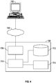
- Processor(s) 110 may further communicate via a network interface 108, which in turn may communicate via the one or more networks 104, such as the Internet or other public or private networks, such that a query or other request may be received from client 102, or other device or service. Additionally, processor(s) 110 may utilize network interface 108 to send information, instructions, workflows query partial workflows, or other data to a user via the one or more networks 104.
- Network interface 104 may include or be communicatively coupled to one or more servers.
- Client 102 may be, e.g., a personal computer coupled to the internet.
- Processor(s) 110 may, in general, be programmed or configured to execute control logic and control operations to implement methods disclosed herein. Processors 110 may be further communicatively coupled (i.e., coupled by way of a communication channel) to co-processors 114. Co-processors 114 can be dedicated hardware and/or firmware components configured to execute the methods disclosed herein. Thus, the methods disclosed herein can be executed by processor 110 and/or co-processors 114.
- Next generation sequencing techniques were used to identify new biomarkers and therapeutic targets for CaP.
- High quality genome sequence data and coverage obtained from histologically defined and precisely dissected primary CaP specimens (80-95% tumor, primary Gleason pattern 3) was compared between cohorts of 7 patients of Caucasian descent and 7 patients of African descent (28 samples total including matched controls from each patient) to evaluate the observed disparities of CaP incidence and mortality between the two ethnic groups.
- These data and analyses provide the first evaluation of prostate cancer genomes from CaP patients of African and Caucasian descent that have been matched for clinic-pathologic features.
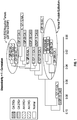
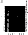
- the top differentially expressed genes in CaP in both ethnic groups include: DLX1, NKX2-3, CRISP3, PHGR1, THBS4, AMACR, GAP43, FFAR2, GCNT1, SIM2, STX19, KLB, APOF, LOC283177, and TRPM4.
- collecting expression data of at least 2, 3, 4, 5, 6, 7, 8, 9, or 10 of these genes from a biological sample provides a unique gene expression profile for use in diagnosing prostate cancer in a subject.
- Also described herein is a method of collecting data for use in diagnosing or prognosing CaP, the method comprising detecting expression in a biological sample of at least 2, 3, 4, 5, 6, 7, 8, 9, 10, 11, 12, 13, 14, or 15 of the following genes: DLX1, NKX2-3, CRISP3, PHGR1, THBS4, AMACR, GAP43, FFAR2, GCNT1, SIM2, STX19, KLB, APOF, LOC283177, and TRPM4.
- the method may optionally include an additional step of obtaining the biological sample from a subject.
- the method may optionally include an additional step of diagnosing or prognosing CaP using the collected gene expression data.
- the methods of collecting data or diagnosing and/or prognosing CaP may further comprise detecting expression of other genes associated with prostate cancer, including, but not limited to ERG, PSA, and PCA3.
- the methods comprise detecting expression of no more than 100, 90, 80, 70, 60, 50, 40, 30, 25, 20, 15, 14, 13, 12, 11, 10, 9, 8, 7, 6, 5, 4, 3, or 2 genes.
- the methods comprise detecting expression of DLX1, NKX2-3, DLX1 and NKX2-3, PHGR1, THBS4, GAP43, FFAR2, GCNT1, SIM2, STX19, KLB, APOF, LOC283177, or TRPM4 and one or more of the other genes listed in Table 1.
- HGNC Hugo Gene Nomenclature Committee
- Entrez Gene for these genes and the accession number of a representative sequence are provided in Table 1.
- Table 1 Gene HGNC ID Entrez Gene ID Accession No.
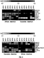
- the following genes were identified as being over expressed in prostate tumors of patients of Caucasian descent as compared to patients of African descent: PCA3, ALOX15, AMACR, CDH19, OR51E2/PSGR, F5, FZD8, and CLDN3.
- obtaining expression data of at least 1, 2, 3, 4, 5, 6, 7, or 8 of these genes provides a unique gene expression profile for use in diagnosing or prognosing prostate cancer in patients of Caucasian descent.
- Also described herein is a method of collecting data for use in diagnosing or prognosing CaP in a patient of Caucasian descent, the method comprising detecting expression in a biological sample of at least 1, 2, 3, 4, 5, 6, 7, or 8 of the following genes: PCA3, ALOX15, AMACR, CDH19, OR51E2/PSGR, F5, FZD8, and CLDN3, wherein the biological sample was obtained from the patient of Caucasian descent.
- the method may optionally include an additional step of obtaining the biological sample from the patient of Caucasian descent.
- the method may optionally include an additional step of diagnosing or prognosing CaP using the collected gene expression data.
- overexpression of at least 1, 2, 3, 4, 5, 6, 7, or 8 of the following genes: PCA3, ALOX15, AMACR, CDH19, OR51E2/PSGR, F5, FZD8, and CLDN3, as compared to a control sample or threshold value indicates the presence of CaP in the biological sample or an increased risk of developing CaP.
- the methods of collecting data for diagnosing CaP may further comprise detecting expression of other genes associated with prostate cancer, including, but not limited to ERG, PSA, and PCA3.
- the methods comprise detecting expression of no more than 100, 90, 80, 70, 60, 50, 40, 30, 25, 20, 15, 14, 13, 12, 11, 10, 9, 8, 7, 6, 5, 4, or 3 genes.
- the methods may comprise detecting expression of ALOX15 and one or more of PCA3, AMACR, CDH19, OR51E2/PSGR, F5, FZD8, and CLDN3. Further, the methods may comprise detecting expression of CDH19 and one or more of PCA3, AMACR, ALOX15, OR51E2/PSGR, F5, FZD8, and CLDN3. As described herein, the methods may comprise detecting expression of F5 and one or more of PCA3, AMACR, ALOX15, OR51E2/PSGR, CDH19, FZD8, and CLDN3.
- the methods may comprise detecting expression of FZD8 and one or more of PCA3, AMACR, ALOX15, OR51E2/PSGR, CDH19, F5, and CLDN3.
- the methods may comprise detecting expression of CLDN3 and one or more of PCA3, AMACR, ALOX15, OR51E2/PSGR, CDH19, F5, and FZD8.
- the methods may comprise detecting expression of PCA3 and AMACR and one or more of ALOX15, CDH19, F5, FZD8, and CLDN3.
- HGNC and Entrez Gene NCBI Reference PCA3 8637 50652 AF103907.1 GI:6165973 ALOX15 433 246 NM_001140.3 GI:40316936 AMACR 451 23600 NM_014324.5 GI:266456114 CDH19 1758 28513 NM_021153.3 GI:402534572 OR51E2/PSGR 15195 81285 AY033942.1 GI:16943640 F5 3542 2153 NM_000130.4 GI:119395710 FZD8 4046 8325 AB043703.1 GI:13623798 CLDN3 2045 1365 NM_001306.3 GI:171541813
- the following genes were identified as being over expressed in prostate tumors of patients of African ancestry as compared to patients of Caucasian descent: COL10A1, HOXC4, ESPL1, MMP9, ABCA13, PCDHGA1, and AGSK1.
- obtaining expression data of at least 1, 2, 3, 4, 5, 6, or 7 of these genes provides a unique gene expression profile for use in diagnosing or prognosing prostate cancer in patients of African descent.
- Certain embodiments are directed to a method of collecting data for use in diagnosing CaP in a patient of African descent, the method comprising detecting expression in a biological sample of at least 1, 2, 3, 4, 5, 6, or 7 of the following genes: COL10A1, HOXC4, ESPL1, MMP9, ABCA13, PCDHGA1, and AGSK1, wherein the biological sample was obtained from the patient of African descent.
- the method may optionally include an additional step of obtaining the biological sample from the patient of African descent.
- the method may optionally include an additional step of diagnosing CaP using the collected gene expression data.
- methods of diagnosing CaP overexpression of at least 3, 4, 5, 6, or 7 of the following genes: COL10A1, HOXC4, ESPL1, MMP9, ABCA13, PCDHGA1, and AGSK1, as compared to a control sample or threshold value indicates the presence of CaP in the biological sample or an increased risk of developing CaP.
- the methods of collecting data or diagnosing CaP may further comprise detecting expression of other genes associated with prostate cancer, including, but not limited to ERG, PSA, and PCA3.
- the methods comprise detecting expression of no more than 100, 90, 80, 70, 60, 50, 40, 30, 25, 20, 15, 14, 13, 12, 11, 10, 9, 8, 7, 6, 5, 4, or 3, genes.
- the methods comprise detecting expression of COL10A1 and one or more of HOXC4, ESPL1, MMP9, ABCA13, PCDHGA1, and AGSK1.
- the methods may comprise detecting expression of HOXC4 and one or more of COL10A1, ESPL1, MMP9, ABCA13, PCDHGA1, and AGSK1.
- the methods may comprise detecting expression of ESPL1 and one or more of COL10A1, HOXC4, MMP9, ABCA13, PCDHGA1, and AGSK1.
- the methods may comprise detecting expression of MMP9 and one or more of COL10A1, HOXC4, ESPL1, ABCA13, PCDHGA1, and AGSK1.
- the methods may comprise detecting expression of ABCA13 and one or more of COL10A1, HOXC4, ESPL1, MMP9, PCDHGA1, and AGSK1.
- the methods may comprise detecting expression of PCDHGA1 and one or more of COL10A1, HOXC4, ESPL1, MMP9, ABCA13, and AGSK1.
- the methods may comprise detecting expression of AGSK1 and one or more of COL10A1, HOXC4, ESPL1, MMP9, ABCA13, and PCDHGA1.
- HGNC and Entrez Gene NCBI Reference COL10A1 2185 1300 NM_000493.3 GI:98985802 HOXC4 5126 3221 NM_014620.5 GI:546232084 ESPL1 16856 9700 NM_012291.4 GI:134276942 MMP9 7176 4318 NM_004994.2 GI:74272286 ABCA13 14638 154664 AY204751.1 GI:30089663 PCDHGA1 8696 56114 NM_018912.2 GI:14196453 AGSK1 N/A 80154 NR_026811 GI:536293433 NR_033936.3 GI:536293365 NR_103496.2 GI:536293435
- whole genome sequence analysis of the 28 samples identified 65 gene mutations present with higher confidence in at least one of the 14 prostate tumors analyzed.
- the 65 gene mutations having the highest allele frequency in the prostate tumors analyzed occurred in the following genes: GLI1, IRX4, PAPPA, SPOP, TEX15, ZNF292, ANKRD11, FAT4, HECW2, KIAA1109, SHROOM3, SPOP, TTC36, ZNRF3, C17orf65, DEGS2, NEK3, KIAA0947, LSP1, NOX3, AKR1B1, ARHGAP12, ITGA4, PVRL4, RBM26, UCN3, CATSPERB, FCRL2, CACNA1E, CORO6, DMKN, EXT1, HEATR7B2, NDUFB5, GPR180, LRRC4, TPRA1, ZIM2, C12orf50, ELMO2, RBM26, SEC14L1, TNFSF11, C9orf125, CDC73, ITSN1, KCNK16,
- Also described herein is a method of collecting data for use in diagnosing or prognosing CaP, the method comprising detecting expression in a biological sample of one or more mutations in at least 2, 3, 4, 5, 6, 7, 8, 9, 10, 15, 20, 25, 30, 35, 40, 45, or 50 of the following genes: GLI1, IRX4, PAPPA, SPOP, TEX15, ZNF292, ANKRD11, FAT4, HECW2, KIAA1109, SHROOM3, SPOP, TTC36, ZNRF3, C17orf65, DEGS2, NEK3, KIAA0947, LSP1, NOX3, AKR1B1, ARHGAP12, ITGA4, PVRL4, RBM26, UCN3, CATSPERB, FCRL2, CACNA1E, CORO6, DMKN, EXT1, HEATR7B2, NDUFB5, GPR180, LRRC4, TPRA1, ZIM2, C12orf50, ELMO2, RBM26, SEC14L1, TNFSF11, C9orf
- the method may optionally include an additional step of obtaining the biological sample from a subject.
- the method may optionally include an additional step of diagnosing or prognosing CaP using the collected gene mutation data.
- Table 4 The unique identifier code assigned by HGNC for these genes and their Entrez Gene ID are provided in Table 4.
- Table 4 provides the frequency with which each mutation was identified in prostate tumors and a matched normal sample.
- Table 4 Gene Tumor Freq. Normal Freq. HGNC ID Entrez Gene ID GLI1 G:21/T:23(52%) G:37/T:0(0%) 20500 79820 IRX4 G:18/A:19(51%) G:42/A:0(0%) 14875 79368 PAPPA C:21/T:20(48%) C:40/T:0(0%) 1392 777 SPOP A:18/C:15(45%) A:39/C:0(0%) 21356 84940 TEX15 C:25/T:21(45%) C:32/T:0(0%) 25063 93099 ZNF292 T:14/A:11(44%) T:40/A:0(0%) 3512 2131 ANKRD11 C:19/A:
- the GLI1 mutation showed the highest allele frequency in the tumors analyzed and also shares a common pathway with SPOP, a gene with a mutation known to be associated with prostate cancer. Therefore, the methods described herein include detecting the GLI1 mutation either alone or in combination with one or more of the gene mutations listed in Table 4.
- the methods of collecting data or diagnosing and/or prognosing CaP may further comprise detecting expression of other genes associated with prostate cancer, including, but not limited to ERG, PSA, and PCA3.
- measuring or detecting the expression of any of the foregoing genes or nucleic acids comprises measuring or detecting any nucleic acid transcript (e.g., mRNA, cDNA, or genomic DNA) corresponding to the gene of interest or the protein encoded thereby. If a gene is associated with more than one mRNA transcript or isoform, the expression of the gene can be measured or detected by measuring or detecting one or more of the mRNA transcripts of the gene, or all of the mRNA transcripts associated with the gene.
- nucleic acid transcript e.g., mRNA, cDNA, or genomic DNA
- gene expression can be detected or measured on the basis of mRNA or cDNA levels, although protein levels also can be used when appropriate. Any quantitative or qualitative method for measuring mRNA levels, cDNA, or protein levels can be used. Suitable methods of detecting or measuring mRNA or cDNA levels include, for example, Northern Blotting, microarray analysis, or a nucleic acid amplification procedure, such as reverse-transcription PCR (RT-PCR) or real-time RT-PCR, also known as quantitative RT-PCR (qRT-PCR). Such methods are well known in the art. See e.g., Sambrook et al., Molecular Cloning: A Laboratory Manual, 4th Ed., Cold Spring Harbor Press, Cold Spring Harbor, N.Y., 2012 .
- RT-PCR reverse-transcription PCR
- qRT-PCR quantitative RT-PCR
- nCounter® NanoString Technologies, Seattle, WA
- Detecting a nucleic acid of interest generally involves hybridization between a target (e.g. mRNA, cDNA, or genomic DNA) and a probe. Sequences of the genes used in the prostate cancer gene expression profile are known (see above). Therefore, one of skill in the art can readily design hybridization probes for detecting those genes. See e.g., Sambrook et al., Molecular Cloning: A Laboratory Manual, 4th Ed., Cold Spring Harbor Press, Cold Spring Harbor, N.Y., 2012 . Each probe should be substantially specific for its target, to avoid any cross-hybridization and false positives.
- An alternative to using specific probes is to use specific reagents when deriving materials from transcripts (e.g., during cDNA production, or using target-specific primers during amplification). In both cases specificity can be achieved by hybridization to portions of the targets that are substantially unique within the group of genes being analyzed, e.g. hybridization to the polyA tail would not provide specificity. If a target has multiple splice variants, it is possible to design a hybridization reagent that recognizes a region common to each variant and/or to use more than one reagent, each of which may recognize one or more variants.
- measuring the expression of the foregoing nucleic acids in prostate cancer tissue can comprise, for instance, contacting a sample containing or suspected of containing prostate cancer cells with polynucleotide probes specific to the genes of interest, or with primers designed to amplify a portion of the genes of interest, and detecting binding of the probes to the nucleic acid targets or amplification of the nucleic acids, respectively.
- PCR primers are known in the art. See e.g., Sambrook et al., Molecular Cloning: A Laboratory Manual, 4th Ed., Cold Spring Harbor Press, Cold Spring Harbor, N.Y., 2012 .
- detailed protocols for preparing and using microarrays to analyze gene expression are known in the art and described herein.
- expression levels of genes can be determined at the protein level, meaning that levels of proteins encoded by the genes discussed above are measured.
- Several methods and devices are well known for determining levels of proteins including immunoassays such as described in e.g., U.S. Pat. Nos. 6,143,576 ; 6,113,855 ; 6,019,944 ; 5,985,579 ; 5,947,124 ; 5,939,272 ; 5,922,615 ; 5,885,527 ; 5,851,776 ; 5,824,799 ; 5,679,526 ; 5,525,524 ; 5,458,852 ; and 5,480,792 .
- These assays include various sandwich, competitive, or non-competitive assay formats, to generate a signal that is related to the presence or amount of a protein of interest.
- Any suitable immunoassay may be utilized, for example, lateral flow, enzyme-linked immunoassays (ELISA), radioimmunoassays (RIAs), competitive binding assays, and the like.
- ELISA enzyme-linked immunoassays
- RIAs radioimmunoassays
- Numerous formats for antibody arrays have been described. Such arrays typically include different antibodies having specificity for different proteins intended to be detected. For example, at least 100 different antibodies are used to detect 100 different protein targets, each antibody being specific for one target. Other ligands having specificity for a particular protein target can also be used, such as the synthetic antibodies disclosed in WO/2008/048970 .
- NADIA nucleic acid detection immunoassay
- PCR polymerase chain reaction
- This amplified DNA-immunoassay approach is similar to that of an enzyme immunoassay, involving antibody binding reactions and intermediate washing steps, except the enzyme label is replaced by a strand of DNA and detected by an amplification reaction using an amplification technique, such as PCR.
- Exemplary NADIA techniques are described in U.S. Patent No. 5,665,539 and U.S. Patent No. 8,338,579 .
- NADIA uses a first (reporter) antibody that is specific for the protein of interest and labelled with an assay-specific nucleic acid.
- the presence of the nucleic acid does not interfere with the binding of the antibody, nor does the antibody interfere with the nucleic acid amplification and detection.
- a second (capturing) antibody that is specific for a different epitope on the protein of interest is coated onto a solid phase (e.g., paramagnetic particles).
- the reporter antibody/nucleic acid conjugate is reacted with sample in a microtiter plate to form a first immune complex with the target antigen.
- the immune complex is then captured onto the solid phase particles coated with the capture antibody, forming an insoluble sandwich immune complex.
- microparticles are washed to remove excess, unbound reporter antibody/nucleic acid conjugate.
- the bound nucleic acid label is then detected by subjecting the suspended particles to an amplification reaction (e.g. PCR) and monitoring the amplified nucleic acid product.
- an amplification reaction e.g. PCR
- MS mass spectrometry
- SRM Selected reaction monitoring
- MRM multiple reaction monitoring
- prostate cells are found in a biological sample, such as prostate tissue, blood, serum, plasma, urine, saliva, or prostatic fluid. Nucleic acids or polypeptides may be isolated from the cells prior to detecting gene expression.
- the biological sample may comprise prostate tissue and is obtained through a biopsy, such as a transrectal or transperineal biopsy.
- the biological sample may be urine. Urine samples may be collected following a digital rectal examination (DRE) or a prostate biopsy.
- the sample may be blood, serum, or plasma, and contains circulating tumor cells that have detached from a primary tumor.
- the sample may also contain tumor-derived exosomes. Exosomes are small (typically 30 to 100 nm) membrane-bound particles that are released from normal, diseased, and neoplastic cells and are present in blood and other bodily fluids.
- the methods disclosed in this application can be used with samples collected from a variety of mammals, but preferably with samples obtained from a human subject.
- the control can be any suitable reference that allows evaluation of the expression level of the genes in the prostate cancer cells as compared to the expression of the same genes in a sample comprising non-cancerous prostate cells, such as normal prostate epithelial cells from a matched subject, or a pool of such samples.
- the control can be a sample from the same subject that is analyzed simultaneously or sequentially with the test sample, or the control can be the average expression level of the genes of interest, as described above, in a pool of prostate samples known to be non-cancerous.
- the control can be defined by mRNA copy numbers of other genes in the sample, such as housekeeping genes (e.g., PBGD or GAPDH) that can be used to normalize gene expression levels.
- control can be embodied, for example, in a pre-prepared microarray used as a standard or reference, or in data that reflects the expression profile of relevant genes in a sample or pool of non-cancerous samples, such as might be part of an electronic database or computer program.
- Over expression and decreased expression of a gene can be determined by any suitable method, such as by comparing the expression of the genes in a test sample with a control (e.g., a positive or negative control), or by using a predetermined "cut-off" or threshold value of absolute expression.
- a control can be provided as previously discussed.
- over expression and decreased expression can be defined as any level of expression greater than or less than the level of expression of the same genes, or other genes (e.g., housekeeping genes), in non-cancerous prostate cells or tissue.
- over expression can be defined as expression that is at least about 1.2-fold, 1.5-fold, 2-fold, 2.5-fold, 5-fold, 10-fold, 20-fold, 50-fold, 100-fold higher or even greater expression as compared to non-cancerous prostate cells or tissue
- decreased expression can similarly be defined as expression that is at least about 1.2-fold, 1.5-fold, 2-fold, 2.5-fold, 5-fold, 10-fold, 20-fold. 50-fold, 100-fold lower or even lower expression as compared to non-cancerous prostate cells or tissue.
- Over expression or decreased expression as used herein is defined as expression that is at least about 2-fold higher or lower, respectively, as compared to a control sample or threshold value.
- This disclosure provides gene expression profiles that are associated with prostate cancer.
- the gene expression profiles can be used to detect prostate cancer cells in a sample or to measure the severity or aggressiveness of the prostate cancer, for example, distinguishing between well differentiated prostate (WD) cancer and poorly differentiated (PD) prostate cancer.
- WD well differentiated prostate
- PD poorly differentiated prostate cancer
- prostate cancer When prostate cancer is found in a biopsy, it is typically graded to estimate how quickly it is likely to grow and spread.
- the most commonly used prostate cancer grading system called Gleason grading, evaluates prostate cancer cells on a scale of 1 to 5, based on their pattern when viewed under a microscope.
- Cancer cells that still resemble healthy prostate cells have uniform patterns with well-defined boundaries and are considered well differentiated (Gleason grades 1 and 2). The more closely the cancer cells resemble prostate tissue, the more the cells will behave like normal prostate tissue and the less aggressive the cancer. Gleason grade 3, the most common grade, shows cells that are moderately differentiated, that is, still somewhat well-differentiated, but with boundaries that are not as well-defined. Poorly-differentiated cancer cells have random patterns with poorly defined boundaries and no longer resemble prostate tissue (Gleason grades 4 and 5), indicating a more aggressive cancer.
- the lower the Gleason score the less aggressive the cancer and the better the prognosis (outlook for cure or long-term survival).
- the higher the Gleason score the more aggressive the cancer and the poorer the prognosis for long-term, metastasis-free survival.
- a convenient way of measuring RNA transcript levels for multiple genes in parallel is to use an array (also referred to as microarrays in the art). Techniques for using arrays to assess and compare gene expression levels are well known in the art and include appropriate hybridization, detection and data processing protocols.
- a useful array includes multiple polynucleotide probes (typically DNA) that are immobilized on a solid substrate (e.g. a glass support such as a microscope slide, or a membrane) in separate locations (e.g., addressable elements) such that detectable hybridization can occur between the probes and the transcripts to indicate the amount of each transcript that is present.
- a solid substrate e.g. a glass support such as a microscope slide, or a membrane
- addressable elements e.g., addressable elements
- the array may comprise (a) a substrate and (b) 2, 3, 4, 5, 6, 7, 8, 9, or 10 or more different addressable elements that each comprise at least one polynucleotide probe for detecting the expression of an mRNA transcript (or cDNA synthesized from the mRNA transcript) of one of the following human genes: DLX1, NKX2-3, CRISP3, PHGR1, THBS4, AMACR, GAP43, FFAR2, GCNT1, SIM2, STX19, KLB, APOF, LOC283177, and TRPM4.
- the array may comprise (a) a substrate and (b) 2, 3, 4, 5, 6, 7, or 8 or more different addressable elements that each comprise at least one polynucleotide probe for detecting the expression of an mRNA transcript (or cDNA synthesized from the mRNA transcript) of one of the following human genes; PCA3, ALOX15, AMACR, CDH19, OR51E2/PSGR, F5, FZD8, and CLDN3.
- the array may comprise (a) a substrate and (b) 2, 3, 4, 5, 6, or 7 or more different addressable elements that each comprise at least one polynucleotide probe for detecting the expression of an mRNA transcript (or cDNA synthesized from the mRNA transcript) of one of the following human genes: COL10A1, HOXC4, ESPL1, MMP9, ABCA13, PCDHGA1, and AGSK1.
- the term "addressable element” means an element that is attached to the substrate at a predetermined position and specifically binds a known target molecule, such that when target-binding is detected (e.g., by fluorescent labeling), information regarding the identity of the bound molecule is provided on the basis of the location of the element on the substrate.
- Addressable elements are "different” for the purposes of the present disclosure if they do not bind to the same target gene.
- the addressable element comprises one or more polynucleotide probes specific for an mRNA transcript of a given gene, or a cDNA synthesized from the mRNA transcript.
- the addressable element can comprise more than one copy of a polynucleotide, can comprise more than one different polynucleotide, provided that all of the polynucleotides bind the same target molecule.
- the addressable element for the gene can comprise different probes for different transcripts, or probes designed to detect a nucleic acid sequence common to two or more (or all) of the transcripts.
- the array can comprise an addressable element for the different transcripts.
- the addressable element also can comprise a detectable label, suitable examples of which are well known in the art.
- the array can comprise addressable elements that bind to mRNA or cDNA other than that of 1) DLX1, NKX2-3, CRISP3, PHGR1, THBS4, AMACR, GAP43, FFAR2, GCNT1, SIM2, STX19, KLB, APOF, LOC283177, and TRPM4; 2) PCA3, ALOX15, AMACR, CDH19, OR51E2/PSGR, F5, FZD8, and CLDN3; or 3) COL10A1, HOXC4, ESPL1, MMP9, ABCA13, PCDHGA1, and AGSK1.
- an array capable of detecting a vast number of targets e.g., mRNA or polypeptide targets
- targets e.g., mRNA or polypeptide targets
- the array preferably comprises a limited number of addressable elements.
- the array comprises no more than about 1000 different addressable elements, more preferably no more than about 500 different addressable elements, no more than about 250 different addressable elements, or even no more than about 100 different addressable elements, such as about 75 or fewer different addressable elements, or even about 50 or fewer different addressable elements.
- even smaller arrays can comprise about 25 or fewer different addressable elements, such as about 15 or fewer different addressable elements or about 12 or fewer different addressable elements.
- the array can even be limited to about 7 different addressable elements without interfering with its functionality.
- the array has polynucleotide probes for no more than 1000 genes immobilized on the substrate or the array has oligonucleotide probes for no more than 500, 250, 100, 50, 25, 15, 10, 9, 8, 7, 6, 5, 4, 3, or 2 genes immobilized on the substrate.
- the substrate can be any rigid or semi-rigid support to which polynucleotides can be covalently or non-covalently attached.
- Suitable substrates include membranes, filters, chips, slides, wafers, fibers, beads, gels, capillaries, plates, polymers, microparticles, and the like.
- Materials that are suitable for substrates include, for example, nylon, glass, ceramic, plastic, silica, aluminosilicates, borosilicates, metal oxides such as alumina and nickel oxide, various clays, nitrocellulose, and the like.
- the polynucleotides of the addressable elements can be attached to the substrate in a pre-determined 1- or 2-dimensional arrangement, such that the pattern of hybridization or binding to a probe is easily correlated with the expression of a particular gene. Because the probes are located at specified locations on the substrate (i.e., the elements are "addressable"), the hybridization or binding patterns and intensities create a unique expression profile, which can be interpreted in terms of expression levels of particular genes and can be correlated with prostate cancer in accordance with the methods described herein.
- Polynucleotide and polypeptide probes can be generated by any suitable method known in the art (see e.g., Sambrook et al., Molecular Cloning: A Laboratory Manual, 4th Ed., Cold Spring Harbor Press, Cold Spring Harbor, N.Y., 2012 ).
- polynucleotide probes that specifically bind to the mRNA transcripts of the genes described herein (or cDNA synthesized therefrom) can be created using the nucleic acid sequences of the mRNA or cDNA targets themselves (e.g., nucleic acid sequences disclosed in Tables 1-4) by routine techniques (e.g., PCR or synthesis).
- fragment means a contiguous part or portion of a polynucleotide sequence comprising about 10 or more nucleotides, about 15 or more nucleotides, about 20 or more nucleotides, about 30 or more, or even about 50 or more nucleotides.
- a polynucleotide probe that binds to an mRNA transcript of DLX1 can be provided by a polynucleotide comprising a nucleic acid sequence that is complementary to the mRNA transcript (e.g., SEQ ID NO: 2) or a fragment thereof, or sufficiently complementary to SEQ ID NO: 2 or fragment thereof that it selectively binds to SEQ ID NO: 2.
- SEQ ID NO: 2 a nucleic acid sequence that is complementary to the mRNA transcript
- SEQ ID NO: 2 e.g., SEQ ID NO: 2
- the exact nature of the polynucleotide probe is not critical to the invention; any probe that will selectively bind the mRNA or cDNA target can be used.
- the polynucleotide probes will comprise 10 or more nucleic acids, 20 or more, 50 or more, or 100 or more nucleic acids.
- the probe will have a sequence identity to a complement of the target sequence (e.g., nucleic acid sequences disclosed in Tables 1-4) of about 90% or more, preferably about 95% or more (e.g., about 98% or more or about 99% or more) as determined, for example, using the well-known Basic Local Alignment Search Tool (BLAST) algorithm (available through the National Center for Biotechnology Information (NCBI), Bethesda, Md.).
- BLAST Basic Local Alignment Search Tool
- Stringency of hybridization reactions is readily determinable by one of ordinary skill in the art, and generally is an empirical calculation dependent upon probe length, washing temperature, and salt concentration. In general, longer probes require higher temperatures for proper annealing, while shorter probes need lower temperatures.
- Hybridization generally depends on the ability of denatured nucleic acid sequences to reanneal when complementary strands are present in an environment below their melting temperature. The higher the degree of desired homology between the probe and hybridizable sequence, the higher the relative temperature that can be used. As a result, it follows that higher relative temperatures would tend to make the reaction conditions more stringent, while lower temperatures less so. For additional details and explanation of stringency of hybridization reactions, see Ausubel et al., Current Protocols in Molecular Biology, Wiley Interscience Publishers, (1995 ).
- “Stringent conditions” or “high stringency conditions,” as defined herein, are identified by, but not limited to, those that: (1) use low ionic strength and high temperature for washing, for example 0.015 M sodium chloride/0.0015 M sodium citrate/0.1% sodium dodecyl sulfate at 50°C; (2) use during hybridization a denaturing agent, such as formamide, for example, 50% (v/v) formamide with 0.1% bovine serum albumin/0.1% Ficoll/0.1% polyvinylpyrrolidone/50 mM sodium phosphate buffer at pH 6.5 with 750 mM sodium chloride, 75 mM sodium citrate at 42°C; or (3) use 50% formamide, 5XSSC (0.75 M NaCl, 0.075 M sodium citrate), 50 mM sodium phosphate (pH 6.8), 0.1% sodium pyrophosphate, 5X Denhardt's solution, sonicated salmon sperm DNA (50 ⁇ g/ml), 0.1% SDS, and 10% dextran
- Modely stringent conditions are described by, but not limited to, those in Sambrook et al., Molecular Cloning: A Laboratory Manual, New York: Cold Spring Harbor Press, 1989 , and include the use of washing solution and hybridization conditions (e.g., temperature, ionic strength and % SDS) less stringent than those described above.
- moderately stringent conditions is overnight incubation at 37°C in a solution comprising: 20% formamide, 5XSSC (150 mM NaCl, 15 mM trisodium citrate), 50 mM sodium phosphate (pH 7.6), 5X Denhardt's solution, 10% dextran sulfate, and 20 mg/mL denatured sheared salmon sperm DNA, followed by washing the filters in 1XSSC at about 37-50°C.
- 5XSSC 150 mM NaCl, 15 mM trisodium citrate
- 50 mM sodium phosphate pH 7.6
- 5X Denhardt's solution 10% dextran sulfate
- 20 mg/mL denatured sheared salmon sperm DNA followed by washing the filters in 1XSSC at about 37-50°C.
- the skilled artisan will recognize how to adjust the temperature, ionic strength, etc. as necessary to accommodate factors such as probe length and the like.
- the array can comprise other elements common to polynucleotide arrays.
- the array also can include one or more elements that serve as a control, standard, or reference molecule, such as a housekeeping gene or portion thereof (e.g.. PBGD or GAPDH), to assist in the normalization of expression levels or the determination of nucleic acid quality and binding characteristics, reagent quality and effectiveness, hybridization success, analysis thresholds and success, etc.
- PBGD housekeeping gene or portion thereof
- the array may comprise (a) a substrate and (b) two or more different addressable elements that each comprise at least one polynucleotide probe for detecting the expression of an mRNA transcript of one of the following human genes: DLX1, NKX2-3, CRISP3, PHGR1, THBS4, AMACR, GAP43, FFAR2, GCNT1, SIM2, STX19, KLB, APOF, LOC283177, and TRPM4, wherein the array comprises no more than 500, 250, 100, 50, 25, 15, 10, 9, 8, 7, 6, 5, 4, 3, or 2 addressable elements.
- the array may comprise at least 3, 4, 5, 6, 7, 10, 12, or 15 different addressable elements.
- the array may comprise two or more different addressable elements each of which comprises at least one polynucleotide probe for detecting the expression of an mRNA transcript of one of the following human genes: PCA3, ALOX15, AMACR, CDH19, OR51E2/PSGR, F5, FZD8, and CLDN3, wherein the array comprises no more than 500, no more than 250, no more than 100, no more than 50, no more than 25, or no more than 15 addressable elements.
- the array may comprise at least 3, 4, 5, 6, 7, 10, 12, or 15 different addressable elements.
- the array may comprise two or more different addressable elements each of which comprises at least one polynucleotide probe for detecting the expression of an mRNA transcript of one of the following human genes: COL10A1, HOXC4, ESPL1, MMP9, ABCA13, PCDHGA1, and AGSK1, wherein the array comprises no more than 500, 250, 100, 50, 25, 15, 10, 9, 8, 7, 6, 5, 4, 3, or 2 addressable elements.
- the array may comprise at least 3, 4, 5, 6, 7, 10, 12, or 15 different addressable elements.
- An array can also be used to measure protein levels of multiple proteins in parallel.
- Such an array comprises one or more supports bearing a plurality of ligands that specifically bind to a plurality of proteins, wherein the plurality of proteins comprises no more than 500, 250, 100, 50, 25, 15, 10, 9, 8, 7, 6, 5, 4, 3, or 2 different proteins.
- the ligands are optionally attached to a planar support or beads.
- the ligands may be antibodies.
- the proteins that are to be detected using the array correspond to the proteins encoded by the nucleic acids of interest, as described above, including the specific gene expression profiles disclosed.
- each ligand e.g. antibody
- each ligand is designed to bind to one of the target proteins (e.g., polypeptide sequences disclosed in Tables 1-4).
- each ligand is preferably associated with a different addressable element to facilitate detection of the different proteins in a sample.
- This application describes methods of diagnosing and prognosing prostate cancer in a sample obtained from a subject, in which gene expression in prostate cells and/or tissues are analyzed. If a sample shows over expression of certain genes or the expression of certain gene mutations, then there is an increased likelihood that the subject has prostate cancer or a less or more advanced stage (e.g., WD or PD prostate cancer) of prostate cancer.
- the methods of detecting or prognosing prostate cancer may include one or more of the following steps: informing the patient that they are likely to have prostate cancer, WD prostate cancer or PD prostate cancer; confirmatory histological examination of prostate tissue; and/or treating the patient by a prostate cancer therapy.
- the methods further comprise a step of taking a prostate biopsy from the subject and examining the prostate tissue in the biopsy (e.g., histological examination) to confirm whether the patient has prostate cancer.
- the methods of detecting or prognosing prostate cancer may be used to assess the need for therapy or to monitor a response to a therapy (e.g., disease-free recurrence following surgery or other therapy), and, thus may include an additional step of treating a subject having prostate cancer.
- Prostate cancer treatment options include surgery, radiation therapy, hormone therapy, chemotherapy, biological therapy, or high intensity focused ultrasound.
- Drugs approved for prostate cancer include: Enzalutamide (XTANDI), Abiraterone Acetate, Cabazitaxel, Degarelix, Jevtana (Cabazitaxel), Prednisone, Provenge (Sipuleucel-T), Sipuleucel-T, or Docetaxel.
- a method as described in this application may, after a positive result, include a further step of surgery, radiation therapy, hormone therapy, chemotherapy, biological therapy, or high intensity focused ultrasound.
- the gene expression profiles associated with prostate cancer or lack thereof provided by the methods described in this application can also be useful in screening drugs, either in clinical trials or in animal models of prostate cancer.
- a clinical trial can be performed on a drug in similar fashion to the monitoring of an individual patient, except that the drug is administered in parallel to a population of prostate cancer patients, usually in comparison with a control population administered a placebo.
- the changes in expression levels of genes can be analyzed in individual patients and across a treated or control population. Analysis at the level of an individual patient provides an indication of the overall status of the patient at the end of the trial (i.e., whether gene expression profile indicates the presence or severity (e.g., WD or PD) of prostate cancer) and/or an indication whether that profile has changed toward or away from such indication in the course of the trial. Results for individual patients can be aggregated for a population allowing comparison between treated and control population.
- the expression levels of genes detected are the species variants or homologs of the human genes referenced above in whatever species of non-human animal on which tests are being conducted.
- the average expression levels of human genes determined in human prostate cancer patients are not necessarily directly comparable to those of homolog genes in an animal model, the human values can nevertheless be used to provide an indication whether a change in expression level of a non-human homolog is in a direction toward or away from the diagnosis of prostate cancer or prognosis of WD or PD prostate cancer.
- the expression profile of individual animals in a trial can provide an indication of the status of the animal at the end of the trial (i.e., whether gene expression profile indicates the presence or severity (e.g., WD or PD) of prostate cancer) and/or change in such status during the trial.
- Results from individual animals can be aggregated across a population and treated and control populations compared. Average changes in the expression levels of genes can then be compared between the two populations.
- the methods provided may be computer-implemented.
- Gene expression levels can be analyzed and associated with status of a subject (e.g., presence of prostate cancer or severity of disease (e.g., WD or PD prostate cancer)) in a digital computer.
- a computer is directly linked to a scanner or the like receiving experimentally determined signals related to gene expression levels.
- expression levels can be input by other means.
- the computer can be programmed to convert raw signals into expression levels (absolute or relative), compare measured expression levels with one or more reference expression levels, or a scale of such values.
- the computer can also be programmed to assign values or other designations to expression levels based on the comparison with one or more reference expression levels, and to aggregate such values or designations for multiple genes in an expression profile.
- the computer can also be programmed to output a value or other designation providing an indication of the presence or severity of prostate cancer as well as any of the raw or intermediate data used in determining such a value or designation.
- a typical computer includes a bus which interconnects major subsystems such as a central processor, a system memory, an input/output controller, an external device such as a printer via a parallel port, a display screen via a display adapter, a serial port, a keyboard, a fixed disk drive and a port (e.g., USB port) operative to receive an external memory storage device.
- major subsystems such as a central processor, a system memory, an input/output controller, an external device such as a printer via a parallel port, a display screen via a display adapter, a serial port, a keyboard, a fixed disk drive and a port (e.g., USB port) operative to receive an external memory storage device.
- Many other devices can be connected such as a scanner via I/O controller, a mouse connected to serial port or a network interface.
- the computer contains computer readable media holding codes to allow the computer to perform a variety of functions. These functions include controlling automated apparatus, receiving input and delivering output as described above
- a typical computer system 106 may also include one or more processors 110 coupled to random access memory operating under control of or in conjunction with an operating system as set forth in Figure 4 and discussed above.
- Any of the computer-implemented methods of the invention may comprise a step of obtaining by at least one processor information reflecting the expression level of at least 2, 3, 4, 5, 6, 7, 8, 9, 10, 11, 12, 13, 14, or 15 of the following human genes: DLX1, NKX2-3, CRISP3, PHGR1, THBS4, AMACR, GAP43, FFAR2, GCNT1, SIM2, STX19, KLB, APOF, LOC283177, and TRPM4 in a biological sample.
- the computer-implemented methods may comprise obtaining by at least one processor information reflecting the expression level of DLX1, NKX2-3, DLX1 and NKX2-3, PHGR1, THBS4, GAP43, FFAR2, GCNT1, SIM2, STX19, KLB, APOF, LOC283177, or TRPM4 and one or more of the other genes listed in Table 1.
- Any of the computer-implemented methods describe herein may comprise a step of obtaining by at least one processor information reflecting the expression level of at least 2, 3, 4, 5, 6, 7, or 8 of the following human genes: PCA3, ALOX15, AMACR, CDH19, OR51E2/PSGR, F5, FZD8, and CLDN3 in a biological sample obtained from a patient of Caucasian descent.
- the computer-implemented methods may comprise obtaining by at least one processor information reflecting the expression level of ALOX15 and one or more of PCA3, AMACR, CDH19, OR51E2/PSGR, F5, FZD8, and CLDN3.
- the computer-implemented methods may comprise obtaining by at least one processor information reflecting the expression level of CDH19 and one or more of PCA3, AMACR, ALOX15, OR51E2/PSGR, F5, FZD8, and CLDN3.
- the computer-implemented methods may comprise obtaining by at least one processor information reflecting the expression level of F5 and one or more of PCA3, AMACR, ALOX15, OR51E2/PSGR, CDH19, FZD8, and CLDN3.
- the computer-implemented methods may comprise obtaining by at least one processor information reflecting the expression level of FZD8 and one or more of PCA3, AMACR, ALOX15, OR5IE2/PSGR, CDH19, F5, and CLDN3.
- the computer-implemented methods may comprise obtaining by at least one processor information reflecting the expression level of CLDN3 and one or more of PCA3, AMACR, ALOX15, OR51E2/PSGR, CDH19, F5, and FZD8.
- the computer-implemented methods may comprise obtaining by at least one processor information reflecting the expression level of PCA3 and AMACR and one or more of ALOX15, CDH19, F5, FZD8, and CLDN3.
- Any of the computer-implemented methods of the invention may comprise a step of obtaining by at least one processor information reflecting the expression level of at least 2, 3, 4, 5, 6, or 7 of the following human genes: COL10A1, HOXC4, ESPL1, MMP9, ABCA13, PCDHGA1, and AGSK1 in a biological sample obtained from a patient of African descent.
- the computer-implemented methods may comprise obtaining by at least one processor information reflecting the expression level of COL10A1 and one or more of HOXC4, ESPL1, MMP9, ABCA13, PCDHGA1, and AGSK1.
- the computer-implemented method may comprise obtaining by at least one processor information reflecting the expression level of HOXC4 and one or more of COL10A1, ESPL1, MMP9, ABCA13, PCDHGA1, and AGSK1.
- the computer-implemented methods may comprise obtaining by at least one processor information reflecting the expression level of ESPL1 and one or more of COL10A1, HOXC4, MMP9, ABCA13, PCDHGA1, and AGSK1.
- the computer-implemented methods may comprise obtaining by at least one processor information reflecting the expression level of MMP9 and one or more of COL10A1, HOXC4, ESPL1, ABCA13, PCDHGA1, and AGSK1.
- the computer-implemented methods may comprise obtaining by at least one processor information reflecting the expression level of ABCA13 and one or more of COL10A1, HOXC4, ESPL1, MMP9, PCDHGA1, and AGSK1.
- the computer-implemented methods may comprise obtaining by at least one processor information reflecting the expression level of PCDHGA1 and one or more of COL10A1, HOXC4, ESPL1, MMP9, ABCA13, and AGSK1.
- the computer-implemented methods may comprise obtaining by at least one processor information reflecting the expression level of AGSK1 and one or more of COL10A1, HOXC4, ESPL1, MMP9, ABCA13, and PCDHGA1.
- the computer-implemented methods may additionally comprise the steps of i) determining by at least one processor a difference between the expression level of one or more control genes and the expression level of 1) at least 2, 3, 4, 5, 6, 7, 8, 9, 10, 11, 12, 13, 14, or 15 of the following human genes: DLX1, NKX2-3, CRISP3, PHGR1, THBS4, AMACR, GAP43, FFAR2, GCNT1, S1M2, STX19, KLB, APOF, LOC283177, and TRPM4 in a biological sample; 2) at least 2, 3, 4, 5, 6, 7, or 8 of the following human genes: PCA3, ALOX15, AMACR, CDH19, OR51E2/PSGR, F5, FZD8, and CLDN3 in a biological sample obtained from a patient of Caucasian descent; or 3) at least 2, 3, 4, 5, 6, or 7 of the following human genes: COL10A1, HOXC4, ESPL1, MMP9, ABCA13, PCDHGA1, and AGSK1 in a biological sample obtained from
- the computer-implemented methods may further comprise outputting in user readable format a determination that the subject has prostate cancer, well differentiated prostate cancer, or poorly differentiated prostate cancer based on the difference obtained in the outputting step.
- kits for diagnosing or prognosing prostate cancer comprising a plurality of polynucleotide probes for detecting at least 2, 3, 4, 5, 6, 7, 8, 9, 10, 11, 12, 13, 14, or 15 of the following human genes: DLX1, NKX2-3, CRISP3, PHGR1, THBS4, AMACR, GAP43, FFAR2, GCNT1, SIM2, STX19, KLB, APOF, LOC283177, and TRPM4, wherein the plurality of polynucleotide probes contains polynucleotide probes for no more than 500, 250, 100, 50, 25, 15, 10, 9, 8, 7, 6, 5, 4, 3, or 2 genes.
- the plurality of polynucleotide probes comprises polynucleotide probes for detecting at least 4 or 5 of the aforementioned genes, wherein the plurality of polynucleotide probes contains polynucleotide probes for no more than 10 genes.
- the polynucleotide probes may be optionally labeled.
- the kit may optionally include polynucleotide primers for amplifying a portion of the mRNA transcripts from at least 2, 3, 4, 5, 6, 7, 8, 9, 10, 11, 12, 13, 14, or 15 of the following human genes: DLX1, NKX2-3, CRISP3, PHGR1, THBS4, AMACR, GAP43, FFAR2, GCNT1, SIM2, STX19, KLB, APOF, LOC283177, and TRPM4.
- polynucleotide primers for amplifying a portion of the mRNA transcripts from at least 2, 3, 4, 5, 6, 7, 8, 9, 10, 11, 12, 13, 14, or 15 of the following human genes: DLX1, NKX2-3, CRISP3, PHGR1, THBS4, AMACR, GAP43, FFAR2, GCNT1, SIM2, STX19, KLB, APOF, LOC283177, and TRPM4.
- kits for diagnosing or prognosing prostate cancer in a patient of Caucasian descent comprising a plurality of polynucleotide probes for detecting at least 3, 4, 5, 6, 7, or 8 of the following human genes: PCA3, ALOX15, AMACR, CDH19, OR51E2/PSGR, F5, FZD8, and CLDN3, wherein the plurality of polynucleotide probes contains polynucleotide probes for no more than 500, 250, 100, 50, 25, 15, 10, 9, 8, 7, 6, 5, 4, 3, or 2 genes.
- the plurality of polynucleotide probes comprises polynucleotide probes for detecting at least 4 or 5 of the aforementioned genes, wherein the plurality of polynucleotide probes contains polynucleotide probes for no more than 10 genes.
- the polynucleotide probes may be optionally labeled.
- the kit may optionally include polynucleotide primers for amplifying a portion of the mRNA transcripts from at least 3, 4, 5, 6, 7, or 8 of the following human genes: PCA3, ALOX15, AMACR, CDH19, OR51E2/PSGR, F5, FZD8, and CLDN3.
- One embodiment is directed to a kit for diagnosing prostate cancer in a patient of African descent, the kit comprising a plurality of polynucleotide probes for detecting at least 3, of the following human genes: COL10A1, HOXC4, ESPL1, MMP9, ABCA13, PCDHGA1, and AGSK1, wherein the plurality of polynucleotide probes contains polynucleotide probes for no more than 500, 250, 100, 50, 25, 15, 10, 9, 8, 7. 6, 5, 4, or 3 genes.
- the plurality of polynucleotide probes comprises polynucleotide probes for detecting at least 4 or 5 of the aforementioned genes, wherein the plurality of polynucleotide probes contains polynucleotide probes for no more than 10 genes.
- the polynucleotide probes may be optionally labeled.
- the kit may optionally include polynucleotide primers for amplifying a portion of the mRNA transcripts from at least 3, 4, 5, 6, or 7 of the following human genes: COL10A1, HOXC4, ESPL1, MMP9, ABCA13, PCDHGA1, and AGSK1.
- kits for diagnosing or prognosing prostate cancer comprising antibodies.
- the kit for diagnosing or prognosing prostate cancer may comprise a plurality of antibodies for detecting at least 3, 4, 5, 6, 7, 8, 9, 10, 11, 12, 13, 14, or 15 of the polypeptides encoded by the following human genes: DLX1, NKX2-3, CRISP3, PHGR1, THBS4, AMACR, GAP43, FFAR2, GCNT1, SIM2, STX19, KLB, APOF, LOC283177, and TRPM, wherein the plurality of antibodies contains antibodies for no more than 500, 250, 100, 50, 25, 15, 10,9, 8, 7, 6, 5, 4, 3, or 2 polypeptides.
- the plurality of antibodies comprises antibodies for detecting at least 4 or 5 of the polypeptides encoded by the aforementioned genes and wherein the plurality of antibodies contains antibodies for no more than 10 polypeptides.
- the antibodies may be optionally labeled.
- the kit for diagnosing or prognosing prostate cancer in a patient of Caucasian descent may comprise a plurality of antibodies for detecting at least 3, 4, 5, 6, 7, or 8 of the polypeptides encoded by the following human genes: PCA3, ALOX15, AMACR, CDH19, OR51E2/PSGR, F5, FZD8, and CLDN3, wherein the plurality of antibodies contains antibodies for no more than 500, 250, 100, 50, 25, 15, 10, 9, 8, 7, 6, 5, 4, 3, or 2 polypeptides.
- the plurality of antibodies comprises antibodies for detecting at least 4 or 5 of the polypeptides encoded by the aforementioned genes and wherein the plurality of antibodies contains antibodies for no more than 10 polypeptides.
- the antibodies may be optionally labeled.
- the kit for diagnosing prostate cancer in a patient of African descent comprises a plurality of antibodies for detecting at least 3, 4, 5, 6, or 7 of the polypeptides encoded by following-human genes: COL10A1, HOXC4, ESPL1, MMP9, ABCA13, PCDHGA1, and AGSK1, wherein the plurality of antibodies contains antibodies for no more than 500, 250, 100, 50, 25, 15, 10, 9, 8, 7, 6, 5, 4, 3, or 2 polypeptides.
- the plurality of antibodies comprises antibodies for detecting at least 4 or 5 of the polypeptides encoded by the aforementioned genes and wherein the plurality of antibodies contains antibodies for no more than 10 polypeptides.
- the antibodies may be optionally labeled.
- the kit for diagnosing prostate cancer may comprise polypeptide probes that can be used, for example, in spectrometry methods, such as mass spectrometry.
- the kit for diagnosing or prognosing prostate cancer comprises a plurality of polypeptide probes for detecting at least 3, 4, 5, 6, 7, 8, 9, 10, 11, 12, 13, 14, or 15 of the polypeptides encoded by the following human genes: DLX1, NKX2-3, CRISP3, PHGR1, THBS4, AMACR, GAP43, FFAR2, GCNT1, SIM2, STX19, KLB, APOF, LOC283177, and TRPM, wherein the plurality of polypeptide probes contains polypeptide probes for no more than 500, 250, 100, 50, 25, 15, 10, 9, 8, 7, 6, 5, 4, 3, or 2 polypeptides.
- the plurality of polypeptide probes comprises polypeptide probes for detecting at least 4 or 5 of the polypeptides encoded by the aforementioned genes and wherein the plurality of polypeptide probes contains polypeptide probes for no more than 10 polypeptides.
- the polypeptide probes may be optionally labeled.
- the kit for diagnosing or prognosing prostate cancer in a patient of Caucasian descent comprises a plurality of polypeptide probes for detecting at least 3, 4, 5, 6, 7, or 8 of the polypeptides encoded by the following human genes: PCA3, ALOX15, AMACR, CDH19, OR51E2/PSGR, F5, FZD8, and CLDN3, wherein the plurality of polypeptide probes contains polypeptide probes for no more than 500, 250, 100, 50, 25, 15, 10, 9, 8, 7, 6, 5, 4, 3, or 2 polypeptides.
- the plurality of polypeptide probes comprises polypeptide probes for detecting at least 4 or 5 of the polypeptides encoded by the aforementioned genes and wherein the plurality of polypeptide probes contains polypeptide probes for no more than 10 polypeptides.
- the polypeptide probes may be optionally labeled.
- the kit for diagnosing prostate cancer in a patient of African descent comprises a plurality of polypeptide probes for detecting at least 3, 4, 5, 6, or 7 of the polypeptides encoded by following human genes: COL10A1, HOXC4, ESPL1, MMP9, ABCA13, PCDHGA1, and AGSK1, wherein the plurality of polypeptide probes contains polypeptide probes for no more than 500, 250, 100, 50, 25, 15, 10, 9, 8, 7, 6, 5, 4 or 3 polypeptides.
- the plurality of polypeptide probes comprises polypeptide probes for detecting at least 4 or 5 of the polypeptides encoded by the aforementioned genes and wherein the plurality of polypeptide probes contains polypeptide probes for no more than 10 polypeptides.
- the polypeptide probes may be optionally labeled.
- a kit includes instructional materials disclosing methods of use of the kit contents in a disclosed method.
- the instructional materials may be provided in any number of forms, including, but not limited to, written form (e.g., hardcopy paper, etc.), in an electronic form (e.g., computer diskette or compact disk) or may be visual (e.g., video files).
- the kits may also include additional components to facilitate the particular application for which the kit is designed. Thus, for example, the kits may additionally include other reagents routinely used for the practice of a particular method, including, but not limited to buffers, enzymes, labeling compounds, and the like. Such kits and appropriate contents are well known to those of skill in the art.
- the kit can also include a reference or control sample.
- the reference or control sample can be a biological sample or a data base.
- the polynucleotide or polypeptide probes and antibodies described in this application are optionally labeled with a detectable label.
- a detectable label used in conjunction with probe or antibody technology, as known by one of ordinary skill in the art, can be used.
- the probe is labeled with a detectable label selected from the group consisting of: a fluorescent label, a chemiluminescent label, a quencher, a radioactive label, biotin, mass tags and/or gold.
- a comparative full genome analysis was conducted using primary prostate tumors and corresponding normal tissue (blood) in a cohort of seven AA and seven CA CaP patients (28 specimens).
- the cohort was selected based on the following criteria: primary treatment radical prostatectomy, no neo-adjuvant treatment, Gleason grade 3 +3 and 3+4 (representing the majority of PSA-screened CaP at diagnosis/primary treatment), frozen tumor tissue with 80% or more tumor cell content, dissected tumor tissue yielding over 2 ⁇ g high molecular weight genomic DNA, availability of corresponding blood genomic DNA and patient clinico-pathological data.
- RNA-Seq analysis was performed in the same cohort of prostate tumor samples.
- RNA-Seq technology has the ability to interrogate multiple aspects of the transcriptome including gene fusion, gene and transcript expression.
- Surrounding normal tissue was collected from 4 of the 14 patients. Two of the normal tissue samples were from AA men and two were from CA men.
- RNA samples were shipped to Expression Analysis (Durham, NC) for transcriptome sequencing.
- the details of sequencing statistics are as follows: Sequencing type: paired-end, average read length of each sample: 50nt, average read quality: 37, number of reads: approximately 31 million in each sample.
- Raw reads from expression analysis were obtained in fastq format for each sample. These files contain all sequences passing Illumina's purity filter and per-base quality score as defined by Illumina phred metric. These raw reads were filtered for low quality reads (quality score ⁇ 20), artifact/duplicate sequences and adapter sequences prior to actual analysis.
- the human reference genome (hg19) used for mapping in this analysis was downloaded from UCSC website. Clean Paired end reads were aligned to the hg19 reference genome using TopHat software version 2.0.8 (a free open source tool available for mapping), and for each read no more than 2 mismatches were allowed in the alignment. TopHat maps reads to the reference genome using Bowtie, an ultra high-throughput short read aligner. Software outputs numerous files for further analysis: mapped reads, fusion junction, splice junction, insertions and deletions files.
- Cufflinks Aligned reads were assembled to transcripts using Cufflinks, an open source tool that assembles transcripts, estimates their abundances, and tests for differential expression and regulation in RNA-Seq samples.
- Cufflinks calculates the expression level of gene depending on all the known splice variants/isoform of that gene.
- FPKM C LN
- C is the number of mappable reads in the feature (transcript, exon)
- L is the length in feature (in kb)
- N is the total number of mappable reads in the feature (in millions).
- the Cuffdiff program included in the Cufflinks package, calculates differential expression between CaP tumor and normal samples and includes a p-value for each observed change in expression between samples.
- Cuffdiff allows inputting multiple sample files based on the experimental condition. Gene and transcript expression levels are reported in tabular format and these files contain gene information, log2 scale fold change for each gene, P values and false discovery rate (FDR). Pathway analysis and Gene Ontology Biological Processes on statistically significant genes was performed with Genomatix Pathway Analysis Software.
- Hierarchical clustering (performed using R package) was used to group samples based on their expression levels.
- Figure 1 Clustering of all 18 samples (14 tumors and 4 normal) indicate a clear demarcation between tumor and normal samples. However, most of the tumor samples were not clustered according to AA and CA groups. Interestingly, three out of four fusion negative AA samples clustered in to one group, and based on the patient follow up studies, two of the 3 patients developed metastasis (the only two metastasis in this cohort), and the third had biochemical recurrence.
- gene expression profiles for each patient were generated by comparing tumor with normal sample within each group. Statistically significant genes were extracted using fold change (tumor/normal ratios), at least 2 fold over/under expressed in tumors and P-value ⁇ 0.05. 101 genes and 180 genes were statistically significant (2fold & p-value ⁇ 0.05) in African- and Caucasian-American groups respectively. Few of the prostate cancer literature associated genes in African-American list included CRISP3, SIM2, THBS4 and MMP9; Caucasian-American list included AMACR, APOF, CRISP3, OR51E2 (PSGR), SIM2 and THBS4.
- tumor/normal ratios of CA group were compared to the tumor/normal ratios in AA group.
- This gene list revealed that some of the well-studied prostate cancer genes, such as, PCA3 (10-fold), PSGR (5-fold) and AMACR (2-fold) were over expressed in the CA group as compared to the AA group ( Figure 3B ).
- gene expression levels of TMPRSS2/ERG fusion positive samples were compared with fusion negative samples.
- CRISP3, GLDC, and TDRD1 were the top differentially expressed genes in TMPRSS2/ERG fusion positive samples, while COL2A1 and PLA2G7 were the top differentially expressed genes in TMPRSS2/ERG fusion negative samples.
- the tumor/normal ratios of differentially expressed gene list in AA group were compared to the tumor/normal ratios in CA group to evaluate AA race specific gene expression trend.
- the heatmap in Figure 3A shows genes that were consistently up-regulated in the AA group and simultaneously down-regulated (or no change of expression) in the CA group.
- MMP9 was the top gene which was found to be very strongly up-regulated in the AA group but down-regulated in the CA group.
- the top differentially expressed genes in prostate tumors of the AA group as compared to the CA group are set forth in Table 7.
Landscapes
- Health & Medical Sciences (AREA)
- Life Sciences & Earth Sciences (AREA)
- Chemical & Material Sciences (AREA)
- Engineering & Computer Science (AREA)
- Immunology (AREA)
- Organic Chemistry (AREA)
- Proteomics, Peptides & Aminoacids (AREA)
- Pathology (AREA)
- Analytical Chemistry (AREA)
- Molecular Biology (AREA)
- General Health & Medical Sciences (AREA)
- Urology & Nephrology (AREA)
- Genetics & Genomics (AREA)
- Zoology (AREA)
- Wood Science & Technology (AREA)
- Microbiology (AREA)
- Biotechnology (AREA)
- Physics & Mathematics (AREA)
- Biochemistry (AREA)
- Hematology (AREA)
- Medicinal Chemistry (AREA)
- Biomedical Technology (AREA)
- Bioinformatics & Cheminformatics (AREA)
- Oncology (AREA)
- Biophysics (AREA)
- Hospice & Palliative Care (AREA)
- General Engineering & Computer Science (AREA)
- Public Health (AREA)
- General Physics & Mathematics (AREA)
- Food Science & Technology (AREA)
- Cell Biology (AREA)
- Veterinary Medicine (AREA)
- Animal Behavior & Ethology (AREA)
- Pharmacology & Pharmacy (AREA)
- Nuclear Medicine, Radiotherapy & Molecular Imaging (AREA)
- General Chemical & Material Sciences (AREA)
- Chemical Kinetics & Catalysis (AREA)
- Measuring Or Testing Involving Enzymes Or Micro-Organisms (AREA)
- Peptides Or Proteins (AREA)
- Epoxy Compounds (AREA)
Description
- This application claims the benefit of, and relies on the filing date of,
U.S. provisional patent application number 61/921,739, filed 30 December 2013 - This invention was made in part with Government support. The Government has certain rights in the invention.
- The instant application contains a Sequence Listing which has been submitted electronically in ASCII format. Said ASCII copy, created on December 29, 2014, is named HMJ-145-PCT_SL.txt and is 344,410 bytes in size.
- In 2013 an estimated 238,590 men will be diagnosed with carcinoma of the prostate (CaP) and an estimated 29,720 men will die from the disease [1]. This malignancy is the second leading cause of cancer-related death in men in the United States. In addition, African American (AA) men have the highest incidence and mortality from CaP compared with other races [1]. The racial disparity exists from presentation and diagnosis through treatment, survival, and quality of life [2]. Researchers have suggested that socio-economic status (SES) contributes significantly to these disparities including CaP-specific mortality [3]. As well, there is evidence that reduced access to care is associated with poor CaP outcomes, which is more prevalent among AA men than Caucasian American (CA) men [4].
- However, there are populations in which AA men have similar outcomes to CA men. Sridhar and colleagues [5] published a meta-analysis in which they concluded that when SES is accounted for, there are no differences in the overall and CaP-specific survival between AA and CA men. Similarly, the military and veteran populations (systems of equal access and screening) do not observe differences in survival across race [6], and differences in pathologic stage at diagnosis narrowed by the early 2000s in a veterans' cohort [7]. Of note, both of these studies showed that AA men were more likely to have higher Gleason scores and PSA levels than CA men [6, 7].
- While socio-economic factors may contribute to CaP outcomes, they do not seem to account for all variables associated with the diagnosis and disease risk. Several studies support that AA men have a higher incidence of CaP compared to CA men [1, 8, 9]. Studies also show that AA men have a significantly higher PSA at diagnosis, higher grade disease on biopsy, greater tumor volume for each stage, and a shorter PSA doubling time before radical prostatectomy [10-12]. Biological differences between prostate cancers from CA and AA men have been noted in the tumor microenvironment with regard to stress and inflammatory responses [13]. Although controversy remains over the role of biological differences, observed differences in incidence and disease aggressiveness at presentation indicate a potential role for different pathways of prostate carcinogenesis between AA and CA men.
- Over the past decade, much research has focused on alterations of cancer genes and their effects in CaP [14-16]. Variations in prevalence across ethnicity and race have been noted in the TMPRSS2/ERG gene fusion that is overexpressed in CaP and is the most common known oncogene in CaP [17, 18]. Accumulating data suggest that there are differences of ERG oncogenic alterations across ethnicities [17, 19-21]. Significantly greater ERG expression in CA men compared to AA men was noted in initial papers describing ERG overexpression and ERG splice variants [17, 21]. The difference is even more pronounced between CA and AA (50% versus 16%) in patients with high Gleason grade (8-10) tumors. Thus, ERG is a major somatic gene alteration between these ethnic groups. Yet beyond TMPRSS2/ERG, little is known regarding the genetic basis for the CaP disparity between AA and CA men remains unknown [24]. A pilot project study relating to microarray comparison of prostate tumor gene expression in African-American and Caucasian American males has been done by Reams et al. [28].
- Therefore, new biomarkers and therapeutic markers that are specific for distinct ethnic populations and provide more accurate diagnostic and/or prognostic potential are needed. As such, separate gene expression profiles for patients of African and Caucasian descent can be used to diagnose or prognose CaP in distinct ethnic populations and offer more informed treatment options based on these ethnic-specific gene expression signatures.
- The present disclosure provides gene expression profiles that are associated with prostate cancer and methods of using the same. The gene expression profiles can be used to detect prostate cancer cells in a sample or to predict the likelihood of a patient developing prostate cancer. The gene expression profiles can also be used to evaluate the severity or stage of prostate cancer or to assess the effectiveness of a therapy or monitor the progression or regression of prostate cancer following therapy (e.g., disease-free recurrence following surgery). The gene expression profiles can be measured at either the nucleic acid or protein level. In one aspect, the gene expression profile is specific for patients of African descent. In another aspect, the gene expression profile is specific for patients of Caucasian descent.
- In one aspect, the present invention provides a method of collecting data for diagnosing prostate cancer, the method comprising:
detecting expression of a plurality of genes in a biological sample obtained from a patient, wherein the patient is of African descent and wherein the plurality of genes is selected because the patient is of African descent and comprises at least three of the following genes: COL10A1, HOXC4, ESPL1, MMP9, ABCA13, PCDHGA1, and AGSK1, wherein the at least three genes comprise COL10A1. - In one embodiment, the plurality of genes comprises COL10A1, HOXC4, ESPL1, MMP9, ABCA13, PCDHGA1, and AGSK1.
- In another embodiment, the method further comprises a step of diagnosing prostate cancer using the expression data obtained.
- In another embodiment, overexpression of the at least three genes as compared to a control sample or a threshold value indicates the presence of prostate cancer in the biological sample or an increased risk of developing prostate cancer.
- In one embodiment, the method may further comprise detecting expression of an ERG gene in the biological sample. In one embodiment, the biological sample is a tissue sample, a cell sample, a blood sample, a serum sample, or a urine sample. In one embodiment, the biological sample comprises prostate cells or nucleic acids or polypeptides isolated from prostate cells. In one embodiment, nucleic acid expression is detected. In one embodiment, polypeptide expression is detected.
- In another aspect, the invention provides a kit for use in diagnosing prostate cancer, the kit comprising a plurality of probes for detecting at least three of the following genes: COL10A1, HOXC4, ESPL1, MMP9, ABCA13, PCDHGA1, and AGSK1, wherein the plurality of probes contains probes for detecting no more than 500, 250, 100, 50, 25, 15, 10, 9, 8, 7, 6, 5, 4, or 3 different genes, wherein the at least three genes comprise COL10A1.
- In one embodiment, the plurality of probes is selected from a plurality of oligonucleotide probes, a plurality of antibodies, or a plurality of polypeptide probes. In one embodiment, the plurality of probes contains probes for detecting no more than 250, 100, 50, 25, 15, 10, or 5 different genes. In one embodiment, the plurality of probes are attached to the surface of an array, wherein the array optionally comprises no more than 500, 250, 100, 50, 25, 15, or 10 addressable elements. In one embodiment, the plurality of probes are labeled. In one embodiment, the plurality of probes further comprises a probe for detecting an ERG gene.
- In another aspect, the invention provides a method of obtaining a gene expression profile in a biological sample, the method comprising:
incubating the aforementioned array with the biological sample, wherein the biological sample is obtained from a patient of African descent, and measuring the expression level of the at least three genes to obtain the gene expression profile. - In one embodiment, the biological sample is a tissue sample, a cell sample, a blood sample, a serum sample, or a urine sample. In one embodiment, the biological sample comprises nucleic acids or polypeptides isolated from prostate cells. In one embodiment, the measuring step comprises measuring nucleic acid expression levels. In one embodiment, the measuring step comprises measuring polypeptide expression levels. In one embodiment, the measuring step further comprises measuring the expression level of an ERG gene.
- In yet another aspect, the invention provides a method of identifying a patient in need of prostate cancer treatment, wherein the patient is of African descent, the method comprising:
testing a biological sample from the patient for the overexpression of a plurality of genes, wherein the plurality of genes is selected because the patient is of African descent and comprises at least three of the following genes: COL10A1, HOXC4, ESPL1, MMP9, ABCA13, PCDHGA1, and AGSK1, wherein the at least three genes comprise COL10A1, and identifying the patient as in need of prostate cancer treatment if one or more of the at least three genes is overexpressed in the biological sample as compared to a control sample or a threshold value. - In one embodiment, the plurality of genes consists of no more than 100, 90, 80, 70, 60, 50, 40, 30, 25, 20, 15, 14, 13, 12, 11, 10, 9, 8, 7, 6, 5, 4, or 3 genes.
- In one embodiment, the method of any of the above aspects is a computer-implemented method.
- A further aspect described herein is directed to a gene expression profile that is associated with prostate cancer in a patient of African descent where the gene expression profile comprises a combination of the following genes: COL10A1, HOXC4, ESPL1, MMP9, ABCA13, PCDHGA1, and AGSK1.
- Another aspect described herein is directed to a gene expression profile that is associated with prostate cancer in a patient of Caucasian descent where the gene expression profile comprises a combination of the following genes: PCA3, ALOX15, AMACR, CDH19, OR51E2/PSGR, F5, FZD8, and CLDN.
- Yet another aspect described herein is directed to a gene expression profile that represents the top differentially expressed genes in prostate cancer in both ethnic groups and includes a combination of the following genes: DLX1, NKX2-3, CRISP3, PHGR1, THBS4, AMACR, GAP43, FFAR2, GCNT1, SIM2, STX19, KLB, APOF, LOC283177, and TRPM4. In certain embodiments, the gene expression profile comprises at least DLX1 and NKX2-3. In one embodiment, the combination includes at least DLX1 and NKX2-3.
- These gene profiles can be used in a method of collecting data described or claimed herein for diagnosing prostate cancer, the method comprising measuring the expression of a representative number of genes in one of the disclosed gene profiles, where gene expression is measured in a sample obtained from a patient. The collected gene expression data can be used to predict whether a subject has prostate cancer or will develop prostate cancer or to predict the stage or severity of prostate cancer. The collected gene expression data can be also used to inform decisions about treating or monitoring a patient. Given the identification of these unique gene expression profiles, one of skill in the art can determine which of the identified genes to include in the gene profiling analysis. A representative number of genes may include all of the genes listed in a particular profile or some lesser number, for example, three or four or more of the genes. In certain embodiments, the method further comprises detecting expression of one or more other genes associated with prostate cancer, including, but not limited to ERG, PSA, and PCA3.
- Another aspect is directed to kits for use in diagnosing prostate cancer. In one embodiment, the kit is designed for use in diagnosing prostate cancer in a patient of African descent and comprises a plurality of probes for detecting at least three of the following genes (or polypeptides encoded by the same): COL10A1, HOXC4, ESPL1, MMP9, ABCA13, PCDHGA1, and AGSK1. In certain embodiments, the plurality of probes is selected from a plurality of oligonucleotide probes, a plurality of antibodies, or a plurality of polypeptide probes. In other embodiments, the plurality of probes contains probes for no more than 500, 250, 100, 50, 25, 15, 10, 9, 8, 7, 6, 5, 4, 3, genes (or polypeptides). In one embodiment, the kit further comprises a probe for detecting expression of one or more other genes associated with prostate cancer, including, but not limited to ERG, PSA, and PCA3.
- Another kit is designed for use in diagnosing prostate cancer in a patient of Caucasian descent and comprises a plurality of probes for detecting at least one (preferably, at least three) of the following genes (or polypeptides encoded by the same): PCA3, ALOX15, AMACR, CDH19, OR51E2/PSGR, F5, FZD8, and CLDN. The plurality of probes is selected from a plurality of oligonucleotide probes, a plurality of antibodies, or a plurality of polypeptide probes and contains probes for no more than 500, 100, 50, 25, 15, 10, 9, 8, 7, 6, 5, 4, 3, or 2 genes (or polypeptides). The kit may be used for detecting expression of one or more other genes associated with prostate cancer, including, but not limited to ERG, PSA, and PCA3.
- Another kit for diagnosing prostate cancer comprises a plurality of probes for detecting at least one (preferably, at least four) of the following genes (or polypeptides encoded by the same): DLX1, NKX2-3, CRISP3, PHGR1, THBS4, AMACR, GAP43, FFAR2, GCNT1, SIM2, STX19, KLB, APOF, LOC283177, and TRPM4. The genes comprise DLX1 and/or NKX2-3. The plurality of probes is selected from a plurality of oligonucleotide probes, a plurality of antibodies, or a plurality of polypeptide probes and contains probes for no more than 500, 100, 50, 25, 15, 10, 9, 8, 7, 6, 5, 4, 3, or 2 genes (or polypeptides). The kit may be used for detecting expression of one or more other genes associated with prostate cancer, including, but not limited to ERG, PSA, and PCA3.
- As described herein the disclosure provides an array for diagnosing prostate cancer. The array comprises (a) a substrate and (b) a plurality of probes immobilized on the substrate for detecting the expression of at least 3 of the following human genes: COL10A1, HOXC4, ESPL1, MMP9, ABCA13, PCDHGA1, and AGSK1, or the expression of at least 3 of the following human genes: PCA3, ALOX15, AMACR, CDH19, OR51E2/PSGR, F5, FZD8, and CLDN, or the expression of at least 4 of the following human genes: DLX1, NKX2-3, CRISP3, PHGR1, THBS4, AMACR, GAP43, FFAR2, GCNT1, SIM2, STX19, KLB, APOF, LOC283177, and TRPM4. The array may further comprise probes for detecting expression of one or more other genes associated with prostate cancer, including, but not limited to ERG, PSA, and PCA3.
- The probes are preferably arranged on the substrate within addressable elements to facilitate detection. Preferably, the array comprises a limited number of addressable elements so as to distinguish the array from a more comprehensive array, such as a genomic array or the like. Thus, the array comprises 500 or fewer addressable elements, e.g. no more than 250, 100, 50, or 25 addressable elements. No more than 1000 polynucleotide probes are immobilized on the array. The disclosure provides methods of using the arrays described herein to detect gene expression in a biological sample. Using these arrays to detect gene expression can also be part of a method for detecting prostate cancer in a biological sample.
- Further, the disclosure provides methods of using the gene expression profiles to identify a patient in need of prostate cancer treatment. In one embodiment, the patient is of African descent and the method comprises a) testing a biological sample from the patient for the overexpression of a plurality of genes, wherein the plurality of genes is selected because the patient is of African descent and comprises at least three of the following genes: COL10A1, HOXC4, ESPL1, MMP9, ABCA13, PCDHGA1, and AGSK1; and b) identifying the patient as in need of prostate cancer treatment if one or more of the COL10A1, HOXC4, ESPL1, MMP9, ABCA13, PCDHGA1, and AGSK1 genes is overexpressed in the biological sample as compared to a control sample or a threshold value. If the patient is of Caucasian descent and the method may comprise a) testing a biological sample from the patient for the overexpression of a plurality of genes, wherein the plurality of genes is selected because the patient is of Caucasian descent and comprises at least four of the following genes: PCA3, ALOX15, AMACR, CDH19, OR51E2/PSGR, F5, FZD8, and CLDN3; and b) identifying the patient as in need of prostate cancer treatment if one or more of the PCA3, ALOX15, AMACR, CDH19, OR51E2/PSGR, F5, FZD8, and CLDN3 genes is overexpressed in the biological sample as compared to a control sample or a threshold value. In certain embodiments, the method further comprise detecting expression of one or more other genes associated with prostate cancer, including, but not limited to ERG, PSA, and PCA3. The methods can also further comprise a step of treating the patient.
- The accompanying drawings, which are incorporated in and constitute a part of this specification, illustrate and together with the written description, serve to explain certain principles of the antibodies and methods disclosed herein.
-
Figure 1 shows hierarchical clustering analysis (HCA) of 14 tumor and 4 normal samples using average-linkage method. The HCA reveals a distinct cluster of normal patients (GP-04, GP-10, GP-09, and GP-06) and another distinct cluster of AA, ERG fusion negative patients (GP-02, GP-10, and GP-04). Clustering is based on the expression levels of the genes. All the groups are color coded. -
Figure 2A is a heatmap with clustering of 14 tumor and 4 normal samples, with African-American patients on the left and CA on the right of the Heatmap. Genes presented in the Heatmap are the overlaps of over and under expressed genes (tumor vs. normal) for AA and CA patients. -
Figure 2B provides expression values (log2) of top 3 over expressed genes in both tumor and normal samples from AA and CA patients. -
Figure 3A is a heatmap showing genes that are consistently over expressed in AA patients and simultaneously under expressed or show no change in CA patients. -
Figure 3B is a heatmap showing genes that are consistently over expressed in CA patients and simultaneously under expressed or show no change in AA patients. -
Figure 4 shows a schematic diagram of a system according to some embodiments of the invention. In particular, this figure illustrates various hardware, software, and other resources that may be used in implementations ofcomputer system 106 according to disclosed systems and methods. In embodiments as shown,computer system 106 may include one ormore processors 110 coupled to random access memory operating under control of or in conjunction with an operating system. The processor(s) 110 in embodiments may be included in one or more servers, clusters, or other computers or hardware resources, or may be implemented using cloud-based resources. The operating system may be, for example, a distribution of the Linux™ operating system, the Unix™ operating system, or other open-source or proprietary operating system or platform. Processor(s) 110 may communicate withdata store 112, such as a database stored on a hard drive or drive array, to access or store program instructions other data. - Processor(s) 110 may further communicate via a
network interface 108, which in turn may communicate via the one ormore networks 104, such as the Internet or other public or private networks, such that a query or other request may be received fromclient 102, or other device or service. Additionally, processor(s) 110 may utilizenetwork interface 108 to send information, instructions, workflows query partial workflows, or other data to a user via the one ormore networks 104.Network interface 104 may include or be communicatively coupled to one or more servers.Client 102 may be, e.g., a personal computer coupled to the internet. - Processor(s) 110 may, in general, be programmed or configured to execute control logic and control operations to implement methods disclosed herein.
Processors 110 may be further communicatively coupled (i.e., coupled by way of a communication channel) toco-processors 114.Co-processors 114 can be dedicated hardware and/or firmware components configured to execute the methods disclosed herein. Thus, the methods disclosed herein can be executed byprocessor 110 and/orco-processors 114. - Other configurations of
computer system 106, associated network connections, and other hardware, software, and service resources are possible. - Reference will now be made in detail to various exemplary embodiments, examples of which are illustrated in the accompanying drawings. It is to be understood that the following detailed description is provided to give the reader a fuller understanding of certain embodiments, features, and details of aspects of the invention.
- In order that the present invention may be more readily understood, certain terms are first defined. Additional definitions are set forth throughout the detailed description.
- The term "of African descent" refers to individuals who self-identify as being of African descent, including individuals who self-identify as being African-American, and individuals determined to have genetic markers correlated with African ancestry, also called Ancestry Informative Markers (AIM), such as the AIMs identified in Judith Kidd et al., Analyses of a set of 128 ancestry informative single-nucleotide polymorphisms in a global set of 119 population samples, Investigative Genetics, (2):1, 2011.
- The term "of Caucasian descent" refers to individuals who self-identify as being of Caucasian descent, including individuals who self-identify as being Caucasian-American, and individuals determined to have genetic markers correlated with Caucasian (e.g., European, North African, or Asian (Western, Central or Southern) ancestry, also called Ancestry Informative Markers (AIM), such as the AIMs identified in Judith Kidd et al., Analyses of a set of 128 ancestry informative single-nucleotide polymorphisms in a global set of 119 population samples, Investigative Genetics, (2):1, 2011.
- The term "antibody" refers to an immunoglobulin or antigen-binding fragment thereof, and encompasses any polypeptide comprising an antigen-binding fragment or an antigen-binding domain. The term includes but is not limited to polyclonal, monoclonal, monospecific, polyspecific, humanized, human, single-chain, chimeric, synthetic, recombinant, hybrid, mutated, grafted, and in vitro generated antibodies. Unless preceded by the word "intact", the term "antibody" includes antibody fragments such as Fab, F(ab')2, Fv, scFv, Fd, dAb, and other antibody fragments that retain antigen-binding function. Unless otherwise specified, an antibody is not necessarily from any particular source, nor is it produced by any particular method.
- The term "detecting" or "detection" means any of a variety of methods known in the art for determining the presence or amount of a nucleic acid or a protein. As used throughout the specification, the term "detecting" or "detection" includes either qualitative or quantitative detection.
- The term "gene expression profile" refers to the expression levels of a plurality of genes in a sample. As is understood in the art, the expression level of a gene can be analyzed by measuring the expression of a nucleic acid (e.g., genomic DNA or mRNA) or a polypeptide that is encoded by the nucleic acid.
- The term "isolated," when used in the context of a polypeptide or nucleic acid refers to a polypeptide or nucleic acid that is substantially free of its natural environment and is thus distinguishable from a polypeptide or nucleic acid that might happen to occur naturally. For instance, an isolated polypeptide or nucleic acid is substantially free of cellular material or other polypeptides or nucleic acids from the cell or tissue source from which it was derived.
- The terms "polypeptide," "peptide," and "protein" are used interchangeably herein to refer to polymers of amino acids.
- The term "polypeptide probe" as used herein refers to a labeled (e.g., isotopically labeled) polypeptide that can be used in a protein detection assay (e.g., mass spectrometry) to quantify a polypeptide of interest in a biological sample.
- The term "primer" means a polynucleotide capable of binding to a region of a target nucleic acid, or its complement, and promoting nucleic acid amplification of the target nucleic acid. Generally, a primer will have a free 3' end that can be extended by a nucleic acid polymerase. Primers also generally include a base sequence capable of hybridizing via complementary base interactions either directly with at least one strand of the target nucleic acid or with a strand that is complementary to the target sequence. A primer may comprise target-specific sequences and optionally other sequences that are non-complementary to the target sequence. These non-complementary sequences may comprise, for example, a promoter sequence or a restriction endonuclease recognition site.
- A "variation" or "variant" refers to an allele sequence that is different from the reference at as little as a single base or for a longer interval.
- The term "ERG" or "ERG gene" refers to Ets-related gene (ERG), which has been assigned the unique Hugo Gene Nomenclature Committee (HGNC) identifier code: HGNC:3446, and includes ERG gene fusion products that are prevalent in prostate cancer, including TMPRSS2-ERG fusion products. Analyzing the expression of ERG or the ERG gene includes analyzing the expression of ERG gene fusion products that are associated with prostate cancer, such as TMPRSS2-ERG.
- Next generation sequencing techniques were used to identify new biomarkers and therapeutic targets for CaP. High quality genome sequence data and coverage obtained from histologically defined and precisely dissected primary CaP specimens (80-95% tumor, primary Gleason pattern 3) was compared between cohorts of 7 patients of Caucasian descent and 7 patients of African descent (28 samples total including matched controls from each patient) to evaluate the observed disparities of CaP incidence and mortality between the two ethnic groups. These data and analyses provide the first evaluation of prostate cancer genomes from CaP patients of African and Caucasian descent that have been matched for clinic-pathologic features.
- The top differentially expressed genes in CaP in both ethnic groups include: DLX1, NKX2-3, CRISP3, PHGR1, THBS4, AMACR, GAP43, FFAR2, GCNT1, SIM2, STX19, KLB, APOF, LOC283177, and TRPM4. Thus, collecting expression data of at least 2, 3, 4, 5, 6, 7, 8, 9, or 10 of these genes from a biological sample provides a unique gene expression profile for use in diagnosing prostate cancer in a subject.
- Also described herein is a method of collecting data for use in diagnosing or prognosing CaP, the method comprising detecting expression in a biological sample of at least 2, 3, 4, 5, 6, 7, 8, 9, 10, 11, 12, 13, 14, or 15 of the following genes: DLX1, NKX2-3, CRISP3, PHGR1, THBS4, AMACR, GAP43, FFAR2, GCNT1, SIM2, STX19, KLB, APOF, LOC283177, and TRPM4. The method may optionally include an additional step of obtaining the biological sample from a subject. The method may optionally include an additional step of diagnosing or prognosing CaP using the collected gene expression data. Overexpression of at least 2, 3, 4, 5, 6, 7, 8, 9, 10, 11, 12, 1 3, 14, or 15 of the following genes: DLX1, NKX2-3, CRISP3, PHGR1, THBS4, AMACR, GAP43, FFAR2, GCNT1, SIM2, STX19, KLB, APOF, LOC283177, and TRPM4, as compared to a control sample or threshold value indicates the presence of CaP in the biological sample or an increased likelihood of developing CaP. The methods of collecting data or diagnosing and/or prognosing CaP may further comprise detecting expression of other genes associated with prostate cancer, including, but not limited to ERG, PSA, and PCA3. The methods comprise detecting expression of no more than 100, 90, 80, 70, 60, 50, 40, 30, 25, 20, 15, 14, 13, 12, 11, 10, 9, 8, 7, 6, 5, 4, 3, or 2 genes.
- As described herein the methods comprise detecting expression of DLX1, NKX2-3, DLX1 and NKX2-3, PHGR1, THBS4, GAP43, FFAR2, GCNT1, SIM2, STX19, KLB, APOF, LOC283177, or TRPM4 and one or more of the other genes listed in Table 1.
- The nucleic acid and amino acid sequences for human DLX1, NKX2-3, CRISP3, PHGR1, THBS4, AMACR, GAP43, FFAR2, GCNT1, SIM2, STX19, KLB, APOF, LOC283177, and TRPM4 are known. The unique identifier code assigned by Hugo Gene Nomenclature Committee (HGNC) and Entrez Gene for these genes and the accession number of a representative sequence are provided in Table 1.
Table 1 Gene HGNC ID Entrez Gene ID Accession No. DLX1 2914 1745 NM_178120.4, GI: 84043957 NKX2-3 7836 159296 NM_145285.2 GI: 148746210 CRISP3 16904 10321 NM_006061.2 GI:300244559 PHGR1 37226 644844 NM _001145643.1 GI:224548949 THBS4 11788 7060 NM_003248.4 GI:291167798 AMACR 451 23600 NM_014324.5 GI:266456114 GAP43 4140 2596 AK091466.1 GI:21749841 FFAR2 4501 2867 NM_005306.2 GI:227430361 GCNTI 4203 2650 NM_001097634.1 GI:148277030 SIM2 10833 6493 NM_005069.3 GI:194239685 STX19 19300 415117 NM_001001850.2 GI:344313159 KLB 15527 152831 NM_175737.3 GI:198041706 APOF 615 319 BC026257.1 GI:20072209 LOC283177 N/A 283177 AK095081.1 GI:21754271 TRPM4 17993 54795 NM_017636.3 GI:304766649 - The following genes were identified as being over expressed in prostate tumors of patients of Caucasian descent as compared to patients of African descent: PCA3, ALOX15, AMACR, CDH19, OR51E2/PSGR, F5, FZD8, and CLDN3. Thus, obtaining expression data of at least 1, 2, 3, 4, 5, 6, 7, or 8 of these genes provides a unique gene expression profile for use in diagnosing or prognosing prostate cancer in patients of Caucasian descent.
- Also described herein is a method of collecting data for use in diagnosing or prognosing CaP in a patient of Caucasian descent, the method comprising detecting expression in a biological sample of at least 1, 2, 3, 4, 5, 6, 7, or 8 of the following genes: PCA3, ALOX15, AMACR, CDH19, OR51E2/PSGR, F5, FZD8, and CLDN3, wherein the biological sample was obtained from the patient of Caucasian descent. The method may optionally include an additional step of obtaining the biological sample from the patient of Caucasian descent. The method may optionally include an additional step of diagnosing or prognosing CaP using the collected gene expression data. In methods of diagnosing or prognosing CaP, overexpression of at least 1, 2, 3, 4, 5, 6, 7, or 8 of the following genes: PCA3, ALOX15, AMACR, CDH19, OR51E2/PSGR, F5, FZD8, and CLDN3, as compared to a control sample or threshold value indicates the presence of CaP in the biological sample or an increased risk of developing CaP. The methods of collecting data for diagnosing CaP may further comprise detecting expression of other genes associated with prostate cancer, including, but not limited to ERG, PSA, and PCA3. In certain embodiments, the methods comprise detecting expression of no more than 100, 90, 80, 70, 60, 50, 40, 30, 25, 20, 15, 14, 13, 12, 11, 10, 9, 8, 7, 6, 5, 4, or 3 genes.
- As described herein, the methods may comprise detecting expression of ALOX15 and one or more of PCA3, AMACR, CDH19, OR51E2/PSGR, F5, FZD8, and CLDN3. Further, the methods may comprise detecting expression of CDH19 and one or more of PCA3, AMACR, ALOX15, OR51E2/PSGR, F5, FZD8, and CLDN3. As described herein, the methods may comprise detecting expression of F5 and one or more of PCA3, AMACR, ALOX15, OR51E2/PSGR, CDH19, FZD8, and CLDN3. As described herein, the methods may comprise detecting expression of FZD8 and one or more of PCA3, AMACR, ALOX15, OR51E2/PSGR, CDH19, F5, and CLDN3. As described herein, the methods may comprise detecting expression of CLDN3 and one or more of PCA3, AMACR, ALOX15, OR51E2/PSGR, CDH19, F5, and FZD8. As described herein, the methods may comprise detecting expression of PCA3 and AMACR and one or more of ALOX15, CDH19, F5, FZD8, and CLDN3.
- The unique identifier code assigned by HGNC and Entrez Gene for these genes that are more frequently overexpressed in patients of Caucasian descent and the accession number of a representative sequence are provided in Table 2.
Table 2 Gene HGNC ID Entrez Gene ID NCBI Reference PCA3 8637 50652 AF103907.1 GI:6165973 ALOX15 433 246 NM_001140.3 GI:40316936 AMACR 451 23600 NM_014324.5 GI:266456114 CDH19 1758 28513 NM_021153.3 GI:402534572 OR51E2/PSGR 15195 81285 AY033942.1 GI:16943640 F5 3542 2153 NM_000130.4 GI:119395710 FZD8 4046 8325 AB043703.1 GI:13623798 CLDN3 2045 1365 NM_001306.3 GI:171541813 - The following genes were identified as being over expressed in prostate tumors of patients of African ancestry as compared to patients of Caucasian descent: COL10A1, HOXC4, ESPL1, MMP9, ABCA13, PCDHGA1, and AGSK1. Thus, obtaining expression data of at least 1, 2, 3, 4, 5, 6, or 7 of these genes provides a unique gene expression profile for use in diagnosing or prognosing prostate cancer in patients of African descent.
- Certain embodiments are directed to a method of collecting data for use in diagnosing CaP in a patient of African descent, the method comprising detecting expression in a biological sample of at least 1, 2, 3, 4, 5, 6, or 7 of the following genes: COL10A1, HOXC4, ESPL1, MMP9, ABCA13, PCDHGA1, and AGSK1, wherein the biological sample was obtained from the patient of African descent. The method may optionally include an additional step of obtaining the biological sample from the patient of African descent. The method may optionally include an additional step of diagnosing CaP using the collected gene expression data. In methods of diagnosing CaP, overexpression of at least 3, 4, 5, 6, or 7 of the following genes: COL10A1, HOXC4, ESPL1, MMP9, ABCA13, PCDHGA1, and AGSK1, as compared to a control sample or threshold value indicates the presence of CaP in the biological sample or an increased risk of developing CaP. The methods of collecting data or diagnosing CaP may further comprise detecting expression of other genes associated with prostate cancer, including, but not limited to ERG, PSA, and PCA3. In certain embodiments, the methods comprise detecting expression of no more than 100, 90, 80, 70, 60, 50, 40, 30, 25, 20, 15, 14, 13, 12, 11, 10, 9, 8, 7, 6, 5, 4, or 3, genes.
- As described herein, the methods, the methods comprise detecting expression of COL10A1 and one or more of HOXC4, ESPL1, MMP9, ABCA13, PCDHGA1, and AGSK1. As described herein, the methods may comprise detecting expression of HOXC4 and one or more of COL10A1, ESPL1, MMP9, ABCA13, PCDHGA1, and AGSK1. As described herein, the methods may comprise detecting expression of ESPL1 and one or more of COL10A1, HOXC4, MMP9, ABCA13, PCDHGA1, and AGSK1. As described herein, the methods may comprise detecting expression of MMP9 and one or more of COL10A1, HOXC4, ESPL1, ABCA13, PCDHGA1, and AGSK1. As described herein, the methods may comprise detecting expression of ABCA13 and one or more of COL10A1, HOXC4, ESPL1, MMP9, PCDHGA1, and AGSK1. As described herein, the methods may comprise detecting expression of PCDHGA1 and one or more of COL10A1, HOXC4, ESPL1, MMP9, ABCA13, and AGSK1. As described herein, the methods may comprise detecting expression of AGSK1 and one or more of COL10A1, HOXC4, ESPL1, MMP9, ABCA13, and PCDHGA1.
- The unique identifier codes assigned by HGNC and Entrez Gene for these genes that are more frequently overexpressed in patients of African descent and the accession number of a representative sequence are provided in Table 3.
Table 3 Gene HGNC ID Entrez Gene ID NCBI Reference COL10A1 2185 1300 NM_000493.3 GI:98985802 HOXC4 5126 3221 NM_014620.5 GI:546232084 ESPL1 16856 9700 NM_012291.4 GI:134276942 MMP9 7176 4318 NM_004994.2 GI:74272286 ABCA13 14638 154664 AY204751.1 GI:30089663 PCDHGA1 8696 56114 NM_018912.2 GI:14196453 AGSK1 N/A 80154 NR_026811 GI:536293433 NR_033936.3 GI:536293365 NR_103496.2 GI:536293435 - Additionally, whole genome sequence analysis of the 28 samples identified 65 gene mutations present with higher confidence in at least one of the 14 prostate tumors analyzed. The 65 gene mutations having the highest allele frequency in the prostate tumors analyzed occurred in the following genes: GLI1, IRX4, PAPPA, SPOP, TEX15, ZNF292, ANKRD11, FAT4, HECW2, KIAA1109, SHROOM3, SPOP, TTC36, ZNRF3, C17orf65, DEGS2, NEK3, KIAA0947, LSP1, NOX3, AKR1B1, ARHGAP12, ITGA4, PVRL4, RBM26, UCN3, CATSPERB, FCRL2, CACNA1E, CORO6, DMKN, EXT1, HEATR7B2, NDUFB5, GPR180, LRRC4, TPRA1, ZIM2, C12orf50, ELMO2, RBM26, SEC14L1, TNFSF11, C9orf125, CDC73, ITSN1, KCNK16, LRRC7, METTL6, MOSC1, RP11-50B3.2, STAB2, STARD13, PTPRT, RBPJ, UBA2, DIAPH3, IL18R1, LIPF, SLITRK5, TMEM132E, POT1, RB1CC1, TAOK1, and UNC5A. Of these 65 genes, only SPOP is known to have a mutation that is associated with prostate cancer. Thus, identifying one or more of these gene mutations in a sample can provide gene signatures useful for diagnosing or prognosing prostate cancer.
- Also described herein is a method of collecting data for use in diagnosing or prognosing CaP, the method comprising detecting expression in a biological sample of one or more mutations in at least 2, 3, 4, 5, 6, 7, 8, 9, 10, 15, 20, 25, 30, 35, 40, 45, or 50 of the following genes: GLI1, IRX4, PAPPA, SPOP, TEX15, ZNF292, ANKRD11, FAT4, HECW2, KIAA1109, SHROOM3, SPOP, TTC36, ZNRF3, C17orf65, DEGS2, NEK3, KIAA0947, LSP1, NOX3, AKR1B1, ARHGAP12, ITGA4, PVRL4, RBM26, UCN3, CATSPERB, FCRL2, CACNA1E, CORO6, DMKN, EXT1, HEATR7B2, NDUFB5, GPR180, LRRC4, TPRA1, ZIM2, C12orf50, ELMO2, RBM26, SEC14L1, TNFSF11, C9orf125, CDC73, ITSN1, KCNK16, LRRC7, METTL6, MOSC1, RP11-50B3.2, STAB2, STARD13, PTPRT, RBPJ, UBA2, DIAPH3, IL18R1, LIPF, SLITRK5, TMEM132E, POT1, RB1CC1, TAOK1, and UNC5A. The method may optionally include an additional step of obtaining the biological sample from a subject. The method may optionally include an additional step of diagnosing or prognosing CaP using the collected gene mutation data. In methods of diagnosing or prognosing CaP, detection of one or more mutations in at least 2, 3, 4, 5, 6, 7, 8, 9, 10, 15, 20, 25, 30, 35, 40, 45, or 50 of the following genes: GLI1, IRX4, PAPPA, SPOP, TEX15, ZNF292, ANKRD11, FAT4, HECW2, KIAA1109, SHROOM3, SPOP, TTC36, ZNRF3, C17orf65, DEGS2, NEK3, KIAA0947, LSP1, NOX3, AKR1B1, ARHGAP12, ITGA4, PVRL4, RBM26, UCN3, CATSPERB, FCRL2, CACNA1E, CORO6, DMKN, EXT1, HEATR7B2, NDUFB5, GPR180, LRRC4, TPRA1, ZIM2, C12orf50, ELMO2, RBM26, SEC14L1, TNFSF11, C9orf125, CDC73, ITSN1, KCNK16, LRRC7, METTL6, MOSC1, RP11-50B3.2, STAB2, STARD13, PTPRT, RBPJ, UBA2, DIAPH3, IL18R1, LIPF, SLITRK5, TMEM132E, POT1, RB1CC1, TAOK1, and UNC5A indicates the presence of CaP in the biological sample or an increased risk to develop CaP. The methods comprise detecting expression of no more than 100, 90, 80, 70, 60, 50, 40, 30, 25, 20, 15, 14, 13, 12, 11, 10, 9, 8, 7, 6, 5, 4, 3, or 2 mutated genes.
- The unique identifier code assigned by HGNC for these genes and their Entrez Gene ID are provided in Table 4. In addition, Table 4 provides the frequency with which each mutation was identified in prostate tumors and a matched normal sample.
Table 4 Gene Tumor Freq. Normal Freq. HGNC ID Entrez Gene ID GLI1 G:21/T:23(52%) G:37/T:0(0%) 20500 79820 IRX4 G:18/A:19(51%) G:42/A:0(0%) 14875 79368 PAPPA C:21/T:20(48%) C:40/T:0(0%) 1392 777 SPOP A:18/C:15(45%) A:39/C:0(0%) 21356 84940 TEX15 C:25/T:21(45%) C:32/T:0(0%) 25063 93099 ZNF292 T:14/A:11(44%) T:40/A:0(0%) 3512 2131 ANKRD11 C:19/A:14(42%) C:41/A:0(0%) 26857 133558 FAT4 C:26/T:19(42%) C:30/T:0(0%) 7700 4711 HECW2 C:22/T:16(42%) C:42/T:0(0%) 28899 160897 KIAA1109 A:19/G:14(42%) A:47/G:0(0%) 15586 64101 SHROOM3 G:23/A:16(41%) G:39/A:0(0%) 30413 131601 SPOP G:20/C:14(41%) G:37/C:0(0%) 12875 23619 TTC36 G:21/A:15(41%) G:30/A:0(0%) 26665 160419 ZNRF3 C:15/T:10(40%) C:31/T:0(0%) 17233 63916 C17orf65 T:19/C:12(38%) T:41/C:0(0%) 20327 64062 DEGS2 C:19/G:12(38%) C:41/G:0(0%) 10698 6397 NEK3 G:24/C:15(38%) G:32/C:0(0%) 11926 8600 KIAA0947 G:28/A:17(37%) G:35/A:0(0%) 28180 84302 LSP1 G:18/T:11(37%) G:38/T:0(0%) 16783 79577 NOX3 C:22/T:13(37%) C:38/T:0(0%) 6183 6453 AKR1B1 T:19/A:11(36%) T:47/A:0(0%) 14464 83795 ARHGAP12 G:24/A:14(36%) G:40/A:0(0%) 18531 57554 ITGA4 A:30/G:17(36%) A:35/G:0(0%) 28343 131965 PVRL4 C:26AT:15(36%) C:21/T:0(0%) 26189 64757 RBM26 C:30/G:17(36%) C:61/G:0(0%) 15446 26121 UCN3 T:24/G:14(36%) T:41/G:0(0%) 18629 55576 CATSPERB T:36/G:20(35%) T:46/G:0(0%) 19164 90627 FCRL2 G:26/A:14(35%) G:32/A:0(0%) 9682 11122 CACNA1E C:25/T:13(34%) C:40/T:0(0%) 5724 3516 CORO6 T:30/A:16(34%) T:24/A:0(0%) 30661 10054 DMKN A:23/C:12(34%) A:37/C:0(0%) 15480 81624 EXT1 G:23/A:12(34%) G:31/A:0(0%) 5988 8809 HEATR7B2 C:23/T:12(34%) C:41/T:0(0%) 6622 8513 NDUFB5 A:32/C:17(34%) A:55/C:0(0%) 20295 26050 GPR180 A:32/G:16(33%) A:41/G:0(0%) 26991 124842 LRRC4 T:14/G:7(33%) T:40/G:0(0%) 17284 25913 TPRA1 A:18/C:9(33%) A:33/C:0(0%) 15574 9821 ZIM2 C:28/T:14(33%) C:30/T:0(0%) 29259 57551 C12orf50 C:35/A:17(32%) C:38/A:0(0%) 12567 90249 ELMO2 T:35/C:17(32%) T:45/C:0(0%) 20500 79820 RBM26 C:34/T:16(32%) C:58/T:0(0%) 14875 79368 SEC14L1 T:31/G:15(32%) T:35/G:0(0%) 1392 777 TNFSF11 A:33/C:16(32%) A:44/C:0(0%) 21356 84940 C9orf125 T:26/G:12(31%) T:27/G:0(0%) 25063 93099 CDC73 G:31/T:14(31%) G:44/T:0(0%) 3512 2131 ITSN1 T:31/C:14(31%) T:40/C:0(0%) 26857 133558 KCNK16 C:24/A:11(31%) C:39/A:0(0%) 7700 4711 LRRC7 C:39/T:18(31%) C:32/T:0(0%) 28899 160897 METTL6 A:28/G:13(31%) A:35/G:0(0%) 15586 64101 MOSC1 A:20/G:9(31%) A:35/G:0(0%) 30413 131601 RP11-50B3.2 G:26/A:12(31%) G:44/A:0(0%) 12875 23619 STAB2 G:20/A:9(31%) G:38/A:0(0%) 26665 160419 STARD13 O:24/T:11(31%) C:35/T:0(0%) 17233 63916 PTPRT C:30/T:13(30%) C:40/T:0(0%) 20327 64062 RBPJ C:23/T:9/G:1(30%) C:30/T:0(0%) 10698 6397 UBA2 T:25/A:11(30%) T:46/A:0(0%) 11926 8600 DIAPH3 C:39/A:16(29%) C:32/A:0(0%) 28180 84302 1L18R1 G:34/T:14(29%) G:42/T:0(0%) 16783 79577 LIPF G:29/T:12(29%) G:43/T:0(0%) 6183 6453 SLITRK5 G:22/A:9(29%) G:45/A:0(0%) 14464 83795 TMEM132E C:34/T:14(29%) 0:32/T:0(0%) 18531 57554 POT1 T:30/C:12(28%) T:46/C:0(0%) 28343 131965 RB1CC1 A:27/C:11(28%) A:42/C:0(0%) 26189 64757 TAOK1 A:25/C:10(28%) A:44/C:0(0%) 15446 26121 UNC5A G:27/A:11(28%) G:38/A:0(0%) 18629 55576 - The GLI1 mutation showed the highest allele frequency in the tumors analyzed and also shares a common pathway with SPOP, a gene with a mutation known to be associated with prostate cancer. Therefore, the methods described herein include detecting the GLI1 mutation either alone or in combination with one or more of the gene mutations listed in Table 4.
- The methods of collecting data or diagnosing and/or prognosing CaP may further comprise detecting expression of other genes associated with prostate cancer, including, but not limited to ERG, PSA, and PCA3.
- As used herein, measuring or detecting the expression of any of the foregoing genes or nucleic acids comprises measuring or detecting any nucleic acid transcript (e.g., mRNA, cDNA, or genomic DNA) corresponding to the gene of interest or the protein encoded thereby. If a gene is associated with more than one mRNA transcript or isoform, the expression of the gene can be measured or detected by measuring or detecting one or more of the mRNA transcripts of the gene, or all of the mRNA transcripts associated with the gene.
- Typically, gene expression can be detected or measured on the basis of mRNA or cDNA levels, although protein levels also can be used when appropriate. Any quantitative or qualitative method for measuring mRNA levels, cDNA, or protein levels can be used. Suitable methods of detecting or measuring mRNA or cDNA levels include, for example, Northern Blotting, microarray analysis, or a nucleic acid amplification procedure, such as reverse-transcription PCR (RT-PCR) or real-time RT-PCR, also known as quantitative RT-PCR (qRT-PCR). Such methods are well known in the art. See e.g., Sambrook et al., Molecular Cloning: A Laboratory Manual, 4th Ed., Cold Spring Harbor Press, Cold Spring Harbor, N.Y., 2012. Other techniques include digital, multiplexed analysis of gene expression, such as the nCounter® (NanoString Technologies, Seattle, WA) gene expression assays, which are further described in [22], [23],
US20100112710 andUS20100047924 . - Detecting a nucleic acid of interest generally involves hybridization between a target (e.g. mRNA, cDNA, or genomic DNA) and a probe. Sequences of the genes used in the prostate cancer gene expression profile are known (see above). Therefore, one of skill in the art can readily design hybridization probes for detecting those genes. See e.g., Sambrook et al., Molecular Cloning: A Laboratory Manual, 4th Ed., Cold Spring Harbor Press, Cold Spring Harbor, N.Y., 2012. Each probe should be substantially specific for its target, to avoid any cross-hybridization and false positives. An alternative to using specific probes is to use specific reagents when deriving materials from transcripts (e.g., during cDNA production, or using target-specific primers during amplification). In both cases specificity can be achieved by hybridization to portions of the targets that are substantially unique within the group of genes being analyzed, e.g. hybridization to the polyA tail would not provide specificity. If a target has multiple splice variants, it is possible to design a hybridization reagent that recognizes a region common to each variant and/or to use more than one reagent, each of which may recognize one or more variants.
- Preferably, microarray analysis or a PCR-based method is used. In this respect, measuring the expression of the foregoing nucleic acids in prostate cancer tissue can comprise, for instance, contacting a sample containing or suspected of containing prostate cancer cells with polynucleotide probes specific to the genes of interest, or with primers designed to amplify a portion of the genes of interest, and detecting binding of the probes to the nucleic acid targets or amplification of the nucleic acids, respectively. Detailed protocols for designing PCR primers are known in the art. See e.g., Sambrook et al., Molecular Cloning: A Laboratory Manual, 4th Ed., Cold Spring Harbor Press, Cold Spring Harbor, N.Y., 2012. Similarly, detailed protocols for preparing and using microarrays to analyze gene expression are known in the art and described herein.
- Alternatively or additionally, expression levels of genes can be determined at the protein level, meaning that levels of proteins encoded by the genes discussed above are measured. Several methods and devices are well known for determining levels of proteins including immunoassays such as described in e.g.,
U.S. Pat. Nos. 6,143,576 ;6,113,855 ;6,019,944 ;5,985,579 ;5,947,124 ;5,939,272 ;5,922,615 ;5,885,527 ;5,851,776 ;5,824,799 ;5,679,526 ;5,525,524 ;5,458,852 ; and5,480,792 . These assays include various sandwich, competitive, or non-competitive assay formats, to generate a signal that is related to the presence or amount of a protein of interest. Any suitable immunoassay may be utilized, for example, lateral flow, enzyme-linked immunoassays (ELISA), radioimmunoassays (RIAs), competitive binding assays, and the like. Numerous formats for antibody arrays have been described. Such arrays typically include different antibodies having specificity for different proteins intended to be detected. For example, at least 100 different antibodies are used to detect 100 different protein targets, each antibody being specific for one target. Other ligands having specificity for a particular protein target can also be used, such as the synthetic antibodies disclosed inWO/2008/048970 . Other compounds with a desired binding specificity can be selected from random libraries of peptides or small molecules.U.S. Pat. No. 5,922,615 , describes a device that uses multiple discrete zones of immobilized antibodies on membranes to detect multiple target antigens in an array. Microtiter plates or automation can be used to facilitate detection of large numbers of different proteins. - One type of immunoassay, called nucleic acid detection immunoassay (NADIA), combines the specificity of protein antigen detection by immunoassay with the sensitivity and precision of the polymerase chain reaction (PCR). This amplified DNA-immunoassay approach is similar to that of an enzyme immunoassay, involving antibody binding reactions and intermediate washing steps, except the enzyme label is replaced by a strand of DNA and detected by an amplification reaction using an amplification technique, such as PCR. Exemplary NADIA techniques are described in
U.S. Patent No. 5,665,539 andU.S. Patent No. 8,338,579 . Briefly, NADIA uses a first (reporter) antibody that is specific for the protein of interest and labelled with an assay-specific nucleic acid. The presence of the nucleic acid does not interfere with the binding of the antibody, nor does the antibody interfere with the nucleic acid amplification and detection. Typically, a second (capturing) antibody that is specific for a different epitope on the protein of interest is coated onto a solid phase (e.g., paramagnetic particles). The reporter antibody/nucleic acid conjugate is reacted with sample in a microtiter plate to form a first immune complex with the target antigen. The immune complex is then captured onto the solid phase particles coated with the capture antibody, forming an insoluble sandwich immune complex. The microparticles are washed to remove excess, unbound reporter antibody/nucleic acid conjugate. The bound nucleic acid label is then detected by subjecting the suspended particles to an amplification reaction (e.g. PCR) and monitoring the amplified nucleic acid product. - Although immunoassays have typically been used for the identification and quantification of proteins, recent advances in mass spectrometry (MS) techniques have led to the development of sensitive, high throughput MS protein analyses. The MS methods can be used to detect low abundant proteins in complex biological samples. For example, it is possible to perform targeted MS by fractionating the biological sample prior to MS analysis. Common techniques for carrying out such fractionation prior to MS analysis include two-dimensional electrophoresis, liquid chromatography, and capillary electrophoresis [25]. Selected reaction monitoring (SRM), also known as multiple reaction monitoring (MRM), has also emerged as a useful high throughput MS-based technique for quantifying targeted proteins in complex biological samples, including prostate cancer biomarkers that are encoded by gene fusions (e.g., TMPRSS2/ERG) [26, 27].
- The methods described in this application involve analysis of gene expression profiles in prostate cells. These prostate cells are found in a biological sample, such as prostate tissue, blood, serum, plasma, urine, saliva, or prostatic fluid. Nucleic acids or polypeptides may be isolated from the cells prior to detecting gene expression.
- The biological sample may comprise prostate tissue and is obtained through a biopsy, such as a transrectal or transperineal biopsy. The biological sample may be urine. Urine samples may be collected following a digital rectal examination (DRE) or a prostate biopsy. The sample may be blood, serum, or plasma, and contains circulating tumor cells that have detached from a primary tumor. The sample may also contain tumor-derived exosomes. Exosomes are small (typically 30 to 100 nm) membrane-bound particles that are released from normal, diseased, and neoplastic cells and are present in blood and other bodily fluids. The methods disclosed in this application can be used with samples collected from a variety of mammals, but preferably with samples obtained from a human subject.
- The control can be any suitable reference that allows evaluation of the expression level of the genes in the prostate cancer cells as compared to the expression of the same genes in a sample comprising non-cancerous prostate cells, such as normal prostate epithelial cells from a matched subject, or a pool of such samples. Thus, for instance, the control can be a sample from the same subject that is analyzed simultaneously or sequentially with the test sample, or the control can be the average expression level of the genes of interest, as described above, in a pool of prostate samples known to be non-cancerous. Alternatively, the control can be defined by mRNA copy numbers of other genes in the sample, such as housekeeping genes (e.g., PBGD or GAPDH) that can be used to normalize gene expression levels. Thus, the control can be embodied, for example, in a pre-prepared microarray used as a standard or reference, or in data that reflects the expression profile of relevant genes in a sample or pool of non-cancerous samples, such as might be part of an electronic database or computer program.
- Over expression and decreased expression of a gene can be determined by any suitable method, such as by comparing the expression of the genes in a test sample with a control (e.g., a positive or negative control), or by using a predetermined "cut-off" or threshold value of absolute expression. A control can be provided as previously discussed. Regardless of the method used, over expression and decreased expression can be defined as any level of expression greater than or less than the level of expression of the same genes, or other genes (e.g., housekeeping genes), in non-cancerous prostate cells or tissue. By way of further illustration, over expression can be defined as expression that is at least about 1.2-fold, 1.5-fold, 2-fold, 2.5-fold, 5-fold, 10-fold, 20-fold, 50-fold, 100-fold higher or even greater expression as compared to non-cancerous prostate cells or tissue, and decreased expression can similarly be defined as expression that is at least about 1.2-fold, 1.5-fold, 2-fold, 2.5-fold, 5-fold, 10-fold, 20-fold. 50-fold, 100-fold lower or even lower expression as compared to non-cancerous prostate cells or tissue. Over expression or decreased expression as used herein is defined as expression that is at least about 2-fold higher or lower, respectively, as compared to a control sample or threshold value.
- This disclosure provides gene expression profiles that are associated with prostate cancer. The gene expression profiles can be used to detect prostate cancer cells in a sample or to measure the severity or aggressiveness of the prostate cancer, for example, distinguishing between well differentiated prostate (WD) cancer and poorly differentiated (PD) prostate cancer.
- When prostate cancer is found in a biopsy, it is typically graded to estimate how quickly it is likely to grow and spread. The most commonly used prostate cancer grading system, called Gleason grading, evaluates prostate cancer cells on a scale of 1 to 5, based on their pattern when viewed under a microscope.
- Cancer cells that still resemble healthy prostate cells have uniform patterns with well-defined boundaries and are considered well differentiated (
Gleason grades 1 and 2). The more closely the cancer cells resemble prostate tissue, the more the cells will behave like normal prostate tissue and the less aggressive the cancer.Gleason grade 3, the most common grade, shows cells that are moderately differentiated, that is, still somewhat well-differentiated, but with boundaries that are not as well-defined. Poorly-differentiated cancer cells have random patterns with poorly defined boundaries and no longer resemble prostate tissue (Gleason grades 4 and 5), indicating a more aggressive cancer. - Prostate cancers often have areas with different grades. A combined Gleason score is determined by adding the grades from the two most common cancer cell patterns within the tumor. For example, if the most common pattern is grade 4 and the second most common pattern is
grade 3, then the combined Gleason score is 4+3=7. If there is only one pattern within the tumor, the combined Gleason score can be as low as 1+1=2 or as high as 5+5=10. Combined scores of 2 to 4 are considered well-differentiated, scores of 5 to 6 are considered moderately-differentiated and scores of 7 to 10 are considered poorly-differentiated. Cancers with a high Gleason score are more likely to have already spread beyond the prostate gland at the time they were found. - In general, the lower the Gleason score, the less aggressive the cancer and the better the prognosis (outlook for cure or long-term survival). The higher the Gleason score, the more aggressive the cancer and the poorer the prognosis for long-term, metastasis-free survival.
- A convenient way of measuring RNA transcript levels for multiple genes in parallel is to use an array (also referred to as microarrays in the art). Techniques for using arrays to assess and compare gene expression levels are well known in the art and include appropriate hybridization, detection and data processing protocols. A useful array includes multiple polynucleotide probes (typically DNA) that are immobilized on a solid substrate (e.g. a glass support such as a microscope slide, or a membrane) in separate locations (e.g., addressable elements) such that detectable hybridization can occur between the probes and the transcripts to indicate the amount of each transcript that is present. The arrays disclosed in this application can be used in methods of detecting the expression of a desired combination of genes, which combinations are discussed throughout this application.
- The array may comprise (a) a substrate and (b) 2, 3, 4, 5, 6, 7, 8, 9, or 10 or more different addressable elements that each comprise at least one polynucleotide probe for detecting the expression of an mRNA transcript (or cDNA synthesized from the mRNA transcript) of one of the following human genes: DLX1, NKX2-3, CRISP3, PHGR1, THBS4, AMACR, GAP43, FFAR2, GCNT1, SIM2, STX19, KLB, APOF, LOC283177, and TRPM4.
- The array may comprise (a) a substrate and (b) 2, 3, 4, 5, 6, 7, or 8 or more different addressable elements that each comprise at least one polynucleotide probe for detecting the expression of an mRNA transcript (or cDNA synthesized from the mRNA transcript) of one of the following human genes; PCA3, ALOX15, AMACR, CDH19, OR51E2/PSGR, F5, FZD8, and CLDN3.
- The array may comprise (a) a substrate and (b) 2, 3, 4, 5, 6, or 7 or more different addressable elements that each comprise at least one polynucleotide probe for detecting the expression of an mRNA transcript (or cDNA synthesized from the mRNA transcript) of one of the following human genes: COL10A1, HOXC4, ESPL1, MMP9, ABCA13, PCDHGA1, and AGSK1.
- As used herein, the term "addressable element" means an element that is attached to the substrate at a predetermined position and specifically binds a known target molecule, such that when target-binding is detected (e.g., by fluorescent labeling), information regarding the identity of the bound molecule is provided on the basis of the location of the element on the substrate. Addressable elements are "different" for the purposes of the present disclosure if they do not bind to the same target gene. The addressable element comprises one or more polynucleotide probes specific for an mRNA transcript of a given gene, or a cDNA synthesized from the mRNA transcript. The addressable element can comprise more than one copy of a polynucleotide, can comprise more than one different polynucleotide, provided that all of the polynucleotides bind the same target molecule. Where a gene is known to express more than one mRNA transcript, the addressable element for the gene can comprise different probes for different transcripts, or probes designed to detect a nucleic acid sequence common to two or more (or all) of the transcripts. Alternatively, the array can comprise an addressable element for the different transcripts. The addressable element also can comprise a detectable label, suitable examples of which are well known in the art.
- The array can comprise addressable elements that bind to mRNA or cDNA other than that of 1) DLX1, NKX2-3, CRISP3, PHGR1, THBS4, AMACR, GAP43, FFAR2, GCNT1, SIM2, STX19, KLB, APOF, LOC283177, and TRPM4; 2) PCA3, ALOX15, AMACR, CDH19, OR51E2/PSGR, F5, FZD8, and CLDN3; or 3) COL10A1, HOXC4, ESPL1, MMP9, ABCA13, PCDHGA1, and AGSK1. However, an array capable of detecting a vast number of targets (e.g., mRNA or polypeptide targets), such as arrays designed for comprehensive expression profiling of a cell line, chromosome, genome, or the like, are not economical or convenient for collecting data to use in diagnosing an/or prognosing prostate cancer. Thus, to facilitate the convenient use of the array as a diagnostic tool or screen, for example, in conjunction with the methods described herein, the array preferably comprises a limited number of addressable elements. In this regard, the array comprises no more than about 1000 different addressable elements, more preferably no more than about 500 different addressable elements, no more than about 250 different addressable elements, or even no more than about 100 different addressable elements, such as about 75 or fewer different addressable elements, or even about 50 or fewer different addressable elements. Of course, even smaller arrays can comprise about 25 or fewer different addressable elements, such as about 15 or fewer different addressable elements or about 12 or fewer different addressable elements. The array can even be limited to about 7 different addressable elements without interfering with its functionality.
- It is also possible to distinguish these diagnostic arrays from the more comprehensive genomic arrays and the like by limiting the number of polynucleotide probes on the array. Thus, the array has polynucleotide probes for no more than 1000 genes immobilized on the substrate or the array has oligonucleotide probes for no more than 500, 250, 100, 50, 25, 15, 10, 9, 8, 7, 6, 5, 4, 3, or 2 genes immobilized on the substrate.
- The substrate can be any rigid or semi-rigid support to which polynucleotides can be covalently or non-covalently attached. Suitable substrates include membranes, filters, chips, slides, wafers, fibers, beads, gels, capillaries, plates, polymers, microparticles, and the like. Materials that are suitable for substrates include, for example, nylon, glass, ceramic, plastic, silica, aluminosilicates, borosilicates, metal oxides such as alumina and nickel oxide, various clays, nitrocellulose, and the like.
- The polynucleotides of the addressable elements (also referred to as "probes") can be attached to the substrate in a pre-determined 1- or 2-dimensional arrangement, such that the pattern of hybridization or binding to a probe is easily correlated with the expression of a particular gene. Because the probes are located at specified locations on the substrate (i.e., the elements are "addressable"), the hybridization or binding patterns and intensities create a unique expression profile, which can be interpreted in terms of expression levels of particular genes and can be correlated with prostate cancer in accordance with the methods described herein.
- Polynucleotide and polypeptide probes can be generated by any suitable method known in the art (see e.g., Sambrook et al., Molecular Cloning: A Laboratory Manual, 4th Ed., Cold Spring Harbor Press, Cold Spring Harbor, N.Y., 2012). For example, polynucleotide probes that specifically bind to the mRNA transcripts of the genes described herein (or cDNA synthesized therefrom) can be created using the nucleic acid sequences of the mRNA or cDNA targets themselves (e.g., nucleic acid sequences disclosed in Tables 1-4) by routine techniques (e.g., PCR or synthesis). As used herein, the term "fragment" means a contiguous part or portion of a polynucleotide sequence comprising about 10 or more nucleotides, about 15 or more nucleotides, about 20 or more nucleotides, about 30 or more, or even about 50 or more nucleotides. By way of further illustration, a polynucleotide probe that binds to an mRNA transcript of DLX1 (or cDNA corresponding thereto) can be provided by a polynucleotide comprising a nucleic acid sequence that is complementary to the mRNA transcript (e.g., SEQ ID NO: 2) or a fragment thereof, or sufficiently complementary to SEQ ID NO: 2 or fragment thereof that it selectively binds to SEQ ID NO: 2. The same is true with respect to the other genes described herein. The exact nature of the polynucleotide probe is not critical to the invention; any probe that will selectively bind the mRNA or cDNA target can be used. Typically, the polynucleotide probes will comprise 10 or more nucleic acids, 20 or more, 50 or more, or 100 or more nucleic acids. In order to confer sufficient specificity, the probe will have a sequence identity to a complement of the target sequence (e.g., nucleic acid sequences disclosed in Tables 1-4) of about 90% or more, preferably about 95% or more (e.g., about 98% or more or about 99% or more) as determined, for example, using the well-known Basic Local Alignment Search Tool (BLAST) algorithm (available through the National Center for Biotechnology Information (NCBI), Bethesda, Md.).
- Stringency of hybridization reactions is readily determinable by one of ordinary skill in the art, and generally is an empirical calculation dependent upon probe length, washing temperature, and salt concentration. In general, longer probes require higher temperatures for proper annealing, while shorter probes need lower temperatures. Hybridization generally depends on the ability of denatured nucleic acid sequences to reanneal when complementary strands are present in an environment below their melting temperature. The higher the degree of desired homology between the probe and hybridizable sequence, the higher the relative temperature that can be used. As a result, it follows that higher relative temperatures would tend to make the reaction conditions more stringent, while lower temperatures less so. For additional details and explanation of stringency of hybridization reactions, see Ausubel et al., Current Protocols in Molecular Biology, Wiley Interscience Publishers, (1995).
- "Stringent conditions" or "high stringency conditions," as defined herein, are identified by, but not limited to, those that: (1) use low ionic strength and high temperature for washing, for example 0.015 M sodium chloride/0.0015 M sodium citrate/0.1% sodium dodecyl sulfate at 50°C; (2) use during hybridization a denaturing agent, such as formamide, for example, 50% (v/v) formamide with 0.1% bovine serum albumin/0.1% Ficoll/0.1% polyvinylpyrrolidone/50 mM sodium phosphate buffer at pH 6.5 with 750 mM sodium chloride, 75 mM sodium citrate at 42°C; or (3) use 50% formamide, 5XSSC (0.75 M NaCl, 0.075 M sodium citrate), 50 mM sodium phosphate (pH 6.8), 0.1% sodium pyrophosphate, 5X Denhardt's solution, sonicated salmon sperm DNA (50µg/ml), 0.1% SDS, and 10% dextran sulfate at 42°C, with washes at 42°C in 0.2XSSC (sodium chloride/sodium, citrate) and 50% formamide at 55°C, followed by a high-stringency wash consisting of 0.1XSSC containing EDTA at 55°C. "Moderately stringent conditions" are described by, but not limited to, those in Sambrook et al., Molecular Cloning: A Laboratory Manual, New York: Cold Spring Harbor Press, 1989, and include the use of washing solution and hybridization conditions (e.g., temperature, ionic strength and % SDS) less stringent than those described above. An example of moderately stringent conditions is overnight incubation at 37°C in a solution comprising: 20% formamide, 5XSSC (150 mM NaCl, 15 mM trisodium citrate), 50 mM sodium phosphate (pH 7.6), 5X Denhardt's solution, 10% dextran sulfate, and 20 mg/mL denatured sheared salmon sperm DNA, followed by washing the filters in 1XSSC at about 37-50°C. The skilled artisan will recognize how to adjust the temperature, ionic strength, etc. as necessary to accommodate factors such as probe length and the like.
- The array can comprise other elements common to polynucleotide arrays. For instance, the array also can include one or more elements that serve as a control, standard, or reference molecule, such as a housekeeping gene or portion thereof (e.g.. PBGD or GAPDH), to assist in the normalization of expression levels or the determination of nucleic acid quality and binding characteristics, reagent quality and effectiveness, hybridization success, analysis thresholds and success, etc. These other common aspects of the arrays or the addressable elements, as well as methods for constructing and using arrays, including generating, labeling, and attaching suitable probes to the substrate, consistent with the invention are well-known in the art. Other aspects of the array are as previously described herein with respect to the methods of the invention.
- The array may comprise (a) a substrate and (b) two or more different addressable elements that each comprise at least one polynucleotide probe for detecting the expression of an mRNA transcript of one of the following human genes: DLX1, NKX2-3, CRISP3, PHGR1, THBS4, AMACR, GAP43, FFAR2, GCNT1, SIM2, STX19, KLB, APOF, LOC283177, and TRPM4, wherein the array comprises no more than 500, 250, 100, 50, 25, 15, 10, 9, 8, 7, 6, 5, 4, 3, or 2 addressable elements. The array may comprise at least 3, 4, 5, 6, 7, 10, 12, or 15 different addressable elements.
- The array may comprise two or more different addressable elements each of which comprises at least one polynucleotide probe for detecting the expression of an mRNA transcript of one of the following human genes: PCA3, ALOX15, AMACR, CDH19, OR51E2/PSGR, F5, FZD8, and CLDN3, wherein the array comprises no more than 500, no more than 250, no more than 100, no more than 50, no more than 25, or no more than 15 addressable elements. The array may comprise at least 3, 4, 5, 6, 7, 10, 12, or 15 different addressable elements.
- The array may comprise two or more different addressable elements each of which comprises at least one polynucleotide probe for detecting the expression of an mRNA transcript of one of the following human genes: COL10A1, HOXC4, ESPL1, MMP9, ABCA13, PCDHGA1, and AGSK1, wherein the array comprises no more than 500, 250, 100, 50, 25, 15, 10, 9, 8, 7, 6, 5, 4, 3, or 2 addressable elements. The array may comprise at least 3, 4, 5, 6, 7, 10, 12, or 15 different addressable elements.
- An array can also be used to measure protein levels of multiple proteins in parallel. Such an array comprises one or more supports bearing a plurality of ligands that specifically bind to a plurality of proteins, wherein the plurality of proteins comprises no more than 500, 250, 100, 50, 25, 15, 10, 9, 8, 7, 6, 5, 4, 3, or 2 different proteins. The ligands are optionally attached to a planar support or beads. The ligands may be antibodies. The proteins that are to be detected using the array correspond to the proteins encoded by the nucleic acids of interest, as described above, including the specific gene expression profiles disclosed. Thus, each ligand (e.g. antibody) is designed to bind to one of the target proteins (e.g., polypeptide sequences disclosed in Tables 1-4). As with the nucleic acid arrays, each ligand is preferably associated with a different addressable element to facilitate detection of the different proteins in a sample.
- This application describes methods of diagnosing and prognosing prostate cancer in a sample obtained from a subject, in which gene expression in prostate cells and/or tissues are analyzed. If a sample shows over expression of certain genes or the expression of certain gene mutations, then there is an increased likelihood that the subject has prostate cancer or a less or more advanced stage (e.g., WD or PD prostate cancer) of prostate cancer. In the event of such a result, the methods of detecting or prognosing prostate cancer may include one or more of the following steps: informing the patient that they are likely to have prostate cancer, WD prostate cancer or PD prostate cancer; confirmatory histological examination of prostate tissue; and/or treating the patient by a prostate cancer therapy.
- Thus, if the detection step indicates that the subject has prostate cancer, the methods further comprise a step of taking a prostate biopsy from the subject and examining the prostate tissue in the biopsy (e.g., histological examination) to confirm whether the patient has prostate cancer. Alternatively, the methods of detecting or prognosing prostate cancer may be used to assess the need for therapy or to monitor a response to a therapy (e.g., disease-free recurrence following surgery or other therapy), and, thus may include an additional step of treating a subject having prostate cancer.
- Prostate cancer treatment options include surgery, radiation therapy, hormone therapy, chemotherapy, biological therapy, or high intensity focused ultrasound. Drugs approved for prostate cancer include: Enzalutamide (XTANDI), Abiraterone Acetate, Cabazitaxel, Degarelix, Jevtana (Cabazitaxel), Prednisone, Provenge (Sipuleucel-T), Sipuleucel-T, or Docetaxel. Thus a method as described in this application may, after a positive result, include a further step of surgery, radiation therapy, hormone therapy, chemotherapy, biological therapy, or high intensity focused ultrasound.
- The gene expression profiles associated with prostate cancer or lack thereof provided by the methods described in this application can also be useful in screening drugs, either in clinical trials or in animal models of prostate cancer. A clinical trial can be performed on a drug in similar fashion to the monitoring of an individual patient, except that the drug is administered in parallel to a population of prostate cancer patients, usually in comparison with a control population administered a placebo.
- The changes in expression levels of genes can be analyzed in individual patients and across a treated or control population. Analysis at the level of an individual patient provides an indication of the overall status of the patient at the end of the trial (i.e., whether gene expression profile indicates the presence or severity (e.g., WD or PD) of prostate cancer) and/or an indication whether that profile has changed toward or away from such indication in the course of the trial. Results for individual patients can be aggregated for a population allowing comparison between treated and control population.
- Similar trials can be performed in non-human animal models of prostate cancer. In this case, the expression levels of genes detected are the species variants or homologs of the human genes referenced above in whatever species of non-human animal on which tests are being conducted. Although the average expression levels of human genes determined in human prostate cancer patients are not necessarily directly comparable to those of homolog genes in an animal model, the human values can nevertheless be used to provide an indication whether a change in expression level of a non-human homolog is in a direction toward or away from the diagnosis of prostate cancer or prognosis of WD or PD prostate cancer. The expression profile of individual animals in a trial can provide an indication of the status of the animal at the end of the trial (i.e., whether gene expression profile indicates the presence or severity (e.g., WD or PD) of prostate cancer) and/or change in such status during the trial. Results from individual animals can be aggregated across a population and treated and control populations compared. Average changes in the expression levels of genes can then be compared between the two populations.
- In accordance with all aspects and embodiments of the invention, the methods provided may be computer-implemented.
- Gene expression levels can be analyzed and associated with status of a subject (e.g., presence of prostate cancer or severity of disease (e.g., WD or PD prostate cancer)) in a digital computer. Optionally, such a computer is directly linked to a scanner or the like receiving experimentally determined signals related to gene expression levels. Alternatively, expression levels can be input by other means. The computer can be programmed to convert raw signals into expression levels (absolute or relative), compare measured expression levels with one or more reference expression levels, or a scale of such values. The computer can also be programmed to assign values or other designations to expression levels based on the comparison with one or more reference expression levels, and to aggregate such values or designations for multiple genes in an expression profile. The computer can also be programmed to output a value or other designation providing an indication of the presence or severity of prostate cancer as well as any of the raw or intermediate data used in determining such a value or designation.
- A typical computer (see
U.S. Pat. No. 6,785,613 ;FIGS. 4 and 5) includes a bus which interconnects major subsystems such as a central processor, a system memory, an input/output controller, an external device such as a printer via a parallel port, a display screen via a display adapter, a serial port, a keyboard, a fixed disk drive and a port (e.g., USB port) operative to receive an external memory storage device. Many other devices can be connected such as a scanner via I/O controller, a mouse connected to serial port or a network interface. The computer contains computer readable media holding codes to allow the computer to perform a variety of functions. These functions include controlling automated apparatus, receiving input and delivering output as described above. The automated apparatus can include a robotic arm for delivering reagents for determining expression levels, as well as small vessels, e.g., microtiter wells for performing the expression analysis. - A
typical computer system 106 may also include one ormore processors 110 coupled to random access memory operating under control of or in conjunction with an operating system as set forth inFigure 4 and discussed above. - Any of the computer-implemented methods of the invention may comprise a step of obtaining by at least one processor information reflecting the expression level of at least 2, 3, 4, 5, 6, 7, 8, 9, 10, 11, 12, 13, 14, or 15 of the following human genes: DLX1, NKX2-3, CRISP3, PHGR1, THBS4, AMACR, GAP43, FFAR2, GCNT1, SIM2, STX19, KLB, APOF, LOC283177, and TRPM4 in a biological sample.
- The computer-implemented methods may comprise obtaining by at least one processor information reflecting the expression level of DLX1, NKX2-3, DLX1 and NKX2-3, PHGR1, THBS4, GAP43, FFAR2, GCNT1, SIM2, STX19, KLB, APOF, LOC283177, or TRPM4 and one or more of the other genes listed in Table 1.
- Any of the computer-implemented methods describe herein may comprise a step of obtaining by at least one processor information reflecting the expression level of at least 2, 3, 4, 5, 6, 7, or 8 of the following human genes: PCA3, ALOX15, AMACR, CDH19, OR51E2/PSGR, F5, FZD8, and CLDN3 in a biological sample obtained from a patient of Caucasian descent.
- The computer-implemented methods may comprise obtaining by at least one processor information reflecting the expression level of ALOX15 and one or more of PCA3, AMACR, CDH19, OR51E2/PSGR, F5, FZD8, and CLDN3. The computer-implemented methods may comprise obtaining by at least one processor information reflecting the expression level of CDH19 and one or more of PCA3, AMACR, ALOX15, OR51E2/PSGR, F5, FZD8, and CLDN3. The computer-implemented methods may comprise obtaining by at least one processor information reflecting the expression level of F5 and one or more of PCA3, AMACR, ALOX15, OR51E2/PSGR, CDH19, FZD8, and CLDN3. The computer-implemented methods may comprise obtaining by at least one processor information reflecting the expression level of FZD8 and one or more of PCA3, AMACR, ALOX15, OR5IE2/PSGR, CDH19, F5, and CLDN3. The computer-implemented methods may comprise obtaining by at least one processor information reflecting the expression level of CLDN3 and one or more of PCA3, AMACR, ALOX15, OR51E2/PSGR, CDH19, F5, and FZD8. The computer-implemented methods may comprise obtaining by at least one processor information reflecting the expression level of PCA3 and AMACR and one or more of ALOX15, CDH19, F5, FZD8, and CLDN3.
- Any of the computer-implemented methods of the invention may comprise a step of obtaining by at least one processor information reflecting the expression level of at least 2, 3, 4, 5, 6, or 7 of the following human genes: COL10A1, HOXC4, ESPL1, MMP9, ABCA13, PCDHGA1, and AGSK1 in a biological sample obtained from a patient of African descent.
- The computer-implemented methods may comprise obtaining by at least one processor information reflecting the expression level of COL10A1 and one or more of HOXC4, ESPL1, MMP9, ABCA13, PCDHGA1, and AGSK1. The computer-implemented method may comprise obtaining by at least one processor information reflecting the expression level of HOXC4 and one or more of COL10A1, ESPL1, MMP9, ABCA13, PCDHGA1, and AGSK1. The computer-implemented methods may comprise obtaining by at least one processor information reflecting the expression level of ESPL1 and one or more of COL10A1, HOXC4, MMP9, ABCA13, PCDHGA1, and AGSK1. The computer-implemented methods may comprise obtaining by at least one processor information reflecting the expression level of MMP9 and one or more of COL10A1, HOXC4, ESPL1, ABCA13, PCDHGA1, and AGSK1. The computer-implemented methods may comprise obtaining by at least one processor information reflecting the expression level of ABCA13 and one or more of COL10A1, HOXC4, ESPL1, MMP9, PCDHGA1, and AGSK1. The computer-implemented methods may comprise obtaining by at least one processor information reflecting the expression level of PCDHGA1 and one or more of COL10A1, HOXC4, ESPL1, MMP9, ABCA13, and AGSK1. The computer-implemented methods may comprise obtaining by at least one processor information reflecting the expression level of AGSK1 and one or more of COL10A1, HOXC4, ESPL1, MMP9, ABCA13, and PCDHGA1.
- The computer-implemented methods may additionally comprise the steps of i) determining by at least one processor a difference between the expression level of one or more control genes and the expression level of 1) at least 2, 3, 4, 5, 6, 7, 8, 9, 10, 11, 12, 13, 14, or 15 of the following human genes: DLX1, NKX2-3, CRISP3, PHGR1, THBS4, AMACR, GAP43, FFAR2, GCNT1, S1M2, STX19, KLB, APOF, LOC283177, and TRPM4 in a biological sample; 2) at least 2, 3, 4, 5, 6, 7, or 8 of the following human genes: PCA3, ALOX15, AMACR, CDH19, OR51E2/PSGR, F5, FZD8, and CLDN3 in a biological sample obtained from a patient of Caucasian descent; or 3) at least 2, 3, 4, 5, 6, or 7 of the following human genes: COL10A1, HOXC4, ESPL1, MMP9, ABCA13, PCDHGA1, and AGSK1 in a biological sample obtained from a patient of African descent; and (ii) outputting in user readable format the difference obtained in the determining step.
- The computer-implemented methods may further comprise outputting in user readable format a determination that the subject has prostate cancer, well differentiated prostate cancer, or poorly differentiated prostate cancer based on the difference obtained in the outputting step.
- The polynucleotide probes and/or primers or antibodies or polypeptide probes that are used in the methods described in this application can be arranged in a kit. Thus, also described herein is a kit for diagnosing or prognosing prostate cancer comprising a plurality of polynucleotide probes for detecting at least 2, 3, 4, 5, 6, 7, 8, 9, 10, 11, 12, 13, 14, or 15 of the following human genes: DLX1, NKX2-3, CRISP3, PHGR1, THBS4, AMACR, GAP43, FFAR2, GCNT1, SIM2, STX19, KLB, APOF, LOC283177, and TRPM4, wherein the plurality of polynucleotide probes contains polynucleotide probes for no more than 500, 250, 100, 50, 25, 15, 10, 9, 8, 7, 6, 5, 4, 3, or 2 genes. Optionally, the plurality of polynucleotide probes comprises polynucleotide probes for detecting at least 4 or 5 of the aforementioned genes, wherein the plurality of polynucleotide probes contains polynucleotide probes for no more than 10 genes. The polynucleotide probes may be optionally labeled. The kit may optionally include polynucleotide primers for amplifying a portion of the mRNA transcripts from at least 2, 3, 4, 5, 6, 7, 8, 9, 10, 11, 12, 13, 14, or 15 of the following human genes: DLX1, NKX2-3, CRISP3, PHGR1, THBS4, AMACR, GAP43, FFAR2, GCNT1, SIM2, STX19, KLB, APOF, LOC283177, and TRPM4.
- Also described herein is a kit for diagnosing or prognosing prostate cancer in a patient of Caucasian descent, the kit comprising a plurality of polynucleotide probes for detecting at least 3, 4, 5, 6, 7, or 8 of the following human genes: PCA3, ALOX15, AMACR, CDH19, OR51E2/PSGR, F5, FZD8, and CLDN3, wherein the plurality of polynucleotide probes contains polynucleotide probes for no more than 500, 250, 100, 50, 25, 15, 10, 9, 8, 7, 6, 5, 4, 3, or 2 genes. Optionally, the plurality of polynucleotide probes comprises polynucleotide probes for detecting at least 4 or 5 of the aforementioned genes, wherein the plurality of polynucleotide probes contains polynucleotide probes for no more than 10 genes. The polynucleotide probes may be optionally labeled. The kit may optionally include polynucleotide primers for amplifying a portion of the mRNA transcripts from at least 3, 4, 5, 6, 7, or 8 of the following human genes: PCA3, ALOX15, AMACR, CDH19, OR51E2/PSGR, F5, FZD8, and CLDN3.
- One embodiment is directed to a kit for diagnosing prostate cancer in a patient of African descent, the kit comprising a plurality of polynucleotide probes for detecting at least 3, of the following human genes: COL10A1, HOXC4, ESPL1, MMP9, ABCA13, PCDHGA1, and AGSK1, wherein the plurality of polynucleotide probes contains polynucleotide probes for no more than 500, 250, 100, 50, 25, 15, 10, 9, 8, 7. 6, 5, 4, or 3 genes. Optionally, the plurality of polynucleotide probes comprises polynucleotide probes for detecting at least 4 or 5 of the aforementioned genes, wherein the plurality of polynucleotide probes contains polynucleotide probes for no more than 10 genes. The polynucleotide probes may be optionally labeled. The kit may optionally include polynucleotide primers for amplifying a portion of the mRNA transcripts from at least 3, 4, 5, 6, or 7 of the following human genes: COL10A1, HOXC4, ESPL1, MMP9, ABCA13, PCDHGA1, and AGSK1.
- Another aspect described herein is directed to a kit for diagnosing or prognosing prostate cancer comprising antibodies. Thus, the kit for diagnosing or prognosing prostate cancer may comprise a plurality of antibodies for detecting at least 3, 4, 5, 6, 7, 8, 9, 10, 11, 12, 13, 14, or 15 of the polypeptides encoded by the following human genes: DLX1, NKX2-3, CRISP3, PHGR1, THBS4, AMACR, GAP43, FFAR2, GCNT1, SIM2, STX19, KLB, APOF, LOC283177, and TRPM, wherein the plurality of antibodies contains antibodies for no more than 500, 250, 100, 50, 25, 15, 10,9, 8, 7, 6, 5, 4, 3, or 2 polypeptides. Optionally, the plurality of antibodies comprises antibodies for detecting at least 4 or 5 of the polypeptides encoded by the aforementioned genes and wherein the plurality of antibodies contains antibodies for no more than 10 polypeptides. The antibodies may be optionally labeled.
- As described herein the kit for diagnosing or prognosing prostate cancer in a patient of Caucasian descent may comprise a plurality of antibodies for detecting at least 3, 4, 5, 6, 7, or 8 of the polypeptides encoded by the following human genes: PCA3, ALOX15, AMACR, CDH19, OR51E2/PSGR, F5, FZD8, and CLDN3, wherein the plurality of antibodies contains antibodies for no more than 500, 250, 100, 50, 25, 15, 10, 9, 8, 7, 6, 5, 4, 3, or 2 polypeptides. Optionally, the plurality of antibodies comprises antibodies for detecting at least 4 or 5 of the polypeptides encoded by the aforementioned genes and wherein the plurality of antibodies contains antibodies for no more than 10 polypeptides. The antibodies may be optionally labeled.
- In one embodiment, the kit for diagnosing prostate cancer in a patient of African descent comprises a plurality of antibodies for detecting at least 3, 4, 5, 6, or 7 of the polypeptides encoded by following-human genes: COL10A1, HOXC4, ESPL1, MMP9, ABCA13, PCDHGA1, and AGSK1, wherein the plurality of antibodies contains antibodies for no more than 500, 250, 100, 50, 25, 15, 10, 9, 8, 7, 6, 5, 4, 3, or 2 polypeptides. Optionally, the plurality of antibodies comprises antibodies for detecting at least 4 or 5 of the polypeptides encoded by the aforementioned genes and wherein the plurality of antibodies contains antibodies for no more than 10 polypeptides. The antibodies may be optionally labeled.
- In another aspect, the kit for diagnosing prostate cancer may comprise polypeptide probes that can be used, for example, in spectrometry methods, such as mass spectrometry. The kit for diagnosing or prognosing prostate cancer comprises a plurality of polypeptide probes for detecting at least 3, 4, 5, 6, 7, 8, 9, 10, 11, 12, 13, 14, or 15 of the polypeptides encoded by the following human genes: DLX1, NKX2-3, CRISP3, PHGR1, THBS4, AMACR, GAP43, FFAR2, GCNT1, SIM2, STX19, KLB, APOF, LOC283177, and TRPM, wherein the plurality of polypeptide probes contains polypeptide probes for no more than 500, 250, 100, 50, 25, 15, 10, 9, 8, 7, 6, 5, 4, 3, or 2 polypeptides. Optionally, the plurality of polypeptide probes comprises polypeptide probes for detecting at least 4 or 5 of the polypeptides encoded by the aforementioned genes and wherein the plurality of polypeptide probes contains polypeptide probes for no more than 10 polypeptides. The polypeptide probes may be optionally labeled.
- As described herein, the kit for diagnosing or prognosing prostate cancer in a patient of Caucasian descent comprises a plurality of polypeptide probes for detecting at least 3, 4, 5, 6, 7, or 8 of the polypeptides encoded by the following human genes: PCA3, ALOX15, AMACR, CDH19, OR51E2/PSGR, F5, FZD8, and CLDN3, wherein the plurality of polypeptide probes contains polypeptide probes for no more than 500, 250, 100, 50, 25, 15, 10, 9, 8, 7, 6, 5, 4, 3, or 2 polypeptides. Optionally, the plurality of polypeptide probes comprises polypeptide probes for detecting at least 4 or 5 of the polypeptides encoded by the aforementioned genes and wherein the plurality of polypeptide probes contains polypeptide probes for no more than 10 polypeptides. The polypeptide probes may be optionally labeled.
- In one embodiment, the kit for diagnosing prostate cancer in a patient of African descent comprises a plurality of polypeptide probes for detecting at least 3, 4, 5, 6, or 7 of the polypeptides encoded by following human genes: COL10A1, HOXC4, ESPL1, MMP9, ABCA13, PCDHGA1, and AGSK1, wherein the plurality of polypeptide probes contains polypeptide probes for no more than 500, 250, 100, 50, 25, 15, 10, 9, 8, 7, 6, 5, 4 or 3 polypeptides. Optionally, the plurality of polypeptide probes comprises polypeptide probes for detecting at least 4 or 5 of the polypeptides encoded by the aforementioned genes and wherein the plurality of polypeptide probes contains polypeptide probes for no more than 10 polypeptides. The polypeptide probes may be optionally labeled.
- In one embodiment, a kit includes instructional materials disclosing methods of use of the kit contents in a disclosed method. The instructional materials may be provided in any number of forms, including, but not limited to, written form (e.g., hardcopy paper, etc.), in an electronic form (e.g., computer diskette or compact disk) or may be visual (e.g., video files). The kits may also include additional components to facilitate the particular application for which the kit is designed. Thus, for example, the kits may additionally include other reagents routinely used for the practice of a particular method, including, but not limited to buffers, enzymes, labeling compounds, and the like. Such kits and appropriate contents are well known to those of skill in the art. The kit can also include a reference or control sample. The reference or control sample can be a biological sample or a data base.
- As noted above, the polynucleotide or polypeptide probes and antibodies described in this application are optionally labeled with a detectable label. Any detectable label used in conjunction with probe or antibody technology, as known by one of ordinary skill in the art, can be used. In a particular embodiment, the probe is labeled with a detectable label selected from the group consisting of: a fluorescent label, a chemiluminescent label, a quencher, a radioactive label, biotin, mass tags and/or gold.
- Unless otherwise defined, all technical and scientific terms used herein have the same meaning as commonly understood by one of ordinary skill in the art. Although methods and materials similar or equivalent to those described herein can be used in the practice or testing of the present invention, suitable methods and materials are described below. In case of conflict, the present specification, including definitions, will control. In addition, the materials, methods, and examples are illustrative only and not intended to be limiting.
- A comparative full genome analysis was conducted using primary prostate tumors and corresponding normal tissue (blood) in a cohort of seven AA and seven CA CaP patients (28 specimens). The cohort was selected based on the following criteria: primary treatment radical prostatectomy, no neo-adjuvant treatment,
Gleason grade 3 +3 and 3+4 (representing the majority of PSA-screened CaP at diagnosis/primary treatment), frozen tumor tissue with 80% or more tumor cell content, dissected tumor tissue yielding over 2 µg high molecular weight genomic DNA, availability of corresponding blood genomic DNA and patient clinico-pathological data. - 28 samples were sent to Illumina Inc. (UK) for sequencing. Sequences from tumor samples were mapped to the reference genome using Illumina's ELAND alignment algorithm. Sequencing reported good coverage (average 37). Variant calling for single nucleotide polymorphisms (SNPs), small insertions and deletions (InDels), copy number variants (CNVs), and structural variants (SVs) was performed concurrently using the Strelka algorithm. All established CaP mutations (TMPRSS2/ERG, SPOP, CHD1, and PTEN) were identified at expected frequencies in this cohort.
- Thirty one genes (including known mutations) with SNV, CNV or InDel somatic mutations in at least two of 14 patients were identified: AC091435.2; APC; ASMTL; ASMTL-AS1; CDC73; CHD1; CSF2RA; EYS; FRG1; FRG1B; HK2; IL3RA; KLLN; LIPF; LOC100293744; MT-ATP6; MT-BD4; MT-CO1; MT-CYB; MT-ND2; MT-ND3; MUC16; MUC6; NOX3; PDHA2; PTEN; SLC25A6; SLC9B1; SPOP; TRAV20; and USH2A.
- The mutations did not appear to exhibit association with any specific group (AA-, AA+, CA-, CA+). However, the absence of PTEN deletions in AA patients was unexpected. Unequivocal PTEN deletion was detected in two CA cases with lesser apparent PTEN deletion in three additional CA cases, indicating the potential exclusivity of PTEN deletions in CA cases.
- To complement the genomic DNA analysis, RNA-Seq analysis was performed in the same cohort of prostate tumor samples. RNA-Seq technology has the ability to interrogate multiple aspects of the transcriptome including gene fusion, gene and transcript expression. Surrounding normal tissue was collected from 4 of the 14 patients. Two of the normal tissue samples were from AA men and two were from CA men.
- RNA samples were shipped to Expression Analysis (Durham, NC) for transcriptome sequencing. The details of sequencing statistics are as follows: Sequencing type: paired-end, average read length of each sample: 50nt, average read quality: 37, number of reads: approximately 31 million in each sample.
- Raw reads from expression analysis were obtained in fastq format for each sample. These files contain all sequences passing Illumina's purity filter and per-base quality score as defined by Illumina phred metric. These raw reads were filtered for low quality reads (quality score <20), artifact/duplicate sequences and adapter sequences prior to actual analysis.
- The human reference genome (hg19) used for mapping in this analysis was downloaded from UCSC website. Clean Paired end reads were aligned to the hg19 reference genome using TopHat software version 2.0.8 (a free open source tool available for mapping), and for each read no more than 2 mismatches were allowed in the alignment. TopHat maps reads to the reference genome using Bowtie, an ultra high-throughput short read aligner. Software outputs numerous files for further analysis: mapped reads, fusion junction, splice junction, insertions and deletions files.
- Aligned reads were assembled to transcripts using Cufflinks, an open source tool that assembles transcripts, estimates their abundances, and tests for differential expression and regulation in RNA-Seq samples. Cufflinks calculates the expression level of gene depending on all the known splice variants/isoform of that gene.
- Cufflinks measures transcript and Gene abundance levels in Fragments per Kilobase of transcript per Million mapped reads (FPKM). FPKM formula is defined as below:

- C is the number of mappable reads in the feature (transcript, exon), L is the length in feature (in kb) and N is the total number of mappable reads in the feature (in millions). The FPKM normalization method eliminates discrepancies of gene lengths and sequencing for comparing the differences in gene expression between samples.
- The Cuffdiff program, included in the Cufflinks package, calculates differential expression between CaP tumor and normal samples and includes a p-value for each observed change in expression between samples. Cuffdiff allows inputting multiple sample files based on the experimental condition. Gene and transcript expression levels are reported in tabular format and these files contain gene information, log2 scale fold change for each gene, P values and false discovery rate (FDR). Pathway analysis and Gene Ontology Biological Processes on statistically significant genes was performed with Genomatix Pathway Analysis Software.
- Hierarchical clustering (performed using R package) was used to group samples based on their expression levels.
Figure 1 . Clustering of all 18 samples (14 tumors and 4 normal) indicate a clear demarcation between tumor and normal samples. However, most of the tumor samples were not clustered according to AA and CA groups. Interestingly, three out of four fusion negative AA samples clustered in to one group, and based on the patient follow up studies, two of the 3 patients developed metastasis (the only two metastasis in this cohort), and the third had biochemical recurrence. - Gene expression profiles were obtained from 14 tumor (7 AA and 7 CA) and 4 normal (2 AA and 2CA) samples. A limitation for the comparison analysis between tumor and normal samples was the availability of normal samples for all tumor samples. Hence, in the current analysis two normal samples within each group were pooled together and the average value was compared to their respective groups.
- In the initial analysis, gene expression profiles for each patient were generated by comparing tumor with normal sample within each group. Statistically significant genes were extracted using fold change (tumor/normal ratios), at least 2 fold over/under expressed in tumors and P-value < 0.05. 101 genes and 180 genes were statistically significant (2fold & p-value<0.05) in African- and Caucasian-American groups respectively. Few of the prostate cancer literature associated genes in African-American list included CRISP3, SIM2, THBS4 and MMP9; Caucasian-American list included AMACR, APOF, CRISP3, OR51E2 (PSGR), SIM2 and THBS4.
- Comparison of AA gene list to CA gene lists showed 84 genes to be common in both the ethnic groups. The list of common genes included a few well studied genes in prostate cancer like AMACR, CRIPS3 and SIM2. DLX1, NKX3-2 and CRISP3 were the top over expressed genes in this list.
Figure 2 . The most consistently overexpressed genes in both ethnic groups were DLX1 and NKX2-3. The top 15 over expressed genes in both AA and CA prostate tumors are listed in Table 5 (ranked by fold change).Table 5 Gene Symbol AA CA DLX1 168.76 94.24 NKX2-3 128.58 49.14 CRISP3* 128.08 711.14 PHGR1 44.04 100.24 THBS4 17.37 18.87 AMACR* 11.89 25 GAP43 7.19 7.99 FFAR2 7.03 8.54 GCNT1 6.62 15.23 SIM2* 6.41 9.68 STX19 5.98 7.67 KLB 5.07 5.91 APOF 5.04 19.42 LOC283177 4.86 7 TRPM4 4.35 6.48 * Known gene alternation in prostate cancer - Similarly, tumor/normal ratios of CA group (180 genes) were compared to the tumor/normal ratios in AA group. This gene list revealed that some of the well-studied prostate cancer genes, such as, PCA3 (10-fold), PSGR (5-fold) and AMACR (2-fold) were over expressed in the CA group as compared to the AA group (
Figure 3B ). Additionally, gene expression levels of TMPRSS2/ERG fusion positive samples were compared with fusion negative samples. CRISP3, GLDC, and TDRD1 were the top differentially expressed genes in TMPRSS2/ERG fusion positive samples, while COL2A1 and PLA2G7 were the top differentially expressed genes in TMPRSS2/ERG fusion negative samples. The top differential ly expressed genes in prostate tumors of the CA group as compared to the AA group are set forth in Table 6.Table 6 Gene Symbol CA AA PCA3* 94.76 6.09 ALOX15 79.68 9.66 AMACR* 25 11.89 CDH19 13.73 1.43 OR51E2/PSGR 10.79 2.8 F5 8.89 4.16 FZD8 7.72 3.08 CLDN3 5.28 2.58 * current prostate cancer diagnostic markers - The tumor/normal ratios of differentially expressed gene list in AA group (101 genes) were compared to the tumor/normal ratios in CA group to evaluate AA race specific gene expression trend. The heatmap in
Figure 3A shows genes that were consistently up-regulated in the AA group and simultaneously down-regulated (or no change of expression) in the CA group. In this list, MMP9 was the top gene which was found to be very strongly up-regulated in the AA group but down-regulated in the CA group. The top differentially expressed genes in prostate tumors of the AA group as compared to the CA group are set forth in Table 7.Table 7 Gene Symbol AA CA COL10A1 539.86 16.81 HOXC4 72.06 13.13 ESPL1 35.49 1.92 MMP9 32.23 0.27 ABCA13 22.65 2.02 PCDHGA1 15.15 1.82 AGSK1 6.09 0.98 - The following references are cited in the application and provide general information on the field of the invention and provide assays and other details discussed in the application.
- 1. Siegel, R.; Naishadham, D.; Jemal, A. Cancer statistics. CA Cancer J. Clin. 2013, 63, 11-30.
- 2. Chornokur, G.; Dalton, K.; Borysova, M.E.; Kumar, N.B. Disparities at presentation, diagnosis, treatment, and survival in African American men affected by prostate cancer. Prostate 2011, 71, 985-997.
- 3. Schwartz, K.; Powell, I.J.; Underwood, W., 3rd; George, J.; Yee, C.; Banerjee, M. Interplay of race, socioeconomic status, and treatment on survival of patients with prostate cancer. Urology 2009, 74, 1296-1302.
- 4. Major, J.M.; Oliver, M.N.; Doubeni, C.A.; Hollenbeck, A.R.; Graubard, B.I.; Sinha, R. Socioeconomic status, healthcare density, and risk of prostate cancer among African American and Caucasian men in a large prospective study. Cancer Causes Control 2012, 23, 1185-1191.
- 5. Sridhar, G.; Masho, S.W.; Adera, T.; Ramakrishnan, V.; Roberts, J.D. Do African American men have lower survival from prostate cancer compared with White men? A meta-analysis. Am. J Mens. Health 2010, 4, 189-206.
- 6. Cullen, J.; Brassell, S.; Chen, Y.; Porter, C.; L'Esperance, J.; Brand, T.; McLeod, D.G. Racial/ethnic patterns in prostate cancer outcomes in an active surveillance cohort. Prostate Cancer 2011, 2011, doi:10.1155/2011/234519.
- 7. Berger, A.D.; Satagopan, J.; Lee, P.; Taneja, S.S.; Osman, I. Differences in clinicopathologic features of prostate cancer between black and white patients treated in the 1990s and 2000s. Urology 2006, 67, 120-124.
- 8. Kheirandish, P.; Chinegwundoh, F. Ethnic differences in prostate cancer. Br. J. Cancer 2011, 105, 481-485.
- 9. Odedina, F.T.; Akinremi, T.O.; Chinegwundoh, F.; Roberts, R.; Yu, D.; Reams, R.R.; Freedman, M.L.; Rivers, B.; Green, B.L.; Kumar, N. Prostate cancer disparities in black men of African descent: A comparative literature review of prostate cancer burden among black men in the United States, Caribbean, United Kingdom, and West Africa. Infect, Agents Cancer 2009, 4, doi:10.1186/1750-9378-4S1-S2.
- 10. Heath, E.I.; Kattan, M.W.; Powell, I.J.; Sakr, W.; Brand, T.C.; Rybicki, B.A.; Thompson, I.M.; Aronson, W.J.; Terris, M.K.; Kane, C.J.; et al. The effect of race/ethnicity on the accuracy of the 2001 Partin Tables for predicting pathologic stage of localized prostate cancer. Urology 2008, 71, 151-155.
- 11. Moul, J.W.; Sesterhenn, I.A.; Connelly, R.R.; Douglas, T.; Srivastava, S.; Mostofi, F.K.; McLeod, D.G. Prostate-specific antigen values at the time of prostate cancer diagnosis in African-American men. JAMA 1995, 274, 1277-1281.
- 12. Tewari, A.; Horninger, W.; Badani, K.K.; Hasan, M.; Coon, S.; Crawford, E.D.; Gamito, E.J.; Wei, J.; Taub, D.; Montie, J.; et al. Racial differences in serum prostate-specific (PSA) doubling time, histopathological variables and long-term PSA recurrence between African-American and white American men undergoing radical prostatectomy for clinically localized prostate cancer. BJU Int. 2005, 96, 29-33.
- 13. Wallace, T.A.; Prueitt, R.L.; Yi, M.; Howe, T.M.; Gillespie, J.W.; Yfantis, H.G.; Stephens, R.M.; Caporaso, N.E.; Loffredo, C.A.; Ambs, S. Tumor immunobiological differences in prostate cancer between African-American and Caucasian-American men. Cancer Res. 2008, 68, 927-936.
- 14. Prensner, J.R.; Rubin, M.A.; Wei, J.T.; Chinnaiyan, A.M. Beyond PSA: The next generation of prostate cancer biomarkers. Sci. Transl. Med. 2012, 4, doi: 10.1126/scitranslmed.3003180.
- 15. Rubin, M.A.; Maher, C.A.; Chinnaiyan, A.M. Common gene rearrangements in prostate cancer. J. Clin. Oncol. 2011, 29, 3659-3668.
- 16. Sreenath, T.L.; Dobi, A.; Petrovics, G.; Srivastava, S. Oncogenic activation of ERG: A predominant mechanism in prostate cancer. J. Carcinog. 2011, 11. 10-21.
- 17. Petrovics, G.; Liu, A.; Shaheduzzaman, S.; Furasato, B.; Sun, C.; Chen, Y.; Nau, M. Ravindranath, L.; Chen, Y.; Dobi, A.; et al. Frequent overexpression of ETS-related gene-1 (ERG1) in prostate cancer transcriptome. Oncogene 2005, 24, 3847-3852.
- 18. Tomlins, S.A.; Rhodes, D.R.; Perner, S.; Dhanasekaran, S.M.; Mehra, R.; Sun, X.W.; Varambally, S.; Cao, X.; Tchinda, J.; Kuefer, R.; et al. Recurrent fusion of TMPRSS2 and ETS transcription factor genes in prostate cancer. Science 2005, 310, 644-648.
- 19. Magi-Galluzzi, C.; Tsusuki, T.; Elson, P.; Simmerman, K.; LaFarque, C.; Esqueva, R.; Klein, E.; Rubin, M.A.; Zhou, M. TMPRSS2-ERG gene fusion prevalence and class are significantly different in prostate cancer of Caucasian, African-American and Japanese patients. Prostate 2011, 71, 489-497.
- 20. Rosen, P.; Pfister, D.; Young, D.; Petrovics, G.; Chen, Y.; Cullen, J.; Bohm, D.; Perner, S.; Dobi, A.; McLeod, D.G.; et al. Differences in frequency of ERG oncoprotein expression between index tumors of Caucasian and African American patients with prostate cancer. Urology 2012, 80, 749-753.
- 21. Hu, Y.; Dobi, A.; Sreenath, T.; Cook, C.; Tadase, A.Y.; Ravindranath, L.; Cullen,J.; Furusato, B.; Chen, Y.; Thanqapazham, R.L.; et al. Delineation of TMPRSS2-ERG splice variants in prostate cancer. Clin. Cancer Res. 2008, 14, 4719-4725.
- 22. Gary K Geiss, et al. (2008) Direct multiplexed measurement of gene expression with color-coded probe pairs, Nature Biotechnology 26:317-25.
- 23.Paolo Fortina and Saul Surrey, (2008) Digital mRNA Profiling, Nature Biotechnology 26:317-25.
- 24. Farrell J, Petrovics G, McLeod DG, Srivastava S.: Genetic and molecular differences in prostate carcinogenesis between African American and Caucasian American men. International Journal of Molecular Sciences. 2013; 14(8):15510-31.
- 25. Rodriquez-Suarez et al., Urine as a source for clinical proteome analysis: From discovery to clinical application, Biochimica et Biophysica Acta (2013).
- 26. Shi et al., Antibody-free, targeted mass-spectrometric approach for quantification of proteins at low picogram per milliliter levels in human plasma/serum, PNAS, 109(38): 15395-15400 (2012).
- 27. Elentiboba-Johnson and Lim, Fusion peptides from oncogenic chimeric proteins as specific biomarkers of cancer, Mol Cell Proteomics, 12:2714 (2013).
- 28. Reams et al., Microarray comparison of prostate tumor gene expression in African-American and Caucasian American males: a pilot project, Infectious Agents and Cancer, 4(Suppl I):S3 (2009).
-
- <110> THE HENRY M. JACKSON FOUNDATION FOR THE ADVANCEMENT OF MILITARY MEDICINE, INC. GENOMATIX
- <120> PROSTATE CANCER GENE PROFILES AND METHODS OF USING THE SAME
- <130> HMJ-145-PCT
- <140>
<141> - <150>
61/921,739
<151> 2013-12-30 - <160> 57
- <170> PatentIn version 3.5
- <210> 1
<211> 255
<212> PRT
<213> Homo sapiens - <400> 1


- <210> 2
<211> 2403
<212> DNA
<213> Homo sapiens - <400> 2


- <210> 3
<211> 364
<212> PRT
<213> Homo sapiens - <400> 3


- <210> 4
<211> 2117
<212> DNA
<213> Homo sapiens - <400> 4


- <210> 5
<211> 258
<212> PRT
<213> Homo sapiens - <400> 5


- <210> 6
<211> 2219
<212> DNA
<213> Homo sapiens - <400> 6


- <210> 7
<211> 82
<212> PRT
<213> Homo sapiens - <400> 7

- <210> 8
<211> 464
<212> DNA
<213> Homo sapiens - <400> 8

- <210> 9
<211> 961
<212> PRT
<213> Homo sapiens - <400> 9


- <210> 10
<211> 3233
<212> DNA
<213> Homo sapiens - <400> 10


- <210> 11
<211> 382
<212> PRT
<213> Homo sapiens - <400> 11


- <210> 12
<211> 3352
<212> DNA
<213> Homo sapiens - <400> 12


- <210> 13
<211> 238
<212> PRT
<213> Homo sapiens - <400> 13


- <210> 14
<211> 3710
<212> DNA
<213> Homo sapiens - <400> 14


- <210> 15
<211> 330
<212> PRT
<213> Homo sapiens - <400> 15
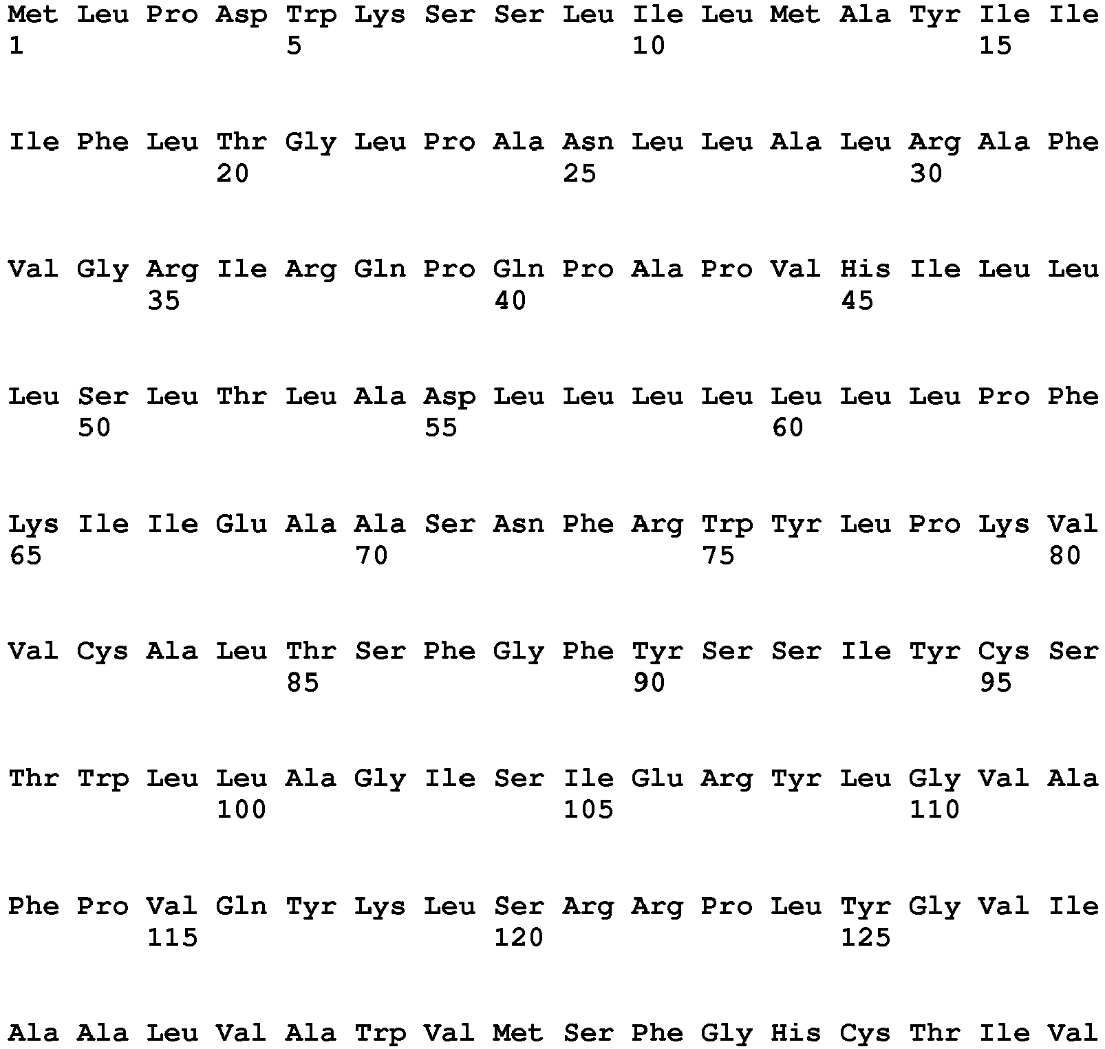

- <210> 16
<211> 2069
<212> DNA
<213> Homo sapiens - <400> 16


- <210> 17
<211> 428
<212> PRT
<213> Homo sapiens - <400> 17


- <210> 18
<211> 5984
<212> DNA
<213> Homo sapiens - <400> 18

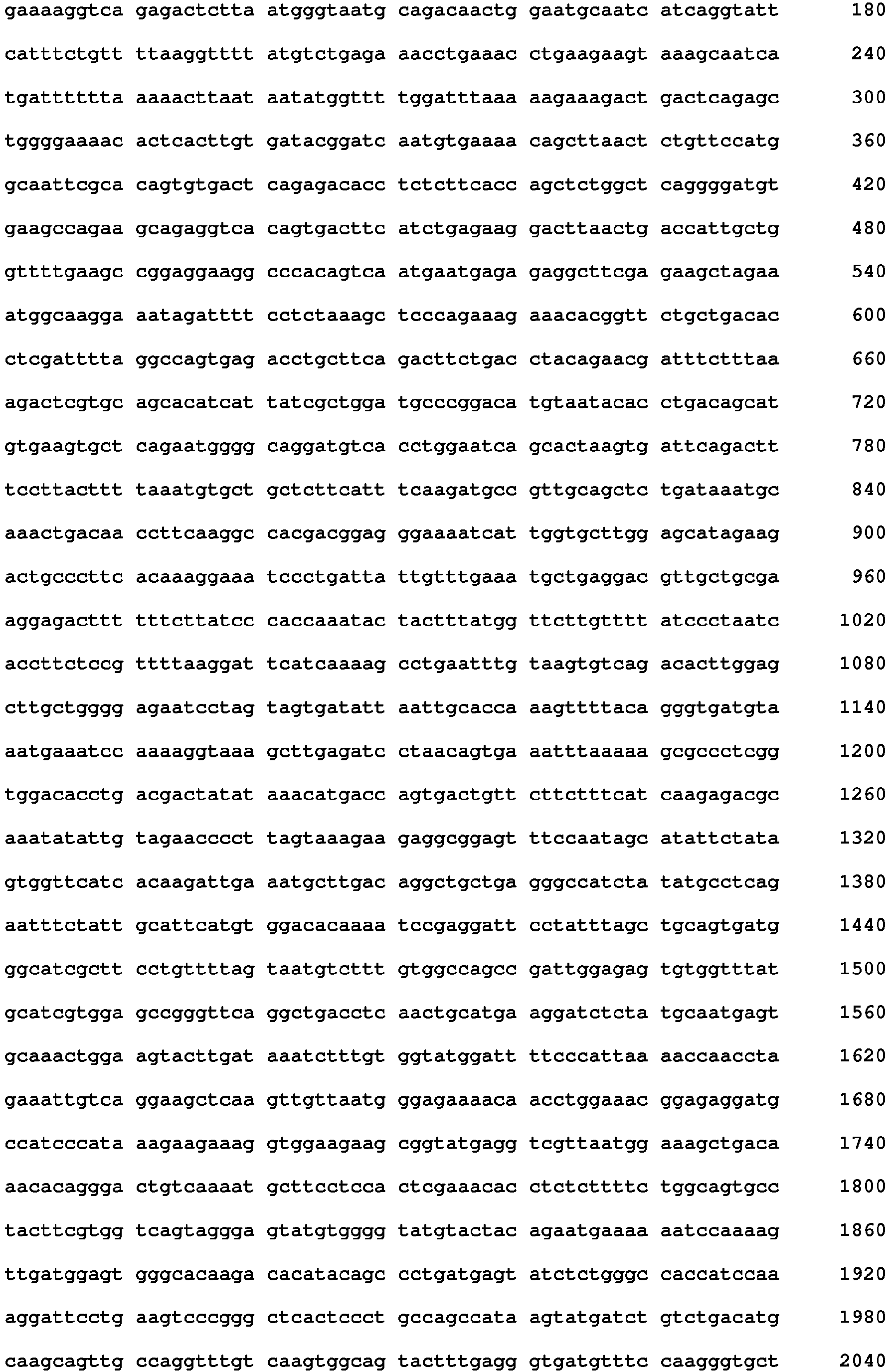


- <210> 19
<211> 667
<212> PRT
<213> Homo sapiens - <400> 19


- <210> 20
<211> 4177
<212> DNA
<213> Homo sapiens - <400> 20


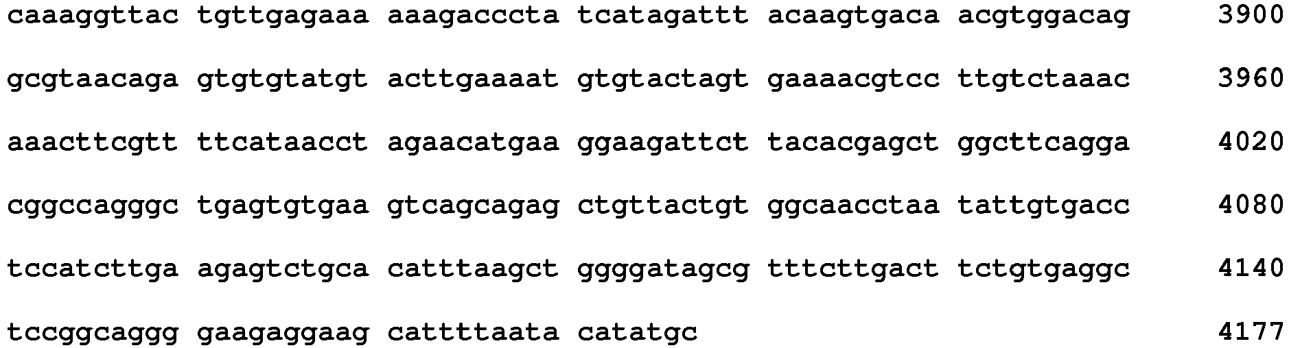
- <210> 21
<211> 294
<212> PRT
<213> Homo sapiens - <400> 21
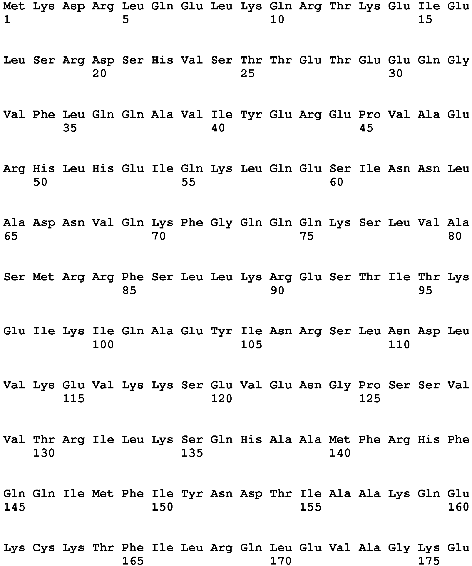

- <210> 22
<211> 1198
<212> DNA
<213> Homo sapiens - <400> 22


- <210> 23
<211> 1044
<212> PRT
<213> Homo sapiens - <400> 23


- <210> 24
<211> 6079
<212> DNA
<213> Homo sapiens - <400> 24


- <210> 25
<211> 326
<212> PRT
<213> Homo sapiens - <400> 25


- <210> 26
<211> 1722
<212> DNA
<213> Homo sapiens - <400> 26

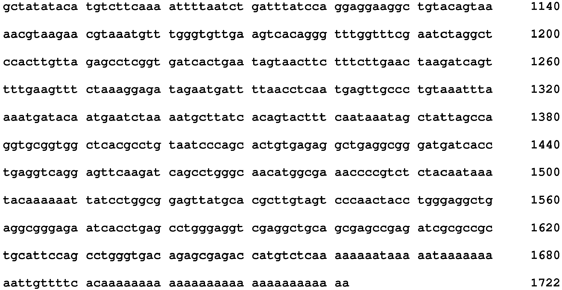
- <210> 27
<211> 3653
<212> DNA
<213> Homo sapiens - <400> 27


- <210> 28
<211> 1214
<212> PRT
<213> Homo sapiens - <400> 28


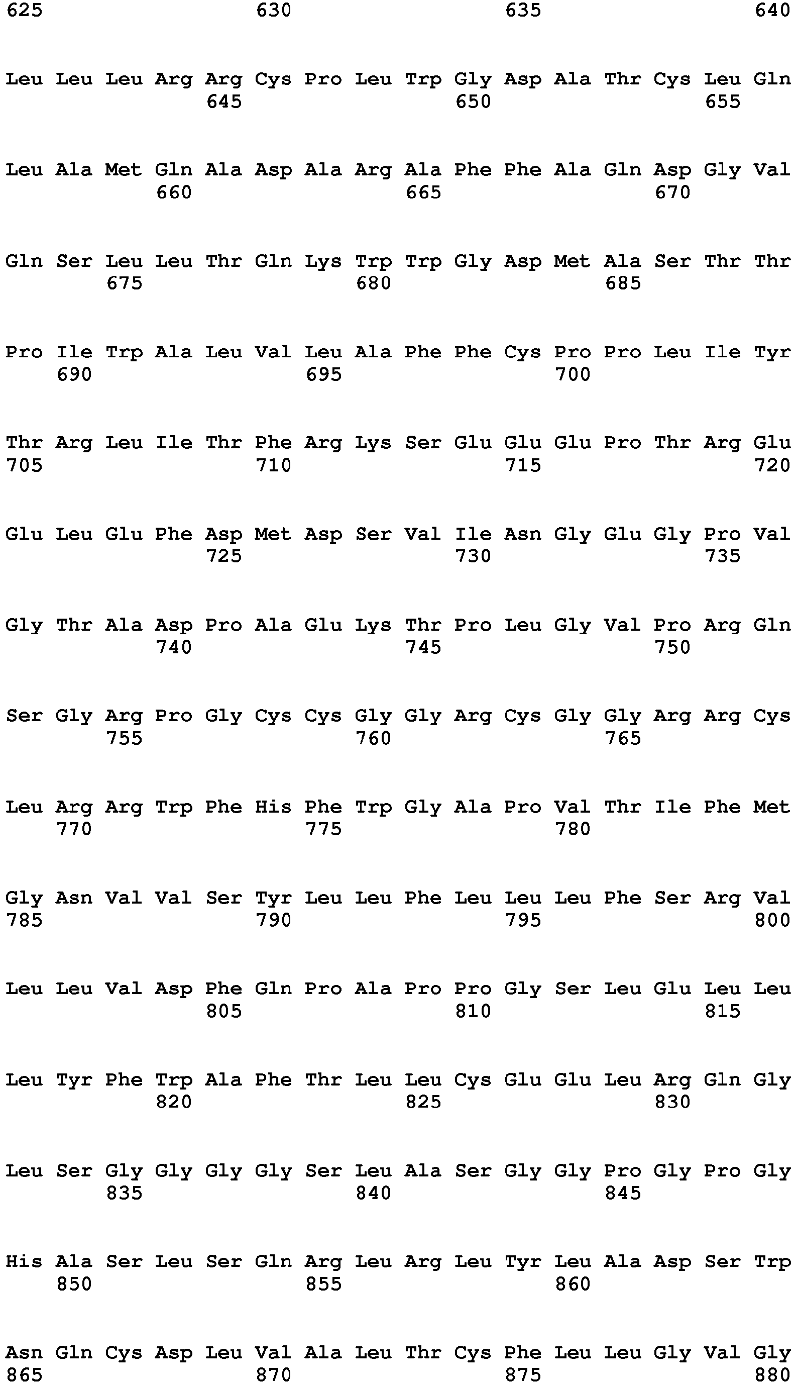


- <210> 29
<211> 4103
<212> DNA
<213> Homo sapiens - <400> 29


- <210> 30
<211> 3923
<212> DNA
<213> Homo sapiens - <400> 30


- <210> 31
<211> 662
<212> PRT
<213> Homo sapiens - <400> 31


- <210> 32
<211> 2707
<212> DNA
<213> Homo sapiens - <400> 32


- <210> 33
<211> 772
<212> PRT
<213> Homo sapiens - <400> 33


- <210> 34
<211> 6241
<212> DNA
<213> Homo sapiens - <400> 34


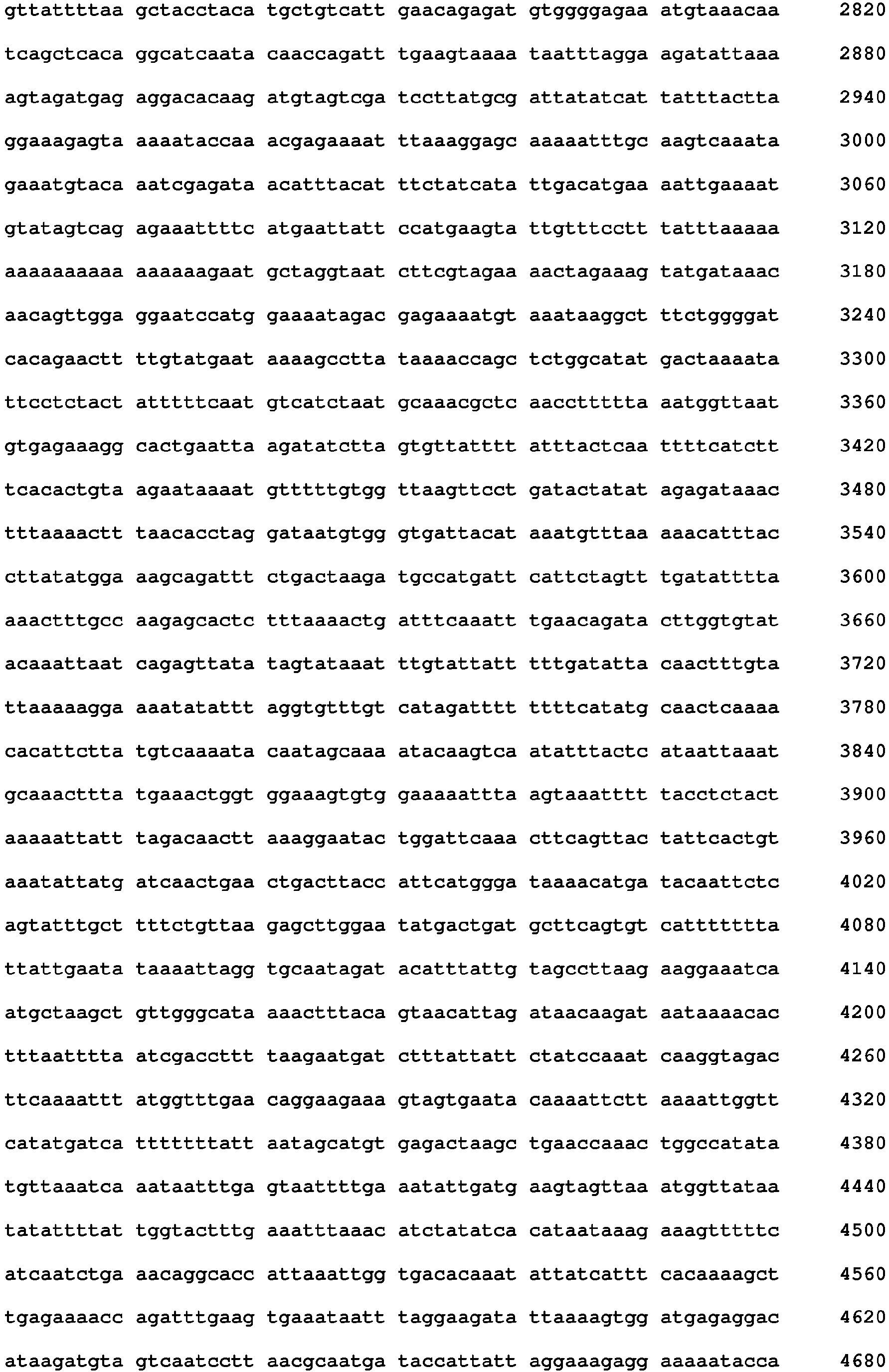

- <210> 35
<211> 320
<212> PRT
<213> Homo sapiens - <400> 35


- <210> 36
<211> 2799
<212> DNA
<213> Homo sapiens - <400> 36
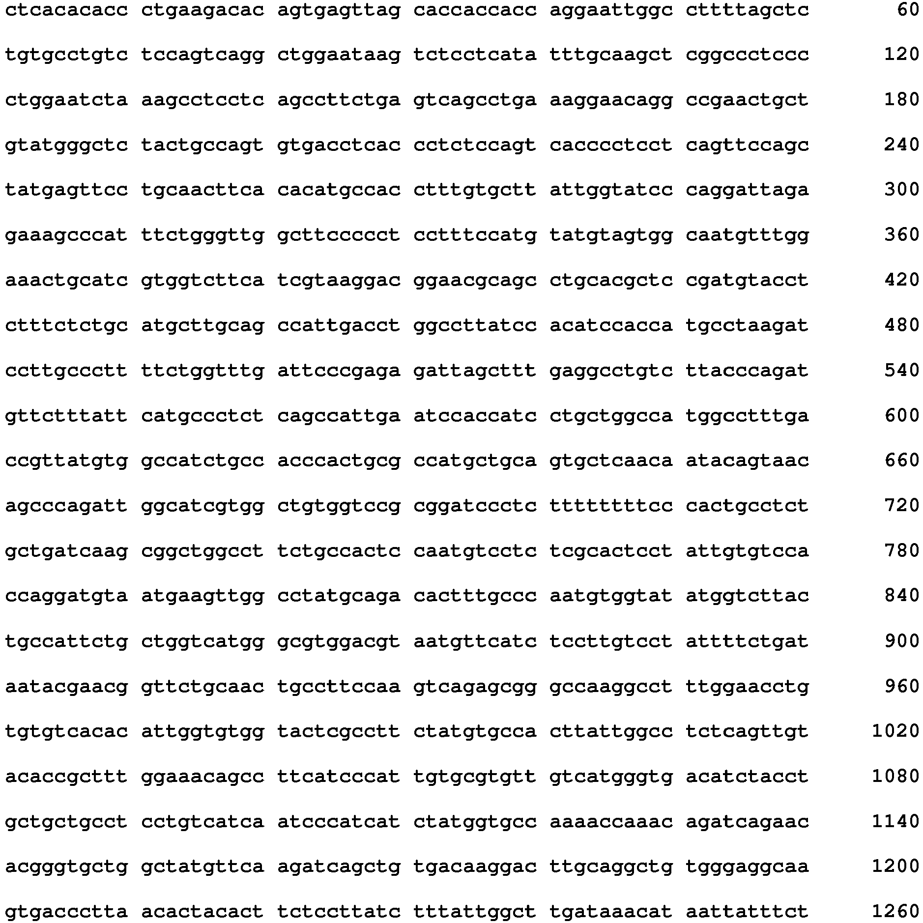

- <210> 37
<211> 2224
<212> PRT
<213> Homo sapiens - <400> 37




- <210> 38
<211> 9179
<212> DNA
<213> Homo sapiens - <400> 38


- <210> 39
<211> 694
<212> PRT
<213> Homo sapiens - <400> 39


- <210> 40
<211> 3195
<212> DNA
<213> Homo sapiens - <400> 40


- <210> 41
<211> 220
<212> PRT
<213> Homo sapiens - <400> 41


- <210> 42
<211> 1318
<212> DNA
<213> Homo sapiens - <400> 42


- <210> 43
<211> 264
<212> PRT
<213> Homo sapiens - <400> 43


- <210> 44
<211> 2328
<212> DNA
<213> Homo sapiens - <400> 44


- <210> 45
<211> 264
<212> PRT
<213> Homo sapiens - <400> 45


- <210> 46
<211> 2459
<212> DNA
<213> Homo sapiens - <400> 46


- <210> 47
<211> 2120
<212> PRT
<213> Homo sapiens - <400> 47




- <210> 48
<211> 6641
<212> DNA
<213> Homo sapiens - <400> 48


- <210> 49
<211> 707
<212> PRT
<213> Homo sapiens - <400> 49


- <210> 50
<211> 2387
<212> DNA
<213> Homo sapiens - <400> 50


- <210> 51
<211> 5058
<212> PRT
<213> Homo sapiens - <220>
<221> MOD_RES
<222> (283)..(283)
<223> Any amino acid - <220>
<221> MOD_RES
<222> (2674)..(2674)
<223> Any amino acid - <400> 51


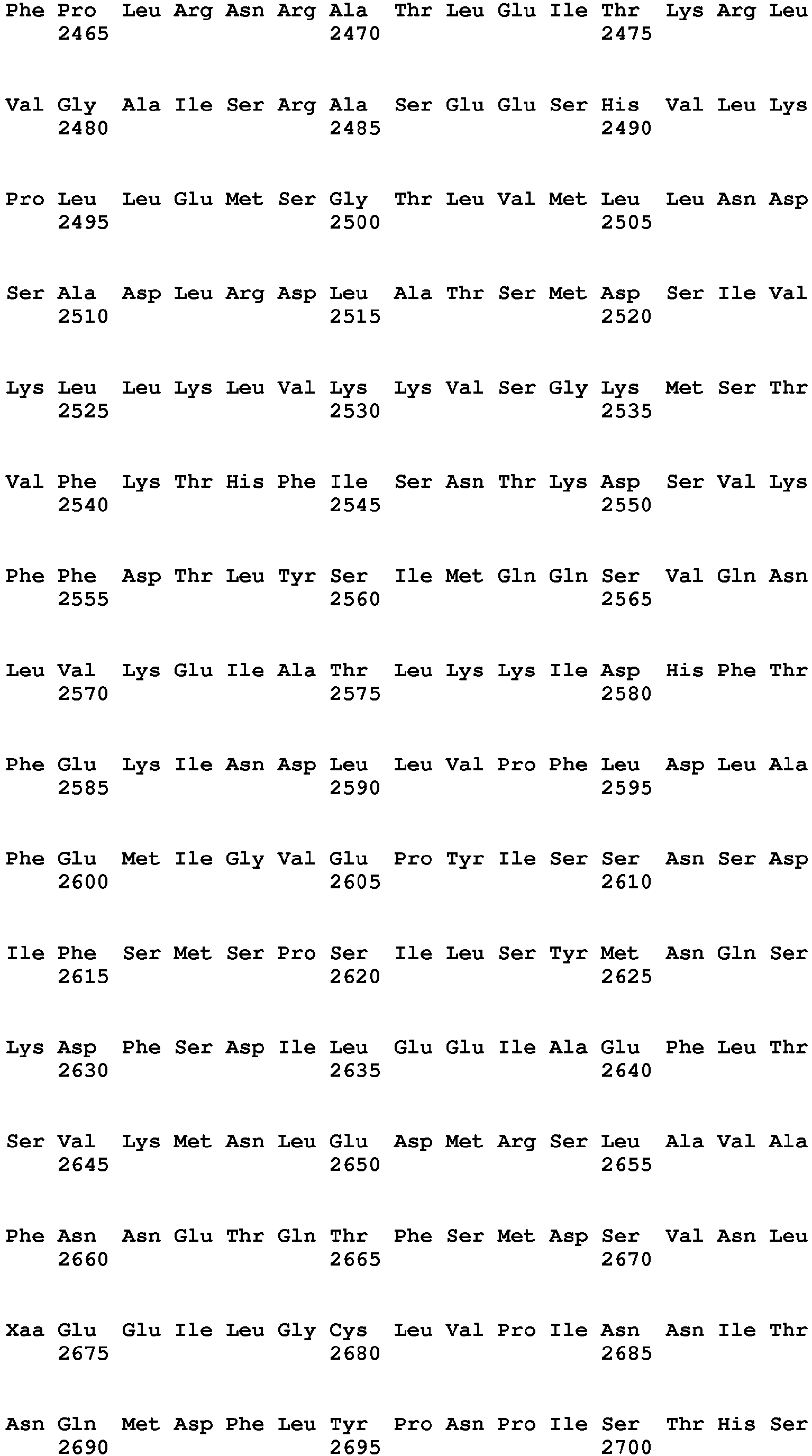


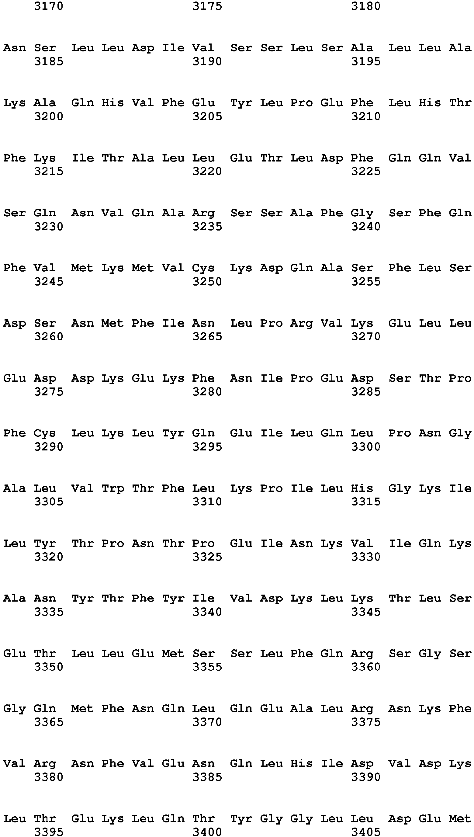


- <210> 52
<211> 17209
<212> DNA
<213> Homo sapiens - <400> 52


- <210> 53
<211> 931
<212> PRT
<213> Homo sapiens - <400> 53


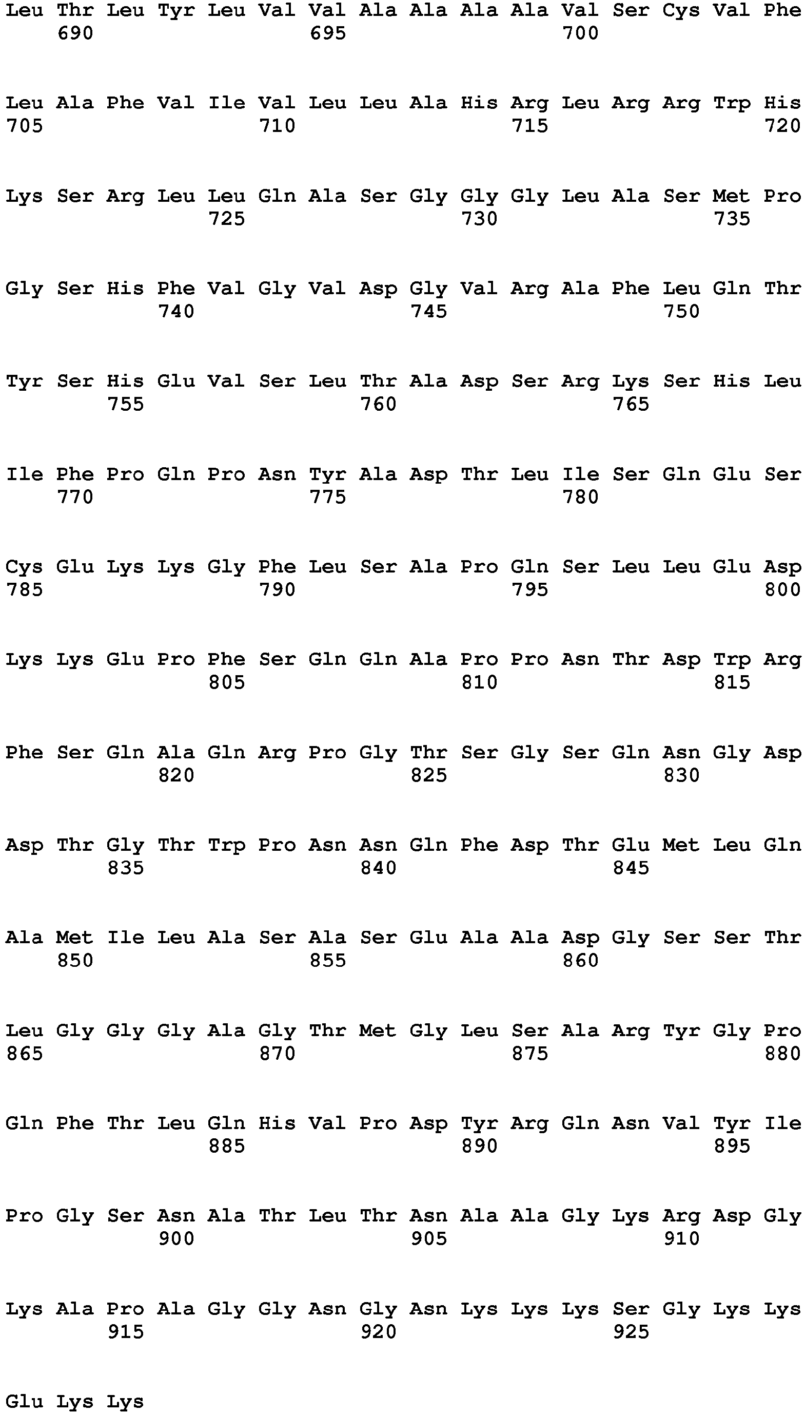

- <210> 54
<211> 4602
<212> DNA
<213> Homo sapiens - <400> 54


- <210> 55
<211> 3114
<212> DNA
<213> Homo sapiens - <400> 55


- <210> 56
<211> 3357
<212> DNA
<213> Homo sapiens - <400> 56


- <210> 57
<211> 2975
<212> DNA
<213> Homo sapiens - <400> 57


Claims (12)
- A method of collecting data for diagnosing prostate cancer, the method comprising:detecting expression of a plurality of genes in a biological sample obtained from a patient, wherein the patient is of African descent and wherein the plurality of genes is selected because the patient is of African descent and comprises at least three of the following genes: COL10A1, HOXC4, ESPL1, MMP9, ABCA13, PCDHGA1, and AGSK1,wherein the at least three genes comprise COL10A1.
- The method of claim 1, wherein the plurality of genes comprises COL10A1, HOXC4, ESPL1, MMP9, ABCA13, PCDHGA1, and AGSK1.
- The method of claim 1, further comprising a step of diagnosing prostate cancer using the expression data obtained.
- The method of claim 3, wherein overexpression of the at least three genes as compared to a control sample or a threshold value indicates the presence of prostate cancer in the biological sample or an increased risk of developing prostate cancer.
- The method of any one of claims 1-4, whereini) the method further comprises detecting expression of an ERG gene in the biological sample,ii) the biological sample is a tissue sample, a cell sample, a blood sample, a serum sample, or a urine sample,iii) the biological sample comprises prostate cells or nucleic acids or polypeptides isolated from prostate cells,iv) nucleic acid expression is detected, and/orv) polypeptide expression is detected.
- A kit for use in diagnosing prostate cancer, the kit comprising a plurality of probes for detecting at least three of the following genes: COL10A1, HOXC4, ESPL1, MMP9, ABCA13, PCDHGA1, and AGSK1, wherein the plurality of probes contains probes for detecting no more than 500, 250, 100, 50, 25, 15, 10, 9, 8, 7, 6, 5, 4, or 3 different genes, wherein the at least three genes comprise COL10A1.
- The kit of claim 6, whereina) the plurality of probes is selected from a plurality of oligonucleotide probes, a plurality of antibodies, or a plurality of polypeptide probes,b) the plurality of probes contains probes for detecting no more than 250, 100, 50, 25, 15, 10, or 5 different genes,c) the plurality of probes are attached to the surface of an array,
wherein the array optionally comprises no more than 500, 250, 100, 50, 25, 15, or 10 addressable elements,d) the plurality of probes are labeled, and/ore) the plurality of probes further comprises a probe for detecting an ERG gene. - A method of obtaining a gene expression profile in a biological sample, the method comprising:a) incubating the array of claim 7 with the biological sample, wherein the biological sample is obtained from a patient of African descent; andb) measuring the expression level of the at least three genes to obtain the gene expression profile.
- The method of claim 8, whereina) the biological sample is a tissue sample, a cell sample, a blood sample, a serum sample, or a urine sample,b) the biological sample comprises nucleic acids or polypeptides isolated from prostate cells,c) the measuring step comprises measuring nucleic acid expression levels,d) the measuring step comprises measuring polypeptide expression levels, and/ore) the measuring step further comprises measuring the expression level of an ERG gene.
- A method of identifying a patient in need of prostate cancer treatment, wherein the patient is of African descent, the method comprising:a) testing a biological sample from the patient for the overexpression of a plurality of genes, wherein the plurality of genes is selected because the patient is of African descent and comprises at least three of the following genes: COL10A1, HOXC4, ESPL1, MMP9, ABCA13, PCDHGA1, and AGSK1, wherein the at least three genes comprise COL10A1; andb) identifying the patient as in need of prostate cancer treatment if one or more of the at least three genes is overexpressed in the biological sample as compared to a control sample or a threshold value.
- The method of any one of claims 1-5, wherein the plurality of genes consists of no more than 100, 90, 80, 70, 60, 50, 40, 30, 25, 20, 15, 14, 13, 12, 11, 10, 9, 8, 7, 6, 5, 4, or 3 genes.
- The method of any one of claims 1-5 or 8-11, wherein the method is a computer-implemented method.
Applications Claiming Priority (2)
| Application Number | Priority Date | Filing Date | Title |
|---|---|---|---|
| US201361921739P | 2013-12-30 | 2013-12-30 | |
| PCT/US2014/072610 WO2015103166A1 (en) | 2013-12-30 | 2014-12-29 | Prostate cancer gene profiles and methods of using the same |
Publications (3)
| Publication Number | Publication Date |
|---|---|
| EP3090265A1 EP3090265A1 (en) | 2016-11-09 |
| EP3090265A4 EP3090265A4 (en) | 2018-03-21 |
| EP3090265B1 true EP3090265B1 (en) | 2020-04-08 |
Family
ID=53493959
Family Applications (1)
| Application Number | Title | Priority Date | Filing Date |
|---|---|---|---|
| EP14877549.7A Active EP3090265B1 (en) | 2013-12-30 | 2014-12-29 | Prostate cancer gene profiles and methods of using the same |
Country Status (8)
| Country | Link |
|---|---|
| US (1) | US20160326594A1 (en) |
| EP (1) | EP3090265B1 (en) |
| JP (1) | JP2017509351A (en) |
| CN (1) | CN106796236A (en) |
| AU (1) | AU2014373927B2 (en) |
| CA (1) | CA2934828A1 (en) |
| WO (1) | WO2015103166A1 (en) |
| ZA (1) | ZA201604370B (en) |
Families Citing this family (10)
| Publication number | Priority date | Publication date | Assignee | Title |
|---|---|---|---|---|
| JO3519B1 (en) | 2013-01-25 | 2020-07-05 | Amgen Inc | Antibody combinations for CDH19 and CD3 |
| US11332794B2 (en) | 2016-01-19 | 2022-05-17 | Wayne State University | Prostate cancer aggressiveness gene signatures for subjects of African or European descent |
| EP3635131A4 (en) * | 2017-05-12 | 2021-01-27 | The Henry M. Jackson Foundation for the Advancement of Military Medicine, Inc. | PROSTATE CANCER GENE PROFILES AND METHODS OF USE |
| WO2019169336A1 (en) * | 2018-03-02 | 2019-09-06 | The Johns Hopkins University | Methods for prostate cancer detection |
| CN110054674B (en) * | 2019-03-07 | 2022-08-05 | 深圳市龙华区人民医院 | Immunogenic polypeptide, anti-TTC 36 antibody AP2-19 and application |
| CN110628908B (en) * | 2019-09-19 | 2022-05-10 | 北京恩泽康泰生物科技有限公司 | Biomarker and detection kit for prostate cancer diagnosis grading and benign and malignant prediction |
| WO2021253134A1 (en) * | 2020-06-18 | 2021-12-23 | Ontario Institute For Cancer Research (Oicr) | Molecular classifiers for prostate cancer |
| CN111893177B (en) * | 2020-08-19 | 2022-10-04 | 南通大学 | Mutation screening method and mutation detection kit for hereditary hearing loss inducing gene |
| CN113284559B (en) * | 2021-07-21 | 2021-10-15 | 暨南大学 | A kind of promoter query method, system and device of species genome |
| CN120118995A (en) * | 2024-09-25 | 2025-06-10 | 深圳汇芯生物医疗科技有限公司 | Primer sets, probe sets, kits and methods for detecting prostate cancer biomarkers |
Family Cites Families (22)
| Publication number | Priority date | Publication date | Assignee | Title |
|---|---|---|---|---|
| US6453242B1 (en) | 1999-01-12 | 2002-09-17 | Sangamo Biosciences, Inc. | Selection of sites for targeting by zinc finger proteins and methods of designing zinc finger proteins to bind to preselected sites |
| US20050032065A1 (en) * | 2002-06-24 | 2005-02-10 | Afar Daniel E. H. | Methods of prognosis of prostate cancer |
| US20040191783A1 (en) * | 2003-03-31 | 2004-09-30 | Guy Leclercq | Low density micro-array analysis in human breast cancer |
| CA2565450C (en) * | 2004-05-07 | 2018-03-06 | Gyorgy Petrovics | Methods of diagnosing or treating prostate cancer using the erg gene, alone or in combination with other over or under expressed genes in prostate cancer |
| EP1824995A2 (en) * | 2004-11-24 | 2007-08-29 | St. George's Enterprises Limited | Diagnosis of prostate cancer |
| CN101310185A (en) * | 2005-09-19 | 2008-11-19 | 约翰·霍普金斯大学 | Prostate Cancer Biomarkers |
| US8338109B2 (en) * | 2006-11-02 | 2012-12-25 | Mayo Foundation For Medical Education And Research | Predicting cancer outcome |
| US20080254481A1 (en) * | 2006-11-13 | 2008-10-16 | Invitrogen Corporation | Methods and kits for detecting prostate cancer biomarkers |
| EP2288722B1 (en) * | 2008-04-10 | 2014-11-05 | The Henry M. Jackson Foundation for the Advancement of Military Medicine, Inc. | Prostate cancer-specific alterations in erg gene expression and detection and treatment methods based on those alterations |
| WO2010065940A1 (en) * | 2008-12-04 | 2010-06-10 | The Regents Of The University Of California | Materials and methods for determining diagnosis and prognosis of prostate cancer |
| FR2945820A1 (en) * | 2009-05-25 | 2010-11-26 | Univ Clermont Auvergne | GENE PANEL FOR THE PROGNOSIS OF PROSTATE CANCER |
| WO2011127219A1 (en) * | 2010-04-06 | 2011-10-13 | Caris Life Sciences Luxembourg Holdings | Circulating biomarkers for disease |
| DK2598659T3 (en) * | 2010-07-27 | 2015-05-26 | Genomic Health Inc | PROCEDURE FOR USING GENEPRESSION FOR DETERMINING PROSTATE CANCER FORECAST |
| WO2012024543A1 (en) * | 2010-08-18 | 2012-02-23 | Caris Life Sciences Luxembourg Holdings | Circulating biomarkers for disease |
| WO2012031008A2 (en) * | 2010-08-31 | 2012-03-08 | The General Hospital Corporation | Cancer-related biological materials in microvesicles |
| ES2593959T3 (en) * | 2011-04-19 | 2016-12-14 | MAX-PLANCK-Gesellschaft zur Förderung der Wissenschaften e.V. | Markers of prostate cancer |
| WO2012152800A1 (en) * | 2011-05-12 | 2012-11-15 | Noviogendix Research B.V. | Molecular markers in prostate cancer |
| KR20140067001A (en) * | 2011-08-08 | 2014-06-03 | 카리스 라이프 사이언스 룩셈부르크 홀딩스, 에스.에이.알.엘. | Biomarker compositions and methods |
| HK1199099A1 (en) * | 2011-08-31 | 2015-06-19 | Oncocyte Corporation | Methods and compositions for the treatment and diagnosis of cancer |
| SE536352C2 (en) * | 2011-10-24 | 2013-09-03 | Chundsell Medicals Ab | Cursor genes for classification of prostate cancer |
| US20150017640A1 (en) * | 2011-11-04 | 2015-01-15 | Noviogendix Research B.V. | Combinations of Molecular Markers in Prostate Cancer providing a Diagnostic Tool with Improved Sensitivity/Specificity |
| US9863002B2 (en) * | 2012-06-04 | 2018-01-09 | North Carolina Central University | Genetic determinants of prostate cancer risk |
-
2014
- 2014-12-29 AU AU2014373927A patent/AU2014373927B2/en active Active
- 2014-12-29 CA CA2934828A patent/CA2934828A1/en not_active Abandoned
- 2014-12-29 US US15/108,909 patent/US20160326594A1/en not_active Abandoned
- 2014-12-29 WO PCT/US2014/072610 patent/WO2015103166A1/en not_active Ceased
- 2014-12-29 JP JP2016561993A patent/JP2017509351A/en active Pending
- 2014-12-29 EP EP14877549.7A patent/EP3090265B1/en active Active
- 2014-12-29 CN CN201480076628.8A patent/CN106796236A/en active Pending
-
2016
- 2016-06-28 ZA ZA2016/04370A patent/ZA201604370B/en unknown
Non-Patent Citations (1)
| Title |
|---|
| None * |
Also Published As
| Publication number | Publication date |
|---|---|
| AU2014373927B2 (en) | 2018-03-22 |
| WO2015103166A1 (en) | 2015-07-09 |
| AU2014373927A1 (en) | 2016-07-07 |
| CA2934828A1 (en) | 2015-07-09 |
| ZA201604370B (en) | 2020-12-23 |
| CN106796236A (en) | 2017-05-31 |
| EP3090265A4 (en) | 2018-03-21 |
| US20160326594A1 (en) | 2016-11-10 |
| EP3090265A1 (en) | 2016-11-09 |
| JP2017509351A (en) | 2017-04-06 |
Similar Documents
| Publication | Publication Date | Title |
|---|---|---|
| US20160326594A1 (en) | Prostate cancer gene profiles and methods of using the same | |
| US20130065789A1 (en) | Compositions and methods for classifying lung cancer and prognosing lung cancer survival | |
| EP3090067B1 (en) | Genomic rearrangements associated with prostate cancer and methods of using the same | |
| JP2011526487A (en) | Breast cancer genome fingerprint | |
| US20250369053A1 (en) | Recurrence gene signature across multiple cancer types | |
| JP2016514966A (en) | Composition for prognosis detection and determination of prostate cancer and method for detection and determination | |
| WO2013033073A1 (en) | Expression of ets related gene (erg) and phosphatase and tensin homolog (pten) correlates with prostate cancer capsular penetration | |
| US20240218451A1 (en) | Prostate cancer gene profiles and methods of using the same | |
| US20150344962A1 (en) | Methods for evaluating breast cancer prognosis | |
| WO2010108638A9 (en) | Tumour gene profile | |
| US10081842B2 (en) | Prostate cancer gene expression profiles | |
| US20220033913A1 (en) | Genomic rearrangements associated with prostate cancer and methods of using the same | |
| WO2021178832A2 (en) | Dna damage repair genes in cancer | |
| WO2014173986A2 (en) | Methods for diagnosing and monitoring the response to treatment of hepatocellular carcinoma | |
| WO2024178032A2 (en) | Methods for diagnosing and treating ovarian cancer | |
| HK1238718A1 (en) | Prostate cancer gene profiles and methods of using the same | |
| CA2934878C (en) | Genomic rearrangements associated with prostate cancer and methods of using the same | |
| EP2607494A1 (en) | Biomarkers for lung cancer risk assessment | |
| HK1238683A1 (en) | Genomic rearrangements associated with prostate cancer and methods of using the same |
Legal Events
| Date | Code | Title | Description |
|---|---|---|---|
| PUAI | Public reference made under article 153(3) epc to a published international application that has entered the european phase |
Free format text: ORIGINAL CODE: 0009012 |
|
| 17P | Request for examination filed |
Effective date: 20160729 |
|
| AK | Designated contracting states |
Kind code of ref document: A1 Designated state(s): AL AT BE BG CH CY CZ DE DK EE ES FI FR GB GR HR HU IE IS IT LI LT LU LV MC MK MT NL NO PL PT RO RS SE SI SK SM TR |
|
| AX | Request for extension of the european patent |
Extension state: BA ME |
|
| DAX | Request for extension of the european patent (deleted) | ||
| RIC1 | Information provided on ipc code assigned before grant |
Ipc: G01N 33/574 20060101AFI20171113BHEP Ipc: C12Q 1/68 20060101ALI20171113BHEP |
|
| A4 | Supplementary search report drawn up and despatched |
Effective date: 20180219 |
|
| RIC1 | Information provided on ipc code assigned before grant |
Ipc: G01N 33/574 20060101AFI20180213BHEP Ipc: C12Q 1/68 20180101ALI20180213BHEP |
|
| STAA | Information on the status of an ep patent application or granted ep patent |
Free format text: STATUS: EXAMINATION IS IN PROGRESS |
|
| 17Q | First examination report despatched |
Effective date: 20190116 |
|
| REG | Reference to a national code |
Ref country code: DE Ref legal event code: R079 Ref document number: 602014063654 Country of ref document: DE Free format text: PREVIOUS MAIN CLASS: G01N0033574000 Ipc: C12Q0001688600 |
|
| RIC1 | Information provided on ipc code assigned before grant |
Ipc: C12Q 1/6886 20180101AFI20190821BHEP |
|
| GRAP | Despatch of communication of intention to grant a patent |
Free format text: ORIGINAL CODE: EPIDOSNIGR1 |
|
| STAA | Information on the status of an ep patent application or granted ep patent |
Free format text: STATUS: GRANT OF PATENT IS INTENDED |
|
| INTG | Intention to grant announced |
Effective date: 20191011 |
|
| GRAS | Grant fee paid |
Free format text: ORIGINAL CODE: EPIDOSNIGR3 |
|
| GRAA | (expected) grant |
Free format text: ORIGINAL CODE: 0009210 |
|
| STAA | Information on the status of an ep patent application or granted ep patent |
Free format text: STATUS: THE PATENT HAS BEEN GRANTED |
|
| AK | Designated contracting states |
Kind code of ref document: B1 Designated state(s): AL AT BE BG CH CY CZ DE DK EE ES FI FR GB GR HR HU IE IS IT LI LT LU LV MC MK MT NL NO PL PT RO RS SE SI SK SM TR |
|
| REG | Reference to a national code |
Ref country code: AT Ref legal event code: REF Ref document number: 1254450 Country of ref document: AT Kind code of ref document: T Effective date: 20200415 Ref country code: CH Ref legal event code: EP |
|
| REG | Reference to a national code |
Ref country code: DE Ref legal event code: R096 Ref document number: 602014063654 Country of ref document: DE |
|
| REG | Reference to a national code |
Ref country code: IE Ref legal event code: FG4D |
|
| REG | Reference to a national code |
Ref country code: NL Ref legal event code: MP Effective date: 20200408 |
|
| REG | Reference to a national code |
Ref country code: LT Ref legal event code: MG4D |
|
| PG25 | Lapsed in a contracting state [announced via postgrant information from national office to epo] |
Ref country code: FI Free format text: LAPSE BECAUSE OF FAILURE TO SUBMIT A TRANSLATION OF THE DESCRIPTION OR TO PAY THE FEE WITHIN THE PRESCRIBED TIME-LIMIT Effective date: 20200408 Ref country code: SE Free format text: LAPSE BECAUSE OF FAILURE TO SUBMIT A TRANSLATION OF THE DESCRIPTION OR TO PAY THE FEE WITHIN THE PRESCRIBED TIME-LIMIT Effective date: 20200408 Ref country code: NO Free format text: LAPSE BECAUSE OF FAILURE TO SUBMIT A TRANSLATION OF THE DESCRIPTION OR TO PAY THE FEE WITHIN THE PRESCRIBED TIME-LIMIT Effective date: 20200708 Ref country code: GR Free format text: LAPSE BECAUSE OF FAILURE TO SUBMIT A TRANSLATION OF THE DESCRIPTION OR TO PAY THE FEE WITHIN THE PRESCRIBED TIME-LIMIT Effective date: 20200709 Ref country code: LT Free format text: LAPSE BECAUSE OF FAILURE TO SUBMIT A TRANSLATION OF THE DESCRIPTION OR TO PAY THE FEE WITHIN THE PRESCRIBED TIME-LIMIT Effective date: 20200408 Ref country code: PT Free format text: LAPSE BECAUSE OF FAILURE TO SUBMIT A TRANSLATION OF THE DESCRIPTION OR TO PAY THE FEE WITHIN THE PRESCRIBED TIME-LIMIT Effective date: 20200817 Ref country code: NL Free format text: LAPSE BECAUSE OF FAILURE TO SUBMIT A TRANSLATION OF THE DESCRIPTION OR TO PAY THE FEE WITHIN THE PRESCRIBED TIME-LIMIT Effective date: 20200408 Ref country code: IS Free format text: LAPSE BECAUSE OF FAILURE TO SUBMIT A TRANSLATION OF THE DESCRIPTION OR TO PAY THE FEE WITHIN THE PRESCRIBED TIME-LIMIT Effective date: 20200808 |
|
| REG | Reference to a national code |
Ref country code: AT Ref legal event code: MK05 Ref document number: 1254450 Country of ref document: AT Kind code of ref document: T Effective date: 20200408 |
|
| PG25 | Lapsed in a contracting state [announced via postgrant information from national office to epo] |
Ref country code: BG Free format text: LAPSE BECAUSE OF FAILURE TO SUBMIT A TRANSLATION OF THE DESCRIPTION OR TO PAY THE FEE WITHIN THE PRESCRIBED TIME-LIMIT Effective date: 20200708 Ref country code: LV Free format text: LAPSE BECAUSE OF FAILURE TO SUBMIT A TRANSLATION OF THE DESCRIPTION OR TO PAY THE FEE WITHIN THE PRESCRIBED TIME-LIMIT Effective date: 20200408 Ref country code: RS Free format text: LAPSE BECAUSE OF FAILURE TO SUBMIT A TRANSLATION OF THE DESCRIPTION OR TO PAY THE FEE WITHIN THE PRESCRIBED TIME-LIMIT Effective date: 20200408 Ref country code: HR Free format text: LAPSE BECAUSE OF FAILURE TO SUBMIT A TRANSLATION OF THE DESCRIPTION OR TO PAY THE FEE WITHIN THE PRESCRIBED TIME-LIMIT Effective date: 20200408 |
|
| PG25 | Lapsed in a contracting state [announced via postgrant information from national office to epo] |
Ref country code: AL Free format text: LAPSE BECAUSE OF FAILURE TO SUBMIT A TRANSLATION OF THE DESCRIPTION OR TO PAY THE FEE WITHIN THE PRESCRIBED TIME-LIMIT Effective date: 20200408 |
|
| REG | Reference to a national code |
Ref country code: DE Ref legal event code: R097 Ref document number: 602014063654 Country of ref document: DE |
|
| PG25 | Lapsed in a contracting state [announced via postgrant information from national office to epo] |
Ref country code: EE Free format text: LAPSE BECAUSE OF FAILURE TO SUBMIT A TRANSLATION OF THE DESCRIPTION OR TO PAY THE FEE WITHIN THE PRESCRIBED TIME-LIMIT Effective date: 20200408 Ref country code: AT Free format text: LAPSE BECAUSE OF FAILURE TO SUBMIT A TRANSLATION OF THE DESCRIPTION OR TO PAY THE FEE WITHIN THE PRESCRIBED TIME-LIMIT Effective date: 20200408 Ref country code: DK Free format text: LAPSE BECAUSE OF FAILURE TO SUBMIT A TRANSLATION OF THE DESCRIPTION OR TO PAY THE FEE WITHIN THE PRESCRIBED TIME-LIMIT Effective date: 20200408 Ref country code: IT Free format text: LAPSE BECAUSE OF FAILURE TO SUBMIT A TRANSLATION OF THE DESCRIPTION OR TO PAY THE FEE WITHIN THE PRESCRIBED TIME-LIMIT Effective date: 20200408 Ref country code: RO Free format text: LAPSE BECAUSE OF FAILURE TO SUBMIT A TRANSLATION OF THE DESCRIPTION OR TO PAY THE FEE WITHIN THE PRESCRIBED TIME-LIMIT Effective date: 20200408 Ref country code: CZ Free format text: LAPSE BECAUSE OF FAILURE TO SUBMIT A TRANSLATION OF THE DESCRIPTION OR TO PAY THE FEE WITHIN THE PRESCRIBED TIME-LIMIT Effective date: 20200408 Ref country code: ES Free format text: LAPSE BECAUSE OF FAILURE TO SUBMIT A TRANSLATION OF THE DESCRIPTION OR TO PAY THE FEE WITHIN THE PRESCRIBED TIME-LIMIT Effective date: 20200408 Ref country code: SM Free format text: LAPSE BECAUSE OF FAILURE TO SUBMIT A TRANSLATION OF THE DESCRIPTION OR TO PAY THE FEE WITHIN THE PRESCRIBED TIME-LIMIT Effective date: 20200408 |
|
| PLBE | No opposition filed within time limit |
Free format text: ORIGINAL CODE: 0009261 |
|
| STAA | Information on the status of an ep patent application or granted ep patent |
Free format text: STATUS: NO OPPOSITION FILED WITHIN TIME LIMIT |
|
| PG25 | Lapsed in a contracting state [announced via postgrant information from national office to epo] |
Ref country code: SK Free format text: LAPSE BECAUSE OF FAILURE TO SUBMIT A TRANSLATION OF THE DESCRIPTION OR TO PAY THE FEE WITHIN THE PRESCRIBED TIME-LIMIT Effective date: 20200408 Ref country code: PL Free format text: LAPSE BECAUSE OF FAILURE TO SUBMIT A TRANSLATION OF THE DESCRIPTION OR TO PAY THE FEE WITHIN THE PRESCRIBED TIME-LIMIT Effective date: 20200408 |
|
| 26N | No opposition filed |
Effective date: 20210112 |
|
| PG25 | Lapsed in a contracting state [announced via postgrant information from national office to epo] |
Ref country code: SI Free format text: LAPSE BECAUSE OF FAILURE TO SUBMIT A TRANSLATION OF THE DESCRIPTION OR TO PAY THE FEE WITHIN THE PRESCRIBED TIME-LIMIT Effective date: 20200408 |
|
| REG | Reference to a national code |
Ref country code: CH Ref legal event code: PL |
|
| PG25 | Lapsed in a contracting state [announced via postgrant information from national office to epo] |
Ref country code: MC Free format text: LAPSE BECAUSE OF FAILURE TO SUBMIT A TRANSLATION OF THE DESCRIPTION OR TO PAY THE FEE WITHIN THE PRESCRIBED TIME-LIMIT Effective date: 20200408 |
|
| REG | Reference to a national code |
Ref country code: BE Ref legal event code: MM Effective date: 20201231 |
|
| PG25 | Lapsed in a contracting state [announced via postgrant information from national office to epo] |
Ref country code: LU Free format text: LAPSE BECAUSE OF NON-PAYMENT OF DUE FEES Effective date: 20201229 Ref country code: IE Free format text: LAPSE BECAUSE OF NON-PAYMENT OF DUE FEES Effective date: 20201229 |
|
| PG25 | Lapsed in a contracting state [announced via postgrant information from national office to epo] |
Ref country code: CH Free format text: LAPSE BECAUSE OF NON-PAYMENT OF DUE FEES Effective date: 20201231 Ref country code: LI Free format text: LAPSE BECAUSE OF NON-PAYMENT OF DUE FEES Effective date: 20201231 |
|
| PG25 | Lapsed in a contracting state [announced via postgrant information from national office to epo] |
Ref country code: TR Free format text: LAPSE BECAUSE OF FAILURE TO SUBMIT A TRANSLATION OF THE DESCRIPTION OR TO PAY THE FEE WITHIN THE PRESCRIBED TIME-LIMIT Effective date: 20200408 Ref country code: MT Free format text: LAPSE BECAUSE OF FAILURE TO SUBMIT A TRANSLATION OF THE DESCRIPTION OR TO PAY THE FEE WITHIN THE PRESCRIBED TIME-LIMIT Effective date: 20200408 Ref country code: CY Free format text: LAPSE BECAUSE OF FAILURE TO SUBMIT A TRANSLATION OF THE DESCRIPTION OR TO PAY THE FEE WITHIN THE PRESCRIBED TIME-LIMIT Effective date: 20200408 |
|
| PG25 | Lapsed in a contracting state [announced via postgrant information from national office to epo] |
Ref country code: MK Free format text: LAPSE BECAUSE OF FAILURE TO SUBMIT A TRANSLATION OF THE DESCRIPTION OR TO PAY THE FEE WITHIN THE PRESCRIBED TIME-LIMIT Effective date: 20200408 |
|
| PG25 | Lapsed in a contracting state [announced via postgrant information from national office to epo] |
Ref country code: BE Free format text: LAPSE BECAUSE OF NON-PAYMENT OF DUE FEES Effective date: 20201231 |
|
| PGFP | Annual fee paid to national office [announced via postgrant information from national office to epo] |
Ref country code: GB Payment date: 20251229 Year of fee payment: 12 |
|
| PGFP | Annual fee paid to national office [announced via postgrant information from national office to epo] |
Ref country code: FR Payment date: 20251226 Year of fee payment: 12 |
|
| PGFP | Annual fee paid to national office [announced via postgrant information from national office to epo] |
Ref country code: DE Payment date: 20251229 Year of fee payment: 12 |