EP3089992B1 - Compositions and methods for imaging cancer - Google Patents
Compositions and methods for imaging cancer Download PDFInfo
- Publication number
- EP3089992B1 EP3089992B1 EP15733077.0A EP15733077A EP3089992B1 EP 3089992 B1 EP3089992 B1 EP 3089992B1 EP 15733077 A EP15733077 A EP 15733077A EP 3089992 B1 EP3089992 B1 EP 3089992B1
- Authority
- EP
- European Patent Office
- Prior art keywords
- somatostatin
- fluorinated
- optionally
- derivative according
- ambf
- Prior art date
- Legal status (The legal status is an assumption and is not a legal conclusion. Google has not performed a legal analysis and makes no representation as to the accuracy of the status listed.)
- Active
Links
Images


Classifications
-
- C—CHEMISTRY; METALLURGY
- C07—ORGANIC CHEMISTRY
- C07K—PEPTIDES
- C07K14/00—Peptides having more than 20 amino acids; Gastrins; Somatostatins; Melanotropins; Derivatives thereof
- C07K14/435—Peptides having more than 20 amino acids; Gastrins; Somatostatins; Melanotropins; Derivatives thereof from animals; from humans
- C07K14/575—Hormones
- C07K14/655—Somatostatins
-
- A—HUMAN NECESSITIES
- A61—MEDICAL OR VETERINARY SCIENCE; HYGIENE
- A61K—PREPARATIONS FOR MEDICAL, DENTAL OR TOILETRY PURPOSES
- A61K51/00—Preparations containing radioactive substances for use in therapy or testing in vivo
- A61K51/02—Preparations containing radioactive substances for use in therapy or testing in vivo characterised by the carrier, i.e. characterised by the agent or material covalently linked or complexing the radioactive nucleus
- A61K51/04—Organic compounds
- A61K51/08—Peptides, e.g. proteins, carriers being peptides, polyamino acids, proteins
- A61K51/083—Peptides, e.g. proteins, carriers being peptides, polyamino acids, proteins the peptide being octreotide or a somatostatin-receptor-binding peptide
Definitions
- the present invention relates to the field of radioimaging and, in particular, to radiolabelled compounds, methods of preparing the compounds and their use in imaging cancer.
- sstr2 The somatostatin receptor subtype 2 (sstr2) is overexpressed in many neuroendocrine tumours. Hence over the past 30 years, there has been considerable interest in developing high-affinity somatostatin-derived ligands that bind sstr2, notably for radionuclide therapy ( Kwekkeboom DJ, et al., Semin Nucl Med. 2010, 40:78-88 ).
- radiotracers based on the somatostatin family of peptides, notably octreotate and octreotide, have been labelled with various radioisotopes for non-invasive imaging ( Breeman WAP, et al., Eur J Nucl Med., 2001, 28:1421-1429 ; Ginj M, et al., Chem Biol., 2006, 13:1081-1090 ; Antunes P, et al., Bioconjug Chem., 2007, 18:84-92 ; Kwekkeboom DJ, et al., Endocr Relat Cancer., 2010, 17:R53-R73 ).
- 99m Tc derivatives such as 99m Tc-depreotide ( Virgolini I, et al., Cancer Res., 1998, 58:1850-1859 ) and 99m Tc-hydrazinonicotinyl-Tyr3-octreotide have also been used ( Gabriel M, et al., J Nucl Med., 2003, 44:708-716 ) but are not commercialized in North America.
- 68 Ga ligands such as 68 Ga-DOTATOC, 68 Ga-DOTATATE, and 68 Ga-DOTANOC have shown promise for neuroendocrine tumour imaging ( Henze M, et al., J Nucl Med., 2001, 42:1053-1056 ; Kayani I, et al., J Nucl Med., 2009, 50:1927-1932 ; Poeppel TD, et al., J Nucl Med., 2011, 52:1864-1870 ) and are used in clinical trials as well as under the local practice of pharmacy, particularly in Europe.
- 68 Ga-PET imaging is not widely available because of the limited daily availability of 68 Ga ( ⁇ 50 mCi) and the lack of FDA-approved 68 Ge/ 68 Ga generators ( Banerjee SR, Pomper MG., Appl Radiat Isot., 2013, 76:2-13 ).
- 18 F-fluoride presents several attractive properties for imaging ( Laforest R, Liu X., Q J Nucl Med Mol Imaging, 2008, 52:151-158 ; Kemerink GJ, et al., Eur J Nucl Med Mol Imaging, 2011, 38:940-948 ) and is produced on a daily basis in large quantities in hundreds of cyclotrons in hospitals and radiopharmacies worldwide.
- aryltrifluoroborate prosthetics when conjugated to various peptides, allow one-step aqueous radiofluorination in high yield and very high specific activity ( Liu Z, et al., J Labelled Comp Radiopharm., 2012, 14:491-497 ; Liu Z, et al., Nucl Med Biol., 2013, 40:841-849 ; Liu Z, et al., Angew Chem Int Ed., 2013, 52:2305-2307 , and International Patent Application Publication No. WO2009/012596 ).
- the present invention relates generally to compositions and methods for imaging cancer.
- One aspect of the invention relates to a fluorinated somatostatin derivative having general formula (I): B - F 3 -(CH 2 ) n -N + R 1 R 2 -L-X ( I ) wherein:
- Another aspect of the invention relates to a fluorinated somatostatin derivative selected from: AMBF 3 -TATE (compound 2 ), AMBF 3 -JR11 (compound 7 ), AMBF 3 -LM3 (compound 3 ) and AMBF 3 -TOC (compound 6 ).
- fluorinated somatostatin derivative according to any one of claims 1 to 19, wherein at least one F is 18 F.
- Another aspect of the invention relates to a method of preparing a 18 F-labelled somatostatin derivative comprising submitting a fluorinated somatostatin derivative as described above in which each F is 19 F to an isotope exchange reaction using 18 F-fluoride.
- Another aspect of the invention relates to an 18 F-labelled somatostatin derivative prepared by a method as described above.
- Another aspect of the invention relates to a use of a fluorinated somatostatin derivative as described above in which at least one F is 18 F as a radiotracer.
- Another aspect of the invention relates to a use of a fluorinated somatostatin derivative as described above in which at least one F is 18 F as a positron emission tomography (PET) imaging agent.
- PET positron emission tomography
- Another aspect of the invention relates to a use of a fluorinated somatostatin derivative as described above in which each F is 19 F as a therapeutic agent.
- kits for the preparation of a 18 F-labelled imaging agent comprising a fluorinated somatostatin derivative as described above in which each F is 19 F and optionally instructions for use.
- this invention relates to somatostatin derivative compounds that may be readily labelled with the isotope fluorine-18 and that have affinity and selectivity for cellular somatostatin receptors.
- the labelled compounds are useful clinically as radioactive tracers in various in vivo imaging applications (for example, using positron emission tomography (PET) and related techniques) to detect somatostatin-expressing cells and tissues, including tumours.
- PET positron emission tomography
- the somatostatin derivatives may be based on a variety of somatostatin analogues provided that the selected somatostatin analogue is capable of selectively binding to a somatostatin receptor.
- the somatostatin derivatives are octreotide derivatives.
- the invention relates to easily radiolabelled, high-affinity octreotide derivatives for imaging of somatostatin receptors in cancer (for example, neuroendocrine tumours) utilizing the commonly available 18 F isotope that is used in PET imaging and used on a daily basis in most medical cyclotrons.
- the unlabelled somatostatin derivatives can be readily labelled by isotope exchange in a single vessel reaction, and free fluoride conveniently removed by simple solid phase extraction (SPE), with no requirement for HPLC purification. Certain embodiments of the invention thus relate to the provision of unlabelled somatostatin derivatives in kit format for labelling just prior to use.
- an exemplary labelled somatostatin derivative compound 18 F-AMBF 3 -TATE, exhibited higher than expected affinity for somatostatin receptors, together with low liver uptake, resulting in a higher than expected tumour-to-liver ratio.
- the trifluoroboronate moiety included in the somatostatin derivative compounds of the present disclosure provides for a simplified labelling due to one or more of: requiring only submilligram quantities of unlabelled precursor compound for labelling; obviating the need for azeotropic fluoride drying through the use of no-carrier-added 18 F-fluoride directly for an aqueous labelling reaction; allowing for rapid (for example, less than 30 min) labelling; providing labelled compounds with high specific activity, and/or removing the requirement for HPLC purification as the unlabelled precursor is chemically identical to the product.
- the simplified labelling procedure can allow for yields that provide multiple human doses in a single run.
- the process is readily amenable to automation and/or microfluidic flow technologies.
- C 1 -C 6 alkyl refers to a substituted or unsubstituted straight chain or branched hydrocarbon of one to six carbon atoms. This term is exemplified by such groups as methyl, ethyl, n -propyl, i -propyl, n -butyl, t -butyl, i- butyl, isopentyl, n -pentyl, hexyl, and the like.
- C 3 -C 6 cycloalkyl refers to a substituted or unsubstituted cyclic alkyl group containing 3 to 6 carbon atoms.
- C 3 -C 6 aryl refers to a substituted or unsubstituted aromatic cycloalkyl group having 3 to 6 carbon atoms.
- substituted when used with one of the foregoing terms indicates that the named group is substituted at one or more positions with a group such as hydroxyl, thiol, alkylthiol, halogen, alkoxy, amino, amido, carboxyl, acyl, carboxyl, nitro or cyano.
- the term "about” refers to an approximately +/-10% variation from a given value. It is to be understood that such a variation is always included in any given value provided herein, whether or not it is specifically referred to.
- compositions, use or method denotes that additional elements and/or method steps may be present, but that these additions do not materially affect the manner in which the recited composition, method or use functions.
- Consisting of when used herein in connection with a composition, use or method, excludes the presence of additional elements and/or method steps.
- a composition, use or method described herein as comprising certain elements and/or steps may also, in certain embodiments consist essentially of those elements and/or steps, and in other embodiments consist of those elements and/or steps, whether or not these embodiments are specifically referred to.
- the somatostatin derivatives according to the present disclosure are fluorinated compounds of general formula (I): B - F 3 -(CH 2 ) n -N + R 1 R 2 -L-X ( I ) wherein:
- the somatostatin analogue is a peptidic compound that is capable of binding to a somatostatin receptor.
- the somatostatin analogue is capable of binding to the somatostatin receptor subtype 2 (sstr2).
- the somatostatin analogue is capable of selectively binding to the somatostatin receptor subtype 2 (sstr2).
- R 1 and R 2 are each independently C 1 -C 6 alkyl. In certain embodiments, in compounds of general formula (I), R 1 and R 2 are each independently C 1 -C 4 alkyl.
- n 1
- compounds of general formula (I) may be readily prepared by conjugation of an appropriately derivatized trifluoroboronate moiety to either the naturally-occurring amine group at the N-terminus of the somatostatin analogue X, or to an appropriately N-terminally derivatized somatostatin analogue. Accordingly, the nature of the linking group L in the compounds of general formula (I) will be dependent on the method by which the compound was prepared.
- the linking group L may comprise formula (VII): wherein q and r are each independently 0 to 15. or formula (VIII): wherein s is 1 to 15.
- L in the compounds of general formula (I) comprises formula (VII) in which each of q and r are independently 0 to 10. In some embodiments, L in the compounds of general formula (I) comprises formula (VII) in which each of q and r are independently 0 to 6. In some embodiments, L in the compounds of general formula (I) comprises formula (VII) in which each of q and r are independently 0 to 4.
- L in the compounds of general formula (I) comprises formula (VIII) in which s is 1 to 10. In some embodiments, L in the compounds of general formula (I) comprises formula (VIII) in which s is 1 to 6. In some embodiments, L in the compounds of general formula (I) comprises formula (VIII) in which s is 1 to 4.
- the somatostatin analogue is conjugated to the trifluoroboronate moiety via copper-catalyzed azide-alkyne cycloaddition ("click" chemistry).
- the linking group L will comprise a 1,2,3-triazole moiety:
- linking group L comprises formula (II): wherein: m is 1 to 15.
- L in the compounds of general formula (I) comprises formula (II) in which m is 1 to 10. In some embodiments, L in the compounds of general formula (I) comprises formula (II) in which m is 1 to 6. In some embodiments, L in the compounds of general formula (I) comprises formula (II) in which m is 1 to 4. In some embodiments, L in the compounds of general formula (I) comprises formula (II) in which m is 1 or 2.
- L is a linking group of formula (III): wherein: m is 1 to 15 and p is 1 to 8.
- L in the compounds of general formula (I) comprises formula (III) in which m and p are each independently 1 to 8. In some embodiments, L in the compounds of general formula (I) comprises formula (III) in which m and p are each independently 1 to 6. In some embodiments, L in the compounds of general formula (I) comprises formula (III) in which m and p are each independently 1 to 4.
- L is a linking group of formula (III) in which m is 1. In some embodiments, L is a linking group of formula (III) in which p is 1.
- L is:
- somatostatin analogues are known in the art and may be used to prepare the fluorinated somatostatin derivative of general formula (I).
- the somatostatin analogues are generally peptidic compounds.
- peptidic compound it is meant a compound that comprises a sequence of amino acids, which may be naturally-occurring amino acids, non-naturally occurring amino acids or a combination thereof.
- the somatostatin analogue may comprise all or a part of a naturally-occurring somatostatin amino acid sequence.
- the somatostatin analogue may comprise a part of a naturally-occurring somatostatin amino acid sequence and may further comprise one or more modified amino acids and/or additional amino acid sequences.
- the one or more modified amino acids may be modified in that the naturally-occurring amino acid is substituted with a different naturally-occurring amino acid, or it may be substituted with a non-naturally occurring amino acid.
- Naturally-occurring human somatostatins include the art-known 14-amino acid and 28-amino acid forms of somatostatin (SEQ ID NOs: 1 and 2, respectively), the sequences of which are known and publicly available from various databases.
- Somatostatin-14 AGCKNFFWKTFTSC [SEQ ID NO:1] (disulfide bridge present between Cys 3 and Cys 14)
- Somatostatin-28 SANSNPAMAPRERKAGCKNFFWKTFTSC [SEQ ID NO:2] (disulfide bridge present between Cys 17 and Cys 28)
- Non-naturally occurring amino acids are known in the art. Examples include, but are not limited to, D-amino acids (i.e. an amino acid of an opposite chirality to the naturally occurring form), N- ⁇ -methyl amino acids, C- ⁇ -methyl amino acids, ⁇ -methyl amino acids and D- or L- ⁇ -amino acids.
- D-amino acids i.e. an amino acid of an opposite chirality to the naturally occurring form
- N- ⁇ -methyl amino acids i.e. an amino acid of an opposite chirality to the naturally occurring form
- C- ⁇ -methyl amino acids C- ⁇ -methyl amino acids
- ⁇ -methyl amino acids ⁇ -methyl amino acids
- D- or L- ⁇ -amino acids D- or L- ⁇ -amino acids.
- More specific examples include, but are not limited to, 2-aminobutyric acid (Abu), 4-aminobutyric acid ( ⁇ -Abu), 6-aminohexanoic acid ( ⁇ -Ahx or Ahx), ⁇ -aminoisobutyric acid (Aib), ⁇ -alanine ( ⁇ -Ala), ⁇ -aspartic acid ( ⁇ -Asp), ⁇ -cyclohexylalanine (Cha), ⁇ -cyclohexylglycine (Chg), citrulline (Cit), diaminobutyric acid (Dab), diaminopimelic acid (Dap), ⁇ -glutamic acid ( ⁇ -Glu), pyroglutamic acid (pGlu), homocysteine (Hcy), homoserine (Hse), hydroxyproline (Hyp), N- ⁇ -dinitrophenyl-lysine (Lys(Dnp)), N- ⁇ -methyl-lysine (Lys(Me
- substituted ⁇ -alanine comprising one or more substituents selected from arylsulphonyl (such as benzenesulphonyl or 2-naphthalene sulphonyl) and alkoxycarbonyl (such as t-butoxycarbonyl); phosphono- or sulphated (e.g.
- -SO 3 H non-carboxylate amino acids
- D- or L-2-indole(alkyl)alanines and D- or L-alkylalanines, wherein alkyl is substituted or unsubstituted methyl, ethyl, propyl, hexyl, butyl, pentyl, hexyl, octyl, isopropyl, iso-butyl, or iso-pentyl.
- Examples of known somatostatin analogues that may be included in the compounds of general formula (I) in some embodiments include, but are not limited to, octreotide and octreotide derivatives.
- Examples of octreotide derivatives include, but are not limited to, octreotate, [Tyr 3 ]octreotate (TATE), JR-11, JR-10, LM3, [Tyr 3 ]octreotide (TOC) and [Nal 3 ]octreotide (NOC).
- the somatostatin analogue comprised by the somatostatin derivative of general formula (I) is:
- the somatostatin analogue comprised by the somatostatin derivative of general formula (I) is: TATE, LM3, JR-11 or TOC.
- the compounds of general formula (I) have general formula (Ia): B - F 3 -CH 2 -N + R 1 R 2 -L-X ( Ia ) wherein: R 1 , R 2 , L and X are as defined in any one of embodiments set forth above for general formula (I).
- the somatostatin derivatives are compounds of general formula (Ia) in which R 1 and R 2 are each independently C 1 -C 6 alkyl. In some embodiments, the somatostatin derivatives are compounds of general formula (Ia) in which R 1 and R 2 are each independently C 1 -C 4 alkyl. In some embodiments, the somatostatin derivatives are compounds of general formula (Ia) in which R 1 and R 2 are each independently C 1 or C 2 alkyl.
- the somatostatin derivatives are compounds of general formula (Ia) in which R 1 and R 2 are each independently C 1 -C 6 alkyl, and L is a linking group of formula (II) or (III).
- the compounds of general formula (I) have general formula (IV): B - F 3 -CH 2 -N + (Me) 2 -L-X ( IV ) wherein: L and X are as described in any one of the embodiments set forth above for general formula (I).
- the somatostatin derivatives are compounds of general formula (IV) in which L is a linking group of formula (II) or (III).
- the somatostatin derivatives are compounds of general formula (IV) in which X is octreotate, TATE, JR-11, JR-10, LM3, TOC or NOC.
- compounds of general formula (I) have general formula (V): wherein, m and p are each independently 1 to 8, and n and X are as described in any one of the embodiments set forth above for general formula (I).
- the somatostatin derivatives are compounds of general formula (V) in which m and p are each independently 1 to 4.
- the somatostatin derivatives are compounds of general formula (V) in which n is 1.
- the somatostatin derivatives are compounds of general formula (V) in which X is octreotate, TATE, JR-11, JR-10, LM3, TOC or NOC.
- compounds of general formula (I) have general formula (VI): wherein, X is as described in any one of the embodiments set forth above for general formula (I).
- a compound of general formula (VI) may also be referred to herein as AMBF 3 -X.
- a compound of general formula (VI) in which the somatostatin analogue X is octreotide may be referred to as AMBF 3 -octreotide.
- the fluorinated somatostatin derivative is selected from: AMBF 3 -TATE (see Figure 8 ; compound 2 ), AMBF 3 -LM3 (see Figure 8 ; compound 3), AMBF 3 -NOC, AMBF 3 -TOC (see Figure 8 ; compound 6), AMBF 3 -JR10 and AMBF 3 -JR11 (see Figure 8 ; compound 7 ).
- the fluorinated somatostatin derivatives can be easily labelled with the isotope 18 F (for example, by using isotope exchange as shown in Figure 1 and described in more detail below) to provide the corresponding radioactive tracer compound.
- fluorinated somatostatin derivatives of general formula (I) can be readily prepared by standard peptide and synthetic chemistry techniques.
- Somatostatin analogue X may be prepared, for example, by standard solid-phase synthesis methods and derivatized as necessary for conjugation to the trifluoroboronate moiety by standard synthetic organic chemistry techniques.
- the trifluoroboronate moiety may likewise be prepared by standard synthetic techniques from commercially available starting materials. Examples of methods of synthesis for exemplary trifluoroboronate moieties comprising either an alkyne group or an azide group for conjugation with a somatostain analogue are provided in Examples 1 and 2 herein.
- the fluorinated somatostatin derivatives can subsequently be labelled with 18 F by simple isotope exchange using standard protocols (see, for example, Example 1).
- the isotope exchange may be carried out in a single reaction vessel with subsequent removal of excess fluoride ions by SPE, for example, on a Sep-Pak® or similar cartridge, without the need for HPLC purification.
- Certain embodiments of the invention relate to the use of the fluorinated somatostatin derivatives, once labelled with 18 F, as radiotracers. Some embodiments thus also relate to the use of the non-labelled fluorinated somatostatin derivatives in the preparation of radiotracers. In certain embodiments, the facile labelling of the fluorinated somatostatin derivatives by isotope exchange will allow for the radiotracers to be readily prepared on site in facilities, such as hospitals and clinics, which have access to a cyclotron for generation of 18 F-fluoride.
- exemplary fluorinated somatostatin derivatives of general formula (I) exhibit high affinity for somatostatin receptors. Accordingly, when labelled with 18 F, the fluorinated somatostatin derivatives of general formula (I) are useful for in vivo imaging applications, for example PET imaging applications, to image cells and tissues expressing somatostatin receptors including, but not limited to in vivo imaging of neuroendocrine tumours.
- 18 F-labelled fluorinated somatostatin derivatives of general formula (I) may find use as PET imaging agents for imaging cancer, including cancers that express somatostatin receptors.
- cancers include but not limited to, neuroendocrine tumours, breast cancers, small cell lung cancer, lymphomas, meningiomas, pituitary adenomas and pancreatic cancer.
- the fluorinated somatostatin derivatives of general formula (I), when the fluorine atoms are present as the 19 F isotope, may find use as a therapeutic agents for treatment of diseases or disorders characterized by expression or overexpression of somatostatin receptors.
- diseases or disorders include, but not limited to, neuroendocrine tumours, breast cancers, small cell lung cancer, lymphomas, meningiomas, pituitary adenomas and pancreatic cancer.
- the fluorinated somatostatin derivatives of general formula (I) may also find use as research reagents, for example, in research into the role of somatostatin receptors in certain diseases or conditions.
- kits comprising a fluorinated somatostatin derivative of general formula (I) for the preparation of a radiolabelled tracer.
- the kit may also include instructions for use, which may be provided in paper form or in computer-readable form, such as a disc, CD, DVD or the like, and may further comprise a notice in the form prescribed by a governmental agency regulating the manufacture, use or sale of pharmaceuticals or biological products, which notice reflects approval by the agency of manufacture, for use or sale for human or animal administration.
- the kit may comprise one or more containers containing a pre-determined amount of the fluorinated somatostatin derivative.
- the container will be a one that can be used directly in the isotope exchange reaction, for example, a vial or tube made of polypropylene or other suitable material.
- the amount of the fluorinated somatostatin derivative in each container may be an amount suitable to provide a single dose of the final radiolabelled tracer, or it may be an amount suitable to provide multiple doses of the radiolabelled tracer.
- one or more of the components of the kit can be lyophilized and the kit can additionally contain a suitable solvent for reconstitution of the lyophilized components.
- the kit may optionally comprise other components for use in the radiolabelling reaction, such as one or more of, solvents, sealing means for the container (such as a septum or other air-tight seal), syringes, needles, SPE cartridges or columns containing a suitable sorbent for trapping [ 18 F]fluoride ion or for removal of free fluoride ion from the radiolabelled tracer.
- solvents such as a septum or other air-tight seal
- syringes such as a septum or other air-tight seal
- needles such as a septum or other air-tight seal
- SPE cartridges or columns containing a suitable sorbent for trapping [ 18 F]fluoride ion or for removal of free fluoride ion from the radiolabelled tracer.
- Reagents and solvents were purchased from Advanced Chemtech, Sigma-Aldrich, Combi-Blocks, or Novabiochem.
- the AR42J cell line was purchased from ATCC.
- 18 F-fluoride Trap & Release Columns were purchased from ORTG Inc., and C18 Sep-Pak cartridges (1 cm3, 50 mg) were obtained from Waters.
- An Endeavor 90 peptide synthesizer (Aapptec) was applied to synthesize the peptide.
- Electron-spray ionization low-resolution mass spectroscopy was performed on a Waters ZQ with a single quadrupole detector, attached to a Waters 2695 high-performance liquid chromatography (HPLC) column. All nuclear MR spectra were recorded at room temperature on a Bruker Avance 300 MHz spectrometer.
- Method A Agilent Eclipse XDB-C18 5-mm 9.2 x 250 mm semipreparative column; solvent A, 0.1% trifluoroacetic acid (TFA) water; solvent B, MeCN; 0-15 min, 20%-40% B; 15-20min, 40%-20% B; flow rate, 4.5mL/min; column temperature, 19°C-21°C.
- TFA trifluoroacetic acid
- Method B Agilent Eclipse XDB-C18 5-mm 9.2 x 250 mm semipreparative column; solvent A, 0.1% TFA water; solvent B, MeCN; 0-2min, 5%-20% B; 2-5 min, 20%-30% B; 5-20 min, 30%-50%; 20-22 min, 50%-5% B; flow rate, 3 mL/min; column temperature, 19°C-21°C.
- Method C Phenomenex Jupiter 10-mm C18 300-A° 4.6 x 250 mm analytic column; solvent A, 0.1% TFA water; solvent B, MeCN; 0-2 min, 5%-5% B; 2-7min, 5%-20% B; 7-15 min, 20%-100%; 15-20min, 100%-5% B; flow rate, 2 mL/min; column temperature, 19°C-21°C.
- a suitable TATE was first synthesized as previously described ( Lewis JS, et al., Nucl Med Biol. 1999, 26:267-273 ) and converted to an azide derivative.
- the resin (Fmoc-Thr(tBu)-Wang) and growing chain were treated with 20% piperidine (1 x 5 min and 1 x 10 min) in N,N-dimethylformamide to remove the N ⁇ -Fmoc protecting group.
- TATE-N 3 was purified by HPLC with a semipreparative column using method A to afford pure TATE-N 3 in quantities of about 10 mg.
- the calculated mass was 1,131.2, and the measured mass by electrospray ionization was 1,131.4.
- the purity of the peptide was greater than 99%.
- N-propargyl-N,N-dimethyl-ammoniomethylboronylpinacolate (alkynyl-AMB(pin)) was first synthesized by condensation of iodomethylboronylpinacolate and propargylamine as previously described ( Matteson DS, et al., J Organomet Chem. 1979, 170:259-264 ).
- N-propargyl-N,N-dimethylammonio-methylboronylpinacolate (5.0 mg, 22.3 ⁇ mol) was converted to the trifluoroborate (alkynyl-AMBF 3 ) through the addition of KHF 2 (3 M, 30 ⁇ L in water), HCl (4 M, 30 ⁇ L in water), deionized water (20 ⁇ L), and N,N-dimethylformamide (60 ⁇ L), 45°C, 2 h, and then quenched by NH 4 OH (concentration, 10 ⁇ L).
- 19 F NMR confirmed very slow solvolysis of the alkynyl-AMBF 3 (t 1/2 : 13 ⁇ 0.3 days) ( Figure 10 ).
- the crude reaction from preparation of the alkynyl-AMBF 3 above was directly used for click conjugation to TATE-azide without further purification: a mixture of TATE-azide (4.0 mg, 3.4 ⁇ mol), CuSO 4 (1.0 M, 5.0 ⁇ L), sodium ascorbate (1.0 M, 12.5 ⁇ L), and 5% NH 4 OH (1:1 MeCN:H 2 O, 50 ⁇ L) was added, and the mixture was heated to 45°C for 2 h. Purification was performed with method B to isolate 2.3 mg of AMBF 3 -TATE. Purity was confirmed with liquid chromatography-mass spectrometry (calculated, 1,296.5; obtained, 1,296.4). The purified 19 F-AMBF 3 -TATE was diluted in ethanol and portioned into aliquots of approximately 60 ⁇ g (50 nmol) for radiolabelling in kit-like fashion.
- membranes (0.25 ⁇ L/well) were incubated with the 125 I-labelled standard at a concentration of 0.05 nM for sstr2 or 0.2 nM for other subtypes.
- Increasing concentrations of AMBF 3 -TATE were added to the wells in buffer (25 mM N-2-hydroxyethylpiperazine-N-2-ethanesulfonic acid, pH 7.4; 10 mM MgCl 2 ; 1 mM CaCl 2 ; and 0.5% bovine serum albumin).
- AMBF 3 -TATE (50 nmol) was resuspended in aqueous pyridazine-HCl buffer ( ⁇ 50 ⁇ L, pH 2) in a vial (polypropylene Falcon Tube; Corning) just before labelling.
- No-carrier-added 18 F-fluoride, 29.6-37 GBq (800-1,000 mCi) was obtained by bombardment of H 2 18 O with 18-MeV protons, followed by trapping on an anion exchange resin (9 mg, quaternary ammonium, chloride form, prewashed with deionized water).
- the 18 F-fluoride was eluted with 70-100 ⁇ L of isotonic saline into the reaction vial containing AMBF 3 -TATE.
- the vial was placed in a heating block set at 80°C for 20 min, whereupon the reaction was quenched by the injection of 2 mL of 5% NH 4 OH in water.
- the reaction mixture was loaded onto a C18 light cartridge that was preconditioned by wetting with MeCN and washing with distilled water. Impurities (e.g., 18 F-fluoride, pyridazine) were removed by flushing with 2 mL of saline.
- Radiochemically pure 18 F-AMBF 3 -TATE was released into a glass vial with 0.5 mL of 1:1 ethanol:saline to provide 7.4 GBq (200 mCi) of tracer.
- This solution was formulated in isotonic saline (5 mL).
- a small sample was removed for quality control analysis by HPLC with mass detection at 277 nm ( Figure 3 ).
- 20 ⁇ L of 18 F-AMBF 3 -TATE were added to mouse plasma (500 ⁇ L) and incubated at 37°C for 0, 60, and 120 min. After incubation at each time point, the reaction was quenched by adding 1 mL of MeCN to precipitate insoluble proteins from the solution. The quenched reactions were centrifuged to remove insoluble material. The supernatant was aspirated, filtered, and analyzed by HPLC using method C.
- Rat pancreatic adenocarcinoma cells (10 7 AR42J cells) were freshly expanded in a mixture of phosphate-buffered saline and Matrigel (Corning) and inoculated subcutaneously in female immunocompromised mice (NOD SCID [non-obese diabetic severe combined immunodeficient] IL2r- ⁇ -null, bred in house). The tumours were grown for 2 wk until they reached 5-7 mm in diameter.
- 0.37-0.74 MBq 10-20 ⁇ Ci, 4-8 pmol
- 18 F-AMBF 3 -TATE 18 F-AMBF 3 -TATE
- the mice were anesthetized with isoflurane and euthanized by carbon dioxide. The organs were harvested, rinsed with saline, blotted dry, and collected in previously weighed tubes.
- the tubes containing the organs were counted in a Cobra-II ⁇ counter.
- the tissue weight and associated count per minute were used to calculate the percentage injected dose per gram of tissue (%ID/g). Images were acquired using a multimodality PET/CT system (Inveon; Siemens).
- the images were reconstructed by an iterative reconstruction algorithm (3-dimensional ordered-subsets expectation maximization/maximum ⁇ posteriori ) using the Inveon Acquisition Workplace Software (Siemens), applying normalization, dead time, random, and attenuation corrections.
- Uptake into tumour and tissues of interest was determined by regions of interest, and %ID/g was calculated (assuming a tissue density of 1.0 g/cm 3 ).
- the mean %ID/g was calculated from a region of interest that matched the tumour contours on CT.
- the peak %ID/g was calculated from the hottest 2 x 2 voxel cluster within the tumour.
- the K i of AMBF 3 -TATE using human sstr2 receptors expressed on Chinese hamster ovary membranes was 0.13 ⁇ 0.03 nM.
- the K i for gallium-DOTATATE was 0.7 ⁇ 0.2 nM.
- a representative competitive binding assay curve is shown in Figure 2 . No significant displacement of binding to sstr1 was observed.
- the inhibition constants for sstr3 and sstr5 were 28.4 ⁇ 8.6 nM and 11.6 ⁇ 2.8 nM, respectively.
- Time-activity curves of uptake in tumour and other tissues from a tumour-bearing mouse are presented in Figure 4 .
- Time-dependent tumour uptake increased to a peak voxel cluster value of approximately 40 %ID/g in a mouse with a fairly large tumour.
- Uptake in non-target tissues rapidly declined after reaching the peak value at a time point soon after intravenous administration.
- liver uptake and in particular nonspecific uptake, is often observed and may preclude clinical detection of liver metastasis.
- the liver uptake of 18 F-AMBF 3 -TATE was low, resulting in a higher tumour-to-liver ratio (26.21 ⁇ 0.79 1 h after injection) than has been reported for other 18 F-labelled TATE analogues (0.25-5.0 2 h after injection) ( Leyton J, et al., J Nucl Med. 2011, 52:1441-1448 ; W Lucasler C, et al., Bioconjug Chem. 2010, 21:2289-2296 ).
- a plasma stability assay (37°C) showed negligible decomposition of 18 F-AMBF 3 -TATE after 120 min. Consistent with this finding, minimal bone uptake was observed in both PET/CT and biodistribution, resulting in a high tumour-to-bone ratio of up to 21.3 ⁇ 3.6. This low, nonspecific bone uptake is particularly encouraging for the detection of bone metastasis.
- This Example reports high-affinity octreotate-organotrifluoroborate conjugate that was radiolabelled with 18 F in high yield and high specific activity via a facile isotope exchange reaction using minute quantities of precursor peptide, without HPLC purification.
- This methodology provides for rapid, multi-dose tracer production in a single run that should be amenable to automation.
- the biologic evaluation of 18 F-AMBF 3 -TATE indicated that this tracer provides good stability, optimal pharmacokinetics, excellent binding affinity, and high tumour-to-nontarget-tissue ratios for in vivo imaging.
- N , N -dimethyl-2-azidoethylamine 4 (114 mg, 1.0 mmol) was dissolved in anhydrous diethyl ether (5 mL) in a flame-dried round bottom flask. At room temperature, iodomethylboronyl pinacolate (182 ⁇ L, 1.0 mmol) was added drop-wise over 5 min. The alkylated product precipitated as a fluffy white powder, which was separated by filtration and dried under vacuum.
- a wet NCA (no carrier added) solution of [ 18 F]fluoride ion was used directly following trapping.
- a very small QMA cartridge (9 mg resin) was affixed to the reaction vessel (10 mL polypropylene conical tube), as shown in Figure 9 .
- the very small QMA column efficiently traps Curie levels of NCA [ 18 F]fluoride ion, which was directly eluted with ⁇ 60 ⁇ L saline into the reaction vessel containing the AMBF 3 -bioconjugate to be labelled by isotope exchange (IEX).
- IEX isotope exchange
- reaction was quenched with 2 mL PBS or 2 mL NH 4 OH and the entire reaction mixture was directly loaded onto a C18 Sep-Pak column. Following water wash (5 mL) the labelled compound was eluted into PBS/ethanol (2 mL).
- Azidoacetyl-LM3 was synthesized via the N ⁇ -Fmoc solid-phase peptide synthesis strategy starting from D-Tyr-Rink Amide MBHA resin.
- the resin was treated with 20% piperidine in DMF to remove the N ⁇ -Fmoc protecting group.
- Fmoc-protected amino acids including Fmoc-Cys(Acm)-OH, Fmoc-Thr(tBu)-OH, Fmoc-Lys(Boc)-OH, Fmoc-D-Phe(Cbm)-OH, Fmoc-Tyr(tBu)-OH, Fmoc-D-Cys(Acm)-OH, Fmoc-Cl-Phe-OH were subsequently coupled to the sequence in correct order.
- the coupling was carried out in NMP with standard in situ activating reagent HBTU/HOBT (3 equivalents) in the presence of DIEA (6 equivalents).
- Cyclization was performed by incubation of the resin with 2 equivalents of thallium(III) trifluoroacetate in DMF at room temperature for 90 min. Bromoacetic acid (40 equivalents) was pre-activated with DIC (20 equivalents) in DCM for 10 min, filtered, and then coupled to the peptide sequence. Finally, the resin was treated with sodium azide (27.5 equivalents) in DMSO to provide the azide functional group at the N-terminus for click reaction.
- Propargyl-AMBF 3 (compound 1 , Figure 1 ) was prepared as described in Example 1, and conjugated to azidoacetyl-LM3 as described in Example 1 for TATE-azide to give AMBF 3 -LM3 (compound 3, Figure 8 ). Labelling as described in Example 3 gave radiochemically pure 18 F-AMBF 3 -LM3 (ca. 200 mCi) in 20 min in comparable yields to AMBF 3 -TATE at high specific activity (ca. 3 Ci ⁇ mol -1 ). 18 F-AMBF 3 -LM3 also showed specific and very high tumour uptake in AR42J pancreatic xenograft tumours in mice. Unbound tracer cleared rapidly through the kidneys with minimal uptake in liver and negligible uptake in bone.
- Azidoacetyl-TOC was synthesized via the N ⁇ -Fmoc solid-phase peptide synthesis strategy starting from H-Threoninol(But)-2-ClTrt resin.
- the following Fmoc-protected amino acids (3 equivalents) including Fmoc-Cys(Acm)-OH, Fmoc-Thr(tBu)-OH, Fmoc-Lys(Boc)-OH, Fmoc-D-Trp(Boc)-OH, Fmoc-Tyr(tBu)-OH, Fmoc-Cys(Acm)-OH, Fmoc-D-Phe-OH were subsequently coupled to the sequence in correct order.
- the reaction mixture was subsequently quenched with 5% aqueous NH 4 OH (2 mL), and loaded onto a C18 light Sep-Pak cartridge. Free 18 F-fluoride was removed by washing the Sep-Pak cartridge with deionized water (2 mL ⁇ 2).
- 18 F-AMBF 3 -TOC was then eluted from the cartridge with 9:1 ethanol:saline (0.4 mL), and diluted with saline for in vitro plasma stability, biodistribution and PET/CT imaging studies.
- the LogD (7.4) of 18 F-AMBF 3 -TOC was -1.6 ⁇ 0.01.
- EXAMPLE 6 Evaluation of Binding, Biodistribution and PET/CT Imaging with AMBF 3 -TOC
- the binding affinity of AMBF 3 -TOC was analysed in vitro using competitive binding assays.
- Purified membranes from Chinese hamster ovary K1 cells transfected with sstr2a (Perkin Elmer) were co-incubated with [ 125 I]-Tyr 11 -somatostatin-14 (Perkin Elmer) at increasing concentrations of BF 3 -TOC.
- the assay was performed in a 96-well filter plate (Millipore) with a 1.2 ⁇ L pore size.
- a baseline CT scan was obtained prior to PET for localization and attenuation correction.
- a 10 minute single static emission PET scan was acquired exactly 60 minutes post injection.
- the scanner bed was heated to maintain mouse body temperature at 37°C. At the end of the scan, mice were euthanized via carbon dioxide asphyxiation.
- PET/CT images were further analysed for tissue uptake, also in %ID/g.
- Regions of interest (ROIs) were drawn based on the CT image and transferred the PET image.
- the peak %ID/g was calculated from the hottest 2 x 2 voxel cluster in the respective ROI.
- the uptake in the left kidney, right kidney, heart contents and bone was 8.55 ⁇ 3.32 %ID/g, 8.70 ⁇ 3.11 %ID/g, 0.55 + 0.076 %ID/g and 0.34 ⁇ 0.0028 %ID/g, respectively.
- a representative PET/CT image is shown in Figure 12 .
- tumour uptake was 12.57 ⁇ 3.66 %ID/g .
- Uptake values in blood, muscle and bone were low compared to tumour: 1.15 ⁇ 0.66 %ID/g, 0.26 ⁇ 0.13 %ID/g and 0.56% ⁇ 0.09 %ID/g, respectively.
- the tumour-to-blood and tumour-to-muscle ratios were 10.93 ⁇ 0.64 and 48.35 ⁇ 0.58, respectively.
- Azidoacetyl-JR11 was synthesized via the N ⁇ -Fmoc solid-phase peptide synthesis strategy starting from D-Tyr-Rink Amide MBHA resin.
- the resin was treated with 20% piperidine in DMF to remove the N ⁇ -Fmoc protecting group.
- Fmoc-protected amino acids including Fmoc-Cys(Acm)-OH, Fmoc-Thr(tBu)-OH, Fmoc-Lys(Boc)-OH, Fmoc-D-Aph(Cbm)-OH, Fmoc-Aph(Hor)-OH, Fmoc-D-Cys(Acm)-OH, Fmoc-Cl-Phe-OH were subsequently coupled to the sequence in correct order.
- the coupling was carried out in NMP with standard in situ activating reagent HBTU/HOBT (3 equivalents) in the presence of DIEA (6 equivalents).
- Cyclization was performed by incubation of the resin with 2 equivalents of thallium(III) trifluoroacetate in DMF at room temperature for 90 min. Finally, azidoacetic acid (10 equivalents) was pre-activated with DIC (5 equivalents) in DCM for 10 min, filtered, and then coupled to the peptide sequence to provide the azide functional group at the N-terminus for click reaction.
Description
- The present invention relates to the field of radioimaging and, in particular, to radiolabelled compounds, methods of preparing the compounds and their use in imaging cancer.
- The somatostatin receptor subtype 2 (sstr2) is overexpressed in many neuroendocrine tumours. Hence over the past 30 years, there has been considerable interest in developing high-affinity somatostatin-derived ligands that bind sstr2, notably for radionuclide therapy (Kwekkeboom DJ, et al., Semin Nucl Med. 2010, 40:78-88). To diagnose and monitor patients with sstr2-positive tumours, radiotracers based on the somatostatin family of peptides, notably octreotate and octreotide, have been labelled with various radioisotopes for non-invasive imaging (Breeman WAP, et al., Eur J Nucl Med., 2001, 28:1421-1429; Ginj M, et al., Chem Biol., 2006, 13:1081-1090; Antunes P, et al., Bioconjug Chem., 2007, 18:84-92; Kwekkeboom DJ, et al., Endocr Relat Cancer., 2010, 17:R53-R73). 111In-diethylenetriaminepentaacetic acid-pentetreotide (Octreoscan™; Mallinckrodt) is the current clinical standard for imaging neuroendocrine tumours (Krausz Y, et al., Clin Endocrinol (Oxf)., 2003, 59:565-573; Buchmann I, et al., Eur J Nucl Med Mol Imaging., 2007, 34:1617-1626; Storch D, et al., J Nucl Med., 2005, 46:1561-1569). 99mTc derivatives such as 99mTc-depreotide (Virgolini I, et al., Cancer Res., 1998, 58:1850-1859) and 99mTc-hydrazinonicotinyl-Tyr3-octreotide have also been used (Gabriel M, et al., J Nucl Med., 2003, 44:708-716) but are not commercialized in North America.
- For PET imaging, 68Ga, 64Cu, and 18F along with various radioprosthetics have been conjugated to various octreotide derivatives (Sprague JE, et al., Clin Cancer Res., 2004, 10:8674-8682; Gabriel M, et al., J Nucl Med., 2007, 48:508-518; Guo Y, et al., Bioconjug Chem., 2012, 23:1470-1477; Wester HJ, et al., Eur J Nucl Med Mol Imaging, 2003, 30:117-122; Poethko T, et al., J Nucl Med., 2004, 45:892-902; Leyton J, et al., J Nucl Med., 2011, 52:1441-1448, and International Patent Application Publication No.
WO2012/118909 ). Of these, certain 68Ga ligands such as 68Ga-DOTATOC, 68Ga-DOTATATE, and 68Ga-DOTANOC have shown promise for neuroendocrine tumour imaging (Henze M, et al., J Nucl Med., 2001, 42:1053-1056; Kayani I, et al., J Nucl Med., 2009, 50:1927-1932; Poeppel TD, et al., J Nucl Med., 2011, 52:1864-1870) and are used in clinical trials as well as under the local practice of pharmacy, particularly in Europe. Nevertheless, 68Ga-PET imaging is not widely available because of the limited daily availability of 68Ga (∼50 mCi) and the lack of FDA-approved 68Ge/68Ga generators (Banerjee SR, Pomper MG., Appl Radiat Isot., 2013, 76:2-13). - 18F-fluoride presents several attractive properties for imaging (Laforest R, Liu X., Q J Nucl Med Mol Imaging, 2008, 52:151-158; Kemerink GJ, et al., Eur J Nucl Med Mol Imaging, 2011, 38:940-948) and is produced on a daily basis in large quantities in hundreds of cyclotrons in hospitals and radiopharmacies worldwide. Yet the challenges of labelling peptides with 18F-fluoride are significant: the low chemical reactivity of 18F-fluoride in water (Zhan C-G, Dixon DA., J Phys Chem A., 2004, 108:2020-2029) and short half-life (109.8 min) challenge 18F labeling of peptides that are generally soluble only in water or aqueous cosolvents. Hence, fluoride must be dried and reacted in dry solvents at high temperature to radiolabel a radioprosthetic that is then conjugated to the peptide in at least one additional step. Although such multistep 18F-labeling reactions are commonplace (Chin FT, et al., Mol Imaging Biol., 2012, 14:88-95), the relatively short half-life of 18F-fluoride often impedes the clinical application of multistep reactions, particularly in terms of ensuring specific activity greater than 37 GBq/µmol (>1 Ci/µmol) (Cai H, Conti PS., J Labelled Comp Radiopharm., 2013, 56:264-279). Given these challenges, an sstr2 ligand that is easily labelled with 18F-fluoride in high yield and at high specific activity would facilitate sstr2 imaging by PET. Toward these ends, new 18F-octreotate derivatives, such as 18F-SiFA and Al-18F-NOTA, have been labelled in one step and imaged with relative success (Wangler C, et al., Bioconjug Chem., 2010, 21:2289-2296; Laverman P, et al., Tumour Biol., 2012, 33:427-434; Laverman P, et al., J Nucl Med., 2010, 51:454-461).
- Similarly, aryltrifluoroborate prosthetics, when conjugated to various peptides, allow one-step aqueous radiofluorination in high yield and very high specific activity (Liu Z, et al., J Labelled Comp Radiopharm., 2012, 14:491-497; Liu Z, et al., Nucl Med Biol., 2013, 40:841-849; Liu Z, et al., Angew Chem Int Ed., 2013, 52:2305-2307, and International Patent Application Publication No.
WO2009/012596 ). - Another methodology for incorporating 18F into imaging agents that makes use of boron as an acceptor capable of binding several 18F atoms, thus increasing the density of positron emitters in the resulting imaging agent, is described in International Patent Application Publication No.
WO2005/0077967 U.S. Patent No. 8,114,381 . - This background information is provided for the purpose of making known information believed by the applicant to be of possible relevance to the present invention. No admission is necessarily intended, nor should be construed, that any of the preceding information constitutes prior art against the present invention.
- The present invention relates generally to compositions and methods for imaging cancer. One aspect of the invention relates to a fluorinated somatostatin derivative having general formula (I):
B-F3-(CH2)n-N+R1R2-L-X (I)
wherein: - R1 and R2 are each independently H, C1-C6 alkyl, C3-C6 cycloalkyl, or
- C3-C6 aryl;
- L is a linking group;
- X is a somatostatin analogue conjugated via an N-terminal amino group to L, and
- n is 1 or 2,
- Another aspect of the invention relates to a fluorinated somatostatin derivative selected from: AMBF3-TATE (compound 2), AMBF3-JR11 (compound 7), AMBF3-LM3 (compound 3) and AMBF3-TOC (compound 6).
- The fluorinated somatostatin derivative according to any one of
claims 1 to 19, wherein each F is 19F. - fluorinated somatostatin derivative according to any one of
claims 1 to 19, wherein at least one F is 18F. - The fluorinated somatostatin derivative according to claim 21, wherein each F is 18F.
- Another aspect of the invention relates to a method of preparing a 18F-labelled somatostatin derivative comprising submitting a fluorinated somatostatin derivative as described above in which each F is 19F to an isotope exchange reaction using 18F-fluoride.
- Another aspect of the invention relates to an 18F-labelled somatostatin derivative prepared by a method as described above.
- Another aspect of the invention relates to a use of a fluorinated somatostatin derivative as described above in which at least one F is 18F as a radiotracer.
- Another aspect of the invention relates to a use of a fluorinated somatostatin derivative as described above in which at least one F is 18F as a positron emission tomography (PET) imaging agent.
- Another aspect of the invention relates to a use of a fluorinated somatostatin derivative as described above in which each F is 19F as a therapeutic agent.
- Another aspect of the invention relates to a kit for the preparation of a 18F-labelled imaging agent comprising a fluorinated somatostatin derivative as described above in which each F is 19F and optionally instructions for use.
- These and other features of the invention will become more apparent in the following detailed description in which reference is made to the appended drawings.
-
Figure 1 depicts the synthesis of a radiotracer, 18F-AMBF3-TATE in one embodiment of the invention: N3-TATE is condensed with N-propargyl-N,N-dimethylammoniomethyltrifluoroborate (1) to provide precursor AMBF3-TATE (2). Precursor 2 is labelled by isotope exchange to provide isotopolog 18F-2 at high specific activity for tracer studies. -
Figure 2 presents the results from a representative example of competitive binding assay for 19F-AMBF3-TATE; y-axis shows counts bound. Assay was run with triplicate data points. CPM = counts per minute. -
Figure 3 shows HPLC traces of Sep-Pak-purified 18F-AMBF3-TATE. (A) Ultraviolet trace measured at 277 nm. (B) Radioactivity trace. AU = arbitrary units. -
Figure 4 presents time-activity curves indicating blood, liver, and kidney clearance and peak tumour uptake (from hottest voxel cluster in tumour from a single mouse) for 18F-AMBF3-TATE. -
Figure 5 depicts the results of a plasma stability assay of 18F-AMBF3-TATE; radiotraces are shown for 0, 60, and 120 min. -
Figure 6 shows 18F-AMBF3-TATE PET images of AR42J tumour-bearing mice at 60 min after injection: unblocked (A and C) and blocked (B and D). Upper panels are maximum-intensity-projection images; bottom panels are corresponding fused coronal images. Color bars are calibrated in %ID/g with no background subtracted. Tracer specifically accumulated into tumour (t), whereas remainder rapidly cleared via kidneys (k) to bladder (b). Some gut (g) and gallbladder (gb) accumulation occurred because of rapid hepatobiliary excretion. -
Figure 7 shows relative uptake of 18F-AMBF3-TATE into selected organs at 60 min after injection in AR42J-bearing mice, and demonstrates high receptor-mediated uptake in tumours compared with normal tissues. -
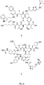
Figure 8 shows the chemical structures of AMBF3-TATE (2), AMBF3-LM3 (3), AMBF3-TOC (6) and AMBP3-JR11 (7). -
Figure 9 shows an example of a no carrier added (NCA) [18F]fluoride ion elution trap and single reaction vessel for isotope exchange. A: 16-gauge needle where the needle has been cut; B: anion-exchange cartridge containing 9 mg of standard QMA resin fitted with the remaining needle point; C: standard rubber septum; D: in temp block: polypropylene vial (sawed-off Falcon tube) that contains AMBF3 precursor. NCA [18F]fluoride ion is trapped on the cartridge, which is then inserted into the septum. The [18F]fluoride ion is eluted with 60 µL isotonic saline into the tube D in which labelling proceeds. All events occur within a fully shielded hot-cell. -
Figure 10 shows (A) 19F-NMR kinetic analysis of the defluoridation from the 19F-AMBF3 moiety (19F-fluoride: 121ppm relative to CFCl3; 19F-AMBF3: 137ppm relative to CFCl3), and (B) a plot of the relative 19F-NMR integration of the 19F-PyBF3 as a function of all compounds (19F-AMBF3 + 19F-fluoride) vs. time. Data are scaled 1000,000 min. Data were fit to a first-order reaction, and the half-life of the corresponding 19F-AMBF3 was calculated to be 19,5000 ± 500 min. -
Figure 11 depicts a representative competitive binding assay for AMBF3-TOC using [125I]-Tyr11-somatostatin-14 as the displaced radioligand. The y-axis represents the amount of radioactivity bound to sstr2a receptors (CPM = counts per minute) and the x-axis represents the logarithmic molar concentration of BF3-TOC. n=3. -
Figure 12 presents (A) a fused PET/CT coronal image showing high tumour and kidney uptake of 18F-AMBF3-TOC, and (B) a maximum-intensity-projection image showing uptake of 18F-AMBF3-TOC in tumour (t), kidneys (k), intestine (i) and bladder (b). Both color bars are calibrated in %ID/g. -
Figure 13 shows tissue uptake of 18F-AMBF3-TOC in %ID/g in certain organs of interest as determined by biodistribution 75 minutes post-injection. Values are displayed as mean + SD. n = 4. - In broad terms this invention relates to somatostatin derivative compounds that may be readily labelled with the isotope fluorine-18 and that have affinity and selectivity for cellular somatostatin receptors. The labelled compounds are useful clinically as radioactive tracers in various in vivo imaging applications (for example, using positron emission tomography (PET) and related techniques) to detect somatostatin-expressing cells and tissues, including tumours.
- The somatostatin derivatives may be based on a variety of somatostatin analogues provided that the selected somatostatin analogue is capable of selectively binding to a somatostatin receptor. In certain embodiments, the somatostatin derivatives are octreotide derivatives. In some embodiments, the invention relates to easily radiolabelled, high-affinity octreotide derivatives for imaging of somatostatin receptors in cancer (for example, neuroendocrine tumours) utilizing the commonly available 18F isotope that is used in PET imaging and used on a daily basis in most medical cyclotrons.
- The unlabelled somatostatin derivatives can be readily labelled by isotope exchange in a single vessel reaction, and free fluoride conveniently removed by simple solid phase extraction (SPE), with no requirement for HPLC purification. Certain embodiments of the invention thus relate to the provision of unlabelled somatostatin derivatives in kit format for labelling just prior to use.
- As shown herein, an exemplary labelled somatostatin derivative compound, 18F-AMBF3-TATE, exhibited higher than expected affinity for somatostatin receptors, together with low liver uptake, resulting in a higher than expected tumour-to-liver ratio. In addition, in certain embodiments of the invention, the trifluoroboronate moiety included in the somatostatin derivative compounds of the present disclosure provides for a simplified labelling due to one or more of: requiring only submilligram quantities of unlabelled precursor compound for labelling; obviating the need for azeotropic fluoride drying through the use of no-carrier-added 18F-fluoride directly for an aqueous labelling reaction; allowing for rapid (for example, less than 30 min) labelling; providing labelled compounds with high specific activity, and/or removing the requirement for HPLC purification as the unlabelled precursor is chemically identical to the product.
- In certain embodiments, the simplified labelling procedure can allow for yields that provide multiple human doses in a single run. In some embodiments, the process is readily amenable to automation and/or microfluidic flow technologies.
- Unless defined otherwise, all technical and scientific terms used herein have the same meaning as commonly understood by one of ordinary skill in the art to which this invention belongs.
- The term "C1-C6 alkyl," as used herein, refers to a substituted or unsubstituted straight chain or branched hydrocarbon of one to six carbon atoms. This term is exemplified by such groups as methyl, ethyl, n-propyl, i-propyl, n-butyl, t-butyl, i-butyl, isopentyl, n-pentyl, hexyl, and the like.
- The term "C3-C6 cycloalkyl" refers to a substituted or unsubstituted cyclic alkyl group containing 3 to 6 carbon atoms.
- The term "C3-C6 aryl" refers to a substituted or unsubstituted aromatic cycloalkyl group having 3 to 6 carbon atoms.
- The term "substituted" when used with one of the foregoing terms indicates that the named group is substituted at one or more positions with a group such as hydroxyl, thiol, alkylthiol, halogen, alkoxy, amino, amido, carboxyl, acyl, carboxyl, nitro or cyano.
- As used herein, the term "about" refers to an approximately +/-10% variation from a given value. It is to be understood that such a variation is always included in any given value provided herein, whether or not it is specifically referred to.
- The use of the word "a" or "an" when used herein in conjunction with the term "comprising" may mean "one," but it is also consistent with the meaning of "one or more," "at least one" and "one or more than one."
- As used herein, the terms "comprising," "having," "including" and "containing," and grammatical variations thereof, are inclusive or open-ended and do not exclude additional, unrecited elements and/or method steps. The term "consisting essentially of" when used herein in connection with a composition, use or method, denotes that additional elements and/or method steps may be present, but that these additions do not materially affect the manner in which the recited composition, method or use functions. The term "consisting of" when used herein in connection with a composition, use or method, excludes the presence of additional elements and/or method steps. A composition, use or method described herein as comprising certain elements and/or steps may also, in certain embodiments consist essentially of those elements and/or steps, and in other embodiments consist of those elements and/or steps, whether or not these embodiments are specifically referred to.
- It is contemplated that any embodiment discussed herein can be implemented with respect to any of the disclosed methods, uses, kits or compositions of the invention, and vice versa.
- The somatostatin derivatives according to the present disclosure are fluorinated compounds of general formula (I):
B-F3-(CH2)n-N+R1R2-L-X (I)
wherein: - R1 and R2 are each independently H, C1-C6 alkyl, C3-C6 cycloalkyl, or C3-C6 aryl;
- L is a linking group;
- X is a somatostatin analogue conjugated via an N-terminal amino group to L, and
- n is 1 or 2.
- Generally, the somatostatin analogue is a peptidic compound that is capable of binding to a somatostatin receptor. In certain embodiments, the somatostatin analogue is capable of binding to the somatostatin receptor subtype 2 (sstr2). In some embodiments, the somatostatin analogue is capable of selectively binding to the somatostatin receptor subtype 2 (sstr2).
- In certain embodiments, in compounds of general formula (I), R1 and R2 are each independently C1-C6 alkyl. In certain embodiments, in compounds of general formula (I), R1 and R2 are each independently C1-C4 alkyl.
- In certain embodiments, in compounds of general formula (I), n is 1.
- One skilled in the art will appreciate that compounds of general formula (I) may be readily prepared by conjugation of an appropriately derivatized trifluoroboronate moiety to either the naturally-occurring amine group at the N-terminus of the somatostatin analogue X, or to an appropriately N-terminally derivatized somatostatin analogue. Accordingly, the nature of the linking group L in the compounds of general formula (I) will be dependent on the method by which the compound was prepared.
- A variety of synthetic chemical groups that will form chemical bonds with primary amines are known in the art. Examples include, but are not limited to, isothiocyanates, isocyanates, acyl azides, NHS esters, sulfonyl chlorides, aldehydes, glyoxals, epoxides, oxiranes, carbonates, aryl halides, imidoesters, carbodiimides, anhydrides, and fluorophenyl ester, most of which conjugate to amines by either acylation or alkylation. In accordance with certain embodiments, therefore, the linking group L may comprise formula (VII):

or formula (VIII):
- In certain embodiments, L in the compounds of general formula (I) comprises formula (VII) in which each of q and r are independently 0 to 10. In some embodiments, L in the compounds of general formula (I) comprises formula (VII) in which each of q and r are independently 0 to 6. In some embodiments, L in the compounds of general formula (I) comprises formula (VII) in which each of q and r are independently 0 to 4.
- In certain embodiments, L in the compounds of general formula (I) comprises formula (VIII) in which s is 1 to 10. In some embodiments, L in the compounds of general formula (I) comprises formula (VIII) in which s is 1 to 6. In some embodiments, L in the compounds of general formula (I) comprises formula (VIII) in which s is 1 to 4.
- Alternative approaches to conjugation of proteins and peptides to other groups are known in the art. For example, chemical modification of proteins is described in G. E. Means and R. E. Feeney, Bioconjugate Chemistry, 1990, 1:2-12.
- In certain embodiments, the somatostatin analogue is conjugated to the trifluoroboronate moiety via copper-catalyzed azide-alkyne cycloaddition ("click" chemistry). In accordance with some embodiments, therefore, the linking group L will comprise a 1,2,3-triazole moiety:

- In some embodiments, linking group L comprises formula (II):

- In certain embodiments, L in the compounds of general formula (I) comprises formula (II) in which m is 1 to 10. In some embodiments, L in the compounds of general formula (I) comprises formula (II) in which m is 1 to 6. In some embodiments, L in the compounds of general formula (I) comprises formula (II) in which m is 1 to 4. In some embodiments, L in the compounds of general formula (I) comprises formula (II) in which m is 1 or 2.
- In some embodiments, L is a linking group of formula (III):

- In certain embodiments, L in the compounds of general formula (I) comprises formula (III) in which m and p are each independently 1 to 8. In some embodiments, L in the compounds of general formula (I) comprises formula (III) in which m and p are each independently 1 to 6. In some embodiments, L in the compounds of general formula (I) comprises formula (III) in which m and p are each independently 1 to 4.
- In certain embodiments, L is a linking group of formula (III) in which m is 1. In some embodiments, L is a linking group of formula (III) in which p is 1.
- In some embodiments, L is:

- Various somatostatin analogues are known in the art and may be used to prepare the fluorinated somatostatin derivative of general formula (I). As noted above, the somatostatin analogues are generally peptidic compounds. By "peptidic compound" it is meant a compound that comprises a sequence of amino acids, which may be naturally-occurring amino acids, non-naturally occurring amino acids or a combination thereof. In some embodiments, the somatostatin analogue may comprise all or a part of a naturally-occurring somatostatin amino acid sequence. In some embodiments, the somatostatin analogue may comprise a part of a naturally-occurring somatostatin amino acid sequence and may further comprise one or more modified amino acids and/or additional amino acid sequences. The one or more modified amino acids may be modified in that the naturally-occurring amino acid is substituted with a different naturally-occurring amino acid, or it may be substituted with a non-naturally occurring amino acid.
- Naturally-occurring human somatostatins include the art-known 14-amino acid and 28-amino acid forms of somatostatin (SEQ ID NOs: 1 and 2, respectively), the sequences of which are known and publicly available from various databases.
- Somatostatin-14: AGCKNFFWKTFTSC [SEQ ID NO:1] (disulfide bridge present between
Cys 3 and Cys 14) - Somatostatin-28: SANSNPAMAPRERKAGCKNFFWKTFTSC [SEQ ID NO:2] (disulfide bridge present between Cys 17 and Cys 28)
- Various non-naturally occurring amino acids are known in the art. Examples include, but are not limited to, D-amino acids (i.e. an amino acid of an opposite chirality to the naturally occurring form), N-α-methyl amino acids, C-α-methyl amino acids, β-methyl amino acids and D- or L-β-amino acids. More specific examples include, but are not limited to, 2-aminobutyric acid (Abu), 4-aminobutyric acid (γ-Abu), 6-aminohexanoic acid (ε-Ahx or Ahx), α-aminoisobutyric acid (Aib), β-alanine (β-Ala), β-aspartic acid (β-Asp), β-cyclohexylalanine (Cha), α-cyclohexylglycine (Chg), citrulline (Cit), diaminobutyric acid (Dab), diaminopimelic acid (Dap), γ-glutamic acid (γ-Glu), pyroglutamic acid (pGlu), homocysteine (Hcy), homoserine (Hse), hydroxyproline (Hyp), N-ε-dinitrophenyl-lysine (Lys(Dnp)), N-ε-methyl-lysine (Lys(Me)), N,N-ε-dimethyl-lysine (Lys(Me2)), N,N,N-ε-trimethyl-lysine (Lys(Me3)), 3-mercaptopropionic acid (Mpa), L-1-napthylalanine (L-1-Nal), L-2-napthylalanine (L-2-Nal), norleucine (Nle), norvaline (Nva), norleucine (Nle), ornithine (Orn), 3-(2-pyridyl)-L-alanine (L-2-Pal), 3-(3-pyridyl)-L-alanine (L-2-Pal), 3-(4-pyridyl)-L-alanine (L-4-Pal), penacillamine (Pen), 4-chlorophenyl-L-alanine (L-4-Cl-Phe), 4-fluorophenyl-L-alanine (L-4-F-Phe), 4-iodophenyl-L-alanine (L-4-I-Phe), 4-nitrophenyl-L-alanine (L-4-NO2-Phe), phenylglycine (Phg), sarcosine (Sar), D-2-methyl-tryptophan (D-2-Me-Trp), phosphor-serine (pSer), phosphor-threonine (pThr), phosphor-tyrosine (pTyr), 11-amino-3.6.9,-trioxa-undecanoic acid (mini-PEG), cysteic acid, cyclohexylalanine, t-butylglycine, t-butylalanine, 3-aminopropionic acid, 2,3-diaminopropionic acid (2,3-diaP), D-2-naphthylalanine (D-2-Nal), 1,2,3,4-tetrahydroisoquinoline-3-carboxylic acid (Tic), octahydroindole-2-carboxylic acid (Oic), α-cyclopentylglycine (Cpg), 2-indanylglycine (Igl), D- or L-2-thienylalanine (Thi), D- or L-3-thienylalanine, D- or L-1-, 2-, 3- or 4-pyrenylalanine, D-(2-pyridinyl)-alanine, D-(3-pyridinyl)-alanine, D- or L-(2-pyrazinyl)-alanine, D- or L-(4-isopropyl)-phenylglycine, D-(trifluoromethyl)-phenylglycine, D-(trifluoromethyl)-phenylalanine, D-p-fluorophenylalanine, D- or L-p-biphenylalanine, D- or L-p-methoxybiphenylalanine, methionine sulphoxide (MSO) and homoarginine (Har). Other examples include substituted β-alanine (β-Ala) comprising one or more substituents selected from arylsulphonyl (such as benzenesulphonyl or 2-naphthalene sulphonyl) and alkoxycarbonyl (such as t-butoxycarbonyl); phosphono- or sulphated (e.g. -SO3H) non-carboxylate amino acids; D- or L-2-indole(alkyl)alanines, and D- or L-alkylalanines, wherein alkyl is substituted or unsubstituted methyl, ethyl, propyl, hexyl, butyl, pentyl, hexyl, octyl, isopropyl, iso-butyl, or iso-pentyl.
- Examples of known somatostatin analogues that may be included in the compounds of general formula (I) in some embodiments include, but are not limited to, octreotide and octreotide derivatives. Examples of octreotide derivatives include, but are not limited to, octreotate, [Tyr3]octreotate (TATE), JR-11, JR-10, LM3, [Tyr3]octreotide (TOC) and [Nal3]octreotide (NOC).
- In certain embodiments, the somatostatin analogue comprised by the somatostatin derivative of general formula (I) is:
- Octreotide: d-Phe-c[Cys-Phe-d-Trp-Lys-Thr-Cys]-Thr-ol;
- Octreotate: d-Phe-c[Cys-Phe-d-Trp-Lys-Thr-Cys]-Thr;
- TATE: d-Phe-c[Cys-Tyr-d-Trp-Lys-Thr-Cys]-Thr;
- JR-11: Cpa-c[d-Cys-Aph(Hor)-d-Aph(Cbm)-Lys-Thr-Cys]-d-Tyr-NH2;
- LM3: p-Cl-Phe-c[d-Cys-Tyr-d-Aph(Cbm)-Lys-Thr-Cys]-d-Tyr-NH2;
- JR-10: p-NO2-Phe-c[d-Cys-Tyr-d-Aph(Cbm)-Lys-Thr-Cys]-d-Tyr-NH2;
- TOC: d-Phe-c[Cys-Tyr-d-Trp-Lys-Thr-Cys]-Thr-ol,
- or NOC: d-Phe-c[Cys-1-Nal-d-Trp-Lys-Thr-Cys]-Thr-ol,
- In certain embodiments, the somatostatin analogue comprised by the somatostatin derivative of general formula (I) is: TATE, LM3, JR-11 or TOC.
- Combinations of any of the foregoing embodiments for compounds of general Formula (I) are also contemplated and each combination forms a separate embodiment for the purposes of the present disclosure.
- In certain embodiments, the compounds of general formula (I) have general formula (Ia):
B-F3-CH2-N+R1R2-L-X (Ia)
wherein: R1, R2, L and X are as defined in any one of embodiments set forth above for general formula (I). - In some embodiments, the somatostatin derivatives are compounds of general formula (Ia) in which R1 and R2 are each independently C1-C6 alkyl. In some embodiments, the somatostatin derivatives are compounds of general formula (Ia) in which R1 and R2 are each independently C1-C4 alkyl. In some embodiments, the somatostatin derivatives are compounds of general formula (Ia) in which R1 and R2 are each independently C1 or C2 alkyl.
- In some embodiments, the somatostatin derivatives are compounds of general formula (Ia) in which R1 and R2 are each independently C1-C6 alkyl, and L is a linking group of formula (II) or (III).
- In certain embodiments, the compounds of general formula (I) have general formula (IV):
B-F3-CH2-N+(Me)2-L-X (IV)
wherein:
L and X are as described in any one of the embodiments set forth above for general formula (I). - In some embodiments, the somatostatin derivatives are compounds of general formula (IV) in which L is a linking group of formula (II) or (III).
- In some embodiments, the somatostatin derivatives are compounds of general formula (IV) in which X is octreotate, TATE, JR-11, JR-10, LM3, TOC or NOC.
- In certain embodiments, compounds of general formula (I) have general formula (V):

- In some embodiments, the somatostatin derivatives are compounds of general formula (V) in which m and p are each independently 1 to 4.
- In some embodiments, the somatostatin derivatives are compounds of general formula (V) in which n is 1.
- In some embodiments, the somatostatin derivatives are compounds of general formula (V) in which X is octreotate, TATE, JR-11, JR-10, LM3, TOC or NOC.
- In certain embodiments, compounds of general formula (I) have general formula (VI):

- For ease of reference, a compound of general formula (VI) may also be referred to herein as AMBF3-X. For example, a compound of general formula (VI) in which the somatostatin analogue X is octreotide may be referred to as AMBF3-octreotide.
- In certain embodiments, the fluorinated somatostatin derivative is selected from: AMBF3-TATE (see
Figure 8 ; compound 2), AMBF3-LM3 (seeFigure 8 ; compound 3), AMBF3-NOC, AMBF3-TOC (seeFigure 8 ; compound 6), AMBF3-JR10 and AMBF3-JR11 (seeFigure 8 ; compound 7). - The fluorinated somatostatin derivatives can be easily labelled with the isotope 18F (for example, by using isotope exchange as shown in
Figure 1 and described in more detail below) to provide the corresponding radioactive tracer compound. - It is to be understood that reference to compounds of general Formula (I) throughout the following disclosure, includes in various embodiments, compounds of general Formulae (IV), (V) and (VI) to the same extent as if embodiments reciting each of these formulae individually were specifically recited.
- The fluorinated somatostatin derivatives of general formula (I) can be readily prepared by standard peptide and synthetic chemistry techniques.
- Somatostatin analogue X may be prepared, for example, by standard solid-phase synthesis methods and derivatized as necessary for conjugation to the trifluoroboronate moiety by standard synthetic organic chemistry techniques.
- The trifluoroboronate moiety may likewise be prepared by standard synthetic techniques from commercially available starting materials. Examples of methods of synthesis for exemplary trifluoroboronate moieties comprising either an alkyne group or an azide group for conjugation with a somatostain analogue are provided in Examples 1 and 2 herein. While the trifluoroboronate moieties described in Examples 1 and 2 have been derivatized to allow for conjugation to an appropriately derivatized somatostatin analogue by click chemistry (as shown, for example, in
Figure 1 ), it will be readily apparent to those skilled in the art that other conjugation methods may be employed, for example, the use of NHS esters, maleimides, and the like, and appropriate trifluoroboronate moieties and somatostatin analogues may be prepared accordingly. - The fluorinated somatostatin derivatives can subsequently be labelled with 18F by simple isotope exchange using standard protocols (see, for example, Example 1). In certain embodiments, the isotope exchange may be carried out in a single reaction vessel with subsequent removal of excess fluoride ions by SPE, for example, on a Sep-Pak® or similar cartridge, without the need for HPLC purification.
- Certain embodiments of the invention relate to the use of the fluorinated somatostatin derivatives, once labelled with 18F, as radiotracers. Some embodiments thus also relate to the use of the non-labelled fluorinated somatostatin derivatives in the preparation of radiotracers. In certain embodiments, the facile labelling of the fluorinated somatostatin derivatives by isotope exchange will allow for the radiotracers to be readily prepared on site in facilities, such as hospitals and clinics, which have access to a cyclotron for generation of 18F-fluoride.
- As demonstrated herein, exemplary fluorinated somatostatin derivatives of general formula (I) exhibit high affinity for somatostatin receptors. Accordingly, when labelled with 18F, the fluorinated somatostatin derivatives of general formula (I) are useful for in vivo imaging applications, for example PET imaging applications, to image cells and tissues expressing somatostatin receptors including, but not limited to in vivo imaging of neuroendocrine tumours.
- In some embodiments, 18F-labelled fluorinated somatostatin derivatives of general formula (I) may find use as PET imaging agents for imaging cancer, including cancers that express somatostatin receptors. Examples of such cancers, include but not limited to, neuroendocrine tumours, breast cancers, small cell lung cancer, lymphomas, meningiomas, pituitary adenomas and pancreatic cancer.
- In some embodiments, it is contemplated that the fluorinated somatostatin derivatives of general formula (I), when the fluorine atoms are present as the 19F isotope, may find use as a therapeutic agents for treatment of diseases or disorders characterized by expression or overexpression of somatostatin receptors. Examples of such diseases or disorders include, but not limited to, neuroendocrine tumours, breast cancers, small cell lung cancer, lymphomas, meningiomas, pituitary adenomas and pancreatic cancer.
- The fluorinated somatostatin derivatives of general formula (I) may also find use as research reagents, for example, in research into the role of somatostatin receptors in certain diseases or conditions.
- As the fluorinated somatostatin derivatives of general formula (I) are generally stable and can be readily labelled in a single reaction vessel, certain embodiments of the invention relate to kits comprising a fluorinated somatostatin derivative of general formula (I) for the preparation of a radiolabelled tracer. The kit may also include instructions for use, which may be provided in paper form or in computer-readable form, such as a disc, CD, DVD or the like, and may further comprise a notice in the form prescribed by a governmental agency regulating the manufacture, use or sale of pharmaceuticals or biological products, which notice reflects approval by the agency of manufacture, for use or sale for human or animal administration.
- The kit may comprise one or more containers containing a pre-determined amount of the fluorinated somatostatin derivative. Typically, the container will be a one that can be used directly in the isotope exchange reaction, for example, a vial or tube made of polypropylene or other suitable material. The amount of the fluorinated somatostatin derivative in each container may be an amount suitable to provide a single dose of the final radiolabelled tracer, or it may be an amount suitable to provide multiple doses of the radiolabelled tracer.
- In certain embodiments, one or more of the components of the kit can be lyophilized and the kit can additionally contain a suitable solvent for reconstitution of the lyophilized components.
- The kit may optionally comprise other components for use in the radiolabelling reaction, such as one or more of, solvents, sealing means for the container (such as a septum or other air-tight seal), syringes, needles, SPE cartridges or columns containing a suitable sorbent for trapping [18F]fluoride ion or for removal of free fluoride ion from the radiolabelled tracer.
- To gain a better understanding of the invention described herein, the following examples are set forth. It will be understood that these examples are intended to describe illustrative embodiments of the invention and are not intended to limit the scope of the invention in any way.
- Reagents and solvents were purchased from Advanced Chemtech, Sigma-Aldrich, Combi-Blocks, or Novabiochem. The AR42J cell line was purchased from ATCC. 18F-fluoride Trap & Release Columns were purchased from ORTG Inc., and C18 Sep-Pak cartridges (1 cm3, 50 mg) were obtained from Waters. An
Endeavor 90 peptide synthesizer (Aapptec) was applied to synthesize the peptide. Electron-spray ionization low-resolution mass spectroscopy was performed on a Waters ZQ with a single quadrupole detector, attached to a Waters 2695 high-performance liquid chromatography (HPLC) column. All nuclear MR spectra were recorded at room temperature on a Bruker Avance 300 MHz spectrometer. - The following HPLC methods were used for purification and quality control. Method A: Agilent Eclipse XDB-C18 5-mm 9.2 x 250 mm semipreparative column; solvent A, 0.1% trifluoroacetic acid (TFA) water; solvent B, MeCN; 0-15 min, 20%-40% B; 15-20min, 40%-20% B; flow rate, 4.5mL/min; column temperature, 19°C-21°C. Method B: Agilent Eclipse XDB-C18 5-mm 9.2 x 250 mm semipreparative column; solvent A, 0.1% TFA water; solvent B, MeCN; 0-2min, 5%-20% B; 2-5 min, 20%-30% B; 5-20 min, 30%-50%; 20-22 min, 50%-5% B; flow rate, 3 mL/min; column temperature, 19°C-21°C. Method C: Phenomenex Jupiter 10-mm C18 300-A° 4.6 x 250 mm analytic column; solvent A, 0.1% TFA water; solvent B, MeCN; 0-2 min, 5%-5% B; 2-7min, 5%-20% B; 7-15 min, 20%-100%; 15-20min, 100%-5% B; flow rate, 2 mL/min; column temperature, 19°C-21°C.
- To synthesize the precursor for labelling, a suitable TATE was first synthesized as previously described (Lewis JS, et al., Nucl Med Biol. 1999, 26:267-273) and converted to an azide derivative. The resin (Fmoc-Thr(tBu)-Wang) and growing chain were treated with 20% piperidine (1 x 5 min and 1 x 10 min) in N,N-dimethylformamide to remove the Nα-Fmoc protecting group. The amino acids (3 equivalents [eq.] per coupling) Fmoc-Cys(Acm)OH, Fmoc-Thr(tBu)-OH, Fmoc-Lys(Boc)-OH, Fmoc-D-Trp(Boc)-OH, Fmoc-Tyr(tBu)-OH, and Fmoc-D-Phe-OH were subsequently coupled in nuclear matrix protein with the standard in situ activating reagent O-benzotriazole-N,N,N9,N9-tetramethyl-uronium-hexafluoro-phosphate (HBTU) (3eq.) in the presence of diisopropylethylamine (6 eq.). Thallium (III) trifluoroacetate (2eq.) in N,N-dimethylformamide deprotected the cysteines and concomitantly induced disulfide formation. Bromoaceticacid (40 eq.) was preactivated with diisopropylcarbodiimide (20eq.) in dichloromethane for 10 min, filtered, and coupled to the N-terminus. NaN3 (27.5 eq.) in dimethyl sulfoxide was added. The peptide was deprotected and simultaneously cleaved from the resin in 90:2.5:2.5:5 TFA:H2O:triisopropylsilane:phenol for 4 h at room temperature. TATE-N3 was purified by HPLC with a semipreparative column using method A to afford pure TATE-N3 in quantities of about 10 mg. The calculated mass was 1,131.2, and the measured mass by electrospray ionization was 1,131.4. The purity of the peptide was greater than 99%.
- N-propargyl-N,N-dimethyl-ammoniomethylboronylpinacolate (alkynyl-AMB(pin)) was first synthesized by condensation of iodomethylboronylpinacolate and propargylamine as previously described (Matteson DS, et al., J Organomet Chem. 1979, 170:259-264). Briefly, a dry round-bottomed flask was charged with N,N-dimethylpropargylamine (98 µL, 1.0 mmol) and 2 mL of anhydrous diethyl ether under argon, to which iodomethyl-boronylpinacolate (165 µL, 0.9 mmol) was added dropwise at room temperature. On stirring, the solution became cloudy and the desired product was collected as a white precipitate that was filtered and washed with cold Et2O and then dried under high vacuum to give a fluffy white powder in 95% yield. 1H nuclear MR (300 MHz [Bruker], CD3CN): d 4.40 (d, 2H), 3.31 (s, 2H), 3.22 (s, 6H), 3.21 (t, 1H), 1.27 (s, 12H); electrospray ionization: calculated, 224.1; found, 224.1.
- N-propargyl-N,N-dimethylammonio-methylboronylpinacolate (5.0 mg, 22.3 µmol) was converted to the trifluoroborate (alkynyl-AMBF3) through the addition of KHF2 (3 M, 30 µL in water), HCl (4 M, 30 µL in water), deionized water (20 µL), and N,N-dimethylformamide (60 µL), 45°C, 2 h, and then quenched by NH4OH (concentration, 10 µL). 19F NMR confirmed very slow solvolysis of the alkynyl-AMBF3 (t1/2: 13 ± 0.3 days) (
Figure 10 ). - The crude reaction from preparation of the alkynyl-AMBF3 above was directly used for click conjugation to TATE-azide without further purification: a mixture of TATE-azide (4.0 mg, 3.4 µmol), CuSO4 (1.0 M, 5.0 µL), sodium ascorbate (1.0 M, 12.5 µL), and 5% NH4OH (1:1 MeCN:H2O, 50 µL) was added, and the mixture was heated to 45°C for 2 h. Purification was performed with method B to isolate 2.3 mg of AMBF3-TATE. Purity was confirmed with liquid chromatography-mass spectrometry (calculated, 1,296.5; obtained, 1,296.4). The purified 19F-AMBF3-TATE was diluted in ethanol and portioned into aliquots of approximately 60 µg (50 nmol) for radiolabelling in kit-like fashion.
- After successful synthesis, the activity of 19F-AMBF3-TATE was examined in vitro. Membranes from Chinese hamster ovary K1 cells transfected with sstr1, sstr2, sstr3, sstr5, and [125I]-Tyr-somatostatin-14 were obtained from PerkinElmer. A standard filtration binding assay was performed in 96-well plates (MultiScreen; Millipore) to determine the binding affinities (inhibition constant, or Ki) of AMBF3-TATE against different receptor subtypes. Briefly, membranes (0.25 µL/well) were incubated with the 125I-labelled standard at a concentration of 0.05 nM for sstr2 or 0.2 nM for other subtypes. Increasing concentrations of AMBF3-TATE were added to the wells in buffer (25 mM N-2-hydroxyethylpiperazine-N-2-ethanesulfonic acid, pH 7.4; 10 mM MgCl2; 1 mM CaCl2; and 0.5% bovine serum albumin). After incubation (37°C, 1 h), the wells were aspirated and washed 8 times with 50 mM ice-cold buffer (Tris-HCl, pH 7.4) over grade GF/B filters. The filters were removed and counted by a γ counter (Cobra II; Packard). A typical competition curve is shown in
Figure 2 (n=3). Data were fitted to a 1-site competition model (GraphPad Prism 6.1 software) to calculate Ki. - For 18F labelling, AMBF3-TATE (50 nmol) was resuspended in aqueous pyridazine-HCl buffer (∼50 µL, pH 2) in a vial (polypropylene Falcon Tube; Corning) just before labelling. No-carrier-added 18F-fluoride, 29.6-37 GBq (800-1,000 mCi), was obtained by bombardment of H2 18O with 18-MeV protons, followed by trapping on an anion exchange resin (9 mg, quaternary ammonium, chloride form, prewashed with deionized water). The 18F-fluoride was eluted with 70-100 µL of isotonic saline into the reaction vial containing AMBF3-TATE. The vial was placed in a heating block set at 80°C for 20 min, whereupon the reaction was quenched by the injection of 2 mL of 5% NH4OH in water. The reaction mixture was loaded onto a C18 light cartridge that was preconditioned by wetting with MeCN and washing with distilled water. Impurities (e.g., 18F-fluoride, pyridazine) were removed by flushing with 2 mL of saline. Radiochemically pure 18F-AMBF3-TATE was released into a glass vial with 0.5 mL of 1:1 ethanol:saline to provide 7.4 GBq (200 mCi) of tracer. This solution was formulated in isotonic saline (5 mL). A small sample was removed for quality control analysis by HPLC with mass detection at 277 nm (
Figure 3 ). - Radiochemically pure 18F-AMBF3-TATE, formulated in saline, was assayed for plasma stability. For this assay, 20 µL of 18F-AMBF3-TATE were added to mouse plasma (500 µL) and incubated at 37°C for 0, 60, and 120 min. After incubation at each time point, the reaction was quenched by adding 1 mL of MeCN to precipitate insoluble proteins from the solution. The quenched reactions were centrifuged to remove insoluble material. The supernatant was aspirated, filtered, and analyzed by HPLC using method C.
- After labelling, imaging was undertaken. All animal studies were performed in accordance with the Canadian Council on Animal Care guidelines and were approved by the animal care committee of the University of British Columbia. Rat pancreatic adenocarcinoma cells (107 AR42J cells) were freshly expanded in a mixture of phosphate-buffered saline and Matrigel (Corning) and inoculated subcutaneously in female immunocompromised mice (NOD SCID [non-obese diabetic severe combined immunodeficient] IL2r-γ-null, bred in house). The tumours were grown for 2 wk until they reached 5-7 mm in diameter. While under 2% isoflurane anesthesia, the mice were injected via the tail vein with 0.37-0.74 MBq (10-20 µCi, 4-8 pmol) of 18F-AMBF3-TATE (n=5). To demonstrate the specificity of in vivo uptake in receptor-positive tissues, 100 µg of 69/71Ga-DOTATATE were pre-injected 15 min before 18F-AMBF3-TATE injection as a blocking control cohort (n= 4). Sixty minutes after injection, the mice were anesthetized with isoflurane and euthanized by carbon dioxide. The organs were harvested, rinsed with saline, blotted dry, and collected in previously weighed tubes. The tubes containing the organs were counted in a Cobra-II γ counter. The tissue weight and associated count per minute were used to calculate the percentage injected dose per gram of tissue (%ID/g). Images were acquired using a multimodality PET/CT system (Inveon; Siemens).
- Approximately 3.7 MBq (100 µCi, ∼40 pmol) of radiotracer were injected in the caudal lateral tail vein of tumour-bearing mice. Sixty minutes after radiotracer injection, the animals were anesthetized with isoflurane inhalation and a baseline low-dose CT scan was obtained for localization and attenuation correction, followed by a static PET scan acquired for 10 min. The mice were kept warm by a heated pad on the scanner bed during acquisition. 69/71Ga-DOTATATE (100 µg per mouse) was pre-injected as a blocking agent. The images were reconstructed by an iterative reconstruction algorithm (3-dimensional ordered-subsets expectation maximization/maximum α posteriori) using the Inveon Acquisition Workplace Software (Siemens), applying normalization, dead time, random, and attenuation corrections. Uptake into tumour and tissues of interest was determined by regions of interest, and %ID/g was calculated (assuming a tissue density of 1.0 g/cm3). The mean %ID/g was calculated from a region of interest that matched the tumour contours on CT. The peak %ID/g was calculated from the hottest 2 x 2 voxel cluster within the tumour. In 1 animal, a dynamic scan was acquired in list mode for 60 min under continuous isoflurane inhalation, starting concurrently with radiotracer injection. Imaging data from this mouse were not combined with the results of static imaging and were used to obtain the tissue time-activity curves reported in
Figure 4 . - The Ki of AMBF3-TATE using human sstr2 receptors expressed on Chinese hamster ovary membranes was 0.13 ± 0.03 nM. Using identical assay conditions and the same lot of membranes, the Ki for gallium-DOTATATE was 0.7 ± 0.2 nM. A representative competitive binding assay curve is shown in
Figure 2 . No significant displacement of binding to sstr1 was observed. The inhibition constants for sstr3 and sstr5 were 28.4 ± 8.6 nM and 11.6 ± 2.8 nM, respectively. - Starting with 29.6-37 GBq of no-carrier-added 18F-fluoride (800-1,000 mCi), approximately 7.4 GBq of 18F-AMBF3-TATE were obtained within 25 min (24% ± 4%, n=5) and reinjected into HPLC for quality analysis (
Figure 3 ), which showed a single peak in both radioactive and ultraviolet modes. Because approximately 7.4 GBq (∼200 µCi) of 2 were obtained starting with 50 nmol of precursor, the specific activity was 148 GBq/µmol (3 Ci/µmol), with a radiochemical yield of 20%-25% (not corrected for decay). To validate this calculation, a standard curve showed that the specific activity was more than 111 GBq/µmol (.3 Ci/µmol). 18F-AMBF3-TATE was incubated in mouse plasma for 120 min with no detectable decomposition (Figure 5 ). - Uptake in the AR42J tumours was intense and clearly specific as evidenced by the lack of uptake in the tumours of mice receiving unlabelled competitor (
Figure 6 ). The average of the tumour uptake based on the whole tumour region of interest was 10.2 ± 2.1 %ID/g. The average of the peak tumour uptake based on the hottest voxel cluster was 23.6 ± 3.0 %ID/g. In contrast, the average uptake in the liver, blood pool, and muscle was 0.83 ± 0.16, 0.47 ± 0.12, and 0.09 ± 0.03 %ID/g, respectively. Excretion was predominantly renal, with significant clearance to the bladder and low kidney retention. Some hepatobiliary tract excretion was notably rapid, leading to high tumour-to-liver ratios. Bone uptake was undetectable, and there was low background activity in blood and muscle, resulting in high-contrast images. - Time-activity curves of uptake in tumour and other tissues from a tumour-bearing mouse are presented in
Figure 4 . Time-dependent tumour uptake increased to a peak voxel cluster value of approximately 40 %ID/g in a mouse with a fairly large tumour. Uptake in non-target tissues rapidly declined after reaching the peak value at a time point soon after intravenous administration. - The ex vivo biodistribution data of 18F-AMBF3-TATE at 1 h (see Table 1) corroborate the scanning data. The relative uptake values in representative tissues are shown in
Figure 7 . Uptake in AR42J xenograft tumours in the unblocked mice was 10.11 ± 1.67 %ID/g. As expected, excess competitor caused a substantial reduction in tumour uptake: 0.32 ± 0.21 %ID/g. Hence, blocking efficiency was 97%. Uptake values in blood and muscle were low: 0.40 ± 0.31 %ID/g and 0.11 ± 0.03 %ID/g, respectively, which gave high tumour-to-blood and tumour-to-muscle ratios of 25.1 ± 1.0 and 89.0 ± 3.1, respectively. Bone uptake was negligible (0.46 ± 0.17 %ID/g), indicating no in vivo defluorination.Table 1: Biodistribution of 18F-AMBF3-TATA (%ID/g) Unblocked Blocked Tissues Average SD Average SD Blood 0.40 0.31 0.32 0.15 Plasma 0.72 0.71 0.92 0.16 Uterus 0.26 0.05 0.51 0.11 Large intestine 2.28 2.64 4.27 6.20 Small intestine 3.23 1.58 1.82 1.70 Spleen 0.42 0.19 0.31 0.11 Liver 0.39 0.05 0.41 0.14 Pancreas 2.81 1.49 0.20 0.01 Adrenal glands 0.54 0.18 0.28 0.07 Kidney 4.90 1.54 4.50 3.54 Lungs 1.85 0.83 0.79 0.26 Heart 0.17 0.05 0.88 1.12 Tumour 10.11 1.67 0.32 0.21 Muscle 0.11 0.03 0.11 0.09 Bone 0.46 0.17 0.54 0.36 Brain 0.03 0.01 0.22 0.33 Tail 0.28 0.06 0.38 0.09 - Isotope exchange on the organotrifluoroborate prosthetic greatly simplified labelling on several accounts. First, only submilligram quantities of precursor were needed for labelling. Second, no time-consuming azeotropic fluoride drying was required, because no-carrier-added 18F-fluoride was eluted in isotonic saline and used directly for an aqueous labelling reaction. Third, labelling was rapid (∼20 min) and provided for high specific activity. Fourth, because the precursor is chemically identical to the product, time-consuming HPLC purification was obviated in favor of a simple C18 Sep-Pak elution to remove free fluoride. Besides the methodologic simplicity, the yields provide multiple human doses in a single run. In light of improvements in cyclotron output to provide multiple-curie levels of 18F-fluoride (Eberl S, et al., Appl Radiat Isot. 2012, 70:922-930), this methodology is readily applicable to existing production facilities. Moreover, the simplicity of the process should be easily amenable to automation and microfluidic flow technologies.
- Whereas good radiosynthetic attributes are a prerequisite for use, the real value of a given tracer ultimately lies in the in vivo image data and corroborating biodistribution data. There is an extensive body of literature detailing the labelling and imaging of various TATE analogues by SPECT or PET. A brief comparison of the representative radiolabelled TATE derivatives is featured in Table 2. Comparison of receptor binding affinities is difficult because many authors report an inhibitory concentration of 50%, which is dependent on assay conditions. Among the published TATE-based radiotracers, gallium-DOTATATE has the highest affinity for sstr2 reported to date. Under identical conditions, 18F-AMBF3-TATE showed higher affinity than gallium-DOTATATE. This finding was both unanticipated and significant. The sensitivity of somatostatin analogues, either agonists or antagonists, to substitutions at the N-terminus and to the radiometal is well documented (Fani M, et al., J Nucl Med. 2012, 53:1481-1489; Reubi JC, et al., Eur J Nucl Med. 2000, 27:273-282). In the present Example, the octreotate was modified with the AMBF3 prosthetic via copper-catalyzed click conjugation, and a decrease in the binding affinity to the sstr2 was expected. Instead, a more than 5-fold higher binding affinity than for 68Ga-DOTATATE as assayed under identical conditions was observed.
Table 2: Comparison of Some Octreotide and Octreotate Derivatives Ligand Synthesis Time (min) Specific Activity (GBq/µmol) HPLC Purification Tumour Type Tumour %ID/g 111In-DTPA-pentetreotide1 >60 44.2 No AR42J 0.99* 99mTc- depreotide 120 37 No CA20948 4.81§ 111In-DOTA-TATE2 >60 15.9 No AR42J 4.12* 99mTc-EDDA/HYNIC- TATE 220 60 No AR42J 5.01 64Cu-TE1A1P-Y3-TATE3 >60 48 No AR42J 5.11 18F-FETE-PEG- TOCA 490 5.9 Yes AR42J 5.14 18F-FET-bAG- TOCA 490 3.9 Yes AR42J 8.23 18F-FET-bAG-[W-c-K]4 90 12.3 Yes AR42J 0.11 18F-AIF-NOTA-OC5 45 36.1 No AR42J 6.43 18F-SiFA-Tyr3-OC6 <30 29-56 No AR42J 7.73 68Ga-DOTA-TATE7 45 Not given No AR42J 2.75 18F-AMBF3-TATE <30 111 No AR42J 10.11 * 4-h time point
§ 90-min time point
1 Vallabhajosula S, et al., J Nucl Med. 1996, 37:1016-1022; 2 Storch D, et al., J Nucl Med. 2005, 46:1561-1569; 3 Guo Y, et al., Bioconjug Chem. 2012, 23:1470-1477; 4 Leyton J, et al., J Nucl Med. 2011, 52:1441-1448; 5 Laverman P, et al., J Nucl Med. 2010, 51:454-461; 6 Wangler C, et al., Bioconjug Chem. 2010, 21:2289-2296; 7 Poeppel TD, et al., J Nucl Med. 2011, 52:1864-1870. - The inhibition constants to sstr3 and sstr5 also appeared to be lower than the values published for Ga-DOTATATE, suggesting that other zwitterionic moieties at the N-terminus may improve affinity. On the basis of PET/CT imaging, 18F-AMBF3-TATE exhibited low background activity in non-target tissues and high receptor-mediated uptake in a preclinical murine model of sstr2-positive cancer. Corroborating the in vivo imaging data, ex vivo biodistribution verified the high tumour uptake values. Although liver uptake of radiometallated octreotides typically is low, this is not always the case for 18F-labelled octreotides. Liver uptake, and in particular nonspecific uptake, is often observed and may preclude clinical detection of liver metastasis. Interestingly, the liver uptake of 18F-AMBF3-TATE was low, resulting in a higher tumour-to-liver ratio (26.21 ± 0.79 1 h after injection) than has been reported for other 18F-labelled TATE analogues (0.25-5.0 2 h after injection) (Leyton J, et al., J Nucl Med. 2011, 52:1441-1448; Wängler C, et al., Bioconjug Chem. 2010, 21:2289-2296).
- A plasma stability assay (37°C) showed negligible decomposition of 18F-AMBF3-TATE after 120 min. Consistent with this finding, minimal bone uptake was observed in both PET/CT and biodistribution, resulting in a high tumour-to-bone ratio of up to 21.3 ± 3.6. This low, nonspecific bone uptake is particularly encouraging for the detection of bone metastasis.
- This Example reports high-affinity octreotate-organotrifluoroborate conjugate that was radiolabelled with 18F in high yield and high specific activity via a facile isotope exchange reaction using minute quantities of precursor peptide, without HPLC purification. This methodology provides for rapid, multi-dose tracer production in a single run that should be amenable to automation. In addition to radiosynthetic ease, the biologic evaluation of 18F-AMBF3-TATE indicated that this tracer provides good stability, optimal pharmacokinetics, excellent binding affinity, and high tumour-to-nontarget-tissue ratios for in vivo imaging.
- Azidoethyl-AMBF3 (compound 5) was prepared as shown generally in Scheme 1:

- N,N-dimethyl-2-azidoethylamine 4 (114 mg, 1.0 mmol) was dissolved in anhydrous diethyl ether (5 mL) in a flame-dried round bottom flask. At room temperature, iodomethylboronyl pinacolate (182 µL, 1.0 mmol) was added drop-wise over 5 min. The alkylated product precipitated as a fluffy white powder, which was separated by filtration and dried under vacuum. The pincaolate was then converted to the corresponding trifluoroborate through the addition of KHF2 (3 M, 300 µL in water) and HCl (4M, 300 µL in water) along with deionized water (200 µL) and DMF (600 µL) at 45°C for 2 hours, and then quenched by NH4OH (conc., 10 µL). Free fluoride was removed by passing the reaction mixture through silica gel to give
compound 5. ESI: [M-F]+ calculated: 177.09; obtained: 177.1; HRMS [M+]: calculated: 218.1041; found: 218.1041.Compound 5 was used without further purification for condensation with peptides. - For labelling, a wet NCA (no carrier added) solution of [18F]fluoride ion was used directly following trapping. Using disposable labware, a very small QMA cartridge (9 mg resin) was affixed to the reaction vessel (10 mL polypropylene conical tube), as shown in
Figure 9 . The very small QMA column efficiently traps Curie levels of NCA [18F]fluoride ion, which was directly eluted with <60 µL saline into the reaction vessel containing the AMBF3-bioconjugate to be labelled by isotope exchange (IEX). Once mixed (10 s), the reaction vessel was placed in a heating block at 80°C. After 15 min, the reaction was quenched with 2 mL PBS or 2 mL NH4OH and the entire reaction mixture was directly loaded onto a C18 Sep-Pak column. Following water wash (5 mL) the labelled compound was eluted into PBS/ethanol (2 mL). - Azidoacetyl-LM3 was synthesized via the Nα-Fmoc solid-phase peptide synthesis strategy starting from D-Tyr-Rink Amide MBHA resin. The resin was treated with 20% piperidine in DMF to remove the Nα-Fmoc protecting group. The following Fmoc-protected amino acids (3 equivalents) including Fmoc-Cys(Acm)-OH, Fmoc-Thr(tBu)-OH, Fmoc-Lys(Boc)-OH, Fmoc-D-Phe(Cbm)-OH, Fmoc-Tyr(tBu)-OH, Fmoc-D-Cys(Acm)-OH, Fmoc-Cl-Phe-OH were subsequently coupled to the sequence in correct order. The coupling was carried out in NMP with standard in situ activating reagent HBTU/HOBT (3 equivalents) in the presence of DIEA (6 equivalents). Cyclization was performed by incubation of the resin with 2 equivalents of thallium(III) trifluoroacetate in DMF at room temperature for 90 min. Bromoacetic acid (40 equivalents) was pre-activated with DIC (20 equivalents) in DCM for 10 min, filtered, and then coupled to the peptide sequence. Finally, the resin was treated with sodium azide (27.5 equivalents) in DMSO to provide the azide functional group at the N-terminus for click reaction.
- The peptide was de-protected and simultaneously cleaved from the resin by the treatment of a cocktail of trifluoroacetic acid/water/triisopropylsilane (95:2.5:2.5). After filtration, the peptide was precipitated by the addition of cold diethyl ether to the TFA solution. The crude product was filtered, dried, and purified by HPLC (Luna C18 semi-prep column, 4.5 mL/min, 20-35% MeCN (0.1% TFA) in Water (0.1% TFA) in 30 min, RT = 30). Azidoacetyl-LM3 was obtained in 15% yield. ESI-MS: calculated for Azidoacetyl-LM3 C55H68ClN15O13S2 1245.4, found [M+H]+ 1246.9.
- Propargyl-AMBF3 (
compound 1,Figure 1 ) was prepared as described in Example 1, and conjugated to azidoacetyl-LM3 as described in Example 1 for TATE-azide to give AMBF3-LM3 (compound 3,Figure 8 ). Labelling as described in Example 3 gave radiochemically pure 18F-AMBF3-LM3 (ca. 200 mCi) in 20 min in comparable yields to AMBF3-TATE at high specific activity (ca. 3 Ci µmol-1). 18F-AMBF3-LM3 also showed specific and very high tumour uptake in AR42J pancreatic xenograft tumours in mice. Unbound tracer cleared rapidly through the kidneys with minimal uptake in liver and negligible uptake in bone. - Azidoacetyl-TOC was synthesized via the Nα-Fmoc solid-phase peptide synthesis strategy starting from H-Threoninol(But)-2-ClTrt resin. The following Fmoc-protected amino acids (3 equivalents) including Fmoc-Cys(Acm)-OH, Fmoc-Thr(tBu)-OH, Fmoc-Lys(Boc)-OH, Fmoc-D-Trp(Boc)-OH, Fmoc-Tyr(tBu)-OH, Fmoc-Cys(Acm)-OH, Fmoc-D-Phe-OH were subsequently coupled to the sequence in correct order. The coupling was carried out in NMP with the standard in situ activating reagent HBTU/HOBT (3 equivalents) in the presence of DIEA (6 equivalents). Cyclization was performed by incubation of the resin with 2 equivalents of thallium (III) trifluoroacetate in DMF at room temperature for 90 min. Finally, azidoacetic acid (10 equivalents) was pre-activated with DIC (5 equivalents) in DCM for 10 min, filtered, and then coupled to the peptide sequence to provide the azide functional group at the N-terminus for the subsequent click reaction. The peptide was de-protected and simultaneously cleaved from the resin by the treatment with a mixture of trifluoroacetic acid:water:triisopropylsilane (95:2.5:2.5). After filtration, the peptide was precipitated by the addition of cold diethyl ether to the TFA solution. The crude product was filtered, dried, and purified by HPLC (Luna C18 semi-prep column, 4.5 mL/min, 30% MeCN (0.1% TFA) in water (0.1% TFA), RT = 18). Azidoacetyl-TOC was obtained in 16% yield. ESI-MS: calculated for azidoacetyl-TOC C51H67N13O12S2 1117.45, found [M+H]+ 1118.0.
- An Eppendorf tube (1.5 mL) was charged with a mixture of N-propargyl-N,N-dimethylammonio-methyltrifluoroborate (1.03 mg, 6.3 µmol), CuSO4 (1.0 M, 5.0 µL), sodium ascorbate (1.0 M, 12.5 µL), 5% NH4OH (MeCN/H2O = 1:1, 50 µL) and azidoacetyl-TOC (3.5 mg, 3.13 µmol). The mixture was warmed up to 45°C for 2 h. Purification was performed by HPLC using the following conditions: Luna C18 semi-prep column, 4.5 mL/min, 26% MeCN (0.1% TFA) in water (0.1% TFA), RT = 17, to isolate 3.6 mg of AMBF3-TOC (
compound 6,Figure 8 ) (54%). ESI-MS: calculated for AMBF3-TOC C57H78BF3N14O12S2 1282.5, found [M+H]+ 1283.6. - 19F-AMBF3-TOC (100 nmol) was dissolved in a mixture of aqueous pyridazine-HCl buffer (12 µL, 1M, pH = 2) and DMF (15 µL) in a 6-mL Falcon tube. No carrier-added 18F-fluoride was obtained by bombarding H2 18O with 18 MeV protons, followed by trapping on a 18F-fluoride Trap & Release Column. The 18F-fluoride was eluted from the column with 70 µL saline into the Falcon tube containing 19F-AMBF3-TOC. The tube was placed in a heating block, and heated at 70°C for 20 min. The reaction mixture was subsequently quenched with 5% aqueous NH4OH (2 mL), and loaded onto a C18 light Sep-Pak cartridge. Free 18F-fluoride was removed by washing the Sep-Pak cartridge with deionized water (2 mL × 2). 18F-AMBF3-TOC was then eluted from the cartridge with 9:1 ethanol:saline (0.4 mL), and diluted with saline for in vitro plasma stability, biodistribution and PET/CT imaging studies. A small sample was removed for quality control analysis by HPLC (Luna C18 semi-prep column, 4.5 mL/min, 26% MeCN (0.1% TFA) in water (0.1% TFA), RT = 18). The decay-corrected radiochemical yield was 17 ± 1 % and radiochemical purity was 94 ± 2 % (n = 3). The LogD (7.4) of 18F-AMBF3-TOC was -1.6±0.01.
- The stability of 18F-AMBF3-TOC was assessed in mouse plasma and analyzed by HPLC as described in Example 1. More than 88% and 63% of 18F-AMBF3-TOC remained intact after 1 h and 2 h incubation at 37°C, respectively.
- The binding affinity of AMBF3-TOC was analysed in vitro using competitive binding assays. Purified membranes from Chinese hamster ovary K1 cells transfected with sstr2a (Perkin Elmer) were co-incubated with [125I]-Tyr11-somatostatin-14 (Perkin Elmer) at increasing concentrations of BF3-TOC. The assay was performed in a 96-well filter plate (Millipore) with a 1.2 µL pore size. Each well contained 0.213 µg of membrane, 0.05 nM of radioligand and various concentrations of BF3-TOC (10-13 to 10-5 M) all dissolved in buffer (25 mM 4-(2-hydroxyethyl)-1-piperazineethanesulfonic acid, pH 7.4, 10 mM MgCl2, ImM CaCl2, 0.5% BSA) and incubated for 60 min at 27°C. After incubation, wells were aspirated and washed 6 times with 200 µL wash buffer (50 mM Tris-HCl pH 7.4, 0.2% BSA). Filters were removed and counted in a 2480 Automatic Gamma counter (Perkin Elmer). The inhibition constant (Ki) was calculated by fitting data to a 1-site competition model (GraphPad Prism 6.05). All samples were performed in triplicates (n=3).
- All animal studies were done in accordance with the Canadian council on animal care guidelines and approved by the animal care committee at the University of British Columbia. Tumour-bearing NOD scid gamma (NSG) mice (bread in house) were used for PET/CT imaging (n=2) and biodistribution (n=4). 6-11 days prior to tumour inoculation, slow release estrogen pellets (17 β-estradiol, IRA) were implanted subcutaneously on the posterior of mice, accompanied with Metacam (Boehringer Ingelheim) analgesia at a dose of 2 mg/kg. Metacam was further administered at 1 mg/kg once a day for the following two days. Cells from the ZR-75-1 breast cancer model were implanted subcutaneously on the right shoulder and grown 5-6 weeks until tumours were 100-300 mm3. A total of 3.3 x 10⁶ cells were implanted in a 1:1 solution of matrigel (Corning) and phosphate buffered saline (total volume= 100 µL).
- The mice used for PET/CT imaging (n = 2) were sedated with 2% isoflurane prior to receiving a [18F]-BF3-TOC radiotracer injection of 5.77-7.28 MBq (577 - 728 µCi) via the lateral tail vain. They were allowed to recover and given approximately 40 minutes of uptake time, until they were sedated once again and placed in the scanner. A baseline CT scan was obtained prior to PET for localization and attenuation correction. Subsequently, a 10 minute single static emission PET scan was acquired exactly 60 minutes post injection. The scanner bed was heated to maintain mouse body temperature at 37°C. At the end of the scan, mice were euthanized via carbon dioxide asphyxiation. Blood was promptly extracted using cardiac puncture and organs were harvested, weighted and counted in a 2480 Automatic Gamma counter (Perkin Elmer) for biodistribution studies. Additionally, two extra mice were used for biodistribution alone, receiving 1.03-1.71 MBq (103-171 µCi) of activity. They were sacrificed 75 min post-injection and organs were harvested the same way. The weight of the tissue and its associated counts-per minute was used to calculate injected dose per gram (%ID/g).
- PET/CT images were further analysed for tissue uptake, also in %ID/g. Regions of interest (ROIs) were drawn based on the CT image and transferred the PET image. The peak %ID/g was calculated from the hottest 2 x 2 voxel cluster in the respective ROI.
- The inhibition constant (Ki) of AMBF3-TOC measured in sstr2a receptors with [125I]-Tyr11-somatostatin-14 as the displaced radioligand was 2.285 ± 1.266 nM (n=3, mean ± SD). A representative competition assay curve is shown in
Figure 11 .. - The average tumour uptake based on the whole tumour region of interest was 3.15 ± 0.80 %ID/g and the uptake based on the hottest 2 x 2 voxel cluster was 7.40 ± 0.28 %ID/g (Mean ± SD, n=2). The uptake in the left kidney, right kidney, heart contents and bone was 8.55 ± 3.32 %ID/g, 8.70 ± 3.11 %ID/g, 0.55 + 0.076 %ID/g and 0.34 ± 0.0028 %ID/g, respectively. A representative PET/CT image is shown in
Figure 12 . - The ex vivo biodistribution data is listed in Table 3, and the values for certain tissues of interest are graphically displayed in
Figure 13 . Tumour uptake was 12.57 ± 3.66 %ID/g . Uptake values in blood, muscle and bone were low compared to tumour: 1.15 ± 0.66 %ID/g, 0.26 ± 0.13 %ID/g and 0.56% ± 0.09 %ID/g, respectively. There was some uptake in the pancreas (7.92 ± 1.98 %ID/g) as expected for an sstr2a positive tissue. The tumour-to-blood and tumour-to-muscle ratios were 10.93 ± 0.64 and 48.35 ± 0.58, respectively.Table 3: Biodistribution of 18F-AMBF3-TOC (%ID/g) (n=4) Tissues Average SD Blood 1.15 0.66 Fat 0.17 0.12 Uterus 1.17 0.50 Intestine 6.21 5.06 Stomach 1.68 0.18 Spleen 1.35 0.29 Liver 2.88 0.90 Pancreas 7.92 1.98 Adrenal Glands 2.23 1.52 Kidney 28.48 13.89 Lungs 13.80 6.47 Heart 0.63 0.25 Tumour 12.57 3.66 Muscle 0.26 0.13 Bone 0.56 0.09 Brain 0.06 0.01 Tail 1.80 0.67 - Azidoacetyl-JR11 was synthesized via the Nα-Fmoc solid-phase peptide synthesis strategy starting from D-Tyr-Rink Amide MBHA resin. The resin was treated with 20% piperidine in DMF to remove the Nα-Fmoc protecting group. The following Fmoc-protected amino acids (3 equivalents) including Fmoc-Cys(Acm)-OH, Fmoc-Thr(tBu)-OH, Fmoc-Lys(Boc)-OH, Fmoc-D-Aph(Cbm)-OH, Fmoc-Aph(Hor)-OH, Fmoc-D-Cys(Acm)-OH, Fmoc-Cl-Phe-OH were subsequently coupled to the sequence in correct order. The coupling was carried out in NMP with standard in situ activating reagent HBTU/HOBT (3 equivalents) in the presence of DIEA (6 equivalents). Cyclization was performed by incubation of the resin with 2 equivalents of thallium(III) trifluoroacetate in DMF at room temperature for 90 min. Finally, azidoacetic acid (10 equivalents) was pre-activated with DIC (5 equivalents) in DCM for 10 min, filtered, and then coupled to the peptide sequence to provide the azide functional group at the N-terminus for click reaction.
- The peptide was de-protected and simultaneously cleaved from the resin by the treatment of a cocktail of trifluoroacetic acid/water/triisopropylsilane (95:2.5:2.5). After filtration, the peptide was precipitated by the addition of cold diethyl ether to the TFA solution. The crude product was filtered, dried, and purified by HPLC (Luna C18 semi-prep column, 4.5 mL/min, 30-35% MeCN (0.1% TFA) in water (0.1% TFA), RT = 10). Azidoacetyl-JR11 was obtained in 19% yield. ESI-MS: calculated for Azidoacetyl-JR11 C60H73ClN18O15S2 1384.5, found [M+H]+ 1385.8.
- An Eppendorf tube (1.5 mL) was charged with a mixture of N-propargyl-N,N-dimethylammonio-methyltrifluoroborate (2.9 mg, 17.7 µmol), CuSO4 (1.0 M, 5.0 µL), sodium ascorbate (1.0 M, 12.5 µL), 5% NH4OH (MeCN/H2O = 1:1, 50 µL) and azidoacetyl-JR11 (8.1 mg, 5.9 µmol). The mixture was warmed up to 45 °C for 2 h. Purification was performed by HPLC (Luna C18 semi-prep column) using conditions: 4.5 mL/min, 25-30% MeCN (0.1% TFA) in water (0.1% TFA), RT = 13) to isolate to isolate 5.6 mg of AMBF3-JR11 (
compound 7,Figure 8 ) (61%). - The disclosures of all patents, patent applications, publications and database entries referenced in this specification are hereby specifically incorporated by reference in their entirety to the same extent as if each such individual patent, patent application, publication and database entry were specifically and individually indicated to be incorporated by reference.
Claims (15)
- A fluorinated somatostatin derivative having general formula (I):
BF3-(CH2)n-N+R1R2-L-X (I)
wherein:R1 and R2 are each independently H, C1-C6 alkyl, C3-C6 cycloalkyl, or C3-C6 aryl;L is a linking group;X is a somatostatin analogue conjugated via an N-terminal amino group to L, andn is 1 or 2,and wherein the somatostatin analogue is capable of binding to a somatostatin receptor. - The fluorinated somatostatin derivative according to claim 1, wherein the somatostatin analogue is capable of binding to the somatostatin receptor subtype 2 (sstr2).
- The fluorinated somatostatin derivative according to claim 1 or 2, wherein n is 1.
- The fluorinated somatostatin derivative according to any one of claims 1 to 3, wherein R1 and R2 are each independently C1-C6 alkyl, or wherein the fluorinated somatostatin derivative has general formula (IV):
BF3-CH2-N+(Me)2-L-X (IV).
- The fluorinated somatostatin derivative according to any one of claims 1 to 4, wherein:L comprises a 1,2,3-triazole moiety
 L comprises formula (II)
 L is a linking group of formula (III)
 L is
L is
- The fluorinated somatostatin derivative according to claim 1, wherein:the fluorinated somatostatin derivative has general formula (V)
 the fluorinated somatostatin derivative has general formula (VI)
- The fluorinated somatostatin derivative according to any one of claims 1 to 6, wherein X is octreotide, an octreotide derivative, octreotate, [Tyr3]octreotate (TATE), JR-11, JR-10, LM3, [Tyr3]octreotide (TOC) or [Nal3]octreotide (NOC).
- The fluorinated somatostatin derivative according to any one of claims 1 to 7, wherein: each F is 19F; or at least one F is 18F wherein, optionally, each F is 18F.
- A method of preparing a 18F-labelled somatostatin derivative comprising submitting a fluorinated somatostatin derivative according to any one of claims 1 to 7, wherein each F is 19F, to an isotope exchange reaction using 18F-fluoride, and wherein the 18F-fluoride is optionally a no carrier added 18F-fluoride.
- The fluorinated somatostatin derivative according to any one of claims 1 to 7, wherein at least one F is 18F or optionally each F is 18F, for use as a radiotracer, wherein the radiotracer is optionally for imaging cells or tissues expressing somatostatin receptors.
- The fluorinated somatostatin derivative according to any one of claims 1 to 7, wherein at least one F is 18F or optionally each F is 18F, for use as a positron emission tomography (PET) imaging agent.
- The fluorinated somatostatin derivative according to claim 11, wherein the PET imaging agent is for imaging cancer, optionally wherein the cancer expresses somatostatin receptors.
- The fluorinated somatostatin derivative according to claim 12, wherein the cancer is a neuroendocrine tumour, breast cancer, small cell lung cancer, lymphoma, meningioma, pituitary adenoma or pancreatic cancer.
- The fluorinated somatostatin derivative according to any one of claims 1 to 7, wherein each F is 19F, as a therapeutic agent, wherein the therapeutic agent is optionally for treatment of a disease or disorder characterized by expression or overexpression of somatostatin receptors, and wherein the disease or disorder is optionally a neuroendocrine tumour, breast cancer, small cell lung cancer, lymphoma, meningioma, pituitary adenoma or pancreatic cancer.
- A kit for the preparation of a 18F-labelled imaging agent comprising the fluorinated somatostatin derivative according to any one of claims 1 to 7, wherein each F is 19F, and optionally instructions for use, wherein, optionally, the kit comprises one or more containers and the somatostatin derivative is provided in the one or more containers in an amount sufficient to provide a single dose of the 18F-labelled imaging agent or multiple doses of the 18F-labelled imaging agent.
Applications Claiming Priority (2)
| Application Number | Priority Date | Filing Date | Title |
|---|---|---|---|
| US201461923233P | 2014-01-03 | 2014-01-03 | |
| PCT/CA2015/000002 WO2015100498A1 (en) | 2014-01-03 | 2015-01-02 | Compositions and methods for imaging cancer |
Publications (3)
| Publication Number | Publication Date |
|---|---|
| EP3089992A1 EP3089992A1 (en) | 2016-11-09 |
| EP3089992A4 EP3089992A4 (en) | 2017-07-19 |
| EP3089992B1 true EP3089992B1 (en) | 2018-11-07 |
Family
ID=53492863
Family Applications (1)
| Application Number | Title | Priority Date | Filing Date |
|---|---|---|---|
| EP15733077.0A Active EP3089992B1 (en) | 2014-01-03 | 2015-01-02 | Compositions and methods for imaging cancer |
Country Status (4)
| Country | Link |
|---|---|
| US (1) | US10150804B2 (en) |
| EP (1) | EP3089992B1 (en) |
| CA (1) | CA2972058C (en) |
| WO (1) | WO2015100498A1 (en) |
Families Citing this family (9)
| Publication number | Priority date | Publication date | Assignee | Title |
|---|---|---|---|---|
| CU20170052A7 (en) | 2014-10-14 | 2017-11-07 | Dana Farber Cancer Inst Inc | ANTIBODY MOLECULES THAT JOIN PD-L1 |
| EP3288952A4 (en) * | 2015-04-30 | 2018-10-10 | The U.S.A. As Represented By The Secretary, Department Of Health And Human Services | Organotrifluoroborate mimics of amino acids and uses thereof |
| US10882871B2 (en) | 2016-01-10 | 2021-01-05 | British Columbia Cancer Agency Branch | 18/19F-labelled compounds which target the prostate specific membrane antigen |
| WO2018048856A1 (en) * | 2016-09-06 | 2018-03-15 | The Regents Of The University Of California | Method for efficient radiolabeling of biomolecules |
| CN106977576B (en) * | 2017-03-03 | 2020-02-07 | 江苏省原子医学研究所 | A kind of18F-labeled ethinylestradiol and preparation method and application thereof |
| EP3820886A4 (en) | 2018-05-23 | 2022-04-06 | Provincial Health Services Authority | Radiolabeled melanocortin 1 receptor-specific alpha-melanocyte-stimulating hormone analogues for imaging or therapy |
| US11396535B2 (en) | 2019-03-01 | 2022-07-26 | Provincial Health Services Authority | Cyclic peptide analogs of melanocortin and amanitin and methods of making such |
| BR112021025810A2 (en) | 2019-06-21 | 2022-05-10 | Provincial Health Services Authority | Radiolabeled compounds targeting prostate-specific membrane antigen |
| CN116284236B (en) * | 2023-02-21 | 2024-02-20 | 中国医学科学院北京协和医院 | The method comprises the following steps of 18 F-nuclein labeled somatostatin receptor inhibitor probe, preparation method thereof and kit |
Family Cites Families (9)
| Publication number | Priority date | Publication date | Assignee | Title |
|---|---|---|---|---|
| US5650134A (en) | 1993-01-12 | 1997-07-22 | Novartis Ag (Formerly Sandoz Ltd.) | Peptides |
| US7563433B2 (en) | 2007-01-11 | 2009-07-21 | Immunomedics, Inc. | Methods and compositions for F-18 labeling of proteins, peptides and other molecules |
| JP2007524677A (en) | 2004-02-13 | 2007-08-30 | ザ ユニバーシティ オブ ブリティッシュ コロンビア | Radiolabeled compounds and compositions, their precursors and methods for their production |
| US8691761B2 (en) | 2006-10-16 | 2014-04-08 | Jean E. F. Rivier | Somatostatin receptor 2 antagonists |
| WO2009012596A1 (en) * | 2007-07-24 | 2009-01-29 | The University Of British Columbia | Substituted aryl-fluoroborates as imaging agents |
| WO2012094334A1 (en) * | 2011-01-04 | 2012-07-12 | The United States Of America, As Represented By The Secretary, Department Of Health And Human Services | One-step 18f labeling of biological substrates and uses of 18f labeled biological substrates |
| CA2828810A1 (en) * | 2011-03-01 | 2012-09-07 | Ge Healthcare Limited | Radiolabelled octreotate analogues as pet tracers |
| US20140147381A1 (en) | 2012-11-29 | 2014-05-29 | Gregory David Espenan | 89zr compounds, to include somatostatin, apparatus and products comprising such compounds, methods of making same, and methods of using same for radio imaging and/or treatment |
| WO2014134716A1 (en) | 2013-03-08 | 2014-09-12 | The University Of British Columbia | Substituted organofluoroborates as imaging agents |
-
2015
- 2015-01-02 CA CA2972058A patent/CA2972058C/en active Active
- 2015-01-02 EP EP15733077.0A patent/EP3089992B1/en active Active
- 2015-01-02 US US15/109,551 patent/US10150804B2/en active Active
- 2015-01-02 WO PCT/CA2015/000002 patent/WO2015100498A1/en active Application Filing
Non-Patent Citations (1)
| Title |
|---|
| None * |
Also Published As
| Publication number | Publication date |
|---|---|
| WO2015100498A1 (en) | 2015-07-09 |
| EP3089992A1 (en) | 2016-11-09 |
| CA2972058C (en) | 2022-05-24 |
| US20160333068A1 (en) | 2016-11-17 |
| US10150804B2 (en) | 2018-12-11 |
| EP3089992A4 (en) | 2017-07-19 |
| CA2972058A1 (en) | 2015-07-09 |
Similar Documents
| Publication | Publication Date | Title |
|---|---|---|
| EP3089992B1 (en) | Compositions and methods for imaging cancer | |
| JP7358430B2 (en) | GRPR antagonists for the detection, diagnosis and treatment of GRPR-positive cancers | |
| De Jong et al. | Pre‐clinical comparison of [DTPA0] octreotide,[DTPA0, Tyr3] octreotide and [DOTA0, Tyr3] octreotide as carriers for somatostatin receptor‐targeted scintigraphy and radionuclide therapy | |
| EP0759786B1 (en) | Somatostatin binding peptide-metal chelate conjugates | |
| US20120065365A1 (en) | Stable Radiopharmaceutical Compositions and Methods for Their Preparation | |
| EP2680889B1 (en) | Radiolabelled octreotate analogues as pet tracers | |
| Okarvi et al. | Synthesis and evaluation of a technetium-99m labeled cytotoxic bombesin peptide conjugate for targeting bombesin receptor-expressing tumors | |
| US6358491B1 (en) | Somatostatin analogs | |
| EP2895204B1 (en) | Compositions targeting bradykinin receptor b1 for medical imaging of cancer and other disorders | |
| WO2007001466A2 (en) | Peptidomimetic somatostatin receptor subtype 2 ligands and pet imaging agents | |
| WO2022002022A1 (en) | Radionuclide label and use thereof | |
| Janota et al. | Oxidation of methionine—is it limiting the diagnostic properties of 99mTc-labeled Exendin-4, a Glucagon-Like Peptide-1 receptor agonist? | |
| De et al. | Radiolabeled new somatostatin analogs conjugated to DOMA chelator used as targeted tumor imaging agent: synthesis and radiobiological evaluation | |
| US20070066516A1 (en) | Compounds comprising cyclized somatostatin receptor binding peptides | |
| Zhang et al. | [99mTc (CO) 3]+ and [99mTcO2]+ Radiolabeled cyclic melanotropin peptides for melanoma SPECT imaging | |
| Melo et al. | Tecnetium-99m as alternative to produce somatostatin-labeled derivatives: comparative biodistribution evaluation with 111in-dtpa-octreotide | |
| JP4318985B2 (en) | Somatostatin analog derivatives and uses thereof | |
| EP1501554B1 (en) | Benzothienyl analogue of somatostatine, selective for certain somatostatin receptors | |
| US20050226813A1 (en) | Labelled somatostatin analogs backbone cyclized through metal complexation | |
| WO2005123761A1 (en) | GnRH ANALOGS BACKBONE CYCLIZED THROUGH METAL COMPLEXATION |
Legal Events
| Date | Code | Title | Description |
|---|---|---|---|
| PUAI | Public reference made under article 153(3) epc to a published international application that has entered the european phase |
Free format text: ORIGINAL CODE: 0009012 |
|
| 17P | Request for examination filed |
Effective date: 20160822 |
|
| AK | Designated contracting states |
Kind code of ref document: A1 Designated state(s): AL AT BE BG CH CY CZ DE DK EE ES FI FR GB GR HR HU IE IS IT LI LT LU LV MC MK MT NL NO PL PT RO RS SE SI SK SM TR |
|
| AX | Request for extension of the european patent |
Extension state: BA ME |
|
| DAX | Request for extension of the european patent (deleted) | ||
| A4 | Supplementary search report drawn up and despatched |
Effective date: 20170619 |
|
| RIC1 | Information provided on ipc code assigned before grant |
Ipc: A61K 51/08 20060101ALI20170612BHEP Ipc: C07K 1/13 20060101ALI20170612BHEP Ipc: C07K 14/655 20060101AFI20170612BHEP Ipc: C07K 7/06 20060101ALI20170612BHEP |
|
| REG | Reference to a national code |
Ref country code: HK Ref legal event code: DE Ref document number: 1231085 Country of ref document: HK |
|
| GRAP | Despatch of communication of intention to grant a patent |
Free format text: ORIGINAL CODE: EPIDOSNIGR1 |
|
| STAA | Information on the status of an ep patent application or granted ep patent |
Free format text: STATUS: GRANT OF PATENT IS INTENDED |
|
| INTG | Intention to grant announced |
Effective date: 20180522 |
|
| GRAS | Grant fee paid |
Free format text: ORIGINAL CODE: EPIDOSNIGR3 |
|
| GRAA | (expected) grant |
Free format text: ORIGINAL CODE: 0009210 |
|
| STAA | Information on the status of an ep patent application or granted ep patent |
Free format text: STATUS: THE PATENT HAS BEEN GRANTED |
|
| AK | Designated contracting states |
Kind code of ref document: B1 Designated state(s): AL AT BE BG CH CY CZ DE DK EE ES FI FR GB GR HR HU IE IS IT LI LT LU LV MC MK MT NL NO PL PT RO RS SE SI SK SM TR |
|
| REG | Reference to a national code |
Ref country code: GB Ref legal event code: FG4D |
|
| REG | Reference to a national code |
Ref country code: CH Ref legal event code: EP Ref country code: AT Ref legal event code: REF Ref document number: 1061954 Country of ref document: AT Kind code of ref document: T Effective date: 20181115 |
|
| REG | Reference to a national code |
Ref country code: DE Ref legal event code: R096 Ref document number: 602015019509 Country of ref document: DE |
|
| REG | Reference to a national code |
Ref country code: IE Ref legal event code: FG4D |
|
| REG | Reference to a national code |
Ref country code: NL Ref legal event code: MP Effective date: 20181107 |
|
| REG | Reference to a national code |
Ref country code: LT Ref legal event code: MG4D |
|
| REG | Reference to a national code |
Ref country code: AT Ref legal event code: MK05 Ref document number: 1061954 Country of ref document: AT Kind code of ref document: T Effective date: 20181107 |
|
| PG25 | Lapsed in a contracting state [announced via postgrant information from national office to epo] |
Ref country code: AT Free format text: LAPSE BECAUSE OF FAILURE TO SUBMIT A TRANSLATION OF THE DESCRIPTION OR TO PAY THE FEE WITHIN THE PRESCRIBED TIME-LIMIT Effective date: 20181107 Ref country code: HR Free format text: LAPSE BECAUSE OF FAILURE TO SUBMIT A TRANSLATION OF THE DESCRIPTION OR TO PAY THE FEE WITHIN THE PRESCRIBED TIME-LIMIT Effective date: 20181107 Ref country code: NO Free format text: LAPSE BECAUSE OF FAILURE TO SUBMIT A TRANSLATION OF THE DESCRIPTION OR TO PAY THE FEE WITHIN THE PRESCRIBED TIME-LIMIT Effective date: 20190207 Ref country code: LV Free format text: LAPSE BECAUSE OF FAILURE TO SUBMIT A TRANSLATION OF THE DESCRIPTION OR TO PAY THE FEE WITHIN THE PRESCRIBED TIME-LIMIT Effective date: 20181107 Ref country code: BG Free format text: LAPSE BECAUSE OF FAILURE TO SUBMIT A TRANSLATION OF THE DESCRIPTION OR TO PAY THE FEE WITHIN THE PRESCRIBED TIME-LIMIT Effective date: 20190207 Ref country code: IS Free format text: LAPSE BECAUSE OF FAILURE TO SUBMIT A TRANSLATION OF THE DESCRIPTION OR TO PAY THE FEE WITHIN THE PRESCRIBED TIME-LIMIT Effective date: 20190307 Ref country code: LT Free format text: LAPSE BECAUSE OF FAILURE TO SUBMIT A TRANSLATION OF THE DESCRIPTION OR TO PAY THE FEE WITHIN THE PRESCRIBED TIME-LIMIT Effective date: 20181107 Ref country code: ES Free format text: LAPSE BECAUSE OF FAILURE TO SUBMIT A TRANSLATION OF THE DESCRIPTION OR TO PAY THE FEE WITHIN THE PRESCRIBED TIME-LIMIT Effective date: 20181107 Ref country code: FI Free format text: LAPSE BECAUSE OF FAILURE TO SUBMIT A TRANSLATION OF THE DESCRIPTION OR TO PAY THE FEE WITHIN THE PRESCRIBED TIME-LIMIT Effective date: 20181107 |
|
| PG25 | Lapsed in a contracting state [announced via postgrant information from national office to epo] |
Ref country code: GR Free format text: LAPSE BECAUSE OF FAILURE TO SUBMIT A TRANSLATION OF THE DESCRIPTION OR TO PAY THE FEE WITHIN THE PRESCRIBED TIME-LIMIT Effective date: 20190208 Ref country code: SE Free format text: LAPSE BECAUSE OF FAILURE TO SUBMIT A TRANSLATION OF THE DESCRIPTION OR TO PAY THE FEE WITHIN THE PRESCRIBED TIME-LIMIT Effective date: 20181107 Ref country code: RS Free format text: LAPSE BECAUSE OF FAILURE TO SUBMIT A TRANSLATION OF THE DESCRIPTION OR TO PAY THE FEE WITHIN THE PRESCRIBED TIME-LIMIT Effective date: 20181107 Ref country code: AL Free format text: LAPSE BECAUSE OF FAILURE TO SUBMIT A TRANSLATION OF THE DESCRIPTION OR TO PAY THE FEE WITHIN THE PRESCRIBED TIME-LIMIT Effective date: 20181107 Ref country code: PT Free format text: LAPSE BECAUSE OF FAILURE TO SUBMIT A TRANSLATION OF THE DESCRIPTION OR TO PAY THE FEE WITHIN THE PRESCRIBED TIME-LIMIT Effective date: 20190307 Ref country code: NL Free format text: LAPSE BECAUSE OF FAILURE TO SUBMIT A TRANSLATION OF THE DESCRIPTION OR TO PAY THE FEE WITHIN THE PRESCRIBED TIME-LIMIT Effective date: 20181107 |
|
| PG25 | Lapsed in a contracting state [announced via postgrant information from national office to epo] |
Ref country code: DK Free format text: LAPSE BECAUSE OF FAILURE TO SUBMIT A TRANSLATION OF THE DESCRIPTION OR TO PAY THE FEE WITHIN THE PRESCRIBED TIME-LIMIT Effective date: 20181107 Ref country code: PL Free format text: LAPSE BECAUSE OF FAILURE TO SUBMIT A TRANSLATION OF THE DESCRIPTION OR TO PAY THE FEE WITHIN THE PRESCRIBED TIME-LIMIT Effective date: 20181107 Ref country code: CZ Free format text: LAPSE BECAUSE OF FAILURE TO SUBMIT A TRANSLATION OF THE DESCRIPTION OR TO PAY THE FEE WITHIN THE PRESCRIBED TIME-LIMIT Effective date: 20181107 Ref country code: IT Free format text: LAPSE BECAUSE OF FAILURE TO SUBMIT A TRANSLATION OF THE DESCRIPTION OR TO PAY THE FEE WITHIN THE PRESCRIBED TIME-LIMIT Effective date: 20181107 |
|
| REG | Reference to a national code |
Ref country code: DE Ref legal event code: R097 Ref document number: 602015019509 Country of ref document: DE |
|
| PG25 | Lapsed in a contracting state [announced via postgrant information from national office to epo] |
Ref country code: RO Free format text: LAPSE BECAUSE OF FAILURE TO SUBMIT A TRANSLATION OF THE DESCRIPTION OR TO PAY THE FEE WITHIN THE PRESCRIBED TIME-LIMIT Effective date: 20181107 Ref country code: MC Free format text: LAPSE BECAUSE OF FAILURE TO SUBMIT A TRANSLATION OF THE DESCRIPTION OR TO PAY THE FEE WITHIN THE PRESCRIBED TIME-LIMIT Effective date: 20181107 Ref country code: SK Free format text: LAPSE BECAUSE OF FAILURE TO SUBMIT A TRANSLATION OF THE DESCRIPTION OR TO PAY THE FEE WITHIN THE PRESCRIBED TIME-LIMIT Effective date: 20181107 Ref country code: SM Free format text: LAPSE BECAUSE OF FAILURE TO SUBMIT A TRANSLATION OF THE DESCRIPTION OR TO PAY THE FEE WITHIN THE PRESCRIBED TIME-LIMIT Effective date: 20181107 Ref country code: EE Free format text: LAPSE BECAUSE OF FAILURE TO SUBMIT A TRANSLATION OF THE DESCRIPTION OR TO PAY THE FEE WITHIN THE PRESCRIBED TIME-LIMIT Effective date: 20181107 |
|
| REG | Reference to a national code |
Ref country code: CH Ref legal event code: PL |
|
| PLBE | No opposition filed within time limit |
Free format text: ORIGINAL CODE: 0009261 |
|
| STAA | Information on the status of an ep patent application or granted ep patent |
Free format text: STATUS: NO OPPOSITION FILED WITHIN TIME LIMIT |
|
| PG25 | Lapsed in a contracting state [announced via postgrant information from national office to epo] |
Ref country code: LU Free format text: LAPSE BECAUSE OF NON-PAYMENT OF DUE FEES Effective date: 20190102 |
|
| 26N | No opposition filed |
Effective date: 20190808 |
|
| REG | Reference to a national code |
Ref country code: BE Ref legal event code: MM Effective date: 20190131 |
|
| REG | Reference to a national code |
Ref country code: IE Ref legal event code: MM4A |
|
| PG25 | Lapsed in a contracting state [announced via postgrant information from national office to epo] |
Ref country code: SI Free format text: LAPSE BECAUSE OF FAILURE TO SUBMIT A TRANSLATION OF THE DESCRIPTION OR TO PAY THE FEE WITHIN THE PRESCRIBED TIME-LIMIT Effective date: 20181107 |
|
| PG25 | Lapsed in a contracting state [announced via postgrant information from national office to epo] |
Ref country code: BE Free format text: LAPSE BECAUSE OF NON-PAYMENT OF DUE FEES Effective date: 20190131 |
|
| PG25 | Lapsed in a contracting state [announced via postgrant information from national office to epo] |
Ref country code: LI Free format text: LAPSE BECAUSE OF NON-PAYMENT OF DUE FEES Effective date: 20190131 Ref country code: CH Free format text: LAPSE BECAUSE OF NON-PAYMENT OF DUE FEES Effective date: 20190131 |
|
| PG25 | Lapsed in a contracting state [announced via postgrant information from national office to epo] |
Ref country code: IE Free format text: LAPSE BECAUSE OF NON-PAYMENT OF DUE FEES Effective date: 20190102 |
|
| PG25 | Lapsed in a contracting state [announced via postgrant information from national office to epo] |
Ref country code: TR Free format text: LAPSE BECAUSE OF FAILURE TO SUBMIT A TRANSLATION OF THE DESCRIPTION OR TO PAY THE FEE WITHIN THE PRESCRIBED TIME-LIMIT Effective date: 20181107 |
|
| PG25 | Lapsed in a contracting state [announced via postgrant information from national office to epo] |
Ref country code: MT Free format text: LAPSE BECAUSE OF NON-PAYMENT OF DUE FEES Effective date: 20190102 |
|
| PG25 | Lapsed in a contracting state [announced via postgrant information from national office to epo] |
Ref country code: CY Free format text: LAPSE BECAUSE OF FAILURE TO SUBMIT A TRANSLATION OF THE DESCRIPTION OR TO PAY THE FEE WITHIN THE PRESCRIBED TIME-LIMIT Effective date: 20181107 |
|
| PG25 | Lapsed in a contracting state [announced via postgrant information from national office to epo] |
Ref country code: HU Free format text: LAPSE BECAUSE OF FAILURE TO SUBMIT A TRANSLATION OF THE DESCRIPTION OR TO PAY THE FEE WITHIN THE PRESCRIBED TIME-LIMIT; INVALID AB INITIO Effective date: 20150102 |
|
| PG25 | Lapsed in a contracting state [announced via postgrant information from national office to epo] |
Ref country code: MK Free format text: LAPSE BECAUSE OF FAILURE TO SUBMIT A TRANSLATION OF THE DESCRIPTION OR TO PAY THE FEE WITHIN THE PRESCRIBED TIME-LIMIT Effective date: 20181107 |
|
| PGFP | Annual fee paid to national office [announced via postgrant information from national office to epo] |
Ref country code: FR Payment date: 20230124 Year of fee payment: 9 |
|
| PGFP | Annual fee paid to national office [announced via postgrant information from national office to epo] |
Ref country code: GB Payment date: 20230119 Year of fee payment: 9 Ref country code: DE Payment date: 20230123 Year of fee payment: 9 |