EP3087096B1 - An antibody possessing an affinity for epithelial secretory and nervous tissue, as well as tumour tissue derived from these tissues as well as the use thereof - Google Patents
An antibody possessing an affinity for epithelial secretory and nervous tissue, as well as tumour tissue derived from these tissues as well as the use thereof Download PDFInfo
- Publication number
- EP3087096B1 EP3087096B1 EP14835589.4A EP14835589A EP3087096B1 EP 3087096 B1 EP3087096 B1 EP 3087096B1 EP 14835589 A EP14835589 A EP 14835589A EP 3087096 B1 EP3087096 B1 EP 3087096B1
- Authority
- EP
- European Patent Office
- Prior art keywords
- tissues
- antibodies
- well
- tissue
- cells
- Prior art date
- Legal status (The legal status is an assumption and is not a legal conclusion. Google has not performed a legal analysis and makes no representation as to the accuracy of the status listed.)
- Active
Links
- GDOPTJXRTPNYNR-UHFFFAOYSA-N CC1CCCC1 Chemical compound CC1CCCC1 GDOPTJXRTPNYNR-UHFFFAOYSA-N 0.000 description 1
Images





Classifications
-
- C—CHEMISTRY; METALLURGY
- C07—ORGANIC CHEMISTRY
- C07K—PEPTIDES
- C07K16/00—Immunoglobulins [IG], e.g. monoclonal or polyclonal antibodies
- C07K16/12—Immunoglobulins [IG], e.g. monoclonal or polyclonal antibodies against material from bacteria
- C07K16/1203—Gram-negative bacteria
- C07K16/1228—Enterobacterales (O), e.g. Citrobacter (G), Serratia (G), Proteus (G), Providencia (G), Morganella (G) or Yersinia (G)
- C07K16/1232—Escherichia (G)
-
- A—HUMAN NECESSITIES
- A61—MEDICAL OR VETERINARY SCIENCE; HYGIENE
- A61P—SPECIFIC THERAPEUTIC ACTIVITY OF CHEMICAL COMPOUNDS OR MEDICINAL PREPARATIONS
- A61P35/00—Antineoplastic agents
-
- C—CHEMISTRY; METALLURGY
- C07—ORGANIC CHEMISTRY
- C07K—PEPTIDES
- C07K14/00—Peptides having more than 20 amino acids; Gastrins; Somatostatins; Melanotropins; Derivatives thereof
- C07K14/195—Peptides having more than 20 amino acids; Gastrins; Somatostatins; Melanotropins; Derivatives thereof from bacteria
- C07K14/24—Peptides having more than 20 amino acids; Gastrins; Somatostatins; Melanotropins; Derivatives thereof from bacteria from Enterobacteriaceae (F), e.g. Citrobacter, Serratia, Proteus, Providencia, Morganella, Yersinia
- C07K14/245—Escherichia (G)
-
- C—CHEMISTRY; METALLURGY
- C07—ORGANIC CHEMISTRY
- C07K—PEPTIDES
- C07K16/00—Immunoglobulins [IG], e.g. monoclonal or polyclonal antibodies
- C07K16/18—Immunoglobulins [IG], e.g. monoclonal or polyclonal antibodies against material from animals or humans
-
- C—CHEMISTRY; METALLURGY
- C07—ORGANIC CHEMISTRY
- C07K—PEPTIDES
- C07K16/00—Immunoglobulins [IG], e.g. monoclonal or polyclonal antibodies
- C07K16/18—Immunoglobulins [IG], e.g. monoclonal or polyclonal antibodies against material from animals or humans
- C07K16/28—Immunoglobulins [IG], e.g. monoclonal or polyclonal antibodies against material from animals or humans against receptors, cell surface antigens or cell surface determinants
- C07K16/30—Immunoglobulins [IG], e.g. monoclonal or polyclonal antibodies against material from animals or humans against receptors, cell surface antigens or cell surface determinants from tumour cells
- C07K16/3076—Immunoglobulins [IG], e.g. monoclonal or polyclonal antibodies against material from animals or humans against receptors, cell surface antigens or cell surface determinants from tumour cells against structure-related tumour-associated moieties
-
- G—PHYSICS
- G01—MEASURING; TESTING
- G01N—INVESTIGATING OR ANALYSING MATERIALS BY DETERMINING THEIR CHEMICAL OR PHYSICAL PROPERTIES
- G01N33/00—Investigating or analysing materials by specific methods not covered by groups G01N1/00 - G01N31/00
- G01N33/48—Biological material, e.g. blood, urine; Haemocytometers
- G01N33/50—Chemical analysis of biological material, e.g. blood, urine; Testing involving biospecific ligand binding methods; Immunological testing
- G01N33/53—Immunoassay; Biospecific binding assay; Materials therefor
- G01N33/569—Immunoassay; Biospecific binding assay; Materials therefor for microorganisms, e.g. protozoa, bacteria, viruses
- G01N33/56911—Bacteria
-
- C—CHEMISTRY; METALLURGY
- C07—ORGANIC CHEMISTRY
- C07K—PEPTIDES
- C07K2317/00—Immunoglobulins specific features
- C07K2317/30—Immunoglobulins specific features characterized by aspects of specificity or valency
- C07K2317/33—Crossreactivity, e.g. for species or epitope, or lack of said crossreactivity
-
- C—CHEMISTRY; METALLURGY
- C07—ORGANIC CHEMISTRY
- C07K—PEPTIDES
- C07K2317/00—Immunoglobulins specific features
- C07K2317/30—Immunoglobulins specific features characterized by aspects of specificity or valency
- C07K2317/34—Identification of a linear epitope shorter than 20 amino acid residues or of a conformational epitope defined by amino acid residues
-
- C—CHEMISTRY; METALLURGY
- C07—ORGANIC CHEMISTRY
- C07K—PEPTIDES
- C07K2317/00—Immunoglobulins specific features
- C07K2317/90—Immunoglobulins specific features characterized by (pharmaco)kinetic aspects or by stability of the immunoglobulin
- C07K2317/92—Affinity (KD), association rate (Ka), dissociation rate (Kd) or EC50 value
-
- G—PHYSICS
- G01—MEASURING; TESTING
- G01N—INVESTIGATING OR ANALYSING MATERIALS BY DETERMINING THEIR CHEMICAL OR PHYSICAL PROPERTIES
- G01N2400/00—Assays, e.g. immunoassays or enzyme assays, involving carbohydrates
- G01N2400/10—Polysaccharides, i.e. having more than five saccharide radicals attached to each other by glycosidic linkages; Derivatives thereof, e.g. ethers, esters
- G01N2400/50—Lipopolysaccharides; LPS
-
- G—PHYSICS
- G01—MEASURING; TESTING
- G01N—INVESTIGATING OR ANALYSING MATERIALS BY DETERMINING THEIR CHEMICAL OR PHYSICAL PROPERTIES
- G01N2469/00—Immunoassays for the detection of microorganisms
- G01N2469/10—Detection of antigens from microorganism in sample from host
Definitions
- the present invention relates to a new use of bacterial antigens and antibodies obtained in accordance with this use.
- the resulting antibodies can be used in the diagnosis of tumours by immunohistochemistry and in the binding of drugs to antibodies for application in cancer therapy.
- Lipopolysaccharide is an endotoxin molecule located on the cell surface of Gram-negative bacteria. It consists of a toxic component, lipid A, a core region, and an O-polysaccharide antigen which is specific for each serotype [1].
- O24 and O56 serotypes of Escherichia coli are characterized by the presence of sialic acid in their lipopolisacharydach.
- the presence of sialic acid in LPS contributes to the pathogenicity of bacteria by a mechanism of molecular mimicry, ie. sharing a common epitope with host structures [2].
- the presence of bacterial epitopes structurally similar to host antigens, the molecular mimicry phenomenon may interrupt the host immune response.
- O-specific polysaccharides of the lipopolysaccharide of E. coli O24 and O56 have a similar sequence of sugars with a common structure ⁇ 7)- ⁇ -NeuNAc-(2 ⁇ 3)- ⁇ -D-Glc(1 ⁇ .
- Sialic acid is glycosylated by ⁇ -D-Glc p NAc in O56 and ⁇ -D-Gal p NAc in O24 polysaccharides respectively [3].
- the molecular mimicry based on the presence of sialic acid epitopes in common with the host structures may be supported by a well-known example of a bacterial colominic acid being structurally identical to a tissue polisialyl-glycoconjugate [4], or to O-specific polysaccharide O37 of Citrobacter cross-reactive with the band 3 glycoprotein of human erythrocytes [5].
- a bacterial colominic acid being structurally identical to a tissue polisialyl-glycoconjugate [4], or to O-specific polysaccharide O37 of Citrobacter cross-reactive with the band 3 glycoprotein of human erythrocytes [5].
- O37 O-specific polysaccharide O37 of Citrobacter cross-reactive with the band 3 glycoprotein of human erythrocytes
- the subject of the present invention is an antibody having an affinity for secretory epithelial and nervous tissue and tumour tissue derived from these tissues recognizing a bacterial antigen comprising a structural motif defined by the formula:
- Another object of the invention is a diagnostic kit for the detection of secretory epithelial cells and neural tissue and tumour tissue cells derived from these tissues, comprising an antibody as defined above.
- Another subject of the present invention is the use of the antigen defined by the formula above in the production of antibodies specific for secretory epithelial and nervous tissue and tumour tissue derived from these tissues.
- the antibodies are produced for the diagnosis of tumours.
- Another subject of the present invention is a method for the preparation of antibodies specific for secretory epithelial and nervous tissue, characterized in that the mammal is immunized with a bacterial antigen defined by the formula above, then antibodies that recognize the antigen are isolated.
- the antibodies are isolated using affinity chromatography on a column packed with a carrier containing the immobilized antigen defined by the formula above.
- Bacterial strains were obtained from the Polish Collection of Microorganisms (PCM) at the Institute of Immunology and Experimental Therapy, Polish Academy of Sciences in Wroclaw. Escherichia coli O24 (PCM 195), and O56 (PCM 2372) were the same as used previously [3].
- the bacteria were grown in nutrient Davis broth, supplemented with casein hydrolyzate and yeast extract (Difco), with aeration at 37 ° C. After 24 h, the cells were collected and lyophilized. Lipopolysaccharides were obtained by extraction using phenol-water and purified by ultracentrifugation [7].
- Rabbits were immunized with lyophilized bacteria suspended in PBS, initially subcutaneously at a dose of 100 mg of dry cells per 1 ml in PBS and then IV twice a week with increasing amounts of bacteria (100 to 6400 mg / ml PBS). One week after the last immunization the rabbits were bled and the serum was separated and the complement was heat inactivated (56 ° C, 30 min) and stored at -20 ° C [9].
- the soluble form of lipopolysaccharide of E. coli O24 and O56 (20 mg) was obtained by suspending it in 2 ml of 2% SDS in 200 ml of 0.5 M EDTA and then three times by ethanol precipitation (1: 4, v / v) and centrifugation (12,000 rpm per minute for 20 minutes).
- LPS was dissolved in water (1 mL), sonicated and passed through a column of Dowex H + or Dowex Na + to obtain preparations, respectively of acid or sodium salts.
- the antibodies were isolated from rabbit sera against E.
- the affinity column was prepared by binding the acid form of LPS to C18 silica gel in DMSO, after washing out the unbound LPS with 50% methanol, this was packed into a column (1x10cm) in 50% methanol, washed with water and PBS, then blocked with 1% casein in PBS, and washed with PBS with monitoring at 280 nm.
- the salted out antibodies (0.5 ml) were bound to the affinity column with LPS-bound C18 silica gel, washed with PBS to remove unbound protein and the antibody was eluted with 3 M KSCN in PBS, dialyzed against PBS and then stored in 50% glycerol at -20 °C.
- Example 2 The use of antibodies according to the present invention
- SDS-PAGE was performed by the Laemmli [10] method using a 15% polyacrylamide gel as previously described [3].
- resuspended LPS (1mg / ml) in sample buffer or samples treated with proteinase K, and bacterial extract was boiled for 5 minutes and 10 ml were loaded onto the gel. After electrophoresis, the gels were stained with silver according to Tsai and Frash [11,12].
- immunoblotting test material was transferred electrophoretically from the gel to a nitrocellulose membrane (Schleicher-Schuell, 0.45 um) as described previously [13].
- the membranes were incubated overnight at 36 ° C in a rabbit serum diluted 1: 200 with 1% (w / v) gelatin, washed with Tris-buffered saline (TBS, 20 mM Tris-HCI, 50 mM NaCl, 0.05% Tween 20, pH 7.5) and then incubated with goat anti-rabbit IgG conjugated with horseradish peroxidase diluted 1:5000 in TBS containing 1% (w / v) gelatin for 1 hour at 36 ° C. The membrane was stained with 4-chloro-1-naphthol in the presence of H 2 O 2 .
- TBS Tris-buffered saline
- LPS preparations were isolated from the cell mass by extraction with phenol-water at a preparative scale. The method of isolation of LPS with proteinase K is used in quantiative analysis. The structures of sialic acid-containing polysaccharides have been established previously [3], are shown in Fig. 1 .
- the purified LPS were bound to a solid phase and an affinity column prepared from the acidic form of LPS and C18 silica gel. Rabbit polyclonal antisera were prepared against whole bacterial cells.
- Anti- E . coli O56 recognize long chains of LPS molecules present in small quantities, which have been difficult to visualize using silver staining. Structural differences between the O-specific units are responsible for the lack of serological cross-reactivity with E. coli O56, but have a similar sugar backbone. Rabbit antibodies obtained by purification using the affinity reaction were investigated through immunohistochemistry with selected strips of human tissues. For these experiments we used normal healthy tissues, as well as rejected kidney preparations, derived from the original benign and malignant tumours, and metastatic cancer. The results are shown in Fig. 3-9 . No staining for either antibody (O24 and O56) has been demonstrated in:
- Immunohistochemistry plays an important role in distinguishing hepatocellular carcinoma (HCC) from the other primary and metastatic tumours. It is known that no more specific nor sensitive marker for HCC has yet been established. In this case, it is recommended to use an immunohistochemical library including a plurality of antibodies, such as those specific for an antigen hepatocyte PCEA, MOC-31, CD34, TTF-1 [15]. Based on this information, an E. coli O56 antibody can be suggested as useful in combination with other known markers, and not used as a sole marker. A positive antibody reaction to either E. coli O56 and O24 of E. coli antibodies is found in many juvenile malignant tumours, for example.
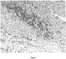
- Hepatoblastoma, germ gliomas or meningioma which is usually in a mild form and with a very strong reaction observed for cancers of the bile duct ( Fig. 5 ).
- the neuroganglioma demonstrates that the tissues of neuroectodermal origin are recognized by antibodies E. coli O56 ( Fig. 6 ) and only trace resactivity was found with E. coli O24.
- Normal renal tubule cells FIG. 7
- ganglion cells Fig. 8
- normal thyroid epithelial cells of the thyroid gland Fig. 9
- the antibodies against E. coli O56 are more reactive in staining of nervous tissue and epithelial cells than antibodies against E. coli O24.
- the nervous system is colored in normal tissues (ganglia), and in the case of tumours (neuroganglioma). With respect to the epithelium, each type of this tissue has a different biological function, and many specific biomarker proteins. Functional markers that are generally expressed by epithelial neoplasms are useful for the immunohistochemical differentiation of metastatic tumours of unknown origin.
Landscapes
- Health & Medical Sciences (AREA)
- Chemical & Material Sciences (AREA)
- Life Sciences & Earth Sciences (AREA)
- Organic Chemistry (AREA)
- Immunology (AREA)
- Molecular Biology (AREA)
- Medicinal Chemistry (AREA)
- General Health & Medical Sciences (AREA)
- Biochemistry (AREA)
- Proteomics, Peptides & Aminoacids (AREA)
- Genetics & Genomics (AREA)
- Biophysics (AREA)
- Engineering & Computer Science (AREA)
- Cell Biology (AREA)
- Urology & Nephrology (AREA)
- Biomedical Technology (AREA)
- Hematology (AREA)
- Virology (AREA)
- Analytical Chemistry (AREA)
- Tropical Medicine & Parasitology (AREA)
- Gastroenterology & Hepatology (AREA)
- Pathology (AREA)
- General Physics & Mathematics (AREA)
- Biotechnology (AREA)
- Physics & Mathematics (AREA)
- Food Science & Technology (AREA)
- Microbiology (AREA)
- Chemical Kinetics & Catalysis (AREA)
- Public Health (AREA)
- General Chemical & Material Sciences (AREA)
- Pharmacology & Pharmacy (AREA)
- Animal Behavior & Ethology (AREA)
- Nuclear Medicine, Radiotherapy & Molecular Imaging (AREA)
- Veterinary Medicine (AREA)
- Peptides Or Proteins (AREA)
- Preparation Of Compounds By Using Micro-Organisms (AREA)
- Medicines Containing Antibodies Or Antigens For Use As Internal Diagnostic Agents (AREA)
Description
- The present invention relates to a new use of bacterial antigens and antibodies obtained in accordance with this use. The resulting antibodies can be used in the diagnosis of tumours by immunohistochemistry and in the binding of drugs to antibodies for application in cancer therapy.
- Lipopolysaccharide (LPS) is an endotoxin molecule located on the cell surface of Gram-negative bacteria. It consists of a toxic component, lipid A, a core region, and an O-polysaccharide antigen which is specific for each serotype [1]. O24 and O56 serotypes of Escherichia coli are characterized by the presence of sialic acid in their lipopolisacharydach. The presence of sialic acid in LPS contributes to the pathogenicity of bacteria by a mechanism of molecular mimicry, ie. sharing a common epitope with host structures [2]. The presence of bacterial epitopes structurally similar to host antigens, the molecular mimicry phenomenon, may interrupt the host immune response. O-specific polysaccharides of the lipopolysaccharide of E. coli O24 and O56 have a similar sequence of sugars with a common structure →7)-α-NeuNAc-(2→3)-β-D-Glc(1→. Sialic acid is glycosylated by β-D-GlcpNAc in O56 and β-D-GalpNAc in O24 polysaccharides respectively [3]. The molecular mimicry based on the presence of sialic acid epitopes in common with the host structures may be supported by a well-known example of a bacterial colominic acid being structurally identical to a tissue polisialyl-glycoconjugate [4], or to O-specific polysaccharide O37 of Citrobacter cross-reactive with the band 3 glycoprotein of human erythrocytes [5]. Of particularly importance is the expression of sialic acid on the surface of tumor cells, indicating a functional relationship with the neoplasmic phenotype. The transformation progression and metastasis accompanied by changes in the amounts, connectivity and types of sialic acids on the surface of tumor cells [6].
- The specific recognition of particular cancers is still a problem in need of a solution. It is particularly desirable to provide a new diagnostic tool for the identification and differentiation of secretory epithelial cancers and nervous tissue.
- Unexpectedly, it turned out that the above defined problem has been solved in the present invention.
- The subject of the present invention is an antibody having an affinity for secretory epithelial and nervous tissue and tumour tissue derived from these tissues recognizing a bacterial antigen comprising a structural motif defined by the formula:

- Another object of the invention is a diagnostic kit for the detection of secretory epithelial cells and neural tissue and tumour tissue cells derived from these tissues, comprising an antibody as defined above.
- Another subject of the present invention is the use of the antigen defined by the formula above in the production of antibodies specific for secretory epithelial and nervous tissue and tumour tissue derived from these tissues.
- Preferably, the antibodies are produced for the diagnosis of tumours.
- Another subject of the present invention is a method for the preparation of antibodies specific for secretory epithelial and nervous tissue, characterized in that the mammal is immunized with a bacterial antigen defined by the formula above, then antibodies that recognize the antigen are isolated.
- Preferably, the antibodies are isolated using affinity chromatography on a column packed with a carrier containing the immobilized antigen defined by the formula above.
- Unexpectedly, as a result of the research forming the basis for the present invention, it turned out that selected human tissues are recognized specifically by anti-A24 or anti-O56, although some of the epitopes were recognized by both anti-A24 and anti-O56. Furthermore, preferential reactivity with anti-O56 was found. Many tissues tested did not react with any of the two antibodies, confirming the specificity of the observed phenomenon.
- Immunohistochemistry experiments demonstrated that antibodies against the O-specific polysaccharide of Escherichia coli O24 and O56 recognize different epitopes on human tissues. An unexpected fact is the recognition of tissue structures by rabbit antibodies obtained after immunization with bacterial cells of Escherichia coli O24 and O56, purification affinity chromatography on a column of of immobilized lipopolysaccharide. Unexpectedly, we noted the high reactivity of these antibacterial antibodies with tumour tissue, especially cancer tissue, and particular specificity for secretory epithelial and neural tissue. Particularly interesting is the discovered reactivity of anti-O24 and O56 anti-tumour reactivity with several structures that can be used in the diagnosis of cancer and oncology.
- To facilitate a better understanding of the present invention, the above description has been enriched with the accompanying figures and discussion of example embodiments of the present invention.
-
-
FIG. 1 . The structure of O-specific polysaccharide from E. coli O24 and O56. -
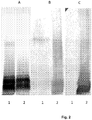
Fig. 2 . SDS-PAGE analysis of lipopolysaccharide from E. coli serotype O24 (1) and O56 (2) (A) and immunoblotting with rabbit antibodies using affinity-purified anti-E. coli O24 (B) and anti-E. coli O56 (C). -
Fig. 3 . Metastatic adenocarcinoma of the colon in the liver. Strong positive staining is seen in the secretory cells of the metastasis with anti-O56; LSAB, counterstained with hematoxylin, 100x magnification. -
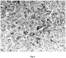
Fig. 4 . Hepatocellular carcinoma (HCC). HCC cells reveal a strong reaction with the anti-dispersed O56; LSAB, counterstained with hematoxylin, 100x magnification. -
FIG. 5 . Cancer of bile duct cells (CCC). CCC cells reveal a very strong dispersed reaction with anti- O56 antibodies; LSAB, counterstained with hematoxylin, 100x magnification. -
FIG. 6 . Coil neuroma. Visible in the center of the photomicrograph is a moderate positive response of ganglion cells of the benign tumour with anti-O56; LSAB, counterstained with hematoxylin, 100x magnification. -
Fig. 7 . Chronic kidney transplant rejection. Renal tubular cells show strong positive staining with anti-O56; LSAB, counterstained with hematoxylin, 100x magnification. -
Fig. 8 . Neural coils. Ganglion cells show a moderate positive reaction with anti-O56; LSAB, counterstained with hematoxylin, 100x magnification. -
Fig. 9 . The thyroid gland. Thyrocytes (thyroid epithelial cells) in normal thyroid show a strong reaction with the anti-O56 antibody; LSAB, counterstained with hematoxylin, 100x magnification. - Bacterial strains were obtained from the Polish Collection of Microorganisms (PCM) at the Institute of Immunology and Experimental Therapy, Polish Academy of Sciences in Wroclaw. Escherichia coli O24 (PCM 195), and O56 (PCM 2372) were the same as used previously [3]. The bacteria were grown in nutrient Davis broth, supplemented with casein hydrolyzate and yeast extract (Difco), with aeration at 37 ° C. After 24 h, the cells were collected and lyophilized. Lipopolysaccharides were obtained by extraction using phenol-water and purified by ultracentrifugation [7].
- Isolation and analysis of LPS, using the proteinase K method.
- An original procedure [8] was slightly modified as follows. The bacterial suspension in phosphate buffered saline (PBS) adjusted to a concentration with a uniform optical density of A600 = 0.3, then 1.5 ml of this material was centrifuged (13 000 g, 4 ° C, 15 min). Pelleted cells were resuspended in 200 ul of 10 mM Tris-HCI buffer containing EDTA, glycerol and SDS, boiled for 10 minutes and treated with proteinase K at 60 ° C for 2 hours. After removal of the precipitate, the solution was subjected to polyacrylamide gel electrophoresis in the presence of SDS, followed by an immunoblotting assay.
- Rabbits were immunized with lyophilized bacteria suspended in PBS, initially subcutaneously at a dose of 100 mg of dry cells per 1 ml in PBS and then IV twice a week with increasing amounts of bacteria (100 to 6400 mg / ml PBS). One week after the last immunization the rabbits were bled and the serum was separated and the complement was heat inactivated (56 ° C, 30 min) and stored at -20 ° C [9].
- Purification of antibodies by affinity chromatography using bound lipopolysaccharides.
- The soluble form of lipopolysaccharide of E. coli O24 and O56 (20 mg) was obtained by suspending it in 2 ml of 2% SDS in 200 ml of 0.5 M EDTA and then three times by ethanol precipitation (1: 4, v / v) and centrifugation (12,000 rpm per minute for 20 minutes). LPS was dissolved in water (1 mL), sonicated and passed through a column of Dowex H + or Dowex Na + to obtain preparations, respectively of acid or sodium salts. The antibodies were isolated from rabbit sera against E. coli O24 and O56 (20 ml) diluted in PBS (1: 2, v / v) after salting out with ammonium sulfate (9.6 g (NH4) 2SO4), 4 ° C, 1h, 30 min centrifugation of 3000 xg, 25 ° C). The precipitated antibodies were dissolved in 5 ml of PBS and dialyzed to PBS at 4 ° C. The affinity column was prepared by binding the acid form of LPS to C18 silica gel in DMSO, after washing out the unbound LPS with 50% methanol, this was packed into a column (1x10cm) in 50% methanol, washed with water and PBS, then blocked with 1% casein in PBS, and washed with PBS with monitoring at 280 nm. The salted out antibodies (0.5 ml) were bound to the affinity column with LPS-bound C18 silica gel, washed with PBS to remove unbound protein and the antibody was eluted with 3 M KSCN in PBS, dialyzed against PBS and then stored in 50% glycerol at -20 °C.
- SDS-PAGE was performed by the Laemmli [10] method using a 15% polyacrylamide gel as previously described [3]. In short, resuspended LPS (1mg / ml) in sample buffer or samples treated with proteinase K, and bacterial extract was boiled for 5 minutes and 10 ml were loaded onto the gel. After electrophoresis, the gels were stained with silver according to Tsai and Frash [11,12]. After SDS-PAGE, immunoblotting test material was transferred electrophoretically from the gel to a nitrocellulose membrane (Schleicher-Schuell, 0.45 um) as described previously [13]. The membranes were incubated overnight at 36 ° C in a rabbit serum diluted 1: 200 with 1% (w / v) gelatin, washed with Tris-buffered saline (TBS, 20 mM Tris-HCI, 50 mM NaCl, 0.05% Tween 20, pH 7.5) and then incubated with goat anti-rabbit IgG conjugated with horseradish peroxidase diluted 1:5000 in TBS containing 1% (w / v) gelatin for 1 hour at 36 ° C. The membrane was stained with 4-chloro-1-naphthol in the presence of H2O2.
- Human tissue sections, fixed in formalin (4%) and paraffin-embedded (FFPE), were cut from blocks 4 µm slices and deparaffinized. The procedure using an immunoperoxidase ABC kit from DAKO: endogenous peroxidase was blocked with blocking reagent and distilled water at room temperature (15 min); then acid citrate buffer pH 6.0 (2 x 8 min for the first time in the microwave at a power of 350 W, and then at room temperature), TBS (0.05 mmol, pH 7.6) with swine serum 1:50 (0, 5 hours at room temperature), distilled water, the test antibody (150 ul / formulation, 40 ° C, overnight) TBS; LSAB reagent (30 min), 3,3'-diaminobenzidine tetrahydrochloride (DAB) (5 min). Sections were stained with hematoxylin and embedded in the resin layer. Negative controls were performed in TBS, and without the primary antibody.
- Bacterial cells of the O24 and O56 serotypes of E. coli were grown in liquid medium for obtaining and analyzing the lipopolysaccharide. LPS preparations were isolated from the cell mass by extraction with phenol-water at a preparative scale. The method of isolation of LPS with proteinase K is used in quantiative analysis. The structures of sialic acid-containing polysaccharides have been established previously [3], are shown in
Fig. 1 . The purified LPS were bound to a solid phase and an affinity column prepared from the acidic form of LPS and C18 silica gel. Rabbit polyclonal antisera were prepared against whole bacterial cells. For the purification of antibodies using affinity, immunoglobulin fractions were precipitated with ammonium sulfate. The antibodies of this fraction were specifically bound to immobilised LPS and eluted with KSCN. The obtained antibody was then subjected to SDS-PAGE followed by an immunoblotting analysis. As shown inFig. 2 , the resulting antibodies recognize the corresponding lipopolysaccharides. It should be noted that, in particular, antibodies against E. coli O24 showed cross-reactivity with the LPS of E. coli O56, while the anti-O56 antibodies should be viewed as only reactive with the homologous LPS. This mild method of binding LPS is particularly valuable in the case of labile compounds, such as sialic acid-containing polysaccharides. Anti-E. coli O56 recognize long chains of LPS molecules present in small quantities, which have been difficult to visualize using silver staining. Structural differences between the O-specific units are responsible for the lack of serological cross-reactivity with E. coli O56, but have a similar sugar backbone. Rabbit antibodies obtained by purification using the affinity reaction were investigated through immunohistochemistry with selected strips of human tissues. For these experiments we used normal healthy tissues, as well as rejected kidney preparations, derived from the original benign and malignant tumours, and metastatic cancer. The results are shown inFig. 3-9 . No staining for either antibody (O24 and O56) has been demonstrated in: - 1. The following normal tissues and cells: parathyroid glands, adrenal glands, lymphatic vessels or blood vessels, bronchial squamous epithelium, ovarian serous epithelium, bone, T and B cells, macrophages, granulocytes and nerve cells,
- 2. Benign tumours: lipomas and papillomas,
- 3. Cancers, such as the gastrointestinal stromal tumour (GIST), cancer, nasopharyngeal cancer, and small cell bronchial squamous cell, basal cell carcinoma, malignant melanoma and fatty sarcoma.
- We noticed an intense positive antibody response of E. coli O56 antibody in bronchial adenocarcinoma cells (adenocarcinoma), and liver cancer metastases with moderately differentiated adenocarcinoma of the colon, where the glands are less regular (
Fig. 3 ). A similar reaction with the antibody was found in adenocarcinoma of the pancreas and slightly weaker endometrial cancer (data not shown). These antibodies react with an as yet uncharacterised antigen present on hepatocytes, as shown inFig. 4 . Based on the granular nature of the intracellular staining , it can be assumed that the epitope may be located in the cytoplasm [14]. Immunohistochemistry plays an important role in distinguishing hepatocellular carcinoma (HCC) from the other primary and metastatic tumours. It is known that no more specific nor sensitive marker for HCC has yet been established. In this case, it is recommended to use an immunohistochemical library including a plurality of antibodies, such as those specific for an antigen hepatocyte PCEA, MOC-31, CD34, TTF-1 [15]. Based on this information, an E. coli O56 antibody can be suggested as useful in combination with other known markers, and not used as a sole marker. A positive antibody reaction to either E. coli O56 and O24 of E. coli antibodies is found in many juvenile malignant tumours, for example. Hepatoblastoma, germ gliomas or meningioma, which is usually in a mild form and with a very strong reaction observed for cancers of the bile duct (Fig. 5 ). Interestingly, a benign tumour, the neuroganglioma demonstrates that the tissues of neuroectodermal origin are recognized by antibodies E. coli O56 (Fig. 6 ) and only trace resactivity was found with E. coli O24. Normal renal tubule cells (FIG. 7 ), ganglion cells (Fig. 8 ), and normal thyroid epithelial cells of the thyroid gland (Fig. 9 ) were stained with both antibodies and demonstrated a much stronger reaction with the antibody against E. coli O56. During the tests using neuroectodermal tissues (ganglions, nerves and brain), we demonstrated the reactivity of our antibody in normal tissues (ganglia) as well as in tumour tissue (neuroganglioma). Digestive glands, derived from the endoderm and the thyroid gland also showed a positive reaction with the antibody of E. coli O56. An interesting observation is that the normal thymus, spleen and tonsils do not show immunoreactivity, indicating that the results are significant. - Generally, the antibodies against E. coli O56 are more reactive in staining of nervous tissue and epithelial cells than antibodies against E. coli O24. The nervous system is colored in normal tissues (ganglia), and in the case of tumours (neuroganglioma). With respect to the epithelium, each type of this tissue has a different biological function, and many specific biomarker proteins. Functional markers that are generally expressed by epithelial neoplasms are useful for the immunohistochemical differentiation of metastatic tumours of unknown origin. The results of our experiments indicate that the cancers of secretory tissues, metastases, the epithelium of the renal tubules and the thyroid gland epithelium was stained, but the skin epithelium was definitely not stained (results not shown). Based on these findings, we believe that the E. coli O56 antibodies should be considered rather as a biomarker of secretory epithelium. Therefore, the most important observation is that the epitope recognized by anti-O56 antibodies is a new marker, specific for secretory epithelial and nervous tissue.
-
- 1. Brade H., Opal S.M., Vogel S.N. & Morrison D.C. (eds) Endotoxin in Health and Disease. Macel Dekker, Inc., New York 1999
- 2. Corfield A.P., Schauer R. 1982: Occurrence of sialic acids, In: Sialic acids, chemistry, metabolism and function. Ed.: Schauer R. Cell Biol. Monogr. 10: 5-50.
- 3. Gamian A., Kenne L., Mieszata M., Ulrich J., Defaye J. 1994: Structure of the .
- 4. Janas et al, 1991
- 5. Ebaid H., Duk M., Gamian A. Antibodies against Citrobacter braakii 037 cells recognize the N -glycan of the band 3 glycoprotein of human erythrocyte membrane. FEMS Immunol Med Microbiol 52 (2008) 352-361.
- 6. Schultz M.J., Swindall A.F., Bellis S.L., Regulation of the metastatic cell phenotype by sialylated glycans. Cancer Metastasis Rev., published online, 2012
- 7. Westphal O., Jann K., Bacterial lipopolysaccharides: extraction with phenol water and further applications of the procedure, Methods Carbohydr. Chem., 5, 1965, 83-92
- 8. Hitchcock P.J., Brown T.M., Morphological heterogeneity among Salmonella lipopolysaccharide chemotypes in silver-stained polyacrylamide gels, J. Bacteriol., 154, 1983, 269-277.
- 9. Gamian A., Romanowska A., Romanowska E. 1992b: Immunochemical studies on sialic acid-containing lipopolysaccharides from Enterobacterial species. FEMS Microbiol. Immunol. 89: 323-328.
- 10. Laemmli U. K., Cleavage of structural proteins during the assembly of the head of bacteriophage T4. Nature (London) 227, 1970, 680-685
- 11. Tsai C. M., Frash C. E., A sensitive silver stain for detecting lipopolysaccharides in polyacrylamide gels. Anal. Biochem. 119, 1982, 115-119
- 12. Fomsgaard A., Freudenberg M.A. and Galanos C., Modification of the silver staining technique to detect lipopolysaccharide in polyacrylamide gels. J.Clin.Microbiol. 1990, 28; 2627-2631.
- 13. Towbin H., Staehelin T. and Gordon J., Electrophoretic transfer of proteins from polyacrylamide gels to nitrocellulose sheets: procedure and some applications. Proc. Natl. Acad. Sci. USA, 1979, 76; 4350-4354.
- 14. Minervini M.I., Demetris A.J., Lee R.G., et al., Utilization of hepatocyte-specific antibody in the immunocytochemical evaluation of liver tumors. Mol. Pathol., 1997, 10, 686-692
- 15. Ordonez N.G., Broad-spectrum immunohistochemical epithelial markers: a review. Human Pathology 2013, 44, 1195-1215
Claims (3)
- Use of an antibody directed against bacterial antigen comprising a structural motif defined by the formula:

- A diagnostic kit for use in detection of glandular epithelial cells, nervous tissue or tumor tissue cells derived from these tissues, characterized in that it comprises an antibody as defined in the Claim 1.
- Method for detection ex-vivo of glandular epithelial cells, nervous tissue or tumor tissue cells derived from these tissues, wherein sample of said tissue cells is recognized by antibody directed against bacterial antigen comprising a structural motif defined by the formula:

Applications Claiming Priority (2)
| Application Number | Priority Date | Filing Date | Title |
|---|---|---|---|
| PL406694A PL231667B1 (en) | 2013-12-27 | 2013-12-27 | Antibody having affinity to glandular epithelium and nervous tissue as well as neoplastic tissue originating from those tissues and its application |
| PCT/PL2014/050080 WO2015099548A1 (en) | 2013-12-27 | 2014-12-29 | An antibody possessing an affinity for epithelial secretory and nervous tissue, as well as tumour tissue derived from these tissues as well as the use thereof |
Publications (2)
| Publication Number | Publication Date |
|---|---|
| EP3087096A1 EP3087096A1 (en) | 2016-11-02 |
| EP3087096B1 true EP3087096B1 (en) | 2020-03-04 |
Family
ID=52463102
Family Applications (1)
| Application Number | Title | Priority Date | Filing Date |
|---|---|---|---|
| EP14835589.4A Active EP3087096B1 (en) | 2013-12-27 | 2014-12-29 | An antibody possessing an affinity for epithelial secretory and nervous tissue, as well as tumour tissue derived from these tissues as well as the use thereof |
Country Status (6)
| Country | Link |
|---|---|
| US (1) | US9938338B2 (en) |
| EP (1) | EP3087096B1 (en) |
| CN (1) | CN106170495B (en) |
| ES (1) | ES2796003T3 (en) |
| PL (1) | PL231667B1 (en) |
| WO (1) | WO2015099548A1 (en) |
-
2013
- 2013-12-27 PL PL406694A patent/PL231667B1/en unknown
-
2014
- 2014-12-29 EP EP14835589.4A patent/EP3087096B1/en active Active
- 2014-12-29 CN CN201480072974.9A patent/CN106170495B/en active Active
- 2014-12-29 US US15/108,202 patent/US9938338B2/en active Active
- 2014-12-29 WO PCT/PL2014/050080 patent/WO2015099548A1/en not_active Ceased
- 2014-12-29 ES ES14835589T patent/ES2796003T3/en active Active
Non-Patent Citations (2)
| Title |
|---|
| ATREYA RAJA ET AL: "Prediction of Anti-TNF Antibody Responses in Crohn's Disease by Endoscopic Molecular Imaging In Vivo: A Clinical Phase 1 Study", GASTROENTEROLOGY, W.B. SAUNDERS CO, US, vol. 142, no. 5, Suppl.1, 1 May 2012 (2012-05-01), pages S535, XP009169867, ISSN: 0016-5085, DOI: 10.1016/S0016-5085(12)62054-X * |
| PANAGIOTIS KAMPERIDIS ET AL: "Development of a novel recombinant biotherapeutic with applications in targeted therapy of human arthritis", ARTHRITIS & RHEUMATISM, vol. 63, no. 12, 29 November 2011 (2011-11-29), pages 3758 - 3767, XP055156190, ISSN: 0004-3591, DOI: 10.1002/art.30650 * |
Also Published As
| Publication number | Publication date |
|---|---|
| PL231667B1 (en) | 2019-03-29 |
| US20160326235A1 (en) | 2016-11-10 |
| WO2015099548A1 (en) | 2015-07-02 |
| EP3087096A1 (en) | 2016-11-02 |
| ES2796003T3 (en) | 2020-11-25 |
| CN106170495B (en) | 2021-10-15 |
| US9938338B2 (en) | 2018-04-10 |
| PL406694A1 (en) | 2015-07-06 |
| CN106170495A (en) | 2016-11-30 |
Similar Documents
| Publication | Publication Date | Title |
|---|---|---|
| Jesaitis et al. | The chemical and antiviral properties of the somatic antigen of Phase II Shigella sonnei | |
| Wu et al. | Identification and confirmation of differentially expressed fucosylated glycoproteins in the serum of ovarian cancer patients using a lectin array and LC–MS/MS | |
| Rodríguez‐Zhurbenko et al. | Human antibodies reactive to N eu G c GM 3 ganglioside have cytotoxic antitumor properties | |
| Gao et al. | Identification of tumor antigens that elicit a humoral immune response in the sera of Chinese esophageal squamous cell carcinoma patients by modified serological proteome analysis | |
| JP2020170002A (en) | Methods for detecting cancer cells, reagents for introducing substances into cancer cells, and compositions for treating cancer | |
| Örntoft et al. | Comparative immunoperoxidase demonstration of T-antigens in human colorectal carcinomas and morphologically abnormal mucosa | |
| US8658376B2 (en) | DEK as a urine based biomarker for bladder cancer | |
| EP2993184B1 (en) | Monoclonal antibody recognizing sialylated sugar chains | |
| KR101296511B1 (en) | Biomarker composition for diagnosing exposure to nano particle in air and the method for detecting using the same | |
| JP6754932B2 (en) | Methods and kits for detecting stem cells | |
| US9938338B2 (en) | Antibody possessing an affinity for epithelial secretory and nervous tissue, as well as tumour tissue derived from these tissues as well as the use thereof | |
| Dragacevic et al. | ELLSA based profiling of surface glycosylation in microorganisms reveals that ß-glucan rich yeasts’ surfaces are selectively recognized with recombinant banana lectin | |
| Newman et al. | Immunochemical Detection of the Thomsen‐Friedenreich Antigen (T‐antigen) on the Pig Lymphocyte Plasma Membrane | |
| HK1231101A1 (en) | An antibody possessing an affinity for epithelial secretory and nervous tissue, as well as tumour tissue derived from these tissues as well as the use thereof | |
| Tanaka et al. | Enhanced expression of unique gangliosides with GM2-determinant in human uterine cervical carcinoma-derived cell lines | |
| HK1231101B (en) | An antibody possessing an affinity for epithelial secretory and nervous tissue, as well as tumour tissue derived from these tissues as well as the use thereof | |
| JPS6193A (en) | Specific Cancer-Associated Antigen, Fucosylsialosylgangliotetraose and its Use in Diagnostic or Therapeutic Treatments Related to Human Lung Cancer | |
| Björklund | Tissue polypeptide antigen. Review and recent progress | |
| Nelson Jr et al. | Distribution of lipopolysaccharide and the detection of a new subfraction in the cell envelope of a marine pseudomonad | |
| PL234956B1 (en) | Monoclonal antibody with dual specificity, method for obtaining it and applications | |
| Smorodin et al. | The isolation and characterization of human natural αGal‐specific IgG antibodies applicable to the detection of αGal‐glycosphingolipids | |
| Wu et al. | Chromatographic characterization of CA 19‐9 molecules from cystic fibrosis and pancreatic carcinoma | |
| Yari et al. | Purification of modified mycobacterial A60 antigen by affinity chromatography and its use for rapid diagnostic tuberculosis infection | |
| KR20130135590A (en) | Lawsonia intracellularis-specific monoclonal antibody and hybridoma cell producing the same | |
| CN116814557A (en) | Hybridoma cell strain, monoclonal antibody for resisting human Axl and application of monoclonal antibody |
Legal Events
| Date | Code | Title | Description |
|---|---|---|---|
| PUAI | Public reference made under article 153(3) epc to a published international application that has entered the european phase |
Free format text: ORIGINAL CODE: 0009012 |
|
| 17P | Request for examination filed |
Effective date: 20160727 |
|
| AK | Designated contracting states |
Kind code of ref document: A1 Designated state(s): AL AT BE BG CH CY CZ DE DK EE ES FI FR GB GR HR HU IE IS IT LI LT LU LV MC MK MT NL NO PL PT RO RS SE SI SK SM TR |
|
| AX | Request for extension of the european patent |
Extension state: BA ME |
|
| STAA | Information on the status of an ep patent application or granted ep patent |
Free format text: STATUS: REQUEST FOR EXAMINATION WAS MADE |
|
| DAX | Request for extension of the european patent (deleted) | ||
| STAA | Information on the status of an ep patent application or granted ep patent |
Free format text: STATUS: EXAMINATION IS IN PROGRESS |
|
| 17Q | First examination report despatched |
Effective date: 20180119 |
|
| GRAP | Despatch of communication of intention to grant a patent |
Free format text: ORIGINAL CODE: EPIDOSNIGR1 |
|
| STAA | Information on the status of an ep patent application or granted ep patent |
Free format text: STATUS: GRANT OF PATENT IS INTENDED |
|
| INTG | Intention to grant announced |
Effective date: 20190912 |
|
| GRAS | Grant fee paid |
Free format text: ORIGINAL CODE: EPIDOSNIGR3 |
|
| GRAA | (expected) grant |
Free format text: ORIGINAL CODE: 0009210 |
|
| STAA | Information on the status of an ep patent application or granted ep patent |
Free format text: STATUS: THE PATENT HAS BEEN GRANTED |
|
| AK | Designated contracting states |
Kind code of ref document: B1 Designated state(s): AL AT BE BG CH CY CZ DE DK EE ES FI FR GB GR HR HU IE IS IT LI LT LU LV MC MK MT NL NO PL PT RO RS SE SI SK SM TR |
|
| REG | Reference to a national code |
Ref country code: GB Ref legal event code: FG4D |
|
| REG | Reference to a national code |
Ref country code: CH Ref legal event code: EP |
|
| REG | Reference to a national code |
Ref country code: AT Ref legal event code: REF Ref document number: 1240269 Country of ref document: AT Kind code of ref document: T Effective date: 20200315 |
|
| REG | Reference to a national code |
Ref country code: DE Ref legal event code: R096 Ref document number: 602014061959 Country of ref document: DE |
|
| REG | Reference to a national code |
Ref country code: IE Ref legal event code: FG4D |
|
| REG | Reference to a national code |
Ref country code: NL Ref legal event code: FP |
|
| REG | Reference to a national code |
Ref country code: CH Ref legal event code: NV Representative=s name: DENNEMEYER AG, CH |
|
| PG25 | Lapsed in a contracting state [announced via postgrant information from national office to epo] |
Ref country code: RS Free format text: LAPSE BECAUSE OF FAILURE TO SUBMIT A TRANSLATION OF THE DESCRIPTION OR TO PAY THE FEE WITHIN THE PRESCRIBED TIME-LIMIT Effective date: 20200304 Ref country code: FI Free format text: LAPSE BECAUSE OF FAILURE TO SUBMIT A TRANSLATION OF THE DESCRIPTION OR TO PAY THE FEE WITHIN THE PRESCRIBED TIME-LIMIT Effective date: 20200304 Ref country code: NO Free format text: LAPSE BECAUSE OF FAILURE TO SUBMIT A TRANSLATION OF THE DESCRIPTION OR TO PAY THE FEE WITHIN THE PRESCRIBED TIME-LIMIT Effective date: 20200604 |
|
| PG25 | Lapsed in a contracting state [announced via postgrant information from national office to epo] |
Ref country code: BG Free format text: LAPSE BECAUSE OF FAILURE TO SUBMIT A TRANSLATION OF THE DESCRIPTION OR TO PAY THE FEE WITHIN THE PRESCRIBED TIME-LIMIT Effective date: 20200604 Ref country code: SE Free format text: LAPSE BECAUSE OF FAILURE TO SUBMIT A TRANSLATION OF THE DESCRIPTION OR TO PAY THE FEE WITHIN THE PRESCRIBED TIME-LIMIT Effective date: 20200304 Ref country code: LV Free format text: LAPSE BECAUSE OF FAILURE TO SUBMIT A TRANSLATION OF THE DESCRIPTION OR TO PAY THE FEE WITHIN THE PRESCRIBED TIME-LIMIT Effective date: 20200304 Ref country code: HR Free format text: LAPSE BECAUSE OF FAILURE TO SUBMIT A TRANSLATION OF THE DESCRIPTION OR TO PAY THE FEE WITHIN THE PRESCRIBED TIME-LIMIT Effective date: 20200304 Ref country code: GR Free format text: LAPSE BECAUSE OF FAILURE TO SUBMIT A TRANSLATION OF THE DESCRIPTION OR TO PAY THE FEE WITHIN THE PRESCRIBED TIME-LIMIT Effective date: 20200605 |
|
| REG | Reference to a national code |
Ref country code: LT Ref legal event code: MG4D |
|
| PG25 | Lapsed in a contracting state [announced via postgrant information from national office to epo] |
Ref country code: IS Free format text: LAPSE BECAUSE OF FAILURE TO SUBMIT A TRANSLATION OF THE DESCRIPTION OR TO PAY THE FEE WITHIN THE PRESCRIBED TIME-LIMIT Effective date: 20200704 Ref country code: SK Free format text: LAPSE BECAUSE OF FAILURE TO SUBMIT A TRANSLATION OF THE DESCRIPTION OR TO PAY THE FEE WITHIN THE PRESCRIBED TIME-LIMIT Effective date: 20200304 Ref country code: EE Free format text: LAPSE BECAUSE OF FAILURE TO SUBMIT A TRANSLATION OF THE DESCRIPTION OR TO PAY THE FEE WITHIN THE PRESCRIBED TIME-LIMIT Effective date: 20200304 Ref country code: SM Free format text: LAPSE BECAUSE OF FAILURE TO SUBMIT A TRANSLATION OF THE DESCRIPTION OR TO PAY THE FEE WITHIN THE PRESCRIBED TIME-LIMIT Effective date: 20200304 Ref country code: LT Free format text: LAPSE BECAUSE OF FAILURE TO SUBMIT A TRANSLATION OF THE DESCRIPTION OR TO PAY THE FEE WITHIN THE PRESCRIBED TIME-LIMIT Effective date: 20200304 Ref country code: RO Free format text: LAPSE BECAUSE OF FAILURE TO SUBMIT A TRANSLATION OF THE DESCRIPTION OR TO PAY THE FEE WITHIN THE PRESCRIBED TIME-LIMIT Effective date: 20200304 Ref country code: PT Free format text: LAPSE BECAUSE OF FAILURE TO SUBMIT A TRANSLATION OF THE DESCRIPTION OR TO PAY THE FEE WITHIN THE PRESCRIBED TIME-LIMIT Effective date: 20200729 Ref country code: CZ Free format text: LAPSE BECAUSE OF FAILURE TO SUBMIT A TRANSLATION OF THE DESCRIPTION OR TO PAY THE FEE WITHIN THE PRESCRIBED TIME-LIMIT Effective date: 20200304 |
|
| REG | Reference to a national code |
Ref country code: ES Ref legal event code: FG2A Ref document number: 2796003 Country of ref document: ES Kind code of ref document: T3 Effective date: 20201125 |
|
| REG | Reference to a national code |
Ref country code: DE Ref legal event code: R097 Ref document number: 602014061959 Country of ref document: DE |
|
| PLBE | No opposition filed within time limit |
Free format text: ORIGINAL CODE: 0009261 |
|
| STAA | Information on the status of an ep patent application or granted ep patent |
Free format text: STATUS: NO OPPOSITION FILED WITHIN TIME LIMIT |
|
| PG25 | Lapsed in a contracting state [announced via postgrant information from national office to epo] |
Ref country code: DK Free format text: LAPSE BECAUSE OF FAILURE TO SUBMIT A TRANSLATION OF THE DESCRIPTION OR TO PAY THE FEE WITHIN THE PRESCRIBED TIME-LIMIT Effective date: 20200304 |
|
| 26N | No opposition filed |
Effective date: 20201207 |
|
| PG25 | Lapsed in a contracting state [announced via postgrant information from national office to epo] |
Ref country code: SI Free format text: LAPSE BECAUSE OF FAILURE TO SUBMIT A TRANSLATION OF THE DESCRIPTION OR TO PAY THE FEE WITHIN THE PRESCRIBED TIME-LIMIT Effective date: 20200304 Ref country code: PL Free format text: LAPSE BECAUSE OF FAILURE TO SUBMIT A TRANSLATION OF THE DESCRIPTION OR TO PAY THE FEE WITHIN THE PRESCRIBED TIME-LIMIT Effective date: 20200304 |
|
| PG25 | Lapsed in a contracting state [announced via postgrant information from national office to epo] |
Ref country code: MC Free format text: LAPSE BECAUSE OF FAILURE TO SUBMIT A TRANSLATION OF THE DESCRIPTION OR TO PAY THE FEE WITHIN THE PRESCRIBED TIME-LIMIT Effective date: 20200304 |
|
| REG | Reference to a national code |
Ref country code: BE Ref legal event code: MM Effective date: 20201231 |
|
| PG25 | Lapsed in a contracting state [announced via postgrant information from national office to epo] |
Ref country code: IE Free format text: LAPSE BECAUSE OF NON-PAYMENT OF DUE FEES Effective date: 20201229 Ref country code: LU Free format text: LAPSE BECAUSE OF NON-PAYMENT OF DUE FEES Effective date: 20201229 |
|
| REG | Reference to a national code |
Ref country code: AT Ref legal event code: UEP Ref document number: 1240269 Country of ref document: AT Kind code of ref document: T Effective date: 20200304 |
|
| PG25 | Lapsed in a contracting state [announced via postgrant information from national office to epo] |
Ref country code: TR Free format text: LAPSE BECAUSE OF FAILURE TO SUBMIT A TRANSLATION OF THE DESCRIPTION OR TO PAY THE FEE WITHIN THE PRESCRIBED TIME-LIMIT Effective date: 20200304 Ref country code: MT Free format text: LAPSE BECAUSE OF FAILURE TO SUBMIT A TRANSLATION OF THE DESCRIPTION OR TO PAY THE FEE WITHIN THE PRESCRIBED TIME-LIMIT Effective date: 20200304 Ref country code: CY Free format text: LAPSE BECAUSE OF FAILURE TO SUBMIT A TRANSLATION OF THE DESCRIPTION OR TO PAY THE FEE WITHIN THE PRESCRIBED TIME-LIMIT Effective date: 20200304 |
|
| PG25 | Lapsed in a contracting state [announced via postgrant information from national office to epo] |
Ref country code: MK Free format text: LAPSE BECAUSE OF FAILURE TO SUBMIT A TRANSLATION OF THE DESCRIPTION OR TO PAY THE FEE WITHIN THE PRESCRIBED TIME-LIMIT Effective date: 20200304 Ref country code: AL Free format text: LAPSE BECAUSE OF FAILURE TO SUBMIT A TRANSLATION OF THE DESCRIPTION OR TO PAY THE FEE WITHIN THE PRESCRIBED TIME-LIMIT Effective date: 20200304 |
|
| PG25 | Lapsed in a contracting state [announced via postgrant information from national office to epo] |
Ref country code: BE Free format text: LAPSE BECAUSE OF NON-PAYMENT OF DUE FEES Effective date: 20201231 |
|
| PGFP | Annual fee paid to national office [announced via postgrant information from national office to epo] |
Ref country code: NL Payment date: 20221222 Year of fee payment: 9 Ref country code: AT Payment date: 20221222 Year of fee payment: 9 |
|
| PGFP | Annual fee paid to national office [announced via postgrant information from national office to epo] |
Ref country code: CH Payment date: 20230103 Year of fee payment: 9 |
|
| P01 | Opt-out of the competence of the unified patent court (upc) registered |
Effective date: 20230719 |
|
| REG | Reference to a national code |
Ref country code: CH Ref legal event code: PL |
|
| REG | Reference to a national code |
Ref country code: NL Ref legal event code: MM Effective date: 20240101 |
|
| REG | Reference to a national code |
Ref country code: AT Ref legal event code: MM01 Ref document number: 1240269 Country of ref document: AT Kind code of ref document: T Effective date: 20231229 |
|
| PG25 | Lapsed in a contracting state [announced via postgrant information from national office to epo] |
Ref country code: NL Free format text: LAPSE BECAUSE OF NON-PAYMENT OF DUE FEES Effective date: 20240101 |
|
| PG25 | Lapsed in a contracting state [announced via postgrant information from national office to epo] |
Ref country code: NL Free format text: LAPSE BECAUSE OF NON-PAYMENT OF DUE FEES Effective date: 20240101 |
|
| PG25 | Lapsed in a contracting state [announced via postgrant information from national office to epo] |
Ref country code: CH Free format text: LAPSE BECAUSE OF NON-PAYMENT OF DUE FEES Effective date: 20231231 |
|
| PG25 | Lapsed in a contracting state [announced via postgrant information from national office to epo] |
Ref country code: AT Free format text: LAPSE BECAUSE OF NON-PAYMENT OF DUE FEES Effective date: 20231229 |
|
| PG25 | Lapsed in a contracting state [announced via postgrant information from national office to epo] |
Ref country code: CH Free format text: LAPSE BECAUSE OF NON-PAYMENT OF DUE FEES Effective date: 20231231 Ref country code: AT Free format text: LAPSE BECAUSE OF NON-PAYMENT OF DUE FEES Effective date: 20231229 |
|
| PGFP | Annual fee paid to national office [announced via postgrant information from national office to epo] |
Ref country code: DE Payment date: 20251211 Year of fee payment: 12 |
|
| PGFP | Annual fee paid to national office [announced via postgrant information from national office to epo] |
Ref country code: GB Payment date: 20251219 Year of fee payment: 12 |
|
| PGFP | Annual fee paid to national office [announced via postgrant information from national office to epo] |
Ref country code: IT Payment date: 20251223 Year of fee payment: 12 |
|
| PGFP | Annual fee paid to national office [announced via postgrant information from national office to epo] |
Ref country code: FR Payment date: 20251223 Year of fee payment: 12 |
|
| PGFP | Annual fee paid to national office [announced via postgrant information from national office to epo] |
Ref country code: ES Payment date: 20260130 Year of fee payment: 12 |