EP3050964B1 - Method for positioning and integrating transgene and use thereof - Google Patents
Method for positioning and integrating transgene and use thereof Download PDFInfo
- Publication number
- EP3050964B1 EP3050964B1 EP13894189.3A EP13894189A EP3050964B1 EP 3050964 B1 EP3050964 B1 EP 3050964B1 EP 13894189 A EP13894189 A EP 13894189A EP 3050964 B1 EP3050964 B1 EP 3050964B1
- Authority
- EP
- European Patent Office
- Prior art keywords
- gene
- cell
- sequence
- site
- plasmid
- Prior art date
- Legal status (The legal status is an assumption and is not a legal conclusion. Google has not performed a legal analysis and makes no representation as to the accuracy of the status listed.)
- Not-in-force
Links
- 238000000034 method Methods 0.000 title claims description 52
- 108700019146 Transgenes Proteins 0.000 title description 12
- 210000004027 cell Anatomy 0.000 claims description 123
- 108090000623 proteins and genes Proteins 0.000 claims description 104
- 241000283707 Capra Species 0.000 claims description 74
- 230000009261 transgenic effect Effects 0.000 claims description 60
- 230000014509 gene expression Effects 0.000 claims description 47
- 108010068072 salmon calcitonin Proteins 0.000 claims description 31
- 238000012216 screening Methods 0.000 claims description 28
- 239000013598 vector Substances 0.000 claims description 28
- 241001465754 Metazoa Species 0.000 claims description 18
- 230000000694 effects Effects 0.000 claims description 18
- 210000001082 somatic cell Anatomy 0.000 claims description 17
- 102000004190 Enzymes Human genes 0.000 claims description 16
- 108090000790 Enzymes Proteins 0.000 claims description 16
- 229940088598 enzyme Drugs 0.000 claims description 16
- 108010014251 Muramidase Proteins 0.000 claims description 12
- 108010062010 N-Acetylmuramoyl-L-alanine Amidase Proteins 0.000 claims description 12
- 230000001605 fetal effect Effects 0.000 claims description 12
- 241000282414 Homo sapiens Species 0.000 claims description 9
- 238000004520 electroporation Methods 0.000 claims description 9
- 239000013604 expression vector Substances 0.000 claims description 9
- 210000000349 chromosome Anatomy 0.000 claims description 6
- 102000007056 Recombinant Fusion Proteins Human genes 0.000 claims description 5
- 108010008281 Recombinant Fusion Proteins Proteins 0.000 claims description 5
- 108010071390 Serum Albumin Proteins 0.000 claims description 5
- 230000001404 mediated effect Effects 0.000 claims description 5
- 108010051219 Cre recombinase Proteins 0.000 claims description 4
- 108010063045 Lactoferrin Proteins 0.000 claims description 4
- 238000000520 microinjection Methods 0.000 claims description 4
- 238000001638 lipofection Methods 0.000 claims description 3
- 239000001506 calcium phosphate Substances 0.000 claims description 2
- 229910000389 calcium phosphate Inorganic materials 0.000 claims description 2
- 235000011010 calcium phosphates Nutrition 0.000 claims description 2
- 210000001671 embryonic stem cell Anatomy 0.000 claims description 2
- 230000006801 homologous recombination Effects 0.000 claims description 2
- 238000002744 homologous recombination Methods 0.000 claims description 2
- 238000001556 precipitation Methods 0.000 claims description 2
- 238000012546 transfer Methods 0.000 claims description 2
- QORWJWZARLRLPR-UHFFFAOYSA-H tricalcium bis(phosphate) Chemical compound [Ca+2].[Ca+2].[Ca+2].[O-]P([O-])([O-])=O.[O-]P([O-])([O-])=O QORWJWZARLRLPR-UHFFFAOYSA-H 0.000 claims description 2
- 230000009385 viral infection Effects 0.000 claims description 2
- 239000013612 plasmid Substances 0.000 description 79
- 238000001514 detection method Methods 0.000 description 78
- 230000010354 integration Effects 0.000 description 65
- 108020004414 DNA Proteins 0.000 description 63
- 238000001962 electrophoresis Methods 0.000 description 54
- 108091034117 Oligonucleotide Proteins 0.000 description 35
- 239000003550 marker Substances 0.000 description 35
- 108091006905 Human Serum Albumin Proteins 0.000 description 34
- 102000008100 Human Serum Albumin Human genes 0.000 description 32
- BBBFJLBPOGFECG-VJVYQDLKSA-N calcitonin Chemical compound N([C@H](C(=O)N[C@@H](CC(C)C)C(=O)NCC(=O)N[C@@H](CCCCN)C(=O)N[C@@H](CC(C)C)C(=O)N[C@@H](CO)C(=O)N[C@@H](CCC(N)=O)C(=O)N[C@@H](CCC(O)=O)C(=O)N[C@@H](CC(C)C)C(=O)N[C@@H](CC=1NC=NC=1)C(=O)N[C@@H](CCCCN)C(=O)N[C@@H](CC(C)C)C(=O)N[C@@H](CCC(N)=O)C(=O)N[C@@H]([C@@H](C)O)C(=O)N[C@@H](CC=1C=CC(O)=CC=1)C(=O)N1[C@@H](CCC1)C(=O)N[C@@H](CCCNC(N)=N)C(=O)N[C@@H]([C@@H](C)O)C(=O)N[C@@H](CC(N)=O)C(=O)N[C@@H]([C@@H](C)O)C(=O)NCC(=O)N[C@@H](CO)C(=O)NCC(=O)N[C@@H]([C@@H](C)O)C(=O)N1[C@@H](CCC1)C(N)=O)C(C)C)C(=O)[C@@H]1CSSC[C@H](N)C(=O)N[C@@H](CO)C(=O)N[C@@H](CC(N)=O)C(=O)N[C@@H](CC(C)C)C(=O)N[C@@H](CO)C(=O)N[C@@H]([C@@H](C)O)C(=O)N1 BBBFJLBPOGFECG-VJVYQDLKSA-N 0.000 description 31
- 235000013336 milk Nutrition 0.000 description 30
- 239000008267 milk Substances 0.000 description 30
- 210000004080 milk Anatomy 0.000 description 30
- 229960003773 calcitonin (salmon synthetic) Drugs 0.000 description 27
- 239000013642 negative control Substances 0.000 description 25
- RXWNCPJZOCPEPQ-NVWDDTSBSA-N puromycin Chemical compound C1=CC(OC)=CC=C1C[C@H](N)C(=O)N[C@H]1[C@@H](O)[C@H](N2C3=NC=NC(=C3N=C2)N(C)C)O[C@@H]1CO RXWNCPJZOCPEPQ-NVWDDTSBSA-N 0.000 description 18
- 239000000243 solution Substances 0.000 description 17
- 102000004169 proteins and genes Human genes 0.000 description 16
- 101001018100 Homo sapiens Lysozyme C Proteins 0.000 description 15
- 238000012163 sequencing technique Methods 0.000 description 14
- 235000013601 eggs Nutrition 0.000 description 13
- 239000013641 positive control Substances 0.000 description 13
- 235000018102 proteins Nutrition 0.000 description 13
- 239000000872 buffer Substances 0.000 description 12
- 210000003754 fetus Anatomy 0.000 description 12
- XLYOFNOQVPJJNP-UHFFFAOYSA-N water Substances O XLYOFNOQVPJJNP-UHFFFAOYSA-N 0.000 description 12
- 210000000481 breast Anatomy 0.000 description 11
- 238000010276 construction Methods 0.000 description 11
- 238000002360 preparation method Methods 0.000 description 11
- 101150032426 HLF gene Proteins 0.000 description 10
- 101000798114 Homo sapiens Lactotransferrin Proteins 0.000 description 10
- 238000004458 analytical method Methods 0.000 description 10
- 230000001413 cellular effect Effects 0.000 description 10
- 102000050459 human LTF Human genes 0.000 description 10
- 210000001161 mammalian embryo Anatomy 0.000 description 10
- 102000016943 Muramidase Human genes 0.000 description 9
- 238000010370 cell cloning Methods 0.000 description 9
- 210000002950 fibroblast Anatomy 0.000 description 9
- 239000004325 lysozyme Substances 0.000 description 9
- 229960000274 lysozyme Drugs 0.000 description 9
- 235000010335 lysozyme Nutrition 0.000 description 9
- 239000002609 medium Substances 0.000 description 9
- 210000000287 oocyte Anatomy 0.000 description 9
- 239000000047 product Substances 0.000 description 9
- 229950010131 puromycin Drugs 0.000 description 9
- 235000020251 goat milk Nutrition 0.000 description 8
- 238000001890 transfection Methods 0.000 description 8
- 241000894006 Bacteria Species 0.000 description 7
- 238000010367 cloning Methods 0.000 description 7
- 230000006651 lactation Effects 0.000 description 7
- 230000002441 reversible effect Effects 0.000 description 7
- PEDCQBHIVMGVHV-UHFFFAOYSA-N Glycerine Chemical compound OCC(O)CO PEDCQBHIVMGVHV-UHFFFAOYSA-N 0.000 description 6
- 239000006285 cell suspension Substances 0.000 description 6
- 238000010586 diagram Methods 0.000 description 6
- 238000001976 enzyme digestion Methods 0.000 description 6
- 239000012634 fragment Substances 0.000 description 6
- 230000004927 fusion Effects 0.000 description 6
- 238000002347 injection Methods 0.000 description 6
- 239000007924 injection Substances 0.000 description 6
- 210000000472 morula Anatomy 0.000 description 6
- 238000002415 sodium dodecyl sulfate polyacrylamide gel electrophoresis Methods 0.000 description 6
- 238000011282 treatment Methods 0.000 description 6
- 108010091086 Recombinases Proteins 0.000 description 5
- 102000018120 Recombinases Human genes 0.000 description 5
- 108091081062 Repeated sequence (DNA) Proteins 0.000 description 5
- 101150092159 SCT gene Proteins 0.000 description 5
- 230000003321 amplification Effects 0.000 description 5
- 238000005516 engineering process Methods 0.000 description 5
- 238000007918 intramuscular administration Methods 0.000 description 5
- 238000003199 nucleic acid amplification method Methods 0.000 description 5
- 210000003101 oviduct Anatomy 0.000 description 5
- 230000008569 process Effects 0.000 description 5
- 230000000638 stimulation Effects 0.000 description 5
- 239000006228 supernatant Substances 0.000 description 5
- 210000001519 tissue Anatomy 0.000 description 5
- 230000001580 bacterial effect Effects 0.000 description 4
- 210000002459 blastocyst Anatomy 0.000 description 4
- 239000003480 eluent Substances 0.000 description 4
- 101150026546 hsa gene Proteins 0.000 description 4
- 239000002502 liposome Substances 0.000 description 4
- 239000007788 liquid Substances 0.000 description 4
- 239000012460 protein solution Substances 0.000 description 4
- 238000000746 purification Methods 0.000 description 4
- 238000011084 recovery Methods 0.000 description 4
- 238000001262 western blot Methods 0.000 description 4
- 101001008231 Bos taurus Beta-lactoglobulin Proteins 0.000 description 3
- 241001045988 Neogene Species 0.000 description 3
- 108091028043 Nucleic acid sequence Proteins 0.000 description 3
- 241001494479 Pecora Species 0.000 description 3
- 108010076504 Protein Sorting Signals Proteins 0.000 description 3
- 229920002684 Sepharose Polymers 0.000 description 3
- 108090000631 Trypsin Proteins 0.000 description 3
- 102000004142 Trypsin Human genes 0.000 description 3
- 230000001464 adherent effect Effects 0.000 description 3
- 238000000137 annealing Methods 0.000 description 3
- 239000003242 anti bacterial agent Substances 0.000 description 3
- 229940088710 antibiotic agent Drugs 0.000 description 3
- 238000005119 centrifugation Methods 0.000 description 3
- 238000006243 chemical reaction Methods 0.000 description 3
- 238000003776 cleavage reaction Methods 0.000 description 3
- 238000012761 co-transfection Methods 0.000 description 3
- 239000002299 complementary DNA Substances 0.000 description 3
- 238000011161 development Methods 0.000 description 3
- 230000029087 digestion Effects 0.000 description 3
- 238000002474 experimental method Methods 0.000 description 3
- 239000013613 expression plasmid Substances 0.000 description 3
- 238000000605 extraction Methods 0.000 description 3
- 239000000499 gel Substances 0.000 description 3
- 235000003642 hunger Nutrition 0.000 description 3
- 238000009396 hybridization Methods 0.000 description 3
- 244000144972 livestock Species 0.000 description 3
- 238000011068 loading method Methods 0.000 description 3
- 239000007758 minimum essential medium Substances 0.000 description 3
- 101150091879 neo gene Proteins 0.000 description 3
- 239000002244 precipitate Substances 0.000 description 3
- 108090000765 processed proteins & peptides Proteins 0.000 description 3
- 230000006798 recombination Effects 0.000 description 3
- 238000005215 recombination Methods 0.000 description 3
- 230000001105 regulatory effect Effects 0.000 description 3
- 238000011160 research Methods 0.000 description 3
- 230000007017 scission Effects 0.000 description 3
- 230000003248 secreting effect Effects 0.000 description 3
- 230000037351 starvation Effects 0.000 description 3
- 239000000758 substrate Substances 0.000 description 3
- 238000012360 testing method Methods 0.000 description 3
- 238000012250 transgenic expression Methods 0.000 description 3
- 238000002054 transplantation Methods 0.000 description 3
- 239000012588 trypsin Substances 0.000 description 3
- 210000004291 uterus Anatomy 0.000 description 3
- IJGRMHOSHXDMSA-UHFFFAOYSA-N Atomic nitrogen Chemical compound N#N IJGRMHOSHXDMSA-UHFFFAOYSA-N 0.000 description 2
- 241000283690 Bos taurus Species 0.000 description 2
- LFQSCWFLJHTTHZ-UHFFFAOYSA-N Ethanol Chemical compound CCO LFQSCWFLJHTTHZ-UHFFFAOYSA-N 0.000 description 2
- CSNNHWWHGAXBCP-UHFFFAOYSA-L Magnesium sulfate Chemical compound [Mg+2].[O-][S+2]([O-])([O-])[O-] CSNNHWWHGAXBCP-UHFFFAOYSA-L 0.000 description 2
- 229930193140 Neomycin Natural products 0.000 description 2
- 241000283973 Oryctolagus cuniculus Species 0.000 description 2
- RJKFOVLPORLFTN-LEKSSAKUSA-N Progesterone Chemical compound C1CC2=CC(=O)CC[C@]2(C)[C@@H]2[C@@H]1[C@@H]1CC[C@H](C(=O)C)[C@@]1(C)CC2 RJKFOVLPORLFTN-LEKSSAKUSA-N 0.000 description 2
- 102000007562 Serum Albumin Human genes 0.000 description 2
- 101150040974 Set gene Proteins 0.000 description 2
- FAPWRFPIFSIZLT-UHFFFAOYSA-M Sodium chloride Chemical compound [Na+].[Cl-] FAPWRFPIFSIZLT-UHFFFAOYSA-M 0.000 description 2
- 206010042573 Superovulation Diseases 0.000 description 2
- 102100038126 Tenascin Human genes 0.000 description 2
- 108010008125 Tenascin Proteins 0.000 description 2
- 210000002593 Y chromosome Anatomy 0.000 description 2
- 238000010521 absorption reaction Methods 0.000 description 2
- 230000002411 adverse Effects 0.000 description 2
- 230000002146 bilateral effect Effects 0.000 description 2
- 108010006025 bovine growth hormone Proteins 0.000 description 2
- 238000009395 breeding Methods 0.000 description 2
- 230000001488 breeding effect Effects 0.000 description 2
- 238000004113 cell culture Methods 0.000 description 2
- 230000002759 chromosomal effect Effects 0.000 description 2
- 238000013377 clone selection method Methods 0.000 description 2
- 230000000295 complement effect Effects 0.000 description 2
- 238000001816 cooling Methods 0.000 description 2
- BNIILDVGGAEEIG-UHFFFAOYSA-L disodium hydrogen phosphate Chemical compound [Na+].[Na+].OP([O-])([O-])=O BNIILDVGGAEEIG-UHFFFAOYSA-L 0.000 description 2
- 229910000397 disodium phosphate Inorganic materials 0.000 description 2
- 238000010828 elution Methods 0.000 description 2
- 238000003780 insertion Methods 0.000 description 2
- 230000037431 insertion Effects 0.000 description 2
- 238000002955 isolation Methods 0.000 description 2
- 239000000463 material Substances 0.000 description 2
- 239000000203 mixture Substances 0.000 description 2
- 230000035772 mutation Effects 0.000 description 2
- 229960004927 neomycin Drugs 0.000 description 2
- 239000002773 nucleotide Substances 0.000 description 2
- 125000003729 nucleotide group Chemical group 0.000 description 2
- 210000004940 nucleus Anatomy 0.000 description 2
- 235000015097 nutrients Nutrition 0.000 description 2
- 108091033319 polynucleotide Proteins 0.000 description 2
- 239000002157 polynucleotide Substances 0.000 description 2
- 102000040430 polynucleotide Human genes 0.000 description 2
- 229920001184 polypeptide Polymers 0.000 description 2
- 102000004196 processed proteins & peptides Human genes 0.000 description 2
- 238000010188 recombinant method Methods 0.000 description 2
- 108091008146 restriction endonucleases Proteins 0.000 description 2
- 230000028327 secretion Effects 0.000 description 2
- 125000006850 spacer group Chemical group 0.000 description 2
- UCSJYZPVAKXKNQ-HZYVHMACSA-N streptomycin Chemical compound CN[C@H]1[C@H](O)[C@@H](O)[C@H](CO)O[C@H]1O[C@@H]1[C@](C=O)(O)[C@H](C)O[C@H]1O[C@@H]1[C@@H](NC(N)=N)[C@H](O)[C@@H](NC(N)=N)[C@H](O)[C@H]1O UCSJYZPVAKXKNQ-HZYVHMACSA-N 0.000 description 2
- 229960005322 streptomycin Drugs 0.000 description 2
- 230000009466 transformation Effects 0.000 description 2
- 238000002525 ultrasonication Methods 0.000 description 2
- DGVVWUTYPXICAM-UHFFFAOYSA-N β‐Mercaptoethanol Chemical compound OCCS DGVVWUTYPXICAM-UHFFFAOYSA-N 0.000 description 2
- DNXIKVLOVZVMQF-UHFFFAOYSA-N (3beta,16beta,17alpha,18beta,20alpha)-17-hydroxy-11-methoxy-18-[(3,4,5-trimethoxybenzoyl)oxy]-yohimban-16-carboxylic acid, methyl ester Natural products C1C2CN3CCC(C4=CC=C(OC)C=C4N4)=C4C3CC2C(C(=O)OC)C(O)C1OC(=O)C1=CC(OC)=C(OC)C(OC)=C1 DNXIKVLOVZVMQF-UHFFFAOYSA-N 0.000 description 1
- KZDCMKVLEYCGQX-UDPGNSCCSA-N 2-(diethylamino)ethyl 4-aminobenzoate;(2s,5r,6r)-3,3-dimethyl-7-oxo-6-[(2-phenylacetyl)amino]-4-thia-1-azabicyclo[3.2.0]heptane-2-carboxylic acid;hydrate Chemical compound O.CCN(CC)CCOC(=O)C1=CC=C(N)C=C1.N([C@H]1[C@H]2SC([C@@H](N2C1=O)C(O)=O)(C)C)C(=O)CC1=CC=CC=C1 KZDCMKVLEYCGQX-UDPGNSCCSA-N 0.000 description 1
- JKMHFZQWWAIEOD-UHFFFAOYSA-N 2-[4-(2-hydroxyethyl)piperazin-1-yl]ethanesulfonic acid Chemical compound OCC[NH+]1CCN(CCS([O-])(=O)=O)CC1 JKMHFZQWWAIEOD-UHFFFAOYSA-N 0.000 description 1
- 101150094083 24 gene Proteins 0.000 description 1
- 101150112497 26 gene Proteins 0.000 description 1
- 229920000936 Agarose Polymers 0.000 description 1
- 102000009027 Albumins Human genes 0.000 description 1
- 108010088751 Albumins Proteins 0.000 description 1
- 208000031873 Animal Disease Models Diseases 0.000 description 1
- 108010077805 Bacterial Proteins Proteins 0.000 description 1
- UYIFTLBWAOGQBI-BZDYCCQFSA-N Benzhormovarine Chemical compound C([C@@H]1[C@@H](C2=CC=3)CC[C@]4([C@H]1CC[C@@H]4O)C)CC2=CC=3OC(=O)C1=CC=CC=C1 UYIFTLBWAOGQBI-BZDYCCQFSA-N 0.000 description 1
- 102000055006 Calcitonin Human genes 0.000 description 1
- 108060001064 Calcitonin Proteins 0.000 description 1
- UXVMQQNJUSDDNG-UHFFFAOYSA-L Calcium chloride Chemical compound [Cl-].[Cl-].[Ca+2] UXVMQQNJUSDDNG-UHFFFAOYSA-L 0.000 description 1
- 102000011632 Caseins Human genes 0.000 description 1
- 108010076119 Caseins Proteins 0.000 description 1
- 108091026890 Coding region Proteins 0.000 description 1
- FBPFZTCFMRRESA-KVTDHHQDSA-N D-Mannitol Chemical compound OC[C@@H](O)[C@@H](O)[C@H](O)[C@H](O)CO FBPFZTCFMRRESA-KVTDHHQDSA-N 0.000 description 1
- 239000006144 Dulbecco’s modified Eagle's medium Substances 0.000 description 1
- 108010013369 Enteropeptidase Proteins 0.000 description 1
- 102100029727 Enteropeptidase Human genes 0.000 description 1
- 108091029865 Exogenous DNA Proteins 0.000 description 1
- 102000003839 Human Proteins Human genes 0.000 description 1
- 108090000144 Human Proteins Proteins 0.000 description 1
- 108010003272 Hyaluronate lyase Proteins 0.000 description 1
- 102000001974 Hyaluronidases Human genes 0.000 description 1
- 102000010445 Lactoferrin Human genes 0.000 description 1
- 102000008192 Lactoglobulins Human genes 0.000 description 1
- 108010060630 Lactoglobulins Proteins 0.000 description 1
- 229930195725 Mannitol Natural products 0.000 description 1
- 241000191938 Micrococcus luteus Species 0.000 description 1
- BVIAOQMSVZHOJM-UHFFFAOYSA-N N(6),N(6)-dimethyladenine Chemical compound CN(C)C1=NC=NC2=C1N=CN2 BVIAOQMSVZHOJM-UHFFFAOYSA-N 0.000 description 1
- 239000012124 Opti-MEM Substances 0.000 description 1
- 238000012408 PCR amplification Methods 0.000 description 1
- 235000016496 Panda oleosa Nutrition 0.000 description 1
- 240000000220 Panda oleosa Species 0.000 description 1
- 102000035195 Peptidases Human genes 0.000 description 1
- 108091005804 Peptidases Proteins 0.000 description 1
- 241000009328 Perro Species 0.000 description 1
- 239000004365 Protease Substances 0.000 description 1
- LCQMZZCPPSWADO-UHFFFAOYSA-N Reserpilin Natural products COC(=O)C1COCC2CN3CCc4c([nH]c5cc(OC)c(OC)cc45)C3CC12 LCQMZZCPPSWADO-UHFFFAOYSA-N 0.000 description 1
- QEVHRUUCFGRFIF-SFWBKIHZSA-N Reserpine Natural products O=C(OC)[C@@H]1[C@H](OC)[C@H](OC(=O)c2cc(OC)c(OC)c(OC)c2)C[C@H]2[C@@H]1C[C@H]1N(C2)CCc2c3c([nH]c12)cc(OC)cc3 QEVHRUUCFGRFIF-SFWBKIHZSA-N 0.000 description 1
- VMHLLURERBWHNL-UHFFFAOYSA-M Sodium acetate Chemical compound [Na+].CC([O-])=O VMHLLURERBWHNL-UHFFFAOYSA-M 0.000 description 1
- 241000282898 Sus scrofa Species 0.000 description 1
- 230000004913 activation Effects 0.000 description 1
- 101150063416 add gene Proteins 0.000 description 1
- 238000000246 agarose gel electrophoresis Methods 0.000 description 1
- 150000001413 amino acids Chemical class 0.000 description 1
- 238000011558 animal model by disease Methods 0.000 description 1
- 230000004888 barrier function Effects 0.000 description 1
- 230000008901 benefit Effects 0.000 description 1
- 229960004015 calcitonin Drugs 0.000 description 1
- 239000001110 calcium chloride Substances 0.000 description 1
- 229910001628 calcium chloride Inorganic materials 0.000 description 1
- 230000003197 catalytic effect Effects 0.000 description 1
- 210000000170 cell membrane Anatomy 0.000 description 1
- 230000008859 change Effects 0.000 description 1
- 239000003153 chemical reaction reagent Substances 0.000 description 1
- 230000001276 controlling effect Effects 0.000 description 1
- 238000007796 conventional method Methods 0.000 description 1
- 238000012136 culture method Methods 0.000 description 1
- 210000004748 cultured cell Anatomy 0.000 description 1
- 230000009089 cytolysis Effects 0.000 description 1
- 238000012217 deletion Methods 0.000 description 1
- 230000037430 deletion Effects 0.000 description 1
- 238000011033 desalting Methods 0.000 description 1
- 239000012153 distilled water Substances 0.000 description 1
- 210000002257 embryonic structure Anatomy 0.000 description 1
- 239000003623 enhancer Substances 0.000 description 1
- 230000006862 enzymatic digestion Effects 0.000 description 1
- 230000002255 enzymatic effect Effects 0.000 description 1
- 239000003797 essential amino acid Substances 0.000 description 1
- 235000020776 essential amino acid Nutrition 0.000 description 1
- 229950002007 estradiol benzoate Drugs 0.000 description 1
- 238000011156 evaluation Methods 0.000 description 1
- 239000000706 filtrate Substances 0.000 description 1
- 238000001914 filtration Methods 0.000 description 1
- 238000001502 gel electrophoresis Methods 0.000 description 1
- 238000012215 gene cloning Methods 0.000 description 1
- 230000002068 genetic effect Effects 0.000 description 1
- 239000011521 glass Substances 0.000 description 1
- 210000002503 granulosa cell Anatomy 0.000 description 1
- 239000001963 growth medium Substances 0.000 description 1
- 230000036541 health Effects 0.000 description 1
- 229960002773 hyaluronidase Drugs 0.000 description 1
- 238000001727 in vivo Methods 0.000 description 1
- 230000001939 inductive effect Effects 0.000 description 1
- 230000003993 interaction Effects 0.000 description 1
- PGHMRUGBZOYCAA-UHFFFAOYSA-N ionomycin Natural products O1C(CC(O)C(C)C(O)C(C)C=CCC(C)CC(C)C(O)=CC(=O)C(C)CC(C)CC(CCC(O)=O)C)CCC1(C)C1OC(C)(C(C)O)CC1 PGHMRUGBZOYCAA-UHFFFAOYSA-N 0.000 description 1
- PGHMRUGBZOYCAA-ADZNBVRBSA-N ionomycin Chemical compound O1[C@H](C[C@H](O)[C@H](C)[C@H](O)[C@H](C)/C=C/C[C@@H](C)C[C@@H](C)C(/O)=C/C(=O)[C@@H](C)C[C@@H](C)C[C@@H](CCC(O)=O)C)CC[C@@]1(C)[C@@H]1O[C@](C)([C@@H](C)O)CC1 PGHMRUGBZOYCAA-ADZNBVRBSA-N 0.000 description 1
- BPHPUYQFMNQIOC-NXRLNHOXSA-N isopropyl beta-D-thiogalactopyranoside Chemical compound CC(C)S[C@@H]1O[C@H](CO)[C@H](O)[C@H](O)[C@H]1O BPHPUYQFMNQIOC-NXRLNHOXSA-N 0.000 description 1
- 229960000318 kanamycin Drugs 0.000 description 1
- 229930027917 kanamycin Natural products 0.000 description 1
- SBUJHOSQTJFQJX-NOAMYHISSA-N kanamycin Chemical compound O[C@@H]1[C@@H](O)[C@H](O)[C@@H](CN)O[C@@H]1O[C@H]1[C@H](O)[C@@H](O[C@@H]2[C@@H]([C@@H](N)[C@H](O)[C@@H](CO)O2)O)[C@H](N)C[C@@H]1N SBUJHOSQTJFQJX-NOAMYHISSA-N 0.000 description 1
- 229930182823 kanamycin A Natural products 0.000 description 1
- CSSYQJWUGATIHM-IKGCZBKSSA-N l-phenylalanyl-l-lysyl-l-cysteinyl-l-arginyl-l-arginyl-l-tryptophyl-l-glutaminyl-l-tryptophyl-l-arginyl-l-methionyl-l-lysyl-l-lysyl-l-leucylglycyl-l-alanyl-l-prolyl-l-seryl-l-isoleucyl-l-threonyl-l-cysteinyl-l-valyl-l-arginyl-l-arginyl-l-alanyl-l-phenylal Chemical compound C([C@H](N)C(=O)N[C@@H](CCCCN)C(=O)N[C@@H](CS)C(=O)N[C@@H](CCCNC(N)=N)C(=O)N[C@@H](CCCNC(N)=N)C(=O)N[C@@H](CC=1C2=CC=CC=C2NC=1)C(=O)N[C@@H](CCC(N)=O)C(=O)N[C@@H](CC=1C2=CC=CC=C2NC=1)C(=O)N[C@@H](CCCNC(N)=N)C(=O)N[C@@H](CCSC)C(=O)N[C@@H](CCCCN)C(=O)N[C@@H](CCCCN)C(=O)N[C@@H](CC(C)C)C(=O)NCC(=O)N[C@@H](C)C(=O)N1CCC[C@H]1C(=O)N[C@@H](CO)C(=O)N[C@@H]([C@@H](C)CC)C(=O)N[C@@H]([C@@H](C)O)C(=O)N[C@@H](CS)C(=O)N[C@@H](C(C)C)C(=O)N[C@@H](CCCNC(N)=N)C(=O)N[C@@H](CCCNC(N)=N)C(=O)N[C@@H](C)C(=O)N[C@@H](CC=1C=CC=CC=1)C(O)=O)C1=CC=CC=C1 CSSYQJWUGATIHM-IKGCZBKSSA-N 0.000 description 1
- 235000021242 lactoferrin Nutrition 0.000 description 1
- 229940078795 lactoferrin Drugs 0.000 description 1
- 230000031700 light absorption Effects 0.000 description 1
- 239000006194 liquid suspension Substances 0.000 description 1
- 239000012160 loading buffer Substances 0.000 description 1
- 229910052943 magnesium sulfate Inorganic materials 0.000 description 1
- 235000019341 magnesium sulphate Nutrition 0.000 description 1
- 210000004962 mammalian cell Anatomy 0.000 description 1
- 210000005075 mammary gland Anatomy 0.000 description 1
- 239000000594 mannitol Substances 0.000 description 1
- 235000010355 mannitol Nutrition 0.000 description 1
- 238000004519 manufacturing process Methods 0.000 description 1
- 239000011159 matrix material Substances 0.000 description 1
- 238000010369 molecular cloning Methods 0.000 description 1
- 229910052757 nitrogen Inorganic materials 0.000 description 1
- 238000010449 nuclear transplantation Methods 0.000 description 1
- 230000000737 periodic effect Effects 0.000 description 1
- 238000012257 pre-denaturation Methods 0.000 description 1
- 239000002243 precursor Substances 0.000 description 1
- 230000035935 pregnancy Effects 0.000 description 1
- 239000000186 progesterone Substances 0.000 description 1
- 229960003387 progesterone Drugs 0.000 description 1
- 238000003259 recombinant expression Methods 0.000 description 1
- BJOIZNZVOZKDIG-MDEJGZGSSA-N reserpine Chemical compound O([C@H]1[C@@H]([C@H]([C@H]2C[C@@H]3C4=C([C]5C=CC(OC)=CC5=N4)CCN3C[C@H]2C1)C(=O)OC)OC)C(=O)C1=CC(OC)=C(OC)C(OC)=C1 BJOIZNZVOZKDIG-MDEJGZGSSA-N 0.000 description 1
- 229960003147 reserpine Drugs 0.000 description 1
- MDMGHDFNKNZPAU-UHFFFAOYSA-N roserpine Natural products C1C2CN3CCC(C4=CC=C(OC)C=C4N4)=C4C3CC2C(OC(C)=O)C(OC)C1OC(=O)C1=CC(OC)=C(OC)C(OC)=C1 MDMGHDFNKNZPAU-UHFFFAOYSA-N 0.000 description 1
- 238000000926 separation method Methods 0.000 description 1
- 239000012679 serum free medium Substances 0.000 description 1
- 239000001632 sodium acetate Substances 0.000 description 1
- 235000017281 sodium acetate Nutrition 0.000 description 1
- 239000011780 sodium chloride Substances 0.000 description 1
- 238000001356 surgical procedure Methods 0.000 description 1
- 238000003786 synthesis reaction Methods 0.000 description 1
- 230000008685 targeting Effects 0.000 description 1
- 238000010257 thawing Methods 0.000 description 1
- 238000013518 transcription Methods 0.000 description 1
- 230000035897 transcription Effects 0.000 description 1
- 238000000108 ultra-filtration Methods 0.000 description 1
- 241001515965 unidentified phage Species 0.000 description 1
- 238000011144 upstream manufacturing Methods 0.000 description 1
- 230000009278 visceral effect Effects 0.000 description 1
- 235000021247 β-casein Nutrition 0.000 description 1
Images







Classifications
-
- C—CHEMISTRY; METALLURGY
- C12—BIOCHEMISTRY; BEER; SPIRITS; WINE; VINEGAR; MICROBIOLOGY; ENZYMOLOGY; MUTATION OR GENETIC ENGINEERING
- C12N—MICROORGANISMS OR ENZYMES; COMPOSITIONS THEREOF; PROPAGATING, PRESERVING, OR MAINTAINING MICROORGANISMS; MUTATION OR GENETIC ENGINEERING; CULTURE MEDIA
- C12N15/00—Mutation or genetic engineering; DNA or RNA concerning genetic engineering, vectors, e.g. plasmids, or their isolation, preparation or purification; Use of hosts therefor
- C12N15/09—Recombinant DNA-technology
- C12N15/87—Introduction of foreign genetic material using processes not otherwise provided for, e.g. co-transformation
- C12N15/90—Stable introduction of foreign DNA into chromosome
- C12N15/902—Stable introduction of foreign DNA into chromosome using homologous recombination
- C12N15/907—Stable introduction of foreign DNA into chromosome using homologous recombination in mammalian cells
-
- A—HUMAN NECESSITIES
- A01—AGRICULTURE; FORESTRY; ANIMAL HUSBANDRY; HUNTING; TRAPPING; FISHING
- A01K—ANIMAL HUSBANDRY; AVICULTURE; APICULTURE; PISCICULTURE; FISHING; REARING OR BREEDING ANIMALS, NOT OTHERWISE PROVIDED FOR; NEW BREEDS OF ANIMALS
- A01K67/00—Rearing or breeding animals, not otherwise provided for; New or modified breeds of animals
- A01K67/027—New or modified breeds of vertebrates
- A01K67/0275—Genetically modified vertebrates, e.g. transgenic
-
- C—CHEMISTRY; METALLURGY
- C12—BIOCHEMISTRY; BEER; SPIRITS; WINE; VINEGAR; MICROBIOLOGY; ENZYMOLOGY; MUTATION OR GENETIC ENGINEERING
- C12N—MICROORGANISMS OR ENZYMES; COMPOSITIONS THEREOF; PROPAGATING, PRESERVING, OR MAINTAINING MICROORGANISMS; MUTATION OR GENETIC ENGINEERING; CULTURE MEDIA
- C12N15/00—Mutation or genetic engineering; DNA or RNA concerning genetic engineering, vectors, e.g. plasmids, or their isolation, preparation or purification; Use of hosts therefor
- C12N15/09—Recombinant DNA-technology
- C12N15/63—Introduction of foreign genetic material using vectors; Vectors; Use of hosts therefor; Regulation of expression
- C12N15/79—Vectors or expression systems specially adapted for eukaryotic hosts
- C12N15/85—Vectors or expression systems specially adapted for eukaryotic hosts for animal cells
- C12N15/8509—Vectors or expression systems specially adapted for eukaryotic hosts for animal cells for producing genetically modified animals, e.g. transgenic
-
- A—HUMAN NECESSITIES
- A01—AGRICULTURE; FORESTRY; ANIMAL HUSBANDRY; HUNTING; TRAPPING; FISHING
- A01K—ANIMAL HUSBANDRY; AVICULTURE; APICULTURE; PISCICULTURE; FISHING; REARING OR BREEDING ANIMALS, NOT OTHERWISE PROVIDED FOR; NEW BREEDS OF ANIMALS
- A01K2217/00—Genetically modified animals
- A01K2217/07—Animals genetically altered by homologous recombination
- A01K2217/072—Animals genetically altered by homologous recombination maintaining or altering function, i.e. knock in
-
- A—HUMAN NECESSITIES
- A01—AGRICULTURE; FORESTRY; ANIMAL HUSBANDRY; HUNTING; TRAPPING; FISHING
- A01K—ANIMAL HUSBANDRY; AVICULTURE; APICULTURE; PISCICULTURE; FISHING; REARING OR BREEDING ANIMALS, NOT OTHERWISE PROVIDED FOR; NEW BREEDS OF ANIMALS
- A01K2227/00—Animals characterised by species
- A01K2227/10—Mammal
- A01K2227/102—Caprine
-
- A—HUMAN NECESSITIES
- A01—AGRICULTURE; FORESTRY; ANIMAL HUSBANDRY; HUNTING; TRAPPING; FISHING
- A01K—ANIMAL HUSBANDRY; AVICULTURE; APICULTURE; PISCICULTURE; FISHING; REARING OR BREEDING ANIMALS, NOT OTHERWISE PROVIDED FOR; NEW BREEDS OF ANIMALS
- A01K2267/00—Animals characterised by purpose
- A01K2267/03—Animal model, e.g. for test or diseases
-
- C—CHEMISTRY; METALLURGY
- C12—BIOCHEMISTRY; BEER; SPIRITS; WINE; VINEGAR; MICROBIOLOGY; ENZYMOLOGY; MUTATION OR GENETIC ENGINEERING
- C12N—MICROORGANISMS OR ENZYMES; COMPOSITIONS THEREOF; PROPAGATING, PRESERVING, OR MAINTAINING MICROORGANISMS; MUTATION OR GENETIC ENGINEERING; CULTURE MEDIA
- C12N2800/00—Nucleic acids vectors
- C12N2800/30—Vector systems comprising sequences for excision in presence of a recombinase, e.g. loxP or FRT
-
- C—CHEMISTRY; METALLURGY
- C12—BIOCHEMISTRY; BEER; SPIRITS; WINE; VINEGAR; MICROBIOLOGY; ENZYMOLOGY; MUTATION OR GENETIC ENGINEERING
- C12N—MICROORGANISMS OR ENZYMES; COMPOSITIONS THEREOF; PROPAGATING, PRESERVING, OR MAINTAINING MICROORGANISMS; MUTATION OR GENETIC ENGINEERING; CULTURE MEDIA
- C12N2800/00—Nucleic acids vectors
- C12N2800/80—Vectors containing sites for inducing double-stranded breaks, e.g. meganuclease restriction sites
Definitions
- the application relates to the field of biological technology and, in particular, to a method for positioning and integrating transgene and use thereof.
- transgenic integration efficiency and expression rate are extremely important in the basic and applied researches. Up to present, scientists have developed many transgenic technologies to improve the targeting and integration efficiency in transgenic animals. However, in large animals, the transgenic integration efficiency and expression rate are not satisfactory.
- the current universal transgenic method in the world is microinjection, which has high costs and low success rate. For a transgenic animal or a mammary gland bioreactor, the success rate can only reach about 3%.
- Microinjection results in random integration of an exogenous gene in chromosomes, and the transgenic expression is greatly influenced by the surrounding host genome.
- the random integration of an exogenous gene may cause the following questions. 1.
- the probability for integrating a transgene into inactive chromosomal sequences is higher than that for integrating into an active chromosomal region and most of transgenes are low expressed or not expressed at all because of the influence from the integration site.
- the transgene integrates into a high-frequency recombination site, it may cause genetic instability. 3.
- Random integration of an exogenous gene can often lead to mutation or altered expression of endogenous genes, thus effecting normal development and health of transgenic animals. 4.
- the isolation of integration sites in its offspring may lead to property change of transgenic expression and difficulty in breeding. 5.
- a significantly high or low copy number of integration may easily cause low or no transgenic expression.
- the object of the present invention is to provide a method for positioning and integrating transgene and use thereof.
- the loxP element comprises a sequence as shown in SEQ ID NO.: 30, or a sequence complete complementary to the sequence as shown in SEQ ID NO.: 30.
- a construct is provided, and the construct comprises from 5' to 3' the following elements:
- the screened marker is neomycin gene, or puromycin resistance genes.
- the exogenous gene is selected from the group consisting of: lysozyme gene, salmon calcitonin gene, lactoferrin gene, and serum albumin gene.
- element (a) and element (c) are placed in the same orientation. In another preferred embodiment, the element (a) and element (c) are placed in the opposite orientation.
- a vector is provided, and the vector contains a construct according to the second aspect of the present invention.
- a host cell in the fourth aspect, contains a construct according to the second aspect of the present invention, or a chromosome thereof is integrated with one or more constructs according to the second aspect of the present invention.
- the host cell is human or non-human mammalian cell.
- the non-human mammal is selected from the group consisting of: goat, sheep, pig, cow, dog, and rabbit; and preferably, it is goat.
- the host cell is a goat adult somatic cell, or a goat fetal somatic cell, or a goat embryonic stem cell.
- the construct according to the second aspect of the present invention is introduced into the host cell by a method selected from the group consisting of: homologous recombination method, microinjection, electroporation, lipofection, calcium phosphate precipitation, virus infection method, or sperm-mediated gene transfer technique.
- the fifth aspect provides a method for preparing a transgenic animal, which comprises the following steps:
- step (i) comprises steps of: (i-1) the cell in the fourth aspect of the present invention is co-transformed by a Cre enzyme expression vector and the vector according to the third aspect of the present invention; or (i-2) by using a TAT-Cre recombinant protein having a cell-penetrating activity, a chromosome of the cell in the fourth aspect of the present invention is genetically integrated with the vector according to the third aspect of the present invention.
- the sixth aspect provides a transgenic non-human mammal animal produced by the method according to the fifth aspect of the present invention.
- a method for producing a heterologous protein which comprises the steps of: (i) feeding the transgenic non-human mammal animal (female) according to the sixth aspect of the present invention, thereby obtaining milk containing heterologous protein; and (ii) optionally, isolating the heterologous protein from the milk.
- the heterologous protein is human protein.
- the heterologous protein includes (but not limited to): lysozyme, salmon calcitonin, lactoferrin, serum albumin, or a combination thereof.
- the eighth aspect provides a milk product from non-human mammal animal, which comprises a high concentration of the heterologous protein.
- the content of the heterologous protein is 0.8-3g/L.
- the milk contains two or more heterologous proteins, and the content of each heterologous protein is 0.8-3g/L.
- the inventors of the invention have unexpectedly found that when the reverse repeated sequence "ATAAC" in wild-type loxp site is mutated into "CACCT", the integration specificity and integration efficiency are greatly improved in cre-loxp integration system.
- the mutation is introduced into the reverse repeated sequence at one side of the loxp site, and a plasmid having same mutant loxp site in the other side of the reverse repeated sequence is co-introduced, then an exogenous gene will be inserted in the presence of the Cre enzyme and a structure with two ends is formed, wherein one end is the wild-type loxp site and the other end has a bilateral mutant loxp site structure.
- the bilateral mutant loxp site is not a good substrate for Cre enzyme, the deleting reaction is prevented and the efficiency for forming stable integration is improved. Based on this, a highly efficient genomic site-directed strategy can be achieved. Based on the above findings, the present invention is completed.
- Cre recombinase The full length of the coding sequence of Cre recombinase is 1029bp (EMBL Database Accession number: X03453) and encodes a protein of 38kDa.
- Cre recombinase is a kind of monomeric protein, consisting of 343 amino acids, and belongs to ⁇ Int enzymatic supergene family. It not only has catalytic activity, but also recognizes the specific DNA sequence, i.e. loxP site (which is similar to restriction enzyme), thus deleting or recombining the sequence within the loxP sites.
- the sequence of loxP (locus of X-over P1) is derived from the P1 bacteriophage, consisting of two 13bp reverse repeated sequences and the 8bp spacer region sequence located therebetween.
- the 8bp spacer region sequence also determines the direction of loxP.
- Cre enzyme will covalently bind DNA during the exchanging process of catalyzed DNA strands, and the 13bp reverse repeated sequences are the binding domain of Cre enzyme.
- sequence of wild-type loxP site is as follows: 5' - ATAACTTCGTATA - ATGTATGC - TATACGAAGTTAT - 3' (SEQ ID NO.:28), and its complementary sequence is 3' -TATTGAAGCATAT-TACATACG-ATATGCTTCAATA - 5'(SEQ ID NO.:29).
- loxP element refers to two cis repeat sites that can be recognized by Cre recombinant protein.
- a mutant loxP element is provided in the present invention, the sequence of which is as follows: CACCTTTCGTATAATGTATGC TATACGAAGTTAT(SEQ ID NO.: 30).
- Cre enzyme refers to a protease that can mediate the specific recombination between 2 loxP sites, so that the nucleotide sequence between the loxP sites is deleted or recombined.
- exogenous gene refers to an exogenous DNA molecular having a periodic function. There is no special limitation for the exogenous gene useful in the present invention. It includes all kinds of exogenous gene common used in the transgenic animal field. Representative examples include (but are not limited to): lysozyme gene, salmon calcitonin gene, lactoferrin gene, or serum albumin gene, etc.
- selective gene for screening refers to a gene used to screen transgenic cells or transgenic animals during the transgenic process. There is no special limitation for selective gene for screening useful in the present invention. It includes all kinds of selective gene for screening that are common used in the transgenic field. The representative examples include (but are not limited to): neomycin gene, or puromycin resistance gene.
- a construct is also provided in the present invention, which comprises from 5' to 3' the following elements:
- element (a) and element (c) are in the same or opposite orientation.
- the term "expression cassette” refers to a polynucleotide sequence containing a gene to be expressed and the sequence components for expressing the element of interest.
- the term “expression cassette of selective gene for screening” refers to a polynucleotide sequence containing a sequence encoding the selective gene for screening and sequence components for expressing the element of interest.
- the expression cassette of selective gene for screening may further include or not include other sequences, which includes (but are not limited to): enhancer, signal peptide sequence for secretion, etc.
- the promoter suitable for expression cassette of exogenous gene and selective gene for screening can be any common promoter, either constitutive promoter or inducible promoter.
- the promoter is a strong constitutive promoter, such as bovine ⁇ -lactoglobulin promoter and other promoters suitable for eukaryotic expression.
- operably linked to refers to a situation in which some parts of a linear DNA sequence can affect the activity of other parts in the same linear DNA sequence. For example, if a signal peptide DNA is expressed as a precursor and involves in the secretion of polypeptide, then the signal peptide (secretory leader sequence) DNA is operably linked to polypeptide DNA; if a promoter controls transcription of a sequence, then it is operably linked to the encoding sequence; and if a ribosome binding site is positioned in a position where it can be translated, then it is operably linked to the encoding sequence. Generally, “operably linked to” means “neighboring", and, for secretory leader sequence, it means “neighboring” in reading frame.
- the construct according to the present invention is inserted into an exogenous vector (especially a vector suitable for operation in transgenic animals) to form the vector of the present invention.
- a host cell is co-transformed by the vector of the present invention and the Cre enzyme expression vector.
- a chromosome of host cell is integrated by the vector of the present invention under the mediation by TAT-Cre recombinant protein having a cell-penetrating activity.
- a method for site-directed integrating exogenous genes is also provided in the present invention, which comprises the steps of : in the presence of Cre enzyme, a cell of the invention is transformed by the vector of the present invention; and the transformed cell is regenerated into an animal, thereby obtaining a transgenic animal.
- the commercial available female goat and the male goat containing the wild-type Loxp element (the male goat was disclosed in the patent ZL200510110772.1) were bred. After 32-40 days, the fetus was taken out by using surgical method, and then transferred to a bechtop, washed twice with 1% PBS buffer containing two antibiotics (penicillin 100 U/ml, and streptomycin 0.1mg/ml). The head, limbs and visceral were removed, the remaining tissue was soaked in the 75% ethanol for 30s; then the fetal tissue was washed three to five times with PBS (without two antibiotics), cut into 1 mm 3 size of tissue block.
- PBS penicillin 100 U/ml
- the tissue was evenly tiled on a 100mm size of cellular petri dish, stood still in GMEM medium supplemented with 10% PBS, penicillin-streptomycin and non-essential amino acids (commercially available from Invitrogen, USA) at 37°C, 5% CO 2 incubator for 4h. On day 2, GMEM medium was added around the tissue block and to immerse the tissue. Observation was conducted every 2-3days and the medium was changed. Primary cells were cultured until 80% confluence of cell, and the cells were digested and collected, subcultured, countered, and cryopreservated in liquid nitrogen.
- N5 was a goat fetus of 32 days
- N4-A was fetus of 37 days
- N37-A was fetus of 37 days
- N37-B was fetus of 37 days.
- a pair of primers were designed according to the sequence of Y chromosome of goat:
- a specific sequence of 638bp on Y chromosome was specifically amplified. Based on that the sequence, the fetal sex identification was performed. If the specific sequence of 638bp could be amplified from the sample, the sample was from male goat; Otherwise, it was from male goat.
- a pair of primers were designed according to the sequence of the human lysozyme genome:
- a specific sequence of the human lysozyme genomic fragment of 429bp was amplified, and the identification of integration was performed. If the specific sequence of 429bp could be amplified from the sample, the sample was from integrated fetus.
- Expression plasmid pET 28.2 TAT-Cre containing TAT-Cre recombinase The vector can be freely obtained from Howard Hughes Medical Institute. The information of structure, multiple cloning site (MCS), intact nucleotide sequence of the vector is from http://cmm.ucsd.edu/Lab_Pages/dowdy/Vectors/ vector_seq/pET28.2bTATCRE.txt) was selected. The bacterial colony having TAT-Cre recombinant expression plasmid was seeded in 20ml LB medium containing kanamycin, and shaked overnight at 37°C.
- MCS multiple cloning site
- the overnight germ solution containing target plasmid was seeded according to 1:100 into 2L kana LB medium, shaked for 3h at 37°C. IPTG was added in a final concentration of 1mmol/L to induce expression and it was shaked for 3h at 37°C. After centrifugation at 3000g for 10 min, the bacterial precipitate was obtained and resuspended in 100ml pre-cooling PBS. After centrifugation at 5000g for 10 min, the bacterial precipitate was obtained and this operation was repeated three times. The bacteria were stored in -80°C for ready to use.
- the bacteria containing target protein were resuspended in pre-cooling bacterial protein lysis solution, placed on the ice, and ultrasonically treated (intensity was 10) for 10s. Then the mixture was placed on ice for 30s to 1min, and the operation was repeated several times until the bacteria were totally lysed. After centrifugation at 15000g for 30min, the supernatant was obtained, placed on ice.
- SP sepharose TM column was balanced using 30% SB buffer (Na 2 HPO 4 10 mmol/L, glycerol 2.5%, ⁇ -mercaptoethanol 10 mmol/L, NaCl 1 mol/L, pH7.4) with a rate of 4ml/min for 30min.
- the supernatant from lysed bacteria was taken, filtered with 0.22 micrometer filter and loaded.
- the loading velocity was 4ml/min.
- the column was washed using 30% SB buffer with a rate of 4ml/min until the ultraviolet absorption value was stable and with no dropping.
- Gradient elution was conducted using 30% to 100% SB buffer (2ml/min), and the total elute volume was 100ml. All eluents were collected by automatic accumulator. Elution was continued using 100% SB buffer until the ultraviolet absorption value dropped to below 100mAU.
- the eluents above 250 mAU were combined, thus obtaining protein TAT-Cre recombinase.
- the eluents were carefully mixed with two volumes of SA buffer (Na 2 HPO 4 10mmol / L, glycerol 2.5%, ⁇ -mercaptoethanol, 10mmol/L, pH7.4) on ice. After a small amount of precipitate formed, the mixture was filtered with 0.22um filter, and the filtrate was placed on ice for use.
- the source 15s column was balanced with 30% SB buffer for 15s (4ml/min, 30min). The resulting supernatant after filtration was loaded with a loading rate of 4ml/min. The column was eluted using 30% to 100% SB buffer until the peak was present. All the eluents above 250 mAU were collected, in which the target protein was contained. The target protein was loaded on HiPrepTM 26/10 Desalting column, eluted in buffer replacement (10ml/min), and the eluted peak protein solution was collected.
- the peak protein solution was put into a millipore MWCO 30000 ultrafiltration tube, it was centrifuged at 3000g for 30min. More than about 1/4 of the original volume of the protein solution was remained in the tube. The protein solution was sucked out and preserved at -80°C. Because each freeze-thawing operation had a certain influence on the concentration, the quantitative concentration of TAT-Cre recombinase was determined by DC before use.
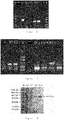
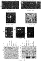
- Figure 3 showed the SDS-PAGE electrophoresis results of TAT-Cre recombinase.
- M was marker
- SF was supernatant after ultrasonication treatment of TAT-Cre bacteria
- FT was liquid flow of SP-sepharose TM column
- E2 was elute from source 15s column. The results showed that the TAT-Cre recombinase was successfully purified.
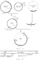
- a complete sequence (as shown in SEQ ID NO.:31) was fully synthesized, wherein one side was a mutant Loxp site and the other side was a wild-type Loxp site in the same orientation; and in the middle were the expression frameworks of selective genes for screening, neo and TK. Then the sequence was cloned into SalI site of a commercial available vector pGEM-7Z, and the recombinant plasmid was named pGEM-2loxp'.
- the element was then introduced into human lysozyme transgenic goat fetal fibroblasts so as to replace a selective marker gene which existed between the wild-type Loxp sites having same orientation.
- the human lysozyme transgenic goat fetal fibroblasts with mutant loxp site were used.
- a mammary breast-specific expression vector pTM-sCT2 for salmon calcitonin was constructed, which had two loxp sequences.
- the preparation method of pbLGpA was disclosed in the Chinese patent ZL200510110772.1.
- This plasmid pbLGpA contained a breast-specific expression regulatory framework which contained a bovine ⁇ -lactoglobulin promoter and a bovine growth hormone polyA sequence.
- the sequence with double loxp sites was fully synthesized and the sequence was shown in Figure 5 .
- the sequence was cloned into a commercial available pUC18 vector, and the resultant plasmid was named pUC18-2loxp.
- Plasmid pbLGpA contained a breast-specific expression regulatory framework including a bovine ⁇ -lactoglobulin promoter and a bovine growth hormone polyA sequence.
- Figure 6 showed a schematic diagram for constructing plasmid pBLG-sct.
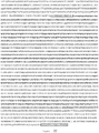
- the encoding sequence of salmon calcitonin was artificially synthesized by Shanghai Generay Biological Engineering Co. Ltd ( Figure 7 ).
- three elements were added in front of the encoding framework of salmon calcitonin, which included a goat ⁇ - casein secretory peptide, 6His tag and enterokinase cleavage site, and was flanked by XhoI site.
- the sCT sequence was cloned into pUC18 vector, thereby forming plasmid pUC18-sCT.
- Figure 7 showed the sequence of artificial synthetic salmon calcitonin.
- the sCT expression cassette was inserted between double loxp sites, thereby obtaining a sCT breast-specific expression plasmid p2xGsct with double loxp sites.
- the puromycin resistance gene expression framework was amplified.
- the gene expression fragment was purified, recovered and screened using TIANGEN DNA purification and recovery kit.
- the recovered band and vector pGM-T were linked and transformed into competent DH5 ⁇ strain, thereby obtaining plasmid pMD-Puro ( Figure 8 , wherein M is D2000 marker; the bands from the bottom to the top are respectively 100, 250, 500, 750, 1000, and 2000bp; 1 and 2 are the electrophoretograms of amplification of puro expression framework).
- the identification of pMD-Puro was conducted by using PCR method ( Figure 9 , wherein M is D2000 marker; 11-20 represent the selected clones after transformation; 11, 12, 13, 14, 16, 17, 18, 19, 20 are positive clones; 21 represents a negative control, and the target band is 1183bp) and enzyme digestion identification ( Figure 10 , wherein M is 1kb ladder; 11-20 represent selected clones after transformation; 16, 17, 18, 19, 20 are positive clones, the target band is 1183bp) respectively.
- the Puro expression cassette was obtained by NotI digestion of plasmid pMD-Puro, and then inserted into a linear p2xGsct digested with NotI, thereby obtaining salmon calcitonin site-directed integration plasmid pTM-sCT2.
- the identification showed that there were 4 positive clones out of 23 colonies.
- the expected band of 1183bp was obtained by PCR ( Figure 11 , wherein M is D2000 marker; 1-23 represent selected monoclones and 2, 19, and 20 are positive clones; 24 represents a negative control).
- pTM-sCT2 could be digested by XhoI to produce a sCT fragment of 180bp ( Figure 12 , wherein M is D2000 marker; 2, 19, and 20 are positive clones).
- the Puro expression cassette of 1183bp was obtained by Not I enzyme digestion ( Figure 13 , wherein 1 is BM15000 DNA Marker II, and the bands from the top to the bottom are respectively 500, 1000, 1500, 3000, 5000, 7500, 10000, and 15000bp; 2 shows electrophoretogram of plasmid pTM-sCT2 digested by NotI enzyme), indicating that the plasmid was successfully constructed.
- pBS185 was commercially available from Addgene corporation.
- Plasmid pTM-sCT2 was obtained in the Example 4.
- Each plasmid was extracted in large amount by using EndoFree Plasmid Maxi Kit (Cat. No. 12362) of QIAGEN.
- the final concentration of plasmid pBS185 was 485ng/ ⁇ l, 500 ⁇ l totally, and the concentration of plasmid pTM-sCT2 was 1.9ng/ ⁇ l, 500 ⁇ l totally.
- the human lysozyme transgenic goat fetal fibroblasts was recovered on the 60mm cell petri dish, grown in the complete medium and passaged to a 24-well plate, 4 x 10 5 cells/well, 500 ⁇ l of medium without double-antibiotics was added to each well. By the next day, cells were grown to 50-80% confluence , and could be used for transfection.
- the DNA-lipid complex was prepared according to the following method.
- the plasmids (pBS185 and pTM-sCT2 added in an amount ratio of 1: 1) were prepared as Table 1, and were group numbered (according to 24-well plate coordinates), each tube of plasmid was diluted with 100 ⁇ l Opti-MEM® I serum-free medium (available from Invitrogen, USA), and well mixed.
- the PLUS reaction solution (available from Invitrogen, USA) was gently mixed, and into the diluted plasmid was added 1 ⁇ l of PLUS reaction solution, mixed gently, then stood still at the room temperature for 5min.
- Lipofectamine TM LTX (available from Invitrogen, USA) was gently mixed.
- Table 1 Group B C D A 1 amount of plasmid/ng 250:250 375:375 500:500 0 amount of liposome/ ⁇ l 2 2 2 2 2 amount of plasmid/ng 250:250 375:375 500:500 0 amount of liposome/ ⁇ l 3 3 3 3
- the cells were passaged into 48-well cell culture plate by normal culture method.
- the grown cells were digested as 2 parts, in which one part was used in the integration of identification, while the other part was further cultured for propagation and frozen preserved for somatic cell cloning.
- the cellular genome was extracted with cellular genome extraction kit (Product code: Dp302-02) from Tiangen Biotech (Beijing) Co., Ltd, and the concentration was adjusted to about 50ng/ ⁇ l.
- the recombinant clonal genome extracted with the primer pairs above was detected respectively for 5'-end ( Figures 14 and 19 ), 3'-end ( Figures 15 and 20 ), functional gene (sct) ( Figure 16 ), selectable marker gene (Neo) ( Figure 17 ), and plasmid pBS185 gene ( Figure 18 ).
- the detection method was PCR (94°C pre-denaturate for 5min, 94°C 30s Tm 30s, 72°C 1min (/ 3min), 72°C 7min).
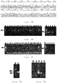
- Figure 14 shows 5'-end detection results of recombinant clones, wherein M is DL2000; 66-69, 71, 73-85, 93 are all the 5'-end detection results of clones; 67, 73, 74, 78, 81, 85 are positive; 0 is the negative control.
- Figure 15 shows 3'-end detection results of recombinant clones, wherein M is 1kb DNA Marker; 20, 23, 24, 34, 41, 47, and 50 are 3'-end detection electrophoretograms of selected clones; 0 is the negative control.
- Figure 16 shows detection electrophoretogram of the salmon calcitonin functional gene; wherein M is DL2000; 10, 14, 20, 23, 24, 34, 41, 47, 54, 59, 67, 73, 74, 79, 81, 85, and 89 are detection electrophoretograms of the functional gene neo; 1 is the positive control; and 2 is the negative control.
- Figure 17 shows detection results of the selected gene neo; wherein M is DL2000; 20, 23, 24, 34, 41, 47, 54, 59, 67, 73, 74, 79, 81, and 85 are the detection electrophoretograms for screening neo as clone selection gene; 79 is positive gene neo; 1 is the positive control; and 2 is the negative control.
- Figure 18 shows detection electrophoretogram of pBS185 gene, wherein M is DL2000; 23, 24, 34, 47, 59, 74, 78, 81, and 85 are detection electrophoretograms for screening cloned gene pBS185; 24, 34, and 81 are positive for gene; and 0 represents a negative control.
- Figure 19 shows the 5' end sequencing analysis result of a site-directed integrated clone of salmon calcitonin.
- Figure 20 shows the 3' end sequencing analysis result of a site-directed integrated clone of salmon calcitonin.
- the method was the same to the pBS185 and pTM-sCT2 co-transfection method for achieving site-directed integration.
- the amount of the plasmid added was shown in Table 4.
- Table 4 Groups B C D A 1 amount of plasmid/ng 500 750 1000 0 amount of liposome/ ⁇ l 2 2 2 2 2 amount of plasmid/ng 500 750 1000 0 amount of liposome/ ⁇ l 3 3 3 3 3 3
- Plasmid pTM-sCT2 was quantified into a concentration of 1.9 ⁇ g / ⁇ l, and 7 ⁇ l was taken for further use.
- the human lysozyme transgenic goat fetal fibroblasts were recovered on the 60mm cell petri dish, and were passaged into the 100mm cell petri dish after grown to confluence. The cell transfection was started when the cell density was about 80%. The number of the cells was counted after the cells were digested by trypsin, the cells were centrifuged at 1000rpm for 5min, and then resuspended in the electroporation buffer PBS, and the number of the cells was adjusted to 5 x 10 6 -1 x 10 7 cells/mL.
- the digested cells were transferred into 6-8 100mm cellular culture plate, and puromycin (final concentration: 0.08 ⁇ g / ml) was used to pressure select for 7 days.
- the monoclonal cells were transferred into 96-well cellular culture plate by using a cloning ring, and were cultured by normal passage method. After growing to confluence in 48 wells, the cells were digested as 2 parts, in which one part was used in the integration of identification, while the other part was further cultured and frozen preserved for somatic cell cloning.
- the genome of selected clonal cell lines was extracted and preserved in 4°C.
- Figure 23 shows a 5' end detection electrophoretogram of the selected clonal cell line; wherein M is DL2000 marker; 1-12 are the 5' end detection electrophoretogram of the selected clonal cell lines, and No.10 is the positive clone.
- Figure 24 shows the 3' end detection electrophoretogram for screening of a clonal cell line; wherein M is DL2000 marker; 1-12 are the 3' end detection electrophoretogram of the selective clonal cell lines, and 10 is the positive clones.
- Figure 25 shows detection electrophoretogram of functional gene; wherein M is DL2000 marker, 10, and 14 are the detection electrophoretogram of the selected clonal functional gene sct.
- human serum albumin mini gene was fully synthesized, of which the full-length was 4015bp, including partial sequences of exon1, intron1, exon2, intron2, exon3 and all cDNA encoding sequences (the complete sequence was shown in SEQ ID NO.:27).
- the sequence was then cloned into a commercial available pUC19 vector after synthesized, and the plasmid was named pUC19-miniHSA.
- pUC19-miniHSA was digested by Xho I enzyme to obtain the human serum albumin mini gene. The product was then recovered and site-directed inserted into the XhoI site of plasmid pbLGpA. PCR identification was performed using a pair of orientation identifying primers HSAg-Fnew (GTGGGTAACCTTTATTTCC)and T7(TAATACGACTCACTATAGGG) for 55 single colonies. Only NO.9 has amplified the 4394bp target fragment. The NO.9 bacterial solution was sent for sequencing, and the NO.9 plasmid was sequenced and it was found to that it was a clone with correct insert-orientation. Thereby, the human serum albumin mini gene regulated by bovine lactoglobulin albumin to obtain mammary breast-specific expression cassette was obtained, and the plasmid was named pBLG-minihSA.
- Figure 42 shows the identification results of mammary breast-specific expression vector of human serum albumin, wherein M is ⁇ / HindIII DNA Marker; each band is respectively 23130, 9416, 6557, 4361, 2322, 2027, 564, or 125bp.
- M is ⁇ / HindIII DNA Marker
- each band is respectively 23130, 9416, 6557, 4361, 2322, 2027, 564, or 125bp.
- 1-32 are plasmid samples; and the sample of NO.9 has a positive band of about 4.3kb.
- the plasmid pTM-sCT2 was digested by restriction enzyme Xhol, and the sCT gene was excised to obtain plasmid framework of which the size was 7019bp ( Figure 26 ). Then the plasmid pBLG-minihSA was digested by XhoI enzyme ( Figure 27 ) to obtain hSA expression cassette of 4015bp. The product obtained above was linked and transferred into to competent DH5 ⁇ strain, and the monoclones were selected, the bacteria was cultured with shaking, and the plasmid was extracted ( Figure 28 ), thereby obtaining plasmid pTM-hSA2.
- the plasmid was digested by EcoRI enzyme ( Figure 29 ), thus obtaining target bands of 3.209kb, 3.906kb, and 3.872kb. (If the hSA expression cassette was inserted in the opposite orientation, the target bands would be: 6kb, 0.655kb, 3.872kb).
- a pair of primers were designed on the 5'-end upstream of the hSA gene and within the hSA gene to conduct sequencing-detection by PCR. The sequences were shown in Table 5: Table 5 fx691-F 5-TAGAGGAAGCAACCCCAGG-3 SEQ ID NO.:17 fx691-R 5-CAGCAACCAAGAAGACAGAC-3 SEQ ID NO.:18
- the length of amplified product was 691bp, and the annealing temperature was 58°C.
- the desired bands were obtained by amplification.
- the sequencing analysis was performed in the PCR amplified products ( Figure 30 , and Figure 32 ), and the results were in accordance with theoretical sequence ( Figure 31 ).
- Extensive extraction was performed to plasmid pTM-hSA2 by EndoFree Plasmid Maxi Kit of QIAGEN, in the 1 ⁇ g/1 ⁇ l concentration, 500 ⁇ l in total, and was packed and preserved at -20°C for further use. And the corresponding strains were stored.
- the human lysozyme transgenic goat fetal fibroblasts were recovered on the 60mm cell petri dish, and was passaged into the 100mm cell petri dish after grown to confluence, and the cell transfection was started when the cell density was about 80%.
- the number of the cells was counted after the cells were digested by trypsin, the cells were centrifuged at 1000rpm for 5min, and then resuspended in the electroporation buffer PBS, and the number of the cells was adjusted to 5 x 10 6 -1 x 10 7 cells/mL.
- the digested cells were transferred into 6-8 100mm cellular culture plate and puromycin (final concentration: 0.08 ⁇ g / ml) was used for pressure selection for 7 days.
- the monoclonal cells were transferred into 96-well cellular culture plate by using a cloning rings, and were cultured by the normal passage method. After growing to confluence in 48 wells, the cells were digested as 2 parts, in which one part was used in the integration of identification, while the other part was further cultured and frozen preserved for somatic cell cloning.
- primers were designed to detect the 5'-end , 3'-end and the key elements of the selected clones (functional gene, screened marker gene).
- Table 6 primers Length of amplified fragment (bp) location 5'-end detection sy1126-F:CAAGCCACCTAACCTCAC SEQ ID NO.:19 1126 genome of goat sy1126-R:TGCTATGCCAAAGTGTTC SEQ ID NO.:20 hsA gene 3'-end xy3365-F:TTCTCGAAACAAGCGCAC 3365 expression cassette detection SEQ ID NO.:21 of puro xy3365-R:CCGTATTCCTTTATCGGGTAT SEQ ID NO.:22 genomic DNA of goat detection of Neo F: 5-CTGCTATTGGGCGAAGTGC-3 SEQ ID NO.:23 469 gene Neo R: 5-CGGCGATACCGTAAAGCAC-3 SEQ ID NO.:24 gene Neo detection of hsA F:5-GCTGCGAAATCATCCATA-3 SEQ
- Figure 33 shows the 5' end detection electrophoretogram of the selected clonal cell lines; wherein M1 is D2000 marker; 1, 2, and 8-24 are the 5' end detection electrophoretograms of selected clonal cell lines, wherein 11, 12, 14, 20, 21, 1, and 2 are positive; 26 and 5 are the positive controls; and 27 and 6 are the negative controls.
- Figure 34 shows the 3' end detection electrophoretogram of selected clonal cell lines; wherein M1 is D2000; 1-4 and 8-25 are the 3' end detection electrophoretogram of selective clonal cell line, wherein 11, 14, 17, 20, 21, and 1 are positive; and 26 and 5 are the positive controls; and 27 and 6 are the negative controls.
- Figure 35 shows the detection pattern of functional genes in the screened the clonal cell lines, wherein M is 1kb DNA ladder; 1 is the detection pattern for screening clone selective gene.
- Figure 36 shows detection pattern of selective gene in the screened clonal cell lines; wherein M is D2000; 1 and 2 are detection pattern for screening selective gene, 1 is positive clone; 3 is the positive control; and 4 is the negative control.
- Figure 37 shows the 5'-end recombinant theoretical sequence of a site-directed recombinant clone of human serum albumin.
- Figure 38 shows the 3'-end recombinant theoretical sequence of a site-directed recombinant clone of human serum albumin.
- the female goats useful as oocyte donors were selected and intramuscular injected with PG at 0.1 mg/ goat, the second PG injection was performed after 10 to 14 days intervals, and the superovulation was begun 10 to 13 days after the second PG injection, that is, firstly, intramuscular injected with FSH, which was divided into 6 times, twice a day, and the daily amount was 100IU, 80IU, 80IU.
- PG 0.1mg/goat
- LRH was injected 24 hours after, 25 ⁇ g/time, and the oocytes were recovered 26-28 hours after LRH injection.
- the receptor goats were PG intramuscular injected in twice at the intervals of 9-11 days. 24 hours after superovulation PG injection to the donor goats, the receptor goats were also injected with PG, the time and dose of LRH injection for the receptors were the same to those of donor goats.
- Pre-starvation cells were seeded on 35mm petri dish. When the confluence of cells reached about 70%-75%, the nutrient solution was sucked out, and the DMEM medium containing 0.5% FCS was added. After starvated for 5 days, the cells were digested and collected by conventional method, and then preserved in the -85°C refrigerator. Six days before nuclear transplantation experiments, 2 small tubes of cells were taken out to recover and seeded into the 4-well plate. Starvation was conducted after re-cultured for 2 or 3 days, and the starvation method was indicated above.
- the oocytes were washed 3 times with M16-Hepes (Hepes 2.8mg/ml, CB 7.5 ⁇ g/ml), treated in M16-Hepes (comprising 7.5 ⁇ g/ml of CB) for 10min. Meanwhile, the cultured cells were digested by trypsin, and dispersed into single cells.
- the oocytes and donor cells were simultaneously transferred into 1ml M16-Hepes (containing CB) on sterilized glass slides, the nuclei was removed under microscope, and the donor cells were sucked in, injected from the original incision into perivitelline space, thus making it attached tightly to cytoplasmic membrane, and was fused by electrical stimulation, the fusion matrix was a solution with 0.3nM mannitol, 0.05mM calcium chloride, 0.1 mM magnesium sulfate, 0.5% BSA.
- the fusion condition was DC600-610 v/cm, of which the pulse duration time was 80 ⁇ s, and the stimulation was continuously stimulated for three times.
- Fused fetus was cultured in M16 solution for 5h, treated in M16 solution comprising 5 ⁇ M ionomycin and 7.5 ⁇ g / ml CB for 5min, and then treated in M16 solution comprising 2mM 6-DMAP and 7.5 ⁇ g / ml CB for 5h, then transferred into in M16 culture solution and cultured until it was embedded or transplanted.
- Reconstructed eggs were embedded in 1% Agarose, transplanted to oviduct,, and the strip was washed out after cultured for 5 days in vivo.
- the embryo inside the strip was stripped out, and the morula and blastocyst were selected and transplanted to the uterus of recipient goats, and the developmental rate of morula and blastocyst was counted.
- 27 receptors were transplanted into the uterus; 12 receptors were directly transplanted into oviduct. Each was transplanted with 10-12 reconstructed embryoes. 39 receptors were transplanted in total. The results of 30-35 days B ultrasonic examination: 9 receptors of 27 receptors transplanted with morula and blastocyst into the uteruses were pregnant (of which pregnant rate was 33.3%); 11 receptors of the 12 receptors transplanted with 1-2 cells into the oviduct were pregnant (91.7% of pregnant rate). 20 receptors were pregnant out of 39 receptors in total (51.3% of pregnant rate).
- somatic cell cloning for the site-directed integration cells was normal, in which indexes of all the parts such as fusion rate of reconstructed embryoes, cleavage rate, developmental rate of morula and blastocysts and pregnant rate of receptors were consistent with the efficiency of non-targeted integrated somatic cell cloning. It indicated that there were no significantly adverse effect on the cloning of somatic cells treated by site-directed integration method during the afterward somatic cell cloning, and the method is useful to prepare somatic cell cloned goats.
- Human lactoferrin mini gene was artificially synthesized.
- the gene included cDNA sequence of human lactoferrin and the fifteenth intron sequence of human lactoferrin (SEQ ID NO.: 32 and Figure 43 ), human lactoferrin site-directed integration framework pTM-hLf was constructed and prepared based on the method of Example 8.
- Fibroblasts of goat ear were co-transfected by pTM-hLf and plasmid pBS185 expressing Cre at a molar ratio of 1: 1, selected with 0.08 ⁇ g/mL of puromycin for 8-10 days. 45 monoclones were selected onto 96-well plates, and further cultured on 6-well plates. Meanwhile, 9 clones grown in good condition were detected with test primers, wherein, 1 cell line hLf-hlz-6 was identified as site-directed integration cell (see Figure 44, Figure 45 and Figure 46 ). Identifying primers were shown in Table 7. Table 7.
- primers used in the process of hLF transgenic site-directed integration of goats detection regions primers'name and the sequence thereof(5'-3') SEQ ID NO.: annealing temperature length of amplified fragment 5'-end detection P1-F 33 58°C 760 bp P1-R 34 3'-end detection P2-F 35 58°C 3365 bp P2-R 36 integrated detection of LF P3-F 37 58°C 734 bp P3-R 38
- the cell of hLf-hlz-6 was in good condition, used for somatic cell cloning. 774 normal goat oocytes were used, and 712 oocytes were nuclei transplanted, while 606 oocytes were fused in total, and of which the fusion rate was 77.72% (471/606). 48 receptor goats were transplanted. After transplanted for 30 days, B-pregnancy check showed that 10 receptor goats were pregnant, and the pregnant rate was 20.83%.
- hSA-GTTG human Serum Albumin gene Targeted Transgenic Goat
- hSA-GTTG transgenic goats were selected, and estradiol benzoate (2mg) and progesterone (20mg) were intramuscular injected twice a day during 7 days in total. Then reserpine (0.5mg) was intramuscular injected on Day 8, 10, 12, and 14 respectively. The milk was collected on Day 12, which was the first day milk, and preserved at -20°C ready for use.
- hSA-GTTG goats were lactation stimulated by traditional lactation stimulation method, about 120 ⁇ l milk was collected in 4 days, of which the average was 40 ⁇ l/day.
- micrococcus lysodeikticus (available from Sigma, M0192) was formulated with 0.18M sodium acetate (pH5.5) buffer into 0.8mg/ml substrate testing liquid suspension (OD450: 0.6-1.0). 2 ⁇ l milk was added into 3ml substrate testing liquid, and the variation of OD450 light absorption was measured. Every 0.001 decrease was defined as an activity unit.
- the tested sample included: milk of hSA-GTTG male goats, human lysozyme transgenic goats and normal goats.
- the activity unit number of lysozyme in hSA-GTTG milk was 1.665 times larger than that in human lysozyme transgenic milk, which indicated that there was no significantly adverse effect on the expression of lysozyme framework after the hSA expression cassette was integrated into the lysozyme integration site.
- the activity of lysozyme in the hSA-GTTG milk was improved, due to lactation stimulation for male goats, the protein content in milk was increased. Therefore, it was not determined that the expression cassette of human lysozyme was improved after integration.
- the goat milk was 10-fold diluted with distilled water, and an equal volume of 2 x SDS gel loading buffer was added, incubated at 65°C for 15min. 5 ⁇ l was taken out for SDS-PAGE Electrophoresis, the concentration of stacking gel was 4%, the concentration of separation gel was 7.5%, and the constant voltage was 100V.
- An anti-mouse anti-hSA monoclonal antibody (Abcam, ab10241) was used as the first antibody for Western-blotting analysis (1:1000); the second antibody was HRP-labeled rabbit anti-mouse IgG (Abcam, ab97046) (1:2000), finally colorated with DAB.
- hSA standards were added to normal goat's milk at a concentration of 2mg/ml, the normal goat's milk was used as gradient to dilute hSA to 1mg/ml, 0.5mg/ml, 0.25, 0.125mg/ml and 0.0625mg/ml of concentration.
- the hybridization signal was obtained by Western-bloting or Slot-bloting method, the hybridization signal was scanned by Bio-1D software, and the standard curve was prepared.
- the content of hSA in milk was calculated by the hybridization signal of hSA-GTTG milk sample based on the standard curve.
- transgenic goats simultaneously carried exogenous human lysozyme and human serum albumin gene, expression of either protein was not mutually interfered.
Landscapes
- Life Sciences & Earth Sciences (AREA)
- Health & Medical Sciences (AREA)
- Genetics & Genomics (AREA)
- Engineering & Computer Science (AREA)
- Biotechnology (AREA)
- Zoology (AREA)
- Bioinformatics & Cheminformatics (AREA)
- Chemical & Material Sciences (AREA)
- Biomedical Technology (AREA)
- General Engineering & Computer Science (AREA)
- Organic Chemistry (AREA)
- Wood Science & Technology (AREA)
- Molecular Biology (AREA)
- General Health & Medical Sciences (AREA)
- Environmental Sciences (AREA)
- Biochemistry (AREA)
- Veterinary Medicine (AREA)
- Biophysics (AREA)
- Microbiology (AREA)
- Physics & Mathematics (AREA)
- Plant Pathology (AREA)
- Cell Biology (AREA)
- Mycology (AREA)
- Animal Behavior & Ethology (AREA)
- Animal Husbandry (AREA)
- Biodiversity & Conservation Biology (AREA)
- Micro-Organisms Or Cultivation Processes Thereof (AREA)
Description
- The application relates to the field of biological technology and, in particular, to a method for positioning and integrating transgene and use thereof.
- Methods for preparing transgenic animals are extremely important in the basic and applied researches. Up to present, scientists have developed many transgenic technologies to improve the targeting and integration efficiency in transgenic animals. However, in large animals, the transgenic integration efficiency and expression rate are not satisfactory.
- The current universal transgenic method in the world is microinjection, which has high costs and low success rate. For a transgenic animal or a mammary gland bioreactor, the success rate can only reach about 3%. Microinjection results in random integration of an exogenous gene in chromosomes, and the transgenic expression is greatly influenced by the surrounding host genome. The random integration of an exogenous gene may cause the following questions. 1. The probability for integrating a transgene into inactive chromosomal sequences is higher than that for integrating into an active chromosomal region and most of transgenes are low expressed or not expressed at all because of the influence from the integration site. 2. When the transgene integrates into a high-frequency recombination site, it may cause genetic instability. 3. Random integration of an exogenous gene can often lead to mutation or altered expression of endogenous genes, thus effecting normal development and health of transgenic animals. 4. For a transgenic animal with integration in multiple sites, the isolation of integration sites in its offspring may lead to property change of transgenic expression and difficulty in breeding. 5. A significantly high or low copy number of integration may easily cause low or no transgenic expression.
- The above questions are key barriers in the studies of gene function, creation of animal disease models, development of commercially valuable models and creation of transgenic breeding new materials. Developing a method for positioning and integrating transgene may overcome negative effects of high costs, low efficiency caused by the traditional technology, and promote industrial development of transgenic livestock. However, there is no method for controlling target gene to efficiently integrate into a specific site of genome. Therefore, there is an urgent need in the art to develop an efficient method for positioning and integrating transgene.
- The object of the present invention is to provide a method for positioning and integrating transgene and use thereof.
- In the first aspect of the present invention, it provides a mutant loxP element, and the loxP element comprises a sequence as shown in SEQ ID NO.: 30, or a sequence complete complementary to the sequence as shown in SEQ ID NO.: 30.
- In the second aspect, a construct is provided, and the construct comprises from 5' to 3' the following elements:
- (a) the mutant loxP element according to the first aspect of the present invention; (b) an expression cassette of an exogenous gene and/or an expression cassette of a selective gene for screening; (c) a wild-type loxP element having a sequence as shown in SEQ ID NO.: 28;
- wherein element (a) and element (c) are interchangeable in position.
- In another preferred embodiment, the screened marker is neomycin gene, or puromycin resistance genes.
- In another preferred embodiment, the exogenous gene is selected from the group consisting of: lysozyme gene, salmon calcitonin gene, lactoferrin gene, and serum albumin gene.
- In another preferred embodiment, element (a) and element (c) are placed in the same orientation. In another preferred embodiment, the element (a) and element (c) are placed in the opposite orientation.
- In the third aspect, a vector is provided, and the vector contains a construct according to the second aspect of the present invention.
- In the fourth aspect, a host cell is provided, and the host cell contains a construct according to the second aspect of the present invention, or a chromosome thereof is integrated with one or more constructs according to the second aspect of the present invention.
- In another preferred embodiment, the host cell is human or non-human mammalian cell. In another preferred embodiment, the non-human mammal is selected from the group consisting of: goat, sheep, pig, cow, dog, and rabbit; and preferably, it is goat.
- In another preferred embodiment, the host cell is a goat adult somatic cell, or a goat fetal somatic cell, or a goat embryonic stem cell.
- In another preferred embodiment, the construct according to the second aspect of the present invention is introduced into the host cell by a method selected from the group consisting of: homologous recombination method, microinjection, electroporation, lipofection, calcium phosphate precipitation, virus infection method, or sperm-mediated gene transfer technique.
- In the fifth aspect, it provides a method for preparing a transgenic animal, which comprises the following steps:
- (i) in the presence of Cre recombinase, a cell in the fourth aspect of the present invention is transformed by a vector according to the third aspect of the present invention; and (ii) the transformed cell is regenerated into an animal, thereby obtaining a transgenic animal.
- In another preferred embodiment, step (i) comprises steps of: (i-1) the cell in the fourth aspect of the present invention is co-transformed by a Cre enzyme expression vector and the vector according to the third aspect of the present invention; or (i-2) by using a TAT-Cre recombinant protein having a cell-penetrating activity, a chromosome of the cell in the fourth aspect of the present invention is genetically integrated with the vector according to the third aspect of the present invention.
- In the sixth aspect, it provides a transgenic non-human mammal animal produced by the method according to the fifth aspect of the present invention.
- In the seventh aspect, a method for producing a heterologous protein, which comprises the steps of: (i) feeding the transgenic non-human mammal animal (female) according to the sixth aspect of the present invention, thereby obtaining milk containing heterologous protein; and (ii) optionally, isolating the heterologous protein from the milk.
- In another preferred embodiment, the heterologous protein is human protein.
- In another preferred embodiment, the heterologous protein includes (but not limited to): lysozyme, salmon calcitonin, lactoferrin, serum albumin, or a combination thereof.
- In the eighth aspect, it provides a milk product from non-human mammal animal, which comprises a high concentration of the heterologous protein.
- In another preferred embodiment, in the milk without concentrating treatment, the content of the heterologous protein is 0.8-3g/L. Preferably, the milk contains two or more heterologous proteins, and the content of each heterologous protein is 0.8-3g/L.
- It should be understood that, within the scope of the present invention, the technical features specifically described above and below (such as the Examples) can be combined with each other, thereby constituting a new or preferred technical solution which needs not be described one by one.
- The drawings below are intended to illustrate the invention, but not to limit the scope of the present invention defined by the claims.
-
Figure 1 shows the sex identification results of the goat fetal fibroblast. M is DL2000 Marker, and the bands from the bottom to the top are respectively 100, 250, 500, 750, 1000, and 2000bp; N5, N4-A, N37-A and N37-B are all isolated from the fetus; "-" represents negative goat control; "+" represents positive goat control; water represents PCR system control. -
Figure 2 shows identification results of the transgenic integration. M is DL2000 Marker, and the bands from the bottom to the top are respectively 100, 250, 500, 750, 1000, and 2000bp; N5, N4-A, N37-A and N37-B are all isolated from the fetus; "-" represents negative goat control; "+" represents positive goat control; water represents PCR system control. -
Figure 3 shows the identification results of the TAT-Cre recombinant protein. M is marker, SF is supernatant after TAT-Cre bacteria ultrasonication, FT is flow through liquid of SP-sepharose TM column, E1 is eluant of SP sepharoseTM column, and E2 is eluant of source 15s column. -
Figure 4 shows a schematic diagram of construction of salmon calcitonin site-directed integration plasmid pTM-sCT2. -
Figure 5 shows the artificial synthesized double loxP gene sequence (SEQ ID NO.: 39). -
Figure 6 shows a schematic diagram of construction of pBLG-sct plasmid. -
Figure 7 shows the completely artificial synthesized salmon calcitonin sequence (SEQ ID NO.:40). -
Figure 8 shows an amplification electrophoretogram of a puro expression cassette. M is D2000 marker; 1 and 2 represent amplification electrophoretogram of the puro expression cassette. -
Figure 9 shows PCR identification of plasmid pMD-Puro. M is D2000 marker; 11-20 represent the selected transformant clones; 11, 12, 13, 14, 16, 17, 18, 19, 20 are positive clones; and 21 is negative control. -
Figure 10 shows electrophoretogram of plasmid pMD-Puro identification by Not I digestion. M is 1kb marker; 11-20 represent the picked transformant clones, wherein 16, 17, 18, 19, 20 are positive clones. -
Figure 11 shows PCR identification pattern of pTM-sCT2. M is D2000 marker; 1-23 represent the selected monoclones: 2, 19, 20 are positive clones; and 24 represents negative control. -
Figure 12 shows electrophoretogram of plasmid pTM-sCT2 as identified by Not I digestion. M is D2000 marker; and 2, 19, 20 are positive clones. -
Figure 13 shows an identification pattern of pTM-sCT2 as identified by enzyme digestion; wherein 1 is DL5000 DNA Marker, and 2 is the electrophoretogram of plasmid pTM-sCT2 digested with Not I enzyme. -
Figure 14 shows detection electrophoretogram for screening 5' end clones. M is D2000; 66-69, 71, 73-85, 93 are all detection electrophoretograms for screening 5' end clones; 67, 73, 74, 78, 81, 85 are positive, and 0 is the negative control. -
Figure 15 shows detection electrophoretogram for screening 3' end clones. M is 1kb DNA Marker; 20, 23, 24, 34, 41, 47, 50 are detection electrophoretograms for screening 3' end clones, and 0 is the negative control. -
Figure 16 shows detection electrophoretogram of the salmon calcitonin functional gene. M is D2000; 10, 14, 20, 23, 24, 34, 41, 47, 54, 59, 67, 73, 74, 79, 81, 85, 89 are detection electrophoretograms of the functional gene neo, 1 is the positive control, and 2 is the negative control. -
Figure 17 shows detection electrophoretogram for screening neo gene. M is D2000; 20, 23, 24, 34, 41, 47, 54, 59, 67, 73, 74, 79, 81, 85 are detection electrophoretograms for screening neo as clone selection gene; 79 is positive gene neo, 1 is the positive control, and 2 is the negative control. -
Figure 18 shows detection electrophoretogram of pBS185 gene. M is D2000; 23, 24, 34, 47, 59, 74, 78, 81, 85 are detection electrophoretograms for screening cloned gene pBS185, wherein 24, 34, 81 represent a positive gene and 0 represents a negative control. -
Figure 19 shows the 5' end sequencing analysis result of a site-directed integrated clone of salmon calcitonin. -
Figure 20 shows the 3' end sequencing analysis result of a site-directed integrated clone of salmon calcitonin. -
Figure 21 shows the 5' end recombinant theoretical sequence of a site-directed integrated clone of salmon calcitonin (SEQ ID NO.:41). -
Figure 22 shows the 3' end recombinant theoretical sequence of a site-directed integrated clone of salmon calcitonin (SEQ ID NO.:42). -
Figure 23 shows the 5' end detection electrophoretogram for screening a clonal cell line. M is D2000 marker; 1-12 are the 5' end detection electrophoretogram of the selected clonal cell lines, and 10 is the positive clone. -
Figure 24 shows the 3' end detection electrophoretogram for screening of a clonal cell line. M is D2000 marker; 1-12 are the 3' end detection electrophoretogram of the selected clonal cell lines, and 10 is the positive clone. -
Figure 25 shows detection electrophoretogram of functional gene. M is D2000 marker; 10 and 14 are detection electrophoretograms for screening clonal functional gene sct. -
Figure 26 shows the recovered pTM-sCT2 plasmid framework. M is 1kb DNA ladder; 1 shows the recovered pTM-sCT2 plasmid framework. -
Figure 27 shows the recovered hsA expression framework. M is 1kb DNA ladder; 1 shows the recovered hsA expression framework. -
Figure 28 shows electrophoretogram of plasmid pTM-hSA2. M is 1kb DNA ladder; 1-12 are the electrophoretograms of the extracted plasmids, wherein, 3, 5 are positive plasmids; 13 is an electrophoretogram of the plasmid pTM-sCT2. -
Figure 29 shows an identification electrophoretogram of plasmid pTM-hSA2 in enzyme digestion. M is 1kb DNA ladder; 3 is an electrophoretogram of the reverse insertion of hsA expression cassette into plasmid pTM-sCT2 framework; 5 is an electrophoretogram of the forward insertion of hsA expression cassette into plasmid pTM-sCT2 framework; and 10 is the negative control. -
Figure 30 shows an orientation identification pattern of plasmid pTM-hSA2. M is 1kb DNA ladder; 1 is amplification electrophoretogram of plasmid pTM-hSA2 using primers for orientation identification. -
Figure 31 shows partial theoretical sequence of plasmid pTM-hSA2 (SEQ ID NO.:43). -
Figure 32 shows sequencing analysis results of orientation identification of the plasmid pTM-hSA2. -
Figure 33 shows the 5' end detection electrophoretogram of selected clonal cell lines. M1 is D2000 marker; 1, 2, 8-24 are the 5' end detection electrophoretogram of selected clones, wherein 11, 12, 14, 20, 21, 1, and 2 are positive; 26 and 5 are the positive controls; and 27 and 6 are the negative controls. -
Figure 34 shows the 3' end detection electrophoretogram of selected clonal cell lines. M1 is D2000; 1-4 and 8-25 are the 3' end detection electrophoretogram of selective clones, wherein 11, 14, 17, 20, 21, and 1 are positive; 26 and 5 are the positive controls; and 27 and 6 are the negative controls. -
Figure 35 shows the detection pattern of functional genes in the screened clonal cell lines. M is 1kb DNA ladder; 1 is the detection pattern for screening clone selective gene. -
Figure 36 shows detection pattern of selective gene in the screened clonal cell lines. M is D2000; 1 and 2 are detection pattern for screening selective gene; 1 is positive clone; 3 is the positive control; and 4 is the negative control. -
Figure 37 shows the 5' end recombinant theoretical sequence of a site-directed recombinant clone of human serum albumin (SEQ ID NO.:44) . -
Figure 38 shows the 3' end recombinant theoretical sequence of a site-directed recombinant clone of human serum albumin (SEQ ID NO.:45). -
Figure 39 shows the 5' end sequencing results of a site-directed integrated clone of human serum albumin. -
Figure 40 shows the 3' end sequencing results of a site-directed integrated clone of human serum albumin. -
Figure 41 shows a schematic diagram of the efficient and sited recombinant method of the present invention. -
Figure 42 shows the identification result of a mammary breast-specific expression vector for human serum albumin. -
Figure 43 shows sequence of human lactoferrin mini gene (SEQ ID NO.: 32). The underlined sequence is XhoI enzymatic digestion site for constructing the plasmid; the sequence shown in lowercase letters is the 15th intron sequence of genomic sequence of human lactoferrin; the sequence shown in capital letters is the cDNA sequence of human lactoferrin. -
Figure 44 shows an identification of 5'-end of hLf gene in a site-directed integration in the clonal cell line. The lanes are as followed: M is 1kb DNA Marker; Nos. 1-9 shows 5'-end sited integration in the detected clonal cell line of hLf; "-" is the negative control. -
Figure 45 shows an identification of 3'-end of hLf gene in a site-directed integration in the clonal cell line. The lanes are as followed: M is 1kb DNA Marker; Nos.1-9 are 5'-end sited integration in the detected clonal cell line of hLf; "+" is the positive control, "-" is the negative control, and 0 is blank control. -
Figure 46 shows an identification of hLf gene integration in the clonal cell line. Lanes are as follows: No. 6 is a selected clonal cell line screened with puromycin for 8 days, "-" means normal goat, "+" is the positive control, 0 is blank control, and M is DL2000 Marker. -
Figure 47 shows hLf site-specific integration of transgenic cloned sheep. -
Figure 48 shows an identification of hLf site-specific (or site-directed) integration in a transgenic cloned sheep. Wherein, "a" shows 5'-end of transgenic detection, in which M1 shows 3'-end of transgenic detection in which M2 is 1kb DNA Marker, 4 is hLF clonal goat, 5 is a cellular positive control, 6 is a negative control, and 7 is blank control; "c" shows the detection of Lf functional gene in which M3 is DL2000 Marker, 8 is clonal goat, 9 is a negative control, and 10 is blank control. -
Figure 49 shows detection of SDS-PAGE electrophoresis and Western-blotting of the hSA-GTTG goat's milk. Wherein, "A" shows analysis results of SDS-PAGE; and "B" shows Western-blotting results. -
Figure 50 shows a hSA content detection in hSA-GTTG goat's milk on the third day of lactation. -
Figure 51 shows a hSA content detection in hSA-GTTG goat's milk on the seventh day of lactation. - After extensive and intensive researches, the inventors of the invention have unexpectedly found that when the reverse repeated sequence "ATAAC" in wild-type loxp site is mutated into "CACCT", the integration specificity and integration efficiency are greatly improved in cre-loxp integration system. In particular, when the mutation is introduced into the reverse repeated sequence at one side of the loxp site, and a plasmid having same mutant loxp site in the other side of the reverse repeated sequence is co-introduced, then an exogenous gene will be inserted in the presence of the Cre enzyme and a structure with two ends is formed, wherein one end is the wild-type loxp site and the other end has a bilateral mutant loxp site structure. Because the bilateral mutant loxp site is not a good substrate for Cre enzyme, the deleting reaction is prevented and the efficiency for forming stable integration is improved. Based on this, a highly efficient genomic site-directed strategy can be achieved. Based on the above findings, the present invention is completed.
- The full length of the coding sequence of Cre recombinase is 1029bp (EMBL Database Accession number: X03453) and encodes a protein of 38kDa. Cre recombinase is a kind of monomeric protein, consisting of 343 amino acids, and belongs to λInt enzymatic supergene family. It not only has catalytic activity, but also recognizes the specific DNA sequence, i.e. loxP site (which is similar to restriction enzyme), thus deleting or recombining the sequence within the loxP sites.
- The sequence of loxP (locus of X-over P1) is derived from the P1 bacteriophage, consisting of two 13bp reverse repeated sequences and the 8bp spacer region sequence located therebetween. The 8bp spacer region sequence also determines the direction of loxP. Cre enzyme will covalently bind DNA during the exchanging process of catalyzed DNA strands, and the 13bp reverse repeated sequences are the binding domain of Cre enzyme.
- The sequence of wild-type loxP site is as follows: 5' - ATAACTTCGTATA - ATGTATGC - TATACGAAGTTAT - 3' (SEQ ID NO.:28),
and its complementary sequence is 3' -TATTGAAGCATAT-TACATACG-ATATGCTTCAATA - 5'(SEQ ID NO.:29). - As used herein, the term "loxP element" refers to two cis repeat sites that can be recognized by Cre recombinant protein. A mutant loxP element is provided in the present invention, the sequence of which is as follows: CACCTTTCGTATAATGTATGC TATACGAAGTTAT(SEQ ID NO.: 30).
- As used herein, the term "Cre enzyme" refers to a protease that can mediate the specific recombination between 2 loxP sites, so that the nucleotide sequence between the loxP sites is deleted or recombined.
- As used herein, "exogenous gene" refers to an exogenous DNA molecular having a periodic function. There is no special limitation for the exogenous gene useful in the present invention. It includes all kinds of exogenous gene common used in the transgenic animal field. Representative examples include (but are not limited to): lysozyme gene, salmon calcitonin gene, lactoferrin gene, or serum albumin gene, etc.
- As used herein, "selective gene for screening" refers to a gene used to screen transgenic cells or transgenic animals during the transgenic process. There is no special limitation for selective gene for screening useful in the present invention. It includes all kinds of selective gene for screening that are common used in the transgenic field. The representative examples include (but are not limited to): neomycin gene, or puromycin resistance gene.
- A construct is also provided in the present invention, which comprises from 5' to 3' the following elements:
- (a) the mutant loxP element according to the present invention;
- (b) an expression cassette of an exogenous gene; and/or an expression cassette of selective gene for screening;
- (c) non-mutating (wild-type) loxP element as shown in SEQ ID NO.: 28;
- In a preferred embodiment, element (a) and element (c) are in the same or opposite orientation.
- As used herein, the term "expression cassette" refers to a polynucleotide sequence containing a gene to be expressed and the sequence components for expressing the element of interest. For example, in the present invention, the term "expression cassette of selective gene for screening" refers to a polynucleotide sequence containing a sequence encoding the selective gene for screening and sequence components for expressing the element of interest. Moreover, the expression cassette of selective gene for screening may further include or not include other sequences, which includes (but are not limited to): enhancer, signal peptide sequence for secretion, etc.
- In the present invention, the promoter suitable for expression cassette of exogenous gene and selective gene for screening can be any common promoter, either constitutive promoter or inducible promoter. Preferably, the promoter is a strong constitutive promoter, such as bovine β-lactoglobulin promoter and other promoters suitable for eukaryotic expression.
- As used herein, "operably linked to" or "operably linked" refers to a situation in which some parts of a linear DNA sequence can affect the activity of other parts in the same linear DNA sequence. For example, if a signal peptide DNA is expressed as a precursor and involves in the secretion of polypeptide, then the signal peptide (secretory leader sequence) DNA is operably linked to polypeptide DNA; if a promoter controls transcription of a sequence, then it is operably linked to the encoding sequence; and if a ribosome binding site is positioned in a position where it can be translated, then it is operably linked to the encoding sequence. Generally, "operably linked to" means "neighboring", and, for secretory leader sequence, it means "neighboring" in reading frame.
- All kinds of elements used in the construct according to the present invention are known in the art, so that the skilled in the art can use the common method (for example, the method of PCR, artificial chemical synthesis method, enzyme digestion method) to obtain the corresponding elements. Then the corresponding elements are connected together by any known technology of DNA ligation to form the construct of the present invention.
- The construct according to the present invention is inserted into an exogenous vector (especially a vector suitable for operation in transgenic animals) to form the vector of the present invention.
- A host cell is co-transformed by the vector of the present invention and the Cre enzyme expression vector. Alternatively, a chromosome of host cell is integrated by the vector of the present invention under the mediation by TAT-Cre recombinant protein having a cell-penetrating activity.
- A method for site-directed integrating exogenous genes is also provided in the present invention, which comprises the steps of : in the presence of Cre enzyme, a cell of the invention is transformed by the vector of the present invention; and the transformed cell is regenerated into an animal, thereby obtaining a transgenic animal.
- In a preferred embodiment of the present invention, it comprises the steps of :
- (1) utilizing a gene-introducing method, a mutant loxp sequence is integrated into a specific position in a genome of livestock, thereby obtaining a cell having a loxp site-directed integration; (2) a gene framework to be introduced is placed between multiple loxp sites with same orientation, thereby forming a transgenic vector; (3) utilizing a gene-introducing method, the transgenic vector is introduced into the cell having loxp site-directed integration, and the gene framework is integrated at the loxp sites in the cell having loxp site-directed integration under the mediation by Cre enzyme, thereby obtaining a transgenic cell having a site-directed integration; and (4) utilizing a modern biological technology, an alive transgenic site-directed integrated animal is prepared by using the transgenic site-directed integrated cell.
-
- (1) An exogenous gene can be high-efficiently integrated into a specific position in the livestock genome by the method of the present invention, thus avoiding random integration of the exogenous gene which may cause the integration site unclear and disturb expression of transgene.
- (2) The plasmid containing an exogenous gene is directly transfected into cells by the method of the present invention, while without using enzyme digestion, gel electrophoresis, recovery and purification or other complex treatments, so that it extremely improves operability of plasmid construction, and saves human resources, material resources, and financial resources.
- (3) In the transgenic site-directed integrated cells obtained by the method of the present invention, the location of the gene integration is clear, and the detection of gene integration is simple, thus avoiding the complex detection of traditional cell having transgenic integration and the excessive passages during the detection, thus making the genetically modified cells more suitable for cloning somatic cell.
- (4) The method of the present invention can significantly simplify identification step during the safety evaluation and industrialization process of transgenic animals. it is of high-efficiency and good consistency. There is no significant influence from the transgenic fragment size on the efficiency of site-directed integration, and it is easy to be used in large scale.
- (5) The method of the present invention can be used to conduct reversible treatment of integration and deletion of transgene, which is not only suitable for studying gene function and interaction, but also suitable for eliminating and deleting any harmful transgenes.
- The invention is further illustrated by the following examples. These examples are only intended to illustrate the invention, but not to limit the scope of the invention. For the experimental methods in the following examples the specific conditions of which are not specifically indicated, they are performed under routine conditions, e.g., those described by Sambrook. et al., in Molecular Cloning: A Laboratory Manual, New York: Cold Spring Harbor Laboratory Press, 1989, or as instructed by the manufacturers, unless otherwise specified.
- The commercial available female goat and the male goat containing the wild-type Loxp element (the male goat was disclosed in the patent ZL200510110772.1) were bred. After 32-40 days, the fetus was taken out by using surgical method, and then transferred to a bechtop, washed twice with 1% PBS buffer containing two antibiotics (penicillin 100 U/ml, and streptomycin 0.1mg/ml). The head, limbs and visceral were removed, the remaining tissue was soaked in the 75% ethanol for 30s; then the fetal tissue was washed three to five times with PBS (without two antibiotics), cut into 1 mm3 size of tissue block. The tissue was evenly tiled on a 100mm size of cellular petri dish, stood still in GMEM medium supplemented with 10% PBS, penicillin-streptomycin and non-essential amino acids (commercially available from Invitrogen, USA) at 37°C, 5% CO2 incubator for 4h. On
day 2, GMEM medium was added around the tissue block and to immerse the tissue. Observation was conducted every 2-3days and the medium was changed. Primary cells were cultured until 80% confluence of cell, and the cells were digested and collected, subcultured, countered, and cryopreservated in liquid nitrogen. - 4 fetuses were obtained, the situation of the fetal goat was shown as followed: N5 was a goat fetus of 32 days; N4-A was fetus of 37 days; N37-A was fetus of 37 days; N37-B was fetus of 37 days.
- A pair of primers were designed according to the sequence of Y chromosome of goat:
- SRY16f (5'-caatcgtatgcttctgctatgttc-3' SEQ ID NO.:1);
- SRY654r (5'-caatgttaccctatcgtggcc-3' SEQ ID NO.:2).
- A specific sequence of 638bp on Y chromosome was specifically amplified. Based on that the sequence, the fetal sex identification was performed. If the specific sequence of 638bp could be amplified from the sample, the sample was from male goat; Otherwise, it was from male goat.
- Results (
Figure 1 ) showed that N5, N37-B were male goats, N4-A and N37-A were female goats. - A pair of primers were designed according to the sequence of the human lysozyme genome:
- Lyz983 (5' - tacatttgaggacctggcagagc-3' SEQ ID NO.:3);
- Lyz1412 (5' - tcctaccactttgggaggctga -3' SEQ ID NO.:4).
- A specific sequence of the human lysozyme genomic fragment of 429bp was amplified, and the identification of integration was performed. If the specific sequence of 429bp could be amplified from the sample, the sample was from integrated fetus.
- Results (
Figure 2 ) showed that N5 and N37-B were from integrated positive fetus. - Expression plasmid pET 28.2 TAT-Cre containing TAT-Cre recombinase (The vector can be freely obtained from Howard Hughes Medical Institute. The information of structure, multiple cloning site (MCS), intact nucleotide sequence of the vector is from http://cmm.ucsd.edu/Lab_Pages/dowdy/Vectors/ vector_seq/pET28.2bTATCRE.txt) was selected. The bacterial colony having TAT-Cre recombinant expression plasmid was seeded in 20ml LB medium containing kanamycin, and shaked overnight at 37°C. The overnight germ solution containing target plasmid was seeded according to 1:100 into 2L kana LB medium, shaked for 3h at 37°C. IPTG was added in a final concentration of 1mmol/L to induce expression and it was shaked for 3h at 37°C. After centrifugation at 3000g for 10 min, the bacterial precipitate was obtained and resuspended in 100ml pre-cooling PBS. After centrifugation at 5000g for 10 min, the bacterial precipitate was obtained and this operation was repeated three times. The bacteria were stored in -80°C for ready to use.
- The bacteria containing target protein were resuspended in pre-cooling bacterial protein lysis solution, placed on the ice, and ultrasonically treated (intensity was 10) for 10s. Then the mixture was placed on ice for 30s to 1min, and the operation was repeated several times until the bacteria were totally lysed. After centrifugation at 15000g for 30min, the supernatant was obtained, placed on ice. SP sepharose TM column was balanced using 30% SB buffer (Na2HPO4 10 mmol/L, glycerol 2.5%, β-
mercaptoethanol 10 mmol/L,NaCl 1 mol/L, pH7.4) with a rate of 4ml/min for 30min. The supernatant from lysed bacteria was taken, filtered with 0.22 micrometer filter and loaded. The loading velocity was 4ml/min. After loading, the column was washed using 30% SB buffer with a rate of 4ml/min until the ultraviolet absorption value was stable and with no dropping. Gradient elution was conducted using 30% to 100% SB buffer (2ml/min), and the total elute volume was 100ml. All eluents were collected by automatic accumulator. Elution was continued using 100% SB buffer until the ultraviolet absorption value dropped to below 100mAU. The eluents above 250 mAU were combined, thus obtaining protein TAT-Cre recombinase. - The eluents were carefully mixed with two volumes of SA buffer (Na2HPO4 10mmol / L, glycerol 2.5%, β-mercaptoethanol, 10mmol/L, pH7.4) on ice. After a small amount of precipitate formed, the mixture was filtered with 0.22um filter, and the filtrate was placed on ice for use. The source 15s column was balanced with 30% SB buffer for 15s (4ml/min, 30min). The resulting supernatant after filtration was loaded with a loading rate of 4ml/min. The column was eluted using 30% to 100% SB buffer until the peak was present. All the eluents above 250 mAU were collected, in which the target protein was contained. The target protein was loaded on
HiPrepTM 26/10 Desalting column, eluted in buffer replacement (10ml/min), and the eluted peak protein solution was collected. - After the peak protein solution was put into a millipore MWCO 30000 ultrafiltration tube, it was centrifuged at 3000g for 30min. More than about 1/4 of the original volume of the protein solution was remained in the tube. The protein solution was sucked out and preserved at -80°C. Because each freeze-thawing operation had a certain influence on the concentration, the quantitative concentration of TAT-Cre recombinase was determined by DC before use.
-
Figure 3 showed the SDS-PAGE electrophoresis results of TAT-Cre recombinase. M was marker, SF was supernatant after ultrasonication treatment of TAT-Cre bacteria, FT was liquid flow of SP-sepharose TM column, and E2 was elute from source 15s column. The results showed that the TAT-Cre recombinase was successfully purified. - A complete sequence (as shown in SEQ ID NO.:31) was fully synthesized, wherein one side was a mutant Loxp site and the other side was a wild-type Loxp site in the same orientation; and in the middle were the expression frameworks of selective genes for screening, neo and TK. Then the sequence was cloned into SalI site of a commercial available vector pGEM-7Z, and the recombinant plasmid was named pGEM-2loxp'. Utilizing a loxp recombinant method mediated by TAT-Cre in Example 2, the element was then introduced into human lysozyme transgenic goat fetal fibroblasts so as to replace a selective marker gene which existed between the wild-type Loxp sites having same orientation. For the researches on transgenic element site-directed integration as described below, the human lysozyme transgenic goat fetal fibroblasts with mutant loxp site were used.
- According to the diagram in
Figure 4 , a mammary breast-specific expression vector pTM-sCT2 for salmon calcitonin was constructed, which had two loxp sequences. The preparation method of pbLGpA was disclosed in the Chinese patentZL200510110772.1. - The sequence with double loxp sites was fully synthesized and the sequence was shown in
Figure 5 . The sequence was cloned into a commercial available pUC18 vector, and the resultant plasmid was named pUC18-2loxp. - Between the 2 loxp sites was introduced a multiclonal site consisting of SacI, SalI and NotI so as to facilitate the subsequent gene cloning.
- According to the diagram in
Figure 6 , the salmon calcitonin breast-specific expression vector pBLG-sCT was constructed. Plasmid pbLGpA contained a breast-specific expression regulatory framework including a bovine β-lactoglobulin promoter and a bovine growth hormone polyA sequence.Figure 6 showed a schematic diagram for constructing plasmid pBLG-sct. - The encoding sequence of salmon calcitonin (sCT) was artificially synthesized by Shanghai Generay Biological Engineering Co. Ltd (
Figure 7 ). In order to secret salmon calcitonin into milk and to facilitate purification, three elements were added in front of the encoding framework of salmon calcitonin, which included a goat β- casein secretory peptide, 6His tag and enterokinase cleavage site, and was flanked by XhoI site. The sCT sequence was cloned into pUC18 vector, thereby forming plasmid pUC18-sCT.Figure 7 showed the sequence of artificial synthetic salmon calcitonin. - 4. Construction of sCT breast-specific expression vector with double Loxp sites
- Using plasmids pBLG-sct and pUC18-2loxp digested with SalI and SacII, the sCT expression cassette was inserted between double loxp sites, thereby obtaining a sCT breast-specific expression plasmid p2xGsct with double loxp sites.
- Based on the sequence of plasmid pGL4.20[luc2/Puro] (Commercially available from Promega Corporation, GenBank Accession Number: DQ1888 40), the following primers were designed and synthesized:
- F-puro1183 (5'-ttgcggccgcgataaggatccg tttgcgta-3 'SEQ ID NO.:5);
- R-puro1183 (5'-ttgcggccgcatcggtcgacagcatctagt-3 'SEQ ID NO.:6).
- The puromycin resistance gene expression framework was amplified.
- After successfully detecting with 2% agarose gel electrophoresis, the gene expression fragment was purified, recovered and screened using TIANGEN DNA purification and recovery kit. The recovered band and vector pGM-T were linked and transformed into competent DH5α strain, thereby obtaining plasmid pMD-Puro (
Figure 8 , wherein M is D2000 marker; the bands from the bottom to the top are respectively 100, 250, 500, 750, 1000, and 2000bp; 1 and 2 are the electrophoretograms of amplification of puro expression framework). - The identification of pMD-Puro was conducted by using PCR method (
Figure 9 , wherein M is D2000 marker; 11-20 represent the selected clones after transformation; 11, 12, 13, 14, 16, 17, 18, 19, 20 are positive clones; 21 represents a negative control, and the target band is 1183bp) and enzyme digestion identification (Figure 10 , wherein M is 1kb ladder; 11-20 represent selected clones after transformation; 16, 17, 18, 19, 20 are positive clones, the target band is 1183bp) respectively. - The identification results showed that the plasmid was constructed correctly.
- The Puro expression cassette was obtained by NotI digestion of plasmid pMD-Puro, and then inserted into a linear p2xGsct digested with NotI, thereby obtaining salmon calcitonin site-directed integration plasmid pTM-sCT2.
- The identification showed that there were 4 positive clones out of 23 colonies. The expected band of 1183bp was obtained by PCR (
Figure 11 , wherein M is D2000 marker; 1-23 represent selected monoclones and 2, 19, and 20 are positive clones; 24 represents a negative control). - The further identification showed that pTM-sCT2 could be digested by XhoI to produce a sCT fragment of 180bp (
Figure 12 , wherein M is D2000 marker; 2, 19, and 20 are positive clones). The Puro expression cassette of 1183bp was obtained by Not I enzyme digestion (Figure 13 , wherein 1 is BM15000 DNA Marker II, and the bands from the top to the bottom are respectively 500, 1000, 1500, 3000, 5000, 7500, 10000, and 15000bp; 2 shows electrophoretogram of plasmid pTM-sCT2 digested by NotI enzyme), indicating that the plasmid was successfully constructed. - pBS185 was commercially available from Addgene corporation.
- Plasmid pTM-sCT2 was obtained in the Example 4.
- Each plasmid was extracted in large amount by using EndoFree Plasmid Maxi Kit (Cat. No. 12362) of QIAGEN. The final concentration of plasmid pBS185 was 485ng/µl, 500 µl totally, and the concentration of plasmid pTM-sCT2 was 1.9ng/µl, 500 µl totally.
- The human lysozyme transgenic goat fetal fibroblasts was recovered on the 60mm cell petri dish, grown in the complete medium and passaged to a 24-well plate, 4 x 10⁵ cells/well, 500µl of medium without double-antibiotics was added to each well. By the next day, cells were grown to 50-80% confluence , and could be used for transfection.
- Before transfection, the DNA-lipid complex was prepared according to the following method. The plasmids (pBS185 and pTM-sCT2 added in an amount ratio of 1: 1) were prepared as Table 1, and were group numbered (according to 24-well plate coordinates), each tube of plasmid was diluted with 100µl Opti-MEM® I serum-free medium (available from Invitrogen, USA), and well mixed. Before use, the PLUS reaction solution (available from Invitrogen, USA) was gently mixed, and into the diluted plasmid was added 1µl of PLUS reaction solution, mixed gently, then stood still at the room temperature for 5min. Lipofectamine ™ LTX (available from Invitrogen, USA) was gently mixed. Into each of the tube above was added the reagent (the amount shown in Table 1), mixed well. Stood still at the room temperature for 30min. To 24-well plate was dropwise added with 100µl formulated above DNA-lipid complexes, the culture plate was gently slided back and forth, and well mixed. The treated cells above were incubated in a 37°C incubator (5% CO2 of volume fraction) for 6h, then additional cultured on the complete medium, after the cells became adherent, they were washed three times in PBS, and OPTI-MEM was added to dilute TAT-Cre (2µM of final concentration). After 3 hours of treatment, it was replaced with normal culture medium to additionally culture.
Table 1 Group B C D A 1 amount of plasmid/ng 250:250 375:375 500:500 0 amount of liposome/ µl 2 2 2 2 2 amount of plasmid/ng 250:250 375:375 500:500 0 amount of liposome/ µl 3 3 3 3 - The selection and identification of integrated clonal cell lines. After the above cells were additionally cultured for 24h, the cells in each well were plated into a 100mm cell petri dish, 16 totally. 0.08µg/ml of Puromycin was used for pressure selection for 10 days, the monoclonal cells were transferred into 96-well cell culture plate by using a cloning ring, and the number of picked clones were shown in Table 2.
Table 2 Group The number of clones in Group A/clones The number of clones in Group B/clones The number of clones in Group C/clones The number of clones in Group D/ clones 1 0 7 5 4 2 0 5 10 13 - The cells were passaged into 48-well cell culture plate by normal culture method. The grown cells were digested as 2 parts, in which one part was used in the integration of identification, while the other part was further cultured for propagation and frozen preserved for somatic cell cloning.
- The cellular genome was extracted with cellular genome extraction kit (Product code: Dp302-02) from Tiangen Biotech (Beijing) Co., Ltd, and the concentration was adjusted to about 50ng/µl.
- According to the recombinant theoretical sequence, the detection primers shown in Table 3 were designed:
Table 3 Primers Bp Location P1 (detection of 5'-end) F:5-CAAGCCACCTAACCTCACTG-3 SEQ ID NO.:7 820 genome of goat (5'-end) pbLGpA R:5-TCGTAGAGGAAGCAACCC-3 SEQ ID NO.:8 P2 (detection of 3'-end) F:5-TTCTGACACTAGCGCCACC-3 SEQ ID NO.:9 808 Expression cassette of Puro genome of goat (3'-end) R: 5-GCCAGCTCCCATTAGCTTG-3 SEQ ID NO.:10 detection of Neo F: 5-CTGCTATTGGGCGAAGTGC-3 SEQ ID NO.:11 469 neo gene R: 5-CGGCGATACCGTAAAGCAC-3 SEQ ID NO.:12 neo gene detection of Sct F:5-ACCTACTCAGACAATGCGATGC-3 SEQ ID NO.:13 1100 set gene R: 5-CGGAGCCCTAGTGCTACTCA-3 SEQ ID NO.:14 set gene detection of pBS185 F: 5-TTACGGCGCTAAGGATGA-3 SEQ ID NO.:15 357 pBS185 gene R:5-CTTTACAGTGACAATGACGGC-3 SEQ ID NO.:16 pBS185 gene - The recombinant clonal genome extracted with the primer pairs above was detected respectively for 5'-end (
Figures 14 and19 ), 3'-end (Figures 15 and20 ), functional gene (sct) (Figure 16 ), selectable marker gene (Neo) (Figure 17 ), and plasmid pBS185 gene (Figure 18 ). The detection method was PCR (94°C pre-denaturate for 5min, 94°C 30s Tm 30s, 72°C 1min (/ 3min), 72°C 7min). -
Figure 14 shows 5'-end detection results of recombinant clones, wherein M is DL2000; 66-69, 71, 73-85, 93 are all the 5'-end detection results of clones; 67, 73, 74, 78, 81, 85 are positive; 0 is the negative control. -
Figure 15 shows 3'-end detection results of recombinant clones, wherein M is 1kb DNA Marker; 20, 23, 24, 34, 41, 47, and 50 are 3'-end detection electrophoretograms of selected clones; 0 is the negative control. -
Figure 16 shows detection electrophoretogram of the salmon calcitonin functional gene; wherein M is DL2000; 10, 14, 20, 23, 24, 34, 41, 47, 54, 59, 67, 73, 74, 79, 81, 85, and 89 are detection electrophoretograms of the functional gene neo; 1 is the positive control; and 2 is the negative control. -
Figure 17 shows detection results of the selected gene neo; wherein M is DL2000; 20, 23, 24, 34, 41, 47, 54, 59, 67, 73, 74, 79, 81, and 85 are the detection electrophoretograms for screening neo as clone selection gene; 79 is positive gene neo; 1 is the positive control; and 2 is the negative control. -
Figure 18 shows detection electrophoretogram of pBS185 gene, wherein M is DL2000; 23, 24, 34, 47, 59, 74, 78, 81, and 85 are detection electrophoretograms for screening cloned gene pBS185; 24, 34, and 81 are positive for gene; and 0 represents a negative control. -
Figure 19 shows the 5' end sequencing analysis result of a site-directed integrated clone of salmon calcitonin. -
Figure 20 shows the 3' end sequencing analysis result of a site-directed integrated clone of salmon calcitonin. - The above results of detection and sequencing were coincident with the theoretical sequence (
Figures 21 and22 ). - Totally 44 calcitonin transgenic integration cell clones were detected, wherein 9 site-directed integration positive clones were obtained. In the 6 transfection conditions, the best site-directed integration efficiency was 40%(2/5), while the other site-directed integration efficiency were 20% (1/5, 2/10), 23.1%(3/13), and 25%(1/4) respectively. 4 lines integrated with pBS185 were found, which could not be used in the preparation of clonal goat. Sequencing analysis of PCR products was performed for the screened representative positive clones, and it was found by the BLAST that the results were consistent with the theoretical sequence. The results above all showed that the site-directed recombination of exogenous functional genes into human lysozyme transgenic goat genome was successfully achieved.
- The method was the same to the pBS185 and pTM-sCT2 co-transfection method for achieving site-directed integration. The amount of the plasmid added was shown in Table 4.
Table 4 Groups B C D A 1 amount of plasmid/ ng 500 750 1000 0 amount of liposome/ µl 2 2 2 2 2 amount of plasmid/ ng 500 750 1000 0 amount of liposome/ µl 3 3 3 3 - The results showed that among the selected 49 clones, 2 of them were positive clonal cell lines. In the transfection conditions of 6, the recombinant efficiency were 20% (1/5), and 12.5% (1/8). The identification results were shown in
Figures 13-17 . - Plasmid pTM-sCT2 was quantified into a concentration of 1.9µg / µl, and 7µl was taken for further use.
- The human lysozyme transgenic goat fetal fibroblasts were recovered on the 60mm cell petri dish, and were passaged into the 100mm cell petri dish after grown to confluence. The cell transfection was started when the cell density was about 80%. The number of the cells was counted after the cells were digested by trypsin, the cells were centrifuged at 1000rpm for 5min, and then resuspended in the electroporation buffer PBS, and the number of the cells was adjusted to 5 x 10⁶-1
x 107cells/mL. 500µl cell suspension was taken, and 12µg of circular plasmid pTM-sCT2 filtered by 0.22um filter was added into the cell suspension, well mixed, added to a pre-cooled 4mm electroporation cup, stood still on ice for 5min and transferred to the electroporation device for electric shock in a 220V pulse voltage of and 950µF capacitance. After electric shock, the cell suspension was put on ice for 10min, and then transferred into the 100mm culture plate with culture solution, cultured in 37°C, 5% CO2. The cells were washed with PBS for 3 times after became adherent. The OPTI-MEM was added to dilute TAT-Cre (2µM of final concentration). After treated for 3h, it was replaced with the normal culture solution to further culture. After 24h, the digested cells were transferred into 6-8 100mm cellular culture plate, and puromycin (final concentration: 0.08µg / ml) was used to pressure select for 7 days. The monoclonal cells were transferred into 96-well cellular culture plate by using a cloning ring, and were cultured by normal passage method. After growing to confluence in 48 wells, the cells were digested as 2 parts, in which one part was used in the integration of identification, while the other part was further cultured and frozen preserved for somatic cell cloning. - As shown in
Figures 23, 24 and 25 , the results indicated that in the 18 detected transfection clonal cell lines, 2 of them were positive clones, and the recombinant efficiency was 2/18=11.1%. -
Figure 23 shows a 5' end detection electrophoretogram of the selected clonal cell line; wherein M is DL2000 marker; 1-12 are the 5' end detection electrophoretogram of the selected clonal cell lines, and No.10 is the positive clone. -
Figure 24 shows the 3' end detection electrophoretogram for screening of a clonal cell line; wherein M is DL2000 marker; 1-12 are the 3' end detection electrophoretogram of the selective clonal cell lines, and 10 is the positive clones. -
Figure 25 shows detection electrophoretogram of functional gene; wherein M is DL2000 marker, 10, and 14 are the detection electrophoretogram of the selected clonal functional gene sct. - According to the genomic sequence of human serum albumin, human serum albumin mini gene was fully synthesized, of which the full-length was 4015bp, including partial sequences of exon1, intron1, exon2, intron2, exon3 and all cDNA encoding sequences (the complete sequence was shown in SEQ ID NO.:27). The sequence was then cloned into a commercial available pUC19 vector after synthesized, and the plasmid was named pUC19-miniHSA.
- pUC19-miniHSA was digested by Xho I enzyme to obtain the human serum albumin mini gene. The product was then recovered and site-directed inserted into the XhoI site of plasmid pbLGpA. PCR identification was performed using a pair of orientation identifying primers HSAg-Fnew (GTGGGTAACCTTTATTTCC)and T7(TAATACGACTCACTATAGGG) for 55 single colonies. Only NO.9 has amplified the 4394bp target fragment. The NO.9 bacterial solution was sent for sequencing, and the NO.9 plasmid was sequenced and it was found to that it was a clone with correct insert-orientation. Thereby, the human serum albumin mini gene regulated by bovine lactoglobulin albumin to obtain mammary breast-specific expression cassette was obtained, and the plasmid was named pBLG-minihSA.
-
Figure 42 shows the identification results of mammary breast-specific expression vector of human serum albumin, wherein M is λ / HindIII DNA Marker; each band is respectively 23130, 9416, 6557, 4361, 2322, 2027, 564, or 125bp. 1-32 are plasmid samples; and the sample of NO.9 has a positive band of about 4.3kb. - The plasmid pTM-sCT2 was digested by restriction enzyme Xhol, and the sCT gene was excised to obtain plasmid framework of which the size was 7019bp (
Figure 26 ). Then the plasmid pBLG-minihSA was digested by XhoI enzyme (Figure 27 ) to obtain hSA expression cassette of 4015bp. The product obtained above was linked and transferred into to competent DH5α strain, and the monoclones were selected, the bacteria was cultured with shaking, and the plasmid was extracted (Figure 28 ), thereby obtaining plasmid pTM-hSA2. - The plasmid was digested by EcoRI enzyme (
Figure 29 ), thus obtaining target bands of 3.209kb, 3.906kb, and 3.872kb. (If the hSA expression cassette was inserted in the opposite orientation, the target bands would be: 6kb, 0.655kb, 3.872kb). To further verify the orientation validity of the inserted hSA gene in plasmid pTM-hSA2, based on the theoretical sequence of the plasmid, a pair of primers were designed on the 5'-end upstream of the hSA gene and within the hSA gene to conduct sequencing-detection by PCR. The sequences were shown in Table 5:Table 5 fx691-F 5-TAGAGGAAGCAACCCCAGG-3 SEQ ID NO.:17 fx691-R 5-CAGCAACCAAGAAGACAGAC-3 SEQ ID NO.:18 - The length of amplified product was 691bp, and the annealing temperature was 58°C. The desired bands were obtained by amplification. The sequencing analysis was performed in the PCR amplified products (
Figure 30 , andFigure 32 ), and the results were in accordance with theoretical sequence (Figure 31 ). - The results above indicated that the plasmid pTM-sCT2 was constructed correctly.
- Extensive extraction was performed to plasmid pTM-hSA2 by EndoFree Plasmid Maxi Kit of QIAGEN, in the 1µg/1µl concentration, 500µl in total, and was packed and preserved at -20°C for further use. And the corresponding strains were stored.
- The human lysozyme transgenic goat fetal fibroblasts were recovered on the 60mm cell petri dish, and was passaged into the 100mm cell petri dish after grown to confluence, and the cell transfection was started when the cell density was about 80%. The number of the cells was counted after the cells were digested by trypsin, the cells were centrifuged at 1000rpm for 5min, and then resuspended in the electroporation buffer PBS, and the number of the cells was adjusted to 5 x 10⁶-1
x 107 cells/mL. 500µl cell suspension was taken, and 12µg of circular plasmid pTM-hSA2 filtered by 0.22um filter was added into the cell suspension, well mixed, added to a pre-cooled 4mm electroporation cup, stood still on ice for 5min, and transferred into the electroporation device for electric shock in a 220V pulse voltage of and 950µF capacitance. After electric shock, the cell suspension was put on ice for 10min, and then transferred into the 100mm culture plate with culture solution, cultured in 37°C, 5% CO2. The cells washed with PBS for 3 times after became adherent, the OPTI-MEM was added to dilute TAT-Cre (2µM of final concentration). After treated for 3h, it was replaced with the normal culture solution to further culture. After 24h, the digested cells were transferred into 6-8 100mm cellular culture plate and puromycin (final concentration: 0.08µg / ml) was used for pressure selection for 7 days. The monoclonal cells were transferred into 96-well cellular culture plate by using a cloning rings, and were cultured by the normal passage method. After growing to confluence in 48 wells, the cells were digested as 2 parts, in which one part was used in the integration of identification, while the other part was further cultured and frozen preserved for somatic cell cloning. - The following primers were designed to detect the 5'-end , 3'-end and the key elements of the selected clones (functional gene, screened marker gene).
Table 6 primers Length of amplified fragment (bp) location 5'-end detection sy1126-F:CAAGCCACCTAACCTCAC SEQ ID NO.:19 1126 genome of goat sy1126-R:TGCTATGCCAAAGTGTTC SEQ ID NO.:20 hsA gene 3'-end xy3365-F:TTCTCGAAACAAGCGCAC 3365 expression cassette detection SEQ ID NO.:21 of puro xy3365-R:CCGTATTCCTTTATCGGGTAT SEQ ID NO.:22 genomic DNA of goat detection of Neo F: 5-CTGCTATTGGGCGAAGTGC-3 SEQ ID NO.:23 469 gene Neo R: 5-CGGCGATACCGTAAAGCAC-3 SEQ ID NO.:24 gene Neo detection of hsA F:5-GCTGCGAAATCATCCATA-3 SEQ ID NO.:25 1338 gene hsA R: 5-TGACAACCCAAACCTCCCC-3 SEQ ID NO.:26 gene hsA - 25 clones of plasmid pTM-hSA2 transferred by rhlyzGFC were obtained in total, of which the genome was extracted, and the detections were made for 5'-end (
Figure 33 ), 3'-end (Figure 34 ), functional gene (Figure 35 ) and selective gene (Figure 36 ) by PCR amplification method (pre-denaturation at 4°C for 5min; denaturated at 94°C for 30s; renaturated for 30s (the annealing temperature was indicated in Table 6), extended at 72°C for 30s, 30 cycles in total; finally extended at 72°C for 5min). The results showed that there were 5positive clones Figure 39, Figure 40 ) were in accordance to the theoretical sequence (Figure 37, Figure 38 ), indicating that site-directed recombinant was successfully achieved. -
Figure 33 shows the 5' end detection electrophoretogram of the selected clonal cell lines; wherein M1 is D2000 marker; 1, 2, and 8-24 are the 5' end detection electrophoretograms of selected clonal cell lines, wherein 11, 12, 14, 20, 21, 1, and 2 are positive; 26 and 5 are the positive controls; and 27 and 6 are the negative controls.Figure 34 shows the 3' end detection electrophoretogram of selected clonal cell lines; wherein M1 is D2000; 1-4 and 8-25 are the 3' end detection electrophoretogram of selective clonal cell line, wherein 11, 14, 17, 20, 21, and 1 are positive; and 26 and 5 are the positive controls; and 27 and 6 are the negative controls.Figure 35 shows the detection pattern of functional genes in the screened the clonal cell lines, wherein M is 1kb DNA ladder; 1 is the detection pattern for screening clone selective gene.Figure 36 shows detection pattern of selective gene in the screened clonal cell lines; wherein M is D2000; 1 and 2 are detection pattern for screening selective gene, 1 is positive clone; 3 is the positive control; and 4 is the negative control.Figure 37 shows the 5'-end recombinant theoretical sequence of a site-directed recombinant clone of human serum albumin.Figure 38 shows the 3'-end recombinant theoretical sequence of a site-directed recombinant clone of human serum albumin. - The female goats useful as oocyte donors were selected and intramuscular injected with PG at 0.1 mg/ goat, the second PG injection was performed after 10 to 14 days intervals, and the superovulation was begun 10 to 13 days after the second PG injection, that is, firstly, intramuscular injected with FSH, which was divided into 6 times, twice a day, and the daily amount was 100IU, 80IU, 80IU. PG (0.1mg/goat) was injected simultaneously to the last FSH injection, and LRH was injected 24 hours after, 25µg/time, and the oocytes were recovered 26-28 hours after LRH injection.
- In order to synchronize with the donor goats which provided oocytes, the receptor goats were PG intramuscular injected in twice at the intervals of 9-11 days. 24 hours after superovulation PG injection to the donor goats, the receptor goats were also injected with PG, the time and dose of LRH injection for the receptors were the same to those of donor goats.
- Oviducts were exposed by surgery, the eggs were washed with F-10 nutrient solution, and inspected by stereoscope. Granulosa cells were digested with hyaluronidase, washed 4-5 times with M16, and cultured in the M16 square cup for further use.
- Pre-starvation cells were seeded on 35mm petri dish. When the confluence of cells reached about 70%-75%, the nutrient solution was sucked out, and the DMEM medium containing 0.5% FCS was added. After starvated for 5 days, the cells were digested and collected by conventional method, and then preserved in the -85°C refrigerator. Six days before nuclear transplantation experiments, 2 small tubes of cells were taken out to recover and seeded into the 4-well plate. Starvation was conducted after re-cultured for 2 or 3 days, and the starvation method was indicated above.
- The oocytes were washed 3 times with M16-Hepes (Hepes 2.8mg/ml, CB 7.5µg/ml), treated in M16-Hepes (comprising 7.5µg/ml of CB) for 10min. Meanwhile, the cultured cells were digested by trypsin, and dispersed into single cells. The oocytes and donor cells were simultaneously transferred into 1ml M16-Hepes (containing CB) on sterilized glass slides, the nuclei was removed under microscope, and the donor cells were sucked in, injected from the original incision into perivitelline space, thus making it attached tightly to cytoplasmic membrane, and was fused by electrical stimulation, the fusion matrix was a solution with 0.3nM mannitol, 0.05mM calcium chloride, 0.1 mM magnesium sulfate, 0.5% BSA. The fusion condition was DC600-610 v/cm, of which the pulse duration time was 80 µs, and the stimulation was continuously stimulated for three times. Fused fetus was cultured in M16 solution for 5h, treated in M16 solution comprising 5µM ionomycin and 7.5 µg / ml CB for 5min, and then treated in M16 solution comprising 2mM 6-DMAP and 7.5 µg / ml CB for 5h, then transferred into in M16 culture solution and cultured until it was embedded or transplanted.
- Reconstructed eggs were embedded in 1% Agarose, transplanted to oviduct,, and the strip was washed out after cultured for 5 days in vivo. The embryo inside the strip was stripped out, and the morula and blastocyst were selected and transplanted to the uterus of recipient goats, and the developmental rate of morula and blastocyst was counted.
- For salmon calcitonin site-directed integration cells, there were 39 egg donor goats in total, and 462 eggs were taken; 423 eggs were used for nuclei-transplantation, and there were 387 fused eggs in total, of which the fusion rate was 91.56% (387/423). 355 reconstructed embryoes were activated, 277 embryoes were embedded, and 127 embryoes were directly transplanted. After embedded, 216 embryoes were recovered, recovery rate was 95.15%; the number of cleaved was 203, of which the cleavage rate was 93.98%, the number of normal fissured was 114, which was 52.78% of the number of total fissured. 48 morulas were obtained, of which the morula rate was 22.22%. 27 receptors were transplanted into the uterus; 12 receptors were directly transplanted into oviduct. Each was transplanted with 10-12 reconstructed embryoes. 39 receptors were transplanted in total. The results of 30-35 days B ultrasonic examination: 9 receptors of 27 receptors transplanted with morula and blastocyst into the uteruses were pregnant (of which pregnant rate was 33.3%); 11 receptors of the 12 receptors transplanted with 1-2 cells into the oviduct were pregnant (91.7% of pregnant rate). 20 receptors were pregnant out of 39 receptors in total (51.3% of pregnant rate).
- For site-directed integration cells of serum albumin expression cassette, there were 32 egg donors in total, and 441 eggs were taken; 401 eggs were nuclei-transplanted, and there were 375 eggs fused in total, of which the fusion rate was 92.27% (346/375). 338 reconstructed embryoes were activated, and 338 embryoes were directly transplanted. 35 receptors were directly transplanted into oviduct, while each of 10-12 reconstructed embryoes was transplanted. 32 days after transplantation, the results of-B ultrasonic examination showed that 20 receptors were pregnant, of which the pregnant rate was 57.1%.
- The above results showed that the effect of somatic cell cloning for the site-directed integration cells was normal, in which indexes of all the parts such as fusion rate of reconstructed embryoes, cleavage rate, developmental rate of morula and blastocysts and pregnant rate of receptors were consistent with the efficiency of non-targeted integrated somatic cell cloning. It indicated that there were no significantly adverse effect on the cloning of somatic cells treated by site-directed integration method during the afterward somatic cell cloning, and the method is useful to prepare somatic cell cloned goats.
- Human lactoferrin mini gene was artificially synthesized. The gene included cDNA sequence of human lactoferrin and the fifteenth intron sequence of human lactoferrin (SEQ ID NO.: 32 and
Figure 43 ), human lactoferrin site-directed integration framework pTM-hLf was constructed and prepared based on the method of Example 8. - Fibroblasts of goat ear were co-transfected by pTM-hLf and plasmid pBS185 expressing Cre at a molar ratio of 1: 1, selected with 0.08µg/mL of puromycin for 8-10 days. 45 monoclones were selected onto 96-well plates, and further cultured on 6-well plates. Meanwhile, 9 clones grown in good condition were detected with test primers, wherein, 1 cell line hLf-hlz-6 was identified as site-directed integration cell (see
Figure 44, Figure 45 and Figure 46 ). Identifying primers were shown in Table 7.Table 7. primers used in the process of hLF transgenic site-directed integration of goats detection regions primers'name and the sequence thereof(5'-3') SEQ ID NO.: annealing temperature length of amplified fragment 5'-end detection P1- F 
33 58°C 760 bp P1- R 
34 3'-end detection P2- F 
35 58°C 3365 bp P2- R 
36 integrated detection of LF P3- F 
37 58°C 734 bp P3-R 
38 - The homology of sequencing results of site-directed integration identified 3 'and 5' ends of the PCR products were both of >99% homology with that of theoretical sequence, and both ends comprised loxP sequence, which indicated that site-directed integration was accurate. Therefore, one cell line (marked as NO.6 cell of hLf-hlz-6) was selected from the site-directed cells which were identified as accurate for performing the subsequent somatic cell cloning experiments.
- The cell of hLf-hlz-6 was in good condition, used for somatic cell cloning. 774 normal goat oocytes were used, and 712 oocytes were nuclei transplanted, while 606 oocytes were fused in total, and of which the fusion rate was 77.72% (471/606). 48 receptor goats were transplanted. After transplanted for 30 days, B-pregnancy check showed that 10 receptor goats were pregnant, and the pregnant rate was 20.83%.
- 3 live clonal goats were finally obtained, and was detected with the primers shown in Table 1, the results indicating that the goats were positive transgenic site-directed integrated.
- In this embodiment, human Serum Albumin gene Targeted Transgenic Goat (hSA-GTTG) was lactation stimulated, and the expression amount of human serum albumin (hSA) in the milk of the hSA-GTTG was detected. The effectiveness of the targeted system was determined. The process of detection and the results thereof was listed as followed:
- Primary hSA-GTTG transgenic goats were selected, and estradiol benzoate (2mg) and progesterone (20mg) were intramuscular injected twice a day during 7 days in total. Then reserpine (0.5mg) was intramuscular injected on
Day Day 12, which was the first day milk, and preserved at -20°C ready for use. hSA-GTTG goats were lactation stimulated by traditional lactation stimulation method, about 120µl milk was collected in 4 days, of which the average was 40µl/day. - Firstly, the micrococcus lysodeikticus (available from Sigma, M0192) was formulated with 0.18M sodium acetate (pH5.5) buffer into 0.8mg/ml substrate testing liquid suspension (OD450: 0.6-1.0). 2µl milk was added into 3ml substrate testing liquid, and the variation of OD450 light absorption was measured. Every 0.001 decrease was defined as an activity unit. The tested sample included: milk of hSA-GTTG male goats, human lysozyme transgenic goats and normal goats.
- After lysozyme activity was identified, in the milk of hSA-GTTG goats, there were 92.6 activity units per µl, and 55.6 activity units in the ordinary milk of human lysozyme transgenic goats. The activity unit in the normal goats' milk was negative value. The detailed results were shown in Table 8.
Table 8. Determination of activity unit of lysozyme in hSA milk non-milk control normal goat milk lysozyme milk hSA milk OD450 of T0min 0.8509 0.8814 0.8521 0.8141 OD450 of T1min 0.8495 0.8844 0.7395 0.6275 ΔT0min-T1min 0.0014 -0.003 0.1126 0.1866 activity unit/µl milk / -3 55.6 92.6 - The activity unit number of lysozyme in hSA-GTTG milk was 1.665 times larger than that in human lysozyme transgenic milk, which indicated that there was no significantly adverse effect on the expression of lysozyme framework after the hSA expression cassette was integrated into the lysozyme integration site. Although the activity of lysozyme in the hSA-GTTG milk was improved, due to lactation stimulation for male goats, the protein content in milk was increased. Therefore, it was not determined that the expression cassette of human lysozyme was improved after integration.
- The goat milk was 10-fold diluted with distilled water, and an equal volume of 2 x SDS gel loading buffer was added, incubated at 65°C for 15min. 5µl was taken out for SDS-PAGE Electrophoresis, the concentration of stacking gel was 4%, the concentration of separation gel was 7.5%, and the constant voltage was 100V. An anti-mouse anti-hSA monoclonal antibody (Abcam, ab10241) was used as the first antibody for Western-blotting analysis (1:1000); the second antibody was HRP-labeled rabbit anti-mouse IgG (Abcam, ab97046) (1:2000), finally colorated with DAB.
- The results of SDS-PAGE and Western-bloting both indicated that there was clear band in the hSA-GTTG milk of which the size was close to that of natural hSA (
Figure 49 ), which showed that hSA was expressed effectively. - Preparation of standard curve. The hSA standards were added to normal goat's milk at a concentration of 2mg/ml, the normal goat's milk was used as gradient to dilute hSA to 1mg/ml, 0.5mg/ml, 0.25, 0.125mg/ml and 0.0625mg/ml of concentration. The hybridization signal was obtained by Western-bloting or Slot-bloting method, the hybridization signal was scanned by Bio-1D software, and the standard curve was prepared. The content of hSA in milk was calculated by the hybridization signal of hSA-GTTG milk sample based on the standard curve.
- After tested, the content of hSA in the goat's milk of hSA-GTTG lactated on
day 3 was about 1.464g/L (Figure 50 ). The expression quantity of hSA in the milk lactated onday 7 was about 0.895g/L (Figure 51 ). - Moreover, although the transgenic goats simultaneously carried exogenous human lysozyme and human serum albumin gene, expression of either protein was not mutually interfered.
-
- <110> SHAGNHAI GENON BIOENGINEERING CO., LTD SHANGHAI TRANSGENIC RESEARCH CENTER
- <120> METHOD FOR POSITIONING AND INTEGRATING TRANSGENE AND USE THEREOF
- <130> P2013-0999
- <160> 40
- <170> PatentIn version 3.5
- <210> 1
<211> 24
<212> DNA
<213> Artificial sequence - <220>
<223> oligonucleotide - <400> 1
caatcgtatg cttctgctat gttc 24 - <210> 2
<211> 21
<212> DNA
<213> Artificial sequence - <220>
<223> oligonucleotide - <400> 2
caatgttacc ctatcgtggcc 21 - <210> 3
<211> 23
<212> DNA
<213> Artificial sequence - <220>
<223> oligonucleotide - <400> 3
tacatttgag gacctggcag agc 23 - <210> 4
<211> 22
<212> DNA
<213> Artificial sequence - <220>
<223> oligonucleotide - <400> 4
tcctaccact ttgggaggctga 22 - <210> 5
<211> 30
<212> DNA
<213> Artificial sequence - <220>
<223> oligonucleotide - <400> 5
ttgcggccgc gataaggatc cgtttgcgta 30 - <210> 6
<211> 30
<212> DNA
<213> Artificial sequence - <220>
<223> oligonucleotide - <400> 6
ttgcggccgc atcggtcgac agcatctagt 30 - <210> 7
<211> 20
<212> DNA
<213> Artificial sequence - <220>
<223> oligonucleotide - <400> 7
caagccacct aacctcactg 20 - <210> 8
<211> 18
<212> DNA
<213> Artificial sequence - <220>
<223> oligonucleotide - <400> 8
tcgtagagga agcaaccc 18 - <210> 9
<211> 19
<212> DNA
<213> Artificial sequence - <220>
<223> oligonucleotide - <400> 9
ttctgacact agcgccacc 19 - <210> 10
<211> 19
<212> DNA
<213> Artificial sequence - <220>
<223> oligonucleotide - <400> 10
gccagctccc attagcttg 19 - <210> 11
<211> 19
<212> DNA
<213> Artificial sequence - <220>
<223> oligonucleotide - <400> 11
ctgctattgg gcgaagtgc 19 - <210> 12
<211> 19
<212> DNA
<213> Artificial sequence - <220>
<223> oligonucleotide - <400> 12
cggcgatacc gtaaagcac 19 - <210> 13
<211> 22
<212> DNA
<213> Artificial sequence - <220>
<223> oligonucleotide - <400> 13
acctactcag acaatgcgat gc 22 - <210> 14
<211> 20
<212> DNA
<213> Artificial sequence - <220>
<223> oligonucleotide - <400> 14
cggagcccta gtgctactca 20 - <210> 15
<211> 18
<212> DNA
<213> Artificial sequence - <220>
<223> oligonucleotide - <400> 15
ttacggcgct aaggatga 18 - <210> 16
<211> 21
<212> DNA
<213> Artificial sequence - <220>
<223> oligonucleotide - <400> 16
ctttacagtg acaatgacggc 21 - <210> 17
<211> 19
<212> DNA
<213> Artificial sequence - <220>
<223> oligonucleotide - <400> 17
tagaggaagc aaccccagg 19 - <210> 18
<211> 20
<212> DNA
<213> Artificial sequence - <220>
<223> oligonucleotide - <400> 18
cagcaaccaa gaagacagac 20 - <210> 19
<211> 18
<212> DNA
<213> Artificial sequence - <220>
<223> oligonucleotide - <400> 19
caagccacct aacctcac 18 - <210> 20
<211> 18
<212> DNA
<213> Artificial sequence - <220>
<223> oligonucleotide - <400> 20
tgctatgcca aagtgttc 18 - <210> 21
<211> 18
<212> DNA
<213> Artificial sequence - <220>
<223> oligonucleotide - <400> 21
ttctcgaaac aagcgcac 18 - <210> 22
<211> 21
<212> DNA
<213> Artificial sequence - <220>
<223> oligonucleotide - <400> 22
ccgtattcct ttatcgggtat 21 - <210> 23
<211> 19
<212> DNA
<213> Artificial sequence - <220>
<223> oligonucleotide - <400> 23
ctgctattgg gcgaagtgc 19 - <210> 24
<211> 19
<212> DNA
<213> Artificial sequence - <220>
<223> oligonucleotide - <400> 24
cggcgatacc gtaaagcac 19 - <210> 25
<211> 18
<212> DNA
<213> Artificial sequence - <220>
<223> oligonucleotide - <400> 25
gctgcgaaat catccata 18 - <210> 26
<211> 19
<212> DNA
<213> Artificial sequence - <220>
<223> oligonucleotide - <400> 26
tgacaaccca aacctcccc 19 - <210> 27
<211> 4015
<212> DNA
<213> Homo sapiens - <400> 27


- <210> 28
<211> 34
<212> DNA
<213> Artificial sequence - <220>
<223> oligonucleotide - <400> 28
ataacttcgt ataatgtatg ctatacgaag ttat 34 - <210> 29
<211> 34
<212> DNA
<213> Artificial sequence - <220>
<223> oligonucleotide - <400> 29
tattgaagca tattacatac gatatgcttc aata 34 - <210> 30
<211> 34
<212> DNA
<213> Artificial sequence - <220>
<223> oligonucleotide - <400> 30
cacctttcgt ataatgtatg ctatacgaag ttat 34 - <210> 31
<211> 3155
<212> DNA
<213> Artificial sequence - <220>
<223> a sequence containing a mutant Loxp site, a wide type Loxp site in same orientation, and neo and TK genes - <400> 31


- <210> 32
<211> 3314
<212> DNA
<213> Artificial sequence - <220>
<223> artificial human lactoferrin mini gene - <400> 32


- <210> 33
<211> 21
<212> DNA
<213> Artificial sequence - <220>
<223> oligonucleotide - <400> 33
accagttagc taggtgggcat 21 - <210> 34
<211> 20
<212> DNA
<213> Artificial sequence - <220>
<223> oligonucleotide - <400> 34
ggttttctca gggctgttct 20 - <210> 35
<211> 19
<212> DNA
<213> Artificial sequence - <220>
<223> oligonucleotide - <400> 35
ttctgacact agcgccacc 19 - <210> 36
<211> 19
<212> DNA
<213> Artificial sequence - <220>
<223> oligonucleotide - <400> 36
gccagctccc attagcttg 19 - <210> 37
<211> 20
<212> DNA
<213> Artificial sequence - <220>
<223> oligonucleotide - <400> 37
gaagtctacg ggaccgaaag 20 - <210> 38
<211> 19
<212> DNA
<213> Artificial sequence - <220>
<223> oligonucleotide - <400> 38
tagccggagc caaggtaca 19 - <210> 39
<211> 116
<212> DNA
<213> Artificial sequence - <220>
<223> double Loxp sequence - <400> 39

- <210> 40
<211> 192
<212> DNA
<213> Artificial sequence - <220>
<223> sequence encoding salmon calcitonin - <400> 40

Claims (10)
- A mutant loxP element, characterized in that the loxP element comprises
a loxP2 element whose sequence is ataacttcgt ataatgtatg ctatacgaaa ggtg. - A construct wherein the construct comprises from 5' to 3' the following elements:(a) the mutant loxP element according to claim 1;(b) an expression cassette of an exogenous gene and/or an expression cassette of a selective gene for screening;(c) a wild-type loxP element having a sequence as shown in SEQ ID NO.: 28;wherein element (a) and element (c) are interchangeable in position.
- The construct according to claim 2, wherein the exogenous gene is selected from the group consisting of: lysozyme gene, salmon calcitonin gene, lactoferrin gene, and serum albumin gene.
- The construct according to claim 2, wherein element (a) and element (c) are placed in the same orientation.
- A vector wherein the vector contains a construct according to claim 2.
- A host cell wherein the host cell contains a construct according to claim 2, or a chromosome thereof in which one or more constructs according to claim 2 are integrated.
- The host cell according to claim 6, wherein the host cell is a goat adult somatic cell, a goat fetal somatic cell, or a goat embryonic stem cell.
- The host cell according to claim 6, wherein the construct according to claim 2 is introduced into the host cell by a method selected from the group consisting of:homologous recombination method, microinjection, electroporation, lipofection, calcium phosphate precipitation, virus infection method, and sperm-mediated gene transfer technique.
- A method for preparing a transgenic non-human animal comprising the following steps:(i) in the presence of Cre recombinase, a cell according to claim 6 is transformed by a vector according to claim 5; and(ii) the transformed cell is regenerated into an animal, thereby obtaining a transgenic animal.
- The method according to claim 9, wherein step (i) comprises steps of:(i-1) the cell of claim 6 is co-transformed by a Cre enzyme expression vector and the vector according to claim 5; or(i-2) by using a TAT-Cre recombinant protein having a cell-penetrating activity, a chromosome of the cell according to claim 6 is genetically integrated with the vector according to claim 5.
Applications Claiming Priority (1)
| Application Number | Priority Date | Filing Date | Title |
|---|---|---|---|
| PCT/CN2013/084554 WO2015042895A1 (en) | 2013-09-28 | 2013-09-28 | Method for positioning and integrating transgene and use thereof |
Publications (3)
| Publication Number | Publication Date |
|---|---|
| EP3050964A1 EP3050964A1 (en) | 2016-08-03 |
| EP3050964A4 EP3050964A4 (en) | 2017-03-08 |
| EP3050964B1 true EP3050964B1 (en) | 2018-12-26 |
Family
ID=52741837
Family Applications (1)
| Application Number | Title | Priority Date | Filing Date |
|---|---|---|---|
| EP13894189.3A Not-in-force EP3050964B1 (en) | 2013-09-28 | 2013-09-28 | Method for positioning and integrating transgene and use thereof |
Country Status (3)
| Country | Link |
|---|---|
| US (1) | US10036041B2 (en) |
| EP (1) | EP3050964B1 (en) |
| WO (1) | WO2015042895A1 (en) |
Family Cites Families (4)
| Publication number | Priority date | Publication date | Assignee | Title |
|---|---|---|---|---|
| CN1502694A (en) * | 2002-11-20 | 2004-06-09 | 上海杰隆生物工程股份有限公司 | Development of clone of domestic animal used as mammary gland boireactor by means of Cre-LoxP site-specific targeting somatic cell |
| CN1970771B (en) | 2005-11-25 | 2011-07-06 | 上海杰隆生物工程股份有限公司 | Process for high-efficiency production of human lysozyme by using mammary gland of domestic animal |
| CN101603045B (en) * | 2008-06-12 | 2013-02-13 | 上海杰隆生物工程股份有限公司 | Human lysozyme efficiently produced by utilizing mammary gland of domestic animal |
| CN103361342B (en) * | 2012-03-30 | 2019-01-25 | 上海杰隆生物工程股份有限公司 | A kind of method and its application of transgenosis positioning integration |
-
2013
- 2013-09-28 EP EP13894189.3A patent/EP3050964B1/en not_active Not-in-force
- 2013-09-28 US US15/024,405 patent/US10036041B2/en not_active Expired - Fee Related
- 2013-09-28 WO PCT/CN2013/084554 patent/WO2015042895A1/en not_active Ceased
Non-Patent Citations (1)
| Title |
|---|
| None * |
Also Published As
| Publication number | Publication date |
|---|---|
| US20160304906A1 (en) | 2016-10-20 |
| EP3050964A4 (en) | 2017-03-08 |
| US10036041B2 (en) | 2018-07-31 |
| WO2015042895A1 (en) | 2015-04-02 |
| EP3050964A1 (en) | 2016-08-03 |
Similar Documents
| Publication | Publication Date | Title |
|---|---|---|
| JP4901471B2 (en) | Screening method for somatic cell nuclear reprogramming substances | |
| KR101485840B1 (en) | Mouse artificial chromosome vector | |
| US20110119779A1 (en) | Methods for sequential replacement of targeted region by homologous recombination | |
| CN110740641A (en) | Humanized mouse model with improved human innate immune cell development | |
| JP2019507610A (en) | Fel d1 knockout and related compositions and methods based on CRISPR-Cas genome editing | |
| CN110184301A (en) | Efficiently accurate targeted integration is realized by Tild-CRISPR | |
| US20110097803A1 (en) | CLDN5 Mini-Promoters | |
| CN106978416B (en) | Gene positioning integration expression system and application thereof | |
| CN101591672A (en) | A recombinant bovine fetal fibroblast containing hBD3 mammary gland-specific expression vector | |
| CN103361342B (en) | A kind of method and its application of transgenosis positioning integration | |
| JP7578306B2 (en) | Animal Production Methods | |
| KR101342522B1 (en) | Enhancer Element of the EphA7 Gene and Uses Thereof | |
| US10036041B2 (en) | Method for positioning and integrating transgene and use thereof | |
| EP4248743A1 (en) | Method for preparing transgenic non-human animal having genome including humanized immunoglobulin gene locus | |
| US20090151011A1 (en) | Chimeric Non-Human Animal and Use Thereof | |
| CN102203249A (en) | A method for introducing a mutant gene, a gene for introducing a mutation, a cassette for introducing a mutation, a vector for introducing a mutation, and knocking in a gene into a non-human mammal | |
| US10717991B2 (en) | Transgenic pig which simultaneously expresses HO-1 gene and TNFR1-Fc gene, and comprises knocked-out GGTA1 gene, and use thereof | |
| KR101175156B1 (en) | Enhancer Element of the Ephrin-A5 Gene and Uses Thereof | |
| US8980627B2 (en) | Method for enabling stable expression of transgene | |
| KR20110117841A (en) | PPC Overexpressing Transgenic Cloned Canines and Their Production Methods | |
| EP3020273B1 (en) | A method of producing biotechnological drugs using transgenic bovine animals | |
| KR20240000047A (en) | Melanoma transgenic pig model and uses thereof | |
| CN102558343A (en) | Mutant growth hormone receptor and application thereof | |
| Tucker | Creating targeted mutations in mouse embryonic stem cells using yeast artificial chromosomes | |
| WO2004032615A1 (en) | Chimeric mouse with regulated bradeion gene expression |
Legal Events
| Date | Code | Title | Description |
|---|---|---|---|
| PUAI | Public reference made under article 153(3) epc to a published international application that has entered the european phase |
Free format text: ORIGINAL CODE: 0009012 |
|
| 17P | Request for examination filed |
Effective date: 20160428 |
|
| AK | Designated contracting states |
Kind code of ref document: A1 Designated state(s): AL AT BE BG CH CY CZ DE DK EE ES FI FR GB GR HR HU IE IS IT LI LT LU LV MC MK MT NL NO PL PT RO RS SE SI SK SM TR |
|
| AX | Request for extension of the european patent |
Extension state: BA ME |
|
| DAX | Request for extension of the european patent (deleted) | ||
| A4 | Supplementary search report drawn up and despatched |
Effective date: 20170206 |
|
| RIC1 | Information provided on ipc code assigned before grant |
Ipc: C12N 15/11 20060101AFI20170130BHEP Ipc: A01K 67/00 20060101ALI20170130BHEP Ipc: C12N 15/63 20060101ALI20170130BHEP Ipc: A01K 67/027 20060101ALI20170130BHEP Ipc: C12N 15/90 20060101ALI20170130BHEP Ipc: C12N 15/85 20060101ALI20170130BHEP |
|
| GRAP | Despatch of communication of intention to grant a patent |
Free format text: ORIGINAL CODE: EPIDOSNIGR1 |
|
| STAA | Information on the status of an ep patent application or granted ep patent |
Free format text: STATUS: GRANT OF PATENT IS INTENDED |
|
| INTG | Intention to grant announced |
Effective date: 20180327 |
|
| GRAS | Grant fee paid |
Free format text: ORIGINAL CODE: EPIDOSNIGR3 |
|
| GRAJ | Information related to disapproval of communication of intention to grant by the applicant or resumption of examination proceedings by the epo deleted |
Free format text: ORIGINAL CODE: EPIDOSDIGR1 |
|
| GRAL | Information related to payment of fee for publishing/printing deleted |
Free format text: ORIGINAL CODE: EPIDOSDIGR3 |
|
| STAA | Information on the status of an ep patent application or granted ep patent |
Free format text: STATUS: REQUEST FOR EXAMINATION WAS MADE |
|
| INTC | Intention to grant announced (deleted) | ||
| GRAP | Despatch of communication of intention to grant a patent |
Free format text: ORIGINAL CODE: EPIDOSNIGR1 |
|
| STAA | Information on the status of an ep patent application or granted ep patent |
Free format text: STATUS: GRANT OF PATENT IS INTENDED |
|
| INTG | Intention to grant announced |
Effective date: 20180914 |
|
| GRAA | (expected) grant |
Free format text: ORIGINAL CODE: 0009210 |
|
| STAA | Information on the status of an ep patent application or granted ep patent |
Free format text: STATUS: THE PATENT HAS BEEN GRANTED |
|
| AK | Designated contracting states |
Kind code of ref document: B1 Designated state(s): AL AT BE BG CH CY CZ DE DK EE ES FI FR GB GR HR HU IE IS IT LI LT LU LV MC MK MT NL NO PL PT RO RS SE SI SK SM TR |
|
| REG | Reference to a national code |
Ref country code: GB Ref legal event code: FG4D |
|
| REG | Reference to a national code |
Ref country code: CH Ref legal event code: EP |
|
| REG | Reference to a national code |
Ref country code: AT Ref legal event code: REF Ref document number: 1081461 Country of ref document: AT Kind code of ref document: T Effective date: 20190115 |
|
| REG | Reference to a national code |
Ref country code: DE Ref legal event code: R096 Ref document number: 602013049005 Country of ref document: DE |
|
| REG | Reference to a national code |
Ref country code: IE Ref legal event code: FG4D |
|
| REG | Reference to a national code |
Ref country code: NL Ref legal event code: FP |
|
| PG25 | Lapsed in a contracting state [announced via postgrant information from national office to epo] |
Ref country code: LV Free format text: LAPSE BECAUSE OF FAILURE TO SUBMIT A TRANSLATION OF THE DESCRIPTION OR TO PAY THE FEE WITHIN THE PRESCRIBED TIME-LIMIT Effective date: 20181226 Ref country code: BG Free format text: LAPSE BECAUSE OF FAILURE TO SUBMIT A TRANSLATION OF THE DESCRIPTION OR TO PAY THE FEE WITHIN THE PRESCRIBED TIME-LIMIT Effective date: 20190326 Ref country code: HR Free format text: LAPSE BECAUSE OF FAILURE TO SUBMIT A TRANSLATION OF THE DESCRIPTION OR TO PAY THE FEE WITHIN THE PRESCRIBED TIME-LIMIT Effective date: 20181226 Ref country code: LT Free format text: LAPSE BECAUSE OF FAILURE TO SUBMIT A TRANSLATION OF THE DESCRIPTION OR TO PAY THE FEE WITHIN THE PRESCRIBED TIME-LIMIT Effective date: 20181226 Ref country code: NO Free format text: LAPSE BECAUSE OF FAILURE TO SUBMIT A TRANSLATION OF THE DESCRIPTION OR TO PAY THE FEE WITHIN THE PRESCRIBED TIME-LIMIT Effective date: 20190326 Ref country code: FI Free format text: LAPSE BECAUSE OF FAILURE TO SUBMIT A TRANSLATION OF THE DESCRIPTION OR TO PAY THE FEE WITHIN THE PRESCRIBED TIME-LIMIT Effective date: 20181226 |
|
| REG | Reference to a national code |
Ref country code: LT Ref legal event code: MG4D |
|
| PG25 | Lapsed in a contracting state [announced via postgrant information from national office to epo] |
Ref country code: SE Free format text: LAPSE BECAUSE OF FAILURE TO SUBMIT A TRANSLATION OF THE DESCRIPTION OR TO PAY THE FEE WITHIN THE PRESCRIBED TIME-LIMIT Effective date: 20181226 Ref country code: AL Free format text: LAPSE BECAUSE OF FAILURE TO SUBMIT A TRANSLATION OF THE DESCRIPTION OR TO PAY THE FEE WITHIN THE PRESCRIBED TIME-LIMIT Effective date: 20181226 Ref country code: RS Free format text: LAPSE BECAUSE OF FAILURE TO SUBMIT A TRANSLATION OF THE DESCRIPTION OR TO PAY THE FEE WITHIN THE PRESCRIBED TIME-LIMIT Effective date: 20181226 Ref country code: GR Free format text: LAPSE BECAUSE OF FAILURE TO SUBMIT A TRANSLATION OF THE DESCRIPTION OR TO PAY THE FEE WITHIN THE PRESCRIBED TIME-LIMIT Effective date: 20190327 |
|
| REG | Reference to a national code |
Ref country code: AT Ref legal event code: MK05 Ref document number: 1081461 Country of ref document: AT Kind code of ref document: T Effective date: 20181226 |
|
| PG25 | Lapsed in a contracting state [announced via postgrant information from national office to epo] |
Ref country code: PT Free format text: LAPSE BECAUSE OF FAILURE TO SUBMIT A TRANSLATION OF THE DESCRIPTION OR TO PAY THE FEE WITHIN THE PRESCRIBED TIME-LIMIT Effective date: 20190426 Ref country code: CZ Free format text: LAPSE BECAUSE OF FAILURE TO SUBMIT A TRANSLATION OF THE DESCRIPTION OR TO PAY THE FEE WITHIN THE PRESCRIBED TIME-LIMIT Effective date: 20181226 Ref country code: ES Free format text: LAPSE BECAUSE OF FAILURE TO SUBMIT A TRANSLATION OF THE DESCRIPTION OR TO PAY THE FEE WITHIN THE PRESCRIBED TIME-LIMIT Effective date: 20181226 Ref country code: IT Free format text: LAPSE BECAUSE OF FAILURE TO SUBMIT A TRANSLATION OF THE DESCRIPTION OR TO PAY THE FEE WITHIN THE PRESCRIBED TIME-LIMIT Effective date: 20181226 Ref country code: PL Free format text: LAPSE BECAUSE OF FAILURE TO SUBMIT A TRANSLATION OF THE DESCRIPTION OR TO PAY THE FEE WITHIN THE PRESCRIBED TIME-LIMIT Effective date: 20181226 |
|
| PG25 | Lapsed in a contracting state [announced via postgrant information from national office to epo] |
Ref country code: SK Free format text: LAPSE BECAUSE OF FAILURE TO SUBMIT A TRANSLATION OF THE DESCRIPTION OR TO PAY THE FEE WITHIN THE PRESCRIBED TIME-LIMIT Effective date: 20181226 Ref country code: IS Free format text: LAPSE BECAUSE OF FAILURE TO SUBMIT A TRANSLATION OF THE DESCRIPTION OR TO PAY THE FEE WITHIN THE PRESCRIBED TIME-LIMIT Effective date: 20190426 Ref country code: RO Free format text: LAPSE BECAUSE OF FAILURE TO SUBMIT A TRANSLATION OF THE DESCRIPTION OR TO PAY THE FEE WITHIN THE PRESCRIBED TIME-LIMIT Effective date: 20181226 Ref country code: SM Free format text: LAPSE BECAUSE OF FAILURE TO SUBMIT A TRANSLATION OF THE DESCRIPTION OR TO PAY THE FEE WITHIN THE PRESCRIBED TIME-LIMIT Effective date: 20181226 Ref country code: EE Free format text: LAPSE BECAUSE OF FAILURE TO SUBMIT A TRANSLATION OF THE DESCRIPTION OR TO PAY THE FEE WITHIN THE PRESCRIBED TIME-LIMIT Effective date: 20181226 |
|
| REG | Reference to a national code |
Ref country code: DE Ref legal event code: R097 Ref document number: 602013049005 Country of ref document: DE |
|
| PG25 | Lapsed in a contracting state [announced via postgrant information from national office to epo] |
Ref country code: AT Free format text: LAPSE BECAUSE OF FAILURE TO SUBMIT A TRANSLATION OF THE DESCRIPTION OR TO PAY THE FEE WITHIN THE PRESCRIBED TIME-LIMIT Effective date: 20181226 Ref country code: DK Free format text: LAPSE BECAUSE OF FAILURE TO SUBMIT A TRANSLATION OF THE DESCRIPTION OR TO PAY THE FEE WITHIN THE PRESCRIBED TIME-LIMIT Effective date: 20181226 |
|
| PLBE | No opposition filed within time limit |
Free format text: ORIGINAL CODE: 0009261 |
|
| STAA | Information on the status of an ep patent application or granted ep patent |
Free format text: STATUS: NO OPPOSITION FILED WITHIN TIME LIMIT |
|
| 26N | No opposition filed |
Effective date: 20190927 |
|
| PG25 | Lapsed in a contracting state [announced via postgrant information from national office to epo] |
Ref country code: SI Free format text: LAPSE BECAUSE OF FAILURE TO SUBMIT A TRANSLATION OF THE DESCRIPTION OR TO PAY THE FEE WITHIN THE PRESCRIBED TIME-LIMIT Effective date: 20181226 |
|
| PG25 | Lapsed in a contracting state [announced via postgrant information from national office to epo] |
Ref country code: TR Free format text: LAPSE BECAUSE OF FAILURE TO SUBMIT A TRANSLATION OF THE DESCRIPTION OR TO PAY THE FEE WITHIN THE PRESCRIBED TIME-LIMIT Effective date: 20181226 |
|
| REG | Reference to a national code |
Ref country code: DE Ref legal event code: R119 Ref document number: 602013049005 Country of ref document: DE |
|
| PG25 | Lapsed in a contracting state [announced via postgrant information from national office to epo] |
Ref country code: MC Free format text: LAPSE BECAUSE OF FAILURE TO SUBMIT A TRANSLATION OF THE DESCRIPTION OR TO PAY THE FEE WITHIN THE PRESCRIBED TIME-LIMIT Effective date: 20181226 |
|
| REG | Reference to a national code |
Ref country code: CH Ref legal event code: PL |
|
| PG25 | Lapsed in a contracting state [announced via postgrant information from national office to epo] |
Ref country code: DE Free format text: LAPSE BECAUSE OF NON-PAYMENT OF DUE FEES Effective date: 20200401 Ref country code: CH Free format text: LAPSE BECAUSE OF NON-PAYMENT OF DUE FEES Effective date: 20190930 Ref country code: IE Free format text: LAPSE BECAUSE OF NON-PAYMENT OF DUE FEES Effective date: 20190928 Ref country code: LI Free format text: LAPSE BECAUSE OF NON-PAYMENT OF DUE FEES Effective date: 20190930 Ref country code: LU Free format text: LAPSE BECAUSE OF NON-PAYMENT OF DUE FEES Effective date: 20190928 |
|
| REG | Reference to a national code |
Ref country code: BE Ref legal event code: MM Effective date: 20190930 |
|
| PG25 | Lapsed in a contracting state [announced via postgrant information from national office to epo] |
Ref country code: BE Free format text: LAPSE BECAUSE OF NON-PAYMENT OF DUE FEES Effective date: 20190930 |
|
| GBPC | Gb: european patent ceased through non-payment of renewal fee |
Effective date: 20190928 |
|
| PG25 | Lapsed in a contracting state [announced via postgrant information from national office to epo] |
Ref country code: GB Free format text: LAPSE BECAUSE OF NON-PAYMENT OF DUE FEES Effective date: 20190928 |
|
| REG | Reference to a national code |
Ref country code: NL Ref legal event code: PD Owner name: SHANGHAI TRANSGENIC RESEARCH CENTER; CN Free format text: DETAILS ASSIGNMENT: CHANGE OF OWNER(S), ASSIGNMENT; FORMER OWNER NAME: SHANGHAI TRANSGENIC RESEARCH CENTER Effective date: 20201027 |
|
| PG25 | Lapsed in a contracting state [announced via postgrant information from national office to epo] |
Ref country code: CY Free format text: LAPSE BECAUSE OF FAILURE TO SUBMIT A TRANSLATION OF THE DESCRIPTION OR TO PAY THE FEE WITHIN THE PRESCRIBED TIME-LIMIT Effective date: 20181226 |
|
| PG25 | Lapsed in a contracting state [announced via postgrant information from national office to epo] |
Ref country code: HU Free format text: LAPSE BECAUSE OF FAILURE TO SUBMIT A TRANSLATION OF THE DESCRIPTION OR TO PAY THE FEE WITHIN THE PRESCRIBED TIME-LIMIT; INVALID AB INITIO Effective date: 20130928 Ref country code: MT Free format text: LAPSE BECAUSE OF FAILURE TO SUBMIT A TRANSLATION OF THE DESCRIPTION OR TO PAY THE FEE WITHIN THE PRESCRIBED TIME-LIMIT Effective date: 20181226 |
|
| PGFP | Annual fee paid to national office [announced via postgrant information from national office to epo] |
Ref country code: NL Payment date: 20211117 Year of fee payment: 9 Ref country code: FR Payment date: 20211117 Year of fee payment: 9 |
|
| PG25 | Lapsed in a contracting state [announced via postgrant information from national office to epo] |
Ref country code: MK Free format text: LAPSE BECAUSE OF FAILURE TO SUBMIT A TRANSLATION OF THE DESCRIPTION OR TO PAY THE FEE WITHIN THE PRESCRIBED TIME-LIMIT Effective date: 20181226 |
|
| REG | Reference to a national code |
Ref country code: NL Ref legal event code: MM Effective date: 20221001 |
|
| PG25 | Lapsed in a contracting state [announced via postgrant information from national office to epo] |
Ref country code: NL Free format text: LAPSE BECAUSE OF NON-PAYMENT OF DUE FEES Effective date: 20221001 |
|
| PG25 | Lapsed in a contracting state [announced via postgrant information from national office to epo] |
Ref country code: FR Free format text: LAPSE BECAUSE OF NON-PAYMENT OF DUE FEES Effective date: 20220930 |