EP3028716B1 - Complement inhibition for improved nerve regeneration - Google Patents
Complement inhibition for improved nerve regeneration Download PDFInfo
- Publication number
- EP3028716B1 EP3028716B1 EP15187453.4A EP15187453A EP3028716B1 EP 3028716 B1 EP3028716 B1 EP 3028716B1 EP 15187453 A EP15187453 A EP 15187453A EP 3028716 B1 EP3028716 B1 EP 3028716B1
- Authority
- EP
- European Patent Office
- Prior art keywords
- injury
- inhibitor
- nerve
- treated
- complement
- Prior art date
- Legal status (The legal status is an assumption and is not a legal conclusion. Google has not performed a legal analysis and makes no representation as to the accuracy of the status listed.)
- Active
Links
- 210000005036 nerve Anatomy 0.000 title claims description 119
- 230000008929 regeneration Effects 0.000 title claims description 56
- 238000011069 regeneration method Methods 0.000 title claims description 56
- 230000000295 complement effect Effects 0.000 title description 25
- 230000005764 inhibitory process Effects 0.000 title description 18
- 230000001976 improved effect Effects 0.000 title description 9
- 208000014674 injury Diseases 0.000 claims description 101
- 208000027418 Wounds and injury Diseases 0.000 claims description 92
- 239000003112 inhibitor Substances 0.000 claims description 62
- 230000006378 damage Effects 0.000 claims description 54
- 230000003376 axonal effect Effects 0.000 claims description 49
- 230000004154 complement system Effects 0.000 claims description 20
- 210000000578 peripheral nerve Anatomy 0.000 claims description 15
- 210000003169 central nervous system Anatomy 0.000 claims description 12
- 208000037265 diseases, disorders, signs and symptoms Diseases 0.000 claims description 12
- 102000016574 Complement C3-C5 Convertases Human genes 0.000 claims description 10
- 108010067641 Complement C3-C5 Convertases Proteins 0.000 claims description 10
- 201000010099 disease Diseases 0.000 claims description 10
- 230000008733 trauma Effects 0.000 claims description 10
- 230000001404 mediated effect Effects 0.000 claims description 8
- 230000000472 traumatic effect Effects 0.000 claims description 7
- 208000015122 neurodegenerative disease Diseases 0.000 claims description 6
- 206010002026 amyotrophic lateral sclerosis Diseases 0.000 claims description 5
- 239000000074 antisense oligonucleotide Substances 0.000 claims description 5
- 238000012230 antisense oligonucleotides Methods 0.000 claims description 5
- 201000006417 multiple sclerosis Diseases 0.000 claims description 5
- 108020004707 nucleic acids Proteins 0.000 claims description 5
- 102000039446 nucleic acids Human genes 0.000 claims description 5
- 150000007523 nucleic acids Chemical class 0.000 claims description 5
- 125000003729 nucleotide group Chemical group 0.000 claims description 5
- 108091093037 Peptide nucleic acid Proteins 0.000 claims description 4
- 206010052428 Wound Diseases 0.000 claims description 4
- 108091023037 Aptamer Proteins 0.000 claims description 3
- 102000053642 Catalytic RNA Human genes 0.000 claims description 3
- 108090000994 Catalytic RNA Proteins 0.000 claims description 3
- 208000032984 Intraoperative Complications Diseases 0.000 claims description 3
- 108091034117 Oligonucleotide Proteins 0.000 claims description 3
- 108020004459 Small interfering RNA Proteins 0.000 claims description 3
- 208000027866 inflammatory disease Diseases 0.000 claims description 3
- 238000001990 intravenous administration Methods 0.000 claims description 3
- -1 methoxy ethyl Chemical group 0.000 claims description 3
- 108091070501 miRNA Proteins 0.000 claims description 3
- 239000002679 microRNA Substances 0.000 claims description 3
- 230000001737 promoting effect Effects 0.000 claims description 3
- 108091092562 ribozyme Proteins 0.000 claims description 3
- 210000000278 spinal cord Anatomy 0.000 claims description 3
- 108020000948 Antisense Oligonucleotides Proteins 0.000 claims description 2
- 208000034693 Laceration Diseases 0.000 claims description 2
- 108091028664 Ribonucleotide Proteins 0.000 claims description 2
- 206010039203 Road traffic accident Diseases 0.000 claims description 2
- 125000000217 alkyl group Chemical group 0.000 claims description 2
- 238000007918 intramuscular administration Methods 0.000 claims description 2
- 230000003902 lesion Effects 0.000 claims description 2
- 125000004573 morpholin-4-yl group Chemical group N1(CCOCC1)* 0.000 claims description 2
- 239000002773 nucleotide Substances 0.000 claims description 2
- 230000000750 progressive effect Effects 0.000 claims description 2
- 239000002336 ribonucleotide Substances 0.000 claims description 2
- 125000002652 ribonucleotide group Chemical group 0.000 claims description 2
- 238000007920 subcutaneous administration Methods 0.000 claims description 2
- 238000006467 substitution reaction Methods 0.000 claims description 2
- 241000700159 Rattus Species 0.000 description 109
- 241001465754 Metazoa Species 0.000 description 57
- 210000003050 axon Anatomy 0.000 description 52
- 230000004913 activation Effects 0.000 description 50
- 210000002540 macrophage Anatomy 0.000 description 46
- 108010083674 Myelin Proteins Proteins 0.000 description 40
- 102000006386 Myelin Proteins Human genes 0.000 description 40
- 210000004027 cell Anatomy 0.000 description 40
- 230000002950 deficient Effects 0.000 description 40
- 210000005012 myelin Anatomy 0.000 description 40
- 238000011084 recovery Methods 0.000 description 37
- 230000037361 pathway Effects 0.000 description 35
- 208000025962 Crush injury Diseases 0.000 description 31
- 108010034753 Complement Membrane Attack Complex Proteins 0.000 description 28
- 230000024203 complement activation Effects 0.000 description 27
- 210000003497 sciatic nerve Anatomy 0.000 description 27
- 230000000694 effects Effects 0.000 description 26
- 101000934372 Homo sapiens Macrosialin Proteins 0.000 description 23
- 102100025136 Macrosialin Human genes 0.000 description 23
- 238000012360 testing method Methods 0.000 description 22
- 206010073696 Wallerian degeneration Diseases 0.000 description 20
- 230000008734 wallerian degeneration Effects 0.000 description 20
- 230000037152 sensory function Effects 0.000 description 19
- 238000000034 method Methods 0.000 description 18
- 210000001428 peripheral nervous system Anatomy 0.000 description 15
- 238000007492 two-way ANOVA Methods 0.000 description 15
- 230000015556 catabolic process Effects 0.000 description 14
- 230000008021 deposition Effects 0.000 description 13
- 108090000623 proteins and genes Proteins 0.000 description 13
- 102000004169 proteins and genes Human genes 0.000 description 13
- 210000002972 tibial nerve Anatomy 0.000 description 13
- 239000003981 vehicle Substances 0.000 description 13
- 208000028389 Nerve injury Diseases 0.000 description 12
- 230000001154 acute effect Effects 0.000 description 12
- 238000004458 analytical method Methods 0.000 description 12
- 230000007850 degeneration Effects 0.000 description 12
- 208000016192 Demyelinating disease Diseases 0.000 description 11
- 239000003814 drug Substances 0.000 description 10
- 230000002949 hemolytic effect Effects 0.000 description 10
- 238000002347 injection Methods 0.000 description 10
- 239000007924 injection Substances 0.000 description 10
- 230000007659 motor function Effects 0.000 description 10
- 108700009273 Complement Component 6 Deficiency Proteins 0.000 description 9
- 238000012937 correction Methods 0.000 description 9
- 230000006870 function Effects 0.000 description 9
- 239000012528 membrane Substances 0.000 description 9
- 238000010186 staining Methods 0.000 description 9
- 102100037354 Ectodysplasin-A Human genes 0.000 description 8
- 230000003111 delayed effect Effects 0.000 description 8
- 210000004379 membrane Anatomy 0.000 description 8
- 239000000203 mixture Substances 0.000 description 8
- 230000007115 recruitment Effects 0.000 description 8
- 230000001172 regenerating effect Effects 0.000 description 8
- 238000001356 surgical procedure Methods 0.000 description 8
- 206010012305 Demyelination Diseases 0.000 description 7
- 239000000835 fiber Substances 0.000 description 7
- 230000008764 nerve damage Effects 0.000 description 7
- 238000011002 quantification Methods 0.000 description 7
- 210000004116 schwann cell Anatomy 0.000 description 7
- XLYOFNOQVPJJNP-UHFFFAOYSA-N water Chemical compound O XLYOFNOQVPJJNP-UHFFFAOYSA-N 0.000 description 7
- 102000055157 Complement C1 Inhibitor Human genes 0.000 description 6
- 108700040183 Complement C1 Inhibitor Proteins 0.000 description 6
- 102100022133 Complement C3 Human genes 0.000 description 6
- 101000901154 Homo sapiens Complement C3 Proteins 0.000 description 6
- 210000004369 blood Anatomy 0.000 description 6
- 239000008280 blood Substances 0.000 description 6
- 238000006731 degradation reaction Methods 0.000 description 6
- 238000009826 distribution Methods 0.000 description 6
- 238000012744 immunostaining Methods 0.000 description 6
- 230000031978 negative regulation of complement activation Effects 0.000 description 6
- 230000002829 reductive effect Effects 0.000 description 6
- 210000002966 serum Anatomy 0.000 description 6
- 210000001519 tissue Anatomy 0.000 description 6
- 102100031506 Complement C5 Human genes 0.000 description 5
- 208000035895 Guillain-Barré syndrome Diseases 0.000 description 5
- 101000941598 Homo sapiens Complement C5 Proteins 0.000 description 5
- PIWKPBJCKXDKJR-UHFFFAOYSA-N Isoflurane Chemical compound FC(F)OC(Cl)C(F)(F)F PIWKPBJCKXDKJR-UHFFFAOYSA-N 0.000 description 5
- 102000008763 Neurofilament Proteins Human genes 0.000 description 5
- 108010088373 Neurofilament Proteins Proteins 0.000 description 5
- 241000283973 Oryctolagus cuniculus Species 0.000 description 5
- 239000006180 TBST buffer Substances 0.000 description 5
- 238000009825 accumulation Methods 0.000 description 5
- 238000010171 animal model Methods 0.000 description 5
- 238000003556 assay Methods 0.000 description 5
- 238000003364 immunohistochemistry Methods 0.000 description 5
- 230000002401 inhibitory effect Effects 0.000 description 5
- 238000007912 intraperitoneal administration Methods 0.000 description 5
- 229960002725 isoflurane Drugs 0.000 description 5
- 210000002414 leg Anatomy 0.000 description 5
- 210000003205 muscle Anatomy 0.000 description 5
- 210000003007 myelin sheath Anatomy 0.000 description 5
- 210000005044 neurofilament Anatomy 0.000 description 5
- 230000001953 sensory effect Effects 0.000 description 5
- 108091003079 Bovine Serum Albumin Proteins 0.000 description 4
- 241000283707 Capra Species 0.000 description 4
- 102000000989 Complement System Proteins Human genes 0.000 description 4
- 108010069112 Complement System Proteins Proteins 0.000 description 4
- 102100024339 Complement component C6 Human genes 0.000 description 4
- 108090000056 Complement factor B Proteins 0.000 description 4
- 102000004127 Cytokines Human genes 0.000 description 4
- 108090000695 Cytokines Proteins 0.000 description 4
- 101100496569 Homo sapiens C6 gene Proteins 0.000 description 4
- 208000010886 Peripheral nerve injury Diseases 0.000 description 4
- FAPWRFPIFSIZLT-UHFFFAOYSA-M Sodium chloride Chemical compound [Na+].[Cl-] FAPWRFPIFSIZLT-UHFFFAOYSA-M 0.000 description 4
- 230000015572 biosynthetic process Effects 0.000 description 4
- 229940098773 bovine serum albumin Drugs 0.000 description 4
- 230000001684 chronic effect Effects 0.000 description 4
- 150000001875 compounds Chemical class 0.000 description 4
- 230000002354 daily effect Effects 0.000 description 4
- 230000001419 dependent effect Effects 0.000 description 4
- 238000001493 electron microscopy Methods 0.000 description 4
- 230000003203 everyday effect Effects 0.000 description 4
- 238000002474 experimental method Methods 0.000 description 4
- 238000001802 infusion Methods 0.000 description 4
- 230000008569 process Effects 0.000 description 4
- 230000001105 regulatory effect Effects 0.000 description 4
- 230000001225 therapeutic effect Effects 0.000 description 4
- 210000000689 upper leg Anatomy 0.000 description 4
- 206010002091 Anaesthesia Diseases 0.000 description 3
- 208000037187 Autoimmune Experimental Neuritis Diseases 0.000 description 3
- 102000003712 Complement factor B Human genes 0.000 description 3
- 238000012286 ELISA Assay Methods 0.000 description 3
- WQZGKKKJIJFFOK-GASJEMHNSA-N Glucose Natural products OC[C@H]1OC(O)[C@H](O)[C@@H](O)[C@@H]1O WQZGKKKJIJFFOK-GASJEMHNSA-N 0.000 description 3
- PEDCQBHIVMGVHV-UHFFFAOYSA-N Glycerine Chemical compound OCC(O)CO PEDCQBHIVMGVHV-UHFFFAOYSA-N 0.000 description 3
- 101001081555 Homo sapiens Plasma protease C1 inhibitor Proteins 0.000 description 3
- 102000004856 Lectins Human genes 0.000 description 3
- 108090001090 Lectins Proteins 0.000 description 3
- OKKJLVBELUTLKV-UHFFFAOYSA-N Methanol Chemical compound OC OKKJLVBELUTLKV-UHFFFAOYSA-N 0.000 description 3
- 241000699666 Mus <mouse, genus> Species 0.000 description 3
- 108010025020 Nerve Growth Factor Proteins 0.000 description 3
- 206010056677 Nerve degeneration Diseases 0.000 description 3
- 229930040373 Paraformaldehyde Natural products 0.000 description 3
- 230000003213 activating effect Effects 0.000 description 3
- 230000037005 anaesthesia Effects 0.000 description 3
- 230000007844 axonal damage Effects 0.000 description 3
- 230000027455 binding Effects 0.000 description 3
- 239000002775 capsule Substances 0.000 description 3
- 238000003776 cleavage reaction Methods 0.000 description 3
- 239000004074 complement inhibitor Substances 0.000 description 3
- 230000003292 diminished effect Effects 0.000 description 3
- 235000013861 fat-free Nutrition 0.000 description 3
- 102000044507 human SERPING1 Human genes 0.000 description 3
- 230000006872 improvement Effects 0.000 description 3
- 230000008595 infiltration Effects 0.000 description 3
- 238000001764 infiltration Methods 0.000 description 3
- 230000004941 influx Effects 0.000 description 3
- 230000030214 innervation Effects 0.000 description 3
- 239000002523 lectin Substances 0.000 description 3
- 230000004770 neurodegeneration Effects 0.000 description 3
- 210000002569 neuron Anatomy 0.000 description 3
- 229920002866 paraformaldehyde Polymers 0.000 description 3
- 239000008194 pharmaceutical composition Substances 0.000 description 3
- 229920001184 polypeptide Polymers 0.000 description 3
- 235000008476 powdered milk Nutrition 0.000 description 3
- 108090000765 processed proteins & peptides Proteins 0.000 description 3
- 102000004196 processed proteins & peptides Human genes 0.000 description 3
- 230000035755 proliferation Effects 0.000 description 3
- 230000007017 scission Effects 0.000 description 3
- 239000011780 sodium chloride Substances 0.000 description 3
- 238000007619 statistical method Methods 0.000 description 3
- 208000011580 syndromic disease Diseases 0.000 description 3
- 230000009885 systemic effect Effects 0.000 description 3
- 239000003826 tablet Substances 0.000 description 3
- 230000008736 traumatic injury Effects 0.000 description 3
- VHJLVAABSRFDPM-UHFFFAOYSA-N 1,4-dithiothreitol Chemical compound SCC(O)C(O)CS VHJLVAABSRFDPM-UHFFFAOYSA-N 0.000 description 2
- IJGRMHOSHXDMSA-UHFFFAOYSA-N Atomic nitrogen Chemical compound N#N IJGRMHOSHXDMSA-UHFFFAOYSA-N 0.000 description 2
- 241000894006 Bacteria Species 0.000 description 2
- OYPRJOBELJOOCE-UHFFFAOYSA-N Calcium Chemical compound [Ca] OYPRJOBELJOOCE-UHFFFAOYSA-N 0.000 description 2
- 208000030939 Chronic inflammatory demyelinating polyneuropathy Diseases 0.000 description 2
- 108010028771 Complement C6 Proteins 0.000 description 2
- 101710203188 Complement component C6 Proteins 0.000 description 2
- 238000002965 ELISA Methods 0.000 description 2
- 102000004190 Enzymes Human genes 0.000 description 2
- 108090000790 Enzymes Proteins 0.000 description 2
- WZUVPPKBWHMQCE-UHFFFAOYSA-N Haematoxylin Chemical compound C12=CC(O)=C(O)C=C2CC2(O)C1C1=CC=C(O)C(O)=C1OC2 WZUVPPKBWHMQCE-UHFFFAOYSA-N 0.000 description 2
- 208000006411 Hereditary Sensory and Motor Neuropathy Diseases 0.000 description 2
- 108010001336 Horseradish Peroxidase Proteins 0.000 description 2
- 208000023105 Huntington disease Diseases 0.000 description 2
- 102000000589 Interleukin-1 Human genes 0.000 description 2
- 108010002352 Interleukin-1 Proteins 0.000 description 2
- 241000124008 Mammalia Species 0.000 description 2
- 206010049567 Miller Fisher syndrome Diseases 0.000 description 2
- 102000015336 Nerve Growth Factor Human genes 0.000 description 2
- 239000000020 Nitrocellulose Substances 0.000 description 2
- 206010036105 Polyneuropathy Diseases 0.000 description 2
- 239000007983 Tris buffer Substances 0.000 description 2
- 230000021917 activation of membrane attack complex Effects 0.000 description 2
- 239000012190 activator Substances 0.000 description 2
- 239000004480 active ingredient Substances 0.000 description 2
- 239000003708 ampul Substances 0.000 description 2
- 239000000427 antigen Substances 0.000 description 2
- 102000036639 antigens Human genes 0.000 description 2
- 108091007433 antigens Proteins 0.000 description 2
- 230000009286 beneficial effect Effects 0.000 description 2
- WQZGKKKJIJFFOK-VFUOTHLCSA-N beta-D-glucose Chemical compound OC[C@H]1O[C@@H](O)[C@H](O)[C@@H](O)[C@@H]1O WQZGKKKJIJFFOK-VFUOTHLCSA-N 0.000 description 2
- 239000000872 buffer Substances 0.000 description 2
- 239000011575 calcium Substances 0.000 description 2
- 229910052791 calcium Inorganic materials 0.000 description 2
- 239000000969 carrier Substances 0.000 description 2
- 230000036755 cellular response Effects 0.000 description 2
- 239000001913 cellulose Substances 0.000 description 2
- 229920002678 cellulose Polymers 0.000 description 2
- 235000010980 cellulose Nutrition 0.000 description 2
- 239000003795 chemical substances by application Substances 0.000 description 2
- 201000005795 chronic inflammatory demyelinating polyneuritis Diseases 0.000 description 2
- 230000008045 co-localization Effects 0.000 description 2
- 102000006834 complement receptors Human genes 0.000 description 2
- 108010047295 complement receptors Proteins 0.000 description 2
- 230000003247 decreasing effect Effects 0.000 description 2
- 230000007812 deficiency Effects 0.000 description 2
- 230000003210 demyelinating effect Effects 0.000 description 2
- 230000004069 differentiation Effects 0.000 description 2
- 238000010790 dilution Methods 0.000 description 2
- 239000012895 dilution Substances 0.000 description 2
- 208000035475 disorder Diseases 0.000 description 2
- 239000003937 drug carrier Substances 0.000 description 2
- 238000001378 electrochemiluminescence detection Methods 0.000 description 2
- 210000002889 endothelial cell Anatomy 0.000 description 2
- CCIVGXIOQKPBKL-UHFFFAOYSA-N ethanesulfonic acid Chemical compound CCS(O)(=O)=O CCIVGXIOQKPBKL-UHFFFAOYSA-N 0.000 description 2
- 239000000284 extract Substances 0.000 description 2
- 238000010230 functional analysis Methods 0.000 description 2
- 238000002825 functional assay Methods 0.000 description 2
- 238000011990 functional testing Methods 0.000 description 2
- 229920000159 gelatin Polymers 0.000 description 2
- 235000019322 gelatine Nutrition 0.000 description 2
- 238000003205 genotyping method Methods 0.000 description 2
- 239000008103 glucose Substances 0.000 description 2
- 244000144993 groups of animals Species 0.000 description 2
- 208000021995 hereditary motor and sensory neuropathy Diseases 0.000 description 2
- 238000011534 incubation Methods 0.000 description 2
- 230000002757 inflammatory effect Effects 0.000 description 2
- 230000004054 inflammatory process Effects 0.000 description 2
- 239000004615 ingredient Substances 0.000 description 2
- 210000000265 leukocyte Anatomy 0.000 description 2
- 230000000670 limiting effect Effects 0.000 description 2
- 150000002632 lipids Chemical class 0.000 description 2
- 239000008297 liquid dosage form Substances 0.000 description 2
- 210000001165 lymph node Anatomy 0.000 description 2
- 230000002132 lysosomal effect Effects 0.000 description 2
- 230000002101 lytic effect Effects 0.000 description 2
- HQKMJHAJHXVSDF-UHFFFAOYSA-L magnesium stearate Chemical compound [Mg+2].CCCCCCCCCCCCCCCCCC([O-])=O.CCCCCCCCCCCCCCCCCC([O-])=O HQKMJHAJHXVSDF-UHFFFAOYSA-L 0.000 description 2
- 238000012423 maintenance Methods 0.000 description 2
- 239000003550 marker Substances 0.000 description 2
- 239000000463 material Substances 0.000 description 2
- 230000002906 microbiologic effect Effects 0.000 description 2
- 238000000386 microscopy Methods 0.000 description 2
- 230000004660 morphological change Effects 0.000 description 2
- 229940053128 nerve growth factor Drugs 0.000 description 2
- 230000002981 neuropathic effect Effects 0.000 description 2
- 230000007823 neuropathy Effects 0.000 description 2
- 201000001119 neuropathy Diseases 0.000 description 2
- 229920001220 nitrocellulos Polymers 0.000 description 2
- 210000004248 oligodendroglia Anatomy 0.000 description 2
- 239000012188 paraffin wax Substances 0.000 description 2
- 230000007170 pathology Effects 0.000 description 2
- 208000027232 peripheral nervous system disease Diseases 0.000 description 2
- 208000033808 peripheral neuropathy Diseases 0.000 description 2
- 102000013415 peroxidase activity proteins Human genes 0.000 description 2
- 108040007629 peroxidase activity proteins Proteins 0.000 description 2
- 210000001539 phagocyte Anatomy 0.000 description 2
- 230000036470 plasma concentration Effects 0.000 description 2
- 239000004033 plastic Substances 0.000 description 2
- 229920003023 plastic Polymers 0.000 description 2
- 239000000047 product Substances 0.000 description 2
- 239000011347 resin Substances 0.000 description 2
- 229920005989 resin Polymers 0.000 description 2
- 230000000284 resting effect Effects 0.000 description 2
- 238000013207 serial dilution Methods 0.000 description 2
- 239000007787 solid Substances 0.000 description 2
- 239000000243 solution Substances 0.000 description 2
- 230000000638 stimulation Effects 0.000 description 2
- 230000008685 targeting Effects 0.000 description 2
- KUUVQVSHGLHAKZ-UHFFFAOYSA-N thionine Chemical compound C=1C=CC=CSC=CC=1 KUUVQVSHGLHAKZ-UHFFFAOYSA-N 0.000 description 2
- 230000001960 triggered effect Effects 0.000 description 2
- LENZDBCJOHFCAS-UHFFFAOYSA-N tris Chemical compound OCC(N)(CO)CO LENZDBCJOHFCAS-UHFFFAOYSA-N 0.000 description 2
- 239000003656 tris buffered saline Substances 0.000 description 2
- 238000011870 unpaired t-test Methods 0.000 description 2
- 230000003827 upregulation Effects 0.000 description 2
- 210000003462 vein Anatomy 0.000 description 2
- 238000001262 western blot Methods 0.000 description 2
- QKNYBSVHEMOAJP-UHFFFAOYSA-N 2-amino-2-(hydroxymethyl)propane-1,3-diol;hydron;chloride Chemical compound Cl.OCC(N)(CO)CO QKNYBSVHEMOAJP-UHFFFAOYSA-N 0.000 description 1
- OXEUETBFKVCRNP-UHFFFAOYSA-N 9-ethyl-3-carbazolamine Chemical compound NC1=CC=C2N(CC)C3=CC=CC=C3C2=C1 OXEUETBFKVCRNP-UHFFFAOYSA-N 0.000 description 1
- 108010088751 Albumins Proteins 0.000 description 1
- 102000009027 Albumins Human genes 0.000 description 1
- GUBGYTABKSRVRQ-XLOQQCSPSA-N Alpha-Lactose Chemical compound O[C@@H]1[C@@H](O)[C@@H](O)[C@@H](CO)O[C@H]1O[C@@H]1[C@@H](CO)O[C@H](O)[C@H](O)[C@H]1O GUBGYTABKSRVRQ-XLOQQCSPSA-N 0.000 description 1
- 208000032116 Autoimmune Experimental Encephalomyelitis Diseases 0.000 description 1
- 208000023275 Autoimmune disease Diseases 0.000 description 1
- 108010017384 Blood Proteins Proteins 0.000 description 1
- 101150051519 C6 gene Proteins 0.000 description 1
- 208000010693 Charcot-Marie-Tooth Disease Diseases 0.000 description 1
- 201000006867 Charcot-Marie-Tooth disease type 4 Diseases 0.000 description 1
- 101800004419 Cleaved form Proteins 0.000 description 1
- 108090000955 Complement C2 Proteins 0.000 description 1
- 102100034622 Complement factor B Human genes 0.000 description 1
- 229940124073 Complement inhibitor Drugs 0.000 description 1
- FBPFZTCFMRRESA-KVTDHHQDSA-N D-Mannitol Chemical compound OC[C@@H](O)[C@@H](O)[C@H](O)[C@H](O)CO FBPFZTCFMRRESA-KVTDHHQDSA-N 0.000 description 1
- 229920002307 Dextran Polymers 0.000 description 1
- KCXVZYZYPLLWCC-UHFFFAOYSA-N EDTA Chemical compound OC(=O)CN(CC(O)=O)CCN(CC(O)=O)CC(O)=O KCXVZYZYPLLWCC-UHFFFAOYSA-N 0.000 description 1
- LFQSCWFLJHTTHZ-UHFFFAOYSA-N Ethanol Chemical compound CCO LFQSCWFLJHTTHZ-UHFFFAOYSA-N 0.000 description 1
- 101000941893 Felis catus Leucine-rich repeat and calponin homology domain-containing protein 1 Proteins 0.000 description 1
- 102000003688 G-Protein-Coupled Receptors Human genes 0.000 description 1
- 108090000045 G-Protein-Coupled Receptors Proteins 0.000 description 1
- 108010010803 Gelatin Proteins 0.000 description 1
- 239000001828 Gelatine Substances 0.000 description 1
- SXRSQZLOMIGNAQ-UHFFFAOYSA-N Glutaraldehyde Chemical compound O=CCCCC=O SXRSQZLOMIGNAQ-UHFFFAOYSA-N 0.000 description 1
- 206010018910 Haemolysis Diseases 0.000 description 1
- 206010061218 Inflammation Diseases 0.000 description 1
- GUBGYTABKSRVRQ-QKKXKWKRSA-N Lactose Natural products OC[C@H]1O[C@@H](O[C@H]2[C@H](O)[C@@H](O)C(O)O[C@@H]2CO)[C@H](O)[C@@H](O)[C@H]1O GUBGYTABKSRVRQ-QKKXKWKRSA-N 0.000 description 1
- NNJVILVZKWQKPM-UHFFFAOYSA-N Lidocaine Chemical compound CCN(CC)CC(=O)NC1=C(C)C=CC=C1C NNJVILVZKWQKPM-UHFFFAOYSA-N 0.000 description 1
- 229930195725 Mannitol Natural products 0.000 description 1
- 108010052285 Membrane Proteins Proteins 0.000 description 1
- 206010028980 Neoplasm Diseases 0.000 description 1
- 102000007072 Nerve Growth Factors Human genes 0.000 description 1
- 206010029240 Neuritis Diseases 0.000 description 1
- 108700022034 Opsonin Proteins Proteins 0.000 description 1
- 239000007990 PIPES buffer Substances 0.000 description 1
- 206010033799 Paralysis Diseases 0.000 description 1
- 206010057249 Phagocytosis Diseases 0.000 description 1
- 229920005372 Plexiglas® Polymers 0.000 description 1
- 208000000474 Poliomyelitis Diseases 0.000 description 1
- 229920001213 Polysorbate 20 Polymers 0.000 description 1
- 229940122055 Serine protease inhibitor Drugs 0.000 description 1
- 101710102218 Serine protease inhibitor Proteins 0.000 description 1
- 229920002472 Starch Polymers 0.000 description 1
- 235000021355 Stearic acid Nutrition 0.000 description 1
- CZMRCDWAGMRECN-UGDNZRGBSA-N Sucrose Chemical compound O[C@H]1[C@H](O)[C@@H](CO)O[C@@]1(CO)O[C@@H]1[C@H](O)[C@@H](O)[C@H](O)[C@@H](CO)O1 CZMRCDWAGMRECN-UGDNZRGBSA-N 0.000 description 1
- 229930006000 Sucrose Natural products 0.000 description 1
- COQLPRJCUIATTQ-UHFFFAOYSA-N Uranyl acetate Chemical compound O.O.O=[U]=O.CC(O)=O.CC(O)=O COQLPRJCUIATTQ-UHFFFAOYSA-N 0.000 description 1
- 230000002159 abnormal effect Effects 0.000 description 1
- 239000008351 acetate buffer Substances 0.000 description 1
- DPKHZNPWBDQZCN-UHFFFAOYSA-N acridine orange free base Chemical compound C1=CC(N(C)C)=CC2=NC3=CC(N(C)C)=CC=C3C=C21 DPKHZNPWBDQZCN-UHFFFAOYSA-N 0.000 description 1
- 239000013543 active substance Substances 0.000 description 1
- 239000002671 adjuvant Substances 0.000 description 1
- 230000003321 amplification Effects 0.000 description 1
- 230000003444 anaesthetic effect Effects 0.000 description 1
- 230000003110 anti-inflammatory effect Effects 0.000 description 1
- 238000013459 approach Methods 0.000 description 1
- 239000012062 aqueous buffer Substances 0.000 description 1
- 230000001174 ascending effect Effects 0.000 description 1
- 210000001130 astrocyte Anatomy 0.000 description 1
- 239000012298 atmosphere Substances 0.000 description 1
- 230000001363 autoimmune Effects 0.000 description 1
- 230000008335 axon cargo transport Effects 0.000 description 1
- 210000002469 basement membrane Anatomy 0.000 description 1
- DZBUGLKDJFMEHC-UHFFFAOYSA-N benzoquinolinylidene Natural products C1=CC=CC2=CC3=CC=CC=C3N=C21 DZBUGLKDJFMEHC-UHFFFAOYSA-N 0.000 description 1
- 230000008512 biological response Effects 0.000 description 1
- 238000001574 biopsy Methods 0.000 description 1
- HOQPTLCRWVZIQZ-UHFFFAOYSA-H bis[[2-(5-hydroxy-4,7-dioxo-1,3,2$l^{2}-dioxaplumbepan-5-yl)acetyl]oxy]lead Chemical compound [Pb+2].[Pb+2].[Pb+2].[O-]C(=O)CC(O)(CC([O-])=O)C([O-])=O.[O-]C(=O)CC(O)(CC([O-])=O)C([O-])=O HOQPTLCRWVZIQZ-UHFFFAOYSA-H 0.000 description 1
- 210000003461 brachial plexus Anatomy 0.000 description 1
- 239000007975 buffered saline Substances 0.000 description 1
- 239000006172 buffering agent Substances 0.000 description 1
- 244000309466 calf Species 0.000 description 1
- 208000003295 carpal tunnel syndrome Diseases 0.000 description 1
- 230000003197 catalytic effect Effects 0.000 description 1
- 210000005056 cell body Anatomy 0.000 description 1
- 230000006037 cell lysis Effects 0.000 description 1
- 210000003855 cell nucleus Anatomy 0.000 description 1
- 230000004663 cell proliferation Effects 0.000 description 1
- 230000001413 cellular effect Effects 0.000 description 1
- 238000006243 chemical reaction Methods 0.000 description 1
- 230000003399 chemotactic effect Effects 0.000 description 1
- 230000035605 chemotaxis Effects 0.000 description 1
- 238000004040 coloring Methods 0.000 description 1
- 239000007891 compressed tablet Substances 0.000 description 1
- 238000007906 compression Methods 0.000 description 1
- 230000006835 compression Effects 0.000 description 1
- 230000021615 conjugation Effects 0.000 description 1
- 210000004351 coronary vessel Anatomy 0.000 description 1
- 210000000805 cytoplasm Anatomy 0.000 description 1
- 238000012217 deletion Methods 0.000 description 1
- 230000037430 deletion Effects 0.000 description 1
- 206010061811 demyelinating polyneuropathy Diseases 0.000 description 1
- 230000001627 detrimental effect Effects 0.000 description 1
- 239000008121 dextrose Substances 0.000 description 1
- 239000002270 dispersing agent Substances 0.000 description 1
- 239000002552 dosage form Substances 0.000 description 1
- 229940079593 drug Drugs 0.000 description 1
- 239000012636 effector Substances 0.000 description 1
- 238000010218 electron microscopic analysis Methods 0.000 description 1
- 238000005538 encapsulation Methods 0.000 description 1
- 201000002491 encephalomyelitis Diseases 0.000 description 1
- 230000002255 enzymatic effect Effects 0.000 description 1
- 230000009483 enzymatic pathway Effects 0.000 description 1
- 210000002919 epithelial cell Anatomy 0.000 description 1
- 239000003822 epoxy resin Substances 0.000 description 1
- 230000009986 erectile function Effects 0.000 description 1
- 210000003743 erythrocyte Anatomy 0.000 description 1
- 208000012997 experimental autoimmune encephalomyelitis Diseases 0.000 description 1
- 230000001815 facial effect Effects 0.000 description 1
- 239000003925 fat Substances 0.000 description 1
- 238000001914 filtration Methods 0.000 description 1
- 238000009472 formulation Methods 0.000 description 1
- 239000012634 fragment Substances 0.000 description 1
- 238000004108 freeze drying Methods 0.000 description 1
- 210000001035 gastrointestinal tract Anatomy 0.000 description 1
- 239000008273 gelatin Substances 0.000 description 1
- 235000011852 gelatine desserts Nutrition 0.000 description 1
- 230000009395 genetic defect Effects 0.000 description 1
- 230000002068 genetic effect Effects 0.000 description 1
- 239000011521 glass Substances 0.000 description 1
- 239000003102 growth factor Substances 0.000 description 1
- 230000036541 health Effects 0.000 description 1
- 230000008588 hemolysis Effects 0.000 description 1
- 210000003494 hepatocyte Anatomy 0.000 description 1
- 238000010562 histological examination Methods 0.000 description 1
- 235000003642 hunger Nutrition 0.000 description 1
- 210000000987 immune system Anatomy 0.000 description 1
- 238000003119 immunoblot Methods 0.000 description 1
- 230000001024 immunotherapeutic effect Effects 0.000 description 1
- 230000001771 impaired effect Effects 0.000 description 1
- 238000001727 in vivo Methods 0.000 description 1
- 239000005414 inactive ingredient Substances 0.000 description 1
- 230000006698 induction Effects 0.000 description 1
- 208000015181 infectious disease Diseases 0.000 description 1
- 230000000977 initiatory effect Effects 0.000 description 1
- 230000003993 interaction Effects 0.000 description 1
- 239000000543 intermediate Substances 0.000 description 1
- 230000003834 intracellular effect Effects 0.000 description 1
- 230000031146 intracellular signal transduction Effects 0.000 description 1
- 238000010255 intramuscular injection Methods 0.000 description 1
- 239000007927 intramuscular injection Substances 0.000 description 1
- 230000000302 ischemic effect Effects 0.000 description 1
- 239000008101 lactose Substances 0.000 description 1
- 230000011268 leukocyte chemotaxis Effects 0.000 description 1
- 238000011694 lewis rat Methods 0.000 description 1
- 229960004194 lidocaine Drugs 0.000 description 1
- 239000002502 liposome Substances 0.000 description 1
- 239000007788 liquid Substances 0.000 description 1
- 230000007774 longterm Effects 0.000 description 1
- ZLNQQNXFFQJAID-UHFFFAOYSA-L magnesium carbonate Chemical compound [Mg+2].[O-]C([O-])=O ZLNQQNXFFQJAID-UHFFFAOYSA-L 0.000 description 1
- 239000001095 magnesium carbonate Substances 0.000 description 1
- 229910000021 magnesium carbonate Inorganic materials 0.000 description 1
- 235000014380 magnesium carbonate Nutrition 0.000 description 1
- 235000019359 magnesium stearate Nutrition 0.000 description 1
- 239000000594 mannitol Substances 0.000 description 1
- 235000010355 mannitol Nutrition 0.000 description 1
- 238000004519 manufacturing process Methods 0.000 description 1
- 238000005259 measurement Methods 0.000 description 1
- 230000007246 mechanism Effects 0.000 description 1
- 230000002503 metabolic effect Effects 0.000 description 1
- 239000003094 microcapsule Substances 0.000 description 1
- 239000011859 microparticle Substances 0.000 description 1
- 238000007431 microscopic evaluation Methods 0.000 description 1
- 230000002297 mitogenic effect Effects 0.000 description 1
- 210000005087 mononuclear cell Anatomy 0.000 description 1
- 210000002864 mononuclear phagocyte Anatomy 0.000 description 1
- NKAAEMMYHLFEFN-UHFFFAOYSA-M monosodium tartrate Chemical compound [Na+].OC(=O)C(O)C(O)C([O-])=O NKAAEMMYHLFEFN-UHFFFAOYSA-M 0.000 description 1
- 239000004570 mortar (masonry) Substances 0.000 description 1
- 201000005545 motor peripheral neuropathy Diseases 0.000 description 1
- 210000000663 muscle cell Anatomy 0.000 description 1
- 230000023105 myelination Effects 0.000 description 1
- 210000001087 myotubule Anatomy 0.000 description 1
- 239000013642 negative control Substances 0.000 description 1
- 230000019236 negative regulation of macrophage activation Effects 0.000 description 1
- 210000004126 nerve fiber Anatomy 0.000 description 1
- 230000000626 neurodegenerative effect Effects 0.000 description 1
- 230000003955 neuronal function Effects 0.000 description 1
- 239000003900 neurotrophic factor Substances 0.000 description 1
- 229910052757 nitrogen Inorganic materials 0.000 description 1
- 230000009871 nonspecific binding Effects 0.000 description 1
- 231100000252 nontoxic Toxicity 0.000 description 1
- 230000003000 nontoxic effect Effects 0.000 description 1
- 238000003199 nucleic acid amplification method Methods 0.000 description 1
- 210000004940 nucleus Anatomy 0.000 description 1
- QIQXTHQIDYTFRH-UHFFFAOYSA-N octadecanoic acid Chemical compound CCCCCCCCCCCCCCCCCC(O)=O QIQXTHQIDYTFRH-UHFFFAOYSA-N 0.000 description 1
- OQCDKBAXFALNLD-UHFFFAOYSA-N octadecanoic acid Natural products CCCCCCCC(C)CCCCCCCCC(O)=O OQCDKBAXFALNLD-UHFFFAOYSA-N 0.000 description 1
- 239000007800 oxidant agent Substances 0.000 description 1
- 230000001590 oxidative effect Effects 0.000 description 1
- 230000000242 pagocytic effect Effects 0.000 description 1
- 230000036407 pain Effects 0.000 description 1
- 238000007911 parenteral administration Methods 0.000 description 1
- 230000036961 partial effect Effects 0.000 description 1
- 239000002245 particle Substances 0.000 description 1
- 244000052769 pathogen Species 0.000 description 1
- 238000010827 pathological analysis Methods 0.000 description 1
- 230000000149 penetrating effect Effects 0.000 description 1
- 230000002093 peripheral effect Effects 0.000 description 1
- 230000008782 phagocytosis Effects 0.000 description 1
- 239000000825 pharmaceutical preparation Substances 0.000 description 1
- 230000000144 pharmacologic effect Effects 0.000 description 1
- 210000003105 phrenic nerve Anatomy 0.000 description 1
- 230000004962 physiological condition Effects 0.000 description 1
- 239000000902 placebo Substances 0.000 description 1
- 229940068196 placebo Drugs 0.000 description 1
- 229920000647 polyepoxide Polymers 0.000 description 1
- 239000004926 polymethyl methacrylate Substances 0.000 description 1
- 208000019629 polyneuritis Diseases 0.000 description 1
- 239000000256 polyoxyethylene sorbitan monolaurate Substances 0.000 description 1
- 235000010486 polyoxyethylene sorbitan monolaurate Nutrition 0.000 description 1
- 208000000813 polyradiculoneuropathy Diseases 0.000 description 1
- 239000013641 positive control Substances 0.000 description 1
- 239000000843 powder Substances 0.000 description 1
- 238000012545 processing Methods 0.000 description 1
- 210000002307 prostate Anatomy 0.000 description 1
- 238000002731 protein assay Methods 0.000 description 1
- 238000000751 protein extraction Methods 0.000 description 1
- 238000004445 quantitative analysis Methods 0.000 description 1
- 238000011552 rat model Methods 0.000 description 1
- 102000005962 receptors Human genes 0.000 description 1
- 108020003175 receptors Proteins 0.000 description 1
- 230000009467 reduction Effects 0.000 description 1
- 230000008439 repair process Effects 0.000 description 1
- 238000011160 research Methods 0.000 description 1
- 230000004044 response Effects 0.000 description 1
- CVHZOJJKTDOEJC-UHFFFAOYSA-N saccharin Chemical compound C1=CC=C2C(=O)NS(=O)(=O)C2=C1 CVHZOJJKTDOEJC-UHFFFAOYSA-N 0.000 description 1
- 230000028327 secretion Effects 0.000 description 1
- 230000035945 sensitivity Effects 0.000 description 1
- 238000000926 separation method Methods 0.000 description 1
- 239000003001 serine protease inhibitor Substances 0.000 description 1
- 230000035939 shock Effects 0.000 description 1
- 230000016160 smooth muscle contraction Effects 0.000 description 1
- 238000002415 sodium dodecyl sulfate polyacrylamide gel electrophoresis Methods 0.000 description 1
- 239000007909 solid dosage form Substances 0.000 description 1
- 108010035132 soluble complement inhibitor 1 Proteins 0.000 description 1
- 230000007480 spreading Effects 0.000 description 1
- 238000003892 spreading Methods 0.000 description 1
- 239000008107 starch Substances 0.000 description 1
- 235000019698 starch Nutrition 0.000 description 1
- 230000037351 starvation Effects 0.000 description 1
- 239000008117 stearic acid Substances 0.000 description 1
- 238000011146 sterile filtration Methods 0.000 description 1
- 239000008223 sterile water Substances 0.000 description 1
- 239000008227 sterile water for injection Substances 0.000 description 1
- 238000004659 sterilization and disinfection Methods 0.000 description 1
- 239000000758 substrate Substances 0.000 description 1
- 239000005720 sucrose Substances 0.000 description 1
- 239000012134 supernatant fraction Substances 0.000 description 1
- 230000004083 survival effect Effects 0.000 description 1
- 239000000725 suspension Substances 0.000 description 1
- 238000013268 sustained release Methods 0.000 description 1
- 239000012730 sustained-release form Substances 0.000 description 1
- 230000008961 swelling Effects 0.000 description 1
- 210000000225 synapse Anatomy 0.000 description 1
- 235000020357 syrup Nutrition 0.000 description 1
- 239000006188 syrup Substances 0.000 description 1
- 239000000454 talc Substances 0.000 description 1
- 235000012222 talc Nutrition 0.000 description 1
- 229910052623 talc Inorganic materials 0.000 description 1
- 230000000451 tissue damage Effects 0.000 description 1
- 231100000827 tissue damage Toxicity 0.000 description 1
- 230000000699 topical effect Effects 0.000 description 1
- 231100000331 toxic Toxicity 0.000 description 1
- 230000002588 toxic effect Effects 0.000 description 1
- 239000003440 toxic substance Substances 0.000 description 1
- 238000005406 washing Methods 0.000 description 1
- 239000008215 water for injection Substances 0.000 description 1
- 239000001993 wax Substances 0.000 description 1
Images


Classifications
-
- C—CHEMISTRY; METALLURGY
- C07—ORGANIC CHEMISTRY
- C07K—PEPTIDES
- C07K16/00—Immunoglobulins [IGs], e.g. monoclonal or polyclonal antibodies
- C07K16/40—Immunoglobulins [IGs], e.g. monoclonal or polyclonal antibodies against enzymes
-
- A—HUMAN NECESSITIES
- A61—MEDICAL OR VETERINARY SCIENCE; HYGIENE
- A61K—PREPARATIONS FOR MEDICAL, DENTAL OR TOILETRY PURPOSES
- A61K31/00—Medicinal preparations containing organic active ingredients
- A61K31/185—Acids; Anhydrides, halides or salts thereof, e.g. sulfur acids, imidic, hydrazonic or hydroximic acids
- A61K31/19—Carboxylic acids, e.g. valproic acid
-
- A—HUMAN NECESSITIES
- A61—MEDICAL OR VETERINARY SCIENCE; HYGIENE
- A61K—PREPARATIONS FOR MEDICAL, DENTAL OR TOILETRY PURPOSES
- A61K31/00—Medicinal preparations containing organic active ingredients
- A61K31/21—Esters, e.g. nitroglycerine, selenocyanates
- A61K31/215—Esters, e.g. nitroglycerine, selenocyanates of carboxylic acids
- A61K31/235—Esters, e.g. nitroglycerine, selenocyanates of carboxylic acids having an aromatic ring attached to a carboxyl group
- A61K31/24—Esters, e.g. nitroglycerine, selenocyanates of carboxylic acids having an aromatic ring attached to a carboxyl group having an amino or nitro group
- A61K31/245—Amino benzoic acid types, e.g. procaine, novocaine
-
- A—HUMAN NECESSITIES
- A61—MEDICAL OR VETERINARY SCIENCE; HYGIENE
- A61K—PREPARATIONS FOR MEDICAL, DENTAL OR TOILETRY PURPOSES
- A61K31/00—Medicinal preparations containing organic active ingredients
- A61K31/70—Carbohydrates; Sugars; Derivatives thereof
- A61K31/715—Polysaccharides, i.e. having more than five saccharide radicals attached to each other by glycosidic linkages; Derivatives thereof, e.g. ethers, esters
- A61K31/726—Glycosaminoglycans, i.e. mucopolysaccharides
- A61K31/727—Heparin; Heparan
-
- A—HUMAN NECESSITIES
- A61—MEDICAL OR VETERINARY SCIENCE; HYGIENE
- A61K—PREPARATIONS FOR MEDICAL, DENTAL OR TOILETRY PURPOSES
- A61K38/00—Medicinal preparations containing peptides
- A61K38/16—Peptides having more than 20 amino acids; Gastrins; Somatostatins; Melanotropins; Derivatives thereof
- A61K38/17—Peptides having more than 20 amino acids; Gastrins; Somatostatins; Melanotropins; Derivatives thereof from animals; from humans
- A61K38/177—Receptors; Cell surface antigens; Cell surface determinants
-
- A—HUMAN NECESSITIES
- A61—MEDICAL OR VETERINARY SCIENCE; HYGIENE
- A61P—SPECIFIC THERAPEUTIC ACTIVITY OF CHEMICAL COMPOUNDS OR MEDICINAL PREPARATIONS
- A61P25/00—Drugs for disorders of the nervous system
-
- A—HUMAN NECESSITIES
- A61—MEDICAL OR VETERINARY SCIENCE; HYGIENE
- A61P—SPECIFIC THERAPEUTIC ACTIVITY OF CHEMICAL COMPOUNDS OR MEDICINAL PREPARATIONS
- A61P25/00—Drugs for disorders of the nervous system
- A61P25/02—Drugs for disorders of the nervous system for peripheral neuropathies
-
- A—HUMAN NECESSITIES
- A61—MEDICAL OR VETERINARY SCIENCE; HYGIENE
- A61P—SPECIFIC THERAPEUTIC ACTIVITY OF CHEMICAL COMPOUNDS OR MEDICINAL PREPARATIONS
- A61P25/00—Drugs for disorders of the nervous system
- A61P25/28—Drugs for disorders of the nervous system for treating neurodegenerative disorders of the central nervous system, e.g. nootropic agents, cognition enhancers, drugs for treating Alzheimer's disease or other forms of dementia
-
- A—HUMAN NECESSITIES
- A61—MEDICAL OR VETERINARY SCIENCE; HYGIENE
- A61P—SPECIFIC THERAPEUTIC ACTIVITY OF CHEMICAL COMPOUNDS OR MEDICINAL PREPARATIONS
- A61P43/00—Drugs for specific purposes, not provided for in groups A61P1/00-A61P41/00
-
- C—CHEMISTRY; METALLURGY
- C12—BIOCHEMISTRY; BEER; SPIRITS; WINE; VINEGAR; MICROBIOLOGY; ENZYMOLOGY; MUTATION OR GENETIC ENGINEERING
- C12N—MICROORGANISMS OR ENZYMES; COMPOSITIONS THEREOF; PROPAGATING, PRESERVING, OR MAINTAINING MICROORGANISMS; MUTATION OR GENETIC ENGINEERING; CULTURE MEDIA
- C12N15/00—Mutation or genetic engineering; DNA or RNA concerning genetic engineering, vectors, e.g. plasmids, or their isolation, preparation or purification; Use of hosts therefor
- C12N15/09—Recombinant DNA-technology
- C12N15/11—DNA or RNA fragments; Modified forms thereof; Non-coding nucleic acids having a biological activity
- C12N15/113—Non-coding nucleic acids modulating the expression of genes, e.g. antisense oligonucleotides; Antisense DNA or RNA; Triplex- forming oligonucleotides; Catalytic nucleic acids, e.g. ribozymes; Nucleic acids used in co-suppression or gene silencing
- C12N15/1137—Non-coding nucleic acids modulating the expression of genes, e.g. antisense oligonucleotides; Antisense DNA or RNA; Triplex- forming oligonucleotides; Catalytic nucleic acids, e.g. ribozymes; Nucleic acids used in co-suppression or gene silencing against enzymes
-
- A—HUMAN NECESSITIES
- A61—MEDICAL OR VETERINARY SCIENCE; HYGIENE
- A61K—PREPARATIONS FOR MEDICAL, DENTAL OR TOILETRY PURPOSES
- A61K39/00—Medicinal preparations containing antigens or antibodies
- A61K2039/505—Medicinal preparations containing antigens or antibodies comprising antibodies
Definitions
- the present invention relates to methods and medicaments used for treating conditions that require axonal regeneration, e.g. in mammals affected by injury or disease of the central or peripheral nervous system.
- the medicaments used in these methods promote axonal regeneration by inhibition of the complement system.
- Axon degeneration occurs frequently in many types of chronic neurodegenerative diseases and in injuries to axons caused by toxic, ischemic, or traumatic insults. It may lead to separation of the neuron from its targets, resulting in loss of neuronal function.
- One model of axon degeneration is the self-destructive process observed at the distal portion of a transected axon upon injury, termed Wallerian degeneration (WD) as first described by Waller (1850). In the process of WD, if a nerve fiber is cut or crushed, the part distal to the injury (i.e. the part of the axon separated from the neuron's cell nucleus) will degenerate.
- macrophages can degrade molecules inhibitory to axonal regeneration (Bedi et al., 1992) as well as release factors, such as interleukin-1 (IL-1), which can promote axonal growth via the induction of neurotrophic factors such as nerve growth factor (NGF) (Lindholm et al., 1987).
- IL-1 interleukin-1
- NGF nerve growth factor
- EAE antibody-mediated experimental autoimmune encephalomyelitis
- Axonal degeneration is the main cause of disability both in hereditary and in acquired demyelinating neuropathies. While most current therapeutic research aims at restoring myelination, the present inventors focus on the consequence of demyelination: secondary axonal degeneration. As a model we have used acute demyelination and axonal degeneration after crush injury and subsequent regeneration of the nerve. It is an object of the present invention to provide for means and methods that promote and improve regeneration of nerves.
- the invention provides an inhibitor of a mammalian complement system for use according to the claims. Further provided is an inhibitor for use according to the invention, wherein the condition requiring axonal regeneration is a physical injury of a peripheral nerve. In one embodiment, the condition requiring axonal regeneration is a neurodegenerative disorder of the peripheral or central nervous system.
- inhibitors for use according to the invention, wherein the inhibitor inhibits formation of the membrane attack complex.
- the invention also provides an inhibitor for use according to the invention, wherein the inhibitor blocks activation of the classical pathway of complement activation. Also provided is an inhibitor for use according to the invention, wherein the inhibitor blocks activation of both the classical and alternative pathway of complement activation.
- an inhibitor for use according to the invention wherein the inhibitor is an anti-sense oligonucleotide, aptamer, miRNA, ribozyme, siRNA that blocks expression of one or more of C3 convertase, C5, C6, C7, C8, C9.
- an inhibitor for use according to the invention wherein the inhibitor is administered at or near a site of injury.
- the present invention is based on the surprising finding that axonal regeneration is enhanced in rats that are deficient in the complement C6 factor. This surprising finding opens new ways to promote axonal regeneration by manipulation of the complement system and/or macrophage activation.
- the invention pertains to a method for treating a condition requiring axonal regeneration.
- the method comprises the administration of an inhibitor of a mammalian complement system, or the administration of a medicament (e.g. a pharmaceutical composition) comprising the inhibitor.
- a medicament e.g. a pharmaceutical composition
- an effective amount of the inhibitor is administered.
- the invention pertains to an inhibitor of a mammalian complement system, or a medicament comprising the inhibitor, for use in a method for treating a condition requiring axonal regeneration.
- facilitating axonal regeneration is distinguished from reducing or preventing axonal degeneration.
- Facilitation (or promotion) of axonal regeneration is herein understood to mean that regeneration of an axon is improved in subjects that are treated as compared to non-treated subjects.
- Improved regeneration of an axon preferably is regeneration that occurs at an earlier point in time (after axonal injury or after start of the treatment) in treated subject as compared to non-treated subjects.
- Improved regeneration of an axon may also comprise regeneration that occurs at a higher rate and/or to a larger extent in treated subject as compared to non-treated subjects.
- a medicament according to the invention thus preferably produces a gain of sensory or motor function.
- Improvement in axonal regeneration is preferably determined by functional tests that are relatively easily conducted in human subjects, e.g. recovery of sensory or motor function is preferably determined in a standardised test as is available in the art (see e.i. Wong et al., 2006; Jerosch-Herold, 2005). Suitable tests preferably are quantitative, standardised and more preferably have had their psychometric properties evaluated and quantified. Such tests include e.g. the Weinstein Enhanced Sensory Test (WEST) or the Semmes-Weinstein Monofilament Test (SWMT) and the shape-texture identification (STI) test for tactile gnosis. Improved axonal regeneration may experimentally be determined in test animals by functional tests for recovery of sensory or motor function as described by Hare et al. (1992) and De Koning et al. (1986). A medicament according to the invention thus preferably produces a gain of sensory or motor function, as may be determined in e.g. an above-indicated test.
- WEST Weinstein Enhanced Sensory
- Improved axonal regeneration may also be experimentally determined in test animals by histological examination.
- improved remyelination may be determined by comparing measurements of myelin sheaths around the axon in treated animals vs. non-treated animals, whereby a thicker myelin sheath is indicative of improved remyelination.
- More efficient axonal regeneration may be determined as the production of single, large diameter, axon sprouts in treated animals as compared to clusters of smaller axons in non-treated animals.
- the appropriate dose of the inhibitor is that amount effective to promote axonal regeneration as may be seen by improvement of sensory or motor function as described above.
- effective amount By “effective amount,” “therapeutic amount,” or “effective dose” is meant that amount sufficient to elicit the desired pharmacological or therapeutic effects, thus resulting in effective treatment of the injury or disorder.
- the medicament is preferably administered shortly after the occurrence of the nerve injury, i.e. within 24, 12, 6, 3, 2, or 1 hours, more preferably within 45, 30, 20 or 10 minutes after the occurrence of the nerve injury.
- the medicament may be administered (e.g. as a precautionary measure) prior to surgery with a risk of nerve injury (see below), so as to minimise nerve injury and/or to facilitate axonal regeneration immediately upon surgical injury of the nerve.
- the conditions include injury of the PNS as well as injury of the CNS.
- the conditions include nerve trauma as a result of physical injuries as well as resulting from disease.
- diseases include immune-mediated inflammatory disorders or injuries and/or progressive neurodegenerative disorders which may be acquired and/or hereditary.
- Traumatic PNS and CNS injuries that may be treated with the inhibitors for use according to the invention include spinal cord lesions as well as traumatic wounds to peripheral nerves, including injuries from collisions, motor vehicle accidents, gun wounds, fractures, dislocations, lacerations, or some other form of penetrating trauma.
- Peripheral nerves injured through trauma include the digital, median, ulnar, radial, facial, spinal accessory and brachial plexus nerves.
- Surgical PNS injuries are herein understood as injuries to peripheral nerves that arise when it becomes clinically necessary to remove or dissect a nerve during a surgical procedure. This occurs in thousands of surgical procedures each year.
- One example of surgically injured peripheral nerves that may be treated with the inhibitors for use according to the invention include e.g. the cavernous nerves that support erectile function and bladder control; these nerves are often damaged during surgical removal of a prostate tumour and the tissue around it.
- Another example of a surgically injured peripheral nerve that may be treated in accordance with the invention is the phrenic nerve after coronary artery bypass grafting (CABG).
- CABG coronary artery bypass grafting
- Non-traumatic physical PNS injuries that may be treated with the inhibitors for use according to the invention include compression and/or adhesion of peripheral nerves, also known as entrapment syndromes.
- entrapment syndromes also known as carpal tunnel syndrome.
- immune-mediated inflammatory disorders or injuries may be treated with the inhibitors for use according to the invention.
- demyelinating diseases of the central and peripheral nervous systems that are believed to have an autoimmune basis and result in nerve demyelination as a result of damage caused to oligodendrocytes or to myelin directly.
- demyelinating diseases include e.g. Guillain-Barré syndrome (GBS; also referred to as inflammatory demyelinating polyneuropathy, acute idiopathic polyradiculoneuritis, acute idiopathic polyneuritis, French Polio and Landry's ascending paralysis).
- CIDP chronic inflammatory demyelinating polyneuropathy
- MS Multiple sclerosis
- ALS Amyotrophic Lateral Sclerosis
- HMSN Charcot-Marie-Tooth disease
- HD Huntington Disease
- the complement system (see McAleer and Sim, 1993; Reid and Law, 1988) is concerned with host defence against infection. Upon activation of the system a catalytic set of reactions and interactions occur resulting in the targeting of the activating cell, organism or particle for destruction.
- the complement system comprises a set of over 30 plasma and membrane proteins that act together in a regulated cascade system to attack extra cellular forms of pathogens (e.g., bacterium).
- the complement system includes two distinct enzymatic activation cascades, the classical and alternative pathways which converge in a common terminal non-enzymatic pathway known as the membrane attack pathway.
- the first enzymatically activated cascade comprises several components, C1, C4, C2, C3 and C5 (listed by order in the pathway).
- Initiation of the classical pathway of the complement system occurs following binding and activation of the first complement component (C1) by both immune and non-immune activators.
- C1 comprises a calcium-dependent complex of components C1q, C1r and C1s, and is activated through binding of the C1q component.
- C1q contains six identical subunits and each subunit comprises three chains (the A, B and C chains). Each chain has a globular head region that is connected to a collagen-like tail. Binding and activation of C1q by antigen-antibody complexes occurs through the C1q head group region.
- C1q activators including proteins, lipids and nucleic acids, bind and activate C1q through a distinct site on the collagen-like stalk region.
- the Clqrs complex then catalyzes the activation of complement components C4 and C2, forming the C4b2a complex which functions as a C3 convertase.
- the second enzymatically activated cascade is a rapid, antibody-independent route for complement system activation and amplification.
- the alternative pathway comprises several components, C3, Factor B, and Factor D (listed by order in the pathway).
- Activation of the alternative pathway occurs when C3b, a proteolytically cleaved form of C3, is bound to an activating surface agent such as a bacterium.
- Factor B is then bound to C3b, and cleaved by Factor D to yield the active enzyme, Ba.
- the enzyme Ba then cleaves more C3 to generate more C3b, producing extensive deposition of C3b-Ba complexes on the activating surface.
- both the classical and alternate complement pathways produce C3 convertases that split factor C3 into C3a and C3b.
- C3 convertases further assemble into C5 convertases (C4b2a3b and C3b3bBb).
- C5 convertases C4b2a3b and C3b3bBb.
- These complexes subsequently cleave complement component C5 into two components: the C5a polypeptide (9 kDa) and the C5b polypeptide (170 kDa).
- the C5a polypeptide binds to a 7 transmembrane G-protein coupled receptor, which was originally associated with leukocytes and is now known to be expressed on a variety of tissues including hepatocytes and neurons.
- the C5a molecule is the primary chemotactic component of the human complement system and can trigger a variety of biological responses including leukocyte chemotaxis, smooth muscle contraction, activation of intracellular signal transduction pathways, neutrophil-endothelial adhesion, cytokine and lipid mediator release and oxidant formation.
- the larger C5b fragment binds sequentially to later components of the complement cascade, C6, C7, C8 and C9 to form the C5b-9 membrane attack complex ("MAC").
- the lipophylic C5b-9 MAC can directly lyse erythrocytes, and in greater quantities it is lytic for leukocytes and damaging to tissues such as muscle, epithelial and endothelial cells.
- the C5b-9 MAC can stimulate upregulation of adhesion molecules, intracellular calcium increase and cytokine release.
- the C5b-9 MAC can stimulate cells such as endothelial cells and platelets without causing cell lysis.
- the non-lytic effects of C5a and the C5b-9 MAC are comparable and interchangeable.
- An inhibitor for use according to the present invention preferably inhibits or blocks the formation of the membrane attack complex.
- the inhibitor preferably blocks activation of the complement system through both the classical and alternative pathway of complement.
- a preferred inhibitor is an inhibitor that blocks C3 convertase and MAC assembly.
- a further preferred inhibitor is an inhibitor that blocks one or more of C5, C6, C7, C8 and C9.
- nucleic acid inhibitors of complement such as anti-sense oligonucleotide, aptamer, miRNA, ribozyme, siRNA, are known to the skilled person persé.
- nucleic acid inhibitors comprise one or more modified nucleotides such as e.g. 2'-O substituted ribonucleotides, including alkyl and methoxy ethyl substitutions, peptide nucleic acid (PNA), locked nucleic acid (LNA) and morpholino antisense oligonucleotides and ethylene-bridged nucleotides (ENA) and combinations thereof.
- modified nucleotides such as e.g. 2'-O substituted ribonucleotides, including alkyl and methoxy ethyl substitutions, peptide nucleic acid (PNA), locked nucleic acid (LNA) and morpholino antisense oligonucleotides and ethylene-bridged nucleotides
- the compounds may be administered by any convenient route, for example by infusion or bolus injection.
- Various delivery systems are known and can be used for delivery of the inhibitor compounds. These include encapsulation in liposomes, microparticles, or microcapsules.
- administration of the compounds by oral and/or mucosal routes is not excluded, usually the complement inhibitors will be administered parenterally, including e.g. intradermal, intramuscular, intraperitoneal, intravenous, and subcutaneous routes.
- the compounds may be administered systemically or may be used by local, topical or regional administration at or near a site of disease or injury, e.g. using injection and/or any neurosurgically suitable technique.
- the disclosure further relates to a pharmaceutical preparation comprising as active ingredient a complement inhibitor as defined above.
- the composition preferably at least comprises a pharmaceutically acceptable carrier in addition to the active ingredient.
- the pharmaceutical carrier can be any compatible, non-toxic substance suitable to deliver the inhibitors to the patient. Sterile water, alcohol, fats, waxes, and inert solids may be used as the carrier. Pharmaceutically acceptable adjuvants, buffering agents, dispersing agents, and the like, may also be incorporated into the pharmaceutical compositions.
- the inhibitor can be administered in solid dosage forms, such as capsules, tablets, and powders, or in liquid dosage forms, such as elixirs, syrups, and suspensions.
- Active component(s) can be encapsulated in gelatine capsules together with inactive ingredients and powdered carriers, such as glucose, lactose, sucrose, mannitol, starch, cellulose or cellulose derivatives, magnesium stearate, stearic acid, sodium saccharin, talcum, magnesium carbonate and the like. Both tablets and capsules can be manufactured as sustained release products to provide for continuous release of medication over a period of hours.
- Compressed tablets can be sugar coated or film coated to mask any unpleasant taste and protect the tablet from the atmosphere, or enteric-coated for selective disintegration in the gastrointestinal tract.
- Liquid dosage forms for oral administration can contain colouring and flavouring to increase patient acceptance.
- the inhibitors are however preferably administered parentally.
- suitable carriers for parental formulations include saline, buffered saline, dextrose, and water.
- compositions for parenteral administration are solutions in sterile isotonic aqueous buffer. Sterilisation is readily accomplished by filtration through sterile filtration membranes, prior to or following lyophilisation and reconstitution.
- a typical composition for intravenous infusion could be made up to contain 10 to 50 ml of sterile 0.9% NaCl or 5% glucose optionally supplemented with a 20% albumin solution and an appropriate amount (1 to 1000 ⁇ g) of the inhibitor.
- a typical pharmaceutical composition for intramuscular injection would be made up to contain, for example, 1 - 10 ml of sterile buffered water and 1 to 1000 ⁇ g of the of the inhibitor.
- Methods for preparing parenterally administrable compositions are well known in the art and described in more detail in various sources, including, for example, Remington's Pharmaceutical Science (15th ed., Mack Publishing, Easton, PA, 1980 ) (incorporated by reference in its entirety for all purposes).
- the composition may also include a solubilising agent and a local anaesthetic such as lignocaine to ease pain at the site of the injection.
- the ingredients will be supplied either separately or mixed together in unit dosage form, contained in a hermetically sealed container such as an ampoule or sachette indicating the quantity of active agent in activity units.
- a hermetically sealed container such as an ampoule or sachette indicating the quantity of active agent in activity units.
- the composition is to be administered by infusion, it can be dispensed with an infusion bottle containing sterile pharmaceutical grade 'Water for Injection' or saline.
- an ampoule of sterile water for injection or saline may be provided so that the ingredients may be mixed prior to administration.
- Example 1 Improved post-traumatic nerve recovery in complement component C6 deficient rats as compared to wild-type rats
- ED1 (CD68) immunoreactive (-ir) cells were counted in non-consecutive sections of crushed sciatic nerves at 0, 24, 48 and 72 hr post-injury that were taken from wild type rats, C6 deficient rats and C6 deficient rats that were supplemented with C6, respectively.
- CD68 ED1 antibody
- the C6 deficient animals showed a delayed appearance of CD68 positive cells ( Figure 2 , compare solid and dotted line). C6 suppletion restored the accumulation of CD68 cells (see 72 hr time point). In the C6 deficient animals there was a lack of activation of macrophages, as assayed by immunohistochemistry CR3 (ED7 antibody) staining (not shown).
- lymphnodes of these animals contain CR3 positive cells we could exclude that the C6 deficient animals are defective in macrophage activation per se.
- C6 reconstitution the accumulation of CD68 positive cells and CR3 expression on macrophages was restored and subsequently myelin degeneration occurred. This directly links steps in the complement pathway downstream of C6, i.e. Membrane Attack Complex (MAC) formation to WD.
- MAC Membrane Attack Complex
- CD68 positive cells were found in the C6 deficient and wild type cells. These cells do not display ED7 (CR3) in the C6 deficient animals and are most likely not activated macrophages (data not shown).
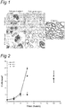
- Figure 3 shows light microscopic analysis of myelinated axons during regeneration.
- Semi-thin sections of the proximal site of the rat tibial nerve were analysed at 5 weeks post-injury of wild type rats, C6 deficient rats and C6 deficient rats reconstituted with C6. Few thinly myelinated axons are present in the wild type (WT) nerve while many thickly myelinated axons are present in the C6 deficient (C6-/-) nerve.
- WT wild type
- C6-/- C6 deficient
- the nerve from the rat that was reconstituted with C6 (C6+) shows less myelinated axons than the C6 deficient nerve.
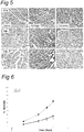
- Figure 4 shows the effect of C6 reconstitution on functional recovery of the nerve. Recovery of sensory function was measured with the footflick apparatus at currents ranging from 0.1 mA to 0.5 mA. Values were normalised to control levels.
- the arrow ( ⁇ ) indicates the time at which the crush injury was performed. Wild type rats take 4 weeks to fully recover while C6 deficient rats are already recovered at 3 weeks post-crush. C6 reconstitution in C6 deficient animals results in the wild type (slow) regeneration phenotype after crush.
- Statistical significance between C6-/- and WT (*) or C6+ ( ⁇ ) is for p ⁇ 0.05.
- FIG. 5 shows C1q, C4c and C3c immunostaining of injured wild type rat sciatic nerves treated with rhC1INH or vehicle (PBS) alone at 1 hour after nerve crush. High immunoreactivity for C1q is present in all crushed nerves, confirming C1q up-regulation after the crush injury.
- sCR1 soluble CR1
- Wild type PVG rats were treated with soluble CR1 (TP10 from Avant Immunotherapeutics, Inc.) at a dose of 15 mg/kg/day (TP10 soluble CR1 was obtained from Prof. P. Morgan, Cambridge, UK).
- Control rats were treated with the same volume (600 ⁇ l) of vehicle alone (PBS). Soluble CR1 or PBS was delivered i.p. 24 hours before the crush and every following day for a maximum of 8 injections (up to day 6 after crush). The sciatic nerve of the right leg was crushed and the left leg served as control. Both histology and sensory function were analysed.
- Figure 6 shows that in a functional analysis with the footflick test a faster recovery of the sensory function is seen in the sCR1-treated animals compared to the PBS-treated.
- the footflick test was performed as described above in Example 1.3.
- Example 4 Inhibition of complement activation facilitates axon regeneration and recovery in a model of peripheral nerve injury
- the C6 -/- rats carry a deletion of 31 basepairs (bp) in the C6 gene (Bhole and Stahl, 2004). Genotyping was performed according to Ramaglia et al (2007).
- C6 was purified from human serum (Mead et al., 2002). It was administered i.v. in eight C6 -/- rats at a dose of 4 mg/kg/day in PBS one day before the crush injury (day -1) and every day thereafter for 1 week (day 0, 1, 2, 3, 4, 5, 6). Eight wildtype and eight C6 -/- rats were treated with equal volume of vehicle (PBS) alone. The C6 -/- rats reconstituted with purified human C6 will be indicated in the text as C6 + .
- sCR1 Recombinant soluble complement receptor 1
- Plasma samples from wildtype PBS-treated, C6 -/- PBS-treated, C6 + and sCR1-treated rats were collected from the tail vein one day before the crush injury (day -1) and every following day until 1 week post-injury (day 0, 1, 2, 3, 4, 5, 6, 7). All samples were collected immediately before each injection of treatment. Plasma was separated and stored at -80°C until used to monitor C6 activity and sCR1 inhibitory effect via standard complement hemolytic assay (Morgan, 2000). Plasma levels of sCR1 were measured using ELISA assay as previously described (Mulligan et al., 1992) using serial dilutions assayed in duplicates.
- the print length (PL), toe (1 st to 5 th ) spread (TS) and intermediary toe (2 nd to 4 th ) spread (IT) were recorded from the uninjured normal foot (NPL, NTS, NIT) and the contralateral foot on the injured experimental side (EPL, ETS, EIT).
- the SFI was derived with the formula: -38.3*[(EPL-NPL)/NPL]+109.5*[(ETS-NTS)/NTS]+13.3*[(EIT-NIT)/NIT].
- EPL 60 mm
- ETS 6 mm
- the g-ratio is the numerical ratio of unmyelinated axon diameter to myelinated axon diameter and was calculated over the entire nerve section.
- the frequency of large caliber (>8 ⁇ m) myelinated fibers was calculated over the entire nerve section.
- the study was set up according to a scheme that extends over a period of 5 weeks.
- Time 0 is the time of the crush injury.
- Each group of animals was treated either with placebo (PBS) or purified C6 protein or sCR1 the day before the injury (day -1) and every day thereafter until 1 week post-injury.
- Blood was collected from each animal the day before the injury (day-1) and at days 0, 1, 2, 3, 5 and 7 post-injury to determine serum complement haemolytic activity.
- Functional analysis to determine recovery of motor function by the sciatic functional index (SFI) and recovery of sensory function by the footflick test, was performed 1 day before the injury for baseline values and every week thereafter until 5 weeks post-injury.
- Pathological analysis of the tibial nerves distal from the site of injury was performed at weeks 1, 3 and 5 post-injury to determine nerve regeneration.
- tibial nerve To follow the histological regeneration of the damaged nerve, we analyzed the tibial nerve at different time points. The regenerative process is marked by the occurrence of regenerative clusters of axons which are sprouts of the originally injured axon. Initially, the axon sprouts reside within a single Schwann cell cytoplasm but they are later separated by radial sorting. Once the 1:1 relationship between Schwann cell and axon is established, the pro-myelinating SC starts to ensheath the axon to form myelin and the basal lamina tube.
- the regenerative clusters appear as groups of small caliber, thinly myelinated axons within adjacent Schwann cells ( Figure 10 , arrows). Once one axon has reached its target, the rest of the axon sprouts are eliminated while the remaining axon increases in size.
- untreated wildtypes and the PBS vehicle-treated controls showed regenerative clusters of small caliber thinly myelinated axons in contrast to C6 -/- and sCR1-treated animals where regenerative clusters were absent confirming a faster recovery when C is inhibited or absent ( Figure 10 ).
- Blockade of C activation, and particularly MAC formation reduces tissue damage during nerve degeneration, appears to rescue the architecture necessary for the guidance of the axon and resulting in more efficient regeneration and recovery of function. Functional improvement in the absence of increased myelin sheath thickness can be explained by the increase in the number of large caliber fibers.
- Treatment started 1 day (day -1) before the injury (day 0) and it was repeated every day until 1 week.
- Statistical significance refers to p ⁇ 0.001 determined by a two way ANOVA test with Bonferroni correction. n.d., not determined.
- PVG/c Male 12 weeks old PVG/c were obtained from Harlan (UK). The animals weighed between 200 g and 250 g and were allowed acclimatization for at least two weeks before the beginning of the study. Animals were kept in the same animal facility during the entire course of the experiment and monitored for microbiological status according to the FELASA recommendations. Animals were housed in pairs in plastic cages. They were given rat chow and water ad libitum and kept at a room temperature of 20°C on a 12 hours: 12 hours light:dark cycle.
- sCR1 Recombinant soluble complement receptor 1
- Cetor Complement C1 inhibitor
- sCR1 was administered i.p. in twelve (12) rats at a dose of 15 mg/kg/day. Cetor was administered i.v. in six (6) rats at a dose of 50 U/rat/day. Twelve (12) rats were treated with equal volumes of vehicle (PBS) alone. The treatment was given one day before the crush injury (day -1) and every 24 hours (day 0, 1, 2) until the nerves were removed at 3 days post-injury.
- PBS vehicle
- the treatment was given one day before the crush injury (day -1) and every 24 hours (day 0, 1, 2) until the nerves were removed at 3 days post-injury.
- Ten (10) rats were treated either with sCR1 (6) or with PBS (4) up to 6 days post-injury (day -1, 0, 1, 2, 3, 4, 5, 6) and the nerves were removed 1 day after the end of the treatment (day 7).
- Plasma samples from PBS- and sCR1-treated rats were collected from the tail vein one day before the crush injury (day -1) and every following day (day 0, 1, 2) until the animals were sacrificed at 3 days after the injury. In the group treated up to 6 days, additional blood samples were collected at day 3, 5 and 7 after injury. All samples were collected immediately before each injection of treatment. Plasma was separated and stored at -80°C until used to monitor sCR1 inhibitory activity via standard complement hemolytic assay (Morgan, 2000).
- Plasma levels of sCR1 were measured using ELISA assay as previously described (Mulligan et al., 1992) using serial dilutions assayed in duplicates.
- Paraffin wax sections were stained using a three-step immunoperoxidase method. All the incubations were performed at room temperature (RT). Following deparaffination and rehydration, endogenous peroxidase activity was blocked with 1 % H 2 O 2 in methanol for 20 min. In all cases, microwave antigen retrieval was used (800 W for 3 min followed by 10 min at 440 W in 10 mM Tris/1 mM EDTA pH 6.5). To block the non-specific binding sites, slides were incubated in 10 % normal goat serum (NGS) in Tris buffered saline (TBS) for 20 min.
- NGS normal goat serum
- TBS Tris buffered saline
- Antibodies Monoclonal mouse anti-human Phosphorilated neurofilament (SMI31 clone) Stemberger (Lutherville, UK) 1:1000 Polyclonal rabbit anti-human MBP DakoCytomation (Glostrup, DK) 1:100 Monoclonal mouse anti-rat CD68 (ED1 clone) Serotec (Oxford, UK) 1:100 Polyclonal rabbit anti-rat C9 B.P.
- CD68 (ED1 clone)-immunoreactive cells were scored positive when the CD68 positive signal was associated with nuclei. Thirty non-consecutive sections of sciatic nerve per rat were scored. An average of 3 non-overlapping fields of view including >90% of the entire nerve area was taken for each section. Quantification of the MAC and MBP immunostaining was performed at 40x magnification on two non-overlapping fields per section examined. Ten sections per rat were scored. The surface area stained is expressed as percentage of total area examined.
- Frozen sciatic nerves from 2 untreated rats sacrificed at 2 days following the crush injury were homogenized using a pestle and mortar in liquid nitrogen in 20 mmol 1 -1 Tris (pH 7.4), 5 mmol 1 -1 1,4-dithio-DL-threitol (DTT) and 0.4 % SDS and 6 % glycerol.
- the homogenates were centrifuged at 10,000 x g , at 2 °C for 10 min. The supernatant fraction was collected and used for protein analysis. Protein concentrations were determined with a DC protein assay kit (Bio-Rad Laboratories, USA), using bovine serum albumin (BSA) as a standard.
- BSA bovine serum albumin
- Protein extracts (20 ⁇ g/sample) were boiled for 5 min, separated by 10 % SDS-PAGE and transferred to nitrocellulose membrane overnight at 4 °C. Prior to blotting, the nitrocellulose membranes were stained with Ponseau red for 30 sec to verify protein load. The membranes were pre-incubated in 50 mmol 1 -1 TrisHCl containing 0.5% Tween20 (TBST) and 5 % non-fat dried milk for 1 hour at RT. Blots were incubated for 2 hours in the polyclonal goat anti-factor Bb (fBb) (Quidel, San Diego, CA) diluted in TBST containing 5 % non-fat dried milk.
- fBb polyclonal goat anti-factor Bb
- the membranes were incubated for 1 hour in polyclonal rabbit anti-goat horseradish peroxidase-conjugated secondary antibody diluted 1:2000 in TBST containing 5 % non-fat dried milk. Membranes were washed in TBST for 30 min and immunoreactive bands were detected using enhanced chemiluminescence (ECL, Amersham, Piscataway, NJ, USA). Quantification of the immunoreactive bands was performed using Advanced Image Data Analyzer software v. 3.4 (Raytest, Germany).
- sCR1 Wallerian degeneration
- the sCR1 treated animals showed inhibition of complement activation in the crushed nerve ( Figure 12 ).
- the sCR1-treated nerves showed virtually no MAC deposits (0.8 ⁇ 0.9%) whereas MAC immunoreactivity covered 31.4 ⁇ 7.8% of the total area examined in the nerves of the PBS-treated rats.
- MAC immunoreactivity was undetectable in the uninjured control nerves.
- Deposition of C4c and C3c was also prevented in the sCR1-treated nerves whereas high amount of immunoreactivity was detected in the PBS-treated nerves (not shown).
- Neurofilament (SMI31) staining showed that the sciatic nerve of PBS-treated rats had empty and enlarged axonal spaces, delimited by a thin immunoreactive axolemma, and sparse axonal debris within the nerve which are signs of axonal swelling and degradation (data not shown).
- the sCR1-treated rats still showed the typical punctuated appearance of axons, similarly to the uninjured control nerve, demonstrating rescued axonal breakdown at 3 days after injury.
- Myelin (MBP) immunostaining revealed signs of myelin breakdown in nerves of PBS-treated rats at 3 days following the injury whereas the nerves of sCR1- treated rats showed the typical annulated myelin staining similar to uninjured control nerves, demonstrating rescued myelin degradation at this time point after injury (data not shown).
- the nerves of the PBS-treated rats showed large and asymmetrical CD68 immunoreactive cells (average size 103.6 ⁇ 71.8 ⁇ m 2 ) at 3 days post-injury, while small and round cells (average size 22.8 ⁇ 14.1 ⁇ m 2 ) were detected in the nerves of the sCR1-treated rats, a size and shape similar to that seen in the uninjured control nerves (average size 18.8 ⁇ 6.6 ⁇ m 2 ) (data not shown).
- CD68 immunoreactive cell size distribution was performed on 11 cells in the uninjured nerves, 778 cells in the PBS-treated nerves and 294 cells in the sCR1-treated nerves.
- Cell size distribution showed high variability in the PBS-treated nerves with cell dimension ranging from 20 to more than 400 ⁇ m 2 with a large population of cells of about 60 ⁇ m 2 .
- the sCR1-treated nerves showed cell dimension ranging from 0 to 40 ⁇ m 2 , similar to the size of cells found in the uninjured control nerves ( Figure 14 ).
- MBP and CD68 show macrophages engulfing myelin in the PBS-treated nerves while small resting macrophages are visible between the morphologically intact myelin sheaths of the uninjured and sCR1-treated nerves (data not shown). These results show that macrophages are activated in the PBS-treated nerves but not in the sCR1-treated ones.
- the myelin (MBP) staining showed normal annulated myelin morphology (data not shown) and the CD68 staining revealed a number of cells (59.8 ⁇ 28.3 cells/mm 2 ) similar to that observed in the sCR1-treated nerves.
- the average CD68 immunoreactive cell size (19.1 ⁇ 10.5 ⁇ m 2 ) and size distribution determined on 218 cells did not differ from sCR1-treated nerves or uninjured controls (data not shown).
- the colocalization of MBP and CD68 shows small resting macrophages between the morphologically intact myelin sheaths (data not shown).
- the present invention demonstrates that systemic treatment with sCR1, an inhibitor of classical, lectin and alternative pathways of complement activation, protects from early axon loss and myelin breakdown after peripheral nerve injury.
- sCR1 cannot protect the nerve from axonal degradation and myelin breakdown at 7 days post-injury even when hemolytic complement activity is maintained low. Therefore we conclude that inhibition of complement activation only affects the early events of WD. Lack of C4c deposition in the sCR1-treated nerves is a noteworthy finding because sCR1 inhibits the C3 convertase which is downstream of C4 cleavage, thus little effect on C4c deposition would be expected. However, as also noted in previous studies (Piddlesden et al., 1994), blockade of C-mediated damage by sCR1 will also inhibit overall C deposition on damaged tissue, also resulting in undetectable C4c levels.
- C-inhibitors are also promising tools in the treatment of non-autoimmune diseases, such as inherited peripheral neuropathies, where a secondary role of the immune system superimposed to the primary genetic defect has recently emerged (reviewed in Martini R and Toyka, 2004).
Description
- The present invention relates to methods and medicaments used for treating conditions that require axonal regeneration, e.g. in mammals affected by injury or disease of the central or peripheral nervous system. The medicaments used in these methods promote axonal regeneration by inhibition of the complement system.
- Axon degeneration occurs frequently in many types of chronic neurodegenerative diseases and in injuries to axons caused by toxic, ischemic, or traumatic insults. It may lead to separation of the neuron from its targets, resulting in loss of neuronal function. One model of axon degeneration is the self-destructive process observed at the distal portion of a transected axon upon injury, termed Wallerian degeneration (WD) as first described by Waller (1850). In the process of WD, if a nerve fiber is cut or crushed, the part distal to the injury (i.e. the part of the axon separated from the neuron's cell nucleus) will degenerate. Because most neuronal proteins are synthesised in the soma and carried to the axon by specialised axonal transport systems, degeneration of the transected axons has long been thought to result from starvation of necessary proteins and other materials. However, the discovery of a spontaneously occurring mutant mouse strain, C57BL/Wlds, whose axons survived for as long as weeks after transection suggested that Wallerian degeneration involves an active and regulated auto-destruction program.
- Indeed one of the most striking cellular responses during WD in the peripheral nervous system (PNS) is the proliferation and infiltration of macrophages (Bruck, 1997). Macrophages participate in a wide array of cellular responses during WD. Once activated, they release factors that are mitogenic for Schwann cells (Baichwal et al., 1988). The completion of WD relies on the phagocytic ability of macrophages to degrade myelin and axonal debris (Griffin et al., 1992). In addition, macrophages can degrade molecules inhibitory to axonal regeneration (Bedi et al., 1992) as well as release factors, such as interleukin-1 (IL-1), which can promote axonal growth via the induction of neurotrophic factors such as nerve growth factor (NGF) (Lindholm et al., 1987).
- The precise mechanisms responsible for macrophage recruitment during WD are not completely understood. One group of factors that may play a role in macrophage recruitment and activation is the serum complement proteins. The importance of complement proteins immune-mediated peripheral nerve injury has been investigated previously.
- Mead et al. (2002) showed that C6 deficient PVG/c rats, unable to form the membrane attack complex (MAC), exhibit neither demyelination nor axonal damage and significantly reduced clinical score in the antibody-mediated experimental autoimmune encephalomyelitis (EAE) model for multiple sclerosis when compared with matched C6 sufficient rats. However, levels of mononuclear cell infiltration were equivalent to those seen in C6 sufficient rats. Mead et al. (2002) concluded that demyelination and axonal damage occur in the presence of Ab and require activation of the entire complement cascade, including MAC deposition.
- Jung et al. (1995) disclosed that treatment with recombinant human soluble complement receptor type 1 (sCR1) markedly suppressed clinical signs of myelin-induced experimental autoimmune neuritis (EAN) in Lewis rats (an animal model of the human Guillain-Barré syndrome). Extended demyelination and axonal degeneration were also prevented. These findings underscore the functional importance of complement during inflammatory demyelination in the peripheral nervous system.
- Indeed, in EAN, complement depletion diminished myelin breakdown and macrophage recruitment in vivo (Feasby et al., 1987; Vriesendorp et al., 1995). Other groups have suggested that inhibition of the complement cascade reduces damage in neurodegenerative disease of the central nervous system (CNS) (e.g. Woodruff et al. 2006; Leinhase et al. 2006).
- Daily et al. (1998) disclose a significant reduction in the recruitment of macrophages into distal degenerating nerve in complement-depleted animals. Complement depletion also decreased macrophage activation, as indicated by their failure to become large and multivacuolated and their reduced capacity to clear myelin. In the normal situation the myelin is cleared, the proximal part of the nerve forms sprouts which slowly grow along the path of the degenerated nerve. However, regeneration is slow (2-2.5 mm/day) and the environment of a degenerated nerve is full of factors which inhibit the growth of the axon and the necessary growth factors can be limiting or even absent. Myelin itself has been proposed to be a major inhibiting factor. Therefore rapid clearance of myelin is considered a conditio sine qua non for axonal regeneration. Thus the delayed clearance of myelin in complement-depleted animals is expected to result in impaired axonal regeneration. These findings indicate a role for serum complement in both the recruitment and activation of macrophages during peripheral nerve degeneration as well as an active role for macrophages in promoting axonal regeneration.
- Indeed
US 6,267,955 discloses the methods wherein mononuclear phagocytes are administered at or near a site of injury or disease of the central or peripheral nervous system of a mammal in order to effect removal of the myelin debris that reportedly inhibits axonal regeneration, and for release of macrophage-derived cytokines that promote modulation of astrocytes and oligodendrocytes so as to support axonal regeneration. - Axonal degeneration is the main cause of disability both in hereditary and in acquired demyelinating neuropathies. While most current therapeutic research aims at restoring myelination, the present inventors focus on the consequence of demyelination: secondary axonal degeneration. As a model we have used acute demyelination and axonal degeneration after crush injury and subsequent regeneration of the nerve. It is an object of the present invention to provide for means and methods that promote and improve regeneration of nerves.
- The invention provides an inhibitor of a mammalian complement system for use according to the claims. Further provided is an inhibitor for use according to the invention, wherein the condition requiring axonal regeneration is a physical injury of a peripheral nerve. In one embodiment, the condition requiring axonal regeneration is a neurodegenerative disorder of the peripheral or central nervous system.
- Further provided is an inhibitor for use according to the invention, wherein the inhibitor inhibits formation of the membrane attack complex.
- The invention also provides an inhibitor for use according to the invention, wherein the inhibitor blocks activation of the classical pathway of complement activation. Also provided is an inhibitor for use according to the invention, wherein the inhibitor blocks activation of both the classical and alternative pathway of complement activation.
- Provided is an inhibitor for use according to the invention, wherein the inhibitor is an anti-sense oligonucleotide, aptamer, miRNA, ribozyme, siRNA that blocks expression of one or more of C3 convertase, C5, C6, C7, C8, C9.
- Also provided is an inhibitor for use according to the invention, wherein the inhibitor is administered at or near a site of injury.
- In the Examples herein we have observed activation of the complement (C)-system in the rat during WD and in human nerve biopsies of chronic demyelinating neuropathies. The present invention is based on the surprising finding that axonal regeneration is enhanced in rats that are deficient in the complement C6 factor. This surprising finding opens new ways to promote axonal regeneration by manipulation of the complement system and/or macrophage activation.
- In a first aspect, therefore, the invention pertains to a method for treating a condition requiring axonal regeneration. The method comprises the administration of an inhibitor of a mammalian complement system, or the administration of a medicament (e.g. a pharmaceutical composition) comprising the inhibitor. Preferably an effective amount of the inhibitor is administered. Thus, in this aspect the invention pertains to an inhibitor of a mammalian complement system, or a medicament comprising the inhibitor, for use in a method for treating a condition requiring axonal regeneration.
- In the context of the present invention "facilitating axonal regeneration" is distinguished from reducing or preventing axonal degeneration. Facilitation (or promotion) of axonal regeneration is herein understood to mean that regeneration of an axon is improved in subjects that are treated as compared to non-treated subjects. Improved regeneration of an axon preferably is regeneration that occurs at an earlier point in time (after axonal injury or after start of the treatment) in treated subject as compared to non-treated subjects. Improved regeneration of an axon may also comprise regeneration that occurs at a higher rate and/or to a larger extent in treated subject as compared to non-treated subjects. A medicament according to the invention thus preferably produces a gain of sensory or motor function.
- Improvement in axonal regeneration is preferably determined by functional tests that are relatively easily conducted in human subjects, e.g. recovery of sensory or motor function is preferably determined in a standardised test as is available in the art (see e.i. Wong et al., 2006; Jerosch-Herold, 2005). Suitable tests preferably are quantitative, standardised and more preferably have had their psychometric properties evaluated and quantified. Such tests include e.g. the Weinstein Enhanced Sensory Test (WEST) or the Semmes-Weinstein Monofilament Test (SWMT) and the shape-texture identification (STI) test for tactile gnosis. Improved axonal regeneration may experimentally be determined in test animals by functional tests for recovery of sensory or motor function as described by Hare et al. (1992) and De Koning et al. (1986). A medicament according to the invention thus preferably produces a gain of sensory or motor function, as may be determined in e.g. an above-indicated test.
- Improved axonal regeneration may also be experimentally determined in test animals by histological examination. E.g. improved remyelination may be determined by comparing measurements of myelin sheaths around the axon in treated animals vs. non-treated animals, whereby a thicker myelin sheath is indicative of improved remyelination. More efficient axonal regeneration may be determined as the production of single, large diameter, axon sprouts in treated animals as compared to clusters of smaller axons in non-treated animals.
- The appropriate dose of the inhibitor is that amount effective to promote axonal regeneration as may be seen by improvement of sensory or motor function as described above. By "effective amount," "therapeutic amount," or "effective dose" is meant that amount sufficient to elicit the desired pharmacological or therapeutic effects, thus resulting in effective treatment of the injury or disorder.
- In order to minimise nerve injury and/or to facilitate axonal regeneration at soon as possible, in the inhibitors for use according to the invention, the medicament is preferably administered shortly after the occurrence of the nerve injury, i.e. within 24, 12, 6, 3, 2, or 1 hours, more preferably within 45, 30, 20 or 10 minutes after the occurrence of the nerve injury. In one embodiment of the invention, the medicament may be administered (e.g. as a precautionary measure) prior to surgery with a risk of nerve injury (see below), so as to minimise nerve injury and/or to facilitate axonal regeneration immediately upon surgical injury of the nerve.
- A variety of conditions that require axonal regeneration may be treated with the inhibitors for use according to the invention. The conditions include injury of the PNS as well as injury of the CNS. The conditions include nerve trauma as a result of physical injuries as well as resulting from disease. Such diseases include immune-mediated inflammatory disorders or injuries and/or progressive neurodegenerative disorders which may be acquired and/or hereditary.
- The physical injuries of the PNS and CNS may be traumatic injuries, including surgical injuries, or non-traumatic injuries. Traumatic PNS and CNS injuries that may be treated with the inhibitors for use according to the invention include spinal cord lesions as well as traumatic wounds to peripheral nerves, including injuries from collisions, motor vehicle accidents, gun wounds, fractures, dislocations, lacerations, or some other form of penetrating trauma. Peripheral nerves injured through trauma that may be treated include the digital, median, ulnar, radial, facial, spinal accessory and brachial plexus nerves.
- Surgical PNS injuries are herein understood as injuries to peripheral nerves that arise when it becomes clinically necessary to remove or dissect a nerve during a surgical procedure. This occurs in thousands of surgical procedures each year. One example of surgically injured peripheral nerves that may be treated with the inhibitors for use according to the invention include e.g. the cavernous nerves that support erectile function and bladder control; these nerves are often damaged during surgical removal of a prostate tumour and the tissue around it. Another example of a surgically injured peripheral nerve that may be treated in accordance with the invention is the phrenic nerve after coronary artery bypass grafting (CABG).
- Non-traumatic physical PNS injuries that may be treated with the inhibitors for use according to the invention include compression and/or adhesion of peripheral nerves, also known as entrapment syndromes. The most common entrapment syndrome is carpal tunnel syndrome.
- In addition immune-mediated inflammatory disorders or injuries may be treated with the inhibitors for use according to the invention. These include demyelinating diseases of the central and peripheral nervous systems that are believed to have an autoimmune basis and result in nerve demyelination as a result of damage caused to oligodendrocytes or to myelin directly. Such demyelinating diseases include e.g. Guillain-Barré syndrome (GBS; also referred to as inflammatory demyelinating polyneuropathy, acute idiopathic polyradiculoneuritis, acute idiopathic polyneuritis, French Polio and Landry's ascending paralysis). Preferably, methods and/or the medicaments of the invention are applied to promote axonal regeneration subsequent to acute phase in GBS. Similarly chronic inflammatory demyelinating polyneuropathy (CIDP), considered the chronic counterpart of GBS, may be treated with the inhibitors for use according to the invention.
- Multiple sclerosis (MS) is another demyelinating disease that may be treated with the inhibitors for use according to the invention.
- Further neurodegenerative CNS and/or PNS disorders with a genetic component that may be treated with the inhibitors for use according to the invention include Amyotrophic Lateral Sclerosis (ALS, sometimes called Lou Gehrig's disease), Charcot-Marie-Tooth disease (Hereditary Motor and Sensory Neuropathy, HMSN) and Huntington Disease (HD).
- The complement system (see McAleer and Sim, 1993; Reid and Law, 1988) is concerned with host defence against infection. Upon activation of the system a catalytic set of reactions and interactions occur resulting in the targeting of the activating cell, organism or particle for destruction. The complement system comprises a set of over 30 plasma and membrane proteins that act together in a regulated cascade system to attack extra cellular forms of pathogens (e.g., bacterium). The complement system includes two distinct enzymatic activation cascades, the classical and alternative pathways which converge in a common terminal non-enzymatic pathway known as the membrane attack pathway.
- The first enzymatically activated cascade, known as the classical pathway, comprises several components, C1, C4, C2, C3 and C5 (listed by order in the pathway). Initiation of the classical pathway of the complement system occurs following binding and activation of the first complement component (C1) by both immune and non-immune activators. C1 comprises a calcium-dependent complex of components C1q, C1r and C1s, and is activated through binding of the C1q component. C1q contains six identical subunits and each subunit comprises three chains (the A, B and C chains). Each chain has a globular head region that is connected to a collagen-like tail. Binding and activation of C1q by antigen-antibody complexes occurs through the C1q head group region. Numerous non-antibody C1q activators, including proteins, lipids and nucleic acids, bind and activate C1q through a distinct site on the collagen-like stalk region. The Clqrs complex then catalyzes the activation of complement components C4 and C2, forming the C4b2a complex which functions as a C3 convertase.
- The second enzymatically activated cascade, known as the alternative pathway, is a rapid, antibody-independent route for complement system activation and amplification. The alternative pathway comprises several components, C3, Factor B, and Factor D (listed by order in the pathway). Activation of the alternative pathway occurs when C3b, a proteolytically cleaved form of C3, is bound to an activating surface agent such as a bacterium. Factor B is then bound to C3b, and cleaved by Factor D to yield the active enzyme, Ba. The enzyme Ba then cleaves more C3 to generate more C3b, producing extensive deposition of C3b-Ba complexes on the activating surface.
- Thus, both the classical and alternate complement pathways produce C3 convertases that split factor C3 into C3a and C3b. At this point, both C3 convertases further assemble into C5 convertases (C4b2a3b and C3b3bBb). These complexes subsequently cleave complement component C5 into two components: the C5a polypeptide (9 kDa) and the C5b polypeptide (170 kDa). The C5a polypeptide binds to a 7 transmembrane G-protein coupled receptor, which was originally associated with leukocytes and is now known to be expressed on a variety of tissues including hepatocytes and neurons. The C5a molecule is the primary chemotactic component of the human complement system and can trigger a variety of biological responses including leukocyte chemotaxis, smooth muscle contraction, activation of intracellular signal transduction pathways, neutrophil-endothelial adhesion, cytokine and lipid mediator release and oxidant formation.
- The larger C5b fragment binds sequentially to later components of the complement cascade, C6, C7, C8 and C9 to form the C5b-9 membrane attack complex ("MAC"). The lipophylic C5b-9 MAC can directly lyse erythrocytes, and in greater quantities it is lytic for leukocytes and damaging to tissues such as muscle, epithelial and endothelial cells. In sublytic amounts, the C5b-9 MAC can stimulate upregulation of adhesion molecules, intracellular calcium increase and cytokine release. In addition, at sublytic concentrations the C5b-9 MAC can stimulate cells such as endothelial cells and platelets without causing cell lysis. The non-lytic effects of C5a and the C5b-9 MAC are comparable and interchangeable.
- Although the complement system has an important role in the maintenance of health, it has the potential to cause or contribute to disease.
- An inhibitor for use according to the present invention preferably inhibits or blocks the formation of the membrane attack complex. The inhibitor preferably blocks activation of the complement system through both the classical and alternative pathway of complement. A preferred inhibitor is an inhibitor that blocks C3 convertase and MAC assembly. A further preferred inhibitor is an inhibitor that blocks one or more of C5, C6, C7, C8 and C9.
- Methods for producing nucleic acid inhibitors of complement such as anti-sense oligonucleotide, aptamer, miRNA, ribozyme, siRNA, are known to the skilled person persé. Preferably such nucleic acid inhibitors comprise one or more modified nucleotides such as e.g. 2'-O substituted ribonucleotides, including alkyl and methoxy ethyl substitutions, peptide nucleic acid (PNA), locked nucleic acid (LNA) and morpholino antisense oligonucleotides and ethylene-bridged nucleotides (ENA) and combinations thereof.
- In the above inhibitors for use according to the invention, the compounds may be administered by any convenient route, for example by infusion or bolus injection. Various delivery systems are known and can be used for delivery of the inhibitor compounds. These include encapsulation in liposomes, microparticles, or microcapsules. Although in the inhibitors for use according to the invention administration of the compounds by oral and/or mucosal routes (intranasal, inhalation, rectal) is not excluded, usually the complement inhibitors will be administered parenterally, including e.g. intradermal, intramuscular, intraperitoneal, intravenous, and subcutaneous routes. The compounds may be administered systemically or may be used by local, topical or regional administration at or near a site of disease or injury, e.g. using injection and/or any neurosurgically suitable technique.
- The disclosure further relates to a pharmaceutical preparation comprising as active ingredient a complement inhibitor as defined above. The composition preferably at least comprises a pharmaceutically acceptable carrier in addition to the active ingredient. The pharmaceutical carrier can be any compatible, non-toxic substance suitable to deliver the inhibitors to the patient. Sterile water, alcohol, fats, waxes, and inert solids may be used as the carrier. Pharmaceutically acceptable adjuvants, buffering agents, dispersing agents, and the like, may also be incorporated into the pharmaceutical compositions.
- For oral administration, the inhibitor can be administered in solid dosage forms, such as capsules, tablets, and powders, or in liquid dosage forms, such as elixirs, syrups, and suspensions. Active component(s) can be encapsulated in gelatine capsules together with inactive ingredients and powdered carriers, such as glucose, lactose, sucrose, mannitol, starch, cellulose or cellulose derivatives, magnesium stearate, stearic acid, sodium saccharin, talcum, magnesium carbonate and the like. Both tablets and capsules can be manufactured as sustained release products to provide for continuous release of medication over a period of hours. Compressed tablets can be sugar coated or film coated to mask any unpleasant taste and protect the tablet from the atmosphere, or enteric-coated for selective disintegration in the gastrointestinal tract. Liquid dosage forms for oral administration can contain colouring and flavouring to increase patient acceptance.
- The inhibitors are however preferably administered parentally. Suitable carriers for parental formulations include saline, buffered saline, dextrose, and water. Typically compositions for parenteral administration are solutions in sterile isotonic aqueous buffer. Sterilisation is readily accomplished by filtration through sterile filtration membranes, prior to or following lyophilisation and reconstitution. A typical composition for intravenous infusion could be made up to contain 10 to 50 ml of sterile 0.9% NaCl or 5% glucose optionally supplemented with a 20% albumin solution and an appropriate amount (1 to 1000 µg) of the inhibitor. A typical pharmaceutical composition for intramuscular injection would be made up to contain, for example, 1 - 10 ml of sterile buffered water and 1 to 1000 µg of the of the inhibitor. Methods for preparing parenterally administrable compositions are well known in the art and described in more detail in various sources, including, for example, Remington's Pharmaceutical Science (15th ed., Mack Publishing, Easton, PA, 1980) (incorporated by reference in its entirety for all purposes). Where necessary, the composition may also include a solubilising agent and a local anaesthetic such as lignocaine to ease pain at the site of the injection. Generally, the ingredients will be supplied either separately or mixed together in unit dosage form, contained in a hermetically sealed container such as an ampoule or sachette indicating the quantity of active agent in activity units. Where the composition is to be administered by infusion, it can be dispensed with an infusion bottle containing sterile pharmaceutical grade 'Water for Injection' or saline. Where the composition is to be administered by injection, an ampoule of sterile water for injection or saline may be provided so that the ingredients may be mixed prior to administration.
- In this document and in its claims, the verb "to comprise" and its conjugations is used in its non-limiting sense to mean that items following the word are included, but items not specifically mentioned are not excluded. In addition, reference to an element by the indefinite article "a" or "an" does not exclude the possibility that more than one of the element is present, unless the context clearly requires that there be one and only one of the elements. The indefinite article "a" or "an" thus usually means "at least one".
-
-
Figure 1 . The effect of complement C6-deficiency on regeneration of the tibial nerve. The right sciatic nerve was crushed for 30 seconds in wild type and C6-deficient PVG rats. Tibial nerve was analyzed 1 and 5 weeks after injury. Control picture: left tibial nerve of a PVG rat. -
Figure 2 . C6 deficiency leads to a delayed influx/activation of phagocytic cells. ED1 (CD68) immunoreactive (-ir) cells were counted in non-consecutive sections of sciatic nerves from WT (wild type), C6-/- (C6-deficient) and C6+ (C6-deficient rats supplemented with C6) rats at 0, 24, 48 and 72h post-injury. Statistical significance indicated by the asterisk (*) refers to p<0.05. -
Figure 3 . The effect of C6 reconstitution on regeneration. Analysis of myelinated axons during regeneration. Light microscopy on semi-thin sections of the proximal site of the rat tibial nerve at 5 wk post-injury. From left to right: uncrushed nerve; wild type nerve (WT); C6-deficient nerve (C6-/-); and, C6-deficient nerve reconstituted with C6 (C6+). -
Figure 4 . The effect of C6 reconstitution on functional recovery. Recovery of sensory function as measured with the footflick apparatus at currents ranging from 0.1 mA to 0.5 mA. Values are normalised to control levels. The arrow (→) indicates the time at which the crush injury was performed. WT = wild type rats; C6-/- = C6-deficient rats; and, C6+ = C6 reconstitution in C6-deficient animals. Statistical significance between C6-/- and WT (*) or C6+ (†) is for p<0.05. -
Figure 5 . Recombinant human C1-inhibitor (rhC1INH) inhibits complement activation after crush. C1q, C4c and C3c immunostaining of injured wild type rat sciatic nerves treated with rhC1INH or vehicle (PBS) alone. -
Figure 6 . The effect of soluble CR1 on post traumatic nerve regeneration. Recovery of sensory function as measured with the footflick apparatus at currents ranging from 0.1 mA to 0.5 mA. Values are normalised to control levels. The arrow (→) indicates the time at which the crush injury was performed. PBS = control with vehicle only; sCR1 = soluble CR1. -
Figure 7 . Activation of macrophages after nerve crush is dependent on activation of a downstream component of the complement cascade. CD68 positive cells were determined by immunostaining with the ED1 antibody. In wild type (WT) and vehicle (PBS) treated animals the number of CD68 positive cells in the distal part of the lesioned sciatic nerve increased 72 hours after nerve crush. Treatment with sCR1 blocked this activation to a similar level as seen in C6 deficient rats (C6-). C6 reconstitution in the C6 deficient animals (C6+) resulted in almost complete recovery from this block in activation. -
Figure 8 . Recovery of function. (a) Sciatic Functional Index (SFI) and footprints after sciatic nerve crush injury (time=0) in wildtype (n=8), C6-/- (n=8) and C6+ (n=8) rats showing recovery of motor function over a period of 5 weeks post-injury. Control levels are near 0 whereas values near -140 indicate complete loss of function (week 1). The asterisks (*) refers to significant differences between the wildtype and C6-/- group of rats whereas the cross(†) refers to significant differences between C6-/- and C6+ group of rats with p≤0.05 determined by two way ANOVA test with Bonferroni correction. The wider toe spread in the C6-/- footprint (week 4) compared to the wildtype and C6+ footprints indicates increased muscle strength. (b) Footflick analysis after sciatic nerve crush injury (time=0) in wildtype (n=8), C6-/- (n=8) and C6+ (n=8) rats showing recovery of sensory function over a period of 5 weeks post-injury. Values are expressed as percentage of control levels (100% function). The asterisks (*) refers to significant differences between the wildtype and C6-/- group of rats whereas the cross(†) refers to significant differences between C6-/- and C6+ group of rats with p≤0.05 determined by two way ANOVA test with Bonferroni correction. -
Figure 9 . Recovery of sensory function of C inhibited rats. Footflick analysis after sciatic nerve crush injury (time=0) in wildtype PBS- (n=6) and sCR1- (n=6) treated rats showing recovery of sensory function over a period of 5 weeks post-injury. Values are expressed as percentage of control levels (100% function). The asterisks (*) refers p≤0.05 determined by two way ANOVA test with Bonferroni correction. -
Figure 10 . Pathology. Thionine staining and electron microscopy of cross-sections of the distal ends of tibial nerves from uninjured (n=6), wildtype (n=5), C6-/- (n=5) and sCR1-treated (n=6) rats at 5 weeks following the crush injury. Note the presence of regenerative clusters of small caliber, thinly myelinated axons in the wildtype nerve (arrows →) whereas single large caliber axons are present in the C6-/- and sCR1-treated nerves, similarly to the uninjured control. Bar is 50µm (light microscopy, left panels) and 10µm (electron microscopy, right panels). -
Figure 11a and 11b . sCR1 inhibition of complement activation (A) Plasma sCR1 levels in sCR1-treated rats, showing concentration of sCR1 over time with daily treatment. (B) Plasma hemolytic activity of PBS- and sCR1-treated rats, showing decreased activity in the sCR1-treated rats compared to the PBS-treated controls. (A,B) Day 0 is the day of the crush injury. Rats received i.p injections of sCR1 (15mg/kg/day) or PBS (equal volume) at days (-1, 0, 1, 2, 3, 4, 5 and 6). Blood was collected immediately before each treatment. Data represents mean±SD. Statistical significance is determined by two-way ANOVA with Bonferroni correction (* is p≤0.001). -
Figure 12 . sCR1 inhibition of complement activation. Quantification of MAC immunoreactivity expressed as percentage of total area scored. Data represents mean±SD. Statistical significance is determined by two-way ANOVA with Bonferroni correction (* is p≤0.001). -
Figure 13 . Analysis of macrophages. Quantification of CD68-ir cells in non-consecutive sections of sciatic nerves, showing a high number of cells in the PBS-treated nerves and slight increase in the sCR1-treated nerves, compared to the uninjured nerve. Data represents mean±SD. Statistical significance is determined by two-way ANOVA with Bonferroni correction. -
Figure 14 . Analysis of macrophages. Size distribution of CD68-ir cells sciatic nerves from uninjured nerve (a), PBS-treated (b) and sCR1-treated (c) nerves at 3 days post-injury. Note the shift in the peak of CD68-ir cell size distribution from a size of 0-40µm2 in the uninjured and sCR1-treated nerves to a size of 40-120µm2 in the PBS-treated nerves. -
Figure 15 . Analysis of alternative pathway activation. (a) Western blotting analysis of rat sciatic nerves at 2 days post-injury, showing higher amount of cleaved fBb protein in the injured nerves compared to the uninjured controls. (b) Relative quantification of fBb immunoreactive bands. The fBb immunoreactivity in uninjured controls is defined as 1.0 fold relative expression. Values are normalized to total protein load and represent mean±SD of three blots. Statistical significance is determined by unpaired t-test. - We have explored the role of the complement system in acute and chronic nerve injury and during regeneration. As a model we used the complement C6 deficient PVG rat strain (Bhole and Stahl, 2004) and compared this with wild type PVG rats. Since the complement system has many functions we chose an animal model in which only the most terminal effectors of the complement cascade was defective.
- The effect of complement inhibition on nerve regeneration was studied in the acute model of nerve crush (Glass, 2004). The right sciatic nerve was crushed for 30 seconds in wild type as well as in the C6-deficient PVG rats. Tibial nerve was then analyzed 1 and 5 weeks after injury (see
Figure 1 ). - At one week, electron microscopy shows equally severe degeneration in wild type and in C6 deficient rats. At 5 weeks, the C6 deficient rats already show myelinated axons, whereas the wild type rats exhibit incipient recovery. In the C6 deficient animals most myelinated axons show the normal one to one ratio with Schwann cells (compare with the control picture of the left tibial nerve of a PVG rat). In contrast, in the wild type rats there are several myelinated fibres in each regenerative cluster.
- We found two effects of C6 deficiency on post-traumatic nerve recovery:
- 1) The clearance of myelin during Wallerian degeneration was delayed in the C6 deficient animals. Wild type rats showed signs of WD (myelin degeneration, macrophage activation) already after 24 hours. In C6-deficient animals this process was delayed. Only after 72 hrs myelin degeneration was visible and macrophage activation did not occur. After one week both types of animals showed severe nerve degeneration.
- 2) Unexpectedly however, the post-traumatic nerve recovery was much better in complement component C6 deficient rats compared to wild-type rat. Remyelination of single axons occurred much faster in the C6 deficient animals and the sprouting process was more efficient since a single, large diameter, axon sprout was produced rather than a cluster of smaller axons. See
Figure 1 . - In view of the important role of macrophages in myelin clearance, we next analyzed the number and activation state of macrophages after crush.
- ED1 (CD68) immunoreactive (-ir) cells were counted in non-consecutive sections of crushed sciatic nerves at 0, 24, 48 and 72 hr post-injury that were taken from wild type rats, C6 deficient rats and C6 deficient rats that were supplemented with C6, respectively.
- In both the wild type and C6 deficient animals, CD68 (ED1 antibody) positive cells accumulated in the crushed nerve. However, the C6 deficient animals showed a delayed appearance of CD68 positive cells (
Figure 2 , compare solid and dotted line). C6 suppletion restored the accumulation of CD68 cells (see 72 hr time point). In the C6 deficient animals there was a lack of activation of macrophages, as assayed by immunohistochemistry CR3 (ED7 antibody) staining (not shown). - Since lymphnodes of these animals contain CR3 positive cells we could exclude that the C6 deficient animals are defective in macrophage activation per se. In addition, upon C6 reconstitution, the accumulation of CD68 positive cells and CR3 expression on macrophages was restored and subsequently myelin degeneration occurred. This directly links steps in the complement pathway downstream of C6, i.e. Membrane Attack Complex (MAC) formation to WD.
- After 7 days equal number of CD68 positive cells were found in the C6 deficient and wild type cells. These cells do not display ED7 (CR3) in the C6 deficient animals and are most likely not activated macrophages (data not shown).
-
Figure 3 shows light microscopic analysis of myelinated axons during regeneration. Semi-thin sections of the proximal site of the rat tibial nerve were analysed at 5 weeks post-injury of wild type rats, C6 deficient rats and C6 deficient rats reconstituted with C6. Few thinly myelinated axons are present in the wild type (WT) nerve while many thickly myelinated axons are present in the C6 deficient (C6-/-) nerve. The nerve from the rat that was reconstituted with C6 (C6+) shows less myelinated axons than the C6 deficient nerve. -
Figure 4 shows the effect of C6 reconstitution on functional recovery of the nerve. Recovery of sensory function was measured with the footflick apparatus at currents ranging from 0.1 mA to 0.5 mA. Values were normalised to control levels. The arrow (→) indicates the time at which the crush injury was performed. Wild type rats take 4 weeks to fully recover while C6 deficient rats are already recovered at 3 weeks post-crush. C6 reconstitution in C6 deficient animals results in the wild type (slow) regeneration phenotype after crush. Statistical significance between C6-/- and WT (*) or C6+ (†) is for p<0.05. - We conclude that the observed effect on regeneration of the PNS after crush is due to the C6 deficiency since reconstitution of the C6 deficient rats with purified human C6 restores the wild type phenotype in neuropathological and functional assays (
Figure 3 and 4 ). - We tested whether recombinant human C1-inhibitor (rhC1INH; obtained from Pharming, Leiden, The Netherlands) is able to inhibit the rapid (1 hr) complement activation after nerve crush.
Figure 5 shows C1q, C4c and C3c immunostaining of injured wild type rat sciatic nerves treated with rhC1INH or vehicle (PBS) alone at 1 hour after nerve crush. High immunoreactivity for C1q is present in all crushed nerves, confirming C1q up-regulation after the crush injury. C4c and C3c immunoreactivity was detected in the PBS-treated nerves as expected but no C4c and C3c immunoreactivity was detected in the nerves from the rhC1INH treated rats sacrificed at 1 hr post-injury. This demonstrates effective blockade of the complement cascade by rhClINH after crush and suggests that the alternative pathway of complement activation is not involved in the crush injury model of Wallerian degeneration. Thus, activation of the complement cascade after nerve crush occurs through the classical pathway. There is one caveat however: due to the short half life of rhC1INH in rats, we could only monitor C3 andC4 cleavage 1 hr after crush. Therefore we cannot exclude that activation through the alternative pathway occurs at a later time point. - Next, we tested the effect of soluble CR1 (sCR1) on post traumatic nerve regeneration. sCR1 inhibits the C3/C5 convertase, and thereby affects both the classical and alternative pathway of the complement system.
- Wild type PVG rats were treated with soluble CR1 (TP10 from Avant Immunotherapeutics, Inc.) at a dose of 15 mg/kg/day (TP10 soluble CR1 was obtained from Prof. P. Morgan, Cardiff, UK). Control rats were treated with the same volume (600 µl) of vehicle alone (PBS). Soluble CR1 or PBS was delivered i.p. 24 hours before the crush and every following day for a maximum of 8 injections (up to day 6 after crush). The sciatic nerve of the right leg was crushed and the left leg served as control. Both histology and sensory function were analysed.
-
Figure 6 shows that in a functional analysis with the footflick test a faster recovery of the sensory function is seen in the sCR1-treated animals compared to the PBS-treated. The footflick test was performed as described above in Example 1.3. - Histological analysis of nerves at 72 hrs after the crush shows that sCR1 strongly inhibited the influx and activation of macrophages (see
Figure 7 ). sCR1 treatment resulted in similar levels of inhibition of macrophage activation as measured by CD68 positivity as compared to deficiency of C6. - This study was approved by the Academic Medical Center Animal Ethics Committee and complies with the guidelines for the care of experimental animals. Male 12 weeks old PVG/c (wildtype) were obtained from Harlan (UK) and PVG/c- (C6-/-) rats were bred in our facility. The animals weighed between 200 g and 250 g and were allowed to acclimatize for at least two weeks before the beginning of the study. Animals were kept in the same animal facility during the entire course of the experiment and monitored for microbiological status according to the FELASA recommendations. Animals were housed in pairs in plastic cages. They were given rat chow and water ad libitum and kept at a room temperature of 20°C on a 12 hours: 12 hours light:dark cycle.
- The C6-/- rats carry a deletion of 31 basepairs (bp) in the C6 gene (Bhole and Stahl, 2004). Genotyping was performed according to Ramaglia et al (2007).
- C6 was purified from human serum (Mead et al., 2002). It was administered i.v. in eight C6-/- rats at a dose of 4 mg/kg/day in PBS one day before the crush injury (day -1) and every day thereafter for 1 week (
day - Recombinant soluble complement receptor 1 (sCR1) was obtained as previously described (Piddlesden et al., 1994). sCR1 was administered i.p. in six rats at a dose of 15 mg/kg/day. Six rats were treated with equal volumes of vehicle (PBS) alone. The treatment was given one day before the crush injury (day -1) and every day thereafter for 1 week (
day - Blood samples from wildtype PBS-treated, C6-/- PBS-treated, C6+ and sCR1-treated rats were collected from the tail vein one day before the crush injury (day -1) and every following day until 1 week post-injury (
day - All experiments were conducted by the same investigator who was blinded of the genotype and treatment groups. Both motor and sensory tests were performed at the same time during the day, every week until 5 weeks post-injury. Recovery of motor function was assessed using a standardized walking track analysis and derived sciatic functional index (SFI) according to Hare et al (1992). Briefly, the rats were allowed to walk across a plexiglas platform while their walking patter was recorded by a camera underneath the platform. An index of the sciatic nerve function was calculated from the recorded footprints using the ImagePro analysis program (Media Cybernatics, The Netherlands). The print length (PL), toe (1st to 5th) spread (TS) and intermediary toe (2nd to 4th) spread (IT) were recorded from the uninjured normal foot (NPL, NTS, NIT) and the contralateral foot on the injured experimental side (EPL, ETS, EIT). The SFI was derived with the formula: -38.3*[(EPL-NPL)/NPL]+109.5*[(ETS-NTS)/NTS]+13.3*[(EIT-NIT)/NIT]. In case on no print produced by the animals, the standard values of EPL=60 mm, ETS=6 mm and EIT=6 mm were used according to De Koning et al (1986). Recovery of sensory function was assessed with the footflick test according to De Koning et al (1986). Briefly, a shock source with a variable current of 0.1-0.5mA was used. Recordings were performed one day before the injury and every week until 5 weeks post-injury. The rats were immobilized and two stimulation electrodes were placed at the same point on the rat foot sole for every animal and stimulation. A response was scored positive if the rat retracted its paw. The current (mA) at which the retraction occurred was recorded. Values are expressed as percentage of normal function.
- All the surgical procedures were performed aseptically under deep isoflurane anesthesia (2.5% vol isoflurane, 1 L/min O2 and 1 L/min N2O). The left thigh was shaved and the sciatic nerve was exposed via an incision in the upper thigh. The nerve was crushed for three 10 s periods at the level of the sciatic notch using smooth, curved forceps (No.7). The crush site was marked by a suture which did not constrict the nerve. On the right side, sham surgery was performed which exposed the sciatic nerve but did not disturb it. A suture was also placed. The muscle and the skin were then closed with stitches. The right leg served as control. Following the crush, the rats were allowed to recover for 1 (wildtype n=5; C6-/- n=5; C6+ n=2), 3 (wildtype n=6; C6-/- n=6; C6+ n=3) and 5 (wildtype n=5; C6-/- n=5; C6+ n=3; wildtype sCR1-treated n=6; wildtype PBS-treated n=6) weeks.
- All animals were intracardially perfused with 4% paraformaldehyde in piparazine-N-N'-bis (2-ethane sulphonic acid) (PIPES) buffer (pH 7.6), under deep isoflurane anesthesia. Left and right tibial nerves were removed from each animal and postfixed with 1% glutaraldehyde, 1% paraformaldehyde and 1% dextran (MW 20,000) in 0.1 M PIPES buffer (pH 7.6). They were divided into one proximal and one distal segment of 10 mm length. Each segment was conventionally processed into epoxy resin. Semithin resin sections of 0.5 µm were stained with thionine and acridine orange and images were captured with a light microscope (Leica DM5000B, The Netherlands) connected to a digital camera (Leica DFC500, The Netherlands). Electron microscopy was performed on ultrathin sections of the tibial nerve from wildtype and C6-/- rats at 5 weeks following the crush injury. Sections were contrasted with uranyl acetate and lead citrate as previously described (King, 1999). Images were captured with a digital camera attached to an electron microscope (
FEO 10, Philips, The Netherlands). The number of regenerative clusters of axons at 5 weeks post-injury was determined on semithin resin section. The entire section was scored per each animal in each group. The g-ratio is the numerical ratio of unmyelinated axon diameter to myelinated axon diameter and was calculated over the entire nerve section. The frequency of large caliber (>8µm) myelinated fibers was calculated over the entire nerve section. - Two way ANOVA with Bonferroni correction was performed to determine statistically significant differences in the hemolytic assay (p<0.001), ELISA assay (p<0.001), SFI (p≤0.05), Footflick test (p≤0.05).
- To test the effects of C activation on nerve regeneration after acute trauma we determined the effect of C on recovery from crush injury of the sciatic nerve in the rat model in two complementary ways, first by examining the effects of C6 deficiency (C6-/-) and second by inhibition of C activation.
- The study was set up according to a scheme that extends over a period of 5 weeks.
Time 0 is the time of the crush injury. Each group of animals was treated either with placebo (PBS) or purified C6 protein or sCR1 the day before the injury (day -1) and every day thereafter until 1 week post-injury. Blood was collected from each animal the day before the injury (day-1) and atdays weeks - Both functional recovery and effects on histology were determined. As controls for C6 deficiency we reconstituted C6-/- rats with purified C6 protein (4mg/kg/day; n=8) (C6+), which restored the plasma hemolytic activity (CH50) to wildtype levels (>80%; p<0.001, two way ANOVA) (Table 1). Inhibition of C activation was achieved by systemic treatment with soluble C receptor 1 (sCR1) (15mg/kg/day; n=6), a recombinant soluble form of the human membrane C regulator CR1 which inhibits all three C activation pathways (Weisman et al., 1990). This treatment reduced hemolytic C activity to about 30% of the PBS vehicle-treated controls (n=6; p<0.001, two way ANOVA) over the entire course of the treatment (Table 1). We found this level of C inhibition in the plasma completely abrogated deposition of activated C in the nerve at 3 days after injury.
- Recovery of motor function was monitored every week after injury by measuring the sciatic functional index (SFI) calculated from the rats walking pattern (Hare et al., 1992). At 1 week post-injury, none of the animals used the foot of the injured leg to walk, failing to produce a footprint on the walking platform, suggesting complete loss of muscle innervation in the leg. From
week 2 post-injury and throughout the whole study C6-/- rats (n=16) produced an SFI significantly higher than wildtype animals (n=16; p≤0.05, two way ANOVA) (Figure 8a ). A higher SFI results from an increase in the print length and toe spreading parameters and indicates re-innervation of the calf and small foot muscles, respectively. Reconstitution of the C6-/- rats with purified C6 protein (C6+) significantly reduced the SFI (n=8; p≤0.05, two way ANOVA) to wildtype levels atweek 4 and 5 post-injury. These data demonstrate that C6 deficiency in rats results in a faster recovery of motor function compared to wildtype. Recovery of sensory function was assayed with the footflick test. At 1 week post-injury, none of the animals retracted their paw when the footsole was stimulated by an electric shock at 0.5mA, suggesting complete loss of sensory innervation. Fromweek 2 toweek 3 post-injury, the C6-/- rats showed 20-50% greater recovery of sensory function compared to wildtype (n=16) and C6+ (n=8) rats (p≤0.05, two way ANOVA). The sensory function did not differ between groups atweeks 4 and 5 post-injury (Figure 8b ). Similarly, animals treated with sCR1 showed a faster (10-30% increase, n=6; p<0.05, two way ANOVA) recovery of sensory function than the PBS-treated rats (n=6) betweenweeks 2 and 4 post-injury (Figure 9 ). These data indicate that both C deficiency and inhibition of C activation accelerate and improve the return of sensory innervation to the footsole after sciatic crush injury. - To follow the histological regeneration of the damaged nerve, we analyzed the tibial nerve at different time points. The regenerative process is marked by the occurrence of regenerative clusters of axons which are sprouts of the originally injured axon. Initially, the axon sprouts reside within a single Schwann cell cytoplasm but they are later separated by radial sorting. Once the 1:1 relationship between Schwann cell and axon is established, the pro-myelinating SC starts to ensheath the axon to form myelin and the basal lamina tube. At this stage, the regenerative clusters appear as groups of small caliber, thinly myelinated axons within adjacent Schwann cells (
Figure 10 , arrows). Once one axon has reached its target, the rest of the axon sprouts are eliminated while the remaining axon increases in size. On histological sections at 5 weeks post-injury, untreated wildtypes and the PBS vehicle-treated controls showed regenerative clusters of small caliber thinly myelinated axons in contrast to C6-/- and sCR1-treated animals where regenerative clusters were absent confirming a faster recovery when C is inhibited or absent (Figure 10 ). The frequency of high caliber (>8µm) myelinated fibers was increased in the C6-/- (0.59±0.20%, n=5) and sCR1-treated (0.58±0.11%, n=6) animals compared to the untreated wildtypes (0.05±0.02; n=5), the C6+ (0.06±0.01; n=3) and the PBS vehicle-treated (none; n=6) controls at 5 weeks post-injury whereas no difference in the frequency of low (<4µm) and intermediate (4-8µm) caliber myelinated fibers was found. The myelin thickness was not altered between groups of animals (g-ratio of 0.69±0.01, n=5, wildtype; 0.65±0.02, n=5, C6-/-; 0.65±0.01, n=3, C6+; 0.70±0.01, n=6, sCR1-treated; 0.66±0.003, n=6, PBS vehicle-treated) (data not shown). - Taken together, these data show that axonal regeneration and functional recovery after peripheral nerve injury are enhanced in the absence of C6 or when C activation is inhibited by sCR1. Thus the ability to form MAC is a negative determinant of nerve recovery.
- Functional recovery after axonal crush injury requires axons to re-enter the Schwann cell tubes injured at the crush site. Once in the distal stump, the axons need to re-navigate the paths followed before injury and generate specific synapse with exactly the same muscle fiber they had previously innervated. In this task they are guided by attractive and repulsive molecular cues (Tessier-Lavigne and Goodman, 1996; Yu and Bargmann, 2001) but recent evidence showed that physical factors also play a key role (Nguyen et al., 2002). Thus, maintenance of intact endoneurial tubes could be of high importance for the regenerating adult peripheral nerve.
- Blockade of C activation, and particularly MAC formation, reduces tissue damage during nerve degeneration, appears to rescue the architecture necessary for the guidance of the axon and resulting in more efficient regeneration and recovery of function. Functional improvement in the absence of increased myelin sheath thickness can be explained by the increase in the number of large caliber fibers.
- A wealth of evidence over the last decade points to a possible beneficial role of macrophages during recovery (Kiefer et al., 2001). Later after injury, macrophages secrete anti-inflammatory cytokines which are involved in resolving the inflammatory process. Once the inflammation terminates, macrophages contribute to Schwann cell proliferation and survival, remyelination and recovery through the secretion of growth and differentiation factors. We have shown that, early after injury, C inhibition markedly reduces infiltration of endoneurial macrophages (5-fold increase, compared to 25-fold in the absence of C inhibition) (Ramaglia et al., 2007). We postulated that this is due to the proliferation of the resident macrophage population while little contribution comes from the haematogenous macrophages. It is possible that we separated the detrimental effect of the haematogenous macrophages from the beneficial effect which can be exerted by the endoneurial population.
- Our findings open the door to a novel therapeutic approach in which blockade of the C cascade, or selective inhibition of MAC, promotes regeneration after traumatic injury and in peripheral neuropathies and neurodegenerative diseases where C-dependent nerve damage has been reported.
Table 1. Plasma haemolytic activity (%CH50). Day -1 Day 0 (crush) Day 1Day 2Day 3Day 4 Day 5Day 7Wildtype PBS-treated 91,6±1.0 91,7±1.1 81.2±1.7 89.5±1.5 86.1±1.4 n.d. 82.1±1.7 90.8±4.1 C6 deficient PBS-treated 14.0±0.1 n.d. n.d. 12.8±0.2 n.d. 13.5±0.3 n.d. 15.7±0.4 C6 deficient C6-treated (C6+) 14.0±0.1 n.d. n.d. 78.5±0.9* n.d. 76.9±0.8* n.d. 78.5±1.6* Wildtype sCR1 -treated 87.4±0.6 36.8±1.1* 27.2±0.9* 27.2±3.6* 27.9±0.3* n.d. 29.6±1.7* 33.4±2.9* - This study was approved by the Academic Medical Center Animal Ethics Committee and complies with the guidelines for the care of experimental animals.
- Male 12 weeks old PVG/c were obtained from Harlan (UK). The animals weighed between 200 g and 250 g and were allowed acclimatization for at least two weeks before the beginning of the study. Animals were kept in the same animal facility during the entire course of the experiment and monitored for microbiological status according to the FELASA recommendations. Animals were housed in pairs in plastic cages. They were given rat chow and water ad libitum and kept at a room temperature of 20°C on a 12 hours: 12 hours light:dark cycle.
- Recombinant soluble complement receptor 1 (sCR1) was obtained as previously described (Piddlesden et al., 1994). Complement C1 inhibitor (Cetor) was kindly provided by Sanquin (Amsterdam, The Netherlands). sCR1 was administered i.p. in twelve (12) rats at a dose of 15 mg/kg/day. Cetor was administered i.v. in six (6) rats at a dose of 50 U/rat/day. Twelve (12) rats were treated with equal volumes of vehicle (PBS) alone. The treatment was given one day before the crush injury (day -1) and every 24 hours (
day - Blood samples from PBS- and sCR1-treated rats were collected from the tail vein one day before the crush injury (day -1) and every following day (
day day - Plasma levels of sCR1 were measured using ELISA assay as previously described (Mulligan et al., 1992) using serial dilutions assayed in duplicates.
- All the surgical procedures were performed aseptically under deep isoflurane anesthesia (2.5% Vol isoflurane, 1 L/min O2 and 1L/min N2O). The left thigh was shaved and the sciatic nerve was exposed via an incision in the upper thigh. The nerve was crushed for three 10 s periods at the level of the sciatic notch using smooth, curved forceps (No.7). The crush site was marked by a suture which did not constrict the nerve. On the right side, a sham surgery was performed which exposed the sciatic nerve but did not disturb it. A suture was also placed. The muscle and the skin were closed with stitches. Following the crush, the rats were allowed to recover for 3 days (PBS-treated n=8; sCR1-treated n=6; Cetor-treated n=6) and 7 days (PBS-treated n=4; sCR1-treated n=6).
- All the animals were intracardially perfused with 4% paraformaldehyde in piparazine-N-N'-bis (2-ethane sulphonic acid) (PIPES) buffer (pH 7.6). Left and right sciatic nerves were removed from each animal and one segment of 5mm length was collected distally from the crush site. Each segment was conventionally processed into paraffin wax for immunohistochemistry.
- Paraffin wax sections were stained using a three-step immunoperoxidase method. All the incubations were performed at room temperature (RT). Following deparaffination and rehydration, endogenous peroxidase activity was blocked with 1 % H2O2 in methanol for 20 min. In all cases, microwave antigen retrieval was used (800 W for 3 min followed by 10 min at 440 W in 10 mM Tris/1 mM EDTA pH 6.5). To block the non-specific binding sites, slides were incubated in 10 % normal goat serum (NGS) in Tris buffered saline (TBS) for 20 min. Following incubation in the appropriate primary antibody diluted in 1 % BSA (see Table 2) for 90 min, sections were incubated for 30 min in biotinylated goat anti-rabbit or goat anti-mouse IgG from DakoCytomation (Glostrup, DK) diluted 1:200 in 1 % BSA and 30 min in horseradish peroxidase labeled polystreptavidin (ABC-complex, DAKO). To visualize peroxidase activity, the slides were incubated in 0.05 % 3-amino-9-ethylcarbazole in acetate buffer (pH 5) for 5 min followed by a 30 sec counterstaining with hematoxylin and mounted in gelatin. Sections immunostained with secondary conjugate alone were included with every experiment as negative controls while sections of rat spinal cord and lymph nodes served as positive controls.
- Images were captured with a digital camera (Olympus, DP12, The Netherlands) attached to a light microscope (Olympus, BX41, The Netherlands).
Table 2: Antibodies, source and dilutions for immunohistochemistry. Antibodies Source Dilutions Monoclonal mouse anti-human Phosphorilated neurofilament (SMI31 clone) Stemberger (Lutherville, UK) 1:1000 Polyclonal rabbit anti-human MBP DakoCytomation (Glostrup, DK) 1:100 Monoclonal mouse anti-rat CD68 (ED1 clone) Serotec (Oxford, UK) 1:100 Polyclonal rabbit anti-rat C9 B.P. Morgan 1:300 Polyclonal rabbit anti-human C3c DakoCytomation (Glostrup, DK) 1:750 Polyclonal rabbit anti-human C4c DakoCytomation (Glostrup, DK) 1:100 - All analyses were performed with the Image Pro Plus version 5.02 (Media Cybernatics, The Netherlands). CD68 (ED1 clone)-immunoreactive cells were scored positive when the CD68 positive signal was associated with nuclei. Thirty non-consecutive sections of sciatic nerve per rat were scored. An average of 3 non-overlapping fields of view including >90% of the entire nerve area was taken for each section. Quantification of the MAC and MBP immunostaining was performed at 40x magnification on two non-overlapping fields per section examined. Ten sections per rat were scored. The surface area stained is expressed as percentage of total area examined.
- Frozen sciatic nerves from 2 untreated rats sacrificed at 2 days following the crush injury were homogenized using a pestle and mortar in liquid nitrogen in 20
mmol 1-1 Tris (pH 7.4), 5mmol 1-1 1,4-dithio-DL-threitol (DTT) and 0.4 % SDS and 6 % glycerol. The homogenates were centrifuged at 10,000 x g, at 2 °C for 10 min. The supernatant fraction was collected and used for protein analysis. Protein concentrations were determined with a DC protein assay kit (Bio-Rad Laboratories, USA), using bovine serum albumin (BSA) as a standard. - Protein extracts (20 µg/sample) were boiled for 5 min, separated by 10 % SDS-PAGE and transferred to nitrocellulose membrane overnight at 4 °C. Prior to blotting, the nitrocellulose membranes were stained with Ponseau red for 30 sec to verify protein load. The membranes were pre-incubated in 50
mmol 1-1 TrisHCl containing 0.5% Tween20 (TBST) and 5 % non-fat dried milk for 1 hour at RT. Blots were incubated for 2 hours in the polyclonal goat anti-factor Bb (fBb) (Quidel, San Diego, CA) diluted in TBST containing 5 % non-fat dried milk. Following washing in TBST, the membranes were incubated for 1 hour in polyclonal rabbit anti-goat horseradish peroxidase-conjugated secondary antibody diluted 1:2000 in TBST containing 5 % non-fat dried milk. Membranes were washed in TBST for 30 min and immunoreactive bands were detected using enhanced chemiluminescence (ECL, Amersham, Piscataway, NJ, USA). Quantification of the immunoreactive bands was performed using Advanced Image Data Analyzer software v. 3.4 (Raytest, Germany). - Two-way ANOVA with Bonferroni correction was performed to determine statistically significant differences (p≤0.001). Statistical analysis of the immunoblotting quantification was determined by unpaired t-test (p≤0.05).
- To determine the effects of inhibition of all complement activation pathways on Wallerian degeneration (WD), we treated animals with sCR1. Treatment was started 1 day prior to a crush injury of the sciatic nerve. We measured plasma sCR1 levels and CH50 after daily i.p. injections of either sCR1 at a dose of 15mg/kg/day or equal volume of vehicle. sCR1 levels increased after the first day of injection and hemolytic complement activity was reduced to about 30% of controls (
Figure 11 ). - The sCR1 treated animals showed inhibition of complement activation in the crushed nerve (
Figure 12 ). The sCR1-treated nerves showed virtually no MAC deposits (0.8±0.9%) whereas MAC immunoreactivity covered 31.4±7.8% of the total area examined in the nerves of the PBS-treated rats. MAC immunoreactivity was undetectable in the uninjured control nerves. Deposition of C4c and C3c was also prevented in the sCR1-treated nerves whereas high amount of immunoreactivity was detected in the PBS-treated nerves (not shown). These results demonstrate that sCR1 is an effective inhibitor of complement activation after acute nerve trauma. - To determine the effects of sCR1-mediated complement inhibition on WD morphological changes of axons and myelin at 3 days post-injury were analyzed.
- Neurofilament (SMI31) staining showed that the sciatic nerve of PBS-treated rats had empty and enlarged axonal spaces, delimited by a thin immunoreactive axolemma, and sparse axonal debris within the nerve which are signs of axonal swelling and degradation (data not shown). In contrast, the sCR1-treated rats still showed the typical punctuated appearance of axons, similarly to the uninjured control nerve, demonstrating rescued axonal breakdown at 3 days after injury. Myelin (MBP) immunostaining revealed signs of myelin breakdown in nerves of PBS-treated rats at 3 days following the injury whereas the nerves of sCR1- treated rats showed the typical annulated myelin staining similar to uninjured control nerves, demonstrating rescued myelin degradation at this time point after injury (data not shown). These observations demonstrate that sCR1 protects nerves from axonal degradation and myelin at 3 days post-injury.
- Analysis of sciatic nerves of both PBS- and sCR1-treated rats at 7 days post-injury shows axonal and myelin breakdown in both groups of animals, demonstrating that WD was delayed but not prevented in sCR1-treated nerves following the crush injury (data not shown).
- Quantification of the MBP staining showed significant lower immunoreactivity in the crushed nerves compared to the uninjured nerves (21.7±3.5%). The amount of MBP immunoreactive debris differed between nerves of PBS- and sCR1-treated rats. The PBS-treated nerves showed significantly less percentage of MBP immunoreactive area (2.1±1.3%) compared to the sCR1-treated nerves (7.6±1.0%). This demonstrates that clearance of myelin debris is delayed in the sCR1-treated nerves.
- We monitored accumulation and morphological changes of macrophages because complement activation mediates macrophages recruitment and activation. We used the CD68 antibody (ED1 clone), a lysosomal marker, as marker for their metabolic state. A few CD68 immunoreactive cells were found in the control uninjured nerve (5.3±1.7 cells/mm2). The number increased to 261.2±10.7 cells/mm2 in the nerves of the PBS-treated rats at 3 days post-injury while the nerves from the sCR1-treated rats showed a milder increase (63.1±4.7 cells/mm2) (
Figure 13 ). - The nerves of the PBS-treated rats showed large and asymmetrical CD68 immunoreactive cells (average size 103.6±71.8 µm2) at 3 days post-injury, while small and round cells (average size 22.8±14.1 µm2) were detected in the nerves of the sCR1-treated rats, a size and shape similar to that seen in the uninjured control nerves (average size 18.8±6.6 µm2) (data not shown).
- Determination of the CD68 immunoreactive cell size distribution was performed on 11 cells in the uninjured nerves, 778 cells in the PBS-treated nerves and 294 cells in the sCR1-treated nerves. Cell size distribution showed high variability in the PBS-treated nerves with cell dimension ranging from 20 to more than 400 µm2 with a large population of cells of about 60 µm2. In contrast, the sCR1-treated nerves showed cell dimension ranging from 0 to 40 µm2, similar to the size of cells found in the uninjured control nerves (
Figure 14 ). The colocalization of MBP and CD68 shows macrophages engulfing myelin in the PBS-treated nerves while small resting macrophages are visible between the morphologically intact myelin sheaths of the uninjured and sCR1-treated nerves (data not shown). These results show that macrophages are activated in the PBS-treated nerves but not in the sCR1-treated ones. - We have found that the classical pathway of the complement system is activated after acute nerve trauma. To determine whether the alternative pathway is also triggered by a crush injury of the sciatic nerve, we measured the expression level of Bb, the 60 kD protein fragment which results from the cleavage of factor B. Low levels of Bb immunoreactivity were detected in protein extracts of uninjured rat nerves, whereas a near two fold increase (1.8±0.2) was seen at 2 days following the crush injury (
Figure 15 A, B ). These results indicate that the alternative pathway loop is triggered after acute nerve trauma, generating more cleaved fB. - To determine whether the alternative pathway is sufficient to cause pathology we treated rats with C1 inhibitor (Cetor). Inhibiting the classical and lectin pathways with the complement C1 inhibitor, Cetor, would allow us to determine the contribution of the different complement pathways. Cetor dosage was extrapolated from the work of de Smet et al.. Immunostaining of the Cetor treated crushed nerves for activation products of the classical pathway (C4c) was negative and thus suggested inhibition of the classical pathway.
- Low amounts of MAC immunoreactivity (7.3±2.7 % of total area examined) were visible in the nerves of Cetor-treated animals at 3 days post-injury and the staining was mainly localized in the axonal compartment of some fibers (data not shown). The neurofilament (SMI31 clone) staining showed fibers with normal punctuated axonal immunoreactivity and fibers with atypical annulated immunoreactivity outlining enlarged axonal spaces. This demonstrates abnormal distribution of the phosphorylated neurofilament epitope, compatible with neurofilament breakdown (data not shown). These observations suggest an intimate link between MAC deposition and axon loss. The myelin (MBP) staining showed normal annulated myelin morphology (data not shown) and the CD68 staining revealed a number of cells (59.8±28.3 cells/mm2) similar to that observed in the sCR1-treated nerves. In addition the average CD68 immunoreactive cell size (19.1±10.5 µm2) and size distribution determined on 218 cells did not differ from sCR1-treated nerves or uninjured controls (data not shown). The colocalization of MBP and CD68 shows small resting macrophages between the morphologically intact myelin sheaths (data not shown). These results suggest a link between lack of macrophage activation and preserved myelin morphology at 3 days post-injury.
- The present invention demonstrates that systemic treatment with sCR1, an inhibitor of classical, lectin and alternative pathways of complement activation, protects from early axon loss and myelin breakdown after peripheral nerve injury.
- Daily administration of sCR1 to injured rats prevented both systemic and local complement activation, resulting in blockade of MAC deposition in the nerve. In untreated animals, crush injury leads to a rapid increase of CD68 positive cells which enlarge and phagocytose myelin. In the inhibitor-treated nerves only a slight increase of CD68 positive cells was detectable but they failed to enlarge. This appears to be due to the proliferation and differentiation of the endoneurial macrophage population which occurs already at 2 days after injury (Mueller et al., 2001). Both long-term and short-term resident macrophages newly express the lysosomal ED1 antigen and have the potential to phagocytose myelin (Leonhard et al., 2002). However, this is a complement-mediated event (Bruck W and Friede, 1991). Since complement activation is inhibited in the sCR1-treated nerves, complement opsonins are not deposited on the nervous tissue hampering target recognition and preventing myelin phagocytosis. In addition, complement inhibition also results in inefficient chemotaxis, preventing the recruitment of blood-derived macrophages which probably accounts for the additional 4 fold increase observed in the PBS-treated nerve.
- Despite the diminished recruitment and activation of macrophages, sCR1 cannot protect the nerve from axonal degradation and myelin breakdown at 7 days post-injury even when hemolytic complement activity is maintained low. Therefore we conclude that inhibition of complement activation only affects the early events of WD. Lack of C4c deposition in the sCR1-treated nerves is a noteworthy finding because sCR1 inhibits the C3 convertase which is downstream of C4 cleavage, thus little effect on C4c deposition would be expected. However, as also noted in previous studies (Piddlesden et al., 1994), blockade of C-mediated damage by sCR1 will also inhibit overall C deposition on damaged tissue, also resulting in undetectable C4c levels.
- We demonstrated that, beside the classical pathway, also the alternative pathway is activated following a crush injury of the peripheral nerve. Blockade of the classical (and lectin) pathway of complement with C1 inhibitor (Cetor), a serine protease inhibitor which blocks activation of the C1q-C1r-C1s (and MBL-MASP) complex,6,10 diminished but did not ablate MAC deposition in the nerve. Since low rate activation of the alternative pathway occurs under physiological conditions and is negatively regulated by complement inhibitors, disruption of membrane bound complement regulatory components at the site of injury could set the alternative pathway out of control, generating more C3 convertase and leading to MAC deposition. In addition, we cannot rule out that low levels of C3b, which would accumulate during activation of the classical pathway, could escape inhibition by Cetor forming low levels of C5 convertase and acting as substrate for the alternative pathway to further amplify activation. Partial blockade of complement activation results in reduced C3 deposition which reduces macrophage accumulation and prevents their activation while low amounts of MAC are still deposited in the nerve. Interestingly, this is sufficient to cause marked axonal injury (but not much myelin degradation), emphasizing the sensitivity of the axons to MAC-induced damage. This also suggests that myelin loss is an indirect effect of axon loss and it requires macrophages to target the opsonised surface, become activated, strip and degrade the myelin.
- Our data show that even low levels of MAC deposition, occurring with C1 inhibitor treatment, are sufficient to cause marked axonal damage.
- This invention demonstrates that C-inhibitors protect the peripheral nerve from early axonal degradation and myelin breakdown due to a mechanical injury. Previous studies on demyelinating diseases of the PNS, such as Guillan Barrè Syndrome, have been performed on animal models immunized with peripheral nerve myelin to induce the disease phenotype, making their findings directly applicable to diseases where an antigen-antibody complex is likely to mediate complement activation. In WD of the peripheral nerve after a crush injury, complement activation occurs in an antibody-independent manner, directly targeting epitopes on damaged axons and myelin. Thus, the data show that C-inhibitors are also promising tools in the treatment of non-autoimmune diseases, such as inherited peripheral neuropathies, where a secondary role of the immune system superimposed to the primary genetic defect has recently emerged (reviewed in Martini R and Toyka, 2004).
-
- Baichwal RR, Bigbee JW, DeVries GH (1988) Macrophage-mediated myelin-related mitogenic factor for cultured Schwann cells. Proc Natl Acad Sci USA 85:1701-1705.
- Bedi KS, Winter J, Berry M, Cohen J (1992) Adult rat dorsal root ganglion neurons extend neurites on predegenerated but not on normal peripheral nerves in vitro. Eur J Neurosci 4:193-200.
- Bhole D and Stahl GL (2004) Molecular basis for complement component 6 (C6) deficiency in rats and mice. Immunobiology 209:559-68.
- Bruck, W (1997) The role of macrophages in Wallerian degeneration. Brain Pathol 7:741-752.
- Bruck W and Friede RL (1991) The role of complement in myelin phagocytosis during PNS wallerian degeneration. J Neurol Sci 103: 182-187.
- Canfield and Morrison (1991) The binding affinity of human IgG for its high affinity Fc receptor is determined by multiple amino acids in the CH2 domain and is modulated by the hinge region. J. Exp. Med. 173: 1483-1491.
- Dailey AT, Avellino AM, Benthem L, Silver J, Kliot M. (1998) Complement depletion reduces macrophage infiltration and activation during Wallerian degeneration and axonal regeneration. J Neurosci 18(17):6713-22.
- De Koning P, Brakkee JH, Gispen WH (1986) Methods for producing a reproducible crush in the sciatic and tibial nerve of the rat and rapid and precise testing of return of sensory function. Beneficial effects of melanocortins. Journal of Neurological Sciences 74:237-246.
- Evans MJ, Hartman SL, Wolff DW, Rollins SA, Squinto SP (1995) Rapid expression of an anti-human C5 chimeric Fab utilizing a vector that replicates in COS and 293 cells. J. Immunol. Meth 184: 123-138.
- Feasby TE, Gilbert JJ, Hahn AF, Neilson M (1987) Complement depletion suppresses Lewis rat experimental allergic neuritis. Brain Res 419:97-103.
- Fung M, Lu M, Fure H, Sun W, Sun C, Shi NY, Dou Y, Su J, Swanson X, Mollnes TE (2003) Pre-neutralization of C5a-mediated effects by the monoclonal antibody 137-26 reacting with the C5a moiety of native C5 without preventing C5 cleavage. Clin Exp Immunol 133(2): 160-9.
- Glass JD. Wallerian degeneration as a window to peripheral neuropathy (2004) J Neurol Sci 220:123-4.
- Griffin JW, George R, Lobato C, Tyor WR, Yan LC, Glass JD (1992) Macrophage responses and myelin clearance during Wallerian degeneration: relevance to immune-mediated demyelination. J Neuroimmunol 40:153-166.
- Hammerling (1981) Monoclonal Antibodies and T-Cell Hybridomas. Elsevier, N.Y. 563-681
- Hare GMT, Evans PJ, MAckinnon SE, Best TJ, Bain JR, Szalai JP and Hunter RT (1992). Walking track analysis: a long term assessment of peripheral nerve recovery. Plastic and Reconstructive Surgery 89:(2)251-258.
- Harlow (1998) Antibodies: A Laboratory Manual, Cold Spring Harbor Laboratory Press, 2nd ed.
- Hourcade D, Atkinson JP, Holers VM (1988) Identification of an alternative polyadenylation site in the human C3b/C4b receptor (complement receptor type 1) transcriptional unit and prediction of a secreted form of .
- Jerosch-Herold (2005) Assessment of sensibility after nerve injury and repair: a systematic review of evidence for validity, reliability and responsiveness of tests. J Hand Surg 30(3):252-64.
- Jung S, Toyka KV, Hartung HP (1995) Soluble .
- Kacani L, Schennach H, Bajtay Z, Erdei A, Stoiber H, Dierich MP (2001) C5a and C5a(desArg) enhance the susceptibility of monocyte-derived macrophages to HIV infection. J. Immunol 166:3410-3415.
- Kiefer R, Kieseier BC, Stoll G, Hartung HP (2001) The role of macrophages in immune-mediated damage to the peripheral nervous system. Prog Neurobiol. 64(2): 109-127.
- King RHM. Atlas of peripheral nerve pathology. New York: Oxford University Press Inc., 1999.
- Klickstein LB, Wong WW, Smith JA, Weis JH, Wilson JG, Fearon DT (1987) Human C3b/C4b receptor (CR1). Demonstration of long homologous repeating domains that are composed of the short consensus repeats characteristics of C3/C4 binding proteins. J. Exp. Med. 165: 1095-1112
- Klickstein LB, Miletic V, Rabson LD, Smith JA, Fearon DT. (1988) Identification of distinct C3b and C4b recognition sites in the human C3b/C4b receptor (CR1, CD35) by deletion mutagenesis. J. Exp. Med. 168:1699-1717.
- Leinhase I, Holers VM, Thurman JM, Harhausen D, Schmidt OI, Pietzcker M, Taha ME, Rittirsch D, Huber-Lang M, Smith WR, Ward PA, Stahel PF (2006) Reduced neuronal cell death after experimental brain injury in mice lacking a functional alternative pathway of complement activation. .
- Leonhard C, Muller M, Hickey WF, Ringelstein EB, and Kiefer R (2002) Lesion response of long-term and recently immigrated resident endoneurial macrophages in peripheral nerve explant cultures from bone marrow chimeric mice. Eur J Neurosci 16: 1654-1660.
- Lindholm D, Heumann R, Meyer M, Thoenen H (1987) Interleukin-1 regulates synthesis of nerve growth factor in non-neuronal cells of rat sciatic nerve. Nature 330:658-659.
- Martini R and Toyka KV (2004) Immune-mediated components of hereditary demyelinating neuropathies: lessons from animal models and patients. Lancet Neurol 3: 457-465.
- McAleer, MA and Sim, RB (1993) Activators and Inhibitors of Complement. Kluwer Academic Publishers, Dordrecht, ed RB Sim, p. 1-15.
- Mead RJ, Singhrao SK, Neal JW, Lassmann H, Morgan BP (2002) The membrane attack complex of complement causes severe demyelination associated with acute axonal injury. J Immunol 168: 458-465.
- Morgan BP (2000) Measurement of complement hemolytic activity, generation of complement-depleted sera, and production of hemolytic intermediates. Methods Mol Biol 150: 61-71.
- Mueller M, Wacker K, Ringelstein EB, Hickey WF, Imai Y, and Kiefer R (2001) Rapid response of identified resident endoneurial macrophages to nerve injury. Am J Pathol 159: 2187-2197.
- Mulligan MS, Yeh CG, Rudolph AR, and Ward PA (1992) Protective effects of soluble CR1 in complement- and neutrophil-mediated tissue injury. J Immunol 148: 1479-1485.
- Nguyen QT, Sanes JR, Lichtman JW (2002) Pre-existing pathways promote precise projection patterns. Nat Neurosci. 5(9):861-7.
- Oppermann M, Raedt U, Hebell T, Schmidt B, Zimmermann B, Götze O. (1993) Probing the human receptor for C5a anaphylatoxin with site-directed antibodies. Identification of a potential ligand binding site on the NH2-terminal domain. J. Immunol 151(7):3785-3794.
- Piddlesden SJ, Storch MK, Hibbs M, Freeman AM, Lassmann H, and Morgan BP (1994) Soluble .
- Pulito VL, , Adair JR, Rothermel AL, Collins AM, Varga SS, Martocello C, Bodmer M, Jolliffe LK, Zivin RA. (1996) Humanization and molecular modeling of the anti-CD4 monoclonal antibody, OKT4A. J. Immunol 156: 2840-2850.
- Quigg RJ, Kozono Y, Berthiaume D, Lim A, Salant DJ, Weinfeld A, Griffin P, Kremmer E, Holers VM. (1998) Blockade of antibody-induced glomerulonephritis with Crry-Ig, a soluble murine complement inhibitor. J. Immunol 160:4553-9.
- Ramaglia V, King RH, Nourallah M, Wolterman R, de Jonge R, Ramkema M, Vigar MA, van der Wetering S, Morgan BP, Troost D, Baas F (2007) The membrane attack complex of the complement system is essential for rapid Wallerian degeneration. J Neurosci 18;27(29):7663-72.
- Reid KBM and Law A (1988) Complement. IRL Press, Oxford.
- de Smet BJ, de Boer JP, Agterberg J, Rigter G, Bleeker WK, and Hack CE (1993) Clearance of human native, proteinase-complexed, and proteolytically inactivated C1-inhibitor in rats. Blood 81: 56-61.
- Riechmann L, Clark M, Waldmann H, Winter G (1988) Reshaping human antibodies for therapy. Nature 332: 323-327.
- Tessier-Lavigne M and Goodman CS (1996) The molecular biology of axon guidance. .
- Vriesendorp FJ, Flynn RE, Pappolla MA, Koski CL (1995) Complement depletion affects demyelination and inflammation in experimental allergic neuritis. J Neuroimmunol 58:157-165.
- Waller, A (1850) Experiments on the section of glossopharyngeal and hypoglossal nerves of the frog and observations on the alterations produced thereby in the structure of their primitive fibers. Phil Trans R Soc Lond. 140, 423-429.
- Weisman HF, Bartow T, Leppo MK, Boyle MP, Marsh HC, Jr., Carson GR, Roux KH, Weisfeldt ML, and Fearon DT (1990) Recombinant soluble CR1 suppressed complement activation, inflammation, and necrosis associated with reperfusion of ischemic myocardium. Trans Assoc Am Physicians 103: 64-72.
- Wong KH, Coert JH, Robinson PH, Meek MF (2006) Comparison of assessment tools to score recovery of function after repair of traumatic lesions of the median nerve. Scand J Plast Reconstr Surg Hand Surg 40:219-24.
- Woodruff TM, Crane JW, Proctor LM, Buller KM, Shek AB, de Vos K, Pollitt S, Williams HM, Shiels IA, Monk PN, Taylor SM (2006) Therapeutic activity of C5a receptor antagonists in a rat model of neurodegeneration. FASEB J 20: 1407-1417.
- Xu Y, Oomen R, Klein MH (1994) Residue at position 331 in the IgG1 and IgG4 CH2 domains contributes to their differential ability to bind and activate complement. J. Biol. Chem 269: 3468-3474.
- Yu TW and Bargmann CI (2001) Dynamic regulation of axon guidance. Nat Neurosci 4:1169-1176.
Claims (8)
- An inhibitor of a mammalian complement system for use in treating a disease or injury of the PNS or CNS by facilitating axonal regeneration in a subject, wherein the inhibitor is an anti-sense oligonucleotide, aptamer, miRNA, ribozyme or siRNA that blocks expression of one or more of C3 convertase, C5, C6, C7, C8, C9.
- The inhibitor for use according to claim 1, wherein said inhibitor comprises one or more modified nucleotides.
- The inhibitor for use according to claim 2, wherein said one or more modified nucleotides is selected from the group consisting of 2'-O substituted ribonucleotides, alkyl and methoxy ethyl substitutions, peptide nucleic acid (PNA), locked nucleic acid (LNA), morpholino antisense oligonucleotides and ethylene-bridged nucleotides (ENA).
- The inhibitor for use according to any one of claims 1-3, wherein said disease or injury of the PNS or CNS is amyotrophic lateral sclerosis (ALS) or multiple sclerosis (MS).
- The inhibitor for use according to any one of claims 1-4, wherein said inhibitor is for administration by an intradermal, intramuscular, intravenous or subcutaneous route.
- The inhibitor for use according to any one of claims 1-5, wherein said inhibitor is for use in promoting regeneration of nerves after a physical injury of a peripheral nerve.
- The inhibitor for use according to any one of claims 1-6, wherein said inhibitor is for use in promoting regeneration of nerves after a nerve trauma as a result of an immune-mediated inflammatory disorder or injury and/or a progressive neurodegenerative disorder.
- The inhibitor for use according to claim 7, wherein said injury includes surgical injuries, spinal cord lesions, traumatic wounds to peripheral nerves, including injuries from collisions, motor vehicle accidents, gun wounds, fractures, dislocations and lacerations.
Priority Applications (3)
| Application Number | Priority Date | Filing Date | Title |
|---|---|---|---|
| EP20196037.4A EP3804755A1 (en) | 2006-10-10 | 2007-10-10 | Complement inhibition for improved nerve regeneration |
| SI200732164T SI3028716T1 (en) | 2006-10-10 | 2007-10-10 | Complement inhibition for improved nerve regeneration |
| PL15187453T PL3028716T3 (en) | 2006-10-10 | 2007-10-10 | Complement inhibition for improved nerve regeneration |
Applications Claiming Priority (4)
| Application Number | Priority Date | Filing Date | Title |
|---|---|---|---|
| US85027706P | 2006-10-10 | 2006-10-10 | |
| EP07834628A EP2073898A1 (en) | 2006-10-10 | 2007-10-10 | Complement inhibition for improved nerve regeneration |
| PCT/NL2007/050490 WO2008044928A1 (en) | 2006-10-10 | 2007-10-10 | Complement inhibition for improved nerve regeneration |
| EP13181901.3A EP2698166B1 (en) | 2006-10-10 | 2007-10-10 | Complement inhibition for improved nerve regeneration |
Related Parent Applications (2)
| Application Number | Title | Priority Date | Filing Date |
|---|---|---|---|
| EP13181901.3A Division EP2698166B1 (en) | 2006-10-10 | 2007-10-10 | Complement inhibition for improved nerve regeneration |
| EP07834628A Division EP2073898A1 (en) | 2006-10-10 | 2007-10-10 | Complement inhibition for improved nerve regeneration |
Related Child Applications (1)
| Application Number | Title | Priority Date | Filing Date |
|---|---|---|---|
| EP20196037.4A Division EP3804755A1 (en) | 2006-10-10 | 2007-10-10 | Complement inhibition for improved nerve regeneration |
Publications (2)
| Publication Number | Publication Date |
|---|---|
| EP3028716A1 EP3028716A1 (en) | 2016-06-08 |
| EP3028716B1 true EP3028716B1 (en) | 2020-09-16 |
Family
ID=38857887
Family Applications (4)
| Application Number | Title | Priority Date | Filing Date |
|---|---|---|---|
| EP20196037.4A Pending EP3804755A1 (en) | 2006-10-10 | 2007-10-10 | Complement inhibition for improved nerve regeneration |
| EP07834628A Withdrawn EP2073898A1 (en) | 2006-10-10 | 2007-10-10 | Complement inhibition for improved nerve regeneration |
| EP15187453.4A Active EP3028716B1 (en) | 2006-10-10 | 2007-10-10 | Complement inhibition for improved nerve regeneration |
| EP13181901.3A Active EP2698166B1 (en) | 2006-10-10 | 2007-10-10 | Complement inhibition for improved nerve regeneration |
Family Applications Before (2)
| Application Number | Title | Priority Date | Filing Date |
|---|---|---|---|
| EP20196037.4A Pending EP3804755A1 (en) | 2006-10-10 | 2007-10-10 | Complement inhibition for improved nerve regeneration |
| EP07834628A Withdrawn EP2073898A1 (en) | 2006-10-10 | 2007-10-10 | Complement inhibition for improved nerve regeneration |
Family Applications After (1)
| Application Number | Title | Priority Date | Filing Date |
|---|---|---|---|
| EP13181901.3A Active EP2698166B1 (en) | 2006-10-10 | 2007-10-10 | Complement inhibition for improved nerve regeneration |
Country Status (16)
| Country | Link |
|---|---|
| US (4) | US8703136B2 (en) |
| EP (4) | EP3804755A1 (en) |
| JP (2) | JP2010505946A (en) |
| AU (1) | AU2007307375B2 (en) |
| CA (3) | CA2666466C (en) |
| CY (1) | CY1123734T1 (en) |
| DK (2) | DK3028716T3 (en) |
| ES (2) | ES2557056T3 (en) |
| HK (2) | HK1194688A1 (en) |
| HU (2) | HUE026042T2 (en) |
| LT (1) | LT3028716T (en) |
| NZ (1) | NZ576195A (en) |
| PL (2) | PL2698166T3 (en) |
| PT (2) | PT2698166E (en) |
| SI (2) | SI3028716T1 (en) |
| WO (1) | WO2008044928A1 (en) |
Families Citing this family (31)
| Publication number | Priority date | Publication date | Assignee | Title |
|---|---|---|---|---|
| AU2014200822B2 (en) * | 2006-10-10 | 2016-01-28 | Regenesance B.V. | Complement inhibition for improved nerve regeneration |
| HUE026042T2 (en) | 2006-10-10 | 2016-05-30 | Regenesance B V | Complement inhibition for improved nerve regeneration |
| CN102149390A (en) * | 2008-07-10 | 2011-08-10 | 阿姆斯特丹大学附属医院 | Complement antagonists and uses thereof |
| AU2009283889A1 (en) * | 2008-08-20 | 2010-02-25 | The Trustees Of Columbia University In The City Of New York | Complement inhibitors for treatment of injury from intracerebral hemorrhage |
| WO2011105900A2 (en) | 2010-02-23 | 2011-09-01 | Academisch Ziekenhuis Bij De Universiteit Van Amsterdam | Antagonists of complement component 8-alpha (c8-alpha) and uses thereof |
| WO2011105902A2 (en) | 2010-02-23 | 2011-09-01 | Academisch Ziekenhuis Bij De Universiteit Van Amsterdam | Antagonists of complement component 8-beta (c8-beta) and uses thereof |
| WO2011105901A2 (en) | 2010-02-23 | 2011-09-01 | Academisch Ziekenhuis Bij De Universiteit Van Amsterdam | Antagonists of complement component 9 (c9) and uses thereof |
| RU2660370C2 (en) | 2011-07-22 | 2018-07-05 | Цсл Беринг Гмбх | INHIBITORY ANTI-FACTOR XII/XIIa MONOCLONAL ANTIBODIES AND THEIR USE |
| EP2623110A1 (en) * | 2012-01-31 | 2013-08-07 | CSL Behring GmbH | Factor XII inhibitors for the treatment of neurological inflammatory disorders |
| CN104870475B (en) | 2012-10-25 | 2019-11-08 | 美国比奥维拉迪维股份有限公司 | Anticomplement C1s antibody and its purposes |
| RS63212B1 (en) | 2012-11-02 | 2022-06-30 | Bioverativ Usa Inc | Anti-complement c1s antibodies and uses thereof |
| CA2901225C (en) | 2013-03-08 | 2023-09-19 | Csl Behring Gmbh | Treatment and prevention of remote ischemia-reperfusion injury |
| CN106456701B (en) | 2014-06-12 | 2020-01-07 | Ra制药公司 | Modulation of complement activity |
| ES2881346T3 (en) | 2014-12-19 | 2021-11-29 | Regenesance B V | Antibodies that bind to human C6 and uses thereof |
| EP3988110A1 (en) | 2015-01-28 | 2022-04-27 | RA Pharmaceuticals, Inc. | Modulators of complement activity |
| SI3280440T1 (en) | 2015-04-06 | 2023-03-31 | Bioverativ Usa Inc. | Humanized anti-c1s antibodies and methods of use thereof |
| SI3389692T1 (en) | 2015-12-16 | 2020-07-31 | Ra Pharmaceuticals, Inc. | Modulators of complement activity |
| CN108934169A (en) | 2016-01-20 | 2018-12-04 | 维特里萨医疗公司 | Composition and method for inhibiting factor D |
| US10633434B2 (en) | 2016-06-14 | 2020-04-28 | Regeneron Pharmaceuticals, Inc. | Anti-C5 antibodies |
| BR112019011053A2 (en) | 2016-12-07 | 2019-10-15 | Ra Pharmaceuticals Inc | complement activity modulators |
| WO2018136827A1 (en) | 2017-01-20 | 2018-07-26 | Vitrisa Therapeutics, Inc. | Stem-loop compositions and methods for inhibiting factor d |
| EP3720463A1 (en) | 2017-12-04 | 2020-10-14 | RA Pharmaceuticals, Inc. | Modulators of complement activity |
| JP2021506241A (en) | 2017-12-13 | 2021-02-22 | リジェネロン・ファーマシューティカルズ・インコーポレイテッドRegeneron Pharmaceuticals, Inc. | Anti-C5 antibody combination and its use |
| EP3893924A1 (en) | 2018-12-13 | 2021-10-20 | Argenx BVBA | Antibodies to human complement factor c2b and methods of use |
| WO2020185541A2 (en) | 2019-03-08 | 2020-09-17 | Ra Pharmaceuticals, Inc. | Modulators of complement activity |
| TW202102481A (en) | 2019-03-29 | 2021-01-16 | 美商Ra製藥公司 | Complement modulators and related methods |
| AU2020261059A1 (en) | 2019-04-24 | 2021-10-14 | Ra Pharmaceuticals, Inc. | Compositions and methods for modulating complement activity |
| GB2584105B (en) * | 2019-05-21 | 2023-08-02 | Argenx Bvba | Methods of treating neuropathy |
| JP2023527684A (en) | 2020-05-11 | 2023-06-30 | ジェネンテック, インコーポレイテッド | Complement component C1S inhibitors for treating neurological disorders and related compositions, systems and methods of using same |
| EP4150084A1 (en) | 2020-05-11 | 2023-03-22 | Genentech, Inc. | Complement component 4 inhibitors for treating neurological diseases, and related compositons, systems and methods of using same |
| CN115605592A (en) | 2020-05-11 | 2023-01-13 | 基因泰克公司(Us) | Complement component C1R inhibitors for treating neurological diseases and related compositions, systems and methods of using the same |
Family Cites Families (104)
| Publication number | Priority date | Publication date | Assignee | Title |
|---|---|---|---|---|
| US5225539A (en) | 1986-03-27 | 1993-07-06 | Medical Research Council | Recombinant altered antibodies and methods of making altered antibodies |
| US4946778A (en) | 1987-09-21 | 1990-08-07 | Genex Corporation | Single polypeptide chain binding molecules |
| IL89790A (en) | 1988-04-01 | 2002-05-23 | Johns Hopking University | Nucleic acid sequences that encode and cells that produce cr1 protein and methods of production and purification |
| US5256642A (en) | 1988-04-01 | 1993-10-26 | The Johns Hopkins University | Compositions of soluble complement receptor 1 (CR1) and a thrombolytic agent, and the methods of use thereof |
| US5530101A (en) | 1988-12-28 | 1996-06-25 | Protein Design Labs, Inc. | Humanized immunoglobulins |
| US5177190A (en) | 1989-01-03 | 1993-01-05 | Merck & Co., Inc. | Purified C5a receptor from human polymorphonuclear leukocytes |
| EP0497833A1 (en) | 1989-10-27 | 1992-08-12 | Stichting Centraal Laboratorium van de Bloedtransfusiedienst van het Nederlandse Rode Kruis | C1 inhibitor muteins and uses thereof |
| GB9007971D0 (en) | 1990-04-09 | 1990-06-06 | Imutran Ltd | Pharmaceutical formulations |
| WO1992022320A1 (en) | 1991-06-14 | 1992-12-23 | Genentech, Inc. | C1 inhibitor variants and treating inflammatory response with c1 inhibitor |
| DE4222534A1 (en) | 1992-07-09 | 1994-01-13 | Behringwerke Ag | Use of complement inhibitors for the manufacture of a medicament for the prophylaxis and therapy of inflammatory bowel and skin diseases and purpura |
| EP2192131A1 (en) | 1992-08-21 | 2010-06-02 | Vrije Universiteit Brussel | Immunoglobulins devoid of light chains |
| DE4227762A1 (en) | 1992-08-24 | 1994-03-03 | Behringwerke Ag | Use of a kallikrein inhibitor for the manufacture of a medicament for the prophylaxis and therapy of certain diseases |
| DK0698097T3 (en) | 1993-04-29 | 2001-10-08 | Unilever Nv | Production of antibodies or (functionalized) fragments thereof derived from Camelidae heavy chain immunoglobulins |
| US5480974A (en) | 1993-06-18 | 1996-01-02 | The Scripps Research Institute | Antibodies to human C5a receptor |
| FR2708622B1 (en) | 1993-08-02 | 1997-04-18 | Raymond Hamers | Recombinant vector containing a sequence of a structural lipoprotein gene for the expression of nucleotide sequences. |
| AU678064B2 (en) | 1993-09-01 | 1997-05-15 | Stichting Centraal Laboratorium Van De Bloedtransfusiedienst Van Het Nederlandse Rode Kruis | Method to reduce myocardial injury during acute myocardial infarction |
| EP0750458B1 (en) | 1994-03-03 | 2003-08-27 | Alexion Pharmaceuticals, Inc. | Terminal complement inhibitor fusion genes and proteins |
| US5627264A (en) | 1994-03-03 | 1997-05-06 | Alexion Pharmaceuticals, Inc. | Chimeric complement inhibitor proteins |
| US6074642A (en) | 1994-05-02 | 2000-06-13 | Alexion Pharmaceuticals, Inc. | Use of antibodies specific to human complement component C5 for the treatment of glomerulonephritis |
| US5679345A (en) | 1994-06-02 | 1997-10-21 | The Johns Hopkins University | Method for preventing complement-dependent rejection of organ or tissue transplants |
| EP0739981A1 (en) | 1995-04-25 | 1996-10-30 | Vrije Universiteit Brussel | Variable fragments of immunoglobulins - use for therapeutic or veterinary purposes |
| US6267955B1 (en) | 1995-09-15 | 2001-07-31 | Yeda Research And Development Co. Ltd. | Mononuclear phagocytes and their use to promote axonal regeneration |
| SE9503495L (en) | 1995-10-09 | 1997-04-10 | Actinova Ltd | New protein |
| US5843884A (en) | 1995-11-15 | 1998-12-01 | Oklahoma Medical Research Foundation | C9 complement inhibitor |
| US5922690A (en) | 1996-04-25 | 1999-07-13 | Van Gorp; Cornelius L. | Dermatan disulfate, an inhibitor of thrombin generation and activation |
| CA2258518C (en) | 1996-06-27 | 2011-11-22 | Vlaams Interuniversitair Instituut Voor Biotechnologie Vzw | Recognition molecules interacting specifically with the active site or cleft of a target molecule |
| EP1724282B1 (en) | 1997-05-21 | 2013-05-15 | Merck Patent GmbH | Method for the production of non-immunogenic proteins |
| AU756490B2 (en) | 1997-08-26 | 2003-01-16 | Abgenix, Inc. | A process for inhibiting complement activation via the alternative pathway |
| AU748143B2 (en) | 1997-10-28 | 2002-05-30 | University Of British Columbia, The | Immunological compositions and methods of use to transiently alter mammalian central nervous system myelin to promote neuronal regeneration |
| EP1051493A2 (en) | 1998-01-26 | 2000-11-15 | Unilever Plc | Method for producing antibody fragments |
| WO1999037149A1 (en) | 1998-01-27 | 1999-07-29 | Brigham & Women's Hospital | Methods of treating cytotoxic damage |
| WO1999044625A1 (en) | 1998-03-03 | 1999-09-10 | John Hopkins University | Smallpox inhibitor of complement enzymes (spice) protein and methods of inhibiting complement activation |
| US20030096775A1 (en) | 2001-10-23 | 2003-05-22 | Isis Pharmaceuticals Inc. | Antisense modulation of complement component C3 expression |
| WO2000029033A2 (en) * | 1998-11-17 | 2000-05-25 | The General Hospital Corporation | A method of genetic vector delivery |
| CA2347734A1 (en) | 1998-12-15 | 2000-06-22 | Brigham And Women's Hospital, Inc. | Methods and products for regulating lectin complement pathway associated complement activation |
| WO2000040968A1 (en) | 1999-01-05 | 2000-07-13 | Unilever Plc | Binding of antibody fragments to solid supports |
| EP1144616B2 (en) | 1999-01-19 | 2009-01-14 | Unilever Plc | Method for producing antibody fragments |
| WO2000047194A2 (en) | 1999-02-09 | 2000-08-17 | 3-Dimensional Pharmaceuticals, Inc. | METHODS OF TREATING C1s-MEDIATED DISEASES AND CONDITIONS, AND COMPOUNDS AND COMPOSITIONS THEREFOR |
| ID30380A (en) | 1999-04-22 | 2001-11-29 | Unilever Nv | OBSERVATION OF VIRUS INFECTION USING ANTIGEN BINDING PROTEIN |
| AU4095900A (en) | 1999-04-28 | 2000-11-10 | University Of British Columbia, The | Composition for neuronal regeneration comprising myelin-specific antibodies and complement proteins |
| US6479280B1 (en) | 1999-09-24 | 2002-11-12 | Vlaams Interuniversitair Institutuut Voor Biotechnologie Vzw | Recombinant phages capable of entering host cells via specific interaction with an artificial receptor |
| ATE342922T1 (en) | 1999-11-29 | 2006-11-15 | Unilever Nv | IMMOBILIZATION OF PROTEINS USING A POLYPEPTIDE SEGMENT |
| AU2161501A (en) | 1999-11-29 | 2001-06-25 | Unilever Plc | Immobilized single domain antigen-binding molecules |
| PT1252184E (en) | 2000-01-31 | 2008-04-15 | Pharming Intellectual Pty Bv | Human c1 inhibitor produced in the milk of transgenic mammals |
| DE60138333D1 (en) | 2000-03-14 | 2009-05-28 | Unilever Nv | Variable heavy chain domains of an antibody to human nutritional lipases and their uses |
| WO2001090190A2 (en) | 2000-05-26 | 2001-11-29 | National Research Council Of Canada | Single-domain antigen-binding antibody fragments derived from llama antibodies |
| DK1360207T3 (en) | 2000-12-13 | 2011-09-05 | Bac Ip B V | Protein array of variable domains of heavy immunoglobulin chains from camels |
| DE10112617A1 (en) | 2001-03-14 | 2002-10-02 | Aventis Behring Gmbh | Use of a C1 esterase inhibitor to prevent or delay xenograft rejection in mammals |
| EP1416962A4 (en) | 2001-07-26 | 2006-05-24 | Alexion Pharma Inc | Method of improving cognitive function |
| EP1433793A4 (en) | 2001-09-13 | 2006-01-25 | Inst Antibodies Co Ltd | Method of constructing camel antibody library |
| JP2005289809A (en) | 2001-10-24 | 2005-10-20 | Vlaams Interuniversitair Inst Voor Biotechnologie Vzw (Vib Vzw) | Mutant heavy-chain antibody |
| AU2002351896A1 (en) | 2001-12-11 | 2003-06-23 | Ablynx N.V. | Method for displaying loops from immunoglobulin domains in different contexts |
| JP4323317B2 (en) | 2001-12-21 | 2009-09-02 | フラームス・インテルウニフェルシタイル・インステイチュート・フォール・ビオテヒノロヒー・ヴェーゼットウェー(ヴェーイーベー・ヴェーゼットウェー) | Methods for cloning variable region sequences |
| EP1461085A2 (en) | 2002-01-03 | 2004-09-29 | Vlaams Interuniversitair Instituut voor Biotechnologie vzw. | Immunoconjugates useful for treatment of tumours |
| EP3299393A1 (en) | 2002-11-08 | 2018-03-28 | Ablynx N.V. | Single domain antibodies directed against tumour necrosis factor-alpha and uses therefor |
| EP2267032A3 (en) | 2002-11-08 | 2011-11-09 | Ablynx N.V. | Method of administering therapeutic polypeptides, and polypeptides therefor |
| CN100594037C (en) | 2002-11-15 | 2010-03-17 | Musc研究发展基金会 | Complement receptor 2 targeted complement modulators |
| KR20080113286A (en) | 2003-01-10 | 2008-12-29 | 아블린쓰 엔.브이. | Recombinant vhh single domain antibody from camelidae against von willebrand factor (vwf) or against collagen |
| US20060140939A1 (en) | 2003-02-21 | 2006-06-29 | Fung Sek C M | Methods for preventing and treating tissue damage associated with ischemia-reperfusion injury |
| ES2830499T3 (en) | 2003-05-16 | 2021-06-03 | Pharming Intellectual Property B V | C1 inhibitor with a short half-life for transient treatment |
| US7482376B2 (en) | 2003-07-03 | 2009-01-27 | 3-Dimensional Pharmaceuticals, Inc. | Conjugated complement cascade inhibitors |
| US20050043273A1 (en) * | 2003-08-13 | 2005-02-24 | Margolis Richard U. | Compositions and methods for inhibiting slit protein and glypican interactions |
| US20060233776A1 (en) | 2003-08-19 | 2006-10-19 | Norbert Heimburger | C1-inh as a drug for treating viruses pathogenic to humans |
| WO2005025509A2 (en) | 2003-09-11 | 2005-03-24 | Board Of Regents, The University Of Texas System | Methods and materials for treating autoimmune diseases and conditions |
| US20050169921A1 (en) | 2004-02-03 | 2005-08-04 | Leonard Bell | Method of treating hemolytic disease |
| US20070116710A1 (en) | 2004-02-03 | 2007-05-24 | Leonard Bell | Methods of treating hemolytic anemia |
| EP2340850A1 (en) | 2004-02-10 | 2011-07-06 | The Regents of the University of Colorado, a Body Corporate | Inhibition of factor B, the alternative complement pathway and methods related thereto |
| AU2005226792A1 (en) | 2004-03-26 | 2005-10-06 | Promics Pty Limited | Treatment of neurological conditions using complement C5a receptor modulators |
| WO2005110481A2 (en) | 2004-05-14 | 2005-11-24 | Alexion Pharmaceuticals, Inc. | Prolongation of survival of an allograft by inhibiting complement activity |
| EP1674580A1 (en) * | 2004-12-23 | 2006-06-28 | F. Hoffmann-La Roche Ag | Method for identifying activators and/or inhibitors of enzyme activity |
| WO2006103118A2 (en) | 2005-04-01 | 2006-10-05 | Umc Utrecht Holding B.V. | Therapeutic use of scin, a staphylococcal complement inhibitor in inflammatory diseases |
| CA2610340C (en) | 2005-05-26 | 2016-02-16 | Vernon Michael Holers | Inhibition of the alternative complement pathway for treatment of traumatic brain injury, spinal cord injury and related conditions |
| CA2654440C (en) | 2005-06-06 | 2013-09-03 | Girish J. Kotwal | Methods for treatment or prophylaxis of atherosclerosis and reperfusion injury |
| EP1738763A1 (en) * | 2005-06-30 | 2007-01-03 | AVANT Immunotherapeutics, Inc. | Use of complement inhibitory proteins to treat spinal cord injury |
| US20070004625A1 (en) * | 2005-06-30 | 2007-01-04 | Li Liang-Man | Use of complement inhibitory proteins to treat spinal cord injury |
| JP2007008876A (en) | 2005-06-30 | 2007-01-18 | Avant Immunotherapeutics Inc | Use of complement inhibitory protein for treating spinal cord injury |
| CN102764279B (en) | 2005-10-14 | 2015-04-01 | 全面技术公司 | Method and composition for modifying cyte functions |
| NZ606504A (en) | 2005-10-21 | 2014-08-29 | Catalyst Biosciences Inc | Modified proteases that inhibit complement activation |
| DK2500030T4 (en) | 2005-11-04 | 2018-10-29 | Genentech Inc | Use of complement pathway inhibitors for the treatment of eye diseases |
| CA2632400C (en) | 2005-12-21 | 2016-06-07 | Pharming Intellectual Property Bv | Use of c1 inhibitor for the prevention of ischemia-reperfusion injury |
| NZ570802A (en) | 2006-03-02 | 2012-03-30 | Alexion Pharma Inc | Prolongation of survival of an allograft by inhibiting complement activity |
| JP5238695B2 (en) | 2006-06-15 | 2013-07-17 | イースタン バージニア メディカル スクール | Methods for modulating complement cascade proteins using astrovirus coat proteins and derivatives thereof |
| GB0617734D0 (en) | 2006-09-08 | 2006-10-18 | Evolutec Ltd | Method of treating peripheral nerve disorders |
| HUE026042T2 (en) | 2006-10-10 | 2016-05-30 | Regenesance B V | Complement inhibition for improved nerve regeneration |
| US8580735B2 (en) | 2007-02-05 | 2013-11-12 | Apellis Pharmaceuticals, Inc. | Local complement inhibition for treatment of complement-mediated disorders |
| JP2010521194A (en) | 2007-03-22 | 2010-06-24 | ノバルティス アーゲー | C5 antigen and uses thereof |
| US8192742B2 (en) | 2007-03-23 | 2012-06-05 | NovelMed Therapeutics | Method of inhibiting complement activation with human anti-factor C3 antibodies and use thereof |
| WO2008153962A2 (en) | 2007-06-08 | 2008-12-18 | The Trustees Of The University Of Pennylvania | Method of reducing tissue loss in pancreatic islet cell transplantation |
| US20110142837A1 (en) | 2007-07-20 | 2011-06-16 | Trustees Of The University Of Pennsylvania | Method Of Treating Acute Respiratory Distress Syndrome |
| WO2009015087A2 (en) | 2007-07-20 | 2009-01-29 | Potentia Pharmaceuticals, Inc. | Compositions and methods for treatment of trauma |
| AU2009215118B2 (en) | 2008-02-15 | 2015-01-22 | Tufts University | A humanized model of membrane attack complex (MAC) formation on murine retina and compositions, kits and methods for treatment of macular degeneration |
| WO2009105217A2 (en) | 2008-02-19 | 2009-08-27 | The Trustees Of The University Of Pennsylvania | Complement inhibitors as therapeutic agents for treatment of cancer |
| WO2009108153A1 (en) | 2008-02-29 | 2009-09-03 | The Brigham And Women's Hospital, Inc. | Methods and compositions for the regulation of lectin complement pathway (lcp)- associated complement activation in hyperglycemic myocardial damage |
| US20110190221A1 (en) | 2008-03-28 | 2011-08-04 | Apellis Ag | Modulation and repletion/enhancement of the complement system for treatment of trauma |
| WO2009151634A1 (en) | 2008-06-12 | 2009-12-17 | The Board Of Trustees Of The Leland Stanford Junior University | Complement inhibitory agents as therapeutics in posttraumatic and degenerative arthritis |
| AU2009283889A1 (en) | 2008-08-20 | 2010-02-25 | The Trustees Of Columbia University In The City Of New York | Complement inhibitors for treatment of injury from intracerebral hemorrhage |
| DE102008049136B4 (en) | 2008-09-26 | 2012-10-25 | Leibniz-Institut für Naturstoff-Forschung und Infektionsbiologie e.V. | New regulators of the innate immune system |
| AU2009298684B2 (en) | 2008-09-30 | 2015-11-19 | Konstantinos Ritis | Method of inhibiting biomaterial-induced procoagulant activity using complement inhibitors |
| EP3101031A1 (en) | 2008-11-10 | 2016-12-07 | Alexion Pharmaceuticals, Inc. | Methods and compositions for treating complement-associated disorders |
| US20110262456A1 (en) | 2008-11-14 | 2011-10-27 | Rekha Bansal | Method of treating ischemia reperfusion injury |
| GB0906779D0 (en) | 2009-04-20 | 2009-06-03 | Natural Environment Res Council | Composition |
| RU2015145543A (en) | 2010-03-01 | 2019-01-11 | Алексион Фармасьютикалз Инк. | METHODS AND COMPOSITIONS FOR TREATING DEGO DISEASE |
| AU2011222883B2 (en) | 2010-03-05 | 2016-05-26 | Omeros Corporation | Chimeric inhibitor molecules of complement activation |
| US20130324482A1 (en) | 2010-07-09 | 2013-12-05 | Apellis Pharmaceuticals, Inc. | Compstatin analogs for treatment of rhinosinusitis and nasal polyposis |
-
2007
- 2007-10-10 HU HUE13181901A patent/HUE026042T2/en unknown
- 2007-10-10 LT LTEP15187453.4T patent/LT3028716T/en unknown
- 2007-10-10 SI SI200732164T patent/SI3028716T1/en unknown
- 2007-10-10 CA CA2666466A patent/CA2666466C/en active Active
- 2007-10-10 PT PT131819013T patent/PT2698166E/en unknown
- 2007-10-10 NZ NZ576195A patent/NZ576195A/en unknown
- 2007-10-10 PT PT151874534T patent/PT3028716T/en unknown
- 2007-10-10 HU HUE15187453A patent/HUE052964T2/en unknown
- 2007-10-10 ES ES13181901.3T patent/ES2557056T3/en active Active
- 2007-10-10 SI SI200731715T patent/SI2698166T1/en unknown
- 2007-10-10 EP EP20196037.4A patent/EP3804755A1/en active Pending
- 2007-10-10 ES ES15187453T patent/ES2831325T3/en active Active
- 2007-10-10 AU AU2007307375A patent/AU2007307375B2/en active Active
- 2007-10-10 EP EP07834628A patent/EP2073898A1/en not_active Withdrawn
- 2007-10-10 DK DK15187453.4T patent/DK3028716T3/en active
- 2007-10-10 PL PL13181901T patent/PL2698166T3/en unknown
- 2007-10-10 WO PCT/NL2007/050490 patent/WO2008044928A1/en active Application Filing
- 2007-10-10 US US12/445,037 patent/US8703136B2/en active Active
- 2007-10-10 CA CA2980614A patent/CA2980614A1/en not_active Withdrawn
- 2007-10-10 PL PL15187453T patent/PL3028716T3/en unknown
- 2007-10-10 DK DK13181901.3T patent/DK2698166T3/en active
- 2007-10-10 JP JP2009532314A patent/JP2010505946A/en active Pending
- 2007-10-10 CA CA3154415A patent/CA3154415A1/en active Pending
- 2007-10-10 EP EP15187453.4A patent/EP3028716B1/en active Active
- 2007-10-10 EP EP13181901.3A patent/EP2698166B1/en active Active
-
2013
- 2013-06-07 JP JP2013120925A patent/JP2013189470A/en active Pending
-
2014
- 2014-03-12 US US14/206,013 patent/US20140255395A1/en not_active Abandoned
- 2014-08-13 HK HK14108297.7A patent/HK1194688A1/en unknown
-
2016
- 2016-12-06 HK HK16113905A patent/HK1225620A1/en unknown
-
2017
- 2017-08-09 US US15/673,167 patent/US20180022824A1/en not_active Abandoned
-
2020
- 2020-11-13 CY CY20201101076T patent/CY1123734T1/en unknown
-
2022
- 2022-04-05 US US17/713,675 patent/US20220227884A1/en active Pending
Non-Patent Citations (1)
| Title |
|---|
| M. STA ET AL: "C6 deficiency does not alter intrinsic regeneration speed after peripheral nerve crush injury", NEUROSCIENCE RESEARCH., vol. 87, 1 October 2014 (2014-10-01), IR, pages 26 - 32, XP055396137, ISSN: 0168-0102, DOI: 10.1016/j.neures.2014.06.008 * |
Also Published As
Similar Documents
| Publication | Publication Date | Title |
|---|---|---|
| US20220227884A1 (en) | Complement inhibition for improved nerve regeneration | |
| Rashid et al. | Microglia in retinal degeneration | |
| EA011586B1 (en) | Novel carbamylated epo and method for its production | |
| EA010200B1 (en) | Recombinant tissue protective cytokines and encoding nucleic acids thereof for protection, restoration, and enhancement of responsive cells, tissues, and organs | |
| EA010371B1 (en) | Tissue protective cytokines for the protection, restoration, and enhancement of responsive cells, tissues and organs | |
| EP3058074B1 (en) | Oral delivery of angiotensin converting enzyme 2 (ace2) or angiotensin-(1-7) bioencapsulated in plant cells | |
| JP5856117B2 (en) | Method for promoting wound healing and muscle regeneration using cell signaling protein NELL1 | |
| US20210290539A1 (en) | Engineered hemichannels, engineered vesicles, and uses thereof | |
| KR20170012555A (en) | Composition and method for the treatment of neurological diseases and cerebral injury | |
| WO2011130347A2 (en) | Methods for enhancing axonal regeneration | |
| AU2014200822B2 (en) | Complement inhibition for improved nerve regeneration | |
| US20060276385A1 (en) | Anti-inflammatory agents and methods of their use | |
| AU2016202684A1 (en) | Complement inhibition for improved nerve regeneration | |
| EP4014997A1 (en) | Stard1 inhibitors for the treatment of lysosomal disorders | |
| US20240082342A1 (en) | Peptide therapeutics for increasing lung cell viability | |
| AU2003200309B2 (en) | Pharmaceutical Composition for Preventing or Treating Ischaemic Diseases | |
| EP1255559B1 (en) | Use of inhibitors of caspase-3 or caspase-activated desoxyribonuclease (cad) for treating cardiac disease | |
| CN117042773A (en) | Use of Chk2 inhibitors | |
| KR20180014318A (en) | Pharmaceutical composition comprising ABCG2 inhibitor for preventing or treating brain disease | |
| Craveiro | Role of nogo-a in the intact central nervous system: an in vivo and in vitro approach. |
Legal Events
| Date | Code | Title | Description |
|---|---|---|---|
| PUAI | Public reference made under article 153(3) epc to a published international application that has entered the european phase |
Free format text: ORIGINAL CODE: 0009012 |
|
| AC | Divisional application: reference to earlier application |
Ref document number: 2073898 Country of ref document: EP Kind code of ref document: P Ref document number: 2698166 Country of ref document: EP Kind code of ref document: P |
|
| AK | Designated contracting states |
Kind code of ref document: A1 Designated state(s): AT BE BG CH CY CZ DE DK EE ES FI FR GB GR HU IE IS IT LI LT LU LV MC MT NL PL PT RO SE SI SK TR |
|
| 17P | Request for examination filed |
Effective date: 20160907 |
|
| RBV | Designated contracting states (corrected) |
Designated state(s): AT BE BG CH CY CZ DE DK EE ES FI FR GB GR HU IE IS IT LI LT LU LV MC MT NL PL PT RO SE SI SK TR |
|
| STAA | Information on the status of an ep patent application or granted ep patent |
Free format text: STATUS: EXAMINATION IS IN PROGRESS |
|
| REG | Reference to a national code |
Ref country code: HK Ref legal event code: DE Ref document number: 1225620 Country of ref document: HK |
|
| 17Q | First examination report despatched |
Effective date: 20170901 |
|
| RAP1 | Party data changed (applicant data changed or rights of an application transferred) |
Owner name: REGENESANCE B.V. |
|
| GRAJ | Information related to disapproval of communication of intention to grant by the applicant or resumption of examination proceedings by the epo deleted |
Free format text: ORIGINAL CODE: EPIDOSDIGR1 |
|
| STAA | Information on the status of an ep patent application or granted ep patent |
Free format text: STATUS: GRANT OF PATENT IS INTENDED |
|
| GRAP | Despatch of communication of intention to grant a patent |
Free format text: ORIGINAL CODE: EPIDOSNIGR1 |
|
| RIC1 | Information provided on ipc code assigned before grant |
Ipc: C12N 15/11 20060101ALI20200319BHEP Ipc: A61K 39/395 20060101AFI20200319BHEP Ipc: A61K 39/00 20060101ALN20200319BHEP Ipc: A61P 25/00 20060101ALI20200319BHEP |
|
| INTG | Intention to grant announced |
Effective date: 20200409 |
|
| GRAS | Grant fee paid |
Free format text: ORIGINAL CODE: EPIDOSNIGR3 |
|
| GRAA | (expected) grant |
Free format text: ORIGINAL CODE: 0009210 |
|
| STAA | Information on the status of an ep patent application or granted ep patent |
Free format text: STATUS: THE PATENT HAS BEEN GRANTED |
|
| AC | Divisional application: reference to earlier application |
Ref document number: 2698166 Country of ref document: EP Kind code of ref document: P Ref document number: 2073898 Country of ref document: EP Kind code of ref document: P |
|
| AK | Designated contracting states |
Kind code of ref document: B1 Designated state(s): AT BE BG CH CY CZ DE DK EE ES FI FR GB GR HU IE IS IT LI LT LU LV MC MT NL PL PT RO SE SI SK TR |
|
| REG | Reference to a national code |
Ref country code: GB Ref legal event code: FG4D |
|
| REG | Reference to a national code |
Ref country code: CH Ref legal event code: EP |
|
| REG | Reference to a national code |
Ref country code: DE Ref legal event code: R096 Ref document number: 602007060648 Country of ref document: DE |
|
| REG | Reference to a national code |
Ref country code: IE Ref legal event code: FG4D |
|
| REG | Reference to a national code |
Ref country code: AT Ref legal event code: REF Ref document number: 1313549 Country of ref document: AT Kind code of ref document: T Effective date: 20201015 |
|
| REG | Reference to a national code |
Ref country code: RO Ref legal event code: EPE |
|
| REG | Reference to a national code |
Ref country code: FI Ref legal event code: FGE |
|
| REG | Reference to a national code |
Ref country code: DK Ref legal event code: T3 Effective date: 20201117 Ref country code: PT Ref legal event code: SC4A Ref document number: 3028716 Country of ref document: PT Date of ref document: 20201123 Kind code of ref document: T Free format text: AVAILABILITY OF NATIONAL TRANSLATION Effective date: 20201116 |
|
| REG | Reference to a national code |
Ref country code: SE Ref legal event code: TRGR |
|
| REG | Reference to a national code |
Ref country code: NL Ref legal event code: FP |
|
| REG | Reference to a national code |
Ref country code: SK Ref legal event code: T3 Ref document number: E 35732 Country of ref document: SK |
|
| REG | Reference to a national code |
Ref country code: EE Ref legal event code: FG4A Ref document number: E020028 Country of ref document: EE Effective date: 20201117 |
|
| REG | Reference to a national code |
Ref country code: ES Ref legal event code: FG2A Ref document number: 2831325 Country of ref document: ES Kind code of ref document: T3 Effective date: 20210608 |
|
| REG | Reference to a national code |
Ref country code: DE Ref legal event code: R097 Ref document number: 602007060648 Country of ref document: DE |
|
| REG | Reference to a national code |
Ref country code: HU Ref legal event code: AG4A Ref document number: E052964 Country of ref document: HU |
|
| PLBE | No opposition filed within time limit |
Free format text: ORIGINAL CODE: 0009261 |
|
| STAA | Information on the status of an ep patent application or granted ep patent |
Free format text: STATUS: NO OPPOSITION FILED WITHIN TIME LIMIT |
|
| 26N | No opposition filed |
Effective date: 20210617 |
|
| REG | Reference to a national code |
Ref country code: AT Ref legal event code: UEP Ref document number: 1313549 Country of ref document: AT Kind code of ref document: T Effective date: 20200916 |
|
| PGFP | Annual fee paid to national office [announced via postgrant information from national office to epo] |
Ref country code: PL Payment date: 20230131 Year of fee payment: 16 Ref country code: BE Payment date: 20230127 Year of fee payment: 16 |
|
| P01 | Opt-out of the competence of the unified patent court (upc) registered |
Effective date: 20230516 |
|
| PGFP | Annual fee paid to national office [announced via postgrant information from national office to epo] |
Ref country code: NL Payment date: 20231026 Year of fee payment: 17 |
|
| PGFP | Annual fee paid to national office [announced via postgrant information from national office to epo] |
Ref country code: LU Payment date: 20231027 Year of fee payment: 17 |
|
| PGFP | Annual fee paid to national office [announced via postgrant information from national office to epo] |
Ref country code: SK Payment date: 20231018 Year of fee payment: 17 |
|
| PGFP | Annual fee paid to national office [announced via postgrant information from national office to epo] |
Ref country code: GR Payment date: 20231027 Year of fee payment: 17 Ref country code: GB Payment date: 20231027 Year of fee payment: 17 |
|
| PGFP | Annual fee paid to national office [announced via postgrant information from national office to epo] |
Ref country code: MC Payment date: 20231020 Year of fee payment: 17 |
|
| PGFP | Annual fee paid to national office [announced via postgrant information from national office to epo] |
Ref country code: ES Payment date: 20231102 Year of fee payment: 17 |
|
| PGFP | Annual fee paid to national office [announced via postgrant information from national office to epo] |
Ref country code: IS Payment date: 20231025 Year of fee payment: 17 |
|
| PGFP | Annual fee paid to national office [announced via postgrant information from national office to epo] |
Ref country code: TR Payment date: 20231018 Year of fee payment: 17 Ref country code: SI Payment date: 20231019 Year of fee payment: 17 Ref country code: SE Payment date: 20231027 Year of fee payment: 17 Ref country code: RO Payment date: 20231020 Year of fee payment: 17 Ref country code: PT Payment date: 20231018 Year of fee payment: 17 Ref country code: MT Payment date: 20231018 Year of fee payment: 17 Ref country code: LV Payment date: 20231024 Year of fee payment: 17 Ref country code: LT Payment date: 20231018 Year of fee payment: 17 Ref country code: IT Payment date: 20231023 Year of fee payment: 17 Ref country code: IE Payment date: 20231027 Year of fee payment: 17 Ref country code: HU Payment date: 20231031 Year of fee payment: 17 Ref country code: FR Payment date: 20231025 Year of fee payment: 17 Ref country code: FI Payment date: 20231025 Year of fee payment: 17 Ref country code: EE Payment date: 20231023 Year of fee payment: 17 Ref country code: DK Payment date: 20231027 Year of fee payment: 17 Ref country code: DE Payment date: 20231027 Year of fee payment: 17 Ref country code: CZ Payment date: 20231019 Year of fee payment: 17 Ref country code: CY Payment date: 20231019 Year of fee payment: 17 Ref country code: CH Payment date: 20231102 Year of fee payment: 17 Ref country code: BG Payment date: 20231018 Year of fee payment: 17 Ref country code: AT Payment date: 20231019 Year of fee payment: 17 |
|
| PGFP | Annual fee paid to national office [announced via postgrant information from national office to epo] |
Ref country code: PL Payment date: 20231018 Year of fee payment: 17 Ref country code: BE Payment date: 20231027 Year of fee payment: 17 |