EP3028713A1 - Use of g-csf dimer in the treatment of neutropenia - Google Patents
Use of g-csf dimer in the treatment of neutropenia Download PDFInfo
- Publication number
- EP3028713A1 EP3028713A1 EP15188775.9A EP15188775A EP3028713A1 EP 3028713 A1 EP3028713 A1 EP 3028713A1 EP 15188775 A EP15188775 A EP 15188775A EP 3028713 A1 EP3028713 A1 EP 3028713A1
- Authority
- EP
- European Patent Office
- Prior art keywords
- csf
- dimer
- human
- monomer
- neutropenia
- Prior art date
- Legal status (The legal status is an assumption and is not a legal conclusion. Google has not performed a legal analysis and makes no representation as to the accuracy of the status listed.)
- Ceased
Links
Images

Classifications
-
- A—HUMAN NECESSITIES
- A61—MEDICAL OR VETERINARY SCIENCE; HYGIENE
- A61K—PREPARATIONS FOR MEDICAL, DENTAL OR TOILETRY PURPOSES
- A61K38/00—Medicinal preparations containing peptides
- A61K38/16—Peptides having more than 20 amino acids; Gastrins; Somatostatins; Melanotropins; Derivatives thereof
- A61K38/17—Peptides having more than 20 amino acids; Gastrins; Somatostatins; Melanotropins; Derivatives thereof from animals; from humans
- A61K38/19—Cytokines; Lymphokines; Interferons
- A61K38/193—Colony stimulating factors [CSF]
-
- A—HUMAN NECESSITIES
- A61—MEDICAL OR VETERINARY SCIENCE; HYGIENE
- A61K—PREPARATIONS FOR MEDICAL, DENTAL OR TOILETRY PURPOSES
- A61K47/00—Medicinal preparations characterised by the non-active ingredients used, e.g. carriers or inert additives; Targeting or modifying agents chemically bound to the active ingredient
- A61K47/50—Medicinal preparations characterised by the non-active ingredients used, e.g. carriers or inert additives; Targeting or modifying agents chemically bound to the active ingredient the non-active ingredient being chemically bound to the active ingredient, e.g. polymer-drug conjugates
- A61K47/51—Medicinal preparations characterised by the non-active ingredients used, e.g. carriers or inert additives; Targeting or modifying agents chemically bound to the active ingredient the non-active ingredient being chemically bound to the active ingredient, e.g. polymer-drug conjugates the non-active ingredient being a modifying agent
- A61K47/68—Medicinal preparations characterised by the non-active ingredients used, e.g. carriers or inert additives; Targeting or modifying agents chemically bound to the active ingredient the non-active ingredient being chemically bound to the active ingredient, e.g. polymer-drug conjugates the non-active ingredient being a modifying agent the modifying agent being an antibody, an immunoglobulin or a fragment thereof, e.g. an Fc-fragment
- A61K47/6801—Drug-antibody or immunoglobulin conjugates defined by the pharmacologically or therapeutically active agent
- A61K47/6803—Drugs conjugated to an antibody or immunoglobulin, e.g. cisplatin-antibody conjugates
- A61K47/6811—Drugs conjugated to an antibody or immunoglobulin, e.g. cisplatin-antibody conjugates the drug being a protein or peptide, e.g. transferrin or bleomycin
- A61K47/6813—Drugs conjugated to an antibody or immunoglobulin, e.g. cisplatin-antibody conjugates the drug being a protein or peptide, e.g. transferrin or bleomycin the drug being a peptidic cytokine, e.g. an interleukin or interferon
-
- A—HUMAN NECESSITIES
- A61—MEDICAL OR VETERINARY SCIENCE; HYGIENE
- A61P—SPECIFIC THERAPEUTIC ACTIVITY OF CHEMICAL COMPOUNDS OR MEDICINAL PREPARATIONS
- A61P37/00—Drugs for immunological or allergic disorders
- A61P37/02—Immunomodulators
- A61P37/04—Immunostimulants
-
- A—HUMAN NECESSITIES
- A61—MEDICAL OR VETERINARY SCIENCE; HYGIENE
- A61P—SPECIFIC THERAPEUTIC ACTIVITY OF CHEMICAL COMPOUNDS OR MEDICINAL PREPARATIONS
- A61P7/00—Drugs for disorders of the blood or the extracellular fluid
Definitions
- This invention relates to the area of biological and medical technologies, in particular, this invention relates to the use of recombinant human G-CSF (rhG-CSF) dimer in the treatment of neutropenia.
- rhG-CSF recombinant human G-CSF
- G-CSF Human granulocyte colony-stimulating factor
- Mature G-CSF protein having 18-20 kDa in molecular weight, is composed of 174 amino acids without signal peptides and secreted out of the cells.
- Human cells mainly responsible for such secretion are monocytes, fibroblasts, and endothelial cells.
- G-CSF G-CSF in an living organism
- hematopoietic growth factors such as stem cell factor, Flt-3 ligand, and GM-CSF to perform hematopoietic functions.
- G-CSF receptor (G-CSFR) is proven to exist in bone marrow hematopoietic stem cell Sca + Lin - Th1 low , precursor cell CD34 + , committed granulocyte precursor cell, and mature neutrophil.
- Human G-CSFR is a single-chain specific receptor having a high affinity to G-CSF and is composed of 812 amino acids.
- each complex has two G-CSF molecules and two G-CSFR molecules.
- Each G-CSF molecule binds to one receptor to form a G-CSF-receptor complex and when two G-CSF-receptor complexes are brought to close proximity, a 2:2 dimer is formed as a result of this interaction.
- the carboxyl terminal of the G-CSF receptor is then able to activate the downstream signal molecules JAK2 (Janus tyrosine kinases). Consequently, JAK2 actives STAT3 to switch on the transcriptional genes to stimulate the cell proliferation.
- JAK2 Japanese tyrosine kinases
- Neutropenia is characterized by a neutrophil count in the peripheral blood of lower than 1.8x10 9 /L for an adult and 1.5x10 9 /L for a child. Neutropenia is often a precursor of infection: the lower the neutrophil count is, the higher the risk of infection is.
- the guideline used to classify neutropenia is shown as below: Neutropenia Neutrophil Count Risk of Infection Mild 1.0 ⁇ 1.8x10 6 /L Minimal Moderate 0.5 ⁇ 1.0x10 9 /L Increasing Severe ⁇ 0.5x10 9 /L Severe
- the frequency and severity of infection caused by neutropenia are also influenced by other factors, such as: the integrity of the mucosa and skin, immunoglobulin, lymphocytes, monocytes, the function and level of the complement system, etc.
- the common clinical neutropenia can be divided into the following categories: disorder of hematopoietic system generation that are caused by secondary factors such as drugs, radiation, chemical reagents and infection; changes of in vivo distribution and circulation, increased utilization and turnover.
- disorder of hematopoietic system generation that are caused by secondary factors such as drugs, radiation, chemical reagents and infection
- changes of in vivo distribution and circulation increased utilization and turnover.
- the severity of chemotherapy-induced neutropenia in tumor patients generally depends on the dosage of chemotherapy, and the repeated use of chemotherapy may have a cumulative effect on neutropenia.
- a main clinical consequence of neutropenia is infected complication.
- Most of the infections in those patients are mainly caused by aerobic bacteria, including Gram-negative bacteria ( Escherichia coli, Klebsiella pheumoniae and Pseudomonas aeruginosa ), Gram-positive bacteria ( Staphylococci , ⁇ -hemolytic Streptococci, and Straphylococcus aureus ) and fungi.
- Gram-negative bacteria Escherichia coli, Klebsiella pheumoniae and Pseudomonas aeruginosa
- Gram-positive bacteria Staphylococci , ⁇ -hemolytic Streptococci, and Straphylococcus aureus
- fungi fungi.
- Cytotoxic-chemotherapy is still one of the major treatments of cancer.
- the biggest disadvantage of chemotherapy treatment is that this treatment would indiscriminately kill healthy cells with rapid proliferation and differentiation together with tumor cells.
- the toxicity caused by chemotherapy is mainly expressed in the hematopoietic system that is neutropenia which is clinically known as chemotherapy-induced neutropenia.
- Neutropenia may delay the next treatment cycle, which directly impacts on the therapeutic effects of chemotherapy.
- a severe neutropenia i.e. the absolute neutrophil count (ANC) is lower than 0.5 x 10 9 /L, can cause infection in patient, organ failure and even threaten the life of the patient.
- Recombinant human granulocyte colony-stimulating factor (rhG-CSF) has been widely used in chemotherapy-induced and/or radiotherapy-induced neutropenia as a standard supportive therapy for chemotherapy-treated cancer patients.
- the first category comprises recombinant proteins expressed by E. coli comprising 175 amino acids with 19 kD in molecular weight and the amino terminus thereof is methionine (Filgrastim); recombinant proteins produced by the mammalian cell CHO comprising 174 amino acids and modified by glycosylation.
- This category of rhG-CSF is short-acting and requires multiple injections daily or weekly.
- the second category comprises Filgrastim with pegylation (20 kD-PEG) modification on the N terminal of the protein molecule thereof.
- the molecular weight of the modified Pegfilgrastim is doubled, which reduces the renal excretion rate, increases the half-life of Filgrastim from 3.5 hours to 15-80 hours and facilitates the clinical use.
- the rhG-CSF used in both categories is G-CSF monomer.
- the present invention in one aspect, provides a use of human granulocyte colony-stimulating factor (G-CSF) dimer in the manufacture of a drug for treating neutropenia.
- G-CSF human granulocyte colony-stimulating factor
- the neutropenia comprises a condition in which neutropenia induced by chemotherapy and/or radiotherapy.
- the neutropenia is severe neutropenia.
- the human G-CSF (hG-CSF) dimer is shown as formula (I): M1-L-M2 (I) wherein
- the G-CSF dimer retains the biological activity of a G-CSF monomer and has a serum half-life of at least twice of the half-life of either the first or the second monomer.
- the linker L is selected from the group consisting of:
- the first monomer and the second monomer are of the same entity.
- the first monomer and the second monomer are of the different entities.
- the biological activity includes:
- the carrier protein is albumin or Fc fragment of human IgG.
- At least the first to the fourth amino acids of the hinge region are missing in the Fc fragment of human IgG and at least two cysteine residues are retained in the Fc fragment.
- the carrier protein is formed by the connection of two Fc fragments of IgG via disulfide bonds. In another exemplary embodiment, there are 2-3 disulfide bonds between the two Fc fragments.
- the "-" is a peptide bond.
- the serum half-life of the G-CSF dimer is at least three, five, or ten times of the half-life of the first and/or the second monomer.
- the G-CSF dimer is formed by two monomers in which the monomer comprises an amino acid sequence selected from a group consisting of SEQ ID NO: 2-6.
- G-CSF dimer of formula (I) is provided: M1-L-M2 (I) wherein
- the G-CSF dimer retains the biological activity of G-CSF monomer and has a serum half-life of at least twice of the half-life of either the first or the second monomer.
- the carrier protein is albumin or Fc fragment of human IgG.
- a method of preparing the G-CSF dimer comprises the following steps of:
- a pharmaceutical composition comprising a human G-CSF dimer as described in the second aspect of the present invention and a pharmaceutically acceptable carrier.
- said pharmaceutical composition basically does not comprise any human G-CSF monomer.
- a weight ratio of human G-CSF dimer to human G-CSF monomer is ⁇ 20:1; in an even preferred embodiment, the weight ratio thereof is ⁇ 30:1; and in the most preferred embodiment, the weight ratio thereof is ⁇ 50:1.
- Fig. 1 illustrates the structure of a G-CSF dimer according to one embodiment of the present invention.
- "-" represents the linker and the oval-shaped object labeled with "G-CSF” represents a G-CSF monomer.
- amino acid sequence of the G-CSF dimer is shown in SEQ ID NO:1, in which the amino acid residues 1-174 represent a G-CSF monomer, the amino acid residues 175-190 represent a linker, and the amino acid residues 191-364 represent another G-CSF monomer.
- Fig. 2A and 2B illustrate the structure of a G-CSF dimer according to one embodiment of the present invention.
- "-" represents the linker and the oval-shaped object labeled with "G-CSF” represents a G-CSF monomer.
- the oval-shaped object labeled with "C” represents a carrier protein in which the G-CSF monomer is disposed at the N-terminal of the carrier protein.
- the coupling of two Fc fragments via disulfide bond is also shown in Fig. 2B .
- G-CSF dimer An amino acid sequence of a G-CSF monomer with Fc fragment to form a G-CSF dimer is shown in SEQ ID NO: 2, in which the amino acid residues 1-174 represent a G-CSF monomer, the amino acid residues 175-190 represent a linker, and the amino acid residues 191-418 represent an Fc fragment of human IgG2.
- a G-CSF dimer is formed by the coupling of the Fc fragments present in the two G-CSF monomers.
- G-CSF dimer An amino acid sequence of a G-CSF monomer with Fc fragment to form a G-CSF dimer is shown in SEQ ID NO: 3, in which the amino acid residues 1-174 represent a G-CSF monomer, the amino acid residues 175-180 represent a linker, and the amino acid residues 191-408 represent an Fc fragment of human IgG2.
- a G-CSF dimer is formed by the coupling of the Fc fragments present in the two G-CSF monomers.
- Fig. 3A and 3B illustrate the structure of a G-CSF dimer according to one embodiment of the present invention.
- "-" represents the linker and the oval-shaped object labeled with "G-CSF” represents a G-CSF monomer.
- the oval-shaped object labeled with "C” represents a carrier protein in which the G-CSF monomer is disposed at the C-terminal of the carrier protein.
- the coupling of two Fc fragments via disulfide bond is also shown in Fig. 3B .
- G-CSF dimer An amino acid sequence of a G-CSF monomer with Fc fragment to form a G-CSF dimer is shown in SEQ ID NO: 4, in which the amino acid residues 1-228 represent an Fc fragment of human IgG2, the amino acid residues 229-244 represent a linker, and the amino acid residues 245-418 represent a G-CSF monomer.
- a G-CSF dimer is formed by the coupling of the Fc fragments present in the two G-CSF monomers.
- G-CSF dimer An amino acid sequence of a G-CSF monomer with Fc fragment to form a G-CSF dimer is shown in SEQ ID NO: 5, in which the amino acid residues 1-228 represent an Fc fragment of human IgG2, the amino acid residues 229-234 represent a linker, and the amino acid residues 235-418 represent a G-CSF monomer.
- a G-CSF dimer is formed by the coupling of the Fc fragments present in the two G-CSF monomers.
- Fig. 4 shows the effect of single injection of rhG-CSF monomer (G-CSF and pegylated G-CSF (G-CSF-Peg)) and G-CSF dimer at equal molar dosage on the neutrophil count in the peripheral blood of healthy mice (average value ⁇ standard deviation).
- G-CSF dimer of the present invention had a stronger in vivo effect of driving the differentiation and maturation of myeloid hematopoietic cells, increasing the absolute neutrophil count (ANC) in the peripheral blood.
- Fig. 5 shows the effect of G-CSF monomer and G-CSF dimer at equal molar dosage on the neutrophil count in mice model with 5-FU-induced neutropenia.
- Fig. 6 shows the effect of G-CSF monomer and G-CSF dimer in cynomolgus monkeys model with cyclophosphamide-induced neutropenia.
- Fig. 7A shows that under non-reducing conditions, the immunoblot analysis results (Western blot) of cell culture supernatant, purified intermediate product and purified G-CSF dimer, using anti-human G-CSF monoclonal antibody (R&D systems, Cat.MAB214) as the first antibody and horseradish peroxidase-labeled anti-mouse IgG goat antibody as the second antibody.
- the lanes are as follow: 1. molecular weight standards; 2. cell culture supernatants; 3. purified intermediate product; and 4. purified G-CSF dimer.
- Fig. 7B shows that under reducing conditions, the immunoblot analysis results (Western blot) of cell culture supernatant, purified intermediate product and purified G-CSF dimer, using anti-human G-CSF monoclonal antibody (R&D systems, Cat.MAB214) as the first antibody and horseradish peroxidase-labeled anti-mouse IgG goat antibody as the second antibody.
- the lanes are as follow: 1. molecular weight standards; 2. cell culture supernatants; 3. purr:died intermediate product; and 4. purified G-CSF dimer (The molecular weight of G-CSF-Fc monomer is around 48 KD).
- rhG-CSF dimer can generate a stronger receptor activation signal to accelerate the differentiation and proliferation of bone marrow neutrophils. Meanwhile, the properties of the pharmacokinetics and pharmacodynamics of G-CSF dimer are better than that of the rhG-CSF monomer. Therefore, the G-CSF dimer can effectively reduce the extent of the severe neutropenia and shorten the time of severe neutropenia for cancer patients upon receiving chemotherapy.
- the present invention was made based on the above understanding.
- the first embodiment of the present invention is a G-CSF dimer represented by the aforesaid formula (I) and the structural illustration thereof is shown in Figs. 1-3 .
- carrier protein includes but not limited to Fc fragment of human IgG1, IgG2, IgG3 and IgG4, and human albumin.
- G-CSF can be disposed at the C-terminal or N-terminal of the carrier protein.
- linker can refer to a short peptide for connecting the two G-CSF monomers and being disposed therebetween. There is no special restriction on the length of the linker. A linker is usually 5-50 amino acid residues in length; in general, a linker does not affect or significantly affect the proper fold and spatial conformation formed by the configuration of the two G-CSF monomers. Examples of linker include but not limited to:
- the linker comprises amino acid sequence selected from a group consisting of:
- the linker has the sequence of GSGGGSGGGGSGGGGS (i.e. 175-190 amino acid residues of SEQ ID NO:1). In another preferred embodiment, the linker has the sequence of ASTKGP (i.e. 175-180 amino acid residues of SEQ ID NO: 3).
- an amino acid sequence not affecting the biological activity of G-CSF monomer can be added to the N-terminal or C-terminal of the fusion protein.
- such appended amino acid sequence is beneficial to expression (e.g. signal peptide), purification (e.g. 6xHis sequence, the cleavage site of Saccharomyces cerevisiae ⁇ - factor signal peptide (Glu-Lys-Arg)), or enhancement of biological activity of the fusion protein.
- the encoding of the DNA sequence of the G-CSF dimer or fusion protein of the present invention can be entirely synthesized artificially.
- the encoded DNA sequences of the first G-CSF monomer and/or the second G-CSF monomer can be obtained by PCR amplification or synthesis and then joined together to form the encoded DNA sequence of the G-CSF dimer or fusion protein of the present invention.
- codon bias of host cells can be used to eliminate sequences that are not beneficial to gene transcription and translation.
- codon bias of yeast cells or mammalian cells can be used together with DNA software for detecting genes of G-CSF dimer, in order to eliminate sequences that are not beneficial to gene transcription and translation.
- the eliminated sequences can be intron cutting site, transcription terminating sequence, etc.
- the encoded DNA sequence of the novel fusion protein of the present invention is obtained, it is first inserted into an appropriate expression carrier, followed by an appropriate host cell. Finally, the transformed host cell is cultivated and purified to obtain the novel fusion protein of the present invention.
- carrier refers to plasmid, cosmid, expression vehicle, cloning vector, virus vector, etc.
- carrier known in the art such as those available in the market
- encoded nucleotide sequence of the novel fusion protein of the present invention is operationally connected to the expressing and controlling sequence to form the protein-expressing carrier.
- operationally connected refers to a scenario that some parts of a linear DNA sequence can affect the biological activity of other parts of the same linear DNA sequence. For instance, if signal DNA is used as the expression of a precursor and participates in secretion of polypeptides, the signal DNA (secretion leader sequence) is “operationally connected” to the polypeptides. If a promoter controls sequence transcription, the promoter is “operationally connected” to the encoded sequence. If a ribosome binding site is situated at a position where translation thereof is made possible, the ribosome binding site is “operationally connected” to the encoded sequence. In general, “operationally connected” means that the residues of concern are in proximity; for secretion leader sequence, “operationally connected” refers to proximity within the reading frame.
- host cells refers to both prokaryotic cells and eukaryotic cells.
- Prokaryotic host cells commonly used include E. coli, B. subtilis , etc.
- Eukaryotic host cells commonly used include yeast cells, insect cells, mammalian cells, etc.
- the host cells used are eukaryotic cells; in another preferred embodiment, the host cells used are mammalian cells.
- the transformed host cells After the transformed host cells are obtained, they can be cultivated under an environment suitable to express the fusion protein of the present invention for expressing the fusion protein. The expressed fusion protein is then separated.
- the G-CSF dimer of the present invention can generate a stronger receptor activation signal and has an excellent serum half-life
- the G-CSF dimer and a pharmaceutical composition comprising the G-CSF dimer as the main active ingredient can be used for treating neutropenia.
- the neutropenia comprises a condition in which neutropenia is induced by chemotherapy and/or radiotherapy.
- the pharmaceutical composition of the present invention comprises a safe and effective amount of the G-CSF dimer of the present invention and a pharmaceutically acceptable excipient or carrier.
- Safe and effective amount refers to an amount of a compound sufficient to substantially improve the condition of the patient in need thereof without causing serious side-effects.
- the pharmaceutical composition comprises 0.001-1,000 mg of G-CSF dimer of the present invention per dose; in a preferred embodiment, the pharmaceutical composition comprises 0.05-300 mg of G-CSF dimer of the present invention per dose; in a further preferred embodiment, the pharmaceutical composition comprises 0.5-200 mg of G-CSF dimer of the present invention per dose.
- the compound of the present invention and its pharmaceutically acceptable salts can be manufactured into different formulations, which comprises a safe and effective amount of the G-CSF dimer of the present invention or its pharmaceutically acceptable salts and a pharmaceutically acceptable excipient or carrier.
- Safe and effective amount refers to an amount of a compound sufficient to substantially improve the condition of the patient in need thereof without causing serious side-effects. The safe and effective amount of a compound is determined according to the age, condition, course of treatment, etc. of the patient in treatment.
- “Pharmaceutically acceptable excipient or carrier” refers to solid or liquid filling or gelatin materials with one or different kinds of compatibility which are suitable to be used in human with sufficient purity and sufficiently low toxicity. "Compatibility” refers to the ability of each ingredient of the composition to mutually blend with the compound of the present invention and the mutual blending ability there between, without substantially decreasing the clinical efficacy of the compound.
- Some of the examples of pharmaceutically acceptable excipient or carrier include cellulose and its derivatives (e.g. sodium carboxymethylcellulose, sodium ethylcellulose, cellulose acetate, etc), gelatin, speckstone, solid lubricating agent (e.g. stearic acid, magnesium stearate), calcium sulphate, plant oil (e.g.
- pea oil sesame oil, peanut oil, olive oil, etc.
- polyols e.g. propylene glycol, glycerol, mannitol, sorbitol, etc.
- emulsifier e.g. Tween®
- wetting agent e.g sodium lauryl sulfate
- colorant e.g sodium lauryl sulfate
- flavoring agent e.g. Tween®
- stabilizer e.g sodium lauryl sulfate
- Route of administration of the G-CSF dimer of the present invention comprises oral administration, rectal administration, parenteral administration (intravenous, intramuscular, or subcutaneous), and partial administration.
- Solid form for oral administration comprises capsules, tablets, pills, powder, and granules.
- active compound is mixed with at least one of the conventionally inert excipients (or carriers), such as sodium citrate, dicalcium phosphate, or any of the following ingredients: (a) filing or bulking agent, e.g. starch, lactose, sucrose, glucose, mannitol, and silicic acid; (b) adhesion agent, e.g. carboxymethylcellulose, alginate, gelatin, polyvinyl pyrrolidone, sucrose, and acacia; (c) humectants, e.g. glycerol; (d) disintegrating agent, e.g.
- filing or bulking agent e.g. starch, lactose, sucrose, glucose, mannitol, and silicic acid
- adhesion agent e.g. carboxymethylcellulose, alginate, gelatin, polyvinyl pyrrolidone,
- agar calcium carbonate, potato starch or cassava starch, alginic acid, compounded silicate, and sodium carbonate;
- buffering agent e.g. paraffin wax;
- absorption accelerating agent e.g. quaternary amine compound;
- wetting agent e.g. cetanol and glycerin monostearate;
- absorbent e.g. bolus alba; and
- lubricating agent e.g. speckstone, calcium stearate, sodium stearate, solid polyethylene glycol, sodium lauryl sulfate, or any mixture thereof.
- Capsules, tablets, and pills can also comprise buffering agent.
- Solid forms such as tablets, sugar pill, capsules, pills, and granules can be prepared with coating and core-shell materials, such as casing and other materials known in the art. These materials comprise opacifying agent and the active compound or compound in such composition can be released in a delayed fashion that the release is done in certain part of the alimentary canal. Embedding component such as polymer materials and wax materials can be used. If desired, active compounds can be mixed with one or more of the above-described excipients to formulate a micro capsule form.
- Liquid forms for oral administration comprise pharmaceutically acceptable emulsion, solution, suspension, syrup, or tincture.
- liquid forms also comprise inert diluents conventionally used in the art such as water or other solvent, solublilizing agent and emulsifier such as ethanol, isopropanol, carbonate acetate, ethyl acetate, propan-2-ol, 1,3-butan-2-ol, dimethylfomamide, and oil, in particular cotton oil, peanut oil, castor oil, olive oil, maize embryo oil, and sesame oil or any mixture thereof.
- inert diluents conventionally used in the art such as water or other solvent, solublilizing agent and emulsifier such as ethanol, isopropanol, carbonate acetate, ethyl acetate, propan-2-ol, 1,3-butan-2-ol, dimethylfomamide, and oil, in particular cotton oil, peanut oil, castor oil, olive
- the compound can also comprise additives, such as wetting agent, emulsifying agent, suspending agent, sweetening agent, correctives, and spices.
- additives such as wetting agent, emulsifying agent, suspending agent, sweetening agent, correctives, and spices.
- suspension can also comprise suspending agent, such as ethoxyl isostearic alcohol, polyoxyethylene sorbitol, sorbitan, microcrystalline cellulose, aluminium methoxide, agar, or any mixture thereof.
- suspending agent such as ethoxyl isostearic alcohol, polyoxyethylene sorbitol, sorbitan, microcrystalline cellulose, aluminium methoxide, agar, or any mixture thereof.
- Compounds used for parenteral administration can also comprise physiologically acceptable sterile water or anhydrous solution, dispersion solution, suspension, or emulsion, and sterile powder that can be re-dissolved into sterile injectable solution or dispersion solution.
- sterile water or anhydrous solution sterile water or anhydrous solution, dispersion solution, suspension, or emulsion, and sterile powder that can be re-dissolved into sterile injectable solution or dispersion solution.
- Appropriate hydrated or anhydrous carriers, diluting agent, solvent, or excipient comprises water, ethanol, polyols, and their appropriate mixtures threof.
- G-CSF dimer of the present invention used for partial administration comprise ointment, powder, patch, sprayer, and inhalant.
- active components can be mixed with physiologically acceptable carrier and any antiseptic, buffering agent, or may be propellant if desired.
- the G-CSF dimer of the present invention can be solely administrated or be administrated in conjunction with any pharmaceutically acceptable compounds. Usually, the G-CSF dimer of the present invention is not administrated associated with a G-CSF monomer.
- a safe and effective of the amount of the G-CSF dimer of the present invention is administrated to a mammal (e.g. human) in use thereof in which the dosage administrated is a pharmaceutically acceptable effective administration dosage.
- a mammal e.g. human
- the administration dosage is usually 0.01-300 mg; in a preferred embodiment, the administration dosage is 0.5-100 mg.
- factors known in the art such as administration route, patients' condition, etc. have to be considered, which is clear to a skilled person in the art.
- G-CSF dimer with the structure described in Figs. 1-3 is prepared and purified by conventional methods.
- SEQ ID NO: 1 represents G-CSF dimer and SEQ ID NOs: 2-5 represent G-CSF monomer.
- the pharmacokinetic parameters (n 6) were calculated and listed in Table below.
- mice Female, 20-22 grams, were randomly divided into four groups with 6-8 mice per group.
- the injection volume is 0.1 mL/10g of body weight, and each test group were given equal molar dosage of the G-CSF molecule (i.e. 1 mole of G-CSF dimer comprises 2 moles of G-CSF monomer).
- the mice were injected subcutaneously once with an equal volume of the carrier (control group), G-CSF-Peg 40 ⁇ g/kg, rhG-CSF 40 ⁇ g/kg and G-CSF-D 100 ⁇ g/kg.
- G-CSF-Peg monomer is Neulasta (Amgen Pegylated Filgrastim).
- G-CSF monomer is injectable rhG-CSF (GenSci).
- G-CSF-D is a G-CSF dimer formed by two G-CSF monomers with an amino acid sequence as shown in SEQ ID NO:5.
- blood samples 40 ⁇ L were collected from orbital venous plexus at corresponding time points, and the blood count was detected and classified.
- G-CSF monomer rhG-CSF and G-CSF-Peg
- G-CSF dimer G-CSF-D
- the tested animals were healthy animals, therefore after drug administration, the G-CSF monomer or G-CSF dimer bound with neutrophil receptor and then was metabolized and eliminated, which cause a decrease of the neutrophil count correspondingly.
- the neutrophil counts of all three groups with G-CSF monomer or G-CSF dimer were returned to the baseline level in 72 hours.
- the ANC increasing of G-CSF-D group was the highest.
- the average value of neutrophil of the G-CSF-D group, the G-CSF-Peg group and the G-CSF monomer group was 22.14 X10 9 /L,7.04 X10 9 /L and 3.61 X10 9 /L respectively, which means, the neutrophil count of the G-CSF-D group was 3.1 times of that of the G-CSF-Peg group, and 6.1 times of that of the G-CSF monomer group.
- G-CSF-D > G-CSF-Peg > G-CSF showing that G-CSF dimer has a better therapeutic effect.
- the increase of neutrophil in mice injected with G-CSF dimer was not only significantly higher than the group injected with G-CSF monomer, but also significantly higher than the group injected with G-CSF-Peg monomer which had a longer half-life.
- the result showed that G-CSF dimer has better biologic activities than G-CSF monomer in animals at equal molar dosage of G-CSF.
- mice half male and half female, 24-26 grams, were randomly divided into groups with 10 mice per group. All animals were intravenously injected a dose of 150 mg/kg 5-FU for modeling. 24 hours later, animals of the control group were injected with the carrier; those of the G-CSF-D (dimer) group were injected with 1500 ⁇ g/kg rhG-CSF dimer (which was formed by two G-CSF monomers with amino acid sequence shown in SEQ ID NO:3); and those of the G-CSF-Peg monomer group were injected with 300 ⁇ g/kg Neulasta. The animals of the test groups were given at equal molar dosage of the G-CSF molecule (i.e.
- 1 mole of G-CSF dimer comprises 2 moles of G-CSF monomer) by subcutaneous injection. Each group was administrated once on Day 1, 3, 5 and 7 with an injection dosage of 0.1 mL/10g body weight. Peripheral blood samples were collected on Day 0, 2, 4, 6 and 8, and the white blood cell count was detected and classified.
- G-CSF-D G-CSF dimer
- cynomolgus monkeys 24 healthy cynomolgus monkeys, half male and half female, were intravenously injected a dose of 60 mg/kg cyclophosphamide (lot: 2008040921, Jiangsu Hengrui Medicine Co., Ltd.) twice (Day 0 and Day1) to induce a decrease of white blood cells and neutrophils.
- the cynomolgus monkeys were randomly divided into three groups, half male and half female.
- G-CSF dimer 60 ⁇ g/kg, which is equal to 0.67 ⁇ M/kg
- G-CSF-D is a dimer formed by two G-CSF monomers with an amino acid sequence shown in SEQ ID NO:2) once; or with G-CSF-Peg monomer (60 ⁇ g/kg, which is equal to 3.2 ⁇ M/kg) once; or with G-CSF monomer (10 ⁇ g/kg/day, which is equal to 0.53 ⁇ M/kg) for 5 continuous days with a total dose of 50 ⁇ g/kg, which is equal to 2.65 ⁇ M/kg.
- the injection volume was 0.2 mL/kg. Blood samples were collected at different time points, and the effect of F627 with different dose on the neutrophil count in peripheral blood of cynomolgus monkeys was observed.
- Fig. 6 After the intravenous administration of cyclophosphamide, the neutrophil count of control group declined and reached the nadir on Days 6-8. For animals with injection equal dosage of rhG-CSF monomer and G-CSF-Peg monomer, the nadir of neutrophil count was reached on Days 6-7. For animals with single injection of G-CSF dimer, the occurrence of neutropenia was prevented when the molar dosage of the injected dimer was at 4.8 times less than that of G-CSF-Peg monomer.
- the neutrophil count of the animal group injected with G-CSF dimer was 3.0 times of that with G-CSF monomer (average value 0.39x10 9 /L) and 6.9 times of that with G-CSF-Peg monomer (0.17x10 9 /L).
- G-CSF dimer G-CSF-D
- G-CSF-Peg G-CSF monomer
- G-CSF-Peg G-CSF dimer
- the full length cDNA sequence of the G-CSF-Fc complexes (as shown in SEQ ID NO: 7) was synthesized. cDNA sequence of human G-CSF monomer was connected with cDNA sequence of Fe fragment of IgG2. cDNA sequences containing Hind III site, and expression elements required by mammalian cell such as Kozak sequence and signal peptide were introduced at the 5' end. cDNA sequence containing EcoRI site was introduced at the 3' end. The full length G-CSF dimer cDNA sequence was cloned into pUC19 plasmid to obtain pG-CSF-Fc, transformed E. coli TGl.
- the plasmid was digested with Hind III and EcoRI, an approximately 1400bp G-CSF-IgG2Fc fragment was harvested and connected with pcDNA3 (Invitrogen) expression plasmid which was also digested with Hind III and EcoRI, and an expression plasmid pEX-G-CSF-Fc was then constructed.
- pEX-G-CSF-Fc was linearized, purified and transfected into CHO cells by electroporation. The transfected cells were selected in selecting media. The expression levels of individual clones were measured by ELISA. The cell lines with the higher protein expression levels were selected and cells thereof were frozen to generate cell bank.
- the first G-CSF monomer synthesized using SEQ ID NO: 7 had a structure of "G-CSF-linker-IgG2Fc", and the amino acid sequence thereof was shown in SEQ ID NO: 6.
- the seeding expansion The culture volume was expanded from 10 mL to 30-40 mL. When the cell density reached 1.0-1.5 x 10 6 cells/mL with viability ⁇ 90%, the culture volume was expanded to 300-400 mL progressively and then the culture was moved to shaking flasks and incubated at 120 rpm, 37°C, 5% CO 2 .
- cell supernatant After the culture expansion in bioreactor, cell supernatant was harvested which contained G-CSF-Fc complex, G-CSF dimer, G-CSF-Fc multi-mers, and metabolites. After being harvested from the bioreactor culture, the cell culture supernatant was obtained by filtration and purified by a series of chromatography methods; for example, using a rProtein A Sepharose FF (GE Healthcare, cat#l7-1279-04), eluted with 50 mM critic acid/ sodium citrate and 0.2M NaCl at pH 3.7-3.8, resulting in >90% pure G-CSF dimer.
- rProtein A Sepharose FF GE Healthcare, cat#l7-1279-04
- the purity of the G-CSF dimer was> 95% (analyzed by reverse phase HPLC), with estimated molecular weight of 47 ⁇ 5 kD (analyzed by reduced SDS-PAGE analysis).
- the G-CSF dimer was glycosylated with oligosaccharide of 2-10% of the total molecular weight.
- the isoelectric point of the protein was between pH 5.8 to pH 6.8.
- the maximum UV absorbing wavelength was at 280 nM.
- the G-CSF dimer can activate STAT3 in M-NSF-60 cells and stimulate the proliferation of M-NSF-60 cells in vitro (the ED50 thereof was between 0.1-10 ng/mL).
- G-CSF dimer (comprising two G-CSF monomers with sequence shown in SEQ ID NO: 6) was shown in Fig. 7A and Fig. 7B , indicating GCSF dimer was obtained after the purification (as shown in Fig 7A , lane 4).
- the results showed that, if the Fe fragment of the carrier protein contained four cysteine at the N-terminal, for example, ERKCCVECPPC(SEQ ID NO.: 2, amino acid residues 191-201), the formation of normally paired G-CSF dimer may be effected, indicating that it would be better for two Fe fragments to be connected with each other via 2-3 disulfide bonds.
- Healthy subjects were randomly divided into four dosage groups of 30, 60, 120, 240 ⁇ g/kg respectively receiving a single dose of 30, 60, 120, 240 ⁇ g/kg of G-CSF dimer (comprising two G-CSF monomers with sequence shown in SEQ ID NO: 6).
- Blood samples were collected at the 0.5, 1 st , 2 nd , 4th, 8th, 16th 24th, 36th, 48th, 72nd, 96th hour, Day 6 (120 hours), 7, 9, 11, 13, and 15 after administration. Serum was separated and stored below -70°C.
- the blood concentrations of G-CSF-D were measured by ELISA (ELISA, Quantikine human G-CSF ELISA kit, R&D System, Inc.
Abstract
Description
- The hard copy of the sequence listing submitted herewith and the corresponding computer readable form are both incorporated herein by reference in their entireties.
- This invention relates to the area of biological and medical technologies, in particular, this invention relates to the use of recombinant human G-CSF (rhG-CSF) dimer in the treatment of neutropenia.
- Human granulocyte colony-stimulating factor (G-CSF) is a glycoprotein having 204 amino acids with 30 amino-acid signal peptides. Mature G-CSF protein, having 18-20 kDa in molecular weight, is composed of 174 amino acids without signal peptides and secreted out of the cells. Human cells mainly responsible for such secretion are monocytes, fibroblasts, and endothelial cells.
- There are three main biological functions for G-CSF in an living organism, namely: 1. acting on neutrophil precursor cells and myeloid stem cells to drive the differentiation, proliferation, and maturation of neutrophils; 2. activating mature neutrophils to participate in immune response; and 3. synergizing with other hematopoietic growth factors such as stem cell factor, Flt-3 ligand, and GM-CSF to perform hematopoietic functions.
- G-CSF receptor (G-CSFR) is proven to exist in bone marrow hematopoietic stem cell Sca+Lin-Th1low, precursor cell CD34+, committed granulocyte precursor cell, and mature neutrophil. Human G-CSFR is a single-chain specific receptor having a high affinity to G-CSF and is composed of 812 amino acids.
- Tamada et al. obtained the crystalline structure of the G-CSF:G-CSFR complex and the stoichiometry of G-CSF:G-CSFR complex was shown as a 2:2 ratio by the 2.8 angstrom diffraction analysis (PNAS, 2008, Vol. 103: 3135-3140). In other words, each complex has two G-CSF molecules and two G-CSFR molecules. Each G-CSF molecule binds to one receptor to form a G-CSF-receptor complex and when two G-CSF-receptor complexes are brought to close proximity, a 2:2 dimer is formed as a result of this interaction. Under this circumstance, the carboxyl terminal of the G-CSF receptor is then able to activate the downstream signal molecules JAK2 (Janus tyrosine kinases). Consequently, JAK2 actives STAT3 to switch on the transcriptional genes to stimulate the cell proliferation.
- Neutropenia is characterized by a neutrophil count in the peripheral blood of lower than 1.8x10⁹/L for an adult and 1.5x10⁹/L for a child. Neutropenia is often a precursor of infection: the lower the neutrophil count is, the higher the risk of infection is.
- The guideline used to classify neutropenia is shown as below:
Neutropenia Neutrophil Count Risk of Infection Mild 1.0∼1.8x10⁶/L Minimal Moderate 0.5∼1.0x10⁹/L Increasing Severe <0.5x10⁹/L Severe - The frequency and severity of infection caused by neutropenia are also influenced by other factors, such as: the integrity of the mucosa and skin, immunoglobulin, lymphocytes, monocytes, the function and level of the complement system, etc.
- According to the cause of neutropenia, the common clinical neutropenia can be divided into the following categories: disorder of hematopoietic system generation that are caused by secondary factors such as drugs, radiation, chemical reagents and infection; changes of in vivo distribution and circulation, increased utilization and turnover. The severity of chemotherapy-induced neutropenia in tumor patients generally depends on the dosage of chemotherapy, and the repeated use of chemotherapy may have a cumulative effect on neutropenia. A main clinical consequence of neutropenia is infected complication. Most of the infections in those patients are mainly caused by aerobic bacteria, including Gram-negative bacteria (Escherichia coli, Klebsiella pheumoniae and Pseudomonas aeruginosa), Gram-positive bacteria (Staphylococci, α-hemolytic Streptococci, and Straphylococcus aureus) and fungi.
- Cytotoxic-chemotherapy is still one of the major treatments of cancer. The biggest disadvantage of chemotherapy treatment is that this treatment would indiscriminately kill healthy cells with rapid proliferation and differentiation together with tumor cells. The toxicity caused by chemotherapy is mainly expressed in the hematopoietic system that is neutropenia which is clinically known as chemotherapy-induced neutropenia.
- Neutropenia may delay the next treatment cycle, which directly impacts on the therapeutic effects of chemotherapy. A severe neutropenia, i.e. the absolute neutrophil count (ANC) is lower than 0.5 x 10⁹/L, can cause infection in patient, organ failure and even threaten the life of the patient. Recombinant human granulocyte colony-stimulating factor (rhG-CSF) has been widely used in chemotherapy-induced and/or radiotherapy-induced neutropenia as a standard supportive therapy for chemotherapy-treated cancer patients.
- There are two main categories of rhG-CSF used for therapy available in the market. The first category comprises recombinant proteins expressed by E. coli comprising 175 amino acids with 19 kD in molecular weight and the amino terminus thereof is methionine (Filgrastim); recombinant proteins produced by the mammalian cell CHO comprising 174 amino acids and modified by glycosylation. This category of rhG-CSF is short-acting and requires multiple injections daily or weekly. The second category comprises Filgrastim with pegylation (20 kD-PEG) modification on the N terminal of the protein molecule thereof. The molecular weight of the modified Pegfilgrastim is doubled, which reduces the renal excretion rate, increases the half-life of Filgrastim from 3.5 hours to 15-80 hours and facilitates the clinical use. The rhG-CSF used in both categories is G-CSF monomer.
- However, the short-acting Filgrastim and the long-acting Pegfilgrastim currently used in clinical application still cannot meet the needs of patients. In 2008, Sierra et. al. compared the effects of Filgrastim and the Pegfilgrastim in the treatment of neutropenia in chemotherapy-treated acute leukemia patients (BMC Cancer 2008, 8:195). The study was designed as a randomized double-blind clinical trial. Patients with acute myeloid leukemia were treated with chemotherapy Induction I, receiving a chemotherapeutic agent of Idarubicin 12 mg/M2 from
Day 1 toDay 3, and a chemotherapeutic agent of Cytarabine 100 mg/M2 fromDay 1 to Day 7, twice per day. The patients were randomly divided into two groups at Days 6-8. One group was treated with Filgrastim 5 µg/kg/day (n=41) while the other group was treated with Pegfilgrastim 6.0 mg/week (n=42). The results showed that, after completion of chemotherapy, all patients suffered from severe neutropenia that lasted around three weeks starting from 3-4 days upon the use of rhG-CSF therapy. Moreover, there was no significant difference in the efficacy of two groups receiving two different rhG-CSF treatments. This suggested that the mere extension of half-life seems to be sufficient to obtain a satisfactory therapeutic effect. - Therefore, there is an urgent need in the art to develop more effective drugs for treating neutropenia, in order to effectively reduce the severity of the neutropenia and / or shorten the time of severe neutropenia.
- In the light of the foregoing background, it is an object of the present invention to provide an alternate drug for the treatment of neutropenia with improved efficacy and the use thereof.
- Accordingly, the present invention, in one aspect, provides a use of human granulocyte colony-stimulating factor (G-CSF) dimer in the manufacture of a drug for treating neutropenia.
- In an exemplary embodiment of the present invention, the neutropenia comprises a condition in which neutropenia induced by chemotherapy and/or radiotherapy.
- In another exemplary embodiment, the neutropenia is severe neutropenia.
- In another implementation, the human G-CSF (hG-CSF) dimer is shown as formula (I):
M1-L-M2 (I)
wherein - M1 is a first human G-CSF monomer ;
- M2 is a second human G-CSF monomer ; and
- L is a linker connecting the first monomer and the second monomer and disposed there between.
- The G-CSF dimer retains the biological activity of a G-CSF monomer and has a serum half-life of at least twice of the half-life of either the first or the second monomer.
- In an exemplary embodiment of the present invention, the linker L is selected from the group consisting of:
- i). a short peptide (or a connecting peptide) comprising 3 to 50 (or 5 to 50) amino acids; and
- ii). a polypeptide of formula (II):
-Z-Y-Z- (II)
wherein- Y is a carrier protein;
- Z is null, or a short peptide(s) comprising 1 to 30 amino acids.
- "-" is a chemical bond or a covalent bond.
- In another exemplary embodiment, the first monomer and the second monomer are of the same entity.
- In another exemplary embodiment, the first monomer and the second monomer are of the different entities.
- In an exemplary embodiment, the biological activity includes:
- (a). acting on neutrophil precursor cells and myeloid stem cells to drive the differentiation, proliferation, and maturation of neutrophils; and
- (b). activating mature neutrophils to participate in immune response.
- In another exemplary embodiment, the carrier protein is albumin or Fc fragment of human IgG.
- In another exemplary embodiment, at least the first to the fourth amino acids of the hinge region are missing in the Fc fragment of human IgG and at least two cysteine residues are retained in the Fc fragment.
- In another exemplary embodiment, the carrier protein is formed by the connection of two Fc fragments of IgG via disulfide bonds. In another exemplary embodiment, there are 2-3 disulfide bonds between the two Fc fragments.
- In another exemplary embodiment, the "-" is a peptide bond.
- In one exemplary embodiment, the serum half-life of the G-CSF dimer is at least three, five, or ten times of the half-life of the first and/or the second monomer.
- In another exemplary embodiment, the G-CSF dimer is formed by two monomers in which the monomer comprises an amino acid sequence selected from a group consisting of SEQ ID NO: 2-6.
- In another aspect of the present invention, a G-CSF dimer of formula (I) is provided:
M1-L-M2 (I)
wherein - M1 is a first G-CSF monomer ;
- M2 is a second G-CSF monomer ; and
- L is a linker connecting the first monomer and the second monomer and disposed there between.
- Also, the G-CSF dimer retains the biological activity of G-CSF monomer and has a serum half-life of at least twice of the half-life of either the first or the second monomer.
- In another exemplary embodiment, the carrier protein is albumin or Fc fragment of human IgG.
- In another exemplary embodiment, a method of preparing the G-CSF dimer comprises the following steps of:
- a). transforming mammalian cells with an expression vector comprising a DNA sequence encoding G-CSF-Fc complex;
- b). culturing the transformed mammalian cells for expressing an expression product comprising the G-CSF-Fc complex, the G-CSF dimer and the polymer thereof; and
- c). isolating and purifying the G-CSF dimer.
- In the third aspect of the present invention, a pharmaceutical composition is provided, comprising a human G-CSF dimer as described in the second aspect of the present invention and a pharmaceutically acceptable carrier.
- In another exemplary embodiment, said pharmaceutical composition basically does not comprise any human G-CSF monomer. In a preferred exemplary embodiment, a weight ratio of human G-CSF dimer to human G-CSF monomer is ≥ 20:1; in an even preferred embodiment, the weight ratio thereof is ≥ 30:1; and in the most preferred embodiment, the weight ratio thereof is ≥ 50:1.
- It is clear for a skilled person in the art that, the technical features mentioned above and discussed in the examples below of the present invention could combine with each other to result in a new or even better technical solution. Hence this invention should not be construed as limited to the embodiments set forth herein.
-
Fig. 1 illustrates the structure of a G-CSF dimer according to one embodiment of the present invention. In the figure, "-" represents the linker and the oval-shaped object labeled with "G-CSF" represents a G-CSF monomer. - An amino acid sequence of the G-CSF dimer is shown in SEQ ID NO:1, in which the amino acid residues 1-174 represent a G-CSF monomer, the amino acid residues 175-190 represent a linker, and the amino acid residues 191-364 represent another G-CSF monomer.
-
Fig. 2A and 2B illustrate the structure of a G-CSF dimer according to one embodiment of the present invention. In the figure, "-" represents the linker and the oval-shaped object labeled with "G-CSF" represents a G-CSF monomer. The oval-shaped object labeled with "C" represents a carrier protein in which the G-CSF monomer is disposed at the N-terminal of the carrier protein. The coupling of two Fc fragments via disulfide bond is also shown inFig. 2B . - An amino acid sequence of a G-CSF monomer with Fc fragment to form a G-CSF dimer is shown in SEQ ID NO: 2, in which the amino acid residues 1-174 represent a G-CSF monomer, the amino acid residues 175-190 represent a linker, and the amino acid residues 191-418 represent an Fc fragment of human IgG2. A G-CSF dimer is formed by the coupling of the Fc fragments present in the two G-CSF monomers.
- An amino acid sequence of a G-CSF monomer with Fc fragment to form a G-CSF dimer is shown in SEQ ID NO: 3, in which the amino acid residues 1-174 represent a G-CSF monomer, the amino acid residues 175-180 represent a linker, and the amino acid residues 191-408 represent an Fc fragment of human IgG2. A G-CSF dimer is formed by the coupling of the Fc fragments present in the two G-CSF monomers.
-
Fig. 3A and 3B illustrate the structure of a G-CSF dimer according to one embodiment of the present invention. In the figure, "-" represents the linker and the oval-shaped object labeled with "G-CSF" represents a G-CSF monomer. The oval-shaped object labeled with "C" represents a carrier protein in which the G-CSF monomer is disposed at the C-terminal of the carrier protein. The coupling of two Fc fragments via disulfide bond is also shown inFig. 3B . - An amino acid sequence of a G-CSF monomer with Fc fragment to form a G-CSF dimer is shown in SEQ ID NO: 4, in which the amino acid residues 1-228 represent an Fc fragment of human IgG2, the amino acid residues 229-244 represent a linker, and the amino acid residues 245-418 represent a G-CSF monomer. A G-CSF dimer is formed by the coupling of the Fc fragments present in the two G-CSF monomers.
- An amino acid sequence of a G-CSF monomer with Fc fragment to form a G-CSF dimer is shown in SEQ ID NO: 5, in which the amino acid residues 1-228 represent an Fc fragment of human IgG2, the amino acid residues 229-234 represent a linker, and the amino acid residues 235-418 represent a G-CSF monomer. A G-CSF dimer is formed by the coupling of the Fc fragments present in the two G-CSF monomers.
-
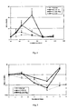
Fig. 4 shows the effect of single injection of rhG-CSF monomer (G-CSF and pegylated G-CSF (G-CSF-Peg)) and G-CSF dimer at equal molar dosage on the neutrophil count in the peripheral blood of healthy mice (average value ± standard deviation). The result indicated that the G-CSF dimer of the present invention had a stronger in vivo effect of driving the differentiation and maturation of myeloid hematopoietic cells, increasing the absolute neutrophil count (ANC) in the peripheral blood. -
Fig. 5 shows the effect of G-CSF monomer and G-CSF dimer at equal molar dosage on the neutrophil count in mice model with 5-FU-induced neutropenia. The result indicated that the G-CSF dimer (G-CSF-D) of the present invention had a better therapeutic effect than pegylated G-CSF monomer. -
Fig. 6 shows the effect of G-CSF monomer and G-CSF dimer in cynomolgus monkeys model with cyclophosphamide-induced neutropenia. The result indicated that the G-CSF dimer (G-CSF-D) of the present invention had a better therapeutic effect than pegylated G-CSF monomer. -
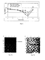
Fig. 7A shows that under non-reducing conditions, the immunoblot analysis results (Western blot) of cell culture supernatant, purified intermediate product and purified G-CSF dimer, using anti-human G-CSF monoclonal antibody (R&D systems, Cat.MAB214) as the first antibody and horseradish peroxidase-labeled anti-mouse IgG goat antibody as the second antibody. The lanes are as follow: 1. molecular weight standards; 2. cell culture supernatants; 3. purified intermediate product; and 4. purified G-CSF dimer. -
Fig. 7B shows that under reducing conditions, the immunoblot analysis results (Western blot) of cell culture supernatant, purified intermediate product and purified G-CSF dimer, using anti-human G-CSF monoclonal antibody (R&D systems, Cat.MAB214) as the first antibody and horseradish peroxidase-labeled anti-mouse IgG goat antibody as the second antibody. The lanes are as follow: 1. molecular weight standards; 2. cell culture supernatants; 3. purr:died intermediate product; and 4. purified G-CSF dimer (The molecular weight of G-CSF-Fc monomer is around 48 KD). - Upon an extensive and thorough research, the inventors have, for the first time ever, accidentally discovered that on comparing with G-CSF monomer, rhG-CSF dimer can generate a stronger receptor activation signal to accelerate the differentiation and proliferation of bone marrow neutrophils. Meanwhile, the properties of the pharmacokinetics and pharmacodynamics of G-CSF dimer are better than that of the rhG-CSF monomer. Therefore, the G-CSF dimer can effectively reduce the extent of the severe neutropenia and shorten the time of severe neutropenia for cancer patients upon receiving chemotherapy. The present invention was made based on the above understanding.
- The first embodiment of the present invention is a G-CSF dimer represented by the aforesaid formula (I) and the structural illustration thereof is shown in
Figs. 1-3 . In these figures, carrier protein includes but not limited to Fc fragment of human IgG1, IgG2, IgG3 and IgG4, and human albumin. - In one preferred embodiment, G-CSF can be disposed at the C-terminal or N-terminal of the carrier protein.
- As used herein, "linker" can refer to a short peptide for connecting the two G-CSF monomers and being disposed therebetween. There is no special restriction on the length of the linker. A linker is usually 5-50 amino acid residues in length; in general, a linker does not affect or significantly affect the proper fold and spatial conformation formed by the configuration of the two G-CSF monomers. Examples of linker include but not limited to:
- In a further preferred embodiment, the linker comprises amino acid sequence selected from a group consisting of:
- (a). an amino acid sequence with 3-16 amino acid residues formed by hydrophobic amino acids glycine (Gly) or proline (Pro), such as Gly-Pro-Gly-Pro-Gly-Pro;
- (b). an amino acid sequence encoded by multiple cloning sites. Such sequence usually contains 5-20 amino acid residues; in a preferred embodiment, such sequence contains 10-20 amino acid residues;
- (c). an amino acid sequence comprising protein(s) not from G-CSF monomer, such as an amino acid sequence of IgG or albumin;
- (d). an amino acid sequence comprising any combination of (a), (b), and (c) above.
- In one preferred embodiment, the linker has the sequence of GSGGGSGGGGSGGGGS (i.e. 175-190 amino acid residues of SEQ ID NO:1). In another preferred embodiment, the linker has the sequence of ASTKGP (i.e. 175-180 amino acid residues of SEQ ID NO: 3).
- In a further preferred embodiment, an amino acid sequence not affecting the biological activity of G-CSF monomer can be added to the N-terminal or C-terminal of the fusion protein. In a preferred embodiment, such appended amino acid sequence is beneficial to expression (e.g. signal peptide), purification (e.g. 6xHis sequence, the cleavage site of Saccharomyces cerevisiae α- factor signal peptide (Glu-Lys-Arg)), or enhancement of biological activity of the fusion protein.
- The encoding of the DNA sequence of the G-CSF dimer or fusion protein of the present invention can be entirely synthesized artificially. Alternatively, the encoded DNA sequences of the first G-CSF monomer and/or the second G-CSF monomer can be obtained by PCR amplification or synthesis and then joined together to form the encoded DNA sequence of the G-CSF dimer or fusion protein of the present invention.
- In order to enhance the expression volume of the host cells, modification can be performed on the encoded sequence of G-CSF dimer. For example, codon bias of host cells can be used to eliminate sequences that are not beneficial to gene transcription and translation. In a preferred embodiment, codon bias of yeast cells or mammalian cells can be used together with DNA software for detecting genes of G-CSF dimer, in order to eliminate sequences that are not beneficial to gene transcription and translation. In one preferred embodiment, the eliminated sequences can be intron cutting site, transcription terminating sequence, etc.
- After the encoded DNA sequence of the novel fusion protein of the present invention is obtained, it is first inserted into an appropriate expression carrier, followed by an appropriate host cell. Finally, the transformed host cell is cultivated and purified to obtain the novel fusion protein of the present invention.
- As used herein and in the claims, "carrier" refers to plasmid, cosmid, expression vehicle, cloning vector, virus vector, etc.
- In this invention, carrier known in the art, such as those available in the market, can be used. For example, with the use of carrier obtained from the market, encoded nucleotide sequence of the novel fusion protein of the present invention is operationally connected to the expressing and controlling sequence to form the protein-expressing carrier.
- As used herein and in the claims, "operationally connected" refers to a scenario that some parts of a linear DNA sequence can affect the biological activity of other parts of the same linear DNA sequence. For instance, if signal DNA is used as the expression of a precursor and participates in secretion of polypeptides, the signal DNA (secretion leader sequence) is "operationally connected" to the polypeptides. If a promoter controls sequence transcription, the promoter is "operationally connected" to the encoded sequence. If a ribosome binding site is situated at a position where translation thereof is made possible, the ribosome binding site is "operationally connected" to the encoded sequence. In general, "operationally connected" means that the residues of concern are in proximity; for secretion leader sequence, "operationally connected" refers to proximity within the reading frame.
- As used herein and in the claims, "host cells" refers to both prokaryotic cells and eukaryotic cells. Prokaryotic host cells commonly used include E. coli, B. subtilis, etc. Eukaryotic host cells commonly used include yeast cells, insect cells, mammalian cells, etc. In a preferred embodiment, the host cells used are eukaryotic cells; in another preferred embodiment, the host cells used are mammalian cells.
- After the transformed host cells are obtained, they can be cultivated under an environment suitable to express the fusion protein of the present invention for expressing the fusion protein. The expressed fusion protein is then separated.
- Since the G-CSF dimer of the present invention can generate a stronger receptor activation signal and has an excellent serum half-life, the G-CSF dimer and a pharmaceutical composition comprising the G-CSF dimer as the main active ingredient can be used for treating neutropenia. In a preferred embodiment, the neutropenia comprises a condition in which neutropenia is induced by chemotherapy and/or radiotherapy.
- The pharmaceutical composition of the present invention comprises a safe and effective amount of the G-CSF dimer of the present invention and a pharmaceutically acceptable excipient or carrier. "Safe and effective amount" refers to an amount of a compound sufficient to substantially improve the condition of the patient in need thereof without causing serious side-effects. In general, the pharmaceutical composition comprises 0.001-1,000 mg of G-CSF dimer of the present invention per dose; in a preferred embodiment, the pharmaceutical composition comprises 0.05-300 mg of G-CSF dimer of the present invention per dose; in a further preferred embodiment, the pharmaceutical composition comprises 0.5-200 mg of G-CSF dimer of the present invention per dose.
- The compound of the present invention and its pharmaceutically acceptable salts can be manufactured into different formulations, which comprises a safe and effective amount of the G-CSF dimer of the present invention or its pharmaceutically acceptable salts and a pharmaceutically acceptable excipient or carrier. "Safe and effective amount" refers to an amount of a compound sufficient to substantially improve the condition of the patient in need thereof without causing serious side-effects. The safe and effective amount of a compound is determined according to the age, condition, course of treatment, etc. of the patient in treatment.
- "Pharmaceutically acceptable excipient or carrier" refers to solid or liquid filling or gelatin materials with one or different kinds of compatibility which are suitable to be used in human with sufficient purity and sufficiently low toxicity. "Compatibility" refers to the ability of each ingredient of the composition to mutually blend with the compound of the present invention and the mutual blending ability there between, without substantially decreasing the clinical efficacy of the compound. Some of the examples of pharmaceutically acceptable excipient or carrier include cellulose and its derivatives (e.g. sodium carboxymethylcellulose, sodium ethylcellulose, cellulose acetate, etc), gelatin, speckstone, solid lubricating agent (e.g. stearic acid, magnesium stearate), calcium sulphate, plant oil (e.g. pea oil, sesame oil, peanut oil, olive oil, etc.), polyols (e.g. propylene glycol, glycerol, mannitol, sorbitol, etc.), emulsifier (e.g. Tween®), wetting agent (e.g sodium lauryl sulfate), colorant, flavoring agent, stabilizer, anti-oxidant, antiseptic, pyrogen-free water, etc.
- Route of administration of the G-CSF dimer of the present invention comprises oral administration, rectal administration, parenteral administration (intravenous, intramuscular, or subcutaneous), and partial administration.
- Solid form for oral administration comprises capsules, tablets, pills, powder, and granules. In these solid forms, active compound is mixed with at least one of the conventionally inert excipients (or carriers), such as sodium citrate, dicalcium phosphate, or any of the following ingredients: (a) filing or bulking agent, e.g. starch, lactose, sucrose, glucose, mannitol, and silicic acid; (b) adhesion agent, e.g. carboxymethylcellulose, alginate, gelatin, polyvinyl pyrrolidone, sucrose, and acacia; (c) humectants, e.g. glycerol; (d) disintegrating agent, e.g. agar, calcium carbonate, potato starch or cassava starch, alginic acid, compounded silicate, and sodium carbonate; (e) buffering agent, e.g. paraffin wax; (f) absorption accelerating agent, e.g. quaternary amine compound; (g) wetting agent, e.g. cetanol and glycerin monostearate; (h) absorbent, e.g. bolus alba; and (i). lubricating agent, e.g. speckstone, calcium stearate, sodium stearate, solid polyethylene glycol, sodium lauryl sulfate, or any mixture thereof. Capsules, tablets, and pills can also comprise buffering agent.
- Solid forms such as tablets, sugar pill, capsules, pills, and granules can be prepared with coating and core-shell materials, such as casing and other materials known in the art. These materials comprise opacifying agent and the active compound or compound in such composition can be released in a delayed fashion that the release is done in certain part of the alimentary canal. Embedding component such as polymer materials and wax materials can be used. If desired, active compounds can be mixed with one or more of the above-described excipients to formulate a micro capsule form.
- Liquid forms for oral administration comprise pharmaceutically acceptable emulsion, solution, suspension, syrup, or tincture. Apart from active compounds, liquid forms also comprise inert diluents conventionally used in the art such as water or other solvent, solublilizing agent and emulsifier such as ethanol, isopropanol, carbonate acetate, ethyl acetate, propan-2-ol, 1,3-butan-2-ol, dimethylfomamide, and oil, in particular cotton oil, peanut oil, castor oil, olive oil, maize embryo oil, and sesame oil or any mixture thereof.
- Apart from the inert diluents, the compound can also comprise additives, such as wetting agent, emulsifying agent, suspending agent, sweetening agent, correctives, and spices.
- Apart from active compounds, suspension can also comprise suspending agent, such as ethoxyl isostearic alcohol, polyoxyethylene sorbitol, sorbitan, microcrystalline cellulose, aluminium methoxide, agar, or any mixture thereof.
- Compounds used for parenteral administration can also comprise physiologically acceptable sterile water or anhydrous solution, dispersion solution, suspension, or emulsion, and sterile powder that can be re-dissolved into sterile injectable solution or dispersion solution. Appropriate hydrated or anhydrous carriers, diluting agent, solvent, or excipient comprises water, ethanol, polyols, and their appropriate mixtures threof.
- Forms of the G-CSF dimer of the present invention used for partial administration comprise ointment, powder, patch, sprayer, and inhalant. Under sterile conditions, active components can be mixed with physiologically acceptable carrier and any antiseptic, buffering agent, or may be propellant if desired.
- The G-CSF dimer of the present invention can be solely administrated or be administrated in conjunction with any pharmaceutically acceptable compounds. Usually, the G-CSF dimer of the present invention is not administrated associated with a G-CSF monomer.
- On using the pharmaceutical composition, a safe and effective of the amount of the G-CSF dimer of the present invention is administrated to a mammal (e.g. human) in use thereof in which the dosage administrated is a pharmaceutically acceptable effective administration dosage. For a human of 60kg, the administration dosage is usually 0.01-300 mg; in a preferred embodiment, the administration dosage is 0.5-100 mg. In determination of the actual dosage, factors known in the art such as administration route, patients' condition, etc. have to be considered, which is clear to a skilled person in the art.
- There are many advantages of the G-CSF dimer of the present invention which include but not limited to:
- 1. A stronger receptor activation signal;
- 2. A longer in vivo biological half-life.
- 3. A substantial clinical efficacy in the treatment of neutropenia in animal models, which is better than other present products.
- The following exemplary embodiments further describe the present invention. Although the description referred to particular embodiments, it will be clear to one skilled in the art that the present invention may be practiced with variation of these specific details. Hence this invention should not be construed as limited to the embodiments set forth herein. Further, for the embodiments in which details of the experimental methods are not described, such methods are carried out according to conventional conditions such as those described in Sambrook et al. Molecular Cloning: A Laboratory Manual (New York: Cold Spring Harbor Laboratory Pres, 1989), or suggested by the manufacturers.
- The invention can also be defined by the following clauses:
-
Clause 1. Use of human granulocyte colony-stimulating factor (G-CSF) dimer in the manufacture of a drug for treating neutropenia. -
Clause 2. The use ofclause 1 wherein said neutropenia comprises a condition in which said neutropenia is induced by chemotherapy and/or radiotherapy. -
Clause 3. The use ofclause 1, wherein said human G-CSF dimer is shown as formula (I):
(I) M1-L-M2
wherein- M1 is a first human G-CSF monomer;
- M2 is a second human G-CSF monomer; and
- L is a linker connecting said first monomer and said second monomer and disposed therebetween,
- said G-CSF dimer retains the biological activity of a G-CSF monomer and has a serum half-life of at least twice of the half-life of either said first or said second monomer.
-
Clause 4. The use ofclause 3, wherein said linker L is selected from the group consisting of:- i). a short peptide comprising 3 to 50 amino acids; and
- ii). a polypeptide of formula (II):
(II) -Z-Y-Z-
wherein- Y is a carrier protein;
- Z is null, or a short peptide(s) comprising 1 to 30 amino acids;
- "-" is a chemical bond or a covalent bond.
-
Clause 5. The use ofclause 3, wherein said first monomer and said second monomer are of the same entity. -
Clause 6. The use ofclause 3, wherein said biological activity comprises:- (a). acting on neutrophil precursor cells and myeloid stem cells to drive the differentiation, development, and maturation of neutrophils; and
- (b). activating mature neutrophils to participate in immune response.
- Clause 7. The use of
clause 4, wherein said carrier protein is albumin or Fc fragment of human IgG. -
Clause 8. The use ofclause 4, wherein said "-" is a peptide bond. - Clause 9. The use of
clause 4, wherein said G-CSF dimer is formed by monomers in which said monomer comprises an amino acid sequence selected from a group consisting of SEQ ID NOs: 2-6. -
Clause 10. A human G-CSF dimer of formula (I):
(I) M1-L-M2
wherein- M1 is a first G-CSF monomer;
- M2 is a second G-CSF monomer; and
- L is a linker connecting said first monomer and said second monomer and disposed therebetween,
- said G-CSF dimer retains the biological activity of a G-CSF monomer and has a serum half-life of at least twice of the half-life of either said first or said second monomer.
- The G-CSF dimer with the structure described in
Figs. 1-3 is prepared and purified by conventional methods. SEQ ID NO: 1 represents G-CSF dimer and SEQ ID NOs: 2-5 represent G-CSF monomer. - Rats received a single subcutaneous injection of G-CSF dimer (which is formed by two G-CSF monomers of SEQ ID NO: 2) with a dose of 100 µg/kg. The pharmacokinetic parameters (n=6) were calculated and listed in Table below. The half-life of G-CSF monomer in rats is about 2 hours.
Parameter Unit Average Value SD AUC(0-t) ng/mL*h 4234.8 640.3 MRT(0-t) h 21.6 1.4 t(1/2) h 7.7 1.2 Clz/F L/h/kg 0.024 0.003 Cmax ng/mL 162.2 30.2 - ICR mice, female, 20-22 grams, were randomly divided into four groups with 6-8 mice per group. The injection volume is 0.1 mL/10g of body weight, and each test group were given equal molar dosage of the G-CSF molecule (i.e. 1 mole of G-CSF dimer comprises 2 moles of G-CSF monomer). In other words, the mice were injected subcutaneously once with an equal volume of the carrier (control group), G-CSF-
Peg 40 µg/kg, rhG-CSF 40 µg/kg and G-CSF-D 100 µg/kg. - G-CSF-Peg monomer is Neulasta (Amgen Pegylated Filgrastim).
- G-CSF monomer is injectable rhG-CSF (GenSci).
- G-CSF-D is a G-CSF dimer formed by two G-CSF monomers with an amino acid sequence as shown in SEQ ID NO:5.
- After drug administration, blood samples (40 µL) were collected from orbital venous plexus at corresponding time points, and the blood count was detected and classified.
- The result is shown in
Fig. 4 . On comparing with the control group, G-CSF monomer (rhG-CSF and G-CSF-Peg) and G-CSF dimer (G-CSF-D) have different effects in driving neutrophil hyperplasia in healthy mice. As mentioned, the tested animals were healthy animals, therefore after drug administration, the G-CSF monomer or G-CSF dimer bound with neutrophil receptor and then was metabolized and eliminated, which cause a decrease of the neutrophil count correspondingly. In the present test, the neutrophil counts of all three groups with G-CSF monomer or G-CSF dimer were returned to the baseline level in 72 hours. - It is worth noting that, at an equal molar dosage of the G-CSF molecule, the ANC increasing of G-CSF-D group was the highest. In particular, at the 48th hour after drug administration, the average value of neutrophil of the G-CSF-D group, the G-CSF-Peg group and the G-CSF monomer group was 22.14 X10⁹/L,7.04 X109/L and 3.61 X10⁹ /L respectively, which means, the neutrophil count of the G-CSF-D group was 3.1 times of that of the G-CSF-Peg group, and 6.1 times of that of the G-CSF monomer group. Efficacy comparison result was: G-CSF-D > G-CSF-Peg > G-CSF, showing that G-CSF dimer has a better therapeutic effect. In other words, the increase of neutrophil in mice injected with G-CSF dimer was not only significantly higher than the group injected with G-CSF monomer, but also significantly higher than the group injected with G-CSF-Peg monomer which had a longer half-life. The result showed that G-CSF dimer has better biologic activities than G-CSF monomer in animals at equal molar dosage of G-CSF.
- ICR mice, half male and half female, 24-26 grams, were randomly divided into groups with 10 mice per group. All animals were intravenously injected a dose of 150 mg/kg 5-FU for modeling. 24 hours later, animals of the control group were injected with the carrier; those of the G-CSF-D (dimer) group were injected with 1500 µg/kg rhG-CSF dimer (which was formed by two G-CSF monomers with amino acid sequence shown in SEQ ID NO:3); and those of the G-CSF-Peg monomer group were injected with 300 µg/kg Neulasta. The animals of the test groups were given at equal molar dosage of the G-CSF molecule (i.e. 1 mole of G-CSF dimer comprises 2 moles of G-CSF monomer) by subcutaneous injection. Each group was administrated once on
Day Day - The result was shown in
Fig. 5 . After the administration of 5-Fu, neutrophil count of the control group declined rapidly and the nadir was shown onDay 6. For animals with injection of rhG-CSF monomer (G-CSF-Peg) and G-CSF dimer (G-CSF-D) at equal molar dosage, the nadir of neutrophil counts was also shown on aroundDay 6. However, the neutrophil count of the G-CSF-D group (with an average value of 0.23X10⁹/L) was 4.6 times of that of the G-CSF-Peg group (with an average value of 0.05 X10⁹/L). - The result indicated that in the treatment of mice model with 5-Fu induced neutropenia, G-CSF dimer (G-CSF-D) had a significantly better therapeutic effect comparing to G-CSF monomer (G-CSF-Peg).
- 24 healthy cynomolgus monkeys, half male and half female, were intravenously injected a dose of 60 mg/kg cyclophosphamide (lot: 2008040921, Jiangsu Hengrui Medicine Co., Ltd.) twice (Day 0 and Day1) to induce a decrease of white blood cells and neutrophils. The cynomolgus monkeys were randomly divided into three groups, half male and half female. Starting from
Day 5, the test groups were separately subcutaneously injected with G-CSF dimer (60 µg/kg, which is equal to 0.67 µM/kg) (G-CSF-D is a dimer formed by two G-CSF monomers with an amino acid sequence shown in SEQ ID NO:2) once; or with G-CSF-Peg monomer (60 µg/kg, which is equal to 3.2 µM/kg) once; or with G-CSF monomer (10 µg/kg/day, which is equal to 0.53 µM/kg) for 5 continuous days with a total dose of 50 µg/kg, which is equal to 2.65 µM/kg. The injection volume was 0.2 mL/kg. Blood samples were collected at different time points, and the effect of F627 with different dose on the neutrophil count in peripheral blood of cynomolgus monkeys was observed. - The result was shown in
Fig. 6 . After the intravenous administration of cyclophosphamide, the neutrophil count of control group declined and reached the nadir on Days 6-8. For animals with injection equal dosage of rhG-CSF monomer and G-CSF-Peg monomer, the nadir of neutrophil count was reached on Days 6-7. For animals with single injection of G-CSF dimer, the occurrence of neutropenia was prevented when the molar dosage of the injected dimer was at 4.8 times less than that of G-CSF-Peg monomer. At the nadir (Day 8 after the administration of cyclophosphamide), the neutrophil count of the animal group injected with G-CSF dimer (average value 1.17 X 10⁹/L) once was 3.0 times of that with G-CSF monomer (average value 0.39x10⁹/L) and 6.9 times of that with G-CSF-Peg monomer (0.17x10⁹/L). - The result indicated that in the treatment of cynomolgus monkeys model with cyclophosphamide-induced neutropenia, G-CSF dimer (G-CSF-D) had a better therapeutic effect than G-CSF monomer and G-CSF-Peg, which can effectively prevent the occurrence of neutropenia induced by chemotherapy agent, and can reduce the severity of neutropenia and / or shorten the time of severe neutropenia.
- The full length cDNA sequence of the G-CSF-Fc complexes (as shown in SEQ ID NO: 7) was synthesized. cDNA sequence of human G-CSF monomer was connected with cDNA sequence of Fe fragment of IgG2. cDNA sequences containing Hind III site, and expression elements required by mammalian cell such as Kozak sequence and signal peptide were introduced at the 5' end. cDNA sequence containing EcoRI site was introduced at the 3' end. The full length G-CSF dimer cDNA sequence was cloned into pUC19 plasmid to obtain pG-CSF-Fc, transformed E. coli TGl. The plasmid was digested with Hind III and EcoRI, an approximately 1400bp G-CSF-IgG2Fc fragment was harvested and connected with pcDNA3 (Invitrogen) expression plasmid which was also digested with Hind III and EcoRI, and an expression plasmid pEX-G-CSF-Fc was then constructed. pEX-G-CSF-Fc was linearized, purified and transfected into CHO cells by electroporation. The transfected cells were selected in selecting media. The expression levels of individual clones were measured by ELISA. The cell lines with the higher protein expression levels were selected and cells thereof were frozen to generate cell bank.
- The first G-CSF monomer synthesized using SEQ ID NO: 7 had a structure of "G-CSF-linker-IgG2Fc", and the amino acid sequence thereof was shown in SEQ ID NO: 6.
- The expression plasmids were constructed in similar methods, resulting in the second, third and fourth G-CSF monomers (Table 1) with different structures. G-CSF dimers with different structures were obtained by similar expression methods.
TABLE 1 Different G-CSF- Fc monomer G-CSF-Fc monomer Structure Sequence of G-CSF Sequence of linker Sequence of IgG2Fc No. 1 G-CSF-linker-IgG2Fc SEQ ID NO.:1 amino acid residues 1-174 SEQ ID NO.:1 amino acid residues 175-190 SEQ ID NO.: 6 amino acid residues 191-413 No. 2 SEQ ID NO.: 3 amino acid residues 175-180 No. 3 IgG2Fc-linker-G-CSF SEQ ID NO.:1 amino acid residues 175-190 No. 4 SEQ ID NO.: 3 amino acid residues 175-180 - One vial of cells (∼ lx 107 cells/mL) from the cell bank was thawed and seeded in 10 mL basal medium in a 10 cm Petri dish, and incubated at 37°C, 5% CO2 for approximately 24 hours.
- The seeding expansion: The culture volume was expanded from 10 mL to 30-40 mL. When the cell density reached 1.0-1.5
x 106 cells/mL with viability ≥ 90%, the culture volume was expanded to 300-400 mL progressively and then the culture was moved to shaking flasks and incubated at 120 rpm, 37°C, 5% CO2. - Culture expansion in bioreactor (3L-10L): When the cell density in the seeding expansion reached 1.0 - 3.0
x 106 cells/mL with viability ≥ 90%, the 300-400 mL seeding expanded culture was transferred to a 3-10 L bioreactor containing basal medium, with the culture control conditions at pH of 6.8, dissolved oxygen at approximately 50 % and stirring speed at 65-100 rpm. - Culture expansion in bioreactor (30 -100 L): When the cell density in the 3.0-10 L bioreactor reached 1.0 - 3.0
x 106 cells/ml withviability 2 ≥ 90%, the culture was transferred to 30 - 100 L bioreactor with the culture control conditions at pH of 6.8, dissolved oxygen at approximately 50 % and stirring speed at 65-100 rpm. The culture was fed for 12 to 48 hours before controlling the glucose level (<lg/L) with the addition of fed-batch medium. - After the culture expansion in bioreactor, cell supernatant was harvested which contained G-CSF-Fc complex, G-CSF dimer, G-CSF-Fc multi-mers, and metabolites. After being harvested from the bioreactor culture, the cell culture supernatant was obtained by filtration and purified by a series of chromatography methods; for example, using a rProtein A Sepharose FF (GE Healthcare, cat#l7-1279-04), eluted with 50 mM critic acid/ sodium citrate and 0.2M NaCl at pH 3.7-3.8, resulting in >90% pure G-CSF dimer. Additional chromatography steps were performed using Capto Adhere column with elution buffer of 50 mM NaAc/HAC and 0.2 M NaCl at pH 4.5-5.0, followed by SP Sepharose FF (GE Heathcare Cat #17-0729-04) and balanced with equilibrium buffer of 10mM PB (pH 6.0±0.1). Elution buffer used was 10mM PB and 0.2M NaCl (pH 7.2±0.1). Additional processes involved viral activation at low pH, filtration, resulting in G-CSF dimer.
- The purity of the G-CSF dimer was> 95% (analyzed by reverse phase HPLC), with estimated molecular weight of 47±5 kD (analyzed by reduced SDS-PAGE analysis). The G-CSF dimer was glycosylated with oligosaccharide of 2-10% of the total molecular weight. The isoelectric point of the protein was between pH 5.8 to pH 6.8. The maximum UV absorbing wavelength was at 280 nM. The G-CSF dimer can activate STAT3 in M-NSF-60 cells and stimulate the proliferation of M-NSF-60 cells in vitro (the ED50 thereof was between 0.1-10 ng/mL).
- The purification result of G-CSF dimer (comprising two G-CSF monomers with sequence shown in SEQ ID NO: 6) was shown in
Fig. 7A and Fig. 7B , indicating GCSF dimer was obtained after the purification (as shown inFig 7A , lane 4). - Moreover, the results showed that, if the Fe fragment of the carrier protein contained four cysteine at the N-terminal, for example, ERKCCVECPPC(SEQ ID NO.: 2, amino acid residues 191-201), the formation of normally paired G-CSF dimer may be effected, indicating that it would be better for two Fe fragments to be connected with each other via 2-3 disulfide bonds.
- Healthy subjects were randomly divided into four dosage groups of 30, 60, 120, 240 µg/kg respectively receiving a single dose of 30, 60, 120, 240 µg/kg of G-CSF dimer (comprising two G-CSF monomers with sequence shown in SEQ ID NO: 6). Blood samples were collected at the 0.5, 1st, 2nd, 4th, 8th, 16th 24th, 36th, 48th, 72nd, 96th hour, Day 6 (120 hours), 7, 9, 11, 13, and 15 after administration. Serum was separated and stored below -70°C. The blood concentrations of G-CSF-D were measured by ELISA (ELISA, Quantikine human G-CSF ELISA kit, R&D System, Inc. Minneapolis, Min, Cat: PDCS50). The pharmacokinetic parameters were calculated using the standard non-compartmental analytical procedures (Software WinNonlin v 5.2, Pharsight Corporation, USA). The results were shown in Table 2.
TABLE2 Parameter (n=6) 30µg/kg 60µg/kg 120µg/kg 240µg/kg Cmax (ng/mL) 21.3 (10.3) 44.6 (17.7) 219.9 (76.6) 759 (160) T max (h, median & range)* 8 (8-16) 8 (8-16) 16 (16-36) 36 (36) t1/2 (h) 43.9 (4.3) 56.1 (23.3) 59.3 (23.5) 62.8 (10.8) AUC(0-inf) (ng.h/mL) 778 (213) 1847 (686) 8349 (2769) 46664 (17258) CL/F (mL/h/kg) 41.4 (12.8) 36.8 (14.6) 18.5 (7.7) 5.7 (2.0) *T max is represented by the median and rage values: data in the brackets is range values, and that outside the brackets is median values. - All references mentioned in the present invention are cited herein by reference. Although the description referred to particular embodiments, it will be clear to one skilled in the art that the present invention may be practiced with variation of these specific details. Hence this invention should not be construed as limited to the embodiments set forth herein.


Claims (12)
- A human granulocyte colony stimulating factor (G-CSF) dimer of formula (I) for use in treating neutropenia,
(I) M1-L-M2
whereinM1 is a first monomer of G-CSF;M2 is a second monomer of G-CSF; andL is a linker connecting said first monomer and said second monomer and disposed therebetween;
wherein said linker L is a polypeptide of formula (II):
(II) -Z-Y-Z-
whereinY is a carrier protein formed by two Fc fragments of IgG2 connected via disulfide bonds;Z is null, or a short peptide(s) comprising 1 to 30 amino acids;"-" is a chemical bond or a covalent bond or a peptide bond. - The human G-CSF dimer of claim 1, wherein the G-CSF dimer is produced by two G-CSF-Fc complexes, each said G-CSF-Fc complex comprising one of the two Fc fragments of human IgG2, and wherein said G-CSF dimer is formed through the pairing of the two Fc fragments via a plurality of disulfide bonds disposed therebetween.
- The human G-CSF dimer of claim 2, wherein each said G-CSF-Fc complex comprises an amino acid sequence selected from the group consisting of SEQ ID NOs: 2-6.
- The human G-CSF dimer of claim 3, wherein each said G-CSF-Fc complex comprises the amino acid sequence of SEQ ID NO:2.
- The human G-CSF dimer of claim 3, wherein each said G-CSF-Fc complex comprises the amino acid sequence of SEQ ID NO:3.
- The human G-CSF dimer of claim 3, wherein each said G-CSF-Fc complex comprises the amino acid sequence of SEQ ID NO:4.
- The human G-CSF dimer of claim 3, wherein each said G-CSF-Fc complex comprises the amino acid sequence of SEQ ID NO:5.
- The human G-CSF dimer of claim 3, wherein each said G-CSF-Fc complex comprises the amino acid sequence of SEQ ID NO:6.
- The human G-CSF dimer of any one of claims 1-8, wherein the human G-CSF dimer retains biological activity of G-CSF monomer and has a serum half -life of at least twice of the half-life of the first or the second monomer of G-CSF.
- The human G-CSF dimer of claim 9, wherein the biological activity comprises:(a) acting on neutrophil precursor cells and myeloid stem cells to drive the differentiation, development, and maturation of neutrophils; and(b) activating mature neutrophils to participate in immune response.
- The human G-CSF dimer of any one of claims 1-10, wherein the neutropenia is severe neutropenia.
- The human G-CSF dimer of any one of claims 1-11, wherein the neutropenia comprises a condition in which said neutropenia is induced by chemotherapy, radiotherapy, or combination thereof.
Applications Claiming Priority (2)
| Application Number | Priority Date | Filing Date | Title |
|---|---|---|---|
| CN201010268290XA CN102380090A (en) | 2010-08-31 | 2010-08-31 | G-CSF (granulocyte-colony stimulating factor) dimmer applied to treat neutropenia |
| EP11821119.2A EP2612676A4 (en) | 2010-08-31 | 2011-08-31 | Use of g-csf dimer in the treatment of neutropenia |
Related Parent Applications (1)
| Application Number | Title | Priority Date | Filing Date |
|---|---|---|---|
| EP11821119.2A Division EP2612676A4 (en) | 2010-08-31 | 2011-08-31 | Use of g-csf dimer in the treatment of neutropenia |
Publications (1)
| Publication Number | Publication Date |
|---|---|
| EP3028713A1 true EP3028713A1 (en) | 2016-06-08 |
Family
ID=45772165
Family Applications (2)
| Application Number | Title | Priority Date | Filing Date |
|---|---|---|---|
| EP15188775.9A Ceased EP3028713A1 (en) | 2010-08-31 | 2011-08-31 | Use of g-csf dimer in the treatment of neutropenia |
| EP11821119.2A Withdrawn EP2612676A4 (en) | 2010-08-31 | 2011-08-31 | Use of g-csf dimer in the treatment of neutropenia |
Family Applications After (1)
| Application Number | Title | Priority Date | Filing Date |
|---|---|---|---|
| EP11821119.2A Withdrawn EP2612676A4 (en) | 2010-08-31 | 2011-08-31 | Use of g-csf dimer in the treatment of neutropenia |
Country Status (4)
| Country | Link |
|---|---|
| US (2) | US20130165637A1 (en) |
| EP (2) | EP3028713A1 (en) |
| CN (2) | CN102380090A (en) |
| WO (1) | WO2012028093A1 (en) |
Cited By (1)
| Publication number | Priority date | Publication date | Assignee | Title |
|---|---|---|---|---|
| WO2020165664A1 (en) * | 2019-02-13 | 2020-08-20 | Ilkogen Ilac Sanayi Ve Ticaret A.S. | A long-acting g-csf for preventing neutropenia or reducing duration of neutropenia |
Families Citing this family (11)
| Publication number | Priority date | Publication date | Assignee | Title |
|---|---|---|---|---|
| CN102260343A (en) * | 2010-05-25 | 2011-11-30 | 健能隆医药技术(上海)有限公司 | Application of recombinant human G-CSF dimer for treating nerve damage disease |
| US9642917B2 (en) | 2011-07-25 | 2017-05-09 | Generon (Shanghai) Corporation, Ltd. | Use of G-CSF dimer in preparation of medicament for treatment of neurodegenerative diseases |
| NZ702415A (en) | 2012-06-07 | 2016-04-29 | Children’S Hospital Los Angeles | Methods for treating neutropenia using retinoid agonists |
| AU2015219038B2 (en) * | 2014-02-18 | 2018-10-18 | Children's Hospital Los Angeles | Compositions and methods for treating neutropenia |
| MA40904A (en) | 2014-11-03 | 2017-09-12 | Hygeia Tech Inc | PHORBOL ESTER COMPOSITIONS AND METHODS FOR TREATING OR REDUCING THE DURATION OF CYTOPENIA |
| EP3328415B1 (en) * | 2015-07-30 | 2020-11-25 | Endor Technologies, S.L. | Colony stimulating factor for use in pancreatic or colon cancer treatment |
| CN105884906B (en) * | 2016-05-27 | 2021-11-19 | 广州太力生物医药科技有限公司 | Purification method of long-acting human erythropoietin fusion protein |
| US11732016B2 (en) | 2017-12-27 | 2023-08-22 | Council Of Scientific & Industrial Research | Polypeptide exhibiting granulocyte-colony stimulating factor activity |
| KR20210100638A (en) * | 2018-12-04 | 2021-08-17 | 씨에스엘 이노베이션 피티와이 엘티디 | How to treat neutrophil disease |
| CN112175906B (en) * | 2019-07-05 | 2023-09-29 | 中国科学院生物物理研究所 | Use of thymidine induced fibroblast transdifferentiation in the treatment of neutropenia |
| WO2024037633A2 (en) | 2022-08-19 | 2024-02-22 | Evive Biotechnology (Shanghai) Ltd | Formulations comprising g-csf and uses thereof |
Citations (7)
| Publication number | Priority date | Publication date | Assignee | Title |
|---|---|---|---|---|
| WO2001003737A1 (en) * | 1999-07-13 | 2001-01-18 | Bolder Biotechnology Inc. | Immunoglobulin fusion proteins |
| WO2002036626A1 (en) * | 2000-11-02 | 2002-05-10 | Maxygen Aps | Single-chain multimeric polypeptides |
| US20040259209A1 (en) * | 2001-10-01 | 2004-12-23 | Sun Lee-Hwei K. | Fc fusion proteins of human granulocyte colony-stimulating factor with increased biological activities |
| WO2009019441A2 (en) * | 2007-08-03 | 2009-02-12 | Asterion Limited | Granulocyte colony stimulating factor |
| LT2010013A (en) * | 2010-02-10 | 2011-08-25 | Uab Profarma, , | Process for isolating and purifying biologically active g-csf derivatives |
| LT2010012A (en) * | 2010-02-10 | 2011-08-25 | Uab Profarma, , | Linear oligomers of granulocyte stimulating protein having prolonged survival time |
| WO2011147319A1 (en) * | 2010-05-25 | 2011-12-01 | Generon (Shanghai) Corporation Ltd. | Recombinant human g-csf dimer and use thereof for the treatment of neurological diseases |
Family Cites Families (2)
| Publication number | Priority date | Publication date | Assignee | Title |
|---|---|---|---|---|
| US20020142964A1 (en) * | 2000-11-02 | 2002-10-03 | Nissen Torben Lauesgaard | Single-chain polypeptides |
| US10138283B2 (en) * | 2008-07-23 | 2018-11-27 | Ambrx, Inc. | Modified bovine G-CSF polypeptides and their uses |
-
2010
- 2010-08-31 CN CN201010268290XA patent/CN102380090A/en active Pending
-
2011
- 2011-08-31 EP EP15188775.9A patent/EP3028713A1/en not_active Ceased
- 2011-08-31 US US13/819,716 patent/US20130165637A1/en not_active Abandoned
- 2011-08-31 EP EP11821119.2A patent/EP2612676A4/en not_active Withdrawn
- 2011-08-31 WO PCT/CN2011/079143 patent/WO2012028093A1/en active Application Filing
- 2011-08-31 CN CN201180041735.3A patent/CN103079586B/en active Active
-
2014
- 2014-10-28 US US14/526,382 patent/US20150147290A1/en not_active Abandoned
Patent Citations (7)
| Publication number | Priority date | Publication date | Assignee | Title |
|---|---|---|---|---|
| WO2001003737A1 (en) * | 1999-07-13 | 2001-01-18 | Bolder Biotechnology Inc. | Immunoglobulin fusion proteins |
| WO2002036626A1 (en) * | 2000-11-02 | 2002-05-10 | Maxygen Aps | Single-chain multimeric polypeptides |
| US20040259209A1 (en) * | 2001-10-01 | 2004-12-23 | Sun Lee-Hwei K. | Fc fusion proteins of human granulocyte colony-stimulating factor with increased biological activities |
| WO2009019441A2 (en) * | 2007-08-03 | 2009-02-12 | Asterion Limited | Granulocyte colony stimulating factor |
| LT2010013A (en) * | 2010-02-10 | 2011-08-25 | Uab Profarma, , | Process for isolating and purifying biologically active g-csf derivatives |
| LT2010012A (en) * | 2010-02-10 | 2011-08-25 | Uab Profarma, , | Linear oligomers of granulocyte stimulating protein having prolonged survival time |
| WO2011147319A1 (en) * | 2010-05-25 | 2011-12-01 | Generon (Shanghai) Corporation Ltd. | Recombinant human g-csf dimer and use thereof for the treatment of neurological diseases |
Non-Patent Citations (11)
| Title |
|---|
| "Molecular construction and optimization of anti-human IL-1[alpha]/[beta] dual variable domain immunoglobulin (DVD-IgTM) molecules", MABS 2009 LANDES BIOSCIENCE, 4 July 2009 (2009-07-04), pages 339 - 347, XP055114439, Retrieved from the Internet <URL:http://www.ncbi.nlm.nih.gov/pmc/articles/PMC2726605/pdf/mabs0104_0339.pdf> [retrieved on 20140416] * |
| BLOOD, vol. 116, no. 21, November 2010 (2010-11-01), 52ND ANNUAL MEETING OF THE AMERICAN-SOCIETY-OF-HEMATOLOGY (ASH); ORLANDO, FL, USA; DECEMBER 04 -07, 2010, pages 634, ISSN: 0006-4971(print) * |
| BMC CANCER, vol. 8, 2008, pages 195 |
| COX G N ET AL: "Enhanced circulating half-life and hematopoietic properties of a human granulocyte colony-stimulating factor/immunoglobulin fusion protein", EXPERIMENTAL HEMATOLOGY, ELSEVIER INC, US, vol. 32, 1 January 2004 (2004-01-01), pages 441 - 449, XP003006681, ISSN: 0301-472X, DOI: 10.1016/J.EXPHEM.2004.01.012 * |
| DATABASE BIOSIS [online] BIOSCIENCES INFORMATION SERVICE, PHILADELPHIA, PA, US; November 2010 (2010-11-01), HU ZHENG TAO ET AL: "F-627, a G-CSF Dimer, Stimulated a More Rapid Neutrophil Recovery In Cyclophosphamide-Treated Monkeys Compared to Monomer Rhg-CSFs", XP002717781, Database accession no. PREV201100424030 * |
| GUO, SIQI: "Construction and expression, biological activities and protein characteristics analysis of human recombinant bimolecular G-CSF", SHENGWU JISHU TONGXUN = LETTERS IN BIOTECHNOLOGY, vol. 14, no. 6, November 2003 (2003-11-01), pages 489 - 493, XP009175101 * |
| KONTERMANN R, DÜBEL S: "Antibody Engineering", vol. 2, March 2010, SPRINGER, article WU C ET AL.: "Generation and Characterization of a Dual Variable Domain Immunoglobulin", pages: FP1-2,240-250, XP002755514 * |
| PNAS, vol. 103, 2008, pages 3135 - 3140 |
| QIANG ZHU ET AL: "Expression of rhEPO-L-Fc Fusion Protein and Analysis of Its Bioactivity and Pharmacokinetics", CHIN J BIOTECH, vol. 24, no. 11, 25 November 2008 (2008-11-25), pages 1874 - 1879, XP055082112, ISSN: 1000-3061 * |
| SAMBROOK ET AL.: "Molecular Cloning: A Laboratory Manual (New York", 1989, COLD SPRING HARBOR LABORATORY PRES |
| WU CHENGBIN ET AL: "Simultaneous targeting of multiple disease mediators by a dual-variable-domain immunoglobulin", NATURE BIOTECHNOLOGY, NATURE PUBLISHING GROUP, US, vol. 25, no. 11, 1 November 2007 (2007-11-01), pages 1290 - 1297, XP009110104, ISSN: 1087-0156, DOI: 10.1038/NBT1345 * |
Cited By (2)
| Publication number | Priority date | Publication date | Assignee | Title |
|---|---|---|---|---|
| WO2020165664A1 (en) * | 2019-02-13 | 2020-08-20 | Ilkogen Ilac Sanayi Ve Ticaret A.S. | A long-acting g-csf for preventing neutropenia or reducing duration of neutropenia |
| US11813310B2 (en) | 2019-02-13 | 2023-11-14 | Ilkogen Ilaç Sanayi Ve Ticaret A.S. | Long-acting G-CSF for preventing neutropenia or reducing duration of neutropenia |
Also Published As
| Publication number | Publication date |
|---|---|
| CN103079586A (en) | 2013-05-01 |
| US20130165637A1 (en) | 2013-06-27 |
| CN103079586B (en) | 2017-02-22 |
| WO2012028093A1 (en) | 2012-03-08 |
| CN102380090A (en) | 2012-03-21 |
| EP2612676A4 (en) | 2014-01-22 |
| US20150147290A1 (en) | 2015-05-28 |
| EP2612676A1 (en) | 2013-07-10 |
Similar Documents
| Publication | Publication Date | Title |
|---|---|---|
| EP3028713A1 (en) | Use of g-csf dimer in the treatment of neutropenia | |
| EP0401384B1 (en) | Chemically modified granulocyte colony stimulating factor | |
| JP3115318B2 (en) | Fusion protein containing GM-CSF and IL-3 | |
| JP6416106B2 (en) | Method for increasing the hydrodynamic volume of a polypeptide by binding to a gonadotropin carboxy-terminal peptide | |
| EP2576624B1 (en) | Recombinant human g-csf dimer and use thereof for the treatment of neurological diseases | |
| EP1888119B1 (en) | Pegylated g-csf polypeptides and methods of producing same | |
| US9051358B2 (en) | Nonnatural collagen-like protein and use thereof | |
| US5472857A (en) | DNA encoding canine granulocyte colony stimulating factor (G-CSF) | |
| WO2022100741A1 (en) | Fusion protein of human serum albumin and interleukin-2, and use thereof | |
| WO2015055026A1 (en) | Fusion protein of ganoderma lucidum immunoregulatory protein and human serum albumin, and production method therefor and uses thereof | |
| JPH04164098A (en) | Chemically modified, granular spherical colony stimulus factor derivative | |
| JPH11500904A (en) | MPL ligand analog | |
| KR20230157947A (en) | Methods and pharmaceutical combinations for preventing cancer recurrence | |
| WO2012000187A1 (en) | Fusion protein of g-csf variant, preparation method and use thereof | |
| JP2001523727A (en) | How to mobilize hematopoietic stem cells | |
| JPH07103041B2 (en) | Malignant tumor treatment adjuvant | |
| Granulocyte-Colony et al. | Advances in Biochemistry and Biotechnology Volume 1 Pages 53-80 Edited by: Dr. Chiranjib Chakraborty | |
| CA2495480A1 (en) | Interferon and immunoglobulin fc fragment hybrid |
Legal Events
| Date | Code | Title | Description |
|---|---|---|---|
| PUAI | Public reference made under article 153(3) epc to a published international application that has entered the european phase |
Free format text: ORIGINAL CODE: 0009012 |
|
| 17P | Request for examination filed |
Effective date: 20151007 |
|
| AC | Divisional application: reference to earlier application |
Ref document number: 2612676 Country of ref document: EP Kind code of ref document: P |
|
| AK | Designated contracting states |
Kind code of ref document: A1 Designated state(s): AL AT BE BG CH CY CZ DE DK EE ES FI FR GB GR HR HU IE IS IT LI LT LU LV MC MK MT NL NO PL PT RO RS SE SI SK SM TR |
|
| 17Q | First examination report despatched |
Effective date: 20171113 |
|
| STAA | Information on the status of an ep patent application or granted ep patent |
Free format text: STATUS: THE APPLICATION HAS BEEN REFUSED |
|
| 18R | Application refused |
Effective date: 20190506 |