EP3023791A1 - Prévoir la réactivité à un traitement de gemcitabine - Google Patents
Prévoir la réactivité à un traitement de gemcitabine Download PDFInfo
- Publication number
- EP3023791A1 EP3023791A1 EP14194296.1A EP14194296A EP3023791A1 EP 3023791 A1 EP3023791 A1 EP 3023791A1 EP 14194296 A EP14194296 A EP 14194296A EP 3023791 A1 EP3023791 A1 EP 3023791A1
- Authority
- EP
- European Patent Office
- Prior art keywords
- cancer
- sample
- rbm3
- subject
- gemcitabine
- Prior art date
- Legal status (The legal status is an assumption and is not a legal conclusion. Google has not performed a legal analysis and makes no representation as to the accuracy of the status listed.)
- Withdrawn
Links
Images


Classifications
-
- G—PHYSICS
- G01—MEASURING; TESTING
- G01N—INVESTIGATING OR ANALYSING MATERIALS BY DETERMINING THEIR CHEMICAL OR PHYSICAL PROPERTIES
- G01N33/00—Investigating or analysing materials by specific methods not covered by groups G01N1/00 - G01N31/00
- G01N33/48—Biological material, e.g. blood, urine; Haemocytometers
- G01N33/50—Chemical analysis of biological material, e.g. blood, urine; Testing involving biospecific ligand binding methods; Immunological testing
- G01N33/53—Immunoassay; Biospecific binding assay; Materials therefor
- G01N33/574—Immunoassay; Biospecific binding assay; Materials therefor for cancer
- G01N33/57407—Specifically defined cancers
- G01N33/57438—Specifically defined cancers of liver, pancreas or kidney
-
- G—PHYSICS
- G01—MEASURING; TESTING
- G01N—INVESTIGATING OR ANALYSING MATERIALS BY DETERMINING THEIR CHEMICAL OR PHYSICAL PROPERTIES
- G01N2333/00—Assays involving biological materials from specific organisms or of a specific nature
- G01N2333/435—Assays involving biological materials from specific organisms or of a specific nature from animals; from humans
- G01N2333/52—Assays involving cytokines
Definitions
- the present invention relates to the field of treatment prediction, in particular to the prediction of the response to treatment with gemcitabine.
- Cancer is one of the most common diseases, and a major cause of death in the western world. In general, incidence rates increase with age for most forms of cancer. As human populations continue to live longer, due to an increase of the general health status, cancer may affect an increasing number of individuals. The cause of most common cancer types is still largely unknown, although there is an increasing body of knowledge providing a link between environmental factors (dietary, tobacco smoke, UV radiation etc) as well as genetic factors (germ line mutations in "cancer genes" such as p53, APC, BRCA1, XP etc) and the risk for development of cancer.
- cancer is essentially a cellular disease and defined as a transformed cell population with net cell growth and anti-social behavior.
- Malignant transformation represents the transition to a malignant phenotype based on irreversible genetic alterations. Although this has not been formally proven, malignant transformation is believed to take place in one cell, from which a subsequently developed tumor originates (the "clonality of cancer" dogma).
- Carcinogenesis is the process by which cancer is generated and is generally accepted to include multiple events that ultimately lead to growth of a malignant tumor. This multi-step process includes several rate-limiting steps, such as addition of mutations and possibly also epigenetic events, leading to formation of cancer following stages of precancerous proliferation.
- the stepwise changes involve accumulation of errors (mutations) in vital regulatory pathways that determine cell division, asocial behavior and cell death.
- Each of these changes may provide a selective Darwinian growth advantage compared to surrounding cells, resulting in a net growth of the tumor cell population.
- a malignant tumor does not only necessarily consist of the transformed tumor cells themselves but also surrounding normal cells which act as a supportive stroma.
- This recruited cancer stroma consists of connective tissue, blood vessels and various other normal cells, e.g., inflammatory cells, which act in concert to supply the transformed tumor cells with signals necessary for continued tumor growth.
- cancers arise in somatic cells and are predominantly of epithelial origin, e.g., prostate, breast, colon, urothelium and skin, followed by cancers originating from the hematopoetic lineage, e.g., leukemia and lymphoma, neuroectoderm, e.g., malignant gliomas, and soft tissue tumors, e.g., sarcomas.
- epithelial origin e.g., prostate, breast, colon, urothelium and skin
- cancers originating from the hematopoetic lineage e.g., leukemia and lymphoma
- neuroectoderm e.g., malignant gliomas
- soft tissue tumors e.g., sarcomas.
- mice Microscopic evaluation of biopsy material from suspected tumors remains the golden standard for cancer diagnostics.
- the tumor tissue is fixated in formalin, histo-processed and paraffin embedded.
- tissue sections can be produced and stained using both histochemical, i.e., hematoxylin-eosin staining, and immunohistochemical (IHC) methods.
- IHC immunohistochemical
- the surgical specimen is then evaluated with pathology techniques, including gross and microscopic analysis. This analysis often forms the basis for assigning a specific diagnosis, i.e., classifying the tumor type and grading the degree of malignancy, of a tumor.
- TMM tumor-node-metastasis
- NMM tumor-node-metastasis
- N stage describes the local extent of the primary tumor, i.e., how far the tumor has invaded and imposed growth into surrounding tissues
- N stage and M stage describe how the tumor has developed metastases, with the N stage describing spread of tumor to lymph nodes and the M stage describing growth of tumor in other distant organs.
- Early stages include: T0-1, N0, M0, representing localized tumors with negative lymph nodes.
- More advanced stages include: T2-4, N0, M0, localized tumors with more widespread growth and T1-4, N1-3, M0, tumors that have metastasized to lymph nodes and T1-4, N1-3, M1, tumors with a metastasis detected in a distant organ.
- Staging of tumors is often based on several forms of examination, including surgical, radiological and histopathological analyses.
- a classification system to grade the level of malignancy.
- the grading systems rely on morphological assessment of a tumor tissue sample and are based on the microscopic features found in a given tumor. These grading systems may be based on the degree of differentiation, proliferation and atypical appearance of the tumor cells. Examples of generally employed grading systems include Gleason grading for prostatic carcinomas and the Nottingham Histological Grade (NHG) grading for breast carcinomas.
- IHC immunohistochemical staining
- IHC allows for the detection of protein expression patterns in tissues and cells using specific antibodies.
- the use of IHC in clinical diagnostics allows for the detection of immunoreactivity in different cell populations, in addition to the information regarding tissue architecture and cellular morphology that is assessed from the histochemically stained tumor tissue section.
- IHC can be involved in supporting the accurate diagnosis, including staging and grading, of a primary tumor as well as in the diagnostics of metastases of unknown origin.
- the most commonly used antibodies in clinical practice today include antibodies against cell type "specific" proteins, e.g., PSA (prostate), MelanA (melanocytes) and Thyroglobulin (thyroid gland), and antibodies recognizing intermediate filaments (epithelial, mesenchymal, glial), cluster of differentiation (CD) antigens (hematopoetic, sub-classification of lympoid cells) and markers of malignant potential, e.g., Ki67 (proliferation), p53 (commonly mutated tumor suppressor gene) and HER-2 (growth factor receptor).
- Gemcitabine is a nucleoside analogue that is used in chemotherapy treatment of several cancers (e.g urothelial-, pancreatic-, lung- and breast cancer). Common side effects include fever, fatigue, nausea, vomiting, and low blood counts. Gemcitabine is metabolized within the cell, resulting in active diphosphate (dFdCDP)- and triphosphate (dFdCTP) nucleosides.
- dFdCDP inhibits ribonucleotide reductase, the catalyst in the production of deoxynucleoside triphosphates (dCTP) for DNA-synthesis. Inhibiting this enzyme reduces the concentration of deoxynucleosides in general, and in particular dCTP.
- dFdCTP competes with dCTP for DNA incorporation.
- the reduced concentration of dCTP leads to an increased amount of dFdCTP being incorporated into the DNA strand.
- DNA polymerase epsilon lacks the ability to remove the faulty nucleoside, and the DNA strand cannot be repaired. This inhibits DNA synthesis leading to apoptosis.
- Gemcitabine may also to some extent be incorporated into RNA.
- OS Overall survival
- the inventors show that the level of expression of a particular protein (the proposed biomarker) significantly correlates with the response to gemcitabine treatment.
- OS is used as end-point.
- a surrogate endpoint of recurrence free survival (RFS) is also used.
- RFS includes time to any event related to the same cancer, i.e., all cancer recurrences and deaths from the same cancer are events.
- second primary same cancers and other primary cancers are ignored. Deaths from other cancers, non-cancer-related deaths, treatment-related deaths, and loss to follow-up are censored observations.
- pancreatic cancer in general and advanced pancreatic cancer in particular is complex to treat.
- adjuvant chemotherapy will follow whenever surgical resection is possible according to many treatment recommendations in use in 2014.
- the adjuvant chemotherapy for pancreatic cancer commonly includes gemcitabine or 5-FU.
- Such adjuvant treatments have been shown to improve survival, although modestly, also for patients with periampullary tumors.
- the 5-year overall survival for patients with pancreatic adenocarcinoma is less than 5%.
- the death rates are thus almost identical to the incidence rates.
- the disease is number four among leading causes of cancer death in the United States.
- a method of determining whether a mammalian subject having a cancer belongs to a first or a second group, wherein subjects of the first group are more responsive to gemcitabine than subjects of the second group comprising the steps of:
- a cancer subject belongs to a first or a second group, wherein subjects of the first group generally are more responsive to gemcitabine than subjects of the second group.
- the division of subjects having a given cancer into the two groups is determined by comparing samples values from the subjects with a reference value.
- the reference value is thus the determinant for the size of the respective groups; the higher the reference value, the fewer the subjects in the first group and the lower the likelihood that a tested subject belongs to the first group.
- the first and the second group consist of subjects having the same type of cancer as the tested subject.
- Pancreas cancer subjects are thus not compared to or grouped with subjects having lung cancer.
- a subject having a cancer of the pancreatobiliary type may be compared to and grouped with other subjects having cancers of the pancreatobiliary type.
- the first and the second group may consist of subjects having the same or similar stage and/or subtype of cancer as the tested subject.
- the groups may consist of subjects having the same or similar age, race, sex, menopausal status, genetic characteristics and/or medical status or history as the tested subject.
- the present disclosure is based on the inventors' insight that the expression of RBM3 in a sample obtained from a subject having a cancer may serve as an indicator of the subject's response to gemcitabine.
- the inventors have identified a correlation between values of RBM3 on the one hand and response to gemcitabine treatment on the other. Typically, high RBM3 values are shown herein to correlate with a relatively high responsiveness to gemcitabine.
- Gemcitabine has proven effective, in particular in pancreatic cancer, but also in non-small cell lung cancer, urothelial cancer and breast cancer. Further, it is being investigated for use in esophageal cancer, and is used experimentally in lymphomas and various other tumor types.
- the methods of the present disclosure may assist the physician responsible for the treatment of a cancer subject when deciding whether or not to apply gemcitabine.
- RBM3 high cancer subjects are likely to benefit from gemcitabine according to teachings of the present disclosure.
- a method of treatment of a mammalian subject having a cancer comprising the steps of:
- the method of treatment may be limited to the decision-making and treatment.
- a method of treatment of a subject having a cancer comprising:
- a method of determining whether to treat a mammalian subject having a cancer with gemcitabine comprising the steps of:
- an increase in the amount of RBM3 protein or RBM3 mRNA typically results in an increase in the sample value, and not the other way around.
- the evaluated amount may correspond to any of a predetermined number of discrete sample values.
- a first amount and a second, increased, amount may correspond to the same sample value.
- an increase in the amount of RBM3 protein or RBM3 mRNA will not result in a decrease in the sample value in the context of the present disclosure.
- the evaluated amounts may be inversely related to sample values if the qualification between step b) and c) is inverted.
- the qualification between step b) and step c) is inverted if the phrase "if the sample value is higher than the reference value" is replaced with "if the sample value is lower than the reference value”.
- a physician responsible for the treatment of a cancer subject may take several parameters into account, such as the result of an immunohistochemical evaluation, patient age, hormone receptor status, general condition of the patient, medical history, such as cancer history and hereditary characteristics, e.g. whether there is a history of cancer in the subject's family when deciding on a suitable treatment regimen for the subject.
- the physician may perform an RBM3 test, or order an RBM3 test performed, according to anyone of the above methods. Further, the physician may assign to someone else, such as a lab worker, to perform step a), and optionally step b), while performing step c), and optionally b), himself.
- Step a) of the methods of the present disclosure may be carried out ex vivo, which for example means that the step of obtaining a sample from the subject is not part of the methods.
- a mammalian subject having a cancer refers to a mammalian subject having a primary or secondary tumor or a mammalian subject which has had such tumor removed, wherein the removal of the tumor refers to killing or removing the tumor by any type of surgery or therapy.
- the tumor may for example have been removed less than one year ago.
- a subject who has had a tumor removed by surgery and is about to get adjuvant therapy is considered "having a cancer" in the context of the present disclosure.
- the "reference value” refers to a predetermined value which is relevant for making decisions, or drawing conclusions, regarding the treatment or treatment prediction. Guided by the teachings of the present disclosure, the person skilled in the art may select a relevant reference value without undue burden.
- Step a) of the methods of the above aspects involve evaluating an amount of RBM3 present in at least part of the sample, and determining a sample value corresponding to the amount.
- the "at least part of the sample” refers to a relevant part or relevant parts of the sample for drawing conclusions regarding a suitable treatments.
- the person skilled in the art understands which part or parts that are relevant under the circumstances present when performing the method. For example, the skilled person may only consider the nuclei of tumor cells of the sample.
- an automated analysis such as an automated scoring system for stained tissue samples, may be adapted to take only the relevant part(s) into account.
- step a) an amount is evaluated and a sample value corresponding to the amount is determined. Consequently, an exact measurement of the amount of RBM3 protein or RBM3 mRNA is not required for obtaining the sample value.
- the amount of RBM3 protein may be evaluated by visual inspection of a prepared and stained tissue sample and the sample value may then be categorized as for example "high” or "low” based on the evaluated amount.
- the cancer of the present disclosure may for example be selected from the group consisting of non-small cell lung cancer, urothelial cancer (such as bladder cancer), breast cancer, esophageal cancer, lymphomas, pancreatic cancer and periampullary cancers.
- urothelial cancer such as bladder cancer
- breast cancer esophageal cancer
- lymphomas pancreatic cancer
- pancreatic cancer periampullary cancers.
- a periampullary cancer is a cancer that forms near the ampulla of Vater, an enlargement of the ducts from the liver and pancreas where they join and enter the small intestine.
- Periampullary cancers comprise ampullary, distal bile duct, pancreatic, and duodenal cancers.
- the Example below describes an analysis of a cohort of subjects having tumors of the following origins: duodenum, papilla-ampulla intestinal type, papilla-ampulla pancreatobiliary type, distal bile duct and pancreas.
- the cohort may be divided into two subgroups: subjects having intestinal-type tumors, which include duodenum and papilla-ampulla intestinal type; and subjects having pancreatobiliary tumors, which include papilla-ampulla pancreatobiliary type, distal bile duct and pancreas.
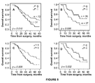
- Figures 5-7 indicate that RBM3 is a predictor of response to gemcitabine in both subgroups.
- the association between high RBM3 expression and response to gemcitabine is however particularly accentuated in latter subgroup (see figures 5-6 ).
- the cancer of the present disclosure may for example be selected from the group consisting of distal bile duct cancer, pancreatic cancer and ampullary cancer of the pancreatobiliary type.
- the level of RBM3 protein is preferably measured intracellularly or in cell-derived material.
- the sample preferably comprises tumor cells.
- the sample may be a tumor tissue sample.
- RBM3 protein expression is both nuclear and cytoplasmic. The inventors have however found that the nuclear expression is particularly relevant for making the treatment predictions of the present disclosure.
- step a) may thus be limited to the nuclei and/or cytoplasms (preferably the nuclei only) of tumor cells of said sample. Consequently, when a tissue sample is examined, only the nuclei of tumor cells may be taken into consideration. Such examination may for example be aided by immunohistochemical staining.
- a sample value of RBM3 protein or RBM3 mRNA being higher than the reference value, or a subject from which such sample value is obtained, is sometimes referred to herein as being "RBM3 high”.
- a sample value of RBM3 protein or RBM3 mRNA being lower than, or equal to, the reference value, or a subject from which such sample value is obtained is sometimes referred to herein as being "RBM3 low”.
- sample value and “reference value” are to be interpreted broadly.
- the quantification of RBM3 to obtain these values may be done via automatic means, via a scoring system based on visual or microscopic inspection of samples, or via combinations thereof.
- a skilled person such as a person skilled in the art of histopathology, to determine the sample and reference values by inspection, e.g., of tissue slides that have been prepared and stained for RBM3 protein expression.
- Determining that the sample value is higher than the reference value may thus be determining, upon visual or microscopic inspection, that a sample tissue slide is more densely stained and/or exhibit a larger fraction of stained cells than a reference tissue slide.
- the sample value may also be compared to a reference value given by a literal reference, such as a reference value described in wording or by a reference picture. Consequently, the sample and/or reference values may in some cases be mental values that the skilled person envisages upon inspection and comparison.
- step a) and optionally step b) may be performed in an apparatus for automatic staining and/or analysis, and such an apparatus may be based on a platform adapted for immunohistochemical staining and/or analysis.
- one or more tumor tissue sample(s) from the subject in question may be prepared for imunohistochemical staining and/or analysis manually and then loaded into the apparatus for automatic staining and/or analysis, which gives the sample value of step a) and optionally also performs the comparison with the reference value of step b).
- the operator performing the analysis, the physician ordering the analysis or the apparatus itself may then draw the conclusion of step c). Consequently, software adapted for drawing the conclusion of step c) may be implemented on the apparatus.
- a reference value found to be relevant for establishing treatment prediction or making treatment decisions regarding cancer subjects, for use as comparison with the sample value from the subject, may be provided in various ways. With the knowledge of the teachings of the present disclosure, the skilled artisan can, without undue burden, provide relevant reference values for performing the methods of the present disclosure.
- the person performing the methods of the above aspects may, for example, adapt the reference value to desired information.
- the reference value may be adapted to yield the most significant information with regard to survival, e.g., the largest separation between the RBM3 high survival curve and the RBM3 low survival curve (see the figures), which corresponds to the largest difference in survival between the first and the second group of the first aspect.
- the reference value may be selected such that a group of subjects having particularly high responsiveness or particularly low responsiveness is singled out.
- the reference value may correspond to the amount of RBM3 protein or RBM3 mRNA measured in a reference sample comprising tumor cells, such as a reference sample of tumor tissue. Such as sample may be derived from another subject having the same type of cancer as the subject of the method. Further, the reference value may for example be provided by the amount of RBM3 protein or RBM3 mRNA measured in a reference sample comprising cancer cell lines expressing a predetermined, or controlled, amount of RBM3 protein or RBM3 mRNA. The person skilled in the art understands how to provide such cell lines, for example guided by the disclosure of Rhodes et al. (2006) The biomedical scientist, p 515-520 .
- the amount of protein or mRNA expression of the reference sample is preferably previously established.
- the reference value may be provided by the amount of RBM3 protein or RBM3 mRNA measured in a reference sample comprising cells expressing a predetermined amount of RBM3 protein or RBM3 mRNA.
- the amount of RBM3 protein or RBM3 mRNA in the reference sample does not have to directly correspond to the reference value.
- the reference sample may also provide an amount of RBM3 protein or RBM3 mRNA that helps a person performing the method to assess various reference values.
- the reference sample(s) may help in creating a mental image of the reference value by providing a "positive" reference value and/or a "negative" reference value.
- the fraction may for example be: a "cellular fraction", wherein the RBM3 protein expression of the whole cells is taken into account; a "cytoplasmic fraction”, wherein the RBM3 protein expression of only the cytoplasms of the cells is taken into account; or a "nuclear fraction”, wherein the RBM3 protein expression of only the nuclei of the cells is taken into account.
- nuclear fraction corresponds to the percentage of relevant cells in a sample that exhibits a positive staining in the nucleus, wherein a medium or distinct and strong immunoreactivity in the nucleus is considered positive and no or faint immunoreactivity in the nucleus is considered negative.
- the person skilled in the art of pathology understands which cells that are relevant under the conditions present when performing the method and may determine a nuclear fraction based on his/her general knowledge and the teachings of the present disclosure.
- the relevant cells may for example be tumor cells. Further, the skilled artisan understands how to perform corresponding measurements employing the "cytoplasmic fraction" or "cellular fraction”.
- the intensity may for example be: a "cellular intensity”, wherein the RBM3 protein expression of the whole cells is taken into account; a "cytoplasmic intensity”, wherein the RBM3 protein expression of only the cytoplasms of the cells is taken into account, or a "nuclear intensity”, wherein the RBM3 protein expression of only the nuclei of the cells is taken into account.
- Nuclear intensity is subjectively evaluated in accordance with standards used in clinical histopathological diagnostics.
- negative no overall immunoreactivity in the nuclei of relevant cells of the sample
- weak faint overall immunoreactivity in the nuclei of relevant cells of the sample
- moderate medium overall immunoreactivity in the nuclei of relevant cells of the sample
- strong distinct and strong overall immunoreactivity in the nuclei of relevant cells of the sample.
- RBM3 protein is particularly relevant for predicting the outcome of gemcitabine treatment.
- the reference value may be a nuclear fraction, a nuclear intensity or a combination or function thereof.
- the sample value may be a nuclear fraction, a nuclear intensity or a combination or function thereof.
- the reference value of step b) may be a nuclear fraction of 95 % or lower, such as 90 % or lower, such as 85 % or lower, such as 80 % or lower, such as 75 % or lower, such as 70 % or lower, such as 65 % or lower, such as 60 % or lower, such as 55 % or lower, such as 50 % or lower, such as 45 % or lower, such as 40 % or lower, such as 35 % or lower, such as 30 % or lower, such as 25 % or lower, such as 20 % or lower, such as 15 % or lower, such as 10 % or lower, such as 5 % or lower, such as 2 % or lower, such as 1 % or lower, such as 0 %.
- the reference value of step b) may be a moderate nuclear intensity of RBM3 protein expression or lower, such as a weak nuclear intensity of RBM3 protein expression or lower, such as a negative nuclear of RBM3 protein expression.
- the reference value may preferably be a nuclear fraction of 50-90 % a moderate nuclear intensity or a function of nuclear fraction and a nuclear intensity in theses ranges.
- the reference value may thus be a combination or a function of a fraction value and an intensity value (see e.g. the value used in the Example below).
- the reference value may thus involve two, and even more, criteria.
- the reference value may thus be a nuclear score (NS), which is the product of the nuclear fraction (in the range 0-100 %) and the nuclear intensity determined as 0, 1, 2, or 3, wherein 0 is a negative nuclear intensity, 1 is a weak nuclear intensity, 2 is a moderate nuclear intensity and 3 is a strong nuclear intensity.
- the NS is thus the result of multiplying the nuclear fraction and the nuclear intensity and may range from 0 to 3.
- the reference value may for example be a NS between 1 and 2.4, such as between 1.5 and 2.2. In the Example below, a reference value/cut-off of 1.77 or 2 is used.
- an intensity value and/or a fraction value as the reference value may depend on the staining procedure, e.g., on the type and amount/concentration of the employed antibody and on the type and concentration of the staining reagents.
- a person skilled in the art e.g., a pathologist understands how to perform the evaluation yielding a fraction, such as a cellular, cytoplasmic or nuclear fraction, or an intensity, such as a cellular, cytoplasmic or nuclear intensity.
- a fraction such as a cellular, cytoplasmic or nuclear fraction
- an intensity such as a cellular, cytoplasmic or nuclear intensity.
- the skilled artisan may use a reference sample comprising a predetermined amount of RBM3 protein for establishing the appearance of a certain fraction or intensity.
- a reference sample may not only be used for the provision of the actual reference value, but also for the provision of an example of a sample with an amount of RBM3 that is higher than the amount corresponding to the reference value.
- histochemical staining such as in immunohistochemical staining
- the skilled artisan may use a reference sample for establishing the appearance of a stained sample having a high amount of RBM3 protein, e.g., a positive reference. Subsequently, the skilled artisan may assess the appearances of samples having lower amounts of RBM3 protein, such as the appearance of a sample with an amount of RBM3 protein corresponding to the reference value.
- the skilled artisan may use a reference sample to create a mental image of a reference value corresponding to an amount of RBM3 protein which is lower than that of the reference sample.
- the skilled artisan may use another reference sample having a low amount of RBM3 protein, or lacking detectable RBM3 protein, for establishing the appearance of such sample, e.g., as a "negative reference”.
- two reference samples may be employed: a first reference sample having no detectable RBM3 protein, and thus corresponding to a negative nuclear intensity, which is lower than the reference value; and a second reference sample having an amount of RBM3 protein corresponding to a strong nuclear intensity, which is higher than the reference value.
- reference sample for establishing the appearance of a sample with a high amount of RBM3 protein.
- Such reference sample may be a sample comprising tissue expressing a high amount of RBM3 protein, such as a sample comprising pancreatic cancer tissue having a pre-established high expression of RBM3 protein.
- the reference sample may provide an example of a strong nuclear intensity (NI).
- NI nuclear intensity
- the skilled artisan may then divide samples into the NI categories absent, weak, moderate and strong. This division may be further assisted by a reference sample lacking detectable RBM3 protein (negative reference), i.e., a reference sample providing an absent nuclear intensity.
- the reference sample may provide an example of a sample with a nuclear fraction (NF) higher than 75 %. With the knowledge of the appearance of a sample with more than 75 % positive cells, the skilled artisan may then evaluate the NF of other samples having e.g., a lower percentage of positive cells.
- This division may be further assisted by a reference sample essentially lacking RBM3 protein (negative reference), i.e., a reference sample providing a low NF (e.g., ⁇ 5%, such as ⁇ 2%), or a NF of 0.
- a reference sample essentially lacking RBM3 protein negative reference
- a reference sample providing a low NF e.g.
- cell lines expressing a controlled amount of RBM3 protein may be used as the reference, in particular as a positive reference.
- One or more pictures may also be provided as the "reference sample”.
- a picture may show an example of a tumor tissue slide stained with a certain antibody during certain conditions and exhibiting a certain nuclear intensity and/or fraction.
- the above discussion about the "reference sample” applies mutatis mutandis to pictures.
- the present disclosure is not limited to the quantification of any particular variant of the RBM3 protein or RBM3 mRNA present in the subject in question, as long as the protein or mRNA is encoded by the relevant gene and presents the relevant pattern of expression.
- the RBM3 protein of the present disclosure may for example comprise a sequence selected from:
- sequence ii) above is at least 90 % identical, at least 91 % identical, at least 92 % identical, at least 93 % identical, at least 94 % identical, at least 95 % identical, at least 96 % identical, at least 97 % identical, at least 98 % identical or at least 99 % identical to SEQ ID NO:1.
- the RBM3 protein of the present disclosure may comprise, or consists of, a sequence selected from:
- sequence ii) above is at least 90 % identical, at least 91 % identical, at least 92 % identical, at least 93 % identical, at least 94 % identical, at least 95 % identical, at least 96 % identical, at least 97 % identical, at least 98 % identical or at least 99 % identical to SEQ ID NO:2.
- the RBM3 mRNA of the present disclosure may comprise or consists of
- sequence ii) above is at least 90 % identical, at least 91 % identical, at least 92 % identical, at least 93 % identical, at least 94 % identical, at least 95 % identical, at least 96 % identical, at least 97 % identical, at least 98 % identical or at least 99 % identical to the sequence i).
- % identical is calculated as follows.
- the query sequence is aligned to the target sequence using the CLUSTAL W algorithm ( Thompson, J.D., Higgins, D.G. and Gibson, T.J., Nucleic Acids Research, 22: 4673-4680 (1994 )).
- the amino acid residues at each position are compared, and the percentage of positions in the query sequence that have identical correspondences in the target sequence is reported as % identical.
- the target sequence determines the number of positions that are compared. Consequently, in the context of the present disclosure, a query sequence that is shorter than the target sequence can never be 100 % identical to the target sequence.
- a query sequence of 85 amino acids may at the most be 85 % identical to a target sequence of 100 amino acids. This corresponds mutatis mutandis nucleic acid sequences.
- step a) of the above methods may comprise:
- the RBM3 protein may be detected and/or quantified through the application to the sample of a detectable and/or quantifiable affinity ligand, which is capable of selective interaction with the RBM3 protein.
- the application of the affinity ligand is performed under conditions that enable binding of the affinity ligand to RBM3 protein in the sample.
- step a) may comprise:
- affinity ligand remaining in association with the sample refers to affinity ligand which was not removed in step a2), e.g., the affinity ligand bound to the sample.
- the binding may for example be the interaction between antibody and antigen.
- step a) may comprise:
- "selective" interaction of e.g., an affinity ligand with its target or antigen means that the interaction is such that a distinction between selective and non-selective becomes meaningful.
- the interaction between two proteins is sometimes measured by the dissociation constant.
- the dissociation constant describes the strength of binding (or affinity) between two molecules.
- the dissociation constant between an antibody and its antigen is from 10 -7 to 10 -11 M.
- high selectivity does not necessarily require high affinity. Molecules with low affinity (in the molar range) for its counterpart have been shown to be as selective as molecules with much higher affinity.
- a selective interaction refers to the extent to which a particular method can be used to determine the presence and/or amount of a specific protein, the target protein, under given conditions in the presence of other proteins in a biological sample, such as a tissue sample or a fluid sample of a naturally occurring or processed biological fluid.
- specificity or selectivity is the capacity to distinguish between related proteins.
- the specificity or selectivity of an antibody may be determined as in WO 2010/092190 (see Examples, Section 2), wherein analysis is performed using a protein array set-up, a suspension bead array and a multiplexed competition assay, respectively. Specificity and selectivity determinations are also described in Nilsson P et al. (2005) Proteomics 5:4327-4337 .
- affinity ligands that may prove useful, as well as examples of formats and conditions for detection and/or quantification, are given below.
- the affinity ligand may be selected from the group consisting of antibodies, fragments thereof and derivatives thereof, i.e., affinity ligands based on an immunoglobulin scaffold.
- the antibodies and the fragments or derivatives thereof may be isolated.
- Antibodies comprise monoclonal and polyclonal antibodies of any origin, including murine, rabbit, human and other antibodies, as well as chimeric antibodies comprising sequences from different species, such as partly humanized antibodies, e.g., partly humanized mouse antibodies.
- Polyclonal antibodies are produced by immunization of animals with the antigen of choice.
- the polyclonal antibodies may be antigen purified.
- Monoclonal antibodies of defined specificity can be produced using the hybridoma technology developed by Köhler and Milstein ( Köhler G and Milstein C (1976) Eur. J. Immunol. 6:511-519 ).
- the antibody fragments and derivatives of the present disclosure are capable of selective interaction with the same antigen (e.g. RBM3 protein) as the antibody they are fragments or derivatives of.
- Antibody fragments and derivatives comprise Fab fragments, consisting of the first constant domain of the heavy chain (CH1), the constant domain of the light chain (CL), the variable domain of the heavy chain (VH) and the variable domain of the light chain (VL) of an intact immunoglobulin protein; Fv fragments, consisting of the two variable antibody domains VH and VL ( Skerra A and Plückthun A (1988) Science 240:1038-1041 ); single chain Fv fragments (scFv), consisting of the two VH and VL domains linked together by a flexible peptide linker ( Bird RE and Walker BW (1991) Trends Biotechnol. 9:132-137 ); Bence Jones dimers ( Stevens FJ et al.
- the affinity ligand of the present disclosure is capable of selective interaction with a peptide consisting of the amino acid sequence SEQ ID NO:1.
- the RBM3 protein fragment SEQ ID NO:1 was designed to lack transmembrane regions to ensure efficient expression in E. coli, and to lack any signal peptide, since those are cleaved off in the mature protein. SEQ ID NO:1 was thus designed for immunizations.
- the protein fragment was designed to consist of a unique sequence with low sequence identity to other human proteins, to minimize cross reactivity of generated affinity reagents, and to be of a suitable size to allow the formation of conformational epitopes and still allow efficient cloning and expression in bacterial systems.
- the affinity ligand may be obtainable by a process comprising a step of immunizing an animal with a peptide whose amino acid sequence consists of the sequence SEQ ID NO:1.
- an immunization process may comprise primary immunization with the protein/antigen in Freund's complete adjuvant.
- the immunization process may further comprise boosting at least two times, in intervals of 2-6 weeks, with the protein/antigen in Freund's incomplete adjuvant. Processes for the production of antibodies or fragments or derivatives thereof against a given target are known in the art.
- an "antigen purified antibody” is one or a population of polyclonal antibodies which has been affinity purified on its own antigen, thereby separating such antigen purified antibodies from other antiserum proteins and non-specific antibodies. This affinity purification results in antibodies that bind selectively to its antigen.
- the polyclonal antisera are purified by a two-step immunoaffinity based protocol to obtain antigen purified antibodies selective for the target protein. Antibodies directed against generic affinity tags of antigen fragments are removed in a primary depletion step, using the immobilized tag protein as the capturing agent.
- the serum is loaded on a second affinity column with the antigen as capturing agent, in order to enrich for antibodies specific for the antigen (see also Nilsson P et al. (2005) Proteomics 5:4327-4337 ).
- the biomolecular diversity needed for selection of affinity ligands may be generated by combinatorial engineering of one of a plurality of possible scaffold molecules, and specific/selective affinity ligands are then selected using a suitable selection platform.
- the scaffold molecule may be of immunoglobulin protein origin ( Bradbury AR and Marks JD (2004) J. Immunol. Meths. 290:29-49 ), of non-immunoglobulin protein origin ( Nygren P ⁇ and Skerra A (2004) J. Immunol. Meths. 290:3-28 ), or of an oligonucleotide origin ( Gold L et al. (1995) Annu. Rev. Biochem. 64:763-797 ).
- Non-limiting examples of such structures useful for generating affinity ligands against RBM3 protein for use according to the present disclosure, are staphylococcal protein A and domains thereof and derivatives of these domains, such as protein Z ( Nord K et al. (1997) Nat. Biotechnol. 15:772-777 ); lipocalins ( Beste G et al. (1999) Proc. Natl. Acad. Sci. U.S.A. 96:1898-1903 ); ankyrin repeat domains ( Binz HK et al. (2003) J. Mol. Biol.
- CBD cellulose binding domains
- GFP green fluorescent protein
- CTL-4 Hufton SE et al. (2000) FEBS Lett. 475:225-231 ; Irving RA et al. (2001) J. Immunol. Meth.
- protease inhibitors such as Knottin proteins ( Wentzel A et al. (2001) J. Bacteriol. 183:7273-7284 ; Baggio R et al. (2002) J. Mol. Recognit. 15:126-134 ) and Kunitz domains ( Roberts BL et al. (1992) Gene 121:9-15 ; Dennis MS and Lazarus RA (1994) J. Biol. Chem. 269:22137-22144 ); PDZ domains ( Schneider S et al. (1999) Nat. Biotechnol. 17:170-175 ); peptide aptamers, such as thioredoxin ( Lu Z et al.
- non-immunoglobulin protein scaffolds include scaffold proteins presenting a single randomized loop used for the generation of novel binding specificities, protein scaffolds with a rigid secondary structure where side chains protruding from the protein surface are randomized for the generation of novel binding specificities, and scaffolds exhibiting a non-contiguous hyper-variable loop region used for the generation of novel binding specificities.
- oligonucleotides may also be used as affinity ligands.
- Single stranded nucleic acids called aptamers or decoys, fold into well-defined three-dimensional structures and bind to their target with high affinity and specificity.
- aptamers or decoys Single stranded nucleic acids
- the oligonucleotide ligands can be either RNA or DNA and can bind to a wide range of target molecule classes.
- Selection platforms include, but are not limited to, phage display ( Smith GP (1985) Science 228:1315-1317 ), ribosome display ( Hanes J and Plückthun A (1997) Proc. Natl. Acad. Sci. U.S.A.
- yeast two-hybrid system Fields S and Song O (1989) Nature 340:245-246
- yeast display Gai SA and Wittrup KD (2007) Curr Opin Struct Biol 17:467-473
- mRNA display Roberts RW and Szostak JW (1997) Proc. Natl. Acad. Sci. U.S.A. 94:12297-12302
- bacterial display Daugherty PS (2007) Curr Opin Struct Biol 17:474-480 , Kronqvist N et al. (2008) Protein Eng Des Sel 1-9 , Harvey BR et al.
- the affinity ligand may be a non-immunoglobulin affinity ligand derived from any of the protein scaffolds listed above, or an oligonucleotide molecule.
- the RBM3 protein fragment SEQ ID NO:1 was designed to consist of a unique sequence with low sequence identity to other human proteins and to minimize cross reactivity of generated affinity reagents. Consequently, in embodiments of the present disclosure, the affinity ligand may be capable of selective interaction with a polypeptide consisting of the amino acid sequence SEQ ID NO:1.
- the affinity ligand of the present disclosure is capable of selective interaction with a peptide consisting of an amino acid sequence selected from SEQ ID NO:4 and 5.
- the affinity ligand of the present disclosure is capable of selective interaction with an RBM3 fragment which consists of 20 amino acid residues or less, such as 15 amino acid residues or less, and comprises a sequence selected from SEQ ID NO:6-9.
- the affinity ligand of the present disclosure is capable of selective interaction with an RBM3 fragment which consists of 20 amino acid residues or less, such as 15 amino acid residues or less, and comprises a sequence selected from SEQ ID NO:10-19.
- Antibodies having selectivity for a single epitope region may provide for increased reproducibility in detection analyses as compared to antibodies generated against a longer peptide sequence (such as a PrEST or a full-length protein).
- the antibodies selective for a single epitope region may also provide for distinct and strong staining in immunohistochemical analyses.
- 6F11 and 1 B5 are considered to be particularly beneficial. 6F11 and 1 B5 have both been shown to be more selective than a polyclonal anti-RBM3 antibody. Further, 1 B5 has been shown to be more selective than 6F11.1 B5 is also employed in the example section below, wherein it is referred to as AAb030038.
- SEQ ID NO:17 to which 1 B5 binds, is within SEQ ID NO:5.
- the affinity ligand is thus capable of selective interaction with an RBM3 fragment which consists of SEQ ID NO:5, and in particularly preferred embodiments of the present disclosure, the affinity ligand is capable of selective interaction with an RBM3 fragment which consists of 20 amino acid residues or less, such as 15 amino acid residues or less, and comprises the sequence SEQ ID NO:17.
- the affinity ligand is thus capable of selective interaction with an RBM3 fragment which consists of 20 amino acid residues or less, such as 15 amino acid residues or less, and comprises a sequence selected from SEQ ID NO:8 and 16. Note that SEQ ID NO:8 and 16 are overlapping and that such a fragment may comprise the sequences of both SEQ ID NO:8 and 16.
- antibodies capable of selective interaction with SEQ ID NO:24 and 25 are shown to be particularly selective.
- the affinity ligand is thus capable of selective interaction with an RBM3 fragment which consists of a sequence selected from SEQ ID NO:24 and 25.
- the affinity ligand is thus capable of selective interaction with an RBM3 fragment which comprises a sequence selected from SEQ ID NO:22 and 26 and consists of 21 amino acids or less, such as 20 amino acids or less, such as 17 amino acids or less, such as 15 amino acid residues or less, such as 10 amino acid residues or less, such as 6 amino acids or less.
- the detection and/or quantification of the affinity ligand capable of selective interaction with the RBM3 protein may be accomplished in any way known to the skilled person for detection and/or quantification of binding reagents in assays based on biological interactions. Accordingly, any affinity ligand described above may be used to quantitatively and/or qualitatively detect the presence of the RBM3 protein.
- affinity ligands may be labeled themselves with various markers or may in turn be detected by secondary, labeled affinity ligands to allow detection, visualization and/or quantification.
- Non-limiting examples of labels that can be conjugated to primary and/or secondary affinity ligands include fluorescent dyes or metals (e.g., fluorescein, rhodamine, phycoerythrin, fluorescamine), chromophoric dyes (e.g., rhodopsin), chemiluminescent compounds (e.g., luminal, imidazole) and bioluminescent proteins (e.g., luciferin, luciferase), haptens (e.g., biotin).
- fluorescent dyes or metals e.g., fluorescein, rhodamine, phycoerythrin, fluorescamine
- chromophoric dyes e.g., rhodopsin
- chemiluminescent compounds e.g., luminal, imidazole
- bioluminescent proteins e.g., luciferin, luciferase
- haptens
- Affinity ligands can also be labeled with enzymes (e.g., horseradish peroxidase, alkaline phosphatase, beta-lactamase), radioisotopes (e.g 3 H, 14 C, 32 P, 35 S or 125 I) and particles (e.g., gold).
- enzymes e.g., horseradish peroxidase, alkaline phosphatase, beta-lactamase
- radioisotopes e.g 3 H, 14 C, 32 P, 35 S or 125 I
- particles e.g., gold
- particles e.g., gold
- particles e.g., gold
- particles e.g., gold
- particles e.g., gold
- particles e.g., gold
- particles e.g., gold
- particles e.g., gold
- particles e.g., gold
- particles e.g., gold
- particles e.g., gold
- Quantum dots have superior quantum yield and are more photostable compared to organic fluorophores and are therefore more easily detected ( Chan et al. (2002) Curr Opi Biotech. 13: 40-46 ).
- the different types of labels can be conjugated to an affinity ligand using various chemistries, e.g., the amine reaction or the thiol reaction.
- chemistries e.g., the amine reaction or the thiol reaction.
- other reactive groups than amines and thiols can be used, e.g., aldehydes, carboxylic acids and glutamine.
- the detection, localization and/or quantification of a labeled affinity ligand bound to its RBM3 protein target may involve visualizing techniques, such as light microscopy or immunofluoresence microscopy. Other methods may involve the detection via flow cytometry or luminometry.
- biological material from the subject may be used for obtaining the sample for detection and/or quantification of RBM3 protein.
- the sample may thus be an earlier obtained sample. If using an earlier obtained sample in a method, no steps of the method are practiced on the human or animal body.
- the affinity ligand may be applied to the sample for detection and/or quantification of the RBM3 protein. This procedure enables not only detection of RBM3 protein, but may in addition show the distribution and relative level of expression thereof.
- the method of visualization of labels on the affinity ligand may include, but is not restricted to, fluorometric, luminometric and/or enzymatic techniques. Fluorescence is detected and/or quantified by exposing fluorescent labels to light of a specific wavelength and thereafter detecting and/or quantifying the emitted light in a specific wavelength region. The presence of a luminescently tagged affinity ligand may be detected and/or quantified by luminescence developed during a chemical reaction. Detection of an enzymatic reaction is due to a color shift in the sample arising from a chemical reaction. Those of skill in the art are aware that a variety of different protocols can be modified in order for proper detection and/or quantification.
- the sample may be immobilized onto a solid phase support or carrier, such as nitrocellulose or any other solid support matrix capable of immobilizing RBM3 protein present in the biological sample applied to it.
- solid state support materials useful in the present invention include glass, carbohydrate (e.g., Sepharose), nylon, plastic, wool, polystyrene, polyethene, polypropylene, dextran, amylase, films, resins, cellulose, polyacrylamide, agarose, alumina, gabbros and magnetite.
- primary affinity ligand selective for RBM3 protein may be applied.
- the supporting matrix may be washed with one or more appropriate buffers known in the art, followed by exposure to a secondary labeled affinity ligand and washed once again with buffers to remove unbound affinity ligands. Thereafter, selective affinity ligands may be detected and/or quantified with conventional methods.
- the binding properties for an affinity ligand may vary from one solid state support to the other, but those skilled in the art should be able to determine operative and optimal assay conditions for each determination by routine experimentation.
- the quantifiable affinity ligand of a1) or al) may be detected using a secondary affinity ligand capable of recognizing the quantifiable affinity ligand.
- the quantification of a3) or aII) may thus be carried out by means of a secondary affinity ligand with affinity for the quantifiable affinity ligand.
- the secondary affinity ligand may be an antibody or a fragment or a derivative thereof.
- one available method for detection and/or quantification of the RBM3 protein is by linking the affinity ligand to an enzyme that can then later be detected and/or quantified in an enzyme immunoassay (such as an EIA or ELISA).
- an enzyme immunoassay such as an EIA or ELISA.
- the biological sample is brought into contact with a solid material or with a solid material conjugated to an affinity ligand against the RBM3 protein, which is then detected and/or quantified with an enzymatically labeled secondary affinity ligand.
- an appropriate substrate is brought to react in appropriate buffers with the enzymatic label to produce a chemical moiety, which for example is detected and/or quantified using a spectrophotometer, fluorometer, luminometer or by visual means.
- primary and any secondary affinity ligands can be labeled with radioisotopes to enable detection and/or quantification.
- appropriate radiolabels in the present disclosure are 3 H, 14 C, 32 P, 35 S or 125 I.
- the specific activity of the labeled affinity ligand is dependent upon the half-life of the radiolabel, isotopic purity, and how the label has been incorporated into the affinity ligand.
- Affinity ligands are preferably labeled using well-known techniques ( Wensel TG and Meares CF (1983) in: Radioimmunoimaging and Radioimmunotherapy (Burchiel SW and Rhodes BA eds.) Elsevier, New York, pp 185-196 ).
- a thus radiolabeled affinity ligand can be used to visualize RBM3 protein by detection of radioactivity in vivo or ex vivo.
- Radionuclear scanning with e.g., gamma camera, magnetic resonance spectroscopy or emission tomography function for detection in vivo and ex vivo, while gamma/beta counters, scintillation counters and radiographies are also used ex vivo.
- total cellular RNA is purified from cells by homogenization in the presence of nucleic acid extraction buffer, followed by centrifugation. Nucleic acids are then precipitated, in order to remove DNA by treatment with DNase and precipitation. The RNA molecules are then separated by gel electrophoresis on agarose gels according to standard techniques, and transferred to nitrocellulose filters by, e.g., the so-called "Northern" blotting technique. The RNA is then immobilized on the filters by heating. Detection and quantification of specific RNA is accomplished using appropriately labeled DNA or RNA probes complementary to the RNA in question.
- the nucleic acid probe may be labeled with, e.g., a radionuclide such as 3 H, 32 P, 33 P, 14 C, or 35 S; a heavy metal; or a ligand capable of functioning as a specific binding pair member for a labeled ligand (e.g., biotin, avidin, or an antibody), a fluorescent molecule, a chemi luminescent molecule, an enzyme, or the like.
- a radionuclide such as 3 H, 32 P, 33 P, 14 C, or 35 S
- a heavy metal e.g., a ligand capable of functioning as a specific binding pair member for a labeled ligand (e.g., biotin, avidin, or an antibody), a fluorescent molecule, a chemi luminescent molecule, an enzyme, or the like.
- Probes may be labeled to high specific activity by either the nick translation method ( Rigby et al., (1977) J. Mol Biol, 113: 237-251 ), or by the random priming method ( Fienberg, (1983) Anal. Biochem., 132: 6-13 ).
- the latter can be a method for synthesizing 32 P-labeled probes of high specific activity from RNA templates. For example, by replacing preexisting nucleotides with highly radioactive nucleotides according to the nick translation method, it is possible to prepare 32 P- labeled nucleic acid probes with a specific activity well in excess of 10 cpm/microgram. Autoradiographic detection of hybridization then can be performed by exposing hybridized filters to photographic film.
- Biomarker levels can be quantified by computerized imaging systems, such as the Molecular Dynamics 400-B 2D Phosphorimager (Amersham Biosciences, Piscataway, NJ., USA).
- the random- primer method can be used to incorporate an analogue, for example, the dTTP analogue 5 -(N-(N-biotinyl-epsilon-aminocaproyl)-3-aminoallyl)deoxyuridine triphosphate, into the probe molecule.
- analogue for example, the dTTP analogue 5 -(N-(N-biotinyl-epsilon-aminocaproyl)-3-aminoallyl)deoxyuridine triphosphate
- the biotinylated probe oligonucleotide can be detected by reaction with biotin-binding proteins, such as avidin, streptavidin, and antibodies (e.g., anti-biotin antibodies) coupled to fluorescent dyes or enzymes that produce color reactions.
- determining the levels of RNA transcript may be accomplished using the technique of in situ hybridization. This technique requires fewer cells than the Northern blotting technique, and involves depositing whole cells onto a microscope cover slip and probing the nucleic acid content of the cell with a solution containing radioactive or otherwise labeled nucleic acid (e.g., cDNA or RNA) probes. This technique is particularly well-suited for analyzing tissue biopsy samples from subjects.
- a solution containing radioactive or otherwise labeled nucleic acid e.g., cDNA or RNA
- RNA transcripts in cells also can be determined by reverse transcription of RNA transcripts, followed by amplification of the reverse-transcribed transcripts by polymerase chain reaction (RT-PCR).
- the levels of RNA transcripts can be quantified in comparison with an internal standard, for example, the level of mRNA from a standard gene present in the same sample.
- an internal standard for example, the level of mRNA from a standard gene present in the same sample.
- the person skilled in the art is capable of selecting suitable genes for use as an internal standard.
- the methods for quantitative RT-PCR and variations thereof are within the skill in the art.
- primers can be used for the quantitative RT-PCR.
- the primers are specific to RBM3. It is within the skill in the art to generate primers specific to RBM3 (e.g. starting from SEQ ID NO:3).
- Primers can be of any suitable length, but are preferably between 19 and 23 (e.g., 19, 20, 21, 22, or 23) nucleotides.
- amplicon length should be 50 to 150 (up to 250 may be necessary but then optimization of the thermal cycling protocol and reaction components may be necessary) bases for optimal PCR efficiency. Designing primers that generate a very long amplicon may lead to poor amplification efficiency. Information about primer design and optimal amplicon size may fo example be found at www.ambion.com.
- microchip technology it may be desirable to use microchip technology to detect biomarker expression.
- the microchip can be fabricated by techniques known in the art. For example, probe oligonucleotides of an appropriate length, e.g., 40 nucleotides, are 5 '-amine modified at position C6 and printed using commercially available microarray systems, e.g., the GENEMACHINE OmniGrid 100 Microarrayer and Amersham CODELINK activated slides. Labeled cDNA oligomer corresponding to the target RNAs is prepared by reverse transcribing the target RNA with labeled primer. Following first strand synthesis, the RNA/DNA hybrids are denatured to degrade the RNA templates.
- the labeled target cDNAs thus prepared are then hybridized to the microarray chip under hybridizing conditions, e.g., 6 times SSPE/30% formamide at 25 °C for 18 hours, followed by washing in 0.75 times TNT at 37 °C for 40 minutes. At positions on the array, where the immobilized probe DNA recognizes a complementary target cDNA in the sample, hybridization occurs.
- the labeled target cDNA marks the exact position on the array where binding occurs, thereby allowing automatic detection and quantification.
- the output consists of a list of hybridization events, which indicate the relative abundance of specific cDNA sequences, and therefore the relative abundance of the corresponding complementary biomarker, in the subject sample.
- the labeled cDNA oligomer is a biotin-labeled cDNA prepared from a biotin-labeled primer.
- the microarray is then processed by direct detection of the biotin-containing transcripts using, e.g., Streptavidin-Alexa647 conjugate, and scanned utilizing conventional scanning methods. Image intensities of each spot on the array are proportional to the abundance of the corresponding biomarker in the subject sample.
- the use of the array has one or more advantages for mRNA expression detection.
- the RBM3 mRNA may for example be extracted from formalin-fixed, paraffin-embedded tumor tissue. Accordingly, the sample of the methods of the present disclosure may be formalin-fixed and/or paraffin-embedded tumor tissue independent of if RBM3 protein or RBM3 mRNA is detected.
- RBM3 mRNA analysis of the present disclosure may be incorporated in an mRNA-based assay designed to support individualized treatment planning.
- Such an assay may employ RT-PCR to analyze the expression of several genes.
- an RBM3 protein or RBM3 mRNA molecule as a marker of response to gemcitabine treatment of a mammalian subject having a cancer, wherein the protein or mRNA molecule is provided in a sample derived from the cancer of the subject.
- the use of the fourth aspect may be entirely ex vivo, e.g., on previously obtained samples.
- the presence of the marker of the fourth aspect, or a relatively high level thereof, is indicative of a relatively high responsiveness to gemcitabine as compared to a relatively low responsiveness indicated by the absence or a relatively low level of the marker of the fourth aspect.
- an affinity ligand capable of selective interaction with an RBM3 protein as a treatment predictive agent for gemcitabine treatment of a mammalian subject having a cancer.
- an affinity ligand capable of selective interaction with an RBM3 protein for predicting the response to a gemcitabine treatment of a mammalian subject having a cancer.
- an affinity ligand capable of selective interaction with an RBM3 protein for indicating whether a mammalian subject having a cancer should be given a gemcitabine.
- the use of the fifth aspect may be entirely ex vivo.
- a product for use in a method of treatment of a mammalian subject having a cancer, wherein said method comprises the step of determining that an amount of RBM3 protein or RBM3 mRNA in a sample derived from the cancer of the subject is higher than a relevant reference value.
- the sixth aspect thus relates to gemcitabine treatment of RBM3 high subjects.
- Various ways of determining that an amount of RBM3 in a sample derived from the cancer of the subject is higher than a relevant reference value are described above. For example, relevant reference values are discussed thoroughly.
- the inventors believe that the RBM3 protein expression is more relevant than the RBM3 mRNA expression in the context of the present disclosure.
- the median age of patients was 67 (38-83) years. Grade was recorded for all patients: 12 tumors were well differentiated, 62 were moderately differentiated, 97 were poorly differentiated, and 4 were undifferentiated.
- the heterogeneous cohort included 14 tumors originating from the duodenum, 51 tumors of papilla-ampulla intestinal type, 19 tumors of papilla-ampulla pancreatobiliary type, 45 distal bile duct tumors, and 45 pancreatic adenocarcinomas.
- Treatment data was available for all patients.
- TMA tumor necrosis factor
- the immunohistochemical staining was evaluated by two independent observers who were blinded to clinical and outcome data. Scoring differences were discussed to reach consensus. Immunohistochemical analysis of RBM3 expression with anti-RBM3 could be performed on 171/175 tumor samples.
- RBM3 nuclear expression was dichotomized into high and low expression with the median value as cut-off.
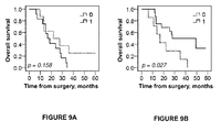
- Recurrence free survival (RFS) was defined from the date of surgery to the date of locoregional or distant recurrence. Kaplan Meier and the log rank test were applied to estimate differences in 5-year overall survival (OS) and RFS in strata according to high and low RBM3 expression.
- OS overall survival
- RFS recurrence free survival
- a tumor tissue sample from the patient is obtained.
- the tumor tissue sample may be obtained from a specimen from an earlier surgical removal of the tumor or from a tumor biopsy.
- a sample is taken from archival material comprising tissue having low, or essentially lacking, RBM3 protein expression.
- archival tissue may for example be tissue having a pre-established low RBM3 protein expression level from the same cancer type as the one of the tested subject.
- a sample is taken from archival tissue material of the same cancer type as the one of the tested subject, which archival material comprises tissue having high RBM3 protein expression.
- the sample and reference material is fixated in buffered formalin and histo-processed in order to obtain thin sections (4 ⁇ m) of the sample material.
- the 4 ⁇ m TMA-sections are automatically pretreated using the PT Link system and then stained in an Autostainer Plus (DAKO; Glostrup, Denmark) using a selective anti-RBM3 antibody, such as the monoclonal antibody AAb030038 (Atlas Antibodies AB, Sweden).
- two control cell-lines may be used as a tool to validate the staining procedure; e.g. one slide with cells expressing RBM3 protein (positive cell line) and one slide having cells with indistinct weak or no RBM3 protein expression (negative cell line).
- RBM3 protein positive cell line
- negative cell line negative cell line
- the skilled artisan understands how to provide such cell lines, for example guided by the disclosure of Rhodes et al. (2006) The biomedical scientist, p 515-520 .
- the control-line slides may be simultaneously stained in the same procedure as the other slides, i.e. incubated with the same primary and secondary antibodies.
- control cell-lines are used, these are inspected to validate the staining procedure. If the cell-lines display staining results outside acceptable criteria, e.g. staining artifacts recognized by the skilled artisan, the staining of the tissue samples is considered invalid and the whole staining procedure is repeated with new slides. If the positive and negative cell-lines display strong staining intensity and indistinct weak or no staining intensity, respectively, the staining is considered as valid.
- the immunohistochemical staining result is then evaluated by two independent observers who are blinded to clinical and outcome data. Scoring differences are preferably discussed to reach consensus.
- the observers performing the evaluation and determination are aided by visual inspection of the stained positive and negative reference slides.
- a suitable reference value may be a staining score of 1.77, in particular if AAb030038 is used.
- the physician responsible for the treatment of the patient concludes that the patient's responsiveness to gemcitabine is relatively high and optionally, that the patient should be given gemcitabine.
- the physician concludes that the patient's responsiveness to gemcitabine is relatively low and, optionally, to refrain from gemcitabine treatment. In the latter case, an alternative treatment may be applied instead.
Priority Applications (2)
| Application Number | Priority Date | Filing Date | Title |
|---|---|---|---|
| EP14194296.1A EP3023791A1 (fr) | 2014-11-21 | 2014-11-21 | Prévoir la réactivité à un traitement de gemcitabine |
| PCT/EP2015/077106 WO2016079240A1 (fr) | 2014-11-21 | 2015-11-19 | Prédiction de la réactivité à un traitement à la gemcitabine |
Applications Claiming Priority (1)
| Application Number | Priority Date | Filing Date | Title |
|---|---|---|---|
| EP14194296.1A EP3023791A1 (fr) | 2014-11-21 | 2014-11-21 | Prévoir la réactivité à un traitement de gemcitabine |
Publications (1)
| Publication Number | Publication Date |
|---|---|
| EP3023791A1 true EP3023791A1 (fr) | 2016-05-25 |
Family
ID=51947198
Family Applications (1)
| Application Number | Title | Priority Date | Filing Date |
|---|---|---|---|
| EP14194296.1A Withdrawn EP3023791A1 (fr) | 2014-11-21 | 2014-11-21 | Prévoir la réactivité à un traitement de gemcitabine |
Country Status (2)
| Country | Link |
|---|---|
| EP (1) | EP3023791A1 (fr) |
| WO (1) | WO2016079240A1 (fr) |
Citations (6)
| Publication number | Priority date | Publication date | Assignee | Title |
|---|---|---|---|---|
| WO2001004144A2 (fr) | 1999-07-13 | 2001-01-18 | Scil Proteins Gmbh | Fabrication de proteines a feuillet plisse beta et a proprietes de liaison specifiques |
| WO2001005808A2 (fr) | 1999-07-20 | 2001-01-25 | Affibody Technology Sweden Ab | Selection in vitro et identification facultative de polypeptides a l'aide de porteurs a support solide |
| US20090221522A1 (en) * | 2008-02-14 | 2009-09-03 | The Johns Hopkins University | Methods to correct gene set expression profiles to drug sensitivity |
| WO2010092190A2 (fr) | 2009-02-16 | 2010-08-19 | Atlas Antibodies Ab | Prévision de la réponse à une thérapie à base de platine |
| EP2524928A1 (fr) * | 2011-05-18 | 2012-11-21 | Atlas Antibodies AB | RBM3 dans le cancer de la vessie |
| WO2013041610A1 (fr) | 2011-09-20 | 2013-03-28 | Atlas Antibodies Ab | Rbm3 dans le pronostic du cancer de la prostate |
-
2014
- 2014-11-21 EP EP14194296.1A patent/EP3023791A1/fr not_active Withdrawn
-
2015
- 2015-11-19 WO PCT/EP2015/077106 patent/WO2016079240A1/fr active Application Filing
Patent Citations (7)
| Publication number | Priority date | Publication date | Assignee | Title |
|---|---|---|---|---|
| WO2001004144A2 (fr) | 1999-07-13 | 2001-01-18 | Scil Proteins Gmbh | Fabrication de proteines a feuillet plisse beta et a proprietes de liaison specifiques |
| WO2001005808A2 (fr) | 1999-07-20 | 2001-01-25 | Affibody Technology Sweden Ab | Selection in vitro et identification facultative de polypeptides a l'aide de porteurs a support solide |
| US20090221522A1 (en) * | 2008-02-14 | 2009-09-03 | The Johns Hopkins University | Methods to correct gene set expression profiles to drug sensitivity |
| WO2010092190A2 (fr) | 2009-02-16 | 2010-08-19 | Atlas Antibodies Ab | Prévision de la réponse à une thérapie à base de platine |
| US8747910B2 (en) * | 2009-02-16 | 2014-06-10 | Atlas Antibodies Ab | Prediction of response to platinum-based therapy |
| EP2524928A1 (fr) * | 2011-05-18 | 2012-11-21 | Atlas Antibodies AB | RBM3 dans le cancer de la vessie |
| WO2013041610A1 (fr) | 2011-09-20 | 2013-03-28 | Atlas Antibodies Ab | Rbm3 dans le pronostic du cancer de la prostate |
Non-Patent Citations (59)
| Title |
|---|
| BAGGIO R ET AL., J. MOL. RECOGNIT., vol. 15, 2002, pages 126 - 134 |
| BESTE G ET AL., PROC. NATL. ACAD. SCI. U.S.A., vol. 96, 1999, pages 1898 - 1903 |
| BIANCHI E ET AL., J. MOL. BIOL., vol. 247, 1995, pages 154 - 160 |
| BINZ HK ET AL., J. MOL. BIOL., vol. 332, 2003, pages 489 - 503 |
| BIRD RE; WALKER BW, TRENDS BIOTECHNOL., vol. 9, 1991, pages 132 - 137 |
| BRADBURY AR; MARKS JD, J. IMMUNOL. METHS., vol. 290, 2004, pages 29 - 49 |
| BRAND L; GOHLKE JR, ANNU. REV. BIOCHEM., vol. 41, 1972, pages 843 - 868 |
| BRODY EN; GOLD L, J. BIOTECHNOL., vol. 74, 2000, pages 5 - 13 |
| CAI X; GAREN A, PROC. NATL. ACAD. SCI. U.S.A., vol. 93, 1996, pages 6280 - 6285 |
| CHAN ET AL., CURR OPI BIOTECH., vol. 13, 2002, pages 40 - 46 |
| DAUGHERTY PS, CURR OPIN STRUCT BIOL, vol. 17, 2007, pages 474 - 480 |
| DENNIS MS; LAZARUS RA, J. BIOL. CHEM., vol. 269, 1994, pages 22137 - 22144 |
| DOOLEY H ET AL., MOL. IMMUNOL., vol. 40, 2003, pages 25 - 33 |
| ELLINGTON AD; SZOSTAK JW, NATURE, vol. 346, 1990, pages 818 - 822 |
| FIELDS S; SONG 0, NATURE, vol. 340, 1989, pages 245 - 246 |
| FIENBERG, ANAL. BIOCHEM., vol. 132, 1983, pages 6 - 13 |
| GAI SA; WITTRUP KD, CURR OPIN STRUCT BIOL, vol. 17, 2007, pages 467 - 473 |
| GOLD L ET AL., ANNU. REV. BIOCHEM., vol. 64, 1995, pages 763 - 797 |
| HAMERS-CASTERMAN C ET AL., NATURE, vol. 363, 1993, pages 446 - 448 |
| HANES J; P UCKTHUN A, PROC. NATL. ACAD. SCI. U.S.A., vol. 94, 1997, pages 4937 - 4942 |
| HARVEY BR ET AL., PNAS, vol. 101, no. 25, 2004, pages 913 - 9198 |
| HUFTON SE ET AL., FEBS LETT., vol. 475, 2000, pages 225 - 231 |
| IRVING RA ET AL., J. IMMUNOL. METH., vol. 248, 2001, pages 31 - 45 |
| K6HLER G; MILSTEIN C, EUR. J. IMMUNOL., vol. 6, 1976, pages 511 - 519 |
| KLEVENZ B ET AL., CELL. MOL. LIFE SCI., vol. 59, 2002, pages 1993 - 1998 |
| KLUG A, J. MOL. BIOL., vol. 293, 1999, pages 215 - 218 |
| KOIDE A ET AL., J. MOL. BIOL., vol. 284, 1998, pages 1141 - 1151 |
| KRONQVIST N ET AL., PROTEIN ENG DES SEL, 2008, pages 1 - 9 |
| LEHTI6 J ET AL., PROTEINS, vol. 41, 2000, pages 316 - 322 |
| LI R ET AL., PROTEIN ENG., vol. 16, 2003, pages 65 - 72 |
| LIV BEN DROR: "A high-throughput pathology approach for further insight into the role of RBM3 as a biomarker of prognosis and chemotherapy response in human cancer", LUND UNIVERSITY DOCTORAL DISSERTATION, 12 December 2014 (2014-12-12), pages 1 - 95, XP055186229, ISBN: 978-9-17-619067-8, Retrieved from the Internet <URL:http://www.med.lu.se/content/download/103691/695855/file/Thesis_Ben_Dror.pdf> [retrieved on 20150428] * |
| LU Z ET AL., BIOTECHNOLOGY, vol. 13, 1995, pages 366 - 372 |
| MASAT L ET AL., PROC. NATL. ACAD. SCI. U.S.A., vol. 91, 1994, pages 893 - 896 |
| MAYER G; JENNE A, BIODRUGS, vol. 18, 2004, pages 351 - 359 |
| MCCONELL SJ; HOESS RH, J. MOL. BIOL., vol. 250, 1995, pages 460 - 479 |
| NILSSON P ET AL., PROTEOMICS, vol. 5, 2005, pages 4327 - 4337 |
| NORD 0 ET AL., J BIOTECHNOL, vol. 106, 2003, pages 1 - 13 |
| NORD K ET AL., NAT. BIOTECHNOL., vol. 15, 1997, pages 772 - 777 |
| NORMAN TC ET AL., SCIENCE, vol. 285, 1999, pages 591 - 595 |
| NYGREN PA; SKERRA A, J. IMMUNOL. METHS., vol. 290, 2004, pages 3 - 28 |
| PEELLE B ET AL., CHEM. BIOL., vol. 8, 2001, pages 521 - 534 |
| REMY I; MICHNICK SW, PROC. NATL. ACAD. SCI. U.S.A., vol. 96, 1999, pages 5394 - 5399 |
| RHODES ET AL., THE BIOMEDICAL SCIENTIST, 2006, pages 515 - 520 |
| RIGBY ET AL., J. MOL BIOL, vol. 113, 1977, pages 237 - 251 |
| ROBERTS BL ET AL., GENE, vol. 121, 1992, pages 9 - 15 |
| ROBERTS RW; SZOSTAK JW, PROC. NATL. ACAD. SCI. U.S.A., vol. 94, 1997, pages 12297 - 12302 |
| SAMBROOK J. ET AL.: "Molecular Cloning: A Laboratory Manual", 1989, COLD SPRING HARBOR LABORATORY PRESS |
| SCHNEIDER S ET AL., NAT. BIOTECHNOL., vol. 17, 1999, pages 170 - 175 |
| SEGAL DJ ET AL., BIOCHEMISTRY, vol. 42, 2003, pages 2137 - 2148 |
| SKERRA A; P UCKTHUN A, SCIENCE, vol. 240, 1988, pages 1038 - 1041 |
| SMITH GP ET AL., J. MOL. BIOL., vol. 277, 1998, pages 317 - 332 |
| SMITH GP, SCIENCE, vol. 228, 1985, pages 1315 - 1317 |
| STEVENS FJ ET AL., BIOCHEMISTRY, vol. 30, 1991, pages 6803 - 6805 |
| STRYER L, SCIENCE, vol. 162, 1968, pages 526 - 533 |
| THOMPSON, J.D.; HIGGINS, D.G.; GIBSON, T.J., NUCLEIC ACIDS RESEARCH, vol. 22, 1994, pages 4673 - 4680 |
| TUERK C; GOLD L, SCIENCE, vol. 249, 1990, pages 505 - 510 |
| WENSEL TG; MEARES CF: "Radioimmunoimaging and Radioimmunotherapy", 1983, ELSEVIER, pages: 185 - 196 |
| WENTZEL A ET AL., J. BACTERIOL., vol. 183, 2001, pages 7273 - 7284 |
| XU L ET AL., CHEM. BIOL., vol. 9, 2002, pages 933 - 942 |
Also Published As
| Publication number | Publication date |
|---|---|
| WO2016079240A1 (fr) | 2016-05-26 |
Similar Documents
| Publication | Publication Date | Title |
|---|---|---|
| EP2396660B1 (fr) | Prevision d'une reponse a un traitement a base de platine | |
| EP2315028A1 (fr) | Protéine PODXL dans le cancer colorectal | |
| US9416176B2 (en) | RBM3 protein in colorectal cancer prognostics | |
| EP2936150B1 (fr) | Podxl dans le cancer de la vessie | |
| EP3023791A1 (fr) | Prévoir la réactivité à un traitement de gemcitabine | |
| EP2344525B1 (fr) | Épitopes issus de satb2 et leurs utilisations | |
| EP2573566A1 (fr) | RBM3 dans le pronostic du cancer de la prostate | |
| EP2787350A1 (fr) | ASRGL1 dans le cancer de l'endomètre | |
| EP2602622A1 (fr) | Prédiction de la réponse à une thérapie à base de platine | |
| EP2241889A1 (fr) | Protéine RBM3 dans les pronostics de cancer colorectal |
Legal Events
| Date | Code | Title | Description |
|---|---|---|---|
| AK | Designated contracting states |
Kind code of ref document: A1 Designated state(s): AL AT BE BG CH CY CZ DE DK EE ES FI FR GB GR HR HU IE IS IT LI LT LU LV MC MK MT NL NO PL PT RO RS SE SI SK SM TR |
|
| AX | Request for extension of the european patent |
Extension state: BA ME |
|
| PUAI | Public reference made under article 153(3) epc to a published international application that has entered the european phase |
Free format text: ORIGINAL CODE: 0009012 |
|
| STAA | Information on the status of an ep patent application or granted ep patent |
Free format text: STATUS: THE APPLICATION IS DEEMED TO BE WITHDRAWN |
|
| 18D | Application deemed to be withdrawn |
Effective date: 20161126 |