EP3013419B1 - Real-time quantification of skin burns in external beam radiation therapy - Google Patents
Real-time quantification of skin burns in external beam radiation therapy Download PDFInfo
- Publication number
- EP3013419B1 EP3013419B1 EP14742360.2A EP14742360A EP3013419B1 EP 3013419 B1 EP3013419 B1 EP 3013419B1 EP 14742360 A EP14742360 A EP 14742360A EP 3013419 B1 EP3013419 B1 EP 3013419B1
- Authority
- EP
- European Patent Office
- Prior art keywords
- recited
- burn
- skin
- radiation therapy
- plan
- Prior art date
- Legal status (The legal status is an assumption and is not a legal conclusion. Google has not performed a legal analysis and makes no representation as to the accuracy of the status listed.)
- Active
Links
Images
Classifications
-
- A—HUMAN NECESSITIES
- A61—MEDICAL OR VETERINARY SCIENCE; HYGIENE
- A61N—ELECTROTHERAPY; MAGNETOTHERAPY; RADIATION THERAPY; ULTRASOUND THERAPY
- A61N5/00—Radiation therapy
- A61N5/10—X-ray therapy; Gamma-ray therapy; Particle-irradiation therapy
- A61N5/103—Treatment planning systems
- A61N5/1039—Treatment planning systems using functional images, e.g. PET or MRI
-
- A—HUMAN NECESSITIES
- A61—MEDICAL OR VETERINARY SCIENCE; HYGIENE
- A61N—ELECTROTHERAPY; MAGNETOTHERAPY; RADIATION THERAPY; ULTRASOUND THERAPY
- A61N5/00—Radiation therapy
- A61N5/10—X-ray therapy; Gamma-ray therapy; Particle-irradiation therapy
- A61N5/103—Treatment planning systems
- A61N5/1038—Treatment planning systems taking into account previously administered plans applied to the same patient, i.e. adaptive radiotherapy
-
- A—HUMAN NECESSITIES
- A61—MEDICAL OR VETERINARY SCIENCE; HYGIENE
- A61N—ELECTROTHERAPY; MAGNETOTHERAPY; RADIATION THERAPY; ULTRASOUND THERAPY
- A61N5/00—Radiation therapy
- A61N5/10—X-ray therapy; Gamma-ray therapy; Particle-irradiation therapy
- A61N5/1048—Monitoring, verifying, controlling systems and methods
- A61N5/1064—Monitoring, verifying, controlling systems and methods for adjusting radiation treatment in response to monitoring
- A61N5/1065—Beam adjustment
- A61N5/1067—Beam adjustment in real time, i.e. during treatment
-
- A—HUMAN NECESSITIES
- A61—MEDICAL OR VETERINARY SCIENCE; HYGIENE
- A61N—ELECTROTHERAPY; MAGNETOTHERAPY; RADIATION THERAPY; ULTRASOUND THERAPY
- A61N5/00—Radiation therapy
- A61N5/10—X-ray therapy; Gamma-ray therapy; Particle-irradiation therapy
- A61N5/1048—Monitoring, verifying, controlling systems and methods
- A61N5/1049—Monitoring, verifying, controlling systems and methods for verifying the position of the patient with respect to the radiation beam
- A61N2005/1054—Monitoring, verifying, controlling systems and methods for verifying the position of the patient with respect to the radiation beam using a portal imaging system
-
- A—HUMAN NECESSITIES
- A61—MEDICAL OR VETERINARY SCIENCE; HYGIENE
- A61N—ELECTROTHERAPY; MAGNETOTHERAPY; RADIATION THERAPY; ULTRASOUND THERAPY
- A61N5/00—Radiation therapy
- A61N5/10—X-ray therapy; Gamma-ray therapy; Particle-irradiation therapy
- A61N5/1048—Monitoring, verifying, controlling systems and methods
- A61N5/1049—Monitoring, verifying, controlling systems and methods for verifying the position of the patient with respect to the radiation beam
- A61N2005/1058—Monitoring, verifying, controlling systems and methods for verifying the position of the patient with respect to the radiation beam using ultrasound imaging
-
- A—HUMAN NECESSITIES
- A61—MEDICAL OR VETERINARY SCIENCE; HYGIENE
- A61N—ELECTROTHERAPY; MAGNETOTHERAPY; RADIATION THERAPY; ULTRASOUND THERAPY
- A61N5/00—Radiation therapy
- A61N5/10—X-ray therapy; Gamma-ray therapy; Particle-irradiation therapy
- A61N5/1048—Monitoring, verifying, controlling systems and methods
- A61N5/1049—Monitoring, verifying, controlling systems and methods for verifying the position of the patient with respect to the radiation beam
- A61N2005/1059—Monitoring, verifying, controlling systems and methods for verifying the position of the patient with respect to the radiation beam using cameras imaging the patient
Definitions
- This disclosure relates to medical instruments and more particularly to systems for measuring changes in skin during operative procedures.
- EBRT External Beam Radiation Therapy
- EBRT External Beam Radiation Therapy
- skin burn is a serious side-effect of the treatment.
- Some EBRT-induced skin reactions are immediate, while others may take days or weeks to occur.
- the skin reactions can occur on any part of the skin that is in the path of the radiation beam. Since EBRT treatments involve the use of radiation beams from multiple directions around the patient, skin burns in the case of breast EBRT can occur in the shoulder, back, neck and contra-lateral breast. Skin reactions such as these cause a great deal of pain and discomfort to the patient, in addition to other side-effects. In some cases, extreme skin reactions may lead to the generation of new cancerous cells.
- a system for radiation therapy includes an imaging device configured to scan an area of interest for tissue undergoing radiation therapy to collect one or more images of the tissue.
- An interpretation module is configured to receive the one or more images of the tissue to determine a burn status of the tissue and provide adjustments for a radiation treatment plan in accordance with the burn status.
- An embodiment of the system for radiation therapy includes a portable imaging device configured to scan an area of interest for tissue undergoing radiation therapy.
- a robotically controlled arm, on which the portable imaging device is mounted, is controlled to avoid interference with radiation beams for the radiation therapy.
- An interpretation module is configured to receive images of the tissue collected by the imaging device to determine a burn status of the tissue and provide adjustments for a radiation treatment plan in accordance with the burn status.
- systems are provided for preventing and/or mitigating skin burns in patients as a result of external bean radiation therapy (EBRT).
- EBRT external bean radiation therapy
- the present principles may be employed with adaptive radiation therapy (RT) planning and delivery schemes that may use the minimization of skin reactions as an additional optimization parameter in the design of adaptive treatment plans.
- RT adaptive radiation therapy
- optical or photoacoustic imaging technology is employed to quantitatively image the progression of radiation-induced skin burns, in real-time during EBRT delivery and at various intervals after EBRT delivery.
- DOT Diffuse optical tomography
- Suitable imaging protocols are described in conjunction with existing RT workflows, to obtain real-time updates of skin burn progression during EBRT delivery. Based on the imaging data, corrective action can be proposed to the patient to pre-empt the subsequent occurrence of burn-related symptoms in the days and weeks following treatment (e.g., icing, creams, gels etc.).
- Intermittent imaging after EBRT delivery can further monitor late skin reactions and may also help ascertain the efficacy of any corrective treatments being used to alleviate the burns.
- population-based statistics on skin burn patterns may be developed and related to RT plan characteristics, to permit the creation of intensity-modulated RT (IMRT) plans with "skin burn reduction" as an additional dose optimization parameter.
- IMRT intensity-modulated RT
- the present methods monitor and reduce skin burn during EBRT delivery.
- a new parameter for adaptive treatment planning in EBRT is also introduced. Reduction in RT side-effects such as skin burns leads to improved quality of life for patients, post-RT.
- the present principles are employed in tracking or analyzing complex biological or mechanical systems.
- the present principles are applicable to tracking procedures of biological systems, procedures in all areas of the body such as, the skin, but may be useful for internal organs, such as the lungs, gastro-intestinal tract, excretory organs, blood vessels, etc.
- the elements depicted in the FIGS. may be implemented in various combinations of hardware and software and provide functions which may be combined in a single element or multiple elements.
- processor or “controller” should not be construed to refer exclusively to hardware capable of executing software, and can implicitly include, without limitation, digital signal processor ("DSP") hardware, read-only memory (“ROM”) for storing software, random access memory (“RAM”), non-volatile storage, etc.
- DSP digital signal processor
- ROM read-only memory
- RAM random access memory
- non-volatile storage etc.
- embodiments of the present invention can take the form of a computer program product accessible from a computer-usable or computer-readable storage medium providing program code for use by or in connection with a computer or any instruction execution system.
- a computer-usable or computer readable storage medium can be any apparatus that may include, store, communicate, propagate, or transport the program for use by or in connection with the instruction execution system, apparatus, or device.
- the medium can be an electronic, magnetic, optical, electromagnetic, infrared, or semiconductor system (or apparatus or device) or a propagation medium.
- Examples of a computer-readable medium include a semiconductor or solid state memory, magnetic tape, a removable computer diskette, a random access memory (RAM), a read-only memory (ROM), a rigid magnetic disk and an optical disk.
- Current examples of optical disks include compact disk - read only memory (CD-ROM), compact disk - read/write (CD-R/W), Blu-RayTM and DVD
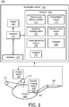
- System 100 may include a workstation or console 112 from which a procedure is supervised and/or managed.
- Workstation 112 preferably includes one or more processors 114 and memory 116 for storing programs and applications.
- Memory 116 may store a planning and delivery module 115 configured to plan and control a radiation therapy session or sessions by controlling radiation sources, imaging devices, etc.
- An interpretation module 110 is configured to receive one or more images of tissue in an area of interest to determine a burn status of the tissue. The interpretation module 110 can provide suggestions for adjustments for a radiation treatment plan in accordance with the burn status.
- the treatment plan may be a treatment plan stored and executed by the planning and delivery module 115.
- the interpretation module 110 employs feedback, e.g., from images, for image maps, or from measurements (optical or acoustical) collected from the tissue of interest. The feedback is employed to interpret the images or data and record changes over time. Module 110 is configured to use the optical, acoustical or image feedback to evaluate skin of a patient in real-time to determine burns or potential burns.
- the system 100 includes one or more imaging devices 104, which are to be employed in conjunction with RT delivery.
- the imaging devices 104 are sensitive to changes in local perfusion patterns on skin of a patient.
- the imaging devices 104 may include a device or devices to perform one or more of diffuse optical spectroscopy (DOS), diffuse optical imaging (DOI), photoacoustic computed tomography (PAT), photoacoustic microscopy (PAM), laser Doppler perfusion imaging (LDPI), polarization sensitive optical coherence tomography (PSOCT), high frequency ultrasound, etc.
- DOS diffuse optical spectroscopy
- DOI diffuse optical imaging
- PAT photoacoustic computed tomography
- PAM photoacoustic microscopy
- LDPI laser Doppler perfusion imaging
- PSOCT polarization sensitive optical coherence tomography
- the imaging modality needs to be capable of differentiating change in the skin during a procedure.
- DOS diffuse optical spectroscopy
- NIR near-infrared
- DOS near-infrared
- DOS employs a large spectral bandwidth, but has a low spatial sampling rate.
- Diffuse optical imaging (DOI) is a complementary tool that provides a good sampling distribution, but with a low spectral bandwidth.
- DOI can be tuned to provide absorption characteristics of specific chromophores, e.g., hemoglobin, water etc. Chromophore concentrations can be estimated directly from the absorption spectra measurements.
- Some of the physiological changes observed in burned skin are the markedly different levels of hemoglobin and water. Periods of respiratory insufficiency have been known to occur in severe skin burns. The oxygen supply to tissues during these periods is regulated by changes in blood flow, hemoglobin mass and variations in the oxygen-releasing capacity of hemoglobin.
- a quantifiable measurement of the mass of hemoglobin/amount of water in a given spatial region at any instant is representative of the physiological changes occurring that can result in (radiation-induced) skin burns.
- the imaging devices 104 may include portable capabilities and may include imaging equipment or a portable imaging device 108 on a robotically controlled arm 106.
- the robotically controlled arm 106 if employed, may include multiple degrees of freedom to position the portable imaging device 108 appropriately so as to not interfere with a currently active radiation beam.
- the system 100 includes one or more radiation sources 113 for radiation therapy (RT) delivery during a procedure.
- the sources 113 may be orchestrated using the planning module 115 to deliver predetermined amounts of radiation at predetermined locations for predetermined amounts of time in accordance with a plan. It should be understood that the robotic arm 106 with the imaging device 108 mounted thereon may be programmed along with the plan to prevent interference with the radiation beams from the sources 113.
- At least intermittent measurements of "skin burn” after RT delivery are provided. This may include imaging one or more areas known to experience skin burn or potentially experience skin burn as a result of the radiation exposure.
- the images of the skin areas may be compared to previous images to identify changes to the tissue, or the images or image maps may be employed with the one or more imaging devices 104, which are sensitive to changes in local perfusion patterns on the skin of the patient.
- the checks on the skin may be performed during the procedure in between radiation periods, during radiation periods and/or continuously during the procedure.
- a display 118 may be provided to view two or three dimensional (2D or 3D) images of spatial patterns in tissue indicating a magnitude of skin burn. Real-time measurements of skin burn during radiation therapy delivery may be provided.
- the skin burn status may be employed during the procedure as real-time feedback to adjust the parameters of the radiation therapy.
- adaptive radiation therapy in the planning and delivery module 115 may incorporate "skin burn" as a parameter to be minimized in the dose optimization procedure. For example, doses associated with skin burn degrees may be monitored when generating a plan or during a procedure to determine whether a different approach or different parameters should be employed to minimize skin burns.
- the workstation 112 may include or work with a position control module 136 configured to control the motion of the robotic arm 106.
- the motion of the robotic arm 106 may be scripted along with the radiation plan stored in the planning and delivery module 115.
- the robotic arm 106 may include a tracking device 102 to provide feedback on its position so that positional interference does not occur between the radiation beams and the robotic arm 106 (and its accessories) during a procedure.
- the memory 116 may include a reporting module 122 to suggest a course of therapy to treat burn injuries (e.g., location of predicted burn symptoms). For example, in one embodiment, radiation exposure areas may have a cumulative radiation dose recorded for each area to predict areas of burns or potential burns. Based upon the location of burn areas and potential burn areas and the severity of the dose, treatment options may be output by the reporting module 122. The treatment options may include applying ice, creams, gels, etc. to defined areas in a report. The report is customized to the individual based upon the procedure and the events during the procedure.
- burn injuries e.g., location of predicted burn symptoms
- radiation exposure areas may have a cumulative radiation dose recorded for each area to predict areas of burns or potential burns.
- treatment options may be output by the reporting module 122.
- the treatment options may include applying ice, creams, gels, etc. to defined areas in a report. The report is customized to the individual based upon the procedure and the events during the procedure.
- the collected 'skin burn' data maps can be input to the reporting module 122, which considers the severity and locations of the any skin burns for predicting present and/or future effects.
- the reporting module 122 can suggest medication(s) (e.g., lotions, ointments, creams, etc.) to pre-empt the occurrence of burn symptoms in the days and weeks following treatment, based on the recorded 'skin burn' data during or after RT delivery. For example, certain anatomical locations may be more prone to development of soreness, redness, rash, etc.
- the planning and delivery module 115 may include or may access a database 126 of skin burn patterns from previous procedures.
- the database 126 derives and stores correlations with treatment plan characteristics, patient geometry, etc., to generate future treatment plans with 'skin burn' (reduction) as a dose optimization parameter.
- the adaptive planning and delivery module 115 may employ the stored data and adapt a treatment plan or adjust a remaining treatment plan based on the data in the database 126.
- the current skin burn data and/or the data in the database 126 may be employed as feedback for adjusting the treatment plan, e.g., as changes occur.
- Minimization of skin burn during RT delivery may be employed as an additional optimization parameter for creating optimized RT plans at the planning stage or to update a plan.
- workstation 112 includes an image generation module 148 configured to receive feedback from the imaging devices 104 and record accumulated image data to determine potential locations for skin burns.
- An image 134 can be displayed on the display device 118 for comparison with previous images and/or measurements. The previous measurements may include the status of water, hemoglobin, etc., which can be measured using the imaging devices 104.
- Workstation 112 includes the display 118 for viewing images (134) of a subject (patient) or volume 130. Display 118 may also permit a user to interact with the workstation 112 and its components and functions, or any other element within the system 100. This is further facilitated by an interface 120 which may include a keyboard, mouse, a joystick, a haptic device, or any other peripheral or control to permit user feedback from and interaction with the workstation 112.
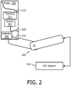
- the one or more imaging devices 104 may include a DOI system 202 having a portable robotically-held probe 208.
- a robotic arm 210 preferably free to move with multiple degrees of freedom, is utilized to position the probe 208, which includes a portable imaging device or scanner.
- the robotic arm 210 is tracked using a tracking mechanism 212.
- the tracking mechanism 212 may include an electromagnetic tracking device, a fiber optic shape sensing system, kinematic equations which rely on the robotic linkages and known movements to define its motion, etc.
- the tracking mechanism 212 provides feedback on the position of the robotic arm 210 and its resident devices to ensure that it does not interfere with radiation beams during a procedure. Since the positions of the radiation beams will be identified and known in space, the tracking device 212 will identify the location of the robotic arm 210 for comparison to ensure that no interference occurs.
- the probe 208 may include optical fibers 222 for illumination (e.g., 5-10 fibers, although other numbers of fibers are contemplated) and detection (e.g., 50-200 fibers, although other numbers of fibers are contemplated).
- a laser diode source 204 is connected to the optical fibers for illumination, and an intensified charge-coupled device (CCD) camera detector 206 is connected to the optical fibers for detection.
- CCD intensified charge-coupled device
- Simultaneous illumination and detection can be carried out using different optical fibers, for real-time 2D imaging, e.g., continuous wave (CW) optical measurements may be made. Frequency domain (FD) or other measurements are also contemplated.
- CW continuous wave
- FD Frequency domain
- the DOI system 202 includes a tuning capability to image a range of variable depths in the tissue, since skin burns can range from superficial (0.07 - 0.12 mm) to deep (> 2 mm).
- Other technologies such as, photoacoustic microscopy (PAM), laser Doppler perfusion imaging (LDPI), polarization sensitive optical coherence tomography (PSOCT) etc. can also be incorporated in such a portable scanner/probe 208.
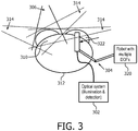
- FIG. 3 a schematic diagram shows optical and/or photoacoustic imaging implemented in a portable scanner or imager 302 that is integrated with the EBRT delivery protocol (e.g., stored in memory 116 of workstation 112, FIG. 1 ) and controlled by a robot or other fixture 320.
- a robotic arm 304 One constraint on a robotic arm 304 is that the portable imager 302 should not interfere with a path of an active beam 306.
- a position control algorithm e.g., stored as position control module 136, FIG. 1 ), controls the position of the portable imager 302 based on the currently active RT beam 306 and the known temporal pattern of line positions obtained from the RT planning system 115 ( FIG.
- the portable imager 302 can be sequentially moved to all needed positions, subject to the constraint that it does not interfere with the path of the currently active beam 306.
- the currently active beam 306 may be switched to other beam directions (inactive beams 314) using other sources or the same repositioned source.
- the positions of the beams are known as well as the position of the portable imager 302.
- the algorithm employs these positions and other constraints, e.g., the sizes of the beams or scanner equipment, accessories or other devices employed during therapy, etc., to avoid any overlap between the positions of the portable imager 302 and the active beam 306.
- a real-time readout of chromophore concentrations can be displayed as a measure of "skin burn" on a console/display 118 ( FIG. 1 ).
- Multiple displays or display panels may be provided. For example, one display may show the values of 'skin burn' updated on a real-time image. Another display may show the cumulative burn effects of the RT delivery, etc.
- An alternative method to robotically moving the sensing system may include using the RT planning system 115 to suggest an optimal static location for monitoring the largest field of view during the procedure. Other configurations and displays are also contemplated.
- skin burns may occur by exposure to multiple beams.
- the beams 306, 314 almost always intersects a contra-lateral breast 322 and possibly other normal structures like the shoulder, neck, etc. Therefore, vigilance in these areas using the portable imager 302 can assist in planning to avoid the occurrence of skin burns where possible.
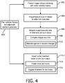
- a method for radiation therapy which employs skin burn status, is shown in accordance with illustrative embodiments.
- a position of an imaging device is controlled to avoid interference with radiation beams for the radiation therapy.
- the position of the imaging device may be controlled in accordance with a script or plan, which involves knowing positions of all active the radiation beams and avoiding interference by the imaging device or devices.
- the imaging device may be mounted on a robotically controlled arm or other fixture.
- an area of interest for tissue undergoing radiation therapy is imaged or measured (e.g., optically/spectrally or acoustically) using the imaging device.
- Imaging an area of interest includes employing one or more of: diffuse optical spectroscopy (DOS), diffuse optical imaging (DOI), photoacoustic computed tomography (PAT), photoacoustic microscopy (PAM), laser Doppler perfusion imaging (LDPI), polarization sensitive optical coherence tomography (PSOCT), high frequency ultrasound, etc.
- DOS diffuse optical spectroscopy
- DOI diffuse optical imaging
- PAT photoacoustic computed tomography
- PAM photoacoustic microscopy
- LDPI laser Doppler perfusion imaging
- PSOCT polarization sensitive optical coherence tomography
- comparisons between photographic images may be compared over time to determine a burn status.
- collected images are interpreted for the tissue to determine a burn status of the tissue.
- the magnitude of the burn may be determined based upon color changes/absorption spectra although density (acoustic changes) and other properties may be employed.
- Interpreting the images may include comparing images over time in block 408, and/or measuring absorption spectra of one or more of hemoglobin, water and lipids in block 410. Other methods may be employed for interpreting burns as well.
- further treatment is adjusted in accordance with the burn status. This may include real-time changes to a plan or include post radiation treatment for the burns incurred.
- further treatment adjustments may include predicting future skin burns based upon images of the tissue collected by the imaging device and the radiation therapy underwent by a patient. This may include using probabilistic and/or historic data to determine potential burn areas and severity.
- skin burn treatment may be suggested by a reporting system or module and is preferably customized to the radiation therapy received and/or the patient receiving it. Burn effects may also be measured post-procedure and employed to determine the skin burn treatment plan or to update the skin burn treatment plan.
- radiation therapy may be planned based at least in part upon a skin burn parameter employed to minimize skin burns in a plan. This may include an initial plan or an updated plan based upon real-time burn data collected during a procedure.
Landscapes
- Health & Medical Sciences (AREA)
- Engineering & Computer Science (AREA)
- Biomedical Technology (AREA)
- Pathology (AREA)
- Nuclear Medicine, Radiotherapy & Molecular Imaging (AREA)
- Radiology & Medical Imaging (AREA)
- Life Sciences & Earth Sciences (AREA)
- Animal Behavior & Ethology (AREA)
- General Health & Medical Sciences (AREA)
- Public Health (AREA)
- Veterinary Medicine (AREA)
- Radiation-Therapy Devices (AREA)
- Ultra Sonic Daignosis Equipment (AREA)
- Measurement Of The Respiration, Hearing Ability, Form, And Blood Characteristics Of Living Organisms (AREA)
Applications Claiming Priority (2)
| Application Number | Priority Date | Filing Date | Title |
|---|---|---|---|
| US201361839911P | 2013-06-27 | 2013-06-27 | |
| PCT/IB2014/062574 WO2014207663A1 (en) | 2013-06-27 | 2014-06-25 | Real-time quantification of skin burns in external beam radiation therapy |
Publications (2)
| Publication Number | Publication Date |
|---|---|
| EP3013419A1 EP3013419A1 (en) | 2016-05-04 |
| EP3013419B1 true EP3013419B1 (en) | 2017-09-20 |
Family
ID=51220613
Family Applications (1)
| Application Number | Title | Priority Date | Filing Date |
|---|---|---|---|
| EP14742360.2A Active EP3013419B1 (en) | 2013-06-27 | 2014-06-25 | Real-time quantification of skin burns in external beam radiation therapy |
Country Status (5)
| Country | Link |
|---|---|
| US (1) | US10213622B2 (enExample) |
| EP (1) | EP3013419B1 (enExample) |
| JP (1) | JP6259078B2 (enExample) |
| CN (1) | CN105358218B (enExample) |
| WO (1) | WO2014207663A1 (enExample) |
Families Citing this family (8)
| Publication number | Priority date | Publication date | Assignee | Title |
|---|---|---|---|---|
| JP6448142B2 (ja) * | 2014-03-28 | 2019-01-09 | 国立研究開発法人量子科学技術研究開発機構 | 放射線照射による皮膚変化予測装置と検証装置 |
| EP3426343B1 (en) * | 2016-03-09 | 2020-09-16 | Koninklijke Philips N.V. | Pre-optimization method for quick prediction of achievability of clinical goals in intensity modulated radiation therapy |
| JP7040890B2 (ja) * | 2016-12-27 | 2022-03-23 | 花王株式会社 | 肌分析方法及び肌分析装置 |
| US10825167B2 (en) * | 2017-04-28 | 2020-11-03 | Siemens Healthcare Gmbh | Rapid assessment and outcome analysis for medical patients |
| KR102098254B1 (ko) * | 2018-01-17 | 2020-05-26 | 사회복지법인 삼성생명공익재단 | 방사선 피부염 평가시스템 및 이를 이용한 평가방법 |
| CN109045489B (zh) * | 2018-08-22 | 2021-05-14 | 北京博纵科技有限公司 | 一种用于皮肤深层的可调超声美容成像系统及其采集方法 |
| CN109045490B (zh) * | 2018-08-22 | 2020-11-10 | 北京博纵科技有限公司 | 一种超声与皮肤量化一体的美容系统 |
| EP4151276A1 (en) * | 2021-09-20 | 2023-03-22 | Koninklijke Philips N.V. | Monitoring and detection of cutaneous reactions caused by radiotherapy |
Family Cites Families (9)
| Publication number | Priority date | Publication date | Assignee | Title |
|---|---|---|---|---|
| US6058352A (en) * | 1997-07-25 | 2000-05-02 | Physical Optics Corporation | Accurate tissue injury assessment using hybrid neural network analysis |
| JP4301358B2 (ja) * | 2003-05-13 | 2009-07-22 | 敦美 樋口 | 癌治療用放射線照射設備 |
| US8160205B2 (en) | 2004-04-06 | 2012-04-17 | Accuray Incorporated | Robotic arm for patient positioning assembly |
| DE102007018810A1 (de) * | 2007-04-20 | 2008-10-30 | Siemens Ag | Verfahren zur Bewegungsüberwachung bei einer medizintechnischen Anlage sowie zugehörige medizintechnische Anlage |
| US8641592B2 (en) * | 2009-03-23 | 2014-02-04 | Xinsheng Yu | Method and device for image guided dynamic radiation treatment of prostate cancer and other pelvic lesions |
| WO2012159043A2 (en) * | 2011-05-19 | 2012-11-22 | The Trustees Of Dartmouth College | Method and system for using cherenkov radiation to monitor beam profiles and radiation therapy |
| CA2856538A1 (en) * | 2011-11-29 | 2013-06-06 | University Of Massachusetts Medical School | Methods for detection and characterization of ionizing radiation exposure in tissue |
| US9486647B2 (en) * | 2012-04-27 | 2016-11-08 | Elekta Ab (Publ) | Vision system for radiotherapy machine control |
| CN102697557B (zh) * | 2012-06-06 | 2014-12-31 | 王建新 | 一种宫颈微波热疗辐射器 |
-
2014
- 2014-06-25 EP EP14742360.2A patent/EP3013419B1/en active Active
- 2014-06-25 JP JP2016522926A patent/JP6259078B2/ja active Active
- 2014-06-25 US US14/893,536 patent/US10213622B2/en active Active
- 2014-06-25 WO PCT/IB2014/062574 patent/WO2014207663A1/en not_active Ceased
- 2014-06-25 CN CN201480036470.1A patent/CN105358218B/zh active Active
Non-Patent Citations (1)
| Title |
|---|
| None * |
Also Published As
| Publication number | Publication date |
|---|---|
| US20160136455A1 (en) | 2016-05-19 |
| CN105358218A (zh) | 2016-02-24 |
| CN105358218B (zh) | 2018-10-12 |
| US10213622B2 (en) | 2019-02-26 |
| EP3013419A1 (en) | 2016-05-04 |
| WO2014207663A1 (en) | 2014-12-31 |
| JP2016529953A (ja) | 2016-09-29 |
| JP6259078B2 (ja) | 2018-01-10 |
Similar Documents
| Publication | Publication Date | Title |
|---|---|---|
| EP3013419B1 (en) | Real-time quantification of skin burns in external beam radiation therapy | |
| US11894123B2 (en) | Radiotherapy mobile and wireless device workflow management system | |
| JP5437997B2 (ja) | 放射性イメージングのための画像生成装置および方法 | |
| EP2911587B1 (en) | Nir image guided targeting | |
| CN111093520A (zh) | 局部空化信号测量 | |
| CN104394766B (zh) | 辐射处置递送期间的实时肿瘤灌注成像 | |
| US20090221911A1 (en) | Biological observation apparatus and method | |
| US11076776B2 (en) | Apparatus and method for real-time tracking of bony structures | |
| US10201291B2 (en) | Apparatus and method for real-time tracking of bony structures | |
| Reinhardt et al. | VertiGo–a pilot project in nystagmus detection via webcam | |
| JP5279435B2 (ja) | 被検体情報取得装置、被検体情報取得装置の制御方法 | |
| WO2015174082A1 (en) | Photoacoustic apparatus | |
| US20250134404A1 (en) | Laser speckle imaging device and method | |
| JP6324456B2 (ja) | 生体情報取得装置 | |
| JP5680141B2 (ja) | 被検体情報取得装置および被検体情報取得装置の制御方法 | |
| KR101276678B1 (ko) | 전자파를 이용한 폐암 진단 장치 및 방법 | |
| IL300134B1 (en) | Non-Contact Rapid Diagnosis Of Illness By Laser, Infra Red, Terahertz And/Or UV Spectroscopy And Analysis Of Water Mixture Envelope | |
| KR20230147120A (ko) | 조직 성분 측정 방법, 장치 및 웨어러블 기기 | |
| Böttrich et al. | Simulation based investigation of source-detector configurations for non-invasive fetal pulse oximetry |
Legal Events
| Date | Code | Title | Description |
|---|---|---|---|
| PUAI | Public reference made under article 153(3) epc to a published international application that has entered the european phase |
Free format text: ORIGINAL CODE: 0009012 |
|
| 17P | Request for examination filed |
Effective date: 20160127 |
|
| AK | Designated contracting states |
Kind code of ref document: A1 Designated state(s): AL AT BE BG CH CY CZ DE DK EE ES FI FR GB GR HR HU IE IS IT LI LT LU LV MC MK MT NL NO PL PT RO RS SE SI SK SM TR |
|
| AX | Request for extension of the european patent |
Extension state: BA ME |
|
| DAX | Request for extension of the european patent (deleted) | ||
| GRAP | Despatch of communication of intention to grant a patent |
Free format text: ORIGINAL CODE: EPIDOSNIGR1 |
|
| INTG | Intention to grant announced |
Effective date: 20170410 |
|
| GRAS | Grant fee paid |
Free format text: ORIGINAL CODE: EPIDOSNIGR3 |
|
| GRAA | (expected) grant |
Free format text: ORIGINAL CODE: 0009210 |
|
| AK | Designated contracting states |
Kind code of ref document: B1 Designated state(s): AL AT BE BG CH CY CZ DE DK EE ES FI FR GB GR HR HU IE IS IT LI LT LU LV MC MK MT NL NO PL PT RO RS SE SI SK SM TR |
|
| REG | Reference to a national code |
Ref country code: GB Ref legal event code: FG4D |
|
| REG | Reference to a national code |
Ref country code: CH Ref legal event code: EP |
|
| REG | Reference to a national code |
Ref country code: AT Ref legal event code: REF Ref document number: 929687 Country of ref document: AT Kind code of ref document: T Effective date: 20171015 |
|
| REG | Reference to a national code |
Ref country code: IE Ref legal event code: FG4D |
|
| REG | Reference to a national code |
Ref country code: DE Ref legal event code: R096 Ref document number: 602014014836 Country of ref document: DE |
|
| REG | Reference to a national code |
Ref country code: NL Ref legal event code: MP Effective date: 20170920 |
|
| PG25 | Lapsed in a contracting state [announced via postgrant information from national office to epo] |
Ref country code: LT Free format text: LAPSE BECAUSE OF FAILURE TO SUBMIT A TRANSLATION OF THE DESCRIPTION OR TO PAY THE FEE WITHIN THE PRESCRIBED TIME-LIMIT Effective date: 20170920 Ref country code: FI Free format text: LAPSE BECAUSE OF FAILURE TO SUBMIT A TRANSLATION OF THE DESCRIPTION OR TO PAY THE FEE WITHIN THE PRESCRIBED TIME-LIMIT Effective date: 20170920 Ref country code: NO Free format text: LAPSE BECAUSE OF FAILURE TO SUBMIT A TRANSLATION OF THE DESCRIPTION OR TO PAY THE FEE WITHIN THE PRESCRIBED TIME-LIMIT Effective date: 20171220 Ref country code: HR Free format text: LAPSE BECAUSE OF FAILURE TO SUBMIT A TRANSLATION OF THE DESCRIPTION OR TO PAY THE FEE WITHIN THE PRESCRIBED TIME-LIMIT Effective date: 20170920 Ref country code: SE Free format text: LAPSE BECAUSE OF FAILURE TO SUBMIT A TRANSLATION OF THE DESCRIPTION OR TO PAY THE FEE WITHIN THE PRESCRIBED TIME-LIMIT Effective date: 20170920 |
|
| REG | Reference to a national code |
Ref country code: LT Ref legal event code: MG4D |
|
| REG | Reference to a national code |
Ref country code: AT Ref legal event code: MK05 Ref document number: 929687 Country of ref document: AT Kind code of ref document: T Effective date: 20170920 |
|
| PG25 | Lapsed in a contracting state [announced via postgrant information from national office to epo] |
Ref country code: RS Free format text: LAPSE BECAUSE OF FAILURE TO SUBMIT A TRANSLATION OF THE DESCRIPTION OR TO PAY THE FEE WITHIN THE PRESCRIBED TIME-LIMIT Effective date: 20170920 Ref country code: GR Free format text: LAPSE BECAUSE OF FAILURE TO SUBMIT A TRANSLATION OF THE DESCRIPTION OR TO PAY THE FEE WITHIN THE PRESCRIBED TIME-LIMIT Effective date: 20171221 Ref country code: LV Free format text: LAPSE BECAUSE OF FAILURE TO SUBMIT A TRANSLATION OF THE DESCRIPTION OR TO PAY THE FEE WITHIN THE PRESCRIBED TIME-LIMIT Effective date: 20170920 Ref country code: BG Free format text: LAPSE BECAUSE OF FAILURE TO SUBMIT A TRANSLATION OF THE DESCRIPTION OR TO PAY THE FEE WITHIN THE PRESCRIBED TIME-LIMIT Effective date: 20171220 |
|
| PG25 | Lapsed in a contracting state [announced via postgrant information from national office to epo] |
Ref country code: NL Free format text: LAPSE BECAUSE OF FAILURE TO SUBMIT A TRANSLATION OF THE DESCRIPTION OR TO PAY THE FEE WITHIN THE PRESCRIBED TIME-LIMIT Effective date: 20170920 |
|
| PG25 | Lapsed in a contracting state [announced via postgrant information from national office to epo] |
Ref country code: ES Free format text: LAPSE BECAUSE OF FAILURE TO SUBMIT A TRANSLATION OF THE DESCRIPTION OR TO PAY THE FEE WITHIN THE PRESCRIBED TIME-LIMIT Effective date: 20170920 Ref country code: CZ Free format text: LAPSE BECAUSE OF FAILURE TO SUBMIT A TRANSLATION OF THE DESCRIPTION OR TO PAY THE FEE WITHIN THE PRESCRIBED TIME-LIMIT Effective date: 20170920 Ref country code: PL Free format text: LAPSE BECAUSE OF FAILURE TO SUBMIT A TRANSLATION OF THE DESCRIPTION OR TO PAY THE FEE WITHIN THE PRESCRIBED TIME-LIMIT Effective date: 20170920 Ref country code: RO Free format text: LAPSE BECAUSE OF FAILURE TO SUBMIT A TRANSLATION OF THE DESCRIPTION OR TO PAY THE FEE WITHIN THE PRESCRIBED TIME-LIMIT Effective date: 20170920 |
|
| PG25 | Lapsed in a contracting state [announced via postgrant information from national office to epo] |
Ref country code: IT Free format text: LAPSE BECAUSE OF FAILURE TO SUBMIT A TRANSLATION OF THE DESCRIPTION OR TO PAY THE FEE WITHIN THE PRESCRIBED TIME-LIMIT Effective date: 20170920 Ref country code: IS Free format text: LAPSE BECAUSE OF FAILURE TO SUBMIT A TRANSLATION OF THE DESCRIPTION OR TO PAY THE FEE WITHIN THE PRESCRIBED TIME-LIMIT Effective date: 20180120 Ref country code: SK Free format text: LAPSE BECAUSE OF FAILURE TO SUBMIT A TRANSLATION OF THE DESCRIPTION OR TO PAY THE FEE WITHIN THE PRESCRIBED TIME-LIMIT Effective date: 20170920 Ref country code: EE Free format text: LAPSE BECAUSE OF FAILURE TO SUBMIT A TRANSLATION OF THE DESCRIPTION OR TO PAY THE FEE WITHIN THE PRESCRIBED TIME-LIMIT Effective date: 20170920 Ref country code: SM Free format text: LAPSE BECAUSE OF FAILURE TO SUBMIT A TRANSLATION OF THE DESCRIPTION OR TO PAY THE FEE WITHIN THE PRESCRIBED TIME-LIMIT Effective date: 20170920 Ref country code: AT Free format text: LAPSE BECAUSE OF FAILURE TO SUBMIT A TRANSLATION OF THE DESCRIPTION OR TO PAY THE FEE WITHIN THE PRESCRIBED TIME-LIMIT Effective date: 20170920 |
|
| REG | Reference to a national code |
Ref country code: DE Ref legal event code: R097 Ref document number: 602014014836 Country of ref document: DE |
|
| REG | Reference to a national code |
Ref country code: FR Ref legal event code: PLFP Year of fee payment: 5 |
|
| PLBE | No opposition filed within time limit |
Free format text: ORIGINAL CODE: 0009261 |
|
| STAA | Information on the status of an ep patent application or granted ep patent |
Free format text: STATUS: NO OPPOSITION FILED WITHIN TIME LIMIT |
|
| PG25 | Lapsed in a contracting state [announced via postgrant information from national office to epo] |
Ref country code: DK Free format text: LAPSE BECAUSE OF FAILURE TO SUBMIT A TRANSLATION OF THE DESCRIPTION OR TO PAY THE FEE WITHIN THE PRESCRIBED TIME-LIMIT Effective date: 20170920 |
|
| 26N | No opposition filed |
Effective date: 20180621 |
|
| PG25 | Lapsed in a contracting state [announced via postgrant information from national office to epo] |
Ref country code: SI Free format text: LAPSE BECAUSE OF FAILURE TO SUBMIT A TRANSLATION OF THE DESCRIPTION OR TO PAY THE FEE WITHIN THE PRESCRIBED TIME-LIMIT Effective date: 20170920 |
|
| REG | Reference to a national code |
Ref country code: CH Ref legal event code: PL |
|
| REG | Reference to a national code |
Ref country code: BE Ref legal event code: MM Effective date: 20180630 |
|
| REG | Reference to a national code |
Ref country code: IE Ref legal event code: MM4A |
|
| PG25 | Lapsed in a contracting state [announced via postgrant information from national office to epo] |
Ref country code: MC Free format text: LAPSE BECAUSE OF FAILURE TO SUBMIT A TRANSLATION OF THE DESCRIPTION OR TO PAY THE FEE WITHIN THE PRESCRIBED TIME-LIMIT Effective date: 20170920 Ref country code: LU Free format text: LAPSE BECAUSE OF NON-PAYMENT OF DUE FEES Effective date: 20180625 |
|
| PG25 | Lapsed in a contracting state [announced via postgrant information from national office to epo] |
Ref country code: CH Free format text: LAPSE BECAUSE OF NON-PAYMENT OF DUE FEES Effective date: 20180630 Ref country code: LI Free format text: LAPSE BECAUSE OF NON-PAYMENT OF DUE FEES Effective date: 20180630 Ref country code: IE Free format text: LAPSE BECAUSE OF NON-PAYMENT OF DUE FEES Effective date: 20180625 |
|
| PG25 | Lapsed in a contracting state [announced via postgrant information from national office to epo] |
Ref country code: BE Free format text: LAPSE BECAUSE OF NON-PAYMENT OF DUE FEES Effective date: 20180630 |
|
| PG25 | Lapsed in a contracting state [announced via postgrant information from national office to epo] |
Ref country code: MT Free format text: LAPSE BECAUSE OF NON-PAYMENT OF DUE FEES Effective date: 20180625 |
|
| PG25 | Lapsed in a contracting state [announced via postgrant information from national office to epo] |
Ref country code: TR Free format text: LAPSE BECAUSE OF FAILURE TO SUBMIT A TRANSLATION OF THE DESCRIPTION OR TO PAY THE FEE WITHIN THE PRESCRIBED TIME-LIMIT Effective date: 20170920 |
|
| PG25 | Lapsed in a contracting state [announced via postgrant information from national office to epo] |
Ref country code: PT Free format text: LAPSE BECAUSE OF FAILURE TO SUBMIT A TRANSLATION OF THE DESCRIPTION OR TO PAY THE FEE WITHIN THE PRESCRIBED TIME-LIMIT Effective date: 20170920 |
|
| PG25 | Lapsed in a contracting state [announced via postgrant information from national office to epo] |
Ref country code: HU Free format text: LAPSE BECAUSE OF FAILURE TO SUBMIT A TRANSLATION OF THE DESCRIPTION OR TO PAY THE FEE WITHIN THE PRESCRIBED TIME-LIMIT; INVALID AB INITIO Effective date: 20140625 Ref country code: MK Free format text: LAPSE BECAUSE OF NON-PAYMENT OF DUE FEES Effective date: 20170920 Ref country code: CY Free format text: LAPSE BECAUSE OF FAILURE TO SUBMIT A TRANSLATION OF THE DESCRIPTION OR TO PAY THE FEE WITHIN THE PRESCRIBED TIME-LIMIT Effective date: 20170920 |
|
| PG25 | Lapsed in a contracting state [announced via postgrant information from national office to epo] |
Ref country code: AL Free format text: LAPSE BECAUSE OF FAILURE TO SUBMIT A TRANSLATION OF THE DESCRIPTION OR TO PAY THE FEE WITHIN THE PRESCRIBED TIME-LIMIT Effective date: 20170920 |
|
| PGFP | Annual fee paid to national office [announced via postgrant information from national office to epo] |
Ref country code: FR Payment date: 20220623 Year of fee payment: 9 |
|
| PG25 | Lapsed in a contracting state [announced via postgrant information from national office to epo] |
Ref country code: FR Free format text: LAPSE BECAUSE OF NON-PAYMENT OF DUE FEES Effective date: 20230630 |
|
| REG | Reference to a national code |
Ref country code: GB Ref legal event code: 732E Free format text: REGISTERED BETWEEN 20250220 AND 20250226 |
|
| PGFP | Annual fee paid to national office [announced via postgrant information from national office to epo] |
Ref country code: DE Payment date: 20250402 Year of fee payment: 12 |
|
| PGFP | Annual fee paid to national office [announced via postgrant information from national office to epo] |
Ref country code: GB Payment date: 20250401 Year of fee payment: 12 |