EP2999486B1 - Metallbeschichtete gerüste für gewebe-engineering - Google Patents
Metallbeschichtete gerüste für gewebe-engineering Download PDFInfo
- Publication number
- EP2999486B1 EP2999486B1 EP14800309.8A EP14800309A EP2999486B1 EP 2999486 B1 EP2999486 B1 EP 2999486B1 EP 14800309 A EP14800309 A EP 14800309A EP 2999486 B1 EP2999486 B1 EP 2999486B1
- Authority
- EP
- European Patent Office
- Prior art keywords
- scaffold
- cardiac
- scaffolds
- cells
- tissue
- Prior art date
- Legal status (The legal status is an assumption and is not a legal conclusion. Google has not performed a legal analysis and makes no representation as to the accuracy of the status listed.)
- Active
Links
- 229910052751 metal Inorganic materials 0.000 title claims description 7
- 239000002184 metal Substances 0.000 title claims description 7
- 210000004027 cell Anatomy 0.000 claims description 117
- 239000000835 fiber Substances 0.000 claims description 94
- 210000001519 tissue Anatomy 0.000 claims description 87
- 239000002105 nanoparticle Substances 0.000 claims description 81
- 239000010931 gold Substances 0.000 claims description 80
- PCHJSUWPFVWCPO-UHFFFAOYSA-N gold Chemical compound [Au] PCHJSUWPFVWCPO-UHFFFAOYSA-N 0.000 claims description 73
- 238000000034 method Methods 0.000 claims description 72
- 229910052737 gold Inorganic materials 0.000 claims description 70
- 239000000203 mixture Substances 0.000 claims description 39
- 208000037265 diseases, disorders, signs and symptoms Diseases 0.000 claims description 26
- 239000002082 metal nanoparticle Substances 0.000 claims description 26
- 102000010834 Extracellular Matrix Proteins Human genes 0.000 claims description 19
- 108010037362 Extracellular Matrix Proteins Proteins 0.000 claims description 19
- 239000011248 coating agent Substances 0.000 claims description 18
- 238000000576 coating method Methods 0.000 claims description 18
- 210000002744 extracellular matrix Anatomy 0.000 claims description 18
- 201000010099 disease Diseases 0.000 claims description 13
- 208000035475 disorder Diseases 0.000 claims description 13
- 238000010899 nucleation Methods 0.000 claims description 13
- 238000001704 evaporation Methods 0.000 claims description 11
- 238000001523 electrospinning Methods 0.000 claims description 10
- 230000000694 effects Effects 0.000 claims description 8
- 210000005003 heart tissue Anatomy 0.000 description 43
- 230000008602 contraction Effects 0.000 description 42
- 229920000642 polymer Polymers 0.000 description 42
- 210000002064 heart cell Anatomy 0.000 description 40
- 230000000747 cardiac effect Effects 0.000 description 30
- -1 Polyethylene terephthalate scaffolds Polymers 0.000 description 28
- 239000000463 material Substances 0.000 description 28
- 210000004413 cardiac myocyte Anatomy 0.000 description 27
- XOJVVFBFDXDTEG-UHFFFAOYSA-N Norphytane Natural products CC(C)CCCC(C)CCCC(C)CCCC(C)C XOJVVFBFDXDTEG-UHFFFAOYSA-N 0.000 description 22
- 229920001610 polycaprolactone Polymers 0.000 description 16
- 208000019622 heart disease Diseases 0.000 description 15
- 239000011159 matrix material Substances 0.000 description 15
- 239000004632 polycaprolactone Substances 0.000 description 15
- 239000000499 gel Substances 0.000 description 14
- 239000000243 solution Substances 0.000 description 14
- 238000004458 analytical method Methods 0.000 description 13
- 230000006870 function Effects 0.000 description 13
- YMWUJEATGCHHMB-UHFFFAOYSA-N Dichloromethane Chemical compound ClCCl YMWUJEATGCHHMB-UHFFFAOYSA-N 0.000 description 12
- ZMXDDKWLCZADIW-UHFFFAOYSA-N N,N-Dimethylformamide Chemical compound CN(C)C=O ZMXDDKWLCZADIW-UHFFFAOYSA-N 0.000 description 12
- 239000003102 growth factor Substances 0.000 description 12
- 230000001965 increasing effect Effects 0.000 description 12
- 210000004165 myocardium Anatomy 0.000 description 12
- 208000020446 Cardiac disease Diseases 0.000 description 11
- 108010010803 Gelatin Proteins 0.000 description 11
- 229920002988 biodegradable polymer Polymers 0.000 description 11
- 239000004621 biodegradable polymer Substances 0.000 description 11
- 230000001413 cellular effect Effects 0.000 description 11
- 239000008273 gelatin Substances 0.000 description 11
- 229920000159 gelatin Polymers 0.000 description 11
- 235000019322 gelatine Nutrition 0.000 description 11
- 235000011852 gelatine desserts Nutrition 0.000 description 11
- 102000004169 proteins and genes Human genes 0.000 description 11
- 108090000623 proteins and genes Proteins 0.000 description 11
- 102000010825 Actinin Human genes 0.000 description 10
- 108010063503 Actinin Proteins 0.000 description 10
- 235000010443 alginic acid Nutrition 0.000 description 10
- 229920000615 alginic acid Polymers 0.000 description 10
- 150000001875 compounds Chemical class 0.000 description 10
- 238000001514 detection method Methods 0.000 description 10
- 238000012876 topography Methods 0.000 description 10
- LFQSCWFLJHTTHZ-UHFFFAOYSA-N Ethanol Chemical compound CCO LFQSCWFLJHTTHZ-UHFFFAOYSA-N 0.000 description 9
- OKKJLVBELUTLKV-UHFFFAOYSA-N Methanol Chemical compound OC OKKJLVBELUTLKV-UHFFFAOYSA-N 0.000 description 9
- 230000015572 biosynthetic process Effects 0.000 description 9
- 230000008020 evaporation Effects 0.000 description 9
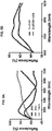
- 238000005259 measurement Methods 0.000 description 9
- 230000008569 process Effects 0.000 description 9
- 108010035532 Collagen Proteins 0.000 description 8
- 102000008186 Collagen Human genes 0.000 description 8
- 239000000853 adhesive Substances 0.000 description 8
- 238000013459 approach Methods 0.000 description 8
- 230000008859 change Effects 0.000 description 8
- 239000003795 chemical substances by application Substances 0.000 description 8
- 229920001436 collagen Polymers 0.000 description 8
- 229920001577 copolymer Polymers 0.000 description 8
- 210000002950 fibroblast Anatomy 0.000 description 8
- UCSJYZPVAKXKNQ-HZYVHMACSA-N streptomycin Chemical compound CN[C@H]1[C@H](O)[C@@H](O)[C@H](CO)O[C@H]1O[C@@H]1[C@](C=O)(O)[C@H](C)O[C@H]1O[C@@H]1[C@@H](NC(N)=N)[C@H](O)[C@@H](NC(N)=N)[C@H](O)[C@H]1O UCSJYZPVAKXKNQ-HZYVHMACSA-N 0.000 description 8
- 229920000954 Polyglycolide Polymers 0.000 description 7
- 230000021164 cell adhesion Effects 0.000 description 7
- 230000005284 excitation Effects 0.000 description 7
- 239000000017 hydrogel Substances 0.000 description 7
- 238000012744 immunostaining Methods 0.000 description 7
- 238000001727 in vivo Methods 0.000 description 7
- FHVDTGUDJYJELY-UHFFFAOYSA-N 6-{[2-carboxy-4,5-dihydroxy-6-(phosphanyloxy)oxan-3-yl]oxy}-4,5-dihydroxy-3-phosphanyloxane-2-carboxylic acid Chemical group O1C(C(O)=O)C(P)C(O)C(O)C1OC1C(C(O)=O)OC(OP)C(O)C1O FHVDTGUDJYJELY-UHFFFAOYSA-N 0.000 description 6
- 239000006144 Dulbecco’s modified Eagle's medium Substances 0.000 description 6
- FAPWRFPIFSIZLT-UHFFFAOYSA-M Sodium chloride Chemical compound [Na+].[Cl-] FAPWRFPIFSIZLT-UHFFFAOYSA-M 0.000 description 6
- 230000002378 acidificating effect Effects 0.000 description 6
- 229910052782 aluminium Inorganic materials 0.000 description 6
- 239000000872 buffer Substances 0.000 description 6
- 210000001054 cardiac fibroblast Anatomy 0.000 description 6
- 230000008878 coupling Effects 0.000 description 6
- 238000010168 coupling process Methods 0.000 description 6
- 238000005859 coupling reaction Methods 0.000 description 6
- JVTAAEKCZFNVCJ-UHFFFAOYSA-N lactic acid Chemical class CC(O)C(O)=O JVTAAEKCZFNVCJ-UHFFFAOYSA-N 0.000 description 6
- 238000007619 statistical method Methods 0.000 description 6
- 239000000758 substrate Substances 0.000 description 6
- 238000012546 transfer Methods 0.000 description 6
- 241000283707 Capra Species 0.000 description 5
- 108010067306 Fibronectins Proteins 0.000 description 5
- 102000016359 Fibronectins Human genes 0.000 description 5
- 206010061216 Infarction Diseases 0.000 description 5
- 229940072056 alginate Drugs 0.000 description 5
- 206010003119 arrhythmia Diseases 0.000 description 5
- 238000000151 deposition Methods 0.000 description 5
- 230000008021 deposition Effects 0.000 description 5
- LOKCTEFSRHRXRJ-UHFFFAOYSA-I dipotassium trisodium dihydrogen phosphate hydrogen phosphate dichloride Chemical compound P(=O)(O)(O)[O-].[K+].P(=O)(O)([O-])[O-].[Na+].[Na+].[Cl-].[K+].[Cl-].[Na+] LOKCTEFSRHRXRJ-UHFFFAOYSA-I 0.000 description 5
- 238000002149 energy-dispersive X-ray emission spectroscopy Methods 0.000 description 5
- 239000010408 film Substances 0.000 description 5
- 239000001963 growth medium Substances 0.000 description 5
- 229920001519 homopolymer Polymers 0.000 description 5
- 230000007574 infarction Effects 0.000 description 5
- 208000028867 ischemia Diseases 0.000 description 5
- 238000002955 isolation Methods 0.000 description 5
- 238000004519 manufacturing process Methods 0.000 description 5
- 239000002609 medium Substances 0.000 description 5
- 239000002114 nanocomposite Substances 0.000 description 5
- 210000002747 omentum Anatomy 0.000 description 5
- 239000002953 phosphate buffered saline Substances 0.000 description 5
- 229920002627 poly(phosphazenes) Polymers 0.000 description 5
- 239000004633 polyglycolic acid Substances 0.000 description 5
- 239000011148 porous material Substances 0.000 description 5
- 238000002360 preparation method Methods 0.000 description 5
- 108090000765 processed proteins & peptides Proteins 0.000 description 5
- 230000008439 repair process Effects 0.000 description 5
- 239000000523 sample Substances 0.000 description 5
- 239000000126 substance Substances 0.000 description 5
- 230000017423 tissue regeneration Effects 0.000 description 5
- 238000004627 transmission electron microscopy Methods 0.000 description 5
- 238000011282 treatment Methods 0.000 description 5
- XLYOFNOQVPJJNP-UHFFFAOYSA-N water Substances O XLYOFNOQVPJJNP-UHFFFAOYSA-N 0.000 description 5
- 239000012114 Alexa Fluor 647 Substances 0.000 description 4
- CURLTUGMZLYLDI-UHFFFAOYSA-N Carbon dioxide Chemical compound O=C=O CURLTUGMZLYLDI-UHFFFAOYSA-N 0.000 description 4
- 108010073385 Fibrin Proteins 0.000 description 4
- 102000009123 Fibrin Human genes 0.000 description 4
- BWGVNKXGVNDBDI-UHFFFAOYSA-N Fibrin monomer Chemical compound CNC(=O)CNC(=O)CN BWGVNKXGVNDBDI-UHFFFAOYSA-N 0.000 description 4
- AEMRFAOFKBGASW-UHFFFAOYSA-N Glycolic acid Chemical compound OCC(O)=O AEMRFAOFKBGASW-UHFFFAOYSA-N 0.000 description 4
- 229920002683 Glycosaminoglycan Polymers 0.000 description 4
- TWRXJAOTZQYOKJ-UHFFFAOYSA-L Magnesium chloride Chemical compound [Mg+2].[Cl-].[Cl-] TWRXJAOTZQYOKJ-UHFFFAOYSA-L 0.000 description 4
- CSNNHWWHGAXBCP-UHFFFAOYSA-L Magnesium sulfate Chemical compound [Mg+2].[O-][S+2]([O-])([O-])[O-] CSNNHWWHGAXBCP-UHFFFAOYSA-L 0.000 description 4
- 229930182555 Penicillin Natural products 0.000 description 4
- JGSARLDLIJGVTE-MBNYWOFBSA-N Penicillin G Chemical compound N([C@H]1[C@H]2SC([C@@H](N2C1=O)C(O)=O)(C)C)C(=O)CC1=CC=CC=C1 JGSARLDLIJGVTE-MBNYWOFBSA-N 0.000 description 4
- 102000035195 Peptidases Human genes 0.000 description 4
- 108091005804 Peptidases Proteins 0.000 description 4
- UIIMBOGNXHQVGW-UHFFFAOYSA-M Sodium bicarbonate Chemical compound [Na+].OC([O-])=O UIIMBOGNXHQVGW-UHFFFAOYSA-M 0.000 description 4
- XAGFODPZIPBFFR-UHFFFAOYSA-N aluminium Chemical compound [Al] XAGFODPZIPBFFR-UHFFFAOYSA-N 0.000 description 4
- 238000003556 assay Methods 0.000 description 4
- 229910052791 calcium Inorganic materials 0.000 description 4
- 239000011575 calcium Substances 0.000 description 4
- 230000015556 catabolic process Effects 0.000 description 4
- 150000001768 cations Chemical class 0.000 description 4
- 239000002131 composite material Substances 0.000 description 4
- 238000004132 cross linking Methods 0.000 description 4
- 238000006731 degradation reaction Methods 0.000 description 4
- 238000011161 development Methods 0.000 description 4
- 230000018109 developmental process Effects 0.000 description 4
- 238000009826 distribution Methods 0.000 description 4
- 230000005684 electric field Effects 0.000 description 4
- 230000005686 electrostatic field Effects 0.000 description 4
- 229950003499 fibrin Drugs 0.000 description 4
- 229960004275 glycolic acid Drugs 0.000 description 4
- 238000003384 imaging method Methods 0.000 description 4
- 238000010348 incorporation Methods 0.000 description 4
- 239000004615 ingredient Substances 0.000 description 4
- 230000002401 inhibitory effect Effects 0.000 description 4
- 238000002347 injection Methods 0.000 description 4
- 239000007924 injection Substances 0.000 description 4
- 150000002500 ions Chemical class 0.000 description 4
- 239000010410 layer Substances 0.000 description 4
- 230000033001 locomotion Effects 0.000 description 4
- 210000004940 nucleus Anatomy 0.000 description 4
- 230000008520 organization Effects 0.000 description 4
- 229940049954 penicillin Drugs 0.000 description 4
- 229920000747 poly(lactic acid) Polymers 0.000 description 4
- 229920001606 poly(lactic acid-co-glycolic acid) Polymers 0.000 description 4
- 229920000728 polyester Polymers 0.000 description 4
- 230000002062 proliferating effect Effects 0.000 description 4
- 230000035755 proliferation Effects 0.000 description 4
- 230000033764 rhythmic process Effects 0.000 description 4
- 229960005322 streptomycin Drugs 0.000 description 4
- 208000024891 symptom Diseases 0.000 description 4
- 230000001225 therapeutic effect Effects 0.000 description 4
- 239000010936 titanium Substances 0.000 description 4
- 238000002054 transplantation Methods 0.000 description 4
- 229910052721 tungsten Inorganic materials 0.000 description 4
- 239000010937 tungsten Substances 0.000 description 4
- 206010003658 Atrial Fibrillation Diseases 0.000 description 3
- OYPRJOBELJOOCE-UHFFFAOYSA-N Calcium Chemical compound [Ca] OYPRJOBELJOOCE-UHFFFAOYSA-N 0.000 description 3
- OKTJSMMVPCPJKN-UHFFFAOYSA-N Carbon Chemical compound [C] OKTJSMMVPCPJKN-UHFFFAOYSA-N 0.000 description 3
- 229920001661 Chitosan Polymers 0.000 description 3
- 102000029816 Collagenase Human genes 0.000 description 3
- 108060005980 Collagenase Proteins 0.000 description 3
- PEDCQBHIVMGVHV-UHFFFAOYSA-N Glycerine Chemical compound OCC(O)CO PEDCQBHIVMGVHV-UHFFFAOYSA-N 0.000 description 3
- 108010085895 Laminin Proteins 0.000 description 3
- 102000007547 Laminin Human genes 0.000 description 3
- 108010019160 Pancreatin Proteins 0.000 description 3
- 239000004793 Polystyrene Substances 0.000 description 3
- 108010067787 Proteoglycans Proteins 0.000 description 3
- 102000016611 Proteoglycans Human genes 0.000 description 3
- 241000700159 Rattus Species 0.000 description 3
- 208000006011 Stroke Diseases 0.000 description 3
- 238000000692 Student's t-test Methods 0.000 description 3
- 239000007983 Tris buffer Substances 0.000 description 3
- 230000002159 abnormal effect Effects 0.000 description 3
- 230000004071 biological effect Effects 0.000 description 3
- 239000012620 biological material Substances 0.000 description 3
- 229910052799 carbon Inorganic materials 0.000 description 3
- 210000000170 cell membrane Anatomy 0.000 description 3
- YRQNKMKHABXEJZ-UVQQGXFZSA-N chembl176323 Chemical compound C1C[C@]2(C)[C@@]3(C)CC(N=C4C[C@]5(C)CCC6[C@]7(C)CC[C@@H]([C@]7(CC[C@]6(C)[C@@]5(C)CC4=N4)C)CCCCCCCC)=C4C[C@]3(C)CCC2[C@]2(C)CC[C@H](CCCCCCCC)[C@]21C YRQNKMKHABXEJZ-UVQQGXFZSA-N 0.000 description 3
- SMNPLAKEGAEPJD-UHFFFAOYSA-N chembl34922 Chemical compound Cl.Cl.Cl.C1CN(C)CCN1C1=CC=C(NC(=N2)C=3C=C4N=C(NC4=CC=3)C=3C=CC(O)=CC=3)C2=C1 SMNPLAKEGAEPJD-UHFFFAOYSA-N 0.000 description 3
- 229910052802 copper Inorganic materials 0.000 description 3
- 239000010949 copper Substances 0.000 description 3
- 230000003247 decreasing effect Effects 0.000 description 3
- 230000029087 digestion Effects 0.000 description 3
- 230000007613 environmental effect Effects 0.000 description 3
- 230000003628 erosive effect Effects 0.000 description 3
- 230000001506 immunosuppresive effect Effects 0.000 description 3
- 230000001771 impaired effect Effects 0.000 description 3
- 235000014655 lactic acid Nutrition 0.000 description 3
- 239000003550 marker Substances 0.000 description 3
- 238000010369 molecular cloning Methods 0.000 description 3
- 208000031225 myocardial ischemia Diseases 0.000 description 3
- VLKZOEOYAKHREP-UHFFFAOYSA-N n-Hexane Chemical compound CCCCCC VLKZOEOYAKHREP-UHFFFAOYSA-N 0.000 description 3
- 108020004707 nucleic acids Proteins 0.000 description 3
- 102000039446 nucleic acids Human genes 0.000 description 3
- 150000007523 nucleic acids Chemical class 0.000 description 3
- 229940055695 pancreatin Drugs 0.000 description 3
- 239000002245 particle Substances 0.000 description 3
- 239000008194 pharmaceutical composition Substances 0.000 description 3
- 125000004437 phosphorous atom Chemical group 0.000 description 3
- 239000004626 polylactic acid Substances 0.000 description 3
- 229920001184 polypeptide Polymers 0.000 description 3
- 229920001282 polysaccharide Polymers 0.000 description 3
- 229920002223 polystyrene Polymers 0.000 description 3
- 102000004196 processed proteins & peptides Human genes 0.000 description 3
- 230000001737 promoting effect Effects 0.000 description 3
- 230000009467 reduction Effects 0.000 description 3
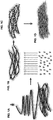
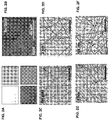
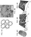
- 238000001878 scanning electron micrograph Methods 0.000 description 3
- 238000004626 scanning electron microscopy Methods 0.000 description 3
- 238000012453 sprague-dawley rat model Methods 0.000 description 3
- 210000000130 stem cell Anatomy 0.000 description 3
- LENZDBCJOHFCAS-UHFFFAOYSA-N tris Chemical compound OCC(N)(CO)CO LENZDBCJOHFCAS-UHFFFAOYSA-N 0.000 description 3
- WFKWXMTUELFFGS-UHFFFAOYSA-N tungsten Chemical compound [W] WFKWXMTUELFFGS-UHFFFAOYSA-N 0.000 description 3
- 230000035899 viability Effects 0.000 description 3
- NWONKYPBYAMBJT-UHFFFAOYSA-L zinc sulfate Chemical compound [Zn+2].[O-]S([O-])(=O)=O NWONKYPBYAMBJT-UHFFFAOYSA-L 0.000 description 3
- 239000011686 zinc sulphate Substances 0.000 description 3
- 108091032973 (ribonucleotides)n+m Proteins 0.000 description 2
- CSCPPACGZOOCGX-UHFFFAOYSA-N Acetone Chemical compound CC(C)=O CSCPPACGZOOCGX-UHFFFAOYSA-N 0.000 description 2
- IJGRMHOSHXDMSA-UHFFFAOYSA-N Atomic nitrogen Chemical compound N#N IJGRMHOSHXDMSA-UHFFFAOYSA-N 0.000 description 2
- 206010003662 Atrial flutter Diseases 0.000 description 2
- 101800001288 Atrial natriuretic factor Proteins 0.000 description 2
- 102400001282 Atrial natriuretic peptide Human genes 0.000 description 2
- 101800001890 Atrial natriuretic peptide Proteins 0.000 description 2
- UXVMQQNJUSDDNG-UHFFFAOYSA-L Calcium chloride Chemical compound [Cl-].[Cl-].[Ca+2] UXVMQQNJUSDDNG-UHFFFAOYSA-L 0.000 description 2
- 108010022452 Collagen Type I Proteins 0.000 description 2
- 102000012422 Collagen Type I Human genes 0.000 description 2
- 102000001045 Connexin 43 Human genes 0.000 description 2
- 108010069241 Connexin 43 Proteins 0.000 description 2
- RYGMFSIKBFXOCR-UHFFFAOYSA-N Copper Chemical compound [Cu] RYGMFSIKBFXOCR-UHFFFAOYSA-N 0.000 description 2
- 102000016942 Elastin Human genes 0.000 description 2
- 108010014258 Elastin Proteins 0.000 description 2
- 102000004190 Enzymes Human genes 0.000 description 2
- 108090000790 Enzymes Proteins 0.000 description 2
- 206010015866 Extravasation Diseases 0.000 description 2
- 102000003886 Glycoproteins Human genes 0.000 description 2
- 108090000288 Glycoproteins Proteins 0.000 description 2
- 206010019280 Heart failures Diseases 0.000 description 2
- 241000124008 Mammalia Species 0.000 description 2
- 102000002274 Matrix Metalloproteinases Human genes 0.000 description 2
- 108010000684 Matrix Metalloproteinases Proteins 0.000 description 2
- 241001465754 Metazoa Species 0.000 description 2
- 108010025020 Nerve Growth Factor Proteins 0.000 description 2
- 102000015336 Nerve Growth Factor Human genes 0.000 description 2
- 108010038512 Platelet-Derived Growth Factor Proteins 0.000 description 2
- 102000010780 Platelet-Derived Growth Factor Human genes 0.000 description 2
- 239000004952 Polyamide Substances 0.000 description 2
- 229920002732 Polyanhydride Polymers 0.000 description 2
- 229920002367 Polyisobutene Polymers 0.000 description 2
- 239000004365 Protease Substances 0.000 description 2
- 108020004511 Recombinant DNA Proteins 0.000 description 2
- 229930003779 Vitamin B12 Natural products 0.000 description 2
- 238000002835 absorbance Methods 0.000 description 2
- 230000009471 action Effects 0.000 description 2
- 239000004480 active ingredient Substances 0.000 description 2
- 230000001070 adhesive effect Effects 0.000 description 2
- SHGAZHPCJJPHSC-YCNIQYBTSA-N all-trans-retinoic acid Chemical compound OC(=O)\C=C(/C)\C=C\C=C(/C)\C=C\C1=C(C)CCCC1(C)C SHGAZHPCJJPHSC-YCNIQYBTSA-N 0.000 description 2
- 230000000735 allogeneic effect Effects 0.000 description 2
- 230000019552 anatomical structure morphogenesis Effects 0.000 description 2
- 210000004102 animal cell Anatomy 0.000 description 2
- 230000001640 apoptogenic effect Effects 0.000 description 2
- 210000002469 basement membrane Anatomy 0.000 description 2
- 230000027455 binding Effects 0.000 description 2
- 229920001222 biopolymer Polymers 0.000 description 2
- 239000001110 calcium chloride Substances 0.000 description 2
- 229910001628 calcium chloride Inorganic materials 0.000 description 2
- 239000001569 carbon dioxide Substances 0.000 description 2
- 229910002092 carbon dioxide Inorganic materials 0.000 description 2
- 125000002843 carboxylic acid group Chemical group 0.000 description 2
- NSQLIUXCMFBZME-MPVJKSABSA-N carperitide Chemical compound C([C@H]1C(=O)NCC(=O)NCC(=O)N[C@@H](CCCNC(N)=N)C(=O)N[C@@H](CCSC)C(=O)N[C@@H](CC(O)=O)C(=O)N[C@@H](CCCNC(N)=N)C(=O)N[C@H](C(NCC(=O)N[C@@H](C)C(=O)N[C@@H](CCC(N)=O)C(=O)N[C@@H](CO)C(=O)NCC(=O)N[C@@H](CC(C)C)C(=O)NCC(=O)N[C@@H](CSSC[C@@H](C(=O)N1)NC(=O)[C@H](CO)NC(=O)[C@H](CO)NC(=O)[C@H](CCCNC(N)=N)NC(=O)[C@H](CCCNC(N)=N)NC(=O)[C@H](CC(C)C)NC(=O)[C@@H](N)CO)C(=O)N[C@@H](CC(N)=O)C(=O)N[C@@H](CO)C(=O)N[C@@H](CC=1C=CC=CC=1)C(=O)N[C@@H](CCCNC(N)=N)C(=O)N[C@@H](CC=1C=CC(O)=CC=1)C(O)=O)=O)[C@@H](C)CC)C1=CC=CC=C1 NSQLIUXCMFBZME-MPVJKSABSA-N 0.000 description 2
- 239000000969 carrier Substances 0.000 description 2
- 230000004663 cell proliferation Effects 0.000 description 2
- 239000002458 cell surface marker Substances 0.000 description 2
- 230000003833 cell viability Effects 0.000 description 2
- 229910052804 chromium Inorganic materials 0.000 description 2
- AGVAZMGAQJOSFJ-WZHZPDAFSA-M cobalt(2+);[(2r,3s,4r,5s)-5-(5,6-dimethylbenzimidazol-1-yl)-4-hydroxy-2-(hydroxymethyl)oxolan-3-yl] [(2r)-1-[3-[(1r,2r,3r,4z,7s,9z,12s,13s,14z,17s,18s,19r)-2,13,18-tris(2-amino-2-oxoethyl)-7,12,17-tris(3-amino-3-oxopropyl)-3,5,8,8,13,15,18,19-octamethyl-2 Chemical compound [Co+2].N#[C-].[N-]([C@@H]1[C@H](CC(N)=O)[C@@]2(C)CCC(=O)NC[C@@H](C)OP(O)(=O)O[C@H]3[C@H]([C@H](O[C@@H]3CO)N3C4=CC(C)=C(C)C=C4N=C3)O)\C2=C(C)/C([C@H](C\2(C)C)CCC(N)=O)=N/C/2=C\C([C@H]([C@@]/2(CC(N)=O)C)CCC(N)=O)=N\C\2=C(C)/C2=N[C@]1(C)[C@@](C)(CC(N)=O)[C@@H]2CCC(N)=O AGVAZMGAQJOSFJ-WZHZPDAFSA-M 0.000 description 2
- 230000021615 conjugation Effects 0.000 description 2
- 229910000366 copper(II) sulfate Inorganic materials 0.000 description 2
- 125000004122 cyclic group Chemical group 0.000 description 2
- 230000002950 deficient Effects 0.000 description 2
- 238000010586 diagram Methods 0.000 description 2
- 230000004064 dysfunction Effects 0.000 description 2
- 229920002549 elastin Polymers 0.000 description 2
- 210000002889 endothelial cell Anatomy 0.000 description 2
- 230000003511 endothelial effect Effects 0.000 description 2
- 238000005516 engineering process Methods 0.000 description 2
- 229940088598 enzyme Drugs 0.000 description 2
- 238000001976 enzyme digestion Methods 0.000 description 2
- 206010015037 epilepsy Diseases 0.000 description 2
- STVZJERGLQHEKB-UHFFFAOYSA-N ethylene glycol dimethacrylate Substances CC(=C)C(=O)OCCOC(=O)C(C)=C STVZJERGLQHEKB-UHFFFAOYSA-N 0.000 description 2
- 230000036251 extravasation Effects 0.000 description 2
- 238000004108 freeze drying Methods 0.000 description 2
- 125000000524 functional group Chemical group 0.000 description 2
- 230000000762 glandular Effects 0.000 description 2
- 230000004217 heart function Effects 0.000 description 2
- 239000007943 implant Substances 0.000 description 2
- 238000002513 implantation Methods 0.000 description 2
- 238000011534 incubation Methods 0.000 description 2
- 230000001939 inductive effect Effects 0.000 description 2
- 239000000976 ink Substances 0.000 description 2
- 230000003993 interaction Effects 0.000 description 2
- 230000008611 intercellular interaction Effects 0.000 description 2
- 230000003834 intracellular effect Effects 0.000 description 2
- 229960000448 lactic acid Drugs 0.000 description 2
- 210000005240 left ventricle Anatomy 0.000 description 2
- 150000002632 lipids Chemical class 0.000 description 2
- 229910001629 magnesium chloride Inorganic materials 0.000 description 2
- 229910052943 magnesium sulfate Inorganic materials 0.000 description 2
- 238000013507 mapping Methods 0.000 description 2
- 210000004379 membrane Anatomy 0.000 description 2
- 239000012528 membrane Substances 0.000 description 2
- 210000002901 mesenchymal stem cell Anatomy 0.000 description 2
- 230000003278 mimic effect Effects 0.000 description 2
- 210000000663 muscle cell Anatomy 0.000 description 2
- 230000003680 myocardial damage Effects 0.000 description 2
- 210000000107 myocyte Anatomy 0.000 description 2
- 239000002121 nanofiber Substances 0.000 description 2
- 239000002086 nanomaterial Substances 0.000 description 2
- 230000001338 necrotic effect Effects 0.000 description 2
- 229940053128 nerve growth factor Drugs 0.000 description 2
- 210000002569 neuron Anatomy 0.000 description 2
- PXHVJJICTQNCMI-UHFFFAOYSA-N nickel Substances [Ni] PXHVJJICTQNCMI-UHFFFAOYSA-N 0.000 description 2
- 230000010412 perfusion Effects 0.000 description 2
- 239000000546 pharmaceutical excipient Substances 0.000 description 2
- 230000000144 pharmacologic effect Effects 0.000 description 2
- 238000007747 plating Methods 0.000 description 2
- 229920003229 poly(methyl methacrylate) Polymers 0.000 description 2
- 229920002239 polyacrylonitrile Polymers 0.000 description 2
- 229920002647 polyamide Polymers 0.000 description 2
- 229920001195 polyisoprene Polymers 0.000 description 2
- 239000005017 polysaccharide Substances 0.000 description 2
- 150000004804 polysaccharides Chemical class 0.000 description 2
- 229920002635 polyurethane Polymers 0.000 description 2
- 239000004814 polyurethane Substances 0.000 description 2
- 235000019833 protease Nutrition 0.000 description 2
- 230000001105 regulatory effect Effects 0.000 description 2
- 238000011160 research Methods 0.000 description 2
- 230000002441 reversible effect Effects 0.000 description 2
- 229910000030 sodium bicarbonate Inorganic materials 0.000 description 2
- 239000011780 sodium chloride Substances 0.000 description 2
- AJPJDKMHJJGVTQ-UHFFFAOYSA-M sodium dihydrogen phosphate Chemical compound [Na+].OP(O)([O-])=O AJPJDKMHJJGVTQ-UHFFFAOYSA-M 0.000 description 2
- 229910000162 sodium phosphate Inorganic materials 0.000 description 2
- 230000003068 static effect Effects 0.000 description 2
- 125000000542 sulfonic acid group Chemical group 0.000 description 2
- 238000002198 surface plasmon resonance spectroscopy Methods 0.000 description 2
- 230000001360 synchronised effect Effects 0.000 description 2
- 238000012360 testing method Methods 0.000 description 2
- 238000002560 therapeutic procedure Methods 0.000 description 2
- 229910052719 titanium Inorganic materials 0.000 description 2
- 239000011715 vitamin B12 Substances 0.000 description 2
- 235000019163 vitamin B12 Nutrition 0.000 description 2
- 229910000368 zinc sulfate Inorganic materials 0.000 description 2
- WCDDVEOXEIYWFB-VXORFPGASA-N (2s,3s,4r,5r,6r)-3-[(2s,3r,5s,6r)-3-acetamido-5-hydroxy-6-(hydroxymethyl)oxan-2-yl]oxy-4,5,6-trihydroxyoxane-2-carboxylic acid Chemical compound CC(=O)N[C@@H]1C[C@H](O)[C@@H](CO)O[C@H]1O[C@@H]1[C@@H](C(O)=O)O[C@@H](O)[C@H](O)[C@H]1O WCDDVEOXEIYWFB-VXORFPGASA-N 0.000 description 1
- 102000040650 (ribonucleotides)n+m Human genes 0.000 description 1
- RKDVKSZUMVYZHH-UHFFFAOYSA-N 1,4-dioxane-2,5-dione Chemical compound O=C1COC(=O)CO1 RKDVKSZUMVYZHH-UHFFFAOYSA-N 0.000 description 1
- OORRCVPWRPVJEK-UHFFFAOYSA-N 2-oxidanylethanoic acid Chemical compound OCC(O)=O.OCC(O)=O OORRCVPWRPVJEK-UHFFFAOYSA-N 0.000 description 1
- DBCAQXHNJOFNGC-UHFFFAOYSA-N 4-bromo-1,1,1-trifluorobutane Chemical compound FC(F)(F)CCCBr DBCAQXHNJOFNGC-UHFFFAOYSA-N 0.000 description 1
- OFNXOACBUMGOPC-HZYVHMACSA-N 5'-hydroxystreptomycin Chemical compound CN[C@H]1[C@H](O)[C@@H](O)[C@H](CO)O[C@H]1O[C@@H]1[C@](C=O)(O)[C@H](CO)O[C@H]1O[C@@H]1[C@@H](NC(N)=N)[C@H](O)[C@@H](NC(N)=N)[C@H](O)[C@H]1O OFNXOACBUMGOPC-HZYVHMACSA-N 0.000 description 1
- 102100039819 Actin, alpha cardiac muscle 1 Human genes 0.000 description 1
- 101710170648 Actin, alpha cardiac muscle 1 Proteins 0.000 description 1
- 102000007469 Actins Human genes 0.000 description 1
- 108010085238 Actins Proteins 0.000 description 1
- 229920001817 Agar Polymers 0.000 description 1
- 229920000936 Agarose Polymers 0.000 description 1
- 206010001497 Agitation Diseases 0.000 description 1
- 108010088751 Albumins Proteins 0.000 description 1
- 102000009027 Albumins Human genes 0.000 description 1
- 239000012103 Alexa Fluor 488 Substances 0.000 description 1
- 208000024827 Alzheimer disease Diseases 0.000 description 1
- IYMAXBFPHPZYIK-BQBZGAKWSA-N Arg-Gly-Asp Chemical compound NC(N)=NCCC[C@H](N)C(=O)NCC(=O)N[C@@H](CC(O)=O)C(O)=O IYMAXBFPHPZYIK-BQBZGAKWSA-N 0.000 description 1
- 206010003671 Atrioventricular Block Diseases 0.000 description 1
- 102100036597 Basement membrane-specific heparan sulfate proteoglycan core protein Human genes 0.000 description 1
- 241001474374 Blennius Species 0.000 description 1
- 102000007350 Bone Morphogenetic Proteins Human genes 0.000 description 1
- 108010007726 Bone Morphogenetic Proteins Proteins 0.000 description 1
- 108091003079 Bovine Serum Albumin Proteins 0.000 description 1
- 108091016585 CD44 antigen Proteins 0.000 description 1
- BHPQYMZQTOCNFJ-UHFFFAOYSA-N Calcium cation Chemical compound [Ca+2] BHPQYMZQTOCNFJ-UHFFFAOYSA-N 0.000 description 1
- BVKZGUZCCUSVTD-UHFFFAOYSA-L Carbonate Chemical compound [O-]C([O-])=O BVKZGUZCCUSVTD-UHFFFAOYSA-L 0.000 description 1
- 206010007558 Cardiac failure chronic Diseases 0.000 description 1
- 241000282693 Cercopithecidae Species 0.000 description 1
- 206010008190 Cerebrovascular accident Diseases 0.000 description 1
- 102000007644 Colony-Stimulating Factors Human genes 0.000 description 1
- 108010071942 Colony-Stimulating Factors Proteins 0.000 description 1
- 206010010356 Congenital anomaly Diseases 0.000 description 1
- 102000010970 Connexin Human genes 0.000 description 1
- 108050001175 Connexin Proteins 0.000 description 1
- 229920001651 Cyanoacrylate Polymers 0.000 description 1
- 229930105110 Cyclosporin A Natural products 0.000 description 1
- PMATZTZNYRCHOR-CGLBZJNRSA-N Cyclosporin A Chemical compound CC[C@@H]1NC(=O)[C@H]([C@H](O)[C@H](C)C\C=C\C)N(C)C(=O)[C@H](C(C)C)N(C)C(=O)[C@H](CC(C)C)N(C)C(=O)[C@H](CC(C)C)N(C)C(=O)[C@@H](C)NC(=O)[C@H](C)NC(=O)[C@H](CC(C)C)N(C)C(=O)[C@H](C(C)C)NC(=O)[C@H](CC(C)C)N(C)C(=O)CN(C)C1=O PMATZTZNYRCHOR-CGLBZJNRSA-N 0.000 description 1
- 108010036949 Cyclosporine Proteins 0.000 description 1
- 102000004127 Cytokines Human genes 0.000 description 1
- 108090000695 Cytokines Proteins 0.000 description 1
- 108020004414 DNA Proteins 0.000 description 1
- 229920002307 Dextran Polymers 0.000 description 1
- 229920001353 Dextrin Polymers 0.000 description 1
- 239000004375 Dextrin Substances 0.000 description 1
- KCXVZYZYPLLWCC-UHFFFAOYSA-N EDTA Chemical compound OC(=O)CN(CC(O)=O)CCN(CC(O)=O)CC(O)=O KCXVZYZYPLLWCC-UHFFFAOYSA-N 0.000 description 1
- 229920002943 EPDM rubber Polymers 0.000 description 1
- 101800003838 Epidermal growth factor Proteins 0.000 description 1
- 102400001368 Epidermal growth factor Human genes 0.000 description 1
- 102000003951 Erythropoietin Human genes 0.000 description 1
- 108090000394 Erythropoietin Proteins 0.000 description 1
- 229920000181 Ethylene propylene rubber Polymers 0.000 description 1
- 206010073306 Exposure to radiation Diseases 0.000 description 1
- 108010049003 Fibrinogen Proteins 0.000 description 1
- 102000008946 Fibrinogen Human genes 0.000 description 1
- 102000018233 Fibroblast Growth Factor Human genes 0.000 description 1
- 108050007372 Fibroblast Growth Factor Proteins 0.000 description 1
- WQZGKKKJIJFFOK-GASJEMHNSA-N Glucose Natural products OC[C@H]1OC(O)[C@H](O)[C@@H](O)[C@@H]1O WQZGKKKJIJFFOK-GASJEMHNSA-N 0.000 description 1
- 208000010271 Heart Block Diseases 0.000 description 1
- 102000008055 Heparan Sulfate Proteoglycans Human genes 0.000 description 1
- 229920002971 Heparan sulfate Polymers 0.000 description 1
- HTTJABKRGRZYRN-UHFFFAOYSA-N Heparin Chemical compound OC1C(NC(=O)C)C(O)OC(COS(O)(=O)=O)C1OC1C(OS(O)(=O)=O)C(O)C(OC2C(C(OS(O)(=O)=O)C(OC3C(C(O)C(O)C(O3)C(O)=O)OS(O)(=O)=O)C(CO)O2)NS(O)(=O)=O)C(C(O)=O)O1 HTTJABKRGRZYRN-UHFFFAOYSA-N 0.000 description 1
- 108090000100 Hepatocyte Growth Factor Proteins 0.000 description 1
- 102100021866 Hepatocyte growth factor Human genes 0.000 description 1
- 102100031000 Hepatoma-derived growth factor Human genes 0.000 description 1
- 101001083798 Homo sapiens Hepatoma-derived growth factor Proteins 0.000 description 1
- 101000691618 Homo sapiens Inactive phospholipase C-like protein 1 Proteins 0.000 description 1
- 102100026207 Inactive phospholipase C-like protein 1 Human genes 0.000 description 1
- 206010061218 Inflammation Diseases 0.000 description 1
- 108090000723 Insulin-Like Growth Factor I Proteins 0.000 description 1
- 102000004218 Insulin-Like Growth Factor I Human genes 0.000 description 1
- 102000014150 Interferons Human genes 0.000 description 1
- 108010050904 Interferons Proteins 0.000 description 1
- 102000015696 Interleukins Human genes 0.000 description 1
- 108010063738 Interleukins Proteins 0.000 description 1
- 102000055120 MEF2 Transcription Factors Human genes 0.000 description 1
- 108010018650 MEF2 Transcription Factors Proteins 0.000 description 1
- FYYHWMGAXLPEAU-UHFFFAOYSA-N Magnesium Chemical compound [Mg] FYYHWMGAXLPEAU-UHFFFAOYSA-N 0.000 description 1
- 102000005741 Metalloproteases Human genes 0.000 description 1
- 108010006035 Metalloproteases Proteins 0.000 description 1
- 206010028980 Neoplasm Diseases 0.000 description 1
- 102100037369 Nidogen-1 Human genes 0.000 description 1
- 229920000459 Nitrile rubber Polymers 0.000 description 1
- 101150114527 Nkx2-5 gene Proteins 0.000 description 1
- 101710163270 Nuclease Proteins 0.000 description 1
- 241000283283 Orcinus orca Species 0.000 description 1
- 208000002193 Pain Diseases 0.000 description 1
- 241000282577 Pan troglodytes Species 0.000 description 1
- 208000018737 Parkinson disease Diseases 0.000 description 1
- 208000004983 Phantom Limb Diseases 0.000 description 1
- 206010056238 Phantom pain Diseases 0.000 description 1
- 108010001014 Plasminogen Activators Proteins 0.000 description 1
- 102000001938 Plasminogen Activators Human genes 0.000 description 1
- 239000004698 Polyethylene Substances 0.000 description 1
- 239000004642 Polyimide Substances 0.000 description 1
- 229920001710 Polyorthoester Polymers 0.000 description 1
- 239000004743 Polypropylene Substances 0.000 description 1
- 241000243142 Porifera Species 0.000 description 1
- 229920000297 Rayon Polymers 0.000 description 1
- 108010081750 Reticulin Proteins 0.000 description 1
- 108010034546 Serratia marcescens nuclease Proteins 0.000 description 1
- 208000004301 Sinus Arrhythmia Diseases 0.000 description 1
- QAOWNCQODCNURD-UHFFFAOYSA-L Sulfate Chemical compound [O-]S([O-])(=O)=O QAOWNCQODCNURD-UHFFFAOYSA-L 0.000 description 1
- NINIDFKCEFEMDL-UHFFFAOYSA-N Sulfur Chemical compound [S] NINIDFKCEFEMDL-UHFFFAOYSA-N 0.000 description 1
- 241000282898 Sus scrofa Species 0.000 description 1
- 108090000054 Syndecan-2 Proteins 0.000 description 1
- 238000003917 TEM image Methods 0.000 description 1
- 108090000190 Thrombin Proteins 0.000 description 1
- ATJFFYVFTNAWJD-UHFFFAOYSA-N Tin Chemical compound [Sn] ATJFFYVFTNAWJD-UHFFFAOYSA-N 0.000 description 1
- RTAQQCXQSZGOHL-UHFFFAOYSA-N Titanium Chemical compound [Ti] RTAQQCXQSZGOHL-UHFFFAOYSA-N 0.000 description 1
- 102000006747 Transforming Growth Factor alpha Human genes 0.000 description 1
- 108090001012 Transforming Growth Factor beta Proteins 0.000 description 1
- 102000004887 Transforming Growth Factor beta Human genes 0.000 description 1
- 101800004564 Transforming growth factor alpha Proteins 0.000 description 1
- 206010052779 Transplant rejections Diseases 0.000 description 1
- RHQDFWAXVIIEBN-UHFFFAOYSA-N Trifluoroethanol Chemical compound OCC(F)(F)F RHQDFWAXVIIEBN-UHFFFAOYSA-N 0.000 description 1
- 102000013394 Troponin I Human genes 0.000 description 1
- 108010065729 Troponin I Proteins 0.000 description 1
- 108060008682 Tumor Necrosis Factor Proteins 0.000 description 1
- XTXRWKRVRITETP-UHFFFAOYSA-N Vinyl acetate Chemical class CC(=O)OC=C XTXRWKRVRITETP-UHFFFAOYSA-N 0.000 description 1
- 101100460507 Xenopus laevis nkx-2.5 gene Proteins 0.000 description 1
- 230000005856 abnormality Effects 0.000 description 1
- QOMNQGZXFYNBNG-UHFFFAOYSA-N acetyloxymethyl 2-[2-[2-[5-[3-(acetyloxymethoxy)-2,7-difluoro-6-oxoxanthen-9-yl]-2-[bis[2-(acetyloxymethoxy)-2-oxoethyl]amino]phenoxy]ethoxy]-n-[2-(acetyloxymethoxy)-2-oxoethyl]-4-methylanilino]acetate Chemical compound CC(=O)OCOC(=O)CN(CC(=O)OCOC(C)=O)C1=CC=C(C)C=C1OCCOC1=CC(C2=C3C=C(F)C(=O)C=C3OC3=CC(OCOC(C)=O)=C(F)C=C32)=CC=C1N(CC(=O)OCOC(C)=O)CC(=O)OCOC(C)=O QOMNQGZXFYNBNG-UHFFFAOYSA-N 0.000 description 1
- 150000001252 acrylic acid derivatives Chemical class 0.000 description 1
- 229920006397 acrylic thermoplastic Polymers 0.000 description 1
- XECAHXYUAAWDEL-UHFFFAOYSA-N acrylonitrile butadiene styrene Chemical compound C=CC=C.C=CC#N.C=CC1=CC=CC=C1 XECAHXYUAAWDEL-UHFFFAOYSA-N 0.000 description 1
- 229920000122 acrylonitrile butadiene styrene Polymers 0.000 description 1
- 239000004676 acrylonitrile butadiene styrene Substances 0.000 description 1
- 108010023082 activin A Proteins 0.000 description 1
- 239000002390 adhesive tape Substances 0.000 description 1
- 239000002671 adjuvant Substances 0.000 description 1
- 210000004504 adult stem cell Anatomy 0.000 description 1
- 239000008272 agar Substances 0.000 description 1
- 230000002776 aggregation Effects 0.000 description 1
- 238000004220 aggregation Methods 0.000 description 1
- 125000003158 alcohol group Chemical group 0.000 description 1
- 239000000783 alginic acid Substances 0.000 description 1
- 229960001126 alginic acid Drugs 0.000 description 1
- 150000004781 alginic acids Chemical class 0.000 description 1
- 150000001413 amino acids Chemical class 0.000 description 1
- 125000000129 anionic group Chemical group 0.000 description 1
- 150000001450 anions Chemical class 0.000 description 1
- 239000003242 anti bacterial agent Substances 0.000 description 1
- 229940088710 antibiotic agent Drugs 0.000 description 1
- 239000007864 aqueous solution Substances 0.000 description 1
- QVGXLLKOCUKJST-UHFFFAOYSA-N atomic oxygen Chemical compound [O] QVGXLLKOCUKJST-UHFFFAOYSA-N 0.000 description 1
- 206010003668 atrial tachycardia Diseases 0.000 description 1
- 230000001363 autoimmune Effects 0.000 description 1
- 229910052788 barium Inorganic materials 0.000 description 1
- DSAJWYNOEDNPEQ-UHFFFAOYSA-N barium atom Chemical compound [Ba] DSAJWYNOEDNPEQ-UHFFFAOYSA-N 0.000 description 1
- 230000009286 beneficial effect Effects 0.000 description 1
- 229920013641 bioerodible polymer Polymers 0.000 description 1
- 230000033228 biological regulation Effects 0.000 description 1
- 230000003592 biomimetic effect Effects 0.000 description 1
- 230000036760 body temperature Effects 0.000 description 1
- 229940112869 bone morphogenetic protein Drugs 0.000 description 1
- 230000036471 bradycardia Effects 0.000 description 1
- 208000006218 bradycardia Diseases 0.000 description 1
- KAKZBPTYRLMSJV-UHFFFAOYSA-N butadiene group Chemical group C=CC=C KAKZBPTYRLMSJV-UHFFFAOYSA-N 0.000 description 1
- 229910001424 calcium ion Inorganic materials 0.000 description 1
- 150000001732 carboxylic acid derivatives Chemical class 0.000 description 1
- 230000003683 cardiac damage Effects 0.000 description 1
- 230000022900 cardiac muscle contraction Effects 0.000 description 1
- 239000003054 catalyst Substances 0.000 description 1
- 238000004113 cell culture Methods 0.000 description 1
- 230000024245 cell differentiation Effects 0.000 description 1
- 230000010261 cell growth Effects 0.000 description 1
- 230000008619 cell matrix interaction Effects 0.000 description 1
- 230000012292 cell migration Effects 0.000 description 1
- 210000003855 cell nucleus Anatomy 0.000 description 1
- 230000036755 cellular response Effects 0.000 description 1
- 210000003850 cellular structure Anatomy 0.000 description 1
- 229920002301 cellulose acetate Polymers 0.000 description 1
- 208000026106 cerebrovascular disease Diseases 0.000 description 1
- 238000012512 characterization method Methods 0.000 description 1
- PBAYDYUZOSNJGU-UHFFFAOYSA-N chelidonic acid Natural products OC(=O)C1=CC(=O)C=C(C(O)=O)O1 PBAYDYUZOSNJGU-UHFFFAOYSA-N 0.000 description 1
- 238000006243 chemical reaction Methods 0.000 description 1
- 230000003399 chemotactic effect Effects 0.000 description 1
- 229940045110 chitosan Drugs 0.000 description 1
- 239000011651 chromium Substances 0.000 description 1
- 229960001265 ciclosporin Drugs 0.000 description 1
- YZFWTZACSRHJQD-UHFFFAOYSA-N ciglitazone Chemical compound C=1C=C(CC2C(NC(=O)S2)=O)C=CC=1OCC1(C)CCCCC1 YZFWTZACSRHJQD-UHFFFAOYSA-N 0.000 description 1
- 238000003501 co-culture Methods 0.000 description 1
- 230000015271 coagulation Effects 0.000 description 1
- 238000005345 coagulation Methods 0.000 description 1
- 229960005188 collagen Drugs 0.000 description 1
- 229960002424 collagenase Drugs 0.000 description 1
- 229940047120 colony stimulating factors Drugs 0.000 description 1
- 230000001276 controlling effect Effects 0.000 description 1
- JZCCFEFSEZPSOG-UHFFFAOYSA-L copper(II) sulfate pentahydrate Chemical compound O.O.O.O.O.[Cu+2].[O-]S([O-])(=O)=O JZCCFEFSEZPSOG-UHFFFAOYSA-L 0.000 description 1
- 238000012258 culturing Methods 0.000 description 1
- 230000006378 damage Effects 0.000 description 1
- 230000034994 death Effects 0.000 description 1
- 238000013461 design Methods 0.000 description 1
- 235000019425 dextrin Nutrition 0.000 description 1
- UQLDLKMNUJERMK-UHFFFAOYSA-L di(octadecanoyloxy)lead Chemical compound [Pb+2].CCCCCCCCCCCCCCCCCC([O-])=O.CCCCCCCCCCCCCCCCCC([O-])=O UQLDLKMNUJERMK-UHFFFAOYSA-L 0.000 description 1
- 206010012601 diabetes mellitus Diseases 0.000 description 1
- 235000005911 diet Nutrition 0.000 description 1
- 230000037213 diet Effects 0.000 description 1
- 230000004069 differentiation Effects 0.000 description 1
- 239000003085 diluting agent Substances 0.000 description 1
- 239000012154 double-distilled water Substances 0.000 description 1
- 229940079593 drug Drugs 0.000 description 1
- 239000003937 drug carrier Substances 0.000 description 1
- 238000001493 electron microscopy Methods 0.000 description 1
- 210000001671 embryonic stem cell Anatomy 0.000 description 1
- JJJFUHOGVZWXNQ-UHFFFAOYSA-N enbucrilate Chemical compound CCCCOC(=O)C(=C)C#N JJJFUHOGVZWXNQ-UHFFFAOYSA-N 0.000 description 1
- 229950010048 enbucrilate Drugs 0.000 description 1
- 206010014665 endocarditis Diseases 0.000 description 1
- 238000000724 energy-dispersive X-ray spectrum Methods 0.000 description 1
- 229940116977 epidermal growth factor Drugs 0.000 description 1
- 229920006335 epoxy glue Polymers 0.000 description 1
- 229940105423 erythropoietin Drugs 0.000 description 1
- 238000005886 esterification reaction Methods 0.000 description 1
- 150000002148 esters Chemical class 0.000 description 1
- 239000005038 ethylene vinyl acetate Substances 0.000 description 1
- 230000001747 exhibiting effect Effects 0.000 description 1
- 210000001723 extracellular space Anatomy 0.000 description 1
- 239000012894 fetal calf serum Substances 0.000 description 1
- 229940012952 fibrinogen Drugs 0.000 description 1
- 238000011049 filling Methods 0.000 description 1
- 229920001973 fluoroelastomer Polymers 0.000 description 1
- 229920002313 fluoropolymer Polymers 0.000 description 1
- 239000004811 fluoropolymer Substances 0.000 description 1
- 229920005560 fluorosilicone rubber Polymers 0.000 description 1
- 239000011888 foil Substances 0.000 description 1
- 238000001298 force spectroscopy Methods 0.000 description 1
- 230000008014 freezing Effects 0.000 description 1
- 238000007710 freezing Methods 0.000 description 1
- 210000003976 gap junction Anatomy 0.000 description 1
- 239000008103 glucose Substances 0.000 description 1
- 125000002791 glucosyl group Chemical group C1([C@H](O)[C@@H](O)[C@H](O)[C@H](O1)CO)* 0.000 description 1
- 230000012010 growth Effects 0.000 description 1
- 229910052736 halogen Inorganic materials 0.000 description 1
- 208000018578 heart valve disease Diseases 0.000 description 1
- XLYOFNOQVPJJNP-ZSJDYOACSA-N heavy water Substances [2H]O[2H] XLYOFNOQVPJJNP-ZSJDYOACSA-N 0.000 description 1
- 229920000669 heparin Polymers 0.000 description 1
- 229960002897 heparin Drugs 0.000 description 1
- 229940088597 hormone Drugs 0.000 description 1
- 239000005556 hormone Substances 0.000 description 1
- 229940014041 hyaluronate Drugs 0.000 description 1
- 230000007062 hydrolysis Effects 0.000 description 1
- 238000006460 hydrolysis reaction Methods 0.000 description 1
- 230000003301 hydrolyzing effect Effects 0.000 description 1
- 230000002209 hydrophobic effect Effects 0.000 description 1
- 230000002706 hydrostatic effect Effects 0.000 description 1
- UACSZOWTRIJIFU-UHFFFAOYSA-N hydroxymethyl 2-methylprop-2-enoate Chemical compound CC(=C)C(=O)OCO UACSZOWTRIJIFU-UHFFFAOYSA-N 0.000 description 1
- OFNXOACBUMGOPC-UHFFFAOYSA-N hydroxystreptomycin Natural products CNC1C(O)C(O)C(CO)OC1OC1C(C=O)(O)C(CO)OC1OC1C(N=C(N)N)C(O)C(N=C(N)N)C(O)C1O OFNXOACBUMGOPC-UHFFFAOYSA-N 0.000 description 1
- 238000010191 image analysis Methods 0.000 description 1
- 125000002883 imidazolyl group Chemical group 0.000 description 1
- 210000001822 immobilized cell Anatomy 0.000 description 1
- 230000028993 immune response Effects 0.000 description 1
- 238000003018 immunoassay Methods 0.000 description 1
- 230000005847 immunogenicity Effects 0.000 description 1
- 230000001976 improved effect Effects 0.000 description 1
- 230000002779 inactivation Effects 0.000 description 1
- 208000015181 infectious disease Diseases 0.000 description 1
- 230000004054 inflammatory process Effects 0.000 description 1
- 239000003112 inhibitor Substances 0.000 description 1
- 230000000977 initiatory effect Effects 0.000 description 1
- 230000010354 integration Effects 0.000 description 1
- 102000006495 integrins Human genes 0.000 description 1
- 108010044426 integrins Proteins 0.000 description 1
- 229940047124 interferons Drugs 0.000 description 1
- 229940047122 interleukins Drugs 0.000 description 1
- 229920000831 ionic polymer Polymers 0.000 description 1
- 230000007794 irritation Effects 0.000 description 1
- OKPOKMCPHKVCPP-UHFFFAOYSA-N isoorientaline Natural products C1=C(O)C(OC)=CC(CC2C3=CC(OC)=C(O)C=C3CCN2C)=C1 OKPOKMCPHKVCPP-UHFFFAOYSA-N 0.000 description 1
- 239000004310 lactic acid Substances 0.000 description 1
- 238000002386 leaching Methods 0.000 description 1
- 239000003446 ligand Substances 0.000 description 1
- 230000000670 limiting effect Effects 0.000 description 1
- 239000007788 liquid Substances 0.000 description 1
- 239000007791 liquid phase Substances 0.000 description 1
- 238000011068 loading method Methods 0.000 description 1
- 230000004807 localization Effects 0.000 description 1
- 230000007774 longterm Effects 0.000 description 1
- 229910052749 magnesium Inorganic materials 0.000 description 1
- 239000011777 magnesium Substances 0.000 description 1
- 238000007726 management method Methods 0.000 description 1
- 108010082117 matrigel Proteins 0.000 description 1
- 238000011326 mechanical measurement Methods 0.000 description 1
- 229940127554 medical product Drugs 0.000 description 1
- 230000002906 microbiologic effect Effects 0.000 description 1
- 238000001000 micrograph Methods 0.000 description 1
- 230000005012 migration Effects 0.000 description 1
- 238000013508 migration Methods 0.000 description 1
- 230000004048 modification Effects 0.000 description 1
- 238000012986 modification Methods 0.000 description 1
- 239000003607 modifier Substances 0.000 description 1
- 229910052750 molybdenum Inorganic materials 0.000 description 1
- 230000000921 morphogenic effect Effects 0.000 description 1
- 230000000877 morphologic effect Effects 0.000 description 1
- 210000003205 muscle Anatomy 0.000 description 1
- 230000002107 myocardial effect Effects 0.000 description 1
- 208000010125 myocardial infarction Diseases 0.000 description 1
- 229920005615 natural polymer Polymers 0.000 description 1
- 230000001537 neural effect Effects 0.000 description 1
- 229910052759 nickel Inorganic materials 0.000 description 1
- 108010008217 nidogen Proteins 0.000 description 1
- 229910052758 niobium Inorganic materials 0.000 description 1
- 229910052757 nitrogen Inorganic materials 0.000 description 1
- 238000007899 nucleic acid hybridization Methods 0.000 description 1
- 235000015097 nutrients Nutrition 0.000 description 1
- 229920001778 nylon Polymers 0.000 description 1
- 229920006285 olefinic elastomer Polymers 0.000 description 1
- 238000002515 oligonucleotide synthesis Methods 0.000 description 1
- 238000000879 optical micrograph Methods 0.000 description 1
- 210000003463 organelle Anatomy 0.000 description 1
- 150000002892 organic cations Chemical class 0.000 description 1
- 229910052760 oxygen Inorganic materials 0.000 description 1
- 239000001301 oxygen Substances 0.000 description 1
- 239000003973 paint Substances 0.000 description 1
- 229910052763 palladium Inorganic materials 0.000 description 1
- 210000000496 pancreas Anatomy 0.000 description 1
- 208000008510 paroxysmal tachycardia Diseases 0.000 description 1
- 239000008188 pellet Substances 0.000 description 1
- 108010049224 perlecan Proteins 0.000 description 1
- 239000012071 phase Substances 0.000 description 1
- 238000005191 phase separation Methods 0.000 description 1
- 229940127126 plasminogen activator Drugs 0.000 description 1
- 229910052697 platinum Inorganic materials 0.000 description 1
- 238000005498 polishing Methods 0.000 description 1
- 229920001992 poloxamer 407 Polymers 0.000 description 1
- 229920000771 poly (alkylcyanoacrylate) Polymers 0.000 description 1
- 229920000848 poly(L-lactide-ε-caprolactone) Polymers 0.000 description 1
- 229920003213 poly(N-isopropyl acrylamide) Polymers 0.000 description 1
- 229920002006 poly(N-vinylimidazole) polymer Polymers 0.000 description 1
- 229920000233 poly(alkylene oxides) Polymers 0.000 description 1
- 229920001308 poly(aminoacid) Polymers 0.000 description 1
- 229920001084 poly(chloroprene) Polymers 0.000 description 1
- 229920001483 poly(ethyl methacrylate) polymer Polymers 0.000 description 1
- 229920001200 poly(ethylene-vinyl acetate) Polymers 0.000 description 1
- 229920000172 poly(styrenesulfonic acid) Polymers 0.000 description 1
- 229920002492 poly(sulfone) Polymers 0.000 description 1
- 229920002401 polyacrylamide Polymers 0.000 description 1
- 229920000058 polyacrylate Polymers 0.000 description 1
- 229920002857 polybutadiene Polymers 0.000 description 1
- 239000004417 polycarbonate Substances 0.000 description 1
- 229920000515 polycarbonate Polymers 0.000 description 1
- 229920000573 polyethylene Polymers 0.000 description 1
- 229920001721 polyimide Polymers 0.000 description 1
- 238000010094 polymer processing Methods 0.000 description 1
- 229920000193 polymethacrylate Polymers 0.000 description 1
- 239000004926 polymethyl methacrylate Substances 0.000 description 1
- 229920000306 polymethylpentene Polymers 0.000 description 1
- 229920001155 polypropylene Polymers 0.000 description 1
- 229940005642 polystyrene sulfonic acid Drugs 0.000 description 1
- 229920001343 polytetrafluoroethylene Polymers 0.000 description 1
- 239000004810 polytetrafluoroethylene Substances 0.000 description 1
- 229920003225 polyurethane elastomer Polymers 0.000 description 1
- 229920000915 polyvinyl chloride Polymers 0.000 description 1
- 229920002620 polyvinyl fluoride Polymers 0.000 description 1
- 229920000036 polyvinylpyrrolidone Polymers 0.000 description 1
- 235000013855 polyvinylpyrrolidone Nutrition 0.000 description 1
- 239000003361 porogen Substances 0.000 description 1
- OXCMYAYHXIHQOA-UHFFFAOYSA-N potassium;[2-butyl-5-chloro-3-[[4-[2-(1,2,4-triaza-3-azanidacyclopenta-1,4-dien-5-yl)phenyl]phenyl]methyl]imidazol-4-yl]methanol Chemical compound [K+].CCCCC1=NC(Cl)=C(CO)N1CC1=CC=C(C=2C(=CC=CC=2)C2=N[N-]N=N2)C=C1 OXCMYAYHXIHQOA-UHFFFAOYSA-N 0.000 description 1
- 230000002028 premature Effects 0.000 description 1
- 125000002924 primary amino group Chemical group [H]N([H])* 0.000 description 1
- SCUZVMOVTVSBLE-UHFFFAOYSA-N prop-2-enenitrile;styrene Chemical compound C=CC#N.C=CC1=CC=CC=C1 SCUZVMOVTVSBLE-UHFFFAOYSA-N 0.000 description 1
- 235000019419 proteases Nutrition 0.000 description 1
- 239000011241 protective layer Substances 0.000 description 1
- 238000012514 protein characterization Methods 0.000 description 1
- 238000001742 protein purification Methods 0.000 description 1
- 210000000449 purkinje cell Anatomy 0.000 description 1
- 210000002763 pyramidal cell Anatomy 0.000 description 1
- 238000011002 quantification Methods 0.000 description 1
- 239000010453 quartz Substances 0.000 description 1
- 239000002964 rayon Substances 0.000 description 1
- 230000002829 reductive effect Effects 0.000 description 1
- 230000036279 refractory period Effects 0.000 description 1
- 210000001567 regular cardiac muscle cell of ventricle Anatomy 0.000 description 1
- 239000013557 residual solvent Substances 0.000 description 1
- JTQHYPFKHZLTSH-UHFFFAOYSA-N reticulin Natural products COC1CC(OC2C(CO)OC(OC3C(O)CC(OC4C(C)OC(CC4OC)OC5CCC6(C)C7CCC8(C)C(CCC8(O)C7CC=C6C5)C(C)O)OC3C)C(O)C2OC)OC(C)C1O JTQHYPFKHZLTSH-UHFFFAOYSA-N 0.000 description 1
- 229930002330 retinoic acid Natural products 0.000 description 1
- 208000004124 rheumatic heart disease Diseases 0.000 description 1
- 150000003839 salts Chemical class 0.000 description 1
- 231100000241 scar Toxicity 0.000 description 1
- 238000012216 screening Methods 0.000 description 1
- 230000028327 secretion Effects 0.000 description 1
- VYPSYNLAJGMNEJ-UHFFFAOYSA-N silicon dioxide Inorganic materials O=[Si]=O VYPSYNLAJGMNEJ-UHFFFAOYSA-N 0.000 description 1
- 229920002379 silicone rubber Polymers 0.000 description 1
- 229910052709 silver Inorganic materials 0.000 description 1
- 239000010944 silver (metal) Substances 0.000 description 1
- 150000003384 small molecules Chemical class 0.000 description 1
- 210000004872 soft tissue Anatomy 0.000 description 1
- 239000007787 solid Substances 0.000 description 1
- 239000002904 solvent Substances 0.000 description 1
- 230000009870 specific binding Effects 0.000 description 1
- 230000003595 spectral effect Effects 0.000 description 1
- 230000002269 spontaneous effect Effects 0.000 description 1
- 230000007480 spreading Effects 0.000 description 1
- 238000003892 spreading Methods 0.000 description 1
- 238000010186 staining Methods 0.000 description 1
- 229910001220 stainless steel Inorganic materials 0.000 description 1
- 239000010935 stainless steel Substances 0.000 description 1
- 239000007858 starting material Substances 0.000 description 1
- 229910052712 strontium Inorganic materials 0.000 description 1
- CIOAGBVUUVVLOB-UHFFFAOYSA-N strontium atom Chemical compound [Sr] CIOAGBVUUVVLOB-UHFFFAOYSA-N 0.000 description 1
- 229920000638 styrene acrylonitrile Polymers 0.000 description 1
- 229910052717 sulfur Inorganic materials 0.000 description 1
- 239000011593 sulfur Substances 0.000 description 1
- 230000008093 supporting effect Effects 0.000 description 1
- 238000001356 surgical procedure Methods 0.000 description 1
- 230000004083 survival effect Effects 0.000 description 1
- 229920002994 synthetic fiber Polymers 0.000 description 1
- 239000012209 synthetic fiber Substances 0.000 description 1
- 229920005613 synthetic organic polymer Polymers 0.000 description 1
- ISXSCDLOGDJUNJ-UHFFFAOYSA-N tert-butyl prop-2-enoate Chemical compound CC(C)(C)OC(=O)C=C ISXSCDLOGDJUNJ-UHFFFAOYSA-N 0.000 description 1
- 238000000427 thin-film deposition Methods 0.000 description 1
- 210000000115 thoracic cavity Anatomy 0.000 description 1
- 229960004072 thrombin Drugs 0.000 description 1
- 229910052718 tin Inorganic materials 0.000 description 1
- 238000013518 transcription Methods 0.000 description 1
- 230000035897 transcription Effects 0.000 description 1
- 230000001052 transient effect Effects 0.000 description 1
- 238000013519 translation Methods 0.000 description 1
- 229960001727 tretinoin Drugs 0.000 description 1
- 102000003390 tumor necrosis factor Human genes 0.000 description 1
- VBEQCZHXXJYVRD-GACYYNSASA-N uroanthelone Chemical compound C([C@@H](C(=O)N[C@H](C(=O)N[C@@H](CS)C(=O)N[C@@H](CC(N)=O)C(=O)N[C@@H](CS)C(=O)N[C@H](C(=O)N[C@@H]([C@@H](C)CC)C(=O)NCC(=O)N[C@@H](CC=1C=CC(O)=CC=1)C(=O)N[C@@H](CO)C(=O)NCC(=O)N[C@@H](CC(O)=O)C(=O)N[C@@H](CCCNC(N)=N)C(=O)N[C@@H](CS)C(=O)N[C@@H](CCC(N)=O)C(=O)N[C@@H]([C@@H](C)O)C(=O)N[C@@H](CCCNC(N)=N)C(=O)N[C@@H](CC(O)=O)C(=O)N[C@@H](CC(C)C)C(=O)N[C@@H](CCCNC(N)=N)C(=O)N[C@@H](CC=1C2=CC=CC=C2NC=1)C(=O)N[C@@H](CC=1C2=CC=CC=C2NC=1)C(=O)N[C@@H](CCC(O)=O)C(=O)N[C@@H](CC(C)C)C(=O)N[C@@H](CCCNC(N)=N)C(O)=O)C(C)C)[C@@H](C)O)NC(=O)[C@H](CO)NC(=O)[C@H](CC(O)=O)NC(=O)[C@H](CC(C)C)NC(=O)[C@H](CO)NC(=O)[C@H](CCC(O)=O)NC(=O)[C@@H](NC(=O)[C@H](CC=1NC=NC=1)NC(=O)[C@H](CCSC)NC(=O)[C@H](CS)NC(=O)[C@@H](NC(=O)CNC(=O)CNC(=O)[C@H](CC(N)=O)NC(=O)[C@H](CC(C)C)NC(=O)[C@H](CS)NC(=O)[C@H](CC=1C=CC(O)=CC=1)NC(=O)CNC(=O)[C@H](CC(O)=O)NC(=O)[C@H](CC=1C=CC(O)=CC=1)NC(=O)[C@H](CO)NC(=O)[C@H](CO)NC(=O)[C@H]1N(CCC1)C(=O)[C@H](CS)NC(=O)CNC(=O)[C@H]1N(CCC1)C(=O)[C@H](CC=1C=CC(O)=CC=1)NC(=O)[C@H](CO)NC(=O)[C@@H](N)CC(N)=O)C(C)C)[C@@H](C)CC)C1=CC=C(O)C=C1 VBEQCZHXXJYVRD-GACYYNSASA-N 0.000 description 1
- 230000002792 vascular Effects 0.000 description 1
- 206010047302 ventricular tachycardia Diseases 0.000 description 1
- 229920002554 vinyl polymer Polymers 0.000 description 1
- 238000009941 weaving Methods 0.000 description 1
- 235000009529 zinc sulphate Nutrition 0.000 description 1
Images


Classifications
-
- A—HUMAN NECESSITIES
- A61—MEDICAL OR VETERINARY SCIENCE; HYGIENE
- A61L—METHODS OR APPARATUS FOR STERILISING MATERIALS OR OBJECTS IN GENERAL; DISINFECTION, STERILISATION OR DEODORISATION OF AIR; CHEMICAL ASPECTS OF BANDAGES, DRESSINGS, ABSORBENT PADS OR SURGICAL ARTICLES; MATERIALS FOR BANDAGES, DRESSINGS, ABSORBENT PADS OR SURGICAL ARTICLES
- A61L27/00—Materials for grafts or prostheses or for coating grafts or prostheses
- A61L27/28—Materials for coating prostheses
- A61L27/30—Inorganic materials
- A61L27/306—Other specific inorganic materials not covered by A61L27/303 - A61L27/32
-
- A—HUMAN NECESSITIES
- A61—MEDICAL OR VETERINARY SCIENCE; HYGIENE
- A61K—PREPARATIONS FOR MEDICAL, DENTAL OR TOILETRY PURPOSES
- A61K9/00—Medicinal preparations characterised by special physical form
- A61K9/48—Preparations in capsules, e.g. of gelatin, of chocolate
- A61K9/50—Microcapsules having a gas, liquid or semi-solid filling; Solid microparticles or pellets surrounded by a distinct coating layer, e.g. coated microspheres, coated drug crystals
- A61K9/51—Nanocapsules; Nanoparticles
- A61K9/5107—Excipients; Inactive ingredients
- A61K9/5115—Inorganic compounds
-
- A—HUMAN NECESSITIES
- A61—MEDICAL OR VETERINARY SCIENCE; HYGIENE
- A61L—METHODS OR APPARATUS FOR STERILISING MATERIALS OR OBJECTS IN GENERAL; DISINFECTION, STERILISATION OR DEODORISATION OF AIR; CHEMICAL ASPECTS OF BANDAGES, DRESSINGS, ABSORBENT PADS OR SURGICAL ARTICLES; MATERIALS FOR BANDAGES, DRESSINGS, ABSORBENT PADS OR SURGICAL ARTICLES
- A61L27/00—Materials for grafts or prostheses or for coating grafts or prostheses
- A61L27/14—Macromolecular materials
- A61L27/18—Macromolecular materials obtained otherwise than by reactions only involving carbon-to-carbon unsaturated bonds
-
- A—HUMAN NECESSITIES
- A61—MEDICAL OR VETERINARY SCIENCE; HYGIENE
- A61L—METHODS OR APPARATUS FOR STERILISING MATERIALS OR OBJECTS IN GENERAL; DISINFECTION, STERILISATION OR DEODORISATION OF AIR; CHEMICAL ASPECTS OF BANDAGES, DRESSINGS, ABSORBENT PADS OR SURGICAL ARTICLES; MATERIALS FOR BANDAGES, DRESSINGS, ABSORBENT PADS OR SURGICAL ARTICLES
- A61L27/00—Materials for grafts or prostheses or for coating grafts or prostheses
- A61L27/14—Macromolecular materials
- A61L27/22—Polypeptides or derivatives thereof, e.g. degradation products
- A61L27/222—Gelatin
-
- A—HUMAN NECESSITIES
- A61—MEDICAL OR VETERINARY SCIENCE; HYGIENE
- A61L—METHODS OR APPARATUS FOR STERILISING MATERIALS OR OBJECTS IN GENERAL; DISINFECTION, STERILISATION OR DEODORISATION OF AIR; CHEMICAL ASPECTS OF BANDAGES, DRESSINGS, ABSORBENT PADS OR SURGICAL ARTICLES; MATERIALS FOR BANDAGES, DRESSINGS, ABSORBENT PADS OR SURGICAL ARTICLES
- A61L27/00—Materials for grafts or prostheses or for coating grafts or prostheses
- A61L27/14—Macromolecular materials
- A61L27/22—Polypeptides or derivatives thereof, e.g. degradation products
- A61L27/225—Fibrin; Fibrinogen
-
- A—HUMAN NECESSITIES
- A61—MEDICAL OR VETERINARY SCIENCE; HYGIENE
- A61L—METHODS OR APPARATUS FOR STERILISING MATERIALS OR OBJECTS IN GENERAL; DISINFECTION, STERILISATION OR DEODORISATION OF AIR; CHEMICAL ASPECTS OF BANDAGES, DRESSINGS, ABSORBENT PADS OR SURGICAL ARTICLES; MATERIALS FOR BANDAGES, DRESSINGS, ABSORBENT PADS OR SURGICAL ARTICLES
- A61L27/00—Materials for grafts or prostheses or for coating grafts or prostheses
- A61L27/36—Materials for grafts or prostheses or for coating grafts or prostheses containing ingredients of undetermined constitution or reaction products thereof, e.g. transplant tissue, natural bone, extracellular matrix
- A61L27/3604—Materials for grafts or prostheses or for coating grafts or prostheses containing ingredients of undetermined constitution or reaction products thereof, e.g. transplant tissue, natural bone, extracellular matrix characterised by the human or animal origin of the biological material, e.g. hair, fascia, fish scales, silk, shellac, pericardium, pleura, renal tissue, amniotic membrane, parenchymal tissue, fetal tissue, muscle tissue, fat tissue, enamel
- A61L27/3633—Extracellular matrix [ECM]
-
- A—HUMAN NECESSITIES
- A61—MEDICAL OR VETERINARY SCIENCE; HYGIENE
- A61L—METHODS OR APPARATUS FOR STERILISING MATERIALS OR OBJECTS IN GENERAL; DISINFECTION, STERILISATION OR DEODORISATION OF AIR; CHEMICAL ASPECTS OF BANDAGES, DRESSINGS, ABSORBENT PADS OR SURGICAL ARTICLES; MATERIALS FOR BANDAGES, DRESSINGS, ABSORBENT PADS OR SURGICAL ARTICLES
- A61L27/00—Materials for grafts or prostheses or for coating grafts or prostheses
- A61L27/36—Materials for grafts or prostheses or for coating grafts or prostheses containing ingredients of undetermined constitution or reaction products thereof, e.g. transplant tissue, natural bone, extracellular matrix
- A61L27/38—Materials for grafts or prostheses or for coating grafts or prostheses containing ingredients of undetermined constitution or reaction products thereof, e.g. transplant tissue, natural bone, extracellular matrix containing added animal cells
- A61L27/3804—Materials for grafts or prostheses or for coating grafts or prostheses containing ingredients of undetermined constitution or reaction products thereof, e.g. transplant tissue, natural bone, extracellular matrix containing added animal cells characterised by specific cells or progenitors thereof, e.g. fibroblasts, connective tissue cells, kidney cells
- A61L27/3826—Muscle cells, e.g. smooth muscle cells
-
- A—HUMAN NECESSITIES
- A61—MEDICAL OR VETERINARY SCIENCE; HYGIENE
- A61L—METHODS OR APPARATUS FOR STERILISING MATERIALS OR OBJECTS IN GENERAL; DISINFECTION, STERILISATION OR DEODORISATION OF AIR; CHEMICAL ASPECTS OF BANDAGES, DRESSINGS, ABSORBENT PADS OR SURGICAL ARTICLES; MATERIALS FOR BANDAGES, DRESSINGS, ABSORBENT PADS OR SURGICAL ARTICLES
- A61L27/00—Materials for grafts or prostheses or for coating grafts or prostheses
- A61L27/36—Materials for grafts or prostheses or for coating grafts or prostheses containing ingredients of undetermined constitution or reaction products thereof, e.g. transplant tissue, natural bone, extracellular matrix
- A61L27/38—Materials for grafts or prostheses or for coating grafts or prostheses containing ingredients of undetermined constitution or reaction products thereof, e.g. transplant tissue, natural bone, extracellular matrix containing added animal cells
- A61L27/3839—Materials for grafts or prostheses or for coating grafts or prostheses containing ingredients of undetermined constitution or reaction products thereof, e.g. transplant tissue, natural bone, extracellular matrix containing added animal cells characterised by the site of application in the body
- A61L27/3873—Muscle tissue, e.g. sphincter
-
- A—HUMAN NECESSITIES
- A61—MEDICAL OR VETERINARY SCIENCE; HYGIENE
- A61P—SPECIFIC THERAPEUTIC ACTIVITY OF CHEMICAL COMPOUNDS OR MEDICINAL PREPARATIONS
- A61P9/00—Drugs for disorders of the cardiovascular system
- A61P9/10—Drugs for disorders of the cardiovascular system for treating ischaemic or atherosclerotic diseases, e.g. antianginal drugs, coronary vasodilators, drugs for myocardial infarction, retinopathy, cerebrovascula insufficiency, renal arteriosclerosis
-
- C—CHEMISTRY; METALLURGY
- C12—BIOCHEMISTRY; BEER; SPIRITS; WINE; VINEGAR; MICROBIOLOGY; ENZYMOLOGY; MUTATION OR GENETIC ENGINEERING
- C12N—MICROORGANISMS OR ENZYMES; COMPOSITIONS THEREOF; PROPAGATING, PRESERVING, OR MAINTAINING MICROORGANISMS; MUTATION OR GENETIC ENGINEERING; CULTURE MEDIA
- C12N11/00—Carrier-bound or immobilised enzymes; Carrier-bound or immobilised microbial cells; Preparation thereof
- C12N11/14—Enzymes or microbial cells immobilised on or in an inorganic carrier
-
- C—CHEMISTRY; METALLURGY
- C12—BIOCHEMISTRY; BEER; SPIRITS; WINE; VINEGAR; MICROBIOLOGY; ENZYMOLOGY; MUTATION OR GENETIC ENGINEERING
- C12N—MICROORGANISMS OR ENZYMES; COMPOSITIONS THEREOF; PROPAGATING, PRESERVING, OR MAINTAINING MICROORGANISMS; MUTATION OR GENETIC ENGINEERING; CULTURE MEDIA
- C12N5/00—Undifferentiated human, animal or plant cells, e.g. cell lines; Tissues; Cultivation or maintenance thereof; Culture media therefor
- C12N5/0068—General culture methods using substrates
-
- A—HUMAN NECESSITIES
- A61—MEDICAL OR VETERINARY SCIENCE; HYGIENE
- A61L—METHODS OR APPARATUS FOR STERILISING MATERIALS OR OBJECTS IN GENERAL; DISINFECTION, STERILISATION OR DEODORISATION OF AIR; CHEMICAL ASPECTS OF BANDAGES, DRESSINGS, ABSORBENT PADS OR SURGICAL ARTICLES; MATERIALS FOR BANDAGES, DRESSINGS, ABSORBENT PADS OR SURGICAL ARTICLES
- A61L2300/00—Biologically active materials used in bandages, wound dressings, absorbent pads or medical devices
- A61L2300/60—Biologically active materials used in bandages, wound dressings, absorbent pads or medical devices characterised by a special physical form
- A61L2300/606—Coatings
-
- A—HUMAN NECESSITIES
- A61—MEDICAL OR VETERINARY SCIENCE; HYGIENE
- A61L—METHODS OR APPARATUS FOR STERILISING MATERIALS OR OBJECTS IN GENERAL; DISINFECTION, STERILISATION OR DEODORISATION OF AIR; CHEMICAL ASPECTS OF BANDAGES, DRESSINGS, ABSORBENT PADS OR SURGICAL ARTICLES; MATERIALS FOR BANDAGES, DRESSINGS, ABSORBENT PADS OR SURGICAL ARTICLES
- A61L2300/00—Biologically active materials used in bandages, wound dressings, absorbent pads or medical devices
- A61L2300/60—Biologically active materials used in bandages, wound dressings, absorbent pads or medical devices characterised by a special physical form
- A61L2300/64—Animal cells
-
- A—HUMAN NECESSITIES
- A61—MEDICAL OR VETERINARY SCIENCE; HYGIENE
- A61L—METHODS OR APPARATUS FOR STERILISING MATERIALS OR OBJECTS IN GENERAL; DISINFECTION, STERILISATION OR DEODORISATION OF AIR; CHEMICAL ASPECTS OF BANDAGES, DRESSINGS, ABSORBENT PADS OR SURGICAL ARTICLES; MATERIALS FOR BANDAGES, DRESSINGS, ABSORBENT PADS OR SURGICAL ARTICLES
- A61L2400/00—Materials characterised by their function or physical properties
- A61L2400/06—Flowable or injectable implant compositions
-
- A—HUMAN NECESSITIES
- A61—MEDICAL OR VETERINARY SCIENCE; HYGIENE
- A61L—METHODS OR APPARATUS FOR STERILISING MATERIALS OR OBJECTS IN GENERAL; DISINFECTION, STERILISATION OR DEODORISATION OF AIR; CHEMICAL ASPECTS OF BANDAGES, DRESSINGS, ABSORBENT PADS OR SURGICAL ARTICLES; MATERIALS FOR BANDAGES, DRESSINGS, ABSORBENT PADS OR SURGICAL ARTICLES
- A61L2400/00—Materials characterised by their function or physical properties
- A61L2400/12—Nanosized materials, e.g. nanofibres, nanoparticles, nanowires, nanotubes; Nanostructured surfaces
-
- A—HUMAN NECESSITIES
- A61—MEDICAL OR VETERINARY SCIENCE; HYGIENE
- A61L—METHODS OR APPARATUS FOR STERILISING MATERIALS OR OBJECTS IN GENERAL; DISINFECTION, STERILISATION OR DEODORISATION OF AIR; CHEMICAL ASPECTS OF BANDAGES, DRESSINGS, ABSORBENT PADS OR SURGICAL ARTICLES; MATERIALS FOR BANDAGES, DRESSINGS, ABSORBENT PADS OR SURGICAL ARTICLES
- A61L2420/00—Materials or methods for coatings medical devices
- A61L2420/02—Methods for coating medical devices
-
- A—HUMAN NECESSITIES
- A61—MEDICAL OR VETERINARY SCIENCE; HYGIENE
- A61L—METHODS OR APPARATUS FOR STERILISING MATERIALS OR OBJECTS IN GENERAL; DISINFECTION, STERILISATION OR DEODORISATION OF AIR; CHEMICAL ASPECTS OF BANDAGES, DRESSINGS, ABSORBENT PADS OR SURGICAL ARTICLES; MATERIALS FOR BANDAGES, DRESSINGS, ABSORBENT PADS OR SURGICAL ARTICLES
- A61L2430/00—Materials or treatment for tissue regeneration
- A61L2430/20—Materials or treatment for tissue regeneration for reconstruction of the heart, e.g. heart valves
-
- C—CHEMISTRY; METALLURGY
- C12—BIOCHEMISTRY; BEER; SPIRITS; WINE; VINEGAR; MICROBIOLOGY; ENZYMOLOGY; MUTATION OR GENETIC ENGINEERING
- C12N—MICROORGANISMS OR ENZYMES; COMPOSITIONS THEREOF; PROPAGATING, PRESERVING, OR MAINTAINING MICROORGANISMS; MUTATION OR GENETIC ENGINEERING; CULTURE MEDIA
- C12N2533/00—Supports or coatings for cell culture, characterised by material
- C12N2533/90—Substrates of biological origin, e.g. extracellular matrix, decellularised tissue
-
- C—CHEMISTRY; METALLURGY
- C12—BIOCHEMISTRY; BEER; SPIRITS; WINE; VINEGAR; MICROBIOLOGY; ENZYMOLOGY; MUTATION OR GENETIC ENGINEERING
- C12N—MICROORGANISMS OR ENZYMES; COMPOSITIONS THEREOF; PROPAGATING, PRESERVING, OR MAINTAINING MICROORGANISMS; MUTATION OR GENETIC ENGINEERING; CULTURE MEDIA
- C12N2537/00—Supports and/or coatings for cell culture characterised by physical or chemical treatment
-
- C—CHEMISTRY; METALLURGY
- C12—BIOCHEMISTRY; BEER; SPIRITS; WINE; VINEGAR; MICROBIOLOGY; ENZYMOLOGY; MUTATION OR GENETIC ENGINEERING
- C12N—MICROORGANISMS OR ENZYMES; COMPOSITIONS THEREOF; PROPAGATING, PRESERVING, OR MAINTAINING MICROORGANISMS; MUTATION OR GENETIC ENGINEERING; CULTURE MEDIA
- C12N2539/00—Supports and/or coatings for cell culture characterised by properties
Definitions
- the present invention in some embodiments thereof, relates to scaffolds coated with metal nanoparticles.
- Transplantation of engineered cardiac patches is a promising strategy for improving the function of diseased hearts.
- cardiac cells are seeded within 3-dimensional (3D) biomaterial scaffolds to induce quick assembly into a functioning cardiac tissue. Then, these cardiac patches are transplanted to replace the injured tissue and regain function.
- engineering strategies were focused on inducing cardiac specific morphogenesis, maintaining cell viability, and promoting proper function of the engineered tissue.
- bioreactors providing enhanced mass transfer increased cell survival, and electrical and mechanical signals were able to promote the formation of typical cardiac ultrastructural morphology.
- the surface of scaffolds was chemically modified with ECM proteins or with their functional motifs.
- Karni et al, Nano letters, 2012, 12(10), 5403-5406 teaches electrospinning of silk fibers together with gold nanoparticles for culturing mesenchymal stem cells.
- U.S. Patent Application 20100106233 teaches decellularized tissue covered with functionalized gold nanoparticles for soft tissue repair.
- composition of matter of any of claims 1-7 for use in treating a disease or disorder associated with a decrease in activity or amount of electrically excitable cells.
- the metal nanoparticles comprise gold nanoparticles.
- the scaffold comprises fibers.
- the coating is between 2-20 nm in thickness, preferably between 4-14 nm in thickness.
- the fibers comprise electro spun fibers.
- the fibers comprise a non-biodegradable polymer.
- the scaffold comprises decellularized extracellular matrix (ECM).
- ECM extracellular matrix
- the fibers comprise a biodegradable polymer.
- the biodegradable polymer is selected from the group consisting of polycaprolactone (PCL), polylactic acid (PLA), polyglycolic acid (PGA), and poly(Lactide-co-Glycolide) (PLGA).
- the composition of matter further comprises an adhesive agent.
- the adhesive agent is selected from the group consisting of gelatin, fibrin, fibronectin, collagen and RGD.
- the composition of matter further comprises at least one agent for promoting cell adhesion, colonization, proliferation, differentiation, extravasation and/or migration.
- the proliferation promoting agent comprises a growth factor
- the growth factor comprises VEGF.
- the scaffold further comprises electrically excitable cells seeded on the coating.
- the electrically excitable cells are selected from the group consisting of muscle cells, glandular cells and nerve cells.
- the muscle cells comprise cardiac muscle cells.
- the metal nanoparticles are non-homogeneously distributed in the coating.
- the metal nanoparticles are attached to an antibody.
- the antibody recognizes a cell surface marker.
- the antibody recognizes a cell-secreted factor.
- the nanoparticles consist of gold.
- the metal comprises gold.
- the method further comprises electrospinning fibers of the scaffold prior to step (a).
- the method further comprises decellularizing a tissue to generate fibers of the scaffold prior to step (a).
- the disease is a cardiac disease.
- the present invention in some embodiments thereof, relates to scaffolds coated with metal nanoparticles.
- the scaffolds may be used for tissue engineering and more specifically for cardiac tissue engineering.
- Treatments for chronic heart failure include medical management with pharmaceutical drugs, diet and exercise, and mechanical assist devices, which are costly and risk failure and infection. The results of all these effort are disappointing with a 75 % five year mortality rate for heart failure victims.
- the landscape for cardiac treatment is turning in recent years to transplantation of tissue or cells. However, as of yet, these transplants have only met with a limited success rate.
- the present inventors have devised a new approach for fabricating 3-dimensional (3D) gold nanoparticle (NP)-based scaffolds, for tissue engineering. They evaporated gold nanoparticles (NPs) on the surface of the scaffold, creating nanocomposites with nominal gold thickness between 2-20 nm. The thickness of the evaporated gold film on the scaffolds allowed for control of island size.
- the present inventors Whilst reducing the present invention to practice, the present inventors generated different types of scaffolds (both electrospun and decellularized extracellular matrix (ECM) upon which they added an external layer of gold nanoparticles.
- ECM extracellular matrix
- the present inventors propose that cardiac tissues engineered within and upon these gold NP scaffolds can be used to improve the function of the infarcted heart.
- composition of matter comprising a scaffold, wherein an outer surface of the scaffold comprises a coating of nanoparticles and wherein more than 50 % of the metal nanoparticles are positioned on the outer surface of the scaffold.
- the term "scaffold” refers to a 3 dimensional matrix upon which cells may be cultured (i.e., survive and preferably proliferate for a predetermined time period).
- the scaffold may be a sponge, hydrogel, acellular matrix and combinations thereof, as further described below.
- the scaffold of the present invention may be made uniformly of a single polymer, co-polymer or blend thereof. However, it is also possible to form a scaffold according to the invention of a plurality of different polymers. There are no particular limitations to the number or arrangement of polymers used in forming the scaffold. Any combination which is biocompatible, may be formed into fibers, and degrades at a suitable rate, may be used.
- Both the choice of polymer and the ratio of polymers in a co-polymer may be adjusted to optimize the stiffness of the scaffold.
- the molecular weight and cross-link density of the scaffold may also be regulated to control both the mechanical properties of the scaffold and the degradation rate (for degradable scaffolds).
- the mechanical properties may also be optimized to mimic those of the tissue at the implant site.
- the shape and size of the final scaffold should be adapted for the implant site and tissue type.
- Scaffold material may comprise natural or synthetic organic polymers that can be gelled, or polymerized or solidified (e.g., by aggregation, coagulation, hydrophobic interactions, or cross-linking) into a 3-D open-lattice structure that entraps water or other molecules, e.g., to form a hydrogel.
- Structural scaffold materials may comprise a single polymer or a mixture of two or more polymers in a single composition. Additionally, two or more structural scaffold materials may be co-deposited so as to form a polymeric mixture at the site of deposition.
- Polymers used in scaffold material compositions may be biocompatible, biodegradable and/or bioerodible and may act as adhesive substrates for cells.
- structural scaffold materials are easy to process into complex shapes and have a rigidity and mechanical strength suitable to maintain the desired shape under in vivo conditions.
- the structural scaffold materials may be non-resorbing or non-biodegradable polymers or materials.
- non-biodegradable polymer refers to a polymer or polymers which at least substantially (i.e. more than 50 %) do not degrade or erode in vivo.
- non-biodegradable and non-resorbing are equivalent and are used interchangeably herein.
- non-resorbing scaffold materials may be used to fabricate materials which are designed for long term or permanent implantation into a host organism.
- non-biodegradable structural scaffold materials may be biocompatible.
- biocompatible non-biodegradable polymers which are useful as scaffold materials include, but are not limited to, polyethylenes, polyvinyl chlorides, polyamides such as nylons, polyesters, rayons, polypropylenes, polyacrylonitriles, acrylics, polyisoprenes, polybutadienes and polybutadiene-polyisoprene copolymers, neoprenes and nitrile rubbers, polyisobutylenes, olefinic rubbers such as ethylene-propylene rubbers, ethylene-propylene-diene monomer rubbers, and polyurethane elastomers, silicone rubbers, fluoroelastomers and fluorosilicone rubbers, homopolymers and copolymers of vinyl acetates such as ethylene vinyl acetates such
- the structural scaffold materials may be a "bioerodible” or “biodegradable” polymer or material.
- biodegradable polymer refers to a polymer or polymers which degrade in vivo, and wherein erosion of the polymer or polymers over time occurs concurrent with or subsequent to release of the islets.
- biodegradable and bioerodible are equivalent and are used interchangeably herein.
- biodegradable or bioerodible structural scaffold materials may be used to fabricate temporary structures.
- biodegradable or bioerodible structural scaffold materials may be biocompatible.
- biocompatible biodegradable polymers which are useful as scaffold materials include, but are not limited to, polylactic acid, polyglycolic acid, polycaprolactone, and copolymers thereof, polyesters such as polyglycolides, polyanhydrides, polyacrylates, polyalkyl cyanoacrylates such as n-butyl cyanoacrylate and isopropyl cyanoacrylate, polyacrylamides, polyorthoesters, polyphosphazenes, polypeptides, polyurethanes, polystyrenes, polystyrene sulfonic acid, polystyrene carboxylic acid, polyalkylene oxides, alginates, agaroses, dextrins, dextrans, polyanhydrides, biopolymers such as collagens and elastin, alginates
- PLA, PGA and PLA/PGA copolymers are particularly useful for forming the scaffolds of the present invention.
- PLA polymers are usually prepared from the cyclic esters of lactic acids. Both L(+) and D(-) forms of lactic acid can be used to prepare the PLA polymers, as well as the optically inactive DL-lactic acid mixture of D(-) and L(+) lactic acids.
- PGA is the homopolymer of glycolic acid (hydroxyacetic acid). In the conversion of glycolic acid to poly(glycolic acid), glycolic acid is initially reacted with itself to form the cyclic ester glycolide, which in the presence of heat and a catalyst is converted to a high molecular weight linear-chain polymer.
- the erosion of the polyester scaffold is related to the molecular weights.
- poly(lactide-co-glycolide) (50:50) degrades in about six weeks following implantation.
- the scaffold comprises polycaprolactone (PCL).
- the scaffold comprises polycaprolactone (PCL) and gelatin.
- the scaffold comprises a 50:50 mixture of poly(L-lactic acid) and poly(lactic acid-co-glycolic acid.
- the scaffold comprises poly(lactic acid-co-glycolic acid.
- the structural scaffold material composition is solidified or set upon exposure to a certain temperature; by interaction with ions, e.g., copper, calcium, aluminum, magnesium, strontium, barium, tin, and di-, tri- or tetra-functional organic cations, low molecular weight dicarboxylate ions, sulfate ions, and carbonate ions; upon a change in pH; or upon exposure to radiation, e.g., ultraviolet or visible light.
- the structural scaffold material is set or solidified upon exposure to the body temperature of a mammal, e.g., a human being.
- the scaffold material composition can be further stabilized by cross-linking with a polyion.
- scaffold materials may comprise naturally occurring substances, such as, fibrinogen, fibrin, thrombin, chitosan, collagen, alginate, poly(N-isopropylacrylamide), hyaluronate, albumin, collagen, synthetic polyamino acids, prolamines, polysaccharides such as alginate, heparin, and other naturally occurring biodegradable polymers of sugar units.
- naturally occurring substances such as, fibrinogen, fibrin, thrombin, chitosan, collagen, alginate, poly(N-isopropylacrylamide), hyaluronate, albumin, collagen, synthetic polyamino acids, prolamines, polysaccharides such as alginate, heparin, and other naturally occurring biodegradable polymers of sugar units.
- structural scaffold materials may be ionic hydrogels, for example, ionic polysaccharides, such as alginates or chitosan.
- Ionic hydrogels may be produced by cross-linking the anionic salt of alginic acid, a carbohydrate polymer isolated from seaweed, with ions, such as calcium cations. The strength of the hydrogel increases with either increasing concentrations of calcium ions or alginate.
- U.S. Pat. No. 4,352,883 describes the ionic cross-linking of alginate with divalent cations, in water, at room temperature, to form a hydrogel matrix.
- these polymers are at least partially soluble in aqueous solutions, e.g., water, or aqueous alcohol solutions that have charged side groups, or a monovalent ionic salt thereof.
- aqueous solutions e.g., water, or aqueous alcohol solutions that have charged side groups, or a monovalent ionic salt thereof.
- polymers with acidic side groups that can be reacted with cations e.g., poly(phosphazenes), poly(acrylic acids), and poly(methacrylic acids).
- acidic groups include carboxylic acid groups, sulfonic acid groups, and halogenated (preferably fluorinated) alcohol groups.
- polymers with basic side groups that can react with anions are poly(vinyl amines), poly(vinyl pyridine), and poly(vinyl imidazole).
- Polyphosphazenes are polymers with backbones consisting of nitrogen and phosphorous atoms separated by alternating single and double bonds. Each phosphorous atom is covalently bonded to two side chains. Polyphosphazenes that can be used have a majority of side chains that are acidic and capable of forming salt bridges with di- or trivalent cations. Examples of acidic side chains are carboxylic acid groups and sulfonic acid groups.
- Bioerodible polyphosphazenes have at least two differing types of side chains, acidic side groups capable of forming salt bridges with multivalent cations, and side groups that hydrolyze under in vivo conditions, e.g., imidazole groups, amino acid esters, glycerol, and glucosyl.
- Bioerodible or biodegradable polymers i.e., polymers that dissolve or degrade within a period that is acceptable in the desired application (usually in vivo therapy), will degrade in less than about five years or in less than about one year, once exposed to a physiological solution of pH 6-8 having a temperature of between about 25.degree. C. and 38.degree. C. Hydrolysis of the side chain results in erosion of the polymer. Examples of hydrolyzing side chains are unsubstituted and substituted imidizoles and amino acid esters in which the side chain is bonded to the phosphorous atom through an amino linkage.
- the scaffolds of the present invention are porous.
- the porosity of the scaffold may be controlled by a variety of techniques known to those skilled in the art.
- the minimum pore size and degree of porosity is dictated by the need to provide enough room for the cells and for nutrients to filter through the scaffold to the cells.
- the maximum pore size and porosity is limited by the ability of the scaffold to maintain its mechanical stability after seeding. As the porosity is increased, use of polymers having a higher modulus, addition of stiffer polymers as a co-polymer or mixture, or an increase in the cross-link density of the polymer may all be used to increase the stability of the scaffold with respect to cellular contraction.
- the scaffolds may be made by any of a variety of techniques known to those skilled in the art. Salt-leaching, porogens, solid-liquid phase separation (sometimes termed freeze-drying), and phase inversion fabrication may all be used to produce porous scaffolds. Fiber pulling and weaving (see, e.g. Vacanti, et al., (1988) Journal of Pediatric Surgery, 23: 3-9 ) may be used to produce scaffolds having more aligned polymer threads. Those skilled in the art will recognize that standard polymer processing techniques may be exploited to create polymer scaffolds having a variety of porosities and microstructures.
- the scaffold is made by electro spinning.
- electrospun fibers e.g. nanofibers
- a dispenser e.g., a syringe with metallic needle
- a source of high voltage preferably of positive polarity
- the collector is grounded, thus forming an electrostatic field between the dispenser and the collector.
- the dispenser can be grounded while the collector is connected to a source of high voltage, preferably with negative polarity.
- any of the above configurations establishes motion of positively charged jet from the dispenser to the collector.
- Reverse polarity for establishing motions of a negatively charged jet from the dispenser to the collector is also contemplated.
- the charge repulsion begins to overcome the surface tension of the liquid drop.
- the charged jets depart from the dispenser and travel within the electrostatic field towards the collector. Moving with high velocity in the inter-electrode space, the jet stretches and the solvent therein evaporates, thus forming fibers which are collected on the collector forming the electro spun scaffold.
- the diameter of the fiber may affect several parameters, these include, the size of the dispensing hole of the dispenser, the dispensing rate, the strength of the electrostatic field, the distance between the dispenser and/or the concentration of the polymer used for fabricating the electro spun fiber.
- the average fiber diameter is between 20 nm - 10 ⁇ m, 20 - 1 ⁇ m, 100-300 nm, e.g. about 250 nm.
- the dispenser can be, for example, a syringe with a metal needle or a bath provided with one or more capillary apertures from which the liquefied polymer(s) can be extruded, e.g., under the action of hydrostatic pressure, mechanical pressure, air pressure and high voltage.
- the collector is a rotating collector which serves for collecting the electrospun scaffold thereupon.
- Employing a rotating collector can result in an electrospun scaffold with a continuous gradient of porosity.
- a porosity gradient can be achieved by continuous variation in the velocity of the collector or by a longitudinal motion of the dispenser, as disclosed for example in WO06106506 , these result in a substantial variation in the density and/or spatial distribution of the fibers on the collector and thus, result in a porosity gradient along the radial direction or along the longitudinal direction of the collector, respectively.
- the rotating collector has a cylindrical shape (e.g., a drum), however, it will be appreciated that the rotating collector can be also of a planar geometry.
- the collector is a flat ground collector which serves for collecting the electrospun scaffold thereupon.
- a flat ground collector enables collection of random nanofibers. It will be appreciated that the flat ground collector is typically a horizontal collector or a vertical collector.
- the polymer used to fabricate the scaffolds of the present invention can be natural, synthetic, biocompatible, biodegradable and/or non-biodegradable polymers, as further described herein above.
- adhesive agents are included in the scaffolds (e.g. electrospun scaffolds) of the present invention. Such adhesive agents may be used to unite or bond the electrospun polymers together.
- Such an adhesive agent may include, without being limited to, gelatin, fibrin, fibronectin, collagen or RGB.
- Ratios of adhesive agents (e.g. gelatin): polymer may be about 50:50, may be about 40:60, may be about 30:70, may be about 20:80, or may be about 10:90.
- the scaffold comprises spring-like fibers (i.e. coiled fibers), that are also able to stretch with extensibility over 200%, and recoil back to the same position.
- Such fibers can be generated by electrospinning by fixing the rate of delivery of the polymer solution through the capillary.
- the present inventors have shown that spring-like fibers can be generated if a polymer solution of PCL (e.g. at concentrations between 10% to 17.5% w/v%) dissolved in dichloromethane(DCM) and dimethylformamide (DMF) (for examples in ratios of 3:1, 2:1, 1:1 and 1:2) through a capillary at a rate of 0.1-20 ml/h (e.g. 0.5ml/h).
- Spring-like fibers can also be generated using PLGA.
- Scaffold materials are readily available to one of ordinary skill in the art, usually in the form of a solution (suppliers are, for example, BDH, United Kingdom, and Pronova Biomedical Technology a.s. Norway).
- supplies are, for example, BDH, United Kingdom, and Pronova Biomedical Technology a.s. Norway.
- F2064-00 entitled Standard Guide for Characterization and Testing of Alginates as Starting Materials Intended for Use in Biomedical and Tissue Engineering Medical Products Applications.
- the scaffold is generated from decellularized tissue and more specifically from decellularized extracellular matrix (ECM) of a tissue.
- ECM extracellular matrix
- tissue refers to the extracellular matrix which supports tissue organization (e.g., a natural tissue) and underwent a decellularization process (i.e., a removal of all cells from the tissue) and is thus completely devoid of any cellular components.
- tissue organization e.g., a natural tissue
- decellularization process i.e., a removal of all cells from the tissue
- cellular components refers to being more than 90 %, 91 %, 92 %, 93 %, 94 %, 95 %, 96 %, 97 %, 98 %, 99 %, (e.g., 100 %) devoid of the cellular components present in the natural (e.g., native) tissue.
- cellular components refers to cell membrane components or intracellular components which make up the cell. Examples of cell components include cell structures (e.g., organelles) or molecules comprised in same.
- Such include, but are not limited to, cell nuclei, nucleic acids, residual nucleic acids (e.g., fragmented nucleic acid sequences), cell membranes and/or residual cell membranes (e.g., fragmented membranes) which are present in cells of the tissue. It will be appreciated that due to the removal of all cellular components from the tissue, such a decellularized matrix cannot induce an immunological response when implanted in a subject.
- extracellular matrix refers to a complex network of materials produced and secreted by the cells of the tissue into the surrounding extracellular space and/or medium and which typically together with the cells of the tissue impart the tissue its mechanical and structural properties.
- the ECM includes fibrous elements (particularly collagen, elastin, or reticulin), cell adhesion polypeptides (e.g., fibronectin, laminin and adhesive glycoproteins), and space-filling molecules [usually glycosaminoglycans (GAG), proteoglycans].
- fibrous elements particularly collagen, elastin, or reticulin
- cell adhesion polypeptides e.g., fibronectin, laminin and adhesive glycoproteins
- space-filling molecules usually glycosaminoglycans (GAG), proteoglycans.
- a tissue-of-interest may be derived from an autologous or non-autologous tissue (e.g., allogeneic or even xenogeneic tissue, due to non-immunogenicity of the resultant decellularized matrix).
- the tissue is removed from the subject [e.g., an animal, preferably a mammal, such as a pig, monkey or chimpanzee, or alternatively, a deceased human being (shortly after death)] and can be washed e.g.
- PBS phosphate buffered saline
- whole tissues can be used, for several applications segments of tissues may be cut e.g. sliced. Such tissue segments can be of various dimensions, depending on the original tissue used and the desired application.
- the outer surface of the scaffolds described herein comprise a coating of metal nanoparticles, wherein more than 50 % of the metal nanoparticles are positioned on the outer surface of the scaffold. According to another embodiment, more than 60 % of the metal nanoparticles are positioned on the outer surface of the scaffold. According to another embodiment, more than 70 % of the metal nanoparticles are positioned on the outer surface of the scaffold. According to yet another embodiment, more than 80 % of the metal nanoparticles are positioned on the outer surface of the scaffold. According to still another embodiment of the present invention, more than 90 % of the metal nanoparticles are positioned on the outer surface of the scaffold.
- the scaffold is coated with the metal nanoparticles after it has been fabricated, and not during the fabrication process.
- metal nanoparticles include, but are not limited to Al, Au, Ti, Ni, Ag, Cr, 80%Ni20%, Cr, Pd, Al, Mo, Nb, Cu, Pt, Ag, Nb and Co.
- the gold nanoparticles may consist entirely of gold or may comprise additional components such as those described in U.S. Patent Application 20100106233 .
- the nanoparticles e.g. gold nanoparticles
- the nanoparticles are not crosslinked on to the surface of the scaffolds.
- the scaffolds are coated with metal nanoparticles by evaporation.
- the metal nanoparticles bind to the surface of the scaffolds by physical conjugation, as high energy metal vapor cools down on the fibers. Later, evaporated metal bonds with ligands such as oxygen and sulfur on the scaffold surface.
- the coating is between 2-20 nm in thickness and more preferably between 4-14 nm in thickness.
- the coating is spread non-homogeneously over the scaffold - i.e. is discontinuous, forming islands of coated scaffold and islands of non-coated scaffolds.
- at least 10 % of the scaffold is coated with nanoparticles
- at least 20 % of the scaffold is coated with nanoparticles at least 30 % of the scaffold is coated with nanoparticles
- at least 40 % of the scaffold is coated with nanoparticles
- at least 50 % of the scaffold is coated with nanoparticles
- at least 60 % of the scaffold is coated with nanoparticles
- at least 70 % of the scaffold is coated with nanoparticles.
- no more than 95 % of the scaffold is coated with the nanoparticles
- no more than 94 % of the scaffold is coated with the nanoparticles
- no more than 93 % of the scaffold is coated with the nanoparticles
- no more than 92 % of the scaffold is coated with the nanoparticles
- no more than 91 % of the scaffold is coated with the nanoparticles
- no more than 90 % of the scaffold is coated with the nanoparticles.
- Therapeutic compounds or agents that modify cellular activity can also be incorporated (e.g. attached to, coated on, embedded or impregnated) into the scaffold material. Such compounds and agents may be incorporated directly onto the scaffold and/or onto the metal (e.g. gold) nanoparticles themselves.
- Campbell et al US Patent Application No. 20030125410 discloses methods for fabrication of 3D scaffolds for stem cell growth, the scaffolds having preformed gradients of therapeutic compounds.
- the scaffold materials, according to Campbell et al fall within the category of "bio-inks". Such "bio-inks" are suitable for use with the compositions and methods of the present invention.
- agents that may be incorporated into or on the scaffold of the present invention include, but are not limited to those that promote cell adhesion (e.g. fibronectin, integrins), cell colonization, cell proliferation, cell differentiation, cell extravasation and/or cell migration.
- the agent may be an amino acid, a small molecule chemical, a peptide, a polypeptide, a protein, a DNA, an RNA, a lipid and/or a proteoglycan.
- an antibody is incorporated on to the scaffold of the present invention.
- the antibody recognizes a cell-surface marker or a cell-secreted agent.
- Proteins that may be incorporated into, or on the surface of the scaffolds of the present invention include, but are not limited to extracellular matrix proteins, cell adhesion proteins, growth factors, cytokines, hormones, proteases and protease substrates.
- exemplary proteins include vascular endothelial-derived growth factor (VEGF), activin-A, retinoic acid, epidermal growth factor, bone morphogenetic protein, TGF ⁇ , hepatocyte growth factor, platelet-derived growth factor (PDGF), TGF ⁇ , IGF-I and II, hematopoetic growth factors, heparin binding growth factor, peptide growth factors, erythropoietin, interleukins, tumor necrosis factors, interferons, colony stimulating factors, basic and acidic fibroblast growth factors, nerve growth factor (NGF) or muscle morphogenic factor (MMP).
- the particular growth factor employed should be appropriate to the desired cell activity.
- the regulatory effects of a large family of growth factors are well known to those skilled
- An exemplary scaffold contemplated by the present inventors is a scaffold comprising fibers of decellularized extracellular matrix (ECM), wherein an outer surface of the scaffold comprises a coating of metal nanoparticles, with the proviso that the metal nanoparticles (e.g. gold nanoparticles) are not crosslinked to the scaffold.
- ECM decellularized extracellular matrix
- Another exemplary scaffold contemplated by the present inventors is a scaffold comprising decellularized omentum, wherein an outer surface of the scaffold comprises a coating of metal nanoparticles (e.g. gold nanoparticles).
- metal nanoparticles e.g. gold nanoparticles
- the scaffolds of the present invention may also comprise cells (i.e. biological cells).
- the cells comprise electrically excitable cells such as cardiac cell (e.g. cardiomyocyte), glandular cells and nerve cells.
- cardiac cell e.g. cardiomyocyte
- glandular cells e.g. pneumyocyte
- nerve cells e.g. nerve cells
- cardiomyocytes refers to fully or at least partially differentiated cardiomyocytes.
- cardiomyocytes may be derived from stem cells (such as embryonic stem cells or adult stem cells, such as mesenchymal stem cells).
- stem cells such as embryonic stem cells or adult stem cells, such as mesenchymal stem cells.
- Methods of differentiating stem cells along a cardiac lineage are well known in the art - [ Muller-Ehmsen J, et al., Circulation. 2002;105:1720-6 ; Zhang M, et al., J Mol Cell Cardiol. 2001;33:907-21 , Xu et al, Circ Res. 2002;91:501-508 , and U.S. Pat. Appl. No. 20050037489 ].
- the cardiomyocytes of the present invention are at least capable of spontaneous contraction.
- the cardiomyocytes of the present invention express at least one marker (more preferably at least two markers and even more preferably at least three markers) of early-immature cardiomyocytes (e.g. atrial natriuretic factor (ANF), Nkx2.5, MEF2C and ⁇ -skeletal actin).
- the cardiomyocytes of the present invention express at least one marker (more preferably at least two markers and even more preferably at least three markers) of fully differentiated cardiomyocytes (e.g. MLC-2V, ⁇ -MHC, ⁇ -cardiac actin and Troponin I).
- Screening of partially differentiated cardiomyocytes may be performed by a method enabling detection of at least one characteristic associated with a cardiac phenotype, as described hereinbelow, for example via detection of cardiac specific mechanical contraction, detection of cardiac specific structures, detection of cardiac specific proteins, detection of cardiac specific RNAs, detection of cardiac specific electrical activity, and detection of cardiac specific changes in the intracellular concentration of a physiological ion.
- Additional cells that may be seeded together with cardiomyoctyes include for example endothelial cells and/or fibroblast cells (which may or may not be derived from cardiac tissue). Accordingly, a pool of cardiomyocytes, endothelial cells and fibroblasts (in the presence or absence of an appropriate gel, as described herein below) may be generated and seeded onto the scaffold.
- Seeding of the cells on the scaffolds is a critical step in the establishment of an engineered tissue. Since it has been observed that the initial distribution of cells within the scaffold after seeding is related to the cell densities subsequently achieved, methods of cell seeding require careful consideration. Thus, cells can be seeded in a scaffold by static loading, or by seeding in stirred flask bioreactors (scaffold is typically suspended from a solid support), in a rotating wall vessel, or using direct perfusion of the cells in medium in a bioreactor. Highest cell density throughout the scaffold is achieved by the latter (direct perfusion) technique.
- the cells may be seeded directly onto the scaffold, or alternatively, the cells may be mixed with a gel which is then absorbed onto the interior and exterior surfaces of the scaffold and which may fill some of the pores of the scaffold. Capillary forces will retain the gel on the scaffold before hardening, or the gel may be allowed to harden on the scaffold to become more self-supporting.
- the cells may be combined with a cell support substrate in the form of a gel optionally including extracellular matrix components.
- An exemplary gel is MatrigelTM, from Becton-Dickinson. MatrigelTM is a solubilized basement membrane matrix extracted from the EHS mouse tumor ( Kleinman, H. K., et al., Biochem. 25:312, 1986 ).
- the primary components of the matrix are laminin, collagen I, entactin, and heparan sulfate proteoglycan (perlecan) ( Vukicevic, S., et al., Exp. Cell Res. 202:1, 1992 ).
- MatrigelTM also contains growth factors, matrix metalloproteinases (MMPs [collagenases]), and other proteinases (plasminogen activators [PAs]) ( Mackay, A. R., et al., BioTechniques 15:1048, 1993 ).
- MMPs [collagenases] matrix metalloproteinases
- PAs proteinases
- the matrix also includes several undefined compounds ( Kleinman, H. K., et al., Biochem. 25:312, 1986 ; McGuire, P. G. and Seeds, N. W., J. Cell. Biochem.
- the gel may be growth-factor reduced Matrigel, produced by removing most of the growth factors from the gel (see Taub, et al., Proc. Natl. Acad. Sci. USA (1990); 87 (10:4002-6 ).
- the gel may be a collagen I gel, alginate, or agar.
- Such a gel may also include other extracellular matrix components, such as glycosaminoglycans, fibrin, fibronectin, proteoglycans, and glycoproteins.
- the gel may also include basement membrane components such as collagen IV and laminin. Enzymes such as proteinases and collagenases may be added to the gel, as may cell response modifiers such as growth factors and chemotactic agents.
- the scaffold-supported cells may be cultured under conditions which allow the formation of 3D structures.
- the scaffold is cultured under conditions which allow formation of endothelial structures within the scaffold.
- the scaffold-supported cells may be cultured for 1 day, two days, three days, four days, five days, six days, at least 1 week, 10 days or two weeks, three weeks, four weeks.
- the scaffolds described herein may be used for modifying the electrophysiological function of excitable tissues.
- the present invention can be utilized to restore enhance or suppress electrophysiological function across a tissue region thereby treating diseases caused by dysfunction in, or damage to, excitable tissues.
- treating refers to inhibiting or arresting the development of a disease, disorder or condition and/or causing the reduction, remission, or regression of a disease, disorder or condition in a subject suffering from, being predisposed to, or diagnosed with, the disease, disorder or condition.
- Those of skill in the art will be aware of various methodologies and assays which can be used to assess the development of a disease, disorder or condition, and similarly, various methodologies and assays which can be used to assess the reduction, remission or regression of a disease, disorder or condition.
- diseases and conditions which can be treated using the scaffolds of the present invention include, but are not limited to glucose regulation disorders (e.g., diabetes mellitus), cardiac arrythmia (e.g., bradycardia, atrial fibrillation) diseases which are treatable by modulating (i.e., increasing or decreasing) the refractory period of a cardiac tissue (e.g., atrial fibrillation, atrial flutter, atrial tachycardia and ventricular tachycardia), diseases which are treatable by modulating (i.e., increasing or decreasing) neural excitability (e.g., epilepsy, Parkinson's disease and Alzheimer's disease) and diseases and conditions which are treatable by modulating (i.e., increasing or decreasing) pyramidal or purkinje cell coupling (e.g., cerebrovascular accident, epilepsy and pain (e.g., phantom pain).
- glucose regulation disorders e.g., diabetes mellitus
- cardiac arrythmia e
- the disorder is a cardiac disorder which is associated with a defective or absent myocardium.
- a method of treating cardiac disorder associated with a defective or absent myocardium in a subject comprising transplanting a therapeutically effective amount of the compositions of the present invention into the subject.
- the method may be applied to repair cardiac tissue in a human subject having a cardiac disorder so as to thereby treat the disorder.
- the method can also be applied to repair cardiac tissue susceptible to be associated with future onset or development of a cardiac disorder so as to thereby inhibit such onset or development.
- the present invention can be advantageously used to treat disorders associated with, for example, necrotic, apoptotic, damaged, dysfunctional or morphologically abnormal myocardium.
- disorders include, but are not limited to, ischemic heart disease, cardiac infarction, rheumatic heart disease, endocarditis, autoimmune cardiac disease, valvular heart disease, congenital heart disorders, cardiac rhythm disorders, impaired myocardial conductivity and cardiac insufficiency.
- the method of repairing cardiac tissue of the present invention can be used to treat the majority of instances of cardiac disorders.
- the method according to this aspect of the present invention can be advantageously used to efficiently reverse, inhibit or prevent cardiac damage caused by ischemia resulting from myocardial infarction.
- the method according to this aspect of the present invention can be used to treat cardiac disorders characterized by abnormal cardiac rhythm, such as, for example, cardiac arrhythmia.
- cardiac arrhythmia refers to any variation from the normal rhythm of the heart beat, including, but not limited to, sinus arrhythmia, premature heat, heart block, atrial fibrillation, atrial flutter, pulsus alternans and paroxysmal tachycardia.
- the method according to this aspect of the present invention can be used to treat impaired cardiac function resulting from tissue loss or dysfunction that occur at critical sites in the electrical conduction system of the heart, that may lead to inefficient rhythm initiation or impulse conduction resulting in abnormalities in heart rate.
- the method according to this aspect of the present invention is effected by transplanting a therapeutically effective dose of the composition of the present invention to the heart of the subject, preferably by injection into the heart of the subject.
- transplanting refers to providing the scaffold of the present invention (with or without cells), using any suitable route.
- a therapeutically effective dose is an amount sufficient to effect a beneficial or desired clinical result, which dose could be administered in one or more administration.
- a single administration is employed.
- the injection can be administered into various regions of the heart, depending on the type of cardiac tissue repair required. Intramyocardial administration is particularly advantageous for repairing cardiac tissue in a subject having a cardiac disorder characterized by cardiac arrhythmia, impaired, cardiac conducting tissue or myocardial ischemia.
- compositions of the present invention can be transplanted via transendocardial or transepicardial injection, depending on the type of cardiac tissue repair being effected, and the physiological context in which the cardiac repair is effected. This allows the administered cells or tissues to penetrate the protective layers surrounding membrane of the myocardium.
- a catheter-based approach is used to deliver a transendocardial injection.
- the use of a catheter precludes more invasive methods of delivery wherein the opening of the chest cavity would be necessitated.
- electrophysiological mapping of the heart and/or inactivation of cardiac tissue by radiofrequency treatment may be advantageously performed in combination with administration of the scaffolds of the present invention if needed.
- the scaffold of the present invention is preferably administered to the border area of the infarct.
- the infarcted area is grossly visible, allowing such specific localization of application of therapeutic cells to be possible.
- the precise determination of an effective dose in this particular case may depend, for example, on the size of an infarct, and the time elapsed following onset of myocardial ischemia.
- Transplantation of the scaffold of the present invention for repair of damaged myocardium is effected following sufficient reduction of inflammation of affected cardiac tissues and prior to formation of excessive scar tissue.
- the present invention can be used to generate cardiomyocytic cells and tissues displaying a desired proliferative capacity, thus cells and tissues are preferably selected displaying a suitable proliferative capacity for administration, depending on the type of cardiac tissue repair being effected.
- Administration of highly proliferative cells may be particularly advantageous for reversing myocardial damage resulting from ischemia since, as previously described, it is the essential inability of normal adult cardiomyocytes to proliferate which causes the irreversibility of ischemia induced myocardial damage.
- porcine models are widely considered to be excellent models for human therapeutic protocols and since such models have been widely employed and characterized, it is well within the grasp of the ordinarily skilled artisan to determine a therapeutically effective dose for a human based on the guidance provided herein, and on that provided by the extensive literature of the art.
- Determination of an effective dose is typically effected based on factors individual to each subject, including, for example, weight, age, physiological status, medical history, and parameters related to the cardiac disorder, such as, for example, infarct size and elapsed time following onset of ischemia.
- factors individual to each subject including, for example, weight, age, physiological status, medical history, and parameters related to the cardiac disorder, such as, for example, infarct size and elapsed time following onset of ischemia.
- parameters related to the cardiac disorder such as, for example, infarct size and elapsed time following onset of ischemia.
- a cardiologist would be able to determine the amount and number of cells comprised in the composition of the present invention that would constitute an effective dose, and the optimal mode of administration thereof without undue experimentation.
- the method of the present invention may also comprise treating the subject with an immunosuppressive regimen, preferably prior to such administration, so as to inhibit such rejection.
- Immunosuppressive protocols for inhibiting allogeneic graft rejection for example via administration of cyclosporin A, immunosuppressive antibodies, and the like are widespread and standard practice in the clinic.
- the scaffold of the present invention may be transplanted to a human subject per se, or in a pharmaceutical composition where it is mixed with suitable carriers or excipients.
- a "pharmaceutical composition” refers to a preparation of one or more of the active ingredients described herein with other chemical components such as physiologically suitable carriers and excipients.
- the purpose of a pharmaceutical composition is to facilitate administration of a compound to an organism.
- active ingredient refers to the scaffold supported cells of the present invention accountable for the biological effect.
- physiologically acceptable carrier and “pharmaceutically acceptable carrier” which may be interchangeably used refer to a carrier or a diluent that does not cause significant irritation to an organism and does not abrogate the biological activity and properties of the administered compound.
- An adjuvant is included under these phrases.
- composition or method may include additional ingredients and/or steps, but only if the additional ingredients and/or steps do not materially alter the basic and novel characteristics of the claimed composition or method.
- a compound or “at least one compound” may include a plurality of compounds, including mixtures thereof.
- method refers to manners, means, techniques and procedures for accomplishing a given task including, but not limited to, those manners, means, techniques and procedures either known to, or readily developed from known manners, means, techniques and procedures by practitioners of the chemical, pharmacological, biological, biochemical and medical arts.
- treating includes abrogating, substantially inhibiting, slowing or reversing the progression of a condition, substantially ameliorating clinical or aesthetical symptoms of a condition or substantially preventing the appearance of clinical or aesthetical symptoms of a condition.
- compositions, method or structure may include additional ingredients, steps and/or parts, but only if the additional ingredients, steps and/or parts do not materially alter the basic and novel characteristics of the claimed composition, method or structure.
- range format is merely for convenience and brevity and should not be construed as an inflexible limitation on the scope of the invention. Accordingly, the description of a range should be considered to have specifically disclosed all the possible subranges as well as individual numerical values within that range. For example, description of a range such as from 1 to 6 should be considered to have specifically disclosed subranges such as from 1 to 3, from 1 to 4, from 1 to 5, from 2 to 4, from 2 to 6, from 3 to 6 etc., as well as individual numbers within that range, for example, 1, 2, 3, 4, 5, and 6. This applies regardless of the breadth of the range.
- method refers to manners, means, techniques and procedures for accomplishing a given task including, but not limited to, those manners, means, techniques and procedures either known to, or readily developed from known manners, means, techniques and procedures by practitioners of the chemical, pharmacological, biological, biochemical and medical arts.
- treating includes abrogating, substantially inhibiting, slowing or reversing the progression of a condition, substantially ameliorating clinical or aesthetical symptoms of a condition or substantially preventing the appearance of clinical or aesthetical symptoms of a condition.
- Electrospinning Electrospun matrices were fabricated as previously described 31 . Briefly, 10% Gelatin (Sigma, St. Louis, MO) and 10% PCL (Sigma, St. Louis, MO) were separately dissolved in 2,2,2 trifluoroethanol (Acros, Belgium) over night at room temperature. The next day, the solutions were mixed at a ratio of 1:1. Using a syringe pump (Harvard apparatus, Holliston, MA), the polymer solution was delivered through a stainless steel 20G capillary at a feeding rate of 0.5ml/hr. A high voltage power supply (Glassman high voltage inc.) was used to apply a 10kV potential between the capillary tip and the collector, positioned 10 cm beneath the tip.
- a high voltage power supply Glassman high voltage inc.
- Gold NP preparation Aluminum foils covered with electrospun fibers were mounted in a VST e-beam evaporator. Homogeneous deposition was obtained by moderate rotation of the substrate plate. Au films (2.0 to 14nm thick) were prepared by Au 99.999% evaporation from a tungsten boat at 1-3 ⁇ 10 -6 torr at a deposition rate of 0.5 A ⁇ sec -1 .
- Reflection measurements Due to the thickness of the scaffolds and therefore limited transparency, localized surface plasmon resonance (LSPR) was detected by reflectance and not absorbance. Reflection measurements were carried out using an Ocean Optics reflection probe model R00-7 VIS/NIR and quartz tungsten halogen (QTH) lamp. The data were collected using an Ocean Optics Red Tide 650 diode-array spectrophotometer (spectral range: 380-900 nm) at an angel of incidence of 90. The reflection probe was centrally placed ca. 5 mm from the top edge of the slide, at a vertical distance of 8 mm from the planar substrate. The measured spot was ca. 4 mm in diameter.
- High resolution Transmitting Electron Microscopy Cross sections were prepared by embedding the polymer in epoxy glue followed by polishing to obtain the desired size. All samples were deposited on carbon coated copper grids (SPI) for transmission electron microscopy (TEM) analysis. Images were recorded using a Philips FEI Tecnai F20 FEG-TEM.
- Neonatal ventricle myocytes (1- to 3-day-old Sprague-Dawley rats) were harvested, and cells were isolated as described previously 12 . Briefly, isolated ventricles were cut into ⁇ 1 mm 3 pieces and incubated repeatedly (6-7 times) in buffer containing collagenase type II (95 U/mL; Worthington, Lakewood, NJ) and pancreatin (0.6 mg/mL; Sigma, St. Louis, MO).
- the mixture was centrifuged (600g, 5 min, 25 °C) and the cell pellet was re-suspended in cold M-199 medium (Biological Industries, Beit Haemek, Israel) supplemented with 0.5% (v/v) fetal calf serum (FCS, Biological Industries).
- FCS fetal calf serum
- the pooled cells were centrifuged and resuspended twice in culture medium (M-199 medium supplemented with 5% FCS, 0.6 mM copper sulphate pentahydrate, 0.5 mM zinc sulphate heptahydrate,500 U/mL penicillin, and 100 mg/mL streptomycin [Biological Industries]). After two rounds of preplating for 40 minutes, the cells were counted and seeded on the scaffolds, using a single droplet.
- cardiomyocytes are essential for engineering functional cardiac tissues 32 . Therefore, the goal of this example was to engineer a cellular microenvironment that encourages coupling of cardiac cells, and the formation of a contracting tissue.
- cardiac cells including cardiomyocytes and fibroblasts are arranged in-between an intricate network of protein fibers, such as fibrilar collagens, ranging from 10 to several hundreds of nanometers 5 .
- the mesh is covered with nanoscale proteins that provide specific binding sites for cell adhesion, and cues for proper cell assembly.
- the present inventors first focused on recapitulating the morphology of the native matrix. Fibrous scaffolds with average fiber diameter of 250 nm were fabricated by electrospinning of PCL and gelatin. PCL provided adequate mechanical properties to the fibers 27 , while the gelatin was used to provide adhesion sites to the cultured cells 33,34 .
- the present inventors conceived of incorporating gold NPs onto the surface of the fibers. By using this approach, they hypothesized that the NPs could serve as couplers between cells and promote functional assembly of a cardiac tissue.
- the functional groups in the gelatin were utilized as binding sites for the evaporated gold NPs ( Figure 1 ).
- the present inventors were able to fabricate fibers with various nominal gold thicknesses, creating scaffolds with a typical pink to maroon color ( Figure 2A ). As judged by SEM and optical microscopy images, the process did not change the fibrous morphology of the scaffolds ( Figure 2B-F ).
- the particles were deposited homogenously on the fibers creating polymer/gold nanocomposites. Furthermore, the samples were characterized by TEM and AFM to evaluate the coverage by the particles and the composite topography ( Figure 4 ). As shown, the particles were distributed on the fibers without affecting their fibrous morphology ( Figure 4D ). The scaffolds were washed several times and treated with adhesive tape to ensure strong gold NP binding.
- LSPR Due to the formation of small gold NPs, LSPR could be observed ( Figure 5A ). Thus, it is conceivable that the NPs could be utilized not only for improving the contraction of the engineered tissue, but also to monitor changes in their surrounding environment. Once the correlation between the reflectance and NP size was found, a simple spectrometer may be used to identify the size of the NPs. Since the NPs are located on the surface of the fibers ( Figure 3 ) the LSPR could be used to observe changes in the surrounding medium, and in theory also for detecting cell adhesion or secretion of biomolecules 35 . After seeding cells on the gold scaffolds (4 nm) a shift in the maximum reflectance of the LSPR could be observed ( Figure 5B ).
- cardiac cell constructs were engineered. Cardiac cells isolated from neonatal rat hearts were seeded by a single droplet on the different scaffolds. To evaluate cardiac tissue assembly, cell constructs were immunostained for cardiac sarcomeric actinin, a protein responsible for cardiac cell contraction. On day 7, cardiac cells cultured on pristine PCL/gelatin scaffolds without gold NPs, mostly exhibited a rounded morphology ( Figure 6A ), while cells cultured on gold NP scaffolds were elongated with massive striation, suggesting a strong contraction potential ( Figure 6B-D ).
- Matrix stiffness and nanostructure morphology are two parameters that have been shown to affect cellular behavior 37 . Since cardiomyocytes, the contracting cells of the heart are terminally differentiated cells, it is essential in cardiac tissue engineering to maintain their ratio compared to non-contracting cells in the culture (i.e. fibroblasts). Otherwise, the fibroblasts can take over the culture, shifting it towards a non-contracting culture. In the present approach, the ability to control the nominal thickness of the NP and their homogeneous distribution on the fibers, may allow a facile approach for tuning the mechanical properties of the matrix according to particular needs.
- matrix stiffness can be optimized to obtain a scaffold that on one hand may attenuate fibroblast proliferation in co-culture, and on the other, may enhance cardiomyocyte contractility performances.
- the ratio of cardiomyocytes to cardiac fibroblasts was measured on the different scaffolds, and evaluated the potential of the NPs to alter proliferation.
- Figure 7B revealed significantly higher ratio of myocytes to fibroblasts in cultures grown on gold NP fibers, suggesting a higher potential to maintain the contractility of the engineered tissue.
- Gold NP preparation Scaffolds, or single electrospun fibers were mounted in a VST e-beam evaporator.
- Au films (10 nm thick) were prepared by evaporation of Au (99.999%) from a tungsten boat at 1-3 ⁇ 10 -6 torr at a deposition rate of 0.5 A s -1 .
- Topography and mechanical properties measurements by AFM Topography and mechanical properties measurements were performed using a JPK research AFM (model NanoWizard III) in the force spectroscopy mode. NanoSensors, PPP-NCHR-50 scanning probe was used with a resonance frequency range of 204 - 497 kHz, force constant range of 10 - 130 N/m.
- Coiled-fiber scaffolds were mounted onto aluminum stubs with conductive paint and sputter-coated with an ultrathin (150 ⁇ ) layer of gold in a Polaron E 5100 coating apparatus.
- the samples were viewed under SEM (JEOL model JSM-840A) at an accelerating voltage of 25 kV.
- Fibers were imaged without additional coating using a Quanta 200 FEG Environmental Scanning Electron Microscope (ESEM) with a field-emission gun (FEG) electron source. Imaging was carried out under low vacuum with a high tension of 20 kV and a working distance of 7.3 mm.
- ESEM Quanta 200 FEG Environmental Scanning Electron Microscope
- FEG field-emission gun
- Cardiac cell isolation, seeding and cultivation Cardiac cells were isolated as previously described. 26 Briefly, left ventricles of 0-3 day old neonatal Sprague-Dawley rats were harvested and cells were isolated using 6 cycles (30 min each) of enzyme digestion with collagenase type II (95 U/mL; Worthington, Lakewood, NJ) and pancreatin (0.6 mg/ml; Sigma-Aldrich) in Dulbecco's modified Eagle Medium (DMEM, (CaCl 2 •2H 2 0 (1.8 mM), KCl (5.36 mM), MgSO 4 •7H 2 O (0.81 mM), NaCl (0.1 M), NaHCO 3 (0.44 mM), NaH 2 PO 4 (0.9 mM)).
- DMEM Dulbecco's modified Eagle Medium
- Immunostaining was performed as previously described. 27 Cardiac cell constructs were fixed and permeabilized in 100% cold methanol for 10 min, washed three times in DMEM-based buffer and then blocked for 1 h at room temperature in DMEM-based buffer containing 2% FBS. The samples were then incubated with primary antibodies to detect ⁇ -sarcomeric actinin (1:750, Sigma-Aldrich), washed three times, and incubated for 1 h with Alexa Fluor 647 conjugated goat anti-mouse antibody (1:500; Jackson, West Grove, PA) and Alexa Fluor 488 conjugated goat anti-rabbit antibody (1:500; Jackson). For nuclei detection, the cells were incubated for 3 min with Hoechst 33258 (1:100; Sigma-Aldrich) and washed three times. Samples were visualized using a confocal microscope (Nikon Eclipse Ni).
- Excitation threshold Cell constructs were placed in Tyrode's solution at 37°C between two carbon electrode rods placed 1 cm apart in a Petri dish. Constructs were stimulated with 100 ms square pulses delivered at a rate of 2 Hz, starting with an amplitude of 1 V electrical field. The amplitude was increased by 0.1 V increments. Excitation threshold was defined as the voltage where the tissue construct started to contract synchronously at a frequency of 2 Hz.
- Engineered cardiac patches require mechanical properties of the natural microenvironment. These properties are essential for efficient tissue contraction and relaxation.
- the natural heart matrix contains a unique subpopulation of coiled perimysial fibers. 21 These fibers stretch and re-coil with the heart muscle, providing it with unique mechanical properties crucial for its efficient and continuous contractions. 21,22
- the present inventors sought to further improve cardiac patch performances on coiled fibers scaffolds by integration of gold nanoparticles (AuNPs) ensuring anisotropic transfer of the electrical signal throughout engineered cardiac tissues, with coiled fiber scaffolds.
- Figure 9 schematically illustrates the experimental process.
- Poly( ⁇ -caprolactone) (PCL) was dissolved in a 1:1 ratio of dichloromethane (DCM) and dimethylformamide (DMF), and electrospun on a static collector to fabricate coiled fibers.
- the fibers were then placed in a VST e-beam evaporator and Au was deposited on their surface to create the nanocomposite coiled fiber scaffolds.
- the present inventors investigated the potential of the AuNPs-coiled fiber scaffolds (AuNPs scaffolds) to induce cardiac cell assembly into a mature tissue with morphological and biochemical hallmarks resembling those of the natural myocardium.
- Cardiac cells were isolated from neonatal rats and cultured for 7 days within pristine or AuNPs scaffolds. On day 7, engineered cardiac tissues were stained for ⁇ -sarcomeric actinin, a marker associated with cardiac muscle contraction.
- Figure 12A revealed that cardiac cells cultured within pristine scaffolds exhibited limited cell spreading and a rounded morphology. Furthermore, massive cell-cell interactions were not observed and cardiac cell bundles were not formed.
- cardiac cells cultured within AuNPs scaffolds exhibited aligned and elongated morphology with massive actinin striation ( Figure 12B ).
- the present inventors assessed the potency of AuNPs scaffolds to induce the assembly of a functional cardiac tissue.
- an engineered cardiac patch should be able to generate a strong contraction force.
- 18 Analysis of tissue contraction rates revealed significantly higher rates in tissues cultured within the AuNPs scaffolds as compared to pristine scaffolds ( Figure 12E ).
- the longitude change of the cardiac patches was measured on day 7.
- Cardiac tissues cultured within AuNPs scaffolds generated significantly stronger contraction forces as compared to the tissues grown within the pristine scaffolds ( Figure 12F ). Based on previous studies it was hypothesized that AuNPs will induce superior cell electrical coupling, leading to synchronous contraction of the entire tissue.
- Omenta of healthy six month old pigs were purchased from the institute of animal research in Kibutz Lahav, Israel.
- the tissues were transferred to hypotonic buffer solution containing 10 mM Tris base, 5 mM ethylenediaminetetraacetic acid (EDTA) and 1 ⁇ M phenylmethylsulfonyl fluoride (PMSF) at pH 8.0 twice for one hour and then overnight.
- EDTA ethylenediaminetetraacetic acid
- PMSF phenylmethylsulfonyl fluoride
- a-polar lipids were extracted by incubation in a 60:40 hexane:acetone solution (with 3 changes), followed by a 30 min 100% ethanol rinse. Subsequently, samples were placed in 70% ethanol overnight at 4°C. The next morning, after 10 min ethanol shake, the tissues were treated again with the hypotonic buffer of 10 mM Tris base and 5 mM EDTA for 2 hour (twice for 1 hour), followed by 16 hours in 0.5% SDS. The tissues were then washed thoroughly twice with PBS for 30 min and then were placed for 30 min in a solution of 50 mM Tris-lmM MgCl2 at pH 8.0.
- the tissues were then gently agitated in a nucleic degradation solution of 50 mM Tris, 1 mM MgCl2 0.1% BSA and 40 units/ml Benzonase® nuclease at pH 8.0 for 20 hours at 37°C. Finally the tissues were washed twice with 50 mM Tris at pH 8.0 (30 min each), once with 70% ethanol for 10-15 min and three times with sterile double distilled water before freezing overnight and then lyophilized.
- AuNP-omentum scaffold preparation Decellularized omentum scaffolds were mounted in a VST e-beam evaporator. Au films (4 or 10 nm thick) were prepared by evaporation of Au (99.999%) from a tungsten boat at 1-3 ⁇ 10 -6 torr at a deposition rate of 0.5 A s -1 .
- Fibers were imaged without additional coating using a Quanta 200 FEG Environmental Scanning Electron Microscope (ESEM) with a field-emission gun (FEG) electron source. Imaging was carried out under high vacuum with tension of 20 kV and a working distance of 10 mm.
- ESEM FEG Environmental Scanning Electron Microscope
- FEG field-emission gun
- Elemental composition was performed using Oxford EDX and acquired using INCA Software.
- TEM measurements were performed on JEOL JEM-1200EX (Japan), operating at 80 kV. Images were captured using SIS Megaview III and iTEM the Tem imaging platform (Olympus).
- Cardiac cell isolation, seeding and cultivation Cardiac cells were isolated as described herein above. Briefly, left ventricles of 0-3 day old neonatal Sprague-Dawley rats were harvested and cells were isolated using 6 cycles (30 min each) of enzyme digestion with collagenase type II (95 U/mL; Worthington, Lakewood, NJ) and pancreatin (0.6 mg/ml; Sigma-Aldrich) in Dulbecco's modified Eagle Medium (DMEM, (CaCl 2 •2H 2 O (1.8 mM), KCl (5.36 mM), MgSO 4 •7H 2 O (0.81 mM), NaCl (0.1 M), NaHCO 3 (0.44 mM), NaH 2 PO 4 (0.9 mM)).
- DMEM Dulbecco's modified Eagle Medium
- Cardiac fibroblasts isolated in the first pre-plating stage of cell isolation process were subjected to 3 day expansion and after seeded in the scaffolds. Cardiac fibroblasts was determined by a colorimetric XTT assay for the quantification of cell proliferation and viability (Biological Industries) according to the manufacturer's instructions. The absorbance of each sample (475 nm) was measured against a background control (665 nm) using a SynergyHT micro-plate photometer.
- Neonatal rat ventricular myocytes were incubated with 10mM fluo-4 AM (Invitrogen) and 0.1% Pluronic F-127 for 45 min at 37°C. Cardiac cell constructs were subsequently washed three times in modified Tyrode solution to allow de-esterification.
- the calcium transients were imaged using Nikon eclipse Ti inverted research microscope. The images were acquired with a Hamamatsu ORCA - flash 4.0 camera at 100 frame/s with 3 ⁇ m/pixel spatial resolution Intensity of fluorescence over time was measured using ImageJ.
- VEGF immunostaining Omentum scaffolds with or without 10 nm AuNP were incubated for 2 hr with VEGF (PeproTech Asia, 0.5 ug/ml), washed with PBS buffer and transferred into a humidified cell incubator. The next day scaffolds were stained with primary mouse monoclonal anti VEGF (1:100; Abcam) antibody and counter stained with Alexa Fluor 647 conjugated goat anti-mouse antibody (1:500; Jackson, West Grove, PA).
- the work leading to this invention has received funding from the People Programme (Marie Curie Actions) of the European Union's Seventh Framework Programme ( EP72007-2013 ) under the REA grant agreement No. 303710.
Landscapes
- Health & Medical Sciences (AREA)
- Chemical & Material Sciences (AREA)
- Life Sciences & Earth Sciences (AREA)
- Engineering & Computer Science (AREA)
- Biomedical Technology (AREA)
- General Health & Medical Sciences (AREA)
- Medicinal Chemistry (AREA)
- Animal Behavior & Ethology (AREA)
- Public Health (AREA)
- Veterinary Medicine (AREA)
- Epidemiology (AREA)
- Dermatology (AREA)
- Transplantation (AREA)
- Oral & Maxillofacial Surgery (AREA)
- Bioinformatics & Cheminformatics (AREA)
- Zoology (AREA)
- Chemical Kinetics & Catalysis (AREA)
- Cell Biology (AREA)
- Organic Chemistry (AREA)
- Botany (AREA)
- Inorganic Chemistry (AREA)
- Genetics & Genomics (AREA)
- Wood Science & Technology (AREA)
- Biotechnology (AREA)
- Urology & Nephrology (AREA)
- Pharmacology & Pharmacy (AREA)
- Biochemistry (AREA)
- Microbiology (AREA)
- General Engineering & Computer Science (AREA)
- Vascular Medicine (AREA)
- Nanotechnology (AREA)
- Physics & Mathematics (AREA)
- Optics & Photonics (AREA)
- Biophysics (AREA)
- Molecular Biology (AREA)
- Cardiology (AREA)
- Heart & Thoracic Surgery (AREA)
- General Chemical & Material Sciences (AREA)
- Nuclear Medicine, Radiotherapy & Molecular Imaging (AREA)
- Materials For Medical Uses (AREA)
Claims (13)
- Materialzusammensetzung, die vitale elektrisch erregbare Zellen umfasst, die auf einem Gerüst platziert sind, wobei eine äußere Oberfläche des Gerüsts eine Beschichtung aus Metallnanopartikeln umfasst und wobei mehr als 50 % der Metallnanopartikel auf der äußeren Oberfläche des Gerüsts angeordnet sind, wobei die Beschichtung zwischen 2 bis 20 nm dick ist.
- Materialzusammensetzung nach Anspruch 1, wobei die Metallnanopartikel Goldnanopartikel umfassen.
- Materialzusammensetzung nach Anspruch 1, wobei das Gerüst Fasern umfasst.
- Materialzusammensetzung nach einem der Ansprüche 1-2, wobei die Beschichtung eine Dicke zwischen 4-14 nm aufweist.
- Materialzusammensetzung nach Anspruch 1, wobei das Gerüst eine dezellularisierte extrazelluläre Matrix (ECM) umfasst.
- Materialzusammensetzung nach Anspruch 1, wobei die Metallnanopartikel in der Beschichtung nicht-homogen verteilt sind.
- Materialzusammensetzung nach Anspruch 1, wobei die Metallnanopartikel an einem Antikörper befestigt sind.
- Verfahren zur Erzeugung eines Gerüstes, auf dem Zellen platziert sind, umfassend(a) Verdampfen von Metallnanopartikeln auf der äußeren Oberfläche des Gerüsts, um eine Beschichtung mit einer Dicke zwischen 2-20 nm zu erzeugen; und anschließend(b) Platzieren von elektrisch erregbaren Zellen auf dem Gerüst, wodurch das Gerüst, auf dem die Zellen platziert sind, erzeugt wird.
- Verfahren nach Anspruch 8, wobei das Metall Gold umfasst.
- Verfahren nach Anspruch 8, des Weiteren umfassend Elektrospinnen von Fasern des Gerüsts vor dem Schritt (a).
- Verfahren nach Anspruch 8, des Weiteren umfassend Dezellularisieren von Gewebe, um Fasern des Gerüsts zu erzeugen, vor dem Schritt (a).
- Das Verfahren nach Anspruch 8, wobei die Beschichtung eine Dicke zwischen 4-14 nm aufweist.
- Materialzusammensetzung nach einem der Ansprüche 1-7, zur Verwendung bei der Behandlung einer Krankheit oder Störung, die mit einer Abnahme der Aktivität oder der Menge an elektrisch erregbaren Zellen verbunden ist.
Applications Claiming Priority (2)
| Application Number | Priority Date | Filing Date | Title |
|---|---|---|---|
| US201361825124P | 2013-05-20 | 2013-05-20 | |
| PCT/IL2014/050445 WO2014188420A1 (en) | 2013-05-20 | 2014-05-20 | Metal-coated scaffolds for tissue engineering |
Publications (3)
| Publication Number | Publication Date |
|---|---|
| EP2999486A1 EP2999486A1 (de) | 2016-03-30 |
| EP2999486A4 EP2999486A4 (de) | 2016-12-28 |
| EP2999486B1 true EP2999486B1 (de) | 2020-07-01 |
Family
ID=51933048
Family Applications (1)
| Application Number | Title | Priority Date | Filing Date |
|---|---|---|---|
| EP14800309.8A Active EP2999486B1 (de) | 2013-05-20 | 2014-05-20 | Metallbeschichtete gerüste für gewebe-engineering |
Country Status (3)
| Country | Link |
|---|---|
| US (1) | US20160106886A1 (de) |
| EP (1) | EP2999486B1 (de) |
| WO (1) | WO2014188420A1 (de) |
Families Citing this family (11)
| Publication number | Priority date | Publication date | Assignee | Title |
|---|---|---|---|---|
| EP3068482B1 (de) | 2013-11-17 | 2020-12-30 | Ramot at Tel-Aviv University Ltd. | Elektronisches gerüst und verwendungen davon |
| US9809906B2 (en) * | 2014-08-18 | 2017-11-07 | University of Central Oklahoma | Method and apparatus to coat a metal implant with electrospun nanofiber matrix |
| US11058521B2 (en) | 2014-08-18 | 2021-07-13 | University of Central Oklahoma | Method and apparatus for improving osseointegration, functional load, and overall strength of intraosseous implants |
| US10932910B2 (en) | 2014-08-18 | 2021-03-02 | University of Central Oklahoma | Nanofiber coating to improve biological and mechanical performance of joint prosthesis |
| CN104784158B (zh) * | 2015-04-03 | 2017-10-31 | 上海交通大学 | 黄豆苷元纳米结构脂质载体的plga电纺丝及制备方法 |
| JP7542297B2 (ja) | 2015-08-13 | 2024-08-30 | ノースイースタン ユニヴァーシティ | がんの放射線療法と免疫療法の組合せのための生体材料 |
| KR101745796B1 (ko) * | 2015-10-26 | 2017-06-09 | 동국대학교 산학협력단 | 전자기가 유도된 금속 나노입자를 이용한 신경세포로의 직접교차분화 리프로그래밍 방법 |
| ES2984278T3 (es) | 2015-12-16 | 2024-10-29 | Univ Ramot | Partículas que comprenden epiplón descelularizado |
| US11364321B2 (en) * | 2017-10-11 | 2022-06-21 | Arizona Board Of Regents On Behalf Of Arizona State University | Nano scale decoration of scaffold-free microtissue using functionalised gold nanostructures |
| KR102148759B1 (ko) * | 2017-12-29 | 2020-08-27 | 강원대학교 산학협력단 | 고정화된 나노입자를 이용한 탈세포화 조직의 생체적합성 개선 방법 |
| EP4097249A4 (de) * | 2020-01-29 | 2024-09-25 | Duke University | Nanoplattformen mit plasmonischen sensoren für die anwendung in menschlichen stammzellen und verfahren dazu |
Family Cites Families (7)
| Publication number | Priority date | Publication date | Assignee | Title |
|---|---|---|---|---|
| WO2006060734A2 (en) * | 2004-12-03 | 2006-06-08 | Trustees Of Boston University | Nanostructured substrate for for surface enhanced raman scattering |
| US20100273667A1 (en) * | 2006-02-10 | 2010-10-28 | The Regents Of The University Of Michigan | Cell culture well-plates having inverted colloidal crystal scaffolds |
| US20090163990A1 (en) * | 2007-12-19 | 2009-06-25 | Chunlin Yang | Decellularized omentum matrix and uses thereof |
| WO2010033860A1 (en) * | 2008-09-18 | 2010-03-25 | The Curators Of The University Of Missouri | Bionanocomposite for tissue regeneration and soft tissue repair |
| US20100255447A1 (en) * | 2009-04-07 | 2010-10-07 | University Of Arkansas | Advanced bio-compatible polymer surface coatings for implants and tissue engineering scaffolds |
| US20110085968A1 (en) * | 2009-10-13 | 2011-04-14 | The Regents Of The University Of California | Articles comprising nano-materials for geometry-guided stem cell differentiation and enhanced bone growth |
| WO2013109642A1 (en) * | 2012-01-16 | 2013-07-25 | Marshall University Research Corporation | Nanofiber scaffolds and methods for repairing damaged cardiac tissue |
-
2014
- 2014-05-20 US US14/891,375 patent/US20160106886A1/en not_active Abandoned
- 2014-05-20 EP EP14800309.8A patent/EP2999486B1/de active Active
- 2014-05-20 WO PCT/IL2014/050445 patent/WO2014188420A1/en not_active Ceased
Non-Patent Citations (2)
| Title |
|---|
| JIN-OH YOU ET AL: "Nanoengineering the Heart: Conductive Scaffolds Enhance Connexin 43 Expression", NANO LETTERS, vol. 11, no. 9, 14 September 2011 (2011-09-14), pages 3643 - 3648, XP055204904, ISSN: 1530-6984, DOI: 10.1021/nl201514a * |
| TAL DVIR ET AL: "Nanowired three-dimensional cardiac patches", NATURE NANOTECHNOLOGY, vol. 6, no. 11, 25 September 2011 (2011-09-25), GB, pages 720 - 725, XP055526382, ISSN: 1748-3387, DOI: 10.1038/nnano.2011.160 * |
Also Published As
| Publication number | Publication date |
|---|---|
| WO2014188420A1 (en) | 2014-11-27 |
| EP2999486A1 (de) | 2016-03-30 |
| US20160106886A1 (en) | 2016-04-21 |
| EP2999486A4 (de) | 2016-12-28 |
Similar Documents
| Publication | Publication Date | Title |
|---|---|---|
| EP2999486B1 (de) | Metallbeschichtete gerüste für gewebe-engineering | |
| US20090169521A1 (en) | Vascularized cardiac tissue and methods of producing and using same | |
| US12186454B2 (en) | Scaffolds fabricated from electrospun decellularized extracellular matrix | |
| Fleischer et al. | Coiled fiber scaffolds embedded with gold nanoparticles improve the performance of engineered cardiac tissues | |
| Ravichandran et al. | Minimally invasive injectable short nanofibers of poly (glycerol sebacate) for cardiac tissue engineering | |
| Prabhakaran et al. | Electrospun biocomposite nanofibrous patch for cardiac tissue engineering | |
| Kai et al. | Stem cell-loaded nanofibrous patch promotes the regeneration of infarcted myocardium with functional improvement in rat model | |
| Ishii et al. | In vitro tissue engineering of a cardiac graft using a degradable scaffold with an extracellular matrix–like topography | |
| Zhou et al. | Development and in vivo evaluation of small-diameter vascular grafts engineered by outgrowth endothelial cells and electrospun chitosan/poly (ɛ-caprolactone) nanofibrous scaffolds | |
| US8865199B2 (en) | Biomatrix composition and methods of biomatrix seeding | |
| US10987059B2 (en) | Electronic scaffold and uses thereof | |
| Dunn et al. | Biomimetic materials design for cardiac tissue regeneration | |
| Streeter et al. | Therapeutic cardiac patches for repairing the myocardium | |
| WO2012142569A2 (en) | Decellularized extracellular matrix | |
| CN106619723A (zh) | 用于心脏治疗的组合物和方法 | |
| US10973957B2 (en) | Patterned electrospun fibers for tissue engineering | |
| EP3673048A1 (de) | Material und verfahren zur lagerung, übertragung und abgabe von mesenchymalen stammzellen, die im zusammenhang mit einem herzinfarkt sofort verfügbar und funktionsfähig sind | |
| CN109602953A (zh) | 一种新型长效缓释VEGF和bFGF可降解生物纳米膜片及其制备方法 | |
| Holt-Casper et al. | Novel xeno-free human heart matrix-derived three-dimensional scaffolds | |
| Spadaccio et al. | Stem cells cardiac differentiation in 3D systems | |
| Streeter | Electrospun Nanofibrous Patches for the Delivery of Cardiac-Derived C-Kit+ Cells | |
| Krishnamoorthi | Engineering 3D scaffolds with iPSCs towards regeneration of cardiac tissues | |
| Kenar | 3D patterned cardiac tissue construct formation using biodegradable materials | |
| Yeshi | Functional Silk-Based Fibrous Scaffolds for Cardiac Tissue Regeneration | |
| Picchio et al. | CPCs and ECM. A good mix for cardiac regeneration |
Legal Events
| Date | Code | Title | Description |
|---|---|---|---|
| PUAI | Public reference made under article 153(3) epc to a published international application that has entered the european phase |
Free format text: ORIGINAL CODE: 0009012 |
|
| 17P | Request for examination filed |
Effective date: 20151125 |
|
| AK | Designated contracting states |
Kind code of ref document: A1 Designated state(s): AL AT BE BG CH CY CZ DE DK EE ES FI FR GB GR HR HU IE IS IT LI LT LU LV MC MK MT NL NO PL PT RO RS SE SI SK SM TR |
|
| AX | Request for extension of the european patent |
Extension state: BA ME |
|
| DAX | Request for extension of the european patent (deleted) | ||
| A4 | Supplementary search report drawn up and despatched |
Effective date: 20161128 |
|
| RIC1 | Information provided on ipc code assigned before grant |
Ipc: A61K 47/02 20060101AFI20161122BHEP Ipc: A61K 47/42 20060101ALI20161122BHEP Ipc: A61K 47/36 20060101ALI20161122BHEP Ipc: A61P 9/10 20060101ALI20161122BHEP |
|
| STAA | Information on the status of an ep patent application or granted ep patent |
Free format text: STATUS: EXAMINATION IS IN PROGRESS |
|
| 17Q | First examination report despatched |
Effective date: 20181129 |
|
| GRAP | Despatch of communication of intention to grant a patent |
Free format text: ORIGINAL CODE: EPIDOSNIGR1 |
|
| STAA | Information on the status of an ep patent application or granted ep patent |
Free format text: STATUS: GRANT OF PATENT IS INTENDED |
|
| INTG | Intention to grant announced |
Effective date: 20200102 |
|
| GRAS | Grant fee paid |
Free format text: ORIGINAL CODE: EPIDOSNIGR3 |
|
| GRAA | (expected) grant |
Free format text: ORIGINAL CODE: 0009210 |
|
| STAA | Information on the status of an ep patent application or granted ep patent |
Free format text: STATUS: THE PATENT HAS BEEN GRANTED |
|
| AK | Designated contracting states |
Kind code of ref document: B1 Designated state(s): AL AT BE BG CH CY CZ DE DK EE ES FI FR GB GR HR HU IE IS IT LI LT LU LV MC MK MT NL NO PL PT RO RS SE SI SK SM TR |
|
| REG | Reference to a national code |
Ref country code: CH Ref legal event code: EP Ref country code: AT Ref legal event code: REF Ref document number: 1285527 Country of ref document: AT Kind code of ref document: T Effective date: 20200715 |
|
| REG | Reference to a national code |
Ref country code: IE Ref legal event code: FG4D |
|
| REG | Reference to a national code |
Ref country code: DE Ref legal event code: R096 Ref document number: 602014067284 Country of ref document: DE |
|
| REG | Reference to a national code |
Ref country code: LT Ref legal event code: MG4D |
|
| PG25 | Lapsed in a contracting state [announced via postgrant information from national office to epo] |
Ref country code: BG Free format text: LAPSE BECAUSE OF FAILURE TO SUBMIT A TRANSLATION OF THE DESCRIPTION OR TO PAY THE FEE WITHIN THE PRESCRIBED TIME-LIMIT Effective date: 20201001 |
|
| REG | Reference to a national code |
Ref country code: NL Ref legal event code: MP Effective date: 20200701 |
|
| REG | Reference to a national code |
Ref country code: AT Ref legal event code: MK05 Ref document number: 1285527 Country of ref document: AT Kind code of ref document: T Effective date: 20200701 |
|
| PG25 | Lapsed in a contracting state [announced via postgrant information from national office to epo] |
Ref country code: FI Free format text: LAPSE BECAUSE OF FAILURE TO SUBMIT A TRANSLATION OF THE DESCRIPTION OR TO PAY THE FEE WITHIN THE PRESCRIBED TIME-LIMIT Effective date: 20200701 Ref country code: PT Free format text: LAPSE BECAUSE OF FAILURE TO SUBMIT A TRANSLATION OF THE DESCRIPTION OR TO PAY THE FEE WITHIN THE PRESCRIBED TIME-LIMIT Effective date: 20201102 Ref country code: LT Free format text: LAPSE BECAUSE OF FAILURE TO SUBMIT A TRANSLATION OF THE DESCRIPTION OR TO PAY THE FEE WITHIN THE PRESCRIBED TIME-LIMIT Effective date: 20200701 Ref country code: GR Free format text: LAPSE BECAUSE OF FAILURE TO SUBMIT A TRANSLATION OF THE DESCRIPTION OR TO PAY THE FEE WITHIN THE PRESCRIBED TIME-LIMIT Effective date: 20201002 Ref country code: NO Free format text: LAPSE BECAUSE OF FAILURE TO SUBMIT A TRANSLATION OF THE DESCRIPTION OR TO PAY THE FEE WITHIN THE PRESCRIBED TIME-LIMIT Effective date: 20201001 Ref country code: AT Free format text: LAPSE BECAUSE OF FAILURE TO SUBMIT A TRANSLATION OF THE DESCRIPTION OR TO PAY THE FEE WITHIN THE PRESCRIBED TIME-LIMIT Effective date: 20200701 Ref country code: ES Free format text: LAPSE BECAUSE OF FAILURE TO SUBMIT A TRANSLATION OF THE DESCRIPTION OR TO PAY THE FEE WITHIN THE PRESCRIBED TIME-LIMIT Effective date: 20200701 Ref country code: CZ Free format text: LAPSE BECAUSE OF FAILURE TO SUBMIT A TRANSLATION OF THE DESCRIPTION OR TO PAY THE FEE WITHIN THE PRESCRIBED TIME-LIMIT Effective date: 20200701 Ref country code: SE Free format text: LAPSE BECAUSE OF FAILURE TO SUBMIT A TRANSLATION OF THE DESCRIPTION OR TO PAY THE FEE WITHIN THE PRESCRIBED TIME-LIMIT Effective date: 20200701 Ref country code: HR Free format text: LAPSE BECAUSE OF FAILURE TO SUBMIT A TRANSLATION OF THE DESCRIPTION OR TO PAY THE FEE WITHIN THE PRESCRIBED TIME-LIMIT Effective date: 20200701 |
|
| PG25 | Lapsed in a contracting state [announced via postgrant information from national office to epo] |
Ref country code: PL Free format text: LAPSE BECAUSE OF FAILURE TO SUBMIT A TRANSLATION OF THE DESCRIPTION OR TO PAY THE FEE WITHIN THE PRESCRIBED TIME-LIMIT Effective date: 20200701 Ref country code: RS Free format text: LAPSE BECAUSE OF FAILURE TO SUBMIT A TRANSLATION OF THE DESCRIPTION OR TO PAY THE FEE WITHIN THE PRESCRIBED TIME-LIMIT Effective date: 20200701 Ref country code: LV Free format text: LAPSE BECAUSE OF FAILURE TO SUBMIT A TRANSLATION OF THE DESCRIPTION OR TO PAY THE FEE WITHIN THE PRESCRIBED TIME-LIMIT Effective date: 20200701 Ref country code: IS Free format text: LAPSE BECAUSE OF FAILURE TO SUBMIT A TRANSLATION OF THE DESCRIPTION OR TO PAY THE FEE WITHIN THE PRESCRIBED TIME-LIMIT Effective date: 20201101 |
|
| PG25 | Lapsed in a contracting state [announced via postgrant information from national office to epo] |
Ref country code: NL Free format text: LAPSE BECAUSE OF FAILURE TO SUBMIT A TRANSLATION OF THE DESCRIPTION OR TO PAY THE FEE WITHIN THE PRESCRIBED TIME-LIMIT Effective date: 20200701 |
|
| REG | Reference to a national code |
Ref country code: DE Ref legal event code: R097 Ref document number: 602014067284 Country of ref document: DE |
|
| PG25 | Lapsed in a contracting state [announced via postgrant information from national office to epo] |
Ref country code: IT Free format text: LAPSE BECAUSE OF FAILURE TO SUBMIT A TRANSLATION OF THE DESCRIPTION OR TO PAY THE FEE WITHIN THE PRESCRIBED TIME-LIMIT Effective date: 20200701 Ref country code: DK Free format text: LAPSE BECAUSE OF FAILURE TO SUBMIT A TRANSLATION OF THE DESCRIPTION OR TO PAY THE FEE WITHIN THE PRESCRIBED TIME-LIMIT Effective date: 20200701 Ref country code: SM Free format text: LAPSE BECAUSE OF FAILURE TO SUBMIT A TRANSLATION OF THE DESCRIPTION OR TO PAY THE FEE WITHIN THE PRESCRIBED TIME-LIMIT Effective date: 20200701 Ref country code: RO Free format text: LAPSE BECAUSE OF FAILURE TO SUBMIT A TRANSLATION OF THE DESCRIPTION OR TO PAY THE FEE WITHIN THE PRESCRIBED TIME-LIMIT Effective date: 20200701 Ref country code: EE Free format text: LAPSE BECAUSE OF FAILURE TO SUBMIT A TRANSLATION OF THE DESCRIPTION OR TO PAY THE FEE WITHIN THE PRESCRIBED TIME-LIMIT Effective date: 20200701 |
|
| PLBE | No opposition filed within time limit |
Free format text: ORIGINAL CODE: 0009261 |
|
| STAA | Information on the status of an ep patent application or granted ep patent |
Free format text: STATUS: NO OPPOSITION FILED WITHIN TIME LIMIT |
|
| PG25 | Lapsed in a contracting state [announced via postgrant information from national office to epo] |
Ref country code: AL Free format text: LAPSE BECAUSE OF FAILURE TO SUBMIT A TRANSLATION OF THE DESCRIPTION OR TO PAY THE FEE WITHIN THE PRESCRIBED TIME-LIMIT Effective date: 20200701 |
|
| 26N | No opposition filed |
Effective date: 20210406 |
|
| PG25 | Lapsed in a contracting state [announced via postgrant information from national office to epo] |
Ref country code: SK Free format text: LAPSE BECAUSE OF FAILURE TO SUBMIT A TRANSLATION OF THE DESCRIPTION OR TO PAY THE FEE WITHIN THE PRESCRIBED TIME-LIMIT Effective date: 20200701 |
|
| PG25 | Lapsed in a contracting state [announced via postgrant information from national office to epo] |
Ref country code: SI Free format text: LAPSE BECAUSE OF FAILURE TO SUBMIT A TRANSLATION OF THE DESCRIPTION OR TO PAY THE FEE WITHIN THE PRESCRIBED TIME-LIMIT Effective date: 20200701 |
|
| REG | Reference to a national code |
Ref country code: DE Ref legal event code: R119 Ref document number: 602014067284 Country of ref document: DE |
|
| REG | Reference to a national code |
Ref country code: CH Ref legal event code: PL |
|
| GBPC | Gb: european patent ceased through non-payment of renewal fee |
Effective date: 20210520 |
|
| PG25 | Lapsed in a contracting state [announced via postgrant information from national office to epo] |
Ref country code: CH Free format text: LAPSE BECAUSE OF NON-PAYMENT OF DUE FEES Effective date: 20210531 Ref country code: MC Free format text: LAPSE BECAUSE OF FAILURE TO SUBMIT A TRANSLATION OF THE DESCRIPTION OR TO PAY THE FEE WITHIN THE PRESCRIBED TIME-LIMIT Effective date: 20200701 Ref country code: LU Free format text: LAPSE BECAUSE OF NON-PAYMENT OF DUE FEES Effective date: 20210520 Ref country code: LI Free format text: LAPSE BECAUSE OF NON-PAYMENT OF DUE FEES Effective date: 20210531 |
|
| REG | Reference to a national code |
Ref country code: BE Ref legal event code: MM Effective date: 20210531 |
|
| PG25 | Lapsed in a contracting state [announced via postgrant information from national office to epo] |
Ref country code: IE Free format text: LAPSE BECAUSE OF NON-PAYMENT OF DUE FEES Effective date: 20210520 Ref country code: GB Free format text: LAPSE BECAUSE OF NON-PAYMENT OF DUE FEES Effective date: 20210520 Ref country code: DE Free format text: LAPSE BECAUSE OF NON-PAYMENT OF DUE FEES Effective date: 20211201 |
|
| PG25 | Lapsed in a contracting state [announced via postgrant information from national office to epo] |
Ref country code: FR Free format text: LAPSE BECAUSE OF NON-PAYMENT OF DUE FEES Effective date: 20210531 |
|
| PG25 | Lapsed in a contracting state [announced via postgrant information from national office to epo] |
Ref country code: BE Free format text: LAPSE BECAUSE OF NON-PAYMENT OF DUE FEES Effective date: 20210531 |
|
| PG25 | Lapsed in a contracting state [announced via postgrant information from national office to epo] |
Ref country code: HU Free format text: LAPSE BECAUSE OF FAILURE TO SUBMIT A TRANSLATION OF THE DESCRIPTION OR TO PAY THE FEE WITHIN THE PRESCRIBED TIME-LIMIT; INVALID AB INITIO Effective date: 20140520 |
|
| PG25 | Lapsed in a contracting state [announced via postgrant information from national office to epo] |
Ref country code: CY Free format text: LAPSE BECAUSE OF FAILURE TO SUBMIT A TRANSLATION OF THE DESCRIPTION OR TO PAY THE FEE WITHIN THE PRESCRIBED TIME-LIMIT Effective date: 20200701 |
|
| PG25 | Lapsed in a contracting state [announced via postgrant information from national office to epo] |
Ref country code: MK Free format text: LAPSE BECAUSE OF FAILURE TO SUBMIT A TRANSLATION OF THE DESCRIPTION OR TO PAY THE FEE WITHIN THE PRESCRIBED TIME-LIMIT Effective date: 20200701 |
|
| PG25 | Lapsed in a contracting state [announced via postgrant information from national office to epo] |
Ref country code: MT Free format text: LAPSE BECAUSE OF FAILURE TO SUBMIT A TRANSLATION OF THE DESCRIPTION OR TO PAY THE FEE WITHIN THE PRESCRIBED TIME-LIMIT Effective date: 20200701 |