EP2997136B1 - Model of alzheimer's disease - Google Patents
Model of alzheimer's disease Download PDFInfo
- Publication number
- EP2997136B1 EP2997136B1 EP14734244.8A EP14734244A EP2997136B1 EP 2997136 B1 EP2997136 B1 EP 2997136B1 EP 14734244 A EP14734244 A EP 14734244A EP 2997136 B1 EP2997136 B1 EP 2997136B1
- Authority
- EP
- European Patent Office
- Prior art keywords
- app
- alzheimer
- rat
- mixture
- model
- Prior art date
- Legal status (The legal status is an assumption and is not a legal conclusion. Google has not performed a legal analysis and makes no representation as to the accuracy of the status listed.)
- Active
Links
Images








Classifications
-
- A—HUMAN NECESSITIES
- A01—AGRICULTURE; FORESTRY; ANIMAL HUSBANDRY; HUNTING; TRAPPING; FISHING
- A01K—ANIMAL HUSBANDRY; AVICULTURE; APICULTURE; PISCICULTURE; FISHING; REARING OR BREEDING ANIMALS, NOT OTHERWISE PROVIDED FOR; NEW BREEDS OF ANIMALS
- A01K67/00—Rearing or breeding animals, not otherwise provided for; New or modified breeds of animals
- A01K67/027—New or modified breeds of vertebrates
-
- A—HUMAN NECESSITIES
- A61—MEDICAL OR VETERINARY SCIENCE; HYGIENE
- A61K—PREPARATIONS FOR MEDICAL, DENTAL OR TOILETRY PURPOSES
- A61K33/00—Medicinal preparations containing inorganic active ingredients
- A61K33/24—Heavy metals; Compounds thereof
-
- A—HUMAN NECESSITIES
- A61—MEDICAL OR VETERINARY SCIENCE; HYGIENE
- A61K—PREPARATIONS FOR MEDICAL, DENTAL OR TOILETRY PURPOSES
- A61K33/00—Medicinal preparations containing inorganic active ingredients
- A61K33/24—Heavy metals; Compounds thereof
- A61K33/241—Lead; Compounds thereof
-
- A—HUMAN NECESSITIES
- A61—MEDICAL OR VETERINARY SCIENCE; HYGIENE
- A61K—PREPARATIONS FOR MEDICAL, DENTAL OR TOILETRY PURPOSES
- A61K33/00—Medicinal preparations containing inorganic active ingredients
- A61K33/24—Heavy metals; Compounds thereof
- A61K33/36—Arsenic; Compounds thereof
-
- A—HUMAN NECESSITIES
- A61—MEDICAL OR VETERINARY SCIENCE; HYGIENE
- A61K—PREPARATIONS FOR MEDICAL, DENTAL OR TOILETRY PURPOSES
- A61K49/00—Preparations for testing in vivo
- A61K49/0004—Screening or testing of compounds for diagnosis of disorders, assessment of conditions, e.g. renal clearance, gastric emptying, testing for diabetes, allergy, rheuma, pancreas functions
- A61K49/0008—Screening agents using (non-human) animal models or transgenic animal models or chimeric hosts, e.g. Alzheimer disease animal model, transgenic model for heart failure
-
- C—CHEMISTRY; METALLURGY
- C12—BIOCHEMISTRY; BEER; SPIRITS; WINE; VINEGAR; MICROBIOLOGY; ENZYMOLOGY; MUTATION OR GENETIC ENGINEERING
- C12N—MICROORGANISMS OR ENZYMES; COMPOSITIONS THEREOF; PROPAGATING, PRESERVING, OR MAINTAINING MICROORGANISMS; MUTATION OR GENETIC ENGINEERING; CULTURE MEDIA
- C12N5/00—Undifferentiated human, animal or plant cells, e.g. cell lines; Tissues; Cultivation or maintenance thereof; Culture media therefor
- C12N5/06—Animal cells or tissues; Human cells or tissues
- C12N5/0602—Vertebrate cells
- C12N5/0618—Cells of the nervous system
- C12N5/0622—Glial cells, e.g. astrocytes, oligodendrocytes; Schwann cells
-
- G—PHYSICS
- G01—MEASURING; TESTING
- G01N—INVESTIGATING OR ANALYSING MATERIALS BY DETERMINING THEIR CHEMICAL OR PHYSICAL PROPERTIES
- G01N33/00—Investigating or analysing materials by specific methods not covered by groups G01N1/00 - G01N31/00
- G01N33/48—Biological material, e.g. blood, urine; Haemocytometers
- G01N33/50—Chemical analysis of biological material, e.g. blood, urine; Testing involving biospecific ligand binding methods; Immunological testing
- G01N33/5005—Chemical analysis of biological material, e.g. blood, urine; Testing involving biospecific ligand binding methods; Immunological testing involving human or animal cells
- G01N33/5008—Chemical analysis of biological material, e.g. blood, urine; Testing involving biospecific ligand binding methods; Immunological testing involving human or animal cells for testing or evaluating the effect of chemical or biological compounds, e.g. drugs, cosmetics
- G01N33/502—Chemical analysis of biological material, e.g. blood, urine; Testing involving biospecific ligand binding methods; Immunological testing involving human or animal cells for testing or evaluating the effect of chemical or biological compounds, e.g. drugs, cosmetics for testing non-proliferative effects
- G01N33/5023—Chemical analysis of biological material, e.g. blood, urine; Testing involving biospecific ligand binding methods; Immunological testing involving human or animal cells for testing or evaluating the effect of chemical or biological compounds, e.g. drugs, cosmetics for testing non-proliferative effects on expression patterns
-
- G—PHYSICS
- G01—MEASURING; TESTING
- G01N—INVESTIGATING OR ANALYSING MATERIALS BY DETERMINING THEIR CHEMICAL OR PHYSICAL PROPERTIES
- G01N33/00—Investigating or analysing materials by specific methods not covered by groups G01N1/00 - G01N31/00
- G01N33/48—Biological material, e.g. blood, urine; Haemocytometers
- G01N33/50—Chemical analysis of biological material, e.g. blood, urine; Testing involving biospecific ligand binding methods; Immunological testing
- G01N33/5005—Chemical analysis of biological material, e.g. blood, urine; Testing involving biospecific ligand binding methods; Immunological testing involving human or animal cells
- G01N33/5008—Chemical analysis of biological material, e.g. blood, urine; Testing involving biospecific ligand binding methods; Immunological testing involving human or animal cells for testing or evaluating the effect of chemical or biological compounds, e.g. drugs, cosmetics
- G01N33/5044—Chemical analysis of biological material, e.g. blood, urine; Testing involving biospecific ligand binding methods; Immunological testing involving human or animal cells for testing or evaluating the effect of chemical or biological compounds, e.g. drugs, cosmetics involving specific cell types
- G01N33/5058—Neurological cells
-
- G—PHYSICS
- G01—MEASURING; TESTING
- G01N—INVESTIGATING OR ANALYSING MATERIALS BY DETERMINING THEIR CHEMICAL OR PHYSICAL PROPERTIES
- G01N33/00—Investigating or analysing materials by specific methods not covered by groups G01N1/00 - G01N31/00
- G01N33/48—Biological material, e.g. blood, urine; Haemocytometers
- G01N33/50—Chemical analysis of biological material, e.g. blood, urine; Testing involving biospecific ligand binding methods; Immunological testing
- G01N33/5005—Chemical analysis of biological material, e.g. blood, urine; Testing involving biospecific ligand binding methods; Immunological testing involving human or animal cells
- G01N33/5008—Chemical analysis of biological material, e.g. blood, urine; Testing involving biospecific ligand binding methods; Immunological testing involving human or animal cells for testing or evaluating the effect of chemical or biological compounds, e.g. drugs, cosmetics
- G01N33/5082—Supracellular entities, e.g. tissue, organisms
- G01N33/5088—Supracellular entities, e.g. tissue, organisms of vertebrates
-
- A—HUMAN NECESSITIES
- A01—AGRICULTURE; FORESTRY; ANIMAL HUSBANDRY; HUNTING; TRAPPING; FISHING
- A01K—ANIMAL HUSBANDRY; AVICULTURE; APICULTURE; PISCICULTURE; FISHING; REARING OR BREEDING ANIMALS, NOT OTHERWISE PROVIDED FOR; NEW BREEDS OF ANIMALS
- A01K2207/00—Modified animals
- A01K2207/20—Animals treated with compounds which are neither proteins nor nucleic acids
-
- A—HUMAN NECESSITIES
- A01—AGRICULTURE; FORESTRY; ANIMAL HUSBANDRY; HUNTING; TRAPPING; FISHING
- A01K—ANIMAL HUSBANDRY; AVICULTURE; APICULTURE; PISCICULTURE; FISHING; REARING OR BREEDING ANIMALS, NOT OTHERWISE PROVIDED FOR; NEW BREEDS OF ANIMALS
- A01K2227/00—Animals characterised by species
- A01K2227/10—Mammal
-
- A—HUMAN NECESSITIES
- A01—AGRICULTURE; FORESTRY; ANIMAL HUSBANDRY; HUNTING; TRAPPING; FISHING
- A01K—ANIMAL HUSBANDRY; AVICULTURE; APICULTURE; PISCICULTURE; FISHING; REARING OR BREEDING ANIMALS, NOT OTHERWISE PROVIDED FOR; NEW BREEDS OF ANIMALS
- A01K2227/00—Animals characterised by species
- A01K2227/10—Mammal
- A01K2227/105—Murine
-
- A—HUMAN NECESSITIES
- A01—AGRICULTURE; FORESTRY; ANIMAL HUSBANDRY; HUNTING; TRAPPING; FISHING
- A01K—ANIMAL HUSBANDRY; AVICULTURE; APICULTURE; PISCICULTURE; FISHING; REARING OR BREEDING ANIMALS, NOT OTHERWISE PROVIDED FOR; NEW BREEDS OF ANIMALS
- A01K2267/00—Animals characterised by purpose
- A01K2267/03—Animal model, e.g. for test or diseases
- A01K2267/0306—Animal model for genetic diseases
- A01K2267/0312—Animal model for Alzheimer's disease
-
- G—PHYSICS
- G01—MEASURING; TESTING
- G01N—INVESTIGATING OR ANALYSING MATERIALS BY DETERMINING THEIR CHEMICAL OR PHYSICAL PROPERTIES
- G01N2800/00—Detection or diagnosis of diseases
- G01N2800/28—Neurological disorders
- G01N2800/2814—Dementia; Cognitive disorders
- G01N2800/2821—Alzheimer
Definitions
- the present invention relates to the use of a non-transgenic animal wistar rat model of early Alzheimer's disease (AD), characterized by the over-expression of amyloid beta (A ⁇ ) peptides, amyloid precursor protein (APP), intermediate ⁇ -secretase (BACE), C-terminal fragment ⁇ (CTF- ⁇ ) and presilin proteins in rat brain cortex and hippocampus.
- AD Alzheimer's disease
- a ⁇ amyloid beta
- APP amyloid precursor protein
- BACE intermediate ⁇ -secretase
- CTF- ⁇ C-terminal fragment ⁇
- presilin proteins presilin proteins in rat brain cortex and hippocampus.
- the wistar rat model also relates to vascular damage associated with AD, characterized by alteration in expression of Receptor for advanced glycation end products (RAGE) and P-glycoprotein (Pgp) in rat brain cortex and hippocampus.
- RAGE Receptor for advanced glycation end products
- the present invention also provides novel neuronal and astrocyte cellular models of AD, characterized by the increase in A ⁇ peptides, APP, BACE and presilin proteins.
- the invention relates to the generation of an early age non-transgenic rat model, and the use of cellular models of AD through exposure to a mixture of As, Pb and Cd, at a particular dose and time.
- the generation of the AD parameters is synergistic.
- These animal and cellular models of AD may be used as convenient tools for studying the etiology of the disease at an early age, and identifying novel compounds targeting the disease.
- the AD is characterized by the abnormal processing of APP by BACE to produce A ⁇ peptides that subsequently aggregate to form the neurodegenerative A ⁇ plaques. This leads to a significant loss in neuron and synapse, thereby, impairing cognition and inducing dementia. AD also involves impairment in vascular clearance of A ⁇ contributing to its accumulation in brain.
- Transgenic animal models for AD have been developed for understanding the in vivo mechanisms related to the AD-pathology. The majority of the models created over-express mutant APP, presenilin or tau, and some double or triple transgenics have been generated to overlay pathologies. However, the production of transgenic animals is a time-consuming, laborious and inefficient task, and ethical concerns limit the numbers of animals employed in experiment.
- a non-transgenic method of inducing plaque deposition in a mammal is by infusing A ⁇ peptide into the brain.
- a disadvantage of the delivery method is the thinning of the cerebral layers due to chronic implantation of the cannula.
- none of the models render early AD symptoms. Therefore, more and more suitable non-transgenic rodent models are needed for a distinct understanding of the developmental pathophysiology of the disease comprising amyloid as well as vascular pathology.
- Culturing cortical and hippocampal cells of brain with A ⁇ 1-42 is the closest prior art for generating cellular models for AD.
- our non-transgenic cellular models for AD are less expensive, quick and efficient.
- the non-transgenic cellular models are validated with the known AD-targeting drugs and preventive agents.
- the mixture of As, Cd and Pb may induce AD-like symptoms in developing rats and promotes vascular damage.
- the mixture may provoke the synergistic generation of AD-marker genes and proteins, at an early age.
- the present invention provides the use of a novel non-transgenic animal wistar rat model of early Alzheimer's disease and neuronal and astrocyte cellular model of AD, showing a synergistic increase in amyloidogenicity due to exposure to a heavy metal mixture of As, Cd and Pb.
- An embodiment of the present invention is to provide a synergistic and dose-dependent induction of A ⁇ -40, -42 and APP in rat brain cortex and hippocampus, at an early age, by orally feeding developing wistar rats with a heavy metal mixture in water.
- the control animals are fed with water devoid of the metals (vehicle).
- the heavy metals are As, Cd and Pb, fed in the form of NaAsO 2 , CdCl 2 and Pb(C 2 H 3 O 2 ) 2 , respectively, in Milli Q water.
- the NaAsO 2 , and CdCl 2 readily dissolve in water, while Pb(C 2 H 3 O 2 ) 2 is dissolved by adding 0.043 % acetic acid in water.
- the treatment with the metals starts at gestation day-5 (G-05), continues through gestation, and until post-natal day- -90 (P-90).
- the increase in A ⁇ -40 peptide, compared to age-matched controls, ranges from 1.3 ⁇ 0.1063-fold (lower dose) to 1.5 ⁇ 0.1052-fold (higher dose) in cortex, and from 1.5 ⁇ 0.1077-fold (lower dose) to 1.8 ⁇ 0.1909-fold (higher dose) in hippocampus.
- the increase in A ⁇ -42 peptide ranges from 1.7 ⁇ 0.1028-fold (lower dose) to 2.4 ⁇ 0.1098-fold (higher dose) in cortex, and from 1.7 ⁇ 0.0875-fold (lower dose) to 3.0 ⁇ 0.1452-fold (higher dose) in hippocampus.
- the increase in APP compared to age-matched controls ranges from 1.5 ⁇ 0.0959-fold (lower dose) to 2.2 ⁇ 0.1032-fold (higher dose) in cortex, and from 1.3 ⁇ 0.0461-fold (lower dose) to 3.0 ⁇ 0.1498-fold (higher dose) in hippocampus.
- Another embodiment of the present invention is to provide a time-dependent induction of A ⁇ 1- 40 , A ⁇ 1-42 , and APP in rat brain cortex and hippocampus, at an early age, by orally feeding developing wistar rats with a heavy metal mixture in water.
- the control animals are fed with water devoid of the metals (vehicle).
- the heavy metals are As, Cd and Pb, fed in the form of NaAsO 2 , CdCl 2 and Pb(C 2 H 3 O 2 ) 2 , respectively, in Milli Q water.
- the NaAsO 2 , and CdCl 2 readily dissolve in water, while Pb(C 2 H 3 O 2 ) 2 is dissolved by adding 0.043 % acetic acid in water.
- the treatment with the metals starts at gestation day-5 (G-05), continues through gestation until post-natal day-24 (P-24), -60 (P-60) or -90 (P-90).
- a nother embodiment of the present invention is to provide a synergistic induction of BACE enzymatic activity in wistar rat cortex and hippocampus, at an early age, by orally feeding wistar rats from G-05 until P-60 or P-90 with a mixture of As, Cd and Pb, in the form of NaAsO 2 CdCl 2 and Pb(C 2 H 3 CO 2 ) 2 , in Milli Q water.
- the increase in BACE compared to age-matched controls ranges from 1.3 ⁇ 0.1450-fold (60 day) to 2.0 ⁇ 0.2679-fold (90 day) in cortex, and from 1.26 ⁇ 0.0510-fold (60 day) to 1.5 ⁇ 0.2733-fold (90 day) in hippocampus.
- Another embodiment of the present invention is to provide an induction of C-terminal fragment (APP-CTF- ⁇ ) and presenilin in wistar rat brain, at an early age, by orally feeding wistar rats from G-05 until P-90 with a mixture of As, Cd and Pb in the form of NaAsO 2 , CdCl 2 and Pb(C 2 H 3 CO 2 ) 2 , in Milli Q water.
- the increase in APP-CTF- ⁇ is around 1.3 ⁇ 0.0908-fold and 1.5 ⁇ 0.1347-fold in cortex and hippocampus respectively.
- the increase in presenilin is around 1.7 ⁇ 0.1635-fold and 1.6 ⁇ 0.1066-fold in cortex and hippocampus respectively.
- Another embodiment of the present disclosure is to provide an induction of A ⁇ -mediated apoptosis in the brain by orally feeding wistar rats from G-05 until P-60 or P-90 with a mixture of As, Cd and Pb, in the form of NaAsO 2 , CdCl 2 and Pb(C 2 H 3 CO 2 ) 2 , in Milli Q water.
- the increase in A ⁇ -mediated-apoptosis is 11 ⁇ 2.1369-fold (60 day) to 16 ⁇ 1.3558-fold (90 day) in rat brain.
- Another embodiment of the present disclosure is to provide an induction of RAGE, and suppression of p-GP in the brain by orally feeding wistar rats from G-05 until P-90 with a mixture of As, Cd and Pb, in the form of NaAsO 2 CdCl 2 and Pb(C 2 H 3 O 2 ) 2 in Milli Q water.
- Another embodiment of the present disclosure is to provide an induction of cognition-loss, at an early age, by orally feeding wistar rats from G-05 until P-90 with a mixture of As, Cd and Pb, in the form of NaAsO 2 , CdCl 2 and Pb(C 2 H 3 CO 2 ) 2 , in Milli Q water.
- the loss in cognition compared to vehicle is around 35%.
- an induction of the interleukin-1 (IL-1 ⁇ ), IL-1 ⁇ and interleukin-1 receptor (IL-1R1) and the inflammatory marker, Iba-1, that provide a casual link between the prominent reactive gliosis and neuritic plaque formation in the wistar rat cortex and hippocampus is provided, using the heavy metal mixture of As, Cd and Pb.
- the inflammation may be induced by orally feeding wistar rats from G-05 until P-60 or P-90 with a mixture of As, Cd and Pb, in the form of NaAsO 2 , CdCl 2 and Pb(C 2 H 3 O 2 ) 2 , in Milli Q water.
- the increase in IL-1 ⁇ , IL-1 ⁇ , and IL-1R1 ranges from 1.6 ⁇ 0.1794-fold (60 day), 1.3 ⁇ 0.1194-fold (60 day), and 1.3 ⁇ 0.1104-fold (60 day) to 1.7 ⁇ 0.1037-fold (90 day), 1.8 ⁇ 0.1901-fold (90 day) and 1.5 ⁇ 0.0769-fold (90 day) respectively. Therefore, this model could be used for identifying drugs that activate inflammation, via IL-1 ⁇ , IL-1 ⁇ andIL-1R1. It could also be helpful in identifying inflammatory mechanism in wistar rat cortex and hippocampus, as such, and pertaining to AD specifically.
- Another embodiment of the present invention is to provide a non-transgenic wistar rat model for AD that may serve as a screening tool for compounds targeted to the disease.
- the known AD-targeting drugs memantine (10mg/kg body weight) and donepezil (conc. 1.5 mg/kg body weight), suppress A ⁇ 1-42 , A ⁇ 1-40 and APP in this non-transgenic wistar rat model of AD.
- Omega-3 fatty acid mixture of eicosapentaenoic acid, 90mg/Kg, and docosahexaenoic acid, 60mg/Kg
- ⁇ -tocopherol 100mg/Kg body weight
- Another embodiment of the present invention is to provide an induction of A ⁇ -42 and APP in rat brain cortex and hippocampus, at an early age (age is different from embodiment i), by orally feeding wistar rats from P-90 until P-120 with a mixture of As, Cd and Pb, in the form of NaAsO 2 , CdCl 2 and Pb(C 2 H 3 O 2 ) 2 in Milli Q water.
- the increase in the APP and A ⁇ 1-42 levels are around 1.4 ⁇ 0.0808-fold and 1.5 ⁇ 0.2274-fold respectively at P-120.
- An embodiment of the present invention is to provide an induction of A ⁇ -42, APP and BACE in rat neuronal and astrocyte cultures, treated with a mixture of As, Cd and Pb, in the form of NaAsO 2 , CdCl 2 and Pb(C 2 H 3 O 2 ) 2 respectively.
- the effect is synergistic.
- the NaAsO 2 , and CdCl 2 readily dissolve in water, while Pb(C 2 H 3 O 2 ) 2 is dissolved by adding 0.043 % acetic acid in water.
- the 80% confluent astrocytes and neurons are treated with the heavy metal mixture and incubated for 16 hr.
- the increase in the levels of A ⁇ , APP and BACE is 2.3 ⁇ 0.2232-fold, 2.0 ⁇ 0.1949-fold and 1.5 ⁇ 0.1917-fold respectively in neurons, and 2.2 ⁇ 0.2015-fold, 2.3 ⁇ 0.2247-fold and 2.0 ⁇ 0.2682-fold respectively in astrocytes.
- the present invention provides a non-transgenic wistar neuronal and astrocyte models for AD that serves as screening tools for compounds targeted to the disease.
- Memantine (conc. 5 ⁇ M and Brand name is Admenta, Sun Pharma Sikkim) and donepezil (conc. 10 ⁇ M is and Brand name: Aricep 10, Eisai Co. Ltd) suppressed the levels of A ⁇ and APP in the cellular model of AD.
- the omega 3 fatty acid (docosahexaenoic acid, 10 ⁇ M, from Sigma-Aldrich), known to reduce the risk of AD, suppressed the levels of A ⁇ and APP in the cellular model of AD.
- Vitamin E (conc. 10 ⁇ M, Sigma-Aldrich) known to be protective against AD, suppressed the levels of A ⁇ and APP in the cellular model of AD.
- Another embodiment of the present invention is to provide a synergistic induction of A ⁇ -mediated apoptosis in the cultured neurons and astrocytes, treated with a mixture of As, Cd and Pb, in the form of NaAsO 2 , CdCl 2 and Pb(C 2 H 3 O 2 ) 2 respectively in autoclaved MilliQ water.
- the 80% confluent astrocytes and neurons are treated with the heavy metals and incubated for 16 hr.
- Another embodiment of the present disclosure is to provide an induction of the inflammatory cytokines, IL-1 ⁇ and IL-1 ⁇ , and a rise in the IL-1R1 in the rat brain cells treated with a mixture of As, Cd and Pb, in the form of NaAsO 2 , CdCl 2 and Pb(C 2 H 3 O 2 ) 2 respectively.
- the 80% confluent brain cells are treated with the heavy metal mixture and incubated for 16 hr. Therefore, this cellular model could be used for identifying drugs that suppress inflammation, via IL-1 ⁇ and IL-1 ⁇ with the up-regulation of IL-1R1. It could also help in identifying inflammatory mechanism in wistar rat cortex and hippocampus, as such, and pertaining to AD specifically.
- the increase in IL-1 ⁇ and IL-1 ⁇ in the brain cells is around 1.7 ⁇ 0.0600-fold and 1.5 ⁇ 0.1021-fold respectively.
- the rise in IL1-R1 in neurons and astrocytes is around 2.3 ⁇ 0.3241-fold and 2.9 ⁇ 0.0792-fold respectively.
- AD novel non-transgenic animal and cellular models of AD can be used (i) for investigating early-age etiology and mechanistic modulations in AD, and (ii) for screening and identifying novel compounds targeting the pathological hallmarks of AD.
- the main objective of the present invention is to provide a new non transgenic animal wistar rat model of early Alzheimer's disease AD, and to offer new non-transgenic neuronal and astrocyte cellular models of AD.
- the aims are to make available the novel animal and cellular models bearing the signs of AD pathology, vascular damage, neuro-inflammation, and neuronal apoptosis associated with AD, for understanding the etiology and mechanisms underlying this disease at an early age.
- the models will also serve as screening tools for identifying novel drugs that could target AD.
- a heavy metal mixture of As, Cd and Pb, in the present invention is utilized for the development of an early and potential model for AD.
- the detailed procedures for the development of the model will be discussed, and subsequently the properties matching that of AD.
- the methods in the following examples should, however, not be construed to limit the scope of invention.
- the main embodiment of the present invention provides a non-transgenic animal wistar rat model of early Alzheimer's disease characterized in having over-expression of A ⁇ and APP proteins in the brain by exposing to a mixture of heavy metals arsenic, cadmium and lead at 0.38 mg/Kg, 0.098 mg/Kg, 0.220 mg/Kg to ten times concentration of each respectively, in water.
- Another embodiment of the present invention provides a method for preparing a non-transgenic animal wistar rat model of early Alzheimer's disease as claimed in claim 1 comprising of:
- Another embodiment of the present invention provides a method described as herein in the present invention wherein the oral feeding of heavy metal mixture is started at gestation day - 05 (in pregnant and lactating dams) and continued in the off-springs until early adulthood, i.e. postnatal 60 - 90.
- Another embodiment of the present invention provides a method described as herein in the present invention wherein the oral feeding of heavy metal mixture at 3.8 mg/Kg, 0.98 mg/Kg, 2.20 mg/Kg is started at gestation day - 05 (in pregnant and lactating dams) and continued in the off-springs until weaning, i.e. postnatal 24.
- Another embodiment of the present invention provides a method described as herein in the present invention wherein the oral feeding of heavy metal mixture at 3.8 mg/Kg, 0.98 mg/Kg, 2.20 mg/Kg is started at postnatal day - 90 and continued until adulthood, i.e. postnatal-120.
- Another embodiment of the present invention provides a non-transgenic wistar rat model of early Alzheimer's disease characterized in having over-expression of BACE, CTF ⁇ and presenilin in the brain by exposing to a mixture of heavy metals arsenic, cadmium and lead at 3.8 mg/Kg, 0.98 mg/Kg, 2.20 mg/Kg respectively in water.
- Another embodiment of the present invention provides a method described as herein in the present invention wherein the heavy metal mixture composition consists of arsenic, cadmium and lead in water.
- Another embodiment of the present invention provides a method described as herein in the present invention wherein the heavy metal mixture composition comprising of NaAsO 2 + CdCl 2 + Pb(C 2 H 3 O 2 ) 2 at 0.38 mg/Kg, 0.098 mg/Kg, 0.220 mg/Kg to ten times concentration of each respectively, in water.
- Another embodiment of the present invention provides use of the rat model as described herein in the present invention for screening anti-Alzheimer's drug.
- Another embodiment of the present invention provides use of the rat model as described herein in the present invention for developing anti- Alzheimer's therapies.
- Another embodiment of the present invention provides a use of the rat model as described herein in the present invention for detecting early age Alzheimer's disease.
- Another embodiment of the present invention provides use of the rat model as described herein in the present invention for screening drugs and developing therapies targeted to BACE, CTF ⁇ and presenilin.
- Another embodiment of the present invention provides a composition for the induction of early Alzheimer's disease in a subject comprising of NaAsO 2 + CdCl 2 + Pb(C 2 H 3 O 2 ) 2 at 0.38 mg/Kg, 0.098 mg/Kg, 0.220 mg/Kg to ten times concentration of each respectively, in water.
- Another embodiment of the present invention provides a composition described as herein in the present invention wherein the subject is a non-transgenic wistar rat.
- Another embodiment of the present invention provides a neuronal model of Alzheimer's disease characterized in having over-expression of A ⁇ , APP and BACE proteins by exposing to a mixture of heavy metals arsenic, cadmium and lead.
- Another embodiment of the present invention provides for a method of preparing the neuronal model of Alzheimer' disease as described herein in the present invention comprising steps:
- Another embodiment of the present invention provides an astrocyte model of Alzheimer's disease characterized in having over-expression of A ⁇ , APP and BACE proteins by exposing to a mixture of heavy metals arsenic, cadmium and lead.
- Another embodiment of the present invention provides for a method of preparing the astrocyte model of Alzheimer' disease described as herein in the present invention said method comprising steps:
- Another embodiment of the present invention provides a composition for preparing neuronal and astrocyte model of Alzheimer's disease comprising of NaAsO 2 + CdCl 2 + Pb(C 2 H 3 CO 2 ) 2 in water.
- Another embodiment of the present invention provides for a use of the neuronal model as described herein in the present invention for screening anti- Alzheimer drugs.
- Another embodiment of the present invention provides for a use of the neuronal model as described herein in the present invention for developing anti-Alzheimer therapies.
- Another embodiment of the present invention provides for a use of the astrocyte model as described in the herein in the present invention for screening anti- Alzheimer drugs.
- Another embodiment of the present invention provides for use of the astrocyte model as described herein in the present invention for developing anti-Alzheimer therapies.
- Beta secretase enzyme activity kit was purchased from Abcam (Cambridge, MA).
- Interleukin-1 alpha, and interleukin-lbeta were purchased from R&D systems (Minneapolis, Mn).
- Diaminobenzidin tetrahydrochloride (DAB) substrate kit, vectashield medium and Elite ABC kit were purchased from Vector Laboratorie (Burlingame, CA).
- the terminal deoxynucleotide-transferase (TdT)- Mediated dUTP nick end labelling (TUNEL) kit was purchased from Roche (Indianapolis, IN).
- ELISA kits for A ⁇ -40 were purchased from IBL, Japan. Memantine (Brand name, Admenta) and donepezil (Brand name, Aricep 10) were from Sun Pharma, Sikkim and Eisai Co. Ltd, Tokyo, Japan respectively. Omega ( ⁇ ) 3 fatty acid (Brand name: MAX-EPA) and ⁇ -tocopherol were from Merck, India and Sigma Chemical Co. (St. Louis, MO) respectively.
- DMEM/F-12, Neurobasal media, antibiotics, fetal bovine serum (FBS) and Trypsin-EDTA were from Gibco BRL, USA.
- Rat recombinant IL-1 receptor antagonist (IL1-Ra) was purchased from R&D systems (Minneapolis, Mn). Rat recombinant A ⁇ 1-42 peptide was purchased from Tocris biosciences (Bristol, United Kingdom).
- Example 1 Treatment of As, Cd and Pb for the development of early wistar rat model of AD
- the heavy metals were As, Cd and Pb, fed in the form of NaAsO 2 , CdCl 2 and Pb(C 2 H 3 O 2 ) 2 , respectively, in Milli Q water.
- the NaAsO 2 , and CdCl 2 readily dissolved in water, while Pb(C 2 H 3 O 2 ) 2 was dissolved by adding 0.043 % acetic acid in water.
- the dams pregnant rats and lactating mother rats
- the tissues were collected at P-24, P-60 or P-90.
- the male rats were selected for the study, and rats from separate litters served as independent subjects.
- the rats were treated with 10X doses of the metal mixture from P-90 until P-120.
- Table 1 Metal treatment given to pregnant, lactating and post-weaning rats Group 1, Vehicle Water (vehicle) Group 2, MM (1X) NaAsO 2 : 0.380 mg/Kg+ CdCl 2 : 0.098 mg/Kg+ Pb(C 2 H 3 O 2 ) 2 : 0.220 mg/Kg Group 3, MM (10X) NaAsO 2 : 3.80 mg/Kg+ CdCl 2 : 0.98 mg/Kg+ Pb(C 2 H 3 O 2 ) 2 : 2.220 mg/Kg Group 4, As-individual treatment NaASO 2 : 11.4 mg/Kg(3x3.80) Group 5, Cd-individual treatment CdCl 2 : 2.94 mg/Kg (3x0.98) Group 6, Pb-individual treatment Pb(C 2 H 3 O 2 ) 2 : 6.660 mg/Kg(3x2.220)
- AD-targeting drugs memantine (10 mg/kg body weight) and donepezil (conc. 1.5 mg/kg body weight) were administered for 10 days along with 10X-MM in P-90 rats.
- Omega -3 ( ⁇ 3) fatty acid mixture of eicosapentaenoic acid, 90mg/Kg, and docosahexaenoic acid, 60mg/Kg was orally treated from G-0 to P-60 along with 10X-MM (treated from G-05 to P-60).
- ⁇ -tocopherol 100mg/Kg body weight
- 10X-MM treated from G-05 to P-60
- Example 2 Determination of the levels of APP, A ⁇ , APP-CTF- ⁇ , presenilin, IL-1 ⁇ , IL-1 ⁇ , IL-1R1, RAGE and pGP in the hippocampus and cortex through western blotting
- Tissues of the cerebral cortex and hippocampus from five to seven postnatal wistar rats were harvested, snap-frozen in liquid nitrogen, and stored at -80° C until further investigation.
- SDS-PAGE and western blotting was performed with APP (1:1000), A ⁇ -40 (1:1000), A ⁇ -42 (1:1000), APP-CTF- ⁇ (1:1000), presenilin (1:1000), IL-1 ⁇ (1:1000), IL-1 ⁇ (1:1000) and IL-1R1 (1:1000), RAGE (1:1000) and pGP (1:1000).
- the working dilutions for secondary anti-rabbit IgG and anti-mouse IgG conjugated to horseradish peroxidase were 1:2000 in 0.2% Triton X-100 containing PBS.
- the samples were detected by chemiluminescence with super signal west femto max substrate. Relative expression of each protein was determined by densitometric quantification of blots using VersaDoc Gel Imaging System (BioRad, Hercules, CF).
- Example 3 Determination of the expression of APP, A ⁇ and Iba-1 in the hippocampus and cortex through immunostaining
- the sections were immunostained with monoclonal APP, A ⁇ or Iba-1antibodies. Following a rinse in 0.1M PBS, the sections were incubated with Alexa Fluor 546 goat anti-rabbit IgG conjugate (1:200) and Alexa Fluor 488 goat anti-mouse IgG conjugate (1:200) for 60 min. After re-rinsing, the sections were counterstained with hoechst 33258 (0.2 mM) for 5 min, cover-slipped on vectashield medium and visualized under a fluorescence microscope. The images were then imported into Image-J 1.42q ( http://rsb.info.nih.gov/ ij/; developed by Wayne Rasband, National Institutes of Health, Bethesda, MD) for quantifying cell fluorescence.
- DAB staining a standard technique was used as described in DAB chromogen and ABC kit.
- the cryo-sections were blocked in 10% normal donkey serum/0.1M PBS and then incubated with the antibodies, APP (1:100) and A ⁇ 1-42 (1:100) at 4°C overnight. Following a rinse in 0.1M PBS, the sections were incubated with secondary antibody for 60 min, and then incubated with ABC reagent for 30 min. After rinsing, the sections were stained with DAB chromogen for 2-5 min, cover-slipped on DPX mounting medium and visualized under optical microscope (Nikon Instech Co. Ltd).
- Example 4 Determination of the BACE enzymatic activity in the cortex and hippocampus
- BACE levels in the cortex and hippocampus of wistar rats were detected spectrofluorometrically using the BACE enzyme activity assay.
- the tissue lysates 50 ⁇ l were added to each well of a black 96-well microplate.
- Fifty ⁇ l of the 2X-Secretase reaction buffer was added to the lysates, followed by the addition of 2 ⁇ l of the respective substrate provided in the kits.
- the plate was covered, tapped gently, kept at 37°C for 1-2 hrs, and then read in a fluorescent microplates reader using excitation (335-355 nm) and emission (495-510 nm) filters.
- Example 5 Determination of the A ⁇ 1-40 through ELISA
- Example 6 A ⁇ 1-42 and IL1-Ra treatment
- Recombinant A ⁇ 1-42 and recombinant IL1-Ra were injected into rat hippocampus using stereotaxic technique. Briefly, rats were anesthetized with intraperitoneal injection of ketamine and xylazine (60 and 20 mg/kg body weight respectively). The rats were placed in the stereotaxic frames (Stoelting Co., USA), skull exposed and disinfected with betadine. An incision was made into the scalp and drilled (depth of 2.5 mm) at 2.0 mm medial-lateral and 3.5 mm posterior-anterior to bregma.
- IL1-Ra 350 ng/ml
- a ⁇ 1-42 (1 ⁇ g/ml) was bilaterally injected slowly into the hippocampus with a 10 ⁇ l Hamilton syringe, and syringe retained for 2 minutes for complete diffusion of IL1-Ra or A ⁇ 1-42 .
- IL1-Ra was injected, once, at P-90 in 10X-MM-treated rats, and dissected after 10 days.
- a ⁇ 1-42 was injected, once, in P-90 control male rats and dissected after 10 days. The needle was slowly withdrawn after injection. Control group, treated with sterile PBS, underwent the same procedures.
- Example 8 Detection of A ⁇ -mediated apoptosis ( in vivo ) in the neurons and astrocytes of rat cortex and hippocampus through TUNEL assay (with A ⁇ -42)
- apoptosis in vivo .
- In situ detection of apoptosis was carried out by TUNEL assay. Briefly, four pups from four different litters were taken at the developmental stages, anesthetized, and perfused and the brain was fixed and cryoprotected. Five-micron sections from the cortex and hippocampus were made using cryomicrotome (Microm HM 520; Labcon). For the TUNEL assay, a labelling reaction was carried out with fluorescein-labelled dUTP in the presence of TdT at 37°C for 1 h.
- the sections were immunostained with anti-mouse MAP-2 or GFAP antibody (1:100 dilution in TBST [10mM Tris, pH 8.0, 150mM NaCl, 0.01% Tween 20]) according to manufacturer's protocol. The sections were then incubated with Alexa Fluor 546-conjugated (fluorescent color: red; Abs/Em: 555/565) goat anti-mouse antibody (1:200 dilution); counterstained with Hoechst 33258 (0.2mM) for 5 min; and visualized under a fluorescence microscope (Nikon Instech Co.
- a ⁇ immunofluorescence a standard immunofluorescence technique was used in serial sections of that used for GFAP/MAP-2 and TUNEL assay. The sections were blocked in 10% normal donkey serum/0.1M PBS and then incubated with the antibodies (1:100) at 4°C for overnight. Following a rinse in 0.1M PBS, the sections were incubated with Alexa Fluor 546 goat anti-mouse IgG conjugate (1:200). After re-rinsing, the sections were counterstained with hoechst 33258 (0.2 mM) for 5 min, cover-slipped on vectashield medium and visualized under a fluorescence microscope. The images were then imported into Image-J 1.42q ( http://rsb.info.nih.gov/ ij/; developed by Wayne Rasband, National Institutes of Health, Bethesda, MD) for quantifying cell fluorescence.
- the rats were subjected to the passive avoidance test by placing in a compartment of computerized shuttle box (Techno, India).
- the light compartment was isolated from the dark compartment by an automated guillotine door. After an acclimatization period of 30 s, the guillotine door was opened and closed automatically after entry of the rat into the dark compartment.
- the subject received a low-intensity foot shock (0.5mA; 10 s) in the dark compartment.
- Infrared sensors monitored the transfer of the animal from one compartment to another, which was recorded as transfer latency time (TLT) in seconds.
- TLT transfer latency time
- the 1 st trial was for acquisition and retention was tested in a 2 nd trial (1 st retention) given 24 h after the 1 st trial. The duration of a trial was 300s.
- 2 nd , 3 rd and 4 th retention trials were given on alternate days to test retention in the metal mixture treated rats.
- the shock was not delivered in the retention trials to avoid reacquisition.
- the criterion for learning was taken as an increase in the TLT on retention (2 nd or subsequent) trials as compared to acquisition (1 st ) trial.
- Example 10 Primary neuronal culture
- the pregnant wistar rats were sacrificed by cervical dislocation and the embryos were removed on the 16 th day of gestation.
- the embryonic brain tissues were mechanically dissociated into individual cells in dissection media containing Glucose (1M), sucrose (1 M), HEPES Buffer (1M) and Hank's salt (1X).
- the resulting cells were centrifuged (1,500 rpm, 5 min), resuspended in NEUROBASAL medium containing B-27 supplement (Invitrogen, Carlsbad, CA), L-glutamine (0.5 mM), penicillin (100 U/ml) and streptomycin (100 ⁇ g/ml) and plated into 60 mm dishes.
- the culture media was changed every 2 days. Greater than 90% of the cells in these cultures were neurons as assessed by cell morphology and immunostaining with rabbit monoclonal antibodies against MAP-2 (1: 100).
- Example 11 Primary astrocyte culture
- the astrocytes from rat brain were isolated and cultured. Briefly, prefrontal cortices of 1-day-old wistar rats were dissected out and digested with trypsin for 10 min at 37°C. A single cell suspension was obtained by triturating, and the cells were seeded onto poly-L-lysine (100 ⁇ g/ml)-coated plates. The cultures were maintained in DMEM/F12 with 10% heat-inactivated fetal bovine serum, 100 U/ml penicillin, and 100 mg/ml streptomycin. The confluent cells were rinsed twice with serum-free media and then detached with 0.25% trypsin with ethylenediaminetetraacetic acid and sub-cultured. The cells reached confluence at 7 days after subculture, and at this point, more than 95-97% of cells were GFAP-positive astrocytes, as determined by the immunofluorescence staining.
- the astrocytes and neurons were grown to 80% confluence, pre-incubated in reduced serum (0.5% fetal bovine serum [FBS]) medium for 2 h, and then treated with a mixture of As, Cd, and Pb (Group 1 and 2, Table 2 for neurons, and Table 3 for astrocytes) in the form of NaAsO 2 , CdCl 2 and Pb(C 2 H 3 O 2 ) 2 , respectively in Milli Q water for 16 h, and incubated in a humidified tissue culture incubator at 37°C with 5% CO2-95% air.
- FBS fetal bovine serum
- Table 2 Metal treatment given to neurons
- Group 1 Vehicle Water (vehicle) Group 2: metal mixture (MM) NaAsO 2 : 5 ( ⁇ M + CdCl 2 : 1 ⁇ M + Pb(C 2 H 3 O 2 ) 2 : 10 ⁇ M
- Group 3 As-individual treatment NaAsO 2 : 15 ⁇ M (3x5 ⁇ M)
- Group 4 Cd-individual treatment NaAsO 2 : 3 ⁇ M (3x1 ⁇ M)
- Group 5 Pb-individual treatment Pb(C 2 H 3 O 2 ) 2 : 30 ⁇ M (3x10 ⁇ M)
- Table 3 Metal treatment given to astrocytes
- Group 1 Vehicle Water (vehicle) Group 2: MM NaAsO 2 : 6 ⁇ M + CdCl 2 : 2 ⁇ M + Pb(C 2 H 3 O 2 ) 2 : 50 ⁇ M
- Group 3 As-individual treatment NaAsO 2 : 18 ⁇ M (3x6
- the astrocytes and neurons were grown to 80% confluence, pre-incubated in reduced serum (0.5% fetal bovine serum [FBS]) medium for 2 h, and then treated with a mixture of As, Cd, and Pb with DHA(10 ⁇ M) or ⁇ -tocopherol (10 ⁇ M) for 16 h, and incubated at 37°C.
- FBS fetal bovine serum
- Example 13 Determination of the levels of APP, A ⁇ , IL-1 ⁇ , IL-1 ⁇ and IL-1R1 in the astrocytes and neurons
- the astrocytes and neurons were treated, washed with PBS and suspended in 100 ⁇ l of CelLyticTM MT Cell Lysis Reagent protease inhibitor cocktail (a mixture of 4-(2-aminoethyl) benzenesulfonyl fluoride, pepstatinA, E-64, bestatin, leupeptin, and aprotinin)] and kept on ice for 20 min.
- CelLyticTM MT Cell Lysis Reagent protease inhibitor cocktail a mixture of 4-(2-aminoethyl) benzenesulfonyl fluoride, pepstatinA, E-64, bestatin, leupeptin, and aprotinin
- the astrocytes and neurons at 80 % confluence were fixed with 4% PFA for 1 hr at room temperature, followed by three rinses in PBS. Cells were then pre-incubated for 15-30 min with PBS containing 0.3% Triton X-100 (Sigma) and 3% normal horse serum (Gibco-BRL) at room temperature. Cultures were then incubated overnight with antibodies directed against APP (1:100), A ⁇ -40 (1:100), A ⁇ -42 (1:100), IL-1 ⁇ (1:100), lL-1 ⁇ (1:100) and IL-1R1 (1:100) and diluted in PBS containing 0.3% Triton X-100 and 5% normal horse serum.
- the sections were incubated with Alexa Fluor 546 goat anti-mouse IgG conjugate (1:200) for 60 min. After re-rinsing, the sections were counterstained with hoechst 33258 (0.2 mM) for 5 min, cover-slipped on vectashield medium and visualized under a fluorescence microscope. The images were then imported into Image-J 1.42q ( http://rsb.info.nih.gov/ ij/; developed by Wayne Rasband, National Institutes of Health, Bethesda, MD) for quantifying cell fluorescence.
- the BACE activity in the cell lysates was detected spectrofluorometrically using BACE enzyme activity assay.
- the cell lysate, in buffer provided with the kit (50 ⁇ l) were added to each well of a black 96-well microplate.
- the protocol is the same as that for tissue BACE assay.
- the rats Upon treating the developing wistar rats with the metal mixture from G-05, the rats demonstrated a dose- dependent increase in the A ⁇ -petides and APP levels in the cortex (Table 4) and hippocampus (Table 5) . The effect was synergistic, with a CI value less than 1.0.
- the known AD-targeting drugs, memantine and donepezil suppressed the levels of A ⁇ and APP in this non-transgenic wistar rat model of AD (Table 6) .
- the metal mixture induced an increase in BACE activity in the rat cortex and hippocampus, at an early age (Table 7). The effect was synergistic, with a CI value less than 1.0.
- the metal mixture induced APP-CTF- ⁇ and presenilin at an early age (Table 8).
- the metal mixture induced A ⁇ -mediated apoptosis in the rat brain (Table 9) .
- the metal mixture induced a loss in cognition at an early age (Table 10).
- the metal mixture induced the inflammatory cytokines IL-1 ⁇ , IL-1 ⁇ and their receptor, IL-1R1, that contribute towards AD pathogenesis (Table 11) .
- the rats Upon treating the developing wistar rats with the metal mixture from P-90 to P-120, the rats demonstrated an increase in the A ⁇ -peptides and APP levels (Table 12) .
- Table 4 Dose-dependent increase in A ⁇ -42, A ⁇ -40 and APP in rat brain cortex upon exposure to the metal mixture A ⁇ -42 (fold increase compared to age- matched vehicle) A ⁇ -40 (fold increase compared to age- matched vehicle) APP (fold increase compared to age- matched vehicle) 1X 10X 1X 10X 1X 10X 1.7 ⁇ 0.1028 2.4 ⁇ 0.1098 1.3 ⁇ 0.1063 1.5 ⁇ 0.1052 1.5 ⁇ 0.0959 2.2 ⁇ 0.1032
- Table 5 Dose-dependent increase in A ⁇ -42, A ⁇ -40 and APP in rat brain hippocampus upon exposure to the metal mixture A ⁇ -42 (fold increase compared to age- matched vehicle) A ⁇ -40 (fold increase compared to age- matched vehicle) APP (fold increase compared to age- matched vehicle) 1x 10X 1X 10X 1X 10X 1.7 ⁇ 0.0875 3.0 ⁇ 0.1452 1.5 ⁇ 0.1077 1.8 ⁇ 0.1909 1.3 ⁇ 0.04
- Example 16 Cellular (neuronal and astrocyte models)
- the cells Upon treating the neuronal and astrocyte cells with the metal mixture, the cells demonstrated an increase in the A ⁇ -peptides and APP levels, and BACE activity (Table 13) .
- the effect was synergistic, with a CI value less than 1.0.
- the known AD-targeting drugs, memantine and donepezil suppressed the levels of A ⁇ and APP in the non-transgenic neuronal and astrocyte models of AD.
- the omega-3 fatty acid (docosahexaenoic acid), known to reduce the risk of AD, suppressed the levels of A ⁇ and APP in the non-transgenic neuronal and astrocyte models of AD.
- Vitamin E known to be protective against AD, suppressed the levels of A ⁇ and APP in the non-transgenic neuronal and astrocyte models of AD (Table 14) .
- the metal mixture induced the inflammatory cytokines IL-1 ⁇ and IL-1 ⁇ in the brain cells (Table 15) .
- Table 13 Increase in A ⁇ , APP and BACE in neurons and astrocytes upon exposure to the metal mixture Cell type A ⁇ -fold increase APP-fold increase BACE-fold increase Neuron 2.3 ⁇ 0.2232 2.0 ⁇ 0.1949 1.5 ⁇ 0.1917 Astrocyte 2.2 ⁇ 0.2015 2.3 ⁇ 0.2247 2.0 ⁇ 0.2682
- Table 14 Reduction in metal mixture-induced A ⁇ and APP in rat astrocytes and neurons by memantine, donepezil, docosahexaenoic acid and Vit.
- the method is more beneficial compared to the transgenic lower species that have a brain anatomy much different from humans.
- This non-transgenic model shows a range of symptoms, such as the generation of the pathological A ⁇ -40, 42 peptides, the APP, A ⁇ -mediated apoptosis and vascular damage along with a rise in the inflammatory markers that are reported to aggravate the disease pathogenicity.
- the time needed to generate the models is less, and the method is less expensive.
- the model does not show toxicity of the other vital organs.
- This method of invention of the non-transgenic cellular model of AD is very novel, with respect to inducing signs of AD in neurons and astrocytes, and synergistically.
- the non transgenic in vitro model for AD is less expensive, quick and efficient.
- treatment with the metal mixture also serves as a model-inducer for cells already over-expressing the amyloidogenic APP.
Landscapes
- Health & Medical Sciences (AREA)
- Life Sciences & Earth Sciences (AREA)
- Engineering & Computer Science (AREA)
- Biomedical Technology (AREA)
- Chemical & Material Sciences (AREA)
- Immunology (AREA)
- General Health & Medical Sciences (AREA)
- Urology & Nephrology (AREA)
- Medicinal Chemistry (AREA)
- Cell Biology (AREA)
- Animal Behavior & Ethology (AREA)
- Molecular Biology (AREA)
- Hematology (AREA)
- Biotechnology (AREA)
- Bioinformatics & Cheminformatics (AREA)
- Zoology (AREA)
- Public Health (AREA)
- Veterinary Medicine (AREA)
- Epidemiology (AREA)
- Biochemistry (AREA)
- Microbiology (AREA)
- Toxicology (AREA)
- Pathology (AREA)
- Inorganic Chemistry (AREA)
- Pharmacology & Pharmacy (AREA)
- Environmental Sciences (AREA)
- Food Science & Technology (AREA)
- Tropical Medicine & Parasitology (AREA)
- General Physics & Mathematics (AREA)
- Analytical Chemistry (AREA)
- Physics & Mathematics (AREA)
- Wood Science & Technology (AREA)
- Genetics & Genomics (AREA)
- Organic Chemistry (AREA)
- Neurosurgery (AREA)
- Neurology (AREA)
- Animal Husbandry (AREA)
- Biodiversity & Conservation Biology (AREA)
- General Engineering & Computer Science (AREA)
- Diabetes (AREA)
Description
- The present invention relates to the use of a non-transgenic animal wistar rat model of early Alzheimer's disease (AD), characterized by the over-expression of amyloid beta (Aβ) peptides, amyloid precursor protein (APP), intermediate β-secretase (BACE), C-terminal fragment β (CTF-β) and presilin proteins in rat brain cortex and hippocampus. The wistar rat model also relates to vascular damage associated with AD, characterized by alteration in expression of Receptor for advanced glycation end products (RAGE) and P-glycoprotein (Pgp) in rat brain cortex and hippocampus.
- The present invention also provides novel neuronal and astrocyte cellular models of AD, characterized by the increase in Aβ peptides, APP, BACE and presilin proteins.
- Overall, the invention relates to the generation of an early age non-transgenic rat model, and the use of cellular models of AD through exposure to a mixture of As, Pb and Cd, at a particular dose and time. The generation of the AD parameters is synergistic. These animal and cellular models of AD may be used as convenient tools for studying the etiology of the disease at an early age, and identifying novel compounds targeting the disease.
- The AD is characterized by the abnormal processing of APP by BACE to produce Aβ peptides that subsequently aggregate to form the neurodegenerative Aβ plaques. This leads to a significant loss in neuron and synapse, thereby, impairing cognition and inducing dementia. AD also involves impairment in vascular clearance of Aβ contributing to its accumulation in brain. Transgenic animal models for AD have been developed for understanding the in vivo mechanisms related to the AD-pathology. The majority of the models created over-express mutant APP, presenilin or tau, and some double or triple transgenics have been generated to overlay pathologies. However, the production of transgenic animals is a time-consuming, laborious and inefficient task, and ethical concerns limit the numbers of animals employed in experiment. The lower species, such as Drosophila, Caenorhabditis elegans, and the Sea lamprey that have been exploited have a brain anatomy much different from humans, making a direct comparison difficult. A non-transgenic method of inducing plaque deposition in a mammal is by infusing Aβ peptide into the brain. A disadvantage of the delivery method is the thinning of the cerebral layers due to chronic implantation of the cannula. Moreover, none of the models render early AD symptoms. Therefore, more and more suitable non-transgenic rodent models are needed for a distinct understanding of the developmental pathophysiology of the disease comprising amyloid as well as vascular pathology.
- Culturing cortical and hippocampal cells of brain with Aβ1-42 is the closest prior art for generating cellular models for AD. However, our non-transgenic cellular models for AD are less expensive, quick and efficient. The non-transgenic cellular models are validated with the known AD-targeting drugs and preventive agents.
- One of the physiologically relevant environmental factors able to affect the conformation of amyloidogenic proteins is metal ions (Rogers and Lahiri 2004). The heavy metals, such as arsenic (As), cadmium (Cd), and lead (Pb) have received attention as potential neurotoxicological hazards. Studies with single metal exposure have demonstrated that As, Cd, or Pb infiltrates the immature blood-brain barrier (BBB) and accumulates in developing brain leading to neurobehavioral aberrations. We have reported that a combination of the three metals, at human exposure relevant doses, compromised the BBB leading to behavioral dysfunction during early rat brain development (Rai et al. 2010).
- We, therefore, hypothesized that the mixture of As, Cd and Pb, at the human relevant doses, may induce AD-like symptoms in developing rats and promotes vascular damage. The mixture may provoke the synergistic generation of AD-marker genes and proteins, at an early age. Accordingly, the present invention provides the use of a novel non-transgenic animal wistar rat model of early Alzheimer's disease and neuronal and astrocyte cellular model of AD, showing a synergistic increase in amyloidogenicity due to exposure to a heavy metal mixture of As, Cd and Pb.
- An embodiment of the present invention is to provide a synergistic and dose-dependent induction of Aβ-40, -42 and APP in rat brain cortex and hippocampus, at an early age, by orally feeding developing wistar rats with a heavy metal mixture in water. The control animals are fed with water devoid of the metals (vehicle). The heavy metals are As, Cd and Pb, fed in the form of NaAsO2, CdCl2 and Pb(C2H3O2)2, respectively, in Milli Q water. The NaAsO2, and CdCl2 readily dissolve in water, while Pb(C2H3O2)2 is dissolved by adding 0.043 % acetic acid in water. The treatment with the metals starts at gestation day-5 (G-05), continues through gestation, and until post-natal day- -90 (P-90). The increase in Aβ-40 peptide, compared to age-matched controls, ranges from 1.3 ± 0.1063-fold (lower dose) to 1.5±0.1052-fold (higher dose) in cortex, and from 1.5 ± 0.1077-fold (lower dose) to 1.8 ± 0.1909-fold (higher dose) in hippocampus. The increase in Aβ-42 peptide ranges from 1.7 ± 0.1028-fold (lower dose) to 2.4 ± 0.1098-fold (higher dose) in cortex, and from 1.7 ± 0.0875-fold (lower dose) to 3.0 ± 0.1452-fold (higher dose) in hippocampus. The increase in APP compared to age-matched controls ranges from 1.5 ± 0.0959-fold (lower dose) to 2.2 ± 0.1032-fold (higher dose) in cortex, and from 1.3 ± 0.0461-fold (lower dose) to 3.0 ± 0.1498-fold (higher dose) in hippocampus. Another embodiment of the present invention is to provide a time-dependent induction of Aβ1- 40, Aβ1-42, and APP in rat brain cortex and hippocampus, at an early age, by orally feeding developing wistar rats with a heavy metal mixture in water. The control animals are fed with water devoid of the metals (vehicle). The heavy metals are As, Cd and Pb, fed in the form of NaAsO2, CdCl2 and Pb(C2H3O2)2, respectively, in Milli Q water. The NaAsO2, and CdCl2 readily dissolve in water, while Pb(C2H3O2)2 is dissolved by adding 0.043 % acetic acid in water. The treatment with the metals starts at gestation day-5 (G-05), continues through gestation until post-natal day-24 (P-24), -60 (P-60) or -90 (P-90).
- Another embodiment of the present invention is to provide a synergistic induction of BACE enzymatic activity in wistar rat cortex and hippocampus, at an early age, by orally feeding wistar rats from G-05 until P-60 or P-90 with a mixture of As, Cd and Pb, in the form of NaAsO2 CdCl2 and Pb(C2H3CO2)2, in Milli Q water. The increase in BACE compared to age-matched controls ranges from 1.3 ± 0.1450-fold (60 day) to 2.0 ± 0.2679-fold (90 day) in cortex, and from 1.26 ± 0.0510-fold (60 day) to 1.5 ± 0.2733-fold (90 day) in hippocampus. Another embodiment of the present invention is to provide an induction of C-terminal fragment (APP-CTF-β) and presenilin in wistar rat brain, at an early age, by orally feeding wistar rats from G-05 until P-90 with a mixture of As, Cd and Pb in the form of NaAsO2, CdCl2 and Pb(C2H3CO2)2, in Milli Q water. The increase in APP-CTF-β is around 1.3 ± 0.0908-fold and 1.5 ± 0.1347-fold in cortex and hippocampus respectively. The increase in presenilin is around 1.7 ± 0.1635-fold and 1.6 ± 0.1066-fold in cortex and hippocampus respectively.
- Another embodiment of the present disclosure is to provide an induction of Aβ-mediated apoptosis in the brain by orally feeding wistar rats from G-05 until P-60 or P-90 with a mixture of As, Cd and Pb, in the form of NaAsO2, CdCl2 and Pb(C2H3CO2)2, in Milli Q water. The increase in Aβ-mediated-apoptosis is 11 ± 2.1369-fold (60 day) to 16 ± 1.3558-fold (90 day) in rat brain.
- vi. Another embodiment of the present disclosure is to provide an induction of RAGE, and suppression of p-GP in the brain by orally feeding wistar rats from G-05 until P-90 with a mixture of As, Cd and Pb, in the form of NaAsO2 CdCl2 and Pb(C2H3O2)2 in Milli Q water. Another embodiment of the present disclosure is to provide an induction of cognition-loss, at an early age, by orally feeding wistar rats from G-05 until P-90 with a mixture of As, Cd and Pb, in the form of NaAsO2, CdCl2 and Pb(C2H3CO2)2, in Milli Q water. The loss in cognition compared to vehicle is around 35%.
- Preferably, an induction of the interleukin-1 (IL-1α), IL-1β and interleukin-1 receptor (IL-1R1) and the inflammatory marker, Iba-1, that provide a casual link between the prominent reactive gliosis and neuritic plaque formation in the wistar rat cortex and hippocampus is provided, using the heavy metal mixture of As, Cd and Pb. The inflammation may be induced by orally feeding wistar rats from G-05 until P-60 or P-90 with a mixture of As, Cd and Pb, in the form of NaAsO2, CdCl2 and Pb(C2H3O2)2, in Milli Q water. The increase in IL-1α, IL-1β, and IL-1R1 ranges from 1.6 ± 0.1794-fold (60 day), 1.3 ± 0.1194-fold (60 day), and 1.3 ± 0.1104-fold (60 day) to 1.7 ± 0.1037-fold (90 day), 1.8 ± 0.1901-fold (90 day) and 1.5 ± 0.0769-fold (90 day) respectively. Therefore, this model could be used for identifying drugs that activate inflammation, via IL-1α, IL-1β andIL-1R1. It could also be helpful in identifying inflammatory mechanism in wistar rat cortex and hippocampus, as such, and pertaining to AD specifically. Another embodiment of the present invention is to provide a non-transgenic wistar rat model for AD that may serve as a screening tool for compounds targeted to the disease. The known AD-targeting drugs, memantine (10mg/kg body weight) and donepezil (conc. 1.5 mg/kg body weight), suppress Aβ1-42, Aβ1-40 and APP in this non-transgenic wistar rat model of AD. Omega-3 fatty acid (mixture of eicosapentaenoic acid, 90mg/Kg, and docosahexaenoic acid, 60mg/Kg) and α-tocopherol (100mg/Kg body weight) suppress Aβ1-42 and APP in this non-transgenic wistar rat model of AD.
- Another embodiment of the present invention is to provide an induction of Aβ-42 and APP in rat brain cortex and hippocampus, at an early age (age is different from embodiment i), by orally feeding wistar rats from P-90 until P-120 with a mixture of As, Cd and Pb, in the form of NaAsO2, CdCl2 and Pb(C2H3O2)2 in Milli Q water. The increase in the APP and Aβ1-42 levels are around 1.4 ± 0.0808-fold and 1.5 ± 0.2274-fold respectively at P-120.
- An embodiment of the present invention is to provide an induction of Aβ-42, APP and BACE in rat neuronal and astrocyte cultures, treated with a mixture of As, Cd and Pb, in the form of NaAsO2, CdCl2 and Pb(C2H3O2)2 respectively. The effect is synergistic. The NaAsO2, and CdCl2 readily dissolve in water, while Pb(C2H3O2)2 is dissolved by adding 0.043 % acetic acid in water. The 80% confluent astrocytes and neurons are treated with the heavy metal mixture and incubated for 16 hr. The increase in the levels of Aβ, APP and BACE is 2.3 ± 0.2232-fold, 2.0 ± 0.1949-fold and 1.5 ± 0.1917-fold respectively in neurons, and 2.2 ± 0.2015-fold, 2.3 ± 0.2247-fold and 2.0 ± 0.2682-fold respectively in astrocytes.
- The present invention provides a non-transgenic wistar neuronal and astrocyte models for AD that serves as screening tools for compounds targeted to the disease. Memantine (conc. 5 µM and Brand name is Admenta, Sun Pharma Sikkim) and donepezil (conc. 10 µM is and Brand name:
Aricep 10, Eisai Co. Ltd) suppressed the levels of Aβ and APP in the cellular model of AD. Theomega 3 fatty acid (docosahexaenoic acid, 10 µM, from Sigma-Aldrich), known to reduce the risk of AD, suppressed the levels of Aβ and APP in the cellular model of AD. Vitamin E (conc. 10 µM, Sigma-Aldrich) known to be protective against AD, suppressed the levels of Aβ and APP in the cellular model of AD. - Another embodiment of the present invention is to provide a synergistic induction of Aβ-mediated apoptosis in the cultured neurons and astrocytes, treated with a mixture of As, Cd and Pb, in the form of NaAsO2, CdCl2 and Pb(C2H3O2)2 respectively in autoclaved MilliQ water. The 80% confluent astrocytes and neurons are treated with the heavy metals and incubated for 16 hr.
- Another embodiment of the present disclosure is to provide an induction of the inflammatory cytokines, IL-1α and IL-1β, and a rise in the IL-1R1 in the rat brain cells treated with a mixture of As, Cd and Pb, in the form of NaAsO2, CdCl2 and Pb(C2H3O2)2 respectively. The 80% confluent brain cells are treated with the heavy metal mixture and incubated for 16 hr. Therefore, this cellular model could be used for identifying drugs that suppress inflammation, via IL-1α and IL-1β with the up-regulation of IL-1R1. It could also help in identifying inflammatory mechanism in wistar rat cortex and hippocampus, as such, and pertaining to AD specifically. The increase in IL-1α and IL-1β in the brain cells is around 1.7 ± 0.0600-fold and 1.5 ± 0.1021-fold respectively. The rise in IL1-R1 in neurons and astrocytes is around 2.3 ± 0.3241-fold and 2.9 ± 0.0792-fold respectively.
- These novel non-transgenic animal and cellular models of AD can be used (i) for investigating early-age etiology and mechanistic modulations in AD, and (ii) for screening and identifying novel compounds targeting the pathological hallmarks of AD.
-
Figure 1 : The As, Cd and Pb-mixture induced Aβ1-42 in rat brain (A), which is dose (B) and time (C) dependent :(A) 5-µm transverse sections were made from the cortex of vehicle (V) and metal mixture (MM, 10X)-treated 90 day wistar rats, immunolabelled with Aβ1-42 through DAB staining and photographed with a light microscope (40X magnification).
(B) The cortical and hippocampal tissues from vehicle (V) and metal mixture (MM, 1X and 10X, refer to Table 1)-treated 90 day wistar rats were immunoblotted for Aβ1-42 and β-actin. Representative western blot and densitometry of Aβ1-42 normalized with β-actin.Data represent means ± SE of three pups from three different litters. ***P<0.001, and **P<0.01 indicate significant changes in the metal mixture treated rats compared to V. c P<0.001 and b P<0.01 indicate significant difference compared to 1X-MM. (C) The hippocampal tissues from vehicle (V) and metal mixture (MM)-treated postnatal 24, 60 and 90 day wistar rats were immunoblotted for Aβ1-42 and β-actin. Representative western blot of Aβ1-42 normalized with β-actin. -
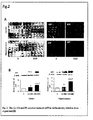
Figure 2 : The As, Cd and Pb-mixture induced APP in rat brain (A), which is dose-dependent (B)- (A) Representative photomicrographs of the cortical sections from the vehicle (V) and MM treated 90 day rats that were labelled for APP through DAB staining (LHS) and immunofluorescence (RHS).
(B) Cortical and hippocampal tissues from vehicle (V) and metal mixture (MM, 1X and 10X, refer to Table 1)-treated wistar rats were immunoblotted for APP and β-actin. Representative western blot and densitometry of APP normalized with β-actin.Data represent means ± SE of three pups from three different litters. ***P<0.001, **P<0.01 and *P<0.05 indicate significant changes in the metal mixture treated rats compared to V. c P<0.001 indicates significant difference compared to 1X-MM. -
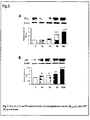
Figure 3 : The As, Cd and Pb-mixture induced synergistic increase in Aβ1-42 (A) and APP (B) in rat brain - Representative western-blot and densitometry of Aβ1-42 (A) and APP (B) normalized with β-actin in cortical tissues in individual metal (As, Cd or Pb) and MM treated rats. Data represent means ± SE of three pups from three different litters. ***P<0.001, **P<0.01 indicate significant changes in the treated rats compared to V. c P<0.001 indicates significant difference compared to individual metals. -
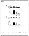
Figure 4 : The As, Cd and Pb-mixture induced BACE activity (A), APP-CTFβ and presenilin-2 (B) in rat brain- (A) Representative graph showing fold change in the activity of beta-secretase (BACE) in cortical and hippocampal tissues. ***P<0.001 indicates significant changes in the metal mixture treated rats compared to V.
(B) Representative western-blot and densitometry of C-terminal fragment of APP (APP-CTF-β) and presenilin-2 normalized with β-actin in cortical and hippocampal tissues. **P<0.01 and *P<0.05 indicate significant changes in the metal mixture treated rats compared to V. -
Figure 5 : Comparison of Aβ1-42 (A) and APP (B) levels of As, Cd and Pb mixture treated and Aβ1-42 injected rat brain- Representative western-blot and densitometry of Aβ1-42 (A) and APP (B) normalized with β-actin in hippocampal tissues in 10X-MM-treated or Aβ1-42-injected rats. ***P<0.001 indicates significant changes in the metal mixture-treated or Aβ1-42-injected rats compared to respective vehicle. -
Figure 6 : Memantine and donepezil reduced Aβ1-42 (A) and APP (B) in As, Cd and Pb-mixture treated rat brain - The hippocampal tissues from vehicle (V), metal mixture (10X-MM), and metal mixture with memantine (Mem) or donepezil (Don)-treated rats were immunoblotted for Aβ1-42, APP and β-actin. Representative western blot and densitometry of Aβ1-42 (A) and APP (B) normalized with β-actin. ***P<0.001 and **P<0.01 indicate significant changes in the metal mixture treated rats compared to vehicle. c P<0.001 indicates significant changes in the10X- MM and memantine or donepezil treated rats compared to 10X-MM. -
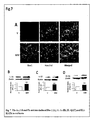
Figure 7 : The As, Cd and Pb-mixture induced Iba-1 (A), IL-1α (B), IL-1β (C) and IL1-R1 (D) in rat brain - (A) Representative photomicrograph of Iba-1-positive cells (green-fluorescence), nucleus (blue-fluorescence), and the two merged in the same field.
The vehicle (V) and metal mixture (MM)-treated brain tissues were immunoblotted for IL-1α, IL-1β, IL-1R1 and β-actin. (B) Representative western blot and densitometry of IL-1α normalized with β-actin in rat brain hippocampus. (C) Representative western blot and densitometry of lL-1β normalized with β-actin in rat brain hippocampus. (D) Representative western blot and densitometry of IL-1R1 normalized with β-actin in rat brain hippocampus. **P<0.01 indicates significant changes in the metal mixture treated rats compared to vehicle. -
Figure 8 : Hippocampal insertion of IL1-Ra suppressed Aβ1-42 (A and C) and APP (B and C) in As, Cd and Pb-mixture treated rat brain- Vehicle (V) and MM treated rats were injected with sterile PBS and IL1-Ra, and western blotting with brain tissues was performed for Aβ1-42, APP and β-actin. Representative western-blot and densitometry of Aβ1-42 (A) and APP (B) normalized with β-actin. **P <0.01 indicates significant changes in the metal mixture treated rats compared to vehicle, a P<0.05 and b P<0.01 indicate significant changes in the IL1-Ra treated rats compared to MM. (C) Representative photomicrograph showing Aβ1-42 or APP (red-fluorescence), nucleus (blue-fluorescence), and the two merged in the same field in hippocampus. -
Figure 9 : α-tocopherol and ω-3 fatty acids suppressed Aβ1-42 (A) and APP (B) in rat brain- (A) Representative western-blot and densitometry of Aβ1-42 (A) or APP (B) normalized with β-actin in hippocampal tissues of 10X-MM, 10X-MM and α-tocopherol (α-toc) or 10X-MM and ω-3-fatty acid (ω-3)-treated rats. ***P<0.001 and **P<0.01 indicate significant changes in the 10X-MM-treated rats compared to V. b P<0.01 indicates significant changes in the α-toc or ω-3 treated rats compared to 10X-MM. -
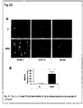
Figure 10 : The As, Cd and Pb-mixture induced Aβ-mediated apoptosis in neurons of rat brain- (A). Representative photomicrograph of apoptotic TUNEL (green- fluorescence), co-labelled with neuron-expressing, MAP-2 (red-fluorescence), and serially immunolabelled with Aβ1-42 (red-fluorescence) in the same field in cerebral cortex. (B). Representative graph showing relative apoptotic index of MM-treated rats with respective vehicle. ***P<0.001 indicates significant changes in the metal mixture treated rats compared to vehicle. -
Figure 11 : The As, Cd and Pb-mixture induced Aβ1-42 (A) and APP (B) in astrocytes- The vehicle (V) and metal mixture (MM, refer to Table 3 for conc.)-treated astrocytes were immunostained for Aβ1-42 or APP and hoechst. (A) Representative photomicrograph of Aβ1-42 immunofluorescence and hoechst, and the two merged together in the same field. (B) Representative photomicrograph of APP immunofluorescence and hoechst, and the two merged together in the same field. The bar diagram indicates the number of Aβ1-42 or APP immunoreactive cells normalized with hoechst-+ve nuclei. ***P<0.001 indicates significant changes in the metal mixture treated astrocytes compared to vehicle. -
Figure 12 : The As, Cd and Pb-mixture induced IL1-R1 in neurons (A) and astrocytes (B)- The vehicle (V) and metal mixture (MM, refer to Table 2 and 3 for conc.)-treated neurons and astrocytes were immunostained for IL1-R1 and hoechst. (A) Representative photomicrograph of IL1-R1 (red fluorescence) and hoechst, and the two merged together in the same field in neuronal cells. (B) Representative photomicrograph of IL1-R1 (red fluorescence) and hoechst, and the two merged together in the same field in astrocytes. -
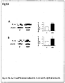
Figure 13 : The As, Cd and Pb-mixture induced IL-1α (A) and IL-1β (B) in brain cells - Representative western blot and densitometric analysis of IL1-α (A) and IL1-β (B) relative to β actin in mixed brain culture treated with MM. ***P<0.001 and **P<0.01 indicate significant changes in the metal mixture treated brain cells compared to vehicle. -
Figure 14 : Docosahexaenoic acid and α-tocopherol reduced the levels of Aβ1-42 (A) and APP (B) in astrocytes- The vehicle (V)- and metal mixture (MM), docosahexaenoic aid (DHA) or α-tocopherol-treated astrocytes were immunostained for Aβ1-42 or APP. (A) Representative photomicrograph of Aβ1-42 immunofluorescence. (B) Representative photomicrograph of APP immunofluorescence. - The main objective of the present invention is to provide a new non transgenic animal wistar rat model of early Alzheimer's disease AD, and to offer new non-transgenic neuronal and astrocyte cellular models of AD.
- The aims are to make available the novel animal and cellular models bearing the signs of AD pathology, vascular damage, neuro-inflammation, and neuronal apoptosis associated with AD, for understanding the etiology and mechanisms underlying this disease at an early age. The models will also serve as screening tools for identifying novel drugs that could target AD.
- A heavy metal mixture of As, Cd and Pb, in the present invention, is utilized for the development of an early and potential model for AD. The detailed procedures for the development of the model will be discussed, and subsequently the properties matching that of AD. The methods in the following examples should, however, not be construed to limit the scope of invention.
- Accordingly, the main embodiment of the present invention provides a non-transgenic animal wistar rat model of early Alzheimer's disease characterized in having over-expression of Aβ and APP proteins in the brain by exposing to a mixture of heavy metals arsenic, cadmium and lead at 0.38 mg/Kg, 0.098 mg/Kg, 0.220 mg/Kg to ten times concentration of each respectively, in water.
- Another embodiment of the present invention provides a method for preparing a non-transgenic animal wistar rat model of early Alzheimer's disease as claimed in
claim 1 comprising of: - (a) providing a wistar rat;
- (b) providing a heavy metal mixture of arsenic, cadmium and lead as claimed in
claim 1; and - (c) orally feeding the metal mix as obtained in step (b) to the rat to obtain the non transgenic wistar rat model of Alzheimer's disease.
- Another embodiment of the present invention provides a method described as herein in the present invention wherein the oral feeding of heavy metal mixture is started at gestation day - 05 (in pregnant and lactating dams) and continued in the off-springs until early adulthood, i.e. postnatal 60 - 90.
- Another embodiment of the present invention provides a method described as herein in the present invention wherein the oral feeding of heavy metal mixture at 3.8 mg/Kg, 0.98 mg/Kg, 2.20 mg/Kg is started at gestation day - 05 (in pregnant and lactating dams) and continued in the off-springs until weaning, i.e.
postnatal 24. - Another embodiment of the present invention provides a method described as herein in the present invention wherein the oral feeding of heavy metal mixture at 3.8 mg/Kg, 0.98 mg/Kg, 2.20 mg/Kg is started at postnatal day - 90 and continued until adulthood, i.e. postnatal-120. Another embodiment of the present invention provides a non-transgenic wistar rat model of early Alzheimer's disease characterized in having over-expression of BACE, CTFβ and presenilin in the brain by exposing to a mixture of heavy metals arsenic, cadmium and lead at 3.8 mg/Kg, 0.98 mg/Kg, 2.20 mg/Kg respectively in water.
- Another embodiment of the present invention provides a method described as herein in the present invention wherein the heavy metal mixture composition consists of arsenic, cadmium and lead in water.
- Another embodiment of the present invention provides a method described as herein in the present invention wherein the heavy metal mixture composition comprising of NaAsO2 + CdCl2 + Pb(C2H3O2)2 at 0.38 mg/Kg, 0.098 mg/Kg, 0.220 mg/Kg to ten times concentration of each respectively, in water.
- Another embodiment of the present invention provides use of the rat model as described herein in the present invention for screening anti-Alzheimer's drug.
- Another embodiment of the present invention provides use of the rat model as described herein in the present invention for developing anti- Alzheimer's therapies.
- Another embodiment of the present invention provides a use of the rat model as described herein in the present invention for detecting early age Alzheimer's disease.
- Another embodiment of the present invention provides use of the rat model as described herein in the present invention for screening drugs and developing therapies targeted to BACE, CTFβ and presenilin.
- Another embodiment of the present invention provides a composition for the induction of early Alzheimer's disease in a subject comprising of NaAsO2 + CdCl2 + Pb(C2H3O2)2 at 0.38 mg/Kg, 0.098 mg/Kg, 0.220 mg/Kg to ten times concentration of each respectively, in water. Another embodiment of the present invention provides a composition described as herein in the present invention wherein the subject is a non-transgenic wistar rat.
- Another embodiment of the present invention provides a neuronal model of Alzheimer's disease characterized in having over-expression of Aβ, APP and BACE proteins by exposing to a mixture of heavy metals arsenic, cadmium and lead.
- Another embodiment of the present invention provides for a method of preparing the neuronal model of Alzheimer' disease as described herein in the present invention comprising steps:
- (a) isolating neurons from embryonic day 14-16 wistar rats;
- (b) preparing mixture of arsenic, cadmium and lead at 5 µM, 1 µM, and 10µM respectively; and
- (c) treating the 80% confluent neurons obtained in step a with the mixture obtained in step b to obtain the neuronal model of Alzheimer' disease.
- Another embodiment of the present invention provides an astrocyte model of Alzheimer's disease characterized in having over-expression of Aβ, APP and BACE proteins by exposing to a mixture of heavy metals arsenic, cadmium and lead.
- Another embodiment of the present invention provides for a method of preparing the astrocyte model of Alzheimer' disease described as herein in the present invention said method comprising steps:
- (a) isolating astrocytes from postnatal day-1 wistar rats;
- (b) preparing mixture of arsenic, cadmium and lead at 6 µM, 2 µM, and 50 µM respectively; and
- (c) treating the 80% confluent astrocytes obtained in step a with the mixture obtained in step b to obtain the astrocyte model of Alzheimer' disease.
- Another embodiment of the present invention provides a composition for preparing neuronal and astrocyte model of Alzheimer's disease comprising of NaAsO2 + CdCl2 + Pb(C2H3CO2)2 in water.
- Another embodiment of the present invention provides for a use of the neuronal model as described herein in the present invention for screening anti- Alzheimer drugs.
- Another embodiment of the present invention provides for a use of the neuronal model as described herein in the present invention for developing anti-Alzheimer therapies.
- Another embodiment of the present invention provides for a use of the astrocyte model as described in the herein in the present invention for screening anti- Alzheimer drugs.
- Another embodiment of the present invention provides for use of the astrocyte model as described herein in the present invention for developing anti-Alzheimer therapies.
- Sodium arsenite, lead acetate, cadmium chloride, protease inhibitor cocktail, Hoechst 33258 stain, poly-L-lysine, mammalian tissue protein extraction reagent, , rabbit monoclonal antibody to Aβ-42, mouse monoclonal antibodies to β-actin and peroxidase conjugated secondary antibodies, were purchased from Sigma Chemical Co. (St. Louis, MO). The sample loading buffer for western blotting, protein markers and Alexa fluor secondary antibodies were purchased from Invitrogen (Carlsbad, CA). The supersignal west femto maximum sensitivity substrate for western blotting was purchased from PIERCE Biotechnology (Rockford, IL). Rabbit monoclonal antibody to APP and Aβ-42 were purchased from Abcam (Cambridge, MA). Beta secretase enzyme activity kit was purchased from Abcam (Cambridge, MA). Interleukin-1 alpha, and interleukin-lbeta were purchased from R&D systems (Minneapolis, Mn). Diaminobenzidin tetrahydrochloride (DAB) substrate kit, vectashield medium and Elite ABC kit were purchased from Vector Laboratorie (Burlingame, CA). The terminal deoxynucleotide-transferase (TdT)- Mediated dUTP nick end labelling (TUNEL) kit was purchased from Roche (Indianapolis, IN). ELISA kits for Aβ-40 were purchased from IBL, Japan. Memantine (Brand name, Admenta) and donepezil (Brand name, Aricep 10) were from Sun Pharma, Sikkim and Eisai Co. Ltd, Tokyo, Japan respectively. Omega (ω) 3 fatty acid (Brand name: MAX-EPA) and α-tocopherol were from Merck, India and Sigma Chemical Co. (St. Louis, MO) respectively. DMEM/F-12, Neurobasal media, antibiotics, fetal bovine serum (FBS) and Trypsin-EDTA were from Gibco BRL, USA. Rat recombinant IL-1 receptor antagonist (IL1-Ra) was purchased from R&D systems (Minneapolis, Mn). Rat recombinant Aβ1-42 peptide was purchased from Tocris biosciences (Bristol, United Kingdom).
- Primary* and secondary** antibodies used for western blotting (WB), immunohistochemistry (IHC) and immunocytochemistry (ICC)
Antibody Cat. No. Source and location Species Dilution APP * ab15272 Abcam, Cambridge, MA Rabbit 1:1000 (WB), 1:100 (IHC, ICC) Aβ1-42 * ab10148 Abcam, Cambridge, MA Rabbit 1:1000 (WB), 1:100 (IHC, ICC) Iba-1* ab15690 Abcam, Cambridge, MA Mouse 1:100 (IHC, ICC) CTF-β* A8717 Sigma Chemical Co., St. Louis, MO Rabbit 1:1000 (WB) Presenilin * 2192S Cell Signaling Technology, Danvers, MA Rabbit 1:1000 (WB) IL-1α * ab7632 Abeam, Cambridge, MA Rabbit 1:1000 (WB) IL-Iβ * ab2105 Abcam, Cambridge, MA Rabbit 1:1000 (WB) IL1-R1* sc-689 Santa Cruz Biotechnology, Dallas, Texas Rabbit 1:1000 (WB), 1:100 (IHC, ICC) β-actin * A5441 Sigma Chemical Co., St. Louis, MO Mouse 1:10,000 (WB) Microtubule associated Protein - 2 (MAP-2) * 4542S Cell Signaling Technology, Danvers, MA Rabbit 1:1000 (WB), 1:100 (IHC) Glial Fibrillary Acidic Protein (GFAP) * SAB4300647 Sigma Chemical Co., St. Louis, MO Rabbit 1:1000 (WB), 1:100 (IHC) RAGE Sc-365154 Santa Cruz Biotechnology, Dallas, Texas Mouse 1:1000 (WB) p-Glycoprotein 517310 Calbiochem, Millipore, (Temecula, CA). Mouse 1:1000 (WB) HRP anti-mouse IgG** A9044 Sigma Chemical Co., St. Louis, MO Mouse 1:2000 (WB) HRP anti-rabbit IgG** A0545 Sigma Chemical Co., St. Louis, MO Rabbit 1:2000 (WB) Alexa Fluor 488 anti-mouse IgG** A11001 Invitrogen, Carlsbad, CA Mouse 1:200 (IHC, ICC) Alexa Fluor 546 anti-rabbit IgG** A11010 Invitrogen, Carlsbad, CA Rabbit 1:200 (IHC, ICC) - The following examples are given by way of illustration of the present invention and therefore should not be construed to limit the scope of the present invention.
- All animal-handling procedures were carried out in accordance with the current regulations of the Indian Institute of Toxicology Research Animal Ethics Committee, and with its prior approval for using the animals. The pregnant female wistar rats were housed in a 12-h day and light cycle environment with ad libitum diet and water. The rats were divided into six groups (n=30 per group), gavage-treated with the metal mixture (Table 1). The metal mixture treatment was at two concentrations (1X and 10X; Groups 1-3, Table 1) for dose-dependent study. To identify the synergistic nature, the animals were treated in mixture as well as individually with the metals (Group 1-6 of Table 1). The heavy metals were As, Cd and Pb, fed in the form of NaAsO2, CdCl2 and Pb(C2H3O2)2, respectively, in Milli Q water. The NaAsO2, and CdCl2 readily dissolved in water, while Pb(C2H3O2)2 was dissolved by adding 0.043 % acetic acid in water. The dams (pregnant rats and lactating mother rats) were daily treated with the metals in water from gestation day 5 (G-05) until the pups weaned (postnatal day-21, P-21). From P-22, the postnatal rats were directly treated with the metals until P-90. The tissues were collected at P-24, P-60 or P-90. The male rats were selected for the study, and rats from separate litters served as independent subjects.
- In another set, the rats were treated with 10X doses of the metal mixture from P-90 until P-120.
Table 1: Metal treatment given to pregnant, lactating and post-weaning rats Group 1, Vehicle Water (vehicle) Group 2, MM (1X)NaAsO2: 0.380 mg/Kg+ CdCl2: 0.098 mg/Kg+ Pb(C2H3O2)2: 0.220 mg/ Kg Group 3, MM (10X) NaAsO2: 3.80 mg/Kg+ CdCl2: 0.98 mg/Kg+ Pb(C2H3O2)2: 2.220 mg/Kg Group 4, As-individual treatment NaASO2: 11.4 mg/Kg(3x3.80) Group 5, Cd-individual treatment CdCl2: 2.94 mg/Kg (3x0.98) Group 6, Pb-individual treatment Pb(C2H3O2)2 : 6.660 mg/Kg(3x2.220) - The known AD-targeting drugs, memantine (10 mg/kg body weight) and donepezil (conc. 1.5 mg/kg body weight) were administered for 10 days along with 10X-MM in P-90 rats. Omega -3 (ω3) fatty acid (mixture of eicosapentaenoic acid, 90mg/Kg, and docosahexaenoic acid, 60mg/Kg) was orally treated from G-0 to P-60 along with 10X-MM (treated from G-05 to P-60). α-tocopherol (100mg/Kg body weight) was orally treated from P-24 to P-60 along with 10X-MM (treated from G-05 to P-60).
- Tissues of the cerebral cortex and hippocampus from five to seven postnatal wistar rats were harvested, snap-frozen in liquid nitrogen, and stored at -80° C until further investigation. SDS-PAGE and western blotting was performed with APP (1:1000), Aβ-40 (1:1000), Aβ-42 (1:1000), APP-CTF-β (1:1000), presenilin (1:1000), IL-1α (1:1000), IL-1β (1:1000) and IL-1R1 (1:1000), RAGE (1:1000) and pGP (1:1000). The working dilutions for secondary anti-rabbit IgG and anti-mouse IgG conjugated to horseradish peroxidase were 1:2000 in 0.2% Triton X-100 containing PBS. The samples were detected by chemiluminescence with super signal west femto max substrate. Relative expression of each protein was determined by densitometric quantification of blots using VersaDoc Gel Imaging System (BioRad, Hercules, CF).
- Four wistar rat pups from four separate litters, treated with the metal mixture or vehicle, were anaesthetized and perfused, and the cerebral cortex and hippocampus were fixed and cryoprotected. Briefly, five to 10-µM cryostat sections were made using cryomicrotome (Microm HM 520, Labcon, Germany), and mounted on (3-Aminopropyl) triethoxy-silane coated slides. For immunofluorescence, a standard immunofluorescence technique was used. The sections were blocked in 10% normal donkey serum/0.1M PBS and then incubated with the antibodies (1:100) at 4°C for overnight. For detecting the expression of APP,Aβ or Iba-1, the sections were immunostained with monoclonal APP, Aβ or Iba-1antibodies. Following a rinse in 0.1M PBS, the sections were incubated with Alexa Fluor 546 goat anti-rabbit IgG conjugate (1:200) and Alexa Fluor 488 goat anti-mouse IgG conjugate (1:200) for 60 min. After re-rinsing, the sections were counterstained with hoechst 33258 (0.2 mM) for 5 min, cover-slipped on vectashield medium and visualized under a fluorescence microscope. The images were then imported into Image-J 1.42q (http://rsb.info.nih.gov/ ij/; developed by Wayne Rasband, National Institutes of Health, Bethesda, MD) for quantifying cell fluorescence.
- For DAB staining, a standard technique was used as described in DAB chromogen and ABC kit. The cryo-sections were blocked in 10% normal donkey serum/0.1M PBS and then incubated with the antibodies, APP (1:100) and Aβ1-42 (1:100) at 4°C overnight. Following a rinse in 0.1M PBS, the sections were incubated with secondary antibody for 60 min, and then incubated with ABC reagent for 30 min. After rinsing, the sections were stained with DAB chromogen for 2-5 min, cover-slipped on DPX mounting medium and visualized under optical microscope (Nikon Instech Co. Ltd).
- BACE levels in the cortex and hippocampus of wistar rats were detected spectrofluorometrically using the BACE enzyme activity assay. The tissue lysates (50 µl) were added to each well of a black 96-well microplate. Fifty µl of the 2X-Secretase reaction buffer was added to the lysates, followed by the addition of 2 µl of the respective substrate provided in the kits. The plate was covered, tapped gently, kept at 37°C for 1-2 hrs, and then read in a fluorescent microplates reader using excitation (335-355 nm) and emission (495-510 nm) filters.
- The effect of metal mixture treatment on Aβ1-40 levels was determined using specific ELISAs (IBL, Immuno-Biological Co., Ltd., Japan). Cortex and hippocampi from the wistar rats were micro-dissected and quickly homogenized in extraction buffer. The homogenates were diluted 1:10 with cold casein buffer (0.25% casein and 0.05% sodium azide), followed by centrifugation for 30 min at 4°C at 40,000 rpm. Hundred µl of sample was added into the pre-coated plates and incubated overnight at 4°C. After washing each well of the pre-coated plate with washing buffer, 100 µl of labelled antibody solution was added and the mixture was incubated for 1 hr at 4°C in the dark. After washing, the chromogen was added and the mixture was incubated for 30 min at room temperature in the dark. After the addition of stop solution, the resulting colour was assayed at 450 nm using a microplate absorbance reader.
- Recombinant Aβ1-42 and recombinant IL1-Ra were injected into rat hippocampus using stereotaxic technique. Briefly, rats were anesthetized with intraperitoneal injection of ketamine and xylazine (60 and 20 mg/kg body weight respectively). The rats were placed in the stereotaxic frames (Stoelting Co., USA), skull exposed and disinfected with betadine. An incision was made into the scalp and drilled (depth of 2.5 mm) at 2.0 mm medial-lateral and 3.5 mm posterior-anterior to bregma. Five µl solution of IL1-Ra (350 ng/ml) or Aβ1-42 (1µg/ml) was bilaterally injected slowly into the hippocampus with a 10 µl Hamilton syringe, and syringe retained for 2 minutes for complete diffusion of IL1-Ra or Aβ1-42. IL1-Ra was injected, once, at P-90 in 10X-MM-treated rats, and dissected after 10 days. Aβ1-42 was injected, once, in P-90 control male rats and dissected after 10 days. The needle was slowly withdrawn after injection. Control group, treated with sterile PBS, underwent the same procedures.
- To characterize the synergistic interaction between the heavy metals for their effects on the APP, Aβ and BACE levels, a combination index (CI) was calculated using the software Calcusyn (Biosoft, Manchester, United Kingdom). CI values <1.0 indicated synergism (Zhao et al., 2004).
- Detection of apoptosis (in vivo) in MAP-2 expressing neurons and GFAP-ir astrocytes was performed. In situ detection of apoptosis was carried out by TUNEL assay. Briefly, four pups from four different litters were taken at the developmental stages, anesthetized, and perfused and the brain was fixed and cryoprotected. Five-micron sections from the cortex and hippocampus were made using cryomicrotome (Microm HM 520; Labcon). For the TUNEL assay, a labelling reaction was carried out with fluorescein-labelled dUTP in the presence of TdT at 37°C for 1 h. To investigate whether the apoptotic cells were neurons or astrocytes, the sections were immunostained with anti-mouse MAP-2 or GFAP antibody (1:100 dilution in TBST [10mM Tris, pH 8.0, 150mM NaCl, 0.01% Tween 20]) according to manufacturer's protocol. The sections were then incubated with Alexa Fluor 546-conjugated (fluorescent color: red; Abs/Em: 555/565) goat anti-mouse antibody (1:200 dilution); counterstained with Hoechst 33258 (0.2mM) for 5 min; and visualized under a fluorescence microscope (Nikon Instech Co. Ltd) after being coverslipped on Vectashield medium (Vector Laboratories). For Aβ immunofluorescence, a standard immunofluorescence technique was used in serial sections of that used for GFAP/MAP-2 and TUNEL assay. The sections were blocked in 10% normal donkey serum/0.1M PBS and then incubated with the antibodies (1:100) at 4°C for overnight. Following a rinse in 0.1M PBS, the sections were incubated with Alexa Fluor 546 goat anti-mouse IgG conjugate (1:200). After re-rinsing, the sections were counterstained with hoechst 33258 (0.2 mM) for 5 min, cover-slipped on vectashield medium and visualized under a fluorescence microscope. The images were then imported into Image-J 1.42q (http://rsb.info.nih.gov/ ij/; developed by Wayne Rasband, National Institutes of Health, Bethesda, MD) for quantifying cell fluorescence.
- The rats were subjected to the passive avoidance test by placing in a compartment of computerized shuttle box (Techno, India). The light compartment was isolated from the dark compartment by an automated guillotine door. After an acclimatization period of 30 s, the guillotine door was opened and closed automatically after entry of the rat into the dark compartment. The subject received a low-intensity foot shock (0.5mA; 10 s) in the dark compartment. Infrared sensors monitored the transfer of the animal from one compartment to another, which was recorded as transfer latency time (TLT) in seconds. The 1st trial was for acquisition and retention was tested in a 2nd trial (1st retention) given 24 h after the 1st trial. The duration of a trial was 300s. Further, 2nd, 3rd and 4th retention trials were given on alternate days to test retention in the metal mixture treated rats. The shock was not delivered in the retention trials to avoid reacquisition. The criterion for learning was taken as an increase in the TLT on retention (2nd or subsequent) trials as compared to acquisition (1st) trial.
- The pregnant wistar rats were sacrificed by cervical dislocation and the embryos were removed on the 16th day of gestation. The embryonic brain tissues were mechanically dissociated into individual cells in dissection media containing Glucose (1M), sucrose (1 M), HEPES Buffer (1M) and Hank's salt (1X). The resulting cells were centrifuged (1,500 rpm, 5 min), resuspended in NEUROBASAL medium containing B-27 supplement (Invitrogen, Carlsbad, CA), L-glutamine (0.5 mM), penicillin (100 U/ml) and streptomycin (100µg/ml) and plated into 60 mm dishes. The culture media was changed every 2 days. Greater than 90% of the cells in these cultures were neurons as assessed by cell morphology and immunostaining with rabbit monoclonal antibodies against MAP-2 (1: 100).
- The astrocytes from rat brain were isolated and cultured. Briefly, prefrontal cortices of 1-day-old wistar rats were dissected out and digested with trypsin for 10 min at 37°C. A single cell suspension was obtained by triturating, and the cells were seeded onto poly-L-lysine (100 µg/ml)-coated plates. The cultures were maintained in DMEM/F12 with 10% heat-inactivated fetal bovine serum, 100 U/ml penicillin, and 100 mg/ml streptomycin. The confluent cells were rinsed twice with serum-free media and then detached with 0.25% trypsin with ethylenediaminetetraacetic acid and sub-cultured. The cells reached confluence at 7 days after subculture, and at this point, more than 95-97% of cells were GFAP-positive astrocytes, as determined by the immunofluorescence staining.
- The astrocytes and neurons were grown to 80% confluence, pre-incubated in reduced serum (0.5% fetal bovine serum [FBS]) medium for 2 h, and then treated with a mixture of As, Cd, and Pb (
Group Table 2: Metal treatment given to neurons Group 1: Vehicle Water (vehicle) Group 2: metal mixture (MM) NaAsO2: 5 (µM + CdCl2: 1 µM + Pb(C2H3O2)2: 10 µM Group 3: As-individual treatment NaAsO2: 15 µM (3x5 µM) Group 4: Cd-individual treatment NaAsO2: 3 µM (3x1 µM) Group 5: Pb-individual treatment Pb(C2H3O2)2: 30 µM (3x10 µM) Table 3: Metal treatment given to astrocytes Group 1: Vehicle Water (vehicle) Group 2: MM NaAsO2: 6 µM + CdCl2: 2 µM + Pb(C2H3O2)2: 50 µM Group 3: As-individual treatment NaAsO2: 18 µM (3x6 µM) Group 4: Cd-individual treatment NaAsO2: 6 µM (3x2 µM) Group 5: Pb-individual treatment Pb(C2H3O2)2: 150 µM (3x50 µM) - The astrocytes and neurons were grown to 80% confluence, pre-incubated in reduced serum (0.5% fetal bovine serum [FBS]) medium for 2 h, and then treated with a mixture of As, Cd, and Pb with DHA(10 µM) or α-tocopherol (10 µM) for 16 h, and incubated at 37°C.
- The astrocytes and neurons were treated, washed with PBS and suspended in 100 µl of CelLytic™ MT Cell Lysis Reagent protease inhibitor cocktail (a mixture of 4-(2-aminoethyl) benzenesulfonyl fluoride, pepstatinA, E-64, bestatin, leupeptin, and aprotinin)] and kept on ice for 20 min. The cells were homogenized using a Teflon homogenizer and centrifuged at 15, 000 rpm for 30 min at 4°C, and the supernatant was collected. SDS-PAGE and western blotting was done with APP (1:1000), Aβ-40 (1:1000), Aβ-42 (1:1000), IL-1 α(1:1000), IL-1 β (1:1000) and IL-1R1. The working dilutions for secondary anti-rabbit IgG and anti-mouse IgG conjugated to horseradish peroxidase were 1:2000 in 0.2% Triton X-100 containing PBS. The samples were detected by chemiluminescence with super signal west femto max substrate. Relative expression of each protein was determined by densitometric quantification of blots using VersaDoc Gel Imaging System (BioRad, Hercules, CF).
- The astrocytes and neurons at 80 % confluence were fixed with 4% PFA for 1 hr at room temperature, followed by three rinses in PBS. Cells were then pre-incubated for 15-30 min with PBS containing 0.3% Triton X-100 (Sigma) and 3% normal horse serum (Gibco-BRL) at room temperature. Cultures were then incubated overnight with antibodies directed against APP (1:100), Aβ-40 (1:100), Aβ-42 (1:100), IL-1α (1:100), lL-1β (1:100) and IL-1R1 (1:100) and diluted in PBS containing 0.3% Triton X-100 and 5% normal horse serum. Following a rinse in 0.1M PBS, the sections were incubated with Alexa Fluor 546 goat anti-mouse IgG conjugate (1:200) for 60 min. After re-rinsing, the sections were counterstained with hoechst 33258 (0.2 mM) for 5 min, cover-slipped on vectashield medium and visualized under a fluorescence microscope. The images were then imported into Image-J 1.42q (http://rsb.info.nih.gov/ ij/; developed by Wayne Rasband, National Institutes of Health, Bethesda, MD) for quantifying cell fluorescence.
- The BACE activity in the cell lysates was detected spectrofluorometrically using BACE enzyme activity assay. The cell lysate, in buffer provided with the kit (50 µl) were added to each well of a black 96-well microplate. The protocol is the same as that for tissue BACE assay.
- Upon treating the developing wistar rats with the metal mixture from G-05, the rats demonstrated a dose- dependent increase in the Aβ-petides and APP levels in the cortex (Table 4) and hippocampus (Table 5). The effect was synergistic, with a CI value less than 1.0. The known AD-targeting drugs, memantine and donepezil suppressed the levels of Aβ and APP in this non-transgenic wistar rat model of AD (Table 6). The metal mixture induced an increase in BACE activity in the rat cortex and hippocampus, at an early age (Table 7). The effect was synergistic, with a CI value less than 1.0. The metal mixture induced APP-CTF-β and presenilin at an early age (Table 8). The metal mixture induced Aβ-mediated apoptosis in the rat brain (Table 9). The metal mixture induced a loss in cognition at an early age (Table 10). The metal mixture induced the inflammatory cytokines IL-1α, IL-1β and their receptor, IL-1R1, that contribute towards AD pathogenesis (Table 11). Upon treating the developing wistar rats with the metal mixture from P-90 to P-120, the rats demonstrated an increase in the Aβ-peptides and APP levels (Table 12).
-
Table 4: Dose-dependent increase in Aβ-42, Aβ-40 and APP in rat brain cortex upon exposure to the metal mixture Aβ-42 (fold increase compared to age- matched vehicle) Aβ-40 (fold increase compared to age- matched vehicle) APP (fold increase compared to age- matched vehicle) 1X 10X 1X 10X 1X 10X 1.7 ± 0.1028 2.4 ± 0.1098 1.3 ± 0.1063 1.5 ± 0.1052 1.5 ± 0.0959 2.2 ± 0.1032 Table 5: Dose-dependent increase in Aβ-42, Aβ-40 and APP in rat brain hippocampus upon exposure to the metal mixture Aβ-42 (fold increase compared to age- matched vehicle) Aβ-40 (fold increase compared to age- matched vehicle) APP (fold increase compared to age- matched vehicle) 1x 10X 1X 10X 1X 10X 1.7 ± 0.0875 3.0 ± 0.1452 1.5 ± 0.1077 1.8 ± 0.1909 1.3 ± 0.0461 3.0 ± 0.1498 Table 6: Reduction in the metal mixture-induced Aβ by memantine and donepezil (two week treatment) in rat brain Compounds fold reduction in Aβ-40 fold reduction in Aβ-42 fold reduction in APP Memantine 2.54 ± 0.0099 2.42. ± 0.0143 1.69 ± 0.0510 Donepezil 3.5 ± 0.0799 3.22 ± 0.0800 3.14 ± 0.2160 Table 7: Increase in BACE activity in rat brain upon exposure to the metal mixture Rat age BACE activity-Cortex (fold increase compared to age- matched vehicle) BACE activity-hippocampus (fold increase compared to age- matched vehicle) Postnatal 60 d 1.3 ± 0.1450 1.26 ± 0.0510 Postnatal 90 d 2.0 ± 0.2679 1.5 ± 0.2733 Table 8: Increase in APP-CTF-β and presenilin in rat brain upon exposure to the metal mixture Fold increase compared to age- matched vehicle in cortex Fold increase compared to age- matched vehicle in hippocampus APP-CTF-β 1.3 ± 0.0908 1.5 ± 0.1347 Presenilin 1.7 ± 0.1635 1.6 ± 0.1066 Table 9: Increase in Aβ-mediated apoptosis in rat brain upon exposure to the metal mixture Rat age Apoptosis-brain (fold increase compared to age-matched vehicle) Postnatal 60 day11.0 ± 2.1369 Postnatal 90 day 16.0 ± 1.3558 Table 10: Loss in cognition upon exposure to the metal mixture Rat age Loss in cognition (approx.) Postnatal 90 d 35 % Table 11: Increase in the levels of inflammatory markers in the rat brain upon exposure to the metal mixture Rat age IL-1α (fold increase compared to age-matched vehicle) IL-1β (fold increase compared to age-matched vehicle) IL-1R1 fold increase compared to age-matched vehicle) Postnatal 60 day1.6 ± 0.1794 1.3 ± 0.1194 1.3 ± 0.1104 Postnatal 90 day 1.7 ± 0.1037 1.8 ± 0.1901 1.5 ± 0.0769 Table 12: Increase in Aβ-42 and APP in P-120 rat brain upon exposure to the metal mixture from P-90 Aβ-42 (fold increase compared to age- matched vehicle) APP (fold increase compared to age- matched vehicle) 1.5 ± 0.2274 1.4 ± 0.0808 - Upon treating the neuronal and astrocyte cells with the metal mixture, the cells demonstrated an increase in the Aβ-peptides and APP levels, and BACE activity (Table 13). The effect was synergistic, with a CI value less than 1.0. The known AD-targeting drugs, memantine and donepezil suppressed the levels of Aβ and APP in the non-transgenic neuronal and astrocyte models of AD. The omega-3 fatty acid (docosahexaenoic acid), known to reduce the risk of AD, suppressed the levels of Aβ and APP in the non-transgenic neuronal and astrocyte models of AD. Vitamin E, known to be protective against AD, suppressed the levels of Aβ and APP in the non-transgenic neuronal and astrocyte models of AD (Table 14).
- The metal mixture induced the inflammatory cytokines IL-1α and IL-1βin the brain cells (Table 15).
-
Table 13: Increase in Aβ, APP and BACE in neurons and astrocytes upon exposure to the metal mixture Cell type Aβ-fold increase APP-fold increase BACE-fold increase Neuron 2.3 ± 0.2232 2.0 ± 0.1949 1.5 ± 0.1917 Astrocyte 2.2 ± 0.2015 2.3 ± 0.2247 2.0 ± 0.2682 Table 14: Reduction in metal mixture-induced Aβ and APP in rat astrocytes and neurons by memantine, donepezil, docosahexaenoic acid and Vit. E Compounds Fold reduction in Aβ Fold reduction in APP Memantine 1.4 ± 0.052 1.3 ± 0.0392 Donepezil 1.8 ± 0.038 2.0 ± 0.0384 Docosahexaenoic acid 2.3 ± 0.0853 1.25 ± 0.0265 α-tocopherol 1.9 ± 0.0192 1.3 ± 0.0493 Table 15: Increase in the levels of inflammatory markers in the rat brain cells exposure to the metal mixture IL-1α (fold increase -matched vehicle) IL-1β (fold increase compared to vehicle) 1.7 ± 0.0600 1.5 ± 0.1021 - This method of invention of the non-transgenic early wistar rat model of AD is very novel, with respect to inducing early signs of AD synergistically. Its advantages over the known methods are,
- (a) reduced mechanical tissue damage from intra-cranial administration procedures,
- (b) non-neurotoxicity of vehicle, and
- (c) convenient method of induction. Its advantages over the production of transgenic animals are (a) less laborious protocol and (b) the ethical concerns that limit the numbers of animals employed in experiments.
- The method is more beneficial compared to the transgenic lower species that have a brain anatomy much different from humans.
- This non-transgenic model shows a range of symptoms, such as the generation of the pathological Aβ-40, 42 peptides, the APP, Aβ-mediated apoptosis and vascular damage along with a rise in the inflammatory markers that are reported to aggravate the disease pathogenicity. The time needed to generate the models is less, and the method is less expensive.
- Moreover, our non-transgenic animal model is validated with the known AD-targeting drugs and preventive agents.
- The model does not show toxicity of the other vital organs.
- This method of invention of the non-transgenic cellular model of AD is very novel, with respect to inducing signs of AD in neurons and astrocytes, and synergistically.
- The non transgenic in vitro model for AD is less expensive, quick and efficient.
- It is validated with the known AD-targeting drugs and preventive agents.
- Moreover, treatment with the metal mixture also serves as a model-inducer for cells already over-expressing the amyloidogenic APP.
Claims (8)
- Use of
a non-transgenic animal wistar rat model of early Alzheimer's disease characterized in having over-expression of Amyloid Beta (Aβ), amyloid precursor protein (APP), β-secretase (BACE), C-terminal fragment beta (CTF-β) and presenilin proteins in the rat brain cortex and hippocampus, induced by exposing the rat from postnatal day 90 until postnatal day 120 to a mixture of heavy metals comprising arsenic, cadmium and lead in the form of NaAsO2, CdCl2 and Pb(C2H3O2)2 at 3.8 mg/Kg, 0.98 mg/Kg, 2.20 mg/Kg respectively, in water,
for screening anti-Alzheimer's drug. - Use of the rat model as claimed in claim 1, for developing anti- Alzheimer's therapies.
- Use of the rat model as claimed in claim 1, for detecting early age Alzheimer's disease.
- Use of the rat model as claimed in claim 1 for screening drugs and developing therapies targeted to BACE, CTFβ and presenilin.
- Use of a non-transgenic wistar rat cellular neuronal model of Alzheimer's disease characterized in having over-expression of Amyloid Beta (Aβ), amyloid precursor protein (APP), β-secretase(BACE) and presenilin proteins by exposing neuronal cells of wistar rats to a mixture of heavy metals arsenic, cadmium and lead for screening anti-Alzheimer's drugs.
- Use of the rat model as claimed in claim 5, for developing anti- Alzheimer's therapies.
- Use ofa non-transgenic wistar rat cellular astrocyte model of Alzheimer's disease having over-expression of Amyloid Beta (Aβ), amyloid precursor protein (APP), β-secretase (BACE) and presenilin proteins by exposing astrocyte cells of wistar rats to a mixture of heavy metals arsenic, cadmium and lead for screening anti- Alzheimer drugs.
- Use of the non-transgenic wisar rat astrocyte model as claimed in claim 7 for developing anti-Alzheimer therapies.
Applications Claiming Priority (2)
| Application Number | Priority Date | Filing Date | Title |
|---|---|---|---|
| IN1442DE2013 | 2013-05-15 | ||
| PCT/IN2014/000330 WO2014184809A1 (en) | 2013-05-15 | 2014-05-15 | Model of alzheimer's disease |
Publications (2)
| Publication Number | Publication Date |
|---|---|
| EP2997136A1 EP2997136A1 (en) | 2016-03-23 |
| EP2997136B1 true EP2997136B1 (en) | 2019-04-17 |
Family
ID=51033260
Family Applications (1)
| Application Number | Title | Priority Date | Filing Date |
|---|---|---|---|
| EP14734244.8A Active EP2997136B1 (en) | 2013-05-15 | 2014-05-15 | Model of alzheimer's disease |
Country Status (3)
| Country | Link |
|---|---|
| US (1) | US20160081312A1 (en) |
| EP (1) | EP2997136B1 (en) |
| WO (1) | WO2014184809A1 (en) |
Families Citing this family (3)
| Publication number | Priority date | Publication date | Assignee | Title |
|---|---|---|---|---|
| CN112266896B (en) * | 2020-10-09 | 2023-04-18 | 天津科技大学 | Cell model for researching AD (AD) induced by intestinal metabolic imbalance through TMAO (tetramethylammonium hydroxide) |
| WO2025089964A1 (en) * | 2023-10-26 | 2025-05-01 | Pleco Therapeutics B.V. | Methods for testing active compounds in models with elevated heavy metal accumulation |
| NL2036133B1 (en) * | 2023-10-26 | 2025-05-12 | Pleco Therapeutics B V | In vitro and/or in vivo models for sub-toxic heavy metal accumulation |
Citations (1)
| Publication number | Priority date | Publication date | Assignee | Title |
|---|---|---|---|---|
| US6175057B1 (en) * | 1997-10-08 | 2001-01-16 | The Regents Of The University Of California | Transgenic mouse model of alzheimer's disease and cerebral amyloid angiopathy |
-
2014
- 2014-05-15 WO PCT/IN2014/000330 patent/WO2014184809A1/en not_active Ceased
- 2014-05-15 EP EP14734244.8A patent/EP2997136B1/en active Active
- 2014-05-15 US US14/785,507 patent/US20160081312A1/en not_active Abandoned
Patent Citations (1)
| Publication number | Priority date | Publication date | Assignee | Title |
|---|---|---|---|---|
| US6175057B1 (en) * | 1997-10-08 | 2001-01-16 | The Regents Of The University Of California | Transgenic mouse model of alzheimer's disease and cerebral amyloid angiopathy |
Non-Patent Citations (2)
| Title |
|---|
| JOHANNES C.M. SCHLACHETZKI ET AL: "Studying neurodegenerative diseases in culture models", REVISTA BRASILEIRA DE PSIQUIATRIA, vol. 35, 1 January 2013 (2013-01-01), BR, pages S92 - S100, XP055279171, ISSN: 1516-4446, DOI: 10.1590/1516-4446-2013-1159 * |
| M Q XIA ET AL: "Expression of the chemokine receptor CXCR3 on neurons and the elevated expression of its ligand IP-10 in reactive astrocytes: in vitro ERK1/2 activation and role in Alzheimer's disease", JOURNAL OF NEUROIMMUNOLOGY, 1 August 2000 (2000-08-01), NETHERLANDS, pages 227 - 235, XP055392748, Retrieved from the Internet <URL:http://ac.els-cdn.com/S016557280000285X/1-s2.0-S016557280000285X-main.pdf?_tid=1cda541a-6d58-11e7-9caa-00000aacb361&acdnat=1500561266_d3048f383aeedda89e7c7c1a77c0beae> * |
Also Published As
| Publication number | Publication date |
|---|---|
| US20160081312A1 (en) | 2016-03-24 |
| WO2014184809A1 (en) | 2014-11-20 |
| EP2997136A1 (en) | 2016-03-23 |
Similar Documents
| Publication | Publication Date | Title |
|---|---|---|
| Bao et al. | Epigallocatechin-3-gallate alleviates cognitive deficits in APP/PS1 mice | |
| Chandra et al. | Cinnamic acid activates PPARα to stimulate Lysosomal biogenesis and lower Amyloid plaque pathology in an Alzheimer's disease mouse model | |
| Giunta et al. | Fish oil enhances anti-amyloidogenic properties of green tea EGCG in Tg2576 mice | |
| Tammineni et al. | Impaired retrograde transport of axonal autophagosomes contributes to autophagic stress in Alzheimer’s disease neurons | |
| Piras et al. | Autophagic and lysosomal defects in human tauopathies: analysis of post-mortem brain from patients with familial Alzheimer disease, corticobasal degeneration and progressive supranuclear palsy | |
| Rezai-Zadeh et al. | Green tea epigallocatechin-3-gallate (EGCG) reduces β-amyloid mediated cognitive impairment and modulates tau pathology in Alzheimer transgenic mice | |
| Ferretti et al. | Minocycline corrects early, pre-plaque neuroinflammation and inhibits BACE-1 in a transgenic model of Alzheimer's disease-like amyloid pathology | |
| Kandalepas et al. | The Alzheimer’s β-secretase BACE1 localizes to normal presynaptic terminals and to dystrophic presynaptic terminals surrounding amyloid plaques | |
| Di Meco et al. | 12/15-Lipoxygenase inhibition reverses cognitive impairment, brain amyloidosis, and tau pathology by stimulating autophagy in aged triple transgenic mice | |
| Dong et al. | Coniferaldehyde attenuates Alzheimer's pathology via activation of Nrf2 and its targets | |
| Luccarini et al. | Oleuropein aglycone protects against pyroglutamylated-3 amyloid-ß toxicity: biochemical, epigenetic and functional correlates | |
| Lim et al. | Green tea catechin leads to global improvement among Alzheimer's disease-related phenotypes in NSE/hAPP-C105 Tg mice | |
| Virgili et al. | KATP channel opener diazoxide prevents neurodegeneration: A new mechanism of action via antioxidative pathway activation | |
| Yang et al. | Mitogen-activated protein kinase signaling pathways are involved in regulating α7 nicotinic acetylcholine receptor-mediated amyloid-β uptake in SH-SY5Y cells | |
| Lee et al. | Alteration of the cerebral zinc pool in a mouse model of Alzheimer disease | |
| Salamone et al. | Prolonged nicotine exposure down-regulates presynaptic NMDA receptors in dopaminergic terminals of the rat nucleus accumbens | |
| Bandyopadhyay et al. | Novel drug targets based on metallobiology of Alzheimer's disease | |
| Leirós et al. | The Streptomyces metabolite anhydroexfoliamycin ameliorates hallmarks of Alzheimer’s disease in vitro and in vivo | |
| Krishnan et al. | Elevated phospholipase D isoform 1 in Alzheimer's disease patients' hippocampus: relevance to synaptic dysfunction and memory deficits | |
| CN105050609A (en) | Method and composition for inducing autophagy | |
| Xiao et al. | ABAD/17β-HSD10 reduction contributes to the protective mechanism of huperzine a on the cerebral mitochondrial function in APP/PS1 mice | |
| Shi et al. | A new chiral pyrrolyl α-nitronyl nitroxide radical attenuates β-amyloid deposition and rescues memory deficits in a mouse model of Alzheimer disease | |
| Shevtsova et al. | Pharmacological sequestration of mitochondrial calcium uptake protects against dementia and β-amyloid neurotoxicity | |
| Liu et al. | NLRP3 inflammasome activation and disruption of IRS-1/PI3K/AKT signaling: Potential mechanisms of arsenic-induced pancreatic beta cells dysfunction in rats | |
| Yu et al. | Connexin43 contributes to Alzheimer’s disease by promoting the mitochondria-associated membrane-related autophagy inhibition |
Legal Events
| Date | Code | Title | Description |
|---|---|---|---|
| PUAI | Public reference made under article 153(3) epc to a published international application that has entered the european phase |
Free format text: ORIGINAL CODE: 0009012 |
|
| 17P | Request for examination filed |
Effective date: 20150908 |
|
| AK | Designated contracting states |
Kind code of ref document: A1 Designated state(s): AL AT BE BG CH CY CZ DE DK EE ES FI FR GB GR HR HU IE IS IT LI LT LU LV MC MK MT NL NO PL PT RO RS SE SI SK SM TR |
|
| AX | Request for extension of the european patent |
Extension state: BA ME |
|
| DAX | Request for extension of the european patent (deleted) | ||
| STAA | Information on the status of an ep patent application or granted ep patent |
Free format text: STATUS: EXAMINATION IS IN PROGRESS |
|
| 17Q | First examination report despatched |
Effective date: 20161207 |
|
| GRAP | Despatch of communication of intention to grant a patent |
Free format text: ORIGINAL CODE: EPIDOSNIGR1 |
|
| STAA | Information on the status of an ep patent application or granted ep patent |
Free format text: STATUS: GRANT OF PATENT IS INTENDED |
|
| INTG | Intention to grant announced |
Effective date: 20181123 |
|
| GRAS | Grant fee paid |
Free format text: ORIGINAL CODE: EPIDOSNIGR3 |
|
| GRAA | (expected) grant |
Free format text: ORIGINAL CODE: 0009210 |
|
| STAA | Information on the status of an ep patent application or granted ep patent |
Free format text: STATUS: THE PATENT HAS BEEN GRANTED |
|
| AK | Designated contracting states |
Kind code of ref document: B1 Designated state(s): AL AT BE BG CH CY CZ DE DK EE ES FI FR GB GR HR HU IE IS IT LI LT LU LV MC MK MT NL NO PL PT RO RS SE SI SK SM TR |
|
| REG | Reference to a national code |
Ref country code: GB Ref legal event code: FG4D |
|
| REG | Reference to a national code |
Ref country code: CH Ref legal event code: EP |
|
| REG | Reference to a national code |
Ref country code: DE Ref legal event code: R096 Ref document number: 602014044883 Country of ref document: DE |
|
| REG | Reference to a national code |
Ref country code: AT Ref legal event code: REF Ref document number: 1121566 Country of ref document: AT Kind code of ref document: T Effective date: 20190515 Ref country code: IE Ref legal event code: FG4D |
|
| REG | Reference to a national code |
Ref country code: NL Ref legal event code: MP Effective date: 20190417 |
|
| REG | Reference to a national code |
Ref country code: LT Ref legal event code: MG4D |
|
| PG25 | Lapsed in a contracting state [announced via postgrant information from national office to epo] |
Ref country code: NL Free format text: LAPSE BECAUSE OF FAILURE TO SUBMIT A TRANSLATION OF THE DESCRIPTION OR TO PAY THE FEE WITHIN THE PRESCRIBED TIME-LIMIT Effective date: 20190417 |
|
| PG25 | Lapsed in a contracting state [announced via postgrant information from national office to epo] |
Ref country code: PT Free format text: LAPSE BECAUSE OF FAILURE TO SUBMIT A TRANSLATION OF THE DESCRIPTION OR TO PAY THE FEE WITHIN THE PRESCRIBED TIME-LIMIT Effective date: 20190817 Ref country code: ES Free format text: LAPSE BECAUSE OF FAILURE TO SUBMIT A TRANSLATION OF THE DESCRIPTION OR TO PAY THE FEE WITHIN THE PRESCRIBED TIME-LIMIT Effective date: 20190417 Ref country code: HR Free format text: LAPSE BECAUSE OF FAILURE TO SUBMIT A TRANSLATION OF THE DESCRIPTION OR TO PAY THE FEE WITHIN THE PRESCRIBED TIME-LIMIT Effective date: 20190417 Ref country code: SE Free format text: LAPSE BECAUSE OF FAILURE TO SUBMIT A TRANSLATION OF THE DESCRIPTION OR TO PAY THE FEE WITHIN THE PRESCRIBED TIME-LIMIT Effective date: 20190417 Ref country code: AL Free format text: LAPSE BECAUSE OF FAILURE TO SUBMIT A TRANSLATION OF THE DESCRIPTION OR TO PAY THE FEE WITHIN THE PRESCRIBED TIME-LIMIT Effective date: 20190417 Ref country code: NO Free format text: LAPSE BECAUSE OF FAILURE TO SUBMIT A TRANSLATION OF THE DESCRIPTION OR TO PAY THE FEE WITHIN THE PRESCRIBED TIME-LIMIT Effective date: 20190717 Ref country code: FI Free format text: LAPSE BECAUSE OF FAILURE TO SUBMIT A TRANSLATION OF THE DESCRIPTION OR TO PAY THE FEE WITHIN THE PRESCRIBED TIME-LIMIT Effective date: 20190417 Ref country code: LT Free format text: LAPSE BECAUSE OF FAILURE TO SUBMIT A TRANSLATION OF THE DESCRIPTION OR TO PAY THE FEE WITHIN THE PRESCRIBED TIME-LIMIT Effective date: 20190417 |
|
| PG25 | Lapsed in a contracting state [announced via postgrant information from national office to epo] |
Ref country code: PL Free format text: LAPSE BECAUSE OF FAILURE TO SUBMIT A TRANSLATION OF THE DESCRIPTION OR TO PAY THE FEE WITHIN THE PRESCRIBED TIME-LIMIT Effective date: 20190417 Ref country code: RS Free format text: LAPSE BECAUSE OF FAILURE TO SUBMIT A TRANSLATION OF THE DESCRIPTION OR TO PAY THE FEE WITHIN THE PRESCRIBED TIME-LIMIT Effective date: 20190417 Ref country code: BG Free format text: LAPSE BECAUSE OF FAILURE TO SUBMIT A TRANSLATION OF THE DESCRIPTION OR TO PAY THE FEE WITHIN THE PRESCRIBED TIME-LIMIT Effective date: 20190717 Ref country code: LV Free format text: LAPSE BECAUSE OF FAILURE TO SUBMIT A TRANSLATION OF THE DESCRIPTION OR TO PAY THE FEE WITHIN THE PRESCRIBED TIME-LIMIT Effective date: 20190417 Ref country code: GR Free format text: LAPSE BECAUSE OF FAILURE TO SUBMIT A TRANSLATION OF THE DESCRIPTION OR TO PAY THE FEE WITHIN THE PRESCRIBED TIME-LIMIT Effective date: 20190718 |
|
| REG | Reference to a national code |
Ref country code: DE Ref legal event code: R119 Ref document number: 602014044883 Country of ref document: DE |
|
| REG | Reference to a national code |
Ref country code: CH Ref legal event code: PL |
|
| PG25 | Lapsed in a contracting state [announced via postgrant information from national office to epo] |
Ref country code: IS Free format text: LAPSE BECAUSE OF FAILURE TO SUBMIT A TRANSLATION OF THE DESCRIPTION OR TO PAY THE FEE WITHIN THE PRESCRIBED TIME-LIMIT Effective date: 20190817 |
|
| PG25 | Lapsed in a contracting state [announced via postgrant information from national office to epo] |
Ref country code: MC Free format text: LAPSE BECAUSE OF FAILURE TO SUBMIT A TRANSLATION OF THE DESCRIPTION OR TO PAY THE FEE WITHIN THE PRESCRIBED TIME-LIMIT Effective date: 20190417 Ref country code: EE Free format text: LAPSE BECAUSE OF FAILURE TO SUBMIT A TRANSLATION OF THE DESCRIPTION OR TO PAY THE FEE WITHIN THE PRESCRIBED TIME-LIMIT Effective date: 20190417 Ref country code: CH Free format text: LAPSE BECAUSE OF NON-PAYMENT OF DUE FEES Effective date: 20190531 Ref country code: DK Free format text: LAPSE BECAUSE OF FAILURE TO SUBMIT A TRANSLATION OF THE DESCRIPTION OR TO PAY THE FEE WITHIN THE PRESCRIBED TIME-LIMIT Effective date: 20190417 Ref country code: SK Free format text: LAPSE BECAUSE OF FAILURE TO SUBMIT A TRANSLATION OF THE DESCRIPTION OR TO PAY THE FEE WITHIN THE PRESCRIBED TIME-LIMIT Effective date: 20190417 Ref country code: LI Free format text: LAPSE BECAUSE OF NON-PAYMENT OF DUE FEES Effective date: 20190531 Ref country code: CZ Free format text: LAPSE BECAUSE OF FAILURE TO SUBMIT A TRANSLATION OF THE DESCRIPTION OR TO PAY THE FEE WITHIN THE PRESCRIBED TIME-LIMIT Effective date: 20190417 Ref country code: RO Free format text: LAPSE BECAUSE OF FAILURE TO SUBMIT A TRANSLATION OF THE DESCRIPTION OR TO PAY THE FEE WITHIN THE PRESCRIBED TIME-LIMIT Effective date: 20190417 |
|
| REG | Reference to a national code |
Ref country code: BE Ref legal event code: MM Effective date: 20190531 |
|
| PLBE | No opposition filed within time limit |
Free format text: ORIGINAL CODE: 0009261 |
|
| STAA | Information on the status of an ep patent application or granted ep patent |
Free format text: STATUS: NO OPPOSITION FILED WITHIN TIME LIMIT |
|
| PG25 | Lapsed in a contracting state [announced via postgrant information from national office to epo] |
Ref country code: SM Free format text: LAPSE BECAUSE OF FAILURE TO SUBMIT A TRANSLATION OF THE DESCRIPTION OR TO PAY THE FEE WITHIN THE PRESCRIBED TIME-LIMIT Effective date: 20190417 Ref country code: IT Free format text: LAPSE BECAUSE OF FAILURE TO SUBMIT A TRANSLATION OF THE DESCRIPTION OR TO PAY THE FEE WITHIN THE PRESCRIBED TIME-LIMIT Effective date: 20190417 Ref country code: LU Free format text: LAPSE BECAUSE OF NON-PAYMENT OF DUE FEES Effective date: 20190515 |
|
| 26N | No opposition filed |
Effective date: 20200120 |
|
| GBPC | Gb: european patent ceased through non-payment of renewal fee |
Effective date: 20190717 |
|
| PG25 | Lapsed in a contracting state [announced via postgrant information from national office to epo] |
Ref country code: TR Free format text: LAPSE BECAUSE OF FAILURE TO SUBMIT A TRANSLATION OF THE DESCRIPTION OR TO PAY THE FEE WITHIN THE PRESCRIBED TIME-LIMIT Effective date: 20190417 |
|
| PG25 | Lapsed in a contracting state [announced via postgrant information from national office to epo] |
Ref country code: GB Free format text: LAPSE BECAUSE OF NON-PAYMENT OF DUE FEES Effective date: 20190717 Ref country code: DE Free format text: LAPSE BECAUSE OF NON-PAYMENT OF DUE FEES Effective date: 20191203 Ref country code: IE Free format text: LAPSE BECAUSE OF NON-PAYMENT OF DUE FEES Effective date: 20190515 |
|
| PG25 | Lapsed in a contracting state [announced via postgrant information from national office to epo] |
Ref country code: SI Free format text: LAPSE BECAUSE OF FAILURE TO SUBMIT A TRANSLATION OF THE DESCRIPTION OR TO PAY THE FEE WITHIN THE PRESCRIBED TIME-LIMIT Effective date: 20190417 Ref country code: BE Free format text: LAPSE BECAUSE OF NON-PAYMENT OF DUE FEES Effective date: 20190531 |
|
| PG25 | Lapsed in a contracting state [announced via postgrant information from national office to epo] |
Ref country code: FR Free format text: LAPSE BECAUSE OF NON-PAYMENT OF DUE FEES Effective date: 20190617 |
|
| REG | Reference to a national code |
Ref country code: AT Ref legal event code: UEP Ref document number: 1121566 Country of ref document: AT Kind code of ref document: T Effective date: 20190417 |
|
| PG25 | Lapsed in a contracting state [announced via postgrant information from national office to epo] |
Ref country code: CY Free format text: LAPSE BECAUSE OF FAILURE TO SUBMIT A TRANSLATION OF THE DESCRIPTION OR TO PAY THE FEE WITHIN THE PRESCRIBED TIME-LIMIT Effective date: 20190417 |
|
| PG25 | Lapsed in a contracting state [announced via postgrant information from national office to epo] |
Ref country code: HU Free format text: LAPSE BECAUSE OF FAILURE TO SUBMIT A TRANSLATION OF THE DESCRIPTION OR TO PAY THE FEE WITHIN THE PRESCRIBED TIME-LIMIT; INVALID AB INITIO Effective date: 20140515 Ref country code: MT Free format text: LAPSE BECAUSE OF FAILURE TO SUBMIT A TRANSLATION OF THE DESCRIPTION OR TO PAY THE FEE WITHIN THE PRESCRIBED TIME-LIMIT Effective date: 20190417 |
|
| PG25 | Lapsed in a contracting state [announced via postgrant information from national office to epo] |
Ref country code: MK Free format text: LAPSE BECAUSE OF FAILURE TO SUBMIT A TRANSLATION OF THE DESCRIPTION OR TO PAY THE FEE WITHIN THE PRESCRIBED TIME-LIMIT Effective date: 20190417 |
|
| PGFP | Annual fee paid to national office [announced via postgrant information from national office to epo] |
Ref country code: AT Payment date: 20260526 Year of fee payment: 13 |