EP2985345B1 - Method for inhibiting proliferation of high lix1l-expressing tumor cell, and tumor cell proliferation-inhibiting peptide - Google Patents
Method for inhibiting proliferation of high lix1l-expressing tumor cell, and tumor cell proliferation-inhibiting peptide Download PDFInfo
- Publication number
- EP2985345B1 EP2985345B1 EP14782500.4A EP14782500A EP2985345B1 EP 2985345 B1 EP2985345 B1 EP 2985345B1 EP 14782500 A EP14782500 A EP 14782500A EP 2985345 B1 EP2985345 B1 EP 2985345B1
- Authority
- EP
- European Patent Office
- Prior art keywords
- lix1l
- cell
- peptide
- tumor cell
- inhibiting
- Prior art date
- Legal status (The legal status is an assumption and is not a legal conclusion. Google has not performed a legal analysis and makes no representation as to the accuracy of the status listed.)
- Not-in-force
Links
- 108090000765 processed proteins & peptides Proteins 0.000 title claims description 299
- 210000004881 tumor cell Anatomy 0.000 title claims description 235
- 230000002401 inhibitory effect Effects 0.000 title claims description 176
- 230000004663 cell proliferation Effects 0.000 title claims description 146
- 238000000034 method Methods 0.000 title claims description 89
- 230000035755 proliferation Effects 0.000 title claims description 53
- 101001005196 Homo sapiens LIX1-like protein Proteins 0.000 claims description 330
- 102100025850 LIX1-like protein Human genes 0.000 claims description 290
- 210000004027 cell Anatomy 0.000 claims description 270
- 150000001413 amino acids Chemical group 0.000 claims description 106
- 230000014509 gene expression Effects 0.000 claims description 93
- 102000004196 processed proteins & peptides Human genes 0.000 claims description 86
- 239000004055 small Interfering RNA Substances 0.000 claims description 86
- 101150063814 LIX1L gene Proteins 0.000 claims description 62
- 108091027967 Small hairpin RNA Proteins 0.000 claims description 62
- 230000036961 partial effect Effects 0.000 claims description 52
- 229920001184 polypeptide Polymers 0.000 claims description 49
- OUYCCCASQSFEME-QMMMGPOBSA-N L-tyrosine Chemical compound OC(=O)[C@@H](N)CC1=CC=C(O)C=C1 OUYCCCASQSFEME-QMMMGPOBSA-N 0.000 claims description 41
- OUYCCCASQSFEME-UHFFFAOYSA-N tyrosine Natural products OC(=O)C(N)CC1=CC=C(O)C=C1 OUYCCCASQSFEME-UHFFFAOYSA-N 0.000 claims description 41
- 210000000170 cell membrane Anatomy 0.000 claims description 38
- 230000035699 permeability Effects 0.000 claims description 38
- 230000026731 phosphorylation Effects 0.000 claims description 32
- 238000006366 phosphorylation reaction Methods 0.000 claims description 32
- 239000013598 vector Substances 0.000 claims description 28
- 108020004459 Small interfering RNA Proteins 0.000 claims description 26
- 230000009368 gene silencing by RNA Effects 0.000 claims description 23
- 238000012228 RNA interference-mediated gene silencing Methods 0.000 claims description 13
- 239000008194 pharmaceutical composition Substances 0.000 claims description 12
- 239000002246 antineoplastic agent Substances 0.000 claims description 11
- ONIBWKKTOPOVIA-UHFFFAOYSA-N Proline Natural products OC(=O)C1CCCN1 ONIBWKKTOPOVIA-UHFFFAOYSA-N 0.000 claims description 6
- 239000004480 active ingredient Substances 0.000 claims description 5
- 206010028980 Neoplasm Diseases 0.000 description 83
- 239000000243 solution Substances 0.000 description 59
- OBMZMSLWNNWEJA-XNCRXQDQSA-N C1=CC=2C(C[C@@H]3NC(=O)[C@@H](NC(=O)[C@H](NC(=O)N(CC#CCN(CCCC[C@H](NC(=O)[C@@H](CC4=CC=CC=C4)NC3=O)C(=O)N)CC=C)NC(=O)[C@@H](N)C)CC3=CNC4=C3C=CC=C4)C)=CNC=2C=C1 Chemical compound C1=CC=2C(C[C@@H]3NC(=O)[C@@H](NC(=O)[C@H](NC(=O)N(CC#CCN(CCCC[C@H](NC(=O)[C@@H](CC4=CC=CC=C4)NC3=O)C(=O)N)CC=C)NC(=O)[C@@H](N)C)CC3=CNC4=C3C=CC=C4)C)=CNC=2C=C1 OBMZMSLWNNWEJA-XNCRXQDQSA-N 0.000 description 52
- 101710176384 Peptide 1 Proteins 0.000 description 52
- 231100000002 MTT assay Toxicity 0.000 description 49
- 238000000134 MTT assay Methods 0.000 description 49
- 208000005718 Stomach Neoplasms Diseases 0.000 description 40
- 206010017758 gastric cancer Diseases 0.000 description 40
- 201000011549 stomach cancer Diseases 0.000 description 40
- 238000012258 culturing Methods 0.000 description 32
- 108090000623 proteins and genes Proteins 0.000 description 31
- 238000003757 reverse transcription PCR Methods 0.000 description 31
- 201000011510 cancer Diseases 0.000 description 25
- 238000010186 staining Methods 0.000 description 24
- 230000000694 effects Effects 0.000 description 23
- 238000002835 absorbance Methods 0.000 description 21
- 239000001963 growth medium Substances 0.000 description 20
- 238000001262 western blot Methods 0.000 description 20
- 102000004169 proteins and genes Human genes 0.000 description 19
- 239000002299 complementary DNA Substances 0.000 description 18
- 230000005764 inhibitory process Effects 0.000 description 16
- ZMMJGEGLRURXTF-UHFFFAOYSA-N ethidium bromide Chemical compound [Br-].C12=CC(N)=CC=C2C2=CC=C(N)C=C2[N+](CC)=C1C1=CC=CC=C1 ZMMJGEGLRURXTF-UHFFFAOYSA-N 0.000 description 15
- 229960005542 ethidium bromide Drugs 0.000 description 15
- 108020004999 messenger RNA Proteins 0.000 description 15
- 108091003079 Bovine Serum Albumin Proteins 0.000 description 14
- 239000012894 fetal calf serum Substances 0.000 description 14
- 230000006870 function Effects 0.000 description 14
- 208000009956 adenocarcinoma Diseases 0.000 description 13
- 230000000052 comparative effect Effects 0.000 description 13
- 239000012634 fragment Substances 0.000 description 13
- 230000008859 change Effects 0.000 description 12
- 206010009944 Colon cancer Diseases 0.000 description 11
- 206010061902 Pancreatic neoplasm Diseases 0.000 description 11
- 239000003795 chemical substances by application Substances 0.000 description 11
- 208000015486 malignant pancreatic neoplasm Diseases 0.000 description 11
- 239000000203 mixture Substances 0.000 description 11
- 201000002528 pancreatic cancer Diseases 0.000 description 11
- 208000008443 pancreatic carcinoma Diseases 0.000 description 11
- 210000003819 peripheral blood mononuclear cell Anatomy 0.000 description 11
- 239000000047 product Substances 0.000 description 11
- 108020004414 DNA Proteins 0.000 description 10
- 108091030071 RNAI Proteins 0.000 description 10
- 108020004445 glyceraldehyde-3-phosphate dehydrogenase Proteins 0.000 description 10
- 238000002347 injection Methods 0.000 description 10
- 239000007924 injection Substances 0.000 description 10
- 238000005259 measurement Methods 0.000 description 10
- 208000001333 Colorectal Neoplasms Diseases 0.000 description 9
- 239000006144 Dulbecco’s modified Eagle's medium Substances 0.000 description 9
- 206010058467 Lung neoplasm malignant Diseases 0.000 description 9
- 206010033128 Ovarian cancer Diseases 0.000 description 9
- 206010061535 Ovarian neoplasm Diseases 0.000 description 9
- 238000006243 chemical reaction Methods 0.000 description 9
- 230000003394 haemopoietic effect Effects 0.000 description 9
- 201000005202 lung cancer Diseases 0.000 description 9
- 208000020816 lung neoplasm Diseases 0.000 description 9
- 210000000056 organ Anatomy 0.000 description 9
- 125000001493 tyrosinyl group Chemical group [H]OC1=C([H])C([H])=C(C([H])=C1[H])C([H])([H])C([H])(N([H])[H])C(*)=O 0.000 description 9
- 206010006187 Breast cancer Diseases 0.000 description 8
- 208000026310 Breast neoplasm Diseases 0.000 description 8
- 208000000461 Esophageal Neoplasms Diseases 0.000 description 8
- 102100031181 Glyceraldehyde-3-phosphate dehydrogenase Human genes 0.000 description 8
- 206010030155 Oesophageal carcinoma Diseases 0.000 description 8
- 206010060862 Prostate cancer Diseases 0.000 description 8
- 208000000236 Prostatic Neoplasms Diseases 0.000 description 8
- 208000024770 Thyroid neoplasm Diseases 0.000 description 8
- 210000004899 c-terminal region Anatomy 0.000 description 8
- LOKCTEFSRHRXRJ-UHFFFAOYSA-I dipotassium trisodium dihydrogen phosphate hydrogen phosphate dichloride Chemical compound P(=O)(O)(O)[O-].[K+].P(=O)(O)([O-])[O-].[Na+].[Na+].[Cl-].[K+].[Cl-].[Na+] LOKCTEFSRHRXRJ-UHFFFAOYSA-I 0.000 description 8
- 201000004101 esophageal cancer Diseases 0.000 description 8
- 206010073071 hepatocellular carcinoma Diseases 0.000 description 8
- 238000011532 immunohistochemical staining Methods 0.000 description 8
- 239000002953 phosphate buffered saline Substances 0.000 description 8
- 201000002510 thyroid cancer Diseases 0.000 description 8
- LFQSCWFLJHTTHZ-UHFFFAOYSA-N Ethanol Chemical compound CCO LFQSCWFLJHTTHZ-UHFFFAOYSA-N 0.000 description 7
- 241000699670 Mus sp. Species 0.000 description 7
- 208000006265 Renal cell carcinoma Diseases 0.000 description 7
- 210000000805 cytoplasm Anatomy 0.000 description 7
- 239000003085 diluting agent Substances 0.000 description 7
- 239000003814 drug Substances 0.000 description 7
- 108020004707 nucleic acids Proteins 0.000 description 7
- 150000007523 nucleic acids Chemical class 0.000 description 7
- 102000039446 nucleic acids Human genes 0.000 description 7
- 208000015347 renal cell adenocarcinoma Diseases 0.000 description 7
- 230000002123 temporal effect Effects 0.000 description 7
- 239000012980 RPMI-1640 medium Substances 0.000 description 6
- 230000000259 anti-tumor effect Effects 0.000 description 6
- 238000001962 electrophoresis Methods 0.000 description 6
- 108091070501 miRNA Proteins 0.000 description 6
- 239000002679 microRNA Substances 0.000 description 6
- 210000001519 tissue Anatomy 0.000 description 6
- 201000007423 tubular adenocarcinoma Diseases 0.000 description 6
- 108091032973 (ribonucleotides)n+m Proteins 0.000 description 5
- SLXKOJJOQWFEFD-UHFFFAOYSA-N 6-aminohexanoic acid Chemical compound NCCCCCC(O)=O SLXKOJJOQWFEFD-UHFFFAOYSA-N 0.000 description 5
- 241000282414 Homo sapiens Species 0.000 description 5
- 229960002684 aminocaproic acid Drugs 0.000 description 5
- 150000001875 compounds Chemical class 0.000 description 5
- 210000004748 cultured cell Anatomy 0.000 description 5
- 239000013604 expression vector Substances 0.000 description 5
- 238000001114 immunoprecipitation Methods 0.000 description 5
- 239000007788 liquid Substances 0.000 description 5
- 238000000926 separation method Methods 0.000 description 5
- 239000007787 solid Substances 0.000 description 5
- 238000001228 spectrum Methods 0.000 description 5
- 208000032791 BCR-ABL1 positive chronic myelogenous leukemia Diseases 0.000 description 4
- 208000010833 Chronic myeloid leukaemia Diseases 0.000 description 4
- 241000699666 Mus <mouse, genus> Species 0.000 description 4
- 208000033761 Myelogenous Chronic BCR-ABL Positive Leukemia Diseases 0.000 description 4
- 206010035226 Plasma cell myeloma Diseases 0.000 description 4
- 101710149951 Protein Tat Proteins 0.000 description 4
- 238000010293 colony formation assay Methods 0.000 description 4
- 229940079593 drug Drugs 0.000 description 4
- 239000003937 drug carrier Substances 0.000 description 4
- 238000001990 intravenous administration Methods 0.000 description 4
- 208000004019 papillary adenocarcinoma Diseases 0.000 description 4
- 125000003607 serino group Chemical group [H]N([H])[C@]([H])(C(=O)[*])C(O[H])([H])[H] 0.000 description 4
- 230000010473 stable expression Effects 0.000 description 4
- XLYOFNOQVPJJNP-UHFFFAOYSA-N water Substances O XLYOFNOQVPJJNP-UHFFFAOYSA-N 0.000 description 4
- 208000031261 Acute myeloid leukaemia Diseases 0.000 description 3
- 241000283707 Capra Species 0.000 description 3
- 201000009030 Carcinoma Diseases 0.000 description 3
- PEDCQBHIVMGVHV-UHFFFAOYSA-N Glycerine Chemical compound OCC(O)CO PEDCQBHIVMGVHV-UHFFFAOYSA-N 0.000 description 3
- GRRNUXAQVGOGFE-UHFFFAOYSA-N Hygromycin-B Natural products OC1C(NC)CC(N)C(O)C1OC1C2OC3(C(C(O)C(O)C(C(N)CO)O3)O)OC2C(O)C(CO)O1 GRRNUXAQVGOGFE-UHFFFAOYSA-N 0.000 description 3
- 208000034578 Multiple myelomas Diseases 0.000 description 3
- 208000033776 Myeloid Acute Leukemia Diseases 0.000 description 3
- 102000003992 Peroxidases Human genes 0.000 description 3
- DNIAPMSPPWPWGF-UHFFFAOYSA-N Propylene glycol Chemical compound CC(O)CO DNIAPMSPPWPWGF-UHFFFAOYSA-N 0.000 description 3
- 206010039491 Sarcoma Diseases 0.000 description 3
- 208000003252 Signet Ring Cell Carcinoma Diseases 0.000 description 3
- GLNADSQYFUSGOU-GPTZEZBUSA-J Trypan blue Chemical compound [Na+].[Na+].[Na+].[Na+].C1=C(S([O-])(=O)=O)C=C2C=C(S([O-])(=O)=O)C(/N=N/C3=CC=C(C=C3C)C=3C=C(C(=CC=3)\N=N\C=3C(=CC4=CC(=CC(N)=C4C=3O)S([O-])(=O)=O)S([O-])(=O)=O)C)=C(O)C2=C1N GLNADSQYFUSGOU-GPTZEZBUSA-J 0.000 description 3
- 230000001133 acceleration Effects 0.000 description 3
- 230000003321 amplification Effects 0.000 description 3
- 239000003153 chemical reaction reagent Substances 0.000 description 3
- KRKNYBCHXYNGOX-UHFFFAOYSA-N citric acid Chemical compound OC(=O)CC(O)(C(O)=O)CC(O)=O KRKNYBCHXYNGOX-UHFFFAOYSA-N 0.000 description 3
- 238000000326 densiometry Methods 0.000 description 3
- 239000012153 distilled water Substances 0.000 description 3
- 238000011156 evaluation Methods 0.000 description 3
- GRRNUXAQVGOGFE-NZSRVPFOSA-N hygromycin B Chemical compound O[C@@H]1[C@@H](NC)C[C@@H](N)[C@H](O)[C@H]1O[C@H]1[C@H]2O[C@@]3([C@@H]([C@@H](O)[C@@H](O)[C@@H](C(N)CO)O3)O)O[C@H]2[C@@H](O)[C@@H](CO)O1 GRRNUXAQVGOGFE-NZSRVPFOSA-N 0.000 description 3
- 229940097277 hygromycin b Drugs 0.000 description 3
- 238000001802 infusion Methods 0.000 description 3
- 239000003112 inhibitor Substances 0.000 description 3
- 238000010253 intravenous injection Methods 0.000 description 3
- 238000004519 manufacturing process Methods 0.000 description 3
- 238000003199 nucleic acid amplification method Methods 0.000 description 3
- 108040007629 peroxidase activity proteins Proteins 0.000 description 3
- 239000002504 physiological saline solution Substances 0.000 description 3
- 239000013612 plasmid Substances 0.000 description 3
- 201000008123 signet ring cell adenocarcinoma Diseases 0.000 description 3
- 238000002415 sodium dodecyl sulfate polyacrylamide gel electrophoresis Methods 0.000 description 3
- 239000002904 solvent Substances 0.000 description 3
- 238000003786 synthesis reaction Methods 0.000 description 3
- 238000012546 transfer Methods 0.000 description 3
- 229940121358 tyrosine kinase inhibitor Drugs 0.000 description 3
- 239000005483 tyrosine kinase inhibitor Substances 0.000 description 3
- 150000004917 tyrosine kinase inhibitor derivatives Chemical class 0.000 description 3
- MTCFGRXMJLQNBG-REOHCLBHSA-N (2S)-2-Amino-3-hydroxypropansäure Chemical compound OC[C@H](N)C(O)=O MTCFGRXMJLQNBG-REOHCLBHSA-N 0.000 description 2
- VZQHRKZCAZCACO-PYJNHQTQSA-N (2s)-2-[[(2s)-2-[2-[[(2s)-2-[[(2s)-2-amino-5-(diaminomethylideneamino)pentanoyl]amino]propanoyl]amino]prop-2-enoylamino]-3-methylbutanoyl]amino]propanoic acid Chemical compound OC(=O)[C@H](C)NC(=O)[C@H](C(C)C)NC(=O)C(=C)NC(=O)[C@H](C)NC(=O)[C@@H](N)CCCNC(N)=N VZQHRKZCAZCACO-PYJNHQTQSA-N 0.000 description 2
- MUSGYEMSJUFFHT-UWABRSFTSA-N 2-[(4R,7S,10S,13S,19S,22S,25S,28S,31S,34R)-34-[[(2S,3S)-2-[[(2R)-2-amino-3-(4-hydroxyphenyl)propanoyl]amino]-3-methylpentanoyl]amino]-4-[[(2S,3S)-1-amino-3-methyl-1-oxopentan-2-yl]-methylcarbamoyl]-25-(3-amino-3-oxopropyl)-7-(3-carbamimidamidopropyl)-10-(1H-imidazol-5-ylmethyl)-19-(1H-indol-3-ylmethyl)-13,17-dimethyl-28-[(1-methylindol-3-yl)methyl]-6,9,12,15,18,21,24,27,30,33-decaoxo-31-propan-2-yl-1,2-dithia-5,8,11,14,17,20,23,26,29,32-decazacyclopentatriacont-22-yl]acetic acid Chemical compound CC[C@H](C)[C@H](NC(=O)[C@H](N)Cc1ccc(O)cc1)C(=O)N[C@H]1CSSC[C@H](NC(=O)[C@H](CCCNC(N)=N)NC(=O)[C@H](Cc2cnc[nH]2)NC(=O)[C@H](C)NC(=O)CN(C)C(=O)[C@H](Cc2c[nH]c3ccccc23)NC(=O)[C@H](CC(O)=O)NC(=O)[C@H](CCC(N)=O)NC(=O)[C@H](Cc2cn(C)c3ccccc23)NC(=O)[C@@H](NC1=O)C(C)C)C(=O)N(C)[C@@H]([C@@H](C)CC)C(N)=O MUSGYEMSJUFFHT-UWABRSFTSA-N 0.000 description 2
- DLZKEQQWXODGGZ-KCJUWKMLSA-N 2-[[(2r)-2-[[(2s)-2-amino-3-(4-hydroxyphenyl)propanoyl]amino]propanoyl]amino]acetic acid Chemical compound OC(=O)CNC(=O)[C@@H](C)NC(=O)[C@@H](N)CC1=CC=C(O)C=C1 DLZKEQQWXODGGZ-KCJUWKMLSA-N 0.000 description 2
- HRPVXLWXLXDGHG-UHFFFAOYSA-N Acrylamide Chemical compound NC(=O)C=C HRPVXLWXLXDGHG-UHFFFAOYSA-N 0.000 description 2
- 108010085238 Actins Proteins 0.000 description 2
- 102000007469 Actins Human genes 0.000 description 2
- 206010000830 Acute leukaemia Diseases 0.000 description 2
- 208000010839 B-cell chronic lymphocytic leukemia Diseases 0.000 description 2
- 206010004593 Bile duct cancer Diseases 0.000 description 2
- 206010005003 Bladder cancer Diseases 0.000 description 2
- 208000003174 Brain Neoplasms Diseases 0.000 description 2
- 208000005623 Carcinogenesis Diseases 0.000 description 2
- 206010008342 Cervix carcinoma Diseases 0.000 description 2
- 241000702421 Dependoparvovirus Species 0.000 description 2
- AOJJSUZBOXZQNB-TZSSRYMLSA-N Doxorubicin Chemical compound O([C@H]1C[C@@](O)(CC=2C(O)=C3C(=O)C=4C=CC=C(C=4C(=O)C3=C(O)C=21)OC)C(=O)CO)[C@H]1C[C@H](N)[C@H](O)[C@H](C)O1 AOJJSUZBOXZQNB-TZSSRYMLSA-N 0.000 description 2
- 208000006168 Ewing Sarcoma Diseases 0.000 description 2
- 208000022072 Gallbladder Neoplasms Diseases 0.000 description 2
- WQZGKKKJIJFFOK-GASJEMHNSA-N Glucose Natural products OC[C@H]1OC(O)[C@H](O)[C@@H](O)[C@@H]1O WQZGKKKJIJFFOK-GASJEMHNSA-N 0.000 description 2
- 208000017604 Hodgkin disease Diseases 0.000 description 2
- 208000021519 Hodgkin lymphoma Diseases 0.000 description 2
- 208000010747 Hodgkins lymphoma Diseases 0.000 description 2
- 101000611183 Homo sapiens Tumor necrosis factor Proteins 0.000 description 2
- VEXZGXHMUGYJMC-UHFFFAOYSA-N Hydrochloric acid Chemical compound Cl VEXZGXHMUGYJMC-UHFFFAOYSA-N 0.000 description 2
- KFZMGEQAYNKOFK-UHFFFAOYSA-N Isopropanol Chemical compound CC(C)O KFZMGEQAYNKOFK-UHFFFAOYSA-N 0.000 description 2
- AYFVYJQAPQTCCC-GBXIJSLDSA-N L-threonine Chemical compound C[C@@H](O)[C@H](N)C(O)=O AYFVYJQAPQTCCC-GBXIJSLDSA-N 0.000 description 2
- 239000005517 L01XE01 - Imatinib Substances 0.000 description 2
- 208000031422 Lymphocytic Chronic B-Cell Leukemia Diseases 0.000 description 2
- 206010025323 Lymphomas Diseases 0.000 description 2
- 208000032271 Malignant tumor of penis Diseases 0.000 description 2
- 241000711408 Murine respirovirus Species 0.000 description 2
- 102000048850 Neoplasm Genes Human genes 0.000 description 2
- 108700019961 Neoplasm Genes Proteins 0.000 description 2
- 208000034176 Neoplasms, Germ Cell and Embryonal Diseases 0.000 description 2
- 206010029260 Neuroblastoma Diseases 0.000 description 2
- 208000015914 Non-Hodgkin lymphomas Diseases 0.000 description 2
- 241000283973 Oryctolagus cuniculus Species 0.000 description 2
- 208000002471 Penile Neoplasms Diseases 0.000 description 2
- 206010034299 Penile cancer Diseases 0.000 description 2
- 208000015634 Rectal Neoplasms Diseases 0.000 description 2
- MTCFGRXMJLQNBG-UHFFFAOYSA-N Serine Natural products OCC(N)C(O)=O MTCFGRXMJLQNBG-UHFFFAOYSA-N 0.000 description 2
- 108010045517 Serum Amyloid P-Component Proteins 0.000 description 2
- 208000000453 Skin Neoplasms Diseases 0.000 description 2
- 206010041067 Small cell lung cancer Diseases 0.000 description 2
- 208000021712 Soft tissue sarcoma Diseases 0.000 description 2
- 208000024313 Testicular Neoplasms Diseases 0.000 description 2
- 206010057644 Testis cancer Diseases 0.000 description 2
- AYFVYJQAPQTCCC-UHFFFAOYSA-N Threonine Natural products CC(O)C(N)C(O)=O AYFVYJQAPQTCCC-UHFFFAOYSA-N 0.000 description 2
- 239000004473 Threonine Substances 0.000 description 2
- 208000023915 Ureteral Neoplasms Diseases 0.000 description 2
- 206010046392 Ureteric cancer Diseases 0.000 description 2
- 208000007097 Urinary Bladder Neoplasms Diseases 0.000 description 2
- 208000006105 Uterine Cervical Neoplasms Diseases 0.000 description 2
- 208000002495 Uterine Neoplasms Diseases 0.000 description 2
- 241000700605 Viruses Species 0.000 description 2
- 239000002253 acid Substances 0.000 description 2
- 239000000654 additive Substances 0.000 description 2
- 230000000692 anti-sense effect Effects 0.000 description 2
- 239000007864 aqueous solution Substances 0.000 description 2
- 208000026900 bile duct neoplasm Diseases 0.000 description 2
- 230000015572 biosynthetic process Effects 0.000 description 2
- 210000004369 blood Anatomy 0.000 description 2
- 239000008280 blood Substances 0.000 description 2
- 230000036952 cancer formation Effects 0.000 description 2
- 231100000504 carcinogenesis Toxicity 0.000 description 2
- 201000010881 cervical cancer Diseases 0.000 description 2
- 208000006990 cholangiocarcinoma Diseases 0.000 description 2
- 210000000349 chromosome Anatomy 0.000 description 2
- 208000032852 chronic lymphocytic leukemia Diseases 0.000 description 2
- 208000029742 colonic neoplasm Diseases 0.000 description 2
- 208000035250 cutaneous malignant susceptibility to 1 melanoma Diseases 0.000 description 2
- 210000000172 cytosol Anatomy 0.000 description 2
- 231100000135 cytotoxicity Toxicity 0.000 description 2
- 230000003013 cytotoxicity Effects 0.000 description 2
- 230000003247 decreasing effect Effects 0.000 description 2
- 230000002950 deficient Effects 0.000 description 2
- 239000002552 dosage form Substances 0.000 description 2
- 201000002246 embryonal cancer Diseases 0.000 description 2
- 238000009472 formulation Methods 0.000 description 2
- 238000005194 fractionation Methods 0.000 description 2
- 210000000232 gallbladder Anatomy 0.000 description 2
- 201000010175 gallbladder cancer Diseases 0.000 description 2
- 239000000499 gel Substances 0.000 description 2
- 239000008103 glucose Substances 0.000 description 2
- PCHJSUWPFVWCPO-UHFFFAOYSA-N gold Chemical compound [Au] PCHJSUWPFVWCPO-UHFFFAOYSA-N 0.000 description 2
- 201000010536 head and neck cancer Diseases 0.000 description 2
- 208000014829 head and neck neoplasm Diseases 0.000 description 2
- 102000057041 human TNF Human genes 0.000 description 2
- KTUFNOKKBVMGRW-UHFFFAOYSA-N imatinib Chemical compound C1CN(C)CCN1CC1=CC=C(C(=O)NC=2C=C(NC=3N=C(C=CN=3)C=3C=NC=CC=3)C(C)=CC=2)C=C1 KTUFNOKKBVMGRW-UHFFFAOYSA-N 0.000 description 2
- 229960002411 imatinib Drugs 0.000 description 2
- 239000012133 immunoprecipitate Substances 0.000 description 2
- 238000010255 intramuscular injection Methods 0.000 description 2
- 239000007927 intramuscular injection Substances 0.000 description 2
- 210000000244 kidney pelvis Anatomy 0.000 description 2
- 201000007270 liver cancer Diseases 0.000 description 2
- 208000014018 liver neoplasm Diseases 0.000 description 2
- 239000006166 lysate Substances 0.000 description 2
- 208000020984 malignant renal pelvis neoplasm Diseases 0.000 description 2
- 201000001441 melanoma Diseases 0.000 description 2
- 229920000609 methyl cellulose Polymers 0.000 description 2
- 239000001923 methylcellulose Substances 0.000 description 2
- 201000010879 mucinous adenocarcinoma Diseases 0.000 description 2
- 239000002105 nanoparticle Substances 0.000 description 2
- 208000002154 non-small cell lung carcinoma Diseases 0.000 description 2
- 230000003287 optical effect Effects 0.000 description 2
- 201000008968 osteosarcoma Diseases 0.000 description 2
- 238000007911 parenteral administration Methods 0.000 description 2
- BASFCYQUMIYNBI-UHFFFAOYSA-N platinum Chemical compound [Pt] BASFCYQUMIYNBI-UHFFFAOYSA-N 0.000 description 2
- 229920005862 polyol Polymers 0.000 description 2
- 150000003077 polyols Chemical class 0.000 description 2
- 239000002244 precipitate Substances 0.000 description 2
- 206010038038 rectal cancer Diseases 0.000 description 2
- 201000001275 rectum cancer Diseases 0.000 description 2
- 201000007444 renal pelvis carcinoma Diseases 0.000 description 2
- 230000001177 retroviral effect Effects 0.000 description 2
- 238000010839 reverse transcription Methods 0.000 description 2
- 230000002441 reversible effect Effects 0.000 description 2
- 238000012552 review Methods 0.000 description 2
- 230000008054 signal transmission Effects 0.000 description 2
- 201000000849 skin cancer Diseases 0.000 description 2
- 208000000587 small cell lung carcinoma Diseases 0.000 description 2
- 239000000725 suspension Substances 0.000 description 2
- 201000003120 testicular cancer Diseases 0.000 description 2
- 125000000341 threoninyl group Chemical group [H]OC([H])(C([H])([H])[H])C([H])(N([H])[H])C(*)=O 0.000 description 2
- 239000012096 transfection reagent Substances 0.000 description 2
- 238000002054 transplantation Methods 0.000 description 2
- 208000029729 tumor suppressor gene on chromosome 11 Diseases 0.000 description 2
- 201000011294 ureter cancer Diseases 0.000 description 2
- 201000005112 urinary bladder cancer Diseases 0.000 description 2
- 206010046766 uterine cancer Diseases 0.000 description 2
- 239000013603 viral vector Substances 0.000 description 2
- 210000001835 viscera Anatomy 0.000 description 2
- PRDFBSVERLRRMY-UHFFFAOYSA-N 2'-(4-ethoxyphenyl)-5-(4-methylpiperazin-1-yl)-2,5'-bibenzimidazole Chemical compound C1=CC(OCC)=CC=C1C1=NC2=CC=C(C=3NC4=CC(=CC=C4N=3)N3CCN(C)CC3)C=C2N1 PRDFBSVERLRRMY-UHFFFAOYSA-N 0.000 description 1
- NHBKXEKEPDILRR-UHFFFAOYSA-N 2,3-bis(butanoylsulfanyl)propyl butanoate Chemical compound CCCC(=O)OCC(SC(=O)CCC)CSC(=O)CCC NHBKXEKEPDILRR-UHFFFAOYSA-N 0.000 description 1
- 229920000936 Agarose Polymers 0.000 description 1
- 108700031308 Antennapedia Homeodomain Proteins 0.000 description 1
- 241000725101 Clea Species 0.000 description 1
- CMSMOCZEIVJLDB-UHFFFAOYSA-N Cyclophosphamide Chemical compound ClCCN(CCCl)P1(=O)NCCCO1 CMSMOCZEIVJLDB-UHFFFAOYSA-N 0.000 description 1
- UHDGCWIWMRVCDJ-CCXZUQQUSA-N Cytarabine Chemical compound O=C1N=C(N)C=CN1[C@H]1[C@@H](O)[C@H](O)[C@@H](CO)O1 UHDGCWIWMRVCDJ-CCXZUQQUSA-N 0.000 description 1
- 102000008142 Cytochrome P-450 CYP1A1 Human genes 0.000 description 1
- 108010074918 Cytochrome P-450 CYP1A1 Proteins 0.000 description 1
- FBPFZTCFMRRESA-KVTDHHQDSA-N D-Mannitol Chemical compound OC[C@@H](O)[C@@H](O)[C@H](O)[C@H](O)CO FBPFZTCFMRRESA-KVTDHHQDSA-N 0.000 description 1
- 241000255581 Drosophila <fruit fly, genus> Species 0.000 description 1
- 206010059866 Drug resistance Diseases 0.000 description 1
- 108010046276 FLP recombinase Proteins 0.000 description 1
- 229920001917 Ficoll Polymers 0.000 description 1
- 101800002068 Galanin Proteins 0.000 description 1
- 102000019432 Galanin Human genes 0.000 description 1
- 206010064571 Gene mutation Diseases 0.000 description 1
- 108090000288 Glycoproteins Proteins 0.000 description 1
- 102000003886 Glycoproteins Human genes 0.000 description 1
- 101000599940 Homo sapiens Interferon gamma Proteins 0.000 description 1
- 241000713772 Human immunodeficiency virus 1 Species 0.000 description 1
- 206010020751 Hypersensitivity Diseases 0.000 description 1
- XDXDZDZNSLXDNA-TZNDIEGXSA-N Idarubicin Chemical compound C1[C@H](N)[C@H](O)[C@H](C)O[C@H]1O[C@@H]1C2=C(O)C(C(=O)C3=CC=CC=C3C3=O)=C3C(O)=C2C[C@@](O)(C(C)=O)C1 XDXDZDZNSLXDNA-TZNDIEGXSA-N 0.000 description 1
- XDXDZDZNSLXDNA-UHFFFAOYSA-N Idarubicin Natural products C1C(N)C(O)C(C)OC1OC1C2=C(O)C(C(=O)C3=CC=CC=C3C3=O)=C3C(O)=C2CC(O)(C(C)=O)C1 XDXDZDZNSLXDNA-UHFFFAOYSA-N 0.000 description 1
- 102100034343 Integrase Human genes 0.000 description 1
- FBOZXECLQNJBKD-ZDUSSCGKSA-N L-methotrexate Chemical compound C=1N=C2N=C(N)N=C(N)C2=NC=1CN(C)C1=CC=C(C(=O)N[C@@H](CCC(O)=O)C(O)=O)C=C1 FBOZXECLQNJBKD-ZDUSSCGKSA-N 0.000 description 1
- 239000012097 Lipofectamine 2000 Substances 0.000 description 1
- 229930195725 Mannitol Natural products 0.000 description 1
- 206010027476 Metastases Diseases 0.000 description 1
- 229930192392 Mitomycin Natural products 0.000 description 1
- NWIBSHFKIJFRCO-WUDYKRTCSA-N Mytomycin Chemical compound C1N2C(C(C(C)=C(N)C3=O)=O)=C3[C@@H](COC(N)=O)[C@@]2(OC)[C@@H]2[C@H]1N2 NWIBSHFKIJFRCO-WUDYKRTCSA-N 0.000 description 1
- 206010029719 Nonspecific reaction Diseases 0.000 description 1
- 239000002033 PVDF binder Substances 0.000 description 1
- 241001494479 Pecora Species 0.000 description 1
- 239000002202 Polyethylene glycol Substances 0.000 description 1
- 108010092799 RNA-directed DNA polymerase Proteins 0.000 description 1
- 238000011530 RNeasy Mini Kit Methods 0.000 description 1
- 101150114468 TUB1 gene Proteins 0.000 description 1
- 108091023040 Transcription factor Proteins 0.000 description 1
- 102000040945 Transcription factor Human genes 0.000 description 1
- 108060008682 Tumor Necrosis Factor Proteins 0.000 description 1
- 230000004913 activation Effects 0.000 description 1
- 230000000996 additive effect Effects 0.000 description 1
- 229940009456 adriamycin Drugs 0.000 description 1
- 239000011543 agarose gel Substances 0.000 description 1
- 238000000246 agarose gel electrophoresis Methods 0.000 description 1
- 239000000556 agonist Substances 0.000 description 1
- 229930013930 alkaloid Natural products 0.000 description 1
- 239000002168 alkylating agent Substances 0.000 description 1
- 229940100198 alkylating agent Drugs 0.000 description 1
- 230000007815 allergy Effects 0.000 description 1
- 238000004458 analytical method Methods 0.000 description 1
- 239000003242 anti bacterial agent Substances 0.000 description 1
- 230000001093 anti-cancer Effects 0.000 description 1
- 230000000340 anti-metabolite Effects 0.000 description 1
- 229940088710 antibiotic agent Drugs 0.000 description 1
- 229940100197 antimetabolite Drugs 0.000 description 1
- 239000002256 antimetabolite Substances 0.000 description 1
- 239000003963 antioxidant agent Substances 0.000 description 1
- 230000003078 antioxidant effect Effects 0.000 description 1
- 239000003659 bee venom Substances 0.000 description 1
- 235000013871 bee wax Nutrition 0.000 description 1
- 239000012166 beeswax Substances 0.000 description 1
- 230000008901 benefit Effects 0.000 description 1
- 230000005540 biological transmission Effects 0.000 description 1
- 230000000903 blocking effect Effects 0.000 description 1
- 230000037396 body weight Effects 0.000 description 1
- 239000007767 bonding agent Substances 0.000 description 1
- 239000012928 buffer substance Substances 0.000 description 1
- 238000010805 cDNA synthesis kit Methods 0.000 description 1
- 239000002775 capsule Substances 0.000 description 1
- 125000003178 carboxy group Chemical group [H]OC(*)=O 0.000 description 1
- 230000010261 cell growth Effects 0.000 description 1
- 239000013592 cell lysate Substances 0.000 description 1
- 230000030570 cellular localization Effects 0.000 description 1
- 238000007385 chemical modification Methods 0.000 description 1
- 108091006116 chimeric peptides Proteins 0.000 description 1
- DQLATGHUWYMOKM-UHFFFAOYSA-L cisplatin Chemical compound N[Pt](N)(Cl)Cl DQLATGHUWYMOKM-UHFFFAOYSA-L 0.000 description 1
- 229960004316 cisplatin Drugs 0.000 description 1
- 239000011248 coating agent Substances 0.000 description 1
- 239000003086 colorant Substances 0.000 description 1
- 230000000295 complement effect Effects 0.000 description 1
- 238000007796 conventional method Methods 0.000 description 1
- 230000007423 decrease Effects 0.000 description 1
- 238000001514 detection method Methods 0.000 description 1
- 238000003745 diagnosis Methods 0.000 description 1
- 230000004069 differentiation Effects 0.000 description 1
- 238000010790 dilution Methods 0.000 description 1
- 239000012895 dilution Substances 0.000 description 1
- 239000002270 dispersing agent Substances 0.000 description 1
- 239000008151 electrolyte solution Substances 0.000 description 1
- 239000003995 emulsifying agent Substances 0.000 description 1
- 238000005516 engineering process Methods 0.000 description 1
- 238000002474 experimental method Methods 0.000 description 1
- 238000011049 filling Methods 0.000 description 1
- 235000003599 food sweetener Nutrition 0.000 description 1
- 239000003205 fragrance Substances 0.000 description 1
- 230000004927 fusion Effects 0.000 description 1
- 239000008187 granular material Substances 0.000 description 1
- 230000012010 growth Effects 0.000 description 1
- 102000043557 human IFNG Human genes 0.000 description 1
- 229960000908 idarubicin Drugs 0.000 description 1
- 238000010348 incorporation Methods 0.000 description 1
- 229960004393 lidocaine hydrochloride Drugs 0.000 description 1
- YECIFGHRMFEPJK-UHFFFAOYSA-N lidocaine hydrochloride monohydrate Chemical compound O.[Cl-].CC[NH+](CC)CC(=O)NC1=C(C)C=CC=C1C YECIFGHRMFEPJK-UHFFFAOYSA-N 0.000 description 1
- 239000000314 lubricant Substances 0.000 description 1
- 229920002521 macromolecule Polymers 0.000 description 1
- 239000000594 mannitol Substances 0.000 description 1
- 235000010355 mannitol Nutrition 0.000 description 1
- MASXKPLGZRMBJF-MVSGICTGSA-N mastoparan Chemical compound CC[C@H](C)[C@H](N)C(=O)N[C@@H](CC(N)=O)C(=O)N[C@@H](CC(C)C)C(=O)N[C@@H](CCCCN)C(=O)N[C@@H](C)C(=O)N[C@@H](CC(C)C)C(=O)N[C@@H](C)C(=O)N[C@@H](C)C(=O)N[C@@H](CC(C)C)C(=O)N[C@@H](C)C(=O)N[C@@H](CCCCN)C(=O)N[C@@H](CCCCN)C(=O)N[C@@H]([C@@H](C)CC)C(=O)N[C@@H](CC(C)C)C(N)=O MASXKPLGZRMBJF-MVSGICTGSA-N 0.000 description 1
- 108010019084 mastoparan Proteins 0.000 description 1
- 230000009401 metastasis Effects 0.000 description 1
- 230000001394 metastastic effect Effects 0.000 description 1
- 206010061289 metastatic neoplasm Diseases 0.000 description 1
- 229960000485 methotrexate Drugs 0.000 description 1
- 210000003470 mitochondria Anatomy 0.000 description 1
- 229960004857 mitomycin Drugs 0.000 description 1
- 239000011259 mixed solution Substances 0.000 description 1
- 238000002156 mixing Methods 0.000 description 1
- 238000010172 mouse model Methods 0.000 description 1
- 229940126619 mouse monoclonal antibody Drugs 0.000 description 1
- 201000000050 myeloid neoplasm Diseases 0.000 description 1
- VMGAPWLDMVPYIA-HIDZBRGKSA-N n'-amino-n-iminomethanimidamide Chemical compound N\N=C\N=N VMGAPWLDMVPYIA-HIDZBRGKSA-N 0.000 description 1
- 230000035407 negative regulation of cell proliferation Effects 0.000 description 1
- 238000012758 nuclear staining Methods 0.000 description 1
- 239000003921 oil Substances 0.000 description 1
- 235000019198 oils Nutrition 0.000 description 1
- 239000002674 ointment Substances 0.000 description 1
- -1 or the like) Chemical compound 0.000 description 1
- 230000003204 osmotic effect Effects 0.000 description 1
- 229940127084 other anti-cancer agent Drugs 0.000 description 1
- 108010043655 penetratin Proteins 0.000 description 1
- MCYTYTUNNNZWOK-LCLOTLQISA-N penetratin Chemical compound C([C@H](NC(=O)[C@H](CC=1C2=CC=CC=C2NC=1)NC(=O)[C@H]([C@@H](C)CC)NC(=O)[C@H](CCCCN)NC(=O)[C@@H](NC(=O)[C@H](CCC(N)=O)NC(=O)[C@@H](N)CCCNC(N)=N)[C@@H](C)CC)C(=O)N[C@@H](CCC(N)=O)C(=O)N[C@@H](CC(N)=O)C(=O)N[C@@H](CCCNC(N)=N)C(=O)N[C@@H](CCCNC(N)=N)C(=O)N[C@@H](CCSC)C(=O)N[C@@H](CCCCN)C(=O)N[C@@H](CC=1C2=CC=CC=C2NC=1)C(=O)N[C@@H](CCCCN)C(=O)N[C@@H](CCCCN)C(N)=O)C1=CC=CC=C1 MCYTYTUNNNZWOK-LCLOTLQISA-N 0.000 description 1
- 239000000546 pharmaceutical excipient Substances 0.000 description 1
- 229910052697 platinum Inorganic materials 0.000 description 1
- 108010011110 polyarginine Proteins 0.000 description 1
- 229920001223 polyethylene glycol Polymers 0.000 description 1
- 238000003752 polymerase chain reaction Methods 0.000 description 1
- 229920002981 polyvinylidene fluoride Polymers 0.000 description 1
- 239000000843 powder Substances 0.000 description 1
- 239000003755 preservative agent Substances 0.000 description 1
- 230000002335 preservative effect Effects 0.000 description 1
- 230000003449 preventive effect Effects 0.000 description 1
- 238000001959 radiotherapy Methods 0.000 description 1
- 238000003753 real-time PCR Methods 0.000 description 1
- 230000000384 rearing effect Effects 0.000 description 1
- 238000006722 reduction reaction Methods 0.000 description 1
- BOLDJAUMGUJJKM-LSDHHAIUSA-N renifolin D Natural products CC(=C)[C@@H]1Cc2c(O)c(O)ccc2[C@H]1CC(=O)c3ccc(O)cc3O BOLDJAUMGUJJKM-LSDHHAIUSA-N 0.000 description 1
- 238000011160 research Methods 0.000 description 1
- 229960004641 rituximab Drugs 0.000 description 1
- 150000003839 salts Chemical class 0.000 description 1
- 235000020183 skimmed milk Nutrition 0.000 description 1
- 239000002002 slurry Substances 0.000 description 1
- 239000001509 sodium citrate Substances 0.000 description 1
- NLJMYIDDQXHKNR-UHFFFAOYSA-K sodium citrate Chemical compound O.O.[Na+].[Na+].[Na+].[O-]C(=O)CC(O)(CC([O-])=O)C([O-])=O NLJMYIDDQXHKNR-UHFFFAOYSA-K 0.000 description 1
- 239000003381 stabilizer Substances 0.000 description 1
- 238000003860 storage Methods 0.000 description 1
- 238000010254 subcutaneous injection Methods 0.000 description 1
- 239000007929 subcutaneous injection Substances 0.000 description 1
- 239000000126 substance Substances 0.000 description 1
- 235000000346 sugar Nutrition 0.000 description 1
- 150000008163 sugars Chemical class 0.000 description 1
- 239000006228 supernatant Substances 0.000 description 1
- 239000000829 suppository Substances 0.000 description 1
- 239000003765 sweetening agent Substances 0.000 description 1
- 208000024891 symptom Diseases 0.000 description 1
- 230000002194 synthesizing effect Effects 0.000 description 1
- 229940124597 therapeutic agent Drugs 0.000 description 1
- 230000001225 therapeutic effect Effects 0.000 description 1
- 238000002560 therapeutic procedure Methods 0.000 description 1
- 239000002562 thickening agent Substances 0.000 description 1
- 238000013518 transcription Methods 0.000 description 1
- 230000035897 transcription Effects 0.000 description 1
- 238000013519 translation Methods 0.000 description 1
- 108010062760 transportan Proteins 0.000 description 1
- PBKWZFANFUTEPS-CWUSWOHSSA-N transportan Chemical compound C([C@@H](C(=O)N[C@@H](CC(C)C)C(=O)N[C@@H](CC(C)C)C(=O)NCC(=O)N[C@@H](CCCCN)C(=O)N[C@H](C(=O)N[C@@H](CC(N)=O)C(=O)N[C@@H](CC(C)C)C(=O)N[C@@H](CCCCN)C(=O)N[C@@H](C)C(=O)N[C@@H](CC(C)C)C(=O)N[C@@H](C)C(=O)N[C@@H](C)C(=O)N[C@@H](CC(C)C)C(=O)N[C@@H](C)C(=O)N[C@@H](CCCCN)C(=O)N[C@@H](CCCCN)C(=O)N[C@@H]([C@@H](C)CC)C(=O)N[C@@H](CC(C)C)C(N)=O)[C@@H](C)CC)NC(=O)CNC(=O)[C@H](C)NC(=O)[C@H](CO)NC(=O)[C@H](CC(N)=O)NC(=O)[C@H](CC(C)C)NC(=O)[C@@H](NC(=O)[C@H](CC=1C2=CC=CC=C2NC=1)NC(=O)CN)[C@@H](C)O)C1=CC=C(O)C=C1 PBKWZFANFUTEPS-CWUSWOHSSA-N 0.000 description 1
- 230000004565 tumor cell growth Effects 0.000 description 1
- 102000003390 tumor necrosis factor Human genes 0.000 description 1
- 235000015112 vegetable and seed oil Nutrition 0.000 description 1
- 239000008158 vegetable oil Substances 0.000 description 1
- OGWKCGZFUXNPDA-XQKSVPLYSA-N vincristine Chemical compound C([N@]1C[C@@H](C[C@]2(C(=O)OC)C=3C(=CC4=C([C@]56[C@H]([C@@]([C@H](OC(C)=O)[C@]7(CC)C=CCN([C@H]67)CC5)(O)C(=O)OC)N4C=O)C=3)OC)C[C@@](C1)(O)CC)CC1=C2NC2=CC=CC=C12 OGWKCGZFUXNPDA-XQKSVPLYSA-N 0.000 description 1
- 229960004528 vincristine Drugs 0.000 description 1
- OGWKCGZFUXNPDA-UHFFFAOYSA-N vincristine Natural products C1C(CC)(O)CC(CC2(C(=O)OC)C=3C(=CC4=C(C56C(C(C(OC(C)=O)C7(CC)C=CCN(C67)CC5)(O)C(=O)OC)N4C=O)C=3)OC)CN1CCC1=C2NC2=CC=CC=C12 OGWKCGZFUXNPDA-UHFFFAOYSA-N 0.000 description 1
- 230000000007 visual effect Effects 0.000 description 1
- 238000012800 visualization Methods 0.000 description 1
- 238000005406 washing Methods 0.000 description 1
- 239000000080 wetting agent Substances 0.000 description 1
Images
















Classifications
-
- C—CHEMISTRY; METALLURGY
- C07—ORGANIC CHEMISTRY
- C07K—PEPTIDES
- C07K7/00—Peptides having 5 to 20 amino acids in a fully defined sequence; Derivatives thereof
- C07K7/04—Linear peptides containing only normal peptide links
- C07K7/06—Linear peptides containing only normal peptide links having 5 to 11 amino acids
-
- A—HUMAN NECESSITIES
- A61—MEDICAL OR VETERINARY SCIENCE; HYGIENE
- A61P—SPECIFIC THERAPEUTIC ACTIVITY OF CHEMICAL COMPOUNDS OR MEDICINAL PREPARATIONS
- A61P35/00—Antineoplastic agents
-
- A—HUMAN NECESSITIES
- A61—MEDICAL OR VETERINARY SCIENCE; HYGIENE
- A61P—SPECIFIC THERAPEUTIC ACTIVITY OF CHEMICAL COMPOUNDS OR MEDICINAL PREPARATIONS
- A61P43/00—Drugs for specific purposes, not provided for in groups A61P1/00-A61P41/00
-
- C—CHEMISTRY; METALLURGY
- C07—ORGANIC CHEMISTRY
- C07K—PEPTIDES
- C07K14/00—Peptides having more than 20 amino acids; Gastrins; Somatostatins; Melanotropins; Derivatives thereof
- C07K14/435—Peptides having more than 20 amino acids; Gastrins; Somatostatins; Melanotropins; Derivatives thereof from animals; from humans
- C07K14/46—Peptides having more than 20 amino acids; Gastrins; Somatostatins; Melanotropins; Derivatives thereof from animals; from humans from vertebrates
- C07K14/47—Peptides having more than 20 amino acids; Gastrins; Somatostatins; Melanotropins; Derivatives thereof from animals; from humans from vertebrates from mammals
-
- C—CHEMISTRY; METALLURGY
- C12—BIOCHEMISTRY; BEER; SPIRITS; WINE; VINEGAR; MICROBIOLOGY; ENZYMOLOGY; MUTATION OR GENETIC ENGINEERING
- C12N—MICROORGANISMS OR ENZYMES; COMPOSITIONS THEREOF; PROPAGATING, PRESERVING, OR MAINTAINING MICROORGANISMS; MUTATION OR GENETIC ENGINEERING; CULTURE MEDIA
- C12N15/00—Mutation or genetic engineering; DNA or RNA concerning genetic engineering, vectors, e.g. plasmids, or their isolation, preparation or purification; Use of hosts therefor
- C12N15/09—Recombinant DNA-technology
- C12N15/11—DNA or RNA fragments; Modified forms thereof; Non-coding nucleic acids having a biological activity
- C12N15/113—Non-coding nucleic acids modulating the expression of genes, e.g. antisense oligonucleotides; Antisense DNA or RNA; Triplex- forming oligonucleotides; Catalytic nucleic acids, e.g. ribozymes; Nucleic acids used in co-suppression or gene silencing
-
- A—HUMAN NECESSITIES
- A61—MEDICAL OR VETERINARY SCIENCE; HYGIENE
- A61K—PREPARATIONS FOR MEDICAL, DENTAL OR TOILETRY PURPOSES
- A61K38/00—Medicinal preparations containing peptides
-
- C—CHEMISTRY; METALLURGY
- C12—BIOCHEMISTRY; BEER; SPIRITS; WINE; VINEGAR; MICROBIOLOGY; ENZYMOLOGY; MUTATION OR GENETIC ENGINEERING
- C12N—MICROORGANISMS OR ENZYMES; COMPOSITIONS THEREOF; PROPAGATING, PRESERVING, OR MAINTAINING MICROORGANISMS; MUTATION OR GENETIC ENGINEERING; CULTURE MEDIA
- C12N2310/00—Structure or type of the nucleic acid
- C12N2310/10—Type of nucleic acid
- C12N2310/14—Type of nucleic acid interfering nucleic acids [NA]
-
- C—CHEMISTRY; METALLURGY
- C12—BIOCHEMISTRY; BEER; SPIRITS; WINE; VINEGAR; MICROBIOLOGY; ENZYMOLOGY; MUTATION OR GENETIC ENGINEERING
- C12N—MICROORGANISMS OR ENZYMES; COMPOSITIONS THEREOF; PROPAGATING, PRESERVING, OR MAINTAINING MICROORGANISMS; MUTATION OR GENETIC ENGINEERING; CULTURE MEDIA
- C12N2320/00—Applications; Uses
- C12N2320/30—Special therapeutic applications
Definitions
- the present invention relates to a peptide which has a cell proliferation inhibitory action of tumor cells (high LIX1L-expressing tumor cell) highly expressing LIX1L, and an ex vivo method for inhibiting proliferation of a high LIX1L-expressing tumor cells using the peptide.
- anti-tumor agents such as antimetabolites (Ara-C, methotrexate, or the like), plant alkaloids (vincristine), alkylating agents (endoxan), anticancer antibiotics (adriamycin, idarubicin mitomycin, or the like), and platinum agents (cisplatin) which are used in malignant tumors are known.
- examples of polypeptide agents include human interferon- ⁇ polypeptides (for example, refer to PTL 1) or polypeptides (for example, refer to PTL 2) formed of a human tumor necrosis factor (TNF) or a part of a converter of the human tumor necrosis factor.
- a human LIX1L gene (Lix1 homolog-like; hereinafter, in some cases, also referred to as hLIX1L) is a structural gene that encodes LIX1L protein consisting of 337 amino acids.
- hLIX1L a structural gene that encodes LIX1L protein consisting of 337 amino acids.
- the molecular biological role of LIX1L is still unclear, it has been reported that the expression level thereof varies depending on the type of cell.
- a LIX1L gene is one of 266 kinds of genes in which there is a statistically significant difference (P ⁇ 1.0x10 -5 ) in the expression level, in a cell which is sensitive to a compound, which is an AhR agonist and is activated by CYP1A1, and in a cell which is not sensitive to the compound.
- a LIX1L gene there is an example of a LIX1L gene as one gene out of many gene groups in which the expression level increases or decreases in accordance with allergies.
- An object of the present invention is to provide a new polypeptide having a wide spectrum of an anti-tumor effect, and a method for inhibiting cell proliferation of a tumor cell using the polypeptide.
- the present inventors have completed the present invention by conducting extensive studies in order to solve the above-described problems, and as a result, they have found that, in a high LIX1L-expressing tumor cell, cell proliferation is inhibited by inhibiting the expression of a LIX1L gene; a partial peptide including a specific tyrosine residue of LIX1L has a cell proliferation inhibitory action of the high LIX1L-expressing tumor cell; and the partial peptide has the cell proliferation inhibitory action of the high LIX1L-expressing tumor cell by inhibiting phosphorylation of a specific tyrosine residue of a LIX1L protein.
- the present specification relates to the following.
- the invention refers in particular to an ex vivo method for inhibiting proliferation of a high LIX1L-expressing tumor cell, comprising:
- the invention further refers to a tumor cell proliferation-inhibiting peptide comprising:
- the invention further refers to a pharmaceutical composition having the tumor cell proliferation-inhibiting peptide according to any one of Claims 2 to 4 as an active ingredient.
- the invention further refers to an ex vivo method for inhibiting proliferation of a high LIX1L-expressing tumor cell, comprising:
- the present invention it is possible to effectively inhibit the proliferation of a tumor cell by inhibiting the expression or function of a LIX1L gene of a high LIX1L-expressing tumor cell.
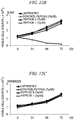
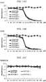
- the tumor cell proliferation-inhibiting peptide as one embodiment of the present invention exhibits a proliferation inhibitory activity with respect to the high LIX1L-expressing tumor cell, but does not exhibit proliferation inhibitory activity with respect to a low LIX1L-expressing tumor cell or a normal cell in which there is almost no expression of a LIX1L gene. That is, although the tumor cell proliferation-inhibiting peptide as one embodiment of the present invention does not exhibit cytotoxicity with respect to the normal cell, the tumor cell proliferation-inhibiting peptide has a wide spectrum of an anti-tumor effect. Therefore, the tumor cell proliferation-inhibiting peptide is extremely useful as an anti-tumor agent and it is possible to efficiently inhibit the proliferation of the high LIX1L-expressing tumor cell using the tumor cell proliferation-inhibiting peptide of the invention.
- a LIX1L gene is a structural gene which exists in a chromosome 1q21.1 region of a human, and is a gene which is almost not expressed in a normal cell, but of which expression is accelerated in many tumor cells.
- tumor used in the present specification means a malignant tumor (also referred to as cancer) that invades surrounding tissues in a cell population in which proliferation occurs which is not autonomously controlled by gene mutation, and which causes metastasis.
- tumor includes solid cancer and hematopoietic organ cancer including various kinds of sarcomas and carcinomas.
- examples of the solid cancer include a brain tumor, head and neck cancer, esophageal cancer, thyroid cancer, small cell lung cancer, non-small cell lung cancer, breast cancer, lung cancer, gastric cancer, gallbladder/bile duct cancer, liver cancer, hepatoma, pancreatic cancer, pancreatic cancer, colorectal cancer, colon cancer, rectal cancer, ovarian cancer, villus epithelial cancer, uterine cancer, cervical cancer, renal pelvis/ureter cancer, bladder cancer, prostate cancer, penile cancer, testicular cancer, embryonal cancer, virus tumor, skin cancer, malignant melanoma, neuroblastoma, osteosarcoma, Ewing's tumor, and soft tissue sarcoma.
- examples of the hematopoietic organ cancer include acute leukemia, chronic lymphocytic leukemia, chronic myelogenous leukemia, genuine hypervolemic illness, malignant lymphoma, multiple myeloma, Hodgkin's lymphoma, and non-Hodgkin's lymphoma.
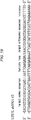
- the amino acid sequence of the hLIX1L protein in context of the invention which is encoded by a LIX1L (hLIX1L) gene is represented by SEQ ID No: 1 and a base sequence of cDNA of the LIX1L gene is represented by SEQ ID No: 2 (accession number of EMBL: NM_153713.1).
- the tumor cell is divided into a high LIX1L-expressing tumor cell in which a LIX1L gene is highly expressed and a low LIX1L-expressing tumor cell in which expression of a LIX1L gene is low (the expression level includes less than a detection limit value).
- the high LIX1L-expressing tumor cell is found in many tumors regardless of internal organs or the degree of differentiation and regardless of whether the cell is primary or metastatic. Particularly, even in all internal organs, the frequency of expression of a high LIX1L-expressing tumor cell is higher in undifferentiated tumor cells than in differentiated tumor cells.
- the inventors have examined patients with numerous solid tumors (gastric cancer, lung cancer, colorectal cancer, pancreatic cancer, prostate cancer, hepatoma, ovarian cancer, thyroid cancer, esophageal cancer, breast cancer, and renal cell cancer). As a result, about 20% to 60% of the tumors were LIX1L-positive tumors (refer to Reference Examples 1 and 2 to be described below).
- the term "high LIX1L-expressing tumor cell” means a tumor cell of which the expression level of a LIX1L gene is 8 or more times the expression level of a normal cell and the term "low-LIX1L expressing tumor cell” means a tumor cell other than the high LIX1L-expressing tumor cell. That is, whether a certain tumor cell is a high LIX1L-expressing tumor cell or a low LIX1L-expressing tumor cell can be determined by measuring the expression level of a LIX1L gene in this tumor cell.
- a normal cell derived from the same tissue as that of a tumor cell which is an object for determining whether or not the tumor cell is a high LIX1L-expressing tumor cell may be used, or a normal cell derived from a different tissue from that of the tumor cell may be used.
- a peripheral blood mononuclear cell collected and separated from a healthy person may be used.
- the expression levels of LIX1L genes may be compared with each other at the protein level. For example, it is possible to quantitatively detect the expression level of a LIX1L gene through a Western blotting method. Specifically, in a band which can be detected through the Western blotting method, by digitizing a signal of the band using densitometry, in a case where the signal detected from a tumor cell is 8 or more times the signal detected from a normal cell, it is possible to determine the signal as a "strong signal", and in a case where the signal detected from a tumor cell is less than 8 times the signal detected from a normal cell, it is possible to determine the signal as a "weak signal".
- a tumor cell of which the signal intensity which is obtained by measuring a signal obtained from the anti-LIX1L antibody that has been bonded to the LIX1L protein using densitometry, is 8 or more times the signal intensity similarly measured from a normal cell is a high LIX1L-expressing tumor cell, and to determine that a tumor cell of which the signal intensity thereof is less than 8 times the signal intensity similarly measured from the normal cell is a low LIX1L-expressing tumor cell.
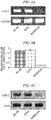
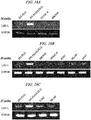
- the expression levels of LIX1L genes may be compared with each other at the mRNA level. For example, as will be shown in Reference Example 3 to be described below, it is possible to quantitatively detect the expression level of a LIX1L gene using a nucleic acid amplification reaction such as an RT-PCR method.
- PCR is performed in which the whole length or a part of cDNA of a LIX1L gene is amplified by having cDNA, synthesized through a reverse transcription reaction from RNA extracted from a tumor cell, as a mold, it is possible to determine that a tumor cell, of which the amount of the obtained amplification product is 8 or more times the amplification product similarly obtained from a normal cell, is a high LIX1L-expressing tumor cell and to determine that a tumor cell other than that is a low LIX1L-expressing tumor cell.
- the LIX1L gene is involved in cell proliferation since the effect of inhibiting the cell proliferation can be obtained through inhibiting the LIX1L gene expression; LIX1L protein plays an important role in proliferation of a cell in which the LIX1L gene is highly expressed; and the LIX1L gene functions as a cancer gene.
- the role of the LIX1L protein is small and the proliferation is accelerated by other components, and therefore, it is inferred that no effect of inhibiting the cell proliferation may be observed even if the expression of the LIX1L gene is inhibited.
- LIX1L gene The function of a LIX1L gene has not been described in the related art, and whether the LIX1L gene is a cancer gene which is a cause of tumorigenesis of a cell or a gene in which expression is accelerated due to the tumorigenesis has not become clear.
- RNA interference as defined in the specification can be performed by introducing small interfering RNA (siRNA), short hairpin RNA (shRNA), or micro RNA (miRNA) which has a double-stranded structure formed of a sense chain and an anti-sense chain in a partial region (RNAi target region) of cDNA of a LIX1L gene, into a high LIX1L-expressing tumor cell.
- siRNA small interfering RNA
- shRNA short hairpin RNA
- miRNA micro RNA
- siRNA or the like may be directly introduced into a cell, or an RNAi-induced vector which can produce siRNA or the like in a cell may be introduced into the cell.
- the production of siRNA, shRNA, and an RNAi-induced vector, the introduction of siRNA, shRNA, and an RNAi-induced vector, and the like into a cell can be performed through a usual method. For example, it is possible to synthesize siRNA and shRNA through chemosynthesis.
- the production of the RNAi-induced vector can be performed through, for example, incorporating the chemically synthesized siRNA or shRNA into a commercially available siRNA expression vector.
- Examples of the introduction of siRNA, shRNA, or RNAi-induced vector into a cell include introduction thereof using a transfection reagent (for example, Lipofectamine (registered trademark)); various viral vectors such as colloidal gold, retroviral vectors, lentiviral vectors, adenoviral vectors, adeno-associated virus vectors, and Sendai virus vectors; and nanoparticles prepared by combining various elements.
- a transfection reagent for example, Lipofectamine (registered trademark)
- various viral vectors such as colloidal gold, retroviral vectors, lentiviral vectors, adenoviral vectors, adeno-associated virus vectors, and Sendai virus vectors
- nanoparticles prepared by combining various elements for example, Lipofectamine (registered trademark)
- various viral vectors such as colloidal gold, retroviral vectors, lentiviral vectors, adenoviral vectors, adeno-associated virus vectors, and Sendai virus
- RNAi-induced vector can be produced by inserting a base sequence in an RNAi target region into base sequences of various commercially available RNAi vectors.
- RNA interference by, for example, making the RNAi target region in cDNA of the LIX1L gene a region consisting of a base sequence represented by SEQ ID No: 3 or 4.
- shRNA, siRNA, or miRNA which includes a base sequence represented by SEQ ID No: 3 or 4, or a vector which produces the shRNA, the siRNA, or the miRNA in a cell is introduced into a cell which is a high LIX1L-expressing tumor cell.
- Examples of shRNA including a base sequence represented by SEQ ID No: 3 include a double-stranded acid consisting of a base sequence represented by a base sequence 5, and examples of shRNA including a base sequence represented by a SEQ ID No: 4 include a double-stranded acid consisting of a base sequence represented by a base sequence 6.
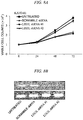
- examples of the method for inhibiting proliferation of a high LIX1L-expressing tumor cell include a method for inhibiting proliferation of a high LIX1L-expressing tumor cell which includes inhibiting the expression of a LIX1L gene through RNA interference and in which the inhibition of the expression of a LIX1L gene through RNA interference includes introduction of shRNA or siRNA which consists of a base sequence represented by SEQ ID No: 3 or 4, or a vector which produces the shRNA or the siRNA in a cell, into the cell.
- shRNA, siRNA, or miRNA which includes a base sequence represented by SEQ ID No: 3 or 4, or a vector which produces the shRNA, the siRNA, or the miRNA in a cell can be formulated through appropriate chemical modification or the like similarly to the tumor cell proliferation-inhibiting peptide as an embodiment of the present invention.
- the effect of inhibiting the proliferation of a high LIX1L-expressing tumor cell can be obtained through inhibiting the function of a LIX1L gene as well as through inhibiting the expression of a LIX1L gene.
- Examples of the method for inhibiting the function of a LIX1L gene include methods of excessively introducing an inactive mutant of a LIX1L protein, a partial protein or a peptide in which a partial region which is required for function or activation is deficient, into a cell.
- tumor cell proliferation-inhibiting peptide as one embodiment of the present invention
- the tumor cell proliferation-inhibiting peptide competes with an endogenous LIX1L protein within a cell which is a high LIX1L-expressing tumor cell. Therefore, it is possible to inhibit the cell proliferation of the high LIX1L-expressing tumor cell similarly to a case in which the expression of the LIX1L protein is inhibited.
- Examples of the tumor cell proliferation-inhibiting peptide as one aspect of the present specification include a peptide including an amino acid sequence (hereinafter, in some cases, simply referred to as a "phosphorylation region-partial sequence") which is homologous to a partial region containing a tyrosine residue, a serine residue, or a threonine residue which is phosphorylated through transmission of a specific signal in a LIX1L protein.
- phosphorylation region-partial sequence an amino acid sequence
- the phosphorylation of a protein plays an important role in signal transmission of the cell proliferation or the like, and the LIX1L protein is also involved in the cell proliferation signal through the phosphorylation of the tyrosine residue or the like.
- tumor cell proliferation-inhibiting peptide in the present specification and the claims means a peptide of which the C-terminal is amidated. That is, the tumor cell proliferation-inhibiting peptide has become -COONH 2 due to a carboxyl group at the C-terminal being amidated.
- Examples of the tumor cell proliferation-inhibiting peptide as one aspect of the present specification include a polypeptide (hereinafter, referred to as a "Y136-containing peptide") containing the 136th tyrosine (Y136) from the N-terminal of a LIX1L protein. It is possible to inhibit the cell proliferation of a high LIX1L-expressing tumor cell by introducing the Y136-containing peptide into a cell.
- Specific examples of the Y136-containing peptide include a polypeptide including a partial sequence (Y136-containing partial sequence), which is a partial sequence of an amino acid sequence represented by SEQ ID No: 1, as a phosphorylation region-partial sequence.
- the tumor cell proliferation-inhibiting peptide as one embodiment of the present invention can be synthesized through, for example, chemical synthesis.
- the position of Y136 in a Y136-containing partial sequence is not particularly limited and may be in the vicinity of the N-terminal or the C-terminal of the Y136-containing partial sequence. However, the position thereof is preferably in the vicinity of the center of the Y136-containing partial sequence.
- the length (number of amino acids) of the Y136-containing partial sequence of the specification may be a length to the extent that the polypeptide consisting of this Y136-containing partial sequence can be distinguished from other polypeptides, and the length thereof is greater than or equal to 8 amino acids, more preferably greater than or equal to 10 amino acids, and still more preferably greater than or equal to 11 amino acids.
- the length of the Y136-containing partial sequence of the specification is less than or equal to 35 amino acids and more preferably less than or equal to 30 amino acids.
- the length of the Y136-containing partial sequence of the invention is 8 amino acids to 35 amino acids, more preferably 10 amino acids to 35 amino acids, and still more preferably 11 amino acids to 30 amino acids.
- the "vicinity of the N-terminal” means a position within 5 amino acids from the N-terminal and the "vicinity of the C-terminal” means a position within 5 amino acids from the C-terminal.
- the "vicinity of the center” means a position within 10 amino acids from the position of the center of the Y136-containing partial sequence.
- a partial amino acid sequence is preferable containing 8 amino acids having the 136th tyrosine starting from the 130th proline from the N-terminal, and a partial amino acid sequence is more preferable containing 11 amino acids having the 136th tyrosine starting from the 130th proline from the N-terminal.
- the tumor cell proliferation-inhibiting peptide as one aspect of the present specification may be a polypeptide consisting of only a phosphorylation region-partial sequence such as the Y136-containing partial sequence, but more preferably has a site having cell membrane permeability.
- the tumor cell proliferation-inhibiting peptide as one embodiment of the present invention having a site having cell membrane permeability, it is possible to more efficiently introduce the tumor cell proliferation-inhibiting peptide into a cell which is a high LIX1L-expressing tumor cell.
- the site having cell membrane permeability may be a site consisting of a polypeptide or a site containing components other than the polypeptide, for example, sugars, nucleic acids, or low-molecular compound.
- this site may be a polypeptide consisting of only a natural type amino acid or a polypeptide containing a modified amino acid.
- Preferred examples of the site having cell membrane permeability consisting of a polypeptide include membrane-permeable peptides such as a Tat peptide derived from a Trans-Activator of Transcription Protein (TAT) of HIV-1; oligoarginine; Penetratin derived from an Antennapedia protein of drosophila; and transportan, which is a chimeric peptide of a neuropeptide galanin and bee venom mastoparan.
- TAT Trans-Activator of Transcription Protein
- the tumor cell proliferation-inhibiting peptide as one aspect of the present specification has a site, such as a membrane-permeable peptide which has cell membrane permeability
- the polypeptide which consists of a phosphorylation region-partial sequence and the site which has cell membrane permeability may be directly bonded to each other, or may be indirectly bonded to each other through various linkers.
- the polypeptide consisting of a phosphorylation region-partial sequence such as a Y136-containing partial sequence may be on an N-terminal side or the site having cell membrane permeability may be on the N-terminal side.
- the linkers are not particularly limited as long as the linkers can connect polypeptides together or a polypeptide to another compound. However, a comparatively smaller molecule is preferable in view of cell membrane permeability, a risk of antigenicity, or the like.
- a linker formed of 1 to 5 amino acids (which may contain a modified amino acid such as ⁇ -aminocaproic acid) is preferable as the linker. 1 amino acid of ⁇ -aminocaproic acid is more preferable as the linker.
- the tumor cell proliferation-inhibiting peptide as one embodiment of the present invention may have another functional site other than or in addition to the site having cell membrane permeability.
- the other functional site is not particularly limited as long as the site does not impair the function of inhibiting the proliferation using a polypeptide consisting of a phosphorylation region-partial sequence.
- Examples of the other functional site include a tag peptide, such as a His tag, a Myc tag, a tag peptide, and a Flag tag, which is widely used in refinement of a protein.
- the tumor cell proliferation-inhibiting peptide itself may be introduced into a cell, or a vector which can produce the tumor cell proliferation-inhibiting peptide in a cell may be introduced into a high LIX1L-expressing tumor cell.
- the tumor cell proliferation-inhibiting peptide according to the present invention or the vector for producing the tumor cell proliferation-inhibiting peptide can be introduced into a high LIX1L-expressing tumor cell through a usual method.
- Examples of the method for introducing a peptide into a high LIX1L-expressing tumor cell include a transfection reagent (for example, Lipofectamine (registered trademark)); various viral vectors such as colloidal gold, retroviral vectors, lentiviral vectors, adenoviral vectors, adeno-associated virus vectors, and Sendai virus vectors; and nanoparticles prepared by combining various elements.
- a transfection reagent for example, Lipofectamine (registered trademark)
- various viral vectors such as colloidal gold, retroviral vectors, lentiviral vectors, adenoviral vectors, adeno-associated virus vectors, and Sendai virus vectors
- nanoparticles prepared by combining various elements particularly when the tumor cell proliferation-inhibiting peptide according to the present invention has a site having cell membrane permeability, it is possible to efficiently introduce the polypeptide into a cell by simply bringing the polypeptide into contact with a high LIX1L-
- examples of the method for inhibiting proliferation of a high LIX1L-expressing tumor cell include a method for inhibiting proliferation of a high LIX1L-expressing tumor cell which includes introduction of a tumor cell proliferation-inhibiting peptide tumor cell proliferation-inhibiting peptide into a high LIX1L-expressing tumor cell.
- the tumor cell proliferation-inhibiting peptide includes a polypeptide consisting of a partial sequence with 8 amino acids to 35 amino acids which is an amino acid sequence represented by SEQ ID No: 1 and contains the 136th tyrosine, and a site which consists of an amino acid sequence represented by SEQ ID No: 7 and has cell membrane permeability, and the polypeptide and the site having cell membrane permeability are directly or indirectly bonded to each other.
- the tumor cell proliferation-inhibiting peptide according to the present invention has an effect of inhibiting the proliferation of a high LIX1L-expressing tumor cell, and therefore, is useful as a pharmaceutical composition including an anti-tumor agent. Since the tumor cell proliferation-inhibiting peptide according to the present invention is a smaller molecule compared to a glycoprotein, which is a macromolecule, the tumor cell proliferation-inhibiting peptide has advantages in that an antibody is hardly produced when the tumor cell proliferation-inhibiting peptide is administered into a human body; it is possible to chemically massively produce the tumor cell proliferation-inhibiting peptide due to its simple structure; and it is easy to manage the quality of the tumor cell proliferation-inhibiting peptide in a production process.
- the tumor cell proliferation-inhibiting peptide according to the present invention has a wide spectrum of an anti-tumor effect and cytotoxicity to a normal cell is extremely low. Therefore, the tumor cell proliferation-inhibiting peptide can become an excellent anti-tumor agent.
- the tumor cell proliferation-inhibiting peptide according to the present specification is a new peptide compound having a LIX1L protein, which is specifically expressed in a tumor cell, as a direct target, in many tumor cells (for example, a brain tumor, head and neck cancer, esophageal cancer, thyroid cancer, small cell lung cancer, non-small cell lung cancer, breast cancer, lung cancer, gastric cancer, gallbladder/bile duct cancer, liver cancer, hepatoma, pancreatic cancer, pancreatic cancer, colorectal cancer, colon cancer, rectal cancer, ovarian cancer, villus epithelial cancer, uterine cancer, cervical cancer, renal pelvis/ureter cancer, bladder cancer, prostate cancer, penile cancer, testicular cancer, embryonal cancer, a virus tumor, skin cancer, malignant melanoma, neuroblastoma, osteosarcoma, Ewing's tumor, and soft tissue sarcoma, acute leukemia, chronic lymphocytic leukemia, chronic tumor cells
- the pharmaceutical composition or the anti-tumor agent according to the present invention may contain a pharmaceutically acceptable carrier or diluent.
- the "pharmaceutically acceptable carrier or diluent” means an excipient (for example, fat, beeswax, polyols of semi-solid and a liquid, or natural or hardened oil); water (for example, distilled water, in particular, distilled water for injection), physiological saline, alcohol (for example, ethanol), glycerol, a polyol, an aqueous glucose solution, mannitol, vegetable oil, and the like; additives (for example, an extending agent, a disintegrator, a bonding agent, a lubricant, a wetting agent, a stabilizer, an emulsifier, a dispersant, a preservative, a sweetener, a colorant, a flavoring or a fragrance, a thickening agent, a diluent, a buffer substance, a solvent or a solubilizing agent, a drug for achieving a storage effect, a salt for changing osmotic pressure,
- agents relating to the pharmaceutical composition or the anti-tumor agent of the present invention can be selected, and examples thereof include oral formulations such as a tablet, a capsule, a powdered agent, a granule, or a liquid; sterilized liquid-like parenteral formulations such as a solution or a suspension; a suppository; and an ointment.
- Examples of a solvent or a diluent which is appropriate when being parentally administered in a form of intramuscular injection, intravenous injection, or subcutaneous injection include distilled water for injection, a lidocaine hydrochloride aqueous solution (for intramuscular injection), physiological saline, an aqueous glucose solution, ethanol, polyethylene glycol, propylene glycol, a liquid for intravenous injection (for example, an aqueous solution such as citric acid and sodium citrate), or an electrolyte solution (for intravenous drip infusion and intravenous injection), or a mixed solution thereof.
- injections can take a form in which injections having been dissolved in advance, or injections in a state of powder or injections to which an appropriate carrier (additive) has been added are dissolved when in use.
- These injection solutions can contain, for example, an active ingredient of 10% by weight to 90% by weight based on the weight of the entire agent.
- agents can be easily produced by those skilled in the art in accordance with a usual method or a conventional technique.
- an agent containing the tumor cell proliferation-inhibiting peptide of the present invention is an injection
- the tumor cell proliferation-inhibiting peptide of the present invention can be used in combination with other agents which are useful for treating various kinds of cancer, or in combination with radiation therapy.
- the present specification discloses a therapy for a tumor (that is, cancer) which includes administering a therapeutically effective amount of a tumor cell proliferation-inhibiting peptide into a target into which administration thereof is required.
- the "target into which administration thereof is required” in the present specification means a patient who has a tumor cell highly expressing LIX1L.
- treatment of a tumor means inhibition of proliferation of a tumor cell through administering an agent containing the tumor cell proliferation-inhibiting peptide of the present invention into a patient for whom the administration is required.
- an agent containing the tumor cell proliferation-inhibiting peptide of the present invention into a patient for whom the administration is required.
- it is possible to retreat a tumor cell that is, to reduce the size of a tumor which can be measured. More preferably, in the treatment, the tumor cell is completely eliminated.
- treatment in the present specification also includes preventive administration.
- the unit of preferred treatment may be changed depending on the form of administering a tumor cell proliferation-inhibiting peptide of the present invention, the type of a tumor cell proliferation-inhibiting peptide of the present invention to be used, and the dosage form of a tumor cell proliferation-inhibiting peptide of the present invention to be used; the type of, the administration form of, and the dosage form of other anti-cancer agents to be used in combination therewith; the condition of a tumor cell (cancer cell) to be treated and the condition of a patient; and the like.
- Those skilled in the art can determine optimum treatment under predetermined conditions based on a conventional treatment determination unit and/or in consideration of the present specification.
- the treatment unit of the tumor cell proliferation-inhibiting peptide of the present invention can change depending on the type of tumor cell proliferation-inhibiting peptide to be used, the type of composition blended, the application frequency, a specific site to be treated, the severity of illness, the age of a patient, the diagnosis by a doctor, the type of tumor (cancer), and the like.
- the dosage per person per day for adults can be set to be within a range of, for example, 10 mg/kg to 200 mg/kg per day, in a case of parenteral administration, preferably in a case of intravenous administration, and still more preferably in a case of intravenous drip infusion.
- administration may be continuously performed for, for example, 1 hour to 48 hours.
- the number of times of administration varies depending on the administration method and the symptoms, but examples thereof include once per day to 5 times per day.
- an intermittent administration method such as alternate day administration or administration once every three days.
- the repose period of treatment in the case of parenteral administration is, for example, 0 weeks to 4 weeks.
- the tumor cell proliferation-inhibiting peptide according to the present invention can specifically inhibit the function of a LIX1L protein in a cell, and therefore, is useful as a tool for transmitting a signal in a cell, and particularly for clarifying the function of a LIX1L gene in a cell proliferation signal.
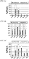
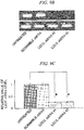
- the expression levels of LIX1L genes of gastric cancer clinical specimens collected from patients with gastric cancer were quantitatively detected through immunohistochemical staining for comparison. These gastric cancer clinical specimens were classified into papillary adenocarcinoma (Pap), tubular adenocarcinoma (Tub), poorly differentiated adenocarcinoma (Por), signet ring cell carcinoma (Sig), mucinous carcinoma (Muc), and others.
- the tubular adenocarcinoma was further classified into a well-differentiated type (Tub1) and a moderately-differentiated type (Tub2), and the poorly differentiated adenocarcinoma was further classified into a solid type (Por1) and a nonsolid type (Por2).
- the papillary adenocarcinoma and the tubular adenocarcinoma were differentiated tumor cells and the others were undifferentiated tumor cells.
- a normal cell and a tumor cell coexist in one tumor tissue. Therefore, a specimen of which the proportion of a high LIX1L-expressing tumor cell included in each of the gastric cancer clinical specimens was high was determined as a LIX1L-positive tumor and a specimen of which the proportion of a high LIX1L-expressing tumor cell included in each of the gastric cancer clinical specimens was low was determined as a LIX1L-negative tumor.
- a cell in each of the gastric cancer clinical specimens was subjected to immunohistochemical staining using an anti-LIX1L rabbit polyclonal antibody (manufactured by Abnova Corporation) as a primary antibody and a peroxidase-labeled goat anti-rabbit IgG polyclonal antibody (product name: Histofine Simple Stain MAX-PO (MULTI), manufactured by Nichirei Corporation) as a secondary antibody.
- an anti-LIX1L rabbit polyclonal antibody manufactured by Abnova Corporation
- a peroxidase-labeled goat anti-rabbit IgG polyclonal antibody product name: Histofine Simple Stain MAX-PO (MULTI), manufactured by Nichirei Corporation
- Three identical enlarged visual fields randomly selected in the gastric cancer clinical specimens were microscopically examined, a signal detected from a tumor site and a signal detected from a cell in an adjacent normal tissue were compared with each other; a cell of the tumor site in which a stronger signal than that detected from the cell in the adjacent normal tissue was set as a LIX1L-positive tumor cell and a cell in which no signal was detected was set as a LIX1L-negative cell; and the number of LIX1L-positive tumor cells and the number of LIX1L-negative tumor cells were measured.
- each of the gastric cancer clinical specimens were determined such that a gastric cancer clinical specimen of which the proportion of the LIX1L-positive cells in the tumor cells was 30% to 100% was determined as a LIX1L-positive specimen (+), a gastric cancer clinical specimen of which the proportion of the LIX1L-positive cells in the tumor cells was 10% to 30% was determined as a LIX1L weakly positive (+/-), and a gastric cancer clinical specimen of which the proportion of the LIX1L-positive cells in the tumor cells was 0% to 10% was determined as a LIX1L-negative (-).
- FIGS. 1A to IF The number of the LIX1L-positive specimens and the LIX1L-negative specimens and the proportions thereof obtained from the results of the immunohistochemical staining are shown in FIGS. 1A to IF.
- FIG. 1A shows the number of LIX1L-positive specimens and LIX1L-negative specimens for each papillary adenocarcinoma, tubular adenocarcinoma, poorly differentiated adenocarcinoma, signet ring cell carcinoma, mucinous carcinoma, and other kinds of cancer ("other" in the drawing)
- FIG. 1B shows results in which the frequency of LIX1L-positive tumors in various kinds of gastric cancer is calculated from the results in FIG. 1A.
- FIG. 1C shows the number of LIX1L-positive specimens and LIX1L-negative specimens for each papillary adenocarcinoma, well-differentiated type tubular adenocarcinoma, moderately-differentiated type tubular adenocarcinoma, solid type poorly differentiated adenocarcinoma, nonsolid type differentiated adenocarcinoma, and signet ring cell carcinoma
- FIG. 1D shows results in which the frequency of LIX1L-positive tumors in various kinds of gastric cancer is calculated from the results in FIG. 1C .
- FIG. 1D shows results in which the frequency of LIX1L-positive tumors in various kinds of gastric cancer is calculated from the results in FIG. 1C .
- FIG. 1E shows the number of LIX1L-positive specimens and LIX1L-negative specimens for each differentiated tumor ("Differen” in the drawing) and undifferentiated tumor ("Undiffer” in the drawing) in all of the gastric cancer clinical specimens
- FIG. IF shows results in which the frequency of LIX1L-positive tumors in the differentiated tumor and the undifferentiated tumor is calculated from the results in FIG. 1E .
- the result was that LIX1L-positive tumors were detected at a frequency of at least 40% or more in all kinds of gastric cancer.
- immunohistochemical staining was performed on clinical specimens collected from patients with solid tumors (lung cancer, colorectal cancer, pancreatic cancer, prostate cancer, hepatoma, ovarian cancer, thyroid cancer, esophageal cancer, breast cancer, and renal cell cancer) other than gastric cancer, and the number of LIX1L-positive specimens and LIX1L-negative specimens in various kinds of cancer and the proportion thereof were obtained.
- solid tumors lung cancer, colorectal cancer, pancreatic cancer, prostate cancer, hepatoma, ovarian cancer, thyroid cancer, esophageal cancer, breast cancer, and renal cell cancer
- FIG. 2A The number of LIX1L-positive specimens and LIX1L-negative specimens in various kinds of cancer obtained from the results of the immunohistochemical staining is shown in FIG. 2A , and results in which the frequency of LIX1L-positive tumors in various kinds of cancer is calculated from the results thereof is shown in FIG. 2B .
- FIG. 2B results in which the frequency of LIX1L-positive tumors in various kinds of cancer is calculated from the results thereof.
- FIG. 2B results in which the frequency of LIX1L-positive tumors in various kinds of cancer is calculated from the results thereof.
- FIG. 2B results in which the frequency of LIX1L-positive tumors in various kinds of cancer is calculated from the results thereof is shown in FIG. 2B .
- about 20% to 60% of the tumors were LIX1L-positive tumors.
- LIX1L genes were quantitatively measured from both a protein level and an mRNA level through a Western blotting method and an RT-PCR method for comparison.
- KATO-III strains Two kinds of strains including KATO-III strains and OCUM-1 strains were used as gastric cancer cell strains. Both cell strains are undifferentiated tumors.
- Each of the KATO-III strains was analyzed after being cultured for 10 days to 14 days in a culture solution in which 10 vol.% of fetal calf serum (FCS) with respect to an RPMI 1640 culture medium was added to the RPMI 1640 culture medium.
- FCS fetal calf serum
- OCUM-1 strains was analyzed after being cultured for 10 days to 14 days in a culture solution in which 10 vol.% of FCS with respect to a DMEM culture medium was added to the DMEM culture medium.
- FCS fetal calf serum
- OCUM-1 strains was analyzed after being cultured for 10 days to 14 days in a culture solution in which 10 vol.% of FCS with respect to a DMEM culture medium was added to the DMEM culture medium.
- peripheral blood mononuclear cell normal peripheral blood mononuclear cell
- the normal peripheral blood mononuclear cell was isolated as follows. First, 5 mL of phosphate-buffered saline (PBS) was added to 5 mL of a whole blood sample for dilution.
- PBS phosphate-buffered saline
- the total amount of the diluent was carefully stacked on 20 mL of Ficoll (registered trademark) (manufactured by GE Healthcare Ltd.) fractionated into a centrifuge tube in advance, and centrifugal separation treatment (under the conditions of maximum acceleration/natural deceleration (no brake)) was performed for 20 minutes at 22°C and 2200 rpm.
- Ficoll registered trademark
- the peripheral blood mononuclear cell (hereinafter, in some cases, referred to as PBMC) layer was carefully taken out of the centrifuge tube so as not to disrupt the separated layer and was collected into a new tube with a 15 mL capacity.
- PBS peripheral blood mononuclear cell
- centrifugal separation treatment under the conditions of maximum acceleration/maximum deceleration
- a washing operation for removing a supernatant was performed twice.
- the washed resultant obtained by adding an appropriate amount of PBS to the PBMC layer for suspension was set as a normal peripheral blood mononuclear cell specimen.
- the normal peripheral blood mononuclear cell specimen was used in an experiment after the number of cells was counted using a hemocytometer and the number of viable cells was examined through Trypan blue staining.
- a reverse transcription reaction was performed using First Strand cDNA Synthesis Kit (manufactured by Roche Life Science) for RT-PCR (AMV), for synthesizing cDNA, in 20 ⁇ L of a reaction solution containing 1 ⁇ g of total RNA and a random primer for 10 minutes at 25°C, subsequently for 45 minutes at 42°C, and then for 2 minutes at 95°C, to deactivate reverse transcriptase.
- PCR was performed using a DNA thermal cycler (model PTC 200, manufactured by MJ Research, Inc.) in 20 ⁇ L of a reaction solution using a primer set consisting of a forward primer consisting of a base sequence represented by SEQ ID No: 8 and a base sequence represented by SEQ ID No: 9, having the synthesized cDNA as a mold.
- This primer set is a primer set designed to amplify fragments of cDNA of LIX1L genes of 218 bp.
- PCR was performed in the same manner except that a primer set consisting of a forward primer consisting of a base sequence represented by SEQ ID No: 10 and a base sequence represented by SEQ ID No: 11 and is designed to amplify fragments of cDNA of GAPDH genes of 253 bp was used instead of the primer set as a control. 28 cycles were performed under the reaction conditions of 30 seconds at 96°C, 30 seconds at 56°C, and 30 seconds at 72°C being one cycle. Amplified products obtained by amplifying both the LIX1L and the GAPDH were applied to 1.5% agarose gel to perform electrophoresis, and a separated nucleic acid was subjected to ethidium bromide staining.
- the signal intensity of a band of LIX1L in an ethidium bromide staining image was measured using densitometry and a relative value of the signal intensity was calculated.
- SYBR registered trademark
- Green I was used in all quantitative PCR reactions. Quantitative determination was performed in 25 ⁇ L of a reaction solution containing the synthesized cDNA and a LIX1L primer using an ABI PRISM 7700 Sequence detector (manufactured by Perkin-Elmer/Applied Biosystems). The expression of GAPDH was also confirmed as an internal standard. 45 cycles were performed under the reaction conditions of 5 seconds at 95°C and 20 seconds at 60°C being one cycle. As a result, regarding the relative value of the signal intensity, the normal peripheral blood mononuclear cell specimen was 0.1, the KATO-III strain was 0.9 ⁇ 0.18, and the OCUM-1 strain was 1.5 ⁇ 0.27.
- the transfer film was treated for 2 hours at room temperature using an anti-LIX1L rabbit polyclonal antibody (manufactured by Abnova Corporation) as a primary antibody and was treated for 1 hour at room temperature using a peroxidase-labeled goat anti-rabbit IgG polyclonal antibody (product name: Goat anti-Rabbit IgG-HRP, Invitrogen) as a secondary antibody, and Western blotting was performed.
- an anti-LIX1L rabbit polyclonal antibody manufactured by Abnova Corporation
- a peroxidase-labeled goat anti-rabbit IgG polyclonal antibody product name: Goat anti-Rabbit IgG-HRP, Invitrogen
- Western blotting was further performed on this transfer film using an anti-Actin mouse monoclonal antibody (product name: C-4, manufactured by ICN Biomedicals) as a primary antibody and a peroxidase-labeled sheep anti-mouse IgG monoclonal antibody (product name: NXA931, manufactured by Amersham) as a secondary antibody, as controls. Visualization of the band was performed using an ECL chemiluminescent reagent (manufactured by Amersham).
- an anti-Actin mouse monoclonal antibody product name: C-4, manufactured by ICN Biomedicals
- a peroxidase-labeled sheep anti-mouse IgG monoclonal antibody product name: NXA931, manufactured by Amersham
- FIG. 3A shows staining images in which electrophoresis is performed on products amplified through RT-PCR in each cell strain and a separated nucleic acid is stained using ethidium bromide.
- FIG. 3B shows relative values of the amount of mRNA of LIX1L obtained from the signal intensity of a band of LIX1L in the staining images in FIG. 3A of each of the cell strains (the amount of mRNA of LIX1L of a KATO-III strain was set to 1).
- FIG. 3C shows bands of LIX1L and Actin detected through Western blotting in each of the cell strains.
- the expression levels of LIX1L genes of hematopoietic organ tumor cell strains were quantitatively measured from both a protein level and an mRNA level through a Western blotting method and an RT-PCR method for comparison.
- the Western blotting method and the RT-PCR method were preformed in the same manner as those in Reference Example 3 except that the hematopoietic organ tumor cell strains were used instead of the gastric cancer cell strains.
- an acute myelogenous leukemia cell strain HL-60 strain, a chronic myelogenous leukemia cell strain K562 strain, and a multiple myeloma cell strain RPMI8226 strain were used.
- the HL-60 strain and the K562 strain are undifferentiated tumors and the RPMI8226 strain is a differentiated tumor. All of the cell strains were respectively analyzed after being cultured for 10 days in a culture solution in which 10 vol.% of fetal calf serum (FCS) with respect to an RPMI 1640 culture medium was added to the RPMI 1640 culture medium.
- FCS fetal calf serum
- FIG. 4A shows staining images in which electrophoresis is performed on products amplified through RT-PCR in each cell strain and a separated nucleic acid is stained using ethidium bromide.
- FIG. 4B shows relative values of the amount of mRNA of LIX1L obtained from the signal intensity of a band of LIX1L in the staining images in FIG. 4A of each of the cell strains (the signal intensity of a band of GAPDH was set to 1).
- FIG. 4C shows bands of LIX1L and Actin detected through Western blotting in each of the cell strains.
- the myeloma cell strain RPMI8226 cell was 0.1
- the HL-60 strain was 1.7 ⁇ 0.21
- the K562 strain was 1.8 ⁇ 0.35
- the RPMI8226 strain was 0.2 ⁇ 0.03.
- the LIX1L shRNA #1 and LIX1L shRNA #2 can be produced through chemical synthesis.
- cells plated such that the density thereof became 1x10 4 cells/3.5 cm well for one plate (6 wells/plate) for each cell strain were cultured for one night in a culture solution having the same composition as those of the culture solutions described in the reference examples. After culturing of 1 well out of the 6 wells was completed, Trypan blue staining was performed on the 1 well, and viable cell counts were counted. The remaining 5 wells were cultured after exchanging the culture solution with a liquid mixture of a Lipofectamine (registered trademark) reagent (12.5 ⁇ L/well) and a culture solution (2 mL/well) of which the shRNA concentration became 2.5 ⁇ g/well.
- a Lipofectamine registered trademark
- RT-PCR was performed on the collected cells in the same manner as that in the Reference Example 3 and fragments of cDNA of LIX1L genes and fragments of cDNA of GAPDH genes were amplified. Agarose gel electrophoresis was performed on the obtained amplified products and an ethidium bromide staining image of a separated nucleic acid was obtained. As a control, RT-PCR was performed by performing culturing similarly to the above except that a culture medium was exchanged with a culture solution to which no shRNA was added, and RT-PCR was performed to obtain an ethidium bromide staining image (untreated).
- FIGS. 6A to 9C show measurement results of cell strains.
- FIGS. 6A , 7A , 8A , and 9A show measurement results of the viable cell counts.
- FIGS. 6B , 7B , 8B , and 9B show the staining images of ethidium bromide staining of fragments of LIX1L and fragments of GAPDH amplified through RT-PCR after the culturing for 72 hours.
- FIGS. 6C , 7C , 8C , and 9C show relative values (the amount of mRNA of untreated LIX1L is set to 1) of the amount of mRNA of LIX1L obtained from the signal intensities of bands of LIX1L in the staining images of FIGS.
- peptides are peptides (peptide 1: SEQ ID No: 12, peptide 2: SEQ ID No: 13) in which the C-terminal of a peptide consisting of a Tat sequence represented by SEQ ID No: 7 and the N-terminal of a partial amino acid sequence of a LIX1L protein including the tyrosine residues are connected to each other using ⁇ -aminocaproic acid.
- a peptide in which ⁇ -aminocaproic acid is bonded to the C-terminal of a peptide consisting of a Tat sequence was also chemically synthesized (control peptide: SEQ ID No: 14). The amino acid sequences of these peptides are shown in FIG. 10 together with the partial amino acid sequence of a LIX1L protein.
- control peptide, the peptide 1, or the peptide 2 was introduced into an HL-60 strain, a K562 strain, an RPMI8226 strain, a KATO-III strain, and an OCUM-1 strain, and the influence on cell proliferation was examined.
- the evaluation of the cell proliferation was performed through an MTT method in which a reduction reaction in mitochondria is used.
- cells plated in a 96-well microplate such that the density thereof became 5x10 3 cells/well were cultured for one night in a culture solution having the same composition as those in the reference examples. Thereafter, the culture solution was exchanged with 100 ⁇ L of a culture solution of which the concentration of peptides is 0 ⁇ M to 100 ⁇ M for culturing for 72 hours. After the culturing, an MTT-containing PBS solution was added to the culture solution, and the mixture was incubated for 4 hours. Then, formazan which was generated by putting 100 ⁇ L of isopropanol (containing 34 (v/v)% of hydrochloric acid) into each of the wells was dissolved.
- the absorbance of each of the wells of the microplate at 570 nm was measured using a microplate reader. As controls, culturing was performed in the same manner except that no peptide was added to the strains, an MTT assay was performed thereon, and the absorbance at 570 nm was measured (untreated).
- the measurement results of the absorbance of each of the cell strains at 570 nm are shown in FIGS. 11A to 11E .
- the results were that, in all of the cell strains, the absorbance at 570 nm in the case of introducing the control peptide into the strains did not particularly change similarly to the untreated case in which no peptide was introduced into the strains, even if the concentration of peptides was increased up to 100 ⁇ M, and it was confirmed that there was no influence on the cell proliferation even if the control peptide was introduced into the strains.
- the effect of inhibiting the cell proliferation due to peptide 1 in the HL-60 strain, the K562 strain, and the OCUM-1 strain in which the expression levels of LIX1L genes were comparatively high was higher than that in the KATO-III strain. Accordingly, it was found that the proliferation inhibition effect of the tumor cell proliferation-inhibiting peptide, such as a Y136-containing peptide, in context of the present invention varied depending on the expression levels of LIX1L genes and it was inferred that the proliferation inhibition effect in the high LIX1L-expressing cells was more effective than that in the low LIX1L-expressing cells.
- the tumor cell proliferation-inhibiting peptide such as a Y136-containing peptide
- control peptide, the peptide 1, or the peptide 2 was introduced into an HL-60 strain, a K562 strain, and an RPMI8226 strain, and the influence on cell proliferation was examined.
- the evaluation of the cell proliferation was performed through measuring the viable cell counts similarly to Example 1.
- the viable cell counts were measured after culturing the cells similarly to Example 1 except that a 5 ⁇ M culture solution containing a control peptide, a peptide 1, or a peptide 2 was used instead of the culture solution (2 mL/well) of which the shRNA concentration was 2.5 ⁇ g/well.
- the viable cell counts were measured (untreated) by performing the culturing similarly to Example 1 using a culture solution containing no peptide 2, as a control.
- the measurement results of the viable cell counts of the cell strains are shown in FIGS. 12A to 12C .
- the results were that, in all of the cell strains, in a case where the control peptide was introduced into the cell strains and in a case where the peptide 2 was introduced into the strains, the viable cell counts were the same as those in an untreated case where no peptide was introduced into the cell strains, even at the point of time after the lapse of 72 hours.
- the viable cell counts decreased in the HL-60 strain and the K562 strain more than that in the untreated case already at the point of time after the culturing for 24 hours, and the viable cell counts were very small after the culturing for 72 hours.
- the viable cell counts therein were the same as those in the untreated case. Even from these results, it can be seen that the cell proliferation is inhibited by introducing the tumor cell proliferation-inhibiting peptide as defined herein above including the peptide 1 into a high LIX1L-expressing cell.
- the Y136-containing partial sequence consists of 11 amino acids. Therefore, a peptide 3 (SEQ ID No: 15) in which the Y136-containing partial sequence consists of 20 amino acids and a peptide 4 (SEQ ID No: 16) in which the Y136-containing partial sequence consists of 30 amino acids were produced through chemical synthesis for comparing the effects of inhibiting the cell proliferation of the peptides 3 and 4 with that of the peptide 1.
- An amino acid sequence of a LIX1L protein is shown in FIG. 13A and amino acid sequences of the peptides 1, 3, and 4, and the control peptide are shown in FIG. 13B .
- the Y136-containing partial sequences of the peptides 1, 3, and 4 are shown in FIG. 13A .
- the U937 strain was a high LIX1L-expressing tumor cell in which LIX1L expression equivalent to that in the K562 strain was confirmed.
- the cells were cultured for 72 hours similarly to Example 2 except that a peptide to be added to a cell strain was set to peptide 1, 3, or 4, or a control peptide, an MTT assay was performed thereon, and the absorbance at 570 nm was measured.
- FIGS. 14A to 14C The measurement results of the absorbance of each of the cell strains are shown in FIGS. 14A to 14C .
- the results were that the cell proliferation was inhibited by introducing the peptide 1, 3, or 4 into the U937 strain and the K562 strain. All of the cell proliferation inhibition effects of the peptides 1, 3, and 4 in the U937 strain and the K562 strain were the same as each other.
- the cell proliferation was not inhibited similarly to the control peptide even if the peptide 1, 3, or 4 was introduced thereinto.
- a LIX1L protein with a FLAG tag was forcibly expressed in a HEK293 strain, and intracellular localization of the LIX1L protein, and the presence and absence of phosphorylation of serine, threonine, and tyrosine were examined.
- the stable expression strains were prepared using a Flp-in (registered) trademark system (manufactured by Invitrogen).
- a FLG/LIX1L fusion gene was inserted into a BamHI/XhoI site of plasmid pcDNA5/FRT (V6010-20; manufactured by Invitrogen) using plasmid pcDNA5/FRT as an expression vector, to prepare a FLG/LIX1L expression vector (pcDNA5/FLG/LIX1L/FRT).
- a 293 FLG/LIX1L cell and a 293 FLG cell were respectively cultured in 10% FCS-containing DMEM culture media to which Hygromycin B at 100 mg/mL was added, for 10 days to 14 days. Then, the cultured cells were separated into nuclear fractions and cytoplasm fractions using Nuclear/Cytosol Fractionation Kit (manufactured by BioVison, Inc.). Immunoprecipitation was performed on both the obtained fractions using an anti-FLAG antibody (manufactured by Sigma-Aldorich Co. LLC.) and an agarose-binding protein A (manufactured by Sigma-Aldorich Co.
- LIX1L of the 293 FLG and 293 FLG/LIX1L prepared in Reference Example 5 The expression levels of LIX1L of the 293 FLG and 293 FLG/LIX1L prepared in Reference Example 5, a NUGC-4 strain (gastric cancer cell strain), an MKN45 strain (gastric cancer cell strain), a K562 strain (chronic myelogenous leukemia cell strain), an HL-60 strain (acute myelogenous leukemia cell strain), and a U937 strain (acute myelogenous leukemia cell strain) were examined through PCR.
- NUGC-4 strain gastric cancer cell strain
- MKN45 strain gastric cancer cell strain
- K562 strain chronic myelogenous leukemia cell strain
- an HL-60 strain acute myelogenous leukemia cell strain
- U937 strain acute myelogenous leukemia cell strain
- 293 FLG a cell in which a FLAG protein was forcibly expressed similarly to Reference Example 5 by introducing plasmid pcDNA5/FLG/FRT into a Flp-In-293 strain was collected.
- 293 FLG/LIX1L a cell in which a FLAG/LIX1L protein was forcibly expressed similarly to Reference Example 5 by introducing a FLAG/LIX1L protein expression vector (pcDNA5/FLG/LIX1L/FRT) into a Flp-In-293 strain was also collected.
- the K562 strain the HL-60 strain, the NUGC-4 strain, the MKN45 strain, and the U937 strain, each cell which was cultured for 10 days to 14 days in a culture solution in which 10 vol.% of fetal calf serum (FCS) with respect to an RPMI 1640 culture medium was added to the RPMI 1640 culture medium was collected.
- FCS fetal calf serum
- FIGS. 16A, 16B, and 16C Staining images of ethidium bromide staining are shown in FIGS. 16A, 16B, and 16C . From these results, the expression level of LIX1L was largest in 293 FLG/LIX1L; the expression levels of 293 FLG, the K562 strain, the HL-60 strain, and the U937 strain were the same as each other; and the expression of LIX1L could hardly be checked at the point of time of 25 cycles in the NUGC-4 strain. The expression level of LIX1L of the MKN45 strain was obviously larger than that of the NUGC-4 strain while the expression level thereof was smaller than that of the K562 strain or the like.
- a control peptide or a peptide 1 was introduced into 7 strains of which the expression level of LIX1L had been examined in Reference Example 6, similarly to Example 3, and the influence on cell proliferation was examined.
- the evaluation of the cell proliferation was performed through a method for measuring the viable cell counts similarly to Example 1 and a method for performing an MTT assay similarly to Example 2.
- cells were cultured for 3 days similarly to Example 1 except that a 25 ⁇ M (10 ⁇ M for an HL-60 strain, a K562 strain, and a U937 strain) culture solution containing a control peptide or a peptide 1 was used, and the viable cell counts were measured.
- a 25 ⁇ M (10 ⁇ M for an HL-60 strain, a K562 strain, and a U937 strain) culture solution containing a control peptide or a peptide 1 was used, and the viable cell counts were measured.
- culturing was performed similarly to the above using a culture solution containing no peptide, and the viable cell counts were measured (untreated).
- FIGS. 17A to 19C The measurement results (left) of the viable cell counts of each cell strain and the measurement results (right) (absorbance at 570 nm) of MTT assays are shown in FIGS. 17A to 19C . From the results, in 293 FLG/LIX1L in which a FLAG/LIX1L protein was forcibly expressed, the cell proliferation was inhibited by the introduction of the peptide 1 ( FIG. 17B ), but in 293 FLG in which only a FLAG protein was forcibly expressed, the cell proliferation was not inhibited by the introduction of the peptide 1 ( FIG 17A ).
- 293 FLG was plated in one plate (6 wells/plate) and 293 FLG/LIX1L was plated in two plates such that the density of the cells became 1x10 4 cells/3.5 cm well.
- one of the plates in which 293 FLG was plated and the plate in which 293 FLG/LIX1L was plated were cultured for 14 days in a 10% vol.% FCS-containing DMEM culture medium.
- the remaining one of the plates in which 293 FLG/LIX1L was plated was cultured for 14 days in a 10% vol.% FCS-containing DMEM culture medium in which the peptide 1 was made to be included such that the concentration thereof became 25 ⁇ M.
- Each of the wells after the culturing was observed under a microscope, and the number of colonies formed was measured.
- the measurement results are shown in FIG. 20 .
- a dominant increase in the number of colonies formed was recognized in 293 FLG/LIX1L, and it was found that the number of colonies was inhibited in the cells (293 FLG/LIX1L/peptide 1) in which the peptide 1 was introduced into 293 FLG/LIX1L.
- a peptide 1 was administered to a cancer mouse model into which a cancer cell strain was transplanted, and the influence was observed.
- mice Rearing of 10 nude male mice (BALB/cAJcl-nu/nu, purchased from CLEA Japan, Inc.) at the age of 7 weeks was started, and MKN45 cell strains separately cultured were transplanted into the mice by being subcutaneously injected thereinto on day 31.
- the day on which the transplantation was performed was set as day 0, the mice were divided into two groups (peptide 1-administered group and control peptide-administered group) of 5 mice on day 14 of the transplantation, 4 mice were selected among the 5 mice from each group, and the peptide 1 or the control peptide was administered. The remaining one mouse was set as a spare.
- peptide 1-administered group 100 ⁇ L of a 30 ⁇ M peptide 1 solution (solution prepared by adding a methylcellulose solution to the solution after the peptide 1 was dissolved in PBS (-)) was subcutaneously injected into a tumor site (site into which the MKN45 cell strain was injected) 4 times every 3 days.
- 100 ⁇ L of a 30 ⁇ M control peptide solution 100 ⁇ L of a 30 ⁇ M control peptide solution (solution prepared by adding a methylcellulose solution to the solution after the control peptide was dissolved in PBS (-)) was subcutaneously injected into a tumor site 4 times on the same days as those in the peptide 1-administered group.
- FIG. 21 The results in which the points of time (days) at which a peptide solution was administered to each mouse and the volume (mm 3 ) of a tumor site were temporally measured are shown in FIG. 21 .
- the width and the length of the tumor site determined from the appearance of the mouse were measured and calculated using the following formula.
- Volume mm 3 of Tumor Site Width mm of Tumor Site 2 ⁇ Length mm of Tumor Site / 2
- the "width of tumor site” referred herein means a maximum width of a portion in which the skin has risen due to the tumor and the "length of tumor site” means a maximum length of a portion in which the skin has risen due to the tumor.
- a FAM-labeled peptide 1 obtained by bonding a fluorescent substance FAM to the C-terminal of the peptide 1 was introduced into a tumor cell strain. The presence and absence of the incorporation of the peptide 1 into a cell was observed using the fluorescence of FRM as an index.
- the FAM-labeled peptide 1 was added to each of the solutions of 293 FLG/LIX1L, which was prepared in Reference Example 5, an MKN45 strain, and an NUGC-4 strain such that the concentration thereof became 20 ⁇ M, for culturing.
- a nuclear staining agent Hoechst 33342 manufactured by Dojindo Laboratories Co., Ltd. was further added to each of the solutions for culturing for 60 minutes. Thereafter, each of the cells was observed using a fluorescence microscope.
- the FAM-labeled peptides 1 were incorporated into the cells in all of 293 FLG/LIX1L, the MKN45 strain, and the NUGC-4 strain. Particularly, in 293 FLG/LIX1L and the MKN45 strain, the FAM-labeled peptides 1 were incorporated into 90% or more cells after the lapse of 2 hours after the addition thereof. In addition, in all of the three strains, most of the FAM-labeled peptides 1 incorporated into the cells were recognized in cytoplasm.
- Y residues contained Amino acid sequence SEQ ID No: Peptide 6 Y95 YGRKKRRQRRR-X-HTQGYGRVNV-NH 2 19 Peptide 5 Y136,Y139 YGRKKRRQRRR-X-PSNSPPYVCY-NH 2 18 Peptide 1 Y136,Y139 YGRKKRRQRRR-X-PSNSPPYVCYV-NH 2 12 Peptide 2 Y139 YGRKKRRQRRR-X-CYVTLPGGSC-NH 2 13
- the cells were cultured for 72 hours similarly to Example 2 except that a peptide to be added to a cell was set to a peptide 1, 2, 5, or 6, MTT assays were performed thereon, and the absorbance at 570 nm was measured.
- culturing was performed similarly to the above using a culture solution containing no peptide, and the MTT assay was performed thereon (untreated).
- FIGS. 22A to 22E The results of an MTT assay of each cell strain are shown in FIGS. 22A to 22E . From the results, a tendency was observed in which cell proliferation was inhibited in all of the cell strains by making the concentration of a peptide to be added to a cell strain high, in cases where the peptide 1 and the peptide 5 were added to the cell strains. However, in the peptide 2 and the peptide 6, the cell proliferation was not inhibited even if the peptides at 250 ⁇ M were added to the cell strains.
- the cells were cultured for 72 hours similarly to Example 2 except that peptides to be added to cell strains were set to peptides 7 to 14, MTT assays were performed thereon, and the absorbance at 570 nm was measured.
- culturing was performed similarly to the above using a culture solution containing no peptide, and the MTT assay was performed thereon (untreated).
- FIGS. 23A to 23E The results of an MTT assay of each cell strain are shown in FIGS. 23A to 23E .
- the results were that, in all peptides, the cell proliferation was not inhibited even if the peptides at 250 ⁇ M were added to all of the cell strains.
- T residues contained Amino acid sequence SEQ ID No: Peptide 15 T15,T20 YGRKKRRQRRR-X-VGTSGRGTLR-NH 2 28 Peptide 16 T298 YGRKKRRQRRR-X-ELASTERELD-NH 2 29
- the cells were cultured for 72 hours similarly to Example 2 except that a peptide to be added to a cell strain was set to a peptide 15 or 16, an MTT assay was performed thereon, and the absorbance at 570 nm was measured.
- culturing was performed similarly to the above using a culture solution containing no peptide, and the MTT assay was performed thereon (untreated).
- FIGS. 24A and 24B The results of an MTT assay of each cell strain are shown in FIGS. 24A and 24B . The results were that, in all peptides, the cell proliferation was not inhibited even if the peptides at 250 ⁇ M were added to all of the cell strains.
- a cell strain (293 FLG) in which only FLAG was forcibly expressed a cell strain (293 FLG/LIX1L) in which a LIX1L protein with a FLAG tag was forcibly expressed, and a cell strain (293 FLG/LIX1L/PY136) in which a LIX1L protein with a FLAG tag for adding a peptide thereto was forcibly expressed were prepared through the same method as Reference Example 5, and were cultured and proliferated.
- the 293 FLG/LIX1L/PY136 cells were cultured for 14 days in a culture solution containing a peptide 1 through the same method as that in Example 3.
- the 293 FLG cells and the 293 FLG/LIX1L cells were cultured for 14 days in the same manner as above using a culture solution containing no peptide 1.
- the 293 FLG cells, the 293 FLG/LIX1L cells, and the 293 FLG/LIX1L/PY136 cells were respectively cultured in 10% FCS-containing DMEM culture media to which Hygromycin B at 100 mg/mL was added, for 14 days. Then, the cultured cells were separated into nuclear fractions and cytoplasm fractions using Nuclear/Cytosol Fractionation Kit (manufactured by BioVison, Inc.). Immunoprecipitation was performed on both the obtained fractions using an anti-FLAG antibody (manufactured by Sigma-Aldorich Co. LLC.) and an agarose-binding protein A (manufactured by Sigma-Aldorich Co.
- FIG. 25A The results of Western blotting of immunoprecipitates using an anti-LIX1L antibody and an anti-phosphorylated tyrosine antibody are shown in FIG. 25A . From the results, it was found that the phosphorylation of the 136th tyrosine of LIX1L was significantly inhibited in a cytoplasm fraction of a 293 FLG/LIX1L/PY136 cell treated using the peptide 1. In addition, it was found that the phosphorylation of the 136th tyrosine of LIX1L was slightly inhibited even in a nuclear fraction.
- the 293 FLG cells, the 293 FLG/LIX1L cells, and the 293 FLG/LIX1L/PY136 cells were respectively mixed in 10% FCS-containing DMEM culture media, to which 0.4% agarose was added, and the mixtures were gently placed on 0.6% agarose- and 10% FCS-containing DMEM culture media for culturing for 14 days. Thereafter, the influence of LIX1L and PY136 on cell proliferation was evaluated by counting the number of colonies in each well using an optical microscope (manufactured by Zeiss).
- the proliferation of a high LIX1L-expressing tumor cell can be inhibited using a tumor cell proliferation-inhibiting peptide and a method for inhibiting proliferation of a high LIX1L-expressing tumor cell according to the present invention, it is possible to use this tumor cell proliferation-inhibiting peptide or the like in fields of developing or producing an anti-tumor agent and treating a tumor, and therefore, is extremely effective industrially.
Landscapes
- Health & Medical Sciences (AREA)
- Chemical & Material Sciences (AREA)
- Life Sciences & Earth Sciences (AREA)
- Organic Chemistry (AREA)
- Genetics & Genomics (AREA)
- General Health & Medical Sciences (AREA)
- Engineering & Computer Science (AREA)
- Molecular Biology (AREA)
- Medicinal Chemistry (AREA)
- Zoology (AREA)
- Biochemistry (AREA)
- Biophysics (AREA)
- Biomedical Technology (AREA)
- Bioinformatics & Cheminformatics (AREA)
- Proteomics, Peptides & Aminoacids (AREA)
- Wood Science & Technology (AREA)
- General Engineering & Computer Science (AREA)
- Biotechnology (AREA)
- Toxicology (AREA)
- Gastroenterology & Hepatology (AREA)
- Nuclear Medicine, Radiotherapy & Molecular Imaging (AREA)
- Chemical Kinetics & Catalysis (AREA)
- General Chemical & Material Sciences (AREA)
- Animal Behavior & Ethology (AREA)
- Public Health (AREA)
- Pharmacology & Pharmacy (AREA)
- Veterinary Medicine (AREA)
- Plant Pathology (AREA)
- Microbiology (AREA)
- Physics & Mathematics (AREA)
- Medicines That Contain Protein Lipid Enzymes And Other Medicines (AREA)
- Peptides Or Proteins (AREA)
- Pharmaceuticals Containing Other Organic And Inorganic Compounds (AREA)
- Micro-Organisms Or Cultivation Processes Thereof (AREA)
Description
- The present invention relates to a peptide which has a cell proliferation inhibitory action of tumor cells (high LIX1L-expressing tumor cell) highly expressing LIX1L, and an ex vivo method for inhibiting proliferation of a high LIX1L-expressing tumor cells using the peptide.
- Priority is claimed on Japanese Patent Application No.
2013-082272, filed April 10, 2013 2013-173696, filed August 23, 2013 - Currently, many anti-tumor agents such as antimetabolites (Ara-C, methotrexate, or the like), plant alkaloids (vincristine), alkylating agents (endoxan), anticancer antibiotics (adriamycin, idarubicin mitomycin, or the like), and platinum agents (cisplatin) which are used in malignant tumors are known. Among these, examples of polypeptide agents include human interferon-γ polypeptides (for example, refer to PTL 1) or polypeptides (for example, refer to PTL 2) formed of a human tumor necrosis factor (TNF) or a part of a converter of the human tumor necrosis factor. Particularly, in recent years, remarkable progress in molecular-targeted therapeutic agents (imatinib or the like) with high tumor specificity, monoclonal antibodies (rituximab or the like), or the like has been recognized and the therapeutic effects thereof have also become high. However, the spectrum of an anti-tumor effect of the anti-tumor agent such as imatinib is narrow and a drug should be selected for each tumor. In addition, there is also a problem of drug resistance. For this reason, a more effective drug of which the spectrum of an anti-tumor effect is wide has been expected in the medical field.
- In contrast, a human LIX1L gene (Lix1 homolog-like; hereinafter, in some cases, also referred to as hLIX1L) is a structural gene that encodes LIX1L protein consisting of 337 amino acids. Although the molecular biological role of LIX1L is still unclear, it has been reported that the expression level thereof varies depending on the type of cell. For example, in
PTL 3, there is a report that a LIX1L gene is one of 266 kinds of genes in which there is a statistically significant difference (P<1.0x10⁻⁵) in the expression level, in a cell which is sensitive to a compound, which is an AhR agonist and is activated by CYP1A1, and in a cell which is not sensitive to the compound. In addition, inPTL 4, there is an example of a LIX1L gene as one gene out of many gene groups in which the expression level increases or decreases in accordance with allergies. -
- [PTL 1] Japanese Unexamined Patent Application, First Publication No.
S5-320200 - [PTL 2] Japanese Unexamined Patent Application, First Publication No.
S5-271290 - [PTL 3] Japanese Unexamined Patent Application, First Publication No.
2010-268690 - [PTL 4] Published Japanese Translation No.
2009-528826 - [D1] Levitzki, A. et al. 2006, Annual Review of Biochelmistry, Vol. 75, No.1, discloses tyrosine phosphorylation inhibitors.
- [D2] is a Database extract dated 18th April 2006 and discloses the sequence of a LIX-like protein of Homo sapiens retrieved from EBI with accession number Uniprot: Q8IVB5.
- D1 and D2 neither disclose nor suggest that the LIX1L gene contributes to cell proliferation of a high LIX1L-expressing tumor cell.
- An object of the present invention is to provide a new polypeptide having a wide spectrum of an anti-tumor effect, and a method for inhibiting cell proliferation of a tumor cell using the polypeptide.
- The present inventors have completed the present invention by conducting extensive studies in order to solve the above-described problems, and as a result, they have found that, in a high LIX1L-expressing tumor cell, cell proliferation is inhibited by inhibiting the expression of a LIX1L gene; a partial peptide including a specific tyrosine residue of LIX1L has a cell proliferation inhibitory action of the high LIX1L-expressing tumor cell; and the partial peptide has the cell proliferation inhibitory action of the high LIX1L-expressing tumor cell by inhibiting phosphorylation of a specific tyrosine residue of a LIX1L protein.
-
- (1) A first aspect of the present specification is a method for inhibiting proliferation of a high LIX1L-expressing tumor cell, including: inhibiting the expression or function of a LIX1L gene with respect to a tumor cell highly expressing the LIX1L gene.
- (2) In the method for inhibiting proliferation of a high LIX1L-expressing tumor cell of (1), it is preferable to inhibit the expression of the LIX1L gene through RNA interference.
- (3) In the method for inhibiting proliferation of a high LIX1L-expressing tumor cell of (2), it is preferable to perform RNA interference using shRNA or siRNA which includes a base sequence represented by SEQ ID No: 3 or 4, or a vector which produces the shRNA or the siRNA in a cell.
- (4) A second aspect of the present specification is a tumor cell proliferation-inhibiting peptide including: a polypeptide consisting of a partial sequence which is a partial sequence of the amino acid sequence represented by SEQ ID No: 1 and includes the 136th tyrosine.
- (5) As the tumor cell proliferation-inhibiting peptide of (4), it is preferable that the polypeptide which consists of the partial sequence including the 136th tyrosine and a site which has cell membrane permeability be directly or indirectly bonded to each other.
- (6) As the tumor cell proliferation-inhibiting peptide of (4) or (5), it is preferable that the partial sequence including the 136th tyrosine consist of 8 or more amino acids.
- (7) As the tumor cell proliferation-inhibiting peptide of (5) or (6), it is preferable that the site having the cell membrane permeability be a peptide consisting of the amino acid sequence represented by SEQ ID No: 7.
- (8) A third aspect of the present specification is a pharmaceutical composition having the tumor cell proliferation-inhibiting peptide according to any one of (4) to (7) as an active ingredient.
- (9) The pharmaceutical composition of (8) is preferably an anti-tumor agent.
- (10) A fourth aspect of the present specification is a method for inhibiting proliferation of a high LIX1L-expressing tumor cell, including: introducing the tumor cell proliferation-inhibiting peptide according to any one of (4) to (7) into a high LIX1L-expressing tumor cell.
- That is, the present specification relates to the following.
- [1] A method for inhibiting proliferation of a high LIX1L-expressing tumor cell, including:
- inhibiting the expression or function of a LIX1L gene with respect to a tumor cell highly expressing the LIX1L gene.
- [2] The method for inhibiting proliferation of a high LIX1L-expressing tumor cell according to [1],
in which the expression of a LIX1L gene is inhibited through RNA interference. - [3] The method for inhibiting proliferation of a high LIX1L-expressing tumor cell according to [2],
in which the inhibition of the expression of a LIX1L gene through RNA interference includes introduction of shRNA or siRNA which consists of a base sequence represented by SEQ ID No: 3 or 4, or a vector which produces the shRNA or the siRNA in a cell, into the cell. - [4] The method for inhibiting proliferation of a high LIX1L-expressing tumor cell according to [1], further including:
- inhibiting phosphorylation of tyrosine of a LIX1L protein.
- [5] The method for inhibiting proliferation of a high LIX1L-expressing tumor cell according to [4],
in which the tyrosine is the 136th tyrosine of the LIX1L protein consisting of an amino acid sequence represented by SEQ ID No: 1. - [6] A tumor cell proliferation-inhibiting peptide including:
- a polypeptide consisting of a partial sequence which is a partial sequence of the amino acid sequence represented by SEQ ID No: 1 and includes the 136th tyrosine.
- [7] The tumor cell proliferation-inhibiting peptide according to [6],
in which the polypeptide which consists of the partial sequence including the 136th tyrosine and a site which has cell membrane permeability are directly or indirectly bonded to each other. - [8] The tumor cell proliferation-inhibiting peptide according to [6] or [7],
in which the partial sequence including the 136th tyrosine consists of 8 amino acids to 35 amino acids. - [9] The tumor cell proliferation-inhibiting peptide according to [7] or [8],
in which the site having the cell membrane permeability is a peptide consisting of an amino acid sequence represented by SEQ ID No: 7. - [10] A pharmaceutical composition having the tumor cell proliferation-inhibiting peptide according to any one of [6] to [9] as an active ingredient.
- [11] The pharmaceutical composition according to [10], which is used for an anti-tumor agent.
- [12] A method for inhibiting proliferation of a high LIX1L-expressing tumor cell, including:
- introducing the tumor cell proliferation-inhibiting peptide according to any one of [6] to [9] into a high LIX1L-expressing tumor cell.
- [13] A phosphorylation inhibitor which is inhibited phosphorylation of tyrosine of a LIX1L protein.
- [14] The phosphorylation inhibitor according to [13],
in which the tyrosine is the 136th tyrosine of the LIX1L protein consisting of an amino acid sequence represented by SEQ ID No: 1. - The invention is as defined in the claims.
- The invention refers in particular to an ex vivo method for inhibiting proliferation of a high LIX1L-expressing tumor cell, comprising:
- inhibiting the expression or function of a LIX1L gene with respect to a tumor cell highly expressing the LIX1L gene, wherein
- the inhibiting the expression of the LIX1L gene is performed through RNA interference which includes introduction of shRNA or siRNA including a base sequence represented by SEQ ID No: 3 or 4, or a vector which produces the shRNA or the siRNA in a cell, into the cell, and
- the inhibiting the function of the LIX1L gene is performed by inhibiting phosphorylation of the 136th tyrosine of the LIX1L protein consisting of the amino acid sequence represented by SEQ ID No: 1.
- The invention further refers to a tumor cell proliferation-inhibiting peptide comprising:
- a polypeptide having a length of 8 amino acids to 35 amino acids which contains a partial sequence consisting of 8 amino acids having the 136th tyrosine starting from the 130th proline from the N-terminal of the amino acid sequence represented by SEQ ID No: 1.
- The invention further refers to a pharmaceutical composition having the tumor cell proliferation-inhibiting peptide according to any one of
Claims 2 to 4 as an active ingredient. - The invention further refers to an ex vivo method for inhibiting proliferation of a high LIX1L-expressing tumor cell, comprising:
- introducing the tumor cell proliferation-inhibiting peptide according to any one of
Claims 2 to 4 into a high LIX1L-expressing tumor cell. - According to the present invention, it is possible to effectively inhibit the proliferation of a tumor cell by inhibiting the expression or function of a LIX1L gene of a high LIX1L-expressing tumor cell.
- Particularly, the tumor cell proliferation-inhibiting peptide as one embodiment of the present invention exhibits a proliferation inhibitory activity with respect to the high LIX1L-expressing tumor cell, but does not exhibit proliferation inhibitory activity with respect to a low LIX1L-expressing tumor cell or a normal cell in which there is almost no expression of a LIX1L gene. That is, although the tumor cell proliferation-inhibiting peptide as one embodiment of the present invention does not exhibit cytotoxicity with respect to the normal cell, the tumor cell proliferation-inhibiting peptide has a wide spectrum of an anti-tumor effect. Therefore, the tumor cell proliferation-inhibiting peptide is extremely useful as an anti-tumor agent and it is possible to efficiently inhibit the proliferation of the high LIX1L-expressing tumor cell using the tumor cell proliferation-inhibiting peptide of the invention.
-
-
FIG. 1A is a view showing the number of LIX1L-positive specimens and LIX1L-negative specimens in various kinds of gastric cancer obtained from results in which gastric cancer clinical specimens are subjected to immunohistochemical staining, in Reference Example 1. -
FIG. 1B is a view showing results in which the frequency of LIX1L-positive tumors in various kinds of gastric cancer is calculated, in Reference Example 1. -
FIG. 1C is a view showing the number of LIX1L-positive specimens and LIX1L-negative specimens in various kinds of gastric cancer obtained from results in which gastric cancer clinical specimens are subjected to immunohistochemical staining, in Reference Example 1. - FIG. ID is a view showing results in which the frequency of LIX1L-positive tumors in various kinds of gastric cancer is calculated, in Reference Example 1.
-
FIG. 1E is a view showing the number of LIX1L-positive specimens and LIX1L-negative specimens for each differentiated tumor and undifferentiated tumor, in all of the gastric cancer clinical specimens in Reference Example 1. - FIG. IF is a view showing results in which the frequency of LIX1L-positive tumors in the differentiated tumor and the undifferentiated tumor is calculated, in Reference Example 1.
-
FIG. 2A is a view showing the number of LIX1L-positive specimens and LIX1L-negative specimens in various kinds of cancer obtained from results in which various kinds of gastric cancer clinical specimens are subjected to immunohistochemical staining, in Reference Example 2. -
FIG. 2B is a view showing results in which the frequency of LIX1L-positive tumors in each cancer is calculated, in Reference Example 2. -
FIG. 3A is a view showing results in which fragments of cDNA of LIX1L genes of two kinds of gastric cancer cell strains amplified through an RT-PCR method are subjected to ethidium bromide staining in Reference Example 3. -
FIG. 3B is a view representing relative values of the expression levels of mRNA of LIX1L genes in two kinds of gastric cancer cell strains measured through the RT-PCR method, in Reference Example 3. -
FIG. 3C is a view showing results in which the expression levels of the LIX1L genes in two kinds of gastric cancer cell strains are measured through a Western blotting method, in Reference Example 3. -
FIG. 4A is a view showing results in which fragments of cDNA of LIX1L genes of three kinds of hematopoietic organ tumor cell strains amplified through an RT-PCR method are subjected to ethidium bromide staining in Reference Example 4. -
FIG. 4B is a view representing relative values of expression levels of mRNA of LIX1L genes in three kinds of hematopoietic organ tumor cell strains measured through the RT-PCR method, in Reference Example 4. -
FIG. 4C is a view showing results in which the expression levels of the LIX1L genes in three kinds of hematopoietic organ tumor cell strains are measured through a Western blotting method, in Reference Example 4. -
FIG. 5A is a view showing a base sequence ofshRNA # 1 used in Example 1. -
FIG. 5B is a view showing a base sequence ofshRNA # 2 used in Example 1. -
FIG. 6A is a view showing a temporal change in viable cell counts due to shRNA treatment of an HL-60 strain, in Example 1. -
FIG. 6B is a view showing results in which the expression levels of LIX1L genes after culturing for 72 hours due to shRNA treatment of the HL-60 strain are measured through an RT-PCR method, in Example 1. -
FIG. 6C is a view representing relative values of the expression levels of mRNA of the LIX1L genes after culturing for 72 hours due to shRNA treatment of the HL-60 strain measured through the RT-PCR method, in Example 1. -
FIG. 7A is a view showing a temporal change in viable cell counts due to shRNA treatment of a K562 strain, in Example 1. -
FIG. 7B is a view showing results in which the expression levels of LIX1L genes after culturing for 72 hours due to shRNA treatment of the K562 strain are measured through the RT-PCR method, in Example 1. -
FIG. 7C is a view representing relative values of the expression levels of mRNA of the LIX1L genes after culturing for 72 hours due to shRNA treatment of the K562 strain measured through the RT-PCR method, in Example 1. -
FIG. 8A is a view showing a temporal change in viable cell counts due to shRNA treatment of a KATO-III strain, in Example 1. -
FIG. 8B is a view showing results in which the expression levels of LIX1L genes after culturing for 72 hours due to shRNA treatment of the KATO-III strain are measured through the RT-PCR method, in Example 1. -
FIG. 8C is a view representing relative values of the expression levels of the LIX1L genes after culturing for 72 hours due to shRNA treatment of the KATO-III strain measured through the RT-PCR method, in Example 1. -
FIG. 9A is a view showing a temporal change in viable cell counts due to shRNA treatment of an OCUM-1 strain, in Example 1. -
FIG. 9B is a view showing results in which the expression levels of LIX1L genes after culturing for 72 hours due to shRNA treatment of the OCUM-1 strain are measured through the RT-PCR method, in Example 1. -
FIG. 9C is a view representing relative values of the expression levels of the LIX1L genes after culturing for 72 hours due to shRNA treatment of the OCUM-1 strain measured through the RT-PCR method, in Example 1. -
FIG. 10 is a view showing an amino acid sequence of a peptide used in Example 2. -
FIG. 11A is a view showing a relationship between the absorbance at 570 nm and the concentration of each peptide added to a culture solution when an MTT assay is performed on a cell of an HL-60 strain after being cultured by adding each of the peptides thereto, in Example 2. -
FIG. 11B is a view showing a relationship between the absorbance at 570 nm and the concentration of each peptide added to a culture solution when an MTT assay is performed on a cell of an HL-60 strain after being cultured by adding each of the peptides thereto, in Example 2. -
FIG. 11C is a view showing a relationship between the absorbance at 570 nm and the concentration of each peptide added to a culture solution when an MTT assay is performed on a cell of an RPMI8226 strain after being cultured by adding each of the peptides thereto, in Example 2. -
FIG. 11D is a view showing a relationship between the absorbance at 570 nm and the concentration of each peptide added to a culture solution when an MTT assay is performed on a cell of a KATO-III strain after being cultured by adding each of the peptides thereto, in Example 2. -
FIG. 11E is a view showing a relationship between the absorbance at 570 nm and the concentration of each peptide added to a culture solution when an MTT assay is performed on a cell of an OCUM-1 strain after being cultured by adding each of the peptides thereto, in Example 2. -
FIG. 12A is a view showing a temporal change in viable cell counts in a case where each peptide is added to an HL-60 strain for culturing, in Example 3. -
FIG. 12B is a view showing a temporal change in viable cell counts in a case where each peptide is added to a K562 strain for culturing, in Example 3. -
FIG. 12C is a view showing a temporal change in viable cell counts in a case where each peptide is added to a RPMI8226 strain for culturing, in Example 3. -
FIG. 13A is a view showing an amino acid sequence of a LIX1L protein. -
FIG. 13B is a view showing amino acid sequences ofpeptides 1 to 3 and a control peptide used in Example 4. -
FIG. 14A is a view showing a relationship between the absorbance at 570 nm and the concentration of each peptide added to a culture solution when an MTT assay is performed on a cell of a U937 strain after being cultured by adding each of the peptides thereto, in Example 4. -
FIG. 14B is a view showing a relationship between the absorbance at 570 nm and the concentration of each peptide added to a culture solution when an MTT assay is performed on a cell of a K562 strain after being cultured by adding each of the peptides thereto, in Example 4. -
FIG. 14C is a view showing a relationship between the absorbance at 570 nm and the concentration of each peptide added to a culture solution when an MTT assay is performed on a cell of a RPMI8226 strain after being cultured by adding each of the peptides thereto, in Example 4. -
FIG. 15 is a view showing results in which immunoprecipitation using an anti-FLAG antibody is performed on a lysate of a stable expression strain in which a LIX1L protein with a FLAG tag is forcibly expressed in a HEK293 strain, and Western blotting is performed on the obtained precipitate, in Reference Example 5. -
FIG. 16A is a view showing results in which fragments of cDNA of LIX1L genes of strains amplified through an RT-PCR method are subjected to ethidium bromide staining in Reference Example 6. -
FIG. 16B is a view showing results in which fragments of cDNA of LIX1L genes of strains amplified through an RT-PCR method are subjected to ethidium bromide staining in Reference Example 6. -
FIG. 16C is a view showing results in which fragments of cDNA of LIX1L genes of strains amplified through an RT-PCR method are subjected to ethidium bromide staining in Reference Example 6. -
FIG. 17A is a view showing measurement results (left) of viable cell counts of cells (293 FLG) in which only FLAG is forcibly expressed in a Flp-In-293 strain, and results (right) of an MTT assay, in Example 5. -
FIG. 17B is a view showing measurement results (left) of viable cell counts of cells (293 FLG/LIX1L) in which a LIX1L protein with a FLAG tag is forcibly expressed in a Flp-In-293 strain, and results (right) of an MTT assay, in Example 5. -
FIG. 18A is a view showing results (left) in which the viable cell counts of an MKN45 strain are measured and results (right) of an MTT assay, in Example 5. -
FIG. 18B is a view showing results (left) in which the viable cell counts of an NUGC-4 strain are measured and results (right) of an MTT assay, in Example 5. -
FIG. 19A is a view showing results (left) in which the viable cell counts of an HL-60 strain are measured and results (right) of an MTT assay, in Example 5. -
FIG. 19B is a view showing results (left) in which the viable cell counts of a K562 strain are measured and results (right) of an MTT assay, in Example 5. -
FIG. 19C is a view showing results (left) in which the viable cell counts of a U937 strain (C) are measured and results (right) of an MTT assay, in Example 5. -
FIG. 20 is a view showing results in which the number of colonies formed in cells (293 FLG) in which only FLAG is forcibly expressed in a HEK293 strain, in cells (293 FLG/LIX1L) in which a LIX1L protein with a FLAG tag is forcibly expressed, and in cells (293 FLG/LIX1L/peptide 1) in which apeptide 1 is introduced into 293 FLG/LIX1L is measured, in Example 6. -
FIG 21 is a view showing results in which the point of time at which a peptide solution is administered to each mouse and the volume of each tumor site are temporally measured, in Example 7. -
FIG 22A is a view showing results of an MTT assay of cells (293 FLG) in which only FLAG is forcibly expressed in a Flp-In-293 strain, in Example 8. -
FIG 22B is a view showing results of an MTT assay of cells (293 FLG/LIX1L) in which a LIX1L protein with a FLAG tag is forcibly expressed in a Flp-In-293 strain, in Example 8. -
FIG 22C is a view showing results of an MTT assay of a K562 strain, in Example 8. -
FIG 22D is a view showing results of an MTT assay of an MKN45 strain, in Example 8. -
FIG 22E is a view showing results of an MTT assay of an NUGC-4 strain, in Example 8. -
FIG 23A is a view showing results of an MTT assay of cells (293 FLG) in which only FLAG is forcibly expressed in a Flp-In-293 strain, in Comparative Example 1. -
FIG 23B is a view showing results of an MTT assay of cells (293 FLG/LIX1L) in which a LIX1L protein with a FLAG tag is forcibly expressed in a Flp-In-293 strain, in Comparative Example 1. -
FIG 23C is a view showing results of an MTT assay of a K562 strain, in Comparative Example 1. -
FIG 23D is a view showing results of an MTT assay of an MKN45 strain, in Comparative Example 1. -
FIG 23E is a view showing results of an MTT assay of an NUGC-4 strain, in Comparative Example 1. -
FIG 24A is a view showing results of an MTT assay of cells (293 FLG) in which only FLAG is forcibly expressed in a Flp-In-293 strain, in Comparative Example 2. -
FIG 24B is a view showing results of an MTT assay of cells (293 FLG/LIX1L) in which a LIX1L protein with a FLAG tag is forcibly expressed in a Flp-In-293 strain, in Comparative Example 2. -
FIG 24C is a view showing results of an MTT assay of a K562 strain, in Comparative Example 2. -
FIG 24D is a view showing results of an MTT assay of an MKN45 strain, in Comparative Example 2. -
FIG 24E is a view showing results of an MTT assay of an NUGC-4 strain, in Comparative Example 2. -
FIG. 25A is a view showing results in which cells (293 FLG) in which only FLAG is forcibly expressed in a HEK293 strain, cells (293 FLG/LIX1L) in which a LIX1L protein with a FLAG tag is forcibly expressed, and cells (293 FLG/LIX1L/PY136) in which apeptide 1 is introduced into 293 FLG/LIX1L are separated into cytoplasm fractions and nuclear fractions; immunoprecipitation using an anti-FLAG antibody is performed on lysates thereof; and Western blotting is performed on the obtained precipitate using a tyrosine phosphorylation antibody, in Example 9. -
FIG 25B is a view showing results in which a colony formation assay is performed on cells (293 FLG) in which only FLAG is forcibly expressed in a HEK293 strain, cells (293 FLG/LIX1L) in which a LIX1L protein with a FLAG tag is forcibly expressed, and cells (293 FLG/LIX1L/PY136) in which apeptide 1 is introduced into 293 FLG/LIX1L; and the state of colonies photographed by an optical microscope, in Example 9. - A LIX1L gene is a structural gene which exists in a chromosome 1q21.1 region of a human, and is a gene which is almost not expressed in a normal cell, but of which expression is accelerated in many tumor cells.
- The term "tumor" used in the present specification means a malignant tumor (also referred to as cancer) that invades surrounding tissues in a cell population in which proliferation occurs which is not autonomously controlled by gene mutation, and which causes metastasis. The term "tumor" includes solid cancer and hematopoietic organ cancer including various kinds of sarcomas and carcinomas.
- Here, examples of the solid cancer include a brain tumor, head and neck cancer, esophageal cancer, thyroid cancer, small cell lung cancer, non-small cell lung cancer, breast cancer, lung cancer, gastric cancer, gallbladder/bile duct cancer, liver cancer, hepatoma, pancreatic cancer, pancreatic cancer, colorectal cancer, colon cancer, rectal cancer, ovarian cancer, villus epithelial cancer, uterine cancer, cervical cancer, renal pelvis/ureter cancer, bladder cancer, prostate cancer, penile cancer, testicular cancer, embryonal cancer, virus tumor, skin cancer, malignant melanoma, neuroblastoma, osteosarcoma, Ewing's tumor, and soft tissue sarcoma. In contrast, examples of the hematopoietic organ cancer include acute leukemia, chronic lymphocytic leukemia, chronic myelogenous leukemia, genuine hypervolemic illness, malignant lymphoma, multiple myeloma, Hodgkin's lymphoma, and non-Hodgkin's lymphoma.
- The amino acid sequence of the hLIX1L protein in context of the invention which is encoded by a LIX1L (hLIX1L) gene is represented by SEQ ID No: 1 and a base sequence of cDNA of the LIX1L gene is represented by SEQ ID No: 2 (accession number of EMBL: NM_153713.1).
- The tumor cell is divided into a high LIX1L-expressing tumor cell in which a LIX1L gene is highly expressed and a low LIX1L-expressing tumor cell in which expression of a LIX1L gene is low (the expression level includes less than a detection limit value). The high LIX1L-expressing tumor cell is found in many tumors regardless of internal organs or the degree of differentiation and regardless of whether the cell is primary or metastatic. Particularly, even in all internal organs, the frequency of expression of a high LIX1L-expressing tumor cell is higher in undifferentiated tumor cells than in differentiated tumor cells. Actually, the inventors have examined patients with numerous solid tumors (gastric cancer, lung cancer, colorectal cancer, pancreatic cancer, prostate cancer, hepatoma, ovarian cancer, thyroid cancer, esophageal cancer, breast cancer, and renal cell cancer). As a result, about 20% to 60% of the tumors were LIX1L-positive tumors (refer to Reference Examples 1 and 2 to be described below).
- In the present invention and the present specification, the term "high LIX1L-expressing tumor cell" means a tumor cell of which the expression level of a LIX1L gene is 8 or more times the expression level of a normal cell and the term "low-LIX1L expressing tumor cell" means a tumor cell other than the high LIX1L-expressing tumor cell. That is, whether a certain tumor cell is a high LIX1L-expressing tumor cell or a low LIX1L-expressing tumor cell can be determined by measuring the expression level of a LIX1L gene in this tumor cell. As the normal reference cell, a normal cell derived from the same tissue as that of a tumor cell which is an object for determining whether or not the tumor cell is a high LIX1L-expressing tumor cell may be used, or a normal cell derived from a different tissue from that of the tumor cell may be used. For example, it is possible to set a peripheral blood mononuclear cell collected and separated from a healthy person as a normal cell.
- The expression levels of LIX1L genes may be compared with each other at the protein level. For example, it is possible to quantitatively detect the expression level of a LIX1L gene through a Western blotting method. Specifically, in a band which can be detected through the Western blotting method, by digitizing a signal of the band using densitometry, in a case where the signal detected from a tumor cell is 8 or more times the signal detected from a normal cell, it is possible to determine the signal as a "strong signal", and in a case where the signal detected from a tumor cell is less than 8 times the signal detected from a normal cell, it is possible to determine the signal as a "weak signal".
- In addition, in a case where a cell lysate of a tumor cell is separated through electrophoresis, and is then transferred to a film on which a LIX1L protein is detected using an anti-LIX1L antibody, it is possible to determine that a tumor cell of which the signal intensity, which is obtained by measuring a signal obtained from the anti-LIX1L antibody that has been bonded to the LIX1L protein using densitometry, is 8 or more times the signal intensity similarly measured from a normal cell is a high LIX1L-expressing tumor cell, and to determine that a tumor cell of which the signal intensity thereof is less than 8 times the signal intensity similarly measured from the normal cell is a low LIX1L-expressing tumor cell.
- The expression levels of LIX1L genes may be compared with each other at the mRNA level. For example, as will be shown in Reference Example 3 to be described below, it is possible to quantitatively detect the expression level of a LIX1L gene using a nucleic acid amplification reaction such as an RT-PCR method. Specifically, in a case where, for example, PCR is performed in which the whole length or a part of cDNA of a LIX1L gene is amplified by having cDNA, synthesized through a reverse transcription reaction from RNA extracted from a tumor cell, as a mold, it is possible to determine that a tumor cell, of which the amount of the obtained amplification product is 8 or more times the amplification product similarly obtained from a normal cell, is a high LIX1L-expressing tumor cell and to determine that a tumor cell other than that is a low LIX1L-expressing tumor cell.
- It is possible to inhibit cell proliferation in a high LIX1L-expressing tumor cell by inhibiting the expression of a LIX1L gene with respect to the high LIX1L-expressing tumor cell. The effect of inhibiting the cell proliferation through the inhibition of the expression of a LIX1L gene tends to be larger as the expression level of the LIX1L gene of a tumor cell before inhibiting the expression becomes larger. In addition, in a case where the expression of a LIX1L gene with respect to a low LIX1L-expressing tumor cell is inhibited, it is impossible to detect such an effect of inhibiting the cell proliferation.
- It is suggested that the LIX1L gene is involved in cell proliferation since the effect of inhibiting the cell proliferation can be obtained through inhibiting the LIX1L gene expression; LIX1L protein plays an important role in proliferation of a cell in which the LIX1L gene is highly expressed; and the LIX1L gene functions as a cancer gene. In the low LIX1L-expressing tumor cell, the role of the LIX1L protein is small and the proliferation is accelerated by other components, and therefore, it is inferred that no effect of inhibiting the cell proliferation may be observed even if the expression of the LIX1L gene is inhibited.
- The function of a LIX1L gene has not been described in the related art, and whether the LIX1L gene is a cancer gene which is a cause of tumorigenesis of a cell or a gene in which expression is accelerated due to the tumorigenesis has not become clear. The fact that the cell proliferation of a high LIX1L-expressing tumor cell is inhibited through the inhibition of the expression of a LIX1L gene, that is, the fact that the LIX1L gene contributes to cell proliferation of a high LIX1L-expressing tumor cell, is knowledge first found by the present inventors.
- The method for inhibiting the expression of a LIX1L gene is not particularly limited, and any well-known method in the related art may be used. In the present invention, the expression of a LIX1L gene is inhibited through RNA interference since it is possible to apply the method to medical practice. RNA interference as defined in the specification can be performed by introducing small interfering RNA (siRNA), short hairpin RNA (shRNA), or micro RNA (miRNA) which has a double-stranded structure formed of a sense chain and an anti-sense chain in a partial region (RNAi target region) of cDNA of a LIX1L gene, into a high LIX1L-expressing tumor cell.
- siRNA or the like may be directly introduced into a cell, or an RNAi-induced vector which can produce siRNA or the like in a cell may be introduced into the cell. The production of siRNA, shRNA, and an RNAi-induced vector, the introduction of siRNA, shRNA, and an RNAi-induced vector, and the like into a cell can be performed through a usual method. For example, it is possible to synthesize siRNA and shRNA through chemosynthesis. In addition, the production of the RNAi-induced vector can be performed through, for example, incorporating the chemically synthesized siRNA or shRNA into a commercially available siRNA expression vector.
- Examples of the introduction of siRNA, shRNA, or RNAi-induced vector into a cell include introduction thereof using a transfection reagent (for example, Lipofectamine (registered trademark)); various viral vectors such as colloidal gold, retroviral vectors, lentiviral vectors, adenoviral vectors, adeno-associated virus vectors, and Sendai virus vectors; and nanoparticles prepared by combining various elements.
- In addition, the RNAi-induced vector can be produced by inserting a base sequence in an RNAi target region into base sequences of various commercially available RNAi vectors.
- It is possible to efficiently inhibit the expression of a LIX1L gene through RNA interference by, for example, making the RNAi target region in cDNA of the LIX1L gene a region consisting of a base sequence represented by SEQ ID No: 3 or 4. Specifically, shRNA, siRNA, or miRNA which includes a base sequence represented by SEQ ID No: 3 or 4, or a vector which produces the shRNA, the siRNA, or the miRNA in a cell is introduced into a cell which is a high LIX1L-expressing tumor cell. Examples of shRNA including a base sequence represented by SEQ ID No: 3 include a double-stranded acid consisting of a base sequence represented by a
base sequence 5, and examples of shRNA including a base sequence represented by a SEQ ID No: 4 include a double-stranded acid consisting of a base sequence represented by abase sequence 6. - That is, examples of the method for inhibiting proliferation of a high LIX1L-expressing tumor cell include a method for inhibiting proliferation of a high LIX1L-expressing tumor cell which includes inhibiting the expression of a LIX1L gene through RNA interference and in which the inhibition of the expression of a LIX1L gene through RNA interference includes introduction of shRNA or siRNA which consists of a base sequence represented by SEQ ID No: 3 or 4, or a vector which produces the shRNA or the siRNA in a cell, into the cell.
- In addition, shRNA, siRNA, or miRNA which includes a base sequence represented by SEQ ID No: 3 or 4, or a vector which produces the shRNA, the siRNA, or the miRNA in a cell can be formulated through appropriate chemical modification or the like similarly to the tumor cell proliferation-inhibiting peptide as an embodiment of the present invention.
- The effect of inhibiting the proliferation of a high LIX1L-expressing tumor cell can be obtained through inhibiting the function of a LIX1L gene as well as through inhibiting the expression of a LIX1L gene. Examples of the method for inhibiting the function of a LIX1L gene include methods of excessively introducing an inactive mutant of a LIX1L protein, a partial protein or a peptide in which a partial region which is required for function or activation is deficient, into a cell.
- Among these, a method for introducing a peptide (hereinafter, referred to as a "tumor cell proliferation-inhibiting peptide as one embodiment of the present invention") or a partial protein, which is homologous to a partial region involved in signal transmission in a LIX1L protein and in which a functional domain for responding to the transmitted signal is deficient, into a cell is preferable. The tumor cell proliferation-inhibiting peptide competes with an endogenous LIX1L protein within a cell which is a high LIX1L-expressing tumor cell. Therefore, it is possible to inhibit the cell proliferation of the high LIX1L-expressing tumor cell similarly to a case in which the expression of the LIX1L protein is inhibited.
- Examples of the tumor cell proliferation-inhibiting peptide as one aspect of the present specification include a peptide including an amino acid sequence (hereinafter, in some cases, simply referred to as a "phosphorylation region-partial sequence") which is homologous to a partial region containing a tyrosine residue, a serine residue, or a threonine residue which is phosphorylated through transmission of a specific signal in a LIX1L protein. This is because it is considered that, in many cells, the phosphorylation of a protein plays an important role in signal transmission of the cell proliferation or the like, and the LIX1L protein is also involved in the cell proliferation signal through the phosphorylation of the tyrosine residue or the like.
- The "tumor cell proliferation-inhibiting peptide" in the present specification and the claims means a peptide of which the C-terminal is amidated. That is, the tumor cell proliferation-inhibiting peptide has become -COONH2 due to a carboxyl group at the C-terminal being amidated.
- Examples of the tumor cell proliferation-inhibiting peptide as one aspect of the present specification include a polypeptide (hereinafter, referred to as a "Y136-containing peptide") containing the 136th tyrosine (Y136) from the N-terminal of a LIX1L protein. It is possible to inhibit the cell proliferation of a high LIX1L-expressing tumor cell by introducing the Y136-containing peptide into a cell. Specific examples of the Y136-containing peptide include a polypeptide including a partial sequence (Y136-containing partial sequence), which is a partial sequence of an amino acid sequence represented by SEQ ID No: 1, as a phosphorylation region-partial sequence.
- The tumor cell proliferation-inhibiting peptide as one embodiment of the present invention can be synthesized through, for example, chemical synthesis.
- The position of Y136 in a Y136-containing partial sequence is not particularly limited and may be in the vicinity of the N-terminal or the C-terminal of the Y136-containing partial sequence. However, the position thereof is preferably in the vicinity of the center of the Y136-containing partial sequence. In addition, the length (number of amino acids) of the Y136-containing partial sequence of the specification may be a length to the extent that the polypeptide consisting of this Y136-containing partial sequence can be distinguished from other polypeptides, and the length thereof is greater than or equal to 8 amino acids, more preferably greater than or equal to 10 amino acids, and still more preferably greater than or equal to 11 amino acids. In contrast, in a polypeptide of which the length is too long, the cell membrane permeability tends to become low and there is a concern that the polypeptide of which the length is too long exhibits antigenicity when being administered into a living body. For this reason, the length of the Y136-containing partial sequence of the specification is less than or equal to 35 amino acids and more preferably less than or equal to 30 amino acids.
- That is, the length of the Y136-containing partial sequence of the invention is 8 amino acids to 35 amino acids, more preferably 10 amino acids to 35 amino acids, and still more preferably 11 amino acids to 30 amino acids.
- The "vicinity of the N-terminal" means a position within 5 amino acids from the N-terminal and the "vicinity of the C-terminal" means a position within 5 amino acids from the C-terminal. The "vicinity of the center" means a position within 10 amino acids from the position of the center of the Y136-containing partial sequence.
- Specifically, as the Y136-containing partial sequence of the specification, a partial amino acid sequence is preferable containing 8 amino acids having the 136th tyrosine starting from the 130th proline from the N-terminal, and a partial amino acid sequence is more preferable containing 11 amino acids having the 136th tyrosine starting from the 130th proline from the N-terminal.
- The tumor cell proliferation-inhibiting peptide as one aspect of the present specification may be a polypeptide consisting of only a phosphorylation region-partial sequence such as the Y136-containing partial sequence, but more preferably has a site having cell membrane permeability. With the tumor cell proliferation-inhibiting peptide as one embodiment of the present invention having a site having cell membrane permeability, it is possible to more efficiently introduce the tumor cell proliferation-inhibiting peptide into a cell which is a high LIX1L-expressing tumor cell. The site having cell membrane permeability may be a site consisting of a polypeptide or a site containing components other than the polypeptide, for example, sugars, nucleic acids, or low-molecular compound. In addition, this site may be a polypeptide consisting of only a natural type amino acid or a polypeptide containing a modified amino acid. Preferred examples of the site having cell membrane permeability consisting of a polypeptide include membrane-permeable peptides such as a Tat peptide derived from a Trans-Activator of Transcription Protein (TAT) of HIV-1; oligoarginine; Penetratin derived from an Antennapedia protein of drosophila; and transportan, which is a chimeric peptide of a neuropeptide galanin and bee venom mastoparan.
- In a case where the tumor cell proliferation-inhibiting peptide as one aspect of the present specification has a site, such as a membrane-permeable peptide which has cell membrane permeability, the polypeptide which consists of a phosphorylation region-partial sequence and the site which has cell membrane permeability may be directly bonded to each other, or may be indirectly bonded to each other through various linkers.
- In addition, in the tumor cell proliferation-inhibiting peptide of the specification, the polypeptide consisting of a phosphorylation region-partial sequence such as a Y136-containing partial sequence may be on an N-terminal side or the site having cell membrane permeability may be on the N-terminal side.
- The linkers are not particularly limited as long as the linkers can connect polypeptides together or a polypeptide to another compound. However, a comparatively smaller molecule is preferable in view of cell membrane permeability, a risk of antigenicity, or the like. For example, in a case where the site having cell membrane permeability is a membrane-permeable peptide, a linker formed of 1 to 5 amino acids (which may contain a modified amino acid such as ε-aminocaproic acid) is preferable as the linker. 1 amino acid of ε-aminocaproic acid is more preferable as the linker.
- The tumor cell proliferation-inhibiting peptide as one embodiment of the present invention may have another functional site other than or in addition to the site having cell membrane permeability. The other functional site is not particularly limited as long as the site does not impair the function of inhibiting the proliferation using a polypeptide consisting of a phosphorylation region-partial sequence. Examples of the other functional site include a tag peptide, such as a His tag, a Myc tag, a tag peptide, and a Flag tag, which is widely used in refinement of a protein.
- In order to introduce the tumor cell proliferation-inhibiting peptide as one embodiment of the present invention into a cell, the tumor cell proliferation-inhibiting peptide itself may be introduced into a cell, or a vector which can produce the tumor cell proliferation-inhibiting peptide in a cell may be introduced into a high LIX1L-expressing tumor cell. The tumor cell proliferation-inhibiting peptide according to the present invention or the vector for producing the tumor cell proliferation-inhibiting peptide can be introduced into a high LIX1L-expressing tumor cell through a usual method. Examples of the method for introducing a peptide into a high LIX1L-expressing tumor cell include a transfection reagent (for example, Lipofectamine (registered trademark)); various viral vectors such as colloidal gold, retroviral vectors, lentiviral vectors, adenoviral vectors, adeno-associated virus vectors, and Sendai virus vectors; and nanoparticles prepared by combining various elements. Particularly when the tumor cell proliferation-inhibiting peptide according to the present invention has a site having cell membrane permeability, it is possible to efficiently introduce the polypeptide into a cell by simply bringing the polypeptide into contact with a high LIX1L-expressing tumor cell.
- That is, examples of the method for inhibiting proliferation of a high LIX1L-expressing tumor cell include a method for inhibiting proliferation of a high LIX1L-expressing tumor cell which includes introduction of a tumor cell proliferation-inhibiting peptide tumor cell proliferation-inhibiting peptide into a high LIX1L-expressing tumor cell. In the method, the tumor cell proliferation-inhibiting peptide includes a polypeptide consisting of a partial sequence with 8 amino acids to 35 amino acids which is an amino acid sequence represented by SEQ ID No: 1 and contains the 136th tyrosine, and a site which consists of an amino acid sequence represented by SEQ ID No: 7 and has cell membrane permeability, and the polypeptide and the site having cell membrane permeability are directly or indirectly bonded to each other.
- The reason why the proliferation of a high LIX1L-expressing tumor cell is inhibited by introducing a Y136-containing peptide into a cell is not obvious. However, since Y136 in a LIX1L protein is a tyrosine residue having a higher probability to be phosphorylated, tyrosine corresponding to Y136 of the LIX1L protein in the peptide is phosphorylated through competition with Y136 of the LIX1L protein, and therefore, a proliferation signal is disconnected based on the phosphorylation of Y136 in the LIX1L protein. As a result, the cell proliferation function of a LIX1L gene is inhibited. Therefore, it is inferred that the same effect as that in a case where the expression of the LIX1L gene is inhibited is obtained.
- The tumor cell proliferation-inhibiting peptide according to the present invention has an effect of inhibiting the proliferation of a high LIX1L-expressing tumor cell, and therefore, is useful as a pharmaceutical composition including an anti-tumor agent. Since the tumor cell proliferation-inhibiting peptide according to the present invention is a smaller molecule compared to a glycoprotein, which is a macromolecule, the tumor cell proliferation-inhibiting peptide has advantages in that an antibody is hardly produced when the tumor cell proliferation-inhibiting peptide is administered into a human body; it is possible to chemically massively produce the tumor cell proliferation-inhibiting peptide due to its simple structure; and it is easy to manage the quality of the tumor cell proliferation-inhibiting peptide in a production process.
- Particularly, the tumor cell proliferation-inhibiting peptide according to the present invention has a wide spectrum of an anti-tumor effect and cytotoxicity to a normal cell is extremely low. Therefore, the tumor cell proliferation-inhibiting peptide can become an excellent anti-tumor agent. This is because the tumor cell proliferation-inhibiting peptide according to the present specification is a new peptide compound having a LIX1L protein, which is specifically expressed in a tumor cell, as a direct target, in many tumor cells (for example, a brain tumor, head and neck cancer, esophageal cancer, thyroid cancer, small cell lung cancer, non-small cell lung cancer, breast cancer, lung cancer, gastric cancer, gallbladder/bile duct cancer, liver cancer, hepatoma, pancreatic cancer, pancreatic cancer, colorectal cancer, colon cancer, rectal cancer, ovarian cancer, villus epithelial cancer, uterine cancer, cervical cancer, renal pelvis/ureter cancer, bladder cancer, prostate cancer, penile cancer, testicular cancer, embryonal cancer, a virus tumor, skin cancer, malignant melanoma, neuroblastoma, osteosarcoma, Ewing's tumor, and soft tissue sarcoma, acute leukemia, chronic lymphocytic leukemia, chronic myelogenous leukemia, genuine hypervolemic illness, malignant lymphoma, multiple myeloma, Hodgkin's lymphoma, and non-Hodgkin's lymphoma; preferably gastric cancer, lung cancer, colorectal cancer, pancreatic cancer, prostate cancer, hepatoma, ovarian cancer, thyroid cancer, esophageal cancer, breast cancer, and renal cell cancer; and more preferably gastric cancer, lung cancer, colorectal cancer, pancreatic cancer, ovarian cancer, and renal cell cancer), and it is possible to expect that the tumor cell proliferation-inhibiting peptide does not particularly affect a cell in which a LIX1L protein is not expressed or the expression level is small.
- The pharmaceutical composition or the anti-tumor agent according to the present invention may contain a pharmaceutically acceptable carrier or diluent.
- Here, the "pharmaceutically acceptable carrier or diluent" means an excipient (for example, fat, beeswax, polyols of semi-solid and a liquid, or natural or hardened oil); water (for example, distilled water, in particular, distilled water for injection), physiological saline, alcohol (for example, ethanol), glycerol, a polyol, an aqueous glucose solution, mannitol, vegetable oil, and the like; additives (for example, an extending agent, a disintegrator, a bonding agent, a lubricant, a wetting agent, a stabilizer, an emulsifier, a dispersant, a preservative, a sweetener, a colorant, a flavoring or a fragrance, a thickening agent, a diluent, a buffer substance, a solvent or a solubilizing agent, a drug for achieving a storage effect, a salt for changing osmotic pressure, a coating agent, or an antioxidant), and the like.
- Various forms of agents relating to the pharmaceutical composition or the anti-tumor agent of the present invention can be selected, and examples thereof include oral formulations such as a tablet, a capsule, a powdered agent, a granule, or a liquid; sterilized liquid-like parenteral formulations such as a solution or a suspension; a suppository; and an ointment.
- Examples of a solvent or a diluent which is appropriate when being parentally administered in a form of intramuscular injection, intravenous injection, or subcutaneous injection include distilled water for injection, a lidocaine hydrochloride aqueous solution (for intramuscular injection), physiological saline, an aqueous glucose solution, ethanol, polyethylene glycol, propylene glycol, a liquid for intravenous injection (for example, an aqueous solution such as citric acid and sodium citrate), or an electrolyte solution (for intravenous drip infusion and intravenous injection), or a mixed solution thereof.
- These injections can take a form in which injections having been dissolved in advance, or injections in a state of powder or injections to which an appropriate carrier (additive) has been added are dissolved when in use. These injection solutions can contain, for example, an active ingredient of 10% by weight to 90% by weight based on the weight of the entire agent.
- These agents can be easily produced by those skilled in the art in accordance with a usual method or a conventional technique. For example, in a case where an agent containing the tumor cell proliferation-inhibiting peptide of the present invention is an injection, it is possible to produce the injection by mixing an adequate amount of the tumor cell proliferation-inhibiting peptide of the present invention with an adequate amount of 0.9% physiological saline and filling a vial for injection with this mixture.
- The tumor cell proliferation-inhibiting peptide of the present invention can be used in combination with other agents which are useful for treating various kinds of cancer, or in combination with radiation therapy.
- The present specification discloses a therapy for a tumor (that is, cancer) which includes administering a therapeutically effective amount of a tumor cell proliferation-inhibiting peptide into a target into which administration thereof is required.
- The "target into which administration thereof is required" in the present specification means a patient who has a tumor cell highly expressing LIX1L.
- The term "treatment of a tumor (cancer)" in the present specification means inhibition of proliferation of a tumor cell through administering an agent containing the tumor cell proliferation-inhibiting peptide of the present invention into a patient for whom the administration is required. Preferably, in the treatment, it is possible to retreat a tumor cell, that is, to reduce the size of a tumor which can be measured. More preferably, in the treatment, the tumor cell is completely eliminated.
- In addition, the "treatment" in the present specification also includes preventive administration.
- In the method according to the present specification, the unit of preferred treatment may be changed depending on the form of administering a tumor cell proliferation-inhibiting peptide of the present invention, the type of a tumor cell proliferation-inhibiting peptide of the present invention to be used, and the dosage form of a tumor cell proliferation-inhibiting peptide of the present invention to be used; the type of, the administration form of, and the dosage form of other anti-cancer agents to be used in combination therewith; the condition of a tumor cell (cancer cell) to be treated and the condition of a patient; and the like. Those skilled in the art can determine optimum treatment under predetermined conditions based on a conventional treatment determination unit and/or in consideration of the present specification.
- Specifically, in the method according to the present specification, the treatment unit of the tumor cell proliferation-inhibiting peptide of the present invention can change depending on the type of tumor cell proliferation-inhibiting peptide to be used, the type of composition blended, the application frequency, a specific site to be treated, the severity of illness, the age of a patient, the diagnosis by a doctor, the type of tumor (cancer), and the like. For example, as a certain yardstick, the dosage per person per day for adults can be set to be within a range of, for example, 10 mg/kg to 200 mg/kg per day, in a case of parenteral administration, preferably in a case of intravenous administration, and still more preferably in a case of intravenous drip infusion. Here, in the case of intravenous drip infusion, administration may be continuously performed for, for example, 1 hour to 48 hours. The number of times of administration varies depending on the administration method and the symptoms, but examples thereof include once per day to 5 times per day. In addition, it is possible to use an intermittent administration method such as alternate day administration or administration once every three days. The repose period of treatment in the case of parenteral administration is, for example, 0 weeks to 4 weeks.
- In addition, the tumor cell proliferation-inhibiting peptide according to the present invention can specifically inhibit the function of a LIX1L protein in a cell, and therefore, is useful as a tool for transmitting a signal in a cell, and particularly for clarifying the function of a LIX1L gene in a cell proliferation signal.
- Examples of other aspects of the present specififcation include:
- a method for inhibiting proliferation of a high LIX1L-expressing tumor cell,
- in which the high LIX1L-expressing tumor cell is a cell in which the expression level of a LIX1L gene in at least one cancer cell selected from the group consisting of gastric cancer, lung cancer, colorectal cancer, pancreatic cancer, prostate cancer, hepatoma, ovarian cancer, thyroid cancer, esophageal cancer, breast cancer, and renal cell cancer is 8 or more times the expression level of a normal cell,
- in which the proliferation inhibition method includes inhibiting the expression of a LIX1L gene through RNA interference, and
- in which the inhibition of the expression of a LIX1L gene through RNA interference includes introduction of shRNA or siRNA which consists of a base sequence represented by SEQ ID No: 3 or 4, or a vector which produces the shRNA or the siRNA in a cell, into the cell.
- Examples of other aspects of the present invention include:
- a tumor cell proliferation-inhibiting peptide,
- in which the tumor cell proliferation-inhibiting peptide includes a polypeptide consisting of a partial sequence with 8 amino acids which is an amino acid sequence represented by SEQ ID No: 1 and contains the 136th tyrosine starting from the 130th proline, and a site which consists of an amino acid sequence represented by SEQ ID No: 7 and has cell membrane permeability, and
- in which the polypeptide and the site having cell membrane permeability are directly or indirectly bonded to each other.
- Examples of other aspects of the present invention include:
- a pharmaceutical composition containing the tumor cell proliferation-inhibiting peptide of the invention and a pharmaceutically acceptable carrier or diluent.
- Examples of other aspects of the present specification include:
- a pharmaceutical composition containing the tumor cell proliferation-inhibiting peptide and a pharmaceutically acceptable carrier or diluent,
- in which the tumor cell proliferation-inhibiting peptide includes a polypeptide consisting of a partial sequence with 8 amino acids to 35 amino acids which is an amino acid sequence represented by SEQ ID No: 1 and contains the 136th tyrosine, and a site which consists of an amino acid sequence represented by SEQ ID No: 7 and has cell membrane permeability, and
- in which the polypeptide and the site having cell membrane permeability are directly or indirectly bonded to each other.
- Examples of other aspects of the present invention include:
- a tumor cell proliferation-inhibiting peptide for inhibiting proliferation of a high LIX1L-expressing tumor cell.
- Examples of other aspects of the present invention include:
- a tumor cell proliferation-inhibiting peptide for inhibiting proliferation of a high LIX1L-expressing tumor cell,
- in which the tumor cell proliferation-inhibiting peptide includes a polypeptide consisting of a partial sequence with 8 amino acids to 35 amino acids which is an amino acid sequence represented by SEQ ID No: 1 and contains the 136th tyrosine, and a site which consists of an amino acid sequence represented by SEQ ID No: 7 and has cell membrane permeability, and
- in which the polypeptide and the site having cell membrane permeability are directly or indirectly bonded to each other.
- Examples of other aspects of the present specification include:
- use of a tumor cell proliferation-inhibiting peptide for producing a medicine for inhibiting proliferation of a high LIX1L-expressing tumor cell.
- Examples of other aspects of the present specification include:
- the use of a tumor cell proliferation-inhibiting peptide for producing a medicine for inhibiting proliferation of a high LIX1L-expressing tumor cell,
- in which the tumor cell proliferation-inhibiting peptide includes a polypeptide consisting of a partial sequence with 8 amino acids to 35 amino acids which is an amino acid sequence represented by SEQ ID No: 1 and contains the 136th tyrosine, and a site which consists of an amino acid sequence represented by SEQ ID No: 7 and has cell membrane permeability,
- in which the polypeptide and the site having cell membrane permeability are directly or indirectly bonded to each other.
- Examples of other aspects of the present specification include:
- a method for inhibiting proliferation of a high LIX1L-expressing tumor cell,
- in which the proliferation inhibition method includes introducing a tumor cell proliferation-inhibiting peptide into a high LIX1L-expressing tumor cell,
- in which the tumor cell proliferation-inhibiting peptide includes a polypeptide consisting of a partial sequence with 8 amino acids to 35 amino acids which is an amino acid sequence represented by SEQ ID No: 1 and contains the 136th tyrosine, and a site which consists of an amino acid sequence represented by SEQ ID No: 7 and has cell membrane permeability, and
- in which the polypeptide and the site having cell membrane permeability are directly or indirectly bonded to each other.
- Examples of other aspects of the present specification include:
- a method for inhibiting proliferation of a high LIX1L-expressing tumor cell,
- in which the proliferation inhibition method includes introducing a tumor cell proliferation-inhibiting peptide into a high LIX1L-expressing tumor cell,
- in which the high LIX1L-expressing tumor cell is a cell in which the expression level of a LIX1L gene in at least one cancer cell selected from the group consisting of gastric cancer, lung cancer, colorectal cancer, pancreatic cancer, prostate cancer, hepatoma, ovarian cancer, thyroid cancer, esophageal cancer, breast cancer, and renal cell cancer is 8 or more times the expression level of a normal cell,
- in which the tumor cell proliferation-inhibiting peptide includes a polypeptide consisting of a partial sequence with 8 amino acids to 35 amino acids which is an amino acid sequence represented by SEQ ID No: 1 and contains the 136th tyrosine, and a site which consists of an amino acid sequence represented by SEQ ID No: 7 and has cell membrane permeability, and
- in which the polypeptide and the site having cell membrane permeability are directly or indirectly bonded to each other.
- Examples of other aspects of the present specification include:
- a method for inhibiting proliferation of a high LIX1L-expressing tumor cell,
- in which the proliferation inhibition method includes introducing a tumor cell proliferation-inhibiting peptide into a high LIX1L-expressing tumor cell,
- in which the high LIX1L-expressing tumor cell is a cell in which the expression level of a LIX1L gene in at least one cancer cell selected from the group consisting of gastric cancer, lung cancer, colorectal cancer, pancreatic cancer, prostate cancer, hepatoma, ovarian cancer, thyroid cancer, esophageal cancer, breast cancer, and renal cell cancer is 8 or more times the expression level of a normal cell,
- in which the tumor cell proliferation-inhibiting peptide includes a polypeptide consisting of a partial sequence with 8 amino acids which is an amino acid sequence represented by SEQ ID No: 1 and contains the 136th tyrosine starting from the 130th proline, and a site which consists of an amino acid sequence represented by SEQ ID No: 7 and has cell membrane permeability, and
- in which the polypeptide and the site having cell membrane permeability are directly or indirectly bonded to each other.
- Examples of other aspects of the present specification include:
- a method for inhibiting proliferation of a high LIX1L-expressing tumor cell,
- wherein the proliferation inhibition method includes administering a therapeutically effective amount of a tumor cell proliferation-inhibiting peptide into a target into which administration thereof is required.
- in which the tumor cell proliferation-inhibiting peptide includes a polypeptide consisting of a partial sequence with 8 amino acids to 35 amino acids which is an amino acid sequence represented by SEQ ID No: 1 and contains the 136th tyrosine, and a site which consists of an amino acid sequence represented by SEQ ID No: 7 and has cell membrane permeability, and
- in which the polypeptide and the site having cell membrane permeability are directly or indirectly bonded to each other.
- Examples of other aspects of the present specification include:
- a method for inhibiting proliferation of a high LIX1L-expressing tumor cell,
- in which the method is a method for inhibiting phosphorylation of tyrosine of a LIX1L protein, and
- in which the method for inhibiting phosphorylation includes administering a therapeutically effective amount of a tyrosine kinase inhibitor into a target into which administration thereof is required.
- Examples of other aspects of the present specification include:
- a method for inhibiting proliferation of a high LIX1L-expressing tumor cell,
- in which the method is a method for inhibiting phosphorylation of tyrosine of a LIX1L protein, and
- in which the method for inhibiting phosphorylation includes administering a therapeutically effective amount of a tyrosine kinase inhibitor into a target into which administration thereof is required,
- in which the tyrosine is the 136th tyrosine of the LIX1L protein consisting of an amino acid sequence represented by SEQ ID No: 1, and
- in which the tyrosine kinase inhibitor is a tumor cell proliferation-inhibiting peptide including a polypeptide consisting of a partial sequence with 8 amino acids to 35 amino acids which is an amino acid sequence represented by SEQ ID No: 1 and contains the 136th tyrosine, and a site which consists of an amino acid sequence represented by SEQ ID No: 7 and has cell membrane permeability, and the polypeptide and the site having cell membrane permeability are directly or indirectly bonded to each other.
- Next, the present invention will be described in more detail while showing examples, but is not limited to the following examples.
- The expression levels of LIX1L genes of gastric cancer clinical specimens collected from patients with gastric cancer were quantitatively detected through immunohistochemical staining for comparison. These gastric cancer clinical specimens were classified into papillary adenocarcinoma (Pap), tubular adenocarcinoma (Tub), poorly differentiated adenocarcinoma (Por), signet ring cell carcinoma (Sig), mucinous carcinoma (Muc), and others. The tubular adenocarcinoma was further classified into a well-differentiated type (Tub1) and a moderately-differentiated type (Tub2), and the poorly differentiated adenocarcinoma was further classified into a solid type (Por1) and a nonsolid type (Por2). Among these, the papillary adenocarcinoma and the tubular adenocarcinoma were differentiated tumor cells and the others were undifferentiated tumor cells.
- In general, a normal cell and a tumor cell coexist in one tumor tissue. Therefore, a specimen of which the proportion of a high LIX1L-expressing tumor cell included in each of the gastric cancer clinical specimens was high was determined as a LIX1L-positive tumor and a specimen of which the proportion of a high LIX1L-expressing tumor cell included in each of the gastric cancer clinical specimens was low was determined as a LIX1L-negative tumor.
- Specifically, a cell in each of the gastric cancer clinical specimens was subjected to immunohistochemical staining using an anti-LIX1L rabbit polyclonal antibody (manufactured by Abnova Corporation) as a primary antibody and a peroxidase-labeled goat anti-rabbit IgG polyclonal antibody (product name: Histofine Simple Stain MAX-PO (MULTI), manufactured by Nichirei Corporation) as a secondary antibody.
- Three identical enlarged visual fields randomly selected in the gastric cancer clinical specimens were microscopically examined, a signal detected from a tumor site and a signal detected from a cell in an adjacent normal tissue were compared with each other; a cell of the tumor site in which a stronger signal than that detected from the cell in the adjacent normal tissue was set as a LIX1L-positive tumor cell and a cell in which no signal was detected was set as a LIX1L-negative cell; and the number of LIX1L-positive tumor cells and the number of LIX1L-negative tumor cells were measured.
- Among all of the measured cells, each of the gastric cancer clinical specimens were determined such that a gastric cancer clinical specimen of which the proportion of the LIX1L-positive cells in the tumor cells was 30% to 100% was determined as a LIX1L-positive specimen (+), a gastric cancer clinical specimen of which the proportion of the LIX1L-positive cells in the tumor cells was 10% to 30% was determined as a LIX1L weakly positive (+/-), and a gastric cancer clinical specimen of which the proportion of the LIX1L-positive cells in the tumor cells was 0% to 10% was determined as a LIX1L-negative (-).
- The number of the LIX1L-positive specimens and the LIX1L-negative specimens and the proportions thereof obtained from the results of the immunohistochemical staining are shown in
FIGS. 1A to IF.FIG. 1A shows the number of LIX1L-positive specimens and LIX1L-negative specimens for each papillary adenocarcinoma, tubular adenocarcinoma, poorly differentiated adenocarcinoma, signet ring cell carcinoma, mucinous carcinoma, and other kinds of cancer ("other" in the drawing), andFIG. 1B shows results in which the frequency of LIX1L-positive tumors in various kinds of gastric cancer is calculated from the results inFIG. 1A. FIG. 1C shows the number of LIX1L-positive specimens and LIX1L-negative specimens for each papillary adenocarcinoma, well-differentiated type tubular adenocarcinoma, moderately-differentiated type tubular adenocarcinoma, solid type poorly differentiated adenocarcinoma, nonsolid type differentiated adenocarcinoma, and signet ring cell carcinoma,FIG. 1D shows results in which the frequency of LIX1L-positive tumors in various kinds of gastric cancer is calculated from the results inFIG. 1C .FIG. 1E shows the number of LIX1L-positive specimens and LIX1L-negative specimens for each differentiated tumor ("Differen" in the drawing) and undifferentiated tumor ("Undiffer" in the drawing) in all of the gastric cancer clinical specimens, and FIG. IF shows results in which the frequency of LIX1L-positive tumors in the differentiated tumor and the undifferentiated tumor is calculated from the results inFIG. 1E . The result was that LIX1L-positive tumors were detected at a frequency of at least 40% or more in all kinds of gastric cancer. - Similarly to Reference Example 1, immunohistochemical staining was performed on clinical specimens collected from patients with solid tumors (lung cancer, colorectal cancer, pancreatic cancer, prostate cancer, hepatoma, ovarian cancer, thyroid cancer, esophageal cancer, breast cancer, and renal cell cancer) other than gastric cancer, and the number of LIX1L-positive specimens and LIX1L-negative specimens in various kinds of cancer and the proportion thereof were obtained.
- The number of LIX1L-positive specimens and LIX1L-negative specimens in various kinds of cancer obtained from the results of the immunohistochemical staining is shown in
FIG. 2A , and results in which the frequency of LIX1L-positive tumors in various kinds of cancer is calculated from the results thereof is shown inFIG. 2B . In all kinds of cancer, about 20% to 60% of the tumors were LIX1L-positive tumors. - The expression levels of LIX1L genes in gastric cancer cell strains were quantitatively measured from both a protein level and an mRNA level through a Western blotting method and an RT-PCR method for comparison.
- Two kinds of strains including KATO-III strains and OCUM-1 strains were used as gastric cancer cell strains. Both cell strains are undifferentiated tumors. Each of the KATO-III strains was analyzed after being cultured for 10 days to 14 days in a culture solution in which 10 vol.% of fetal calf serum (FCS) with respect to an RPMI 1640 culture medium was added to the RPMI 1640 culture medium. Each of the OCUM-1 strains was analyzed after being cultured for 10 days to 14 days in a culture solution in which 10 vol.% of FCS with respect to a DMEM culture medium was added to the DMEM culture medium. Specifically, regarding each analysis, first, the cultured cells were collected, and Western blotting was used for some of the cultured cells and RT-PCR was used for the rest of the cultured cells.
- Similarly, the expression level of a LIX1L gene of a peripheral blood mononuclear cell (normal peripheral blood mononuclear cell) collected from a healthy person and isolated from blood was examined as a comparison. The normal peripheral blood mononuclear cell was isolated as follows. First, 5 mL of phosphate-buffered saline (PBS) was added to 5 mL of a whole blood sample for dilution. Then, the total amount of the diluent was carefully stacked on 20 mL of Ficoll (registered trademark) (manufactured by GE Healthcare Ltd.) fractionated into a centrifuge tube in advance, and centrifugal separation treatment (under the conditions of maximum acceleration/natural deceleration (no brake)) was performed for 20 minutes at 22°C and 2200 rpm.
- After the completion of the centrifugal separation treatment, the peripheral blood mononuclear cell (hereinafter, in some cases, referred to as PBMC) layer was carefully taken out of the centrifuge tube so as not to disrupt the separated layer and was collected into a new tube with a 15 mL capacity. PBS was added to the collected PBMC layer, centrifugal separation treatment (under the conditions of maximum acceleration/maximum deceleration) was performed for 10 minutes at 4°C and 1500 rpm, and a washing operation for removing a supernatant was performed twice. The washed resultant obtained by adding an appropriate amount of PBS to the PBMC layer for suspension was set as a normal peripheral blood mononuclear cell specimen. The normal peripheral blood mononuclear cell specimen was used in an experiment after the number of cells was counted using a hemocytometer and the number of viable cells was examined through Trypan blue staining.
- In the RT-PCR, first, RNA was extracted and purified from the collected cells using RNeasy Mini Kit (manufactured by QIAGEN). The concentration of total RNA was measured using a spectrophotometer. A reverse transcription reaction was performed using First Strand cDNA Synthesis Kit (manufactured by Roche Life Science) for RT-PCR (AMV), for synthesizing cDNA, in 20 µL of a reaction solution containing 1 µg of total RNA and a random primer for 10 minutes at 25°C, subsequently for 45 minutes at 42°C, and then for 2 minutes at 95°C, to deactivate reverse transcriptase. In all PCRs, TaqMan (registered trademark) polymerase (manufactured by Life Technologies Ltd.) was used. PCR was performed using a DNA thermal cycler (
model PTC 200, manufactured by MJ Research, Inc.) in 20 µL of a reaction solution using a primer set consisting of a forward primer consisting of a base sequence represented by SEQ ID No: 8 and a base sequence represented by SEQ ID No: 9, having the synthesized cDNA as a mold. This primer set is a primer set designed to amplify fragments of cDNA of LIX1L genes of 218 bp. Furthermore, PCR was performed in the same manner except that a primer set consisting of a forward primer consisting of a base sequence represented by SEQ ID No: 10 and a base sequence represented by SEQ ID No: 11 and is designed to amplify fragments of cDNA of GAPDH genes of 253 bp was used instead of the primer set as a control. 28 cycles were performed under the reaction conditions of 30 seconds at 96°C, 30 seconds at 56°C, and 30 seconds at 72°C being one cycle. Amplified products obtained by amplifying both the LIX1L and the GAPDH were applied to 1.5% agarose gel to perform electrophoresis, and a separated nucleic acid was subjected to ethidium bromide staining. The signal intensity of a band of LIX1L in an ethidium bromide staining image was measured using densitometry and a relative value of the signal intensity was calculated. In addition, in all quantitative PCR reactions, SYBR (registered trademark) Green I was used. Quantitative determination was performed in 25 µL of a reaction solution containing the synthesized cDNA and a LIX1L primer using an ABI PRISM 7700 Sequence detector (manufactured by Perkin-Elmer/Applied Biosystems). The expression of GAPDH was also confirmed as an internal standard. 45 cycles were performed under the reaction conditions of 5 seconds at 95°C and 20 seconds at 60°C being one cycle. As a result, regarding the relative value of the signal intensity, the normal peripheral blood mononuclear cell specimen was 0.1, the KATO-III strain was 0.9 ± 0.18, and the OCUM-1 strain was 1.5 ± 0.27. - In Western blotting, ultrasonic crushing treatment was performed by adding PBS to a collected cell. The obtained slurry was applied to 12% acrylamide gel and SDS-PAGE was performed to separate a protein therefrom. The protein in the acrylamide gel after electrophoresis was transferred to a PVDF film. Blocking was performed on this transfer film for 1 hour at room temperature using 5% skimmed milk and a 1% BSA solution in order to suppress a nonspecific reaction. Thereafter, the transfer film was treated for 2 hours at room temperature using an anti-LIX1L rabbit polyclonal antibody (manufactured by Abnova Corporation) as a primary antibody and was treated for 1 hour at room temperature using a peroxidase-labeled goat anti-rabbit IgG polyclonal antibody (product name: Goat anti-Rabbit IgG-HRP, Invitrogen) as a secondary antibody, and Western blotting was performed. Western blotting was further performed on this transfer film using an anti-Actin mouse monoclonal antibody (product name: C-4, manufactured by ICN Biomedicals) as a primary antibody and a peroxidase-labeled sheep anti-mouse IgG monoclonal antibody (product name: NXA931, manufactured by Amersham) as a secondary antibody, as controls. Visualization of the band was performed using an ECL chemiluminescent reagent (manufactured by Amersham).
-
FIG. 3A shows staining images in which electrophoresis is performed on products amplified through RT-PCR in each cell strain and a separated nucleic acid is stained using ethidium bromide.FIG. 3B shows relative values of the amount of mRNA of LIX1L obtained from the signal intensity of a band of LIX1L in the staining images inFIG. 3A of each of the cell strains (the amount of mRNA of LIX1L of a KATO-III strain was set to 1).FIG. 3C shows bands of LIX1L and Actin detected through Western blotting in each of the cell strains. The result was that, in the KATO-III strain and the OCUM-1 strain, the acceleration of the expression of LIX1L genes was recognized in both of the protein level and the mRNA level. The expression of LIX1L genes was higher in the OCUM-1 strain than that in the KATO-III strain. - The expression levels of LIX1L genes of hematopoietic organ tumor cell strains were quantitatively measured from both a protein level and an mRNA level through a Western blotting method and an RT-PCR method for comparison. The Western blotting method and the RT-PCR method were preformed in the same manner as those in Reference Example 3 except that the hematopoietic organ tumor cell strains were used instead of the gastric cancer cell strains.
- As the hematopoietic organ tumor cell strains, an acute myelogenous leukemia cell strain HL-60 strain, a chronic myelogenous leukemia cell strain K562 strain, and a multiple myeloma cell strain RPMI8226 strain were used. The HL-60 strain and the K562 strain are undifferentiated tumors and the RPMI8226 strain is a differentiated tumor. All of the cell strains were respectively analyzed after being cultured for 10 days in a culture solution in which 10 vol.% of fetal calf serum (FCS) with respect to an RPMI 1640 culture medium was added to the RPMI 1640 culture medium.
-
FIG. 4A shows staining images in which electrophoresis is performed on products amplified through RT-PCR in each cell strain and a separated nucleic acid is stained using ethidium bromide.FIG. 4B shows relative values of the amount of mRNA of LIX1L obtained from the signal intensity of a band of LIX1L in the staining images inFIG. 4A of each of the cell strains (the signal intensity of a band of GAPDH was set to 1).FIG. 4C shows bands of LIX1L and Actin detected through Western blotting in each of the cell strains. - Regarding the relative value of the signal intensity of the band of LIX1L in the staining image of the ethidium bromide staining of each of the hematopoietic organ tumor cell strains, when the signal intensity of the band of GAPDH was set to 1, the myeloma cell strain RPMI8226 cell was 0.1, the HL-60 strain was 1.7 ± 0.21, the K562 strain was 1.8 ± 0.35, and the RPMI8226 strain was 0.2 ± 0.03.
- That is, in both of the protein level and the mRNA level, it was recognized that the expression levels of LIX1L genes in the HL-60 strain and the K562 strain which were undifferentiated tumors were significantly higher than that in the RPMI8226 strain which was a differentiated tumor, and the expression of the LIX1L genes was more accelerated in the HL-60 strain and the K562 strain than that in the RPMI8226 strain.
- In order to review the molecular biological role of a LIX1L protein in a cancer cell, two kinds of shRNA (#1 and #2) with respect to LIX1L genes were prepared and were introduced into a HL-60 strain, a K562 strain, a KATO-III strain, and an OCUM-1 strain. An effect of the inhibition of the expression of LIX1L genes on cell proliferation was reviewed. The base sequences of
LIX1L shRNA # 1 andLIX1L shRNA # 2 are shown inFIGS. 5A and5B . InFIGS. 5A and5B , "Target sense sequence" represents a region homologous with mRNA of LIX1L and "Target antisense sequence" represents a region complementary to "Target sense sequence". In addition, as a comparative target, scramble shRNA was also used which does not have an ability to inhibit the expression of LIX1L genes. - The
LIX1L shRNA # 1 andLIX1L shRNA # 2 can be produced through chemical synthesis. - Specifically, cells plated such that the density thereof became 1x10⁴ cells/3.5 cm well for one plate (6 wells/plate) for each cell strain were cultured for one night in a culture solution having the same composition as those of the culture solutions described in the reference examples. After culturing of 1 well out of the 6 wells was completed, Trypan blue staining was performed on the 1 well, and viable cell counts were counted. The remaining 5 wells were cultured after exchanging the culture solution with a liquid mixture of a Lipofectamine (registered trademark) reagent (12.5 µL/well) and a culture solution (2 mL/well) of which the shRNA concentration became 2.5 µg/well. Then, Trypan blue staining was performed on 1 well 24 hours after the exchange of a culture medium, 1 well 48 hours after the exchange of a culture medium, and the remaining 1 well 72 hours after the exchange of a culture medium, and the viable cell counts were counted. As a control, the viable cell counts were measured (untreated) by performing the culturing similarly to the above except that a culture medium was exchanged with a culture solution to which no shRNA was added.
- In addition, apart from this, cells plated such that the density thereof became 1x10⁴ cells/3.5 cm well for one plate (6 wells/plate) for each cell strain were cultured for one night in a culture solution having the same composition as those of the culture solutions described in the reference examples, in order to perform RT-PCR. Thereafter, the culture solution was exchanged with a liquid mixture of a Lipofectamine (registered trademark) reagent (12.5 µL/well) and a culture solution (2 mL/well) of which the shRNA concentration became 2.5 µg/well, and the cells were collected for 72 hours. RT-PCR was performed on the collected cells in the same manner as that in the Reference Example 3 and fragments of cDNA of LIX1L genes and fragments of cDNA of GAPDH genes were amplified. Agarose gel electrophoresis was performed on the obtained amplified products and an ethidium bromide staining image of a separated nucleic acid was obtained. As a control, RT-PCR was performed by performing culturing similarly to the above except that a culture medium was exchanged with a culture solution to which no shRNA was added, and RT-PCR was performed to obtain an ethidium bromide staining image (untreated).
- The measurement results of cell strains are shown in
FIGS. 6A to 9C .FIGS. 6A ,7A ,8A , and9A show measurement results of the viable cell counts.FIGS. 6B ,7B ,8B , and9B show the staining images of ethidium bromide staining of fragments of LIX1L and fragments of GAPDH amplified through RT-PCR after the culturing for 72 hours.FIGS. 6C ,7C ,8C , and9C show relative values (the amount of mRNA of untreated LIX1L is set to 1) of the amount of mRNA of LIX1L obtained from the signal intensities of bands of LIX1L in the staining images ofFIGS. 6B ,7B ,8B , and9B in the cell strains. The results were that, in all of the cell strains, the number of cells and the expression levels of LIX1L genes of cells into which scramble shRNA was introduced were the same as those of untreated cells into which shRNA was not introduced. However, although there was no particular difference in the viable cell counts in cells, into whichLIX1L shRNA # 1 andLIX1L shRNA # 2 were introduced, compared to that in the untreated cells, until the point of time after the lapse of 24 hours, the viable cell counts in cells, into whichLIX1L shRNA # 1 andLIX1L shRNA # 2 were introduced, decreased more clearly than that of the untreated cells, after the lapse of 48 hours and 72 hours. In addition, in the result of RT-PCR after the lapse of 72 hours, the expression of LIX1L genes was hardly observed in the cells into whichLIX1L shRNA # 1 andLIX1L shRNA # 2 were introduced, and it was confirmed that the expression of LIX1L genes was inhibited by theLIX1L shRNA # 1 and theLIX1L shRNA # 2. From these results, inhibition of cell proliferation due to inhibition of the expression of LIX1L genes was recognized in these four kinds of cell strains which were high LIX1L-expressing tumor cells, and it was inferred that the expression of LIX1L was involved in the cell proliferation. In addition, the effect of inhibiting the cell proliferation in the KATO-III strain in which the expression levels of LIX1L genes were lowest among these four kinds of cell strains was lower than those of other cell strains. - There are five tyrosine residues (Y95, Y126, Y136, Y139, and Y263) in a LIX1L protein. The probability of phosphorylation of these tyrosine residues was predicted and it was expected that there was the highest probability that Y136 was phosphorylated. Peptides which respectively have the same amino acid sequences as an amino acid sequence containing Y136 and Y139 and as an amino acid sequence containing Y139 but no Y136, in amino acid sequences of the LIX1L protein, were chemically synthesized. These peptides are peptides (peptide 1: SEQ ID No: 12, peptide 2: SEQ ID No: 13) in which the C-terminal of a peptide consisting of a Tat sequence represented by SEQ ID No: 7 and the N-terminal of a partial amino acid sequence of a LIX1L protein including the tyrosine residues are connected to each other using ε-aminocaproic acid. In addition, a peptide in which ε-aminocaproic acid is bonded to the C-terminal of a peptide consisting of a Tat sequence was also chemically synthesized (control peptide: SEQ ID No: 14). The amino acid sequences of these peptides are shown in
FIG. 10 together with the partial amino acid sequence of a LIX1L protein. - The control peptide, the
peptide 1, or thepeptide 2 was introduced into an HL-60 strain, a K562 strain, an RPMI8226 strain, a KATO-III strain, and an OCUM-1 strain, and the influence on cell proliferation was examined. The evaluation of the cell proliferation was performed through an MTT method in which a reduction reaction in mitochondria is used. - Specifically, first, cells plated in a 96-well microplate such that the density thereof became 5x10³ cells/well were cultured for one night in a culture solution having the same composition as those in the reference examples. Thereafter, the culture solution was exchanged with 100 µL of a culture solution of which the concentration of peptides is 0 µM to 100 µM for culturing for 72 hours. After the culturing, an MTT-containing PBS solution was added to the culture solution, and the mixture was incubated for 4 hours. Then, formazan which was generated by putting 100 µL of isopropanol (containing 34 (v/v)% of hydrochloric acid) into each of the wells was dissolved. The absorbance of each of the wells of the microplate at 570 nm was measured using a microplate reader. As controls, culturing was performed in the same manner except that no peptide was added to the strains, an MTT assay was performed thereon, and the absorbance at 570 nm was measured (untreated).
- The measurement results of the absorbance of each of the cell strains at 570 nm are shown in
FIGS. 11A to 11E . The results were that, in all of the cell strains, the absorbance at 570 nm in the case of introducing the control peptide into the strains did not particularly change similarly to the untreated case in which no peptide was introduced into the strains, even if the concentration of peptides was increased up to 100 µM, and it was confirmed that there was no influence on the cell proliferation even if the control peptide was introduced into the strains. Even in a case of introducing apeptide 2 into the strains, in all of the strains, there was no particular change in the absorbance at 570 nm even if the concentration thereof was increased up to 100 µM similarly to that of the control peptide. In contrast, in a case where apeptide 1 was introduced into the strains, a dominant effect of inhibiting the cell proliferation was recognized at a concentration of greater than or equal to 6.25 µM in the HL-60 strain, the K562 strain, and the OCUM-1 strain, and a dominant effect of inhibiting the cell proliferation was recognized at a concentration of greater than or equal to 25 µM in the KATO-III strain. However, in the RPMI8226 strain, there was no particular change in the absorbance at 570 nm even if the concentration of thepeptide 1 was increased up to 100 µM similarly to the control peptide, and no effect of inhibiting the cell proliferation due to thepeptide 1 was recognized. From these results, it can be seen that the peptide (that is, Y136-containing peptide) having the same amino acid sequence as that in a partial region containing Y136 of a LIX1L protein shows an effect of inhibiting the cell proliferation with respect to high LIX1L-expressing cells and the effect is not seen in low LIX1L-expressing cells. Furthermore, among the high LIX1L-expressing cells, the effect of inhibiting the cell proliferation due topeptide 1 in the HL-60 strain, the K562 strain, and the OCUM-1 strain in which the expression levels of LIX1L genes were comparatively high was higher than that in the KATO-III strain. Accordingly, it was found that the proliferation inhibition effect of the tumor cell proliferation-inhibiting peptide, such as a Y136-containing peptide, in context of the present invention varied depending on the expression levels of LIX1L genes and it was inferred that the proliferation inhibition effect in the high LIX1L-expressing cells was more effective than that in the low LIX1L-expressing cells. - The control peptide, the
peptide 1, or thepeptide 2 was introduced into an HL-60 strain, a K562 strain, and an RPMI8226 strain, and the influence on cell proliferation was examined. The evaluation of the cell proliferation was performed through measuring the viable cell counts similarly to Example 1. - Specifically, the viable cell counts were measured after culturing the cells similarly to Example 1 except that a 5 µM culture solution containing a control peptide, a
peptide 1, or apeptide 2 was used instead of the culture solution (2 mL/well) of which the shRNA concentration was 2.5 µg/well. The viable cell counts were measured (untreated) by performing the culturing similarly to Example 1 using a culture solution containing nopeptide 2, as a control. - The measurement results of the viable cell counts of the cell strains are shown in
FIGS. 12A to 12C . The results were that, in all of the cell strains, in a case where the control peptide was introduced into the cell strains and in a case where thepeptide 2 was introduced into the strains, the viable cell counts were the same as those in an untreated case where no peptide was introduced into the cell strains, even at the point of time after the lapse of 72 hours. In contrast, in a case where thepeptide 1 was introduced into the cell strains, the viable cell counts decreased in the HL-60 strain and the K562 strain more than that in the untreated case already at the point of time after the culturing for 24 hours, and the viable cell counts were very small after the culturing for 72 hours. In contrast, in the RPMI8226 strain, even in the case where thepeptide 1 was introduced thereinto, the viable cell counts therein were the same as those in the untreated case. Even from these results, it can be seen that the cell proliferation is inhibited by introducing the tumor cell proliferation-inhibiting peptide as defined herein above including thepeptide 1 into a high LIX1L-expressing cell. - In the
peptide 1 used in Examples 2 and 3, the Y136-containing partial sequence consists of 11 amino acids. Therefore, a peptide 3 (SEQ ID No: 15) in which the Y136-containing partial sequence consists of 20 amino acids and a peptide 4 (SEQ ID No: 16) in which the Y136-containing partial sequence consists of 30 amino acids were produced through chemical synthesis for comparing the effects of inhibiting the cell proliferation of thepeptides peptide 1. An amino acid sequence of a LIX1L protein is shown inFIG. 13A and amino acid sequences of thepeptides FIG. 13B . The Y136-containing partial sequences of thepeptides FIG. 13A . - As cell strains into which the peptides were introduced, a U937 strain, a K562 strain, and an RPMI8226 strain were used. The U937 strain was a high LIX1L-expressing tumor cell in which LIX1L expression equivalent to that in the K562 strain was confirmed.
- Specifically, the cells were cultured for 72 hours similarly to Example 2 except that a peptide to be added to a cell strain was set to
peptide - The measurement results of the absorbance of each of the cell strains are shown in
FIGS. 14A to 14C . The results were that the cell proliferation was inhibited by introducing thepeptide peptides peptide - A LIX1L protein with a FLAG tag was forcibly expressed in a HEK293 strain, and intracellular localization of the LIX1L protein, and the presence and absence of phosphorylation of serine, threonine, and tyrosine were examined.
- First, a stable expression strain (293 FLG/LIX1L), in which a LIX1L protein (FLAG/LIX1L protein, SEQ ID No: 17) to which a FLAG tag was attached to the N-terminal was forcibly expressed, and a stable expression strain (293 FLG), in which only a FLAG protein was forcibly expressed, were prepared. The stable expression strains were prepared using a Flp-in (registered) trademark system (manufactured by Invitrogen).
- Specifically, first, a FLG/LIX1L fusion gene was inserted into a BamHI/XhoI site of plasmid pcDNA5/FRT (V6010-20; manufactured by Invitrogen) using plasmid pcDNA5/FRT as an expression vector, to prepare a FLG/LIX1L expression vector (pcDNA5/FLG/LIX1L/FRT). Genes of pcDNA5/FLG/LIX1L/FRT and pOG44 (V6005-20; manufactured by Invitrogen) which was a Flp recombinase expression vector were introduced into a Flp-In-293 strain (Flp-In host cell in which one FRT site is included in a genome) (R750-07; manufactured by Invitrogen) using Lipofectamine 2000 Kit (V6005-20; manufactured by Invitrogen); culturing was performed thereon for 4 weeks using a 10% FCS-containing DMEM culture medium to which Hygromycin B (manufactured by Invitrogen) at a final concentration of 100 mg/mL was added; and a cell (293 FLG/LIX1L cell) in which a target gene was inserted into a chromosome was selectively proliferated. As a comparison, a cell (293 FLG cell) into which a gene of only a control vector (pcDNA5/FLG/FRT) was introduced was simultaneously cultured and proliferated.
- Next, a 293 FLG/LIX1L cell and a 293 FLG cell were respectively cultured in 10% FCS-containing DMEM culture media to which Hygromycin B at 100 mg/mL was added, for 10 days to 14 days. Then, the cultured cells were separated into nuclear fractions and cytoplasm fractions using Nuclear/Cytosol Fractionation Kit (manufactured by BioVison, Inc.). Immunoprecipitation was performed on both the obtained fractions using an anti-FLAG antibody (manufactured by Sigma-Aldorich Co. LLC.) and an agarose-binding protein A (manufactured by Sigma-Aldorich Co. LLC.), protein separation was performed thereon through 12% SDS-PAGE, and the fractions were transferred to a film. Western blotting was performed on the fractions-transferred film using an anti-FLAG antibody, an anti-LIX1L antibody (manufactured by Abnova Corporation), an anti-phosphorylated threonine antibody (manufactured by Abcam plc.), anti-phosphorylated serine antibody (manufactured by Abcam plc.), and an anti-phosphorylated tyrosine antibody (manufactured by Abcam plc.).
- The results of Western blotting of immunoprecipitates using an anti-FLAG antibody are shown in
FIG. 15 . From the results, it was found that FLAG/LIX1L proteins existed in both of the cytoplasm fractions and the nuclear fractions. In addition, in all of the FLAG/LIX1L proteins existing in both kinds of fractions, tyrosine was phosphorylated, but no phosphorylation of serine or threonine was confirmed. - The expression levels of LIX1L of the 293 FLG and 293 FLG/LIX1L prepared in Reference Example 5, a NUGC-4 strain (gastric cancer cell strain), an MKN45 strain (gastric cancer cell strain), a K562 strain (chronic myelogenous leukemia cell strain), an HL-60 strain (acute myelogenous leukemia cell strain), and a U937 strain (acute myelogenous leukemia cell strain) were examined through PCR.
- Regarding 293 FLG, a cell in which a FLAG protein was forcibly expressed similarly to Reference Example 5 by introducing plasmid pcDNA5/FLG/FRT into a Flp-In-293 strain was collected. Regarding 293 FLG/LIX1L, a cell in which a FLAG/LIX1L protein was forcibly expressed similarly to Reference Example 5 by introducing a FLAG/LIX1L protein expression vector (pcDNA5/FLG/LIX1L/FRT) into a Flp-In-293 strain was also collected. Regarding the K562 strain, the HL-60 strain, the NUGC-4 strain, the MKN45 strain, and the U937 strain, each cell which was cultured for 10 days to 14 days in a culture solution in which 10 vol.% of fetal calf serum (FCS) with respect to an RPMI 1640 culture medium was added to the RPMI 1640 culture medium was collected.
- Each total RNA was collected from each collected cell similarly to Reference Example 3 except that the number of cycles of PCR was set to 24 cycles, 25 cycles, or 27 cycles; fragments of GAPDH and fragments of cDNA of LIX1L genes of 218 bp were amplified through RT-PCR; electrophoresis was performed on the obtained amplified products; and a separated nucleic acid was subjected to ethidium bromide staining.
- Staining images of ethidium bromide staining are shown in
FIGS. 16A, 16B, and 16C . From these results, the expression level of LIX1L was largest in 293 FLG/LIX1L; the expression levels of 293 FLG, the K562 strain, the HL-60 strain, and the U937 strain were the same as each other; and the expression of LIX1L could hardly be checked at the point of time of 25 cycles in the NUGC-4 strain. The expression level of LIX1L of the MKN45 strain was obviously larger than that of the NUGC-4 strain while the expression level thereof was smaller than that of the K562 strain or the like. - A control peptide or a
peptide 1 was introduced into 7 strains of which the expression level of LIX1L had been examined in Reference Example 6, similarly to Example 3, and the influence on cell proliferation was examined. The evaluation of the cell proliferation was performed through a method for measuring the viable cell counts similarly to Example 1 and a method for performing an MTT assay similarly to Example 2. - In the method for measuring the viable cell counts, cells were cultured for 3 days similarly to Example 1 except that a 25 µM (10 µM for an HL-60 strain, a K562 strain, and a U937 strain) culture solution containing a control peptide or a
peptide 1 was used, and the viable cell counts were measured. As a comparison, culturing was performed similarly to the above using a culture solution containing no peptide, and the viable cell counts were measured (untreated). - In addition, in the method for performing an MTT assay, cells were cultured for 72 hours similarly to Example 2 except that a peptide to be added to a strain was set to a control peptide or a
peptide 1 at 25 µM (10 µM for an HL-60 strain, a K562 strain, and a U937 strain), an MTT assay was performed thereon, and the absorbance at 570 nm was measured. As a comparison, culturing was performed similarly to the above using a culture solution containing no peptide, and the MTT assay was performed thereon (untreated). - The measurement results (left) of the viable cell counts of each cell strain and the measurement results (right) (absorbance at 570 nm) of MTT assays are shown in
FIGS. 17A to 19C . From the results, in 293 FLG/LIX1L in which a FLAG/LIX1L protein was forcibly expressed, the cell proliferation was inhibited by the introduction of the peptide 1 (FIG. 17B ), but in 293 FLG in which only a FLAG protein was forcibly expressed, the cell proliferation was not inhibited by the introduction of the peptide 1 (FIG 17A ). In addition, in an MKN45 strain (FIG 18A ), the HL-60 strain (FIG 19A ), the K562 strain (FIG 19B ), and the U937 strain (FIG 19c ) in which it was confirmed that the expression level of LIX1L was large in Reference Example 6, the cell proliferation was inhibited by the introduction of thepeptide 1, but in an NUGC-4 strain in which expression of LIX1L was almost not observed, the cell proliferation was not inhibited even if thepeptide 1 was introduced thereinto (FIG 18B ). - Regarding 293 FLG and 293 FLG/LIX1L, and cells in which a
peptide 1 was introduced into 293 FLG/LIX1L, the numbers of colonies formed therein were compared with each other. - Specifically, 293 FLG was plated in one plate (6 wells/plate) and 293 FLG/LIX1L was plated in two plates such that the density of the cells became 1x10⁴ cells/3.5 cm well. Among these, one of the plates in which 293 FLG was plated and the plate in which 293 FLG/LIX1L was plated were cultured for 14 days in a 10% vol.% FCS-containing DMEM culture medium. The remaining one of the plates in which 293 FLG/LIX1L was plated was cultured for 14 days in a 10% vol.% FCS-containing DMEM culture medium in which the
peptide 1 was made to be included such that the concentration thereof became 25 µM. Each of the wells after the culturing was observed under a microscope, and the number of colonies formed was measured. - The measurement results are shown in
FIG. 20 . A dominant increase in the number of colonies formed was recognized in 293 FLG/LIX1L, and it was found that the number of colonies was inhibited in the cells (293 FLG/LIX1L/peptide 1) in which thepeptide 1 was introduced into 293 FLG/LIX1L. - A
peptide 1 was administered to a cancer mouse model into which a cancer cell strain was transplanted, and the influence was observed. - Rearing of 10 nude male mice (BALB/cAJcl-nu/nu, purchased from CLEA Japan, Inc.) at the age of 7 weeks was started, and MKN45 cell strains separately cultured were transplanted into the mice by being subcutaneously injected thereinto on
day 31. The day on which the transplantation was performed was set asday 0, the mice were divided into two groups (peptide 1-administered group and control peptide-administered group) of 5 mice onday 14 of the transplantation, 4 mice were selected among the 5 mice from each group, and thepeptide 1 or the control peptide was administered. The remaining one mouse was set as a spare. In the peptide 1-administered group, 100 µL of a 30µM peptide 1 solution (solution prepared by adding a methylcellulose solution to the solution after thepeptide 1 was dissolved in PBS (-)) was subcutaneously injected into a tumor site (site into which the MKN45 cell strain was injected) 4 times every 3 days. In the control peptide-administered group, 100 µL of a 30 µM control peptide solution (solution prepared by adding a methylcellulose solution to the solution after the control peptide was dissolved in PBS (-)) was subcutaneously injected into atumor site 4 times on the same days as those in the peptide 1-administered group. - The results in which the points of time (days) at which a peptide solution was administered to each mouse and the volume (mm3) of a tumor site were temporally measured are shown in
FIG. 21 . For the volume of the tumor site, the width and the length of the tumor site determined from the appearance of the mouse were measured and calculated using the following formula.
- The "width of tumor site" referred herein means a maximum width of a portion in which the skin has risen due to the tumor and the "length of tumor site" means a maximum length of a portion in which the skin has risen due to the tumor.
- From the results, it was observed that the size of the tumor site was rarely changed in the peptide 1-administered group whereas the volume of the tumor site became temporally large in the control peptide-administered group, and therefore, the
peptide 1 had an anti-tumor effect even in a living individual. In addition, regarding the body weight, there was no particularly significant change between the spare mice, into which nothing was administered, and the control peptide-administered group and the peptide 1-administered group. - In order to check whether a
peptide 1 is actually incorporated into a cell, a FAM-labeledpeptide 1 obtained by bonding a fluorescent substance FAM to the C-terminal of thepeptide 1 was introduced into a tumor cell strain. The presence and absence of the incorporation of thepeptide 1 into a cell was observed using the fluorescence of FRM as an index. - Specifically, the FAM-labeled
peptide 1 was added to each of the solutions of 293 FLG/LIX1L, which was prepared in Reference Example 5, an MKN45 strain, and an NUGC-4 strain such that the concentration thereof became 20 µM, for culturing. After the lapse of 2 hours after the addition of the FAM-labeledpeptide 1, a nuclear staining agent Hoechst 33342 (manufactured by Dojindo Laboratories Co., Ltd.) was further added to each of the solutions for culturing for 60 minutes. Thereafter, each of the cells was observed using a fluorescence microscope. - As a result, it was confirmed that the FAM-labeled
peptides 1 were incorporated into the cells in all of 293 FLG/LIX1L, the MKN45 strain, and the NUGC-4 strain. Particularly, in 293 FLG/LIX1L and the MKN45 strain, the FAM-labeledpeptides 1 were incorporated into 90% or more cells after the lapse of 2 hours after the addition thereof. In addition, in all of the three strains, most of the FAM-labeledpeptides 1 incorporated into the cells were recognized in cytoplasm. - It was expected that Y95, Y136, and Y139 among five tyrosine residues in a LIX1L protein were phosphorylated. MTT assays were performed on four kinds of peptides shown in Table 1, similarly to Example 2, and the influence on the cell proliferation was examined. In Table 1, "X" represents ε-aminocaproic acid. In addition, as cells into which peptides were introduced, 293 FLG and 293 FLG/LIX1L prepared in Reference Example 5, a K562 strain, an MKN45 strain, and an NUGC-4 strain were used.
[Table 1] Y residues contained Amino acid sequence SEQ ID No: Peptide 6Y95 YGRKKRRQRRR-X-HTQGYGRVNV-NH2 19 Peptide 5Y136,Y139 YGRKKRRQRRR-X-PSNSPPYVCY-NH2 18 Peptide 1Y136,Y139 YGRKKRRQRRR-X-PSNSPPYVCYV- NH 212 Peptide 2Y139 YGRKKRRQRRR-X-CYVTLPGGSC- NH 213 - Specifically, the cells were cultured for 72 hours similarly to Example 2 except that a peptide to be added to a cell was set to a
peptide - The results of an MTT assay of each cell strain are shown in
FIGS. 22A to 22E . From the results, a tendency was observed in which cell proliferation was inhibited in all of the cell strains by making the concentration of a peptide to be added to a cell strain high, in cases where thepeptide 1 and thepeptide 5 were added to the cell strains. However, in thepeptide 2 and thepeptide 6, the cell proliferation was not inhibited even if the peptides at 250 µM were added to the cell strains. - There are 26 serine residues in a LIX1L protein. The probability of phosphorylation of these serine residues was predicted and peptides shown in Table 2 were synthesized with respect to 11 residues (S16, S163, S179, S190, S192, S226, S255, S282, S286, S292, and S297) for which it was expected that there is a high probability of phosphorylation. MTT assays were performed thereon similarly to Example 2 and the influence on cell proliferation was examined. In Table 2, "X" is the same as that in Table 1. In addition, as cells into which the peptides were introduced, the 293 FLG and 293 FLG/LIX1L prepared in Reference Example 5, a K562 strain, an MKN45 strain, and an NUGC-4 strain were used.
[Table 2] S residues contained Amino acid sequence SEQ ID No: Peptide 7S16 YGRKKRRQRRR-X-GVGTSGRGTL- NH 220 Peptide 8S163 YGRKKRRQRRR-X-EARRSAAKIA- NH 221 Peptide 9S179 YGRKKRRQRRR-X-NEHPSRRITD-NH2 22 Peptide 10S190,S192 YGRKKRRQRRR-X-FIEKSVSEAL-NH2 23 Peptide 11S226 YGRKKRRQRRR-X-NKGKSMLEFQ- NH 224 Peptide 12S255 YGRKKRRQRRR-X-ERQCSRQEVL- NH 225 Peptide 13S282, S286 YGRKKRRQRRR-X-DWVSREQSVP-NH2 26 Peptide 14S292, S297 YGRKKRRQRRR-X-ALSRELASTE- NH 227 - Specifically, the cells were cultured for 72 hours similarly to Example 2 except that peptides to be added to cell strains were set to
peptides 7 to 14, MTT assays were performed thereon, and the absorbance at 570 nm was measured. As a comparison, culturing was performed similarly to the above using a culture solution containing no peptide, and the MTT assay was performed thereon (untreated). - The results of an MTT assay of each cell strain are shown in
FIGS. 23A to 23E . The results were that, in all peptides, the cell proliferation was not inhibited even if the peptides at 250 µM were added to all of the cell strains. - There are 13 threonine residues in a LIX1L protein. The probability of phosphorylation of these serine residues was predicted and peptides shown in Table 3 were synthesized with respect to 3 residues (T15, T20, and T298) for which it was expected that there is a high probability of phosphorylation. MTT assays were performed thereon similarly to Example 2 and the influence on cell proliferation was examined. In Table 3, "X" is the same as that in Table 1. In addition, as cells into which the peptides were introduced, the 293 FLG and 293 FLG/LIX1L prepared in Reference Example 5, a K562 strain, an MKN45 strain, and an NUGC-4 strain were used.
[Table 3] T residues contained Amino acid sequence SEQ ID No: Peptide 15T15,T20 YGRKKRRQRRR-X-VGTSGRGTLR- NH 228 Peptide 16T298 YGRKKRRQRRR-X-ELASTERELD-NH2 29 - Specifically, the cells were cultured for 72 hours similarly to Example 2 except that a peptide to be added to a cell strain was set to a
peptide - The results of an MTT assay of each cell strain are shown in
FIGS. 24A and 24B . The results were that, in all peptides, the cell proliferation was not inhibited even if the peptides at 250 µM were added to all of the cell strains. - Cells (293 FLG) in which only FLAG was forcibly expressed in a HEK293 strain, cells (293 FLG/LIX1L) in which a LIX1L protein with a FLAG tag was forcibly expressed, and cells (293 FLG/LIX1L/PY136) in which a peptide 1 (PY136) was introduced into 293 FLG/LIX1L were separated into cytoplasm fractions and nuclear fractions; and the effect of inhibiting tyrosine phosphorylation of LIX1L due to the peptide 1 (SEQ ID No: 12) through immunoprecipitation and Western blotting was examined. In addition, the effect of inhibiting the formation of colonies through colony formation assay was examined.
- First, a cell strain (293 FLG) in which only FLAG was forcibly expressed, a cell strain (293 FLG/LIX1L) in which a LIX1L protein with a FLAG tag was forcibly expressed, and a cell strain (293 FLG/LIX1L/PY136) in which a LIX1L protein with a FLAG tag for adding a peptide thereto was forcibly expressed were prepared through the same method as Reference Example 5, and were cultured and proliferated.
- Next, the 293 FLG/LIX1L/PY136 cells were cultured for 14 days in a culture solution containing a
peptide 1 through the same method as that in Example 3. The 293 FLG cells and the 293 FLG/LIX1L cells were cultured for 14 days in the same manner as above using a culture solution containing nopeptide 1. - Next, the 293 FLG cells, the 293 FLG/LIX1L cells, and the 293 FLG/LIX1L/PY136 cells were respectively cultured in 10% FCS-containing DMEM culture media to which Hygromycin B at 100 mg/mL was added, for 14 days. Then, the cultured cells were separated into nuclear fractions and cytoplasm fractions using Nuclear/Cytosol Fractionation Kit (manufactured by BioVison, Inc.). Immunoprecipitation was performed on both the obtained fractions using an anti-FLAG antibody (manufactured by Sigma-Aldorich Co. LLC.) and an agarose-binding protein A (manufactured by Sigma-Aldorich Co. LLC.), protein separation was performed thereon through 12% SDS-PAGE, and the fractions were transferred to a film. Western blotting was performed on the fractions-transferred film using an anti-LIX1L antibody (manufactured by Abnova Corporation) and an anti-phosphorylated tyrosine antibody (manufactured by Abcam plc.).
- The results of Western blotting of immunoprecipitates using an anti-LIX1L antibody and an anti-phosphorylated tyrosine antibody are shown in
FIG. 25A . From the results, it was found that the phosphorylation of the 136th tyrosine of LIX1L was significantly inhibited in a cytoplasm fraction of a 293 FLG/LIX1L/PY136 cell treated using thepeptide 1. In addition, it was found that the phosphorylation of the 136th tyrosine of LIX1L was slightly inhibited even in a nuclear fraction. - In the colony formation assay, the 293 FLG cells, the 293 FLG/LIX1L cells, and the 293 FLG/LIX1L/PY136 cells were respectively mixed in 10% FCS-containing DMEM culture media, to which 0.4% agarose was added, and the mixtures were gently placed on 0.6% agarose- and 10% FCS-containing DMEM culture media for culturing for 14 days. Thereafter, the influence of LIX1L and PY136 on cell proliferation was evaluated by counting the number of colonies in each well using an optical microscope (manufactured by Zeiss).
- The results in which the colony formation assay is performed on the 293 FLG cells, the 293 FLG/LIX1L cells, and the 293 FLG/LIX1L/PY136 cells are shown in
FIG. 25B . From the results, it was found that the formation of colonies was significantly inhibited in the 293 FLG/LIX1L/PY136 cells treated using thepeptide 1. - Since the proliferation of a high LIX1L-expressing tumor cell can be inhibited using a tumor cell proliferation-inhibiting peptide and a method for inhibiting proliferation of a high LIX1L-expressing tumor cell according to the present invention, it is possible to use this tumor cell proliferation-inhibiting peptide or the like in fields of developing or producing an anti-tumor agent and treating a tumor, and therefore, is extremely effective industrially.
-
- <110> Hamamatsu University School of Medicine
- <120> Method to inhibit the growth of LIX1L-highly expressing cell, and tumor cell growth inhibitory peptide
- <130> J84377B1
<160> 29
<170> PatentIn version 3.1 - <210> 1
<211> 337
<212> PRT
<213> Homo sapiens - <220>
<223> LIX1L - <400> 1


- <210> 2
<211> 1014
<212> DNA
<213> Homo sapiens - <220>
<223> LIX1L - <400> 2
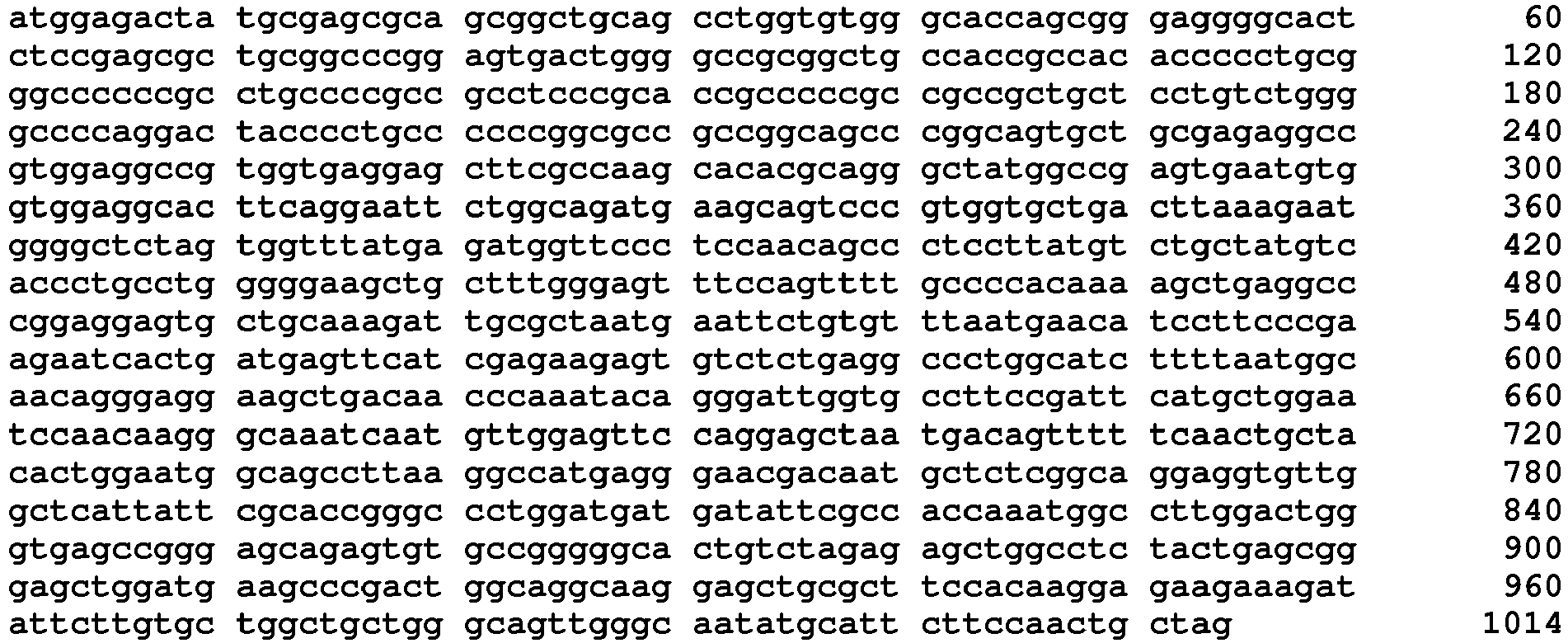
- <210> 3
<211> 21
<212> DNA
<213> Artificial Sequence - <220>
<223> Description of Artificial Sequence: RNAi-target region # 1 of LIX1L. - <400> 3
aggaggtgtt ggctcattatt 21 - <210> 4
<211> 21
<212> DNA
<213> Artificial Sequence - <220>
<223> Description of Artificial Sequence: RNAi-target region # 2 of LIX1L. - <400> 4
agtcccgtgg tgctgactta a 21 - <210> 5
<211> 59
<212> DNA
<213> Artificial Sequence - <220>
<223> Description of Artificial Sequence:shRNA # 1. - <400> 5
gaggaggtgt tggctcatta ttttcaagag aaataatgag ccaacacctc ctctttttt 59 - <210> 6
<211> 59
<212> DNA
<213> Artificial Sequence - <220>
<223> Description of Artificial Sequence:shRNA # 2. - <400> 6
gagtcccgtg gtgctgactt aattcaagag attaagtcag caccacggga ctctttttt 59 - <210> 7
<211> 11
<212> PRT
<213> Artificial Sequence - <220>
<223> Description of Artificial Sequence: Tat peptide - <400> 7

- <210> 8
<211> 20
<212> DNA
<213> Artificial Sequence - <220>
<223> Description of Artificial Sequence: LIX1L Forward Primer. - <400> 8
gggaggggca ctctccgagc 20 - <210> 9
<211> 20
<212> DNA
<213> Artificial Sequence - <220>
<223> Description of Artificial Sequence: LIX1L Reverse Primer. - <400> 9
gcgaagctcc tcaccacggc 20 - <210> 10
<211> 22
<212> DNA
<213> Artificial Sequence - <220>
<223> Description of Artificial Sequence: GAPDH Forward Primer. - <400> 10
gaacagcaac gagtaccggg ta 22 - <210> 11
<211> 21
<212> DNA
<213> Artificial Sequence - <220>
<223> Description of Artificial Sequence: GAPDH Reverse Primer. - <400> 11
cccatggcct tgaccaaggag 21 - <210> 12
<211> 23
<212> PRT
<213> Artificial Sequence - <220>
<222> 12
<223> Acp - <220>
<223> Description of Artificial Sequence:peptide 1. - <400> 12

- <210> 13
<211> 22
<212> PRT
<213> Artificial Sequence - <220>
<222> 12
<223> Acp - <220>
<223> Description of Artificial Sequence:peptide 2. - <400> 13

- <210> 14
<211> 12
<212> PRT
<213> Artificial Sequence - <220>
<222> 12
<223> Acp - <220>
<223> Description of Artificial Sequence: control peptide. - <400> 14

- <210> 15
<211> 32
<212> PRT
<213> Artificial Sequence - <220>
<222> 12
<223> Acp - <220>
<223> Description of Artificial Sequence:peptide 3. - <400> 15

- <210> 16
<211> 42
<212> PRT
<213> Artificial Sequence - <220>
<222> 12
<223> Acp - <220>
<223> Description of Artificial Sequence:peptide 4. - <400> 16

- <210> 17
<211> 345
<212> PRT
<213> Artificial Sequence - <220>
<222> 12
<223> Acp - <220>
<223> Description of Artificial Sequence: FLG/LIX1L - <400> 17


- <210> 18
<211> 22
<212> PRT
<213> Artificial Sequence - <220>
<222> 12
<223> Acp - <220>
<223> Description of Artificial Sequence:peptide 5. - <400> 18

- <210> 19
<211> 22
<212> PRT
<213> Artificial Sequence - <220>
<222> 12
<223> Acp - <220>
<223> Description of Artificial Sequence:peptide 6. - <400> 19


- <210> 20
<211> 22
<212> PRT
<213> Artificial Sequence - <220>
<222> 12
<223> Acp - <220>
<223> Description of Artificial Sequence:peptide 7. - <400> 20

- <210> 21
<211> 22
<212> PRT
<213> Artificial Sequence - <220>
<222> 12
<223> Acp - <220>
<223> Description of Artificial Sequence:peptide 8. - <400> 21

- <210> 22
<211> 22
<212> PRT
<213> Artificial Sequence - <220>
<222> 12
<223> Acp - <220>
<223> Description of Artificial Sequence:peptide 9. - <400> 22

- <210> 23
<211> 22
<212> PRT
<213> Artificial Sequence - <220>
<222> 12
<223> Acp - <220>
<223> Description of Artificial Sequence:peptide 10. - <400> 23

- <210> 24
<211> 22
<212> PRT
<213> Artificial Sequence - <220>
<222> 12
<223> Acp - <220>
<223> Description of Artificial Sequence:peptide 11. - <400> 24

- <210> 25
<211> 22
<212> PRT
<213> Artificial Sequence - <220>
<222> 12
<223> Acp - <220>
<223> Description of Artificial Sequence:peptide 12. - <400> 25

- <210> 26
<211> 22
<212> PRT
<213> Artificial Sequence - <220>
<222> 12
<223> Acp - <220>
<223> Description of Artificial Sequence:peptide 13. - <400> 26

- <210> 27
<211> 22
<212> PRT
<213> Artificial Sequence - <220>
<222> 12
<223> Acp - <220>
<223> Description of Artificial Sequence:peptide 14. - <400> 27

- <210> 28
<211> 22
<212> PRT
<213> Artificial Sequence - <220>
<222> 12
<223> Acp - <220>
<223> Description of Artificial Sequence:peptide 15. - <400> 28

- <210> 29
<211> 22
<212> PRT
<213> Artificial Sequence - <220>
<222> 12
<223> Acp - <220>
<223> Description of Artificial Sequence:peptide 16. - <400> 29

Claims (7)
- An ex vivo method for inhibiting proliferation of a high LIX1L-expressing tumor cell, comprising:inhibiting the expression or function of a LIX1L gene with respect to a tumor cell highly expressing the LIX1L gene, whereinthe inhibiting the expression of the LIX1L gene is performed through RNA interference which includes introduction of shRNA or siRNA including a base sequence represented by SEQ ID No: 3 or 4, or a vector which produces the shRNA or the siRNA in a cell, into the cell, andthe inhibiting the function of the LIX1L gene is performed by inhibiting phosphorylation of the 136th tyrosine of the LIX1L protein consisting of the amino acid sequence represented by SEQ ID No: 1.
- A tumor cell proliferation-inhibiting peptide comprising:a polypeptide having a length of 8 amino acids to 35 amino acids which contains a partial sequence consisting of 8 amino acids having the 136th tyrosine starting from the 130th proline from the N-terminal of the amino acid sequence represented by SEQ ID No: 1.
- The tumor cell proliferation-inhibiting peptide according to Claim 2,
wherein the partial sequence and a site which has cell membrane permeability are directly or indirectly bonded to each other. - The tumor cell proliferation-inhibiting peptide according to Claim 3,
wherein the site having the cell membrane permeability is a peptide consisting of an amino acid sequence represented by SEQ ID No: 7. - A pharmaceutical composition having the tumor cell proliferation-inhibiting peptide according to any one of Claims 2 to 4 as an active ingredient.
- The pharmaceutical composition according to Claim 15, which is an anti-tumor agent.
- An ex vivo method for inhibiting proliferation of a high LIX1L-expressing tumor cell, comprising:introducing the tumor cell proliferation-inhibiting peptide according to any one of Claims 2 to 4 into a high LIX1L-expressing tumor cell.
Applications Claiming Priority (3)
| Application Number | Priority Date | Filing Date | Title |
|---|---|---|---|
| JP2013082272 | 2013-04-10 | ||
| JP2013173696 | 2013-08-23 | ||
| PCT/JP2014/059780 WO2014168064A1 (en) | 2013-04-10 | 2014-04-02 | Method for inhibiting proliferation of high lix1l-expressing tumor cell, and tumor cell proliferation-inhibiting peptide |
Publications (3)
| Publication Number | Publication Date |
|---|---|
| EP2985345A1 EP2985345A1 (en) | 2016-02-17 |
| EP2985345A4 EP2985345A4 (en) | 2016-09-28 |
| EP2985345B1 true EP2985345B1 (en) | 2018-03-14 |
Family
ID=51689474
Family Applications (1)
| Application Number | Title | Priority Date | Filing Date |
|---|---|---|---|
| EP14782500.4A Not-in-force EP2985345B1 (en) | 2013-04-10 | 2014-04-02 | Method for inhibiting proliferation of high lix1l-expressing tumor cell, and tumor cell proliferation-inhibiting peptide |
Country Status (4)
| Country | Link |
|---|---|
| US (1) | US9676820B2 (en) |
| EP (1) | EP2985345B1 (en) |
| JP (1) | JP6176800B2 (en) |
| WO (1) | WO2014168064A1 (en) |
Families Citing this family (2)
| Publication number | Priority date | Publication date | Assignee | Title |
|---|---|---|---|---|
| US11300568B2 (en) | 2016-02-11 | 2022-04-12 | Inserm (Institut National De La Sante Et De La Recherche Medicale | Methods for the diagnosis and treatment of gastrointestinal stromal tumors |
| CA3168968A1 (en) * | 2020-02-25 | 2021-09-02 | Herbert Alexander GRAVES | Gene delivery system |
Family Cites Families (4)
| Publication number | Priority date | Publication date | Assignee | Title |
|---|---|---|---|---|
| JPH0740925B2 (en) | 1983-03-14 | 1995-05-10 | 協和醗酵工業株式会社 | Novel human interferon-γ polypeptide |
| JPH05271290A (en) | 1992-03-17 | 1993-10-19 | Ishihara Sangyo Kaisha Ltd | Polypeptide |
| CA2644162A1 (en) | 2006-03-07 | 2007-09-13 | Telethon Institute For Child Health Research | Method for diagnosing and/or predicting the development of an allergic disorder and agents for treating and/or preventing same |
| JP2010268690A (en) | 2009-05-19 | 2010-12-02 | Nippon Kayaku Co Ltd | Method for determining sensitivity of compound having anti-tumor activity |
-
2014
- 2014-04-02 EP EP14782500.4A patent/EP2985345B1/en not_active Not-in-force
- 2014-04-02 US US14/783,050 patent/US9676820B2/en not_active Expired - Fee Related
- 2014-04-02 JP JP2015511230A patent/JP6176800B2/en not_active Expired - Fee Related
- 2014-04-02 WO PCT/JP2014/059780 patent/WO2014168064A1/en not_active Ceased
Non-Patent Citations (1)
| Title |
|---|
| None * |
Also Published As
| Publication number | Publication date |
|---|---|
| US9676820B2 (en) | 2017-06-13 |
| EP2985345A1 (en) | 2016-02-17 |
| JP6176800B2 (en) | 2017-08-09 |
| WO2014168064A1 (en) | 2014-10-16 |
| US20160046669A1 (en) | 2016-02-18 |
| EP2985345A4 (en) | 2016-09-28 |
| JPWO2014168064A1 (en) | 2017-02-16 |
Similar Documents
| Publication | Publication Date | Title |
|---|---|---|
| Tang et al. | GPR116, an adhesion G-protein–coupled receptor, promotes breast cancer metastasis via the Gαq-p63RhoGEF-Rho GTPase pathway | |
| Piao et al. | Complement 5a enhances hepatic metastases of colon cancer via monocyte chemoattractant protein-1-mediated inflammatory cell infiltration | |
| Li et al. | B7-H3 promotes gastric cancer cell migration and invasion | |
| Shi et al. | Exosomal miRNA-34 from cancer-associated fibroblasts inhibits growth and invasion of gastric cancer cells in vitro and in vivo | |
| Uekita et al. | Oncogenic Ras/ERK signaling activates CDCP1 to promote tumor invasion and metastasis | |
| Tsai et al. | Metastatic progression of prostate cancer is mediated by autonomous binding of galectin-4-O-glycan to cancer cells | |
| Lupia et al. | CD63 tetraspanin is a negative driver of epithelial-to-mesenchymal transition in human melanoma cells | |
| Lin et al. | Multiple steps of HLA-G in ovarian carcinoma metastasis: alter NK cytotoxicity and induce matrix metalloproteinase-15 (MMP-15) expression | |
| EP3356412B1 (en) | Targeting metastasis stem cells through a fatty acid receptor (cd36) | |
| BRPI0715844A2 (en) | PHARMACEUTICAL COMPOSITION FOR TREATMENT OF COLANGIOCARCINOMA, METHOD OF INHIBITION OF GROWTH OR INVASION OF COLANGIOCARCINOMA AND METHOD OF TREATMENT OF COLANGIOCARCINOMA | |
| KR101133243B1 (en) | Use of eIF3m for the Diagnosis and Treatment of Cancer | |
| JPWO2015005473A1 (en) | How to predict responsiveness to cancer treatment | |
| KR102377702B1 (en) | A composition for treating stomach cancer comprising an inhibitor of SYT11 | |
| JP2017526670A (en) | Inhibition of tumor metastasis by inhibition of necroptosis | |
| Bao et al. | Proteolytic release of the p75NTR intracellular domain by ADAM10 promotes metastasis and resistance to anoikis | |
| US20150010561A1 (en) | Semaphorin 3C (Sema3C) Inhibitor Therapeutics, Methods, and Uses | |
| Rikhof et al. | ‘Big’-Insulin-Like Growth Factor–II Signaling Is an Autocrine Survival Pathway in Gastrointestinal Stromal Tumors | |
| Jandova et al. | Fn14 receptor promotes invasive potential and metastatic capacity of non-small lung adenocarcinoma cells through the up-regulation of integrin α6 | |
| US9676820B2 (en) | Method for inhibiting proliferation of high LIX1L-expressing tumor cell, and tumor cell proliferation-inhibiting peptide | |
| WO2014200180A1 (en) | Pharmaceutical composition containing thioredoxin-binding protein as active ingredient and use thereof | |
| Matou-Nasri et al. | CD95-mediated apoptosis in Burkitt's lymphoma B-cells is associated with Pim-1 down-regulation | |
| WO2021015218A1 (en) | Prevention or treatment of fibrotic disease which targets transcription-associated factor | |
| Sánchez-Romero et al. | Expression of caveolin-1 in tooth germ, ameloblastoma and ameloblastic carcinoma | |
| KR102620901B1 (en) | Therapeutic agent of liver cancer with activated FGF19-FGFR4 signaling pathway and a method for screening thereof | |
| JP5551793B2 (en) | TRAIL sensitivity-enhancing composition containing an inhibitor for suppressing the expression or activity of TIP41, a TRAIL sensitizer target gene |
Legal Events
| Date | Code | Title | Description |
|---|---|---|---|
| PUAI | Public reference made under article 153(3) epc to a published international application that has entered the european phase |
Free format text: ORIGINAL CODE: 0009012 |
|
| 17P | Request for examination filed |
Effective date: 20151008 |
|
| AK | Designated contracting states |
Kind code of ref document: A1 Designated state(s): AL AT BE BG CH CY CZ DE DK EE ES FI FR GB GR HR HU IE IS IT LI LT LU LV MC MK MT NL NO PL PT RO RS SE SI SK SM TR |
|
| AX | Request for extension of the european patent |
Extension state: BA ME |
|
| DAX | Request for extension of the european patent (deleted) | ||
| A4 | Supplementary search report drawn up and despatched |
Effective date: 20160830 |
|
| RIC1 | Information provided on ipc code assigned before grant |
Ipc: A61K 38/00 20060101ALI20160819BHEP Ipc: C07K 14/47 20060101ALI20160819BHEP Ipc: A61P 35/00 20060101ALI20160819BHEP Ipc: A61K 31/7105 20060101ALI20160819BHEP Ipc: C12N 15/09 20060101AFI20160819BHEP |
|
| 17Q | First examination report despatched |
Effective date: 20170526 |
|
| GRAP | Despatch of communication of intention to grant a patent |
Free format text: ORIGINAL CODE: EPIDOSNIGR1 |
|
| INTG | Intention to grant announced |
Effective date: 20170818 |
|
| GRAS | Grant fee paid |
Free format text: ORIGINAL CODE: EPIDOSNIGR3 |
|
| GRAA | (expected) grant |
Free format text: ORIGINAL CODE: 0009210 |
|
| AK | Designated contracting states |
Kind code of ref document: B1 Designated state(s): AL AT BE BG CH CY CZ DE DK EE ES FI FR GB GR HR HU IE IS IT LI LT LU LV MC MK MT NL NO PL PT RO RS SE SI SK SM TR |
|
| REG | Reference to a national code |
Ref country code: GB Ref legal event code: FG4D |
|
| REG | Reference to a national code |
Ref country code: CH Ref legal event code: EP Ref country code: AT Ref legal event code: REF Ref document number: 978893 Country of ref document: AT Kind code of ref document: T Effective date: 20180315 |
|
| REG | Reference to a national code |
Ref country code: FR Ref legal event code: PLFP Year of fee payment: 5 |
|
| REG | Reference to a national code |
Ref country code: IE Ref legal event code: FG4D |
|
| REG | Reference to a national code |
Ref country code: DE Ref legal event code: R096 Ref document number: 602014022385 Country of ref document: DE |
|
| REG | Reference to a national code |
Ref country code: NL Ref legal event code: MP Effective date: 20180314 |
|
| REG | Reference to a national code |
Ref country code: LT Ref legal event code: MG4D |
|
| PG25 | Lapsed in a contracting state [announced via postgrant information from national office to epo] |
Ref country code: CY Free format text: LAPSE BECAUSE OF FAILURE TO SUBMIT A TRANSLATION OF THE DESCRIPTION OR TO PAY THE FEE WITHIN THE PRESCRIBED TIME-LIMIT Effective date: 20180314 Ref country code: FI Free format text: LAPSE BECAUSE OF FAILURE TO SUBMIT A TRANSLATION OF THE DESCRIPTION OR TO PAY THE FEE WITHIN THE PRESCRIBED TIME-LIMIT Effective date: 20180314 Ref country code: LT Free format text: LAPSE BECAUSE OF FAILURE TO SUBMIT A TRANSLATION OF THE DESCRIPTION OR TO PAY THE FEE WITHIN THE PRESCRIBED TIME-LIMIT Effective date: 20180314 Ref country code: HR Free format text: LAPSE BECAUSE OF FAILURE TO SUBMIT A TRANSLATION OF THE DESCRIPTION OR TO PAY THE FEE WITHIN THE PRESCRIBED TIME-LIMIT Effective date: 20180314 Ref country code: NO Free format text: LAPSE BECAUSE OF FAILURE TO SUBMIT A TRANSLATION OF THE DESCRIPTION OR TO PAY THE FEE WITHIN THE PRESCRIBED TIME-LIMIT Effective date: 20180614 |
|
| REG | Reference to a national code |
Ref country code: AT Ref legal event code: MK05 Ref document number: 978893 Country of ref document: AT Kind code of ref document: T Effective date: 20180314 |
|
| PG25 | Lapsed in a contracting state [announced via postgrant information from national office to epo] |
Ref country code: RS Free format text: LAPSE BECAUSE OF FAILURE TO SUBMIT A TRANSLATION OF THE DESCRIPTION OR TO PAY THE FEE WITHIN THE PRESCRIBED TIME-LIMIT Effective date: 20180314 Ref country code: BG Free format text: LAPSE BECAUSE OF FAILURE TO SUBMIT A TRANSLATION OF THE DESCRIPTION OR TO PAY THE FEE WITHIN THE PRESCRIBED TIME-LIMIT Effective date: 20180614 Ref country code: GR Free format text: LAPSE BECAUSE OF FAILURE TO SUBMIT A TRANSLATION OF THE DESCRIPTION OR TO PAY THE FEE WITHIN THE PRESCRIBED TIME-LIMIT Effective date: 20180615 Ref country code: SE Free format text: LAPSE BECAUSE OF FAILURE TO SUBMIT A TRANSLATION OF THE DESCRIPTION OR TO PAY THE FEE WITHIN THE PRESCRIBED TIME-LIMIT Effective date: 20180314 Ref country code: LV Free format text: LAPSE BECAUSE OF FAILURE TO SUBMIT A TRANSLATION OF THE DESCRIPTION OR TO PAY THE FEE WITHIN THE PRESCRIBED TIME-LIMIT Effective date: 20180314 |
|
| PG25 | Lapsed in a contracting state [announced via postgrant information from national office to epo] |
Ref country code: RO Free format text: LAPSE BECAUSE OF FAILURE TO SUBMIT A TRANSLATION OF THE DESCRIPTION OR TO PAY THE FEE WITHIN THE PRESCRIBED TIME-LIMIT Effective date: 20180314 Ref country code: IT Free format text: LAPSE BECAUSE OF FAILURE TO SUBMIT A TRANSLATION OF THE DESCRIPTION OR TO PAY THE FEE WITHIN THE PRESCRIBED TIME-LIMIT Effective date: 20180314 Ref country code: ES Free format text: LAPSE BECAUSE OF FAILURE TO SUBMIT A TRANSLATION OF THE DESCRIPTION OR TO PAY THE FEE WITHIN THE PRESCRIBED TIME-LIMIT Effective date: 20180314 Ref country code: PL Free format text: LAPSE BECAUSE OF FAILURE TO SUBMIT A TRANSLATION OF THE DESCRIPTION OR TO PAY THE FEE WITHIN THE PRESCRIBED TIME-LIMIT Effective date: 20180314 Ref country code: NL Free format text: LAPSE BECAUSE OF FAILURE TO SUBMIT A TRANSLATION OF THE DESCRIPTION OR TO PAY THE FEE WITHIN THE PRESCRIBED TIME-LIMIT Effective date: 20180314 Ref country code: AL Free format text: LAPSE BECAUSE OF FAILURE TO SUBMIT A TRANSLATION OF THE DESCRIPTION OR TO PAY THE FEE WITHIN THE PRESCRIBED TIME-LIMIT Effective date: 20180314 Ref country code: EE Free format text: LAPSE BECAUSE OF FAILURE TO SUBMIT A TRANSLATION OF THE DESCRIPTION OR TO PAY THE FEE WITHIN THE PRESCRIBED TIME-LIMIT Effective date: 20180314 |
|
| PG25 | Lapsed in a contracting state [announced via postgrant information from national office to epo] |
Ref country code: AT Free format text: LAPSE BECAUSE OF FAILURE TO SUBMIT A TRANSLATION OF THE DESCRIPTION OR TO PAY THE FEE WITHIN THE PRESCRIBED TIME-LIMIT Effective date: 20180314 Ref country code: SM Free format text: LAPSE BECAUSE OF FAILURE TO SUBMIT A TRANSLATION OF THE DESCRIPTION OR TO PAY THE FEE WITHIN THE PRESCRIBED TIME-LIMIT Effective date: 20180314 Ref country code: CZ Free format text: LAPSE BECAUSE OF FAILURE TO SUBMIT A TRANSLATION OF THE DESCRIPTION OR TO PAY THE FEE WITHIN THE PRESCRIBED TIME-LIMIT Effective date: 20180314 Ref country code: SK Free format text: LAPSE BECAUSE OF FAILURE TO SUBMIT A TRANSLATION OF THE DESCRIPTION OR TO PAY THE FEE WITHIN THE PRESCRIBED TIME-LIMIT Effective date: 20180314 |
|
| REG | Reference to a national code |
Ref country code: CH Ref legal event code: PL |
|
| REG | Reference to a national code |
Ref country code: DE Ref legal event code: R097 Ref document number: 602014022385 Country of ref document: DE |
|
| REG | Reference to a national code |
Ref country code: BE Ref legal event code: MM Effective date: 20180430 |
|
| PG25 | Lapsed in a contracting state [announced via postgrant information from national office to epo] |
Ref country code: PT Free format text: LAPSE BECAUSE OF FAILURE TO SUBMIT A TRANSLATION OF THE DESCRIPTION OR TO PAY THE FEE WITHIN THE PRESCRIBED TIME-LIMIT Effective date: 20180716 |
|
| PLBE | No opposition filed within time limit |
Free format text: ORIGINAL CODE: 0009261 |
|
| STAA | Information on the status of an ep patent application or granted ep patent |
Free format text: STATUS: NO OPPOSITION FILED WITHIN TIME LIMIT |
|
| REG | Reference to a national code |
Ref country code: IE Ref legal event code: MM4A |
|
| PG25 | Lapsed in a contracting state [announced via postgrant information from national office to epo] |
Ref country code: LU Free format text: LAPSE BECAUSE OF NON-PAYMENT OF DUE FEES Effective date: 20180402 Ref country code: DK Free format text: LAPSE BECAUSE OF FAILURE TO SUBMIT A TRANSLATION OF THE DESCRIPTION OR TO PAY THE FEE WITHIN THE PRESCRIBED TIME-LIMIT Effective date: 20180314 Ref country code: MC Free format text: LAPSE BECAUSE OF FAILURE TO SUBMIT A TRANSLATION OF THE DESCRIPTION OR TO PAY THE FEE WITHIN THE PRESCRIBED TIME-LIMIT Effective date: 20180314 |
|
| 26N | No opposition filed |
Effective date: 20181217 |
|
| PG25 | Lapsed in a contracting state [announced via postgrant information from national office to epo] |
Ref country code: LI Free format text: LAPSE BECAUSE OF NON-PAYMENT OF DUE FEES Effective date: 20180430 Ref country code: CH Free format text: LAPSE BECAUSE OF NON-PAYMENT OF DUE FEES Effective date: 20180430 Ref country code: SI Free format text: LAPSE BECAUSE OF FAILURE TO SUBMIT A TRANSLATION OF THE DESCRIPTION OR TO PAY THE FEE WITHIN THE PRESCRIBED TIME-LIMIT Effective date: 20180314 Ref country code: BE Free format text: LAPSE BECAUSE OF NON-PAYMENT OF DUE FEES Effective date: 20180430 |
|
| PG25 | Lapsed in a contracting state [announced via postgrant information from national office to epo] |
Ref country code: IE Free format text: LAPSE BECAUSE OF NON-PAYMENT OF DUE FEES Effective date: 20180402 |
|
| PG25 | Lapsed in a contracting state [announced via postgrant information from national office to epo] |
Ref country code: MT Free format text: LAPSE BECAUSE OF NON-PAYMENT OF DUE FEES Effective date: 20180402 |
|
| PG25 | Lapsed in a contracting state [announced via postgrant information from national office to epo] |
Ref country code: TR Free format text: LAPSE BECAUSE OF FAILURE TO SUBMIT A TRANSLATION OF THE DESCRIPTION OR TO PAY THE FEE WITHIN THE PRESCRIBED TIME-LIMIT Effective date: 20180314 |
|
| PG25 | Lapsed in a contracting state [announced via postgrant information from national office to epo] |
Ref country code: MK Free format text: LAPSE BECAUSE OF NON-PAYMENT OF DUE FEES Effective date: 20180314 Ref country code: HU Free format text: LAPSE BECAUSE OF FAILURE TO SUBMIT A TRANSLATION OF THE DESCRIPTION OR TO PAY THE FEE WITHIN THE PRESCRIBED TIME-LIMIT; INVALID AB INITIO Effective date: 20140402 |
|
| PG25 | Lapsed in a contracting state [announced via postgrant information from national office to epo] |
Ref country code: IS Free format text: LAPSE BECAUSE OF FAILURE TO SUBMIT A TRANSLATION OF THE DESCRIPTION OR TO PAY THE FEE WITHIN THE PRESCRIBED TIME-LIMIT Effective date: 20180714 |
|
| PGFP | Annual fee paid to national office [announced via postgrant information from national office to epo] |
Ref country code: GB Payment date: 20220303 Year of fee payment: 9 |
|
| PGFP | Annual fee paid to national office [announced via postgrant information from national office to epo] |
Ref country code: FR Payment date: 20220308 Year of fee payment: 9 |
|
| PGFP | Annual fee paid to national office [announced via postgrant information from national office to epo] |
Ref country code: DE Payment date: 20220302 Year of fee payment: 9 |
|
| REG | Reference to a national code |
Ref country code: DE Ref legal event code: R119 Ref document number: 602014022385 Country of ref document: DE |
|
| GBPC | Gb: european patent ceased through non-payment of renewal fee |
Effective date: 20230402 |
|
| PG25 | Lapsed in a contracting state [announced via postgrant information from national office to epo] |
Ref country code: GB Free format text: LAPSE BECAUSE OF NON-PAYMENT OF DUE FEES Effective date: 20230402 |
|
| PG25 | Lapsed in a contracting state [announced via postgrant information from national office to epo] |
Ref country code: GB Free format text: LAPSE BECAUSE OF NON-PAYMENT OF DUE FEES Effective date: 20230402 Ref country code: FR Free format text: LAPSE BECAUSE OF NON-PAYMENT OF DUE FEES Effective date: 20230430 Ref country code: DE Free format text: LAPSE BECAUSE OF NON-PAYMENT OF DUE FEES Effective date: 20231103 |