EP2981824B1 - Method for detecting asyn-specific antibodies in a biological sample - Google Patents
Method for detecting asyn-specific antibodies in a biological sample Download PDFInfo
- Publication number
- EP2981824B1 EP2981824B1 EP14714706.0A EP14714706A EP2981824B1 EP 2981824 B1 EP2981824 B1 EP 2981824B1 EP 14714706 A EP14714706 A EP 14714706A EP 2981824 B1 EP2981824 B1 EP 2981824B1
- Authority
- EP
- European Patent Office
- Prior art keywords
- asyn
- aggregates
- disease
- antibodies
- sample
- Prior art date
- Legal status (The legal status is an assumption and is not a legal conclusion. Google has not performed a legal analysis and makes no representation as to the accuracy of the status listed.)
- Not-in-force
Links
- 238000000034 method Methods 0.000 title claims description 74
- 239000012472 biological sample Substances 0.000 title claims description 11
- 208000018737 Parkinson disease Diseases 0.000 claims description 111
- 208000001089 Multiple system atrophy Diseases 0.000 claims description 64
- 239000000523 sample Substances 0.000 claims description 50
- 201000002832 Lewy body dementia Diseases 0.000 claims description 45
- 238000001943 fluorescence-activated cell sorting Methods 0.000 claims description 40
- 238000001514 detection method Methods 0.000 claims description 37
- 208000009829 Lewy Body Disease Diseases 0.000 claims description 30
- 208000037265 diseases, disorders, signs and symptoms Diseases 0.000 claims description 29
- 208000032859 Synucleinopathies Diseases 0.000 claims description 27
- 201000011240 Frontotemporal dementia Diseases 0.000 claims description 26
- 210000002966 serum Anatomy 0.000 claims description 25
- 201000010099 disease Diseases 0.000 claims description 24
- 238000003745 diagnosis Methods 0.000 claims description 18
- 208000024827 Alzheimer disease Diseases 0.000 claims description 17
- 210000001175 cerebrospinal fluid Anatomy 0.000 claims description 17
- 210000002381 plasma Anatomy 0.000 claims description 17
- 201000002212 progressive supranuclear palsy Diseases 0.000 claims description 16
- 206010067889 Dementia with Lewy bodies Diseases 0.000 claims description 15
- 230000002776 aggregation Effects 0.000 claims description 14
- 238000004220 aggregation Methods 0.000 claims description 14
- 208000011990 Corticobasal Degeneration Diseases 0.000 claims description 13
- 208000000609 Pick Disease of the Brain Diseases 0.000 claims description 13
- 208000009144 Pure autonomic failure Diseases 0.000 claims description 13
- 208000025535 REM sleep behavior disease Diseases 0.000 claims description 13
- 210000004369 blood Anatomy 0.000 claims description 13
- 239000008280 blood Substances 0.000 claims description 13
- 201000007601 neurodegeneration with brain iron accumulation Diseases 0.000 claims description 13
- 239000002245 particle Substances 0.000 claims description 13
- 201000010374 Down Syndrome Diseases 0.000 claims description 11
- 238000011161 development Methods 0.000 claims description 11
- 201000008319 inclusion body myositis Diseases 0.000 claims description 11
- 108090000185 alpha-Synuclein Proteins 0.000 claims description 8
- 239000003814 drug Substances 0.000 claims description 8
- 238000011282 treatment Methods 0.000 claims description 8
- 208000001072 type 2 diabetes mellitus Diseases 0.000 claims description 8
- 229940079593 drug Drugs 0.000 claims description 7
- 206010012289 Dementia Diseases 0.000 claims description 6
- 208000037658 Parkinson-dementia complex of Guam Diseases 0.000 claims description 4
- 206010002026 amyotrophic lateral sclerosis Diseases 0.000 claims description 4
- 208000013968 amyotrophic lateral sclerosis-parkinsonism-dementia complex Diseases 0.000 claims description 4
- 208000014450 amyotrophic lateral sclerosis-parkinsonism/dementia complex 1 Diseases 0.000 claims description 4
- 208000017004 dementia pugilistica Diseases 0.000 claims description 4
- 208000023105 Huntington disease Diseases 0.000 claims description 3
- 230000002596 correlated effect Effects 0.000 claims description 3
- 230000008418 synuclein deposition Effects 0.000 claims description 3
- 208000004051 Chronic Traumatic Encephalopathy Diseases 0.000 claims description 2
- 208000009415 Spinocerebellar Ataxias Diseases 0.000 claims description 2
- 238000009169 immunotherapy Methods 0.000 claims description 2
- 210000002751 lymph Anatomy 0.000 claims description 2
- 102000003802 alpha-Synuclein Human genes 0.000 claims 2
- 108090000765 processed proteins & peptides Proteins 0.000 description 56
- 108010064539 amyloid beta-protein (1-42) Proteins 0.000 description 52
- 102000004196 processed proteins & peptides Human genes 0.000 description 46
- 230000009257 reactivity Effects 0.000 description 35
- 238000002965 ELISA Methods 0.000 description 31
- 108090000623 proteins and genes Proteins 0.000 description 28
- 102000004169 proteins and genes Human genes 0.000 description 26
- 238000003556 assay Methods 0.000 description 25
- 238000010790 dilution Methods 0.000 description 24
- 239000012895 dilution Substances 0.000 description 24
- 102000036770 Islet Amyloid Polypeptide Human genes 0.000 description 23
- 108010041872 Islet Amyloid Polypeptide Proteins 0.000 description 23
- 102000013498 tau Proteins Human genes 0.000 description 21
- 108010026424 tau Proteins Proteins 0.000 description 21
- 239000000758 substrate Substances 0.000 description 19
- 239000000203 mixture Substances 0.000 description 18
- 238000002360 preparation method Methods 0.000 description 18
- 229920001184 polypeptide Polymers 0.000 description 17
- 150000001413 amino acids Chemical class 0.000 description 16
- 238000004458 analytical method Methods 0.000 description 15
- 238000011534 incubation Methods 0.000 description 15
- 230000015572 biosynthetic process Effects 0.000 description 14
- 238000002474 experimental method Methods 0.000 description 12
- 108010004729 Phycoerythrin Proteins 0.000 description 11
- 239000000090 biomarker Substances 0.000 description 11
- 230000018109 developmental process Effects 0.000 description 10
- 238000012413 Fluorescence activated cell sorting analysis Methods 0.000 description 9
- 239000000243 solution Substances 0.000 description 9
- 102100026882 Alpha-synuclein Human genes 0.000 description 8
- 241000699666 Mus <mouse, genus> Species 0.000 description 8
- 210000004556 brain Anatomy 0.000 description 8
- 210000004558 lewy body Anatomy 0.000 description 8
- 238000005259 measurement Methods 0.000 description 7
- 230000035945 sensitivity Effects 0.000 description 7
- 238000004448 titration Methods 0.000 description 7
- 241000699670 Mus sp. Species 0.000 description 6
- 208000027089 Parkinsonian disease Diseases 0.000 description 6
- 206010034010 Parkinsonism Diseases 0.000 description 6
- 206010002022 amyloidosis Diseases 0.000 description 6
- 238000005516 engineering process Methods 0.000 description 6
- 238000003384 imaging method Methods 0.000 description 6
- 239000003550 marker Substances 0.000 description 6
- 241000894007 species Species 0.000 description 6
- 241001529936 Murinae Species 0.000 description 5
- 102000001708 Protein Isoforms Human genes 0.000 description 5
- 108010029485 Protein Isoforms Proteins 0.000 description 5
- MTCFGRXMJLQNBG-UHFFFAOYSA-N Serine Natural products OCC(N)C(O)=O MTCFGRXMJLQNBG-UHFFFAOYSA-N 0.000 description 5
- 102100040347 TAR DNA-binding protein 43 Human genes 0.000 description 5
- 101710150875 TAR DNA-binding protein 43 Proteins 0.000 description 5
- 238000009825 accumulation Methods 0.000 description 5
- 210000004899 c-terminal region Anatomy 0.000 description 5
- 230000005750 disease progression Effects 0.000 description 5
- 230000004048 modification Effects 0.000 description 5
- 238000012986 modification Methods 0.000 description 5
- 230000004770 neurodegeneration Effects 0.000 description 5
- 208000015122 neurodegenerative disease Diseases 0.000 description 5
- 230000008506 pathogenesis Effects 0.000 description 5
- 238000005406 washing Methods 0.000 description 5
- 238000001262 western blot Methods 0.000 description 5
- VHUUQVKOLVNVRT-UHFFFAOYSA-N Ammonium hydroxide Chemical compound [NH4+].[OH-] VHUUQVKOLVNVRT-UHFFFAOYSA-N 0.000 description 4
- 206010061818 Disease progression Diseases 0.000 description 4
- 235000011114 ammonium hydroxide Nutrition 0.000 description 4
- 230000003941 amyloidogenesis Effects 0.000 description 4
- 239000000427 antigen Substances 0.000 description 4
- 238000003759 clinical diagnosis Methods 0.000 description 4
- 208000035475 disorder Diseases 0.000 description 4
- 238000009826 distribution Methods 0.000 description 4
- 238000000684 flow cytometry Methods 0.000 description 4
- MHMNJMPURVTYEJ-UHFFFAOYSA-N fluorescein-5-isothiocyanate Chemical compound O1C(=O)C2=CC(N=C=S)=CC=C2C21C1=CC=C(O)C=C1OC1=CC(O)=CC=C21 MHMNJMPURVTYEJ-UHFFFAOYSA-N 0.000 description 4
- 210000000609 ganglia Anatomy 0.000 description 4
- 239000000463 material Substances 0.000 description 4
- 230000000750 progressive effect Effects 0.000 description 4
- 229940043517 specific immunoglobulins Drugs 0.000 description 4
- 238000012360 testing method Methods 0.000 description 4
- 229960005486 vaccine Drugs 0.000 description 4
- 102100036597 Basement membrane-specific heparan sulfate proteoglycan core protein Human genes 0.000 description 3
- WTDRDQBEARUVNC-LURJTMIESA-N L-DOPA Chemical compound OC(=O)[C@@H](N)CC1=CC=C(O)C(O)=C1 WTDRDQBEARUVNC-LURJTMIESA-N 0.000 description 3
- WTDRDQBEARUVNC-UHFFFAOYSA-N L-Dopa Natural products OC(=O)C(N)CC1=CC=C(O)C(O)=C1 WTDRDQBEARUVNC-UHFFFAOYSA-N 0.000 description 3
- 206010044565 Tremor Diseases 0.000 description 3
- VREFGVBLTWBCJP-UHFFFAOYSA-N alprazolam Chemical compound C12=CC(Cl)=CC=C2N2C(C)=NN=C2CN=C1C1=CC=CC=C1 VREFGVBLTWBCJP-UHFFFAOYSA-N 0.000 description 3
- 239000011324 bead Substances 0.000 description 3
- 238000005119 centrifugation Methods 0.000 description 3
- KRKNYBCHXYNGOX-UHFFFAOYSA-N citric acid Chemical compound OC(=O)CC(O)(C(O)=O)CC(O)=O KRKNYBCHXYNGOX-UHFFFAOYSA-N 0.000 description 3
- 238000003776 cleavage reaction Methods 0.000 description 3
- 230000003412 degenerative effect Effects 0.000 description 3
- 238000010494 dissociation reaction Methods 0.000 description 3
- 230000005593 dissociations Effects 0.000 description 3
- 230000028993 immune response Effects 0.000 description 3
- 239000000178 monomer Substances 0.000 description 3
- 239000013642 negative control Substances 0.000 description 3
- 230000001537 neural effect Effects 0.000 description 3
- 230000001575 pathological effect Effects 0.000 description 3
- 210000002856 peripheral neuron Anatomy 0.000 description 3
- 230000008569 process Effects 0.000 description 3
- 238000012545 processing Methods 0.000 description 3
- 238000011160 research Methods 0.000 description 3
- 230000007017 scission Effects 0.000 description 3
- 239000006228 supernatant Substances 0.000 description 3
- 239000000725 suspension Substances 0.000 description 3
- 210000001519 tissue Anatomy 0.000 description 3
- 102000001049 Amyloid Human genes 0.000 description 2
- 108010094108 Amyloid Proteins 0.000 description 2
- 208000037259 Amyloid Plaque Diseases 0.000 description 2
- 102000009091 Amyloidogenic Proteins Human genes 0.000 description 2
- 108010048112 Amyloidogenic Proteins Proteins 0.000 description 2
- XKRFYHLGVUSROY-UHFFFAOYSA-N Argon Chemical compound [Ar] XKRFYHLGVUSROY-UHFFFAOYSA-N 0.000 description 2
- 206010006100 Bradykinesia Diseases 0.000 description 2
- 206010008025 Cerebellar ataxia Diseases 0.000 description 2
- 241000282326 Felis catus Species 0.000 description 2
- DHMQDGOQFOQNFH-UHFFFAOYSA-N Glycine Chemical compound NCC(O)=O DHMQDGOQFOQNFH-UHFFFAOYSA-N 0.000 description 2
- 229920002683 Glycosaminoglycan Polymers 0.000 description 2
- 101000834898 Homo sapiens Alpha-synuclein Proteins 0.000 description 2
- 208000006083 Hypokinesia Diseases 0.000 description 2
- 108060003951 Immunoglobulin Proteins 0.000 description 2
- 241001465754 Metazoa Species 0.000 description 2
- 208000002740 Muscle Rigidity Diseases 0.000 description 2
- 229920001213 Polysorbate 20 Polymers 0.000 description 2
- 108090000544 Proprotein convertase 1 Proteins 0.000 description 2
- 102000004085 Proprotein convertase 1 Human genes 0.000 description 2
- 241001522306 Serinus serinus Species 0.000 description 2
- UIIMBOGNXHQVGW-UHFFFAOYSA-M Sodium bicarbonate Chemical compound [Na+].OC([O-])=O UIIMBOGNXHQVGW-UHFFFAOYSA-M 0.000 description 2
- 102000008221 Superoxide Dismutase-1 Human genes 0.000 description 2
- 108010021188 Superoxide Dismutase-1 Proteins 0.000 description 2
- 108090000054 Syndecan-2 Proteins 0.000 description 2
- 102000019355 Synuclein Human genes 0.000 description 2
- 108050006783 Synuclein Proteins 0.000 description 2
- 230000002159 abnormal effect Effects 0.000 description 2
- 230000004075 alteration Effects 0.000 description 2
- 125000000539 amino acid group Chemical group 0.000 description 2
- 230000003942 amyloidogenic effect Effects 0.000 description 2
- 102000036639 antigens Human genes 0.000 description 2
- 108091007433 antigens Proteins 0.000 description 2
- 230000002567 autonomic effect Effects 0.000 description 2
- 210000004227 basal ganglia Anatomy 0.000 description 2
- 210000000227 basophil cell of anterior lobe of hypophysis Anatomy 0.000 description 2
- 230000000903 blocking effect Effects 0.000 description 2
- 210000001638 cerebellum Anatomy 0.000 description 2
- 230000008859 change Effects 0.000 description 2
- 230000001419 dependent effect Effects 0.000 description 2
- 210000005064 dopaminergic neuron Anatomy 0.000 description 2
- 230000003291 dopaminomimetic effect Effects 0.000 description 2
- 238000013399 early diagnosis Methods 0.000 description 2
- 239000012634 fragment Substances 0.000 description 2
- 238000007710 freezing Methods 0.000 description 2
- 230000002068 genetic effect Effects 0.000 description 2
- 238000003119 immunoblot Methods 0.000 description 2
- 102000018358 immunoglobulin Human genes 0.000 description 2
- 230000001771 impaired effect Effects 0.000 description 2
- 230000006872 improvement Effects 0.000 description 2
- 238000002347 injection Methods 0.000 description 2
- 239000007924 injection Substances 0.000 description 2
- 230000003993 interaction Effects 0.000 description 2
- 238000001990 intravenous administration Methods 0.000 description 2
- 238000002372 labelling Methods 0.000 description 2
- 230000003902 lesion Effects 0.000 description 2
- 238000002595 magnetic resonance imaging Methods 0.000 description 2
- 238000012083 mass cytometry Methods 0.000 description 2
- 239000011325 microbead Substances 0.000 description 2
- 238000000386 microscopy Methods 0.000 description 2
- 238000012544 monitoring process Methods 0.000 description 2
- 239000002102 nanobead Substances 0.000 description 2
- 210000001577 neostriatum Anatomy 0.000 description 2
- 230000002018 overexpression Effects 0.000 description 2
- 239000000256 polyoxyethylene sorbitan monolaurate Substances 0.000 description 2
- 235000010486 polyoxyethylene sorbitan monolaurate Nutrition 0.000 description 2
- 239000012460 protein solution Substances 0.000 description 2
- 230000004044 response Effects 0.000 description 2
- 238000000527 sonication Methods 0.000 description 2
- 210000000278 spinal cord Anatomy 0.000 description 2
- 238000010186 staining Methods 0.000 description 2
- 238000002198 surface plasmon resonance spectroscopy Methods 0.000 description 2
- 208000024891 symptom Diseases 0.000 description 2
- 238000002560 therapeutic procedure Methods 0.000 description 2
- 238000011830 transgenic mouse model Methods 0.000 description 2
- 238000003260 vortexing Methods 0.000 description 2
- XLYOFNOQVPJJNP-UHFFFAOYSA-N water Substances O XLYOFNOQVPJJNP-UHFFFAOYSA-N 0.000 description 2
- 230000003820 β-cell dysfunction Effects 0.000 description 2
- 101150037123 APOE gene Proteins 0.000 description 1
- 206010003694 Atrophy Diseases 0.000 description 1
- 206010003840 Autonomic nervous system imbalance Diseases 0.000 description 1
- 241000282693 Cercopithecidae Species 0.000 description 1
- 208000028698 Cognitive impairment Diseases 0.000 description 1
- 101100216294 Danio rerio apoeb gene Proteins 0.000 description 1
- 102000006441 Dopamine Plasma Membrane Transport Proteins Human genes 0.000 description 1
- 108010044266 Dopamine Plasma Membrane Transport Proteins Proteins 0.000 description 1
- 241000255601 Drosophila melanogaster Species 0.000 description 1
- 108090001126 Furin Proteins 0.000 description 1
- 208000034826 Genetic Predisposition to Disease Diseases 0.000 description 1
- 238000012307 MRI technique Methods 0.000 description 1
- 102000010909 Monoamine Oxidase Human genes 0.000 description 1
- 108010062431 Monoamine oxidase Proteins 0.000 description 1
- 241000699660 Mus musculus Species 0.000 description 1
- 206010028980 Neoplasm Diseases 0.000 description 1
- 241000207836 Olea <angiosperm> Species 0.000 description 1
- 239000004793 Polystyrene Substances 0.000 description 1
- 108090000545 Proprotein Convertase 2 Proteins 0.000 description 1
- 102000004088 Proprotein Convertase 2 Human genes 0.000 description 1
- 108010076504 Protein Sorting Signals Proteins 0.000 description 1
- 238000001530 Raman microscopy Methods 0.000 description 1
- 206010071390 Resting tremor Diseases 0.000 description 1
- 108010090804 Streptavidin Proteins 0.000 description 1
- 208000006011 Stroke Diseases 0.000 description 1
- 101710120037 Toxin CcdB Proteins 0.000 description 1
- 206010044688 Trisomy 21 Diseases 0.000 description 1
- -1 aSyn Proteins 0.000 description 1
- 230000032683 aging Effects 0.000 description 1
- 238000010171 animal model Methods 0.000 description 1
- 230000001078 anti-cholinergic effect Effects 0.000 description 1
- 229910052786 argon Inorganic materials 0.000 description 1
- 238000011948 assay development Methods 0.000 description 1
- 238000004630 atomic force microscopy Methods 0.000 description 1
- 238000001593 atomic mass spectrometry Methods 0.000 description 1
- 230000037444 atrophy Effects 0.000 description 1
- 230000001363 autoimmune Effects 0.000 description 1
- 230000006472 autoimmune response Effects 0.000 description 1
- 238000011888 autopsy Methods 0.000 description 1
- 210000003050 axon Anatomy 0.000 description 1
- OHDRQQURAXLVGJ-HLVWOLMTSA-N azane;(2e)-3-ethyl-2-[(e)-(3-ethyl-6-sulfo-1,3-benzothiazol-2-ylidene)hydrazinylidene]-1,3-benzothiazole-6-sulfonic acid Chemical compound [NH4+].[NH4+].S/1C2=CC(S([O-])(=O)=O)=CC=C2N(CC)C\1=N/N=C1/SC2=CC(S([O-])(=O)=O)=CC=C2N1CC OHDRQQURAXLVGJ-HLVWOLMTSA-N 0.000 description 1
- 230000003542 behavioural effect Effects 0.000 description 1
- 102000003799 beta-Synuclein Human genes 0.000 description 1
- 108090000182 beta-Synuclein Proteins 0.000 description 1
- 239000012148 binding buffer Substances 0.000 description 1
- 230000005540 biological transmission Effects 0.000 description 1
- 229960000074 biopharmaceutical Drugs 0.000 description 1
- 229940126587 biotherapeutics Drugs 0.000 description 1
- 239000010836 blood and blood product Substances 0.000 description 1
- 210000001772 blood platelet Anatomy 0.000 description 1
- 229940125691 blood product Drugs 0.000 description 1
- 210000001124 body fluid Anatomy 0.000 description 1
- 239000010839 body fluid Substances 0.000 description 1
- 210000005013 brain tissue Anatomy 0.000 description 1
- 239000000872 buffer Substances 0.000 description 1
- 210000004027 cell Anatomy 0.000 description 1
- 210000003169 central nervous system Anatomy 0.000 description 1
- 230000002490 cerebral effect Effects 0.000 description 1
- 206010008118 cerebral infarction Diseases 0.000 description 1
- 239000003153 chemical reaction reagent Substances 0.000 description 1
- 239000003795 chemical substances by application Substances 0.000 description 1
- 208000010877 cognitive disease Diseases 0.000 description 1
- 230000001149 cognitive effect Effects 0.000 description 1
- 210000001072 colon Anatomy 0.000 description 1
- 230000000112 colonic effect Effects 0.000 description 1
- 238000013170 computed tomography imaging Methods 0.000 description 1
- 230000008876 conformational transition Effects 0.000 description 1
- IQFVPQOLBLOTPF-HKXUKFGYSA-L congo red Chemical compound [Na+].[Na+].C1=CC=CC2=C(N)C(/N=N/C3=CC=C(C=C3)C3=CC=C(C=C3)/N=N/C3=C(C4=CC=CC=C4C(=C3)S([O-])(=O)=O)N)=CC(S([O-])(=O)=O)=C21 IQFVPQOLBLOTPF-HKXUKFGYSA-L 0.000 description 1
- 239000000470 constituent Substances 0.000 description 1
- 230000000875 corresponding effect Effects 0.000 description 1
- 230000001054 cortical effect Effects 0.000 description 1
- 230000001086 cytosolic effect Effects 0.000 description 1
- 230000000254 damaging effect Effects 0.000 description 1
- 230000006735 deficit Effects 0.000 description 1
- 210000001947 dentate gyrus Anatomy 0.000 description 1
- 238000002405 diagnostic procedure Methods 0.000 description 1
- 230000004069 differentiation Effects 0.000 description 1
- 238000002598 diffusion tensor imaging Methods 0.000 description 1
- 238000011979 disease modifying therapy Methods 0.000 description 1
- 229940052760 dopamine agonists Drugs 0.000 description 1
- 239000003136 dopamine receptor stimulating agent Substances 0.000 description 1
- 230000004771 dopaminergic neurodegeneration Effects 0.000 description 1
- 208000019479 dysautonomia Diseases 0.000 description 1
- 230000004064 dysfunction Effects 0.000 description 1
- 230000000694 effects Effects 0.000 description 1
- 238000001493 electron microscopy Methods 0.000 description 1
- 230000007613 environmental effect Effects 0.000 description 1
- 230000007717 exclusion Effects 0.000 description 1
- 230000001747 exhibiting effect Effects 0.000 description 1
- 229940028757 flebogamma Drugs 0.000 description 1
- 239000012530 fluid Substances 0.000 description 1
- 238000001506 fluorescence spectroscopy Methods 0.000 description 1
- 230000009760 functional impairment Effects 0.000 description 1
- 102000004963 gamma-Synuclein Human genes 0.000 description 1
- 108090001121 gamma-Synuclein Proteins 0.000 description 1
- 229940009600 gammagard Drugs 0.000 description 1
- 229940069042 gamunex Drugs 0.000 description 1
- 230000002518 glial effect Effects 0.000 description 1
- 239000008187 granular material Substances 0.000 description 1
- 230000036541 health Effects 0.000 description 1
- 210000003958 hematopoietic stem cell Anatomy 0.000 description 1
- 108010038082 heparin proteoglycan Proteins 0.000 description 1
- 230000000971 hippocampal effect Effects 0.000 description 1
- 210000001320 hippocampus Anatomy 0.000 description 1
- 208000003906 hydrocephalus Diseases 0.000 description 1
- 201000001421 hyperglycemia Diseases 0.000 description 1
- 230000003100 immobilizing effect Effects 0.000 description 1
- 230000001900 immune effect Effects 0.000 description 1
- 230000002998 immunogenetic effect Effects 0.000 description 1
- 230000001976 improved effect Effects 0.000 description 1
- 238000000338 in vitro Methods 0.000 description 1
- 238000001727 in vivo Methods 0.000 description 1
- 230000001939 inductive effect Effects 0.000 description 1
- 239000003112 inhibitor Substances 0.000 description 1
- 230000000977 initiatory effect Effects 0.000 description 1
- 230000003834 intracellular effect Effects 0.000 description 1
- HXWLAJVUJSVENX-HFIFKADTSA-N ioflupane I(123) Chemical compound C1([C@H]2C[C@@H]3CC[C@@H](N3CCCF)[C@H]2C(=O)OC)=CC=C([123I])C=C1 HXWLAJVUJSVENX-HFIFKADTSA-N 0.000 description 1
- 210000004153 islets of langerhan Anatomy 0.000 description 1
- 229960004502 levodopa Drugs 0.000 description 1
- 239000003446 ligand Substances 0.000 description 1
- 230000005923 long-lasting effect Effects 0.000 description 1
- 238000004519 manufacturing process Methods 0.000 description 1
- 201000000083 maturity-onset diabetes of the young type 1 Diseases 0.000 description 1
- 230000007246 mechanism Effects 0.000 description 1
- 238000002483 medication Methods 0.000 description 1
- 210000003593 megakaryocyte Anatomy 0.000 description 1
- 239000004005 microsphere Substances 0.000 description 1
- 230000003278 mimic effect Effects 0.000 description 1
- 239000011259 mixed solution Substances 0.000 description 1
- 238000002156 mixing Methods 0.000 description 1
- 230000000051 modifying effect Effects 0.000 description 1
- 239000003068 molecular probe Substances 0.000 description 1
- 125000000896 monocarboxylic acid group Chemical group 0.000 description 1
- 210000001616 monocyte Anatomy 0.000 description 1
- 210000003205 muscle Anatomy 0.000 description 1
- 230000035772 mutation Effects 0.000 description 1
- 239000002105 nanoparticle Substances 0.000 description 1
- 210000000822 natural killer cell Anatomy 0.000 description 1
- 210000000478 neocortex Anatomy 0.000 description 1
- 210000002241 neurite Anatomy 0.000 description 1
- 210000004498 neuroglial cell Anatomy 0.000 description 1
- 210000002569 neuron Anatomy 0.000 description 1
- 230000002981 neuropathic effect Effects 0.000 description 1
- 239000000101 novel biomarker Substances 0.000 description 1
- 238000010899 nucleation Methods 0.000 description 1
- 210000000956 olfactory bulb Anatomy 0.000 description 1
- 210000004248 oligodendroglia Anatomy 0.000 description 1
- 238000006384 oligomerization reaction Methods 0.000 description 1
- 208000031237 olivopontocerebellar atrophy Diseases 0.000 description 1
- 238000000424 optical density measurement Methods 0.000 description 1
- 230000003287 optical effect Effects 0.000 description 1
- 238000004806 packaging method and process Methods 0.000 description 1
- 210000000496 pancreas Anatomy 0.000 description 1
- 210000004923 pancreatic tissue Anatomy 0.000 description 1
- 230000007170 pathology Effects 0.000 description 1
- 239000008188 pellet Substances 0.000 description 1
- 210000005259 peripheral blood Anatomy 0.000 description 1
- 239000011886 peripheral blood Substances 0.000 description 1
- 230000002093 peripheral effect Effects 0.000 description 1
- 108010049224 perlecan Proteins 0.000 description 1
- 230000026731 phosphorylation Effects 0.000 description 1
- 238000006366 phosphorylation reaction Methods 0.000 description 1
- 229920002223 polystyrene Polymers 0.000 description 1
- 210000002975 pon Anatomy 0.000 description 1
- 239000013641 positive control Substances 0.000 description 1
- 230000001144 postural effect Effects 0.000 description 1
- 239000002243 precursor Substances 0.000 description 1
- 210000000063 presynaptic terminal Anatomy 0.000 description 1
- 238000002203 pretreatment Methods 0.000 description 1
- 230000002265 prevention Effects 0.000 description 1
- 125000002924 primary amino group Chemical class [H]N([H])* 0.000 description 1
- 208000018290 primary dysautonomia Diseases 0.000 description 1
- 239000000047 product Substances 0.000 description 1
- 239000000092 prognostic biomarker Substances 0.000 description 1
- 125000001500 prolyl group Chemical group [H]N1C([H])(C(=O)[*])C([H])([H])C([H])([H])C1([H])[H] 0.000 description 1
- 230000000272 proprioceptive effect Effects 0.000 description 1
- 238000011002 quantification Methods 0.000 description 1
- 230000001105 regulatory effect Effects 0.000 description 1
- 230000002441 reversible effect Effects 0.000 description 1
- 238000005070 sampling Methods 0.000 description 1
- 238000013207 serial dilution Methods 0.000 description 1
- 238000002603 single-photon emission computed tomography Methods 0.000 description 1
- 229910000030 sodium bicarbonate Inorganic materials 0.000 description 1
- 238000010561 standard procedure Methods 0.000 description 1
- 239000007858 starting material Substances 0.000 description 1
- 238000011351 state-of-the-art imaging technique Methods 0.000 description 1
- 208000003755 striatonigral degeneration Diseases 0.000 description 1
- 230000002739 subcortical effect Effects 0.000 description 1
- 210000003523 substantia nigra Anatomy 0.000 description 1
- 230000008093 supporting effect Effects 0.000 description 1
- 238000002636 symptomatic treatment Methods 0.000 description 1
- 230000000946 synaptic effect Effects 0.000 description 1
- 208000011580 syndromic disease Diseases 0.000 description 1
- 210000001103 thalamus Anatomy 0.000 description 1
- 238000001269 time-of-flight mass spectrometry Methods 0.000 description 1
- 210000003412 trans-golgi network Anatomy 0.000 description 1
- 230000009261 transgenic effect Effects 0.000 description 1
- 230000007704 transition Effects 0.000 description 1
- 238000011269 treatment regimen Methods 0.000 description 1
- 238000002255 vaccination Methods 0.000 description 1
- 230000006439 vascular pathology Effects 0.000 description 1
- 230000001720 vestibular Effects 0.000 description 1
- 230000000007 visual effect Effects 0.000 description 1
Images




Classifications
-
- G—PHYSICS
- G01—MEASURING; TESTING
- G01N—INVESTIGATING OR ANALYSING MATERIALS BY DETERMINING THEIR CHEMICAL OR PHYSICAL PROPERTIES
- G01N33/00—Investigating or analysing materials by specific methods not covered by groups G01N1/00 - G01N31/00
- G01N33/48—Biological material, e.g. blood, urine; Haemocytometers
- G01N33/50—Chemical analysis of biological material, e.g. blood, urine; Testing involving biospecific ligand binding methods; Immunological testing
- G01N33/68—Chemical analysis of biological material, e.g. blood, urine; Testing involving biospecific ligand binding methods; Immunological testing involving proteins, peptides or amino acids
- G01N33/6854—Immunoglobulins
-
- G—PHYSICS
- G01—MEASURING; TESTING
- G01N—INVESTIGATING OR ANALYSING MATERIALS BY DETERMINING THEIR CHEMICAL OR PHYSICAL PROPERTIES
- G01N33/00—Investigating or analysing materials by specific methods not covered by groups G01N1/00 - G01N31/00
- G01N33/48—Biological material, e.g. blood, urine; Haemocytometers
- G01N33/50—Chemical analysis of biological material, e.g. blood, urine; Testing involving biospecific ligand binding methods; Immunological testing
- G01N33/53—Immunoassay; Biospecific binding assay; Materials therefor
- G01N33/564—Immunoassay; Biospecific binding assay; Materials therefor for pre-existing immune complex or autoimmune disease, i.e. systemic lupus erythematosus, rheumatoid arthritis, multiple sclerosis, rheumatoid factors or complement components C1-C9
-
- G—PHYSICS
- G01—MEASURING; TESTING
- G01N—INVESTIGATING OR ANALYSING MATERIALS BY DETERMINING THEIR CHEMICAL OR PHYSICAL PROPERTIES
- G01N33/00—Investigating or analysing materials by specific methods not covered by groups G01N1/00 - G01N31/00
- G01N33/48—Biological material, e.g. blood, urine; Haemocytometers
- G01N33/50—Chemical analysis of biological material, e.g. blood, urine; Testing involving biospecific ligand binding methods; Immunological testing
- G01N33/68—Chemical analysis of biological material, e.g. blood, urine; Testing involving biospecific ligand binding methods; Immunological testing involving proteins, peptides or amino acids
- G01N33/6893—Chemical analysis of biological material, e.g. blood, urine; Testing involving biospecific ligand binding methods; Immunological testing involving proteins, peptides or amino acids related to diseases not provided for elsewhere
- G01N33/6896—Neurological disorders, e.g. Alzheimer's disease
-
- G—PHYSICS
- G01—MEASURING; TESTING
- G01N—INVESTIGATING OR ANALYSING MATERIALS BY DETERMINING THEIR CHEMICAL OR PHYSICAL PROPERTIES
- G01N2800/00—Detection or diagnosis of diseases
- G01N2800/28—Neurological disorders
- G01N2800/2814—Dementia; Cognitive disorders
Definitions
- the present invention relates to methods for detecting alpha Synuclein (aSyn)-specific antibodies in biological samples, especially in connection with and for diagnosing of Synucleinopathies, including Parkinson's disease (PD), Lewy Body Disease (LBD), Dementia with Lewy Bodies (DLB), Parkinson's Disease Dementia (PDD), Multiple System Atrophy (MSA), Pure Autonomic Failure (PAF), REM Sleep Behavior Disorder (RBD), Neurodegeneration with Brain Iron Accumulation type I (NBIA Type I) and inclusion body myositis (IBM).
- PD Parkinson's disease
- LBD Lewy Body Disease
- DLB Dementia with Lewy Bodies
- Parkinson's Disease Dementia PPD
- MSA Multiple System Atrophy
- PAF Pure Autonomic Failure
- RBD REM Sleep Behavior Disorder
- NBIA Type I Neurodegeneration with Brain Iron Accumulation type I
- IBM inclusion body myositis
- the present invention further relates to methods for detecting alpha Synuclein (aSyn)-specific antibodies in biological samples, especially in connection with and for diagnosing of diseases with alpha synuclein deposition and/or aggregation, including Alzheimer's disease (AD), Down Syndrome (DS), Progressive Supranuclear Palsy (PSP), Corticobasal Degeneration (CBD), Frontotemporal Dementia /Pick's Disease (FTD/PiD).
- AD Alzheimer's disease
- DS Down Syndrome
- PSP Progressive Supranuclear Palsy
- CBD Corticobasal Degeneration
- FTD/PiD Frontotemporal Dementia /Pick's Disease
- Synucleinopathies are a diverse group of neurodegenerative disorders that share a common pathologic characteristic: in neuropathologic examinations characteristic lesions can be detected containing abnormal aggregates of aSynuclein (aSyn) protein in selected populations of neurons and glia cells.
- aSyn aSynuclein
- aSyn (initially identified as PARK1 and PARK4) is a 140 amino acid protein widely expressed in the neocortex, hippocampus, dentate gyrus, olfactory bulb, striatum, thalamus and cerebellum as well as in peripheral neurons and ganglia, e.g.: in colon or muscle tissue.
- the major form of the protein is the full 140 amino acids-long transcript.
- Other isoforms are alpha-synuclein-98, lacking exon 3 and 5, alpha-synuclein-126, where exon 3 is lost and lacks residues 41-54; and alpha-synuclein-112, which lacks residue 103-130 due to loss of exon 5.
- aSyn is also highly expressed in hematopoietic cells including B-, T-, and NK cells as well as monocytes and platelets. The exact role in these cells is not known but it has been implicated in the differentiation of megakaryocytes (platelet precursors).
- Parkinson's disease PD
- LBD Lewy Body Disease
- DLB Dementia with Lewy Bodies
- DPD Dementia with Lewy Bodies
- MSA Multiple System Atrophy
- PAF Pure Autonomic Failure
- RBD REM Sleep Behavior Disorder
- NBIA Type I Neurodegeneration with Brain Iron Accumulation type I
- IBM Inclusion Body Myositis
- symptomatic medications such as L-dopa, Dopamine agonists, anticholinergic drugs as well as inhibitors of monoamine oxidase.
- all treatment opportunities currently present only lead to symptomatic alleviation but do not induce a long lasting, disease modifying effect in patients.
- Lewy body disorders are progressive neurodegenerative disorders characterized by tremor, rigidity, bradykinesia and by loss of dopaminergic neurons in the brain. In the case of DLB and PDD signs also include cognitive impairment. Up to 2% of the population above 60 years of age in western countries develop the typical signs of PD/LBD. Currently only symptomatic treatment is available. Unfortunately, these therapies only provide temporary relief from early symptoms and do not halt disease progression. The pathogenesis of PD/LBD is still incompletely understood, but it appears that genetic susceptibility and environmental factors are involved in the development of the disease. Despite all genetic advances, PD/LBD is primarily a sporadic disorder with no known cause (also called idiopathic PD/LBD).
- LBs Lewy bodies
- aSyn the synaptic protein aSyn plays a central role in LBD pathogenesis.
- LBD aSyn accumulates in LBs throughout affected brain areas or peripheral neurons and ganglia.
- MSA Multiple System Atrophy
- GCI glial cytoplasmic
- GCIs for the pathogenesis of MSA is generally acknowledged and underscored by recent analysis of transgenic mouse models analysing the effect of aSyn overexpression in oligodendroglia. In tg mice overexpressing human aSyn both, GCI-like aggregates and biochemical markers of MSA were observed.
- aSyn Although the exact mechanisms by which accumulation of aSyn leads to the typical features of neurodegeneration in synucleopathies are not fully understood, recent studies imply that abnormal formation and accumulation of aSyn is involved in the degenerative processes underlying synucleinopathies. Recently, different forms of aSyn have been identified in LBs. Beside the full length form of the protein, different forms of modified aSyn have been identified including phosphorylated, nitrosylated/nitrated, acetylated and mono-, di-, or tri-ubiquitinated aSyn. In addition, C-terminally truncated forms of the protein, like aSyn 1-119, aSyn 1-122 and aSyn 1-123, have been detected in brain tissue from both transgenic mice and PD/MSA cases.
- truncated and/or modified forms of aSyn are thought to act as seed molecules for formation of aSyn oligomers/fibrils.
- full length aSyn as well as truncated and/or modified forms of aSyn are then believed to accumulate leading to aggregate-formation.
- aggregate-formation i.e. oligomers and fibrils
- PD/LBD/synucleinopathy plays an important role for PD/LBD/synucleinopathy development and could thus be enhanced by the presence of truncated/modified forms of aSyn.
- synucleinopathies especially LBD/PD and MSA
- the diagnosis of synucleinopathies is a purely clinical one based on the gradual occurrence of bradykinesia and at least one of the following aberrations: muscular rigidity, 4-6 Hz resting tremor and/or postural instability not caused by primary visual, vestibular, cerebellar or proprioceptive dysfunction ( Jankovic J. et al. J. Neurol. Neurosurg. Psychiatr. (2008) 79 (4): 368-76 .
- Parkinson plus syndromes such as progressive supranuclear palsy and multiple system atrophy must be ruled out as well to establish a probable PD/LBD diagnosis.
- MSA MSA diagnosis of probable MSA requires a sporadic, progressive adult-onset disorder including rigorously defined autonomic failure and poorly levodopa-responsive parkinsonism or cerebellar ataxia ( Gilman et al. Neurology. (2008) Aug 26;71(9):670-6. Second consensus statement on the diagnosis of multiple system atrophy .).
- Dopaminergic imaging is further employed to differentiate forms of dopaminergic neurodegeneration (e.g.: PD/LBD) from other diseases not affecting the dopaminergic system.
- This imaging can be performed using either single-photon emission computed tomography (SPECT) or PET.
- SPECT single-photon emission computed tomography
- PET PET
- SPECT single-photon emission computed tomography
- baseline imaging with the dopamine transporter ligand [123I]ioflupane (DaTscanTM) has shown 78% sensitivity and 97% specificity with reference to clinical diagnosis at 3 years, versus 93% and 46%, respectively, for baseline clinical diagnosis ( Cummings JL et al., Brain. (2011);134(11):3146-3166 .)
- AD-typical cerebrospinal fluid signature low A ⁇ 42, high total tau, high phospho-Tau
- positive amyloid imaging defined genetic risk; have been a basis for a fundamental change in the diagnostic procedures ( Dubois et al., Lancet Neurol.9 (2010): 1118-1127 ) for AD as advised by international regulatory bodies and interest groups including the National Institute of Aging (NIA, NIH, USA), the NIH NINCDS working group, and the Alzheimer Association.
- PPMI Parkinson's disease progressive Markers Initiative
- CSF levels of post-translationally modified aSyn correlated weakly with PD disease severity and, when combined with total aSyn concentrations, contributed to distinguishing PD from the second most prominent synucleinopathy, MSA and from a third neurodegenerative disease with proteopathic lesions containing among others synuclein, progressive supranuclear palsy (PSP).
- IVIG intravenous immunoglobulin
- Yamandra et al. could demonstrate by ELISA and Immuno Blot analysis of anti monomeric aSyn reactivity that 63% of individuals exhibiting the high responses in a group of early PD patients (5x in mean-10x increase in median levels as compared to controls). In the late PD group the immune responses declined, showing ca. 4 and 6 fold increase in mean and median, respectively (P,0.007), compared to controls and with ca. 58% of patients showing high level of antibodies.
- the diagnostic potentials of the ELISA and Western blot analysis in this report were also assessed statistically (receiver operating characteristic (ROC) analysis).
- the areas under the ROC curves (AUC) for the autoimmune reactivity determined by ELISA in early and late PD patients compared to controls were 0.884 (95% confidence interval of 0.79 to 0.97) and 0.779 (0.6-0.95), respectively.
- the AUC values calculated from the corresponding ROC curves for the Western blot data were 0.85 (0.74-0.95) for early PD vs controls and 0.817 (0.67-0.95) for late PD vs controls, respectively. This indicates that the autoimmune responses to monomeric aSyn have a diagnostic value both in early and late PD patients however with optimal settings allowing the identification of ca. 60% of patients only.
- Besong-Agbo et al. showed that Ab levels are lower in PD patients as compared to healthy controls and patients suffering from Alzheimer's disease, whereas Smith and colleagues showed that there was no significant change comparing synucleinopathy patients and healthy controls ( Neurology. 2013 Jan 8;80(2):169-75 . Naturally occurring ⁇ -synuclein autoantibody levels are lower in patients with Parkinson disease ( Besong-Agbo et al., PLoS ONE 7(12) (2012): e52285 and references therein).
- WO 2010/069603 A1 and EP 2 366 714 A1 disclose a method for detecting aSyn specific antibodies in serum or plasma of patients with PD comprising detecting the antibodies with particles coated with aSyn.
- Papachroni et al. J. Neurochem. 101 (2007): 749-756
- Apetri et al. J. Mol. Biol. 255 (2006) : 63-71
- WO 97/14028 A2 discloses multiplex analysis systems using "beadsets" labelled with specific markers and using FACS. Spells et al. (Abstract of the 29th annual meeting of the American Society for Histocompatibility and Immunogenetics: S113 ) report about the specificity of HLA antibodies detected with HLA antigen-coated polystyrene beads. Li et al. (BMC Neuroscience 8 (2007) : 22 ) show the improvement of a low pH antigen-antibody dissociation procedure for ELISA measurement of circulating anti-Abeta antibodies.
- the present invention provides a method for detecting aSyn-specific antibodies in a biological sample comprising the following steps:
- a new method to detect aSyn-specific antibodies, especially aSyn-specific auto-antibodies is disclosed which can be used as diagnostic tool which is extremely helpful in diagnosing of synucleinopathies, including PD/LBD, DLB, PDD, PAF, RBD, MSA, NBIA type1, and monitoring the development of these diseases, as well as for diseases like Alzheimer's disease (AD), Down Syndrome (DS), PSP, CBD, FTD/PiD.
- AD Alzheimer's disease
- DS Down Syndrome
- CBD FTD/PiD
- the present method is based on the invention that not just single aSyn peptides are used as capturing tools for the aSyn-specific antibodies, but that instead aggregates consisting of aSyn ("aSyn-consisting-aggregates") or comprising i.a. aSyn (“aSyn-comprising-aggregates”) are used and that thereby generated antibody-aSyn-aggregate complexes are detected using a single particle detection technique.
- aSyn-comprising-aggregates and aSyn-consisting-aggregates are referred to herein, also the term “aSyn-aggregates” is used which is meant as “aSyn-comprising-aggregates, especially aSyn-consisting-aggregates", because the latter are a preferred embodiment of the aSyn-aggregates according to the present invention.
- aSyn-consisting-aggregates are aggregates consisting of full-length aSyn protein (the 140 aa long protein as disclosed above).
- aSyn-consisting-aggregates are aggregates consisting of full-length aSyn protein (the 140 aa long protein as disclosed above).
- aSyn-comprising-aggregates contain aSyn protein besides other components, especially other amyloidogenic polypeptides, such as A ⁇ 1-42, A ⁇ p(E)3-42 (or other A ⁇ forms), islet amyloid polypeptide, Tau protein (including phosphorylated Tau), TAR-DNA binding protein 43 (TDP43).
- the term "aSyn-comprising-aggregates” also encompasses aggregates that contain - instead or besides aSyn (the full length aSyn 140 aa protein) - aSyn polypeptides that are truncated or modified, especially aSyn polypeptides that are naturally occurring forms of aSyn, such as aSyn-98 (lacking exon 3 and 5), aSyn-126 (lacking aa 41-54; loss of exon 4) and aSyn-112 (lacking aa 103-130; loss of exon 5), phosphorylated (especially at serin 129 - Ser129), nitrosylated/nitrated, acetylated and mono-, di-, or tri-ubiquitinated aSyn, or C-terminally truncated forms of aSyn such as aSyn lacking the C-terminal 20, preferably
- the aSyn-aggregates according to the present invention are generated e.g. by overnight incubation of the aggregate-forming proteins, e.g. aSyn or aSyn and other aggregate proteins/polypeptides, such as A ⁇ polypeptides (especially A ⁇ 1-42, A ⁇ p(E)3-42 (or other A ⁇ forms)), islet amyloid polypeptide, tau protein, TDP43; and/or aSyn polypeptides that are truncated or modified.
- aggregate-forming proteins e.g. aSyn or aSyn and other aggregate proteins/polypeptides, such as A ⁇ polypeptides (especially A ⁇ 1-42, A ⁇ p(E)3-42 (or other A ⁇ forms)), islet amyloid polypeptide, tau protein, TDP43; and/or aSyn polypeptides that are truncated or modified.
- aSyn-(containing or comprising)-aggregates are incubated with serum samples derived either from healthy donors (HD) or from patients, especially PD and MSA patients or patients suspected of having PD and MSA, to allow binding of present antibodies (both IgG and IgM).
- Antibodies bound to the aSyn-comprising-aggregates, especially the aSyn-consisting-aggregates can be detected by any suitable method available to a person skilled in the art, e.g. by using a labelled secondary antibody which recognises the aSyn-specific antibody bound to the aSyn-aggregates.
- a phycoerythrin (PE)-labelled secondary antibody can be used.
- the immune complexes comprising the aSyn-specific antibody bound to the aSyn-aggregates (and optionally one or more detection agents, such as secondary antibodies) are measured using a single particle detection technique, such as FACS (fluorescence activated cell sorting)-analysis also known as flow cytometry.
- FACS fluorescence activated cell sorting
- the level of aSyn-specific antibodies obtained for a given sample can then be compared to the level of a healthy sample or to the level in a sample of a patient with a known disease status, for example the level in a sample of a PD and MSA patient.
- PD and MSA patients contain different levels of aSyn-specific immunoglobulins which are reactive towards the aSyn-aggregates (free aSyn-specific immunoglobulins) provided according to the present invention compared to healthy subjects. Furthermore, using the method according to the present invention, it could be shown that reactivity of aSyn-specific immunoglobulins (towards the aSyn-aggregates provided according to the present invention) derived from PD and MSA patients can be increased by a procedure known as demasking (removing of potentially bound aSyn-antigens from autoantibodies).
- the present method has a much higher capacity to detect aSyn-specific antibodies and thus has a much higher power to diagnose e.g. PD and MSA compared to methods published so far.
- the method according to the present invention fulfils the theoretical prerequisites of a predictive diagnostic tool to identify synucleinopathies, especially PD and MSA, and to follow the clinical response of a given patient to treatment.
- the present invention was developed for the analysis of aSyn-specific antibodies in human samples. It is therefore a preferred embodiment to detect human aSyn-specific antibodies, preferably human IgG or IgM antibodies, especially human IgG antibodies.
- human aSyn-specific antibodies preferably human IgG or IgM antibodies, especially human IgG antibodies.
- the detection of aSyn-specific antibodies in human is known in principle in the art; however, the role as a possible biomarker could not be verified. As shown with the present invention, this was also due to the analytical insufficiency of the detection methods available in the art. Due to the superiority of the method according to the present invention, the availability of these antibodies in human samples as biomarkers is enabled.
- the present method is therefore specifically suited to detect autoantibodies in biological samples. Accordingly, in a preferred embodiment of the present method, the aSyn-specific antibodies to be detected and quantified are autoantibodies.
- the present method uses aSyn-aggregates as probe for binding the aSyn-specific antibodies from the samples.
- aSyn-aggregates are known in principle in the art, it was not realised that the use of such aggregates in the analysis of aSyn-specific antibodies, especially in human samples, could significantly improve such methods, also in combination with the single particles detection techniques, such as FACS. Due to the use of such aggregates, the detection with single particles detection techniques (which are established techniques in various different fields and for different questions) is possible for analysing aSyn-specific antibodies in human samples (such as blood) which are usually very complex and difficult to handle.
- the dimensions of the aggregates to be used according to the present invention are standardised for analytical use. This can be done by establishing certain parameters during production of the aggregates. Depending on the conditions applied during generation of the aggregates, the size of the aggregates can be adjusted. Preferred sizes of the aSyn-aggregates according to the present invention are from 50 nm to 15 ⁇ m, preferably from 100 nm to 10 ⁇ m, especially from 500 nm to 5 ⁇ m (defined by the length of the aggregates (i.e. the longest extension).
- a preferred method to provide aggregates suitable for the present invention comprises the step of incubating full length aSyn (1-140), naturally occurring splice variants including aSyn-98, aSyn 112 and aSyn-126 or modified full length aSyn including phosphorylated (especially at serin 129 - Ser129), nitrosylated/nitrated, acetylated and mono-, di-, or tri-ubiquitinated aSyn, or C-terminally truncated forms of aSyn such as aSyn lacking the C-terminal 20-30 amino acids or even shorter aSyn proteins capable to form aggregates, at a pH of 2 to 9 for at least 20 min, preferably at least 1 h, especially at least 4 h.
- the duration of incubation is one of the parameters to adjust the size of the aggregates: the longer the incubation, the larger are the aggregates.
- Typical incubation times are from 10 min to 24 h. Shorter incubation times usually result in only very small aggregates and low number of aggregates; the aggregates produced with significantly longer incubation times than 48 h are usually not preferred in the present method.
- aggregates may also be sorted and "sieved" to arrive at the desired size, if needed, e.g. by fractionated centrifugation and similar techniques.
- a preferred embodiment of the aSyn-comprising-aggregates according to the present invention relates to aggregates which - in addition to aSyn or an aSyn polypeptide - contains A ⁇ or a variant thereof.
- a ⁇ variants are therefore specifically preferred in aSyn-comprising-aggregates (group 4 being the most preferred, followed by group 3, then group 2 and group 1)
- Group 1 of A ⁇ variants peptides containing either the core sequence A ⁇ 16-20 or the second core sequence A ⁇ 25-35. These peptides have been shown to form amyloid fibrils by themselves. Starting from these core sequences peptides can be extended by amino acids present in A ⁇ peptide or by alternative amino acids forming either truncated native A ⁇ peptides or truncated-modified A ⁇ peptides
- Group 2 of A ⁇ variants peptides containing the sequence A ⁇ 16-20-X 21-23 -24-28-X 29-30 -31-36 (meaning that aa 16 to 20, 24 to 28 and 31 to 36 are present and linked by a 3 and a 2 aa linker (of arbitrary primary sequence)).
- Starting from this sequence peptide can be extended by amino acids present in A ⁇ peptide or by alternative amino acids forming either truncated native A ⁇ peptides or truncated-modified A ⁇ peptides
- Group 3 of A ⁇ variants peptides containing the sequence A ⁇ 16-36. Starting from this sequence peptide can be extended by amino acids present in A ⁇ peptide or by alternative amino acids forming either truncated native A ⁇ peptides or truncated-modified A ⁇ peptides
- Group 4 of A ⁇ variants peptides containing the sequence A ⁇ 11-36 or A ⁇ p(E)11-36. Starting from this sequence peptide can be extended by amino acids present in A ⁇ peptide or by alternative amino acids forming either truncated native A ⁇ peptides or truncated-modified A ⁇ peptides.
- aSyn-comprising-aggregates relates to aggregates which - in addition to aSyn or an aSyn polypeptide - contains Tau protein or a variant thereof.
- aSyn and A ⁇
- only variants are used that allow aggregate formation.
- Most preferred are aggregates of Tau, however, other aSyn-comprising-aggregates may comprise Tau with modified or truncated forms (e.g.
- Tau441 plus hyperphosphorylated Tau441 and/or shorter versions of Tau e.g.: Tau 412, 410, 383, 381, 352, especially those that are naturally occurring in healthy or - especially - Tau protein variants that have marker function for a disease (either isoform of all 6 naturally occurring isoform or mutant forms thereof (e.g. P301L, P301S, V337M, etc.), e.g.: preferably simultaneously phosphorylated at amino acids 181, 202, 205, 212, 214, 231, 396 and, optionally, at additional residues present in Tau like 18, 153, 175, 199, 235, 262, 394, 404 and 422).
- aSyn-comprising-aggregates relates to aggregates which - in addition to aSyn or an aSyn polypeptide - contains islet amyloid polypeptide (IAPP) or a variant thereof.
- IAPP is synthesized as a 89 amino acid residue pre-prohormone. Cleavage of a signal sequence leads to a pro-IAPP containing 67 amino acids. The latter is further processed to the 37 residue mature IAPP.
- pro-IAPP normal processing of pro-IAPP is a two-step process initiated by cleavage at its COOH terminus (likely by prohormone convertase 1/3 in the trans-Golgi network) followed by cleavage at its NH2 terminus (by prohormone convertase 2 in granules) to form the mature form of IAPP.
- IAPP is strongly conserved among mammalian species but exhibit notable variation between the amino acids 20 and 29. The differences in this region determine the ability of IAPP to aggregate and to form amyloids. In fact, species with one or more proline residues (except cats) at this region do not form islet amyloids.
- Amyloids are insoluble, fibrillar, protein aggregates with a ⁇ -pleated sheet structure. Amyloids have characteristic optical properties after staining with Congo red or thioflavin S and can be analyzed by light microscopy. However, the development of amyloid starts with the formation of fibrils or aggregates (7-10 nm in diameter and hundreds of nm in length) which can be detected by electron microscopy before evidence of amyloid is observed by light microscopy.
- pancreatic amyloid deposits are found in more than 95% of type II diabetes patients and their formation is strongly associated with disease progression.
- amyloids can form from proteins that fold to a compact tertiary structure in their unaggregated state, or they can originate from intrinsically disordered polypeptides that fail to adopt compact tertiary structures in their soluble native state. IAPP is an important example of intrinsically disordered polypeptide that forms amyloid in vivo.
- IAPP is major component of the islet amyloids in the pancreas of T2D patients, at least two additional elements have been identified in these deposits: apoE and the heparin sulfate proteoglycan perlecan (HSPGs).
- HSPGs heparin sulfate proteoglycan perlecan
- the kinetics of amyloid formation is complex and displays a sigmoidal profile characterized by a final steady state where soluble peptide is at equilibrium with amyloid fibrils.
- Amyloid formation can be accelerated by the addition of small amounts of preformed fibrils in a process called "seeding".
- a fraction of the IAPP that is secreted in T2D is incompletely processed.
- the impaired NH 2 -terminal processing of pro-IAPP leads to amyloid formation and ⁇ -cell death.
- IAPP-Npro Accumulation of the NH 2 -terminally extended human pro-IAPP intermediate (IAPP-Npro) may be a critical initiating step in amyloid formation.
- IAPP-Npro was found to be less amyloidogenic in solution than mature IAPP, it interacts more effectively with glycosaminoglycans (GAGs), a component of HSPGs. Accordingly, IAPP forms with impaired NH 2 -terminal processing, especially IAPP-Npro, are preferred forms in aSyn-comprising-aggregates according to the present invention.
- aSyn-comprising-aggregates relates to aggregates which - in addition to aSyn or an aSyn polypeptide - contains at least two further aggregate components in addition to aSyn or an aSyn polypeptide, especially one or more of A ⁇ or a variant thereof and/or one or more of Tau protein or a variant thereof and/or one or more of IAPP or a variant thereof.
- aSyn-comprising-aggregates relates to aggregates that comprise - besides aSyn or an aSyn variant as disclosed herein - an aggregate component of a different aggregate forming polypeptide species (as used herein, the term “polypeptide” is covering the term “protein” and usually these two terms are used synonymously; the term “protein” is therefore applied herein also for polypeptides with less than 100 or less than 50 amino acid residues).
- this further aggregate-forming polypeptide species is selected from the group consisting of Tau protein or an aggregate-forming variant thereof, A ⁇ 1-42 peptide or an aggregate-forming variant thereof; IAPP and an aggregate-forming variant thereof, especially IAPP-Npro, TAR-DNA binding protein 43 (TDP43) or an aggregate-forming variant thereof; and Superoxide-Dismutase 1 (SOD1) or an aggregate-forming variant thereof.
- Tau variants preferred aggregate forming variants of Tau
- hyperphosphorylated Tau abnormally phosphorylated Tau (as referred to and defined in Shahani et al. J. Neurosci. 26 (2006), 6103-6114 ), Tau protein variants that have marker function for a disease (either isoform of all 6 naturally occurring isoforms (Tau441, Tau412, Tau410, Tau383, Tau381, Tau352) or mutant forms thereof (e.g.
- P301L, P301S, V337M, etc. forms preferably simultaneously phosphorylated at amino acids 181, 202, 205, 212, 214, 231, 396 and, optionally at additional residues present in Tau like 18, 153, 175, 189, 235, 262, 394, 404 and 422, of Tau441, Tau412, Tau410, Tau383, Tau381, Tau352 (a more detailed analysis of Tau phosphorylation is disclosed e.g. in Hanger et al., Trends in Mol. Med. 15 (2009), 112-119 ).
- the samples wherein the aSyn-specific antibodies are to be detected are contacted with the aSyn-aggregates to achieve binding of the aSyn-specific antibodies possibly present (and reactive vis-a-vis aSyn-aggregates) in the samples.
- the concentration of the aSyn-aggregates has therefore to be adjusted in order to provide enough binding positions for the antibodies. Accordingly, the concentration of the aSyn-aggregates for binding the antibodies in the sample is preferably in the range of 0.01 to 10 ⁇ M, preferably 0.1 to 1 ⁇ M.
- the optimal concentration is also dependent on the nature of antibodies to be bound, the nature of the sample, the planned contact time and the size of the aggregates.
- biological sample is human blood or a sample derived from human blood, preferably human serum or human plasma; human cerebrospinal fluid or human lymph.
- sample sources also serial and routine testing may be established (especially for samples derived from blood).
- Preferred contact times which allow proper binding of the antibodies in the sample to the aggregates are at least 10 min (e.g. 10 min to 48 h), preferably from 15 min to 24 h, especially from 30 min to 2 h.
- the aSyn-specific antibodies which have a binding capacity to the aSyn-aggregates will be bound during contact with the aSyn-aggregates.
- the aSyn-specific antibodies which are masked in the sample i.e. those antibodies which are already bound to a binding partner (e.g. an aSyn-comprising structure, or endogenous aSyn peptides) will not be detected by the method according to the present invention (absent such specific sample pre-treatment).
- the samples are demasked, i.e. the aSyn-specific antibodies are "freed” from any binding to binding partners present in the sample previous to the contact with the aSyn-aggregates according to the present invention.
- This allows detection of all aSyn-specific antibodies in the sample and not only detection of those antibodies which are not bound to a binding partner in the sample ("free” or "reactive” antibodies).
- the amount of reactive aSyn-specific antibodies, especially reactive aSyn-specific IgG was a key marker for the diagnosis and development of PD and MSA.
- the present method is, as stated above, also suitable for determining the overall amount of aSyn-specific antibodies in a sample, i.e. the free (or "reactive") antibodies as well as those antibodies which are already bound (e.g. to aSyn structures) in the sample.
- This can be helpful in establishing the difference ( ⁇ ) of reactive vs. non-reactive antibodies in a sample, a parameter which is also of significant importance for PD and MSA diagnosis. Whereas such difference is not present (or low) in persons with "healthy status" concerning PD and MSA, this difference is crucial for the marker function in PD and MSA, concerning both aSyn-specific IgG and aSyn-specific IgM.
- the method according to the present invention applies a single particle detection technique.
- Such techniques allow identifying and quantifying ("count") the number and amount of "positive" binding results of the aSyn-specific antibody to the aSyn-aggregates.
- a preferred embodiment of this technology is FACS which is an established technique in the present field.
- Other detection methods to be used to detect the antibodies bound to the aSyn-aggregates are e.g. Luminex or mass cytometry.
- sample preparation may be performed as described in Material and Methods. Following sample preparation aSyn-aggregates recognized by specific aSyn-specific antibodies may be detected by a secondary antibody coupled to fluorescent-dyed microspheres which can be detected in multiplex detecting systems e.g. a Luminex reader ( Binder et al., Lupus15 (2005):412-421 ).
- sample preparation may also be performed as described in Material and Methods of the example section below. Sample preparation is done as described. Following sample preparation aSyn-aggregates recognized by specific Abs may be detected by a secondary antibody coupled to stable isotopes of transition elements which can be detection by atomic mass spectrometry. The sample can then be sprayed through an argon plasma filled inductive coil heated to a temperature of >5,500 K. The sample is vaporized and ionized into its atomic constituents, and the number of the isotope-tagged antibody is quantified by time-of-flight mass spectrometry ( Janes et al., Nat. Biotechnol. 29 (2011): 602-604 ).
- aSyn-aggregates or antibody/serum binding partner
- Hybcell-technology and the Surface Plasmon Resonance technology.
- serum samples can be spotted on the surface of the Hybcell (a rotating cylinder) and incubation can be performed with directly fluorescence-labelled preincubated aSyn-aggregates or alternatively with a fluorescence-labelled monoclonal aSyn-specific second antibody.
- Antibodies bound to aSyn-aggregates are detected with a laser (Ronacher, Anagnostics Technical Note ANA-TN-005 (2010)).
- a reverse setup can be applied: the preincubated aSyn-aggregates can be immobilized on a chip surface.
- the binding of aSyn-specific antibodies from serum to the aSyn-aggregates on the chip can be detected by increase of mass on the chip surface and therefore no labelling of the binding partners is necessary.
- a serial injection of anti-IgG-AB is possible ( Cannon et al., Anal. Biochem. 328 (2004): 67-75 ).
- a capture antibody can be used instead of directly immobilizing aSyn-aggregates to the chip surface.
- an aSyn-specific antibody is immobilized on the chip surface followed by the injection of preincubated aSyn-aggregates. After the capturing of the aggregates serum is injected and reactivity is measured by increase of mass.
- Detection of the binding of aSyn-specific antibodies to the aSyn-aggregates according to the present invention can be performed by any suitable method known e.g. Fluorescence Spectroscopy ( Missailidis et al., Methods in Molecular Biology 248 (2003): 431-441 ) for detecting the aSyn-specific antibodies bound to the aSyn-aggregates by a secondary antibody (e.g. a secondary labelled anti-IgG- or anti-IgM-antibody).
- a secondary antibody e.g. a secondary labelled anti-IgG- or anti-IgM-antibody
- Detection of autoantibodies bound to aggregates can also be performed using substrates specifically binding antibodies such as Protein A or Protein G. Another possibility is to precipitate aSyn-aggregate specific autoantibodies using the aSyn-aggregates, wash the complex and biotinylate the antibodies. Subsequently streptavidin can then be used as second step reagent.
- the aggregates according to the present invention are provided without such surfaces but consist of the aggregates as such without any scaffold or surface aids. This resembles the natural situation, especially in human blood, more closely than with micro- or nanobeads.
- a preferred field of use of the present invention is the diagnosis of Synucleinopathies, including Parkinson's disease (PD), Lewy Body Disease (LBD), Dementia with Lewy Bodies (DLB), Parkinson's Disease Dementia (PDD), Multiple System Atrophy (MSA), Pure Autonomic Failure (PAF), REM Sleep Behaviour Disorder (RBD), Neurodegeneration with Brain Iron Accumulation type I (NBIA Type I) and inclusion body myositis (IBM); as well as other diseases, such as Alzheimer's disease (AD) and Down Syndrome (DS), Progressive Supranuclear Palsy (PSP), Cortico-basal degeneration (CBD), Frontotemporal Dementia /Pick's Disease (FTD/PiD).
- PD Parkinson's disease
- LBD Lewy Body Disease
- DLB Dementia with Lewy Bodies
- DPD Dementia with Lewy Bodies
- MSA Multiple System Atrophy
- PAF Pure Autonomic Failure
- a preferred embodiment of the present invention is the diagnosis of proteinopathies that is significantly improved by the present invention.
- Proteinopathies are diseases, especially neurodegenerative diseases that are caused by a malformed protein.
- Preferred proteinopathies to be diagnosed according to the present invention are Alzheimer's disease (AD), Dementia in Down's Syndrome, Parkinson's disease (PD), Lewy Body Disease (LBD), Dementia with Lewy Bodies (DLB), Parkinson's Disease Dementia (PDD), Multiple System Atrophy (MSA), Pure Autonomic Failure (PAF), REM Sleep Behaviour Disorder (RBD), Neurodegeneration with Brain Iron Accumulation type I (NBIA Type I), inclusion body myositis (IBM), Cortico-Basal Degeneration (CBD), Progressive Supranuclear Palsy (PSP), Pick's Disease (PiD), Dementia pugilistica (chronic traumatic encephalopathy, DP), Frontotemporal dementia (FTD), Lytico-Bodig disease (
- the present method is specifically suited for using in connection with PD and MSA diagnosis.
- aSyn-specific autoantibodies in human patients are provided as markers for PD and MSA status.
- People with "normal" level of aSyn-specific antibodies in their blood are "healthy” with respect to PD and MSA. If this level is modified in a patient with PD and MSA or subjects with a risk of developing PD and MSA or being suspected to have PD and MSA, such modification level correlates with PD and MSA.
- a "modified" level may be a modification of the absolute number of the aSyn-specific antibodies or a modification of the reactivity of the totality of the aSyn-specific antibodies (e.g.
- modified reactive aSyn-specific IgG correlates with and is a marker for PD and MSA.
- a significant modification in reactive aSyn-specific IgG is e.g. a decrease to 70% and lower, to 50% and lower or to 30% and lower, or an increase of at least 30%, e.g. at least 50% or at least 100%, in a blood sample.
- the present invention provides a marker for PD and MSA and even for the development of PD and MSA, it is possible to use this method for observing the development of the disease and the performance of possible treatment methods, especially whether the method of treatment enables to establish "healthy” or “healthier” levels of A ⁇ -specific antibodies, especially IgG or IgM.
- the present method is therefore preferably used for the monitoring of PD and MSA patients, especially PD and MSA patients who are treated with medicaments for curing or ameliorating PD and MSA.
- the present method can be successfully applied for observing patients in clinical trials for PD and MSA vaccines (e.g. PD01A, Trial AFF008, NCT 01568099) or aSyn-targeting disease-modifying drugs.
- the method according to the present invention can also be used for evaluating the risk of developing a proteinopathy, preferably a synucleinopathy, especially PD and MSA, or for detecting early stage of a proteinopathy, preferably a synucleinopathy, especially PD and MSA.
- a proteinopathy preferably a synucleinopathy, especially PD and MSA
- it is in principle made possible to detect changes in the immunological set-up of patients with respect to aSyn-specific autoantibodies at a significantly earlier point in time than cognitive and/or functional impairments. This could allow a significant improvement of early diagnosis of a proteinopathy, preferably a synucleinopathy, especially PD and MSA, with respect to a much larger part of the population if established in routine testing format.
- This makes patients eligible for early stage treatment regimens and/or prevention (or delay) strategies for proteinopathies, preferably synucleinopathies, especially PD and MSA, especially immunotherapy (including vaccination).
- the present invention relates to a use of a kit for performing the method according to the present invention comprising
- the kit used according to the present invention may further contain means for detecting aSyn-comprising-aggregates being bound to aSyn-specific antibodies, preferably secondary antibodies, especially labelled secondary antibodies, e.g. anti-IgG- or anti-IgM-antibodies).
- Further components can be standard samples, positive and/or negative controls, instructions for use and suitable packaging means (e.g. stable boxes, coloured vials, etc.).
- Figure 1 shows the size determination of aSyn-aggregates using FACS-analysis.
- aSyn-aggregates can be detected using flow cytometry and are depicted as a homogenous population in the SSC-A (log-scale)- and FSC-A (log-scale) channel in the dot blot (A).
- the size distribution (defined by FCS-A signal) of aSyn-aggregates was determined using commercially available calibrated size beads (1, 2, 4, 6, 10, and 15 ⁇ m) as shown in FSC-A-histogram (B).
- Figure 2 shows the detection of monoclonal antibody reactivity to aSyn-aggregates (A, B, C) and A ⁇ 1-42 aggregates (D, E, F) using the FACS-based assay.
- the aSyn-specific monoclonal antibody LB509 binds specifically to aSyn aggregates (A) but does not interact with A ⁇ 1-42 aggregates (D).
- the A ⁇ 1-42 specific antibody 3A5 binds A ⁇ 1-42 (E) but not aSyn aggregates (B). Reactivity was determined using a secondary anti immunoglobulin-PE-labelled antibody in FL2-PE-channel.
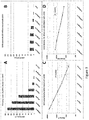
- Figure 3 shows the comparison of assay sensitivity (A, B) and assay linearity (C, D) for two different methods, the FACS-based method (A, C) and ELISA (B, D).
- A aSyn aggregates were incubated with a dilution series of mAb LB509 and reactivity was determined in FL-2 channel using flow cytometry.
- B mAb LB509 titration on Maxisorp ELISA plates coated with aSyn. Please note that for comparison all results are given in fold-background signal.
- Figure 4 shows signals derived from aSyn FACS aggregation assay (black bars) and aSyn ELISA (grey bars) using different dilutions of sera derived from individual mice (A, B, C), or using a 1:100 dilution of CSF derived from 20 individual mice (D) immunised with aSyn specific vaccine. All values are depicted as fold background values.
- the FACS aggregation assay detects aSyn specific antibodies with up to 30-fold higher sensitivity than standard methods such an ELISA.
- Figure 5 shows the determination of aSyn-specific IgG auto-antibody reactivity in a human preparation of IgG fraction (IVIG) which is extracted from plasma derived from healthy donors. IVIG was subjected to the described FACS assay. Fluorescence intensity of A ⁇ -aggregates was evaluated in FL2-PE channel and is expressed as Median Fluorescence Intensity (MFI).
- MFI Median Fluorescence Intensity
- Figure 6 shows the reactivity of mAB against aggregates generated from equimolar mixtures of either aSyn with A ⁇ 1-42, aSyn with p(E)A ⁇ 3-42 or A ⁇ 1-42 with p(E)A ⁇ 3-42. Aggregates were incubated with monoclonal antibodies specific either for A ⁇ 1-42 (3A5) p(E)A ⁇ 3-42 (D129) and aSyn (LB509) and the mAB-binding pattern of the mixed aggregates was determined.
- FACS PE-histograms show that the monoclonal antibody 3A5 binds exclusively mixtures which contain A ⁇ 1-42 aggregates (6A+B) but no reactivity to the p(E)A ⁇ 3-42/aSyn aggregate mixture (6C).
- the anti-p(E)A ⁇ 3-42mAB D129 binds only mixtures that contain p(E)A ⁇ 3-42 (6E+F) but shows no reactivity to A ⁇ 1-42/aSyn (6D).
- the aSyn specific mAB LB509 reacts only with aggregate mixtures containing aSyn (6G+I) but does not react with the A ⁇ 1-42/p(E)A ⁇ 3-42 aggregate mixture (6H).
- FIG. 7 FACS analysis of aggregates generated with equimolar mixtures of differentially fluorescent-labelled A ⁇ 1-42 and aSyn peptides of A ⁇ 1-42-Hilyte-555 (PE) and aSyn-Hilyte-488 (FITC).
- Acquired aggregates with a size range of 0,5-10 ⁇ m were gated in PI in the FSC-A/SSC-A (A) and PI was further evaluated in the PE/FITC-dot plot to determine the contribution of A ⁇ 1-42-Hilyte-555 (PE) and aSyn-Hilyte-488 (FITC) (C).
- Unlabelled mixed-aggregates were used as negative control (B).
- Figure 8 mAB reactivity against aggregates generated from different concentrations of aSyn and A ⁇ 1-42. A ⁇ 1-42 and aSyn peptides were mixed as indicated in table 1.
- Figure 9 shows the reactivity of mAbs specific for Aß1-42 (3A5), Aßp(E)3-42 (D129) and aSyn (LB509) against aggregates composed of an equimolar mixture of Aß1-42, Aßp(E)3-42 and aSyn peptides.
- FIG 10 shows the determination of IgG (A) and IgM (B) auto-antibody reactivity in human plasma derived from PD patients.
- Plasma samples (dilution 1:300) was subjected to the described FACS assay. Fluorescence intensity of binding to aSyn-aggregates was evaluated in FL2-PE channel and is expressed as Median Fluorescence Intensity (MFI).
- MFI Median Fluorescence Intensity
- aSyn peptide (purchased from Anaspec) was diluted in 100 mM NaHCO 3 (pH 9.2) at a concentration of 5 ⁇ g/ml and coated on Maxisorp 96-well plates overnight. To prevent unspecific binding plates were blocked using 1% BSA/PBS at 37°C for 1h.
- the ELISA was performed with a serial dilution of mAbs (starting with indicated concentration) in binding buffer (PBS/0.1% BSA/0.1% Tween20) at 37°C for 1h. After repeated washing steps (3x) with PBS/0.1% Tween20 the secondary antihuman Ig HRP (0.5 ⁇ g/ml) detection-antibody was added for 1h at 37°C.
- aSyn 500 ⁇ g of lyophilized aSyn was dissolved in 100 ⁇ l of sterile filtered MonoQ by resuspending and short vortexing until solution was clear. Subsequently this solution was sonicated for 30 sec in a sonication water bath and stored in 5 ⁇ l aliquots at -20°C without shock-freezing. To induce the formation of aggregates, aliquots were thawed and the entire volume of the aSyn solution was incubated at a concentration of 35 ⁇ M in 1% NH4OH (pH4,5) at 37°C on shaker (350 rpm) overnight in an Eppendorf tube. aSyn aggregates were used for one experiment only and residuals were discarded.
- the amount of aggregates was determined to ensure comparable aSyn-aggregate density before sample preparation for every experiment.
- the number of generated aggregates after overnight incubation was measured in the FSC/SSC channel before sample preparation and the number of aggregates used as a substrate was normalized.
- 3 ⁇ l of overnight generated aSyn-aggregates were diluted in 97 ⁇ l of 0.5% BSA/PBS as described above. 3 ⁇ l represent the standard amount of substrate used for each sample.
- 70 ⁇ l of this aSyn-dilution was measured at high flow rate (2 ⁇ l/s) resulting in an acquisition time of 35 seconds and the number of aggregates was determined. Approximately 10.000 particles acquired under these conditions were defined as standard.
- the volume of aSyn-aggregate solution used as a substrate for further sample preparation was adjusted to ensure comparable aggregate density within samples in different experiments. This means that if for example only 5000 events were acquired the volume of substrate for sample preparation was increased to 6 ⁇ l per well. As indicated above ⁇ 10.000 particles were used as starting material for each sample preparation.
- aSyn-specific antibodies 3 ⁇ l of the aSyn-aggregate suspension were mixed with 92 ⁇ l of 0.2 ⁇ m filtered 0.5% BSA/PBS and subsequently transferred into a well of a 96-well V-shaped bottom plate for further sample preparation. These suspensions were incubated for 60 min at RT for blocking.
- a ⁇ 1-42-Hilyte-555 (detectable in the PE-channel) and aSyn-Hilyte-488 (detectable in the FITC-channel) were mixed at equimolar ratio (20 ⁇ M) and incubated for 20h at room temperature on shaker (350 rpm) in 1% NH4OH (pH 4,5). After incubation these aggregates were analysed using the FACS Canto II.
- Mixed aggregates were used as substrates for further sample preparation and were treated as described for aSyn aggregates in paragraph above.
- aSyn specific auto-antibodies to aSyn likely present in patient sera and, therefore, preventing detection of these aSyn bound auto-antibodies by antigen-based methods (like ELISA or FACS)
- sera were pre-diluted in 10mM Glycin pH2.6 at a dilution of 1:16.7 for 5min. 5 ⁇ l of the acidified serum were then co-incubated with 3 ⁇ l of aSyn for another 5min. Then the mixture was neutralized by addition of 92 ⁇ l of 0.5%BSA/PBS and incubated for 20 to 60 min. Washing steps and incubation with secondary antibody were performed as described above for non-demasked serum.
- aSyn-aggregates from monomeric aSyn has been intensively investigated under multiple conditions in the recent years. It has been found that aggregation of aSyn peptides is very much dependent on different conditions including pH, temperature, buffer composition and protein concentration. Aggregation starts with the formation of ⁇ -hairpins from monomers leading to soluble oligomers. Conformational transition into parallel ⁇ -sheets then leads to the formation of fibrils and fibrillar aggregates, which can be precipitated by centrifugation.
- aSyn-aggregates full length human aSyn 1-140
- aSyn-aggregates can be detected using FACS-analysis.
- MM Material and Methods
- seedless soluble aSyn peptides were incubated for this purpose for 20 h at 37°C at a concentration of 35 ⁇ M.
- Figure 1A upper panel
- Figure 1A a clear homogenous population of aSyn aggregates could be detected by FACS analysis.
- aSyn-aggregates allow the binding of aSyn-specific antibodies and to determine whether such an interaction can be monitored using the here described FACS-based assay.
- aSyn-aggregates as well as A ⁇ 1-42 aggregates (made according to the disclosure in the international application PCT/EP2012/068494 ) were generated and were incubated with monoclonal antibodies specific either for aSyn (LB509) or A ⁇ 1-42 (3A5). Additionally both forms of aggregates where incubated with the unspecific control mAB D129.
- the monoclonal antibody LB509 binds exclusively aSyn aggregates whereas mAb 3A5 interacts only with A ⁇ 1-42 aggregates. Furthermore, the irrelevant mAb D129 used as isotype control does neither react with aSyn nor with A ⁇ -aggregates. This shows that the described FACS-based assay allows the detection of aSyn antibodies in a highly specific manner.
- the aim of this experiment was to define and compare the detection limits of two independent detection methods (ELISA and the FACS-based assay) for aSyn-reactivity of sera and CSF samples derived from animals vaccinated with an aSyn specific vaccine. aSyn was therefore immobilized onto Maxisorp microtiter plates for ELISA measurements. Alternatively, aSyn aggregates were generated for FACS analysis. Subsequently, a dilution series of three different sera or a 1:100 dilution of 20 different murine CSF samples were applied to the individual systems and either OD at 405nm values in case of ELISA or fluorescence intensity (MFI values) in case of FACS assay was defined. For comparison reasons signal to noise ratio was evaluated for different serum- ( Figure 4A , B, C) and CSF-( Figure 4D ) measurements and signals were expressed as fold-background-signal (xBG).
- CSF samples from 20 mice were also analysed with both assays. Comparison of the two methods using a 1:100 dilution of the CSF samples resulted in only one out of 20 samples reaching a significant signal detection in ELISA (2,02 x BG for mouse 8) whereas 17 out of 20 samples showed a MFI signal >2x BG when measured in the FACS aggregation assay. Analysing the CSF sample from mouse 2 resulted in an MFI signal of almost 30 x BG. The same CSF sample did not deliver a positive signal in the ELISA measurement.
- IVIG intravenous immunoglobulin
- IgG IgG fraction extracted from plasma derived from healthy donors (human plasma from at least 1000 donors). It has been shown that IVIG preparations contain naturally occurring antibodies (autoantibodies) specific for aSyn-peptides.
- aSyn aggregates were generated for FACS analysis. Subsequently, different IVIG dilutions ranging from 200 ⁇ g - 64 ng/ml) were applied to the aggregates and fluorescence intensity (MFI values) were defined. As depicted in Figure 5 , the FACS-based assay provided strong fluorescence signals showing that the newly developed FACS-based assay to detect aSyn-specific autoantibodies is highly sensitive for aSyn autoantibodies.
- aSyn specific mAB LB509 reacts only with aggregate mixtures containing aSyn (6G+I) but does not react with the A ⁇ 1-42/p(E)A ⁇ 3-42 aggregate mixture (6H). This shows first that mixed aggregates can be generated containing different peptides and second that these aggregates react specifically with the respective mAbs.
- Acquired aggregates with a size range of 0,5-10 ⁇ m were gated in PI in the FSC-A/SSC-A and PI was further evaluated in the PE/FITC-dot plot to determine the contribution of A ⁇ 1-42-Hilyte-555 (PE) and aSyn-Hilyte-488 (FITC). Co-incubation of A ⁇ 1-42-Hilyte-555 and aSyn-Hilyte-488 resulted in mixed aggregates which were ⁇ 90% double positive indicating a homogenous distribution of A ⁇ 1-42 and aSyn within the aggregate population.
- PE A ⁇ 1-42-Hilyte-555
- FITC aSyn-Hilyte-488
- Substrate 1 A ⁇ 1-42/aSyn 100/0 100/0
- Substrate 2 A ⁇ 1-42/aSyn 99.9/0.1 99.9/0.1
- Substrate 3 A ⁇ 1-42/aSyn 99/1 99/1
- Substrate 4 A ⁇ 1-42/aSyn 90/10 90/10
- Substrate 5 A ⁇ 1-42/aSyn 50/50 50/50
- Substrate 6 A ⁇ 1-42/aSyn 10/90 10/90
- Substrate7 A ⁇ 1-42/aSyn 1/99 1/99
- Substrate 8 A ⁇ 1-42/aSyn 0.1/99.9 0.1/99.9
- Substrate 9 A ⁇ 1-42/aSyn 0/100 0/100
- aSyn is incorporated in way that all available epitopes can be accessed by the mAB without blocking itself and therefore the PE-MFI signal is increased compared to measurements when 100% aSyn aggregates are used as a substrate.
- Serum samples derived from PD patients are analyzed for naturally occurring aSyn-specific antibody content using aSyn-aggregates and FACS analysis as described above.
- Naturally occurring antibodies specific for aSyn-aggregates are detected in all samples tested.
- Results show that naturally occurring antibodies specific for aSyn aggregates are detected in all samples tested although the level of auto-antibodies clearly differs from patient to patient( Fig. 10 A)
- aSyn-aggregate specific IgM reactivity is defined in the same set of serum samples as described above.
- Naturally occurring antibodies of IgM isotype specific for aSYn-aggregates are detected in all samples tested. Again comparable to reactivitiy of IgG antibodies, a different level of auto-antibodies could be detected within this set of PD-patients ( Fig. 10 B) .
Landscapes
- Health & Medical Sciences (AREA)
- Life Sciences & Earth Sciences (AREA)
- Engineering & Computer Science (AREA)
- Immunology (AREA)
- Hematology (AREA)
- Biomedical Technology (AREA)
- Chemical & Material Sciences (AREA)
- Molecular Biology (AREA)
- Urology & Nephrology (AREA)
- Biochemistry (AREA)
- General Health & Medical Sciences (AREA)
- Medicinal Chemistry (AREA)
- Cell Biology (AREA)
- Analytical Chemistry (AREA)
- Food Science & Technology (AREA)
- Biotechnology (AREA)
- Physics & Mathematics (AREA)
- Microbiology (AREA)
- General Physics & Mathematics (AREA)
- Pathology (AREA)
- Proteomics, Peptides & Aminoacids (AREA)
- Rehabilitation Therapy (AREA)
- Rheumatology (AREA)
- Neurosurgery (AREA)
- Neurology (AREA)
- Peptides Or Proteins (AREA)
- Investigating Or Analysing Biological Materials (AREA)
- Organic Chemistry (AREA)
- Measuring Or Testing Involving Enzymes Or Micro-Organisms (AREA)
- Zoology (AREA)
- Tropical Medicine & Parasitology (AREA)
- Virology (AREA)
- Biophysics (AREA)
- Genetics & Genomics (AREA)
- Investigating, Analyzing Materials By Fluorescence Or Luminescence (AREA)
- Investigating Or Analysing Materials By The Use Of Chemical Reactions (AREA)
Description
- The present invention relates to methods for detecting alpha Synuclein (aSyn)-specific antibodies in biological samples, especially in connection with and for diagnosing of Synucleinopathies, including Parkinson's disease (PD), Lewy Body Disease (LBD), Dementia with Lewy Bodies (DLB), Parkinson's Disease Dementia (PDD), Multiple System Atrophy (MSA), Pure Autonomic Failure (PAF), REM Sleep Behavior Disorder (RBD), Neurodegeneration with Brain Iron Accumulation type I (NBIA Type I) and inclusion body myositis (IBM). The present invention further relates to methods for detecting alpha Synuclein (aSyn)-specific antibodies in biological samples, especially in connection with and for diagnosing of diseases with alpha synuclein deposition and/or aggregation, including Alzheimer's disease (AD), Down Syndrome (DS), Progressive Supranuclear Palsy (PSP), Corticobasal Degeneration (CBD), Frontotemporal Dementia /Pick's Disease (FTD/PiD).
- Synucleinopathies are a diverse group of neurodegenerative disorders that share a common pathologic characteristic: in neuropathologic examinations characteristic lesions can be detected containing abnormal aggregates of aSynuclein (aSyn) protein in selected populations of neurons and glia cells.
- aSyn (initially identified as PARK1 and PARK4) is a 140 amino acid protein widely expressed in the neocortex, hippocampus, dentate gyrus, olfactory bulb, striatum, thalamus and cerebellum as well as in peripheral neurons and ganglia, e.g.: in colon or muscle tissue. The major form of the protein is the full 140 amino acids-long transcript. Other isoforms are alpha-synuclein-98, lacking
exon exon 3 is lost and lacks residues 41-54; and alpha-synuclein-112, which lacks residue 103-130 due to loss ofexon 5. aSyn is also highly expressed in hematopoietic cells including B-, T-, and NK cells as well as monocytes and platelets. The exact role in these cells is not known but it has been implicated in the differentiation of megakaryocytes (platelet precursors). - The most common synucleinopathies include Parkinson's disease (PD), Lewy Body Disease (LBD), Dementia with Lewy Bodies (DLB), Parkinson's Disease Dementia (PDD), Multiple System Atrophy (MSA), Pure Autonomic Failure (PAF), REM Sleep Behavior Disorder (RBD), Neurodegeneration with Brain Iron Accumulation type I (NBIA Type I) and Inclusion Body Myositis (IBM).
- The current treatment options for these diseases include symptomatic medications such as L-dopa, Dopamine agonists, anticholinergic drugs as well as inhibitors of monoamine oxidase. However, all treatment opportunities currently present only lead to symptomatic alleviation but do not induce a long lasting, disease modifying effect in patients.
- Lewy body disorders (LBD) are progressive neurodegenerative disorders characterized by tremor, rigidity, bradykinesia and by loss of dopaminergic neurons in the brain. In the case of DLB and PDD signs also include cognitive impairment. Up to 2% of the population above 60 years of age in western countries develop the typical signs of PD/LBD. Currently only symptomatic treatment is available. Unfortunately, these therapies only provide temporary relief from early symptoms and do not halt disease progression. The pathogenesis of PD/LBD is still incompletely understood, but it appears that genetic susceptibility and environmental factors are involved in the development of the disease. Despite all genetic advances, PD/LBD is primarily a sporadic disorder with no known cause (also called idiopathic PD/LBD).
- Patients suffering from this disease develop characteristic ubiquitinated intracellular inclusions called Lewy bodies (LBs) in the cortical and subcortical areas of the brain as well as in peripheral neurons or ganglia, e.g.: in colonic ganglia. Especially regions with high content of dopaminergic neurons or neuronal projections show this typical pathologic feature. Recently, several studies could show that the synaptic protein aSyn plays a central role in LBD pathogenesis. In LBD, aSyn accumulates in LBs throughout affected brain areas or peripheral neurons and ganglia. Additionally, it could be demonstrated that single point mutations as well as gene dosage alterations of aSyn (e.g.: duplications or multiplications in the aSyn gene) are associated with rare familial forms of parkinsonism. Importantly, based on results from overexpression studies in transgenic (tg) mice as well as in Drosophila melanogaster its key role in the pathogenesis of PD/LBD is underscored as these animal models mimic several characteristics of PD/LBD/DLB.
- Another very important synucleinopathy is Multiple System Atrophy (MSA). MSA is a sporadic neurodegenerative disorder that is characterised by symptoms of L-DOPA-resistant parkinsonism, cerebellar ataxia, and dysautonomia. Patients suffer from multisystem neuronal loss affecting various brain areas including striatum, substantia nigra, cerebellum, pons, as well as the inferior olives and the spinal cord. MSA is characterized by aSyn-positive glial cytoplasmic (GCI) and rare neuronal inclusions throughout the central nervous system. These inclusions are associated with striato-nigral degeneration, olivo-ponto-cerebellar atrophy, and involvement of autonomic nuclei in medulla and spinal cord. The importance of GCIs for the pathogenesis of MSA is generally acknowledged and underscored by recent analysis of transgenic mouse models analysing the effect of aSyn overexpression in oligodendroglia. In tg mice overexpressing human aSyn both, GCI-like aggregates and biochemical markers of MSA were observed.
- Although the exact mechanisms by which accumulation of aSyn leads to the typical features of neurodegeneration in synucleopathies are not fully understood, recent studies imply that abnormal formation and accumulation of aSyn is involved in the degenerative processes underlying synucleinopathies. Recently, different forms of aSyn have been identified in LBs. Beside the full length form of the protein, different forms of modified aSyn have been identified including phosphorylated, nitrosylated/nitrated, acetylated and mono-, di-, or tri-ubiquitinated aSyn. In addition, C-terminally truncated forms of the protein, like aSyn 1-119, aSyn 1-122 and aSyn 1-123, have been detected in brain tissue from both transgenic mice and PD/MSA cases.
- It is currently believed that up to 15% of the aSyn detected in LBs and lewy neurites or GCIs is truncated. Previous in vitro studies using truncated aSyn could demonstrate that aSyn lacking the C-terminal 20-30 amino acids was showing an increased tendency to aggregate and to form filaments found in Lewy-neurites and LBs. In addition, approximately 90% of aSyn deposited in LBs is phosphorylated at serine 129 (Ser129). In contrast, only 4% of total aSyn is phosphorylated in normal brain, suggesting that accumulation of Ser129-phosphorylated aSyn might be crucial in the pathogenesis of PD. Therefore, these truncated and/or modified forms of aSyn are thought to act as seed molecules for formation of aSyn oligomers/fibrils. Thus full length aSyn as well as truncated and/or modified forms of aSyn are then believed to accumulate leading to aggregate-formation. Based on recent studies it is believed that such aggregate-formation, (i.e. oligomers and fibrils) for example in the synaptic terminals and axons, plays an important role for PD/LBD/synucleinopathy development and could thus be enhanced by the presence of truncated/modified forms of aSyn.
- The diagnosis of synucleinopathies, especially LBD/PD and MSA, is a purely clinical one based on the gradual occurrence of bradykinesia and at least one of the following aberrations: muscular rigidity, 4-6 Hz resting tremor and/or postural instability not caused by primary visual, vestibular, cerebellar or proprioceptive dysfunction (Jankovic J. et al. J. Neurol. Neurosurg. Psychiatr. (2008) 79 (4): 368-76. criteria: United Kingdom Parkinson's Disease Society Brain Bank Diagnostic Criteria for Parkinson's Disease and Criteria of diagnosis of Parkinson disease (Gelb et al., 1999) commissioned and supported by the Advisory Council of the National Institute of Neurological Disorders and Stroke, US National Institutes of Health). In addition diagnosis of PD/LBD critically depends on the fulfillment of additional supporting clinical signs and the exclusion of other causes that can secondarily produce a parkinsonian syndrome like Alzheimer's disease, multiple cerebral infarction and drug-induced parkinsonism. Parkinson plus syndromes such as progressive supranuclear palsy and multiple system atrophy must be ruled out as well to establish a probable PD/LBD diagnosis.
- For MSA, diagnosis of probable MSA requires a sporadic, progressive adult-onset disorder including rigorously defined autonomic failure and poorly levodopa-responsive parkinsonism or cerebellar ataxia (Gilman et al. Neurology. (2008) Aug 26;71(9):670-6. Second consensus statement on the diagnosis of multiple system atrophy.).
- The limitation of clinical diagnosis of PD/LBD/MSA are high rates of misdiagnoses (diagnostic specificity evaluated at autopsy is 75-90%, with specialists such as neurologists having the highest rates; Jankovic J J. Neurol. Neurosurg. Psychiatr. (2008)79 (4): 368-76.) and the fact that the diagnosis could only be made at a late time point when the disease had caused substantial neuronal loss that resulted in functional deficits.
- In addition to clinical diagnosis, state of the art imaging techniques are currently employed to substantiate diagnosis of synucleinopathies. Computed tomography and magnetic resonance imaging (MRI) brain scans of patients usually appear normal but analyses are used to exclude other secondary causes of parkinsonism, such as basal ganglia tumors, vascular pathology and hydrocephalus (Brooks DJ et al. J. Nucl. Med. (2010) 51 (4): 596-609). However, specialized MRI techniques (e.g.: diffusion tensor imaging (DTI)) have demonstrated more consistent and promising results by exploring modifications of basal ganglia and mesencephalic structures. Dopaminergic imaging is further employed to differentiate forms of dopaminergic neurodegeneration (e.g.: PD/LBD) from other diseases not affecting the dopaminergic system. This imaging can be performed using either single-photon emission computed tomography (SPECT) or PET. For example in patients with diagnostic uncertainty between degenerative parkinsonism and non-degenerative tremor disorders, baseline imaging with the dopamine transporter ligand [123I]ioflupane (DaTscan™) has shown 78% sensitivity and 97% specificity with reference to clinical diagnosis at 3 years, versus 93% and 46%, respectively, for baseline clinical diagnosis (Cummings JL et al., Brain. (2011);134(11):3146-3166.)
- The current algorithms for diagnosing synucleinopathies suffer from the lack of sensitivity and specificity for a secure diagnosis at early stages of disease. In order to enhance the diagnostic capacities, clinical as well as imaging data have to be used in combination with patho-physiologically meaningful changes in disease-specific biomarkers. Such changes as exemplified in Alzheimer's disease with hippocampal atrophy as assessed by structural MRI; AD-typical cerebrospinal fluid signature (low Aβ42, high total tau, high phospho-Tau); positive amyloid imaging; defined genetic risk; have been a basis for a fundamental change in the diagnostic procedures (Dubois et al., Lancet Neurol.9 (2010): 1118-1127) for AD as advised by international regulatory bodies and interest groups including the National Institute of Aging (NIA, NIH, USA), the NIH NINCDS working group, and the Alzheimer Association.
- Unfortunately, no such validated biomarker data are available until now for PD/LBD/MSA. However, an international consortium of clinical and biomedical researchers under the lead of the Michael J Fox Foundation for Parkinson's disease has initiated the Parkinson's disease progressive Markers Initiative (PPMI) recently, a landmark observational clinical study to comprehensively evaluate a cohort of recently diagnosed PD patients and healthy subjects using advanced imaging, biologic sampling and clinical and behavioural assessments to identify biomarkers of Parkinson's disease progression. One of the main objectives is to evaluate changes in aSyn levels in different body fluids as potential biomarker for disease progression in synucleinopathies. This objective is based on the potential central role of aSyn for disease progression as well as on the identification of aSyn in different body tissues and fluids. Studies by Lebouvier et al. and Shannon et al. demonstrated aSyn pathology in peripheral tissue of PD patients (Lebouvier et al., PLoS ONE (2010) 5(9): e12728; Shannon et al., Mov. Disord. (2012) 27: 709-715). aSyn has also been identified in the CSF and plasma of synucleinopathy patients and healthy controls (Borghi et al. Neuroscience Letters, (2000) 287: 1, pp65-67; Journal of Neural Transmission (2006), 113, 10, pp 1435-1439; Lee et al., Experimental Neurology, (2007) 204: 2, pp583-588). Interestingly, there is conflicting evidence on the level of aSyn in healthy and diseased subjects as a recent study by Qiao-Xin Li et al. (Experimental Neurology, (2007) 204: 2, pp 583-588) indicated that aSyn levels in plasma from PD subjects are significantly lower than that in age-matched controls (p = 0.001), and the aSyn levels in patients with early-onset PD are lower than that in both late-onset PD and controls, whereas Lee et al. showed that plasma aSyn levels are indeed increased in PD and MSA patients as compared to controls. El-Agnaf and colleagues could demonstrate the presence of aggregated (oligomerised) aSyn in human plasma and report the presence of significantly elevated (P=0.002) levels of oligomeric forms of aSyn in plasma samples obtained from 34 PD patients compared with 27 controls;. (El-Agnaf et al., FASEB J. (2006) 20, 419-425). Smith and colleagues (PLoS ONE(2012) 7(12): e52285 and references therein) showed that no significant differences of PD and non-PD patients could be detected. These discrepancies demonstrate the problems arising in validating novel biomarkers and underpin the necessity to identify novel and validated biomarkers for early diagnosis. In addition Wang et al. also demonstrated that CSF levels of post-translationally modified aSyn (pS129) correlated weakly with PD disease severity and, when combined with total aSyn concentrations, contributed to distinguishing PD from the second most prominent synucleinopathy, MSA and from a third neurodegenerative disease with proteopathic lesions containing among others synuclein, progressive supranuclear palsy (PSP). (Wang et al., Sci Transl Med. (2012) ))
- In addition to the mere detection of various forms of aSyn in different tissues (i.e.: full length, truncated, phosphorylated (mainly at S129), nitrosylated and ubiquitinated aSyn; and oligomers and aggregates of these forms), several research groups were able to identify auto-antibodies directed against aSyn in serum/plasma or plasma products (e.g.: IVIG) from healthy and diseased patients suffering from idiopathic or inherited synucleinopathies. Woulfe et al. could detect anti aSyn Abs in PD patients already in 2002 (Neurology 58: (2002) 1435-1436.). Papachroni et al. examined the presence of Abs against synuclein family members in the peripheral blood serum of PD patients and control individuals by Western Blot analysis. Presence of Abs against monomeric β- or γ-synuclein did not show PD association. Multi-epitopic Ab against monomeric aSyn however, were detectable in 65% of all patients tested, strongly correlating with an inherited mode of the disease. The frequency of the Ab presence in patients with an idiopathic form of PD was not significantly different from the frequency in the control group but a very high proportion (90%) of patients with familial forms of PD were positive for Abs against aSyn (Papachroni et al., Journal of Neurochemistry, (2007) 101: 749-756.). Accordingly, Patrias et al. were also able to identify Abs specific to aSyn monomer and soluble oligomers (containing monomers as well as undefined aggregates of aSyn) in three intravenous immunoglobulin (IVIG) preparations obtained from healthy controls, Gamunex (Talecris Biotherapeutics), Gammagard (Baxter Healthcare) and Flebogamma (Grifols Biologicals). Antibodies were measured in native IVIG preparations and after antibody-antigen complex dissociation and reactivity was assessed by ELISA and Western blot.
- Again different reports regarding the levels of anti aSyn Abs in healthy controls and diseased patients have been published, mainly based on the analysis by conventional ELISA and immunoblot techniques. In 2002, Woulfe et al. were not able to detect changes between PD and controls by ELISA. In contrast Gruden et al. (2011, 2012) and Yanamandra et al (2011) demonstrated that anti aSyn Abs are increased in PD patients as compared to controls, especially in early stages of the disease (highest increase in early PD (Hoehn and Yahr stage 1-2) and late PD (Hoehn and Yahr stage 2.5-4) as compared to controls. At later stages less increase was detectable in PD patients as compared to controls. Yamandra et al. could demonstrate by ELISA and Immuno Blot analysis of anti monomeric aSyn reactivity that 63% of individuals exhibiting the high responses in a group of early PD patients (5x in mean-10x increase in median levels as compared to controls). In the late PD group the immune responses declined, showing ca. 4 and 6 fold increase in mean and median, respectively (P,0.007), compared to controls and with ca. 58% of patients showing high level of antibodies. The diagnostic potentials of the ELISA and Western blot analysis in this report were also assessed statistically (receiver operating characteristic (ROC) analysis). The areas under the ROC curves (AUC) for the autoimmune reactivity determined by ELISA in early and late PD patients compared to controls were 0.884 (95% confidence interval of 0.79 to 0.97) and 0.779 (0.6-0.95), respectively. The AUC values calculated from the corresponding ROC curves for the Western blot data were 0.85 (0.74-0.95) for early PD vs controls and 0.817 (0.67-0.95) for late PD vs controls, respectively. This indicates that the autoimmune responses to monomeric aSyn have a diagnostic value both in early and late PD patients however with optimal settings allowing the identification of ca. 60% of patients only. No significant binding to oligomeric species of aSyn was detectable and analysis of reactivity to fibrillar aSyn also showed that no significant differences of early PD, late PD or controls were observed. The ROC analysis of the immune responses to fibrils in early / late PD patients vs controls gave the AUC values not greater than 0.5 and ROC curves close to diagonals indicating no or very limited diagnostic value. There were no significant correlations between the immune responses towards aSyn fibrils in PD patients and controls with their age or gender (Gruden et al., J Neuroimmunol 233 (2011): 221-227.; Yanamandra et al., PLoS One 6 (2011): e18513.; Gruden et al., Neuroimmunomodulation 19 (2012):334-342).
- In contrast, Besong-Agbo et al. showed that Ab levels are lower in PD patients as compared to healthy controls and patients suffering from Alzheimer's disease, whereas Smith and colleagues showed that there was no significant change comparing synucleinopathy patients and healthy controls (Neurology. 2013 . Naturally occurring α-synuclein autoantibody levels are lower in patients with Parkinson disease (Besong-Agbo et al., PLoS ONE 7(12) (2012): e52285 and references therein).
-
WO 2010/069603 A1 andEP 2 366 714 A1 -
WO 97/14028 A2 - In summary, so far, none of these methods (detection of aSyn, including full length, truncated or modified as well as aggregated forms or detection of antibodies specifically detecting them by Biacore, ELISA or Western Blot) fulfilled the criteria that would qualify them as predictive biomarker (>80% specificity) for PD/LBD/MSA or any other synucleinopathy.
- Thus, there is still a lack of a reliable biomarker that can be applied repetitively, at low risk and costs. This is especially true since all efforts taken so far to develop blood-based AD, PD and MSA biomarkers failed (Hampel et al., Nat. Rev. Drug Discov. 9 (2010) : 560-574). The availability of such a biomarker would be of outmost importance for the development of disease-modifying therapies. The earlier such therapies would be administered, the bigger the chances for success. And, one could limit such efforts to true PD and MSA cases identified with the help of a specific biomarker.
- It is an object of the present invention to provide means for improving the diagnosis of synucleinopathies, including PD/LBD, DLB, PDD, PAF, RBD, MSA, NBIA type1, especially for tracking early stages of the disease and for observing the development of clinical trials for drugs for the treatment of synucleinopathies. It is a further object to provide reliable tools for detecting anti-aSyn antibodies in biological samples, especially in samples of human synucleinopathy patients or human individuals who are suspected to have or are at risk of developing a synucleinopathy, including PD/LBD, DLB, PDD, PAF, RBD, MSA, NBIA type1, IBM.
- Therefore, the present invention provides a method for detecting aSyn-specific antibodies in a biological sample comprising the following steps:
- contacting the sample with aSyn-comprising-aggregates, especially aSyn-consisting-aggregates, and allowing the aSyn-specific antibodies to bind to the aSyn-comprising-aggregates, and
- detecting the aSyn-specific antibodies bound to the aSyn-comprising-aggregates by a single particle detection technique, preferably by fluorescence activated cell sorting (FACS).
- With the present invention, a new method to detect aSyn-specific antibodies, especially aSyn-specific auto-antibodies is disclosed which can be used as diagnostic tool which is extremely helpful in diagnosing of synucleinopathies, including PD/LBD, DLB, PDD, PAF, RBD, MSA, NBIA type1, and monitoring the development of these diseases, as well as for diseases like Alzheimer's disease (AD), Down Syndrome (DS), PSP, CBD, FTD/PiD. The present method is based on the invention that not just single aSyn peptides are used as capturing tools for the aSyn-specific antibodies, but that instead aggregates consisting of aSyn ("aSyn-consisting-aggregates") or comprising i.a. aSyn ("aSyn-comprising-aggregates") are used and that thereby generated antibody-aSyn-aggregate complexes are detected using a single particle detection technique. If both, aSyn-comprising-aggregates and aSyn-consisting-aggregates are referred to herein, also the term "aSyn-aggregates" is used which is meant as "aSyn-comprising-aggregates, especially aSyn-consisting-aggregates", because the latter are a preferred embodiment of the aSyn-aggregates according to the present invention.
- Briefly, aSyn-consisting-aggregates are aggregates consisting of full-length aSyn protein (the 140 aa long protein as disclosed above). aSyn-consisting-aggregates are aggregates consisting of full-length aSyn protein (the 140 aa long protein as disclosed above). aSyn-comprising-aggregates contain aSyn protein besides other components, especially other amyloidogenic polypeptides, such as Aβ1-42, Aβp(E)3-42 (or other Aβ forms), islet amyloid polypeptide, Tau protein (including phosphorylated Tau), TAR-DNA binding protein 43 (TDP43). For the purpose of the present invention, the term "aSyn-comprising-aggregates" also encompasses aggregates that contain - instead or besides aSyn (the
full length aSyn 140 aa protein) - aSyn polypeptides that are truncated or modified, especially aSyn polypeptides that are naturally occurring forms of aSyn, such as aSyn-98 (lackingexon 3 and 5), aSyn-126 (lacking aa 41-54; loss of exon 4) and aSyn-112 (lacking aa 103-130; loss of exon 5), phosphorylated (especially at serin 129 - Ser129), nitrosylated/nitrated, acetylated and mono-, di-, or tri-ubiquitinated aSyn, or C-terminally truncated forms of aSyn such as aSyn lacking the C-terminal 20, preferably lacking the C-terminal 25, especially lacking the C-terminal 30 amino acids. The prerequisite for such aggregate components, especially the aSyn variants disclosed above, is that these components are capable of forming aggregates (i.e. capable of forming aggregates under conditions applied according to the disclosure according to the present invention, especially in the example section). - The aSyn-aggregates according to the present invention are generated e.g. by overnight incubation of the aggregate-forming proteins, e.g. aSyn or aSyn and other aggregate proteins/polypeptides, such as Aβ polypeptides (especially Aβ1-42, Aβp(E)3-42 (or other Aβ forms)), islet amyloid polypeptide, tau protein, TDP43; and/or aSyn polypeptides that are truncated or modified. Subsequently, aSyn-(containing or comprising)-aggregates are incubated with serum samples derived either from healthy donors (HD) or from patients, especially PD and MSA patients or patients suspected of having PD and MSA, to allow binding of present antibodies (both IgG and IgM). Antibodies bound to the aSyn-comprising-aggregates, especially the aSyn-consisting-aggregates can be detected by any suitable method available to a person skilled in the art, e.g. by using a labelled secondary antibody which recognises the aSyn-specific antibody bound to the aSyn-aggregates. For example a phycoerythrin (PE)-labelled secondary antibody can be used. Thereafter, the immune complexes comprising the aSyn-specific antibody bound to the aSyn-aggregates (and optionally one or more detection agents, such as secondary antibodies) are measured using a single particle detection technique, such as FACS (fluorescence activated cell sorting)-analysis also known as flow cytometry. The level of aSyn-specific antibodies obtained for a given sample can then be compared to the level of a healthy sample or to the level in a sample of a patient with a known disease status, for example the level in a sample of a PD and MSA patient. Using the method according to the present invention, it could be shown that PD and MSA patients contain different levels of aSyn-specific immunoglobulins which are reactive towards the aSyn-aggregates (free aSyn-specific immunoglobulins) provided according to the present invention compared to healthy subjects. Furthermore, using the method according to the present invention, it could be shown that reactivity of aSyn-specific immunoglobulins (towards the aSyn-aggregates provided according to the present invention) derived from PD and MSA patients can be increased by a procedure known as demasking (removing of potentially bound aSyn-antigens from autoantibodies). This is in contrast to the reactivity of aSyn-specific immunoglobulins from healthy subjects where such an increase of reactivity towards the aSyn-aggregates cannot be detected after treating these sera in a special way. On the other hand, the reactivity of IgM-antibodies after demasking (=dissociation of already bound aSyn in the serum) revealed an increased level of IgM in PD and MSA patients. Additionally, also the difference between IgG levels with and without demasking was determined (delta (Δ) values). This parameter was also elevated in PD and MSA patients as compared to healthy controls showing higher antibody occupancy by aSyn of antibodies in the pathological state of the disease. Furthermore, with the present invention data are provided showing that the present method has a much higher capacity to detect aSyn-specific antibodies and thus has a much higher power to diagnose e.g. PD and MSA compared to methods published so far. Given these facts, the method according to the present invention fulfils the theoretical prerequisites of a predictive diagnostic tool to identify synucleinopathies, especially PD and MSA, and to follow the clinical response of a given patient to treatment.
- The present invention was developed for the analysis of aSyn-specific antibodies in human samples. It is therefore a preferred embodiment to detect human aSyn-specific antibodies, preferably human IgG or IgM antibodies, especially human IgG antibodies. As already mentioned, the detection of aSyn-specific antibodies in human is known in principle in the art; however, the role as a possible biomarker could not be verified. As shown with the present invention, this was also due to the analytical insufficiency of the detection methods available in the art. Due to the superiority of the method according to the present invention, the availability of these antibodies in human samples as biomarkers is enabled. The present method is therefore specifically suited to detect autoantibodies in biological samples. Accordingly, in a preferred embodiment of the present method, the aSyn-specific antibodies to be detected and quantified are autoantibodies.
- In contrast to prior art methods, the present method uses aSyn-aggregates as probe for binding the aSyn-specific antibodies from the samples. Although such aggregates are known in principle in the art, it was not realised that the use of such aggregates in the analysis of aSyn-specific antibodies, especially in human samples, could significantly improve such methods, also in combination with the single particles detection techniques, such as FACS. Due to the use of such aggregates, the detection with single particles detection techniques (which are established techniques in various different fields and for different questions) is possible for analysing aSyn-specific antibodies in human samples (such as blood) which are usually very complex and difficult to handle.
- Preferably, the dimensions of the aggregates to be used according to the present invention are standardised for analytical use. This can be done by establishing certain parameters during production of the aggregates. Depending on the conditions applied during generation of the aggregates, the size of the aggregates can be adjusted. Preferred sizes of the aSyn-aggregates according to the present invention are from 50 nm to 15 µm, preferably from 100 nm to 10 µm, especially from 500 nm to 5 µm (defined by the length of the aggregates (i.e. the longest extension).
- A preferred method to provide aggregates suitable for the present invention comprises the step of incubating full length aSyn (1-140), naturally occurring splice variants including aSyn-98, aSyn 112 and aSyn-126 or modified full length aSyn including phosphorylated (especially at serin 129 - Ser129), nitrosylated/nitrated, acetylated and mono-, di-, or tri-ubiquitinated aSyn, or C-terminally truncated forms of aSyn such as aSyn lacking the C-terminal 20-30 amino acids or even shorter aSyn proteins capable to form aggregates, at a pH of 2 to 9 for at least 20 min, preferably at least 1 h, especially at least 4 h. The duration of incubation is one of the parameters to adjust the size of the aggregates: the longer the incubation, the larger are the aggregates. Typical incubation times are from 10 min to 24 h. Shorter incubation times usually result in only very small aggregates and low number of aggregates; the aggregates produced with significantly longer incubation times than 48 h are usually not preferred in the present method. Of course, aggregates may also be sorted and "sieved" to arrive at the desired size, if needed, e.g. by fractionated centrifugation and similar techniques.
- A preferred embodiment of the aSyn-comprising-aggregates according to the present invention relates to aggregates which - in addition to aSyn or an aSyn polypeptide - contains Aβ or a variant thereof. As for aSyn, only variants are used that allow aggregate formation. The following groups of Aβ variants are therefore specifically preferred in aSyn-comprising-aggregates (
group 4 being the most preferred, followed bygroup 3, thengroup 2 and group 1) -
Group 1 of Aβ variants: peptides containing either the core sequence Aβ16-20 or the second core sequence Aβ25-35. These peptides have been shown to form amyloid fibrils by themselves. Starting from these core sequences peptides can be extended by amino acids present in Aβ peptide or by alternative amino acids forming either truncated native Aβ peptides or truncated-modified Aβ peptides -
Group 2 of Aβ variants: peptides containing the sequence Aβ16-20-X21-23-24-28-X29-30-31-36 (meaning thataa 16 to 20, 24 to 28 and 31 to 36 are present and linked by a 3 and a 2 aa linker (of arbitrary primary sequence)). Starting from this sequence peptide can be extended by amino acids present in Aβ peptide or by alternative amino acids forming either truncated native Aβ peptides or truncated-modified Aβ peptides -
Group 3 of Aβ variants: peptides containing the sequence Aβ16-36. Starting from this sequence peptide can be extended by amino acids present in Aβ peptide or by alternative amino acids forming either truncated native Aβ peptides or truncated-modified Aβ peptides -
Group 4 of Aβ variants: peptides containing the sequence Aβ11-36 or Aβp(E)11-36. Starting from this sequence peptide can be extended by amino acids present in Aβ peptide or by alternative amino acids forming either truncated native Aβ peptides or truncated-modified Aβ peptides. - Another preferred embodiment of the aSyn-comprising-aggregates according to the present invention relates to aggregates which - in addition to aSyn or an aSyn polypeptide - contains Tau protein or a variant thereof. As for aSyn (and Aβ), only variants are used that allow aggregate formation. Most preferred are aggregates of Tau, however, other aSyn-comprising-aggregates may comprise Tau with modified or truncated forms (e.g. Tau441 plus hyperphosphorylated Tau441 and/or shorter versions of Tau (e.g.: Tau 412, 410, 383, 381, 352), especially those that are naturally occurring in healthy or - especially - Tau protein variants that have marker function for a disease (either isoform of all 6 naturally occurring isoform or mutant forms thereof (e.g. P301L, P301S, V337M, etc.), e.g.: preferably simultaneously phosphorylated at amino acids 181, 202, 205, 212, 214, 231, 396 and, optionally, at additional residues present in Tau like 18, 153, 175, 199, 235, 262, 394, 404 and 422).
- Another preferred embodiment of the aSyn-comprising-aggregates according to the present invention relates to aggregates which - in addition to aSyn or an aSyn polypeptide - contains islet amyloid polypeptide (IAPP) or a variant thereof. IAPP is synthesized as a 89 amino acid residue pre-prohormone. Cleavage of a signal sequence leads to a pro-IAPP containing 67 amino acids. The latter is further processed to the 37 residue mature IAPP. It has been shown that normal processing of pro-IAPP is a two-step process initiated by cleavage at its COOH terminus (likely by
prohormone convertase 1/3 in the trans-Golgi network) followed by cleavage at its NH2 terminus (byprohormone convertase 2 in granules) to form the mature form of IAPP. IAPP is strongly conserved among mammalian species but exhibit notable variation between theamino acids type 2 diabetes; T2D) patients and in other mammalian species like monkeys and cats. Amyloids are insoluble, fibrillar, protein aggregates with a β-pleated sheet structure. Amyloids have characteristic optical properties after staining with Congo red or thioflavin S and can be analyzed by light microscopy. However, the development of amyloid starts with the formation of fibrils or aggregates (7-10 nm in diameter and hundreds of nm in length) which can be detected by electron microscopy before evidence of amyloid is observed by light microscopy. In T2D patients, histological analyses of pancreatic tissue indicate that an accumulation of IAPP aggregates leads to the replacement of β-cell mass, resulting in progressive β-cell dysfunction and hyperglycemia. Besides that, IAPP fibrils appear to have a direct damaging effect on islets. Pancreatic amyloid deposits are found in more than 95% of type II diabetes patients and their formation is strongly associated with disease progression. In general, amyloids can form from proteins that fold to a compact tertiary structure in their unaggregated state, or they can originate from intrinsically disordered polypeptides that fail to adopt compact tertiary structures in their soluble native state. IAPP is an important example of intrinsically disordered polypeptide that forms amyloid in vivo. Although IAPP is major component of the islet amyloids in the pancreas of T2D patients, at least two additional elements have been identified in these deposits: apoE and the heparin sulfate proteoglycan perlecan (HSPGs). The kinetics of amyloid formation is complex and displays a sigmoidal profile characterized by a final steady state where soluble peptide is at equilibrium with amyloid fibrils. Amyloid formation can be accelerated by the addition of small amounts of preformed fibrils in a process called "seeding". A fraction of the IAPP that is secreted in T2D is incompletely processed. The impaired NH2-terminal processing of pro-IAPP leads to amyloid formation and β-cell death. Accumulation of the NH2-terminally extended human pro-IAPP intermediate (IAPP-Npro) may be a critical initiating step in amyloid formation. In fact, although IAPP-Npro was found to be less amyloidogenic in solution than mature IAPP, it interacts more effectively with glycosaminoglycans (GAGs), a component of HSPGs. Accordingly, IAPP forms with impaired NH2-terminal processing, especially IAPP-Npro, are preferred forms in aSyn-comprising-aggregates according to the present invention. - Another preferred embodiment of the aSyn-comprising-aggregates according to the present invention relates to aggregates which - in addition to aSyn or an aSyn polypeptide - contains at least two further aggregate components in addition to aSyn or an aSyn polypeptide, especially one or more of Aβ or a variant thereof and/or one or more of Tau protein or a variant thereof and/or one or more of IAPP or a variant thereof.
- Another preferred embodiment of the aSyn-comprising-aggregates according to the present invention relates to aggregates that comprise - besides aSyn or an aSyn variant as disclosed herein - an aggregate component of a different aggregate forming polypeptide species (as used herein, the term "polypeptide" is covering the term "protein" and usually these two terms are used synonymously; the term "protein" is therefore applied herein also for polypeptides with less than 100 or less than 50 amino acid residues). Preferably, this further aggregate-forming polypeptide species is selected from the group consisting of Tau protein or an aggregate-forming variant thereof, Aβ1-42 peptide or an aggregate-forming variant thereof; IAPP and an aggregate-forming variant thereof, especially IAPP-Npro, TAR-DNA binding protein 43 (TDP43) or an aggregate-forming variant thereof; and Superoxide-Dismutase 1 (SOD1) or an aggregate-forming variant thereof.
- As already stated, preferred aggregate forming variants of Tau ("Tau variants") are hyperphosphorylated Tau, abnormally phosphorylated Tau (as referred to and defined in Shahani et al. J. Neurosci. 26 (2006), 6103-6114), Tau protein variants that have marker function for a disease (either isoform of all 6 naturally occurring isoforms (Tau441, Tau412, Tau410, Tau383, Tau381, Tau352) or mutant forms thereof (e.g. P301L, P301S, V337M, etc.), e.g.: forms preferably simultaneously phosphorylated at amino acids 181, 202, 205, 212, 214, 231, 396 and, optionally at additional residues present in Tau like 18, 153, 175, 189, 235, 262, 394, 404 and 422, of Tau441, Tau412, Tau410, Tau383, Tau381, Tau352 (a more detailed analysis of Tau phosphorylation is disclosed e.g. in Hanger et al., Trends in Mol. Med. 15 (2009), 112-119).
- According to the present method, the samples wherein the aSyn-specific antibodies are to be detected are contacted with the aSyn-aggregates to achieve binding of the aSyn-specific antibodies possibly present (and reactive vis-a-vis aSyn-aggregates) in the samples. The concentration of the aSyn-aggregates has therefore to be adjusted in order to provide enough binding positions for the antibodies. Accordingly, the concentration of the aSyn-aggregates for binding the antibodies in the sample is preferably in the range of 0.01 to 10 µM, preferably 0.1 to 1 µM. The optimal concentration is also dependent on the nature of antibodies to be bound, the nature of the sample, the planned contact time and the size of the aggregates.
- The present method is mainly aimed for application on human samples. It is therefore preferred that biological sample is human blood or a sample derived from human blood, preferably human serum or human plasma; human cerebrospinal fluid or human lymph. With such sample sources, also serial and routine testing may be established (especially for samples derived from blood).
- Preferred contact times which allow proper binding of the antibodies in the sample to the aggregates are at least 10 min (e.g. 10 min to 48 h), preferably from 15 min to 24 h, especially from 30 min to 2 h.
- If the biological sample is not specifically pre-treated, the aSyn-specific antibodies which have a binding capacity to the aSyn-aggregates will be bound during contact with the aSyn-aggregates. The aSyn-specific antibodies which are masked in the sample (i.e. those antibodies which are already bound to a binding partner (e.g. an aSyn-comprising structure, or endogenous aSyn peptides)) will not be detected by the method according to the present invention (absent such specific sample pre-treatment). Whereas the identification and quantification of only reactive antibodies will in many cases be sufficient and desired, there may be situations or diagnostical questions where the total amount of aSyn-specific antibodies in the sample should be detected (reactive and unreactive) or all, the number of reactive aSyn-specific antibodies, the unreactive ("masked") and the total number of aSyn-specific antibodies.
- Therefore, according to another preferred embodiment of the present invention, the samples are demasked, i.e. the aSyn-specific antibodies are "freed" from any binding to binding partners present in the sample previous to the contact with the aSyn-aggregates according to the present invention. This allows detection of all aSyn-specific antibodies in the sample and not only detection of those antibodies which are not bound to a binding partner in the sample ("free" or "reactive" antibodies). In the course of the present invention it was determined that the amount of reactive aSyn-specific antibodies, especially reactive aSyn-specific IgG, was a key marker for the diagnosis and development of PD and MSA. The present method is, as stated above, also suitable for determining the overall amount of aSyn-specific antibodies in a sample, i.e. the free (or "reactive") antibodies as well as those antibodies which are already bound (e.g. to aSyn structures) in the sample. This can be helpful in establishing the difference (Δ) of reactive vs. non-reactive antibodies in a sample, a parameter which is also of significant importance for PD and MSA diagnosis. Whereas such difference is not present (or low) in persons with "healthy status" concerning PD and MSA, this difference is crucial for the marker function in PD and MSA, concerning both aSyn-specific IgG and aSyn-specific IgM.
- The method according to the present invention applies a single particle detection technique. Such techniques allow identifying and quantifying ("count") the number and amount of "positive" binding results of the aSyn-specific antibody to the aSyn-aggregates. A preferred embodiment of this technology is FACS which is an established technique in the present field. Other detection methods to be used to detect the antibodies bound to the aSyn-aggregates are e.g. Luminex or mass cytometry.
- According to the Luminex technology, sample preparation may be performed as described in Material and Methods. Following sample preparation aSyn-aggregates recognized by specific aSyn-specific antibodies may be detected by a secondary antibody coupled to fluorescent-dyed microspheres which can be detected in multiplex detecting systems e.g. a Luminex reader (Binder et al., Lupus15 (2005):412-421).
- If mass cytometry is used as single particle detection technique, sample preparation may also be performed as described in Material and Methods of the example section below. Sample preparation is done as described. Following sample preparation aSyn-aggregates recognized by specific Abs may be detected by a secondary antibody coupled to stable isotopes of transition elements which can be detection by atomic mass spectrometry. The sample can then be sprayed through an argon plasma filled inductive coil heated to a temperature of >5,500 K. The sample is vaporized and ionized into its atomic constituents, and the number of the isotope-tagged antibody is quantified by time-of-flight mass spectrometry (Janes et al., Nat. Biotechnol. 29 (2011): 602-604).
- Alternatively it is also possible to apply single particle detection techniques where one binding partner (aSyn-aggregates or antibody/serum) is immobilized but binding is measured under flow conditions. Examples are the Hybcell-technology and the Surface Plasmon Resonance technology. Using the Hybcell technology in the present invention, serum samples can be spotted on the surface of the Hybcell (a rotating cylinder) and incubation can be performed with directly fluorescence-labelled preincubated aSyn-aggregates or alternatively with a fluorescence-labelled monoclonal aSyn-specific second antibody. Antibodies bound to aSyn-aggregates are detected with a laser (Ronacher, Anagnostics Technical Note ANA-TN-005 (2010)). If Surface Plasmon Resonance is used in the method according to the present invention, a reverse setup can be applied: the preincubated aSyn-aggregates can be immobilized on a chip surface. The binding of aSyn-specific antibodies from serum to the aSyn-aggregates on the chip can be detected by increase of mass on the chip surface and therefore no labelling of the binding partners is necessary. To increase sensitivity or determine IgG-subtypes a serial injection of anti-IgG-AB is possible (Cannon et al., Anal. Biochem. 328 (2004): 67-75). Instead of directly immobilizing aSyn-aggregates to the chip surface a capture antibody can be used. For this setup an aSyn-specific antibody is immobilized on the chip surface followed by the injection of preincubated aSyn-aggregates. After the capturing of the aggregates serum is injected and reactivity is measured by increase of mass.
- Detection of the binding of aSyn-specific antibodies to the aSyn-aggregates according to the present invention can be performed by any suitable method known e.g. Fluorescence Spectroscopy (Missailidis et al., Methods in Molecular Biology 248 (2003): 431-441) for detecting the aSyn-specific antibodies bound to the aSyn-aggregates by a secondary antibody (e.g. a secondary labelled anti-IgG- or anti-IgM-antibody).
- Detection of autoantibodies bound to aggregates can also be performed using substrates specifically binding antibodies such as Protein A or Protein G. Another possibility is to precipitate aSyn-aggregate specific autoantibodies using the aSyn-aggregates, wash the complex and biotinylate the antibodies. Subsequently streptavidin can then be used as second step reagent.
- In contrast to prior art disclosures (e.g. in
WO 2010/099199 A1 ,WO 2010/069603 A1 , andEP 2 366 714 A1 - A preferred field of use of the present invention is the diagnosis of Synucleinopathies, including Parkinson's disease (PD), Lewy Body Disease (LBD), Dementia with Lewy Bodies (DLB), Parkinson's Disease Dementia (PDD), Multiple System Atrophy (MSA), Pure Autonomic Failure (PAF), REM Sleep Behaviour Disorder (RBD), Neurodegeneration with Brain Iron Accumulation type I (NBIA Type I) and inclusion body myositis (IBM); as well as other diseases, such as Alzheimer's disease (AD) and Down Syndrome (DS), Progressive Supranuclear Palsy (PSP), Cortico-basal degeneration (CBD), Frontotemporal Dementia /Pick's Disease (FTD/PiD).
- A preferred embodiment of the present invention is the diagnosis of proteinopathies that is significantly improved by the present invention. Proteinopathies are diseases, especially neurodegenerative diseases that are caused by a malformed protein. Preferred proteinopathies to be diagnosed according to the present invention are Alzheimer's disease (AD), Dementia in Down's Syndrome, Parkinson's disease (PD), Lewy Body Disease (LBD), Dementia with Lewy Bodies (DLB), Parkinson's Disease Dementia (PDD), Multiple System Atrophy (MSA), Pure Autonomic Failure (PAF), REM Sleep Behaviour Disorder (RBD), Neurodegeneration with Brain Iron Accumulation type I (NBIA Type I), inclusion body myositis (IBM), Cortico-Basal Degeneration (CBD), Progressive Supranuclear Palsy (PSP), Pick's Disease (PiD), Dementia pugilistica (chronic traumatic encephalopathy, DP), Frontotemporal dementia (FTD), Lytico-Bodig disease (LD), Huntington's disease (HD) and Spinocerebellar ataxias (
Type - The present method is specifically suited for using in connection with PD and MSA diagnosis. With the present invention, aSyn-specific autoantibodies in human patients are provided as markers for PD and MSA status. People with "normal" level of aSyn-specific antibodies in their blood are "healthy" with respect to PD and MSA. If this level is modified in a patient with PD and MSA or subjects with a risk of developing PD and MSA or being suspected to have PD and MSA, such modification level correlates with PD and MSA. A "modified" level may be a modification of the absolute number of the aSyn-specific antibodies or a modification of the reactivity of the totality of the aSyn-specific antibodies (e.g. of a given class of aSyn-specific antibodies (IgG, IgM, etc.). For example, modified reactive aSyn-specific IgG correlates with and is a marker for PD and MSA. With the present method, when the "healthy" level of reactive aSyn-specific IgG is set to 100%, a significant modification in reactive aSyn-specific IgG, is e.g. a decrease to 70% and lower, to 50% and lower or to 30% and lower, or an increase of at least 30%, e.g. at least 50% or at least 100%, in a blood sample.
- Since the present invention provides a marker for PD and MSA and even for the development of PD and MSA, it is possible to use this method for observing the development of the disease and the performance of possible treatment methods, especially whether the method of treatment enables to establish "healthy" or "healthier" levels of Aβ-specific antibodies, especially IgG or IgM.
- The present method is therefore preferably used for the monitoring of PD and MSA patients, especially PD and MSA patients who are treated with medicaments for curing or ameliorating PD and MSA. The present method can be successfully applied for observing patients in clinical trials for PD and MSA vaccines (e.g. PD01A, Trial AFF008, NCT 01568099) or aSyn-targeting disease-modifying drugs.
- The method according to the present invention can also be used for evaluating the risk of developing a proteinopathy, preferably a synucleinopathy, especially PD and MSA, or for detecting early stage of a proteinopathy, preferably a synucleinopathy, especially PD and MSA. With the present invention, it is in principle made possible to detect changes in the immunological set-up of patients with respect to aSyn-specific autoantibodies at a significantly earlier point in time than cognitive and/or functional impairments. This could allow a significant improvement of early diagnosis of a proteinopathy, preferably a synucleinopathy, especially PD and MSA, with respect to a much larger part of the population if established in routine testing format. This makes patients eligible for early stage treatment regimens and/or prevention (or delay) strategies for proteinopathies, preferably synucleinopathies, especially PD and MSA, especially immunotherapy (including vaccination).
- According to another aspect, the present invention relates to a use of a kit for performing the method according to the present invention comprising
- aSyn-comprising-aggregates, and
- a sample container, especially for human samples (e.g. blood, serum, plasma).
- Preferably, the kit used according to the present invention may further contain means for detecting aSyn-comprising-aggregates being bound to aSyn-specific antibodies, preferably secondary antibodies, especially labelled secondary antibodies, e.g. anti-IgG- or anti-IgM-antibodies). Further components can be standard samples, positive and/or negative controls, instructions for use and suitable packaging means (e.g. stable boxes, coloured vials, etc.).
- The present invention is further illustrated by the following examples and the drawing figures, yet without being restricted thereto.
-
Figure 1 shows the size determination of aSyn-aggregates using FACS-analysis. aSyn-aggregates can be detected using flow cytometry and are depicted as a homogenous population in the SSC-A (log-scale)- and FSC-A (log-scale) channel in the dot blot (A). The size distribution (defined by FCS-A signal) of aSyn-aggregates was determined using commercially available calibrated size beads (1, 2, 4, 6, 10, and 15 µm) as shown in FSC-A-histogram (B). -
Figure 2 shows the detection of monoclonal antibody reactivity to aSyn-aggregates (A, B, C) and Aβ1-42 aggregates (D, E, F) using the FACS-based assay. The aSyn-specific monoclonal antibody LB509 binds specifically to aSyn aggregates (A) but does not interact with Aβ1-42 aggregates (D). In contrast the Aβ1-42 specific antibody 3A5 binds Aβ1-42 (E) but not aSyn aggregates (B). Reactivity was determined using a secondary anti immunoglobulin-PE-labelled antibody in FL2-PE-channel. - An irrelevant control mAb D129 neither interacts with aSyn (C) nor with Aβ1-42 (F) aggregates indicating the finding that different aggregates bind specifically respective mAbs (E). Fluorescence intensity as shown in C and F are comparable with background staining as seen when aggregates are incubated with PE-labelled secondary antibody alone.
-
Figure 3 shows the comparison of assay sensitivity (A, B) and assay linearity (C, D) for two different methods, the FACS-based method (A, C) and ELISA (B, D). (A) aSyn aggregates were incubated with a dilution series of mAb LB509 and reactivity was determined in FL-2 channel using flow cytometry. (B) mAb LB509 titration on Maxisorp ELISA plates coated with aSyn. Please note that for comparison all results are given in fold-background signal. - Lower panels show the linear range of the herein described FACS aggregation assay (C) and ELISA (D). The linear range was determined with the aSyn specific mAB in the FACS aggregation assay ranging from 80ng/ml to 0,128ng/ml (over three log-stages) and in the ELISA ranging from 15,625ng/ml to 0,498ng/ml (over two log-stages). The black lines represent the trend line calculated in Excel for the indicated range. The Pearson's coefficient of determination (R2) is indicated for both titrations.
-
Figure 4 shows signals derived from aSyn FACS aggregation assay (black bars) and aSyn ELISA (grey bars) using different dilutions of sera derived from individual mice (A, B, C), or using a 1:100 dilution of CSF derived from 20 individual mice (D) immunised with aSyn specific vaccine. All values are depicted as fold background values. - As can be seen the FACS aggregation assay detects aSyn specific antibodies with up to 30-fold higher sensitivity than standard methods such an ELISA.
- Sera from mouse 2 (B) and 3 (C) showed a MFI signal above 2x BG even at a dilution of 1:125,000 and sera from mouse 1 (A) resulted in MFI >2x BG at a dilution of 1:25,000, whereas no signals could be detected in ELISA with indicated dilutions.
- Using sera dilutions of 1:500 the MFI signals reached more than 400 times background (C) whereas in the ELISA system the same serum dilution resulted only in a background increase of 20 fold.
- Comparison of the two methods with a 1:100 dilution of the CSF samples resulted in only one out of 20 samples reaching asignificant signal detection in ELISA. 17 out of 20 samples showed a MFI signal >2x BG when measured in the FACS aggregation assay.
-
Figure 5 shows the determination of aSyn-specific IgG auto-antibody reactivity in a human preparation of IgG fraction (IVIG) which is extracted from plasma derived from healthy donors. IVIG was subjected to the described FACS assay. Fluorescence intensity of Aβ-aggregates was evaluated in FL2-PE channel and is expressed as Median Fluorescence Intensity (MFI). -
Figure 6 shows the reactivity of mAB against aggregates generated from equimolar mixtures of either aSyn with Aβ1-42, aSyn with p(E)Aβ3-42 or Aβ1-42 with p(E)Aβ3-42. Aggregates were incubated with monoclonal antibodies specific either for Aβ1-42 (3A5) p(E)Aβ3-42 (D129) and aSyn (LB509) and the mAB-binding pattern of the mixed aggregates was determined. - FACS PE-histograms show that the monoclonal antibody 3A5 binds exclusively mixtures which contain Aβ1-42 aggregates (6A+B) but no reactivity to the p(E)Aβ3-42/aSyn aggregate mixture (6C). The anti-p(E)Aβ3-42mAB D129 binds only mixtures that contain p(E)Aβ3-42 (6E+F) but shows no reactivity to Aβ1-42/aSyn (6D). The aSyn specific mAB LB509 reacts only with aggregate mixtures containing aSyn (6G+I) but does not react with the Aβ1-42/p(E)Aβ3-42 aggregate mixture (6H).
-
Figure 7 : FACS analysis of aggregates generated with equimolar mixtures of differentially fluorescent-labelled Aβ1-42 and aSyn peptides of Aβ1-42-Hilyte-555 (PE) and aSyn-Hilyte-488 (FITC). Acquired aggregates with a size range of 0,5-10 µm were gated in PI in the FSC-A/SSC-A (A) and PI was further evaluated in the PE/FITC-dot plot to determine the contribution of Aβ1-42-Hilyte-555 (PE) and aSyn-Hilyte-488 (FITC) (C). Unlabelled mixed-aggregates were used as negative control (B). -
Figure 8 : mAB reactivity against aggregates generated from different concentrations of aSyn and Aβ1-42. Aβ1-42 and aSyn peptides were mixed as indicated in table 1. -
Figure 9 shows the reactivity of mAbs specific for Aß1-42 (3A5), Aßp(E)3-42 (D129) and aSyn (LB509) against aggregates composed of an equimolar mixture of Aß1-42, Aßp(E)3-42 and aSyn peptides. -
Figure 10 shows the determination of IgG (A) and IgM (B) auto-antibody reactivity in human plasma derived from PD patients. Plasma samples (dilution 1:300) was subjected to the described FACS assay. Fluorescence intensity of binding to aSyn-aggregates was evaluated in FL2-PE channel and is expressed as Median Fluorescence Intensity (MFI). - aSyn peptide (purchased from Anaspec) was diluted in 100 mM NaHCO3 (pH 9.2) at a concentration of 5µg/ml and coated on Maxisorp 96-well plates overnight. To prevent unspecific binding plates were blocked using 1% BSA/PBS at 37°C for 1h. The ELISA was performed with a serial dilution of mAbs (starting with indicated concentration) in binding buffer (PBS/0.1% BSA/0.1% Tween20) at 37°C for 1h. After repeated washing steps (3x) with PBS/0.1% Tween20 the secondary antihuman Ig HRP (0.5µg/ml) detection-antibody was added for 1h at 37°C. Samples were washed again 3x and ABTS (0.68 mM in 0.1 M citric acid pH 4.3) was added for 30 min for assay development before OD-measurement on plate reader (Biotek - Gen5 Program) at wave length 405 nm.
- 500µg of lyophilized aSyn was dissolved in 100µl of sterile filtered MonoQ by resuspending and short vortexing until solution was clear. Subsequently this solution was sonicated for 30 sec in a sonication water bath and stored in 5µl aliquots at -20°C without shock-freezing. To induce the formation of aggregates, aliquots were thawed and the entire volume of the aSyn solution was incubated at a concentration of 35 µM in 1% NH4OH (pH4,5) at 37°C on shaker (350 rpm) overnight in an Eppendorf tube. aSyn aggregates were used for one experiment only and residuals were discarded.
- Additionally, the amount of aggregates was determined to ensure comparable aSyn-aggregate density before sample preparation for every experiment.
- The number of generated aggregates after overnight incubation was measured in the FSC/SSC channel before sample preparation and the number of aggregates used as a substrate was normalized. In
detail 3 µl of overnight generated aSyn-aggregates were diluted in 97 µl of 0.5% BSA/PBS as described above. 3µl represent the standard amount of substrate used for each sample. Using the FACS Canto II, 70 µl of this aSyn-dilution was measured at high flow rate (2µl/s) resulting in an acquisition time of 35 seconds and the number of aggregates was determined. Approximately 10.000 particles acquired under these conditions were defined as standard. If more or less aggregates were detected, the volume of aSyn-aggregate solution used as a substrate for further sample preparation was adjusted to ensure comparable aggregate density within samples in different experiments. This means that if for example only 5000 events were acquired the volume of substrate for sample preparation was increased to 6µl per well. As indicated above ∼10.000 particles were used as starting material for each sample preparation. - For the detection of aSyn-
specific antibodies 3 µl of the aSyn-aggregate suspension were mixed with 92 µl of 0.2 µm filtered 0.5% BSA/PBS and subsequently transferred into a well of a 96-well V-shaped bottom plate for further sample preparation. These suspensions were incubated for 60 min at RT for blocking. - Aliquots of murine or human serum samples from -80°C were thawed freshly for each measurement, diluted 1:50 in case of murine serum samples or diluted 1:5 in case of human serum samples in 0.5% BSA/PBS and subsequently these samples were 0.2 µm filtered by centrifugation (3000 rpm for 3 min) through 0.22 µm 96-well-filter plates in a 96-well plate-centrifuge. CSF samples were prediluted 1:5 in 0.5 %BSA/PBS and were not sterile filtered. 5 µl of pre-diluted serum- or CSF- samples were added to 95 µl of the aSyn-aggregate-suspension resulting in final murine serum dilution of 1:1000 and a human serum or CSF dilution of 1:100. After 60 min incubation at RT on a shaker (450 rpm) 150 µl of 0.5% BSA/PBS was added to every well. The plate was centrifuged for 5 min at 3000 rpm (96-well plate-centrifuge) and the supernatant (SN) was removed by thoroughly pouring it into the sink. Thereafter, 200 µl of 0.5% BSA/PBS was added and this washing step was repeated 3 times. For the detection of murine serum and CSF samples after the 4th washing step the SN was discarded again and pellet was re-suspended in 100 µl 0.5% BSA/PBS containing a 1:10.000 dilution of anti-mIgG (H+L) F(ab')2 Fragment labeled with PE (Jackson Immuno Research). For detection of IVIG a 1:1000 dilution of anti-hIgG (H+L) F(ab')2 Fragment labeled with PE (Jackson Immuno Research) was used. Samples were incubated for another 60 min at RT on a shaker (600 rpm). Subsequently, samples were measured on a FACS Canto equipped with a high-throughput sampler (HTS) without an additional washing step. aSyn aggregates were identified based on their FSC/SSC characteristics. The signal of Abs binding to the aggregates was assessed in FL2-PE channel and was evaluated based on its median fluorescence intensity (MFI) using FACS Diva software.
- To test whether different peptides (e.g. aSyn and Aβ1-42) form mixed aggregates containing both peptides fluorescent N-terminally-labelled Aβ1-42 and C-terminally labelled aSyn peptides were used. As described by Anderson and Webb (BMC Biotechnology 2011, 11:125) labelling of Aβ and aSyn peptides does not prevent amyloid aggregate formation. Therefore, Aβ1-42-Hilyte-555 (detectable in the PE-channel) and aSyn-Hilyte-488 (detectable in the FITC-channel) were mixed at equimolar ratio (20µM) and incubated for 20h at room temperature on shaker (350 rpm) in 1% NH4OH (
pH 4,5). After incubation these aggregates were analysed using the FACS Canto II. Acquired aggregates were gated for size in PI (∼0.5 µm - 10 µm) in the FSC-A/SSC-A and PI was further analysed in the PE/FITC-dot plot to determine the distribution of Aβ1-42-Hilyte-555 (PE) and aSyn-Hilyte-488 (FITC). - 500 µg of lyophilized aSyn or 100 µg of either Aβ1-42 or p(E)Aβ3-42 was dissolved in 100µl of sterile filtered MonoQ (aSyn) or 100µl of sterile filtered 1%NH4OH pH11 (Aβ1-42 and p(E)Aβ3-42) by resuspending and short vortexing until solution was clear. Subsequently these solutions were sonicated for 30 sec in a sonication water bath and stored in 5µl aliquots at - 20°C without shock-freezing. To induce the formation of aggregates, aliquots from individual proteins were thawed and the entire volume of the three individual protein solutions were diluted to an equimolar concentration of ∼35 µM in 1% NH4OH (pH4,5). These protein solutions were mixed in all three possible combinations before incubation at 37°C on shaker (350 rpm) for 20h in an Eppendorf tube as follows: Aβ1-42 & aSyn, Aβ1-42 & p(E)Aβ3-42 and p(E)Aβ3-42 & aSyn. Furthermore, not only equimolar ratios (=50:50) but also a titration experiment to test different ratios of aggregate mixtures was performed. The ratios of aggregate mixes are indicated in the experiment (e.g. 1:100=1 µl aSyn (20 µM) + 99 µl of Aβ1-42 (20 µM) solution). Mixed aggregates were used as substrates for further sample preparation and were treated as described for aSyn aggregates in paragraph above.
- To disrupt the binding of aSyn specific auto-antibodies to aSyn likely present in patient sera and, therefore, preventing detection of these aSyn bound auto-antibodies by antigen-based methods (like ELISA or FACS), sera were pre-diluted in 10mM Glycin pH2.6 at a dilution of 1:16.7 for 5min. 5µl of the acidified serum were then co-incubated with 3µl of aSyn for another 5min. Then the mixture was neutralized by addition of 92µl of 0.5%BSA/PBS and incubated for 20 to 60 min. Washing steps and incubation with secondary antibody were performed as described above for non-demasked serum.
- The formation of aSyn-aggregates from monomeric aSyn has been intensively investigated under multiple conditions in the recent years. It has been found that aggregation of aSyn peptides is very much dependent on different conditions including pH, temperature, buffer composition and protein concentration. Aggregation starts with the formation of β-hairpins from monomers leading to soluble oligomers. Conformational transition into parallel β-sheets then leads to the formation of fibrils and fibrillar aggregates, which can be precipitated by centrifugation.
- According to the present invention, aSyn-aggregates (full length human aSyn 1-140) were generated. These aSyn-aggregates can be detected using FACS-analysis. As described in Material and Methods (MM), seedless soluble aSyn peptides were incubated for this purpose for 20 h at 37°C at a concentration of 35 µM. As shown in
Figure 1A (upper panel) a clear homogenous population of aSyn aggregates could be detected by FACS analysis. The size distribution of aSyn-aggregates (defined by forward scatter FSC-A) was analyzed using calibrated size beads ranging from 1 to 15 µm (Flow Cytometry Size Calibration Kit (Cat.# F-13838) by Molecular probes) (Figure 1 lower panel). Using this analysis it was shown that the size of generated aSyn-aggregates ranged as expected from sub-micrometer range up to 10 µm in which most of the generated aggregates range from ∼500 nm up to 2 µm. - To define whether aSyn-aggregates allow the binding of aSyn-specific antibodies and to determine whether such an interaction can be monitored using the here described FACS-based assay another set of experiments was undertaken. For this purpose, aSyn-aggregates as well as Aβ1-42 aggregates (made according to the disclosure in the international application
PCT/EP2012/068494 Figure 2 , the monoclonal antibody LB509 binds exclusively aSyn aggregates whereas mAb 3A5 interacts only with Aβ1-42 aggregates. Furthermore, the irrelevant mAb D129 used as isotype control does neither react with aSyn nor with Aβ-aggregates. This shows that the described FACS-based assay allows the detection of aSyn antibodies in a highly specific manner. - To define the upper and lower limits of the linear analytic measurement range, titration experiments were performed using the mAb LB509 (
Figure 3 ). - Calculations to estimate the linear range in Excel using the Pearson's coefficient of determination resulted in R2-values >0,9966 for LB509 concentrations in the range of 80-0,128ng/ml in the FACS aggregation assay, thereby going over three log stages. Determination of linearity in ELISA resulted in a linear range between 15,625 ng/ml and 0,489 ng/ml thereby going over only two log-stages. A R2-value of 1 would indicate 100% linearity.
- The aim of this experiment was to define and compare the detection limits of two independent detection methods (ELISA and the FACS-based assay) for aSyn-reactivity of sera and CSF samples derived from animals vaccinated with an aSyn specific vaccine. aSyn was therefore immobilized onto Maxisorp microtiter plates for ELISA measurements. Alternatively, aSyn aggregates were generated for FACS analysis. Subsequently, a dilution series of three different sera or a 1:100 dilution of 20 different murine CSF samples were applied to the individual systems and either OD at 405nm values in case of ELISA or fluorescence intensity (MFI values) in case of FACS assay was defined. For comparison reasons signal to noise ratio was evaluated for different serum- (
Figure 4A , B, C) and CSF-(Figure 4D ) measurements and signals were expressed as fold-background-signal (xBG). - Higher sensitivity of the FACS aggregation assay was confirmed by titration of sera from immunized mice. Sera from mouse 2 (B) and 3 (C) showed a MFI signal above 2 x BG even at a dilution of 1:125,000 with 2 x BG for mouse two and 3,11 x BG for
mouse 3 respectively, whereas no signal could be detected in ELISA with this serum dilution. Also the MFI signal measured with serum from mouse 1 (C) resulted in MFI of 4,45 x BG at a dilution of 1:25,000 compared to no specific signal in ELISA with this dilution. - Furthermore, CSF samples from 20 mice were also analysed with both assays. Comparison of the two methods using a 1:100 dilution of the CSF samples resulted in only one out of 20 samples reaching a significant signal detection in ELISA (2,02 x BG for mouse 8) whereas 17 out of 20 samples showed a MFI signal >2x BG when measured in the FACS aggregation assay. Analysing the CSF sample from
mouse 2 resulted in an MFI signal of almost 30 x BG. The same CSF sample did not deliver a positive signal in the ELISA measurement. - This shows that the newly developed FACS-based assay to detect aSyn-specific auto-antibodies is up to 30 times more sensitive than conventional assay systems such as ELISA.
- IVIG (intravenous immunoglobulin) is a commercially available blood product. It contains the pooled IgG fraction extracted from plasma derived from healthy donors (human plasma from at least 1000 donors). It has been shown that IVIG preparations contain naturally occurring antibodies (autoantibodies) specific for aSyn-peptides.
- The aim of this experiment was to define whether the here described technology offers the possibility to detect aSyn-specific autoantibodies in IVIG (IVIG-Subcuvia, purchased from Baxter, Austria) in an effective way.
- For this purpose aSyn aggregates were generated for FACS analysis. Subsequently, different IVIG dilutions ranging from 200 µg - 64 ng/ml) were applied to the aggregates and fluorescence intensity (MFI values) were defined. As depicted in
Figure 5 , the FACS-based assay provided strong fluorescence signals showing that the newly developed FACS-based assay to detect aSyn-specific autoantibodies is highly sensitive for aSyn autoantibodies. - To define whether and to which extent mixed-aggregates allow the binding of aSyn, Aβ1-42, and p(E)Aβ3-42 specific antibodies and to determine whether such an interaction can be monitored using the here described FACS-based assay another set of experiments was undertaken. For this purpose, in addition to pure aSyn-aggregates, Aβ1-42 aggregates and p(E)Aβ3-42 also equimolar mixtures or aggregate-mixtures with different ratios of individual proteins were generated as described in MM. These aggregates were then incubated with monoclonal antibodies specific either for aSyn (LB509) or Aβ1-42 (3A5) and p(E)Aβ3-42 (D129) and the mAB-binding pattern of the mixed aggregates was determined.
- The reactivity of these mABs to three different aggregate substrates (established by mixing equimolar amounts of indicated peptides) is depicted in
Figure 6 . As shown in FACS PE-histograms, the monoclonal antibody 3A5 binds exclusively mixtures which contain Aβ1-42 aggregates (6A+B) whereas it shows no reactivity to the p(E)Aβ3-42/aSyn aggregate mixture (6C). The anti-p(E)Aβ3-42mAB D129 binds only mixtures that contain p(E)Aβ3-42 (6E+F) but shows no reactivity to Aβ1-42/aSyn (6D). Furthermore, the aSyn specific mAB LB509 reacts only with aggregate mixtures containing aSyn (6G+I) but does not react with the Aβ1-42/p(E)Aβ3-42 aggregate mixture (6H). This shows first that mixed aggregates can be generated containing different peptides and second that these aggregates react specifically with the respective mAbs. - As described in MM equimolar mixed aggregates with differentially fluorescent-labelled Aβ1-42 and α-Synuclein peptides were generated. Therefore, equimolar ratios of Aβ1-42-Hilyte-555 (detectable in the PE-channel) and aSyn-Hilyte-488 (detectable in the FITC-channel) were mixed and aggregates were generated by overnight incubation. After incubation the mixed aggregates were analysed using the FACS Canto II. Acquired aggregates with a size range of 0,5-10 µm were gated in PI in the FSC-A/SSC-A and PI was further evaluated in the PE/FITC-dot plot to determine the contribution of Aβ1-42-Hilyte-555 (PE) and aSyn-Hilyte-488 (FITC). Co-incubation of Aβ1-42-Hilyte-555 and aSyn-Hilyte-488 resulted in mixed aggregates which were ∼90% double positive indicating a homogenous distribution of Aβ1-42 and aSyn within the aggregate population.
- In a further experiment the reactivity of specific α-Aβ1-42- and aSyn-mAB against mixed aggregates with different peptide constitutions was examined and the p(E)Aβ3-42 specific AB was used as negative control. Therefore a quantitative, symmetric titration series using monomeric Aβ1-42 and aSyn was used to generate different mixed-aggregates-substrates before sample preparation. In detail 35µM monomeric solutions of Aβ1-42 and aSyn were mixed in the ratios as shown in Table 1 and the PE-MFI was determined after sample preparation.
table 1 % µl Substrate 1: Aβ1-42/ aSyn 100/0 100/0 Substrate 2: Aβ1-42/aSyn 99.9/0.1 99.9/0.1 Substrate 3: Aβ1-42/aSyn 99/1 99/1 Substrate 4: Aβ1-42/ aSyn 90/10 90/10 Substrate 5: Aβ1-42/ aSyn 50/50 50/50 Substrate 6: Aβ1-42/ aSyn 10/90 10/90 Substrate7: Aβ1-42/ aSyn 1/99 1/99 Substrate 8: Aβ1-42/aSyn 0.1/99.9 0.1/99.9 Substrate 9: Aβ1-42/ aSyn 0/100 0/100 - As expected there was no reactivity of the unspecific control AB D129 and also no reactivity with the aSyn specific AB LB509 when 100% Aβ1-42 aggregates were used as a substrate. But the titration series revealed that as soon as 0,1%/1% of aSyn was mixed with 99,9/99% of Aβ1-42, it is sufficient to induce a strong reactivity of the aSyn specific AB LB509 which was even stronger than the signals reached when LB509 was incubated with 100% aSyn-aggregates. One reason for this could be that aSyn alone forms aggregates with very tightly packed epitopes which inhibits the mAB to access all available epitopes because of sterical hindrance. On the other hand, in the mixed aggregates aSyn is incorporated in way that all available epitopes can be accessed by the mAB without blocking itself and therefore the PE-MFI signal is increased compared to measurements when 100% aSyn aggregates are used as a substrate.
- Serum samples derived from PD patients (n=30, ∼60-80 years old) are analyzed for naturally occurring aSyn-specific antibody content using aSyn-aggregates and FACS analysis as described above. Naturally occurring antibodies specific for aSyn-aggregates are detected in all samples tested. Results show that naturally occurring antibodies specific for aSyn aggregates are detected in all samples tested although the level of auto-antibodies clearly differs from patient to patient(
Fig. 10 A) - In a next set of experiments aSyn-aggregate specific IgM reactivity is defined in the same set of serum samples as described above. Naturally occurring antibodies of IgM isotype specific for aSYn-aggregates are detected in all samples tested. Again comparable to reactivitiy of IgG antibodies, a different level of auto-antibodies could be detected within this set of PD-patients (
Fig. 10 B) .
Claims (14)
- A method for detecting alpha synuclein (aSyn)-specific antibodies in a biological sample comprising the following steps:- contacting the sample with aSyn-comprising-aggregates, especially aSyn-consisting-aggregates, and allowing the aSyn-specific antibodies to bind to the aSyn-comprising-aggregates, and- detecting the aSyn-specific antibodies bound to the aSyn-comprising-aggregates by a single particle detection technique, preferably by fluorescence activated cell sorting (FACS),wherein the aSyn-comprising-aggregates have a size of 50 nm to 15 µm.
- Method according to claim 1 characterised in that the aSyn -specific antibodies are human antibodies, preferably human IgG or IgM antibodies, especially human IgG antibodies.
- Method according to claim 1 or 2, characterised in that the aSyn-specific antibodies are autoantibodies.
- Method according to any one of claims 1 to 3, characterised in that the aSyn-comprising-aggregates have a size from 100 nm to 10 µm, especially from 200 nm to 5 µm.
- Method according to any one of claims 1 to 4, characterised in that the aSyn-comprising-aggregates have been prepared by incubating aSyn at a pH of 2 to 9 for at least 20 min, preferably at least 1 h, especially at least 4 h.
- Method according to any one of claims 1 to 5, characterised in that the aSyn-comprising-aggregates are present for contacting the sample with aSyn-comprising-aggregates in an amount of 0.01 to 10 µM, preferably 0.1 to 1 µM.
- Method according to any one of claims 1 to 6, characterised in that the biological sample is human blood or a sample derived from human blood, preferably human serum or human plasma; human cerebrospinal fluid or human lymph.
- Method according to any one of claims 1 to 7, characterised in that the aSyn-comprising-aggregates are contacted with the sample for at least 10 min, preferably from 10 min to 24 h, especially from 20 min to 2 h.
- Method according to any one of claims 1 to 8, characterised in that a demasking step is performed on the aSyn-specific antibodies in the sample before contacting the sample with aSyn-comprising-aggregates, especially if IgM antibodies are detected.
- Method according to any one of claims 1 to 9, wherein the biological sample is a sample of a patient undergoing or supposed to undergo an aSyn immunotherapy and wherein the detected amount of aSyn-specific antibodies is correlated to a proteinopathy-related, especially synucleopathy-related diagnosis result of the same patient at the same time the sample was taken from the patient.
- Method according to any one of claims 1 to 10, wherein the method is performed at least twice on samples of the same patient taken at a different time and wherein preferably the detected amounts of aSyn-specific antibodies are correlated to proteinopathy-related, especially synucleopathy-related diagnosis results of the same patient at the same times the samples were taken from the patient.
- Method according to claim 11, wherein the method is performed at least once each six months, preferably at least once each three months, especially at least once each month.
- Use of a method according to any one of claims 1 to 12 for the diagnosis of proteinopathies, especially synucleinopathies, wherein the proteinopathy is preferably selected from Parkinson's disease (PD), Lewy Body Disease (LBD), Dementia with Lewy Bodies (DLB), Parkinson's Disease Dementia (PDD), Multiple System Atrophy (MSA), Pure Autonomic Failure (PAF), REM Sleep Behaviour Disorder (RBD), Neurodegeneration with Brain Iron Accumulation type I (NBIA Type I), inclusion body myositis (IBM), Cortico-Basal Degeneration (CBD), Progressive Supranuclear Palsy (PSP), Pick's Disease (PiD), Dementia pugilistica (chronic traumatic encephalopathy, DP), Frontotemporal dementia (FTD), Lytico-Bodig disease (LD), Huntington's disease (HD) and Spinocerebellar ataxias (Type 1, 2, 3 and 7) and Amyotrophic lateral sclerosis (ALS), prionosis and type II diabetes; and diseases with alpha synuclein deposition and/or aggregation, especially Alzheimer's disease (AD) Down Syndrome (DS), Progressive Supranuclear Palsy (PSP), Corticobasal degeneration (CBD), Frontotemporal Dementia/Pick's Disease (FTD/PiD); especially for tracking early stages of the proteinopathy and for observing the development of clinical trials for drugs for the treatment of proteinopathies.
- Use of a kit for performing the method according to any one of claims 1 to 12 comprising- aSyn-comprising-aggregates, and- a sample container.
Priority Applications (2)
| Application Number | Priority Date | Filing Date | Title |
|---|---|---|---|
| PL14714706T PL2981824T3 (en) | 2013-04-03 | 2014-04-02 | Method for detecting asyn-specific antibodies in a biological sample |
| EP14714706.0A EP2981824B1 (en) | 2013-04-03 | 2014-04-02 | Method for detecting asyn-specific antibodies in a biological sample |
Applications Claiming Priority (3)
| Application Number | Priority Date | Filing Date | Title |
|---|---|---|---|
| EP13162107.0A EP2787348A1 (en) | 2013-04-03 | 2013-04-03 | Method for detecting aSyn-specific antibodies in a biological sample |
| EP14714706.0A EP2981824B1 (en) | 2013-04-03 | 2014-04-02 | Method for detecting asyn-specific antibodies in a biological sample |
| PCT/EP2014/056588 WO2014161879A1 (en) | 2013-04-03 | 2014-04-02 | Method for detecting asyn-specific antibodies in a biological sample |
Publications (2)
| Publication Number | Publication Date |
|---|---|
| EP2981824A1 EP2981824A1 (en) | 2016-02-10 |
| EP2981824B1 true EP2981824B1 (en) | 2017-11-22 |
Family
ID=47998336
Family Applications (2)
| Application Number | Title | Priority Date | Filing Date |
|---|---|---|---|
| EP13162107.0A Withdrawn EP2787348A1 (en) | 2013-04-03 | 2013-04-03 | Method for detecting aSyn-specific antibodies in a biological sample |
| EP14714706.0A Not-in-force EP2981824B1 (en) | 2013-04-03 | 2014-04-02 | Method for detecting asyn-specific antibodies in a biological sample |
Family Applications Before (1)
| Application Number | Title | Priority Date | Filing Date |
|---|---|---|---|
| EP13162107.0A Withdrawn EP2787348A1 (en) | 2013-04-03 | 2013-04-03 | Method for detecting aSyn-specific antibodies in a biological sample |
Country Status (14)
| Country | Link |
|---|---|
| US (1) | US9964547B2 (en) |
| EP (2) | EP2787348A1 (en) |
| JP (1) | JP6416871B2 (en) |
| KR (1) | KR20150137114A (en) |
| CN (1) | CN105247368B (en) |
| AR (1) | AR095994A1 (en) |
| AU (1) | AU2014247129A1 (en) |
| BR (1) | BR112015025208A2 (en) |
| CA (1) | CA2908607A1 (en) |
| DK (1) | DK2981824T3 (en) |
| ES (1) | ES2657093T3 (en) |
| PL (1) | PL2981824T3 (en) |
| TW (1) | TW201514496A (en) |
| WO (1) | WO2014161879A1 (en) |
Families Citing this family (1)
| Publication number | Priority date | Publication date | Assignee | Title |
|---|---|---|---|---|
| EA201991720A1 (en) | 2017-02-17 | 2020-01-20 | Бристол-Маерс Сквибб Компани | ANTIBODIES TO ALPHA-SINUCLEIN AND THEIR APPLICATIONS |
Family Cites Families (7)
| Publication number | Priority date | Publication date | Assignee | Title |
|---|---|---|---|---|
| WO1997014028A2 (en) * | 1995-10-11 | 1997-04-17 | Luminex Corporation | Multiplexed analysis of clinical specimens apparatus and method |
| WO2010069603A1 (en) | 2008-12-19 | 2010-06-24 | Neurimmune Therapeutics Ag | Human anti-alpha-synuclein autoantibodies |
| EP2401617A4 (en) | 2009-02-24 | 2012-09-26 | Winton G Gibbons | Detection of complexes of tau and amyloid |
| JP5699286B2 (en) * | 2009-02-27 | 2015-04-08 | 国立大学法人滋賀医科大学 | Imaging and in vitro diagnostics for intractable neurological diseases |
| WO2010128139A1 (en) | 2009-05-08 | 2010-11-11 | Georg-August-Universität Göttingen Stiftung Öffentlichen Rechts, Universitätsmedizin | Biomarkers and methods for diagnosing alzheimer's disease and/ or mild cognitive impairment |
| EP2366714A1 (en) | 2010-03-03 | 2011-09-21 | Dr. Rentschler Holding GmbH & Co. KG | Naturally occuring autoantibodies against alpha-synuclein that inhibit the aggregation and cytotoxicity of alpha-synuclein |
| WO2012099576A1 (en) * | 2011-01-18 | 2012-07-26 | Baxter International Inc. | Measurement of anti-amyloid antibodies in human blood |
-
2013
- 2013-04-03 EP EP13162107.0A patent/EP2787348A1/en not_active Withdrawn
-
2014
- 2014-04-02 KR KR1020157031401A patent/KR20150137114A/en not_active Withdrawn
- 2014-04-02 DK DK14714706.0T patent/DK2981824T3/en active
- 2014-04-02 BR BR112015025208A patent/BR112015025208A2/en not_active IP Right Cessation
- 2014-04-02 EP EP14714706.0A patent/EP2981824B1/en not_active Not-in-force
- 2014-04-02 WO PCT/EP2014/056588 patent/WO2014161879A1/en not_active Ceased
- 2014-04-02 AU AU2014247129A patent/AU2014247129A1/en not_active Abandoned
- 2014-04-02 CA CA2908607A patent/CA2908607A1/en not_active Abandoned
- 2014-04-02 ES ES14714706.0T patent/ES2657093T3/en active Active
- 2014-04-02 JP JP2016505803A patent/JP6416871B2/en not_active Expired - Fee Related
- 2014-04-02 PL PL14714706T patent/PL2981824T3/en unknown
- 2014-04-02 CN CN201480029965.1A patent/CN105247368B/en not_active Expired - Fee Related
- 2014-04-02 US US14/782,078 patent/US9964547B2/en not_active Expired - Fee Related
- 2014-04-03 TW TW103112523A patent/TW201514496A/en unknown
- 2014-04-03 AR ARP140101473A patent/AR095994A1/en unknown
Also Published As
| Publication number | Publication date |
|---|---|
| EP2981824A1 (en) | 2016-02-10 |
| ES2657093T3 (en) | 2018-03-01 |
| EP2787348A1 (en) | 2014-10-08 |
| JP2016514842A (en) | 2016-05-23 |
| CN105247368A (en) | 2016-01-13 |
| DK2981824T3 (en) | 2018-01-15 |
| US9964547B2 (en) | 2018-05-08 |
| PL2981824T3 (en) | 2018-04-30 |
| KR20150137114A (en) | 2015-12-08 |
| US20160054333A1 (en) | 2016-02-25 |
| BR112015025208A2 (en) | 2017-07-18 |
| CN105247368B (en) | 2018-04-27 |
| TW201514496A (en) | 2015-04-16 |
| WO2014161879A1 (en) | 2014-10-09 |
| JP6416871B2 (en) | 2018-10-31 |
| CA2908607A1 (en) | 2014-10-09 |
| AR095994A1 (en) | 2015-11-25 |
| AU2014247129A1 (en) | 2015-10-29 |
Similar Documents
| Publication | Publication Date | Title |
|---|---|---|
| EP2787349A1 (en) | Method for detecting proteinopathy-specific antibodies in a biological sample | |
| DK2764367T3 (en) | PROCEDURE FOR DIAGNOSTIZING ALZHEIMER'S DISEASE (AD) | |
| CN106065028B (en) | Diagnosis of new autoimmune diseases | |
| JP2021523357A (en) | Diagnostic and therapeutic methods based on site-specific taurine oxidation | |
| JP2026048659A (en) | Method for detecting MTBR tau isoforms and its use | |
| WO2011005628A1 (en) | Detection of neurodegenerative disease | |
| TW201518730A (en) | Method for detecting A[beta]-specific antibodies in a biological sample | |
| CA2823286C (en) | Diagnostic drug and diagnostic method for alzheimer's disease | |
| WO2020032027A1 (en) | Diagnostic drug and diagnostic method for alzheimer's disease | |
| WO2021157634A1 (en) | Determination agent and determination method for tauopathy and dementia-related diseases | |
| CN110824156B (en) | Diagnosis of neuroautoimmune disease | |
| US9964547B2 (en) | Method for detecting aSyn-specific antibodies in a biological sample | |
| EP3146339B1 (en) | Method | |
| US20050163789A9 (en) | Nucleic acid molecules, polypeptides and uses therefor, including diagnosis and treatment of Alzheimer's disease | |
| HK40130133A (en) | Methods to detect csf mtbr-tau and uses thereof | |
| US20130217045A1 (en) | Use of Functional Autoantibodies in Alzheimer Disease | |
| TW201316000A (en) | Method for detecting A β -specific antibodies in biological sample | |
| WO2018212261A1 (en) | Specific binding reagent for detecting qualitative difference of 4-repeat tau, and detection method, detection kit, and medication screening method using same | |
| HK40025165B (en) | Methods for measuring concentrations of biomolecules in biofluids |
Legal Events
| Date | Code | Title | Description |
|---|---|---|---|
| PUAI | Public reference made under article 153(3) epc to a published international application that has entered the european phase |
Free format text: ORIGINAL CODE: 0009012 |
|
| 17P | Request for examination filed |
Effective date: 20151028 |
|
| AK | Designated contracting states |
Kind code of ref document: A1 Designated state(s): AL AT BE BG CH CY CZ DE DK EE ES FI FR GB GR HR HU IE IS IT LI LT LU LV MC MK MT NL NO PL PT RO RS SE SI SK SM TR |
|
| AX | Request for extension of the european patent |
Extension state: BA ME |
|
| DAX | Request for extension of the european patent (deleted) | ||
| REG | Reference to a national code |
Ref country code: DE Ref legal event code: R079 Ref document number: 602014017546 Country of ref document: DE Free format text: PREVIOUS MAIN CLASS: G01N0033564000 Ipc: G01N0033680000 |
|
| RIC1 | Information provided on ipc code assigned before grant |
Ipc: G01N 33/68 20060101AFI20170407BHEP Ipc: G01N 33/564 20060101ALI20170407BHEP |
|
| GRAP | Despatch of communication of intention to grant a patent |
Free format text: ORIGINAL CODE: EPIDOSNIGR1 |
|
| STAA | Information on the status of an ep patent application or granted ep patent |
Free format text: STATUS: GRANT OF PATENT IS INTENDED |
|
| INTG | Intention to grant announced |
Effective date: 20170529 |
|
| GRAS | Grant fee paid |
Free format text: ORIGINAL CODE: EPIDOSNIGR3 |
|
| GRAJ | Information related to disapproval of communication of intention to grant by the applicant or resumption of examination proceedings by the epo deleted |
Free format text: ORIGINAL CODE: EPIDOSDIGR1 |
|
| GRAL | Information related to payment of fee for publishing/printing deleted |
Free format text: ORIGINAL CODE: EPIDOSDIGR3 |
|
| STAA | Information on the status of an ep patent application or granted ep patent |
Free format text: STATUS: REQUEST FOR EXAMINATION WAS MADE |
|
| GRAR | Information related to intention to grant a patent recorded |
Free format text: ORIGINAL CODE: EPIDOSNIGR71 |
|
| STAA | Information on the status of an ep patent application or granted ep patent |
Free format text: STATUS: GRANT OF PATENT IS INTENDED |
|
| GRAA | (expected) grant |
Free format text: ORIGINAL CODE: 0009210 |
|
| STAA | Information on the status of an ep patent application or granted ep patent |
Free format text: STATUS: THE PATENT HAS BEEN GRANTED |
|
| INTC | Intention to grant announced (deleted) | ||
| AK | Designated contracting states |
Kind code of ref document: B1 Designated state(s): AL AT BE BG CH CY CZ DE DK EE ES FI FR GB GR HR HU IE IS IT LI LT LU LV MC MK MT NL NO PL PT RO RS SE SI SK SM TR |
|
| INTG | Intention to grant announced |
Effective date: 20171017 |
|
| REG | Reference to a national code |
Ref country code: GB Ref legal event code: FG4D |
|
| REG | Reference to a national code |
Ref country code: CH Ref legal event code: EP |
|
| REG | Reference to a national code |
Ref country code: IE Ref legal event code: FG4D |
|
| REG | Reference to a national code |
Ref country code: AT Ref legal event code: REF Ref document number: 948882 Country of ref document: AT Kind code of ref document: T Effective date: 20171215 Ref country code: CH Ref legal event code: NV Representative=s name: SPIERENBURG AND PARTNER AG, PATENT- UND MARKEN, CH |
|
| REG | Reference to a national code |
Ref country code: DE Ref legal event code: R096 Ref document number: 602014017546 Country of ref document: DE |
|
| REG | Reference to a national code |
Ref country code: DK Ref legal event code: T3 Effective date: 20180111 |
|
| REG | Reference to a national code |
Ref country code: NL Ref legal event code: FP |
|
| REG | Reference to a national code |
Ref country code: ES Ref legal event code: FG2A Ref document number: 2657093 Country of ref document: ES Kind code of ref document: T3 Effective date: 20180301 |
|
| REG | Reference to a national code |
Ref country code: SE Ref legal event code: TRGR |
|
| REG | Reference to a national code |
Ref country code: FR Ref legal event code: PLFP Year of fee payment: 5 |
|
| REG | Reference to a national code |
Ref country code: LT Ref legal event code: MG4D |
|
| PG25 | Lapsed in a contracting state [announced via postgrant information from national office to epo] |
Ref country code: LT Free format text: LAPSE BECAUSE OF FAILURE TO SUBMIT A TRANSLATION OF THE DESCRIPTION OR TO PAY THE FEE WITHIN THE PRESCRIBED TIME-LIMIT Effective date: 20171122 Ref country code: NO Free format text: LAPSE BECAUSE OF FAILURE TO SUBMIT A TRANSLATION OF THE DESCRIPTION OR TO PAY THE FEE WITHIN THE PRESCRIBED TIME-LIMIT Effective date: 20180222 |
|
| PGFP | Annual fee paid to national office [announced via postgrant information from national office to epo] |
Ref country code: CH Payment date: 20180328 Year of fee payment: 5 |
|
| PG25 | Lapsed in a contracting state [announced via postgrant information from national office to epo] |
Ref country code: LV Free format text: LAPSE BECAUSE OF FAILURE TO SUBMIT A TRANSLATION OF THE DESCRIPTION OR TO PAY THE FEE WITHIN THE PRESCRIBED TIME-LIMIT Effective date: 20171122 Ref country code: HR Free format text: LAPSE BECAUSE OF FAILURE TO SUBMIT A TRANSLATION OF THE DESCRIPTION OR TO PAY THE FEE WITHIN THE PRESCRIBED TIME-LIMIT Effective date: 20171122 Ref country code: RS Free format text: LAPSE BECAUSE OF FAILURE TO SUBMIT A TRANSLATION OF THE DESCRIPTION OR TO PAY THE FEE WITHIN THE PRESCRIBED TIME-LIMIT Effective date: 20171122 Ref country code: BG Free format text: LAPSE BECAUSE OF FAILURE TO SUBMIT A TRANSLATION OF THE DESCRIPTION OR TO PAY THE FEE WITHIN THE PRESCRIBED TIME-LIMIT Effective date: 20180222 Ref country code: GR Free format text: LAPSE BECAUSE OF FAILURE TO SUBMIT A TRANSLATION OF THE DESCRIPTION OR TO PAY THE FEE WITHIN THE PRESCRIBED TIME-LIMIT Effective date: 20180223 |
|
| PGFP | Annual fee paid to national office [announced via postgrant information from national office to epo] |
Ref country code: SE Payment date: 20180319 Year of fee payment: 5 Ref country code: PL Payment date: 20180309 Year of fee payment: 5 Ref country code: FR Payment date: 20180314 Year of fee payment: 5 |
|
| PGFP | Annual fee paid to national office [announced via postgrant information from national office to epo] |
Ref country code: NL Payment date: 20180423 Year of fee payment: 5 |
|
| PG25 | Lapsed in a contracting state [announced via postgrant information from national office to epo] |
Ref country code: EE Free format text: LAPSE BECAUSE OF FAILURE TO SUBMIT A TRANSLATION OF THE DESCRIPTION OR TO PAY THE FEE WITHIN THE PRESCRIBED TIME-LIMIT Effective date: 20171122 Ref country code: SK Free format text: LAPSE BECAUSE OF FAILURE TO SUBMIT A TRANSLATION OF THE DESCRIPTION OR TO PAY THE FEE WITHIN THE PRESCRIBED TIME-LIMIT Effective date: 20171122 Ref country code: CY Free format text: LAPSE BECAUSE OF FAILURE TO SUBMIT A TRANSLATION OF THE DESCRIPTION OR TO PAY THE FEE WITHIN THE PRESCRIBED TIME-LIMIT Effective date: 20171122 Ref country code: CZ Free format text: LAPSE BECAUSE OF FAILURE TO SUBMIT A TRANSLATION OF THE DESCRIPTION OR TO PAY THE FEE WITHIN THE PRESCRIBED TIME-LIMIT Effective date: 20171122 |
|
| PGFP | Annual fee paid to national office [announced via postgrant information from national office to epo] |
Ref country code: DE Payment date: 20180420 Year of fee payment: 5 Ref country code: DK Payment date: 20180418 Year of fee payment: 5 Ref country code: FI Payment date: 20180416 Year of fee payment: 5 Ref country code: ES Payment date: 20180502 Year of fee payment: 5 |
|
| REG | Reference to a national code |
Ref country code: DE Ref legal event code: R097 Ref document number: 602014017546 Country of ref document: DE |
|
| PG25 | Lapsed in a contracting state [announced via postgrant information from national office to epo] |
Ref country code: SM Free format text: LAPSE BECAUSE OF FAILURE TO SUBMIT A TRANSLATION OF THE DESCRIPTION OR TO PAY THE FEE WITHIN THE PRESCRIBED TIME-LIMIT Effective date: 20171122 Ref country code: RO Free format text: LAPSE BECAUSE OF FAILURE TO SUBMIT A TRANSLATION OF THE DESCRIPTION OR TO PAY THE FEE WITHIN THE PRESCRIBED TIME-LIMIT Effective date: 20171122 |
|
| PGFP | Annual fee paid to national office [announced via postgrant information from national office to epo] |
Ref country code: IT Payment date: 20180412 Year of fee payment: 5 Ref country code: BE Payment date: 20180418 Year of fee payment: 5 |
|
| PLBE | No opposition filed within time limit |
Free format text: ORIGINAL CODE: 0009261 |
|
| STAA | Information on the status of an ep patent application or granted ep patent |
Free format text: STATUS: NO OPPOSITION FILED WITHIN TIME LIMIT |
|
| 26N | No opposition filed |
Effective date: 20180823 |
|
| PGFP | Annual fee paid to national office [announced via postgrant information from national office to epo] |
Ref country code: GB Payment date: 20180424 Year of fee payment: 5 |
|
| PG25 | Lapsed in a contracting state [announced via postgrant information from national office to epo] |
Ref country code: MC Free format text: LAPSE BECAUSE OF FAILURE TO SUBMIT A TRANSLATION OF THE DESCRIPTION OR TO PAY THE FEE WITHIN THE PRESCRIBED TIME-LIMIT Effective date: 20171122 Ref country code: SI Free format text: LAPSE BECAUSE OF FAILURE TO SUBMIT A TRANSLATION OF THE DESCRIPTION OR TO PAY THE FEE WITHIN THE PRESCRIBED TIME-LIMIT Effective date: 20171122 |
|
| REG | Reference to a national code |
Ref country code: IE Ref legal event code: MM4A |
|
| PG25 | Lapsed in a contracting state [announced via postgrant information from national office to epo] |
Ref country code: LU Free format text: LAPSE BECAUSE OF NON-PAYMENT OF DUE FEES Effective date: 20180402 |
|
| PG25 | Lapsed in a contracting state [announced via postgrant information from national office to epo] |
Ref country code: IE Free format text: LAPSE BECAUSE OF NON-PAYMENT OF DUE FEES Effective date: 20180402 |
|
| REG | Reference to a national code |
Ref country code: AT Ref legal event code: UEP Ref document number: 948882 Country of ref document: AT Kind code of ref document: T Effective date: 20171122 |
|
| REG | Reference to a national code |
Ref country code: DE Ref legal event code: R119 Ref document number: 602014017546 Country of ref document: DE |
|
| REG | Reference to a national code |
Ref country code: CH Ref legal event code: PL |
|
| REG | Reference to a national code |
Ref country code: DK Ref legal event code: EBP Effective date: 20190430 |
|
| REG | Reference to a national code |
Ref country code: SE Ref legal event code: EUG |
|
| REG | Reference to a national code |
Ref country code: NL Ref legal event code: MM Effective date: 20190501 |
|
| REG | Reference to a national code |
Ref country code: BE Ref legal event code: MM Effective date: 20190430 |
|
| GBPC | Gb: european patent ceased through non-payment of renewal fee |
Effective date: 20190402 |
|
| PG25 | Lapsed in a contracting state [announced via postgrant information from national office to epo] |
Ref country code: LI Free format text: LAPSE BECAUSE OF NON-PAYMENT OF DUE FEES Effective date: 20190430 Ref country code: NL Free format text: LAPSE BECAUSE OF NON-PAYMENT OF DUE FEES Effective date: 20190501 Ref country code: CH Free format text: LAPSE BECAUSE OF NON-PAYMENT OF DUE FEES Effective date: 20190430 Ref country code: GB Free format text: LAPSE BECAUSE OF NON-PAYMENT OF DUE FEES Effective date: 20190402 Ref country code: DE Free format text: LAPSE BECAUSE OF NON-PAYMENT OF DUE FEES Effective date: 20191101 Ref country code: MT Free format text: LAPSE BECAUSE OF NON-PAYMENT OF DUE FEES Effective date: 20180402 Ref country code: SE Free format text: LAPSE BECAUSE OF NON-PAYMENT OF DUE FEES Effective date: 20190403 Ref country code: FI Free format text: LAPSE BECAUSE OF NON-PAYMENT OF DUE FEES Effective date: 20190402 |
|
| PG25 | Lapsed in a contracting state [announced via postgrant information from national office to epo] |
Ref country code: BE Free format text: LAPSE BECAUSE OF NON-PAYMENT OF DUE FEES Effective date: 20190430 Ref country code: FR Free format text: LAPSE BECAUSE OF NON-PAYMENT OF DUE FEES Effective date: 20190430 |
|
| PG25 | Lapsed in a contracting state [announced via postgrant information from national office to epo] |
Ref country code: TR Free format text: LAPSE BECAUSE OF FAILURE TO SUBMIT A TRANSLATION OF THE DESCRIPTION OR TO PAY THE FEE WITHIN THE PRESCRIBED TIME-LIMIT Effective date: 20171122 |
|
| PG25 | Lapsed in a contracting state [announced via postgrant information from national office to epo] |
Ref country code: DK Free format text: LAPSE BECAUSE OF NON-PAYMENT OF DUE FEES Effective date: 20190430 Ref country code: IT Free format text: LAPSE BECAUSE OF NON-PAYMENT OF DUE FEES Effective date: 20190402 |
|
| PG25 | Lapsed in a contracting state [announced via postgrant information from national office to epo] |
Ref country code: PT Free format text: LAPSE BECAUSE OF FAILURE TO SUBMIT A TRANSLATION OF THE DESCRIPTION OR TO PAY THE FEE WITHIN THE PRESCRIBED TIME-LIMIT Effective date: 20171122 |
|
| PG25 | Lapsed in a contracting state [announced via postgrant information from national office to epo] |
Ref country code: MK Free format text: LAPSE BECAUSE OF NON-PAYMENT OF DUE FEES Effective date: 20171122 Ref country code: HU Free format text: LAPSE BECAUSE OF FAILURE TO SUBMIT A TRANSLATION OF THE DESCRIPTION OR TO PAY THE FEE WITHIN THE PRESCRIBED TIME-LIMIT; INVALID AB INITIO Effective date: 20140402 |
|
| PG25 | Lapsed in a contracting state [announced via postgrant information from national office to epo] |
Ref country code: AL Free format text: LAPSE BECAUSE OF FAILURE TO SUBMIT A TRANSLATION OF THE DESCRIPTION OR TO PAY THE FEE WITHIN THE PRESCRIBED TIME-LIMIT Effective date: 20171122 Ref country code: IS Free format text: LAPSE BECAUSE OF FAILURE TO SUBMIT A TRANSLATION OF THE DESCRIPTION OR TO PAY THE FEE WITHIN THE PRESCRIBED TIME-LIMIT Effective date: 20180322 |
|
| REG | Reference to a national code |
Ref country code: AT Ref legal event code: MM01 Ref document number: 948882 Country of ref document: AT Kind code of ref document: T Effective date: 20190402 |
|
| REG | Reference to a national code |
Ref country code: ES Ref legal event code: FD2A Effective date: 20200828 |
|
| PG25 | Lapsed in a contracting state [announced via postgrant information from national office to epo] |
Ref country code: ES Free format text: LAPSE BECAUSE OF NON-PAYMENT OF DUE FEES Effective date: 20190403 |
|
| PG25 | Lapsed in a contracting state [announced via postgrant information from national office to epo] |
Ref country code: AT Free format text: LAPSE BECAUSE OF NON-PAYMENT OF DUE FEES Effective date: 20190402 |
|
| PG25 | Lapsed in a contracting state [announced via postgrant information from national office to epo] |
Ref country code: PL Free format text: LAPSE BECAUSE OF NON-PAYMENT OF DUE FEES Effective date: 20190402 |