WO2018212261A1 - Specific binding reagent for detecting qualitative difference of 4-repeat tau, and detection method, detection kit, and medication screening method using same - Google Patents
Specific binding reagent for detecting qualitative difference of 4-repeat tau, and detection method, detection kit, and medication screening method using same Download PDFInfo
- Publication number
- WO2018212261A1 WO2018212261A1 PCT/JP2018/019028 JP2018019028W WO2018212261A1 WO 2018212261 A1 WO2018212261 A1 WO 2018212261A1 JP 2018019028 W JP2018019028 W JP 2018019028W WO 2018212261 A1 WO2018212261 A1 WO 2018212261A1
- Authority
- WO
- WIPO (PCT)
- Prior art keywords
- antibody
- specific binding
- tau
- binding reagent
- peptide
- Prior art date
- Legal status (The legal status is an assumption and is not a legal conclusion. Google has not performed a legal analysis and makes no representation as to the accuracy of the status listed.)
- Ceased
Links
Images

Classifications
-
- C—CHEMISTRY; METALLURGY
- C07—ORGANIC CHEMISTRY
- C07K—PEPTIDES
- C07K16/00—Immunoglobulins [IG], e.g. monoclonal or polyclonal antibodies
- C07K16/18—Immunoglobulins [IG], e.g. monoclonal or polyclonal antibodies against material from animals or humans
-
- G—PHYSICS
- G01—MEASURING; TESTING
- G01N—INVESTIGATING OR ANALYSING MATERIALS BY DETERMINING THEIR CHEMICAL OR PHYSICAL PROPERTIES
- G01N33/00—Investigating or analysing materials by specific methods not covered by groups G01N1/00 - G01N31/00
- G01N33/15—Medicinal preparations ; Physical properties thereof, e.g. dissolubility
-
- G—PHYSICS
- G01—MEASURING; TESTING
- G01N—INVESTIGATING OR ANALYSING MATERIALS BY DETERMINING THEIR CHEMICAL OR PHYSICAL PROPERTIES
- G01N33/00—Investigating or analysing materials by specific methods not covered by groups G01N1/00 - G01N31/00
- G01N33/48—Biological material, e.g. blood, urine; Haemocytometers
- G01N33/50—Chemical analysis of biological material, e.g. blood, urine; Testing involving biospecific ligand binding methods; Immunological testing
- G01N33/53—Immunoassay; Biospecific binding assay; Materials therefor
Definitions
- the present invention relates to a test method for distinguishing neurodegenerative diseases in which accumulation of phosphorylated tau, which is collectively called tauopathy, is observed.
- tauopathy dementia
- AD Alzheimer's disease
- PSP progressive supranuclear palsy
- the present invention relates to a specific binding reagent for 4-repeat tau capable of distinguishing corticobasal degeneration (hereinafter sometimes referred to as CBD), and a test method using the same.
- CBD corticobasal degeneration
- Non-patent Document 1 Non-patent Document 1
- diagnostic imaging such as CT and MRI has become indispensable for the diagnosis of dementia.
- image diagnosis enables confirmation of localization changes and tracking of changes over time, it is not sufficient to capture qualitative changes, and there is a limit to reflecting it in objective diagnostic criteria.
- dementia is still difficult to make an accurate prenatal diagnosis only by examination findings and clinical findings by image diagnosis using MRI, etc., and a pathological diagnosis is made after the patient's death. It is the current situation that can be done. Therefore, it is believed that many patients are not receiving appropriate treatment due to inaccurate clinical diagnosis before birth. Therefore, establishment of a test method with high diagnostic accuracy and specificity using biomarkers in body fluids such as cerebrospinal fluid and blood is desired.
- markers that can accurately diagnose neurodegenerative diseases not only contribute to the selection of treatment methods but also greatly contribute to the development of new drugs by definitive diagnosis of patients. For example, at present, only a clinical trial of a new drug is being conducted for “a group of patients with a high probability of Alzheimer's disease”. As a result, it is highly likely that patients with other neurodegenerative diseases are included in the group targeted for clinical trials. Therefore, the effect of the drug on a specific neurodegenerative disease such as Alzheimer's disease cannot be accurately determined. If the accuracy of diagnosis can be improved not only by the doctor's judgment based on clinical symptoms but also by objective test results, it will be possible to conduct clinical trials on appropriate patient groups and make a correct evaluation of the effects of candidate drugs. Can do.
- the causative disease of dementia in Japan has been said to be the most cerebrovascular, but according to recent epidemiological studies, Alzheimer's disease is said to be the most common. Therefore, clinically distinguishing Alzheimer's disease from other neurodegenerative diseases is very important for the development of future therapeutic agents.
- amyloid ⁇ which constitutes senile plaque, which is characterized by pathological features, and tau that constitutes neurofibrils, but the cause is not clear is the current situation.
- methods of detecting amyloid ⁇ and tau protein in body fluids and methods of detecting with tau imaging probes have been proposed, but they have not yet reached the accuracy that can influence clinical diagnosis.
- Tau is a protein that contributes to the polymerization and stabilization of microtubules and is also present in the normal brain. Tau accumulates abnormally in AD, PSP, CBD, PiD, etc. and is observed as neurofibrillary tangles and other inclusions. These diseases in which phosphorylated tau accumulates abnormally are collectively called “tauopathy” (Non-patent Document 2).
- Non-Patent Document 3 reports that the subtype of frontotemporal dementia (FTLD) can be classified based on the ratio of phosphorylated tau to total tau protein in cerebrospinal fluid. For example, it is shown that the amount ratio of phosphorylated tau is different in the tau and amyloid ⁇ accumulated AD as well as tau phosphorylation in subtypes such as FTLD-TDP with ALS (Amyotropic lateral sclerosis). Statistically, however, the amount of phosphorylated tau varies greatly among healthy subjects and subtype groups, but there is a large overlap between groups. No discrimination has been made based on quantitative differences.
- FTLD frontotemporal dementia
- Tau has six isoforms due to alternative splicing of exons 2, 3, and 10 (see FIG. 1 (A)). Among these, the number of repeating structures of the microtubule-binding region existing on the C-terminal side is called 3 as 3 repeat tau, and 4 as 4 repeat tau. Tau isoforms are known to be regulated during development and all six isoforms of tau are expressed in the adult brain.
- Non-patent Document 4 Analysis of insoluble tau in inclusion bodies of brain inclusions in patients with degenerative neurological diseases of tauopathy shows that tauopathy has only 3 repeat tau (PiD), 4 repeat tau only (PSP / CBD), and both It has been clarified that it is broadly classified as accumulated (AD) (Non-patent Document 4). If the amount of 3 repeat tau and 4 repeat tau in the cerebrospinal fluid can be distinguished and quantified, it is expected to provide a clue to distinguish these diseases qualitatively. However, even if the amount of 3 repeat tau and 4 repeat tau in the cerebrospinal fluid is measured by an immuno PCR method, these diseases are not distinguished (Non-patent Document 5). Since these isoforms are also expressed in the normal brain, there is no accuracy that can distinguish normal cases, AD, and other cases of dementia by the amount of tau isoforms.
- An object of the present invention is to provide a highly reliable test method, an antibody necessary for the test, a test kit, and a pharmaceutical screening method using the same.
- the present invention relates to a test method for clinically distinguishing Alzheimer's disease from other neurodegenerative diseases, a test reagent used for this, a test kit, and a pharmaceutical screening method.
- the examination method according to (1) or (2), wherein the specimen is cerebrospinal fluid, blood, cranial nerve tissue, or biopsy tissue.
- a specific binding reagent that distinguishes Alzheimer's disease from progressive supranuclear palsy / basal ganglia degeneration, for peptides in which the asparagine at position 279 of tau protein is substituted with aspartic acid A reagent characterized by binding specifically.
- the specific binding reagent according to (6), wherein the antibody is a monoclonal antibody.
- the specific binding reagent according to (6), wherein the antibody is obtained by purifying a polyclonal antibody.
- a test kit comprising the specific binding reagent according to any one of (5) to (8) and a reagent necessary for detection.
- (11) A method for screening a drug, comprising screening using deamidation at position 279 of tau protein as an index.
- a specific binding reagent that specifically binds to a peptide in which asparagine at position 279 of tau protein is substituted with aspartic acid recognizes tau protein in a sample obtained from a patient, Alzheimer's disease How to diagnose that.
- the diagnostic method according to (12), wherein the specific binding reagent is an antibody.
- FIG. 1A schematically shows a tau isoform.
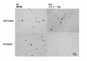
- FIG. 1 (B) shows pathologically stained images of AD and PSP cases with commercially available antibodies. An antibody that specifically detects aspartic acid at position 279 indicates that tau accumulated in AD cases is detected. The figure which analyzed the reactivity with respect to the peptide of the antibody which detects the aspartic acid of the 279th position specifically by ELISA.
- tauopathy can be roughly classified according to the difference in tau isoform accumulated in inclusion bodies.
- 3 repeat tau can be detected by an RD3 antibody that recognizes a peptide (KHQPGGGGVIVYKPV: SEQ ID NO: 1) in a region adjacent to the C-terminal microtubule-binding regions I and III, and 4 repeat tau can be detected by a microtubule-binding region. It can be detected by an RD4 antibody that recognizes a peptide containing II (VQIINKKLDLSNVQSKC: SEQ ID NO: 2) (see FIG. 1 (A)).
- VQIINKKLDLSNVQSKC SEQ ID NO: 2
- tau protein in cerebrospinal fluid cannot be quantified by these antibodies, and tauopathy has not been differentially diagnosed.
- the present inventor has found that in AD and PSP pathological specimens, antibodies that recognize 4 repeat tau have different reactivity due to deamidation, and have completed the present invention.
- the present inventor has prepared a commercially available polyclonal antibody (4R antibody) prepared against the peptide of SEQ ID NO: 3 with an antibody that recognizes the peptide represented by SEQ ID NO: 2 and an antibody that recognizes the peptide represented by SEQ ID NO: 3. I found it mixed. As a result of analysis using the purified antibody, it was revealed that tau deamidated at position 279 exists only in AD. That is, an antibody specific for a peptide deamidated at position 279 detects only tau accumulated in AD.
- 4R antibody polyclonal antibody
- the present inventor has found for the first time that tau deamidated asparagine at position 279 is specifically detected by AD among neurodegenerative diseases collectively referred to as tauopathy.
- deamidated tau is detected by an antibody.
- any reagent can be used as long as it is a reagent that can detect tau in which asparagine at position 279 is deamidated.
- the specific binding reagent in the present specification is not limited to an antibody, and may include a reagent that specifically binds to a specific molecule such as an aptamer. Nucleic acid aptamers and peptide aptamers may be prepared by known methods, and those that can specifically bind to tau in which the 279th position is substituted with aspartic acid may be selected.
- the specific binding reagent of the present invention includes a modified form. Specific examples include those that can be labeled with radioactive substances and used as PET diagnostic agents, and those that are labeled with fluorescent substances and can be detected without using secondary antibodies.
- Fab, Fab ′, F (ab ′) 2 single chain antibody (scFv), disulfide-stabilized V region fragment (dsFv), or a functional antibody such as a peptide containing CDR Fragments are included.
- a functional fragment of an antibody can be obtained by a known method such as digestion with an enzyme such as pepsin or papain.
- detection of a specific binding reagent including an antibody may be performed by directly labeling the specific binding reagent, an indirect method using a secondary antibody or a tertiary antibody, or avidin- Known detection techniques such as detection using biotin can be used.
- Antibodies that recognize repeat tau include monoclonal antibodies (RD4 antibody, manufactured by Merck & Co., Inc.) using the peptide shown in SEQ ID NO: 2 as an immunogen, and polyclonal antibodies using the peptide shown in SEQ ID NO: 3 as an immunogen. (4R antibody, manufactured by Cosmo Bio Inc.) is commercially available.
- the commercially available antibody obtained by immunizing the peptide shown in SEQ ID NO: 3 is found to contain an antibody in which the amino acid at position 279 reacts with both an asparagine peptide and an aspartic acid peptide. It became.
- An antibody that specifically recognizes tau in which the amino acid at position 279 is deamidated to aspartic acid (hereinafter referred to as D279 antibody) is obtained from a commercially available antibody or an animal immunized with the peptide shown in SEQ ID NO: 3.
- the obtained antibody can be used after being purified by a peptide column.
- the antibody may be purified by utilizing specific binding between the antibody and the peptide, and may be purified by combining known anti-peptide antibody purification methods. Specifically, for example, it can be purified as follows.
- D279 antibody that reacts only with a peptide in which the amino acid at position 279 reacts with asparagine or with both aspartic acid and reacts only with a peptide whose position 279 is aspartic acid is performed, for example, as follows. be able to.
- the antibody in which both are mixed is adsorbed onto a column to which the peptide of SEQ ID NO: 3 (a peptide in which the position 279 is substituted with aspartic acid, hereinafter sometimes referred to as D279 peptide) is bound, and the adsorbed fraction is eluted.
- the peptide of SEQ ID NO: 2 (a peptide in which the position 279 is substituted with asparagine, hereinafter sometimes referred to as N279 peptide) is adsorbed to a coupled column and purified by collecting a fraction that does not adsorb. Can do. Further, the fraction that is not adsorbed on the N279 peptide column can be further adsorbed on the D279 peptide column, and the adsorbed fraction can be eluted and purified. Further, when the antibody is mixed with the N279 peptide, the antibody that binds to the N279 peptide is masked, so that only the antibody fraction highly reactive with the D279 peptide is used.
- Antibody production method The antibody can be prepared with reference to a known method (Non-patent Document 7). Polyclonal antibodies and monoclonal antibodies can be prepared by immunizing rabbits and mice with an antibody specific for the peptide shown in SEQ ID NO: 2 or 3.
- body fluids such as cerebrospinal fluid, blood, urine, and biopsy specimens can be used as samples.
- body fluids such as cerebrospinal fluid, blood, urine, and biopsy specimens
- nasal mucosa, skin, salivary gland, gastrointestinal mucosa and the like in which detection of tau and ⁇ -synuclein is recognized can be suitably used.
- Cerebrospinal fluid is preferably used from the viewpoint of sensitivity. Specific examples of immunostaining and ELISA are given below, but any method may be used as long as it is a method capable of detecting a specific reaction.
- Immunostaining method For tissue staining, formalin-fixed and paraffin-embedded tissue is sliced by a conventional method, and stained using a primary antibody specific to the above tau peptide and a labeled secondary antibody. Can be detected.
- the detection method can be performed using a known method such as coloring by an enzyme label or a method of detecting a fluorescent label.
- the peptide of SEQ ID NO: 2 or 3 is placed in a 96-well ELISA plate, left standing for a certain period of time, and then immobilized.
- the solid-phased plate may be washed and blocked and used for inspection.
- the prepared antibody or purified antibody is put into an ELISA plate on which the peptide is immobilized, and then an antibody that binds to the antibody of the animal from which the antibody was prepared, is used And may be detected by a conventional method.
- Detection may be performed using an antibody in which the antibody of the animal from which the antibody is produced is enzyme-labeled, or using an enzyme-labeled antibody as a secondary antibody for detection. Enzyme substrate is added, the reaction is stopped for a certain period of time, the reaction is stopped, and the enzyme product is measured using a microplate reader.
- the specimen When examining a patient specimen, for example, the specimen is put on a plate on which an antibody that binds to tau protein regardless of 4 repeat tau or 3 repeat tau is immobilized, and then reacted with D279 antibody. After the enzyme-labeled secondary antibody that binds to is added and bound, the enzyme substrate is added, the reaction is stopped for a certain period of time, and the enzyme product is measured using a microplate reader. AD, PSP and PiD can be distinguished by performing detection using RD3 antibody and RD4 antibody in addition to D279 antibody.
- patient body fluids such as cerebrospinal fluid and blood are preferably used, but cerebrospinal fluid is more preferably used because tau protein is present in a higher concentration in the cerebrospinal fluid.
- detection can be performed using an anti-tau protein antibody after binding a specimen using a plate on which a D279 antibody is immobilized. Furthermore, detection can be performed by a detection method usually performed using an antibody or the like, such as detection by RIA or fluorescently labeled antibody instead of ELISA.
- a compound capable of suppressing the deamidation of tau protein to aspartic acid at position 279 is highly likely to be effective as a drug for Alzheimer's disease.
- a compound that is effective against Alzheimer's disease can be selected by screening a compound using cultured cells as an index of deamidation of aspartic acid at position 279.
- any cells may be used as long as deamidation at position 279 of tau is observed.
- suitable culture cells neuronal cells established from Alzheimer's disease patients or model animals, and neuronal cells derived from iPS cells obtained from Alzheimer's disease patients, 4 repeat tau is forcibly expressed, and deamidation occurs. Examples of such cells have been confirmed.
- the deamidation at the 279 position of tau is quantified by a known method such as ELISA using D279 antibody or a specific binding reagent having equivalent reactivity to suppress deamidation. Can be screened for.
- model animal The compound which showed the effect by the screening system using a cultured cell can further be examined using a model animal.
- a model animal an aging model such as an aged normal animal or an aging promoting model mouse can be used.
- transgenic animals that modify the tau gene described in Patent Documents 3 to 5 and cause Alzheimer-like lesions may be used.
- a drug can be screened by administering a candidate compound to the animal, using inhibition of deamidation at position 279 of tau protein as an index.
- Example 1 Purification of antibody
- the affinity column for purifying the antibody was used by binding the peptide of SEQ ID NO: 3 to the formyl group of Cellufine Formyl (manufactured by JNC Corporation). Specifically, after washing Serfine Formyl with distilled water, the suspension was suspended in a coupling buffer (50 mM carbonate-bicarbonate buffer, pH 8.5) together with the D279 peptide represented by SEQ ID NO: 3 at room temperature. Shake for 30 minutes. Next, sodium cyanoborohydride is added and shaken at 4 ° C. overnight to reduce excess formyl group.
- a coupling buffer 50 mM carbonate-bicarbonate buffer, pH 8.5
- polyclonal antibody an antibody in which an antibody that reacts with a tau peptide whose amino acid at position 279 is asparagine and an antibody that reacts with a tau peptide that is aspartic acid are mixed, hereinafter sometimes referred to as DN279 antibody
- N279 antibody an antibody in which an antibody that reacts with a tau peptide whose amino acid at position 279 is asparagine and an antibody that reacts with a tau peptide that is aspartic acid are mixed, hereinafter sometimes referred to as DN279 antibody
- N279 peptide represented by SEQ ID NO: 2 and placed in an affinity column equilibrated with TBS (0.15 M NaCl, 20 mM Tris-HCl, pH 7.5), mixed by inversion, and mixed at 4 ° C. for 8 hours. Adsorb to the column.
- the unadsorbed antibody After the unadsorbed antibody is allowed to flow out, it is washed with TBS, a washing buffer, and 0.15 M NaCl, and then the antibody adsorbed on the column is eluted with an elution buffer.
- the eluted antibody was confirmed to bind specifically to the D279 peptide shown in SEQ ID NO: 3 by ELISA or the like, and used in subsequent experiments.
- tissue staining was performed using an unpurified polyclonal antibody (DN279 antibody) prepared using the peptide shown in SEQ ID NO: 3 or a D279 antibody purified using a peptide column. went.
- DN279 antibody unpurified polyclonal antibody
- the samples used are pathological specimens of the olfactory intestine of a patient diagnosed with AD by definitive diagnosis and the lower olive nucleus of a patient diagnosed with PSP.
- Formalin-fixed, paraffin-embedded pathological specimens are sliced to 5-6 ⁇ m, deparaffinized, heated in an autoclave at 0.01 ° C. for 10 minutes in 0.01 M citrate buffer, and then immersed in 100% formic acid for 10 minutes. Then, antigenic activation treatment was performed.
- Endogenous peroxidase was inactivated with 1% hydrogen peroxide, blocked with PBS containing 5% serum, and then purified as RD4 antibody (1000-fold dilution), 4R antibody (DN279 antibody, 30,000 dilution), or purified as a primary antibody.
- D279 antibody (diluted 20 times) was reacted at 4 ° C. for 2 days or more, washed, and then used as a secondary antibody, biotinylated anti-rabbit IgG (Vector, diluted 1000 times), or biotinylated anti-mouse IgG (Vector, 1000 times dilution) and at room temperature for 2 hours.
- the secondary antibody was washed, reacted with Avidin biotin-peroxidase complex (manufactured by Vector, ABC Elite, diluted 1000 times) for 1 hour, and then developed with diaminobenzidine for detection (FIG. 2).
- the DN279 antibody in which an antibody that recognizes both aspartic acid and asparagine at the 279th amino acid of tau protein is mixed, recognizes tau of both AD and PSP samples, but D279 antibody recognizes tau of AD but PSP Can't recognize tau.
- the pathological tissue is shown here, the detection can be similarly performed using patient cells mixed in cerebrospinal fluid.
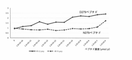
- FIG. 3 shows the results of analyzing the reactivity of the D279 antibody by coating the microtiter plate at different concentrations of the peptide represented by SEQ ID NO: 2 (N279 peptide) and the peptide represented by SEQ ID NO: 3 (D279 peptide).
- Peptide immobilization was carried out at 4 ° C for 16 hours at a concentration of 1.0 ⁇ 10 ⁇ 16 to 1.0 ⁇ 10 ⁇ 6 ⁇ M / ⁇ l on a 96-well ELISA plate (Sumitomo Bakelite Co., Ltd.). Added. After washing with ELISA washing buffer (0.05% Tween-20 / PBS), blocking with ELISA blocking buffer (10% BSA / 0.01 ⁇ PBS), washing again, and then adding purified D279 antibody And left at room temperature for 1.5 hours. After washing, HRP-labeled goat anti-rabbit IgG (Pierce) was added as a secondary antibody and allowed to stand at room temperature for 1 hour. After washing, color was developed using O-phenylenediamine, and measurement was performed with a microplate reader (Chameleon, manufactured by Hidex).
- the D279 antibody can recognize the D279 peptide even at a very low concentration of 1 ⁇ 10 ⁇ 16 ⁇ mol / ⁇ l. It is also clear from this result that the D279 antibody is very specific for the D279 peptide. Therefore, tau containing D279 peptide, that is, AD can be detected with very high specificity.
- the D279 antibody can diagnose Alzheimer's disease among tauopathy. Until now, it is possible to perform a test with good sensitivity and accuracy for the diagnosis of tauopathy for which no objective index has been obtained.
Landscapes
- Health & Medical Sciences (AREA)
- Life Sciences & Earth Sciences (AREA)
- Chemical & Material Sciences (AREA)
- Engineering & Computer Science (AREA)
- Immunology (AREA)
- Molecular Biology (AREA)
- Medicinal Chemistry (AREA)
- Biomedical Technology (AREA)
- General Health & Medical Sciences (AREA)
- Biochemistry (AREA)
- Hematology (AREA)
- Urology & Nephrology (AREA)
- Physics & Mathematics (AREA)
- General Physics & Mathematics (AREA)
- Food Science & Technology (AREA)
- Analytical Chemistry (AREA)
- Pathology (AREA)
- Biophysics (AREA)
- Biotechnology (AREA)
- Cell Biology (AREA)
- Microbiology (AREA)
- Organic Chemistry (AREA)
- Bioinformatics & Cheminformatics (AREA)
- Pharmacology & Pharmacy (AREA)
- Genetics & Genomics (AREA)
- Proteomics, Peptides & Aminoacids (AREA)
- Peptides Or Proteins (AREA)
- Investigating Or Analysing Biological Materials (AREA)
- Preparation Of Compounds By Using Micro-Organisms (AREA)
Abstract
Description
タウオパチーと総称されるリン酸化タウの蓄積が見られる神経変性疾患を鑑別する検査方法に関する。特に、4リピートタウが蓄積するアルツハイマー病(Alzheimer’s disease、以下ADと記載することもある。)と、進行性核上性麻痺(Progressive supranuclear palsy、以下PSPと記載することもある。)/大脳皮質基底核変性症(corticobasal degeneration、以下CBDと記載することもある。)を区別することのできる4リピートタウに対する特異的結合試薬、及びこれを用いた検査方法に関する。また、この特異的結合試薬を用いた医薬のスクリーニング方法に関する。 The present invention relates to a test method for distinguishing neurodegenerative diseases in which accumulation of phosphorylated tau, which is collectively called tauopathy, is observed. In particular, Alzheimer's disease (hereinafter sometimes referred to as AD) in which 4 repeat tau accumulates and progressive supranuclear palsy (hereinafter sometimes referred to as PSP) / The present invention relates to a specific binding reagent for 4-repeat tau capable of distinguishing corticobasal degeneration (hereinafter sometimes referred to as CBD), and a test method using the same. Moreover, it is related with the screening method of the pharmaceutical using this specific binding reagent.
高齢者人口の増加とともに認知症患者も増加し、平成24年の認知症患者数は462万人に達していると言われている(内閣府 平成28年版高齢社会白書(概要版))。今後高齢者の増加とともに、認知症患者はますます増加すると考えられており、大きな社会的問題となっている。 It is said that the number of patients with dementia has increased with the increase in the elderly population, and the number of patients with dementia in 2012 has reached 4.62 million (Cabinet Office, 2016 White Paper on Aging Society (Summary)). As the number of elderly people increases in the future, the number of patients with dementia is considered to increase more and more, which is a big social problem.
しかしながら、認知症と言っても、同じような症状を呈する原因疾患は多様であることが明らかとなってきた。例えば、大脳皮質基底核変性症候群(Corticobasal syndrome:CBS)の臨床像を呈する疾患には、CBD、AD、PSPの他に、ピック病(Pick’s desease、以下PiDと記載することもある。)をはじめとする様々な疾患が含まれていることが明らかとなっている(非特許文献1)。 However, it has been clarified that even if it is dementia, there are various causative diseases that exhibit similar symptoms. For example, for diseases exhibiting a clinical image of corticobasal syndrome (CBS), in addition to CBD, AD, and PSP, Pick's disease (hereinafter sometimes referred to as PiD) may also be used. It has been clarified that various diseases including the above are included (Non-patent Document 1).
近年、画像診断の進歩に伴い、認知症の診断にもCTやMRIなどの画像診断が欠かせないものとなってきている。しかし、画像診断は局在性変化の確認と経時的な変化の追跡を可能とするものの、質的な変化をとらえるには十分でなく、客観的な診断基準に反映させるにも限界がある。 In recent years, with the advancement of diagnostic imaging, diagnostic imaging such as CT and MRI has become indispensable for the diagnosis of dementia. However, although image diagnosis enables confirmation of localization changes and tracking of changes over time, it is not sufficient to capture qualitative changes, and there is a limit to reflecting it in objective diagnostic criteria.
そのため認知症はMRIなどを用いた画像診断による検査所見や臨床所見だけでは正確に生前診断を行うことが未だに困難であると言われており、患者の死後に病理診断を行い、はじめて確定診断を行うことができるのが現状である。したがって、生前の臨床診断が正確でないことから、適切な治療を受けられていない患者も多いと考えられている。そのため、髄液、血液などの体液中のバイオマーカーを用いた診断精度、特異度の高い検査方法の確立が望まれている。 Therefore, it is said that dementia is still difficult to make an accurate prenatal diagnosis only by examination findings and clinical findings by image diagnosis using MRI, etc., and a pathological diagnosis is made after the patient's death. It is the current situation that can be done. Therefore, it is believed that many patients are not receiving appropriate treatment due to inaccurate clinical diagnosis before birth. Therefore, establishment of a test method with high diagnostic accuracy and specificity using biomarkers in body fluids such as cerebrospinal fluid and blood is desired.
さらに、神経変性疾患を正確に診断することのできるマーカーは、患者の確定診断により、治療法の選択に寄与するだけではなく、新薬開発にも大きく寄与することができる。例えば、現状では「アルツハイマー病である確率が高い患者群」を対象に、新薬の臨床試験を行っているにすぎない。その結果、臨床試験の対象としている群には、他の神経変性疾患の患者も含まれている可能性が高い。したがって、アルツハイマー病といった特定の神経変性疾患に対する薬剤の効果を的確に判断することができない。臨床症状による医師の判断だけではなく、客観的な検査結果により診断精度を高めることができれば、適切な患者群に対して臨床試験を行うことが可能となり、候補薬の効果について正しい評価をくだすことができる。 Furthermore, markers that can accurately diagnose neurodegenerative diseases not only contribute to the selection of treatment methods but also greatly contribute to the development of new drugs by definitive diagnosis of patients. For example, at present, only a clinical trial of a new drug is being conducted for “a group of patients with a high probability of Alzheimer's disease”. As a result, it is highly likely that patients with other neurodegenerative diseases are included in the group targeted for clinical trials. Therefore, the effect of the drug on a specific neurodegenerative disease such as Alzheimer's disease cannot be accurately determined. If the accuracy of diagnosis can be improved not only by the doctor's judgment based on clinical symptoms but also by objective test results, it will be possible to conduct clinical trials on appropriate patient groups and make a correct evaluation of the effects of candidate drugs. Can do.
我が国の認知症の原因疾患は、脳血管性が最も多いとされてきたが、近年の疫学的研究によればアルツハイマー病が最多とも言われている。したがって、アルツハイマー病を他の神経変性疾患と臨床的に区別することは今後の治療薬の開発にとっても非常に重要なこととなる。 The causative disease of dementia in Japan has been said to be the most cerebrovascular, but according to recent epidemiological studies, Alzheimer's disease is said to be the most common. Therefore, clinically distinguishing Alzheimer's disease from other neurodegenerative diseases is very important for the development of future therapeutic agents.
アルツハイマー病の原因としては、病理学的な特徴とされる老人斑を構成するアミロイドβにその原因を求める考えと、神経原線維を構成するタウに注目する立場があるが、原因が明らかではないのが現状である。アルツハイマー病の診断には、体液中のアミロイドβやタウタンパク質を検出する方法や、タウのイメージングプローブにより検出する方法が提案されているが、未だ臨床診断を左右できるほどの精度には達していない(特許文献1、2)。
As the cause of Alzheimer's disease, there is a position to focus on amyloid β, which constitutes senile plaque, which is characterized by pathological features, and tau that constitutes neurofibrils, but the cause is not clear is the current situation. For the diagnosis of Alzheimer's disease, methods of detecting amyloid β and tau protein in body fluids and methods of detecting with tau imaging probes have been proposed, but they have not yet reached the accuracy that can influence clinical diagnosis. (
タウは微小管の重合や安定化に寄与するタンパク質であり正常脳にも存在する。タウは、AD、PSP、CBD、PiDなどにおいて異常に蓄積し、神経原線維変化やその他の封入体として観察される。リン酸化タウが異常に蓄積するこれらの疾患を総称して「タウオパチー」と呼んでいる(非特許文献2)。 Tau is a protein that contributes to the polymerization and stabilization of microtubules and is also present in the normal brain. Tau accumulates abnormally in AD, PSP, CBD, PiD, etc. and is observed as neurofibrillary tangles and other inclusions. These diseases in which phosphorylated tau accumulates abnormally are collectively called “tauopathy” (Non-patent Document 2).
タウオパチーでは、正常脳のタウにも起こるリン酸化が亢進して蓄積することが知られている。非特許文献3には、脳脊髄液中のリン酸化タウの全タウタンパク質に対する比により前頭側頭型認知症(frontotemporal lobar degeneration:FTLD)のサブタイプを分類できることが報告されている。例えば、ALS(Amyotrophic lateral sclerosis)を伴うFTLD-TDPなどのサブタイプにおけるタウのリン酸化とともに、タウとアミロイドβが蓄積するADもリン酸化タウの量比が異なっていることが示されている。しかし、統計的にはリン酸化タウの量は、健常者や各サブタイプ群間での差がみられるものの群間の重なりが大きく、個々の臨床例の診断を髄液中のリン酸化タウの量的違いをもとに鑑別するには至っていない。
It is known that tauopathy increases and accumulates phosphorylation that occurs in normal brain tau. Non-Patent
タウはエクソン2、3、10の選択的スプライシングにより6つのアイソフォームが存在する(図1(A)参照。)。このうちC末端側に存在する微小管結合領域の繰り返し構造の数が、3つのものを3リピートタウ、4つのものを4リピートタウと呼んでいる。タウアイソフォームは、発生段階で発現調節を受けていることが知られており、成人の脳ではタウの6つのアイソフォーム全てが発現している。
Tau has six isoforms due to alternative splicing of
タウオパチーの変性神経疾患患者脳の封入体中の不溶化タウの解析から、タウオパチーは3リピートタウのみが蓄積するもの(PiD)と、4リピートタウのみが蓄積するもの(PSP/CBD)と、両者が蓄積するもの(AD)と大別されることが明らかになってきた(非特許文献4)。もし、3リピートタウと4リピートタウの髄液中の量を区別して定量できれば、これらの疾患を質的に区別する手がかりになると期待される。しかし、髄液中の3リピートタウ、4リピートタウの量をイムノPCR法で測定しても、これらの疾患を区別するにいたってはいない(非特許文献5)。これらアイソフォームは正常脳でも発現しているため、正常例、AD、その他の認知症の個々の例をタウアイソフォームの量で鑑別できる精度は得られていない。 Analysis of insoluble tau in inclusion bodies of brain inclusions in patients with degenerative neurological diseases of tauopathy shows that tauopathy has only 3 repeat tau (PiD), 4 repeat tau only (PSP / CBD), and both It has been clarified that it is broadly classified as accumulated (AD) (Non-patent Document 4). If the amount of 3 repeat tau and 4 repeat tau in the cerebrospinal fluid can be distinguished and quantified, it is expected to provide a clue to distinguish these diseases qualitatively. However, even if the amount of 3 repeat tau and 4 repeat tau in the cerebrospinal fluid is measured by an immuno PCR method, these diseases are not distinguished (Non-patent Document 5). Since these isoforms are also expressed in the normal brain, there is no accuracy that can distinguish normal cases, AD, and other cases of dementia by the amount of tau isoforms.
上述のように、脳に病変が生じ、認知症などを生じる疾患の鑑別診断は、現在のところ有効なバイオマーカーが存在しないため、臨床症状と画像診断による確率的推論に頼っているのが現状である。アルツハイマー病をはじめとする神経変性疾患は、同一疾患内でも病変の分布に症例毎の違いがあり、個々の症例における臨床症状、画像所見の違いも大きい。そのため、画像診断、臨床症状のみによる評価、診断方法は、医師の判断によるばらつきが大きいと言われている。適切な治療を行うためには、客観性の高い方法による信頼性の高い診断が必要である。また、各疾患に有効な医薬の開発には、正確な診断がくだされた対象群に対して臨床試験を行い、解析、評価がなされる必要がある。そのためには、各疾患の正確な診断は必須の要件である。本発明は、信頼性の高い検査方法、検査に必要な抗体、検査キット、また、これを用いた医薬のスクリーニング方法を提供することを課題とする。 As mentioned above, there is currently no effective biomarker for differential diagnosis of diseases that cause lesions in the brain and cause dementia, etc., so it currently depends on probabilistic inference based on clinical symptoms and image diagnosis It is. In neurodegenerative diseases such as Alzheimer's disease, the distribution of lesions varies from case to case even within the same disease, and the clinical symptoms and image findings in individual cases are also large. For this reason, it is said that image diagnosis, evaluation based on clinical symptoms only, and diagnosis methods vary greatly depending on the judgment of a doctor. In order to perform appropriate treatment, a highly reliable diagnosis by a highly objective method is necessary. In addition, in order to develop a drug effective for each disease, it is necessary to conduct a clinical trial on a target group for which an accurate diagnosis has been made, and to analyze and evaluate it. To that end, accurate diagnosis of each disease is an essential requirement. An object of the present invention is to provide a highly reliable test method, an antibody necessary for the test, a test kit, and a pharmaceutical screening method using the same.
本発明は、アルツハイマー病を他の神経変性疾患と臨床的に区別する検査方法、これに用いる検査試薬、検査キット、さらに医薬のスクリーニング方法に関する。
(1)検体中のタウタンパク質の279位のアスパラギンがアスパラギン酸に置換しているペプチドに特異的に結合する特異的結合試薬によりアルツハイマー病と、進行性核上性麻痺/大脳皮質基底核変性症を鑑別する検査方法。
(2)前記特異的結合試薬が抗体である(1)記載の検査方法。
(3)前記検体が、髄液、血液、脳神経組織、生検組織である(1)又は(2)に記載の検査方法。
(4)ELISA、又は免疫組織染色により検出することを特徴とする(2)又は(3)記載の検査方法。
(5)アルツハイマー病と、進行性核上性麻痺/大脳皮質基底核変性症を鑑別する特異的結合試薬であって、タウタンパク質の279位のアスパラギンがアスパラギン酸に置換しているペプチドに対して特異的に結合することを特徴とする試薬。
(6)前記特異的結合試薬が抗体である(5)記載の特異的結合試薬。
(7)前記抗体がモノクローナル抗体である(6)記載の特異的結合試薬。
(8)前記抗体がポリクローナル抗体を精製して得られたものである(6)記載の特異的結合試薬。
(9)(5)~(8)いずれか1つ記載の特異的結合試薬と検出に必要な試薬を備えていることを特徴とする検査キット。
(10)前記検出がELISA又は免疫染色によるものであることを特徴とする(9)記載の検査キット。
(11)タウタンパク質の279位の脱アミド化を指標としてスクリーニングすることを特徴とする医薬のスクリーニング方法。
(12)タウタンパク質の279位のアスパラギンがアスパラギン酸に置換しているペプチドに特異的に結合する特異的結合試薬が、患者から得られた検体中のタウタンパク質を認識する場合には、アルツハイマー病であると診断する方法。
(13)前記特異的結合試薬が抗体である(12)記載の診断方法。
(14)前記検体が、髄液、血液、脳神経組織、生検組織である(12)又は(13)に記載の診断方法。
(15)ELISA、又は免疫組織染色により検出することを特徴とする(13)又は(14)記載の診断方法。
The present invention relates to a test method for clinically distinguishing Alzheimer's disease from other neurodegenerative diseases, a test reagent used for this, a test kit, and a pharmaceutical screening method.
(1) Alzheimer's disease and progressive supranuclear paralysis / basal ganglia degeneration with a specific binding reagent that specifically binds to a peptide in which asparagine at position 279 of the tau protein in the sample is substituted with aspartic acid Inspection method to differentiate.
(2) The test method according to (1), wherein the specific binding reagent is an antibody.
(3) The examination method according to (1) or (2), wherein the specimen is cerebrospinal fluid, blood, cranial nerve tissue, or biopsy tissue.
(4) The inspection method according to (2) or (3), wherein detection is performed by ELISA or immunohistochemical staining.
(5) A specific binding reagent that distinguishes Alzheimer's disease from progressive supranuclear palsy / basal ganglia degeneration, for peptides in which the asparagine at position 279 of tau protein is substituted with aspartic acid A reagent characterized by binding specifically.
(6) The specific binding reagent according to (5), wherein the specific binding reagent is an antibody.
(7) The specific binding reagent according to (6), wherein the antibody is a monoclonal antibody.
(8) The specific binding reagent according to (6), wherein the antibody is obtained by purifying a polyclonal antibody.
(9) A test kit comprising the specific binding reagent according to any one of (5) to (8) and a reagent necessary for detection.
(10) The test kit according to (9), wherein the detection is performed by ELISA or immunostaining.
(11) A method for screening a drug, comprising screening using deamidation at position 279 of tau protein as an index.
(12) When a specific binding reagent that specifically binds to a peptide in which asparagine at position 279 of tau protein is substituted with aspartic acid recognizes tau protein in a sample obtained from a patient, Alzheimer's disease How to diagnose that.
(13) The diagnostic method according to (12), wherein the specific binding reagent is an antibody.
(14) The diagnostic method according to (12) or (13), wherein the specimen is cerebrospinal fluid, blood, cranial nerve tissue, or biopsy tissue.
(15) The diagnostic method according to (13) or (14), which is detected by ELISA or immunohistochemical staining.
上述のように、タウオパチーは封入体に蓄積するタウアイソフォームの違いにより大別することができる。3リピートタウは、C末端側の微小管結合領域IとIIIに隣接する領域のペプチド(KHQPGGGKVIVYKPV:配列番号1)を認識するRD3抗体により検出することができ、4リピートタウは、微小管結合領域IIを含むペプチド(VQIINKKLDLSNVQSKC:配列番号2)を認識するRD4抗体により検出することができる(図1(A)参照。)。しかしながら、これら抗体によって、髄液中のタウタンパク質を定量することはできておらず、タウオパチーを鑑別診断するにはいたっていない。本発明者は、ADとPSPの病理標本において、4リピートタウを認識する抗体が脱アミド化により反応性が違うことを見出し、本発明を完成させた。 As described above, tauopathy can be roughly classified according to the difference in tau isoform accumulated in inclusion bodies. 3 repeat tau can be detected by an RD3 antibody that recognizes a peptide (KHQPGGGGVIVYKPV: SEQ ID NO: 1) in a region adjacent to the C-terminal microtubule-binding regions I and III, and 4 repeat tau can be detected by a microtubule-binding region. It can be detected by an RD4 antibody that recognizes a peptide containing II (VQIINKKLDLSNVQSKC: SEQ ID NO: 2) (see FIG. 1 (A)). However, tau protein in cerebrospinal fluid cannot be quantified by these antibodies, and tauopathy has not been differentially diagnosed. The present inventor has found that in AD and PSP pathological specimens, antibodies that recognize 4 repeat tau have different reactivity due to deamidation, and have completed the present invention.
4リピートタウは279位がアスパラギンとして翻訳される。その一部は脱アミド化されアスパラギン酸に変化することが報告されている(非特許文献6)。今まで279位のアスパラギンが脱アミド化したタウタンパク質は、AD、PSP・CBDなどの疾患に関わりなく存在すると考えられていた。 4 Repeat Tau is translated as asparagine at 279th place. It has been reported that some of them are deamidated and converted to aspartic acid (Non-patent Document 6). Until now, it was thought that the tau protein in which asparagine at position 279 was deamidated was present regardless of diseases such as AD, PSP and CBD.
これは、市販の抗体を用いて病理標本を染色すると、279位がアスパラギンであるタウペプチド(配列番号2)に対して作製された抗体(RD4抗体)でも、279位をアスパラギン酸に置換したペプチドであるVQIIDKKLDLSNVQSKC(配列番号3、下線部は279位のアミノ酸を示す。)に対して作製された抗体(4R抗体)でも、4リピートタウの蓄積が同じように検出されていたからである(図1(B)参照。)。 This is because when a pathological specimen is stained with a commercially available antibody, an antibody (RD4 antibody) prepared against a tau peptide (SEQ ID NO: 2) in which position 279 is asparagine is a peptide in which position 279 is substituted with aspartic acid. This is because accumulation of 4 repeat tau was detected in the same manner even in an antibody (4R antibody) prepared against VQII D KKLDLSNVQSKC (SEQ ID NO: 3, the underlined portion indicates the amino acid at position 279). 1 (B).)
本発明者は、配列番号3のペプチドに対して作製された市販ポリクローナル抗体(4R抗体)に、配列番号2で示されるペプチドを認識する抗体と、配列番号3で示されるペプチドを認識する抗体が混在していることを見出した。精製した抗体を用いて解析した結果、279位が脱アミド化したタウは、ADのみに存在することが明らかとなった。すなわち、279位が脱アミド化したペプチドに対して特異的な抗体は、ADに蓄積したタウのみを検出する。 The present inventor has prepared a commercially available polyclonal antibody (4R antibody) prepared against the peptide of SEQ ID NO: 3 with an antibody that recognizes the peptide represented by SEQ ID NO: 2 and an antibody that recognizes the peptide represented by SEQ ID NO: 3. I found it mixed. As a result of analysis using the purified antibody, it was revealed that tau deamidated at position 279 exists only in AD. That is, an antibody specific for a peptide deamidated at position 279 detects only tau accumulated in AD.
279位のアスパラギンが脱アミド化したタウが、タウオパチーとして総称される神経変性疾患のうちADで特異的に検出されることは、本発明者がはじめて見出したことである。脱アミド化したタウはここでは抗体により検出しているが、279位のアスパラギンが脱アミド化しているタウを検出することができる試薬であれば抗体に限らずどのような試薬を用いてもよい。したがって、本明細書で特異的結合試薬という場合には、抗体に限らず、アプタマーなど特定の分子と特異的に結合する試薬を含むことができる。核酸アプタマー、ペプチドアプタマーは公知の方法によって作製し、279位がアスパラギン酸に置換されているタウと特異的に結合することができるものを選択すればよい。さらに、本発明の特異的結合試薬には、修飾された形態のものも含む。具体的には、放射性物質により標識しPET診断薬として用いることができるものや、蛍光物質により標識し二次抗体等を使用することなく検出することができるものなどが挙げられる。 The present inventor has found for the first time that tau deamidated asparagine at position 279 is specifically detected by AD among neurodegenerative diseases collectively referred to as tauopathy. Here, deamidated tau is detected by an antibody. However, any reagent can be used as long as it is a reagent that can detect tau in which asparagine at position 279 is deamidated. . Therefore, the specific binding reagent in the present specification is not limited to an antibody, and may include a reagent that specifically binds to a specific molecule such as an aptamer. Nucleic acid aptamers and peptide aptamers may be prepared by known methods, and those that can specifically bind to tau in which the 279th position is substituted with aspartic acid may be selected. Furthermore, the specific binding reagent of the present invention includes a modified form. Specific examples include those that can be labeled with radioactive substances and used as PET diagnostic agents, and those that are labeled with fluorescent substances and can be detected without using secondary antibodies.
また、本発明で抗体という場合には、Fab、Fab´、F(ab´)2、単鎖抗体(scFv)、ジスルフィド安定化V領域断片(dsFv)、もしくはCDRを含むペプチド等抗体の機能的断片が含まれる。抗体の機能的断片は、ペプシン、又はパパインなどの酵素によって消化する等公知の方法によって得ることができる。 In addition, when referring to an antibody in the present invention, Fab, Fab ′, F (ab ′) 2 , single chain antibody (scFv), disulfide-stabilized V region fragment (dsFv), or a functional antibody such as a peptide containing CDR Fragments are included. A functional fragment of an antibody can be obtained by a known method such as digestion with an enzyme such as pepsin or papain.
さらに、本発明において抗体をはじめとする特異的結合試薬の検出は、特異的結合試薬に直接標識を付与して行ってもよいし、二次抗体、三次抗体を用いた間接法、あるいはアビジン-ビオチンを用いた検出など、公知の検出技術を用いることができる。 Further, in the present invention, detection of a specific binding reagent including an antibody may be performed by directly labeling the specific binding reagent, an indirect method using a secondary antibody or a tertiary antibody, or avidin- Known detection techniques such as detection using biotin can be used.
以下、本発明について説明するが、以下の方法に限らず公知の方法を用いることができることは言うまでもない。 Hereinafter, the present invention will be described, but it goes without saying that not only the following method but also a known method can be used.
1.抗体の精製方法
4リピートタウを認識する抗体としては、配列番号2で示すペプチドを免疫原とするモノクローナル抗体(RD4抗体、メルク株式会社製)、配列番号3で示すペプチドを免疫原とするポリクローナル抗体(4R抗体、コスモ・バイオ株式会社製)が市販されている。
1. Antibody Purification Method 4 Antibodies that recognize repeat tau include monoclonal antibodies (RD4 antibody, manufactured by Merck & Co., Inc.) using the peptide shown in SEQ ID NO: 2 as an immunogen, and polyclonal antibodies using the peptide shown in SEQ ID NO: 3 as an immunogen. (4R antibody, manufactured by Cosmo Bio Inc.) is commercially available.
配列番号3に示すペプチドを免疫して得られた市販抗体は、本発明者の解析により279位のアミノ酸がアスパラギンのペプチド、アスパラギン酸のペプチドの両者に反応する抗体が混在していることが明らかとなった。279位のアミノ酸がアスパラギン酸に脱アミド化されているタウを特異的に認識する抗体(以下D279抗体と記載する。)は、市販の抗体、あるいは配列番号3に示すペプチドを免疫した動物から得られた抗体を、ペプチドカラムにより精製して使用することができる。抗体の精製方法は、抗体とペプチドとの特異的な結合を利用することによって精製すれば良く、公知の抗ペプチド抗体の精製法を組合せて精製することができる。具体的には、例えば以下のようにして精製することができる。 The commercially available antibody obtained by immunizing the peptide shown in SEQ ID NO: 3 is found to contain an antibody in which the amino acid at position 279 reacts with both an asparagine peptide and an aspartic acid peptide. It became. An antibody that specifically recognizes tau in which the amino acid at position 279 is deamidated to aspartic acid (hereinafter referred to as D279 antibody) is obtained from a commercially available antibody or an animal immunized with the peptide shown in SEQ ID NO: 3. The obtained antibody can be used after being purified by a peptide column. The antibody may be purified by utilizing specific binding between the antibody and the peptide, and may be purified by combining known anti-peptide antibody purification methods. Specifically, for example, it can be purified as follows.
279位のアミノ酸がアスパラギン、あるいはアスパラギン酸の両者に反応する抗体が混在している抗体から、279位がアスパラギン酸であるペプチドにのみ反応するD279抗体の精製は、例えば、以下のようにして行うことができる。両者が混在する抗体を、配列番号3のペプチド(279位がアスパラギン酸に置換したペプチド、以下D279ペプチドと記載することもある。)を結合させたカラムに吸着させ、吸着した分画を溶出したり、あるいは、配列番号2のペプチド(279位がアスパラギンに置換したペプチド、以下N279ペプチドと記載することもある。)を結合したカラムに吸着させ、吸着しない分画を採取することによって精製することができる。また、N279ペプチドカラムに吸着しない分画を、さらにD279ペプチドカラムに吸着させ、吸着分画を溶出させて精製することができる。また、抗体をN279ペプチドと混和すれば、N279ペプチドに結合する抗体はマスクされることから、D279ペプチドに反応性の高い抗体分画のみを使用することになる。 Purification of D279 antibody that reacts only with a peptide in which the amino acid at position 279 reacts with asparagine or with both aspartic acid and reacts only with a peptide whose position 279 is aspartic acid is performed, for example, as follows. be able to. The antibody in which both are mixed is adsorbed onto a column to which the peptide of SEQ ID NO: 3 (a peptide in which the position 279 is substituted with aspartic acid, hereinafter sometimes referred to as D279 peptide) is bound, and the adsorbed fraction is eluted. Alternatively, the peptide of SEQ ID NO: 2 (a peptide in which the position 279 is substituted with asparagine, hereinafter sometimes referred to as N279 peptide) is adsorbed to a coupled column and purified by collecting a fraction that does not adsorb. Can do. Further, the fraction that is not adsorbed on the N279 peptide column can be further adsorbed on the D279 peptide column, and the adsorbed fraction can be eluted and purified. Further, when the antibody is mixed with the N279 peptide, the antibody that binds to the N279 peptide is masked, so that only the antibody fraction highly reactive with the D279 peptide is used.
2.抗体の作製方法
抗体は公知の方法(非特許文献7)を参考に作製することができる。配列番号2、又は3に示すペプチドに特異的な抗体を、ウサギ、マウスに抗原を免疫することによって、ポリクローナル抗体、モノクローナル抗体を作製することができる。
2. Antibody production method
The antibody can be prepared with reference to a known method (Non-patent Document 7). Polyclonal antibodies and monoclonal antibodies can be prepared by immunizing rabbits and mice with an antibody specific for the peptide shown in SEQ ID NO: 2 or 3.
3.検査方法
D279抗体を検査に用いる場合には、脳脊髄液、血液、尿などの体液や生検検体を試料として用いることができる。生検検体としては、タウやαシヌクレインの検出が認められている鼻粘膜、皮膚、唾液腺、消化管粘膜などを好適に用いることができる。脳脊髄液を用いることが、感度の点から好ましい。免疫染色法、ELISAを以下に具体例として挙げるが、特異的な反応を検出することができる方法であれば、これに限らずどのような方法を用いてもよい。
3. Testing Method When the D279 antibody is used for testing, body fluids such as cerebrospinal fluid, blood, urine, and biopsy specimens can be used as samples. As a biopsy specimen, nasal mucosa, skin, salivary gland, gastrointestinal mucosa and the like in which detection of tau and α-synuclein is recognized can be suitably used. Cerebrospinal fluid is preferably used from the viewpoint of sensitivity. Specific examples of immunostaining and ELISA are given below, but any method may be used as long as it is a method capable of detecting a specific reaction.
(1)免疫染色法
組織染色は、常法によりホルマリン固定、パラフィン包埋された組織を薄切し、上記のタウペプチドに特異的な一次抗体、標識された二次抗体を用いて染色を行い検出することができる。検出方法は、酵素標識による発色、蛍光標識を検出する方法など、公知の方法を用いて行うことができる。
(1) Immunostaining method For tissue staining, formalin-fixed and paraffin-embedded tissue is sliced by a conventional method, and stained using a primary antibody specific to the above tau peptide and a labeled secondary antibody. Can be detected. The detection method can be performed using a known method such as coloring by an enzyme label or a method of detecting a fluorescent label.
(2)ELISA
得られた抗体の反応性を確認する場合には、96穴ELISA用プレートに配列番号2又は3のペプチドを入れ、一定時間静置し、固相化を行う。固相化したプレートは洗浄、ブロッキングを行い、検査に使用すればよい。具体的には作製した抗体や精製した抗体をペプチドが固相化されたELISA用プレートに入れ、その後、抗マウス抗体、抗ラビット抗体等、抗体を作製した動物の抗体に結合する抗体等を用いて結合させ、常法により検出を行えば良い。検出は、抗体を作製した動物の抗体が酵素標識されている抗体を用いても、あるいは、酵素標識抗体を二次抗体として検出に用いても良い。酵素の基質を加え、一定時間反応後、反応停止し、マイクロプレートリーダーを用い酵素生成物の測定を行う。
(2) ELISA
In order to confirm the reactivity of the obtained antibody, the peptide of SEQ ID NO: 2 or 3 is placed in a 96-well ELISA plate, left standing for a certain period of time, and then immobilized. The solid-phased plate may be washed and blocked and used for inspection. Specifically, the prepared antibody or purified antibody is put into an ELISA plate on which the peptide is immobilized, and then an antibody that binds to the antibody of the animal from which the antibody was prepared, such as an anti-mouse antibody or anti-rabbit antibody, is used And may be detected by a conventional method. Detection may be performed using an antibody in which the antibody of the animal from which the antibody is produced is enzyme-labeled, or using an enzyme-labeled antibody as a secondary antibody for detection. Enzyme substrate is added, the reaction is stopped for a certain period of time, the reaction is stopped, and the enzyme product is measured using a microplate reader.
患者検体を検査する場合には、例えば、4リピートタウ、3リピートタウに関わらずタウタンパク質に結合する抗体を固相化したプレートに検体を入れ、その後、D279抗体と反応させた後、D279抗体に結合する酵素標識した二次抗体を加え結合させた後、酵素の基質を加え、一定時間反応させ反応停止し、マイクロプレートリーダーを用い酵素生成物の測定を行う。D279抗体の他に、RD3抗体、RD4抗体を用いてそれぞれ検出を行うことにより、ADと、PSP、PiDを区別することができる。患者検体としては、髄液、血液などの患者体液を用いることが好ましいが、髄液中により高濃度にタウタンパク質が存在することから、髄液を用いることがより好ましい。 When examining a patient specimen, for example, the specimen is put on a plate on which an antibody that binds to tau protein regardless of 4 repeat tau or 3 repeat tau is immobilized, and then reacted with D279 antibody. After the enzyme-labeled secondary antibody that binds to is added and bound, the enzyme substrate is added, the reaction is stopped for a certain period of time, and the enzyme product is measured using a microplate reader. AD, PSP and PiD can be distinguished by performing detection using RD3 antibody and RD4 antibody in addition to D279 antibody. As patient specimens, patient body fluids such as cerebrospinal fluid and blood are preferably used, but cerebrospinal fluid is more preferably used because tau protein is present in a higher concentration in the cerebrospinal fluid.
また、上記ELISA法は例示であり、D279抗体を固相化したプレートを用い検体を結合させた後、抗タウタンパク質抗体を用いて検出を行うことができることは言うまでもない。さらに、ELISAではなく、RIA、蛍光標識抗体による検出など、通常、抗体等を用いて行われる検出方法によって検出を行うことができる。 Further, the above ELISA method is an example, and it goes without saying that detection can be performed using an anti-tau protein antibody after binding a specimen using a plate on which a D279 antibody is immobilized. Furthermore, detection can be performed by a detection method usually performed using an antibody or the like, such as detection by RIA or fluorescently labeled antibody instead of ELISA.
4.医薬のスクリーニング方法
D279抗体は、AD病変を特異的に認識することから、これを用いてアルツハイマー病を治療する医薬をスクリーニングすることが可能となる。
4). Medicinal Screening Method Since the D279 antibody specifically recognizes AD lesions, it can be used to screen a medicament for treating Alzheimer's disease.
(1)培養細胞を用いたスクリーニング方法
タウタンパク質の279位のアスパラギン酸への脱アミド化を抑制することができる化合物はアルツハイマー病の医薬として有効である可能性が高い。培養細胞を用いて279位のアスパラギン酸の脱アミド化を指標として化合物をスクリーニングすることにより、アルツハイマー病に効果のある化合物を選択することができる。
(1) Screening method using cultured cells A compound capable of suppressing the deamidation of tau protein to aspartic acid at position 279 is highly likely to be effective as a drug for Alzheimer's disease. A compound that is effective against Alzheimer's disease can be selected by screening a compound using cultured cells as an index of deamidation of aspartic acid at position 279.
用いる培養細胞としては、タウの279位の脱アミド化が認められる細胞であればどのような細胞を用いてもよい。好適な培養細胞としては、アルツハイマー病患者、あるいはモデル動物より樹立した神経細胞、また、アルツハイマー病患者より得たiPS細胞から誘導された神経細胞、4リピートタウを強制発現させ、脱アミド化が起こることを確認した細胞などが挙げられる。 As the cultured cells to be used, any cells may be used as long as deamidation at position 279 of tau is observed. As suitable culture cells, neuronal cells established from Alzheimer's disease patients or model animals, and neuronal cells derived from iPS cells obtained from Alzheimer's disease patients, 4 repeat tau is forcibly expressed, and deamidation occurs. Examples of such cells have been confirmed.
培養液に候補化合物を添加した後、D279抗体や同等の反応性をもった特異的結合試薬を用いてELISAなど公知の方法によりタウの279位の脱アミド化を定量し、脱アミド化を抑制する化合物をスクリーニングすることができる。 After adding a candidate compound to the culture solution, the deamidation at the 279 position of tau is quantified by a known method such as ELISA using D279 antibody or a specific binding reagent having equivalent reactivity to suppress deamidation. Can be screened for.
今後の解析を待つ必要があるが、脱アミド化がAD発症の原因の1つであるとすると、脱アミド化を抑制することが知られている化合物は、有力な医薬の候補となる可能性がある。 Although it is necessary to wait for future analysis, if deamidation is one of the causes of AD, compounds known to suppress deamidation may be potential drug candidates. There is.
(2)モデル動物を用いたスクリーニング方法
培養細胞を用いたスクリーニング系で効果が見られた化合物は、モデル動物を用いてさらに検討することができる。モデル動物としては、加齢した正常動物や老化促進モデルマウスなどの老化モデルを用いることができる。また、特許文献3~5に記載されているタウ遺伝子を改変し、アルツハイマー様の病変を生じるトランスジェニック動物を用いてもよい。
(2) Screening method using model animal The compound which showed the effect by the screening system using a cultured cell can further be examined using a model animal. As the model animal, an aging model such as an aged normal animal or an aging promoting model mouse can be used. In addition, transgenic animals that modify the tau gene described in
いずれのモデル動物を使用する場合であっても、候補化合物を動物に投与することによって、タウタンパク質の279位の脱アミド化の抑制を指標として医薬のスクリーニングを行うことができる。 Regardless of which model animal is used, a drug can be screened by administering a candidate compound to the animal, using inhibition of deamidation at position 279 of tau protein as an index.
[実施例1]抗体の精製
以下、データを示しながら、本発明の検査方法を詳細に説明する。抗体を精製するためのアフィニティカラムは、配列番号3のペプチドをセルファインホルミル(JNC株式会社製)のホルミル基に結合させて用いた。具体的には、セルファインホルミルを蒸留水で洗浄後、カップリングバッファー(50mM炭酸-重炭酸緩衝液、pH8.5)中で、配列番号3で表されるD279ペプチドとともに懸濁し、室温で約30分振盪させる。次に、水酸化シアノホウ素ナトリウムを添加し、4℃、一晩振盪し、余剰のホルミル基を還元する。ブロッキングバッファー(0.1M モノエタノールアミン、50mM Tris-HCl、pH8.0)で洗浄後、再度水酸化シアノホウ素ナトリウムを添加し、完全に還元を行う。その後、ペプチドを結合させた樹脂をカラムに充填し、蒸留水、溶出バッファー(0.1M Gly-HCl、pH2.5)、蒸留水、洗浄バッファー(1M NaCl、1% Triton-X 100、20mM Tris-Hcl、pH7.5)、蒸留水の順で洗浄し、アフィニティカラムを作製した。
[Example 1] Purification of antibody Hereinafter, the test method of the present invention will be described in detail while showing data. The affinity column for purifying the antibody was used by binding the peptide of SEQ ID NO: 3 to the formyl group of Cellufine Formyl (manufactured by JNC Corporation). Specifically, after washing Serfine Formyl with distilled water, the suspension was suspended in a coupling buffer (50 mM carbonate-bicarbonate buffer, pH 8.5) together with the D279 peptide represented by SEQ ID NO: 3 at room temperature. Shake for 30 minutes. Next, sodium cyanoborohydride is added and shaken at 4 ° C. overnight to reduce excess formyl group. After washing with a blocking buffer (0.1 M monoethanolamine, 50 mM Tris-HCl, pH 8.0), sodium cyanoborohydride is added again to perform complete reduction. Thereafter, the resin bound with the peptide is packed in a column, and distilled water, elution buffer (0.1 M Gly-HCl, pH 2.5), distilled water, washing buffer (1 M NaCl, 1% Triton-X 100, 20 mM Tris). -Hcl, pH 7.5), and then washed with distilled water in this order to produce an affinity column.
市販のポリクローナル抗体(4R抗体:279位のアミノ酸がアスパラギンであるタウペプチドに反応する抗体と、アスパラギン酸であるタウペプチドに反応する抗体が混在している抗体、以下DN279抗体と記載することもある。)と配列番号2で表されるN279ペプチドを混合し、TBS(0.15M NaCl、20mM Tris-HCl、pH7.5)で平衡化したアフィニティカラムに入れ、転倒混和し、4℃で8時間カラムに吸着させる。未吸着の抗体を流出させた後、TBS、洗浄バッファー、0.15M NaClで洗浄した後、溶出バッファーでカラムに吸着した抗体を溶出させる。溶出させた抗体は、ELISAなどにより、配列番号3に示すD279ペプチドに特異的に結合することを確認して以後の実験に用いている。 Commercially available polyclonal antibody (4R antibody: an antibody in which an antibody that reacts with a tau peptide whose amino acid at position 279 is asparagine and an antibody that reacts with a tau peptide that is aspartic acid are mixed, hereinafter sometimes referred to as DN279 antibody ) And the N279 peptide represented by SEQ ID NO: 2 and placed in an affinity column equilibrated with TBS (0.15 M NaCl, 20 mM Tris-HCl, pH 7.5), mixed by inversion, and mixed at 4 ° C. for 8 hours. Adsorb to the column. After the unadsorbed antibody is allowed to flow out, it is washed with TBS, a washing buffer, and 0.15 M NaCl, and then the antibody adsorbed on the column is eluted with an elution buffer. The eluted antibody was confirmed to bind specifically to the D279 peptide shown in SEQ ID NO: 3 by ELISA or the like, and used in subsequent experiments.
[実施例2]組織染色
病理標本を用いて、配列番号3で示すペプチドを用いて作製された未精製ポリクローナル抗体(DN279抗体)、又はペプチドカラムを用いて精製したD279抗体を用いて組織染色を行った。
[Example 2] Tissue staining Using a pathological specimen, tissue staining was performed using an unpurified polyclonal antibody (DN279 antibody) prepared using the peptide shown in SEQ ID NO: 3 or a D279 antibody purified using a peptide column. went.
用いた試料は確定診断によりADと診断された患者の嗅内野、及びPSPと診断された患者の下オリーブ核の病理標本である。ホルマリン固定、パラフィン包埋された病理標本を5~6μmに薄切し、脱パラフィン後、0.01Mクエン酸バッファー中でオートクレーブにより121℃10分間加熱し、次に、100%ギ酸に10分浸漬して、抗原性賦活化処理を行った。1%過酸化水素で内因性ペルオキシダーゼを不活化し、5%血清を含むPBSでブロッキング後、一次抗体としてRD4抗体(1000倍希釈)、4R抗体(DN279抗体、30,000希釈)、又は精製したD279抗体(20倍希釈)を4℃で2日以上反応させ、洗浄後、二次抗体としてビオチン化抗ウサギIgG(Vector社製、1000倍希釈)、又はビオチン化抗マウスIgG(Vector社製、1000倍希釈)と室温で2時間反応させた。二次抗体を洗浄後、Avidin biotin-peroxidase complex(Vector社製、ABC Elite、1000倍希釈)で1時間反応後、ジアミノベンジジンで発色させ検出を行った(図2)。 The samples used are pathological specimens of the olfactory intestine of a patient diagnosed with AD by definitive diagnosis and the lower olive nucleus of a patient diagnosed with PSP. Formalin-fixed, paraffin-embedded pathological specimens are sliced to 5-6 μm, deparaffinized, heated in an autoclave at 0.01 ° C. for 10 minutes in 0.01 M citrate buffer, and then immersed in 100% formic acid for 10 minutes. Then, antigenic activation treatment was performed. Endogenous peroxidase was inactivated with 1% hydrogen peroxide, blocked with PBS containing 5% serum, and then purified as RD4 antibody (1000-fold dilution), 4R antibody (DN279 antibody, 30,000 dilution), or purified as a primary antibody. D279 antibody (diluted 20 times) was reacted at 4 ° C. for 2 days or more, washed, and then used as a secondary antibody, biotinylated anti-rabbit IgG (Vector, diluted 1000 times), or biotinylated anti-mouse IgG (Vector, 1000 times dilution) and at room temperature for 2 hours. The secondary antibody was washed, reacted with Avidin biotin-peroxidase complex (manufactured by Vector, ABC Elite, diluted 1000 times) for 1 hour, and then developed with diaminobenzidine for detection (FIG. 2).
タウタンパク質279位のアミノ酸がアスパラギン酸、アスパラギン両者を認識する抗体が混在しているDN279抗体は、AD、PSPどちらの試料のタウも認識するが、D279抗体は、ADのタウは認識するもののPSPのタウを認識することができない。ここでは病理組織を用いて示しているが、脳脊髄液に混入する患者の細胞を用いても同様に検出することができる。 The DN279 antibody, in which an antibody that recognizes both aspartic acid and asparagine at the 279th amino acid of tau protein is mixed, recognizes tau of both AD and PSP samples, but D279 antibody recognizes tau of AD but PSP Can't recognize tau. Although the pathological tissue is shown here, the detection can be similarly performed using patient cells mixed in cerebrospinal fluid.
[実施例3]ELISA
脳脊髄液などの体液中に存在するタウタンパク質はELISAによっても検出することができる。図3は、配列番号2で示すペプチド(N279ペプチド)、配列番号3で示すペプチド(D279ペプチド)の濃度を変えてマイクロタイタープレートにコートし、D279抗体の反応性を解析したものである。
[Example 3] ELISA
Tau protein present in body fluids such as cerebrospinal fluid can also be detected by ELISA. FIG. 3 shows the results of analyzing the reactivity of the D279 antibody by coating the microtiter plate at different concentrations of the peptide represented by SEQ ID NO: 2 (N279 peptide) and the peptide represented by SEQ ID NO: 3 (D279 peptide).
ペプチドの固相化は、4℃、16時間、1.0×10⁻¹⁶~1.0×10⁻⁶μM/μlの濃度のペプチド溶液を96穴ELISA用プレート(住友ベークライト株式会社製)に添加して行った。ELISA用洗浄バッファー(0.05% Tween-20/PBS)で洗浄後、ELISA用ブロッキングバッファー(10%BSA/0.01×PBS)でブロッキングを行い、再度洗浄した後、精製したD279抗体を添加し、室温で1.5時間静置した。洗浄後、二次抗体としてHRP標識ヤギ抗ウサギIgG(Pierce社製)を添加し、1時間室温で静置した。洗浄した後、O-フェニレンジアミンを用い発色させ、マイクロプレートリーダー(Chameleon、Hidex社製)で測定を行った。 Peptide immobilization was carried out at 4 ° C for 16 hours at a concentration of 1.0 × 10 −16 to 1.0 × 10 −6 μM / μl on a 96-well ELISA plate (Sumitomo Bakelite Co., Ltd.). Added. After washing with ELISA washing buffer (0.05% Tween-20 / PBS), blocking with ELISA blocking buffer (10% BSA / 0.01 × PBS), washing again, and then adding purified D279 antibody And left at room temperature for 1.5 hours. After washing, HRP-labeled goat anti-rabbit IgG (Pierce) was added as a secondary antibody and allowed to stand at room temperature for 1 hour. After washing, color was developed using O-phenylenediamine, and measurement was performed with a microplate reader (Chameleon, manufactured by Hidex).
1X10-16μmol/μlという非常に低濃度の検体であっても、D279抗体はD279ペプチドを認識することができる。また、D279抗体はD279ペプチドに非常に特異性が高いこともこの結果から明らかである。したがって、非常に特異性高くD279ペプチドを含むタウ、すなわちADを検出することができる。 The D279 antibody can recognize the D279 peptide even at a very low concentration of 1 × 10 −16 μmol / μl. It is also clear from this result that the D279 antibody is very specific for the D279 peptide. Therefore, tau containing D279 peptide, that is, AD can be detected with very high specificity.
以上、示してきたように、D279抗体によれば、タウオパチーのうち、アルツハイマー病を診断することが可能となる。これまで、客観的な指標が得られなかったタウオパチーの診断を感度・精度ともによく検査を行うことが可能となる。 As described above, the D279 antibody can diagnose Alzheimer's disease among tauopathy. Until now, it is possible to perform a test with good sensitivity and accuracy for the diagnosis of tauopathy for which no objective index has been obtained.
Claims (11)
髄液、血液、脳神経組織、生検組織である請求項1又は2に記載の検査方法。 The specimen is
The examination method according to claim 1 or 2, which is cerebrospinal fluid, blood, cranial nerve tissue, or biopsy tissue.
タウタンパク質の279位のアスパラギンがアスパラギン酸に置換しているペプチドに対して特異的に結合することを特徴とする試薬。 A specific binding reagent that differentiates Alzheimer's disease from progressive supranuclear palsy / basal ganglia degeneration,
A reagent which specifically binds to a peptide in which aspartic acid at position 279 of tau protein is substituted with aspartic acid.
検出に必要な試薬を備えていることを特徴とする検査キット。 A test kit comprising the specific binding reagent according to any one of claims 5 to 8 and a reagent necessary for detection.
A method for screening a pharmaceutical, comprising screening using deamidation at position 279 of tau protein as an index.
Applications Claiming Priority (2)
| Application Number | Priority Date | Filing Date | Title |
|---|---|---|---|
| JP2017-098795 | 2017-05-18 | ||
| JP2017098795A JP6908810B2 (en) | 2017-05-18 | 2017-05-18 | 4 Specific binding reagent for detecting qualitative differences in repeat tau, test methods using this, test kits, and drug screening methods. |
Publications (1)
| Publication Number | Publication Date |
|---|---|
| WO2018212261A1 true WO2018212261A1 (en) | 2018-11-22 |
Family
ID=64274445
Family Applications (1)
| Application Number | Title | Priority Date | Filing Date |
|---|---|---|---|
| PCT/JP2018/019028 Ceased WO2018212261A1 (en) | 2017-05-18 | 2018-05-17 | Specific binding reagent for detecting qualitative difference of 4-repeat tau, and detection method, detection kit, and medication screening method using same |
Country Status (2)
| Country | Link |
|---|---|
| JP (1) | JP6908810B2 (en) |
| WO (1) | WO2018212261A1 (en) |
Citations (1)
| Publication number | Priority date | Publication date | Assignee | Title |
|---|---|---|---|---|
| JPH09210998A (en) * | 1996-01-25 | 1997-08-15 | Bayer Ag | Method and test kit for enlargement of assay scope of simultaneous sandwich immunoassay and avoidance of hook effect |
-
2017
- 2017-05-18 JP JP2017098795A patent/JP6908810B2/en active Active
-
2018
- 2018-05-17 WO PCT/JP2018/019028 patent/WO2018212261A1/en not_active Ceased
Patent Citations (1)
| Publication number | Priority date | Publication date | Assignee | Title |
|---|---|---|---|---|
| JPH09210998A (en) * | 1996-01-25 | 1997-08-15 | Bayer Ag | Method and test kit for enlargement of assay scope of simultaneous sandwich immunoassay and avoidance of hook effect |
Non-Patent Citations (3)
| Title |
|---|
| DAN, A. ET AL.: "Extensive deamidation at asparagine residue 279 accounts for weak immunoreactivity of tau with RD4 antibody in Alzheimer's disease brain", ACTA NEUROPATHOLOGICA COMMUNICATIONS, vol. 1, no. 54, 2013, pages 1 - 9, XP021162780 * |
| HASEGAWA, M. ET AL.: "3R and 4R tau isoforms in paired helical filaments in Alzheimer's disease", ACTA NEUROPATHOLOGICA, vol. 127, no. 2, 2014, pages 303 - 305, XP055550900 * |
| UCHIHARA, T. ET AL.: "Specific Detection of Pathological Three-repeat Tau after Pretreatment with Potassium Permanganate and Oxalic Acid in PSP/CBD Brains", BRAIN PATHOLOGY, vol. 21, 12 August 2010 (2010-08-12), pages 180 - 188, XP055550904 * |
Also Published As
| Publication number | Publication date |
|---|---|
| JP2018194451A (en) | 2018-12-06 |
| JP6908810B2 (en) | 2021-07-28 |
Similar Documents
| Publication | Publication Date | Title |
|---|---|---|
| KR102633666B1 (en) | Compounds and methods for targeting human tau | |
| US8105839B2 (en) | Diagnostic kit for Alzheimer's disease, diagnostic marker, and detection method for indicator of pathological state thereof | |
| US8354236B2 (en) | Detection of neurodegenerative disease | |
| JP7457337B2 (en) | Alzheimer's Disease Biomarkers | |
| JP2004527754A (en) | Differential diagnostic processing of Alzheimer's dementia and apparatus therefor | |
| US20220057409A1 (en) | Combinatorial temporal biomarkers and precision medicines with detection and treatment methods for use in neuro injury, neuro disease, and neuro repair | |
| US20220018857A1 (en) | Blood-based assay for detecting tauopathy or amyloidogenic disease | |
| RU2741795C2 (en) | Antibody which binds a linear epitope of human p53, and use thereof in diagnosis | |
| DK2660600T3 (en) | DIAGNOSTIC MEDICINE AND DIAGNOSTIC PROCEDURE FOR ALZHEIMER¿'S DISEASE | |
| CN112567039A (en) | Diagnostic agent and diagnostic method for alzheimer's disease | |
| EP3457133A1 (en) | Diagnostic marker for dementia and method for identifying contraction of dementia using said marker | |
| Lee et al. | Enzyme-linked immunosorbent assays for alpha-synuclein with species and multimeric state specificities | |
| US20220260593A1 (en) | Targets and methods of diagnosing, monitoring and treating frontotemporal dementia | |
| JP5892169B2 (en) | Diagnostic kit, diagnostic marker and detection method for Alzheimer type dementia by measuring sugar chain of complement C3 protein | |
| JP6262727B2 (en) | Tropomyosin isoforms associated with Alzheimer's disease and mild cognitive impairment | |
| WO2021256550A1 (en) | Method for determining disease caused by synaptic dysfunction or disease accompanied by synaptic dysfunction | |
| US20230190967A1 (en) | Method and Composition for Evaluating Response to Neurodegenerative Disease Treatment Agent | |
| JP6908810B2 (en) | 4 Specific binding reagent for detecting qualitative differences in repeat tau, test methods using this, test kits, and drug screening methods. | |
| JP2007319127A (en) | Diagnostic method for Alzheimer's disease | |
| US11435368B2 (en) | Biomarker for senescence and anti-senescence and use thereof | |
| US20220308072A1 (en) | High-sensitivity immunoassay for the detection of frataxin in biofluids | |
| JP5991666B2 (en) | Method and kit for detecting Alzheimer's disease | |
| US20220308073A1 (en) | Biomarker for alzheimer's disease | |
| JP2020153832A (en) | Methods and reagents for detecting neuropathic pain by autotaxin measurement | |
| JP2025179829A (en) | Biomarkers, screening methods, methods for detecting biomarkers and methods for aiding in the diagnosis of demyelinating diseases |
Legal Events
| Date | Code | Title | Description |
|---|---|---|---|
| 121 | Ep: the epo has been informed by wipo that ep was designated in this application |
Ref document number: 18802019 Country of ref document: EP Kind code of ref document: A1 |
|
| NENP | Non-entry into the national phase |
Ref country code: DE |
|
| 122 | Ep: pct application non-entry in european phase |
Ref document number: 18802019 Country of ref document: EP Kind code of ref document: A1 |