EP2979644A1 - Ultrasonic probe for puncture needle and ultrasonic diagnostic device using same - Google Patents
Ultrasonic probe for puncture needle and ultrasonic diagnostic device using same Download PDFInfo
- Publication number
- EP2979644A1 EP2979644A1 EP14776531.7A EP14776531A EP2979644A1 EP 2979644 A1 EP2979644 A1 EP 2979644A1 EP 14776531 A EP14776531 A EP 14776531A EP 2979644 A1 EP2979644 A1 EP 2979644A1
- Authority
- EP
- European Patent Office
- Prior art keywords
- reception
- puncture needle
- ultrasound probe
- transmission
- subject
- Prior art date
- Legal status (The legal status is an assumption and is not a legal conclusion. Google has not performed a legal analysis and makes no representation as to the accuracy of the status listed.)
- Granted
Links
Images



Classifications
-
- A—HUMAN NECESSITIES
- A61—MEDICAL OR VETERINARY SCIENCE; HYGIENE
- A61B—DIAGNOSIS; SURGERY; IDENTIFICATION
- A61B8/00—Diagnosis using ultrasonic, sonic or infrasonic waves
- A61B8/08—Clinical applications
- A61B8/0833—Clinical applications involving detecting or locating foreign bodies or organic structures
- A61B8/0841—Clinical applications involving detecting or locating foreign bodies or organic structures for locating instruments
-
- A—HUMAN NECESSITIES
- A61—MEDICAL OR VETERINARY SCIENCE; HYGIENE
- A61B—DIAGNOSIS; SURGERY; IDENTIFICATION
- A61B8/00—Diagnosis using ultrasonic, sonic or infrasonic waves
- A61B8/44—Constructional features of the ultrasonic, sonic or infrasonic diagnostic device
- A61B8/4444—Constructional features of the ultrasonic, sonic or infrasonic diagnostic device related to the probe
-
- A—HUMAN NECESSITIES
- A61—MEDICAL OR VETERINARY SCIENCE; HYGIENE
- A61B—DIAGNOSIS; SURGERY; IDENTIFICATION
- A61B8/00—Diagnosis using ultrasonic, sonic or infrasonic waves
- A61B8/46—Ultrasonic, sonic or infrasonic diagnostic devices with special arrangements for interfacing with the operator or the patient
- A61B8/461—Displaying means of special interest
Definitions
- the invention relates to an ultrasound probe and an ultrasound diagnostic device using the same, and particularly relates to an ultrasound probe for a puncture needle that is used with a puncture needle and an ultrasound diagnostic device using the same.
- this type of ultrasound diagnostic device includes an ultrasound probe having a built-in transducer array and a diagnostic apparatus main body connected to the ultrasound probe. Ultrasonic waves are transmitted from the ultrasound probe toward an inspection object (subject), ultrasonic echoes from the subject are received by the ultrasound probe, and an ultrasound image is generated by electrically processing the received signals in the diagnostic apparatus main body.
- Patent Literature 1 discloses a probe for a puncture needle capable of accurately checking a position of a puncture needle within a close distance.
- FIG. 8 illustrates an example (hereinafter "conventional ultrasound probe 101") of an ultrasound probe frequently used with puncturing.
- the conventional ultrasound probe 101 is an example of a linear ultrasound probe having a 7.5-MHz center frequency and a 0.3-mm element array pitch.
- the conventional ultrasound probe 101 while it is possible to clearly draw an ultrasound image and check the position of the puncture needle on the ultrasound image with certainty when the puncture angle of the puncture needle is 30°, for example, in a shallow region (at a close distance), if an attempt is made to check the position of the puncture needle in a deep region (at a far distance) with the puncture angle of the puncture needle tilted at 50° or 60°, the ultrasound image attenuates to approximately 1/3 of the value when the insertion angle is 30° at an insertion angle of 50°, and to approximately 1/30 of the value when the insertion angle is 30° at an insertion angle of 60°, making it no longer possible to clearly draw the image of the puncture needle or accurately check the position of the puncture needle.
- an object of the invention is to provide an ultrasound probe for a puncture needle and an ultrasound diagnostic device capable of clearly drawing a puncture needle in a deep region of a subject.
- the present invention provides an ultrasound probe for a puncture needle, comprising: a transducer array having an ultrasonic wave transmission/reception surface that extends along an array direction of a plurality of transducers; and a subject contact surface that is positioned in front of the ultrasonic wave transmission/reception surface of the transducer array, and contacts a subject during ultrasound diagnosis, wherein a puncture position of a puncture needle is set near one end of the subject contact surface; and the transducer array is arranged so as to be tilted at a predetermined array angle 8 of inclination with respect to the subject contact surface, in a direction in which the angle of the ultrasonic wave transmission/reception surface with respect to a puncturing direction of the puncture needle punctured from the puncture position toward the front of the subject contact surface decreases.
- an element array pitch a of the transducer array and a following approximation formula D( ⁇ ) of transducer directivity:
- an array angle ⁇ of inclination may satisfy: D ⁇ / D 0 ⁇ ° ⁇ 1 / 10 and a ⁇ ⁇ / 1 + sin ⁇ .
- a wedge-shaped filling member having a sound attenuation rate less than that of an acoustic lens is arranged between the transducer array and the acoustic lens.
- a sound velocity of the wedge-shaped filling member is slower than a sound velocity of the acoustic lens.
- the transducer array further comprises a sound matching layer arranged on the wedge-shaped filling member side; and the wedge-shaped filling member has a smaller acoustic impedance than that of the sound matching layer.
- the present invention provides an ultrasound diagnostic device, further comprising a diagnostic apparatus main body connected to the ultrasound probe for a puncture needle described above, wherein: the diagnostic apparatus main body includes a transmission driving section that supplies a driving signal to the transducer array of the ultrasound probe to transmit ultrasonic beams toward the subject, a transmission controller that forms ultrasonic beams by imparting a predetermined transmission delay amount to the driving signal supplied to the transmission driving section, a reception signal processor that generates reception data from a reception signal output from the transducer array of the ultrasound probe that received ultrasonic echoes from the subject, a phasing addition section that generates a sound ray signal by imparting a predetermined reception delay amount on the reception data and performing addition processing, a signal processor that performs attenuation correction in accordance with a focal position of ultrasonic waves on the sound ray signal, and a correction table storage having a correction table that describes the transmission delay amount, the reception delay amount, and the attenuation correction amount based on the focal position, individually calculated in
- the present invention provides an ultrasound diagnostic device, further comprising a diagnostic apparatus main body connected to the puncture needle ultrasound probe described above, wherein the diagnostic apparatus main body includes a transmission driving section that supplies a driving signal to the transducer array of the ultrasound probe to transmit ultrasonic beams toward the subject, a transmission controller that forms ultrasonic beams by imparting a predetermined transmission delay amount to the driving signal supplied to the transmission driving section, a reception signal processor that generates reception data from a reception signal output from the transducer array of the ultrasound probe that received ultrasonic echoes from the subject, a phasing addition section that generates a sound ray signal by imparting a predetermined reception delay amount on the reception data and performing addition processing, a signal processor that performs attenuation correction in accordance with a focal position of ultrasonic waves on the sound ray signal, and a high-speed calculation processor that individually calculates the transmission delay amount, the reception delay amount, and an attenuation correction amount based on the focal position, in accordance with an arrangement of the trans
- the high-speed calculation processor serves as the phasing addition section and the signal processor.
- the invention it is possible to clearly draw a puncture needle, even in a deep region of a subject, on an ultrasound image, and accurately identify the position of the puncture needle in the deep region of the subject. As a result, it is possible to prevent erroneous puncture, improve the puncture limiting angle, and perform puncturing with diseases located in a deep region of the subject.
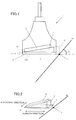
- FIG. 1 is a schematic view illustrating the configuration of the ultrasound probe for a puncture needle according to the embodiment 1 of the invention.
- An ultrasound probe 1 for a puncture needle includes a transducer array 2 having an ultrasonic wave transmission/reception surface S1 that extends along an array direction of a plurality of transducers, an acoustic lens 3 having a subject contact surface S2 that is arranged on the ultrasonic wave transmission/reception surface S1 side of the transducer array 2, that is, in front of the transducer array 2, and contacts a subject P during ultrasound diagnosis, and a wedge-shaped filling member 4 arranged between the transducer array 2 and the acoustic lens 3.
- a puncture position I of a puncture needle N is set near one end of the subject contact surface S2, and the puncture needle N is attached at a predetermined puncture angle ⁇ by a puncture needle holder (not illustrated).
- the transducer array 2 is disposed so as to be tilted at a predetermined array angle ⁇ of inclination with respect to the subject contact surface S2 in a direction in which the angle of the ultrasonic wave transmission/reception surface S1 with respect to the puncturing direction of the puncture needle N punctured from the puncture position I toward the front of the subject contact surface S2 decreases, that is, so that the ultrasonic wave transmission/reception surface S1 faces the puncture position I.
- FIG. 2 is a perspective view of the ultrasound probe 1 for a puncture needle illustrated in FIG. 1 .
- the puncture position I of the puncture needle N is positioned in a center in an elevation direction of the transducer array 2, and the puncture needle N enters from an azimuth direction of the transducer array 2, puncturing the subject P so as to pass through the center in the elevation direction of the transducer array 2.
- ultrasonic beams are transmitted in a direction perpendicular to the ultrasonic wave transmission/reception surface S1 by the ultrasound probe 1 according to the embodiment 1, it is possible to transmit ultrasonic beams that are more perpendicular with respect to the puncture needle N and receive clearer ultrasonic echoes from the puncture needle N compared to the ultrasonic beams from a conventional ultrasound probe in which the ultrasonic wave transmission/reception surface and subject contact surface are parallel. This point physically differs from when the transducer array is scanned and the ultrasonic beams are steered in a conventional ultrasound diagnosis.
- the transducer array 2 is made of a plurality of transducers linearly arranged at an equal interval at a predetermined pitch a, and each transducer is configured to form an electrode on both ends of a piezoelectric body made from, for example, piezoelectric ceramic represented by lead zirconate titanate (PZT), polymer piezoelectric elements represented by polyvinylidene fluoride (PVDF), or the like.
- PZT lead zirconate titanate
- PVDF polymer piezoelectric elements represented by polyvinylidene fluoride
- each transducer When a voltage in pulse form or in the form of a continuous wave is applied to the electrodes of this type of transducer, the piezoelectric body is constricted, an ultrasonic wave in pulse form or continuous wave form is generated from each transducer, and an ultrasonic beam is formed by the synthesis of these ultrasonic waves.
- each transducer receives a propagating ultrasonic wave and as a result expands and contracts, thereby generating an electrical signal. These electrical signals are output as ultrasound reception signals.
- the acoustic lens 3 is arranged in front of the transducer array 2 and, during ultrasound diagnosis, an outer surface S2 thereof directly contacts the subject P.
- the acoustic lens 3 is, for example, formed by silicone rubber, and is for focusing the ultrasonic beams transmitted from the transducer array 2 at a predetermined depth within the subject P.
- the wedge-shaped filling member 4 is a wedge-shaped member for tilting the ultrasonic wave transmission/reception surface S1 of the transducer array 2 at an array angle ⁇ of inclination with respect to the outer surface S2 of the acoustic lens 3, and propagates the ultrasonic waves generated by the transducer array 2 as well as the ultrasonic echoes from within the subject P.
- the wedge-shaped filling member 4 is, for example, formed by silicone rubber, urethane synthetic resin, epoxy, or the like.
- ultrasonic waves are transmitted from the transducer array 2 arranged so as to be tilted at a predetermined array angle ⁇ of inclination with respect to the subject P, ultrasonic beams are formed with respect to a focal position Fa1, for example, ultrasonic echoes are received in the transducer array 2, and sound ray signals La are formed.
- the sound ray signals La are formed so as to be tilted to the puncture needle N side compared to the sound ray signals formed by a conventional ultrasound probe.
- the puncture needle N reaches a deep region of the subject P at an incline of 50°, 60°, or the like, the ultrasonic echoes from the puncture needle N can be more clearly received in the transducer array 2 and, as illustrated in FIG. 4 , a B mode image of the deep region of the subject P that includes the puncture needle N can be more clearly drawn compared to a conventional ultrasound probe.
- an approximation formula D( ⁇ ) of transducer directivity which indicates the relative sound pressure value toward a direction of the angle ⁇ of the transducer, is expressed as the following formula (1).
- D ⁇ sinx / x ⁇ cos ⁇
- x ⁇ a / ⁇ ⁇ sin ⁇
- a satisfactory image for ultrasound diagnosis can be created if the amount of sensitivity reduction of ultrasonic echoes is -20 dB or less.
- the array angle ⁇ of inclination has an upper limit.
- the upper limit of the array angle ⁇ of inclination is expressed by the following formula (2). a ⁇ ⁇ / 1 + sin ⁇
- Formula (2) is determined by a grating lobe that occurs in the ultrasound probe 1.
- the ultrasound probe 1 for a puncture needle according to the invention is a linear ultrasound probe having a 7.5-MHz center frequency and a 0.3-mm transducer element array pitch
- the array angle ⁇ of inclination of the transducer array 2 for more clearly drawing a deep region of the subject P is preferably about 8° to 15°, and most preferably about 10°, based on the aforementioned formulas (1) and (2).
- the ultrasound probe 1 for a puncture according to the embodiment 1 it is possible to improve the puncture visibility as well as the puncture limiting angle (the limiting angle at which the puncture needle can be visually confirmed), thereby preventing erroneous puncture and making it possible to perform puncturing with diseases located in a deep region of the subject.
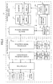
- FIG. 5 is a block diagram illustrating the overall configuration of the ultrasound diagnostic device according to the embodiment 2 of the invention that includes the aforementioned ultrasound probe 1 for a puncture needle.
- the ultrasound diagnostic device is made of the ultrasound probe 1 for a puncture needle (hereinafter, "ultrasound probe 1") and a diagnostic apparatus main body 12 connected to each other, wherein the ultrasound probe 1 includes the transducer array 2 made of a plurality of transducers 2a linearly arranged at an equal interval, a multiplexer 5 interconnected with each of the transducers 2a of the transducer array 2, a transmission/reception switch 5 interconnected with the multiplexer 5, a transmission driving section 7 and transmission controller 8 which are connected sequentially from the transmission/reception switch 6, a reception signal processor 9 connected to the transmission/reception switch 6, a reception controller 10 connected to the reception signal processor 9, and a probe controller 11 connected to the reception controller 10 as well as the transmission controller 8.
- the ultrasound probe 1 includes the transducer array 2 made of a plurality of transducers 2a linearly arranged at an equal interval, a multiplexer 5 interconnected with each of the transducers 2a of the transducer array 2, a transmission/reception switch 5 interconnected with
- the diagnostic apparatus main body 12 includes a phasing addition section 13; a signal processor 14, an image processor 15, a display controller 16, and a monitor 17 connected sequentially from the phasing addition section 13; a main body controller 18 interconnected with the phasing addition section 13, the signal processor 14, and the display controller 16; an operating section 19 connected to the main body controller 18; and a table storage 20 interconnected with the main body controller 18.
- the table storage 20 includes a correction table 21 in the interior, the phasing addition section 13 is connected from the reception signal processor 9 of the ultrasound probe 1, and the main body controller 16 is interconnected with the probe controller 11 of the ultrasound probe 1.
- the plurality of transducers 2a that constitutes the transducer array 3 transmits ultrasonic waves on the basis of driving signals supplied from the transmission driving section 7 via the transmission/reception switch 6 and the multiplexer 5, receives ultrasonic echoes from the subject, and outputs reception signals.
- the multiplexer 5 switches the plurality of transducers 2a that transmits ultrasonic waves on the basis of an instruction from the probe controller 11 via the transmission controller 8 or the reception controller 10, and switches the plurality of transducers 2a that receives ultrasonic echoes.
- the transmission/reception switch 6 switches the connections between the multiplexer 5, the transmission driving section 7, and the reception signal processor 9 so as to connect the transmission driving section 7 and the multiplexer 5 and disconnect the reception processor 9 and the multiplexer 5 when the driving signal supplied from the transmission driving section 7 is transmitted to the transducer array 2, and connect the reception signal processor 9 and the multiplexer 5 and disconnect the transmission driving section 7 and the multiplexer 5 when the reception signal processor 9 acquires a reception signal from the transducer array 2, on the basis of an instruction from the probe controller 11 via the transmission controller 8 or the reception controller 10.
- the transmission driving section 7 includes, for example, a plurality of pulse generators and, on the basis of the transmission delay pattern selected by the transmission controller 8, adjusts the amount of delay of each driving signal, so as to form wide ultrasonic beams in which the ultrasonic waves transmitted from the plurality of transducers 2a cover an area of tissue within the subject, and supplies the driving signals to the plurality of transducers 2a.
- the transmission delay pattern is a pattern of the transmission delay amount set in each of the transducers 2a, in the transmission of the ultrasonic waves based on the driving signal.
- the transmission controller 8 selects one of a plurality of transmission delay patterns stored in an internal memory (not illustrated) of the transmission driving section 7 on the basis of an instruction from the probe controller 11, and imparts a predetermined delay amount on the driving signal imparted on each of the transducers 2a from the transmission driving section on the basis of the transmission delay pattern. Additionally, the plurality of transmission delay patterns stored in the internal memory (not illustrated) of the transmission driving section 7 is stored within the internal memory (not illustrated) of the transmission driving section 7 by the transmission controller 8. Further, the transmission controller 8 controls the switching to the transmission driving section 7 side of the transmission/reception switch 6 on the basis of an instruction from the probe controller 11 as well.
- the reception signal processor 9 includes a low-noise amplifier (LNA), an arc tangent (ATN) circuit, and an analog-to-digital (A/D) converter.
- the reception signal processor 9, under the instruction of the reception controller 10, generates complex base band signals by processing the reception signal output from the corresponding transducer 2a, converting the signal to a digital reception signal, and performing quadrature detection processing or quadrature sampling processing on the digital reception signal, and generates sample data that includes information on an area of tissue by sampling the complex base band signals.
- the generated sample data is output to the phasing addition section 11 of the diagnostic apparatus main body 2.
- the reception controller 10 controls the reception signal processor 9, converts the reception signal output from the transducer 2a to sample data, and outputs the result to the phasing addition section of the diagnostic apparatus main body 12, on the basis of an instruction from the probe controller 11. Further, the reception controller 10 controls the switching to the reception signal processor 9 side of the transmission/reception switch 6 on the basis of an instruction from the probe controller 11 as well.
- the probe controller 11 controls each component of the ultrasound probe 1 on the basis of the various control signals transmitted from the main body controller 18 of the diagnostic apparatus main body 12. As described above, the probe controller 11 controls the transmission controller 8 and the reception controller 10 as described above so that ultrasonic waves are transmitted from the transducer array 2, and ultrasonic echoes are received in the transducer array 2.
- the phasing addition section 13 of the diagnostic apparatus main body 2 acquires the sample data that includes information on the area of tissue from the reception signal processor 8 of the ultrasound probe 1 and, in accordance with the reception direction (here, the direction perpendicular to the ultrasonic wave transmission/reception surface) set in the probe controller 11 of the ultrasound probe 1, selects one reception delay pattern from the plurality of reception delay patterns stored in the internal memory (not illustrated) of the phasing addition section 11, and imparts and adds each reception delay amount to the plurality of complex base band signals indicated by the reception data based on the selected reception delay pattern, thereby forming a beam in response to the plurality of reception data having a time difference generated by the reception signal processor 9, in accordance with each of the transducers 2a.
- the reception direction here, the direction perpendicular to the ultrasonic wave transmission/reception surface
- a base band signal (sound ray signal) in which the focal points of ultrasonic echoes have been narrowed down is generated through this beam formation.
- the reception delay pattern is a pattern of a reception delay amount individually imparted on the reception data acquired by each transducer 2a for generating sound ray signals.
- the signal processor 14 generates a B-mode image signal, which is tomographic image information related to the tissue within the subject P, by correcting the attenuation resulting from the distance in accordance with the depth of the reflection position of the ultrasonic waves, and subsequently converting the sound ray signals generated by the phasing addition section 13 to image signals (raster conversion) in accordance with a regular scanning method of a television signal.
- the generated B-mode image signal is output to the image processor 15.
- the image processor 15 performs predetermined image processing on the B-mode image signal generated by the signal processor 14.
- the predetermined image processing is, for example, image processing required for improving the visibility of the ultrasound image, such as gradation processing or enhancement processing.
- the B-mode image data subjected to image processing is output to the display controller 16 and stored in an image storage (not illustrated).
- the display controller 16 displays an ultrasound image on the monitor 17 on the basis of the B-mode image signal that was subjected to image processing and output by the image processor 15.
- the monitor 17 is, for example, a display apparatus such as a liquid crystal display (LCD), and displays ultrasound images under the control of the display controller 16.
- LCD liquid crystal display
- the main body controller 18 controls each component of the diagnostic apparatus main body 12 on the basis of an operator's instruction from the operating section 19, acquires a plurality of transmission delay patterns, a plurality of reception delay patterns, and an attenuation correction amount in accordance with depth from the correction table 20 of the table storage 19, and outputs the acquired information to the transmission driving section 7, the phasing addition section 13, and the internal memory (not illustrated) of the signal processor 14, respectively.
- the operating section 19 is configured to issue various instructions by an operator to an ultrasound diagnostic device made of the ultrasound probe 1 and the diagnostic apparatus main body 12.
- the table storage 20 is configured to store the correction table 21 in which a pre-calculated plurality of transmission delay patterns, plurality of reception delay patterns, and attenuation correction amounts in accordance with depth are recorded, and output the various information recorded in the correction table 21 on the basis of an instruction from the main body controller 18.
- the required transmission delay amount, reception delay amount, and attenuation correction amount differ for each transducer 2a per combination of sound ray signal and focal position, that is, depth within the subject P, making the pre-calculated plurality of transmission delay patterns, plurality of reception delay patterns, and attenuation correction amounts in accordance with depth recorded in the correction table 21 very high in number.
- the phasing addition section 13, the signal processor 14, the image processor 15, the display controller 16, and the main body controller 18 in such diagnostic apparatus main body 2 are constituted by a central processing unit (CPU) and an operation program for causing the CPU to carry out various types of processes, these sections may instead be constituted by digital circuitry.
- These operation programs are stored in a storage unit (not illustrated) in the diagnostic apparatus main body 2.
- a storage medium of the storage unit (not illustrated) a flexible disk, a magneto-optical disk (MO), a magnet tape (MT), a random access memory (RAM), a compact disk read only memory (CD-ROM), a digital versatile disk read only memory (DVD-ROM), or the like may be used in addition to a built-in hard disk.
- the probe controller 11 switches the transmission/reception switch 6 via the reception controller 10, connects the multiplexer 5 and the transmission driving section 7, outputs driving signals from the transmission driving section 7 to the transducer array 2 via the multiplexer 5, and transmits ultrasonic waves from the plurality of transducers 2a constituting the transducer array 2 in accordance with these driving signals.
- the transmission driving section 7 includes an internal memory (not illustrated) and stores the plurality of transmission delay patterns corresponding to the angle ⁇ of inclination of the ultrasonic wave transmission/reception surface of the transducer array 2 described in the correction table 21 of the table storage 20 via the main body controller 18, and a predetermined transmission delay amount is set per transducer 2a on the basis of the transmission delay pattern selected by the transmission driving section 7 for the driving signals transmitted from the transmission driving section 7 to the transducer array 2.
- the probe controller 11 switches the transmission switch 6 to the reception signal processor 9 side, connects the multiplexer 5 and the reception signal processor 9, and outputs reception signals from the transducer 2a connected by the multiplexer 5 to the reception signal processor 9.
- the reception signal processor 9 processes the reception signals, converts the signals to digital reception signals, generates complex base band signals by quadrature detection processing or quadrature sampling processing, and samples the complex base band signals, thereby generating sample data that includes information on an area of tissue.
- the generated sample data is output to the phasing addition section 11 of the diagnostic apparatus main body 2.
- the phasing addition section 13 selects one reception delay pattern from the plurality of reception delay patterns stored in the internal memory (not illustrated) of the phasing addition section 13 in accordance with the reception direction (here, the direction perpendicular to the ultrasonic wave transmission/reception surface) set in the probe controller 11 of the ultrasound probe 1, and imparts and adds each reception delay amount to the plurality of complex base band signals indicated by the reception data on the basis of the selected reception delay pattern, thereby forming a beam in response to the plurality of reception data having a time difference generated by the reception signal processor 9, in accordance with each of the transducers 2a.
- the sound ray signals generated by the phasing addition section 13 according to the beam formation are output to the signal processor 14.
- the plurality of reception delay patterns is configured to correspond to the angle ⁇ of inclination of the ultrasonic wave transmission/reception surface of the transducer array 2 described in the correction table 21 of the table storage 20, and is stored in the internal memory (not illustrated) of the phasing addition section 13 via the main body controller 18, in the same manner as the plurality of transmission delay patterns described above.
- Attenuation correction processing that corrects attenuation in accordance with the depth of the reflection position of the ultrasonic waves is performed on the sound ray signals acquired from the phasing addition section 13.
- the attenuation correction amount used in the attenuation correction is an attenuation correction amount stored in the correction table 21 of the table storage 20, and is stored in advance in the internal memory (not illustrated) of the signal processor 14 via the main body controller 18.
- the signal processor 4 generates a B-mode image signal by performing raster conversion on the sound ray signals subjected to attenuation correction processing.
- the generated B-mode image signal is output to the image processor 15.
- the image processor 15 performs predetermined image processing on the B-mode image signal output by the signal processor 14, and outputs the B-mode image signal subjected to image processing to the display controller 16.
- the display controller 16 outputs the B-mode image signal subjected to image processing on the basis of an instruction from the main body controller 18 to the monitor 17, and the monitor 17 displays an ultrasound image, which is the B mode image based on the B-mode image signal.
- the transducer array 2 may include a sound matching layer (not illustrated) on the ultrasonic wave transmission/reception surface S1.
- the sound matching layer is a member for matching the acoustic impedance between the transducer array 2, the wedge-shaped filling member 4 that contacts the transducer array 2, the acoustic lens 3, and the subject.
- the sound matching layer (not illustrated) of the transducer array 2 has an acoustic impedance greater than that of the wedge-shaped filling member 4.
- various known members capable of attaining the matching of acoustic impedance can be used.
- the wedge-shaped filling member 4 preferably has a lower sound attenuation rate than that of the acoustic lens 3. Additionally, the sound velocity of the wedge-shaped filling member 4 is preferably slower than the sound velocity of the acoustic lens 3.
- the ultrasonic waves transmitted from the transducer array 2 can be further tilted to the puncture needle N side as illustrated in FIG. 6 , the ultrasonic echoes from the puncture needle N in the deep region of the subject P can be more clearly received, and the ultrasound image can be more clearly drawn, because of the refractive index relationship between the wedge-shaped filling member 4 and the acoustic lens 3.
- the phasing addition section 13, the signal processor 14, and the image processor 15 of a diagnostic apparatus main body 32 may be configured by an ultra-high speed calculating apparatus 33 as shown in FIG. 7 .
- the transmission delay amount, reception delay amount, and attenuation correction amount can be calculated in real-time by the ultra-high speed calculating apparatus 36, making it possible for the main body controller 18 to proceed with each process without referring to the correction table 21 of the table storage 20. Accordingly, in the diagnostic apparatus main body 32, the table storage 20 and the correction table 21, which store idly amounts out data pertaining to the transmission delay amount, reception delay amount, and attenuation correction amount, can be rendered unnecessary.
Landscapes
- Life Sciences & Earth Sciences (AREA)
- Health & Medical Sciences (AREA)
- Biomedical Technology (AREA)
- Biophysics (AREA)
- Nuclear Medicine, Radiotherapy & Molecular Imaging (AREA)
- Pathology (AREA)
- Radiology & Medical Imaging (AREA)
- Engineering & Computer Science (AREA)
- Physics & Mathematics (AREA)
- Heart & Thoracic Surgery (AREA)
- Medical Informatics (AREA)
- Molecular Biology (AREA)
- Surgery (AREA)
- Animal Behavior & Ethology (AREA)
- General Health & Medical Sciences (AREA)
- Public Health (AREA)
- Veterinary Medicine (AREA)
- Ultra Sonic Daignosis Equipment (AREA)
Abstract
Description
- The invention relates to an ultrasound probe and an ultrasound diagnostic device using the same, and particularly relates to an ultrasound probe for a puncture needle that is used with a puncture needle and an ultrasound diagnostic device using the same.
- Conventionally, ultrasound diagnostic devices using ultrasound images are put to practical use in the medical field. Generally, this type of ultrasound diagnostic device includes an ultrasound probe having a built-in transducer array and a diagnostic apparatus main body connected to the ultrasound probe. Ultrasonic waves are transmitted from the ultrasound probe toward an inspection object (subject), ultrasonic echoes from the subject are received by the ultrasound probe, and an ultrasound image is generated by electrically processing the received signals in the diagnostic apparatus main body.
- Additionally, according to the ultrasound diagnostic device, the arrangement of a puncture needle in the ultrasound probe and usage of the ultrasound diagnostic device with the puncture needle so that processing can be performed while checking the ultrasound image have been known to be useful.
Patent Literature 1 discloses a probe for a puncture needle capable of accurately checking a position of a puncture needle within a close distance. - [Patent Literature 1]
JP 2006-288580 A -
FIG. 8 illustrates an example (hereinafter "conventional ultrasound probe 101") of an ultrasound probe frequently used with puncturing. Theconventional ultrasound probe 101 is an example of a linear ultrasound probe having a 7.5-MHz center frequency and a 0.3-mm element array pitch. According to theconventional ultrasound probe 101, while it is possible to clearly draw an ultrasound image and check the position of the puncture needle on the ultrasound image with certainty when the puncture angle of the puncture needle is 30°, for example, in a shallow region (at a close distance), if an attempt is made to check the position of the puncture needle in a deep region (at a far distance) with the puncture angle of the puncture needle tilted at 50° or 60°, the ultrasound image attenuates to approximately 1/3 of the value when the insertion angle is 30° at an insertion angle of 50°, and to approximately 1/30 of the value when the insertion angle is 30° at an insertion angle of 60°, making it no longer possible to clearly draw the image of the puncture needle or accurately check the position of the puncture needle. - Therefore, an object of the invention is to provide an ultrasound probe for a puncture needle and an ultrasound diagnostic device capable of clearly drawing a puncture needle in a deep region of a subject.
- In order to solve the above problem, the present invention provides an ultrasound probe for a puncture needle, comprising: a transducer array having an ultrasonic wave transmission/reception surface that extends along an array direction of a plurality of transducers; and a subject contact surface that is positioned in front of the ultrasonic wave transmission/reception surface of the transducer array, and contacts a subject during ultrasound diagnosis, wherein a puncture position of a puncture needle is set near one end of the subject contact surface; and the transducer array is arranged so as to be tilted at a
predetermined array angle 8 of inclination with respect to the subject contact surface, in a direction in which the angle of the ultrasonic wave transmission/reception surface with respect to a puncturing direction of the puncture needle punctured from the puncture position toward the front of the subject contact surface decreases. - Preferably, given a wavelength λ of ultrasonic waves in the subject, an element array pitch a of the transducer array, and a following approximation formula D(θ) of transducer directivity:


an array angle θ of inclination satisfies:
and
- Further, given a wavelength λ of ultrasonic waves in the subject, an element array pitch a of the transducer array, and a following approximation formula D(θ) of transducer directivity:


an array angle θ of inclination may satisfy:
and
- Preferably, a wedge-shaped filling member having a sound attenuation rate less than that of an acoustic lens is arranged between the transducer array and the acoustic lens.
- Preferably, a sound velocity of the wedge-shaped filling member is slower than a sound velocity of the acoustic lens.
- Preferably, the transducer array further comprises a sound matching layer arranged on the wedge-shaped filling member side; and the wedge-shaped filling member has a smaller acoustic impedance than that of the sound matching layer.
- In addition, the present invention provides an ultrasound diagnostic device, further comprising a diagnostic apparatus main body connected to the ultrasound probe for a puncture needle described above, wherein: the diagnostic apparatus main body includes a transmission driving section that supplies a driving signal to the transducer array of the ultrasound probe to transmit ultrasonic beams toward the subject, a transmission controller that forms ultrasonic beams by imparting a predetermined transmission delay amount to the driving signal supplied to the transmission driving section, a reception signal processor that generates reception data from a reception signal output from the transducer array of the ultrasound probe that received ultrasonic echoes from the subject, a phasing addition section that generates a sound ray signal by imparting a predetermined reception delay amount on the reception data and performing addition processing, a signal processor that performs attenuation correction in accordance with a focal position of ultrasonic waves on the sound ray signal, and a correction table storage having a correction table that describes the transmission delay amount, the reception delay amount, and the attenuation correction amount based on the focal position, individually calculated in advance in accordance with an arrangement of the transducers and the focal position; the transmission controller imparts the predetermined transmission delay amount on the driving signal on the basis of the correction table stored in the correction table storage, and forms the ultrasonic beams; the phasing addition section generates the sound ray signal by imparting the predetermined reception delay amount on the reception data on the basis of the correction table stored in the correction table storage, and performing addition processing; and the signal processor performs the attenuation correction on the basis of the correction table stored in the correction table storage.
- Further, the present invention provides an ultrasound diagnostic device, further comprising a diagnostic apparatus main body connected to the puncture needle ultrasound probe described above, wherein the diagnostic apparatus main body includes a transmission driving section that supplies a driving signal to the transducer array of the ultrasound probe to transmit ultrasonic beams toward the subject, a transmission controller that forms ultrasonic beams by imparting a predetermined transmission delay amount to the driving signal supplied to the transmission driving section, a reception signal processor that generates reception data from a reception signal output from the transducer array of the ultrasound probe that received ultrasonic echoes from the subject, a phasing addition section that generates a sound ray signal by imparting a predetermined reception delay amount on the reception data and performing addition processing, a signal processor that performs attenuation correction in accordance with a focal position of ultrasonic waves on the sound ray signal, and a high-speed calculation processor that individually calculates the transmission delay amount, the reception delay amount, and an attenuation correction amount based on the focal position, in accordance with an arrangement of the transducers and the focal position.
- Preferably, the high-speed calculation processor serves as the phasing addition section and the signal processor.
- According to the invention, it is possible to clearly draw a puncture needle, even in a deep region of a subject, on an ultrasound image, and accurately identify the position of the puncture needle in the deep region of the subject. As a result, it is possible to prevent erroneous puncture, improve the puncture limiting angle, and perform puncturing with diseases located in a deep region of the subject.
-
- [
FIG. 1] FIG. 1 is a schematic view illustrating the configuration of an ultrasound probe for a puncture needle according to anembodiment 1 of the invention. - [
FIG. 2] FIG. 2 is a perspective view of the ultrasound probe for a puncture needle according to theembodiment 1 of the invention. - [
FIG. 3] FIG. 3 is an explanatory view schematically explaining the relationship between sound ray signals formed when the ultrasound probe for a puncture needle according to the invention transmits ultrasonic beams and receives ultrasonic echoes, and the focal position. - [
FIG. 4] FIG. 4 is an explanatory view of an ultrasound image that includes a puncture needle drawn when the ultrasound probe for a puncture needle according to theembodiment 1 of the invention is used. - [
FIG. 5] FIG. 5 is a block diagram illustrating the overall configuration of the ultrasound diagnostic device according to anembodiment 2 of the invention. - [
FIG. 6] FIG. 6 is an explanatory view explaining the refraction of ultrasonic waves transmitted from transducer arrays of the ultrasound probe for a puncture needle according to a modification of theembodiment 1 of the invention, between a wedge-shaped filling member and an acoustic lens. - [
FIG. 7] FIG. 7 is a block diagram illustrating the overall configuration of the diagnostic apparatus main body according to a modification of theembodiment 2 of the invention. - [
FIG. 8] FIG. 8 is an explanatory view of an ultrasound image that includes a puncture needle drawn when a conventional ultrasound probe for a puncture needle is used. - The following is a detailed description of the ultrasound probe and ultrasound diagnostic device according to the invention based on the preferred embodiments illustrated in the accompanying drawings.
-
FIG. 1 is a schematic view illustrating the configuration of the ultrasound probe for a puncture needle according to theembodiment 1 of the invention. - An
ultrasound probe 1 for a puncture needle according to theembodiment 1 includes atransducer array 2 having an ultrasonic wave transmission/reception surface S1 that extends along an array direction of a plurality of transducers, anacoustic lens 3 having a subject contact surface S2 that is arranged on the ultrasonic wave transmission/reception surface S1 side of thetransducer array 2, that is, in front of thetransducer array 2, and contacts a subject P during ultrasound diagnosis, and a wedge-shaped filling member 4 arranged between thetransducer array 2 and theacoustic lens 3. In theultrasound probe 1 for a puncture needle, a puncture position I of a puncture needle N is set near one end of the subject contact surface S2, and the puncture needle N is attached at a predetermined puncture angle φ by a puncture needle holder (not illustrated). - The
transducer array 2 is disposed so as to be tilted at a predetermined array angle θ of inclination with respect to the subject contact surface S2 in a direction in which the angle of the ultrasonic wave transmission/reception surface S1 with respect to the puncturing direction of the puncture needle N punctured from the puncture position I toward the front of the subject contact surface S2 decreases, that is, so that the ultrasonic wave transmission/reception surface S1 faces the puncture position I. -
FIG. 2 is a perspective view of theultrasound probe 1 for a puncture needle illustrated inFIG. 1 . The puncture position I of the puncture needle N is positioned in a center in an elevation direction of thetransducer array 2, and the puncture needle N enters from an azimuth direction of thetransducer array 2, puncturing the subject P so as to pass through the center in the elevation direction of thetransducer array 2. - Accordingly, when ultrasonic beams are transmitted in a direction perpendicular to the ultrasonic wave transmission/reception surface S1 by the
ultrasound probe 1 according to theembodiment 1, it is possible to transmit ultrasonic beams that are more perpendicular with respect to the puncture needle N and receive clearer ultrasonic echoes from the puncture needle N compared to the ultrasonic beams from a conventional ultrasound probe in which the ultrasonic wave transmission/reception surface and subject contact surface are parallel. This point physically differs from when the transducer array is scanned and the ultrasonic beams are steered in a conventional ultrasound diagnosis. - The
transducer array 2 is made of a plurality of transducers linearly arranged at an equal interval at a predetermined pitch a, and each transducer is configured to form an electrode on both ends of a piezoelectric body made from, for example, piezoelectric ceramic represented by lead zirconate titanate (PZT), polymer piezoelectric elements represented by polyvinylidene fluoride (PVDF), or the like. - When a voltage in pulse form or in the form of a continuous wave is applied to the electrodes of this type of transducer, the piezoelectric body is constricted, an ultrasonic wave in pulse form or continuous wave form is generated from each transducer, and an ultrasonic beam is formed by the synthesis of these ultrasonic waves. In addition, each transducer receives a propagating ultrasonic wave and as a result expands and contracts, thereby generating an electrical signal. These electrical signals are output as ultrasound reception signals.
- The
acoustic lens 3 is arranged in front of thetransducer array 2 and, during ultrasound diagnosis, an outer surface S2 thereof directly contacts the subject P. Theacoustic lens 3 is, for example, formed by silicone rubber, and is for focusing the ultrasonic beams transmitted from thetransducer array 2 at a predetermined depth within the subject P. - The wedge-
shaped filling member 4 is a wedge-shaped member for tilting the ultrasonic wave transmission/reception surface S1 of thetransducer array 2 at an array angle θ of inclination with respect to the outer surface S2 of theacoustic lens 3, and propagates the ultrasonic waves generated by thetransducer array 2 as well as the ultrasonic echoes from within the subject P. The wedge-shaped filling member 4 is, for example, formed by silicone rubber, urethane synthetic resin, epoxy, or the like. - Next, the operation of the
ultrasound probe 1 for a puncture needle according to theembodiment 1 will be described. - As illustrated in
FIG. 3 , ultrasonic waves are transmitted from thetransducer array 2 arranged so as to be tilted at a predetermined array angle θ of inclination with respect to the subject P, ultrasonic beams are formed with respect to a focal position Fa1, for example, ultrasonic echoes are received in thetransducer array 2, and sound ray signals La are formed. - The sound ray signals La are formed so as to be tilted to the puncture needle N side compared to the sound ray signals formed by a conventional ultrasound probe. Thus, even if, for example, the puncture needle N reaches a deep region of the subject P at an incline of 50°, 60°, or the like, the ultrasonic echoes from the puncture needle N can be more clearly received in the
transducer array 2 and, as illustrated inFIG. 4 , a B mode image of the deep region of the subject P that includes the puncture needle N can be more clearly drawn compared to a conventional ultrasound probe. - Given a wavelength λ of the ultrasonic waves in a human body (subject P), an approximation formula D(θ) of transducer directivity, which indicates the relative sound pressure value toward a direction of the angle θ of the transducer, is expressed as the following formula (1).

- As general evaluation criteria, a satisfactory image for ultrasound diagnosis can be created if the amount of sensitivity reduction of ultrasonic echoes is -20 dB or less.
- Now, given that the
ultrasound probe 1 for a puncture needle according to the invention is a linear ultrasound probe having a 7.5-MHz center frequency and a 0.3-mm element array pitch a, which is similar to theconventional ultrasound probe 101, if the incline of the puncture needle N is 60°, D(θ = 30°)/D(0°) is 1/10(-20 dB), D(θ = 50°)/D(0°) is 1/30 (-30 dB), and D(θ = 60°)/D(0°) is 1/300 (-50 dB) compared to when the incline of the puncture needle N is 30° as illustrated in the conventional ultrasound image inFIG. 8 , and therefore the ultrasound image (B mode image) attenuates to approximately 1/30. - Accordingly, in the
embodiment 1 of the invention, when the amount of sensitivity reduction of an image range to be observed is increased by at least -20 dB, that is, by setting the array angle θ of inclination to 10° and tilting the ultrasonic wave transmission/reception surface S1 of thetransducer array 2 at 10° to the puncture needle N side with respect to the outer surface S2 of the acoustic lens, it is possible to improve the amount of sensitivity reduction at D(θ = 60°) by -20 dB and make the B mode image when the incline of the puncture needle N of the invention is 60° the same as the B mode image when the incline of a conventional puncture needle N is 50°. - Additionally, the array angle θ of inclination has an upper limit. The upper limit of the array angle θ of inclination is expressed by the following formula (2).

- Formula (2) is determined by a grating lobe that occurs in the
ultrasound probe 1. - The reason is that, during imaging, a steering process is performed to execute conventional imaging, but when the range of θ expressed in this formula (2) is exceeded in the steering process and the transducer array is tilted, the grating lobe actually becomes too large, leading to the generation of a virtual image on the image.
- As a result, given that the
ultrasound probe 1 for a puncture needle according to the invention is a linear ultrasound probe having a 7.5-MHz center frequency and a 0.3-mm transducer element array pitch, the array angle θ of inclination of thetransducer array 2 for more clearly drawing a deep region of the subject P is preferably about 8° to 15°, and most preferably about 10°, based on the aforementioned formulas (1) and (2). - As a result, according to the
ultrasound probe 1 for a puncture according to theembodiment 1, it is possible to improve the puncture visibility as well as the puncture limiting angle (the limiting angle at which the puncture needle can be visually confirmed), thereby preventing erroneous puncture and making it possible to perform puncturing with diseases located in a deep region of the subject. -
FIG. 5 is a block diagram illustrating the overall configuration of the ultrasound diagnostic device according to theembodiment 2 of the invention that includes theaforementioned ultrasound probe 1 for a puncture needle. - The ultrasound diagnostic device according to the
embodiment 2 is made of theultrasound probe 1 for a puncture needle (hereinafter, "ultrasound probe 1") and a diagnostic apparatusmain body 12 connected to each other, wherein theultrasound probe 1 includes thetransducer array 2 made of a plurality oftransducers 2a linearly arranged at an equal interval, amultiplexer 5 interconnected with each of thetransducers 2a of thetransducer array 2, a transmission/reception switch 5 interconnected with themultiplexer 5, a transmission driving section 7 andtransmission controller 8 which are connected sequentially from the transmission/reception switch 6, a reception signal processor 9 connected to the transmission/reception switch 6, areception controller 10 connected to the reception signal processor 9, and a probe controller 11 connected to thereception controller 10 as well as thetransmission controller 8. - Additionally, the diagnostic apparatus
main body 12 includes aphasing addition section 13; asignal processor 14, animage processor 15, a display controller 16, and amonitor 17 connected sequentially from thephasing addition section 13; amain body controller 18 interconnected with thephasing addition section 13, thesignal processor 14, and the display controller 16; anoperating section 19 connected to themain body controller 18; and atable storage 20 interconnected with themain body controller 18. Thetable storage 20 includes a correction table 21 in the interior, thephasing addition section 13 is connected from the reception signal processor 9 of theultrasound probe 1, and the main body controller 16 is interconnected with the probe controller 11 of theultrasound probe 1. - The plurality of
transducers 2a that constitutes thetransducer array 3 transmits ultrasonic waves on the basis of driving signals supplied from the transmission driving section 7 via the transmission/reception switch 6 and themultiplexer 5, receives ultrasonic echoes from the subject, and outputs reception signals. - The
multiplexer 5 switches the plurality oftransducers 2a that transmits ultrasonic waves on the basis of an instruction from the probe controller 11 via thetransmission controller 8 or thereception controller 10, and switches the plurality oftransducers 2a that receives ultrasonic echoes. - The transmission/reception switch 6 switches the connections between the
multiplexer 5, the transmission driving section 7, and the reception signal processor 9 so as to connect the transmission driving section 7 and themultiplexer 5 and disconnect the reception processor 9 and themultiplexer 5 when the driving signal supplied from the transmission driving section 7 is transmitted to thetransducer array 2, and connect the reception signal processor 9 and themultiplexer 5 and disconnect the transmission driving section 7 and themultiplexer 5 when the reception signal processor 9 acquires a reception signal from thetransducer array 2, on the basis of an instruction from the probe controller 11 via thetransmission controller 8 or thereception controller 10. - The transmission driving section 7 includes, for example, a plurality of pulse generators and, on the basis of the transmission delay pattern selected by the
transmission controller 8, adjusts the amount of delay of each driving signal, so as to form wide ultrasonic beams in which the ultrasonic waves transmitted from the plurality oftransducers 2a cover an area of tissue within the subject, and supplies the driving signals to the plurality oftransducers 2a. Here, the transmission delay pattern is a pattern of the transmission delay amount set in each of thetransducers 2a, in the transmission of the ultrasonic waves based on the driving signal. - The
transmission controller 8 selects one of a plurality of transmission delay patterns stored in an internal memory (not illustrated) of the transmission driving section 7 on the basis of an instruction from the probe controller 11, and imparts a predetermined delay amount on the driving signal imparted on each of thetransducers 2a from the transmission driving section on the basis of the transmission delay pattern. Additionally, the plurality of transmission delay patterns stored in the internal memory (not illustrated) of the transmission driving section 7 is stored within the internal memory (not illustrated) of the transmission driving section 7 by thetransmission controller 8. Further, thetransmission controller 8 controls the switching to the transmission driving section 7 side of the transmission/reception switch 6 on the basis of an instruction from the probe controller 11 as well. - The reception signal processor 9 includes a low-noise amplifier (LNA), an arc tangent (ATN) circuit, and an analog-to-digital (A/D) converter. The reception signal processor 9, under the instruction of the
reception controller 10, generates complex base band signals by processing the reception signal output from the correspondingtransducer 2a, converting the signal to a digital reception signal, and performing quadrature detection processing or quadrature sampling processing on the digital reception signal, and generates sample data that includes information on an area of tissue by sampling the complex base band signals. The generated sample data is output to the phasing addition section 11 of the diagnostic apparatusmain body 2. - The
reception controller 10 controls the reception signal processor 9, converts the reception signal output from thetransducer 2a to sample data, and outputs the result to the phasing addition section of the diagnostic apparatusmain body 12, on the basis of an instruction from the probe controller 11. Further, thereception controller 10 controls the switching to the reception signal processor 9 side of the transmission/reception switch 6 on the basis of an instruction from the probe controller 11 as well. - The probe controller 11 controls each component of the
ultrasound probe 1 on the basis of the various control signals transmitted from themain body controller 18 of the diagnostic apparatusmain body 12. As described above, the probe controller 11 controls thetransmission controller 8 and thereception controller 10 as described above so that ultrasonic waves are transmitted from thetransducer array 2, and ultrasonic echoes are received in thetransducer array 2. - The
phasing addition section 13 of the diagnostic apparatusmain body 2 acquires the sample data that includes information on the area of tissue from thereception signal processor 8 of theultrasound probe 1 and, in accordance with the reception direction (here, the direction perpendicular to the ultrasonic wave transmission/reception surface) set in the probe controller 11 of theultrasound probe 1, selects one reception delay pattern from the plurality of reception delay patterns stored in the internal memory (not illustrated) of the phasing addition section 11, and imparts and adds each reception delay amount to the plurality of complex base band signals indicated by the reception data based on the selected reception delay pattern, thereby forming a beam in response to the plurality of reception data having a time difference generated by the reception signal processor 9, in accordance with each of thetransducers 2a. A base band signal (sound ray signal) in which the focal points of ultrasonic echoes have been narrowed down is generated through this beam formation. Here, the reception delay pattern is a pattern of a reception delay amount individually imparted on the reception data acquired by eachtransducer 2a for generating sound ray signals. - The
signal processor 14 generates a B-mode image signal, which is tomographic image information related to the tissue within the subject P, by correcting the attenuation resulting from the distance in accordance with the depth of the reflection position of the ultrasonic waves, and subsequently converting the sound ray signals generated by thephasing addition section 13 to image signals (raster conversion) in accordance with a regular scanning method of a television signal. The generated B-mode image signal is output to theimage processor 15. - The
image processor 15 performs predetermined image processing on the B-mode image signal generated by thesignal processor 14. The predetermined image processing is, for example, image processing required for improving the visibility of the ultrasound image, such as gradation processing or enhancement processing. The B-mode image data subjected to image processing is output to the display controller 16 and stored in an image storage (not illustrated). - The display controller 16 displays an ultrasound image on the
monitor 17 on the basis of the B-mode image signal that was subjected to image processing and output by theimage processor 15. - The
monitor 17 is, for example, a display apparatus such as a liquid crystal display (LCD), and displays ultrasound images under the control of the display controller 16. - The
main body controller 18 controls each component of the diagnostic apparatusmain body 12 on the basis of an operator's instruction from the operatingsection 19, acquires a plurality of transmission delay patterns, a plurality of reception delay patterns, and an attenuation correction amount in accordance with depth from the correction table 20 of thetable storage 19, and outputs the acquired information to the transmission driving section 7, thephasing addition section 13, and the internal memory (not illustrated) of thesignal processor 14, respectively. - The operating
section 19 is configured to issue various instructions by an operator to an ultrasound diagnostic device made of theultrasound probe 1 and the diagnostic apparatusmain body 12. - The
table storage 20 is configured to store the correction table 21 in which a pre-calculated plurality of transmission delay patterns, plurality of reception delay patterns, and attenuation correction amounts in accordance with depth are recorded, and output the various information recorded in the correction table 21 on the basis of an instruction from themain body controller 18. - With the
transducer array 2 arranged so that the ultrasonic wave transmission/reception surface S1 thereof is tilted at an angle θ of inclination with respect to the outer surface S2 of the acoustic lens, the required transmission delay amount, reception delay amount, and attenuation correction amount differ for eachtransducer 2a per combination of sound ray signal and focal position, that is, depth within the subject P, making the pre-calculated plurality of transmission delay patterns, plurality of reception delay patterns, and attenuation correction amounts in accordance with depth recorded in the correction table 21 very high in number. - Although the
phasing addition section 13, thesignal processor 14, theimage processor 15, the display controller 16, and themain body controller 18 in such diagnostic apparatusmain body 2 are constituted by a central processing unit (CPU) and an operation program for causing the CPU to carry out various types of processes, these sections may instead be constituted by digital circuitry. These operation programs are stored in a storage unit (not illustrated) in the diagnostic apparatusmain body 2. For the storage medium of the storage unit (not illustrated), a flexible disk, a magneto-optical disk (MO), a magnet tape (MT), a random access memory (RAM), a compact disk read only memory (CD-ROM), a digital versatile disk read only memory (DVD-ROM), or the like may be used in addition to a built-in hard disk. - Next, the operation of the
embodiment 1 will be described. - When an ultrasound diagnosis is started, the probe controller 11 switches the transmission/reception switch 6 via the
reception controller 10, connects themultiplexer 5 and the transmission driving section 7, outputs driving signals from the transmission driving section 7 to thetransducer array 2 via themultiplexer 5, and transmits ultrasonic waves from the plurality oftransducers 2a constituting thetransducer array 2 in accordance with these driving signals. - It should be noted that the transmission driving section 7 includes an internal memory (not illustrated) and stores the plurality of transmission delay patterns corresponding to the angle θ of inclination of the ultrasonic wave transmission/reception surface of the
transducer array 2 described in the correction table 21 of thetable storage 20 via themain body controller 18, and a predetermined transmission delay amount is set pertransducer 2a on the basis of the transmission delay pattern selected by the transmission driving section 7 for the driving signals transmitted from the transmission driving section 7 to thetransducer array 2. - When the transmission of ultrasonic waves from the plurality of
transducers 2a ends, the probe controller 11 switches the transmission switch 6 to the reception signal processor 9 side, connects themultiplexer 5 and the reception signal processor 9, and outputs reception signals from thetransducer 2a connected by themultiplexer 5 to the reception signal processor 9. - The reception signal processor 9 processes the reception signals, converts the signals to digital reception signals, generates complex base band signals by quadrature detection processing or quadrature sampling processing, and samples the complex base band signals, thereby generating sample data that includes information on an area of tissue. The generated sample data is output to the phasing addition section 11 of the diagnostic apparatus
main body 2. - The
phasing addition section 13 selects one reception delay pattern from the plurality of reception delay patterns stored in the internal memory (not illustrated) of thephasing addition section 13 in accordance with the reception direction (here, the direction perpendicular to the ultrasonic wave transmission/reception surface) set in the probe controller 11 of theultrasound probe 1, and imparts and adds each reception delay amount to the plurality of complex base band signals indicated by the reception data on the basis of the selected reception delay pattern, thereby forming a beam in response to the plurality of reception data having a time difference generated by the reception signal processor 9, in accordance with each of thetransducers 2a. The sound ray signals generated by thephasing addition section 13 according to the beam formation are output to thesignal processor 14. - It should be noted that the plurality of reception delay patterns is configured to correspond to the angle θ of inclination of the ultrasonic wave transmission/reception surface of the
transducer array 2 described in the correction table 21 of thetable storage 20, and is stored in the internal memory (not illustrated) of thephasing addition section 13 via themain body controller 18, in the same manner as the plurality of transmission delay patterns described above. - In the
signal processor 14, attenuation correction processing that corrects attenuation in accordance with the depth of the reflection position of the ultrasonic waves is performed on the sound ray signals acquired from thephasing addition section 13. The attenuation correction amount used in the attenuation correction is an attenuation correction amount stored in the correction table 21 of thetable storage 20, and is stored in advance in the internal memory (not illustrated) of thesignal processor 14 via themain body controller 18. Thesignal processor 4 generates a B-mode image signal by performing raster conversion on the sound ray signals subjected to attenuation correction processing. The generated B-mode image signal is output to theimage processor 15. - The
image processor 15 performs predetermined image processing on the B-mode image signal output by thesignal processor 14, and outputs the B-mode image signal subjected to image processing to the display controller 16. - The display controller 16 outputs the B-mode image signal subjected to image processing on the basis of an instruction from the
main body controller 18 to themonitor 17, and themonitor 17 displays an ultrasound image, which is the B mode image based on the B-mode image signal. - It should be noted that, as a modification of the
ultrasound probe 1 for a puncture needle according to theembodiment 1, thetransducer array 2 may include a sound matching layer (not illustrated) on the ultrasonic wave transmission/reception surface S1. Here, the sound matching layer is a member for matching the acoustic impedance between thetransducer array 2, the wedge-shaped fillingmember 4 that contacts thetransducer array 2, theacoustic lens 3, and the subject. The sound matching layer (not illustrated) of thetransducer array 2 has an acoustic impedance greater than that of the wedge-shaped fillingmember 4. For the sound matching layer (not illustrated), various known members capable of attaining the matching of acoustic impedance can be used. - Additionally, the wedge-shaped filling
member 4 preferably has a lower sound attenuation rate than that of theacoustic lens 3. Additionally, the sound velocity of the wedge-shaped fillingmember 4 is preferably slower than the sound velocity of theacoustic lens 3. When this sound velocity condition is satisfied, the ultrasonic waves transmitted from thetransducer array 2 can be further tilted to the puncture needle N side as illustrated inFIG. 6 , the ultrasonic echoes from the puncture needle N in the deep region of the subject P can be more clearly received, and the ultrasound image can be more clearly drawn, because of the refractive index relationship between the wedge-shaped fillingmember 4 and theacoustic lens 3. - Additionally, as a modification of the ultrasound diagnostic device according to the
embodiment 2, thephasing addition section 13, thesignal processor 14, and theimage processor 15 of a diagnostic apparatusmain body 32 may be configured by an ultra-highspeed calculating apparatus 33 as shown inFIG. 7 . - In this case, the transmission delay amount, reception delay amount, and attenuation correction amount can be calculated in real-time by the ultra-high speed calculating apparatus 36, making it possible for the
main body controller 18 to proceed with each process without referring to the correction table 21 of thetable storage 20. Accordingly, in the diagnostic apparatusmain body 32, thetable storage 20 and the correction table 21, which store idly amounts out data pertaining to the transmission delay amount, reception delay amount, and attenuation correction amount, can be rendered unnecessary. - The ultrasound probe for a puncture needle and the ultrasound diagnostic device that uses the same of the invention have been described in detail hereinabove, but the invention is not limited to the aforementioned embodiments, and various improvements and changes may be made without departing from the scope of the invention.
-
- 1:
- Ultrasound probe for puncture needle
- 2:
- Transducer array
- 2a:
- Transducer
- 3:
- Acoustic lens
- 4:
- Wedge-shaped filling member
- 5:
- Multiplexer
- 6:
- Transmission reception switch
- 7:
- Transmission driving section
- 8:
- Transmission controller
- 9:
- Reception signal processor
- 10:
- Reception controller
- 11:
- Probe controller
- 12:
- Diagnostic apparatus main body
- 13:
- Phasing addition section
- 14:
- Signal processor
- 15:
- Image processor
- 16:
- Display controller
- 17:
- Monitor
- 18:
- Main body controller
- 19:
- Operating section
- 20:
- Table storage
- 21:
- Correction table
- La:
- Sound ray signal
- Fa1:
- Focal position
- S1:
- Ultrasonic wave transmission/reception surface
- S2:
- Outer surface of acoustic lens (subject contact surface)
- I:
- Puncture position
- N:
- Puncture needle
- P:
- Subject
- θ:
- Array angle of inclination
- φ:
- Puncture angle
- λ:
- Wavelength of ultrasonic waves in human body (subject)
- a:
- Element array pitch
- 101:
- Conventional ultrasound probe
Claims (10)
- An ultrasound probe for a puncture needle, comprising:a transducer array having an ultrasonic wave transmission/reception surface that extends along an array direction of a plurality of transducers; anda subject contact surface that is positioned in front of the ultrasonic wave transmission/reception surface of the transducer array, and contacts a subject during ultrasound diagnosis, whereina puncture position of a puncture needle is set near one end of the subject contact surface, andthe transducer array is arranged so as to be tilted at a predetermined array angle θ of inclination with respect to the subject contact surface, in a direction in which the angle of the ultrasonic wave transmission/reception surface with respect to a puncturing direction of the puncture needle punctured from the puncture position toward the front of the subject contact surface decreases.
- The ultrasound probe for a puncture needle according to claim 1, wherein given a wavelength λ of ultrasonic waves in the subject, an element array pitch a of the transducer array, and a following approximation formula D(θ) of transducer directivity:


an array angle θ of inclination satisfies:
and
- The ultrasound probe for a puncture needle according to claim 1, wherein given a wavelength λ of ultrasonic waves in the subject, an element array pitch a of the transducer array, and a following approximation formula D(θ) of transducer directivity:


an array angle θ of inclination satisfies:
and
- The ultrasound probe for a puncture needle according to any one of claims 1 to 3, further comprising:an acoustic lens arranged on a side of the ultrasonic wave transmission/reception surface of the transducer array,wherein an outer surface of the acoustic lens is the subject contact surface.
- The ultrasound probe for a puncture needle according to claim 4, wherein a wedge-shaped filling member having a sound attenuation rate less than that of the acoustic lens is arranged between the transducer array and the acoustic lens.
- The ultrasound probe for a puncture needle according to claim 5, wherein a sound velocity of the wedge-shaped filling member is slower than a sound velocity of the acoustic lens.
- The ultrasound probe for a puncture needle according to claim 5 or 6, wherein the transducer array further comprises a sound matching layer arranged on a side of the wedge-shaped filling member; and
the wedge-shaped filling member has a smaller acoustic impedance than that of the sound matching layer. - An ultrasound diagnostic device, further comprising:a diagnostic apparatus main body connected to the ultrasound probe for a puncture needle according to any one of claims 1 to 7, whereinthe diagnostic apparatus main body includes:a transmission driving section that supplies a driving signal to the transducer array of the ultrasound probe to transmit ultrasonic beams toward the subject;a transmission controller that forms ultrasonic beams by imparting a predetermined transmission delay amount to the driving signal supplied to the transmission driving section;a reception signal processor that generates reception data from a reception signal output from the transducer array of the ultrasound probe that received ultrasonic echoes from the subject;a phasing addition section that generates a sound ray signal by imparting a predetermined reception delay amount on the reception data and performing addition processing;a signal processor that performs attenuation correction in accordance with a focal position of ultrasonic waves on the sound ray signal; anda correction table storage having a correction table that describes the transmission delay amount, the reception delay amount, and the attenuation correction amount based on the focal position, individually calculated in advance in accordance with an arrangement of the transducers and the focal position, whereinthe Transmission controller imparts the predetermined transmission delay amount on the driving signal on the basis of the correction table stored in the correction table storage, and forms the ultrasonic beams,the phasing addition section generates the sound ray signal by imparting the predetermined reception delay amount on the reception data on the basis of the correction table stored in the correction table storage, and performing addition processing, andthe signal processor performs the attenuation correction on the basis of the correction table stored in the correction table storage.
- An ultrasound diagnostic device, further comprising:a diagnostic apparatus main body connected to the puncture needle ultrasound probe according to any one of claims 1 to 7, whereinthe diagnostic apparatus main body includes:a transmission driving section that supplies a driving signal to the transducer array of the ultrasound probe to transmit ultrasonic beams toward the subject;a transmission controller that forms ultrasonic beams by imparting a predetermined transmission delay amount to the driving signal supplied to the transmission driving section;a reception signal processor that generates reception data from a reception signal output from the transducer array of the ultrasound probe that received ultrasonic echoes from the subject;a phasing addition section that generates a sound ray signal by imparting a predetermined reception delay amount on the reception data and performing addition processing;a signal processor that performs attenuation correction in accordance with a focal position of ultrasonic waves on the sound ray signal; anda high-speed calculation processor that individually calculates the transmission delay amount, the reception delay amount, and an attenuation correction amount based on the focal position, in accordance with an arrangement of the transducers and the focal position.
- The ultrasound diagnostic device according to claim 9, wherein the high-speed calculation processor serves as the phasing addition section and the signal processor.
Applications Claiming Priority (2)
| Application Number | Priority Date | Filing Date | Title |
|---|---|---|---|
| JP2013075174 | 2013-03-29 | ||
| PCT/JP2014/050941 WO2014156236A1 (en) | 2013-03-29 | 2014-01-20 | Ultrasonic probe for puncture needle and ultrasonic diagnostic device using same |
Publications (3)
| Publication Number | Publication Date |
|---|---|
| EP2979644A1 true EP2979644A1 (en) | 2016-02-03 |
| EP2979644A4 EP2979644A4 (en) | 2016-04-27 |
| EP2979644B1 EP2979644B1 (en) | 2017-09-13 |
Family
ID=51623241
Family Applications (1)
| Application Number | Title | Priority Date | Filing Date |
|---|---|---|---|
| EP14776531.7A Active EP2979644B1 (en) | 2013-03-29 | 2014-01-20 | Ultrasonic probe for puncture needle and ultrasonic diagnostic device using same |
Country Status (5)
| Country | Link |
|---|---|
| US (1) | US11529115B2 (en) |
| EP (1) | EP2979644B1 (en) |
| JP (1) | JP6001161B2 (en) |
| CN (1) | CN105101882B (en) |
| WO (1) | WO2014156236A1 (en) |
Families Citing this family (7)
| Publication number | Priority date | Publication date | Assignee | Title |
|---|---|---|---|---|
| CN106364123B (en) * | 2016-08-26 | 2018-02-06 | 京东方科技集团股份有限公司 | Lamination equipment and lamination method |
| EP3513736A4 (en) * | 2016-09-14 | 2019-09-11 | Fujifilm Corporation | Photoacoustic image generation apparatus |
| CN113329695B (en) * | 2019-01-17 | 2024-09-20 | 富士胶片株式会社 | Puncture needle, ultrasonic diagnostic device, and control method of ultrasonic diagnostic device |
| CN110200683B (en) * | 2019-07-03 | 2025-02-25 | 北京积水潭医院 | Ultrasound-guided puncture posture monitoring method and device |
| CN110367984B (en) * | 2019-07-08 | 2022-11-08 | 中国人民解放军第四军医大学 | Electrical impedance scanning imaging detection probe with gradient sensitivity |
| US11408860B2 (en) * | 2020-03-30 | 2022-08-09 | Olympus NDT Canada Inc. | Ultrasound probe with row-column addressed array |
| JP2024074142A (en) * | 2022-11-18 | 2024-05-30 | 富士フイルム株式会社 | ULTRASONIC IMAGING DEVICE, ULTRASONIC IMAGE GENERATION METHOD, AND ULTRASONIC IMAGE GENERATION PROGRAM |
Family Cites Families (30)
| Publication number | Priority date | Publication date | Assignee | Title |
|---|---|---|---|---|
| US2913602A (en) * | 1955-11-03 | 1959-11-17 | Ivan L Joy | Method and means for transmitting elastic waves |
| GB1285715A (en) * | 1968-11-30 | 1972-08-16 | Thompson Ltd John | Improvements in ultrasonic testing equipment |
| FR1601372A (en) * | 1968-12-31 | 1970-08-17 | ||
| DE2555134C2 (en) * | 1975-12-08 | 1977-12-29 | Siemens AG, 1000 Berlin und 8000 München | Device for flow measurements according to the ultrasonic Doppler method |
| JPS602048B2 (en) * | 1979-02-20 | 1985-01-18 | アロカ株式会社 | Ultrasonic lancing device |
| DE2951075C2 (en) * | 1979-12-19 | 1982-04-15 | Interatom Internationale Atomreaktorbau Gmbh, 5060 Bergisch Gladbach | Acoustic transducer with a piezoelectric element |
| JPS57131432A (en) * | 1981-02-09 | 1982-08-14 | Yokogawa Electric Works Ltd | Ultrasonic probe for drilling |
| DE3147482C1 (en) * | 1981-12-01 | 1983-06-01 | Krautkrämer GmbH, 5000 Köln | Ultrasonic probe with a variety of ultrasonic transducers |
| US4413629A (en) * | 1982-04-22 | 1983-11-08 | Cryomedics, Inc. | Portable ultrasonic Doppler System |
| FR2585944B1 (en) * | 1985-08-12 | 1988-07-08 | Alvar Electronic Sa | IMPLANTABLE ULTRASOUND PROBE AND ITS MANUFACTURING PROCESS |
| JPH01156661A (en) * | 1987-12-15 | 1989-06-20 | Hitachi Ltd | Joint part survey instrument |
| JPH02213329A (en) * | 1989-02-14 | 1990-08-24 | Toshiba Corp | Probe of ultrasonic diagnostic apparatus |
| US5094108A (en) * | 1990-09-28 | 1992-03-10 | Korea Standards Research Institute | Ultrasonic contact transducer for point-focussing surface waves |
| DE4209394C2 (en) * | 1991-03-26 | 1996-07-18 | Hitachi Ltd | Ultrasound imaging device |
| DE9214948U1 (en) * | 1992-11-03 | 1994-03-10 | Siemens AG, 80333 München | Ultrasonic transducer arrangement |
| JP4149548B2 (en) * | 1998-02-02 | 2008-09-10 | 東芝メディカル製造株式会社 | Ultrasonic probe water bag |
| WO2005016150A1 (en) * | 2003-08-14 | 2005-02-24 | Matsushita Electric Industrial Co., Ltd. | Ultrasonographic device |
| JP4335646B2 (en) * | 2003-11-19 | 2009-09-30 | アロカ株式会社 | Echo coupler for puncture |
| JP2008502402A (en) * | 2004-06-16 | 2008-01-31 | グレーター グラスゴー エヌエイチエス ボード | Ultrasonic waveguide |
| JP2006288580A (en) | 2005-04-08 | 2006-10-26 | Toshiba Corp | Puncture probe |
| DE102005043122A1 (en) * | 2005-09-10 | 2007-07-12 | Intelligendt Systems & Services Gmbh & Co Kg | Method and device for ultrasonic testing of a workpiece with an uneven surface |
| US8216148B2 (en) * | 2006-07-07 | 2012-07-10 | Mayo Foundation For Medical Education And Research | Doppler ultrasound for identifying material properties of a carpal tunnel anatomy |
| JP2008039629A (en) * | 2006-08-08 | 2008-02-21 | Kawasaki Heavy Ind Ltd | Ultrasonic flaw detection apparatus and ultrasonic flaw detection method using the same |
| JP4648888B2 (en) * | 2006-09-29 | 2011-03-09 | 株式会社日立製作所 | Ultrasonic probe |
| US8979776B2 (en) * | 2008-05-02 | 2015-03-17 | Daniel Gelbart | Lithotripsy system with automatic 3D tracking |
| US8496592B2 (en) * | 2009-10-09 | 2013-07-30 | Stephen F. Ridley | Clamp for a medical probe device |
| JP5704637B2 (en) * | 2009-12-16 | 2015-04-22 | 公益財団法人ヒューマンサイエンス振興財団 | Dental ultrasonic drug introduction system |
| CN103124521A (en) * | 2010-09-27 | 2013-05-29 | 富士胶片株式会社 | Ultrasound image generating device, ultrasound image generating method, and program |
| US8841823B2 (en) * | 2011-09-23 | 2014-09-23 | Ascent Ventures, Llc | Ultrasonic transducer wear cap |
| CA2865054A1 (en) * | 2012-03-20 | 2013-09-26 | Intelligendt Systems & Services Gmbh | Ultrasound probe |
-
2014
- 2014-01-20 EP EP14776531.7A patent/EP2979644B1/en active Active
- 2014-01-20 JP JP2015508115A patent/JP6001161B2/en active Active
- 2014-01-20 WO PCT/JP2014/050941 patent/WO2014156236A1/en not_active Ceased
- 2014-01-20 CN CN201480019486.1A patent/CN105101882B/en active Active
-
2015
- 2015-09-28 US US14/867,632 patent/US11529115B2/en active Active
Also Published As
| Publication number | Publication date |
|---|---|
| JPWO2014156236A1 (en) | 2017-02-16 |
| EP2979644A4 (en) | 2016-04-27 |
| WO2014156236A1 (en) | 2014-10-02 |
| CN105101882A (en) | 2015-11-25 |
| EP2979644B1 (en) | 2017-09-13 |
| CN105101882B (en) | 2017-11-07 |
| US20160015361A1 (en) | 2016-01-21 |
| JP6001161B2 (en) | 2016-10-05 |
| US11529115B2 (en) | 2022-12-20 |
Similar Documents
| Publication | Publication Date | Title |
|---|---|---|
| EP2979644B1 (en) | Ultrasonic probe for puncture needle and ultrasonic diagnostic device using same | |
| JP6668817B2 (en) | Ultrasound diagnostic apparatus and control program | |
| US9149252B2 (en) | Ultrasound diagnostic apparatus, method of transmitting and receiving ultrasonic wave, and program for transmitting and receiving ultrasonic wave | |
| US11324478B2 (en) | Ultrasound diagnostic apparatus and ultrasound image display method | |
| US20150320402A1 (en) | Ultrasound probe cap and method for testing ultrasound probe using the same and ultrasound diagnosis system thereof | |
| US9052268B2 (en) | Ultrasound diagnostic apparatus and method of producing ultrasound image | |
| WO2015029499A1 (en) | Ultrasonic diagnostic device and ultrasonic image generation method | |
| US20120215107A1 (en) | Ultrasound probe and ultrasound diagnostic apparatus | |
| JP2012050516A (en) | Portable ultrasonic diagnostic apparatus | |
| US20130226006A1 (en) | Ultrasonic probe | |
| JP2015054007A (en) | Ultrasonic measurement device, ultrasonic imaging device and control method of ultrasonic measurement device | |
| EP3037180B1 (en) | Ultrasonic probe and manufacturing method thereof | |
| EP1890282B1 (en) | Ultrasound system and method for controlling steering angles of scan lines | |
| JP2015043835A (en) | Ultrasonic diagnostic apparatus and ultrasonic image generation method | |
| JP6081744B2 (en) | Ultrasonic diagnostic equipment | |
| US8663115B2 (en) | Ultrasound probe and ultrasound diagnostic apparatus | |
| JP2005168667A (en) | Ultrasonic diagnostic device and its driving method | |
| JP2014124402A (en) | Ultrasonic examination apparatus, and signal processing method and program for ultrasonic examination apparatus | |
| KR100633968B1 (en) | Ultrasonic diagnostic apparatus and driving method therefor | |
| KR20110003056A (en) | Ultrasonic Probes and Ultrasonic Diagnostics | |
| US20120226162A1 (en) | Ultrasound probe and ultrasound diagnostic apparatus | |
| JP5450488B2 (en) | Ultrasonic diagnostic apparatus and ultrasonic image generation method | |
| EP4331498A1 (en) | Ultrasonic diagnostic device and method for controlling ultrasonic diagnostic device | |
| JP5745740B2 (en) | Ultrasonic probe used for medical diagnosis and ultrasonic diagnostic apparatus using the same | |
| JP2024070017A (en) | Ultrasound diagnostic device, ultrasound delay processing method, and ultrasound delay processing program |
Legal Events
| Date | Code | Title | Description |
|---|---|---|---|
| PUAI | Public reference made under article 153(3) epc to a published international application that has entered the european phase |
Free format text: ORIGINAL CODE: 0009012 |
|
| 17P | Request for examination filed |
Effective date: 20150928 |
|
| AK | Designated contracting states |
Kind code of ref document: A1 Designated state(s): AL AT BE BG CH CY CZ DE DK EE ES FI FR GB GR HR HU IE IS IT LI LT LU LV MC MK MT NL NO PL PT RO RS SE SI SK SM TR |
|
| AX | Request for extension of the european patent |
Extension state: BA ME |
|
| A4 | Supplementary search report drawn up and despatched |
Effective date: 20160401 |
|
| RIC1 | Information provided on ipc code assigned before grant |
Ipc: A61B 8/08 20060101ALI20160322BHEP Ipc: A61B 8/00 20060101AFI20160322BHEP |
|
| DAX | Request for extension of the european patent (deleted) | ||
| GRAP | Despatch of communication of intention to grant a patent |
Free format text: ORIGINAL CODE: EPIDOSNIGR1 |
|
| STAA | Information on the status of an ep patent application or granted ep patent |
Free format text: STATUS: GRANT OF PATENT IS INTENDED |
|
| INTG | Intention to grant announced |
Effective date: 20170421 |
|
| GRAS | Grant fee paid |
Free format text: ORIGINAL CODE: EPIDOSNIGR3 |
|
| GRAA | (expected) grant |
Free format text: ORIGINAL CODE: 0009210 |
|
| STAA | Information on the status of an ep patent application or granted ep patent |
Free format text: STATUS: THE PATENT HAS BEEN GRANTED |
|
| AK | Designated contracting states |
Kind code of ref document: B1 Designated state(s): AL AT BE BG CH CY CZ DE DK EE ES FI FR GB GR HR HU IE IS IT LI LT LU LV MC MK MT NL NO PL PT RO RS SE SI SK SM TR |
|
| REG | Reference to a national code |
Ref country code: GB Ref legal event code: FG4D |
|
| REG | Reference to a national code |
Ref country code: CH Ref legal event code: EP |
|
| REG | Reference to a national code |
Ref country code: IE Ref legal event code: FG4D |
|
| REG | Reference to a national code |
Ref country code: AT Ref legal event code: REF Ref document number: 927376 Country of ref document: AT Kind code of ref document: T Effective date: 20171015 |
|
| REG | Reference to a national code |
Ref country code: DE Ref legal event code: R096 Ref document number: 602014014629 Country of ref document: DE |
|
| REG | Reference to a national code |
Ref country code: FR Ref legal event code: PLFP Year of fee payment: 5 |
|
| REG | Reference to a national code |
Ref country code: NL Ref legal event code: MP Effective date: 20170913 |
|
| REG | Reference to a national code |
Ref country code: LT Ref legal event code: MG4D |
|
| PG25 | Lapsed in a contracting state [announced via postgrant information from national office to epo] |
Ref country code: NO Free format text: LAPSE BECAUSE OF FAILURE TO SUBMIT A TRANSLATION OF THE DESCRIPTION OR TO PAY THE FEE WITHIN THE PRESCRIBED TIME-LIMIT Effective date: 20171213 Ref country code: SE Free format text: LAPSE BECAUSE OF FAILURE TO SUBMIT A TRANSLATION OF THE DESCRIPTION OR TO PAY THE FEE WITHIN THE PRESCRIBED TIME-LIMIT Effective date: 20170913 Ref country code: LT Free format text: LAPSE BECAUSE OF FAILURE TO SUBMIT A TRANSLATION OF THE DESCRIPTION OR TO PAY THE FEE WITHIN THE PRESCRIBED TIME-LIMIT Effective date: 20170913 Ref country code: FI Free format text: LAPSE BECAUSE OF FAILURE TO SUBMIT A TRANSLATION OF THE DESCRIPTION OR TO PAY THE FEE WITHIN THE PRESCRIBED TIME-LIMIT Effective date: 20170913 Ref country code: HR Free format text: LAPSE BECAUSE OF FAILURE TO SUBMIT A TRANSLATION OF THE DESCRIPTION OR TO PAY THE FEE WITHIN THE PRESCRIBED TIME-LIMIT Effective date: 20170913 |
|
| REG | Reference to a national code |
Ref country code: AT Ref legal event code: MK05 Ref document number: 927376 Country of ref document: AT Kind code of ref document: T Effective date: 20170913 |
|
| PG25 | Lapsed in a contracting state [announced via postgrant information from national office to epo] |
Ref country code: ES Free format text: LAPSE BECAUSE OF FAILURE TO SUBMIT A TRANSLATION OF THE DESCRIPTION OR TO PAY THE FEE WITHIN THE PRESCRIBED TIME-LIMIT Effective date: 20170913 Ref country code: BG Free format text: LAPSE BECAUSE OF FAILURE TO SUBMIT A TRANSLATION OF THE DESCRIPTION OR TO PAY THE FEE WITHIN THE PRESCRIBED TIME-LIMIT Effective date: 20171213 Ref country code: GR Free format text: LAPSE BECAUSE OF FAILURE TO SUBMIT A TRANSLATION OF THE DESCRIPTION OR TO PAY THE FEE WITHIN THE PRESCRIBED TIME-LIMIT Effective date: 20171214 Ref country code: LV Free format text: LAPSE BECAUSE OF FAILURE TO SUBMIT A TRANSLATION OF THE DESCRIPTION OR TO PAY THE FEE WITHIN THE PRESCRIBED TIME-LIMIT Effective date: 20170913 Ref country code: RS Free format text: LAPSE BECAUSE OF FAILURE TO SUBMIT A TRANSLATION OF THE DESCRIPTION OR TO PAY THE FEE WITHIN THE PRESCRIBED TIME-LIMIT Effective date: 20170913 |
|
| PG25 | Lapsed in a contracting state [announced via postgrant information from national office to epo] |
Ref country code: NL Free format text: LAPSE BECAUSE OF FAILURE TO SUBMIT A TRANSLATION OF THE DESCRIPTION OR TO PAY THE FEE WITHIN THE PRESCRIBED TIME-LIMIT Effective date: 20170913 |
|
| PG25 | Lapsed in a contracting state [announced via postgrant information from national office to epo] |
Ref country code: CZ Free format text: LAPSE BECAUSE OF FAILURE TO SUBMIT A TRANSLATION OF THE DESCRIPTION OR TO PAY THE FEE WITHIN THE PRESCRIBED TIME-LIMIT Effective date: 20170913 Ref country code: PL Free format text: LAPSE BECAUSE OF FAILURE TO SUBMIT A TRANSLATION OF THE DESCRIPTION OR TO PAY THE FEE WITHIN THE PRESCRIBED TIME-LIMIT Effective date: 20170913 Ref country code: RO Free format text: LAPSE BECAUSE OF FAILURE TO SUBMIT A TRANSLATION OF THE DESCRIPTION OR TO PAY THE FEE WITHIN THE PRESCRIBED TIME-LIMIT Effective date: 20170913 |
|
| PG25 | Lapsed in a contracting state [announced via postgrant information from national office to epo] |
Ref country code: IS Free format text: LAPSE BECAUSE OF FAILURE TO SUBMIT A TRANSLATION OF THE DESCRIPTION OR TO PAY THE FEE WITHIN THE PRESCRIBED TIME-LIMIT Effective date: 20180113 Ref country code: EE Free format text: LAPSE BECAUSE OF FAILURE TO SUBMIT A TRANSLATION OF THE DESCRIPTION OR TO PAY THE FEE WITHIN THE PRESCRIBED TIME-LIMIT Effective date: 20170913 Ref country code: AT Free format text: LAPSE BECAUSE OF FAILURE TO SUBMIT A TRANSLATION OF THE DESCRIPTION OR TO PAY THE FEE WITHIN THE PRESCRIBED TIME-LIMIT Effective date: 20170913 Ref country code: IT Free format text: LAPSE BECAUSE OF FAILURE TO SUBMIT A TRANSLATION OF THE DESCRIPTION OR TO PAY THE FEE WITHIN THE PRESCRIBED TIME-LIMIT Effective date: 20170913 Ref country code: SK Free format text: LAPSE BECAUSE OF FAILURE TO SUBMIT A TRANSLATION OF THE DESCRIPTION OR TO PAY THE FEE WITHIN THE PRESCRIBED TIME-LIMIT Effective date: 20170913 Ref country code: SM Free format text: LAPSE BECAUSE OF FAILURE TO SUBMIT A TRANSLATION OF THE DESCRIPTION OR TO PAY THE FEE WITHIN THE PRESCRIBED TIME-LIMIT Effective date: 20170913 |
|
| REG | Reference to a national code |
Ref country code: DE Ref legal event code: R097 Ref document number: 602014014629 Country of ref document: DE |
|
| PLBE | No opposition filed within time limit |
Free format text: ORIGINAL CODE: 0009261 |
|
| STAA | Information on the status of an ep patent application or granted ep patent |
Free format text: STATUS: NO OPPOSITION FILED WITHIN TIME LIMIT |
|
| PG25 | Lapsed in a contracting state [announced via postgrant information from national office to epo] |
Ref country code: DK Free format text: LAPSE BECAUSE OF FAILURE TO SUBMIT A TRANSLATION OF THE DESCRIPTION OR TO PAY THE FEE WITHIN THE PRESCRIBED TIME-LIMIT Effective date: 20170913 |
|
| 26N | No opposition filed |
Effective date: 20180614 |
|
| REG | Reference to a national code |
Ref country code: CH Ref legal event code: PL |
|
| PG25 | Lapsed in a contracting state [announced via postgrant information from national office to epo] |
Ref country code: LU Free format text: LAPSE BECAUSE OF NON-PAYMENT OF DUE FEES Effective date: 20180120 |
|
| REG | Reference to a national code |
Ref country code: IE Ref legal event code: MM4A |
|
| REG | Reference to a national code |
Ref country code: BE Ref legal event code: MM Effective date: 20180131 |
|
| PG25 | Lapsed in a contracting state [announced via postgrant information from national office to epo] |
Ref country code: BE Free format text: LAPSE BECAUSE OF NON-PAYMENT OF DUE FEES Effective date: 20180131 Ref country code: SI Free format text: LAPSE BECAUSE OF FAILURE TO SUBMIT A TRANSLATION OF THE DESCRIPTION OR TO PAY THE FEE WITHIN THE PRESCRIBED TIME-LIMIT Effective date: 20170913 Ref country code: CH Free format text: LAPSE BECAUSE OF NON-PAYMENT OF DUE FEES Effective date: 20180131 Ref country code: LI Free format text: LAPSE BECAUSE OF NON-PAYMENT OF DUE FEES Effective date: 20180131 |
|
| PG25 | Lapsed in a contracting state [announced via postgrant information from national office to epo] |
Ref country code: IE Free format text: LAPSE BECAUSE OF NON-PAYMENT OF DUE FEES Effective date: 20180120 |
|
| PG25 | Lapsed in a contracting state [announced via postgrant information from national office to epo] |
Ref country code: MC Free format text: LAPSE BECAUSE OF FAILURE TO SUBMIT A TRANSLATION OF THE DESCRIPTION OR TO PAY THE FEE WITHIN THE PRESCRIBED TIME-LIMIT Effective date: 20170913 |
|
| PG25 | Lapsed in a contracting state [announced via postgrant information from national office to epo] |
Ref country code: MT Free format text: LAPSE BECAUSE OF NON-PAYMENT OF DUE FEES Effective date: 20180120 |
|
| PG25 | Lapsed in a contracting state [announced via postgrant information from national office to epo] |
Ref country code: TR Free format text: LAPSE BECAUSE OF FAILURE TO SUBMIT A TRANSLATION OF THE DESCRIPTION OR TO PAY THE FEE WITHIN THE PRESCRIBED TIME-LIMIT Effective date: 20170913 |
|
| PG25 | Lapsed in a contracting state [announced via postgrant information from national office to epo] |
Ref country code: PT Free format text: LAPSE BECAUSE OF FAILURE TO SUBMIT A TRANSLATION OF THE DESCRIPTION OR TO PAY THE FEE WITHIN THE PRESCRIBED TIME-LIMIT Effective date: 20170913 |
|
| PG25 | Lapsed in a contracting state [announced via postgrant information from national office to epo] |
Ref country code: CY Free format text: LAPSE BECAUSE OF FAILURE TO SUBMIT A TRANSLATION OF THE DESCRIPTION OR TO PAY THE FEE WITHIN THE PRESCRIBED TIME-LIMIT Effective date: 20170913 Ref country code: MK Free format text: LAPSE BECAUSE OF NON-PAYMENT OF DUE FEES Effective date: 20170913 Ref country code: HU Free format text: LAPSE BECAUSE OF FAILURE TO SUBMIT A TRANSLATION OF THE DESCRIPTION OR TO PAY THE FEE WITHIN THE PRESCRIBED TIME-LIMIT; INVALID AB INITIO Effective date: 20140120 |
|
| PG25 | Lapsed in a contracting state [announced via postgrant information from national office to epo] |
Ref country code: AL Free format text: LAPSE BECAUSE OF FAILURE TO SUBMIT A TRANSLATION OF THE DESCRIPTION OR TO PAY THE FEE WITHIN THE PRESCRIBED TIME-LIMIT Effective date: 20170913 |
|
| P01 | Opt-out of the competence of the unified patent court (upc) registered |
Effective date: 20230515 |
|
| PGFP | Annual fee paid to national office [announced via postgrant information from national office to epo] |
Ref country code: GB Payment date: 20251127 Year of fee payment: 13 |
|
| PGFP | Annual fee paid to national office [announced via postgrant information from national office to epo] |
Ref country code: FR Payment date: 20251128 Year of fee payment: 13 |
|
| PGFP | Annual fee paid to national office [announced via postgrant information from national office to epo] |
Ref country code: DE Payment date: 20251203 Year of fee payment: 13 |