EP2976359B2 - Methods for treating osteogenesis imperfecta - Google Patents
Methods for treating osteogenesis imperfecta Download PDFInfo
- Publication number
- EP2976359B2 EP2976359B2 EP14717386.8A EP14717386A EP2976359B2 EP 2976359 B2 EP2976359 B2 EP 2976359B2 EP 14717386 A EP14717386 A EP 14717386A EP 2976359 B2 EP2976359 B2 EP 2976359B2
- Authority
- EP
- European Patent Office
- Prior art keywords
- antibody
- tgfβ
- bone
- antigen binding
- binding fragment
- Prior art date
- Legal status (The legal status is an assumption and is not a legal conclusion. Google has not performed a legal analysis and makes no representation as to the accuracy of the status listed.)
- Active
Links
- 206010031243 Osteogenesis imperfecta Diseases 0.000 title claims description 86
- 238000000034 method Methods 0.000 title description 32
- 210000000988 bone and bone Anatomy 0.000 claims description 169
- 239000000427 antigen Substances 0.000 claims description 93
- 108091007433 antigens Proteins 0.000 claims description 93
- 102000036639 antigens Human genes 0.000 claims description 93
- 125000003275 alpha amino acid group Chemical group 0.000 claims description 79
- 241000282414 Homo sapiens Species 0.000 claims description 75
- 239000012634 fragment Substances 0.000 claims description 72
- 108010022452 Collagen Type I Proteins 0.000 claims description 48
- 102000012422 Collagen Type I Human genes 0.000 claims description 47
- 108010047041 Complementarity Determining Regions Proteins 0.000 claims description 28
- 210000002966 serum Anatomy 0.000 claims description 23
- 230000002485 urinary effect Effects 0.000 claims description 20
- 239000003814 drug Substances 0.000 claims description 18
- 229940124597 therapeutic agent Drugs 0.000 claims description 15
- 208000006386 Bone Resorption Diseases 0.000 claims description 14
- 230000024279 bone resorption Effects 0.000 claims description 14
- 102000004067 Osteocalcin Human genes 0.000 claims description 13
- 108090000573 Osteocalcin Proteins 0.000 claims description 13
- LCYXYLLJXMAEMT-SAXRGWBVSA-N Pyridinoline Chemical compound OC(=O)[C@@H](N)CCC1=C[N+](C[C@H](O)CC[C@H](N)C([O-])=O)=CC(O)=C1C[C@H](N)C(O)=O LCYXYLLJXMAEMT-SAXRGWBVSA-N 0.000 claims description 13
- 102000007591 Tartrate-Resistant Acid Phosphatase Human genes 0.000 claims description 11
- 108010032050 Tartrate-Resistant Acid Phosphatase Proteins 0.000 claims description 11
- 239000000090 biomarker Substances 0.000 claims description 11
- 230000006870 function Effects 0.000 claims description 11
- ZAHDXEIQWWLQQL-IHRRRGAJSA-N Deoxypyridinoline Chemical compound OC(=O)[C@@H](N)CCCC[N+]1=CC(O)=C(C[C@H](N)C([O-])=O)C(CC[C@H](N)C(O)=O)=C1 ZAHDXEIQWWLQQL-IHRRRGAJSA-N 0.000 claims description 9
- 102000002260 Alkaline Phosphatase Human genes 0.000 claims description 8
- 108020004774 Alkaline Phosphatase Proteins 0.000 claims description 8
- 101500025614 Homo sapiens Transforming growth factor beta-1 Proteins 0.000 claims description 8
- 102000016921 Integrin-Binding Sialoprotein Human genes 0.000 claims description 8
- 108010028750 Integrin-Binding Sialoprotein Proteins 0.000 claims description 8
- 102000004264 Osteopontin Human genes 0.000 claims description 8
- 108010081689 Osteopontin Proteins 0.000 claims description 8
- 230000010256 bone deposition Effects 0.000 claims description 8
- 210000004899 c-terminal region Anatomy 0.000 claims description 8
- 108010049937 collagen type I trimeric cross-linked peptide Proteins 0.000 claims description 8
- 229940096422 collagen type i Drugs 0.000 claims description 8
- 101500025624 Homo sapiens Transforming growth factor beta-2 Proteins 0.000 claims description 7
- 101500026551 Homo sapiens Transforming growth factor beta-3 Proteins 0.000 claims description 7
- PMMYEEVYMWASQN-UHFFFAOYSA-N dl-hydroxyproline Natural products OC1C[NH2+]C(C([O-])=O)C1 PMMYEEVYMWASQN-UHFFFAOYSA-N 0.000 claims description 7
- 230000011164 ossification Effects 0.000 claims description 7
- 102100036213 Collagen alpha-2(I) chain Human genes 0.000 claims description 6
- 101000875067 Homo sapiens Collagen alpha-2(I) chain Proteins 0.000 claims description 6
- PMMYEEVYMWASQN-DMTCNVIQSA-N Hydroxyproline Chemical compound O[C@H]1CN[C@H](C(O)=O)C1 PMMYEEVYMWASQN-DMTCNVIQSA-N 0.000 claims description 5
- 229960002591 hydroxyproline Drugs 0.000 claims description 5
- FGMPLJWBKKVCDB-UHFFFAOYSA-N trans-L-hydroxy-proline Natural products ON1CCCC1C(O)=O FGMPLJWBKKVCDB-UHFFFAOYSA-N 0.000 claims description 5
- 229940122361 Bisphosphonate Drugs 0.000 claims description 4
- LOJFGJZQOKTUBR-XAQOOIOESA-N NC(N)=NCCC[C@@H](C(O)=O)NC(=O)CNC(=O)CNC(=O)[C@H](CC(O)=O)NC(=O)[C@@H](NC(=O)[C@@H](NC(=O)[C@H](CCCCN)NC(=O)[C@@H](N)CCC(O)=O)C)CC1=CN=CN1 Chemical compound NC(N)=NCCC[C@@H](C(O)=O)NC(=O)CNC(=O)CNC(=O)[C@H](CC(O)=O)NC(=O)[C@@H](NC(=O)[C@@H](NC(=O)[C@H](CCCCN)NC(=O)[C@@H](N)CCC(O)=O)C)CC1=CN=CN1 LOJFGJZQOKTUBR-XAQOOIOESA-N 0.000 claims description 4
- 150000004663 bisphosphonates Chemical class 0.000 claims description 4
- 229940095743 selective estrogen receptor modulator Drugs 0.000 claims description 4
- 239000000333 selective estrogen receptor modulator Substances 0.000 claims description 4
- 102000055006 Calcitonin Human genes 0.000 claims description 3
- 108060001064 Calcitonin Proteins 0.000 claims description 3
- 108090000445 Parathyroid hormone Proteins 0.000 claims description 3
- 102000003982 Parathyroid hormone Human genes 0.000 claims description 3
- BBBFJLBPOGFECG-VJVYQDLKSA-N calcitonin Chemical compound N([C@H](C(=O)N[C@@H](CC(C)C)C(=O)NCC(=O)N[C@@H](CCCCN)C(=O)N[C@@H](CC(C)C)C(=O)N[C@@H](CO)C(=O)N[C@@H](CCC(N)=O)C(=O)N[C@@H](CCC(O)=O)C(=O)N[C@@H](CC(C)C)C(=O)N[C@@H](CC=1NC=NC=1)C(=O)N[C@@H](CCCCN)C(=O)N[C@@H](CC(C)C)C(=O)N[C@@H](CCC(N)=O)C(=O)N[C@@H]([C@@H](C)O)C(=O)N[C@@H](CC=1C=CC(O)=CC=1)C(=O)N1[C@@H](CCC1)C(=O)N[C@@H](CCCNC(N)=N)C(=O)N[C@@H]([C@@H](C)O)C(=O)N[C@@H](CC(N)=O)C(=O)N[C@@H]([C@@H](C)O)C(=O)NCC(=O)N[C@@H](CO)C(=O)NCC(=O)N[C@@H]([C@@H](C)O)C(=O)N1[C@@H](CCC1)C(N)=O)C(C)C)C(=O)[C@@H]1CSSC[C@H](N)C(=O)N[C@@H](CO)C(=O)N[C@@H](CC(N)=O)C(=O)N[C@@H](CC(C)C)C(=O)N[C@@H](CO)C(=O)N[C@@H]([C@@H](C)O)C(=O)N1 BBBFJLBPOGFECG-VJVYQDLKSA-N 0.000 claims description 3
- 229960004015 calcitonin Drugs 0.000 claims description 3
- 230000003907 kidney function Effects 0.000 claims description 3
- 210000000056 organ Anatomy 0.000 claims description 3
- 229960001319 parathyroid hormone Drugs 0.000 claims description 3
- 239000000199 parathyroid hormone Substances 0.000 claims description 3
- 230000004199 lung function Effects 0.000 claims description 2
- 102100022919 Tartrate-resistant acid phosphatase type 5 Human genes 0.000 claims 1
- 108090001012 Transforming Growth Factor beta Proteins 0.000 description 268
- 102000004887 Transforming Growth Factor beta Human genes 0.000 description 266
- 241000699670 Mus sp. Species 0.000 description 220
- 230000011664 signaling Effects 0.000 description 53
- 238000011282 treatment Methods 0.000 description 52
- 210000004027 cell Anatomy 0.000 description 44
- 108090000765 processed proteins & peptides Proteins 0.000 description 44
- 229920001436 collagen Polymers 0.000 description 42
- 108090000623 proteins and genes Proteins 0.000 description 41
- 230000000694 effects Effects 0.000 description 40
- 102000008186 Collagen Human genes 0.000 description 39
- 108010035532 Collagen Proteins 0.000 description 39
- 102000004237 Decorin Human genes 0.000 description 37
- 108090000738 Decorin Proteins 0.000 description 37
- 102000004196 processed proteins & peptides Human genes 0.000 description 34
- 210000004072 lung Anatomy 0.000 description 33
- 235000018102 proteins Nutrition 0.000 description 31
- 102000004169 proteins and genes Human genes 0.000 description 31
- 235000001014 amino acid Nutrition 0.000 description 30
- 229920001184 polypeptide Polymers 0.000 description 29
- 238000004458 analytical method Methods 0.000 description 28
- 208000037265 diseases, disorders, signs and symptoms Diseases 0.000 description 28
- 210000000689 upper leg Anatomy 0.000 description 26
- 201000010099 disease Diseases 0.000 description 25
- 230000005764 inhibitory process Effects 0.000 description 24
- 210000002997 osteoclast Anatomy 0.000 description 22
- 230000002829 reductive effect Effects 0.000 description 22
- 210000001519 tissue Anatomy 0.000 description 22
- 229940024606 amino acid Drugs 0.000 description 20
- 150000001413 amino acids Chemical class 0.000 description 20
- 230000001404 mediated effect Effects 0.000 description 20
- 210000000963 osteoblast Anatomy 0.000 description 20
- 125000000539 amino acid group Chemical group 0.000 description 18
- 230000014509 gene expression Effects 0.000 description 17
- 238000006073 displacement reaction Methods 0.000 description 16
- 238000012360 testing method Methods 0.000 description 16
- 241000699666 Mus <mouse, genus> Species 0.000 description 15
- 208000024891 symptom Diseases 0.000 description 15
- 108091005735 TGF-beta receptors Proteins 0.000 description 14
- 102000016715 Transforming Growth Factor beta Receptors Human genes 0.000 description 14
- 230000035772 mutation Effects 0.000 description 14
- 210000004271 bone marrow stromal cell Anatomy 0.000 description 12
- 239000000203 mixture Substances 0.000 description 12
- 102000057208 Smad2 Human genes 0.000 description 11
- 230000003993 interaction Effects 0.000 description 11
- 238000002560 therapeutic procedure Methods 0.000 description 11
- 102000010834 Extracellular Matrix Proteins Human genes 0.000 description 10
- 108010037362 Extracellular Matrix Proteins Proteins 0.000 description 10
- 230000001413 cellular effect Effects 0.000 description 10
- 230000009467 reduction Effects 0.000 description 10
- 239000000126 substance Substances 0.000 description 10
- 108060003951 Immunoglobulin Proteins 0.000 description 9
- 239000005089 Luciferase Substances 0.000 description 9
- 241001465754 Metazoa Species 0.000 description 9
- 101710143123 Mothers against decapentaplegic homolog 2 Proteins 0.000 description 9
- 102000001732 Small Leucine-Rich Proteoglycans Human genes 0.000 description 9
- 108010040068 Small Leucine-Rich Proteoglycans Proteins 0.000 description 9
- 230000010072 bone remodeling Effects 0.000 description 9
- 230000008859 change Effects 0.000 description 9
- 230000001054 cortical effect Effects 0.000 description 9
- 102000018358 immunoglobulin Human genes 0.000 description 9
- 238000012744 immunostaining Methods 0.000 description 9
- 229910052500 inorganic mineral Inorganic materials 0.000 description 9
- 230000007246 mechanism Effects 0.000 description 9
- 239000011707 mineral Substances 0.000 description 9
- 238000001262 western blot Methods 0.000 description 9
- 102100027848 Cartilage-associated protein Human genes 0.000 description 8
- 101710104415 Cartilage-associated protein Proteins 0.000 description 8
- 238000002474 experimental method Methods 0.000 description 8
- 230000036541 health Effects 0.000 description 8
- 238000013001 point bending Methods 0.000 description 8
- 108060001084 Luciferase Proteins 0.000 description 7
- 230000015572 biosynthetic process Effects 0.000 description 7
- 230000008416 bone turnover Effects 0.000 description 7
- 210000002744 extracellular matrix Anatomy 0.000 description 7
- 238000009472 formulation Methods 0.000 description 7
- 238000005805 hydroxylation reaction Methods 0.000 description 7
- 238000001727 in vivo Methods 0.000 description 7
- 239000008194 pharmaceutical composition Substances 0.000 description 7
- 230000004481 post-translational protein modification Effects 0.000 description 7
- 241000282412 Homo Species 0.000 description 6
- 239000011230 binding agent Substances 0.000 description 6
- 230000004071 biological effect Effects 0.000 description 6
- 238000005415 bioluminescence Methods 0.000 description 6
- 230000029918 bioluminescence Effects 0.000 description 6
- 230000037118 bone strength Effects 0.000 description 6
- 230000005754 cellular signaling Effects 0.000 description 6
- 239000003153 chemical reaction reagent Substances 0.000 description 6
- 230000001976 improved effect Effects 0.000 description 6
- 238000010603 microCT Methods 0.000 description 6
- 230000004048 modification Effects 0.000 description 6
- 238000012986 modification Methods 0.000 description 6
- 238000010172 mouse model Methods 0.000 description 6
- 230000003472 neutralizing effect Effects 0.000 description 6
- 230000007170 pathology Effects 0.000 description 6
- 239000000546 pharmaceutical excipient Substances 0.000 description 6
- 238000002360 preparation method Methods 0.000 description 6
- 238000011002 quantification Methods 0.000 description 6
- 230000004044 response Effects 0.000 description 6
- 238000003757 reverse transcription PCR Methods 0.000 description 6
- 238000003786 synthesis reaction Methods 0.000 description 6
- ZRKFYGHZFMAOKI-QMGMOQQFSA-N tgfbeta Chemical compound C([C@H](NC(=O)[C@H](C(C)C)NC(=O)CNC(=O)[C@H](CCC(O)=O)NC(=O)[C@H](CCCNC(N)=N)NC(=O)[C@H](CC(N)=O)NC(=O)[C@H](CC(C)C)NC(=O)[C@H]([C@@H](C)O)NC(=O)[C@H](CCC(O)=O)NC(=O)[C@H]([C@@H](C)O)NC(=O)[C@H](CC(C)C)NC(=O)CNC(=O)[C@H](C)NC(=O)[C@H](CO)NC(=O)[C@H](CCC(N)=O)NC(=O)[C@@H](NC(=O)[C@H](C)NC(=O)[C@H](C)NC(=O)[C@@H](NC(=O)[C@H](CC(C)C)NC(=O)[C@@H](N)CCSC)C(C)C)[C@@H](C)CC)C(=O)N[C@@H]([C@@H](C)O)C(=O)N[C@@H](C(C)C)C(=O)N[C@@H](CC=1C=CC=CC=1)C(=O)N[C@@H](C)C(=O)N1[C@@H](CCC1)C(=O)N[C@@H]([C@@H](C)O)C(=O)N[C@@H](CC(N)=O)C(=O)N[C@@H](CCC(O)=O)C(=O)N[C@@H](C)C(=O)N[C@@H](CC=1C=CC=CC=1)C(=O)N[C@@H](CCCNC(N)=N)C(=O)N[C@@H](C)C(=O)N[C@@H](CC(C)C)C(=O)N1[C@@H](CCC1)C(=O)N1[C@@H](CCC1)C(=O)N[C@@H](CCCNC(N)=N)C(=O)N[C@@H](CCC(O)=O)C(=O)N[C@@H](CCCNC(N)=N)C(=O)N[C@@H](CO)C(=O)N[C@@H](CCCNC(N)=N)C(=O)N[C@@H](CC(C)C)C(=O)N[C@@H](CC(C)C)C(O)=O)C1=CC=C(O)C=C1 ZRKFYGHZFMAOKI-QMGMOQQFSA-N 0.000 description 6
- 230000001225 therapeutic effect Effects 0.000 description 6
- NFGXHKASABOEEW-UHFFFAOYSA-N 1-methylethyl 11-methoxy-3,7,11-trimethyl-2,4-dodecadienoate Chemical compound COC(C)(C)CCCC(C)CC=CC(C)=CC(=O)OC(C)C NFGXHKASABOEEW-UHFFFAOYSA-N 0.000 description 5
- 208000020084 Bone disease Diseases 0.000 description 5
- 101710132601 Capsid protein Proteins 0.000 description 5
- 238000001282 Kruskal–Wallis one-way analysis of variance Methods 0.000 description 5
- 108010022233 Plasminogen Activator Inhibitor 1 Proteins 0.000 description 5
- 102100039418 Plasminogen activator inhibitor 1 Human genes 0.000 description 5
- 230000004913 activation Effects 0.000 description 5
- 239000005557 antagonist Substances 0.000 description 5
- 239000003795 chemical substances by application Substances 0.000 description 5
- 239000003636 conditioned culture medium Substances 0.000 description 5
- 230000004069 differentiation Effects 0.000 description 5
- 210000004602 germ cell Anatomy 0.000 description 5
- 238000003384 imaging method Methods 0.000 description 5
- 230000001900 immune effect Effects 0.000 description 5
- 230000028993 immune response Effects 0.000 description 5
- 238000002347 injection Methods 0.000 description 5
- 239000007924 injection Substances 0.000 description 5
- 230000003834 intracellular effect Effects 0.000 description 5
- 230000005823 lung abnormality Effects 0.000 description 5
- 238000007726 management method Methods 0.000 description 5
- 239000000463 material Substances 0.000 description 5
- 238000005259 measurement Methods 0.000 description 5
- 238000001000 micrograph Methods 0.000 description 5
- 230000028327 secretion Effects 0.000 description 5
- 230000019491 signal transduction Effects 0.000 description 5
- 238000002198 surface plasmon resonance spectroscopy Methods 0.000 description 5
- 208000010392 Bone Fractures Diseases 0.000 description 4
- DHMQDGOQFOQNFH-UHFFFAOYSA-N Glycine Chemical compound NCC(O)=O DHMQDGOQFOQNFH-UHFFFAOYSA-N 0.000 description 4
- 241000124008 Mammalia Species 0.000 description 4
- 229930040373 Paraformaldehyde Natural products 0.000 description 4
- 102000016611 Proteoglycans Human genes 0.000 description 4
- 108010067787 Proteoglycans Proteins 0.000 description 4
- 230000002159 abnormal effect Effects 0.000 description 4
- 230000004075 alteration Effects 0.000 description 4
- 229940090047 auto-injector Drugs 0.000 description 4
- 210000002805 bone matrix Anatomy 0.000 description 4
- 239000012707 chemical precursor Substances 0.000 description 4
- 230000008878 coupling Effects 0.000 description 4
- 238000010168 coupling process Methods 0.000 description 4
- 238000005859 coupling reaction Methods 0.000 description 4
- 230000007812 deficiency Effects 0.000 description 4
- 238000012217 deletion Methods 0.000 description 4
- 230000037430 deletion Effects 0.000 description 4
- 239000000499 gel Substances 0.000 description 4
- 230000006872 improvement Effects 0.000 description 4
- 230000002401 inhibitory effect Effects 0.000 description 4
- 229920002866 paraformaldehyde Polymers 0.000 description 4
- 230000036961 partial effect Effects 0.000 description 4
- 230000000069 prophylactic effect Effects 0.000 description 4
- 238000000926 separation method Methods 0.000 description 4
- 238000002415 sodium dodecyl sulfate polyacrylamide gel electrophoresis Methods 0.000 description 4
- 238000007920 subcutaneous administration Methods 0.000 description 4
- 238000006467 substitution reaction Methods 0.000 description 4
- 101150008656 COL1A1 gene Proteins 0.000 description 3
- 101150072801 COL1A2 gene Proteins 0.000 description 3
- 108010041390 Collagen Type II Proteins 0.000 description 3
- 102000000503 Collagen Type II Human genes 0.000 description 3
- 206010010356 Congenital anomaly Diseases 0.000 description 3
- 206010064571 Gene mutation Diseases 0.000 description 3
- 101000777314 Homo sapiens Choline kinase alpha Proteins 0.000 description 3
- 101000777313 Homo sapiens Choline/ethanolamine kinase Proteins 0.000 description 3
- 101001138544 Homo sapiens UMP-CMP kinase Proteins 0.000 description 3
- OKKJLVBELUTLKV-UHFFFAOYSA-N Methanol Chemical compound OC OKKJLVBELUTLKV-UHFFFAOYSA-N 0.000 description 3
- 241001529936 Murinae Species 0.000 description 3
- 108091007491 NSP3 Papain-like protease domains Proteins 0.000 description 3
- 102100040283 Peptidyl-prolyl cis-trans isomerase B Human genes 0.000 description 3
- FAPWRFPIFSIZLT-UHFFFAOYSA-M Sodium chloride Chemical compound [Na+].[Cl-] FAPWRFPIFSIZLT-UHFFFAOYSA-M 0.000 description 3
- 239000006180 TBST buffer Substances 0.000 description 3
- 230000005856 abnormality Effects 0.000 description 3
- 230000000890 antigenic effect Effects 0.000 description 3
- 230000000903 blocking effect Effects 0.000 description 3
- 230000037396 body weight Effects 0.000 description 3
- 230000003247 decreasing effect Effects 0.000 description 3
- 230000007547 defect Effects 0.000 description 3
- 238000011161 development Methods 0.000 description 3
- 230000018109 developmental process Effects 0.000 description 3
- 208000035475 disorder Diseases 0.000 description 3
- 239000002552 dosage form Substances 0.000 description 3
- 230000008482 dysregulation Effects 0.000 description 3
- -1 etc.) Natural products 0.000 description 3
- 239000000284 extract Substances 0.000 description 3
- 230000013632 homeostatic process Effects 0.000 description 3
- 238000003119 immunoblot Methods 0.000 description 3
- 230000002163 immunogen Effects 0.000 description 3
- 238000011534 incubation Methods 0.000 description 3
- 239000004615 ingredient Substances 0.000 description 3
- 239000003446 ligand Substances 0.000 description 3
- 230000035800 maturation Effects 0.000 description 3
- 239000002609 medium Substances 0.000 description 3
- 210000004379 membrane Anatomy 0.000 description 3
- 239000012528 membrane Substances 0.000 description 3
- 238000002703 mutagenesis Methods 0.000 description 3
- 231100000350 mutagenesis Toxicity 0.000 description 3
- 150000007523 nucleic acids Chemical group 0.000 description 3
- 230000001582 osteoblastic effect Effects 0.000 description 3
- 210000004409 osteocyte Anatomy 0.000 description 3
- 230000002188 osteogenic effect Effects 0.000 description 3
- 230000002018 overexpression Effects 0.000 description 3
- 239000012188 paraffin wax Substances 0.000 description 3
- 230000001991 pathophysiological effect Effects 0.000 description 3
- 229940090048 pen injector Drugs 0.000 description 3
- 239000000902 placebo Substances 0.000 description 3
- 229940068196 placebo Drugs 0.000 description 3
- 229920001223 polyethylene glycol Polymers 0.000 description 3
- 230000002265 prevention Effects 0.000 description 3
- 238000012545 processing Methods 0.000 description 3
- 239000000047 product Substances 0.000 description 3
- 102000005962 receptors Human genes 0.000 description 3
- 108020003175 receptors Proteins 0.000 description 3
- 230000001105 regulatory effect Effects 0.000 description 3
- 238000011160 research Methods 0.000 description 3
- 239000000243 solution Substances 0.000 description 3
- 238000010186 staining Methods 0.000 description 3
- 230000001629 suppression Effects 0.000 description 3
- 210000002303 tibia Anatomy 0.000 description 3
- BJBUEDPLEOHJGE-UHFFFAOYSA-N (2R,3S)-3-Hydroxy-2-pyrolidinecarboxylic acid Natural products OC1CCNC1C(O)=O BJBUEDPLEOHJGE-UHFFFAOYSA-N 0.000 description 2
- QKNYBSVHEMOAJP-UHFFFAOYSA-N 2-amino-2-(hydroxymethyl)propane-1,3-diol;hydron;chloride Chemical compound Cl.OCC(N)(CO)CO QKNYBSVHEMOAJP-UHFFFAOYSA-N 0.000 description 2
- FWBHETKCLVMNFS-UHFFFAOYSA-N 4',6-Diamino-2-phenylindol Chemical compound C1=CC(C(=N)N)=CC=C1C1=CC2=CC=C(C(N)=N)C=C2N1 FWBHETKCLVMNFS-UHFFFAOYSA-N 0.000 description 2
- 239000004475 Arginine Substances 0.000 description 2
- CIWBSHSKHKDKBQ-JLAZNSOCSA-N Ascorbic acid Chemical compound OC[C@H](O)[C@H]1OC(=O)C(O)=C1O CIWBSHSKHKDKBQ-JLAZNSOCSA-N 0.000 description 2
- IJGRMHOSHXDMSA-UHFFFAOYSA-N Atomic nitrogen Chemical compound N#N IJGRMHOSHXDMSA-UHFFFAOYSA-N 0.000 description 2
- 102000007350 Bone Morphogenetic Proteins Human genes 0.000 description 2
- 108010007726 Bone Morphogenetic Proteins Proteins 0.000 description 2
- 241000283690 Bos taurus Species 0.000 description 2
- HEDRZPFGACZZDS-UHFFFAOYSA-N Chloroform Chemical compound ClC(Cl)Cl HEDRZPFGACZZDS-UHFFFAOYSA-N 0.000 description 2
- 102100033601 Collagen alpha-1(I) chain Human genes 0.000 description 2
- 102000015225 Connective Tissue Growth Factor Human genes 0.000 description 2
- 108010039419 Connective Tissue Growth Factor Proteins 0.000 description 2
- IGXWBGJHJZYPQS-SSDOTTSWSA-N D-Luciferin Chemical compound OC(=O)[C@H]1CSC(C=2SC3=CC=C(O)C=C3N=2)=N1 IGXWBGJHJZYPQS-SSDOTTSWSA-N 0.000 description 2
- 238000011891 EIA kit Methods 0.000 description 2
- 238000002965 ELISA Methods 0.000 description 2
- 241000283074 Equus asinus Species 0.000 description 2
- WSFSSNUMVMOOMR-UHFFFAOYSA-N Formaldehyde Chemical compound O=C WSFSSNUMVMOOMR-UHFFFAOYSA-N 0.000 description 2
- WHUUTDBJXJRKMK-UHFFFAOYSA-N Glutamic acid Natural products OC(=O)C(N)CCC(O)=O WHUUTDBJXJRKMK-UHFFFAOYSA-N 0.000 description 2
- DHCLVCXQIBBOPH-UHFFFAOYSA-N Glycerol 2-phosphate Chemical compound OCC(CO)OP(O)(O)=O DHCLVCXQIBBOPH-UHFFFAOYSA-N 0.000 description 2
- 239000004471 Glycine Substances 0.000 description 2
- 102000001214 HSP47 Heat-Shock Proteins Human genes 0.000 description 2
- 108010055039 HSP47 Heat-Shock Proteins Proteins 0.000 description 2
- WZUVPPKBWHMQCE-UHFFFAOYSA-N Haematoxylin Chemical compound C12=CC(O)=C(O)C=C2CC2(O)C1C1=CC=C(O)C(O)=C1OC2 WZUVPPKBWHMQCE-UHFFFAOYSA-N 0.000 description 2
- 101001000206 Homo sapiens Decorin Proteins 0.000 description 2
- 101000599951 Homo sapiens Insulin-like growth factor I Proteins 0.000 description 2
- MHAJPDPJQMAIIY-UHFFFAOYSA-N Hydrogen peroxide Chemical compound OO MHAJPDPJQMAIIY-UHFFFAOYSA-N 0.000 description 2
- 108010054477 Immunoglobulin Fab Fragments Proteins 0.000 description 2
- 102000001706 Immunoglobulin Fab Fragments Human genes 0.000 description 2
- 108700005091 Immunoglobulin Genes Proteins 0.000 description 2
- 102100037852 Insulin-like growth factor I Human genes 0.000 description 2
- ONIBWKKTOPOVIA-BYPYZUCNSA-N L-Proline Chemical compound OC(=O)[C@@H]1CCCN1 ONIBWKKTOPOVIA-BYPYZUCNSA-N 0.000 description 2
- ODKSFYDXXFIFQN-BYPYZUCNSA-P L-argininium(2+) Chemical compound NC(=[NH2+])NCCC[C@H]([NH3+])C(O)=O ODKSFYDXXFIFQN-BYPYZUCNSA-P 0.000 description 2
- CKLJMWTZIZZHCS-REOHCLBHSA-N L-aspartic acid Chemical compound OC(=O)[C@@H](N)CC(O)=O CKLJMWTZIZZHCS-REOHCLBHSA-N 0.000 description 2
- WHUUTDBJXJRKMK-VKHMYHEASA-N L-glutamic acid Chemical compound OC(=O)[C@@H](N)CCC(O)=O WHUUTDBJXJRKMK-VKHMYHEASA-N 0.000 description 2
- HNDVDQJCIGZPNO-YFKPBYRVSA-N L-histidine Chemical compound OC(=O)[C@@H](N)CC1=CN=CN1 HNDVDQJCIGZPNO-YFKPBYRVSA-N 0.000 description 2
- AGPKZVBTJJNPAG-WHFBIAKZSA-N L-isoleucine Chemical compound CC[C@H](C)[C@H](N)C(O)=O AGPKZVBTJJNPAG-WHFBIAKZSA-N 0.000 description 2
- ROHFNLRQFUQHCH-YFKPBYRVSA-N L-leucine Chemical compound CC(C)C[C@H](N)C(O)=O ROHFNLRQFUQHCH-YFKPBYRVSA-N 0.000 description 2
- KDXKERNSBIXSRK-YFKPBYRVSA-N L-lysine Chemical compound NCCCC[C@H](N)C(O)=O KDXKERNSBIXSRK-YFKPBYRVSA-N 0.000 description 2
- COLNVLDHVKWLRT-QMMMGPOBSA-N L-phenylalanine Chemical compound OC(=O)[C@@H](N)CC1=CC=CC=C1 COLNVLDHVKWLRT-QMMMGPOBSA-N 0.000 description 2
- AYFVYJQAPQTCCC-GBXIJSLDSA-N L-threonine Chemical compound C[C@@H](O)[C@H](N)C(O)=O AYFVYJQAPQTCCC-GBXIJSLDSA-N 0.000 description 2
- QIVBCDIJIAJPQS-VIFPVBQESA-N L-tryptophane Chemical compound C1=CC=C2C(C[C@H](N)C(O)=O)=CNC2=C1 QIVBCDIJIAJPQS-VIFPVBQESA-N 0.000 description 2
- OUYCCCASQSFEME-QMMMGPOBSA-N L-tyrosine Chemical compound OC(=O)[C@@H](N)CC1=CC=C(O)C=C1 OUYCCCASQSFEME-QMMMGPOBSA-N 0.000 description 2
- KZSNJWFQEVHDMF-BYPYZUCNSA-N L-valine Chemical compound CC(C)[C@H](N)C(O)=O KZSNJWFQEVHDMF-BYPYZUCNSA-N 0.000 description 2
- 102400000401 Latency-associated peptide Human genes 0.000 description 2
- 101800001155 Latency-associated peptide Proteins 0.000 description 2
- ROHFNLRQFUQHCH-UHFFFAOYSA-N Leucine Natural products CC(C)CC(N)C(O)=O ROHFNLRQFUQHCH-UHFFFAOYSA-N 0.000 description 2
- KDXKERNSBIXSRK-UHFFFAOYSA-N Lysine Natural products NCCCCC(N)C(O)=O KDXKERNSBIXSRK-UHFFFAOYSA-N 0.000 description 2
- 239000004472 Lysine Substances 0.000 description 2
- 208000001826 Marfan syndrome Diseases 0.000 description 2
- 101500025613 Mus musculus Transforming growth factor beta-1 Proteins 0.000 description 2
- 108091028043 Nucleic acid sequence Proteins 0.000 description 2
- CTQNGGLPUBDAKN-UHFFFAOYSA-N O-Xylene Chemical compound CC1=CC=CC=C1C CTQNGGLPUBDAKN-UHFFFAOYSA-N 0.000 description 2
- 241000283973 Oryctolagus cuniculus Species 0.000 description 2
- 208000001132 Osteoporosis Diseases 0.000 description 2
- 239000002033 PVDF binder Substances 0.000 description 2
- 102100040349 Peptidyl-prolyl cis-trans isomerase FKBP10 Human genes 0.000 description 2
- 101710111764 Peptidyl-prolyl cis-trans isomerase FKBP10 Proteins 0.000 description 2
- 241000288906 Primates Species 0.000 description 2
- 101710114875 Procollagen-lysine,2-oxoglutarate 5-dioxygenase 2 Proteins 0.000 description 2
- 102100035198 Procollagen-lysine,2-oxoglutarate 5-dioxygenase 2 Human genes 0.000 description 2
- ONIBWKKTOPOVIA-UHFFFAOYSA-N Proline Natural products OC(=O)C1CCCN1 ONIBWKKTOPOVIA-UHFFFAOYSA-N 0.000 description 2
- 108010029485 Protein Isoforms Proteins 0.000 description 2
- 102000001708 Protein Isoforms Human genes 0.000 description 2
- 241000700159 Rattus Species 0.000 description 2
- 241000283984 Rodentia Species 0.000 description 2
- 108700032504 Smad2 Proteins 0.000 description 2
- 208000005250 Spontaneous Fractures Diseases 0.000 description 2
- 238000000692 Student's t-test Methods 0.000 description 2
- 108010049264 Teriparatide Proteins 0.000 description 2
- AYFVYJQAPQTCCC-UHFFFAOYSA-N Threonine Natural products CC(O)C(N)C(O)=O AYFVYJQAPQTCCC-UHFFFAOYSA-N 0.000 description 2
- 239000004473 Threonine Substances 0.000 description 2
- 108700019146 Transgenes Proteins 0.000 description 2
- 239000013504 Triton X-100 Substances 0.000 description 2
- 229920004890 Triton X-100 Polymers 0.000 description 2
- QIVBCDIJIAJPQS-UHFFFAOYSA-N Tryptophan Natural products C1=CC=C2C(CC(N)C(O)=O)=CNC2=C1 QIVBCDIJIAJPQS-UHFFFAOYSA-N 0.000 description 2
- KZSNJWFQEVHDMF-UHFFFAOYSA-N Valine Natural products CC(C)C(N)C(O)=O KZSNJWFQEVHDMF-UHFFFAOYSA-N 0.000 description 2
- 241000021375 Xenogenes Species 0.000 description 2
- 238000010521 absorption reaction Methods 0.000 description 2
- 108010029483 alpha 1 Chain Collagen Type I Proteins 0.000 description 2
- 238000000540 analysis of variance Methods 0.000 description 2
- 238000010171 animal model Methods 0.000 description 2
- 230000003042 antagnostic effect Effects 0.000 description 2
- ODKSFYDXXFIFQN-UHFFFAOYSA-N arginine Natural products OC(=O)C(N)CCCNC(N)=N ODKSFYDXXFIFQN-UHFFFAOYSA-N 0.000 description 2
- 235000009697 arginine Nutrition 0.000 description 2
- 235000003704 aspartic acid Nutrition 0.000 description 2
- 238000003556 assay Methods 0.000 description 2
- 230000009286 beneficial effect Effects 0.000 description 2
- OQFSQFPPLPISGP-UHFFFAOYSA-N beta-carboxyaspartic acid Natural products OC(=O)C(N)C(C(O)=O)C(O)=O OQFSQFPPLPISGP-UHFFFAOYSA-N 0.000 description 2
- 230000033228 biological regulation Effects 0.000 description 2
- 210000001185 bone marrow Anatomy 0.000 description 2
- 229940112869 bone morphogenetic protein Drugs 0.000 description 2
- 208000024668 brittle bone disease Diseases 0.000 description 2
- 239000000872 buffer Substances 0.000 description 2
- 230000001364 causal effect Effects 0.000 description 2
- 230000007253 cellular alteration Effects 0.000 description 2
- 239000002299 complementary DNA Substances 0.000 description 2
- 150000001875 compounds Chemical class 0.000 description 2
- 210000002808 connective tissue Anatomy 0.000 description 2
- 208000018631 connective tissue disease Diseases 0.000 description 2
- 230000000875 corresponding effect Effects 0.000 description 2
- 230000007423 decrease Effects 0.000 description 2
- 230000008021 deposition Effects 0.000 description 2
- 238000001514 detection method Methods 0.000 description 2
- 238000010494 dissociation reaction Methods 0.000 description 2
- 230000005593 dissociations Effects 0.000 description 2
- 229940079593 drug Drugs 0.000 description 2
- 239000000839 emulsion Substances 0.000 description 2
- 238000005516 engineering process Methods 0.000 description 2
- 229950004003 fresolimumab Drugs 0.000 description 2
- 235000013922 glutamic acid Nutrition 0.000 description 2
- 239000004220 glutamic acid Substances 0.000 description 2
- 239000001963 growth medium Substances 0.000 description 2
- HNDVDQJCIGZPNO-UHFFFAOYSA-N histidine Natural products OC(=O)C(N)CC1=CN=CN1 HNDVDQJCIGZPNO-UHFFFAOYSA-N 0.000 description 2
- 102000045840 human DCN Human genes 0.000 description 2
- 210000004408 hybridoma Anatomy 0.000 description 2
- 238000003018 immunoassay Methods 0.000 description 2
- 230000001771 impaired effect Effects 0.000 description 2
- 238000000338 in vitro Methods 0.000 description 2
- 238000003780 insertion Methods 0.000 description 2
- 230000037431 insertion Effects 0.000 description 2
- 238000010212 intracellular staining Methods 0.000 description 2
- 238000007918 intramuscular administration Methods 0.000 description 2
- 238000001990 intravenous administration Methods 0.000 description 2
- AGPKZVBTJJNPAG-UHFFFAOYSA-N isoleucine Natural products CCC(C)C(N)C(O)=O AGPKZVBTJJNPAG-UHFFFAOYSA-N 0.000 description 2
- 229960000310 isoleucine Drugs 0.000 description 2
- 150000002632 lipids Chemical class 0.000 description 2
- 238000004811 liquid chromatography Methods 0.000 description 2
- 238000011068 loading method Methods 0.000 description 2
- 235000018977 lysine Nutrition 0.000 description 2
- 238000004519 manufacturing process Methods 0.000 description 2
- 238000004949 mass spectrometry Methods 0.000 description 2
- 238000001819 mass spectrum Methods 0.000 description 2
- 239000002773 nucleotide Substances 0.000 description 2
- 125000003729 nucleotide group Chemical group 0.000 description 2
- 239000003921 oil Substances 0.000 description 2
- 230000001599 osteoclastic effect Effects 0.000 description 2
- 230000008506 pathogenesis Effects 0.000 description 2
- 230000001717 pathogenic effect Effects 0.000 description 2
- 108010044156 peptidyl-prolyl cis-trans isomerase b Proteins 0.000 description 2
- ORMNNUPLFAPCFD-DVLYDCSHSA-M phenethicillin potassium Chemical compound [K+].N([C@@H]1C(N2[C@H](C(C)(C)S[C@@H]21)C([O-])=O)=O)C(=O)C(C)OC1=CC=CC=C1 ORMNNUPLFAPCFD-DVLYDCSHSA-M 0.000 description 2
- COLNVLDHVKWLRT-UHFFFAOYSA-N phenylalanine Natural products OC(=O)C(N)CC1=CC=CC=C1 COLNVLDHVKWLRT-UHFFFAOYSA-N 0.000 description 2
- 229920002981 polyvinylidene fluoride Polymers 0.000 description 2
- 230000001323 posttranslational effect Effects 0.000 description 2
- 239000002243 precursor Substances 0.000 description 2
- 230000036316 preload Effects 0.000 description 2
- 230000008569 process Effects 0.000 description 2
- 238000000159 protein binding assay Methods 0.000 description 2
- 230000002685 pulmonary effect Effects 0.000 description 2
- 238000003127 radioimmunoassay Methods 0.000 description 2
- 238000002741 site-directed mutagenesis Methods 0.000 description 2
- 239000011780 sodium chloride Substances 0.000 description 2
- 230000000392 somatic effect Effects 0.000 description 2
- 238000007619 statistical method Methods 0.000 description 2
- UCSJYZPVAKXKNQ-HZYVHMACSA-N streptomycin Chemical compound CN[C@H]1[C@H](O)[C@@H](O)[C@H](CO)O[C@H]1O[C@@H]1[C@](C=O)(O)[C@H](C)O[C@H]1O[C@@H]1[C@@H](NC(N)=N)[C@H](O)[C@@H](NC(N)=N)[C@H](O)[C@H]1O UCSJYZPVAKXKNQ-HZYVHMACSA-N 0.000 description 2
- 239000000758 substrate Substances 0.000 description 2
- 230000008093 supporting effect Effects 0.000 description 2
- 239000004094 surface-active agent Substances 0.000 description 2
- 230000009885 systemic effect Effects 0.000 description 2
- OGBMKVWORPGQRR-UMXFMPSGSA-N teriparatide Chemical compound C([C@H](NC(=O)[C@H](CCSC)NC(=O)[C@H](CC(C)C)NC(=O)[C@H](CCC(N)=O)NC(=O)[C@@H](NC(=O)[C@H](CCC(O)=O)NC(=O)[C@H](CO)NC(=O)[C@@H](NC(=O)[C@@H](N)CO)C(C)C)[C@@H](C)CC)C(=O)N[C@@H](CC(N)=O)C(=O)N[C@@H](CC(C)C)C(=O)NCC(=O)N[C@@H](CCCCN)C(=O)N[C@@H](CC=1N=CNC=1)C(=O)N[C@@H](CC(C)C)C(=O)N[C@@H](CC(N)=O)C(=O)N[C@@H](CO)C(=O)N[C@@H](CCSC)C(=O)N[C@@H](CCC(O)=O)C(=O)N[C@@H](CCCNC(N)=N)C(=O)N[C@@H](C(C)C)C(=O)N[C@@H](CCC(O)=O)C(=O)N[C@@H](CC=1C2=CC=CC=C2NC=1)C(=O)N[C@@H](CC(C)C)C(=O)N[C@@H](CCCNC(N)=N)C(=O)N[C@@H](CCCCN)C(=O)N[C@@H](CCCCN)C(=O)N[C@@H](CC(C)C)C(=O)N[C@@H](CCC(N)=O)C(=O)N[C@@H](CC(O)=O)C(=O)N[C@@H](C(C)C)C(=O)N[C@@H](CC=1N=CNC=1)C(=O)N[C@@H](CC(N)=O)C(=O)N[C@@H](CC=1C=CC=CC=1)C(O)=O)C1=CNC=N1 OGBMKVWORPGQRR-UMXFMPSGSA-N 0.000 description 2
- 229960005460 teriparatide Drugs 0.000 description 2
- 230000025366 tissue development Effects 0.000 description 2
- BJBUEDPLEOHJGE-IMJSIDKUSA-N trans-3-hydroxy-L-proline Chemical compound O[C@H]1CC[NH2+][C@@H]1C([O-])=O BJBUEDPLEOHJGE-IMJSIDKUSA-N 0.000 description 2
- 230000009261 transgenic effect Effects 0.000 description 2
- OUYCCCASQSFEME-UHFFFAOYSA-N tyrosine Natural products OC(=O)C(N)CC1=CC=C(O)C=C1 OUYCCCASQSFEME-UHFFFAOYSA-N 0.000 description 2
- 239000004474 valine Substances 0.000 description 2
- 238000012800 visualization Methods 0.000 description 2
- 239000008096 xylene Substances 0.000 description 2
- HDTRYLNUVZCQOY-UHFFFAOYSA-N α-D-glucopyranosyl-α-D-glucopyranoside Natural products OC1C(O)C(O)C(CO)OC1OC1C(O)C(O)C(O)C(CO)O1 HDTRYLNUVZCQOY-UHFFFAOYSA-N 0.000 description 1
- MTCFGRXMJLQNBG-REOHCLBHSA-N (2S)-2-Amino-3-hydroxypropansäure Chemical compound OC[C@H](N)C(O)=O MTCFGRXMJLQNBG-REOHCLBHSA-N 0.000 description 1
- UCTWMZQNUQWSLP-VIFPVBQESA-N (R)-adrenaline Chemical compound CNC[C@H](O)C1=CC=C(O)C(O)=C1 UCTWMZQNUQWSLP-VIFPVBQESA-N 0.000 description 1
- OWEGMIWEEQEYGQ-UHFFFAOYSA-N 100676-05-9 Natural products OC1C(O)C(O)C(CO)OC1OCC1C(O)C(O)C(O)C(OC2C(OC(O)C(O)C2O)CO)O1 OWEGMIWEEQEYGQ-UHFFFAOYSA-N 0.000 description 1
- JKMHFZQWWAIEOD-UHFFFAOYSA-N 2-[4-(2-hydroxyethyl)piperazin-1-yl]ethanesulfonic acid Chemical compound OCC[NH+]1CCN(CCS([O-])(=O)=O)CC1 JKMHFZQWWAIEOD-UHFFFAOYSA-N 0.000 description 1
- 108010050451 2-oxoglutarate 3-dioxygenase proline Proteins 0.000 description 1
- FWMNVWWHGCHHJJ-SKKKGAJSSA-N 4-amino-1-[(2r)-6-amino-2-[[(2r)-2-[[(2r)-2-[[(2r)-2-amino-3-phenylpropanoyl]amino]-3-phenylpropanoyl]amino]-4-methylpentanoyl]amino]hexanoyl]piperidine-4-carboxylic acid Chemical compound C([C@H](C(=O)N[C@H](CC(C)C)C(=O)N[C@H](CCCCN)C(=O)N1CCC(N)(CC1)C(O)=O)NC(=O)[C@H](N)CC=1C=CC=CC=1)C1=CC=CC=C1 FWMNVWWHGCHHJJ-SKKKGAJSSA-N 0.000 description 1
- OGSPWJRAVKPPFI-UHFFFAOYSA-N Alendronic Acid Chemical compound NCCCC(O)(P(O)(O)=O)P(O)(O)=O OGSPWJRAVKPPFI-UHFFFAOYSA-N 0.000 description 1
- 108700028369 Alleles Proteins 0.000 description 1
- DCXYFEDJOCDNAF-UHFFFAOYSA-N Asparagine Natural products OC(=O)C(N)CC(N)=O DCXYFEDJOCDNAF-UHFFFAOYSA-N 0.000 description 1
- 102000015827 Asporin Human genes 0.000 description 1
- 108050004044 Asporin Proteins 0.000 description 1
- 208000023275 Autoimmune disease Diseases 0.000 description 1
- 102000004954 Biglycan Human genes 0.000 description 1
- 108090001138 Biglycan Proteins 0.000 description 1
- 206010070918 Bone deformity Diseases 0.000 description 1
- 241000282472 Canis lupus familiaris Species 0.000 description 1
- 241000283707 Capra Species 0.000 description 1
- 102000014914 Carrier Proteins Human genes 0.000 description 1
- 102000000844 Cell Surface Receptors Human genes 0.000 description 1
- 108010001857 Cell Surface Receptors Proteins 0.000 description 1
- 241000282693 Cercopithecidae Species 0.000 description 1
- 102000001187 Collagen Type III Human genes 0.000 description 1
- 108010069502 Collagen Type III Proteins 0.000 description 1
- 208000032170 Congenital Abnormalities Diseases 0.000 description 1
- 102000015775 Core Binding Factor Alpha 1 Subunit Human genes 0.000 description 1
- 108010024682 Core Binding Factor Alpha 1 Subunit Proteins 0.000 description 1
- 241000557626 Corvus corax Species 0.000 description 1
- 241000699800 Cricetinae Species 0.000 description 1
- 108010016777 Cyclin-Dependent Kinase Inhibitor p27 Proteins 0.000 description 1
- 102100033270 Cyclin-dependent kinase inhibitor 1 Human genes 0.000 description 1
- 101710157567 Cyclin-dependent kinase inhibitor 1 Proteins 0.000 description 1
- 102100033233 Cyclin-dependent kinase inhibitor 1B Human genes 0.000 description 1
- 102000004127 Cytokines Human genes 0.000 description 1
- 108090000695 Cytokines Proteins 0.000 description 1
- FBPFZTCFMRRESA-FSIIMWSLSA-N D-Glucitol Natural products OC[C@H](O)[C@H](O)[C@@H](O)[C@H](O)CO FBPFZTCFMRRESA-FSIIMWSLSA-N 0.000 description 1
- FBPFZTCFMRRESA-KVTDHHQDSA-N D-Mannitol Chemical compound OC[C@@H](O)[C@@H](O)[C@H](O)[C@H](O)CO FBPFZTCFMRRESA-KVTDHHQDSA-N 0.000 description 1
- FBPFZTCFMRRESA-JGWLITMVSA-N D-glucitol Chemical compound OC[C@H](O)[C@@H](O)[C@H](O)[C@H](O)CO FBPFZTCFMRRESA-JGWLITMVSA-N 0.000 description 1
- 108020004414 DNA Proteins 0.000 description 1
- 208000009328 Dentinogenesis Imperfecta Diseases 0.000 description 1
- BWGNESOTFCXPMA-UHFFFAOYSA-N Dihydrogen disulfide Chemical compound SS BWGNESOTFCXPMA-UHFFFAOYSA-N 0.000 description 1
- 206010058314 Dysplasia Diseases 0.000 description 1
- KCXVZYZYPLLWCC-UHFFFAOYSA-N EDTA Chemical compound OC(=O)CN(CC(O)=O)CCN(CC(O)=O)CC(O)=O KCXVZYZYPLLWCC-UHFFFAOYSA-N 0.000 description 1
- 206010014561 Emphysema Diseases 0.000 description 1
- 241000283086 Equidae Species 0.000 description 1
- DBVJJBKOTRCVKF-UHFFFAOYSA-N Etidronic acid Chemical compound OP(=O)(O)C(O)(C)P(O)(O)=O DBVJJBKOTRCVKF-UHFFFAOYSA-N 0.000 description 1
- 208000010201 Exanthema Diseases 0.000 description 1
- 108050001049 Extracellular proteins Proteins 0.000 description 1
- 102000009109 Fc receptors Human genes 0.000 description 1
- 108010087819 Fc receptors Proteins 0.000 description 1
- 108010008177 Fd immunoglobulins Proteins 0.000 description 1
- 241000282326 Felis catus Species 0.000 description 1
- 206010016654 Fibrosis Diseases 0.000 description 1
- 206010017076 Fracture Diseases 0.000 description 1
- 206010018276 Gingival bleeding Diseases 0.000 description 1
- 101000611202 Homo sapiens Peptidyl-prolyl cis-trans isomerase B Proteins 0.000 description 1
- 101000891649 Homo sapiens Transcription elongation factor A protein-like 1 Proteins 0.000 description 1
- 108010003272 Hyaluronate lyase Proteins 0.000 description 1
- 102000001974 Hyaluronidases Human genes 0.000 description 1
- MPBVHIBUJCELCL-UHFFFAOYSA-N Ibandronate Chemical compound CCCCCN(C)CCC(O)(P(O)(O)=O)P(O)(O)=O MPBVHIBUJCELCL-UHFFFAOYSA-N 0.000 description 1
- 201000009794 Idiopathic Pulmonary Fibrosis Diseases 0.000 description 1
- PIWKPBJCKXDKJR-UHFFFAOYSA-N Isoflurane Chemical compound FC(F)OC(Cl)C(F)(F)F PIWKPBJCKXDKJR-UHFFFAOYSA-N 0.000 description 1
- XUJNEKJLAYXESH-REOHCLBHSA-N L-Cysteine Chemical compound SC[C@H](N)C(O)=O XUJNEKJLAYXESH-REOHCLBHSA-N 0.000 description 1
- QNAYBMKLOCPYGJ-REOHCLBHSA-N L-alanine Chemical compound C[C@H](N)C(O)=O QNAYBMKLOCPYGJ-REOHCLBHSA-N 0.000 description 1
- DCXYFEDJOCDNAF-REOHCLBHSA-N L-asparagine Chemical compound OC(=O)[C@@H](N)CC(N)=O DCXYFEDJOCDNAF-REOHCLBHSA-N 0.000 description 1
- ZDXPYRJPNDTMRX-VKHMYHEASA-N L-glutamine Chemical compound OC(=O)[C@@H](N)CCC(N)=O ZDXPYRJPNDTMRX-VKHMYHEASA-N 0.000 description 1
- FFEARJCKVFRZRR-BYPYZUCNSA-N L-methionine Chemical compound CSCC[C@H](N)C(O)=O FFEARJCKVFRZRR-BYPYZUCNSA-N 0.000 description 1
- GUBGYTABKSRVRQ-PICCSMPSSA-N Maltose Natural products O[C@@H]1[C@@H](O)[C@H](O)[C@@H](CO)O[C@@H]1O[C@@H]1[C@@H](CO)OC(O)[C@H](O)[C@H]1O GUBGYTABKSRVRQ-PICCSMPSSA-N 0.000 description 1
- 229930195725 Mannitol Natural products 0.000 description 1
- 102000005431 Molecular Chaperones Human genes 0.000 description 1
- 108010006519 Molecular Chaperones Proteins 0.000 description 1
- 101000596402 Mus musculus Neuronal vesicle trafficking-associated protein 1 Proteins 0.000 description 1
- 101001098398 Mus musculus Osteocalcin Proteins 0.000 description 1
- 101100206411 Mus musculus Tgfb2 gene Proteins 0.000 description 1
- 101500026553 Mus musculus Transforming growth factor beta-3 Proteins 0.000 description 1
- 101000800539 Mus musculus Translationally-controlled tumor protein Proteins 0.000 description 1
- 229910020700 Na3VO4 Inorganic materials 0.000 description 1
- 206010028980 Neoplasm Diseases 0.000 description 1
- 241000772415 Neovison vison Species 0.000 description 1
- 208000037273 Pathologic Processes Diseases 0.000 description 1
- 229930182555 Penicillin Natural products 0.000 description 1
- JGSARLDLIJGVTE-MBNYWOFBSA-N Penicillin G Chemical compound N([C@H]1[C@H]2SC([C@@H](N2C1=O)C(O)=O)(C)C)C(=O)CC1=CC=CC=C1 JGSARLDLIJGVTE-MBNYWOFBSA-N 0.000 description 1
- 108091005804 Peptidases Proteins 0.000 description 1
- 102000018399 Prolyl 3-hydroxylases Human genes 0.000 description 1
- 108010043005 Prolyl Hydroxylases Proteins 0.000 description 1
- 102000004079 Prolyl Hydroxylases Human genes 0.000 description 1
- 239000004365 Protease Substances 0.000 description 1
- 208000008425 Protein deficiency Diseases 0.000 description 1
- 238000011529 RT qPCR Methods 0.000 description 1
- 238000010240 RT-PCR analysis Methods 0.000 description 1
- 108020004511 Recombinant DNA Proteins 0.000 description 1
- 208000006265 Renal cell carcinoma Diseases 0.000 description 1
- 208000004756 Respiratory Insufficiency Diseases 0.000 description 1
- 102100037486 Reverse transcriptase/ribonuclease H Human genes 0.000 description 1
- IIDJRNMFWXDHID-UHFFFAOYSA-N Risedronic acid Chemical compound OP(=O)(O)C(P(O)(O)=O)(O)CC1=CC=CN=C1 IIDJRNMFWXDHID-UHFFFAOYSA-N 0.000 description 1
- CGNLCCVKSWNSDG-UHFFFAOYSA-N SYBR Green I Chemical compound CN(C)CCCN(CCC)C1=CC(C=C2N(C3=CC=CC=C3S2)C)=C2C=CC=CC2=[N+]1C1=CC=CC=C1 CGNLCCVKSWNSDG-UHFFFAOYSA-N 0.000 description 1
- 101000781972 Schizosaccharomyces pombe (strain 972 / ATCC 24843) Protein wos2 Proteins 0.000 description 1
- MTCFGRXMJLQNBG-UHFFFAOYSA-N Serine Natural products OCC(N)C(O)=O MTCFGRXMJLQNBG-UHFFFAOYSA-N 0.000 description 1
- 102000007562 Serum Albumin Human genes 0.000 description 1
- 108010071390 Serum Albumin Proteins 0.000 description 1
- 102000007374 Smad Proteins Human genes 0.000 description 1
- 108010007945 Smad Proteins Proteins 0.000 description 1
- 229930006000 Sucrose Natural products 0.000 description 1
- CZMRCDWAGMRECN-UGDNZRGBSA-N Sucrose Chemical compound O[C@H]1[C@H](O)[C@@H](CO)O[C@@]1(CO)O[C@@H]1[C@H](O)[C@@H](O)[C@H](O)[C@@H](CO)O1 CZMRCDWAGMRECN-UGDNZRGBSA-N 0.000 description 1
- 241000282887 Suidae Species 0.000 description 1
- 102000043168 TGF-beta family Human genes 0.000 description 1
- 108091085018 TGF-beta family Proteins 0.000 description 1
- DKJJVAGXPKPDRL-UHFFFAOYSA-N Tiludronic acid Chemical compound OP(O)(=O)C(P(O)(O)=O)SC1=CC=C(Cl)C=C1 DKJJVAGXPKPDRL-UHFFFAOYSA-N 0.000 description 1
- 206010062544 Tooth fracture Diseases 0.000 description 1
- 101001009610 Toxoplasma gondii Dense granule protein 5 Proteins 0.000 description 1
- 102000046299 Transforming Growth Factor beta1 Human genes 0.000 description 1
- 101800002279 Transforming growth factor beta-1 Proteins 0.000 description 1
- HDTRYLNUVZCQOY-WSWWMNSNSA-N Trehalose Natural products O[C@@H]1[C@@H](O)[C@@H](O)[C@@H](CO)O[C@@H]1O[C@@H]1[C@H](O)[C@@H](O)[C@@H](O)[C@@H](CO)O1 HDTRYLNUVZCQOY-WSWWMNSNSA-N 0.000 description 1
- 102000004142 Trypsin Human genes 0.000 description 1
- 108090000631 Trypsin Proteins 0.000 description 1
- 238000010162 Tukey test Methods 0.000 description 1
- 241000251539 Vertebrata <Metazoa> Species 0.000 description 1
- UGEPSJNLORCRBO-UHFFFAOYSA-N [3-(dimethylamino)-1-hydroxy-1-phosphonopropyl]phosphonic acid Chemical compound CN(C)CCC(O)(P(O)(O)=O)P(O)(O)=O UGEPSJNLORCRBO-UHFFFAOYSA-N 0.000 description 1
- 230000001594 aberrant effect Effects 0.000 description 1
- 230000021736 acetylation Effects 0.000 description 1
- 238000006640 acetylation reaction Methods 0.000 description 1
- 239000002253 acid Substances 0.000 description 1
- 230000002378 acidificating effect Effects 0.000 description 1
- 150000007513 acids Chemical class 0.000 description 1
- 230000009471 action Effects 0.000 description 1
- 239000004480 active ingredient Substances 0.000 description 1
- 239000013543 active substance Substances 0.000 description 1
- 238000007792 addition Methods 0.000 description 1
- 230000002411 adverse Effects 0.000 description 1
- 235000004279 alanine Nutrition 0.000 description 1
- 229940062527 alendronate Drugs 0.000 description 1
- 229940045714 alkyl sulfonate alkylating agent Drugs 0.000 description 1
- 150000008052 alkyl sulfonates Chemical class 0.000 description 1
- HDTRYLNUVZCQOY-LIZSDCNHSA-N alpha,alpha-trehalose Chemical compound O[C@@H]1[C@@H](O)[C@H](O)[C@@H](CO)O[C@@H]1O[C@@H]1[C@H](O)[C@@H](O)[C@H](O)[C@@H](CO)O1 HDTRYLNUVZCQOY-LIZSDCNHSA-N 0.000 description 1
- 230000001668 ameliorated effect Effects 0.000 description 1
- 230000009435 amidation Effects 0.000 description 1
- 238000007112 amidation reaction Methods 0.000 description 1
- 150000001408 amides Chemical class 0.000 description 1
- 239000003263 anabolic agent Substances 0.000 description 1
- 229940124325 anabolic agent Drugs 0.000 description 1
- 125000000129 anionic group Chemical group 0.000 description 1
- 230000005875 antibody response Effects 0.000 description 1
- 238000011444 antiresorptive therapy Methods 0.000 description 1
- 230000006907 apoptotic process Effects 0.000 description 1
- 125000003118 aryl group Chemical group 0.000 description 1
- 229960005070 ascorbic acid Drugs 0.000 description 1
- 235000010323 ascorbic acid Nutrition 0.000 description 1
- 239000011668 ascorbic acid Substances 0.000 description 1
- 235000009582 asparagine Nutrition 0.000 description 1
- 229960001230 asparagine Drugs 0.000 description 1
- 208000006673 asthma Diseases 0.000 description 1
- 238000004630 atomic force microscopy Methods 0.000 description 1
- 230000006399 behavior Effects 0.000 description 1
- 102000015736 beta 2-Microglobulin Human genes 0.000 description 1
- 108010081355 beta 2-Microglobulin Proteins 0.000 description 1
- GUBGYTABKSRVRQ-QUYVBRFLSA-N beta-maltose Chemical compound OC[C@H]1O[C@H](O[C@H]2[C@H](O)[C@@H](O)[C@H](O)O[C@@H]2CO)[C@H](O)[C@@H](O)[C@@H]1O GUBGYTABKSRVRQ-QUYVBRFLSA-N 0.000 description 1
- 108091008324 binding proteins Proteins 0.000 description 1
- 230000033558 biomineral tissue development Effects 0.000 description 1
- 238000001815 biotherapy Methods 0.000 description 1
- 230000007698 birth defect Effects 0.000 description 1
- 210000004369 blood Anatomy 0.000 description 1
- 239000008280 blood Substances 0.000 description 1
- 210000004204 blood vessel Anatomy 0.000 description 1
- 210000002449 bone cell Anatomy 0.000 description 1
- 230000014461 bone development Effects 0.000 description 1
- 210000002798 bone marrow cell Anatomy 0.000 description 1
- 230000002308 calcification Effects 0.000 description 1
- 201000011510 cancer Diseases 0.000 description 1
- 239000002775 capsule Substances 0.000 description 1
- 150000001720 carbohydrates Chemical class 0.000 description 1
- 125000003178 carboxy group Chemical group [H]OC(*)=O 0.000 description 1
- 239000000969 carrier Substances 0.000 description 1
- 125000002091 cationic group Chemical group 0.000 description 1
- 230000024245 cell differentiation Effects 0.000 description 1
- 239000013592 cell lysate Substances 0.000 description 1
- 230000004663 cell proliferation Effects 0.000 description 1
- 238000012512 characterization method Methods 0.000 description 1
- 238000006243 chemical reaction Methods 0.000 description 1
- 210000000038 chest Anatomy 0.000 description 1
- 230000001684 chronic effect Effects 0.000 description 1
- ACSIXWWBWUQEHA-UHFFFAOYSA-N clodronic acid Chemical compound OP(O)(=O)C(Cl)(Cl)P(O)(O)=O ACSIXWWBWUQEHA-UHFFFAOYSA-N 0.000 description 1
- 229960002286 clodronic acid Drugs 0.000 description 1
- 238000002648 combination therapy Methods 0.000 description 1
- 238000004590 computer program Methods 0.000 description 1
- 239000012141 concentrate Substances 0.000 description 1
- 238000001344 confocal Raman microscopy Methods 0.000 description 1
- 238000013270 controlled release Methods 0.000 description 1
- 238000012937 correction Methods 0.000 description 1
- 230000002596 correlated effect Effects 0.000 description 1
- 238000004132 cross linking Methods 0.000 description 1
- 208000035250 cutaneous malignant susceptibility to 1 melanoma Diseases 0.000 description 1
- 108010048032 cyclophilin B Proteins 0.000 description 1
- XUJNEKJLAYXESH-UHFFFAOYSA-N cysteine Natural products SCC(N)C(O)=O XUJNEKJLAYXESH-UHFFFAOYSA-N 0.000 description 1
- 235000018417 cysteine Nutrition 0.000 description 1
- 230000034994 death Effects 0.000 description 1
- 230000002950 deficient Effects 0.000 description 1
- 230000003412 degenerative effect Effects 0.000 description 1
- 238000000326 densiometry Methods 0.000 description 1
- 230000001419 dependent effect Effects 0.000 description 1
- 230000003831 deregulation Effects 0.000 description 1
- 238000001212 derivatisation Methods 0.000 description 1
- 235000014113 dietary fatty acids Nutrition 0.000 description 1
- 230000029087 digestion Effects 0.000 description 1
- 239000003085 diluting agent Substances 0.000 description 1
- 238000010790 dilution Methods 0.000 description 1
- 239000012895 dilution Substances 0.000 description 1
- 239000012636 effector Substances 0.000 description 1
- 230000013020 embryo development Effects 0.000 description 1
- 238000005538 encapsulation Methods 0.000 description 1
- YQGOJNYOYNNSMM-UHFFFAOYSA-N eosin Chemical compound [Na+].OC(=O)C1=CC=CC=C1C1=C2C=C(Br)C(=O)C(Br)=C2OC2=C(Br)C(O)=C(Br)C=C21 YQGOJNYOYNNSMM-UHFFFAOYSA-N 0.000 description 1
- 229940015979 epipen Drugs 0.000 description 1
- 208000001780 epistaxis Diseases 0.000 description 1
- 210000002919 epithelial cell Anatomy 0.000 description 1
- 229940009626 etidronate Drugs 0.000 description 1
- 201000005884 exanthem Diseases 0.000 description 1
- 239000013604 expression vector Substances 0.000 description 1
- 206010016256 fatigue Diseases 0.000 description 1
- 229930195729 fatty acid Natural products 0.000 description 1
- 239000000194 fatty acid Substances 0.000 description 1
- 150000004665 fatty acids Chemical class 0.000 description 1
- 229960000555 fenyramidol Drugs 0.000 description 1
- 239000000835 fiber Substances 0.000 description 1
- 102000013373 fibrillar collagen Human genes 0.000 description 1
- 108060002894 fibrillar collagen Proteins 0.000 description 1
- 230000004761 fibrosis Effects 0.000 description 1
- 230000003176 fibrotic effect Effects 0.000 description 1
- 235000013312 flour Nutrition 0.000 description 1
- 201000005206 focal segmental glomerulosclerosis Diseases 0.000 description 1
- 231100000854 focal segmental glomerulosclerosis Toxicity 0.000 description 1
- 230000004927 fusion Effects 0.000 description 1
- 230000002068 genetic effect Effects 0.000 description 1
- 206010061989 glomerulosclerosis Diseases 0.000 description 1
- ZDXPYRJPNDTMRX-UHFFFAOYSA-N glutamine Natural products OC(=O)C(N)CCC(N)=O ZDXPYRJPNDTMRX-UHFFFAOYSA-N 0.000 description 1
- 150000002337 glycosamines Chemical group 0.000 description 1
- 230000013595 glycosylation Effects 0.000 description 1
- 238000006206 glycosylation reaction Methods 0.000 description 1
- PCHJSUWPFVWCPO-UHFFFAOYSA-N gold Chemical compound [Au] PCHJSUWPFVWCPO-UHFFFAOYSA-N 0.000 description 1
- 239000010931 gold Substances 0.000 description 1
- 229910052737 gold Inorganic materials 0.000 description 1
- 230000005484 gravity Effects 0.000 description 1
- 230000012010 growth Effects 0.000 description 1
- 238000003505 heat denaturation Methods 0.000 description 1
- 238000007490 hematoxylin and eosin (H&E) staining Methods 0.000 description 1
- 230000011132 hemopoiesis Effects 0.000 description 1
- 238000004128 high performance liquid chromatography Methods 0.000 description 1
- 230000003284 homeostatic effect Effects 0.000 description 1
- 229940062714 humalog mix Drugs 0.000 description 1
- 229960002773 hyaluronidase Drugs 0.000 description 1
- 230000003301 hydrolyzing effect Effects 0.000 description 1
- 229940015872 ibandronate Drugs 0.000 description 1
- 238000010191 image analysis Methods 0.000 description 1
- 210000000987 immune system Anatomy 0.000 description 1
- 238000003364 immunohistochemistry Methods 0.000 description 1
- 238000000099 in vitro assay Methods 0.000 description 1
- 208000027866 inflammatory disease Diseases 0.000 description 1
- 230000002757 inflammatory effect Effects 0.000 description 1
- 230000028709 inflammatory response Effects 0.000 description 1
- 238000001802 infusion Methods 0.000 description 1
- 208000030603 inherited susceptibility to asthma Diseases 0.000 description 1
- 239000003112 inhibitor Substances 0.000 description 1
- 230000010354 integration Effects 0.000 description 1
- 208000036971 interstitial lung disease 2 Diseases 0.000 description 1
- 210000004347 intestinal mucosa Anatomy 0.000 description 1
- 230000010189 intracellular transport Effects 0.000 description 1
- 238000007912 intraperitoneal administration Methods 0.000 description 1
- 230000037427 ion transport Effects 0.000 description 1
- 238000005040 ion trap Methods 0.000 description 1
- 229960002725 isoflurane Drugs 0.000 description 1
- 238000002955 isolation Methods 0.000 description 1
- 235000015110 jellies Nutrition 0.000 description 1
- 230000003902 lesion Effects 0.000 description 1
- 239000002502 liposome Substances 0.000 description 1
- 239000007788 liquid Substances 0.000 description 1
- 210000003141 lower extremity Anatomy 0.000 description 1
- 210000004705 lumbosacral region Anatomy 0.000 description 1
- 238000004020 luminiscence type Methods 0.000 description 1
- 230000007040 lung development Effects 0.000 description 1
- 239000006166 lysate Substances 0.000 description 1
- 239000012139 lysis buffer Substances 0.000 description 1
- 238000012423 maintenance Methods 0.000 description 1
- 239000000594 mannitol Substances 0.000 description 1
- 235000010355 mannitol Nutrition 0.000 description 1
- 239000003550 marker Substances 0.000 description 1
- 239000011159 matrix material Substances 0.000 description 1
- 201000001441 melanoma Diseases 0.000 description 1
- 210000002901 mesenchymal stem cell Anatomy 0.000 description 1
- 229930182817 methionine Natural products 0.000 description 1
- 239000003094 microcapsule Substances 0.000 description 1
- 239000011859 microparticle Substances 0.000 description 1
- 230000003278 mimic effect Effects 0.000 description 1
- 238000010369 molecular cloning Methods 0.000 description 1
- 230000009456 molecular mechanism Effects 0.000 description 1
- 210000001616 monocyte Anatomy 0.000 description 1
- 239000000178 monomer Substances 0.000 description 1
- 238000013425 morphometry Methods 0.000 description 1
- 239000012120 mounting media Substances 0.000 description 1
- 210000002200 mouth mucosa Anatomy 0.000 description 1
- PUUSSSIBPPTKTP-UHFFFAOYSA-N neridronic acid Chemical compound NCCCCCC(O)(P(O)(O)=O)P(O)(O)=O PUUSSSIBPPTKTP-UHFFFAOYSA-N 0.000 description 1
- 229950010733 neridronic acid Drugs 0.000 description 1
- 229910052757 nitrogen Inorganic materials 0.000 description 1
- 239000002736 nonionic surfactant Substances 0.000 description 1
- 231100000252 nontoxic Toxicity 0.000 description 1
- 230000003000 nontoxic effect Effects 0.000 description 1
- 102000039446 nucleic acids Human genes 0.000 description 1
- 108020004707 nucleic acids Proteins 0.000 description 1
- WWZKQHOCKIZLMA-UHFFFAOYSA-M octanoate Chemical compound CCCCCCCC([O-])=O WWZKQHOCKIZLMA-UHFFFAOYSA-M 0.000 description 1
- 239000002674 ointment Substances 0.000 description 1
- 238000001543 one-way ANOVA Methods 0.000 description 1
- 201000010461 osteogenesis imperfecta type 7 Diseases 0.000 description 1
- 208000002865 osteopetrosis Diseases 0.000 description 1
- 210000004663 osteoprogenitor cell Anatomy 0.000 description 1
- 201000008968 osteosarcoma Diseases 0.000 description 1
- WRUUGTRCQOWXEG-UHFFFAOYSA-N pamidronate Chemical compound NCCC(O)(P(O)(O)=O)P(O)(O)=O WRUUGTRCQOWXEG-UHFFFAOYSA-N 0.000 description 1
- 229940046231 pamidronate Drugs 0.000 description 1
- 239000006072 paste Substances 0.000 description 1
- 230000003950 pathogenic mechanism Effects 0.000 description 1
- 230000001575 pathological effect Effects 0.000 description 1
- 230000009054 pathological process Effects 0.000 description 1
- 230000037361 pathway Effects 0.000 description 1
- 230000006320 pegylation Effects 0.000 description 1
- 229940049954 penicillin Drugs 0.000 description 1
- 239000000137 peptide hydrolase inhibitor Substances 0.000 description 1
- 102000013415 peroxidase activity proteins Human genes 0.000 description 1
- 108040007629 peroxidase activity proteins Proteins 0.000 description 1
- 230000009038 pharmacological inhibition Effects 0.000 description 1
- 150000003904 phospholipids Chemical class 0.000 description 1
- 230000026731 phosphorylation Effects 0.000 description 1
- 238000006366 phosphorylation reaction Methods 0.000 description 1
- 230000035790 physiological processes and functions Effects 0.000 description 1
- 239000006187 pill Substances 0.000 description 1
- 239000004033 plastic Substances 0.000 description 1
- 229920003023 plastic Polymers 0.000 description 1
- 229920005862 polyol Polymers 0.000 description 1
- 150000003077 polyols Chemical class 0.000 description 1
- 229950008882 polysorbate Drugs 0.000 description 1
- 229920000136 polysorbate Polymers 0.000 description 1
- 230000008092 positive effect Effects 0.000 description 1
- 239000000843 powder Substances 0.000 description 1
- 239000003755 preservative agent Substances 0.000 description 1
- 230000002335 preservative effect Effects 0.000 description 1
- 125000002924 primary amino group Chemical group [H]N([H])* 0.000 description 1
- 230000002206 pro-fibrotic effect Effects 0.000 description 1
- 230000002062 proliferating effect Effects 0.000 description 1
- 230000002035 prolonged effect Effects 0.000 description 1
- 125000001500 prolyl group Chemical group [H]N1C([H])(C(=O)[*])C([H])([H])C([H])([H])C1([H])[H] 0.000 description 1
- 235000019419 proteases Nutrition 0.000 description 1
- 230000006337 proteolytic cleavage Effects 0.000 description 1
- 210000004879 pulmonary tissue Anatomy 0.000 description 1
- 206010037844 rash Diseases 0.000 description 1
- 238000003753 real-time PCR Methods 0.000 description 1
- 230000010837 receptor-mediated endocytosis Effects 0.000 description 1
- 238000003259 recombinant expression Methods 0.000 description 1
- 230000007115 recruitment Effects 0.000 description 1
- 238000007634 remodeling Methods 0.000 description 1
- 230000008439 repair process Effects 0.000 description 1
- 230000000754 repressing effect Effects 0.000 description 1
- 201000004193 respiratory failure Diseases 0.000 description 1
- 229940089617 risedronate Drugs 0.000 description 1
- 239000000523 sample Substances 0.000 description 1
- 239000012723 sample buffer Substances 0.000 description 1
- 230000009919 sequestration Effects 0.000 description 1
- 206010040882 skin lesion Diseases 0.000 description 1
- 231100000444 skin lesion Toxicity 0.000 description 1
- 231100000046 skin rash Toxicity 0.000 description 1
- 239000007787 solid Substances 0.000 description 1
- 239000008247 solid mixture Substances 0.000 description 1
- 239000000600 sorbitol Substances 0.000 description 1
- 239000003381 stabilizer Substances 0.000 description 1
- 238000010561 standard procedure Methods 0.000 description 1
- 230000003068 static effect Effects 0.000 description 1
- 239000011550 stock solution Substances 0.000 description 1
- 229960005322 streptomycin Drugs 0.000 description 1
- 210000002536 stromal cell Anatomy 0.000 description 1
- 239000005720 sucrose Substances 0.000 description 1
- 239000006228 supernatant Substances 0.000 description 1
- 239000013589 supplement Substances 0.000 description 1
- 238000009120 supportive therapy Methods 0.000 description 1
- 239000000829 suppository Substances 0.000 description 1
- 239000003826 tablet Substances 0.000 description 1
- 230000008685 targeting Effects 0.000 description 1
- 210000002435 tendon Anatomy 0.000 description 1
- 229940019375 tiludronate Drugs 0.000 description 1
- 230000030968 tissue homeostasis Effects 0.000 description 1
- 230000017423 tissue regeneration Effects 0.000 description 1
- 229950003937 tolonium Drugs 0.000 description 1
- HNONEKILPDHFOL-UHFFFAOYSA-M tolonium chloride Chemical compound [Cl-].C1=C(C)C(N)=CC2=[S+]C3=CC(N(C)C)=CC=C3N=C21 HNONEKILPDHFOL-UHFFFAOYSA-M 0.000 description 1
- 210000003437 trachea Anatomy 0.000 description 1
- 230000002103 transcriptional effect Effects 0.000 description 1
- 108091008023 transcriptional regulators Proteins 0.000 description 1
- 238000012546 transfer Methods 0.000 description 1
- 230000007704 transition Effects 0.000 description 1
- 102000027257 transmembrane receptors Human genes 0.000 description 1
- 108091008578 transmembrane receptors Proteins 0.000 description 1
- IHIXIJGXTJIKRB-UHFFFAOYSA-N trisodium vanadate Chemical compound [Na+].[Na+].[Na+].[O-][V]([O-])([O-])=O IHIXIJGXTJIKRB-UHFFFAOYSA-N 0.000 description 1
- 239000012588 trypsin Substances 0.000 description 1
- 230000003827 upregulation Effects 0.000 description 1
- 239000003981 vehicle Substances 0.000 description 1
- XLYOFNOQVPJJNP-UHFFFAOYSA-N water Substances O XLYOFNOQVPJJNP-UHFFFAOYSA-N 0.000 description 1
- 239000001993 wax Substances 0.000 description 1
- 230000029663 wound healing Effects 0.000 description 1
- 238000002424 x-ray crystallography Methods 0.000 description 1
- XRASPMIURGNCCH-UHFFFAOYSA-N zoledronic acid Chemical compound OP(=O)(O)C(P(O)(O)=O)(O)CN1C=CN=C1 XRASPMIURGNCCH-UHFFFAOYSA-N 0.000 description 1
- 229960004276 zoledronic acid Drugs 0.000 description 1
- DGVVWUTYPXICAM-UHFFFAOYSA-N β‐Mercaptoethanol Chemical compound OCCS DGVVWUTYPXICAM-UHFFFAOYSA-N 0.000 description 1
Images


Classifications
-
- A—HUMAN NECESSITIES
- A61—MEDICAL OR VETERINARY SCIENCE; HYGIENE
- A61K—PREPARATIONS FOR MEDICAL, DENTAL OR TOILETRY PURPOSES
- A61K33/00—Medicinal preparations containing inorganic active ingredients
- A61K33/42—Phosphorus; Compounds thereof
-
- A—HUMAN NECESSITIES
- A61—MEDICAL OR VETERINARY SCIENCE; HYGIENE
- A61K—PREPARATIONS FOR MEDICAL, DENTAL OR TOILETRY PURPOSES
- A61K39/00—Medicinal preparations containing antigens or antibodies
- A61K39/395—Antibodies; Immunoglobulins; Immune serum, e.g. antilymphocytic serum
- A61K39/39533—Antibodies; Immunoglobulins; Immune serum, e.g. antilymphocytic serum against materials from animals
- A61K39/3955—Antibodies; Immunoglobulins; Immune serum, e.g. antilymphocytic serum against materials from animals against proteinaceous materials, e.g. enzymes, hormones, lymphokines
-
- A—HUMAN NECESSITIES
- A61—MEDICAL OR VETERINARY SCIENCE; HYGIENE
- A61K—PREPARATIONS FOR MEDICAL, DENTAL OR TOILETRY PURPOSES
- A61K45/00—Medicinal preparations containing active ingredients not provided for in groups A61K31/00 - A61K41/00
- A61K45/06—Mixtures of active ingredients without chemical characterisation, e.g. antiphlogistics and cardiaca
-
- A—HUMAN NECESSITIES
- A61—MEDICAL OR VETERINARY SCIENCE; HYGIENE
- A61P—SPECIFIC THERAPEUTIC ACTIVITY OF CHEMICAL COMPOUNDS OR MEDICINAL PREPARATIONS
- A61P11/00—Drugs for disorders of the respiratory system
-
- A—HUMAN NECESSITIES
- A61—MEDICAL OR VETERINARY SCIENCE; HYGIENE
- A61P—SPECIFIC THERAPEUTIC ACTIVITY OF CHEMICAL COMPOUNDS OR MEDICINAL PREPARATIONS
- A61P13/00—Drugs for disorders of the urinary system
- A61P13/12—Drugs for disorders of the urinary system of the kidneys
-
- A—HUMAN NECESSITIES
- A61—MEDICAL OR VETERINARY SCIENCE; HYGIENE
- A61P—SPECIFIC THERAPEUTIC ACTIVITY OF CHEMICAL COMPOUNDS OR MEDICINAL PREPARATIONS
- A61P19/00—Drugs for skeletal disorders
-
- A—HUMAN NECESSITIES
- A61—MEDICAL OR VETERINARY SCIENCE; HYGIENE
- A61P—SPECIFIC THERAPEUTIC ACTIVITY OF CHEMICAL COMPOUNDS OR MEDICINAL PREPARATIONS
- A61P19/00—Drugs for skeletal disorders
- A61P19/08—Drugs for skeletal disorders for bone diseases, e.g. rachitism, Paget's disease
-
- A—HUMAN NECESSITIES
- A61—MEDICAL OR VETERINARY SCIENCE; HYGIENE
- A61P—SPECIFIC THERAPEUTIC ACTIVITY OF CHEMICAL COMPOUNDS OR MEDICINAL PREPARATIONS
- A61P27/00—Drugs for disorders of the senses
- A61P27/16—Otologicals
-
- A—HUMAN NECESSITIES
- A61—MEDICAL OR VETERINARY SCIENCE; HYGIENE
- A61P—SPECIFIC THERAPEUTIC ACTIVITY OF CHEMICAL COMPOUNDS OR MEDICINAL PREPARATIONS
- A61P43/00—Drugs for specific purposes, not provided for in groups A61P1/00-A61P41/00
-
- C—CHEMISTRY; METALLURGY
- C07—ORGANIC CHEMISTRY
- C07K—PEPTIDES
- C07K16/00—Immunoglobulins [IGs], e.g. monoclonal or polyclonal antibodies
- C07K16/18—Immunoglobulins [IGs], e.g. monoclonal or polyclonal antibodies against material from animals or humans
- C07K16/22—Immunoglobulins [IGs], e.g. monoclonal or polyclonal antibodies against material from animals or humans against growth factors ; against growth regulators
-
- A—HUMAN NECESSITIES
- A61—MEDICAL OR VETERINARY SCIENCE; HYGIENE
- A61K—PREPARATIONS FOR MEDICAL, DENTAL OR TOILETRY PURPOSES
- A61K39/00—Medicinal preparations containing antigens or antibodies
- A61K2039/505—Medicinal preparations containing antigens or antibodies comprising antibodies
-
- C—CHEMISTRY; METALLURGY
- C07—ORGANIC CHEMISTRY
- C07K—PEPTIDES
- C07K2317/00—Immunoglobulins specific features
- C07K2317/20—Immunoglobulins specific features characterized by taxonomic origin
- C07K2317/21—Immunoglobulins specific features characterized by taxonomic origin from primates, e.g. man
-
- C—CHEMISTRY; METALLURGY
- C07—ORGANIC CHEMISTRY
- C07K—PEPTIDES
- C07K2317/00—Immunoglobulins specific features
- C07K2317/50—Immunoglobulins specific features characterized by immunoglobulin fragments
- C07K2317/51—Complete heavy chain or Fd fragment, i.e. VH + CH1
-
- C—CHEMISTRY; METALLURGY
- C07—ORGANIC CHEMISTRY
- C07K—PEPTIDES
- C07K2317/00—Immunoglobulins specific features
- C07K2317/50—Immunoglobulins specific features characterized by immunoglobulin fragments
- C07K2317/515—Complete light chain, i.e. VL + CL
-
- C—CHEMISTRY; METALLURGY
- C07—ORGANIC CHEMISTRY
- C07K—PEPTIDES
- C07K2317/00—Immunoglobulins specific features
- C07K2317/50—Immunoglobulins specific features characterized by immunoglobulin fragments
- C07K2317/52—Constant or Fc region; Isotype
-
- C—CHEMISTRY; METALLURGY
- C07—ORGANIC CHEMISTRY
- C07K—PEPTIDES
- C07K2317/00—Immunoglobulins specific features
- C07K2317/50—Immunoglobulins specific features characterized by immunoglobulin fragments
- C07K2317/56—Immunoglobulins specific features characterized by immunoglobulin fragments variable (Fv) region, i.e. VH and/or VL
- C07K2317/565—Complementarity determining region [CDR]
-
- C—CHEMISTRY; METALLURGY
- C07—ORGANIC CHEMISTRY
- C07K—PEPTIDES
- C07K2317/00—Immunoglobulins specific features
- C07K2317/70—Immunoglobulins specific features characterized by effect upon binding to a cell or to an antigen
- C07K2317/76—Antagonist effect on antigen, e.g. neutralization or inhibition of binding
Definitions
- the present invention relates to an antibody or antigen binding fragment thereof that specifically binds to and neutralizes human TGF ⁇ 1, TGF ⁇ 2, and TGF ⁇ 3 for use for treating osteogenesis imperfecta (OI) in a subject in need thereof comprising a heavy chain variable region comprising three complementarity determining regions (CDRs) having amino acid sequences SEQ ID NOs: 4, 5, and 6; and a light chain variable region comprising three CDRs having amino acid sequences SEQ ID NOs: 7, 8, and 9.
- CDRs complementarity determining regions
- Osteogenesis imperfecta also known as "brittle bone disease” or Lobstein syndrome
- brittle bone disease also known as "brittle bone disease” or Lobstein syndrome
- common symptoms include incomplete ossification of bones and teeth, reduced bone mass, brittle bones, and pathologic fractures.
- These common symptoms of OI are thought to be caused by gene mutations which result in deficiencies in Type-I collagen or other proteins involved in bone matrix deposition or homeostasis. As a result of these symptoms and the propensity for fatal bone fractures and complications, life expectancy of OI patients is reduced as compared to the general population. Accordingly, there clearly exists an urgent need in the art to develop effective treatments for OI.
- Osteoblastics cells from OI patient have an elevated number of cell surface receptors for TGF- ⁇ (see Gebken et al., Pathobiology (2000), vol. 68 n° 3 pages 106-112 ). Effects of injection of transforming growth factor beta on cells derived from patients with OI was studied (see Mörike et al., Journal of Orthopaedic Research (1993), vol. 11, n°4, pages 564-572 ) and gave different results among patients. It has been shown that overexpressing TGF- ⁇ 1 mice develops alterations similar to dentinogenesis imperfecta (see Opsahl et al., Archives of Oral Biology (2005), vol. 50, n°1, pages 279-286 ).
- the present invention relates to an antibody or antigen binding fragment thereof that specifically binds to and neutralizes human TGF ⁇ 1, TGF ⁇ 2, and TGF ⁇ 3 for use for treating osteogenesis imperfecta (OI) in a subject in need thereof comprising a heavy chain variable region comprising three complementarity determining regions (CDRs) having amino acid sequences SEQ ID NOs: 4, 5, and 6; and a light chain variable region comprising three CDRs having amino acid sequences SEQ ID NOs: 7, 8, and 9.
- CDRs complementarity determining regions
- the antibody or antigen binding fragment thereof comprises a heavy chain variable region comprising the amino acid sequence of SEQ ID NO: 10, and a light chain variable region comprising the amino acid sequence of SEQ ID NO: 11.
- the antibody or antigen binding fragment thereof further comprises a human IgG4 constant region.
- the human IgG4 constant region comprises the amino acid sequence of SEQ ID NO: 12.
- the antibody or antigen binding fragment thereof further comprises a human ⁇ light chain constant region.
- the human ⁇ light chain constant region comprises the amino acid sequence of SEQ ID NO: 13.
- the antibody or antigen binding fragment thereof further comprises a human IgG4 constant region, and a human ⁇ light chain constant region.
- the human IgG4 constant region comprises the amino acid sequence of SEQ ID NO: 12, and the human ⁇ light chain constant region comprises the amino acid sequence of SEQ ID NO: 13.
- the antibody comprises a heavy chain comprising the amino acid sequence of SEQ ID NO: 14.
- the antibody comprises a light chain comprising the amino acid sequence of SEQ ID NO: 15.
- the antibody comprises a heavy chain comprising the amino acid sequence of SEQ ID NO: 14, and a light chain comprising the amino acid sequence of SEQ ID NO: 15.
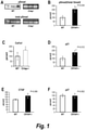
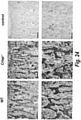
- the antibody or antigen binding fragment thereof improves a bone parameter selected from the group consisting of bone volume density (BV/TV), total bone surface (BS), bone surface density (BS/BV), trabecular number (Tb.N), trabecular thickness (Tb.Th), trabecular spacing (Tb.Sp), and total volume (Dens TV).
- BV/TV bone volume density
- BS total bone surface
- BS/BV bone surface density
- Tb.N trabecular number
- Tb.Th trabecular thickness
- Tb.Sp trabecular spacing
- Dens TV total volume density
- the antibody or antigen binding fragment thereof inhibits bone resorption.
- the antibody or antigen binding fragment thereof reduces a serum biomarker of bone resorption selected from the group consisting of urinary hydroxyproline, urinary total pyridinoline (PYD), urinary free deoxypyridinoline (DPD), urinary collagen type-I cross-linked N-telopeptide (NTX), urinary or serum collagen type-I cross-linked C-telopeptide (CTX), bone sialoprotein (BSP), osteopontin (OPN), and tartrate-resistant acid phosphatase 5b (TRAP).
- a serum biomarker of bone resorption selected from the group consisting of urinary hydroxyproline, urinary total pyridinoline (PYD), urinary free deoxypyridinoline (DPD), urinary collagen type-I cross-linked N-telopeptide (NTX), urinary or serum collagen type-I cross-linked C-telopeptide (CTX), bone sialoprotein (BSP), osteopontin (OPN), and tartrate-resistant
- the antibody or antigen binding fragment thereof increases a serum biomarker of bone deposition selected from the group consisting of as total alkaline phosphatase, bone-specific alkaline phosphatase, osteocalcin, and type-I procollagen (C-terminal/N-terminal).
- the antibody or antigen binding fragment thereof inhibits bone resorption. In another embodiment, the antibody or antigen binding fragment thereof promotes bone deposition. In another embodiment, the antibody or antigen binding fragment thereof improves the function of a non-skeletal organ affected by Oi selected from the group consisting of hearing function, lung function, and kidney function.
- administer refers to the act of injecting or otherwise physically delivering a substance as it exists outside the body (e.g ., an antibody) into a patient, such as by mucosal, intradermal, intravenous, subcutaneous, intramuscular delivery, and/or any other method of physical delivery described herein or known in the art.
- a disease, or a symptom thereof is being treated, administration of the substance typically occurs after the onset of the disease or symptoms thereof.
- administration of the substance typically occurs before the onset of the disease or symptoms thereof.
- an "antagonist” or “inhibitor” of TCF ⁇ refers to a molecule that is capable of inhibiting or otherwise decreasing one or more of the biological activities of TCF ⁇ , such as in a cell expressing TGF ⁇ or in a cell expressing a TGF ⁇ ligand, or expressing a TGF ⁇ receptor.
- antibodies of the invention are antagonist antibodies that inhibit or otherwise decrease the activity of TGF ⁇ in a cell having a cell surface-expressed TGF ⁇ receptor (e.g ., TGF ⁇ receptor 1, 2, or 3) when said antibody is contacted with said cell.
- an antagonist of TGF ⁇ e.g.
- an antibody of the invention may, for example, act by inhibiting or otherwise decreasing the activation and/or cell signaling pathways of the cell expressing a TGF ⁇ receptor, thereby inhibiting a TGF ⁇ -mediated biological activity of the cell relative to the TGF ⁇ -mediated biological activity in the absence of antagonist.
- the anti-TGF ⁇ antibodies are antagonistic anti-TGF ⁇ antibodies, preferably fully human, monoclonal, antagonistic anti-TGF ⁇ antibodies.
- antibody immunoglobulin
- Ig immunoglobulin
- the term antibody includes, but is not limited to, synthetic antibodies, monoclonal antibodies, recombinantly produced antibodies, multispecific antibodies (including bi-specific antibodies), human antibodies, humanized antibodies, chimeric antibodies, intrabodies, single-chain Fvs (scFv) ( e.g ., including monospecific, bispecific, etc.), camelized antibodies, Fab fragments, F(ab') fragments, disulfide-linked Fvs (sdFv), anti-idiotypic (anti-Id) antibodies, and epitope-binding fragments of any of the above.
- scFv single-chain Fvs
- sdFv single-chain Fvs
- sdFv disulfide-linked Fvs
- anti-Id anti-idiotypic antibodies
- antibodies include immunoglobulin molecules and immunologically active portions of immunoglobulin molecules, i.e ., antigen binding domains or molecules that contain an antigen-binding site that specifically binds to a TGF ⁇ antigen (e.g ., one or more complementarity determining regions (CDRs) of an anti-TGF ⁇ antibody).
- the anti-TGF ⁇ antibodies can be of any type (e.g ., IgG. IgE, IgM, IgD, IgA, and IgY), any class (e.g.
- the anti-TGF ⁇ antibodies are humanized, such as humanized monoclonal anti-TGF ⁇ antibodies, In other embodiments, the anti-TGF ⁇ antibodies are fully human, such as fully human monoclonal anti-TGF ⁇ antibodies. In preferred embodiments, the anti-TGF ⁇ antibodies are IgG antibodies, such as IgG4 antibodies.
- composition and “formulation” are intended to encompass a product containing specified ingredients (e.g., an anti-TGF ⁇ antibody) in, optionally, specified amounts, as well as any product which results, directly or indirectly, from the combination of specified ingredients in, optionally, specified amounts.
- specified ingredients e.g., an anti-TGF ⁇ antibody
- constant region or “constant domain” refer to a carboxy terminal portion of the light and heavy chain that is not directly involved in binding of the antibody to antigen, but exhibits various effector functions, such as interaction with the Fc receptor.
- the terms refer to the portion of an immunoglobulin molecule having a more conserved amino acid sequence relative to the other portion of the immunoglobulin, the variable domain, which contains the antigen binding site.
- the constant domain contains the CH1, CH2, and CH3 domains of the heavy chain, and the CHL domain of the light chain.
- epitope refers to a localized region on the surface of an antigen, such as a TGF ⁇ polypeptide or TGF ⁇ polypeptide fragment, that is capable of being bound to one or more antigen binding regions of an antibody, and that has antigenic or immunogenic activity in an animal, preferably a mammal, and most preferably in a human, that is capable of eliciting an immune response.
- An epitope having immunogenic activity is a portion of a polypeptide that elicits an antibody response in an animal.
- An epitope having antigenic activity is a portion of a polypeptide to which an antibody specifically binds, as determined by any method well known in the art, for example, such as an immunoassay.
- Antigenic epitopes need not necessarily be immunogenic. Epitopes usually consist of chemically active surface groupings of molecules, such as amino acids or sugar side chains, and have specific three-dimensional structural characteristics, as well as specific charge characteristics. A region of a polypeptide contributing to an epitope may be contiguous amino acids of the polypeptide or the epitope may come together from two or more non-contiguous regions of the polypeptide. The epitope may or may not be a three-dimensional surface feature of the antigen. In certain embodiments, a TGF ⁇ epitope is a three-dimensional surface feature of a TGF ⁇ polypeptide ( e.g ., in a trimeric form of a TGF ⁇ polypeptide).
- a TGF ⁇ epitope is a linear feature of a TGF ⁇ polypeptide ( e.g ., in a dimeric form or monomeric form of the TGF ⁇ polypeptide).
- Anti-TGF ⁇ antibodies may specifically bind to an epitope of the monomeric form of TGF ⁇ , an epitope of the dimeric form of TGF ⁇ , or both the monomeric form and the dimeric form of TGF ⁇ .
- excipients refers to inert substances that are commonly used as a diluent, vehicle, preservative, binder, stabilizing agent, etc. for drugs and includes, but is not limited to, proteins (e.g ., serum albumin, etc.), amino acids (e.g ., aspartic acid, glutamic acid, lysine, arginine, glycine, histidine, etc.), fatty acids and phospholipids ( e.g ., alkyl sulfonates, caprylate, etc.), surfactants ( e.g ., SDS, polysorbate, nonionic surfactant, etc.), saccharides ( e.g ., sucrose, maltose, trehalose, etc.) and polyols ( e.g ., mannitol, sorbitol, etc.). See, also, Remington's Pharmaceutical Sciences (1990) Mack Publishing Co., Easton, Pa .
- fragment refers to a peptide or polypeptide that comprises less than the full length amino acid sequence. Such a fragment may arise, for example, from a truncation at the amino terminus, a truncation at the carboxy terminus, and/or an internal deletion of a residue(s) from the amino acid sequence. Fragments may, for example, resultfrom alternative RNA splicing orfrom in vivo protease activity.
- TGF ⁇ fragments include polypeptides comprising an amino acid sequence of at least 50, at 100 amino acid residues, at least 125 contiguous amino acid residues, at least 150 contiguous amino acid residues, at least 175 contiguous amino acid residues, at least 200 contiguous amino acid residues, or at least 250 contiguous amino acid residues of the amino acid sequence of a TGF ⁇ polypeptide.
- a fragment of a TGF ⁇ polypeptide or an antibody that specifically binds to a TGF ⁇ antigen retains at least 1, at least 2, or at least 3 functions of the full-length polypeptide or antibody.
- Fully human antibody or “human antibody” are used interchangeably herein and refer to an antibody that comprises a human variable region and, most preferably a human constant region. In specific embodiments, the terms refer to an antibody that comprises a variable region and constant region of human origin.
- Fully human anti-TGF ⁇ antibodies in certain embodiments, can also encompass antibodies that bind TGF ⁇ polypeptides and are encoded by nucleic acid sequences that are naturally occurring somatic variants of a human germline immunoglobulin nucleic acid sequence. In a specific embodiment, the anti-TGF ⁇ antibodies are fully human antibodies.
- the term "fully human antibody” includes antibodies having variable and constant regions corresponding to human germline immunoglobulin sequences as described by Kabat et al.
- recombinant human antibody includes human antibodies that are prepared, expressed, created, or isolated by recombinant means, such as antibodies expressed using a recombinant expression vector transfected into a host cell, antibodies isolated from a recombinant, combinatorial human antibody library, antibodies isolated from an animal ( e.g ., a mouse or cow) that is transgenic and/or transchromosomal for human immunoglobulin genes (see, e.g ., Taylor, L. D. et al. (1992) Nucl. Acids Res. 20:6287-6295 ) or antibodies prepared, expressed, created, or isolated by any other means that involves splicing of human immunoglobulin gene sequences to other DNA sequences.
- Such recombinant human antibodies can have variable and constant regions derived from human germline immunoglobulin sequences (See Kabat, E. A. et al. (1991) Sequences of Proteins of Immunological Interest, Fifth Edition, U.S. Department of Health and Human Services, NIH Publication No. 91-3242 ).
- such recombinant human antibodies are subjected to in vitro mutagenesis (or, when an animal transgenic for human Ig sequences is used, in vivo somatic mutagenesis) and thus the amino acid sequences of the VH and VL regions of the recombinant antibodies are sequences that, while derived from and related to human germline VH and VL sequences, may not naturally exist within the human antibody germline repertoire in vivo.
- heavy chain when used in reference to an antibody refers to five distinct types, called alpha ( ⁇ ), delta ( ⁇ ), epsilon ( ⁇ ), gamma ( ⁇ ) and mu ( ⁇ ), based on the amino acid sequence of the heavy chain constant domain.
- These distinct types of heavy chains are well known in the art and give rise to five classes of antibodies, IgA, IgD, IgE, IgG, and IgM, respectively, including four subclasses of IgG, namely IgG1, IgG1, IgG3, and IgG4.
- the heavy chain is a human heavy chain.
- an “isolated” or “purified” antibody is substantially free of cellular material or other contaminating proteins from the cell or tissue source from which the antibody is derived, or substantially free of chemical precursors or other chemicals when chemically synthesized.
- the language “substantially free of cellular material” includes preparations of an antibody in which the antibody is separated from cellular components of the cells from which it is isolated or recombinantly produced.
- an antibody that is substantially free of cellular material includes preparations of antibody having less than about 30%, 20%, 10%, or 5% (by dry weight) of heterologous protein (also referred to herein as a "contaminating protein").
- the antibody When the antibody is recombinantly produced, it is also preferably substantially free of culture medium, i.e., culture medium represents less than about 20%, 10%, or 5% of the volume of the protein preparation.
- culture medium represents less than about 20%, 10%, or 5% of the volume of the protein preparation.
- the antibody When the antibody is produced by chemical synthesis, it is preferably substantially free of chemical precursors or other chemicals, i.e., it is separated from chemical precursors or other chemicals that are involved in the synthesis of the protein. Accordingly, such preparations of the antibody have less than about 30%, 20%, 10%, 5% (by dry weight) of chemical precursors or compounds other than the antibody of interest.
- anti-TGF ⁇ antibodies are isolated or purified.
- Kabat numbering and like terms are recognized in the art and refer to a system of numbering amino acid residues that are more variable (i.e. hypervariable) than other amino acid residues in the heavy and light chain variable regions of an antibody, or an antigen binding portion thereof ( Kabat et al. (1971) Ann. NY Acad. Sci. 190:382-391 and, Kabat et al. (1991) Sequences of Proteins of Immunological Interest, Fifth Edition, U.S. Department of Health and Human Services, NIH Publication No. 91-3242 ).
- the hypervariable region typically ranges from amino acid positions 31 to 35 for CDR1, amino acid positions 50 to 65 for CDR2, and amino acid positions 95 to 102 for CDR3.
- the hypervariable region typically ranges from amino acid positions 24 to 34 for CDR1, amino acid positions 50 to 56 for CDR2, and amino acid positions 89 to 97 for CDR3.
- light chain when used in reference to an antibody refers to two distinct types, called kappa ( ⁇ ) of lambda ( ⁇ ), based on the amino acid sequence of the constant domains. Light chain amino acid sequences are well known in the art. In preferred embodiments, the light chain is a human light chain.
- a subject derives from a therapy (e.g., a prophylactic or therapeutic agent), which does not result in a cure of the disease or disorder.
- a subject is administered one or more therapies (e.g., prophylactic or therapeutic agents) to "manage” a TGF ⁇ -mediated disease (e.g., OI), or one or more symptoms thereof, so as to prevent the progression or worsening of the disease.
- therapies e.g., prophylactic or therapeutic agents
- a “monoclonal antibody” refers to an antibody obtained from a population of homogenous or substantially homogeneous antibodies, and each monoclonal antibody will typically recognize a single epitope on the antigen.
- a “monoclonal antibody” is an antibody produced by a single hybridoma or other cell.
- the term “monoclonal” is not limited to any particular method for making the antibody. For example, monoclonal antibodies may be made by the hybridoma method as described in Kohler et al.; Nature, 256:495 (1975 ) or may be isolated from phage libraries.
- pharmaceutically acceptable means being approved by a regulatory agency of the Federal or a State government or listed in the U.S. Pharmacopeia, European Pharmacopeia, or other generally recognized Pharmacopeia for use in animals, and more particularly in humans.
- pharmaceutically acceptable excipient means any inert substance that is combined with an active molecule, such as a monoclonal antibody, for preparing an agreeable or convenient dosage form.
- pharmaceutically acceptable excipient is an excipient that is non-toxic to recipients at the dosages and concentrations employed, and is compatible with other ingredients of the formulation comprising the monoclonal antibody.
- prevent refers to the total or partial inhibition of the development, recurrence, onset, or spread of a TGF ⁇ -mediated disease and/or symptom related thereto, resulting from the administration of a therapy or combination of therapies provided herein (e.g., a combination of prophylactic or therapeutic agents).
- TGF ⁇ antigen refers to that portion of a TGF ⁇ polypeptide to which an antibody specifically binds.
- a TGF ⁇ antigen also refers to an analog or derivative of a TGF ⁇ polypeptide or fragment thereof to which an antibody specifically binds.
- a TGF ⁇ antigen is a monomeric TGF ⁇ antigen or a dimeric TGF ⁇ antigen.
- a region of a TGF ⁇ polypeptide contributing to an epitope may be contiguous amino acids of the polypeptide, or the epitope may come together from two or more non-contiguous regions of the polypeptide.
- the epitope may or may not be a three-dimensional surface feature of the antigen.
- a localized region on the surface of a TGF ⁇ antigen that is capable of eliciting an immune response is a TGF ⁇ epitope.
- the epitope may or may not be a three-dimensional surface feature of the antigen.
- an "analog" of the TGF ⁇ antigen refers to a polypeptide that possesses a similar or identical function as a TGF ⁇ polypeptide, a fragment of a TGF ⁇ polypeptide, or a TGF ⁇ epitope described herein.
- the analog may comprise a sequence that is at least 80%, at least 85%, at least 90%, at least 95%, or at least 99% identical to the amino acid sequence of a TGF ⁇ polypeptide (e.g., SEQ ID NO: 1, 2, or 3), a fragment of a TGF ⁇ polypeptide, a TGF ⁇ epitope, or an anti-TGF ⁇ antibody described herein.
- a TGF ⁇ polypeptide e.g., SEQ ID NO: 1, 2, or 3
- the polypeptide is encoded by a nucleotide sequence that hybridizes under stringent conditions to a nucleotide sequence encoding a TGF ⁇ polypeptide, a fragment of a TGF ⁇ polypeptide, or a TGF ⁇ epitope described herein
- human TGF ⁇ refers to the polypeptides ("polypeptides,” “peptides,” and “proteins” are used interchangeably herein) comprising the amino acid sequence of SEQ ID NO: 1, 2, or 3, and related polypeptides, including SNP variants thereof.
- Related polypeptides include allelic variants (e.g., SNP variants); splice variants; fragments; derivatives; substitution, deletion, and insertion variants; fusion polypeptides; and interspecies homologs, preferably, which retain TGF ⁇ activity and/or are sufficient to generate an anti-TGF ⁇ immune response.
- soluble forms of TGF ⁇ that are sufficient to generate an anti-TGF ⁇ immunological response.
- an anti-TGF ⁇ antibody can bind to a TGF ⁇ polypeptide, polypeptide fragment, antigen, and/or epitope, as an epitope is part of the larger antigen, which is part of the larger polypeptide fragment, which, in turn, is part of the larger polypeptide.
- hTGF ⁇ can exist in a dimeric or monomeric form.
- TGF ⁇ -mediated disease and “TGF ⁇ -mediated disorder” are used interchangeably and refer to any disease or disorder that is completely or partially caused by or is the result of TGF ⁇ , e.g., hTGF ⁇ .
- TGF ⁇ is aberrantly expressed.
- TGF ⁇ may be aberrantly upregulated in a particular cell type.
- normal, aberrant, or excessive cell signaling is caused by binding of TGF ⁇ to a TGF ⁇ receptor.
- the TGF ⁇ receptor e.g., TGF ⁇ receptor 1, 2, or 3
- the TGF ⁇ -mediated disease is a degenerative bone disease, such as osteogenesis imperfecta.
- an antigen or a fragment thereof e.g., TGF ⁇
- An antibody that specifically binds to an antigen may bind to other peptides or polypeptides with lower affinity, as determined by, e.g., radioimmunoassays (RIA), enzyme-linked immunosorbent assays (ELISA), BIACORE, or other assays known in the art.
- an anti-TGF ⁇ antibody of the invention may specifically bind to TGF ⁇ (e.g., hTGF ⁇ ) with more than two-fold greater affinity that a different, non-TGF ⁇ antigen.
- Antibodies or variants or fragments thereof that specifically bind to an antigen may be cross-reactive with related antigens.
- an anti-TGF ⁇ antibody may cross-react with hTGF ⁇ and another TGF ⁇ antigen (e.g., a rodent or non-human primate TGF ⁇ antibody).
- TGF ⁇ antigen e.g., a rodent or non-human primate TGF ⁇ antibody
- antibodies or variants or fragments thereof that specifically bind to an antigen do not cross-react with other non-TGF ⁇ antigens.
- An antibody or a variant or a fragment thereof that specifically binds to a TGF ⁇ antigen can be identified, for example, by immunoassays, BIAcore, or other techniques known to those of skill in the art.
- a specific or selective reaction will be at least twice background signal or noise, and more typically more than 10 times background.
- the binding protein or antibody will bind to its antigen, e.g. TGF ⁇ , with a dissociation constant of between 1x10 -6 M and 1x10 -7 .
- the dissociation constant is between 1x10 -6 M and 1x10 -8 . See, e.g., Paul, ed., 1989, Fundamental Immunology Second Edition, Raven Press, New York at pages 332-336 for a discussion regarding antibody specificity.
- a subject is preferably a mammal, such as a non-primate (e.g., cows, pigs, horses, cats, dogs, rats, etc.) or a primate ( e.g., monkey and human), most preferably a human.
- the subject is a mammal, preferably a human, having a TGF ⁇ -mediated disease.
- the subject is a mammal, preferably a human, at risk of developing a TGF ⁇ -mediated disease.
- a therapeutic agent refers to any agent that can be used in the treatment, management, or amelioration of a TGF ⁇ -mediated disease and/or a symptom related thereto.
- the term “therapeutic agent” refers to a TGF ⁇ antibody.
- the term “therapeutic agent” refers to an agent other than a TGF ⁇ antibody.
- a therapeutic agent is an agent that is known to be useful for, or has been, or is currently being used for the treatment, management, or amelioration of a TGF ⁇ -mediated disease, or one or more symptoms related thereto.
- the term “therapy” refers to any protocol, method, and/or agent that can be used in the prevention, management, treatment, and/or amelioration of a TGF ⁇ -mediated disease (e.g., OI).
- the terms “therapies” and “therapy” refer to a biological therapy, supportive therapy, and/or other therapies useful in the prevention, management, treatment, and/or amelioration of a TGF ⁇ -mediated disease known to one of skill in the art, such as medical personnel.
- treat refers to the reduction or amelioration of the progression, severity, and/or duration of a TGF ⁇ -mediated disease (e.g., OI) resulting from the administration of one or more therapies (including, but not limited to, the administration of one or more prophylactic or therapeutic agents).
- a TGF ⁇ -mediated disease e.g., OI
- therapies including, but not limited to, the administration of one or more prophylactic or therapeutic agents.
- such terms refer to the reduction or inhibition of the binding of TGF ⁇ to a TGF ⁇ receptor, the reduction or inhibition of the production or secretion of TGF ⁇ from a cell expressing a TGF ⁇ receptor of a subject, the reduction or inhibition of the production or secretion of TGF ⁇ from a cell not expressing a TGF ⁇ receptor of a subject, and/or the inhibition or reduction of one or more symptoms associated with a TGF ⁇ -mediated disease, such as OI.
- variable region refers to a portion of the light and heavy chains, typically about the amino-terminal 120 to 130 amino acids in the heavy chain and about 100 to 110 amino acids in the light chain, which differ extensively in sequence among antibodies and are used in the binding and specificity of each particular antibody for its particular antigen.
- the variability in sequence is concentrated in those regions called complementarity determining regions (CDRs), while the more highly conserved regions in the variable domain are called framework regions (FR).
- CDRs of the light and heavy chains are primarily responsible for the interaction of the antibody with antigen. Numbering of amino acid positions is according to the EU Index, as in Kabat et al. (1991) Sequences of proteins of immunological interest. (U.S. Department of Health and Human Services, Washington D.C.) 5th ed . (“Kabat et al .”) .
- the variable region is a human variable region.
- OI encompasses a group of congenital bone disorders characterized by deficiencies in one or more proteins involved in bone matrix deposition or homeostasis. There are eight types of OI that are defined by their specific gene mutation, and the resulting protein deficiency and phenotype of the affected individual. Though phenotypes vary among OI types, common symptoms include incomplete ossification of bones and teeth, reduced bone mass, brittle bones, and pathologic fractures.
- Type-I collagen is one of the most abundant connective tissue proteins in both calcified and non-calcified tissues. Accurate synthesis, post-translational modification, and secretion of type-I collagen are necessary for proper tissue development, maintenance, and repair. Most mutations identified in individuals with OI result in reduced synthesis of type-I collagen, or incorrect synthesis and/or processing of type-I collagen.
- FKBP10 FK506 binding protein 10
- HSP47 heat shock protein 47
- intermolecular collagen crosslinking genes such as procollagen-lysine, 2-oxoglutarate 5-dioxygenase 2 (PLOD2), and in members of the collagen prolyl hydroxylase family of genes, including leucine proline-enriched proteoglycan (leprecan) (LEPRE1), peptidylprolyl isomerase B (cyclophilin B) (CYPB), and cartilage associated protein (CRTAP) (Morello et al., 2006; Cabral et al., 2007; Baldridge et al., 2008; van Dijk et al., 2009; Choi et al., 2009; Barnes et al., 2010; Pyott et al., 2011).
- leprecan leucine proline-enriched proteoglycan
- CYPB peptidylprolyl isomerase B
- CRTAP cartilage associated protein
- BMP bone morphogenetic protein
- TGF ⁇ transforming growth factor ⁇
- TGF ⁇ expression is regulated by molecules that bind type-I and type-II collagen.
- a small leucine rich proteoglycan (SLRP) regulates TGF ⁇ expression.
- SLRP small leucine rich proteoglycan
- decorin regulates TGF ⁇ synthesis.
- decorin does not bind type-I or type-II collagen in which the 3-hydroxyproline site is absent at position 986 of the type-I and/or type-II collagen molecules.
- the vertebrate skeleton is comprised of bone, which is a living, calcified tissue that provides structure, support, protection, and a source of minerals for regulating ion transport.
- Bone is a specialized connective tissue that is comprised of both cellular and acellular components.
- the acellular extracellular matrix (ECM) contains both collagenous and non-collagenous proteins, both of which participate in the calcification process.
- a correctly secreted and aligned ECM is critical for proper bone formation. Pathology results when any of the ECM proteins are absent, malformed or misaligned, as is evidenced in osteogenesis imperfecta.
- cortical bone or “compact bone” refers to the outer layer of bone, which is dense, rigid, and tough.
- trabecular bone or “cancellous bone” is the spongy inner layer of bone, which is lighter and less dense than cortical bone.
- trabecula refers to the microscopic structural unit of spongy bone, which is of a rod-like shape and collagenous composition.
- Bone is a dynamic tissue that undergoes constant remodeling.
- osteoblast refers to a terminally-differentiated bone forming cell that deposits osteoid.
- osteoid refers to immature, unmineralized bone that is comprised primarily of type-I collagen.
- pre-osteoblast refers to a proliferating immature osteoblast that is not fully differentiated.
- osteoprogenitor refers to a pluripotent cell that gives rise to several stromal cell types, including osteoblasts. Osteoprogenitor cells, which are commonly referred to as “mesenchymal stem cells,” arise in the bone marrow and can be isolated in small numbers from circulating blood.
- osteoclast refers to a terminally-differentiated bone resorbing cell that is descended from a bone marrow monocyte. Osteoclasts can be identified by their expression of tartrate resistant acid phosphatase (TRAP).
- TRIP tartrate resistant acid phosphatase
- osteoblasts and osteoclasts work in unison to maintain bone integrity.
- Pathology results when bone deposition and bone resorption become uncoupled.
- osteopetrosis is a bone disease characterized by overly dense, hard bone that is a result of unresorptive osteoclasts
- osteoporosis is a bone disorder characterized by brittle, porous bones which can result from increased osteoclast activity.
- Evidence suggests that osteoclast activity may be increased in osteogenesis imperfecta, implicating these cell types as a potential target for therapeutic intervention.
- the present disclosure includes methods of inhibiting osteoclasts with a TGF ⁇ antibody.
- bones are measured and characterized by at least one of these methods.
- bone volume density refers to the fraction of a given volume of bone (total volume or TV) that is comprised of calcified matter (bone volume or BV). Therefore, bone volume density is calculated as BV/TV and reported as a percentage.
- specific bone surface refers to the total bone surface (BS) per given volume of bone. Therefore, specific bone surface is calculated as BS/TV.
- Other common bone measurements include: bone area (B.Ar), trabecular number (Tb.N); trabecular spacing (Tb.Sp); N.Oc (osteoclast number); Oc.S (osteoclast surface area); Oc.S/BS; osteoblast number (N.Ob), osteoblast surface area (Ob.S), osteoblast perimeter (Ob.Pm), and derivatives of any of said measurements.
- a larger Oc.S/BS is an indicator of increased bone resorption by osteoclasts.
- TGF ⁇ Transforming growth factor beta
- TGF ⁇ s are multifunctional cytokines that are involved in cell proliferation and differentiation, embryonic development, extracellular matrix formation, bone development, wound healing, hematopoiesis, and immune and inflammatory responses (Roberts et al., 1981; Border et al., 1995a).
- Secreted TGF ⁇ protein is cleaved into a latency-associated peptide (LAP) and a mature TGF ⁇ peptide, and is found in latent and active forms.
- LAP latency-associated peptide
- the mature TGF ⁇ peptide forms both homodimers and heterodimers with other TGF ⁇ family members.
- TGF ⁇ may be purified from any natural source, or may be produced synthetically ( e.g., by use of recombinant DNA technology).
- the TGF ⁇ molecule is from a human, known herein as "hTGF ".
- TGF ⁇ 1 There are three human TGF ⁇ isoforms: TGF ⁇ 1, TGF ⁇ 2, and TGF ⁇ 3 (Swiss Prot accession numbers P01137, P08112, and P10600, respectively) which, in their biologically active state, are 25 kDa homodimers comprising two 112 amino acid monomers joined by an inter-chain disulfide bridge.
- TGF ⁇ 1 differs from TGF ⁇ 2 by 27, and from TGF ⁇ 3 by 22, mainly conservative amino acid changes.
- TGF ⁇ receptor 1 There are three TGF ⁇ receptors in humans, TGF ⁇ receptor 1, 2, and 3, which can be distinguished by their structural and functional properties, including affinity for TGF ⁇ protein family members. Binding of a TGF ⁇ protein to a homodimeric or heterodimeric TGF ⁇ transmembrane receptor complex activates the canonical TGF ⁇ signaling pathway mediated by intracellular SMAD proteins.
- TGF ⁇ s The deregulation of TGF ⁇ s leads to pathological processes that, in humans, have been implicated in numerous conditions, such as, birth defects, cancer, chronic inflammatory, autoimmune diseases, and fibrotic diseases (Border et al., 1994; Border et al., 1995b).
- Human TGF ⁇ s are very similar to mouse TGF ⁇ s: human TGF ⁇ 1 has only one amino acid difference from mouse TGF ⁇ 1; human TGF ⁇ 2 has only three amino acid differences from mouse TGF ⁇ 2; and human TGF ⁇ 3 is identical to mouse TGF ⁇ 3.
- TGF ⁇ transforming growth factor beta
- the antibody that binds to TGF ⁇ comprises a heavy chain variable region comprising the amino acid sequences of SEQ ID NOs: 4, 5, and 6; and a light chain variable region comprising the amino acid sequences of SEQ ID NOs: 7, 8, and 9.
- the antibody that binds to TGF ⁇ comprises a heavy chain variable region comprising the amino acid sequence of SEQ ID NO: 10:
- the antibody that binds to TGF ⁇ comprises a light chain variable region comprising the amino acid sequence of SEQ ID NO: 11:
- the antibody that binds to TGF ⁇ comprises a heavy chain variable region comprising the amino acid sequence of SEQ ID NO: 10; and a light chain variable region comprising the amino acid sequence of SEQ ID NO: 11.
- the antibody that binds to TGF ⁇ further comprises a constant region, e,g., a human IgG constant region.
- the constant region is a human IgG4 constant region.
- the constant region is a modified human IgG4 constant region.
- the IgG4 constant region comprises the amino acid sequence of SEQ ID NO: 12:
- the constant region is a human CK constant region.
- the CK constant region comprises the amino acid sequence of SEQ ID NO: 13:
- the antibody that binds to TGF ⁇ comprises a heavy chain comprising the amino acid sequence of SEQ ID NO: 14: Positions 1-120: variable region of the heavy chain (VH).
- the CDRs complementarity determining regions, according to Kabat definition) are underlined.
- Positions 121-447 constant region of human IgG4 (SwissProt IGHG4_HUMAN).
- the antibody that binds to TGF ⁇ comprises a light chain comprising the amino acid sequence of SEQ ID NO: 15: Positions 1-108: variable region of the light chain (VL).
- the CDRs complementarity determining regions, according to Kabat definition) are underlined.
- Positions 109-215 constant region of human CK.
- the antibody that binds to TGF ⁇ comprises a heavy chain comprising the amino acid sequence of SEQ ID NO: 14, and a light chain comprising the amino acid sequence of SEQ ID NO: 15.
- the antibody that binds to TGF ⁇ is expressed by a host cells as comprising leader sequences.
- the leader sequence preferably comprises an amino acid sequence from 1-30 amino acids in length, more preferably 25-25 amino acids, and most preferably 19 amino acids.
- the heavy chain, light chain, or both the heavy and light chain may comprise a leader sequence.
- the light or heavy chain leader sequence may comprise the amino acid sequence of SEQ ID NO: 16: MGWSCIILFL VATATGVHS ( SEQ ID NO: 16 ).
- a host cell may expressing a unprocessed heavy chain may comprise the amino acid sequence of SEQ ID NO: 17: wherein positions 1-19: leader sequence
- Positions 20-139 variable region of the heavy chain (VH).
- the CDRs complementarity determining regions, according to Kabat definition
- Positions 140-466 constant region of human IgG4 (SwissProt IGHG4_HUMAN).
- a host cell expressing a unprocessed light chain may comprise the amino acid of SEQ ID NO: 18: wherein Positions 1-19: leader sequence Positions 20-127: variable region of the light chain (VL).
- the CDRs complementarity determining regions, according to Kabat definition) are underlined.
- Positions 128-234 constant region of human CK.
- the antibody that binds to TGF ⁇ is a humanized or fully human antibody.
- humanized and fully human antibody isotypes include IgA, IgD, IgE, IgG, and IgM.
- the anti-TGF ⁇ antibody is an IgG antibody. There are four forms of IgG.
- the anti-TGF ⁇ antibody is an IgG4 antibody.
- the anti-TGF ⁇ antibody is a humanized IgG4 antibody.
- the anti-TGF ⁇ antibody is a fully human IgG4 antibody.
- the anti-TGF ⁇ antibody is an IgG4 anti-TGF ⁇ antibody comprising a heavy chain comprising the amino acid sequence of SEQ ID NO: 14 and a light chain comprising the amino acid sequence of SEQ ID NO: 15.
- the anti-TGF ⁇ antibody is an IgG4 anti-TGF ⁇ antibody comprising a heavy chain variable region and a light chain variable region, the heavy chain variable region comprising 3 complementarity determining regions (CDRs) comprising the amino acid sequences of SEQ ID NOs: 4, 5, and 6, and the light chain variable region comprising 3 CDRs comprising the amino acid sequences of SEQ ID NOs: 7, 8, and 9.
- CDRs complementarity determining regions
- anti-TGF ⁇ antibodies including the anti-TGF ⁇ antibody comprising a heavy chain amino acid sequence comprising SEQ ID NO: 14 and a light chain amino acid sequence comprising SEQ ID NO: 15, and the CDR sequences corresponding with SEQ ID NOs: 4-9 have been described in detail in U.S. Patent No. 7,723,486 , and U.S. Patent No. 8,383,780 .
- the antibody or antigen binding fragment thereof is "pan-specific" and binds to human TGF ⁇ 1, TGF ⁇ 2, and TGF ⁇ 3 and acts as an antagonist.
- the antibody or antigen binding fragment thereof binds to human TGF ⁇ 1, TGF ⁇ 2, and TGF ⁇ 3, and neutralizes human TGF ⁇ 1, TGF ⁇ 2, and TGF ⁇ 3.
- Exemplary pan-specific anti-TGF ⁇ monoclonal antibodies (mAbs) suitable for use in the methods of the invention are described in US Patent Nos. 7,723,476 and 8,383,780 .
- 1D11.16 is an exemplary murine pan-specific anti-TGF ⁇ antibody that neutralizes human and mouse TGF ⁇ 1, TGF ⁇ 2, and TGF ⁇ 3 in a wide range of in vitro assays (Dasch et al., 1989; Dasch et al., 1996; R&D System product sheet for MAB1835) and is efficacious in proof-of principle studies in animal models of fibrosis (Ling et al., 2003; Miyajima et al., 2000; Schneider et al., 1999; Khanna et al., 1999; Shenkar et al., 1994).
- 1D11.16 is a murine monoclonal antibody (Dasch et al., 1989; Dasch et al., 1996), it is not a preferred for therapeutic use in humans. Accordingly, in certain embodiments, variants or derivatives of the 1D11.16 antibody are employed in the methods of the invention.
- certain embodiments of the invention also include variants or derivatives of anti-TGF ⁇ antibodies.
- the invention may include variants of the anti-TGF ⁇ antibody that is an IgG4 anti-TGF ⁇ antibody comprising a heavy chain comprising the amino acid sequence of SEQ ID NO: 14, and a light chain comprising the amino acid sequence of SEQ ID NO: 15.
- the invention includes variants or derivatives of the 1D11.16 antibody.
- Variants of anti-TGF ⁇ antibodies may have similar physicochemical properties based on their high similarity, and therefore are also included within the scope of the invention.
- Variants are defined as antibodies with an amino acid sequence that is at least 80%, at least 90%, at least 95%, or at least 97%, e.g., least 98% or 99% homologous to an anti-TGF ⁇ antibody described herein, and capable of competing for binding to a TGF ⁇ polypeptide, a TGF ⁇ polypeptide fragment, or a TGF ⁇ epitope.
- the variants will ameliorate, neutralize, or otherwise inhibit binding of TGF ⁇ with its receptors and TGF ⁇ biological activity (e.g., TGF ⁇ receptor-mediated intracellular SMAD signaling and resulting cellular activity). Determining competition for binding to the target can be done by routine methods known to the skilled person in the art.
- the variants are human antibodies, and preferably are IgG4 molecules.
- a variant is at least 90%, 95%, 96%, 97%, 98%, or 99% identical in amino acid sequence with the IgG4 anti-TGF ⁇ antibody comprising a heavy chain comprising the amino acid sequence of SEQ ID NO: 14, and a light chain comprising the amino acid sequence of SEQ ID NO: 15.
- the term "variant" refers to an antibody that comprises an amino acid sequence that is altered by one or more amino acids compared to the amino acid sequences of the anti-TGF ⁇ antibody.
- the variant may have conservative sequence modifications, including amino acid substitutions, modifications, additions, and deletions.
- modifications include, but are not limited to, glycosylation, acetylation, pegylation, phosphorylation, amidation, derivatization by known protecting/blocking groups, proteolytic cleavage, and linkage to a cellular ligand or other protein.
- Amino acid modifications can be introduced by standard techniques known in the art, such as site-directed mutagenesis, molecular cloning, oligonucleotide-directed mutagenesis, and random PCR-mediated mutagenesis in the nucleic acid encoding the antibodies.
- Conservative amino acid substitutions include the ones in which the amino acid residue is replaced with an amino acid residue having similar structural or chemical properties. Families of amino acid residues having similar side chains have been defined in the art.
- amino acids with basic side chains e.g., lysine, arginine, histidine
- acidic side chains e.g., aspartic acid, glutamic acid
- uncharged polar side chains e.g., asparagine, glutamine, serine, threonine, tyrosine, cysteine, tryptophan
- nonpolar side chains e.g., glycine, alanine, valine, leucine, isoleucine, proline, phenylalanine, methionine
- beta-branched side chains e.g., threonine, valine, isoleucine
- aromatic side chains e.g., tyrosine, phenylalanine, tryptophan
- a variant may have non-conservative amino acid substitutions, e.g., replacement of an amino acid with an amino acid residue having different structural or chemical properties. Similar minor variations may also include amino acid deletions or insertions, or both.
- Guidance in determining which amino acid residues may be substituted, modified, inserted, or deleted without abolishing immunological activity may be found using computer programs well known in the art. Computer algorithms, such as, inter alia, Gap or Bestfit, which are known to a person skilled in the art, can be used to optimally align amino acid sequences to be compared and to define similar or identical amino acid residues. Variants may have the same or different, either higher or lower, binding affinities compared to an anti-TGF ⁇ antibody, but are still capable of specifically binding to TGF ⁇ , and may have the same, higher or lower, biological activity as the anti-TGF ⁇ antibody.
- Embodiments of the invention also include antigen binding fragments of the anti-TGF ⁇ antibodies.
- the term "antigen binding domain,” “antigen binding region,” “antigen binding fragment,” and similar terms refer to that portion of an antibody that comprises the amino acid residues that interact with an antigen and confer on the binding agent its specificity and affinity for the antigen (e.g., the complementarity determining regions (CDR)).
- the antigen binding region can be derived from any animal species, such as rodents (e.g., rabbit, rat or hamster) and humans. Preferably, the antigen binding region will be of human origin.
- Non-limiting examples of antigen binding fragments include: Fab fragments, F(ab')2 fragments, Fd fragments, Fvfragments, single chain Fv (scFv) molecules, dAb fragments, and minimal recognition units consisting of the amino acid residues that mimic the hypervariable region of the antibody.
- the methods described herein comprise administering a therapeutically effective amount of an antibody that binds to TGF ⁇ to a subject.
- therapeutically effective amount means a dose of antibody that binds to TGF ⁇ that results in a detectable improvement in one or more symptoms associated with OI or which causes a biological effect (e.g., a decrease in the level of a particular biomarker) that is correlated with the underlying pathologic mechanism(s) giving rise to the condition or symptom(s) of osteogenesis imperfecta.
- a dose of antibody that binds to TGF ⁇ that increases bone mineral density, increases bone mass and/or bone strength, reduces bone and/or tooth fractures, and/or improves any diagnostic measurement of OI is deemed a therapeutically effective amount.
- bone mineral density, bone mass, and/or bone strength are increased by about 5% to about 200% following treatment with an antibody that binds to TGF ⁇ .
- bone mineral density, bone mass, and/or bone strength are increased by about 5% to about 10%, 10% to about 15%, 15% to about 20%, 20% to about 25%, 25% to about 30%, 30% to about 35%, 35% to about 40%, 40% to about 45%, 45% to about 50%, 50% to about 55%, 55% to about 60%, 60% to about 65%, 65% to about 70%, 70% to about 75%, 75% to about 80%, 80% to about 85%, 85% to about 90%, 90% to about 95%, 95% to about 100%, 100% to about 105%, 105% to about 110%, 110% to about 115%, 115% to about 120%, 120% to about 125%, 125% to about 130%, 130% to about 135%, 135% to about 140%, 140% to about 145%, 145% to about 150%, 150% to about 155%, 155% to about 160%
- a dose of an antibody which reduces serum biomarkers of bone resorption such as urinary hydroxyproline, urinary total pyridinoline (PYD), urinary free deoxypyridinoline (DPD), urinary collagen type-I cross-linked N-telopeptide (NTX), urinary or serum collagen type-I cross-linked C-telopeptide (CTX), bone sialoprotein (BSP), osteopontin (OPN), and tartrate-resistant acid phosphatase 5b (TRAP), is deemed a therapeutically effective amount.
- serum biomarkers of bone resorption are reduced by about 5% to about 200% following treatment with an antibody that binds to TGF ⁇
- serum biomarkers of bone resorption such as urinary hydroxyproline, urinary total pyridinoline (PYD), urinary free deoxypyridinoline (DPD), urinary collagen type-I cross-linked N-telopeptide (NTX), urinary or serum collagen type-I cross-linked C-telopeptide (CTX), bone sialoprotein (BSP), osteopontin (OPN), and tartrate-resistant acid phosphatase 5b (TRAP)
- urine biomarkers of bone resorption are decreased by about 5% to about 10%, 10% to about 15%, 15% to about 20%, 20% to about 25%, 25% to about 30%, 30% to about 35%, 35% to about 40%, 40% to about 45%, 45% to about 50%, 50% to about 55%, 55% to about 60%, 60% to about 65%, 65% to about 70%, 70% to about 75%, 75% to about 80%, 80% to about 85%, 85% to about 90%, 90% to about 95%, 95% to about 100%, 100% to about 105%, 10
- a dose of an antibody which increase serum biomarkers of bone deposition is deemed a therapeutically effective amount.
- serum biomarkers of bone deposition are increased by about 5% to about 200% following treatment with an antibody that binds to TGF ⁇
- serum biomarkers of bone deposition such as total alkaline phosphatase, bone-specific alkaline phosphatase, osteocalcin, and type-I procollagen (C-terminal/N-terminal) are increased by about 5% to about 10%, 10% to about 15%, 15% to about 20%, 20% to about 25%, 25% to about 30%, 30% to about 35%, 35% to about 40%, 40% to about 45%, 45% to about 50%, 50% to about 55%, 55% to about 60%, 60% to about 65%, 65% to about 70%, 70% to about 75%, 75% to about 80%, 80% to about 85%, 85% to about 90%, 90% to about 95%, 95% to about 100%, 100% to about 105%, 105% to about 110%, 110% to about 115%, 115% to about 120%, 120% to about 125%, 125% to about 130%, 130% to about 135%, 135% to about 140%, 140% to about 145%, 145% to about 150%, 150% to about 155%, 155% to about
- Other embodiments include administering a therapeutically effective dose of an antibody which improves the function of non-skeletal organs affected by OI.
- a dose of antibody that binds to TGF ⁇ that improves hearing, lung, and/or kidney function is deemed a therapeutically effective amount.
- a therapeutically effective amount of an antibody that binds to TGF ⁇ that is administered to a subject will vary depending upon the age and the size (e.g., body weight or body surface area) of the subject, as well as the route of administration, and other factors well known to those of ordinary skill in the art.
- the anti-TGF ⁇ antibody is administered to the subject as a subcutaneous dose.
- Other exemplary modes of administration include, but are not limited to, intradermal, intramuscular, intraperitoneal, intravenous, intranasal, epidural, and oral routes.
- the composition may be administered by any convenient route, for example by infusion or bolus injection, by absorption through epithelial or mucocutaneous linings ( e.g., oral mucosa, rectal and intestinal mucosa, etc .) and may be administered together with other biologically active agents. Administration can be systemic or local.
- the TGF ⁇ antibody can be administered parenterally or subcutaneously.
- compositions e.g., encapsulation in liposomes, microparticles, microcapsules, receptor mediated endocytosis (see, e.g., Wu et al. (1987) J. Biol. Chem. 262:4429-4432 ).
- the therapeutic compositions will be administered with suitable carriers, excipients, and other agents that are incorporated into formulations to provide improved transfer, delivery, tolerance, and the like.
- suitable carriers, excipients, and other agents that are incorporated into formulations to provide improved transfer, delivery, tolerance, and the like.
- a multitude of appropriate formulations can be found in the formulary known to all pharmaceutical chemists: Remington's Pharmaceutical Sciences, Mack Publishing Company, Easton, PA .
- formulations include, for example, powders, pastes, ointments, jellies, waxes, oils, lipids, lipid (cationic or anionic) containing vesicles (such as LIPOFECTIN TM ), DNA conjugates, anhydrous absorption pastes, oil-in-water and water-in-oil emulsions, emulsions carbowax (polyethylene glycols of various molecular weights), semi-solid gels, and semi-solid mixtures containing carbowax. See also Powell et al. "Compendium of excipients for parenteral formulations" PDA (1998) J Pharm Sci Technol 52:238-311 .
- compositions may be prepared into dosage forms in a unit dose suited to fit a dose of the active ingredients.
- dosage forms in a unit dose include, for example, tablets, pills, capsules, injections (ampoules), suppositories, etc.
- compositions can also be administered to the subject using any acceptable device or mechanism.
- administration can be accomplished using a syringe and needle or with a reusable pen and/or autoinjector delivery device.
- the methods of the present invention include the use of numerous reusable pen and/or autoinjector delivery devices to administer a TGF ⁇ binder (or pharmaceutical formulation comprising the binder).
- Examples of such devices include, but are not limited to AUTOPEN TM (Owen Mumford, Inc., Woodstock, UK), DlSETRONlC TM pen (Disetronic Medical Systems, Bergdorf, Switzerland), HUMALOG MIX 75/25 TM pen, HUMALOG TM pen, HUMALIN 70/30 TM pen (Eli Lilly and Co., Indianapolis, IN), NOVOPEN TM I, II and III (Novo Nordisk, Copenhagen, Denmark), NOVOPEN JUNIOR TM (Novo Nordisk, Copenhagen, Denmark), BD TM pen (Becton Dickinson, Franklin Lakes, NJ), OPTIPEN TM , OPTIPEN PRO TM , OPTIPEN STARLET TM , and OPTICLIK TM (sanofi-aventis, Frankfurt, Germany), to name only a few.
- AUTOPEN TM Owen Mumford, Inc., Woodstock, UK
- DlSETRONlC TM pen Disetronic Medical Systems, Bergdorf, Switzerland
- Examples of disposable pen and/or autoinjector delivery devices having applications in subcutaneous delivery of a pharmaceutical composition include, but are not limited to the SOLOSTAR TM pen (sanofi-aventis), the FLEXPEN TM (Novo Nordisk), and the KWIKPEN TM (Eli Lilly), the SURECLICKTM Autoinjector (Amgen, Thousand Oaks, CA), the PENLETTM (Haselmeier, Stuttgart, Germany), the EPIPEN (Dey, L.P.), and the HUMIRATM Pen (Abbott Labs, Abbott Park, IL), to name only a few.
- microinfusor means a subcutaneous delivery device designed to slowly administer large volumes (e.g., up to about 2.5 mL or more) of a therapeutic formulation over a prolonged period of time (e.g., about 10, 15, 20, 25, 30 or more minutes). See, e.g., U.S. 6,629,949 ; US 6,659,982 ; and Meehan et al., J. Controlled Release 46:107-116 (1996 ). Microinfusors are particularly useful for the delivery of large doses of therapeutic proteins contained within high concentration (e.g., about 100, 125, 150, 175, 200 or more mg/mL) and/or viscous solutions.
- high concentration e.g., about 100, 125, 150, 175, 200 or more mg/mL
- the invention includes methods for treating OI that comprise administering to a subject in need of such treatment an antibody that binds to TGF ⁇ in combination with at least one additional therapeutic agent.
- additional therapeutic agents that can be administered in combination with an anti-TGF ⁇ antibody in the practice of the methods of the present invention include, but are not limited to, bisphosphonates, calcitonin, teriparatide, and any other compound known to treat, prevent, or ameliorate osteogenesis imperfecta in a subject.
- the additional therapeutic agent(s) can be administered concurrently or sequentially with the antibody that binds to TGF ⁇ .
- a pharmaceutical formulation can be made that contains both an antibody that binds to TGF ⁇ and at least one additional therapeutic agent.
- the antibody that binds to TGF ⁇ is administered in combination with pharmaceutical bisphosphonates (e.g., Etidronate, Clodronate, Tiludronate, Pamidronate, Neridronate, Olpadronate, Alendronate, Ibandronate, Zoledronate, and Risedronate,).
- pharmaceutical bisphosphonates e.g., Etidronate, Clodronate, Tiludronate, Pamidronate, Neridronate, Olpadronate, Alendronate, Ibandronate, Zoledronate, and Risedronate
- a drug that stimulates bone formation such as parathyroid hormone analogs and calcitonin.
- the antibody that binds to TGF ⁇ is administered in combination with a selective estrogen receptor modulator (SERM).
- SERM selective estrogen receptor modulator
- OI is a generalized connective tissue disease in which affected individuals display an abnormality in forming type-I collagen fibrils due to mutations in the primary sequence of the alpha 1 or alpha 2 chain of type-I collagen, as well abnormalities in the post-translational modification of type-I collagen and proteins that bind type-I collagen fibrils.
- CRTAP encodes a protein called the cartilage associated protein, a member of the prolyl-3-hydroxylation complex whose function is to assist with the proper folding, post-translational modification and secretion of type-I collagen. Mutations to CRTAP are responsible for type VII osteogenesis imperfecta.
- Crtap -/- mice display a phenotype that mimics osteogenesis imperfecta, and are used as a model of this disease (Morello et al., 2006). Crtap - / - mice and age-matched wild type (WT) littermate controls were used in the following examples.
- mice were generated and maintained on a mixed C57Black/6J /129Sv genetic background. Mice harboring a G610C mutation in the Col1a2 gene ( Col1 ⁇ 2 tm1.1Mcbr ) were obtained and bred to wildtype C57B1/6J mice. Mice that were heterozygous for the Col1 ⁇ 2 tm1.1Mcbr allele were used for experiments. TGF ⁇ -reporter mice that express luciferase in response to the Smad2/3 dependent TGF ⁇ signaling pathway (SBE-Luc mice) were obtained and bred to Crtap +/- mice for 2 generations to generate Crtap - / - mice and wildtype littermates expressing the reporter transgene. All mice were housed in a vivarium and animal experiments were performed following the approved protocol of the Animal Care and Use Committee (IACUC).
- IACUC Animal Care and Use Committee
- calvaria of P3 mice were isolated, cleaned of extraskeletal tissue, and snap frozen in liquid nitrogen.
- the lungs of each mouse were equally inflated immediately after sacrifice by gravity with 4% paraformaldehyde at a constant pressure of 25cm H 2 O and then suture closed at the trachea. Lungs were then gently dissected from the thorax and fixed in 4% paraformaldehyde overnight.
- mice Eight week old female Crtap - / - and Col1 ⁇ 2 tm1.1Mcbr mice were treated with the pan-TGF ⁇ neutralizing antibody 1D11 for 8 weeks (10 mg/kg body weight, I.P. injections 3 times each week).
- mice were sacrificed, and lumbar spines and femurs were collected and fixed in 10% formalin for microCT and bone histomorphometry. Contralateral femurs of Crtap - / - mice were stored in -20°C wrapped in saline soaked gauze until biomechanical testing was performed.
- Protein was extracted from snap frozen P3 calvaria samples, transferred to 300 ⁇ l lysis buffer (0.0625 M Tris-HCl pH 7.5, 2% SDS, 5 mM NaF, 2 mM Na 3 VO 4 and RocheComplete proteinase inhibitor) and homogenized for 1 minute, followed by incubation at 95°C for 40 minutes. The supernatant was transferred to Centrifugal Filter Units/Amicon Ultra 3K (Millipore) and centrifuged to concentrate the protein. The total protein concentration of the lysate was measured using the Micro BCA reagent (Pierce) following the manufacturer's directions.
- PVDF membranes were incubated with pSmad2 monoclonal antibody (Cell Signaling #3108, 1:750 in TBST containing 5% BSA overnight), followed by secondary HRP-linked anti-rabbit antibody (GE, 1:5000 in TBST containing 5% BSA for 2 hours), treated with ECL Plus Western Blotting Detection System (GE) and exposed to X-ray film.
- pSmad2 monoclonal antibody Cell Signaling #3108, 1:750 in TBST containing 5% BSA overnight
- secondary HRP-linked anti-rabbit antibody GE, 1:5000 in TBST containing 5% BSA for 2 hours
- ECL Plus Western Blotting Detection System GE
- the Superscript III RT system (Invitrogen) was used to synthesize cDNA from total RNA according to the manufacturer's protocol.
- Quantitative RT-PCR was performed on a LightCycler.v 1.5 (Roche) using gene-specific primers and SYBR Green I reagent (Roche). ⁇ 2-Microglobulin was used as the reference gene for normalizing cDNA concentrations.
- P10 Ctrap - / - mice and wildtype littermates that expressed the TGF ⁇ -reporter transgene were injected with D-luciferin (Goldbio, 150 mg/kg, IP), anaesthetized with isoflurane, and imaged 10 minutes after injection using a bioluminescence imaging system (Xenogen).
- Bone marrow cells were isolated from tibias and femurs of approximately 2 month old Crtap - / - and wildtype mice and cultured in ⁇ -MEM supplied with 10% FBS, 100 U/mL penicillin and 100 ug/mL streptomycin. Media was changed every second day and unattached cells were discarded. After 7 days, the attached cells, defined as bone marrow stromal cells (BMSCs), were reseeded to 24-well plates at 2.5 x 10 4 cells per cm 2 and cultured in osteogenic medium ( ⁇ -MEM, 10% FBS, 500 ⁇ M ascorbic acid, and 10 mM ⁇ -glycerophosphate) for 3 days.
- BMSCs bone marrow stromal cells
- Lumbar vertebrae and femurs were scanned using a Scanco ⁇ CT-40 microCT for quantification of trabecular and cortical bone parameters. Vertebral and femoral trabecular bone parameters were analyzed using the Scanco analysis software by manually contouring the trabecular bone of vertebral body L4 as well as the distal metaphyseal section of the femur. The cortical bone parameters at the center of the femoral midshaft were quantified using the automated thresholding algorithm included in the software.
- mice hind limbs of P5 mice were collected, fixed overnight in 4% paraformaldehyde and embedded in paraffin. After deparaffinization and rehydration, heat-induced antigen retrieval was performed (Dako, S1700) followed by treatment with hyaluronidase for 30 min (2mg/ml; Sigma). Endogenous peroxidase was blocked using 3% hydrogen peroxide for 10 min.
- Substrate DAB was added according to the manufacturer's recommendations and samples were dehydrated and mounted using Cytoseal XYL xylene based mounting medium (Thermo Scientific). Sections of WT and mutant littermates were processed at the same time. Images of the trabecular bone were taken with a light microscope (Axioplan 2, Zeiss) using identical exposure times for WT and mutant littermates.
- Lungs of P10 and 16 week old Crtap - / - mice were equally inflated during tissue collection, fixed in 4% paraformaldehyde, and were paraffin embedded. Lungs of P10 Crtap - / - and wildtype mice were used for immunostaining for pSmad2. Briefly, paraffin sections were treated with xylene, rehydrated, and heated for 20 minutes for antigen retrieval (pH 6; Dako).
- Sections were then incubated in blocking solution (3% normal Donkey serum, 0.1% BSA, 0.1% Triton X-100 in PBS), and subsequently incubated with rabbit anti-pSmad2 antibody (1:500) (Cell signaling, #3108), donkey anti-rabbit secondary antibody conjugated to Alexa flour 594 (1:600) (Invitrogen), and mounted with Prolong Gold anti-fade reagent with DAPI (Invitrogen). Fluorescent images from these sections were taken using a Zeiss microscope (Axiovision Software) using identical exposure times.
- MLI mean linear intercept
- Crtap - / - and WT femurs were tested by three point bending using a span of 6mm with an Instron 5848 device (Instron Inc., Norwood MA). All the femurs were tested wet at room temperature. They were preloaded to IN at a rate of 0.05 N/s for 5 seconds. Following the pre-loading, the femurs were loaded to failure at a rate of 0. 1mm/sec. Load and displacement data was captured at rate of 40Hz by using BLUEHILL Software (Instron 5848).
- the Yield Point was determined after the preload and before the maximum load on the load-displacement curve. This region was separated into 5 segments from which the fitted line of the segment with greatest slope was taken. Next, a 0.012mm offset was implemented on the line. The point of intersection between the offset line and the load-displacement curve was the 0.012 Offset Yield Point. This yield point corresponded more closely to a 0.2% offset strain, which is commonly chosen in the literature.
- the elastic region was identified as the region from the completion of the preload to the Yield Point.
- the Post-Yield region was identified as the region from the Yield Point until the point at which the change in load exceeded -IN, indicating failure.
- the Elastic Displacement was the displacement during which specimen remained in the elastic region.
- the Post-Yield Displacement was the displacement during which the specimen remained in the Post-Yield region.
- the Total Displacement was calculated as the sum of the Elastic Displacement and the Post-Yield Displacement.
- Energy to failure was calculated as the area under the Load-Displacement curve.
- the Maximum Load was determined by finding the highest load value recorded by BLUEHILL, before the specimen failed.
- Stiffness was applied to the steepest segment of the elastic region of the load-displacement curve. Stiffness was the slope of least square fit line.
- Geometric data (diameter and moment of inertia) obtained from microCT analysis of the femoral midshaft were utilized to calculate the intrinsic material properties: ultimate strength, toughness to failure and elastic modulus.
- Serum osteocalcin was quantified using the Mouse Osteocalcin EIA Kit from Biomedical Technologies Inc.
- C-terminal cross-linked telopeptide of bone collagen was quantified using the RatLaps TM EIA Kit from Immunodiagnostic Systems Ltd. Both analyses were performed according to the manufacturer's protocols.
- type I collagen was prepared from Crtap - / - and wildtype tibiae. Bone was defatted with chloroform/methanol (3:1 v/v) and demineralized in 0.5 M EDTA, 0.05 M Tris-HCl, pH 7.5, all steps at 4°C. Bone were finely minced and collagen solubilized by heat denaturation (90°C) in SDS-PAGE sample buffer. Collagen ⁇ -chains were cut from SDS-PAGE gels and subjected to in-gel trypsin digestion.
- Electrospray MS was performed on the tryptic peptides using an LCQ Deca XP ion-trap mass spectrometer equipped with in-line liquid chromatography (LC) (ThermoFinnigan) using a C8 capillary column (300 ⁇ m x 150 mm; Grace Vydac 208 MS5.315) eluted at 4.5 ⁇ l min.
- Sequest search software (ThermoFinnigan) was used for peptide identification using the NCBI protein database.
- Pyridinoline cross-links (HP and LP) were quantified by HPLC after hydrolyzing demineralized bone in 6N HCl.
- the concentration of the stock solution of human decorin was determined by amino acid analysis.
- the binding response of decorin to wild type and Crtap - / - mouse type I collagen was normalized by the amounts of immobilized type I collagen on the CM5 sensor chips.
- Three concentrations of decorin were used (3, 5 and 12 ⁇ M), for each concentration the analysis was repeated three times. This experiment was performed twice with collagen isolated from different mice each time
- Comparisons between two groups were performed using unpaired, two-tailed Student's t-tests. For comparisons between 3 groups, One Way Analysis of Variance (ANOVA) was performed if equal variance of groups was confirmed, followed by all pairwise multiple comparison using the Holm-Sidak method. If the equal variance test failed, Kruskal-Wallis One Way ANOVA on Ranks was performed, followed by all pairwise multiple comparison using the Tukey Test. A P value less than 0.05 was considered statistically significant for Student's t-test, ANOVA and Kruskal-Wallis One Way ANOVA on Ranks.
- each P value was compared to a critical level depending on the rank of the P value and the total number of comparisons made to determine if differences between groups are significant.
- Sigma Plot VI 1.0 Systat Software Inc. was used for statistical analyses.
- Example 1 Altered TGF ⁇ signaling in Crtap -/- calvaria
- Crtap - / - mice and age-matched wild type (WT) littermate controls were analyzed for expression of activated pSmad 2, a member of the TGF ⁇ signaling pathway, as well as other downstream targets of TGF ⁇ .
- Calvaria bones were excised and RNA and protein were extracted and analyzed by Realtime-PCR and Western blot, respectively.
- Crtap - / - mice had a 100% higher ratio of activated pSmad2 to total Smad2 compared to WT mice, as measured by Western blot and quantified by densitometry, indicating that TGF ⁇ signaling is elevated in Crtap -/- mice.
- Transcriptional targets of TGF ⁇ were elevated compared to WT controls, as measured by RT-PCR and demonstrated in Figure 1C and Figure 1D , respectively.
- the pro-fibrotic ECM protein connective tissue growth factor (CTGF) was measured and found to be approximately 50% higher in Crtap - / - mice compared to WT controls, as determined by RT-PCR and demonstrated in Figure 1E .
- CTGF pro-fibrotic ECM protein connective tissue growth factor
- RT-PCR analysis revealed that expression of the cyclin-dependent kinase inhibitor p27 was not altered in either the Crtap -/- or the WT mice.
- Example 2 Increased TGFb activity in Crtap -l- mice in vivo and in crtap -/ - osteoblastic cells
- Bone marrow stromal cells were isolated from Crtap - / - mice and WT mice, cultured under osteogenic conditions ex vivo, and the conditioned culture medium was analyzed for TGF ⁇ activity using a cell line that expresses luciferase in response to activation of TGF ⁇ signaling.
- conditioned medium from Crtap -/- BMSCs resulted in nearly a two-fold greater luciferase activity of the reporter cell line compared to BMSCs from WT mice.
- Example 3 ⁇ CT analysis of Crtap -/- vertebrae
- WT wild-type
- control Crtap - / - mice were cavernous compared to WT control vertebrae.
- 1D11 treatment result in a skeletal phenotype that was comparable to the WT condition.
- FIG. 4 The data from Figure 3 was quantified in Figure 4 , treatment with the pan-specific anti-TGF ⁇ antibody rescued the skeletal phenotype of Crtap -/- mice.
- Vertebrae from treated Crtap - / - mice were statistically similar to WT control mice in measured parameters, including bone volume density (BV/TV), total bone surface (BS), bone surface density (BS/BV), trabecular number (Tb.N), trabecular thickness (Tb.Th), trabecular spacing (Tb.Sp), and total volume (Dens TV).
- BV/TV bone volume density
- BS total bone surface
- BS/BV bone surface density
- Tb.N trabecular number
- Tb.Th trabecular thickness
- Tb.Sp trabecular spacing
- Dens TV total volume density
- Biomechanical testing was performed on the excised femurs of 16 week old mice (after treatment from week 8-16) using a standard three-point bending test with an Instron 5848 device (Instron Inc., Norwood MA) with a 6mm span, preloaded to IN at a rate of 1N/s for 5 seconds. Following the pre-loading, femurs were compressed to failure at a rate of 0.1 mm/sec. Load and displacement data were captured at rate of 40Hz by using BLUEHILL Software (Instron 5848).
- Example 6 Inhibition of TGF ⁇ signaling with 1D11 ameliorates the lung phenotype
- Crtap - / - mice have a generalized connective tissue disease manifested by low bone mass, glomerulosclerosis, and pulmonary dysplasia ( Baldridge et al.; PLoSone, 5(5):e10560 (2010 )). Increased TGF ⁇ expression was seen the lungs of Crtap -/- mice, as evidenced by positive immunostaining for pSmad2 and demonstrated in Figure 7A . Histologically, Crtap - / - mice exhibited increased distal airway space compared to WT mice, as .shown in Figure 7B .
- Transcriptional regulators of TGF ⁇ expression were investigated in order to understand the basis of dysregulated TGF ⁇ signaling in the bones and lungs of Crtap -/- mice.
- a major class of extracellular proteins that can regulate TGF ⁇ in ECM include the small leucine rich proteoglycans (SLRP), such as decorin.
- SLRP small leucine rich proteoglycans
- Immunostaining revealed increased expression of decorin in Crtap -/- lungs compared to WT control lungs, as shown in Figure 8 .
- decorin is a regulator of mature TGF ⁇ , this finding suggests that altered post-translational modification of collagen, as occurs in OI, alters the interactions of ECM proteins, including SLRPs.
- Decorin binds to hydroxyproline sites, such as the one located at amino acid residue 396 of type-I and type-II collagens, which are absent in Crtap -/- mice.
- a decorin binding assay was performed to determine whether decorin binding may be altered in OI, and whether this may be at least partly responsible for the phenotypes observed in Crtap -/- bones and mice. As shown in Figure 9 , decorin binding to 3-hydroxylated collagen peptides (as in type I and II collagen) was greater than collagen peptides without 3-hydroxylation (as in type III collagen).
- Example 8 Increased TGF ⁇ signaling is a common mechanism in Osteogenesis Imperfecta
- OI is characterized by brittle bones, low bone mass, bone deformities and fractures. In addition, extraskeletal manifestations including lung abnormalities contribute substantially to morbidity and mortality. Most cases of OI are caused by autosomal dominant mutations in the genes encoding type I collagen ( COL1A1 and COL1A2 ). In recent years, mutations in additional genes encoding the proteins involved in the post-translational modification of collagen have been identified as causing recessive forms of OI. The first described was in cartilage associated protein ( CRTAP ), a member of the prolyl-3-hydroxylase complex that is responsible for 3-hydroxylation of proline residue 986 ⁇ 1(I) in type I collagen.
- CRTAP cartilage associated protein
- TGF ⁇ acts as a central coordinator of bone remodeling by coupling the localized activity of bone resorbing osteoclasts and bone forming osteoblasts.
- TGF ⁇ is abundantly produced by osteoblasts, is secreted predominantly in inactive latent forms, and is deposited into the bone matrix. Here, it can be released and activated during bone resorption by osteoclasts.
- active TGF ⁇ can be bound by proteoglycans, which modulate its bioactivity in association with collagen fibrils.
- TGF ⁇ target genes were evaluated in calvarial bone of Crtap - / - mice.
- Crtap - / - bone showed an increased expression of the TGF ⁇ downstream targets p21 (cyclin-dependent kinase inhibitor 1), PAI-1 (plasminogen activator inhibitor-1), and Col1a1, consistent with elevated TGF ⁇ activity ( Figure 10A ).
- Smad2 an intracellular second messenger protein, which becomes phosphorylated after activation of TGF ⁇ receptors, was evaluated.
- BMSCs bone marrow stromal cells
- TGF ⁇ inhibition significantly improved trabecular bone parameters, including bone volume/total volume, trabecular number and trabecular thickness to near WT levels ( Figures 11A and 11B , and Figure 17 ). Similar beneficial effects were observed in femoral trabecular bone in Crtap - / - mice, where TGF ⁇ inhibition significantly improved trabecular bone parameters ( Figure 18 ).
- the effects of TGF ⁇ inhibition on the skeleton with 1D11 have been reported previously in WT mice and in Esl-1 -/- mice11, a model with increased TGF ⁇ activity due to a defect in normal TGF ⁇ maturation.
- TGF ⁇ inhibition may reflect distinct cellular effects of TGF ⁇ inhibition in a pathophysiological situation with increased TGF ⁇ signaling and increased bone remodeling compared with normal bone in WT mice.
- TGF ⁇ has been shown to inhibit differentiation of osteoblast precursor cells, and increased TGF ⁇ signaling could thereby lead to a higher proportion of immature osteoblast lineage cells.
- an increased number or higher proportion of immature Ob's on the bone surface could result in an increased amount of secreted TGF ⁇ by these cells.
- TGF ⁇ inhibition with 1D11 significantly reduces the increased Ob numbers in Crtap - / - mice suggests that the increased TGF ⁇ signaling causally contributes to the increase in osteoblast lineage cells.
- TGF ⁇ can inhibit Ob apoptosis during the transition of Ob's to Ot's, and thereby lead to an increased Ot density.
- TGF ⁇ overexpression in lungs results in impaired lung development with areas of enlarged airway space and increased TGF ⁇ signaling is a contributing pathomechanism in lung abnormalities in Marfan syndrome as well as in the development of emphysema and bronchial asthma.
- Our results indicate that excessive TGF ⁇ signaling is an important pathogenic contributor to the lung abnormalities present in Crtap - / - mice.
- dysregulated TGF ⁇ signaling affects pulmonary tissue development when the anatomic structures are established, in addition to maintaining lung tissue at later stages when TGF ⁇ inhibition is able to ameliorate the phenotype.
- the SLRP decorin is able to inhibit distinct effects of TGF ⁇ in osteosarcoma cells whereas it enhances TGF ⁇ activity in preosteoblastic cells.
- the binding region of decorin on type I collagen is suggested to center at residues 961/962 of the triple helical domain, which is located in close proximity to the P986 residue, that is unhydroxylated in OI due to Crtap deficiency. Therefore, it is possible that the P986 3Hyp position marks an interacting site for the binding of decorin to type I collagen, thereby mediating the sequestration of mature TGF ⁇ to collagen.
- TGF- ⁇ - / - mice develop a severe multifocal inflammatory disease and dysregulation of the immune system within the first weeks of life, in both Crtap - / - and Col1 ⁇ 2 tm1.1Mcbr mice treated with 1D11 we did not observe obvious negative effects on general health, behavior or growth, suggesting that the effects of a partial pharmacological inhibition of TGF ⁇ ligands in adult mice are different from a complete loss of TGF ⁇ 1 during development.
- Fresolimumab (GC1008, Genzyme), which is similar to 1D11 in its affinity and specificity to the 3 isoforms of TGF ⁇ , was used in phase I studies in patients with treatment-resistant primary focal segmental glomerulosclerosis, idiopathic pulmonary fibrosis and malignant melanoma or renal cell carcinoma. In these studies, Fresolimumab was in general well-tolerated, with possible, dose-related adverse events including skin rashes or lesions, epistaxis, gingival bleeding and fatigue.
Description
- This invention was made with government support under P01 HD070394, P01 HD22657& R01 DE01771 awarded by the National Institutes of Health. The United States government has certain rights in the invention.
- The present invention relates to an antibody or antigen binding fragment thereof that specifically binds to and neutralizes human TGFβ1, TGFβ2, and TGFβ3 for use for treating osteogenesis imperfecta (OI) in a subject in need thereof comprising a heavy chain variable region comprising three complementarity determining regions (CDRs) having amino acid sequences SEQ ID NOs: 4, 5, and 6; and a light chain variable region comprising three CDRs having amino acid sequences SEQ ID NOs: 7, 8, and 9.
- Osteogenesis imperfecta (OI), also known as "brittle bone disease" or Lobstein syndrome, is a debilitating and rare congenital bone disease that affects about one in every 15,000 people. Though phenotypes vary among OI types, common symptoms include incomplete ossification of bones and teeth, reduced bone mass, brittle bones, and pathologic fractures. These common symptoms of OI are thought to be caused by gene mutations which result in deficiencies in Type-I collagen or other proteins involved in bone matrix deposition or homeostasis. As a result of these symptoms and the propensity for fatal bone fractures and complications, life expectancy of OI patients is reduced as compared to the general population. Accordingly, there clearly exists an urgent need in the art to develop effective treatments for OI. Osteoblastics cells from OI patient have an elevated number of cell surface receptors for TGF-β (see Gebken et al., Pathobiology (2000), vol. 68 n° 3 pages 106-112). Effects of injection of transforming growth factor beta on cells derived from patients with OI was studied (see Mörike et al., Journal of Orthopaedic Research (1993), vol. 11, n°4, pages 564-572) and gave different results among patients. It has been shown that overexpressing TGF-β1 mice develops alterations similar to dentinogenesis imperfecta (see Opsahl et al., Archives of Oral Biology (2005), vol. 50, n°1, pages 279-286). Exposures of normal mice to anti-TGFb antibody 1D11 were shown to increase bone mass (see Edwards et al., Journal of Bone and Mineral Research (2010), vol. 25, n°11, pages 2419-2426). OI can be treated with bisphosphonates as disclosed by Glorieux et al, Pediatrics (2007) vol. 119 ).
-
-
Figures 1A (Western blot) and 1B-1F (graphs) demonstrate that TGFβ signaling is elevated in bones from Crtap-/- mice, as compared to wild type controls. -
Figures 2A (luminescent picture) and 2B-C (graphs) show that Crtap -/- mice crossed with TGFβ reporter mice exhibit higher TGFβ activity compared to wild type controls. -
Figure 3 is a µCT image of vertebrae from Crtap -/- mice treated with the mouse pan-specific anti-TGFβ antibody 1D11. -
Figure 4 is a graph that contains quantitative measurements of vertebrae from Crtap -/- mice treated with the mouse pan-specific anti-TGFβ antibody 1D11. -
Figure 5 is a histomorphometric analysis of vertebrae from Crtap -/- mice treated with the mouse pan-specific anti-TGFβ antibody 1D11. -
Figure 6A and 6B are graphs that demonstrates the biomechanical properties (Figure 6A - maximum load, andFigure 6B - stiffness) of femurs from Crtap -/- mice treated with the mouse pan-specific anti-TGFβ antibody 1D11, as determined by a three-point bending test. -
Figure 7A and 7B (micrograph images) and 7C (graph) demonstrate the lung phenotype of Crtap -/- mice treated with the mouse pan-specific anti-TGFβ antibody 1D11. -
Figure 8 are micrograph images of Crtap-/- and WT lungs stained with an anti-decorin antibody. -
Figure 9 is a graph depicting a decorin binding assay. -
Figure 10 is a group of graphs, photos, and images that show increased TGFβ signaling in Crtap -/- mice.Figure 10A is a series of three graphs that show the results of quantitative RT-PCR of TGFβ target genes p21, PAI-1, and Col1a1. The graphs indicate increased TGFβ signaling in calvarial bone of P3 WT and Crtap -/- mice. Results are shown as fold change of the mean of WT group±SD; n=5 per group, *p<0.05.Figure 10B is a photograph of a Western blot analysis of P3 calvarial protein extracts, which shows increased amounts of activated Smad2 (pSmad2) relative to total Smad2 protein in Crtap -/- versus WT mice, suggesting increased TGFβ -signaling; n=3 per group. -
Figure 10C is a graph showing the quantification of the Western blot shown inFigure 10B . Results are shown as fold change of the mean of WT group±SD, *p<0.05.Figure 10D is an image showing increased bioluminescence in regions that overlap with skeletal structures in Crtap -/- compared with WT mice that were intercrossed to TGFβ-reporter mice (SBE-Luc mice). Representative image of 3 litters at P10 is shown. In 3 litters Crtap -/- mice show a mean 2.86 fold (SD±0.34) bioluminescence signal at the head/calvaria compared with WT mice (scale bar=1cm).Figure 10E is a graph that shows, using a TGFβ reporter cell line, TGFβ activity was assessed in conditioned medium fromWT and Crtap -/- bone marrow stromal cells cultured under osteogenic conditions for 3 days, demonstrating greater TGFβ activity compared with the medium from WT cells. Results are shown as fold change of the mean of WT group±SD, n=5 per group, *p<0.05.Figure 10F is two pictures showing immunostaining of lungs (P10) for pSmad2, which shows increased intracellular staining in WT and Crtap -/- mice (40X magnification). Representative images of n=3 mice per group are shown (scale bar=20µm). -
Figure 11 is a group of photos and graphs showing phenotypic correction of Crtap -/- mice after treatment with the TGFβ neutralizing antibody 1D11.Figure 11A is a series of three MicroCT images of L4 vertebral bodies of 16-week-old wildtype (WT), control antibody-treated Crtap -/- , and 1D11-treated Crtap- / -mice after treatment for 8 weeks.Figure 11B is a group of three graphs showing the results of MicroCT of L4 vertebral bodies, which demonstrates increased bone volume/total volume (BV/TV), trabecular number (Tb.N), and thickness (Tb.Th) in WT, control Crtap -/- and 1D11 treated Crtap -/- mice. Results are shown as means±SDs, n=8 per group, *p<0.05 for Crtap -/- 1D11 vs. Crtap -/- control, +p<0.05 for Crtap -/ -vs. WT.Figure 11C is a group of three graphs showing the results of histomorphometric analysis of L4 vertebrae, which shows increased osteoclast (N.Oc/BS) and osteoblast (N.Ob/BS) numbers per bone surface in Crtap -/- mice compared with WT. Reduced osteoblast and osteoclast numbers after treatment with 1D11 indicates effective suppression of accelerated bone remodeling in Crtap -/- mice. Increased numbers of osteocytes per bone area (N.Ot/B.Ar) in Crtap -/- mice are reduced to WT level after 1D11 treatment. Results are shown as means±SDs, n=6 per group, *p<0.05 for Crtap -/- 1D11 vs. Crtap -/- control, +p<0.05 for Crtap -/- vs. WT.Figure 11D is a series of three pictures showing hematoxylin/eosin staining of inflated lungs of 16-week-old wildtype (WT), control Crtap -/- , and 1D11-treated Crtap -/- mice after treatment for 8 weeks. Crtap -/- control mice show an increase in distal airway space compared with WT mice. After treatment with 1D11, there is a reduction of the distal airway diameter compared with control Crtap -/- mice. Representative images of n=8 mice per group are shown (scale bar=100 µm).Figure 11E is a graph showing quantification of the distance between alveolar structures by the mean-linear-intercept (MLI) method, which demonstrates a significant reduction of the distal airway space in 1D11-treated Crtap -/- mice compared with control antibody-treated Crtap -/- and WT mice. Results are shown as means±SDs, n=8 mice per group, 10 images analyzed per mouse, *p<0.05 for Crtap -/- 1D11 vs. Crtap -/- control, +p<0.05 for Crtap -/- vs. WT. -
Figure 12 is a series of graphs that show that decorin binding to type I collagen overlaps the P986 3Hyp site and is reduced in type I collagen of Crtap -/- mice.Figure 12A is a group of three graphs that show the results of quantitative RT-PCR of calvarial bone of P3 mice, which shows no difference in RNA expression of the small leucine-rich proteoglycans decorin (Dcn), biglycan (Bgn), and asporin (Aspn) in calvarial bone of Crtap -/- mice compared with WT. Results are given as fold change of the mean of WT group±SD, n=5 per group.Figure 12B is a graph that shows the results of surface plasmon resonance analysis, which indicates that binding of recombinant decorin core protein to type I collagen of Crtap -/- mice is approximately 45% less compared with WT type I collagen. Three independent experiments using 3, 5, and 12 µM of decorin were performed. Response units (RU) of total amount decorin bound normalized to type I collagen immobilized on the chip are shown. Mean reduction of decorin binding to Crtap -/- type I collagen is 44.6 ± 7.9 %. -
Figure 13 is a series of graphs and photos showing inhibition of increased TGFβ signaling improves the bone phenotype in a mouse model of dominant OI resulting from a G610C mutation in the Col1a2 gene (Col1α2tm1.1Mcbr ).Figure 13A is two graphs showing the results of quantitative RT-PCR of TGFβ target genes p21 and PAI-1, which indicates increased TGFβ signaling in calvarial bone of P3 WT and Col1α2 tm1.1Mcbr mice. Results are shown as fold change of the mean of WT group±SD; n=3 per group, *p<0.05.Figure 13B is a photo of the results of Western blot analysis, which shows increased levels of activated Smad2 (pSmad2) relative to total levels of Smad2 protein in P3 calvaria of WT and Col1α2tm1.1Mcbr mice compared with WT, suggesting increased TGFβ-signaling; n=3 per group.Figure 13C is a graph showing the quantification of the Western blot seen inFigure 13B . Results are shown as fold change of the mean of WT group±SD; *p<0.05.Figure 13D are a series of photos of MicroCT images of L4 vertebral bodies of 16-week-old wildtype (WT), control antibody-treated Col1α2tm1.1Mcbr and 1D11-treated Col1α2tm1. IMcbr mice after treatment for 8 weeks.Figure 13E is a series of graphs of data from MicroCT of L4 vertebral bodies, which shows increased bone volume/total volume (BV/TV), trabecular number (Tb.N) and thickness (Tb.Th) in Col1α2tm1. IMcbr mice after treatment with 1D11. Results are shown as means±SDs, n=6 per group, *p<0.05 for Col1α2tm1.1Mcbr 1D11 vs. Col1α2tm1. IMcbr control, +p<0.05 for Col1α2tm1.1Mcbr vs. WT. -
Figure 14 is a weight curve graph, which shows a reduced weight of Crtap -/- mice compared with WTs during the study period (p<0.05 for all time points, means±SEs are shown). No statistically significant difference in weight was observed in 1D11-treated Crtap -/- mice compared to control Crtap -/- mice. -
Figure 15 is a series of graphs and tables showing no effect of TGFβ inhibition on the abnormal type I collagen post-translational modification in Crtap -/- mice.Figure 15A is a series of three graphs showing tandem mass spectra of extracted type I collagen from tibia of WT, control Crtap -/- , and 1D11- treated Crtap -/- mice (16 week old mice, after treatment for 8 weeks). The sequence in the top graph is SEQ ID NO: 19, the sequence in the middle graph is SEQ ID NO: 20, and the sequence in the bottom graph is SEQ ID NO: 21.Figure 15B is a table showing a summary of tandem mass spectra analyses. 1D11 treatment did not significantly affect 3-hydroxylation status of collagen residue Pro986 alpha 1(1) in bone samples. Mean of percentage of 3-hydroxylated residues (±SD) is shown, n=5 per group.Figure 15C is a group of three graphs showing that bone type I collagen of control Crtap -/- and 1D11- treated Crtap -/- mice exhibit changes in hydroxylysyl pyridinoline (HP) and lysyl pyridinoline crosslinks (LP) levels and an increased HP/LP ratio compared with WT mice. 1D11 treatment of Crtap -/- mice did not significantly affect these parameters compared to control Crtap -/- mice. Results are given as means±SDs, n=4 mice per group, +p<0.05 for Crtap -/- vs. WT. -
Figure 16 is a series of graphs showing serum bone-turnover markers osteocalcin (OCN) and C-terminal cross-linked telopeptide of bone collagen (CTX) at start (Figure 16A = 8 weeks of age) and end of the treatment study (Figure 16B = 16 weeks of age).Figure 16A is two graphs showing increased OCN and CTX serum levels in 8 week old Crtap -/- compared with WT mice at the start of the study indicate increased bone turnover in Crtap -/- mice. Results are given as means±SDs, n=8 for WT, n=14 for Crtap -/- mice, +p<0.05 for Crtap -/- vs. WT.Figure 16B is two graphs that show that at 16 weeks of age 1D11-treated Crtap -/- mice show a trend to reduced serum OCN and significantly reduced CTX serum levels compared with control Crtap -/- mice, indicating a suppression of increased bone turnover by inhibition of TGFβ. Results are given as means±SDs, n=8 for WT, n=7 per Crtap -/- group; *p<0.05 for Crtap -/- 1D11 vs. Crtap -/- control, +p<0.05 for Crtap -/- vs. WT. -
Figure 17 is a table showing the results of MicroCT analyses of vertebral body L4 of WT, control Crtap -/ -, and 1D11 treated Crtap -/- mice (16 week old mice, after treatment for 8 weeks). Means±SDs are shown for bone volume/tissue volume (BV/TV), trabecular number (Tb.N), trabecular thickness (Tb.Th), trabecular separation (Tb.Sp), and bone mineral density of bone volume (BMD BV); n=8 per group, + indicates Kruskal-Wallis one-way ANOVA on ranks where the equal variance test failed. n.s.=not statistically significant. -
Figure 18 is a table showing the results of MicroCT analyses of trabecular bone in proximal femurs for WT, control Crtap -/- , and 1D11- treated Crtap -/- mice (16 week old mice, after treatment for 8 weeks). Means±SDs are shown for bone volume/tissue volume (BV/TV), trabecular number (Tb.N), trabecular thickness (Tb.Th), trabecular separation (Tb.Sp), and bone mineral density of bone volume (BMD BV); n=8 per group. + indicates Kruskal-Wallis one-way ANOVA on ranks where the equal variance test failed. n.s.=not statistically significant. -
Figure 19 is a table showing the results of MicroCT analysis of cortical bone at the femur midshaft for WT, control Crtap -/ -, and 1D11-treated Crtap -/- mice (16 week old mice, after treatment for 8 weeks). Means±SDs are shown for cortical thickness, bone mineral density of bone volume (BMD BV), anterior-posterior (a.p.) diameter, cross-sectional area (CSA), and cross-sectional moments of inertia (CSMI) for medio-lateral (m.l.) and anterior-posterior (a.p.) axis; n=8 per group. n.s.=not statistically significant. -
Figure 20 is a table showing the results of biomechanical testing of femurs by 3-point bending (16 week old mice, after treatment for 8 weeks). Compared with WT mice, control Crtap -/- mice exhibit significantly reduced biomechanical parameters except elastic modulus and elastic displacement. Anti TGFβ-treatment with 1D11 resulted in significant improvements of maximum load and ultimate strength in Crtap -/- femurs, indicating increased whole bone and tissue strength. However, no significant changes in post-yield displacement were observed, indicating that 1D11 did not affect the increased brittleness of the OI bone. N=6 for WT, n=4 for control Crtap -/- and n=3 for 1D11 treated Crtap -/- mice. n.s.=not statistically significant. -
Figure 21 is a table showing the results of histomorphometry analyses of L4 vertebral bodies of WT, control Crtap -/- , and 1D11- treated Crtap -/- mice (16 week old mice, after treatment for 8 weeks). Means±SDs are shown for bone volume/tissue volume (BV/TV), trabecular number (Tb.N), trabecular thickness (Tb.Th), trabecular separation (Tb.Sp), number of osteoclasts/bone surface (N.Oc/BS), osteoclast surface/bone surface (Oc.S/BS), number of osteoblasts/bone surface (N.Ob/BS), osteoblast surface/bone surface (Oc.S/BS), and number of osteocytes/bone area (N.Ot/B.Ar); n=6 per group. + indicates Kruskal-Wallis one way ANOVA on ranks where equal variance test failed. n.s.=not statistically significant. -
Figure 22 is a table showing the results of MicroCT analyses of vertebral body L4 of WT, control Col1α2tm1.1Mcbr and 1D11 treated Col1α2tm1.1Mcbr mice (16 week old mice, after treatment for 8 weeks). Means±SDs are shown for bone volume/tissue volume (BV/TV), trabecular number (Tb.N), trabecular thickness (Tb.Th), trabecular separation (Tb.Sp), and bone mineral density of bone volume (BMD BV); n=6 per group, n.s.=not statistically significant. -
Figure 23 is a graph showing surface plasmon resonance analysis measuring the binding of recombinant decorin core protein to type I collagen of WT and Crtap -/- mice. Three technical replicates at each of the indicated concentrations of decorin were performed from two independent biological replicates (◆ replicate 1, ▲ replicate 2). Results are shown as the percentage of the mean of WT (bars indicate mean per group). -
Figure 24 (micrograph images) demonstrate immunostaining for decorin in the distal femur metaphysis of WT and Crtap -/- mice at 20X, n=3 per genotype, scale bars=100µm (Figures 24A-24C ) and 40X magnification, n=3 per genotype, scale bars=50µm (Figures 24D-24F ). Control femurs were incubated in secondary antibody only. -
Figure 25 (micrograph images) demonstrate immunostaining for TGFβ1 in the distal femur metaphysis of WT and Crtap -/- mice at 20X, n=3 per genotype, scale bars=100µm (Figures 25A-25C ) and 40X magnification, n=3 per genotype, scale bars=50µm (Figures 25D-25F ). Control femurs were incubated in secondary antibody only. -
Figure 26 (micrograph images) demonstrate immunostaining for TGFβ1 in the distal femur metaphysis of WT and Col1α2tm1.1Mcbr mice at 20X, n=3 per genotype, scale bars=1 00µm (Figures 26A-26C ) and 40X magnification, n=3 per genotype, scale bars=50µm (Figures 26D-26F ). Control femurs were incubated in secondary antibody only. - The present invention relates to an antibody or antigen binding fragment thereof that specifically binds to and neutralizes human TGFβ1, TGFβ2, and TGFβ3 for use for treating osteogenesis imperfecta (OI) in a subject in need thereof comprising a heavy chain variable region comprising three complementarity determining regions (CDRs) having amino acid sequences SEQ ID NOs: 4, 5, and 6; and a light chain variable region comprising three CDRs having amino acid sequences SEQ ID NOs: 7, 8, and 9.
- In an embodiment, the antibody or antigen binding fragment thereof comprises a heavy chain variable region comprising the amino acid sequence of SEQ ID NO: 10, and a light chain variable region comprising the amino acid sequence of SEQ ID NO: 11.
- In one embodiment, the antibody or antigen binding fragment thereof further comprises a human IgG4 constant region. In one embodiment, the human IgG4 constant region comprises the amino acid sequence of SEQ ID NO: 12. In another embodiment, the antibody or antigen binding fragment thereof further comprises a human κ light chain constant region. In another embodiment, the human κ light chain constant region comprises the amino acid sequence of SEQ ID NO: 13. In another embodiment, the antibody or antigen binding fragment thereof further comprises a human IgG4 constant region, and a human κ light chain constant region.
- In another embodiment, the human IgG4 constant region comprises the amino acid sequence of SEQ ID NO: 12, and the human κ light chain constant region comprises the amino acid sequence of SEQ ID NO: 13. In another embodiment, the antibody comprises a heavy chain comprising the amino acid sequence of SEQ ID NO: 14. In another embodiment, the antibody comprises a light chain comprising the amino acid sequence of SEQ ID NO: 15. In another embodiment, the antibody comprises a heavy chain comprising the amino acid sequence of SEQ ID NO: 14, and a light chain comprising the amino acid sequence of SEQ ID NO: 15.
- In another embodiment, the antibody or antigen binding fragment thereof improves a bone parameter selected from the group consisting of bone volume density (BV/TV), total bone surface (BS), bone surface density (BS/BV), trabecular number (Tb.N), trabecular thickness (Tb.Th), trabecular spacing (Tb.Sp), and total volume (Dens TV).
- In another embodiment, the antibody or antigen binding fragment thereof inhibits bone resorption.
- In another embodiment, the antibody or antigen binding fragment thereof reduces a serum biomarker of bone resorption selected from the group consisting of urinary hydroxyproline, urinary total pyridinoline (PYD), urinary free deoxypyridinoline (DPD), urinary collagen type-I cross-linked N-telopeptide (NTX), urinary or serum collagen type-I cross-linked C-telopeptide (CTX), bone sialoprotein (BSP), osteopontin (OPN), and tartrate-resistant acid phosphatase 5b (TRAP).
- In another embodiment, the antibody or antigen binding fragment thereof increases a serum biomarker of bone deposition selected from the group consisting of as total alkaline phosphatase, bone-specific alkaline phosphatase, osteocalcin, and type-I procollagen (C-terminal/N-terminal).
- In another embodiment, the antibody or antigen binding fragment thereof inhibits bone resorption. In another embodiment, the antibody or antigen binding fragment thereof promotes bone deposition. In another embodiment, the antibody or antigen binding fragment thereof improves the function of a non-skeletal organ affected by Oi selected from the group consisting of hearing function, lung function, and kidney function.
- Unless defined otherwise, all technical and scientific terms used herein have the same meaning as is commonly understood by one of ordinary skill in the art.
- It is noted here that as used in this specification and the appended claims, the singular forms "a", "an", and "the" also include plural reference, unless the context clearly dictates otherwise.
- The term "about" or "approximately" means within 10%, and more preferably within 5% (or 1% or less), of a given value or range.
- The terms "administer" or "administration" refer to the act of injecting or otherwise physically delivering a substance as it exists outside the body (e.g., an antibody) into a patient, such as by mucosal, intradermal, intravenous, subcutaneous, intramuscular delivery, and/or any other method of physical delivery described herein or known in the art. When a disease, or a symptom thereof, is being treated, administration of the substance typically occurs after the onset of the disease or symptoms thereof. When a disease or symptoms thereof, are being prevented, administration of the substance typically occurs before the onset of the disease or symptoms thereof.
- An "antagonist" or "inhibitor" of TCFβ refers to a molecule that is capable of inhibiting or otherwise decreasing one or more of the biological activities of TCFβ, such as in a cell expressing TGFβ or in a cell expressing a TGFβ ligand, or expressing a TGFβ receptor. In certain exemplary embodiments, antibodies of the invention are antagonist antibodies that inhibit or otherwise decrease the activity of TGFβ in a cell having a cell surface-expressed TGFβ receptor (e.g.,
TGFβ receptor - The terms "antibody", "Immunoglobulin", or "Ig" may be used interchangeably herein. The term antibody includes, but is not limited to, synthetic antibodies, monoclonal antibodies, recombinantly produced antibodies, multispecific antibodies (including bi-specific antibodies), human antibodies, humanized antibodies, chimeric antibodies, intrabodies, single-chain Fvs (scFv) (e.g., including monospecific, bispecific, etc.), camelized antibodies, Fab fragments, F(ab') fragments, disulfide-linked Fvs (sdFv), anti-idiotypic (anti-Id) antibodies, and epitope-binding fragments of any of the above. In particular, antibodies include immunoglobulin molecules and immunologically active portions of immunoglobulin molecules, i.e., antigen binding domains or molecules that contain an antigen-binding site that specifically binds to a TGFβ antigen (e.g., one or more complementarity determining regions (CDRs) of an anti-TGFβ antibody). The anti-TGFβ antibodies can be of any type (e.g., IgG. IgE, IgM, IgD, IgA, and IgY), any class (e.g., IgG1, IgG2, IgG3, IgG4, IgA1, and IgA2), or any subclass (e.g., IgG2a and IgG2b) of an immunoglobulin molecule. In certain embodiments, the anti-TGFβ antibodies are humanized, such as humanized monoclonal anti-TGFβ antibodies, In other embodiments, the anti-TGFβ antibodies are fully human, such as fully human monoclonal anti-TGFβ antibodies. In preferred embodiments, the anti-TGFβ antibodies are IgG antibodies, such as IgG4 antibodies.
- The terms "composition" and "formulation" are intended to encompass a product containing specified ingredients (e.g., an anti-TGFβ antibody) in, optionally, specified amounts, as well as any product which results, directly or indirectly, from the combination of specified ingredients in, optionally, specified amounts.
- The terms "constant region" or "constant domain" refer to a carboxy terminal portion of the light and heavy chain that is not directly involved in binding of the antibody to antigen, but exhibits various effector functions, such as interaction with the Fc receptor. The terms refer to the portion of an immunoglobulin molecule having a more conserved amino acid sequence relative to the other portion of the immunoglobulin, the variable domain, which contains the antigen binding site. The constant domain contains the CH1, CH2, and CH3 domains of the heavy chain, and the CHL domain of the light chain.
- The term "epitope" refers to a localized region on the surface of an antigen, such as a TGFβ polypeptide or TGFβ polypeptide fragment, that is capable of being bound to one or more antigen binding regions of an antibody, and that has antigenic or immunogenic activity in an animal, preferably a mammal, and most preferably in a human, that is capable of eliciting an immune response. An epitope having immunogenic activity is a portion of a polypeptide that elicits an antibody response in an animal. An epitope having antigenic activity is a portion of a polypeptide to which an antibody specifically binds, as determined by any method well known in the art, for example, such as an immunoassay. Antigenic epitopes need not necessarily be immunogenic. Epitopes usually consist of chemically active surface groupings of molecules, such as amino acids or sugar side chains, and have specific three-dimensional structural characteristics, as well as specific charge characteristics. A region of a polypeptide contributing to an epitope may be contiguous amino acids of the polypeptide or the epitope may come together from two or more non-contiguous regions of the polypeptide. The epitope may or may not be a three-dimensional surface feature of the antigen. In certain embodiments, a TGFβ epitope is a three-dimensional surface feature of a TGFβ polypeptide (e.g., in a trimeric form of a TGFβ polypeptide). In other embodiments, a TGFβ epitope is a linear feature of a TGFβ polypeptide (e.g., in a dimeric form or monomeric form of the TGFβ polypeptide). Anti-TGFβ antibodies may specifically bind to an epitope of the monomeric form of TGFβ, an epitope of the dimeric form of TGFβ, or both the monomeric form and the dimeric form of TGFβ.
- The term "excipients" refers to inert substances that are commonly used as a diluent, vehicle, preservative, binder, stabilizing agent, etc. for drugs and includes, but is not limited to, proteins (e.g., serum albumin, etc.), amino acids (e.g., aspartic acid, glutamic acid, lysine, arginine, glycine, histidine, etc.), fatty acids and phospholipids (e.g., alkyl sulfonates, caprylate, etc.), surfactants (e.g., SDS, polysorbate, nonionic surfactant, etc.), saccharides (e.g., sucrose, maltose, trehalose, etc.) and polyols (e.g., mannitol, sorbitol, etc.). See, also, Remington's Pharmaceutical Sciences (1990) Mack Publishing Co., Easton, Pa.
- In the context of a peptide or polypeptide, the term "fragment" refers to a peptide or polypeptide that comprises less than the full length amino acid sequence. Such a fragment may arise, for example, from a truncation at the amino terminus, a truncation at the carboxy terminus, and/or an internal deletion of a residue(s) from the amino acid sequence. Fragments may, for example, resultfrom alternative RNA splicing orfrom in vivo protease activity. In certain embodiments, TGFβ fragments include polypeptides comprising an amino acid sequence of at least 50, at 100 amino acid residues, at least 125 contiguous amino acid residues, at least 150 contiguous amino acid residues, at least 175 contiguous amino acid residues, at least 200 contiguous amino acid residues, or at least 250 contiguous amino acid residues of the amino acid sequence of a TGFβ polypeptide. In a specific embodiment, a fragment of a TGFβ polypeptide or an antibody that specifically binds to a TGFβ antigen retains at least 1, at least 2, or at least 3 functions of the full-length polypeptide or antibody.
- The terms "fully human antibody" or "human antibody" are used interchangeably herein and refer to an antibody that comprises a human variable region and, most preferably a human constant region. In specific embodiments, the terms refer to an antibody that comprises a variable region and constant region of human origin. "Fully human" anti-TGFβ antibodies, in certain embodiments, can also encompass antibodies that bind TGFβ polypeptides and are encoded by nucleic acid sequences that are naturally occurring somatic variants of a human germline immunoglobulin nucleic acid sequence. In a specific embodiment, the anti-TGFβ antibodies are fully human antibodies. The term "fully human antibody" includes antibodies having variable and constant regions corresponding to human germline immunoglobulin sequences as described by Kabat et al. (See Kabat et al. (1991) Sequences of Proteins of Immunological Interest, Fifth Edition, U.S. Department of Health and Human Services, NIH Publication No. 91-3242). Methods of producing fully human antibodies are known in the art.
- The phrase "recombinant human antibody" includes human antibodies that are prepared, expressed, created, or isolated by recombinant means, such as antibodies expressed using a recombinant expression vector transfected into a host cell, antibodies isolated from a recombinant, combinatorial human antibody library, antibodies isolated from an animal (e.g., a mouse or cow) that is transgenic and/or transchromosomal for human immunoglobulin genes (see, e.g., Taylor, L. D. et al. (1992) Nucl. Acids Res. 20:6287-6295) or antibodies prepared, expressed, created, or isolated by any other means that involves splicing of human immunoglobulin gene sequences to other DNA sequences. Such recombinant human antibodies can have variable and constant regions derived from human germline immunoglobulin sequences (See Kabat, E. A. et al. (1991) Sequences of Proteins of Immunological Interest, Fifth Edition, U.S. Department of Health and Human Services, NIH Publication No. 91-3242). In certain embodiments, however, such recombinant human antibodies are subjected to in vitro mutagenesis (or, when an animal transgenic for human Ig sequences is used, in vivo somatic mutagenesis) and thus the amino acid sequences of the VH and VL regions of the recombinant antibodies are sequences that, while derived from and related to human germline VH and VL sequences, may not naturally exist within the human antibody germline repertoire in vivo.
- The term "heavy chain" when used in reference to an antibody refers to five distinct types, called alpha (α), delta (Δ), epsilon (ε), gamma (γ) and mu (µ), based on the amino acid sequence of the heavy chain constant domain. These distinct types of heavy chains are well known in the art and give rise to five classes of antibodies, IgA, IgD, IgE, IgG, and IgM, respectively, including four subclasses of IgG, namely IgG1, IgG1, IgG3, and IgG4. Preferably the heavy chain is a human heavy chain.
- An "isolated" or "purified" antibody is substantially free of cellular material or other contaminating proteins from the cell or tissue source from which the antibody is derived, or substantially free of chemical precursors or other chemicals when chemically synthesized. The language "substantially free of cellular material" includes preparations of an antibody in which the antibody is separated from cellular components of the cells from which it is isolated or recombinantly produced. Thus, an antibody that is substantially free of cellular material includes preparations of antibody having less than about 30%, 20%, 10%, or 5% (by dry weight) of heterologous protein (also referred to herein as a "contaminating protein"). When the antibody is recombinantly produced, it is also preferably substantially free of culture medium, i.e., culture medium represents less than about 20%, 10%, or 5% of the volume of the protein preparation. When the antibody is produced by chemical synthesis, it is preferably substantially free of chemical precursors or other chemicals, i.e., it is separated from chemical precursors or other chemicals that are involved in the synthesis of the protein. Accordingly, such preparations of the antibody have less than about 30%, 20%, 10%, 5% (by dry weight) of chemical precursors or compounds other than the antibody of interest. In a preferred embodiment, anti-TGFβ antibodies are isolated or purified.
- The terms "Kabat numbering," and like terms are recognized in the art and refer to a system of numbering amino acid residues that are more variable (i.e. hypervariable) than other amino acid residues in the heavy and light chain variable regions of an antibody, or an antigen binding portion thereof (Kabat et al. (1971) Ann. NY Acad. Sci. 190:382-391 and, Kabat et al. (1991) Sequences of Proteins of Immunological Interest, Fifth Edition, U.S. Department of Health and Human Services, NIH Publication No. 91-3242). For the heavy chain variable region, the hypervariable region typically ranges from amino acid positions 31 to 35 for CDR1, amino acid positions 50 to 65 for CDR2, and amino acid positions 95 to 102 for CDR3. For the light chain variable region, the hypervariable region typically ranges from amino acid positions 24 to 34 for CDR1, amino acid positions 50 to 56 for CDR2, and amino acid positions 89 to 97 for CDR3.
- The term "light chain" when used in reference to an antibody refers to two distinct types, called kappa (κ) of lambda (λ), based on the amino acid sequence of the constant domains. Light chain amino acid sequences are well known in the art. In preferred embodiments, the light chain is a human light chain.
- The terms "manage", "managing", and "management" refer to the beneficial effects that a subject derives from a therapy (e.g., a prophylactic or therapeutic agent), which does not result in a cure of the disease or disorder. In certain embodiments, a subject is administered one or more therapies (e.g., prophylactic or therapeutic agents) to "manage" a TGFβ-mediated disease (e.g., OI), or one or more symptoms thereof, so as to prevent the progression or worsening of the disease.
- The term "monoclonal antibody" refers to an antibody obtained from a population of homogenous or substantially homogeneous antibodies, and each monoclonal antibody will typically recognize a single epitope on the antigen. In preferred embodiments, a "monoclonal antibody" is an antibody produced by a single hybridoma or other cell. The term "monoclonal" is not limited to any particular method for making the antibody. For example, monoclonal antibodies may be made by the hybridoma method as described in Kohler et al.; Nature, 256:495 (1975) or may be isolated from phage libraries. Other methods for the preparation of clonal cell lines and of monoclonal antibodies expressed thereby are well known in the art (see, for example,
Chapter 11 in: Short Protocols in Molecular Biology, (2002) 5th Ed.; Ausubel et al., eds., John Wiley and Sons, New York - The term "pharmaceutically acceptable" means being approved by a regulatory agency of the Federal or a State government or listed in the U.S. Pharmacopeia, European Pharmacopeia, or other generally recognized Pharmacopeia for use in animals, and more particularly in humans.
- The term "pharmaceutically acceptable excipient" means any inert substance that is combined with an active molecule, such as a monoclonal antibody, for preparing an agreeable or convenient dosage form. The "pharmaceutically acceptable excipient" is an excipient that is non-toxic to recipients at the dosages and concentrations employed, and is compatible with other ingredients of the formulation comprising the monoclonal antibody.
- The terms "prevent", "preventing", and "prevention" refer to the total or partial inhibition of the development, recurrence, onset, or spread of a TGFβ-mediated disease and/or symptom related thereto, resulting from the administration of a therapy or combination of therapies provided herein (e.g., a combination of prophylactic or therapeutic agents).
- The term "TGFβ antigen" refers to that portion of a TGFβ polypeptide to which an antibody specifically binds. A TGFβ antigen also refers to an analog or derivative of a TGFβ polypeptide or fragment thereof to which an antibody specifically binds. In some embodiments, a TGFβ antigen is a monomeric TGFβ antigen or a dimeric TGFβ antigen. A region of a TGFβ polypeptide contributing to an epitope may be contiguous amino acids of the polypeptide, or the epitope may come together from two or more non-contiguous regions of the polypeptide. The epitope may or may not be a three-dimensional surface feature of the antigen. A localized region on the surface of a TGFβ antigen that is capable of eliciting an immune response is a TGFβ epitope. The epitope may or may not be a three-dimensional surface feature of the antigen. As used herein, an "analog" of the TGFβ antigen refers to a polypeptide that possesses a similar or identical function as a TGFβ polypeptide, a fragment of a TGFβ polypeptide, or a TGFβ epitope described herein. For example, the analog may comprise a sequence that is at least 80%, at least 85%, at least 90%, at least 95%, or at least 99% identical to the amino acid sequence of a TGFβ polypeptide (e.g., SEQ ID NO: 1, 2, or 3), a fragment of a TGFβ polypeptide, a TGFβ epitope, or an anti-TGFβ antibody described herein. Additionally or alternatively, the polypeptide is encoded by a nucleotide sequence that hybridizes under stringent conditions to a nucleotide sequence encoding a TGFβ polypeptide, a fragment of a TGFβ polypeptide, or a TGFβ epitope described herein
- The term "human TGFβ," "hTGFβ,"or"hTGFβ polypeptide" and similar terms refer to the polypeptides ("polypeptides," "peptides," and "proteins" are used interchangeably herein) comprising the amino acid sequence of SEQ ID NO: 1, 2, or 3, and related polypeptides, including SNP variants thereof. Related polypeptides include allelic variants (e.g., SNP variants); splice variants; fragments; derivatives; substitution, deletion, and insertion variants; fusion polypeptides; and interspecies homologs, preferably, which retain TGFβ activity and/or are sufficient to generate an anti-TGFβ immune response. Also encompassed are soluble forms of TGFβ that are sufficient to generate an anti-TGFβ immunological response. As those skilled in the art will appreciate, an anti-TGFβ antibody can bind to a TGFβ polypeptide, polypeptide fragment, antigen, and/or epitope, as an epitope is part of the larger antigen, which is part of the larger polypeptide fragment, which, in turn, is part of the larger polypeptide. hTGFβ can exist in a dimeric or monomeric form.
- The terms "TGFβ-mediated disease" and "TGFβ-mediated disorder" are used interchangeably and refer to any disease or disorder that is completely or partially caused by or is the result of TGFβ, e.g., hTGFβ. In certain embodiments, TGFβ is aberrantly expressed. In some embodiments, TGFβ may be aberrantly upregulated in a particular cell type. In other embodiments, normal, aberrant, or excessive cell signaling is caused by binding of TGFβ to a TGFβ receptor. In certain embodiments, the TGFβ receptor (e.g.,
TGFβ receptor - The terms "specifically binds" or "specifically binding" mean specifically binding to an antigen or a fragment thereof (e.g., TGFβ) and not specifically binding to other antigens. An antibody that specifically binds to an antigen may bind to other peptides or polypeptides with lower affinity, as determined by, e.g., radioimmunoassays (RIA), enzyme-linked immunosorbent assays (ELISA), BIACORE, or other assays known in the art. In certain embodiments, an anti-TGFβ antibody of the invention may specifically bind to TGFβ (e.g., hTGFβ) with more than two-fold greater affinity that a different, non-TGFβ antigen. Antibodies or variants or fragments thereof that specifically bind to an antigen may be cross-reactive with related antigens. For example, in certain embodiments an anti-TGFβ antibody may cross-react with hTGFβ and another TGFβ antigen (e.g., a rodent or non-human primate TGFβ antibody). Preferably, antibodies or variants or fragments thereof that specifically bind to an antigen do not cross-react with other non-TGFβ antigens. An antibody or a variant or a fragment thereof that specifically binds to a TGFβ antigen can be identified, for example, by immunoassays, BIAcore, or other techniques known to those of skill in the art. Typically a specific or selective reaction will be at least twice background signal or noise, and more typically more than 10 times background. In some embodiments, the binding protein or antibody will bind to its antigen, e.g. TGFβ, with a dissociation constant of between 1x10⁻⁶ M and 1x10⁻⁷. In other embodiments, the dissociation constant is between 1x10⁻⁶ M and 1x10⁻⁸. See, e.g., Paul, ed., 1989, Fundamental Immunology Second Edition, Raven Press, New York at pages 332-336 for a discussion regarding antibody specificity.
- The terms "subject" and "patient" are used interchangeably. As used herein, a subject is preferably a mammal, such as a non-primate (e.g., cows, pigs, horses, cats, dogs, rats, etc.) or a primate (e.g., monkey and human), most preferably a human. In one embodiment, the subject is a mammal, preferably a human, having a TGFβ-mediated disease. In another embodiment, the subject is a mammal, preferably a human, at risk of developing a TGFβ-mediated disease.
- The term "therapeutic agent" refers to any agent that can be used in the treatment, management, or amelioration of a TGFβ-mediated disease and/or a symptom related thereto. In certain embodiments, the term "therapeutic agent" refers to a TGFβ antibody. In certain other embodiments, the term "therapeutic agent" refers to an agent other than a TGFβ antibody. Preferably, a therapeutic agent is an agent that is known to be useful for, or has been, or is currently being used for the treatment, management, or amelioration of a TGFβ-mediated disease, or one or more symptoms related thereto.
- The term "therapy" refers to any protocol, method, and/or agent that can be used in the prevention, management, treatment, and/or amelioration of a TGFβ-mediated disease (e.g., OI). In certain embodiments, the terms "therapies" and "therapy" refer to a biological therapy, supportive therapy, and/or other therapies useful in the prevention, management, treatment, and/or amelioration of a TGFβ-mediated disease known to one of skill in the art, such as medical personnel.
- The terms "treat", "treatment", and "treating" refer to the reduction or amelioration of the progression, severity, and/or duration of a TGFβ-mediated disease (e.g., OI) resulting from the administration of one or more therapies (including, but not limited to, the administration of one or more prophylactic or therapeutic agents). In specific embodiments, such terms refer to the reduction or inhibition of the binding of TGFβ to a TGFβ receptor, the reduction or inhibition of the production or secretion of TGFβ from a cell expressing a TGFβ receptor of a subject, the reduction or inhibition of the production or secretion of TGFβ from a cell not expressing a TGFβ receptor of a subject, and/or the inhibition or reduction of one or more symptoms associated with a TGFβ-mediated disease, such as OI.
- The terms "variable region" or "variable domain" refer to a portion of the light and heavy chains, typically about the amino-
terminal 120 to 130 amino acids in the heavy chain and about 100 to 110 amino acids in the light chain, which differ extensively in sequence among antibodies and are used in the binding and specificity of each particular antibody for its particular antigen. The variability in sequence is concentrated in those regions called complementarity determining regions (CDRs), while the more highly conserved regions in the variable domain are called framework regions (FR). The CDRs of the light and heavy chains are primarily responsible for the interaction of the antibody with antigen. Numbering of amino acid positions is according to the EU Index, as in Kabat et al. (1991) Sequences of proteins of immunological interest. (U.S. Department of Health and Human Services, Washington D.C.) 5th ed. ("Kabat et al."). In preferred embodiments, the variable region is a human variable region. - OI encompasses a group of congenital bone disorders characterized by deficiencies in one or more proteins involved in bone matrix deposition or homeostasis. There are eight types of OI that are defined by their specific gene mutation, and the resulting protein deficiency and phenotype of the affected individual. Though phenotypes vary among OI types, common symptoms include incomplete ossification of bones and teeth, reduced bone mass, brittle bones, and pathologic fractures.
- Type-I collagen is one of the most abundant connective tissue proteins in both calcified and non-calcified tissues. Accurate synthesis, post-translational modification, and secretion of type-I collagen are necessary for proper tissue development, maintenance, and repair. Most mutations identified in individuals with OI result in reduced synthesis of type-I collagen, or incorrect synthesis and/or processing of type-I collagen.
- In addition to mutations to the type-I collagen gene, other mutations in genes that participate in the intracellular trafficking and processing of collagens have been identified in OI affected individuals. These genes include molecular chaperones, such as FK506 binding protein 10 (FKBP10) and heat shock protein 47 (HSP47) (Alanay et al., 2010; Christiansen et al., 2010; Kelley et al., 2011). Additional mutations have been identified in intermolecular collagen crosslinking genes, such as procollagen-lysine, 2-oxoglutarate 5-dioxygenase 2 (PLOD2), and in members of the collagen prolyl hydroxylase family of genes, including leucine proline-enriched proteoglycan (leprecan) (LEPRE1), peptidylprolyl isomerase B (cyclophilin B) (CYPB), and cartilage associated protein (CRTAP) (Morello et al., 2006; Cabral et al., 2007; Baldridge et al., 2008; van Dijk et al., 2009; Choi et al., 2009; Barnes et al., 2010; Pyott et al., 2011). Mutations aside, proteins such as bone morphogenetic protein (BMP) and transforming growth factor β (TGFβ) and their respective receptors are thought to participate in the various OI phenotypes, though the exact mechanisms of their actions are unknown (Gebken et al., 2000).
- In an embodiment, TGFβ expression is regulated by molecules that bind type-I and type-II collagen. In certain s embodiment, a small leucine rich proteoglycan (SLRP) regulates TGFβ expression. In a specific embodiment, decorin regulates TGFβ synthesis. In a certain embodiment, decorin does not bind type-I or type-II collagen in which the 3-hydroxyproline site is absent at position 986 of the type-I and/or type-II collagen molecules.
- The vertebrate skeleton is comprised of bone, which is a living, calcified tissue that provides structure, support, protection, and a source of minerals for regulating ion transport. Bone is a specialized connective tissue that is comprised of both cellular and acellular components. The acellular extracellular matrix (ECM) contains both collagenous and non-collagenous proteins, both of which participate in the calcification process. A correctly secreted and aligned ECM is critical for proper bone formation. Pathology results when any of the ECM proteins are absent, malformed or misaligned, as is evidenced in osteogenesis imperfecta.
- The term "cortical bone" or "compact bone" refers to the outer layer of bone, which is dense, rigid, and tough. The term "trabecular bone" or "cancellous bone" is the spongy inner layer of bone, which is lighter and less dense than cortical bone. The term "trabecula" refers to the microscopic structural unit of spongy bone, which is of a rod-like shape and collagenous composition.
- Bone is a dynamic tissue that undergoes constant remodeling. The term "osteoblast" refers to a terminally-differentiated bone forming cell that deposits osteoid. The term "osteoid" refers to immature, unmineralized bone that is comprised primarily of type-I collagen. The term "pre-osteoblast" refers to a proliferating immature osteoblast that is not fully differentiated. The term "osteoprogenitor" refers to a pluripotent cell that gives rise to several stromal cell types, including osteoblasts. Osteoprogenitor cells, which are commonly referred to as "mesenchymal stem cells," arise in the bone marrow and can be isolated in small numbers from circulating blood. The term "osteoclast" refers to a terminally-differentiated bone resorbing cell that is descended from a bone marrow monocyte. Osteoclasts can be identified by their expression of tartrate resistant acid phosphatase (TRAP).
- Under normal homeostatic conditions, osteoblasts and osteoclasts work in unison to maintain bone integrity. Pathology results when bone deposition and bone resorption become uncoupled. For example, osteopetrosis is a bone disease characterized by overly dense, hard bone that is a result of unresorptive osteoclasts, while osteoporosis is a bone disorder characterized by brittle, porous bones which can result from increased osteoclast activity. Evidence suggests that osteoclast activity may be increased in osteogenesis imperfecta, implicating these cell types as a potential target for therapeutic intervention. The present disclosure includes methods of inhibiting osteoclasts with a TGFβ antibody.
- Several methods can be used to measure and characterize the structure, density, and quality of bone, including histology and histomorphometry, atomic force microscopy, confocal Raman microscopy, nanoindentation, three-point bending test, X-ray imaging, and micro computed tomography (µ-CT). In an exemplified embodiment, bones are measured and characterized by at least one of these methods.
- The term "bone volume density" refers to the fraction of a given volume of bone (total volume or TV) that is comprised of calcified matter (bone volume or BV). Therefore, bone volume density is calculated as BV/TV and reported as a percentage. The term "specific bone surface" refers to the total bone surface (BS) per given volume of bone. Therefore, specific bone surface is calculated as BS/TV. Other common bone measurements include: bone area (B.Ar), trabecular number (Tb.N); trabecular spacing (Tb.Sp); N.Oc (osteoclast number); Oc.S (osteoclast surface area); Oc.S/BS; osteoblast number (N.Ob), osteoblast surface area (Ob.S), osteoblast perimeter (Ob.Pm), and derivatives of any of said measurements. A larger Oc.S/BS is an indicator of increased bone resorption by osteoclasts.
- TGFβs are multifunctional cytokines that are involved in cell proliferation and differentiation, embryonic development, extracellular matrix formation, bone development, wound healing, hematopoiesis, and immune and inflammatory responses (Roberts et al., 1981; Border et al., 1995a). Secreted TGFβ protein is cleaved into a latency-associated peptide (LAP) and a mature TGFβ peptide, and is found in latent and active forms. The mature TGFβ peptide forms both homodimers and heterodimers with other TGFβ family members. TGFβ may be purified from any natural source, or may be produced synthetically (e.g., by use of recombinant DNA technology). Preferably, the TGFβ molecule is from a human, known herein as "hTGF ".
- There are three human TGFβ isoforms: TGFβ1, TGFβ2, and TGFβ3 (Swiss Prot accession numbers P01137, P08112, and P10600, respectively) which, in their biologically active state, are 25 kDa homodimers comprising two 112 amino acid monomers joined by an inter-chain disulfide bridge. TGFβ1 differs from TGFβ2 by 27, and from TGFβ3 by 22, mainly conservative amino acid changes. These differences have been mapped on the 3D structure of TGFβ determined by X-ray crystallography (Schlunegger et al., 1992; Peer et al., 1996) and the receptor binding regions have been defined (Griffith et al., 1996; Qian et al., 1996).
- hTGFβ1 (SEQ ID NO: 1)

- hTGFβ2 (SEQ ID NO: 2)

- hTGFβ3 (SEQ ID NO: 3)

- There are three TGFβ receptors in humans,
TGFβ receptor - The deregulation of TGFβs leads to pathological processes that, in humans, have been implicated in numerous conditions, such as, birth defects, cancer, chronic inflammatory, autoimmune diseases, and fibrotic diseases (Border et al., 1994; Border et al., 1995b).
- Human TGFβs are very similar to mouse TGFβs: human TGFβ1 has only one amino acid difference from mouse TGFβ1; human TGFβ2 has only three amino acid differences from mouse TGFβ2; and human TGFβ3 is identical to mouse TGFβ3.
- In the invention, the antibody that binds to TGFβ comprises a heavy chain variable region comprising the amino acid sequences of SEQ ID NOs: 4, 5, and 6; and a light chain variable region comprising the amino acid sequences of SEQ ID NOs: 7, 8, and 9.
- In a specific embodiment, the antibody that binds to TGFβ comprises a heavy chain variable region comprising the amino acid sequence of SEQ ID NO: 10:

- In another specific embodiment, the antibody that binds to TGFβ comprises a light chain variable region comprising the amino acid sequence of SEQ ID NO: 11:

- In more specific embodiments, the antibody that binds to TGFβ comprises a heavy chain variable region comprising the amino acid sequence of SEQ ID NO: 10; and a light chain variable region comprising the amino acid sequence of SEQ ID NO: 11.
- In some embodiments, the antibody that binds to TGFβ further comprises a constant region, e,g., a human IgG constant region. In some embodiments, the constant region is a human IgG4 constant region. In additional embodiments, the constant region is a modified human IgG4 constant region. Preferably, the IgG4 constant region comprises the amino acid sequence of SEQ ID NO: 12:
- In other embodiments, the constant region is a human CK constant region. Preferably, the CK constant region comprises the amino acid sequence of SEQ ID NO: 13:

- In specific embodiments, the antibody that binds to TGFβ comprises a heavy chain comprising the amino acid sequence of SEQ ID NO: 14:

Positions 1-120: variable region of the heavy chain (VH). The CDRs (complementarity determining regions, according to Kabat definition) are underlined. Positions 121-447: constant region of human IgG4 (SwissProt IGHG4_HUMAN). - In other specific embodiments, the antibody that binds to TGFβ comprises a light chain comprising the amino acid sequence of SEQ ID NO: 15:

Positions 1-108: variable region of the light chain (VL). The CDRs (complementarity determining regions, according to Kabat definition) are underlined. Positions 109-215: constant region of human CK. - In further embodiments, the antibody that binds to TGFβ comprises a heavy chain comprising the amino acid sequence of SEQ ID NO: 14, and a light chain comprising the amino acid sequence of SEQ ID NO: 15.
- In some embodiments, the antibody that binds to TGFβ is expressed by a host cells as comprising leader sequences. The leader sequence preferably comprises an amino acid sequence from 1-30 amino acids in length, more preferably 25-25 amino acids, and most preferably 19 amino acids. The heavy chain, light chain, or both the heavy and light chain may comprise a leader sequence.
- For example, the light or heavy chain leader sequence may comprise the amino acid sequence of SEQ ID NO: 16: MGWSCIILFL VATATGVHS (SEQ ID NO: 16). Accordingly, a host cell may expressing a unprocessed heavy chain may comprise the amino acid sequence of SEQ ID NO: 17:wherein

positions 1-19: leader sequence Positions 20-139: variable region of the heavy chain (VH). The CDRs (complementarity determining regions, according to Kabat definition) are underlined. Positions 140-466: constant region of human IgG4 (SwissProt IGHG4_HUMAN). - In other exemplary embodiments, a host cell expressing a unprocessed light chain may comprise the amino acid of SEQ ID NO: 18:wherein

Positions 1-19: leader sequence Positions 20-127: variable region of the light chain (VL). The CDRs (complementarity determining regions, according to Kabat definition) are underlined. Positions 128-234: constant region of human CK. - In an exemplary embodiment of the invention, the antibody that binds to TGFβ is a humanized or fully human antibody. Examples of humanized and fully human antibody isotypes include IgA, IgD, IgE, IgG, and IgM. Preferably, the anti-TGFβ antibody is an IgG antibody. There are four forms of IgG. Preferably, the anti-TGFβ antibody is an IgG4 antibody. In one embodiment of the invention, the anti-TGFβ antibody is a humanized IgG4 antibody. In another embodiment of the invention, the anti-TGFβ antibody is a fully human IgG4 antibody.
- In a most preferred embodiment of the invention, the anti-TGFβ antibody is an IgG4 anti-TGFβ antibody comprising a heavy chain comprising the amino acid sequence of SEQ ID NO: 14 and a light chain comprising the amino acid sequence of SEQ ID NO: 15. In an alternative most preferred embodiment of the invention, the anti-TGFβ antibody is an IgG4 anti-TGFβ antibody comprising a heavy chain variable region and a light chain variable region, the heavy chain variable region comprising 3 complementarity determining regions (CDRs) comprising the amino acid sequences of SEQ ID NOs: 4, 5, and 6, and the light chain variable region comprising 3 CDRs comprising the amino acid sequences of SEQ ID NOs: 7, 8, and 9. Identification, isolation, preparation, and characterization of anti-TGFβ antibodies, including the anti-TGFβ antibody comprising a heavy chain amino acid sequence comprising SEQ ID NO: 14 and a light chain amino acid sequence comprising SEQ ID NO: 15, and the CDR sequences corresponding with SEQ ID NOs: 4-9 have been described in detail in
U.S. Patent No. 7,723,486 , andU.S. Patent No. 8,383,780 . According to the invention, the antibody or antigen binding fragment thereof is "pan-specific" and binds to human TGFβ1, TGFβ2, and TGFβ3 and acts as an antagonist. The antibody or antigen binding fragment thereof binds to human TGFβ1, TGFβ2, and TGFβ3, and neutralizes human TGFβ1, TGFβ2, and TGFβ3. Exemplary pan-specific anti-TGFβ monoclonal antibodies (mAbs) suitable for use in the methods of the invention are described inUS Patent Nos. 7,723,476 and8,383,780 . - 1D11.16 is an exemplary murine pan-specific anti-TGFβ antibody that neutralizes human and mouse TGFβ1, TGFβ2, and TGFβ3 in a wide range of in vitro assays (Dasch et al., 1989; Dasch et al., 1996; R&D System product sheet for MAB1835) and is efficacious in proof-of principle studies in animal models of fibrosis (Ling et al., 2003; Miyajima et al., 2000; Schneider et al., 1999; Khanna et al., 1999; Shenkar et al., 1994). However, since 1D11.16 is a murine monoclonal antibody (Dasch et al., 1989; Dasch et al., 1996), it is not a preferred for therapeutic use in humans. Accordingly, in certain embodiments, variants or derivatives of the 1D11.16 antibody are employed in the methods of the invention.
- As indicated above, certain embodiments of the invention also include variants or derivatives of anti-TGFβ antibodies. Specifically, the invention may include variants of the anti-TGFβ antibody that is an IgG4 anti-TGFβ antibody comprising a heavy chain comprising the amino acid sequence of SEQ ID NO: 14, and a light chain comprising the amino acid sequence of SEQ ID NO: 15. In other embodiment, the invention includes variants or derivatives of the 1D11.16 antibody. Variants of anti-TGFβ antibodies may have similar physicochemical properties based on their high similarity, and therefore are also included within the scope of the invention. Variants are defined as antibodies with an amino acid sequence that is at least 80%, at least 90%, at least 95%, or at least 97%, e.g., least 98% or 99% homologous to an anti-TGFβ antibody described herein, and capable of competing for binding to a TGFβ polypeptide, a TGFβ polypeptide fragment, or a TGFβ epitope. Preferably, the variants will ameliorate, neutralize, or otherwise inhibit binding of TGFβ with its receptors and TGFβ biological activity (e.g., TGFβ receptor-mediated intracellular SMAD signaling and resulting cellular activity). Determining competition for binding to the target can be done by routine methods known to the skilled person in the art. Preferably the variants are human antibodies, and preferably are IgG4 molecules. In preferred embodiments, a variant is at least 90%, 95%, 96%, 97%, 98%, or 99% identical in amino acid sequence with the IgG4 anti-TGFβ antibody comprising a heavy chain comprising the amino acid sequence of SEQ ID NO: 14, and a light chain comprising the amino acid sequence of SEQ ID NO: 15. The term "variant" refers to an antibody that comprises an amino acid sequence that is altered by one or more amino acids compared to the amino acid sequences of the anti-TGFβ antibody. The variant may have conservative sequence modifications, including amino acid substitutions, modifications, additions, and deletions.
- Examples of modifications include, but are not limited to, glycosylation, acetylation, pegylation, phosphorylation, amidation, derivatization by known protecting/blocking groups, proteolytic cleavage, and linkage to a cellular ligand or other protein. Amino acid modifications can be introduced by standard techniques known in the art, such as site-directed mutagenesis, molecular cloning, oligonucleotide-directed mutagenesis, and random PCR-mediated mutagenesis in the nucleic acid encoding the antibodies. Conservative amino acid substitutions include the ones in which the amino acid residue is replaced with an amino acid residue having similar structural or chemical properties. Families of amino acid residues having similar side chains have been defined in the art. These families include amino acids with basic side chains (e.g., lysine, arginine, histidine), acidic side chains (e.g., aspartic acid, glutamic acid), uncharged polar side chains (e.g., asparagine, glutamine, serine, threonine, tyrosine, cysteine, tryptophan), nonpolar side chains (e.g., glycine, alanine, valine, leucine, isoleucine, proline, phenylalanine, methionine), beta-branched side chains (e.g., threonine, valine, isoleucine), and aromatic side chains (e.g., tyrosine, phenylalanine, tryptophan). It will be clear to the skilled artisan that classifications of amino acid residue families other than the one used above can also be employed. Furthermore, a variant may have non-conservative amino acid substitutions, e.g., replacement of an amino acid with an amino acid residue having different structural or chemical properties. Similar minor variations may also include amino acid deletions or insertions, or both. Guidance in determining which amino acid residues may be substituted, modified, inserted, or deleted without abolishing immunological activity may be found using computer programs well known in the art. Computer algorithms, such as, inter alia, Gap or Bestfit, which are known to a person skilled in the art, can be used to optimally align amino acid sequences to be compared and to define similar or identical amino acid residues. Variants may have the same or different, either higher or lower, binding affinities compared to an anti-TGFβ antibody, but are still capable of specifically binding to TGFβ, and may have the same, higher or lower, biological activity as the anti-TGFβ antibody.
- Embodiments of the invention also include antigen binding fragments of the anti-TGFβ antibodies. The term "antigen binding domain," "antigen binding region," "antigen binding fragment," and similar terms refer to that portion of an antibody that comprises the amino acid residues that interact with an antigen and confer on the binding agent its specificity and affinity for the antigen (e.g., the complementarity determining regions (CDR)). The antigen binding region can be derived from any animal species, such as rodents (e.g., rabbit, rat or hamster) and humans. Preferably, the antigen binding region will be of human origin. Non-limiting examples of antigen binding fragments include: Fab fragments, F(ab')2 fragments, Fd fragments, Fvfragments, single chain Fv (scFv) molecules, dAb fragments, and minimal recognition units consisting of the amino acid residues that mimic the hypervariable region of the antibody.
- The methods described herein comprise administering a therapeutically effective amount of an antibody that binds to TGFβ to a subject. As used herein, the phrase "therapeutically effective amount" means a dose of antibody that binds to TGFβ that results in a detectable improvement in one or more symptoms associated with OI or which causes a biological effect (e.g., a decrease in the level of a particular biomarker) that is correlated with the underlying pathologic mechanism(s) giving rise to the condition or symptom(s) of osteogenesis imperfecta. For example, a dose of antibody that binds to TGFβ that increases bone mineral density, increases bone mass and/or bone strength, reduces bone and/or tooth fractures, and/or improves any diagnostic measurement of OI is deemed a therapeutically effective amount.
- In an embodiment, bone mineral density, bone mass, and/or bone strength are increased by about 5% to about 200% following treatment with an antibody that binds to TGFβ. In certain embodiments, bone mineral density, bone mass, and/or bone strength are increased by about 5% to about 10%, 10% to about 15%, 15% to about 20%, 20% to about 25%, 25% to about 30%, 30% to about 35%, 35% to about 40%, 40% to about 45%, 45% to about 50%, 50% to about 55%, 55% to about 60%, 60% to about 65%, 65% to about 70%, 70% to about 75%, 75% to about 80%, 80% to about 85%, 85% to about 90%, 90% to about 95%, 95% to about 100%, 100% to about 105%, 105% to about 110%, 110% to about 115%, 115% to about 120%, 120% to about 125%, 125% to about 130%, 130% to about 135%, 135% to about 140%, 140% to about 145%, 145% to about 150%, 150% to about 155%, 155% to about 160%, 160% to about 165%, 165% to about 170%, 170% to about 175%, 175% to about 180%, 180% to about 185%, 185% to about 190%, 190% to about 195%, or 195% to about 200% , following treatment with an antibody that binds to TGFβ.
- In certain embodiments, a dose of an antibody which reduces serum biomarkers of bone resorption, such as urinary hydroxyproline, urinary total pyridinoline (PYD), urinary free deoxypyridinoline (DPD), urinary collagen type-I cross-linked N-telopeptide (NTX), urinary or serum collagen type-I cross-linked C-telopeptide (CTX), bone sialoprotein (BSP), osteopontin (OPN), and tartrate-resistant acid phosphatase 5b (TRAP), is deemed a therapeutically effective amount. In an embodiment, serum biomarkers of bone resorption are reduced by about 5% to about 200% following treatment with an antibody that binds to TGFβ
- In an embodiment, serum biomarkers of bone resorption, such as urinary hydroxyproline, urinary total pyridinoline (PYD), urinary free deoxypyridinoline (DPD), urinary collagen type-I cross-linked N-telopeptide (NTX), urinary or serum collagen type-I cross-linked C-telopeptide (CTX), bone sialoprotein (BSP), osteopontin (OPN), and tartrate-resistant acid phosphatase 5b (TRAP), are decreased by about 5% to about 10%, 10% to about 15%, 15% to about 20%, 20% to about 25%, 25% to about 30%, 30% to about 35%, 35% to about 40%, 40% to about 45%, 45% to about 50%, 50% to about 55%, 55% to about 60%, 60% to about 65%, 65% to about 70%, 70% to about 75%, 75% to about 80%, 80% to about 85%, 85% to about 90%, 90% to about 95%, 95% to about 100%, 100% to about 105%, 105% to about 110%, 110% to about 115%, 115% to about 120%, 120% to about 125%, 125% to about 130%, 130% to about 135%, 135% to about 140%, 140% to about 145%, 145% to about 150%, 150% to about 155%, 155% to about 160%, 160% to about 165%, 165% to about 170%, 170% to about 175%, 175% to about 180%, 180% to about 185%, 185% to about 190%, 190% to about 195%, or 195% to about 200% , following treatment with an antibody that binds to TGFβ.
- In certain embodiments, a dose of an antibody which increase serum biomarkers of bone deposition, such as total alkaline phosphatase, bone-specific alkaline phosphatase, osteocalcin, and type-I procollagen (C-terminal/N-terminal), is deemed a therapeutically effective amount. In an embodiment, serum biomarkers of bone deposition are increased by about 5% to about 200% following treatment with an antibody that binds to TGFβ
- In an embodiment, serum biomarkers of bone deposition, such as total alkaline phosphatase, bone-specific alkaline phosphatase, osteocalcin, and type-I procollagen (C-terminal/N-terminal), are increased by about 5% to about 10%, 10% to about 15%, 15% to about 20%, 20% to about 25%, 25% to about 30%, 30% to about 35%, 35% to about 40%, 40% to about 45%, 45% to about 50%, 50% to about 55%, 55% to about 60%, 60% to about 65%, 65% to about 70%, 70% to about 75%, 75% to about 80%, 80% to about 85%, 85% to about 90%, 90% to about 95%, 95% to about 100%, 100% to about 105%, 105% to about 110%, 110% to about 115%, 115% to about 120%, 120% to about 125%, 125% to about 130%, 130% to about 135%, 135% to about 140%, 140% to about 145%, 145% to about 150%, 150% to about 155%, 155% to about 160%, 160% to about 165%, 165% to about 170%, 170% to about 175%, 175% to about 180%, 180% to about 185%, 185% to about 190%, 190% to about 195%, or 195% to about 200% , following treatment with an antibody that binds to TGFβ.
- Other embodiments include administering a therapeutically effective dose of an antibody which improves the function of non-skeletal organs affected by OI. For example, a dose of antibody that binds to TGFβ that improves hearing, lung, and/or kidney function is deemed a therapeutically effective amount.
- In accordance with the methods of the present invention, a therapeutically effective amount of an antibody that binds to TGFβ that is administered to a subject will vary depending upon the age and the size (e.g., body weight or body surface area) of the subject, as well as the route of administration, and other factors well known to those of ordinary skill in the art.
- In certain exemplary embodiments, the anti-TGFβ antibody is administered to the subject as a subcutaneous dose. Other exemplary modes of administration include, but are not limited to, intradermal, intramuscular, intraperitoneal, intravenous, intranasal, epidural, and oral routes. The composition may be administered by any convenient route, for example by infusion or bolus injection, by absorption through epithelial or mucocutaneous linings (e.g., oral mucosa, rectal and intestinal mucosa, etc.) and may be administered together with other biologically active agents. Administration can be systemic or local. The TGFβ antibody can be administered parenterally or subcutaneously.
- Various delivery systems are known and can be used to administer the pharmaceutical composition, e.g., encapsulation in liposomes, microparticles, microcapsules, receptor mediated endocytosis (see, e.g., Wu et al. (1987) J. Biol. Chem. 262:4429-4432). The therapeutic compositions will be administered with suitable carriers, excipients, and other agents that are incorporated into formulations to provide improved transfer, delivery, tolerance, and the like. A multitude of appropriate formulations can be found in the formulary known to all pharmaceutical chemists: Remington's Pharmaceutical Sciences, Mack Publishing Company, Easton, PA. These formulations include, for example, powders, pastes, ointments, jellies, waxes, oils, lipids, lipid (cationic or anionic) containing vesicles (such as LIPOFECTIN™), DNA conjugates, anhydrous absorption pastes, oil-in-water and water-in-oil emulsions, emulsions carbowax (polyethylene glycols of various molecular weights), semi-solid gels, and semi-solid mixtures containing carbowax. See also Powell et al. "Compendium of excipients for parenteral formulations" PDA (1998) J Pharm Sci Technol 52:238-311.
- Pharmaceutical compositions may be prepared into dosage forms in a unit dose suited to fit a dose of the active ingredients. Such dosage forms in a unit dose include, for example, tablets, pills, capsules, injections (ampoules), suppositories, etc.
- Pharmaceutical compositions can also be administered to the subject using any acceptable device or mechanism. For example, the administration can be accomplished using a syringe and needle or with a reusable pen and/or autoinjector delivery device. The methods of the present invention include the use of numerous reusable pen and/or autoinjector delivery devices to administer a TGFβ binder (or pharmaceutical formulation comprising the binder). Examples of such devices include, but are not limited to AUTOPEN™ (Owen Mumford, Inc., Woodstock, UK), DlSETRONlC™ pen (Disetronic Medical Systems, Bergdorf, Switzerland), HUMALOG MIX 75/25™ pen, HUMALOG™ pen, HUMALIN 70/30™ pen (Eli Lilly and Co., Indianapolis, IN), NOVOPEN™ I, II and III (Novo Nordisk, Copenhagen, Denmark), NOVOPEN JUNIOR™ (Novo Nordisk, Copenhagen, Denmark), BD™ pen (Becton Dickinson, Franklin Lakes, NJ), OPTIPEN™, OPTIPEN PRO™, OPTIPEN STARLET™, and OPTICLIK™ (sanofi-aventis, Frankfurt, Germany), to name only a few. Examples of disposable pen and/or autoinjector delivery devices having applications in subcutaneous delivery of a pharmaceutical composition include, but are not limited to the SOLOSTAR™ pen (sanofi-aventis), the FLEXPEN™ (Novo Nordisk), and the KWIKPEN™ (Eli Lilly), the SURECLICKTM Autoinjector (Amgen, Thousand Oaks, CA), the PENLETTM (Haselmeier, Stuttgart, Germany), the EPIPEN (Dey, L.P.), and the HUMIRATM Pen (Abbott Labs, Abbott Park, IL), to name only a few.
- The use of a microinfusor to deliver a TGFβ binder (or pharmaceutical formulation comprising the binder) to a subject is also contemplated herein. As used herein, the term "microinfusor" means a subcutaneous delivery device designed to slowly administer large volumes (e.g., up to about 2.5 mL or more) of a therapeutic formulation over a prolonged period of time (e.g., about 10, 15, 20, 25, 30 or more minutes). See, e.g.,
U.S. 6,629,949 ;US 6,659,982 ; and Meehan et al., J. Controlled Release 46:107-116 (1996). Microinfusors are particularly useful for the delivery of large doses of therapeutic proteins contained within high concentration (e.g., about 100, 125, 150, 175, 200 or more mg/mL) and/or viscous solutions. - In certain aspects, the invention includes methods for treating OI that comprise administering to a subject in need of such treatment an antibody that binds to TGFβ in combination with at least one additional therapeutic agent. Examples of additional therapeutic agents that can be administered in combination with an anti-TGFβ antibody in the practice of the methods of the present invention include, but are not limited to, bisphosphonates, calcitonin, teriparatide, and any other compound known to treat, prevent, or ameliorate osteogenesis imperfecta in a subject. In the present methods, the additional therapeutic agent(s) can be administered concurrently or sequentially with the antibody that binds to TGFβ. For example, for concurrent administration, a pharmaceutical formulation can be made that contains both an antibody that binds to TGFβ and at least one additional therapeutic agent. In an embodiment, the antibody that binds to TGFβ is administered in combination with pharmaceutical bisphosphonates (e.g., Etidronate, Clodronate, Tiludronate, Pamidronate, Neridronate, Olpadronate, Alendronate, Ibandronate, Zoledronate, and Risedronate,). In another embodiment, the antibody that binds to TGFβ is administered in combination with a drug that stimulates bone formation, such as parathyroid hormone analogs and calcitonin. In yet another embodiment, the antibody that binds to TGFβ is administered in combination with a selective estrogen receptor modulator (SERM). The amount of the additional therapeutic agent that is administered in combination with the antibody that binds to TGFβ in the practice of the methods of the present invention can be easily determined using routine methods known and readily available in the art.
- OI is a generalized connective tissue disease in which affected individuals display an abnormality in forming type-I collagen fibrils due to mutations in the primary sequence of the
alpha 1 oralpha 2 chain of type-I collagen, as well abnormalities in the post-translational modification of type-I collagen and proteins that bind type-I collagen fibrils. CRTAP encodes a protein called the cartilage associated protein, a member of the prolyl-3-hydroxylation complex whose function is to assist with the proper folding, post-translational modification and secretion of type-I collagen. Mutations to CRTAP are responsible for type VII osteogenesis imperfecta. Mice lacking the Crtap gene (Crtap-/-) display a phenotype that mimics osteogenesis imperfecta, and are used as a model of this disease (Morello et al., 2006). Crtap -/- mice and age-matched wild type (WT) littermate controls were used in the following examples. - Crtap -/- mice were generated and maintained on a mixed C57Black/6J /129Sv genetic background. Mice harboring a G610C mutation in the Col1a2 gene (Col1α2tm1.1Mcbr ) were obtained and bred to wildtype C57B1/6J mice. Mice that were heterozygous for the Col1α2tm1.1Mcbr allele were used for experiments. TGFβ-reporter mice that express luciferase in response to the Smad2/3 dependent TGFβ signaling pathway (SBE-Luc mice) were obtained and bred to Crtap +/- mice for 2 generations to generate Crtap -/- mice and wildtype littermates expressing the reporter transgene. All mice were housed in a vivarium and animal experiments were performed following the approved protocol of the Animal Care and Use Committee (IACUC).
- For protein and RNA analyses, calvaria of P3 mice were isolated, cleaned of extraskeletal tissue, and snap frozen in liquid nitrogen. For immunostaining of Crtap -/- P10 mice lungs, the lungs of each mouse were equally inflated immediately after sacrifice by gravity with 4% paraformaldehyde at a constant pressure of 25cm H2O and then suture closed at the trachea. Lungs were then gently dissected from the thorax and fixed in 4% paraformaldehyde overnight.
- Eight week old female Crtap -/- and Col1α2tm1.1Mcbr mice were treated with the pan-TGFβ neutralizing antibody 1D11 for 8 weeks (10 mg/kg body weight, I.P. injections 3 times each week). Control Crtap -/- , Col1α2 tm1.1Mcbr , and WT mice received a control antibody (13C4) of the same IgG1 isotype. After treatment, mice were sacrificed, and lumbar spines and femurs were collected and fixed in 10% formalin for microCT and bone histomorphometry. Contralateral femurs of Crtap -/- mice were stored in -20°C wrapped in saline soaked gauze until biomechanical testing was performed. Lungs of these Crtap -/- mice were equally inflated, collected and fixed as described for P10 mice. No blinding was possible during treatment, because 1D11 or control antibody were injected according to group allocation. In all subsequent analyses the investigators were blinded to genotype and treatment group.
- Protein was extracted from snap frozen P3 calvaria samples, transferred to 300 µl lysis buffer (0.0625 M Tris-HCl pH 7.5, 2% SDS, 5 mM NaF, 2 mM Na3VO4 and RocheComplete proteinase inhibitor) and homogenized for 1 minute, followed by incubation at 95°C for 40 minutes. The supernatant was transferred to Centrifugal Filter Units/Amicon Ultra 3K (Millipore) and centrifuged to concentrate the protein. The total protein concentration of the lysate was measured using the Micro BCA reagent (Pierce) following the manufacturer's directions. 40 µg of calvaria protein extracts were suspended in laemmeli buffer containing 5% β-mercaptoethanol and separated on Mini Protean TGX SDS-PAGE gels (gradient 4-20%; Bio-Rad) and transferred onto PVDF membranes for western blot analyses. PVDF membranes were incubated with pSmad2 monoclonal antibody (Cell Signaling #3108, 1:750 in TBST containing 5% BSA overnight), followed by secondary HRP-linked anti-rabbit antibody (GE, 1:5000 in TBST containing 5% BSA for 2 hours), treated with ECL Plus Western Blotting Detection System (GE) and exposed to X-ray film. Subsequently, antibodies were stripped from membranes using ReBlot Plus reagent (Millipore), and incubated with Smad2 monoclonal antibody (Cell Signaling #5339, 1:2000 in TBST containing 5% BSA overnight), followed by similar secondary antibody incubation and ECL mediated visualization. X-ray films were scanned and the density of each band was quantified using ImageJ software (National Institutes of Health).
- Total RNA was extracted from snap frozen P3 mouse calvaria using Trizol reagent (Invitrogen). The Superscript III RT system (Invitrogen) was used to synthesize cDNA from total RNA according to the manufacturer's protocol. Quantitative RT-PCR was performed on a LightCycler.v 1.5 (Roche) using gene-specific primers and SYBR Green I reagent (Roche). β2-Microglobulin was used as the reference gene for normalizing cDNA concentrations.
- P10 Ctrap -/- mice and wildtype littermates that expressed the TGFβ-reporter transgene (SBE-Luc mice) were injected with D-luciferin (Goldbio, 150 mg/kg, IP), anaesthetized with isoflurane, and imaged 10 minutes after injection using a bioluminescence imaging system (Xenogen).
- Bone marrow cells were isolated from tibias and femurs of approximately 2 month old Crtap -/- and wildtype mice and cultured in α-MEM supplied with 10% FBS, 100 U/mL penicillin and 100 ug/mL streptomycin. Media was changed every second day and unattached cells were discarded. After 7 days, the attached cells, defined as bone marrow stromal cells (BMSCs), were reseeded to 24-well plates at 2.5 x 10⁴ cells per cm2 and cultured in osteogenic medium (α-MEM, 10% FBS, 500 µM ascorbic acid, and 10 mM β-glycerophosphate) for 3 days. Conditioned medium was collected and incubated with PAI-luciferase reporter mink lung epithelial cells. After 24 hours, the cell lysates were collected for luciferase activity assays, which were measured using the Dual-Luciferase Reporter System (Promega). The results were normalized to the total protein amount quantified using the Micro BCA reagent (Pierce).
- Lumbar vertebrae and femurs were scanned using a Scanco µCT-40 microCT for quantification of trabecular and cortical bone parameters. Vertebral and femoral trabecular bone parameters were analyzed using the Scanco analysis software by manually contouring the trabecular bone of vertebral body L4 as well as the distal metaphyseal section of the femur. The cortical bone parameters at the center of the femoral midshaft were quantified using the automated thresholding algorithm included in the software.
- Scanned undecalcified Crtap -/- mouse spine samples were then embedded in plastic for sectioning. Toluidine blue staining and TRAP staining was performed using standard protocols for visualization and quantification of Ob's and Oc's, respectively, using the Bioquant Osteo Image Analysis System.
- For immunohistochemistry, hind limbs of P5 mice were collected, fixed overnight in 4% paraformaldehyde and embedded in paraffin. After deparaffinization and rehydration, heat-induced antigen retrieval was performed (Dako, S1700) followed by treatment with hyaluronidase for 30 min (2mg/ml; Sigma). Endogenous peroxidase was blocked using 3% hydrogen peroxide for 10 min. After incubation with blocking solution (3% normal goat serum, 0.1% BSA, 0.1% Triton X-100 in PBS), sections were incubated in antibodies for TGFβ1 (G1221, Promega) and decorin (LF-113, kindly provided from Larry Fisher, National Institute of Dental and Craniofacial Research, Bethesda, MD, USA) for 60 min (1:25 dilution each in PBS, control samples were incubated in PBS only) at 37C, and subsequently incubated with secondary antibody (SuperPicTure Ploymer Detection kit, Invitrogen). Substrate DAB was added according to the manufacturer's recommendations and samples were dehydrated and mounted using Cytoseal XYL xylene based mounting medium (Thermo Scientific). Sections of WT and mutant littermates were processed at the same time. Images of the trabecular bone were taken with a light microscope (
Axioplan 2, Zeiss) using identical exposure times for WT and mutant littermates. - Lungs of P10 and 16 week old Crtap -/- mice were equally inflated during tissue collection, fixed in 4% paraformaldehyde, and were paraffin embedded. Lungs of P10 Crtap -/- and wildtype mice were used for immunostaining for pSmad2. Briefly, paraffin sections were treated with xylene, rehydrated, and heated for 20 minutes for antigen retrieval (
pH 6; Dako). Sections were then incubated in blocking solution (3% normal Donkey serum, 0.1% BSA, 0.1% Triton X-100 in PBS), and subsequently incubated with rabbit anti-pSmad2 antibody (1:500) (Cell signaling, #3108), donkey anti-rabbit secondary antibody conjugated to Alexa flour 594 (1:600) (Invitrogen), and mounted with Prolong Gold anti-fade reagent with DAPI (Invitrogen). Fluorescent images from these sections were taken using a Zeiss microscope (Axiovision Software) using identical exposure times. - For lung histology and morphometry of 16 week old mice, parasagittal sections were stained using a standard protocol for Hematoxylin and Eosin staining. The mean linear intercept (MLI) method was used to quantify the distance between alveolar structures. Briefly, 10 histological fields were captured per mouse at 20X magnification from all lobes of both lungs using a light microscope (
Axioplan 2, Zeiss). The MLI was measured using modified ImageJ software (National Institutes of Health, modified by Paul Thompson). After manual removal of blood vessels, large airways and other nonalveolar structures, the software automatically thresholds the alveolar tissue in each image and overlays a line grid comprised of 1,353 lines with each line measuring 21 pixels over the image. The number of lines that intercepted alveolar structures was used to calculate the MLI. - Crtap -/- and WT femurs were tested by three point bending using a span of 6mm with an Instron 5848 device (Instron Inc., Norwood MA). All the femurs were tested wet at room temperature. They were preloaded to IN at a rate of 0.05 N/s for 5 seconds. Following the pre-loading, the femurs were loaded to failure at a rate of 0. 1mm/sec. Load and displacement data was captured at rate of 40Hz by using BLUEHILL Software (Instron 5848).
- To determine the Yield Point, a region was identified after the preload and before the maximum load on the load-displacement curve. This region was separated into 5 segments from which the fitted line of the segment with greatest slope was taken. Next, a 0.012mm offset was implemented on the line. The point of intersection between the offset line and the load-displacement curve was the 0.012 Offset Yield Point. This yield point corresponded more closely to a 0.2% offset strain, which is commonly chosen in the literature. The elastic region was identified as the region from the completion of the preload to the Yield Point. The Post-Yield region was identified as the region from the Yield Point until the point at which the change in load exceeded -IN, indicating failure. The Elastic Displacement was the displacement during which specimen remained in the elastic region. The Post-Yield Displacement was the displacement during which the specimen remained in the Post-Yield region. The Total Displacement was calculated as the sum of the Elastic Displacement and the Post-Yield Displacement. Using the trapezoidal numerical integration method, Energy to failure was calculated as the area under the Load-Displacement curve. The Maximum Load was determined by finding the highest load value recorded by BLUEHILL, before the specimen failed. To calculate Stiffness, the Least Square fit method was applied to the steepest segment of the elastic region of the load-displacement curve. Stiffness was the slope of least square fit line. Geometric data (diameter and moment of inertia) obtained from microCT analysis of the femoral midshaft were utilized to calculate the intrinsic material properties: ultimate strength, toughness to failure and elastic modulus.
- Serum osteocalcin (OCN) was quantified using the Mouse Osteocalcin EIA Kit from Biomedical Technologies Inc. C-terminal cross-linked telopeptide of bone collagen (CTX) was quantified using the RatLaps™ EIA Kit from Immunodiagnostic Systems Ltd. Both analyses were performed according to the manufacturer's protocols.
- For mass spectrometry, type I collagen was prepared from Crtap -/- and wildtype tibiae. Bone was defatted with chloroform/methanol (3:1 v/v) and demineralized in 0.5 M EDTA, 0.05 M Tris-HCl, pH 7.5, all steps at 4°C. Bone were finely minced and collagen solubilized by heat denaturation (90°C) in SDS-PAGE sample buffer. Collagen α-chains were cut from SDS-PAGE gels and subjected to in-gel trypsin digestion. Electrospray MS was performed on the tryptic peptides using an LCQ Deca XP ion-trap mass spectrometer equipped with in-line liquid chromatography (LC) (ThermoFinnigan) using a C8 capillary column (300 µm x 150 mm; Grace Vydac 208 MS5.315) eluted at 4.5µl min. Sequest search software (ThermoFinnigan) was used for peptide identification using the NCBI protein database.
- Pyridinoline cross-links (HP and LP) were quantified by HPLC after hydrolyzing demineralized bone in 6N HCl.
- Surface plasmon resonance experiments were carried out using a BIACore X instrument (GE Healthcare Bio-Science Corp.). Purified native mouse tendon type I collagen from wild type and Crtap -/- mice were immobilized on a CM5 sensor chip by amide coupling at a concentration of about 0.05 ng/mm2 (500 RU) and 0.08 ng/mm2 (800 RU), respectively. The experiments were conducted at a flow rate of 10 µl/min and 20 °C in HBS-P buffer (10 mM Hepes buffer, pH 7.4, containing 150 mM NaCl and 0.005 % Surfactant P20). Recombinant human decorin core protein (R&D systems) was injected onto both type I CM5 chips. The concentration of the stock solution of human decorin was determined by amino acid analysis. The binding response of decorin to wild type and Crtap -/- mouse type I collagen was normalized by the amounts of immobilized type I collagen on the CM5 sensor chips. Three concentrations of decorin were used (3, 5 and 12 µM), for each concentration the analysis was repeated three times. This experiment was performed twice with collagen isolated from different mice each time
- Comparisons between two groups were performed using unpaired, two-tailed Student's t-tests. For comparisons between 3 groups, One Way Analysis of Variance (ANOVA) was performed if equal variance of groups was confirmed, followed by all pairwise multiple comparison using the Holm-Sidak method. If the equal variance test failed, Kruskal-Wallis One Way ANOVA on Ranks was performed, followed by all pairwise multiple comparison using the Tukey Test. A P value less than 0.05 was considered statistically significant for Student's t-test, ANOVA and Kruskal-Wallis One Way ANOVA on Ranks. For posthoc pairwise multiple comparisons, each P value was compared to a critical level depending on the rank of the P value and the total number of comparisons made to determine if differences between groups are significant. Sigma Plot VI 1.0 (Systat Software Inc.) was used for statistical analyses.
- The effects of 1D11 on bone and lungs of OI mice were unknown at study start. To determine the initial sample size per group of mice we calculated that to detect a minimal difference of 20% in bone mass (BV/TV) by MicroCT between 1D11 and control treated OI mice with a 90% power, a group size of 8 mice is required.
- Crtap -/- mice and age-matched wild type (WT) littermate controls were analyzed for expression of activated
pSmad 2, a member of the TGFβ signaling pathway, as well as other downstream targets of TGFβ. Calvaria bones were excised and RNA and protein were extracted and analyzed by Realtime-PCR and Western blot, respectively. As can be seen inFigure 1A and 1B , Crtap -/- mice had a 100% higher ratio of activated pSmad2 to total Smad2 compared to WT mice, as measured by Western blot and quantified by densitometry, indicating that TGFβ signaling is elevated in Crtap-/- mice. Transcriptional targets of TGFβ, such as Col1a1 and p21, were elevated compared to WT controls, as measured by RT-PCR and demonstrated inFigure 1C and Figure 1D , respectively. The pro-fibrotic ECM protein connective tissue growth factor (CTGF) was measured and found to be approximately 50% higher in Crtap -/- mice compared to WT controls, as determined by RT-PCR and demonstrated inFigure 1E . As shown inFigure 1F , RT-PCR analysis revealed that expression of the cyclin-dependent kinase inhibitor p27 was not altered in either the Crtap-/- or the WT mice. - Crtap -/- mice were crossed to TGFβ reporter mice that express luciferase in response to activation of TGFβ signaling (Jackson Laboratory; B6.Cg-Tg(SBE/TK-luc)7Twc/J). P9 mice were injected with the substrate D-Luciferin (150 mg/kg) 10 minutes before imaging (Xenogen; IVIS camera system). As demonstrated in
Figure 2A , Crtap -/- mice had considerably higher luminescence in their tails, long bones, and calvaria compared to WT controls, indicating increased TGFb activity in crtap-/- mice.Figure 2B , quantification of luciferase activity at calvaria. - Bone marrow stromal cells (BMSCs) were isolated from Crtap -/- mice and WT mice, cultured under osteogenic conditions ex vivo, and the conditioned culture medium was analyzed for TGFβ activity using a cell line that expresses luciferase in response to activation of TGFβ signaling. As shown in
Figure 2C , conditioned medium from Crtap-/- BMSCs resulted in nearly a two-fold greater luciferase activity of the reporter cell line compared to BMSCs from WT mice. Together, these data indicate that TGFβ secretion and activity is elevated in the bones and osteoblastic cells of Crtap-/- mice. -
Adult 8 week old Crtap -/- mice (N = 6 per group) were administered 1D11 (10mg/kg, I.P., 3 times/week, 8 weeks total), a murine surrogate of the pan-specific antibody that binds to TGFβ comprising a heavy chain comprising the amino acid sequence of SEQ ID NO: 14 and a light chain comprising the amino acid sequence of SEQ ID NO: 15. An unrelated 13C4 antibody was administered to a separate group of Crtap -/- mice and WT mice as a control (N = 6). The L4 vertebral bodies of 16 week old mice (treated from week 8-16) were imaged by µ-CT. MicroCT data of vertebral body L4 from 8 week old Crtap -/- mice (n=6 per group) that were treated with the TGFβ neutralizing antibody 1D11 (Genzyme; 10mg/kg, I.P., 3 times/week) for 8 weeks and wild-type (WT) and control Crtap -/- mice that were treated with a control antibody (13C4-placebo) is shown inFigure 3 . As shown inFigure 3 , Crtap-/- vertebrae were cavernous compared to WT control vertebrae. However, 1D11 treatment result in a skeletal phenotype that was comparable to the WT condition. - The data from
Figure 3 was quantified inFigure 4 , treatment with the pan-specific anti-TGFβ antibody rescued the skeletal phenotype of Crtap-/- mice. Vertebrae from treated Crtap -/- mice were statistically similar to WT control mice in measured parameters, including bone volume density (BV/TV), total bone surface (BS), bone surface density (BS/BV), trabecular number (Tb.N), trabecular thickness (Tb.Th), trabecular spacing (Tb.Sp), and total volume (Dens TV). - In addition to µ-CT, the vertebral bones of antibody-treated, placebo-treated, and WT mice were analyzed by histomorphometry. As shown in
Figure 5A and5B , µ-CT results were confirmed by histomorphometric analysis. In addition, tissue sections were stained for expression of the osteoclast marker TRAP. Analysis revealed that there were more osteoclasts covering more of the bone surface in Crtap -/- mice compared to WT controls (N.Oc/BS and Oc.S/BS), indicating increased osteoclastic activity. Treatment with the 1D11 anti-TGFβ reduced all osteoclast-specific parameters to below WT numbers. Thus, osteoclasts were identified as a potential target for TGFβ antibodies, and more specifically, pan-specific anti-TGFβ antibodies. - Biomechanical testing was performed on the excised femurs of 16 week old mice (after treatment from week 8-16) using a standard three-point bending test with an Instron 5848 device (Instron Inc., Norwood MA) with a 6mm span, preloaded to IN at a rate of 1N/s for 5 seconds. Following the pre-loading, femurs were compressed to failure at a rate of 0.1 mm/sec. Load and displacement data were captured at rate of 40Hz by using BLUEHILL Software (Instron 5848).
- As demonstrated in
Figure 6 , Crtap -/- mice femurs were less stiff and were able to withstand a significantly smaller maximal load compared to WT control mice. Femurs of 1D11 treated Crtap -/- mice showed a significant improvement of the maximum load, and a trend to an increased stiffness compared to control crtap-/- mice. - Thus, treatment with the 1D11 pan-specific anti-TGFβ antibody quantitatively, qualitatively, and biomechanically restored the skeletal phenotype of Crtap-/- mice.
- Crtap -/- mice have a generalized connective tissue disease manifested by low bone mass, glomerulosclerosis, and pulmonary dysplasia (Baldridge et al.; PLoSone, 5(5):e10560 (2010)). Increased TGFβ expression was seen the lungs of Crtap-/- mice, as evidenced by positive immunostaining for pSmad2 and demonstrated in
Figure 7A . Histologically, Crtap -/- mice exhibited increased distal airway space compared to WT mice, as .shown inFigure 7B . 1D11 treatment (10mg/kg, IP, 3x/week for 8 weeks) reduced pSmad2 expression in Crtap -/- mice and reduced the distal airway space and ameliorated the lung phenotype, as demonstrated inFigure 7A and Figure 7B and quantified inFigure 7C (*P<0.05 vs. control Crtap-/-; 10 images analyzed per mouse, n=8 mice per group). - Transcriptional regulators of TGFβ expression were investigated in order to understand the basis of dysregulated TGFβ signaling in the bones and lungs of Crtap-/- mice. A major class of extracellular proteins that can regulate TGFβ in ECM include the small leucine rich proteoglycans (SLRP), such as decorin. Immunostaining revealed increased expression of decorin in Crtap-/- lungs compared to WT control lungs, as shown in
Figure 8 . - As decorin is a regulator of mature TGFβ, this finding suggests that altered post-translational modification of collagen, as occurs in OI, alters the interactions of ECM proteins, including SLRPs. Decorin binds to hydroxyproline sites, such as the one located at amino acid residue 396 of type-I and type-II collagens, which are absent in Crtap-/- mice. A decorin binding assay was performed to determine whether decorin binding may be altered in OI, and whether this may be at least partly responsible for the phenotypes observed in Crtap-/- bones and mice. As shown in
Figure 9 , decorin binding to 3-hydroxylated collagen peptides (as in type I and II collagen) was greater than collagen peptides without 3-hydroxylation (as in type III collagen). - OI is characterized by brittle bones, low bone mass, bone deformities and fractures. In addition, extraskeletal manifestations including lung abnormalities contribute substantially to morbidity and mortality. Most cases of OI are caused by autosomal dominant mutations in the genes encoding type I collagen (COL1A1 and COL1A2). In recent years, mutations in additional genes encoding the proteins involved in the post-translational modification of collagen have been identified as causing recessive forms of OI. The first described was in cartilage associated protein (CRTAP), a member of the prolyl-3-hydroxylase complex that is responsible for 3-hydroxylation of proline residue 986 α1(I) in type I collagen. Hypomorphic CRTAP mutations lead to partial loss of 3-hydroxyproline (3Hyp) in fibrillar collagen as well as overmodification of other residues and result in recessive OI type VII, which clinically overlaps with dominant forms of severe OI. The physiological function of 3Hyp is not completely understood, but biochemical studies suggest that it may be involved in collagen-protein interactions, rather than negatively affecting collagen stability.
- The ECM is an important reservoir for signaling molecules and their regulators. In bone, TGFβ acts as a central coordinator of bone remodeling by coupling the localized activity of bone resorbing osteoclasts and bone forming osteoblasts. TGFβ is abundantly produced by osteoblasts, is secreted predominantly in inactive latent forms, and is deposited into the bone matrix. Here, it can be released and activated during bone resorption by osteoclasts. As an additional level of regulation, active TGFβ can be bound by proteoglycans, which modulate its bioactivity in association with collagen fibrils. Because type I collagen is the most abundant component of the ECM in bone, this raises the intriguing hypothesis that the alteration of collagen structure observed in OI not only increases bone fragility, but also affects the signaling reservoir function of the bone matrix. Interestingly, Crtap -/- mice show phenotypic overlap with animal models of increased TGFβ signaling. For example, TGFβ overexpression results in low bone mass. In addition, Crtap -/- mice exhibit an enlargement in alveolar airway space in lungs, which is similar to that observed in a mouse model of Marfan syndrome, where increased TGFβ signaling has been shown to be a major contributor to the lung pathology. Therefore, the status of TGFβ signaling was studied in the Crtap -/- mouse model if recessive OI.
- To assess the status of TGFβ signaling in bone, the expression levels of TGFβ target genes in calvarial bone of Crtap -/- mice were evaluated. Compared with wild type (WT) samples, Crtap -/- bone showed an increased expression of the TGFβ downstream targets p21 (cyclin-dependent kinase inhibitor 1), PAI-1 (plasminogen activator inhibitor-1), and Col1a1, consistent with elevated TGFβ activity (
Figure 10A ). To confirm increased activation of the intracellular TGFβ signaling pathway, the status of Smad2, an intracellular second messenger protein, which becomes phosphorylated after activation of TGFβ receptors, was evaluated. Consistent with target gene expression, immunoblot analyses demonstrated a higher ratio of phosphorylated Smad2 (pSmad2) to total Smad2 in bone samples of Crtap -/- mice, indicating increased TGFβ signaling (Figures 10B and 10C ). - To determine whether these static measures reflect increased TGFβ activity in vivo, Crtap -/- mice were intercrossed with TOPPβ-reporter mice expressing luciferase under control of TGFβ-responsive Smad binding elements (SBE-Luc mice). Compared with WT/SBE-Luc littermates, Crtap -/- /SBE-Luc mice showed an increase in bioluminescence of areas over skeletal structures, indicating increased TGFβ activity in vivo (
Figure 10D ). In 3 litters, Crtap -/- mice show a mean 2.86 fold (SD±0.34) bioluminescence signal at the head/calvaria compared with WT mice. Moreover, in order to test whether the increased TGFβ/Smad signaling associated with loss of Crtap is intrinsic to bone, i.e., tissue autonomous, bone marrow stromal cells (BMSCs) were differentiated to osteoblastic cell in vitro. By using a TGFβ reporter cell line, it was found that conditioned medium from Crtap -/- BMSCs exhibited higher TGFβ activity compared with medium from WT BMSCs (Figure 10E ). Together, these findings indicate that loss of Crtap enhances TGFβ signaling in bone in a tissue autonomous fashion. - Patients with severe OI can also exhibit intrinsic lung abnormalities, and respiratory failure is one of the leading causes of death in these individuals. Interestingly, Crtap -/- mice show a diffuse increase in alveolar airway space, a feature associated with increased TGFβ signaling in other developmental models. Accordingly, lungs of Crtap -/- mice showed increased intracellular staining for pSmad2 in alveolar cells, indicating that the increased TGFβ activity is also present in extraskeletal tissues (
Figure 10F ). - To understand whether increased TGFβ signaling represents a causal mechanism contributing to the bone and lung phenotypes in Crtap -/- mice, a rescue experiment was performed with a pan-TGFβ neutralizing antibody (1D11). Eight week old Crtap -/- mice were treated with 1D11 for 8 weeks; control Crtap -/- and WT mice received a non-specific control antibody (13C4). 1D11 did not significantly change body weight of the treated Crtap -/- mice, indicating that TGFβ inhibition did not affect the general nutritioinal status (
Figure 14 ). In addition, mass spectrometric and cross-links analyses showed that 1D11 did not significantly change the status of type I collagen P986 3-hydroxylation or collagen crosslinks in Crtap -/- mice, suggesting that dysregulated TGFβ signaling is a consequence of the altered molecular collagen structure, and not directly involved in intracellular collagen processing or extracellular fibril assembly (Figure 15 ). Crtap -/- mice exhibit a reduced bone mass and abnormal trabecular bone parameters (Figures 11A and 11B ). MicroCT imaging analysis of vertebrae demonstrated that compared with control Crtap -/- mice, TGFβ inhibition significantly improved trabecular bone parameters, including bone volume/total volume, trabecular number and trabecular thickness to near WT levels (Figures 11A and 11B , andFigure 17 ). Similar beneficial effects were observed in femoral trabecular bone in Crtap -/- mice, where TGFβ inhibition significantly improved trabecular bone parameters (Figure 18 ). The effects of TGFβ inhibition on the skeleton with 1D11 have been reported previously in WT mice and in Esl-1-/- mice11, a model with increased TGFβ activity due to a defect in normal TGFβ maturation. While 1D11 moderately increased trabecular BV/TV by 33% in the spine in WT mice, Esl-1-/- mice exhibited an 106% increase in BV/TV. This suggested that targeting TGFβ in a pathophysiological situation where it is increased in the skeleton, could lead to a relatively more pronounced positive effect. In the present study, 1D11 increased the trabecular BV/TV at the spine by 235% in Crtap-/- mice, supporting that the dysregulated TGFβ signaling is an important contributor of the low bone mass in Crtap-/- mice. At the femur midshaft, the parameters of cortical architecture including cortical thickness, diameter, cross-sectional area and cross-sectional moments of inertia in Ctrap -/- mice were significantly reduced compared to WT mice. Following 1D11 treatment, these parameters were no longer significantly different from WT mice (Figure 19 ). To test if these changes in cortical and trabecular bone translated into improved bone strength, biomechanical testing of the femurs by 3-point bending was performed. It was found that TGFβ inhibition was able to increase maximum load and ultimate strength in the treated Crtap -/- mice, indicating improved whole bone and tissue strength and improved resistance to fracture. However, 1D11 treatment had no effects on the increased brittleness of the OI bone, as indicated by the reduced post-yield displacement in both control and 1D11 treated Crtap -/- mice (Figure 20 ). This likely reflects the inherent abnormal mineralization associated with altered collagen structure. Taken together, these findings indicate that increased TGFβ signaling is major contributor to the bone phenotype in recessive OI resulting from Crtap deficiency and that inhibition of dysregulated TGFβ signaling restores bone mass, microstructural parameters and improves whole bone strength. - To understand the effects of TGFβ inhibition in Crtap -/- mice at the cellular level, histomorphometric analyses on treated mice was performed. In sections of vertebral bodies in this study it was found increased osteoclast (Oc) and osteoblast (Ob) numbers per bone surface in control Crtap -/- compared to WT mice, indicating increased bone remodeling in the spine (
Figure 11C andFigure 21 ). Consistently, the serum bone turnover markers osteocalcin (OCN) and C-terminal cross-linked telopeptide of bone collagen (CTX) were elevated in 8 week old (OCN and CTX) and 16 week old (CTX only) control Crtap -/- mice (Figure 19 ). Similar changes in the cellular composition of bone have been described in patients with dominant and recessive OI, showing increased Oc and Ob numbers consistent with an increased bone turnover. Interestingly, mouse models of increased TGFβ signaling also show low bone mass with increased osteoclastic bone resorption and abnormal bone remodeling. Most reports of the effects of TGFβ on bone cells are consistent with a model where TGFβ can stimulate the recruitment and initial differentiation of Oc and Ob precursors at the site of bone repair, followed by insulin-like growth factor 1 (IGF-1) mediated Ob differentiation. However, at persistently high doses, TGFβ can inhibit Ob differentiation by repressing the differentiation factor RUNX2. Given the crucial effects on Oc/Ob interaction, fine tuning of TGFβ availability is a key factor for the local coupling of bone resorption with formation during bone remodeling and its imbalance can lead to significant bone pathology. - In contrast to the findings in control Crtap -/- mice, bone sections of Crtap -/- mice treated with 1D11 revealed reduced Oc and Ob numbers, which were even lower than the values measured in the WT mice, indicating a supraphysiologic suppression of dysregulated bone remodeling as a result of TGFβ inhibition at the dose of 1D11 used in this experiment (
Figure 11C ). Consistent with an earlier report, the observation of a reduction of Oc's and Ob's below WT levels also underscores the physiological requirement of local amounts of TGFβ to normally coordinate Oc's and Ob's during the bone remodeling process. Our findings are different from previous studies in WT mice, where 1D11 treatment reduced Oc numbers but increased Ob numbers. This may reflect distinct cellular effects of TGFβ inhibition in a pathophysiological situation with increased TGFβ signaling and increased bone remodeling compared with normal bone in WT mice. TGFβ has been shown to inhibit differentiation of osteoblast precursor cells, and increased TGFβ signaling could thereby lead to a higher proportion of immature osteoblast lineage cells. On the other hand, an increased number or higher proportion of immature Ob's on the bone surface could result in an increased amount of secreted TGFβ by these cells. The finding that TGFβ inhibition with 1D11 significantly reduces the increased Ob numbers in Crtap -/- mice suggests that the increased TGFβ signaling causally contributes to the increase in osteoblast lineage cells. - In addition to the findings regarding Oc and Ob numbers, greater osteocyte (Ot) numbers per bone area in control Crtap -/- mice were observed, which were reduced to levels comparable with those of WT mice in 1D11 treated Crtap -/- mice (
Figure 11C andFigure 21 ). In OI patients, an increased Ot density has been observed in individuals with more severe forms of the disease, likely reflecting the presence of immature primary bone due to a defect in physiological maturation in OI bone. Consistent with our hypothesis that increased TGFβ signaling contributes to the bone pathology in OI, overexpression of TGFβ in WT mice similarly results in an increased Ot density. As a possible explanation, TGFβ can inhibit Ob apoptosis during the transition of Ob's to Ot's, and thereby lead to an increased Ot density. Collectively, these findings indicate that increased TGFβ signaling contributes to a high bone turnover status and impaired bone maturation in Crtap -/- mice and that inhibition of dysregulated TGFβ signaling reverses these cellular alterations. - Given these crucial effects on Oc/Ob interaction, fine tuning of TGFβ availability is a key factor for the local coupling of bone resorption with bone formation during bone remodeling and its imbalance can lead to significant bone pathology. Our findings indicate that inhibition of dysregulated TGFβ signaling in Crtap -/- mice restores bone mass as well as microstructural parameters, improves whole bone strength, and reverses the cellular alterations observed in Crtap -/- mice. Therefore, dysregulation of TGFβ signaling is an important contributor to the bone phenotype in this mouse model of recessive OI.
- We were also interested in whether TGFβ inhibition affected the lung phenotype of Crtap -/- mice. Lungs of control Crtap -/- mice show an increase in the distal airway space compared with WT mice (
Figure 11D ). Interestingly, lungs of Crtap -/- mice treated with the TGFβ neutralizing antibody showed a 60% improvement in the distance between alveolar structures (Figures 11D and 11E ). This finding indicates that excessive TGFβ signaling is also an important pathogenic contributor to the lung abnormalities present in Crtap-/- mice. Increased TGFβ signaling has been linked to developmental pulmonary abnormalities as well as disease in mature lungs. For example, TGFβ overexpression in lungs results in impaired lung development with areas of enlarged airway space and increased TGFβ signaling is a contributing pathomechanism in lung abnormalities in Marfan syndrome as well as in the development of emphysema and bronchial asthma. Our results indicate that excessive TGFβ signaling is an important pathogenic contributor to the lung abnormalities present in Crtap -/- mice. Given the partial rescue of the lung phenotype with 1D11 in Crtap -/- mice, it is possible that dysregulated TGFβ signaling affects pulmonary tissue development when the anatomic structures are established, in addition to maintaining lung tissue at later stages when TGFβ inhibition is able to ameliorate the phenotype. - The next question asked was how alterations in collagen due to loss of Crtap (leading to the loss of 3Hyp at P986 and post-translational over modification of collagen) result in dysregulated TGFβ signaling. Biochemical analyses indicate that collagen prolyl-3-hydroxylation does not fundamentally affect the stability of collagen molecules, but instead it may affect collagen-protein interactions. An attractive hypothesis is that loss of 3Hyp could affect collagen interaction with small leucine-rich proteoglycans (SLRPs). SLRPs are known to bind to both type I collagen as well as TGFβ, and thereby modulate TGFβ activity. For example, the SLRP decorin is able to inhibit distinct effects of TGFβ in osteosarcoma cells whereas it enhances TGFβ activity in preosteoblastic cells. The binding region of decorin on type I collagen is suggested to center at residues 961/962 of the triple helical domain, which is located in close proximity to the P986 residue, that is unhydroxylated in OI due to Crtap deficiency. Therefore, it is possible that the P986 3Hyp position marks an interacting site for the binding of decorin to type I collagen, thereby mediating the sequestration of mature TGFβ to collagen.
- Hence, it was hypothesized that decorin binding to collagen is critical for TGFβ regulation and that this binding is disrupted with altered collagen structure, for example by loss of post-translational 3-prolyl-hydroxylation modification of P986 in the α1 chain of type I collagen in the case of recessive OI. It was identified that although loss of Crtap did not alter the RNA expression of decorin and other SLRPs in calvarial bone (
Figure 12A ), nor the qualitative abundance of decorin in trabecular bone (Figure 24 ), it did reduce binding of recombinant decorin core protein to type I collagen isolated from Crtap -/- mice versus WT mice (Figure 12B ). Surface plasmon resonance analysis measurements of the binding of recombinant decorin core protein to type I collagen of WT and Crtap -/- mice demonstrated reduced binding in Crtap -/- mice at the three concentrations tested (Figure 23 ). Three technical replicates at each of the indicated concentrations of decorin were performed from two independent biological replicates (◆ replicate 1, ▲ replicate 2). Results are shown as the percentage of the mean of WT (bars indicate mean per group). The mean reductions of decorin binding to Crtap -/- type I collagen at 3, 5 and 12 µM of decorin were 28.5%, 33.5% and 38.1%, respectively. - This finding suggests that alterations of collagen-proteoglycan interactions may contribute to the dysregulated TGFβ signaling in bone and other collagen rich tissues in OI. Based on the reported requirement of decorin-collagen binding for decorin to effectively reduce TGFβ bioactivity, it is possible that the defects in OI collagen lead to altered binding of decorin, and hence, its ability to sequester TGFβ in the matrix and modulate TGFβ functions. Hence, altered proteoglycan-collagen interactions may contribute to dysregulated TGFβ signaling in bone and other collagen rich tissues in OI, even if no major changes in absolute TGFβ levels are present (
Figure 24 andFigure 25 ). This notion is supported by the finding that COL1A1 and COL1A2 mutations in more severe forms of dominant OI cluster in specific regions that are known to bind proteoglycans, further supporting a physiological relevance of proteoglycan-collagen interactions for normal bone homeostasis. This would also imply that other proteoglycans that are competing with decorin for the collagen binding site may also contribute to dysregulated TGFβ activity, and that additional signaling pathways could be altered. - Because of the clinical overlap of some recessive and dominant forms of OI where defective structure of collagen fibers leads to brittle bones and extraskeletal manifestations, it is possible that dysregulation of TGFβ signaling is a common pathophysiological disease mechanism. To address this hypothesis, the status of TGFβ signaling in a mouse model of dominant OI was investigated. Knock-in mice carrying a G610C mutation in the Col1a2 gene (Col1α2tm1.1Mcbr ) phenocopy a dominantly inherited, moderate form of OI that was originally identified in an Amish population. In bone samples of Col1α2tm1.1Mcbr mice, increased expression of the TGFβ target genes p21 and PAI-1 was found, indicating upregulation of TGFβ signaling (
Figure 13A ). Consistently, immunoblot analyses of bone extracts from Col1α2tm1.1Mcbr mice also showed an increased ratio of activated pSmad2/total Smad2, similar to our observation in the Crtap -/- mice (Figures 13B and 13C ). - To test if the increased TGFβ signaling in this model of dominant OI also represents a causal mechanism, 8 week old Col1α2tm1.1Mcbr mice were treated with the TGFβ-neutralizing antibody 1D11 for 8 weeks; control Col1α2tm1.1Mcbr and WT mice were treated with the control antibody 13C4. Similar to the findings in Crtap -/- mice, 1D11-treatment restored the trabecular bone parameters at the spine to WT levels (
Figures 13D, 13E , and22 ). Taken together, these findings indicate that dysregulated TGFβ signaling is also an important contributor to the pathogenesis of dominant forms of OI, and that anti-TGFβ therapy corrects the bone phenotype in dominant OI. - From a clinical-translational perspective, potential negative effects of systemic TGFβ inhibition in OI patients have to be considered. While TGF-β -/- mice develop a severe multifocal inflammatory disease and dysregulation of the immune system within the first weeks of life, in both Crtap -/- and Col1α2tm1.1Mcbr mice treated with 1D11 we did not observe obvious negative effects on general health, behavior or growth, suggesting that the effects of a partial pharmacological inhibition of TGFβ ligands in adult mice are different from a complete loss of TGFβ1 during development. In humans, Fresolimumab (GC1008, Genzyme), which is similar to 1D11 in its affinity and specificity to the 3 isoforms of TGFβ, was used in phase I studies in patients with treatment-resistant primary focal segmental glomerulosclerosis, idiopathic pulmonary fibrosis and malignant melanoma or renal cell carcinoma. In these studies, Fresolimumab was in general well-tolerated, with possible, dose-related adverse events including skin rashes or lesions, epistaxis, gingival bleeding and fatigue.
- The molecular mechanisms of OI are incompletely understood. As a result, current treatment options for patients with OI are mainly limited to anti-resorptive therapies as used for the treatment of osteoporosis. Interestingly, a recent randomized, placebo controlled trial of the anabolic agent teriparatide in adults with OI showed that severe OI type III/IV responded differently than did those with mild OI type I (Orwoll et al., 2014). This suggests genotypic differences in response to therapies targeted at modifying cell signaling and that TGFβ-targeted treatment may be a promising option to further study in severe OI due to collagen and collagen post-translational modification gene mutations. Overall, our data support the concept of dysregulated matrix-cell signaling as a mechanism in the pathogenesis of different genetically inherited forms of brittle bone disease and point to a disease-specific mechanism-based strategy for the treatment of OI by neutralizing the overactive TGFβ activity in skeletal and extraskeletal tissues.
-
- <110> GENZYME CORP.
BAYLOR COLLEGE OF MEDICINE - <120> METHODS FOR TREATING OSTEOGENESIS IMPERFECTA
- <130> 554897 SA9-108WO
- <140>
<141> - <150>
61/883,151
<151> 2013-10-26 - <150>
61/875,399
<151> 2013-09-09 - <150>
61/803,647
<151> 2013-03-20 - <160> 21
- <170> PatentIn version 3.5
- <210> 1
<211> 390
<212> PRT
<213> Homo sapiens - <400> 1


- <210> 2
<211> 414
<212> PRT
<213> Homo sapiens - <400> 2


- <210> 3
<211> 412
<212> PRT
<213> Homo sapiens - <400> 3


- <210> 4
<211> 5
<212> PRT
<213> Artificial Sequence - <220>
<221> source
<223> /note="Description of Artificial Sequence: Synthetic peptide" - <400> 4

- <210> 5
<211> 17
<212> PRT
<213> Artificial Sequence - <220>
<221> source
<223> /note="Description of Artificial Sequence: Synthetic peptide" - <400> 5
- <210> 6
<211> 11
<212> PRT
<213> Artificial Sequence - <220>
<221> source
<223> /note="Description of Artificial Sequence: Synthetic peptide" - <400> 6

- <210> 7
<211> 12
<212> PRT
<213> Artificial Sequence - <220>
<221> source
<223> /note="Description of Artificial Sequence: Synthetic peptide" - <400> 7

- <210> 8
<211> 7
<212> PRT
<213> Artificial Sequence - <220>
<221> source
<223> /note="Description of Artificial Sequence: Synthetic peptide" - <400> 8

- <210> 9
<211> 9
<212> PRT
<213> Artificial Sequence - <220>
<221> source
<223> /note="Description of Artificial Sequence: Synthetic peptide" - <400> 9

- <210> 10
<211> 120
<212> PRT
<213> Artificial Sequence - <220>
<221> source
<223> /note="Description of Artificial Sequence: Synthetic polypeptide" - <400> 10

- <210> 11
<211> 108
<212> PRT
<213> Artificial Sequence - <220>
<221> source
<223> /note="Description of Artificial Sequence: Synthetic polypeptide" - <400> 11

- <210> 12
<211> 327
<212> PRT
<213> Homo sapiens - <400> 12


- <210> 13
<211> 107
<212> PRT
<213> Homo sapiens - <400> 13


- <210> 14
<211> 447
<212> PRT
<213> Artificial Sequence - <220>
<221> source
<223> /note="Description of Artificial Sequence: Synthetic polypeptide" - <400> 14


- <210> 15
<211> 215
<212> PRT
<213> Artificial Sequence - <220>
<221> source
<223> /note="Description of Artificial Sequence: Synthetic polypeptide" - <400> 15

- <210> 16
<211> 19
<212> PRT
<213> Artificial Sequence - <220>
<221> source
<223> /note="Description of Artificial Sequence: Synthetic peptide" - <400> 16

- <210> 17
<211> 466
<212> PRT
<213> Artificial Sequence - <220>
<221> source
<223> /note="Description of Artificial Sequence: Synthetic polypeptide" - <400> 17


- <210> 18
<211> 234
<212> PRT
<213> Artificial Sequence - <220>
<221> source
<223> /note="Description of Artificial Sequence: Synthetic polypeptide" - <400> 18


- <210> 19
<211> 16
<212> PRT
<213> Mus sp. - <220>
<221> MOD_RES
<222> (7)..(7)
<223> hydroxylated Pro - <220>
<221> MOD_RES
<222> (12)..(12)
<223> 3-hydroxylated Pro - <220>
<221> MOD_RES
<222> (13)..(13)
<223> hydroxylated Pro - <400> 19

- <210> 20
<211> 16
<212> PRT
<213> Mus sp. - <220>
<221> MOD_RES
<222> (7)..(7)
<223> hydroxylated Pro - <220>
<221> MOD_RES
<222> (13)..(13)
<223> hydroxylated Pro - <400> 20

- <210> 21
<211> 16
<212> PRT
<213> Mus sp. - <220>
<221> MOD_RES
<222> (7)..(7)
<223> hydroxylated Pro - <220>
<221> MOD_RES
<222> (13)..(13)
<223> hydroxylated Pro - <400> 21

Claims (20)
- An antibody or an antigen binding fragment thereof that specifically binds to and neutralizes human TGFβ1, TGFβ2, and TGFβ3 for use for treating osteogenesis imperfecta (OI) in a subject in need thereof, wherein the antibody or antigen binding fragment thereof comprises a heavy chain variable region comprising three complementarity determining regions (CDRs) having amino acid sequences SEQ ID NOs: 4, 5, and 6; and a light chain variable region comprising three CDRs having amino acid sequences SEQ ID NOs: 7, 8, and 9.
- The antibody or an antigen binding fragment thereof for the use of claim 1, wherein the antibody or antigen binding fragment thereof comprises a heavy chain variable region comprising the amino acid sequence of SEQ ID NO: 10, and a light chain variable region comprising the amino acid sequence of SEQ ID NO: 11.
- The antibody or an antigen binding fragment thereof for the use of claim 1 or 2, wherein the antibody or antigen binding fragment thereof further comprises a human IgG4 constant region.
- The antibody or an antigen binding fragment thereof for the use of claim 3, wherein the human IgG4 constant region comprises the amino acid sequence of SEQ ID NO: 12.
- The antibody or an antigen binding fragment thereof for the use of any one of the previous claims, wherein the antibody or antigen binding fragment thereof further comprises a human κ light chain constant region.
- The antibody or an antigen binding fragment thereof for the use of claim 5, wherein the human κ light chain constant region comprises the amino acid sequence of SEQ ID NO: 13.
- The antibody or an antigen binding fragment thereof for the use of any one of claim 1 or claim 2, wherein the antibody or antigen binding fragment thereof further comprises a human IgG4 constant region, and a human κ light chain constant region.
- The antibody or an antigen binding fragment thereof for the use of claim 7, wherein the human IgG4 constant region comprises the amino acid sequence of SEQ ID NO: 12, and the human κ light chain constant region comprises the amino acid sequence of SEQ ID NO: 13.
- The antibody or an antigen binding fragment thereof for the use of any one of the previous claims, wherein the antibody comprises a heavy chain comprising the amino acid sequence of SEQ ID NO: 14.
- The antibody or an antigen binding fragment thereof for the use of any one of the previous claims, wherein the antibody comprises a light chain comprising the amino acid sequence of SEQ ID NO: 15.
- The antibody or an antigen binding fragment thereof for the use of any one of the previous claims, wherein the antibody comprises a heavy chain comprising the amino acid sequence of SEQ ID NO: 14, and a light chain comprising the amino acid sequence of SEQ ID NO: 15.
- The antibody or an antigen binding fragment thereof for the use of any one of the previous claims, wherein the antibody or antigen binding fragment thereof improves a bone parameter selected from the group consisting of bone volume density (BV/TV), total bone surface (BS), bone surface density (BS/BV), trabecular number (Tb.N), trabecular thickness (Tb.Th), trabecular spacing (Tb.Sp), and total volume (Dens TV).
- The antibody or an antigen binding fragment thereof for the use of any one of the previous claims, wherein the antibody or antigen binding fragment thereof inhibits bone resorption.
- The antibody or an antigen binding fragment thereof for the use of any one of the previous claims, wherein the antibody or antigen binding fragment thereof reduces a serum biomarker of bone resorption selected from the group consisting of urinary hydroxyproline, urinary total pyridinoline (PYO), urinary free deoxypyridinoline (DPD), urinary collagen type-I cross-linked N-telopeptide (NTX), urinary or serum collagen type-I cross-linked C-telopeptide (CTX), bone sialoprotein (BSP), osteopontin (OPN), and tartrate-resistant acid phosphatase 5b (TRAP).
- The antibody or an antigen binding fragment thereof for the use of any one of the previous claims, wherein the antibody or antigen binding fragment thereof increases a serum biomarker of bone deposition selected from the group consisting of total alkaline phosphatase, bone-specific alkaline phosphatase, osteocalcin, and type-I procollagen (C-terminal/N-terminal).
- The antibody or an antigen binding fragment thereof for the use of any one of the previous claims, wherein the antibody or antigen binding fragment thereof promotes bone deposition.
- The antibody or an antigen binding fragment thereof for the use of any one of the previous claims, wherein the antibody or antigen binding fragment thereof improves the function of a non-skeletal organ affected by OI selected from the group consisting of hearing function, lung function, and kidney function.
- The antibody or an antigen binding fragment thereof for the use of claim 1, wherein said antibody or an antigen binding fragment thereof is used in combination with at least one therapeutic agent.
- The antibody or an antigen binding fragment thereof for the use of claim 18, wherein the at least one therapeutic agent is selected from the group consisting of a bisphosphonate, a parathyroid hormone analog that stimulates bone formation, calcitonin, and a selective estrogen receptor modulator (SERM).
- The antibody or an antigen binding fragment for the use of claim 18 or 19, wherein the at least one therapeutic agent is a parathyroid hormone analog that stimulates bone formation.
Priority Applications (8)
| Application Number | Priority Date | Filing Date | Title |
|---|---|---|---|
| PL17206987T PL3312195T3 (en) | 2013-03-20 | 2014-03-20 | Methods for treating osteogenesis imperfecta |
| DK17206987.4T DK3312195T3 (en) | 2013-03-20 | 2014-03-20 | METHODS OF TREATING IMPERFECTA OF OSTEOGENESIS |
| RS20181438A RS58188B2 (en) | 2013-03-20 | 2014-03-20 | Methods for treating osteogenesis imperfecta |
| PL14717386.8T PL2976359T5 (en) | 2013-03-20 | 2014-03-20 | Methods for treating osteogenesis imperfecta |
| EP17206987.4A EP3312195B1 (en) | 2013-03-20 | 2014-03-20 | Methods for treating osteogenesis imperfecta |
| SI201430985T SI2976359T2 (en) | 2013-03-20 | 2014-03-20 | Methods for treating osteogenesis imperfecta |
| EP19194235.8A EP3640260A1 (en) | 2013-03-20 | 2014-03-20 | Methods for treating osteogenesis imperfecta |
| HRP20181939TT HRP20181939T4 (en) | 2013-03-20 | 2014-03-20 | Methods for treating osteogenesis imperfecta |
Applications Claiming Priority (4)
| Application Number | Priority Date | Filing Date | Title |
|---|---|---|---|
| US201361803647P | 2013-03-20 | 2013-03-20 | |
| US201361875399P | 2013-09-09 | 2013-09-09 | |
| US201361883151P | 2013-09-26 | 2013-09-26 | |
| PCT/US2014/031279 WO2014153435A1 (en) | 2013-03-20 | 2014-03-20 | Methods for treating osteogenesis imperfecta |
Related Child Applications (4)
| Application Number | Title | Priority Date | Filing Date |
|---|---|---|---|
| EP19194235.8A Division EP3640260A1 (en) | 2013-03-20 | 2014-03-20 | Methods for treating osteogenesis imperfecta |
| EP19194235.8A Division-Into EP3640260A1 (en) | 2013-03-20 | 2014-03-20 | Methods for treating osteogenesis imperfecta |
| EP17206987.4A Division EP3312195B1 (en) | 2013-03-20 | 2014-03-20 | Methods for treating osteogenesis imperfecta |
| EP17206987.4A Division-Into EP3312195B1 (en) | 2013-03-20 | 2014-03-20 | Methods for treating osteogenesis imperfecta |
Publications (3)
| Publication Number | Publication Date |
|---|---|
| EP2976359A1 EP2976359A1 (en) | 2016-01-27 |
| EP2976359B1 EP2976359B1 (en) | 2018-08-29 |
| EP2976359B2 true EP2976359B2 (en) | 2022-03-23 |
Family
ID=50483613
Family Applications (3)
| Application Number | Title | Priority Date | Filing Date |
|---|---|---|---|
| EP17206987.4A Active EP3312195B1 (en) | 2013-03-20 | 2014-03-20 | Methods for treating osteogenesis imperfecta |
| EP19194235.8A Pending EP3640260A1 (en) | 2013-03-20 | 2014-03-20 | Methods for treating osteogenesis imperfecta |
| EP14717386.8A Active EP2976359B2 (en) | 2013-03-20 | 2014-03-20 | Methods for treating osteogenesis imperfecta |
Family Applications Before (2)
| Application Number | Title | Priority Date | Filing Date |
|---|---|---|---|
| EP17206987.4A Active EP3312195B1 (en) | 2013-03-20 | 2014-03-20 | Methods for treating osteogenesis imperfecta |
| EP19194235.8A Pending EP3640260A1 (en) | 2013-03-20 | 2014-03-20 | Methods for treating osteogenesis imperfecta |
Country Status (27)
| Country | Link |
|---|---|
| US (4) | US9598486B2 (en) |
| EP (3) | EP3312195B1 (en) |
| JP (3) | JP6483086B2 (en) |
| KR (1) | KR102257138B1 (en) |
| CN (2) | CN111000997A (en) |
| AU (1) | AU2014235933B2 (en) |
| CA (1) | CA2907422C (en) |
| CL (1) | CL2015002801A1 (en) |
| DK (2) | DK3312195T3 (en) |
| EA (1) | EA032327B1 (en) |
| ES (2) | ES2700238T5 (en) |
| HK (2) | HK1213576A1 (en) |
| HR (2) | HRP20181939T4 (en) |
| HU (1) | HUE046702T2 (en) |
| IL (2) | IL241461B (en) |
| LT (2) | LT3312195T (en) |
| MX (2) | MX369360B (en) |
| MY (1) | MY172324A (en) |
| PH (2) | PH12015501864B1 (en) |
| PL (2) | PL2976359T5 (en) |
| PT (2) | PT2976359T (en) |
| RS (2) | RS59673B1 (en) |
| SG (2) | SG10201701985YA (en) |
| SI (2) | SI3312195T1 (en) |
| TW (1) | TWI629995B (en) |
| UY (1) | UY35493A (en) |
| WO (1) | WO2014153435A1 (en) |
Families Citing this family (13)
| Publication number | Priority date | Publication date | Assignee | Title |
|---|---|---|---|---|
| EP3312195B1 (en) * | 2013-03-20 | 2019-10-09 | Genzyme Corporation | Methods for treating osteogenesis imperfecta |
| AU2017310344A1 (en) * | 2016-08-11 | 2019-03-07 | Precithera, Inc. | TGF-β antagonist conjugates |
| CN106755395B (en) * | 2016-12-16 | 2020-05-12 | 山东第一医科大学(山东省医学科学院) | Mutation site of XI type osteogenesis imperfecta pathogenic gene FKBP10 and application thereof |
| TWI788321B (en) * | 2017-01-20 | 2023-01-01 | 美商健臻公司 | Bone-targeting antibodies |
| TWI787230B (en) * | 2017-01-20 | 2022-12-21 | 法商賽諾菲公司 | Anti-tgf-beta antibodies and their use |
| AR110904A1 (en) * | 2017-01-20 | 2019-05-15 | Sanofi Sa | ANTI-TGF-b ANTIBODIES AND ITS USE |
| EA201991729A1 (en) * | 2017-01-20 | 2019-12-30 | Санофи | ANTIBODIES TO TGF-BETA AND THEIR APPLICATION |
| WO2019113123A1 (en) * | 2017-12-04 | 2019-06-13 | Precithera, Inc. | TGF-ß RECEPTOR FUSION PROTEINS AND OTHER TGF-ß ANTAGONISTS FOR REDUCING TGF-ß SIGNALING |
| US20210155708A1 (en) | 2018-07-10 | 2021-05-27 | Sanofi | Combination therapies against cancer targeting cd38 and tgf-beta |
| WO2022087435A1 (en) * | 2020-10-23 | 2022-04-28 | Hq Han | Bifunctional antagonists of tumor necrosis factor-alpha and transforming growth factor-beta and uses thereof |
| EP4334347A1 (en) * | 2021-05-07 | 2024-03-13 | Baylor College of Medicine | Treatment of moderate-to-severe osteogenesis imperfecta |
| CN113501879B (en) * | 2021-06-30 | 2022-09-06 | 拜盖特生物科技(上海)有限公司 | Bifunctional antibody for relieving immunosuppression in tumor immune microenvironment, and application and preparation method thereof |
| WO2023077131A1 (en) * | 2021-11-01 | 2023-05-04 | Genzyme Corporation | Treatment of osteogenesis imperfecta |
Family Cites Families (21)
| Publication number | Priority date | Publication date | Assignee | Title |
|---|---|---|---|---|
| NL6716836A (en) * | 1967-12-11 | 1969-06-13 | ||
| US7368111B2 (en) * | 1995-10-06 | 2008-05-06 | Cambridge Antibody Technology Limited | Human antibodies specific for TGFβ2 |
| DE19742706B4 (en) | 1997-09-26 | 2013-07-25 | Pieris Proteolab Ag | lipocalin muteins |
| US6416737B1 (en) * | 1998-11-19 | 2002-07-09 | Board Of Trustees Of The University Of Arkansas | Increasing bone strength with selected bisphosphonates |
| US20040009535A1 (en) * | 1998-11-27 | 2004-01-15 | Celltech R&D, Inc. | Compositions and methods for increasing bone mineralization |
| WO2000032773A1 (en) * | 1998-11-27 | 2000-06-08 | Darwin Discovery Ltd. | Compositions and methods for increasing bone mineralization |
| US6492497B1 (en) * | 1999-04-30 | 2002-12-10 | Cambridge Antibody Technology Limited | Specific binding members for TGFbeta1 |
| CA2308532C (en) * | 1999-05-12 | 2005-11-29 | Gador S.A. | Use of bisphosphonates for the treatment of osteogenesis imperfecta |
| US6659982B2 (en) | 2000-05-08 | 2003-12-09 | Sterling Medivations, Inc. | Micro infusion drug delivery device |
| US6629949B1 (en) | 2000-05-08 | 2003-10-07 | Sterling Medivations, Inc. | Micro infusion drug delivery device |
| CN101163502A (en) | 2005-02-08 | 2008-04-16 | 根茨美公司 | Antibodies to tgf-beta |
| TW200714289A (en) * | 2005-02-28 | 2007-04-16 | Genentech Inc | Treatment of bone disorders |
| US7592429B2 (en) * | 2005-05-03 | 2009-09-22 | Ucb Sa | Sclerostin-binding antibody |
| CN101346394B (en) * | 2005-12-23 | 2012-07-25 | 伊莱利利公司 | TGF-beta binding compositions |
| WO2008022182A1 (en) * | 2006-08-16 | 2008-02-21 | The Uab Research Foundation | Methods for promoting coupling between bone formation and resorption |
| CA3039330C (en) * | 2007-02-09 | 2021-11-09 | Acceleron Pharma Inc. | Activin-actriia antagonists and uses for promoting bone growth in cancer patients |
| US20100204053A1 (en) * | 2007-04-26 | 2010-08-12 | President And Fellows Of Harvard College | Assays for the identification of compounds that modulate bone formation and mineralization |
| TWI489993B (en) * | 2007-10-12 | 2015-07-01 | Novartis Ag | Compositions and methods of use for antibodies against sclerostin |
| WO2009091905A1 (en) * | 2008-01-15 | 2009-07-23 | Kalobios Pharmaceuticals, Inc. | Methods of treating bone-loss disorders using a gm-csf antagonist |
| EP2421559B1 (en) * | 2009-04-24 | 2016-07-20 | Vanderbilt University | Anti-tgf-beta induction of bone growth |
| EP3312195B1 (en) * | 2013-03-20 | 2019-10-09 | Genzyme Corporation | Methods for treating osteogenesis imperfecta |
-
2014
- 2014-03-20 EP EP17206987.4A patent/EP3312195B1/en active Active
- 2014-03-20 ES ES14717386T patent/ES2700238T5/en active Active
- 2014-03-20 PL PL14717386.8T patent/PL2976359T5/en unknown
- 2014-03-20 ES ES17206987T patent/ES2763869T3/en active Active
- 2014-03-20 UY UY0001035493A patent/UY35493A/en not_active Application Discontinuation
- 2014-03-20 DK DK17206987.4T patent/DK3312195T3/en active
- 2014-03-20 HR HRP20181939TT patent/HRP20181939T4/en unknown
- 2014-03-20 LT LTEP17206987.4T patent/LT3312195T/en unknown
- 2014-03-20 LT LTEP14717386.8T patent/LT2976359T/en unknown
- 2014-03-20 CN CN201910988588.9A patent/CN111000997A/en active Pending
- 2014-03-20 PT PT14717386T patent/PT2976359T/en unknown
- 2014-03-20 EP EP19194235.8A patent/EP3640260A1/en active Pending
- 2014-03-20 EA EA201591843A patent/EA032327B1/en not_active IP Right Cessation
- 2014-03-20 CN CN201480028990.8A patent/CN105229028B/en active Active
- 2014-03-20 KR KR1020157028130A patent/KR102257138B1/en active IP Right Grant
- 2014-03-20 EP EP14717386.8A patent/EP2976359B2/en active Active
- 2014-03-20 JP JP2016504355A patent/JP6483086B2/en active Active
- 2014-03-20 PT PT172069874T patent/PT3312195T/en unknown
- 2014-03-20 AU AU2014235933A patent/AU2014235933B2/en active Active
- 2014-03-20 SI SI201431435T patent/SI3312195T1/en unknown
- 2014-03-20 WO PCT/US2014/031279 patent/WO2014153435A1/en active Application Filing
- 2014-03-20 SI SI201430985T patent/SI2976359T2/en unknown
- 2014-03-20 SG SG10201701985YA patent/SG10201701985YA/en unknown
- 2014-03-20 CA CA2907422A patent/CA2907422C/en active Active
- 2014-03-20 SG SG11201506683XA patent/SG11201506683XA/en unknown
- 2014-03-20 RS RS20191624A patent/RS59673B1/en unknown
- 2014-03-20 MY MYPI2015702733A patent/MY172324A/en unknown
- 2014-03-20 RS RS20181438A patent/RS58188B2/en unknown
- 2014-03-20 HU HUE17206987A patent/HUE046702T2/en unknown
- 2014-03-20 DK DK14717386.8T patent/DK2976359T4/en active
- 2014-03-20 TW TW103110468A patent/TWI629995B/en active
- 2014-03-20 US US14/772,708 patent/US9598486B2/en active Active
- 2014-03-20 MX MX2015013402A patent/MX369360B/en active IP Right Grant
- 2014-03-20 PL PL17206987T patent/PL3312195T3/en unknown
-
2015
- 2015-08-25 PH PH12015501864A patent/PH12015501864B1/en unknown
- 2015-09-10 IL IL241461A patent/IL241461B/en unknown
- 2015-09-17 CL CL2015002801A patent/CL2015002801A1/en unknown
- 2015-09-18 MX MX2019009122A patent/MX2019009122A/en unknown
-
2016
- 2016-02-11 HK HK16101478.1A patent/HK1213576A1/en unknown
- 2016-02-11 HK HK18112865.7A patent/HK1253717A1/en unknown
-
2017
- 2017-02-08 US US15/427,920 patent/US10377819B2/en active Active
-
2019
- 2019-02-06 PH PH12019500261A patent/PH12019500261B1/en unknown
- 2019-02-12 JP JP2019022397A patent/JP6677837B2/en active Active
- 2019-07-01 US US16/459,023 patent/US11230595B2/en active Active
- 2019-12-17 HR HRP20192277TT patent/HRP20192277T1/en unknown
-
2020
- 2020-03-12 JP JP2020042524A patent/JP7016113B2/en active Active
-
2021
- 2021-11-08 IL IL287915A patent/IL287915A/en unknown
- 2021-12-09 US US17/547,069 patent/US20220195025A1/en active Pending
Also Published As
Similar Documents
| Publication | Publication Date | Title |
|---|---|---|
| US11230595B2 (en) | Methods for treating osteogenesis imperfecta | |
| EP3191512B1 (en) | Treatment of fibrodysplasia ossificans progressiva | |
| CA2636149A1 (en) | Methods for preventing and treating cancer metastasis and bone loss associated with cancer metastasis | |
| JP2014527040A (en) | Anti-fibrotic peptides and use of said anti-fibrotic peptides in methods for treating diseases and disorders characterized by fibrosis | |
| JP2021535078A (en) | Transferrin receptor 2 antagonists and agonists for use in the treatment of bone disorders | |
| US20160017046A1 (en) | Compositions and methods for treating osteolytic bone disorders | |
| NZ712353B2 (en) | Methods for treating osteogenesis imperfecta | |
| BR112015023905B1 (en) | USES OF AN ANTIBODY OR AN ANTIGEN-BINDING FRAGMENT THEREOF WHICH BINDS TRANSFORMING GROWTH FACTOR BETA IN THE TREATMENT OF OSTEOGENESIS IMPERFECTA | |
| KR20220034176A (en) | Neutralizing antibody targeting USAG-1 for tooth regenerative treatment | |
| US9670288B2 (en) | Osteoprotegerin in neuroprotection |
Legal Events
| Date | Code | Title | Description |
|---|---|---|---|
| PUAI | Public reference made under article 153(3) epc to a published international application that has entered the european phase |
Free format text: ORIGINAL CODE: 0009012 |
|
| 17P | Request for examination filed |
Effective date: 20151002 |
|
| AK | Designated contracting states |
Kind code of ref document: A1 Designated state(s): AL AT BE BG CH CY CZ DE DK EE ES FI FR GB GR HR HU IE IS IT LI LT LU LV MC MK MT NL NO PL PT RO RS SE SI SK SM TR |
|
| AX | Request for extension of the european patent |
Extension state: BA ME |
|
| DAX | Request for extension of the european patent (deleted) | ||
| REG | Reference to a national code |
Ref country code: HK Ref legal event code: DE Ref document number: 1213576 Country of ref document: HK |
|
| 17Q | First examination report despatched |
Effective date: 20161018 |
|
| STAA | Information on the status of an ep patent application or granted ep patent |
Free format text: STATUS: EXAMINATION IS IN PROGRESS |
|
| GRAP | Despatch of communication of intention to grant a patent |
Free format text: ORIGINAL CODE: EPIDOSNIGR1 |
|
| STAA | Information on the status of an ep patent application or granted ep patent |
Free format text: STATUS: GRANT OF PATENT IS INTENDED |
|
| INTG | Intention to grant announced |
Effective date: 20180319 |
|
| RAP1 | Party data changed (applicant data changed or rights of an application transferred) |
Owner name: BAYLOR COLLEGE OF MEDICINE Owner name: GENZYME CORPORATION |
|
| GRAS | Grant fee paid |
Free format text: ORIGINAL CODE: EPIDOSNIGR3 |
|
| GRAA | (expected) grant |
Free format text: ORIGINAL CODE: 0009210 |
|
| STAA | Information on the status of an ep patent application or granted ep patent |
Free format text: STATUS: THE PATENT HAS BEEN GRANTED |
|
| AK | Designated contracting states |
Kind code of ref document: B1 Designated state(s): AL AT BE BG CH CY CZ DE DK EE ES FI FR GB GR HR HU IE IS IT LI LT LU LV MC MK MT NL NO PL PT RO RS SE SI SK SM TR |
|
| REG | Reference to a national code |
Ref country code: GB Ref legal event code: FG4D |
|
| REG | Reference to a national code |
Ref country code: CH Ref legal event code: EP |
|
| REG | Reference to a national code |
Ref country code: AT Ref legal event code: REF Ref document number: 1035014 Country of ref document: AT Kind code of ref document: T Effective date: 20180915 |
|
| REG | Reference to a national code |
Ref country code: IE Ref legal event code: FG4D |
|
| REG | Reference to a national code |
Ref country code: DE Ref legal event code: R096 Ref document number: 602014031283 Country of ref document: DE |
|
| REG | Reference to a national code |
Ref country code: HR Ref legal event code: TUEP Ref document number: P20181939 Country of ref document: HR |
|
| REG | Reference to a national code |
Ref country code: PT Ref legal event code: SC4A Ref document number: 2976359 Country of ref document: PT Date of ref document: 20181205 Kind code of ref document: T Free format text: AVAILABILITY OF NATIONAL TRANSLATION Effective date: 20181129 |
|
| REG | Reference to a national code |
Ref country code: NL Ref legal event code: FP |
|
| REG | Reference to a national code |
Ref country code: SE Ref legal event code: TRGR |
|
| REG | Reference to a national code |
Ref country code: DK Ref legal event code: T3 Effective date: 20181217 |
|
| REG | Reference to a national code |
Ref country code: EE Ref legal event code: FG4A Ref document number: E016446 Country of ref document: EE Effective date: 20181127 |
|
| REG | Reference to a national code |
Ref country code: HR Ref legal event code: T1PR Ref document number: P20181939 Country of ref document: HR |
|
| PG25 | Lapsed in a contracting state [announced via postgrant information from national office to epo] |
Ref country code: IS Free format text: LAPSE BECAUSE OF FAILURE TO SUBMIT A TRANSLATION OF THE DESCRIPTION OR TO PAY THE FEE WITHIN THE PRESCRIBED TIME-LIMIT Effective date: 20181229 |
|
| REG | Reference to a national code |
Ref country code: NO Ref legal event code: T2 Effective date: 20180829 |
|
| REG | Reference to a national code |
Ref country code: ES Ref legal event code: FG2A Ref document number: 2700238 Country of ref document: ES Kind code of ref document: T3 Effective date: 20190214 |
|
| REG | Reference to a national code |
Ref country code: RO Ref legal event code: EPE Ref country code: HR Ref legal event code: ODRP Ref document number: P20181939 Country of ref document: HR Payment date: 20190207 Year of fee payment: 6 |
|
| PG25 | Lapsed in a contracting state [announced via postgrant information from national office to epo] |
Ref country code: AL Free format text: LAPSE BECAUSE OF FAILURE TO SUBMIT A TRANSLATION OF THE DESCRIPTION OR TO PAY THE FEE WITHIN THE PRESCRIBED TIME-LIMIT Effective date: 20180829 |
|
| REG | Reference to a national code |
Ref country code: GR Ref legal event code: EP Ref document number: 20180403525 Country of ref document: GR Effective date: 20190404 |
|
| REG | Reference to a national code |
Ref country code: DE Ref legal event code: R026 Ref document number: 602014031283 Country of ref document: DE |
|
| PG25 | Lapsed in a contracting state [announced via postgrant information from national office to epo] |
Ref country code: SM Free format text: LAPSE BECAUSE OF FAILURE TO SUBMIT A TRANSLATION OF THE DESCRIPTION OR TO PAY THE FEE WITHIN THE PRESCRIBED TIME-LIMIT Effective date: 20180829 |
|
| PLBI | Opposition filed |
Free format text: ORIGINAL CODE: 0009260 |
|
| PLAX | Notice of opposition and request to file observation + time limit sent |
Free format text: ORIGINAL CODE: EPIDOSNOBS2 |
|
| 26 | Opposition filed |
Opponent name: SAGITTARIUS INTELLECTUAL PROPERTY CONSULTANTS LTD Effective date: 20190529 |
|
| PLBB | Reply of patent proprietor to notice(s) of opposition received |
Free format text: ORIGINAL CODE: EPIDOSNOBS3 |
|
| REG | Reference to a national code |
Ref country code: AT Ref legal event code: UEP Ref document number: 1035014 Country of ref document: AT Kind code of ref document: T Effective date: 20180829 |
|
| REG | Reference to a national code |
Ref country code: HR Ref legal event code: ODRP Ref document number: P20181939 Country of ref document: HR Payment date: 20200206 Year of fee payment: 7 |
|
| PG25 | Lapsed in a contracting state [announced via postgrant information from national office to epo] |
Ref country code: MT Free format text: LAPSE BECAUSE OF NON-PAYMENT OF DUE FEES Effective date: 20190320 |
|
| REG | Reference to a national code |
Ref country code: HR Ref legal event code: ODRP Ref document number: P20181939 Country of ref document: HR Payment date: 20210317 Year of fee payment: 8 |
|
| PG25 | Lapsed in a contracting state [announced via postgrant information from national office to epo] |
Ref country code: CY Free format text: LAPSE BECAUSE OF FAILURE TO SUBMIT A TRANSLATION OF THE DESCRIPTION OR TO PAY THE FEE WITHIN THE PRESCRIBED TIME-LIMIT Effective date: 20180829 |
|
| PUAH | Patent maintained in amended form |
Free format text: ORIGINAL CODE: 0009272 |
|
| STAA | Information on the status of an ep patent application or granted ep patent |
Free format text: STATUS: PATENT MAINTAINED AS AMENDED |
|
| 27A | Patent maintained in amended form |
Effective date: 20220323 |
|
| AK | Designated contracting states |
Kind code of ref document: B2 Designated state(s): AL AT BE BG CH CY CZ DE DK EE ES FI FR GB GR HR HU IE IS IT LI LT LU LV MC MK MT NL NO PL PT RO RS SE SI SK SM TR |
|
| REG | Reference to a national code |
Ref country code: DE Ref legal event code: R102 Ref document number: 602014031283 Country of ref document: DE |
|
| REG | Reference to a national code |
Ref country code: HR Ref legal event code: ODRP Ref document number: P20181939 Country of ref document: HR Payment date: 20220318 Year of fee payment: 9 |
|
| REG | Reference to a national code |
Ref country code: HR Ref legal event code: T4IZ Ref document number: P20181939 Country of ref document: HR |
|
| REG | Reference to a national code |
Ref country code: EE Ref legal event code: LD4A Ref document number: E016446 Country of ref document: EE |
|
| REG | Reference to a national code |
Ref country code: ES Ref legal event code: DC2A Ref document number: 2700238 Country of ref document: ES Kind code of ref document: T5 Effective date: 20220520 |
|
| REG | Reference to a national code |
Ref country code: DK Ref legal event code: T4 Effective date: 20220608 |
|
| REG | Reference to a national code |
Ref country code: SK Ref legal event code: T5 Ref document number: E 29533 Country of ref document: SK |
|
| REG | Reference to a national code |
Ref country code: SE Ref legal event code: RPEO |
|
| REG | Reference to a national code |
Ref country code: NL Ref legal event code: FP |
|
| PG25 | Lapsed in a contracting state [announced via postgrant information from national office to epo] |
Ref country code: MK Free format text: LAPSE BECAUSE OF FAILURE TO SUBMIT A TRANSLATION OF THE DESCRIPTION OR TO PAY THE FEE WITHIN THE PRESCRIBED TIME-LIMIT Effective date: 20180829 |
|
| REG | Reference to a national code |
Ref country code: NO Ref legal event code: TB2 |
|
| REG | Reference to a national code |
Ref country code: GR Ref legal event code: EP Ref document number: 20220401276 Country of ref document: GR Effective date: 20220808 |
|
| PGFP | Annual fee paid to national office [announced via postgrant information from national office to epo] |
Ref country code: NL Payment date: 20230215 Year of fee payment: 10 |
|
| REG | Reference to a national code |
Ref country code: HR Ref legal event code: ODRP Ref document number: P20181939 Country of ref document: HR Payment date: 20230207 Year of fee payment: 10 |
|
| PGFP | Annual fee paid to national office [announced via postgrant information from national office to epo] |
Ref country code: RO Payment date: 20230215 Year of fee payment: 10 Ref country code: NO Payment date: 20230309 Year of fee payment: 10 Ref country code: MC Payment date: 20230228 Year of fee payment: 10 Ref country code: LU Payment date: 20230314 Year of fee payment: 10 Ref country code: LT Payment date: 20230309 Year of fee payment: 10 Ref country code: IE Payment date: 20230110 Year of fee payment: 10 Ref country code: FR Payment date: 20230208 Year of fee payment: 10 Ref country code: DK Payment date: 20230314 Year of fee payment: 10 Ref country code: CZ Payment date: 20230228 Year of fee payment: 10 Ref country code: BG Payment date: 20230118 Year of fee payment: 10 Ref country code: AT Payment date: 20230227 Year of fee payment: 10 |
|
| PGFP | Annual fee paid to national office [announced via postgrant information from national office to epo] |
Ref country code: TR Payment date: 20230320 Year of fee payment: 10 Ref country code: SK Payment date: 20230213 Year of fee payment: 10 Ref country code: SI Payment date: 20230127 Year of fee payment: 10 Ref country code: SE Payment date: 20230110 Year of fee payment: 10 Ref country code: RS Payment date: 20230217 Year of fee payment: 10 Ref country code: PL Payment date: 20230102 Year of fee payment: 10 Ref country code: LV Payment date: 20230120 Year of fee payment: 10 Ref country code: IT Payment date: 20230213 Year of fee payment: 10 Ref country code: HU Payment date: 20230314 Year of fee payment: 10 Ref country code: HR Payment date: 20230207 Year of fee payment: 10 Ref country code: GR Payment date: 20230215 Year of fee payment: 10 Ref country code: GB Payment date: 20230126 Year of fee payment: 10 Ref country code: EE Payment date: 20230206 Year of fee payment: 10 Ref country code: DE Payment date: 20230125 Year of fee payment: 10 Ref country code: BE Payment date: 20230216 Year of fee payment: 10 |
|
| P01 | Opt-out of the competence of the unified patent court (upc) registered |
Effective date: 20230424 |
|
| REG | Reference to a national code |
Ref country code: EE Ref legal event code: HC1A Ref document number: E016446 Country of ref document: EE |
|
| PGFP | Annual fee paid to national office [announced via postgrant information from national office to epo] |
Ref country code: PT Payment date: 20230501 Year of fee payment: 10 Ref country code: ES Payment date: 20230405 Year of fee payment: 10 Ref country code: CH Payment date: 20230401 Year of fee payment: 10 |
|
| PGFP | Annual fee paid to national office [announced via postgrant information from national office to epo] |
Ref country code: FI Payment date: 20231219 Year of fee payment: 11 |
|
| PGFP | Annual fee paid to national office [announced via postgrant information from national office to epo] |
Ref country code: NL Payment date: 20240108 Year of fee payment: 11 |
|
| REG | Reference to a national code |
Ref country code: HR Ref legal event code: ODRP Ref document number: P20181939 Country of ref document: HR Payment date: 20240131 Year of fee payment: 11 |
|
| PGFP | Annual fee paid to national office [announced via postgrant information from national office to epo] |
Ref country code: GR Payment date: 20240213 Year of fee payment: 11 |