EP2971122B1 - Biomarkers for response to rapamycin analogs - Google Patents
Biomarkers for response to rapamycin analogs Download PDFInfo
- Publication number
- EP2971122B1 EP2971122B1 EP14763457.0A EP14763457A EP2971122B1 EP 2971122 B1 EP2971122 B1 EP 2971122B1 EP 14763457 A EP14763457 A EP 14763457A EP 2971122 B1 EP2971122 B1 EP 2971122B1
- Authority
- EP
- European Patent Office
- Prior art keywords
- mtor
- biomarker
- mutation
- tsc1
- cancer
- Prior art date
- Legal status (The legal status is an assumption and is not a legal conclusion. Google has not performed a legal analysis and makes no representation as to the accuracy of the status listed.)
- Not-in-force
Links
- 239000000090 biomarker Substances 0.000 title claims description 258
- QFJCIRLUMZQUOT-HPLJOQBZSA-N sirolimus Chemical class C1C[C@@H](O)[C@H](OC)C[C@@H]1C[C@@H](C)[C@H]1OC(=O)[C@@H]2CCCCN2C(=O)C(=O)[C@](O)(O2)[C@H](C)CC[C@H]2C[C@H](OC)/C(C)=C/C=C/C=C/[C@@H](C)C[C@@H](C)C(=O)[C@H](OC)[C@H](O)/C(C)=C/[C@@H](C)C(=O)C1 QFJCIRLUMZQUOT-HPLJOQBZSA-N 0.000 title claims description 67
- 230000004044 response Effects 0.000 title description 12
- 230000035772 mutation Effects 0.000 claims description 220
- 206010028980 Neoplasm Diseases 0.000 claims description 151
- 101000795659 Homo sapiens Tuberin Proteins 0.000 claims description 77
- 102100031638 Tuberin Human genes 0.000 claims description 75
- 201000011510 cancer Diseases 0.000 claims description 66
- 230000000694 effects Effects 0.000 claims description 65
- 102220197956 rs1057519778 Human genes 0.000 claims description 33
- 102100023085 Serine/threonine-protein kinase mTOR Human genes 0.000 claims description 32
- 108090000623 proteins and genes Proteins 0.000 claims description 31
- 229960002930 sirolimus Drugs 0.000 claims description 28
- ZAHRKKWIAAJSAO-UHFFFAOYSA-N rapamycin Natural products COCC(O)C(=C/C(C)C(=O)CC(OC(=O)C1CCCCN1C(=O)C(=O)C2(O)OC(CC(OC)C(=CC=CC=CC(C)CC(C)C(=O)C)C)CCC2C)C(C)CC3CCC(O)C(C3)OC)C ZAHRKKWIAAJSAO-UHFFFAOYSA-N 0.000 claims description 27
- 108010065917 TOR Serine-Threonine Kinases Proteins 0.000 claims description 26
- 125000003275 alpha amino acid group Chemical group 0.000 claims description 25
- 208000006265 Renal cell carcinoma Diseases 0.000 claims description 24
- 230000026731 phosphorylation Effects 0.000 claims description 23
- 238000006366 phosphorylation reaction Methods 0.000 claims description 23
- 150000007523 nucleic acids Chemical class 0.000 claims description 22
- 210000002966 serum Anatomy 0.000 claims description 22
- 238000011282 treatment Methods 0.000 claims description 20
- 108020004707 nucleic acids Proteins 0.000 claims description 19
- 102000039446 nucleic acids Human genes 0.000 claims description 19
- HKVAMNSJSFKALM-GKUWKFKPSA-N Everolimus Chemical compound C1C[C@@H](OCCO)[C@H](OC)C[C@@H]1C[C@@H](C)[C@H]1OC(=O)[C@@H]2CCCCN2C(=O)C(=O)[C@](O)(O2)[C@H](C)CC[C@H]2C[C@H](OC)/C(C)=C/C=C/C=C/[C@@H](C)C[C@@H](C)C(=O)[C@H](OC)[C@H](O)/C(C)=C/[C@@H](C)C(=O)C1 HKVAMNSJSFKALM-GKUWKFKPSA-N 0.000 claims description 18
- 125000000539 amino acid group Chemical group 0.000 claims description 18
- 229960005167 everolimus Drugs 0.000 claims description 18
- CBPNZQVSJQDFBE-FUXHJELOSA-N Temsirolimus Chemical compound C1C[C@@H](OC(=O)C(C)(CO)CO)[C@H](OC)C[C@@H]1C[C@@H](C)[C@H]1OC(=O)[C@@H]2CCCCN2C(=O)C(=O)[C@](O)(O2)[C@H](C)CC[C@H]2C[C@H](OC)/C(C)=C/C=C/C=C/[C@@H](C)C[C@@H](C)C(=O)[C@H](OC)[C@H](O)/C(C)=C/[C@@H](C)C(=O)C1 CBPNZQVSJQDFBE-FUXHJELOSA-N 0.000 claims description 17
- 229960000235 temsirolimus Drugs 0.000 claims description 17
- QFJCIRLUMZQUOT-UHFFFAOYSA-N temsirolimus Natural products C1CC(O)C(OC)CC1CC(C)C1OC(=O)C2CCCCN2C(=O)C(=O)C(O)(O2)C(C)CCC2CC(OC)C(C)=CC=CC=CC(C)CC(C)C(=O)C(OC)C(O)C(C)=CC(C)C(=O)C1 QFJCIRLUMZQUOT-UHFFFAOYSA-N 0.000 claims description 17
- 101000795643 Homo sapiens Hamartin Proteins 0.000 claims description 13
- 101000623857 Homo sapiens Serine/threonine-protein kinase mTOR Proteins 0.000 claims description 11
- GYLDXIAOMVERTK-UHFFFAOYSA-N 5-(4-amino-1-propan-2-yl-3-pyrazolo[3,4-d]pyrimidinyl)-1,3-benzoxazol-2-amine Chemical compound C12=C(N)N=CN=C2N(C(C)C)N=C1C1=CC=C(OC(N)=N2)C2=C1 GYLDXIAOMVERTK-UHFFFAOYSA-N 0.000 claims description 10
- 229950009216 sapanisertib Drugs 0.000 claims description 10
- 238000012163 sequencing technique Methods 0.000 claims description 9
- 102100031561 Hamartin Human genes 0.000 claims description 8
- 102220198941 rs1057524044 Human genes 0.000 claims description 8
- 102220492301 Serine/threonine-protein kinase mTOR_L2220F_mutation Human genes 0.000 claims description 7
- 102220491243 Serine/threonine-protein kinase mTOR_V2406A_mutation Human genes 0.000 claims description 7
- 102220197955 rs1057519777 Human genes 0.000 claims description 7
- 102200163454 rs1057519779 Human genes 0.000 claims description 7
- 102200163427 rs587777893 Human genes 0.000 claims description 7
- 102200163424 rs587777894 Human genes 0.000 claims description 7
- 102220063866 rs786205165 Human genes 0.000 claims description 7
- 102220198207 rs786205165 Human genes 0.000 claims description 7
- 102200163432 rs878855328 Human genes 0.000 claims description 6
- BUROJSBIWGDYCN-GAUTUEMISA-N AP 23573 Chemical compound C1C[C@@H](OP(C)(C)=O)[C@H](OC)C[C@@H]1C[C@@H](C)[C@H]1OC(=O)[C@@H]2CCCCN2C(=O)C(=O)[C@](O)(O2)[C@H](C)CC[C@H]2C[C@H](OC)/C(C)=C/C=C/C=C/[C@@H](C)C[C@@H](C)C(=O)[C@H](OC)[C@H](O)/C(C)=C/[C@@H](C)C(=O)C1 BUROJSBIWGDYCN-GAUTUEMISA-N 0.000 claims description 5
- 230000012010 growth Effects 0.000 claims description 5
- 230000005764 inhibitory process Effects 0.000 claims description 5
- 229960001302 ridaforolimus Drugs 0.000 claims description 5
- 102200061913 rs137854879 Human genes 0.000 claims description 5
- 102200062007 rs45469298 Human genes 0.000 claims description 5
- 229950009819 zotarolimus Drugs 0.000 claims description 5
- CGTADGCBEXYWNE-JUKNQOCSSA-N zotarolimus Chemical compound N1([C@H]2CC[C@@H](C[C@@H](C)[C@H]3OC(=O)[C@@H]4CCCCN4C(=O)C(=O)[C@@]4(O)[C@H](C)CC[C@H](O4)C[C@@H](/C(C)=C/C=C/C=C/[C@@H](C)C[C@@H](C)C(=O)[C@H](OC)[C@H](O)/C(C)=C/[C@@H](C)C(=O)C3)OC)C[C@H]2OC)C=NN=N1 CGTADGCBEXYWNE-JUKNQOCSSA-N 0.000 claims description 5
- 230000000295 complement effect Effects 0.000 claims description 4
- 241000282414 Homo sapiens Species 0.000 description 75
- 239000013615 primer Substances 0.000 description 62
- 210000004027 cell Anatomy 0.000 description 58
- 230000000670 limiting effect Effects 0.000 description 54
- 239000000523 sample Substances 0.000 description 42
- 108020004414 DNA Proteins 0.000 description 41
- 108010035196 Mechanistic Target of Rapamycin Complex 1 Proteins 0.000 description 34
- 102000008135 Mechanistic Target of Rapamycin Complex 1 Human genes 0.000 description 34
- 238000002493 microarray Methods 0.000 description 33
- 208000008839 Kidney Neoplasms Diseases 0.000 description 31
- 238000004458 analytical method Methods 0.000 description 29
- 230000037361 pathway Effects 0.000 description 25
- 102000004169 proteins and genes Human genes 0.000 description 24
- 210000000349 chromosome Anatomy 0.000 description 23
- 206010038389 Renal cancer Diseases 0.000 description 22
- 238000000034 method Methods 0.000 description 22
- 206010069754 Acquired gene mutation Diseases 0.000 description 21
- 230000037430 deletion Effects 0.000 description 21
- 238000012217 deletion Methods 0.000 description 21
- 201000010982 kidney cancer Diseases 0.000 description 21
- 239000002773 nucleotide Substances 0.000 description 21
- 230000037439 somatic mutation Effects 0.000 description 21
- 238000003556 assay Methods 0.000 description 20
- 230000037433 frameshift Effects 0.000 description 18
- 101150097381 Mtor gene Proteins 0.000 description 17
- 125000003729 nucleotide group Chemical group 0.000 description 17
- 108091000080 Phosphotransferase Proteins 0.000 description 15
- 108091008611 Protein Kinase B Proteins 0.000 description 15
- 238000003780 insertion Methods 0.000 description 15
- 230000037431 insertion Effects 0.000 description 15
- 102000020233 phosphotransferase Human genes 0.000 description 15
- 230000008901 benefit Effects 0.000 description 14
- 230000002068 genetic effect Effects 0.000 description 14
- 230000003213 activating effect Effects 0.000 description 13
- 238000003119 immunoblot Methods 0.000 description 13
- 230000007774 longterm Effects 0.000 description 13
- 230000001225 therapeutic effect Effects 0.000 description 13
- 230000001093 anti-cancer Effects 0.000 description 12
- 230000037442 genomic alteration Effects 0.000 description 12
- 239000002609 medium Substances 0.000 description 12
- 108020004485 Nonsense Codon Proteins 0.000 description 11
- 230000037434 nonsense mutation Effects 0.000 description 11
- 239000013643 reference control Substances 0.000 description 11
- 238000006467 substitution reaction Methods 0.000 description 11
- 231100000221 frame shift mutation induction Toxicity 0.000 description 10
- 230000009428 pathway alteration Effects 0.000 description 10
- 230000004797 therapeutic response Effects 0.000 description 10
- 230000004904 long-term response Effects 0.000 description 9
- 239000006166 lysate Substances 0.000 description 9
- 102000038030 PI3Ks Human genes 0.000 description 8
- 108091007960 PI3Ks Proteins 0.000 description 8
- 230000001413 cellular effect Effects 0.000 description 8
- 238000001514 detection method Methods 0.000 description 8
- 229940124302 mTOR inhibitor Drugs 0.000 description 8
- 239000003628 mammalian target of rapamycin inhibitor Substances 0.000 description 8
- 230000009467 reduction Effects 0.000 description 8
- 102000003993 Phosphatidylinositol 3-kinases Human genes 0.000 description 7
- 108090000430 Phosphatidylinositol 3-kinases Proteins 0.000 description 7
- 230000004075 alteration Effects 0.000 description 7
- 238000002474 experimental method Methods 0.000 description 7
- 239000003112 inhibitor Substances 0.000 description 7
- 230000011664 signaling Effects 0.000 description 7
- 108700028369 Alleles Proteins 0.000 description 6
- 208000031404 Chromosome Aberrations Diseases 0.000 description 6
- 108010034057 Mechanistic Target of Rapamycin Complex 2 Proteins 0.000 description 6
- 102000009308 Mechanistic Target of Rapamycin Complex 2 Human genes 0.000 description 6
- 206010027476 Metastases Diseases 0.000 description 6
- 108091034117 Oligonucleotide Proteins 0.000 description 6
- 102000013530 TOR Serine-Threonine Kinases Human genes 0.000 description 6
- 230000009401 metastasis Effects 0.000 description 6
- 238000007481 next generation sequencing Methods 0.000 description 6
- 229940126638 Akt inhibitor Drugs 0.000 description 5
- 101100206208 Camellia sinensis TCS2 gene Proteins 0.000 description 5
- 102100033810 RAC-alpha serine/threonine-protein kinase Human genes 0.000 description 5
- 238000006243 chemical reaction Methods 0.000 description 5
- 239000000306 component Substances 0.000 description 5
- 238000002509 fluorescent in situ hybridization Methods 0.000 description 5
- 206010061289 metastatic neoplasm Diseases 0.000 description 5
- 239000003197 protein kinase B inhibitor Substances 0.000 description 5
- 230000000392 somatic effect Effects 0.000 description 5
- 238000002626 targeted therapy Methods 0.000 description 5
- 108700024394 Exon Proteins 0.000 description 4
- 108091028043 Nucleic acid sequence Proteins 0.000 description 4
- 108091005461 Nucleic proteins Proteins 0.000 description 4
- 208000026911 Tuberous sclerosis complex Diseases 0.000 description 4
- 239000002246 antineoplastic agent Substances 0.000 description 4
- 238000001574 biopsy Methods 0.000 description 4
- 230000002759 chromosomal effect Effects 0.000 description 4
- 210000004602 germ cell Anatomy 0.000 description 4
- ZDXPYRJPNDTMRX-UHFFFAOYSA-N glutamine Natural products OC(=O)C(N)CCC(N)=O ZDXPYRJPNDTMRX-UHFFFAOYSA-N 0.000 description 4
- 102000058179 human TSC1 Human genes 0.000 description 4
- 208000013403 hyperactivity Diseases 0.000 description 4
- 210000003734 kidney Anatomy 0.000 description 4
- 230000004777 loss-of-function mutation Effects 0.000 description 4
- 230000007246 mechanism Effects 0.000 description 4
- 238000003752 polymerase chain reaction Methods 0.000 description 4
- 230000035945 sensitivity Effects 0.000 description 4
- 239000012679 serum free medium Substances 0.000 description 4
- 238000012360 testing method Methods 0.000 description 4
- 238000010200 validation analysis Methods 0.000 description 4
- 102000004190 Enzymes Human genes 0.000 description 3
- 108090000790 Enzymes Proteins 0.000 description 3
- 101001095815 Homo sapiens E3 ubiquitin-protein ligase RING2 Proteins 0.000 description 3
- 101001057193 Homo sapiens Membrane-associated guanylate kinase, WW and PDZ domain-containing protein 1 Proteins 0.000 description 3
- 101000740048 Homo sapiens Ubiquitin carboxyl-terminal hydrolase BAP1 Proteins 0.000 description 3
- 239000002147 L01XE04 - Sunitinib Substances 0.000 description 3
- 101000740049 Latilactobacillus curvatus Bioactive peptide 1 Proteins 0.000 description 3
- 102100027240 Membrane-associated guanylate kinase, WW and PDZ domain-containing protein 1 Human genes 0.000 description 3
- 230000004913 activation Effects 0.000 description 3
- 238000013459 approach Methods 0.000 description 3
- 239000003153 chemical reaction reagent Substances 0.000 description 3
- 239000008358 core component Substances 0.000 description 3
- 238000012350 deep sequencing Methods 0.000 description 3
- 102000052116 epidermal growth factor receptor activity proteins Human genes 0.000 description 3
- 108700015053 epidermal growth factor receptor activity proteins Proteins 0.000 description 3
- 230000005714 functional activity Effects 0.000 description 3
- 125000000404 glutamine group Chemical group N[C@@H](CCC(N)=O)C(=O)* 0.000 description 3
- 238000003306 harvesting Methods 0.000 description 3
- 238000005259 measurement Methods 0.000 description 3
- 230000001394 metastastic effect Effects 0.000 description 3
- YOHYSYJDKVYCJI-UHFFFAOYSA-N n-[3-[[6-[3-(trifluoromethyl)anilino]pyrimidin-4-yl]amino]phenyl]cyclopropanecarboxamide Chemical compound FC(F)(F)C1=CC=CC(NC=2N=CN=C(NC=3C=C(NC(=O)C4CC4)C=CC=3)C=2)=C1 YOHYSYJDKVYCJI-UHFFFAOYSA-N 0.000 description 3
- 238000013059 nephrectomy Methods 0.000 description 3
- 210000005084 renal tissue Anatomy 0.000 description 3
- 230000000717 retained effect Effects 0.000 description 3
- 238000005070 sampling Methods 0.000 description 3
- 238000007480 sanger sequencing Methods 0.000 description 3
- 239000007787 solid Substances 0.000 description 3
- 229960001796 sunitinib Drugs 0.000 description 3
- WINHZLLDWRZWRT-ATVHPVEESA-N sunitinib Chemical compound CCN(CC)CCNC(=O)C1=C(C)NC(\C=C/2C3=CC(F)=CC=C3NC\2=O)=C1C WINHZLLDWRZWRT-ATVHPVEESA-N 0.000 description 3
- 230000004083 survival effect Effects 0.000 description 3
- 210000001519 tissue Anatomy 0.000 description 3
- 238000001890 transfection Methods 0.000 description 3
- 102100036009 5'-AMP-activated protein kinase catalytic subunit alpha-2 Human genes 0.000 description 2
- 102100022900 Actin, cytoplasmic 1 Human genes 0.000 description 2
- 108010085238 Actins Proteins 0.000 description 2
- IJGRMHOSHXDMSA-UHFFFAOYSA-N Atomic nitrogen Chemical compound N#N IJGRMHOSHXDMSA-UHFFFAOYSA-N 0.000 description 2
- 206010005003 Bladder cancer Diseases 0.000 description 2
- 206010009944 Colon cancer Diseases 0.000 description 2
- 239000003155 DNA primer Substances 0.000 description 2
- 108010014303 DNA-directed DNA polymerase Proteins 0.000 description 2
- 102000016928 DNA-directed DNA polymerase Human genes 0.000 description 2
- 238000002965 ELISA Methods 0.000 description 2
- 101000783681 Homo sapiens 5'-AMP-activated protein kinase catalytic subunit alpha-2 Proteins 0.000 description 2
- 101000690268 Homo sapiens Proline-rich AKT1 substrate 1 Proteins 0.000 description 2
- 239000005551 L01XE03 - Erlotinib Substances 0.000 description 2
- 101100388114 Mus musculus Deptor gene Proteins 0.000 description 2
- 102000043276 Oncogene Human genes 0.000 description 2
- 108700020796 Oncogene Proteins 0.000 description 2
- 108010011536 PTEN Phosphohydrolase Proteins 0.000 description 2
- 102000014160 PTEN Phosphohydrolase Human genes 0.000 description 2
- 102100027913 Peptidyl-prolyl cis-trans isomerase FKBP1A Human genes 0.000 description 2
- 102100024091 Proline-rich AKT1 substrate 1 Human genes 0.000 description 2
- 101150020518 RHEB gene Proteins 0.000 description 2
- 102000046951 Ras Homolog Enriched in Brain Human genes 0.000 description 2
- 108700019578 Ras Homolog Enriched in Brain Proteins 0.000 description 2
- 240000004808 Saccharomyces cerevisiae Species 0.000 description 2
- MTCFGRXMJLQNBG-UHFFFAOYSA-N Serine Natural products OCC(N)C(O)=O MTCFGRXMJLQNBG-UHFFFAOYSA-N 0.000 description 2
- 102100026715 Serine/threonine-protein kinase STK11 Human genes 0.000 description 2
- 101710181599 Serine/threonine-protein kinase STK11 Proteins 0.000 description 2
- 108010006877 Tacrolimus Binding Protein 1A Proteins 0.000 description 2
- AYFVYJQAPQTCCC-UHFFFAOYSA-N Threonine Natural products CC(O)C(N)C(O)=O AYFVYJQAPQTCCC-UHFFFAOYSA-N 0.000 description 2
- 239000004473 Threonine Substances 0.000 description 2
- 108700019205 Tuberous Sclerosis Complex 2 Proteins 0.000 description 2
- 102000044209 Tumor Suppressor Genes Human genes 0.000 description 2
- 108700025716 Tumor Suppressor Genes Proteins 0.000 description 2
- 102000001742 Tumor Suppressor Proteins Human genes 0.000 description 2
- 108010040002 Tumor Suppressor Proteins Proteins 0.000 description 2
- 208000007097 Urinary Bladder Neoplasms Diseases 0.000 description 2
- 108091008605 VEGF receptors Proteins 0.000 description 2
- 102000009484 Vascular Endothelial Growth Factor Receptors Human genes 0.000 description 2
- 230000003321 amplification Effects 0.000 description 2
- 238000003491 array Methods 0.000 description 2
- 230000002457 bidirectional effect Effects 0.000 description 2
- JJWKPURADFRFRB-UHFFFAOYSA-N carbonyl sulfide Chemical compound O=C=S JJWKPURADFRFRB-UHFFFAOYSA-N 0.000 description 2
- 230000003197 catalytic effect Effects 0.000 description 2
- 239000003795 chemical substances by application Substances 0.000 description 2
- 229940127089 cytotoxic agent Drugs 0.000 description 2
- 201000010099 disease Diseases 0.000 description 2
- 208000037265 diseases, disorders, signs and symptoms Diseases 0.000 description 2
- 239000003814 drug Substances 0.000 description 2
- 229960001433 erlotinib Drugs 0.000 description 2
- AAKJLRGGTJKAMG-UHFFFAOYSA-N erlotinib Chemical compound C=12C=C(OCCOC)C(OCCOC)=CC2=NC=NC=1NC1=CC=CC(C#C)=C1 AAKJLRGGTJKAMG-UHFFFAOYSA-N 0.000 description 2
- 238000001914 filtration Methods 0.000 description 2
- 239000012634 fragment Substances 0.000 description 2
- 230000009760 functional impairment Effects 0.000 description 2
- 230000003118 histopathologic effect Effects 0.000 description 2
- 229920001519 homopolymer Polymers 0.000 description 2
- 102000058182 human TSC2 Human genes 0.000 description 2
- 238000009396 hybridization Methods 0.000 description 2
- 230000037417 hyperactivation Effects 0.000 description 2
- 238000007901 in situ hybridization Methods 0.000 description 2
- 230000002779 inactivation Effects 0.000 description 2
- 238000013507 mapping Methods 0.000 description 2
- 239000000463 material Substances 0.000 description 2
- 239000000203 mixture Substances 0.000 description 2
- JTSLALYXYSRPGW-UHFFFAOYSA-N n-[5-(4-cyanophenyl)-1h-pyrrolo[2,3-b]pyridin-3-yl]pyridine-3-carboxamide Chemical compound C=1C=CN=CC=1C(=O)NC(C1=C2)=CNC1=NC=C2C1=CC=C(C#N)C=C1 JTSLALYXYSRPGW-UHFFFAOYSA-N 0.000 description 2
- 238000003199 nucleic acid amplification method Methods 0.000 description 2
- 210000005259 peripheral blood Anatomy 0.000 description 2
- 239000011886 peripheral blood Substances 0.000 description 2
- 239000002953 phosphate buffered saline Substances 0.000 description 2
- 239000013612 plasmid Substances 0.000 description 2
- 239000002987 primer (paints) Substances 0.000 description 2
- 230000001737 promoting effect Effects 0.000 description 2
- 238000003127 radioimmunoassay Methods 0.000 description 2
- 238000001959 radiotherapy Methods 0.000 description 2
- 230000000306 recurrent effect Effects 0.000 description 2
- 230000002829 reductive effect Effects 0.000 description 2
- 230000001105 regulatory effect Effects 0.000 description 2
- 238000011160 research Methods 0.000 description 2
- 230000019491 signal transduction Effects 0.000 description 2
- 241000894007 species Species 0.000 description 2
- 230000002195 synergetic effect Effects 0.000 description 2
- 230000004614 tumor growth Effects 0.000 description 2
- 201000005112 urinary bladder cancer Diseases 0.000 description 2
- 229940124676 vascular endothelial growth factor receptor Drugs 0.000 description 2
- SVNJBEMPMKWDCO-KCHLEUMXSA-N (2s)-2-[[(2s)-3-carboxy-2-[[2-[[(2s)-5-(diaminomethylideneamino)-2-[[4-oxo-4-[[4-(4-oxo-8-phenylchromen-2-yl)morpholin-4-ium-4-yl]methoxy]butanoyl]amino]pentanoyl]amino]acetyl]amino]propanoyl]amino]-3-hydroxypropanoate Chemical compound C=1C(=O)C2=CC=CC(C=3C=CC=CC=3)=C2OC=1[N+]1(COC(=O)CCC(=O)N[C@@H](CCCNC(=N)N)C(=O)NCC(=O)N[C@@H](CC(O)=O)C(=O)N[C@@H](CO)C([O-])=O)CCOCC1 SVNJBEMPMKWDCO-KCHLEUMXSA-N 0.000 description 1
- DWZAEMINVBZMHQ-UHFFFAOYSA-N 1-[4-[4-(dimethylamino)piperidine-1-carbonyl]phenyl]-3-[4-(4,6-dimorpholin-4-yl-1,3,5-triazin-2-yl)phenyl]urea Chemical compound C1CC(N(C)C)CCN1C(=O)C(C=C1)=CC=C1NC(=O)NC1=CC=C(C=2N=C(N=C(N=2)N2CCOCC2)N2CCOCC2)C=C1 DWZAEMINVBZMHQ-UHFFFAOYSA-N 0.000 description 1
- JUSFANSTBFGBAF-IRXDYDNUSA-N 3-[2,4-bis[(3s)-3-methylmorpholin-4-yl]pyrido[2,3-d]pyrimidin-7-yl]-n-methylbenzamide Chemical compound CNC(=O)C1=CC=CC(C=2N=C3N=C(N=C(C3=CC=2)N2[C@H](COCC2)C)N2[C@H](COCC2)C)=C1 JUSFANSTBFGBAF-IRXDYDNUSA-N 0.000 description 1
- XWQAZHGVGYUFNK-UHFFFAOYSA-N 4-(1h-pyrazolo[4,3-d]pyrimidin-3-yl)morpholine Chemical group C1COCCN1C1=NNC2=CN=CN=C12 XWQAZHGVGYUFNK-UHFFFAOYSA-N 0.000 description 1
- 108010013238 70-kDa Ribosomal Protein S6 Kinases Proteins 0.000 description 1
- KVLFRAWTRWDEDF-IRXDYDNUSA-N AZD-8055 Chemical compound C1=C(CO)C(OC)=CC=C1C1=CC=C(C(=NC(=N2)N3[C@H](COCC3)C)N3[C@H](COCC3)C)C2=N1 KVLFRAWTRWDEDF-IRXDYDNUSA-N 0.000 description 1
- 102000002260 Alkaline Phosphatase Human genes 0.000 description 1
- 108020004774 Alkaline Phosphatase Proteins 0.000 description 1
- 206010051810 Angiomyolipoma Diseases 0.000 description 1
- 206010003571 Astrocytoma Diseases 0.000 description 1
- 206010065869 Astrocytoma, low grade Diseases 0.000 description 1
- YUXMAKUNSXIEKN-BTJKTKAUSA-N BGT226 Chemical compound OC(=O)\C=C/C(O)=O.C1=NC(OC)=CC=C1C1=CC=C(N=CC2=C3N(C=4C=C(C(N5CCNCC5)=CC=4)C(F)(F)F)C(=O)N2C)C3=C1 YUXMAKUNSXIEKN-BTJKTKAUSA-N 0.000 description 1
- 241000283690 Bos taurus Species 0.000 description 1
- 108091003079 Bovine Serum Albumin Proteins 0.000 description 1
- 241000282472 Canis lupus familiaris Species 0.000 description 1
- 241000283707 Capra Species 0.000 description 1
- OKTJSMMVPCPJKN-UHFFFAOYSA-N Carbon Chemical compound [C] OKTJSMMVPCPJKN-UHFFFAOYSA-N 0.000 description 1
- 241000700198 Cavia Species 0.000 description 1
- 208000030808 Clear cell renal carcinoma Diseases 0.000 description 1
- 206010065163 Clonal evolution Diseases 0.000 description 1
- 102100026139 DNA damage-inducible transcript 4 protein Human genes 0.000 description 1
- 238000001712 DNA sequencing Methods 0.000 description 1
- 206010061818 Disease progression Diseases 0.000 description 1
- 102100038191 Double-stranded RNA-specific editase 1 Human genes 0.000 description 1
- 239000006144 Dulbecco’s modified Eagle's medium Substances 0.000 description 1
- 241000283086 Equidae Species 0.000 description 1
- 241000282326 Felis catus Species 0.000 description 1
- 108010043121 Green Fluorescent Proteins Proteins 0.000 description 1
- 102000004144 Green Fluorescent Proteins Human genes 0.000 description 1
- 102100027755 Histone-lysine N-methyltransferase 2C Human genes 0.000 description 1
- 102100032742 Histone-lysine N-methyltransferase SETD2 Human genes 0.000 description 1
- 101000912753 Homo sapiens DNA damage-inducible transcript 4 protein Proteins 0.000 description 1
- 101000742223 Homo sapiens Double-stranded RNA-specific editase 1 Proteins 0.000 description 1
- 101001008892 Homo sapiens Histone-lysine N-methyltransferase 2C Proteins 0.000 description 1
- 101000654725 Homo sapiens Histone-lysine N-methyltransferase SETD2 Proteins 0.000 description 1
- 101000605630 Homo sapiens Phosphatidylinositol 3-kinase catalytic subunit type 3 Proteins 0.000 description 1
- 101001126417 Homo sapiens Platelet-derived growth factor receptor alpha Proteins 0.000 description 1
- 101000601770 Homo sapiens Protein polybromo-1 Proteins 0.000 description 1
- 108010001336 Horseradish Peroxidase Proteins 0.000 description 1
- 206010020751 Hypersensitivity Diseases 0.000 description 1
- 102100034343 Integrase Human genes 0.000 description 1
- OUYCCCASQSFEME-QMMMGPOBSA-N L-tyrosine Chemical compound OC(=O)[C@@H](N)CC1=CC=C(O)C=C1 OUYCCCASQSFEME-QMMMGPOBSA-N 0.000 description 1
- 239000005517 L01XE01 - Imatinib Substances 0.000 description 1
- 239000005411 L01XE02 - Gefitinib Substances 0.000 description 1
- 102000003960 Ligases Human genes 0.000 description 1
- 108090000364 Ligases Proteins 0.000 description 1
- 239000012097 Lipofectamine 2000 Substances 0.000 description 1
- 108060001084 Luciferase Proteins 0.000 description 1
- 239000005089 Luciferase Substances 0.000 description 1
- 206010058467 Lung neoplasm malignant Diseases 0.000 description 1
- 239000004472 Lysine Substances 0.000 description 1
- KDXKERNSBIXSRK-UHFFFAOYSA-N Lysine Natural products NCCCCC(N)C(O)=O KDXKERNSBIXSRK-UHFFFAOYSA-N 0.000 description 1
- 102000002576 MAP Kinase Kinase 1 Human genes 0.000 description 1
- 108010068342 MAP Kinase Kinase 1 Proteins 0.000 description 1
- 102000007474 Multiprotein Complexes Human genes 0.000 description 1
- 108010085220 Multiprotein Complexes Proteins 0.000 description 1
- 241000699670 Mus sp. Species 0.000 description 1
- 102100038895 Myc proto-oncogene protein Human genes 0.000 description 1
- 101710135898 Myc proto-oncogene protein Proteins 0.000 description 1
- 108700019961 Neoplasm Genes Proteins 0.000 description 1
- 102000048850 Neoplasm Genes Human genes 0.000 description 1
- 239000004157 Nitrosyl chloride Substances 0.000 description 1
- 108020004711 Nucleic Acid Probes Proteins 0.000 description 1
- 108020005187 Oligonucleotide Probes Proteins 0.000 description 1
- 241000283973 Oryctolagus cuniculus Species 0.000 description 1
- 238000012408 PCR amplification Methods 0.000 description 1
- 241001494479 Pecora Species 0.000 description 1
- 102100038329 Phosphatidylinositol 3-kinase catalytic subunit type 3 Human genes 0.000 description 1
- OAICVXFJPJFONN-UHFFFAOYSA-N Phosphorus Chemical compound [P] OAICVXFJPJFONN-UHFFFAOYSA-N 0.000 description 1
- 102100030485 Platelet-derived growth factor receptor alpha Human genes 0.000 description 1
- 101000686491 Platymeris rhadamanthus Venom redulysin 1 Proteins 0.000 description 1
- 102000001253 Protein Kinase Human genes 0.000 description 1
- 102000009516 Protein Serine-Threonine Kinases Human genes 0.000 description 1
- 108010009341 Protein Serine-Threonine Kinases Proteins 0.000 description 1
- 102100037516 Protein polybromo-1 Human genes 0.000 description 1
- 108010092799 RNA-directed DNA polymerase Proteins 0.000 description 1
- 241000700159 Rattus Species 0.000 description 1
- 108010029031 Regulatory-Associated Protein of mTOR Proteins 0.000 description 1
- 241000235347 Schizosaccharomyces pombe Species 0.000 description 1
- 208000034189 Sclerosis Diseases 0.000 description 1
- 238000012300 Sequence Analysis Methods 0.000 description 1
- 241000282887 Suidae Species 0.000 description 1
- -1 TSC1 Proteins 0.000 description 1
- 101710150448 Transcriptional regulator Myc Proteins 0.000 description 1
- 102000044633 Tuberous Sclerosis Complex 2 Human genes 0.000 description 1
- 102000015098 Tumor Suppressor Protein p53 Human genes 0.000 description 1
- 108010078814 Tumor Suppressor Protein p53 Proteins 0.000 description 1
- JLCPHMBAVCMARE-UHFFFAOYSA-N [3-[[3-[[3-[[3-[[3-[[3-[[3-[[3-[[3-[[3-[[3-[[5-(2-amino-6-oxo-1H-purin-9-yl)-3-[[3-[[3-[[3-[[3-[[3-[[5-(2-amino-6-oxo-1H-purin-9-yl)-3-[[5-(2-amino-6-oxo-1H-purin-9-yl)-3-hydroxyoxolan-2-yl]methoxy-hydroxyphosphoryl]oxyoxolan-2-yl]methoxy-hydroxyphosphoryl]oxy-5-(5-methyl-2,4-dioxopyrimidin-1-yl)oxolan-2-yl]methoxy-hydroxyphosphoryl]oxy-5-(6-aminopurin-9-yl)oxolan-2-yl]methoxy-hydroxyphosphoryl]oxy-5-(6-aminopurin-9-yl)oxolan-2-yl]methoxy-hydroxyphosphoryl]oxy-5-(6-aminopurin-9-yl)oxolan-2-yl]methoxy-hydroxyphosphoryl]oxy-5-(6-aminopurin-9-yl)oxolan-2-yl]methoxy-hydroxyphosphoryl]oxyoxolan-2-yl]methoxy-hydroxyphosphoryl]oxy-5-(5-methyl-2,4-dioxopyrimidin-1-yl)oxolan-2-yl]methoxy-hydroxyphosphoryl]oxy-5-(4-amino-2-oxopyrimidin-1-yl)oxolan-2-yl]methoxy-hydroxyphosphoryl]oxy-5-(5-methyl-2,4-dioxopyrimidin-1-yl)oxolan-2-yl]methoxy-hydroxyphosphoryl]oxy-5-(5-methyl-2,4-dioxopyrimidin-1-yl)oxolan-2-yl]methoxy-hydroxyphosphoryl]oxy-5-(6-aminopurin-9-yl)oxolan-2-yl]methoxy-hydroxyphosphoryl]oxy-5-(6-aminopurin-9-yl)oxolan-2-yl]methoxy-hydroxyphosphoryl]oxy-5-(4-amino-2-oxopyrimidin-1-yl)oxolan-2-yl]methoxy-hydroxyphosphoryl]oxy-5-(4-amino-2-oxopyrimidin-1-yl)oxolan-2-yl]methoxy-hydroxyphosphoryl]oxy-5-(4-amino-2-oxopyrimidin-1-yl)oxolan-2-yl]methoxy-hydroxyphosphoryl]oxy-5-(6-aminopurin-9-yl)oxolan-2-yl]methoxy-hydroxyphosphoryl]oxy-5-(4-amino-2-oxopyrimidin-1-yl)oxolan-2-yl]methyl [5-(6-aminopurin-9-yl)-2-(hydroxymethyl)oxolan-3-yl] hydrogen phosphate Polymers Cc1cn(C2CC(OP(O)(=O)OCC3OC(CC3OP(O)(=O)OCC3OC(CC3O)n3cnc4c3nc(N)[nH]c4=O)n3cnc4c3nc(N)[nH]c4=O)C(COP(O)(=O)OC3CC(OC3COP(O)(=O)OC3CC(OC3COP(O)(=O)OC3CC(OC3COP(O)(=O)OC3CC(OC3COP(O)(=O)OC3CC(OC3COP(O)(=O)OC3CC(OC3COP(O)(=O)OC3CC(OC3COP(O)(=O)OC3CC(OC3COP(O)(=O)OC3CC(OC3COP(O)(=O)OC3CC(OC3COP(O)(=O)OC3CC(OC3COP(O)(=O)OC3CC(OC3COP(O)(=O)OC3CC(OC3COP(O)(=O)OC3CC(OC3COP(O)(=O)OC3CC(OC3COP(O)(=O)OC3CC(OC3COP(O)(=O)OC3CC(OC3CO)n3cnc4c(N)ncnc34)n3ccc(N)nc3=O)n3cnc4c(N)ncnc34)n3ccc(N)nc3=O)n3ccc(N)nc3=O)n3ccc(N)nc3=O)n3cnc4c(N)ncnc34)n3cnc4c(N)ncnc34)n3cc(C)c(=O)[nH]c3=O)n3cc(C)c(=O)[nH]c3=O)n3ccc(N)nc3=O)n3cc(C)c(=O)[nH]c3=O)n3cnc4c3nc(N)[nH]c4=O)n3cnc4c(N)ncnc34)n3cnc4c(N)ncnc34)n3cnc4c(N)ncnc34)n3cnc4c(N)ncnc34)O2)c(=O)[nH]c1=O JLCPHMBAVCMARE-UHFFFAOYSA-N 0.000 description 1
- 230000001594 aberrant effect Effects 0.000 description 1
- 238000009825 accumulation Methods 0.000 description 1
- 230000009471 action Effects 0.000 description 1
- 239000012190 activator Substances 0.000 description 1
- 230000006978 adaptation Effects 0.000 description 1
- 101150063416 add gene Proteins 0.000 description 1
- 208000026935 allergic disease Diseases 0.000 description 1
- 150000001413 amino acids Chemical class 0.000 description 1
- 239000004037 angiogenesis inhibitor Substances 0.000 description 1
- 239000003242 anti bacterial agent Substances 0.000 description 1
- 229940088710 antibiotic agent Drugs 0.000 description 1
- 239000000427 antigen Substances 0.000 description 1
- 108091007433 antigens Proteins 0.000 description 1
- 102000036639 antigens Human genes 0.000 description 1
- 238000003149 assay kit Methods 0.000 description 1
- QVGXLLKOCUKJST-UHFFFAOYSA-N atomic oxygen Chemical compound [O] QVGXLLKOCUKJST-UHFFFAOYSA-N 0.000 description 1
- 108010056708 bcr-abl Fusion Proteins Proteins 0.000 description 1
- 239000011324 bead Substances 0.000 description 1
- 230000009286 beneficial effect Effects 0.000 description 1
- 238000003339 best practice Methods 0.000 description 1
- 230000004071 biological effect Effects 0.000 description 1
- 239000000872 buffer Substances 0.000 description 1
- 230000009702 cancer cell proliferation Effects 0.000 description 1
- 229910052799 carbon Inorganic materials 0.000 description 1
- 238000004113 cell culture Methods 0.000 description 1
- 230000010261 cell growth Effects 0.000 description 1
- 230000004663 cell proliferation Effects 0.000 description 1
- 230000005754 cellular signaling Effects 0.000 description 1
- 230000008859 change Effects 0.000 description 1
- 238000004587 chromatography analysis Methods 0.000 description 1
- 206010073251 clear cell renal cell carcinoma Diseases 0.000 description 1
- 238000012761 co-transfection Methods 0.000 description 1
- 208000029742 colonic neoplasm Diseases 0.000 description 1
- 239000002299 complementary DNA Substances 0.000 description 1
- 239000002131 composite material Substances 0.000 description 1
- 238000005094 computer simulation Methods 0.000 description 1
- 239000000470 constituent Substances 0.000 description 1
- 239000013078 crystal Substances 0.000 description 1
- 229950006418 dactolisib Drugs 0.000 description 1
- JOGKUKXHTYWRGZ-UHFFFAOYSA-N dactolisib Chemical compound O=C1N(C)C2=CN=C3C=CC(C=4C=C5C=CC=CC5=NC=4)=CC3=C2N1C1=CC=C(C(C)(C)C#N)C=C1 JOGKUKXHTYWRGZ-UHFFFAOYSA-N 0.000 description 1
- 238000003745 diagnosis Methods 0.000 description 1
- 238000010586 diagram Methods 0.000 description 1
- LOKCTEFSRHRXRJ-UHFFFAOYSA-I dipotassium trisodium dihydrogen phosphate hydrogen phosphate dichloride Chemical compound P(=O)(O)(O)[O-].[K+].P(=O)(O)([O-])[O-].[Na+].[Na+].[Cl-].[K+].[Cl-].[Na+] LOKCTEFSRHRXRJ-UHFFFAOYSA-I 0.000 description 1
- 230000005750 disease progression Effects 0.000 description 1
- 230000009977 dual effect Effects 0.000 description 1
- 239000012636 effector Substances 0.000 description 1
- 229940121647 egfr inhibitor Drugs 0.000 description 1
- 230000002255 enzymatic effect Effects 0.000 description 1
- 239000003797 essential amino acid Substances 0.000 description 1
- 235000020776 essential amino acid Nutrition 0.000 description 1
- 238000011156 evaluation Methods 0.000 description 1
- 230000001747 exhibiting effect Effects 0.000 description 1
- 239000012091 fetal bovine serum Substances 0.000 description 1
- 238000009093 first-line therapy Methods 0.000 description 1
- GNBHRKFJIUUOQI-UHFFFAOYSA-N fluorescein Chemical compound O1C(=O)C2=CC=CC=C2C21C1=CC=C(O)C=C1OC1=CC(O)=CC=C21 GNBHRKFJIUUOQI-UHFFFAOYSA-N 0.000 description 1
- 229960002584 gefitinib Drugs 0.000 description 1
- XGALLCVXEZPNRQ-UHFFFAOYSA-N gefitinib Chemical compound C=12C=C(OCCCN3CCOCC3)C(OC)=CC2=NC=NC=1NC1=CC=C(F)C(Cl)=C1 XGALLCVXEZPNRQ-UHFFFAOYSA-N 0.000 description 1
- 238000001502 gel electrophoresis Methods 0.000 description 1
- 238000003205 genotyping method Methods 0.000 description 1
- 239000005090 green fluorescent protein Substances 0.000 description 1
- 230000002962 histologic effect Effects 0.000 description 1
- 230000009610 hypersensitivity Effects 0.000 description 1
- KTUFNOKKBVMGRW-UHFFFAOYSA-N imatinib Chemical compound C1CN(C)CCN1CC1=CC=C(C(=O)NC=2C=C(NC=3N=C(C=CN=3)C=3C=NC=CC=3)C(C)=CC=2)C=C1 KTUFNOKKBVMGRW-UHFFFAOYSA-N 0.000 description 1
- 229960002411 imatinib Drugs 0.000 description 1
- 238000002649 immunization Methods 0.000 description 1
- 230000003053 immunization Effects 0.000 description 1
- 238000003018 immunoassay Methods 0.000 description 1
- 238000003364 immunohistochemistry Methods 0.000 description 1
- 238000001114 immunoprecipitation Methods 0.000 description 1
- 239000012535 impurity Substances 0.000 description 1
- 230000000977 initiatory effect Effects 0.000 description 1
- 230000010354 integration Effects 0.000 description 1
- 230000003993 interaction Effects 0.000 description 1
- 230000002601 intratumoral effect Effects 0.000 description 1
- 229940043355 kinase inhibitor Drugs 0.000 description 1
- 230000004807 localization Effects 0.000 description 1
- 201000005202 lung cancer Diseases 0.000 description 1
- 208000020816 lung neoplasm Diseases 0.000 description 1
- 125000003588 lysine group Chemical group [H]N([H])C([H])([H])C([H])([H])C([H])([H])C([H])([H])C([H])(N([H])[H])C(*)=O 0.000 description 1
- 238000004949 mass spectrometry Methods 0.000 description 1
- 239000011159 matrix material Substances 0.000 description 1
- 230000010534 mechanism of action Effects 0.000 description 1
- 230000001404 mediated effect Effects 0.000 description 1
- 230000004060 metabolic process Effects 0.000 description 1
- 208000037819 metastatic cancer Diseases 0.000 description 1
- 208000011575 metastatic malignant neoplasm Diseases 0.000 description 1
- 238000010208 microarray analysis Methods 0.000 description 1
- 230000000869 mutational effect Effects 0.000 description 1
- 239000013642 negative control Substances 0.000 description 1
- 230000007935 neutral effect Effects 0.000 description 1
- 229910052757 nitrogen Inorganic materials 0.000 description 1
- 201000011330 nonpapillary renal cell carcinoma Diseases 0.000 description 1
- 239000002853 nucleic acid probe Substances 0.000 description 1
- 239000002751 oligonucleotide probe Substances 0.000 description 1
- 229910052760 oxygen Inorganic materials 0.000 description 1
- 239000001301 oxygen Substances 0.000 description 1
- 239000012188 paraffin wax Substances 0.000 description 1
- 230000036961 partial effect Effects 0.000 description 1
- 230000037438 passenger mutation Effects 0.000 description 1
- 230000008506 pathogenesis Effects 0.000 description 1
- 230000007170 pathology Effects 0.000 description 1
- 210000003819 peripheral blood mononuclear cell Anatomy 0.000 description 1
- 229910052698 phosphorus Inorganic materials 0.000 description 1
- 239000011574 phosphorus Substances 0.000 description 1
- 239000003757 phosphotransferase inhibitor Substances 0.000 description 1
- 239000013641 positive control Chemical group 0.000 description 1
- 238000012805 post-processing Methods 0.000 description 1
- 230000004481 post-translational protein modification Effects 0.000 description 1
- 230000008569 process Effects 0.000 description 1
- 238000001243 protein synthesis Methods 0.000 description 1
- 238000002708 random mutagenesis Methods 0.000 description 1
- 201000010174 renal carcinoma Diseases 0.000 description 1
- 230000004043 responsiveness Effects 0.000 description 1
- 238000012552 review Methods 0.000 description 1
- PYWVYCXTNDRMGF-UHFFFAOYSA-N rhodamine B Chemical compound [Cl-].C=12C=CC(=[N+](CC)CC)C=C2OC2=CC(N(CC)CC)=CC=C2C=1C1=CC=CC=C1C(O)=O PYWVYCXTNDRMGF-UHFFFAOYSA-N 0.000 description 1
- 102200089551 rs5030826 Human genes 0.000 description 1
- 238000009094 second-line therapy Methods 0.000 description 1
- 238000004088 simulation Methods 0.000 description 1
- 238000009097 single-agent therapy Methods 0.000 description 1
- 238000002741 site-directed mutagenesis Methods 0.000 description 1
- 150000003384 small molecules Chemical class 0.000 description 1
- 230000037436 splice-site mutation Effects 0.000 description 1
- 238000010561 standard procedure Methods 0.000 description 1
- 201000004059 subependymal giant cell astrocytoma Diseases 0.000 description 1
- 239000000758 substrate Substances 0.000 description 1
- 208000024891 symptom Diseases 0.000 description 1
- 238000009121 systemic therapy Methods 0.000 description 1
- 229940124597 therapeutic agent Drugs 0.000 description 1
- 238000002560 therapeutic procedure Methods 0.000 description 1
- 238000011285 therapeutic regimen Methods 0.000 description 1
- 101150049389 tor2 gene Proteins 0.000 description 1
- 230000014616 translation Effects 0.000 description 1
- 239000001226 triphosphate Substances 0.000 description 1
- 235000011178 triphosphate Nutrition 0.000 description 1
- 125000002264 triphosphate group Chemical class [H]OP(=O)(O[H])OP(=O)(O[H])OP(=O)(O[H])O* 0.000 description 1
- 208000009999 tuberous sclerosis Diseases 0.000 description 1
- 210000004881 tumor cell Anatomy 0.000 description 1
- OUYCCCASQSFEME-UHFFFAOYSA-N tyrosine Natural products OC(=O)C(N)CC1=CC=C(O)C=C1 OUYCCCASQSFEME-UHFFFAOYSA-N 0.000 description 1
- 229940121358 tyrosine kinase inhibitor Drugs 0.000 description 1
- 239000005483 tyrosine kinase inhibitor Substances 0.000 description 1
- 150000004917 tyrosine kinase inhibitor derivatives Chemical class 0.000 description 1
- 238000012800 visualization Methods 0.000 description 1
- 102100035070 von Hippel-Lindau disease tumor suppressor Human genes 0.000 description 1
- 238000001262 western blot Methods 0.000 description 1
- 238000007482 whole exome sequencing Methods 0.000 description 1
Images
















Classifications
-
- A—HUMAN NECESSITIES
- A61—MEDICAL OR VETERINARY SCIENCE; HYGIENE
- A61K—PREPARATIONS FOR MEDICAL, DENTAL OR TOILETRY PURPOSES
- A61K31/00—Medicinal preparations containing organic active ingredients
- A61K31/33—Heterocyclic compounds
- A61K31/395—Heterocyclic compounds having nitrogen as a ring hetero atom, e.g. guanethidine or rifamycins
- A61K31/435—Heterocyclic compounds having nitrogen as a ring hetero atom, e.g. guanethidine or rifamycins having six-membered rings with one nitrogen as the only ring hetero atom
- A61K31/4353—Heterocyclic compounds having nitrogen as a ring hetero atom, e.g. guanethidine or rifamycins having six-membered rings with one nitrogen as the only ring hetero atom ortho- or peri-condensed with heterocyclic ring systems
- A61K31/436—Heterocyclic compounds having nitrogen as a ring hetero atom, e.g. guanethidine or rifamycins having six-membered rings with one nitrogen as the only ring hetero atom ortho- or peri-condensed with heterocyclic ring systems the heterocyclic ring system containing a six-membered ring having oxygen as a ring hetero atom, e.g. rapamycin
-
- C—CHEMISTRY; METALLURGY
- C12—BIOCHEMISTRY; BEER; SPIRITS; WINE; VINEGAR; MICROBIOLOGY; ENZYMOLOGY; MUTATION OR GENETIC ENGINEERING
- C12Q—MEASURING OR TESTING PROCESSES INVOLVING ENZYMES, NUCLEIC ACIDS OR MICROORGANISMS; COMPOSITIONS OR TEST PAPERS THEREFOR; PROCESSES OF PREPARING SUCH COMPOSITIONS; CONDITION-RESPONSIVE CONTROL IN MICROBIOLOGICAL OR ENZYMOLOGICAL PROCESSES
- C12Q1/00—Measuring or testing processes involving enzymes, nucleic acids or microorganisms; Compositions therefor; Processes of preparing such compositions
- C12Q1/68—Measuring or testing processes involving enzymes, nucleic acids or microorganisms; Compositions therefor; Processes of preparing such compositions involving nucleic acids
- C12Q1/6806—Preparing nucleic acids for analysis, e.g. for polymerase chain reaction [PCR] assay
-
- C—CHEMISTRY; METALLURGY
- C12—BIOCHEMISTRY; BEER; SPIRITS; WINE; VINEGAR; MICROBIOLOGY; ENZYMOLOGY; MUTATION OR GENETIC ENGINEERING
- C12Q—MEASURING OR TESTING PROCESSES INVOLVING ENZYMES, NUCLEIC ACIDS OR MICROORGANISMS; COMPOSITIONS OR TEST PAPERS THEREFOR; PROCESSES OF PREPARING SUCH COMPOSITIONS; CONDITION-RESPONSIVE CONTROL IN MICROBIOLOGICAL OR ENZYMOLOGICAL PROCESSES
- C12Q1/00—Measuring or testing processes involving enzymes, nucleic acids or microorganisms; Compositions therefor; Processes of preparing such compositions
- C12Q1/68—Measuring or testing processes involving enzymes, nucleic acids or microorganisms; Compositions therefor; Processes of preparing such compositions involving nucleic acids
- C12Q1/6876—Nucleic acid products used in the analysis of nucleic acids, e.g. primers or probes
- C12Q1/6883—Nucleic acid products used in the analysis of nucleic acids, e.g. primers or probes for diseases caused by alterations of genetic material
- C12Q1/6886—Nucleic acid products used in the analysis of nucleic acids, e.g. primers or probes for diseases caused by alterations of genetic material for cancer
-
- C—CHEMISTRY; METALLURGY
- C12—BIOCHEMISTRY; BEER; SPIRITS; WINE; VINEGAR; MICROBIOLOGY; ENZYMOLOGY; MUTATION OR GENETIC ENGINEERING
- C12Q—MEASURING OR TESTING PROCESSES INVOLVING ENZYMES, NUCLEIC ACIDS OR MICROORGANISMS; COMPOSITIONS OR TEST PAPERS THEREFOR; PROCESSES OF PREPARING SUCH COMPOSITIONS; CONDITION-RESPONSIVE CONTROL IN MICROBIOLOGICAL OR ENZYMOLOGICAL PROCESSES
- C12Q2600/00—Oligonucleotides characterized by their use
- C12Q2600/106—Pharmacogenomics, i.e. genetic variability in individual responses to drugs and drug metabolism
-
- C—CHEMISTRY; METALLURGY
- C12—BIOCHEMISTRY; BEER; SPIRITS; WINE; VINEGAR; MICROBIOLOGY; ENZYMOLOGY; MUTATION OR GENETIC ENGINEERING
- C12Q—MEASURING OR TESTING PROCESSES INVOLVING ENZYMES, NUCLEIC ACIDS OR MICROORGANISMS; COMPOSITIONS OR TEST PAPERS THEREFOR; PROCESSES OF PREPARING SUCH COMPOSITIONS; CONDITION-RESPONSIVE CONTROL IN MICROBIOLOGICAL OR ENZYMOLOGICAL PROCESSES
- C12Q2600/00—Oligonucleotides characterized by their use
- C12Q2600/156—Polymorphic or mutational markers
Definitions
- the present invention relates to a rapamycin analog selected from the group consisting of sirolimus, everolimus, temsirolimus, deforolimus, and zotarolimus for use in the treatment of renal cell carcinoma, wherein cells of the cancer cells contain a biomarker selected from the group consisting of a specified mTOR biomarker, a specified TSC1 biomarker and a specified TSC2 biomarker.
- Targeted therapies including vascular endothelial growth factor receptor (VEGFR) inhibitors such as sunitinib (first line therapy), and mTOR inhibitors such as temsirolimus and everolimus (second line therapy), are standard of care in treating advanced renal cell carcinoma (RCC; 1-4) .
- VCGFR vascular endothelial growth factor receptor
- mTOR inhibitors such as temsirolimus and everolimus
- RRCC advanced renal cell carcinoma
- Temsirolimus and everolimus are rapamycin analogs (rapalogs) that inhibit the mammalian target of rapamycin complex 1 (mTORC1)5-7.
- International phase III trials led to their approval for treating kidney cancer, yet only demonstrated modest clinical benefit with median progression free survival (PFS) of 4.9-5.5 months (8-11).
- PFS median progression free survival
- PI3K/AKT/mTOR signaling through its effects on protein synthesis, cell survival, and metabolism, has long been implicated in promoting tumor growth.
- the serine-threonine kinase mTOR exerts its actions as the enzymatic component of two structurally and functionally distinct multi-protein complexes, mTORC1 and mTORC2 (6,7).
- Therapeutic inhibition of mTORC1 is the mechanism of action for rapalogs.
- Components of the PI3K/mTOR pathway can either act as activators (PI3K, AKT, and Rheb) or repressors (PTEN, TSC1, and TSC2) of the mTORC1 signaling (14).
- TSC1 and TSC2 Functional loss of TSC1 and TSC2 was shown in preclinical models to sensitize tumors to rapalogs (15), and everolimus has been approved for treating subependymal giant-cell astrocytomas and angiomyolipoma in patients with Tuberous Sclerosis Complex (16,17). Recent reports have also implicated TSC1 as a tumor suppressor in RCC and bladder cancer patients (18,19). However, the recent elucidation of intra-tumor heterogeneity of kidney cancer challenges the predictability of genomic biomarkers derived from single biopsies (20).
- WO 2006/122053 relates to compositions and methods for evaluating the likelihood that a tumor is sensitive to an mTOR inhibitor, e.g. rapamycin or a rapamycin analog.
- Cho et al. (2007) discloses a study on potential histologic and molecular predictors of response to temsirolimus in patients with advanced renal cell carcinoma.
- US 2012/0196870 relates to methods of treating tuberous scleroses comprising administrating a composition comprising an mTOR inhibitor and a tyrosine kinase inhibitor to a subject that is diagnosed with, suspected of, or exhibiting symptoms of cancer.
- Kucejova et al. (2011) discloses a study on the interplay between pVHL and mTORC1 pathways in clear-cell renal cell carcinoma.
- rapamycin analog a rapamycin analog would produce an anti-cancer effect in a subject. It is based, at least in part, on the results of experiments employing an integrated next-generation sequencing approach to interrogate spatially separated tumor specimens from the same individuals to decipher intra-tumor and intertumor heterogeneity and determine the oncogenomic basis of exceptional therapeutic benefit to rapalogs in kidney cancer patients. These experiments implicated loss of function mutations in TSC1 and/or TSC2 and/or gain-of-function of mTOR in therapeutic responsiveness to rapamycin analogs.
- the present invention provides a rapamycin analog for use in the treatment of a cancer, wherein cells of the cancer contain a biomarker selected from the group consisting of a mTOR biomarker, which is a mTOR mutation of Q2223K, R2505P, L2431P, S2215F, V2406A, M2327I, L2230V, I2228T, L2220F, V2006L, T1977K, I1973F, F1888V, F1888I, F1888L, F1888L in combination with L2230V, C1483F, C1483Y, L1460P, A1459P, L1433S, A1105P, or K860N; a mutation in one or more residues of amino acid residues 1382-1982 of the amino acid sequence of human mTOR set forth in NCBI Accession No.
- NP_004949 which increases mTOR activity, a mutation in one or more residues of amino acid residues 2182-2516 of the amino acid sequence of human mTOR set forth in NCBI Accession No. NP_004949 which increases mTOR activity or a combination thereof; a TSC1 biomarker, which is a TSC1 Q781* biomarker, TSC1 P311fs*4 biomarker, TSC1 I580fs*7 biomarker, TSC1 Q527* biomarker, TSC1 S334* biomarker and a combination thereof; a TSC2 biomarker, which is a TSC2 Q794* biomarker, TSC2 R611W biomarker, TSC2 S1498N biomarker and a combination thereof; and a combination thereof, wherein the cancer is renal cell carcinoma, and wherein the rapamycin analog is selected from the group consisting of sirolimus, everolimus, temsirolimus, deforolimus, and
- Biomarkers as that term is used herein, includes nucleic acid, protein, and/or chromosomal markers (i) disclosed below and/or (ii) that are related to the activity level of mTOR, of which rapamycin analogs are inhibitors, in a subject.
- a biomarker is an allelic variant or mutation of the version of the gene or protein present in a given population.
- a subject may be a human or a non-human subject.
- non-human subjects include non-human primates, dogs, cats, mice, rats, guinea pigs, rabbits, fowl, pigs, horses, cows, goats, sheep, etc.
- a biomarker may be a nucleic acid.
- Methods of detecting a biomarker which is a nucleic acid include but are not limited to polymerase chain reaction, in situ hybridization (for example but not limited to Fluorescent In Situ Hybridization (“FISH”)), gel electrophoresis, sequencing and sequence analysis, and microarray analysis.
- FISH Fluorescent In Situ Hybridization
- a biomarker may be a protein.
- Methods of detecting a biomarker which is a protein include but are not limited to mass spectrometry techniques, 1-D or 2-D gel-based analysis systems, chromatography, enzyme linked immunosorbent assays (ELISAs), radioimmunoassays (RIA), enzyme immunoassays (EIA), Western Blotting, immunoprecipitation, and immunohistochemistry.
- ELISAs enzyme linked immunosorbent assays
- RIA radioimmunoassays
- EIA enzyme immunoassays
- Western Blotting immunoprecipitation
- immunohistochemistry immunohistochemistry.
- Antibody arrays or protein chips may also be employed.
- a biomarker may be a chromosome or a chromosome fragment.
- Methods of detecting a biomarker which is a chromosome or a chromosome fragment include but are not limited to karyotyping and fluorescent in situ hybridization (FISH).
- a biomarker may be assessed for its effect on mTOR function.
- the activity of mTOR may be evaluated in a cell, which may or may not be a cancer cell, containing the biomarker.
- mTOR activity is reflected by phosphorylation of S6K at T389.
- the level of mTOR function in a cell or cells containing the biomarker may be compared to the level of mTOR function in the absence of the biomarker.
- a biomarker may be assessed for its effect on TSC1 and/or TSC2 function.
- the activity of TSC1 and/or TSC2 may be evaluated in a cell containing the TSC1 or TSC2 biomarker.
- the activity of TSC1 and/or TSC2 may be evaluated by evaluating mTOR function, where the activity of TSC1 and/or TSC2 is/are inversely proportional to the activity of mTOR.
- the level of TSC1 and/or TSC2 function in a cell or cells containing the TSC1 or TCS2 biomarker may be compared to the level of TSC1 and/or TSC2 function in a cell or cells having a different type of TSC1 or TSC2.
- a healthy control level of mTOR function may be established using a non-cancer cell or cells from a healthy control subject.
- a healthy control level of TSC1 and/or TSC2 function may be established using a non-cancer cell or cells from a healthy control subject.
- Tuberous sclerosis 1 protein is denoted TSC1 herein.
- Tuberous sclerosis 2 protein is denoted TSC2 herein.
- a reference TSC1 molecule is a human TSC1 nucleic acid molecule which has the nucleic acid sequence as set forth in NCBI database accession no. NM_000368 or a TSC1 protein molecule which has the amino acid sequence as set forth in NCBI database accession no. NP_000359.
- the functional activity of a TSC1 protein having the foregoing amino acid sequence is referred to as a human TSC1 reference control activity.
- a reference TSC2 molecule is a human TSC2 nucleic acid molecule which has the nucleic acid sequence as set forth in NCBI database accession no. NM_000548 or a TSC2 protein molecule which has the amino acid sequence as set forth in NCBI database accession no. NP_000539 .
- the functional activity of a TSC2 protein having the foregoing amino acid sequence is referred to as a human TSC2 reference control activity.
- TSC1 and/or TSC2 nucleic acids and proteins for non-human species are known or can be determined according to methods known in the art, for example where the reference sequence is the allele represented in the majority of a population.
- the biomarker is assessed relative to the reference control activity within the same species. For example, a human TSC1 biomarker activity is compared with a human TSC1 reference control activity.
- a TSC1 biomarker is a biomarker which manifests as reduced TSC1 function relative to a TSC1 reference control activity or a TSC1 healthy control activity and/or increased mTOR function relative to a mTOR reference control activity or a mTOR healthy control activity.
- a TSC2 biomarker is a biomarker which manifests as reduced TSC2 function relative to a TSC2 reference control activity and/or increased mTOR function relative to a mTOR reference control activity.
- a TSC1 or TSC2 biomarker is a nucleic acid bearing an insertion, deletion, or substitution relative to a reference TSC1 or TSC2 gene, or a protein encoded by said nucleic acid. Said insertion, deletion, or substitution may result in a nonsense mutation, a frameshift mutation, a missense mutation, or a termination relative to protein expression.
- a TSC1 or TSC2 biomarker is a protein bearing an insertion, deletion, or substitution relative to a reference TSC1 or TSC2 protein.
- a TSC1 or TSC2 biomarker is a chromosome bearing a deletion, substitution, duplication or inversion which includes the TSC1 or TSC2 locus.
- a TSC1 biomarker for a human subject is the P311fs*4 frameshift mutation.
- a TSC1 biomarker for a human subject is the I580fs*7 frameshift mutation.
- a TSC1 biomarker for a human subject is the Q527* nonsense mutation.
- a TSC1 biomarker for a human subject is the Q781* nonsense mutation.
- a TSC1 biomarker for a human subject is the S334* nonsense mutation.
- a TSC2 biomarker for a human subject is the Q794* nonsense mutation.
- a TSC2 biomarker for a human subject is the R611W missense mutation.
- a TSC2 biomarker for a human subject is the S1498N missense mutation.
- a TSC1 biomarker for a human subject is a chromosomal abnormality involving chromosome position 9q34.
- the chromosomal abnormality may be an insertion, deletion, duplication, inversion, etc. of one or both chromosome(s) including or in immediate proximity to position 9q34.
- a TSC2 biomarker for a human subject is a chromosomal abnormality involving chromosome position 16p13.
- the chromosomal abnormality may be an insertion, deletion, duplication, inversion, etc. of one or both chromosome(s) including or in immediate proximity to position 16p13.
- mTOR Mammalian target of rapamycin is denoted mTOR herein and is also known as FRAP (FKBP12-rapamcyin-associated protein), RAFT1 (rapamycin and FKBP12 target), RAPT 1 (rapamycin target 1), or SEP (sirolimus effector protein).
- FRAP FKBP12-rapamcyin-associated protein
- RAFT1 rapamycin and FKBP12 target
- RAPT 1 rapamycin target 1
- SEP sirolimus effector protein
- a reference mTOR molecule is a human mTOR nucleic acid molecule which has the nucleic acid sequence as set forth in NCBI database accession no. NM_004958 or a mTOR protein molecule which has the amino acid sequence as set forth in NCBI database accession no. NP_004949.
- the functional activity of a mTOR protein having the foregoing amino acid sequence is referred to as a human mTOR reference control activity
- mTOR nucleic acids and proteins for non-human species are known or can be determined according to methods known in the art, for example where the reference sequence is the allele represented in the majority of a population.
- a mTOR biomarker is a biomarker which manifests as increased mTOR function and/or activity relative to a mTOR reference control activity or a mTOR healthy control activity.
- An increase in mTOR function and/or activity may for example be manifested as (i.e., indicated by) an increase in phosphporylation of S6K, for example at residue T389, relative to reference (e.g., wild type) mTOR.
- an increase in mTOR function and/or activity may be manifested as increased growth in serum-depleted medium (for example, medium containing less than or equal to 2 percent serum, medium containing less than or equal to 1 percent serum or serum-free medium) relative to reference (e.g. wild type) mTOR.
- an increase in mTOR function and/or activity may be manifested as increased resistance to AKT inhibition relative to reference (e.g., wild type) mTOR.
- an increase in mTOR function and/or activity may be manifested as the ability to be inhibited by (that is to say, sensitivity to) rapamycin and/or INK128.
- a mTOR biomarker may for example be associated with one or more, or two or more, or three or more, or four or more, of the following indicators of increased activity: an increase in phosphporylation of S6K, for example at residue T389, relative to wild type mTOR; increased growth in serum-depleted medium relative to wild type mTOR; increased resistance to AKT inhibition relative to wild type mTOR: and/or the ability to be inhibited by (that is to say, sensitivity to) rapamycin and/or INK128.
- a mTOR protein comprising one or more of the biomarkers disclosed herein may further comprise additional variations from the wild-type sequence.
- a mTOR biomarker is a nucleic acid bearing one or more mutation, which may be an insertion, deletion, or substitution relative to the reference mTOR gene, or a protein encoded by said nucleic acid. Said insertion, deletion, or substitution may result in a nonsense mutation, a frameshift mutation, a missense mutation, or a termination relative to protein expression.
- a mTOR biomarker is a protein bearing one or more mutation, which may be an insertion, deletion, or substitution relative to the mTOR reference protein.
- a mTOR biomarker is a chromosome bearing one or more mutation, which may be an insertion, deletion, substitution, duplication or inversion which includes the mTOR gene.
- amino acid numbering is based on the amino acid sequence set forth as NCBI Accession No. NP_004949.
- a mTOR biomarker for a human subject is a Q2223 mutation.
- a mTOR biomarker for a human subject is a R2505 mutation.
- a mTOR biomarker for a human subject is a L2431 mutation.
- a mTOR biomarker for a human subject is a S2215 mutation.
- a mTOR biomarker for a human subject is a V2406 mutation.
- a mTOR biomarker for a human subject is a M2327 mutation.
- a mTOR biomarker for a human subject is a L2230 mutation.
- a mTOR biomarker for a human subject is a 12228 mutation.
- a mTOR biomarker for a human subject is a L2220 mutation.
- a mTOR biomarker for a human subject is a V2006 mutation.
- a mTOR biomarker for a human subject is a T1977 mutation.
- a mTOR biomarker for a human subject is a 11973 mutation.
- a mTOR biomarker for a human subject is a F1888 mutation.
- a mTOR biomarker for a human subject is a F1888 mutation.
- a mTOR biomarker for a human subject is a F1888 mutation.
- a mTOR biomarker for a human subject is a F1888 mutation in combination with a L2230 mutation.
- a mTOR biomarker for a human subject is a C1483 mutation.
- a mTOR biomarker for a human subject is a L1460 mutation.
- a mTOR biomarker for a human subject is a A1459 mutation.
- a mTOR biomarker for a human subject is a L1433 mutation.
- a mTOR biomarker for a human subject is a A1105 mutation.
- a mTOR biomarker for a human subject is a E919 mutation.
- a mTOR biomarker for a human subject is a K860 mutation.
- a mTOR biomarker for a human subject is a 12500 mutation.
- a mTOR biomarker for a human subject is a K1452 mutation.
- a mTOR biomarker for a human subject is a Y1463 mutation.
- a mTOR biomarker for a human subject is the Q2223K frameshift mutation.
- a mTOR biomarker for a human subject is the R2505P mutation.
- a mTOR biomarker for a human subject is the L2431P mutation.
- a mTOR biomarker for a human subject is the S2215F mutation.
- a mTOR biomarker for a human subject is the V2406A mutation.
- a mTOR biomarker for a human subject is the M2327I mutation.
- a mTOR biomarker for a human subject is the L2230V mutation.
- a mTOR biomarker for a human subject is the I2228T mutation.
- a mTOR biomarker for a human subject is the L2220F mutation.
- a mTOR biomarker for a human subject is the V2006L mutation.
- a mTOR biomarker for a human subject is the T1977K mutation.
- a mTOR biomarker for a human subject is the I1973F mutation.
- a mTOR biomarker for a human subject is the F1888V mutation.
- a mTOR biomarker for a human subject is the F1888I mutation.
- a mTOR biomarker for a human subject is the F1888L mutation.
- a mTOR biomarker for a human subject is the F1888L mutation in combination with the L2230V mutation.
- a mTOR biomarker for a human subject is the C1483F or the C1483Y mutation.
- a mTOR biomarker for a human subject is the L1460P mutation.
- a mTOR biomarker for a human subject is the A1459P mutation.
- a mTOR biomarker for a human subject is the L1433S mutation.
- a mTOR biomarker for a human subject is the A1105P mutation.
- a mTOR biomarker for a human subject is the E919V mutation.
- a mTOR biomarker for a human subject is the K860N mutation.
- a mTOR biomarker used by the invention may comprise more than one of the above-listed mutations.
- a mTOR biomarker involves a mutation in a FAT or KINASE domain of mTOR.
- a mTOR biomarker is one or more mutation in the FAT domain of mTOR, for example, which increases mTOR activity.
- the mutation in the FAT domain is a mutation of one or more residue of amino acid residues 1382-1982 of the amino acid sequence set forth in NCBI Accession No. NP_004949.
- the mutated sequence is at least 95 or at least 98 or at least 99 or at least 99.5 percent homologous to the wild-type sequence (as determined, for example, by standard software such as BLAST or FASTA).
- the mutation in the FAT domain is a mutation of one or more residue of amino acid residues of the following amino acid sequence which represents residues 1382-1982:
- the mutation in the FAT domain is a mutation of one or more residue of amino acid residues 1400-1500 of the amino acid sequence set forth in NCBI Accession No. NP_004949. In certain non-limiting embodiments, the mutation in the FAT domain is a mutation of one or more residue of amino acid residues of the following amino acid sequence which represents residues 1400-1500 :
- the mutation in the FAT domain is a mutation of one or more residue of amino acid residues 1850-1982 of the amino acid sequence set forth in NCBI Accession No. NP_004949. In certain non-limiting embodiments, the mutation in the FAT domain is a mutation of one or more residue of amino acid residues of the following amino acid sequence which represents residues 1850-1982 :
- the mutation in the FAT domain is a mutation of L1433, a mutation of A1459, a mutation of L1460, a mutation of C1483, a mutation of F1888, a mutation of 11973, a mutation of T1977, or a combination thereof.
- the mutation is one or more of L1433S, A1459P, L1460P, C1483F, C1483Y, F1888 L, I or V, I1973F, or T1977K.
- a mTOR biomarker is one or more mutation in the PI3 kinase domain of mTOR, for example, which increases mTOR activity.
- the mutation in the PI3 kinase domain is a mutation of one or more residue of amino acid residues 2182-2516 of the amino acid sequence set forth in NCBI Accession No. NP_004949.
- the mutated sequence is at least 95 or at least 98 or at least 99 or at least 99.5 percent homologous to the wild-type sequence (as determined, for example, by standard software such as BLAST or FASTA).
- the mutation in the PI3 kinase domain is a mutation of one or more residue of amino acid residues of the following amino acid sequence which represents residues 2182-2516:
- the mutation in the PI3 kinase domain is a mutation of one or more residue of amino acid residues 2200-2250 of the amino acid sequence set forth in NCBI Accession No. NP_004949.
- the mutation in the FAT domain is a mutation of one or more residue of amino acid residues of the following amino acid sequence which represents residues 2200-2250:
- the mutation in the PI3 kinase domain is a mutation of L2230, L2220, Q2223, M2327, V2406, S2215, L2431, R2505, or a combination thereof. In certain non-limiting embodiments, the mutation in the PI3 kinase domain is a mutation of L2230, L2220, Q2223, S2215, or a combination thereof. In certain non-limiting embodiments, the mutation is one or more of L2230V, L2220F, Q2223K, M2327I, V2406A, S2215F, L2431P, or R2505P.
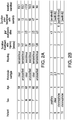
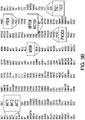
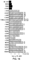
- biomarkers are set forth in FIGURE 14B and FIGURE 16 herein.
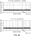
- a mTOR biomarker for a human subject is a chromosomal abnormality involving chromosome position 1p36.
- the chromosomal abnormality may be one or more insertion, deletion, duplication, inversion, etc. of one or both chromosome(s) including or in immediate proximity to position 1p36.
- a mTOR biomarker is a molecule other than the mTOR gene or protein which increases the activity of mTOR.
- mTOR biomarkers include Rheb, Raptor, Deptor, PRAS40, AMPK, REDD1/2, LKB1 and variants or mutations thereof relative to reference sequences.
- Deptor, AMPK, LKB1, PRAS40, RED1/2 nucleic acid or protein comprising one or more insertion, deletion, or substitution, which may result in a frameshift, nonsense or missense mutation which decrease the functionality of the subject protein and increase function of mTOR may serve as a mTOR biomarker.
- the cancer that is subject to the present invention is renal cell carcinoma (RCC).
- Rapamycin analogs according to the present invention are sirolimus and its analogs/derivatives everolimus, temsirolimus, deforolimus, and zotarolimus.
- Cancers that are found to express mTOR biomarkers, where the activity of mTOR is increased relative to wild-type, may be treated with non-rapamycin mTOR inhibitors.
- mTOR inhibitors include INK128, AZD8055, AZD2014 and analogs having a morpholino pyrazolopyrimidine scaffold; and mTOR/PI3 kinase dual inhibitors such as, but not limited to, NVP-BEZ235, BGT226, SF1126, PKI-587.
- An anti-cancer effect in a cancer cell expressing a mTOR biomarker that increases mTOR activity may be produced by exposing said cell to an effective amount of a non-rapamycin mTOR activity, and provides for methods of treatment of a subject having a cancer in which said cancer cells are present.
- the present invention provides a rapamycin analog for use in the treatment of a cancer, wherein cells of the cancer contain a biomarker selected from the group consisting of a mTOR biomarker, which is a mTOR mutation of Q2223K, R2505P, L2431P, S2215F, V2406A, M2327I, L2230V, I2228T, L2220F, V2006L, T1977K, I1973F, F1888V, F1888I, F1888L, F1888L in combination with L2230V, C1483F, C1483Y, L1460P, A1459P, L1433S, A1105P, or K860N; a mutation in one or more residues of amino acid residues 1382-1982 of the amino acid sequence of human mTOR set forth in NCBI Accession No.
- NP_004949 which increases mTOR activity, a mutation in one or more residues of amino acid residues 2182-2516 of the amino acid sequence of human mTOR set forth in NCBI Accession No. NP_004949 which increases mTOR activity or a combination thereof; a TSC1 biomarker, which is a TSC1 Q781* biomarker, TSC1 P311fs*4 biomarker, TSC1 I580fs*7 biomarker, TSC1 Q527* biomarker, TSC1 S334* biomarker and a combination thereof; a TSC2 biomarker, which is a TSC2 Q794* biomarker, TSC2 R611W biomarker, TSC2 S1498N biomarker and a combination thereof; and a combination thereof, wherein the cancer is renal cell carcinoma, and wherein the rapamycin analog is selected from the group consisting of sirolimus, everolimus, temsirolimus, deforolimus, and
- TSC1 biomarkers TCS2 biomarkers
- mTOR biomarkers are described in the sections above. Cancers suitable for treatment are described above. Rapamycin analogs are described above.
- Disclosed is further a method of producing an anti-cancer effect in a cancer comprising determining whether cells of the cancer contain a biomarker selected from a TSC1 biomarker, a TSC2 biomarker, a mTOR biomarker, and a combination thereof, and, where a TSC1 biomarker, a TSC2 biomarker, and/or a mTOR biomarker is present, administering a therapeutically effective amount of a rapamycin analog to produce an anti-cancer effect.
- An anti-cancer effect means one or more of a reduction in aggregate cancer cell mass, a reduction in cancer cell growth rate, a reduction in cancer cell proliferation, a reduction in tumor mass, a reduction in tumor volume, a reduction in tumor cell proliferation, a reduction in tumor growth rate, and/or a reduction in tumor metastasis.
- a method of treating a subject suffering from a cancer comprising determining whether cells of the cancer contain a biomarker selected from a TSC1 biomarker, a TSC2 biomarker, a mTOR biomarker, and a combination thereof, and, where a TSC1 biomarker, a TCS2 biomarker, and/or a mTOR biomarker is present, treating the subject with a therapeutically effective amount of a rapamycin analog.
- the subject is not treated with a rapamycin analog but treatment with another modality, for example an alternative chemotherapeutic agent, biologic anticancer agent, or radiation therapy, is administered.
- a therapeutically effective amount is an amount that is able to achieve one or more of an anticancer effect, prolongation of survival, and/or prolongation of period until relapse.
- a method of determining whether an anti-cancer effect is likely to be produced in a cancer by a rapamycin analog comprising obtaining a sample of the cancer, and determining, in the sample, whether cells of the cancer contain a biomarker selected from a TSC1 biomarker, a TSC2 biomarker, a mTOR biomarker, and a combination thereof, where if the TSC1, TSC2, and/or mTOR biomarker is present, it is more likely that a rapamycin analog would have an anti-cancer effect on the cancer.
- Methods for determining the presence of a biomarker are set forth in section 5.1 above.
- a method of treating a subject suffering from a cancer comprising obtaining a plurality of samples from different locations of cancer in the subject, determining, in each sample, whether cells of the cancer contain a biomarker selected from a TSC1 biomarker, a TSC2 biomarker, a mTOR biomarker, and a combination thereof, and, where a TSC1 biomarker, a TCS2 biomarker, and/or a mTOR biomarker is present in a sample or a plurality of the samples or in all of the samples, treating the subject with a therapeutically effective amount of a rapamycin analog.
- a TSC1 biomarker, a TSC2 biomarker, and/or a mTOR biomarker is present in all the samples.
- the TSC1, TCS2, and/or mTOR biomarkers in multiple samples need not be the same.
- the different locations of cancer that may give rise to the samples are part of the same tumor mass.
- the different locations of cancer that may give rise to the samples are not all part of the same tumor mass (for example, one or more may arise from a tumor at a separate body location, such as a metastasis).
- the subject is not treated with a rapamycin analog but treatment with another modality, for example an alternative chemotherapeutic agent, biologic anticancer agent, or radiation therapy, is administered.
- another modality for example an alternative chemotherapeutic agent, biologic anticancer agent, or radiation therapy
- kits for determining whether an anti-cancer effect is likely to be produced in a cancer by a rapamycin analog comprising a means for detecting a biomarker selected from a TSC1 biomarker, a TSC2 biomarker, a mTOR biomarker, and a combination thereof.
- TSC1 biomarkers, TSC2 biomarkers, and mTOR biomarkers are set forth in the preceding sections.
- kits include, but are not limited to, packaged probe and primer sets (e.g. TaqMan probe/primer sets), arrays/microarrays, biomarker-specific antibodies and beads, which further contain one or more probes, primers, or other detection reagents for detecting one or more biomarkers of the present invention.
- packaged probe and primer sets e.g. TaqMan probe/primer sets
- arrays/microarrays e.g., arrays/microarrays
- biomarker-specific antibodies and beads which further contain one or more probes, primers, or other detection reagents for detecting one or more biomarkers of the present invention.
- the kit may comprise a pair of oligonucleotide primers, suitable for polymerase chain reaction (PCR) or nucleic acid sequencing, for detecting the biomarker(s) to be identified.
- a pair of primers may comprise nucleotide sequences complementary to a biomarker set forth above, and be of sufficient length to selectively hybridize with said biomarker.
- the complementary nucleotides may selectively hybridize to a specific region in close enough proximity 5' and/or 3' to the biomarker position to perform PCR and/or sequencing.
- Multiple biomarker-specific primers may be included in the kit to simultaneously assay large number of biomarkers.
- the kit may also comprise one or more polymerases, reverse transcriptase, and nucleotide bases, wherein the nucleotide bases can be further detectably labeled.
- a primer may be at least about 10 nucleotides or at least about 15 nucleotides or at least about 20 nucleotides in length and/or up to about 200 nucleotides or up to about 150 nucleotides or up to about 100 nucleotides or up to about 75 nucleotides or up to about 50 nucleotides in length.
- the oligonucleotide primers may be immobilized on a solid surface or support, for example, on a nucleic acid microarray, wherein the position of each oligonucleotide primer bound to the solid surface or support is known and identifiable.
- the kit may comprise at least one nucleic acid probe, suitable for in situ hybridization or fluorescent in situ hybridization, for detecting the biomarker(s) to be identified.
- kits will generally comprise one or more oligonucleotide probes that have specificity for various biomarkers. Means for testing multiple biomarkers may optionally be comprised in a single kit.
- the kit may comprise at least one antibody for immunodetection of the biomarker(s) to be identified.
- Antibodies both polyclonal and monoclonal, specific for a biomarker, may be prepared using conventional immunization techniques, as will be generally known to those of skill in the art.
- the immunodetection reagents of the kit may include detectable labels that are associated with, or linked to, the given antibody or antigen itself.
- detectable labels include, for example, chemiluminescent or fluorescent molecules (rhodamine, fluorescein, green fluorescent protein, luciferase, Cy3, Cy5, or ROX), radio labels (3H, 35S, 32P, 14C, 1311) or enzymes (alkaline phosphatase, horseradish peroxidase).
- chemiluminescent or fluorescent molecules rhodamine, fluorescein, green fluorescent protein, luciferase, Cy3, Cy5, or ROX
- radio labels 3H, 35S, 32P, 14C, 1311
- enzymes alkaline phosphatase, horseradish peroxidase
- the biomarker-specific antibody may be provided bound to a solid support, such as a column matrix, an array, or well of a microtiter plate.
- the support may be provided as a separate element of the kit.
- the kit may comprise a pair of primers, a probe, microarray, or antibody suitable for detecting the TSC1 Q781* nonsense mutation biomarker.
- the kit may comprise a pair of primers, a probe, microarray, or antibody suitable for detecting the TSC1 P311fs*4 frameshift mutation biomarker.
- the kit may comprise a pair of primers, a probe, microarray, or antibody suitable for detecting the TSC1 I580fs*7 frameshift biomarker.
- the kit may comprise a pair of primers, a probe, microarray, or antibody suitable for detecting the TSC1 Q527* nonsense biomarker.
- the kit may comprise a pair of primers, a probe, microarray, or antibody suitable for detecting the TSC1 S334* nonsense biomarker.
- the kit may comprise a pair of primers, a probe, microarray, or antibody suitable for detecting the TSC2 Q794* nonsense mutation biomarker.
- the kit may comprise a pair of primers, a probe, microarray, or antibody suitable for detecting the TSC2 R611W missense mutation biomarker.
- the kit may comprise a pair of primers, a probe, microarray, or antibody suitable for detecting the TSC2 S1498N missense mutation biomarker.
- the kit may comprise a pair of primers, a probe, microarray, or antibody suitable for detecting the mTOR Q2223K frameshift mutation biomarker.
- the kit may comprise a pair of primers, a probe, microarray, or antibody suitable for detecting the mTOR R2505P mutation biomarker.
- the kit may comprise a pair of primers, a probe, microarray, or antibody suitable for detecting the mTOR L2431P mutation biomarker.
- the kit may comprise a pair of primers, a probe, microarray, or antibody suitable for detecting the mTOR S2215F mutation biomarker.
- the kit may comprise a pair of primers, a probe, microarray, or antibody suitable for detecting the mTOR V2406A mutation biomarker.
- the kit may comprise a pair of primers, a probe, microarray, or antibody suitable for detecting the mTOR M2327L mutation biomarker.
- the kit may comprise a pair of primers, a probe, microarray, or antibody suitable for detecting the mTOR L2230V mutation biomarker.
- the kit may comprise a pair of primers, a probe, microarray, or antibody suitable for detecting the mTOR I2228T mutation biomarker.
- the kit may comprise a pair of primers, a probe, microarray, or antibody suitable for detecting the mTOR L2220F mutation biomarker.
- the kit may comprise a pair of primers, a probe, microarray, or antibody suitable for detecting the mTOR V2006L mutation biomarker.
- the kit may comprise a pair of primers, a probe, microarray, or antibody suitable for detecting the mTOR T1977K mutation biomarker.
- the kit may comprise a pair of primers, a probe, microarray, or antibody suitable for detecting the mTOR I1973F mutation biomarker.
- the kit may comprise a pair of primers, a probe, microarray, or antibody suitable for detecting the mTOR F1888V, I, and/or L mutation biomarker.
- the kit may comprise a pair of primers, a probe, microarray, or antibody suitable for detecting the mTOR F1888L and L2230V mutation biomarkers.
- kits may comprise a pair of primers, a probe, microarray, or antibody suitable for detecting the mTOR C1483F and/or C1483Y mutation biomarker.
- the kit may comprise a pair of primers, a probe, microarray, or antibody suitable for detecting the mTOR L1460P mutation biomarker.
- the kit may comprise a pair of primers, a probe, microarray, or antibody suitable for detecting the mTOR A1459P mutation biomarker.
- the kit may comprise a pair of primers, a probe, microarray, or antibody suitable for detecting the mTOR L1433S mutation biomarker.
- kits may comprise a pair of primers, a probe, microarray, or antibody suitable for detecting the mTOR A1105P mutation biomarker.
- the kit may comprise a pair of primers, a probe, microarray, or antibody suitable for detecting the mTOR E919V mutation biomarker.
- the kit may comprise a pair of primers, a probe, microarray, or antibody suitable for detecting the mTOR K860N mutation biomarker.
- the kit may comprise a pair of primers, a probe, microarray, or antibody suitable for detecting one or more of the mTOR mutations shown in FIGURE 16 .
- the set of biomarkers set forth above may constitute at least 10 percent or at least 20 percent or at least 30 percent or at least 40 percent or at least 50 percent or at least 60 percent or at least 70 percent or at least 80 percent of the species of markers represented on the microarray.
- the biomarker detection kit may comprise one or more detection reagents and other components (e.g. a buffer, enzymes such as DNA polymerases or ligases, chain extension nucleotides such as deoxynucleotide triphosphates, and in the case of Sanger-type DNA sequencing reactions, chain terminating nucleotides, positive control sequences, negative control sequences, and the like) necessary to carry out an assay or reaction to detect a biomarker.
- detection reagents e.g. a buffer, enzymes such as DNA polymerases or ligases, chain extension nucleotides such as deoxynucleotide triphosphates, and in the case of Sanger-type DNA sequencing reactions, chain terminating nucleotides, positive control sequences, negative control sequences, and the like
- a kit may further contain means for comparing the biomarker with a standard, and can include instructions for using the kit to detect the biomarker of interest. Specifically, the instructions describes that the presence of a biomarker, set forth herein, is indicative of an increased possibility of an anti-cancer effect in a cancer by a rapamycin analog.
- FIGURE 1 As shown in FIGURE 1 , among patients treated with rapamycin analogs (rapalogs) only a relatively small subset of patients receive sufficient therapeutic benefit to support long-term use. This group of "long-term responder" patients was selected for further study in hopes of identifying characteristics which could be used to prospectively assess the likelihood of beneficial therapeutic response.
- FIGURE 2A and B show, respectively, characteristics of long-term responders and patients who did not substantially benefit from treatment ("poor responders”). DNA from both long-term responders and poor responders was then subjected to Integrated Mutation Profiling of Actionable Cancer Targets ("IMPACT”) analysis, which employs bait for 230 cancer genes panel ( FIGURE 3A , and see 36). Genes of interest to mTOR pathway are enlarged and in bold in FIGURE 3B .
- IMPACT Integrated Mutation Profiling of Actionable Cancer Targets
- FIGURE 4 presents a summary of results for responders.
- Three tumor sites ( FIGURE 5B ) were analyzed for patient 1, who was diagnosed with clear cell RCC and had a long-term response to temsirolimus.
- patient 1 had a single copy of Chromosome 9 and different mutations of TSC1 at different tumor sites (P311fs and Q527).
- Three tumor sites and one metastasis ( FIGURE 6B ) were analyzed for patient 2, who was diagnosed with unclassified RCC and had a long-term response to temsirolimus.
- patient 2 had a 1580fs mutation in TSC1.
- FIGURE 9A-D two tumor sites ( FIGURE 9A ) were studied in patient 4, who was diagnosed with unclassified RCC and had a long-term response to everolimus. While no mutations were found, site 1 manifested homozygous loss of TSC2. At site 2, a heterozygous deletion, mutation Q794*, was observed ( FIGURE 9D ).
- FIGURE 10 shows a summary of the mutations found and their functional consequences in the various long-term responder patients. As shown in FIGURE 11 , there were some mTOR relevant mutations identified in several poor responder patients but the mutations were missense mutations without apparent functional effect.
- Previously archived operative specimens were reviewed by an expert genitourinary pathologist to confirm the diagnosis and to identify separate areas of ⁇ 70% tumor content and healthy kidney tissues. Macrodissection was performed for each area of interest, and DNA was extracted using the DNeasy tissue kit (Qiagen). DNA was quantified using the Thermo Scientific NanoDropTM 1000 Spectrophotometer and samples with an A260/A280 ratio of 1.8-2.0 and concentration of 135 ng/ ⁇ L or greater were considered acceptable for further analysis.
- the IMPACT assay Integrated Mutation Profiling of Actionable Cancer Targets
- the whole-exome capture assay with standard sequencing coverage ( ⁇ 85x) using the Agilent SureSelect XT HumanAllExon 50Mb was performed. Single-nucleotide variants, small insertions and deletions, and copy number alterations (CNA) were interrogated.
- the IMPACT Assay Ultra-deep targeted sequencing of key cancer-associated genes was performed using the IMPACT assay (Integrated Mutation Profiling of Actionable Cancer Targets). We designed target-specific probes to capture all protein-coding exons of 230 genes of interest for hybrid selection (Agilent SureSelect or Nimblegen SeqCap) as previously described (21). This list included commonly implicated oncogenes, tumor suppressor genes, and components of pathways deemed actionable by current targeted therapies (Table 2). Two protocols were followed during the course of the study. For 10 samples, barcoded sequence libraries (Illumina TruSeq) were prepared using 500 ng of input tumor or matched normal DNA according to the manufacturer's instructions.
- Single-nucleotide variants were called using muTect and retained if the variant allele frequency in the tumor was >5 times that in the matched normal.
- Indels were called using the SomaticIndelDetector tool in GATK. All candidate mutations and indels were reviewed manually using the Integrative Genomics Viewer34. The mean sequence coverage was calculated using the DepthOfCoverage tool in GATK and was used to compute copy number as described previously (22).
- Exome reads were aligned with BWA 32 which does a gaped alignment for the detection of small indels, as described in below.
- Samples mapping to the reference genome which mapped uniquely (MAPQ>0) were retained and then converted to SAM format 31 for subsequent analyses and for visualization in the Integrative Genomics Viewer 34.
- Single nucleotide variants were determined in regions of sufficient coverage.
- variant detection in exome data was performed with the UnifiedGenotyper.
- variants were excluded if their variant quality was ⁇ 30, genotype quality ⁇ 5, or if they were associated with either homopolymer runs or excessive strand bias.
- Novel variants those not previously identified in either dbSNP ver. 132, were required to be derived from basespace reads not duplicated from non-duplicate color-space reads, were not resident exclusively in higher-error base positions (positions 38-50) and had evidence of the variant allele in reads mapping to both strands.
- Candidate somatic mutations were those with a variant genotype in the tumor and reference genotype in the normal sample with minimum coverage of ⁇ 10 and 6 reads respectively. Additionally, we required that the tumor variant frequency was ⁇ 10%, and each variant was detected in 4 or more tumor reads.
- Our pipeline for small insertion and deletion (indel) detection was as follows. Gapped alignment of exome sequencing reads was performed with BWA. The alignment output was sorted and duplicate reads removed with the Picard pipeline and BAM files created and indexed with Samtools. Interval detection, local realignment, indel genotyping, and post-processing were performed with the GATK framework after base quality recalibration, as described above. Retained indels were those with sufficient quality and coverage and not associated with homopolymer runs of 5bp or greater.
- Plasmids Plasmids containing cDNA encoding myc-epitope-tagged S6K (pcDNA3-myc-S6K) and Flag-epitope-tagged Wild-type mTOR (pcDNA3-Flag mTOR) were obtained from Addgene (#26610 and #26603). To generate individual mTOR mutations, a corresponding nucleotide change was introduced via QuikChange site-directed mutagenesis (Stratagene), producing pcDNA3-Flag mTOR mutants. The primers used are shown in Table 7.
- HEK293T cells were cultured in DMEM with 10% fetal bovine serum, glutamine, non-essential amino acids, and antibiotics.
- pcDNA3-Flag mTOR was transfected alone or with Myc-S6K into HEK293T cells using Lipofectamine 2000, according to manufacturer instructions (Invitrogen). Twenty-four hours after transfection, cells were treated as indicated.
- Lysates were measured for protein concentration (Pierce BCA assay), and equal amounts of protein were resolved by PAGE and subjected to immunoblotting using the following antibodies against phospho-S6K1(T389), total S6K, phospho-S6 (S235/236), phospho-AKT (S473) (Cell Signaling #9205, #9202, #4858, #9271), ⁇ -Actin (Sigma, AC-15), Flag (Sigma, M2), and c-Myc (Santa Cruz, SC-40).
- MSKCC Memorial Sloan-Kettering Cancer Center
- next-generation sequencing approach identified genetic mutations involving three core components of the mTORC1 signaling pathway.
- DNA from primary kidney tumors and matched normal kidney tissues or peripheral blood mononuclear cells was subjected to an integrated next-generation sequencing analysis employing two platforms: the IMPACT (Integrated Mutation Profiling of Actionable Cancer Targets; 19,21,22), and WEC (whole-exome capture) assays ( FIGURES 17 and 18 ).
- IMPACT Integrated Mutation Profiling of Actionable Cancer Targets
- WEC whole-exome capture
- the IMPACT assay a customized targeted-exome capture assay of 230 cancer-associated genes (Table 2) with an ultra-deep sequencing coverage (>500x) provides (5) opportunity to (1) capture genomic events potentially missed by WEC due to tumor impurity and/or heterogeneity, (2) effectively analyze DNA from formalin-fixed paraffinembedded (FFPE) samples, (3) compute copy number alterations (CNA), and (4) establish a platform which, by means of cost and efficiency, shows promise for future adaptation to a clinical test.
- FFPE formalin-fixed paraffinembedded
- CNA compute copy number alterations
- FIGURE 12E Again a concurrent LOH was observed in this patient ( FIGURE 12E ), indicating a complete functional abrogation of TSC1.
- patient #4 a 16 year-old tuberous sclerosis complex 2 male who presented with metastatic disease and multiple (6) primaries of both kidneys, copy number analysis of germline (from peripheral blood) and tumor DNA revealed heterozygous loss of TSC2 in the germline and a bi-allelic TSC2 loss in primary kidney tumor 1 (T1) ( FIGURE 12C and FIGURE 20 ).
- T1 primary kidney tumor 1
- the IMPACT analysis identified a novel somatic missense mutation of mTOR (Q2223K) at region 1 (R1) of the primary tumor ( FIGURE 12D ).
- Amino acid glutamine at position 2,223 resides in the PI3K-related kinase domain of mTOR.
- the presence of a Q2223K mutation in a long-term rapalog responder raises a possibility that missense mutations of mTOR in the kinase domain might induce hyperactivity and yet remain sensitive to rapalogs which bind FKBP12 and then target the regulatory FRB domain.
- IMPACT assay did not detect apparent causative oncogenomics events (Table 3). Furthermore, despite capturing a greater number of mutations, WEC (Table 4) also offered no apparent genetic clues concerning the therapeutic benefit of patients #5 and #6 to temsirolimus. Overall, IMPACT assays alone may be sufficient to evaluate the genetic signature of response to rapalogs.
- the mTOR (Q2223K) mutant aberrantly activates mTORC1 yet remains sensitive to rapamycin.
- the discovery of an mTOR (Q2223K) mutant in the kidney tumor of patient #3 suggests that this mTOR kinase mutant may aberrantly activate mTORC1 yet remain sensitive to (7) rapalogs.
- mTOR (Q2223K) affects the mTORC1 signaling we examined its effect on the threonine 389 phosphorylation of S6K (p70S6 kinase, one of the key substrates of mTORC1).
- Cells expressing mTOR (Q2223K) displayed stronger S6K phosphorylation than those expressing wild-type mTOR ( FIGURE 13A ).
- mTOR (Q2223K) mutant persisted over lower serum concentrations ( FIGURE 13B ).
- the mTOR (Q2223K) mutant was sensitive to rapamycin as wildtype mTOR ( FIGURE 13C ). This contrasts with the hypersensitivity of EGFR activating mutants to small molecule ERFR inhibitors such as gefitinib and erlotinib (23), likely reflecting the intrinsic differences between EGFR tyrosine and PI3K/PI3K-like kinases.
- FIGURE 14A-C we performed functional assessment on most of mTOR mutants identified in TCGA, COSMIC, and MSKCC kidney cancer databases.
- FIGURE 14B and FIGURE 16 nearly all mutations clustered on FAT or kinase domains exhibited hyperactive mTORC1 activity ( FIGURE 14B and FIGURE 16 ) and were sensitive to rapamycin ( FIGURE 14C ).
- the scattered uncommon mutations near the HEAT domains appear to be neutral and likely represent passenger mutations due to the inherent large size of mTOR.
- Our discovery and subsequent functional validation of clustered hyperactive mTOR mutations in the kidney cancer TCGA database suggests that these are recurrent events in ccRCC.
- FIGURE 14A clusters of activating mTOR mutations around the FAT and kinase domains identified in kidney cancer recapitulate a pattern reported in fission yeast ( FIGURE 14A; 24 ). Using random mutagenesis screens, they identified and functionally validated a large number of similarly clustered activating mutations in TOR2, the yeast equivalent of mTOR ( FIGURE 14A ). Together, we unveiled a preserved structural/functional evolution/selection of mTOR/TOR in human cancer pathology and unicellular biology.
- FIGURE 15A-D Genomic findings are summarized ( FIGURE 15A-D and Table 5).
- Intra-tumor heterogeneity with TSC1 intra-genic mTOR pathway convergent evolution.
- two additional regions R2, R3 within the primary tumor were examined.
- R2 carried a TSC1 (P311fs*4) deletion and a heterozygous loss of chromosome 9 (9-) ( FIGURE 15A ).
- analysis of R3 revealed a different nonsense TSC1 (Q527*) mutation with a concurrent heterozygous loss of chromosome 9 ( FIGURE 15A and FIGURE 21 ).
- two distinct TSC1 loss-of-function mutations were discovered in spatially separated areas within the same primary tumor.
- TSC1 intra-genic
- R2, R3 the analysis included 2 additional regions within the primary tumor (R2, R3) and one distant metastasis (M1).
- R2, R3, and M1 all harbored the same TSC1 (I580fs*7) mutation and a concurrent LOH through one copy loss of chromosome 9, which are identical to what discovered in R1 ( FIGURE 15B ).
- Matching results across all analyzed specimens support a notion that all examined sites originated from one dominant clone which has a complete functional loss of TSC1.
- Inter-tumor heterogeneity with TSC2 intra-genic mTOR pathway convergent evolution.
- T1, T2 primary tumors of both kidneys
- T1 and T2 harbored distinct genomic events, yet converged on a complete loss of TSC2 function : a large deletion with loss of the 2nd allele of TSC2 in T1 ( FIGURE 12C ), and a nonsense mutation of TSC2 (Q794*) in T2 ( FIGURE 15D and FIGURE 21 ), exemplifying an inter-tumor (T1 vs. T2), intra-genic (TSC2) convergent evolution of kidney cancers in the same patient.
- T1 vs. T2 inter-tumor
- TSC2 intra-genic
- Intra-tumor heterogeneity with mTOR gain-of-function and TSC1 loss-of-function inter-genic mTOR pathway convergent evolution.
- R1 kidney tumor region 1
- R2, R3, R4 three additional sites within the nephrectomy specimen were analyzed.
- R2 carries the same mTOR (Q2223K) mutation as R1 ( FIGURE 15C ).
- R3 and R4 carried the same VHL and MLL3 mutations as R1 and R2, consistent with a shared ancestral clone, R3 and R4 tumors contained a loss-of-function TSC1 (Q781*) mutation and a concurrent LOH through chromosome 9-, resulting in a complete functional impairment of TSC1 (( FIGURE 15C and FIGURES 21 and 22 ).
- TSC1 loss-of-function TSC1
- TSC1 tumor suppressor
- kidney cancers suggest that specific tumor types might be prone to evolve around cancer type-specific central pathways (29,30) that contain regulatory circuits consisting of multiple genetic constituents.
- multiple distinct mechanisms including various nonsense mutations, LOH through chromosomal loss, and activating mutations, can all be usurped by kidney cancer cells to activate mTORC1, for example the complete loss of TSC1 or TSC2, and the activating mutation of mTOR.
- mTORC1 for example the complete loss of TSC1 or TSC2
- pathway convergence in turns signals out converged vulnerability of cancers that can be effectively intervened with pathway inhibitors.
- rapamycin and INK128 were tested.
- Transfected Hela cells expressing various mTOR mutants were cultured in medium with (+) or without (-) 25 nM rapamycin or 100 nM INK128 for 2 hours. Cellular lysates were then subjected to immunoblot analysis analysis to assess phosphorylation of S6K as a measure of mTOR activity.
- mTOR mutants associated with therapeutic response to rapalogs were found to be senstive to rapamycin or to INK128, an ATP-competitive inhibitor of mTORC1 and mTORC2.
- FIGURE 27A depicts the level of phosphorylation of S6K in the presence of single F1881L and L2230V mutations.
- FIGURE 27B depicts phosphorylation of S6K where both mutations are present (other single mutants shown for comparison), where phosphorylation levels were substantially higher in the double-mutant mTOR expressing cells. This higher level of mTOR activity was further demonstrated by resistance to serum depletion ( FIGURE 27C ) and was even observed when cells were cultured in phosphate buffered saline ( FIGURE 27D ), indicative of a high level of activity.
Landscapes
- Chemical & Material Sciences (AREA)
- Health & Medical Sciences (AREA)
- Life Sciences & Earth Sciences (AREA)
- Organic Chemistry (AREA)
- Proteomics, Peptides & Aminoacids (AREA)
- Analytical Chemistry (AREA)
- Zoology (AREA)
- Wood Science & Technology (AREA)
- Engineering & Computer Science (AREA)
- General Health & Medical Sciences (AREA)
- Immunology (AREA)
- Genetics & Genomics (AREA)
- Pathology (AREA)
- Biochemistry (AREA)
- General Engineering & Computer Science (AREA)
- Molecular Biology (AREA)
- Physics & Mathematics (AREA)
- Biotechnology (AREA)
- Microbiology (AREA)
- Biophysics (AREA)
- Bioinformatics & Cheminformatics (AREA)
- Oncology (AREA)
- Hospice & Palliative Care (AREA)
- Medicinal Chemistry (AREA)
- Pharmacology & Pharmacy (AREA)
- Epidemiology (AREA)
- Animal Behavior & Ethology (AREA)
- Public Health (AREA)
- Veterinary Medicine (AREA)
- Chemical Kinetics & Catalysis (AREA)
- Measuring Or Testing Involving Enzymes Or Micro-Organisms (AREA)
- Investigating Or Analysing Biological Materials (AREA)
Description
- This application claims priority to United States Provisional Applications Nos.
61/798,020 61/852,109, both filed March 15, 2013 - The present invention relates to a rapamycin analog selected from the group consisting of sirolimus, everolimus, temsirolimus, deforolimus, and zotarolimus for use in the treatment of renal cell carcinoma, wherein cells of the cancer cells contain a biomarker selected from the group consisting of a specified mTOR biomarker, a specified TSC1 biomarker and a specified TSC2 biomarker.
- Targeted therapies, including vascular endothelial growth factor receptor (VEGFR) inhibitors such as sunitinib (first line therapy), and mTOR inhibitors such as temsirolimus and everolimus (second line therapy), are standard of care in treating advanced renal cell carcinoma (RCC; 1-4) . Temsirolimus and everolimus are rapamycin analogs (rapalogs) that inhibit the mammalian target of rapamycin complex 1 (mTORC1)5-7. International phase III trials led to their approval for treating kidney cancer, yet only demonstrated modest clinical benefit with median progression free survival (PFS) of 4.9-5.5 months (8-11).
- However, extended periods of freedom from disease progression with rapalogs have been reported in isolated patients among whom some were refractory to first line antiangiogenic agents (10,12,13). These clinical data raise a testable hypothesis that genomic alterations may dictate clinical response.
- Hyperactive PI3K/AKT/mTOR signaling, through its effects on protein synthesis, cell survival, and metabolism, has long been implicated in promoting tumor growth. The serine-threonine kinase mTOR exerts its actions as the enzymatic component of two structurally and functionally distinct multi-protein complexes, mTORC1 and mTORC2 (6,7). Therapeutic inhibition of mTORC1 is the mechanism of action for rapalogs. Components of the PI3K/mTOR pathway can either act as activators (PI3K, AKT, and Rheb) or repressors (PTEN, TSC1, and TSC2) of the mTORC1 signaling (14). Functional loss of TSC1 and TSC2 was shown in preclinical models to sensitize tumors to rapalogs (15), and everolimus has been approved for treating subependymal giant-cell astrocytomas and angiomyolipoma in patients with Tuberous Sclerosis Complex (16,17). Recent reports have also implicated TSC1 as a tumor suppressor in RCC and bladder cancer patients (18,19). However, the recent elucidation of intra-tumor heterogeneity of kidney cancer challenges the predictability of genomic biomarkers derived from single biopsies (20).
-
WO 2006/122053 relates to compositions and methods for evaluating the likelihood that a tumor is sensitive to an mTOR inhibitor, e.g. rapamycin or a rapamycin analog. Cho et al. (2007) discloses a study on potential histologic and molecular predictors of response to temsirolimus in patients with advanced renal cell carcinoma.US 2012/0196870 relates to methods of treating tuberous scleroses comprising administrating a composition comprising an mTOR inhibitor and a tyrosine kinase inhibitor to a subject that is diagnosed with, suspected of, or exhibiting symptoms of cancer. Kucejova et al. (2011) discloses a study on the interplay between pVHL and mTORC1 pathways in clear-cell renal cell carcinoma. - Disclosed is the use of one or more biomarkers to evaluate the likelihood that a rapamycin analog would produce an anti-cancer effect in a subject. It is based, at least in part, on the results of experiments employing an integrated next-generation sequencing approach to interrogate spatially separated tumor specimens from the same individuals to decipher intra-tumor and intertumor heterogeneity and determine the oncogenomic basis of exceptional therapeutic benefit to rapalogs in kidney cancer patients. These experiments implicated loss of function mutations in TSC1 and/or TSC2 and/or gain-of-function of mTOR in therapeutic responsiveness to rapamycin analogs.
- Disclosed are assay methods and kits for determining the presence of loss of function mutations in TSC1 and/or TSC2 and/or gain-of-function of mTOR, and methods of using such determinations in selecting a therapeutic regimen for a cancer patient and in methods of treating cancer patients.
Disclosed is also the evaluation of a plurality of tumor sites and the assessment of the composite effect of the genetic background on mTOR function. - The present invention provides a rapamycin analog for use in the treatment of a cancer, wherein cells of the cancer contain a biomarker selected from the group consisting of a mTOR biomarker, which is a mTOR mutation of Q2223K, R2505P, L2431P, S2215F, V2406A, M2327I, L2230V, I2228T, L2220F, V2006L, T1977K, I1973F, F1888V, F1888I, F1888L, F1888L in combination with L2230V, C1483F, C1483Y, L1460P, A1459P, L1433S, A1105P, or K860N; a mutation in one or more residues of amino acid residues 1382-1982 of the amino acid sequence of human mTOR set forth in NCBI Accession No. NP_004949 which increases mTOR activity, a mutation in one or more residues of amino acid residues 2182-2516 of the amino acid sequence of human mTOR set forth in NCBI Accession No. NP_004949 which increases mTOR activity or a combination thereof; a TSC1 biomarker, which is a TSC1 Q781* biomarker, TSC1 P311fs*4 biomarker, TSC1 I580fs*7 biomarker, TSC1 Q527* biomarker, TSC1 S334* biomarker and a combination thereof; a TSC2 biomarker, which is a TSC2 Q794* biomarker, TSC2 R611W biomarker, TSC2 S1498N biomarker and a combination thereof; and a combination thereof, wherein the cancer is renal cell carcinoma, and wherein the rapamycin analog is selected from the group consisting of sirolimus, everolimus, temsirolimus, deforolimus, and zotarolimus.
-
-
FIGURE 1 . Heterogeneity of therapeutic benefit of renal carcinoma patients treated with rapalog (n=301). Duration of rapalog treatment (in weeks) for individual patients are shown. Box marks patients deemed to have received substantial therapeutic benefit. -
FIGURE 2A-B . Characteristics of clinical "outlier" patients (A) from among the group receiving substantial therapeutic benefit ("Long-term Responders") or (B) from among those who did not substantially benefit from treatment ("Poor Responders"). -
FIGURE 3A-B . (A) Integrated Mutation Profiling of Actionable Cancer Targets ("IMPACT") analysis. Image adapted from Wagle, Berger et al., 2012, Cancer Discovery 2:82-93,reference 36. (B) Genes included in IMPACT panel. Genes of interest to mTOR pathway are enlarged and in bold. -
FIGURE 4 . Summary of results for responders. -
FIGURE 5A-C . Results forpatient 1, diagnosed with clear cell RCC and showing long-term response to temsirolimus. (A) Normalized coverage (tumor/normal) across the genome. (B) Schematic showing tumor sampling sites. (C) Genetic results. -
FIGURE 6A-C . Results forpatient 2, diagnosed with unclassified RCC and showing long-term response to temsirolimus. (A) Normalized coverage (tumor/normal) across the genome (B) Schematic showing tumor sampling sites. (C) Genetic results. -
FIGURE 7A-B . Results forpatient 3, diagnosed with clear cell RCC and showing long-term response to everolimus. (A) Schematic showing tumor sampling sites. (B) Genetic results. -
FIGURE 8A-D . Results for patient 3 (see above). (A) Normalized coverage (tumor/normal) across the genome fortumor site 1. (B) Immunoblots of S6K (T389) showing phosphorylation of S6K, downstream of mTOR. (C) Genetic results. (D) Map of mTOR gene showing exons and functional domains. -
FIGURE 9A-D . Results forpatient 4, diagnosed with unclassified RCC and showing long-term response to everolimus. (A) Schematic showing tumor sites in kidneys. (B) Normalized coverage (tumor/normal) across the genome for tumor site 1 - no mutations found, but homozygous loss of TSC2. (C) Normalized coverage (tumor/normal) across the genome fortumor site 2. (D) Genetic results. -
FIGURE 10 . Summary of results of long-term responders. -
FIGURE 11 . Summary of results of poor responders. -
FIGURE 12A-F . Genomic alterations along the core mTORC1 pathway are identified in patients with exceptional rapalog response using the IMPACT assays. (A, B) Integrated Genomics Viewer (IGV) snapshots of region 1 (R1) of the primary tumors and matched adjacent normal tissues illustrate the P311fs*4 and the I580fs*7 frameshift mutations of TSC1 in patients #1 (a) and #2 (b), respectively. Number of reads carrying the mutation is noted. (C) A magnified copy number plot of tumor 1 (T1) inpatient # 4 illustrates the homozygous loss ofchromosome 16p where TSC2 resides. (D) IGV snapshots of R1 and adjacent normal inpatient # 3 illustrate the mTOR Q2223K missense kinase domain mutation. (E) Copy number plots of patients #1 to #6 with notations on pertinent chromosomal alterations. TSC1, TSC2, and mTOR reside on chromosome bands 9q34, 16p13, and 1p36, respectively. (F) A diagram of the central mTORC1 signaling pathway illustrates mutations identified in the core components from rapalog (everolimus and temsirolimus) responders. -
FIGURE 13A-F . The Q2223K mutation of mTOR causes hyperactivation of mTORC1. (A) The mTOR Q2223K mutant induces more phosphorylation of endogenous S6K at threonine 389 (T389) than wild-type mTOR. HEK293T cells, transfected with the indicated FlagmTOR constructs for 24 hours, were serum-deprived overnight and then exposed to 1% serum-containing medium for 1 hour. Cellular lysates were subjected to immunoblot analysis using the indicated antibodies. Levels of Flag-mTOR and β-actin indicate equivalent transfection and protein loading, respectively. n.s. denotes non-specific bands. (B) Immunoblots of S6K (T389) and S6 (Serine 235/236; S235/236) phosphorylation demonstrate the hyperactivity of Q2223K mTORC1 over a range of serum concentrations. HEK293T cells, transfected with the indicated Flag-mTOR constructs for 24 hours, were washed with serum free medium, exposed to medium containing the indicated serum concentrations for 1 hour, and analyzed by immunoblots using the indicated antibodies. (C) The hyperactivity of Q2223K mTORC1 can be inhibited by rapamycin. The Q2223K mutant is as sensitive as wild-type mTORC1 to rapamycin as determined by the phosphorylation of S6K (T389). Experiments were performed as in (B), except with addition of the indicated concentrations (nM) of rapamycin in medium containing 10% serum in the final hour prior to harvest. (D,E) Cells in (D) and (E) were treated similarly to (b) and (c), respectively, except with the co-transfection of Myc-tagged S6K. (F) Structural simulation of the mTOR kinase active site, based on the solved PI3K kinase domain, illustrates the position of glutamine 2,223. Q2223 shown in yellow sticks is localized on a loop in close proximity to the ATP binding site (shown as colored lines - gray: carbon; red: oxygen; blue: nitrogen; orange: phosphorus). The kinase activation and catalytic loops are colored red and green, respectively. -
FIGURE 14A-C . Clustered mTOR activating mutations in kidney cancer in MSKCC, COSMIC, and KIRC TCGA datasets. (A) Schematics depict the highly similar localizations between human mTOR mutations identified in kidney cancer and yeast Tor2 gain-of-function mutations identified in a genetic screen. (B) 293T cells were co-transfected with Myc-S6K and the indicated Flag-mTOR constructs, and 24 hours later, washed with serum free medium, and then exposed to serum (10%) containing medium for 1 hour prior to harvest. Cellular lysates were analyzed with the indicated antibodies. The T389 phosphorylation of S6K was quantified. Corresponding regions of the mTOR protein are shown above the respective blots. (C) Cells were transfected and treated as described in (B), in the absence (-) or presence (+) of 50nM of rapamycin during the last hour prior to harvest for immunoblot analyses. -
FIGURE 15A-D . Pertinent genetic findings discovered onpatients # 1 to #4 (A-D) are illustrated as braided cancer rivers to model both genetic divergence and pathway convergence during cancer evolutions in given patients. T, R, and M denote primary tumor, region, and metastasis, respectively. -
FIGURE 16 . Bar graph depicts the relative activity of mTORC1, comparing individual mTOR mutants to Wild-type mTOR, based on S6K (T389) phosphorylation. -
FIGURE 17 . Flow chart depicts the IMPACT assay mutation identification and filtering algorithm. -
FIGURE 18 . Flow chart depicts the WEC assay mutation identification and filtering algorithm. -
FIGURE 19 . Sanger validations of mutations in mTORC1 pathway indentified by IMPACT. -
FIGURE 20 . Copy number plots forpatient # 4 showing inherited one copy loss of TSC2 in the germline DNA and the current somatic loss (homozygous deletion) in the tumor. -
FIGURE 21 . Sanger validations of additional mutations in the mTORC1 pathway identified by IMPACT assays inpatients # 1, #3, and #4. Nucleotide changes are circled in red. -
FIGURE 22 . Copy number plots for multiple tumor regions inpatient # 3 showing the loss ofchromosome 9 only in tumor regions (R3, R4) carrying the TSC1 nonsense mutation. -
FIGURE 23 . Gene maps of mTOR mutations across published TCGA cancer studies. -
FIGURE 24 . mTOR mutants associated with therapeutic response to rapalogs are more resistant to serum than wild-type mTOR. Hela cells, transfected with the indicated Flag-mTOR constructs, were cultured either without serum (-) or in the presence of 10% serum (+) for 1 hour. Cellular lysates were then subjected to immunoblot analysis using the indicated antibodies. -
FIGURE 25 . mTOR mutants associated with therapeutic response to rapalogs are more resistant to AKT inhibitor than wild-type mTOR. Hela cells, transfected with the indicated FlagmTOR constructs, were cultured in medium with (+) or without (-) 1 µM MK2206, an AKT inhibitor for 2 hours. Cellular lysates were then subjected to immunoblot analysis using the indicated antibodies. -
FIGURE 26 . mTOR mutants associated with therapeutic response to rapalogs are senstive to rapamycin or to INK128, an ATP-competitive inhibitor of mTORC1 and mTORC2. Hela cells, transfected with the indicated Flag-mTOR constructs, were cultured in medium with (+) or without (-) 25 nM rapamycin or 100 nM INK128 for 2 hours. Cellular lysates were then subjected to immunoblot analysis using the indicated antibodies. -
FIGURE 27A-D . F1881L/L2230 double mutation has a synergistic effect at increasing mTOR activity. (A) Phosphorylation of S6K in the presence of single F1881L and L2230V mutations. (B) Phosphorylation of S6K where both mutations are present (other single mutants shown for comparison). (C) Phosphorylation of S6K in cells bearing the double mutant, relative to wild-type control, when serum starved for 1, 3, 6 or 20 hours. (D) Phosphorylation of S6K in cells bearing the double mutant in serum-free medium relative to wild-type and single mutants, compared with cells grown in PBS. - The detailed description of the invention is divided into the following subsections for clarity:
- (i) biomarkers and methods of measurement;
- (ii) TSC biomarkers;
- (iii) mTOR biomarkers;
- (iv) cancer targets;
- (v) rapamycin analogs;
- (vi) medical use; and
- (vii) kits.
- Biomarkers, as that term is used herein, includes nucleic acid, protein, and/or chromosomal markers (i) disclosed below and/or (ii) that are related to the activity level of mTOR, of which rapamycin analogs are inhibitors, in a subject. In certain examples, a biomarker is an allelic variant or mutation of the version of the gene or protein present in a given population.
- A subject may be a human or a non-human subject. Examples of non-human subjects include non-human primates, dogs, cats, mice, rats, guinea pigs, rabbits, fowl, pigs, horses, cows, goats, sheep, etc.
- A biomarker may be a nucleic acid. Methods of detecting a biomarker which is a nucleic acid include but are not limited to polymerase chain reaction, in situ hybridization (for example but not limited to Fluorescent In Situ Hybridization ("FISH")), gel electrophoresis, sequencing and sequence analysis, and microarray analysis.
- A biomarker may be a protein. Methods of detecting a biomarker which is a protein include but are not limited to mass spectrometry techniques, 1-D or 2-D gel-based analysis systems, chromatography, enzyme linked immunosorbent assays (ELISAs), radioimmunoassays (RIA), enzyme immunoassays (EIA), Western Blotting, immunoprecipitation, and immunohistochemistry. Antibody arrays or protein chips may also be employed.
- A biomarker may be a chromosome or a chromosome fragment. Methods of detecting a biomarker which is a chromosome or a chromosome fragment include but are not limited to karyotyping and fluorescent in situ hybridization (FISH).
- In certain examples, a biomarker may be assessed for its effect on mTOR function. For example, the activity of mTOR may be evaluated in a cell, which may or may not be a cancer cell, containing the biomarker. As one non-limiting example, as illustrated in the working examples below, mTOR activity is reflected by phosphorylation of S6K at T389. The level of mTOR function in a cell or cells containing the biomarker may be compared to the level of mTOR function in the absence of the biomarker.
- In certain examples, a biomarker may be assessed for its effect on TSC1 and/or TSC2 function. For example, the activity of TSC1 and/or TSC2 may be evaluated in a cell containing the TSC1 or TSC2 biomarker. In a certain example, the activity of TSC1 and/or TSC2 may be evaluated by evaluating mTOR function, where the activity of TSC1 and/or TSC2 is/are inversely proportional to the activity of mTOR. The level of TSC1 and/or TSC2 function in a cell or cells containing the TSC1 or TCS2 biomarker may be compared to the level of TSC1 and/or TSC2 function in a cell or cells having a different type of TSC1 or TSC2.
- A healthy control level of mTOR function (healthy control activity) may be established using a non-cancer cell or cells from a healthy control subject.
- A healthy control level of TSC1 and/or TSC2 function (healthy control activity) may be established using a non-cancer cell or cells from a healthy control subject.
-
Tuberous sclerosis 1 protein is denoted TSC1 herein. -
Tuberous sclerosis 2 protein is denoted TSC2 herein. - In a specific non-limiting embodiment, a reference TSC1 molecule is a human TSC1 nucleic acid molecule which has the nucleic acid sequence as set forth in NCBI database accession no. NM_000368 or a TSC1 protein molecule which has the amino acid sequence as set forth in NCBI database accession no. NP_000359. The functional activity of a TSC1 protein having the foregoing amino acid sequence is referred to as a human TSC1 reference control activity.
- In a specific non-limiting embodiment, a reference TSC2 molecule is a human TSC2 nucleic acid molecule which has the nucleic acid sequence as set forth in NCBI database accession no. NM_000548 or a TSC2 protein molecule which has the amino acid sequence as set forth in NCBI database accession no. NP_000539 .The functional activity of a TSC2 protein having the foregoing amino acid sequence is referred to as a human TSC2 reference control activity.
- Reference TSC1 and/or TSC2 nucleic acids and proteins for non-human species are known or can be determined according to methods known in the art, for example where the reference sequence is the allele represented in the majority of a population.
- Where comparisons to a reference control activity are referred to herein, the biomarker is assessed relative to the reference control activity within the same species. For example, a human TSC1 biomarker activity is compared with a human TSC1 reference control activity.
- A TSC1 biomarker is a biomarker which manifests as reduced TSC1 function relative to a TSC1 reference control activity or a TSC1 healthy control activity and/or increased mTOR function relative to a mTOR reference control activity or a mTOR healthy control activity.
- A TSC2 biomarker is a biomarker which manifests as reduced TSC2 function relative to a TSC2 reference control activity and/or increased mTOR function relative to a mTOR reference control activity.
- In certain examples a TSC1 or TSC2 biomarker is a nucleic acid bearing an insertion, deletion, or substitution relative to a reference TSC1 or TSC2 gene, or a protein encoded by said nucleic acid. Said insertion, deletion, or substitution may result in a nonsense mutation, a frameshift mutation, a missense mutation, or a termination relative to protein expression.
- In certain examples a TSC1 or TSC2 biomarker is a protein bearing an insertion, deletion, or substitution relative to a reference TSC1 or TSC2 protein.
- In certain examples a TSC1 or TSC2 biomarker is a chromosome bearing a deletion, substitution, duplication or inversion which includes the TSC1 or TSC2 locus.
- In one specific non-limiting embodiment, a TSC1 biomarker for a human subject is the P311fs*4 frameshift mutation.
- In one specific non-limiting embodiment, a TSC1 biomarker for a human subject is the I580fs*7 frameshift mutation.
- In one specific non-limiting embodiment, a TSC1 biomarker for a human subject is the Q527* nonsense mutation.
- In one specific non-limiting embodiment, a TSC1 biomarker for a human subject is the Q781* nonsense mutation.
- In one specific non-limiting embodiment, a TSC1 biomarker for a human subject is the S334* nonsense mutation.
- In one specific non-limiting embodiment, a TSC2 biomarker for a human subject is the Q794* nonsense mutation.
- In one specific non-limiting embodiment, a TSC2 biomarker for a human subject is the R611W missense mutation.
- In one specific non-limiting embodiment, a TSC2 biomarker for a human subject is the S1498N missense mutation.
- In one specific example, a TSC1 biomarker for a human subject is a chromosomal abnormality involving chromosome position 9q34.
- For example, the chromosomal abnormality may be an insertion, deletion, duplication, inversion, etc. of one or both chromosome(s) including or in immediate proximity to position 9q34.
- In one specific example, a TSC2 biomarker for a human subject is a chromosomal abnormality involving chromosome position 16p13. For example, the chromosomal abnormality may be an insertion, deletion, duplication, inversion, etc. of one or both chromosome(s) including or in immediate proximity to position 16p13.
- Mammalian target of rapamycin is denoted mTOR herein and is also known as FRAP (FKBP12-rapamcyin-associated protein), RAFT1 (rapamycin and FKBP12 target), RAPT 1 (rapamycin target 1), or SEP (sirolimus effector protein).
- In a specific non-limiting embodiment, a reference mTOR molecule is a human mTOR nucleic acid molecule which has the nucleic acid sequence as set forth in NCBI database accession no. NM_004958 or a mTOR protein molecule which has the amino acid sequence as set forth in NCBI database accession no. NP_004949. The functional activity of a mTOR protein having the foregoing amino acid sequence is referred to as a human mTOR reference control activity
- Reference mTOR nucleic acids and proteins for non-human species are known or can be determined according to methods known in the art, for example where the reference sequence is the allele represented in the majority of a population.
- A mTOR biomarker is a biomarker which manifests as increased mTOR function and/or activity relative to a mTOR reference control activity or a mTOR healthy control activity. An increase in mTOR function and/or activity may for example be manifested as (i.e., indicated by) an increase in phosphporylation of S6K, for example at residue T389, relative to reference (e.g., wild type) mTOR. Alternatively, an increase in mTOR function and/or activity may be manifested as increased growth in serum-depleted medium (for example, medium containing less than or equal to 2 percent serum, medium containing less than or equal to 1 percent serum or serum-free medium) relative to reference (e.g. wild type) mTOR. Alternatively, an increase in mTOR function and/or activity may be manifested as increased resistance to AKT inhibition relative to reference (e.g., wild type) mTOR. Alternatively, an increase in mTOR function and/or activity may be manifested as the ability to be inhibited by (that is to say, sensitivity to) rapamycin and/or INK128.
- A mTOR biomarker may for example be associated with one or more, or two or more, or three or more, or four or more, of the following indicators of increased activity: an increase in phosphporylation of S6K, for example at residue T389, relative to wild type mTOR; increased growth in serum-depleted medium relative to wild type mTOR; increased resistance to AKT inhibition relative to wild type mTOR: and/or the ability to be inhibited by (that is to say, sensitivity to) rapamycin and/or INK128.
- A mTOR protein comprising one or more of the biomarkers disclosed herein may further comprise additional variations from the wild-type sequence.
- In certain examples a mTOR biomarker is a nucleic acid bearing one or more mutation, which may be an insertion, deletion, or substitution relative to the reference mTOR gene, or a protein encoded by said nucleic acid. Said insertion, deletion, or substitution may result in a nonsense mutation, a frameshift mutation, a missense mutation, or a termination relative to protein expression.
- In certain examples a mTOR biomarker is a protein bearing one or more mutation, which may be an insertion, deletion, or substitution relative to the mTOR reference protein.
- In certain examples a mTOR biomarker is a chromosome bearing one or more mutation, which may be an insertion, deletion, substitution, duplication or inversion which includes the mTOR gene.
- In the following description of mTOR biomarkers, amino acid numbering is based on the amino acid sequence set forth as NCBI Accession No. NP_004949.
- In one specific example, a mTOR biomarker for a human subject is a Q2223 mutation.
- In one specific example, a mTOR biomarker for a human subject is a R2505 mutation.
- In one specific example, a mTOR biomarker for a human subject is a L2431 mutation.
- In one specific example, a mTOR biomarker for a human subject is a S2215 mutation.
- In one specific example, a mTOR biomarker for a human subject is a V2406 mutation.
- In one specific example, a mTOR biomarker for a human subject is a M2327 mutation.
- In one specific example, a mTOR biomarker for a human subject is a L2230 mutation.
- In one specific example, a mTOR biomarker for a human subject is a 12228 mutation.
- In one specific example, a mTOR biomarker for a human subject is a L2220 mutation.
- In one specific example, a mTOR biomarker for a human subject is a V2006 mutation.
- In one specific example, a mTOR biomarker for a human subject is a T1977 mutation.
- In one specific example, a mTOR biomarker for a human subject is a 11973 mutation.
- In one specific example, a mTOR biomarker for a human subject is a F1888 mutation.
- In one specific example, a mTOR biomarker for a human subject is a F1888 mutation.
- In one specific example, a mTOR biomarker for a human subject is a F1888 mutation.
- In one specific example, a mTOR biomarker for a human subject is a F1888 mutation in combination with a L2230 mutation.
- In one specific example, a mTOR biomarker for a human subject is a C1483 mutation.
- In one specific example, a mTOR biomarker for a human subject is a L1460 mutation.
- In one specific example, a mTOR biomarker for a human subject is a A1459 mutation.
- In one specific example, a mTOR biomarker for a human subject is a L1433 mutation.
- In one specific example, a mTOR biomarker for a human subject is a A1105 mutation.
- In one specific example, a mTOR biomarker for a human subject is a E919 mutation.
- In one specific example, a mTOR biomarker for a human subject is a K860 mutation.
- In one specific example, a mTOR biomarker for a human subject is a 12500 mutation.
- In one specific non-limiting embodiment, a mTOR biomarker for a human subject is a K1452 mutation.
- In one specific non-limiting embodiment, a mTOR biomarker for a human subject is a Y1463 mutation.]
- In one specific non-limiting embodiment, a mTOR biomarker for a human subject is the Q2223K frameshift mutation.
- In one specific non-limiting embodiment, a mTOR biomarker for a human subject is the R2505P mutation.
- In one specific non-limiting embodiment, a mTOR biomarker for a human subject is the L2431P mutation.
- In one specific non-limiting embodiment, a mTOR biomarker for a human subject is the S2215F mutation.
- In one specific non-limiting embodiment, a mTOR biomarker for a human subject is the V2406A mutation.
- In one specific non-limiting embodiment, a mTOR biomarker for a human subject is the M2327I mutation.
- In one specific non-limiting embodiment, a mTOR biomarker for a human subject is the L2230V mutation.
- In one specific non-limiting embodiment, a mTOR biomarker for a human subject is the I2228T mutation.
- In one specific non-limiting embodiment, a mTOR biomarker for a human subject is the L2220F mutation.
- In one specific non-limiting embodiment, a mTOR biomarker for a human subject is the V2006L mutation.
- In one specific non-limiting embodiment, a mTOR biomarker for a human subject is the T1977K mutation.
- In one specific non-limiting embodiment, a mTOR biomarker for a human subject is the I1973F mutation.
- In one specific non-limiting embodiment, a mTOR biomarker for a human subject is the F1888V mutation.
- In one specific non-limiting embodiment, a mTOR biomarker for a human subject is the F1888I mutation.
- In one specific non-limiting embodiment, a mTOR biomarker for a human subject is the F1888L mutation.
- In one specific non-limiting embodiment, a mTOR biomarker for a human subject is the F1888L mutation in combination with the L2230V mutation.
- In one specific non-limiting embodiment, a mTOR biomarker for a human subject is the C1483F or the C1483Y mutation.
- In one specific non-limiting embodiment, a mTOR biomarker for a human subject is the L1460P mutation.
- In one specific non-limiting embodiment, a mTOR biomarker for a human subject is the A1459P mutation.
- In one specific non-limiting embodiment, a mTOR biomarker for a human subject is the L1433S mutation.
- In one specific non-limiting embodiment, a mTOR biomarker for a human subject is the A1105P mutation.
- In one specific example, a mTOR biomarker for a human subject is the E919V mutation.
- In one specific non-limiting embodiment, a mTOR biomarker for a human subject is the K860N mutation. A mTOR biomarker used by the invention may comprise more than one of the above-listed mutations.
- In certain examples, a mTOR biomarker involves a mutation in a FAT or KINASE domain of mTOR.
- In certain examples, a mTOR biomarker is one or more mutation in the FAT domain of mTOR, for example, which increases mTOR activity. In certain non-limiting embodiments, the mutation in the FAT domain is a mutation of one or more residue of amino acid residues 1382-1982 of the amino acid sequence set forth in NCBI Accession No. NP_004949. In certain non-limiting embodiments, the mutated sequence is at least 95 or at least 98 or at least 99 or at least 99.5 percent homologous to the wild-type sequence (as determined, for example, by standard software such as BLAST or FASTA). In certain non-limiting embodiments, the mutation in the FAT domain is a mutation of one or more residue of amino acid residues of the following amino acid sequence which represents residues 1382-1982:


- In certain non-limiting embodiments, the mutation in the FAT domain is a mutation of one or more residue of amino acid residues 1400-1500 of the amino acid sequence set forth in NCBI Accession No. NP_004949. In certain non-limiting embodiments, the mutation in the FAT domain is a mutation of one or more residue of amino acid residues of the following amino acid sequence which represents residues 1400-1500 :

- In certain non-limiting embodiments, the mutation in the FAT domain is a mutation of one or more residue of amino acid residues 1850-1982 of the amino acid sequence set forth in NCBI Accession No. NP_004949. In certain non-limiting embodiments, the mutation in the FAT domain is a mutation of one or more residue of amino acid residues of the following amino acid sequence which represents residues 1850-1982 :

- In certain non-limiting embodiments, the mutation in the FAT domain is a mutation of L1433, a mutation of A1459, a mutation of L1460, a mutation of C1483, a mutation of F1888, a mutation of 11973, a mutation of T1977, or a combination thereof. In certain non-limiting embodiments, the mutation is one or more of L1433S, A1459P, L1460P, C1483F, C1483Y, F1888 L, I or V, I1973F, or T1977K.
- In certain examples, a mTOR biomarker is one or more mutation in the PI3 kinase domain of mTOR, for example, which increases mTOR activity. In certain non-limiting embodiments, the mutation in the PI3 kinase domain is a mutation of one or more residue of amino acid residues 2182-2516 of the amino acid sequence set forth in NCBI Accession No. NP_004949. In certain non-limiting embodiments, the mutated sequence is at least 95 or at least 98 or at least 99 or at least 99.5 percent homologous to the wild-type sequence (as determined, for example, by standard software such as BLAST or FASTA). In certain non-limiting embodiments, the mutation in the PI3 kinase domain is a mutation of one or more residue of amino acid residues of the following amino acid sequence which represents residues 2182-2516:

- In certain non-limiting embodiments, the mutation in the PI3 kinase domain is a mutation of one or more residue of amino acid residues 2200-2250 of the amino acid sequence set forth in NCBI Accession No. NP_004949. In certain non-limiting embodiments, the mutation in the FAT domain is a mutation of one or more residue of amino acid residues of the following amino acid sequence which represents residues 2200-2250:

- In certain non-limiting embodiments, the mutation in the PI3 kinase domain is a mutation of L2230, L2220, Q2223, M2327, V2406, S2215, L2431, R2505, or a combination thereof. In certain non-limiting embodiments, the mutation in the PI3 kinase domain is a mutation of L2230, L2220, Q2223, S2215, or a combination thereof. In certain non-limiting embodiments, the mutation is one or more of L2230V, L2220F, Q2223K, M2327I, V2406A, S2215F, L2431P, or R2505P.
- Specific non-limiting examples of biomarkers are set forth in
FIGURE 14B andFIGURE 16 herein. - In one specific example, a mTOR biomarker for a human subject is a chromosomal abnormality involving chromosome position 1p36. For example, the chromosomal abnormality may be one or more insertion, deletion, duplication, inversion, etc. of one or both chromosome(s) including or in immediate proximity to position 1p36.
- In further examples, a mTOR biomarker is a molecule other than the mTOR gene or protein which increases the activity of mTOR. Non-limiting examples of such mTOR biomarkers include Rheb, Raptor, Deptor, PRAS40, AMPK, REDD1/2, LKB1 and variants or mutations thereof relative to reference sequences. In specific examples, Deptor, AMPK, LKB1, PRAS40, RED1/2 nucleic acid or protein comprising one or more insertion, deletion, or substitution, which may result in a frameshift, nonsense or missense mutation which decrease the functionality of the subject protein and increase function of mTOR may serve as a mTOR biomarker.
- The cancer that is subject to the present invention is renal cell carcinoma (RCC).
- Rapamycin analogs according to the present invention are sirolimus and its analogs/derivatives everolimus, temsirolimus, deforolimus, and zotarolimus.
- Cancers that are found to express mTOR biomarkers, where the activity of mTOR is increased relative to wild-type, may be treated with non-rapamycin mTOR inhibitors. Non-limiting examples of such mTOR inhibitors include INK128, AZD8055, AZD2014 and analogs having a morpholino pyrazolopyrimidine scaffold; and mTOR/PI3 kinase dual inhibitors such as, but not limited to, NVP-BEZ235, BGT226, SF1126, PKI-587. An anti-cancer effect in a cancer cell expressing a mTOR biomarker that increases mTOR activity may be produced by exposing said cell to an effective amount of a non-rapamycin mTOR activity, and provides for methods of treatment of a subject having a cancer in which said cancer cells are present.
- 5.7 MEDICAL USE The present invention provides a rapamycin analog for use in the treatment of a cancer, wherein cells of the cancer contain a biomarker selected from the group consisting of a mTOR biomarker, which is a mTOR mutation of Q2223K, R2505P, L2431P, S2215F, V2406A, M2327I, L2230V, I2228T, L2220F, V2006L, T1977K, I1973F, F1888V, F1888I, F1888L, F1888L in combination with L2230V, C1483F, C1483Y, L1460P, A1459P, L1433S, A1105P, or K860N; a mutation in one or more residues of amino acid residues 1382-1982 of the amino acid sequence of human mTOR set forth in NCBI Accession No. NP_004949 which increases mTOR activity, a mutation in one or more residues of amino acid residues 2182-2516 of the amino acid sequence of human mTOR set forth in NCBI Accession No. NP_004949 which increases mTOR activity or a combination thereof; a TSC1 biomarker, which is a TSC1 Q781* biomarker, TSC1 P311fs*4 biomarker, TSC1 I580fs*7 biomarker, TSC1 Q527* biomarker, TSC1 S334* biomarker and a combination thereof; a TSC2 biomarker, which is a TSC2 Q794* biomarker, TSC2 R611W biomarker, TSC2 S1498N biomarker and a combination thereof; and a combination thereof, wherein the cancer is renal cell carcinoma, and wherein the rapamycin analog is selected from the group consisting of sirolimus, everolimus, temsirolimus, deforolimus, and zotarolimus.
- Disclosed is further a method of determining whether an anti-cancer effect is likely to be produced in a cancer by a rapamycin analog, comprising determining whether cells of the cancer contain a biomarker selected from a TSC1 biomarker, a TSC2 biomarker, a mTOR biomarker, and a combination thereof, where if the TSC1, TSC2, and/or mTOR biomarker is present, it is more likely that a rapamycin analog would have an anti-cancer effect on the cancer.
- TSC1 biomarkers, TCS2 biomarkers, and mTOR biomarkers are described in the sections above. Cancers suitable for treatment are described above. Rapamycin analogs are described above.
- Disclosed is further a method of producing an anti-cancer effect in a cancer, comprising determining whether cells of the cancer contain a biomarker selected from a TSC1 biomarker, a TSC2 biomarker, a mTOR biomarker, and a combination thereof, and, where a TSC1 biomarker, a TSC2 biomarker, and/or a mTOR biomarker is present, administering a therapeutically effective amount of a rapamycin analog to produce an anti-cancer effect.
- An anti-cancer effect means one or more of a reduction in aggregate cancer cell mass, a reduction in cancer cell growth rate, a reduction in cancer cell proliferation, a reduction in tumor mass, a reduction in tumor volume, a reduction in tumor cell proliferation, a reduction in tumor growth rate, and/or a reduction in tumor metastasis.
- Disclosed is a method of treating a subject suffering from a cancer, comprising determining whether cells of the cancer contain a biomarker selected from a TSC1 biomarker, a TSC2 biomarker, a mTOR biomarker, and a combination thereof, and, where a TSC1 biomarker, a TCS2 biomarker, and/or a mTOR biomarker is present, treating the subject with a therapeutically effective amount of a rapamycin analog. Where cells of the cancer do not contain a TSC1, TSC2 or mTOR biomarker, the subject is not treated with a rapamycin analog but treatment with another modality, for example an alternative chemotherapeutic agent, biologic anticancer agent, or radiation therapy, is administered.
- A therapeutically effective amount is an amount that is able to achieve one or more of an anticancer effect, prolongation of survival, and/or prolongation of period until relapse.
- Disclosed is a method of determining whether an anti-cancer effect is likely to be produced in a cancer by a rapamycin analog, comprising obtaining a sample of the cancer, and determining, in the sample, whether cells of the cancer contain a biomarker selected from a TSC1 biomarker, a TSC2 biomarker, a mTOR biomarker, and a combination thereof, where if the TSC1, TSC2, and/or mTOR biomarker is present, it is more likely that a rapamycin analog would have an anti-cancer effect on the cancer. Methods for determining the presence of a biomarker are set forth in section 5.1 above.
- Disclosed is a method of treating a subject suffering from a cancer, comprising obtaining a plurality of samples from different locations of cancer in the subject, determining, in each sample, whether cells of the cancer contain a biomarker selected from a TSC1 biomarker, a TSC2 biomarker, a mTOR biomarker, and a combination thereof, and, where a TSC1 biomarker, a TCS2 biomarker, and/or a mTOR biomarker is present in a sample or a plurality of the samples or in all of the samples, treating the subject with a therapeutically effective amount of a rapamycin analog. In certain examples, a TSC1 biomarker, a TSC2 biomarker, and/or a mTOR biomarker is present in all the samples. As exemplified below, the TSC1, TCS2, and/or mTOR biomarkers in multiple samples need not be the same. The different locations of cancer that may give rise to the samples are part of the same tumor mass. The different locations of cancer that may give rise to the samples are not all part of the same tumor mass (for example, one or more may arise from a tumor at a separate body location, such as a metastasis). In a certain example, where one or more of the samples does not contain a TSC1, TSC2 or mTOR biomarker, the subject is not treated with a rapamycin analog but treatment with another modality, for example an alternative chemotherapeutic agent, biologic anticancer agent, or radiation therapy, is administered.
- Disclosed is a kit for determining whether an anti-cancer effect is likely to be produced in a cancer by a rapamycin analog, comprising a means for detecting a biomarker selected from a TSC1 biomarker, a TSC2 biomarker, a mTOR biomarker, and a combination thereof. TSC1 biomarkers, TSC2 biomarkers, and mTOR biomarkers are set forth in the preceding sections.
- Types of kits include, but are not limited to, packaged probe and primer sets (e.g. TaqMan probe/primer sets), arrays/microarrays, biomarker-specific antibodies and beads, which further contain one or more probes, primers, or other detection reagents for detecting one or more biomarkers of the present invention.
- In a specific example , the kit may comprise a pair of oligonucleotide primers, suitable for polymerase chain reaction (PCR) or nucleic acid sequencing, for detecting the biomarker(s) to be identified. A pair of primers may comprise nucleotide sequences complementary to a biomarker set forth above, and be of sufficient length to selectively hybridize with said biomarker. Alternatively, the complementary nucleotides may selectively hybridize to a specific region in close enough proximity 5' and/or 3' to the biomarker position to perform PCR and/or sequencing. Multiple biomarker-specific primers may be included in the kit to simultaneously assay large number of biomarkers. The kit may also comprise one or more polymerases, reverse transcriptase, and nucleotide bases, wherein the nucleotide bases can be further detectably labeled.
- In one example, a primer may be at least about 10 nucleotides or at least about 15 nucleotides or at least about 20 nucleotides in length and/or up to about 200 nucleotides or up to about 150 nucleotides or up to about 100 nucleotides or up to about 75 nucleotides or up to about 50 nucleotides in length.
- In a further example, the oligonucleotide primers may be immobilized on a solid surface or support, for example, on a nucleic acid microarray, wherein the position of each oligonucleotide primer bound to the solid surface or support is known and identifiable.
- In a specific example, the kit may comprise at least one nucleic acid probe, suitable for in situ hybridization or fluorescent in situ hybridization, for detecting the biomarker(s) to be identified. Such kits will generally comprise one or more oligonucleotide probes that have specificity for various biomarkers. Means for testing multiple biomarkers may optionally be comprised in a single kit.
- In other examples, the kit may comprise at least one antibody for immunodetection of the biomarker(s) to be identified. Antibodies, both polyclonal and monoclonal, specific for a biomarker, may be prepared using conventional immunization techniques, as will be generally known to those of skill in the art. The immunodetection reagents of the kit may include detectable labels that are associated with, or linked to, the given antibody or antigen itself. Such detectable labels include, for example, chemiluminescent or fluorescent molecules (rhodamine, fluorescein, green fluorescent protein, luciferase, Cy3, Cy5, or ROX), radio labels (3H, 35S, 32P, 14C, 1311) or enzymes (alkaline phosphatase, horseradish peroxidase).
- In a further example, the biomarker-specific antibody may be provided bound to a solid support, such as a column matrix, an array, or well of a microtiter plate. Alternatively, the support may be provided as a separate element of the kit.
- In one specific example, the kit may comprise a pair of primers, a probe, microarray, or antibody suitable for detecting the TSC1 Q781* nonsense mutation biomarker.
- In one specific example, the kit may comprise a pair of primers, a probe, microarray, or antibody suitable for detecting the TSC1 P311fs*4 frameshift mutation biomarker.
- In one specific example, the kit may comprise a pair of primers, a probe, microarray, or antibody suitable for detecting the TSC1 I580fs*7 frameshift biomarker.
- In one specific example, the kit may comprise a pair of primers, a probe, microarray, or antibody suitable for detecting the TSC1 Q527* nonsense biomarker.
- In one specific example, the kit may comprise a pair of primers, a probe, microarray, or antibody suitable for detecting the TSC1 S334* nonsense biomarker.
- In one specific example, the kit may comprise a pair of primers, a probe, microarray, or antibody suitable for detecting the TSC2 Q794* nonsense mutation biomarker.
- In one specific example, the kit may comprise a pair of primers, a probe, microarray, or antibody suitable for detecting the TSC2 R611W missense mutation biomarker.
- In one specific example, the kit may comprise a pair of primers, a probe, microarray, or antibody suitable for detecting the TSC2 S1498N missense mutation biomarker.
- In one specific example, the kit may comprise a pair of primers, a probe, microarray, or antibody suitable for detecting the mTOR Q2223K frameshift mutation biomarker.
- In one specific example, the kit may comprise a pair of primers, a probe, microarray, or antibody suitable for detecting the mTOR R2505P mutation biomarker.
- In one specific example, the kit may comprise a pair of primers, a probe, microarray, or antibody suitable for detecting the mTOR L2431P mutation biomarker.
- In one specific example, the kit may comprise a pair of primers, a probe, microarray, or antibody suitable for detecting the mTOR S2215F mutation biomarker.
- In one specific example, the kit may comprise a pair of primers, a probe, microarray, or antibody suitable for detecting the mTOR V2406A mutation biomarker.
- In one specific example, the kit may comprise a pair of primers, a probe, microarray, or antibody suitable for detecting the mTOR M2327L mutation biomarker.
- In one specific example, the kit may comprise a pair of primers, a probe, microarray, or antibody suitable for detecting the mTOR L2230V mutation biomarker.
- In one specific example, the kit may comprise a pair of primers, a probe, microarray, or antibody suitable for detecting the mTOR I2228T mutation biomarker.
- In one specific example, the kit may comprise a pair of primers, a probe, microarray, or antibody suitable for detecting the mTOR L2220F mutation biomarker.
- In one specific example, the kit may comprise a pair of primers, a probe, microarray, or antibody suitable for detecting the mTOR V2006L mutation biomarker.
- In one specific example, the kit may comprise a pair of primers, a probe, microarray, or antibody suitable for detecting the mTOR T1977K mutation biomarker.
- In one specific example, the kit may comprise a pair of primers, a probe, microarray, or antibody suitable for detecting the mTOR I1973F mutation biomarker.
- In one specific example, the kit may comprise a pair of primers, a probe, microarray, or antibody suitable for detecting the mTOR F1888V, I, and/or L mutation biomarker.
- In one specific example, the kit may comprise a pair of primers, a probe, microarray, or antibody suitable for detecting the mTOR F1888L and L2230V mutation biomarkers.
- In one specific example, a kit may comprise a pair of primers, a probe, microarray, or antibody suitable for detecting the mTOR C1483F and/or C1483Y mutation biomarker.
- In one specific example, the kit may comprise a pair of primers, a probe, microarray, or antibody suitable for detecting the mTOR L1460P mutation biomarker.
- In one specific example, the kit may comprise a pair of primers, a probe, microarray, or antibody suitable for detecting the mTOR A1459P mutation biomarker.
- In one specific example, the kit may comprise a pair of primers, a probe, microarray, or antibody suitable for detecting the mTOR L1433S mutation biomarker.
- In one specific example, a kit may comprise a pair of primers, a probe, microarray, or antibody suitable for detecting the mTOR A1105P mutation biomarker.
- In one specific example, the kit may comprise a pair of primers, a probe, microarray, or antibody suitable for detecting the mTOR E919V mutation biomarker.
- In one specific example, the kit may comprise a pair of primers, a probe, microarray, or antibody suitable for detecting the mTOR K860N mutation biomarker.
- In one specific example, the kit may comprise a pair of primers, a probe, microarray, or antibody suitable for detecting one or more of the mTOR mutations shown in
FIGURE 16 . - In certain examples, where the measurement means in the kit employs an array, the set of biomarkers set forth above may constitute at least 10 percent or at least 20 percent or at least 30 percent or at least 40 percent or at least 50 percent or at least 60 percent or at least 70 percent or at least 80 percent of the species of markers represented on the microarray.
- In certain examples, the biomarker detection kit may comprise one or more detection reagents and other components (e.g. a buffer, enzymes such as DNA polymerases or ligases, chain extension nucleotides such as deoxynucleotide triphosphates, and in the case of Sanger-type DNA sequencing reactions, chain terminating nucleotides, positive control sequences, negative control sequences, and the like) necessary to carry out an assay or reaction to detect a biomarker.
- A kit may further contain means for comparing the biomarker with a standard, and can include instructions for using the kit to detect the biomarker of interest. Specifically, the instructions describes that the presence of a biomarker, set forth herein, is indicative of an increased possibility of an anti-cancer effect in a cancer by a rapamycin analog.
- As shown in
FIGURE 1 , among patients treated with rapamycin analogs (rapalogs) only a relatively small subset of patients receive sufficient therapeutic benefit to support long-term use. This group of "long-term responder" patients was selected for further study in hopes of identifying characteristics which could be used to prospectively assess the likelihood of beneficial therapeutic response.FIGURE 2A and B show, respectively, characteristics of long-term responders and patients who did not substantially benefit from treatment ("poor responders"). DNA from both long-term responders and poor responders was then subjected to Integrated Mutation Profiling of Actionable Cancer Targets ("IMPACT") analysis, which employs bait for 230 cancer genes panel (FIGURE 3A , and see 36). Genes of interest to mTOR pathway are enlarged and in bold inFIGURE 3B . -
FIGURE 4 presents a summary of results for responders. Three tumor sites (FIGURE 5B ) were analyzed forpatient 1, who was diagnosed with clear cell RCC and had a long-term response to temsirolimus. As shown inFIGURE 5A and C ,patient 1 had a single copy ofChromosome 9 and different mutations of TSC1 at different tumor sites (P311fs and Q527). Three tumor sites and one metastasis (FIGURE 6B ) were analyzed forpatient 2, who was diagnosed with unclassified RCC and had a long-term response to temsirolimus. As shown inFIGURE 6A and C ,patient 2 had a 1580fs mutation in TSC1. -
Patient 3, diagnosed with clear cell RCC and having a long-term response to everolimus, was found to have a Q781 mutation in TSC1 at two tumor sites of four assayed. Interestingly, the other two sites, while lacking the TSC1 mutation, had a Q2223K mutation in mTOR (FIGURE 7A-B . Further study showed that the Q2223K mutation in mTOR resulted in gain-of-function (FIGURE 8B ). This mutation maps in the PI3K domain of mTOR (FIGURE 8D ). - As shown in
FIGURE 9A-D , two tumor sites (FIGURE 9A ) were studied inpatient 4, who was diagnosed with unclassified RCC and had a long-term response to everolimus. While no mutations were found,site 1 manifested homozygous loss of TSC2. Atsite 2, a heterozygous deletion, mutation Q794*, was observed (FIGURE 9D ). -
FIGURE 10 shows a summary of the mutations found and their functional consequences in the various long-term responder patients. As shown inFIGURE 11 , there were some mTOR relevant mutations identified in several poor responder patients but the mutations were missense mutations without apparent functional effect. - The foregoing data provides plausible oncogenomic causes for the exceptional treatment responses observed in some, but not all patients. It suggests that identification of mutations alone may not be sufficient without consideration of their biologic effects. It also illustrates that despite of intratumoral heterogeneity, targeted therapy can be successful due to clonal convergence within the pathway.
- The foregoing study was advanced further and the results are presented below in
Section 7. - Patients. Six RCC cases were retrospectively identified from an institutional database of RCC patients treated with rapalogs at Memorial Sloan-Kettering Cancer Center (MSKCC). All had received either temsirolimus or everolimus as single-agent therapy. Six were selected based on extended therapeutic benefit, evident by treatment duration of ≥ 20 months. We felt this cut-off to be representative of long-term benefit, as the registration trials reported <10% or 0% of patients to be alive and progression-free at 20 months, respectively. As part of our analysis all scans obtained between commencement and discontinuation of rapalogs were retrospectively reviewed by a research radiologist. This protocol was approved by our institutional review board, and all patients had previously provided written consents on an institutional tissue procurement protocol. The access and utilization of the kidney cancer TCGA database for publication were approved by the TCGA Program Office.
- Samples. All 6 patients underwent nephrectomy prior to the initiation of rapalog therapy and have frozen specimens. Adjacent normal kidney tissue (n=5) or peripheral blood (n=1) were used for reference. To interrogate intratumor heterogeneity, DNA was extracted from FFPE materials of additional regions within the primary tumors (regions marked as R). Regions were chosen by a dedicated genitourinary pathologist based on interregional differences in histopathologic appearance. Furthermore, fresh frozen specimens of metastasis (marked as M1; patient #2) and a contralateral primary kidney tumor (2 separate tumors marked as T1 and T2, patient #4) were obtained to determine inter-tumor heterogeneity. Previously archived operative specimens were reviewed by an expert genitourinary pathologist to confirm the diagnosis and to identify separate areas of ≥ 70% tumor content and healthy kidney tissues. Macrodissection was performed for each area of interest, and DNA was extracted using the DNeasy tissue kit (Qiagen). DNA was quantified using the Thermo Scientific NanoDropTM 1000 Spectrophotometer and samples with an A260/A280 ratio of 1.8-2.0 and concentration of 135 ng/µL or greater were considered acceptable for further analysis.
- Next-generation sequencing. DNA from tumors and matched normal was subjected to an analysis by two next-generation sequencing platforms. The IMPACT assay (Integrated Mutation Profiling of Actionable Cancer Targets) is a customized targeted-exome capture assay of 230 cancer-associated genes with ultra-deep sequencing coverage (>500x) using
Illumina HiSeq 2000. Target-specific probes for hybrid selection were designed as previously described to capture all protein-coding exons of a list of oncogenes, tumor suppressor genes, and components of pathways deemed actionable by targeted therapies. The whole-exome capture assay with standard sequencing coverage (∼85x) using the Agilent SureSelect XT HumanAllExon 50Mb was performed. Single-nucleotide variants, small insertions and deletions, and copy number alterations (CNA) were interrogated. - The IMPACT Assay. Ultra-deep targeted sequencing of key cancer-associated genes was performed using the IMPACT assay (Integrated Mutation Profiling of Actionable Cancer Targets). We designed target-specific probes to capture all protein-coding exons of 230 genes of interest for hybrid selection (Agilent SureSelect or Nimblegen SeqCap) as previously described (21). This list included commonly implicated oncogenes, tumor suppressor genes, and components of pathways deemed actionable by current targeted therapies (Table 2). Two protocols were followed during the course of the study. For 10 samples, barcoded sequence libraries (Illumina TruSeq) were prepared using 500 ng of input tumor or matched normal DNA according to the manufacturer's instructions. Libraries were pooled at equimolar concentrations (100 ng per library) for a single exon capture reaction (Agilent SureSelect) as previously described (31). For 12 samples, barcoded sequence libraries were prepared using 250 ng of input DNA using a hybrid protocol based on the NEBNext DNA Library Prep Kit (New England Biolabs). Manufacturer's instructions were followed with two substitutions: we used NEXTflex barcoded adapters (Bio Scientific) and HiFi DNA polymerase (Kapa Biosystems). Libraries were pooled at 100 ng per tumor library and 50 ng per normal library for a single exon capture reaction (Nimblegen SeqCap). To prevent off-target hybridization in all capture reactions, we spiked in a pool of blocker oligonucleotides complementary to the full sequences of all barcoded adaptors (to a final total concentration of 10 µM). Hybridized DNA was sequenced on an
Illumina HiSeq 2000 to generate paired-end 75-bp reads. Data were demultiplexed using CASAVA, and reads were aligned to the reference human genome (hg19) using the Burrows-Wheeler Alignment tool (32). Local realignment and quality score recalibration were performed using the Genome Analysis Toolkit (GATK) according to GATK best practices (33). We achieved mean exon sequence coverage of 590x (625x for all tumor samples). Deep sequencing ensured sensitivity for detecting mutations in multiclonal and stroma-admixed samples and enabled accurate determination of mutation allele frequencies. - single-nucleotide variants, small insertions/deletions (indels), and copy number alterations. Single-nucleotide variants were called using muTect and retained if the variant allele frequency in the tumor was >5 times that in the matched normal. Indels were called using the SomaticIndelDetector tool in GATK. All candidate mutations and indels were reviewed manually using the Integrative Genomics Viewer34. The mean sequence coverage was calculated using the DepthOfCoverage tool in GATK and was used to compute copy number as described previously (22).
- Whole-exome capture sequencing. Between 1.9 and 3 µg of high quality genomic DNA was captured by hybridization using the SureSelect XT HumanAllExon 50Mb (Agilent). Samples were prepared according to the manufacturer instructions. PCR amplification of the libraries was carried out for 6 cycles in the pre-capture step and for 10 cycles post capture. Samples were bar-coded and run on a
Hiseq 2000 in a 75bp/75bp Paired end run, using the TruSeq SBS Kit v3 (Illumina). Two samples were pooled in a lane, the average number of read pairs per sample was 69 million. All reads were aligned to the reference human genome (NCBI build 37.1 hg19). Exome reads were aligned with BWA 32 which does a gaped alignment for the detection of small indels, as described in below. Samples mapping to the reference genome which mapped uniquely (MAPQ>0) were retained and then converted to SAM format 31 for subsequent analyses and for visualization in theIntegrative Genomics Viewer 34. Single nucleotide variants were determined in regions of sufficient coverage. We first removed duplicate reads (using Picard MarkDuplicates) from further analysis, defined here as any read chromosome, start position, strand, and color-space sequence matched another aligned read. Indel realignment, base quality recalibration, variant detection, and variant annotation were performed with the GATK framework (33,35). Specifically, after base quality recalibration for color-space reads, variant detection in exome data was performed with the UnifiedGenotyper. For high-coverage exome experiments, variants were excluded if their variant quality was <30, genotype quality <5, or if they were associated with either homopolymer runs or excessive strand bias. Novel variants, those not previously identified in either dbSNP ver. 132, were required to be derived from basespace reads not duplicated from non-duplicate color-space reads, were not resident exclusively in higher-error base positions (positions 38-50) and had evidence of the variant allele in reads mapping to both strands. Candidate somatic mutations were those with a variant genotype in the tumor and reference genotype in the normal sample with minimum coverage of≥10 and 6 reads respectively. Additionally, we required that the tumor variant frequency was ≥10%, and each variant was detected in 4 or more tumor reads. Our pipeline for small insertion and deletion (indel) detection was as follows. Gapped alignment of exome sequencing reads was performed with BWA. The alignment output was sorted and duplicate reads removed with the Picard pipeline and BAM files created and indexed with Samtools. Interval detection, local realignment, indel genotyping, and post-processing were performed with the GATK framework after base quality recalibration, as described above. Retained indels were those with sufficient quality and coverage and not associated with homopolymer runs of 5bp or greater. - Sanger Sequencing. Bidirectional Sanger sequencing for validation was performed for all mTOR pathway mutations using standard techniques with primers provided by the Geoffrey Beene Translational Oncology Core .
- Plasmids. Plasmids containing cDNA encoding myc-epitope-tagged S6K (pcDNA3-myc-S6K) and Flag-epitope-tagged Wild-type mTOR (pcDNA3-Flag mTOR) were obtained from Addgene (#26610 and #26603). To generate individual mTOR mutations, a corresponding nucleotide change was introduced via QuikChange site-directed mutagenesis (Stratagene), producing pcDNA3-Flag mTOR mutants. The primers used are shown in Table 7.
- Cell Culture, transfection, and immunoblots. HEK293T cells were cultured in DMEM with 10% fetal bovine serum, glutamine, non-essential amino acids, and antibiotics. To assay mTOR activity, pcDNA3-Flag mTOR was transfected alone or with Myc-S6K into HEK293T
cells using Lipofectamine 2000, according to manufacturer instructions (Invitrogen). Twenty-four hours after transfection, cells were treated as indicated. Lysates were measured for protein concentration (Pierce BCA assay), and equal amounts of protein were resolved by PAGE and subjected to immunoblotting using the following antibodies against phospho-S6K1(T389), total S6K, phospho-S6 (S235/236), phospho-AKT (S473) (Cell Signaling #9205, #9202, #4858, #9271), β-Actin (Sigma, AC-15), Flag (Sigma, M2), and c-Myc (Santa Cruz, SC-40). - Computational modeling of mTOR kinase domain. The model was built by HMMHMM (hidden markov-model) comparison, using the HHpred server (http://toolkit.tuebingen.mpg.de) and the x-ray crystal structure of PIK3C3 (pdb: 31s8) as a template.
- Long-term rapalog treatment responders identified in the MSKCC kidney cancer database. To probe into the genomic determinants underlying long-term rapalog therapeutic benefits, we searched the Memorial Sloan-Kettering Cancer Center (MSKCC) kidney cancer database and identified patients who (1) underwent nephrectomy prior to receiving systemic therapy for metastatic diseases (n=305) with archived fresh frozen tumors available for analysis, (2) were treated with single agent temsirolimus or everolimus, (3) displayed exceptional disease control (partial response or stable disease > 20 months) on rapalogs, and (4) had received sunitinib previously, yet obtained greater clinical benefit with rapalogs. With such criteria, 6 patients were identified who were on average treated with single agent sunitinib for 9 months followed by rapalogs for 29+ months (Table 1).
- An integrated next-generation sequencing approach identified genetic mutations involving three core components of the mTORC1 signaling pathway. DNA from primary kidney tumors and matched normal kidney tissues or peripheral blood mononuclear cells was subjected to an integrated next-generation sequencing analysis employing two platforms: the IMPACT (Integrated Mutation Profiling of Actionable Cancer Targets; 19,21,22), and WEC (whole-exome capture) assays (
FIGURES 17 and18 ). With standard sequencing coverage (∼100x), WEC enables us to survey mutational landscapes encompassing all coding exons. On the other hand, the IMPACT assay, a customized targeted-exome capture assay of 230 cancer-associated genes (Table 2) with an ultra-deep sequencing coverage (>500x) provides (5) opportunity to (1) capture genomic events potentially missed by WEC due to tumor impurity and/or heterogeneity, (2) effectively analyze DNA from formalin-fixed paraffinembedded (FFPE) samples, (3) compute copy number alterations (CNA), and (4) establish a platform which, by means of cost and efficiency, shows promise for future adaptation to a clinical test. - Single-nucleotide variants, small insertions and deletions, and copy number alterations (CNA) were first interrogated with IMPACT on all 6 patients (Table 3) and then with WEC on
patients # 5 and #6 (Table 4). Pertinent genomic findings are summarized (Table 5) and mutations relevant to our research question were confirmed by orthogonal bidirectional Sanger sequencing (FIGURE 19 ). Interestingly, this approach identified genetic mutations involving three core components of the mTORC1 pathway, i.e., TSC1, TSC2, and mTOR, through distinct mechanisms, in 4 of 6 (67%) patients. - Complete functional loss of TSC1 or TSC2. For
patient # 1, a 58 year-old female, the IMPACT analysis of the primary tumor at region 1 (R1) revealed a novel somatic TSC1 single nucleotide deletion (P311fs*4) (FIGURE 12A ,FIGURE 19 and Table 5). Copy number analysis of the tumor revealed a single copy ofchromosome 9 where the TSC1 gene resides (FIGURE 12E ). The frameshift mutation with LOH (loss of heterozygosity) would result in a complete functional impairment of TSC1 in the tumor. Forpatient # 2, a 73 year-old female, the IMPACT analysis of R1 identified another novel somatic TSC1 frameshift mutation (I580fs*7) (FIGURE 12B ). Again a concurrent LOH was observed in this patient (FIGURE 12E ), indicating a complete functional abrogation of TSC1. Forpatient # 4, a 16 year-oldtuberous sclerosis complex 2 male who presented with metastatic disease and multiple (6) primaries of both kidneys, copy number analysis of germline (from peripheral blood) and tumor DNA revealed heterozygous loss of TSC2 in the germline and a bi-allelic TSC2 loss in primary kidney tumor 1 (T1) (FIGURE 12C andFIGURE 20 ). Complete functional abrogation of either TSC1 or TSC2 would hyperactivate the mTORC1 signaling. - An mTOR kinase domain mutation identified in the tumor of
patient # 3. Forpatient # 3, a 66 year-old male, the IMPACT analysis identified a novel somatic missense mutation of mTOR (Q2223K) at region 1 (R1) of the primary tumor (FIGURE 12D ). Amino acid glutamine at position 2,223 resides in the PI3K-related kinase domain of mTOR. The presence of a Q2223K mutation in a long-term rapalog responder raises a possibility that missense mutations of mTOR in the kinase domain might induce hyperactivity and yet remain sensitive to rapalogs which bind FKBP12 and then target the regulatory FRB domain. Forpatients # 5 and #6, 60 and 50 year-old females, the IMPACT assay did not detect apparent causative oncogenomics events (Table 3). Furthermore, despite capturing a greater number of mutations, WEC (Table 4) also offered no apparent genetic clues concerning the therapeutic benefit ofpatients # 5 and #6 to temsirolimus. Overall, IMPACT assays alone may be sufficient to evaluate the genetic signature of response to rapalogs. - The mTOR (Q2223K) mutant aberrantly activates mTORC1 yet remains sensitive to rapamycin. The discovery of an mTOR (Q2223K) mutant in the kidney tumor of
patient # 3 suggests that this mTOR kinase mutant may aberrantly activate mTORC1 yet remain sensitive to (7) rapalogs. To examine whether mTOR (Q2223K) affects the mTORC1 signaling, we examined its effect on the threonine 389 phosphorylation of S6K (p70S6 kinase, one of the key substrates of mTORC1). Cells expressing mTOR (Q2223K) displayed stronger S6K phosphorylation than those expressing wild-type mTOR (FIGURE 13A ). The observed hyperactivation by mTOR (Q2223K) mutant persisted over lower serum concentrations (FIGURE 13B ). Importantly, the mTOR (Q2223K) mutant was sensitive to rapamycin as wildtype mTOR (FIGURE 13C ). This contrasts with the hypersensitivity of EGFR activating mutants to small molecule ERFR inhibitors such as gefitinib and erlotinib (23), likely reflecting the intrinsic differences between EGFR tyrosine and PI3K/PI3K-like kinases. When exogenous S6K was co-transfected, a process known to augment mTORC1 signaling, we further demonstrated the hyperactivity of the mTOR (Q2223K) mutant at low serum and affirmed its sensitivity to rapamycin (FIGURE 13D,E ). Of note, the serine 473 phosphorylation of AKT, a key post-translational modification mediated by mTORC2, was not enhanced by mTOR (Q2223K), indicating that such mutant has no strong effect on mTORC2 (FIGURE 13D ). The lysine substitution of glutamine 2223 of the mTOR kinase domain has created an mTOR that only activates mTORC1 but not mTORC2. Modeling of the mTOR (Q2223K) mutation based on the solved PI3K kinase domain structure suggests that the 2,223 glutamine residue locates right next to the ATP binding site of the catalytic domain (FIGURE 13F ). - Clustered, evolutionarily preserved activating mutations of mTOR in kidney cancer. Our discovery of a hyperactive mTOR (Q2223K) mutant in a long-term rapalog-treated patient suggests that activating mTOR mutations in tumors could be implicative of treatment response to rapalogs. Analysis of the clear cell kidney cancer TCGA database (8)through the cBio Genomics Portal revealed mTOR mutations in 5.4% of examined cases (23/424) (Table 6). Importantly, all of these are missense mutations, and the majority clusters on either the FAT or kinase domain (
FIGURE 14A ). Accordingly, we performed functional assessment on most of mTOR mutants identified in TCGA, COSMIC, and MSKCC kidney cancer databases (FIGURE 14A-C ). Remarkably, nearly all mutations clustered on FAT or kinase domains exhibited hyperactive mTORC1 activity (FIGURE 14B andFIGURE 16 ) and were sensitive to rapamycin (FIGURE 14C ). The scattered uncommon mutations near the HEAT domains appear to be neutral and likely represent passenger mutations due to the inherent large size of mTOR. Our discovery and subsequent functional validation of clustered hyperactive mTOR mutations in the kidney cancer TCGA database suggests that these are recurrent events in ccRCC. It is highly noteworthy that clusters of activating mTOR mutations around the FAT and kinase domains identified in kidney cancer recapitulate a pattern reported in fission yeast (FIGURE 14A; 24 ). Using random mutagenesis screens, they identified and functionally validated a large number of similarly clustered activating mutations in TOR2, the yeast equivalent of mTOR (FIGURE 14A ). Together, we unveiled a preserved structural/functional evolution/selection of mTOR/TOR in human cancer pathology and unicellular biology. - Thus far, our cancer genomics based on single biopsies of therapeutic outliers was able to successfully provide logical genetic explanations for observed exceptional response to rapalogs in 4 of 6 (67%) patients. All of these mutations would have led to hyperactive mTORC1 signaling. Our findings are in line with the exceptional therapeutic response of imatinib, an Abelson kinase inhibitor, in treating CML bearing the BCR-ABL fusion gene (25), and erlotinib, an EGFR inhibitor, in treating EGFR mutant lung cancer (23). However, recent reports on the complexity of intra-tumor heterogeneity and the branched clonal evolution of RCC raise concerns about the feasibility of single-biopsy genomics in formulating personalized cancer medicine (26,27). To resolve this conundrum, multiregional IMPACT assays were performed on all available additional specimens (spatially separated regions within the primary kidney tumors and metastatic sites whenever are available; and mainly are formalin-fixed paraffin embedded samples) of patients #1 to #4.
- Additional 9 regions were chosen based on interregional histopathologic differences. Genomic findings are summarized (
FIGURE 15A-D and Table 5). - Intra-tumor heterogeneity with TSC1 intra-genic mTOR pathway convergent evolution. For
patient # 1, two additional regions (R2, R3) within the primary tumor were examined. Identical to R1 described above, R2 carried a TSC1 (P311fs*4) deletion and a heterozygous loss of chromosome 9 (9-) (FIGURE 15A ). Interestingly, analysis of R3 revealed a different nonsense TSC1 (Q527*) mutation with a concurrent heterozygous loss of chromosome 9 (FIGURE 15A andFIGURE 21 ). Hence, two distinct TSC1 loss-of-function mutations were discovered in spatially separated areas within the same primary tumor. This would simultaneously trigger aberrant mTORC1 activation at different regions of the same tumor, exemplifying an intra-genic (TSC1) clonal convergent evolution of kidney cancer cells in the same patient. Forpatient # 2, the analysis included 2 additional regions within the primary tumor (R2, R3) and one distant metastasis (M1). R2, R3, and M1 all harbored the same TSC1 (I580fs*7) mutation and a concurrent LOH through one copy loss ofchromosome 9, which are identical to what discovered in R1 (FIGURE 15B ). Matching results across all analyzed specimens support a notion that all examined sites originated from one dominant clone which has a complete functional loss of TSC1. - Inter-tumor heterogeneity with TSC2 intra-genic mTOR pathway convergent evolution. For the tuberous
sclerosis patient # 4, primary tumors of both kidneys (T1, T2) were analyzed. In addition to the germline heterozygous, chromosomal loss of TSC2 (16p+/-), T1 and T2 harbored distinct genomic events, yet converged on a complete loss of TSC2 function : a large deletion with loss of the 2nd allele of TSC2 in T1 (FIGURE 12C ), and a nonsense mutation of TSC2 (Q794*) in T2 (FIGURE 15D andFIGURE 21 ), exemplifying an inter-tumor (T1 vs. T2), intra-genic (TSC2) convergent evolution of kidney cancers in the same patient. - Intra-tumor heterogeneity with mTOR gain-of-function and TSC1 loss-of-function inter-genic mTOR pathway convergent evolution. For
patient # 3 whose kidney tumor region 1 (R1) carries an mTOR kinase domain activating mutation, three additional sites (R2, R3, R4) within the nephrectomy specimen were analyzed. R2 carries the same mTOR (Q2223K) mutation as R1 (FIGURE 15C ). Strikingly, although R3 and R4 carried the same VHL and MLL3 mutations as R1 and R2, consistent with a shared ancestral clone, R3 and R4 tumors contained a loss-of-function TSC1 (Q781*) mutation and a concurrent LOH through chromosome 9-, resulting in a complete functional impairment of TSC1 ((FIGURE 15C and FIGURES 21 and 22 ). Hence, three distinct mechanisms, including a missense gain-of-function mutation of growth promoting gene (mTOR), a nonsense loss-of-function mutation of tumor suppressor (TSC1), and another loss-of-function of TSC1 through LOH (9-), were employed by spatially separated cancer clones within the same primary tumor to concurrently activate mTORC1. The presence of different BAP1 splice site mutations between R1/R2 and R3/R4 not only exemplifies their convergent evolution on BAP1 but also supports observed clonal bifurcation (FIGURE 15C ). To our knowledge, this represents the first concurrent inter-genic (mTOR and TSC1) pathway convergent evolution (mTORC1 activation) discovered in human cancer. - Our multiregional IMPACT study revealed complete loss-of- function of either TSC1 or TSC2 in all tumors tested for
patients # 1, #2, and #4, and the coexistence of complete functional loss of TSC1 and gain-of-function of mTOR (Q2223K) in spatially separated regions of the same primary tumor inpatient # 3. Additionally, we discovered that a significant number of clustered activating mutations of mTOR exist in kidney albeit to a lesser extent in other cancers (FIGURE 23 ). Importantly, mTOR (Q2223K) also occurrs in colon cancer (CRC TCGA) and thus represents a recurrent mutation. Altogether, our study not only suggests uncontrolled mTORC1 signaling as an important contributor to the pathogenesis of kidney cancer but also discovers logical predictive genomic biomarkers for exceptional rapalog response in kidney cancer. Of note, the mTORC1 pathway convergent evolution, through complete functional loss of TSC1 orTSC2, or clustered activating mutations of mTOR discovered in our study, was not present in 5 MSKCC kidney cancer patients whose metastatic cancers progressed within two months despite rapalog treatment (non-responders) (data not shown). Intriguingly,we noticed a heterozygous TSC1 loss in one of the non-responders, suggesting that haploid inactivation of TSC1 alone likely has limited predictive value in kidney cancer. This contrasts with a recent report on bladder cancer where haploid inactivation of TSC1 was reported to correlate with response to everolimus in a 14 patient cohort with a median treatment time of 2.6 (1.8-4.1) months, when a long-term responder (33+ months) carrying an additional NF2 mutation was excluded (19). Since mTOR activating mutations are also present in cancers for which rapalogs are not standard of care (FIGURE 23 ), it might be prudent to conduct a clinical trial administering rapalogs on selected patients whose tumors carry these pathway convergent mutations. - The accumulation of diverged mutations during individual cancer evolution underscores the propensity of given tumors in achieving higher intra-tumor heterogeneity for the greater robustness of cancer cells (28). Our data are in agreement with recent demonstrations of the existence of a significant number of private mutations in separated regions of primary and metastatic kidney tumors, demonstrating the highly heterogeneous nature in genomics of individual kidney cancers (20). Branching evolution of cancers supports a central tenet that in order to achieve sustainable therapeutic benefit with targeted therapy one must identify and interfere with the earliest mutation events. Under such premises, ensuing genetic ramifications would mitigate the applicability of targeted therapeutic agents. Importantly, despite such an inherent predicament, our data based on kidney cancers suggest that specific tumor types might be prone to evolve around cancer type-specific central pathways (29,30) that contain regulatory circuits consisting of multiple genetic constituents. In our case, multiple distinct mechanisms, including various nonsense mutations, LOH through chromosomal loss, and activating mutations, can all be usurped by kidney cancer cells to activate mTORC1, for example the complete loss of TSC1 or TSC2, and the activating mutation of mTOR. Such pathway convergence in turns signals out converged vulnerability of cancers that can be effectively intervened with pathway inhibitors. On a cautionary note, one kidney cancer patient, reported in a recent article, harbored an activating mTOR (L2431P) mutation in the primary tumor but not metastatic sites (20). This patient progressed after ∼7 months of everolimus treatment, explaining the lack of exceptional therapeutic response. Hence, multiple biopsies of tumors at different sites could be advised to discover those converging "Achilles' Heels".
- To visualize this concept for treatment planning, we designate "a braided cancer river" to model cancer of a given patient, which branches to illustrate tumor heterogeneity generated by genetic mutations and merges to depict functional convergence through pathway integration. Lastly, it is tempting to speculate that other cancers may also channel through several key tumor type-specific converging pathways that prevalently operate in given cancer types for their robustness, which at the same time provides unique therapeutic vantage points.


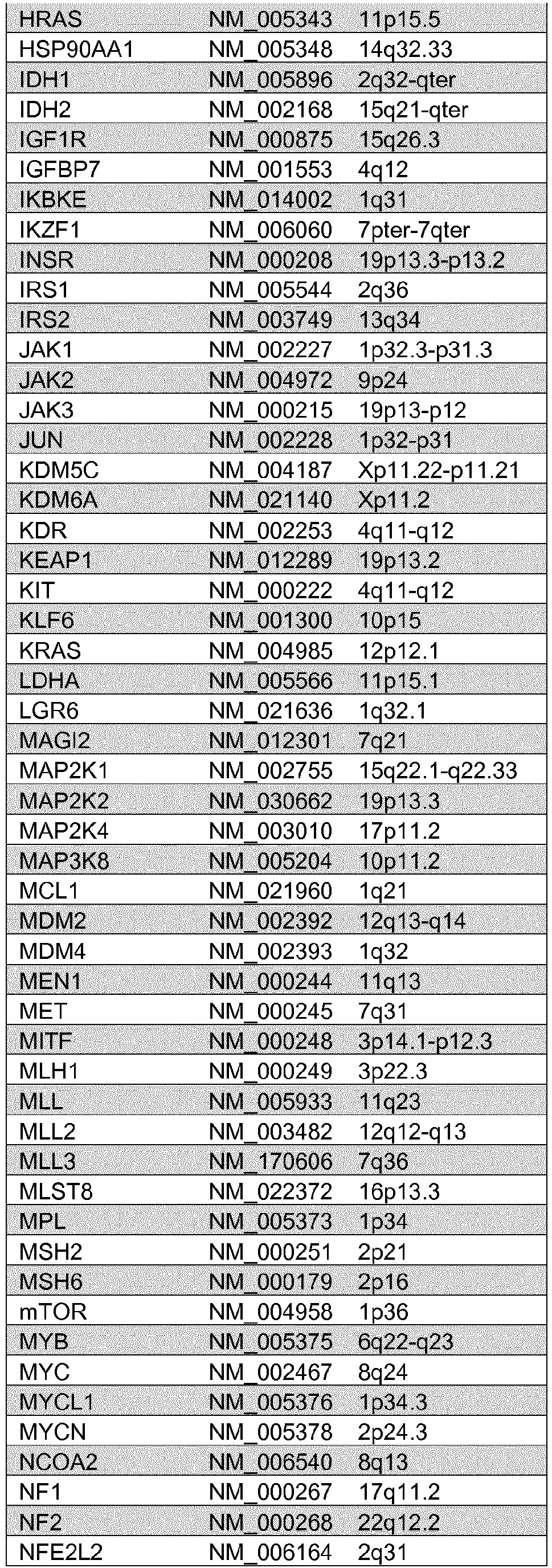


Table 5. Summary of oncogenomic findings in patients # 1 to #6. CNA: copy number alteration; Chr: chromosomePatient Oncongenomic Findings Proposed Mechanism for repalog-sensitivity 1 R1 R2 R3 Functional loss of Tsc1 P13K/Akt/MTOR pathway alterations: Somatic mutations: TSCI frameshift (P311fs*4) X X TSCI nonsense (Q527*) X CNA: Heterozygous loss of Chr 9 X X X Other pertinent genomic alterations: Somatic mutations: VHL Nonsense (E94*) X X X PBRMI Missense (E991D) X X X CNA: Heterozygous loss of Chr 3p X X X 2 R1 R2 R3 M1 Functional loss of Tsc1 P13K/Akt/MTOR pathway alterations Somatic mutations: TSCI Frameshift (1580fs*7) X X X X CNA: Heterozygous loss of Chr 9 X X X X Other pertinent genomic alterations: Somatic mutations: VHL Misssense (H115N) X X X X TP53 Missense (R273H) X X X X CNA: 3 R1 R2 R3 R4 Hyperactive MTOR/ Functional loss of Tsc1 P13K/Akt/MTOR pathway alterations: Somatic mutations: MTOR missense (Q2223K) x x x TSCI nonsense (Q781*) x x CNA: Heterozygous loss of Chr 9 x x Other pertinent genomic alterations: Somatic mutations: VHL frameshift (G212fs) BAPI splice (g220_splice) x x x BAPI splice (Q85_splice) x x CNA: Heterozygous loss of Chr 3p 4 T1 T2 Functional loss Tsc2 P13K/Akt/MTOR pathway alterations: Somatic mutations: TSC2 frameshift (Q794*) X CNA: Homozygous loss of TSC2 X Heterozygous loss of TSC2 X Other pertinent genomic alterations: Somatic mutations: none CNA: None 5 R1 M1 Unclear P13K/Akt/MTOR pathway alterations: Somatic mutations: none CNA: None Other pertinent genomic alterations: Somatic mutations: VHL missense (L118P) X X PBRMI frameshift (E1360fs) X X CNA: Heterozygous loss of Chr 3p X X 6 R1 Unclear P13K/Akt/MTOR pathway alterations: Somatic mutations: none CNA: None Other pertinent genomic alterations: Somatic mutations: none CNA: None 7 R1 N/A P13K/Akt/MTOR pathway alterations: Somatic mutations: none CNA: Heterozygous loss of Chr 9 X Other pertinent genomic alterations: Somatic mutations: none CNA: Heterozygous loss of Chr 9 X 8 Somatic mutations R1 N/A P13K/Akt/MTOR pathway alterations: MTOR missense (E919V) X CNA: Heterozygous loss of Chr 9q X Other pertinent genomic alterations: Somatic mutations: VHL missense (S65L) X BAP1 splice (e6-1) X PBRM1 splice (e5-2) X SETD2 frameshift (E2477fs) X CNA: Heterozygous loss of Chr 3p X 9 R1 N/A P13K/Akt/MTOR pathway alterations: Somatic mutations: TSC2 missense (1475F) x CNA: Other pertinent genomic alterations: Somatic mutations: None CNA: MAP2K1 amplification X 10 R1 N/A P13K/Akt/MTOR pathway alterations: Somatic mutations: PTEN missense (F2001) X CNA: None Other pertinent genomic alterations: Somatic mutations: None CNA: PDGFRA, KIT amplification X 


- Various experiments were performed to explore the properties of identified mTOR mutants.
- To test the ability of cells carrying mTOR mutations to grow in serum-depleted conditions, Hela cells, transfected with Flag-mTOR constructs bearing various mutations, were cultured either without serum (-) or in the presence of 10% serum (+) for 1 hour. Cellular lysates were then subjected to immunoblot analysis to assess phosphorylation of S6K as a measure of mTOR activity. As shown in
FIGURE 24 , mTOR mutants associated with therapeutic response to rapalogs were found to be more resistant to serum than wild-type mTOR. - Experiments were then performed to test the effect of an AKT inhibitor on the activity of mTOR mutants. Hela cells, transfected with Flag-mTOR constructs carrying various mTOR mutations, were cultured in medium with (+) or without (-) 1 µM MK2206, an AKT inhibitor for 2 hours. Cellular lysates were then subjected to immunoblot analysis analysis to assess phosphorylation of S6K as a measure of mTOR activity. As shown in
FIGURE 25 , mTOR mutants associated with therapeutic response to rapalogs were found to be more resistant to AKT inhibitor than wild-type mTOR. - The ability of rapamycin and INK128 to inhibit activity of the mTOR mutants was also tested. Transfected Hela cells expressing various mTOR mutants were cultured in medium with (+) or without (-) 25 nM rapamycin or 100 nM INK128 for 2 hours. Cellular lysates were then subjected to immunoblot analysis analysis to assess phosphorylation of S6K as a measure of mTOR activity. As shown in
FIGURE 26 , mTOR mutants associated with therapeutic response to rapalogs were found to be senstive to rapamycin or to INK128, an ATP-competitive inhibitor of mTORC1 and mTORC2. - The effect of concurrent mutations F188L and L2230V on mTOR activity, as reflected by phosphorylation of S6K, was evaluated. In experiments analogous to those described in the preceding section, cells expressing single or double mTOR mutants were prepared.
FIGURE 27A depicts the level of phosphorylation of S6K in the presence of single F1881L and L2230V mutations.FIGURE 27B depicts phosphorylation of S6K where both mutations are present (other single mutants shown for comparison), where phosphorylation levels were substantially higher in the double-mutant mTOR expressing cells. This higher level of mTOR activity was further demonstrated by resistance to serum depletion (FIGURE 27C ) and was even observed when cells were cultured in phosphate buffered saline (FIGURE 27D ), indicative of a high level of activity. -
- 1. Molina, A.M. & Motzer, R.J. Clinical practice guidelines for the treatment of metastatic renal cell carcinoma: today and tomorrow. The ).
- 2. Linehan, W.M., Srinivasan, R. & Schmidt, L.S. The genetic basis of kidney cancer: a metabolic disease. Nature reviews. ).
- 3. Kaelin, W.G., Jr. The von Hippel-Lindau tumour suppressor protein: O2 sensing and cancer. Nature reviews. ).
- 4. Kaelin, W.G., Jr. Treatment of kidney cancer: insights provided by the VHL tumor-suppressor protein. Cancer 115, 2262-2272 (2009).
- 5. Brugarolas, J. Renal-cell carcinoma--molecular pathways and therapies. The New England journal of medicine 356, 185-187 (2007).
- 6. Laplante, M. & Sabatini, D.M. mTOR signaling in growth control and disease. Cell 149, 274-293 (2012).
- 7. Dazert, E. & Hall, M.N. mTOR signaling in disease. Current opinion in ).
- 8. Hudes, G., et al. Temsirolimus, interferon alfa, or both for advanced renal-cell carcinoma. The New England journal of medicine 356, 2271-2281 (2007).
- 9. Motzer, R.J., et al. Efficacy of everolimus in advanced renal cell carcinoma: a double-blind, randomised, placebo-controlled phase III trial. Lancet 372, 449-456 (2008).
- 10. Molina, A.M., Ginsberg, M.S. & Motzer, R.J. Long-term response with everolimus for metastatic renal cell carcinoma refractory to sunitinib. ).
- 11. Dutcher, J.P., et al. Effect of temsirolimus versus interferon-alpha on outcome of patients with advanced renal cell carcinoma of different tumor histologies. ).
- 12. Atkins, M.B., et al. Randomized phase II study of multiple dose levels of CCI- 779, a novel mammalian target of rapamycin kinase inhibitor, in patients with advanced refractory renal cell carcinoma. Journal of clinical oncology : official journal of the American Society of ).
- 13. Amato, R.J., Jac, J., Giessinger, S., Saxena, S. & Willis, ).
- 14. Vivanco, I. & Sawyers, C.L. The phosphatidylinositol 3-Kinase AKT pathway in human cancer. Nature reviews. ).
- 15. Kwiatkowski, D.J. Animal models of lymphangioleiomyomatosis (LAM) and tuberous sclerosis complex (TSC). Lymphatic research and ).
- 16. Krueger, D.A., et al. Everolimus for subependymal giant-cell astrocytomas in tuberous sclerosis. The New England journal of medicine 363, 1801-1811 (2010).
- 17. Qin, W., et al. Angiomyolipoma have common mutations in TSC2 but no other common genetic events. PLoS One 6, e24919 (2011).
- 18. Kucejova, B., et al. Interplay between pVHL and mTORC1 pathways in clear-cell renal cell carcinoma. Molecular cancer research : ).
- 19. Iyer, G., et al. Genome sequencing identifies a basis for everolimus sensitivity. Science 338, 221 (2012).
- 20. Gerlinger, M., et al. Intratumor heterogeneity and branched evolution revealed by multiregion sequencing. The New England journal of medicine 366, 883-892 (2012).
- 21. Gnirke, A., et al. Solution hybrid selection with ultra-long oligonucleotides for massively parallel targeted sequencing. ).
- 22. Wagle, N., et al. High-throughput detection of actionable genomic alterations in clinical tumor samples by targeted, massively parallel sequencing. ).
- 23. Paez, J.G., et al. EGFR mutations in lung cancer: correlation with clinical response to gefitinib therapy. Science 304, 1497-1500 (2004).
- 24. Urano, J., et al. Point mutations in TOR confer Rheb-independent growth in fission yeast and nutrient-independent mammalian TOR signaling in mammalian cells. Proceedings of the National Academy of Sciences of the United States of America 104, 3514-3519 (2007).
- 25. Sawyers, C.L. The cancer biomarker problem. Nature 452, 548-552 (2008).
- 26. Longo, D.L. Tumor heterogeneity and personalized medicine. The New England journal of medicine 366, 956-957 (2012).
- 27. Yap, T.A., Gerlinger, M., Futreal, P.A., Pusztai, L. & Swanton, C. Intratumor heterogeneity: seeing the wood for the trees. Science ).
- 28. Nowell, P.C. The clonal evolution of tumor cell populations. Science 194, 23-28 (1976).
- 29. Vogelstein, B. & Kinzler, K.W. Cancer genes and the pathways they control. ).
- 30. Hanahan, D. & Weinberg, R.A. Hallmarks of cancer: the next generation. Cell 144, 646-674 (2011).
- 31. Li, H., et al. The Sequence Alignment/Map format and SAMtools. ).
- 32. Li, H. & Durbin, R. Fast and accurate short read alignment with Burrows-Wheeler transform. ).
- 33. DePristo, M.A., et al. A framework for variation discovery and genotyping using next-generation DNA sequencing data. Nat Genet 43, 491-498 (2011).
- 34. Robinson, J.T., et al. Integrative genomics viewer. ).
- 35. McKenna, A., et al. The Genome Analysis Toolkit: a MapReduce framework for analyzing next-generation DNA sequencing data. ).
- 36. Wagle, Berger et al., Cancer Discovery 2:82-93 (2012).
-
- <110> MEMORIAL SLOAN-KETTERING CANCER CENTER
- <120> BIOMARKERS FOR RESPONSE TO RAPAMYCIN ANALOGS
- <130> 072734.0153
- <140>
PCT/US2014/028867
<141> 2014-03-14 - <150>
61/852,109
<151> 2013-03-15 - <150>
61/798,020
<151> 2013-03-15 - <160> 32
- <170> PatentIn version 3.5
- <210> 1
<211> 601
<212> PRT
<213> Homo sapiens - <400> 1
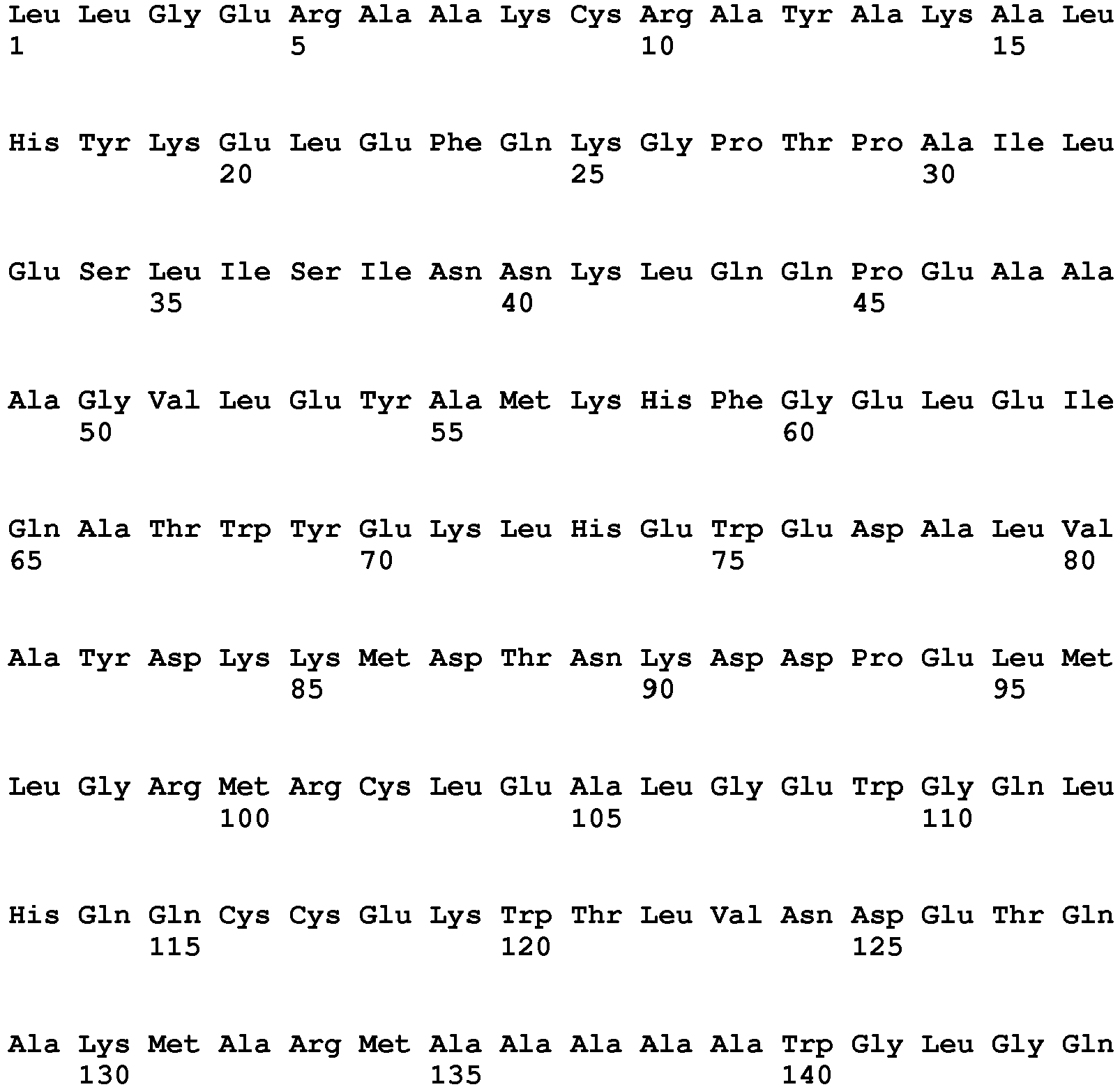


- <210> 2
<211> 101
<212> PRT
<213> Homo sapiens - <400> 2


- <210> 3
<211> 133
<212> PRT
<213> Homo sapiens - <400> 3


- <210> 4
<211> 335
<212> PRT
<213> Homo sapiens - <400> 4


- <210> 5
<211> 51
<212> PRT
<213> Homo sapiens - <400> 5

- <210> 6
<211> 28
<212> DNA
<213> Artificial Sequence - <220>
<223> Description of Artificial Sequence: Synthetic primer - <400> 6
gagccctaca ggaattaccc tactttgc 28 - <210> 7
<211> 28
<212> DNA
<213> Artificial Sequence - <220>
<223> Description of Artificial Sequence: Synthetic primer - <400> 7
gtcagcctgt cagtatccaa gtcaagtc 28 - <210> 8
<211> 28
<212> DNA
<213> Artificial Sequence - <220>
<223> Description of Artificial Sequence: Synthetic primer - <400> 8
ccagctgttt ggccccaacc tggatgac 28 - <210> 9
<211> 31
<212> DNA
<213> Artificial Sequence - <220>
<223> Description of Artificial Sequence: Synthetic primer - <400> 9
gcggccggag tgtcagaata tgccatgaaa c 31 - <210> 10
<211> 31
<212> DNA
<213> Artificial Sequence - <220>
<223> Description of Artificial Sequence: Synthetic primer - <400> 10
cacgagtggg aggatcccct tgtggcctat g 31 - <210> 11
<211> 31
<212> DNA
<213> Artificial Sequence - <220>
<223> Description of Artificial Sequence: Synthetic primer - <400> 11
gtgggaggat gcccctgtgg cctatgacaa g 31 - <210> 12
<211> 30
<212> DNA
<213> Artificial Sequence - <220>
<223> Description of Artificial Sequence: Synthetic primer - <400> 12
gggccgcatg cgcttcctcg aggccttggg 30 - <210> 13
<211> 30
<212> DNA
<213> Artificial Sequence - <220>
<223> Description of Artificial Sequence: Synthetic primer - <400> 13
gcagtgatga acttcaaagc tgtgctacac 30 - <210> 14
<211> 31
<212> DNA
<213> Artificial Sequence - <220>
<223> Description of Artificial Sequence: Synthetic primer - <400> 14
ctgccgtcca gggcatcttc cgttccatct c 31 - <210> 15
<211> 30
<212> DNA
<213> Artificial Sequence - <220>
<223> Description of Artificial Sequence: Synthetic primer - <400> 15
gccgtccagg gcttgttccg ttccatctcc 30 - <210> 16
<211> 31
<212> DNA
<213> Artificial Sequence - <220>
<223> Description of Artificial Sequence: Synthetic primer - <400> 16
ctgccgtcca gggcgtcttc cgttccatct c 31 - <210> 17
<211> 29
<212> DNA
<213> Artificial Sequence - <220>
<223> Description of Artificial Sequence: Synthetic primer - <400> 17
cccccaggcc ctcttctacc cactgacag 29 - <210> 18
<211> 30
<212> DNA
<213> Artificial Sequence - <220>
<223> Description of Artificial Sequence: Synthetic primer - <400> 18
catctaccca ctgaaagtgg cttctaagtc 30 - <210> 19
<211> 30
<212> DNA
<213> Artificial Sequence - <220>
<223> Description of Artificial Sequence: Synthetic primer - <400> 19
cacagcaaca ccctgctcca gcaggccatg 30 - <210> 20
<211> 30
<212> DNA
<213> Artificial Sequence - <220>
<223> Description of Artificial Sequence: Synthetic primer - <400> 20
ccaatgaccc aacatttctt cggaaaaacc 30 - <210> 21
<211> 31
<212> DNA
<213> Artificial Sequence - <220>
<223> Description of Artificial Sequence: Synthetic primer - <400> 21
catctcttcg gaaaaacttc agcatccaga g 31 - <210> 22
<211> 32
<212> DNA
<213> Artificial Sequence - <220>
<223> Description of Artificial Sequence: Synthetic primer - <400> 22
ggaaaaacct cagcatcaag agatacgctg tc 32 - <210> 23
<211> 28
<212> DNA
<213> Artificial Sequence - <220>
<223> Description of Artificial Sequence: Synthetic primer - <400> 23
gagatacgct gtcacccctt tatcgacc 28 - <210> 24
<211> 29
<212> DNA
<213> Artificial Sequence - <220>
<223> Description of Artificial Sequence: Synthetic primer - <400> 24
cgctgtcatc cctgtatcga ccaactcgg 29 - <210> 25
<211> 29
<212> DNA
<213> Artificial Sequence - <220>
<223> Description of Artificial Sequence: Synthetic primer - <400> 25
gttctttagc ggtcatatca atggttggg 29 - <210> 26
<211> 28
<212> DNA
<213> Artificial Sequence - <220>
<223> Description of Artificial Sequence: Synthetic primer - <400> 26
cacagtgatg gaggcgctgc gagagcac 28 - <210> 27
<211> 31
<212> DNA
<213> Artificial Sequence - <220>
<223> Description of Artificial Sequence: Synthetic primer - <400> 27
gctgaactgg aggccgatgg acacaaatac c 31 - <210> 28
<211> 33
<212> DNA
<213> Artificial Sequence - <220>
<223> Description of Artificial Sequence: Synthetic primer - <400> 28
gattattaac agggttccag ataagctcac tgg 33 - <210> 29
<211> 12
<212> DNA
<213> Artificial Sequence - <220>
<223> Description of Artificial Sequence: Synthetic oligonucleotide - <400> 29
aaggggtaga ag 12 - <210> 30
<211> 12
<212> DNA
<213> Artificial Sequence - <220>
<223> Description of Artificial Sequence: Synthetic oligonucleotide - <400> 30
gtgaagatac tg 12 - <210> 31
<211> 10
<212> DNA
<213> Artificial Sequence - <220>
<223> Description of Artificial Sequence: Synthetic oligonucleotide - <400> 31
tggagcaggg 10 - <210> 32
<211> 11
<212> DNA
<213> Artificial Sequence - <220>
<223> Description of Artificial Sequence: Synthetic oligonucleotide - <400> 32
tcactgctga a 11
Claims (10)
- A rapamycin analog for use in the treatment of a cancer, wherein cells of the cancer contain a biomarker selected from the group consisting of a mTOR biomarker, which is a mTOR mutation of Q2223K, R2505P, L2431P, S2215F, V2406A, M2327I, L2230V, I2228T, L2220F, V2006L, T1977K, I1973F, F1888V, F1888I, F1888L, F1888L in combination with L2230V, C1483F, C1483Y, L1460P, A1459P, L1433S, A1 105P, or K860N; a mutation in one or more residues of amino acid residues 1382-1982 of the amino acid sequence of human mTOR set forth in NCBI Accession No. NP_004949 which increases mTOR activity, a mutation in one or more residues of amino acid residues 2182-2516 of the amino acid sequence of human mTOR set forth in NCBI Accession No. NP_004949 which increases mTOR activity or a combination thereof; a TSC1 biomarker, which is a TSC1 Q781* biomarker, TSC1 P31 1fs*4 biomarker, TSC1 1580fs*7 biomarker, TSC1 Q527* biomarker, TSC1 S334* biomarker and a combination thereof; a TSC2 biomarker, which is a TSC2 Q794* biomarker, TSC2 R611W biomarker, TSC2 S1498N biomarker and a combination thereof; and a combination thereof, wherein the cancer is renal cell carcinoma, and wherein the rapamycin analog is selected from the group consisting of sirolimus, everolimus, temsirolimus, deforolimus, and zotarolimus.
- The rapamycin analog for use according to claim 1, wherein the presence of the TSC1 biomarker, the TSC2 biomarker, and/or the mTOR biomarker is determined by nucleic acid sequencing.
- The rapamycin analog for use according to claim 2, wherein a nucleic acid complementary to a TSC1 gene, a TSC2 gene, and/or a gene encoding the mTOR biomarker is amplified and the amplified nucleic acid is subjected to nucleic acid sequencing.
- The rapamycin analog for use according to claim 1, wherein the biomarker is the mTOR biomarker.
- The rapamycin analog for use according to claim 4, wherein the mTOR biomarker is Q2223K or is a mutation in one or more residues of amino acid residues 1382-1982 of the amino acid sequence of human mTOR set forth in NCBI Accession No. NP_004949 which increases mTOR activity, as indicated by one or more of the following indicators: an increase in phosphorylation of S6K, for example at residue T389, relative to wild type mTOR; increased growth in serum-depleted medium relative to wild type mTOR; increased resistance to AKT inhibition relative to wild type mTOR; and/or the ability to be inhibited by rapamycin and/or INK128.
- The rapamycin analog for use according to claim 4, wherein the mTOR biomarker is a mutation in one or more residue of amino acid residues 2182-2516 of the amino acid sequence of human mTOR set forth in NCBI Accession No. NP_004949 which increases mTOR activity, as indicated by one or more of the following indicators: an increase in phosphorylation of S6K, for example at residue T389, relative to wild type mTOR; increased growth in serum-depleted medium relative to wild type mTOR; increased resistance to AKT inhibition relative to wild type mTOR; and/or the ability to be inhibited by rapamycin and/or INK128.
- The rapamycin analog for use according to claim 4, wherein the mTOR biomarker is selected from the group of mutations consisting of Q2223K, R2505P, L2431P, S2215F, V2406A, M2327I, L2230V, I2228T, L2220F, V2006L, T1977K, I1973F, F1888V, F1888I, F1888L, F1888L in combination with L2230V, C1483F, C1483Y, L1460P, A1459P, L1433S, A1105P, K860N and a combination thereof.
- The rapamycin analog for use according to claim 1, wherein the rapamycin analog is everolimus.
- The rapamycin analog for use according to claim 1, wherein the rapamycin analog is temsirolimus.
- The rapamycin analog for use according to claim 4, wherein mTOR biomarker is Q2223K.
Applications Claiming Priority (3)
| Application Number | Priority Date | Filing Date | Title |
|---|---|---|---|
| US201361798020P | 2013-03-15 | 2013-03-15 | |
| US201361852109P | 2013-03-15 | 2013-03-15 | |
| PCT/US2014/028867 WO2014144451A2 (en) | 2013-03-15 | 2014-03-14 | Biomarkers for response to rapamycin analogs |
Publications (3)
| Publication Number | Publication Date |
|---|---|
| EP2971122A2 EP2971122A2 (en) | 2016-01-20 |
| EP2971122A4 EP2971122A4 (en) | 2017-02-22 |
| EP2971122B1 true EP2971122B1 (en) | 2020-08-26 |
Family
ID=51538328
Family Applications (1)
| Application Number | Title | Priority Date | Filing Date |
|---|---|---|---|
| EP14763457.0A Not-in-force EP2971122B1 (en) | 2013-03-15 | 2014-03-14 | Biomarkers for response to rapamycin analogs |
Country Status (5)
| Country | Link |
|---|---|
| US (1) | US10610521B2 (en) |
| EP (1) | EP2971122B1 (en) |
| AU (1) | AU2014229108B2 (en) |
| CA (1) | CA2906678A1 (en) |
| WO (1) | WO2014144451A2 (en) |
Families Citing this family (7)
| Publication number | Priority date | Publication date | Assignee | Title |
|---|---|---|---|---|
| EP3271848B1 (en) * | 2015-03-16 | 2025-03-12 | Personal Genome Diagnostics, Inc. | Systems and methods for analyzing nucleic acid |
| EP3313401B1 (en) | 2015-06-29 | 2021-10-06 | Abraxis BioScience, LLC | Nanoparticles comprising sirolimus and an albumin for use in treating epithelioid cell tumors |
| JP2018527308A (en) * | 2015-06-29 | 2018-09-20 | アブラクシス バイオサイエンス, エルエルシー | Biomarkers for nanoparticle compositions |
| SG11201805600VA (en) * | 2016-01-15 | 2018-07-30 | Ventana Med Syst Inc | Deep sequencing profiling of tumors |
| CN111684078B (en) * | 2018-02-12 | 2024-04-19 | 豪夫迈·罗氏有限公司 | Methods for predicting response to treatment by assessing tumor genetic heterogeneity |
| CN109858518B (en) * | 2018-12-26 | 2021-07-06 | 中译语通科技股份有限公司 | Large data set clustering method based on MapReduce |
| CN114134228B (en) * | 2021-10-09 | 2024-05-03 | 复旦大学附属中山医院 | Kit, system, storage medium and application thereof for evaluating PI3K/Akt/mTOR pathway-related gene mutations |
Family Cites Families (9)
| Publication number | Priority date | Publication date | Assignee | Title |
|---|---|---|---|---|
| WO1999003883A1 (en) * | 1997-07-18 | 1999-01-28 | Brigham And Women's Hospital | Compositions and methods based upon the tuberous sclerosis-1 (tsc1) gene and gene product |
| US20050070567A1 (en) * | 2002-08-12 | 2005-03-31 | The Regents Of The University Of Michigan | Diagnosis and treatment of diseases arising from defects in the tuberous sclerosis pathway |
| EP1899463A4 (en) * | 2005-05-09 | 2009-11-25 | Ariad Pharma Inc | BIOMARKERS FOR EVALUATING THE PROBABILITY THAT A TUMOR IS SENSITIVE TO A MTOR INHIBITOR |
| EP2542536B1 (en) * | 2010-03-04 | 2015-01-21 | Cellzome Limited | Morpholino substituted urea derivatives as mtor inhibitors |
| AU2011264714A1 (en) * | 2010-06-09 | 2012-12-20 | Nodality, Inc. | Pathways characterization of cells |
| US10144971B2 (en) * | 2011-01-04 | 2018-12-04 | The Johns Hopkins University | Genes frequently altered in pancreatic neuroendocrine tumors |
| US8691777B2 (en) * | 2011-01-27 | 2014-04-08 | Emory University | Combination therapy |
| GB2488028A (en) * | 2011-02-09 | 2012-08-15 | Astrazeneca Ab | mTOR mutants as biomarkers for mTOR inhibitor treatment |
| US20140378500A1 (en) * | 2012-02-01 | 2014-12-25 | 20/20 Gene Systems, Inc. | Methods for predicting tumor reponse to targeted therapies |
-
2014
- 2014-03-14 EP EP14763457.0A patent/EP2971122B1/en not_active Not-in-force
- 2014-03-14 AU AU2014229108A patent/AU2014229108B2/en not_active Ceased
- 2014-03-14 CA CA2906678A patent/CA2906678A1/en not_active Abandoned
- 2014-03-14 WO PCT/US2014/028867 patent/WO2014144451A2/en not_active Ceased
-
2015
- 2015-09-14 US US14/853,468 patent/US10610521B2/en not_active Expired - Fee Related
Non-Patent Citations (1)
| Title |
|---|
| None * |
Also Published As
| Publication number | Publication date |
|---|---|
| CA2906678A1 (en) | 2014-09-18 |
| WO2014144451A2 (en) | 2014-09-18 |
| WO2014144451A3 (en) | 2014-12-31 |
| AU2014229108B2 (en) | 2020-07-02 |
| US20160067229A1 (en) | 2016-03-10 |
| EP2971122A2 (en) | 2016-01-20 |
| AU2014229108A1 (en) | 2015-10-08 |
| EP2971122A4 (en) | 2017-02-22 |
| US10610521B2 (en) | 2020-04-07 |
Similar Documents
| Publication | Publication Date | Title |
|---|---|---|
| Jamaspishvili et al. | Clinical implications of PTEN loss in prostate cancer | |
| Chen et al. | Genomic characteristics and drug screening among organoids derived from non‐small cell lung cancer patients | |
| Hsieh et al. | Genomic biomarkers of a randomized trial comparing first-line everolimus and sunitinib in patients with metastatic renal cell carcinoma | |
| Topp et al. | Molecular correlates of platinum response in human high-grade serous ovarian cancer patient-derived xenografts | |
| Zhang et al. | RETRACTED ARTICLE: KDM4 orchestrates epigenomic remodeling of senescent cells and potentiates the senescence-associated secretory phenotype | |
| Juratli et al. | DMD genomic deletions characterize a subset of progressive/higher-grade meningiomas with poor outcome | |
| Janiszewska et al. | The impact of tumor epithelial and microenvironmental heterogeneity on treatment responses in HER2+ breast cancer | |
| Rosenbaum et al. | Clinical, genomic, and transcriptomic correlates of response to immune checkpoint blockade-based therapy in a cohort of patients with angiosarcoma treated at a single center | |
| Ji et al. | Identification of driving ALK fusion genes and genomic landscape of medullary thyroid cancer | |
| Yong et al. | Ribosomal proteins RPS11 and RPS20, two stress-response markers of glioblastoma stem cells, are novel predictors of poor prognosis in glioblastoma patients | |
| Felsberg et al. | Promoter methylation and expression of MGMT and the DNA mismatch repair genes MLH1, MSH2, MSH6 and PMS2 in paired primary and recurrent glioblastomas | |
| US10610521B2 (en) | Biomarkers for response to rapamycin analogs | |
| EP2929350A2 (en) | Molecular profiling for cancer | |
| WO2010028099A1 (en) | Genetic alterations in isocitrate dehydrogenase and other genes in malignant glioma | |
| Stanta et al. | Tumour heterogeneity: principles and practical consequences | |
| Wu et al. | Establishment of patient‐derived organoids for guiding personalized therapies in breast cancer patients | |
| EP2859356A2 (en) | Pathways characterization of cells | |
| WO2019197816A1 (en) | Biomarkers in clear cell renal cell carcinoma | |
| Wu et al. | Combining the tyrosine kinase inhibitor cabozantinib and the mTORC1/2 inhibitor sapanisertib blocks ERK pathway activity and suppresses tumor growth in renal cell carcinoma | |
| Cui et al. | Deleterious AHNAK2 mutation as a novel biomarker for immune checkpoint inhibitors in non-small cell lung cancer | |
| JP2020529870A (en) | Materials and methods for stratifying and treating cancer | |
| Zhong et al. | Evolution and genotypic characteristics of small cell lung cancer transformation in non-small cell lung carcinomas | |
| Huo et al. | Comprehensive analyses unveil novel genomic and immunological characteristics of micropapillary pattern in lung adenocarcinoma | |
| Tariki et al. | Circulating tumor cells as a predictor and prognostic tool for metastatic clear cell renal carcinoma: An immunocytochemistry and genomic analysis | |
| Hou et al. | Integrative analysis of bulk RNA‐seq and scRNA‐seq data indicates the prognostic and immunologic values of SERPINH1 in glioma |
Legal Events
| Date | Code | Title | Description |
|---|---|---|---|
| PUAI | Public reference made under article 153(3) epc to a published international application that has entered the european phase |
Free format text: ORIGINAL CODE: 0009012 |
|
| 17P | Request for examination filed |
Effective date: 20151015 |
|
| AK | Designated contracting states |
Kind code of ref document: A2 Designated state(s): AL AT BE BG CH CY CZ DE DK EE ES FI FR GB GR HR HU IE IS IT LI LT LU LV MC MK MT NL NO PL PT RO RS SE SI SK SM TR |
|
| AX | Request for extension of the european patent |
Extension state: BA ME |
|
| DAX | Request for extension of the european patent (deleted) | ||
| RIC1 | Information provided on ipc code assigned before grant |
Ipc: A61K 31/436 20060101ALI20161017BHEP Ipc: C12Q 1/68 20060101AFI20161017BHEP |
|
| A4 | Supplementary search report drawn up and despatched |
Effective date: 20170123 |
|
| RIC1 | Information provided on ipc code assigned before grant |
Ipc: A61K 31/436 20060101ALI20170117BHEP Ipc: C12Q 1/68 20060101AFI20170117BHEP |
|
| STAA | Information on the status of an ep patent application or granted ep patent |
Free format text: STATUS: EXAMINATION IS IN PROGRESS |
|
| 17Q | First examination report despatched |
Effective date: 20190502 |
|
| REG | Reference to a national code |
Ref country code: DE Ref legal event code: R079 Ref document number: 602014069414 Country of ref document: DE Free format text: PREVIOUS MAIN CLASS: C12Q0001680000 Ipc: A61K0031436000 |
|
| GRAP | Despatch of communication of intention to grant a patent |
Free format text: ORIGINAL CODE: EPIDOSNIGR1 |
|
| STAA | Information on the status of an ep patent application or granted ep patent |
Free format text: STATUS: GRANT OF PATENT IS INTENDED |
|
| RIC1 | Information provided on ipc code assigned before grant |
Ipc: A61K 31/436 20060101AFI20200225BHEP Ipc: A61P 35/00 20060101ALI20200225BHEP Ipc: C12Q 1/68 20180101ALI20200225BHEP |
|
| INTG | Intention to grant announced |
Effective date: 20200316 |
|
| GRAS | Grant fee paid |
Free format text: ORIGINAL CODE: EPIDOSNIGR3 |
|
| GRAA | (expected) grant |
Free format text: ORIGINAL CODE: 0009210 |
|
| STAA | Information on the status of an ep patent application or granted ep patent |
Free format text: STATUS: THE PATENT HAS BEEN GRANTED |
|
| AK | Designated contracting states |
Kind code of ref document: B1 Designated state(s): AL AT BE BG CH CY CZ DE DK EE ES FI FR GB GR HR HU IE IS IT LI LT LU LV MC MK MT NL NO PL PT RO RS SE SI SK SM TR |
|
| REG | Reference to a national code |
Ref country code: GB Ref legal event code: FG4D |
|
| REG | Reference to a national code |
Ref country code: CH Ref legal event code: EP |
|
| REG | Reference to a national code |
Ref country code: AT Ref legal event code: REF Ref document number: 1305688 Country of ref document: AT Kind code of ref document: T Effective date: 20200915 |
|
| REG | Reference to a national code |
Ref country code: IE Ref legal event code: FG4D |
|
| REG | Reference to a national code |
Ref country code: DE Ref legal event code: R096 Ref document number: 602014069414 Country of ref document: DE |
|
| REG | Reference to a national code |
Ref country code: LT Ref legal event code: MG4D |
|
| PG25 | Lapsed in a contracting state [announced via postgrant information from national office to epo] |
Ref country code: PT Free format text: LAPSE BECAUSE OF FAILURE TO SUBMIT A TRANSLATION OF THE DESCRIPTION OR TO PAY THE FEE WITHIN THE PRESCRIBED TIME-LIMIT Effective date: 20201228 Ref country code: NO Free format text: LAPSE BECAUSE OF FAILURE TO SUBMIT A TRANSLATION OF THE DESCRIPTION OR TO PAY THE FEE WITHIN THE PRESCRIBED TIME-LIMIT Effective date: 20201126 Ref country code: GR Free format text: LAPSE BECAUSE OF FAILURE TO SUBMIT A TRANSLATION OF THE DESCRIPTION OR TO PAY THE FEE WITHIN THE PRESCRIBED TIME-LIMIT Effective date: 20201127 Ref country code: FI Free format text: LAPSE BECAUSE OF FAILURE TO SUBMIT A TRANSLATION OF THE DESCRIPTION OR TO PAY THE FEE WITHIN THE PRESCRIBED TIME-LIMIT Effective date: 20200826 Ref country code: LT Free format text: LAPSE BECAUSE OF FAILURE TO SUBMIT A TRANSLATION OF THE DESCRIPTION OR TO PAY THE FEE WITHIN THE PRESCRIBED TIME-LIMIT Effective date: 20200826 Ref country code: HR Free format text: LAPSE BECAUSE OF FAILURE TO SUBMIT A TRANSLATION OF THE DESCRIPTION OR TO PAY THE FEE WITHIN THE PRESCRIBED TIME-LIMIT Effective date: 20200826 Ref country code: SE Free format text: LAPSE BECAUSE OF FAILURE TO SUBMIT A TRANSLATION OF THE DESCRIPTION OR TO PAY THE FEE WITHIN THE PRESCRIBED TIME-LIMIT Effective date: 20200826 Ref country code: BG Free format text: LAPSE BECAUSE OF FAILURE TO SUBMIT A TRANSLATION OF THE DESCRIPTION OR TO PAY THE FEE WITHIN THE PRESCRIBED TIME-LIMIT Effective date: 20201126 |
|
| REG | Reference to a national code |
Ref country code: NL Ref legal event code: MP Effective date: 20200826 |
|
| REG | Reference to a national code |
Ref country code: AT Ref legal event code: MK05 Ref document number: 1305688 Country of ref document: AT Kind code of ref document: T Effective date: 20200826 |
|
| PG25 | Lapsed in a contracting state [announced via postgrant information from national office to epo] |
Ref country code: PL Free format text: LAPSE BECAUSE OF FAILURE TO SUBMIT A TRANSLATION OF THE DESCRIPTION OR TO PAY THE FEE WITHIN THE PRESCRIBED TIME-LIMIT Effective date: 20200826 Ref country code: RS Free format text: LAPSE BECAUSE OF FAILURE TO SUBMIT A TRANSLATION OF THE DESCRIPTION OR TO PAY THE FEE WITHIN THE PRESCRIBED TIME-LIMIT Effective date: 20200826 Ref country code: LV Free format text: LAPSE BECAUSE OF FAILURE TO SUBMIT A TRANSLATION OF THE DESCRIPTION OR TO PAY THE FEE WITHIN THE PRESCRIBED TIME-LIMIT Effective date: 20200826 Ref country code: NL Free format text: LAPSE BECAUSE OF FAILURE TO SUBMIT A TRANSLATION OF THE DESCRIPTION OR TO PAY THE FEE WITHIN THE PRESCRIBED TIME-LIMIT Effective date: 20200826 Ref country code: IS Free format text: LAPSE BECAUSE OF FAILURE TO SUBMIT A TRANSLATION OF THE DESCRIPTION OR TO PAY THE FEE WITHIN THE PRESCRIBED TIME-LIMIT Effective date: 20201226 |
|
| PG25 | Lapsed in a contracting state [announced via postgrant information from national office to epo] |
Ref country code: RO Free format text: LAPSE BECAUSE OF FAILURE TO SUBMIT A TRANSLATION OF THE DESCRIPTION OR TO PAY THE FEE WITHIN THE PRESCRIBED TIME-LIMIT Effective date: 20200826 Ref country code: SM Free format text: LAPSE BECAUSE OF FAILURE TO SUBMIT A TRANSLATION OF THE DESCRIPTION OR TO PAY THE FEE WITHIN THE PRESCRIBED TIME-LIMIT Effective date: 20200826 Ref country code: DK Free format text: LAPSE BECAUSE OF FAILURE TO SUBMIT A TRANSLATION OF THE DESCRIPTION OR TO PAY THE FEE WITHIN THE PRESCRIBED TIME-LIMIT Effective date: 20200826 Ref country code: EE Free format text: LAPSE BECAUSE OF FAILURE TO SUBMIT A TRANSLATION OF THE DESCRIPTION OR TO PAY THE FEE WITHIN THE PRESCRIBED TIME-LIMIT Effective date: 20200826 Ref country code: CZ Free format text: LAPSE BECAUSE OF FAILURE TO SUBMIT A TRANSLATION OF THE DESCRIPTION OR TO PAY THE FEE WITHIN THE PRESCRIBED TIME-LIMIT Effective date: 20200826 |
|
| REG | Reference to a national code |
Ref country code: DE Ref legal event code: R097 Ref document number: 602014069414 Country of ref document: DE |
|
| PG25 | Lapsed in a contracting state [announced via postgrant information from national office to epo] |
Ref country code: ES Free format text: LAPSE BECAUSE OF FAILURE TO SUBMIT A TRANSLATION OF THE DESCRIPTION OR TO PAY THE FEE WITHIN THE PRESCRIBED TIME-LIMIT Effective date: 20200826 Ref country code: AT Free format text: LAPSE BECAUSE OF FAILURE TO SUBMIT A TRANSLATION OF THE DESCRIPTION OR TO PAY THE FEE WITHIN THE PRESCRIBED TIME-LIMIT Effective date: 20200826 Ref country code: AL Free format text: LAPSE BECAUSE OF FAILURE TO SUBMIT A TRANSLATION OF THE DESCRIPTION OR TO PAY THE FEE WITHIN THE PRESCRIBED TIME-LIMIT Effective date: 20200826 |
|
| PG25 | Lapsed in a contracting state [announced via postgrant information from national office to epo] |
Ref country code: SK Free format text: LAPSE BECAUSE OF FAILURE TO SUBMIT A TRANSLATION OF THE DESCRIPTION OR TO PAY THE FEE WITHIN THE PRESCRIBED TIME-LIMIT Effective date: 20200826 |
|
| PLBE | No opposition filed within time limit |
Free format text: ORIGINAL CODE: 0009261 |
|
| STAA | Information on the status of an ep patent application or granted ep patent |
Free format text: STATUS: NO OPPOSITION FILED WITHIN TIME LIMIT |
|
| PG25 | Lapsed in a contracting state [announced via postgrant information from national office to epo] |
Ref country code: IT Free format text: LAPSE BECAUSE OF FAILURE TO SUBMIT A TRANSLATION OF THE DESCRIPTION OR TO PAY THE FEE WITHIN THE PRESCRIBED TIME-LIMIT Effective date: 20200826 |
|
| 26N | No opposition filed |
Effective date: 20210527 |
|
| PG25 | Lapsed in a contracting state [announced via postgrant information from national office to epo] |
Ref country code: SI Free format text: LAPSE BECAUSE OF FAILURE TO SUBMIT A TRANSLATION OF THE DESCRIPTION OR TO PAY THE FEE WITHIN THE PRESCRIBED TIME-LIMIT Effective date: 20200826 |
|
| REG | Reference to a national code |
Ref country code: DE Ref legal event code: R119 Ref document number: 602014069414 Country of ref document: DE |
|
| PG25 | Lapsed in a contracting state [announced via postgrant information from national office to epo] |
Ref country code: MC Free format text: LAPSE BECAUSE OF FAILURE TO SUBMIT A TRANSLATION OF THE DESCRIPTION OR TO PAY THE FEE WITHIN THE PRESCRIBED TIME-LIMIT Effective date: 20200826 |
|
| REG | Reference to a national code |
Ref country code: CH Ref legal event code: PL |
|
| GBPC | Gb: european patent ceased through non-payment of renewal fee |
Effective date: 20210314 |
|
| REG | Reference to a national code |
Ref country code: BE Ref legal event code: MM Effective date: 20210331 |
|
| PG25 | Lapsed in a contracting state [announced via postgrant information from national office to epo] |
Ref country code: CH Free format text: LAPSE BECAUSE OF NON-PAYMENT OF DUE FEES Effective date: 20210331 Ref country code: LI Free format text: LAPSE BECAUSE OF NON-PAYMENT OF DUE FEES Effective date: 20210331 Ref country code: LU Free format text: LAPSE BECAUSE OF NON-PAYMENT OF DUE FEES Effective date: 20210314 Ref country code: GB Free format text: LAPSE BECAUSE OF NON-PAYMENT OF DUE FEES Effective date: 20210314 Ref country code: FR Free format text: LAPSE BECAUSE OF NON-PAYMENT OF DUE FEES Effective date: 20210331 Ref country code: IE Free format text: LAPSE BECAUSE OF NON-PAYMENT OF DUE FEES Effective date: 20210314 Ref country code: DE Free format text: LAPSE BECAUSE OF NON-PAYMENT OF DUE FEES Effective date: 20211001 |
|
| PG25 | Lapsed in a contracting state [announced via postgrant information from national office to epo] |
Ref country code: BE Free format text: LAPSE BECAUSE OF NON-PAYMENT OF DUE FEES Effective date: 20210331 |
|
| PG25 | Lapsed in a contracting state [announced via postgrant information from national office to epo] |
Ref country code: HU Free format text: LAPSE BECAUSE OF FAILURE TO SUBMIT A TRANSLATION OF THE DESCRIPTION OR TO PAY THE FEE WITHIN THE PRESCRIBED TIME-LIMIT; INVALID AB INITIO Effective date: 20140314 |
|
| PG25 | Lapsed in a contracting state [announced via postgrant information from national office to epo] |
Ref country code: CY Free format text: LAPSE BECAUSE OF FAILURE TO SUBMIT A TRANSLATION OF THE DESCRIPTION OR TO PAY THE FEE WITHIN THE PRESCRIBED TIME-LIMIT Effective date: 20200826 |
|
| P01 | Opt-out of the competence of the unified patent court (upc) registered |
Effective date: 20230526 |
|
| PG25 | Lapsed in a contracting state [announced via postgrant information from national office to epo] |
Ref country code: MK Free format text: LAPSE BECAUSE OF FAILURE TO SUBMIT A TRANSLATION OF THE DESCRIPTION OR TO PAY THE FEE WITHIN THE PRESCRIBED TIME-LIMIT Effective date: 20200826 |
|
| PG25 | Lapsed in a contracting state [announced via postgrant information from national office to epo] |
Ref country code: TR Free format text: LAPSE BECAUSE OF FAILURE TO SUBMIT A TRANSLATION OF THE DESCRIPTION OR TO PAY THE FEE WITHIN THE PRESCRIBED TIME-LIMIT Effective date: 20200826 |
|
| PG25 | Lapsed in a contracting state [announced via postgrant information from national office to epo] |
Ref country code: MT Free format text: LAPSE BECAUSE OF FAILURE TO SUBMIT A TRANSLATION OF THE DESCRIPTION OR TO PAY THE FEE WITHIN THE PRESCRIBED TIME-LIMIT Effective date: 20200826 |