EP2970393B1 - Heat-stable respiratory syncytial virus prefusion f protein oligomers and their use in immunological compositions - Google Patents
Heat-stable respiratory syncytial virus prefusion f protein oligomers and their use in immunological compositions Download PDFInfo
- Publication number
- EP2970393B1 EP2970393B1 EP14709318.1A EP14709318A EP2970393B1 EP 2970393 B1 EP2970393 B1 EP 2970393B1 EP 14709318 A EP14709318 A EP 14709318A EP 2970393 B1 EP2970393 B1 EP 2970393B1
- Authority
- EP
- European Patent Office
- Prior art keywords
- protein
- fusion
- flys
- gcn
- proteins
- Prior art date
- Legal status (The legal status is an assumption and is not a legal conclusion. Google has not performed a legal analysis and makes no representation as to the accuracy of the status listed.)
- Not-in-force
Links
Images


Classifications
-
- A—HUMAN NECESSITIES
- A61—MEDICAL OR VETERINARY SCIENCE; HYGIENE
- A61K—PREPARATIONS FOR MEDICAL, DENTAL OR TOILETRY PURPOSES
- A61K39/00—Medicinal preparations containing antigens or antibodies
- A61K39/12—Viral antigens
- A61K39/155—Paramyxoviridae, e.g. parainfluenza virus
-
- A—HUMAN NECESSITIES
- A61—MEDICAL OR VETERINARY SCIENCE; HYGIENE
- A61K—PREPARATIONS FOR MEDICAL, DENTAL OR TOILETRY PURPOSES
- A61K39/00—Medicinal preparations containing antigens or antibodies
- A61K39/12—Viral antigens
-
- A—HUMAN NECESSITIES
- A61—MEDICAL OR VETERINARY SCIENCE; HYGIENE
- A61P—SPECIFIC THERAPEUTIC ACTIVITY OF CHEMICAL COMPOUNDS OR MEDICINAL PREPARATIONS
- A61P31/00—Antiinfectives, i.e. antibiotics, antiseptics, chemotherapeutics
- A61P31/12—Antivirals
- A61P31/14—Antivirals for RNA viruses
-
- C—CHEMISTRY; METALLURGY
- C07—ORGANIC CHEMISTRY
- C07K—PEPTIDES
- C07K14/00—Peptides having more than 20 amino acids; Gastrins; Somatostatins; Melanotropins; Derivatives thereof
- C07K14/005—Peptides having more than 20 amino acids; Gastrins; Somatostatins; Melanotropins; Derivatives thereof from viruses
-
- C—CHEMISTRY; METALLURGY
- C12—BIOCHEMISTRY; BEER; SPIRITS; WINE; VINEGAR; MICROBIOLOGY; ENZYMOLOGY; MUTATION OR GENETIC ENGINEERING
- C12N—MICROORGANISMS OR ENZYMES; COMPOSITIONS THEREOF; PROPAGATING, PRESERVING, OR MAINTAINING MICROORGANISMS; MUTATION OR GENETIC ENGINEERING; CULTURE MEDIA
- C12N2760/00—MICROORGANISMS OR ENZYMES; COMPOSITIONS THEREOF; PROPAGATING, PRESERVING, OR MAINTAINING MICROORGANISMS; MUTATION OR GENETIC ENGINEERING; CULTURE MEDIA ssRNA viruses negative-sense
- C12N2760/00011—Details
- C12N2760/18011—Paramyxoviridae
- C12N2760/18511—Pneumovirus, e.g. human respiratory syncytial virus
- C12N2760/18534—Use of virus or viral component as vaccine, e.g. live-attenuated or inactivated virus, VLP, viral protein
Definitions
- the binding region of D25 to the pre-fusion form of F encompasses residues 60-71 (in the F2 region) and residues 197-210 (in the F1 region) of the F protein shown in SEQ ID NO:2 ( McLellan et al. 2013 Science 340(6136):1113-7 ). Results are shown in Figure 20A . Apparently the presence of only the F1 part is sufficient for D25 to bind to the peptide. Control antibodies 131-2A and Synagis are not able to bind to peptide hA-his, as expected. Next the rabbit serum samples were tested. The results are given in Figure 20B .
Landscapes
- Health & Medical Sciences (AREA)
- Life Sciences & Earth Sciences (AREA)
- Chemical & Material Sciences (AREA)
- Virology (AREA)
- General Health & Medical Sciences (AREA)
- Medicinal Chemistry (AREA)
- Organic Chemistry (AREA)
- Public Health (AREA)
- Veterinary Medicine (AREA)
- Animal Behavior & Ethology (AREA)
- Pharmacology & Pharmacy (AREA)
- Epidemiology (AREA)
- Immunology (AREA)
- Microbiology (AREA)
- Mycology (AREA)
- Molecular Biology (AREA)
- Proteomics, Peptides & Aminoacids (AREA)
- Gastroenterology & Hepatology (AREA)
- Biochemistry (AREA)
- Biophysics (AREA)
- Genetics & Genomics (AREA)
- Pulmonology (AREA)
- General Chemical & Material Sciences (AREA)
- Chemical Kinetics & Catalysis (AREA)
- Oncology (AREA)
- Communicable Diseases (AREA)
- Nuclear Medicine, Radiotherapy & Molecular Imaging (AREA)
- Peptides Or Proteins (AREA)
- Medicines Containing Antibodies Or Antigens For Use As Internal Diagnostic Agents (AREA)
- Micro-Organisms Or Cultivation Processes Thereof (AREA)
Description
- The invention relates to the field of medicine and in particular to vaccines, and, more particularly, recombinant proteins that are useful in vaccines to immunize against Respiratory Syncytial Virus (RSV).
- Human Respiratory Syncytial Virus (hRSV) causes acute upper and lower respiratory tract infections and is a major cause for hospitalization of infants in the first year of life. Re-infection with RSV occurs frequently and sterilizing immunity is never firmly established. RSV also causes a significant disease burden and mortality in the elderly, comparable to influenza.
- hRSV is an enveloped negative strand RNA virus belonging to the subfamily Pneumovirinae of the family Paramyxoviridae. Other members of this subfamily are bovine RSV (bRSV) and human metapneumovirus (hMPV). The hRSV particle contains two major glycoproteins, which are the key targets of neutralizing antibodies: the attachment protein G and the fusion protein F (review by Collins PL and JA Melero. 2011. Progress in understanding and controlling respiratory syncytial virus: still crazy after all these years. Virus Res 162:80-99). There are two RSV serotypes (A and B), which differ more in their G than F proteins. The F protein appears to be a more efficient neutralizing and protective antigen compared to G. This may be related to the high carbohydrate content of the G protein, which may shield the protein from immune recognition. In addition, the G protein is also secreted from infected cells, in which form it may function as an antigen decoy. The F protein not only functions to fuse viral and host membranes, but also plays a major role in virus-cell attachment. Neutralizing antibodies targeting F may therefore interfere with virus-cell attachment and/or with virus-cell fusion.
- The RSV F protein is a type I membrane protein that is synthesized as an inactive precursor protein (named 'F0') that assembles into trimers. The nucleotide sequence of the recombinant wild type RSV F encoding gene is provided as SEQ ID NO:1, whereas the encoded recombinant soluble wild type protein sequence is provided as SEQ ID NO:2. This precursor protein is cleaved by furin-like proteases into the forms named 'F2', 'p27' and 'F1' during its transport through the secretory route. Homotrimers of F2 and F1, which are covalently linked via disulfide bridges, form the metastable pre-fusion active structure. The F1 contains heptad repeats A and B (referred to as HRA and HRB), the fusion peptide (FP) and the transmembrane (TM) domain, the latter two positioned at opposite sides of the molecule. Upon virus-cell attachment, conformational changes in the RSV F protein lead to the insertion of the hydrophobic fusion peptide into a host cell membrane. Subsequently, this fusion intermediate refolds into a highly stable post-fusion structure. The assembly of this latter structure is dictated by the assembly of a six-helix bundle (6HB). This 6HB contains HRA and HRB of each monomer in an antiparallel conformation, as a result of which the transmembrane domain, located downstream of HRB, and the fusion peptide, located upstream of HRA, are positioned in adjacent positions and fusion of the viral and host membranes is achieved. Studies have elucidated the structure of the F protein in its post-fusion conformation (McLellan JS et al. 2011. J Virol 85:7788-96; Swanson KA et al. 2011. Proc Natl Acad Sci USA 108:9619-24). Recently, the structure of the F protein in its pre-fusion conformation has been elucidated as well (McLellan et al. 2013 Science 340(6136):1113-7). To stabilize F in its desired conformation, the protein was co-expressed with pre-fusion specific neutralizing antibody D25 (
US 2012/0070446 ). The co-crystal structure revealed that D25 binds to a site at the trimer apex (Figure 2 ) designatedantigenic site 0. The binding region of D25 to the pre-fusion form of F encompasses both amino acids 60-71 (F2 region) and amino acids 197-210 (F1 region) of the wild type RSV F protein sequence given in SEQ ID NO:2. These regions correspond to amino acids 63-74 and 200-213, respectively, as noted infigure 3 in McLellan et al. April 2013. Structure of RSV fusion glycoprotein trimer bound to a prefusion-specific neutralizing antibody. Science 340:1113-7. Electron microscopy and competition binding showed that AM22 (US 2012/0070446 ) binds to the same antigenic site. - hRSV vaccine development has been haunted by the disastrous results obtained with the formalin-inactivated virus vaccine that was tested in the 1960s. Disease severity and hospital admission rates were increased in vaccinated children, who were naturally infected with RSV later, and several deaths occurred. The mechanism of this vaccine-induced disease enhancement remains incompletely understood, but appears associated with low induction of neutralizing antibodies and recruitment of eosinophils. Next to this effort, a large number of RSV vaccine strategies has been explored with varying success, including live attenuated RSV strains, subunit vaccines and viral vectored vaccines (Groothuis JR et al. 2011. Prevention of serious respiratory syncytial virus-related illness. I: Disease pathogenesis and early attempts at prevention. Adv Ther 28:91-109; Hurwitz JL. 2011. Respiratory syncytial virus vaccine development. Expert Rev Vaccines 10:1415-33). Obviously, successful RSV vaccines should induce protective immunity, but no immunopathology.
- Currently, the only available option to prevent RSV-mediated disease is the passive administration of the commercially available RSV neutralizing monoclonal antibody Palivizumab. This product is used as prophylaxis for RSV infection and recognizes a highly conserved epitope in the F protein (Beeler JA and K van Wyke Coelingh. 1989. Neutralization epitopes of the F glycoprotein of respiratory syncytial virus: effect of mutation upon fusion function. J Virol 63:2941-50; Groothuis JR et al. 2011. Prevention of serious respiratory syncytial virus-related illness. II: Immunoprophylaxis. Adv Ther 28:110-25). However, due to its high cost the use of Palivizumab is restricted to infants considered at high risk of developing severe respiratory disease.
- Although there is a need for a vaccine for protection of the general population, there is currently no approved vaccine against RSV available. Many vaccine candidates based on the main RSV neutralizing antigen, which is the F protein, failed due to problems with stability, reproducibility and potency.
- Although the post-fusion form of RSV F was shown to contain neutralizing epitopes (McLellan JS et al. 2011. Structure of respiratory syncytial virus fusion glycoprotein in the postfusion conformation reveals preservation of neutralizing epitopes. J Virol 85:7788-96; Swanson KA et al. 2011. Structural basis for immunization with postfusion respiratory syncytial virus fusion F glycoprotein (RSV F) to elicit high neutralizing antibody titers. Proc Natl Acad Sci USA 108:9619-24) Magro and coworkers showed that antibodies specific for the pre-fusion form of F account for most of the neutralizing activity found in human sera (Magro M et al. 2012. Neutralizing antibodies against the preactive form of respiratory syncytial virus fusion protein offer unique possibilities for clinical intervention. Proc Natl Acad Sci USA 109:3089-94). Furthermore, RSV neutralizing antibodies were identified that recognize F, but that do not recognize recombinant soluble ectodomains thereof that are presumably in the post-fusion conformation (
WO 2008/147196 ;US 2012/0070446 ; McLellan JS et al. 2011. J Virol 85:7788-96; Swanson KA et al. 2011. Proc Natl Acad Sci USA 108:9619-24; Gonzalez-Reyes L et al. 2001. Cleavage of the human respiratory syncytial virus fusion protein at two distinct sites is required for activation of membrane fusion. Proc Natl Acad Sci USA 98:9859-64; Ruiz-Arguello MB et al. 2002. Effect of proteolytic processing at two distinct sites on shape and aggregation of an anchorless fusion protein of human respiratory syncytial virus and fate of the intervening segment. Virology 298:317-26; Ruiz-Arguello MB et al. 2004. Thermostability of the human respiratory syncytial virus fusion protein before and after activation: implications for the membrane-fusion mechanism. J Gen Virol 85:3677-87), seeFigure 2 . These antibodies, which presumably recognize pre-fusion F or an intermediate form between pre- and post-fusion F, and not post-fusion F, were shown to more effectively neutralize RSV than Palivizumab, which recognizes all forms of F protein. - Attempts to modify the RSV F protein for use in an immunological composition have been reported (
WO2010/149743 ;WO2012/158613 ). -
-
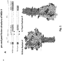
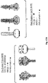
Figure 1A shows a comparison of wild-type fusion (F) protein from respiratory syncytial virus (RSV) with an engineered RSV F antigen. Arrowheads indicate furin cleavage sites, and the peptide p27 is released after cleavage. The F1 and F2 fragments of the wild-type sequence, which are produced as a result of furin cleavage, are indicated.Figure 1B is a model of the engineered RSV pre-fusion F, based on data from parainfluenza virus 5 (PIV-5) pre-fusion F and engineered RSV post-fusion F crystal structures.Figure 1C displays the engineered RSV post-fusion F structure ofFigure 1A . CT = cytoplasmic tail; SP = signal peptide (DNA sequence of the SP provided as SEQ ID NO:7, and amino acid sequence provided as SEQ ID NO:8); TM = transmembrane region. All figures are taken fromNature Reviews Microbiology 10, 807-813; 2012 -
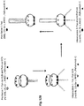
Figure 2A (taken from taken from McLellan JS et al. 2011. J Virol 85:7788-96) shows a three-dimensional model of the proposed pre-fusion conformation of the hRSV F trimer, built using the SWISS-MODEL server facilities (http://swissmodel.expasy.org) and the atomic coordinates of the pre-fusion structure of the PIV5 F protein (Protein Data Bank code, 2B9B) as a template. The backbone structure of the three monomers is shown in gray. In the original published color drawing (McLellan JS et al. 2011. J Virol 85:7788-96), the fusion peptide sequences of one monomer are shown in pink, and those of HRA are shown in black. Residues that are changed in virus isolates or in escape mutants selected with monoclonal antibodies, whose epitopes map in different antigenic sites of the F protein, are shown therein as colored spheres (antigenic site I, amino acid 389; antigenic site II, amino acids 262, 268, 272, and 275; and antigenic site IV, amino acids 429, 432, 433, 436, and 447).Antigenic site 0 is indicated inFigure 2A (location of antigenic site adapted from McLellan et al. 2013 Science 340(6136):1113-7). The two proteolytic cleavage sites are indicated with arrows in one of the monomers.Figure 2B (also taken from McLellan JS et al. 2011. J Virol 85:7788-96) shows the neutralizing epitopes on RSV F with a deleted FP. Epitopes for motavizumab and palivizumab (antigenic site II), 101F (antigenic site IV), and 131-2a (antigenic site I) are solvent exposed and in conformations compatible with antibody binding. Residues 254 to 277 are colored red (antigenic site II), residues 429 to 437 are colored blue (antigenic site IV), and atoms in Pro389 are shown as spheres (antigenic site I). Residue numbering according to McLellan JS et al. 2011. J Virol 85:7788-96. -
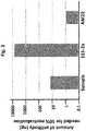
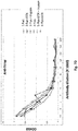
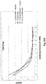
Figure 3 shows the amount of antibody needed to block 50% infection in a microneutralization assay. The lower the amount, the more effective the antibody. Synagis®= Palivizumab. -
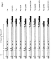
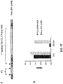
Figure 4 provides a schematic representation of the different constructs used in the present invention. TM = transmembrane region; CT = cytoplasmic tail; CD5 = signal peptide; HRA = heptad repeat A; HRB = heptad repeat B; GCN = GCN4 trimerization motif. The black dots represent mutations from cysteine to alanine. The dotted lines indicate the position of the furin cleavage sites. The black arrows indicate the positions where the signal peptide (CD5) and P27 are cleaved off. The grey arrows indicate the trypsin sensitive cleavage sites. -
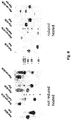
Figure 5 shows western blots of Fwt, Fwt-GCN, Flys and Flys-GCN proteins that were either reduced and heated (left panel), not reduced and heated (middle panel) or not reduced and not heated (right panel). Higher order proteins in the right panel indicate the post-fusion state of these proteins that almost fully disappears upon treatment with 2-betamercaptoethanol (reducing) and heating prior to running the proteins on SDS-PAGE. -
Figure 6 shows a Coomassie Blue staining of gels on which Fwt, Fwt-GCN, Flys and Flys-GCN proteins were separated after heating (upper panels) or after no heating (lower panels) and increasing cleavage with trypsin (T). -
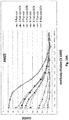
Figure 7A shows the results of an ELISA assay with which the reactivity of different soluble F proteins (Fwt, Fwt-GCN, Flys and Flys-GCN) with the AM22 antibody was checked. The treatment with trypsin is indicated for Flys and Flys-GCN.Figure 7B shows the results of an ELISA assay with which the reactivity of different soluble F proteins (Fwt, Fwt-GCN, Flys and Flys-GCN) with the 131-2a antibody was checked. The treatment with trypsin is indicated for Flys and Flys-GCN.Figure 7C shows the results of an ELISA assay with which the reactivity of different soluble F proteins (Fwt, Fwt-GCN, Flys and Flys-GCN) with the Synagis® (Palivizumab) antibody was checked. The treatment with trypsin is indicated for Flys and Flys-GCN.Figure 7D shows the results of an ELISA assay with which the reactivity of different soluble F proteins (Fwt, Fwt-GCN, Flys and Flys-GCN) with the anti-strep antibody was checked. The treatment with trypsin is indicated for Flys and Flys-GCN. -
Figure 8 shows a western blot of Fwt-GCN, Fwt, Fwt-cys-GCN, Fwt-cys, Flys-GCN, Flys, Flys-cys-GCN and Flys-cys proteins that were either heated but not reduced (left panel) or that were heated and reduced (right panel). The introduction of cysteines in the HRB domain as outlined in example 2 results in higher order structures that are resistant to heat and only partially sensitive to reduction. -
Figure 9 shows a Coomassie Blue staining of gels on which Fwt-cys, Fwt-cys-GCN, Flys-cys and Flys-cys-GCN proteins were separated after heating (upper panels) or after no heating (lower panels) and increasing digestion with trypsin (T). -
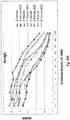
Figure 10A shows the results of an ELISA assay with which the reactivity of different soluble F proteins with cysteine and alanine mutations in their HRB domain (Fwt-cys, Fwt-cys-GCN, Flys-cys, Flys-cys-GCN, Fwt-ala, Fwt-ala-GCN, Flys-ala, Flys-ala-GCN) with the AM22 conformational antibody was checked.Figure 10B shows the results of an ELISA assay with which the reactivity of different soluble F proteins with cysteine and alanine mutations in their HRB domain (Fwt-cys, Fwt-cys-GCN, Flys-cys, Flys-cys-GCN, Fwt-ala, Fwt-ala-GCN, Flys-ala, Flys-ala-GCN) with the 131-2a conformational antibody was checked.Figure 10C shows the results of an ELISA assay with which the reactivity of different soluble F proteins with cysteine and alanine mutations in their HRB domain (Fwt-cys, Fwt-cys-GCN, Flys-cys, Flys-cys-GCN, Fwt-ala, Fwt-ala-GCN, Flys-ala, Flys-ala-GCN) with the Synagis® (Palivizumab) conformational antibody was checked.Figure 10D shows the results of an ELISA assay with which the reactivity of different soluble F proteins with cysteine and alanine mutations in their HRB domain (Fwt-cys, Fwt-cys-GCN, Flys-cys, Flys-cys-GCN, Fwt-ala, Fwt-ala-GCN, Flys-ala, Flys-ala-GCN) with the anti-strep conformational antibody was checked. -
Figure 11 displays western blots of the Fwt-ala-GCN, Fwt-ala, Flys-ala-GCN and Flys-ala proteins that were either not reduced and heated (left panel) or both not reduced and not heated (right panel). -
Figure 12A shows the pre-fusion (left panel) and post-fusion (right panel) states of the F protein with the different epitopes of antibodies that are and that are not available. In the pre-fusion state, where 6HB is not present, the epitope for AM22 is available (black star), while the epitope for 131-2a is shielded (open symbol). In the post-fusion state of the F protein where 6HB is present, the epitope for AM22 is not presented while the epitope for 131-2a is available (solid instead of open symbol). The epitope for Palivizumab (triangles) remains available in both the pre-fusion and post-fusion states.Figure 12B shows the pre-fusion state of the F protein (no 6HB), transitioning through two intermediate states and ending in the post-fusion state exhibiting the 6HB, and also depicting the presence, appearance and disappearance of the various antigenic sites (symbols are given together with the separate antibodies, wherein the open symbol for 131-2a represents the shielded epitope and the solid symbols for 131-2a represent the available epitope). The refolding of the head domain (striped) causes the disappearance of the AM22 epitope from the pre- to the post-fusion state. -
Figure 13 shows the expression of Flys-ΔHRB and Flys-ΔHRB-GCN proteins on reducing (left panel) and non-reducing conditions (middle and right panel). The heat stability was checked as well (middle panel versus right panel). -
Figure 14 shows a western blot with Flys-GCN, Flys-ΔHRB and Flys-ΔHRB-GCN under non-reducing conditions and after heat treatment (left panel) and no heat treatment (right panel). Increasing trypsin digestion is indicated (T). No trypsin treatment is indicated with a minus (-). Flys-ΔHRB-GCN forms higher order structures regardless of cleavage and regardless of heating of the purified product. These higher order structures do not relate to 6HB as a functional HRB domain is absent. -
Figure 15A shows the results of an ELISA assay with which the reactivity of two different soluble F proteins (Flys-ΔHRB and Flys-ΔHRB-GCN) with the AM22 conformational antibody was checked. The treatment of trypsin is indicated.Figure 15B shows the results of an ELISA assay with which the reactivity of two different soluble F proteins (Flys-ΔHRB and Flys-ΔHRB-GCN) with the 131-2a conformational antibody was checked. The treatment of trypsin is indicated.Figure 15C shows the results of an ELISA assay with which the reactivity of two different soluble F proteins (Flys-ΔHRB and Flys-ΔHRB-GCN) with the Synagis® (Palivizumab) conformational antibody was checked. The treatment of trypsin is indicated.Figure 15D shows the results of an ELISA assay with which the reactivity of two different soluble F proteins (Flys-ΔHRB and Flys-ΔHRB-GCN) with the anti-strep conformational antibody was checked. The treatment of trypsin is indicated. -
Figure 16 is a schematic representation of the RSV F protein with a functional HRB domain (Pre-fusion F; left) and of a RSV F protein with a replacement of the HRB stem by a GCN4 stem (Flys-ΔHRB-GCN; upper panel, middle). In the pre-fusion state, where 6HB is not present, the epitope for AM22 is available (black stars), while the epitope for 131-2a is not presented (open symbols). Upon trypsin treatment a refolding of the head domain occurs, through which recognition by AM22 is significantly diminished, while the epitope for Palivizumab (triangles) is retained. Also the epitope for 131-2a is still not presented. When no stem is present (Flys-ΔHRB; right panel), trypsin treatment also results in refolding of the head domain. The epitope for AM22 is no longer present, but the epitope for 131-2a is available (closed symbols). -
Figure 17A depicts a schematic representation of construct Fwt-ΔFP-ΔHRB used as a comparison RSV F protein, where: TM = transmembrane region; CT = cytoplasmic tail; P27 = 27 amino acid peptide dissociated from F after furin cleavage; CD5 = signal peptide; FP= fusion peptide; HRA = heptad repeat A; HRB = heptad repeat B. The dotted lines indicate the position of the furin cleavage sites. The arrows indicate the positions where the signal peptide (CD5) and P27 are cleaved off.Figure 17B shows the results of an ELISA assay for the reactivity of different soluble F proteins (Fwt-ΔFP-ΔHRB and Flys-ΔHRB-GCN) with different conformational antibodies (D25 and 131-2a) The reactivity of the recombinant proteins with MAbs D25 and 131-2a were determined by applying 2-fold dilutions of these antibodies. Binding of the antibodies was detected using HRP-conjugated secondary antibodies. The bar graphs depict the values corresponding to a single dilution of antibody. For each F protein preparation, the same antibody dilutions are shown, which are taken from linear parts of the curves of the F protein preparations with the highest reactivity to these antibodies. -
Figure 18A depicts a schematic representation of construct Fala-GCN used in the present invention, where: TM = transmembrane region; CT = cytoplasmic tail; CD5 = signal peptide; FP = fusion peptide; HRA = heptad repeat A; HRB = heptad repeat. The black arrow indicates the position where the signal peptide (CD5) is cleaved off. The arginine and lysine residues in the two multibasic furin cleavage sites (indicated with dotted grey arrows) are mutated into alanine residues (RARR to RAAA and RKRR to RKAA). This cleavage site (arg -> ala) mutant construct is referred to as 'Fala-GCN' (nucleotide sequence: SEQ ID NO:23; amino acid sequence: SEQ ID NO:24).Figure 18B shows the production level (mg F/L tissue culture supernatant) after transient transfection of RSV F ectodomain-encoding sequences (Fwt, Fwt-ΔFP-ΔHRB, Flys-ΔHRB-GCN, Fala-GCN and Flys-GCN) transfected into HEK293T cells using polyethyleneimine I (PEI). The yields were determined using ELISA and SDS-PAGE. -
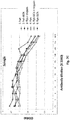
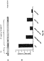
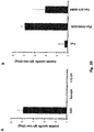
Figures 19A , B and C show the results of a vaccination study where pure bred strain New Zealand white rabbits (N=2 per group) were vaccinated intramuscularly with different soluble F proteins (Fwt, Fwt-ΔFP ΔHRB and Flys-ΔHRB-GCN). Final sera were subjected to ELISA to determine the antibody titer directed against pre-fusion F (ELISA plates coated with Flys-ΔHRB trimerized with a T4 foldon), shown inFigure 19A , or post-fusion F (plates coated with Fwt), shown inFigure 19B . Titers (2log) reported are the reciprocal of the calculated sample dilution corresponding with an OD450 of at least 0.2.Figure 19C shows the ratio between the pre- and post-fusion F specific titers. -
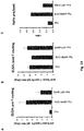
Figure 20 shows the results of an ELISA analysis using the sera described in the legend ofFigure 19 . The ELISA plates were coated with synthetic peptide hA-his encompassing part ofantigenic site 0. Titers (2log) reported are the reciprocal of the calculated sample dilution corresponding with an OD450 of at least 0.2.Figure 20A shows the antibody titer of monoclonals D25, Synagis, and 131-2A subjected to ELISA specifically directed against peptide hA-his.Figure 20B shows the antibody titer of final rabbit sera subjected to ELISA specifically directed against peptide hA-his. -
Figure 21A and B show the RSV virus neutralization titers of the sera described in the legend ofFigures 19A , B and C.Figure 21A depicts the average of the pre-fusion specific ELISA titers (2log) obtained with the final sera against RSV strains A and B.Figure 21 B depicts the average of the post-fusion specific ELISA titers (2log) obtained with the final sera against RSV strains A and B. - The present invention relates to a heat-stable oligomeric complex of a recombinant polypeptide presenting at least one antigenic epitope of the pre-fusion Respiratory Syncytial Virus (RSV) F protein, said polypeptide comprising the RSV F protein ectodomain from which the HRB region is functionally deleted and from which the transmembrane and cytoplasmic domains are deleted and replaced with a heterologous trimerization domain, and wherein the two multibasic furin cleavage sites in said ectodomain are mutated by the substitution of all arginine residues in said sites with lysine residues, thereby rendering said sites defective. The invention further relates to an immunogenic composition comprising the aforesaid heat-stable oligomeric recombinant polypeptide and a method of inducing an immune response in a subject to RSV F comprising administering to said subject said immunogenic composition.
- The design and evaluation of recombinant soluble F proteins of RSV has suffered considerably by a lack of tools to demonstrate the conformational status of a recombinant F protein. Based on the available literature the inventors of the present invention hypothesized that the post-fusion form of F should comprise a stable 6HB and that it should be recognized by post-fusion specific antibodies, but not by pre-fusion specific antibodies. In contrast, the pre-fusion form of the F protein should not carry the 6HB, will be recognized by pre-fusion specific antibodies, but not by post-fusion specific antibodies. Using this knowledge, it was reasoned that such pre-fusion specific antibodies would enable the identification of RSV F protein mutants that are stable (resistant to for instance heat) and remain in their pre-fusion conformation also under stressful conditions. Such stable pre-fusion mutants could in a next step be used in vaccines against RSV because they would - in their stable - conformation give an immune response (in vivo) and give rise to neutralizing antibodies that would be able to neutralize the virus carrying pre-fusion state F proteins. The present invention discloses that the inventors were indeed able to identify a recombinant RSV F protein mutant that is stably maintained in its pre-fusion conformation and is therefore useful in RSV vaccines.
- The present invention may be understood with reference to the following definitions.
- 'Functionally deleted' means a deletion of a sequence of amino acids (which may be referred to as a 'domain') from a natural protein sequence such that the function of the deleted domain is lost, and the properties of the protein are thereby altered.
- 'Heat-stable' means that a polypeptide retains its three dimensional conformation in aqueous solution over a range of temperatures, and thereby retains its properties, including for example such polypeptide's antigenic properties. Preferred temperature ranges are from about 5 degrees C to about 60 degrees C. A more preferred range is from about 10 degrees C to about 80 degrees C. A most preferred range is from about 20 degrees C to about 100 degrees C. Exemplary conformations are those characterized as a pre-fusion state.
- "Oligomer" or 'oligomeric complex' means polypeptide that consists of two, three, or four polypeptide monomers, such as a dimeric, trimeric or tetrameric complexes of essentially the same polypeptide monomers. Most preferred oligomeric polypeptides according to the present invention are trimeric polypeptides.
- 'Post-fusion conformation' or 'post-fusion state' means a three-dimensional protein configuration that differs from that configuration taken by the polypeptide or polypeptide oligomer upon initial expression or oligomeric assembly, and results from the interaction of such polypeptide or oligomer from enzymatic action and/or physical contact with other proteins or proteinaceous assemblies, such as a cell membrane. The RSV F proteins that form a post-fusion state are RSV F proteins that include an HRA-HRB 6HB.
- 'Pre-fusion conformation' or 'pre-fusion state' means a three-dimensional protein configuration taken by the polypeptide or polypeptide oligomer upon initial expression or oligomeric assembly. RSV F proteins form RSV F protein oligomers that exhibit a pre-fusion configuration prior to fusing with the cell membrane. Pre-fusion RSV F proteins include the following characteristics: the HRA region is packed against domain III in the RSV F head region and the HRB region forms a trimer coiled-coil stalk in proximity to domains I and II rather than associating with the HRA region in the context of the 6-helix bundle (6HB).
- 'Purified' protein or polypeptide means a protein or polypeptide isolated from other components of the polypeptide production system such that the amount of protein relative to other macromolecular components present in a composition is substantially higher than that present in a crude preparation. In general, a purified protein or polypeptide will be at least about 50% homogeneous and more preferably at least about 75%, at least about 80%, at least about 90%, at least about 95% or substantially homogeneous.
- The RSV F ectodomain protein sequence is exemplified by the amino acid sequence of SEQ ID NO:18. The corresponding nucleotide sequence is provided in SEQ ID NO:17. References to the amino acid sequence numbering and identifiable domains of the RSV F protein will herein be made to SEQ ID NO:18. However, other strains of RSV may also be used to generate equivalent recombinant polypeptides.
- The present invention relates to a heat-stable oligomeric recombinant polypeptide presenting at least one antigenic epitope of the pre-fusion Respiratory Syncytial Virus F protein, said polypeptide comprising the Respiratory Syncytial Virus F protein ectodomain from which the HRB region is functionally deleted, and from which the transmembrane and cytoplasmic domains are deleted and replaced with a heterologous trimerization domain; and wherein the two multibasic furin cleavage sites in said Respiratory Syncytial Virus F protein ectodomain are mutated by the substitution of all arginine residues in said sites with lysine residues, thereby rendering said furin cleavage sites defective. In another preferred embodiment, said heterologous trimerization domain is a GCN4 leucine zipper trimerization motif. In yet another preferred embodiment, said functional deletion of the HRB region comprises the deletion of SEQ ID NO:10. In another aspect of the invention the heat-stable oligomeric recombinant polypeptide according to the invention further comprises a LysM peptidoglycan binding domain linked to the carboxy-terminal end of said trimerization domain, and in yet another preferred embodiment, the recombinant polypeptide according to the invention further comprises a triple Strep-tag. Preferably, the ectodomain is a soluble ectodomain, and in another preferred embodiment, the antigenic epitope is recognized by a pre-fusion specific monoclonal antibody such as AM22 or D25, or by both AM22 and D25.
- The present invention also relates to an immunogenic composition comprising the oligomeric recombinant polypeptide of the present invention. Preferably, the composition of the invention further comprises an adjuvant. Suitable adjuvants are well known in the art and the skilled person is well aware of what adjuvants may suitably be used in the immunogenic compositions of the present invention. In yet another preferred embodiment, the oligomeric recombinant polypeptide is bound, covalently or non-covalently, to a carrier particle, whereas in another preferred aspect, the carrier particle is a bacterium-like particle.
- The present invention also relates to a recombinant expression vector comprising a nucleotide sequence encoding the polypeptide forming the heat-stable oligomer of the present invention. The polypeptides and expression vectors and compositions comprising such proteins or vectors may suitably be used in therapeutic settings. The present invention therefore also relates to a method of inducing an immune response in a subject to RSV comprising administering to said subject an immunogenic composition according to the present invention.
- The present invention relates to recombinant soluble proteins that mimic the pre-fusion state of human RSV F protein. See
Figure 1 for an image depicting the pre-fusion and the post-fusion conformation of the RSV F protein. The higher order modified RSV F protein structures found to be heat-stable, as disclosed herein, are oligomers of the polypeptide, and most likely, the polypeptide trimers of the modified RSV F polypeptide. The present polypeptide is likely formed into a trimer configuration based on size as detected in the experiments disclosed herein and because a trimerization motif is present in the modified polypeptide construct. - Preferably, the heat-stable oligomeric polypeptide according to the present invention is stable at room temperature. More preferably, the polypeptide is stable at temperatures up to 40°C, and even more preferably, the polypeptide is stable at temperatures up to 60°C, and yet in an even more preferred embodiment, the heat-stable recombinant polypeptide of the invention is stable at temperatures up to 70°C. In a most preferred aspect, the heat-stable recombinant polypeptide according to the present invention remains stable at temperatures of about 96°C for at least about 5 to about 15 minutes.
- The heat-stable recombinant polypeptide according to the invention comprises a functional deletion of the HRB region. This is different from what has been performed in the art and as shown herein, for instance by introducing (cysteine or alanine) mutations in the HRB region (
WO 2012/158613 ). Such mutants cannot - similar to what has been shown intra - form 6HB structures, but still result in labile conformations. Such labile conformations are prevented by removing the HRB region from the RSV F protein as disclosed herein. The polypeptide of the present invention has a functional deletion of the HRB region of the RSV F protein such that the HRB region can no longer perform its natural function, for instance in building the 6-helix bundle (6HB) rendering the protein unable to form a post-fusion conformation. The deletion of the HRB region preferably comprises the amino acids of SEQ ID NO:10 (deleted nucleotides are given in SEQ ID NO:9). It will be appreciated by the person skilled in the art that such deletion may be slightly smaller and/or slightly bigger on either side of the HRB region, and/or may be shifted by a small number of amino acids. Nonetheless, such deletions will still render a functional deletion of the HRB region and, in combination with the mutations of the furin cleavage sites and the addition of a heterologous trimerization motif, provide a heat-stable polypeptide as shown herein. - In an embodiment of the disclosure, the furin cleavage sites of the RSV protein may be mutated by different methods known in the art, for instance by replacement of the arginine residues by any other type of amino acid, or by deletions of the crucial residues. In a preferred embodiment, the mutation of the furin cleavage sites comprises the replacement of all arginine residues with lysine residues.
- The heat-stable oligomeric polypeptide of the invention comprises a heterologous trimerization domain selected from the group consisting of: GCN4 leucine zipper trimerization motif, the trimerization motif from influenza virus HA protein, SARS spike, HIV gp41, NadA, ATCase and foldon sequence. In a preferred embodiment, said heterologous trimerization domain is a GCN4 leucine zipper trimerization motif. Leucine zipper motifs such as GCN4, as well as other trimerization motifs, induce the formation of trimeric-coiled coils, in which three alpha-helices are coiled together like the strands of a rope. Such trimerization motifs have been used in the art to generate RSV vaccines based on RSV F proteins (
WO 2010/149743 andWO 2012/158613 ). The GCN4 trimerization motif sequence that is used in the present invention is provided in SEQ ID NO:15 (nucleotides) and SEQ ID NO:16 (amino acids). - The heat-stable oligomeric polypeptide according to the invention is preferably recognized by the pre-fusion specific monoclonal antibodies AM22 and D25, the preparation and characteristics of which are both described in
WO2008/147196 , the recognition indicating that pre-fusion specific antigenic epitopes are available. - The polypeptides of the present invention are suitable as antigenic component(s) of a vaccine that protects against infection and disease caused by human RSV (serotype A and B). By employing the present F protein mutation/deletion strategy to F proteins of other viruses belonging to the subfamily Pneumovirinae, immunogenic polypeptides protective against infection caused by these similar viruses, such as bovine RSV or human metapneumovirus, may be prepared.
- The invention further relates to an immunogenic composition that is an effective vaccine to immunize against RSV infections. In a preferred embodiment, said immunogenic composition comprises an adjuvant to boost immunogenicity. In yet another aspect, the invention relates to a recombinant expression vector comprising a nucleotide sequence encoding the heat-stable polypeptide according to the invention.
- The recombinant proteins of the present invention may also be used in diagnostics assays, with which one may measure the antibody response specifically targeted against the pre-fusion form of the F protein of RSV (or relatives thereof). This response may be of predictive value with respect to disease. Furthermore, the recombinant proteins of the present invention may be used to test the quality of the antibody response induced by a candidate vaccine, and may be used to generate conformation-specific antibodies which may be used as therapeutics, to study the epitopes present on a candidate vaccine, or to control the antigenicity of a (candidate) vaccine.
- In another preferred embodiment, the invention relates to a heat-stable oligomeric complex of a recombinant polypeptide according to the invention that further comprises a LysM peptidoglycan binding domain as a tag. The LysM tag nucleotide sequence as used in the present invention is provided in SEQ ID NO:13, whereas the amino acid sequence is provided in SEQ ID NO:14. For easy purification and detection of the recombinant polypeptide, the polypeptide preferably comprises a triple Strep-tag. The nucleotide sequence for the triple Strep-tag used in the present invention is provided in SEQ ID NO:11, whereas the amino acid sequence is provided in SEQ ID NO:12. In yet another preferred aspect, the ectodomain within the polypeptide is a soluble ectodomain.
- The heat-stable oligomeric complex of a recombinant polypeptide of the present invention is in a pre-fusion conformation that is antigenic, and can be confirmed by the use of antibodies that neutralize and recognize certain epitopes in the RSV F protein. In a preferred aspect, the heat-stable recombinant polypeptide according to the invention comprises an available epitope that is recognized by monoclonal antibody AM22. Whether AM22 recognizes the polypeptide can be easily checked by common methods used in the art, and as disclosed herein.
- The present invention is useful in the field of medicine, and in particular in the field of vaccines against RSV infections. The heat-stable polypeptides of the present invention can be used in immunogenic compositions that may be applied in vaccination programs, in regions that present storage and handling challenges such as the third world, to protect (human) subjects who are at risk of developing disease caused by RSV.
- Because the polypeptides of the present invention are stable in their pre-fusion conformation, presenting pre-fusion specific epitopes recognized by potent neutralizing antibodies, epitopes absent in polypeptides folded in the post-fusion conformation, the polypeptides are capable of inducing superior pre-fusion specific neutralizing antibodies that protect against RSV infection. The importance of prefusion-specific VN antibodies in naturally RSV infected humans has been demonstrated by Magro M et al. (2012. Neutralizing antibodies against the preactive form of respiratory syncytial virus fusion protein offer unique possibilities for clinical intervention. Proc Natl Acad Sci USA 109:3089-94). Hence, the present invention also relates to an immunogenic composition comprising the purified heat-stable oligomeric polypeptide of the present invention, and optionally further comprises excipients commonly used in vaccine preparations.
- It should be understood that the present polypeptides used in the immunogenic compositions of the present invention do not include the signal peptide, which is co-expressed with the heat-stable polypeptide, but which is cleaved from the polypeptide before leaving the production cell. In a preferred aspect, although not strictly necessary, the additional (non-RSV F protein) tag sequences are removed by enzymatic digestion during or after the purification process. Preferably, an immunogenic composition according to the present invention further comprises an adjuvant to further boost the immune response.
- The present invention also relates to recombinant expression vectors comprising the nucleotide sequences encoding the heat-stable polypeptides of the present invention. Moreover, the invention relates to a method of inducing an immune response in a subject to RSV F comprising administering to said subject an immunogenic composition according to the invention, and to methods of vaccinating human subjects against RSV infections by applying the immunogenic compositions as disclosed herein. The invention also relates to the use of a heat-stable polypeptide according to the invention for the manufacture of a medicament for the prophylaxis or treatments of RSV infections or diseases that follow an RSV infection. The invention further relates to heat-stable recombinant polypeptides and/or recombinant expression vectors according to the invention for use in vaccines against RSV infections.
- Immunogenic compositions according to the present invention may be prepared, tested for immunogenicity, efficacy and safety employing the technology disclosed in published PCT application
WO2012/128628 , hereby incorporated by reference. Vaccine formulations may be based on particles derived from inactivated Lactococcus lactis bacteria, a safe bacterium traditionally used in the food industry, such as for the production of cheese (described elsewhere as Gram-positive Enhancer Matrix or Bacterium-Like Particles and herein referred to as "BLPs"). BLPs are obtained by the acidic heat treatment of L. lactis bacteria, resulting in non-living spherical particles that predominantly consist of a peptidoglycan surface, the preparation of BLPs is disclosed inWO 02/101026 US 7,312,311 , which is hereby incorporated by reference. The resulting antigen-associated BLPs constitute the final vaccine that may be delivered to humans via the mucosal layers of the nose (e.g. drops or spray) or mouth (e.g. capsule, tablet or liquid), without the need for an injection. - This example describes the generation of different F protein constructs and their characterization with respect to antibody binding and gel electrophoretic mobility. This analysis demonstrates that the tools and assays are suitable to demonstrate the conformational state of a RSV F protein. Also, a method is disclosed that shows the production of recombinant soluble proteins that mimic the pre-fusion state of human RSV F.
- In an initial step, a comparative analysis of the virus neutralizing capacity was performed of the MAbs that were used herein (
Figure 3 ). In agreement with previous studies, antibody 131-2a hardly demonstrated any neutralizing capacity compared to Palivizumab or AM22 (McLellan JS et al. 2011. J Virol 85:7788-96). In contrast, AM22 efficiently neutralized infection with hRSV and was even more potent than Palivizumab, in agreement with what was shown inUS 2012/0070446 . Palivizumab was shown to recognize the post-fusion form of hRSV F, but it likely also recognizes the pre-fusion form of F, because its epitope appears also to be present in the pre-fusion form based on the X-ray structure solved for parainfluenza virus 5 F protein (Yin HS et al. 2006. Structure of the parainfluenza virus 5 F protein in its metastable, prefusion conformation. Nature 439:38-44). Antibody 131-2a recognizes an epitope that is probably not accessible for antibody binding in the pre-fusion form of F. The presumed inability of 131-2a to bind the pre-fusion form of RSV F corresponds with its lack of neutralizing activity. - The inventors of the present invention and others previously demonstrated that recombinant soluble class I fusion proteins can be stably maintained in their pre-fusion conformation by the addition of artificial trimerization domains (de Vries RP et al. 2010. The influenza A virus hemagglutinin glycosylation state affects receptor-binding specificity. Virology 403:17-25; Wei CJ et al. 2008. Comparative efficacy of neutralizing antibodies elicited by recombinant hemagglutinin proteins from avian H5N1 influenza virus. J Virol 82:6200-8; Yang X et al. 2000. Characterization of stable, soluble trimers containing complete ectodomains of human ; Yin HS et al. 2006. Nature 439:38-44). Use was made of a similar construct that was used previously to express recombinant soluble bioactive influenza A virus HA protein to express soluble F proteins (Cornelissen LA et al. 2010. A single immunization with soluble recombinant trimeric hemagglutinin protects chickens against highly pathogenic avian influenza virus H5N1. PLoS One 5:e10645; de Vries RP et al. 2011. Only two residues are responsible for the dramatic difference in receptor binding between swine and new pandemic H1 hemagglutinin. J Biol Chem 286:5868-75; de Vries RP et al. 2010. Virology 403:17-25).
- In the expression constructs used in the present invention (overview provided in
Figure 4 ), the human codon-optimized RSV F ectodomain sequence of a European clinical isolate (Tan L et al. 2012. Genetic Variability among Complete Human Respiratory Syncytial Virus Subgroup A Genomes: Bridging Molecular Evolutionary Dynamics and Epidemiology. PLoS One 7:e51439) was preceded by a signal peptide-encoding sequence. The ectodomain sequence was optionally followed by a sequence coding for the GCN4 leucine zipper trimerization motif (Harbury PB et al. 1993. A switch between two-, three-, and four-stranded coiled coils in GCN4 leucine zipper mutants. Science 262:1401-7). The protein with the codon-optimized ectodomain sequence (which encodes the wild type version of this part of the protein), in which the wild type furin cleavage sites and the HRB domain are present, and wherein the ectodomain is followed by the GCN4 motif was referred to as Fwt-GCN. For comparison, the wild-type F protein-encoding gene was also cloned into vectors lacking the trimerization motif (referred to as Fwt). The tags consisted either of a triple Strep-tag for easy purification, or of a LysM peptidoglycan binding domain followed by the triple Strep tag as it turned out that, for yet unknown reasons, GCN-containing F constructs were expressed to higher levels when also the LysM domain was present. In addition to the wild-type constructs (Fwt and Fwt-GCN), expression vectors encoding F proteins with modified furin-cleavage sites were also constructed. These mutations are specified in the protein sequence of SEQ ID NO:20, shown as R to K mutations in the 1st furin cleavage site (positions 81, 83 and 84) and in the 2nd furin cleavage site (positions 108, 110 and 111). The nucleotide sequences of these mutants are given in SEQ ID NO:19. These cleavage site (arg -> lys) mutant constructs are referred to as 'Flys'. Others showed that in the absence of furin cleavage the F protein is prevented to adopt the post-fusion conformation (Ruiz-Arguello MB et al. 2002. Virology 298:317-26; Ruiz-Arguello MB et al. 2004. J Gen Virol 85:3677-87). This is in line with the idea that cleavage of F is required for activation of membrane fusion. - Gene construction and cloning was performed as follows: Two variants of a cDNA clone corresponding to
residues 26 to 515 of the F protein of a European isolate of RSV serotype A (Genbank accession number JX015498.1) were synthesized using human-preferred codons by GenScript USA Inc. While one cDNA clone encoded the wild type F protein ectodomain, the other clone encoded a F protein ectodomain in which the arginine residues in the two multibasic furin cleavage sites are mutated into lysines (RARR to KAKK and RKRR to KKKK). Each cDNA was cloned into the pCD5 expression vector for efficient expression in mammalian cells (de Vries RP et al. 2010. Virology 403:17-25). The pCD5 vector had been modified such that the F protein-encoding sequences were cloned in frame downstream of a DNA sequence coding for a CD5 signal peptide and when indicated upstream of sequences encoding the heterologous GCN4 isoleucine zipper trimerization motif and the specified tag. The tag either consisted of a triple Strep-tagII (IBA, Germany) or of a LysM peptidoglycan binding domain (van Roosmalen ML et al. 2006. Mucosal vaccine delivery of antigens tightly bound to an adjuvant particle made from food-grade bacteria. Methods 38:144-9;WO2012/128628 ) followed by a triple Strep-tagII. Two codon-optimized DNA fragments encoding the variable heavy and light chains of antibody AM22 (US 2012/0070446 A1 ) were synthesized by GenScript USA, Inc. and cloned in-frame into pCAGGS mammalian expression vectors containing human IgG1 heavy and light constant domains, respectively. - Expression of the F protein ectodomains was achieved by transient transfection as follows: pCD5 expression vectors containing RSV F ectodomain-encoding sequences were transfected into HEK293T cells using polyethyleneimine I (PEI) in a 1:5 w/w ratio (µg DNA: µg PEI). At 6 h post transfection, the transfection mixture was replaced by 293 SFMII expression medium (Invitrogen), supplemented with sodium bicarbonate (3.7 g/L), glucose (2.0 g/L), Primatone RL-UF (3.0 g/L), penicillin (100 units/ml), Streptomycin (100 µg/ml), glutaMAX (Gibco), and 1,5% dimethylsulfoxide. Tissue culture supernatants were harvested 5-6 days post transfection. F proteins were either purified using Strep-tactin Sepharose beads according to the manufacturer's instructions (IBA, Germany) for further analysis of the protein. The AM22 and D25 expression vectors were co-transfected at a 1:1 ratio into HEK293T cells similarly as described above. The cell culture media were clarified by centrifugation and the AM22 and D25 antibodies were purified with protein A sepharose beads using standard conditions. The concentration of purified proteins was determined by using a
Nanodrop 1000 spectrophotometer (Isogen Life Sciences) according to the manufacturer's instructions. - Expression and secretion of recombinant proteins were confirmed by sodium dodecylsulfate (SDS)-polyacrylamide gel electrophoresis (PAGE; 10% NuPAGE BisTris, Invitrogen) followed by western blotting using anti-Strep-tag antibody conjugated with horse radish peroxidase (HRP) (StrepMAB-classic-HRP, IBA), Palivizumab (Synagis®, Abbott Laboratories) followed by HRP-conjugated anti-human IgG antibody (ITK Southern Biotech). This latter antibody was also used to confirm expression of recombinant antibody AM22. Prior to SDS-PAGE analysis, the samples were resuspended in Laemmli sample buffer (LSB) that either did or did not contain 5% 2-mercaptoethanol (Sigma), and when indicated heated at 96°C for 5-15 minutes.
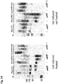
- The results are given in
Figure 5 . The epitope recognized by Palivizumab is located in the F1 part of the F protein. The results show that when the F ectodomains are subjected to SDS-PAGE under reducing conditions (i.e. in the presence of 2-mercaptoethanol), which results in separation of the otherwise disulfide-linked F1 and F2, they migrated with different electrophoretic mobilities corresponding to the absence or presence of the GCN-LysM sequences. Furthermore, the F proteins migrated at a higher position in the gel, when the furin-cleavage sites were mutated (compare Fwt with Flys, and Fwt-GCN with Flys-GCN) in agreement with these proteins not being cleaved. When the same F protein preparations were subjected to SDS-PAGE in the absence of reducing agents, the migration of the non-cleaved F proteins did not appear to be much affected. - In contrast, while the Fwt and Fwt-GCN proteins clearly ran at a lower position in the gel than the Flys and Flys-GCN under reducing conditions, the difference in the electrophoretic mobility appeared much smaller in the absence of reducing agents, in agreement with the F2 part still being attached to the F1 part via disulfide bridges also in the furin-cleaved proteins. The small difference in electrophoretic mobility between the cleaved and non-cleaved F proteins that was still noticeable is most likely explained by the dissociation of the glycosylated p27 sequence from the cleaved proteins. Interestingly, the electrophoretic mobility of the cleaved F proteins was dramatically changed when the preparations were not heated prior to electrophoresis under non-reducing conditions. In contrast to the non-cleaved proteins (Flys and Flys-GCN, the electrophoretic mobility of which was not much affected) the majority of the Fwt and Fwt-GCN proteins migrated at a much higher position in the gel. The migration of these latter proteins is explained by the cleaved F proteins adopting a stable post-fusion conformation, characterized by the presence of an extremely stable 6HB, resistant to SDS unless the protein preparations are heated. These results indicate that the large majority of the soluble, cleaved F ectodomains adopts a post-fusion conformation. The post-fusion conformation is not prevented when the ectodomain is extended with an artificial trimerization domain. However, the stable post-fusion conformation is not formed when the F proteins are not cleaved.
- To confirm and extend these observations, a subsequent experiment was performed in which the purified F proteins were subjected to limiting proteolysis followed by SDS-PAGE under non-reducing conditions. Despite the fact that the furin-cleavage sites in Flys and Flys-GCN had been mutated by substitution of the arginines by lysines, these positions are still sensitive to trypsin digestion. Treatment of the Flys and Flys-GCN proteins with trypsin will thus result in cleavage of these proteins and possibly in formation of the SDS-resistant higher-order structure corresponding to the post-fusion conformation of the F protein.
- Purified F proteins were (mock-) treated with varying amounts of TPCK treated trypsin from bovine pancreas (Sigma) for 30 min at 23°C. The samples were next put on ice and trypsin inhibitor (Sigma) was added, after which they were analyzed by SDS-PAGE as described above. Protein bands were visualized by general staining using the Colloidal Blue Staining kit (Invitrogen). The results are shown in
Figure 6 . Clearly, digestion with trypsin indeed results in the appearance of the SDS-resistant higher-order structure corresponding to the post-fusion conformation of the F protein. Fwt and Fwt-GCN proteins were taken along as controls. The purified proteins were detected using Colloidal Coomassie Blue staining. Again Fwt and Fwt-GCN ran at their expected positions in the gel, when the samples were heated prior to electrophoresis (upper panels). When the samples were not heated, proteins ran at a much higher position in the gel (lower panels). As expected, treatment of these samples with trypsin did not affect the migration of the higher order structures much. However, trypsin treatment resulted to some extent in the removal of the F protein tags as demonstrated by the appearance of lower migrating F protein species when the samples were heated prior to electrophoresis. Treatment of the non-cleaved F proteins with trypsin resulted in the appearance of F proteins migrating at a much higher position in the gel under non-reducing conditions, similarly as observed for their Fwt and Fwt-GCN counterparts. The formation of the SDS-resistant higher-order structures was more apparent for Flys when compared to Flys-GCN, suggesting that the formation of the post-fusion conformation is prevented to some extend by the GCN trimerization motif. - The reactivity of the F protein preparations with the RSV F specific MAbs Palivizumab (Synagis®), AM22 and 131-2a were probed using an ELISA format. For this, 96-well Nunc maxisorp plates were overnight coated with different F protein preparations (50 ng per well) at 4°C. After blocking and extensive washing, the plates were incubated with limiting dilutions of Palivizumab (Synagis®, starting with 1 in 500 dilution of a 3 mg/ml stock), AM22 (starting with a 200 fold dilution of a 0.7 mg/ml stock), 131-2a (Millipore, starting with a 500 fold dilution of a 1 mg/ml stock), or anti-strep (StrepMAb classic from IBA, starting with a 500 fold dilution of the stock). After extensive washing, the plates were incubated with HRP conjugated goat-anti-human IgG antibodies (ITK Southern Biotech) or HRP conjugated rabbit-anti-mouse IgG antibodies (DAKO) at a 1:500 dilution for 1 h at RT. Detection of HRP reactivity was performed using tetramethylbenzidine substrate (BioFX) and a ELISA plate reader (EL-808 from Biotek). The results are given in
Figure 7 . - All F proteins were coated with similar efficiencies as demonstrated by the binding of MAb specific for the Strep tag (Anti-Strep panel). Palivizumab displayed a concentration-dependent binding to all F protein preparations in agreement with the assumption that this antibody recognizes the F protein regardless of its conformational state. In contrast, AM22 was not able to bind Fwt, in agreement with the assumption that this antibody is not able to bind a protein in post-fusion conformation. However, intermediate binding was observed when the cleaved protein was extended with the trimerization motif (Fwt-GCN) or when cleavage was prevented (Flys). The highest reactivity was observed when these two features were combined (Flys-GCN). Trypsin treatment of Flys and Flys-GCN prior to coating of the wells resulted in reduced AM22 reactivity, which subsequently was comparable to the reactivity observed with Fwt and Fwt-GCN. Reactivity of Palivizumab with the F proteins was not affected by the trypsin treatment. MAb 131-2a efficiently bound to all F protein preparations.
- These results show that binding of neutralizing antibody AM22 differs between different F protein preparations: Fwt, which adopts the post-fusion conformation, is hardly detected, while the highest reactivity was observed for Flys-GCN. From these results it was concluded that the majority of Fwt is in the post-fusion conformation (6HB +, 131-2a +, AM22 -). Fwt-GCN is probably present in a mixture of conformations (6HB +, 131-2a +, AM22 +/-). In the absence of cleavage, the F proteins do not adopt the post-fusion conformation (no 6HB). Their reactivity with both 131-2a and AM22 indicates that these non-cleaved proteins are in some form of intermediate state.
- Next, the characteristics of F proteins, in which 4 cysteine residues were introduced into their HRB domain (designated Fwt-cys; Fwt-cys-GCN; Flys-cys; Flys-cys-GCN;
Figure 4 ) were investigated. These cys substitutions, when introduced in a full length F protein (containing a transmembrane domain and cytoplasmic tail), were previously shown to result in intermolecular disulfide bridges which appeared to stabilize the F protein in its pre-fusion conformation (Magro M et al. 2012. Proc Natl Acad Sci USA 109:3089-94). Purified proteins were analyzed by SDS-PAGE in the absence or presence of reducing agents. As shown inFigure 8 , introducing the cysteine residues in HRB resulted in the formation of higher order structures, which could be observed on the non-reducing gel, even when the samples were heated. These higher order structures were most apparent for the F proteins with the GCN4 trimerization motif (Fwt-cys-GCN and Flys-cys-GCN), but also to some extent for the F proteins that lacked the GCN4 motif (Fwt-cys and Flys-cys). These higher order structures were not observed for the F proteins that contained a wild type HRB under these conditions, in agreement with the results shown inFigure 5 and6 . While the higher order structures of Fwt-cys and Flys-cys were sensitive to the presence of reducing agents (Figure 8 ; right panel), this was much less so the case for the proteins that additionally contained the GCN4 motif. These data indicate the extreme stability of the higher order structures by introduction of the cysteine residues in HRB when also the GCN4 motif is present. - To confirm and extend these observations, a subsequent experiment was performed in which the purified F proteins were subjected to limiting proteolysis followed by SDS-PAGE under non-reducing conditions. The results are shown in
Figure 9 . The purified proteins were detected using Colloidal Coomassie Blue staining. Again, the higher order structures observed after introduction of the cysteine residues were shown not to be sensitive to heating of the sample prior to gel electrophoresis. Furthermore, the results indicate that trypsin-treated Fwt-cys and Flys-cys, which lack the GCN4 motif and are only present in a limited amount in the heat-resistant higher order structure, do not form higher structures when the samples were not heated prior to electrophoresis, as was observed for trypsin-treated Fwt and Flys (Figure 6 ). From these results it was concluded that the higher structures observed after introduction of cysteine residues in HRB differ from the higher order structures that are observed for the cleaved F proteins with a wild type HRB, because, in contrast to the latter, the former are resistant to heating and are also formed when the F protein is not cleaved. Furthermore, the data show that introduction of cysteine residues in HRB prevents the formation of the heat-sensitive higher order structure. This indicates that these proteins do not assembly the 6HB that is present in the post-fusion conformation. - The reactivity of the cysteine mutant F proteins preparation was investigated with the RSV F specific MAbs Palivizumab, AM22 and 131-2a using the ELISA format (number 1-4 in
Figure 10 ), as outlined above. All cysteine mutant F proteins were coated with similar efficiencies as demonstrated by the binding of MAb specific for the Strep tag. Palivizumab displayed a concentration dependent binding to all F protein preparations, which differed only slightly between the different F preparations. MAbs 131-2a and AM22 displayed differential binding to the different F proteins. Fwt-cys-GCN and Flys-cys-GCN were not bound by 131-2a, but were clearly bound by AM22. These results indicate that the higher order structures that are observed for these two proteins correspond with stabilized pre-fusion F proteins (6HB -, 131-2a -, AM22+). The results also indicate that the epitope recognized by AM22 is affected to some extent by cleavage of F. Fwt-cys was efficiently recognized by 131-2a (Flys-cys somewhat less), in agreement that only part of these proteins form the pre-fusion higher order structure that is not reactive with this antibody. In agreement herewith, these preparations also reacted with AM22. The reactivity observed with AM22 may also be explained in part by Fwt-cys and Flys-cys being in some form of intermediate state (6HB -, 131-2a +, AM22+). While this may be expected for Flys-cys in view of the results with Flys, an alternative explanation must account for the intermediate phenotype of Fwt-cys. The introduction of the cysteines might also promote AM22 reactivity because of HRB not being able to bind HRA, as the inability to form the 6HB may be directly coupled to preservation of the AM22 epitope. - To study the effect of mutations in the HRB domain in more detail, mutant F proteins were produced in which the same amino acids as outlined in example 2 (see also Magro M et al. 2012. Proc Natl Acad Sci USA 109:3089-94) were substituted by alanines rather than cysteines (
Figure 4 ), to prevent the formation of a disulfide stabilized pre-fusion trimer. Also these proteins were subjected to SDS-PAGE analysis for the absence or presence of higher order structures. The results are shown inFigure 11 . Introduction of the alanine substitutions in the absence of GCN4 (construct referred to as Fwt-ala) prevented the formation of heat-sensitive higher order structures (6HB) under non-reducing conditions (compareFigure 11 andFigure 5 ), similarly as observed for Fwt-cys. In the presence of GCN4, some higher order structures were observed, which were not sensitive to heating of the sample prior to electrophoresis. These minor higher order structures may correspond with pre-fusion structures. Fwt-ala and Flys-ala, neither of which are able to assemble the 6HB, also do not react with AM22 (right top panel,line Figure 10 ), indicating that preventing the formation of the 6HB does not preserve the epitope for AM22. These proteins are probably in an intermediate state in which the stem formed by HRB in the prefusion proteins is dissociated, thereby making the epitope for 131-2a accessible. At the same time, the head domain is refolded resulting in loss of the epitope for AM22. Even so, the 6HB cannot be formed as a result of the alanine mutations in HRB, which probably prevent interaction of HRB with HRA. Introducing the GCN4 motif in these proteins (Fwt-ala-GCN and Flys-ala-GCN), probably results in a mixed population with some pre-fusion F proteins that are positive for AM22 but not 131-2a. As a result the reactivity of these proteins with AM22 increased, while their reactivity with 131-2a decreased. Strikingly, all F protein with alanine mutations in their HRB display decreased reactivity with AM22 when compared to their wild type HRB counterparts. Apparently, mutation of HRB may either help to preserve pre-fusion epitopes (F proteins with cysteine mutations in HRB) but may also decrease the reactivity with pre-fusion specific epitopes (F proteins with alanine mutations in HRB). Strikingly, these mutations are introduced at the same positions in the protein and both prevent 6HB formation to the same extent, indicating that the ability of the protein to adopt the post-fusion structure is not the driving force for the conformational change of the pre-fusion structure. - The disclosed data show that a set of assays was developed with which the conformation of recombinant soluble F proteins can be determined. With these assays four conformational states of the F protein can be discriminated that are schematically shown in
-
- 1) Pre-fusion F (6HB -, 131-2a -, AM22 +), see
Figure 12A (left panel; comparing the schematic version with the three-dimensional representation of the protein) andFigure 12B (left upper panel). Stable forms are Fwt-cys-GCN and Flys-cys-GCN; - 2) Intermediate 1 (6HB -, 131-2a +, AM22 +), see
Figure 12B (left lower panel). The stem formed by HRB is dissociated, but the head domain with HRA is not yet refolded. Examples are Flys and Flys-GCN; - 3) Intermediate 2 (6HB -, 131-2a +, AM22 -), see
Figure 12B (right lower panel). The stem formed by HRB is dissociated, and the head domain that probably contains the epitope recognized by AM22 is refolded. The 6HB is not yet assembled in thisIntermediate 2 form. Examples are Fwt-ala and Flys-ala; - 4) Post-fusion F (6HB +, 131-2a +, AM22 -), see
Figure 12A (right panel; comparing the schematic version with the three-dimensional representation of the protein) andFigure 12B (right upper panel). The best example is Fwt. - In the examples above it is demonstrated that F proteins in the pre-fusion conformation are antigenically different from F proteins that have other conformations. The immune response specifically targeted against pre-fusion F epitopes, may be hampered by the presence of non-pre-fusion specific epitopes, the corresponding antibodies of which do not neutralize the virus (e.g. 131-2a). Although Fwt-cys-GCN and Flys-cys-GCN are in the pre-fusion conformation, these proteins are not considered suitable for the production of pre-fusion F proteins at a larger scale, primarily because these proteins are expressed at very low levels. To solve this, the inventors set out to device an alternative strategy to express high levels of recombinant F proteins that contain pre-fusion specific epitopes and lack post-fusion specific epitopes. The data show that GCN4 is able to confer the pre-fusion state onto recombinant proteins, but this only efficiently occurs when the HRB stem in the pre-fusion state is stabilized (e.g. in Fwt-cys-GCN and Flys-cys-GCN). This instability may even be increased by other mutations in HRB (such as the alanine mutations described herein).
- The inventors hypothesized that there might be some form of inherent instability in HRB, which makes it possible for HRB to dissociate and subsequently to interact with the extended HRA. Rather than to try to stabilize HRB via the introduction of intermolecular disulfide bonds, which severely reduces protein expression levels, it was decided to remove the HRB region (and its inherent instability) altogether. However, in the complete absence of HRB it was expected that epitopes that are normally not available in the pre-fusion state would be exposed, either directly by removal of HRB (e.g. epitope of 131-2a) or because the resulting stem-less protein may behave as a monomer rather than a trimer. Furthermore, the data indicate that in the absence of cleavage the AM22 epitope and probably other pre-fusion specific epitopes are better maintained in the recombinant F protein (compare Flys-cys-GCN and Fwt-cys-GCN). In view of these considerations, a recombinant protein was generated, in which HRB was removed, the GCN4 trimerization motif was added and the furin cleavage site was mutated (
Figure 4 ). The HRB part of the ectodomain that was deleted is given in SEQ ID NO:10. The full nucleotide sequence coding for the expressed polypeptide Flys-ΔHRB-GCN is provided in SEQ ID NO:5, whereas the amino acid sequence is provided in SEQ ID NO:6. As a control, a variant was synthesized that lacked GCN4 (SEQ ID NO:3 and 4). The deletion in the constructs of the present invention is somewhat larger (namely position 478-512 in SEQ ID NO:2) than is regarded as the HRB region in literature (Swanson et al. 2011, Proc Natl Acad Sci USA 10.1073. PNAS.1106536108), which holds HRB to run from position 482-510 (in SEQ ID NO:2). - These recombinant proteins were analyzed by SDS-PAGE using reducing and non-reducing conditions (
Figure 13 ). Both proteins migrated at the expected position in the reducing gel. However, in the absence of reducing agents the majority of Flys-ΔHRB-GCN ran at a much higher position in the gel. This was not the case for Flys-ΔHRB. Heating the samples did not affect the electrophoretic mobilities of the proteins much (Figure 13 , middle panel). This result indicates that the higher order structure observed for Flys-ΔHRB-GCN does not correspond to the heat-sensitive 6HB. This is expected because the 6HB cannot be formed in the absence of a functional HRB. - Next, cleavage of these recombinant proteins was induced by trypsin treatment to study the effect of protein digestion on the protein conformation. As a control Flys-GCN was taken and proteins were run on non-reducing gels. The results are shown in
Figure 14 . Upon trypsin treatment Flys-GCN, and in the absence of heating, the cleaved Flys-GCN protein ran as the (heat-sensitive) higher order structure indicative of 6HB formation. In contrast, the majority of Flys-ΔHRB ran at its expected (calculated) position in the gel, and trypsin cleavage did not induce the formation of higher order structures. This is expected as in the absence of HRB it is not possible to form the 6HB. The majority of Flys-ΔHRB-GCN4 again ran as a higher order structure, which was resistant to heat. This migration was not significantly affected by the trypsin treatment. The results show that replacing the HRB domain with GCN4 results in an extremely stable higher order structure that obviously does not correspond to the 6HB. - The reactivity of the Flys-ΔHRB and Flys-ΔHRB-GCN proteins with the monoclonal antibodies was tested in an ELISA as outlined above (
Figure 15 ). Flys-ΔHRB was recognized both by 131-2a and AM22. In the additional presence of GCN4 (Flys-ΔHRB-GCN), the reactivity for 131-2a was lost, while the reactivity with AM22 increased. Apparently, the GCN4 domain shields the 131-2a epitope (similarly as a stabilized HRB) and stimulates the presence of the epitope of AM22. Trypsin treatment of the samples did not affect the 131-2a reactivity, but clearly decreased the AM22 reactivity. The reactivity with Palivizumab or the monoclonal against the strep-tag was not affected by the trypsin treatment. From these results, it was concluded that Flys-ΔHRB-GCN is representative of the pre-fusion conformation of the RSV F protein as this protein does not form the 6HB, does react with AM22, but not with 131-2a (see for a schematic representation of this mutant and the effect of trypsin treatment on the availability of the different epitopes,Figure 16 ). - In examples 1-4, it was demonstrated that a combination of 1) the removal of HRB; 2) the inactivation of the furin cleavage sites; and 3) the addition of an heterologous trimerization motif is required to maintain a soluble human RSV F protein in the pre-fusion state. In the following comparative examples, the characteristics of an F protein, in which the HRB and fusion peptide (FP) region are deleted (designated Fwt-ΔFP-ΔHRB; see
Figure 17 ), are investigated.WO2012/158613 disclosed that these deletions, when introduced in a soluble RSV F protein (lacking a transmembrane domain and cytoplasmic tail), (partly) stabilized the F protein in its pre-fusion conformation as determined by its multimerization state and appearance using electron microscopy (WO2012/158613 A1 ; page 87;section 5 construct R057 which lacks the fusion peptide and the HRB region). - A protein which lacked the exact same FP and HRB region as described in
WO 2012/158613 A1 was synthesized (herein referred to as Fwt-ΔFP-ΔHRB; nucleotide sequence: SEQ ID NO:21; amino acid sequence: SEQ ID NO:22). A schematic representation of this protein is provided inFigure 17A . Gene construction and cloning was performed as follows: a cDNA clone encoding was synthesized using human-preferred codons by GenScript USA Inc. The cDNA was cloned into the pCD5 expression vector for efficient expression in mammalian cells (de Vries RP et al. 2010. Virology 403:17-25). The pCD5 vector had been modified such that the F protein-encoding sequence was cloned in frame downstream of a DNA sequence coding for a CD5 signal peptide and upstream of sequences encoding the specified tag-consisting of a triple Strep-tagll (IBA, Germany). The pCD5 expression vector containing the gene encoding Fwt-ΔFP-ΔHRB was transfected into HEK293T cells using polyethyleneimine I (PEI) in a 1:5 w/w ratio (µg DNA: µg PEI). Tissue culture supernatants were harvested 6-7 days post transfection. The F protein was purified using Strep-tactin Sepharose beads according to the manufacturer's instructions (IBA, Germany) for further analysis of the protein. Expression and secretion of the recombinant protein was confirmed by sodium dodecylsulfate (SDS)-polyacrylamide gel electrophoresis. - The reactivity of Fwt-ΔFP-ΔHRB with monoclonal antibodies was tested in comparison with Flys-ΔHRB-GCN in an ELISA as outlined above for
Figure 15 .Figure 17B shows that Fwt-ΔFP-ΔHRB was recognized both by 131-2a and D25. In contrast, Flys-ΔHRB-GCN had a clearly decreased reactivity with post fusion- specific 131-2a, but reacted well with pre-fusion specific D25. The reactivity of Fwt-ΔFP-ΔHRB with both 131-2a and D25 indicates that the combination of a HRB and FP deletion is not sufficient to maintain the F protein in its full pre-fusion state (as observed for Flys-ΔHRB-GCN) but results in some form of intermediate state. - Soluble F proteins were expressed in HEK293T cells using polyethyleneimine I (PEI) (see example 1 for description of expression). The expression levels were determined by ELISA and SDS-PAGE. For ELISA, 96-well plates were overnight coated with Synagis at 4°C. After blocking and extensive washing, the plates were incubated with 3 fold dilutions of cell culture media. After extensive washing, the plates were incubated with HRP conjugated goat-anti-RSV antibodies (Meridian) at a 1:1000 dilution for 1 h at RT. Detection of HRP reactivity was performed using tetramethylbenzidine substrate and a ELISA plate reader (Fwt, Flys-GCN, Fala-GCN). See
Figure 18A for a schematic representation of Fala-GCN. For quantification with SDS-PAGE the F proteins were first purified from cell culture media using C-terminal strep tags. The amount of purified protein was determined on SDS-PAGE using bovine serum albumin as a reference (Fwt, Flys-GCN, Flys-ΔHRB-GCN, and Fwt-ΔFP-ΔHRB). - The inactivation of the furin cleavage site(s) is a prerequisite to maintain the RSV F protein in its pre-fusion conformation. The inactivation was achieved by replacing the arginines located in the furin cleavage site with lysines (Flys-GCN, Flys-ΔHRB-GCN) or alanines (Fala-GCN). Here the expression levels of RSV F proteins with its furin cleavage site inactivated by either alanine or lysine substitutions was determined.
Figure 18B shows the results of the expression experiments. Fala-GCN (0.2 mg/L) was expressed to a considerable lower level compared to Flys-GCN (1 mg/L) and Fwt (2.5 mg/L). The inactivation of the 2 furin cleavage sites by substituting positively charged amino acids with neutral ones (alanine) apparently results in very low production levels. Amino acid substitution in the furin cleavage site which preserves the positive charge of the sites only mildly influences production levels: both F proteins which had the furin cleavage site mutated by substituting the arginines for lysines (Flys-ΔHRB-GCN and Flys-GCN) showed expression levels closer to the one measured for Fwt. Manipulation of the furine cleavage site apparently easily results in a drastically reduced expression of the F protein (see Fala-GCN). However a change of one positively charged amino acid (R) for another (K) in the cleavage site retains its level of expression compared to Fwt; an important feature is for the application of the protein in vaccines. - Fwt-ΔFP-ΔHRB (0.5 mg/L) was expressed to levels approximately 50% of the ones observed for Flys-ΔHRB-GCN (1.2 mg/L).
- To study the vaccine potential of F proteins in various conformations, preparations of mutant F proteins (Fwt, Fwt-ΔFP-ΔHRB and Flys-ΔHRB-GCN) were used to vaccinate pure bred strain New Zealand white rabbits via the intramuscular route (N=2 per group; 40 µg per vaccination). The animals were vaccinated by Eurogentec (Belgium) according to their Speedy 28-day programme (vaccinations at day 0.7, 10 and 21). Serum was taken at day 0 (pre-immune) and day 28 (final bleed).
- The proportion of serum antibodies able to interact with F protein in either the pre- or post-fusion conformation was probed using an ELISA format. For this, 96-well Nunc maxisorp plates were overnight coated with Fwt protein (post-fusion) or Flys-ΔHRB-T4foldon protein (pre-fusion) (100 ng per well) at 4°C. Both proteins contain a C-terminal flag tag for purification purposes. After blocking (2.5% Provitar) and extensive washing, the plates were incubated with limiting (3 fold) dilutions of the rabbit sera starting with 1 in 500 dilution. After extensive washing, the plates were incubated with HRP conjugated goat-anti-rabbit IgG antibodies (Biorad) at a 1:500 dilution for 1 h at RT. Detection of HRP reactivity was performed using tetramethylbenzidine substrate (BioFX) and a ELISA plate reader (EL-808 from Biotek). The results are given in
Figure 19A and 19B . A 25 and 8 fold higher level of pre-fusion specific antibodies was measured in sera taken from Flys-ΔHRB-GCN vaccinated animals compared to sera from Fwt and Fwt-ΔFP ΔHRB vaccinated rabbits, respectively. The post-fusion specific antibody titers were similar among groups. Pre-immune sera were negative. - Next, the proportion of serum antibodies able to interact with peptide hA-his was determined using an ELISA format. Peptide hA-his encompasses residues 191-212 of SEQ ID NO:2. These residues represent the F1 part of the
antigenic site 0. The hA-his peptide was synthetically produced.
The sequence is: GSDLKNYIDKQLLPIVNKQSCSISASHHHHHH (SEQ ID NO:25). Here, residues 191-212 of SEQ ID NO:2 are given in bold. The N-terminal GS and C-terminal AS are linker sequences. The HHHHHH located at the C-terminus is a 6-histidine tag. - For the interaction experiment, 96-well Nunc maxisorp plates were coated overnight with 100 ng/well peptide hA-His. After blocking and extensive washing, the plates were incubated with limiting dilutions of the rabbit sera starting with 1 in 500 dilution. D25 (starting with a 40 fold dilution of a 0.7 mg/ml stock), 131-2a (Millipore, starting with a 670 fold dilution of a 1 mg/ml stock) were taken along as coating controls. Detection of HRP reactivity was performed as described above. The binding region of D25 to the pre-fusion form of F encompasses residues 60-71 (in the F2 region) and residues 197-210 (in the F1 region) of the F protein shown in SEQ ID NO:2 (McLellan et al. 2013 Science 340(6136):1113-7). Results are shown in
Figure 20A . Apparently the presence of only the F1 part is sufficient for D25 to bind to the peptide. Control antibodies 131-2A and Synagis are not able to bind to peptide hA-his, as expected. Next the rabbit serum samples were tested. The results are given inFigure 20B . A 12- and 3.5- fold higher antibody titer specific for peptide hA-his was measured in sera taken from Flys-ΔHRB-GCN vaccinated animals compared to serum titers of Fwt and Fwt-ΔFP-ΔHRB vaccinated rabbits, respectively. - Then, a comparative analysis of the virus neutralizing capacity against RSV type A or B was performed with the rabbit sera taken at day 0 (pre-immune) and day 28 (final sera). The pre-immune sera showed no virus neutralizing capacity as expected. The neutralizing capacity of final sera derived from rabbits vaccinated with Flys-ΔHRB-GCN was 4 and 2 fold higher compared to the sera derived from animals vaccinated with Fwt and Fwt-ΔFP-ΔHRB, respectively. The level of virus neutralization appears to positively correlate with pre-fusion specific ELISA titers (see
Figure 21A ) but not with post-fusion specific ELISA titers (see - Together these results indicate that Flys-ΔHRB-GCN, which is stabilized in its pre-fusion conformation, is able to induce higher levels of pre-fusion and
antigenic site 0 specific antibodies upon vaccination compared to Fwt or Fwt-ΔFP-ΔHRB, which are in a post and intermediate fusion conformation, respectively. Moreover, the high levels of pre-fusion andantigenic site 0 specific antibodies induced by Flys-ΔHRB-GCN positively correlate with higher levels of virus neutralizing antibodies. -
- <110> Mucosis B.V.
De Haan, Cornelis A.M.
Rottier, Petrus J.M. - <120> Heat-Stable Respiratory Syncytial Virus Prefusion F Protein Oligomers and Their Use in Immunological Compositions
- <130> 241.001 WO
- <150>
US 13/828,667
<151> 2013-03-14 - <160> 25
- <170> PatentIn version 3.5
- <210> 1
<211> 1728
<212> DNA
<213> respiratory syncytial virus - <220>
<221> six_peptide
<222> (1)..(65) - <220>
<221> ectodomain
<222> (66)..(1536)
<223> codon optimized - <220>
<221> strep-tag
<222> (1537)..(1725) - <400> 1


- <210> 2
<211> 575
<212> PRT
<213> respiratory syncytial virus - <220>
<221> SIGNAL
<222> (1)..(22) - <220>
<221> ectodomain
<222> (23)..(512) - <220>
<221> Furin-cleavage-site
<222> (103)..(106) - <220>
<221> p27
<222> (107)..(133) - <220>
<221> Furin-cleavage-site
<222> (130)..(133) - <220>
<221> HRB
<222> (478)..(512) - <220>
<221> strep-tag
<222> (513)..(575) - <400> 2


- <210> 3
<211> 1623
<212> DNA
<213> respiratory syncytial virus - <400> 3


- <210> 4
<211> 540
<212> PRT
<213> respiratory syncytial virus - <400> 4


- <210> 5
<211> 2010
<212> DNA
<213> respiratory syncytial virus - <400> 5


- <210> 6
<211> 669
<212> PRT
<213> respiratory syncytial virus - <400> 6


- <210> 7
<211> 65
<212> DNA
<213> Recombinant - <400> 7

- <210> 8
<211> 22
<212> PRT
<213> Recombinant - <400> 8


- <210> 9
<211> 105
<212> DNA
<213> Recombinant - <400> 9

- <210> 10
<211> 35
<212> PRT
<213> Recombinant - <400> 10

- <210> 11
<211> 192
<212> DNA
<213> Recombinant - <400> 11

- <210> 12
<211> 63
<212> PRT
<213> Recombinant - <400> 12


- <210> 13
<211> 314
<212> DNA
<213> Recombinant - <400> 13

- <210> 14
<211> 101
<212> PRT
<213> Recombinant - <400> 14

- <210> 15
<211> 93
<212> DNA
<213> Recombinant - <400> 15

- <210> 16
<211> 31
<212> PRT
<213> Recombinant - <400> 16

- <210> 17
<211> 1471
<212> DNA
<213> respiratory syncytial virus - <400> 17


- <210> 18
<211> 490
<212> PRT
<213> respiratory syncytial virus - <400> 18


- <210> 19
<211> 1365
<212> DNA
<213> respiratory syncytial virus - <400> 19


- <210> 20
<211> 455
<212> PRT
<213> respiratory syncytial virus - <400> 20


- <210> 21
<211> 1617
<212> DNA
<213> respiratory syncytial virus - <400> 21


- <210> 22
<211> 538
<212> PRT
<213> respiratory syncytial virus - <400> 22


- <210> 23
<211> 1941
<212> DNA
<213> respiratory syncytial virus - <400> 23


- <210> 24
<211> 646
<212> PRT
<213> respiratory syncytial virus - <400> 24


- <210> 25
<211> 32
<212> PRT
<213> Synthetic - <400> 25

Claims (13)
- A heat-stable oligomeric recombinant polypeptide presenting at least one antigenic epitope of the pre-fusion Respiratory Syncytial Virus F protein, said polypeptide comprisingthe Respiratory Syncytial Virus F protein ectodomain,from which the HRB region is deleted, andfrom which the transmembrane and cytoplasmic domains are deleted and replaced with a heterologous trimerization domain,and
wherein the two multibasic furin cleavage sites in said Respiratory Syncytial Virus F protein ectodomain are mutated by the substitution of all arginine residues in said sites with lysine residues, thereby rendering said furin cleavage sites defective. - The heat-stable oligomeric recombinant polypeptide according to claim 1, wherein said trimerization domain is a GCN4 leucine zipper trimerization motif.
- The heat-stable oligomeric recombinant polypeptide according to claim 1 or 2, wherein said deletion of the HRB region comprises the deletion of SEQ ID NO:10.
- The heat-stable oligomeric recombinant polypeptide according to any one of claims 1 to 3, further comprising a LysM peptidoglycan binding domain linked to the carboxy-terminal end of said trimerization domain.
- The heat-stable oligomeric recombinant polypeptide according to any one of claims 1 to 4, further comprising a triple Strep-tag.
- The heat-stable oligomeric recombinant polypeptide according to any one of claims 1 to 5, wherein said ectodomain is a soluble ectodomain.
- The heat-stable oligomeric recombinant polypeptide according to any one of claims 1 to 6, wherein said antigenic epitope is recognized by a pre-fusion specific monoclonal antibody AM22 or D25, or AM22 and D25.
- An immunogenic composition comprising the oligomeric recombinant polypeptide of any one of claims 1 to 7.
- An immunogenic composition according to claim 8, further comprising an adjuvant.
- An immunogenic composition according to claim 8 or 9, wherein said oligomeric recombinant polypeptide is bound, covalently or non-covalently, to a carrier particle.
- An immunogenic composition according to any one of claims 8 to 10, wherein said carrier particle is a bacterium-like particle.
- A recombinant expression vector comprising a nucleotide sequence encoding the polypeptide of any one of claims 1 to 7.
- An immunogenic composition according to claims 8 to 11 for use in a method of inducing an immune response in a subject to RSV comprising administering said immunogenic composition to said subject.
Priority Applications (1)
| Application Number | Priority Date | Filing Date | Title |
|---|---|---|---|
| EP16200916.1A EP3178840A1 (en) | 2013-03-14 | 2014-03-12 | Heat-stable respiratory syncytial virus prefusion f protein oligomers and their use in immunological compositions |
Applications Claiming Priority (2)
| Application Number | Priority Date | Filing Date | Title |
|---|---|---|---|
| US13/828,667 US9060975B2 (en) | 2013-03-14 | 2013-03-14 | Heat-stable respiratory syncytial virus F protein oligomers and their use in immunological compositions |
| PCT/EP2014/054804 WO2014140083A1 (en) | 2013-03-14 | 2014-03-12 | Heat-stable respiratory syncytial virus prefusion f protein oligomers and their use in immunological compositions |
Related Child Applications (2)
| Application Number | Title | Priority Date | Filing Date |
|---|---|---|---|
| EP16200916.1A Division EP3178840A1 (en) | 2013-03-14 | 2014-03-12 | Heat-stable respiratory syncytial virus prefusion f protein oligomers and their use in immunological compositions |
| EP16200916.1A Division-Into EP3178840A1 (en) | 2013-03-14 | 2014-03-12 | Heat-stable respiratory syncytial virus prefusion f protein oligomers and their use in immunological compositions |
Publications (2)
| Publication Number | Publication Date |
|---|---|
| EP2970393A1 EP2970393A1 (en) | 2016-01-20 |
| EP2970393B1 true EP2970393B1 (en) | 2017-01-11 |
Family
ID=50241453
Family Applications (2)
| Application Number | Title | Priority Date | Filing Date |
|---|---|---|---|
| EP14709318.1A Not-in-force EP2970393B1 (en) | 2013-03-14 | 2014-03-12 | Heat-stable respiratory syncytial virus prefusion f protein oligomers and their use in immunological compositions |
| EP16200916.1A Withdrawn EP3178840A1 (en) | 2013-03-14 | 2014-03-12 | Heat-stable respiratory syncytial virus prefusion f protein oligomers and their use in immunological compositions |
Family Applications After (1)
| Application Number | Title | Priority Date | Filing Date |
|---|---|---|---|
| EP16200916.1A Withdrawn EP3178840A1 (en) | 2013-03-14 | 2014-03-12 | Heat-stable respiratory syncytial virus prefusion f protein oligomers and their use in immunological compositions |
Country Status (8)
| Country | Link |
|---|---|
| US (2) | US9060975B2 (en) |
| EP (2) | EP2970393B1 (en) |
| JP (1) | JP6285469B2 (en) |
| CN (2) | CN107029227A (en) |
| AU (1) | AU2014230822B2 (en) |
| BR (1) | BR112015022617A2 (en) |
| CA (1) | CA2905571A1 (en) |
| WO (1) | WO2014140083A1 (en) |
Families Citing this family (17)
| Publication number | Priority date | Publication date | Assignee | Title |
|---|---|---|---|---|
| RS60056B1 (en) | 2013-02-01 | 2020-04-30 | Medimmune Llc | Respiratory syncytial virus f protein epitopes |
| EP2970981B1 (en) | 2013-03-14 | 2020-10-28 | Emory University | Recombinant rsv with silent mutations, vaccines, and methods related thereto |
| US11235050B2 (en) * | 2015-10-29 | 2022-02-01 | Emory University | Chimeric RSV, immunogenic compositions, and methods of use |
| WO2017174568A1 (en) * | 2016-04-05 | 2017-10-12 | Janssen Vaccines & Prevention B.V. | Stabilized soluble pre-fusion rsv f proteins |
| CN109311946A (en) * | 2016-05-30 | 2019-02-05 | 扬森疫苗与预防公司 | RSV F protein before stabilized fusion |
| CN108300705B (en) * | 2017-01-12 | 2021-10-22 | 厦门大学 | Method for stabilizing respiratory syncytial virus fusion protein |
| JP7291398B2 (en) | 2017-03-30 | 2023-06-15 | ザ ユニバーシティー オブ クイーンズランド | Chimeric molecules and uses thereof |
| JP7168938B2 (en) | 2017-04-04 | 2022-11-10 | ユニヴァーシティ オブ ワシントン | Self-assembling protein nanostructures displaying F proteins of Paramyxoviruses and/or Pneumoviruses and uses thereof |
| WO2019063844A1 (en) * | 2017-09-29 | 2019-04-04 | Universiteit Antwerpen | Respiratory syncytial virus vaccination |
| KR20200050264A (en) * | 2018-11-01 | 2020-05-11 | 에스케이바이오사이언스(주) | Recombinant RSV F protein and vaccine composition comprising thereof |
| CN109694400A (en) * | 2019-01-31 | 2019-04-30 | 苏州高泓利康生物科技有限公司 | A kind of expression respiratory syncystial virus F protein and preparation method thereof |
| WO2020205986A1 (en) * | 2019-04-02 | 2020-10-08 | Sanofi | Antigenic multimeric respiratory syncytial virus polypeptides |
| CN110054668B (en) * | 2019-04-25 | 2021-09-10 | 北京交通大学 | Respiratory syncytial virus pre-fusion F protein and application thereof |
| CN114502572A (en) | 2019-09-04 | 2022-05-13 | 华盛顿大学 | Self-assembled protein nanostructures displaying paramyxovirus and/or pneumovirus F proteins and uses thereof |
| CN111303245B (en) * | 2020-02-21 | 2023-06-27 | 成都奥达生物科技有限公司 | Anti-syncytial virus membrane fusion inhibitor |
| JP2023521183A (en) * | 2020-04-22 | 2023-05-23 | ポステック・リサーチ・アンド・ビジネス・ディヴェロップメント・ファウンデイション | Recombinant hemagglutinin protein derived from trimer-forming influenza virus surface protein and uses thereof |
| CN112480217B (en) * | 2020-11-30 | 2022-04-08 | 广州阿格纳生物医药制造有限公司 | Vaccines and compositions based on S antigen protein of SARS-CoV-2 |
Family Cites Families (9)
| Publication number | Priority date | Publication date | Assignee | Title |
|---|---|---|---|---|
| EP0916726A1 (en) | 1997-11-13 | 1999-05-19 | Rijksuniversiteit te Groningen | Attaching substances to micro-organisms |
| NZ530527A (en) | 2001-06-11 | 2006-06-30 | Applied Nanosystems B | Methods for binding AcmA-type protein anchor fusions to cell-wall material of micro-organisms |
| EP1997830A1 (en) | 2007-06-01 | 2008-12-03 | AIMM Therapeutics B.V. | RSV specific binding molecules and means for producing them |
| AU2010264686A1 (en) | 2009-06-24 | 2012-01-19 | Glaxosmithkline Biologicals S.A. | Vaccine |
| EA023054B1 (en) | 2009-06-24 | 2016-04-29 | Глэксосмитклайн Байолоджикалз С.А. | Recombinant rsv antigens |
| PL3178490T3 (en) * | 2009-07-15 | 2022-08-01 | Glaxosmithkline Biologicals S.A. | Rsv f protein compositions and methods for making same |
| US8568726B2 (en) | 2009-10-06 | 2013-10-29 | Medimmune Limited | RSV specific binding molecule |
| WO2012128628A1 (en) | 2011-03-22 | 2012-09-27 | Mucosis B.V. | Immunogenic compositions in particulate form and methods for producing the same |
| WO2012158613A1 (en) | 2011-05-13 | 2012-11-22 | Novartis Ag | Pre-fusion rsv f antigens |
-
2013
- 2013-03-14 US US13/828,667 patent/US9060975B2/en not_active Ceased
-
2014
- 2014-03-12 AU AU2014230822A patent/AU2014230822B2/en not_active Ceased
- 2014-03-12 EP EP14709318.1A patent/EP2970393B1/en not_active Not-in-force
- 2014-03-12 WO PCT/EP2014/054804 patent/WO2014140083A1/en active Application Filing
- 2014-03-12 CN CN201710040168.9A patent/CN107029227A/en active Pending
- 2014-03-12 JP JP2015562115A patent/JP6285469B2/en not_active Expired - Fee Related
- 2014-03-12 EP EP16200916.1A patent/EP3178840A1/en not_active Withdrawn
- 2014-03-12 CN CN201480022815.8A patent/CN105246910A/en active Pending
- 2014-03-12 CA CA2905571A patent/CA2905571A1/en not_active Abandoned
- 2014-03-12 BR BR112015022617A patent/BR112015022617A2/en not_active IP Right Cessation
-
2016
- 2016-03-26 US US15/081,854 patent/USRE47471E1/en not_active Expired - Fee Related
Non-Patent Citations (1)
| Title |
|---|
| None * |
Also Published As
| Publication number | Publication date |
|---|---|
| USRE47471E1 (en) | 2019-07-02 |
| AU2014230822A1 (en) | 2015-10-01 |
| CN107029227A (en) | 2017-08-11 |
| WO2014140083A1 (en) | 2014-09-18 |
| EP2970393A1 (en) | 2016-01-20 |
| BR112015022617A2 (en) | 2017-10-31 |
| JP6285469B2 (en) | 2018-02-28 |
| US9060975B2 (en) | 2015-06-23 |
| CA2905571A1 (en) | 2014-09-18 |
| AU2014230822B2 (en) | 2018-04-12 |
| EP3178840A1 (en) | 2017-06-14 |
| CN105246910A (en) | 2016-01-13 |
| US20140271696A1 (en) | 2014-09-18 |
| JP2016510983A (en) | 2016-04-14 |
Similar Documents
| Publication | Publication Date | Title |
|---|---|---|
| EP2970393B1 (en) | Heat-stable respiratory syncytial virus prefusion f protein oligomers and their use in immunological compositions | |
| Rigter et al. | A protective and safe intranasal RSV vaccine based on a recombinant prefusion-like form of the F protein bound to bacterium-like particles | |
| DK2445526T3 (en) | Recombinant RSV antigens. | |
| EP2222710B1 (en) | Recombinant rsv antigens | |
| EP2988780B1 (en) | Stabilized soluble prefusion rsv f polypeptides | |
| US20160122398A1 (en) | Recombinant rsv antigens | |
| AU2007239354A1 (en) | N protein fusion proteins of a virus in the Paramyxoviridae-protein of interest family | |
| Widjaja et al. | Recombinant soluble respiratory syncytial virus F protein that lacks heptad repeat B, contains a GCN4 trimerization motif and is not cleaved displays prefusion-like characteristics | |
| EP3202780A1 (en) | Fusion protein comprising the n protein of a virus of the pneumovirinae subfamily | |
| Rigter et al. | A Protective and Safe Intranasal RSV Vaccine Based on a Recombinant | |
| BAUDOUX et al. | Patent 2710600 Summary |
Legal Events
| Date | Code | Title | Description |
|---|---|---|---|
| PUAI | Public reference made under article 153(3) epc to a published international application that has entered the european phase |
Free format text: ORIGINAL CODE: 0009012 |
|
| 17P | Request for examination filed |
Effective date: 20151014 |
|
| AK | Designated contracting states |
Kind code of ref document: A1 Designated state(s): AL AT BE BG CH CY CZ DE DK EE ES FI FR GB GR HR HU IE IS IT LI LT LU LV MC MK MT NL NO PL PT RO RS SE SI SK SM TR |
|
| AX | Request for extension of the european patent |
Extension state: BA ME |
|
| DAX | Request for extension of the european patent (deleted) | ||
| REG | Reference to a national code |
Ref country code: DE Ref legal event code: R079 Ref document number: 602014006210 Country of ref document: DE Free format text: PREVIOUS MAIN CLASS: C07K0014005000 Ipc: A61K0039120000 |
|
| GRAP | Despatch of communication of intention to grant a patent |
Free format text: ORIGINAL CODE: EPIDOSNIGR1 |
|
| RIC1 | Information provided on ipc code assigned before grant |
Ipc: A61K 39/12 20060101AFI20160706BHEP |
|
| INTG | Intention to grant announced |
Effective date: 20160729 |
|
| GRAS | Grant fee paid |
Free format text: ORIGINAL CODE: EPIDOSNIGR3 |
|
| GRAA | (expected) grant |
Free format text: ORIGINAL CODE: 0009210 |
|
| AK | Designated contracting states |
Kind code of ref document: B1 Designated state(s): AL AT BE BG CH CY CZ DE DK EE ES FI FR GB GR HR HU IE IS IT LI LT LU LV MC MK MT NL NO PL PT RO RS SE SI SK SM TR |
|
| REG | Reference to a national code |
Ref country code: GB Ref legal event code: FG4D |
|
| REG | Reference to a national code |
Ref country code: CH Ref legal event code: EP |
|
| REG | Reference to a national code |
Ref country code: AT Ref legal event code: REF Ref document number: 860656 Country of ref document: AT Kind code of ref document: T Effective date: 20170115 |
|
| REG | Reference to a national code |
Ref country code: IE Ref legal event code: FG4D |
|
| REG | Reference to a national code |
Ref country code: DE Ref legal event code: R096 Ref document number: 602014006210 Country of ref document: DE |
|
| REG | Reference to a national code |
Ref country code: FR Ref legal event code: PLFP Year of fee payment: 4 |
|
| REG | Reference to a national code |
Ref country code: NL Ref legal event code: FP |
|
| REG | Reference to a national code |
Ref country code: LT Ref legal event code: MG4D |
|
| REG | Reference to a national code |
Ref country code: AT Ref legal event code: MK05 Ref document number: 860656 Country of ref document: AT Kind code of ref document: T Effective date: 20170111 |
|
| PG25 | Lapsed in a contracting state [announced via postgrant information from national office to epo] |
Ref country code: FI Free format text: LAPSE BECAUSE OF FAILURE TO SUBMIT A TRANSLATION OF THE DESCRIPTION OR TO PAY THE FEE WITHIN THE PRESCRIBED TIME-LIMIT Effective date: 20170111 Ref country code: HR Free format text: LAPSE BECAUSE OF FAILURE TO SUBMIT A TRANSLATION OF THE DESCRIPTION OR TO PAY THE FEE WITHIN THE PRESCRIBED TIME-LIMIT Effective date: 20170111 Ref country code: IS Free format text: LAPSE BECAUSE OF FAILURE TO SUBMIT A TRANSLATION OF THE DESCRIPTION OR TO PAY THE FEE WITHIN THE PRESCRIBED TIME-LIMIT Effective date: 20170511 Ref country code: LT Free format text: LAPSE BECAUSE OF FAILURE TO SUBMIT A TRANSLATION OF THE DESCRIPTION OR TO PAY THE FEE WITHIN THE PRESCRIBED TIME-LIMIT Effective date: 20170111 Ref country code: NO Free format text: LAPSE BECAUSE OF FAILURE TO SUBMIT A TRANSLATION OF THE DESCRIPTION OR TO PAY THE FEE WITHIN THE PRESCRIBED TIME-LIMIT Effective date: 20170411 Ref country code: GR Free format text: LAPSE BECAUSE OF FAILURE TO SUBMIT A TRANSLATION OF THE DESCRIPTION OR TO PAY THE FEE WITHIN THE PRESCRIBED TIME-LIMIT Effective date: 20170412 |
|
| PG25 | Lapsed in a contracting state [announced via postgrant information from national office to epo] |
Ref country code: PL Free format text: LAPSE BECAUSE OF FAILURE TO SUBMIT A TRANSLATION OF THE DESCRIPTION OR TO PAY THE FEE WITHIN THE PRESCRIBED TIME-LIMIT Effective date: 20170111 Ref country code: SE Free format text: LAPSE BECAUSE OF FAILURE TO SUBMIT A TRANSLATION OF THE DESCRIPTION OR TO PAY THE FEE WITHIN THE PRESCRIBED TIME-LIMIT Effective date: 20170111 Ref country code: RS Free format text: LAPSE BECAUSE OF FAILURE TO SUBMIT A TRANSLATION OF THE DESCRIPTION OR TO PAY THE FEE WITHIN THE PRESCRIBED TIME-LIMIT Effective date: 20170111 Ref country code: AT Free format text: LAPSE BECAUSE OF FAILURE TO SUBMIT A TRANSLATION OF THE DESCRIPTION OR TO PAY THE FEE WITHIN THE PRESCRIBED TIME-LIMIT Effective date: 20170111 Ref country code: PT Free format text: LAPSE BECAUSE OF FAILURE TO SUBMIT A TRANSLATION OF THE DESCRIPTION OR TO PAY THE FEE WITHIN THE PRESCRIBED TIME-LIMIT Effective date: 20170511 Ref country code: LV Free format text: LAPSE BECAUSE OF FAILURE TO SUBMIT A TRANSLATION OF THE DESCRIPTION OR TO PAY THE FEE WITHIN THE PRESCRIBED TIME-LIMIT Effective date: 20170111 Ref country code: ES Free format text: LAPSE BECAUSE OF FAILURE TO SUBMIT A TRANSLATION OF THE DESCRIPTION OR TO PAY THE FEE WITHIN THE PRESCRIBED TIME-LIMIT Effective date: 20170111 Ref country code: BG Free format text: LAPSE BECAUSE OF FAILURE TO SUBMIT A TRANSLATION OF THE DESCRIPTION OR TO PAY THE FEE WITHIN THE PRESCRIBED TIME-LIMIT Effective date: 20170411 |
|
| REG | Reference to a national code |
Ref country code: DE Ref legal event code: R097 Ref document number: 602014006210 Country of ref document: DE |
|
| PG25 | Lapsed in a contracting state [announced via postgrant information from national office to epo] |
Ref country code: EE Free format text: LAPSE BECAUSE OF FAILURE TO SUBMIT A TRANSLATION OF THE DESCRIPTION OR TO PAY THE FEE WITHIN THE PRESCRIBED TIME-LIMIT Effective date: 20170111 Ref country code: IT Free format text: LAPSE BECAUSE OF FAILURE TO SUBMIT A TRANSLATION OF THE DESCRIPTION OR TO PAY THE FEE WITHIN THE PRESCRIBED TIME-LIMIT Effective date: 20170111 Ref country code: SK Free format text: LAPSE BECAUSE OF FAILURE TO SUBMIT A TRANSLATION OF THE DESCRIPTION OR TO PAY THE FEE WITHIN THE PRESCRIBED TIME-LIMIT Effective date: 20170111 Ref country code: RO Free format text: LAPSE BECAUSE OF FAILURE TO SUBMIT A TRANSLATION OF THE DESCRIPTION OR TO PAY THE FEE WITHIN THE PRESCRIBED TIME-LIMIT Effective date: 20170111 Ref country code: CZ Free format text: LAPSE BECAUSE OF FAILURE TO SUBMIT A TRANSLATION OF THE DESCRIPTION OR TO PAY THE FEE WITHIN THE PRESCRIBED TIME-LIMIT Effective date: 20170111 |
|
| REG | Reference to a national code |
Ref country code: CH Ref legal event code: PL |
|
| PLBE | No opposition filed within time limit |
Free format text: ORIGINAL CODE: 0009261 |
|
| STAA | Information on the status of an ep patent application or granted ep patent |
Free format text: STATUS: NO OPPOSITION FILED WITHIN TIME LIMIT |
|
| PG25 | Lapsed in a contracting state [announced via postgrant information from national office to epo] |
Ref country code: MC Free format text: LAPSE BECAUSE OF FAILURE TO SUBMIT A TRANSLATION OF THE DESCRIPTION OR TO PAY THE FEE WITHIN THE PRESCRIBED TIME-LIMIT Effective date: 20170111 Ref country code: SM Free format text: LAPSE BECAUSE OF FAILURE TO SUBMIT A TRANSLATION OF THE DESCRIPTION OR TO PAY THE FEE WITHIN THE PRESCRIBED TIME-LIMIT Effective date: 20170111 Ref country code: DK Free format text: LAPSE BECAUSE OF FAILURE TO SUBMIT A TRANSLATION OF THE DESCRIPTION OR TO PAY THE FEE WITHIN THE PRESCRIBED TIME-LIMIT Effective date: 20170111 |
|
| 26N | No opposition filed |
Effective date: 20171012 |
|
| REG | Reference to a national code |
Ref country code: IE Ref legal event code: MM4A |
|
| PG25 | Lapsed in a contracting state [announced via postgrant information from national office to epo] |
Ref country code: LU Free format text: LAPSE BECAUSE OF NON-PAYMENT OF DUE FEES Effective date: 20170312 |
|
| PG25 | Lapsed in a contracting state [announced via postgrant information from national office to epo] |
Ref country code: IE Free format text: LAPSE BECAUSE OF NON-PAYMENT OF DUE FEES Effective date: 20170312 Ref country code: SI Free format text: LAPSE BECAUSE OF FAILURE TO SUBMIT A TRANSLATION OF THE DESCRIPTION OR TO PAY THE FEE WITHIN THE PRESCRIBED TIME-LIMIT Effective date: 20170111 Ref country code: CH Free format text: LAPSE BECAUSE OF NON-PAYMENT OF DUE FEES Effective date: 20170331 Ref country code: LI Free format text: LAPSE BECAUSE OF NON-PAYMENT OF DUE FEES Effective date: 20170331 |
|
| REG | Reference to a national code |
Ref country code: FR Ref legal event code: PLFP Year of fee payment: 5 |
|
| PGFP | Annual fee paid to national office [announced via postgrant information from national office to epo] |
Ref country code: GB Payment date: 20180328 Year of fee payment: 5 Ref country code: NL Payment date: 20180321 Year of fee payment: 5 |
|
| PGFP | Annual fee paid to national office [announced via postgrant information from national office to epo] |
Ref country code: FR Payment date: 20180322 Year of fee payment: 5 |
|
| PGFP | Annual fee paid to national office [announced via postgrant information from national office to epo] |
Ref country code: DE Payment date: 20180405 Year of fee payment: 5 |
|
| PGFP | Annual fee paid to national office [announced via postgrant information from national office to epo] |
Ref country code: BE Payment date: 20180329 Year of fee payment: 5 |
|
| PG25 | Lapsed in a contracting state [announced via postgrant information from national office to epo] |
Ref country code: MT Free format text: LAPSE BECAUSE OF NON-PAYMENT OF DUE FEES Effective date: 20170312 |
|
| PG25 | Lapsed in a contracting state [announced via postgrant information from national office to epo] |
Ref country code: HU Free format text: LAPSE BECAUSE OF FAILURE TO SUBMIT A TRANSLATION OF THE DESCRIPTION OR TO PAY THE FEE WITHIN THE PRESCRIBED TIME-LIMIT; INVALID AB INITIO Effective date: 20140312 |
|
| REG | Reference to a national code |
Ref country code: DE Ref legal event code: R119 Ref document number: 602014006210 Country of ref document: DE |
|
| PG25 | Lapsed in a contracting state [announced via postgrant information from national office to epo] |
Ref country code: CY Free format text: LAPSE BECAUSE OF FAILURE TO SUBMIT A TRANSLATION OF THE DESCRIPTION OR TO PAY THE FEE WITHIN THE PRESCRIBED TIME-LIMIT Effective date: 20170111 |
|
| REG | Reference to a national code |
Ref country code: NL Ref legal event code: MM Effective date: 20190401 |
|
| GBPC | Gb: european patent ceased through non-payment of renewal fee |
Effective date: 20190312 |
|
| PG25 | Lapsed in a contracting state [announced via postgrant information from national office to epo] |
Ref country code: MK Free format text: LAPSE BECAUSE OF FAILURE TO SUBMIT A TRANSLATION OF THE DESCRIPTION OR TO PAY THE FEE WITHIN THE PRESCRIBED TIME-LIMIT Effective date: 20170111 |
|
| REG | Reference to a national code |
Ref country code: BE Ref legal event code: MM Effective date: 20190331 |
|
| PG25 | Lapsed in a contracting state [announced via postgrant information from national office to epo] |
Ref country code: DE Free format text: LAPSE BECAUSE OF NON-PAYMENT OF DUE FEES Effective date: 20191001 Ref country code: NL Free format text: LAPSE BECAUSE OF NON-PAYMENT OF DUE FEES Effective date: 20190401 Ref country code: GB Free format text: LAPSE BECAUSE OF NON-PAYMENT OF DUE FEES Effective date: 20190312 |
|
| PG25 | Lapsed in a contracting state [announced via postgrant information from national office to epo] |
Ref country code: FR Free format text: LAPSE BECAUSE OF NON-PAYMENT OF DUE FEES Effective date: 20190331 Ref country code: BE Free format text: LAPSE BECAUSE OF NON-PAYMENT OF DUE FEES Effective date: 20190331 |
|
| PG25 | Lapsed in a contracting state [announced via postgrant information from national office to epo] |
Ref country code: TR Free format text: LAPSE BECAUSE OF FAILURE TO SUBMIT A TRANSLATION OF THE DESCRIPTION OR TO PAY THE FEE WITHIN THE PRESCRIBED TIME-LIMIT Effective date: 20170111 |
|
| PG25 | Lapsed in a contracting state [announced via postgrant information from national office to epo] |
Ref country code: AL Free format text: LAPSE BECAUSE OF FAILURE TO SUBMIT A TRANSLATION OF THE DESCRIPTION OR TO PAY THE FEE WITHIN THE PRESCRIBED TIME-LIMIT Effective date: 20170111 |