EP2968882B1 - Drug delivery device comprising silicon-based carrier particles - Google Patents
Drug delivery device comprising silicon-based carrier particles Download PDFInfo
- Publication number
- EP2968882B1 EP2968882B1 EP14779965.4A EP14779965A EP2968882B1 EP 2968882 B1 EP2968882 B1 EP 2968882B1 EP 14779965 A EP14779965 A EP 14779965A EP 2968882 B1 EP2968882 B1 EP 2968882B1
- Authority
- EP
- European Patent Office
- Prior art keywords
- tube
- beneficial substance
- carrier material
- particles
- shell
- Prior art date
- Legal status (The legal status is an assumption and is not a legal conclusion. Google has not performed a legal analysis and makes no representation as to the accuracy of the status listed.)
- Active
Links
- 239000002245 particle Substances 0.000 title claims description 131
- XUIMIQQOPSSXEZ-UHFFFAOYSA-N Silicon Chemical compound [Si] XUIMIQQOPSSXEZ-UHFFFAOYSA-N 0.000 title claims description 34
- 229910052710 silicon Inorganic materials 0.000 title claims description 33
- 239000010703 silicon Substances 0.000 title claims description 33
- 238000012377 drug delivery Methods 0.000 title description 2
- 239000000126 substance Substances 0.000 claims description 184
- 230000009286 beneficial effect Effects 0.000 claims description 182
- 239000011148 porous material Substances 0.000 claims description 146
- 239000012876 carrier material Substances 0.000 claims description 117
- VYPSYNLAJGMNEJ-UHFFFAOYSA-N Silicium dioxide Chemical compound O=[Si]=O VYPSYNLAJGMNEJ-UHFFFAOYSA-N 0.000 claims description 57
- -1 poly(vinyl alcohol) Polymers 0.000 claims description 51
- 229920000642 polymer Polymers 0.000 claims description 35
- 229910021426 porous silicon Inorganic materials 0.000 claims description 33
- 238000000034 method Methods 0.000 claims description 26
- 229920002451 polyvinyl alcohol Polymers 0.000 claims description 24
- 239000000377 silicon dioxide Substances 0.000 claims description 24
- 229920001606 poly(lactic acid-co-glycolic acid) Polymers 0.000 claims description 11
- 238000004519 manufacturing process Methods 0.000 claims description 8
- 229940088710 antibiotic agent Drugs 0.000 claims description 7
- 230000000844 anti-bacterial effect Effects 0.000 claims description 5
- 229940121363 anti-inflammatory agent Drugs 0.000 claims description 5
- 239000002260 anti-inflammatory agent Substances 0.000 claims description 5
- 230000003110 anti-inflammatory effect Effects 0.000 claims description 4
- 239000004090 neuroprotective agent Substances 0.000 claims description 4
- 229920001296 polysiloxane Polymers 0.000 claims description 4
- 239000000150 Sympathomimetic Substances 0.000 claims description 3
- 239000000850 decongestant Substances 0.000 claims description 3
- 229940124581 decongestants Drugs 0.000 claims description 3
- 230000003547 miosis Effects 0.000 claims description 3
- 239000003604 miotic agent Substances 0.000 claims description 3
- 239000002637 mydriatic agent Substances 0.000 claims description 3
- 230000002911 mydriatic effect Effects 0.000 claims description 3
- 230000001975 sympathomimetic effect Effects 0.000 claims description 3
- 229940064707 sympathomimetics Drugs 0.000 claims description 3
- 239000003814 drug Substances 0.000 description 42
- 229940002612 prodrug Drugs 0.000 description 36
- 239000000651 prodrug Substances 0.000 description 36
- 229940079593 drug Drugs 0.000 description 33
- 150000002148 esters Chemical class 0.000 description 25
- 150000003839 salts Chemical class 0.000 description 25
- 239000000463 material Substances 0.000 description 24
- 239000003795 chemical substances by application Substances 0.000 description 18
- 108090000623 proteins and genes Proteins 0.000 description 17
- 102000004169 proteins and genes Human genes 0.000 description 16
- 239000000243 solution Substances 0.000 description 16
- 150000001875 compounds Chemical class 0.000 description 14
- 239000007788 liquid Substances 0.000 description 11
- 239000003443 antiviral agent Substances 0.000 description 9
- 229910052751 metal Inorganic materials 0.000 description 9
- 239000002184 metal Substances 0.000 description 9
- 238000001723 curing Methods 0.000 description 8
- 238000011068 loading method Methods 0.000 description 8
- 239000000203 mixture Substances 0.000 description 8
- 238000013268 sustained release Methods 0.000 description 7
- 239000012730 sustained-release form Substances 0.000 description 7
- 229940124597 therapeutic agent Drugs 0.000 description 7
- AOJJSUZBOXZQNB-TZSSRYMLSA-N Doxorubicin Chemical compound O([C@H]1C[C@@](O)(CC=2C(O)=C3C(=O)C=4C=CC=C(C=4C(=O)C3=C(O)C=21)OC)C(=O)CO)[C@H]1C[C@H](N)[C@H](O)[C@H](C)O1 AOJJSUZBOXZQNB-TZSSRYMLSA-N 0.000 description 6
- 206010029113 Neovascularisation Diseases 0.000 description 6
- JYGXADMDTFJGBT-VWUMJDOOSA-N hydrocortisone Chemical compound O=C1CC[C@]2(C)[C@H]3[C@@H](O)C[C@](C)([C@@](CC4)(O)C(=O)CO)[C@@H]4[C@@H]3CCC2=C1 JYGXADMDTFJGBT-VWUMJDOOSA-N 0.000 description 6
- 230000002779 inactivation Effects 0.000 description 6
- JVTAAEKCZFNVCJ-UHFFFAOYSA-N lactic acid Chemical compound CC(O)C(O)=O JVTAAEKCZFNVCJ-UHFFFAOYSA-N 0.000 description 6
- 230000035699 permeability Effects 0.000 description 6
- 239000000546 pharmaceutical excipient Substances 0.000 description 6
- 230000009885 systemic effect Effects 0.000 description 6
- 230000001225 therapeutic effect Effects 0.000 description 6
- 238000013270 controlled release Methods 0.000 description 5
- 238000006731 degradation reaction Methods 0.000 description 5
- 238000009826 distribution Methods 0.000 description 5
- 230000000694 effects Effects 0.000 description 5
- 230000000144 pharmacologic effect Effects 0.000 description 5
- 235000012239 silicon dioxide Nutrition 0.000 description 5
- IJGRMHOSHXDMSA-UHFFFAOYSA-N Atomic nitrogen Chemical compound N#N IJGRMHOSHXDMSA-UHFFFAOYSA-N 0.000 description 4
- 206010028980 Neoplasm Diseases 0.000 description 4
- 239000002253 acid Substances 0.000 description 4
- 238000004458 analytical method Methods 0.000 description 4
- 239000003242 anti bacterial agent Substances 0.000 description 4
- 230000001028 anti-proliverative effect Effects 0.000 description 4
- 229940121357 antivirals Drugs 0.000 description 4
- 230000015556 catabolic process Effects 0.000 description 4
- 210000004027 cell Anatomy 0.000 description 4
- 239000005038 ethylene vinyl acetate Substances 0.000 description 4
- BTCSSZJGUNDROE-UHFFFAOYSA-N gamma-aminobutyric acid Chemical compound NCCCC(O)=O BTCSSZJGUNDROE-UHFFFAOYSA-N 0.000 description 4
- 238000010438 heat treatment Methods 0.000 description 4
- 239000003112 inhibitor Substances 0.000 description 4
- 238000002347 injection Methods 0.000 description 4
- 239000007924 injection Substances 0.000 description 4
- 239000011777 magnesium Substances 0.000 description 4
- NQDJXKOVJZTUJA-UHFFFAOYSA-N nevirapine Chemical compound C12=NC=CC=C2C(=O)NC=2C(C)=CC=NC=2N1C1CC1 NQDJXKOVJZTUJA-UHFFFAOYSA-N 0.000 description 4
- 229940021182 non-steroidal anti-inflammatory drug Drugs 0.000 description 4
- BASFCYQUMIYNBI-UHFFFAOYSA-N platinum Chemical compound [Pt] BASFCYQUMIYNBI-UHFFFAOYSA-N 0.000 description 4
- 229920001223 polyethylene glycol Polymers 0.000 description 4
- 108091033319 polynucleotide Proteins 0.000 description 4
- 239000002157 polynucleotide Substances 0.000 description 4
- 102000040430 polynucleotide Human genes 0.000 description 4
- 229960005205 prednisolone Drugs 0.000 description 4
- OIGNJSKKLXVSLS-VWUMJDOOSA-N prednisolone Chemical compound O=C1C=C[C@]2(C)[C@H]3[C@@H](O)C[C@](C)([C@@](CC4)(O)C(=O)CO)[C@@H]4[C@@H]3CCC2=C1 OIGNJSKKLXVSLS-VWUMJDOOSA-N 0.000 description 4
- 230000005855 radiation Effects 0.000 description 4
- 239000002002 slurry Substances 0.000 description 4
- 150000003384 small molecules Chemical class 0.000 description 4
- 239000007787 solid Substances 0.000 description 4
- 230000002459 sustained effect Effects 0.000 description 4
- 108010006654 Bleomycin Proteins 0.000 description 3
- PTOAARAWEBMLNO-KVQBGUIXSA-N Cladribine Chemical compound C1=NC=2C(N)=NC(Cl)=NC=2N1[C@H]1C[C@H](O)[C@@H](CO)O1 PTOAARAWEBMLNO-KVQBGUIXSA-N 0.000 description 3
- UHDGCWIWMRVCDJ-CCXZUQQUSA-N Cytarabine Chemical compound O=C1N=C(N)C=CN1[C@H]1[C@@H](O)[C@H](O)[C@@H](CO)O1 UHDGCWIWMRVCDJ-CCXZUQQUSA-N 0.000 description 3
- BXZVVICBKDXVGW-NKWVEPMBSA-N Didanosine Chemical compound O1[C@H](CO)CC[C@@H]1N1C(NC=NC2=O)=C2N=C1 BXZVVICBKDXVGW-NKWVEPMBSA-N 0.000 description 3
- ULGZDMOVFRHVEP-RWJQBGPGSA-N Erythromycin Chemical compound O([C@@H]1[C@@H](C)C(=O)O[C@@H]([C@@]([C@H](O)[C@@H](C)C(=O)[C@H](C)C[C@@](C)(O)[C@H](O[C@H]2[C@@H]([C@H](C[C@@H](C)O2)N(C)C)O)[C@H]1C)(C)O)CC)[C@H]1C[C@@](C)(OC)[C@@H](O)[C@H](C)O1 ULGZDMOVFRHVEP-RWJQBGPGSA-N 0.000 description 3
- 238000005033 Fourier transform infrared spectroscopy Methods 0.000 description 3
- 208000010412 Glaucoma Diseases 0.000 description 3
- XQFRJNBWHJMXHO-RRKCRQDMSA-N IDUR Chemical compound C1[C@H](O)[C@@H](CO)O[C@H]1N1C(=O)NC(=O)C(I)=C1 XQFRJNBWHJMXHO-RRKCRQDMSA-N 0.000 description 3
- 208000001344 Macular Edema Diseases 0.000 description 3
- 206010025415 Macular oedema Diseases 0.000 description 3
- FYYHWMGAXLPEAU-UHFFFAOYSA-N Magnesium Chemical compound [Mg] FYYHWMGAXLPEAU-UHFFFAOYSA-N 0.000 description 3
- OKKJLVBELUTLKV-UHFFFAOYSA-N Methanol Chemical compound OC OKKJLVBELUTLKV-UHFFFAOYSA-N 0.000 description 3
- FQISKWAFAHGMGT-SGJOWKDISA-M Methylprednisolone sodium succinate Chemical compound [Na+].C([C@@]12C)=CC(=O)C=C1[C@@H](C)C[C@@H]1[C@@H]2[C@@H](O)C[C@]2(C)[C@@](O)(C(=O)COC(=O)CCC([O-])=O)CC[C@H]21 FQISKWAFAHGMGT-SGJOWKDISA-M 0.000 description 3
- NWIBSHFKIJFRCO-WUDYKRTCSA-N Mytomycin Chemical compound C1N2C(C(C(C)=C(N)C3=O)=O)=C3[C@@H](COC(N)=O)[C@@]2(OC)[C@@H]2[C@H]1N2 NWIBSHFKIJFRCO-WUDYKRTCSA-N 0.000 description 3
- DNIAPMSPPWPWGF-UHFFFAOYSA-N Propylene glycol Chemical compound CC(O)CO DNIAPMSPPWPWGF-UHFFFAOYSA-N 0.000 description 3
- 229940035676 analgesics Drugs 0.000 description 3
- 239000000730 antalgic agent Substances 0.000 description 3
- 239000003146 anticoagulant agent Substances 0.000 description 3
- 229960002537 betamethasone Drugs 0.000 description 3
- UREBDLICKHMUKA-DVTGEIKXSA-N betamethasone Chemical compound C1CC2=CC(=O)C=C[C@]2(C)[C@]2(F)[C@@H]1[C@@H]1C[C@H](C)[C@@](C(=O)CO)(O)[C@@]1(C)C[C@@H]2O UREBDLICKHMUKA-DVTGEIKXSA-N 0.000 description 3
- 229960000397 bevacizumab Drugs 0.000 description 3
- OYVAGSVQBOHSSS-UAPAGMARSA-O bleomycin A2 Chemical class N([C@H](C(=O)N[C@H](C)[C@@H](O)[C@H](C)C(=O)N[C@@H]([C@H](O)C)C(=O)NCCC=1SC=C(N=1)C=1SC=C(N=1)C(=O)NCCC[S+](C)C)[C@@H](O[C@H]1[C@H]([C@@H](O)[C@H](O)[C@H](CO)O1)O[C@@H]1[C@H]([C@@H](OC(N)=O)[C@H](O)[C@@H](CO)O1)O)C=1N=CNC=1)C(=O)C1=NC([C@H](CC(N)=O)NC[C@H](N)C(N)=O)=NC(N)=C1C OYVAGSVQBOHSSS-UAPAGMARSA-O 0.000 description 3
- 239000006172 buffering agent Substances 0.000 description 3
- DQXBYHZEEUGOBF-UHFFFAOYSA-N but-3-enoic acid;ethene Chemical compound C=C.OC(=O)CC=C DQXBYHZEEUGOBF-UHFFFAOYSA-N 0.000 description 3
- 239000011575 calcium Substances 0.000 description 3
- 238000004132 cross linking Methods 0.000 description 3
- 229960000684 cytarabine Drugs 0.000 description 3
- 238000009792 diffusion process Methods 0.000 description 3
- 201000010099 disease Diseases 0.000 description 3
- 208000037265 diseases, disorders, signs and symptoms Diseases 0.000 description 3
- 238000004090 dissolution Methods 0.000 description 3
- 229960000961 floxuridine Drugs 0.000 description 3
- ODKNJVUHOIMIIZ-RRKCRQDMSA-N floxuridine Chemical compound C1[C@H](O)[C@@H](CO)O[C@H]1N1C(=O)NC(=O)C(F)=C1 ODKNJVUHOIMIIZ-RRKCRQDMSA-N 0.000 description 3
- FEBLZLNTKCEFIT-VSXGLTOVSA-N fluocinolone acetonide Chemical compound C1([C@@H](F)C2)=CC(=O)C=C[C@]1(C)[C@]1(F)[C@@H]2[C@@H]2C[C@H]3OC(C)(C)O[C@@]3(C(=O)CO)[C@@]2(C)C[C@@H]1O FEBLZLNTKCEFIT-VSXGLTOVSA-N 0.000 description 3
- 229910021485 fumed silica Inorganic materials 0.000 description 3
- 229960002963 ganciclovir Drugs 0.000 description 3
- IRSCQMHQWWYFCW-UHFFFAOYSA-N ganciclovir Chemical compound O=C1NC(N)=NC2=C1N=CN2COC(CO)CO IRSCQMHQWWYFCW-UHFFFAOYSA-N 0.000 description 3
- 239000000499 gel Substances 0.000 description 3
- 229960000890 hydrocortisone Drugs 0.000 description 3
- 229960004716 idoxuridine Drugs 0.000 description 3
- XEEYBQQBJWHFJM-UHFFFAOYSA-N iron Substances [Fe] XEEYBQQBJWHFJM-UHFFFAOYSA-N 0.000 description 3
- 239000004310 lactic acid Substances 0.000 description 3
- 235000014655 lactic acid Nutrition 0.000 description 3
- GGXICVAJURFBLW-CEYXHVGTSA-N latanoprost Chemical compound CC(C)OC(=O)CCC\C=C/C[C@H]1[C@@H](O)C[C@@H](O)[C@@H]1CC[C@@H](O)CCC1=CC=CC=C1 GGXICVAJURFBLW-CEYXHVGTSA-N 0.000 description 3
- 229960001160 latanoprost Drugs 0.000 description 3
- 201000010230 macular retinal edema Diseases 0.000 description 3
- 229910052749 magnesium Inorganic materials 0.000 description 3
- 150000002739 metals Chemical class 0.000 description 3
- 229960004584 methylprednisolone Drugs 0.000 description 3
- CFCUWKMKBJTWLW-BKHRDMLASA-N mithramycin Chemical compound O([C@@H]1C[C@@H](O[C@H](C)[C@H]1O)OC=1C=C2C=C3C[C@H]([C@@H](C(=O)C3=C(O)C2=C(O)C=1C)O[C@@H]1O[C@H](C)[C@@H](O)[C@H](O[C@@H]2O[C@H](C)[C@H](O)[C@H](O[C@@H]3O[C@H](C)[C@@H](O)[C@@](C)(O)C3)C2)C1)[C@H](OC)C(=O)[C@@H](O)[C@@H](C)O)[C@H]1C[C@@H](O)[C@H](O)[C@@H](C)O1 CFCUWKMKBJTWLW-BKHRDMLASA-N 0.000 description 3
- 239000002777 nucleoside Substances 0.000 description 3
- KDLHZDBZIXYQEI-UHFFFAOYSA-N palladium Substances [Pd] KDLHZDBZIXYQEI-UHFFFAOYSA-N 0.000 description 3
- 230000001766 physiological effect Effects 0.000 description 3
- 229960003171 plicamycin Drugs 0.000 description 3
- 229920001200 poly(ethylene-vinyl acetate) Polymers 0.000 description 3
- 239000000843 powder Substances 0.000 description 3
- 102000004196 processed proteins & peptides Human genes 0.000 description 3
- 108090000765 processed proteins & peptides Proteins 0.000 description 3
- 229960003876 ranibizumab Drugs 0.000 description 3
- 239000002210 silicon-based material Substances 0.000 description 3
- 238000001179 sorption measurement Methods 0.000 description 3
- 230000001954 sterilising effect Effects 0.000 description 3
- 238000003860 storage Methods 0.000 description 3
- 229960005294 triamcinolone Drugs 0.000 description 3
- GFNANZIMVAIWHM-OBYCQNJPSA-N triamcinolone Chemical compound O=C1C=C[C@]2(C)[C@@]3(F)[C@@H](O)C[C@](C)([C@@]([C@H](O)C4)(O)C(=O)CO)[C@@H]4[C@@H]3CCC2=C1 GFNANZIMVAIWHM-OBYCQNJPSA-N 0.000 description 3
- GBABOYUKABKIAF-GHYRFKGUSA-N vinorelbine Chemical compound C1N(CC=2C3=CC=CC=C3NC=22)CC(CC)=C[C@H]1C[C@]2(C(=O)OC)C1=CC([C@]23[C@H]([C@]([C@H](OC(C)=O)[C@]4(CC)C=CCN([C@H]34)CC2)(O)C(=O)OC)N2C)=C2C=C1OC GBABOYUKABKIAF-GHYRFKGUSA-N 0.000 description 3
- 229960002066 vinorelbine Drugs 0.000 description 3
- OGNSCSPNOLGXSM-UHFFFAOYSA-N (+/-)-DABA Natural products NCCC(N)C(O)=O OGNSCSPNOLGXSM-UHFFFAOYSA-N 0.000 description 2
- FPVKHBSQESCIEP-UHFFFAOYSA-N (8S)-3-(2-deoxy-beta-D-erythro-pentofuranosyl)-3,6,7,8-tetrahydroimidazo[4,5-d][1,3]diazepin-8-ol Natural products C1C(O)C(CO)OC1N1C(NC=NCC2O)=C2N=C1 FPVKHBSQESCIEP-UHFFFAOYSA-N 0.000 description 2
- FDKXTQMXEQVLRF-ZHACJKMWSA-N (E)-dacarbazine Chemical compound CN(C)\N=N\c1[nH]cnc1C(N)=O FDKXTQMXEQVLRF-ZHACJKMWSA-N 0.000 description 2
- IAKHMKGGTNLKSZ-INIZCTEOSA-N (S)-colchicine Chemical compound C1([C@@H](NC(C)=O)CC2)=CC(=O)C(OC)=CC=C1C1=C2C=C(OC)C(OC)=C1OC IAKHMKGGTNLKSZ-INIZCTEOSA-N 0.000 description 2
- GVJHHUAWPYXKBD-UHFFFAOYSA-N (±)-α-Tocopherol Chemical compound OC1=C(C)C(C)=C2OC(CCCC(C)CCCC(C)CCCC(C)C)(C)CCC2=C1C GVJHHUAWPYXKBD-UHFFFAOYSA-N 0.000 description 2
- FUFLCEKSBBHCMO-UHFFFAOYSA-N 11-dehydrocorticosterone Natural products O=C1CCC2(C)C3C(=O)CC(C)(C(CC4)C(=O)CO)C4C3CCC2=C1 FUFLCEKSBBHCMO-UHFFFAOYSA-N 0.000 description 2
- CNIIGCLFLJGOGP-UHFFFAOYSA-N 2-(1-naphthalenylmethyl)-4,5-dihydro-1H-imidazole Chemical compound C=1C=CC2=CC=CC=C2C=1CC1=NCCN1 CNIIGCLFLJGOGP-UHFFFAOYSA-N 0.000 description 2
- OKQHSIGMOWQUIK-UHFFFAOYSA-N 2-[(2-aminopurin-9-yl)methoxy]ethanol Chemical compound NC1=NC=C2N=CN(COCCO)C2=N1 OKQHSIGMOWQUIK-UHFFFAOYSA-N 0.000 description 2
- GIKNHHRFLCDOEU-UHFFFAOYSA-N 4-(2-aminopropyl)phenol Chemical compound CC(N)CC1=CC=C(O)C=C1 GIKNHHRFLCDOEU-UHFFFAOYSA-N 0.000 description 2
- STQGQHZAVUOBTE-UHFFFAOYSA-N 7-Cyan-hept-2t-en-4,6-diinsaeure Natural products C1=2C(O)=C3C(=O)C=4C(OC)=CC=CC=4C(=O)C3=C(O)C=2CC(O)(C(C)=O)CC1OC1CC(N)C(O)C(C)O1 STQGQHZAVUOBTE-UHFFFAOYSA-N 0.000 description 2
- SHGAZHPCJJPHSC-ZVCIMWCZSA-N 9-cis-retinoic acid Chemical compound OC(=O)/C=C(\C)/C=C/C=C(/C)\C=C\C1=C(C)CCCC1(C)C SHGAZHPCJJPHSC-ZVCIMWCZSA-N 0.000 description 2
- 208000004639 AIDS-Related Opportunistic Infections Diseases 0.000 description 2
- RZVAJINKPMORJF-UHFFFAOYSA-N Acetaminophen Chemical compound CC(=O)NC1=CC=C(O)C=C1 RZVAJINKPMORJF-UHFFFAOYSA-N 0.000 description 2
- QTBSBXVTEAMEQO-UHFFFAOYSA-M Acetate Chemical compound CC([O-])=O QTBSBXVTEAMEQO-UHFFFAOYSA-M 0.000 description 2
- XYLJNLCSTIOKRM-UHFFFAOYSA-N Alphagan Chemical compound C1=CC2=NC=CN=C2C(Br)=C1NC1=NCCN1 XYLJNLCSTIOKRM-UHFFFAOYSA-N 0.000 description 2
- 206010059313 Anogenital warts Diseases 0.000 description 2
- BSYNRYMUTXBXSQ-UHFFFAOYSA-N Aspirin Chemical compound CC(=O)OC1=CC=CC=C1C(O)=O BSYNRYMUTXBXSQ-UHFFFAOYSA-N 0.000 description 2
- OYPRJOBELJOOCE-UHFFFAOYSA-N Calcium Chemical compound [Ca] OYPRJOBELJOOCE-UHFFFAOYSA-N 0.000 description 2
- OKTJSMMVPCPJKN-UHFFFAOYSA-N Carbon Chemical compound [C] OKTJSMMVPCPJKN-UHFFFAOYSA-N 0.000 description 2
- DLGOEMSEDOSKAD-UHFFFAOYSA-N Carmustine Chemical compound ClCCNC(=O)N(N=O)CCCl DLGOEMSEDOSKAD-UHFFFAOYSA-N 0.000 description 2
- 239000004099 Chlortetracycline Substances 0.000 description 2
- 208000000094 Chronic Pain Diseases 0.000 description 2
- 208000000907 Condylomata Acuminata Diseases 0.000 description 2
- MFYSYFVPBJMHGN-ZPOLXVRWSA-N Cortisone Chemical compound O=C1CC[C@]2(C)[C@H]3C(=O)C[C@](C)([C@@](CC4)(O)C(=O)CO)[C@@H]4[C@@H]3CCC2=C1 MFYSYFVPBJMHGN-ZPOLXVRWSA-N 0.000 description 2
- MFYSYFVPBJMHGN-UHFFFAOYSA-N Cortisone Natural products O=C1CCC2(C)C3C(=O)CC(C)(C(CC4)(O)C(=O)CO)C4C3CCC2=C1 MFYSYFVPBJMHGN-UHFFFAOYSA-N 0.000 description 2
- 241000701022 Cytomegalovirus Species 0.000 description 2
- 206010012689 Diabetic retinopathy Diseases 0.000 description 2
- 102000004190 Enzymes Human genes 0.000 description 2
- 108090000790 Enzymes Proteins 0.000 description 2
- 108050007372 Fibroblast Growth Factor Proteins 0.000 description 2
- 102000018233 Fibroblast Growth Factor Human genes 0.000 description 2
- KRHYYFGTRYWZRS-UHFFFAOYSA-N Fluorane Chemical compound F KRHYYFGTRYWZRS-UHFFFAOYSA-N 0.000 description 2
- GHASVSINZRGABV-UHFFFAOYSA-N Fluorouracil Chemical compound FC1=CNC(=O)NC1=O GHASVSINZRGABV-UHFFFAOYSA-N 0.000 description 2
- 229930182566 Gentamicin Natural products 0.000 description 2
- CEAZRRDELHUEMR-URQXQFDESA-N Gentamicin Chemical compound O1[C@H](C(C)NC)CC[C@@H](N)[C@H]1O[C@H]1[C@H](O)[C@@H](O[C@@H]2[C@@H]([C@@H](NC)[C@@](C)(O)CO2)O)[C@H](N)C[C@@H]1N CEAZRRDELHUEMR-URQXQFDESA-N 0.000 description 2
- PEDCQBHIVMGVHV-UHFFFAOYSA-N Glycerine Chemical compound OCC(O)CO PEDCQBHIVMGVHV-UHFFFAOYSA-N 0.000 description 2
- AEMRFAOFKBGASW-UHFFFAOYSA-N Glycolic acid Chemical compound OCC(O)=O AEMRFAOFKBGASW-UHFFFAOYSA-N 0.000 description 2
- 208000031886 HIV Infections Diseases 0.000 description 2
- HTTJABKRGRZYRN-UHFFFAOYSA-N Heparin Chemical compound OC1C(NC(=O)C)C(O)OC(COS(O)(=O)=O)C1OC1C(OS(O)(=O)=O)C(O)C(OC2C(C(OS(O)(=O)=O)C(OC3C(C(O)C(O)C(O3)C(O)=O)OS(O)(=O)=O)C(CO)O2)NS(O)(=O)=O)C(C(O)=O)O1 HTTJABKRGRZYRN-UHFFFAOYSA-N 0.000 description 2
- 238000011993 High Performance Size Exclusion Chromatography Methods 0.000 description 2
- HEFNNWSXXWATRW-UHFFFAOYSA-N Ibuprofen Chemical compound CC(C)CC1=CC=C(C(C)C(O)=O)C=C1 HEFNNWSXXWATRW-UHFFFAOYSA-N 0.000 description 2
- XDXDZDZNSLXDNA-UHFFFAOYSA-N Idarubicin Natural products C1C(N)C(O)C(C)OC1OC1C2=C(O)C(C(=O)C3=CC=CC=C3C3=O)=C3C(O)=C2CC(O)(C(C)=O)C1 XDXDZDZNSLXDNA-UHFFFAOYSA-N 0.000 description 2
- XDXDZDZNSLXDNA-TZNDIEGXSA-N Idarubicin Chemical compound C1[C@H](N)[C@H](O)[C@H](C)O[C@H]1O[C@@H]1C2=C(O)C(C(=O)C3=CC=CC=C3C3=O)=C3C(O)=C2C[C@@](O)(C(C)=O)C1 XDXDZDZNSLXDNA-TZNDIEGXSA-N 0.000 description 2
- SIKJAQJRHWYJAI-UHFFFAOYSA-N Indole Chemical compound C1=CC=C2NC=CC2=C1 SIKJAQJRHWYJAI-UHFFFAOYSA-N 0.000 description 2
- 206010061218 Inflammation Diseases 0.000 description 2
- FBOZXECLQNJBKD-ZDUSSCGKSA-N L-methotrexate Chemical compound C=1N=C2N=C(N)N=C(N)C2=NC=1CN(C)C1=CC=C(C(=O)N[C@@H](CCC(O)=O)C(O)=O)C=C1 FBOZXECLQNJBKD-ZDUSSCGKSA-N 0.000 description 2
- GZENKSODFLBBHQ-ILSZZQPISA-N Medrysone Chemical compound C([C@@]12C)CC(=O)C=C1[C@@H](C)C[C@@H]1[C@@H]2[C@@H](O)C[C@]2(C)[C@@H](C(C)=O)CC[C@H]21 GZENKSODFLBBHQ-ILSZZQPISA-N 0.000 description 2
- 229930192392 Mitomycin Natural products 0.000 description 2
- HRHKSTOGXBBQCB-UHFFFAOYSA-N Mitomycin E Natural products O=C1C(N)=C(C)C(=O)C2=C1C(COC(N)=O)C1(OC)C3N(C)C3CN12 HRHKSTOGXBBQCB-UHFFFAOYSA-N 0.000 description 2
- GQPLMRYTRLFLPF-UHFFFAOYSA-N Nitrous Oxide Chemical compound [O-][N+]#N GQPLMRYTRLFLPF-UHFFFAOYSA-N 0.000 description 2
- 206010030113 Oedema Diseases 0.000 description 2
- 239000004100 Oxytetracycline Substances 0.000 description 2
- WVXOAWPSZWSWDX-UHFFFAOYSA-K P(=O)(=O)C(=O)[O-].[Na+].[Na+].[Na+].P(=O)(=O)C(=O)[O-].P(=O)(=O)C(=O)[O-] Chemical compound P(=O)(=O)C(=O)[O-].[Na+].[Na+].[Na+].P(=O)(=O)C(=O)[O-].P(=O)(=O)C(=O)[O-] WVXOAWPSZWSWDX-UHFFFAOYSA-K 0.000 description 2
- 229930012538 Paclitaxel Natural products 0.000 description 2
- 208000002193 Pain Diseases 0.000 description 2
- JGSARLDLIJGVTE-MBNYWOFBSA-N Penicillin G Chemical compound N([C@H]1[C@H]2SC([C@@H](N2C1=O)C(O)=O)(C)C)C(=O)CC1=CC=CC=C1 JGSARLDLIJGVTE-MBNYWOFBSA-N 0.000 description 2
- 229920000954 Polyglycolide Polymers 0.000 description 2
- 238000001069 Raman spectroscopy Methods 0.000 description 2
- 208000025747 Rheumatic disease Diseases 0.000 description 2
- XNKLLVCARDGLGL-JGVFFNPUSA-N Stavudine Chemical compound O=C1NC(=O)C(C)=CN1[C@H]1C=C[C@@H](CO)O1 XNKLLVCARDGLGL-JGVFFNPUSA-N 0.000 description 2
- 108010073929 Vascular Endothelial Growth Factor A Proteins 0.000 description 2
- 102000005789 Vascular Endothelial Growth Factors Human genes 0.000 description 2
- 108010019530 Vascular Endothelial Growth Factors Proteins 0.000 description 2
- JXLYSJRDGCGARV-WWYNWVTFSA-N Vinblastine Natural products O=C(O[C@H]1[C@](O)(C(=O)OC)[C@@H]2N(C)c3c(cc(c(OC)c3)[C@]3(C(=O)OC)c4[nH]c5c(c4CCN4C[C@](O)(CC)C[C@H](C3)C4)cccc5)[C@@]32[C@H]2[C@@]1(CC)C=CCN2CC3)C JXLYSJRDGCGARV-WWYNWVTFSA-N 0.000 description 2
- 208000036142 Viral infection Diseases 0.000 description 2
- 208000000208 Wet Macular Degeneration Diseases 0.000 description 2
- 238000002441 X-ray diffraction Methods 0.000 description 2
- WREGKURFCTUGRC-POYBYMJQSA-N Zalcitabine Chemical compound O=C1N=C(N)C=CN1[C@@H]1O[C@H](CO)CC1 WREGKURFCTUGRC-POYBYMJQSA-N 0.000 description 2
- 229960001138 acetylsalicylic acid Drugs 0.000 description 2
- 229960004150 aciclovir Drugs 0.000 description 2
- MKUXAQIIEYXACX-UHFFFAOYSA-N aciclovir Chemical compound N1C(N)=NC(=O)C2=C1N(COCCO)C=N2 MKUXAQIIEYXACX-UHFFFAOYSA-N 0.000 description 2
- 150000007513 acids Chemical class 0.000 description 2
- 230000009471 action Effects 0.000 description 2
- 239000013543 active substance Substances 0.000 description 2
- 239000000464 adrenergic agent Substances 0.000 description 2
- 230000002776 aggregation Effects 0.000 description 2
- 238000004220 aggregation Methods 0.000 description 2
- 229960001445 alitretinoin Drugs 0.000 description 2
- 229930013930 alkaloid Natural products 0.000 description 2
- 150000003797 alkaloid derivatives Chemical class 0.000 description 2
- 229910021417 amorphous silicon Inorganic materials 0.000 description 2
- 239000004037 angiogenesis inhibitor Substances 0.000 description 2
- 238000002048 anodisation reaction Methods 0.000 description 2
- 230000000843 anti-fungal effect Effects 0.000 description 2
- 230000000340 anti-metabolite Effects 0.000 description 2
- 230000002927 anti-mitotic effect Effects 0.000 description 2
- 229940127219 anticoagulant drug Drugs 0.000 description 2
- 229940006133 antiglaucoma drug and miotics carbonic anhydrase inhibitors Drugs 0.000 description 2
- 229940100197 antimetabolite Drugs 0.000 description 2
- 239000002256 antimetabolite Substances 0.000 description 2
- 230000015572 biosynthetic process Effects 0.000 description 2
- 229960003679 brimonidine Drugs 0.000 description 2
- 229910052791 calcium Inorganic materials 0.000 description 2
- 238000004364 calculation method Methods 0.000 description 2
- 229910052799 carbon Inorganic materials 0.000 description 2
- 239000003489 carbonate dehydratase inhibitor Substances 0.000 description 2
- 230000004656 cell transport Effects 0.000 description 2
- HOKIDJSKDBPKTQ-GLXFQSAKSA-N cephalosporin C Chemical compound S1CC(COC(=O)C)=C(C(O)=O)N2C(=O)[C@@H](NC(=O)CCC[C@@H](N)C(O)=O)[C@@H]12 HOKIDJSKDBPKTQ-GLXFQSAKSA-N 0.000 description 2
- 239000000919 ceramic Substances 0.000 description 2
- CYDMQBQPVICBEU-UHFFFAOYSA-N chlorotetracycline Natural products C1=CC(Cl)=C2C(O)(C)C3CC4C(N(C)C)C(O)=C(C(N)=O)C(=O)C4(O)C(O)=C3C(=O)C2=C1O CYDMQBQPVICBEU-UHFFFAOYSA-N 0.000 description 2
- 229960004475 chlortetracycline Drugs 0.000 description 2
- 235000019365 chlortetracycline Nutrition 0.000 description 2
- CYDMQBQPVICBEU-XRNKAMNCSA-N chlortetracycline Chemical compound C1=CC(Cl)=C2[C@](O)(C)[C@H]3C[C@H]4[C@H](N(C)C)C(O)=C(C(N)=O)C(=O)[C@@]4(O)C(O)=C3C(=O)C2=C1O CYDMQBQPVICBEU-XRNKAMNCSA-N 0.000 description 2
- 229960002436 cladribine Drugs 0.000 description 2
- 238000010276 construction Methods 0.000 description 2
- 229920001577 copolymer Polymers 0.000 description 2
- 239000003246 corticosteroid Substances 0.000 description 2
- 229960001334 corticosteroids Drugs 0.000 description 2
- 229960004544 cortisone Drugs 0.000 description 2
- 229960000975 daunorubicin Drugs 0.000 description 2
- STQGQHZAVUOBTE-VGBVRHCVSA-N daunorubicin Chemical compound O([C@H]1C[C@@](O)(CC=2C(O)=C3C(=O)C=4C=CC=C(C=4C(=O)C3=C(O)C=21)OC)C(C)=O)[C@H]1C[C@H](N)[C@H](O)[C@H](C)O1 STQGQHZAVUOBTE-VGBVRHCVSA-N 0.000 description 2
- 238000000354 decomposition reaction Methods 0.000 description 2
- 230000007423 decrease Effects 0.000 description 2
- WHBIGIKBNXZKFE-UHFFFAOYSA-N delavirdine Chemical compound CC(C)NC1=CC=CN=C1N1CCN(C(=O)C=2NC3=CC=C(NS(C)(=O)=O)C=C3C=2)CC1 WHBIGIKBNXZKFE-UHFFFAOYSA-N 0.000 description 2
- 238000013461 design Methods 0.000 description 2
- 238000011161 development Methods 0.000 description 2
- 230000018109 developmental process Effects 0.000 description 2
- 229960001259 diclofenac Drugs 0.000 description 2
- DCOPUUMXTXDBNB-UHFFFAOYSA-N diclofenac Chemical compound OC(=O)CC1=CC=CC=C1NC1=C(Cl)C=CC=C1Cl DCOPUUMXTXDBNB-UHFFFAOYSA-N 0.000 description 2
- 229960002656 didanosine Drugs 0.000 description 2
- 239000002552 dosage form Substances 0.000 description 2
- 229960004679 doxorubicin Drugs 0.000 description 2
- 229940088598 enzyme Drugs 0.000 description 2
- 229960003276 erythromycin Drugs 0.000 description 2
- 238000005530 etching Methods 0.000 description 2
- MMXKVMNBHPAILY-UHFFFAOYSA-N ethyl laurate Chemical compound CCCCCCCCCCCC(=O)OCC MMXKVMNBHPAILY-UHFFFAOYSA-N 0.000 description 2
- VJJPUSNTGOMMGY-MRVIYFEKSA-N etoposide Chemical compound COC1=C(O)C(OC)=CC([C@@H]2C3=CC=4OCOC=4C=C3[C@@H](O[C@H]3[C@@H]([C@@H](O)[C@@H]4O[C@H](C)OC[C@H]4O3)O)[C@@H]3[C@@H]2C(OC3)=O)=C1 VJJPUSNTGOMMGY-MRVIYFEKSA-N 0.000 description 2
- 229960005420 etoposide Drugs 0.000 description 2
- 238000001704 evaporation Methods 0.000 description 2
- 230000008020 evaporation Effects 0.000 description 2
- 238000001125 extrusion Methods 0.000 description 2
- 229940126864 fibroblast growth factor Drugs 0.000 description 2
- NJNWEGFJCGYWQT-VSXGLTOVSA-N fluclorolone acetonide Chemical compound C1([C@@H](F)C2)=CC(=O)C=C[C@]1(C)[C@]1(Cl)[C@@H]2[C@@H]2C[C@H]3OC(C)(C)O[C@@]3(C(=O)CO)[C@@]2(C)C[C@@H]1Cl NJNWEGFJCGYWQT-VSXGLTOVSA-N 0.000 description 2
- 229940094766 flucloronide Drugs 0.000 description 2
- 229960003469 flumetasone Drugs 0.000 description 2
- WXURHACBFYSXBI-GQKYHHCASA-N flumethasone Chemical compound C1([C@@H](F)C2)=CC(=O)C=C[C@]1(C)[C@]1(F)[C@@H]2[C@@H]2C[C@@H](C)[C@@](C(=O)CO)(O)[C@@]2(C)C[C@@H]1O WXURHACBFYSXBI-GQKYHHCASA-N 0.000 description 2
- 229960000676 flunisolide Drugs 0.000 description 2
- 229960001347 fluocinolone acetonide Drugs 0.000 description 2
- FAOZLTXFLGPHNG-KNAQIMQKSA-N fluorometholone Chemical compound C([C@@]12C)=CC(=O)C=C1[C@@H](C)C[C@@H]1[C@]2(F)[C@@H](O)C[C@]2(C)[C@@](O)(C(C)=O)CC[C@H]21 FAOZLTXFLGPHNG-KNAQIMQKSA-N 0.000 description 2
- 229960002949 fluorouracil Drugs 0.000 description 2
- 229960003692 gamma aminobutyric acid Drugs 0.000 description 2
- 239000007789 gas Substances 0.000 description 2
- 229910052732 germanium Inorganic materials 0.000 description 2
- GNPVGFCGXDBREM-UHFFFAOYSA-N germanium atom Chemical compound [Ge] GNPVGFCGXDBREM-UHFFFAOYSA-N 0.000 description 2
- 239000003862 glucocorticoid Substances 0.000 description 2
- 229940088597 hormone Drugs 0.000 description 2
- 239000005556 hormone Substances 0.000 description 2
- 230000002209 hydrophobic effect Effects 0.000 description 2
- 229960001680 ibuprofen Drugs 0.000 description 2
- 229960000908 idarubicin Drugs 0.000 description 2
- 230000028993 immune response Effects 0.000 description 2
- 238000011065 in-situ storage Methods 0.000 description 2
- CGIGDMFJXJATDK-UHFFFAOYSA-N indomethacin Chemical compound CC1=C(CC(O)=O)C2=CC(OC)=CC=C2N1C(=O)C1=CC=C(Cl)C=C1 CGIGDMFJXJATDK-UHFFFAOYSA-N 0.000 description 2
- 230000004054 inflammatory process Effects 0.000 description 2
- NOESYZHRGYRDHS-UHFFFAOYSA-N insulin Chemical compound N1C(=O)C(NC(=O)C(CCC(N)=O)NC(=O)C(CCC(O)=O)NC(=O)C(C(C)C)NC(=O)C(NC(=O)CN)C(C)CC)CSSCC(C(NC(CO)C(=O)NC(CC(C)C)C(=O)NC(CC=2C=CC(O)=CC=2)C(=O)NC(CCC(N)=O)C(=O)NC(CC(C)C)C(=O)NC(CCC(O)=O)C(=O)NC(CC(N)=O)C(=O)NC(CC=2C=CC(O)=CC=2)C(=O)NC(CSSCC(NC(=O)C(C(C)C)NC(=O)C(CC(C)C)NC(=O)C(CC=2C=CC(O)=CC=2)NC(=O)C(CC(C)C)NC(=O)C(C)NC(=O)C(CCC(O)=O)NC(=O)C(C(C)C)NC(=O)C(CC(C)C)NC(=O)C(CC=2NC=NC=2)NC(=O)C(CO)NC(=O)CNC2=O)C(=O)NCC(=O)NC(CCC(O)=O)C(=O)NC(CCCNC(N)=N)C(=O)NCC(=O)NC(CC=3C=CC=CC=3)C(=O)NC(CC=3C=CC=CC=3)C(=O)NC(CC=3C=CC(O)=CC=3)C(=O)NC(C(C)O)C(=O)N3C(CCC3)C(=O)NC(CCCCN)C(=O)NC(C)C(O)=O)C(=O)NC(CC(N)=O)C(O)=O)=O)NC(=O)C(C(C)CC)NC(=O)C(CO)NC(=O)C(C(C)O)NC(=O)C1CSSCC2NC(=O)C(CC(C)C)NC(=O)C(NC(=O)C(CCC(N)=O)NC(=O)C(CC(N)=O)NC(=O)C(NC(=O)C(N)CC=1C=CC=CC=1)C(C)C)CC1=CN=CN1 NOESYZHRGYRDHS-UHFFFAOYSA-N 0.000 description 2
- 229960004752 ketorolac Drugs 0.000 description 2
- OZWKMVRBQXNZKK-UHFFFAOYSA-N ketorolac Chemical compound OC(=O)C1CCN2C1=CC=C2C(=O)C1=CC=CC=C1 OZWKMVRBQXNZKK-UHFFFAOYSA-N 0.000 description 2
- DMKSVUSAATWOCU-HROMYWEYSA-N loteprednol etabonate Chemical compound C1CC2=CC(=O)C=C[C@]2(C)[C@@H]2[C@@H]1[C@@H]1CC[C@@](C(=O)OCCl)(OC(=O)OCC)[C@@]1(C)C[C@@H]2O DMKSVUSAATWOCU-HROMYWEYSA-N 0.000 description 2
- 229960003744 loteprednol etabonate Drugs 0.000 description 2
- 208000002780 macular degeneration Diseases 0.000 description 2
- 239000011572 manganese Substances 0.000 description 2
- HCZKYJDFEPMADG-TXEJJXNPSA-N masoprocol Chemical compound C([C@H](C)[C@H](C)CC=1C=C(O)C(O)=CC=1)C1=CC=C(O)C(O)=C1 HCZKYJDFEPMADG-TXEJJXNPSA-N 0.000 description 2
- 229960001011 medrysone Drugs 0.000 description 2
- 229960001924 melphalan Drugs 0.000 description 2
- SGDBTWWWUNNDEQ-LBPRGKRZSA-N melphalan Chemical compound OC(=O)[C@@H](N)CC1=CC=C(N(CCCl)CCCl)C=C1 SGDBTWWWUNNDEQ-LBPRGKRZSA-N 0.000 description 2
- 239000012528 membrane Substances 0.000 description 2
- 229960000485 methotrexate Drugs 0.000 description 2
- HRHKSTOGXBBQCB-VFWICMBZSA-N methylmitomycin Chemical compound O=C1C(N)=C(C)C(=O)C2=C1[C@@H](COC(N)=O)[C@@]1(OC)[C@H]3N(C)[C@H]3CN12 HRHKSTOGXBBQCB-VFWICMBZSA-N 0.000 description 2
- 229960004857 mitomycin Drugs 0.000 description 2
- 239000000178 monomer Substances 0.000 description 2
- BQJCRHHNABKAKU-KBQPJGBKSA-N morphine Chemical compound O([C@H]1[C@H](C=C[C@H]23)O)C4=C5[C@@]12CCN(C)[C@@H]3CC5=CC=C4O BQJCRHHNABKAKU-KBQPJGBKSA-N 0.000 description 2
- 229930014626 natural product Natural products 0.000 description 2
- 229960000689 nevirapine Drugs 0.000 description 2
- 229910052757 nitrogen Inorganic materials 0.000 description 2
- 150000003833 nucleoside derivatives Chemical class 0.000 description 2
- 125000003729 nucleotide group Chemical group 0.000 description 2
- 229920001778 nylon Polymers 0.000 description 2
- 229960000625 oxytetracycline Drugs 0.000 description 2
- IWVCMVBTMGNXQD-PXOLEDIWSA-N oxytetracycline Chemical compound C1=CC=C2[C@](O)(C)[C@H]3[C@H](O)[C@H]4[C@H](N(C)C)C(O)=C(C(N)=O)C(=O)[C@@]4(O)C(O)=C3C(=O)C2=C1O IWVCMVBTMGNXQD-PXOLEDIWSA-N 0.000 description 2
- 235000019366 oxytetracycline Nutrition 0.000 description 2
- 229960001592 paclitaxel Drugs 0.000 description 2
- 229960002340 pentostatin Drugs 0.000 description 2
- FPVKHBSQESCIEP-JQCXWYLXSA-N pentostatin Chemical compound C1[C@H](O)[C@@H](CO)O[C@H]1N1C(N=CNC[C@H]2O)=C2N=C1 FPVKHBSQESCIEP-JQCXWYLXSA-N 0.000 description 2
- 239000008194 pharmaceutical composition Substances 0.000 description 2
- 230000004962 physiological condition Effects 0.000 description 2
- 229910052697 platinum Inorganic materials 0.000 description 2
- 229920000771 poly (alkylcyanoacrylate) Polymers 0.000 description 2
- 229920001432 poly(L-lactide) Polymers 0.000 description 2
- 229920000747 poly(lactic acid) Polymers 0.000 description 2
- 229920003229 poly(methyl methacrylate) Polymers 0.000 description 2
- 229920001610 polycaprolactone Polymers 0.000 description 2
- 239000004926 polymethyl methacrylate Substances 0.000 description 2
- 229920001343 polytetrafluoroethylene Polymers 0.000 description 2
- 229920002635 polyurethane Polymers 0.000 description 2
- 239000004814 polyurethane Substances 0.000 description 2
- 239000011118 polyvinyl acetate Substances 0.000 description 2
- 229920002689 polyvinyl acetate Polymers 0.000 description 2
- 229950004406 porfiromycin Drugs 0.000 description 2
- JDOZJEUDSLGTLU-VWUMJDOOSA-N prednisolone phosphate Chemical compound O=C1C=C[C@]2(C)[C@H]3[C@@H](O)C[C@](C)([C@@](CC4)(O)C(=O)COP(O)(O)=O)[C@@H]4[C@@H]3CCC2=C1 JDOZJEUDSLGTLU-VWUMJDOOSA-N 0.000 description 2
- 229960004618 prednisone Drugs 0.000 description 2
- XOFYZVNMUHMLCC-ZPOLXVRWSA-N prednisone Chemical compound O=C1C=C[C@]2(C)[C@H]3C(=O)C[C@](C)([C@@](CC4)(O)C(=O)CO)[C@@H]4[C@@H]3CCC2=C1 XOFYZVNMUHMLCC-ZPOLXVRWSA-N 0.000 description 2
- 238000002360 preparation method Methods 0.000 description 2
- 150000003180 prostaglandins Chemical class 0.000 description 2
- RXWNCPJZOCPEPQ-NVWDDTSBSA-N puromycin Chemical compound C1=CC(OC)=CC=C1C[C@H](N)C(=O)N[C@H]1[C@@H](O)[C@H](N2C3=NC=NC(=C3N=C2)N(C)C)O[C@@H]1CO RXWNCPJZOCPEPQ-NVWDDTSBSA-N 0.000 description 2
- MIXMJCQRHVAJIO-TZHJZOAOSA-N qk4dys664x Chemical compound O.C1([C@@H](F)C2)=CC(=O)C=C[C@]1(C)[C@@H]1[C@@H]2[C@@H]2C[C@H]3OC(C)(C)O[C@@]3(C(=O)CO)[C@@]2(C)C[C@@H]1O.C1([C@@H](F)C2)=CC(=O)C=C[C@]1(C)[C@@H]1[C@@H]2[C@@H]2C[C@H]3OC(C)(C)O[C@@]3(C(=O)CO)[C@@]2(C)C[C@@H]1O MIXMJCQRHVAJIO-TZHJZOAOSA-N 0.000 description 2
- 238000011002 quantification Methods 0.000 description 2
- ZAHRKKWIAAJSAO-UHFFFAOYSA-N rapamycin Natural products COCC(O)C(=C/C(C)C(=O)CC(OC(=O)C1CCCCN1C(=O)C(=O)C2(O)OC(CC(OC)C(=CC=CC=CC(C)CC(C)C(=O)C)C)CCC2C)C(C)CC3CCC(O)C(C3)OC)C ZAHRKKWIAAJSAO-UHFFFAOYSA-N 0.000 description 2
- 229940044601 receptor agonist Drugs 0.000 description 2
- 239000000018 receptor agonist Substances 0.000 description 2
- 230000002829 reductive effect Effects 0.000 description 2
- 230000000552 rheumatic effect Effects 0.000 description 2
- 239000004065 semiconductor Substances 0.000 description 2
- 150000004760 silicates Chemical class 0.000 description 2
- LIVNPJMFVYWSIS-UHFFFAOYSA-N silicon monoxide Chemical compound [Si-]#[O+] LIVNPJMFVYWSIS-UHFFFAOYSA-N 0.000 description 2
- 239000012890 simulated body fluid Substances 0.000 description 2
- 229960002930 sirolimus Drugs 0.000 description 2
- QFJCIRLUMZQUOT-HPLJOQBZSA-N sirolimus Chemical compound C1C[C@@H](O)[C@H](OC)C[C@@H]1C[C@@H](C)[C@H]1OC(=O)[C@@H]2CCCCN2C(=O)C(=O)[C@](O)(O2)[C@H](C)CC[C@H]2C[C@H](OC)/C(C)=C/C=C/C=C/[C@@H](C)C[C@@H](C)C(=O)[C@H](OC)[C@H](O)/C(C)=C/[C@@H](C)C(=O)C1 QFJCIRLUMZQUOT-HPLJOQBZSA-N 0.000 description 2
- 230000000087 stabilizing effect Effects 0.000 description 2
- 238000004659 sterilization and disinfection Methods 0.000 description 2
- 150000003431 steroids Chemical class 0.000 description 2
- 229960001052 streptozocin Drugs 0.000 description 2
- ZSJLQEPLLKMAKR-GKHCUFPYSA-N streptozocin Chemical compound O=NN(C)C(=O)N[C@H]1[C@@H](O)O[C@H](CO)[C@@H](O)[C@@H]1O ZSJLQEPLLKMAKR-GKHCUFPYSA-N 0.000 description 2
- 239000004094 surface-active agent Substances 0.000 description 2
- 238000003786 synthesis reaction Methods 0.000 description 2
- 229910002029 synthetic silica gel Inorganic materials 0.000 description 2
- 229940037128 systemic glucocorticoids Drugs 0.000 description 2
- RCINICONZNJXQF-MZXODVADSA-N taxol Chemical compound O([C@@H]1[C@@]2(C[C@@H](C(C)=C(C2(C)C)[C@H](C([C@]2(C)[C@@H](O)C[C@H]3OC[C@]3([C@H]21)OC(C)=O)=O)OC(=O)C)OC(=O)[C@H](O)[C@@H](NC(=O)C=1C=CC=CC=1)C=1C=CC=CC=1)O)C(=O)C1=CC=CC=C1 RCINICONZNJXQF-MZXODVADSA-N 0.000 description 2
- NRUKOCRGYNPUPR-QBPJDGROSA-N teniposide Chemical compound COC1=C(O)C(OC)=CC([C@@H]2C3=CC=4OCOC=4C=C3[C@@H](O[C@H]3[C@@H]([C@@H](O)[C@@H]4O[C@@H](OC[C@H]4O3)C=3SC=CC=3)O)[C@@H]3[C@@H]2C(OC3)=O)=C1 NRUKOCRGYNPUPR-QBPJDGROSA-N 0.000 description 2
- 229960001278 teniposide Drugs 0.000 description 2
- IWVCMVBTMGNXQD-UHFFFAOYSA-N terramycin dehydrate Natural products C1=CC=C2C(O)(C)C3C(O)C4C(N(C)C)C(O)=C(C(N)=O)C(=O)C4(O)C(O)=C3C(=O)C2=C1O IWVCMVBTMGNXQD-UHFFFAOYSA-N 0.000 description 2
- 229930101283 tetracycline Natural products 0.000 description 2
- WYWHKKSPHMUBEB-UHFFFAOYSA-N tioguanine Chemical compound N1C(N)=NC(=S)C2=C1N=CN2 WYWHKKSPHMUBEB-UHFFFAOYSA-N 0.000 description 2
- 210000001519 tissue Anatomy 0.000 description 2
- 238000004627 transmission electron microscopy Methods 0.000 description 2
- VSQQQLOSPVPRAZ-RRKCRQDMSA-N trifluridine Chemical compound C1[C@H](O)[C@@H](CO)O[C@H]1N1C(=O)NC(=O)C(C(F)(F)F)=C1 VSQQQLOSPVPRAZ-RRKCRQDMSA-N 0.000 description 2
- 229960003962 trifluridine Drugs 0.000 description 2
- 229960000653 valrubicin Drugs 0.000 description 2
- ZOCKGBMQLCSHFP-KQRAQHLDSA-N valrubicin Chemical compound O([C@H]1C[C@](CC2=C(O)C=3C(=O)C4=CC=CC(OC)=C4C(=O)C=3C(O)=C21)(O)C(=O)COC(=O)CCCC)[C@H]1C[C@H](NC(=O)C(F)(F)F)[C@H](O)[C@H](C)O1 ZOCKGBMQLCSHFP-KQRAQHLDSA-N 0.000 description 2
- 229960003048 vinblastine Drugs 0.000 description 2
- JXLYSJRDGCGARV-XQKSVPLYSA-N vincaleukoblastine Chemical compound C([C@@H](C[C@]1(C(=O)OC)C=2C(=CC3=C([C@]45[C@H]([C@@]([C@H](OC(C)=O)[C@]6(CC)C=CCN([C@H]56)CC4)(O)C(=O)OC)N3C)C=2)OC)C[C@@](C2)(O)CC)N2CCC2=C1NC1=CC=CC=C21 JXLYSJRDGCGARV-XQKSVPLYSA-N 0.000 description 2
- 230000009385 viral infection Effects 0.000 description 2
- 239000011800 void material Substances 0.000 description 2
- 238000002424 x-ray crystallography Methods 0.000 description 2
- 229960000523 zalcitabine Drugs 0.000 description 2
- 239000011701 zinc Substances 0.000 description 2
- 229940124629 β-receptor antagonist Drugs 0.000 description 2
- QCHFTSOMWOSFHM-WPRPVWTQSA-N (+)-Pilocarpine Chemical compound C1OC(=O)[C@@H](CC)[C@H]1CC1=CN=CN1C QCHFTSOMWOSFHM-WPRPVWTQSA-N 0.000 description 1
- JWZZKOKVBUJMES-UHFFFAOYSA-N (+-)-Isoprenaline Chemical compound CC(C)NCC(O)C1=CC=C(O)C(O)=C1 JWZZKOKVBUJMES-UHFFFAOYSA-N 0.000 description 1
- AYIRNRDRBQJXIF-NXEZZACHSA-N (-)-Florfenicol Chemical compound CS(=O)(=O)C1=CC=C([C@@H](O)[C@@H](CF)NC(=O)C(Cl)Cl)C=C1 AYIRNRDRBQJXIF-NXEZZACHSA-N 0.000 description 1
- NNJPGOLRFBJNIW-HNNXBMFYSA-N (-)-demecolcine Chemical compound C1=C(OC)C(=O)C=C2[C@@H](NC)CCC3=CC(OC)=C(OC)C(OC)=C3C2=C1 NNJPGOLRFBJNIW-HNNXBMFYSA-N 0.000 description 1
- RJMIEHBSYVWVIN-LLVKDONJSA-N (2r)-2-[4-(3-oxo-1h-isoindol-2-yl)phenyl]propanoic acid Chemical compound C1=CC([C@H](C(O)=O)C)=CC=C1N1C(=O)C2=CC=CC=C2C1 RJMIEHBSYVWVIN-LLVKDONJSA-N 0.000 description 1
- RDJGLLICXDHJDY-NSHDSACASA-N (2s)-2-(3-phenoxyphenyl)propanoic acid Chemical compound OC(=O)[C@@H](C)C1=CC=CC(OC=2C=CC=CC=2)=C1 RDJGLLICXDHJDY-NSHDSACASA-N 0.000 description 1
- MDKGKXOCJGEUJW-VIFPVBQESA-N (2s)-2-[4-(thiophene-2-carbonyl)phenyl]propanoic acid Chemical compound C1=CC([C@@H](C(O)=O)C)=CC=C1C(=O)C1=CC=CS1 MDKGKXOCJGEUJW-VIFPVBQESA-N 0.000 description 1
- FLWWDYNPWOSLEO-HQVZTVAUSA-N (2s)-2-[[4-[1-(2-amino-4-oxo-1h-pteridin-6-yl)ethyl-methylamino]benzoyl]amino]pentanedioic acid Chemical compound C=1N=C2NC(N)=NC(=O)C2=NC=1C(C)N(C)C1=CC=C(C(=O)N[C@@H](CCC(O)=O)C(O)=O)C=C1 FLWWDYNPWOSLEO-HQVZTVAUSA-N 0.000 description 1
- HBJOXQRURQPDEX-MHXMMLMNSA-N (2s,4r)-n-[(1s,2s)-2-chloro-1-[(2r,3r,4s,5r,6r)-3,4,5-trihydroxy-6-methylsulfanyloxan-2-yl]propyl]-4-ethylpiperidine-2-carboxamide Chemical compound C1[C@H](CC)CCN[C@@H]1C(=O)N[C@H]([C@H](C)Cl)[C@@H]1[C@H](O)[C@H](O)[C@@H](O)[C@@H](SC)O1 HBJOXQRURQPDEX-MHXMMLMNSA-N 0.000 description 1
- JETQIUPBHQNHNZ-NJBDSQKTSA-N (2s,5r,6r)-3,3-dimethyl-7-oxo-6-[[(2r)-2-phenyl-2-sulfoacetyl]amino]-4-thia-1-azabicyclo[3.2.0]heptane-2-carboxylic acid Chemical compound C1([C@H](C(=O)N[C@H]2[C@H]3SC([C@@H](N3C2=O)C(O)=O)(C)C)S(O)(=O)=O)=CC=CC=C1 JETQIUPBHQNHNZ-NJBDSQKTSA-N 0.000 description 1
- GRRNUXAQVGOGFE-SVNOMHMPSA-N (3'r,3as,4s,4's,5'r,6r,6'r,7s,7as)-4-[(1s,2r,3s,5r,6s)-3-amino-2,6-dihydroxy-5-(methylamino)cyclohexyl]oxy-6'-[(1s)-1-amino-2-hydroxyethyl]-6-(hydroxymethyl)spiro[4,6,7,7a-tetrahydro-3ah-[1,3]dioxolo[4,5-c]pyran-2,2'-oxane]-3',4',5',7-tetrol Chemical compound O[C@H]1[C@H](NC)C[C@H](N)[C@@H](O)[C@@H]1O[C@H]1[C@H]2OC3([C@@H]([C@@H](O)[C@@H](O)[C@@H]([C@@H](N)CO)O3)O)O[C@H]2[C@@H](O)[C@@H](CO)O1 GRRNUXAQVGOGFE-SVNOMHMPSA-N 0.000 description 1
- JNIIDKODPGHQSS-MPSMQSNBSA-N (3s)-3,6-diamino-n-[[(2s,5s,8z,11s,15s)-15-amino-11-[(6r)-2-amino-1,4,5,6-tetrahydropyrimidin-6-yl]-8-[(carbamoylamino)methylidene]-2-(hydroxymethyl)-3,6,9,12,16-pentaoxo-1,4,7,10,13-pentazacyclohexadec-5-yl]methyl]hexanamide Chemical compound N1C(=O)\C(=C\NC(N)=O)NC(=O)[C@H](CNC(=O)C[C@@H](N)CCCN)NC(=O)[C@H](CO)NC(=O)[C@@H](N)CNC(=O)[C@@H]1[C@@H]1NC(N)=NCC1 JNIIDKODPGHQSS-MPSMQSNBSA-N 0.000 description 1
- FRXNXDHFQYZYNA-GOTGUIIGSA-N (3s)-3,6-diamino-n-[[(2s,5s,8z,11s,15s)-15-amino-11-[(6r)-2-amino-1,4,5,6-tetrahydropyrimidin-6-yl]-8-[(carbamoylamino)methylidene]-2-methyl-3,6,9,12,16-pentaoxo-1,4,7,10,13-pentazacyclohexadec-5-yl]methyl]hexanamide Chemical compound N1C(=O)\C(=C\NC(N)=O)NC(=O)[C@H](CNC(=O)C[C@@H](N)CCCN)NC(=O)[C@H](C)NC(=O)[C@@H](N)CNC(=O)[C@@H]1[C@@H]1NC(N)=NCC1 FRXNXDHFQYZYNA-GOTGUIIGSA-N 0.000 description 1
- XIYOPDCBBDCGOE-IWVLMIASSA-N (4s,4ar,5s,5ar,12ar)-4-(dimethylamino)-1,5,10,11,12a-pentahydroxy-6-methylidene-3,12-dioxo-4,4a,5,5a-tetrahydrotetracene-2-carboxamide Chemical compound C=C1C2=CC=CC(O)=C2C(O)=C2[C@@H]1[C@H](O)[C@H]1[C@H](N(C)C)C(=O)C(C(N)=O)=C(O)[C@@]1(O)C2=O XIYOPDCBBDCGOE-IWVLMIASSA-N 0.000 description 1
- SGKRLCUYIXIAHR-AKNGSSGZSA-N (4s,4ar,5s,5ar,6r,12ar)-4-(dimethylamino)-1,5,10,11,12a-pentahydroxy-6-methyl-3,12-dioxo-4a,5,5a,6-tetrahydro-4h-tetracene-2-carboxamide Chemical compound C1=CC=C2[C@H](C)[C@@H]([C@H](O)[C@@H]3[C@](C(O)=C(C(N)=O)C(=O)[C@H]3N(C)C)(O)C3=O)C3=C(O)C2=C1O SGKRLCUYIXIAHR-AKNGSSGZSA-N 0.000 description 1
- FFTVPQUHLQBXQZ-KVUCHLLUSA-N (4s,4as,5ar,12ar)-4,7-bis(dimethylamino)-1,10,11,12a-tetrahydroxy-3,12-dioxo-4a,5,5a,6-tetrahydro-4h-tetracene-2-carboxamide Chemical compound C1C2=C(N(C)C)C=CC(O)=C2C(O)=C2[C@@H]1C[C@H]1[C@H](N(C)C)C(=O)C(C(N)=O)=C(O)[C@@]1(O)C2=O FFTVPQUHLQBXQZ-KVUCHLLUSA-N 0.000 description 1
- MTCQOMXDZUULRV-ADOAZJKMSA-N (4s,4as,5ar,12ar)-4-(dimethylamino)-1,10,11,12a-tetrahydroxy-3,12-dioxo-4a,5,5a,6-tetrahydro-4h-tetracene-2-carboxamide Chemical compound C1C2=CC=CC(O)=C2C(O)=C2[C@@H]1C[C@H]1[C@H](N(C)C)C(=O)C(C(N)=O)=C(O)[C@@]1(O)C2=O MTCQOMXDZUULRV-ADOAZJKMSA-N 0.000 description 1
- NWXMGUDVXFXRIG-WESIUVDSSA-N (4s,4as,5as,6s,12ar)-4-(dimethylamino)-1,6,10,11,12a-pentahydroxy-6-methyl-3,12-dioxo-4,4a,5,5a-tetrahydrotetracene-2-carboxamide Chemical compound C1=CC=C2[C@](O)(C)[C@H]3C[C@H]4[C@H](N(C)C)C(=O)C(C(N)=O)=C(O)[C@@]4(O)C(=O)C3=C(O)C2=C1O NWXMGUDVXFXRIG-WESIUVDSSA-N 0.000 description 1
- RAUQZDNDLCIQFJ-REHNUXHNSA-N (4s,4as,5as,6s,12ar)-4-(dimethylamino)-1,6,10,11,12a-pentahydroxy-n-[[4-(2-hydroxyethyl)piperazin-1-yl]methyl]-6-methyl-3,12-dioxo-4,4a,5,5a-tetrahydrotetracene-2-carboxamide Chemical compound OC([C@@]1(O)C(=O)C=2[C@@H]([C@](C3=CC=CC(O)=C3C=2O)(C)O)C[C@H]1[C@@H](C1=O)N(C)C)=C1C(=O)NCN1CCN(CCO)CC1 RAUQZDNDLCIQFJ-REHNUXHNSA-N 0.000 description 1
- FAMUIRDLAWWMCQ-AQFAATAFSA-N (4s,4as,5as,6s,12ar)-n-[[4-[n-(diaminomethylidene)carbamimidoyl]piperazin-1-yl]methyl]-4-(dimethylamino)-1,6,10,11,12a-pentahydroxy-6-methyl-3,12-dioxo-4,4a,5,5a-tetrahydrotetracene-2-carboxamide Chemical compound OC([C@@]1(O)C(=O)C=2[C@@H]([C@](C3=CC=CC(O)=C3C=2O)(C)O)C[C@H]1[C@@H](C1=O)N(C)C)=C1C(=O)NCN1CCN(C(=N)N=C(N)N)CC1 FAMUIRDLAWWMCQ-AQFAATAFSA-N 0.000 description 1
- XSPUSVIQHBDITA-KXDGEKGBSA-N (6r,7r)-7-[[(2e)-2-(2-amino-1,3-thiazol-4-yl)-2-methoxyiminoacetyl]amino]-3-[(5-methyltetrazol-2-yl)methyl]-8-oxo-5-thia-1-azabicyclo[4.2.0]oct-2-ene-2-carboxylic acid Chemical compound S([C@@H]1[C@@H](C(N1C=1C(O)=O)=O)NC(=O)/C(=N/OC)C=2N=C(N)SC=2)CC=1CN1N=NC(C)=N1 XSPUSVIQHBDITA-KXDGEKGBSA-N 0.000 description 1
- WDLWHQDACQUCJR-ZAMMOSSLSA-N (6r,7r)-7-[[(2r)-2-azaniumyl-2-(4-hydroxyphenyl)acetyl]amino]-8-oxo-3-[(e)-prop-1-enyl]-5-thia-1-azabicyclo[4.2.0]oct-2-ene-2-carboxylate Chemical compound C1([C@@H](N)C(=O)N[C@H]2[C@@H]3N(C2=O)C(=C(CS3)/C=C/C)C(O)=O)=CC=C(O)C=C1 WDLWHQDACQUCJR-ZAMMOSSLSA-N 0.000 description 1
- MINDHVHHQZYEEK-UHFFFAOYSA-N (E)-(2S,3R,4R,5S)-5-[(2S,3S,4S,5S)-2,3-epoxy-5-hydroxy-4-methylhexyl]tetrahydro-3,4-dihydroxy-(beta)-methyl-2H-pyran-2-crotonic acid ester with 9-hydroxynonanoic acid Natural products CC(O)C(C)C1OC1CC1C(O)C(O)C(CC(C)=CC(=O)OCCCCCCCCC(O)=O)OC1 MINDHVHHQZYEEK-UHFFFAOYSA-N 0.000 description 1
- RXZBMPWDPOLZGW-XMRMVWPWSA-N (E)-roxithromycin Chemical compound O([C@@H]1[C@@H](C)C(=O)O[C@@H]([C@@]([C@H](O)[C@@H](C)C(=N/OCOCCOC)/[C@H](C)C[C@@](C)(O)[C@H](O[C@H]2[C@@H]([C@H](C[C@@H](C)O2)N(C)C)O)[C@H]1C)(C)O)CC)[C@H]1C[C@@](C)(OC)[C@@H](O)[C@H](C)O1 RXZBMPWDPOLZGW-XMRMVWPWSA-N 0.000 description 1
- 229930182837 (R)-adrenaline Natural products 0.000 description 1
- UCTWMZQNUQWSLP-VIFPVBQESA-N (R)-adrenaline Chemical compound CNC[C@H](O)C1=CC=C(O)C(O)=C1 UCTWMZQNUQWSLP-VIFPVBQESA-N 0.000 description 1
- METKIMKYRPQLGS-GFCCVEGCSA-N (R)-atenolol Chemical compound CC(C)NC[C@@H](O)COC1=CC=C(CC(N)=O)C=C1 METKIMKYRPQLGS-GFCCVEGCSA-N 0.000 description 1
- TVYLLZQTGLZFBW-ZBFHGGJFSA-N (R,R)-tramadol Chemical compound COC1=CC=CC([C@]2(O)[C@H](CCCC2)CN(C)C)=C1 TVYLLZQTGLZFBW-ZBFHGGJFSA-N 0.000 description 1
- NWIUTZDMDHAVTP-KRWDZBQOSA-N (S)-betaxolol Chemical compound C1=CC(OC[C@@H](O)CNC(C)C)=CC=C1CCOCC1CC1 NWIUTZDMDHAVTP-KRWDZBQOSA-N 0.000 description 1
- TWBNMYSKRDRHAT-RCWTXCDDSA-N (S)-timolol hemihydrate Chemical compound O.CC(C)(C)NC[C@H](O)COC1=NSN=C1N1CCOCC1.CC(C)(C)NC[C@H](O)COC1=NSN=C1N1CCOCC1 TWBNMYSKRDRHAT-RCWTXCDDSA-N 0.000 description 1
- IWKXBHQELWQLHF-CAPFRKAQSA-N (ne)-n-[(2-amino-3-propan-2-ylsulfonylbenzimidazol-5-yl)-phenylmethylidene]hydroxylamine Chemical compound C1=C2N(S(=O)(=O)C(C)C)C(N)=NC2=CC=C1C(=N\O)\C1=CC=CC=C1 IWKXBHQELWQLHF-CAPFRKAQSA-N 0.000 description 1
- UBCHPRBFMUDMNC-UHFFFAOYSA-N 1-(1-adamantyl)ethanamine Chemical compound C1C(C2)CC3CC2CC1(C(N)C)C3 UBCHPRBFMUDMNC-UHFFFAOYSA-N 0.000 description 1
- QITVRBREBFELAB-HCWSKCQFSA-N 1-[(2R,3R,4S,5R)-2-azido-3,4-dihydroxy-5-(hydroxymethyl)oxolan-2-yl]pyrimidine-2,4-dione Chemical compound O[C@@H]1[C@H](O)[C@@H](CO)O[C@@]1(N=[N+]=[N-])N1C(=O)NC(=O)C=C1 QITVRBREBFELAB-HCWSKCQFSA-N 0.000 description 1
- IPVFGAYTKQKGBM-BYPJNBLXSA-N 1-[(2r,3s,4r,5r)-3-fluoro-4-hydroxy-5-(hydroxymethyl)oxolan-2-yl]-5-iodopyrimidine-2,4-dione Chemical compound F[C@H]1[C@H](O)[C@@H](CO)O[C@H]1N1C(=O)NC(=O)C(I)=C1 IPVFGAYTKQKGBM-BYPJNBLXSA-N 0.000 description 1
- XKKCQTLDIPIRQD-JGVFFNPUSA-N 1-[(2r,5s)-5-(hydroxymethyl)oxolan-2-yl]-5-methylpyrimidine-2,4-dione Chemical compound O=C1NC(=O)C(C)=CN1[C@@H]1O[C@H](CO)CC1 XKKCQTLDIPIRQD-JGVFFNPUSA-N 0.000 description 1
- MKXBOPXRKXGSTI-PJKMHFRUSA-N 1-[(2s,4s,5r)-2-fluoro-4-hydroxy-5-(hydroxymethyl)oxolan-2-yl]-5-methylpyrimidine-2,4-dione Chemical compound O=C1NC(=O)C(C)=CN1[C@]1(F)O[C@H](CO)[C@@H](O)C1 MKXBOPXRKXGSTI-PJKMHFRUSA-N 0.000 description 1
- HBOMLICNUCNMMY-UHFFFAOYSA-N 1-[4-azido-5-(hydroxymethyl)oxolan-2-yl]-5-methylpyrimidine-2,4-dione Chemical compound O=C1NC(=O)C(C)=CN1C1OC(CO)C(N=[N+]=[N-])C1 HBOMLICNUCNMMY-UHFFFAOYSA-N 0.000 description 1
- 102100025573 1-alkyl-2-acetylglycerophosphocholine esterase Human genes 0.000 description 1
- VSNHCAURESNICA-NJFSPNSNSA-N 1-oxidanylurea Chemical compound N[14C](=O)NO VSNHCAURESNICA-NJFSPNSNSA-N 0.000 description 1
- MPDGHEJMBKOTSU-YKLVYJNSSA-N 18beta-glycyrrhetic acid Chemical compound C([C@H]1C2=CC(=O)[C@H]34)[C@@](C)(C(O)=O)CC[C@]1(C)CC[C@@]2(C)[C@]4(C)CC[C@@H]1[C@]3(C)CC[C@H](O)C1(C)C MPDGHEJMBKOTSU-YKLVYJNSSA-N 0.000 description 1
- WVXRAFOPTSTNLL-NKWVEPMBSA-N 2',3'-dideoxyadenosine Chemical compound C1=NC=2C(N)=NC=NC=2N1[C@H]1CC[C@@H](CO)O1 WVXRAFOPTSTNLL-NKWVEPMBSA-N 0.000 description 1
- WVPAABNYMHNFJG-QDVBXLKVSA-N 2,2-dimethylpropanoyloxymethyl (6r,7r)-7-[[(z)-2-(2-amino-1,3-thiazol-4-yl)pent-2-enoyl]amino]-3-(carbamoyloxymethyl)-8-oxo-5-thia-1-azabicyclo[4.2.0]oct-2-ene-2-carboxylate Chemical compound N([C@@H]1C(N2C(=C(COC(N)=O)CS[C@@H]21)C(=O)OCOC(=O)C(C)(C)C)=O)C(=O)\C(=C/CC)C1=CSC(N)=N1 WVPAABNYMHNFJG-QDVBXLKVSA-N 0.000 description 1
- IQPPOXSMSDPZKU-JQIJEIRASA-N 2-[4-[(3e)-3-hydroxyiminocyclohexyl]phenyl]propanoic acid Chemical compound C1=CC(C(C(O)=O)C)=CC=C1C1CC(=N/O)/CCC1 IQPPOXSMSDPZKU-JQIJEIRASA-N 0.000 description 1
- OCLZPNCLRLDXJC-NTSWFWBYSA-N 2-amino-9-[(2r,5s)-5-(hydroxymethyl)oxolan-2-yl]-3h-purin-6-one Chemical compound C1=2NC(N)=NC(=O)C=2N=CN1[C@H]1CC[C@@H](CO)O1 OCLZPNCLRLDXJC-NTSWFWBYSA-N 0.000 description 1
- QSAVEGSLJISCDF-UHFFFAOYSA-N 2-hydroxy-2-phenylacetic acid (1,2,2,6-tetramethyl-4-piperidinyl) ester Chemical compound C1C(C)(C)N(C)C(C)CC1OC(=O)C(O)C1=CC=CC=C1 QSAVEGSLJISCDF-UHFFFAOYSA-N 0.000 description 1
- CTRPRMNBTVRDFH-UHFFFAOYSA-N 2-n-methyl-1,3,5-triazine-2,4,6-triamine Chemical class CNC1=NC(N)=NC(N)=N1 CTRPRMNBTVRDFH-UHFFFAOYSA-N 0.000 description 1
- NDMPLJNOPCLANR-UHFFFAOYSA-N 3,4-dihydroxy-15-(4-hydroxy-18-methoxycarbonyl-5,18-seco-ibogamin-18-yl)-16-methoxy-1-methyl-6,7-didehydro-aspidospermidine-3-carboxylic acid methyl ester Natural products C1C(CC)(O)CC(CC2(C(=O)OC)C=3C(=CC4=C(C56C(C(C(O)C7(CC)C=CCN(C67)CC5)(O)C(=O)OC)N4C)C=3)OC)CN1CCC1=C2NC2=CC=CC=C12 NDMPLJNOPCLANR-UHFFFAOYSA-N 0.000 description 1
- IYNWSQDZXMGGGI-NUEKZKHPSA-N 3-hydroxymorphinan Chemical compound C1CCC[C@H]2[C@H]3CC4=CC=C(O)C=C4[C@]21CCN3 IYNWSQDZXMGGGI-NUEKZKHPSA-N 0.000 description 1
- UIAGMCDKSXEBJQ-IBGZPJMESA-N 3-o-(2-methoxyethyl) 5-o-propan-2-yl (4s)-2,6-dimethyl-4-(3-nitrophenyl)-1,4-dihydropyridine-3,5-dicarboxylate Chemical compound COCCOC(=O)C1=C(C)NC(C)=C(C(=O)OC(C)C)[C@H]1C1=CC=CC([N+]([O-])=O)=C1 UIAGMCDKSXEBJQ-IBGZPJMESA-N 0.000 description 1
- VUOPFFVAZTUEGW-SRXUPNLNSA-N 32p08cbb99 Chemical compound C=1C([C@@H]2C(=O)N[C@@H]([C@@H](C3=CC=C(C=C3)OC=3C=C4C=C(C=3O[C@H]3[C@@H]([C@@H](O)[C@H](O)[C@@H](CO[C@H]5[C@@H]([C@H](O)[C@@H](O)[C@H](C)O5)O)O3)O[C@@H]3[C@H]([C@@H](O)[C@H](O)[C@@H](CO)O3)O[C@H]3[C@H]([C@H](O)[C@H](O)CO3)O)OC3=CC=C(C=C3)[C@@H](O)[C@@H]3C(=O)N[C@@H](C=5C=C(C(=C(O)C=5)C)OC=5C(O)=CC=C(C=5)[C@@H](N)C(=O)N3)C(=O)N[C@H]4C(=O)N2)O[C@@H]2O[C@@H](C)[C@H](O)[C@H](N)C2)C(=O)N[C@@H](C2=CC(O)=C3)C(=O)OC)=CC=C(O)C=1C2=C3O[C@@H]1O[C@H](CO)[C@@H](O)[C@H](O)[C@@H]1O VUOPFFVAZTUEGW-SRXUPNLNSA-N 0.000 description 1
- AOJJSUZBOXZQNB-VTZDEGQISA-N 4'-epidoxorubicin Chemical compound O([C@H]1C[C@@](O)(CC=2C(O)=C3C(=O)C=4C=CC=C(C=4C(=O)C3=C(O)C=21)OC)C(=O)CO)[C@H]1C[C@H](N)[C@@H](O)[C@H](C)O1 AOJJSUZBOXZQNB-VTZDEGQISA-N 0.000 description 1
- WREGKURFCTUGRC-UHFFFAOYSA-N 4-Amino-1-[5-(hydroxymethyl)oxolan-2-yl]pyrimidin-2-one Chemical compound O=C1N=C(N)C=CN1C1OC(CO)CC1 WREGKURFCTUGRC-UHFFFAOYSA-N 0.000 description 1
- VXEGSRKPIUDPQT-UHFFFAOYSA-N 4-[4-(4-methoxyphenyl)piperazin-1-yl]aniline Chemical compound C1=CC(OC)=CC=C1N1CCN(C=2C=CC(N)=CC=2)CC1 VXEGSRKPIUDPQT-UHFFFAOYSA-N 0.000 description 1
- LTDCCBLBAQXNKP-SHYZEUOFSA-N 4-amino-1-[(2r,3r,5s)-3-fluoro-5-(hydroxymethyl)oxolan-2-yl]pyrimidin-2-one Chemical compound O=C1N=C(N)C=CN1[C@H]1[C@H](F)C[C@@H](CO)O1 LTDCCBLBAQXNKP-SHYZEUOFSA-N 0.000 description 1
- GIMSJJHKKXRFGV-BYPJNBLXSA-N 4-amino-1-[(2r,3s,4r,5r)-3-fluoro-4-hydroxy-5-(hydroxymethyl)oxolan-2-yl]-5-iodopyrimidin-2-one Chemical compound C1=C(I)C(N)=NC(=O)N1[C@H]1[C@@H](F)[C@H](O)[C@@H](CO)O1 GIMSJJHKKXRFGV-BYPJNBLXSA-N 0.000 description 1
- YVMBAUWDIGJRNY-BESUKNQGSA-N 4o8o7q7iu4 Chemical compound C1C(=O)C[C@H](O)\C=C(/C)\C=C\CNC(=O)\C=C\[C@@H](C)[C@@H](C(C)C)OC(=O)C2=CCCN2C(=O)C2=COC1=N2.N([C@@H]1C(=O)N[C@@H](C(N2CCC[C@H]2C(=O)N(C)[C@@H](CC=2C=CC(=CC=2)N(C)C)C(=O)N2CCC(=O)C[C@H]2C(=O)N[C@H](C(=O)O[C@@H]1C)C=1C=CC=CC=1)=O)CC)C(=O)C1=NC=CC=C1O YVMBAUWDIGJRNY-BESUKNQGSA-N 0.000 description 1
- YSNABXSEHNLERR-ZIYNGMLESA-N 5'-Deoxy-5-fluorocytidine Chemical compound O[C@@H]1[C@H](O)[C@@H](C)O[C@H]1N1C(=O)N=C(N)C(F)=C1 YSNABXSEHNLERR-ZIYNGMLESA-N 0.000 description 1
- LSLYOANBFKQKPT-DIFFPNOSSA-N 5-[(1r)-1-hydroxy-2-[[(2r)-1-(4-hydroxyphenyl)propan-2-yl]amino]ethyl]benzene-1,3-diol Chemical compound C([C@@H](C)NC[C@H](O)C=1C=C(O)C=C(O)C=1)C1=CC=C(O)C=C1 LSLYOANBFKQKPT-DIFFPNOSSA-N 0.000 description 1
- MYYIMZRZXIQBGI-HVIRSNARSA-N 6alpha-Fluoroprednisolone Chemical compound O=C1C=C[C@]2(C)[C@H]3[C@@H](O)C[C@](C)([C@@](CC4)(O)C(=O)CO)[C@@H]4[C@@H]3C[C@H](F)C2=C1 MYYIMZRZXIQBGI-HVIRSNARSA-N 0.000 description 1
- ACAZKHULUUVWCY-UHFFFAOYSA-N 6beta-hexanoylamino-penicillanic acid Natural products S1C(C)(C)C(C(O)=O)N2C(=O)C(NC(=O)CCCCC)C21 ACAZKHULUUVWCY-UHFFFAOYSA-N 0.000 description 1
- WAIJIHDWAKJCBX-BULBTXNYSA-N 9-Fluoroprednisolone Chemical compound O=C1C=C[C@]2(C)[C@@]3(F)[C@@H](O)C[C@](C)([C@@](CC4)(O)C(=O)CO)[C@@H]4[C@@H]3CCC2=C1 WAIJIHDWAKJCBX-BULBTXNYSA-N 0.000 description 1
- SLWSQPYUSBXANQ-BAJZRUMYSA-N 9-[(2r,3r,5s)-3-fluoro-5-(hydroxymethyl)oxolan-2-yl]-3h-purin-6-one Chemical compound O1[C@H](CO)C[C@@H](F)[C@@H]1N1C(NC=NC2=O)=C2N=C1 SLWSQPYUSBXANQ-BAJZRUMYSA-N 0.000 description 1
- DPSPPJIUMHPXMA-UHFFFAOYSA-N 9-fluoro-5-methyl-1-oxo-6,7-dihydro-1H,5H-pyrido[3,2,1-ij]quinoline-2-carboxylic acid Chemical compound C1CC(C)N2C=C(C(O)=O)C(=O)C3=C2C1=CC(F)=C3 DPSPPJIUMHPXMA-UHFFFAOYSA-N 0.000 description 1
- HDZZVAMISRMYHH-UHFFFAOYSA-N 9beta-Ribofuranosyl-7-deazaadenin Natural products C1=CC=2C(N)=NC=NC=2N1C1OC(CO)C(O)C1O HDZZVAMISRMYHH-UHFFFAOYSA-N 0.000 description 1
- 229930000680 A04AD01 - Scopolamine Natural products 0.000 description 1
- 108010042708 Acetylmuramyl-Alanyl-Isoglutamine Proteins 0.000 description 1
- 229920001817 Agar Polymers 0.000 description 1
- GUBGYTABKSRVRQ-XLOQQCSPSA-N Alpha-Lactose Chemical compound O[C@@H]1[C@@H](O)[C@@H](O)[C@@H](CO)O[C@H]1O[C@@H]1[C@@H](CO)O[C@H](O)[C@H](O)[C@H]1O GUBGYTABKSRVRQ-XLOQQCSPSA-N 0.000 description 1
- 108020000948 Antisense Oligonucleotides Proteins 0.000 description 1
- 229940088872 Apoptosis inhibitor Drugs 0.000 description 1
- 108010024976 Asparaginase Proteins 0.000 description 1
- 241000416162 Astragalus gummifer Species 0.000 description 1
- XHVAWZZCDCWGBK-WYRLRVFGSA-M Aurothioglucose Chemical compound OC[C@H]1O[C@H](S[Au])[C@H](O)[C@@H](O)[C@@H]1O XHVAWZZCDCWGBK-WYRLRVFGSA-M 0.000 description 1
- NOWKCMXCCJGMRR-UHFFFAOYSA-N Aziridine Chemical class C1CN1 NOWKCMXCCJGMRR-UHFFFAOYSA-N 0.000 description 1
- 239000005552 B01AC04 - Clopidogrel Substances 0.000 description 1
- 239000005528 B01AC05 - Ticlopidine Substances 0.000 description 1
- 108010001478 Bacitracin Proteins 0.000 description 1
- 206010004446 Benign prostatic hyperplasia Diseases 0.000 description 1
- 239000004135 Bone phosphate Substances 0.000 description 1
- ZOXJGFHDIHLPTG-UHFFFAOYSA-N Boron Chemical compound [B] ZOXJGFHDIHLPTG-UHFFFAOYSA-N 0.000 description 1
- BWNWKKNJDBZGSM-RRKCRQDMSA-N BrC(C=1C(NC(N([C@H]2C[C@H](O)[C@@H](CO)O2)C=1)=O)=O)(Br)Br Chemical compound BrC(C=1C(NC(N([C@H]2C[C@H](O)[C@@H](CO)O2)C=1)=O)=O)(Br)Br BWNWKKNJDBZGSM-RRKCRQDMSA-N 0.000 description 1
- VOVIALXJUBGFJZ-KWVAZRHASA-N Budesonide Chemical compound C1CC2=CC(=O)C=C[C@]2(C)[C@@H]2[C@@H]1[C@@H]1C[C@H]3OC(CCC)O[C@@]3(C(=O)CO)[C@@]1(C)C[C@@H]2O VOVIALXJUBGFJZ-KWVAZRHASA-N 0.000 description 1
- RHLJLALHBZGAFM-UHFFFAOYSA-N Bunazosinum Chemical compound C1CN(C(=O)CCC)CCCN1C1=NC(N)=C(C=C(OC)C(OC)=C2)C2=N1 RHLJLALHBZGAFM-UHFFFAOYSA-N 0.000 description 1
- 229940127291 Calcium channel antagonist Drugs 0.000 description 1
- GAGWJHPBXLXJQN-UHFFFAOYSA-N Capecitabine Natural products C1=C(F)C(NC(=O)OCCCCC)=NC(=O)N1C1C(O)C(O)C(C)O1 GAGWJHPBXLXJQN-UHFFFAOYSA-N 0.000 description 1
- GAGWJHPBXLXJQN-UORFTKCHSA-N Capecitabine Chemical compound C1=C(F)C(NC(=O)OCCCCC)=NC(=O)N1[C@H]1[C@H](O)[C@H](O)[C@@H](C)O1 GAGWJHPBXLXJQN-UORFTKCHSA-N 0.000 description 1
- 229930188120 Carbomycin Natural products 0.000 description 1
- UKXFZNMZXWBSGH-UHFFFAOYSA-N Carbomycin A Natural products COC1C(OC2OC(C)C(OC3CC(C)(O)C(OC(=O)CC(C)C)C(C)O3)C(C2O)N(C)C)C(CC=O)CC(C)C(=O)C=CC4OC4CC(C)OC(=O)CC1C(=O)C UKXFZNMZXWBSGH-UHFFFAOYSA-N 0.000 description 1
- UQLLWWBDSUHNEB-CZUORRHYSA-N Cefaprin Chemical compound N([C@H]1[C@@H]2N(C1=O)C(=C(CS2)COC(=O)C)C(O)=O)C(=O)CSC1=CC=NC=C1 UQLLWWBDSUHNEB-CZUORRHYSA-N 0.000 description 1
- QYQDKDWGWDOFFU-IUODEOHRSA-N Cefotiam Chemical compound CN(C)CCN1N=NN=C1SCC1=C(C(O)=O)N2C(=O)[C@@H](NC(=O)CC=3N=C(N)SC=3)[C@H]2SC1 QYQDKDWGWDOFFU-IUODEOHRSA-N 0.000 description 1
- 229940123587 Cell cycle inhibitor Drugs 0.000 description 1
- LXWBXEWUSAABOA-UHFFFAOYSA-N Cephamycin-C Natural products S1CC(COC(N)=O)=C(C(O)=O)N2C(=O)C(OC)(NC(=O)CCCC(N)C(O)=O)C21 LXWBXEWUSAABOA-UHFFFAOYSA-N 0.000 description 1
- 201000006082 Chickenpox Diseases 0.000 description 1
- JWBOIMRXGHLCPP-UHFFFAOYSA-N Chloditan Chemical compound C=1C=CC=C(Cl)C=1C(C(Cl)Cl)C1=CC=C(Cl)C=C1 JWBOIMRXGHLCPP-UHFFFAOYSA-N 0.000 description 1
- MKQWTWSXVILIKJ-LXGUWJNJSA-N Chlorozotocin Chemical compound OC[C@@H](O)[C@@H](O)[C@H](O)[C@H](C=O)NC(=O)N(N=O)CCCl MKQWTWSXVILIKJ-LXGUWJNJSA-N 0.000 description 1
- 208000002691 Choroiditis Diseases 0.000 description 1
- RURLVUZRUFHCJO-UHFFFAOYSA-N Chromomycin A3 Natural products COC(C1Cc2cc3cc(OC4CC(OC(=O)C)C(OC5CC(O)C(OC)C(C)O5)C(C)O4)c(C)c(O)c3c(O)c2C(=O)C1OC6CC(OC7CC(C)(O)C(OC(=O)C)C(C)O7)C(O)C(C)O6)C(=O)C(O)C(C)O RURLVUZRUFHCJO-UHFFFAOYSA-N 0.000 description 1
- BHIHHJWSQVIVOR-RRKCRQDMSA-N ClC(C=1C(NC(N([C@H]2C[C@H](O)[C@@H](CO)O2)C=1)=O)=O)(Cl)Cl Chemical compound ClC(C=1C(NC(N([C@H]2C[C@H](O)[C@@H](CO)O2)C=1)=O)=O)(Cl)Cl BHIHHJWSQVIVOR-RRKCRQDMSA-N 0.000 description 1
- 229920002261 Corn starch Polymers 0.000 description 1
- OMFXVFTZEKFJBZ-UHFFFAOYSA-N Corticosterone Natural products O=C1CCC2(C)C3C(O)CC(C)(C(CC4)C(=O)CO)C4C3CCC2=C1 OMFXVFTZEKFJBZ-UHFFFAOYSA-N 0.000 description 1
- CMSMOCZEIVJLDB-UHFFFAOYSA-N Cyclophosphamide Chemical compound ClCCN(CCCl)P1(=O)NCCCO1 CMSMOCZEIVJLDB-UHFFFAOYSA-N 0.000 description 1
- PMATZTZNYRCHOR-CGLBZJNRSA-N Cyclosporin A Chemical compound CC[C@@H]1NC(=O)[C@H]([C@H](O)[C@H](C)C\C=C\C)N(C)C(=O)[C@H](C(C)C)N(C)C(=O)[C@H](CC(C)C)N(C)C(=O)[C@H](CC(C)C)N(C)C(=O)[C@@H](C)NC(=O)[C@H](C)NC(=O)[C@H](CC(C)C)N(C)C(=O)[C@H](C(C)C)NC(=O)[C@H](CC(C)C)N(C)C(=O)CN(C)C1=O PMATZTZNYRCHOR-CGLBZJNRSA-N 0.000 description 1
- 108010036949 Cyclosporine Proteins 0.000 description 1
- 108090000695 Cytokines Proteins 0.000 description 1
- 102000004127 Cytokines Human genes 0.000 description 1
- 206010011831 Cytomegalovirus infection Diseases 0.000 description 1
- FBPFZTCFMRRESA-FSIIMWSLSA-N D-Glucitol Natural products OC[C@H](O)[C@H](O)[C@@H](O)[C@H](O)CO FBPFZTCFMRRESA-FSIIMWSLSA-N 0.000 description 1
- FBPFZTCFMRRESA-KVTDHHQDSA-N D-Mannitol Chemical compound OC[C@@H](O)[C@@H](O)[C@H](O)[C@H](O)CO FBPFZTCFMRRESA-KVTDHHQDSA-N 0.000 description 1
- FBPFZTCFMRRESA-JGWLITMVSA-N D-glucitol Chemical compound OC[C@H](O)[C@@H](O)[C@H](O)[C@H](O)CO FBPFZTCFMRRESA-JGWLITMVSA-N 0.000 description 1
- QMLVECGLEOSESV-RYUDHWBXSA-N Danofloxacin Chemical compound C([C@@H]1C[C@H]2CN1C)N2C(C(=CC=1C(=O)C(C(O)=O)=C2)F)=CC=1N2C1CC1 QMLVECGLEOSESV-RYUDHWBXSA-N 0.000 description 1
- NNJPGOLRFBJNIW-UHFFFAOYSA-N Demecolcine Natural products C1=C(OC)C(=O)C=C2C(NC)CCC3=CC(OC)=C(OC)C(OC)=C3C2=C1 NNJPGOLRFBJNIW-UHFFFAOYSA-N 0.000 description 1
- FMTDIUIBLCQGJB-UHFFFAOYSA-N Demethylchlortetracyclin Natural products C1C2C(O)C3=C(Cl)C=CC(O)=C3C(=O)C2=C(O)C2(O)C1C(N(C)C)C(O)=C(C(N)=O)C2=O FMTDIUIBLCQGJB-UHFFFAOYSA-N 0.000 description 1
- 208000001490 Dengue Diseases 0.000 description 1
- 206010012310 Dengue fever Diseases 0.000 description 1
- LXBIFEVIBLOUGU-UHFFFAOYSA-N Deoxymannojirimycin Natural products OCC1NCC(O)C(O)C1O LXBIFEVIBLOUGU-UHFFFAOYSA-N 0.000 description 1
- FEWJPZIEWOKRBE-JCYAYHJZSA-N Dextrotartaric acid Chemical compound OC(=O)[C@H](O)[C@@H](O)C(O)=O FEWJPZIEWOKRBE-JCYAYHJZSA-N 0.000 description 1
- 206010012688 Diabetic retinal oedema Diseases 0.000 description 1
- IJVCSMSMFSCRME-KBQPJGBKSA-N Dihydromorphine Chemical compound O([C@H]1[C@H](CC[C@H]23)O)C4=C5[C@@]12CCN(C)[C@@H]3CC5=CC=C4O IJVCSMSMFSCRME-KBQPJGBKSA-N 0.000 description 1
- JWCSIUVGFCSJCK-CAVRMKNVSA-N Disodium Moxalactam Chemical compound N([C@]1(OC)C(N2C(=C(CSC=3N(N=NN=3)C)CO[C@@H]21)C(O)=O)=O)C(=O)C(C(O)=O)C1=CC=C(O)C=C1 JWCSIUVGFCSJCK-CAVRMKNVSA-N 0.000 description 1
- 206010013883 Dwarfism Diseases 0.000 description 1
- XPOQHMRABVBWPR-UHFFFAOYSA-N Efavirenz Natural products O1C(=O)NC2=CC=C(Cl)C=C2C1(C(F)(F)F)C#CC1CC1 XPOQHMRABVBWPR-UHFFFAOYSA-N 0.000 description 1
- LVGKNOAMLMIIKO-UHFFFAOYSA-N Elaidinsaeure-aethylester Natural products CCCCCCCCC=CCCCCCCCC(=O)OCC LVGKNOAMLMIIKO-UHFFFAOYSA-N 0.000 description 1
- SAMRUMKYXPVKPA-VFKOLLTISA-N Enocitabine Chemical compound O=C1N=C(NC(=O)CCCCCCCCCCCCCCCCCCCCC)C=CN1[C@H]1[C@@H](O)[C@H](O)[C@@H](CO)O1 SAMRUMKYXPVKPA-VFKOLLTISA-N 0.000 description 1
- HTIJFSOGRVMCQR-UHFFFAOYSA-N Epirubicin Natural products COc1cccc2C(=O)c3c(O)c4CC(O)(CC(OC5CC(N)C(=O)C(C)O5)c4c(O)c3C(=O)c12)C(=O)CO HTIJFSOGRVMCQR-UHFFFAOYSA-N 0.000 description 1
- OBMLHUPNRURLOK-XGRAFVIBSA-N Epitiostanol Chemical compound C1[C@@H]2S[C@@H]2C[C@]2(C)[C@H]3CC[C@](C)([C@H](CC4)O)[C@@H]4[C@@H]3CC[C@H]21 OBMLHUPNRURLOK-XGRAFVIBSA-N 0.000 description 1
- 208000007985 Erythema Infectiosum Diseases 0.000 description 1
- 229930006677 Erythromycin A Natural products 0.000 description 1
- LFQSCWFLJHTTHZ-UHFFFAOYSA-N Ethanol Chemical compound CCO LFQSCWFLJHTTHZ-UHFFFAOYSA-N 0.000 description 1
- 239000001856 Ethyl cellulose Substances 0.000 description 1
- ZZSNKZQZMQGXPY-UHFFFAOYSA-N Ethyl cellulose Chemical compound CCOCC1OC(OC)C(OCC)C(OCC)C1OC1C(O)C(O)C(OC)C(CO)O1 ZZSNKZQZMQGXPY-UHFFFAOYSA-N 0.000 description 1
- OGDVEMNWJVYAJL-LEPYJNQMSA-N Ethyl morphine Chemical compound C([C@H]1[C@H](N(CC[C@@]112)C)C3)=C[C@H](O)[C@@H]1OC1=C2C3=CC=C1OCC OGDVEMNWJVYAJL-LEPYJNQMSA-N 0.000 description 1
- OGDVEMNWJVYAJL-UHFFFAOYSA-N Ethylmorphine Natural products C1C(N(CCC234)C)C2C=CC(O)C3OC2=C4C1=CC=C2OCC OGDVEMNWJVYAJL-UHFFFAOYSA-N 0.000 description 1
- 201000005866 Exanthema Subitum Diseases 0.000 description 1
- 208000001860 Eye Infections Diseases 0.000 description 1
- APQPGQGAWABJLN-UHFFFAOYSA-N Floctafenine Chemical compound OCC(O)COC(=O)C1=CC=CC=C1NC1=CC=NC2=C(C(F)(F)F)C=CC=C12 APQPGQGAWABJLN-UHFFFAOYSA-N 0.000 description 1
- UIOFUWFRIANQPC-JKIFEVAISA-N Floxacillin Chemical compound N([C@@H]1C(N2[C@H](C(C)(C)S[C@@H]21)C(O)=O)=O)C(=O)C1=C(C)ON=C1C1=C(F)C=CC=C1Cl UIOFUWFRIANQPC-JKIFEVAISA-N 0.000 description 1
- WJOHZNCJWYWUJD-IUGZLZTKSA-N Fluocinonide Chemical compound C1([C@@H](F)C2)=CC(=O)C=C[C@]1(C)[C@]1(F)[C@@H]2[C@@H]2C[C@H]3OC(C)(C)O[C@@]3(C(=O)COC(=O)C)[C@@]2(C)C[C@@H]1O WJOHZNCJWYWUJD-IUGZLZTKSA-N 0.000 description 1
- POPFMWWJOGLOIF-XWCQMRHXSA-N Flurandrenolide Chemical compound C1([C@@H](F)C2)=CC(=O)CC[C@]1(C)[C@@H]1[C@@H]2[C@@H]2C[C@H]3OC(C)(C)O[C@@]3(C(=O)CO)[C@@]2(C)C[C@@H]1O POPFMWWJOGLOIF-XWCQMRHXSA-N 0.000 description 1
- BIDUPMYXGFNAEJ-UHFFFAOYSA-N Fortimicin A Natural products OC1C(N(C)C(=O)CN)C(OC)C(O)C(N)C1OC1C(N)CCC(C(C)N)O1 BIDUPMYXGFNAEJ-UHFFFAOYSA-N 0.000 description 1
- WFMQYKIRAVMXSU-UHFFFAOYSA-N Fortimicin AE Natural products NC1C(O)C(OC)C(NC)C(O)C1OC1C(N)CCC(C(C)N)O1 WFMQYKIRAVMXSU-UHFFFAOYSA-N 0.000 description 1
- WFMQYKIRAVMXSU-LCVFDZPESA-N Fortimicin B Chemical compound N[C@H]1[C@H](O)[C@H](OC)[C@@H](NC)[C@@H](O)[C@@H]1O[C@@H]1[C@H](N)CC[C@@H]([C@H](C)N)O1 WFMQYKIRAVMXSU-LCVFDZPESA-N 0.000 description 1
- IECPWNUMDGFDKC-UHFFFAOYSA-N Fusicsaeure Natural products C12C(O)CC3C(=C(CCC=C(C)C)C(O)=O)C(OC(C)=O)CC3(C)C1(C)CCC1C2(C)CCC(O)C1C IECPWNUMDGFDKC-UHFFFAOYSA-N 0.000 description 1
- 108010010803 Gelatin Proteins 0.000 description 1
- DNYGXMICFMACRA-XHEDQWPISA-N Gentamicin C2b Chemical compound O1[C@H](CNC)CC[C@@H](N)[C@H]1O[C@H]1[C@H](O)[C@@H](O[C@@H]2[C@@H]([C@@H](NC)[C@@](C)(O)CO2)O)[C@H](N)C[C@@H]1N DNYGXMICFMACRA-XHEDQWPISA-N 0.000 description 1
- WQZGKKKJIJFFOK-GASJEMHNSA-N Glucose Natural products OC[C@H]1OC(O)[C@H](O)[C@@H](O)[C@@H]1O WQZGKKKJIJFFOK-GASJEMHNSA-N 0.000 description 1
- MPDGHEJMBKOTSU-UHFFFAOYSA-N Glycyrrhetinsaeure Natural products C12C(=O)C=C3C4CC(C)(C(O)=O)CCC4(C)CCC3(C)C1(C)CCC1C2(C)CCC(O)C1(C)C MPDGHEJMBKOTSU-UHFFFAOYSA-N 0.000 description 1
- 108010026389 Gramicidin Proteins 0.000 description 1
- 208000037357 HIV infectious disease Diseases 0.000 description 1
- MUQNGPZZQDCDFT-JNQJZLCISA-N Halcinonide Chemical compound C1CC2=CC(=O)CC[C@]2(C)[C@]2(F)[C@@H]1[C@@H]1C[C@H]3OC(C)(C)O[C@@]3(C(=O)CCl)[C@@]1(C)C[C@@H]2O MUQNGPZZQDCDFT-JNQJZLCISA-N 0.000 description 1
- YCISZOVUHXIOFY-HKXOFBAYSA-N Halopredone acetate Chemical compound C1([C@H](F)C2)=CC(=O)C(Br)=C[C@]1(C)[C@]1(F)[C@@H]2[C@@H]2CC[C@](OC(C)=O)(C(=O)COC(=O)C)[C@@]2(C)C[C@@H]1O YCISZOVUHXIOFY-HKXOFBAYSA-N 0.000 description 1
- 208000020061 Hand, Foot and Mouth Disease Diseases 0.000 description 1
- 208000025713 Hand-foot-and-mouth disease Diseases 0.000 description 1
- 102000002812 Heat-Shock Proteins Human genes 0.000 description 1
- 108010004889 Heat-Shock Proteins Proteins 0.000 description 1
- 208000005176 Hepatitis C Diseases 0.000 description 1
- 208000009889 Herpes Simplex Diseases 0.000 description 1
- 208000007514 Herpes zoster Diseases 0.000 description 1
- 201000002563 Histoplasmosis Diseases 0.000 description 1
- ZTVIKZXZYLEVOL-MCOXGKPRSA-N Homatropine Chemical compound O([C@H]1C[C@H]2CC[C@@H](C1)N2C)C(=O)C(O)C1=CC=CC=C1 ZTVIKZXZYLEVOL-MCOXGKPRSA-N 0.000 description 1
- VEXZGXHMUGYJMC-UHFFFAOYSA-N Hydrochloric acid Chemical compound Cl VEXZGXHMUGYJMC-UHFFFAOYSA-N 0.000 description 1
- GRRNUXAQVGOGFE-UHFFFAOYSA-N Hygromycin-B Natural products OC1C(NC)CC(N)C(O)C1OC1C2OC3(C(C(O)C(O)C(C(N)CO)O3)O)OC2C(O)C(CO)O1 GRRNUXAQVGOGFE-UHFFFAOYSA-N 0.000 description 1
- STECJAGHUSJQJN-GAUPFVANSA-N Hyoscine Natural products C1([C@H](CO)C(=O)OC2C[C@@H]3N([C@H](C2)[C@@H]2[C@H]3O2)C)=CC=CC=C1 STECJAGHUSJQJN-GAUPFVANSA-N 0.000 description 1
- 102000001706 Immunoglobulin Fab Fragments Human genes 0.000 description 1
- 108010054477 Immunoglobulin Fab Fragments Proteins 0.000 description 1
- 102000004877 Insulin Human genes 0.000 description 1
- 108090001061 Insulin Proteins 0.000 description 1
- 102000014150 Interferons Human genes 0.000 description 1
- 108010050904 Interferons Proteins 0.000 description 1
- 102000015696 Interleukins Human genes 0.000 description 1
- 108010063738 Interleukins Proteins 0.000 description 1
- MIFYHUACUWQUKT-UHFFFAOYSA-N Isopenicillin N Natural products OC(=O)C1C(C)(C)SC2C(NC(=O)CCCC(N)C(O)=O)C(=O)N21 MIFYHUACUWQUKT-UHFFFAOYSA-N 0.000 description 1
- 208000007993 Kaposi Varicelliform Eruption Diseases 0.000 description 1
- JVTAAEKCZFNVCJ-REOHCLBHSA-N L-lactic acid Chemical compound C[C@H](O)C(O)=O JVTAAEKCZFNVCJ-REOHCLBHSA-N 0.000 description 1
- XAGMUUZPGZWTRP-ZETCQYMHSA-N LSM-5745 Chemical compound C([C@@H](N1C2=C(C(C(C(O)=O)=C1)=O)C=C1F)C)OC2=C1C1(N)CC1 XAGMUUZPGZWTRP-ZETCQYMHSA-N 0.000 description 1
- JVTAAEKCZFNVCJ-UHFFFAOYSA-M Lactate Chemical compound CC(O)C([O-])=O JVTAAEKCZFNVCJ-UHFFFAOYSA-M 0.000 description 1
- GUBGYTABKSRVRQ-QKKXKWKRSA-N Lactose Natural products OC[C@H]1O[C@@H](O[C@H]2[C@H](O)[C@@H](O)C(O)O[C@@H]2CO)[C@H](O)[C@@H](O)[C@H]1O GUBGYTABKSRVRQ-QKKXKWKRSA-N 0.000 description 1
- 229920001491 Lentinan Polymers 0.000 description 1
- JAQUASYNZVUNQP-USXIJHARSA-N Levorphanol Chemical compound C1C2=CC=C(O)C=C2[C@]23CCN(C)[C@H]1[C@@H]2CCCC3 JAQUASYNZVUNQP-USXIJHARSA-N 0.000 description 1
- NNJVILVZKWQKPM-UHFFFAOYSA-N Lidocaine Chemical compound CCN(CC)CC(=O)NC1=C(C)C=CC=C1C NNJVILVZKWQKPM-UHFFFAOYSA-N 0.000 description 1
- OJMMVQQUTAEWLP-UHFFFAOYSA-N Lincomycin Natural products CN1CC(CCC)CC1C(=O)NC(C(C)O)C1C(O)C(O)C(O)C(SC)O1 OJMMVQQUTAEWLP-UHFFFAOYSA-N 0.000 description 1
- WHXSMMKQMYFTQS-UHFFFAOYSA-N Lithium Chemical compound [Li] WHXSMMKQMYFTQS-UHFFFAOYSA-N 0.000 description 1
- PWHULOQIROXLJO-UHFFFAOYSA-N Manganese Chemical compound [Mn] PWHULOQIROXLJO-UHFFFAOYSA-N 0.000 description 1
- 229930195725 Mannitol Natural products 0.000 description 1
- 201000005505 Measles Diseases 0.000 description 1
- SBDNJUWAMKYJOX-UHFFFAOYSA-N Meclofenamic Acid Chemical compound CC1=CC=C(Cl)C(NC=2C(=CC=CC=2)C(O)=O)=C1Cl SBDNJUWAMKYJOX-UHFFFAOYSA-N 0.000 description 1
- UDKABVSQKJNZBH-DWNQPYOZSA-N Melengestrol acetate Chemical compound C1=C(C)C2=CC(=O)CC[C@]2(C)[C@@H]2[C@@H]1[C@@H]1CC(=C)[C@](OC(=O)C)(C(C)=O)[C@@]1(C)CC2 UDKABVSQKJNZBH-DWNQPYOZSA-N 0.000 description 1
- 241001465754 Metazoa Species 0.000 description 1
- RJQXTJLFIWVMTO-TYNCELHUSA-N Methicillin Chemical compound COC1=CC=CC(OC)=C1C(=O)N[C@@H]1C(=O)N2[C@@H](C(O)=O)C(C)(C)S[C@@H]21 RJQXTJLFIWVMTO-TYNCELHUSA-N 0.000 description 1
- DMUAPQTXSSNEDD-QALJCMCCSA-N Midecamycin Chemical compound C1[C@](O)(C)[C@@H](OC(=O)CC)[C@H](C)O[C@H]1O[C@H]1[C@H](N(C)C)[C@@H](O)[C@H](O[C@@H]2[C@H]([C@H](OC(=O)CC)CC(=O)O[C@H](C)C/C=C/C=C/[C@H](O)[C@H](C)C[C@@H]2CC=O)OC)O[C@@H]1C DMUAPQTXSSNEDD-QALJCMCCSA-N 0.000 description 1
- 229930192051 Mikamycin Natural products 0.000 description 1
- 108010046774 Mikamycin Proteins 0.000 description 1
- 229940121948 Muscarinic receptor antagonist Drugs 0.000 description 1
- 241000186367 Mycobacterium avium Species 0.000 description 1
- IJHNSHDBIRRJRN-UHFFFAOYSA-N N,N-dimethyl-3-phenyl-3-(2-pyridinyl)-1-propanamine Chemical compound C=1C=CC=NC=1C(CCN(C)C)C1=CC=CC=C1 IJHNSHDBIRRJRN-UHFFFAOYSA-N 0.000 description 1
- STECJAGHUSJQJN-UHFFFAOYSA-N N-Methyl-scopolamin Natural products C1C(C2C3O2)N(C)C3CC1OC(=O)C(CO)C1=CC=CC=C1 STECJAGHUSJQJN-UHFFFAOYSA-N 0.000 description 1
- GXCLVBGFBYZDAG-UHFFFAOYSA-N N-[2-(1H-indol-3-yl)ethyl]-N-methylprop-2-en-1-amine Chemical compound CN(CCC1=CNC2=C1C=CC=C2)CC=C GXCLVBGFBYZDAG-UHFFFAOYSA-N 0.000 description 1
- ZDZOTLJHXYCWBA-VCVYQWHSSA-N N-debenzoyl-N-(tert-butoxycarbonyl)-10-deacetyltaxol Chemical compound O([C@H]1[C@H]2[C@@](C([C@H](O)C3=C(C)[C@@H](OC(=O)[C@H](O)[C@@H](NC(=O)OC(C)(C)C)C=4C=CC=CC=4)C[C@]1(O)C3(C)C)=O)(C)[C@@H](O)C[C@H]1OC[C@]12OC(=O)C)C(=O)C1=CC=CC=C1 ZDZOTLJHXYCWBA-VCVYQWHSSA-N 0.000 description 1
- 238000005481 NMR spectroscopy Methods 0.000 description 1
- BLXXJMDCKKHMKV-UHFFFAOYSA-N Nabumetone Chemical compound C1=C(CCC(C)=O)C=CC2=CC(OC)=CC=C21 BLXXJMDCKKHMKV-UHFFFAOYSA-N 0.000 description 1
- WJBLNOPPDWQMCH-MBPVOVBZSA-N Nalmefene Chemical compound N1([C@@H]2CC3=CC=C(C=4O[C@@H]5[C@](C3=4)([C@]2(CCC5=C)O)CC1)O)CC1CC1 WJBLNOPPDWQMCH-MBPVOVBZSA-N 0.000 description 1
- UIQMVEYFGZJHCZ-SSTWWWIQSA-N Nalorphine Chemical compound C([C@@H](N(CC1)CC=C)[C@@H]2C=C[C@@H]3O)C4=CC=C(O)C5=C4[C@@]21[C@H]3O5 UIQMVEYFGZJHCZ-SSTWWWIQSA-N 0.000 description 1
- CMWTZPSULFXXJA-UHFFFAOYSA-N Naproxen Natural products C1=C(C(C)C(O)=O)C=CC2=CC(OC)=CC=C21 CMWTZPSULFXXJA-UHFFFAOYSA-N 0.000 description 1
- 229930193140 Neomycin Natural products 0.000 description 1
- MWUXSHHQAYIFBG-UHFFFAOYSA-N Nitric oxide Chemical class O=[N] MWUXSHHQAYIFBG-UHFFFAOYSA-N 0.000 description 1
- KGTDRFCXGRULNK-UHFFFAOYSA-N Nogalamycin Natural products COC1C(OC)(C)C(OC)C(C)OC1OC1C2=C(O)C(C(=O)C3=C(O)C=C4C5(C)OC(C(C(C5O)N(C)C)O)OC4=C3C3=O)=C3C=C2C(C(=O)OC)C(C)(O)C1 KGTDRFCXGRULNK-UHFFFAOYSA-N 0.000 description 1
- ONBWJWYUHXVEJS-ZTYRTETDSA-N Normorphine Chemical compound C([C@@H](NCC1)[C@@H]2C=C[C@@H]3O)C4=CC=C(O)C5=C4[C@@]21[C@H]3O5 ONBWJWYUHXVEJS-ZTYRTETDSA-N 0.000 description 1
- 206010030043 Ocular hypertension Diseases 0.000 description 1
- 239000004104 Oleandomycin Substances 0.000 description 1
- RZPAKFUAFGMUPI-UHFFFAOYSA-N Oleandomycin Natural products O1C(C)C(O)C(OC)CC1OC1C(C)C(=O)OC(C)C(C)C(O)C(C)C(=O)C2(OC2)CC(C)C(OC2C(C(CC(C)O2)N(C)C)O)C1C RZPAKFUAFGMUPI-UHFFFAOYSA-N 0.000 description 1
- 108091034117 Oligonucleotide Proteins 0.000 description 1
- 229930187135 Olivomycin Natural products 0.000 description 1
- MXETZQATVMJCFH-UHFFFAOYSA-N Olivomycin A Natural products COC(C1Cc2cc3cc(OC4CC(OC(=O)C)C(OC5CC(O)C(OC)C(C)O5)C(C)O4)cc(O)c3c(O)c2C(=O)C1OC6CC(OC7CC(OC8CC(C)(O)C(OC(=O)C(C)C)C(C)O8)C(O)C(C)O7)C(O)C(C)O6)C(=O)C(O)C(C)C MXETZQATVMJCFH-UHFFFAOYSA-N 0.000 description 1
- 229910019142 PO4 Inorganic materials 0.000 description 1
- MKPDWECBUAZOHP-AFYJWTTESA-N Paramethasone Chemical compound C1([C@@H](F)C2)=CC(=O)C=C[C@]1(C)[C@@H]1[C@@H]2[C@@H]2C[C@@H](C)[C@@](C(=O)CO)(O)[C@@]2(C)C[C@@H]1O MKPDWECBUAZOHP-AFYJWTTESA-N 0.000 description 1
- 235000019483 Peanut oil Nutrition 0.000 description 1
- 229930182555 Penicillin Natural products 0.000 description 1
- 108010057150 Peplomycin Proteins 0.000 description 1
- OAICVXFJPJFONN-UHFFFAOYSA-N Phosphorus Chemical compound [P] OAICVXFJPJFONN-UHFFFAOYSA-N 0.000 description 1
- KMSKQZKKOZQFFG-HSUXVGOQSA-N Pirarubicin Chemical compound O([C@H]1[C@@H](N)C[C@@H](O[C@H]1C)O[C@H]1C[C@@](O)(CC=2C(O)=C3C(=O)C=4C=CC=C(C=4C(=O)C3=C(O)C=21)OC)C(=O)CO)[C@H]1CCCCO1 KMSKQZKKOZQFFG-HSUXVGOQSA-N 0.000 description 1
- 241000233872 Pneumocystis carinii Species 0.000 description 1
- 239000002202 Polyethylene glycol Substances 0.000 description 1
- 239000004642 Polyimide Substances 0.000 description 1
- 108010040201 Polymyxins Proteins 0.000 description 1
- 239000004372 Polyvinyl alcohol Substances 0.000 description 1
- 208000003971 Posterior uveitis Diseases 0.000 description 1
- TVQZAMVBTVNYLA-UHFFFAOYSA-N Pranoprofen Chemical compound C1=CC=C2CC3=CC(C(C(O)=O)C)=CC=C3OC2=N1 TVQZAMVBTVNYLA-UHFFFAOYSA-N 0.000 description 1
- HFVNWDWLWUCIHC-GUPDPFMOSA-N Prednimustine Chemical compound O=C([C@@]1(O)CC[C@H]2[C@H]3[C@@H]([C@]4(C=CC(=O)C=C4CC3)C)[C@@H](O)C[C@@]21C)COC(=O)CCCC1=CC=C(N(CCCl)CCCl)C=C1 HFVNWDWLWUCIHC-GUPDPFMOSA-N 0.000 description 1
- LRJOMUJRLNCICJ-JZYPGELDSA-N Prednisolone acetate Chemical compound C1CC2=CC(=O)C=C[C@]2(C)[C@@H]2[C@@H]1[C@@H]1CC[C@@](C(=O)COC(=O)C)(O)[C@@]1(C)C[C@@H]2O LRJOMUJRLNCICJ-JZYPGELDSA-N 0.000 description 1
- RJKFOVLPORLFTN-LEKSSAKUSA-N Progesterone Chemical class C1CC2=CC(=O)CC[C@]2(C)[C@@H]2[C@@H]1[C@@H]1CC[C@H](C(=O)C)[C@@]1(C)CC2 RJKFOVLPORLFTN-LEKSSAKUSA-N 0.000 description 1
- 208000002158 Proliferative Vitreoretinopathy Diseases 0.000 description 1
- XBDQKXXYIPTUBI-UHFFFAOYSA-M Propionate Chemical compound CCC([O-])=O XBDQKXXYIPTUBI-UHFFFAOYSA-M 0.000 description 1
- 208000004403 Prostatic Hyperplasia Diseases 0.000 description 1
- 206010064911 Pulmonary arterial hypertension Diseases 0.000 description 1
- AHHFEZNOXOZZQA-ZEBDFXRSSA-N Ranimustine Chemical compound CO[C@H]1O[C@H](CNC(=O)N(CCCl)N=O)[C@@H](O)[C@H](O)[C@H]1O AHHFEZNOXOZZQA-ZEBDFXRSSA-N 0.000 description 1
- 208000017442 Retinal disease Diseases 0.000 description 1
- 206010038910 Retinitis Diseases 0.000 description 1
- 206010038923 Retinopathy Diseases 0.000 description 1
- 206010038934 Retinopathy proliferative Diseases 0.000 description 1
- IWUCXVSUMQZMFG-AFCXAGJDSA-N Ribavirin Chemical compound N1=C(C(=O)N)N=CN1[C@H]1[C@H](O)[C@H](O)[C@@H](CO)O1 IWUCXVSUMQZMFG-AFCXAGJDSA-N 0.000 description 1
- URWAJWIAIPFPJE-UHFFFAOYSA-N Rickamicin Natural products O1CC(O)(C)C(NC)C(O)C1OC1C(O)C(OC2C(CC=C(CN)O2)N)C(N)CC1N URWAJWIAIPFPJE-UHFFFAOYSA-N 0.000 description 1
- HJYYPODYNSCCOU-ZDHWWVNNSA-N Rifamycin SV Natural products COC1C=COC2(C)Oc3c(C)c(O)c4c(O)c(NC(=O)C(=C/C=C/C(C)C(O)C(C)C(O)C(C)C(OC(=O)C)C1C)C)cc(O)c4c3C2=O HJYYPODYNSCCOU-ZDHWWVNNSA-N 0.000 description 1
- QCHFTSOMWOSFHM-UHFFFAOYSA-N SJ000285536 Natural products C1OC(=O)C(CC)C1CC1=CN=CN1C QCHFTSOMWOSFHM-UHFFFAOYSA-N 0.000 description 1
- 235000019485 Safflower oil Nutrition 0.000 description 1
- 229910052581 Si3N4 Inorganic materials 0.000 description 1
- 229910004074 SiF6 Inorganic materials 0.000 description 1
- 229930192786 Sisomicin Natural products 0.000 description 1
- 229920002472 Starch Polymers 0.000 description 1
- 108010023197 Streptokinase Proteins 0.000 description 1
- CZMRCDWAGMRECN-UGDNZRGBSA-N Sucrose Chemical compound O[C@H]1[C@H](O)[C@@H](CO)O[C@@]1(CO)O[C@@H]1[C@H](O)[C@@H](O)[C@H](O)[C@@H](CO)O1 CZMRCDWAGMRECN-UGDNZRGBSA-N 0.000 description 1
- 229930006000 Sucrose Natural products 0.000 description 1
- QAOWNCQODCNURD-UHFFFAOYSA-L Sulfate Chemical compound [O-]S([O-])(=O)=O QAOWNCQODCNURD-UHFFFAOYSA-L 0.000 description 1
- NHUHCSRWZMLRLA-UHFFFAOYSA-N Sulfisoxazole Chemical compound CC1=NOC(NS(=O)(=O)C=2C=CC(N)=CC=2)=C1C NHUHCSRWZMLRLA-UHFFFAOYSA-N 0.000 description 1
- NINIDFKCEFEMDL-UHFFFAOYSA-N Sulfur Chemical compound [S] NINIDFKCEFEMDL-UHFFFAOYSA-N 0.000 description 1
- QJJXYPPXXYFBGM-LFZNUXCKSA-N Tacrolimus Chemical compound C1C[C@@H](O)[C@H](OC)C[C@@H]1\C=C(/C)[C@@H]1[C@H](C)[C@@H](O)CC(=O)[C@H](CC=C)/C=C(C)/C[C@H](C)C[C@H](OC)[C@H]([C@H](C[C@H]2C)OC)O[C@@]2(O)C(=O)C(=O)N2CCCC[C@H]2C(=O)O1 QJJXYPPXXYFBGM-LFZNUXCKSA-N 0.000 description 1
- 108010053950 Teicoplanin Proteins 0.000 description 1
- BPEGJWRSRHCHSN-UHFFFAOYSA-N Temozolomide Chemical compound O=C1N(C)N=NC2=C(C(N)=O)N=CN21 BPEGJWRSRHCHSN-UHFFFAOYSA-N 0.000 description 1
- 239000004098 Tetracycline Substances 0.000 description 1
- WKDDRNSBRWANNC-UHFFFAOYSA-N Thienamycin Natural products C1C(SCCN)=C(C(O)=O)N2C(=O)C(C(O)C)C21 WKDDRNSBRWANNC-UHFFFAOYSA-N 0.000 description 1
- FOCVUCIESVLUNU-UHFFFAOYSA-N Thiotepa Chemical compound C1CN1P(N1CC1)(=S)N1CC1 FOCVUCIESVLUNU-UHFFFAOYSA-N 0.000 description 1
- 108090000190 Thrombin Proteins 0.000 description 1
- 108090000373 Tissue Plasminogen Activator Proteins 0.000 description 1
- 102000003978 Tissue Plasminogen Activator Human genes 0.000 description 1
- IVTVGDXNLFLDRM-HNNXBMFYSA-N Tomudex Chemical compound C=1C=C2NC(C)=NC(=O)C2=CC=1CN(C)C1=CC=C(C(=O)N[C@@H](CCC(O)=O)C(O)=O)S1 IVTVGDXNLFLDRM-HNNXBMFYSA-N 0.000 description 1
- 201000005485 Toxoplasmosis Diseases 0.000 description 1
- 229920001615 Tragacanth Polymers 0.000 description 1
- 206010052779 Transplant rejections Diseases 0.000 description 1
- TZIZWYVVGLXXFV-FLRHRWPCSA-N Triamcinolone hexacetonide Chemical compound C1CC2=CC(=O)C=C[C@]2(C)[C@]2(F)[C@@H]1[C@@H]1C[C@H]3OC(C)(C)O[C@@]3(C(=O)COC(=O)CC(C)(C)C)[C@@]1(C)C[C@@H]2O TZIZWYVVGLXXFV-FLRHRWPCSA-N 0.000 description 1
- 108010021119 Trichosanthin Proteins 0.000 description 1
- BGDKAVGWHJFAGW-UHFFFAOYSA-N Tropicamide Chemical compound C=1C=CC=CC=1C(CO)C(=O)N(CC)CC1=CC=NC=C1 BGDKAVGWHJFAGW-UHFFFAOYSA-N 0.000 description 1
- 239000004182 Tylosin Substances 0.000 description 1
- 229930194936 Tylosin Natural products 0.000 description 1
- GBOGMAARMMDZGR-UHFFFAOYSA-N UNPD149280 Natural products N1C(=O)C23OC(=O)C=CC(O)CCCC(C)CC=CC3C(O)C(=C)C(C)C2C1CC1=CC=CC=C1 GBOGMAARMMDZGR-UHFFFAOYSA-N 0.000 description 1
- 108090000435 Urokinase-type plasminogen activator Proteins 0.000 description 1
- 102000003990 Urokinase-type plasminogen activator Human genes 0.000 description 1
- 206010046851 Uveitis Diseases 0.000 description 1
- 108010059993 Vancomycin Proteins 0.000 description 1
- 206010046980 Varicella Diseases 0.000 description 1
- 241000700647 Variola virus Species 0.000 description 1
- 241000341910 Vesta Species 0.000 description 1
- OIRDTQYFTABQOQ-UHTZMRCNSA-N Vidarabine Chemical compound C1=NC=2C(N)=NC=NC=2N1[C@@H]1O[C@H](CO)[C@@H](O)[C@@H]1O OIRDTQYFTABQOQ-UHTZMRCNSA-N 0.000 description 1
- 229940122803 Vinca alkaloid Drugs 0.000 description 1
- 208000028227 Viral hemorrhagic fever Diseases 0.000 description 1
- 229930003427 Vitamin E Natural products 0.000 description 1
- 208000000260 Warts Diseases 0.000 description 1
- MUXFZBHBYYYLTH-UHFFFAOYSA-N Zaltoprofen Chemical compound O=C1CC2=CC(C(C(O)=O)C)=CC=C2SC2=CC=CC=C21 MUXFZBHBYYYLTH-UHFFFAOYSA-N 0.000 description 1
- HCHKCACWOHOZIP-UHFFFAOYSA-N Zinc Chemical compound [Zn] HCHKCACWOHOZIP-UHFFFAOYSA-N 0.000 description 1
- QCWXUUIWCKQGHC-UHFFFAOYSA-N Zirconium Chemical compound [Zr] QCWXUUIWCKQGHC-UHFFFAOYSA-N 0.000 description 1
- HOBWAPHTEJGALG-JKCMADFCSA-N [(1r,5s)-8-methyl-8-azoniabicyclo[3.2.1]octan-3-yl] 3-hydroxy-2-phenylpropanoate;sulfate Chemical compound [O-]S([O-])(=O)=O.C([C@H]1CC[C@@H](C2)[NH+]1C)C2OC(=O)C(CO)C1=CC=CC=C1.C([C@H]1CC[C@@H](C2)[NH+]1C)C2OC(=O)C(CO)C1=CC=CC=C1 HOBWAPHTEJGALG-JKCMADFCSA-N 0.000 description 1
- ZYVSOIYQKUDENJ-ASUJBHBQSA-N [(2R,3R,4R,6R)-6-[[(6S,7S)-6-[(2S,4R,5R,6R)-4-[(2R,4R,5R,6R)-4-[(2S,4S,5S,6S)-5-acetyloxy-4-hydroxy-4,6-dimethyloxan-2-yl]oxy-5-hydroxy-6-methyloxan-2-yl]oxy-5-hydroxy-6-methyloxan-2-yl]oxy-7-[(3S,4R)-3,4-dihydroxy-1-methoxy-2-oxopentyl]-4,10-dihydroxy-3-methyl-5-oxo-7,8-dihydro-6H-anthracen-2-yl]oxy]-4-[(2R,4R,5R,6R)-4-hydroxy-5-methoxy-6-methyloxan-2-yl]oxy-2-methyloxan-3-yl] acetate Chemical class COC([C@@H]1Cc2cc3cc(O[C@@H]4C[C@@H](O[C@@H]5C[C@@H](O)[C@@H](OC)[C@@H](C)O5)[C@H](OC(C)=O)[C@@H](C)O4)c(C)c(O)c3c(O)c2C(=O)[C@H]1O[C@H]1C[C@@H](O[C@@H]2C[C@@H](O[C@H]3C[C@](C)(O)[C@@H](OC(C)=O)[C@H](C)O3)[C@H](O)[C@@H](C)O2)[C@H](O)[C@@H](C)O1)C(=O)[C@@H](O)[C@@H](C)O ZYVSOIYQKUDENJ-ASUJBHBQSA-N 0.000 description 1
- FQVHOULQCKDUCY-OGHXVOSASA-N [(2s,3s,4r,6s)-6-[(2r,3s,4r,5r,6s)-6-[[(1s,3r,7r,8s,9s,10r,12r,14e,16s)-7-acetyloxy-8-methoxy-3,12-dimethyl-5,13-dioxo-10-(2-oxoethyl)-4,17-dioxabicyclo[14.1.0]heptadec-14-en-9-yl]oxy]-4-(dimethylamino)-5-hydroxy-2-methyloxan-3-yl]oxy-4-hydroxy-2,4-dimeth Chemical compound O([C@@H]1[C@@H](C)O[C@H]([C@@H]([C@H]1N(C)C)O)O[C@H]1[C@@H](CC=O)C[C@@H](C)C(=O)/C=C/[C@@H]2O[C@H]2C[C@@H](C)OC(=O)C[C@H]([C@@H]1OC)OC(C)=O)[C@H]1C[C@@](C)(O)[C@@H](OC(=O)CC(C)C)[C@H](C)O1 FQVHOULQCKDUCY-OGHXVOSASA-N 0.000 description 1
- KBEMFSMODRNJHE-BAJZRUMYSA-N [(2s,4r,5r)-5-(6-aminopurin-9-yl)-4-fluorooxolan-2-yl]methanol Chemical compound C1=NC=2C(N)=NC=NC=2N1[C@@H]1O[C@H](CO)C[C@H]1F KBEMFSMODRNJHE-BAJZRUMYSA-N 0.000 description 1
- ZWBTYMGEBZUQTK-PVLSIAFMSA-N [(7S,9E,11S,12R,13S,14R,15R,16R,17S,18S,19E,21Z)-2,15,17,32-tetrahydroxy-11-methoxy-3,7,12,14,16,18,22-heptamethyl-1'-(2-methylpropyl)-6,23-dioxospiro[8,33-dioxa-24,27,29-triazapentacyclo[23.6.1.14,7.05,31.026,30]tritriaconta-1(32),2,4,9,19,21,24,26,30-nonaene-28,4'-piperidine]-13-yl] acetate Chemical compound CO[C@H]1\C=C\O[C@@]2(C)Oc3c(C2=O)c2c4NC5(CCN(CC(C)C)CC5)N=c4c(=NC(=O)\C(C)=C/C=C/[C@H](C)[C@H](O)[C@@H](C)[C@@H](O)[C@@H](C)[C@H](OC(C)=O)[C@@H]1C)c(O)c2c(O)c3C ZWBTYMGEBZUQTK-PVLSIAFMSA-N 0.000 description 1
- XFGCTBYTRMLWCB-MRSVHRRQSA-N [(z)-[(3s,9s,12s,15s)-15-amino-9-(aminomethyl)-3-[(6r)-2-amino-1,4,5,6-tetrahydropyrimidin-6-yl]-12-(hydroxymethyl)-2,5,8,11,14-pentaoxo-1,4,7,10,13-pentazacyclohexadec-6-ylidene]methyl]urea Chemical compound N1C(=O)\C(=C\NC(N)=O)NC(=O)[C@H](CN)NC(=O)[C@H](CO)NC(=O)[C@@H](N)CNC(=O)[C@@H]1[C@@H]1NC(N)=NCC1 XFGCTBYTRMLWCB-MRSVHRRQSA-N 0.000 description 1
- MHBQQDNEPQYCOS-CBQPQWBASA-N [(z)-[(3s,9s,12s,15s)-15-amino-9-(aminomethyl)-3-[(6r)-2-amino-1,4,5,6-tetrahydropyrimidin-6-yl]-12-methyl-2,5,8,11,14-pentaoxo-1,4,7,10,13-pentazacyclohexadec-6-ylidene]methyl]urea Chemical compound N1C(=O)\C(=C\NC(N)=O)NC(=O)[C@H](CN)NC(=O)[C@H](C)NC(=O)[C@@H](N)CNC(=O)[C@@H]1[C@@H]1NC(N)=NCC1 MHBQQDNEPQYCOS-CBQPQWBASA-N 0.000 description 1
- JLCPHMBAVCMARE-UHFFFAOYSA-N [3-[[3-[[3-[[3-[[3-[[3-[[3-[[3-[[3-[[3-[[3-[[5-(2-amino-6-oxo-1H-purin-9-yl)-3-[[3-[[3-[[3-[[3-[[3-[[5-(2-amino-6-oxo-1H-purin-9-yl)-3-[[5-(2-amino-6-oxo-1H-purin-9-yl)-3-hydroxyoxolan-2-yl]methoxy-hydroxyphosphoryl]oxyoxolan-2-yl]methoxy-hydroxyphosphoryl]oxy-5-(5-methyl-2,4-dioxopyrimidin-1-yl)oxolan-2-yl]methoxy-hydroxyphosphoryl]oxy-5-(6-aminopurin-9-yl)oxolan-2-yl]methoxy-hydroxyphosphoryl]oxy-5-(6-aminopurin-9-yl)oxolan-2-yl]methoxy-hydroxyphosphoryl]oxy-5-(6-aminopurin-9-yl)oxolan-2-yl]methoxy-hydroxyphosphoryl]oxy-5-(6-aminopurin-9-yl)oxolan-2-yl]methoxy-hydroxyphosphoryl]oxyoxolan-2-yl]methoxy-hydroxyphosphoryl]oxy-5-(5-methyl-2,4-dioxopyrimidin-1-yl)oxolan-2-yl]methoxy-hydroxyphosphoryl]oxy-5-(4-amino-2-oxopyrimidin-1-yl)oxolan-2-yl]methoxy-hydroxyphosphoryl]oxy-5-(5-methyl-2,4-dioxopyrimidin-1-yl)oxolan-2-yl]methoxy-hydroxyphosphoryl]oxy-5-(5-methyl-2,4-dioxopyrimidin-1-yl)oxolan-2-yl]methoxy-hydroxyphosphoryl]oxy-5-(6-aminopurin-9-yl)oxolan-2-yl]methoxy-hydroxyphosphoryl]oxy-5-(6-aminopurin-9-yl)oxolan-2-yl]methoxy-hydroxyphosphoryl]oxy-5-(4-amino-2-oxopyrimidin-1-yl)oxolan-2-yl]methoxy-hydroxyphosphoryl]oxy-5-(4-amino-2-oxopyrimidin-1-yl)oxolan-2-yl]methoxy-hydroxyphosphoryl]oxy-5-(4-amino-2-oxopyrimidin-1-yl)oxolan-2-yl]methoxy-hydroxyphosphoryl]oxy-5-(6-aminopurin-9-yl)oxolan-2-yl]methoxy-hydroxyphosphoryl]oxy-5-(4-amino-2-oxopyrimidin-1-yl)oxolan-2-yl]methyl [5-(6-aminopurin-9-yl)-2-(hydroxymethyl)oxolan-3-yl] hydrogen phosphate Polymers Cc1cn(C2CC(OP(O)(=O)OCC3OC(CC3OP(O)(=O)OCC3OC(CC3O)n3cnc4c3nc(N)[nH]c4=O)n3cnc4c3nc(N)[nH]c4=O)C(COP(O)(=O)OC3CC(OC3COP(O)(=O)OC3CC(OC3COP(O)(=O)OC3CC(OC3COP(O)(=O)OC3CC(OC3COP(O)(=O)OC3CC(OC3COP(O)(=O)OC3CC(OC3COP(O)(=O)OC3CC(OC3COP(O)(=O)OC3CC(OC3COP(O)(=O)OC3CC(OC3COP(O)(=O)OC3CC(OC3COP(O)(=O)OC3CC(OC3COP(O)(=O)OC3CC(OC3COP(O)(=O)OC3CC(OC3COP(O)(=O)OC3CC(OC3COP(O)(=O)OC3CC(OC3COP(O)(=O)OC3CC(OC3COP(O)(=O)OC3CC(OC3CO)n3cnc4c(N)ncnc34)n3ccc(N)nc3=O)n3cnc4c(N)ncnc34)n3ccc(N)nc3=O)n3ccc(N)nc3=O)n3ccc(N)nc3=O)n3cnc4c(N)ncnc34)n3cnc4c(N)ncnc34)n3cc(C)c(=O)[nH]c3=O)n3cc(C)c(=O)[nH]c3=O)n3ccc(N)nc3=O)n3cc(C)c(=O)[nH]c3=O)n3cnc4c3nc(N)[nH]c4=O)n3cnc4c(N)ncnc34)n3cnc4c(N)ncnc34)n3cnc4c(N)ncnc34)n3cnc4c(N)ncnc34)O2)c(=O)[nH]c1=O JLCPHMBAVCMARE-UHFFFAOYSA-N 0.000 description 1
- JTLGKFXVWNCJGW-UHFFFAOYSA-N [I].P1=CCCC1 Chemical compound [I].P1=CCCC1 JTLGKFXVWNCJGW-UHFFFAOYSA-N 0.000 description 1
- 229960000446 abciximab Drugs 0.000 description 1
- 229940022663 acetate Drugs 0.000 description 1
- 229960000571 acetazolamide Drugs 0.000 description 1
- BZKPWHYZMXOIDC-UHFFFAOYSA-N acetazolamide Chemical compound CC(=O)NC1=NN=C(S(N)(=O)=O)S1 BZKPWHYZMXOIDC-UHFFFAOYSA-N 0.000 description 1
- 235000011054 acetic acid Nutrition 0.000 description 1
- PDODBKYPSUYQGT-UHFFFAOYSA-N acetic acid;1h-indene Chemical class CC(O)=O.C1=CC=C2CC=CC2=C1 PDODBKYPSUYQGT-UHFFFAOYSA-N 0.000 description 1
- DPXJVFZANSGRMM-UHFFFAOYSA-N acetic acid;2,3,4,5,6-pentahydroxyhexanal;sodium Chemical compound [Na].CC(O)=O.OCC(O)C(O)C(O)C(O)C=O DPXJVFZANSGRMM-UHFFFAOYSA-N 0.000 description 1
- 229930183665 actinomycin Natural products 0.000 description 1
- 230000004913 activation Effects 0.000 description 1
- 229950008644 adicillin Drugs 0.000 description 1
- 229940009456 adriamycin Drugs 0.000 description 1
- 239000008272 agar Substances 0.000 description 1
- 206010064930 age-related macular degeneration Diseases 0.000 description 1
- 230000004931 aggregating effect Effects 0.000 description 1
- 239000000556 agonist Substances 0.000 description 1
- 229960000552 alclometasone Drugs 0.000 description 1
- FJXOGVLKCZQRDN-PHCHRAKRSA-N alclometasone Chemical compound C([C@H]1Cl)C2=CC(=O)C=C[C@]2(C)[C@@H]2[C@@H]1[C@@H]1C[C@@H](C)[C@@](C(=O)CO)(O)[C@@]1(C)C[C@@H]2O FJXOGVLKCZQRDN-PHCHRAKRSA-N 0.000 description 1
- 229960001900 algestone Drugs 0.000 description 1
- CXDWHYOBSJTRJU-SRWWVFQWSA-N algestone Chemical compound C1CC2=CC(=O)CC[C@]2(C)[C@@H]2[C@@H]1[C@@H]1C[C@@H](O)[C@@](C(=O)C)(O)[C@@]1(C)CC2 CXDWHYOBSJTRJU-SRWWVFQWSA-N 0.000 description 1
- 239000000783 alginic acid Substances 0.000 description 1
- 235000010443 alginic acid Nutrition 0.000 description 1
- 229920000615 alginic acid Polymers 0.000 description 1
- 229960001126 alginic acid Drugs 0.000 description 1
- 150000004781 alginic acids Chemical class 0.000 description 1
- 125000000217 alkyl group Chemical group 0.000 description 1
- 230000002152 alkylating effect Effects 0.000 description 1
- 229950008560 almecillin Drugs 0.000 description 1
- 239000002160 alpha blocker Substances 0.000 description 1
- 229940124308 alpha-adrenoreceptor antagonist Drugs 0.000 description 1
- 229960000473 altretamine Drugs 0.000 description 1
- 150000004645 aluminates Chemical class 0.000 description 1
- 229910052782 aluminium Inorganic materials 0.000 description 1
- XAGFODPZIPBFFR-UHFFFAOYSA-N aluminium Chemical compound [Al] XAGFODPZIPBFFR-UHFFFAOYSA-N 0.000 description 1
- WNROFYMDJYEPJX-UHFFFAOYSA-K aluminium hydroxide Chemical compound [OH-].[OH-].[OH-].[Al+3] WNROFYMDJYEPJX-UHFFFAOYSA-K 0.000 description 1
- 229910000323 aluminium silicate Inorganic materials 0.000 description 1
- DKNWSYNQZKUICI-UHFFFAOYSA-N amantadine Chemical compound C1C(C2)CC3CC2CC1(N)C3 DKNWSYNQZKUICI-UHFFFAOYSA-N 0.000 description 1
- 229960003805 amantadine Drugs 0.000 description 1
- ILKJAFIWWBXGDU-MOGDOJJUSA-N amcinonide Chemical compound O([C@@]1([C@H](O2)C[C@@H]3[C@@]1(C[C@H](O)[C@]1(F)[C@@]4(C)C=CC(=O)C=C4CC[C@H]13)C)C(=O)COC(=O)C)C12CCCC1 ILKJAFIWWBXGDU-MOGDOJJUSA-N 0.000 description 1
- 229960003099 amcinonide Drugs 0.000 description 1
- 229960003437 aminoglutethimide Drugs 0.000 description 1
- ROBVIMPUHSLWNV-UHFFFAOYSA-N aminoglutethimide Chemical compound C=1C=C(N)C=CC=1C1(CC)CCC(=O)NC1=O ROBVIMPUHSLWNV-UHFFFAOYSA-N 0.000 description 1
- 229940051879 analgesics and antipyretics salicylic acid and derivative Drugs 0.000 description 1
- 229940035674 anesthetics Drugs 0.000 description 1
- 230000033115 angiogenesis Effects 0.000 description 1
- 229940121369 angiogenesis inhibitor Drugs 0.000 description 1
- 230000002491 angiogenic effect Effects 0.000 description 1
- 229940125364 angiotensin receptor blocker Drugs 0.000 description 1
- 150000001450 anions Chemical class 0.000 description 1
- 210000003423 ankle Anatomy 0.000 description 1
- 238000007743 anodising Methods 0.000 description 1
- 208000025009 anogenital human papillomavirus infection Diseases 0.000 description 1
- 201000004201 anogenital venereal wart Diseases 0.000 description 1
- 239000005557 antagonist Substances 0.000 description 1
- REYFJDPCWQRWAA-UHFFFAOYSA-N antazoline Chemical compound N=1CCNC=1CN(C=1C=CC=CC=1)CC1=CC=CC=C1 REYFJDPCWQRWAA-UHFFFAOYSA-N 0.000 description 1
- 229960002469 antazoline Drugs 0.000 description 1
- 229940045799 anthracyclines and related substance Drugs 0.000 description 1
- RWZYAGGXGHYGMB-UHFFFAOYSA-N anthranilic acid Chemical class NC1=CC=CC=C1C(O)=O RWZYAGGXGHYGMB-UHFFFAOYSA-N 0.000 description 1
- 230000002280 anti-androgenic effect Effects 0.000 description 1
- 230000002424 anti-apoptotic effect Effects 0.000 description 1
- 230000003556 anti-epileptic effect Effects 0.000 description 1
- 230000001384 anti-glaucoma Effects 0.000 description 1
- 230000001315 anti-hyperlipaemic effect Effects 0.000 description 1
- 230000002095 anti-migrative effect Effects 0.000 description 1
- 230000000118 anti-neoplastic effect Effects 0.000 description 1
- 230000000702 anti-platelet effect Effects 0.000 description 1
- 230000001262 anti-secretory effect Effects 0.000 description 1
- 230000000692 anti-sense effect Effects 0.000 description 1
- 230000000767 anti-ulcer Effects 0.000 description 1
- 239000000043 antiallergic agent Substances 0.000 description 1
- 239000000051 antiandrogen Substances 0.000 description 1
- 229940030495 antiandrogen sex hormone and modulator of the genital system Drugs 0.000 description 1
- 239000003416 antiarrhythmic agent Substances 0.000 description 1
- 239000001961 anticonvulsive agent Substances 0.000 description 1
- 229940125708 antidiabetic agent Drugs 0.000 description 1
- 239000003472 antidiabetic agent Substances 0.000 description 1
- 229960003965 antiepileptics Drugs 0.000 description 1
- 239000000427 antigen Substances 0.000 description 1
- 102000036639 antigens Human genes 0.000 description 1
- 108091007433 antigens Proteins 0.000 description 1
- 229940030600 antihypertensive agent Drugs 0.000 description 1
- 239000002220 antihypertensive agent Substances 0.000 description 1
- 239000003524 antilipemic agent Substances 0.000 description 1
- 229940045687 antimetabolites folic acid analogs Drugs 0.000 description 1
- 239000003080 antimitotic agent Substances 0.000 description 1
- 229940034982 antineoplastic agent Drugs 0.000 description 1
- 239000002246 antineoplastic agent Substances 0.000 description 1
- 229940045719 antineoplastic alkylating agent nitrosoureas Drugs 0.000 description 1
- 239000000164 antipsychotic agent Substances 0.000 description 1
- 239000000074 antisense oligonucleotide Substances 0.000 description 1
- 238000012230 antisense oligonucleotides Methods 0.000 description 1
- 239000000158 apoptosis inhibitor Substances 0.000 description 1
- 229960002610 apraclonidine Drugs 0.000 description 1
- IEJXVRYNEISIKR-UHFFFAOYSA-N apraclonidine Chemical compound ClC1=CC(N)=CC(Cl)=C1NC1=NCCN1 IEJXVRYNEISIKR-UHFFFAOYSA-N 0.000 description 1
- 230000002917 arthritic effect Effects 0.000 description 1
- 206010003246 arthritis Diseases 0.000 description 1
- MDJRZSNPHZEMJH-MTMZYOSNSA-N artisone acetate Chemical compound C1C=C2C[C@@H](O)CC[C@]2(C)[C@@H]2[C@@H]1[C@@H]1CC[C@H](C(=O)COC(=O)C)[C@@]1(C)CC2 MDJRZSNPHZEMJH-MTMZYOSNSA-N 0.000 description 1
- TYNZVWXDLOJTIM-QQFWICJTSA-N astromycin sulfate Chemical compound OS(O)(=O)=O.OS(O)(=O)=O.O[C@@H]1[C@H](N(C)C(=O)CN)[C@@H](OC)[C@@H](O)[C@H](N)[C@H]1O[C@@H]1[C@H](N)CC[C@@H]([C@H](C)N)O1 TYNZVWXDLOJTIM-QQFWICJTSA-N 0.000 description 1
- 229960002274 atenolol Drugs 0.000 description 1
- 238000001636 atomic emission spectroscopy Methods 0.000 description 1
- 229960002028 atropine sulfate Drugs 0.000 description 1
- AUJRCFUBUPVWSZ-XTZHGVARSA-M auranofin Chemical compound CCP(CC)(CC)=[Au]S[C@@H]1O[C@H](COC(C)=O)[C@@H](OC(C)=O)[C@H](OC(C)=O)[C@H]1OC(C)=O AUJRCFUBUPVWSZ-XTZHGVARSA-M 0.000 description 1
- 229960005207 auranofin Drugs 0.000 description 1
- 229960001799 aurothioglucose Drugs 0.000 description 1
- VSRXQHXAPYXROS-UHFFFAOYSA-N azanide;cyclobutane-1,1-dicarboxylic acid;platinum(2+) Chemical compound [NH2-].[NH2-].[Pt+2].OC(=O)C1(C(O)=O)CCC1 VSRXQHXAPYXROS-UHFFFAOYSA-N 0.000 description 1
- 229960002170 azathioprine Drugs 0.000 description 1
- LMEKQMALGUDUQG-UHFFFAOYSA-N azathioprine Chemical compound CN1C=NC([N+]([O-])=O)=C1SC1=NC=NC2=C1NC=N2 LMEKQMALGUDUQG-UHFFFAOYSA-N 0.000 description 1
- 229960003071 bacitracin Drugs 0.000 description 1
- 229930184125 bacitracin Natural products 0.000 description 1
- CLKOFPXJLQSYAH-ABRJDSQDSA-N bacitracin A Chemical compound C1SC([C@@H](N)[C@@H](C)CC)=N[C@@H]1C(=O)N[C@@H](CC(C)C)C(=O)N[C@H](CCC(O)=O)C(=O)N[C@@H]([C@@H](C)CC)C(=O)N[C@@H]1C(=O)N[C@H](CCCN)C(=O)N[C@@H]([C@@H](C)CC)C(=O)N[C@H](CC=2C=CC=CC=2)C(=O)N[C@@H](CC=2N=CNC=2)C(=O)N[C@H](CC(O)=O)C(=O)N[C@@H](CC(N)=O)C(=O)NCCCC1 CLKOFPXJLQSYAH-ABRJDSQDSA-N 0.000 description 1
- 229940092705 beclomethasone Drugs 0.000 description 1
- NBMKJKDGKREAPL-DVTGEIKXSA-N beclomethasone Chemical compound C1CC2=CC(=O)C=C[C@]2(C)[C@]2(Cl)[C@@H]1[C@@H]1C[C@H](C)[C@@](C(=O)CO)(O)[C@@]1(C)C[C@@H]2O NBMKJKDGKREAPL-DVTGEIKXSA-N 0.000 description 1
- PSPRNQOVLYLHSA-CPCZMJQVSA-N benzylpenillic acid Chemical compound CC([C@@H](N12)C(O)=O)(C)SC1C(C(O)=O)N=C2CC1=CC=CC=C1 PSPRNQOVLYLHSA-CPCZMJQVSA-N 0.000 description 1
- 229960002890 beraprost Drugs 0.000 description 1
- CTPOHARTNNSRSR-APJZLKAGSA-N beraprost Chemical compound O([C@H]1C[C@@H](O)[C@@H]([C@@H]21)/C=C/[C@@H](O)C(C)CC#CC)C1=C2C=CC=C1CCCC(O)=O CTPOHARTNNSRSR-APJZLKAGSA-N 0.000 description 1
- WQZGKKKJIJFFOK-VFUOTHLCSA-N beta-D-glucose Chemical compound OC[C@H]1O[C@@H](O)[C@H](O)[C@@H](O)[C@@H]1O WQZGKKKJIJFFOK-VFUOTHLCSA-N 0.000 description 1
- 229960004324 betaxolol Drugs 0.000 description 1
- NWIUTZDMDHAVTP-UHFFFAOYSA-N betaxolol Chemical compound C1=CC(OCC(O)CNC(C)C)=CC=C1CCOCC1CC1 NWIUTZDMDHAVTP-UHFFFAOYSA-N 0.000 description 1
- 239000011230 binding agent Substances 0.000 description 1
- 239000003139 biocide Substances 0.000 description 1
- 229920000249 biocompatible polymer Polymers 0.000 description 1
- 230000004071 biological effect Effects 0.000 description 1
- 230000031018 biological processes and functions Effects 0.000 description 1
- 229960001561 bleomycin Drugs 0.000 description 1
- 239000010839 body fluid Substances 0.000 description 1
- 210000001124 body fluid Anatomy 0.000 description 1
- 235000019347 bone phosphate Nutrition 0.000 description 1
- 229910052796 boron Inorganic materials 0.000 description 1
- 150000001642 boronic acid derivatives Chemical class 0.000 description 1
- 206010006060 bowenoid papulosis Diseases 0.000 description 1
- 210000004556 brain Anatomy 0.000 description 1
- 229960000722 brinzolamide Drugs 0.000 description 1
- HCRKCZRJWPKOAR-JTQLQIEISA-N brinzolamide Chemical compound CCN[C@H]1CN(CCCOC)S(=O)(=O)C2=C1C=C(S(N)(=O)=O)S2 HCRKCZRJWPKOAR-JTQLQIEISA-N 0.000 description 1
- 230000001680 brushing effect Effects 0.000 description 1
- 229960004436 budesonide Drugs 0.000 description 1
- 229960002467 bunazosin Drugs 0.000 description 1
- 229960002092 busulfan Drugs 0.000 description 1
- 239000000480 calcium channel blocker Substances 0.000 description 1
- BRPQOXSCLDDYGP-UHFFFAOYSA-N calcium oxide Chemical compound [O-2].[Ca+2] BRPQOXSCLDDYGP-UHFFFAOYSA-N 0.000 description 1
- 239000000292 calcium oxide Substances 0.000 description 1
- ODINCKMPIJJUCX-UHFFFAOYSA-N calcium oxide Inorganic materials [Ca]=O ODINCKMPIJJUCX-UHFFFAOYSA-N 0.000 description 1
- 201000011510 cancer Diseases 0.000 description 1
- 229960004117 capecitabine Drugs 0.000 description 1
- JNIIDKODPGHQSS-UHFFFAOYSA-N capreomycin IA Natural products N1C(=O)C(=CNC(N)=O)NC(=O)C(CNC(=O)CC(N)CCCN)NC(=O)C(CO)NC(=O)C(N)CNC(=O)C1C1NC(N)=NCC1 JNIIDKODPGHQSS-UHFFFAOYSA-N 0.000 description 1
- FRXNXDHFQYZYNA-UHFFFAOYSA-N capreomycin IB Natural products N1C(=O)C(=CNC(N)=O)NC(=O)C(CNC(=O)CC(N)CCCN)NC(=O)C(C)NC(=O)C(N)CNC(=O)C1C1NC(N)=NCC1 FRXNXDHFQYZYNA-UHFFFAOYSA-N 0.000 description 1
- XFGCTBYTRMLWCB-UHFFFAOYSA-N capreomycin IIA Natural products N1C(=O)C(=CNC(N)=O)NC(=O)C(CN)NC(=O)C(CO)NC(=O)C(N)CNC(=O)C1C1NC(N)=NCC1 XFGCTBYTRMLWCB-UHFFFAOYSA-N 0.000 description 1
- MHBQQDNEPQYCOS-UHFFFAOYSA-N capreomycin IIB Natural products N1C(=O)C(=CNC(N)=O)NC(=O)C(CN)NC(=O)C(C)NC(=O)C(N)CNC(=O)C1C1NC(N)=NCC1 MHBQQDNEPQYCOS-UHFFFAOYSA-N 0.000 description 1
- 229960004484 carbachol Drugs 0.000 description 1
- AIXAANGOTKPUOY-UHFFFAOYSA-N carbachol Chemical compound [Cl-].C[N+](C)(C)CCOC(N)=O AIXAANGOTKPUOY-UHFFFAOYSA-N 0.000 description 1
- 150000001720 carbohydrates Chemical class 0.000 description 1
- 235000014633 carbohydrates Nutrition 0.000 description 1
- 239000006229 carbon black Substances 0.000 description 1
- 150000004649 carbonic acid derivatives Chemical class 0.000 description 1
- 229960004562 carboplatin Drugs 0.000 description 1
- 239000001768 carboxy methyl cellulose Substances 0.000 description 1
- 150000001732 carboxylic acid derivatives Chemical group 0.000 description 1
- 239000000496 cardiotonic agent Substances 0.000 description 1
- 230000003177 cardiotonic effect Effects 0.000 description 1
- XREUEWVEMYWFFA-CSKJXFQVSA-N carminomycin Chemical compound C1[C@H](N)[C@H](O)[C@H](C)O[C@H]1O[C@@H]1C2=C(O)C(C(=O)C3=C(O)C=CC=C3C3=O)=C3C(O)=C2C[C@@](O)(C(C)=O)C1 XREUEWVEMYWFFA-CSKJXFQVSA-N 0.000 description 1
- XREUEWVEMYWFFA-UHFFFAOYSA-N carminomycin I Natural products C1C(N)C(O)C(C)OC1OC1C2=C(O)C(C(=O)C3=C(O)C=CC=C3C3=O)=C3C(O)=C2CC(O)(C(C)=O)C1 XREUEWVEMYWFFA-UHFFFAOYSA-N 0.000 description 1
- 229960005243 carmustine Drugs 0.000 description 1
- 239000000969 carrier Substances 0.000 description 1
- 229960001222 carteolol Drugs 0.000 description 1
- LWAFSWPYPHEXKX-UHFFFAOYSA-N carteolol Chemical compound N1C(=O)CCC2=C1C=CC=C2OCC(O)CNC(C)(C)C LWAFSWPYPHEXKX-UHFFFAOYSA-N 0.000 description 1
- 229950001725 carubicin Drugs 0.000 description 1
- UIMOJFJSJSIGLV-JNHMLNOCSA-N carumonam Chemical compound O=C1N(S(O)(=O)=O)[C@H](COC(=O)N)[C@@H]1NC(=O)C(=N/OCC(O)=O)\C1=CSC(N)=N1 UIMOJFJSJSIGLV-JNHMLNOCSA-N 0.000 description 1
- 229960000662 carumonam Drugs 0.000 description 1
- 238000006555 catalytic reaction Methods 0.000 description 1
- 229960005361 cefaclor Drugs 0.000 description 1
- QYIYFLOTGYLRGG-GPCCPHFNSA-N cefaclor Chemical compound C1([C@H](C(=O)N[C@@H]2C(N3C(=C(Cl)CS[C@@H]32)C(O)=O)=O)N)=CC=CC=C1 QYIYFLOTGYLRGG-GPCCPHFNSA-N 0.000 description 1
- 229960004841 cefadroxil Drugs 0.000 description 1
- NBFNMSULHIODTC-CYJZLJNKSA-N cefadroxil monohydrate Chemical compound O.C1([C@@H](N)C(=O)N[C@H]2[C@@H]3N(C2=O)C(=C(CS3)C)C(O)=O)=CC=C(O)C=C1 NBFNMSULHIODTC-CYJZLJNKSA-N 0.000 description 1
- DIGADQKVPFDJSI-SPYBWZPUSA-N cefalexin pivoxil Chemical compound C1([C@@H](N)C(=O)N[C@H]2[C@@H]3N(C2=O)C(=C(CS3)C)C(=O)OCOC(=O)C(C)(C)C)=CC=CC=C1 DIGADQKVPFDJSI-SPYBWZPUSA-N 0.000 description 1
- 229960003866 cefaloridine Drugs 0.000 description 1
- CZTQZXZIADLWOZ-CRAIPNDOSA-N cefaloridine Chemical compound O=C([C@@H](NC(=O)CC=1SC=CC=1)[C@H]1SC2)N1C(C(=O)[O-])=C2C[N+]1=CC=CC=C1 CZTQZXZIADLWOZ-CRAIPNDOSA-N 0.000 description 1
- 229960000603 cefalotin Drugs 0.000 description 1
- XIURVHNZVLADCM-IUODEOHRSA-N cefalotin Chemical compound N([C@H]1[C@@H]2N(C1=O)C(=C(CS2)COC(=O)C)C(O)=O)C(=O)CC1=CC=CS1 XIURVHNZVLADCM-IUODEOHRSA-N 0.000 description 1
- 229960003012 cefamandole Drugs 0.000 description 1
- OLVCFLKTBJRLHI-AXAPSJFSSA-N cefamandole Chemical compound CN1N=NN=C1SCC1=C(C(O)=O)N2C(=O)[C@@H](NC(=O)[C@H](O)C=3C=CC=CC=3)[C@H]2SC1 OLVCFLKTBJRLHI-AXAPSJFSSA-N 0.000 description 1
- 229960004350 cefapirin Drugs 0.000 description 1
- 229960002420 cefatrizine Drugs 0.000 description 1
- ACXMTAJLYQCRGF-PBFPGSCMSA-N cefatrizine Chemical compound S([C@@H]1[C@@H](C(N1C=1C(O)=O)=O)NC(=O)[C@H](N)C=2C=CC(O)=CC=2)CC=1CSC1=CN=N[N]1 ACXMTAJLYQCRGF-PBFPGSCMSA-N 0.000 description 1
- 229960005312 cefazedone Drugs 0.000 description 1
- VTLCNEGVSVJLDN-MLGOLLRUSA-N cefazedone Chemical compound S1C(C)=NN=C1SCC1=C(C(O)=O)N2C(=O)[C@@H](NC(=O)CN3C=C(Cl)C(=O)C(Cl)=C3)[C@H]2SC1 VTLCNEGVSVJLDN-MLGOLLRUSA-N 0.000 description 1
- 229960001139 cefazolin Drugs 0.000 description 1
- MLYYVTUWGNIJIB-BXKDBHETSA-N cefazolin Chemical compound S1C(C)=NN=C1SCC1=C(C(O)=O)N2C(=O)[C@@H](NC(=O)CN3N=NN=C3)[C@H]2SC1 MLYYVTUWGNIJIB-BXKDBHETSA-N 0.000 description 1
- 229960001817 cefbuperazone Drugs 0.000 description 1
- SMSRCGPDNDCXFR-CYWZMYCQSA-N cefbuperazone Chemical compound O=C1C(=O)N(CC)CCN1C(=O)N[C@H]([C@H](C)O)C(=O)N[C@]1(OC)C(=O)N2C(C(O)=O)=C(CSC=3N(N=NN=3)C)CS[C@@H]21 SMSRCGPDNDCXFR-CYWZMYCQSA-N 0.000 description 1
- 229950004627 cefcapene pivoxil Drugs 0.000 description 1
- 229950011467 cefclidin Drugs 0.000 description 1
- JUVHVMCKLDZLGN-TVNFHGJBSA-N cefclidin Chemical compound N([C@@H]1C(N2C(=C(C[N+]34CCC(CC3)(CC4)C(N)=O)CS[C@@H]21)C([O-])=O)=O)C(=O)\C(=N/OC)C1=NSC(N)=N1 JUVHVMCKLDZLGN-TVNFHGJBSA-N 0.000 description 1
- 229960003719 cefdinir Drugs 0.000 description 1
- RTXOFQZKPXMALH-GHXIOONMSA-N cefdinir Chemical compound S1C(N)=NC(C(=N\O)\C(=O)N[C@@H]2C(N3C(=C(C=C)CS[C@@H]32)C(O)=O)=O)=C1 RTXOFQZKPXMALH-GHXIOONMSA-N 0.000 description 1
- 229960004069 cefditoren Drugs 0.000 description 1
- KMIPKYQIOVAHOP-YLGJWRNMSA-N cefditoren Chemical compound S([C@@H]1[C@@H](C(N1C=1C(O)=O)=O)NC(=O)\C(=N/OC)C=2N=C(N)SC=2)CC=1\C=C/C=1SC=NC=1C KMIPKYQIOVAHOP-YLGJWRNMSA-N 0.000 description 1
- 229960003791 cefmenoxime Drugs 0.000 description 1
- HJJDBAOLQAWBMH-YCRCPZNHSA-N cefmenoxime Chemical compound S([C@@H]1[C@@H](C(N1C=1C(O)=O)=O)NC(=O)\C(=N/OC)C=2N=C(N)SC=2)CC=1CSC1=NN=NN1C HJJDBAOLQAWBMH-YCRCPZNHSA-N 0.000 description 1
- 229960002025 cefminox Drugs 0.000 description 1
- JSDXOWVAHXDYCU-VXSYNFHWSA-N cefminox Chemical compound S([C@@H]1[C@@](C(N1C=1C(O)=O)=O)(NC(=O)CSC[C@@H](N)C(O)=O)OC)CC=1CSC1=NN=NN1C JSDXOWVAHXDYCU-VXSYNFHWSA-N 0.000 description 1
- 229960001958 cefodizime Drugs 0.000 description 1
- XDZKBRJLTGRPSS-BGZQYGJUSA-N cefodizime Chemical compound S([C@@H]1[C@@H](C(N1C=1C(O)=O)=O)NC(=O)\C(=N/OC)C=2N=C(N)SC=2)CC=1CSC1=NC(C)=C(CC(O)=O)S1 XDZKBRJLTGRPSS-BGZQYGJUSA-N 0.000 description 1
- 229960004489 cefonicid Drugs 0.000 description 1
- DYAIAHUQIPBDIP-AXAPSJFSSA-N cefonicid Chemical compound S([C@@H]1[C@@H](C(N1C=1C(O)=O)=O)NC(=O)[C@H](O)C=2C=CC=CC=2)CC=1CSC1=NN=NN1CS(O)(=O)=O DYAIAHUQIPBDIP-AXAPSJFSSA-N 0.000 description 1
- 229960004682 cefoperazone Drugs 0.000 description 1
- GCFBRXLSHGKWDP-XCGNWRKASA-N cefoperazone Chemical compound O=C1C(=O)N(CC)CCN1C(=O)N[C@H](C=1C=CC(O)=CC=1)C(=O)N[C@@H]1C(=O)N2C(C(O)=O)=C(CSC=3N(N=NN=3)C)CS[C@@H]21 GCFBRXLSHGKWDP-XCGNWRKASA-N 0.000 description 1
- 229960004292 ceforanide Drugs 0.000 description 1
- SLAYUXIURFNXPG-CRAIPNDOSA-N ceforanide Chemical compound NCC1=CC=CC=C1CC(=O)N[C@@H]1C(=O)N2C(C(O)=O)=C(CSC=3N(N=NN=3)CC(O)=O)CS[C@@H]21 SLAYUXIURFNXPG-CRAIPNDOSA-N 0.000 description 1
- 229960004261 cefotaxime Drugs 0.000 description 1
- GPRBEKHLDVQUJE-VINNURBNSA-N cefotaxime Chemical compound N([C@@H]1C(N2C(=C(COC(C)=O)CS[C@@H]21)C(O)=O)=O)C(=O)/C(=N/OC)C1=CSC(N)=N1 GPRBEKHLDVQUJE-VINNURBNSA-N 0.000 description 1
- 229960005495 cefotetan Drugs 0.000 description 1
- SRZNHPXWXCNNDU-RHBCBLIFSA-N cefotetan Chemical compound N([C@]1(OC)C(N2C(=C(CSC=3N(N=NN=3)C)CS[C@@H]21)C(O)=O)=O)C(=O)C1SC(=C(C(N)=O)C(O)=O)S1 SRZNHPXWXCNNDU-RHBCBLIFSA-N 0.000 description 1
- 229960001242 cefotiam Drugs 0.000 description 1
- 229960002682 cefoxitin Drugs 0.000 description 1
- WZOZEZRFJCJXNZ-ZBFHGGJFSA-N cefoxitin Chemical compound N([C@]1(OC)C(N2C(=C(COC(N)=O)CS[C@@H]21)C(O)=O)=O)C(=O)CC1=CC=CS1 WZOZEZRFJCJXNZ-ZBFHGGJFSA-N 0.000 description 1
- LNZMRLHZGOBKAN-KAWPREARSA-N cefpimizole Chemical compound N1=CNC(C(=O)N[C@@H](C(=O)N[C@@H]2C(N3C(=C(C[N+]=4C=CC(CCS(O)(=O)=O)=CC=4)CS[C@@H]32)C([O-])=O)=O)C=2C=CC=CC=2)=C1C(=O)O LNZMRLHZGOBKAN-KAWPREARSA-N 0.000 description 1
- 229950004036 cefpimizole Drugs 0.000 description 1
- 229960005446 cefpiramide Drugs 0.000 description 1
- PWAUCHMQEXVFJR-PMAPCBKXSA-N cefpiramide Chemical compound C1=NC(C)=CC(O)=C1C(=O)N[C@H](C=1C=CC(O)=CC=1)C(=O)N[C@@H]1C(=O)N2C(C(O)=O)=C(CSC=3N(N=NN=3)C)CS[C@@H]21 PWAUCHMQEXVFJR-PMAPCBKXSA-N 0.000 description 1
- 229960000466 cefpirome Drugs 0.000 description 1
- DKOQGJHPHLTOJR-WHRDSVKCSA-N cefpirome Chemical compound N([C@@H]1C(N2C(=C(C[N+]=3C=4CCCC=4C=CC=3)CS[C@@H]21)C([O-])=O)=O)C(=O)\C(=N/OC)C1=CSC(N)=N1 DKOQGJHPHLTOJR-WHRDSVKCSA-N 0.000 description 1
- 229960002580 cefprozil Drugs 0.000 description 1
- 229960002588 cefradine Drugs 0.000 description 1
- 229960003844 cefroxadine Drugs 0.000 description 1
- RDMOROXKXONCAL-UEKVPHQBSA-N cefroxadine Chemical compound C1([C@@H](N)C(=O)N[C@H]2[C@@H]3N(C2=O)C(=C(CS3)OC)C(O)=O)=CCC=CC1 RDMOROXKXONCAL-UEKVPHQBSA-N 0.000 description 1
- 229960003202 cefsulodin Drugs 0.000 description 1
- SYLKGLMBLAAGSC-QLVMHMETSA-N cefsulodin Chemical compound C1=CC(C(=O)N)=CC=[N+]1CC1=C(C([O-])=O)N2C(=O)[C@@H](NC(=O)[C@@H](C=3C=CC=CC=3)S(O)(=O)=O)[C@H]2SC1 SYLKGLMBLAAGSC-QLVMHMETSA-N 0.000 description 1
- 229960000484 ceftazidime Drugs 0.000 description 1
- NMVPEQXCMGEDNH-TZVUEUGBSA-N ceftazidime pentahydrate Chemical compound O.O.O.O.O.S([C@@H]1[C@@H](C(N1C=1C([O-])=O)=O)NC(=O)\C(=N/OC(C)(C)C(O)=O)C=2N=C(N)SC=2)CC=1C[N+]1=CC=CC=C1 NMVPEQXCMGEDNH-TZVUEUGBSA-N 0.000 description 1
- 229950000679 cefteram Drugs 0.000 description 1
- 229960004366 ceftezole Drugs 0.000 description 1
- DZMVCVMFETWNIU-LDYMZIIASA-N ceftezole Chemical compound O=C([C@@H](NC(=O)CN1N=NN=C1)[C@H]1SC2)N1C(C(=O)O)=C2CSC1=NN=CS1 DZMVCVMFETWNIU-LDYMZIIASA-N 0.000 description 1
- 229960004086 ceftibuten Drugs 0.000 description 1
- UNJFKXSSGBWRBZ-BJCIPQKHSA-N ceftibuten Chemical compound S1C(N)=NC(C(=C\CC(O)=O)\C(=O)N[C@@H]2C(N3C(=CCS[C@@H]32)C(O)=O)=O)=C1 UNJFKXSSGBWRBZ-BJCIPQKHSA-N 0.000 description 1
- 229960005229 ceftiofur Drugs 0.000 description 1
- ZBHXIWJRIFEVQY-IHMPYVIRSA-N ceftiofur Chemical compound S([C@@H]1[C@@H](C(N1C=1C(O)=O)=O)NC(=O)\C(=N/OC)C=2N=C(N)SC=2)CC=1CSC(=O)C1=CC=CO1 ZBHXIWJRIFEVQY-IHMPYVIRSA-N 0.000 description 1
- 229960001991 ceftizoxime Drugs 0.000 description 1
- NNULBSISHYWZJU-LLKWHZGFSA-N ceftizoxime Chemical compound N([C@@H]1C(N2C(=CCS[C@@H]21)C(O)=O)=O)C(=O)\C(=N/OC)C1=CSC(N)=N1 NNULBSISHYWZJU-LLKWHZGFSA-N 0.000 description 1
- 229960004755 ceftriaxone Drugs 0.000 description 1
- VAAUVRVFOQPIGI-SPQHTLEESA-N ceftriaxone Chemical compound S([C@@H]1[C@@H](C(N1C=1C(O)=O)=O)NC(=O)\C(=N/OC)C=2N=C(N)SC=2)CC=1CSC1=NC(=O)C(=O)NN1C VAAUVRVFOQPIGI-SPQHTLEESA-N 0.000 description 1
- 229960001668 cefuroxime Drugs 0.000 description 1
- JFPVXVDWJQMJEE-IZRZKJBUSA-N cefuroxime Chemical compound N([C@@H]1C(N2C(=C(COC(N)=O)CS[C@@H]21)C(O)=O)=O)C(=O)\C(=N/OC)C1=CC=CO1 JFPVXVDWJQMJEE-IZRZKJBUSA-N 0.000 description 1
- CXHKZHZLDMQGFF-ZSDSSEDPSA-N cefuzonam Chemical compound S([C@@H]1[C@@H](C(N1C=1C(O)=O)=O)NC(=O)\C(=N/OC)C=2N=C(N)SC=2)CC=1CSC1=CN=NS1 CXHKZHZLDMQGFF-ZSDSSEDPSA-N 0.000 description 1
- 229950000807 cefuzonam Drugs 0.000 description 1
- 239000001913 cellulose Substances 0.000 description 1
- 229920002678 cellulose Polymers 0.000 description 1
- 229920002301 cellulose acetate Polymers 0.000 description 1
- 229940106164 cephalexin Drugs 0.000 description 1
- ZAIPMKNFIOOWCQ-UEKVPHQBSA-N cephalexin Chemical compound C1([C@@H](N)C(=O)N[C@H]2[C@@H]3N(C2=O)C(=C(CS3)C)C(O)=O)=CC=CC=C1 ZAIPMKNFIOOWCQ-UEKVPHQBSA-N 0.000 description 1
- LXWBXEWUSAABOA-VXSYNFHWSA-N cephamycin C Chemical compound S1CC(COC(N)=O)=C(C(O)=O)N2C(=O)[C@@](OC)(NC(=O)CCC[C@@H](N)C(O)=O)[C@H]21 LXWBXEWUSAABOA-VXSYNFHWSA-N 0.000 description 1
- RDLPVSKMFDYCOR-UEKVPHQBSA-N cephradine Chemical compound C1([C@@H](N)C(=O)N[C@H]2[C@@H]3N(C2=O)C(=C(CS3)C)C(O)=O)=CCC=CC1 RDLPVSKMFDYCOR-UEKVPHQBSA-N 0.000 description 1
- DDTDNCYHLGRFBM-YZEKDTGTSA-N chembl2367892 Chemical compound CC(=O)N[C@H]1[C@@H](O)[C@H](O)[C@H](CO)O[C@H]1O[C@@H]([C@H]1C(N[C@@H](C2=CC(O)=CC(O[C@@H]3[C@H]([C@H](O)[C@H](O)[C@@H](CO)O3)O)=C2C=2C(O)=CC=C(C=2)[C@@H](NC(=O)[C@@H]2NC(=O)[C@@H]3C=4C=C(O)C=C(C=4)OC=4C(O)=CC=C(C=4)[C@@H](N)C(=O)N[C@H](CC=4C=C(Cl)C(O5)=CC=4)C(=O)N3)C(=O)N1)C(O)=O)=O)C(C=C1Cl)=CC=C1OC1=C(O[C@H]3[C@H]([C@@H](O)[C@H](O)[C@H](CO)O3)NC(C)=O)C5=CC2=C1 DDTDNCYHLGRFBM-YZEKDTGTSA-N 0.000 description 1
- 238000007385 chemical modification Methods 0.000 description 1
- 238000006243 chemical reaction Methods 0.000 description 1
- 229960004630 chlorambucil Drugs 0.000 description 1
- JCKYGMPEJWAADB-UHFFFAOYSA-N chlorambucil Chemical compound OC(=O)CCCC1=CC=C(N(CCCl)CCCl)C=C1 JCKYGMPEJWAADB-UHFFFAOYSA-N 0.000 description 1
- 229960005091 chloramphenicol Drugs 0.000 description 1
- WIIZWVCIJKGZOK-RKDXNWHRSA-N chloramphenicol Chemical compound ClC(Cl)C(=O)N[C@H](CO)[C@H](O)C1=CC=C([N+]([O-])=O)C=C1 WIIZWVCIJKGZOK-RKDXNWHRSA-N 0.000 description 1
- 229950006229 chloroprednisone Drugs 0.000 description 1
- NPSLCOWKFFNQKK-ZPSUVKRCSA-N chloroprednisone Chemical compound O=C1C=C[C@]2(C)[C@H]3C(=O)C[C@](C)([C@@](CC4)(O)C(=O)CO)[C@@H]4[C@@H]3C[C@H](Cl)C2=C1 NPSLCOWKFFNQKK-ZPSUVKRCSA-N 0.000 description 1
- 229960001480 chlorozotocin Drugs 0.000 description 1
- 229960003291 chlorphenamine Drugs 0.000 description 1
- SOYKEARSMXGVTM-UHFFFAOYSA-N chlorphenamine Chemical compound C=1C=CC=NC=1C(CCN(C)C)C1=CC=C(Cl)C=C1 SOYKEARSMXGVTM-UHFFFAOYSA-N 0.000 description 1
- 239000000812 cholinergic antagonist Substances 0.000 description 1
- 239000000544 cholinesterase inhibitor Substances 0.000 description 1
- ZYVSOIYQKUDENJ-WKSBCEQHSA-N chromomycin A3 Chemical compound O([C@@H]1C[C@@H](O[C@H](C)[C@@H]1OC(C)=O)OC=1C=C2C=C3C[C@H]([C@@H](C(=O)C3=C(O)C2=C(O)C=1C)O[C@@H]1O[C@H](C)[C@@H](O)[C@H](O[C@@H]2O[C@H](C)[C@@H](O)[C@H](O[C@@H]3O[C@@H](C)[C@H](OC(C)=O)[C@@](C)(O)C3)C2)C1)[C@H](OC)C(=O)[C@@H](O)[C@@H](C)O)[C@@H]1C[C@@H](O)[C@@H](OC)[C@@H](C)O1 ZYVSOIYQKUDENJ-WKSBCEQHSA-N 0.000 description 1
- 229960001265 ciclosporin Drugs 0.000 description 1
- DQLATGHUWYMOKM-UHFFFAOYSA-L cisplatin Chemical compound N[Pt](N)(Cl)Cl DQLATGHUWYMOKM-UHFFFAOYSA-L 0.000 description 1
- 229960004316 cisplatin Drugs 0.000 description 1
- 229960001117 clenbuterol Drugs 0.000 description 1
- STJMRWALKKWQGH-UHFFFAOYSA-N clenbuterol Chemical compound CC(C)(C)NCC(O)C1=CC(Cl)=C(N)C(Cl)=C1 STJMRWALKKWQGH-UHFFFAOYSA-N 0.000 description 1
- 229960002227 clindamycin Drugs 0.000 description 1
- KDLRVYVGXIQJDK-AWPVFWJPSA-N clindamycin Chemical compound CN1C[C@H](CCC)C[C@H]1C(=O)N[C@H]([C@H](C)Cl)[C@@H]1[C@H](O)[C@H](O)[C@@H](O)[C@@H](SC)O1 KDLRVYVGXIQJDK-AWPVFWJPSA-N 0.000 description 1
- 229960002842 clobetasol Drugs 0.000 description 1
- CBGUOGMQLZIXBE-XGQKBEPLSA-N clobetasol propionate Chemical compound C1CC2=CC(=O)C=C[C@]2(C)[C@]2(F)[C@@H]1[C@@H]1C[C@H](C)[C@@](C(=O)CCl)(OC(=O)CC)[C@@]1(C)C[C@@H]2O CBGUOGMQLZIXBE-XGQKBEPLSA-N 0.000 description 1
- 229960001146 clobetasone Drugs 0.000 description 1
- XXIFVOHLGBURIG-OZCCCYNHSA-N clobetasone Chemical compound C1CC2=CC(=O)C=C[C@]2(C)[C@]2(F)[C@@H]1[C@@H]1C[C@H](C)[C@@](C(=O)CCl)(O)[C@@]1(C)CC2=O XXIFVOHLGBURIG-OZCCCYNHSA-N 0.000 description 1
- 229960004299 clocortolone Drugs 0.000 description 1
- YMTMADLUXIRMGX-RFPWEZLHSA-N clocortolone Chemical compound C1([C@@H](F)C2)=CC(=O)C=C[C@]1(C)[C@]1(Cl)[C@@H]2[C@@H]2C[C@@H](C)[C@H](C(=O)CO)[C@@]2(C)C[C@@H]1O YMTMADLUXIRMGX-RFPWEZLHSA-N 0.000 description 1
- JKXQBIZCQJLVOS-GSNLGQFWSA-N clometocillin Chemical compound N([C@@H]1C(N2[C@H](C(C)(C)S[C@@H]21)C(O)=O)=O)C(=O)C(OC)C1=CC=C(Cl)C(Cl)=C1 JKXQBIZCQJLVOS-GSNLGQFWSA-N 0.000 description 1
- 229960001351 clometocillin Drugs 0.000 description 1
- 229960004094 clomocycline Drugs 0.000 description 1
- BXVOHUQQUBSHLD-XCTBDMBQSA-N clomocycline Chemical compound C1=CC(Cl)=C2[C@](O)(C)[C@H]3C[C@H]4[C@H](N(C)C)C(=O)C(=C(/O)NCO)/C(=O)[C@@]4(O)C(=O)C3=C(O)C2=C1O BXVOHUQQUBSHLD-XCTBDMBQSA-N 0.000 description 1
- 229960003009 clopidogrel Drugs 0.000 description 1
- GKTWGGQPFAXNFI-HNNXBMFYSA-N clopidogrel Chemical compound C1([C@H](N2CC=3C=CSC=3CC2)C(=O)OC)=CC=CC=C1Cl GKTWGGQPFAXNFI-HNNXBMFYSA-N 0.000 description 1
- 229960002219 cloprednol Drugs 0.000 description 1
- YTJIBEDMAQUYSZ-FDNPDPBUSA-N cloprednol Chemical compound O=C1C=C[C@]2(C)[C@H]3[C@@H](O)C[C@](C)([C@@](CC4)(O)C(=O)CO)[C@@H]4[C@@H]3C=C(Cl)C2=C1 YTJIBEDMAQUYSZ-FDNPDPBUSA-N 0.000 description 1
- 229960003326 cloxacillin Drugs 0.000 description 1
- LQOLIRLGBULYKD-JKIFEVAISA-N cloxacillin Chemical compound N([C@@H]1C(N2[C@H](C(C)(C)S[C@@H]21)C(O)=O)=O)C(=O)C1=C(C)ON=C1C1=CC=CC=C1Cl LQOLIRLGBULYKD-JKIFEVAISA-N 0.000 description 1
- 229910017052 cobalt Inorganic materials 0.000 description 1
- 239000010941 cobalt Substances 0.000 description 1
- GUTLYIVDDKVIGB-UHFFFAOYSA-N cobalt atom Chemical compound [Co] GUTLYIVDDKVIGB-UHFFFAOYSA-N 0.000 description 1
- 229940110456 cocoa butter Drugs 0.000 description 1
- 235000019868 cocoa butter Nutrition 0.000 description 1
- 229960001338 colchicine Drugs 0.000 description 1
- 239000003086 colorant Substances 0.000 description 1
- 230000009352 congenital transmission Effects 0.000 description 1
- 235000005687 corn oil Nutrition 0.000 description 1
- 239000002285 corn oil Substances 0.000 description 1
- 239000008120 corn starch Substances 0.000 description 1
- OMFXVFTZEKFJBZ-HJTSIMOOSA-N corticosterone Chemical compound O=C1CC[C@]2(C)[C@H]3[C@@H](O)C[C@](C)([C@H](CC4)C(=O)CO)[C@@H]4[C@@H]3CCC2=C1 OMFXVFTZEKFJBZ-HJTSIMOOSA-N 0.000 description 1
- ALEXXDVDDISNDU-JZYPGELDSA-N cortisol 21-acetate Chemical compound C1CC2=CC(=O)CC[C@]2(C)[C@@H]2[C@@H]1[C@@H]1CC[C@@](C(=O)COC(=O)C)(O)[C@@]1(C)C[C@@H]2O ALEXXDVDDISNDU-JZYPGELDSA-N 0.000 description 1
- 229960003840 cortivazol Drugs 0.000 description 1
- RKHQGWMMUURILY-UHRZLXHJSA-N cortivazol Chemical compound C([C@H]1[C@@H]2C[C@H]([C@]([C@@]2(C)C[C@H](O)[C@@H]1[C@@]1(C)C2)(O)C(=O)COC(C)=O)C)=C(C)C1=CC1=C2C=NN1C1=CC=CC=C1 RKHQGWMMUURILY-UHRZLXHJSA-N 0.000 description 1
- 235000012343 cottonseed oil Nutrition 0.000 description 1
- 239000002385 cottonseed oil Substances 0.000 description 1
- 201000003740 cowpox Diseases 0.000 description 1
- 229960004244 cyclacillin Drugs 0.000 description 1
- HGBLNBBNRORJKI-WCABBAIRSA-N cyclacillin Chemical compound N([C@H]1[C@H]2SC([C@@H](N2C1=O)C(O)=O)(C)C)C(=O)C1(N)CCCCC1 HGBLNBBNRORJKI-WCABBAIRSA-N 0.000 description 1
- SKYSRIRYMSLOIN-UHFFFAOYSA-N cyclopentolate Chemical compound C1CCCC1(O)C(C(=O)OCCN(C)C)C1=CC=CC=C1 SKYSRIRYMSLOIN-UHFFFAOYSA-N 0.000 description 1
- 229960001815 cyclopentolate Drugs 0.000 description 1
- 229960004397 cyclophosphamide Drugs 0.000 description 1
- 229930182912 cyclosporin Natural products 0.000 description 1
- GBOGMAARMMDZGR-TYHYBEHESA-N cytochalasin B Chemical compound C([C@H]1[C@@H]2[C@@H](C([C@@H](O)[C@@H]3/C=C/C[C@H](C)CCC[C@@H](O)/C=C/C(=O)O[C@@]23C(=O)N1)=C)C)C1=CC=CC=C1 GBOGMAARMMDZGR-TYHYBEHESA-N 0.000 description 1
- GBOGMAARMMDZGR-JREHFAHYSA-N cytochalasin B Natural products C[C@H]1CCC[C@@H](O)C=CC(=O)O[C@@]23[C@H](C=CC1)[C@H](O)C(=C)[C@@H](C)[C@@H]2[C@H](Cc4ccccc4)NC3=O GBOGMAARMMDZGR-JREHFAHYSA-N 0.000 description 1
- 229940104302 cytosine Drugs 0.000 description 1
- 229960003901 dacarbazine Drugs 0.000 description 1
- 229960004385 danofloxacin Drugs 0.000 description 1
- 230000003247 decreasing effect Effects 0.000 description 1
- 230000007812 deficiency Effects 0.000 description 1
- 229960001145 deflazacort Drugs 0.000 description 1
- FBHSPRKOSMHSIF-GRMWVWQJSA-N deflazacort Chemical compound C1CC2=CC(=O)C=C[C@]2(C)[C@@H]2[C@@H]1[C@@H]1C[C@H]3OC(C)=N[C@@]3(C(=O)COC(=O)C)[C@@]1(C)C[C@@H]2O FBHSPRKOSMHSIF-GRMWVWQJSA-N 0.000 description 1
- 229960005319 delavirdine Drugs 0.000 description 1
- YHKBUDZECQDYBR-UHFFFAOYSA-L demecarium bromide Chemical compound [Br-].[Br-].C=1C=CC([N+](C)(C)C)=CC=1OC(=O)N(C)CCCCCCCCCCN(C)C(=O)OC1=CC=CC([N+](C)(C)C)=C1 YHKBUDZECQDYBR-UHFFFAOYSA-L 0.000 description 1
- 229960003715 demecarium bromide Drugs 0.000 description 1
- 229960005052 demecolcine Drugs 0.000 description 1
- 238000004925 denaturation Methods 0.000 description 1
- 230000036425 denaturation Effects 0.000 description 1
- 208000025729 dengue disease Diseases 0.000 description 1
- CFCUWKMKBJTWLW-UHFFFAOYSA-N deoliosyl-3C-alpha-L-digitoxosyl-MTM Natural products CC=1C(O)=C2C(O)=C3C(=O)C(OC4OC(C)C(O)C(OC5OC(C)C(O)C(OC6OC(C)C(O)C(C)(O)C6)C5)C4)C(C(OC)C(=O)C(O)C(C)O)CC3=CC2=CC=1OC(OC(C)C1O)CC1OC1CC(O)C(O)C(C)O1 CFCUWKMKBJTWLW-UHFFFAOYSA-N 0.000 description 1
- 229950000330 desciclovir Drugs 0.000 description 1
- LNNWVNGFPYWNQE-UHFFFAOYSA-N desomorphine Chemical compound C1C2=CC=C(O)C3=C2C24CCN(C)C1C2CCCC4O3 LNNWVNGFPYWNQE-UHFFFAOYSA-N 0.000 description 1
- 229960003662 desonide Drugs 0.000 description 1
- WBGKWQHBNHJJPZ-LECWWXJVSA-N desonide Chemical compound C1CC2=CC(=O)C=C[C@]2(C)[C@@H]2[C@@H]1[C@@H]1C[C@H]3OC(C)(C)O[C@@]3(C(=O)CO)[C@@]1(C)C[C@@H]2O WBGKWQHBNHJJPZ-LECWWXJVSA-N 0.000 description 1
- 229960002593 desoximetasone Drugs 0.000 description 1
- VWVSBHGCDBMOOT-IIEHVVJPSA-N desoximetasone Chemical compound C1CC2=CC(=O)C=C[C@]2(C)[C@]2(F)[C@@H]1[C@@H]1C[C@@H](C)[C@H](C(=O)CO)[C@@]1(C)C[C@@H]2O VWVSBHGCDBMOOT-IIEHVVJPSA-N 0.000 description 1
- 229960003957 dexamethasone Drugs 0.000 description 1
- UREBDLICKHMUKA-CXSFZGCWSA-N dexamethasone Chemical compound C1CC2=CC(=O)C=C[C@]2(C)[C@]2(F)[C@@H]1[C@@H]1C[C@@H](C)[C@@](C(=O)CO)(O)[C@@]1(C)C[C@@H]2O UREBDLICKHMUKA-CXSFZGCWSA-N 0.000 description 1
- VQODGRNSFPNSQE-CXSFZGCWSA-N dexamethasone phosphate Chemical compound C1CC2=CC(=O)C=C[C@]2(C)[C@]2(F)[C@@H]1[C@@H]1C[C@@H](C)[C@@](C(=O)COP(O)(O)=O)(O)[C@@]1(C)C[C@@H]2O VQODGRNSFPNSQE-CXSFZGCWSA-N 0.000 description 1
- 229960003461 dezocine Drugs 0.000 description 1
- VTMVHDZWSFQSQP-VBNZEHGJSA-N dezocine Chemical compound C1CCCC[C@H]2CC3=CC=C(O)C=C3[C@]1(C)[C@H]2N VTMVHDZWSFQSQP-VBNZEHGJSA-N 0.000 description 1
- 206010012601 diabetes mellitus Diseases 0.000 description 1
- 201000011190 diabetic macular edema Diseases 0.000 description 1
- 229960005081 diclofenamide Drugs 0.000 description 1
- GJQPMPFPNINLKP-UHFFFAOYSA-N diclofenamide Chemical compound NS(=O)(=O)C1=CC(Cl)=C(Cl)C(S(N)(=O)=O)=C1 GJQPMPFPNINLKP-UHFFFAOYSA-N 0.000 description 1
- YFAGHNZHGGCZAX-JKIFEVAISA-N dicloxacillin Chemical compound N([C@@H]1C(N2[C@H](C(C)(C)S[C@@H]21)C(O)=O)=O)C(=O)C1=C(C)ON=C1C1=C(Cl)C=CC=C1Cl YFAGHNZHGGCZAX-JKIFEVAISA-N 0.000 description 1
- 229960001585 dicloxacillin Drugs 0.000 description 1
- 239000005546 dideoxynucleotide Substances 0.000 description 1
- 238000000113 differential scanning calorimetry Methods 0.000 description 1
- 229960004154 diflorasone Drugs 0.000 description 1
- WXURHACBFYSXBI-XHIJKXOTSA-N diflorasone Chemical compound C1([C@@H](F)C2)=CC(=O)C=C[C@]1(C)[C@]1(F)[C@@H]2[C@@H]2C[C@H](C)[C@@](C(=O)CO)(O)[C@@]2(C)C[C@@H]1O WXURHACBFYSXBI-XHIJKXOTSA-N 0.000 description 1
- 229960004091 diflucortolone Drugs 0.000 description 1
- OGPWIDANBSLJPC-RFPWEZLHSA-N diflucortolone Chemical compound C1([C@@H](F)C2)=CC(=O)C=C[C@]1(C)[C@]1(F)[C@@H]2[C@@H]2C[C@@H](C)[C@H](C(=O)CO)[C@@]2(C)C[C@@H]1O OGPWIDANBSLJPC-RFPWEZLHSA-N 0.000 description 1
- MUCZHBLJLSDCSD-UHFFFAOYSA-N diisopropyl fluorophosphate Chemical compound CC(C)OP(F)(=O)OC(C)C MUCZHBLJLSDCSD-UHFFFAOYSA-N 0.000 description 1
- 239000003085 diluting agent Substances 0.000 description 1
- 239000004205 dimethyl polysiloxane Substances 0.000 description 1
- 229960002768 dipyridamole Drugs 0.000 description 1
- IZEKFCXSFNUWAM-UHFFFAOYSA-N dipyridamole Chemical compound C=12N=C(N(CCO)CCO)N=C(N3CCCCC3)C2=NC(N(CCO)CCO)=NC=1N1CCCCC1 IZEKFCXSFNUWAM-UHFFFAOYSA-N 0.000 description 1
- WLOHNSSYAXHWNR-NXPDYKKBSA-N dirithromycin Chemical compound O([C@@H]1[C@@H](C)C(=O)O[C@@H]([C@@]([C@H]2O[C@H](COCCOC)N[C@H]([C@@H]2C)[C@H](C)C[C@@](C)(O)[C@H](O[C@H]2[C@@H]([C@H](C[C@@H](C)O2)N(C)C)O)[C@H]1C)(C)O)CC)[C@H]1C[C@@](C)(OC)[C@@H](O)[C@H](C)O1 WLOHNSSYAXHWNR-NXPDYKKBSA-N 0.000 description 1
- 229960004100 dirithromycin Drugs 0.000 description 1
- 230000006806 disease prevention Effects 0.000 description 1
- 239000002270 dispersing agent Substances 0.000 description 1
- 229960003668 docetaxel Drugs 0.000 description 1
- 239000003136 dopamine receptor stimulating agent Substances 0.000 description 1
- 229940005501 dopaminergic agent Drugs 0.000 description 1
- 229960003933 dorzolamide Drugs 0.000 description 1
- IAVUPMFITXYVAF-XPUUQOCRSA-N dorzolamide Chemical compound CCN[C@H]1C[C@H](C)S(=O)(=O)C2=C1C=C(S(N)(=O)=O)S2 IAVUPMFITXYVAF-XPUUQOCRSA-N 0.000 description 1
- 229940017825 dromostanolone Drugs 0.000 description 1
- NOTIQUSPUUHHEH-UXOVVSIBSA-N dromostanolone propionate Chemical compound C([C@@H]1CC2)C(=O)[C@H](C)C[C@]1(C)[C@@H]1[C@@H]2[C@@H]2CC[C@H](OC(=O)CC)[C@@]2(C)CC1 NOTIQUSPUUHHEH-UXOVVSIBSA-N 0.000 description 1
- 208000011325 dry age related macular degeneration Diseases 0.000 description 1
- 238000001312 dry etching Methods 0.000 description 1
- LXBIFEVIBLOUGU-JGWLITMVSA-N duvoglustat Chemical compound OC[C@H]1NC[C@H](O)[C@@H](O)[C@@H]1O LXBIFEVIBLOUGU-JGWLITMVSA-N 0.000 description 1
- 239000000975 dye Substances 0.000 description 1
- 229950006700 edatrexate Drugs 0.000 description 1
- FSIRXIHZBIXHKT-MHTVFEQDSA-N edatrexate Chemical compound C=1N=C2N=C(N)N=C(N)C2=NC=1CC(CC)C1=CC=C(C(=O)N[C@@H](CCC(O)=O)C(O)=O)C=C1 FSIRXIHZBIXHKT-MHTVFEQDSA-N 0.000 description 1
- XPOQHMRABVBWPR-ZDUSSCGKSA-N efavirenz Chemical compound C([C@]1(C2=CC(Cl)=CC=C2NC(=O)O1)C(F)(F)F)#CC1CC1 XPOQHMRABVBWPR-ZDUSSCGKSA-N 0.000 description 1
- 229960003804 efavirenz Drugs 0.000 description 1
- 230000002526 effect on cardiovascular system Effects 0.000 description 1
- 230000002500 effect on skin Effects 0.000 description 1
- 210000001513 elbow Anatomy 0.000 description 1
- 239000003792 electrolyte Substances 0.000 description 1
- 238000002149 energy-dispersive X-ray emission spectroscopy Methods 0.000 description 1
- 229950011487 enocitabine Drugs 0.000 description 1
- 229960003720 enoxolone Drugs 0.000 description 1
- 229950008161 enviroxime Drugs 0.000 description 1
- 230000007515 enzymatic degradation Effects 0.000 description 1
- 230000002255 enzymatic effect Effects 0.000 description 1
- 229960002457 epicillin Drugs 0.000 description 1
- RPBAFSBGYDKNRG-NJBDSQKTSA-N epicillin Chemical compound C1([C@@H](N)C(=O)N[C@H]2[C@H]3SC([C@@H](N3C2=O)C(O)=O)(C)C)=CCC=CC1 RPBAFSBGYDKNRG-NJBDSQKTSA-N 0.000 description 1
- 201000004306 epidermodysplasia verruciformis Diseases 0.000 description 1
- 229960005139 epinephrine Drugs 0.000 description 1
- 229960001904 epirubicin Drugs 0.000 description 1
- 229950002973 epitiostanol Drugs 0.000 description 1
- 229960001123 epoprostenol Drugs 0.000 description 1
- KAQKFAOMNZTLHT-VVUHWYTRSA-N epoprostenol Chemical compound O1C(=CCCCC(O)=O)C[C@@H]2[C@@H](/C=C/[C@@H](O)CCCCC)[C@H](O)C[C@@H]21 KAQKFAOMNZTLHT-VVUHWYTRSA-N 0.000 description 1
- ZOWQTJXNFTWSCS-IAQYHMDHSA-N eptazocine Chemical compound C1N(C)CC[C@@]2(C)C3=CC(O)=CC=C3C[C@@H]1C2 ZOWQTJXNFTWSCS-IAQYHMDHSA-N 0.000 description 1
- 229950010920 eptazocine Drugs 0.000 description 1
- HCZKYJDFEPMADG-UHFFFAOYSA-N erythro-nordihydroguaiaretic acid Natural products C=1C=C(O)C(O)=CC=1CC(C)C(C)CC1=CC=C(O)C(O)=C1 HCZKYJDFEPMADG-UHFFFAOYSA-N 0.000 description 1
- 229960001842 estramustine Drugs 0.000 description 1
- FRPJXPJMRWBBIH-RBRWEJTLSA-N estramustine Chemical compound ClCCN(CCCl)C(=O)OC1=CC=C2[C@H]3CC[C@](C)([C@H](CC4)O)[C@@H]4[C@@H]3CCC2=C1 FRPJXPJMRWBBIH-RBRWEJTLSA-N 0.000 description 1
- 229940011871 estrogen Drugs 0.000 description 1
- 239000000262 estrogen Substances 0.000 description 1
- IDGUHHHQCWSQLU-UHFFFAOYSA-N ethanol;hydrate Chemical compound O.CCO IDGUHHHQCWSQLU-UHFFFAOYSA-N 0.000 description 1
- 229950005098 ethoxzolamide Drugs 0.000 description 1
- OUZWUKMCLIBBOG-UHFFFAOYSA-N ethoxzolamide Chemical compound CCOC1=CC=C2N=C(S(N)(=O)=O)SC2=C1 OUZWUKMCLIBBOG-UHFFFAOYSA-N 0.000 description 1
- 235000019325 ethyl cellulose Nutrition 0.000 description 1
- 229920001249 ethyl cellulose Polymers 0.000 description 1
- LVGKNOAMLMIIKO-QXMHVHEDSA-N ethyl oleate Chemical compound CCCCCCCC\C=C/CCCCCCCC(=O)OCC LVGKNOAMLMIIKO-QXMHVHEDSA-N 0.000 description 1
- 229940093471 ethyl oleate Drugs 0.000 description 1
- 229960004578 ethylmorphine Drugs 0.000 description 1
- 229950002420 eucatropine Drugs 0.000 description 1
- VZPPEUOYDWPUKO-MQWDNKACSA-N fenbenicillin Chemical compound N([C@H]1[C@H]2SC([C@@H](N2C1=O)C(O)=O)(C)C)C(=O)C(C=1C=CC=CC=1)OC1=CC=CC=C1 VZPPEUOYDWPUKO-MQWDNKACSA-N 0.000 description 1
- 229950002965 fenbenicillin Drugs 0.000 description 1
- 229960001419 fenoprofen Drugs 0.000 description 1
- 229960001022 fenoterol Drugs 0.000 description 1
- ZEAJXCPGHPJVNP-UHFFFAOYSA-N fenyramidol Chemical compound C=1C=CC=CC=1C(O)CNC1=CC=CC=N1 ZEAJXCPGHPJVNP-UHFFFAOYSA-N 0.000 description 1
- 229950003564 fiacitabine Drugs 0.000 description 1
- 229950008802 fialuridine Drugs 0.000 description 1
- 239000003527 fibrinolytic agent Substances 0.000 description 1
- 230000003480 fibrinolytic effect Effects 0.000 description 1
- 239000000945 filler Substances 0.000 description 1
- 238000011049 filling Methods 0.000 description 1
- 238000001914 filtration Methods 0.000 description 1
- DBEPLOCGEIEOCV-WSBQPABSSA-N finasteride Chemical compound N([C@@H]1CC2)C(=O)C=C[C@]1(C)[C@@H]1[C@@H]2[C@@H]2CC[C@H](C(=O)NC(C)(C)C)[C@@]2(C)CC1 DBEPLOCGEIEOCV-WSBQPABSSA-N 0.000 description 1
- 229960004039 finasteride Drugs 0.000 description 1
- 239000000796 flavoring agent Substances 0.000 description 1
- 229960003240 floctafenine Drugs 0.000 description 1
- 229960002878 flomoxef Drugs 0.000 description 1
- UHRBTBZOWWGKMK-DOMZBBRYSA-N flomoxef Chemical compound O([C@@H]1[C@@](C(N1C=1C(O)=O)=O)(NC(=O)CSC(F)F)OC)CC=1CSC1=NN=NN1CCO UHRBTBZOWWGKMK-DOMZBBRYSA-N 0.000 description 1
- 229960003760 florfenicol Drugs 0.000 description 1
- 229960004273 floxacillin Drugs 0.000 description 1
- 229950002335 fluazacort Drugs 0.000 description 1
- BYZCJOHDXLROEC-RBWIMXSLSA-N fluazacort Chemical compound C1CC2=CC(=O)C=C[C@]2(C)[C@]2(F)[C@@H]1[C@@H]1C[C@H]3OC(C)=N[C@@]3(C(=O)COC(=O)C)[C@@]1(C)C[C@@H]2O BYZCJOHDXLROEC-RBWIMXSLSA-N 0.000 description 1
- RFHAOTPXVQNOHP-UHFFFAOYSA-N fluconazole Chemical compound C1=NC=NN1CC(C=1C(=CC(F)=CC=1)F)(O)CN1C=NC=N1 RFHAOTPXVQNOHP-UHFFFAOYSA-N 0.000 description 1
- 229960004884 fluconazole Drugs 0.000 description 1
- 229960000390 fludarabine Drugs 0.000 description 1
- GIUYCYHIANZCFB-FJFJXFQQSA-N fludarabine phosphate Chemical compound C1=NC=2C(N)=NC(F)=NC=2N1[C@@H]1O[C@H](COP(O)(O)=O)[C@@H](O)[C@@H]1O GIUYCYHIANZCFB-FJFJXFQQSA-N 0.000 description 1
- 229960002011 fludrocortisone Drugs 0.000 description 1
- AAXVEMMRQDVLJB-BULBTXNYSA-N fludrocortisone Chemical compound O=C1CC[C@]2(C)[C@@]3(F)[C@@H](O)C[C@](C)([C@@](CC4)(O)C(=O)CO)[C@@H]4[C@@H]3CCC2=C1 AAXVEMMRQDVLJB-BULBTXNYSA-N 0.000 description 1
- 229960004511 fludroxycortide Drugs 0.000 description 1
- 229960000702 flumequine Drugs 0.000 description 1
- 229940043075 fluocinolone Drugs 0.000 description 1
- 229960000785 fluocinonide Drugs 0.000 description 1
- 229950008509 fluocortin butyl Drugs 0.000 description 1
- XWTIDFOGTCVGQB-FHIVUSPVSA-N fluocortin butyl Chemical group C1([C@@H](F)C2)=CC(=O)C=C[C@]1(C)[C@@H]1[C@@H]2[C@@H]2C[C@@H](C)[C@H](C(=O)C(=O)OCCCC)[C@@]2(C)C[C@@H]1O XWTIDFOGTCVGQB-FHIVUSPVSA-N 0.000 description 1
- 229960003973 fluocortolone Drugs 0.000 description 1
- GAKMQHDJQHZUTJ-ULHLPKEOSA-N fluocortolone Chemical compound C1([C@@H](F)C2)=CC(=O)C=C[C@]1(C)[C@@H]1[C@@H]2[C@@H]2C[C@@H](C)[C@H](C(=O)CO)[C@@]2(C)C[C@@H]1O GAKMQHDJQHZUTJ-ULHLPKEOSA-N 0.000 description 1
- 229960001048 fluorometholone Drugs 0.000 description 1
- 229960003590 fluperolone Drugs 0.000 description 1
- HHPZZKDXAFJLOH-QZIXMDIESA-N fluperolone Chemical compound C1CC2=CC(=O)C=C[C@]2(C)[C@]2(F)[C@@H]1[C@@H]1CC[C@@](C(=O)[C@@H](OC(C)=O)C)(O)[C@@]1(C)C[C@@H]2O HHPZZKDXAFJLOH-QZIXMDIESA-N 0.000 description 1
- 229960002650 fluprednidene acetate Drugs 0.000 description 1
- DEFOZIFYUBUHHU-IYQKUMFPSA-N fluprednidene acetate Chemical compound C1CC2=CC(=O)C=C[C@]2(C)[C@]2(F)[C@@H]1[C@@H]1CC(=C)[C@@](C(=O)COC(=O)C)(O)[C@@]1(C)C[C@@H]2O DEFOZIFYUBUHHU-IYQKUMFPSA-N 0.000 description 1
- 229960000618 fluprednisolone Drugs 0.000 description 1
- 229960002390 flurbiprofen Drugs 0.000 description 1
- SYTBZMRGLBWNTM-UHFFFAOYSA-N flurbiprofen Chemical compound FC1=CC(C(C(O)=O)C)=CC=C1C1=CC=CC=C1 SYTBZMRGLBWNTM-UHFFFAOYSA-N 0.000 description 1
- 229960000289 fluticasone propionate Drugs 0.000 description 1
- WMWTYOKRWGGJOA-CENSZEJFSA-N fluticasone propionate Chemical compound C1([C@@H](F)C2)=CC(=O)C=C[C@]1(C)[C@]1(F)[C@@H]2[C@@H]2C[C@@H](C)[C@@](C(=O)SCF)(OC(=O)CC)[C@@]2(C)C[C@@H]1O WMWTYOKRWGGJOA-CENSZEJFSA-N 0.000 description 1
- 230000004907 flux Effects 0.000 description 1
- 150000002224 folic acids Chemical class 0.000 description 1
- 235000013355 food flavoring agent Nutrition 0.000 description 1
- 229960004421 formestane Drugs 0.000 description 1
- OSVMTWJCGUFAOD-KZQROQTASA-N formestane Chemical compound O=C1CC[C@]2(C)[C@H]3CC[C@](C)(C(CC4)=O)[C@@H]4[C@@H]3CCC2=C1O OSVMTWJCGUFAOD-KZQROQTASA-N 0.000 description 1
- 229960000671 formocortal Drugs 0.000 description 1
- QNXUUBBKHBYRFW-QWAPGEGQSA-N formocortal Chemical compound C1C(C=O)=C2C=C(OCCCl)CC[C@]2(C)[C@]2(F)[C@@H]1[C@@H]1C[C@H]3OC(C)(C)O[C@@]3(C(=O)COC(=O)C)[C@@]1(C)C[C@@H]2O QNXUUBBKHBYRFW-QWAPGEGQSA-N 0.000 description 1
- 238000009472 formulation Methods 0.000 description 1
- 239000012634 fragment Substances 0.000 description 1
- 239000012458 free base Substances 0.000 description 1
- 229960004675 fusidic acid Drugs 0.000 description 1
- IECPWNUMDGFDKC-MZJAQBGESA-N fusidic acid Chemical compound O[C@@H]([C@@H]12)C[C@H]3\C(=C(/CCC=C(C)C)C(O)=O)[C@@H](OC(C)=O)C[C@]3(C)[C@@]2(C)CC[C@@H]2[C@]1(C)CC[C@@H](O)[C@H]2C IECPWNUMDGFDKC-MZJAQBGESA-N 0.000 description 1
- WIGCFUFOHFEKBI-UHFFFAOYSA-N gamma-tocopherol Natural products CC(C)CCCC(C)CCCC(C)CCCC1CCC2C(C)C(O)C(C)C(C)C2O1 WIGCFUFOHFEKBI-UHFFFAOYSA-N 0.000 description 1
- 229920000159 gelatin Polymers 0.000 description 1
- 239000008273 gelatin Substances 0.000 description 1
- 235000019322 gelatine Nutrition 0.000 description 1
- 235000011852 gelatine desserts Nutrition 0.000 description 1
- 229960005277 gemcitabine Drugs 0.000 description 1
- SDUQYLNIPVEERB-QPPQHZFASA-N gemcitabine Chemical compound O=C1N=C(N)C=CN1[C@H]1C(F)(F)[C@H](O)[C@@H](CO)O1 SDUQYLNIPVEERB-QPPQHZFASA-N 0.000 description 1
- 238000001415 gene therapy Methods 0.000 description 1
- 239000003193 general anesthetic agent Substances 0.000 description 1
- DNYGXMICFMACRA-UHFFFAOYSA-N gentamicin C1A Natural products O1C(CNC)CCC(N)C1OC1C(O)C(OC2C(C(NC)C(C)(O)CO2)O)C(N)CC1N DNYGXMICFMACRA-UHFFFAOYSA-N 0.000 description 1
- GWOFUCIGLDBNKM-UHFFFAOYSA-N glafenine Chemical compound OCC(O)COC(=O)C1=CC=CC=C1NC1=CC=NC2=CC(Cl)=CC=C12 GWOFUCIGLDBNKM-UHFFFAOYSA-N 0.000 description 1
- 229960001650 glafenine Drugs 0.000 description 1
- 208000005017 glioblastoma Diseases 0.000 description 1
- 238000001036 glow-discharge mass spectrometry Methods 0.000 description 1
- 239000008103 glucose Substances 0.000 description 1
- 235000011187 glycerol Nutrition 0.000 description 1
- 150000002334 glycols Chemical class 0.000 description 1
- ZBRCAASZBMWIDA-QFPUCQTMSA-N glyconiazide Chemical compound C(/[C@H](O)[C@@H]1[C@@H]([C@H](O)C(=O)O1)O)=N\NC(=O)C1=CC=NC=C1 ZBRCAASZBMWIDA-QFPUCQTMSA-N 0.000 description 1
- 229950007053 glyconiazide Drugs 0.000 description 1
- PCHJSUWPFVWCPO-UHFFFAOYSA-N gold Chemical compound [Au] PCHJSUWPFVWCPO-UHFFFAOYSA-N 0.000 description 1
- 229910052737 gold Inorganic materials 0.000 description 1
- 239000010931 gold Substances 0.000 description 1
- 150000002344 gold compounds Chemical class 0.000 description 1
- 229940015045 gold sodium thiomalate Drugs 0.000 description 1
- 229960004905 gramicidin Drugs 0.000 description 1
- ZWCXYZRRTRDGQE-SORVKSEFSA-N gramicidina Chemical compound C1=CC=C2C(C[C@H](NC(=O)[C@@H](CC(C)C)NC(=O)[C@H](CC=3C4=CC=CC=C4NC=3)NC(=O)[C@@H](CC(C)C)NC(=O)[C@H](CC=3C4=CC=CC=C4NC=3)NC(=O)[C@@H](CC(C)C)NC(=O)[C@H](CC=3C4=CC=CC=C4NC=3)NC(=O)[C@H](C(C)C)NC(=O)[C@H](C(C)C)NC(=O)[C@@H](C(C)C)NC(=O)[C@H](C)NC(=O)[C@H](NC(=O)[C@H](C)NC(=O)CNC(=O)[C@@H](NC=O)C(C)C)CC(C)C)C(=O)NCCO)=CNC2=C1 ZWCXYZRRTRDGQE-SORVKSEFSA-N 0.000 description 1
- 239000008187 granular material Substances 0.000 description 1
- 239000003102 growth factor Substances 0.000 description 1
- 239000000122 growth hormone Substances 0.000 description 1
- 229950007488 guamecycline Drugs 0.000 description 1
- 229960002383 halcinonide Drugs 0.000 description 1
- GGXMRPUKBWXVHE-MIHLVHIWSA-N halometasone Chemical compound C1([C@@H](F)C2)=CC(=O)C(Cl)=C[C@]1(C)[C@]1(F)[C@@H]2[C@@H]2C[C@@H](C)[C@@](C(=O)CO)(O)[C@@]2(C)C[C@@H]1O GGXMRPUKBWXVHE-MIHLVHIWSA-N 0.000 description 1
- 229960002475 halometasone Drugs 0.000 description 1
- 229950004611 halopredone acetate Drugs 0.000 description 1
- CPBQJMYROZQQJC-UHFFFAOYSA-N helium neon Chemical compound [He].[Ne] CPBQJMYROZQQJC-UHFFFAOYSA-N 0.000 description 1
- 239000003219 hemolytic agent Substances 0.000 description 1
- 229960002897 heparin Drugs 0.000 description 1
- 229920000669 heparin Polymers 0.000 description 1
- 229960003884 hetacillin Drugs 0.000 description 1
- DXVUYOAEDJXBPY-NFFDBFGFSA-N hetacillin Chemical compound C1([C@@H]2C(=O)N(C(N2)(C)C)[C@H]2[C@H]3SC([C@@H](N3C2=O)C(O)=O)(C)C)=CC=CC=C1 DXVUYOAEDJXBPY-NFFDBFGFSA-N 0.000 description 1
- UUVWYPNAQBNQJQ-UHFFFAOYSA-N hexamethylmelamine Chemical compound CN(C)C1=NC(N(C)C)=NC(N(C)C)=N1 UUVWYPNAQBNQJQ-UHFFFAOYSA-N 0.000 description 1
- 238000004128 high performance liquid chromatography Methods 0.000 description 1
- 238000001198 high resolution scanning electron microscopy Methods 0.000 description 1
- 210000001624 hip Anatomy 0.000 description 1
- 229960000857 homatropine Drugs 0.000 description 1
- 230000003054 hormonal effect Effects 0.000 description 1
- 208000033519 human immunodeficiency virus infectious disease Diseases 0.000 description 1
- 229950000208 hydrocortamate Drugs 0.000 description 1
- FWFVLWGEFDIZMJ-FOMYWIRZSA-N hydrocortamate Chemical compound C1CC2=CC(=O)CC[C@]2(C)[C@@H]2[C@@H]1[C@@H]1CC[C@@](C(=O)COC(=O)CN(CC)CC)(O)[C@@]1(C)C[C@@H]2O FWFVLWGEFDIZMJ-FOMYWIRZSA-N 0.000 description 1
- 229960001067 hydrocortisone acetate Drugs 0.000 description 1
- 239000001257 hydrogen Substances 0.000 description 1
- 229910052739 hydrogen Inorganic materials 0.000 description 1
- 230000007062 hydrolysis Effects 0.000 description 1
- 238000006460 hydrolysis reaction Methods 0.000 description 1
- WVLOADHCBXTIJK-YNHQPCIGSA-N hydromorphone Chemical compound O([C@H]1C(CC[C@H]23)=O)C4=C5[C@@]12CCN(C)[C@@H]3CC5=CC=C4O WVLOADHCBXTIJK-YNHQPCIGSA-N 0.000 description 1
- 229960001410 hydromorphone Drugs 0.000 description 1
- 229950005360 hydroxyamfetamine Drugs 0.000 description 1
- 229910052588 hydroxylapatite Inorganic materials 0.000 description 1
- 229940005608 hypericin Drugs 0.000 description 1
- BTXNYTINYBABQR-UHFFFAOYSA-N hypericin Chemical compound C12=C(O)C=C(O)C(C(C=3C(O)=CC(C)=C4C=33)=O)=C2C3=C2C3=C4C(C)=CC(O)=C3C(=O)C3=C(O)C=C(O)C1=C32 BTXNYTINYBABQR-UHFFFAOYSA-N 0.000 description 1
- PHOKTTKFQUYZPI-UHFFFAOYSA-N hypericin Natural products Cc1cc(O)c2c3C(=O)C(=Cc4c(O)c5c(O)cc(O)c6c7C(=O)C(=Cc8c(C)c1c2c(c78)c(c34)c56)O)O PHOKTTKFQUYZPI-UHFFFAOYSA-N 0.000 description 1
- 229960002240 iloprost Drugs 0.000 description 1
- HIFJCPQKFCZDDL-ACWOEMLNSA-N iloprost Chemical compound C1\C(=C/CCCC(O)=O)C[C@@H]2[C@@H](/C=C/[C@@H](O)C(C)CC#CC)[C@H](O)C[C@@H]21 HIFJCPQKFCZDDL-ACWOEMLNSA-N 0.000 description 1
- 229960002182 imipenem Drugs 0.000 description 1
- ZSKVGTPCRGIANV-ZXFLCMHBSA-N imipenem Chemical compound C1C(SCC\N=C\N)=C(C(O)=O)N2C(=O)[C@H]([C@H](O)C)[C@H]21 ZSKVGTPCRGIANV-ZXFLCMHBSA-N 0.000 description 1
- 238000007654 immersion Methods 0.000 description 1
- 230000002519 immonomodulatory effect Effects 0.000 description 1
- 229940125721 immunosuppressive agent Drugs 0.000 description 1
- 239000007943 implant Substances 0.000 description 1
- 238000002513 implantation Methods 0.000 description 1
- PZOUSPYUWWUPPK-UHFFFAOYSA-N indole Natural products CC1=CC=CC2=C1C=CN2 PZOUSPYUWWUPPK-UHFFFAOYSA-N 0.000 description 1
- 229940076263 indole Drugs 0.000 description 1
- RKJUIXBNRJVNHR-UHFFFAOYSA-N indolenine Natural products C1=CC=C2CC=NC2=C1 RKJUIXBNRJVNHR-UHFFFAOYSA-N 0.000 description 1
- 229960000905 indomethacin Drugs 0.000 description 1
- 229960004187 indoprofen Drugs 0.000 description 1
- 230000001939 inductive effect Effects 0.000 description 1
- 230000005764 inhibitory process Effects 0.000 description 1
- 229910052816 inorganic phosphate Inorganic materials 0.000 description 1
- 229910052909 inorganic silicate Inorganic materials 0.000 description 1
- 229960003786 inosine Drugs 0.000 description 1
- 229940125396 insulin Drugs 0.000 description 1
- 108010071003 insulin-related factor Proteins 0.000 description 1
- 230000003993 interaction Effects 0.000 description 1
- 229940079322 interferon Drugs 0.000 description 1
- 238000007917 intracranial administration Methods 0.000 description 1
- 238000007918 intramuscular administration Methods 0.000 description 1
- 238000007912 intraperitoneal administration Methods 0.000 description 1
- UWKQSNNFCGGAFS-XIFFEERXSA-N irinotecan Chemical compound C1=C2C(CC)=C3CN(C(C4=C([C@@](C(=O)OC4)(O)CC)C=4)=O)C=4C3=NC2=CC=C1OC(=O)N(CC1)CCC1N1CCCCC1 UWKQSNNFCGGAFS-XIFFEERXSA-N 0.000 description 1
- 229960004768 irinotecan Drugs 0.000 description 1
- 229910052742 iron Inorganic materials 0.000 description 1
- UQSXHKLRYXJYBZ-UHFFFAOYSA-N iron oxide Inorganic materials [Fe]=O UQSXHKLRYXJYBZ-UHFFFAOYSA-N 0.000 description 1
- 235000013980 iron oxide Nutrition 0.000 description 1
- VBMVTYDPPZVILR-UHFFFAOYSA-N iron(2+);oxygen(2-) Chemical class [O-2].[Fe+2] VBMVTYDPPZVILR-UHFFFAOYSA-N 0.000 description 1
- UDIIBEDMEYAVNG-ZKFPOVNWSA-N isepamicin Chemical compound O1C[C@@](O)(C)[C@H](NC)[C@@H](O)[C@H]1O[C@@H]1[C@@H](O)[C@H](O[C@@H]2[C@@H]([C@@H](O)[C@H](O)[C@@H](CN)O2)O)[C@@H](N)C[C@H]1NC(=O)[C@@H](O)CN UDIIBEDMEYAVNG-ZKFPOVNWSA-N 0.000 description 1
- 229960000798 isepamicin Drugs 0.000 description 1
- SUMDYPCJJOFFON-UHFFFAOYSA-N isethionic acid Chemical compound OCCS(O)(=O)=O SUMDYPCJJOFFON-UHFFFAOYSA-N 0.000 description 1
- 229960002857 isoflupredone Drugs 0.000 description 1
- 229960001317 isoprenaline Drugs 0.000 description 1
- 238000010902 jet-milling Methods 0.000 description 1
- 229960004144 josamycin Drugs 0.000 description 1
- XJSFLOJWULLJQS-NGVXBBESSA-N josamycin Chemical compound CO[C@H]1[C@H](OC(C)=O)CC(=O)O[C@H](C)C\C=C\C=C\[C@H](O)[C@H](C)C[C@H](CC=O)[C@@H]1O[C@H]1[C@H](O)[C@@H](N(C)C)[C@H](O[C@@H]2O[C@@H](C)[C@H](OC(=O)CC(C)C)[C@](C)(O)C2)[C@@H](C)O1 XJSFLOJWULLJQS-NGVXBBESSA-N 0.000 description 1
- 229960000318 kanamycin Drugs 0.000 description 1
- 229930027917 kanamycin Natural products 0.000 description 1
- SBUJHOSQTJFQJX-NOAMYHISSA-N kanamycin Chemical compound O[C@@H]1[C@@H](O)[C@H](O)[C@@H](CN)O[C@@H]1O[C@H]1[C@H](O)[C@@H](O[C@@H]2[C@@H]([C@@H](N)[C@H](O)[C@@H](CO)O2)O)[C@H](N)C[C@@H]1N SBUJHOSQTJFQJX-NOAMYHISSA-N 0.000 description 1
- 229930182823 kanamycin A Natural products 0.000 description 1
- DKYWVDODHFEZIM-UHFFFAOYSA-N ketoprofen Chemical compound OC(=O)C(C)C1=CC=CC(C(=O)C=2C=CC=CC=2)=C1 DKYWVDODHFEZIM-UHFFFAOYSA-N 0.000 description 1
- 229960000991 ketoprofen Drugs 0.000 description 1
- 229940043355 kinase inhibitor Drugs 0.000 description 1
- 210000003127 knee Anatomy 0.000 description 1
- 239000008101 lactose Substances 0.000 description 1
- 150000002605 large molecules Chemical class 0.000 description 1
- 229960000433 latamoxef Drugs 0.000 description 1
- 239000010410 layer Substances 0.000 description 1
- 229940115286 lentinan Drugs 0.000 description 1
- 229960004771 levobetaxolol Drugs 0.000 description 1
- 229960003406 levorphanol Drugs 0.000 description 1
- 229960004194 lidocaine Drugs 0.000 description 1
- 239000003446 ligand Substances 0.000 description 1
- 229960005287 lincomycin Drugs 0.000 description 1
- OJMMVQQUTAEWLP-KIDUDLJLSA-N lincomycin Chemical compound CN1C[C@H](CCC)C[C@H]1C(=O)N[C@H]([C@@H](C)O)[C@@H]1[C@H](O)[C@H](O)[C@@H](O)[C@@H](SC)O1 OJMMVQQUTAEWLP-KIDUDLJLSA-N 0.000 description 1
- 150000002632 lipids Chemical class 0.000 description 1
- 229910052744 lithium Inorganic materials 0.000 description 1
- 239000003589 local anesthetic agent Substances 0.000 description 1
- 229960005015 local anesthetics Drugs 0.000 description 1
- KBEMFSMODRNJHE-JFWOZONXSA-N lodenosine Chemical compound C1=NC=2C(N)=NC=NC=2N1[C@@H]1O[C@H](CO)C[C@@H]1F KBEMFSMODRNJHE-JFWOZONXSA-N 0.000 description 1
- 229960002422 lomefloxacin Drugs 0.000 description 1
- ZEKZLJVOYLTDKK-UHFFFAOYSA-N lomefloxacin Chemical compound FC1=C2N(CC)C=C(C(O)=O)C(=O)C2=CC(F)=C1N1CCNC(C)C1 ZEKZLJVOYLTDKK-UHFFFAOYSA-N 0.000 description 1
- 229960003538 lonidamine Drugs 0.000 description 1
- WDRYRZXSPDWGEB-UHFFFAOYSA-N lonidamine Chemical compound C12=CC=CC=C2C(C(=O)O)=NN1CC1=CC=C(Cl)C=C1Cl WDRYRZXSPDWGEB-UHFFFAOYSA-N 0.000 description 1
- 229960001977 loracarbef Drugs 0.000 description 1
- JAPHQRWPEGVNBT-UTUOFQBUSA-N loracarbef Chemical compound C1([C@H](C(=O)N[C@@H]2C(N3C(=C(Cl)CC[C@@H]32)C([O-])=O)=O)[NH3+])=CC=CC=C1 JAPHQRWPEGVNBT-UTUOFQBUSA-N 0.000 description 1
- 229960004196 lymecycline Drugs 0.000 description 1
- AHEVKYYGXVEWNO-UEPZRUIBSA-N lymecycline Chemical compound C1=CC=C2[C@](O)(C)[C@H]3C[C@H]4[C@H](N(C)C)C(O)=C(C(=O)NCNCCCC[C@H](N)C(O)=O)C(=O)[C@@]4(O)C(O)=C3C(=O)C2=C1O AHEVKYYGXVEWNO-UEPZRUIBSA-N 0.000 description 1
- 229940124302 mTOR inhibitor Drugs 0.000 description 1
- 229920002521 macromolecule Polymers 0.000 description 1
- 210000002540 macrophage Anatomy 0.000 description 1
- VTHJTEIRLNZDEV-UHFFFAOYSA-L magnesium dihydroxide Chemical compound [OH-].[OH-].[Mg+2] VTHJTEIRLNZDEV-UHFFFAOYSA-L 0.000 description 1
- 239000000347 magnesium hydroxide Substances 0.000 description 1
- 229910001862 magnesium hydroxide Inorganic materials 0.000 description 1
- 239000000395 magnesium oxide Substances 0.000 description 1
- CPLXHLVBOLITMK-UHFFFAOYSA-N magnesium oxide Inorganic materials [Mg]=O CPLXHLVBOLITMK-UHFFFAOYSA-N 0.000 description 1
- AXZKOIWUVFPNLO-UHFFFAOYSA-N magnesium;oxygen(2-) Chemical compound [O-2].[Mg+2] AXZKOIWUVFPNLO-UHFFFAOYSA-N 0.000 description 1
- VZCYOOQTPOCHFL-UPHRSURJSA-N maleic acid Chemical compound OC(=O)\C=C/C(O)=O VZCYOOQTPOCHFL-UPHRSURJSA-N 0.000 description 1
- 239000003628 mammalian target of rapamycin inhibitor Substances 0.000 description 1
- 229910052748 manganese Inorganic materials 0.000 description 1
- 239000000594 mannitol Substances 0.000 description 1
- 235000010355 mannitol Nutrition 0.000 description 1
- 229960003951 masoprocol Drugs 0.000 description 1
- 238000004949 mass spectrometry Methods 0.000 description 1
- CZBOZZDZNVIXFC-VRRJBYJJSA-N mazipredone Chemical compound C1CN(C)CCN1CC(=O)[C@]1(O)[C@@]2(C)C[C@H](O)[C@@H]3[C@@]4(C)C=CC(=O)C=C4CC[C@H]3[C@@H]2CC1 CZBOZZDZNVIXFC-VRRJBYJJSA-N 0.000 description 1
- 229950002555 mazipredone Drugs 0.000 description 1
- 238000005259 measurement Methods 0.000 description 1
- 229960004961 mechlorethamine Drugs 0.000 description 1
- HAWPXGHAZFHHAD-UHFFFAOYSA-N mechlorethamine Chemical compound ClCCN(C)CCCl HAWPXGHAZFHHAD-UHFFFAOYSA-N 0.000 description 1
- 229960003803 meclofenamic acid Drugs 0.000 description 1
- 229960003464 mefenamic acid Drugs 0.000 description 1
- HYYBABOKPJLUIN-UHFFFAOYSA-N mefenamic acid Chemical compound CC1=CC=CC(NC=2C(=CC=CC=2)C(O)=O)=C1C HYYBABOKPJLUIN-UHFFFAOYSA-N 0.000 description 1
- 229960004805 melengestrol Drugs 0.000 description 1
- LWYJUZBXGAFFLP-OCNCTQISSA-N menogaril Chemical compound O1[C@@]2(C)[C@H](O)[C@@H](N(C)C)[C@H](O)[C@@H]1OC1=C3C(=O)C(C=C4C[C@@](C)(O)C[C@H](C4=C4O)OC)=C4C(=O)C3=C(O)C=C12 LWYJUZBXGAFFLP-OCNCTQISSA-N 0.000 description 1
- 229950002676 menogaril Drugs 0.000 description 1
- 229960001810 meprednisone Drugs 0.000 description 1
- PIDANAQULIKBQS-RNUIGHNZSA-N meprednisone Chemical compound C1CC2=CC(=O)C=C[C@]2(C)[C@@H]2[C@@H]1[C@@H]1C[C@H](C)[C@@](C(=O)CO)(O)[C@@]1(C)CC2=O PIDANAQULIKBQS-RNUIGHNZSA-N 0.000 description 1
- 229960000582 mepyramine Drugs 0.000 description 1
- YECBIJXISLIIDS-UHFFFAOYSA-N mepyramine Chemical compound C1=CC(OC)=CC=C1CN(CCN(C)C)C1=CC=CC=N1 YECBIJXISLIIDS-UHFFFAOYSA-N 0.000 description 1
- GLVAUDGFNGKCSF-UHFFFAOYSA-N mercaptopurine Chemical compound S=C1NC=NC2=C1NC=N2 GLVAUDGFNGKCSF-UHFFFAOYSA-N 0.000 description 1
- 229960001428 mercaptopurine Drugs 0.000 description 1
- 230000004060 metabolic process Effects 0.000 description 1
- 229910001092 metal group alloy Inorganic materials 0.000 description 1
- 229910044991 metal oxide Inorganic materials 0.000 description 1
- 150000004706 metal oxides Chemical class 0.000 description 1
- 229960003806 metampicillin Drugs 0.000 description 1
- FZECHKJQHUVANE-MCYUEQNJSA-N metampicillin Chemical compound C1([C@@H](N=C)C(=O)N[C@H]2[C@H]3SC([C@@H](N3C2=O)C(O)=O)(C)C)=CC=CC=C1 FZECHKJQHUVANE-MCYUEQNJSA-N 0.000 description 1
- 229940042016 methacycline Drugs 0.000 description 1
- HNJJXZKZRAWDPF-UHFFFAOYSA-N methapyrilene Chemical compound C=1C=CC=NC=1N(CCN(C)C)CC1=CC=CS1 HNJJXZKZRAWDPF-UHFFFAOYSA-N 0.000 description 1
- 229960004083 methazolamide Drugs 0.000 description 1
- FLOSMHQXBMRNHR-DAXSKMNVSA-N methazolamide Chemical compound CC(=O)\N=C1/SC(S(N)(=O)=O)=NN1C FLOSMHQXBMRNHR-DAXSKMNVSA-N 0.000 description 1
- WPHGSKGZRAQSGP-UHFFFAOYSA-N methylenecyclohexane Natural products C1CCCC2CC21 WPHGSKGZRAQSGP-UHFFFAOYSA-N 0.000 description 1
- 229960003085 meticillin Drugs 0.000 description 1
- 229960002704 metipranolol Drugs 0.000 description 1
- BLWNYSZZZWQCKO-UHFFFAOYSA-N metipranolol hydrochloride Chemical compound [Cl-].CC(C)[NH2+]CC(O)COC1=CC(C)=C(OC(C)=O)C(C)=C1C BLWNYSZZZWQCKO-UHFFFAOYSA-N 0.000 description 1
- 229950006080 metopon Drugs 0.000 description 1
- NPZXCTIHHUUEEJ-CMKMFDCUSA-N metopon Chemical compound O([C@@]1(C)C(=O)CC[C@@H]23)C4=C5[C@@]13CCN(C)[C@@H]2CC5=CC=C4O NPZXCTIHHUUEEJ-CMKMFDCUSA-N 0.000 description 1
- 229960000198 mezlocillin Drugs 0.000 description 1
- YPBATNHYBCGSSN-VWPFQQQWSA-N mezlocillin Chemical compound N([C@@H](C(=O)N[C@H]1[C@H]2SC([C@@H](N2C1=O)C(O)=O)(C)C)C=1C=CC=CC=1)C(=O)N1CCN(S(C)(=O)=O)C1=O YPBATNHYBCGSSN-VWPFQQQWSA-N 0.000 description 1
- 229960004744 micronomicin Drugs 0.000 description 1
- 229960002757 midecamycin Drugs 0.000 description 1
- 229950007764 mikamycin Drugs 0.000 description 1
- 208000020298 milker nodule Diseases 0.000 description 1
- 238000003801 milling Methods 0.000 description 1
- 229960004023 minocycline Drugs 0.000 description 1
- VFKZTMPDYBFSTM-GUCUJZIJSA-N mitolactol Chemical compound BrC[C@H](O)[C@@H](O)[C@@H](O)[C@H](O)CBr VFKZTMPDYBFSTM-GUCUJZIJSA-N 0.000 description 1
- 229950010913 mitolactol Drugs 0.000 description 1
- 229960000350 mitotane Drugs 0.000 description 1
- KKZJGLLVHKMTCM-UHFFFAOYSA-N mitoxantrone Chemical compound O=C1C2=C(O)C=CC(O)=C2C(=O)C2=C1C(NCCNCCO)=CC=C2NCCNCCO KKZJGLLVHKMTCM-UHFFFAOYSA-N 0.000 description 1
- 229960001156 mitoxantrone Drugs 0.000 description 1
- 230000004048 modification Effects 0.000 description 1
- 238000012986 modification Methods 0.000 description 1
- 239000003607 modifier Substances 0.000 description 1
- 208000008588 molluscum contagiosum Diseases 0.000 description 1
- 229960002744 mometasone furoate Drugs 0.000 description 1
- WOFMFGQZHJDGCX-ZULDAHANSA-N mometasone furoate Chemical compound O([C@]1([C@@]2(C)C[C@H](O)[C@]3(Cl)[C@@]4(C)C=CC(=O)C=C4CC[C@H]3[C@@H]2C[C@H]1C)C(=O)CCl)C(=O)C1=CC=CO1 WOFMFGQZHJDGCX-ZULDAHANSA-N 0.000 description 1
- 208000005871 monkeypox Diseases 0.000 description 1
- OOGNFQMTGRZRAB-UHFFFAOYSA-N morazone Chemical compound CC1C(C=2C=CC=CC=2)OCCN1CC(C1=O)=C(C)N(C)N1C1=CC=CC=C1 OOGNFQMTGRZRAB-UHFFFAOYSA-N 0.000 description 1
- 229960004610 morazone Drugs 0.000 description 1
- 229960005181 morphine Drugs 0.000 description 1
- 229960003128 mupirocin Drugs 0.000 description 1
- 229930187697 mupirocin Natural products 0.000 description 1
- DDHVILIIHBIMQU-YJGQQKNPSA-L mupirocin calcium hydrate Chemical compound O.O.[Ca+2].C[C@H](O)[C@H](C)[C@@H]1O[C@H]1C[C@@H]1[C@@H](O)[C@@H](O)[C@H](C\C(C)=C\C(=O)OCCCCCCCCC([O-])=O)OC1.C[C@H](O)[C@H](C)[C@@H]1O[C@H]1C[C@@H]1[C@@H](O)[C@@H](O)[C@H](C\C(C)=C\C(=O)OCCCCCCCCC([O-])=O)OC1 DDHVILIIHBIMQU-YJGQQKNPSA-L 0.000 description 1
- BSOQXXWZTUDTEL-ZUYCGGNHSA-N muramyl dipeptide Chemical compound OC(=O)CC[C@H](C(N)=O)NC(=O)[C@H](C)NC(=O)[C@@H](C)O[C@H]1[C@H](O)[C@@H](CO)O[C@@H](O)[C@@H]1NC(C)=O BSOQXXWZTUDTEL-ZUYCGGNHSA-N 0.000 description 1
- 229940035363 muscle relaxants Drugs 0.000 description 1
- 229960004866 mycophenolate mofetil Drugs 0.000 description 1
- RTGDFNSFWBGLEC-SYZQJQIISA-N mycophenolate mofetil Chemical compound COC1=C(C)C=2COC(=O)C=2C(O)=C1C\C=C(/C)CCC(=O)OCCN1CCOCC1 RTGDFNSFWBGLEC-SYZQJQIISA-N 0.000 description 1
- 239000003158 myorelaxant agent Substances 0.000 description 1
- 229960004270 nabumetone Drugs 0.000 description 1
- GPXLMGHLHQJAGZ-JTDSTZFVSA-N nafcillin Chemical compound C1=CC=CC2=C(C(=O)N[C@@H]3C(N4[C@H](C(C)(C)S[C@@H]43)C(O)=O)=O)C(OCC)=CC=C21 GPXLMGHLHQJAGZ-JTDSTZFVSA-N 0.000 description 1
- 229960000515 nafcillin Drugs 0.000 description 1
- 229960000805 nalbuphine Drugs 0.000 description 1
- NETZHAKZCGBWSS-CEDHKZHLSA-N nalbuphine Chemical compound C([C@]12[C@H]3OC=4C(O)=CC=C(C2=4)C[C@@H]2[C@]1(O)CC[C@@H]3O)CN2CC1CCC1 NETZHAKZCGBWSS-CEDHKZHLSA-N 0.000 description 1
- 229960005297 nalmefene Drugs 0.000 description 1
- 229960000938 nalorphine Drugs 0.000 description 1
- 229960004127 naloxone Drugs 0.000 description 1
- UZHSEJADLWPNLE-GRGSLBFTSA-N naloxone Chemical compound O=C([C@@H]1O2)CC[C@@]3(O)[C@H]4CC5=CC=C(O)C2=C5[C@@]13CCN4CC=C UZHSEJADLWPNLE-GRGSLBFTSA-N 0.000 description 1
- 229960005016 naphazoline Drugs 0.000 description 1
- 229960002009 naproxen Drugs 0.000 description 1
- CMWTZPSULFXXJA-VIFPVBQESA-N naproxen Chemical compound C1=C([C@H](C)C(O)=O)C=CC2=CC(OC)=CC=C21 CMWTZPSULFXXJA-VIFPVBQESA-N 0.000 description 1
- 239000003887 narcotic antagonist Substances 0.000 description 1
- 229960004927 neomycin Drugs 0.000 description 1
- 201000003142 neovascular glaucoma Diseases 0.000 description 1
- 208000021971 neovascular inflammatory vitreoretinopathy Diseases 0.000 description 1
- 229960000715 nimodipine Drugs 0.000 description 1
- 239000002840 nitric oxide donor Substances 0.000 description 1
- IAIWVQXQOWNYOU-FPYGCLRLSA-N nitrofural Chemical compound NC(=O)N\N=C\C1=CC=C([N+]([O-])=O)O1 IAIWVQXQOWNYOU-FPYGCLRLSA-N 0.000 description 1
- 229960001907 nitrofurazone Drugs 0.000 description 1
- 239000001272 nitrous oxide Substances 0.000 description 1
- KGTDRFCXGRULNK-JYOBTZKQSA-N nogalamycin Chemical compound CO[C@@H]1[C@@](OC)(C)[C@@H](OC)[C@H](C)O[C@H]1O[C@@H]1C2=C(O)C(C(=O)C3=C(O)C=C4[C@@]5(C)O[C@H]([C@H]([C@@H]([C@H]5O)N(C)C)O)OC4=C3C3=O)=C3C=C2[C@@H](C(=O)OC)[C@@](C)(O)C1 KGTDRFCXGRULNK-JYOBTZKQSA-N 0.000 description 1
- 229950009266 nogalamycin Drugs 0.000 description 1
- 231100000252 nontoxic Toxicity 0.000 description 1
- 230000003000 nontoxic effect Effects 0.000 description 1
- 229950011519 norlevorphanol Drugs 0.000 description 1
- 229950006134 normorphine Drugs 0.000 description 1
- 108020004707 nucleic acids Proteins 0.000 description 1
- 102000039446 nucleic acids Human genes 0.000 description 1
- 150000007523 nucleic acids Chemical class 0.000 description 1
- 239000002773 nucleotide Substances 0.000 description 1
- QIQXTHQIDYTFRH-UHFFFAOYSA-N octadecanoic acid Chemical compound CCCCCCCCCCCCCCCCCC(O)=O QIQXTHQIDYTFRH-UHFFFAOYSA-N 0.000 description 1
- 239000003921 oil Substances 0.000 description 1
- 235000019198 oils Nutrition 0.000 description 1
- 229960002351 oleandomycin Drugs 0.000 description 1
- RZPAKFUAFGMUPI-KGIGTXTPSA-N oleandomycin Chemical compound O1[C@@H](C)[C@H](O)[C@@H](OC)C[C@@H]1O[C@@H]1[C@@H](C)C(=O)O[C@H](C)[C@H](C)[C@H](O)[C@@H](C)C(=O)[C@]2(OC2)C[C@H](C)[C@H](O[C@H]2[C@@H]([C@H](C[C@@H](C)O2)N(C)C)O)[C@H]1C RZPAKFUAFGMUPI-KGIGTXTPSA-N 0.000 description 1
- 235000019367 oleandomycin Nutrition 0.000 description 1
- 239000004006 olive oil Substances 0.000 description 1
- 235000008390 olive oil Nutrition 0.000 description 1
- CZDBNBLGZNWKMC-MWQNXGTOSA-N olivomycin Chemical class O([C@@H]1C[C@@H](O[C@H](C)[C@@H]1O)OC=1C=C2C=C3C[C@H]([C@@H](C(=O)C3=C(O)C2=C(O)C=1)O[C@H]1O[C@@H](C)[C@H](O)[C@@H](OC2O[C@@H](C)[C@H](O)[C@@H](O)C2)C1)[C@H](OC)C(=O)[C@@H](O)[C@@H](C)O)[C@H]1C[C@H](O)[C@H](OC)[C@H](C)O1 CZDBNBLGZNWKMC-MWQNXGTOSA-N 0.000 description 1
- OCOLTXUAPMAMPP-AJVJTBPOSA-N olivomycin A Chemical compound O([C@@H]1C[C@@H](O[C@H](C)[C@@H]1OC(C)=O)OC=1C=C2C=C3C[C@H]([C@@H](C(=O)C3=C(O)C2=C(O)C=1)O[C@@H]1O[C@H](C)[C@@H](O)[C@H](O[C@@H]2O[C@H](C)[C@@H](O)[C@H](O[C@@H]3O[C@@H](C)[C@H](OC(=O)C(C)C)[C@@](C)(O)C3)C2)C1)[C@H](OC)C(=O)[C@@H](O)[C@@H](C)O)[C@@H]1C[C@@H](O)[C@@H](OC)[C@@H](C)O1 OCOLTXUAPMAMPP-AJVJTBPOSA-N 0.000 description 1
- 238000011275 oncology therapy Methods 0.000 description 1
- 239000000014 opioid analgesic Substances 0.000 description 1
- 229940005483 opioid analgesics Drugs 0.000 description 1
- 230000003287 optical effect Effects 0.000 description 1
- ZLLOIFNEEWYATC-XMUHMHRVSA-N osaterone Chemical compound C1=C(Cl)C2=CC(=O)OC[C@]2(C)[C@@H]2[C@@H]1[C@@H]1CC[C@@](C(=O)C)(O)[C@@]1(C)CC2 ZLLOIFNEEWYATC-XMUHMHRVSA-N 0.000 description 1
- 229950006466 osaterone Drugs 0.000 description 1
- 230000001599 osteoclastic effect Effects 0.000 description 1
- 230000002188 osteogenic effect Effects 0.000 description 1
- 230000003647 oxidation Effects 0.000 description 1
- 238000007254 oxidation reaction Methods 0.000 description 1
- SOQBVABWOPYFQZ-UHFFFAOYSA-N oxygen(2-);titanium(4+) Chemical class [O-2].[O-2].[Ti+4] SOQBVABWOPYFQZ-UHFFFAOYSA-N 0.000 description 1
- RVTZCBVAJQQJTK-UHFFFAOYSA-N oxygen(2-);zirconium(4+) Chemical class [O-2].[O-2].[Zr+4] RVTZCBVAJQQJTK-UHFFFAOYSA-N 0.000 description 1
- 230000003119 painkilling effect Effects 0.000 description 1
- 229910052763 palladium Inorganic materials 0.000 description 1
- 229960005489 paracetamol Drugs 0.000 description 1
- 229960002858 paramethasone Drugs 0.000 description 1
- 230000036961 partial effect Effects 0.000 description 1
- 230000037361 pathway Effects 0.000 description 1
- 229960002625 pazufloxacin Drugs 0.000 description 1
- 239000000312 peanut oil Substances 0.000 description 1
- NLOOMWLTUVBWAW-HLLBOEOZSA-N penamecillin Chemical compound N([C@H]1[C@@H]2N(C1=O)[C@H](C(S2)(C)C)C(=O)OCOC(=O)C)C(=O)CC1=CC=CC=C1 NLOOMWLTUVBWAW-HLLBOEOZSA-N 0.000 description 1
- 229960000596 penamecillin Drugs 0.000 description 1
- MIFYHUACUWQUKT-GPUHXXMPSA-N penicillin N Chemical compound OC(=O)[C@H]1C(C)(C)S[C@@H]2[C@H](NC(=O)CCC[C@@H](N)C(O)=O)C(=O)N21 MIFYHUACUWQUKT-GPUHXXMPSA-N 0.000 description 1
- QULKGELYPOJSLP-WCABBAIRSA-N penicillin O Chemical compound OC(=O)[C@H]1C(C)(C)S[C@@H]2[C@H](NC(=O)CSCC=C)C(=O)N21 QULKGELYPOJSLP-WCABBAIRSA-N 0.000 description 1
- 229940056360 penicillin g Drugs 0.000 description 1
- 150000002960 penicillins Chemical class 0.000 description 1
- XYJRXVWERLGGKC-UHFFFAOYSA-D pentacalcium;hydroxide;triphosphate Chemical compound [OH-].[Ca+2].[Ca+2].[Ca+2].[Ca+2].[Ca+2].[O-]P([O-])([O-])=O.[O-]P([O-])([O-])=O.[O-]P([O-])([O-])=O XYJRXVWERLGGKC-UHFFFAOYSA-D 0.000 description 1
- XDRYMKDFEDOLFX-UHFFFAOYSA-N pentamidine Chemical compound C1=CC(C(=N)N)=CC=C1OCCCCCOC1=CC=C(C(N)=N)C=C1 XDRYMKDFEDOLFX-UHFFFAOYSA-N 0.000 description 1
- 229960004448 pentamidine Drugs 0.000 description 1
- VOKSWYLNZZRQPF-GDIGMMSISA-N pentazocine Chemical compound C1C2=CC=C(O)C=C2[C@@]2(C)[C@@H](C)[C@@H]1N(CC=C(C)C)CC2 VOKSWYLNZZRQPF-GDIGMMSISA-N 0.000 description 1
- 229960005301 pentazocine Drugs 0.000 description 1
- QIMGFXOHTOXMQP-GFAGFCTOSA-N peplomycin Chemical compound N([C@H](C(=O)N[C@H](C)[C@@H](O)[C@H](C)C(=O)N[C@@H]([C@H](O)C)C(=O)NCCC=1SC=C(N=1)C=1SC=C(N=1)C(=O)NCCCN[C@@H](C)C=1C=CC=CC=1)[C@@H](O[C@H]1[C@H]([C@@H](O)[C@H](O)[C@H](CO)O1)O[C@@H]1[C@H]([C@@H](OC(N)=O)[C@H](O)[C@@H](CO)O1)O)C=1NC=NC=1)C(=O)C1=NC([C@H](CC(N)=O)NC[C@H](N)C(N)=O)=NC(N)=C1C QIMGFXOHTOXMQP-GFAGFCTOSA-N 0.000 description 1
- 229950003180 peplomycin Drugs 0.000 description 1
- XKFIQZCHJUUSBA-UHFFFAOYSA-N perisoxal Chemical compound C1=C(C=2C=CC=CC=2)ON=C1C(O)CN1CCCCC1 XKFIQZCHJUUSBA-UHFFFAOYSA-N 0.000 description 1
- 229950005491 perisoxal Drugs 0.000 description 1
- NONJJLVGHLVQQM-JHXYUMNGSA-N phenethicillin Chemical compound N([C@@H]1C(N2[C@H](C(C)(C)S[C@@H]21)C(O)=O)=O)C(=O)C(C)OC1=CC=CC=C1 NONJJLVGHLVQQM-JHXYUMNGSA-N 0.000 description 1
- 229960004894 pheneticillin Drugs 0.000 description 1
- 229960001190 pheniramine Drugs 0.000 description 1
- 229960002895 phenylbutazone Drugs 0.000 description 1
- VYMDGNCVAMGZFE-UHFFFAOYSA-N phenylbutazonum Chemical compound O=C1C(CCCC)C(=O)N(C=2C=CC=CC=2)N1C1=CC=CC=C1 VYMDGNCVAMGZFE-UHFFFAOYSA-N 0.000 description 1
- 229960001802 phenylephrine Drugs 0.000 description 1
- SONNWYBIRXJNDC-VIFPVBQESA-N phenylephrine Chemical compound CNC[C@H](O)C1=CC=CC(O)=C1 SONNWYBIRXJNDC-VIFPVBQESA-N 0.000 description 1
- 235000021317 phosphate Nutrition 0.000 description 1
- 150000003013 phosphoric acid derivatives Chemical class 0.000 description 1
- 229910052698 phosphorus Inorganic materials 0.000 description 1
- 239000011574 phosphorus Substances 0.000 description 1
- 239000003757 phosphotransferase inhibitor Substances 0.000 description 1
- HZOTZTANVBDFOF-PBCQUBLHSA-N physostigmine salicylate Chemical compound OC(=O)C1=CC=CC=C1O.C12=CC(OC(=O)NC)=CC=C2N(C)[C@@H]2[C@@]1(C)CCN2C HZOTZTANVBDFOF-PBCQUBLHSA-N 0.000 description 1
- 229960001416 pilocarpine Drugs 0.000 description 1
- 229960001221 pirarubicin Drugs 0.000 description 1
- 229960001635 pirlimycin Drugs 0.000 description 1
- 229960002702 piroxicam Drugs 0.000 description 1
- QYSPLQLAKJAUJT-UHFFFAOYSA-N piroxicam Chemical compound OC=1C2=CC=CC=C2S(=O)(=O)N(C)C=1C(=O)NC1=CC=CC=N1 QYSPLQLAKJAUJT-UHFFFAOYSA-N 0.000 description 1
- 229960000851 pirprofen Drugs 0.000 description 1
- PIDSZXPFGCURGN-UHFFFAOYSA-N pirprofen Chemical compound ClC1=CC(C(C(O)=O)C)=CC=C1N1CC=CC1 PIDSZXPFGCURGN-UHFFFAOYSA-N 0.000 description 1
- 229960003342 pivampicillin Drugs 0.000 description 1
- ZEMIJUDPLILVNQ-ZXFNITATSA-N pivampicillin Chemical compound C1([C@@H](N)C(=O)N[C@H]2[C@@H]3N(C2=O)[C@H](C(S3)(C)C)C(=O)OCOC(=O)C(C)(C)C)=CC=CC=C1 ZEMIJUDPLILVNQ-ZXFNITATSA-N 0.000 description 1
- 229920000435 poly(dimethylsiloxane) Polymers 0.000 description 1
- 239000004417 polycarbonate Substances 0.000 description 1
- 229920000515 polycarbonate Polymers 0.000 description 1
- 229910021420 polycrystalline silicon Inorganic materials 0.000 description 1
- 229920001721 polyimide Polymers 0.000 description 1
- 239000013047 polymeric layer Substances 0.000 description 1
- 229920005862 polyol Polymers 0.000 description 1
- 150000003077 polyols Chemical class 0.000 description 1
- 229920001282 polysaccharide Polymers 0.000 description 1
- 239000005017 polysaccharide Substances 0.000 description 1
- 150000004804 polysaccharides Chemical class 0.000 description 1
- 229920001592 potato starch Polymers 0.000 description 1
- 229960003101 pranoprofen Drugs 0.000 description 1
- 229960002794 prednicarbate Drugs 0.000 description 1
- FNPXMHRZILFCKX-KAJVQRHHSA-N prednicarbate Chemical compound C1CC2=CC(=O)C=C[C@]2(C)[C@@H]2[C@@H]1[C@@H]1CC[C@@](C(=O)COC(=O)CC)(OC(=O)OCC)[C@@]1(C)C[C@@H]2O FNPXMHRZILFCKX-KAJVQRHHSA-N 0.000 description 1
- 229960004694 prednimustine Drugs 0.000 description 1
- 229950011122 prednisolamate Drugs 0.000 description 1
- ILZSJEITWDWIRX-FOMYWIRZSA-N prednisolamate Chemical compound C1CC2=CC(=O)C=C[C@]2(C)[C@@H]2[C@@H]1[C@@H]1CC[C@@](C(=O)COC(=O)CN(CC)CC)(O)[C@@]1(C)C[C@@H]2O ILZSJEITWDWIRX-FOMYWIRZSA-N 0.000 description 1
- 229960002800 prednisolone acetate Drugs 0.000 description 1
- 229960002943 prednisolone sodium phosphate Drugs 0.000 description 1
- 229950000696 prednival Drugs 0.000 description 1
- BOFKYYWJAOZDPB-FZNHGJLXSA-N prednival Chemical compound C1CC2=CC(=O)C=C[C@]2(C)[C@@H]2[C@@H]1[C@@H]1CC[C@@](C(=O)CO)(OC(=O)CCCC)[C@@]1(C)C[C@@H]2O BOFKYYWJAOZDPB-FZNHGJLXSA-N 0.000 description 1
- 229960001917 prednylidene Drugs 0.000 description 1
- WSVOMANDJDYYEY-CWNVBEKCSA-N prednylidene Chemical group O=C1C=C[C@]2(C)[C@H]3[C@@H](O)C[C@](C)([C@@](C(=C)C4)(O)C(=O)CO)[C@@H]4[C@@H]3CCC2=C1 WSVOMANDJDYYEY-CWNVBEKCSA-N 0.000 description 1
- 230000002028 premature Effects 0.000 description 1
- 239000003755 preservative agent Substances 0.000 description 1
- 230000002335 preservative effect Effects 0.000 description 1
- 230000002265 prevention Effects 0.000 description 1
- 229930010796 primary metabolite Natural products 0.000 description 1
- 229960000624 procarbazine Drugs 0.000 description 1
- CPTBDICYNRMXFX-UHFFFAOYSA-N procarbazine Chemical compound CNNCC1=CC=C(C(=O)NC(C)C)C=C1 CPTBDICYNRMXFX-UHFFFAOYSA-N 0.000 description 1
- 230000008569 process Effects 0.000 description 1
- 239000000583 progesterone congener Substances 0.000 description 1
- 230000006785 proliferative vitreoretinopathy Effects 0.000 description 1
- 150000003815 prostacyclins Chemical class 0.000 description 1
- 230000006432 protein unfolding Effects 0.000 description 1
- 230000017854 proteolysis Effects 0.000 description 1
- SSKVDVBQSWQEGJ-UHFFFAOYSA-N pseudohypericin Natural products C12=C(O)C=C(O)C(C(C=3C(O)=CC(O)=C4C=33)=O)=C2C3=C2C3=C4C(C)=CC(O)=C3C(=O)C3=C(O)C=C(O)C1=C32 SSKVDVBQSWQEGJ-UHFFFAOYSA-N 0.000 description 1
- 239000003422 purinergic receptor affecting agent Substances 0.000 description 1
- 150000003212 purines Chemical class 0.000 description 1
- 229950010131 puromycin Drugs 0.000 description 1
- 150000003230 pyrimidines Chemical class 0.000 description 1
- GPMSLJIYNWBYEL-TYNCELHUSA-N quinacillin Chemical compound C1=CC=C2N=C(C(O)=O)C(C(=O)N[C@H]3[C@H]4SC([C@@H](N4C3=O)C(O)=O)(C)C)=NC2=C1 GPMSLJIYNWBYEL-TYNCELHUSA-N 0.000 description 1
- 229950009721 quinacillin Drugs 0.000 description 1
- 229960004432 raltitrexed Drugs 0.000 description 1
- 229960002185 ranimustine Drugs 0.000 description 1
- 102000005962 receptors Human genes 0.000 description 1
- 108020003175 receptors Proteins 0.000 description 1
- 210000001525 retina Anatomy 0.000 description 1
- 208000004644 retinal vein occlusion Diseases 0.000 description 1
- 229960000329 ribavirin Drugs 0.000 description 1
- HZCAHMRRMINHDJ-DBRKOABJSA-N ribavirin Natural products O[C@@H]1[C@H](O)[C@@H](CO)O[C@H]1N1N=CN=C1 HZCAHMRRMINHDJ-DBRKOABJSA-N 0.000 description 1
- NSKGQURZWSPSBC-NLZFXWNVSA-N ribostamycin Chemical compound N[C@H]1[C@H](O)[C@@H](O)[C@H](CN)O[C@@H]1O[C@@H]1[C@@H](O[C@H]2[C@@H]([C@@H](O)[C@H](CO)O2)O)[C@H](O)[C@@H](N)C[C@H]1N NSKGQURZWSPSBC-NLZFXWNVSA-N 0.000 description 1
- 229960003485 ribostamycin Drugs 0.000 description 1
- 229930190553 ribostamycin Natural products 0.000 description 1
- NSKGQURZWSPSBC-UHFFFAOYSA-N ribostamycin A Natural products NC1C(O)C(O)C(CN)OC1OC1C(OC2C(C(O)C(CO)O2)O)C(O)C(N)CC1N NSKGQURZWSPSBC-UHFFFAOYSA-N 0.000 description 1
- 229960000885 rifabutin Drugs 0.000 description 1
- 229950003104 rifamide Drugs 0.000 description 1
- JQXXHWHPUNPDRT-WLSIYKJHSA-N rifampicin Chemical compound O([C@](C1=O)(C)O/C=C/[C@@H]([C@H]([C@@H](OC(C)=O)[C@H](C)[C@H](O)[C@H](C)[C@@H](O)[C@@H](C)\C=C\C=C(C)/C(=O)NC=2C(O)=C3C([O-])=C4C)C)OC)C4=C1C3=C(O)C=2\C=N\N1CC[NH+](C)CC1 JQXXHWHPUNPDRT-WLSIYKJHSA-N 0.000 description 1
- 229960001225 rifampicin Drugs 0.000 description 1
- HJYYPODYNSCCOU-ODRIEIDWSA-N rifamycin SV Chemical compound OC1=C(C(O)=C2C)C3=C(O)C=C1NC(=O)\C(C)=C/C=C/[C@H](C)[C@H](O)[C@@H](C)[C@@H](O)[C@@H](C)[C@H](OC(C)=O)[C@H](C)[C@@H](OC)\C=C\O[C@@]1(C)OC2=C3C1=O HJYYPODYNSCCOU-ODRIEIDWSA-N 0.000 description 1
- VFYNXKZVOUXHDX-VDPUEHCXSA-N rifamycin b diethylamide Chemical compound CC1=C(O)C(C=2O)=C3C(OCC(=O)N(CC)CC)=CC=2NC(=O)\C(C)=C/C=C/[C@H](C)[C@H](O)[C@@H](C)[C@@H](O)[C@@H](C)[C@H](OC(C)=O)[C@H](C)[C@@H](OC)\C=C\O[C@@]2(C)OC1=C3C2=O VFYNXKZVOUXHDX-VDPUEHCXSA-N 0.000 description 1
- 229940109171 rifamycin sv Drugs 0.000 description 1
- WDZCUPBHRAEYDL-GZAUEHORSA-N rifapentine Chemical compound O([C@](C1=O)(C)O/C=C/[C@@H]([C@H]([C@@H](OC(C)=O)[C@H](C)[C@H](O)[C@H](C)[C@@H](O)[C@@H](C)\C=C\C=C(C)/C(=O)NC=2C(O)=C3C(O)=C4C)C)OC)C4=C1C3=C(O)C=2\C=N\N(CC1)CCN1C1CCCC1 WDZCUPBHRAEYDL-GZAUEHORSA-N 0.000 description 1
- 229960002599 rifapentine Drugs 0.000 description 1
- 229960003040 rifaximin Drugs 0.000 description 1
- NZCRJKRKKOLAOJ-XRCRFVBUSA-N rifaximin Chemical compound OC1=C(C(O)=C2C)C3=C4N=C5C=C(C)C=CN5C4=C1NC(=O)\C(C)=C/C=C/[C@H](C)[C@H](O)[C@@H](C)[C@@H](O)[C@@H](C)[C@H](OC(C)=O)[C@H](C)[C@@H](OC)\C=C\O[C@@]1(C)OC2=C3C1=O NZCRJKRKKOLAOJ-XRCRFVBUSA-N 0.000 description 1
- 229960000888 rimantadine Drugs 0.000 description 1
- 229960001487 rimexolone Drugs 0.000 description 1
- QTTRZHGPGKRAFB-OOKHYKNYSA-N rimexolone Chemical compound C1CC2=CC(=O)C=C[C@]2(C)[C@@H]2[C@@H]1[C@@H]1C[C@@H](C)[C@@](C(=O)CC)(C)[C@@]1(C)C[C@@H]2O QTTRZHGPGKRAFB-OOKHYKNYSA-N 0.000 description 1
- 108010062729 ristocetin A Proteins 0.000 description 1
- IKQNRQOUOZJHTR-UWBRJAPDSA-N ritipenem Chemical compound S1C(COC(N)=O)=C(C(O)=O)N2C(=O)[C@H]([C@H](O)C)[C@H]21 IKQNRQOUOZJHTR-UWBRJAPDSA-N 0.000 description 1
- 229950004286 ritipenem Drugs 0.000 description 1
- 229960005009 rolitetracycline Drugs 0.000 description 1
- HMEYVGGHISAPJR-IAHYZSEUSA-N rolitetracycline Chemical compound O=C([C@@]1(O)C(O)=C2[C@@H]([C@](C3=CC=CC(O)=C3C2=O)(C)O)C[C@H]1[C@@H](C=1O)N(C)C)C=1C(=O)NCN1CCCC1 HMEYVGGHISAPJR-IAHYZSEUSA-N 0.000 description 1
- IUPCWCLVECYZRV-JZMZINANSA-N rosaramicin Chemical compound O([C@@H]1[C@@H](C)[C@H](O)CC(=O)O[C@@H]([C@H]([C@@H]2O[C@@]2(C)/C=C/C(=O)[C@H](C)C[C@@H]1CC=O)C)CC)[C@@H]1O[C@H](C)C[C@H](N(C)C)[C@H]1O IUPCWCLVECYZRV-JZMZINANSA-N 0.000 description 1
- 229950001447 rosaramicin Drugs 0.000 description 1
- 229960005224 roxithromycin Drugs 0.000 description 1
- 201000005404 rubella Diseases 0.000 description 1
- 235000005713 safflower oil Nutrition 0.000 description 1
- 239000003813 safflower oil Substances 0.000 description 1
- 229950000614 sancycline Drugs 0.000 description 1
- 210000003786 sclera Anatomy 0.000 description 1
- 229960002646 scopolamine Drugs 0.000 description 1
- STECJAGHUSJQJN-FWXGHANASA-N scopolamine Chemical compound C1([C@@H](CO)C(=O)O[C@H]2C[C@@H]3N([C@H](C2)[C@@H]2[C@H]3O2)C)=CC=CC=C1 STECJAGHUSJQJN-FWXGHANASA-N 0.000 description 1
- 229930000044 secondary metabolite Natural products 0.000 description 1
- 238000007560 sedimentation technique Methods 0.000 description 1
- 230000011218 segmentation Effects 0.000 description 1
- 239000008159 sesame oil Substances 0.000 description 1
- 235000011803 sesame oil Nutrition 0.000 description 1
- 210000002832 shoulder Anatomy 0.000 description 1
- 238000007873 sieving Methods 0.000 description 1
- 230000019491 signal transduction Effects 0.000 description 1
- HBMJWWWQQXIZIP-UHFFFAOYSA-N silicon carbide Chemical compound [Si+]#[C-] HBMJWWWQQXIZIP-UHFFFAOYSA-N 0.000 description 1
- 229910010271 silicon carbide Inorganic materials 0.000 description 1
- HQVNEWCFYHHQES-UHFFFAOYSA-N silicon nitride Chemical compound N12[Si]34N5[Si]62N3[Si]51N64 HQVNEWCFYHHQES-UHFFFAOYSA-N 0.000 description 1
- 239000005049 silicon tetrachloride Substances 0.000 description 1
- 239000011856 silicon-based particle Substances 0.000 description 1
- 229920002379 silicone rubber Polymers 0.000 description 1
- 239000004945 silicone rubber Substances 0.000 description 1
- 229960005456 sisomicin Drugs 0.000 description 1
- URWAJWIAIPFPJE-YFMIWBNJSA-N sisomycin Chemical compound O1C[C@@](O)(C)[C@H](NC)[C@@H](O)[C@H]1O[C@@H]1[C@@H](O)[C@H](O[C@@H]2[C@@H](CC=C(CN)O2)N)[C@@H](N)C[C@H]1N URWAJWIAIPFPJE-YFMIWBNJSA-N 0.000 description 1
- 235000019812 sodium carboxymethyl cellulose Nutrition 0.000 description 1
- 229920001027 sodium carboxymethylcellulose Polymers 0.000 description 1
- JXKPEJDQGNYQSM-UHFFFAOYSA-M sodium propionate Chemical compound [Na+].CCC([O-])=O JXKPEJDQGNYQSM-UHFFFAOYSA-M 0.000 description 1
- 235000010334 sodium propionate Nutrition 0.000 description 1
- 239000004324 sodium propionate Substances 0.000 description 1
- 229960003212 sodium propionate Drugs 0.000 description 1
- AGHLUVOCTHWMJV-UHFFFAOYSA-J sodium;gold(3+);2-sulfanylbutanedioate Chemical compound [Na+].[Au+3].[O-]C(=O)CC(S)C([O-])=O.[O-]C(=O)CC(S)C([O-])=O AGHLUVOCTHWMJV-UHFFFAOYSA-J 0.000 description 1
- 239000000600 sorbitol Substances 0.000 description 1
- 235000010356 sorbitol Nutrition 0.000 description 1
- 239000003549 soybean oil Substances 0.000 description 1
- 235000012424 soybean oil Nutrition 0.000 description 1
- 229960004954 sparfloxacin Drugs 0.000 description 1
- DZZWHBIBMUVIIW-DTORHVGOSA-N sparfloxacin Chemical compound C1[C@@H](C)N[C@@H](C)CN1C1=C(F)C(N)=C2C(=O)C(C(O)=O)=CN(C3CC3)C2=C1F DZZWHBIBMUVIIW-DTORHVGOSA-N 0.000 description 1
- UNFWWIHTNXNPBV-WXKVUWSESA-N spectinomycin Chemical compound O([C@@H]1[C@@H](NC)[C@@H](O)[C@H]([C@@H]([C@H]1O1)O)NC)[C@]2(O)[C@H]1O[C@H](C)CC2=O UNFWWIHTNXNPBV-WXKVUWSESA-N 0.000 description 1
- 229960000268 spectinomycin Drugs 0.000 description 1
- 238000005507 spraying Methods 0.000 description 1
- 239000003381 stabilizer Substances 0.000 description 1
- 239000010935 stainless steel Substances 0.000 description 1
- 229910001220 stainless steel Inorganic materials 0.000 description 1
- 235000019698 starch Nutrition 0.000 description 1
- 229960001203 stavudine Drugs 0.000 description 1
- WNIFXKPDILJURQ-UHFFFAOYSA-N stearyl glycyrrhizinate Natural products C1CC(O)C(C)(C)C2CCC3(C)C4(C)CCC5(C)CCC(C(=O)OCCCCCCCCCCCCCCCCCC)(C)CC5C4=CC(=O)C3C21C WNIFXKPDILJURQ-UHFFFAOYSA-N 0.000 description 1
- 229960005202 streptokinase Drugs 0.000 description 1
- 238000007920 subcutaneous administration Methods 0.000 description 1
- 239000005720 sucrose Substances 0.000 description 1
- 235000000346 sugar Nutrition 0.000 description 1
- 150000008163 sugars Chemical class 0.000 description 1
- 229960004932 sulbenicillin Drugs 0.000 description 1
- 229960002673 sulfacetamide Drugs 0.000 description 1
- SKIVFJLNDNKQPD-UHFFFAOYSA-N sulfacetamide Chemical compound CC(=O)NS(=O)(=O)C1=CC=C(N)C=C1 SKIVFJLNDNKQPD-UHFFFAOYSA-N 0.000 description 1
- 229960000654 sulfafurazole Drugs 0.000 description 1
- VACCAVUAMIDAGB-UHFFFAOYSA-N sulfamethizole Chemical compound S1C(C)=NN=C1NS(=O)(=O)C1=CC=C(N)C=C1 VACCAVUAMIDAGB-UHFFFAOYSA-N 0.000 description 1
- 229960005158 sulfamethizole Drugs 0.000 description 1
- 229940124530 sulfonamide Drugs 0.000 description 1
- 150000003456 sulfonamides Chemical class 0.000 description 1
- 229910052717 sulfur Inorganic materials 0.000 description 1
- 239000011593 sulfur Substances 0.000 description 1
- 150000003467 sulfuric acid derivatives Chemical class 0.000 description 1
- 229960000894 sulindac Drugs 0.000 description 1
- MLKXDPUZXIRXEP-MFOYZWKCSA-N sulindac Chemical compound CC1=C(CC(O)=O)C2=CC(F)=CC=C2\C1=C/C1=CC=C(S(C)=O)C=C1 MLKXDPUZXIRXEP-MFOYZWKCSA-N 0.000 description 1
- OPYGFNJSCUDTBT-PMLPCWDUSA-N sultamicillin Chemical compound C1([C@@H](N)C(=O)N[C@H]2[C@H]3SC([C@@H](N3C2=O)C(=O)OCOC(=O)[C@H]2C(S(=O)(=O)[C@H]3N2C(C3)=O)(C)C)(C)C)=CC=CC=C1 OPYGFNJSCUDTBT-PMLPCWDUSA-N 0.000 description 1
- 229960001326 sultamicillin Drugs 0.000 description 1
- 239000000829 suppository Substances 0.000 description 1
- 229960004492 suprofen Drugs 0.000 description 1
- FIAFUQMPZJWCLV-UHFFFAOYSA-N suramin Chemical compound OS(=O)(=O)C1=CC(S(O)(=O)=O)=C2C(NC(=O)C3=CC=C(C(=C3)NC(=O)C=3C=C(NC(=O)NC=4C=C(C=CC=4)C(=O)NC=4C(=CC=C(C=4)C(=O)NC=4C5=C(C=C(C=C5C(=CC=4)S(O)(=O)=O)S(O)(=O)=O)S(O)(=O)=O)C)C=CC=3)C)=CC=C(S(O)(=O)=O)C2=C1 FIAFUQMPZJWCLV-UHFFFAOYSA-N 0.000 description 1
- 229960005314 suramin Drugs 0.000 description 1
- 239000002344 surface layer Substances 0.000 description 1
- 208000010648 susceptibility to HIV infection Diseases 0.000 description 1
- 239000000725 suspension Substances 0.000 description 1
- 229960003755 suxibuzone Drugs 0.000 description 1
- ONWXNHPOAGOMTG-UHFFFAOYSA-N suxibuzone Chemical compound O=C1C(CCCC)(COC(=O)CCC(O)=O)C(=O)N(C=2C=CC=CC=2)N1C1=CC=CC=C1 ONWXNHPOAGOMTG-UHFFFAOYSA-N 0.000 description 1
- 239000003826 tablet Substances 0.000 description 1
- 229960002780 talampicillin Drugs 0.000 description 1
- SOROUYSPFADXSN-SUWVAFIASA-N talampicillin Chemical compound C1([C@@H](N)C(=O)N[C@H]2[C@H]3SC([C@@H](N3C2=O)C(=O)OC2C3=CC=CC=C3C(=O)O2)(C)C)=CC=CC=C1 SOROUYSPFADXSN-SUWVAFIASA-N 0.000 description 1
- 239000000454 talc Substances 0.000 description 1
- 229910052623 talc Inorganic materials 0.000 description 1
- 229940095064 tartrate Drugs 0.000 description 1
- 229960001608 teicoplanin Drugs 0.000 description 1
- 229960001114 temocillin Drugs 0.000 description 1
- BVCKFLJARNKCSS-DWPRYXJFSA-N temocillin Chemical compound N([C@]1(OC)C(N2[C@H](C(C)(C)S[C@@H]21)C(O)=O)=O)C(=O)C(C(O)=O)C=1C=CSC=1 BVCKFLJARNKCSS-DWPRYXJFSA-N 0.000 description 1
- 229960004964 temozolomide Drugs 0.000 description 1
- 229960002871 tenoxicam Drugs 0.000 description 1
- LZNWYQJJBLGYLT-UHFFFAOYSA-N tenoxicam Chemical compound OC=1C=2SC=CC=2S(=O)(=O)N(C)C=1C(=O)NC1=CC=CC=N1 LZNWYQJJBLGYLT-UHFFFAOYSA-N 0.000 description 1
- 229960002180 tetracycline Drugs 0.000 description 1
- 235000019364 tetracycline Nutrition 0.000 description 1
- 150000003522 tetracyclines Chemical class 0.000 description 1
- 229960001196 thiotepa Drugs 0.000 description 1
- 229960004072 thrombin Drugs 0.000 description 1
- 229940104230 thymidine Drugs 0.000 description 1
- 229960004885 tiamulin Drugs 0.000 description 1
- UURAUHCOJAIIRQ-QGLSALSOSA-N tiamulin Chemical compound CCN(CC)CCSCC(=O)O[C@@H]1C[C@@](C)(C=C)[C@@H](O)[C@H](C)[C@@]23CC[C@@H](C)[C@]1(C)[C@@H]2C(=O)CC3 UURAUHCOJAIIRQ-QGLSALSOSA-N 0.000 description 1
- 229960004659 ticarcillin Drugs 0.000 description 1
- OHKOGUYZJXTSFX-KZFFXBSXSA-N ticarcillin Chemical compound C=1([C@@H](C(O)=O)C(=O)N[C@H]2[C@H]3SC([C@@H](N3C2=O)C(O)=O)(C)C)C=CSC=1 OHKOGUYZJXTSFX-KZFFXBSXSA-N 0.000 description 1
- 229960005001 ticlopidine Drugs 0.000 description 1
- PHWBOXQYWZNQIN-UHFFFAOYSA-N ticlopidine Chemical compound ClC1=CC=CC=C1CN1CC(C=CS2)=C2CC1 PHWBOXQYWZNQIN-UHFFFAOYSA-N 0.000 description 1
- VAMSVIZLXJOLHZ-QWFSEIHXSA-N tigemonam Chemical compound O=C1N(OS(O)(=O)=O)C(C)(C)[C@@H]1NC(=O)C(=N/OCC(O)=O)\C1=CSC(N)=N1 VAMSVIZLXJOLHZ-QWFSEIHXSA-N 0.000 description 1
- 229950010206 tigemonam Drugs 0.000 description 1
- 229960000223 tilmicosin Drugs 0.000 description 1
- JTSDBFGMPLKDCD-XVFHVFLVSA-N tilmicosin Chemical compound O([C@@H]1[C@@H](C)[C@H](O)CC(=O)O[C@@H]([C@H](/C=C(\C)/C=C/C(=O)[C@H](C)C[C@@H]1CCN1C[C@H](C)C[C@H](C)C1)CO[C@H]1[C@@H]([C@H](OC)[C@H](O)[C@@H](C)O1)OC)CC)[C@@H]1O[C@H](C)[C@@H](O)[C@H](N(C)C)[C@H]1O JTSDBFGMPLKDCD-XVFHVFLVSA-N 0.000 description 1
- 229960004605 timolol Drugs 0.000 description 1
- 229960003087 tioguanine Drugs 0.000 description 1
- 229960000187 tissue plasminogen activator Drugs 0.000 description 1
- OGIDPMRJRNCKJF-UHFFFAOYSA-N titanium oxide Inorganic materials [Ti]=O OGIDPMRJRNCKJF-UHFFFAOYSA-N 0.000 description 1
- 229960004631 tixocortol Drugs 0.000 description 1
- YWDBSCORAARPPF-VWUMJDOOSA-N tixocortol Chemical compound O=C1CC[C@]2(C)[C@H]3[C@@H](O)C[C@](C)([C@@](CC4)(O)C(=O)CS)[C@@H]4[C@@H]3CCC2=C1 YWDBSCORAARPPF-VWUMJDOOSA-N 0.000 description 1
- 229960000707 tobramycin Drugs 0.000 description 1
- NLVFBUXFDBBNBW-PBSUHMDJSA-N tobramycin Chemical compound N[C@@H]1C[C@H](O)[C@@H](CN)O[C@@H]1O[C@H]1[C@H](O)[C@@H](O[C@@H]2[C@@H]([C@@H](N)[C@H](O)[C@@H](CO)O2)O)[C@H](N)C[C@@H]1N NLVFBUXFDBBNBW-PBSUHMDJSA-N 0.000 description 1
- 229960001017 tolmetin Drugs 0.000 description 1
- UPSPUYADGBWSHF-UHFFFAOYSA-N tolmetin Chemical compound C1=CC(C)=CC=C1C(=O)C1=CC=C(CC(O)=O)N1C UPSPUYADGBWSHF-UHFFFAOYSA-N 0.000 description 1
- UCFGDBYHRUNTLO-QHCPKHFHSA-N topotecan Chemical compound C1=C(O)C(CN(C)C)=C2C=C(CN3C4=CC5=C(C3=O)COC(=O)[C@]5(O)CC)C4=NC2=C1 UCFGDBYHRUNTLO-QHCPKHFHSA-N 0.000 description 1
- 229960000303 topotecan Drugs 0.000 description 1
- 231100000331 toxic Toxicity 0.000 description 1
- 230000002588 toxic effect Effects 0.000 description 1
- 239000000196 tragacanth Substances 0.000 description 1
- 235000010487 tragacanth Nutrition 0.000 description 1
- 229940116362 tragacanth Drugs 0.000 description 1
- 229960004380 tramadol Drugs 0.000 description 1
- TVYLLZQTGLZFBW-GOEBONIOSA-N tramadol Natural products COC1=CC=CC([C@@]2(O)[C@@H](CCCC2)CN(C)C)=C1 TVYLLZQTGLZFBW-GOEBONIOSA-N 0.000 description 1
- VZCYOOQTPOCHFL-UHFFFAOYSA-N trans-butenedioic acid Natural products OC(=O)C=CC(O)=O VZCYOOQTPOCHFL-UHFFFAOYSA-N 0.000 description 1
- MKPLKVHSHYCHOC-AHTXBMBWSA-N travoprost Chemical compound CC(C)OC(=O)CCC\C=C/C[C@H]1[C@@H](O)C[C@@H](O)[C@@H]1\C=C\[C@@H](O)COC1=CC=CC(C(F)(F)F)=C1 MKPLKVHSHYCHOC-AHTXBMBWSA-N 0.000 description 1
- 229960002368 travoprost Drugs 0.000 description 1
- 229960005032 treprostinil Drugs 0.000 description 1
- PAJMKGZZBBTTOY-ZFORQUDYSA-N treprostinil Chemical compound C1=CC=C(OCC(O)=O)C2=C1C[C@@H]1[C@@H](CC[C@@H](O)CCCCC)[C@H](O)C[C@@H]1C2 PAJMKGZZBBTTOY-ZFORQUDYSA-N 0.000 description 1
- 229960002117 triamcinolone acetonide Drugs 0.000 description 1
- YNDXUCZADRHECN-JNQJZLCISA-N triamcinolone acetonide Chemical compound C1CC2=CC(=O)C=C[C@]2(C)[C@]2(F)[C@@H]1[C@@H]1C[C@H]3OC(C)(C)O[C@@]3(C(=O)CO)[C@@]1(C)C[C@@H]2O YNDXUCZADRHECN-JNQJZLCISA-N 0.000 description 1
- 229950006782 triamcinolone benetonide Drugs 0.000 description 1
- GUYPYYARYIIWJZ-CYEPYHPTSA-N triamcinolone benetonide Chemical compound O=C([C@]12[C@H](OC(C)(C)O1)C[C@@H]1[C@@]2(C[C@H](O)[C@]2(F)[C@@]3(C)C=CC(=O)C=C3CC[C@H]21)C)COC(=O)C(C)CNC(=O)C1=CC=CC=C1 GUYPYYARYIIWJZ-CYEPYHPTSA-N 0.000 description 1
- 229960004221 triamcinolone hexacetonide Drugs 0.000 description 1
- 150000004654 triazenes Chemical class 0.000 description 1
- QORWJWZARLRLPR-UHFFFAOYSA-H tricalcium bis(phosphate) Chemical compound [Ca+2].[Ca+2].[Ca+2].[O-]P([O-])([O-])=O.[O-]P([O-])([O-])=O QORWJWZARLRLPR-UHFFFAOYSA-H 0.000 description 1
- 229950002470 tropesin Drugs 0.000 description 1
- UCCJWNPWWPJKGL-UHFFFAOYSA-N tropesin Chemical compound CC1=C(CC(=O)OCC(C(O)=O)C=2C=CC=CC=2)C2=CC(OC)=CC=C2N1C(=O)C1=CC=C(Cl)C=C1 UCCJWNPWWPJKGL-UHFFFAOYSA-N 0.000 description 1
- 229960004791 tropicamide Drugs 0.000 description 1
- 229960000497 trovafloxacin Drugs 0.000 description 1
- WVPSKSLAZQPAKQ-CDMJZVDBSA-N trovafloxacin Chemical compound C([C@H]1[C@@H]([C@H]1C1)N)N1C(C(=CC=1C(=O)C(C(O)=O)=C2)F)=NC=1N2C1=CC=C(F)C=C1F WVPSKSLAZQPAKQ-CDMJZVDBSA-N 0.000 description 1
- HDZZVAMISRMYHH-LITAXDCLSA-N tubercidin Chemical compound C1=CC=2C(N)=NC=NC=2N1[C@@H]1O[C@@H](CO)[C@H](O)[C@H]1O HDZZVAMISRMYHH-LITAXDCLSA-N 0.000 description 1
- 239000000814 tuberculostatic agent Substances 0.000 description 1
- WFKWXMTUELFFGS-UHFFFAOYSA-N tungsten Chemical compound [W] WFKWXMTUELFFGS-UHFFFAOYSA-N 0.000 description 1
- 229910052721 tungsten Inorganic materials 0.000 description 1
- 239000010937 tungsten Substances 0.000 description 1
- 229960004059 tylosin Drugs 0.000 description 1
- WBPYTXDJUQJLPQ-VMXQISHHSA-N tylosin Chemical compound O([C@@H]1[C@@H](C)O[C@H]([C@@H]([C@H]1N(C)C)O)O[C@@H]1[C@@H](C)[C@H](O)CC(=O)O[C@@H]([C@H](/C=C(\C)/C=C/C(=O)[C@H](C)C[C@@H]1CC=O)CO[C@H]1[C@@H]([C@H](OC)[C@H](O)[C@@H](C)O1)OC)CC)[C@H]1C[C@@](C)(O)[C@@H](O)[C@H](C)O1 WBPYTXDJUQJLPQ-VMXQISHHSA-N 0.000 description 1
- 235000019375 tylosin Nutrition 0.000 description 1
- 229940121358 tyrosine kinase inhibitor Drugs 0.000 description 1
- 239000005483 tyrosine kinase inhibitor Substances 0.000 description 1
- 238000000870 ultraviolet spectroscopy Methods 0.000 description 1
- 241001529453 unidentified herpesvirus Species 0.000 description 1
- 229960005356 urokinase Drugs 0.000 description 1
- 229960003165 vancomycin Drugs 0.000 description 1
- MYPYJXKWCTUITO-UHFFFAOYSA-N vancomycin Natural products O1C(C(=C2)Cl)=CC=C2C(O)C(C(NC(C2=CC(O)=CC(O)=C2C=2C(O)=CC=C3C=2)C(O)=O)=O)NC(=O)C3NC(=O)C2NC(=O)C(CC(N)=O)NC(=O)C(NC(=O)C(CC(C)C)NC)C(O)C(C=C3Cl)=CC=C3OC3=CC2=CC1=C3OC1OC(CO)C(O)C(O)C1OC1CC(C)(N)C(O)C(C)O1 MYPYJXKWCTUITO-UHFFFAOYSA-N 0.000 description 1
- MYPYJXKWCTUITO-LYRMYLQWSA-O vancomycin(1+) Chemical compound O([C@@H]1[C@@H](O)[C@H](O)[C@@H](CO)O[C@H]1OC1=C2C=C3C=C1OC1=CC=C(C=C1Cl)[C@@H](O)[C@H](C(N[C@@H](CC(N)=O)C(=O)N[C@H]3C(=O)N[C@H]1C(=O)N[C@H](C(N[C@@H](C3=CC(O)=CC(O)=C3C=3C(O)=CC=C1C=3)C([O-])=O)=O)[C@H](O)C1=CC=C(C(=C1)Cl)O2)=O)NC(=O)[C@@H](CC(C)C)[NH2+]C)[C@H]1C[C@](C)([NH3+])[C@H](O)[C@H](C)O1 MYPYJXKWCTUITO-LYRMYLQWSA-O 0.000 description 1
- 229940124549 vasodilator Drugs 0.000 description 1
- 239000003071 vasodilator agent Substances 0.000 description 1
- 229960003636 vidarabine Drugs 0.000 description 1
- 229960002825 viminol Drugs 0.000 description 1
- ZILPIBYANAFGMS-UHFFFAOYSA-N viminol Chemical compound CCC(C)N(C(C)CC)CC(O)C1=CC=CN1CC1=CC=CC=C1Cl ZILPIBYANAFGMS-UHFFFAOYSA-N 0.000 description 1
- 229960004528 vincristine Drugs 0.000 description 1
- OGWKCGZFUXNPDA-XQKSVPLYSA-N vincristine Chemical compound C([N@]1C[C@@H](C[C@]2(C(=O)OC)C=3C(=CC4=C([C@]56[C@H]([C@@]([C@H](OC(C)=O)[C@]7(CC)C=CCN([C@H]67)CC5)(O)C(=O)OC)N4C=O)C=3)OC)C[C@@](C1)(O)CC)CC1=C2NC2=CC=CC=C12 OGWKCGZFUXNPDA-XQKSVPLYSA-N 0.000 description 1
- OGWKCGZFUXNPDA-UHFFFAOYSA-N vincristine Natural products C1C(CC)(O)CC(CC2(C(=O)OC)C=3C(=CC4=C(C56C(C(C(OC(C)=O)C7(CC)C=CCN(C67)CC5)(O)C(=O)OC)N4C=O)C=3)OC)CN1CCC1=C2NC2=CC=CC=C12 OGWKCGZFUXNPDA-UHFFFAOYSA-N 0.000 description 1
- 229960004355 vindesine Drugs 0.000 description 1
- UGGWPQSBPIFKDZ-KOTLKJBCSA-N vindesine Chemical compound C([C@@H](C[C@]1(C(=O)OC)C=2C(=CC3=C([C@]45[C@H]([C@@]([C@H](O)[C@]6(CC)C=CCN([C@H]56)CC4)(O)C(N)=O)N3C)C=2)OC)C[C@@](C2)(O)CC)N2CCC2=C1N=C1[C]2C=CC=C1 UGGWPQSBPIFKDZ-KOTLKJBCSA-N 0.000 description 1
- 125000000391 vinyl group Chemical group [H]C([*])=C([H])[H] 0.000 description 1
- 229920002554 vinyl polymer Polymers 0.000 description 1
- 229940088594 vitamin Drugs 0.000 description 1
- 239000011782 vitamin Substances 0.000 description 1
- 229930003231 vitamin Natural products 0.000 description 1
- 235000013343 vitamin Nutrition 0.000 description 1
- 229940046009 vitamin E Drugs 0.000 description 1
- 235000019165 vitamin E Nutrition 0.000 description 1
- 239000011709 vitamin E Substances 0.000 description 1
- 150000003722 vitamin derivatives Chemical class 0.000 description 1
- 235000012431 wafers Nutrition 0.000 description 1
- 239000001993 wax Substances 0.000 description 1
- 238000001039 wet etching Methods 0.000 description 1
- 210000000707 wrist Anatomy 0.000 description 1
- 238000004876 x-ray fluorescence Methods 0.000 description 1
- 229950000707 ximoprofen Drugs 0.000 description 1
- 229910052727 yttrium Inorganic materials 0.000 description 1
- VWQVUPCCIRVNHF-UHFFFAOYSA-N yttrium atom Chemical compound [Y] VWQVUPCCIRVNHF-UHFFFAOYSA-N 0.000 description 1
- 229950004227 zaltoprofen Drugs 0.000 description 1
- 229960005332 zileuton Drugs 0.000 description 1
- MWLSOWXNZPKENC-SSDOTTSWSA-N zileuton Chemical compound C1=CC=C2SC([C@H](N(O)C(N)=O)C)=CC2=C1 MWLSOWXNZPKENC-SSDOTTSWSA-N 0.000 description 1
- 229910052725 zinc Inorganic materials 0.000 description 1
- 229910052726 zirconium Inorganic materials 0.000 description 1
- 229910001928 zirconium oxide Inorganic materials 0.000 description 1
- 229960003414 zomepirac Drugs 0.000 description 1
- ZXVNMYWKKDOREA-UHFFFAOYSA-N zomepirac Chemical compound C1=C(CC(O)=O)N(C)C(C(=O)C=2C=CC(Cl)=CC=2)=C1C ZXVNMYWKKDOREA-UHFFFAOYSA-N 0.000 description 1
- 229960000641 zorubicin Drugs 0.000 description 1
- FBTUMDXHSRTGRV-ALTNURHMSA-N zorubicin Chemical compound O([C@H]1C[C@@](O)(CC=2C(O)=C3C(=O)C=4C=CC=C(C=4C(=O)C3=C(O)C=21)OC)C(\C)=N\NC(=O)C=1C=CC=CC=1)[C@H]1C[C@H](N)[C@H](O)[C@H](C)O1 FBTUMDXHSRTGRV-ALTNURHMSA-N 0.000 description 1
Images
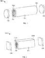
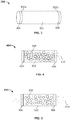
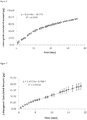
Classifications
-
- B—PERFORMING OPERATIONS; TRANSPORTING
- B65—CONVEYING; PACKING; STORING; HANDLING THIN OR FILAMENTARY MATERIAL
- B65B—MACHINES, APPARATUS OR DEVICES FOR, OR METHODS OF, PACKAGING ARTICLES OR MATERIALS; UNPACKING
- B65B29/00—Packaging of materials presenting special problems
- B65B29/02—Packaging of substances, e.g. tea, which are intended to be infused in the package
-
- A—HUMAN NECESSITIES
- A61—MEDICAL OR VETERINARY SCIENCE; HYGIENE
- A61K—PREPARATIONS FOR MEDICAL, DENTAL OR TOILETRY PURPOSES
- A61K31/00—Medicinal preparations containing organic active ingredients
- A61K31/557—Eicosanoids, e.g. leukotrienes or prostaglandins
- A61K31/5575—Eicosanoids, e.g. leukotrienes or prostaglandins having a cyclopentane, e.g. prostaglandin E2, prostaglandin F2-alpha
-
- A—HUMAN NECESSITIES
- A61—MEDICAL OR VETERINARY SCIENCE; HYGIENE
- A61K—PREPARATIONS FOR MEDICAL, DENTAL OR TOILETRY PURPOSES
- A61K47/00—Medicinal preparations characterised by the non-active ingredients used, e.g. carriers or inert additives; Targeting or modifying agents chemically bound to the active ingredient
- A61K47/02—Inorganic compounds
-
- A—HUMAN NECESSITIES
- A61—MEDICAL OR VETERINARY SCIENCE; HYGIENE
- A61K—PREPARATIONS FOR MEDICAL, DENTAL OR TOILETRY PURPOSES
- A61K9/00—Medicinal preparations characterised by special physical form
- A61K9/0012—Galenical forms characterised by the site of application
- A61K9/0019—Injectable compositions; Intramuscular, intravenous, arterial, subcutaneous administration; Compositions to be administered through the skin in an invasive manner
- A61K9/0024—Solid, semi-solid or solidifying implants, which are implanted or injected in body tissue
-
- A—HUMAN NECESSITIES
- A61—MEDICAL OR VETERINARY SCIENCE; HYGIENE
- A61K—PREPARATIONS FOR MEDICAL, DENTAL OR TOILETRY PURPOSES
- A61K9/00—Medicinal preparations characterised by special physical form
- A61K9/0012—Galenical forms characterised by the site of application
- A61K9/0048—Eye, e.g. artificial tears
- A61K9/0051—Ocular inserts, ocular implants
-
- A—HUMAN NECESSITIES
- A61—MEDICAL OR VETERINARY SCIENCE; HYGIENE
- A61K—PREPARATIONS FOR MEDICAL, DENTAL OR TOILETRY PURPOSES
- A61K9/00—Medicinal preparations characterised by special physical form
- A61K9/0087—Galenical forms not covered by A61K9/02 - A61K9/7023
- A61K9/0092—Hollow drug-filled fibres, tubes of the core-shell type, coated fibres, coated rods, microtubules or nanotubes
-
- A—HUMAN NECESSITIES
- A61—MEDICAL OR VETERINARY SCIENCE; HYGIENE
- A61M—DEVICES FOR INTRODUCING MEDIA INTO, OR ONTO, THE BODY; DEVICES FOR TRANSDUCING BODY MEDIA OR FOR TAKING MEDIA FROM THE BODY; DEVICES FOR PRODUCING OR ENDING SLEEP OR STUPOR
- A61M31/00—Devices for introducing or retaining media, e.g. remedies, in cavities of the body
-
- A—HUMAN NECESSITIES
- A61—MEDICAL OR VETERINARY SCIENCE; HYGIENE
- A61P—SPECIFIC THERAPEUTIC ACTIVITY OF CHEMICAL COMPOUNDS OR MEDICINAL PREPARATIONS
- A61P27/00—Drugs for disorders of the senses
- A61P27/02—Ophthalmic agents
-
- B—PERFORMING OPERATIONS; TRANSPORTING
- B65—CONVEYING; PACKING; STORING; HANDLING THIN OR FILAMENTARY MATERIAL
- B65B—MACHINES, APPARATUS OR DEVICES FOR, OR METHODS OF, PACKAGING ARTICLES OR MATERIALS; UNPACKING
- B65B55/00—Preserving, protecting or purifying packages or package contents in association with packaging
- B65B55/02—Sterilising, e.g. of complete packages
- B65B55/04—Sterilising wrappers or receptacles prior to, or during, packaging
- B65B55/06—Sterilising wrappers or receptacles prior to, or during, packaging by heat
-
- B—PERFORMING OPERATIONS; TRANSPORTING
- B65—CONVEYING; PACKING; STORING; HANDLING THIN OR FILAMENTARY MATERIAL
- B65B—MACHINES, APPARATUS OR DEVICES FOR, OR METHODS OF, PACKAGING ARTICLES OR MATERIALS; UNPACKING
- B65B55/00—Preserving, protecting or purifying packages or package contents in association with packaging
- B65B55/02—Sterilising, e.g. of complete packages
- B65B55/04—Sterilising wrappers or receptacles prior to, or during, packaging
- B65B55/08—Sterilising wrappers or receptacles prior to, or during, packaging by irradiation
-
- B—PERFORMING OPERATIONS; TRANSPORTING
- B65—CONVEYING; PACKING; STORING; HANDLING THIN OR FILAMENTARY MATERIAL
- B65B—MACHINES, APPARATUS OR DEVICES FOR, OR METHODS OF, PACKAGING ARTICLES OR MATERIALS; UNPACKING
- B65B7/00—Closing containers or receptacles after filling
- B65B7/16—Closing semi-rigid or rigid containers or receptacles not deformed by, or not taking-up shape of, contents, e.g. boxes or cartons
- B65B7/28—Closing semi-rigid or rigid containers or receptacles not deformed by, or not taking-up shape of, contents, e.g. boxes or cartons by applying separate preformed closures, e.g. lids, covers
-
- Y—GENERAL TAGGING OF NEW TECHNOLOGICAL DEVELOPMENTS; GENERAL TAGGING OF CROSS-SECTIONAL TECHNOLOGIES SPANNING OVER SEVERAL SECTIONS OF THE IPC; TECHNICAL SUBJECTS COVERED BY FORMER USPC CROSS-REFERENCE ART COLLECTIONS [XRACs] AND DIGESTS
- Y02—TECHNOLOGIES OR APPLICATIONS FOR MITIGATION OR ADAPTATION AGAINST CLIMATE CHANGE
- Y02A—TECHNOLOGIES FOR ADAPTATION TO CLIMATE CHANGE
- Y02A50/00—TECHNOLOGIES FOR ADAPTATION TO CLIMATE CHANGE in human health protection, e.g. against extreme weather
- Y02A50/30—Against vector-borne diseases, e.g. mosquito-borne, fly-borne, tick-borne or waterborne diseases whose impact is exacerbated by climate change
Definitions
- the concentration of drug in a given area of the body increases through an ineffective concentration and eventually reaches an effective concentration. Frequently the concentration may actually reach some toxic threshold. After a relatively short period, however, the drug concentration decreases as drug is either metabolized in the body or is eliminated. Frequently, drug levels decrease so low that therapeutic levels are no longer maintained. A second dose is then given and the cycle is repeated.
- the goal of a sustained release system is to maintain drug levels within the therapeutic range and ideally at a constant level US 2013/053794 describes an ocular implant providing sustained release of latanoprost.
- sustained release systems fall short of one or more of these goals. Certain sustained release systems lose efficacy over time due to degradation, dispersal within the body, or other unwanted biological processes. Some sustained release systems deliver the drug over too short a time scale, necessitating frequent replacement. Still other sustained release systems perform adequately for releasing one drug, but are not readily customizable to deliver a variety of different classes of therapeutics. Therefore, there is a continuing need for the development of improved dosage forms for the controlled release of therapeutic agents.
- the present invention relates to a device for delivering a beneficial substance to an eye of a patient, comprising a shell and a plurality of particles disposed within the shell, the particles comprising a porous silicon-based carrier material and a beneficial substance disposed within pores of the carrier material, and the shell comprising at least one portion that is permeable to the beneficial substance, wherein the beneficial substance is selected from the group consisting of neuroprotectants, antibacterials, antiallergenics, antiinflammatories, decongestants, miotics, mydriatics, and sympathomimetics.
- the present invention relates to a method of making a device, comprising: (a) providing a tube having first and second ends; (b) inserting a plurality of particles into the tube, wherein the particles comprise a porous silicon-based carrier material; (c) adding a first member to the first end of the tube before or after inserting the plurality of particles into the tube; and (d) adding a second member to the second end to the tube before or after inserting the plurality of particles into the tube, wherein at least one of the tube, the first member, and the second member is permeable to the beneficial substance.
- This specification generally provides a device for delivering a beneficial substance to a patient.
- the device comprises a shell enclosing a plurality of carrier particles.
- the carrier particles are porous, and a beneficial substance, such as a drug, is disposed in the pores.
- the drug can diffuse out of the pores, into the interior of the device, and then through at least one permeable portion of the shell.
- This specification also provides methods of making and using these devices.
- the devices described herein have several advantageous properties. For instance, because each device is administered as a single unit, the device makes it easier for a user to administer a fixed dose. Thus, the device may reduce the chance of accidentally administering an incorrect dose and the chance of an end-user measuring a dose inaccurately.
- the device can also advantageously localize administration of the beneficial substance to a desired area in a patient's body, because the shell contains the particles at a desired region of the body.
- the device comprises a shell that advantageously protects the carrier and beneficial substance against unwanted reactions in the body. For instance, the shell may protect the carrier and beneficial substance against enzymatic degradation or the patient's antibodies. The shell can also protect its contents from cells that would adhere to, break down, or otherwise modify the carrier or beneficial substance.
- the device may provide improved protection to the beneficial substance during the manufacturing process.
- the carrier particles and/or shell can stabilize the beneficial substance during the manufacturing process, for instance during steps that involve heat.
- the device can also provide improved protection of the beneficial substance during storage, for instance by stabilizing the beneficial substance from interaction with air or humidity.
- the device can stabilize the beneficial substance over the course of treatment.
- the present disclosure also provides a device comprising a shell and a plurality of particles disposed within the shell, the particles comprising a porous carrier material and a beneficial substance disposed within pores of the carrier, and the shell comprising at least one portion that is permeable to the beneficial substance.
- the disclosure also provides a method of making a device, comprising: (a) providing a tube having first and second ends; (b) inserting a plurality of particles into the tube, wherein the particles comprise porous silicon-based carrier material; (c) adding a first member to the first end of the tube; and (d) adding a second member to the second end to the tube, wherein at least one of the tube, the first member, and the second member is permeable to the beneficial substance.
- the disclosure also provides a device made by this method. Step (b) may be performed before step (c), and also step (c) may be performed before step (b).
- the disclosure provides a method of delivering a beneficial substance to a patient, comprising administering to a patient a device as described herein, whereby the beneficial substance is released from the device into the patient after administration.
- the shell may comprise a polymer.
- the shell may comprise: a tube having first and second ends; a first member positioned at the first end; and a second member positioned at the second end; wherein the particles are inside the tube and the first and second members retain the particles in the tube.
- the tube may have a substantially cylindrical shape.
- the tube may be impermeable to the beneficial substance.
- the tube may comprise poly(lactic-co-glycolic acid) (PLGA).
- the first member and second member may be permeable to the beneficial substance.
- the first member and second member may both comprise poly(vinyl alcohol) (PVA).
- PVA poly(vinyl alcohol)
- the first member may be permeable to the beneficial substance and the second member may be impermeable to the beneficial substance.
- the first member may comprise poly(vinyl alcohol) (PVA).
- the second member may comprise silicone.
- the tube may have a length of between 1 and 4 mm.
- the tube may have a diameter of between 0.2 - 0.5 mm.
- the carrier material may be mesoporous.
- the carrier material may have a porosity in the range of about 30 to about 90%, for instance about 50% to about 80% or about 70% to about 90%.
- the carrier material may be silicon-based.
- the carrier material may comprise elemental silicon.
- the carrier material comprises silica.
- the pores in the carrier material may be substantially parallel.
- the carrier material may comprise anodized silicon.
- the carrier material may be resorbable or bio-erodible.
- the particles may have a largest dimension, on the average, of between about 1 micron and about 20 microns.
- the device may release the beneficial substance at a release rate, the release rate being determined primarily by release of the beneficial substance from pores of the carrier material.
- the beneficial substance may be released from the device according to a substantially zero-order release profile.
- the beneficial substance may be selected from small molecules, proteins, peptides, antibodies, carbohydrates, lipids, polymers, oligonucleotides, and polynucleotides.
- the beneficial substance may be ranibizumab or bevacizumab.
- the device may be produced using a method in which the particles comprise a beneficial substance disposed within pores of the carrier material.
- the method may further comprise contacting the device to a beneficial substance and allowing the beneficial substance to enter pores of the carrier material.
- Adding the first member may comprise contacting the first end of the tube to a polymer solution and curing the polymer solution, thereby forming a first member on the first end of the tube.
- Adding the second member may comprise contacting the second end of the tube to a polymer solution and curing the polymer solution, thereby forming a second member on the second end of the tube.
- the first and second ends may be cured simultaneously.
- the device may release the beneficial substance for between about one month and one year when immersed in simulated body fluid.
- Administering the device may comprise injecting, implanting, or inserting the device into the patient.
- the device comprises a shell enclosing a plurality of carrier particles.
- the carrier particles are porous, and a beneficial substance, such as a drug, is disposed in the pores.
- the beneficial substance can diffuse out of the pores, into the interior of the device, and then through at least one permeable portion of the shell.
- This specification also provides methods of making and using these devices.
- the shell encloses the carrier particles, keeping the carrier localized in one area of the body.
- the shell allows the beneficial substance (such as a drug) to exit the shell and reach target tissues.
- the beneficial substance such as a drug
- the shell can also provide structure to the device.
- the shell may be dimensionally stable and retain its shape in the absence of the particles.
- the shell may comprise a rigid tube that retains its shape even when not filled with the carrier material particles.
- the tube may also retain its shape in the absence of the first end and second end of the shell.
- the device may comprise a single shell.
- the shell may be made from a tube with a member closing each end, so that the members hold the particles inside.
- a device 100 of this type is illustrated in Fig. 1 , in exploded form.
- the tube 102 has a first end 102a and a second end 102b.
- a first member 104 contacts the first end 102a of the tube 102.
- a second member 106 contacts the second end 102b of the tube 102.
- the tube 102, first member 104, and second member 106 enclose a plurality of porous particles 108.
- the pores are suitable for receiving a beneficial substance like a drug.
- first and second members 104 and 106 are shown having about the same thickness, they may also be given different thicknesses.
- the first and second members 104 and 106 have approximately the same diameter as the tube 102.
- the members may have a slightly larger or slightly smaller diameter as long as they are appropriately sized to retain the particles in the tube.
- the first and second members may be formed in situ at the ends of the tube, by inducing cross-linking of a polymeric layer at the ends of the tube.
- Figure 1 illustrates an exploded view of a device 200 made with a tube 202 having a square cross section.
- the tube 202 has a first end 202a and a second end 202b.
- a first member 204 contacts the first end 202a, and a second member 206 contacts the second end 202b.
- the tube 202, first member 204, and second member 206 enclose a plurality of porous particles 108.
- Figure 3 illustrates a device 300 with the first and second members in contact with the tube.
- the tube 302 has a first end 302a in contact with a first member 304.
- the tube 302 has a second end 302b in contact with a second member 306.
- the length of the device (i.e., the distance between the first member and the second member) may be from about 1 - 2 mm, 2 - 4 mm, 4 - 6 mm, 6 - 8 mm, 8 - 10 mm, 1 - 2 cm, 2 - 4 cm, or 4 - 10 cm.
- the width of the device may be from about 0.1 - 0.2 mm, 0.2 - 0.4 mm, 0.4 - 0.6 mm, 0.6 - 0.8 mm, 0.8 - 1.0 mm, 1 - 2 mm, 2 - 4 mm, 4-6 mm, 6 - 8 mm, 8 - 10 mm, 1-2 cm, 2 - 4 cm, or 4 - 10 cm.
- the device may be shaped and sized for injection (e.g., less than about 4 mm long and less than about 0.5 mm in diameter, e.g., to fit through at least one of a needle having a size from about 30 gauge to about 15 gauge or a cannula having a size from about 30 gauge to about 15 gauge, preferably to fit through a less than 22-gauge cannula).
- a needle having a size from about 30 gauge to about 15 gauge or a cannula having a size from about 30 gauge to about 15 gauge, preferably to fit through a less than 22-gauge cannula
- the device may not exceed about 7 mm in any direction, so that the device can be inserted through a less than 7 mm incision.
- the devices may not exceed 7 mm in height or 3 mm in diameter.
- the tube wall is preferably sufficiently thick to allow the tube to retain its shape in the absence of any other material.
- the thickness of the tube walls may range between about 0.01 mm and about 1.0 mm.
- the tube wall may be from about 0.01 - 0.02 mm, 0.02 - 0.04 mm, 0.04 - 0.06 mm, 0.06 - 0.08 mm, 0.08 - 1.0 mm, 1 - 2 mm, 2 - 4 mm, or 4 - 10 mm.
- Exemplary diameters for the tube of the device include 0.011" +/- 0.001" for the inner diameter and 0.0145" +/- 0.001" for the outer diameter.
- Exemplary diameters for the tube of the device also include 0.0061 +/-0.001" for the inner diameter and 0.0145" +/- 0.001" for the outer diameter. Exemplary diameters for the tube of the device also include 0.016" +/- 0.001" for the inner diameter and 0.018" +/- 0.001" for the outer diameter.
- the shell is permeable to the beneficial substance.
- Permeable denotes that the shell allows an effective amount of the beneficial substance to exit the device. Certain parts of the shell may be impermeable to the beneficial substance.
- the term "impermeable”, as used herein, means that the layer will not allow passage of the beneficial substance at a rate required to obtain the desired local or systemic physiological or pharmacological effect, during the period when the device delivers an effective amount of the beneficial substance to the patient.
- the impermeable region may have a permeability for the beneficial substance of less than 10%, 5%, 2%, 1%, 0.5%, 0.2%, 0.1%, 0.05%, 0.02%, or 0.01% of the permeability of the permeable region.
- the tube may be permeable to the beneficial substance.
- the first member may be permeable to the beneficial substance.
- the second member may be permeable to the beneficial substance.
- the tube may be impermeable and one or both of the members are permeable.
- the tube may be made of a permeable material. For instance, the tube, first member, and second member may all be permeable.
- the permeability of a portion of the shell can be affected by its thickness.
- An impermeable member should be thick enough not to release a significant amount of beneficial substance, relative to a permeable region of the device.
- the thickness of an impermeable member can be, for example, between about 0.01 and about 2 mm, preferably between about 0.01 and about 0.5 mm, most preferably between about 0.01 and about 0.2 mm.
- a permeable member should be thick enough to contain the carrier particles in the tube, yet not so thick as to prevent release of an effective amount of the beneficial substance.
- the thickness of the permeable member can be, for example, between about 0.01 and about 2 mm, preferably between about 0.01 and about 0.5 mm, most preferably between about 0.01 and about 0.2 mm.
- At least a portion of the shell may be porous and the beneficial substance can exit the shell through the pores.
- the pores should be small enough that the carrier particles do not pass through the pores in any substantial amount.
- the shell is essentially insoluble in body fluids which the material will come in contact.
- the shell ma ybe substantially non-biodegradable.
- the shell may not substantially biodegrade in a biological environment prior to release of at least 90%, 95%, or 99% of the beneficial substance.
- the shell may substantially biodegrade in a biological environment after release of at least 90%, 95%, or 99% of the beneficial substance.
- the shell may comprise a polymer.
- the tube, first member, and/or second member may be polymeric.
- suitable biocompatible polymers for use in the subject devices include, but are not limited to, poly(vinyl acetate) (PVAC), poly(caprolactone) (PCL), ethylene vinyl acetate polymer (EVA), poly(ethylene glycol) (PEG), poly(vinyl alcohol) (PVA), poly(lactic acid) (PLA), poly(glycolic acid) (PGA), poly(lactic-co-glycolic acid) (PLGA), polyalkyl cyanoacrylate, polyurethane, nylons, or copolymers thereof.
- PVAC poly(vinyl acetate)
- PCL poly(caprolactone)
- EVA ethylene vinyl acetate polymer
- PEG poly(ethylene glycol)
- PVA poly(vinyl alcohol)
- PLA poly(lactic acid)
- PGA poly(glycolic acid)
- PLGA polyalkyl
- the lactic acid may be D-, L- (e.g., poly-L-lactic acid (PLLA)), or any mixture of D- and L- isomers.
- the polymer may be polyimide.
- the polymer may be PLGA that comprises lactic acid (L) and glycolic acid (G) monomers in a ratio of about 95% L and 5% G.
- the percentage of L may range between 80-97%.
- the percentage of G may range between 3-20%.
- the polymer may be heat curable, radiation curable, light (including ultraviolet) curable, evaporation curable, or curable by catalysis.
- the polymer may be silicone, such as a silicone rubber, polydimethylsiloxane, or silicone-carbonate copolymer.
- Certain polymers can be made more or less permeable by altering the degree of polymer cross-linking. Some polymers may be permeable or impermeable depending on the relative characteristics of the polymer and the drug in the drug core. For instance, a given polymer may be permeable to a small molecule but impermeable to an antibody.
- Exemplary polymers suitable for construction of permeable portions of the shell include PVA and PEG.
- Exemplary polymers suitable for construction of impermeable portions of the shell include nylons, polyurethane, EVA, polyalkyl cyanoacrylate, poly(tetrafluoroethylene) (PTFE), polycarbonate (PC), poly(methyl methacrylate) (PMMA), high grades of ethylene vinyl acetate (EVA) (e.g., 9% vinyl, content), poly(lactic-co-glycolic acid) (PLGA), and polyvinyl alcohol (PVA), especially cross-linked PVA.
- EVA ethylene vinyl acetate
- PLGA poly(lactic-co-glycolic acid)
- PVA polyvinyl alcohol
- the shell may comprise metal, such as gold, platinum, and (surgical) stainless steel.
- the tube may be made of metal.
- the metal portion of the shell may be impermeable to the beneficial substance.
- the metal is preferably biocompatible.
- the metal may be biodegradable.
- the biocompatible and/or biodegradable metal alloy may comprise one or more of Fe (iron), Mg (magnesium), Mn (manganese), Pd (palladium), Co (cobalt), Al (aluminum), W (tungsten), B (boron), C (carbon), S (sulfur), Si (silicon), Li (lithium), Zr (zirconium), Ca (calcium), Y (yttrium), Zn (zinc). Exemplary biodegradable metals are described in H.
- the shell may comprise silicon (for instance, elemental silicon) or silica.
- a silicon or silica shell may be biodegradable.
- the materials for the tube, first member, and second member can be chosen to achieve the desired rate of release for the beneficial substance.
- a "low-flow" example of the device is illustrated in cross-section in Figure 4 .
- the device 400 comprises a tube 402 made of an impermeable material and a first member 404 made of an impermeable material.
- the second member 406 is made of a permeable material.
- the particles 408 are contained inside the device 400. The particles 408 initially release the beneficial substance 410 into the interior of the device. The beneficial substance 410 then diffuses through the permeable second member 406 to exit the device. No substantial amount of the beneficial substance exits the device through the tube 402 or first member 404.
- the tube may comprise impermeable poly(dl-lactide-co-glycolide) PLGA
- the first member may be made of an impermeable substance such as silicone
- the second member may comprise permeable poly(vinyl alcohol) (PVA).
- FIG. 5 illustrates a "high-flow" example of the device, in cross-section.
- the device 500 comprises a tube 502 made of an impermeable material.
- the first member 504 and second member 506 are both made of permeable material.
- the particles 508 are contained inside the device 500.
- the particles 508 initially release the beneficial substance 510 into the interior of the device.
- the beneficial substance 510 then diffuses through the permeable first and second members 504 and 506 to exit the device. No substantial amount of the beneficial substance exits the device through the tube 502.
- the tube may comprise impermeable poly(dl-lactide-co-glycolide) PLGA, and the first and second members may comprise permeable poly(vinyl alcohol) (PVA).
- the beneficial substance diffuses in the direction of lower chemical potential, i.e., toward the exterior surface of the device. Release of the beneficial substance from the device is controlled by several factors. It is influenced by the beneficial substance's dissolution rate, rate of release from the pores of the carrier material, and passage through the shell. The device shape, size, and materials can be chosen to achieve a desired release rate of the beneficial substance. Thus, the release rate may be determined primarily by dissolution of the beneficial substance. Also, the release rate may be determined primarily by release of the beneficial substance from pores of the carrier material. Further, the release rate may be determined primarily by the permeability of the shell. The release rate may be significantly affected by any two or all three of these steps.
- the rate of diffusion of the beneficial substance through the device's shell may be determined, for instance, via diffusion cell studies carried out under sink conditions.
- concentration of drug in the receptor compartment is essentially zero when compared to the high concentration in the donor compartment.
- a material may be permeable to a drug and also substantially control the rate at which the drug diffuses or otherwise passes through the material. Consequently, a permeable portion of the shell may also be release-rate-limiting or release-rate-controlling, and the permeability of such a membrane may be one of the most significant factors controlling the release rate for a device.
- the device may release the beneficial substance at a rate that is essentially constant over time (i.e., zero-order kinetics). Zero-order release is desirable when the goal is to maintain a substantially constant amount of the beneficial substance to the patient over a sustained period.
- the carrier material holds and gradually releases a beneficial substance, such as a drug.
- the beneficial substance resides in small pores in the carrier material.
- the carrier material is a plurality of particles enclosed by the device's shell. Each of the carrier particles releases beneficial substance into the interior of the device, and from there the beneficial substance then exits the shell.
- the particles of the device measured at the largest diameter, may have a d10 in the range 1-5 microns (meaning 10% of the particles in the device have a diameter below a number in the range of 1-5 microns), a d50 in the range 5-10 microns (meaning 50% of the particles in the device have a diameter below a number in the range of 5-10 microns), and a d90 in the range of 10-20 microns (meaning 90% of the particle in the device have a diameter below a number in the range of 10-20 microns).
- the d10 may be 1-5 microns, 1-3 microns, or 3-5 microns.
- the d50 may be 5-7 microns, 6-8 microns, 7-9 microns, or 8-10 microns.
- the d90 may be 10-15 microns or 15-20 microns.
- the particles of the device measured at the largest diameter, may have an average size of about 1 to about 500 microns, such as about 5 to 500 microns, about 5 to about 100 microns, or about 5 to about 20 microns.
- greater than 60%, greater than 70%, greater than 80% or greater than 90% of the particles have a particle size of from 1-20 microns, preferably 5-15 microns, measured at the largest dimension.
- the particles may have an average particle size between 1 and 20 microns such as between 5-15 microns or about 15 microns, about 16 microns, about 17 microns, about 18 microns, about 19 microns.
- the carrier may comprise a semiconductor material such as semiconductor silicon.
- additional materials that may be used as porous carrier materials are germanium, ceramics, metal oxides, bone phosphate, phosphates of calcium (e.g., hydroxyapatite), other inorganic phosphates, carbon black, carbonates, sulfates, aluminates, borates, aluminosilicates, magnesium oxide, calcium oxide, iron oxides, zirconium oxides, titanium oxides, and other comparable materials.
- the device may comprise a silicon-based carrier material such as elemental silicon, silicon dioxide (silica), silicon monoxide, silicates (compounds containing a silicon-bearing anion, e.g., SiF 6 2- , Si 2 O 7 6- , or SiO 4 4- ), or any combination of such materials.
- the carrier material may be, for example, semiconducting silicon, elemental silicon, polycrystalline silicon, or amorphous silicon.
- the silicon may be undoped, or may be doped (for example with phosphorus).
- the carrier material may be silicon carbide or silicon nitride.
- the carrier material comprises a complete or partial framework of elemental silicon and that framework is substantially or fully covered by a silicon dioxide surface layer. II is also preferred that the carrier material is entirely or substantially entirely silica.
- the silicon-based carrier material may be synthetic amorphous silica.
- the silicon-based carrier material may be fumed silica.
- the carrier material may comprise silica, such as greater than about 50% silica, greater than about 60 wt% silica, greater than about 70 wt% silica, greater than about 80 wt% silica, greater than about 90 wt% silica, greater than about 95 wt% silica, greater than 99 wt% silica, or even greater than 99.9 wt% silica.
- silica such as greater than about 50% silica, greater than about 60 wt% silica, greater than about 70 wt% silica, greater than about 80 wt% silica, greater than about 90 wt% silica, greater than about 95 wt% silica, greater than 99 wt% silica, or even greater than 99.9 wt% silica.
- Porous silica may be purchased from suppliers such as Grace Davison (and sold under the trademark Davisil), Silicycle, and Macherey-Nagel.
- the carrier material may comprise elemental silicon, greater than 60 wt% silicon, greater than 70 wt% silicon, greater than 80 wt% silicon, greater than 90 wt% silicon, or even greater than 95 wt% silicon. Silicon may be purchased from suppliers such as Vesta Ceramics.
- Purity of the silicon-based material can be quantitatively assessed using techniques such as Energy Dispersive X-ray Analysis, X-ray fluorescence, Inductively Coupled Optical Emission Spectroscopy or Glow Discharge Mass Spectroscopy.
- the based carrier material may a porous, amorphous solid or a porous, crystalline solid.
- silicon-based carrier material may comprise elemental silicon or compounds thereof, e.g., silicon dioxide or silicates, in an amorphous form.
- the elemental silicon or compounds thereof may be present in a crystalline form.
- the carrier material may comprise amorphous silica and/or amorphous silicon.
- the silicon-based material may be greater than about 60 wt% amorphous, greater than about 70 wt% amorphous, greater than about 80 wt% amorphous, greater than about 90 wt% amorphous, greater than about 92 wt% amorphous, greater than about 95 wt% amorphous, greater than about 99 wt% amorphous, or even greater than 99.9 wt% amorphous.
- the amorphous silica may be fumed silica.
- the amorphous silica may be synthetic amorphous silica.
- X-ray diffraction analysis can be used to identify crystalline phases of silicon-based material.
- Powder diffraction can be taken, for example, on a Scintag PAD-X diffractometer, e.g., equipped with a liquid nitrogen cooled germanium solid state detector using Cu K-alpha radiation.
- the carrier material may have a porosity of about 30% to about 95%, about 30% to about 90%, or about 60% to about 80%.
- Porosity is a measure of the void spaces in a material, and is a fraction of the volume of voids over the total volume of the material.
- the carrier material may have a porosity of at least about 10%, at least about 20%, at least about 30%, at least about 40%, at least about 50%, at least about 60%, at least about 70%, at least about 80%, or even at least about 90%.
- the porosity may be greater than about 40%, such as greater than about 50%, greater than about 60%, or even greater than about 70%.
- the carrier material of the devices may have a surface area to weight ratio selected from about 20 m 2 /g to about 2000 m 2 /g, such as from about 20 m 2 /g to about 1000 m 2 /g, or even from about 100 m 2 /g to about 300 m 2 /g.
- the surface area may be greater than about 200 m 2 /g, greater than about 250 m 2 /g or greater than about 300 m 2 /g.
- the surface area may be about 200 m 2 /g.
- the beneficial substance may be distributed to a pore depth from the surface of the material of at least about 10 microns, at least about 20 microns, at least about 30 microns, at least about 40 microns, at least about 50 microns, at least about 60 microns, at least about 70 microns, at least about 80 microns, at least about 90 microns, at least about 100 microns, at least about 110 microns, at least about 120 microns, at least about 130 micron, at least about 140 microns or at least about 150 microns.
- the beneficial substance may be distributed in the pores of the carrier material substantially uniformly.
- the beneficial substance may be loaded into the carrier material to a depth which is measured as a ratio of the depth to which the beneficial substance penetrates the carrier material to the total width of the carrier material.
- the beneficial substance may be distributed to a depth of at least about 10% into the carrier material, to at least about 20% into the carrier material, at least about 30% into the carrier material, at least about 40% into the carrier material, at least about 50% into the carrier material, or at least about 60% into the carrier material.
- Quantification of gross loading may be achieved by a number of analytic methods, for example, gravimetric, EDX (energy-dispersive analysis by x-rays), Fourier transform infra-red (FTIR) or Raman spectroscopy of the pharmaceutical composition or by UV spectrophotometry, titrimetric analysis, HPLC or mass spectroscopy of the eluted therapeutic agent in solution.
- Quantification of the uniformity of loading may be obtained by compositional techniques that are capable of spatial resolution such as cross-sectional EDX, Auger depth profiling, micro-Raman and micro-FTIR.
- the carrier material preferably comprises pores that can receive a beneficial substance.
- Microporous carrier pore size less than 2 nm
- mesoporous carrier pore size 2-50 nm
- macroporous carrier pore size >50 nm
- the average pore size of the carrier material may be selected from 2-50 nm, such as from about 5 to about 40 nm, from about 15 to about 40 nm, such as about 20 to about 30 nm.
- the average pore size may be selected from about 2 to about 15 nm, such as about 5 to about 10 nm.
- the average pore size may be about 30 nm.
- greater than 50% of the pores of the carrier material have a pore size from 2-50 nm
- greater than 60% of the pores of the carrier material have a pore size from 2-50 nm
- greater than 70% of the pores of the carrier material have a pore size from 2-50 nm
- greater than 80% of the pores of the carrier material have a pore size from 2-50 nm
- or even greater than 90% of the pores of the carrier material have a pore size from 2-50 nm.
- the carrier material may comprise porous silicon dioxide, such as mesoporous silicon dioxide or amorphous silica, such as fumed silica.
- the carrier material may have a population of pores with a well-defined pore size, i.e., the distribution of pore sizes for the carrier material falls within a defined range.
- a well-defined population of pores may have about 50% to about 99% of the pore sizes within about 1 nm to 15 nm of the average pore size for that population, preferably within about 10 nm, about 5 nm, or even within 3 nm or 2 nm of the average pore size for that population.
- greater than about 50%, greater than about 60%, greater than about 70%, greater than about 80%, greater than about 90%, or even greater than about 95% of the pores of the carrier material have pore sizes within the specified range.
- a population of pores with a well-defined pore size can be a population in which greater than about 50%, greater than about 60%, greater than about 70%, greater than about 80%, greater than about 90%, or even greater than about 95% of the pores have pore sizes within 20%, preferably within 15%, 10%, or even 5% of the average pore size for that population.
- Pore (e.g., mesopore) size distribution can be quantified using established analytical methods such as gas adsorption, high resolution scanning electron microscopy, nuclear magnetic resonance cryoporosimetry and differential scanning calorimetry.
- a population of pores with a well-defined pore size can be a population for which the standard deviation of the pore sizes is less than 20%, preferably less than 15%, less than 10%, or even less than 5% of the average pore size for that population.
- the pore size may be preselected to the dimensional characteristics of the beneficial substance to control the release rate of the beneficial substance in a biological system. Typically, pore sizes that are too small preclude loading of the beneficial substance, while oversized pores do not interact with the beneficial substance sufficiently strongly to exert the desired control over the rate of release.
- the average pore diameter for a carrier material may be selected from larger pores, e.g., 15 nm to 40 nm, for high molecular weight molecules, e.g., 200,000-500,000 amu, and smaller pores, e.g., 2 nm to 10 nm, for molecules of a lower molecular weight, e.g., 10,000-50,0000 amu.
- average pore sizes of about 6 nm in diameter may be suitable for molecules of molecular weight around 14,000 to 15,000 amu, such as about 14,700 amu.
- Average pore sizes of about 10 nm in diameter may be selected for molecules of molecular weight around 45,000 to 50,000 amu, such as about 48,000 amu.
- Average pore sizes of about 25-30 nm in diameter may be selected for molecules of molecular weight around 150,000 amu.
- the pore size may be preselected to be adapted to the molecular radius of the beneficial substance to control the release rate of the beneficial substance in a biological system. For instance, average pore sizes of about 25 nm to about 40 nm in diameter may be suitable for molecules with a largest molecular radius from about 6 nm to about 8 nm.
- Molecular radii may be calculated by any suitable method such as by using the physical dimensions of the molecule based on the X-ray crystallography data or using the hydrodynamic radius which represents the solution state size of the molecule. As the solution state calculation is dependant upon the nature of the solution in which the calculation is made, it may be preferable for some measurements to use the physical dimensions of the molecule based on X-ray crystallography data. As used herein, the largest molecular radius reflects half of the largest dimension of the therapeutic agent.
- the average pore diameter may be selected to limit the aggregation of molecules, e.g., proteins, within a pore. It would be advantageous to prevent biomolecules, such as proteins, from aggregating in a device as this is believed to impede the controlled release of molecules into a biological system. Therefore, a pore that, due to the relationship between its size and the size of a biomolecule, allows, for example, only one biomolecule to enter the pore at any one time will be preferable to a pore that allows multiple biomolecules to enter the pore together and aggregate within the pore. Multiple biomolecules (e.g., proteins) may be loaded into a pore, but due to the depth of the pore, the proteins distributed throughout this depth of the pore will aggregate to a lesser extent. For instance, a pore may have a diameter slightly larger than the diameter of a protein inside the pore. In this case, the pore's narrow diameter can constrain the arrangement of proteins, decreasing aggregation.
- molecules e.g., proteins
- the carrier material may comprise two or more different materials with different properties (e.g., pore sizes, particle diameters, or surface characteristics), each preselected to be adapted to a different beneficial substance.
- two different carrier materials may be admixed, one with a first population of pores whose pore size is adapted to a first beneficial substance, the other with a second population of pores whose pore size is adapted to a second beneficial substance.
- the device may comprise a first population of carrier particles having a first population of pores whose pore size is adapted to a first beneficial substance, and a second population of carrier particles having a second population of pores whose pore size is adapted to a second beneficial substance.
- a particle may comprise a single material that has two or more well-defined populations of pores, e.g., wherein the carrier material is made by a molecular templating technique, wherein the characteristics of the pores are preselected for two or more beneficial substances, e.g., two beneficial substances with different molecular radii.
- the carrier material may deliver two or more beneficial substances in the controlled manner described herein.
- the loading of the beneficial substances is preferably ordered from largest to smallest agent, so that the largest agent selectively adsorbs into the largest pores (i.e., it does not fit into the smaller pores), so that the larger pores do not adsorb smaller agents.
- the differences between the properties of the different populations of pores are preferably selected to limit the adsorption of each different beneficial substance to a certain population of pores.
- the average pore size of the two or more distinct well-defined pore populations may be selected to limit the adsorption of the larger beneficial substance into smaller pores.
- the average pore size differential may be defined as the difference between the average pore sizes for the different populations of pores in the carrier material.
- an average pore size differential of at least 10 nm could indicate that the carrier material may comprise at least two populations of pores whose average pore sizes differ ("average pore size differential") by at least 10 nm.
- the composition may comprise two pore populations having average pore sizes of 10 nm and 20 nm, three populations of pores with average pore sizes of 10 nm, 20 nm, and 30 nm, or four populations of pores with average pore sizes of 10 nm, 20 nm, 30 nm, and 40 nm.
- the average pore size differential is preferably at least about 5 nm, at least about 10 nm, at least 15 nm, at least about 20 nm, or at least about 30 nm.
- the two or more well-defined pore populations may have distinct average pore sizes, such that the average pore sizes of any two populations differ by at least 20%, preferably at least 30%, 40%, or even 50% of the smaller average pore size.
- the walls of the carrier material that separate the pores may have an average width of less than 5 nm, such as about 4.8 nm, about 4.6 nm, about 4.4 nm, about 4.2 nm, about 4.0 nm, about 3.8 nm, about 3.6 nm, about 3.4 nm, about 3.2 nm, about 3.0 nm, about 2.8 nm, or even about 2.6 nm.
- the walls of the carrier material that separate the pores may have an average width of less than about 3 nm, such as about 2.8 nm, about 2.6 nm, about 2.4 nm, about 2.2 nm, about 2.0 nm, about 1.8 nm, about 1.6 nm, about 1.4 nm, about 1.2 nm, about 1.0 nm, or even about 0.8 nm.
- Dimensionality and morphology of the device can be measured, for example, by Transmission Electron Microscopy (TEM) using a 2000 JEOL electron microscope operating, for example, at 200 keV.
- Samples for TEM can be prepared by dispensing a large number of porous carrier materials onto a holey carbon film on a metal grid, via a dilute slurry.
- the pores of the carrier material may define space having a volume of about 0.1 mL/g to about 5 mL/g of the carrier material.
- the pore volume may be about 0.2 mL/g to about 3 mL/g, such as about 0.4 mL/g to about 2.5 mL/g, such as about 1.0 mL/g to about 2.5 mL/g.
- the load level of the carrier material may be up to 70%, such as up to 40% by weight based on the combined weight of the carrier material and the beneficial substance.
- the load level is calculated by dividing the weight of the loaded beneficial substance by the combined weight of the loaded therapeutic agent and carrier material and multiplying by 100.
- the load level of the carrier material may be greater than 10%, such as greater than 15%, greater than 20%, greater than 25%, greater than 30%, greater than 35%, greater than 40%, greater than 45% or greater than 50%.
- the load level of the carrier material may less than 5%.
- the load level may be between about 5% and about 10%.
- the load level of the carrier material may be between about 10% and about 20%, between about 20% and about 30%, between about 30% and about 40%, between about 40% and about 50%, or between about 50% and about 60% by weight.
- the load volume of the devices described herein may be evaluated in terms of the volume of the pores in the porous material being occupied by the beneficial substance.
- the percentage of the maximum loading capacity that is occupied by the beneficial substance (that is, the percentage of the total volume of the pores in the porous carrier material that is occupied by the beneficial substance) for carrier materials may be from about 30% to about 100%, such as from about 50% to about 90%. For any given carrier material, this value may be determined by dividing the volume of the beneficial substance taken up during loading by the void volume of the carrier material prior to loading and multiplying by one hundred.
- the carrier particles measured at the largest diameter, may have an average size of about 1 to about 500 microns, such as about 5 to about 100 microns. At least 80%, 90%, 99%, or even 100% of the particles in the device, measured at the largest diameter, may be about 1 to about 500 microns, such as about 5 to about 500 microns, or about 2 to about 100 microns.
- the beneficial substance In order to increase the rate of loading of the beneficial substance into the particles, it may be advantageous to use relatively small particles. As smaller particles have pores with less depth for the beneficial substance to penetrate, the amount of time needed to load the particles may be reduced. This may be particularly advantageous when the pore diameters are similar in dimensions to the molecular diameters or size of the therapeutic agents. Smaller particles may be from 1-20 microns, such as about 10-20 microns, e.g., about 15-20 microns, measured at the largest dimension.
- greater than 60%, greater than 70%, greater than 80% or greater than 90% of the particles have a particle size of from 1-20 microns, preferably 5-15 microns, measured at the largest dimension.
- the particles may have an average particle size between 1 and 20 microns such as between 5-15 microns or about 15 microns, about 16 microns, about 17 microns, about 18 microns, about 19 microns.
- Particle size distribution including the mean particle diameter can be measured, for example, using a Malvern Particle Size Analyzer, Model Mastersizer, from Malvern Instruments, UK.
- a helium-neon gas laser beam may be projected through an optical cell containing a suspension of the carrier material. Light rays striking the carrier material are scattered through angles which are inversely proportional to the particle size.
- the photodetector array measures the light intensity at several predetermined angles and electrical signals proportional to the measured light flux values are then processed by a microcomputer system against a scatter pattern predicted from the refractive indices of the sample carrier material and aqueous dispersant.
- the device may also comprise one or more pharmaceutically acceptable excipients.
- the excipient may be a filler, binder, diluent, buffering agent, moistening agent, preservative, stabilizer, flavoring agent, dye, coloring agent, disintegrating agent, or surfactant.
- a buffering agent may be used to tailor the drug release rate by creating a micro-environment pH in the device. The pH can affect the dissolution rate of the beneficial substance or the permeability of the shell for the beneficial substance, thereby affecting the overall release rate.
- a surfactant may be used to adjust the charge, lipophilicity or hydrophilicity of the carrier, such as to enhance wettability of poorly soluble or hydrophobic compositions.
- materials which can serve as pharmaceutically acceptable excipients include: (1) sugars, such as lactose, glucose and sucrose; (2) starches, such as corn starch and potato starch; (3) cellulose, and its derivatives, such as sodium carboxymethyl cellulose, ethyl cellulose and cellulose acetate; (4) powdered tragacanth; (5) malt; (6) gelatin; (7) talc; (8) hydrophobic materials such as cocoa butter, suppository waxes, and the like; (9) oils, such as peanut oil, cottonseed oil, safflower oil, sesame oil, olive oil, corn oil and soybean oil; (10) glycols, such as propylene glycol; (11) polyols, such as glycerin, sorbitol, mannitol and polyethylene glycol; (12) esters, such as ethyl oleate and ethyl laurate; (13) agar; (14) buffering agents, such as magnesium hydroxide, such
- the excipient may be disposed within pores of the carrier material. Also, the excipient may be outside the particles and inside the device's shell. For instance, the particles may be suspended in solution and/or form a slurry, and the excipient may be in the solution.
- the device can store and deliver a therapeutically effective amount of a beneficial substance.
- the beneficial substance is a therapeutic.
- the term "therapeutic” encompasses the active molecule as well as salts of the active molecule.
- the therapeutic may be, for example, a drug or a prodrug.
- the beneficial substance may be selected from any agent useful in the treatment or prevention of diseases.
- the beneficial substance may be selected from small molecule therapeutic agents, i.e., compounds with molecular weights less than 1000 amu.
- the beneficial substance may be selected from large molecules with molecular weight equal to or greater than 1000 amu.
- the beneficial substance of the invention may be a biomolecule.
- Biomolecules as used herein, refer to any molecule that is produced by a living organism, including large polymeric molecules such as proteins, polysaccharides, and nucleic acids as well as small molecules such as primary metabolites, secondary metabolites, and natural products or synthetic variations thereof.
- proteins such as antibodies, ligands, and enzymes may be used as beneficial substances in the devices described herein.
- the biomolecules for use in the device may have molecular weights ranging from about 10,000 amu to about 500,000 amu.
- the beneficial substance may be a protein such as an antibody.
- the antibody may be a monoclonal antibody.
- the antibody may be, for instance, an antigen-binding portion of a full-length antibody, e.g., a Fab fragment or a single chain variable fragment.
- Particular therapeutic antibodies that may be delivered by the device include ranibizumab and bevacizumab.
- Polynucleotides that may be administered using the devices herein include DNA, RNA, and analogs of DNA and RNA.
- the polynucleotides may include 2'O-Me nucleotides or dideoxynucleotides.
- the polynucleotides may encode proteins for gene therapy, or may be designed to reduce expression of a target gene via an antisense pathway.
- the beneficial substance may have a molecular weight between 10,000 and 50,000 amu, between 50,000 and 100,000 amu or between 100,000 and 150,000 amu.
- the beneficial substance may be a protein with a molecular weight between 5,000 amu and 200,000 amu, such as about 10,000 to about 150,000 amu.
- the size of a beneficial substance may alternatively be characterized by the molecular radius, which may be determined, for example, through X-ray crystallographic analysis or by hydrodynamic radius.
- the beneficial substance may be a protein, e.g., with a molecular radius selected from 0.5 nm to 20 nm such as about 0.5 nm to 10 nm, even from about 1 to 8 nm.
- the pore size of the carrier material can be selected based at least in part on the size of the beneficial substance.
- a beneficial substance with molecular radius from 1 to 2.5 nm may be advantageously used with a carrier material with a minimum pore radius of from 4.5 to 5.8 nm.
- a beneficial substance with a molecular radius of 7 nm may be advantageously used with a carrier material with a minimum pore radius of from 11 to 13 nm, such as about 12 nm. Additional discussion of selecting an appropriate size of pore for a given beneficial substance may be found, for example, in International Application WO 2012/061377 .
- the carrier particles may be loaded with a liquid drug.
- carrier particles loaded with a liquid drug can be easier to handle than the liquid drug itself.
- the devices herein can ease the manufacturing process for controlled release devices that deliver liquid drugs.
- the liquid drug may comprise a carboxylic acid moiety.
- the liquid beneficial substance in the device may be prostaglandin or a prostaglandin analog.
- the liquid drug may be the free acid form of latanoprost.
- the liquid drug may be a free acid form of travoprost.
- the liquid drug may be prostacyclin or a prostacyclin analog such as treprostinil, iloprost, or beraprost.
- the liquid may comprise a fat-soluble vitamin such as Vitamin E.
- the beneficial substance may be prone to decomposition and/or inactivation, and the carrier material may reduce decomposition/inactivation of the beneficial substance.
- the beneficial substance can be inactivated, for example, by degradation or unfolding/denaturation.
- the beneficial substance might be prone to inactivation during the process of curing the first or second member (e.g., with heat or ultraviolet light) and the carrier material reduces this inactivation.
- the carrier material may block the agent that inactivates the beneficial substance (e.g., by absorbing the wavelength of light used for curing).
- the carrier may stabilize a beneficial substance against the effects of the agent that inactivates the beneficial substance, e.g., by stabilizing the beneficial substance in an active conformation and restricting protein unfolding or degradation.
- the beneficial substance may experience at least twice, five times, ten times, 20 times, 50, times or 100 times as much inactivation (e.g., degradation or unfolding) without the carrier material as the beneficial substance within the carrier material under the same conditions during production of the device.
- the beneficial substance within the carrier material may have a half-life at room temperature that is at least twice, five times, ten times, 20 times, 50, times or 100 times the half-life of the beneficial substance without the carrier material under the same conditions.
- the beneficial substance within the carrier material may have a shelf-life at room temperature that is at least twice, five times, ten times, 20 times, 50, times or 100 times the shelf life of the beneficial substance without the carrier material under the same conditions.
- the beneficial substance within the carrier material may be stable at 25 °C for at least 15 days, 1 month, 2 months, 3 months, 6 months, at least 1 year, at least 1.5 years, at least 2 years, at least 2.5 years, at least 3 years or at least 4 years.
- Stability may be assessed, for example, by high performance size exclusion chromatography (HPSEC) or by comparing the biological activity of the stored biomolecule-loaded devices against a sample of freshly prepared biomolecule-loaded devices or against the activity of the devices as measured prior to storage.
- HPSEC high performance size exclusion chromatography
- the activity of the stored devices is at least 75%, at least 80%, at least 85%, at least 90%, at least 95%, at least 98%, at least 99%, at least 99.5%, at least 99.8%, or even at least 99.9% of the activity of the corresponding freshly prepared devices.
- biomolecule-loaded devices are stored at 25 °C for at least 6 months, at least 1 year, at least 1.5 years, at least 2 years, at least 2.5 years, at least 3 years or at least 4 years prior to administering the devices to a patient.
- the degradation-sensitive beneficial substance may be a biomolecule, such as a protein, including an antibody.
- the device may comprises two or more beneficial substances.
- the device may comprise two populations of particles, each population loaded with one beneficial substance.
- a single particle may comprise two or more beneficial substances.
- the single particle has a population of larger pores and a population of smaller pores, and each population of pores contains one of the beneficial substances. Carriers with two populations of pores are described above in Section B.
- suitable drugs include steroids, alpha receptor agonists, beta receptor antagonists, carbonic anhydrase inhibitors, adrenergic agents, physiologically active peptides and/or proteins, antineoplastic agents, antibiotics, analgesics, anti-inflammatory agents, muscle relaxants, antiepileptics, anti-ulcerative agents, anti-allergic agents, cardiotonics, anti-arrhythmic agents, vasodilators, antihypertensive agents, anti-diabetic agents, anti-hyperlipidemics, anticoagulants, hemolytic agents, antituberculous agents, hormones, narcotic antagonists, osteoclastic suppressants, osteogenic promoters, angiogenesis suppressors, antibacterials, non-steroidal anti-inflammatory drugs (NSAIDs), glucocorticoids or other anti-inflammatory corticosteroids, alpha receptor agonists, beta receptor antagonists, carbonic anhydrase inhibitors, adrenergic agents, physiologically active peptides and/
- Suitable NSAIDs for use in the devices described herein include diclofenac, etoldolac, fenoprofen, floctafenine, flurbiprofen, ibuprofen, indoprofen, ketoprofen, ketorolac, lomoxicam, morazone, naproxen, perisoxal, pirprofen, pranoprofen, suprofen, suxibuzone, tropesin, ximoprofen, zaltoprofen, zileuton, and zomepirac, and analogs, derivatives, pharmaceutically acceptable salts, esters, prodrugs, and protected forms thereof.
- Suitable carbonic anhydrase inhibitors for use in the devices described herein include brinzolamide, acetazolamide, methazolamide, dichlorphenamide, ethoxzolamide, and dorzolamide, and analogs, derivatives, pharmaceutically acceptable salts, esters, prodrugs, and protected forms thereof.
- Suitable adrenergic agents for use in the devices described herein include brimonidine, apraclonidine, bunazosin, levobetaxolol, levobunalol, carteolol, isoprenaline, fenoterol, metipranolol, and clenbuterol, and analogs, derivatives, pharmaceutically acceptable salts, esters, prodrugs, and protected forms thereof.
- Suitable alpha receptor agonists for use in the devices described herein include brimonidine and analogs, derivatives, pharmaceutically acceptable salts, esters, prodrugs, and protected forms thereof.
- Suitable beta receptor antagonists for use in the devices described herein include atenolol, betaxolol, and timolol, and analogs, derivatives, pharmaceutically acceptable salts, esters, prodrugs, and protected forms thereof.
- Suitable antiviral agents for use in the devices described herein include nevirapine and analogs, derivatives, pharmaceutically acceptable salts, esters, prodrugs, and protected forms thereof.
- Suitable alkaloid analgesics for use in the devices described herein include desmorphine, dezocine, dihydromorphine, eptazocine, ethylmorphine, glafenine, hydromorphone, isoladol, ketobenidone, p-lactophetide, levorphanol, moptazinol, metazocin, metopon, morphine, nalbuphine, nalmefene, nalorphine, naloxone, norlevorphanol, normorphine, oxmorphone, pentazocine, phenperidine, phenylramidol, tramadol, and viminol, and analogs, derivatives, pharmaceutically acceptable salts, esters, prodrugs, and protected forms thereof.
- Suitable glucocorticoids for use in the devices described herein include 21-acetoxypregnenolone, alclometasone, algestone, anacortave acetate, amcinonide, beclomethasone, betamethasone, budesonide, chloroprednisone, clobetasol, clobetasone, clocortolone, cloprednol, corticosterone, cortisone, cortivazol, deflazacort, desonide, desoximetasone, diflorasone, diflucortolone, difuprednate, enoxolone, fluazacort, flucloronide, flumethasone, flunisolide, fluocinolone acetonide, fluocinonide, flucloronide, flumethasone, flunisolide, fluocortin but
- Suitable BPH drugs for use in the devices described herein include finasteride and osaterone, and analogs, derivatives, pharmaceutically acceptable salts, esters, prodrugs, and protected forms thereof.
- Suitable antineoplastic compounds for use in the devices described herein include adriamycin, alitretinoin (9-cis-retinoic acid); bleomycins, including bleomycin A; capecitabine (5'-deoxy-5-fluoro-cytidine); carubicin; chlorozotocin, chromomycins, including chromomycin A3, cladribine; colchicine, cytarabine; daunorubicin; demecolcine, denopterin, docetaxel, doxyifluridine, doxorubicin; dromostanolone, edatrexate, enocitabine, epirubicin, epitiostanol, estramustine; etoposide; floxuridine, fludarabine, 5-fluorouracil, formestane, gemcitabine; irinotecan; lent
- Suitable antibacterial compounds for use in the devices described herein include capreomycins, including capreomycin IA, capreomycin IB, capreomycin IIA and capreomycin IIB; carbomycins, including carbomycin A; carumonam; cefaclor, cefadroxil, cefamandole, cefatrizine, cefazedone, cefazolin, cefbuperazone, cefcapene pivoxil, cefclidin, cefdinir, cefditoren, cefime, ceftamet, cefmenoxime, cefmetzole, cefminox, cefodizime, cefonicid, cefoperazone, ceforanide, cefotaxime, cefotetan, cefotiam, cefoxitin, cefpimizole, cefpiramide, cefpirome, cef
- Suitable anti-fungal compounds for use in the devices described herein include fluconazole and analogs, derivatives, pharmaceutically acceptable salts, esters, prodrugs, and protected forms thereof.
- Suitable immunological response modifiers for use in the devices described herein include muramyl dipeptide and analogs, derivatives, pharmaceutically acceptable salts, esters, prodrugs, and protected forms thereof.
- Suitable peptides and proteins for use in the devices described herein include insulin, growth hormones, insulin related growth factor, heat shock proteins, and analogs, derivatives, pharmaceutically acceptable salts, esters, prodrugs, and protected forms thereof.
- Suitable anesthetics and pain killing agents for use in the devices described herein include lidocaine, benzodiazepam, and analogs, derivatives, pharmaceutically acceptable salts, esters, prodrugs, and protected forms thereof.
- Suitable cell transport/mobility impending agents for use in the devices described herein (e.g., devices comprising a shell and porous silicon-based carrier particles) include cytochalasin B and analogs, derivatives, pharmaceutically acceptable salts, esters, prodrugs, and protected forms thereof.
- Antiproliferative/antimitotic drugs and prodrugs suitable for use in the devices described herein include natural products such as vinca alkaloids (e.g., vinblastine, vincristine, and vinorelbine), paclitaxel, epidipodophyllotoxins (e.g., etoposide, teniposide), antibiotics (e.g., actinomycins, daunorubicin, doxorubicin and idarubicin), anthracyclines, mitoxantrone, bleomycins, plicamycin (mithramycin) and mitomycin, enzymes (e.g., L-asparaginase); antiplatelet prodrugs; antiproliferative/antimitotic alkylating prodrugs such as nitrogen mustards (mechlorethamine, cyclophosphamide and analogs, melphalan, chlorambucil
- vinca alkaloids e.g., vinblastine, vincris
- Suitable antiviral agents for use in the devices described herein include acyclovir, azidouridine, anismoycin, amantadine, bromovinyldeoxusidine, chlorovinyldeoxusidine, cytarabine, delavirdine, didanosine, deoxynojirimycin, dideoxycytidine, dideoxyinosine, dideoxynucleoside, desciclovir, deoxyacyclovir, efavirenz, enviroxime, fiacitabine, foscamet, fialuridine, fluorothymidine, floxuridine, ganciclovir, hypericin, idoxuridine, interferon, interleukin, isethionate, nevirapine, pentamidine, ribavirin, rimantadine, stavudine, sargramostin, sur
- Suitable antiviral agents for use in the devices described herein include 2',3'-dideoxyadenosine (ddA), 2',3'-dideoxyguanosine (ddG), 2',3'-dideoxycytidine (ddC), 2',3'-dideoxythymidine (ddT), 2'3'-dideoxy-dideoxythymidine (d4T), 2'-deoxy-3'-thia-cytosine (3TC or lamivudime), 2',3'-dideoxy-2'-fluoroadenosine, 2',3'-dideoxy-2'-fluoroinosine, 2',3'-dideoxy-2'-fluorothymidine, 2',3'-dideoxy-2'-fluorocytosine, 2'3'-dideoxy-2',3'-didehydro-2'-fluoro
- ddA 2',3'-dideoxyadenosine
- the antiviral agent may be selected from trisodium phosphomonoformate, ganciclovir, trifluorothymidine, acyclovir, 3'-azido-3'-thymidine (AZT), dideoxyinosine (ddI), and idoxuridine and analogs, derivatives, pharmaceutically acceptable salts, esters, prodrugs, and protected forms thereof.
- Beneficial substances suitable for administration to the eye and its surrounding tissues using the devices described herein (e.g., devices comprising a shell and porous silicon-based carrier particles), to produce a local or a systemic physiologic or pharmacologic beneficial effect include neuroprotectants such as nimodipine and related compounds; antibiotics such as tetracycline, chlortetracycline, bacitracin, neomycin, polymyxin, gramicidin, oxytetracycline, chloramphenicol, gentamycin, and erythromycin; antibacterials such as sulfonamides, sulfacetamide, sulfamethizole, and sulfisoxazole; antivirals, including idoxuridine; and other antibacterial agents such as nitrofurazone and sodium propionate; antiallergenics such as antazoline, methapyriline, chlorpheniramine, pyrilamine, and prophenpyridamine; antiinflammatories such
- Prodrugs are generally compounds that, under physiological conditions, are converted into therapeutically active agents in a patient's body.
- a common method for making a prodrug is to include selected moieties, such as esters, that are hydrolyzed under physiological conditions to convert the prodrug to an active biological moiety.
- the prodrug may also be converted by an enzymatic activity of the host animal.
- Prodrugs are typically formed by chemical modification of a biologically active moiety. Conventional procedures for the selection and preparation of suitable prodrug derivatives are described, for example, in Design of Prodrugs, ed. H. Bundgaard, Elsevier, 1985 .
- any pharmaceutically acceptable form of such a compound may be employed as a beneficial substance, i.e., the free base or a pharmaceutically acceptable salt or ester thereof.
- Pharmaceutically acceptable salts for instance, include sulfate, lactate, acetate, stearate, hydrochloride, tartrate, maleate, and the like.
- the devices described herein may be made in a wide variety of ways, portions of which are described above in Section 1.
- the device is prepared by filling a tube with carrier particles and enclosing the ends of the tube.
- Carrier particles are prepared or provided.
- the carrier particles may be made porous by introducing pores into non-porous material. This may be done, for instance, by anodizing a solid such as silicon to produce parallel pores. Anodization may be performed together with etching, e.g., wet etching or dry etching. Thus, the pores may be formed by electrochemical etching.
- the carrier particles may be porous when they are first formed.
- sol-gel synthesis a solution is induced to form a porous network or gel of polymers.
- Sol-gel synthesis can be used, for example, to make porous silica.
- Porous silica can be chemically reduced to silicon by treatment with, for instance, magnesium vapor.
- the magnesium vapor method and related methods for reducing silica are described in International Application WO/2012/114126 .
- Part or all of a large body of carrier material may be made porous, and the porous region may then be milled into small particles.
- the particles may be formed of a non-porous material, and then pores may be introduced into the particles.
- a mesh may be used to select particles of the desired size.
- the porous silicon-based carrier material may be prepared by flame hydrolysis of silicon tetrachloride in an oxy-hydrogen flame.
- Carrier particles may be loaded with a beneficial substance before or after the particles are assembled into the device.
- a tube may be produced or provided.
- the tube may be extruded, e.g., from a polymeric mass.
- Commercially available extruders include the Randcastle model RCP-0250 Microtruder (Randcastle Extrusion Systems, Cedar Grove, N.J.), and its associated heaters, controllers, and the like. Exemplary extruders are also disclosed, for example, in U.S. Pat. Nos. 5,569,429 , 5,518,672 , and 5,486,328 .
- An extruder can include an exit port that establishes a cross-sectional shape of the extruded matter.
- the tube may be cured after extrusion.
- the tube may be coextruded with the carrier particle composition inside the tube.
- the tube may be formed in the absence of carrier particles, and carrier particles are inserted into the formed tube.
- the tube may be segmented into a plurality of tubular segments. Segmentation may employ shears, slicing blades,
- the tube is filled with carrier particles.
- the tube may be several multiples of the length of the finished device, and the tube is cut to the proper length before or after being filled with carrier particles.
- the particles may be dry when they are placed in the tube, and may have the consistency of a powder or granulate.
- the particles may be in solution when they are placed in the tube, and may have the consistency of a slurry.
- the slurry may be drawn into the tube with capillary action or pushed in using a syringe, for example.
- the carrier particles are loaded with the beneficial substance when they are placed in the tube.
- the particles may also be loaded with drug after this step, e.g., when they are in the tube but before members are added to close the tube, or even after one or both members are added.
- the first member and second member are added to the first and second ends of the tube to enclose the particles.
- One or both of the members may be formed in situ on the end or ends of the tube.
- An amount of polymer solution may be applied to the end of the tube. For instance, one end of the tube can be dipped in a polymer solution one or more times, so that a film of the polymer will form on the end of the tube.
- the polymer solution may be applied to the end of the tube by dropping, spraying, brushing or other means. Once the polymer is in contact with the tube, the polymer can be cured (hardened by cross-linking) on the end of the tube.
- Polymers can be cured, e.g., using heat, radiation, light (including ultraviolet light or visible light such as blue light), evaporation, and catalyzation. Curing with light in the visible or near-visible ranges (e.g., of ultraviolet or blue wavelengths) sometimes avoids inactivation of the beneficial substance that can result from harsher curing techniques. Curing may be performed using an intense light source, such as a tuned laser or the like.
- Each polymer can be cured using one or more suitable curing techniques, and numerous examples are known in the art. For instance, PVA may be cured with (for instance) ultraviolet light, infrared light, and/or by heating it in an oven.
- the desired thickness of the first or second member may be obtained by applying more than one coat of a polymer. Each coat may be dried and/or cured prior to applying the next coat.
- the device shell may comprise heat-cured PVA.
- a heat-cured first member and/or second member may be formed by applying a PVA solution to the first end of the tube and then heating the PVA.
- the PVA may be heated, e.g., at a temperature in the range of 60-120°C, e.g., 80°C, for at least 2 hours, preferably at least 4 hours, e.g., 5 hours. Heating may be performed, for example, in an oven or other heating element.
- the device and/or the components of the device may be sterilized.
- the heat-resistant portions of the device may be heat-sterilized, e.g., by autoclaving.
- Other methods of sterilizing the device or its components include sterilization with radiation, ultraviolet light, immersion in alcohol, or filtration.
- a heat-sensitive component (such as a beneficial substance, including a biomolecule) may be filter-sterilized. All heat sterilization of the device or its components may take place prior to addition of the heat-sensitive component to the device.
- the particles and/or the shell may be heat-sterilized before the beneficial substance is added to the device.
- the device may be packaged, such as by preloading a needle of appropriate gauge with the device and enclosing the assembly in a suitable package for shipment to an end user.
- the present disclosure also provides methods for treating a patient to obtain a desired local or systemic physiological or pharmacological effect. These methods include administering the disclosed devices to the patient and allowing the beneficial substance to pass through the device to come in direct contact with the patient.
- the device can be administered for a sufficient period of time and under conditions to allow treatment of the disease state of concern.
- the patient may be a mammalian organism, and preferably the patient is a human.
- the device may be inserted into a desired location in the patient's body.
- the device may be injected or surgically implanted into the patient's body.
- the beneficial substance acts on the eye
- the device can gradually release the beneficial substance to the eye, avoiding painful repeated administrations of a different formulation of the beneficial substance.
- the device can be surgically implanted into the eye of the patient, for example the vitreous of the eye, under the retina, and onto the sclera.
- the device can also be inserted into numerous other locations in the body, including administration that is subcutaneous, intramuscular, intraperitoneal, intranasal, dermal, into the brain, including intracranial and intradural, into the joints, including ankles, knees, hips, shoulders, elbows, wrists, directly into tumors, and the like.
- administration that is subcutaneous, intramuscular, intraperitoneal, intranasal, dermal, into the brain, including intracranial and intradural, into the joints, including ankles, knees, hips, shoulders, elbows, wrists, directly into tumors, and the like.
- the device can also be administered orally.
- the device may be administered to a patient by injection.
- Injection may use, for example, a standard gauge hypodermic needle, such as about a 30 gauge to about a 12 gauge needle, or a needle ranging in inside diameter from about 0.0055 inches to about 0.0850 inches.
- An injectable device can also be administered through, for example, an arthroscope, catheter, or other medical device.
- the devices may be surgically implanted at or near the site of action. This is the case for devices as described herein, used in treating ocular conditions, primary tumors, rheumatic and arthritic conditions, and chronic pain, for instance.
- the devices may be implanted subcutaneously, intramuscularly, intraarterially, intrathecally, or intraperitoneally, for example. This is the case when devices are to give sustained systemic levels and avoid premature metabolism.
- the devices may be administered orally.
- the devices described herein may be particularly suitable for treating ocular conditions such as glaucoma, proliferative vitreoretinopathy, macular edema, including diabetic macular edema, age-related macular degeneration, diabetic retinopathy, uveitis, ocular neovascularization, and ocular infection.
- the devices may also be particularly suitable for use as an ocular device in treating patients, both human and for veterinarian use, suffering from ocular histoplasmosis, wherein the device may be surgically implanted within the vitreous of the eye.
- the device may contain one or more drugs that reduce the risk of mother to child transmission of viral infections.
- viral infections include HIV, Bowenoid Papulosis, Chickenpox, Childhood HIV Disease, Human Cowpox, Hepatitis C, Dengue, Enteroviral, Epidermodysplasia Verruciformis, Erythema Infectiosum (Fifth Disease), Giant Condylomata Acuminata of Buschke and Lowenstein, Hand-Foot-and-Mouth Disease, Herpes Simplex, Herpes Virus 6, Herpes Zoster, Kaposi Varicelliform Eruption, Rubeola Measles, Milker's Nodules, Molluscum Contagiosum, Monkeypox, Orf, Roseola Infantum, Rubella, Smallpox, Viral Hemorrhagic Fevers, Genital Warts, and Nongenital Warts.
- the device may contain an antiviral agent that inhibits or reduces HIV infection or susceptibility to HIV infection.
- the device may be used to treat mammalian organisms infected with HIV and AIDS-related opportunistic infections such as cytomegalovirus infections, toxoplasmosis, pneumocystis carinii, and mycobacterium avium intercellular.
- the device may contain one or more drugs that treat pulmonary arterial hypertension.
- the device may be used to provide a controlled and sustained release of agents effective in obtaining a desired local or systemic physiological or pharmacological effect relating at least to the following areas: treatment of cancerous primary tumors, (e.g., glioblastoma); inhibition of neovascularization, including ocular neovascularization; edema, including ocular edema; inflammation, including ocular inflammation; chronic pain; arthritis; rheumatic conditions; hormonal deficiencies such as diabetes and dwarfism; and modification of the immune response such as in the prevention of transplant rejection and in cancer therapy.
- cancerous primary tumors e.g., glioblastoma
- inhibition of neovascularization including ocular neovascularization
- edema including ocular edema
- inflammation including ocular inflammation
- chronic pain e.g., arthritis
- arthritis rheumatic conditions
- hormonal deficiencies such as diabetes and dwarfism
- modification of the immune response such as in the prevention of transplant
- the device may deliver ganciclovir to the eye for the treatment of cytomegalovirus (CMV) retinitis.
- CMV cytomegalovirus
- the device e.g., a device comprising a shell enclosing porous silicon-based carrier particles
- fluocinolone acetonide may be delivered to the eye for the treatment of ocular neovascularization, macular edema, wet or dry age-related macular degeneration, retinal vein occlusion, or posterior uveitis.
- the device may deliver bevacizumab or ranibizumab for the treatment of neovascularization, including ocular neovascularization, for example when caused by cancer, macular degeneration (especially wet age-related macular degeneration), diabetic retinopathy, neovascular glaucoma, macular edema, or retinopathy.
- the device e.g., a device comprising a shell enclosing porous silicon-based carrier particles
- the devices herein may release the beneficial substance over a sustained time period when immersed in simulated body fluid or when administered to a patient.
- the device may release an effective amount of the beneficial substance for between 1 week - 1 year, 2 weeks - 1 year, 1 month - 1 year, 2 months - 1 year, 3 months - 1 year, or 6 months - 1 year.
- the device may release the effective amount of the beneficial substance for 1 month - 2 yeas, 2 months - 2 years, 3 months - 2 years, or 6 months - 2 years.
- Example 1 Method of making silicon particles
- Mesoporous silicon flakes of approximately 5 mm 2 were pooled from membranes fabricated by anodization of 5-20 mohm cm resistivity silicon wafers.
- the electrolyte composition was a fixed blend (1:1 by volume) of 40% hydrofluoric acid and methanol. Flakes were extensively washed in an ethanol-water mixture, dried, and then subjected to a sequence of rotor milling and jet milling to produce a powder of defined size distribution, prior to thermal oxidation in air at 800 °C for 3 hours.
- the final target size distribution (d10 in the range of 1-5 microns, d50 in the range of 5-10 microns, and d90 in the range of 10-20 micron) was then achieved by sieving at small batch size or a sedimentation technique at larger batch size.
Description
- There has been considerable interest within the pharmaceutical industry in the development of dosage forms that provide controlled release of therapeutic agents over a period of time. Releasing an active substance in this way can help to improve bioavailability and ensure that appropriate concentrations of the agent are provided for a sustained period without the need for repeated dosing. In turn, this also helps to minimize the effects of patient non-compliance which is frequently an issue with other forms of administration.
- With conventional dosing (tablets, injections, etc.), the concentration of drug in a given area of the body increases through an ineffective concentration and eventually reaches an effective concentration. Frequently the concentration may actually reach some toxic threshold. After a relatively short period, however, the drug concentration decreases as drug is either metabolized in the body or is eliminated. Frequently, drug levels decrease so low that therapeutic levels are no longer maintained. A second dose is then given and the cycle is repeated. The goal of a sustained release system is to maintain drug levels within the therapeutic range and ideally at a constant level
US 2013/053794 describes an ocular implant providing sustained release of latanoprost. - However, many of currently available sustained release systems fall short of one or more of these goals. Certain sustained release systems lose efficacy over time due to degradation, dispersal within the body, or other unwanted biological processes. Some sustained release systems deliver the drug over too short a time scale, necessitating frequent replacement. Still other sustained release systems perform adequately for releasing one drug, but are not readily customizable to deliver a variety of different classes of therapeutics. Therefore, there is a continuing need for the development of improved dosage forms for the controlled release of therapeutic agents.
- The present invention is defined by the claims. Accordingly, the present invention relates to a device for delivering a beneficial substance to an eye of a patient, comprising a shell and a plurality of particles disposed within the shell, the particles comprising a porous silicon-based carrier material and a beneficial substance disposed within pores of the carrier material, and the shell comprising at least one portion that is permeable to the beneficial substance, wherein the beneficial substance is selected from the group consisting of neuroprotectants, antibacterials, antiallergenics, antiinflammatories, decongestants, miotics, mydriatics, and sympathomimetics. Likewise, the present invention relates to a method of making a device, comprising: (a) providing a tube having first and second ends; (b) inserting a plurality of particles into the tube, wherein the particles comprise a porous silicon-based carrier material; (c) adding a first member to the first end of the tube before or after inserting the plurality of particles into the tube; and (d) adding a second member to the second end to the tube before or after inserting the plurality of particles into the tube, wherein at least one of the tube, the first member, and the second member is permeable to the beneficial substance.
- This specification generally provides a device for delivering a beneficial substance to a patient. The device comprises a shell enclosing a plurality of carrier particles. The carrier particles are porous, and a beneficial substance, such as a drug, is disposed in the pores. The drug can diffuse out of the pores, into the interior of the device, and then through at least one permeable portion of the shell. This specification also provides methods of making and using these devices.
- The devices described herein have several advantageous properties. For instance, because each device is administered as a single unit, the device makes it easier for a user to administer a fixed dose. Thus, the device may reduce the chance of accidentally administering an incorrect dose and the chance of an end-user measuring a dose inaccurately. The device can also advantageously localize administration of the beneficial substance to a desired area in a patient's body, because the shell contains the particles at a desired region of the body. In addition, the device comprises a shell that advantageously protects the carrier and beneficial substance against unwanted reactions in the body. For instance, the shell may protect the carrier and beneficial substance against enzymatic degradation or the patient's antibodies. The shell can also protect its contents from cells that would adhere to, break down, or otherwise modify the carrier or beneficial substance. The device may provide improved protection to the beneficial substance during the manufacturing process. For instance, the carrier particles and/or shell can stabilize the beneficial substance during the manufacturing process, for instance during steps that involve heat. The device can also provide improved protection of the beneficial substance during storage, for instance by stabilizing the beneficial substance from interaction with air or humidity. Furthermore, the device can stabilize the beneficial substance over the course of treatment.
- In certain aspects, the present disclosure also provides a device comprising a shell and a plurality of particles disposed within the shell, the particles comprising a porous carrier material and a beneficial substance disposed within pores of the carrier, and the shell comprising at least one portion that is permeable to the beneficial substance.
- In some aspects, the disclosure also provides a method of making a device, comprising: (a) providing a tube having first and second ends; (b) inserting a plurality of particles into the tube, wherein the particles comprise porous silicon-based carrier material; (c) adding a first member to the first end of the tube; and (d) adding a second member to the second end to the tube, wherein at least one of the tube, the first member, and the second member is permeable to the beneficial substance. In some aspects, the disclosure also provides a device made by this method. Step (b) may be performed before step (c), and also step (c) may be performed before step (b).
- In certain aspects, the disclosure provides a method of delivering a beneficial substance to a patient, comprising administering to a patient a device as described herein, whereby the beneficial substance is released from the device into the patient after administration.
- The shell may comprise a polymer. The shell may comprise: a tube having first and second ends; a first member positioned at the first end; and a second member positioned at the second end; wherein the particles are inside the tube and the first and second members retain the particles in the tube.
- The tube may have a substantially cylindrical shape. The tube may be impermeable to the beneficial substance. For instance, the tube may comprise poly(lactic-co-glycolic acid) (PLGA). The first member and second member may be permeable to the beneficial substance. For instance, the first member and second member may both comprise poly(vinyl alcohol) (PVA). The first member may be permeable to the beneficial substance and the second member may be impermeable to the beneficial substance. For instance, the first member may comprise poly(vinyl alcohol) (PVA). The second member may comprise silicone.
- The tube may have a length of between 1 and 4 mm. The tube may have a diameter of between 0.2 - 0.5 mm.
- The carrier material may be mesoporous. The carrier material may have a porosity in the range of about 30 to about 90%, for instance about 50% to about 80% or about 70% to about 90%. The carrier material may be silicon-based. The carrier material may comprise elemental silicon. In some embodiments, the carrier material comprises silica. The pores in the carrier material may be substantially parallel. The carrier material may comprise anodized silicon. The carrier material may be resorbable or bio-erodible.
- The particles may have a largest dimension, on the average, of between about 1 micron and about 20 microns. The device may release the beneficial substance at a release rate, the release rate being determined primarily by release of the beneficial substance from pores of the carrier material. When the device is placed in a biological medium, the beneficial substance may be released from the device according to a substantially zero-order release profile.
- The beneficial substance may be selected from small molecules, proteins, peptides, antibodies, carbohydrates, lipids, polymers, oligonucleotides, and polynucleotides. The beneficial substance may be ranibizumab or bevacizumab.
- The device may be produced using a method in which the particles comprise a beneficial substance disposed within pores of the carrier material. The method may further comprise contacting the device to a beneficial substance and allowing the beneficial substance to enter pores of the carrier material. Adding the first member may comprise contacting the first end of the tube to a polymer solution and curing the polymer solution, thereby forming a first member on the first end of the tube. Adding the second member may comprise contacting the second end of the tube to a polymer solution and curing the polymer solution, thereby forming a second member on the second end of the tube. The first and second ends may be cured simultaneously.
- The device may release the beneficial substance for between about one month and one year when immersed in simulated body fluid. Administering the device may comprise injecting, implanting, or inserting the device into the patient.
- The devices will now be described in more detail with reference to preferred aspects, given only by way of example, and with reference to the accompanying drawings, in which:
-
Figure 1 is an enlarged exploded perspective drawing of a cylindrical device for delivering a beneficial substance. -
Figure 2 is an enlarged exploded perspective drawing of a device for delivering a beneficial substance, made with a tube with a square cross-section. -
Figure 3 is an enlarged perspective drawing of a cylindrical device for delivering a beneficial substance. -
Figure 4 is an enlarged cross-sectional view of a device for delivering a beneficial substance, in which the beneficial substance diffuses out of one end of the device. -
Figure 5 is an enlarged cross-sectional view of a device for delivering a beneficial substance, in which the beneficial substance diffuses out of both ends of the device. - This specification provides a device for delivering a beneficial substance to a patient. The device comprises a shell enclosing a plurality of carrier particles. The carrier particles are porous, and a beneficial substance, such as a drug, is disposed in the pores. The beneficial substance can diffuse out of the pores, into the interior of the device, and then through at least one permeable portion of the shell. This specification also provides methods of making and using these devices.
- The shell encloses the carrier particles, keeping the carrier localized in one area of the body. At the same time, the shell allows the beneficial substance (such as a drug) to exit the shell and reach target tissues. Thus, at least a portion of the shell is permeable to the beneficial substance.
- The shell can also provide structure to the device. The shell may be dimensionally stable and retain its shape in the absence of the particles. For instance, the shell may comprise a rigid tube that retains its shape even when not filled with the carrier material particles. The tube may also retain its shape in the absence of the first end and second end of the shell.
- The device may comprise a single shell.
- The shell may be made from a tube with a member closing each end, so that the members hold the particles inside. A
device 100 of this type is illustrated inFig. 1 , in exploded form. Thetube 102 has afirst end 102a and asecond end 102b. Afirst member 104 contacts thefirst end 102a of thetube 102. Asecond member 106 contacts thesecond end 102b of thetube 102. Thetube 102,first member 104, andsecond member 106 enclose a plurality ofporous particles 108. The pores are suitable for receiving a beneficial substance like a drug. - While, in
Figure 1 , the first andsecond members second members tube 102. However, the members may have a slightly larger or slightly smaller diameter as long as they are appropriately sized to retain the particles in the tube. As will be discussed in more detail in Section 2, the first and second members may be formed in situ at the ends of the tube, by inducing cross-linking of a polymeric layer at the ends of the tube. - Although the device of
Figure 1 is substantially cylindrical, (i.e., it has a tube with a circular cross-section) other geometries are possible. The tube can have a cross-section that is a circle, an oval, a square, a rectangle, a hexagon, or any other shape that is suitable for containing particles inside.Figure 2 illustrates an exploded view of adevice 200 made with atube 202 having a square cross section. Thetube 202 has afirst end 202a and asecond end 202b. Afirst member 204 contacts thefirst end 202a, and asecond member 206 contacts thesecond end 202b. Thetube 202,first member 204, andsecond member 206 enclose a plurality ofporous particles 108. -
Figure 3 illustrates adevice 300 with the first and second members in contact with the tube. In particular, the tube 302 has afirst end 302a in contact with afirst member 304. The tube 302 has asecond end 302b in contact with asecond member 306. - The length of the device (i.e., the distance between the first member and the second member) may be from about 1 - 2 mm, 2 - 4 mm, 4 - 6 mm, 6 - 8 mm, 8 - 10 mm, 1 - 2 cm, 2 - 4 cm, or 4 - 10 cm. The width of the device (e.g., the diameter of the tube, or the diameter of the first and second members), may be from about 0.1 - 0.2 mm, 0.2 - 0.4 mm, 0.4 - 0.6 mm, 0.6 - 0.8 mm, 0.8 - 1.0 mm, 1 - 2 mm, 2 - 4 mm, 4-6 mm, 6 - 8 mm, 8 - 10 mm, 1-2 cm, 2 - 4 cm, or 4 - 10 cm. The device may be shaped and sized for injection (e.g., less than about 4 mm long and less than about 0.5 mm in diameter, e.g., to fit through at least one of a needle having a size from about 30 gauge to about 15 gauge or a cannula having a size from about 30 gauge to about 15 gauge, preferably to fit through a less than 22-gauge cannula). When such devices are prepared for implantation within the vitreous of the eye, the device may not exceed about 7 mm in any direction, so that the device can be inserted through a less than 7 mm incision. Thus, the devices may not exceed 7 mm in height or 3 mm in diameter.
- The tube wall is preferably sufficiently thick to allow the tube to retain its shape in the absence of any other material. The thickness of the tube walls may range between about 0.01 mm and about 1.0 mm. The tube wall may be from about 0.01 - 0.02 mm, 0.02 - 0.04 mm, 0.04 - 0.06 mm, 0.06 - 0.08 mm, 0.08 - 1.0 mm, 1 - 2 mm, 2 - 4 mm, or 4 - 10 mm. Exemplary diameters for the tube of the device include 0.011" +/- 0.001" for the inner diameter and 0.0145" +/- 0.001" for the outer diameter. Exemplary diameters for the tube of the device also include 0.0061 +/-0.001" for the inner diameter and 0.0145" +/- 0.001" for the outer diameter. Exemplary diameters for the tube of the device also include 0.016" +/- 0.001" for the inner diameter and 0.018" +/- 0.001" for the outer diameter.
- At least a portion of the shell is permeable to the beneficial substance. "Permeable" denotes that the shell allows an effective amount of the beneficial substance to exit the device. Certain parts of the shell may be impermeable to the beneficial substance. The term "impermeable", as used herein, means that the layer will not allow passage of the beneficial substance at a rate required to obtain the desired local or systemic physiological or pharmacological effect, during the period when the device delivers an effective amount of the beneficial substance to the patient. The impermeable region may have a permeability for the beneficial substance of less than 10%, 5%, 2%, 1%, 0.5%, 0.2%, 0.1%, 0.05%, 0.02%, or 0.01% of the permeability of the permeable region.
- The tube may be permeable to the beneficial substance. The first member may be permeable to the beneficial substance. The second member may be permeable to the beneficial substance. The tube may be impermeable and one or both of the members are permeable. To promote greater release of the beneficial substance, the tube may be made of a permeable material. For instance, the tube, first member, and second member may all be permeable.
- The permeability of a portion of the shell can be affected by its thickness. An impermeable member should be thick enough not to release a significant amount of beneficial substance, relative to a permeable region of the device. The thickness of an impermeable member can be, for example, between about 0.01 and about 2 mm, preferably between about 0.01 and about 0.5 mm, most preferably between about 0.01 and about 0.2 mm. A permeable member should be thick enough to contain the carrier particles in the tube, yet not so thick as to prevent release of an effective amount of the beneficial substance. The thickness of the permeable member can be, for example, between about 0.01 and about 2 mm, preferably between about 0.01 and about 0.5 mm, most preferably between about 0.01 and about 0.2 mm.
- At least a portion of the shell may be porous and the beneficial substance can exit the shell through the pores. The pores should be small enough that the carrier particles do not pass through the pores in any substantial amount.
- Preferably, the shell is essentially insoluble in body fluids which the material will come in contact.
- The shell ma ybe substantially non-biodegradable. The shell may not substantially biodegrade in a biological environment prior to release of at least 90%, 95%, or 99% of the beneficial substance. The shell may substantially biodegrade in a biological environment after release of at least 90%, 95%, or 99% of the beneficial substance.
- The shell may comprise a polymer. In particular, the tube, first member, and/or second member may be polymeric. Generally speaking, suitable biocompatible polymers for use in the subject devices include, but are not limited to, poly(vinyl acetate) (PVAC), poly(caprolactone) (PCL), ethylene vinyl acetate polymer (EVA), poly(ethylene glycol) (PEG), poly(vinyl alcohol) (PVA), poly(lactic acid) (PLA), poly(glycolic acid) (PGA), poly(lactic-co-glycolic acid) (PLGA), polyalkyl cyanoacrylate, polyurethane, nylons, or copolymers thereof. In polymers including lactic acid monomers, the lactic acid may be D-, L- (e.g., poly-L-lactic acid (PLLA)), or any mixture of D- and L- isomers. The polymer may be polyimide. The polymer may be PLGA that comprises lactic acid (L) and glycolic acid (G) monomers in a ratio of about 95% L and 5% G. The percentage of L may range between 80-97%. The percentage of G may range between 3-20%. The polymer may be heat curable, radiation curable, light (including ultraviolet) curable, evaporation curable, or curable by catalysis. The polymer may be silicone, such as a silicone rubber, polydimethylsiloxane, or silicone-carbonate copolymer.
- Certain polymers, like PVA, can be made more or less permeable by altering the degree of polymer cross-linking. Some polymers may be permeable or impermeable depending on the relative characteristics of the polymer and the drug in the drug core. For instance, a given polymer may be permeable to a small molecule but impermeable to an antibody.
- Exemplary polymers suitable for construction of permeable portions of the shell include PVA and PEG.
- Exemplary polymers suitable for construction of impermeable portions of the shell include nylons, polyurethane, EVA, polyalkyl cyanoacrylate, poly(tetrafluoroethylene) (PTFE), polycarbonate (PC), poly(methyl methacrylate) (PMMA), high grades of ethylene vinyl acetate (EVA) (e.g., 9% vinyl, content), poly(lactic-co-glycolic acid) (PLGA), and polyvinyl alcohol (PVA), especially cross-linked PVA.
- The shell may comprise metal, such as gold, platinum, and (surgical) stainless steel. For instance, the tube may be made of metal. The metal portion of the shell may be impermeable to the beneficial substance. The metal is preferably biocompatible. The metal may be biodegradable. The biocompatible and/or biodegradable metal alloy may comprise one or more of Fe (iron), Mg (magnesium), Mn (manganese), Pd (palladium), Co (cobalt), Al (aluminum), W (tungsten), B (boron), C (carbon), S (sulfur), Si (silicon), Li (lithium), Zr (zirconium), Ca (calcium), Y (yttrium), Zn (zinc). Exemplary biodegradable metals are described in H. Hermawan "Biodegradable Metals" SpringerBriefs in Materials 2012 p. 13-22 and Moravej and Martovani, "Biodegradable Metals for Cardiovascular Stent Application: Interests and New Opportunities" Int J Mol Sci. 2011; 12(7): 4250-4270.
- The shell may comprise silicon (for instance, elemental silicon) or silica. A silicon or silica shell may be biodegradable.
- The materials for the tube, first member, and second member can be chosen to achieve the desired rate of release for the beneficial substance. For instance, a "low-flow" example of the device is illustrated in cross-section in
Figure 4 . Thedevice 400 comprises atube 402 made of an impermeable material and afirst member 404 made of an impermeable material. Thesecond member 406 is made of a permeable material. Theparticles 408 are contained inside thedevice 400. Theparticles 408 initially release thebeneficial substance 410 into the interior of the device. Thebeneficial substance 410 then diffuses through the permeablesecond member 406 to exit the device. No substantial amount of the beneficial substance exits the device through thetube 402 orfirst member 404. As an example, the tube may comprise impermeable poly(dl-lactide-co-glycolide) PLGA, the first member may be made of an impermeable substance such as silicone, and the second member may comprise permeable poly(vinyl alcohol) (PVA). - In contrast,
Figure 5 illustrates a "high-flow" example of the device, in cross-section. Thedevice 500 comprises atube 502 made of an impermeable material. Thefirst member 504 andsecond member 506 are both made of permeable material. Theparticles 508 are contained inside thedevice 500. Theparticles 508 initially release thebeneficial substance 510 into the interior of the device. Thebeneficial substance 510 then diffuses through the permeable first andsecond members tube 502. As an example, the tube may comprise impermeable poly(dl-lactide-co-glycolide) PLGA, and the first and second members may comprise permeable poly(vinyl alcohol) (PVA). - The beneficial substance diffuses in the direction of lower chemical potential, i.e., toward the exterior surface of the device. Release of the beneficial substance from the device is controlled by several factors. It is influenced by the beneficial substance's dissolution rate, rate of release from the pores of the carrier material, and passage through the shell. The device shape, size, and materials can be chosen to achieve a desired release rate of the beneficial substance. Thus, the release rate may be determined primarily by dissolution of the beneficial substance. Also, the release rate may be determined primarily by release of the beneficial substance from pores of the carrier material. Further, the release rate may be determined primarily by the permeability of the shell. The release rate may be significantly affected by any two or all three of these steps.
- The rate of diffusion of the beneficial substance through the device's shell may be determined, for instance, via diffusion cell studies carried out under sink conditions. In diffusion cell studies carried out under sink conditions, the concentration of drug in the receptor compartment is essentially zero when compared to the high concentration in the donor compartment.
- It will be appreciated that a material may be permeable to a drug and also substantially control the rate at which the drug diffuses or otherwise passes through the material. Consequently, a permeable portion of the shell may also be release-rate-limiting or release-rate-controlling, and the permeability of such a membrane may be one of the most significant factors controlling the release rate for a device.
- The device may release the beneficial substance at a rate that is essentially constant over time (i.e., zero-order kinetics). Zero-order release is desirable when the goal is to maintain a substantially constant amount of the beneficial substance to the patient over a sustained period.
- The carrier material holds and gradually releases a beneficial substance, such as a drug. The beneficial substance resides in small pores in the carrier material. The carrier material is a plurality of particles enclosed by the device's shell. Each of the carrier particles releases beneficial substance into the interior of the device, and from there the beneficial substance then exits the shell.
- The particles of the device, measured at the largest diameter, may have a d10 in the range 1-5 microns (meaning 10% of the particles in the device have a diameter below a number in the range of 1-5 microns), a d50 in the range 5-10 microns (meaning 50% of the particles in the device have a diameter below a number in the range of 5-10 microns), and a d90 in the range of 10-20 microns (meaning 90% of the particle in the device have a diameter below a number in the range of 10-20 microns). The d10 may be 1-5 microns, 1-3 microns, or 3-5 microns. The d50 may be 5-7 microns, 6-8 microns, 7-9 microns, or 8-10 microns. The d90 may be 10-15 microns or 15-20 microns. The particles of the device, measured at the largest diameter, may have an average size of about 1 to about 500 microns, such as about 5 to 500 microns, about 5 to about 100 microns, or about 5 to about 20 microns. For instance, greater than 60%, greater than 70%, greater than 80% or greater than 90% of the particles have a particle size of from 1-20 microns, preferably 5-15 microns, measured at the largest dimension. The particles may have an average particle size between 1 and 20 microns such as between 5-15 microns or about 15 microns, about 16 microns, about 17 microns, about 18 microns, about 19 microns.
- The carrier may comprise a semiconductor material such as semiconductor silicon. Examples of additional materials that may be used as porous carrier materials are germanium, ceramics, metal oxides, bone phosphate, phosphates of calcium (e.g., hydroxyapatite), other inorganic phosphates, carbon black, carbonates, sulfates, aluminates, borates, aluminosilicates, magnesium oxide, calcium oxide, iron oxides, zirconium oxides, titanium oxides, and other comparable materials.
- The device may comprise a silicon-based carrier material such as elemental silicon, silicon dioxide (silica), silicon monoxide, silicates (compounds containing a silicon-bearing anion, e.g., SiF6 2-, Si2O7 6-, or SiO4 4-), or any combination of such materials. The carrier material may be, for example, semiconducting silicon, elemental silicon, polycrystalline silicon, or amorphous silicon. The silicon may be undoped, or may be doped (for example with phosphorus). The carrier material may be silicon carbide or silicon nitride. Preferably, the carrier material comprises a complete or partial framework of elemental silicon and that framework is substantially or fully covered by a silicon dioxide surface layer. II is also preferred that the carrier material is entirely or substantially entirely silica. The silicon-based carrier material may be synthetic amorphous silica. The silicon-based carrier material may be fumed silica.
- The carrier material may comprise silica, such as greater than about 50% silica, greater than about 60 wt% silica, greater than about 70 wt% silica, greater than about 80 wt% silica, greater than about 90 wt% silica, greater than about 95 wt% silica, greater than 99 wt% silica, or even greater than 99.9 wt% silica. Porous silica may be purchased from suppliers such as Grace Davison (and sold under the trademark Davisil), Silicycle, and Macherey-Nagel.
- The carrier material may comprise elemental silicon, greater than 60 wt% silicon, greater than 70 wt% silicon, greater than 80 wt% silicon, greater than 90 wt% silicon, or even greater than 95 wt% silicon. Silicon may be purchased from suppliers such as Vesta Ceramics.
- Purity of the silicon-based material can be quantitatively assessed using techniques such as Energy Dispersive X-ray Analysis, X-ray fluorescence, Inductively Coupled Optical Emission Spectroscopy or Glow Discharge Mass Spectroscopy.
- The based carrier material may a porous, amorphous solid or a porous, crystalline solid. For example, silicon-based carrier material may comprise elemental silicon or compounds thereof, e.g., silicon dioxide or silicates, in an amorphous form. The elemental silicon or compounds thereof may be present in a crystalline form. The carrier material may comprise amorphous silica and/or amorphous silicon. The silicon-based material may be greater than about 60 wt% amorphous, greater than about 70 wt% amorphous, greater than about 80 wt% amorphous, greater than about 90 wt% amorphous, greater than about 92 wt% amorphous, greater than about 95 wt% amorphous, greater than about 99 wt% amorphous, or even greater than 99.9 wt% amorphous. The amorphous silica may be fumed silica. The amorphous silica may be synthetic amorphous silica.
- X-ray diffraction analysis can be used to identify crystalline phases of silicon-based material. Powder diffraction can be taken, for example, on a Scintag PAD-X diffractometer, e.g., equipped with a liquid nitrogen cooled germanium solid state detector using Cu K-alpha radiation. The carrier material may have a porosity of about 30% to about 95%, about 30% to about 90%, or about 60% to about 80%. Porosity, as used herein, is a measure of the void spaces in a material, and is a fraction of the volume of voids over the total volume of the material. The carrier material may have a porosity of at least about 10%, at least about 20%, at least about 30%, at least about 40%, at least about 50%, at least about 60%, at least about 70%, at least about 80%, or even at least about 90%. The porosity may be greater than about 40%, such as greater than about 50%, greater than about 60%, or even greater than about 70%.
- The carrier material of the devices may have a surface area to weight ratio selected from about 20 m2/g to about 2000 m2/g, such as from about 20 m2/g to about 1000 m2/g, or even from about 100 m2/g to about 300 m2/g. The surface area may be greater than about 200 m2/g, greater than about 250 m2/g or greater than about 300 m2/g. The surface area may be about 200 m2/g.
- The beneficial substance may be distributed to a pore depth from the surface of the material of at least about 10 microns, at least about 20 microns, at least about 30 microns, at least about 40 microns, at least about 50 microns, at least about 60 microns, at least about 70 microns, at least about 80 microns, at least about 90 microns, at least about 100 microns, at least about 110 microns, at least about 120 microns, at least about 130 micron, at least about 140 microns or at least about 150 microns. The beneficial substance may be distributed in the pores of the carrier material substantially uniformly.
- The beneficial substance may be loaded into the carrier material to a depth which is measured as a ratio of the depth to which the beneficial substance penetrates the carrier material to the total width of the carrier material. The beneficial substance may be distributed to a depth of at least about 10% into the carrier material, to at least about 20% into the carrier material, at least about 30% into the carrier material, at least about 40% into the carrier material, at least about 50% into the carrier material, or at least about 60% into the carrier material.
- Quantification of gross loading may be achieved by a number of analytic methods, for example, gravimetric, EDX (energy-dispersive analysis by x-rays), Fourier transform infra-red (FTIR) or Raman spectroscopy of the pharmaceutical composition or by UV spectrophotometry, titrimetric analysis, HPLC or mass spectroscopy of the eluted therapeutic agent in solution. Quantification of the uniformity of loading may be obtained by compositional techniques that are capable of spatial resolution such as cross-sectional EDX, Auger depth profiling, micro-Raman and micro-FTIR.
- The carrier material preferably comprises pores that can receive a beneficial substance. Microporous carrier (pore size less than 2 nm), mesoporous carrier (pore size 2-50 nm) and macroporous carrier (pore size >50 nm) are all suitable carrier materials. The average pore size of the carrier material may be selected from 2-50 nm, such as from about 5 to about 40 nm, from about 15 to about 40 nm, such as about 20 to about 30 nm. The average pore size may be selected from about 2 to about 15 nm, such as about 5 to about 10 nm. The average pore size may be about 30 nm. For instance, greater than 50% of the pores of the carrier material have a pore size from 2-50 nm, greater than 60% of the pores of the carrier material have a pore size from 2-50 nm, greater than 70% of the pores of the carrier material have a pore size from 2-50 nm, greater than 80% of the pores of the carrier material have a pore size from 2-50 nm, or even greater than 90% of the pores of the carrier material have a pore size from 2-50 nm.
- The carrier material may comprise porous silicon dioxide, such as mesoporous silicon dioxide or amorphous silica, such as fumed silica.
- The carrier material may have a population of pores with a well-defined pore size, i.e., the distribution of pore sizes for the carrier material falls within a defined range. A well-defined population of pores may have about 50% to about 99% of the pore sizes within about 1 nm to 15 nm of the average pore size for that population, preferably within about 10 nm, about 5 nm, or even within 3 nm or 2 nm of the average pore size for that population. In certain such aspects, greater than about 50%, greater than about 60%, greater than about 70%, greater than about 80%, greater than about 90%, or even greater than about 95% of the pores of the carrier material have pore sizes within the specified range. Similarly, a population of pores with a well-defined pore size can be a population in which greater than about 50%, greater than about 60%, greater than about 70%, greater than about 80%, greater than about 90%, or even greater than about 95% of the pores have pore sizes within 20%, preferably within 15%, 10%, or even 5% of the average pore size for that population.
- Pore (e.g., mesopore) size distribution can be quantified using established analytical methods such as gas adsorption, high resolution scanning electron microscopy, nuclear magnetic resonance cryoporosimetry and differential scanning calorimetry.
- A population of pores with a well-defined pore size can be a population for which the standard deviation of the pore sizes is less than 20%, preferably less than 15%, less than 10%, or even less than 5% of the average pore size for that population.
- The pore size may be preselected to the dimensional characteristics of the beneficial substance to control the release rate of the beneficial substance in a biological system. Typically, pore sizes that are too small preclude loading of the beneficial substance, while oversized pores do not interact with the beneficial substance sufficiently strongly to exert the desired control over the rate of release. For example, the average pore diameter for a carrier material may be selected from larger pores, e.g., 15 nm to 40 nm, for high molecular weight molecules, e.g., 200,000-500,000 amu, and smaller pores, e.g., 2 nm to 10 nm, for molecules of a lower molecular weight, e.g., 10,000-50,0000 amu. For instance, average pore sizes of about 6 nm in diameter may be suitable for molecules of molecular weight around 14,000 to 15,000 amu, such as about 14,700 amu. Average pore sizes of about 10 nm in diameter may be selected for molecules of molecular weight around 45,000 to 50,000 amu, such as about 48,000 amu. Average pore sizes of about 25-30 nm in diameter may be selected for molecules of molecular weight around 150,000 amu.
- The pore size may be preselected to be adapted to the molecular radius of the beneficial substance to control the release rate of the beneficial substance in a biological system. For instance, average pore sizes of about 25 nm to about 40 nm in diameter may be suitable for molecules with a largest molecular radius from about 6 nm to about 8 nm. Molecular radii may be calculated by any suitable method such as by using the physical dimensions of the molecule based on the X-ray crystallography data or using the hydrodynamic radius which represents the solution state size of the molecule. As the solution state calculation is dependant upon the nature of the solution in which the calculation is made, it may be preferable for some measurements to use the physical dimensions of the molecule based on X-ray crystallography data. As used herein, the largest molecular radius reflects half of the largest dimension of the therapeutic agent.
- The average pore diameter may be selected to limit the aggregation of molecules, e.g., proteins, within a pore. It would be advantageous to prevent biomolecules, such as proteins, from aggregating in a device as this is believed to impede the controlled release of molecules into a biological system. Therefore, a pore that, due to the relationship between its size and the size of a biomolecule, allows, for example, only one biomolecule to enter the pore at any one time will be preferable to a pore that allows multiple biomolecules to enter the pore together and aggregate within the pore. Multiple biomolecules (e.g., proteins) may be loaded into a pore, but due to the depth of the pore, the proteins distributed throughout this depth of the pore will aggregate to a lesser extent. For instance, a pore may have a diameter slightly larger than the diameter of a protein inside the pore. In this case, the pore's narrow diameter can constrain the arrangement of proteins, decreasing aggregation.
- The carrier material may comprise two or more different materials with different properties (e.g., pore sizes, particle diameters, or surface characteristics), each preselected to be adapted to a different beneficial substance. For example, two different carrier materials may be admixed, one with a first population of pores whose pore size is adapted to a first beneficial substance, the other with a second population of pores whose pore size is adapted to a second beneficial substance. The device may comprise a first population of carrier particles having a first population of pores whose pore size is adapted to a first beneficial substance, and a second population of carrier particles having a second population of pores whose pore size is adapted to a second beneficial substance. Also, a particle may comprise a single material that has two or more well-defined populations of pores, e.g., wherein the carrier material is made by a molecular templating technique, wherein the characteristics of the pores are preselected for two or more beneficial substances, e.g., two beneficial substances with different molecular radii. Thus, the carrier material may deliver two or more beneficial substances in the controlled manner described herein. Here, the loading of the beneficial substances is preferably ordered from largest to smallest agent, so that the largest agent selectively adsorbs into the largest pores (i.e., it does not fit into the smaller pores), so that the larger pores do not adsorb smaller agents.
- In case the carrier material has two or more distinct well-defined populations of pores (e.g., the distinct pore populations are substantially non-overlapping), the differences between the properties of the different populations of pores are preferably selected to limit the adsorption of each different beneficial substance to a certain population of pores. The average pore size of the two or more distinct well-defined pore populations may be selected to limit the adsorption of the larger beneficial substance into smaller pores. The average pore size differential may be defined as the difference between the average pore sizes for the different populations of pores in the carrier material. For example, an average pore size differential of at least 10 nm could indicate that the carrier material may comprise at least two populations of pores whose average pore sizes differ ("average pore size differential") by at least 10 nm., e.g., the composition may comprise two pore populations having average pore sizes of 10 nm and 20 nm, three populations of pores with average pore sizes of 10 nm, 20 nm, and 30 nm, or four populations of pores with average pore sizes of 10 nm, 20 nm, 30 nm, and 40 nm. The average pore size differential is preferably at least about 5 nm, at least about 10 nm, at least 15 nm, at least about 20 nm, or at least about 30 nm. The two or more well-defined pore populations may have distinct average pore sizes, such that the average pore sizes of any two populations differ by at least 20%, preferably at least 30%, 40%, or even 50% of the smaller average pore size.
- The walls of the carrier material that separate the pores may have an average width of less than 5 nm, such as about 4.8 nm, about 4.6 nm, about 4.4 nm, about 4.2 nm, about 4.0 nm, about 3.8 nm, about 3.6 nm, about 3.4 nm, about 3.2 nm, about 3.0 nm, about 2.8 nm, or even about 2.6 nm. The walls of the carrier material that separate the pores may have an average width of less than about 3 nm, such as about 2.8 nm, about 2.6 nm, about 2.4 nm, about 2.2 nm, about 2.0 nm, about 1.8 nm, about 1.6 nm, about 1.4 nm, about 1.2 nm, about 1.0 nm, or even about 0.8 nm.
- Dimensionality and morphology of the device can be measured, for example, by Transmission Electron Microscopy (TEM) using a 2000 JEOL electron microscope operating, for example, at 200 keV. Samples for TEM can be prepared by dispensing a large number of porous carrier materials onto a holey carbon film on a metal grid, via a dilute slurry.
- The pores of the carrier material may define space having a volume of about 0.1 mL/g to about 5 mL/g of the carrier material. The pore volume may be about 0.2 mL/g to about 3 mL/g, such as about 0.4 mL/g to about 2.5 mL/g, such as about 1.0 mL/g to about 2.5 mL/g.
- The load level of the carrier material may be up to 70%, such as up to 40% by weight based on the combined weight of the carrier material and the beneficial substance. The load level is calculated by dividing the weight of the loaded beneficial substance by the combined weight of the loaded therapeutic agent and carrier material and multiplying by 100. The load level of the carrier material may be greater than 10%, such as greater than 15%, greater than 20%, greater than 25%, greater than 30%, greater than 35%, greater than 40%, greater than 45% or greater than 50%. The load level of the carrier material may less than 5%. The load level may be between about 5% and about 10%. The load level of the carrier material may be between about 10% and about 20%, between about 20% and about 30%, between about 30% and about 40%, between about 40% and about 50%, or between about 50% and about 60% by weight.
- The load volume of the devices described herein may be evaluated in terms of the volume of the pores in the porous material being occupied by the beneficial substance. The percentage of the maximum loading capacity that is occupied by the beneficial substance (that is, the percentage of the total volume of the pores in the porous carrier material that is occupied by the beneficial substance) for carrier materials may be from about 30% to about 100%, such as from about 50% to about 90%. For any given carrier material, this value may be determined by dividing the volume of the beneficial substance taken up during loading by the void volume of the carrier material prior to loading and multiplying by one hundred.
- The carrier particles, measured at the largest diameter, may have an average size of about 1 to about 500 microns, such as about 5 to about 100 microns. At least 80%, 90%, 99%, or even 100% of the particles in the device, measured at the largest diameter, may be about 1 to about 500 microns, such as about 5 to about 500 microns, or about 2 to about 100 microns.
- In order to increase the rate of loading of the beneficial substance into the particles, it may be advantageous to use relatively small particles. As smaller particles have pores with less depth for the beneficial substance to penetrate, the amount of time needed to load the particles may be reduced. This may be particularly advantageous when the pore diameters are similar in dimensions to the molecular diameters or size of the therapeutic agents. Smaller particles may be from 1-20 microns, such as about 10-20 microns, e.g., about 15-20 microns, measured at the largest dimension.
- In some aspects, greater than 60%, greater than 70%, greater than 80% or greater than 90% of the particles have a particle size of from 1-20 microns, preferably 5-15 microns, measured at the largest dimension. The particles may have an average particle size between 1 and 20 microns such as between 5-15 microns or about 15 microns, about 16 microns, about 17 microns, about 18 microns, about 19 microns.
- Particle size distribution, including the mean particle diameter can be measured, for example, using a Malvern Particle Size Analyzer, Model Mastersizer, from Malvern Instruments, UK. A helium-neon gas laser beam may be projected through an optical cell containing a suspension of the carrier material. Light rays striking the carrier material are scattered through angles which are inversely proportional to the particle size. The photodetector array measures the light intensity at several predetermined angles and electrical signals proportional to the measured light flux values are then processed by a microcomputer system against a scatter pattern predicted from the refractive indices of the sample carrier material and aqueous dispersant.
- Methods of preparing suitable carrier materials, including those described above, may be found in International Application
WO 2012/061377 . - The device may also comprise one or more pharmaceutically acceptable excipients. The excipient may be a filler, binder, diluent, buffering agent, moistening agent, preservative, stabilizer, flavoring agent, dye, coloring agent, disintegrating agent, or surfactant. A buffering agent may be used to tailor the drug release rate by creating a micro-environment pH in the device. The pH can affect the dissolution rate of the beneficial substance or the permeability of the shell for the beneficial substance, thereby affecting the overall release rate. A surfactant may be used to adjust the charge, lipophilicity or hydrophilicity of the carrier, such as to enhance wettability of poorly soluble or hydrophobic compositions. Some examples of materials which can serve as pharmaceutically acceptable excipients include: (1) sugars, such as lactose, glucose and sucrose; (2) starches, such as corn starch and potato starch; (3) cellulose, and its derivatives, such as sodium carboxymethyl cellulose, ethyl cellulose and cellulose acetate; (4) powdered tragacanth; (5) malt; (6) gelatin; (7) talc; (8) hydrophobic materials such as cocoa butter, suppository waxes, and the like; (9) oils, such as peanut oil, cottonseed oil, safflower oil, sesame oil, olive oil, corn oil and soybean oil; (10) glycols, such as propylene glycol; (11) polyols, such as glycerin, sorbitol, mannitol and polyethylene glycol; (12) esters, such as ethyl oleate and ethyl laurate; (13) agar; (14) buffering agents, such as magnesium hydroxide and aluminum hydroxide; (15) alginic acid; and (16) other non-toxic compatible substances employed in pharmaceutical formulations. The excipient may be disposed within pores of the carrier material. Also, the excipient may be outside the particles and inside the device's shell. For instance, the particles may be suspended in solution and/or form a slurry, and the excipient may be in the solution.
- The device can store and deliver a therapeutically effective amount of a beneficial substance. Preferably, the beneficial substance is a therapeutic. As used herein, the term "therapeutic" encompasses the active molecule as well as salts of the active molecule. The therapeutic may be, for example, a drug or a prodrug.
- The beneficial substance may be selected from any agent useful in the treatment or prevention of diseases. The beneficial substance may be selected from small molecule therapeutic agents, i.e., compounds with molecular weights less than 1000 amu. The beneficial substance may be selected from large molecules with molecular weight equal to or greater than 1000 amu. The beneficial substance of the invention may be a biomolecule. Biomolecules, as used herein, refer to any molecule that is produced by a living organism, including large polymeric molecules such as proteins, polysaccharides, and nucleic acids as well as small molecules such as primary metabolites, secondary metabolites, and natural products or synthetic variations thereof. In particular, proteins such as antibodies, ligands, and enzymes may be used as beneficial substances in the devices described herein. In particular, the biomolecules for use in the device may have molecular weights ranging from about 10,000 amu to about 500,000 amu.
- The beneficial substance may be a protein such as an antibody. The antibody may be a monoclonal antibody. The antibody may be, for instance, an antigen-binding portion of a full-length antibody, e.g., a Fab fragment or a single chain variable fragment. Particular therapeutic antibodies that may be delivered by the device include ranibizumab and bevacizumab.
- Polynucleotides that may be administered using the devices herein include DNA, RNA, and analogs of DNA and RNA. For example, the polynucleotides may include 2'O-Me nucleotides or dideoxynucleotides. The polynucleotides may encode proteins for gene therapy, or may be designed to reduce expression of a target gene via an antisense pathway.
- The beneficial substance may have a molecular weight between 10,000 and 50,000 amu, between 50,000 and 100,000 amu or between 100,000 and 150,000 amu. The beneficial substance may be a protein with a molecular weight between 5,000 amu and 200,000 amu, such as about 10,000 to about 150,000 amu.
- The size of a beneficial substance may alternatively be characterized by the molecular radius, which may be determined, for example, through X-ray crystallographic analysis or by hydrodynamic radius. The beneficial substance may be a protein, e.g., with a molecular radius selected from 0.5 nm to 20 nm such as about 0.5 nm to 10 nm, even from about 1 to 8 nm.
- The pore size of the carrier material can be selected based at least in part on the size of the beneficial substance. A beneficial substance with molecular radius from 1 to 2.5 nm may be advantageously used with a carrier material with a minimum pore radius of from 4.5 to 5.8 nm. A beneficial substance with a molecular radius of 7 nm may be advantageously used with a carrier material with a minimum pore radius of from 11 to 13 nm, such as about 12 nm. Additional discussion of selecting an appropriate size of pore for a given beneficial substance may be found, for example, in International Application
WO 2012/061377 . - The carrier particles may be loaded with a liquid drug. Advantageously, carrier particles loaded with a liquid drug can be easier to handle than the liquid drug itself. Thus, the devices herein can ease the manufacturing process for controlled release devices that deliver liquid drugs. The liquid drug may comprise a carboxylic acid moiety. The liquid beneficial substance in the device may be prostaglandin or a prostaglandin analog. For instance, the liquid drug may be the free acid form of latanoprost. As another example, the liquid drug may be a free acid form of travoprost. The liquid drug may be prostacyclin or a prostacyclin analog such as treprostinil, iloprost, or beraprost. The liquid may comprise a fat-soluble vitamin such as Vitamin E.
- The beneficial substance may be prone to decomposition and/or inactivation, and the carrier material may reduce decomposition/inactivation of the beneficial substance. The beneficial substance can be inactivated, for example, by degradation or unfolding/denaturation. For instance, the beneficial substance might be prone to inactivation during the process of curing the first or second member (e.g., with heat or ultraviolet light) and the carrier material reduces this inactivation. For instance, the carrier material may block the agent that inactivates the beneficial substance (e.g., by absorbing the wavelength of light used for curing). As another example, the carrier may stabilize a beneficial substance against the effects of the agent that inactivates the beneficial substance, e.g., by stabilizing the beneficial substance in an active conformation and restricting protein unfolding or degradation. The beneficial substance may experience at least twice, five times, ten times, 20 times, 50, times or 100 times as much inactivation (e.g., degradation or unfolding) without the carrier material as the beneficial substance within the carrier material under the same conditions during production of the device.
- The beneficial substance within the carrier material may have a half-life at room temperature that is at least twice, five times, ten times, 20 times, 50, times or 100 times the half-life of the beneficial substance without the carrier material under the same conditions. The beneficial substance within the carrier material may have a shelf-life at room temperature that is at least twice, five times, ten times, 20 times, 50, times or 100 times the shelf life of the beneficial substance without the carrier material under the same conditions. The beneficial substance within the carrier material may be stable at 25 °C for at least 15 days, 1 month, 2 months, 3 months, 6 months, at least 1 year, at least 1.5 years, at least 2 years, at least 2.5 years, at least 3 years or at least 4 years. Stability may be assessed, for example, by high performance size exclusion chromatography (HPSEC) or by comparing the biological activity of the stored biomolecule-loaded devices against a sample of freshly prepared biomolecule-loaded devices or against the activity of the devices as measured prior to storage. Preferably, at the end of the storage period, the activity of the stored devices is at least 75%, at least 80%, at least 85%, at least 90%, at least 95%, at least 98%, at least 99%, at least 99.5%, at least 99.8%, or even at least 99.9% of the activity of the corresponding freshly prepared devices. Accordingly, this disclosure contemplates methods of treatment wherein biomolecule-loaded devices are stored at 25 °C for at least 6 months, at least 1 year, at least 1.5 years, at least 2 years, at least 2.5 years, at least 3 years or at least 4 years prior to administering the devices to a patient. The degradation-sensitive beneficial substance may be a biomolecule, such as a protein, including an antibody.
- The device may comprises two or more beneficial substances. For instance, the device may comprise two populations of particles, each population loaded with one beneficial substance. Alternatively, a single particle may comprise two or more beneficial substances. In some such cases, the single particle has a population of larger pores and a population of smaller pores, and each population of pores contains one of the beneficial substances. Carriers with two populations of pores are described above in Section B.
- Many different beneficial substances may be incorporated into the devices described above (e.g., such as devices comprising a shell and porous silicon-based carrier particles). For example, suitable drugs include steroids, alpha receptor agonists, beta receptor antagonists, carbonic anhydrase inhibitors, adrenergic agents, physiologically active peptides and/or proteins, antineoplastic agents, antibiotics, analgesics, anti-inflammatory agents, muscle relaxants, antiepileptics, anti-ulcerative agents, anti-allergic agents, cardiotonics, anti-arrhythmic agents, vasodilators, antihypertensive agents, anti-diabetic agents, anti-hyperlipidemics, anticoagulants, hemolytic agents, antituberculous agents, hormones, narcotic antagonists, osteoclastic suppressants, osteogenic promoters, angiogenesis suppressors, antibacterials, non-steroidal anti-inflammatory drugs (NSAIDs), glucocorticoids or other anti-inflammatory corticosteroids, alkaloid analgesics, such as opioid analgesics, antivirals, such as nucleoside antivirals or a non-nucleoside antivirals, anti-benign prostatic hypertrophy (BPH) agents, anti-fungal compounds, antiproliferative compounds, anti-glaucoma compounds, immunomodulatory compounds, cell transport/mobility impeding agents, cytokines, pegylated agents, alpha-blockers, anti-androgens, anti-cholinergic agents, purinergic agents, dopaminergic agents, local anesthetics, vanilloids, nitrous oxide inhibitors, anti-apoptotic agents, macrophage activation inhibitors, antimetabolites, neuroprotectants, calcium channel blockers, gamma-aminobutyric acid (GABA) antagonists, alpha agonists, anti-psychotic agents, tyrosine kinase inhibitors, nucleoside compounds, and nucleotide compounds, and analogs, derivatives, pharmaceutically acceptable salts, esters, prodrugs, and protected forms thereof.
- Suitable NSAIDs for use in the devices described herein (e.g., devices comprising a shell and porous silicon-based carrier particles) include diclofenac, etoldolac, fenoprofen, floctafenine, flurbiprofen, ibuprofen, indoprofen, ketoprofen, ketorolac, lomoxicam, morazone, naproxen, perisoxal, pirprofen, pranoprofen, suprofen, suxibuzone, tropesin, ximoprofen, zaltoprofen, zileuton, and zomepirac, and analogs, derivatives, pharmaceutically acceptable salts, esters, prodrugs, and protected forms thereof.
- Suitable carbonic anhydrase inhibitors for use in the devices described herein (e.g., devices comprising a shell and porous silicon-based carrier particles) include brinzolamide, acetazolamide, methazolamide, dichlorphenamide, ethoxzolamide, and dorzolamide, and analogs, derivatives, pharmaceutically acceptable salts, esters, prodrugs, and protected forms thereof.
- Suitable adrenergic agents for use in the devices described herein (e.g., devices comprising a shell and porous silicon-based carrier particles) include brimonidine, apraclonidine, bunazosin, levobetaxolol, levobunalol, carteolol, isoprenaline, fenoterol, metipranolol, and clenbuterol, and analogs, derivatives, pharmaceutically acceptable salts, esters, prodrugs, and protected forms thereof.
- Suitable alpha receptor agonists for use in the devices described herein (e.g., devices comprising a shell and porous silicon-based carrier particles) include brimonidine and analogs, derivatives, pharmaceutically acceptable salts, esters, prodrugs, and protected forms thereof.
- Suitable beta receptor antagonists for use in the devices described herein (e.g., devices comprising a shell and porous silicon-based carrier particles) include atenolol, betaxolol, and timolol, and analogs, derivatives, pharmaceutically acceptable salts, esters, prodrugs, and protected forms thereof.
- Suitable antiviral agents for use in the devices described herein (e.g., devices comprising a shell and porous silicon-based carrier particles) include nevirapine and analogs, derivatives, pharmaceutically acceptable salts, esters, prodrugs, and protected forms thereof.
- Suitable alkaloid analgesics for use in the devices described herein (e.g., devices comprising a shell and porous silicon-based carrier particles) include desmorphine, dezocine, dihydromorphine, eptazocine, ethylmorphine, glafenine, hydromorphone, isoladol, ketobenidone, p-lactophetide, levorphanol, moptazinol, metazocin, metopon, morphine, nalbuphine, nalmefene, nalorphine, naloxone, norlevorphanol, normorphine, oxmorphone, pentazocine, phenperidine, phenylramidol, tramadol, and viminol, and analogs, derivatives, pharmaceutically acceptable salts, esters, prodrugs, and protected forms thereof.
- Suitable glucocorticoids for use in the devices described herein (e.g., devices comprising a shell and porous silicon-based carrier particles) include 21-acetoxypregnenolone, alclometasone, algestone, anacortave acetate, amcinonide, beclomethasone, betamethasone, budesonide, chloroprednisone, clobetasol, clobetasone, clocortolone, cloprednol, corticosterone, cortisone, cortivazol, deflazacort, desonide, desoximetasone, diflorasone, diflucortolone, difuprednate, enoxolone, fluazacort, flucloronide, flumethasone, flunisolide, fluocinolone acetonide, fluocinonide, flucloronide, flumethasone, flunisolide, fluocortin butyl, fluocortolone, fluorometholone, fluperolone acetate, fluprednisolone, flurandrenolide, fluticasone propionate, hydrocortamate, hydrocortisone, meprednisone, methylprednisolone, paramethasone, prednisolone, prednisolone 21-diethylaminoacetate, fluprednidene acetate, formocortal, loteprednol etabonate, medrysone, mometasone furoate, prednicarbate, prednisolone, prednisolone 25-diethylaminoacetate, prednisolone sodium phosphate, prednisone, prednival, prednylidene, triamcinolone, triamcinolone acetonide, triamcinolone benetonide, and triamcinolone hexacetonide, and analogs, derivatives, pharmaceutically acceptable salts, esters, prodrugs, and protected forms thereof.
- Other suitable steroids for use in the devices described herein (e.g., devices comprising a shell and porous silicon-based carrier particles) include halcinonide, halbetasol propionate, halometasone, halopredone acetate, isoflupredone, loteprednol etabonate, mazipredone, rimexolone, and tixocortol, and analogs, derivatives, pharmaceutically acceptable salts, esters, prodrugs, and protected forms thereof.
- Suitable BPH drugs for use in the devices described herein (e.g., devices comprising a shell and porous silicon-based carrier particles) include finasteride and osaterone, and analogs, derivatives, pharmaceutically acceptable salts, esters, prodrugs, and protected forms thereof.
- Suitable antineoplastic compounds for use in the devices described herein (e.g., devices comprising a shell and porous silicon-based carrier particles) include adriamycin, alitretinoin (9-cis-retinoic acid); bleomycins, including bleomycin A; capecitabine (5'-deoxy-5-fluoro-cytidine); carubicin; chlorozotocin, chromomycins, including chromomycin A3, cladribine; colchicine, cytarabine; daunorubicin; demecolcine, denopterin, docetaxel, doxyifluridine, doxorubicin; dromostanolone, edatrexate, enocitabine, epirubicin, epitiostanol, estramustine; etoposide; floxuridine, fludarabine, 5-fluorouracil, formestane, gemcitabine; irinotecan; lentinan, lonidamine, melengestrol, melphalan; menogaril, methotrexate; mitolactol; nogalamycin; nordihydroguaiaretic acid, olivomycins such as olivomycin A, paclitaxel; pentostatin; pirarubicin, plicamycin, porfiromycin, prednimustine, puromycin; ranimustine, ristocetins such as ristocetin A; temozolamide; teniposide; tomudex; topotecan; tubercidin, ubenimax, valrubicin (N-trifluoroacetyladriamycin-14-valerate), vinorelbine, vinblastine, vindesine, vinorelbine, and zorubicin and analogs, derivatives, pharmaceutically acceptable salts, esters, prodrugs, and protected forms thereof.
- Suitable antibacterial compounds for use in the devices described herein (e.g., devices comprising a shell and porous silicon-based carrier particles) include capreomycins, including capreomycin IA, capreomycin IB, capreomycin IIA and capreomycin IIB; carbomycins, including carbomycin A; carumonam; cefaclor, cefadroxil, cefamandole, cefatrizine, cefazedone, cefazolin, cefbuperazone, cefcapene pivoxil, cefclidin, cefdinir, cefditoren, cefime, ceftamet, cefmenoxime, cefmetzole, cefminox, cefodizime, cefonicid, cefoperazone, ceforanide, cefotaxime, cefotetan, cefotiam, cefoxitin, cefpimizole, cefpiramide, cefpirome, cefprozil, cefroxadine, cefsulodin, ceftazidime, cefteram, ceftezole, ceftibuten, ceftiofur, ceftizoxime, ceftriaxone, cefuroxime, cefuzonam, cephalexin, cephalogycin, cephaloridine, cephalosporin C, cephalothin, cephapirin, cephamycins, such as cephamycin C, cephradine, chlortetracycline; chlarithromycin, clindamycin, clometocillin, clomocycline, cloxacillin, cyclacillin, danofloxacin, demeclocyclin, destomycin A, dicloxacillin, dirithromycin, doxycyclin, epicillin, erythromycin A, ethanbutol, fenbenicillin, flomoxef, florfenicol, floxacillin, flumequine, fortimicin A, fortimicin B, forfomycin, foraltadone, fusidic acid, gentamycin, glyconiazide, guamecycline, hetacillin, idarubicin, imipenem, isepamicin, josamycin, kanamycin, leumycins such as leumycin A1, lincomycin, lomefloxacin, loracarbef, lymecycline, meropenam, metampicillin, methacycline, methicillin, mezlocillin, micronomicin, midecamycins such as midecamycin A1, mikamycin, minocycline, mitomycins such as mitomycin C, moxalactam, mupirocin, nafcillin, netilicin, norcardians such as norcardian A, oleandomycin, oxytetracycline, panipenam, pazufloxacin, penamecillin, penicillins such as penicillin G, penicillin N and penicillin O, penillic acid, pentylpenicillin, peplomycin, phenethicillin, pipacyclin, piperacilin, pirlimycin, pivampicillin, pivcefalexin, porfiromycin, propiallin, quinacillin, ribostamycin, rifabutin, rifamide, rifampin, rifamycin SV, rifapentine, rifaximin, ritipenem, rekitamycin, rolitetracycline, rosaramicin, roxithromycin, sancycline, sisomicin, sparfloxacin, spectinomycin, streptozocin, sulbenicillin, sultamicillin, talampicillin, teicoplanin, temocillin, tetracyclin, thostrepton, tiamulin, ticarcillin, tigemonam, tilmicosin, tobramycin, tropospectromycin, trovafloxacin, tylosin, and vancomycin, and analogs, derivatives, pharmaceutically acceptable salts, esters, prodrugs, and protected forms thereof.
- Suitable anti-fungal compounds for use in the devices described herein (e.g., devices comprising a shell and porous silicon-based carrier particles) include fluconazole and analogs, derivatives, pharmaceutically acceptable salts, esters, prodrugs, and protected forms thereof.
- Suitable immunological response modifiers for use in the devices described herein (e.g., devices comprising a shell and porous silicon-based carrier particles) include muramyl dipeptide and analogs, derivatives, pharmaceutically acceptable salts, esters, prodrugs, and protected forms thereof.
- Suitable peptides and proteins for use in the devices described herein (e.g., devices comprising a shell and porous silicon-based carrier particles) include insulin, growth hormones, insulin related growth factor, heat shock proteins, and analogs, derivatives, pharmaceutically acceptable salts, esters, prodrugs, and protected forms thereof.
- Suitable anesthetics and pain killing agents for use in the devices described herein (e.g., devices comprising a shell and porous silicon-based carrier particles) include lidocaine, benzodiazepam, and analogs, derivatives, pharmaceutically acceptable salts, esters, prodrugs, and protected forms thereof.
- Suitable cell transport/mobility impending agents for use in the devices described herein (e.g., devices comprising a shell and porous silicon-based carrier particles) include cytochalasin B and analogs, derivatives, pharmaceutically acceptable salts, esters, prodrugs, and protected forms thereof.
- Antiproliferative/antimitotic drugs and prodrugs suitable for use in the devices described herein (e.g., devices comprising a shell and porous silicon-based carrier particles) include natural products such as vinca alkaloids (e.g., vinblastine, vincristine, and vinorelbine), paclitaxel, epidipodophyllotoxins (e.g., etoposide, teniposide), antibiotics (e.g., actinomycins, daunorubicin, doxorubicin and idarubicin), anthracyclines, mitoxantrone, bleomycins, plicamycin (mithramycin) and mitomycin, enzymes (e.g., L-asparaginase); antiplatelet prodrugs; antiproliferative/antimitotic alkylating prodrugs such as nitrogen mustards (mechlorethamine, cyclophosphamide and analogs, melphalan, chlorambucil), ethylenimines and methylmelamines (hexamethylmelamine and thiotepa), alkyl sulfonates-busulfan, nitrosoureas (carmustine (BCNU) and analogs, streptozocin), triazenes, dacarbazine (DTIC); antiproliferative/antimitotic antimetabolites such as folic acid analogs (methotrexate), pyrimidine analogs (fluorouracil, floxuridine, and cytarabine), purine analogs and related inhibitors (mercaptopurine, thioguanine, pentostatin and 2-chlorodeoxyadenosine (cladribine); platinum coordination complexes (cisplatin, carboplatin), procarbazine, hydroxyurea, mitotane, aminoglutethimide; hormones (e.g., estrogen, progestin); anticoagulants (e.g., heparin, synthetic heparin salts and other inhibitors of thrombin); fibrinolytic prodrugs such as tissue plasminogen activator, streptokinase and urokinase, aspirin, dipyridamole, ticlopidine, clopidogrel, abciximab; antimigratories; antisecretories (breveldin); anti-inflammatory agents such as corticosteroids (cortisol, cortisone, fludrocortisone, flucinolone, prednisone, prednisolone, methylprednisolone, triamcinolone, betamethasone, and dexamethasone), NSAIDS (salicylic acid and derivatives, aspirin, acetaminophen, indole and indene acetic acids (indomethacin, sulindac and etodalac), heteroaryl acetic acids (tolmetin, diclofenac, and ketorolac), arylpropionic acids (e.g., ibuprofen and derivatives), anthranilic acids (mefenamic acid, and meclofenamic acid), enolic acids (piroxicam, tenoxicam, phenylbutazone, and oxyphenthatrazone), nabumetone, gold compounds (auranofin, aurothioglucose, gold sodium thiomalate), and 6-mannose phosphate; immunosuppressives (e.g., cyclosporine, tacrolimus (FK-506), sirolimus (rapamycin), azathioprine, and mycophenolate mofetil); angiogenic agents such as vascular endothelial growth factor (VEGF), fibroblast growth factor (FGF); angiotensin receptor blocker; nitric oxide donors; anti-sense oligonucleotides and combinations thereof; cell cycle inhibitors, mTOR inhibitors, growth factor signal transduction kinase inhibitors, neovascularization inhibitors, angiogenesis inhibitors, and apoptosis inhibitors, and analogs, derivatives, pharmaceutically acceptable salts, esters, prodrugs, and protected forms thereof.
- Suitable antiviral agents for use in the devices described herein (e.g., devices comprising a shell and porous silicon-based carrier particles) include acyclovir, azidouridine, anismoycin, amantadine, bromovinyldeoxusidine, chlorovinyldeoxusidine, cytarabine, delavirdine, didanosine, deoxynojirimycin, dideoxycytidine, dideoxyinosine, dideoxynucleoside, desciclovir, deoxyacyclovir, efavirenz, enviroxime, fiacitabine, foscamet, fialuridine, fluorothymidine, floxuridine, ganciclovir, hypericin, idoxuridine, interferon, interleukin, isethionate, nevirapine, pentamidine, ribavirin, rimantadine, stavudine, sargramostin, suramin, trichosanthin, tribromothymidine, trichlorothymidine, trifluorothymidine, trisodium phosphomonoformate, vidarabine, zidoviridine, zalcitabine and 3-azido-3-deoxythymidine and analogs, derivatives, pharmaceutically acceptable salts, esters, prodrugs, and protected forms thereof.
- Other suitable antiviral agents for use in the devices described herein (e.g., devices comprising a shell and porous silicon-based carrier particles) include 2',3'-dideoxyadenosine (ddA), 2',3'-dideoxyguanosine (ddG), 2',3'-dideoxycytidine (ddC), 2',3'-dideoxythymidine (ddT), 2'3'-dideoxy-dideoxythymidine (d4T), 2'-deoxy-3'-thia-cytosine (3TC or lamivudime), 2',3'-dideoxy-2'-fluoroadenosine, 2',3'-dideoxy-2'-fluoroinosine, 2',3'-dideoxy-2'-fluorothymidine, 2',3'-dideoxy-2'-fluorocytosine, 2'3'-dideoxy-2',3'-didehydro-2'-fluorothymidine (Fd4T), 2'3'-dideoxy-2'-beta-fluoroadenosine (F-ddA), 2'3'-dideoxy-2'-beta-fluoro-inosine (F-ddI), and 2',3'-dideoxy-2'-beta-flurocytosine (F-ddC). The antiviral agent may be selected from trisodium phosphomonoformate, ganciclovir, trifluorothymidine, acyclovir, 3'-azido-3'-thymidine (AZT), dideoxyinosine (ddI), and idoxuridine and analogs, derivatives, pharmaceutically acceptable salts, esters, prodrugs, and protected forms thereof.
- Beneficial substances suitable for administration to the eye and its surrounding tissues, using the devices described herein (e.g., devices comprising a shell and porous silicon-based carrier particles), to produce a local or a systemic physiologic or pharmacologic beneficial effect include neuroprotectants such as nimodipine and related compounds; antibiotics such as tetracycline, chlortetracycline, bacitracin, neomycin, polymyxin, gramicidin, oxytetracycline, chloramphenicol, gentamycin, and erythromycin; antibacterials such as sulfonamides, sulfacetamide, sulfamethizole, and sulfisoxazole; antivirals, including idoxuridine; and other antibacterial agents such as nitrofurazone and sodium propionate; antiallergenics such as antazoline, methapyriline, chlorpheniramine, pyrilamine, and prophenpyridamine; antiinflammatories such as hydrocortisone, hydrocortisone acetate, dexamethasone 21-phosphate, fluocinolone, medrysone, methylprednisolone, prednisolone 21-phosphate, prednisolone acetate, fluoromethalone, betamethasone, and triamcinolone; decongestants such as phenylephrine, naphazoline, and tetrahydrazoline; miotics and anti-cholinesterase such as pilocarpine, eserine salicylate, carbachol, di-isopropyl fluorophosphate, phospholine iodine, and demecarium bromide; mydriatics such as atropine sulfate, cyclopentolate, homatropine, scopolamine, tropicamide, eucatropine, and hydroxyamphetamine; sympathomimetics such as epinephrine; and analogs, derivatives, pharmaceutically acceptable salts, esters, prodrugs, and protected forms thereof; and prodrugs such as those described in Design of Prodrugs, edited by Hans Bundgaard, Elsevier Scientific Publishing Co., Amsterdam, 1985. Reference may be made to any standard pharmaceutical textbook such as Remington's Pharmaceutical Sciences for the identification of other agents.
- Prodrugs are generally compounds that, under physiological conditions, are converted into therapeutically active agents in a patient's body. A common method for making a prodrug is to include selected moieties, such as esters, that are hydrolyzed under physiological conditions to convert the prodrug to an active biological moiety. The prodrug may also be converted by an enzymatic activity of the host animal. Prodrugs are typically formed by chemical modification of a biologically active moiety. Conventional procedures for the selection and preparation of suitable prodrug derivatives are described, for example, in Design of Prodrugs, ed. H. Bundgaard, Elsevier, 1985.
- Any pharmaceutically acceptable form of such a compound may be employed as a beneficial substance, i.e., the free base or a pharmaceutically acceptable salt or ester thereof. Pharmaceutically acceptable salts, for instance, include sulfate, lactate, acetate, stearate, hydrochloride, tartrate, maleate, and the like.
- The devices described herein may be made in a wide variety of ways, portions of which are described above in Section 1. In preferred aspects, the device is prepared by filling a tube with carrier particles and enclosing the ends of the tube.
- Carrier particles are prepared or provided. The carrier particles may be made porous by introducing pores into non-porous material. This may be done, for instance, by anodizing a solid such as silicon to produce parallel pores. Anodization may be performed together with etching, e.g., wet etching or dry etching. Thus, the pores may be formed by electrochemical etching.
- The carrier particles may be porous when they are first formed. For example, in sol-gel synthesis, a solution is induced to form a porous network or gel of polymers. Sol-gel synthesis can be used, for example, to make porous silica. Porous silica can be chemically reduced to silicon by treatment with, for instance, magnesium vapor. The magnesium vapor method and related methods for reducing silica are described in International Application
WO/2012/114126 . - Part or all of a large body of carrier material may be made porous, and the porous region may then be milled into small particles. The particles may be formed of a non-porous material, and then pores may be introduced into the particles.
- In forming carrier particles, a mesh may be used to select particles of the desired size.
- The porous silicon-based carrier material may be prepared by flame hydrolysis of silicon tetrachloride in an oxy-hydrogen flame.
- Carrier particles may be loaded with a beneficial substance before or after the particles are assembled into the device.
- A tube may be produced or provided. The tube may be extruded, e.g., from a polymeric mass. Commercially available extruders include the Randcastle model RCP-0250 Microtruder (Randcastle Extrusion Systems, Cedar Grove, N.J.), and its associated heaters, controllers, and the like. Exemplary extruders are also disclosed, for example, in
U.S. Pat. Nos. 5,569,429 ,5,518,672 , and5,486,328 . An extruder can include an exit port that establishes a cross-sectional shape of the extruded matter. The tube may be cured after extrusion. The tube may be coextruded with the carrier particle composition inside the tube. The tube may be formed in the absence of carrier particles, and carrier particles are inserted into the formed tube. The tube may be segmented into a plurality of tubular segments. Segmentation may employ shears, slicing blades, or any other technique. - The tube is filled with carrier particles. The tube may be several multiples of the length of the finished device, and the tube is cut to the proper length before or after being filled with carrier particles. The particles may be dry when they are placed in the tube, and may have the consistency of a powder or granulate. The particles may be in solution when they are placed in the tube, and may have the consistency of a slurry. The slurry may be drawn into the tube with capillary action or pushed in using a syringe, for example. Preferably , the carrier particles are loaded with the beneficial substance when they are placed in the tube. However, the particles may also be loaded with drug after this step, e.g., when they are in the tube but before members are added to close the tube, or even after one or both members are added.
- The first member and second member are added to the first and second ends of the tube to enclose the particles. One or both of the members may be formed in situ on the end or ends of the tube. An amount of polymer solution may be applied to the end of the tube. For instance, one end of the tube can be dipped in a polymer solution one or more times, so that a film of the polymer will form on the end of the tube. Alternatively, the polymer solution may be applied to the end of the tube by dropping, spraying, brushing or other means. Once the polymer is in contact with the tube, the polymer can be cured (hardened by cross-linking) on the end of the tube. Polymers can be cured, e.g., using heat, radiation, light (including ultraviolet light or visible light such as blue light), evaporation, and catalyzation. Curing with light in the visible or near-visible ranges (e.g., of ultraviolet or blue wavelengths) sometimes avoids inactivation of the beneficial substance that can result from harsher curing techniques. Curing may be performed using an intense light source, such as a tuned laser or the like. Each polymer can be cured using one or more suitable curing techniques, and numerous examples are known in the art. For instance, PVA may be cured with (for instance) ultraviolet light, infrared light, and/or by heating it in an oven. The desired thickness of the first or second member may be obtained by applying more than one coat of a polymer. Each coat may be dried and/or cured prior to applying the next coat.
- The device shell may comprise heat-cured PVA. In particular, a heat-cured first member and/or second member may be formed by applying a PVA solution to the first end of the tube and then heating the PVA. The PVA may be heated, e.g., at a temperature in the range of 60-120°C, e.g., 80°C, for at least 2 hours, preferably at least 4 hours, e.g., 5 hours. Heating may be performed, for example, in an oven or other heating element.
- The device and/or the components of the device may be sterilized. The heat-resistant portions of the device may be heat-sterilized, e.g., by autoclaving. Other methods of sterilizing the device or its components include sterilization with radiation, ultraviolet light, immersion in alcohol, or filtration. A heat-sensitive component (such as a beneficial substance, including a biomolecule) may be filter-sterilized. All heat sterilization of the device or its components may take place prior to addition of the heat-sensitive component to the device. For example, the particles and/or the shell may be heat-sterilized before the beneficial substance is added to the device.
- The device may be packaged, such as by preloading a needle of appropriate gauge with the device and enclosing the assembly in a suitable package for shipment to an end user.
- The present disclosure also provides methods for treating a patient to obtain a desired local or systemic physiological or pharmacological effect. These methods include administering the disclosed devices to the patient and allowing the beneficial substance to pass through the device to come in direct contact with the patient. The device can be administered for a sufficient period of time and under conditions to allow treatment of the disease state of concern. The patient may be a mammalian organism, and preferably the patient is a human.
- The device may be inserted into a desired location in the patient's body. For instance, the device may be injected or surgically implanted into the patient's body. When the beneficial substance acts on the eye, the device can gradually release the beneficial substance to the eye, avoiding painful repeated administrations of a different formulation of the beneficial substance. Accordingly, the device can be surgically implanted into the eye of the patient, for example the vitreous of the eye, under the retina, and onto the sclera. The device can also be inserted into numerous other locations in the body, including administration that is subcutaneous, intramuscular, intraperitoneal, intranasal, dermal, into the brain, including intracranial and intradural, into the joints, including ankles, knees, hips, shoulders, elbows, wrists, directly into tumors, and the like. The device can also be administered orally.
- The device may be administered to a patient by injection. Injection may use, for example, a standard gauge hypodermic needle, such as about a 30 gauge to about a 12 gauge needle, or a needle ranging in inside diameter from about 0.0055 inches to about 0.0850 inches. An injectable device can also be administered through, for example, an arthroscope, catheter, or other medical device.
- For localized drug delivery, the devices may be surgically implanted at or near the site of action. This is the case for devices as described herein, used in treating ocular conditions, primary tumors, rheumatic and arthritic conditions, and chronic pain, for instance.
- For systemic relief, the devices may be implanted subcutaneously, intramuscularly, intraarterially, intrathecally, or intraperitoneally, for example. This is the case when devices are to give sustained systemic levels and avoid premature metabolism. In addition, the devices may be administered orally.
- The devices described herein may be particularly suitable for treating ocular conditions such as glaucoma, proliferative vitreoretinopathy, macular edema, including diabetic macular edema, age-related macular degeneration, diabetic retinopathy, uveitis, ocular neovascularization, and ocular infection. The devices may also be particularly suitable for use as an ocular device in treating patients, both human and for veterinarian use, suffering from ocular histoplasmosis, wherein the device may be surgically implanted within the vitreous of the eye.
- The device may contain one or more drugs that reduce the risk of mother to child transmission of viral infections. Examples of viral infections include HIV, Bowenoid Papulosis, Chickenpox, Childhood HIV Disease, Human Cowpox, Hepatitis C, Dengue, Enteroviral, Epidermodysplasia Verruciformis, Erythema Infectiosum (Fifth Disease), Giant Condylomata Acuminata of Buschke and Lowenstein, Hand-Foot-and-Mouth Disease, Herpes Simplex, Herpes Virus 6, Herpes Zoster, Kaposi Varicelliform Eruption, Rubeola Measles, Milker's Nodules, Molluscum Contagiosum, Monkeypox, Orf, Roseola Infantum, Rubella, Smallpox, Viral Hemorrhagic Fevers, Genital Warts, and Nongenital Warts.
- The device may contain an antiviral agent that inhibits or reduces HIV infection or susceptibility to HIV infection. The device may be used to treat mammalian organisms infected with HIV and AIDS-related opportunistic infections such as cytomegalovirus infections, toxoplasmosis, pneumocystis carinii, and mycobacterium avium intercellular.
- The device may contain one or more drugs that treat pulmonary arterial hypertension.
- The device may be used to provide a controlled and sustained release of agents effective in obtaining a desired local or systemic physiological or pharmacological effect relating at least to the following areas: treatment of cancerous primary tumors, (e.g., glioblastoma); inhibition of neovascularization, including ocular neovascularization; edema, including ocular edema; inflammation, including ocular inflammation; chronic pain; arthritis; rheumatic conditions; hormonal deficiencies such as diabetes and dwarfism; and modification of the immune response such as in the prevention of transplant rejection and in cancer therapy. A wide variety of other disease states may also be prevented or treated using the delivery devices herein. Such disease states are known by those of ordinary skill in the art. For those not skilled in the art, reference may be made to Goodman and Gilman, The Pharmacological Basis of Therapeutics, 8th Ed., Pergamon Press, NY, 1990; and Remington's Pharmaceutical Sciences, 18th Ed., Mack Publishing Co., Easton, Pa., 1990.
- The device (e.g., a device comprising a shell enclosing porous silicon-based carrier particles) may deliver ganciclovir to the eye for the treatment of cytomegalovirus (CMV) retinitis. The device (e.g., a device comprising a shell enclosing porous silicon-based carrier particles) may deliver fluocinolone acetonide to the eye for the treatment of ocular neovascularization, macular edema, wet or dry age-related macular degeneration, retinal vein occlusion, or posterior uveitis. The device (e.g., a device comprising a shell enclosing porous silicon-based carrier particles) may deliver bevacizumab or ranibizumab for the treatment of neovascularization, including ocular neovascularization, for example when caused by cancer, macular degeneration (especially wet age-related macular degeneration), diabetic retinopathy, neovascular glaucoma, macular edema, or retinopathy. The device (e.g., a device comprising a shell enclosing porous silicon-based carrier particles) may deliver latanoprost to the eye for treatment of ocular hypertension and/or glaucoma.
- The devices herein may release the beneficial substance over a sustained time period when immersed in simulated body fluid or when administered to a patient. For instance, the device may release an effective amount of the beneficial substance for between 1 week - 1 year, 2 weeks - 1 year, 1 month - 1 year, 2 months - 1 year, 3 months - 1 year, or 6 months - 1 year. The device may release the effective amount of the beneficial substance for 1 month - 2 yeas, 2 months - 2 years, 3 months - 2 years, or 6 months - 2 years.
- The invention now being generally described, it will be more readily understood by reference to the following examples, which are included merely for purposes of illustration of certain aspects and embodiments of the present invention, and are not intended to limit the invention.
- Mesoporous silicon flakes of approximately 5 mm2 were pooled from membranes fabricated by anodization of 5-20 mohm cm resistivity silicon wafers. The electrolyte composition was a fixed blend (1:1 by volume) of 40% hydrofluoric acid and methanol. Flakes were extensively washed in an ethanol-water mixture, dried, and then subjected to a sequence of rotor milling and jet milling to produce a powder of defined size distribution, prior to thermal oxidation in air at 800 °C for 3 hours. The final target size distribution (d10 in the range of 1-5 microns, d50 in the range of 5-10 microns, and d90 in the range of 10-20 micron) was then achieved by sieving at small batch size or a sedimentation technique at larger batch size.
Claims (13)
- A device for delivering a beneficial substance to an eye of a patient, comprising a shell and a plurality of particles disposed within the shell, the particles comprising a porous silicon-based carrier material and a beneficial substance disposed within pores of the carrier material, and the shell comprising at least one portion that is permeable to the beneficial substance, wherein the beneficial substance is selected from the group consisting of neuroprotectants, antibacterials, antiallergenics, antiinflammatories, decongestants, miotics, mydriatics, and sympathomimetics.
- The device of claim 1, wherein the shell comprises a polymer.
- The device of claim 1 or 2, wherein the shell comprises:(a) a tube having first and second ends, optionally wherein the tube has a substantially cylindrical shape;(b) a first member positioned at the first end; and(c) a second member positioned at the second end;wherein the particles are inside the tube and the first and second members retain the particles in the tube.
- The device of claim 3, wherein the tube is impermeable to the beneficial substance, optionally wherein the tube comprises poly(lactic-co-glycolic acid) (PLGA).
- The device of any of claim 3 or 4, wherein the first member and second member are permeable to the beneficial substance, optionally wherein the first member and second member optionally comprise poly(vinyl alcohol) (PVA).
- The device of any of claims 3-5, wherein the first member is permeable to the beneficial substance, optionally wherein the first member optionally comprises poly(vinyl alcohol) (PVA); and the second member is impermeable to the beneficial substance, optionally wherein the second member comprises silicone.
- The device of any of claims 3-6, wherein the tube has a length of between 1 and 4 mm, and/or optionally the tube has a diameter of between 0.2 and 0.5 mm.
- The device of any of claims 1-7, wherein the carrier material is mesoporous, the carrier material has a porosity in the range of about 50% to about 80%; the pores of the carrier material are substantially parallel; or the particles of the carrier material have a d50 in the range of 5-10 microns.
- The device of any of claims 1-8, wherein the carrier material comprises elemental silicon, further optionally wherein the carrier material comprises silica, optionally wherein the carrier material comprises anodized silicon.
- The device of any of claims 1-9, wherein the carrier material is resorbable or bio-erodible.
- A method of making a device, comprising:(a) providing a tube having first and second ends;(b) inserting a plurality of particles into the tube, wherein the particles comprise a porous silicon-based carrier material;(c) adding a first member to the first end of the tube before or after inserting the plurality of particles into the tube; and(d) adding a second member to the second end to the tube before or after inserting the plurality of particles into the tube,wherein at least one of the tube, the first member, and the second member is permeable to the beneficial substance.
- The method of claim 11, wherein the particles comprise a beneficial substance disposed within pores of the carrier material; or the method further comprises contacting the device to a beneficial substance and allowing the beneficial substance to enter pores of the carrier material.
- The method of claim 11 or 12, wherein adding the first member comprises contacting the first end of the tube to a polymer solution and curing the polymer at the first end, thereby forming a first member on the first end of the tube; or adding the second member comprises contacting the second end of the tube to a polymer solution and curing the polymer at the second end, thereby forming a second member on the second end of the tube.
Applications Claiming Priority (2)
| Application Number | Priority Date | Filing Date | Title |
|---|---|---|---|
| US201361778111P | 2013-03-12 | 2013-03-12 | |
| PCT/US2014/024362 WO2014165097A1 (en) | 2013-03-12 | 2014-03-12 | Drug delivery device comprising silicon-based carrier particles |
Publications (3)
| Publication Number | Publication Date |
|---|---|
| EP2968882A1 EP2968882A1 (en) | 2016-01-20 |
| EP2968882A4 EP2968882A4 (en) | 2016-08-10 |
| EP2968882B1 true EP2968882B1 (en) | 2021-05-19 |
Family
ID=51659062
Family Applications (1)
| Application Number | Title | Priority Date | Filing Date |
|---|---|---|---|
| EP14779965.4A Active EP2968882B1 (en) | 2013-03-12 | 2014-03-12 | Drug delivery device comprising silicon-based carrier particles |
Country Status (9)
| Country | Link |
|---|---|
| US (1) | US20140309610A1 (en) |
| EP (1) | EP2968882B1 (en) |
| JP (1) | JP6568513B2 (en) |
| CN (1) | CN105025974A (en) |
| AU (1) | AU2014248707B2 (en) |
| CA (1) | CA2904204A1 (en) |
| HK (1) | HK1216864A1 (en) |
| RU (1) | RU2695536C2 (en) |
| WO (1) | WO2014165097A1 (en) |
Families Citing this family (6)
| Publication number | Priority date | Publication date | Assignee | Title |
|---|---|---|---|---|
| WO2016204026A1 (en) * | 2015-06-15 | 2016-12-22 | seven dreamers laboratories株式会社 | Nostril indwelling device |
| US11298323B2 (en) * | 2017-06-13 | 2022-04-12 | EyePoint Pharmaceuticals, Inc. | Bioerodible drug delivery devices |
| CN109001188B (en) * | 2018-08-12 | 2021-09-14 | 河北农业大学 | Specific molecularly imprinted polymer of adamantanamine and rimantadine, chemiluminescence kit, detection method and application |
| WO2021013861A1 (en) * | 2019-07-22 | 2021-01-28 | Nanomi, B.V. | Sustained release trepostinil-compound microparticle compositions |
| CN112137984B (en) * | 2020-10-30 | 2023-02-03 | 四川制药制剂有限公司 | Cefaclor capsule and preparation process thereof |
| CN114948864A (en) * | 2021-02-26 | 2022-08-30 | 奈力生医股份有限公司 | Methods for delivering agents to the posterior segment of the eye and uses thereof |
Family Cites Families (13)
| Publication number | Priority date | Publication date | Assignee | Title |
|---|---|---|---|---|
| DE3214667C2 (en) * | 1982-04-21 | 1985-07-18 | Akzo Gmbh, 5600 Wuppertal | Composite body for the long-term delivery of active ingredients |
| GB9815819D0 (en) * | 1998-07-22 | 1998-09-16 | Secr Defence | Transferring materials into cells and a microneedle array |
| CA2355860C (en) * | 1998-12-23 | 2010-03-30 | Alza Corporation | Dosage forms comprising porous particles |
| US6375972B1 (en) * | 2000-04-26 | 2002-04-23 | Control Delivery Systems, Inc. | Sustained release drug delivery devices, methods of use, and methods of manufacturing thereof |
| GB0104383D0 (en) * | 2001-02-22 | 2001-04-11 | Psimedica Ltd | Cancer Treatment |
| US20030064095A1 (en) * | 2001-09-14 | 2003-04-03 | Imedd, Inc. | Microfabricated nanopore device for sustained release of therapeutic agent |
| JP6067207B2 (en) * | 2007-07-10 | 2017-01-25 | ザ リージェンツ オブ ザ ユニバーシティ オブ カリフォルニア | Materials and methods for delivering compositions to selected tissues |
| WO2010111627A1 (en) * | 2009-03-26 | 2010-09-30 | Psivida Us, Inc. | Implantable formulations of bisphosphonic acids |
| CN102740830A (en) * | 2009-12-23 | 2012-10-17 | 普西维达公司 | Sustained release delivery devices |
| CN104586742A (en) * | 2010-11-01 | 2015-05-06 | 普西维达公司 | Bioerodible silicon-based devices for delivery of therapeutic agents |
| WO2012088306A2 (en) * | 2010-12-22 | 2012-06-28 | Psivida Us, Inc. | Two-piece injectable drug delivery device with heat-cured seal |
| WO2012109673A1 (en) * | 2011-02-11 | 2012-08-16 | Psivida Us, Inc. | Methods of treating macular edema using antiedema therapeutics |
| DK2750660T3 (en) * | 2011-08-29 | 2016-12-12 | Mati Therapeutics Inc | FILING WITH PROLONGED RELEASE OF ACTIVE AGENTS to treat glaucoma and ocular hypertension |
-
2014
- 2014-03-12 AU AU2014248707A patent/AU2014248707B2/en active Active
- 2014-03-12 CN CN201480012001.6A patent/CN105025974A/en active Pending
- 2014-03-12 RU RU2015132216A patent/RU2695536C2/en active
- 2014-03-12 WO PCT/US2014/024362 patent/WO2014165097A1/en active Application Filing
- 2014-03-12 CA CA2904204A patent/CA2904204A1/en not_active Abandoned
- 2014-03-12 JP JP2016501507A patent/JP6568513B2/en not_active Expired - Fee Related
- 2014-03-12 US US14/206,823 patent/US20140309610A1/en not_active Abandoned
- 2014-03-12 EP EP14779965.4A patent/EP2968882B1/en active Active
-
2016
- 2016-04-28 HK HK16104878.1A patent/HK1216864A1/en unknown
Non-Patent Citations (1)
| Title |
|---|
| None * |
Also Published As
| Publication number | Publication date |
|---|---|
| WO2014165097A1 (en) | 2014-10-09 |
| CA2904204A1 (en) | 2014-10-09 |
| EP2968882A4 (en) | 2016-08-10 |
| EP2968882A1 (en) | 2016-01-20 |
| HK1216864A1 (en) | 2016-12-09 |
| US20140309610A1 (en) | 2014-10-16 |
| RU2015132216A (en) | 2017-04-18 |
| AU2014248707A1 (en) | 2015-08-20 |
| CN105025974A (en) | 2015-11-04 |
| AU2014248707B2 (en) | 2018-11-22 |
| RU2695536C2 (en) | 2019-07-23 |
| JP2016519654A (en) | 2016-07-07 |
| JP6568513B2 (en) | 2019-08-28 |
Similar Documents
| Publication | Publication Date | Title |
|---|---|---|
| US20120238993A1 (en) | Two-piece injectable drug delivery device with heat-cured seal | |
| US20190201324A1 (en) | Injectable sustained release delivery devices | |
| EP2968882B1 (en) | Drug delivery device comprising silicon-based carrier particles | |
| US8871241B2 (en) | Injectable sustained release delivery devices | |
| US20110238036A1 (en) | Sustained release delivery devices | |
| CA2545650C (en) | Injectable sustained release delivery devices | |
| PT1765909E (en) | Oligocarbonate polyols comprising terminal secondary hydroxyl groups | |
| ZA200604777B (en) | Injectable sustained release implant having a bioerodible matrix core and a bioerodible skin |
Legal Events
| Date | Code | Title | Description |
|---|---|---|---|
| PUAI | Public reference made under article 153(3) epc to a published international application that has entered the european phase |
Free format text: ORIGINAL CODE: 0009012 |
|
| 17P | Request for examination filed |
Effective date: 20151012 |
|
| AK | Designated contracting states |
Kind code of ref document: A1 Designated state(s): AL AT BE BG CH CY CZ DE DK EE ES FI FR GB GR HR HU IE IS IT LI LT LU LV MC MK MT NL NO PL PT RO RS SE SI SK SM TR |
|
| AX | Request for extension of the european patent |
Extension state: BA ME |
|
| DAX | Request for extension of the european patent (deleted) | ||
| A4 | Supplementary search report drawn up and despatched |
Effective date: 20160708 |
|
| RIC1 | Information provided on ipc code assigned before grant |
Ipc: A61K 9/00 20060101ALI20160704BHEP Ipc: A61M 31/00 20060101AFI20160704BHEP |
|
| STAA | Information on the status of an ep patent application or granted ep patent |
Free format text: STATUS: EXAMINATION IS IN PROGRESS |
|
| 17Q | First examination report despatched |
Effective date: 20180308 |
|
| RAP1 | Party data changed (applicant data changed or rights of an application transferred) |
Owner name: EYEPOINT PHARMACEUTICALS US, INC. Owner name: PSIMEDICA LIMITED |
|
| REG | Reference to a national code |
Ref country code: DE Ref legal event code: R079 Ref document number: 602014077557 Country of ref document: DE Free format text: PREVIOUS MAIN CLASS: A61M0031000000 Ipc: A61K0031557500 |
|
| GRAP | Despatch of communication of intention to grant a patent |
Free format text: ORIGINAL CODE: EPIDOSNIGR1 |
|
| STAA | Information on the status of an ep patent application or granted ep patent |
Free format text: STATUS: GRANT OF PATENT IS INTENDED |
|
| RIC1 | Information provided on ipc code assigned before grant |
Ipc: A61M 31/00 20060101ALI20201105BHEP Ipc: A61K 47/02 20060101ALI20201105BHEP Ipc: A61P 27/02 20060101ALI20201105BHEP Ipc: A61K 31/5575 20060101AFI20201105BHEP |
|
| INTG | Intention to grant announced |
Effective date: 20201202 |
|
| GRAS | Grant fee paid |
Free format text: ORIGINAL CODE: EPIDOSNIGR3 |
|
| GRAA | (expected) grant |
Free format text: ORIGINAL CODE: 0009210 |
|
| STAA | Information on the status of an ep patent application or granted ep patent |
Free format text: STATUS: THE PATENT HAS BEEN GRANTED |
|
| AK | Designated contracting states |
Kind code of ref document: B1 Designated state(s): AL AT BE BG CH CY CZ DE DK EE ES FI FR GB GR HR HU IE IS IT LI LT LU LV MC MK MT NL NO PL PT RO RS SE SI SK SM TR |
|
| REG | Reference to a national code |
Ref country code: GB Ref legal event code: FG4D |
|
| REG | Reference to a national code |
Ref country code: CH Ref legal event code: EP |
|
| REG | Reference to a national code |
Ref country code: DE Ref legal event code: R096 Ref document number: 602014077557 Country of ref document: DE |
|
| REG | Reference to a national code |
Ref country code: AT Ref legal event code: REF Ref document number: 1393338 Country of ref document: AT Kind code of ref document: T Effective date: 20210615 |
|
| REG | Reference to a national code |
Ref country code: IE Ref legal event code: FG4D |
|
| REG | Reference to a national code |
Ref country code: LT Ref legal event code: MG9D |
|
| REG | Reference to a national code |
Ref country code: AT Ref legal event code: MK05 Ref document number: 1393338 Country of ref document: AT Kind code of ref document: T Effective date: 20210519 |
|
| REG | Reference to a national code |
Ref country code: NL Ref legal event code: MP Effective date: 20210519 |
|
| PG25 | Lapsed in a contracting state [announced via postgrant information from national office to epo] |
Ref country code: HR Free format text: LAPSE BECAUSE OF FAILURE TO SUBMIT A TRANSLATION OF THE DESCRIPTION OR TO PAY THE FEE WITHIN THE PRESCRIBED TIME-LIMIT Effective date: 20210519 Ref country code: AT Free format text: LAPSE BECAUSE OF FAILURE TO SUBMIT A TRANSLATION OF THE DESCRIPTION OR TO PAY THE FEE WITHIN THE PRESCRIBED TIME-LIMIT Effective date: 20210519 Ref country code: BG Free format text: LAPSE BECAUSE OF FAILURE TO SUBMIT A TRANSLATION OF THE DESCRIPTION OR TO PAY THE FEE WITHIN THE PRESCRIBED TIME-LIMIT Effective date: 20210819 Ref country code: FI Free format text: LAPSE BECAUSE OF FAILURE TO SUBMIT A TRANSLATION OF THE DESCRIPTION OR TO PAY THE FEE WITHIN THE PRESCRIBED TIME-LIMIT Effective date: 20210519 Ref country code: LT Free format text: LAPSE BECAUSE OF FAILURE TO SUBMIT A TRANSLATION OF THE DESCRIPTION OR TO PAY THE FEE WITHIN THE PRESCRIBED TIME-LIMIT Effective date: 20210519 |
|
| PG25 | Lapsed in a contracting state [announced via postgrant information from national office to epo] |
Ref country code: PT Free format text: LAPSE BECAUSE OF FAILURE TO SUBMIT A TRANSLATION OF THE DESCRIPTION OR TO PAY THE FEE WITHIN THE PRESCRIBED TIME-LIMIT Effective date: 20210920 Ref country code: NO Free format text: LAPSE BECAUSE OF FAILURE TO SUBMIT A TRANSLATION OF THE DESCRIPTION OR TO PAY THE FEE WITHIN THE PRESCRIBED TIME-LIMIT Effective date: 20210819 Ref country code: PL Free format text: LAPSE BECAUSE OF FAILURE TO SUBMIT A TRANSLATION OF THE DESCRIPTION OR TO PAY THE FEE WITHIN THE PRESCRIBED TIME-LIMIT Effective date: 20210519 Ref country code: ES Free format text: LAPSE BECAUSE OF FAILURE TO SUBMIT A TRANSLATION OF THE DESCRIPTION OR TO PAY THE FEE WITHIN THE PRESCRIBED TIME-LIMIT Effective date: 20210519 Ref country code: RS Free format text: LAPSE BECAUSE OF FAILURE TO SUBMIT A TRANSLATION OF THE DESCRIPTION OR TO PAY THE FEE WITHIN THE PRESCRIBED TIME-LIMIT Effective date: 20210519 Ref country code: SE Free format text: LAPSE BECAUSE OF FAILURE TO SUBMIT A TRANSLATION OF THE DESCRIPTION OR TO PAY THE FEE WITHIN THE PRESCRIBED TIME-LIMIT Effective date: 20210519 Ref country code: LV Free format text: LAPSE BECAUSE OF FAILURE TO SUBMIT A TRANSLATION OF THE DESCRIPTION OR TO PAY THE FEE WITHIN THE PRESCRIBED TIME-LIMIT Effective date: 20210519 Ref country code: IS Free format text: LAPSE BECAUSE OF FAILURE TO SUBMIT A TRANSLATION OF THE DESCRIPTION OR TO PAY THE FEE WITHIN THE PRESCRIBED TIME-LIMIT Effective date: 20210919 Ref country code: GR Free format text: LAPSE BECAUSE OF FAILURE TO SUBMIT A TRANSLATION OF THE DESCRIPTION OR TO PAY THE FEE WITHIN THE PRESCRIBED TIME-LIMIT Effective date: 20210820 |
|
| PG25 | Lapsed in a contracting state [announced via postgrant information from national office to epo] |
Ref country code: NL Free format text: LAPSE BECAUSE OF FAILURE TO SUBMIT A TRANSLATION OF THE DESCRIPTION OR TO PAY THE FEE WITHIN THE PRESCRIBED TIME-LIMIT Effective date: 20210519 |
|
| PG25 | Lapsed in a contracting state [announced via postgrant information from national office to epo] |
Ref country code: RO Free format text: LAPSE BECAUSE OF FAILURE TO SUBMIT A TRANSLATION OF THE DESCRIPTION OR TO PAY THE FEE WITHIN THE PRESCRIBED TIME-LIMIT Effective date: 20210519 Ref country code: EE Free format text: LAPSE BECAUSE OF FAILURE TO SUBMIT A TRANSLATION OF THE DESCRIPTION OR TO PAY THE FEE WITHIN THE PRESCRIBED TIME-LIMIT Effective date: 20210519 Ref country code: CZ Free format text: LAPSE BECAUSE OF FAILURE TO SUBMIT A TRANSLATION OF THE DESCRIPTION OR TO PAY THE FEE WITHIN THE PRESCRIBED TIME-LIMIT Effective date: 20210519 Ref country code: DK Free format text: LAPSE BECAUSE OF FAILURE TO SUBMIT A TRANSLATION OF THE DESCRIPTION OR TO PAY THE FEE WITHIN THE PRESCRIBED TIME-LIMIT Effective date: 20210519 Ref country code: SM Free format text: LAPSE BECAUSE OF FAILURE TO SUBMIT A TRANSLATION OF THE DESCRIPTION OR TO PAY THE FEE WITHIN THE PRESCRIBED TIME-LIMIT Effective date: 20210519 Ref country code: SK Free format text: LAPSE BECAUSE OF FAILURE TO SUBMIT A TRANSLATION OF THE DESCRIPTION OR TO PAY THE FEE WITHIN THE PRESCRIBED TIME-LIMIT Effective date: 20210519 |
|
| REG | Reference to a national code |
Ref country code: DE Ref legal event code: R097 Ref document number: 602014077557 Country of ref document: DE |
|
| PLBE | No opposition filed within time limit |
Free format text: ORIGINAL CODE: 0009261 |
|
| STAA | Information on the status of an ep patent application or granted ep patent |
Free format text: STATUS: NO OPPOSITION FILED WITHIN TIME LIMIT |
|
| 26N | No opposition filed |
Effective date: 20220222 |
|
| PG25 | Lapsed in a contracting state [announced via postgrant information from national office to epo] |
Ref country code: IS Free format text: LAPSE BECAUSE OF FAILURE TO SUBMIT A TRANSLATION OF THE DESCRIPTION OR TO PAY THE FEE WITHIN THE PRESCRIBED TIME-LIMIT Effective date: 20210919 Ref country code: AL Free format text: LAPSE BECAUSE OF FAILURE TO SUBMIT A TRANSLATION OF THE DESCRIPTION OR TO PAY THE FEE WITHIN THE PRESCRIBED TIME-LIMIT Effective date: 20210519 |
|
| PG25 | Lapsed in a contracting state [announced via postgrant information from national office to epo] |
Ref country code: IT Free format text: LAPSE BECAUSE OF FAILURE TO SUBMIT A TRANSLATION OF THE DESCRIPTION OR TO PAY THE FEE WITHIN THE PRESCRIBED TIME-LIMIT Effective date: 20210519 |
|
| PG25 | Lapsed in a contracting state [announced via postgrant information from national office to epo] |
Ref country code: MC Free format text: LAPSE BECAUSE OF FAILURE TO SUBMIT A TRANSLATION OF THE DESCRIPTION OR TO PAY THE FEE WITHIN THE PRESCRIBED TIME-LIMIT Effective date: 20210519 |
|
| REG | Reference to a national code |
Ref country code: CH Ref legal event code: PL |
|
| REG | Reference to a national code |
Ref country code: BE Ref legal event code: MM Effective date: 20220331 |
|
| PG25 | Lapsed in a contracting state [announced via postgrant information from national office to epo] |
Ref country code: LU Free format text: LAPSE BECAUSE OF NON-PAYMENT OF DUE FEES Effective date: 20220312 Ref country code: LI Free format text: LAPSE BECAUSE OF NON-PAYMENT OF DUE FEES Effective date: 20220331 Ref country code: CH Free format text: LAPSE BECAUSE OF NON-PAYMENT OF DUE FEES Effective date: 20220331 |
|
| PG25 | Lapsed in a contracting state [announced via postgrant information from national office to epo] |
Ref country code: BE Free format text: LAPSE BECAUSE OF NON-PAYMENT OF DUE FEES Effective date: 20220331 |
|
| PGFP | Annual fee paid to national office [announced via postgrant information from national office to epo] |
Ref country code: IE Payment date: 20230110 Year of fee payment: 10 |
|
| PGFP | Annual fee paid to national office [announced via postgrant information from national office to epo] |
Ref country code: GB Payment date: 20230119 Year of fee payment: 10 Ref country code: DE Payment date: 20230117 Year of fee payment: 10 |
|
| PGFP | Annual fee paid to national office [announced via postgrant information from national office to epo] |
Ref country code: FR Payment date: 20231229 Year of fee payment: 11 |
|
| PG25 | Lapsed in a contracting state [announced via postgrant information from national office to epo] |
Ref country code: HU Free format text: LAPSE BECAUSE OF FAILURE TO SUBMIT A TRANSLATION OF THE DESCRIPTION OR TO PAY THE FEE WITHIN THE PRESCRIBED TIME-LIMIT; INVALID AB INITIO Effective date: 20140312 |
|
| PGFP | Annual fee paid to national office [announced via postgrant information from national office to epo] |
Ref country code: IE Payment date: 20240103 Year of fee payment: 11 |