EP2968502B1 - Methods for controlling t cell proliferation - Google Patents
Methods for controlling t cell proliferation Download PDFInfo
- Publication number
- EP2968502B1 EP2968502B1 EP14770399.5A EP14770399A EP2968502B1 EP 2968502 B1 EP2968502 B1 EP 2968502B1 EP 14770399 A EP14770399 A EP 14770399A EP 2968502 B1 EP2968502 B1 EP 2968502B1
- Authority
- EP
- European Patent Office
- Prior art keywords
- cells
- cell
- ligand
- sequence
- region
- Prior art date
- Legal status (The legal status is an assumption and is not a legal conclusion. Google has not performed a legal analysis and makes no representation as to the accuracy of the status listed.)
- Active
Links
- 238000000034 method Methods 0.000 title claims description 144
- 230000006052 T cell proliferation Effects 0.000 title description 6
- 210000004027 cell Anatomy 0.000 claims description 386
- 210000001744 T-lymphocyte Anatomy 0.000 claims description 232
- 239000003446 ligand Substances 0.000 claims description 160
- 108090000765 processed proteins & peptides Proteins 0.000 claims description 106
- 150000007523 nucleic acids Chemical class 0.000 claims description 99
- 102000004196 processed proteins & peptides Human genes 0.000 claims description 99
- 229920001184 polypeptide Polymers 0.000 claims description 95
- -1 ICOS Proteins 0.000 claims description 93
- 102000039446 nucleic acids Human genes 0.000 claims description 88
- 108020004707 nucleic acids Proteins 0.000 claims description 88
- 230000001939 inductive effect Effects 0.000 claims description 85
- 230000011664 signaling Effects 0.000 claims description 80
- 210000004881 tumor cell Anatomy 0.000 claims description 64
- 230000027455 binding Effects 0.000 claims description 61
- 208000037265 diseases, disorders, signs and symptoms Diseases 0.000 claims description 60
- 102000005962 receptors Human genes 0.000 claims description 56
- 108020003175 receptors Proteins 0.000 claims description 56
- 201000010099 disease Diseases 0.000 claims description 55
- 102000040430 polynucleotide Human genes 0.000 claims description 42
- 108091033319 polynucleotide Proteins 0.000 claims description 42
- 239000002157 polynucleotide Substances 0.000 claims description 42
- 102100024222 B-lymphocyte antigen CD19 Human genes 0.000 claims description 40
- 101000980825 Homo sapiens B-lymphocyte antigen CD19 Proteins 0.000 claims description 40
- 239000000427 antigen Substances 0.000 claims description 40
- 108091007433 antigens Proteins 0.000 claims description 40
- 102000036639 antigens Human genes 0.000 claims description 40
- 230000001086 cytosolic effect Effects 0.000 claims description 40
- 239000012634 fragment Substances 0.000 claims description 33
- 230000028993 immune response Effects 0.000 claims description 33
- 101000914514 Homo sapiens T-cell-specific surface glycoprotein CD28 Proteins 0.000 claims description 31
- 102100027213 T-cell-specific surface glycoprotein CD28 Human genes 0.000 claims description 31
- 101000851370 Homo sapiens Tumor necrosis factor receptor superfamily member 9 Proteins 0.000 claims description 23
- 102100036856 Tumor necrosis factor receptor superfamily member 9 Human genes 0.000 claims description 23
- 210000003719 b-lymphocyte Anatomy 0.000 claims description 18
- 210000003527 eukaryotic cell Anatomy 0.000 claims description 18
- 108010006877 Tacrolimus Binding Protein 1A Proteins 0.000 claims description 17
- 102100027913 Peptidyl-prolyl cis-trans isomerase FKBP1A Human genes 0.000 claims description 16
- 230000003463 hyperproliferative effect Effects 0.000 claims description 16
- 230000001404 mediated effect Effects 0.000 claims description 15
- 102100022153 Tumor necrosis factor receptor superfamily member 4 Human genes 0.000 claims description 14
- 101710165473 Tumor necrosis factor receptor superfamily member 4 Proteins 0.000 claims description 13
- 238000000338 in vitro Methods 0.000 claims description 12
- 108010076504 Protein Sorting Signals Proteins 0.000 claims description 11
- 208000032839 leukemia Diseases 0.000 claims description 11
- 102100027207 CD27 antigen Human genes 0.000 claims description 8
- 101000914511 Homo sapiens CD27 antigen Proteins 0.000 claims description 8
- 230000007423 decrease Effects 0.000 claims description 8
- 229920001481 poly(stearyl methacrylate) Polymers 0.000 claims description 8
- 230000008685 targeting Effects 0.000 claims description 8
- 210000000822 natural killer cell Anatomy 0.000 claims description 6
- 230000003834 intracellular effect Effects 0.000 claims description 5
- 230000007498 myristoylation Effects 0.000 claims description 5
- 230000013823 prenylation Effects 0.000 claims description 4
- 230000004850 protein–protein interaction Effects 0.000 claims description 4
- 125000001495 ethyl group Chemical group [H]C([H])([H])C([H])([H])* 0.000 claims description 2
- 230000026792 palmitoylation Effects 0.000 claims description 2
- 108090000623 proteins and genes Proteins 0.000 description 166
- 230000014509 gene expression Effects 0.000 description 109
- 239000013598 vector Substances 0.000 description 87
- 206010028980 Neoplasm Diseases 0.000 description 85
- 239000000203 mixture Substances 0.000 description 71
- 108010019670 Chimeric Antigen Receptors Proteins 0.000 description 69
- 102000004169 proteins and genes Human genes 0.000 description 68
- 210000001519 tissue Anatomy 0.000 description 45
- 108020004414 DNA Proteins 0.000 description 43
- 150000001413 amino acids Chemical class 0.000 description 42
- 125000005647 linker group Chemical group 0.000 description 42
- 241000700605 Viruses Species 0.000 description 41
- 241001430294 unidentified retrovirus Species 0.000 description 39
- 230000001177 retroviral effect Effects 0.000 description 37
- 238000010361 transduction Methods 0.000 description 37
- 230000026683 transduction Effects 0.000 description 36
- 241000282414 Homo sapiens Species 0.000 description 35
- 230000004913 activation Effects 0.000 description 34
- 239000003795 chemical substances by application Substances 0.000 description 33
- 239000008194 pharmaceutical composition Substances 0.000 description 33
- 102000017420 CD3 protein, epsilon/gamma/delta subunit Human genes 0.000 description 31
- 108050005493 CD3 protein, epsilon/gamma/delta subunit Proteins 0.000 description 31
- 201000011510 cancer Diseases 0.000 description 30
- 239000013604 expression vector Substances 0.000 description 30
- 125000003729 nucleotide group Chemical group 0.000 description 30
- 239000013612 plasmid Substances 0.000 description 30
- 108010002350 Interleukin-2 Proteins 0.000 description 29
- 102000000588 Interleukin-2 Human genes 0.000 description 29
- 230000003612 virological effect Effects 0.000 description 29
- 108700019146 Transgenes Proteins 0.000 description 28
- 230000006870 function Effects 0.000 description 28
- 238000004519 manufacturing process Methods 0.000 description 27
- 239000002773 nucleotide Substances 0.000 description 27
- 238000011282 treatment Methods 0.000 description 26
- 125000003275 alpha amino acid group Chemical group 0.000 description 25
- 230000000694 effects Effects 0.000 description 25
- 230000001225 therapeutic effect Effects 0.000 description 24
- 238000013518 transcription Methods 0.000 description 23
- 230000035897 transcription Effects 0.000 description 23
- 239000013603 viral vector Substances 0.000 description 23
- 239000000047 product Substances 0.000 description 22
- 238000001727 in vivo Methods 0.000 description 21
- 239000000411 inducer Substances 0.000 description 21
- 208000015181 infectious disease Diseases 0.000 description 21
- 230000001105 regulatory effect Effects 0.000 description 21
- 238000002560 therapeutic procedure Methods 0.000 description 21
- 101150013553 CD40 gene Proteins 0.000 description 20
- 108090000695 Cytokines Proteins 0.000 description 20
- 102100040245 Tumor necrosis factor receptor superfamily member 5 Human genes 0.000 description 20
- 238000012546 transfer Methods 0.000 description 20
- 102000004127 Cytokines Human genes 0.000 description 19
- 241000702421 Dependoparvovirus Species 0.000 description 18
- 108020001507 fusion proteins Proteins 0.000 description 18
- 102000037865 fusion proteins Human genes 0.000 description 18
- 241000701161 unidentified adenovirus Species 0.000 description 18
- LFQSCWFLJHTTHZ-UHFFFAOYSA-N Ethanol Chemical compound CCO LFQSCWFLJHTTHZ-UHFFFAOYSA-N 0.000 description 17
- 239000003814 drug Substances 0.000 description 17
- 238000002347 injection Methods 0.000 description 17
- 239000007924 injection Substances 0.000 description 17
- 239000012528 membrane Substances 0.000 description 17
- 210000004379 membrane Anatomy 0.000 description 17
- 230000010076 replication Effects 0.000 description 17
- 239000000243 solution Substances 0.000 description 17
- 239000004098 Tetracycline Substances 0.000 description 16
- 238000000684 flow cytometry Methods 0.000 description 16
- 238000004806 packaging method and process Methods 0.000 description 16
- 235000019364 tetracycline Nutrition 0.000 description 16
- 238000002659 cell therapy Methods 0.000 description 15
- 229940079593 drug Drugs 0.000 description 15
- 239000003623 enhancer Substances 0.000 description 15
- 239000003550 marker Substances 0.000 description 15
- 239000002245 particle Substances 0.000 description 15
- 239000000126 substance Substances 0.000 description 15
- 229960002180 tetracycline Drugs 0.000 description 15
- 229930101283 tetracycline Natural products 0.000 description 15
- CURLTUGMZLYLDI-UHFFFAOYSA-N Carbon dioxide Chemical compound O=C=O CURLTUGMZLYLDI-UHFFFAOYSA-N 0.000 description 14
- 238000003501 co-culture Methods 0.000 description 14
- 238000006471 dimerization reaction Methods 0.000 description 14
- 230000008569 process Effects 0.000 description 14
- 230000014616 translation Effects 0.000 description 14
- 108091032973 (ribonucleotides)n+m Proteins 0.000 description 13
- 102000010168 Myeloid Differentiation Factor 88 Human genes 0.000 description 13
- 108010077432 Myeloid Differentiation Factor 88 Proteins 0.000 description 13
- 108091028043 Nucleic acid sequence Proteins 0.000 description 13
- FAPWRFPIFSIZLT-UHFFFAOYSA-M Sodium chloride Chemical compound [Na+].[Cl-] FAPWRFPIFSIZLT-UHFFFAOYSA-M 0.000 description 13
- 102000018679 Tacrolimus Binding Proteins Human genes 0.000 description 13
- 210000004369 blood Anatomy 0.000 description 13
- 239000008280 blood Substances 0.000 description 13
- 230000001419 dependent effect Effects 0.000 description 13
- 230000035772 mutation Effects 0.000 description 13
- 241000701022 Cytomegalovirus Species 0.000 description 12
- 230000006044 T cell activation Effects 0.000 description 12
- 108010027179 Tacrolimus Binding Proteins Proteins 0.000 description 12
- 230000022534 cell killing Effects 0.000 description 12
- 238000009472 formulation Methods 0.000 description 12
- 230000001965 increasing effect Effects 0.000 description 12
- 108020001756 ligand binding domains Proteins 0.000 description 12
- 108020004999 messenger RNA Proteins 0.000 description 12
- 239000006228 supernatant Substances 0.000 description 12
- 150000003522 tetracyclines Chemical class 0.000 description 12
- 238000013519 translation Methods 0.000 description 12
- 229960005486 vaccine Drugs 0.000 description 12
- 210000003819 peripheral blood mononuclear cell Anatomy 0.000 description 11
- 208000024891 symptom Diseases 0.000 description 11
- 238000001890 transfection Methods 0.000 description 11
- 102000004190 Enzymes Human genes 0.000 description 10
- 108090000790 Enzymes Proteins 0.000 description 10
- 241001465754 Metazoa Species 0.000 description 10
- 125000002252 acyl group Chemical group 0.000 description 10
- 210000001185 bone marrow Anatomy 0.000 description 10
- 230000001413 cellular effect Effects 0.000 description 10
- 238000010367 cloning Methods 0.000 description 10
- 238000004520 electroporation Methods 0.000 description 10
- 229940088598 enzyme Drugs 0.000 description 10
- 238000009396 hybridization Methods 0.000 description 10
- 239000002502 liposome Substances 0.000 description 10
- 102200015453 rs121912293 Human genes 0.000 description 10
- 210000003171 tumor-infiltrating lymphocyte Anatomy 0.000 description 10
- 238000004458 analytical method Methods 0.000 description 9
- 238000002474 experimental method Methods 0.000 description 9
- 238000001802 infusion Methods 0.000 description 9
- 150000002632 lipids Chemical class 0.000 description 9
- 230000004048 modification Effects 0.000 description 9
- 238000012986 modification Methods 0.000 description 9
- 230000037361 pathway Effects 0.000 description 9
- 230000035755 proliferation Effects 0.000 description 9
- 230000004044 response Effects 0.000 description 9
- 238000006467 substitution reaction Methods 0.000 description 9
- 231100000331 toxic Toxicity 0.000 description 9
- 230000002588 toxic effect Effects 0.000 description 9
- 108091026890 Coding region Proteins 0.000 description 8
- 108020004705 Codon Proteins 0.000 description 8
- PEDCQBHIVMGVHV-UHFFFAOYSA-N Glycerine Chemical compound OCC(O)CO PEDCQBHIVMGVHV-UHFFFAOYSA-N 0.000 description 8
- DHMQDGOQFOQNFH-UHFFFAOYSA-N Glycine Chemical compound NCC(O)=O DHMQDGOQFOQNFH-UHFFFAOYSA-N 0.000 description 8
- 108020004684 Internal Ribosome Entry Sites Proteins 0.000 description 8
- 241000699666 Mus <mouse, genus> Species 0.000 description 8
- 108060008682 Tumor Necrosis Factor Proteins 0.000 description 8
- 102000000852 Tumor Necrosis Factor-alpha Human genes 0.000 description 8
- 230000001580 bacterial effect Effects 0.000 description 8
- 239000001569 carbon dioxide Substances 0.000 description 8
- 229910002092 carbon dioxide Inorganic materials 0.000 description 8
- 230000007812 deficiency Effects 0.000 description 8
- 230000002950 deficient Effects 0.000 description 8
- 238000001415 gene therapy Methods 0.000 description 8
- 230000002068 genetic effect Effects 0.000 description 8
- 230000000670 limiting effect Effects 0.000 description 8
- 230000002829 reductive effect Effects 0.000 description 8
- RWQNBRDOKXIBIV-UHFFFAOYSA-N thymine Chemical compound CC1=CNC(=O)NC1=O RWQNBRDOKXIBIV-UHFFFAOYSA-N 0.000 description 8
- SGKRLCUYIXIAHR-AKNGSSGZSA-N (4s,4ar,5s,5ar,6r,12ar)-4-(dimethylamino)-1,5,10,11,12a-pentahydroxy-6-methyl-3,12-dioxo-4a,5,5a,6-tetrahydro-4h-tetracene-2-carboxamide Chemical compound C1=CC=C2[C@H](C)[C@@H]([C@H](O)[C@@H]3[C@](C(O)=C(C(N)=O)C(=O)[C@H]3N(C)C)(O)C3=O)C3=C(O)C2=C1O SGKRLCUYIXIAHR-AKNGSSGZSA-N 0.000 description 7
- 241000894006 Bacteria Species 0.000 description 7
- 102100041003 Glutamate carboxypeptidase 2 Human genes 0.000 description 7
- 102100031573 Hematopoietic progenitor cell antigen CD34 Human genes 0.000 description 7
- 101000892862 Homo sapiens Glutamate carboxypeptidase 2 Proteins 0.000 description 7
- 101000777663 Homo sapiens Hematopoietic progenitor cell antigen CD34 Proteins 0.000 description 7
- 108091008874 T cell receptors Proteins 0.000 description 7
- 102000016266 T-Cell Antigen Receptors Human genes 0.000 description 7
- 239000002299 complementary DNA Substances 0.000 description 7
- 230000000139 costimulatory effect Effects 0.000 description 7
- 238000012217 deletion Methods 0.000 description 7
- 230000037430 deletion Effects 0.000 description 7
- 238000013461 design Methods 0.000 description 7
- 229960003722 doxycycline Drugs 0.000 description 7
- 210000004700 fetal blood Anatomy 0.000 description 7
- 239000002609 medium Substances 0.000 description 7
- 210000001778 pluripotent stem cell Anatomy 0.000 description 7
- 230000008488 polyadenylation Effects 0.000 description 7
- 239000011780 sodium chloride Substances 0.000 description 7
- 238000003786 synthesis reaction Methods 0.000 description 7
- 230000001988 toxicity Effects 0.000 description 7
- 231100000419 toxicity Toxicity 0.000 description 7
- 239000003981 vehicle Substances 0.000 description 7
- 102000019034 Chemokines Human genes 0.000 description 6
- 108010012236 Chemokines Proteins 0.000 description 6
- 108090001126 Furin Proteins 0.000 description 6
- 101000716102 Homo sapiens T-cell surface glycoprotein CD4 Proteins 0.000 description 6
- 108010074328 Interferon-gamma Proteins 0.000 description 6
- 102000003814 Interleukin-10 Human genes 0.000 description 6
- 108090000174 Interleukin-10 Proteins 0.000 description 6
- 241000829100 Macaca mulatta polyomavirus 1 Species 0.000 description 6
- 241000124008 Mammalia Species 0.000 description 6
- 241000699670 Mus sp. Species 0.000 description 6
- 108091034117 Oligonucleotide Proteins 0.000 description 6
- 102100036011 T-cell surface glycoprotein CD4 Human genes 0.000 description 6
- ISAKRJDGNUQOIC-UHFFFAOYSA-N Uracil Chemical compound O=C1C=CNC(=O)N1 ISAKRJDGNUQOIC-UHFFFAOYSA-N 0.000 description 6
- 230000003213 activating effect Effects 0.000 description 6
- 239000004480 active ingredient Substances 0.000 description 6
- 230000010933 acylation Effects 0.000 description 6
- 238000005917 acylation reaction Methods 0.000 description 6
- 230000010261 cell growth Effects 0.000 description 6
- OPTASPLRGRRNAP-UHFFFAOYSA-N cytosine Chemical compound NC=1C=CNC(=O)N=1 OPTASPLRGRRNAP-UHFFFAOYSA-N 0.000 description 6
- 230000003013 cytotoxicity Effects 0.000 description 6
- 231100000135 cytotoxicity Toxicity 0.000 description 6
- 210000004443 dendritic cell Anatomy 0.000 description 6
- 230000004069 differentiation Effects 0.000 description 6
- 210000001671 embryonic stem cell Anatomy 0.000 description 6
- 238000001476 gene delivery Methods 0.000 description 6
- 239000001963 growth medium Substances 0.000 description 6
- UYTPUPDQBNUYGX-UHFFFAOYSA-N guanine Chemical compound O=C1NC(N)=NC2=C1N=CN2 UYTPUPDQBNUYGX-UHFFFAOYSA-N 0.000 description 6
- 210000003958 hematopoietic stem cell Anatomy 0.000 description 6
- 210000005260 human cell Anatomy 0.000 description 6
- 230000006698 induction Effects 0.000 description 6
- 230000010354 integration Effects 0.000 description 6
- 239000006166 lysate Substances 0.000 description 6
- 238000000386 microscopy Methods 0.000 description 6
- 238000006384 oligomerization reaction Methods 0.000 description 6
- 210000003463 organelle Anatomy 0.000 description 6
- 239000011886 peripheral blood Substances 0.000 description 6
- 210000005259 peripheral blood Anatomy 0.000 description 6
- 239000012071 phase Substances 0.000 description 6
- 150000003905 phosphatidylinositols Chemical class 0.000 description 6
- 238000002360 preparation method Methods 0.000 description 6
- 108010056030 retronectin Proteins 0.000 description 6
- 150000003839 salts Chemical class 0.000 description 6
- 239000002904 solvent Substances 0.000 description 6
- 210000002536 stromal cell Anatomy 0.000 description 6
- 235000000346 sugar Nutrition 0.000 description 6
- 230000004083 survival effect Effects 0.000 description 6
- MZOFCQQQCNRIBI-VMXHOPILSA-N (3s)-4-[[(2s)-1-[[(2s)-1-[[(1s)-1-carboxy-2-hydroxyethyl]amino]-4-methyl-1-oxopentan-2-yl]amino]-5-(diaminomethylideneamino)-1-oxopentan-2-yl]amino]-3-[[2-[[(2s)-2,6-diaminohexanoyl]amino]acetyl]amino]-4-oxobutanoic acid Chemical compound OC[C@@H](C(O)=O)NC(=O)[C@H](CC(C)C)NC(=O)[C@H](CCCN=C(N)N)NC(=O)[C@H](CC(O)=O)NC(=O)CNC(=O)[C@@H](N)CCCCN MZOFCQQQCNRIBI-VMXHOPILSA-N 0.000 description 5
- 102000001493 Cyclophilins Human genes 0.000 description 5
- 108010068682 Cyclophilins Proteins 0.000 description 5
- 230000004543 DNA replication Effects 0.000 description 5
- 241000725303 Human immunodeficiency virus Species 0.000 description 5
- 206010061598 Immunodeficiency Diseases 0.000 description 5
- 108090000978 Interleukin-4 Proteins 0.000 description 5
- 108010002616 Interleukin-5 Proteins 0.000 description 5
- 108090001007 Interleukin-8 Proteins 0.000 description 5
- 101710120463 Prostate stem cell antigen Proteins 0.000 description 5
- 102100036735 Prostate stem cell antigen Human genes 0.000 description 5
- 241001420369 Thosea Species 0.000 description 5
- 108020005202 Viral DNA Proteins 0.000 description 5
- 230000000735 allogeneic effect Effects 0.000 description 5
- 239000003242 anti bacterial agent Substances 0.000 description 5
- 239000002246 antineoplastic agent Substances 0.000 description 5
- 230000006907 apoptotic process Effects 0.000 description 5
- 239000007864 aqueous solution Substances 0.000 description 5
- 238000003556 assay Methods 0.000 description 5
- 230000015572 biosynthetic process Effects 0.000 description 5
- 208000015322 bone marrow disease Diseases 0.000 description 5
- 210000004556 brain Anatomy 0.000 description 5
- 210000000170 cell membrane Anatomy 0.000 description 5
- 238000006243 chemical reaction Methods 0.000 description 5
- 230000000973 chemotherapeutic effect Effects 0.000 description 5
- 238000003776 cleavage reaction Methods 0.000 description 5
- 230000016396 cytokine production Effects 0.000 description 5
- 230000018109 developmental process Effects 0.000 description 5
- 208000035475 disorder Diseases 0.000 description 5
- 230000008030 elimination Effects 0.000 description 5
- 238000003379 elimination reaction Methods 0.000 description 5
- 210000002889 endothelial cell Anatomy 0.000 description 5
- 238000005516 engineering process Methods 0.000 description 5
- 238000011156 evaluation Methods 0.000 description 5
- 208000014951 hematologic disease Diseases 0.000 description 5
- 230000000977 initiatory effect Effects 0.000 description 5
- 238000002955 isolation Methods 0.000 description 5
- 230000002147 killing effect Effects 0.000 description 5
- 210000004072 lung Anatomy 0.000 description 5
- 210000004698 lymphocyte Anatomy 0.000 description 5
- 210000004962 mammalian cell Anatomy 0.000 description 5
- 230000009467 reduction Effects 0.000 description 5
- 230000007017 scission Effects 0.000 description 5
- 150000003384 small molecules Chemical class 0.000 description 5
- 210000000130 stem cell Anatomy 0.000 description 5
- 201000008827 tuberculosis Diseases 0.000 description 5
- XLYOFNOQVPJJNP-UHFFFAOYSA-N water Substances O XLYOFNOQVPJJNP-UHFFFAOYSA-N 0.000 description 5
- KBPLFHHGFOOTCA-UHFFFAOYSA-N 1-Octanol Chemical compound CCCCCCCCO KBPLFHHGFOOTCA-UHFFFAOYSA-N 0.000 description 4
- 208000023275 Autoimmune disease Diseases 0.000 description 4
- 208000019838 Blood disease Diseases 0.000 description 4
- 102000014914 Carrier Proteins Human genes 0.000 description 4
- UPEZCKBFRMILAV-JNEQICEOSA-N Ecdysone Natural products O=C1[C@H]2[C@@](C)([C@@H]3C([C@@]4(O)[C@@](C)([C@H]([C@H]([C@@H](O)CCC(O)(C)C)C)CC4)CC3)=C1)C[C@H](O)[C@H](O)C2 UPEZCKBFRMILAV-JNEQICEOSA-N 0.000 description 4
- 102000004961 Furin Human genes 0.000 description 4
- 241000713813 Gibbon ape leukemia virus Species 0.000 description 4
- 241001135569 Human adenovirus 5 Species 0.000 description 4
- 241000701027 Human herpesvirus 6 Species 0.000 description 4
- 108060003951 Immunoglobulin Proteins 0.000 description 4
- 102100037850 Interferon gamma Human genes 0.000 description 4
- 108090001005 Interleukin-6 Proteins 0.000 description 4
- 102000004889 Interleukin-6 Human genes 0.000 description 4
- 108010052285 Membrane Proteins Proteins 0.000 description 4
- ISWSIDIOOBJBQZ-UHFFFAOYSA-N Phenol Chemical compound OC1=CC=CC=C1 ISWSIDIOOBJBQZ-UHFFFAOYSA-N 0.000 description 4
- 206010035226 Plasma cell myeloma Diseases 0.000 description 4
- 241000700159 Rattus Species 0.000 description 4
- 108010038036 Receptor Activator of Nuclear Factor-kappa B Proteins 0.000 description 4
- 240000004808 Saccharomyces cerevisiae Species 0.000 description 4
- 241000700584 Simplexvirus Species 0.000 description 4
- 102100034922 T-cell surface glycoprotein CD8 alpha chain Human genes 0.000 description 4
- 102100028787 Tumor necrosis factor receptor superfamily member 11A Human genes 0.000 description 4
- 230000002411 adverse Effects 0.000 description 4
- UPEZCKBFRMILAV-UHFFFAOYSA-N alpha-Ecdysone Natural products C1C(O)C(O)CC2(C)C(CCC3(C(C(C(O)CCC(C)(C)O)C)CCC33O)C)C3=CC(=O)C21 UPEZCKBFRMILAV-UHFFFAOYSA-N 0.000 description 4
- 239000012736 aqueous medium Substances 0.000 description 4
- 239000011324 bead Substances 0.000 description 4
- 108091008324 binding proteins Proteins 0.000 description 4
- 210000000601 blood cell Anatomy 0.000 description 4
- 210000001772 blood platelet Anatomy 0.000 description 4
- 238000004113 cell culture Methods 0.000 description 4
- 150000001875 compounds Chemical class 0.000 description 4
- 230000007547 defect Effects 0.000 description 4
- 238000011161 development Methods 0.000 description 4
- 238000010790 dilution Methods 0.000 description 4
- 239000012895 dilution Substances 0.000 description 4
- LOKCTEFSRHRXRJ-UHFFFAOYSA-I dipotassium trisodium dihydrogen phosphate hydrogen phosphate dichloride Chemical compound P(=O)(O)(O)[O-].[K+].P(=O)(O)([O-])[O-].[Na+].[Na+].[Cl-].[K+].[Cl-].[Na+] LOKCTEFSRHRXRJ-UHFFFAOYSA-I 0.000 description 4
- UPEZCKBFRMILAV-JMZLNJERSA-N ecdysone Chemical compound C1[C@@H](O)[C@@H](O)C[C@]2(C)[C@@H](CC[C@@]3([C@@H]([C@@H]([C@H](O)CCC(C)(C)O)C)CC[C@]33O)C)C3=CC(=O)[C@@H]21 UPEZCKBFRMILAV-JMZLNJERSA-N 0.000 description 4
- 239000000835 fiber Substances 0.000 description 4
- 230000012010 growth Effects 0.000 description 4
- 208000018706 hematopoietic system disease Diseases 0.000 description 4
- 210000003494 hepatocyte Anatomy 0.000 description 4
- 230000001900 immune effect Effects 0.000 description 4
- 210000000987 immune system Anatomy 0.000 description 4
- 102000018358 immunoglobulin Human genes 0.000 description 4
- 238000011221 initial treatment Methods 0.000 description 4
- 239000002054 inoculum Substances 0.000 description 4
- 230000003993 interaction Effects 0.000 description 4
- 210000004185 liver Anatomy 0.000 description 4
- 238000011068 loading method Methods 0.000 description 4
- 210000004324 lymphatic system Anatomy 0.000 description 4
- 230000002934 lysing effect Effects 0.000 description 4
- 238000005259 measurement Methods 0.000 description 4
- 238000002703 mutagenesis Methods 0.000 description 4
- 231100000350 mutagenesis Toxicity 0.000 description 4
- 239000002777 nucleoside Substances 0.000 description 4
- 230000036961 partial effect Effects 0.000 description 4
- 230000001717 pathogenic effect Effects 0.000 description 4
- 239000002953 phosphate buffered saline Substances 0.000 description 4
- 238000001556 precipitation Methods 0.000 description 4
- 230000002265 prevention Effects 0.000 description 4
- 210000004986 primary T-cell Anatomy 0.000 description 4
- 125000001500 prolyl group Chemical group [H]N1C([H])(C(=O)[*])C([H])([H])C([H])([H])C1([H])[H] 0.000 description 4
- 229940113082 thymine Drugs 0.000 description 4
- 230000002463 transducing effect Effects 0.000 description 4
- 230000009466 transformation Effects 0.000 description 4
- PHAFOFIVSNSAPQ-UHFFFAOYSA-N 4-fluoro-6-methyl-1h-benzimidazole Chemical compound CC1=CC(F)=C2NC=NC2=C1 PHAFOFIVSNSAPQ-UHFFFAOYSA-N 0.000 description 3
- QCXGJTGMGJOYDP-UHFFFAOYSA-N 4-methyl-1h-benzimidazole Chemical compound CC1=CC=CC2=C1N=CN2 QCXGJTGMGJOYDP-UHFFFAOYSA-N 0.000 description 3
- 102000007469 Actins Human genes 0.000 description 3
- 108010085238 Actins Proteins 0.000 description 3
- 229930024421 Adenine Natural products 0.000 description 3
- GFFGJBXGBJISGV-UHFFFAOYSA-N Adenine Chemical compound NC1=NC=NC2=C1N=CN2 GFFGJBXGBJISGV-UHFFFAOYSA-N 0.000 description 3
- 206010052747 Adenocarcinoma pancreas Diseases 0.000 description 3
- 201000001320 Atherosclerosis Diseases 0.000 description 3
- 102100032367 C-C motif chemokine 5 Human genes 0.000 description 3
- 108700012434 CCL3 Proteins 0.000 description 3
- 102000000013 Chemokine CCL3 Human genes 0.000 description 3
- 108010055166 Chemokine CCL5 Proteins 0.000 description 3
- 108010035563 Chloramphenicol O-acetyltransferase Proteins 0.000 description 3
- 101710094648 Coat protein Proteins 0.000 description 3
- 206010010099 Combined immunodeficiency Diseases 0.000 description 3
- 102000053602 DNA Human genes 0.000 description 3
- 102100038132 Endogenous retrovirus group K member 6 Pro protein Human genes 0.000 description 3
- 241000588724 Escherichia coli Species 0.000 description 3
- 102100037362 Fibronectin Human genes 0.000 description 3
- 108010067306 Fibronectins Proteins 0.000 description 3
- 108700028146 Genetic Enhancer Elements Proteins 0.000 description 3
- 108700039691 Genetic Promoter Regions Proteins 0.000 description 3
- WQZGKKKJIJFFOK-GASJEMHNSA-N Glucose Natural products OC[C@H]1OC(O)[C@H](O)[C@@H](O)[C@@H]1O WQZGKKKJIJFFOK-GASJEMHNSA-N 0.000 description 3
- 208000009329 Graft vs Host Disease Diseases 0.000 description 3
- 108010017213 Granulocyte-Macrophage Colony-Stimulating Factor Proteins 0.000 description 3
- 102100039620 Granulocyte-macrophage colony-stimulating factor Human genes 0.000 description 3
- 102000001554 Hemoglobins Human genes 0.000 description 3
- 108010054147 Hemoglobins Proteins 0.000 description 3
- 241000238631 Hexapoda Species 0.000 description 3
- 101100383038 Homo sapiens CD19 gene Proteins 0.000 description 3
- 208000022559 Inflammatory bowel disease Diseases 0.000 description 3
- 102000003816 Interleukin-13 Human genes 0.000 description 3
- 108090000176 Interleukin-13 Proteins 0.000 description 3
- 206010025323 Lymphomas Diseases 0.000 description 3
- 102000018697 Membrane Proteins Human genes 0.000 description 3
- 241000714177 Murine leukemia virus Species 0.000 description 3
- ZDZOTLJHXYCWBA-VCVYQWHSSA-N N-debenzoyl-N-(tert-butoxycarbonyl)-10-deacetyltaxol Chemical compound O([C@H]1[C@H]2[C@@](C([C@H](O)C3=C(C)[C@@H](OC(=O)[C@H](O)[C@@H](NC(=O)OC(C)(C)C)C=4C=CC=CC=4)C[C@]1(O)C3(C)C)=O)(C)[C@@H](O)C[C@H]1OC[C@]12OC(=O)C)C(=O)C1=CC=CC=C1 ZDZOTLJHXYCWBA-VCVYQWHSSA-N 0.000 description 3
- 229930193140 Neomycin Natural products 0.000 description 3
- 108010032605 Nerve Growth Factor Receptors Proteins 0.000 description 3
- 108700026244 Open Reading Frames Proteins 0.000 description 3
- 241001631646 Papillomaviridae Species 0.000 description 3
- 208000000474 Poliomyelitis Diseases 0.000 description 3
- DNIAPMSPPWPWGF-UHFFFAOYSA-N Propylene glycol Chemical compound CC(O)CO DNIAPMSPPWPWGF-UHFFFAOYSA-N 0.000 description 3
- KDCGOANMDULRCW-UHFFFAOYSA-N Purine Natural products N1=CNC2=NC=NC2=C1 KDCGOANMDULRCW-UHFFFAOYSA-N 0.000 description 3
- 108091081024 Start codon Proteins 0.000 description 3
- 108060008683 Tumor Necrosis Factor Receptor Proteins 0.000 description 3
- 102100033725 Tumor necrosis factor receptor superfamily member 16 Human genes 0.000 description 3
- 108010019530 Vascular Endothelial Growth Factors Proteins 0.000 description 3
- 206010053648 Vascular occlusion Diseases 0.000 description 3
- WTIJXIZOODAMJT-WBACWINTSA-N [(3r,4s,5r,6s)-5-hydroxy-6-[4-hydroxy-3-[[5-[[4-hydroxy-7-[(2s,3r,4s,5r)-3-hydroxy-5-methoxy-6,6-dimethyl-4-(5-methyl-1h-pyrrole-2-carbonyl)oxyoxan-2-yl]oxy-8-methyl-2-oxochromen-3-yl]carbamoyl]-4-methyl-1h-pyrrole-3-carbonyl]amino]-8-methyl-2-oxochromen- Chemical compound O([C@@H]1[C@H](C(O[C@H](OC=2C(=C3OC(=O)C(NC(=O)C=4C(=C(C(=O)NC=5C(OC6=C(C)C(O[C@@H]7[C@@H]([C@H](OC(=O)C=8NC(C)=CC=8)[C@@H](OC)C(C)(C)O7)O)=CC=C6C=5O)=O)NC=4)C)=C(O)C3=CC=2)C)[C@@H]1O)(C)C)OC)C(=O)C1=CC=C(C)N1 WTIJXIZOODAMJT-WBACWINTSA-N 0.000 description 3
- 238000010521 absorption reaction Methods 0.000 description 3
- 239000002253 acid Substances 0.000 description 3
- 230000009471 action Effects 0.000 description 3
- 229960000643 adenine Drugs 0.000 description 3
- WQZGKKKJIJFFOK-PHYPRBDBSA-N alpha-D-galactose Chemical compound OC[C@H]1O[C@H](O)[C@H](O)[C@@H](O)[C@H]1O WQZGKKKJIJFFOK-PHYPRBDBSA-N 0.000 description 3
- 125000000539 amino acid group Chemical group 0.000 description 3
- 230000000844 anti-bacterial effect Effects 0.000 description 3
- 230000000259 anti-tumor effect Effects 0.000 description 3
- 229940121375 antifungal agent Drugs 0.000 description 3
- 239000003429 antifungal agent Substances 0.000 description 3
- 210000000612 antigen-presenting cell Anatomy 0.000 description 3
- 238000013459 approach Methods 0.000 description 3
- 230000008901 benefit Effects 0.000 description 3
- WQZGKKKJIJFFOK-VFUOTHLCSA-N beta-D-glucose Chemical compound OC[C@H]1O[C@@H](O)[C@H](O)[C@@H](O)[C@@H]1O WQZGKKKJIJFFOK-VFUOTHLCSA-N 0.000 description 3
- 210000000481 breast Anatomy 0.000 description 3
- 239000000872 buffer Substances 0.000 description 3
- 239000001506 calcium phosphate Substances 0.000 description 3
- 229910000389 calcium phosphate Inorganic materials 0.000 description 3
- 235000011010 calcium phosphates Nutrition 0.000 description 3
- 239000013592 cell lysate Substances 0.000 description 3
- 108700010039 chimeric receptor Proteins 0.000 description 3
- 238000000576 coating method Methods 0.000 description 3
- 230000003750 conditioning effect Effects 0.000 description 3
- 229940104302 cytosine Drugs 0.000 description 3
- 229940127089 cytotoxic agent Drugs 0.000 description 3
- 230000034994 death Effects 0.000 description 3
- 231100000517 death Toxicity 0.000 description 3
- 230000003247 decreasing effect Effects 0.000 description 3
- 239000000551 dentifrice Substances 0.000 description 3
- 239000003085 diluting agent Substances 0.000 description 3
- 239000006185 dispersion Substances 0.000 description 3
- 239000002612 dispersion medium Substances 0.000 description 3
- 239000003937 drug carrier Substances 0.000 description 3
- 108010048367 enhanced green fluorescent protein Proteins 0.000 description 3
- 150000002148 esters Chemical class 0.000 description 3
- 108010021843 fluorescent protein 583 Proteins 0.000 description 3
- 239000012737 fresh medium Substances 0.000 description 3
- 239000000499 gel Substances 0.000 description 3
- 239000008103 glucose Substances 0.000 description 3
- 235000011187 glycerol Nutrition 0.000 description 3
- PCHJSUWPFVWCPO-UHFFFAOYSA-N gold Chemical compound [Au] PCHJSUWPFVWCPO-UHFFFAOYSA-N 0.000 description 3
- 229910052737 gold Inorganic materials 0.000 description 3
- 239000010931 gold Substances 0.000 description 3
- 208000024908 graft versus host disease Diseases 0.000 description 3
- ZRALSGWEFCBTJO-UHFFFAOYSA-O guanidinium Chemical compound NC(N)=[NH2+] ZRALSGWEFCBTJO-UHFFFAOYSA-O 0.000 description 3
- 150000002402 hexoses Chemical group 0.000 description 3
- 230000002209 hydrophobic effect Effects 0.000 description 3
- 206010020718 hyperplasia Diseases 0.000 description 3
- 230000002163 immunogen Effects 0.000 description 3
- 229940072221 immunoglobulins Drugs 0.000 description 3
- 238000009169 immunotherapy Methods 0.000 description 3
- 230000001976 improved effect Effects 0.000 description 3
- 230000002401 inhibitory effect Effects 0.000 description 3
- 238000003780 insertion Methods 0.000 description 3
- 230000037431 insertion Effects 0.000 description 3
- 239000000543 intermediate Substances 0.000 description 3
- 238000007918 intramuscular administration Methods 0.000 description 3
- 238000001990 intravenous administration Methods 0.000 description 3
- 239000007951 isotonicity adjuster Substances 0.000 description 3
- 210000000265 leukocyte Anatomy 0.000 description 3
- 239000007788 liquid Substances 0.000 description 3
- 210000001165 lymph node Anatomy 0.000 description 3
- 238000012423 maintenance Methods 0.000 description 3
- 230000007246 mechanism Effects 0.000 description 3
- 244000000010 microbial pathogen Species 0.000 description 3
- 244000005700 microbiome Species 0.000 description 3
- 238000009126 molecular therapy Methods 0.000 description 3
- 210000000581 natural killer T-cell Anatomy 0.000 description 3
- 229960004927 neomycin Drugs 0.000 description 3
- 201000002094 pancreatic adenocarcinoma Diseases 0.000 description 3
- 244000045947 parasite Species 0.000 description 3
- 244000052769 pathogen Species 0.000 description 3
- 108010089193 pattern recognition receptors Proteins 0.000 description 3
- 102000007863 pattern recognition receptors Human genes 0.000 description 3
- 125000001997 phenyl group Chemical group [H]C1=C([H])C([H])=C(*)C([H])=C1[H] 0.000 description 3
- 239000002644 phorbol ester Substances 0.000 description 3
- 230000026731 phosphorylation Effects 0.000 description 3
- 238000006366 phosphorylation reaction Methods 0.000 description 3
- 229920001223 polyethylene glycol Polymers 0.000 description 3
- 230000001855 preneoplastic effect Effects 0.000 description 3
- 230000002062 proliferating effect Effects 0.000 description 3
- 230000000069 prophylactic effect Effects 0.000 description 3
- 238000011321 prophylaxis Methods 0.000 description 3
- 210000002307 prostate Anatomy 0.000 description 3
- 238000011084 recovery Methods 0.000 description 3
- 208000037803 restenosis Diseases 0.000 description 3
- 238000012552 review Methods 0.000 description 3
- 239000000523 sample Substances 0.000 description 3
- 238000013207 serial dilution Methods 0.000 description 3
- 208000002491 severe combined immunodeficiency Diseases 0.000 description 3
- 230000019491 signal transduction Effects 0.000 description 3
- 239000007787 solid Substances 0.000 description 3
- 241000894007 species Species 0.000 description 3
- 230000009870 specific binding Effects 0.000 description 3
- 230000002269 spontaneous effect Effects 0.000 description 3
- 238000010561 standard procedure Methods 0.000 description 3
- 230000004936 stimulating effect Effects 0.000 description 3
- 230000000638 stimulation Effects 0.000 description 3
- 238000007920 subcutaneous administration Methods 0.000 description 3
- 208000006379 syphilis Diseases 0.000 description 3
- 230000002103 transcriptional effect Effects 0.000 description 3
- 230000001131 transforming effect Effects 0.000 description 3
- 230000001052 transient effect Effects 0.000 description 3
- 230000032258 transport Effects 0.000 description 3
- QORWJWZARLRLPR-UHFFFAOYSA-H tricalcium bis(phosphate) Chemical compound [Ca+2].[Ca+2].[Ca+2].[O-]P([O-])([O-])=O.[O-]P([O-])([O-])=O QORWJWZARLRLPR-UHFFFAOYSA-H 0.000 description 3
- 102000003298 tumor necrosis factor receptor Human genes 0.000 description 3
- 238000011144 upstream manufacturing Methods 0.000 description 3
- 229940035893 uracil Drugs 0.000 description 3
- 208000021331 vascular occlusion disease Diseases 0.000 description 3
- 230000003442 weekly effect Effects 0.000 description 3
- YBJHBAHKTGYVGT-ZKWXMUAHSA-N (+)-Biotin Chemical compound N1C(=O)N[C@@H]2[C@H](CCCCC(=O)O)SC[C@@H]21 YBJHBAHKTGYVGT-ZKWXMUAHSA-N 0.000 description 2
- 208000031261 Acute myeloid leukaemia Diseases 0.000 description 2
- 208000004881 Amebiasis Diseases 0.000 description 2
- 206010001980 Amoebiasis Diseases 0.000 description 2
- 108010081589 Becaplermin Proteins 0.000 description 2
- DWRXFEITVBNRMK-UHFFFAOYSA-N Beta-D-1-Arabinofuranosylthymine Natural products O=C1NC(=O)C(C)=CN1C1C(O)C(O)C(CO)O1 DWRXFEITVBNRMK-UHFFFAOYSA-N 0.000 description 2
- 102000004506 Blood Proteins Human genes 0.000 description 2
- 108010017384 Blood Proteins Proteins 0.000 description 2
- 102100021943 C-C motif chemokine 2 Human genes 0.000 description 2
- 101710155857 C-C motif chemokine 2 Proteins 0.000 description 2
- 241000282465 Canis Species 0.000 description 2
- 241000283707 Capra Species 0.000 description 2
- 108090000565 Capsid Proteins Proteins 0.000 description 2
- 241000282693 Cercopithecidae Species 0.000 description 2
- 102100023321 Ceruloplasmin Human genes 0.000 description 2
- 102000001326 Chemokine CCL4 Human genes 0.000 description 2
- 108010055165 Chemokine CCL4 Proteins 0.000 description 2
- 201000006082 Chickenpox Diseases 0.000 description 2
- 102100022641 Coagulation factor IX Human genes 0.000 description 2
- 208000035473 Communicable disease Diseases 0.000 description 2
- 206010053138 Congenital aplastic anaemia Diseases 0.000 description 2
- WQZGKKKJIJFFOK-QTVWNMPRSA-N D-mannopyranose Chemical compound OC[C@H]1OC(O)[C@@H](O)[C@@H](O)[C@@H]1O WQZGKKKJIJFFOK-QTVWNMPRSA-N 0.000 description 2
- 108010054814 DNA Gyrase Proteins 0.000 description 2
- 102000004163 DNA-directed RNA polymerases Human genes 0.000 description 2
- 108090000626 DNA-directed RNA polymerases Proteins 0.000 description 2
- 208000008334 Dermatofibrosarcoma Diseases 0.000 description 2
- 229920002307 Dextran Polymers 0.000 description 2
- KCXVZYZYPLLWCC-UHFFFAOYSA-N EDTA Chemical compound OC(=O)CN(CC(O)=O)CCN(CC(O)=O)CC(O)=O KCXVZYZYPLLWCC-UHFFFAOYSA-N 0.000 description 2
- 238000002965 ELISA Methods 0.000 description 2
- 206010014612 Encephalitis viral Diseases 0.000 description 2
- 102100037241 Endoglin Human genes 0.000 description 2
- 102100023688 Eotaxin Human genes 0.000 description 2
- 101710139422 Eotaxin Proteins 0.000 description 2
- 239000004812 Fluorinated ethylene propylene Substances 0.000 description 2
- ZHNUHDYFZUAESO-UHFFFAOYSA-N Formamide Chemical compound NC=O ZHNUHDYFZUAESO-UHFFFAOYSA-N 0.000 description 2
- 108091006027 G proteins Proteins 0.000 description 2
- 102000030782 GTP binding Human genes 0.000 description 2
- 108091000058 GTP-Binding Proteins 0.000 description 2
- 239000004471 Glycine Substances 0.000 description 2
- 108010017080 Granulocyte Colony-Stimulating Factor Proteins 0.000 description 2
- 102100039619 Granulocyte colony-stimulating factor Human genes 0.000 description 2
- 206010019799 Hepatitis viral Diseases 0.000 description 2
- 241000282412 Homo Species 0.000 description 2
- 101001057504 Homo sapiens Interferon-stimulated gene 20 kDa protein Proteins 0.000 description 2
- 101001055144 Homo sapiens Interleukin-2 receptor subunit alpha Proteins 0.000 description 2
- 101000599048 Homo sapiens Interleukin-6 receptor subunit alpha Proteins 0.000 description 2
- 101001018097 Homo sapiens L-selectin Proteins 0.000 description 2
- 101000581981 Homo sapiens Neural cell adhesion molecule 1 Proteins 0.000 description 2
- 101000946843 Homo sapiens T-cell surface glycoprotein CD8 alpha chain Proteins 0.000 description 2
- 241000713772 Human immunodeficiency virus 1 Species 0.000 description 2
- MHAJPDPJQMAIIY-UHFFFAOYSA-N Hydrogen peroxide Chemical compound OO MHAJPDPJQMAIIY-UHFFFAOYSA-N 0.000 description 2
- XQFRJNBWHJMXHO-RRKCRQDMSA-N IDUR Chemical compound C1[C@H](O)[C@@H](CO)O[C@H]1N1C(=O)NC(=O)C(I)=C1 XQFRJNBWHJMXHO-RRKCRQDMSA-N 0.000 description 2
- 108090001061 Insulin Proteins 0.000 description 2
- 102000008070 Interferon-gamma Human genes 0.000 description 2
- 108010002352 Interleukin-1 Proteins 0.000 description 2
- 102000000589 Interleukin-1 Human genes 0.000 description 2
- 102000003812 Interleukin-15 Human genes 0.000 description 2
- 108090000172 Interleukin-15 Proteins 0.000 description 2
- 102100035018 Interleukin-17 receptor A Human genes 0.000 description 2
- 101710186083 Interleukin-17 receptor A Proteins 0.000 description 2
- 102100026878 Interleukin-2 receptor subunit alpha Human genes 0.000 description 2
- 102100037792 Interleukin-6 receptor subunit alpha Human genes 0.000 description 2
- 108010002586 Interleukin-7 Proteins 0.000 description 2
- 108010002335 Interleukin-9 Proteins 0.000 description 2
- 102000000585 Interleukin-9 Human genes 0.000 description 2
- 108091092195 Intron Proteins 0.000 description 2
- 102100033467 L-selectin Human genes 0.000 description 2
- 208000004554 Leishmaniasis Diseases 0.000 description 2
- 241000713666 Lentivirus Species 0.000 description 2
- 208000030289 Lymphoproliferative disease Diseases 0.000 description 2
- 108010059343 MM Form Creatine Kinase Proteins 0.000 description 2
- 201000005505 Measles Diseases 0.000 description 2
- 206010027476 Metastases Diseases 0.000 description 2
- 241000713333 Mouse mammary tumor virus Species 0.000 description 2
- 208000034578 Multiple myelomas Diseases 0.000 description 2
- LRJUYAVTHIEHAI-UHFFFAOYSA-N Muristeron A Natural products C1C(O)C(O)CC2(C)C(C(O)CC3(C(C(C)(O)C(O)CCC(C)C)CCC33O)C)C3=CC(=O)C21O LRJUYAVTHIEHAI-UHFFFAOYSA-N 0.000 description 2
- 208000033776 Myeloid Acute Leukemia Diseases 0.000 description 2
- 102000003505 Myosin Human genes 0.000 description 2
- 108060008487 Myosin Proteins 0.000 description 2
- 108010057466 NF-kappa B Proteins 0.000 description 2
- 102000003945 NF-kappa B Human genes 0.000 description 2
- 102100027347 Neural cell adhesion molecule 1 Human genes 0.000 description 2
- 206010029260 Neuroblastoma Diseases 0.000 description 2
- 241000283973 Oryctolagus cuniculus Species 0.000 description 2
- 206010035664 Pneumonia Diseases 0.000 description 2
- 239000004698 Polyethylene Substances 0.000 description 2
- 239000002202 Polyethylene glycol Substances 0.000 description 2
- 102100024168 Polymerase delta-interacting protein 2 Human genes 0.000 description 2
- 206010060862 Prostate cancer Diseases 0.000 description 2
- 208000000236 Prostatic Neoplasms Diseases 0.000 description 2
- 108091008611 Protein Kinase B Proteins 0.000 description 2
- 241000714474 Rous sarcoma virus Species 0.000 description 2
- 206010039491 Sarcoma Diseases 0.000 description 2
- 201000004283 Shwachman-Diamond syndrome Diseases 0.000 description 2
- DBMJMQXJHONAFJ-UHFFFAOYSA-M Sodium laurylsulphate Chemical compound [Na+].CCCCCCCCCCCCOS([O-])(=O)=O DBMJMQXJHONAFJ-UHFFFAOYSA-M 0.000 description 2
- 102000007451 Steroid Receptors Human genes 0.000 description 2
- 108010085012 Steroid Receptors Proteins 0.000 description 2
- 101710172711 Structural protein Proteins 0.000 description 2
- 229940123237 Taxane Drugs 0.000 description 2
- IQFYYKKMVGJFEH-XLPZGREQSA-N Thymidine Chemical compound O=C1NC(=O)C(C)=CN1[C@@H]1O[C@H](CO)[C@@H](O)C1 IQFYYKKMVGJFEH-XLPZGREQSA-N 0.000 description 2
- 108020004440 Thymidine kinase Proteins 0.000 description 2
- 102000040945 Transcription factor Human genes 0.000 description 2
- 108091023040 Transcription factor Proteins 0.000 description 2
- 102000004887 Transforming Growth Factor beta Human genes 0.000 description 2
- 108090001012 Transforming Growth Factor beta Proteins 0.000 description 2
- 102000016715 Transforming Growth Factor beta Receptors Human genes 0.000 description 2
- 208000005448 Trichomonas Infections Diseases 0.000 description 2
- 206010044620 Trichomoniasis Diseases 0.000 description 2
- 102000004142 Trypsin Human genes 0.000 description 2
- 108090000631 Trypsin Proteins 0.000 description 2
- 241000700618 Vaccinia virus Species 0.000 description 2
- 206010046980 Varicella Diseases 0.000 description 2
- 102000005789 Vascular Endothelial Growth Factors Human genes 0.000 description 2
- 208000033779 X-linked lymphoproliferative disease Diseases 0.000 description 2
- 239000003070 absorption delaying agent Substances 0.000 description 2
- 239000013543 active substance Substances 0.000 description 2
- SHGAZHPCJJPHSC-YCNIQYBTSA-N all-trans-retinoic acid Chemical compound OC(=O)\C=C(/C)\C=C\C=C(/C)\C=C\C1=C(C)CCCC1(C)C SHGAZHPCJJPHSC-YCNIQYBTSA-N 0.000 description 2
- 230000000172 allergic effect Effects 0.000 description 2
- 208000007502 anemia Diseases 0.000 description 2
- 125000002178 anthracenyl group Chemical group C1(=CC=CC2=CC3=CC=CC=C3C=C12)* 0.000 description 2
- 230000000692 anti-sense effect Effects 0.000 description 2
- 230000000840 anti-viral effect Effects 0.000 description 2
- 229940088710 antibiotic agent Drugs 0.000 description 2
- 230000000890 antigenic effect Effects 0.000 description 2
- 208000010668 atopic eczema Diseases 0.000 description 2
- 230000009286 beneficial effect Effects 0.000 description 2
- 230000001588 bifunctional effect Effects 0.000 description 2
- 239000011230 binding agent Substances 0.000 description 2
- 230000003115 biocidal effect Effects 0.000 description 2
- 239000003124 biologic agent Substances 0.000 description 2
- 239000012472 biological sample Substances 0.000 description 2
- 230000037396 body weight Effects 0.000 description 2
- 210000000988 bone and bone Anatomy 0.000 description 2
- 229910021538 borax Inorganic materials 0.000 description 2
- 125000000484 butyl group Chemical group [H]C([*])([H])C([H])([H])C([H])([H])C([H])([H])[H] 0.000 description 2
- 210000004899 c-terminal region Anatomy 0.000 description 2
- BMQGVNUXMIRLCK-OAGWZNDDSA-N cabazitaxel Chemical compound O([C@H]1[C@@H]2[C@]3(OC(C)=O)CO[C@@H]3C[C@@H]([C@]2(C(=O)[C@H](OC)C2=C(C)[C@@H](OC(=O)[C@H](O)[C@@H](NC(=O)OC(C)(C)C)C=3C=CC=CC=3)C[C@]1(O)C2(C)C)C)OC)C(=O)C1=CC=CC=C1 BMQGVNUXMIRLCK-OAGWZNDDSA-N 0.000 description 2
- 229960001573 cabazitaxel Drugs 0.000 description 2
- 238000004422 calculation algorithm Methods 0.000 description 2
- 230000015556 catabolic process Effects 0.000 description 2
- 230000020411 cell activation Effects 0.000 description 2
- 238000010370 cell cloning Methods 0.000 description 2
- 239000006143 cell culture medium Substances 0.000 description 2
- 230000024245 cell differentiation Effects 0.000 description 2
- 230000008859 change Effects 0.000 description 2
- 239000003153 chemical reaction reagent Substances 0.000 description 2
- 230000014564 chemokine production Effects 0.000 description 2
- 238000002512 chemotherapy Methods 0.000 description 2
- OSASVXMJTNOKOY-UHFFFAOYSA-N chlorobutanol Chemical compound CC(C)(O)C(Cl)(Cl)Cl OSASVXMJTNOKOY-UHFFFAOYSA-N 0.000 description 2
- 238000004587 chromatography analysis Methods 0.000 description 2
- 239000011651 chromium Substances 0.000 description 2
- MYSWGUAQZAJSOK-UHFFFAOYSA-N ciprofloxacin Chemical compound C12=CC(N3CCNCC3)=C(F)C=C2C(=O)C(C(=O)O)=CN1C1CC1 MYSWGUAQZAJSOK-UHFFFAOYSA-N 0.000 description 2
- 230000004186 co-expression Effects 0.000 description 2
- 210000001072 colon Anatomy 0.000 description 2
- 230000002301 combined effect Effects 0.000 description 2
- 230000000295 complement effect Effects 0.000 description 2
- 238000010276 construction Methods 0.000 description 2
- 230000001276 controlling effect Effects 0.000 description 2
- CVSVTCORWBXHQV-UHFFFAOYSA-N creatine Chemical compound NC(=[NH2+])N(C)CC([O-])=O CVSVTCORWBXHQV-UHFFFAOYSA-N 0.000 description 2
- 238000004132 cross linking Methods 0.000 description 2
- 239000003431 cross linking reagent Substances 0.000 description 2
- 239000012228 culture supernatant Substances 0.000 description 2
- 238000012258 culturing Methods 0.000 description 2
- 238000006731 degradation reaction Methods 0.000 description 2
- 238000002716 delivery method Methods 0.000 description 2
- 210000004207 dermis Anatomy 0.000 description 2
- 239000003599 detergent Substances 0.000 description 2
- 230000000447 dimerizing effect Effects 0.000 description 2
- 238000009826 distribution Methods 0.000 description 2
- POULHZVOKOAJMA-UHFFFAOYSA-N dodecanoic acid Chemical compound CCCCCCCCCCCC(O)=O POULHZVOKOAJMA-UHFFFAOYSA-N 0.000 description 2
- 230000009977 dual effect Effects 0.000 description 2
- 238000001378 electrochemiluminescence detection Methods 0.000 description 2
- 210000002472 endoplasmic reticulum Anatomy 0.000 description 2
- 230000002708 enhancing effect Effects 0.000 description 2
- 230000002255 enzymatic effect Effects 0.000 description 2
- AEUTYOVWOVBAKS-UWVGGRQHSA-N ethambutol Chemical compound CC[C@@H](CO)NCCN[C@@H](CC)CO AEUTYOVWOVBAKS-UWVGGRQHSA-N 0.000 description 2
- HQQADJVZYDDRJT-UHFFFAOYSA-N ethene;prop-1-ene Chemical group C=C.CC=C HQQADJVZYDDRJT-UHFFFAOYSA-N 0.000 description 2
- 230000006126 farnesylation Effects 0.000 description 2
- 150000004665 fatty acids Chemical class 0.000 description 2
- 210000002950 fibroblast Anatomy 0.000 description 2
- 239000012530 fluid Substances 0.000 description 2
- 108700004026 gag Genes Proteins 0.000 description 2
- 230000006130 geranylgeranylation Effects 0.000 description 2
- 208000005017 glioblastoma Diseases 0.000 description 2
- 239000003862 glucocorticoid Substances 0.000 description 2
- 238000003306 harvesting Methods 0.000 description 2
- 210000002443 helper t lymphocyte Anatomy 0.000 description 2
- 125000004404 heteroalkyl group Chemical group 0.000 description 2
- 238000002744 homologous recombination Methods 0.000 description 2
- 230000006801 homologous recombination Effects 0.000 description 2
- 230000002390 hyperplastic effect Effects 0.000 description 2
- 210000002865 immune cell Anatomy 0.000 description 2
- 230000036039 immunity Effects 0.000 description 2
- 230000006872 improvement Effects 0.000 description 2
- 206010022000 influenza Diseases 0.000 description 2
- NOESYZHRGYRDHS-UHFFFAOYSA-N insulin Chemical compound N1C(=O)C(NC(=O)C(CCC(N)=O)NC(=O)C(CCC(O)=O)NC(=O)C(C(C)C)NC(=O)C(NC(=O)CN)C(C)CC)CSSCC(C(NC(CO)C(=O)NC(CC(C)C)C(=O)NC(CC=2C=CC(O)=CC=2)C(=O)NC(CCC(N)=O)C(=O)NC(CC(C)C)C(=O)NC(CCC(O)=O)C(=O)NC(CC(N)=O)C(=O)NC(CC=2C=CC(O)=CC=2)C(=O)NC(CSSCC(NC(=O)C(C(C)C)NC(=O)C(CC(C)C)NC(=O)C(CC=2C=CC(O)=CC=2)NC(=O)C(CC(C)C)NC(=O)C(C)NC(=O)C(CCC(O)=O)NC(=O)C(C(C)C)NC(=O)C(CC(C)C)NC(=O)C(CC=2NC=NC=2)NC(=O)C(CO)NC(=O)CNC2=O)C(=O)NCC(=O)NC(CCC(O)=O)C(=O)NC(CCCNC(N)=N)C(=O)NCC(=O)NC(CC=3C=CC=CC=3)C(=O)NC(CC=3C=CC=CC=3)C(=O)NC(CC=3C=CC(O)=CC=3)C(=O)NC(C(C)O)C(=O)N3C(CCC3)C(=O)NC(CCCCN)C(=O)NC(C)C(O)=O)C(=O)NC(CC(N)=O)C(O)=O)=O)NC(=O)C(C(C)CC)NC(=O)C(CO)NC(=O)C(C(C)O)NC(=O)C1CSSCC2NC(=O)C(CC(C)C)NC(=O)C(NC(=O)C(CCC(N)=O)NC(=O)C(CC(N)=O)NC(=O)C(NC(=O)C(N)CC=1C=CC=CC=1)C(C)C)CC1=CN=CN1 NOESYZHRGYRDHS-UHFFFAOYSA-N 0.000 description 2
- 229960003130 interferon gamma Drugs 0.000 description 2
- 238000007912 intraperitoneal administration Methods 0.000 description 2
- 238000011173 large scale experimental method Methods 0.000 description 2
- 230000003902 lesion Effects 0.000 description 2
- 229920002521 macromolecule Polymers 0.000 description 2
- 201000004792 malaria Diseases 0.000 description 2
- 230000003211 malignant effect Effects 0.000 description 2
- 239000000463 material Substances 0.000 description 2
- 201000001441 melanoma Diseases 0.000 description 2
- 239000011325 microbead Substances 0.000 description 2
- 238000000520 microinjection Methods 0.000 description 2
- 238000010369 molecular cloning Methods 0.000 description 2
- 238000012544 monitoring process Methods 0.000 description 2
- 239000000178 monomer Substances 0.000 description 2
- 239000002324 mouth wash Substances 0.000 description 2
- 210000002894 multi-fate stem cell Anatomy 0.000 description 2
- 201000000050 myeloid neoplasm Diseases 0.000 description 2
- 231100000252 nontoxic Toxicity 0.000 description 2
- 230000003000 nontoxic effect Effects 0.000 description 2
- 238000010899 nucleation Methods 0.000 description 2
- 150000003833 nucleoside derivatives Chemical class 0.000 description 2
- 125000003835 nucleoside group Chemical group 0.000 description 2
- 210000000056 organ Anatomy 0.000 description 2
- 230000008520 organization Effects 0.000 description 2
- 230000005298 paramagnetic effect Effects 0.000 description 2
- 230000003071 parasitic effect Effects 0.000 description 2
- 239000006072 paste Substances 0.000 description 2
- 125000003933 pentacenyl group Chemical group C1(=CC=CC2=CC3=CC4=CC5=CC=CC=C5C=C4C=C3C=C12)* 0.000 description 2
- 229920009441 perflouroethylene propylene Polymers 0.000 description 2
- 230000002688 persistence Effects 0.000 description 2
- 239000008177 pharmaceutical agent Substances 0.000 description 2
- 239000000825 pharmaceutical preparation Substances 0.000 description 2
- YBYRMVIVWMBXKQ-UHFFFAOYSA-N phenylmethanesulfonyl fluoride Chemical compound FS(=O)(=O)CC1=CC=CC=C1 YBYRMVIVWMBXKQ-UHFFFAOYSA-N 0.000 description 2
- 150000004633 phorbol derivatives Chemical class 0.000 description 2
- 150000003904 phospholipids Chemical class 0.000 description 2
- 230000000704 physical effect Effects 0.000 description 2
- 230000036470 plasma concentration Effects 0.000 description 2
- BASFCYQUMIYNBI-UHFFFAOYSA-N platinum Chemical compound [Pt] BASFCYQUMIYNBI-UHFFFAOYSA-N 0.000 description 2
- 125000003367 polycyclic group Chemical group 0.000 description 2
- 229920000642 polymer Polymers 0.000 description 2
- 239000000843 powder Substances 0.000 description 2
- 230000001566 pro-viral effect Effects 0.000 description 2
- RXWNCPJZOCPEPQ-NVWDDTSBSA-N puromycin Chemical compound C1=CC(OC)=CC=C1C[C@H](N)C(=O)N[C@H]1[C@@H](O)[C@H](N2C3=NC=NC(=C3N=C2)N(C)C)O[C@@H]1CO RXWNCPJZOCPEPQ-NVWDDTSBSA-N 0.000 description 2
- 125000001725 pyrenyl group Chemical group 0.000 description 2
- 239000002510 pyrogen Substances 0.000 description 2
- 230000005855 radiation Effects 0.000 description 2
- 230000003439 radiotherapeutic effect Effects 0.000 description 2
- 238000001959 radiotherapy Methods 0.000 description 2
- 230000007115 recruitment Effects 0.000 description 2
- 101150066583 rep gene Proteins 0.000 description 2
- 230000003362 replicative effect Effects 0.000 description 2
- 229930002330 retinoic acid Natural products 0.000 description 2
- 230000002441 reversible effect Effects 0.000 description 2
- 210000003705 ribosome Anatomy 0.000 description 2
- 238000013341 scale-up Methods 0.000 description 2
- 230000028327 secretion Effects 0.000 description 2
- 238000002864 sequence alignment Methods 0.000 description 2
- 208000007056 sickle cell anemia Diseases 0.000 description 2
- 210000003491 skin Anatomy 0.000 description 2
- 210000004927 skin cell Anatomy 0.000 description 2
- 238000011172 small scale experimental method Methods 0.000 description 2
- 235000010339 sodium tetraborate Nutrition 0.000 description 2
- 238000000638 solvent extraction Methods 0.000 description 2
- 238000000527 sonication Methods 0.000 description 2
- 210000000952 spleen Anatomy 0.000 description 2
- 238000011476 stem cell transplantation Methods 0.000 description 2
- 238000003860 storage Methods 0.000 description 2
- UCSJYZPVAKXKNQ-HZYVHMACSA-N streptomycin Chemical compound CN[C@H]1[C@H](O)[C@@H](O)[C@H](CO)O[C@H]1O[C@@H]1[C@](C=O)(O)[C@H](C)O[C@H]1O[C@@H]1[C@@H](NC(N)=N)[C@H](O)[C@@H](NC(N)=N)[C@H](O)[C@H]1O UCSJYZPVAKXKNQ-HZYVHMACSA-N 0.000 description 2
- 150000008163 sugars Chemical class 0.000 description 2
- 229940124530 sulfonamide Drugs 0.000 description 2
- 150000003456 sulfonamides Chemical class 0.000 description 2
- 238000001356 surgical procedure Methods 0.000 description 2
- 230000002459 sustained effect Effects 0.000 description 2
- DKPFODGZWDEEBT-QFIAKTPHSA-N taxane Chemical class C([C@]1(C)CCC[C@@H](C)[C@H]1C1)C[C@H]2[C@H](C)CC[C@@H]1C2(C)C DKPFODGZWDEEBT-QFIAKTPHSA-N 0.000 description 2
- 229940063683 taxotere Drugs 0.000 description 2
- 125000001935 tetracenyl group Chemical group C1(=CC=CC2=CC3=CC4=CC=CC=C4C=C3C=C12)* 0.000 description 2
- ZRKFYGHZFMAOKI-QMGMOQQFSA-N tgfbeta Chemical compound C([C@H](NC(=O)[C@H](C(C)C)NC(=O)CNC(=O)[C@H](CCC(O)=O)NC(=O)[C@H](CCCNC(N)=N)NC(=O)[C@H](CC(N)=O)NC(=O)[C@H](CC(C)C)NC(=O)[C@H]([C@@H](C)O)NC(=O)[C@H](CCC(O)=O)NC(=O)[C@H]([C@@H](C)O)NC(=O)[C@H](CC(C)C)NC(=O)CNC(=O)[C@H](C)NC(=O)[C@H](CO)NC(=O)[C@H](CCC(N)=O)NC(=O)[C@@H](NC(=O)[C@H](C)NC(=O)[C@H](C)NC(=O)[C@@H](NC(=O)[C@H](CC(C)C)NC(=O)[C@@H](N)CCSC)C(C)C)[C@@H](C)CC)C(=O)N[C@@H]([C@@H](C)O)C(=O)N[C@@H](C(C)C)C(=O)N[C@@H](CC=1C=CC=CC=1)C(=O)N[C@@H](C)C(=O)N1[C@@H](CCC1)C(=O)N[C@@H]([C@@H](C)O)C(=O)N[C@@H](CC(N)=O)C(=O)N[C@@H](CCC(O)=O)C(=O)N[C@@H](C)C(=O)N[C@@H](CC=1C=CC=CC=1)C(=O)N[C@@H](CCCNC(N)=N)C(=O)N[C@@H](C)C(=O)N[C@@H](CC(C)C)C(=O)N1[C@@H](CCC1)C(=O)N1[C@@H](CCC1)C(=O)N[C@@H](CCCNC(N)=N)C(=O)N[C@@H](CCC(O)=O)C(=O)N[C@@H](CCCNC(N)=N)C(=O)N[C@@H](CO)C(=O)N[C@@H](CCCNC(N)=N)C(=O)N[C@@H](CC(C)C)C(=O)N[C@@H](CC(C)C)C(O)=O)C1=CC=C(O)C=C1 ZRKFYGHZFMAOKI-QMGMOQQFSA-N 0.000 description 2
- 230000007704 transition Effects 0.000 description 2
- 229960001727 tretinoin Drugs 0.000 description 2
- GETQZCLCWQTVFV-UHFFFAOYSA-N trimethylamine Chemical compound CN(C)C GETQZCLCWQTVFV-UHFFFAOYSA-N 0.000 description 2
- BSVBQGMMJUBVOD-UHFFFAOYSA-N trisodium borate Chemical compound [Na+].[Na+].[Na+].[O-]B([O-])[O-] BSVBQGMMJUBVOD-UHFFFAOYSA-N 0.000 description 2
- 201000002311 trypanosomiasis Diseases 0.000 description 2
- 239000012588 trypsin Substances 0.000 description 2
- 230000004614 tumor growth Effects 0.000 description 2
- WFKWXMTUELFFGS-UHFFFAOYSA-N tungsten Chemical compound [W] WFKWXMTUELFFGS-UHFFFAOYSA-N 0.000 description 2
- 229910052721 tungsten Inorganic materials 0.000 description 2
- 239000010937 tungsten Substances 0.000 description 2
- 125000001493 tyrosinyl group Chemical group [H]OC1=C([H])C([H])=C(C([H])=C1[H])C([H])([H])C([H])(N([H])[H])C(*)=O 0.000 description 2
- 210000003954 umbilical cord Anatomy 0.000 description 2
- 241001529453 unidentified herpesvirus Species 0.000 description 2
- VBEQCZHXXJYVRD-GACYYNSASA-N uroanthelone Chemical compound C([C@@H](C(=O)N[C@H](C(=O)N[C@@H](CS)C(=O)N[C@@H](CC(N)=O)C(=O)N[C@@H](CS)C(=O)N[C@H](C(=O)N[C@@H]([C@@H](C)CC)C(=O)NCC(=O)N[C@@H](CC=1C=CC(O)=CC=1)C(=O)N[C@@H](CO)C(=O)NCC(=O)N[C@@H](CC(O)=O)C(=O)N[C@@H](CCCNC(N)=N)C(=O)N[C@@H](CS)C(=O)N[C@@H](CCC(N)=O)C(=O)N[C@@H]([C@@H](C)O)C(=O)N[C@@H](CCCNC(N)=N)C(=O)N[C@@H](CC(O)=O)C(=O)N[C@@H](CC(C)C)C(=O)N[C@@H](CCCNC(N)=N)C(=O)N[C@@H](CC=1C2=CC=CC=C2NC=1)C(=O)N[C@@H](CC=1C2=CC=CC=C2NC=1)C(=O)N[C@@H](CCC(O)=O)C(=O)N[C@@H](CC(C)C)C(=O)N[C@@H](CCCNC(N)=N)C(O)=O)C(C)C)[C@@H](C)O)NC(=O)[C@H](CO)NC(=O)[C@H](CC(O)=O)NC(=O)[C@H](CC(C)C)NC(=O)[C@H](CO)NC(=O)[C@H](CCC(O)=O)NC(=O)[C@@H](NC(=O)[C@H](CC=1NC=NC=1)NC(=O)[C@H](CCSC)NC(=O)[C@H](CS)NC(=O)[C@@H](NC(=O)CNC(=O)CNC(=O)[C@H](CC(N)=O)NC(=O)[C@H](CC(C)C)NC(=O)[C@H](CS)NC(=O)[C@H](CC=1C=CC(O)=CC=1)NC(=O)CNC(=O)[C@H](CC(O)=O)NC(=O)[C@H](CC=1C=CC(O)=CC=1)NC(=O)[C@H](CO)NC(=O)[C@H](CO)NC(=O)[C@H]1N(CCC1)C(=O)[C@H](CS)NC(=O)CNC(=O)[C@H]1N(CCC1)C(=O)[C@H](CC=1C=CC(O)=CC=1)NC(=O)[C@H](CO)NC(=O)[C@@H](N)CC(N)=O)C(C)C)[C@@H](C)CC)C1=CC=C(O)C=C1 VBEQCZHXXJYVRD-GACYYNSASA-N 0.000 description 2
- 231100000925 very toxic Toxicity 0.000 description 2
- 201000002498 viral encephalitis Diseases 0.000 description 2
- 201000001862 viral hepatitis Diseases 0.000 description 2
- 210000002845 virion Anatomy 0.000 description 2
- 238000001262 western blot Methods 0.000 description 2
- WWUZIQQURGPMPG-UHFFFAOYSA-N (-)-D-erythro-Sphingosine Natural products CCCCCCCCCCCCCC=CC(O)C(N)CO WWUZIQQURGPMPG-UHFFFAOYSA-N 0.000 description 1
- FXYPGCIGRDZWNR-UHFFFAOYSA-N (2,5-dioxopyrrolidin-1-yl) 3-[[3-(2,5-dioxopyrrolidin-1-yl)oxy-3-oxopropyl]disulfanyl]propanoate Chemical compound O=C1CCC(=O)N1OC(=O)CCSSCCC(=O)ON1C(=O)CCC1=O FXYPGCIGRDZWNR-UHFFFAOYSA-N 0.000 description 1
- DIGQNXIGRZPYDK-WKSCXVIASA-N (2R)-6-amino-2-[[2-[[(2S)-2-[[2-[[(2R)-2-[[(2S)-2-[[(2R,3S)-2-[[2-[[(2S)-2-[[2-[[(2S)-2-[[(2S)-2-[[(2R)-2-[[(2S,3S)-2-[[(2R)-2-[[(2S)-2-[[(2S)-2-[[(2S)-2-[[2-[[(2S)-2-[[(2R)-2-[[2-[[2-[[2-[(2-amino-1-hydroxyethylidene)amino]-3-carboxy-1-hydroxypropylidene]amino]-1-hydroxy-3-sulfanylpropylidene]amino]-1-hydroxyethylidene]amino]-1-hydroxy-3-sulfanylpropylidene]amino]-1,3-dihydroxypropylidene]amino]-1-hydroxyethylidene]amino]-1-hydroxypropylidene]amino]-1,3-dihydroxypropylidene]amino]-1,3-dihydroxypropylidene]amino]-1-hydroxy-3-sulfanylpropylidene]amino]-1,3-dihydroxybutylidene]amino]-1-hydroxy-3-sulfanylpropylidene]amino]-1-hydroxypropylidene]amino]-1,3-dihydroxypropylidene]amino]-1-hydroxyethylidene]amino]-1,5-dihydroxy-5-iminopentylidene]amino]-1-hydroxy-3-sulfanylpropylidene]amino]-1,3-dihydroxybutylidene]amino]-1-hydroxy-3-sulfanylpropylidene]amino]-1,3-dihydroxypropylidene]amino]-1-hydroxyethylidene]amino]-1-hydroxy-3-sulfanylpropylidene]amino]-1-hydroxyethylidene]amino]hexanoic acid Chemical compound C[C@@H]([C@@H](C(=N[C@@H](CS)C(=N[C@@H](C)C(=N[C@@H](CO)C(=NCC(=N[C@@H](CCC(=N)O)C(=NC(CS)C(=N[C@H]([C@H](C)O)C(=N[C@H](CS)C(=N[C@H](CO)C(=NCC(=N[C@H](CS)C(=NCC(=N[C@H](CCCCN)C(=O)O)O)O)O)O)O)O)O)O)O)O)O)O)O)N=C([C@H](CS)N=C([C@H](CO)N=C([C@H](CO)N=C([C@H](C)N=C(CN=C([C@H](CO)N=C([C@H](CS)N=C(CN=C(C(CS)N=C(C(CC(=O)O)N=C(CN)O)O)O)O)O)O)O)O)O)O)O)O DIGQNXIGRZPYDK-WKSCXVIASA-N 0.000 description 1
- XMAYWYJOQHXEEK-OZXSUGGESA-N (2R,4S)-ketoconazole Chemical compound C1CN(C(=O)C)CCN1C(C=C1)=CC=C1OC[C@@H]1O[C@@](CN2C=NC=C2)(C=2C(=CC(Cl)=CC=2)Cl)OC1 XMAYWYJOQHXEEK-OZXSUGGESA-N 0.000 description 1
- JSPNNZKWADNWHI-PNANGNLXSA-N (2r)-2-hydroxy-n-[(2s,3r,4e,8e)-3-hydroxy-9-methyl-1-[(2r,3r,4s,5s,6r)-3,4,5-trihydroxy-6-(hydroxymethyl)oxan-2-yl]oxyoctadeca-4,8-dien-2-yl]heptadecanamide Chemical compound CCCCCCCCCCCCCCC[C@@H](O)C(=O)N[C@H]([C@H](O)\C=C\CC\C=C(/C)CCCCCCCCC)CO[C@@H]1O[C@H](CO)[C@@H](O)[C@H](O)[C@H]1O JSPNNZKWADNWHI-PNANGNLXSA-N 0.000 description 1
- VCOPTHOUUNAYKQ-WBTCAYNUSA-N (3s)-3,6-diamino-n-[[(2s,5s,8e,11s,15s)-15-amino-11-[(6r)-2-amino-1,4,5,6-tetrahydropyrimidin-6-yl]-8-[(carbamoylamino)methylidene]-2-(hydroxymethyl)-3,6,9,12,16-pentaoxo-1,4,7,10,13-pentazacyclohexadec-5-yl]methyl]hexanamide;(3s)-3,6-diamino-n-[[(2s,5s,8 Chemical compound N1C(=O)\C(=C/NC(N)=O)NC(=O)[C@H](CNC(=O)C[C@@H](N)CCCN)NC(=O)[C@H](C)NC(=O)[C@@H](N)CNC(=O)[C@@H]1[C@@H]1NC(N)=NCC1.N1C(=O)\C(=C/NC(N)=O)NC(=O)[C@H](CNC(=O)C[C@@H](N)CCCN)NC(=O)[C@H](CO)NC(=O)[C@@H](N)CNC(=O)[C@@H]1[C@@H]1NC(N)=NCC1 VCOPTHOUUNAYKQ-WBTCAYNUSA-N 0.000 description 1
- FFTVPQUHLQBXQZ-KVUCHLLUSA-N (4s,4as,5ar,12ar)-4,7-bis(dimethylamino)-1,10,11,12a-tetrahydroxy-3,12-dioxo-4a,5,5a,6-tetrahydro-4h-tetracene-2-carboxamide Chemical compound C1C2=C(N(C)C)C=CC(O)=C2C(O)=C2[C@@H]1C[C@H]1[C@H](N(C)C)C(=O)C(C(N)=O)=C(O)[C@@]1(O)C2=O FFTVPQUHLQBXQZ-KVUCHLLUSA-N 0.000 description 1
- 125000003837 (C1-C20) alkyl group Chemical group 0.000 description 1
- 125000004765 (C1-C4) haloalkyl group Chemical group 0.000 description 1
- 125000005913 (C3-C6) cycloalkyl group Chemical group 0.000 description 1
- FYADHXFMURLYQI-UHFFFAOYSA-N 1,2,4-triazine Chemical class C1=CN=NC=N1 FYADHXFMURLYQI-UHFFFAOYSA-N 0.000 description 1
- TZCPCKNHXULUIY-RGULYWFUSA-N 1,2-distearoyl-sn-glycero-3-phosphoserine Chemical compound CCCCCCCCCCCCCCCCCC(=O)OC[C@H](COP(O)(=O)OC[C@H](N)C(O)=O)OC(=O)CCCCCCCCCCCCCCCCC TZCPCKNHXULUIY-RGULYWFUSA-N 0.000 description 1
- LKUDPHPHKOZXCD-UHFFFAOYSA-N 1,3,5-trimethoxybenzene Chemical compound COC1=CC(OC)=CC(OC)=C1 LKUDPHPHKOZXCD-UHFFFAOYSA-N 0.000 description 1
- SGKGZYGMLGVQHP-ZOQUXTDFSA-N 1-[(2r,3r,4s,5r)-3,4-dihydroxy-5-(hydroxymethyl)oxolan-2-yl]-6-methylpyrimidine-2,4-dione Chemical compound CC1=CC(=O)NC(=O)N1[C@H]1[C@H](O)[C@H](O)[C@@H](CO)O1 SGKGZYGMLGVQHP-ZOQUXTDFSA-N 0.000 description 1
- VVJYUAYZJAKGRQ-BGZDPUMWSA-N 1-[(2r,4r,5s,6r)-4,5-dihydroxy-6-(hydroxymethyl)oxan-2-yl]-5-methylpyrimidine-2,4-dione Chemical compound O=C1NC(=O)C(C)=CN1[C@@H]1O[C@H](CO)[C@@H](O)[C@H](O)C1 VVJYUAYZJAKGRQ-BGZDPUMWSA-N 0.000 description 1
- IIZPXYDJLKNOIY-JXPKJXOSSA-N 1-palmitoyl-2-arachidonoyl-sn-glycero-3-phosphocholine Chemical compound CCCCCCCCCCCCCCCC(=O)OC[C@H](COP([O-])(=O)OCC[N+](C)(C)C)OC(=O)CCC\C=C/C\C=C/C\C=C/C\C=C/CCCCC IIZPXYDJLKNOIY-JXPKJXOSSA-N 0.000 description 1
- 150000003923 2,5-pyrrolediones Chemical class 0.000 description 1
- JVKUCNQGESRUCL-UHFFFAOYSA-N 2-Hydroxyethyl 12-hydroxyoctadecanoate Chemical compound CCCCCCC(O)CCCCCCCCCCC(=O)OCCO JVKUCNQGESRUCL-UHFFFAOYSA-N 0.000 description 1
- VHVPQPYKVGDNFY-DFMJLFEVSA-N 2-[(2r)-butan-2-yl]-4-[4-[4-[4-[[(2r,4s)-2-(2,4-dichlorophenyl)-2-(1,2,4-triazol-1-ylmethyl)-1,3-dioxolan-4-yl]methoxy]phenyl]piperazin-1-yl]phenyl]-1,2,4-triazol-3-one Chemical compound O=C1N([C@H](C)CC)N=CN1C1=CC=C(N2CCN(CC2)C=2C=CC(OC[C@@H]3O[C@](CN4N=CN=C4)(OC3)C=3C(=CC(Cl)=CC=3)Cl)=CC=2)C=C1 VHVPQPYKVGDNFY-DFMJLFEVSA-N 0.000 description 1
- LQGNCUXDDPRDJH-UHFFFAOYSA-N 3'-GMP Natural products C1C(O)C(O)CC2(C)C(C(O)CC3(C(C(C)(O)C(O)CCC(C)C)CCC33O)C)C3=CC(=O)C21 LQGNCUXDDPRDJH-UHFFFAOYSA-N 0.000 description 1
- BIGBDMFRWJRLGJ-UHFFFAOYSA-N 3-benzyl-1,5-didiazoniopenta-1,4-diene-2,4-diolate Chemical compound [N-]=[N+]=CC(=O)C(C(=O)C=[N+]=[N-])CC1=CC=CC=C1 BIGBDMFRWJRLGJ-UHFFFAOYSA-N 0.000 description 1
- VPLZGVOSFFCKFC-UHFFFAOYSA-N 3-methyluracil Chemical compound CN1C(=O)C=CNC1=O VPLZGVOSFFCKFC-UHFFFAOYSA-N 0.000 description 1
- FWMNVWWHGCHHJJ-SKKKGAJSSA-N 4-amino-1-[(2r)-6-amino-2-[[(2r)-2-[[(2r)-2-[[(2r)-2-amino-3-phenylpropanoyl]amino]-3-phenylpropanoyl]amino]-4-methylpentanoyl]amino]hexanoyl]piperidine-4-carboxylic acid Chemical compound C([C@H](C(=O)N[C@H](CC(C)C)C(=O)N[C@H](CCCCN)C(=O)N1CCC(N)(CC1)C(O)=O)NC(=O)[C@H](N)CC=1C=CC=CC=1)C1=CC=CC=C1 FWMNVWWHGCHHJJ-SKKKGAJSSA-N 0.000 description 1
- WUBBRNOQWQTFEX-UHFFFAOYSA-N 4-aminosalicylic acid Chemical compound NC1=CC=C(C(O)=O)C(O)=C1 WUBBRNOQWQTFEX-UHFFFAOYSA-N 0.000 description 1
- NLPWSMKACWGINL-UHFFFAOYSA-N 4-azido-2-hydroxybenzoic acid Chemical compound OC(=O)C1=CC=C(N=[N+]=[N-])C=C1O NLPWSMKACWGINL-UHFFFAOYSA-N 0.000 description 1
- GCNTZFIIOFTKIY-UHFFFAOYSA-N 4-hydroxypyridine Chemical compound OC1=CC=NC=C1 GCNTZFIIOFTKIY-UHFFFAOYSA-N 0.000 description 1
- UZOVYGYOLBIAJR-UHFFFAOYSA-N 4-isocyanato-4'-methyldiphenylmethane Chemical compound C1=CC(C)=CC=C1CC1=CC=C(N=C=O)C=C1 UZOVYGYOLBIAJR-UHFFFAOYSA-N 0.000 description 1
- 102100033051 40S ribosomal protein S19 Human genes 0.000 description 1
- 102100022464 5'-nucleotidase Human genes 0.000 description 1
- ZAYHVCMSTBRABG-UHFFFAOYSA-N 5-Methylcytidine Natural products O=C1N=C(N)C(C)=CN1C1C(O)C(O)C(CO)O1 ZAYHVCMSTBRABG-UHFFFAOYSA-N 0.000 description 1
- AGFIRQJZCNVMCW-UAKXSSHOSA-N 5-bromouridine Chemical compound O[C@@H]1[C@H](O)[C@@H](CO)O[C@H]1N1C(=O)NC(=O)C(Br)=C1 AGFIRQJZCNVMCW-UAKXSSHOSA-N 0.000 description 1
- ZAYHVCMSTBRABG-JXOAFFINSA-N 5-methylcytidine Chemical compound O=C1N=C(N)C(C)=CN1[C@H]1[C@H](O)[C@H](O)[C@@H](CO)O1 ZAYHVCMSTBRABG-JXOAFFINSA-N 0.000 description 1
- BZTDTCNHAFUJOG-UHFFFAOYSA-N 6-carboxyfluorescein Chemical compound C12=CC=C(O)C=C2OC2=CC(O)=CC=C2C11OC(=O)C2=CC=C(C(=O)O)C=C21 BZTDTCNHAFUJOG-UHFFFAOYSA-N 0.000 description 1
- GSDSWSVVBLHKDQ-UHFFFAOYSA-N 9-fluoro-3-methyl-10-(4-methylpiperazin-1-yl)-7-oxo-2,3-dihydro-7H-[1,4]oxazino[2,3,4-ij]quinoline-6-carboxylic acid Chemical compound FC1=CC(C(C(C(O)=O)=C2)=O)=C3N2C(C)COC3=C1N1CCN(C)CC1 GSDSWSVVBLHKDQ-UHFFFAOYSA-N 0.000 description 1
- 239000013607 AAV vector Substances 0.000 description 1
- 208000030507 AIDS Diseases 0.000 description 1
- DLFVBJFMPXGRIB-UHFFFAOYSA-N Acetamide Chemical compound CC(N)=O DLFVBJFMPXGRIB-UHFFFAOYSA-N 0.000 description 1
- 241000251468 Actinopterygii Species 0.000 description 1
- 108010052946 Activin Receptors Proteins 0.000 description 1
- 102000018918 Activin Receptors Human genes 0.000 description 1
- 108700016155 Acyl transferases Proteins 0.000 description 1
- 102000057234 Acyl transferases Human genes 0.000 description 1
- 208000003200 Adenoma Diseases 0.000 description 1
- 206010068873 Adenosquamous cell carcinoma Diseases 0.000 description 1
- 102100022524 Alpha-1-antichymotrypsin Human genes 0.000 description 1
- 102100033312 Alpha-2-macroglobulin Human genes 0.000 description 1
- 208000037540 Alveolar soft tissue sarcoma Diseases 0.000 description 1
- QGZKDVFQNNGYKY-UHFFFAOYSA-O Ammonium Chemical compound [NH4+] QGZKDVFQNNGYKY-UHFFFAOYSA-O 0.000 description 1
- APKFDSVGJQXUKY-KKGHZKTASA-N Amphotericin-B Natural products O[C@H]1[C@@H](N)[C@H](O)[C@@H](C)O[C@H]1O[C@H]1C=CC=CC=CC=CC=CC=CC=C[C@H](C)[C@@H](O)[C@@H](C)[C@H](C)OC(=O)C[C@H](O)C[C@H](O)CC[C@@H](O)[C@H](O)C[C@H](O)C[C@](O)(C[C@H](O)[C@H]2C(O)=O)O[C@H]2C1 APKFDSVGJQXUKY-KKGHZKTASA-N 0.000 description 1
- 241000272525 Anas platyrhynchos Species 0.000 description 1
- 102000004881 Angiotensinogen Human genes 0.000 description 1
- 108090001067 Angiotensinogen Proteins 0.000 description 1
- 108010039627 Aprotinin Proteins 0.000 description 1
- 108010031480 Artificial Receptors Proteins 0.000 description 1
- 102000005427 Asialoglycoprotein Receptor Human genes 0.000 description 1
- 206010003571 Astrocytoma Diseases 0.000 description 1
- 241000713826 Avian leukosis virus Species 0.000 description 1
- 241000713842 Avian sarcoma virus Species 0.000 description 1
- 108090001008 Avidin Proteins 0.000 description 1
- 208000025324 B-cell acute lymphoblastic leukemia Diseases 0.000 description 1
- 208000035143 Bacterial infection Diseases 0.000 description 1
- 208000033932 Blackfan-Diamond anemia Diseases 0.000 description 1
- 206010005003 Bladder cancer Diseases 0.000 description 1
- 108010040422 Bone Morphogenetic Protein Receptors Proteins 0.000 description 1
- 102000001893 Bone Morphogenetic Protein Receptors Human genes 0.000 description 1
- 241000588832 Bordetella pertussis Species 0.000 description 1
- 241000589968 Borrelia Species 0.000 description 1
- 241000283690 Bos taurus Species 0.000 description 1
- 108091003079 Bovine Serum Albumin Proteins 0.000 description 1
- 206010006187 Breast cancer Diseases 0.000 description 1
- 208000026310 Breast neoplasm Diseases 0.000 description 1
- 241001598984 Bromius obscurus Species 0.000 description 1
- 108010074051 C-Reactive Protein Proteins 0.000 description 1
- 102100032752 C-reactive protein Human genes 0.000 description 1
- YDNKGFDKKRUKPY-JHOUSYSJSA-N C16 ceramide Natural products CCCCCCCCCCCCCCCC(=O)N[C@@H](CO)[C@H](O)C=CCCCCCCCCCCCCC YDNKGFDKKRUKPY-JHOUSYSJSA-N 0.000 description 1
- 125000003358 C2-C20 alkenyl group Chemical group 0.000 description 1
- 101710168309 CCAAT/enhancer-binding protein alpha Proteins 0.000 description 1
- 102100034808 CCAAT/enhancer-binding protein alpha Human genes 0.000 description 1
- 201000007155 CD40 ligand deficiency Diseases 0.000 description 1
- 102100032912 CD44 antigen Human genes 0.000 description 1
- 102100035793 CD83 antigen Human genes 0.000 description 1
- 101100381481 Caenorhabditis elegans baz-2 gene Proteins 0.000 description 1
- 101100352418 Caenorhabditis elegans plp-1 gene Proteins 0.000 description 1
- OYPRJOBELJOOCE-UHFFFAOYSA-N Calcium Chemical compound [Ca] OYPRJOBELJOOCE-UHFFFAOYSA-N 0.000 description 1
- 241000589876 Campylobacter Species 0.000 description 1
- 241000178270 Canarypox virus Species 0.000 description 1
- 101150044789 Cap gene Proteins 0.000 description 1
- 108010065839 Capreomycin Proteins 0.000 description 1
- 101710193695 Capsid protein precursor Proteins 0.000 description 1
- KXDHJXZQYSOELW-UHFFFAOYSA-M Carbamate Chemical compound NC([O-])=O KXDHJXZQYSOELW-UHFFFAOYSA-M 0.000 description 1
- OKTJSMMVPCPJKN-UHFFFAOYSA-N Carbon Chemical compound [C] OKTJSMMVPCPJKN-UHFFFAOYSA-N 0.000 description 1
- 108010022366 Carcinoembryonic Antigen Proteins 0.000 description 1
- 102100025475 Carcinoembryonic antigen-related cell adhesion molecule 5 Human genes 0.000 description 1
- 208000009458 Carcinoma in Situ Diseases 0.000 description 1
- 108010078791 Carrier Proteins Proteins 0.000 description 1
- 208000004918 Cartilage-hair hypoplasia Diseases 0.000 description 1
- 102000053642 Catalytic RNA Human genes 0.000 description 1
- 108090000994 Catalytic RNA Proteins 0.000 description 1
- 241000700199 Cavia porcellus Species 0.000 description 1
- GNWUOVJNSFPWDD-XMZRARIVSA-M Cefoxitin sodium Chemical compound [Na+].N([C@]1(OC)C(N2C(=C(COC(N)=O)CS[C@@H]21)C([O-])=O)=O)C(=O)CC1=CC=CS1 GNWUOVJNSFPWDD-XMZRARIVSA-M 0.000 description 1
- 102000000844 Cell Surface Receptors Human genes 0.000 description 1
- 108010001857 Cell Surface Receptors Proteins 0.000 description 1
- 206010008342 Cervix carcinoma Diseases 0.000 description 1
- 108091007741 Chimeric antigen receptor T cells Proteins 0.000 description 1
- 206010008583 Chloroma Diseases 0.000 description 1
- 241000579895 Chlorostilbon Species 0.000 description 1
- 108010077544 Chromatin Proteins 0.000 description 1
- VYZAMTAEIAYCRO-UHFFFAOYSA-N Chromium Chemical compound [Cr] VYZAMTAEIAYCRO-UHFFFAOYSA-N 0.000 description 1
- 102100035371 Chymotrypsin-like elastase family member 1 Human genes 0.000 description 1
- 101710138848 Chymotrypsin-like elastase family member 1 Proteins 0.000 description 1
- 108091062157 Cis-regulatory element Proteins 0.000 description 1
- 241001112696 Clostridia Species 0.000 description 1
- 108060005980 Collagenase Proteins 0.000 description 1
- 102000029816 Collagenase Human genes 0.000 description 1
- 206010009944 Colon cancer Diseases 0.000 description 1
- 108010028780 Complement C3 Proteins 0.000 description 1
- 102000016918 Complement C3 Human genes 0.000 description 1
- 206010010144 Completed suicide Diseases 0.000 description 1
- 206010010356 Congenital anomaly Diseases 0.000 description 1
- 206010062759 Congenital dyskeratosis Diseases 0.000 description 1
- 208000025212 Constitutional neutropenia Diseases 0.000 description 1
- 241000557626 Corvus corax Species 0.000 description 1
- 241000186227 Corynebacterium diphtheriae Species 0.000 description 1
- 102000008198 Coxsackie and Adenovirus Receptor Like Membrane Protein Human genes 0.000 description 1
- 108010035601 Coxsackie and Adenovirus Receptor Like Membrane Protein Proteins 0.000 description 1
- 241000699800 Cricetinae Species 0.000 description 1
- 208000011231 Crohn disease Diseases 0.000 description 1
- CMSMOCZEIVJLDB-UHFFFAOYSA-N Cyclophosphamide Chemical compound ClCCN(CCCl)P1(=O)NCCCO1 CMSMOCZEIVJLDB-UHFFFAOYSA-N 0.000 description 1
- 229930105110 Cyclosporin A Natural products 0.000 description 1
- PMATZTZNYRCHOR-CGLBZJNRSA-N Cyclosporin A Chemical compound CC[C@@H]1NC(=O)[C@H]([C@H](O)[C@H](C)C\C=C\C)N(C)C(=O)[C@H](C(C)C)N(C)C(=O)[C@H](CC(C)C)N(C)C(=O)[C@H](CC(C)C)N(C)C(=O)[C@@H](C)NC(=O)[C@H](C)NC(=O)[C@H](CC(C)C)N(C)C(=O)[C@H](C(C)C)NC(=O)[C@H](CC(C)C)N(C)C(=O)CN(C)C1=O PMATZTZNYRCHOR-CGLBZJNRSA-N 0.000 description 1
- 108010036949 Cyclosporine Proteins 0.000 description 1
- 201000003883 Cystic fibrosis Diseases 0.000 description 1
- 102100025621 Cytochrome b-245 heavy chain Human genes 0.000 description 1
- DYDCUQKUCUHJBH-UWTATZPHSA-N D-Cycloserine Chemical compound N[C@@H]1CONC1=O DYDCUQKUCUHJBH-UWTATZPHSA-N 0.000 description 1
- DYDCUQKUCUHJBH-UHFFFAOYSA-N D-Cycloserine Natural products NC1CONC1=O DYDCUQKUCUHJBH-UHFFFAOYSA-N 0.000 description 1
- WQZGKKKJIJFFOK-CBPJZXOFSA-N D-Gulose Chemical compound OC[C@H]1OC(O)[C@H](O)[C@H](O)[C@H]1O WQZGKKKJIJFFOK-CBPJZXOFSA-N 0.000 description 1
- IGXWBGJHJZYPQS-SSDOTTSWSA-N D-Luciferin Chemical compound OC(=O)[C@H]1CSC(C=2SC3=CC=C(O)C=C3N=2)=N1 IGXWBGJHJZYPQS-SSDOTTSWSA-N 0.000 description 1
- WQZGKKKJIJFFOK-WHZQZERISA-N D-aldose Chemical compound OC[C@H]1OC(O)[C@@H](O)[C@@H](O)[C@H]1O WQZGKKKJIJFFOK-WHZQZERISA-N 0.000 description 1
- HMFHBZSHGGEWLO-SOOFDHNKSA-N D-ribofuranose Chemical compound OC[C@H]1OC(O)[C@H](O)[C@@H]1O HMFHBZSHGGEWLO-SOOFDHNKSA-N 0.000 description 1
- MQJKPEGWNLWLTK-UHFFFAOYSA-N Dapsone Chemical compound C1=CC(N)=CC=C1S(=O)(=O)C1=CC=C(N)C=C1 MQJKPEGWNLWLTK-UHFFFAOYSA-N 0.000 description 1
- 206010057070 Dermatofibrosarcoma protuberans Diseases 0.000 description 1
- 102100036912 Desmin Human genes 0.000 description 1
- 108010044052 Desmin Proteins 0.000 description 1
- 239000004803 Di-2ethylhexylphthalate Substances 0.000 description 1
- 201000004449 Diamond-Blackfan anemia Diseases 0.000 description 1
- 102100024746 Dihydrofolate reductase Human genes 0.000 description 1
- 241000255581 Drosophila <fruit fly, genus> Species 0.000 description 1
- 108010069091 Dystrophin Proteins 0.000 description 1
- 102000001301 EGF receptor Human genes 0.000 description 1
- 108060006698 EGF receptor Proteins 0.000 description 1
- 101150029707 ERBB2 gene Proteins 0.000 description 1
- 101710099240 Elastase-1 Proteins 0.000 description 1
- 241000196324 Embryophyta Species 0.000 description 1
- 108010036395 Endoglin Proteins 0.000 description 1
- 101710121417 Envelope glycoprotein Proteins 0.000 description 1
- 101710091045 Envelope protein Proteins 0.000 description 1
- 101800003838 Epidermal growth factor Proteins 0.000 description 1
- YQYJSBFKSSDGFO-UHFFFAOYSA-N Epihygromycin Natural products OC1C(O)C(C(=O)C)OC1OC(C(=C1)O)=CC=C1C=C(C)C(=O)NC1C(O)C(O)C2OCOC2C1O YQYJSBFKSSDGFO-UHFFFAOYSA-N 0.000 description 1
- 241000283073 Equus caballus Species 0.000 description 1
- 208000031637 Erythroblastic Acute Leukemia Diseases 0.000 description 1
- 208000036566 Erythroleukaemia Diseases 0.000 description 1
- 241000701959 Escherichia virus Lambda Species 0.000 description 1
- IAYPIBMASNFSPL-UHFFFAOYSA-N Ethylene oxide Chemical compound C1CO1 IAYPIBMASNFSPL-UHFFFAOYSA-N 0.000 description 1
- 241000206602 Eukaryota Species 0.000 description 1
- 108091029865 Exogenous DNA Proteins 0.000 description 1
- 108010037362 Extracellular Matrix Proteins Proteins 0.000 description 1
- 102000010834 Extracellular Matrix Proteins Human genes 0.000 description 1
- 208000031969 Eye Hemorrhage Diseases 0.000 description 1
- 108091011114 FK506 binding proteins Proteins 0.000 description 1
- 108010076282 Factor IX Proteins 0.000 description 1
- 201000004939 Fanconi anemia Diseases 0.000 description 1
- 102100027286 Fanconi anemia group C protein Human genes 0.000 description 1
- 108010049003 Fibrinogen Proteins 0.000 description 1
- 102000008946 Fibrinogen Human genes 0.000 description 1
- 108090000331 Firefly luciferases Proteins 0.000 description 1
- 241000233866 Fungi Species 0.000 description 1
- 102000034353 G alpha subunit Human genes 0.000 description 1
- 108091006099 G alpha subunit Proteins 0.000 description 1
- 208000000259 GATA2 Deficiency Diseases 0.000 description 1
- 208000022140 GATA2 deficiency with susceptibility to MDS/AML Diseases 0.000 description 1
- 102000007606 GTP-Binding Protein gamma Subunits Human genes 0.000 description 1
- 108010007166 GTP-Binding Protein gamma Subunits Proteins 0.000 description 1
- 241000287828 Gallus gallus Species 0.000 description 1
- 206010051066 Gastrointestinal stromal tumour Diseases 0.000 description 1
- 108010010803 Gelatin Proteins 0.000 description 1
- 108700007698 Genetic Terminator Regions Proteins 0.000 description 1
- 229930182566 Gentamicin Natural products 0.000 description 1
- CEAZRRDELHUEMR-URQXQFDESA-N Gentamicin Chemical compound O1[C@H](C(C)NC)CC[C@@H](N)[C@H]1O[C@H]1[C@H](O)[C@@H](O[C@@H]2[C@@H]([C@@H](NC)[C@@](C)(O)CO2)O)[C@H](N)C[C@@H]1N CEAZRRDELHUEMR-URQXQFDESA-N 0.000 description 1
- 101710103262 Glandular kallikrein Proteins 0.000 description 1
- 102100039289 Glial fibrillary acidic protein Human genes 0.000 description 1
- 101710193519 Glial fibrillary acidic protein Proteins 0.000 description 1
- 108090000079 Glucocorticoid Receptors Proteins 0.000 description 1
- 102100033417 Glucocorticoid receptor Human genes 0.000 description 1
- SXRSQZLOMIGNAQ-UHFFFAOYSA-N Glutaraldehyde Chemical compound O=CCCCC=O SXRSQZLOMIGNAQ-UHFFFAOYSA-N 0.000 description 1
- JZNWSCPGTDBMEW-UHFFFAOYSA-N Glycerophosphorylethanolamin Natural products NCCOP(O)(=O)OCC(O)CO JZNWSCPGTDBMEW-UHFFFAOYSA-N 0.000 description 1
- ZWZWYGMENQVNFU-UHFFFAOYSA-N Glycerophosphorylserin Natural products OC(=O)C(N)COP(O)(=O)OCC(O)CO ZWZWYGMENQVNFU-UHFFFAOYSA-N 0.000 description 1
- 102000003886 Glycoproteins Human genes 0.000 description 1
- 108090000288 Glycoproteins Proteins 0.000 description 1
- 208000031886 HIV Infections Diseases 0.000 description 1
- 102000006354 HLA-DR Antigens Human genes 0.000 description 1
- 108010058597 HLA-DR Antigens Proteins 0.000 description 1
- 208000005794 Hairy Leukoplakia Diseases 0.000 description 1
- 108050005077 Haptoglobin Proteins 0.000 description 1
- 102000014702 Haptoglobin Human genes 0.000 description 1
- 241000589989 Helicobacter Species 0.000 description 1
- 208000006050 Hemangiopericytoma Diseases 0.000 description 1
- 208000002250 Hematologic Neoplasms Diseases 0.000 description 1
- 208000031220 Hemophilia Diseases 0.000 description 1
- 208000009292 Hemophilia A Diseases 0.000 description 1
- 108010068250 Herpes Simplex Virus Protein Vmw65 Proteins 0.000 description 1
- 208000032672 Histiocytosis haematophagic Diseases 0.000 description 1
- 108010027412 Histocompatibility Antigens Class II Proteins 0.000 description 1
- 102000018713 Histocompatibility Antigens Class II Human genes 0.000 description 1
- 208000017604 Hodgkin disease Diseases 0.000 description 1
- 208000021519 Hodgkin lymphoma Diseases 0.000 description 1
- 208000010747 Hodgkins lymphoma Diseases 0.000 description 1
- 101000678236 Homo sapiens 5'-nucleotidase Proteins 0.000 description 1
- 101000868273 Homo sapiens CD44 antigen Proteins 0.000 description 1
- 101000946856 Homo sapiens CD83 antigen Proteins 0.000 description 1
- 101000881679 Homo sapiens Endoglin Proteins 0.000 description 1
- 101000914680 Homo sapiens Fanconi anemia group C protein Proteins 0.000 description 1
- 101001040800 Homo sapiens Integral membrane protein GPR180 Proteins 0.000 description 1
- 101000608935 Homo sapiens Leukosialin Proteins 0.000 description 1
- 101000934372 Homo sapiens Macrosialin Proteins 0.000 description 1
- 101000961414 Homo sapiens Membrane cofactor protein Proteins 0.000 description 1
- 101000946889 Homo sapiens Monocyte differentiation antigen CD14 Proteins 0.000 description 1
- 101001133056 Homo sapiens Mucin-1 Proteins 0.000 description 1
- 101001060744 Homo sapiens Peptidyl-prolyl cis-trans isomerase FKBP1A Proteins 0.000 description 1
- 101001012157 Homo sapiens Receptor tyrosine-protein kinase erbB-2 Proteins 0.000 description 1
- 101000738771 Homo sapiens Receptor-type tyrosine-protein phosphatase C Proteins 0.000 description 1
- 101001059454 Homo sapiens Serine/threonine-protein kinase MARK2 Proteins 0.000 description 1
- 101000637821 Homo sapiens Serum amyloid A-2 protein Proteins 0.000 description 1
- 101000914484 Homo sapiens T-lymphocyte activation antigen CD80 Proteins 0.000 description 1
- 101000800116 Homo sapiens Thy-1 membrane glycoprotein Proteins 0.000 description 1
- 101001050288 Homo sapiens Transcription factor Jun Proteins 0.000 description 1
- 108010000521 Human Growth Hormone Proteins 0.000 description 1
- 108090000144 Human Proteins Proteins 0.000 description 1
- 102000003839 Human Proteins Human genes 0.000 description 1
- 241000701024 Human betaherpesvirus 5 Species 0.000 description 1
- 241000701044 Human gammaherpesvirus 4 Species 0.000 description 1
- 241000713340 Human immunodeficiency virus 2 Species 0.000 description 1
- DGAQECJNVWCQMB-PUAWFVPOSA-M Ilexoside XXIX Chemical compound C[C@@H]1CC[C@@]2(CC[C@@]3(C(=CC[C@H]4[C@]3(CC[C@@H]5[C@@]4(CC[C@@H](C5(C)C)OS(=O)(=O)[O-])C)C)[C@@H]2[C@]1(C)O)C)C(=O)O[C@H]6[C@@H]([C@H]([C@@H]([C@H](O6)CO)O)O)O.[Na+] DGAQECJNVWCQMB-PUAWFVPOSA-M 0.000 description 1
- 108700002232 Immediate-Early Genes Proteins 0.000 description 1
- 206010052210 Infantile genetic agranulocytosis Diseases 0.000 description 1
- 206010061218 Inflammation Diseases 0.000 description 1
- 108020005350 Initiator Codon Proteins 0.000 description 1
- UGQMRVRMYYASKQ-KQYNXXCUSA-N Inosine Chemical compound O[C@@H]1[C@H](O)[C@@H](CO)O[C@H]1N1C2=NC=NC(O)=C2N=C1 UGQMRVRMYYASKQ-KQYNXXCUSA-N 0.000 description 1
- 229930010555 Inosine Natural products 0.000 description 1
- 102000004877 Insulin Human genes 0.000 description 1
- 102100021244 Integral membrane protein GPR180 Human genes 0.000 description 1
- 102000004551 Interleukin-10 Receptors Human genes 0.000 description 1
- 108010017550 Interleukin-10 Receptors Proteins 0.000 description 1
- 108010065805 Interleukin-12 Proteins 0.000 description 1
- 102000013462 Interleukin-12 Human genes 0.000 description 1
- 102000000704 Interleukin-7 Human genes 0.000 description 1
- 102100021593 Interleukin-7 receptor subunit alpha Human genes 0.000 description 1
- 208000000816 Intravenous Substance Abuse Diseases 0.000 description 1
- 102000004310 Ion Channels Human genes 0.000 description 1
- 108090000862 Ion Channels Proteins 0.000 description 1
- 102000006391 Ion Pumps Human genes 0.000 description 1
- 108010083687 Ion Pumps Proteins 0.000 description 1
- 208000007766 Kaposi sarcoma Diseases 0.000 description 1
- 108010077861 Kininogens Proteins 0.000 description 1
- 102000010631 Kininogens Human genes 0.000 description 1
- ZQISRDCJNBUVMM-UHFFFAOYSA-N L-Histidinol Natural products OCC(N)CC1=CN=CN1 ZQISRDCJNBUVMM-UHFFFAOYSA-N 0.000 description 1
- WQZGKKKJIJFFOK-VSOAQEOCSA-N L-altropyranose Chemical compound OC[C@@H]1OC(O)[C@H](O)[C@@H](O)[C@H]1O WQZGKKKJIJFFOK-VSOAQEOCSA-N 0.000 description 1
- ZDXPYRJPNDTMRX-VKHMYHEASA-N L-glutamine Chemical compound OC(=O)[C@@H](N)CCC(N)=O ZDXPYRJPNDTMRX-VKHMYHEASA-N 0.000 description 1
- 229930182816 L-glutamine Natural products 0.000 description 1
- HNDVDQJCIGZPNO-YFKPBYRVSA-N L-histidine Chemical compound OC(=O)[C@@H](N)CC1=CN=CN1 HNDVDQJCIGZPNO-YFKPBYRVSA-N 0.000 description 1
- ZQISRDCJNBUVMM-YFKPBYRVSA-N L-histidinol Chemical compound OC[C@@H](N)CC1=CNC=N1 ZQISRDCJNBUVMM-YFKPBYRVSA-N 0.000 description 1
- HXEACLLIILLPRG-YFKPBYRVSA-N L-pipecolic acid Chemical compound [O-]C(=O)[C@@H]1CCCC[NH2+]1 HXEACLLIILLPRG-YFKPBYRVSA-N 0.000 description 1
- 239000005639 Lauric acid Substances 0.000 description 1
- 201000001779 Leukocyte adhesion deficiency Diseases 0.000 description 1
- 102100039564 Leukosialin Human genes 0.000 description 1
- GDBQQVLCIARPGH-UHFFFAOYSA-N Leupeptin Natural products CC(C)CC(NC(C)=O)C(=O)NC(CC(C)C)C(=O)NC(C=O)CCCN=C(N)N GDBQQVLCIARPGH-UHFFFAOYSA-N 0.000 description 1
- 239000000232 Lipid Bilayer Substances 0.000 description 1
- 206010024612 Lipoma Diseases 0.000 description 1
- 108010013563 Lipoprotein Lipase Proteins 0.000 description 1
- 102100022119 Lipoprotein lipase Human genes 0.000 description 1
- 206010058467 Lung neoplasm malignant Diseases 0.000 description 1
- 208000016604 Lyme disease Diseases 0.000 description 1
- 208000035809 Lymphohistiocytosis Diseases 0.000 description 1
- 108010074338 Lymphokines Proteins 0.000 description 1
- 102000008072 Lymphokines Human genes 0.000 description 1
- 102000043136 MAP kinase family Human genes 0.000 description 1
- 108091054455 MAP kinase family Proteins 0.000 description 1
- 102000043129 MHC class I family Human genes 0.000 description 1
- 108091054437 MHC class I family Proteins 0.000 description 1
- 102100025136 Macrosialin Human genes 0.000 description 1
- 208000006644 Malignant Fibrous Histiocytoma Diseases 0.000 description 1
- 208000002720 Malnutrition Diseases 0.000 description 1
- 102000000422 Matrix Metalloproteinase 3 Human genes 0.000 description 1
- 241000712079 Measles morbillivirus Species 0.000 description 1
- 102000012750 Membrane Glycoproteins Human genes 0.000 description 1
- 108010090054 Membrane Glycoproteins Proteins 0.000 description 1
- 102100039373 Membrane cofactor protein Human genes 0.000 description 1
- 206010027406 Mesothelioma Diseases 0.000 description 1
- 201000011442 Metachromatic leukodystrophy Diseases 0.000 description 1
- 102000003792 Metallothionein Human genes 0.000 description 1
- 108090000157 Metallothionein Proteins 0.000 description 1
- 206010027480 Metastatic malignant melanoma Diseases 0.000 description 1
- 241000713869 Moloney murine leukemia virus Species 0.000 description 1
- 102100035877 Monocyte differentiation antigen CD14 Human genes 0.000 description 1
- 102100034256 Mucin-1 Human genes 0.000 description 1
- 208000002678 Mucopolysaccharidoses Diseases 0.000 description 1
- LRJUYAVTHIEHAI-LHBNDURVSA-N Muristerone Chemical compound C1[C@@H](O)[C@@H](O)C[C@]2(C)[C@@H]([C@H](O)C[C@@]3([C@@H]([C@@](C)(O)[C@H](O)CCC(C)C)CC[C@]33O)C)C3=CC(=O)[C@@]21O LRJUYAVTHIEHAI-LHBNDURVSA-N 0.000 description 1
- OKIZCWYLBDKLSU-UHFFFAOYSA-M N,N,N-Trimethylmethanaminium chloride Chemical compound [Cl-].C[N+](C)(C)C OKIZCWYLBDKLSU-UHFFFAOYSA-M 0.000 description 1
- NQTADLQHYWFPDB-UHFFFAOYSA-N N-Hydroxysuccinimide Chemical class ON1C(=O)CCC1=O NQTADLQHYWFPDB-UHFFFAOYSA-N 0.000 description 1
- BACYUWVYYTXETD-UHFFFAOYSA-N N-Lauroylsarcosine Chemical compound CCCCCCCCCCCC(=O)N(C)CC(O)=O BACYUWVYYTXETD-UHFFFAOYSA-N 0.000 description 1
- CRJGESKKUOMBCT-VQTJNVASSA-N N-acetylsphinganine Chemical compound CCCCCCCCCCCCCCC[C@@H](O)[C@H](CO)NC(C)=O CRJGESKKUOMBCT-VQTJNVASSA-N 0.000 description 1
- 125000001429 N-terminal alpha-amino-acid group Chemical group 0.000 description 1
- 208000002454 Nasopharyngeal Carcinoma Diseases 0.000 description 1
- 241000588652 Neisseria gonorrhoeae Species 0.000 description 1
- 201000004404 Neurofibroma Diseases 0.000 description 1
- 241001028048 Nicola Species 0.000 description 1
- 208000015914 Non-Hodgkin lymphomas Diseases 0.000 description 1
- 108700001237 Nucleic Acid-Based Vaccines Proteins 0.000 description 1
- 108010061952 Orosomucoid Proteins 0.000 description 1
- 102000012404 Orosomucoid Human genes 0.000 description 1
- 206010033128 Ovarian cancer Diseases 0.000 description 1
- 206010061535 Ovarian neoplasm Diseases 0.000 description 1
- 102000038030 PI3Ks Human genes 0.000 description 1
- 108091007960 PI3Ks Proteins 0.000 description 1
- 108010011536 PTEN Phosphohydrolase Proteins 0.000 description 1
- 102000014160 PTEN Phosphohydrolase Human genes 0.000 description 1
- 206010061902 Pancreatic neoplasm Diseases 0.000 description 1
- 206010033701 Papillary thyroid cancer Diseases 0.000 description 1
- 208000002606 Paramyxoviridae Infections Diseases 0.000 description 1
- 208000018737 Parkinson disease Diseases 0.000 description 1
- 241001494479 Pecora Species 0.000 description 1
- 108091005804 Peptidases Proteins 0.000 description 1
- 102000003993 Phosphatidylinositol 3-kinases Human genes 0.000 description 1
- 108090000430 Phosphatidylinositol 3-kinases Proteins 0.000 description 1
- 102000004160 Phosphoric Monoester Hydrolases Human genes 0.000 description 1
- 108090000608 Phosphoric Monoester Hydrolases Proteins 0.000 description 1
- 241000709664 Picornaviridae Species 0.000 description 1
- 241000224016 Plasmodium Species 0.000 description 1
- 102000010995 Pleckstrin homology domains Human genes 0.000 description 1
- 108050001185 Pleckstrin homology domains Proteins 0.000 description 1
- 239000004952 Polyamide Substances 0.000 description 1
- 208000025237 Polyendocrinopathy Diseases 0.000 description 1
- 229920002565 Polyethylene Glycol 400 Polymers 0.000 description 1
- 241001672814 Porcine teschovirus 1 Species 0.000 description 1
- ZLMJMSJWJFRBEC-UHFFFAOYSA-N Potassium Chemical compound [K] ZLMJMSJWJFRBEC-UHFFFAOYSA-N 0.000 description 1
- 102100033237 Pro-epidermal growth factor Human genes 0.000 description 1
- 208000033759 Prolymphocytic T-Cell Leukemia Diseases 0.000 description 1
- 208000004965 Prostatic Intraepithelial Neoplasia Diseases 0.000 description 1
- 206010071019 Prostatic dysplasia Diseases 0.000 description 1
- 239000004365 Protease Substances 0.000 description 1
- 102000001253 Protein Kinase Human genes 0.000 description 1
- 101710188315 Protein X Proteins 0.000 description 1
- 102000004022 Protein-Tyrosine Kinases Human genes 0.000 description 1
- 108090000412 Protein-Tyrosine Kinases Proteins 0.000 description 1
- VRDIULHPQTYCLN-UHFFFAOYSA-N Prothionamide Chemical compound CCCC1=CC(C(N)=S)=CC=N1 VRDIULHPQTYCLN-UHFFFAOYSA-N 0.000 description 1
- 108010071563 Proto-Oncogene Proteins c-fos Proteins 0.000 description 1
- 102000007568 Proto-Oncogene Proteins c-fos Human genes 0.000 description 1
- 201000004681 Psoriasis Diseases 0.000 description 1
- CZPWVGJYEJSRLH-UHFFFAOYSA-N Pyrimidine Chemical compound C1=CN=CN=C1 CZPWVGJYEJSRLH-UHFFFAOYSA-N 0.000 description 1
- 108091034057 RNA (poly(A)) Proteins 0.000 description 1
- 230000006819 RNA synthesis Effects 0.000 description 1
- 101100372762 Rattus norvegicus Flt1 gene Proteins 0.000 description 1
- 108090000873 Receptor Protein-Tyrosine Kinases Proteins 0.000 description 1
- 102000004278 Receptor Protein-Tyrosine Kinases Human genes 0.000 description 1
- 102100030086 Receptor tyrosine-protein kinase erbB-2 Human genes 0.000 description 1
- 102100037422 Receptor-type tyrosine-protein phosphatase C Human genes 0.000 description 1
- 108700005075 Regulator Genes Proteins 0.000 description 1
- 108010034634 Repressor Proteins Proteins 0.000 description 1
- 102000009661 Repressor Proteins Human genes 0.000 description 1
- 241000725643 Respiratory syncytial virus Species 0.000 description 1
- 201000000582 Retinoblastoma Diseases 0.000 description 1
- 206010038997 Retroviral infections Diseases 0.000 description 1
- PYMYPHUHKUWMLA-LMVFSUKVSA-N Ribose Natural products OC[C@@H](O)[C@@H](O)[C@@H](O)C=O PYMYPHUHKUWMLA-LMVFSUKVSA-N 0.000 description 1
- 241000702670 Rotavirus Species 0.000 description 1
- 102000001332 SRC Human genes 0.000 description 1
- 108060006706 SRC Proteins 0.000 description 1
- 241000293871 Salmonella enterica subsp. enterica serovar Typhi Species 0.000 description 1
- 206010040047 Sepsis Diseases 0.000 description 1
- 102100028904 Serine/threonine-protein kinase MARK2 Human genes 0.000 description 1
- 102100032007 Serum amyloid A-2 protein Human genes 0.000 description 1
- 208000019802 Sexually transmitted disease Diseases 0.000 description 1
- 241000607768 Shigella Species 0.000 description 1
- 108010061228 Sialomucins Proteins 0.000 description 1
- 102000012010 Sialomucins Human genes 0.000 description 1
- 108020004682 Single-Stranded DNA Proteins 0.000 description 1
- 206010068771 Soft tissue neoplasm Diseases 0.000 description 1
- 229920001304 Solutol HS 15 Polymers 0.000 description 1
- 208000010346 Sphingolipidoses Diseases 0.000 description 1
- 201000001307 Sphingolipidosis Diseases 0.000 description 1
- 101100289792 Squirrel monkey polyomavirus large T gene Proteins 0.000 description 1
- 108010090804 Streptavidin Proteins 0.000 description 1
- 238000000692 Student's t-test Methods 0.000 description 1
- 241000701093 Suid alphaherpesvirus 1 Species 0.000 description 1
- 241000282898 Sus scrofa Species 0.000 description 1
- 230000024932 T cell mediated immunity Effects 0.000 description 1
- 206010042970 T-cell chronic lymphocytic leukaemia Diseases 0.000 description 1
- 206010042971 T-cell lymphoma Diseases 0.000 description 1
- 208000027585 T-cell non-Hodgkin lymphoma Diseases 0.000 description 1
- 208000026651 T-cell prolymphocytic leukemia Diseases 0.000 description 1
- 102100027222 T-lymphocyte activation antigen CD80 Human genes 0.000 description 1
- 108091005735 TGF-beta receptors Proteins 0.000 description 1
- 208000002903 Thalassemia Diseases 0.000 description 1
- 102100033523 Thy-1 membrane glycoprotein Human genes 0.000 description 1
- 102000006601 Thymidine Kinase Human genes 0.000 description 1
- 102000057032 Tissue Kallikreins Human genes 0.000 description 1
- 102100023132 Transcription factor Jun Human genes 0.000 description 1
- 108010092867 Transforming Growth Factor beta Receptors Proteins 0.000 description 1
- 206010052779 Transplant rejections Diseases 0.000 description 1
- 239000007983 Tris buffer Substances 0.000 description 1
- 239000013504 Triton X-100 Substances 0.000 description 1
- 229920004890 Triton X-100 Polymers 0.000 description 1
- GLNADSQYFUSGOU-GPTZEZBUSA-J Trypan blue Chemical compound [Na+].[Na+].[Na+].[Na+].C1=C(S([O-])(=O)=O)C=C2C=C(S([O-])(=O)=O)C(/N=N/C3=CC=C(C=C3C)C=3C=C(C(=CC=3)\N=N\C=3C(=CC4=CC(=CC(N)=C4C=3O)S([O-])(=O)=O)S([O-])(=O)=O)C)=C(O)C2=C1N GLNADSQYFUSGOU-GPTZEZBUSA-J 0.000 description 1
- 206010045170 Tumour lysis syndrome Diseases 0.000 description 1
- 108091000117 Tyrosine 3-Monooxygenase Proteins 0.000 description 1
- 102000048218 Tyrosine 3-monooxygenases Human genes 0.000 description 1
- 208000025865 Ulcer Diseases 0.000 description 1
- 208000015778 Undifferentiated pleomorphic sarcoma Diseases 0.000 description 1
- 208000007097 Urinary Bladder Neoplasms Diseases 0.000 description 1
- 208000006105 Uterine Cervical Neoplasms Diseases 0.000 description 1
- 208000002495 Uterine Neoplasms Diseases 0.000 description 1
- 108010073929 Vascular Endothelial Growth Factor A Proteins 0.000 description 1
- 241000251539 Vertebrata <Metazoa> Species 0.000 description 1
- 241000607626 Vibrio cholerae Species 0.000 description 1
- 108010015940 Viomycin Proteins 0.000 description 1
- OZKXLOZHHUHGNV-UHFFFAOYSA-N Viomycin Natural products NCCCC(N)CC(=O)NC1CNC(=O)C(=CNC(=O)N)NC(=O)C(CO)NC(=O)C(CO)NC(=O)C(NC1=O)C2CC(O)NC(=N)N2 OZKXLOZHHUHGNV-UHFFFAOYSA-N 0.000 description 1
- 108010003533 Viral Envelope Proteins Proteins 0.000 description 1
- 108700005077 Viral Genes Proteins 0.000 description 1
- 108010067390 Viral Proteins Proteins 0.000 description 1
- 229930003316 Vitamin D Natural products 0.000 description 1
- QYSXJUFSXHHAJI-XFEUOLMDSA-N Vitamin D3 Natural products C1(/[C@@H]2CC[C@@H]([C@]2(CCC1)C)[C@H](C)CCCC(C)C)=C/C=C1\C[C@@H](O)CCC1=C QYSXJUFSXHHAJI-XFEUOLMDSA-N 0.000 description 1
- 208000006110 Wiskott-Aldrich syndrome Diseases 0.000 description 1
- 108010084455 Zeocin Proteins 0.000 description 1
- ZWBTYMGEBZUQTK-PVLSIAFMSA-N [(7S,9E,11S,12R,13S,14R,15R,16R,17S,18S,19E,21Z)-2,15,17,32-tetrahydroxy-11-methoxy-3,7,12,14,16,18,22-heptamethyl-1'-(2-methylpropyl)-6,23-dioxospiro[8,33-dioxa-24,27,29-triazapentacyclo[23.6.1.14,7.05,31.026,30]tritriaconta-1(32),2,4,9,19,21,24,26,30-nonaene-28,4'-piperidine]-13-yl] acetate Chemical compound CO[C@H]1\C=C\O[C@@]2(C)Oc3c(C2=O)c2c4NC5(CCN(CC(C)C)CC5)N=c4c(=NC(=O)\C(C)=C/C=C/[C@H](C)[C@H](O)[C@@H](C)[C@@H](O)[C@@H](C)[C@H](OC(C)=O)[C@@H]1C)c(O)c2c(O)c3C ZWBTYMGEBZUQTK-PVLSIAFMSA-N 0.000 description 1
- 238000002679 ablation Methods 0.000 description 1
- 239000003082 abrasive agent Substances 0.000 description 1
- SAZUGELZHZOXHB-UHFFFAOYSA-N acecarbromal Chemical compound CCC(Br)(CC)C(=O)NC(=O)NC(C)=O SAZUGELZHZOXHB-UHFFFAOYSA-N 0.000 description 1
- 150000007513 acids Chemical class 0.000 description 1
- 239000012190 activator Substances 0.000 description 1
- 239000008186 active pharmaceutical agent Substances 0.000 description 1
- 239000000488 activin Substances 0.000 description 1
- 208000021841 acute erythroid leukemia Diseases 0.000 description 1
- 208000013593 acute megakaryoblastic leukemia Diseases 0.000 description 1
- 108700014220 acyltransferase activity proteins Proteins 0.000 description 1
- 208000009956 adenocarcinoma Diseases 0.000 description 1
- 201000008395 adenosquamous carcinoma Diseases 0.000 description 1
- 210000001789 adipocyte Anatomy 0.000 description 1
- 210000004504 adult stem cell Anatomy 0.000 description 1
- 230000002776 aggregation Effects 0.000 description 1
- 238000004220 aggregation Methods 0.000 description 1
- 150000001298 alcohols Chemical class 0.000 description 1
- 125000005103 alkyl silyl group Chemical group 0.000 description 1
- 230000003281 allosteric effect Effects 0.000 description 1
- 125000003525 allosyl group Chemical group 0.000 description 1
- 108010091628 alpha 1-Antichymotrypsin Proteins 0.000 description 1
- 108010050122 alpha 1-Antitrypsin Proteins 0.000 description 1
- 102000015395 alpha 1-Antitrypsin Human genes 0.000 description 1
- 229940024142 alpha 1-antitrypsin Drugs 0.000 description 1
- HMFHBZSHGGEWLO-UHFFFAOYSA-N alpha-D-Furanose-Ribose Natural products OCC1OC(O)C(O)C1O HMFHBZSHGGEWLO-UHFFFAOYSA-N 0.000 description 1
- 108010044736 alpha2-macroglobulin-chymotrypsin Proteins 0.000 description 1
- 230000004075 alteration Effects 0.000 description 1
- 208000008524 alveolar soft part sarcoma Diseases 0.000 description 1
- 229940059260 amidate Drugs 0.000 description 1
- 229960004821 amikacin Drugs 0.000 description 1
- LKCWBDHBTVXHDL-RMDFUYIESA-N amikacin Chemical compound O([C@@H]1[C@@H](N)C[C@H]([C@@H]([C@H]1O)O[C@@H]1[C@@H]([C@@H](N)[C@H](O)[C@@H](CO)O1)O)NC(=O)[C@@H](O)CCN)[C@H]1O[C@H](CN)[C@@H](O)[C@H](O)[C@H]1O LKCWBDHBTVXHDL-RMDFUYIESA-N 0.000 description 1
- 125000003277 amino group Chemical group 0.000 description 1
- 229940126575 aminoglycoside Drugs 0.000 description 1
- 229960004909 aminosalicylic acid Drugs 0.000 description 1
- 229960003022 amoxicillin Drugs 0.000 description 1
- LSQZJLSUYDQPKJ-NJBDSQKTSA-N amoxicillin Chemical compound C1([C@@H](N)C(=O)N[C@H]2[C@H]3SC([C@@H](N3C2=O)C(O)=O)(C)C)=CC=C(O)C=C1 LSQZJLSUYDQPKJ-NJBDSQKTSA-N 0.000 description 1
- APKFDSVGJQXUKY-INPOYWNPSA-N amphotericin B Chemical compound O[C@H]1[C@@H](N)[C@H](O)[C@@H](C)O[C@H]1O[C@H]1/C=C/C=C/C=C/C=C/C=C/C=C/C=C/[C@H](C)[C@@H](O)[C@@H](C)[C@H](C)OC(=O)C[C@H](O)C[C@H](O)CC[C@@H](O)[C@H](O)C[C@H](O)C[C@](O)(C[C@H](O)[C@H]2C(O)=O)O[C@H]2C1 APKFDSVGJQXUKY-INPOYWNPSA-N 0.000 description 1
- 229960003942 amphotericin b Drugs 0.000 description 1
- 229960000723 ampicillin Drugs 0.000 description 1
- AVKUERGKIZMTKX-NJBDSQKTSA-N ampicillin Chemical compound C1([C@@H](N)C(=O)N[C@H]2[C@H]3SC([C@@H](N3C2=O)C(O)=O)(C)C)=CC=CC=C1 AVKUERGKIZMTKX-NJBDSQKTSA-N 0.000 description 1
- 238000004873 anchoring Methods 0.000 description 1
- 230000001093 anti-cancer Effects 0.000 description 1
- 230000003466 anti-cipated effect Effects 0.000 description 1
- 230000000845 anti-microbial effect Effects 0.000 description 1
- 230000002421 anti-septic effect Effects 0.000 description 1
- 230000005809 anti-tumor immunity Effects 0.000 description 1
- 229960004405 aprotinin Drugs 0.000 description 1
- 125000001204 arachidyl group Chemical group [H]C([*])([H])C([H])([H])C([H])([H])C([H])([H])C([H])([H])C([H])([H])C([H])([H])C([H])([H])C([H])([H])C([H])([H])C([H])([H])C([H])([H])C([H])([H])C([H])([H])C([H])([H])C([H])([H])C([H])([H])C([H])([H])C([H])([H])C([H])([H])[H] 0.000 description 1
- 150000001491 aromatic compounds Chemical class 0.000 description 1
- 210000001367 artery Anatomy 0.000 description 1
- 210000004507 artificial chromosome Anatomy 0.000 description 1
- 210000001106 artificial yeast chromosome Anatomy 0.000 description 1
- 125000003118 aryl group Chemical group 0.000 description 1
- 108010006523 asialoglycoprotein receptor Proteins 0.000 description 1
- 210000001130 astrocyte Anatomy 0.000 description 1
- 239000012298 atmosphere Substances 0.000 description 1
- KUCQYCKVKVOKAY-CTYIDZIISA-N atovaquone Chemical compound C1([C@H]2CC[C@@H](CC2)C2=C(C(C3=CC=CC=C3C2=O)=O)O)=CC=C(Cl)C=C1 KUCQYCKVKVOKAY-CTYIDZIISA-N 0.000 description 1
- 229960003159 atovaquone Drugs 0.000 description 1
- 230000003416 augmentation Effects 0.000 description 1
- MQTOSJVFKKJCRP-BICOPXKESA-N azithromycin Chemical compound O([C@@H]1[C@@H](C)C(=O)O[C@@H]([C@@]([C@H](O)[C@@H](C)N(C)C[C@H](C)C[C@@](C)(O)[C@H](O[C@H]2[C@@H]([C@H](C[C@@H](C)O2)N(C)C)O)[C@H]1C)(C)O)CC)[C@H]1C[C@@](C)(OC)[C@@H](O)[C@H](C)O1 MQTOSJVFKKJCRP-BICOPXKESA-N 0.000 description 1
- 229960004099 azithromycin Drugs 0.000 description 1
- 208000022362 bacterial infectious disease Diseases 0.000 description 1
- 230000006399 behavior Effects 0.000 description 1
- 125000002511 behenyl group Chemical group [H]C([*])([H])C([H])([H])C([H])([H])C([H])([H])C([H])([H])C([H])([H])C([H])([H])C([H])([H])C([H])([H])C([H])([H])C([H])([H])C([H])([H])C([H])([H])C([H])([H])C([H])([H])C([H])([H])C([H])([H])C([H])([H])C([H])([H])C([H])([H])C([H])([H])C([H])([H])[H] 0.000 description 1
- WQZGKKKJIJFFOK-FPRJBGLDSA-N beta-D-galactose Chemical compound OC[C@H]1O[C@@H](O)[C@H](O)[C@@H](O)[C@H]1O WQZGKKKJIJFFOK-FPRJBGLDSA-N 0.000 description 1
- WPIHMWBQRSAMDE-YCZTVTEBSA-N beta-D-galactosyl-(1->4)-beta-D-galactosyl-N-(pentacosanoyl)sphingosine Chemical compound CCCCCCCCCCCCCCCCCCCCCCCCC(=O)N[C@@H](CO[C@@H]1O[C@H](CO)[C@H](O[C@@H]2O[C@H](CO)[C@H](O)[C@H](O)[C@H]2O)[C@H](O)[C@H]1O)[C@H](O)\C=C\CCCCCCCCCCCCC WPIHMWBQRSAMDE-YCZTVTEBSA-N 0.000 description 1
- HMFHBZSHGGEWLO-TXICZTDVSA-N beta-D-ribose Chemical compound OC[C@H]1O[C@@H](O)[C@H](O)[C@@H]1O HMFHBZSHGGEWLO-TXICZTDVSA-N 0.000 description 1
- 108010005774 beta-Galactosidase Proteins 0.000 description 1
- IQFYYKKMVGJFEH-UHFFFAOYSA-N beta-L-thymidine Natural products O=C1NC(=O)C(C)=CN1C1OC(CO)C(O)C1 IQFYYKKMVGJFEH-UHFFFAOYSA-N 0.000 description 1
- 125000002619 bicyclic group Chemical group 0.000 description 1
- LPCWKMYWISGVSK-UHFFFAOYSA-N bicyclo[3.2.1]octane Chemical compound C1C2CCC1CCC2 LPCWKMYWISGVSK-UHFFFAOYSA-N 0.000 description 1
- WNTGVOIBBXFMLR-UHFFFAOYSA-N bicyclo[3.3.1]nonane Chemical compound C1CCC2CCCC1C2 WNTGVOIBBXFMLR-UHFFFAOYSA-N 0.000 description 1
- 239000003012 bilayer membrane Substances 0.000 description 1
- 238000002306 biochemical method Methods 0.000 description 1
- 230000033228 biological regulation Effects 0.000 description 1
- 239000000090 biomarker Substances 0.000 description 1
- 229960000074 biopharmaceutical Drugs 0.000 description 1
- 238000001815 biotherapy Methods 0.000 description 1
- 229960002685 biotin Drugs 0.000 description 1
- 235000020958 biotin Nutrition 0.000 description 1
- 239000011616 biotin Substances 0.000 description 1
- 230000006287 biotinylation Effects 0.000 description 1
- 238000007413 biotinylation Methods 0.000 description 1
- BJQHLKABXJIVAM-UHFFFAOYSA-N bis(2-ethylhexyl) phthalate Chemical compound CCCCC(CC)COC(=O)C1=CC=CC=C1C(=O)OCC(CC)CCCC BJQHLKABXJIVAM-UHFFFAOYSA-N 0.000 description 1
- 210000002459 blastocyst Anatomy 0.000 description 1
- 230000036770 blood supply Effects 0.000 description 1
- 210000004204 blood vessel Anatomy 0.000 description 1
- 210000002449 bone cell Anatomy 0.000 description 1
- 210000002798 bone marrow cell Anatomy 0.000 description 1
- 108010006025 bovine growth hormone Proteins 0.000 description 1
- UDSAIICHUKSCKT-UHFFFAOYSA-N bromophenol blue Chemical compound C1=C(Br)C(O)=C(Br)C=C1C1(C=2C=C(Br)C(O)=C(Br)C=2)C2=CC=CC=C2S(=O)(=O)O1 UDSAIICHUKSCKT-UHFFFAOYSA-N 0.000 description 1
- 239000011575 calcium Substances 0.000 description 1
- 229910052791 calcium Inorganic materials 0.000 description 1
- 229940112129 campath Drugs 0.000 description 1
- 229960004602 capreomycin Drugs 0.000 description 1
- 239000002775 capsule Substances 0.000 description 1
- 229910052799 carbon Inorganic materials 0.000 description 1
- 235000011089 carbon dioxide Nutrition 0.000 description 1
- 125000003178 carboxy group Chemical group [H]OC(*)=O 0.000 description 1
- 125000002057 carboxymethyl group Chemical group [H]OC(=O)C([H])([H])[*] 0.000 description 1
- 229920006317 cationic polymer Polymers 0.000 description 1
- 229960004261 cefotaxime Drugs 0.000 description 1
- AZZMGZXNTDTSME-JUZDKLSSSA-M cefotaxime sodium Chemical compound [Na+].N([C@@H]1C(N2C(=C(COC(C)=O)CS[C@@H]21)C([O-])=O)=O)C(=O)\C(=N/OC)C1=CSC(N)=N1 AZZMGZXNTDTSME-JUZDKLSSSA-M 0.000 description 1
- 229960002682 cefoxitin Drugs 0.000 description 1
- 229960004755 ceftriaxone Drugs 0.000 description 1
- VAAUVRVFOQPIGI-SPQHTLEESA-N ceftriaxone Chemical compound S([C@@H]1[C@@H](C(N1C=1C(O)=O)=O)NC(=O)\C(=N/OC)C=2N=C(N)SC=2)CC=1CSC1=NC(=O)C(=O)NN1C VAAUVRVFOQPIGI-SPQHTLEESA-N 0.000 description 1
- 230000022131 cell cycle Effects 0.000 description 1
- 230000003915 cell function Effects 0.000 description 1
- 230000017455 cell-cell adhesion Effects 0.000 description 1
- 230000036755 cellular response Effects 0.000 description 1
- 230000005754 cellular signaling Effects 0.000 description 1
- 210000003169 central nervous system Anatomy 0.000 description 1
- 238000005119 centrifugation Methods 0.000 description 1
- 229940106189 ceramide Drugs 0.000 description 1
- ZVEQCJWYRWKARO-UHFFFAOYSA-N ceramide Natural products CCCCCCCCCCCCCCC(O)C(=O)NC(CO)C(O)C=CCCC=C(C)CCCCCCCCC ZVEQCJWYRWKARO-UHFFFAOYSA-N 0.000 description 1
- 229930183167 cerebroside Natural products 0.000 description 1
- RIZIAUKTHDLMQX-UHFFFAOYSA-N cerebroside D Natural products CCCCCCCCCCCCCCCCC(O)C(=O)NC(C(O)C=CCCC=C(C)CCCCCCCCC)COC1OC(CO)C(O)C(O)C1O RIZIAUKTHDLMQX-UHFFFAOYSA-N 0.000 description 1
- 201000010881 cervical cancer Diseases 0.000 description 1
- 230000003196 chaotropic effect Effects 0.000 description 1
- 238000007385 chemical modification Methods 0.000 description 1
- 239000012829 chemotherapy agent Substances 0.000 description 1
- 210000000038 chest Anatomy 0.000 description 1
- 230000035606 childbirth Effects 0.000 description 1
- 229960005091 chloramphenicol Drugs 0.000 description 1
- WIIZWVCIJKGZOK-RKDXNWHRSA-N chloramphenicol Chemical compound ClC(Cl)C(=O)N[C@H](CO)[C@H](O)C1=CC=C([N+]([O-])=O)C=C1 WIIZWVCIJKGZOK-RKDXNWHRSA-N 0.000 description 1
- 229960004926 chlorobutanol Drugs 0.000 description 1
- 210000001612 chondrocyte Anatomy 0.000 description 1
- 210000003483 chromatin Anatomy 0.000 description 1
- 229910052804 chromium Inorganic materials 0.000 description 1
- 210000000349 chromosome Anatomy 0.000 description 1
- 208000016532 chronic granulomatous disease Diseases 0.000 description 1
- 208000032852 chronic lymphocytic leukemia Diseases 0.000 description 1
- 229960001265 ciclosporin Drugs 0.000 description 1
- 229960003405 ciprofloxacin Drugs 0.000 description 1
- DQLATGHUWYMOKM-UHFFFAOYSA-L cisplatin Chemical compound N[Pt](N)(Cl)Cl DQLATGHUWYMOKM-UHFFFAOYSA-L 0.000 description 1
- 229960004316 cisplatin Drugs 0.000 description 1
- 229960002626 clarithromycin Drugs 0.000 description 1
- AGOYDEPGAOXOCK-KCBOHYOISA-N clarithromycin Chemical compound O([C@@H]1[C@@H](C)C(=O)O[C@@H]([C@@]([C@H](O)[C@@H](C)C(=O)[C@H](C)C[C@](C)([C@H](O[C@H]2[C@@H]([C@H](C[C@@H](C)O2)N(C)C)O)[C@H]1C)OC)(C)O)CC)[C@H]1C[C@@](C)(OC)[C@@H](O)[C@H](C)O1 AGOYDEPGAOXOCK-KCBOHYOISA-N 0.000 description 1
- 229960002227 clindamycin Drugs 0.000 description 1
- KDLRVYVGXIQJDK-AWPVFWJPSA-N clindamycin Chemical compound CN1C[C@H](CCC)C[C@H]1C(=O)N[C@H]([C@H](C)Cl)[C@@H]1[C@H](O)[C@H](O)[C@@H](O)[C@@H](SC)O1 KDLRVYVGXIQJDK-AWPVFWJPSA-N 0.000 description 1
- 229960004287 clofazimine Drugs 0.000 description 1
- WDQPAMHFFCXSNU-BGABXYSRSA-N clofazimine Chemical compound C12=CC=CC=C2N=C2C=C(NC=3C=CC(Cl)=CC=3)C(=N/C(C)C)/C=C2N1C1=CC=C(Cl)C=C1 WDQPAMHFFCXSNU-BGABXYSRSA-N 0.000 description 1
- 230000015271 coagulation Effects 0.000 description 1
- 238000005345 coagulation Methods 0.000 description 1
- 239000011248 coating agent Substances 0.000 description 1
- 206010009887 colitis Diseases 0.000 description 1
- 229960002424 collagenase Drugs 0.000 description 1
- 238000001360 collision-induced dissociation Methods 0.000 description 1
- 208000029742 colonic neoplasm Diseases 0.000 description 1
- 238000002648 combination therapy Methods 0.000 description 1
- 230000002860 competitive effect Effects 0.000 description 1
- 238000004590 computer program Methods 0.000 description 1
- 238000012790 confirmation Methods 0.000 description 1
- 108091036078 conserved sequence Proteins 0.000 description 1
- 229960003624 creatine Drugs 0.000 description 1
- 239000006046 creatine Substances 0.000 description 1
- WZHCOOQXZCIUNC-UHFFFAOYSA-N cyclandelate Chemical compound C1C(C)(C)CC(C)CC1OC(=O)C(O)C1=CC=CC=C1 WZHCOOQXZCIUNC-UHFFFAOYSA-N 0.000 description 1
- 125000000596 cyclohexenyl group Chemical group C1(=CCCCC1)* 0.000 description 1
- 229960004397 cyclophosphamide Drugs 0.000 description 1
- 229960003077 cycloserine Drugs 0.000 description 1
- 206010052015 cytokine release syndrome Diseases 0.000 description 1
- 230000009089 cytolysis Effects 0.000 description 1
- 230000001472 cytotoxic effect Effects 0.000 description 1
- 230000006378 damage Effects 0.000 description 1
- 229960000860 dapsone Drugs 0.000 description 1
- 125000002704 decyl group Chemical group [H]C([H])([H])C([H])([H])C([H])([H])C([H])([H])C([H])([H])C([H])([H])C([H])([H])C([H])([H])C([H])([H])C([H])([H])* 0.000 description 1
- 230000008260 defense mechanism Effects 0.000 description 1
- 230000006735 deficit Effects 0.000 description 1
- 230000001934 delay Effects 0.000 description 1
- 230000000779 depleting effect Effects 0.000 description 1
- 210000003595 dermal dendritic cell Anatomy 0.000 description 1
- 210000005045 desmin Anatomy 0.000 description 1
- 230000001687 destabilization Effects 0.000 description 1
- 206010012601 diabetes mellitus Diseases 0.000 description 1
- 235000014113 dietary fatty acids Nutrition 0.000 description 1
- 230000029087 digestion Effects 0.000 description 1
- 108020001096 dihydrofolate reductase Proteins 0.000 description 1
- ZPTBLXKRQACLCR-XVFCMESISA-N dihydrouridine Chemical compound O[C@@H]1[C@H](O)[C@@H](CO)O[C@H]1N1C(=O)NC(=O)CC1 ZPTBLXKRQACLCR-XVFCMESISA-N 0.000 description 1
- UGMCXQCYOVCMTB-UHFFFAOYSA-K dihydroxy(stearato)aluminium Chemical compound CCCCCCCCCCCCCCCCCC(=O)O[Al](O)O UGMCXQCYOVCMTB-UHFFFAOYSA-K 0.000 description 1
- 239000000539 dimer Substances 0.000 description 1
- NAGJZTKCGNOGPW-UHFFFAOYSA-K dioxido-sulfanylidene-sulfido-$l^{5}-phosphane Chemical compound [O-]P([O-])([S-])=S NAGJZTKCGNOGPW-UHFFFAOYSA-K 0.000 description 1
- 238000006073 displacement reaction Methods 0.000 description 1
- 229960003668 docetaxel Drugs 0.000 description 1
- 125000003438 dodecyl group Chemical group [H]C([H])([H])C([H])([H])C([H])([H])C([H])([H])C([H])([H])C([H])([H])C([H])([H])C([H])([H])C([H])([H])C([H])([H])C([H])([H])C([H])([H])* 0.000 description 1
- 239000002552 dosage form Substances 0.000 description 1
- 230000007783 downstream signaling Effects 0.000 description 1
- 239000003107 drug analog Substances 0.000 description 1
- 238000012377 drug delivery Methods 0.000 description 1
- 238000009510 drug design Methods 0.000 description 1
- 229940126534 drug product Drugs 0.000 description 1
- 241001493065 dsRNA viruses Species 0.000 description 1
- 230000004064 dysfunction Effects 0.000 description 1
- 208000009356 dyskeratosis congenita Diseases 0.000 description 1
- 108010057988 ecdysone receptor Proteins 0.000 description 1
- 210000003981 ectoderm Anatomy 0.000 description 1
- 239000012636 effector Substances 0.000 description 1
- 238000001962 electrophoresis Methods 0.000 description 1
- 239000010976 emerald Substances 0.000 description 1
- 229910052876 emerald Inorganic materials 0.000 description 1
- 238000005538 encapsulation Methods 0.000 description 1
- 210000001900 endoderm Anatomy 0.000 description 1
- 230000003511 endothelial effect Effects 0.000 description 1
- 208000037902 enteropathy Diseases 0.000 description 1
- 108700004025 env Genes Proteins 0.000 description 1
- 229940116977 epidermal growth factor Drugs 0.000 description 1
- 230000008029 eradication Effects 0.000 description 1
- 210000003743 erythrocyte Anatomy 0.000 description 1
- 229940011871 estrogen Drugs 0.000 description 1
- 239000000262 estrogen Substances 0.000 description 1
- 102000015694 estrogen receptors Human genes 0.000 description 1
- 108010038795 estrogen receptors Proteins 0.000 description 1
- 229960000285 ethambutol Drugs 0.000 description 1
- BEFDCLMNVWHSGT-UHFFFAOYSA-N ethenylcyclopentane Chemical compound C=CC1CCCC1 BEFDCLMNVWHSGT-UHFFFAOYSA-N 0.000 description 1
- 229960002001 ethionamide Drugs 0.000 description 1
- AEOCXXJPGCBFJA-UHFFFAOYSA-N ethionamide Chemical compound CCC1=CC(C(N)=S)=CC=N1 AEOCXXJPGCBFJA-UHFFFAOYSA-N 0.000 description 1
- NPUKDXXFDDZOKR-LLVKDONJSA-N etomidate Chemical compound CCOC(=O)C1=CN=CN1[C@H](C)C1=CC=CC=C1 NPUKDXXFDDZOKR-LLVKDONJSA-N 0.000 description 1
- 230000007717 exclusion Effects 0.000 description 1
- 210000002744 extracellular matrix Anatomy 0.000 description 1
- 238000000605 extraction Methods 0.000 description 1
- 229960004222 factor ix Drugs 0.000 description 1
- 229930195729 fatty acid Natural products 0.000 description 1
- 239000000194 fatty acid Substances 0.000 description 1
- 230000002349 favourable effect Effects 0.000 description 1
- 239000012091 fetal bovine serum Substances 0.000 description 1
- 229940012952 fibrinogen Drugs 0.000 description 1
- 206010016629 fibroma Diseases 0.000 description 1
- 239000000796 flavoring agent Substances 0.000 description 1
- 229960004884 fluconazole Drugs 0.000 description 1
- RFHAOTPXVQNOHP-UHFFFAOYSA-N fluconazole Chemical compound C1=NC=NN1CC(C=1C(=CC(F)=CC=1)F)(O)CN1C=NC=N1 RFHAOTPXVQNOHP-UHFFFAOYSA-N 0.000 description 1
- 229940124307 fluoroquinolone Drugs 0.000 description 1
- 239000004088 foaming agent Substances 0.000 description 1
- 235000013355 food flavoring agent Nutrition 0.000 description 1
- 230000004927 fusion Effects 0.000 description 1
- 101150098622 gag gene Proteins 0.000 description 1
- 108010027225 gag-pol Fusion Proteins Proteins 0.000 description 1
- 229930182830 galactose Natural products 0.000 description 1
- 150000002270 gangliosides Chemical class 0.000 description 1
- 230000002496 gastric effect Effects 0.000 description 1
- 201000011243 gastrointestinal stromal tumor Diseases 0.000 description 1
- 239000008273 gelatin Substances 0.000 description 1
- 229920000159 gelatin Polymers 0.000 description 1
- 235000019322 gelatine Nutrition 0.000 description 1
- 235000011852 gelatine desserts Nutrition 0.000 description 1
- 238000010363 gene targeting Methods 0.000 description 1
- 210000001654 germ layer Anatomy 0.000 description 1
- 239000011521 glass Substances 0.000 description 1
- 239000003365 glass fiber Substances 0.000 description 1
- 210000005046 glial fibrillary acidic protein Anatomy 0.000 description 1
- 108020004445 glyceraldehyde-3-phosphate dehydrogenase Proteins 0.000 description 1
- 102000006602 glyceraldehyde-3-phosphate dehydrogenase Human genes 0.000 description 1
- 208000017750 granulocytic sarcoma Diseases 0.000 description 1
- ZJYYHGLJYGJLLN-UHFFFAOYSA-N guanidinium thiocyanate Chemical compound SC#N.NC(N)=N ZJYYHGLJYGJLLN-UHFFFAOYSA-N 0.000 description 1
- 230000003394 haemopoietic effect Effects 0.000 description 1
- 230000035876 healing Effects 0.000 description 1
- 230000036541 health Effects 0.000 description 1
- 229910001385 heavy metal Inorganic materials 0.000 description 1
- 201000011066 hemangioma Diseases 0.000 description 1
- 208000034737 hemoglobinopathy Diseases 0.000 description 1
- 230000003258 hemophagocytic effect Effects 0.000 description 1
- 208000009429 hemophilia B Diseases 0.000 description 1
- 208000006454 hepatitis Diseases 0.000 description 1
- 231100000283 hepatitis Toxicity 0.000 description 1
- 125000003187 heptyl group Chemical group [H]C([*])([H])C([H])([H])C([H])([H])C([H])([H])C([H])([H])C([H])([H])C([H])([H])[H] 0.000 description 1
- 229940022353 herceptin Drugs 0.000 description 1
- 125000004051 hexyl group Chemical group [H]C([H])([H])C([H])([H])C([H])([H])C([H])([H])C([H])([H])C([H])([H])* 0.000 description 1
- 238000013537 high throughput screening Methods 0.000 description 1
- 210000003630 histaminocyte Anatomy 0.000 description 1
- HNDVDQJCIGZPNO-UHFFFAOYSA-N histidine Natural products OC(=O)C(N)CC1=CN=CN1 HNDVDQJCIGZPNO-UHFFFAOYSA-N 0.000 description 1
- 229940088597 hormone Drugs 0.000 description 1
- 239000005556 hormone Substances 0.000 description 1
- 238000001794 hormone therapy Methods 0.000 description 1
- 239000003906 humectant Substances 0.000 description 1
- 235000011167 hydrochloric acid Nutrition 0.000 description 1
- 150000004679 hydroxides Chemical class 0.000 description 1
- 125000002951 idosyl group Chemical class C1([C@@H](O)[C@H](O)[C@@H](O)[C@H](O1)CO)* 0.000 description 1
- 150000002463 imidates Chemical class 0.000 description 1
- 125000001841 imino group Chemical group [H]N=* 0.000 description 1
- 208000014165 immunodeficiency 21 Diseases 0.000 description 1
- 230000005847 immunogenicity Effects 0.000 description 1
- 229940027941 immunoglobulin g Drugs 0.000 description 1
- 230000016784 immunoglobulin production Effects 0.000 description 1
- 238000013394 immunophenotyping Methods 0.000 description 1
- 230000001506 immunosuppresive effect Effects 0.000 description 1
- 239000003018 immunosuppressive agent Substances 0.000 description 1
- 230000001024 immunotherapeutic effect Effects 0.000 description 1
- 239000012535 impurity Substances 0.000 description 1
- 201000004933 in situ carcinoma Diseases 0.000 description 1
- 238000010348 incorporation Methods 0.000 description 1
- 238000011534 incubation Methods 0.000 description 1
- 210000004263 induced pluripotent stem cell Anatomy 0.000 description 1
- 239000012678 infectious agent Substances 0.000 description 1
- 230000002458 infectious effect Effects 0.000 description 1
- 208000000509 infertility Diseases 0.000 description 1
- 230000036512 infertility Effects 0.000 description 1
- 208000021267 infertility disease Diseases 0.000 description 1
- 230000002757 inflammatory effect Effects 0.000 description 1
- 230000004054 inflammatory process Effects 0.000 description 1
- 239000003112 inhibitor Substances 0.000 description 1
- ZPNFWUPYTFPOJU-LPYSRVMUSA-N iniprol Chemical compound C([C@H]1C(=O)NCC(=O)NCC(=O)N[C@H]2CSSC[C@H]3C(=O)N[C@@H](CCCCN)C(=O)N[C@@H](C)C(=O)N[C@@H](CCCNC(N)=N)C(=O)N[C@H](C(N[C@H](C(=O)N[C@@H](CCCNC(N)=N)C(=O)N[C@@H](CC=4C=CC(O)=CC=4)C(=O)N[C@@H](CC=4C=CC=CC=4)C(=O)N[C@@H](CC=4C=CC(O)=CC=4)C(=O)N[C@@H](CC(N)=O)C(=O)N[C@@H](C)C(=O)N[C@@H](CCCCN)C(=O)N[C@@H](C)C(=O)NCC(=O)N[C@@H](CC(C)C)C(=O)N[C@@H](CSSC[C@H](NC(=O)[C@H](CC(O)=O)NC(=O)[C@H](CCC(O)=O)NC(=O)[C@H](C)NC(=O)[C@H](CO)NC(=O)[C@H](CCCCN)NC(=O)[C@H](CC=4C=CC=CC=4)NC(=O)[C@H](CC(N)=O)NC(=O)[C@H](CC(N)=O)NC(=O)[C@H](CCCNC(N)=N)NC(=O)[C@H](CCCCN)NC(=O)[C@H](C)NC(=O)[C@H](CCCNC(N)=N)NC2=O)C(=O)N[C@@H](CCSC)C(=O)N[C@@H](CCCNC(N)=N)C(=O)N[C@@H]([C@@H](C)O)C(=O)N[C@@H](CSSC[C@H](NC(=O)[C@H](CC=2C=CC=CC=2)NC(=O)[C@H](CC(O)=O)NC(=O)[C@H]2N(CCC2)C(=O)[C@@H](N)CCCNC(N)=N)C(=O)N[C@@H](CC(C)C)C(=O)N[C@@H](CCC(O)=O)C(=O)N2[C@@H](CCC2)C(=O)N2[C@@H](CCC2)C(=O)N[C@@H](CC=2C=CC(O)=CC=2)C(=O)N[C@@H]([C@@H](C)O)C(=O)NCC(=O)N2[C@@H](CCC2)C(=O)N3)C(=O)NCC(=O)NCC(=O)N[C@@H](C)C(O)=O)C(=O)N[C@@H](CCC(N)=O)C(=O)N[C@H](C(=O)N[C@@H](CC=2C=CC=CC=2)C(=O)N[C@H](C(=O)N1)C(C)C)[C@@H](C)O)[C@@H](C)CC)=O)[C@@H](C)CC)C1=CC=C(O)C=C1 ZPNFWUPYTFPOJU-LPYSRVMUSA-N 0.000 description 1
- 239000007972 injectable composition Substances 0.000 description 1
- 150000007529 inorganic bases Chemical class 0.000 description 1
- 229910052500 inorganic mineral Inorganic materials 0.000 description 1
- 229960003786 inosine Drugs 0.000 description 1
- 238000007689 inspection Methods 0.000 description 1
- 239000012212 insulator Substances 0.000 description 1
- 229940125396 insulin Drugs 0.000 description 1
- 208000028774 intestinal disease Diseases 0.000 description 1
- 102000027411 intracellular receptors Human genes 0.000 description 1
- 108091008582 intracellular receptors Proteins 0.000 description 1
- 230000004068 intracellular signaling Effects 0.000 description 1
- 238000010253 intravenous injection Methods 0.000 description 1
- 230000009545 invasion Effects 0.000 description 1
- 229960003350 isoniazid Drugs 0.000 description 1
- QRXWMOHMRWLFEY-UHFFFAOYSA-N isoniazide Chemical compound NNC(=O)C1=CC=NC=C1 QRXWMOHMRWLFEY-UHFFFAOYSA-N 0.000 description 1
- JJWLVOIRVHMVIS-UHFFFAOYSA-N isopropylamine Chemical compound CC(C)N JJWLVOIRVHMVIS-UHFFFAOYSA-N 0.000 description 1
- 229960004130 itraconazole Drugs 0.000 description 1
- 229960000318 kanamycin Drugs 0.000 description 1
- 229930027917 kanamycin Natural products 0.000 description 1
- SBUJHOSQTJFQJX-NOAMYHISSA-N kanamycin Chemical compound O[C@@H]1[C@@H](O)[C@H](O)[C@@H](CN)O[C@@H]1O[C@H]1[C@H](O)[C@@H](O[C@@H]2[C@@H]([C@@H](N)[C@H](O)[C@@H](CO)O2)O)[C@H](N)C[C@@H]1N SBUJHOSQTJFQJX-NOAMYHISSA-N 0.000 description 1
- 229930182823 kanamycin A Natural products 0.000 description 1
- 229960004125 ketoconazole Drugs 0.000 description 1
- 210000003734 kidney Anatomy 0.000 description 1
- 210000003292 kidney cell Anatomy 0.000 description 1
- HXEACLLIILLPRG-RXMQYKEDSA-N l-pipecolic acid Natural products OC(=O)[C@H]1CCCCN1 HXEACLLIILLPRG-RXMQYKEDSA-N 0.000 description 1
- 238000002372 labelling Methods 0.000 description 1
- 229940067606 lecithin Drugs 0.000 description 1
- 235000010445 lecithin Nutrition 0.000 description 1
- 239000000787 lecithin Substances 0.000 description 1
- 201000010260 leiomyoma Diseases 0.000 description 1
- 230000021633 leukocyte mediated immunity Effects 0.000 description 1
- GDBQQVLCIARPGH-ULQDDVLXSA-N leupeptin Chemical compound CC(C)C[C@H](NC(C)=O)C(=O)N[C@@H](CC(C)C)C(=O)N[C@H](C=O)CCCN=C(N)N GDBQQVLCIARPGH-ULQDDVLXSA-N 0.000 description 1
- 108010052968 leupeptin Proteins 0.000 description 1
- 125000002463 lignoceryl group Chemical group [H]C([*])([H])C([H])([H])C([H])([H])C([H])([H])C([H])([H])C([H])([H])C([H])([H])C([H])([H])C([H])([H])C([H])([H])C([H])([H])C([H])([H])C([H])([H])C([H])([H])C([H])([H])C([H])([H])C([H])([H])C([H])([H])C([H])([H])C([H])([H])C([H])([H])C([H])([H])C([H])([H])C([H])([H])[H] 0.000 description 1
- 238000001638 lipofection Methods 0.000 description 1
- 150000002634 lipophilic molecules Chemical class 0.000 description 1
- 206010024627 liposarcoma Diseases 0.000 description 1
- 201000007270 liver cancer Diseases 0.000 description 1
- 210000005229 liver cell Anatomy 0.000 description 1
- 208000019423 liver disease Diseases 0.000 description 1
- 208000014018 liver neoplasm Diseases 0.000 description 1
- 230000004807 localization Effects 0.000 description 1
- 201000005202 lung cancer Diseases 0.000 description 1
- 210000005265 lung cell Anatomy 0.000 description 1
- 208000020816 lung neoplasm Diseases 0.000 description 1
- 210000003563 lymphoid tissue Anatomy 0.000 description 1
- 239000012139 lysis buffer Substances 0.000 description 1
- 210000002540 macrophage Anatomy 0.000 description 1
- 230000005291 magnetic effect Effects 0.000 description 1
- 230000014759 maintenance of location Effects 0.000 description 1
- 230000010874 maintenance of protein location Effects 0.000 description 1
- 206010025482 malaise Diseases 0.000 description 1
- 230000036210 malignancy Effects 0.000 description 1
- 208000015486 malignant pancreatic neoplasm Diseases 0.000 description 1
- 230000001071 malnutrition Effects 0.000 description 1
- 235000000824 malnutrition Nutrition 0.000 description 1
- 210000001161 mammalian embryo Anatomy 0.000 description 1
- 108010082117 matrigel Proteins 0.000 description 1
- 239000011159 matrix material Substances 0.000 description 1
- 230000035800 maturation Effects 0.000 description 1
- 238000002844 melting Methods 0.000 description 1
- 230000008018 melting Effects 0.000 description 1
- 210000004779 membrane envelope Anatomy 0.000 description 1
- 206010027191 meningioma Diseases 0.000 description 1
- 210000002901 mesenchymal stem cell Anatomy 0.000 description 1
- 210000003716 mesoderm Anatomy 0.000 description 1
- 208000030159 metabolic disease Diseases 0.000 description 1
- 230000002503 metabolic effect Effects 0.000 description 1
- 239000002923 metal particle Substances 0.000 description 1
- 230000009401 metastasis Effects 0.000 description 1
- 230000001394 metastastic effect Effects 0.000 description 1
- 208000021039 metastatic melanoma Diseases 0.000 description 1
- 206010061289 metastatic neoplasm Diseases 0.000 description 1
- YCXSYMVGMXQYNT-UHFFFAOYSA-N methyl 3-[(4-azidophenyl)disulfanyl]propanimidate Chemical compound COC(=N)CCSSC1=CC=C(N=[N+]=[N-])C=C1 YCXSYMVGMXQYNT-UHFFFAOYSA-N 0.000 description 1
- YACKEPLHDIMKIO-UHFFFAOYSA-N methylphosphonic acid Chemical compound CP(O)(O)=O YACKEPLHDIMKIO-UHFFFAOYSA-N 0.000 description 1
- 230000004089 microcirculation Effects 0.000 description 1
- 235000010755 mineral Nutrition 0.000 description 1
- 239000011707 mineral Substances 0.000 description 1
- 150000007522 mineralic acids Chemical class 0.000 description 1
- 244000309715 mini pig Species 0.000 description 1
- 229960004023 minocycline Drugs 0.000 description 1
- 230000009149 molecular binding Effects 0.000 description 1
- 125000004573 morpholin-4-yl group Chemical group N1(CCOCC1)* 0.000 description 1
- 229940051866 mouthwash Drugs 0.000 description 1
- 206010028093 mucopolysaccharidosis Diseases 0.000 description 1
- 210000004400 mucous membrane Anatomy 0.000 description 1
- 210000003205 muscle Anatomy 0.000 description 1
- 201000006938 muscular dystrophy Diseases 0.000 description 1
- 230000000869 mutational effect Effects 0.000 description 1
- 201000005987 myeloid sarcoma Diseases 0.000 description 1
- 208000010125 myocardial infarction Diseases 0.000 description 1
- 125000001419 myristoyl group Chemical group O=C([*])C([H])([H])C([H])([H])C([H])([H])C([H])([H])C([H])([H])C([H])([H])C([H])([H])C([H])([H])C([H])([H])C([H])([H])C([H])([H])C([H])([H])C([H])([H])[H] 0.000 description 1
- 125000001421 myristyl group Chemical group [H]C([*])([H])C([H])([H])C([H])([H])C([H])([H])C([H])([H])C([H])([H])C([H])([H])C([H])([H])C([H])([H])C([H])([H])C([H])([H])C([H])([H])C([H])([H])C([H])([H])[H] 0.000 description 1
- 210000004897 n-terminal region Anatomy 0.000 description 1
- 239000002105 nanoparticle Substances 0.000 description 1
- 125000001624 naphthyl group Chemical group 0.000 description 1
- 239000013642 negative control Substances 0.000 description 1
- 208000007538 neurilemmoma Diseases 0.000 description 1
- 208000029974 neurofibrosarcoma Diseases 0.000 description 1
- 210000002569 neuron Anatomy 0.000 description 1
- 210000000607 neurosecretory system Anatomy 0.000 description 1
- 230000007935 neutral effect Effects 0.000 description 1
- 208000004235 neutropenia Diseases 0.000 description 1
- VVGIYYKRAMHVLU-UHFFFAOYSA-N newbouldiamide Natural products CCCCCCCCCCCCCCCCCCCC(O)C(O)C(O)C(CO)NC(=O)CCCCCCCCCCCCCCCCC VVGIYYKRAMHVLU-UHFFFAOYSA-N 0.000 description 1
- 125000001400 nonyl group Chemical group [H]C([*])([H])C([H])([H])C([H])([H])C([H])([H])C([H])([H])C([H])([H])C([H])([H])C([H])([H])C([H])([H])[H] 0.000 description 1
- UMRZSTCPUPJPOJ-KNVOCYPGSA-N norbornane Chemical group C1C[C@H]2CC[C@@H]1C2 UMRZSTCPUPJPOJ-KNVOCYPGSA-N 0.000 description 1
- 230000030147 nuclear export Effects 0.000 description 1
- 238000001668 nucleic acid synthesis Methods 0.000 description 1
- 229940023146 nucleic acid vaccine Drugs 0.000 description 1
- 208000015380 nutritional deficiency disease Diseases 0.000 description 1
- 125000002347 octyl group Chemical group [H]C([*])([H])C([H])([H])C([H])([H])C([H])([H])C([H])([H])C([H])([H])C([H])([H])C([H])([H])[H] 0.000 description 1
- 229960001699 ofloxacin Drugs 0.000 description 1
- 238000002515 oligonucleotide synthesis Methods 0.000 description 1
- 238000011275 oncology therapy Methods 0.000 description 1
- 238000005457 optimization Methods 0.000 description 1
- 206010030979 oral hairy leukoplakia Diseases 0.000 description 1
- 150000007524 organic acids Chemical class 0.000 description 1
- 235000005985 organic acids Nutrition 0.000 description 1
- 150000007530 organic bases Chemical class 0.000 description 1
- 239000013110 organic ligand Substances 0.000 description 1
- 239000003960 organic solvent Substances 0.000 description 1
- 201000008482 osteoarthritis Diseases 0.000 description 1
- 210000000963 osteoblast Anatomy 0.000 description 1
- 210000002997 osteoclast Anatomy 0.000 description 1
- 208000002865 osteopetrosis Diseases 0.000 description 1
- 230000002611 ovarian Effects 0.000 description 1
- 210000001672 ovary Anatomy 0.000 description 1
- 125000004430 oxygen atom Chemical group O* 0.000 description 1
- LSQZJLSUYDQPKJ-UHFFFAOYSA-N p-Hydroxyampicillin Natural products O=C1N2C(C(O)=O)C(C)(C)SC2C1NC(=O)C(N)C1=CC=C(O)C=C1 LSQZJLSUYDQPKJ-UHFFFAOYSA-N 0.000 description 1
- 125000000913 palmityl group Chemical group [H]C([*])([H])C([H])([H])C([H])([H])C([H])([H])C([H])([H])C([H])([H])C([H])([H])C([H])([H])C([H])([H])C([H])([H])C([H])([H])C([H])([H])C([H])([H])C([H])([H])C([H])([H])C([H])([H])[H] 0.000 description 1
- 210000000496 pancreas Anatomy 0.000 description 1
- 201000002528 pancreatic cancer Diseases 0.000 description 1
- 208000008443 pancreatic carcinoma Diseases 0.000 description 1
- 238000007911 parenteral administration Methods 0.000 description 1
- 238000005192 partition Methods 0.000 description 1
- 230000001575 pathological effect Effects 0.000 description 1
- JLFNLZLINWHATN-UHFFFAOYSA-N pentaethylene glycol Chemical compound OCCOCCOCCOCCOCCO JLFNLZLINWHATN-UHFFFAOYSA-N 0.000 description 1
- XDRYMKDFEDOLFX-UHFFFAOYSA-N pentamidine Chemical compound C1=CC(C(=N)N)=CC=C1OCCCCCOC1=CC=C(C(N)=N)C=C1 XDRYMKDFEDOLFX-UHFFFAOYSA-N 0.000 description 1
- 229960004448 pentamidine Drugs 0.000 description 1
- 125000001147 pentyl group Chemical group C(CCCC)* 0.000 description 1
- 102000013415 peroxidase activity proteins Human genes 0.000 description 1
- 108040007629 peroxidase activity proteins Proteins 0.000 description 1
- 239000000546 pharmaceutical excipient Substances 0.000 description 1
- 230000000144 pharmacologic effect Effects 0.000 description 1
- 229960003742 phenol Drugs 0.000 description 1
- CWCMIVBLVUHDHK-ZSNHEYEWSA-N phleomycin D1 Chemical compound N([C@H](C(=O)N[C@H](C)[C@@H](O)[C@H](C)C(=O)N[C@@H]([C@H](O)C)C(=O)NCCC=1SC[C@@H](N=1)C=1SC=C(N=1)C(=O)NCCCCNC(N)=N)[C@@H](O[C@H]1[C@H]([C@@H](O)[C@H](O)[C@H](CO)O1)O[C@@H]1[C@H]([C@@H](OC(N)=O)[C@H](O)[C@@H](CO)O1)O)C=1N=CNC=1)C(=O)C1=NC([C@H](CC(N)=O)NC[C@H](N)C(N)=O)=NC(N)=C1C CWCMIVBLVUHDHK-ZSNHEYEWSA-N 0.000 description 1
- PHEDXBVPIONUQT-RGYGYFBISA-N phorbol 13-acetate 12-myristate Chemical compound C([C@]1(O)C(=O)C(C)=C[C@H]1[C@@]1(O)[C@H](C)[C@H]2OC(=O)CCCCCCCCCCCCC)C(CO)=C[C@H]1[C@H]1[C@]2(OC(C)=O)C1(C)C PHEDXBVPIONUQT-RGYGYFBISA-N 0.000 description 1
- 125000002467 phosphate group Chemical group [H]OP(=O)(O[H])O[*] 0.000 description 1
- WTJKGGKOPKCXLL-RRHRGVEJSA-N phosphatidylcholine Chemical compound CCCCCCCCCCCCCCCC(=O)OC[C@H](COP([O-])(=O)OCC[N+](C)(C)C)OC(=O)CCCCCCCC=CCCCCCCCC WTJKGGKOPKCXLL-RRHRGVEJSA-N 0.000 description 1
- 150000008104 phosphatidylethanolamines Chemical class 0.000 description 1
- 150000008300 phosphoramidites Chemical class 0.000 description 1
- 235000011007 phosphoric acid Nutrition 0.000 description 1
- 150000003016 phosphoric acids Chemical class 0.000 description 1
- OJMIONKXNSYLSR-UHFFFAOYSA-N phosphorous acid Chemical compound OP(O)O OJMIONKXNSYLSR-UHFFFAOYSA-N 0.000 description 1
- 239000002504 physiological saline solution Substances 0.000 description 1
- 210000002826 placenta Anatomy 0.000 description 1
- 239000004033 plastic Substances 0.000 description 1
- 229920003023 plastic Polymers 0.000 description 1
- 229910052697 platinum Inorganic materials 0.000 description 1
- 231100000572 poisoning Toxicity 0.000 description 1
- 230000000607 poisoning effect Effects 0.000 description 1
- 108700004029 pol Genes Proteins 0.000 description 1
- 229920002401 polyacrylamide Polymers 0.000 description 1
- 229920002647 polyamide Polymers 0.000 description 1
- 229920005862 polyol Polymers 0.000 description 1
- 150000003077 polyols Chemical class 0.000 description 1
- 235000010482 polyoxyethylene sorbitan monooleate Nutrition 0.000 description 1
- 208000014081 polyp of colon Diseases 0.000 description 1
- 229920000053 polysorbate 80 Polymers 0.000 description 1
- 239000013641 positive control Substances 0.000 description 1
- 230000029279 positive regulation of transcription, DNA-dependent Effects 0.000 description 1
- 231100000683 possible toxicity Toxicity 0.000 description 1
- 229910052700 potassium Inorganic materials 0.000 description 1
- 235000007686 potassium Nutrition 0.000 description 1
- 239000011591 potassium Substances 0.000 description 1
- 235000015497 potassium bicarbonate Nutrition 0.000 description 1
- 229910000028 potassium bicarbonate Inorganic materials 0.000 description 1
- 239000011736 potassium bicarbonate Substances 0.000 description 1
- TYJJADVDDVDEDZ-UHFFFAOYSA-M potassium hydrogencarbonate Chemical compound [K+].OC([O-])=O TYJJADVDDVDEDZ-UHFFFAOYSA-M 0.000 description 1
- 230000003389 potentiating effect Effects 0.000 description 1
- 125000002924 primary amino group Chemical group [H]N([H])* 0.000 description 1
- 208000028529 primary immunodeficiency disease Diseases 0.000 description 1
- 230000037452 priming Effects 0.000 description 1
- 230000000861 pro-apoptotic effect Effects 0.000 description 1
- 210000001948 pro-b lymphocyte Anatomy 0.000 description 1
- MFDFERRIHVXMIY-UHFFFAOYSA-N procaine Chemical compound CCN(CC)CCOC(=O)C1=CC=C(N)C=C1 MFDFERRIHVXMIY-UHFFFAOYSA-N 0.000 description 1
- 229960004919 procaine Drugs 0.000 description 1
- 238000012545 processing Methods 0.000 description 1
- 230000002250 progressing effect Effects 0.000 description 1
- 230000002035 prolonged effect Effects 0.000 description 1
- 230000001737 promoting effect Effects 0.000 description 1
- ULWHHBHJGPPBCO-UHFFFAOYSA-N propane-1,1-diol Chemical compound CCC(O)O ULWHHBHJGPPBCO-UHFFFAOYSA-N 0.000 description 1
- 125000001436 propyl group Chemical group [H]C([*])([H])C([H])([H])C([H])([H])[H] 0.000 description 1
- OSFBJERFMQCEQY-UHFFFAOYSA-N propylidene Chemical group [CH]CC OSFBJERFMQCEQY-UHFFFAOYSA-N 0.000 description 1
- MWWATHDPGQKSAR-UHFFFAOYSA-N propyne Chemical compound CC#C MWWATHDPGQKSAR-UHFFFAOYSA-N 0.000 description 1
- 208000021046 prostate intraepithelial neoplasia Diseases 0.000 description 1
- 230000001681 protective effect Effects 0.000 description 1
- 108020001580 protein domains Proteins 0.000 description 1
- 108060006633 protein kinase Proteins 0.000 description 1
- 229960000918 protionamide Drugs 0.000 description 1
- IGFXRKMLLMBKSA-UHFFFAOYSA-N purine Chemical compound N1=C[N]C2=NC=NC2=C1 IGFXRKMLLMBKSA-UHFFFAOYSA-N 0.000 description 1
- 229950010131 puromycin Drugs 0.000 description 1
- 229960005206 pyrazinamide Drugs 0.000 description 1
- IPEHBUMCGVEMRF-UHFFFAOYSA-N pyrazinecarboxamide Chemical compound NC(=O)C1=CN=CC=N1 IPEHBUMCGVEMRF-UHFFFAOYSA-N 0.000 description 1
- UBQKCCHYAOITMY-UHFFFAOYSA-N pyridin-2-ol Chemical compound OC1=CC=CC=N1 UBQKCCHYAOITMY-UHFFFAOYSA-N 0.000 description 1
- WKSAUQYGYAYLPV-UHFFFAOYSA-N pyrimethamine Chemical compound CCC1=NC(N)=NC(N)=C1C1=CC=C(Cl)C=C1 WKSAUQYGYAYLPV-UHFFFAOYSA-N 0.000 description 1
- 229960000611 pyrimethamine Drugs 0.000 description 1
- 150000007660 quinolones Chemical class 0.000 description 1
- 238000002708 random mutagenesis Methods 0.000 description 1
- 230000036647 reaction Effects 0.000 description 1
- 230000007420 reactivation Effects 0.000 description 1
- 230000010837 receptor-mediated endocytosis Effects 0.000 description 1
- 230000006798 recombination Effects 0.000 description 1
- 238000005215 recombination Methods 0.000 description 1
- 238000005057 refrigeration Methods 0.000 description 1
- 230000008929 regeneration Effects 0.000 description 1
- 238000011069 regeneration method Methods 0.000 description 1
- 230000037425 regulation of transcription Effects 0.000 description 1
- 230000022532 regulation of transcription, DNA-dependent Effects 0.000 description 1
- 238000007634 remodeling Methods 0.000 description 1
- 238000009877 rendering Methods 0.000 description 1
- 230000003252 repetitive effect Effects 0.000 description 1
- 238000011160 research Methods 0.000 description 1
- 108091008146 restriction endonucleases Proteins 0.000 description 1
- 238000010839 reverse transcription Methods 0.000 description 1
- 206010039073 rheumatoid arthritis Diseases 0.000 description 1
- DWRXFEITVBNRMK-JXOAFFINSA-N ribothymidine Chemical compound O=C1NC(=O)C(C)=CN1[C@H]1[C@H](O)[C@H](O)[C@@H](CO)O1 DWRXFEITVBNRMK-JXOAFFINSA-N 0.000 description 1
- 108091092562 ribozyme Proteins 0.000 description 1
- 229960000885 rifabutin Drugs 0.000 description 1
- JQXXHWHPUNPDRT-WLSIYKJHSA-N rifampicin Chemical compound O([C@](C1=O)(C)O/C=C/[C@@H]([C@H]([C@@H](OC(C)=O)[C@H](C)[C@H](O)[C@H](C)[C@@H](O)[C@@H](C)\C=C\C=C(C)/C(=O)NC=2C(O)=C3C([O-])=C4C)C)OC)C4=C1C3=C(O)C=2\C=N\N1CC[NH+](C)CC1 JQXXHWHPUNPDRT-WLSIYKJHSA-N 0.000 description 1
- 229960001225 rifampicin Drugs 0.000 description 1
- 108700004121 sarkosyl Proteins 0.000 description 1
- 238000012216 screening Methods 0.000 description 1
- 208000011571 secondary malignant neoplasm Diseases 0.000 description 1
- 210000002966 serum Anatomy 0.000 description 1
- 230000020837 signal transduction in absence of ligand Effects 0.000 description 1
- 230000037432 silent mutation Effects 0.000 description 1
- 210000002027 skeletal muscle Anatomy 0.000 description 1
- 239000002002 slurry Substances 0.000 description 1
- 208000000649 small cell carcinoma Diseases 0.000 description 1
- 229910052708 sodium Inorganic materials 0.000 description 1
- 239000011734 sodium Substances 0.000 description 1
- YQDGWZZYGYKDLR-UZVLBLASSA-K sodium stibogluconate Chemical compound O.O.O.O.O.O.O.O.O.[Na+].[Na+].[Na+].O1[C@H]([C@H](O)CO)[C@H](O2)[C@H](C([O-])=O)O[Sb]21([O-])O[Sb]1(O)(O[C@H]2C([O-])=O)O[C@H]([C@H](O)CO)[C@@H]2O1 YQDGWZZYGYKDLR-UZVLBLASSA-K 0.000 description 1
- 229960001567 sodium stibogluconate Drugs 0.000 description 1
- 210000004872 soft tissue Anatomy 0.000 description 1
- 239000007790 solid phase Substances 0.000 description 1
- 235000010199 sorbic acid Nutrition 0.000 description 1
- 239000004334 sorbic acid Substances 0.000 description 1
- 229940075582 sorbic acid Drugs 0.000 description 1
- 125000006850 spacer group Chemical group 0.000 description 1
- 229960004954 sparfloxacin Drugs 0.000 description 1
- DZZWHBIBMUVIIW-DTORHVGOSA-N sparfloxacin Chemical compound C1[C@@H](C)N[C@@H](C)CN1C1=C(F)C(N)=C2C(=O)C(C(O)=O)=CN(C3CC3)C2=C1F DZZWHBIBMUVIIW-DTORHVGOSA-N 0.000 description 1
- 150000003408 sphingolipids Chemical class 0.000 description 1
- WWUZIQQURGPMPG-KRWOKUGFSA-N sphingosine Chemical compound CCCCCCCCCCCCC\C=C\[C@@H](O)[C@@H](N)CO WWUZIQQURGPMPG-KRWOKUGFSA-N 0.000 description 1
- 206010041823 squamous cell carcinoma Diseases 0.000 description 1
- 230000000087 stabilizing effect Effects 0.000 description 1
- 230000010473 stable expression Effects 0.000 description 1
- 238000010186 staining Methods 0.000 description 1
- 238000007619 statistical method Methods 0.000 description 1
- 125000004079 stearyl group Chemical group [H]C([*])([H])C([H])([H])C([H])([H])C([H])([H])C([H])([H])C([H])([H])C([H])([H])C([H])([H])C([H])([H])C([H])([H])C([H])([H])C([H])([H])C([H])([H])C([H])([H])C([H])([H])C([H])([H])C([H])([H])C([H])([H])[H] 0.000 description 1
- 229960005322 streptomycin Drugs 0.000 description 1
- 108091007196 stromelysin Proteins 0.000 description 1
- 125000001424 substituent group Chemical group 0.000 description 1
- 125000003107 substituted aryl group Chemical group 0.000 description 1
- SEEPANYCNGTZFQ-UHFFFAOYSA-N sulfadiazine Chemical compound C1=CC(N)=CC=C1S(=O)(=O)NC1=NC=CC=N1 SEEPANYCNGTZFQ-UHFFFAOYSA-N 0.000 description 1
- 229960004306 sulfadiazine Drugs 0.000 description 1
- IIACRCGMVDHOTQ-UHFFFAOYSA-M sulfamate Chemical compound NS([O-])(=O)=O IIACRCGMVDHOTQ-UHFFFAOYSA-M 0.000 description 1
- 229960005404 sulfamethoxazole Drugs 0.000 description 1
- BDHFUVZGWQCTTF-UHFFFAOYSA-M sulfonate Chemical compound [O-]S(=O)=O BDHFUVZGWQCTTF-UHFFFAOYSA-M 0.000 description 1
- 230000008093 supporting effect Effects 0.000 description 1
- 239000004094 surface-active agent Substances 0.000 description 1
- 239000000725 suspension Substances 0.000 description 1
- 229940037128 systemic glucocorticoids Drugs 0.000 description 1
- 238000010863 targeted diagnosis Methods 0.000 description 1
- 238000002626 targeted therapy Methods 0.000 description 1
- 230000002123 temporal effect Effects 0.000 description 1
- 230000002381 testicular Effects 0.000 description 1
- 238000012360 testing method Methods 0.000 description 1
- 210000001550 testis Anatomy 0.000 description 1
- 229940040944 tetracyclines Drugs 0.000 description 1
- 229940124597 therapeutic agent Drugs 0.000 description 1
- 230000004797 therapeutic response Effects 0.000 description 1
- RTKIYNMVFMVABJ-UHFFFAOYSA-L thimerosal Chemical compound [Na+].CC[Hg]SC1=CC=CC=C1C([O-])=O RTKIYNMVFMVABJ-UHFFFAOYSA-L 0.000 description 1
- 229940033663 thimerosal Drugs 0.000 description 1
- 229960003231 thioacetazone Drugs 0.000 description 1
- SRVJKTDHMYAMHA-WUXMJOGZSA-N thioacetazone Chemical compound CC(=O)NC1=CC=C(\C=N\NC(N)=S)C=C1 SRVJKTDHMYAMHA-WUXMJOGZSA-N 0.000 description 1
- RYYWUUFWQRZTIU-UHFFFAOYSA-K thiophosphate Chemical compound [O-]P([O-])([O-])=S RYYWUUFWQRZTIU-UHFFFAOYSA-K 0.000 description 1
- 229940104230 thymidine Drugs 0.000 description 1
- 208000008732 thymoma Diseases 0.000 description 1
- 210000001541 thymus gland Anatomy 0.000 description 1
- 208000030045 thyroid gland papillary carcinoma Diseases 0.000 description 1
- 230000000699 topical effect Effects 0.000 description 1
- 239000012096 transfection reagent Substances 0.000 description 1
- 230000009261 transgenic effect Effects 0.000 description 1
- 238000003146 transient transfection Methods 0.000 description 1
- 229960000575 trastuzumab Drugs 0.000 description 1
- LENZDBCJOHFCAS-UHFFFAOYSA-N tris Chemical compound OCC(N)(CO)CO LENZDBCJOHFCAS-UHFFFAOYSA-N 0.000 description 1
- 230000010415 tropism Effects 0.000 description 1
- 230000005909 tumor killing Effects 0.000 description 1
- 208000010380 tumor lysis syndrome Diseases 0.000 description 1
- 125000002948 undecyl group Chemical group [H]C([*])([H])C([H])([H])C([H])([H])C([H])([H])C([H])([H])C([H])([H])C([H])([H])C([H])([H])C([H])([H])C([H])([H])C([H])([H])[H] 0.000 description 1
- 241000712461 unidentified influenza virus Species 0.000 description 1
- 230000003827 upregulation Effects 0.000 description 1
- 210000003932 urinary bladder Anatomy 0.000 description 1
- 201000005112 urinary bladder cancer Diseases 0.000 description 1
- 206010046766 uterine cancer Diseases 0.000 description 1
- 210000005167 vascular cell Anatomy 0.000 description 1
- 210000005166 vasculature Anatomy 0.000 description 1
- 235000015112 vegetable and seed oil Nutrition 0.000 description 1
- 239000008158 vegetable oil Substances 0.000 description 1
- 210000003462 vein Anatomy 0.000 description 1
- GXFAIFRPOKBQRV-GHXCTMGLSA-N viomycin Chemical compound N1C(=O)\C(=C\NC(N)=O)NC(=O)[C@H](CO)NC(=O)[C@H](CO)NC(=O)[C@@H](NC(=O)C[C@@H](N)CCCN)CNC(=O)[C@@H]1[C@@H]1NC(=N)N[C@@H](O)C1 GXFAIFRPOKBQRV-GHXCTMGLSA-N 0.000 description 1
- 229950001272 viomycin Drugs 0.000 description 1
- 230000009385 viral infection Effects 0.000 description 1
- 235000019166 vitamin D Nutrition 0.000 description 1
- 239000011710 vitamin D Substances 0.000 description 1
- 150000003710 vitamin D derivatives Chemical class 0.000 description 1
- 102000009310 vitamin D receptors Human genes 0.000 description 1
- 108050000156 vitamin D receptors Proteins 0.000 description 1
- 229940046008 vitamin d Drugs 0.000 description 1
- 238000010792 warming Methods 0.000 description 1
- 108010015889 zeta receptor Proteins 0.000 description 1
- DGVVWUTYPXICAM-UHFFFAOYSA-N β‐Mercaptoethanol Chemical compound OCCS DGVVWUTYPXICAM-UHFFFAOYSA-N 0.000 description 1
Images


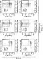


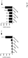
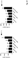
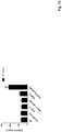
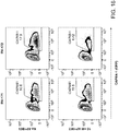
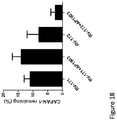
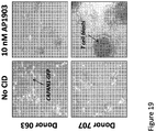


Classifications
-
- C—CHEMISTRY; METALLURGY
- C07—ORGANIC CHEMISTRY
- C07K—PEPTIDES
- C07K14/00—Peptides having more than 20 amino acids; Gastrins; Somatostatins; Melanotropins; Derivatives thereof
- C07K14/435—Peptides having more than 20 amino acids; Gastrins; Somatostatins; Melanotropins; Derivatives thereof from animals; from humans
- C07K14/705—Receptors; Cell surface antigens; Cell surface determinants
- C07K14/70578—NGF-receptor/TNF-receptor superfamily, e.g. CD27, CD30, CD40, CD95
-
- A—HUMAN NECESSITIES
- A61—MEDICAL OR VETERINARY SCIENCE; HYGIENE
- A61P—SPECIFIC THERAPEUTIC ACTIVITY OF CHEMICAL COMPOUNDS OR MEDICINAL PREPARATIONS
- A61P19/00—Drugs for skeletal disorders
-
- A—HUMAN NECESSITIES
- A61—MEDICAL OR VETERINARY SCIENCE; HYGIENE
- A61P—SPECIFIC THERAPEUTIC ACTIVITY OF CHEMICAL COMPOUNDS OR MEDICINAL PREPARATIONS
- A61P3/00—Drugs for disorders of the metabolism
-
- A—HUMAN NECESSITIES
- A61—MEDICAL OR VETERINARY SCIENCE; HYGIENE
- A61P—SPECIFIC THERAPEUTIC ACTIVITY OF CHEMICAL COMPOUNDS OR MEDICINAL PREPARATIONS
- A61P31/00—Antiinfectives, i.e. antibiotics, antiseptics, chemotherapeutics
- A61P31/04—Antibacterial agents
-
- A—HUMAN NECESSITIES
- A61—MEDICAL OR VETERINARY SCIENCE; HYGIENE
- A61P—SPECIFIC THERAPEUTIC ACTIVITY OF CHEMICAL COMPOUNDS OR MEDICINAL PREPARATIONS
- A61P31/00—Antiinfectives, i.e. antibiotics, antiseptics, chemotherapeutics
- A61P31/04—Antibacterial agents
- A61P31/06—Antibacterial agents for tuberculosis
-
- A—HUMAN NECESSITIES
- A61—MEDICAL OR VETERINARY SCIENCE; HYGIENE
- A61P—SPECIFIC THERAPEUTIC ACTIVITY OF CHEMICAL COMPOUNDS OR MEDICINAL PREPARATIONS
- A61P31/00—Antiinfectives, i.e. antibiotics, antiseptics, chemotherapeutics
- A61P31/12—Antivirals
- A61P31/14—Antivirals for RNA viruses
-
- A—HUMAN NECESSITIES
- A61—MEDICAL OR VETERINARY SCIENCE; HYGIENE
- A61P—SPECIFIC THERAPEUTIC ACTIVITY OF CHEMICAL COMPOUNDS OR MEDICINAL PREPARATIONS
- A61P31/00—Antiinfectives, i.e. antibiotics, antiseptics, chemotherapeutics
- A61P31/12—Antivirals
- A61P31/14—Antivirals for RNA viruses
- A61P31/16—Antivirals for RNA viruses for influenza or rhinoviruses
-
- A—HUMAN NECESSITIES
- A61—MEDICAL OR VETERINARY SCIENCE; HYGIENE
- A61P—SPECIFIC THERAPEUTIC ACTIVITY OF CHEMICAL COMPOUNDS OR MEDICINAL PREPARATIONS
- A61P31/00—Antiinfectives, i.e. antibiotics, antiseptics, chemotherapeutics
- A61P31/12—Antivirals
- A61P31/14—Antivirals for RNA viruses
- A61P31/18—Antivirals for RNA viruses for HIV
-
- A—HUMAN NECESSITIES
- A61—MEDICAL OR VETERINARY SCIENCE; HYGIENE
- A61P—SPECIFIC THERAPEUTIC ACTIVITY OF CHEMICAL COMPOUNDS OR MEDICINAL PREPARATIONS
- A61P31/00—Antiinfectives, i.e. antibiotics, antiseptics, chemotherapeutics
- A61P31/12—Antivirals
- A61P31/20—Antivirals for DNA viruses
-
- A—HUMAN NECESSITIES
- A61—MEDICAL OR VETERINARY SCIENCE; HYGIENE
- A61P—SPECIFIC THERAPEUTIC ACTIVITY OF CHEMICAL COMPOUNDS OR MEDICINAL PREPARATIONS
- A61P31/00—Antiinfectives, i.e. antibiotics, antiseptics, chemotherapeutics
- A61P31/12—Antivirals
- A61P31/20—Antivirals for DNA viruses
- A61P31/22—Antivirals for DNA viruses for herpes viruses
-
- A—HUMAN NECESSITIES
- A61—MEDICAL OR VETERINARY SCIENCE; HYGIENE
- A61P—SPECIFIC THERAPEUTIC ACTIVITY OF CHEMICAL COMPOUNDS OR MEDICINAL PREPARATIONS
- A61P33/00—Antiparasitic agents
- A61P33/02—Antiprotozoals, e.g. for leishmaniasis, trichomoniasis, toxoplasmosis
-
- A—HUMAN NECESSITIES
- A61—MEDICAL OR VETERINARY SCIENCE; HYGIENE
- A61P—SPECIFIC THERAPEUTIC ACTIVITY OF CHEMICAL COMPOUNDS OR MEDICINAL PREPARATIONS
- A61P33/00—Antiparasitic agents
- A61P33/02—Antiprotozoals, e.g. for leishmaniasis, trichomoniasis, toxoplasmosis
- A61P33/04—Amoebicides
-
- A—HUMAN NECESSITIES
- A61—MEDICAL OR VETERINARY SCIENCE; HYGIENE
- A61P—SPECIFIC THERAPEUTIC ACTIVITY OF CHEMICAL COMPOUNDS OR MEDICINAL PREPARATIONS
- A61P33/00—Antiparasitic agents
- A61P33/02—Antiprotozoals, e.g. for leishmaniasis, trichomoniasis, toxoplasmosis
- A61P33/06—Antimalarials
-
- A—HUMAN NECESSITIES
- A61—MEDICAL OR VETERINARY SCIENCE; HYGIENE
- A61P—SPECIFIC THERAPEUTIC ACTIVITY OF CHEMICAL COMPOUNDS OR MEDICINAL PREPARATIONS
- A61P35/00—Antineoplastic agents
-
- A—HUMAN NECESSITIES
- A61—MEDICAL OR VETERINARY SCIENCE; HYGIENE
- A61P—SPECIFIC THERAPEUTIC ACTIVITY OF CHEMICAL COMPOUNDS OR MEDICINAL PREPARATIONS
- A61P35/00—Antineoplastic agents
- A61P35/02—Antineoplastic agents specific for leukemia
-
- A—HUMAN NECESSITIES
- A61—MEDICAL OR VETERINARY SCIENCE; HYGIENE
- A61P—SPECIFIC THERAPEUTIC ACTIVITY OF CHEMICAL COMPOUNDS OR MEDICINAL PREPARATIONS
- A61P37/00—Drugs for immunological or allergic disorders
- A61P37/02—Immunomodulators
-
- A—HUMAN NECESSITIES
- A61—MEDICAL OR VETERINARY SCIENCE; HYGIENE
- A61P—SPECIFIC THERAPEUTIC ACTIVITY OF CHEMICAL COMPOUNDS OR MEDICINAL PREPARATIONS
- A61P37/00—Drugs for immunological or allergic disorders
- A61P37/02—Immunomodulators
- A61P37/04—Immunostimulants
-
- A—HUMAN NECESSITIES
- A61—MEDICAL OR VETERINARY SCIENCE; HYGIENE
- A61P—SPECIFIC THERAPEUTIC ACTIVITY OF CHEMICAL COMPOUNDS OR MEDICINAL PREPARATIONS
- A61P7/00—Drugs for disorders of the blood or the extracellular fluid
-
- A—HUMAN NECESSITIES
- A61—MEDICAL OR VETERINARY SCIENCE; HYGIENE
- A61P—SPECIFIC THERAPEUTIC ACTIVITY OF CHEMICAL COMPOUNDS OR MEDICINAL PREPARATIONS
- A61P7/00—Drugs for disorders of the blood or the extracellular fluid
- A61P7/06—Antianaemics
-
- C—CHEMISTRY; METALLURGY
- C07—ORGANIC CHEMISTRY
- C07K—PEPTIDES
- C07K14/00—Peptides having more than 20 amino acids; Gastrins; Somatostatins; Melanotropins; Derivatives thereof
- C07K14/435—Peptides having more than 20 amino acids; Gastrins; Somatostatins; Melanotropins; Derivatives thereof from animals; from humans
- C07K14/705—Receptors; Cell surface antigens; Cell surface determinants
- C07K14/70503—Immunoglobulin superfamily
- C07K14/7051—T-cell receptor (TcR)-CD3 complex
-
- C—CHEMISTRY; METALLURGY
- C07—ORGANIC CHEMISTRY
- C07K—PEPTIDES
- C07K14/00—Peptides having more than 20 amino acids; Gastrins; Somatostatins; Melanotropins; Derivatives thereof
- C07K14/435—Peptides having more than 20 amino acids; Gastrins; Somatostatins; Melanotropins; Derivatives thereof from animals; from humans
- C07K14/705—Receptors; Cell surface antigens; Cell surface determinants
- C07K14/70503—Immunoglobulin superfamily
- C07K14/70521—CD28, CD152
-
- C—CHEMISTRY; METALLURGY
- C07—ORGANIC CHEMISTRY
- C07K—PEPTIDES
- C07K16/00—Immunoglobulins [IGs], e.g. monoclonal or polyclonal antibodies
- C07K16/18—Immunoglobulins [IGs], e.g. monoclonal or polyclonal antibodies against material from animals or humans
- C07K16/28—Immunoglobulins [IGs], e.g. monoclonal or polyclonal antibodies against material from animals or humans against receptors, cell surface antigens or cell surface determinants
- C07K16/30—Immunoglobulins [IGs], e.g. monoclonal or polyclonal antibodies against material from animals or humans against receptors, cell surface antigens or cell surface determinants from tumour cells
- C07K16/3069—Reproductive system, e.g. ovaria, uterus, testes, prostate
-
- C—CHEMISTRY; METALLURGY
- C07—ORGANIC CHEMISTRY
- C07K—PEPTIDES
- C07K2317/00—Immunoglobulins specific features
- C07K2317/20—Immunoglobulins specific features characterized by taxonomic origin
- C07K2317/24—Immunoglobulins specific features characterized by taxonomic origin containing regions, domains or residues from different species, e.g. chimeric, humanized or veneered
-
- C—CHEMISTRY; METALLURGY
- C07—ORGANIC CHEMISTRY
- C07K—PEPTIDES
- C07K2317/00—Immunoglobulins specific features
- C07K2317/60—Immunoglobulins specific features characterized by non-natural combinations of immunoglobulin fragments
- C07K2317/62—Immunoglobulins specific features characterized by non-natural combinations of immunoglobulin fragments comprising only variable region components
- C07K2317/622—Single chain antibody (scFv)
-
- C—CHEMISTRY; METALLURGY
- C07—ORGANIC CHEMISTRY
- C07K—PEPTIDES
- C07K2317/00—Immunoglobulins specific features
- C07K2317/60—Immunoglobulins specific features characterized by non-natural combinations of immunoglobulin fragments
- C07K2317/64—Immunoglobulins specific features characterized by non-natural combinations of immunoglobulin fragments comprising a combination of variable region and constant region components
-
- C—CHEMISTRY; METALLURGY
- C07—ORGANIC CHEMISTRY
- C07K—PEPTIDES
- C07K2319/00—Fusion polypeptide
-
- C—CHEMISTRY; METALLURGY
- C07—ORGANIC CHEMISTRY
- C07K—PEPTIDES
- C07K2319/00—Fusion polypeptide
- C07K2319/33—Fusion polypeptide fusions for targeting to specific cell types, e.g. tissue specific targeting, targeting of a bacterial subspecies
-
- C—CHEMISTRY; METALLURGY
- C07—ORGANIC CHEMISTRY
- C07K—PEPTIDES
- C07K2319/00—Fusion polypeptide
- C07K2319/70—Fusion polypeptide containing domain for protein-protein interaction
Definitions
- the technology relates generally to the field of immunology and relates in part to compositions and methods for controlling the proliferation of T cells, for example, therapeutic T cells.
- the methods further relate to compositions and methods for inducing an immune response in a subject.
- T cell activation is an important step in the protective immunity against pathogenic microorganisms (e.g., viruses, bacteria, and parasites), foreign proteins, and harmful chemicals in the environment.
- T cells express receptors on their surfaces (i.e., T cell receptors) that recognize antigens presented on the surface of antigen-presenting cells. During a normal immune response, binding of these antigens to the T cell receptor initiates intracellular changes leading to T cell activation.
- Chimeric antigen receptors are artificial receptors designed to convey antigen specificity to T cells. They include an antigen-specific component, a transmembrane component, and an intracellular component selected to activate the T cell and provide specific immunity. Chimeric antigen receptor-expressing T cells may be used in various therapies, including cancer therapies. While effective against tumors, in some cases these therapies have led to side effects due, in part to non-specific attacks on healthy tissue. A method for controllable T cell therapy is needed that provides a strong immunotherapeutic response and avoids toxic side effects.
- WO 2004/073641 describes induced activation in dendritic cells.
- WO 2008/049113 describes the generation of an immune response by inducing CD40 and pattern recognition receptors.
- WO 2005/044996 describes chimeric receptors with a 4-1BB stimulatory signaling domain.
- WO 2011/130566 describes a method for treating solid tumors.
- Zhong, et al., Molecular Therapeutics 18, 413-420 (2010 ) describes chimeric antigen receptors.
- US 2010/203067 describes methods and compositions for generating an immune response by inducing CD40 and pattern recognition receptor adapters.
- the present invention relates to a eukaryotic cell, transfected or transduced with a nucleic acid that comprises a polynucleotide encoding an inducible chimeric signaling molecule, wherein the inducible chimeric signaling molecule comprises (a) an intracellular FKBP12 or FKBP12v36 variant multimerizing region, that binds to an AP1903 or AP20187 ligand, wherein the chimeric signaling molecule oligomerizes upon binding to the ligand; (b) a first co-stimulatory polypeptide cytoplasmic signaling region selected from the group consisting of CD27, CD28, ICOS, 4-1BB, and OX40; and (c) a second co-stimulatory polypeptide cytoplasmic signaling region selected from the group consisting of CD27, CD28, ICOS, 4-1BB, and OX40; wherein the chimeric signaling molecule lacks an extracellular domain, the cell is selected from the group
- CSMs CID-inducible chimeric signaling molecules
- the CSMs may be used alone, or in combination with chimeric antigen receptors (CARs), which allows the immune response to be specifically directed against particular tumor cells.
- CARs chimeric antigen receptors
- the controlled T cell activation methods avoid many of the toxic side effects of earlier CAR-based treatments.
- the chimeric signaling molecules discussed herein allow for a sustained, modulated control of a chimeric antigen receptor (CAR) that is co-expressed in the cell.
- CAR chimeric antigen receptor
- the activation of the antigen-specific T cell, designed to target a cellular antigen implicated in a disease or condition, is dependent on the administration of a ligand inducer.
- the ligand inducer activates the CAR-expressing cell by multimerizing the chimeric signaling molecule, which, in turn, activates NF- K B signaling, which activates the cell, for example, a T cell, a tumor-infiltrating lymphocyte, a natural killer cell, or a natural killer T cell.
- the T cell In the absence of the ligand inducer, the T cell is quiescent, or has a basal level of activity.
- a regular dosing schedule of the ligand determines the rate and magnitude of the CAR-expressing T cell proliferation and activation.
- T cells transduced with a nucleic acid encoding a chimeric antigen receptor have been administered to patients to treat cancer ( Zhong, X.-S., (2010) Molecular Therapy 18:413-420 ).
- T cells expressing a chimeric antigen receptor based on the humanized monoclonal antibody Trastuzumab (Herceptin) has been used to treat cancer patients.
- Adverse events are possible, however, and in at least one reported case, the therapy had fatal consequences to the patient ( Morgan, R.A., et al., (2010) Molecular Therapy 18:843-851 ).
- Transducing the cells with a controllable inducible safety switch would provide a safety switch that could stop the adverse event from progressing, by stopping the administration of the ligand inducer. Although a low level basal activity might remain, removing the presence of the inducer should drastically reduce, if not cease, the symptoms of the adverse event.
- T cells are modified so that they express a non-functional TGF-beta receptor, rendering them resistant to TGF-beta.
- it also could result in a T cell lymphoma, or other adverse effect, as the modified T cells now lack part of the normal cellular control; these therapeutic T cells could themselves become malignant.
- Transducing these modified T cells with an inducible CSM polypeptide-based safety switch as presented herein, would provide a safety switch that could avoid this result.
- composition comprising a nucleic acid that comprises a polynucleotide encoding an inducible chimeric signaling molecule, wherein the inducible chimeric signaling molecule comprises a membrane-targeting region, a multimerizing region and a co-stimulatory polypeptide cytoplasmic signaling region selected from the group consisting of CD27, CD28, ICOS, 4-1BB, CD40, RANK/TRANCE-R, CD3 zeta chain, and OX40.
- the membrane-targeting region is selected from the group consisting of myristoylation-targeting sequence, palmitoylation-targeting sequence, prenylation sequences (i.e., farnesylation, geranyl-geranylation, CAAX Box), protein-protein interaction motifs and transmembrane sequences (utilizing signal peptides) from receptors.
- the membrane-targeting region is a myristoylation targeting sequence.
- the inducible chimeric signaling molecule further comprises a second co-stimulatory polypeptide cytoplasmic signaling region selected from the group consisting of CD27, CD28, ICOS, 4-1BB, CD40, RANK/TRANCE-R, CD3 zeta chain, and OX40.
- the co-stimulatory polypeptide cytoplasmic signaling regions comprise a CD28 cytoplasmic signaling region and a 4-1BB cytoplasmic signaling region.
- the co-stimulatory polypeptide cytoplasmic signaling regions comprise an OX40 cytoplasmic signaling region polypeptide and a 4-1BB cytoplasmic signaling region polypeptide.
- the inducible chimeric signaling molecule further comprises a CD3 ⁇ polypeptide.
- multimerizing region is selected from the group consisting of FKBP, cyclophilin receptor, steroid receptor, tetracycline receptor, heavy chain antibody subunit, light chain antibody subunit, and mutated sequences thereof.
- the multimerizing region is an FKBP12 region.
- the FKB12-region is an FKB12v36 region.
- the multimerizing region is Fv'Fvls.
- the multimerizing region binds a ligand selected from the group consisting of an FK506 dimer and a dimeric FK506 analog ligand.
- the ligand is AP1903 or AP20187.
- the multimerizing region has an amino acid sequence of SEQ ID NO: 58 or a functional fragment thereof. In some cases, the multimerizing region is encoded by a nucleotide sequence in SEQ ID NO: 57, or a functional fragment thereof. In somecases, the multimerizing region further comprises a polypeptide having an amino acid sequence of SEQ ID NO: 60, or a functional fragment thereof. In some cases, the multimerizing region further comprises a polypeptide encoded by a nucleotide sequence in SEQ ID NO: 59, or a functional fragment thereof. In some cases, the multimerizing region further comprises a polypeptide having an amino acid sequence of SEQ ID NO: 60, or a functional fragment thereof.
- the multimerizing region further comprises a polypeptide encoded by a nucleotide sequence in SEQ ID NO: 59, or a functional fragment thereof. In some cases, the multimerizing region further comprises a polypeptide having an amino acid sequence of SEQ ID NO: 58 or SEQ ID NO: 60, or a functional fragment thereof. In some cases, the multimerizing region further comprises a polypeptide encoded by a nucleotide sequence in SEQ ID NO: 57 or SEQ ID NO: 59, or a functional fragment thereof.
- the nucleic acid comprises a promoter sequence operably linked to the polynucleotide.
- the nucleic acid is contained within a viral vector.
- the viral vector is a retroviral vector.
- the retroviral vector is a murine leukemia virus vector.
- the murine leukemia virus vector is a MoMLV vector.
- the retroviral vector is an SFG vector.
- the viral vector is an adenoviral vector.
- the viral vector is a lentiviral vector.
- the nucleic acid is contained within a plasmid.
- the cell is a T cell, tumor infiltrating lymphocyte, B cell, NK cell, or NK-T cell.
- cell is a T cell.
- the cell is obtained or prepared from bone marrow.
- the cell is obtained or prepared from umbilical cord blood.
- the cell is obtained or prepared from peripheral blood.
- the cell is obtained or prepared from peripheral blood mononuclear cells.
- the cell is a human cell.
- the cell is further transformed or transduced with a nucleic acid comprising a polynucleotide that encodes a chimeric polypeptide comprising a signal peptide, a single chain variable fragment, a CH2-CH3 hinge region and a CD3 ⁇ polypeptide.
- a nucleic acid comprising a polynucleotide that encodes a chimeric polypeptide comprising a signal peptide, a single chain variable fragment, a CH2-CH3 hinge region and a CD3 ⁇ polypeptide.
- the single chain variable fragment binds to an antigen on a tumor cell.
- the single chain variable fragment binds to an antigen on a cell involved in a hyperproliferative disease.
- the single chain variable fragment is selected from the group consisting of ⁇ PSMA, ⁇ PSCA, ⁇ MUC1, ⁇ CD19, ⁇ ROR1, ⁇ Mesothelin, ⁇ GD2, ⁇ CD123, ⁇ MUC16, and ⁇ Her2/Neu single chain variable fragments. In some cases, the single chain variable fragment is an ⁇ CD19 single chain variable fragment.
- the method further comprises contacting the cell with a ligand that binds to the multimerizing region resulting in multimerization of the inducible chimeric signaling molecule.
- the ligand is dimeric.
- the ligand is dimeric FK506, or a dimeric FK506-like analog.
- the ligand is AP1903 or AP20187.
- the method further comprises administering the transfected or transformed cell to a subject. In some cases, the cell is administered to the subject by intravenous administration.
- a method for inducing an immune response in vivo comprising administering to a subject a composition of the present application.
- the methods further comprise administering to the subject a composition comprising a ligand that binds to the multimerizing region resulting in multimerization of the inducible chimeric signaling molecule.
- the ligand is dimeric.
- the ligand is dimeric FK506, or a dimeric FK506-like analog.
- the ligand is AP1903 or AP20187.
- the subject to be treated with the cell of the invention has been diagnosed with a hyperproliferative disease. In other cases, wherein the subject has been diagnosed with a tumor. In other cases, the subject has cancer. In other cases, the subject has a solid tumor. In other cases, the cell is a tumor infiltrating lymphocyte or a T cell. In other cases, the cell is delivered to the tumor bed. In other cases, the cancer is present in the blood or bone marrow of the subject. In other cases, the subject has a blood or bone marrow disease. In other cases, the subject has been diagnosed with any condition or disorder that can be alleviated by stem cell transplantation. In other cases, the subject has been diagnosed with sickle cell anemia or metachromatic leukodystrophy.
- the subject has been diagnosed with a condition selected from the group consisting of a primary immune deficiency disorder, hemophagocytosis lymphohistiocytosis (HLH) or other hemophagocytic disorder, an inherited marrow failure disorder, a hemoglobinopathy, a metabolic disorder, and an osteoclast disorder.
- a condition selected from the group consisting of a primary immune deficiency disorder, hemophagocytosis lymphohistiocytosis (HLH) or other hemophagocytic disorder, an inherited marrow failure disorder, a hemoglobinopathy, a metabolic disorder, and an osteoclast disorder.
- the subject has been diagnosed with a condition is selected from the group consisting of Severe Combined Immune Deficiency (SCID), Combined Immune Deficiency (CID), Congenital T-cell Defect/Deficiency, Common Variable Immune Deficiency (CVID), Chronic Granulomatous Disease, IPEX (Immune deficiency, polyendocrinopathy, enteropathy, X-linked) or IPEX-like, Wiskott-Aldrich Syndrome, CD40 Ligand Deficiency, Leukocyte Adhesion Deficiency, DOCK 8 Deficiency, IL-10 Deficiency/IL-10 Receptor Deficiency, GATA 2 deficiency, X-linked lymphoproliferative disease (XLP), Cartilage Hair Hypoplasia, Shwachman Diamond Syndrome, Diamond Blackfan Anemia, Dyskeratosis Congenita, Fanconi Anemia, Congenital Neutropenia, Sickle Cell Disease, Thalassemia, Mucopolys
- the subject has been diagnosed with an infection of viral etiology selected from the group consisting HIV, influenza, Herpes, viral hepatitis, Epstein Bar, polio, viral encephalitis, measles, chicken pox, Cytomegalovirus (CMV), adenovirus (ADV), HHV-6 (human herpesvirus 6, I), and Papilloma virus, or has been diagnosed with an infection of bacterial etiology selected from the group consisting of pneumonia, tuberculosis, and syphilis, or has been diagnosed with an infection of parasitic etiology selected from the group consisting of malaria, trypanosomiasis, leishmaniasis, trichomoniasis, and amoebiasis.
- viral etiology selected from the group consisting HIV, influenza, Herpes, viral hepatitis, Epstein Bar, polio, viral encephalitis, measles, chicken pox, Cytomegalovirus (CMV),
- the single chain variable fragment binds to CD19.
- the multimeric ligand is AP1903 or AP20187.
- the cell is a T cell.
- the subject is human.
- the methods further comprise determining whether an additional dose of the multimeric ligand should be administered to the subject.
- the methods further comprise administering an additional dose of the multimeric ligand to the subject, wherein the disease or condition symptoms remain or are detected following a reduction in symptoms.
- the subject has been diagnosed with a disease or condition before administration of the composition or cell of the present disclosure, and after administration of the multimeric ligand the disease or condition is detected, an additional dose of the multimeric ligand is administered to the subject.
- the methods further comprise identifying the presence, absence or stage of a condition or disease in a subject, and transmitting an indication to administer a multimeric ligand that binds to the multimeric binding region, maintain a subsequent dosage of the multimeric ligand or adjust a subsequent dosage of the multimeric ligand administered to the patient based on the presence, absence or stage of the condition or disease identified in the subject.
- the condition is cancer. In some cases, the condition is leukemia. In some cases, the condition is a solid tumor.
- the methods further comprise determining the presence or absence of a tumor size increase and/or increase in the number of tumor cells in a subject relative to the tumor size and/or the number of tumor cells following administration of the multimeric ligand, and administering an additional dose of the multimeric ligand to the subject in the event the presence of a tumor size increase and/or increase in the number of tumor cells is determined.
- the tumor size and/or the number of tumor cells is decreased following administration of the multimeric ligand relative to the tumor size and/or number of tumor cells before administration of the multimeric ligand.
- the methods further comprise determining the presence or absence of an increase in CD19-expressing B cells in the subject relative to the level of CD19-expressing B cells following administration of the multimeric ligand, and administering an additional dose of the multimeric ligand to the subject in the event the presence of an increase in CD19-expressing B cells in the subject is determined.
- the level of CD19-expressing B cells is decreased following administration of the multimeric ligand relative to the level of CD19-expressing B cells before administration of the multimeric ligand.
- T cell therapy has involved the difficulty of poor in vivo expansion of the infused cells.
- One way this issue has been addressed is by administering high doses of IL-2 to the patient.
- This therapy helps T cell growth and anti-tumor function, but is also very toxic to the patient.
- This has generally been used in melanoma as high dose IL-2 is considered a standard-of-care therapy for that disease.
- Most other T cell therapy applications have not used IL-2 with T cell therapy due to toxic effects.
- Another issue arising in T cell therapy is the poor engraftment and persistence of infused T cells (also a function of in vivo proliferation), which has been addressed by lymphodepleting conditioning prior to T cell infusion.
- T cell therapy has largely been considered a boutique therapy since each patient needs to have a unique cell product manufactured for them.
- Conventional T cell therapies (generated by repetitive antigen stimulation or isolation of tumor infiltrating lymphocytes (TILs)) are not reproducible in their specificity or function and lead to extremely variable results, and in some cases the inability to produce a product for treatment.
- TILs tumor infiltrating lymphocytes
- Gene transfer of natural or chimeric T cell receptors has started to solve this problem (where highly tumor specific T cells can be generated in less than 2 weeks), but it is apparent that gene-modified T cells can function differently than naturally occurring T cells.
- highly specific CAR T cells or T cells expressing optimized TCR alpha and beta chains can cause off-target toxicity, necessitating the inclusion of a suicide gene.
- FIG 1 illustrates the most basic components of a chimeric antigen receptor (CAR).
- CAR chimeric antigen receptor
- V H variable heavy
- V L variable heavy
- V L variable heavy
- ⁇ CD3 zeta chain
- the V H and V L are generally connected together using a flexible glycine-serine linker, and then attached to the transmembrane domain by a spacer (CH2CH3) to extend the scFv away from the cell surface so that it can interact with tumor antigens.
- CH2CH3 spacer
- T cells Following transduction, T cells now express the CAR on their surface, and upon contact and ligation with a tumor antigen, signal through the CD3 zeta chain inducing cytotoxicity and cellular activation.
- FIG. 2 illustrates the development of various chimeric antigen receptors.
- Investigators have noted that activation of T cells through CD3 zeta is sufficient to induce a tumor-specific killing, but is insufficient to induce T cell proliferation and survival.
- Early clinical trials using T cells modified with CARs expressing only the zeta chain showed that gene-modified T cells exhibited poor survival and proliferation in vivo. These constructs are termed 1 st generation CARs.
- CD28 signaling domain As co-stimulation through the B7 axis is necessary for complete T cell activation, investigators added the co-stimulatory polypeptide CD28 signaling domain to the CAR construct.
- This region generally contains the transmembrane region (in place of the CD3 zeta version) and the YMNM motif for binding PI3K and Lck.
- CD28 enhanced expansion in vivo, in part due to increased IL-2 nd production following activation. The inclusion of CD28 is called a 2 generation CAR.
- co-stimulatory polypeptides 4-1BB or OX40 in CAR design has further improved T cell survival and efficacy.
- 4-1BB in particular appears to greatly enhance T cell proliferation and survival.
- This 3 rd generation design (with 3 signaling domains) has been used in PSMA CARs ( Zhong XS, et al., Mol Ther. 2010 Feb;18(2):413-20 ), and in CD19 CARs, most notably for the treatment of CLL ( Milone, M.C., et al., (2009) Mol. Ther. 17:1453-1464 ; Kalos, M., et al., Sci. Transl. Med. (2011) 3:95ra73 ; Porter, D., et al., (2011) N. Engl. J. Med. 365: 725-533 ). These cells showed impressive function in 3 patients, expanding more than a 1000-fold in vivo, and resulted in sustained remission in all three patients.
- a suicide gene provides ample protection against unwanted side-effects from adoptively transferred T cells; however, elimination of gene-modified T cells following toxicity may also ablate the therapeutic efficacy of the treatment.
- T cell receptor signaling can be induced using a chemical inducer of dimerization (CID) in combination with a chimeric receptor that includes a multimerization region that binds to the CID, T cells were engineered to express the CD3 zeta chain, which was linked with 1, 2, or 3 FKBP fragments. The cells expressed the chimeric receptor, and demonstrated CID-dependent T cell activation ( Spencer, D. M., et al., Science, 1993. 262: p. 1019-1024 ).
- CSMs inducible chimeric signaling molecules
- Figure 3 compares the therapies of the present application with methods of CAR treatment using a suicide gene.
- the present application provides, in part, a gene-engineering approach to amplify T cell proliferation and function in vivo so that the anti-tumor effect is gradually increased.
- a chemical inducer of dimerization is used in a controllable system for activating T cells in vivo to increase their function and frequency.
- the CSM uses a multimerization region, such as Fv domains, in tandem with one or more co-stimulatory polypeptides, such as, for example, CD28 and 4-1BB, with and without the CD3 zeta chain to enable CID-dependent proliferation and co-stimulation.
- the CSM may be used alone to provide co-stimulation, and increase a T cell immune response.
- a population of T cells for example a population with non-specific targets, may be transfected or transformed with DNA coding for CSM, then administered to a subject to enhance a general immune response.
- This CSM may also be expressed in a cell along with a CAR, which may, for example, comprise the scFv polypeptide, and the CD3 zeta chain.
- a CAR which may, for example, comprise the scFv polypeptide, and the CD3 zeta chain.
- an inducible CSM molecule is used in combination with a CAR, thereby segregating CAR signaling into two separate functions.
- This second function, provided by the CAR provides antigen-specific cytotoxicity to the engineered T cells.
- the example shows a CAR with specificity against PSMA; these engineered T cells may, for example, be administered to a subject to generate a specific immune response, for example one directed against a prostate cancer tumor ( Figure 6 ).
- the inducible co-stimulatory polypeptide such as, for example, a CD40 cytoplasmic region polypeptide or a truncated MyD88 polypeptide is used to control activation of the chimeric antigen receptor itself.
- a polynucleotide that encodes this modified inducible chimeric antigen receptor may be used to transduce cells, such as, for example T cells.
- the cells may further express a chimeric signaling molecule as discussed herein, and in certain cases, the chimeric signaling molecule comprises a CD3 zeta polypeptide.
- the inducible chimeric antigen receptor comprises both a CD40 cytoplasmic region polypeptide and a MyD88 polypeptide.
- allogeneic refers to HLA or MHC loci that are antigenically distinct between the host and donor cells.
- syngeneic mice can differ at one or more loci (congenics) and allogeneic mice can have the same background.
- antigen as used herein is defined as a molecule that provokes an immune response. This immune response may involve either antibody production, or the activation of specific immunologically-competent cells, or both.
- An antigen can be derived from organisms, subunits of proteins/antigens, killed or inactivated whole cells or lysates.
- Exemplary organisms include but are not limited to, Helicobacters, Campylobacters, Clostridia, Corynebacterium diphtheriae, Bordetella pertussis, influenza virus, parainfluenza viruses, respiratory syncytial virus, Borrelia burgdorfei, Plasmodium, herpes simplex viruses, human immunodeficiency virus, papillomavirus, Vibrio cholera, E. coli, measles virus, rotavirus, shigella, Salmonella typhi, Neisseria gonorrhea. Therefore, any macromolecules, including virtually all proteins or peptides, can serve as antigens.
- antigens can be derived from recombinant or genomic DNA. Any DNA that contains nucleotide sequences or partial nucleotide sequences of a pathogenic genome or a gene or a fragment of a gene for a protein that elicits an immune response results in synthesis of an antigen.
- the present methods are not limited to the use of the entire nucleic acid sequence of a gene or genome. It is readily inherent that the present invention includes, but is not limited to, the use of partial nucleic acid sequences of more than one gene or genome and that these nucleic acid sequences are arranged in various combinations to elicit the desired immune response.
- cancer as used herein is defined as a hyperproliferation of cells whose unique trait-loss of normal controls-results in unregulated growth, lack of differentiation, local tissue invasion, and metastasis. Examples include but are not limited to, melanoma, non-small cell lung, small-cell lung, lung, hepatocarcinoma, leukemia, retinoblastoma, astrocytoma, glioblastoma, gum, tongue, neuroblastoma, head, neck, breast, pancreatic, prostate, renal, bone, testicular, ovarian, mesothelioma, cervical, gastrointestinal, lymphoma, brain, colon, sarcoma or bladder.
- cell may be used interchangeably. All of these terms also include their progeny, which are any and all subsequent generations. It is understood that all progeny may not be identical due to deliberate or inadvertent mutations.
- cDNA is intended to refer to DNA prepared using messenger RNA (mRNA) as template.
- mRNA messenger RNA
- expression construct or "transgene” is defined as any type of genetic construct containing a nucleic acid coding for gene products in which part or all of the nucleic acid encoding sequence is capable of being transcribed can be inserted into the vector.
- the transcript is translated into a protein, but it need not be.
- expression includes both transcription of a gene and translation of mRNA into a gene product. In other cases, expression only includes transcription of the nucleic acid encoding genes of interest.
- therapeutic construct may also be used to refer to the expression construct or transgene.
- the expression construct or transgene may be used, for example, as a therapy to treat hyperproliferative diseases or disorders, such as cancer, thus the expression construct or transgene is a therapeutic construct or a prophylactic construct.
- expression vector refers to a vector containing a nucleic acid sequence coding for at least part of a gene product capable of being transcribed. In some cases, RNA molecules are then translated into a protein, polypeptide, or peptide. In other cases, these sequences are not translated, for example, in the production of antisense molecules or ribozymes.
- Expression vectors can contain a variety of control sequences, which refer to nucleic acid sequences necessary for the transcription and possibly translation of an operatively linked coding sequence in a particular host organism. In addition to control sequences that govern transcription and translation, vectors and expression vectors may contain nucleic acid sequences that serve other functions as well and are discussed infra.
- ex vivo refers to "outside” the body.
- in vitro can be used interchangeably herein.
- “Functionally equivalent” refers, for example, to a co-stimulatory polypeptide that is lacking the extracellular domain, but is capable of amplifying the T cell-mediated tumor killing response when expressed in T cells.
- hyperproliferative disease is defined as a disease that results from a hyperproliferation of cells.
- exemplary hyperproliferative diseases include, but are not limited to cancer or autoimmune diseases.
- Other hyperproliferative diseases may include vascular occlusion, restenosis, atherosclerosis, or inflammatory bowel disease.
- the term "gene” is defined as a functional protein, polypeptide, or peptide-encoding unit. As will be understood, this functional term includes genomic sequences, cDNA sequences, and smaller engineered gene segments that express, or are adapted to express, proteins, polypeptides, domains, peptides, fusion proteins, and mutants.
- immunogen refers to a substance that is capable of provoking an immune response.
- immunogens include, e.g., antigens, autoantigens that play a role in induction of autoimmune diseases, and tumor-associated antigens expressed on cancer cells.
- immunocompromised as used herein is defined as a subject that has reduced or weakened immune system.
- the immunocompromised condition may be due to a defect or dysfunction of the immune system or to other factors that heighten susceptibility to infection and/or disease.
- a defect or dysfunction of the immune system or to other factors that heighten susceptibility to infection and/or disease.
- immunocompromised individuals often do not fit completely into one group or the other. More than one defect in the body's defense mechanisms may be affected. For example, individuals with a specific T-lymphocyte defect caused by HIV may also have neutropenia caused by drugs used for antiviral therapy or be immunocompromised because of a breach of the integrity of the skin and mucous membranes.
- An immunocompromised state can result from indwelling central lines or other types of impairment due to intravenous drug abuse; or be caused by secondary malignancy, malnutrition, or having been infected with other infectious agents such as tuberculosis or sexually transmitted diseases, e.g., syphilis or hepatitis.
- the term "pharmaceutically or pharmacologically acceptable” refers to molecular entities and compositions that do not produce adverse, allergic, or other untoward reactions when administered to an animal or a human.
- the subject is a mammal. In some cases, the subject is a human.
- pharmaceutically acceptable carrier includes any and all solvents, dispersion media, coatings, antibacterial and antifungal agents, isotonic and absorption delaying agents and the like.
- the use of such media and agents for pharmaceutically active substances is well known in the art. Except insofar as any conventional media or agent is incompatible with the vectors or cells presented herein, its use in therapeutic compositions is contemplated. Supplementary active ingredients also can be incorporated into the compositions.
- nucleotide is defined as a chain of nucleotides.
- nucleic acids are polymers of nucleotides.
- nucleic acids and polynucleotides as used herein are interchangeable.
- Nucleic acids are polynucleotides, which can be hydrolyzed into the monomeric “nucleotides.” The monomeric nucleotides can be hydrolyzed into nucleosides.
- polynucleotides include, but are not limited to, all nucleic acid sequences which are obtained by any means available in the art, including, without limitation, recombinant means, i.e., the cloning of nucleic acid sequences from a recombinant library or a cell genome, using ordinary cloning technology and PCRTM, and the like, and by synthetic means.
- polynucleotides include mutations of the polynucleotides, include but are not limited to, mutation of the nucleotides, or nucleosides by methods well known in the art.
- a nucleic acid may comprise one or more polynucleotides.
- polypeptide is defined as a chain of amino acid residues, usually having a defined sequence.
- polypeptide may be interchangeable with the term proteins.
- promoter is defined as a DNA sequence recognized by the synthetic machinery of the cell, or introduced synthetic machinery, required to initiate the specific transcription of a gene.
- the terms “regulate an immune response,” “modulate an immune response,” or “control an immune response,” refer to the ability to modify the immune response.
- the composition is capable of enhancing and/or activating the immune response.
- the composition is also capable of inhibiting the immune response.
- the form of regulation is determined by the ligand that is used with the composition. For example, a dimeric analog of the chemical results in dimerization of the co-stimulatory polypeptide leading to activation of the T cell, however, a monomeric analog of the chemical does not result in dimerization of the co-stimulatory polypeptide, which would not activate the T cells.
- transfection and “transduction” are interchangeable and refer to the process by which an exogenous DNA sequence is introduced into a eukaryotic host cell.
- Transfection can be achieved by any one of a number of means including electroporation, microinjection, gene gun delivery, retroviral infection; lipofection, superfection and the like.
- syngeneic refers to cells, tissues or animals that have genotypes that are identical or closely related enough to allow tissue transplant, or are immunologically compatible. For example, identical twins or animals of the same inbred strain. Syngeneic and isogeneic can be used interchangeably.
- patient or “subject” are interchangeable, and, as used herein includesinclude, but isare not limited to, an organism or animal; a mammal, including, e.g., a human, non-human primate (e.g., monkey), mouse, pig, cow, goat, rabbit, rat, guinea pig, hamster, horse, monkey, sheep, or other non-human mammal; a non-mammal, including, e.g., a non-mammalian'vertebrate, such as a bird (e.g., a chicken or duck) or a fish, and a non-mammalian invertebrate.
- a mammal including, e.g., a human, non-human primate (e.g., monkey), mouse, pig, cow, goat, rabbit, rat, guinea pig, hamster, horse, monkey, sheep, or other non-human mammal
- a non-mammal including,
- the term "vaccine” refers to a formulation that contains a composition presented herein which is in a form that is capable of being administered to an animal.
- the vaccine comprises a conventional saline or buffered aqueous solution medium in which the composition is suspended or dissolved.
- the composition can be used conveniently to prevent, ameliorate, or otherwise treat a condition.
- the vaccine Upon introduction into a subject, the vaccine is able to provoke an immune response including, but not limited to, the production of antibodies, cytokines and/or other cellular responses.
- under transcriptional control or "operatively linked” is defined as the promoter is in the correct location and orientation in relation to the nucleic acid to control RNA polymerase initiation and expression of the gene.
- treatment refers to prophylaxis and/or therapy.
- a solid tumor such as a cancerous solid tumor
- the term refers to prevention by prophylactic treatment, which increases the subject's resistance to solid tumors or cancer.
- the subject may be treated to prevent cancer, where the cancer is familial, or is genetically associated.
- the term refers to a prophylactic treatment which increases the resistance of a subject to infection with a pathogen or, in other words, decreases the likelihood that the subject will become infected with the pathogen or will show signs of illness attributable to the infection, as well as a treatment after the subject has become infected in order to fight the infection, e. g., reduce or eliminate the infection or prevent it from becoming worse.
- Blood disease refers to conditions that affect the production of blood and its components, including but not limited to, blood cells, hemoglobin, blood proteins, the mechanism of coagulation, production of blood, production of blood proteins, the like and combinations thereof.
- blood diseases include anemias, leukemias, lymphomas, hematological neoplasms, albuminemias, haemophilias and the like.
- Bone marrow disease refers to conditions leading to a decrease in the production of blood cells and blood platelets.
- normal bone marrow architecture can be displaced by infections (e.g., tuberculosis) or malignancies, which in turn can lead to the decrease in production of blood cells and blood platelets.
- infections e.g., tuberculosis
- malignancies e.g., malignancies
- Non-limiting examples of bone marrow diseases include leukemias, bacterial infections (e.g., tuberculosis), radiation sickness or poisoning, apnocytopenia, anemia, multiple myeloma and the like.
- T cells and Activated T cells include that this means CD3+ cells: T cells (also referred to as T lymphocytes) belong to a group of white blood cells referred to as lymphocytes. Lymphocytes generally are involved in cell-mediated immunity.
- the "T” in “T cells” refers to cells derived from or whose maturation is influenced by the thymus. T cells can be distinguished from other lymphocytes types such as B cells and Natural Killer (NK) cells by the presence of cell surface proteins known as T cell receptors.
- NK Natural Killer
- activated T cells refers to T cells that have been stimulated to produce an immune response (e.g., clonal expansion of activated T cells) by recognition of an antigenic determinant presented in the context of a Class II major histo-compatibility (MHC) marker.
- T-cells are activated by the presence of an antigenic determinant, cytokines and/or lymphokines and cluster of differentiation cell surface proteins (e.g., CD3, CD4, CD8, the like and combinations thereof).
- Cells that express a cluster of differential protein often are said to be "positive" for expression of that protein on the surface of T-cells (e.g., cells positive for CD3 or CD 4 expression are referred to as CD3+ or CD4+).
- CD3 and CD4 proteins are cell surface receptors or co-receptors that may be directly and/or indirectly involved in signal transduction in T cells.
- peripheral blood refers to cellular components of blood (e.g., red blood cells, white blood cells and platelets), which are obtained or prepared from the circulating pool of blood and not sequestered within the lymphatic system, spleen, liver or bone marrow.
- red blood cells e.g., red blood cells, white blood cells and platelets
- platelets e.g., red blood cells, white blood cells and platelets
- Umbilical cord blood is distinct from peripheral blood and blood sequestered within the lymphatic system, spleen, liver or bone marrow.
- Cord blood often contains stem cells including hematopoietic cells.
- the cells or cell culture are isolated, purified, or partially purified from the source, where the source may be, for example, umbilical cord blood, bone marrow, or peripheral blood.
- the terms may also apply to the case where the original source, or a cell culture, has been cultured and the cells have replicated, and where the progeny cells are now derived from the original source.
- kill or “killing” as in a percent of cells killed, is meant the death of a cell through apoptosis, as measured using any method known for measuring apoptosis. The term may also refer to cell ablation.
- Donor T cell refers to T cells that often are administered to a recipient to confer anti-viral and/or anti-tumor immunity following allogeneic stem cell transplantation. Donor T cells often are utilized to inhibit marrow graft rejection and increase the success of alloengraftment, however the same donor T cells can cause an alloaggressive response against host antigens, which in turn can result in graft versus host disease (GVHD). Certain activated donor T cells can cause a higher or lower GvHD response than other activated T cells. Donor T cells may also be reactive against recipient tumor cells, causing a beneficial graft vs. tumor effect.
- GVHD graft versus host disease
- “Function-conservative variants” are proteins or enzymes in which a given amino acid residue has been changed without altering overall conformation and function of the protein or enzyme, including, but not limited to, replacement of an amino acid with one having similar properties, including polar or non-polar character, size, shape and charge.
- Conservative amino acid substitutions for many of the commonly known non-genetically encoded amino acids are well known in the art.
- Conservative substitutions for other non-encoded amino acids can be determined based on their physical properties as compared to the properties of the genetically encoded amino acids.
- amino acids other than those indicated as conserved may differ in a protein or enzyme so that the percent protein or amino acid sequence similarity between any two proteins of similar function may vary and can be, for example, at least 70%, preferably at least 80%, more preferably at least 90%, and most preferably at least 95%, as determined according to an alignment scheme.
- sequence similarity means the extent to which nucleotide or protein sequences are related. The extent of similarity between two sequences can be based on percent sequence identity and/or conservation.
- Sequence identity herein means the extent to which two nucleotide or amino acid sequences are invariant.
- Sequence alignment means the process of lining up two or more sequences to achieve maximal levels of identity (and, in the case of amino acid sequences, conservation) for the purpose of assessing the degree of similarity.
- Numerous methods for aligning sequences and assessing similarity/identity are known in the art such as, for example, the Cluster Method, wherein similarity is based on the MEGALIGN algorithm, as well as BLASTN, BLASTP, and FASTA. When using any of these programs, the preferred settings are those that results in the highest sequence similarity.
- Mesenchymal stromal cell refers to multipotent stem cells that can differentiate ex vivo, in vitro and in vivo into adipocytes, osteoblasts and chondroblasts, and may be further defined as a fraction of mononuclear bone marrow cells that adhere to plastic culture dishes in standard culture conditions, are negative for hematopoietic lineage markers and are positive for CD73, CD90 and CD105.
- Embryonic stem cell refers to pluripotent stem cells derived from the inner cell mass of the blastocyst, an early-stage embryo of between 50 to 150 cells. Embryonic stem cells are characterized by their ability to renew themselves indefinitely and by their ability to differentiate into derivatives of all three primary germ layers, ectoderm, endoderm and mesoderm. Pluripotent is distinguished from mutipotent in that pluripotent cells can generate all cell types, while multipotent cells (e.g., adult stem cells) can only produce a limited number of cell types.
- inducible pluripotent stem cell refers to adult, or differentiated cells, that are “reprogrammed” or induced by genetic (e.g., expression of genes that in turn activates pluripotency), biological (e.g., treatment viruses or retroviruses) and/or chemical (e.g., small molecules, peptides and the like) manipulation to generate cells that are capable of differentiating into many if not all cell types, like embryonic stem cells.
- Inducible pluripotent stem cells are distinguished from embryonic stem cells in that they achieve an intermediate or terminally differentiated state (e.g., skin cells, bone cells, fibroblasts, and the like) and then are induced to dedifferentiate, thereby regaining some or all of the ability to generate multipotent or pluripotent cells.
- an intermediate or terminally differentiated state e.g., skin cells, bone cells, fibroblasts, and the like
- CD34+ cell refers to a cell expressing the CD34 protein on its cell surface.
- CD34 refers to a cell surface glycoprotein (e.g., sialomucin protein) that often acts as a cell-cell adhesion factor and is involved in T cell entrance into lymph nodes, and is a member of the "cluster of differentiation" gene family. CD34 also may mediate the attachment of stem cells to bone marrow, extracellular matrix or directly to stromal cells.
- a cell surface glycoprotein e.g., sialomucin protein
- CD34+ cells often are found in the umbilical cord and bone marrow as hematopoietic cells, a subset of mesenchymal stem cells, endothelial progenitor cells, endothelial cells of blood vessels but not lymphatics (except pleural lymphatics), mast cells, a sub-population of dendritic cells (which are factor Xllla negative) in the interstitium and around the adnexa of dermis of skin, as well as cells in certain soft tissue tumors (e.g., alveolar soft part sarcoma, pre-B acute lymphoblastic leukemia (Pre-B-ALL), acute myelogenous leukemia (AML), AML-M7, dermatofibrosarcoma protuberans, gastrointestinal stromal tumors, giant cell fibroblastoma, granulocytic sarcoma, Kaposi's sarcoma, liposarcoma, malignant fibrous histiocyto
- Tumor infiltrating lymphocytes refer to T cells having various receptors which infiltrate tumors and kill tumor cells in a targeted manor. Regulating the activity of the TILs using the methods of the present application would allow for more direct control of the elimination of tumor cells.
- Gene expression vector The terms "gene expression vector”, “nucleic acid expression vector”, or “expression vector” as used herein, which can be used interchangeably throughout the document, generally refers to a nucleic acid molecule (e.g., a plasmid, phage, autonomously replicating sequence (ARS), artificial chromosome, yeast artificial chromosome (e.g., YAC)) that can be replicated in a host cell and be utilized to introduce a gene or genes into a host cell.
- ARS autonomously replicating sequence
- ARS autonomously replicating sequence
- YAC yeast artificial chromosome
- the genes introduced on the expression vector can be endogenous genes (e.g., a gene normally found in the host cell or organism) or heterologous genes (e.g., genes not normally found in the genome or on extra-chromosomal nucleic acids of the host cell or organism).
- the genes introduced into a cell by an expression vector can be native genes or genes that have been modified or engineered.
- the gene expression vector also can be engineered to contain 5' and 3' untranslated regulatory sequences that sometimes can function as enhancer sequences, promoter regions and/or terminator sequences that can facilitate or enhance efficient transcription of the gene or genes carried on the expression vector.
- a gene expression vector sometimes also is engineered for replication and/or expression functionality (e.g., transcription and translation) in a particular cell type, cell location, or tissue type. Expression vectors sometimes include a selectable marker for maintenance of the vector in the host or recipient cell.
- Developmentally regulated promoter refers to a promoter that acts as the initial binding site for RNA polymerase to transcribe a gene which is expressed under certain conditions that are controlled, initiated by or influenced by a developmental program or pathway.
- Developmentally regulated promoters often have additional control regions at or near the promoter region for binding activators or repressors of transcription that can influence transcription of a gene that is part of a development program or pathway.
- Developmentally regulated promoters sometimes are involved in transcribing genes whose gene products influence the developmental differentiation of cells.
- developmentally differentiated cells refers to cells that have undergone a process, often involving expression of specific developmentally regulated genes, by which the cell evolves from a less specialized form to a more specialized form in order to perform a specific function.
- Non-limiting examples of developmentally differentiated cells are liver cells, lung cells, skin cells, nerve cells, blood cells, and the like.
- Changes in developmental differentiation generally involve changes in gene expression (e.g., changes in patterns of gene expression), genetic re-organization (e.g., remodeling or chromatin to hide or expose genes that will be silenced or expressed, respectively), and occasionally involve changes in DNA sequences (e.g., immune diversity differentiation).
- a regulatory gene and its cis-regulatory modules are nodes in a gene regulatory network that receive input (e.g., protein expressed upstream in a development pathway or program) and create output elsewhere in the network (e.g., the expressed gene product acts on other genes downstream in the developmental pathway or program).
- hyperproliferative disease is defined as a disease that results from a hyperproliferation of cells.
- exemplary hyperproliferative diseases include, but are not limited to cancer or autoimmune diseases.
- Other hyperproliferative diseases may include vascular occlusion, restenosis, atherosclerosis, or inflammatory bowel disease.
- the nucleic acid is contained within a viral vector.
- the viral vector is a retroviral or lentiviral vector. It is understood that in some cases, the T cell is contacted with the viral vector ex vivo, and in some cases, the T cell is contacted with the viral vector in vivo.
- Expression constructs encode a co-stimulatory polypeptide and a ligand-binding domain, all operatively linked.
- the term "operably linked” is meant to indicate that the promoter sequence is functionally linked to a second sequence, wherein the promoter sequence initiates and mediates transcription of the DNA corresponding to the second sequence. More particularly, more than one ligand-binding domain is used in the expression construct.
- the expression construct contains a membrane-targeting sequence.
- Appropriate expression constructs may include a co-stimulatory polypeptide element on either side of the above FKBP ligand-binding elements.
- the expression construct may be inserted into a vector, for example a viral vector or plasmid. The steps of the methods provided may be performed using any suitable method, these methods include, without limitation, methods of transducing, transforming, or otherwise providing nucleic acid to the antigen-presenting cell, presented herein.
- the expression constructs may further comprise a marker polypeptide.
- the marker polypeptide is linked to the co-stimulatory polypeptide.
- the marker polypeptide may be linked to the co-stimulatory polypeptide via a polypeptide sequence, such as, for example, a cleavable 2A-like sequence.
- the marker polypeptide may be, for example, CD19, ⁇ CD19, or may be, for example, a heterologous protein, selected to not affect the activity of the inducible CSM.
- 2A-like sequences are derived from, for example, many different viruses, including, for example, from Thosea asigna . These sequences are sometimes also known as “peptide skipping sequences.” When this type of sequence is placed within a cistron, between two peptides that are intended to be separated, the ribosome appears to skip a peptide bond, in the case of Thosea asigna sequence, the bond between the Gly and Pro amino acids is omitted. This leaves two to three polypeptides, in this case the co-stimulatory polypeptide cytoplasmic region and the marker polypeptide.
- the peptide that is encoded 5' of the 2A sequence may end up with additional amino acids at the carboxy terminus, including the Gly residue and any upstream in the 2A sequence.
- the peptide that is encoded 3' of the 2A sequence may end up with additional amino acids at the amino terminus, including the Pro residue and any downstream in the 2A sequence.
- Co-stimulatory polypeptide molecules are capable of amplifying the cell-mediated immune response through activation of signaling pathways involved in cell survival and proliferation.
- Co-stimulatory proteins that are contemplated include, for example, but are not limited, to the members of tumor necrosis factor receptor (TNFR) family (i.e., CD40, RANK/TRANCE-R, OX40, 4-1BB) and CD28 family members (CD28, ICOS).
- TNFR tumor necrosis factor receptor
- Co-stimulatory proteins may include, for example, CD28, 4-1BB, OX40, and the CD3 zeta chain, or, for example, the cytoplasmic regions thereof.
- the inducible CSM may comprise a CD28 cytoplasmic polypeptide and a 4-1BB cytoplasmic polypeptide.
- the inducible CSM may comprise a CD28 cytoplasmic polypeptide and an OX40 cytoplasmic polypeptide.
- the inducible CSM may further comprise a CD3 zeta domain polypeptide.
- Co-stimulatory polypeptides include any molecule or polypeptide that activates the NF-kappaB pathway, Akt pathway, and/or p38 pathway.
- the cellular activation system is based upon utilizing a recombinant signaling molecule fused to one or more ligand-binding domains (i.e., a small molecule binding domain) in which the co-stimulatory polypeptide is activated and/or regulated with a ligand resulting in oligomerization (i.e., a lipid-permeable, organic, dimerizing drug).
- another dimerization systems contemplated include the coumermycin/DNA gyrase B system.
- Co-stimulatory polypeptides that can be used include those that activate NF-kappaB and other variable signaling cascades for example the p38 pathway and/or Akt pathway.
- Such co-stimulatory polypeptides include, but are not limited to CD28 family members (e.g. CD28, ICOS), TNF receptors (i.e., CD40, RANK/TRANCE-R, OX40, 4-1 BB).
- the present methods involve the manipulation of genetic material to produce expression constructs that encode an inducible form of the co-stimulatory polypeptide (for example, iCD28, i4-1BB, iCD3-zeta).
- expression constructs that encode an inducible form of the co-stimulatory polypeptide (for example, iCD28, i4-1BB, iCD3-zeta).
- Such methods involve the generation of expression constructs containing, for example, a heterologous nucleic acid sequence encoding the respective cytoplasmic domain and a means for its expression.
- the vector can be replicated in an appropriate helper cell, viral particles may be produced therefrom, and cells infected with the recombinant virus particles.
- the co-stimulatory molecule presented herein may, for example, lack the extracellular domain.
- the extracellular domain is truncated or removed. It is also contemplated that the extracellular domain can be mutated using standard mutagenesis, insertions, deletions, or substitutions to produce a co-stimulatory molecule that does not have a functional extracellular domain.
- the chimeric signaling molecule comprises a CD40 cytoplasmic region polypeptide and a truncated MyD88 polypeptide as shown in, for example, Figure 21 .
- Polypeptides comprising CD40 cytoplasmic region polypeptides and truncated MyD88 polypeptides are discussed in U.S. Patent Application Serial Number 12/563,991, filed September 21, 2009 , entitled METHODS AND COMPOSITIONS FOR GENERATING AN IMMUNE RESPONSE BY INDUCING CD40 AND PATTERN RECOGNITION RECEPTOR ADAPTERS, which is hereby incorporated by reference herein in its entirety.
- the gene will be a heterologous polynucleotide sequence derived from a source other than the viral genome, which provides the backbone of the vector.
- the gene is derived from a prokaryotic or eukaryotic source such as a bacterium, a virus, yeast, a parasite, a plant, or even an animal.
- the heterologous DNA also is derived from more than one source, i.e., a multigene construct or a fusion protein.
- the heterologous DNA also may include a regulatory sequence, which is derived from one source and the gene from a different source.
- Co-stimulatory polypeptides may comprise, but are not limited to, the amino acid sequences provided herein, may include functional conservative mutations, including deletions or truncations, and may comprise amino acid sequences that are 70%, 75%, 80%, 85%, 90%, 95% or 100% identical to the amino acid sequences provided herein.
- the ligand-binding ("dimerization") domain of the expression construct can be any convenient domain that will allow for induction using a natural or unnatural ligand, for example, an unnatural synthetic ligand.
- the multimerizing region, or ligand-binding domain can be internal or external to the cellular membrane, depending upon the nature of the construct and the choice of ligand.
- a wide variety of ligand-binding proteins, including receptors, are known, including ligand-binding proteins associated with the cytoplasmic regions indicated above.
- the term "ligand-binding domain can be interchangeable with the term "receptor".
- ligand-binding proteins for which ligands for example, small organic ligands
- ligand-binding domains or receptors include the FKBPs and cyclophilin receptors, the steroid receptors, the tetracycline receptor, the other receptors indicated above, and the like, as well as "unnatural" receptors, which can be obtained from antibodies, particularly the heavy or light chain subunit, mutated sequences thereof, random amino acid sequences obtained by stochastic procedures, combinatorial syntheses, and the like.
- the ligand-binding region is selected from the group consisting of FKBP ligand-binding region, cyclophilin receptor ligand-binding region, steroid receptor ligand-binding region, cyclophilin receptors ligand-binding region, and tetracycline receptor ligand-binding region.
- the ligand-binding region comprises an F v F vls sequence.
- the F v' F vls sequence further comprises an additional Fv' sequence.
- Examples include, for example, those discussed in Kopytek, S.J., et al., Chemistry & Biology 7:313-321 (2000 ) and in Gestwicki, J.E., et al., Combinatorial Chem. & High Throughput Screening 10:667-675 (2007 ); Clackson T (2006) Chem Biol Drug Des 67:440-2 ; Clackson, T. , in Chemical Biology: From Small Molecules to Systems Biology and Drug Design (Schreiber, s., et al., eds., Wiley, 2007 )).
- the ligand-binding domains or receptor domains will be at least about 50 amino acids, and fewer than about 350 amino acids, usually fewer than 200 amino acids, either as the natural domain or truncated active portion thereof.
- the binding domain may, for example, be small ( ⁇ 25 kDa, to allow efficient transfection in viral vectors), monomeric, nonimmunogenic, have synthetically accessible, cell permeable, nontoxic ligands that can be configured for dimerization.
- the receptor domain can be intracellular or extracellular depending upon the design of the expression construct and the availability of an appropriate ligand.
- the binding domain can be on either side of the membrane, but for hydrophilic ligands, particularly protein ligands, the binding domain will usually be external to the cell membrane, unless there is a transport system for internalizing the ligand in a form in which it is available for binding.
- the construct can encode a signal peptide and transmembrane domain 5' or 3' of the receptor domain sequence or may have a lipid attachment signal sequence 5' of the receptor domain sequence. Where the receptor domain is between the signal peptide and the transmembrane domain, the receptor domain will be extracellular.
- the portion of the expression construct encoding the receptor can be subjected to mutagenesis for a variety of reasons.
- the mutagenized protein can provide for higher binding affinity, allow for discrimination by the ligand of the naturally occurring receptor and the mutagenized receptor, provide opportunities to design a receptor-ligand pair, or the like.
- the change in the receptor can involve changes in amino acids known to be at the binding site, random mutagenesis using combinatorial techniques, where the codons for the amino acids associated with the binding site or other amino acids associated with conformational changes can be subject to mutagenesis by changing the codon(s) for the particular amino acid, either with known changes or randomly, expressing the resulting proteins in an appropriate prokaryotic host and then screening the resulting proteins for binding.
- Antibodies and antibody subunits e.g., heavy or light chain, particularly fragments, more particularly all or part of the variable region, or fusions of heavy and light chain to create high-affinity binding, can be used as the binding domain.
- Antibodies that are contemplated include ones that are an ectopically expressed human product, such as an extracellular domain that would not trigger an immune response and generally not expressed in the periphery (i.e., outside the CNS/brain area). Such examples, include, but are not limited to low affinity nerve growth factor receptor (LNGFR), and embryonic surface proteins (i.e., carcinoembryonic antigen).
- LNGFR low affinity nerve growth factor receptor
- embryonic surface proteins i.e., carcinoembryonic antigen
- antibodies can be prepared against haptenic molecules, which are physiologically acceptable, and the individual antibody subunits screened for binding affinity.
- the cDNA encoding the subunits can be isolated and modified by deletion of the constant region, portions of the variable region, mutagenesis of the variable, region, or the like, to obtain a binding protein domain that has the appropriate affinity for the ligand.
- almost any physiologically acceptable haptenic compound can be employed as the ligand or to provide an epitope for the ligand.
- natural receptors can be employed, where the binding domain is known and there is a useful ligand for binding.
- the transduced signal will normally result from ligand-mediated oligomerization of the chimeric protein molecules, i.e., as a result of oligomerization following ligand-binding, although other binding events, for example allosteric activation, can be employed to initiate a signal.
- the construct of the chimeric protein will vary as to the order of the various domains and the number of repeats of an individual domain.
- the ligand for the ligand-binding domains/receptor domains of the chimeric surface membrane proteins will usually be multimeric in the sense that it will have at least two binding sites, with each of the binding sites capable of binding to the ligand receptor domain.
- multimeric ligand binding region is meant a ligand binding region that binds to a multimeric ligand.
- multimeric ligands include dimeric ligands. A dimeric ligand will have two binding sites capable of binding to the ligand receptor domain.
- the subject ligands will be a dimer or higher order oligomer, usually not greater than about tetrameric, of small synthetic organic molecules, the individual molecules typically being at least about 150 Da and less than about 5 kDa, usually less than about 3 kDa.
- a variety of pairs of synthetic ligands and receptors can be employed.
- dimeric FK506 can be used with an FKBP12 receptor
- dimerized cyclosporin A can be used with the cyclophilin receptor
- dimerized estrogen with an estrogen receptor
- dimerized glucocorticoids with a glucocorticoid receptor
- dimerized tetracycline with the tetracycline receptor
- dimerized vitamin D with the vitamin D receptor
- higher orders of the ligands e.g., trimeric can be used.
- any of a large variety of compounds can be used.
- a significant characteristic of these ligand units is that each binding site is able to bind the receptor with high affinity and they are able to be dimerized chemically. Also, methods are available to balance the hydrophobicity/hydrophilicity of the ligands so that they are able to dissolve in serum at functional levels, yet diffuse across plasma membranes for most applications.
- the present methods utilize the technique of chemically induced dimerization (CID) to produce a conditionally controlled protein or polypeptide.
- CID chemically induced dimerization
- this technique is inducible, it also is reversible, due to the degradation of the labile dimerizing agent or administration of a monomeric competitive inhibitor.
- the CID system uses synthetic bivalent ligands to rapidly crosslink signaling molecules that are fused to ligand-binding domains. This system has been used to trigger the oligomerization and activation of cell surface ( Spencer, D. M., et al., Science, 1993. 262: p. 1019-1024 ; Spencer D. M. et al., Curr Biol 1996, 6:839-847 ; Blau, C. A. et al., Proc Natl Acad.Sci. USA 1997, 94:3076-3081 ), or cytosolic proteins ( Luo, Z. et al., Nature 1996,383:181-185 ; MacCorkle, R. A.
- the CID system is based upon the notion that surface receptor aggregation effectively activates downstream signaling cascades.
- the CID system uses a dimeric analog of the lipid permeable immunosuppressant drug, FK506, which loses its normal bioactivity while gaining the ability to crosslink molecules genetically fused to the FK506-binding protein, FKBP12.
- FKBP12 lipid permeable immunosuppressant drug
- FKBP12 permits specific activation of the recombinant receptor in vivo without the induction of non-specific side effects through endogenous FKBP12.
- FKBP12 variants having amino acid substitutions and deletions, such as FKBP12 v 36, that bind to a dimerizer drug, may also be used.
- the synthetic ligands are resistant to protease degradation, making them more efficient at activating receptors in vivo than most delivered protein agents.
- the ligands used are capable of binding to two or more of the ligand-binding domains.
- the chimeric signaling molecules may be able to bind to more than one ligand when they contain more than one ligand-binding domain.
- the ligand is typically a non-protein or a chemical.
- Exemplary ligands include, but are not limited to dimeric FK506 (e.g., FK1012).
- ligand binding regions may be, for example, dimeric regions, or modified ligand binding regions with a wobble substitution, such as, for example, FKBP12(V36):
- Two tandem copies of the protein may also be used in the construct so that higher-order oligomers are induced upon cross-linking by AP1903.
- F36V'-FKBP is a codon-wobbled version of F36V-FKBP. It encodes the identical polypeptide sequence as F36V-FKPB but has only 62% homology at the nucleotide level.
- F36V'-FKBP was designed to reduce recombination in retroviral vectors ( Schellhammer, P.F. et al., J. Urol. 157, 1731-5 (1997 )).
- F36V'-FKBP was constructed by a PCR assembly procedure. The transgene contains one copy of F36V'-FKBP linked directly to one copy of F36V-FKBP.
- the ligand is a small molecule.
- the appropriate ligand for the selected ligand-binding region may be selected.
- the ligand is dimeric, sometimes, the ligand is a dimeric FK506 or a dimeric FK506 analog.
- the ligand is AP1903 (CAS Index Name: 2-Piperidinecarboxylic acid, 1-[(2S)-1-oxo-2-(3,4,5-trimethoxyphenyl)butyl]-, 1,2-ethanediylbis[imino(2-oxo-2,1-ethanediyl)oxy-3,1-phenylene[(1R)-3-(3,4-dimethoxyphenyl)propylidene]]] ester, [2S-[1(R*),2R*[S*[S*[1(R*),2R*]]]]]]-(9Cl) CAS Registry Number: 195514-63-7 ; Molecular Formula: C78H98N4020 Molecular Weight: 1411.65).
- the ligand is AP20187.
- the ligand is an AP20187 analog, such as, for example, AP1510.
- certain analogs will be appropriate for the FKBP12, and certain analogs appropriate for the wobbled version of FKBP12.
- one ligand binding region is included in the chimeric protein.
- two or more ligand binding regions are included. Where, for example, the ligand binding region is FKBP12, where two of these regions are included, one may, for example, be the wobbled version.
- dimerization systems contemplated include the coumermycin/DNA gyrase B system.
- Coumermycin-induced dimerization activates a modified Raf protein and stimulates the MAP kinase cascade. See Farrar et al., 1996.
- a membrane-targeting sequence provides for transport of the chimeric protein to the cell surface membrane, where the same or other sequences can encode binding of the chimeric protein to the cell surface membrane.
- Molecules in association with cell membranes contain certain regions that facilitate the membrane association, and such regions can be incorporated into a chimeric protein molecule to generate membrane-targeted molecules.
- some proteins contain sequences at the N-terminus or C-terminus that are acylated, and these acyl moieties facilitate membrane association.
- Such sequences are recognized by acyltransferases and often conform to a particular sequence motif.
- Certain acylation motifs are capable of being modified with a single acyl moiety (often followed by several positively charged residues (e.g.
- human c-Src M-G-S-N-K-S-K-P-K-D-A-S-Q-R-R-R) to improve association with anionic lipid head groups
- others are capable of being modified with multiple acyl moieties.
- the N-terminal sequence of the protein tyrosine kinase Src can comprise a single myristoyl moiety.
- Dual acylation regions are located within the N-terminal regions of certain protein kinases, such as a subset of Src family members (e.g., Yes, Fyn, Lck) and G-protein alpha subunits.
- Such dual acylation regions often are located within the first eighteen amino acids of such proteins, and conform to the sequence motif Met-Gly-Cys-Xaa-Cys, where the Met is cleaved, the Gly is N-acylated and one of the Cys residues is S-acylated.
- the Gly often is myristoylated and a Cys can be palmitoylated.
- These and other acylation motifs include, for example, those discussed in Gauthier-Campbell et al., Molecular Biology of the Cell 15: 2205-2217 (2004 ); Glabati et al., Biochem. J.
- a native sequence from a protein containing an acylation motif is incorporated into a chimeric protein.
- an N-terminal portion of Lck, Fyn or Yes or a G-protein alpha subunit such as the first twenty-five N-terminal amino acids or fewer from such proteins (e.g., about 5 to about 20 amino acids, about 10 to about 19 amino acids, or about 15 to about 19 amino acids of the native sequence with optional mutations), may be incorporated within the N-terminus of a chimeric protein.
- a C-terminal sequence of about 25 amino acids or less from a G-protein gamma subunit containing a CAAX box motif sequence can be linked to the C-terminus of a chimeric protein.
- an acyl moiety has a log p value of +1 to +6, and sometimes has a log p value of +3 to +4.5.
- Log p values are a measure of hydrophobicity and often are derived from octanol/water partitioning studies, in which molecules with higher hydrophobicity partition into octanol with higher frequency and are characterized as having a higher log p value.
- Log p values are published for a number of lipophilic molecules and log p values can be calculated using known partitioning processes (e.g., Chemical Reviews, Vol. 71, Issue 6, page 599 , where entry 4493 shows lauric acid having a log p value of 4.2). Any acyl moiety can be linked to a peptide composition discussed above and tested for antimicrobial activity using known methods and those discussed hereafter.
- the acyl moiety sometimes is a C1-C20 alkyl, C2-C20 alkenyl, C2-C20 alkynyl, C3-C6 cycloalkyl, C1-C4 haloalkyl, C4-C12 cyclalkylalkyl, aryl, substituted aryl, or aryl (C1-C4) alkyl, for example.
- Any acyl-containing moiety sometimes is a fatty acid, and examples of fatty acid moieties are propyl (C3), butyl (C4), pentyl (C5), hexyl (C6), heptyl (C7), octyl (C8), nonyl (C9), decyl (C10), undecyl (C11), lauryl (C12), myristyl (C14), palmityl (C16), stearyl (C18), arachidyl (C20), behenyl (C22) and lignoceryl moieties (C24), and each moiety can contain 0, 1, 2, 3, 4, 5, 6, 7 or 8 unsaturations (i.e., double bonds).
- An acyl moiety sometimes is a lipid molecule, such as a phosphatidyl lipid (e.g., phosphatidyl serine, phosphatidyl inositol, phosphatidyl ethanolamine, phosphatidyl choline), sphingolipid (e.g., shingomyelin, sphingosine, ceramide, ganglioside, cerebroside), or modified versions thereof. In certain cases, one, two, three, four or five or more acyl moieties are linked to a membrane association region.
- a phosphatidyl lipid e.g., phosphatidyl serine, phosphatidyl inositol, phosphatidyl ethanolamine, phosphatidyl choline
- sphingolipid e.g., shingomyelin, sphingosine, ceramide, ganglioside, cerebroside
- a chimeric protein herein also may include a single-pass or multiple pass transmembrane sequence (e.g., at the N-terminus or C-terminus of the chimeric protein).
- Single pass transmembrane regions are found in certain CD molecules, tyrosine kinase receptors, serine/threonine kinase receptors, TGFbeta, BMP, activin and phosphatases.
- Single pass transmembrane regions often include a signal peptide region and a transmembrane region of about 20 to about 25 amino acids, many of which are hydrophobic amino acids and can form an alpha helix.
- a short track of positively charged amino acids often follows the transmembrane span to anchor the protein in the membrane.
- Multiple pass proteins include ion pumps, ion channels, and transporters, and include two or more helices that span the membrane multiple times. All or substantially all of a multiple pass protein sometimes is incorporated in a chimeric protein. Sequences for single pass and multiple pass transmembrane regions are known and can be selected for incorporation into a chimeric protein molecule.
- any membrane-targeting sequence can be employed that is functional in the host and may, or may not, be associated with one of the other domains of the chimeric protein.
- such sequences are myristoylation-targeting sequences, palmitoylation-targeting sequences, prenylation sequences (i.e., farnesylation, geranyl-geranylation, CAAX Box), protein-protein interaction motifs or transmembrane sequences (utilizing signal peptides) from receptors. Examples include those discussed in, for example, ten Klooster JP et al, Biology of the Cell (2007) 99, 1-12 , Vincent, S., et al., Nature Biotechnology 21:936-40, 1098 (2003 ).
- PH domains can increase protein retention at various membranes.
- an ⁇ 120 amino acid pleckstrin homology (PH) domain is found in over 200 human proteins that are typically involved in intracellular signaling.
- PH domains can bind various phosphatidylinositol (PI) lipids within membranes (e.g. PI (3,4,5)-P3, PI (3,4)-P2, PI (4,5)-P2) and thus play a key role in recruiting proteins to different membrane or cellular compartments.
- PI phosphatidylinositol
- PI phosphatidylinositol
- PI phosphatidylinositol
- PI phosphatidylinositol
- PTEN phosphatidylinositol
- interaction of membranes with PH domains is not as stable as by acyl lipids.
- AP1903 API is manufactured by Alphora Research Inc. and AP1903 Drug Product for Injection is made by AAI Pharma Services Corp. It is formulated as a 5 mg/mL solution of AP1903 in a 25% solution of the non-ionic solubilizer Solutol HS 15 (250 mg/mL, BASF). At room temperature, this formulation is a clear solution. Upon refrigeration, this formulation undergoes a reversible phase transition on extended storage, resulting in a milky solution. This phase transition is reversed upon re-warming to room temperature. The fill is 8 mL in a 10 mL glass vial ( ⁇ 40 mg AP1903 for Injection total per vial).
- the AP1903 will be warmed to room temperature and diluted prior to administration.
- the AP1903 is administered via i.v. infusion at a dose of 40 mg diluted in 100 mL physiological saline over 2 hours at a rate of 50 mL per hour using a DEHP-free saline bag and solution set.
- Subjects less than 50 kg receive 0.4 mg/kg AP1903. All study medication is maintained at a temperature between 2 degrees C and 8 degrees C, protected from excessive light and heat, and stored in a locked area with restricted access.
- patients may be, for example, administered a single fixed dose of AP1903 for Injection (0.4 mg/kg) via IV infusion over 2 hours, using a non-DEHP, non-ethylene oxide sterilized infusion set.
- the dose of AP1903 is calculated individually for all patients, and is not be recalculated unless body weight fluctuates by ⁇ 10%.
- the calculated dose is diluted in 100 mL in 0.9% normal saline before infusion.
- AP1903 plasma levels were directly proportional to dose, with mean Cmax values ranging from approximately 10 - 1275 ng/mL over the 0.01 - 1.0 mg/kg dose range.
- blood concentrations demonstrated a rapid distribution phase, with plasma levels reduced to approximately 18, 7, and 1% of maximal concentration at 0.5, 2 and 10 hours post-dose, respectively.
- AP1903 for Injection was shown to be safe and well tolerated at all dose levels and demonstrated a favorable pharmacokinetic profile. luliucci JD, et al., J Clin Pharmacol. 41: 870-9, 2001 .
- the fixed dose of AP1903 for injection used may be 0.4 mg/kg intravenously infused over 2 hours.
- the amount of AP1903 needed in vitro for effective signaling of cells is about 10 - 100 nM (MW: 1412 Da). This equates to 14 - 140 ⁇ g/L or -0.014 - 0.14 mg/kg (1.4 - 140 ⁇ g/kg).
- the dosage may vary according to the application, and may, in certain examples, be more in the range of 0.1-10 nM, or in the range of 50-150 nM, 10-200 nM, 75-125 nM, 100-500 nM, 100-600 nM, 100-700 nM, 100-800 nM, or 100-900 nM. Doses up to 1 mg/kg were well-tolerated in the Phase I study of AP1903 described above.
- the expression constructs contain nucleic acid constructs whose expression is identified in vitro or in vivo by including a marker in the expression construct.
- markers would confer an identifiable change to the cell permitting easy identification of cells containing the expression construct.
- a drug selection marker aids in cloning and in the selection of transformants.
- genes that confer resistance to neomycin, puromycin, hygromycin, DHFR, GPT, zeocin and histidinol are useful selectable markers.
- enzymes such as Herpes Simplex Virus thymidine kinase (tk) are employed.
- Immunologic surface markers containing the extracellular, non-signaling domains or various proteins (e.g.
- CD34, CD19, LNGFR also can be employed, permitting a straightforward method for magnetic or fluorescence antibody-mediated sorting.
- the selectable marker employed is not believed to be important, so long as it is capable of being expressed simultaneously with the nucleic acid encoding a gene product.
- Further examples of selectable markers include, for example, reporters such as GFP, EGFP, beta-gal or chloramphenicol acetyltransferase (CAT).
- the marker protein, such as, for example, CD19 is used for selection of the cells for transfusion, such as, for example, in immunomagnetic selection.
- the particular promoter employed to control the expression of a polynucleotide sequence of interest is not believed to be important, so long as it is capable of directing the expression of the polynucleotide in the targeted cell.
- the polynucleotide sequence-coding region may, for example, be placed adjacent to and under the control of a promoter that is capable of being expressed in a human cell.
- a promoter might include either a human or viral promoter.
- the human cytomegalovirus (CMV) immediate early gene promoter can be used to obtain high-level expression of the coding sequence of interest.
- CMV human cytomegalovirus
- the use of other viral or mammalian cellular or bacterial phage promoters which are well known in the art to achieve expression of a coding sequence of interest is contemplated as well, provided that the levels of expression are sufficient for a given purpose.
- a promoter with well-known properties, the level and pattern of expression of the protein of interest following transfection or transformation can be optimized.
- Selection of a promoter that is regulated in response to specific physiologic or synthetic signals can permit inducible expression of the gene product.
- a transgene or transgenes when a multicistronic vector is utilized, is toxic to the cells in which the vector is produced in, it is desirable to prohibit or reduce expression of one or more of the transgenes.
- transgenes that are toxic to the producer cell line are pro-apoptotic and cytokine genes.
- inducible promoter systems are available for production of viral vectors where the transgene products are toxic (add in more inducible promoters).
- the ecdysone system (Invitrogen, Carlsbad, CA) is one such system. This system is designed to allow regulated expression of a gene of interest in mammalian cells. It consists of a tightly regulated expression mechanism that allows virtually no basal level expression of the transgene, but over 200-fold inducibility.
- the system is based on the heterodimeric ecdysone receptor of Drosophila, and when ecdysone or an analog such as muristerone A binds to the receptor, the receptor activates a promoter to turn on expression of the downstream transgene high levels of mRNA transcripts are attained.
- both monomers of the heterodimeric receptor are constitutively expressed from one vector, whereas the ecdysone-responsive promoter, which drives expression of the gene of interest, is on another plasmid.
- Engineering of this type of system into the gene transfer vector of interest would therefore be useful.
- Cotransfection of plasmids containing the gene of interest and the receptor monomers in the producer cell line would then allow for the production of the gene transfer vector without expression of a potentially toxic transgene.
- expression of the transgene could be activated with ecdysone or muristeron A.
- Tet-OffTM or Tet-OnTM system (Clontech, Palo Alto, CA) originally developed by Gossen and Bujard ( Gossen and Bujard, Proc. Natl. Acad. Sci. USA, 89:5547-5551, 1992 ; Gossen et al., Science, 268:1766-1769, 1995 ).
- This system also allows high levels of gene expression to be regulated in response to tetracycline or tetracycline derivatives such as doxycycline.
- Tet-OnTM system gene expression is turned on in the presence of doxycycline
- Tet-OffTM system gene expression is turned on in the absence of doxycycline.
- tetracycline-controlled transactivator which is composed, in the Tet-OffTM system, of the VP16 domain from the herpes simplex virus and the wild-type tertracycline repressor.
- the tetracycline repressor is not wild type and in the presence of doxycycline activates transcription.
- the Tet-OffTM system may be used so that the producer cells could be grown in the presence of tetracycline or doxycycline and prevent expression of a potentially toxic transgene, but when the vector is introduced to the patient, the gene expression would be constitutively on.
- a transgene in a gene therapy vector.
- different viral promoters with varying strengths of activity are utilized depending on the level of expression desired.
- the CMV immediate early promoter is often used to provide strong transcriptional activation.
- the CMV promoter is reviewed in Donnelly, J.J., et al., 1997. Annu. Rev. Immunol. 15:617-48 . Modified versions of the CMV promoter that are less potent have also been used when reduced levels of expression of the transgene are desired.
- retroviral promoters such as the LTRs from MLV or MMTV are often used.
- viral promoters that are used depending on the desired effect include SV40, RSV LTR, HIV-1 and HIV-2 LTR, adenovirus promoters such as from the E1A, E2A, or MLP region, AAV LTR, HSV-TK, and avian sarcoma virus.
- promoters may be selected that are developmentally regulated and are active in particular differentiated cells.
- a promoter may not be active in a pluripotent stem cell, but, for example, where the pluripotent stem cell differentiates into a more mature cell, the promoter may then be activated.
- tissue specific promoters are used to effect transcription in specific tissues or cells so as to reduce potential toxicity or undesirable effects to non-targeted tissues. These promoters may result in reduced expression compared to a stronger promoter such as the CMV promoter, but may also result in more limited expression, and immunogenicity.
- tissue specific promoters such as the PSA associated promoter or prostate-specific glandular kallikrein, or the muscle creatine kinase gene may be used where appropriate.
- tissue specific or differentiation specific promoters include, but are not limited to, the following: B29 (B cells); CD14 (monocytic cells); CD43 (leukocytes and platelets); CD45 (hematopoietic cells); CD68 (macrophages); desmin (muscle); elastase-1 (pancreatic acinar cells); endoglin (endothelial cells); fibronectin (differentiating cells, healing tissues); and Flt-1 (endothelial cells); GFAP (astrocytes).
- telomeres are hormone or cytokine regulatable.
- Cytokine and inflammatory protein responsive promoters that can be used include K and T kininogen ( Kageyama et al., (1987) J. Biol. Chem., 262,2345-2351 ), c-fos, TNF-alpha, C-reactive protein ( Arcone, et al., (1988) Nucl.
- angiotensinogen Ron, et al., (1991) Mol. Cell. Biol., 2887-2895
- fibrinogen inducible by phorbol esters, TNF-alpha, UV radiation, retinoic acid, and hydrogen peroxide
- collagenase induced by phorbol esters and retinoic acid
- metallothionein heavy metal and glucocorticoid inducible
- Stromelysin inducible by phorbol ester, interleukin-1 and EGF
- alpha-2 macroglobulin and alpha-1 anti-chymotrypsin alpha-2 macroglobulin and alpha-1 anti-chymotrypsin.
- promoters include, for example, SV40, MMTV, Human Immunodeficiency Virus (MV), Moloney virus, ALV, Epstein Barr virus, Rous Sarcoma virus, human actin, myosin, hemoglobin, and creatine.
- MV Human Immunodeficiency Virus
- Moloney virus Moloney virus
- ALV Epstein Barr virus
- Rous Sarcoma virus human actin
- myosin myosin
- hemoglobin and creatine.
- promoters alone or in combination with another can be useful depending on the action desired. Promoters, and other regulatory elements, are selected such that they are functional in the desired cells or tissue. In addition, this list of promoters should not be construed to be exhaustive or limiting; other promoters that are used in conjunction with the promoters and methods disclosed herein.
- Enhancers are genetic elements that increase transcription from a promoter located at a distant position on the same molecule of DNA. Early examples include the enhancers associated with immunoglobulin and T cell receptors that both flank the coding sequence and occur within several introns. Many viral promoters, such as CMV, SV40, and retroviral LTRs are closely associated with enhancer activity and are often treated like single elements. Enhancers are organized much like promoters. That is, they are composed of many individual elements, each of which binds to one or more transcriptional proteins. The basic distinction between enhancers and promoters is operational. An enhancer region as a whole stimulates transcription at a distance and often independent of orientation; this need not be true of a promoter region or its component elements.
- a promoter has one or more elements that direct initiation of RNA synthesis at a particular site and in a particular orientation, whereas enhancers lack these specificities. Promoters and enhancers are often overlapping and contiguous, often seeming to have a very similar modular organization. A subset of enhancers includes locus-control regions (LCRs) that can not only increase transcriptional activity, but (along with insulator elements) can also help to insulate the transcriptional element from adjacent sequences when integrated into the genome. Any promoter/enhancer combination (as per the Eukaryotic Promoter Data Base EPDB) can be used to drive expression of the gene, although many will restrict expression to a particular tissue type or subset of tissues.
- LCRs locus-control regions
- Eukaryotic cells can support cytoplasmic transcription from certain bacterial promoters if the appropriate bacterial polymerase is provided, either as part of the delivery complex or as an additional genetic expression construct.
- a polyadenylation signal to effect proper polyadenylation of the gene transcript.
- the nature of the polyadenylation signal is not believed to be crucial to the successful practice of the present methods, and any such sequence is employed such as human or bovine growth hormone and SV40 polyadenylation signals and LTR polyadenylation signals.
- SV40 polyadenylation signal present in the pCEP3 plasmid (Invitrogen, Carlsbad, California).
- a terminator also contemplated as an element of the expression cassette. These elements can serve to enhance message levels and to minimize read through from the cassette into other sequences.
- Termination or poly(A) signal sequences may be, for example, positioned about 11-30 nucleotides downstream from a conserved sequence (AAUAAA) at the 3' end of the mRNA.
- a specific initiation signal also may be required for efficient translation of coding sequences. These signals include the ATG initiation codon or adjacent sequences. Exogenous translational control signals, including the ATG initiation codon, may need to be provided. The initiation codon is placed in-frame with the reading frame of the desired coding sequence to ensure translation of the entire insert. The exogenous translational control signals and initiation codons can be either natural or synthetic. The efficiency of expression may be enhanced by the inclusion of appropriate transcription enhancer elements.
- IRES internal ribosome entry sites
- IRES elements are able to bypass the ribosome-scanning model of 5' methylated cap-dependent translation and begin translation at internal sites ( Pelletier and Sonenberg, Nature, 334:320-325, 1988 ).
- IRES elements from two members of the picornavirus family polio and encephalomyocarditis have been discussed (Pelletier and Sonenberg, 1988), as well an IRES from a mammalian message ( Macejak and Sarnow, Nature, 353:90-94, 1991 ).
- IRES elements can be linked to heterologous open reading frames.
- each open reading frame can be transcribed together, each separated by an IRES, creating polycistronic messages.
- IRES element By virtue of the IRES element, each open reading frame is accessible to ribosomes for efficient translation.
- Multiple genes can be efficiently expressed using a single promoter/enhancer to transcribe a single message (see U.S. Patent Nos. 5,925,565 and 5,935,819 ).
- Protein production may also be increased by optimizing the codons in the transgene. Species specific codon changes may be used to increase protein production. Also, codons may be optimized to produce an optimized RNA, which may result in more efficient translation. By optimizing the codons to be incorporated in the RNA, elements such as those that result in a secondary structure that causes instability, secondary mRNA structures that can, for example, inhibit ribosomal binding, or cryptic sequences that can inhibit nuclear export of mRNA can be removed. ( Kutzler, M.A., and Weiner, D.B., 2008. Nature Rev. Gen. 9:776-88 ; Yan., J. et al., 2007. Mol. Ther.
- the FBP12 or other multimerizing region polypeptide, the co-stimulatory polypeptide cytoplasmic signaling region, and the CD19 sequences may be optimized by changes in the codons.
- Leader sequences may be added to enhance the stability of mRNA and result in more efficient translation.
- the leader sequence is usually involved in targeting the mRNA to the endoplasmic reticulum. Examples include the signal sequence for the HIV-1 envelope glycoprotein (Env), which delays its own cleavage, and the IgE gene leader sequence ( Kutzler, M.A., and Weiner, D.B., 2008. Nature Rev. Gen. 9:776-88 ; Li, V., et al., 2000. Virology 272:417-28 ; Xu, Z.L., et al. 2001. Gene 272:149-56 ; Malin, A.S., et al., 2000. Microbes Infect.
- the IgE leader may be used to enhance insertion into the endoplasmic reticulum ( Tepler, I, et al. (1989) J. Biol. Chem. 264:5912 ).
- Expression of the transgenes may be optimized and/or controlled by the selection of appropriate methods for optimizing expression. These methods include, for example, optimizing promoters, delivery methods, and gene sequences, (for example, as presented in Laddy, D.J., et al., 2008. PLoS.ONE 3 e2517 ; Kutzler, M.A., and Weiner, D.B., 2008. Nature Rev. Gen. 9:776-88 ).
- nucleic acid generally refers to a molecule (one, two or more strands) of DNA, RNA or a derivative or analog thereof, comprising a nucleobase.
- a nucleobase includes, for example, a naturally occurring purine or pyrimidine base found in DNA (e.g., an adenine "A,” a guanine “G,” a thymine “T” or a cytosine “C”) or RNA (e.g., an A, a G, an uracil "U” or a C).
- nucleic acid encompasses the terms “oligonucleotide” and “polynucleotide,” each as a subgenus of the term “nucleic acid.” Nucleic acids may be, be at least, be at most, or be about 3, 4, 5, 6, 7, 8, 9, 10, 11, 12, 13, 14, 15, 16, 17, 18, 19, 20, 21, 22, 23, 24, 25, 26, 27, 28, 29, 30, 31, 32, 33, 34, 35, 36, 37, 38, 39, 40, 41, 42, 43, 44, 45, 46, 47, 48, 49, 50, 51, 52, 53, 54, 55, 56, 57, 58, 59, 60, 61, 62, 63, 64, 65, 66, 67, 68, 69, 70, 71, 72, 73, 74, 75, 76, 77, 78, 79, 80, 81, 82, 83, 84, 85, 86, 87, 88, 89, 90, 91, 92, 93, 94, 95,
- Nucleic acids herein provided may have regions of identity or complementarity to another nucleic acid. It is contemplated that the region of complementarity or identity can be at least 5 contiguous residues, though it is specifically contemplated that the region is, is at least, is at most, or is about 6, 7, 8, 9, 10, 11, 12, 13, 14, 15, 16, 17, 18, 19, 20, 21, 22, 23, 24, 25, 26, 27, 28, 29, 30, 31, 32, 33, 34, 35, 36, 37, 38, 39, 40, 41, 42, 43, 44, 45, 46, 47, 48, 49, 50, 51, 52, 53, 54, 55, 56, 57, 58, 59, 60, 61, 62, 63, 64, 65, 66, 67, 68, 69, 70, 71, 72, 73, 74, 75, 76, 77, 78, 79, 80, 81, 82, 83, 84, 85, 86, 87, 88, 89, 90, 91, 92, 93, 94, 95,
- hybridization As used herein, “hybridization”, “hybridizes” or “capable of hybridizing” is understood to mean forming a double or triple stranded molecule or a molecule with partial double or triple stranded nature.
- anneal as used herein is synonymous with “hybridize.”
- hybridization “hybridize(s)” or “capable of hybridizing” encompasses the terms “stringent condition(s)” or “high stringency” and the terms “low stringency” or “low stringency condition(s).”
- stringent condition(s) or “high stringency” are those conditions that allow hybridization between or within one or more nucleic acid strand(s) containing complementary sequence(s), but preclude hybridization of random sequences. Stringent conditions tolerate little, if any, mismatch between a nucleic acid and a target strand. Such conditions are known, and are often used for applications requiring high selectivity. Non-limiting applications include isolating a nucleic acid, such as a gene or a nucleic acid segment thereof, or detecting at least one specific mRNA transcript or a nucleic acid segment thereof, and the like.
- Stringent conditions may comprise low salt and/or high temperature conditions, such as provided by about 0.02 M to about 0.5 M NaCl at temperatures of about 42 degrees C to about 70 degrees C. It is understood that the temperature and ionic strength of a desired stringency are determined in part by the length of the particular nucleic acid(s), the length and nucleobase content of the target sequence(s), the charge composition of the nucleic acid(s), and the presence or concentration of formamide, tetramethylammonium chloride or other solvent(s) in a hybridization mixture.
- low stringency or “low stringency conditions”
- non-limiting examples of low stringency include hybridization performed at about 0.15 M to about 0.9 M NaCl at a temperature range of about 20 degrees C. to about 50 degrees C.
- the low or high stringency conditions may be further modified to suit a particular application.
- “Function-conservative variants” are proteins or enzymes in which a given amino acid residue has been changed without altering overall conformation and function of the protein or enzyme, including, but not limited to, replacement of an amino acid with one having similar properties, including polar or non-polar character, size, shape and charge.
- Conservative amino acid substitutions for many of the commonly known non-genetically encoded amino acids are well known in the art.
- Conservative substitutions for other non-encoded amino acids can be determined based on their physical properties as compared to the properties of the genetically encoded amino acids.
- amino acids other than those indicated as conserved may differ in a protein or enzyme so that the percent protein or amino acid sequence similarity between any two proteins of similar function may vary and can be, for example, at least 70%, preferably at least 80%, more preferably at least 90%, and most preferably at least 95%, as determined according to an alignment scheme.
- sequence similarity means the extent to which nucleotide or protein sequences are related. The extent of similarity between two sequences can be based on percent sequence identity and/or conservation.
- Sequence identity herein means the extent to which two nucleotide or amino acid sequences are invariant.
- Sequence alignment means the process of lining up two or more sequences to achieve maximal levels of identity (and, in the case of amino acid sequences, conservation) for the purpose of assessing the degree of similarity.
- Numerous methods for aligning sequences and assessing similarity/identity are known in the art such as, for example, the Cluster Method, wherein similarity is based on the MEGALIGN algorithm, as well as BLASTN, BLASTP, and FASTA. When using any of these programs, the preferred settings are those that results in the highest sequence similarity.
- any of the modifications discussed below may be applied to a nucleic acid.
- modifications include alterations to the RNA or DNA backbone, sugar or base, and various combinations thereof. Any suitable number of backbone linkages, sugars and/or bases in a nucleic acid can be modified (e.g., independently about 5%, 10%, 15%, 20%, 25%, 30%, 35%, 40%, 45%, 50%, 55%, 60%, 65%, 70%, 75%, 80%, 85%, 90%, 95%, up to 100%).
- An unmodified nucleoside is any one of the bases adenine, cytosine, guanine, thymine, or uracil joined to the 1' carbon of beta-D-ribo-furanose.
- a modified base is a nucleotide base other than adenine, guanine, cytosine and uracil at a 1' position.
- modified bases include inosine, purine, pyridin-4-one, pyridin-2-one, phenyl, pseudouracil, 2, 4, 6-trimethoxy benzene, 3-methyl uracil, dihydrouridine, naphthyl, aminophenyl, 5-alkylcytidines (e. g., 5-methylcytidine), 5-alkyluridines (e. g., ribothymidine), 5-halouridine (e.
- modified bases include nitropyrrolyl (e.g., 3-nitropyrrolyl), nitroindolyl (e.g., 4-, 5-, 6-nitroindolyl), hypoxanthinyl, isoinosinyl, 2-aza-inosinyl, 7-deaza-inosinyl, nitroimidazolyl, nitropyrazolyl, nitrobenzimidazolyl, nitroindazolyl, aminoindolyl, pyrrolopyrimidinyl, difluorotolyl, 4-fluoro-6-methylbenzimidazole, 4-methylbenzimidazole, 3-methyl isocarbostyrilyl, 5-methyl isocarbostyrilyl, 3-methyl-7-propynyl isocarbostyrilyl
- a nucleid acid may comprise modified nucleic acid molecules, with phosphate backbone modifications.
- backbone modifications include phosphorothioate, phosphorodithioate, methylphosphonate, phosphotriester, morpholino, amidate carbamate, carboxymethyl, acetamidate, polyamide, sulfonate, sulfonamide, sulfamate, formacetal, thioformacetal, and/or alkylsilyl modifications.
- a ribose sugar moiety that naturally occurs in a nucleoside is replaced with a hexose sugar, polycyclic heteroalkyl ring, or cyclohexenyl group.
- the hexose sugar is an allose, altrose, glucose, mannose, gulose, idose, galactose, talose, or a derivative thereof.
- the hexose may be a D-hexose, glucose, or mannose.
- the polycyclic heteroalkyl group may be a bicyclic ring containing one oxygen atom in the ring.
- the polycyclic heteroalkyl group is a bicyclo[2.2.1]heptane, a bicyclo[3.2.1]octane, or a bicyclo[3.3.1]nonane.
- Nitropyrrolyl and nitroindolyl nucleobases are members of a class of compounds known as universal bases. Universal bases are those compounds that can replace any of the four naturally occurring bases without, substantially affecting the melting behavior or activity of the oligonucleotide duplex. In contrast to the stabilizing, hydrogen-bonding interactions associated with naturally occurring nucleobases, oligonucleotide duplexes containing 3-nitropyrrolyl nucleobases may be stabilized solely by stacking interactions. The absence of significant hydrogen-bonding interactions with nitropyrrolyl nucleobases obviates the specificity for a specific complementary base. In addition, 4-, 5- and 6-nitroindolyl display very little specificity for the four natural bases.
- Difluorotolyl is a non-natural nucleobase that functions as a universal base.
- Difluorotolyl is an isostere of the natural nucleobase thymine. But unlike thymine, difluorotolyl shows no appreciable selectivity for any of the natural bases.
- Other aromatic compounds that function as universal bases are 4-fluoro-6-methylbenzimidazole and 4-methylbenzimidazole.
- the relatively hydrophobic isocarbostyrilyl derivatives 3-methyl isocarbostyrilyl, 5-methyl isocarbostyrilyl, and 3-methyl-7-propynyl isocarbostyrilyl are universal bases which cause only slight destabilization of oligonucleotide duplexes compared to the oligonucleotide sequence containing only natural bases.
- nucleobases include 7-azaindolyl, 6-methyl-7-azaindolyl, imidizopyridinyl, 9-methyl-imidizopyridinyl, pyrrolopyrizinyl, isocarbostyrilyl, 7-propynyl isocarbostyrilyl, propynyl-7-azaindolyl, 2,4,5-trimethylphenyl, 4-methylindolyl, 4,6-dimethylindolyl, phenyl, napthalenyl, anthracenyl, phenanthracenyl, pyrenyl, stilbenyl, tetracenyl, pentacenyl, and structural derivates thereof.
- cross-linking agents may be used to add further stability or irreversibility to the reaction.
- cross-linking agents include, for example, 1,1-bis(diazoacetyl)-2-phenylethane, glutaraldehyde, N-hydroxysuccinimide esters, for example, esters with 4-azidosalicylic acid, homobifunctional imidoesters, including disuccinimidyl esters such as 3,3'-dithiobis(succinimidylpropionate), bifunctional maleimides such as bis-N-maleimido-1,8-octane and agents such as methyl-3-[(p-azidophenyl) dithio]propioimidate.
- a nucleotide analog may also include a "locked" nucleic acid.
- Certain compositions can be used to essentially “anchor” or “lock” an endogenous nucleic acid into a particular structure.
- Anchoring sequences serve to prevent disassociation of a nucleic acid complex, and thus not only can prevent copying but may also enable labeling, modification, and/or cloning of the endogenous sequence.
- the locked structure may regulate gene expression (i.e. inhibit or enhance transcription or replication), or can be used as a stable structure that can be used to label or otherwise modify the endogenous nucleic acid sequence, or can be used to isolate the endogenous sequence, i.e. for cloning.
- Nucleic acid molecules need not be limited to those molecules containing only RNA or DNA, but further encompass chemically-modified nucleotides and non-nucleotides.
- the percent of non-nucleotides or modified nucleotides may be from 1% to 100% (e.g., about 5, 10, 15, 20, 25, 30, 35, 40, 45, 50, 55, 60, 65, 70, 75, 80, 85, 90 or 95%).
- a nucleic acid is provided for use as a control or standard in an assay, or therapeutic, for example.
- a nucleic acid may be made by any technique known in the art, such as for example, chemical synthesis, enzymatic production or biological production.
- Nucleic acids may be recovered or isolated from a biological sample. The nucleic acid may be recombinant or it may be natural or endogenous to the cell (produced from the cell's genome). It is contemplated that a biological sample may be treated in a way so as to enhance the recovery of small nucleic acid molecules. Generally, methods may involve lysing cells with a solution having guanidinium and a detergent.
- Nucleic acid synthesis may also be performed according to standard methods.
- Nonlimiting examples of a synthetic nucleic acid include a nucleic acid made by in vitro chemical synthesis using phosphotriester, phosphite, or phosphoramidite chemistry and solid phase techniques or via deoxynucleoside H-phosphonate intermediates.
- a synthetic nucleic acid e.g., a synthetic oligonucleotide
- Nucleic acids may be isolated using known techniques. In particular cases, methods for isolating small nucleic acid molecules, and/or isolating RNA molecules can be employed. Chromatography is a process used to separate or isolate nucleic acids from protein or from other nucleic acids. Such methods can involve electrophoresis with a gel matrix, filter columns, alcohol precipitation, and/or other chromatography. If a nucleic acid from cells is to be used or evaluated, methods generally involve lysing the cells with a chaotropic (e.g., guanidinium isothiocyanate) and/or detergent (e.g., N-lauroyl sarcosine) prior to implementing processes for isolating particular populations of RNA.
- a chaotropic e.g., guanidinium isothiocyanate
- detergent e.g., N-lauroyl sarcosine
- Methods may involve the use of organic solvents and/or alcohol to isolate nucleic acids.
- the amount of alcohol added to a cell lysate achieves an alcohol concentration of about 55% to 60%. While different alcohols can be employed, ethanol works well.
- a solid support may be any structure, and it includes beads, filters, and columns, which may include a mineral or polymer support with electronegative groups.
- a glass fiber filter or column is effective for such isolation procedures.
- a nucleic acid isolation processes may sometimes include: a) lysing cells in the sample with a lysing solution comprising guanidinium, where a lysate with a concentration of at least about 1 M guanidinium is produced; b) extracting nucleic acid molecules from the lysate with an extraction solution comprising phenol; c) adding to the lysate an alcohol solution for form a lysate/alcohol mixture, wherein the concentration of alcohol in the mixture is between about 35% to about 70%; d) applying the lysate/alcohol mixture to a solid support; e) eluting the nucleic acid molecules from the solid support with an ionic solution; and, f) capturing the nucleic acid molecules.
- the sample may be dried down and resuspended in a liquid and volume appropriate for subsequent manipulation.
- a transformed cell comprising an expression vector is generated by introducing into the cell the expression vector.
- Suitable methods for polynucleotide delivery for transformation of an organelle, a cell, a tissue or an organism for use with the current methods include virtually any method by which a polynucleotide (e.g., DNA) can be introduced into an organelle, a cell, a tissue or an organism.
- a host cell can, and has been, used as a recipient for vectors.
- Host cells may be derived from prokaryotes or eukaryotes,, depending upon whether the desired result is replication of the vector or expression of part or all of the vector-encoded polynucleotide sequences.
- Numerous cell lines and cultures are available for use as a host cell, and they can be obtained through the American Type Culture Collection (ATCC), which is an organization that serves as an archive for living cultures and genetic materials.
- ATCC American Type Culture Collection
- An appropriate host may be determined. Generally this is based on the vector backbone and the desired result.
- a plasmid or cosmid for example, can be introduced into a prokaryote host cell for replication of many vectors.
- Bacterial cells used as host cells for vector replication and/or expression include DH5alpha, JM109, and KC8, as well as a number of commercially available bacterial hosts such as SURE® Competent Cells and SOLOPACK Gold Cells (STRATAGENE®, La Jolla, CA). Alternatively, bacterial cells such as E. coli LE392 could be used as host cells for phage viruses.
- Eukaryotic cells that can be used as host cells include, but are not limited to yeast, insects and mammals. Examples of mammalian eukaryotic host cells for replication and/or expression of a vector include, but are not limited to, HeLa, NIH3T3, Jurkat, 293, COS, CHO, Saos, and PC12. Examples of yeast strains include, but are not limited to, YPH499, YPH500 and YPH501.
- Nucleic acid vaccines may include, for example, non-viral DNA vectors, "naked” DNA and RNA, and viral vectors. Methods of transforming cells with these vaccines, and for optimizing the expression of genes included in these vaccines are known and are also discussed herein.
- Any appropriate method may be used to transfect or transform the cell, for example, the T cells, or to administer the nucleotide sequences or compositions of the present methods.
- Certain examples are presented herein, and further include methods such as delivery using cationic polymers, lipid like molecules, and certain commercial products such as, for example, IN-VIVO-JET PEI.
- vascular cells and tissues removed from an organism in an ex vivo setting Various methods are available for transfecting vascular cells and tissues removed from an organism in an ex vivo setting.
- canine endothelial cells have been genetically altered by retroviral gene transfer in vitro and transplanted into a canine ( Wilson et al., Science, 244:1344-1346, 1989 ).
- Yucatan minipig endothelial cells were transfected by retrovirus in vitro and transplanted into an artery using a double-balloon catheter ( Nabel et al., Science, 244(4910):1342-1344, 1989 ).
- the transplanted cells or tissues may be placed into an organism. For example, dendritic cells from an animal, transfect the cells with the expression vector and then administer the transfected or transformed cells back to the animal.
- an antigen presenting cell or a nucleic acid or viral vector may be delivered to an organelle, a cell, a tissue or an organism via one or more injections (i.e., a needle injection), such as, for example, subcutaneous, intradermal, intramuscular, intravenous, intraprotatic, intratumor, intrintraperitoneal, etc.
- injections i.e., a needle injection
- Methods of injection include, foe example, injection of a composition comprising a saline solution.
- Further cases include the introduction of a polynucleotide by direct microinjection.
- the amount of the expression vector used may vary upon the nature of the antigen as well as the organelle, cell, tissue or organism used.
- Intradermal, intranodal, or intralymphatic injections are some of the more commonly used methods of DC administration. Intradermal injection is characterized by a low rate of absorption into the bloodstream but rapid uptake into the lymphatic system. The presence of large numbers of Langerhans dendritic cells in the dermis will transport intact as well as processed antigen to draining lymph nodes. Proper site preparation is necessary to perform this correctly (i.e., hair is clipped in order to observe proper needle placement). Intranodal injection allows for direct delivery of antigen to lymphoid tissues. Intralymphatic injection allows direct administration of DCs.
- a polynucleotide is introduced into an organelle, a cell, a tissue or an organism via electroporation.
- Electroporation involves the exposure of a suspension of cells and DNA to a high-voltage electric discharge.
- certain cell wall-degrading enzymes such as pectin-degrading enzymes, are employed to render the target recipient cells more susceptible to transformation by electroporation than untreated cells ( U.S. Patent No. 5,384,253 ).
- a DNA vector encoding tumor antigen is injected intradermally in a patient. Then electrodes apply electrical pulses to the intradermal space causing the cells localized there, especially resident dermal dendritic cells, to take up the DNA vector and express the encoded tumor antigen. These tumor antigen-expressing dendritic cells activated by local inflammation can then migrate to lymph-nodes, presenting tumor antigens and priming tumor antigen-specific T cells.
- a nucleic acid is electroporetically administered when it is administered using electroporation, following, for example, but not limited to, injection of the nucleic acid or any other means of administration where the nucleic acid may be delivered to the cells by electroporation
- a polynucleotide is introduced to the cells using calcium phosphate precipitation.
- Human KB cells have been transfected with adenovirus 5 DNA ( Graham and van der Eb, (1973) Virology, 52,456-467 ) using this technique.
- mouse L(A9), mouse C127, CHO, CV-1, BHK, NIH3T3 and HeLa cells were transfected with a neomycin marker gene ( Chen and Okayama, Mol. Cell Biol., 7(8):2745-2752, 1987 ), and rat hepatocytes were transfected with a variety of marker genes ( Rippe et al., Mol. Cell Biol., 10:689-695, 1990 ).
- a polynucleotide is delivered into a cell using DEAE-dextran followed by polyethylene glycol.
- reporter plasmids were introduced into mouse myeloma and erythroleukemia cells ( Gopal, T.V., Mol Cell Biol. 1985 May;5(5):1188-90 ).
- LTK- fibroblasts have been transfected with the thymidine kinase gene by sonication loading ( Fechheimer et al., (1987) Proc. Nat'l Acad. Sci. USA, 84,8463-8467 ).
- a polynucleotide may be entrapped in a lipid complex such as, for example, a liposome.
- Liposomes are vesicular structures characterized by a phospholipid bilayer membrane and an inner aqueous medium. Multilamellar liposomes have multiple lipid layers separated by aqueous medium. They form spontaneously when phospholipids are suspended in an excess of aqueous solution. The lipid components undergo self-rearrangement before the formation of closed structures and entrap water and dissolved solutes between the lipid bilayers ( Ghosh and Bachhawat, (1991) In: Liver Diseases, Targeted Diagnosis and Therapy Using Specific Receptors and Ligands. pp. 87-104 ). Also contemplated is a polynucleotide complexed with Lipofectamine (Gibco BRL) or Superfect (Qiagen).
- a polynucleotide may be delivered to a target cell via receptor-mediated delivery vehicles.
- receptor-mediated delivery vehicles comprise a cell receptor-specific ligand and a polynucleotide-binding agent.
- Others comprise a cell receptor-specific ligand to which the polynucleotide to be delivered has been operatively attached.
- ligands have been used for receptor-mediated gene transfer ( Wu and Wu, (1987) J. Biol.
- a ligand is chosen to correspond to a receptor specifically expressed on the target cell population.
- a polynucleotide delivery vehicle component of a cell-specific polynucleotide-targeting vehicle may comprise a specific binding ligand in combination with a liposome.
- the polynucleotide(s) to be delivered are housed within the liposome and the specific binding ligand is functionally incorporated into the liposome membrane.
- the liposome will thus specifically bind to the receptor(s) of a target cell and deliver the contents to a cell.
- Such systems have been shown to be functional using systems in which, for example, epidermal growth factor (EGF) is used in the receptor-mediated delivery of a polynucleotide to cells that exhibit upregulation of the EGF receptor.
- EGF epidermal growth factor
- the polynucleotide delivery vehicle component of a targeted delivery vehicle may be a liposome itself, which may, for example, comprise one or more lipids or glycoproteins that direct cell-specific binding.
- lipids or glycoproteins that direct cell-specific binding.
- lactosyl-ceramide, a galactose-terminal asialoganglioside have been incorporated into liposomes and observed an increase in the uptake of the insulin gene by hepatocytes ( Nicolau et al., (1987) Methods Enzymol., 149,157-176 ). It is contemplated that the tissue-specific transforming constructs may be specifically delivered into a target cell in a similar manner.
- Microprojectile bombardment techniques can be used to introduce a polynucleotide into at least one, organelle, cell, tissue or organism ( U.S. Patent No. 5,550,318 ; U.S. Patent No. 5,538,880 ; U.S. Patent No. 5,610,042 ; and PCT Application WO 94/09699 ). This method depends on the ability to accelerate DNA-coated microprojectiles to a high velocity allowing them to pierce cell membranes and enter cells without killing them ( Klein et al., (1987) Nature, 327,70-73 ). There are a wide variety of microprojectile bombardment techniques known in the art, many of which are applicable to the present methods.
- one or; more particles may be coated with at least one polynucleotide and delivered into cells by a propelling force.
- Several devices for accelerating small particles have been developed.
- One such device relies on a high voltage discharge to generate an electrical, current, which in turn provides the motive force ( Yang et al., (1990) Proc. Nat'l Acad. Sci. USA, 87,9568-9572 ).
- the microprojectiles used have consisted of biologically inert substances such as tungsten or gold particles or beads.
- Exemplary particles include those comprised of tungsten, platinum, and, in certain examples, gold, including, for example, nanoparticles.
- DNA precipitation onto metal particles would not be necessary for DNA delivery to a recipient cell using microprojectile bombardment.
- particles may contain DNA rather than be coated with DNA.
- DNA-coated particles may increase the level of DNA delivery via particle bombardment but are not, in and of themselves, necessary.
- any viral vector suitable for administering nucleotide sequences, or compositions comprising nucleotide sequences, to a cell or to a subject, such that the cell or cells in the subject may express the genes encoded by the nucleotide sequences may be employed in the present methods.
- a transgene is incorporated into a viral particle to mediate gene transfer to a cell.
- the virus simply will be exposed to the appropriate host cell under physiologic conditions, permitting uptake of the virus.
- the present methods are advantageously employed using a variety of viral vectors, as discussed below.
- Adenovirus is particularly suitable for use as a gene transfer vector because of its mid-sized DNA genome, ease of manipulation, high titer, wide target-cell range, and high infectivity.
- the roughly 36 kb viral genome is bounded by 100-200 base pair (bp) inverted terminal repeats (ITR), in which are contained cis-acting elements necessary for viral DNA replication and packaging.
- ITR inverted terminal repeats
- the early (E) and late (L) regions of the genome that contain different transcription units are divided by the onset of viral DNA replication.
- the E1 region encodes proteins responsible for the regulation of transcription of the viral genome and a few cellular genes.
- the expression of the E2 region (E2A and E2B) results in the synthesis of the proteins for viral DNA replication. These proteins are involved in DNA replication, late gene expression, and host cell shut off ( Renan, M. J. (1990) Radiother Oncol., 19, 197-218 ).
- the products of the late genes (L1, L2, L3, L4 and L5), including the majority of the viral capsid proteins, are expressed only after significant processing of a single primary transcript issued by the major late promoter (MLP).
- MLP located at 16.8 map units
- TL tripartite leader
- adenovirus In order for adenovirus to be optimized for gene therapy, it is necessary to maximize the carrying capacity so that large segments of DNA can be included. It also is very desirable to reduce the toxicity and immunologic reaction associated with certain adenoviral products.
- the two goals are, to an extent, coterminous in that elimination of adenoviral genes serves both ends. By practice of the present methods, it is possible to achieve both these goals while retaining the ability to manipulate the therapeutic constructs with relative ease.
- ITR inverted terminal repeats
- the packaging signal for viral encapsulation is localized between 194-385 bp (0.5-1.1 map units) at the left end of the viral genome ( Hearing et al., J. (1987) Virol., 67, 2555-2558 ).
- This signal mimics the protein recognition site in bacteriophage lambda DNA where a specific sequence close to the left end, but outside the cohesive end sequence, mediates the binding to proteins that are required for insertion of the DNA into the head structure.
- E1 substitution vectors of Ad have demonstrated that a 450 bp (0-1.25 map units) fragment at the left end of the viral genome could direct packaging in 293 cells ( Levrero et al., Gene, 101:195-202, 1991 ).
- adenoviral genome can be incorporated into the genome of mammalian cells and the genes encoded thereby expressed. These cell lines are capable of supporting the replication of an adenoviral vector that is deficient in the adenoviral function encoded by the cell line. There also have been reports of complementation of replication deficient adenoviral vectors by "helping" vectors, e.g., wild-type virus or conditionally defective mutants.
- Replication-deficient adenoviral vectors can be complemented, in trans, by helper virus. This observation alone does not permit isolation of the replication-deficient vectors, however, since the presence of helper virus, needed to provide replicative functions, would contaminate any preparation. Thus, an additional element was needed that would add specificity to the replication and/or packaging of the replication-deficient vector. That element derives from the packaging function of adenovirus.
- helper viruses that are packaged with varying efficiencies.
- the mutations are point mutations or deletions.
- helper viruses with low efficiency packaging are grown in helper cells, the virus is packaged, albeit at reduced rates compared to wild-type virus, thereby permitting propagation of the helper.
- helper viruses are grown in cells along with virus that contains wild-type packaging signals, however, the wild-type packaging signals are recognized preferentially over the mutated versions.
- the virus containing the wild-type signals is packaged selectively when compared to the helpers. If the preference is great enough, stocks approaching homogeneity may be achieved.
- the receptor-binding fiber sequences can often be substituted between adenoviral isolates.
- the Coxsackie-adenovirus receptor (CAR) ligand found in adenovirus 5 can be substituted for the CD46-binding fiber sequence from adenovirus 35, making a virus with greatly improved binding affinity for human hematopoietic cells.
- the resulting "pseudotyped" virus, Ad5f35 has been the basis for several clinically developed viral isolates.
- various biochemical methods exist to modify the fiber to allow retargeting of the virus to target cells, such as Tcells.
- Methods include use of bifunctional antibodies (with one end binding the CAR ligand and one end binding the target sequence), and metabolic biotinylation of the fiber to permit association with customized avidin-based chimeric ligands.
- ligands e.g. anti-CD205 by heterobifunctional linkers (e.g. PEG-containing), to the adenovirus particle.
- the retroviruses are a group of single-stranded RNA viruses characterized by an ability to convert their RNA to double-stranded DNA in infected cells by a process of reverse-transcription ( Coffin, (1990) In: Virology, ed., New York: Raven Press, pp. 1437-1500 ).
- the resulting DNA then stably integrates into cellular chromosomes as a provirus and directs synthesis of viral proteins.
- the integration results in the retention of the viral gene sequences in the recipient cell and its descendants.
- the retroviral genome contains three genes - gag, pol and env - that code for capsid proteins, polymerase enzyme, and envelope components, respectively.
- a sequence found upstream from the gag gene functions as a signal for packaging of the genome into virions.
- Two long terminal repeat (LTR) sequences are present at the 5' and 3' ends of the viral genome. These contain strong promoter and enhancer sequences and also are required for integration in the host cell genome (Coffin, 1990).
- the present technology includes, for example, cells whereby the polynucleotide used to transduce the cell is integrated into the genome of the cell.
- a nucleic acid encoding a promoter is inserted into the viral genome in the place of certain viral sequences to produce a virus that is replication-defective.
- a packaging cell line containing the gag, pol and env genes but without the LTR and psi components is constructed ( Mann et al., (1983) Cell, 33,153-159 ).
- RNA transcript of the recombinant plasmid When a recombinant plasmid containing a human cDNA, together with the retroviral LTR and psi sequences is introduced into this cell line (by calcium phosphate precipitation for example), the psi sequence allows the RNA transcript of the recombinant plasmid to be packaged into viral particles, which are then secreted into the culture media ( Nicolas, J.F., and Rubenstein, J.L.R., (1988) In: Vectors: a Survey of Molecular Cloning Vectors and Their Uses, Rodriquez and Denhardt, Eds.). Nicolas and Rubenste in; Temin et al., (1986) In: Gene Transfer, Kucherlapati (ed.), New York: Plenum Press, pp.
- Retroviral vectors are able to infect a broad variety of cell types. However, integration and stable expression of many types of retroviruses require the division of host cells ( Paskind et al., (1975) Virology, 67,242-248 ).
- An approach designed to allow specific targeting of retrovirus vectors recently was developed based on the chemical modification of a retrovirus by the chemical addition of galactose residues to the viral envelope. This modification could permit the specific infection of cells such as hepatocytes via asialoglycoprotein receptors, may be desired.
- AAV utilizes a linear, single-stranded DNA of about 4700 base pairs. Inverted terminal repeats flank the genome. Two genes are present within the genome, giving rise to a number of distinct gene products. The first, the cap gene, produces three different virion proteins (VP), designated VP-1, VP-2 and VP-3. The second, the rep gene, encodes four non-structural proteins (NS). One or more of these rep gene products is responsible for transactivating AAV transcription.
- the three promoters in AAV are designated by their location, in map units, in the genome. These are, from left to right, p5, p19 and p40. Transcription gives rise to six transcripts, two initiated at each of three promoters, with one of each pair being spliced. The splice site, derived from map units 42-46, is the same for each transcript. The four non-structural proteins apparently are derived from the longer of the transcripts, and three virion proteins all arise from the smallest transcript.
- AAV is not associated with any pathologic state in humans.
- AAV requires "helping" functions from viruses such as herpes simplex virus I and II, cytomegalovirus, pseudorabies virus and, of course, adenovirus.
- helpers'is adenovirus The best characterized of the helpers'is adenovirus, and many "early" functions for this virus have been shown to assist with AAV replication.
- Low-level expression of AAV rep proteins is believed to hold AAV structural expression in check, and helper virus infection is thought to remove this block.
- the terminal repeats of the AAV vector can be obtained by restriction endonuclease digestion of AAV or a plasmid such as p201, which contains a modified AAV genome ( Samulski et al., J. Virol., 61:3096-3101 (1987 )), or by other methods, including but not limited to chemical or enzymatic synthesis of the terminal repeats based upon the published sequence of AAV. It can be determined, for example, by deletion analysis, the minimum sequence or part of the AAV ITRs which is required to allow function, i.e., stable and site-specific integration. It can also be determined which minor modifications of the sequence can be tolerated while maintaining the ability of the terminal repeats to direct stable, site-specific integration.
- AAV-based vectors have proven to be safe and effective vehicles for gene delivery in vitro, and these vectors are being developed and tested in pre-clinical and clinical stages for a wide range of applications in potential gene therapy, both ex vivo and in vivo ( Carter and Flotte, (1995) Ann. N.Y. Acad. Sci., 770; 79-90 ; Chatteijee, et al., (1995) Ann. N.Y. Acad. Sci., 770,79-90 ; Ferrari et al., (1996) J. Virol., 70,3227-3234 ; Fisher et al., (1996) J. Virol., 70,520-532 ; Flotte et al., Proc. Nat'l Acad.
- AAV-mediated efficient gene transfer and expression in the lung has led to clinical trials for the treatment of cystic fibrosis (Carter and Flotte, 1995; Flotte et al., Proc. Nat'l Acad. Sci. USA, 90,10613-10617, (1993 )).
- viral vectors are employed as expression constructs in the present methods and compositions.
- Vectors derived from viruses such as vaccinia virus ( Ridgeway, (1988) In: Vectors: A survey of molecular cloning vectors and their uses, pp. 467-492 ; Baichwal and Sugden, (1986) In, Gene Transfer, pp. 117-148 ; Coupar et al., Gene, 68:1-10, 1988 ) canary poxvirus, and herpes viruses are employed. These viruses offer several features for use in gene transfer into various mammalian cells.
- the nucleic acid encoding the transgene are positioned and expressed at different sites.
- the nucleic acid encoding the transgene is stably integrated into the genome of the cell. This integration is in the cognate location and orientation via homologous recombination (gene replacement) or it is integrated in a random, non-specific location (gene augmentation).
- the nucleic acid is stably maintained in the cell as a separate, episomal segment of DNA.
- Such nucleic acid segments or "episomes" encode sequences sufficient to permit maintenance and replication independent of or in synchronization with the host cell cycle. How the expression construct is delivered to a cell and where in the cell the nucleic acid remains is dependent on the type of expression construct employed.
- the present methods also encompass methods of treatment or prevention of a disease where administration of cells by, for example, infusion, may be beneficial.
- Cells such as, for example, T cells, tumor infiltrating lymphocytes, natural killer cells, natural killer T cells, or progenitor cells, such as, for example, hematopoietic stem cells, mesenchymal stromal cells, stem cells, pluripotent stem cells, and embryonic stem cells may be used for cell therapy.
- the cells may be from a donor, or may be cells obtained from the patient.
- the cells may, for example, be used in regeneration, for example, to replace the function of diseased cells.
- the cells may also be modified to express a heterologous gene so that biological agents may be delivered to specific microenvironments such as, for example, diseased bone marrow or metastatic deposits.
- Mesenchymal stromal cells have also, for example, been used to provide immunosuppressive activity, and may be used in the treatment of graft versus host disease and autoimmune disorders.
- the cells provided in the present application contain a safety switch that may be valuable in a situation where following cell therapy, the activity of the therapeutic cells needs;to be increased, or decreased.
- T cells that express a chimeric antigen receptor are provided to the patient, in some situations there may be an adverse event, such as off-target toxicity.
- Ceasing the administration of the ligand would return the therapeutic T cells to a non-activated state, remaining at a low, non-toxic, level of expression.
- the therapeutic cell may work to decrease the tumor cell, or tumor size, and may no longer be needed.
- the ligand may cease, and the therapeutic cells would no longer be activated. If the tumor cells return, or the tumor size increases following the initial therapy, the ligand may be administered again, in order to activate the chimeric antigen receptor-expressing T cells, and re-treat the patient.
- terapéutica cell is meant a cell used for cell therapy, that is, a cell administered to a subject to treat or prevent a condition or disease.
- unit dose refers to physically discrete units suitable as unitary dosages for mammals, each unit containing a predetermined quantity of pharmaceutical composition calculated to produce the desired immune-stimulating effect in association with the required diluent.
- the specifications for the unit dose of an inoculum are dictated by and are dependent upon the unique characteristics of the pharmaceutical composition and the particular immunologic effect to be achieved.
- an effective amount of the pharmaceutical composition such as the multimeric ligand presented herein, would be the amount that achieves this selected result of activating the inducible CSM-expressing T cells, such that over 60%, 70%, 80%, 85%, 90%, 95%, or 97%, or that under 80%, 70%, 60%, 50%, 40%, 30%, 20%, or 10% of the therapeutic cells are activated.
- the term is also synonymous with "sufficient amount.”
- the effective amount may also be the amount that achieves the desired therapeutic response, such as, the reduction of tumor size, the decrease in the level of tumor cells, or the decrease in the level of CD19-expressing leukemic cells, compared to the time before the ligand inducer is administered.
- the effective amount for any particular application can vary depending on such factors as the disease or condition being treated, the particular composition being administered, the size of the subject, and/or the severity of the disease or condition.
- contacted and “exposed,” when applied to a cell, tissue or organism are used herein to describe the process by which the pharmaceutical composition and/or another agent, such as for example a chemotherapeutic or radiotherapeutic agent, are delivered to a target cell, tissue or organism or are placed in direct juxtaposition with the target cell, tissue or organism.
- the pharmaceutical composition and/or additional agent(s) are delivered to one or more cells in a combined amount effective to kill the cell(s) or prevent them from dividing.
- the administration of the pharmaceutical composition may precede, be concurrent with and/or follow the other agent(s) by intervals ranging from minutes to weeks.
- the pharmaceutical composition and other agent(s) are applied separately to a cell, tissue or organism, one would generally ensure that a significant period of time did not expire between the times of each delivery, such that the pharmaceutical composition and agent(s) would still be able to exert an advantageously combined effect on the cell, tissue or organism.
- one or more agents may be administered within of from substantially simultaneously, about 1 minute, to about 24 hours to about 7 days to about 1 to about 8 weeks or more, and any range derivable therein, prior to and/or after administering the expression vector. Yet further, various combination regimens of the pharmaceutical composition presented herein and one or more agents may be employed.
- the dosage and administration schedule of the ligand inducer may be optimized by determining the level of the disease or condition to be treated. For example, the size of any remaining solid tumor, or the level of targeted cells such as, for example, tumor cells or CD19-expressing B cells, may that remain in the patient, may be determined.
- determining that a patient has clinically relevant levels of tumor cells, or a solid tumor after initial therapy, provides an indication to a clinician that it may be necessary to activate the chimeric-antigen receptor-expressing T cells by activating the cells by administering the multimeric ligand.
- determining that a patient has a reduced level of tumor cells or reduced tumor size after treatment with the multimeric ligand may indicate to the clinician that no additional dose of the multimeric ligand is needed.
- determining that the patient continues to exhibit disease or condition symptoms, or suffers a relapse of symptoms may indicate to the clinician that it may be necessary to administer at least one additional dose of multimeric ligand.
- the term “dosage” is meant to include both the amount of the dose and the frequency of administration, such as, for example, the timing of the next dose.
- the term “dosage level” refers to the amount of the multimeric ligand administered in relation to the body weight of the subject. Thus increasing the dosage level would mean increasing the amount of the ligand administered relative to the subject's weight.
- increasing the concentration of the dose administered such as, for example, when the multimeric ligand is administered using a continuous infusion pump would mean that the concentration administered (and thus the amount administered) per minute, or second, is increased.
- the methods comprise determining the presence or absence of a tumor size increase and/or increase in the number of tumor cells in a subject relative to the tumor size and/or the number of tumor cells following administration of the multimeric ligand, and administering an additional dose of the multimeric ligand to the subject in the event the presence of a tumor size increase and/or increase in the number of tumor cells is determined.
- the methods also comprise, for example, determining the presence or absence of an increase in CD19-expressing B cells in the subject relative to the level of CD19-expressing B cells following administration of the multimeric ligand, and administering an additional dose of the multimeric ligand to the subject in the event the presence of an increase in CD19-expressing B cells in the subject is determined.
- the patient is initially treated with the therapeutic cells and ligand according to the methods provided herein.
- the size of the tumor, the number of tumor cells, or the number of CD19-expressing B cells may decrease relative to the time prior to the initial treatment.
- the patient is again tested, or the patient may be continually monitored for disease symptoms. If it is determined that the size of the tumor, the number of tumor cells, or the number of CD19-expressing B cells, for example, is increased relative to the time just after the initial treatment, then the ligand may be administered for an additional dose. This monitoring and treatment schedule may continue, because the therapeutic cells that express the inducible CSM remain in the patient, although in a relatively inactive state in the absence of additional ligand.
- An indication of adjusting or maintaining a subsequent drug dose can be provided in any convenient manner.
- An indication may be provided in tabular form (e.g., in a physical or electronic medium) in some cases.
- the size of the tumor cell, or the number or level of tumor cells in a sample may be provided in a table, and a clinician may compare the symptoms with a list or table of stages of the disease. The clinician then can identify from the table an indication for subsequent drug dose.
- an indication can be presented (e.g., displayed) by a computer, after the symptoms are provided to the computer (e.g., entered into memory on the computer).
- this information can be provided to a computer (e.g., entered into computer memory by a user or transmitted to a computer via a remote device in a computer network), and software in the computer can generate an indication for adjusting or maintaining a subsequent drug dose, and/or provide the subsequent drug dose amount.
- a computer e.g., entered into computer memory by a user or transmitted to a computer via a remote device in a computer network
- software in the computer can generate an indication for adjusting or maintaining a subsequent drug dose, and/or provide the subsequent drug dose amount.
- a clinician may administer the subsequent dose or provide instructions to adjust the dose to another person or entity.
- the term "clinician" as used herein refers to a decision maker, and a clinician is a medical professional in certain cases.
- a decision maker can be a computer or a displayed computer program output in some cases, and a health service provider may act on the indication or subsequent drug dose displayed by the computer.
- a decision maker may administer the subsequent dose directly (e.g., infuse the subsequent dose into the subject) or remotely (e.g., pump parameters may be changed remotely by a decision maker).
- an effective amount of an activated cell is defined as that amount sufficient to detectably and repeatedly to achieve the stated desired result, for example, to ameliorate, reduce, minimize or limit the extent of the disease or its symptoms. Other more rigorous definitions may apply, including elimination, eradication or cure of disease. In some cases there may be a step of monitoring the biomarkers to evaluate the effectiveness of treatment and to control toxicity.
- compositions-expression constructs expression vectors, fused proteins, transduced cells, activated T cells, transduced T cells--in a form appropriate for the intended application.
- this will entail preparing compositions that are essentially free of pyrogens, as well as other impurities that could be harmful to humans or animals.
- the multimeric ligand such as, for example, AP1903, may be delivered, for example at doses of about 0.01 to 1 mg/kg subject weight, of about 0.05 to 0.5 mg/kg subject weight, 0.1 to 2 mg/kg subject weight, of about 0.05 to 1.0 mg/kg subject weight, of about 0.1 to 5 mg/kg subject weight, of about 0.2 to 4 mg/kg subject weight, of about 0.3 to 3 mg/kg subject weight, of about 0.3 to 2 mg/kg subject weight, or about 0.3 to 1 mg/kg subject weight, for example, about 0.1, 0.2, 0.3, 0.4, 0.5, 0.6, 0.7, 0.8, 0.9, 1.0, 1.5, 2, 2.5, 3, 3.5, 4, 4.5, 5, 6, 7, 8, 9, or 10 mg/kg subject weight.
- the ligand is provided at 0.4mg/kg per dose, for example at a concentration of 5mg/mL.
- Vials or other containers may be provided containing the ligand at, for example, a volume per vial of about 0.25 ml to about 10 ml, for example, about 0.25, 0.5, 1, 1.5, 2, 2.5, 3, 3.5, 4, 4.5, 5, 5.5, 6, 6.5, 7, 7.5, 8, 8.5, 9, 9.5, or 10 ml, for example, about 2 ml.
- Aqueous compositions comprise an effective amount of the vector to cells, dissolved or dispersed in a pharmaceutically acceptable carrier or aqueous medium. Such compositions also are referred to as inocula.
- pharmaceutically acceptable carrier includes any and all solvents, dispersion media, coatings, antibacterial and antifungal agents, isotonic and absorption delaying agents and the like. The use of such media and agents for pharmaceutically active substances is known. Except insofar as any conventional media or agent is incompatible with the vectors or cells, its use in therapeutic compositions is contemplated. Supplementary active ingredients also can be incorporated into the compositions.
- the active compositions may include classic pharmaceutical preparations. Administration of these compositions will be via any common route so long as the target tissue is available via that route. This includes, for example, oral, nasal, buccal, rectal, vaginal or topical. Alternatively, administration may be by orthotopic, intradermal, subcutaneous, intramuscular, intraperitoneal or intravenous injection. Such compositions would normally be administered as pharmaceutically acceptable compositions, discussed herein.
- the pharmaceutical forms suitable for injectable use include sterile aqueous solutions or dispersions and sterile powders for the extemporaneous preparation of sterile injectable solutions or dispersions.
- the form is sterile and is fluid to the extent that easy syringability exists. It is stable under the conditions of manufacture and storage and is preserved against the contaminating action of microorganisms, such as bacteria and fungi.
- the carrier can be a solvent or dispersion medium containing, for example, water, ethanol, polyol (for example, glycerol, propylene glycol, and liquid polyethylene glycol, and the like), suitable mixtures, thereof, and vegetable oils.
- the proper fluidity can be maintained, for example, by the use of a coating, such as lecithin, by the maintenance of the required particle size in the case of dispersion and by the use of surfactants.
- a coating such as lecithin
- surfactants for example, sodium sulfate, sodium sulfate, sodium sulfate, sodium sulfate, sodium sulfate, sodium sulfate, sodium sulfate, sodium stearate, and gelatin.
- the compositions may be incorporated with excipients and used in the form of non-ingestible mouthwashes and dentifrices.
- a mouthwash may be prepared incorporating the active ingredient in the required amount in an appropriate solvent, such as a sodium borate solution (Dobell's Solution).
- the active ingredient may be incorporated into an antiseptic wash containing sodium borate, glycerin and potassium bicarbonate.
- the active ingredient also may be dispersed in dentifrices, including, for example: gels, pastes, powders and slurries.
- the active ingredient may be added in a therapeutically effective amount to a paste dentifrice that may include, for example, water, binders, abrasives, flavoring agents, foaming agents, and humectants.
- compositions may be formulated in a neutral or salt form.
- Pharmaceutically-acceptable salts include, for example, the acid addition salts (formed with the free amino groups of the protein) and which are formed with inorganic acids such as, for example, hydrochloric or phosphoric acids, or such organic acids as acetic, oxalic, tartaric, mandelic, and the like. Salts formed with the free carboxyl groups can also be derived from inorganic bases such as, for example, sodium, potassium, ammonium, calcium, or ferric hydroxides, and such organic bases as isopropylamine, trimethylamine, histidine, procaine and the like.
- solutions Upon formulation, solutions will be administered in a manner compatible with the dosage formulation and in such amount as is therapeutically effective.
- the formulations are easily administered in a variety of dosage forms such as injectable solutions, drug release capsules and the like.
- the solution may be suitably buffered if necessary and the liquid diluent first rendered isotonic with sufficient saline or glucose.
- aqueous solutions are especially suitable for intravenous, intramuscular, subcutaneous and intraperitoneal administration.
- sterile aqueous media can be employed.
- one dosage could be dissolved in 1 ml of isotonic NaCl solution and either added to 1000 ml of hypodermoclysis fluid or injected at the proposed site of infusion, (see for example, " Remington's Pharmaceutical Sciences” 15th Edition, pages 1035-1038 and 1570-1580 ). Some variation in dosage will necessarily occur depending on the condition of the subject being treated. The person responsible for administration will, in any event, determine the appropriate dose for the individual subject. Moreover, for human administration, preparations may meet sterility, pyrogenicity, and general safety and purity standards as required by FDA Office of Biologics standards.
- the administration schedule may be determined as appropriate for the patient and may, for example, comprise a dosing schedule where the cells are administered at week 0, followed by induction by administration of the chemical inducer of dimerization, followed by administration of additional cells and inducer at 2 week intervals thereafter for a total of, for example, 2, 4, 6, 8, 10, 12, 14, 16, 18, 20, 22, 24, 26, 28, or 30 weeks.
- dosing schedules include, for example, a schedule where one dose of the cells and one dose of the inducer are administered.
- the schedule may comprise administering the cells and the inducer are administered at week 0, followed by the administration of additional cells and inducer at 4 week intervals, for a total of, for example, 4, 8, 12, 16, 20, 24, 28, or 32 weeks.
- Administration of a dose of cells may occur in one session, or in more than one session, but the term dose may refer to the total amount of cells administered before administration of the ligand.
- the method may further include additional leukaphereses to obtain more cells to be used in treatment.
- the present methods also encompass methods of treatment or prevention of a disease caused by pathogenic microorganisms and/or a hyperproliferative disease.
- Diseases may be treated or prevented include diseases caused by viruses, bacteria, yeast, parasites, protozoa, cancer cells and the like.
- the pharmaceutical composition (transduced T cells, expression vector, expression construct, etc.) may be used as a generalized immune enhancer (T cell activating composition or system) and as such has utility in treating diseases.
- Exemplary diseases that can be treated and/or prevented include, but are not limited, to infections of viral etiology such as HIV, influenza, Herpes, viral hepatitis, Epstein Bar, polio, viral encephalitis, measles, chicken pox, Cytomegalovirus (CMV), adenovirus (ADV), HHV-6 (human herpesvirus 6, I), Papilloma virus etc.; or infections of bacterial etiology such as pneumonia, tuberculosis, syphilis, etc.; or infections of parasitic etiology such as malaria, trypanosomiasis, leishmaniasis, trichomoniasis, amoebiasis, etc.
- viral etiology such as HIV, influenza, Herpes, viral hepatitis, Epstein Bar, polio, viral encephalitis, measles, chicken pox, Cytomegalovirus (CMV), adenovirus (ADV), HHV-6
- Preneoplastic or hyperplastic states which may be treated or prevented using the pharmaceutical composition include but are not limited to preneoplastic or hyperplastic states such as colon polyps, Crohn's disease, ulcerative; colitis, breast lesions and the like.
- Cancers including solid tumors, which may be treated using the pharmaceutical composition include, but are not limited to primary or metastatic melanoma, adenocarcinoma, squamous cell carcinoma, adenosquamous cell carcinoma, thymoma, lymphoma, sarcoma, lung cancer, liver cancer, non-Hodgkin's lymphoma, Hodgkin's lymphoma, leukemias, uterine cancer, breast cancer, prostate cancer, ovarian cancer, pancreatic cancer, colon cancer, multiple myeloma, neuroblastoma, NPC, bladder cancer, cervical cancer and the like.
- Other hyperproliferative diseases including solid tumors, that may be treated using T cell activation system presented herein include, but are not limited to rheumatoid arthritis, inflammatory bowel disease, osteoarthritis, leiomyomas, adenomas, lipomas, hemangiomas, fibromas, vascular occlusion, restenosis, atherosclerosis, pre-neoplastic lesions (such as adenomatous hyperplasia and prostatic intraepithelial neoplasia), carcinoma in situ, oral hairy leukoplakia, or psoriasis.
- the administration of the pharmaceutical composition may be for either "prophylactic” or "therapeutic" purpose.
- the pharmaceutical composition is provided in advance of any symptom.
- the prophylactic administration of pharmaceutical composition serves to prevent or ameliorate any subsequent infection or disease.
- the pharmaceutical composition is provided at or after the onset of a symptom of infection or disease.
- the compositions presented herein may be provided either prior to the anticipated exposure to a disease-causing agent or disease state or after the initiation of the infection or disease.
- Solid tumors from any tissue or organ may be treated using the present methods, including, for example, any tumor expressing PSA, for example, PSMA, in the vasculature, for example, solid tumors present in, for example, lungs, bone, liver, prostate, or brain, and also, for example, in breast, ovary, bowel, testes, colon, pancreas, kidney, bladder, neuroendocrine system, soft tissue, boney mass, and lymphatic system.
- Other solid tumors that may be treated include, for example, glioblastoma, and malignant myeloma.
- unit dose refers to physically discrete units suitable as unitary dosages for mammals, each unit containing a predetermined quantity of pharmaceutical composition calculated to produce the desired immunogenic effect in association with the required diluent.
- the specifications for the unit dose of an inoculum are dictated by and are dependent upon the unique characteristics of the pharmaceutical composition and the particular immunologic effect to be achieved.
- an effective amount of the pharmaceutical composition would be the amount that achieves this selected result of enhancing the immune response, and such an amount could be determined.
- an effective amount of for treating an immune system deficiency could be that amount necessary to cause activation of the immune system, resulting in the development of an antigen specific immune response upon exposure to antigen.
- the term is also synonymous with "sufficient amount.”
- the effective amount for any particular application can vary depending on such factors as the disease or condition being treated, the particular composition being administered, the size of the subject, and/or the severity of the disease or condition.
- a cell is provided with an expression construct capable of providing a co-stimulatory polypeptide, such as those discussed herein, and, for example, in a T cell.
- the expression vectors may be viral vectors, such as adenovirus, adeno-associated virus, herpes virus, vaccinia virus and retrovirus.
- the vector may be a lysosomal-encapsulated expression vector.
- Gene delivery may be performed in both in vivo and ex vivo situations.
- viral vectors one generally will prepare a viral vector stock. Examples of viral vector-mediated gene delivery ex vivo and in vivo are presented in the present application.
- in vivo delivery depending on the kind of virus and the titer attainable, one will deliver, for example, about 1, 2, 3, 4, 5, 6; 7, 8, or 9 X 10 4 , 1, 2, 3, 4, 5, 6, 7, 8, or 9 X 10 5 , 1, 2, 3, 4, 5, 6, 7, 8, or 9 X 10 6 , 1,2, 3, 4, 5, 6, 7, 8, or 9 X 10 7 , 1, 2, 3, 4, 5, 6, 7, 8, or 9 X 10 8 , 1,2, 3, 4, 5, 6, 7, 8, or 9 X 10 9 , 1, 2, 3, 4, 5, 6, 7, 8, or 9 X 10 10 , 1, 2, 3, 4, 5, 6, 7, 8, or 9 X 10 11 or 1, 2, 3, 4, 5, 6, 7, 8, or 9 X 10 12 infectious particles to the patient.
- the multimeric ligand such as, for example, AP1903, may be delivered, for example at doses of about 0.1, 0.2, 0.3, 0.4, 0.5, 0.6, 0.7, 0.8, 0.9, 1.0, 1.5, 2, 2.5, 3, 3.5, 4, 4.5, 5, 6, 7, 8, 9, or 10 mg/kg subject weight.
- T cells may be transduced in vitro.
- Formulation as a pharmaceutically acceptable composition is discussed herein.
- the transduced T cells may be, for example, transfected with target antigen nucleic acids, such as mRNA or DNA or proteins; pulsed with cell lysates, proteins or nucleic acids; or electrofused with cells.
- target antigen nucleic acids such as mRNA or DNA or proteins
- the cells, proteins, cell lysates, or nucleic acid may derive from cells, such as tumor cells or other pathogenic microorganism, for example, viruses, bacteria, protozoa, etc.
- compositions and methods presented herein it may be desirable to combine these compositions and methods with an agent effective in the treatment of the disease.
- anti-cancer agents may be used in combination with the present methods.
- An "anti-cancer” agent is capable of negatively affecting cancer in a subject, for example, by killing one or more cancer cells, inducing apoptosis in one or more cancer cells, reducing the growth rate of one or more cancer cells, reducing the incidence or number of metastases, reducing a tumor's size, inhibiting a tumor's growth, reducing the blood supply to a tumor or one or more cancer cells, promoting an immune response against one or more cancer cells or a tumor, preventing or inhibiting the progression of a cancer, or increasing the lifespan of a subject with a cancer.
- Anti-cancer agents include, for example, chemotherapy agents (chemotherapy), radiotherapy agents (radiotherapy), a surgical procedure (surgery), immune therapy agents (immunotherapy), genetic therapy agents (gene therapy), hormonal therapy, other biological agents (biotherapy) and/alternative therapies.
- Antibiotics can be used in combination with the pharmaceutical composition to treat and/or prevent an infectious disease.
- antibiotics include, but are not limited to, amikacin, aminoglycosides (e.g., gentamycin), amoxicillin, amphotericin B, ampicillin, antimonials, atovaquone sodium stibogluconate, azithromycin, capreomycin, cefotaxime, cefoxitin, ceftriaxone, chloramphenicol, clarithromycin, clindamycin, clofazimine, cycloserine, dapsone, doxycycline, ethambutol, ethionamide, fluconazole, fluoroquinolones, isoniazid, itraconazole, kanamycin, ketoconazole, minocycline, ofloxacin), para-aminosalicylic acid, pentamidine, polymixin definsins, prothionamide, pyrazinamide, pyrimethamine
- Such an agent would be provided in a combined amount with the expression vector effective to kill or inhibit proliferation of a cancer cell and/or microorganism.
- This process may involve contacting the cell(s) with an agent(s) and the pharmaceutical composition at the same time or within a period of time wherein separate administration of the pharmaceutical composition and an agent to a cell, tissue or organism produces a desired therapeutic benefit.
- This may be achieved by contacting the cell, tissue or organism with a single composition or pharmacological formulation that includes both the pharmaceutical composition and one or more agents, or by contacting the cell with two or more distinct compositions or formulations, wherein one composition includes the pharmaceutical composition and the other includes one or more agents.
- contacted and “exposed,” when applied to a cell, tissue or organism are used herein to describe the process by which the pharmaceutical composition and/or another agent, such as for example a chemotherapeutic or radiotherapeutic agent, are delivered to a target cell, tissue or organism or are placed in direct juxtaposition with the target cell, tissue or organism.
- the pharmaceutical composition and/or additional agent(s) are delivered to one or more cells in a combined amount effective to kill the cell(s) or prevent them from dividing.
- the administration of the pharmaceutical composition may precede, be co-current with and/or follow the other agent(s) by intervals ranging from minutes to weeks.
- one or more agents may be administered within of from substantially simultaneously, about 1 minute, to about 24 hours to about 7 days to about 1 to about 8 weeks or more, and any range derivable therein, prior to and/or after administering the expression vector.
- various combination regimens of the pharmaceutical composition presented herein and one or more agents may be employed.
- the chemotherapeutic agent may be Taxotere (docetaxel), or another taxane, such as, for example, cabazitaxel.
- the chemotherapeutic may be administered either before, during, or after treatment with the therapeutic cell and inducer.
- the chemotherapeutic may be administered about 1 year, 11, 10, 9, 8, 7, 6, 5, or 4 months, or 18, 17, 16, 15, 14, 13, 12,11, 10, 9, 8, 7, 6, 5, 4, 3, 2, weeks or 1 week prior to administering the first dose of T cells.
- the chemotherapeutic may be administered about 1 week or 2, 3, 4, 5, 6, 7, 8, 9, 10, 11, 12, 13, 14, 15, 16, 17, or 18 weeks or 4, 5, 6, 7, 8, 9, 10, or 11 months or 1 year after administering the first dose of T cells or inducer.
- Administration of a chemotherapeutic agent may comprise the administration of more than one chemotherapeutic agent.
- cisplatin may be administered in addition to Taxotere or other taxane, such as, for example, cabazitaxel.
- Expression vectors suitable for use as a therapeutic agent are constructed that include a signaling molecule fused to a human FK506-binding protein (FKBP), such as, for example, FKBP12v36. These methods may also be used to express one or more costimulatory polypeptides.
- FKBP human FK506-binding protein
- the inducible CSMs can be dimerized (or multimerized) using a small molecule pharmaceutical. Nucleic acids coding for the inducible CSMs are fused to nucleic acids coding for the ligand-binding domain, and inserted into the SFG retroviral or pLenti7.3 lentiviral vector, which also allows expression of the fluorescent marker, GFP.
- the inducible CSM polypeptide includes 2, 3, or more, in certain cases, 2 or 3, FK506-binding proteins (FKBPs-for example, FKBP12v36 variants, or FKBP12; GenBank AH002 818) that contains an F36V mutation) linked with a Gly-Ser-Gly-Gly-Gly-Ser linker to the CSM sequence.
- FKBPs FK506-binding proteins
- 293T cells are transfected with each of these constructs and 48 hours after transduction expression of the marker gene GFP or ⁇ CD19 is analyzed by flow cytometry.
- the expressed gene products are also analyzed by western blot to confirm the expression of the inducible chimeric signaling molecule.
- antibodies that bind to the costimulatory polypeptides may be used for the western blot.
- Transfected 293T cells are resuspended in lysis buffer (50% Tris/Gly, 10% sodium dodecyl sulfate [SDS], 4% beta-mercaptoethanol, 10% glycerol, 12% water, 4% bromophenol blue at 0.5%) containing aprotinin, leupeptin, and phenylmethylsulfonyl fluoride (Boehringer, Ingelheim, Germany) and incubated for 30 minutes on ice. After a 30-minute centrifugation, supernatant is harvested, mixed 1:2 with Laemmli buffer (Bio-Rad, Hercules, CA), boiled and loaded on a 10% SDS-polyacrylamide gel.
- lysis buffer 50% Tris/Gly, 10% sodium dodecyl sulfate [SDS], 4% beta-mercaptoethanol, 10% glycerol, 12% water, 4% bromophenol blue at 0.5%) containing aprotinin, leupeptin
- the membrane is probed with rabbit anti-costimulatory polypeptide immunoglobulin G (IgG; Affinity BioReagents, Golden, CO; 1:500 dilution) and with mouse anti-GFP IgG (Covance, Berkeley, CA; 1:25,000 dilution). Blots are then exposed to appropriate peroxidase-coupled secondary antibodies and protein expression is detected with enhanced chemiluminescence (ECL; Amersham, Arlington Heights, IL). The membrane is then stripped and reprobed with goat polyclonal antiactin (Santa Cruz Biotechnology; 1:500 dilution) to check equality of loading.
- IgG rabbit anti-costimulatory polypeptide immunoglobulin G
- mouse anti-GFP IgG Covance, Berkeley, CA; 1:25,000 dilution
- ECL enhanced chemiluminescence
- the membrane is then stripped and reprobed with goat polyclonal antiactin (Santa Cruz Biotechnology; 1:500 dilution) to check equality of loading.
- the cancer cell lines LNCaP, PC3, DU145 and A549, and the human embryonic kidney cell line HEK-293T are obtained from American Type Culture Collection (Rockville, MD). Cells are maintained in complete IMDM (Sigma, St Louis, MO) containing 10% fetal bovine serum (Hyclone, Waltham, MA), and 2 mM L-glutamine in a humidified atmosphere containing 5% carbon dioxide (CO2) at 37 °C. Transduced T cells and PHA blasts are maintained in Cellgenix DC (Cellgenix) media supplemented with 100 U/ml IL-2 (Cellgenix)
- Activation of T cells for expansion and transduction is performed using soluble ⁇ CD3 and ⁇ CD28 (Miltenyi Biotec, Auburn, CA).
- PBMCs are resuspended in Cellgenix DC media supplemented with 100 U/ml IL-2 (Cellgenix) at 1x10 6 cells/ml and stimulated with 0.2 ⁇ g/ml ⁇ CD3 and 0.5 ⁇ g/ml ⁇ CD28 soluble antibody.
- Cells are then cultured at 37°C, 5% CO2 for 4 days. On day four, 1 ml of fresh media containing IL-2 is added. On day 7, cells are harvested and resuspended in Cellgenix DC media for transduction.
- iCSM Inducible CSM
- CAR-CD3.zeta constructs comprised of the codon-optimized single-chain variable fragments targeting PSMA, PSCA, MUC1 and Her2/Neu are synthesized by Blue Heron Bio (Bothell, WA).
- iCSM constructs consist of FKBP12v36 domains linked in-frame to costimulatory endodomains, including CD28, 4-1BB, and the CD3 zeta chain of the T cell receptor.
- CARs constructs are generated by cloning the scFv fragment in-frame with the human IgG1-Ch2Ch3 domain and with the CD3-zeta chain.
- iCSM and CAR-CD3.zeta constructs are sub-cloned into the SFG retroviral backbone or the pLenti7.3 lentiviral backbone (Invitrogen), which co-expresses emerald GFP. Evaluation of the stimulatory and co-stimulatory effect of the iCSM, and the cytotoxicity of the CAR-CD3.zeta is performed by single or co-transduction of T cells with retro- or lentivirus encoding these transgenes.
- 293T cells are transfected with iCSM constructs, along with plasmids encoding gag-pol and RD 114 envelope using GeneJuice transfection reagent (Novagen, Madison, WI). Virus is harvested 48 to 72 hours after transfection, snap frozen, and stored at ⁇ 80°C until use.
- 293T cells are transfected with iCAR constructs along with the plasmids pLP1 (gag/pol), pLP2 (rev) and pLP/VSVG (VSVG env) using GeneJuice. Virus is harvested 48 to 72 hours after transfection, snap frozen, and stored at ⁇ 80°C until use.
- a stable FLYRD 18-derived retroviral producer line is generated by multiple transductions with VSV-G pseudotyped transient retroviral supernatant.
- FLYRD18 cells with highest transgene expression are single-cell sorted, and the clone that produce the highest virus titer is expanded and used to produce virus for lymphocyte transduction.
- the transgene expression, function, and retroviral titer of this clone is maintained during continuous culture for more than 8 weeks.
- Non-tissue culture-treated 24-well plates are coated with 7 ⁇ g/ml Retronectin (Takara Bio, Otsu, Shiga, Japan) for 1 hour at 37°C or overnight at 4°C.
- T cell blasts are plated at 5 x10 5 cells per well in viral supernatant supplemented with 100 U/ml IL-2. Transduction is performed over a 60-hour period. Following transduction, cells are harvested and phenotyped for CD19 or GFP expression by flow cytometry.
- PBS phosphate-buffered saline
- T cells transduced with either iCSM, PSMA CAR-CD3.zeta or both iCSM and CAR viruses are compared against Cr 51 -labeled target cells, including autologous phytohaemagglutinin (PHA) stimulated lymphocytes (PHA blasts), LNCaP, PC3 or DU145 and A549 cancer cell lines, and transgenic A549 expressing human PSMA (A549-PSMA).
- PHA phytohaemagglutinin
- PHA blasts autologous phytohaemagglutinin
- LNCaP lymphocytes
- PC3 or DU145 and A549 cancer cell lines and transgenic A549 expressing human PSMA (A549-PSMA).
- Target cells incubated in complete medium or 1% Triton X-100 (Sigma, St Louis, MO) are used to determine spontaneous and maximum 51 Cr release, respectively.
- the mean percentage of specific lysis of triplicate wells was calculated as 100 X (experimental release - spontaneous release) / (maximal release - spontaneous release).
- co-culture experiments with are performed.
- the cell lines LNCaP, PC3, DU145, A549 and A549-PSMA are transduced to express fluorescent mOrange and used as target cells.
- mOrange-expressing tumor cells are co-cultured with non-transduced or CAR-modified T cells at a ratio of 1:10 tumor cells to T cells in the presence of IL-2 (50 U/ml) in complete media. After 24 hours, T cells bearing the iCAR are stimulated with 100 nM AP1903. After 72 hours, cells are collected, counted and labeled with CD3 to detect T cells and percentage of mOrange tumor cells is analyzed by flow cytometry (LSRII; BD).
- Cell surface phenotype of iCAR transduced T cells is investigated using the following monoclonal antibodies: CD3, CD4, CD8, CD19, CD25, CD27, CD28, CD44, CD45RA, CD45RO, CD62L, CD80, CD83, CD86, CD127, CD134, CD137, HLA-ABC and HLA-DR. Phenotyping is performed with and without 10-100 nM AP1903 as a iCSM stimulant. Appropriate matched isotype controls are used in each experiment and cells are analyzed with a LSRII flow cytometer (BD). CAR expression was assessed using anti-F(ab')2 (Jackson ImmunoResearch Laboratories, West Grove, PA).
- the concentration of interferon- ⁇ (IFN- ⁇ ), IL-2, IL-4, IL-5, IL-10, and tumor necrosis factor- ⁇ (TNF ⁇ ) in T cell culture supernatants before and after (24 hours) 100 nM AP1903 stimulation is measured using the Human Th1/Th2 cytokine cytometric Bead Array (BD Pharmingen). Induced cytokine production in the culture supernatants is validated by enzyme-linked immunosorbent assay (ELISA; R&D Systems, Minneapolis, MN) according to the instructions of the manufacturer.
- ELISA enzyme-linked immunosorbent assay
- the proliferative effect of AP1903-induced signaling through iCSM is evaluated by measuring cell growth of transduced and non-transduced T cells following exposure to AP1903.
- T cells are labeled with 10 ⁇ M carboxyfluorescein diacetate, succinimidyl ester (CFSE) for 10 minutes at 37°C. After incubation, cells are washed in PBS and then resuspended in Cellgenix DC media. 1x10 6 CFSE-labeled iCSM-modified or non-transduced T cells are subsequently cultured in Cellgenix DC media alone, or stimulated with 100 nM AP1903. After 5 days, cells are harvested and labeled with CD3-PerCP.Cy5.5 and CD19-PE and analyzed by flow cytometry for CFSE dilution.
- CD3-PerCP.Cy5.5 and CD19-PE analyzed by flow cytometry for CFSE dilution.
- cells are cultured at 1 ⁇ 10 5 cells/well either with serial dilution of human plasma obtained from healthy donors or serial dilution of purified human immunoglobulins (Jackson ImmunoResearch) without any addition of exogenous cytokines. After 72 hours, the cells are pulsed with 1 ⁇ Ci (0.037 MBq) methyl- 3 [H]thymidine (Amersham Pharmacia Biotech, Piscataway, NJ) and cultured for additional 15 hours.
- the cells were then, harvested onto filters and dried, and counts per minute are measured in a ⁇ -scintiitation counter (TriCarb 2500 TR; Packard BioScience, Meridien, CT). The experiments are performed in triplicate. In other experiments, control and CAR + T lymphocytes are cultured either with media alone or with media in which serial dilution of plasma or purified immunoglobulins are added every second day. Cells are then counted every third day using trypan blue exclusion.
- Non-obese diabetic severe combined immunodeficient mice 6 to 8 weeks of age, are irradiated (250 rad) and injected subcutaneously in the right flank with 10 x 10 6 to 15 x 10 6 LNCaP tumor cells resuspended in Matrigel (BD Bioscience). Two weeks later mice bearing tumors that are approximately 0.5 cm in diameter were injected into the tail vein with either non-transduced or iCSM/CAR -transduced T cells (total 15 x 10 6 ).
- mice are randomly segregated in 2 groups: 1 group receives CID (50-125 ⁇ g AP1903, intraperitoneally, twice weekly) and 1 group receives carrier only (16.7% propanediol, 22.5% PEG400, and 1.25% Tween 80, intraperitoneally, twice weekly) to expand T cells.
- Mice are evaluated for tumor growth by caliper measurement for 21 days. Peripheral blood samples are taken by retro-orbital eye bleeding on days 7, 14 and 21 to measure the persistence and expansion of iCSM or control T cells using flow cytometric analysis for human CD3/human CD19 expressing T cells.
- the phenotype, antigen-specificity, proliferative potential, and function of nontransduced or nonfunctional inducible CARs are compared with that of iCSM/CAR-transduced T cells.
- the numbers of CD4 + , CD8 + , CD56 + , and TCR ⁇ / ⁇ + cells in transduced and non-transduced cells are compared, as is the production production of cytokines including IFN- ⁇ , TNF ⁇ , IL-10, IL-4, IL-5, and IL-2.
- the growth characteristics of exponentially growing CTLs, and dependence on antigen and IL-2 for proliferation are evaluated, as is phenotypic and secretion data of type T H 1 and T H 2 cytokines upon antigen stimulation.
- Example 2 Using the Inducible CSM in Human Cells for Therapy
- T cells are generated from healthy volunteers, using standard methods. Briefly, peripheral blood mononuclear cells (PBMCs) from healthy donors or cancer patients are activated for expansion and transduction using soluble ⁇ CD3 and ⁇ CD28 (Miltenyi Biotec, Auburn, CA). PBMCs are resuspended in Cellgenix DC media supplemented with 100 U/ml IL-2 (Cellgenix) at 1x10 6 cells/ml and stimulated with 0.2 ⁇ g/ml ⁇ CD3 and 0.5 ⁇ g/ml ⁇ CD28 soluble antibody. Cells are then cultured at 37°C, 5% CO2 for 4 days. On day four, 1 ml of fresh media containing IL-2 is added. On day 7, cells are harvested and resuspended in Cellgenix DC media for transduction.
- PBMCs peripheral blood mononuclear cells
- the SFG plasmid consists of inducible CSM linked, via a cleavable 2A-like sequence, to truncated human CD19.
- the inducible CSM consists of a human FK506-binding protein (FKBP12; GenBank AH002 818) with an F36V mutation, connected via a Ser-Gly-Gly-Gly-Ser linker to a human CSM.
- the F36V mutation increases the binding affinity of FKBP12 to the synthetic homodimerizer, AP20187 or AP1903.
- the 2A-like sequence encodes a 20 amino acid peptide from Thosea asigna insect virus, which mediates >95% cleavage between a glycine and terminal proline residue, resulting in 19 extra amino acids in the C terminus of iCSM, and one extra proline residue in the N terminus of CD19.
- CD19 consists of full-length CD19 (GenBank NM 001770) truncated at amino acid 333 (TDPTRRF), which shortens the intracytoplasmic domain from 242 to 19 amino acids, and removes all conserved tyrosine residues that are potential sites for phosphorylation.
- a stable PG13-based clone producing Gibbon ape leukemia virus (Gal-V) pseudotyped retrovirus is made by transiently transfecting Phoenix Eco cell line (ATCC product #SD3444; ATCC, Manassas, VA) with the SFG plasmid. This produces Eco-pseudotyped retrovirus.
- the PG13 packaging cell line (ATCC) is transduced three times with Eco-pseudotyped retrovirus to generate a producer line that contained multiple SFG plasmid proviral integrants per cell. Single cell cloning is performed, and the PG13 clone that produced the highest titer is expanded and used for vector production.
- Culture medium for T cell activation and expansion is serum-free Cellgenix DC medium (Cellgenix) supplemented by 100 U/ml IL-2 (Cellgenix).
- T cells are activated by soluble anti-CD3 and anti-CD28 (Miltenyi Biotec) for 7 days before transduction with retroviral vector.
- Immunomagnetic selection of ⁇ CD19, if necessary, is performed on day 4 after transduction; the positive fraction was expanded for a further 2 days and cryopreserved.
- retronectin-coated flasks or bags are loaded once with 10 ml of retrovirus-containing supernatant for 2 to 3 hours.
- Activated T cells are seeded at 1 ⁇ 10 6 cells/ml in fresh retroviral vector-containing medium and T cell culture medium at a ratio of 3:1, supplemented with 100 U/ml IL-2.
- Cells are harvested the following morning and expanded in tissue-culture treated T75 or T175 flasks in culture medium supplemented with 100 U/ml IL-2 at a seeding density of between about 5 ⁇ 10 5 cells/ ml to 8 ⁇ 10 5 cells/ml.
- Immunomagnetic selection for CD19 may be performed, in one example, performed 4 days after transduction.
- Cells are labeled with paramagnetic microbeads conjugated to monoclonal mouse anti-human CD19 antibodies (Miltenyi Biotech, Auburn, CA) and selected on MS or LS columns in small scale experiments and on a CliniMacs Plus automated selection device in large scale experiments.
- CD19-selected cells are expanded for a further 4 days and cryopreserved on day 8 post transduction. These cells are referred to as "gene-modified cells".
- Flow cytometric analysis (FACSCalibur and CellQuest software; Becton Dickinson) is performed using the following antibodies: CD3, CD4, CD8, CD19, CD25, CD27, CD28, CD45RA, CD45RO, CD56 and CD62L.
- CD19-PE (Clone 4G7; Becton Dickinson) is found to give optimum staining and was used in all subsequent analysis.
- a non-transduced control is used to set the negative gate for CD19.
- CAR expression is assessed using anti-F(ab')2 (Jackson ImmunoResearch Laboratories, West Grove, PA).
- Aim To transduce primary T cells with a retroviral vector encoding signaling molecules linked to two FKBPv36 molecules to enable AP1903 activation of the T cells.
- production of cytokines in response to dimerization was measured using a multiplex cytokine bead array.
- T cells were stimulated with 10 nM AP1903 for 24 hours then supernatants were assayed for cytokine levels.
- Aim To transduce primary T cells with a retroviral vector encoding signaling molecules linked to two FKBPv36 molecules to enable AP1903 activation of the T cells.
- two aspects of AP1903 activation were examined. First, if T cells were in close proximity to tumor cells, would their activation induce tumor cell killing? Second, if T cells were activated via AP1903, would they proliferate?
- CAPAN-1 was purchased from ATCC. Subsequently, the cell line was gene-modified by transfection with the pBP0168-pcDNA3.1-EGFPluc plasmid, which contains the gene for the EGFP/firefly luciferase fusion protein, as well as the neomycin-resistance gene, allowing stably transfected cells to be selected over time by culturing with G418 antibiotic. Following culture, clones with high GFP expression were selected and subcultured until a cell line with >95% GFP was obtained.
- Non-transduced T cells or cells transduced with RV-171 (control vector) or RV-172 (iMC vector) were cultured at a 5:1 ratio of T cells to tumor cells in media supplemented with 50 U/ml IL-2, and either with or without 10 nM AP1903. Co-cultures were then incubated at 37°C and 5% CO 2 for 72 hours. Cultures were subsequently analyzed for the presence of GFP + tumor cells by fluorescent microscopy and by harvesting the cultures with 0.25% trypsin/EDTA and measuring the frequency of GFP + CD3 - tumor cells in the culture by flow cytometry.
- Example 5 Activation of T cells ex vivo and administration to a human subject
- the costimulatory polypeptide cytoplasmic regions are derived from CD40 and MyD88. These methods may be adapted for other cells, such as, for example NK and NKT cells, as well as tumor-infiltrating lymphocytes, and may also be adapted for inducible CSMs that comprise other costimulatory polypeptide cytoplasmic regions as discussed herein.
- T cells are generated from healthy volunteers, using standard methods. Briefly, peripheral blood mononuclear cells (PBMCs) from healthy donors or cancer patients are activated for expansion and transduction using soluble ⁇ CD3 and ⁇ CD28 (Miltenyi Biotec, Auburn, CA). PBMCs are resuspended in Cellgenix DC media supplemented with 100 U/ml IL-2 (Cellgenix) at 1x10 6 cells/ml and stimulated with 0.2 ⁇ g/ml ⁇ CD3 and 0.5 ⁇ g/ml ⁇ CD28 soluble antibody. Cells are then cultured at 37°C, 5% CO2 for 4 days. On day four, 1 ml of fresh media containing IL-2 is added. On day 7, cells are harvested and resuspended in Cellgenix DC media for transduction.
- PBMCs peripheral blood mononuclear cells
- the SFG plasmid consists of inducible CSM linked, via a cleavable 2A-like sequence, to truncated human CD19.
- the inducible CSM consists of a human FK506-binding protein (FKBP12; GenBank AH002 818) with an F36V mutation, connected via a Ser-Gly-Gly-Gly-Ser linker to a human CSM.
- the F36V mutation increases the binding affinity of FKBP12 to the synthetic homodimerizer, AP20187 or AP1903.
- the 2A-like sequence encodes an 20 amino acid peptide from Thosea asigna insect virus, which mediates >99% cleavage between a glycine and terminal proline residue, resulting in 19 extra amino acids in the C terminus of the inducible CSM, and one extra proline residue in the N terminus of CD19.
- CD19 consists of full-length CD19 (GenBank NM 001770) truncated at amino acid 333 (TDPTRRF), which shortens the intracytoplasmic domain from 242 to 19 amino acids, and removes all conserved tyrosine residues that are potential sites for phosphorylation.
- a stable PG13 clone producing Gibbon ape leukemia virus (Gal-V) pseudotyped retrovirus is made by transiently transfecting Phoenix Eco cell line (ATCC product #SD3444; ATCC, Manassas, VA) with the SFG plasmid. This produces Eco-pseudotyped retrovirus.
- the PG13 packaging cell line (ATCC) is transduced three times with Eco-pseudotyped retrovirus to generate a producer line that contained multiple SFG plamid proviral integrants per cell. Single cell cloning is performed, and the PG13 clone that produced the highest titer is expanded and used for vector production.
- Culture medium for T cell activation and expansion is serum-free Cellgenix DC medium (Cellgenix) supplemented by 100 U/ml IL-2 (Cellgenix).
- T cells are activated by soluble anti-CD3 and anti-CD28 (Miltenyi Biotec) for 7 days before transduction with retroviral vector.
- Immunomagnetic selection of ⁇ CD19, if necessary, is performed on day 4 after transduction; the positive fraction was expanded for a further 2 days and cryopreserved.
- retronectin-coated flasks or bags are loaded once with 10ml of retrovirus-containing supernatant for 2 to 3 hours.
- Activated T cells are seeded at 1 ⁇ 10 6 cells/ml in fresh retroviral vector-containing medium and T cell culture medium at a ratio of 3:1, supplemented with 100U/ml IL-2.
- Cells are harvested the following morning and expanded in tissue-culture treated T75 or T175 flasks in culture medium supplemented with 100 U/ml IL-2 at a seeding density of between about 5 ⁇ 10 5 cells/ ml to 8 ⁇ 10 5 cells/ml.
- CD19 immunomagnetic selection for CD19 is performed 4 days after transduction.
- Cells are labeled with paramagnetic microbeads conjugated to monoclonal mouse anti-human CD19 antibodies (Miltenyi Biotech, Auburn, CA) and selected on MS or LS columns in small scale experiments and on a CliniMacs Plus automated selection device in large scale experiments.
- CD19-selected cells are expanded for a further 4 days and cryopreserved on day 8 post transduction. These cells are referred to as "gene-modified cells.”
- Example 6 Treatment of a Leukemia Patient
- the present example of the treatment of a leukemia patient having advanced treatment refractory leukemia may also be applied to other conditions or diseases, such as, for example, other hyperproliferative diseases or solid tumors.
- the methods may be used essentially as discussed, with the understanding that the single chain variable fragment may vary according to the target antigen.
- T cells are transduced with a nucleic acid comprising a polynucleotide coding for an inducible chimeric signaling molecule.
- the T cells are also transduced with a nucleic acid comprising a polynucleotide coding for a chimeric antigen receptor.
- the inducible CSM include, but are not limited to, those depicted in Figure 4 , comprising a CD28 polypeptide cytoplasmic stimulating region and a 4-1BB polypeptide cytoplasmic signaling regions.
- the inducible CSM may also include a CD3 zeta polypeptide.
- the chimeric antigen receptor comprises a single chain variable fragment that recognizes CD19.
- the patient undergoes lymphodepletive conditioning, followed by administration of the transduced CD19-targeted T cells.
- the T cells may be autologous, allogeneic, or non-allogeneic.
- the ligand inducer is administered to the patient, in order to expand the CD19-targeted T cells by inducing the chimeric signaling molecule.
- the dose may be provided, for example, daily, twice a week, or weekly.
- the level of tumor cells is monitored, and the ligand inducer, for example, AP1903, dosing schedule is adjusted based on the tumor cell load.
- the dosing schedule is designed to achieve a complete recovery at a rate that limits toxicity and does not cause extensive harm to the patient, for example, keeping the patient out of the intensive care unit at a hospital.
- Example 7 Measurement of iMC activity in CAR transduced T cells:
- Aim To transduce primary T cells with a retroviral vector encoding signaling molecules linked to two FKBPv36 molecules to allow AP1903 activation of the T cells. The experiment is designed to examine whether the inducible costimulatory molecule comprising the truncated MyD88 and CD40 polypeptides, improve killing of the GFP-modified CAPAN-1 (pancreatic adenocarcinoma) cells by T cells also transduced with a CAR recognizing prostate stem cell antigen (PSCA), which is highly expressed on CAPAN-1 tumor cells.
- PSCA prostate stem cell antigen
- CAPAN-1 is purchased from ATCC. Subsequently, the cell line is gene-modified by transfection with the pBP0168-pcDNA3.1-EGFPluc which contains the gene for the EGFP/firefly luciferin fusion protein, as well as the neomycin resistance gene allowing stably transfected cells to be selected over time by culturing with G418 antibiotic. Following culture, clones with high GFP expression are selected and subcultured until a cell line with >95% GFP is obtained.
- Non-transduced or T cells co-transduced with RV-89 (PSCA CAR) and RV-172 (iMC vector) are cultured at a 5:1 ratio of T cells to tumor cells in media supplemented with 50 U/ml IL-2, and either with or without 10 nM AP1903. Co-cultures are then incubated at 37° C and 5% CO 2 for 72 hours. Cultures are subsequently analyzed for the presence of GFP + tumor cells by fluorescent microscopy and by harvesting the cultures with 0.25% trypsin/EDTA and measuring the frequency of GFP + CD3 - tumor cells in the culture by flow cytometry.
- Example 8 Examples of Particular Nucleic Acid and Amino Acid Sequences
- CSM chimeric signaling molecule
- CAR chimeric antigen receptor
- SEQ ID NO: 53 Thosea asigna virus-2A from capsid protein precursor amino acid sequence AEGRGSLLTCGDVEENPGP
- a or “an” can refer to one of or a plurality of the elements it modifies (e.g., "a reagent” can mean one or more reagents) unless it is contextually clear either one of the elements or more than one of the elements is described.
- the term “about” as used herein refers to a value within 10% of the underlying parameter (i.e., plus or minus 10%), and use of the term “about” at the beginning of a string of values modifies each of the values (i.e., "about 1, 2 and 3" refers to about 1, about 2 and about 3).
- a weight of "about 100 grams” can include weights between 90 grams and 110 grams.
- a listing of values is described herein (e.g., about 50%, 60%, 70%, 80%, 85% or 86%) the listing includes all intermediate and fractional values thereof (e.g., 54%, 85.4%).
Description
- The technology relates generally to the field of immunology and relates in part to compositions and methods for controlling the proliferation of T cells, for example, therapeutic T cells. The methods further relate to compositions and methods for inducing an immune response in a subject.
- Priority is claimed to
U.S. Provisional Patent Application serial number 61/783,445, filed March 14, 2013 - T cell activation is an important step in the protective immunity against pathogenic microorganisms (e.g., viruses, bacteria, and parasites), foreign proteins, and harmful chemicals in the environment. T cells express receptors on their surfaces (i.e., T cell receptors) that recognize antigens presented on the surface of antigen-presenting cells. During a normal immune response, binding of these antigens to the T cell receptor initiates intracellular changes leading to T cell activation.
- Chimeric antigen receptors (CARs) are artificial receptors designed to convey antigen specificity to T cells. They include an antigen-specific component, a transmembrane component, and an intracellular component selected to activate the T cell and provide specific immunity. Chimeric antigen receptor-expressing T cells may be used in various therapies, including cancer therapies. While effective against tumors, in some cases these therapies have led to side effects due, in part to non-specific attacks on healthy tissue. A method for controllable T cell therapy is needed that provides a strong immunotherapeutic response and avoids toxic side effects.
-
WO 2004/073641 describes induced activation in dendritic cells.WO 2008/049113 describes the generation of an immune response by inducing CD40 and pattern recognition receptors.WO 2005/044996 describes chimeric receptors with a 4-1BB stimulatory signaling domain.WO 2011/130566 describes a method for treating solid tumors. Zhong, et al., Molecular Therapeutics 18, 413-420 (2010) describes chimeric antigen receptors.US 2010/203067 describes methods and compositions for generating an immune response by inducing CD40 and pattern recognition receptor adapters. Koehler et al., Advances in Hermatology 180, 6365 (2012) describes engineered B-cells for the adoptive therapy of T cell chronic lymphocytic leukemia. Chicaybam et al., International Rewievs of Immunology 30, 294-311 (2011) is a review about chimeric antigen receptors in cancer immuno-gene therapy. Morgan et al., Molecular Therapy 18, 843-851 (2010) describes a case report of a serious adverse event following the administration of T cells transduced with a chimeric antigen receptor recognizing ERBB2. - The present invention relates to a eukaryotic cell, transfected or transduced with a nucleic acid that comprises a polynucleotide encoding an inducible chimeric signaling molecule, wherein the inducible chimeric signaling molecule comprises (a) an intracellular FKBP12 or FKBP12v36 variant multimerizing region, that binds to an AP1903 or AP20187 ligand, wherein the chimeric signaling molecule oligomerizes upon binding to the ligand; (b) a first co-stimulatory polypeptide cytoplasmic signaling region selected from the group consisting of CD27, CD28, ICOS, 4-1BB, and OX40; and (c) a second co-stimulatory polypeptide cytoplasmic signaling region selected from the group consisting of CD27, CD28, ICOS, 4-1BB, and OX40; wherein the chimeric signaling molecule lacks an extracellular domain, the cell is selected from the group consisting of T cells, NK cells, and NK-T cells, and the inducible chimeric signaling molecule comprises a membrane-targeting region selected from the group consisting of myristoylation-targeting sequence, palmitoylation targeting sequence, prenylation sequences, protein-protein interaction motifs and transmembrane sequences from receptors.
- Provided in part are CID-inducible chimeric signaling molecules (CSMs), that may be used, for example, to induce or increase an immune response. The CSMs may be used alone, or in combination with chimeric antigen receptors (CARs), which allows the immune response to be specifically directed against particular tumor cells. The controlled T cell activation methods avoid many of the toxic side effects of earlier CAR-based treatments.
- The chimeric signaling molecules discussed herein allow for a sustained, modulated control of a chimeric antigen receptor (CAR) that is co-expressed in the cell. The activation of the antigen-specific T cell, designed to target a cellular antigen implicated in a disease or condition, is dependent on the administration of a ligand inducer. The ligand inducer activates the CAR-expressing cell by multimerizing the chimeric signaling molecule, which, in turn, activates NF-KB signaling, which activates the cell, for example, a T cell, a tumor-infiltrating lymphocyte, a natural killer cell, or a natural killer T cell. (see, for example,
Figure 20 ) In the absence of the ligand inducer, the T cell is quiescent, or has a basal level of activity. A regular dosing schedule of the ligand determines the rate and magnitude of the CAR-expressing T cell proliferation and activation. - Full activation and tumor cell killing remains dependent on antigen recognition and additional activation of NFAT via CD3 zeta signaling. Once a complete response (CR) is achieved, the dosing of the ligand is ceased. If the disease or condition reoccurs, the ligand dosing is reinitiated, leading to re-expansion and reactivation of quiescent, tumor-target, T cells.
- In one example of cell therapy, T cells transduced with a nucleic acid encoding a chimeric antigen receptor have been administered to patients to treat cancer (Zhong, X.-S., (2010) Molecular Therapy 18:413-420). For example, T cells expressing a chimeric antigen receptor based on the humanized monoclonal antibody Trastuzumab (Herceptin) has been used to treat cancer patients. Adverse events are possible, however, and in at least one reported case, the therapy had fatal consequences to the patient (Morgan, R.A., et al., (2010) Molecular Therapy 18:843-851). Transducing the cells with a controllable inducible safety switch, as presented herein, would provide a safety switch that could stop the adverse event from progressing, by stopping the administration of the ligand inducer. Although a low level basal activity might remain, removing the presence of the inducer should drastically reduce, if not cease, the symptoms of the adverse event.
- In another example of cell therapy, T cells are modified so that they express a non-functional TGF-beta receptor, rendering them resistant to TGF-beta. This allows the modified T cells to avoid the cytotoxicity caused by TGF-beta, and allows the cells to be used in cellular therapy (Bollard, C.J., et al., (2002) Blood 99:3179-3187; Bollard, C.M., et al., (2004) J. Exptl. Med. 200:1623-1633). However, it also could result in a T cell lymphoma, or other adverse effect, as the modified T cells now lack part of the normal cellular control; these therapeutic T cells could themselves become malignant. Transducing these modified T cells with an inducible CSM polypeptide-based safety switch as presented herein, would provide a safety switch that could avoid this result.
- Thus, described is a composition, comprising a nucleic acid that comprises a polynucleotide encoding an inducible chimeric signaling molecule, wherein the inducible chimeric signaling molecule comprises a membrane-targeting region, a multimerizing region and a co-stimulatory polypeptide cytoplasmic signaling region selected from the group consisting of CD27, CD28, ICOS, 4-1BB, CD40, RANK/TRANCE-R, CD3 zeta chain, and OX40. In some cases, the membrane-targeting region is selected from the group consisting of myristoylation-targeting sequence, palmitoylation-targeting sequence, prenylation sequences (i.e., farnesylation, geranyl-geranylation, CAAX Box), protein-protein interaction motifs and transmembrane sequences (utilizing signal peptides) from receptors. In certain aspects, the membrane-targeting region is a myristoylation targeting sequence. In some cases, the inducible chimeric signaling molecule further comprises a second co-stimulatory polypeptide cytoplasmic signaling region selected from the group consisting of CD27, CD28, ICOS, 4-1BB, CD40, RANK/TRANCE-R, CD3 zeta chain, and OX40. In some cases, the co-stimulatory polypeptide cytoplasmic signaling regions comprise a CD28 cytoplasmic signaling region and a 4-1BB cytoplasmic signaling region. In some cases, wherein the co-stimulatory polypeptide cytoplasmic signaling regions comprise an OX40 cytoplasmic signaling region polypeptide and a 4-1BB cytoplasmic signaling region polypeptide. In some cases, the inducible chimeric signaling molecule further comprises a CD3ζ polypeptide.
- In some cases, multimerizing region is selected from the group consisting of FKBP, cyclophilin receptor, steroid receptor, tetracycline receptor, heavy chain antibody subunit, light chain antibody subunit, and mutated sequences thereof. In some cases, the multimerizing region is an FKBP12 region. In some cases, the FKB12-region is an FKB12v36 region. In some cases, the multimerizing region is Fv'Fvls. In some cases, the multimerizing region binds a ligand selected from the group consisting of an FK506 dimer and a dimeric FK506 analog ligand. In some cases, the ligand is AP1903 or AP20187. In some cases, the multimerizing region has an amino acid sequence of SEQ ID NO: 58 or a functional fragment thereof. In some cases, the multimerizing region is encoded by a nucleotide sequence in SEQ ID NO: 57, or a functional fragment thereof. In somecases, the multimerizing region further comprises a polypeptide having an amino acid sequence of SEQ ID NO: 60, or a functional fragment thereof. In some cases, the multimerizing region further comprises a polypeptide encoded by a nucleotide sequence in SEQ ID NO: 59, or a functional fragment thereof. In some cases, the multimerizing region further comprises a polypeptide having an amino acid sequence of SEQ ID NO: 60, or a functional fragment thereof. In some cases, the multimerizing region further comprises a polypeptide encoded by a nucleotide sequence in SEQ ID NO: 59, or a functional fragment thereof. In some cases, the multimerizing region further comprises a polypeptide having an amino acid sequence of SEQ ID NO: 58 or SEQ ID NO: 60, or a functional fragment thereof. In some cases, the multimerizing region further comprises a polypeptide encoded by a nucleotide sequence in SEQ ID NO: 57 or SEQ ID NO: 59, or a functional fragment thereof.
- In some cases, the nucleic acid comprises a promoter sequence operably linked to the polynucleotide. In some cases, the nucleic acid is contained within a viral vector. In some cases, the viral vector is a retroviral vector. In some cases, the retroviral vector is a murine leukemia virus vector. In some cases, the murine leukemia virus vector is a MoMLV vector. In some cases, the retroviral vector is an SFG vector. In some cases, the viral vector is an adenoviral vector. In some cases, the viral vector is a lentiviral vector. In some cases, the nucleic acid is contained within a plasmid.
- Also featured in the present disclosureis a cell transformed or transfected with any of the compositions of the present application. In some cases, the cell is a T cell, tumor infiltrating lymphocyte, B cell, NK cell, or NK-T cell. In some cases, cell is a T cell. In some cases, the cell is obtained or prepared from bone marrow. In some cases, the cell is obtained or prepared from umbilical cord blood. In some cases, the cell is obtained or prepared from peripheral blood. In some cases, the cell is obtained or prepared from peripheral blood mononuclear cells. In some cases, the cell is a human cell.
- In other embodiments, the cell is further transformed or transduced with a nucleic acid comprising a polynucleotide that encodes a chimeric polypeptide comprising a signal peptide, a single chain variable fragment, a CH2-CH3 hinge region and a CD3ζ polypeptide. In some cases, the single chain variable fragment binds to an antigen on a tumor cell. In some cases, the single chain variable fragment binds to an antigen on a cell involved in a hyperproliferative disease. In some cases, the single chain variable fragment is selected from the group consisting of αPSMA, αPSCA, αMUC1, αCD19, αROR1, αMesothelin, αGD2, αCD123, αMUC16, and αHer2/Neu single chain variable fragments. In some cases, the single chain variable fragment is an αCD19 single chain variable fragment.
- Also provided are methods for inducing an immune response, comprising transfecting or transducing a cell in vitro or ex vivo with a composition of the present application. In some cases, the method further comprises contacting the cell with a ligand that binds to the multimerizing region resulting in multimerization of the inducible chimeric signaling molecule. In some cases, the ligand is dimeric. In some cases, the ligand is dimeric FK506, or a dimeric FK506-like analog. In some cases, the ligand is AP1903 or AP20187. In some cases, the method further comprises administering the transfected or transformed cell to a subject. In some cases, the cell is administered to the subject by intravenous administration. In some cases, a method is provided for inducing an immune response in vivo, comprising administering to a subject a composition of the present application. In some cases, the methods further comprise administering to the subject a composition comprising a ligand that binds to the multimerizing region resulting in multimerization of the inducible chimeric signaling molecule. In some cases, the ligand is dimeric. In some cases, the ligand is dimeric FK506, or a dimeric FK506-like analog. In some cases, the ligand is AP1903 or AP20187.
- The subject to be treated with the cell of the invention has been diagnosed with a hyperproliferative disease. In other cases, wherein the subject has been diagnosed with a tumor. In other cases, the subject has cancer. In other cases, the subject has a solid tumor. In other cases, the cell is a tumor infiltrating lymphocyte or a T cell. In other cases, the cell is delivered to the tumor bed. In other cases, the cancer is present in the blood or bone marrow of the subject. In other cases, the subject has a blood or bone marrow disease. In other cases, the subject has been diagnosed with any condition or disorder that can be alleviated by stem cell transplantation. In other cases, the subject has been diagnosed with sickle cell anemia or metachromatic leukodystrophy. In other cases, the subject has been diagnosed with a condition selected from the group consisting of a primary immune deficiency disorder, hemophagocytosis lymphohistiocytosis (HLH) or other hemophagocytic disorder, an inherited marrow failure disorder, a hemoglobinopathy, a metabolic disorder, and an osteoclast disorder. In other cases, the subject has been diagnosed with a condition is selected from the group consisting of Severe Combined Immune Deficiency (SCID), Combined Immune Deficiency (CID), Congenital T-cell Defect/Deficiency, Common Variable Immune Deficiency (CVID), Chronic Granulomatous Disease, IPEX (Immune deficiency, polyendocrinopathy, enteropathy, X-linked) or IPEX-like, Wiskott-Aldrich Syndrome, CD40 Ligand Deficiency, Leukocyte Adhesion Deficiency,
DOCK 8 Deficiency, IL-10 Deficiency/IL-10 Receptor Deficiency,GATA 2 deficiency, X-linked lymphoproliferative disease (XLP), Cartilage Hair Hypoplasia, Shwachman Diamond Syndrome, Diamond Blackfan Anemia, Dyskeratosis Congenita, Fanconi Anemia, Congenital Neutropenia, Sickle Cell Disease, Thalassemia, Mucopolysaccharidosis, Sphingolipidoses, and Osteopetrosis. In some cases, the subject has been diagnosed with an infection of viral etiology selected from the group consisting HIV, influenza, Herpes, viral hepatitis, Epstein Bar, polio, viral encephalitis, measles, chicken pox, Cytomegalovirus (CMV), adenovirus (ADV), HHV-6 (human herpesvirus 6, I), and Papilloma virus, or has been diagnosed with an infection of bacterial etiology selected from the group consisting of pneumonia, tuberculosis, and syphilis, or has been diagnosed with an infection of parasitic etiology selected from the group consisting of malaria, trypanosomiasis, leishmaniasis, trichomoniasis, and amoebiasis. - Also provided is a method for treating leukemia in a subject, comprising administering a cell of the present disclosure, wherein the cell is transduced or transfected with an-inducible CSM and a chimeric antigen receptor comprising a single chain variable fragment, and administering a multimeric ligand to the subject. In some cases, the single chain variable fragment binds to CD19. In some cases, the multimeric ligand is AP1903 or
AP20187. In some cases, the cell is a T cell. - In some cases, the subject is human. In some cases, the methods further comprise determining whether an additional dose of the multimeric ligand should be administered to the subject. In some cases, the methods further comprise administering an additional dose of the multimeric ligand to the subject, wherein the disease or condition symptoms remain or are detected following a reduction in symptoms. In some cases, the subject has been diagnosed with a disease or condition before administration of the composition or cell of the present disclosure, and after administration of the multimeric ligand the disease or condition is detected, an additional dose of the multimeric ligand is administered to the subject.
- In some cases, the methods further comprise identifying the presence, absence or stage of a condition or disease in a subject, and transmitting an indication to administer a multimeric ligand that binds to the multimeric binding region, maintain a subsequent dosage of the multimeric ligand or adjust a subsequent dosage of the multimeric ligand administered to the patient based on the presence, absence or stage of the condition or disease identified in the subject.
- In some cases, the condition is cancer. In some cases, the condition is leukemia. In some cases, the condition is a solid tumor.
- In other cases, the methods further comprise determining the presence or absence of a tumor size increase and/or increase in the number of tumor cells in a subject relative to the tumor size and/or the number of tumor cells following administration of the multimeric ligand, and administering an additional dose of the multimeric ligand to the subject in the event the presence of a tumor size increase and/or increase in the number of tumor cells is determined. In some cases, the tumor size and/or the number of tumor cells is decreased following administration of the multimeric ligand relative to the tumor size and/or number of tumor cells before administration of the multimeric ligand.
- In some cases, the methods further comprise determining the presence or absence of an increase in CD19-expressing B cells in the subject relative to the level of CD19-expressing B cells following administration of the multimeric ligand, and administering an additional dose of the multimeric ligand to the subject in the event the presence of an increase in CD19-expressing B cells in the subject is determined. In some cases, the level of CD19-expressing B cells is decreased following administration of the multimeric ligand relative to the level of CD19-expressing B cells before administration of the multimeric ligand.
- The drawings illustrate cases of the technology and are not limiting. For clarity and ease of illustration, the drawings are not made to scale and, in some instances, various aspects may be shown exaggerated or enlarged.
-
Figure 1 provides an illustration of gene transfer of chimeric antigen receptors (CARs). -
Figure 2 provides an illustration of CAR improvements and associated toxicity. -
Figure 3 provides a graphical depiction of a theoretical analysis of a CID-controlled chimeric signaling molecule compared to CAR-expressing cells that also express a suicide (apoptosis) gene. -
Figure 4 provides an illustration of some examples of CID-controlled CSMs. -
Figure 5 provides an illustration of CID-induction of a CSM, and inducible CSM activation of a T cell comprising a CAR. -
Figure 6 provides an illustration of CID-controlled T cell killing of tumor cells. -
Figure 7 provides the results of FACs sorting analysis of modified T cells. -
Figure 8 provides bar graphs of GM-CSF and Interferon gamma levels in the modified and control T cells. -
Figure 9 provides bar graphs of IL-10 and IL-13 levels in the modified and control T cells. -
Figure 10 provides bar graphs of IL-4 and IL-5 levels in the modified and control T cells. -
Figure 11 provides bar graphs of IL-6 and IL-8 levels in the modified and control T cells. -
Figure 12 provides bar graphs of IL-1β and IL-12-p70 levels in the modified and control T cells. -
Figure 13 provides bar graphs of IP-10 and MIP1α levels in the modified and control T cells. -
Figure 14 provides bar graphs of MIP1β and RANTES levels in the modified and control T cells. -
Figure 15 provides a bar graph TNF-α levels in the modified and control T cells. -
Figure 16 : Activation of iMC-transduced T cells with AP1903 induces T cell killing of tumor cells. T cells transduced with a control vector (lacking MyD88/CD40 signaling domains) or with iMC were cultured with CAPAN-1-GFP tumor cells at a ratio of 5:1 T cells to tumor cells. Co-cultures were cultured with or without 10 nM AP1903. After 72 hours, co-cultures were analyzed for GFP+ tumor cells (X-axis) by flow cytometry. -
Figure 17 depicts the results of a similar experiment to that discussed forFigure 16 , for a different donor. -
Figure 18 : Activation of iMC-transduced T cells with AP1903 induces T cell killing of tumor cells. T cells transduced with a control vector (lacking MyD88/CD40 signaling domains) or with iMC were cultured with CAPAN-1-GFP tumor cells at a ratio of 5:1 T cells to tumor cells. Co-cultures were cultured with or without 10 nM AP1903. After 72 hours, co-cultures were analyzed for GFP+ tumor cells by flow cytometry (n = 2). -
Figure 19 : Activation of iMC-transduced T cells with AP1903 induces T cell killing of tumor cells. T cells transduced with a control vector (lacking MyD88/CD40 signaling domains) or with iMC were cultured with CAPAN-1-GFP tumor cells at a ratio of 5:1 T cells to tumor cells. Co-cultures were cultured with or without 10 nM AP1903. After 72 hours, co-cultures were analyzed by fluorescent microscopy, showing the activation of T cell blasts (right two panels) and the elimination of GFP+ tumor cells when activated with 10 nM AP1903. -
Figure 20 is a schematic of a cell transduced or transfected with a chimeric antigen receptor (left) and an example of a chimeric signaling molecule as provided herein. -
Figure 21 is a schematic of a cell transduced or transfected with a chimeric antigen receptor (left) and an example of a chimeric signaling molecule as provided herein. -
Figure 22 is a plasmid map of an inducible chimeric antigen receptor. - In general, T cell therapy has involved the difficulty of poor in vivo expansion of the infused cells. One way this issue has been addressed is by administering high doses of IL-2 to the patient. This therapy helps T cell growth and anti-tumor function, but is also very toxic to the patient. This has generally been used in melanoma as high dose IL-2 is considered a standard-of-care therapy for that disease. Most other T cell therapy applications have not used IL-2 with T cell therapy due to toxic effects. Another issue arising in T cell therapy is the poor engraftment and persistence of infused T cells (also a function of in vivo proliferation), which has been addressed by lymphodepleting conditioning prior to T cell infusion. Investigators generally use chemotherapy (cyclophosphamide in particular) to achieve this, although some use antibodies including Campath. Conditioning appears to greatly facilitate T cell therapy through creating lymphoid "space" and depleting regulatory immune cells that compete for growth and survival factors. However, it is very toxic to the patient, completely ablates normal immune cells (e.g. pathogen-specific) and cannot be readily used for some types of cancer or older patients. In addition, use of a lymphodepleting regimen might push a T cell therapy toward a "procedure" rather than a standalone therapeutic.
- T cell therapy has largely been considered a boutique therapy since each patient needs to have a unique cell product manufactured for them. Conventional T cell therapies (generated by repetitive antigen stimulation or isolation of tumor infiltrating lymphocytes (TILs)) are not reproducible in their specificity or function and lead to extremely variable results, and in some cases the inability to produce a product for treatment. Gene transfer of natural or chimeric T cell receptors has started to solve this problem (where highly tumor specific T cells can be generated in less than 2 weeks), but it is apparent that gene-modified T cells can function differently than naturally occurring T cells. In addition, highly specific CAR T cells or T cells expressing optimized TCR alpha and beta chains can cause off-target toxicity, necessitating the inclusion of a suicide gene.
-
Figure 1 illustrates the most basic components of a chimeric antigen receptor (CAR). The variable heavy (VH) and light (VL) chains for a tumor-specific monoclonal antibody are fused in-frame with the CD3 zeta chain (ζ) from the T cell receptor complex. The VH and VL are generally connected together using a flexible glycine-serine linker, and then attached to the transmembrane domain by a spacer (CH2CH3) to extend the scFv away from the cell surface so that it can interact with tumor antigens. - Following transduction, T cells now express the CAR on their surface, and upon contact and ligation with a tumor antigen, signal through the CD3 zeta chain inducing cytotoxicity and cellular activation.
-
Figure 2 illustrates the development of various chimeric antigen receptors. Investigators have noted that activation of T cells through CD3 zeta is sufficient to induce a tumor-specific killing, but is insufficient to induce T cell proliferation and survival. Early clinical trials using T cells modified with CARs expressing only the zeta chain showed that gene-modified T cells exhibited poor survival and proliferation in vivo. These constructs are termed 1st generation CARs. - As co-stimulation through the B7 axis is necessary for complete T cell activation, investigators added the co-stimulatory polypeptide CD28 signaling domain to the CAR construct. This region generally contains the transmembrane region (in place of the CD3 zeta version) and the YMNM motif for binding PI3K and Lck. In vivo comparisons between T cells expressing CARs with only zeta or CARs with both zeta and CD28 demonstrated that CD28 enhanced expansion in vivo, in part due to increased IL-2 nd production following activation. The inclusion of CD28 is called a 2 generation CAR.
- The use of co-stimulatory polypeptides 4-1BB or OX40 in CAR design has further improved T cell survival and efficacy. 4-1BB in particular appears to greatly enhance T cell proliferation and survival. This 3rd generation design (with 3 signaling domains) has been used in PSMA CARs (Zhong XS, et al., Mol Ther. 2010 Feb;18(2):413-20), and in CD19 CARs, most notably for the treatment of CLL (Milone, M.C., et al., (2009) Mol. Ther. 17:1453-1464; Kalos, M., et al., Sci. Transl. Med. (2011) 3:95ra73; Porter, D., et al., (2011) N. Engl. J. Med. 365: 725-533). These cells showed impressive function in 3 patients, expanding more than a 1000-fold in vivo, and resulted in sustained remission in all three patients.
- However, as CARs have improved in their anti-tumor effects, they have also become more dangerous. There have been two high-profile deaths using 2nd and 3rd generation CARs, which is high considering only a handful of patients have been treated. These deaths resulted from sepsis due to cytokine storm and tumor lysis syndrome caused by highly activated T cells (Morgan, R.A., et al. (2010) Mol. Ther. 14:843-851).
- A suicide gene provides ample protection against unwanted side-effects from adoptively transferred T cells; however, elimination of gene-modified T cells following toxicity may also ablate the therapeutic efficacy of the treatment.
- T cell receptor signaling can be induced using a chemical inducer of dimerization (CID) in combination with a chimeric receptor that includes a multimerization region that binds to the CID, T cells were engineered to express the CD3 zeta chain, which was linked with 1, 2, or 3 FKBP fragments. The cells expressed the chimeric receptor, and demonstrated CID-dependent T cell activation (Spencer, D. M., et al., Science, 1993. 262: p. 1019-1024). The present application provides, in part, inducible chimeric signaling molecules (CSMs) that are controlled by CID. Contacting T cells that express the inducible CSMs with a CID results in cell activation, and induction of an immune response.
-
Figure 3 compares the therapies of the present application with methods of CAR treatment using a suicide gene. The present application provides, in part, a gene-engineering approach to amplify T cell proliferation and function in vivo so that the anti-tumor effect is gradually increased. A chemical inducer of dimerization is used in a controllable system for activating T cells in vivo to increase their function and frequency. - As shown in
Figures 4 and5 , in some cases the CSM uses a multimerization region, such as Fv domains, in tandem with one or more co-stimulatory polypeptides, such as, for example, CD28 and 4-1BB, with and without the CD3 zeta chain to enable CID-dependent proliferation and co-stimulation. The CSM may be used alone to provide co-stimulation, and increase a T cell immune response. Using this method, a population of T cells, for example a population with non-specific targets, may be transfected or transformed with DNA coding for CSM, then administered to a subject to enhance a general immune response. - This CSM may also be expressed in a cell along with a CAR, which may, for example, comprise the scFv polypeptide, and the CD3 zeta chain. In this method, an inducible CSM molecule is used in combination with a CAR, thereby segregating CAR signaling into two separate functions. This second function, provided by the CAR, provides antigen-specific cytotoxicity to the engineered T cells. In
Figure 4 , the example shows a CAR with specificity against PSMA; these engineered T cells may, for example, be administered to a subject to generate a specific immune response, for example one directed against a prostate cancer tumor (Figure 6 ). - As shown in
Figure 22 , in some cases, the inducible co-stimulatory polypeptide, such as, for example, a CD40 cytoplasmic region polypeptide or a truncated MyD88 polypeptide is used to control activation of the chimeric antigen receptor itself. A polynucleotide that encodes this modified inducible chimeric antigen receptor may be used to transduce cells, such as, for example T cells. The cells may further express a chimeric signaling molecule as discussed herein, and in certain cases, the chimeric signaling molecule comprises a CD3 zeta polypeptide. In some cases, the inducible chimeric antigen receptor comprises both a CD40 cytoplasmic region polypeptide and a MyD88 polypeptide. - As used herein, the use of the word "a" or "an" when used in conjunction with the term "comprising" in the claims and/or the specification may mean "one," but it is also consistent with the meaning of "one or more," "at least one," and "one or more than one." Still further, the terms "having", "including", "containing" and "comprising" are interchangeable and one of skill in the art is cognizant that these terms are open ended terms.
- The term "allogeneic" as used herein, refers to HLA or MHC loci that are antigenically distinct between the host and donor cells.
- Thus, cells or tissue transferred from the same species can be antigenically distinct. Syngeneic mice can differ at one or more loci (congenics) and allogeneic mice can have the same background.
- The term "antigen" as used herein is defined as a molecule that provokes an immune response. This immune response may involve either antibody production, or the activation of specific immunologically-competent cells, or both. An antigen can be derived from organisms, subunits of proteins/antigens, killed or inactivated whole cells or lysates. Exemplary organisms include but are not limited to, Helicobacters, Campylobacters, Clostridia, Corynebacterium diphtheriae, Bordetella pertussis, influenza virus, parainfluenza viruses, respiratory syncytial virus, Borrelia burgdorfei, Plasmodium, herpes simplex viruses, human immunodeficiency virus, papillomavirus, Vibrio cholera, E. coli, measles virus, rotavirus, shigella, Salmonella typhi, Neisseria gonorrhea. Therefore, any macromolecules, including virtually all proteins or peptides, can serve as antigens. Furthermore, antigens can be derived from recombinant or genomic DNA. Any DNA that contains nucleotide sequences or partial nucleotide sequences of a pathogenic genome or a gene or a fragment of a gene for a protein that elicits an immune response results in synthesis of an antigen. Furthermore, the present methods are not limited to the use of the entire nucleic acid sequence of a gene or genome. It is readily inherent that the present invention includes, but is not limited to, the use of partial nucleic acid sequences of more than one gene or genome and that these nucleic acid sequences are arranged in various combinations to elicit the desired immune response.
- The term "cancer" as used herein is defined as a hyperproliferation of cells whose unique trait-loss of normal controls-results in unregulated growth, lack of differentiation, local tissue invasion, and metastasis. Examples include but are not limited to, melanoma, non-small cell lung, small-cell lung, lung, hepatocarcinoma, leukemia, retinoblastoma, astrocytoma, glioblastoma, gum, tongue, neuroblastoma, head, neck, breast, pancreatic, prostate, renal, bone, testicular, ovarian, mesothelioma, cervical, gastrointestinal, lymphoma, brain, colon, sarcoma or bladder.
- The terms "cell," "cell line," and "cell culture" as used herein may be used interchangeably. All of these terms also include their progeny, which are any and all subsequent generations. It is understood that all progeny may not be identical due to deliberate or inadvertent mutations.
- As used herein, the term "cDNA" is intended to refer to DNA prepared using messenger RNA (mRNA) as template. The advantage of using a cDNA, as opposed to genomic DNA or DNA polymerized from a genomic, non- or partially-processed RNA template, is that the cDNA primarily contains coding sequences of the corresponding protein. There are times when the full or partial genomic sequence is used, such as where the non-coding regions are required for optimal expression or where non-coding regions such as introns are to be targeted in an antisense strategy.
- As used herein, the term "expression construct" or "transgene" is defined as any type of genetic construct containing a nucleic acid coding for gene products in which part or all of the nucleic acid encoding sequence is capable of being transcribed can be inserted into the vector. The transcript is translated into a protein, but it need not be. In certain cases, expression includes both transcription of a gene and translation of mRNA into a gene product. In other cases, expression only includes transcription of the nucleic acid encoding genes of interest. The term "therapeutic construct" may also be used to refer to the expression construct or transgene. The expression construct or transgene may be used, for example, as a therapy to treat hyperproliferative diseases or disorders, such as cancer, thus the expression construct or transgene is a therapeutic construct or a prophylactic construct.
- As used herein, the term "expression vector" refers to a vector containing a nucleic acid sequence coding for at least part of a gene product capable of being transcribed. In some cases, RNA molecules are then translated into a protein, polypeptide, or peptide. In other cases, these sequences are not translated, for example, in the production of antisense molecules or ribozymes. Expression vectors can contain a variety of control sequences, which refer to nucleic acid sequences necessary for the transcription and possibly translation of an operatively linked coding sequence in a particular host organism. In addition to control sequences that govern transcription and translation, vectors and expression vectors may contain nucleic acid sequences that serve other functions as well and are discussed infra.
- As used herein, the term "ex vivo" refers to "outside" the body. The terms "ex vivo" and "in vitro" can be used interchangeably herein.
- As used herein, the term "functionally equivalent," as it relates to a co-stimulatory polypeptide, the cytoplasmic region, or the signaling region, as it refers to nucleic acid fragment, variant, or analog, refers to a nucleic acid that codes for a co-stimulatory that stimulates an immune response to destroy tumors or hyperproliferative disease. "Functionally equivalent" refers, for example, to a co-stimulatory polypeptide that is lacking the extracellular domain, but is capable of amplifying the T cell-mediated tumor killing response when expressed in T cells.
- The term "hyperproliferative disease" is defined as a disease that results from a hyperproliferation of cells. Exemplary hyperproliferative diseases include, but are not limited to cancer or autoimmune diseases. Other hyperproliferative diseases may include vascular occlusion, restenosis, atherosclerosis, or inflammatory bowel disease.
- As used herein, the term "gene" is defined as a functional protein, polypeptide, or peptide-encoding unit. As will be understood, this functional term includes genomic sequences, cDNA sequences, and smaller engineered gene segments that express, or are adapted to express, proteins, polypeptides, domains, peptides, fusion proteins, and mutants.
- The term "immunogenic composition" or "immunogen" refers to a substance that is capable of provoking an immune response. Examples of immunogens include, e.g., antigens, autoantigens that play a role in induction of autoimmune diseases, and tumor-associated antigens expressed on cancer cells.
- The term "immunocompromised" as used herein is defined as a subject that has reduced or weakened immune system. The immunocompromised condition may be due to a defect or dysfunction of the immune system or to other factors that heighten susceptibility to infection and/or disease. Although such a categorization allows a conceptual basis for evaluation, immunocompromised individuals often do not fit completely into one group or the other. More than one defect in the body's defense mechanisms may be affected. For example, individuals with a specific T-lymphocyte defect caused by HIV may also have neutropenia caused by drugs used for antiviral therapy or be immunocompromised because of a breach of the integrity of the skin and mucous membranes. An immunocompromised state can result from indwelling central lines or other types of impairment due to intravenous drug abuse; or be caused by secondary malignancy, malnutrition, or having been infected with other infectious agents such as tuberculosis or sexually transmitted diseases, e.g., syphilis or hepatitis.
- As used herein, the term "pharmaceutically or pharmacologically acceptable" refers to molecular entities and compositions that do not produce adverse, allergic, or other untoward reactions when administered to an animal or a human. In some cases, the subject is a mammal. In some cases, the subject is a human.
- As used herein, "pharmaceutically acceptable carrier" includes any and all solvents, dispersion media, coatings, antibacterial and antifungal agents, isotonic and absorption delaying agents and the like. The use of such media and agents for pharmaceutically active substances is well known in the art. Except insofar as any conventional media or agent is incompatible with the vectors or cells presented herein, its use in therapeutic compositions is contemplated. Supplementary active ingredients also can be incorporated into the compositions.
- As used herein, the term "polynucleotide" is defined as a chain of nucleotides. Furthermore, nucleic acids are polymers of nucleotides. Thus, nucleic acids and polynucleotides as used herein are interchangeable. Nucleic acids are polynucleotides, which can be hydrolyzed into the monomeric "nucleotides." The monomeric nucleotides can be hydrolyzed into nucleosides. As used herein polynucleotides include, but are not limited to, all nucleic acid sequences which are obtained by any means available in the art, including, without limitation, recombinant means, i.e., the cloning of nucleic acid sequences from a recombinant library or a cell genome, using ordinary cloning technology and PCR™, and the like, and by synthetic means. Furthermore, polynucleotides include mutations of the polynucleotides, include but are not limited to, mutation of the nucleotides, or nucleosides by methods well known in the art. A nucleic acid may comprise one or more polynucleotides.
- As used herein, the term "polypeptide" is defined as a chain of amino acid residues, usually having a defined sequence. As used herein the term polypeptide may be interchangeable with the term proteins.
- As used herein, the term "promoter" is defined as a DNA sequence recognized by the synthetic machinery of the cell, or introduced synthetic machinery, required to initiate the specific transcription of a gene.
- As used herein, the terms "regulate an immune response," "modulate an immune response," or "control an immune response," refer to the ability to modify the immune response. For example, the composition is capable of enhancing and/or activating the immune response. Still further, the composition is also capable of inhibiting the immune response. The form of regulation is determined by the ligand that is used with the composition. For example, a dimeric analog of the chemical results in dimerization of the co-stimulatory polypeptide leading to activation of the T cell, however, a monomeric analog of the chemical does not result in dimerization of the co-stimulatory polypeptide, which would not activate the T cells.
- The term "transfection" and "transduction" are interchangeable and refer to the process by which an exogenous DNA sequence is introduced into a eukaryotic host cell.
- Transfection (or transduction) can be achieved by any one of a number of means including electroporation, microinjection, gene gun delivery, retroviral infection; lipofection, superfection and the like.
- As used herein, the term "syngeneic" refers to cells, tissues or animals that have genotypes that are identical or closely related enough to allow tissue transplant, or are immunologically compatible. For example, identical twins or animals of the same inbred strain. Syngeneic and isogeneic can be used interchangeably.
- The term "terms "patient" or "subject"" are interchangeable, and, as used herein includesinclude, but isare not limited to, an organism or animal; a mammal, including, e.g., a human, non-human primate (e.g., monkey), mouse, pig, cow, goat, rabbit, rat, guinea pig, hamster, horse, monkey, sheep, or other non-human mammal; a non-mammal, including, e.g., a non-mammalian'vertebrate, such as a bird (e.g., a chicken or duck) or a fish, and a non-mammalian invertebrate.
- As used herein, the term "vaccine" refers to a formulation that contains a composition presented herein which is in a form that is capable of being administered to an animal. Typically, the vaccine comprises a conventional saline or buffered aqueous solution medium in which the composition is suspended or dissolved. In this form, the composition can be used conveniently to prevent, ameliorate, or otherwise treat a condition. Upon introduction into a subject, the vaccine is able to provoke an immune response including, but not limited to, the production of antibodies, cytokines and/or other cellular responses.
- As used herein, the term "under transcriptional control" or "operatively linked" is defined as the promoter is in the correct location and orientation in relation to the nucleic acid to control RNA polymerase initiation and expression of the gene.
- As used herein, the terms "treatment", "treat", "treated", or "treating" refer to prophylaxis and/or therapy. When used with respect to a solid tumor, such as a cancerous solid tumor, for example, the term refers to prevention by prophylactic treatment, which increases the subject's resistance to solid tumors or cancer. In some examples, the subject may be treated to prevent cancer, where the cancer is familial, or is genetically associated. When used with respect to an infectious disease, for example, the term refers to a prophylactic treatment which increases the resistance of a subject to infection with a pathogen or, in other words, decreases the likelihood that the subject will become infected with the pathogen or will show signs of illness attributable to the infection, as well as a treatment after the subject has become infected in order to fight the infection, e. g., reduce or eliminate the infection or prevent it from becoming worse.
- Blood disease: The terms "blood disease", "blood disease" and/or "diseases of the blood" as used herein, refers to conditions that affect the production of blood and its components, including but not limited to, blood cells, hemoglobin, blood proteins, the mechanism of coagulation, production of blood, production of blood proteins, the like and combinations thereof. Non-limiting examples of blood diseases include anemias, leukemias, lymphomas, hematological neoplasms, albuminemias, haemophilias and the like.
- Bone marrow disease: The term "bone marrow disease" as used herein, refers to conditions leading to a decrease in the production of blood cells and blood platelets. In some bone marrow diseases, normal bone marrow architecture can be displaced by infections (e.g., tuberculosis) or malignancies, which in turn can lead to the decrease in production of blood cells and blood platelets. Non-limiting examples of bone marrow diseases include leukemias, bacterial infections (e.g., tuberculosis), radiation sickness or poisoning, apnocytopenia, anemia, multiple myeloma and the like.
- T cells and Activated T cells (include that this means CD3+ cells): T cells (also referred to as T lymphocytes) belong to a group of white blood cells referred to as lymphocytes. Lymphocytes generally are involved in cell-mediated immunity. The "T" in "T cells" refers to cells derived from or whose maturation is influenced by the thymus. T cells can be distinguished from other lymphocytes types such as B cells and Natural Killer (NK) cells by the presence of cell surface proteins known as T cell receptors. The term "activated T cells" as used herein, refers to T cells that have been stimulated to produce an immune response (e.g., clonal expansion of activated T cells) by recognition of an antigenic determinant presented in the context of a Class II major histo-compatibility (MHC) marker. T-cells are activated by the presence of an antigenic determinant, cytokines and/or lymphokines and cluster of differentiation cell surface proteins (e.g., CD3, CD4, CD8, the like and combinations thereof). Cells that express a cluster of differential protein often are said to be "positive" for expression of that protein on the surface of T-cells (e.g., cells positive for CD3 or CD 4 expression are referred to as CD3+ or CD4+). CD3 and CD4 proteins are cell surface receptors or co-receptors that may be directly and/or indirectly involved in signal transduction in T cells.
- Peripheral blood: The term "peripheral blood" as used herein, refers to cellular components of blood (e.g., red blood cells, white blood cells and platelets), which are obtained or prepared from the circulating pool of blood and not sequestered within the lymphatic system, spleen, liver or bone marrow.
- Umbilical cord blood: Umbilical cord blood is distinct from peripheral blood and blood sequestered within the lymphatic system, spleen, liver or bone marrow. The terms "umbilical cord blood", "umbilical blood" or "cord blood", which can be used interchangeably, refers to blood that remains in the placenta and in the attached umbilical cord after child birth. Cord blood often contains stem cells including hematopoietic cells.
- By "obtained or prepared" as, for example, in the case of cells, is meant that the cells or cell culture are isolated, purified, or partially purified from the source, where the source may be, for example, umbilical cord blood, bone marrow, or peripheral blood. The terms may also apply to the case where the original source, or a cell culture, has been cultured and the cells have replicated, and where the progeny cells are now derived from the original source.
- By "kill" or "killing" as in a percent of cells killed, is meant the death of a cell through apoptosis, as measured using any method known for measuring apoptosis. The term may also refer to cell ablation.
- Donor T cell: The term "donor T cell" as used here refers to T cells that often are administered to a recipient to confer anti-viral and/or anti-tumor immunity following allogeneic stem cell transplantation. Donor T cells often are utilized to inhibit marrow graft rejection and increase the success of alloengraftment, however the same donor T cells can cause an alloaggressive response against host antigens, which in turn can result in graft versus host disease (GVHD). Certain activated donor T cells can cause a higher or lower GvHD response than other activated T cells. Donor T cells may also be reactive against recipient tumor cells, causing a beneficial graft vs. tumor effect.
- "Function-conservative variants" are proteins or enzymes in which a given amino acid residue has been changed without altering overall conformation and function of the protein or enzyme, including, but not limited to, replacement of an amino acid with one having similar properties, including polar or non-polar character, size, shape and charge. Conservative amino acid substitutions for many of the commonly known non-genetically encoded amino acids are well known in the art. Conservative substitutions for other non-encoded amino acids can be determined based on their physical properties as compared to the properties of the genetically encoded amino acids.
- Amino acids other than those indicated as conserved may differ in a protein or enzyme so that the percent protein or amino acid sequence similarity between any two proteins of similar function may vary and can be, for example, at least 70%, preferably at least 80%, more preferably at least 90%, and most preferably at least 95%, as determined according to an alignment scheme. As referred to herein, "sequence similarity" means the extent to which nucleotide or protein sequences are related. The extent of similarity between two sequences can be based on percent sequence identity and/or conservation. "Sequence identity" herein means the extent to which two nucleotide or amino acid sequences are invariant. "Sequence alignment" means the process of lining up two or more sequences to achieve maximal levels of identity (and, in the case of amino acid sequences, conservation) for the purpose of assessing the degree of similarity. Numerous methods for aligning sequences and assessing similarity/identity are known in the art such as, for example, the Cluster Method, wherein similarity is based on the MEGALIGN algorithm, as well as BLASTN, BLASTP, and FASTA. When using any of these programs, the preferred settings are those that results in the highest sequence similarity.
- Mesenchymal stromal cell: The terms "mesenchymal stromal cell" or "bone marrow derived mesenchymal stromal cell" as used herein, refer to multipotent stem cells that can differentiate ex vivo, in vitro and in vivo into adipocytes, osteoblasts and chondroblasts, and may be further defined as a fraction of mononuclear bone marrow cells that adhere to plastic culture dishes in standard culture conditions, are negative for hematopoietic lineage markers and are positive for CD73, CD90 and CD105.
- Embryonic stem cell: The term "embryonic stem cell" as used herein, refers to pluripotent stem cells derived from the inner cell mass of the blastocyst, an early-stage embryo of between 50 to 150 cells. Embryonic stem cells are characterized by their ability to renew themselves indefinitely and by their ability to differentiate into derivatives of all three primary germ layers, ectoderm, endoderm and mesoderm. Pluripotent is distinguished from mutipotent in that pluripotent cells can generate all cell types, while multipotent cells (e.g., adult stem cells) can only produce a limited number of cell types.
- Inducible pluripotent stem cell: The terms "inducible pluripotent stem cell" or "induced pluripotent stem cell" as used herein refers to adult, or differentiated cells, that are "reprogrammed" or induced by genetic (e.g., expression of genes that in turn activates pluripotency), biological (e.g., treatment viruses or retroviruses) and/or chemical (e.g., small molecules, peptides and the like) manipulation to generate cells that are capable of differentiating into many if not all cell types, like embryonic stem cells. Inducible pluripotent stem cells are distinguished from embryonic stem cells in that they achieve an intermediate or terminally differentiated state (e.g., skin cells, bone cells, fibroblasts, and the like) and then are induced to dedifferentiate, thereby regaining some or all of the ability to generate multipotent or pluripotent cells.
- CD34+ cell: The term "CD34+ cell" as used herein refers to a cell expressing the CD34 protein on its cell surface. "CD34" as used herein refers to a cell surface glycoprotein (e.g., sialomucin protein) that often acts as a cell-cell adhesion factor and is involved in T cell entrance into lymph nodes, and is a member of the "cluster of differentiation" gene family. CD34 also may mediate the attachment of stem cells to bone marrow, extracellular matrix or directly to stromal cells. CD34+ cells often are found in the umbilical cord and bone marrow as hematopoietic cells, a subset of mesenchymal stem cells, endothelial progenitor cells, endothelial cells of blood vessels but not lymphatics (except pleural lymphatics), mast cells, a sub-population of dendritic cells (which are factor Xllla negative) in the interstitium and around the adnexa of dermis of skin, as well as cells in certain soft tissue tumors (e.g., alveolar soft part sarcoma, pre-B acute lymphoblastic leukemia (Pre-B-ALL), acute myelogenous leukemia (AML), AML-M7, dermatofibrosarcoma protuberans, gastrointestinal stromal tumors, giant cell fibroblastoma, granulocytic sarcoma, Kaposi's sarcoma, liposarcoma, malignant fibrous histiocytoma, malignant peripheral nerve sheath tumors, mengingeal hemangiopericytomas, meningiomas, neurofibromas, schwannomas, and papillary thyroid carcinoma).
- Tumor infiltrating lymphocytes (TILs) refer to T cells having various receptors which infiltrate tumors and kill tumor cells in a targeted manor. Regulating the activity of the TILs using the methods of the present application would allow for more direct control of the elimination of tumor cells.
- Gene expression vector: The terms "gene expression vector", "nucleic acid expression vector", or "expression vector" as used herein, which can be used interchangeably throughout the document, generally refers to a nucleic acid molecule (e.g., a plasmid, phage, autonomously replicating sequence (ARS), artificial chromosome, yeast artificial chromosome (e.g., YAC)) that can be replicated in a host cell and be utilized to introduce a gene or genes into a host cell. The genes introduced on the expression vector can be endogenous genes (e.g., a gene normally found in the host cell or organism) or heterologous genes (e.g., genes not normally found in the genome or on extra-chromosomal nucleic acids of the host cell or organism). The genes introduced into a cell by an expression vector can be native genes or genes that have been modified or engineered. The gene expression vector also can be engineered to contain 5' and 3' untranslated regulatory sequences that sometimes can function as enhancer sequences, promoter regions and/or terminator sequences that can facilitate or enhance efficient transcription of the gene or genes carried on the expression vector. A gene expression vector sometimes also is engineered for replication and/or expression functionality (e.g., transcription and translation) in a particular cell type, cell location, or tissue type. Expression vectors sometimes include a selectable marker for maintenance of the vector in the host or recipient cell.
- Developmentally regulated promoter: The term "developmentally regulated promoter" as used herein refers to a promoter that acts as the initial binding site for RNA polymerase to transcribe a gene which is expressed under certain conditions that are controlled, initiated by or influenced by a developmental program or pathway. Developmentally regulated promoters often have additional control regions at or near the promoter region for binding activators or repressors of transcription that can influence transcription of a gene that is part of a development program or pathway. Developmentally regulated promoters sometimes are involved in transcribing genes whose gene products influence the developmental differentiation of cells.
- Developmentally differentiated cells: The term "developmentally differentiated cells", as used herein refers to cells that have undergone a process, often involving expression of specific developmentally regulated genes, by which the cell evolves from a less specialized form to a more specialized form in order to perform a specific function. Non-limiting examples of developmentally differentiated cells are liver cells, lung cells, skin cells, nerve cells, blood cells, and the like. Changes in developmental differentiation generally involve changes in gene expression (e.g., changes in patterns of gene expression), genetic re-organization (e.g., remodeling or chromatin to hide or expose genes that will be silenced or expressed, respectively), and occasionally involve changes in DNA sequences (e.g., immune diversity differentiation). Cellular differentiation during development can be understood as the result of a gene regulatory network. A regulatory gene and its cis-regulatory modules are nodes in a gene regulatory network that receive input (e.g., protein expressed upstream in a development pathway or program) and create output elsewhere in the network (e.g., the expressed gene product acts on other genes downstream in the developmental pathway or program).
- The term "hyperproliferative disease" is defined as a disease that results from a hyperproliferation of cells. Exemplary hyperproliferative diseases include, but are not limited to cancer or autoimmune diseases. Other hyperproliferative diseases may include vascular occlusion, restenosis, atherosclerosis, or inflammatory bowel disease.
- In some cases, the nucleic acid is contained within a viral vector. In certain cases, the viral vector is a retroviral or lentiviral vector. It is understood that in some cases, the T cell is contacted with the viral vector ex vivo, and in some cases, the T cell is contacted with the viral vector in vivo.
- Expression constructs encode a co-stimulatory polypeptide and a ligand-binding domain, all operatively linked. In general, the term "operably linked" is meant to indicate that the promoter sequence is functionally linked to a second sequence, wherein the promoter sequence initiates and mediates transcription of the DNA corresponding to the second sequence. More particularly, more than one ligand-binding domain is used in the expression construct. Yet further, the expression construct contains a membrane-targeting sequence. Appropriate expression constructs may include a co-stimulatory polypeptide element on either side of the above FKBP ligand-binding elements. The expression construct may be inserted into a vector, for example a viral vector or plasmid. The steps of the methods provided may be performed using any suitable method, these methods include, without limitation, methods of transducing, transforming, or otherwise providing nucleic acid to the antigen-presenting cell, presented herein.
- The expression constructs may further comprise a marker polypeptide. In certain cases, the marker polypeptide is linked to the co-stimulatory polypeptide. For example, the marker polypeptide may be linked to the co-stimulatory polypeptide via a polypeptide sequence, such as, for example, a cleavable 2A-like sequence. The marker polypeptide may be, for example, CD19, ΔCD19, or may be, for example, a heterologous protein, selected to not affect the activity of the inducible CSM.
- 2A-like sequences, or "cleavable" 2A sequences, are derived from, for example, many different viruses, including, for example, from Thosea asigna. These sequences are sometimes also known as "peptide skipping sequences." When this type of sequence is placed within a cistron, between two peptides that are intended to be separated, the ribosome appears to skip a peptide bond, in the case of Thosea asigna sequence, the bond between the Gly and Pro amino acids is omitted. This leaves two to three polypeptides, in this case the co-stimulatory polypeptide cytoplasmic region and the marker polypeptide. When this sequence is used, the peptide that is encoded 5' of the 2A sequence may end up with additional amino acids at the carboxy terminus, including the Gly residue and any upstream in the 2A sequence. The peptide that is encoded 3' of the 2A sequence may end up with additional amino acids at the amino terminus, including the Pro residue and any downstream in the 2A sequence.
- Co-stimulatory polypeptide molecules are capable of amplifying the cell-mediated immune response through activation of signaling pathways involved in cell survival and proliferation. Co-stimulatory proteins that are contemplated include, for example, but are not limited, to the members of tumor necrosis factor receptor (TNFR) family (i.e., CD40, RANK/TRANCE-R, OX40, 4-1BB) and CD28 family members (CD28, ICOS). Co-stimulatory proteins may include, for example, CD28, 4-1BB, OX40, and the CD3 zeta chain, or, for example, the cytoplasmic regions thereof. More than one co-stimulatory polypeptide, or co-stimulatory polypeptide cytoplasmic region may be used in the inducible chimeric signaling molecules discussed herein. For example, the inducible CSM may comprise a CD28 cytoplasmic polypeptide and a 4-1BB cytoplasmic polypeptide. Or, for example, the inducible CSM may comprise a CD28 cytoplasmic polypeptide and an OX40 cytoplasmic polypeptide. Or, for example, the inducible CSM may further comprise a CD3 zeta domain polypeptide.
- Co-stimulatory polypeptides include any molecule or polypeptide that activates the NF-kappaB pathway, Akt pathway, and/or p38 pathway. The cellular activation system is based upon utilizing a recombinant signaling molecule fused to one or more ligand-binding domains (i.e., a small molecule binding domain) in which the co-stimulatory polypeptide is activated and/or regulated with a ligand resulting in oligomerization (i.e., a lipid-permeable, organic, dimerizing drug). Other systems that may be used for crosslinking, or for oligomerization, of co-stimulatory polypeptides include antibodies, natural ligands, and/or artificial cross-reacting or synthetic ligands. Yet further, another dimerization systems contemplated include the coumermycin/DNA gyrase B system.
- Co-stimulatory polypeptides that can be used include those that activate NF-kappaB and other variable signaling cascades for example the p38 pathway and/or Akt pathway. Such co-stimulatory polypeptides include, but are not limited to CD28 family members (e.g. CD28, ICOS), TNF receptors (i.e., CD40, RANK/TRANCE-R, OX40, 4-1 BB).
- In certain cases, the present methods involve the manipulation of genetic material to produce expression constructs that encode an inducible form of the co-stimulatory polypeptide (for example, iCD28, i4-1BB, iCD3-zeta). Such methods involve the generation of expression constructs containing, for example, a heterologous nucleic acid sequence encoding the respective cytoplasmic domain and a means for its expression. The vector can be replicated in an appropriate helper cell, viral particles may be produced therefrom, and cells infected with the recombinant virus particles.
- Thus, the co-stimulatory molecule presented herein may, for example, lack the extracellular domain. In specific cases, the extracellular domain is truncated or removed. It is also contemplated that the extracellular domain can be mutated using standard mutagenesis, insertions, deletions, or substitutions to produce a co-stimulatory molecule that does not have a functional extracellular domain.
- In some cases, the chimeric signaling molecule comprises a CD40 cytoplasmic region polypeptide and a truncated MyD88 polypeptide as shown in, for example,
Figure 21 . Polypeptides comprising CD40 cytoplasmic region polypeptides and truncated MyD88 polypeptides are discussed inU.S. Patent Application Serial Number 12/563,991, filed September 21, 2009 - In the context of gene therapy, the gene will be a heterologous polynucleotide sequence derived from a source other than the viral genome, which provides the backbone of the vector. The gene is derived from a prokaryotic or eukaryotic source such as a bacterium, a virus, yeast, a parasite, a plant, or even an animal. The heterologous DNA also is derived from more than one source, i.e., a multigene construct or a fusion protein. The heterologous DNA also may include a regulatory sequence, which is derived from one source and the gene from a different source.
- Co-stimulatory polypeptides may comprise, but are not limited to, the amino acid sequences provided herein, may include functional conservative mutations, including deletions or truncations, and may comprise amino acid sequences that are 70%, 75%, 80%, 85%, 90%, 95% or 100% identical to the amino acid sequences provided herein.
- The ligand-binding ("dimerization") domain of the expression construct can be any convenient domain that will allow for induction using a natural or unnatural ligand, for example, an unnatural synthetic ligand. The multimerizing region, or ligand-binding domain can be internal or external to the cellular membrane, depending upon the nature of the construct and the choice of ligand. A wide variety of ligand-binding proteins, including receptors, are known, including ligand-binding proteins associated with the cytoplasmic regions indicated above. As used herein the term "ligand-binding domain can be interchangeable with the term "receptor". Of particular interest are ligand-binding proteins for which ligands (for example, small organic ligands) are known or may be readily produced. These ligand-binding domains or receptors include the FKBPs and cyclophilin receptors, the steroid receptors, the tetracycline receptor, the other receptors indicated above, and the like, as well as "unnatural" receptors, which can be obtained from antibodies, particularly the heavy or light chain subunit, mutated sequences thereof, random amino acid sequences obtained by stochastic procedures, combinatorial syntheses, and the like. In certain cases, the ligand-binding region is selected from the group consisting of FKBP ligand-binding region, cyclophilin receptor ligand-binding region, steroid receptor ligand-binding region, cyclophilin receptors ligand-binding region, and tetracycline receptor ligand-binding region. Often, the ligand-binding region comprises an FvFvls sequence. Sometimes, the Fv'Fvls sequence further comprises an additional Fv' sequence. Examples include, for example, those discussed in Kopytek, S.J., et al., Chemistry & Biology 7:313-321 (2000) and in Gestwicki, J.E., et al., Combinatorial Chem. & High Throughput Screening 10:667-675 (2007); Clackson T (2006) Chem Biol Drug Des 67:440-2; Clackson, T. , in Chemical Biology: From Small Molecules to Systems Biology and Drug Design (Schreiber, s., et al., eds., Wiley, 2007)).
- For the most part, the ligand-binding domains or receptor domains will be at least about 50 amino acids, and fewer than about 350 amino acids, usually fewer than 200 amino acids, either as the natural domain or truncated active portion thereof. The binding domain may, for example, be small (<25 kDa, to allow efficient transfection in viral vectors), monomeric, nonimmunogenic, have synthetically accessible, cell permeable, nontoxic ligands that can be configured for dimerization.
- The receptor domain can be intracellular or extracellular depending upon the design of the expression construct and the availability of an appropriate ligand. For hydrophobic ligands, the binding domain can be on either side of the membrane, but for hydrophilic ligands, particularly protein ligands, the binding domain will usually be external to the cell membrane, unless there is a transport system for internalizing the ligand in a form in which it is available for binding. For an intracellular receptor, the construct can encode a signal peptide and transmembrane domain 5' or 3' of the receptor domain sequence or may have a lipid attachment signal sequence 5' of the receptor domain sequence. Where the receptor domain is between the signal peptide and the transmembrane domain, the receptor domain will be extracellular.
- The portion of the expression construct encoding the receptor can be subjected to mutagenesis for a variety of reasons. The mutagenized protein can provide for higher binding affinity, allow for discrimination by the ligand of the naturally occurring receptor and the mutagenized receptor, provide opportunities to design a receptor-ligand pair, or the like. The change in the receptor can involve changes in amino acids known to be at the binding site, random mutagenesis using combinatorial techniques, where the codons for the amino acids associated with the binding site or other amino acids associated with conformational changes can be subject to mutagenesis by changing the codon(s) for the particular amino acid, either with known changes or randomly, expressing the resulting proteins in an appropriate prokaryotic host and then screening the resulting proteins for binding.
- Antibodies and antibody subunits, e.g., heavy or light chain, particularly fragments, more particularly all or part of the variable region, or fusions of heavy and light chain to create high-affinity binding, can be used as the binding domain. Antibodies that are contemplated include ones that are an ectopically expressed human product, such as an extracellular domain that would not trigger an immune response and generally not expressed in the periphery (i.e., outside the CNS/brain area). Such examples, include, but are not limited to low affinity nerve growth factor receptor (LNGFR), and embryonic surface proteins (i.e., carcinoembryonic antigen).
Yet further, antibodies can be prepared against haptenic molecules, which are physiologically acceptable, and the individual antibody subunits screened for binding affinity. The cDNA encoding the subunits can be isolated and modified by deletion of the constant region, portions of the variable region, mutagenesis of the variable, region, or the like, to obtain a binding protein domain that has the appropriate affinity for the ligand. In this way, almost any physiologically acceptable haptenic compound can be employed as the ligand or to provide an epitope for the ligand. Instead of antibody units, natural receptors can be employed, where the binding domain is known and there is a useful ligand for binding. - The transduced signal will normally result from ligand-mediated oligomerization of the chimeric protein molecules, i.e., as a result of oligomerization following ligand-binding, although other binding events, for example allosteric activation, can be employed to initiate a signal. The construct of the chimeric protein will vary as to the order of the various domains and the number of repeats of an individual domain.
- For multimerizing the receptor, the ligand for the ligand-binding domains/receptor domains of the chimeric surface membrane proteins will usually be multimeric in the sense that it will have at least two binding sites, with each of the binding sites capable of binding to the ligand receptor domain. By "multimeric ligand binding region" is meant a ligand binding region that binds to a multimeric ligand. The term "multimeric ligands" include dimeric ligands. A dimeric ligand will have two binding sites capable of binding to the ligand receptor domain. Desirably, the subject ligands will be a dimer or higher order oligomer, usually not greater than about tetrameric, of small synthetic organic molecules, the individual molecules typically being at least about 150 Da and less than about 5 kDa, usually less than about 3 kDa. A variety of pairs of synthetic ligands and receptors can be employed. For example, in cases involving natural receptors, dimeric FK506 can be used with an FKBP12 receptor, dimerized cyclosporin A can be used with the cyclophilin receptor, dimerized estrogen with an estrogen receptor, dimerized glucocorticoids with a glucocorticoid receptor, dimerized tetracycline with the tetracycline receptor, dimerized vitamin D with the vitamin D receptor, and the like. Alternatively higher orders of the ligands, e.g., trimeric can be used. For cases involving unnatural receptors, e.g., antibody subunits, modified antibody subunits, single chain antibodies comprised of heavy and light chain variable regions in tandem, separated by a flexible linker domain, or modified receptors, and mutated sequences thereof, and the like, any of a large variety of compounds can be used. A significant characteristic of these ligand units is that each binding site is able to bind the receptor with high affinity and they are able to be dimerized chemically. Also, methods are available to balance the hydrophobicity/hydrophilicity of the ligands so that they are able to dissolve in serum at functional levels, yet diffuse across plasma membranes for most applications.
- In certain cases, the present methods utilize the technique of chemically induced dimerization (CID) to produce a conditionally controlled protein or polypeptide. In addition to this technique being inducible, it also is reversible, due to the degradation of the labile dimerizing agent or administration of a monomeric competitive inhibitor.
- The CID system uses synthetic bivalent ligands to rapidly crosslink signaling molecules that are fused to ligand-binding domains. This system has been used to trigger the oligomerization and activation of cell surface (Spencer, D. M., et al., Science, 1993. 262: p. 1019-1024; Spencer D. M. et al., Curr Biol 1996, 6:839-847; Blau, C. A. et al., Proc Natl Acad.Sci. USA 1997, 94:3076-3081), or cytosolic proteins (Luo, Z. et al., Nature 1996,383:181-185; MacCorkle, R. A. et al., Proc Natl Acad Sci USA 1998, 95:3655-3660), the recruitment of transcription factors to DNA elements to modulate transcription (Ho, S. N. et al., Nature 1996, 382:822-826; Rivera, V. M. et al., Nat.Med. 1996, 2:1028-1032) or the recruitment of signaling molecules to the plasma membrane to stimulate signaling (Spencer D. M. et al., Proc.Natl.Acad.Sci. USA 1995, 92:9805-9809; Holsinger, L. J. et al., Proc.Natl.Acad.Sci. USA 1995, 95:9810-9814).
- The CID system is based upon the notion that surface receptor aggregation effectively activates downstream signaling cascades. In the simplest case, the CID system uses a dimeric analog of the lipid permeable immunosuppressant drug, FK506, which loses its normal bioactivity while gaining the ability to crosslink molecules genetically fused to the FK506-binding protein, FKBP12. By fusing one or more FKBPs and a myristoylation sequence to the cytoplasmic signaling domain of a target receptor, one can stimulate signaling in a dimerizer drug-dependent, but ligand and ectodomain-independent manner. This provides the system with temporal control, reversibility using monomeric drug analogs, and enhanced specificity. The high affinity of third-generation AP20187/AP1903 CIDs for their binding domain, FKBP12 permits specific activation of the recombinant receptor in vivo without the induction of non-specific side effects through endogenous FKBP12. FKBP12 variants having amino acid substitutions and deletions, such as FKBP12v36, that bind to a dimerizer drug, may also be used. - In addition, the synthetic ligands are resistant to protease degradation, making them more efficient at activating receptors in vivo than most delivered protein agents.
- The ligands used are capable of binding to two or more of the ligand-binding domains. The chimeric signaling molecules may be able to bind to more than one ligand when they contain more than one ligand-binding domain. The ligand is typically a non-protein or a chemical. Exemplary ligands include, but are not limited to dimeric FK506 (e.g., FK1012).
- Other ligand binding regions may be, for example, dimeric regions, or modified ligand binding regions with a wobble substitution, such as, for example, FKBP12(V36): The human 12 kDa FK506-binding protein with an F36 to V substitution, the complete mature coding sequence (amino acids 1-107), provides a binding site for synthetic dimerizer drug AP1903 (Jemal, A. et al., CA Cancer J. Clinic. 58, 71-96 (2008); Scher, H.I. and Kelly, W.K., Journal of Clinical Oncology 11, 1566-72 (1993)). Two tandem copies of the protein may also be used in the construct so that higher-order oligomers are induced upon cross-linking by AP1903.
- F36V'-FKBP: F36V'-FKBP is a codon-wobbled version of F36V-FKBP. It encodes the identical
polypeptide sequence as F36V-FKPB but has only 62% homology at the nucleotide level.
F36V'-FKBP was designed to reduce recombination in retroviral vectors (Schellhammer, P.F. et al., J. Urol. 157, 1731-5 (1997)). F36V'-FKBP was constructed by a PCR assembly procedure. The transgene contains one copy of F36V'-FKBP linked directly to one copy of F36V-FKBP. - In some cases, the ligand is a small molecule. The appropriate ligand for the selected ligand-binding region may be selected. Often, the ligand is dimeric, sometimes, the ligand is a dimeric FK506 or a dimeric FK506 analog. In certain cases, the ligand is AP1903 (CAS Index Name: 2-Piperidinecarboxylic acid, 1-[(2S)-1-oxo-2-(3,4,5-trimethoxyphenyl)butyl]-, 1,2-ethanediylbis[imino(2-oxo-2,1-ethanediyl)oxy-3,1-phenylene[(1R)-3-(3,4-dimethoxyphenyl)propylidene]] ester, [2S-[1(R*),2R*[S*[S*[1(R*),2R*]]]]]-(9Cl) CAS Registry Number: 195514-63-7; Molecular Formula: C78H98N4020
Molecular Weight: 1411.65). In certain cases, the ligand is AP20187. In certain cases, the ligand is an AP20187 analog, such as, for example, AP1510. In some cases, certain analogs will be appropriate for the FKBP12, and certain analogs appropriate for the wobbled version of FKBP12. In certain cases, one ligand binding region is included in the chimeric protein. In other cases, two or more ligand binding regions are included. Where, for example, the ligand binding region is FKBP12, where two of these regions are included, one may, for example, be the wobbled version. - Other dimerization systems contemplated include the coumermycin/DNA gyrase B system. Coumermycin-induced dimerization activates a modified Raf protein and stimulates the MAP kinase cascade. See Farrar et al., 1996.
- A membrane-targeting sequence provides for transport of the chimeric protein to the cell surface membrane, where the same or other sequences can encode binding of the chimeric protein to the cell surface membrane. Molecules in association with cell membranes contain certain regions that facilitate the membrane association, and such regions can be incorporated into a chimeric protein molecule to generate membrane-targeted molecules. For example, some proteins contain sequences at the N-terminus or C-terminus that are acylated, and these acyl moieties facilitate membrane association. Such sequences are recognized by acyltransferases and often conform to a particular sequence motif. Certain acylation motifs are capable of being modified with a single acyl moiety (often followed by several positively charged residues (e.g. human c-Src: M-G-S-N-K-S-K-P-K-D-A-S-Q-R-R-R) to improve association with anionic lipid head groups) and others are capable of being modified with multiple acyl moieties. For example the N-terminal sequence of the protein tyrosine kinase Src can comprise a single myristoyl moiety. Dual acylation regions are located within the N-terminal regions of certain protein kinases, such as a subset of Src family members (e.g., Yes, Fyn, Lck) and G-protein alpha subunits. Such dual acylation regions often are located within the first eighteen amino acids of such proteins, and conform to the sequence motif Met-Gly-Cys-Xaa-Cys, where the Met is cleaved, the Gly is N-acylated and one of the Cys residues is S-acylated. The Gly often is myristoylated and a Cys can be palmitoylated. Acylation regions conforming to the sequence motif Cys-Ala-Ala-Xaa (so called "CAAX boxes"), which can modified with C15 or C10 isoprenyl moieties, from the C-terminus of G-protein gamma subunits and other proteins (e.g., World Wide Web address ebi.ac.uk/interpro/DisplaylproEntry?ac=IPR001230) also can be utilized. These and other acylation motifs include, for example, those discussed in Gauthier-Campbell et al., Molecular Biology of the Cell 15: 2205-2217 (2004); Glabati et al., Biochem. J. 303: 697-700 (1994) and Zlakine et al., J. Cell Science 110: 673-679 (1997), and can be incorporated in chimeric molecules to induce membrane localization. In certain cases, a native sequence from a protein containing an acylation motif is incorporated into a chimeric protein. For example, in some cases, an N-terminal portion of Lck, Fyn or Yes or a G-protein alpha subunit, such as the first twenty-five N-terminal amino acids or fewer from such proteins (e.g., about 5 to about 20 amino acids, about 10 to about 19 amino acids, or about 15 to about 19 amino acids of the native sequence with optional mutations), may be incorporated within the N-terminus of a chimeric protein. In certain cases, a C-terminal sequence of about 25 amino acids or less from a G-protein gamma subunit containing a CAAX box motif sequence (e.g., about 5 to about 20 amino acids, about 10 to about 18 amino acids, or about 15 to about 18 amino acids of the native sequence with optional mutations) can be linked to the C-terminus of a chimeric protein. In some cases, an acyl moiety has a log p value of +1 to +6, and sometimes has a log p value of +3 to +4.5. Log p values are a measure of hydrophobicity and often are derived from octanol/water partitioning studies, in which molecules with higher hydrophobicity partition into octanol with higher frequency and are characterized as having a higher log p value. Log p values are published for a number of lipophilic molecules and log p values can be calculated using known partitioning processes (e.g., Chemical Reviews, Vol. 71, Issue 6, page 599, where entry 4493 shows lauric acid having a log p value of 4.2). Any acyl moiety can be linked to a peptide composition discussed above and tested for antimicrobial activity using known methods and those discussed hereafter. The acyl moiety sometimes is a C1-C20 alkyl, C2-C20 alkenyl, C2-C20 alkynyl, C3-C6 cycloalkyl, C1-C4 haloalkyl, C4-C12 cyclalkylalkyl, aryl, substituted aryl, or aryl (C1-C4) alkyl, for example. Any acyl-containing moiety sometimes is a fatty acid, and examples of fatty acid moieties are propyl (C3), butyl (C4), pentyl (C5), hexyl (C6), heptyl (C7), octyl (C8), nonyl (C9), decyl (C10), undecyl (C11), lauryl (C12), myristyl (C14), palmityl (C16), stearyl (C18), arachidyl (C20), behenyl (C22) and lignoceryl moieties (C24), and each moiety can contain 0, 1, 2, 3, 4, 5, 6, 7 or 8 unsaturations (i.e., double bonds). An acyl moiety sometimes is a lipid molecule, such as a phosphatidyl lipid (e.g., phosphatidyl serine, phosphatidyl inositol, phosphatidyl ethanolamine, phosphatidyl choline), sphingolipid (e.g., shingomyelin, sphingosine, ceramide, ganglioside, cerebroside), or modified versions thereof. In certain cases, one, two, three, four or five or more acyl moieties are linked to a membrane association region.
- A chimeric protein herein also may include a single-pass or multiple pass transmembrane sequence (e.g., at the N-terminus or C-terminus of the chimeric protein). Single pass transmembrane regions are found in certain CD molecules, tyrosine kinase receptors, serine/threonine kinase receptors, TGFbeta, BMP, activin and phosphatases. Single pass transmembrane regions often include a signal peptide region and a transmembrane region of about 20 to about 25 amino acids, many of which are hydrophobic amino acids and can form an alpha helix. A short track of positively charged amino acids often follows the transmembrane span to anchor the protein in the membrane. Multiple pass proteins include ion pumps, ion channels, and transporters, and include two or more helices that span the membrane multiple times. All or substantially all of a multiple pass protein sometimes is incorporated in a chimeric protein. Sequences for single pass and multiple pass transmembrane regions are known and can be selected for incorporation into a chimeric protein molecule.
- Any membrane-targeting sequence can be employed that is functional in the host and may, or may not, be associated with one of the other domains of the chimeric protein. According to the invention, such sequences are myristoylation-targeting sequences, palmitoylation-targeting sequences, prenylation sequences (i.e., farnesylation, geranyl-geranylation, CAAX Box), protein-protein interaction motifs or transmembrane sequences (utilizing signal peptides) from receptors. Examples include those discussed in, for example, ten Klooster JP et al, Biology of the Cell (2007) 99, 1-12, Vincent, S., et al., Nature Biotechnology 21:936-40, 1098 (2003).
- Additional protein domains exist that can increase protein retention at various membranes. For example, an ~ 120 amino acid pleckstrin homology (PH) domain is found in over 200 human proteins that are typically involved in intracellular signaling. PH domains can bind various phosphatidylinositol (PI) lipids within membranes (e.g. PI (3,4,5)-P3, PI (3,4)-P2, PI (4,5)-P2) and thus play a key role in recruiting proteins to different membrane or cellular compartments. Often the phosphorylation state of PI lipids is regulated, such as by PI-3 kinase or PTEN, and thus, interaction of membranes with PH domains is not as stable as by acyl lipids.
- AP1903 API is manufactured by Alphora Research Inc. and AP1903 Drug Product for Injection is made by AAI Pharma Services Corp. It is formulated as a 5 mg/mL solution of AP1903 in a 25% solution of the non-ionic solubilizer Solutol HS 15 (250 mg/mL, BASF). At room temperature, this formulation is a clear solution. Upon refrigeration, this formulation undergoes a reversible phase transition on extended storage, resulting in a milky solution. This phase transition is reversed upon re-warming to room temperature. The fill is 8 mL in a 10 mL glass vial (∼40 mg AP1903 for Injection total per vial).
- For use, the AP1903 will be warmed to room temperature and diluted prior to administration. For subjects over 50 kg, the AP1903 is administered via i.v. infusion at a dose of 40 mg diluted in 100 mL physiological saline over 2 hours at a rate of 50 mL per hour using a DEHP-free saline bag and solution set. Subjects less than 50 kg receive 0.4 mg/kg AP1903.
All study medication is maintained at a temperature between 2 degrees C and 8 degrees C, protected from excessive light and heat, and stored in a locked area with restricted access. - Upon determining a need to administer AP1903 and activate the therapeutic T cells, for example the chimeric antigen-receptor and inducible chimeric signaling moleculeexpressing T cells, patients may be, for example, administered a single fixed dose of AP1903 for Injection (0.4 mg/kg) via IV infusion over 2 hours, using a non-DEHP, non-ethylene oxide sterilized infusion set. The dose of AP1903 is calculated individually for all patients, and is not be recalculated unless body weight fluctuates by ≥10%. The calculated dose is diluted in 100 mL in 0.9% normal saline before infusion.
- In a previous Phase I study of AP1903, 24 healthy volunteers were treated with single doses of AP1903 for Injection at dose levels of 0.01, 0.05, 0.1, 0.5 and 1.0 mg/kg infused IV over 2 hours. AP1903 plasma levels were directly proportional to dose, with mean Cmax values ranging from approximately 10 - 1275 ng/mL over the 0.01 - 1.0 mg/kg dose range. Following the initial infusion period, blood concentrations demonstrated a rapid distribution phase, with plasma levels reduced to approximately 18, 7, and 1% of maximal concentration at 0.5, 2 and 10 hours post-dose, respectively. AP1903 for Injection was shown to be safe and well tolerated at all dose levels and demonstrated a favorable pharmacokinetic profile. luliucci JD, et al., J Clin Pharmacol. 41: 870-9, 2001.
- The fixed dose of AP1903 for injection used, for example, may be 0.4 mg/kg intravenously infused over 2 hours. The amount of AP1903 needed in vitro for effective signaling of cells is about 10 - 100 nM (MW: 1412 Da). This equates to 14 - 140 µg/L or -0.014 - 0.14 mg/kg (1.4 - 140 µg/kg). The dosage may vary according to the application, and may, in certain examples, be more in the range of 0.1-10 nM, or in the range of 50-150 nM, 10-200 nM, 75-125 nM, 100-500 nM, 100-600 nM, 100-700 nM, 100-800 nM, or 100-900 nM. Doses up to 1 mg/kg were well-tolerated in the Phase I study of AP1903 described above.
- In certain cases, the expression constructs contain nucleic acid constructs whose expression is identified in vitro or in vivo by including a marker in the expression construct. Such markers would confer an identifiable change to the cell permitting easy identification of cells containing the expression construct. Usually the inclusion of a drug selection marker aids in cloning and in the selection of transformants. For example, genes that confer resistance to neomycin, puromycin, hygromycin, DHFR, GPT, zeocin and histidinol are useful selectable markers. Alternatively, enzymes such as Herpes Simplex Virus thymidine kinase (tk) are employed. Immunologic surface markers containing the extracellular, non-signaling domains or various proteins (e.g. CD34, CD19, LNGFR) also can be employed, permitting a straightforward method for magnetic or fluorescence antibody-mediated sorting. The selectable marker employed is not believed to be important, so long as it is capable of being expressed simultaneously with the nucleic acid encoding a gene product. Further examples of selectable markers include, for example, reporters such as GFP, EGFP, beta-gal or chloramphenicol acetyltransferase (CAT). In certain cases, the marker protein, such as, for example, CD19 is used for selection of the cells for transfusion, such as, for example, in immunomagnetic selection.
- The particular promoter employed to control the expression of a polynucleotide sequence of interest is not believed to be important, so long as it is capable of directing the expression of the polynucleotide in the targeted cell. Thus, where a human cell is targeted the polynucleotide sequence-coding region may, for example, be placed adjacent to and under the control of a promoter that is capable of being expressed in a human cell. Generally speaking, such a promoter might include either a human or viral promoter.
- In various cases, the human cytomegalovirus (CMV) immediate early gene promoter, the SV40 early promoter, the Rous sarcoma virus long terminal repeat, β-actin, rat insulin promoter and glyceraldehyde-3-phosphate dehydrogenase can be used to obtain high-level expression of the coding sequence of interest. The use of other viral or mammalian cellular or bacterial phage promoters which are well known in the art to achieve expression of a coding sequence of interest is contemplated as well, provided that the levels of expression are sufficient for a given purpose. By employing a promoter with well-known properties, the level and pattern of expression of the protein of interest following transfection or transformation can be optimized.
- Selection of a promoter that is regulated in response to specific physiologic or synthetic signals can permit inducible expression of the gene product. For example in the case where expression of a transgene, or transgenes when a multicistronic vector is utilized, is toxic to the cells in which the vector is produced in, it is desirable to prohibit or reduce expression of one or more of the transgenes. Examples of transgenes that are toxic to the producer cell line are pro-apoptotic and cytokine genes. Several inducible promoter systems are available for production of viral vectors where the transgene products are toxic (add in more inducible promoters).
- The ecdysone system (Invitrogen, Carlsbad, CA) is one such system. This system is designed to allow regulated expression of a gene of interest in mammalian cells. It consists of a tightly regulated expression mechanism that allows virtually no basal level expression of the transgene, but over 200-fold inducibility. The system is based on the heterodimeric ecdysone receptor of Drosophila, and when ecdysone or an analog such as muristerone A binds to the receptor, the receptor activates a promoter to turn on expression of the downstream transgene high levels of mRNA transcripts are attained. In this system, both monomers of the heterodimeric receptor are constitutively expressed from one vector, whereas the ecdysone-responsive promoter, which drives expression of the gene of interest, is on another plasmid. Engineering of this type of system into the gene transfer vector of interest would therefore be useful. Cotransfection of plasmids containing the gene of interest and the receptor monomers in the producer cell line would then allow for the production of the gene transfer vector without expression of a potentially toxic transgene. At the appropriate time, expression of the transgene could be activated with ecdysone or muristeron A.
- Another inducible system that may be useful is the Tet-Off™ or Tet-On™ system (Clontech, Palo Alto, CA) originally developed by Gossen and Bujard (Gossen and Bujard, Proc. Natl. Acad. Sci. USA, 89:5547-5551, 1992; Gossen et al., Science, 268:1766-1769, 1995). This system also allows high levels of gene expression to be regulated in response to tetracycline or tetracycline derivatives such as doxycycline. In the Tet-On™ system, gene expression is turned on in the presence of doxycycline, whereas in the Tet-Off™ system, gene expression is turned on in the absence of doxycycline. These systems are based on two regulatory elements derived from the tetracycline resistance operon of E. coli. The tetracycline operator sequence to which the tetracycline repressor binds, and the tetracycline repressor protein. The gene of interest is cloned into a plasmid behind a promoter that has tetracycline-responsive elements present in it. A second plasmid contains a regulatory element called the tetracycline-controlled transactivator, which is composed, in the Tet-Off™ system, of the VP16 domain from the herpes simplex virus and the wild-type tertracycline repressor. Thus in the absence of doxycycline, transcription is constitutively on. In the Tet-On™ system, the tetracycline repressor is not wild type and in the presence of doxycycline activates transcription. For gene therapy vector production, the Tet-Off™ system may be used so that the producer cells could be grown in the presence of tetracycline or doxycycline and prevent expression of a potentially toxic transgene, but when the vector is introduced to the patient, the gene expression would be constitutively on.
- In some circumstances, it is desirable to regulate expression of a transgene in a gene therapy vector. For example, different viral promoters with varying strengths of activity are utilized depending on the level of expression desired. In mammalian cells, the CMV immediate early promoter is often used to provide strong transcriptional activation. The CMV promoter is reviewed in Donnelly, J.J., et al., 1997. Annu. Rev. Immunol. 15:617-48. Modified versions of the CMV promoter that are less potent have also been used when reduced levels of expression of the transgene are desired. When expression of a transgene in hematopoietic cells is desired, retroviral promoters such as the LTRs from MLV or MMTV are often used. Other viral promoters that are used depending on the desired effect include SV40, RSV LTR, HIV-1 and HIV-2 LTR, adenovirus promoters such as from the E1A, E2A, or MLP region, AAV LTR, HSV-TK, and avian sarcoma virus.
- In other examples, promoters may be selected that are developmentally regulated and are active in particular differentiated cells. Thus, for example, a promoter may not be active in a pluripotent stem cell, but, for example, where the pluripotent stem cell differentiates into a more mature cell, the promoter may then be activated.
- Similarly tissue specific promoters are used to effect transcription in specific tissues or cells so as to reduce potential toxicity or undesirable effects to non-targeted tissues. These promoters may result in reduced expression compared to a stronger promoter such as the CMV promoter, but may also result in more limited expression, and immunogenicity. (Bojak, A., et al., 2002.Vaccine 20:1975-79; Cazeaux., N., et al., 2002. Vaccine 20:3322-31). For example, tissue specific promoters such as the PSA associated promoter or prostate-specific glandular kallikrein, or the muscle creatine kinase gene may be used where appropriate.
- Examples of tissue specific or differentiation specific promoters include, but are not limited to, the following: B29 (B cells); CD14 (monocytic cells); CD43 (leukocytes and platelets); CD45 (hematopoietic cells); CD68 (macrophages); desmin (muscle); elastase-1 (pancreatic acinar cells); endoglin (endothelial cells); fibronectin (differentiating cells, healing tissues); and Flt-1 (endothelial cells); GFAP (astrocytes).
- In certain indications, it is desirable to activate transcription at specific times after administration of the gene therapy vector. This is done with such promoters as those that are hormone or cytokine regulatable. Cytokine and inflammatory protein responsive promoters that can be used include K and T kininogen (Kageyama et al., (1987) J. Biol. Chem., 262,2345-2351), c-fos, TNF-alpha, C-reactive protein (Arcone, et al., (1988) Nucl. Acids Res., 16(8), 3195-3207), haptoglobin (Oliviero et al., (1987) EMBO J., 6, 1905-1912), serum amyloid A2, C/EBP alpha, IL-1, IL-6 (Poli and Cortese, (1989) Proc. Nat'l Acad. Sci. USA, 86,8202-8206), Complement C3 (Wilson et al., (1990) Mol. Cell. Biol., 6181-6191), IL-8, alpha-1 acid glycoprotein (Prowse and Baumann, (1988) Mol Cell Biol, 8,42-51), alpha-1 antitrypsin, lipoprotein lipase (Zechner et al., Mol. Cell. Biol., 2394-2401, 1988), angiotensinogen (Ron, et al., (1991) Mol. Cell. Biol., 2887-2895), fibrinogen, c-jun (inducible by phorbol esters, TNF-alpha, UV radiation, retinoic acid, and hydrogen peroxide), collagenase (induced by phorbol esters and retinoic acid), metallothionein (heavy metal and glucocorticoid inducible), Stromelysin (inducible by phorbol ester, interleukin-1 and EGF), alpha-2 macroglobulin and alpha-1 anti-chymotrypsin. Other promoters include, for example, SV40, MMTV, Human Immunodeficiency Virus (MV), Moloney virus, ALV, Epstein Barr virus, Rous Sarcoma virus, human actin, myosin, hemoglobin, and creatine.
- It is envisioned that any of the above promoters alone or in combination with another can be useful depending on the action desired. Promoters, and other regulatory elements, are selected such that they are functional in the desired cells or tissue. In addition, this list of promoters should not be construed to be exhaustive or limiting; other promoters that are used in conjunction with the promoters and methods disclosed herein.
- Enhancers are genetic elements that increase transcription from a promoter located at a distant position on the same molecule of DNA. Early examples include the enhancers associated with immunoglobulin and T cell receptors that both flank the coding sequence and occur within several introns. Many viral promoters, such as CMV, SV40, and retroviral LTRs are closely associated with enhancer activity and are often treated like single elements. Enhancers are organized much like promoters. That is, they are composed of many individual elements, each of which binds to one or more transcriptional proteins. The basic distinction between enhancers and promoters is operational. An enhancer region as a whole stimulates transcription at a distance and often independent of orientation; this need not be true of a promoter region or its component elements. On the other hand, a promoter has one or more elements that direct initiation of RNA synthesis at a particular site and in a particular orientation, whereas enhancers lack these specificities. Promoters and enhancers are often overlapping and contiguous, often seeming to have a very similar modular organization. A subset of enhancers includes locus-control regions (LCRs) that can not only increase transcriptional activity, but (along with insulator elements) can also help to insulate the transcriptional element from adjacent sequences when integrated into the genome.
Any promoter/enhancer combination (as per the Eukaryotic Promoter Data Base EPDB) can be used to drive expression of the gene, although many will restrict expression to a particular tissue type or subset of tissues. (reviewed in, for example, Kutzler, M.A., and Weiner, D.B., 2008. Nature Reviews Genetics 9:776-88). Examples include, but are not limited to, enhancers from the human, actin, myosin, hemoglobin, muscle creatine kinase, sequences, and from viruses CMV, RSV, and EBV. Appropriate enhancers may be selected for particular applications. Eukaryotic cells can support cytoplasmic transcription from certain bacterial promoters if the appropriate bacterial polymerase is provided, either as part of the delivery complex or as an additional genetic expression construct. - Where a cDNA insert is employed, one will typically desire to include a polyadenylation signal to effect proper polyadenylation of the gene transcript. The nature of the polyadenylation signal is not believed to be crucial to the successful practice of the present methods, and any such sequence is employed such as human or bovine growth hormone and SV40 polyadenylation signals and LTR polyadenylation signals. One non-limiting example is the SV40 polyadenylation signal present in the pCEP3 plasmid (Invitrogen, Carlsbad, California). Also contemplated as an element of the expression cassette is a terminator. These elements can serve to enhance message levels and to minimize read through from the cassette into other sequences. Termination or poly(A) signal sequences may be, for example, positioned about 11-30 nucleotides downstream from a conserved sequence (AAUAAA) at the 3' end of the mRNA. (Montgomery, D.L., et al., 1993. DNA Cell Biol. 12:777-83; Kutzler, M.A., and Weiner, D.B., 2008. Nature Rev. Gen. 9:776-88).
- A specific initiation signal also may be required for efficient translation of coding sequences. These signals include the ATG initiation codon or adjacent sequences. Exogenous translational control signals, including the ATG initiation codon, may need to be provided. The initiation codon is placed in-frame with the reading frame of the desired coding sequence to ensure translation of the entire insert. The exogenous translational control signals and initiation codons can be either natural or synthetic. The efficiency of expression may be enhanced by the inclusion of appropriate transcription enhancer elements.
- In certain cases, the use of internal ribosome entry sites (IRES) elements is used to create multigene, or polycistronic messages. IRES elements are able to bypass the ribosome-scanning model of 5' methylated cap-dependent translation and begin translation at internal sites (Pelletier and Sonenberg, Nature, 334:320-325, 1988). IRES elements from two members of the picornavirus family (polio and encephalomyocarditis) have been discussed (Pelletier and Sonenberg, 1988), as well an IRES from a mammalian message (Macejak and Sarnow, Nature, 353:90-94, 1991). IRES elements can be linked to heterologous open reading frames. Multiple open reading frames can be transcribed together, each separated by an IRES, creating polycistronic messages. By virtue of the IRES element, each open reading frame is accessible to ribosomes for efficient translation. Multiple genes can be efficiently expressed using a single promoter/enhancer to transcribe a single message (see
U.S. Patent Nos. 5,925,565 and5,935,819 ). - Protein production may also be increased by optimizing the codons in the transgene. Species specific codon changes may be used to increase protein production. Also, codons may be optimized to produce an optimized RNA, which may result in more efficient translation. By optimizing the codons to be incorporated in the RNA, elements such as those that result in a secondary structure that causes instability, secondary mRNA structures that can, for example, inhibit ribosomal binding, or cryptic sequences that can inhibit nuclear export of mRNA can be removed. (Kutzler, M.A., and Weiner, D.B., 2008. Nature Rev. Gen. 9:776-88; Yan., J. et al., 2007. Mol. Ther. 15:411-21; Cheung, Y.K., et al., 2004. Vaccine 23:629-38; Narum., D.L., et al., 2001. 69:7250-55; Yadava, A., and Ockenhouse, C.F., 2003. Infect. Immun. 71:4962-69; Smith., J.M., et al., 2004. AIDS Res. Hum. Retroviruses 20:1335-47; Zhou, W., et al., 2002. Vet. Microbiol. 88:127-51; Wu, X., et al., 2004. Biochem. Biophys. Res. Commun. 313:89-96; Zhang, W., et al., 2006. Biochem. Biophys. Res. Commun. 349:69-78; Demi, L.A., et al., 2001. J. Virol. 75:1099-11001; Schneider, R. M., et al., 1997. J. Virol. 71:4892-4903; Wang, S.D., et al., 2006. Vaccine 24:4531-40; zur Megede, J., et al., 2000. J. Virol. 74:2628-2635). For example, the FBP12 or other multimerizing region polypeptide, the co-stimulatory polypeptide cytoplasmic signaling region, and the CD19 sequences may be optimized by changes in the codons.
- Leader sequences may be added to enhance the stability of mRNA and result in more efficient translation. The leader sequence is usually involved in targeting the mRNA to the endoplasmic reticulum. Examples include the signal sequence for the HIV-1 envelope glycoprotein (Env), which delays its own cleavage, and the IgE gene leader sequence (Kutzler, M.A., and Weiner, D.B., 2008. Nature Rev. Gen. 9:776-88; Li, V., et al., 2000. Virology 272:417-28; Xu, Z.L., et al. 2001. Gene 272:149-56; Malin, A.S., et al., 2000. Microbes Infect. 2:1677-85; Kutzler, M.A., et al., 2005. J. Immunol. 175:112-125; Yang., J.S., et al., 2002. Emerg. Infect. Dis. 8:1379-84; Kumar., S., et al., 2006. DNA Cell Biol. 25:383-92; Wang, S., et al., 2006. Vaccine 24:4531-40). The IgE leader may be used to enhance insertion into the endoplasmic reticulum (Tepler, I, et al. (1989) J. Biol. Chem. 264:5912).
- Expression of the transgenes may be optimized and/or controlled by the selection of appropriate methods for optimizing expression. These methods include, for example, optimizing promoters, delivery methods, and gene sequences, (for example, as presented in Laddy, D.J., et al., 2008. PLoS.ONE 3 e2517; Kutzler, M.A., and Weiner, D.B., 2008. Nature Rev. Gen. 9:776-88).
- A "nucleic acid" as used herein generally refers to a molecule (one, two or more strands) of DNA, RNA or a derivative or analog thereof, comprising a nucleobase. A nucleobase includes, for example, a naturally occurring purine or pyrimidine base found in DNA (e.g., an adenine "A," a guanine "G," a thymine "T" or a cytosine "C") or RNA (e.g., an A, a G, an uracil "U" or a C). The term "nucleic acid" encompasses the terms "oligonucleotide" and "polynucleotide," each as a subgenus of the term "nucleic acid." Nucleic acids may be, be at least, be at most, or be about 3, 4, 5, 6, 7, 8, 9, 10, 11, 12, 13, 14, 15, 16, 17, 18, 19, 20, 21, 22, 23, 24, 25, 26, 27, 28, 29, 30, 31, 32, 33, 34, 35, 36, 37, 38, 39, 40, 41, 42, 43, 44, 45, 46, 47, 48, 49, 50, 51, 52, 53, 54, 55, 56, 57, 58, 59, 60, 61, 62, 63, 64, 65, 66, 67, 68, 69, 70, 71, 72, 73, 74, 75, 76, 77, 78, 79, 80, 81, 82, 83, 84, 85, 86, 87, 88, 89, 90, 91, 92, 93, 94, 95, 96, 97, 98, 99, 100, 101, 102, 103, 104, 105, 106, 107, 108, 109, 110, 120, 130, 140, 150, 160, 170, 180, 190, 200, 210, 220, 230, 240, 250, 260, 270, 280, 290, 300, 310, 320, 330, 340, 350, 360, 370, 380, 390, 400, 410, 420, 430, 440, 441, 450, 460, 470, 480, 490, 500, 510, 520, 530, 540, 550, 560, 570, 580, 590, 600, 610, 620, 630, 640, 650, 660, 670, 680, 690, 700, 710, 720, 730, 740, 750, 760, 770, 780, 790, 800, 810, 820, 830, 840, 850, 860, 870, 880, 890, 900, 910, 920, 930, 940, 950, 960, 970, 980, 990, or 1000 nucleotides, or any range derivable therein, in length.
- Nucleic acids herein provided may have regions of identity or complementarity to another nucleic acid. It is contemplated that the region of complementarity or identity can be at least 5 contiguous residues, though it is specifically contemplated that the region is, is at least, is at most, or is about 6, 7, 8, 9, 10, 11, 12, 13, 14, 15, 16, 17, 18, 19, 20, 21, 22, 23, 24, 25, 26, 27, 28, 29, 30, 31, 32, 33, 34, 35, 36, 37, 38, 39, 40, 41, 42, 43, 44, 45, 46, 47, 48, 49, 50, 51, 52, 53, 54, 55, 56, 57, 58, 59, 60, 61, 62, 63, 64, 65, 66, 67, 68, 69, 70, 71, 72, 73, 74, 75, 76, 77, 78, 79, 80, 81, 82, 83, 84, 85, 86, 87, 88, 89, 90, 91, 92, 93, 94, 95, 96, 97, 98, 99, 100, 110, 120, 130, 140, 150, 160, 170, 180, 190, 200, 210, 220, 230, 240, 250, 260, 270, 280, 290, 300, 310, 320, 330, 340, 350, 360; 370, 380, 390, 400, 410, 420, 430, 440, 441, 450, 460, 470, 480, 490, 500, 510, 520, 530, 540, 550, 560, 570, 580, 590, 600, 610, 620, 630, 640, 650, 660, 670, 680, 690, 700, 710, 720, 730, 740, 750, 760, 770, 780, 790, 800, 810, 820, 830, 840, 850, 860, 870, 880, 890, 900, 910, 920, 930, 940, 950, 960, 970, 980, 990, or 1000 contiguous nucleotides.
- As used herein, "hybridization", "hybridizes" or "capable of hybridizing" is understood to mean forming a double or triple stranded molecule or a molecule with partial double or triple stranded nature. The term "anneal" as used herein is synonymous with "hybridize." The term "hybridization", "hybridize(s)" or "capable of hybridizing" encompasses the terms "stringent condition(s)" or "high
stringency" and the terms "low stringency" or "low stringency condition(s)." - As used herein "stringent condition(s)" or "high stringency" are those conditions that allow hybridization between or within one or more nucleic acid strand(s) containing complementary sequence(s), but preclude hybridization of random sequences. Stringent conditions tolerate little, if any, mismatch between a nucleic acid and a target strand. Such conditions are known, and are often used for applications requiring high selectivity. Non-limiting applications include isolating a nucleic acid, such as a gene or a nucleic acid segment thereof, or detecting at least one specific mRNA transcript or a nucleic acid segment thereof, and the like.
- Stringent conditions may comprise low salt and/or high temperature conditions, such as provided by about 0.02 M to about 0.5 M NaCl at temperatures of about 42 degrees C to about 70 degrees C. It is understood that the temperature and ionic strength of a desired stringency are determined in part by the length of the particular nucleic acid(s), the length and nucleobase content of the target sequence(s), the charge composition of the nucleic acid(s), and the presence or concentration of formamide, tetramethylammonium chloride or other solvent(s) in a hybridization mixture.
- It is understood that these ranges, compositions and conditions for hybridization are mentioned by way of non-limiting examples only, and that the desired stringency for a particular hybridization reaction is often determined empirically by comparison to one or more positive or negative controls. Depending on the application envisioned varying conditions of hybridization may be employed to achieve varying degrees of selectivity of a nucleic acid towards a target sequence. In a non-limiting example, identification or isolation of a related target nucleic acid that does not hybridize to a nucleic acid under stringent conditions may be achieved by hybridization at low temperature and/or high ionic strength. Such conditions are termed "low stringency" or "low stringency conditions," and non-limiting examples of low stringency include hybridization performed at about 0.15 M to about 0.9 M NaCl at a temperature range of about 20 degrees C. to about 50 degrees C. The low or high stringency conditions may be further modified to suit a particular application.
- "Function-conservative variants" are proteins or enzymes in which a given amino acid residue has been changed without altering overall conformation and function of the protein or enzyme, including, but not limited to, replacement of an amino acid with one having similar properties, including polar or non-polar character, size, shape and charge. Conservative amino acid substitutions for many of the commonly known non-genetically encoded amino acids are well known in the art. Conservative substitutions for other non-encoded amino acids can be determined based on their physical properties as compared to the properties of the genetically encoded amino acids.
- Amino acids other than those indicated as conserved may differ in a protein or enzyme so that the percent protein or amino acid sequence similarity between any two proteins of similar function may vary and can be, for example, at least 70%, preferably at least 80%, more preferably at least 90%, and most preferably at least 95%, as determined according to an alignment scheme. As referred to herein, "sequence similarity" means the extent to which nucleotide or protein sequences are related. The extent of similarity between two sequences can be based on percent sequence identity and/or conservation. "Sequence identity" herein means the extent to which two nucleotide or amino acid sequences are invariant. "Sequence alignment" means the process of lining up two or more sequences to achieve maximal levels of identity (and, in the case of amino acid sequences, conservation) for the purpose of assessing the degree of similarity. Numerous methods for aligning sequences and assessing similarity/identity are known in the art such as, for example, the Cluster Method, wherein similarity is based on the MEGALIGN algorithm, as well as BLASTN, BLASTP, and FASTA. When using any of these programs, the preferred settings are those that results in the highest sequence similarity.
- Any of the modifications discussed below may be applied to a nucleic acid. Examples of modifications include alterations to the RNA or DNA backbone, sugar or base, and various combinations thereof. Any suitable number of backbone linkages, sugars and/or bases in a nucleic acid can be modified (e.g., independently about 5%, 10%, 15%, 20%, 25%, 30%, 35%, 40%, 45%, 50%, 55%, 60%, 65%, 70%, 75%, 80%, 85%, 90%, 95%, up to 100%). An unmodified nucleoside is any one of the bases adenine, cytosine, guanine, thymine, or uracil joined to the 1' carbon of beta-D-ribo-furanose.
- A modified base is a nucleotide base other than adenine, guanine, cytosine and uracil at a 1' position. Non-limiting examples of modified bases include inosine, purine, pyridin-4-one, pyridin-2-one, phenyl, pseudouracil, 2, 4, 6-trimethoxy benzene, 3-methyl uracil, dihydrouridine, naphthyl, aminophenyl, 5-alkylcytidines (e. g., 5-methylcytidine), 5-alkyluridines (e. g., ribothymidine), 5-halouridine (e. g., 5-bromouridine) or 6-azapyrimidines or 6-alkylpyrimidines (e. g. 6- methyluridine), propyne, and the like. Other non-limiting examples of modified bases include nitropyrrolyl (e.g., 3-nitropyrrolyl), nitroindolyl (e.g., 4-, 5-, 6-nitroindolyl), hypoxanthinyl, isoinosinyl, 2-aza-inosinyl, 7-deaza-inosinyl, nitroimidazolyl, nitropyrazolyl, nitrobenzimidazolyl, nitroindazolyl, aminoindolyl, pyrrolopyrimidinyl, difluorotolyl, 4-fluoro-6-methylbenzimidazole, 4-methylbenzimidazole, 3-methyl isocarbostyrilyl, 5-methyl isocarbostyrilyl, 3-methyl-7-propynyl isocarbostyrilyl, 7-azaindolyl, 6-methyl-7-azaindolyl, imidizopyridinyl, 9-methyl-imidizopyridinyl, pyrrolopyrizinyl, isocarbostyrilyl, 7-propynyl isocarbostyrilyl, propynyl-7-azaindolyl, 2,4,5-trimethylphenyl, 4-methylindolyl, 4,6-dimethylindolyl, phenyl, napthalenyl, anthracenyl, phenanthracenyl, pyrenyl, stilbenyl, tetracenyl, pentacenyl and the like.
- In some cases, for example, a nucleid acid may comprise modified nucleic acid molecules, with phosphate backbone modifications. Non-limiting examples of backbone modifications include phosphorothioate, phosphorodithioate, methylphosphonate, phosphotriester, morpholino, amidate carbamate, carboxymethyl, acetamidate, polyamide, sulfonate, sulfonamide, sulfamate, formacetal, thioformacetal, and/or alkylsilyl modifications. In certain instances, a ribose sugar moiety that naturally occurs in a nucleoside is replaced with a hexose sugar, polycyclic heteroalkyl ring, or cyclohexenyl group. In certain instances, the hexose sugar is an allose, altrose, glucose, mannose, gulose, idose, galactose, talose, or a derivative thereof. The hexose may be a D-hexose, glucose, or mannose. In certain instances, the polycyclic heteroalkyl group may be a bicyclic ring containing one oxygen atom in the ring. In certain instances, the polycyclic heteroalkyl group is a bicyclo[2.2.1]heptane, a bicyclo[3.2.1]octane, or a bicyclo[3.3.1]nonane.
- Nitropyrrolyl and nitroindolyl nucleobases are members of a class of compounds known as universal bases. Universal bases are those compounds that can replace any of the four naturally occurring bases without, substantially affecting the melting behavior or activity of the oligonucleotide duplex. In contrast to the stabilizing, hydrogen-bonding interactions associated with naturally occurring nucleobases, oligonucleotide duplexes containing 3-nitropyrrolyl nucleobases may be stabilized solely by stacking interactions. The absence of significant hydrogen-bonding interactions with nitropyrrolyl nucleobases obviates the specificity for a specific complementary base. In addition, 4-, 5- and 6-nitroindolyl display very little specificity for the four natural bases. Procedures for the preparation of 1-(2'-O-methyl-.beta.-D-ribofuranosyl)-5-nitroindole are discussed in Gaubert, G.; Wengel, J. Tetrahedron Letters 2004, 45, 5629. Other universal bases include hypoxanthinyl, isoinosinyl, 2-aza-inosinyl, 7-deaza-inosinyl, nitroimidazolyl, nitropyrazolyl, nitrobenzimidazolyl, nitroindazolyl, aminoindolyl, pyrrolopyrimidinyl, and structural derivatives thereof.
- Difluorotolyl is a non-natural nucleobase that functions as a universal base. Difluorotolyl is an isostere of the natural nucleobase thymine. But unlike thymine, difluorotolyl shows no appreciable selectivity for any of the natural bases. Other aromatic compounds that function as universal bases are 4-fluoro-6-methylbenzimidazole and 4-methylbenzimidazole. In addition, the relatively hydrophobic isocarbostyrilyl derivatives 3-methyl isocarbostyrilyl, 5-methyl isocarbostyrilyl, and 3-methyl-7-propynyl isocarbostyrilyl are universal bases which cause only slight destabilization of oligonucleotide duplexes compared to the oligonucleotide sequence containing only natural bases. Other non-natural nucleobases include 7-azaindolyl, 6-methyl-7-azaindolyl, imidizopyridinyl, 9-methyl-imidizopyridinyl, pyrrolopyrizinyl, isocarbostyrilyl, 7-propynyl isocarbostyrilyl, propynyl-7-azaindolyl, 2,4,5-trimethylphenyl, 4-methylindolyl, 4,6-dimethylindolyl, phenyl, napthalenyl, anthracenyl, phenanthracenyl, pyrenyl, stilbenyl, tetracenyl, pentacenyl, and structural derivates thereof. For a more detailed discussion, including synthetic procedures, of difluorotolyl, 4-fluoro-6-methylbenzimidazole, 4-methylbenzimidazole, and other non-natural bases mentioned above, see: Schweitzer et al., J. Org. Chem., 59:7238-7242 (1994);
- In addition, chemical substituents, for example cross-linking agents, may be used to add further stability or irreversibility to the reaction. Non-limiting examples of cross-linking agents include, for example, 1,1-bis(diazoacetyl)-2-phenylethane, glutaraldehyde, N-hydroxysuccinimide esters, for example, esters with 4-azidosalicylic acid, homobifunctional imidoesters, including disuccinimidyl esters such as 3,3'-dithiobis(succinimidylpropionate), bifunctional maleimides such as bis-N-maleimido-1,8-octane and agents such as methyl-3-[(p-azidophenyl) dithio]propioimidate.
- A nucleotide analog may also include a "locked" nucleic acid. Certain compositions can be used to essentially "anchor" or "lock" an endogenous nucleic acid into a particular structure. Anchoring sequences serve to prevent disassociation of a nucleic acid complex, and thus not only can prevent copying but may also enable labeling, modification, and/or cloning of the endogenous sequence. The locked structure may regulate gene expression (i.e. inhibit or enhance transcription or replication), or can be used as a stable structure that can be used to label or otherwise modify the endogenous nucleic acid sequence, or can be used to isolate the endogenous sequence, i.e. for cloning.
- Nucleic acid molecules need not be limited to those molecules containing only RNA or DNA, but further encompass chemically-modified nucleotides and non-nucleotides. The percent of non-nucleotides or modified nucleotides may be from 1% to 100% (e.g., about 5, 10, 15, 20, 25, 30, 35, 40, 45, 50, 55, 60, 65, 70, 75, 80, 85, 90 or 95%).
- In some cases, a nucleic acid is provided for use as a control or standard in an assay, or therapeutic, for example. A nucleic acid may be made by any technique known in the art, such as for example, chemical synthesis, enzymatic production or biological production. Nucleic acids may be recovered or isolated from a biological sample. The nucleic acid may be recombinant or it may be natural or endogenous to the cell (produced from the cell's genome). It is contemplated that a biological sample may be treated in a way so as to enhance the recovery of small nucleic acid molecules. Generally, methods may involve lysing cells with a solution having guanidinium and a detergent.
- Nucleic acid synthesis may also be performed according to standard methods. Nonlimiting examples of a synthetic nucleic acid (e.g., a synthetic oligonucleotide), include a nucleic acid made by in vitro chemical synthesis using phosphotriester, phosphite, or phosphoramidite chemistry and solid phase techniques or via deoxynucleoside H-phosphonate intermediates. Various different mechanisms of oligonucleotide synthesis have been disclosed elsewhere.
- Nucleic acids may be isolated using known techniques. In particular cases, methods for isolating small nucleic acid molecules, and/or isolating RNA molecules can be employed. Chromatography is a process used to separate or isolate nucleic acids from protein or from other nucleic acids. Such methods can involve electrophoresis with a gel matrix, filter columns, alcohol precipitation, and/or other chromatography. If a nucleic acid from cells is to be used or evaluated, methods generally involve lysing the cells with a chaotropic (e.g., guanidinium isothiocyanate) and/or detergent (e.g., N-lauroyl sarcosine) prior to implementing processes for isolating particular populations of RNA.
- Methods may involve the use of organic solvents and/or alcohol to isolate nucleic acids. In some cases, the amount of alcohol added to a cell lysate achieves an alcohol concentration of about 55% to 60%. While different alcohols can be employed, ethanol works well. A solid support may be any structure, and it includes beads, filters, and columns, which may include a mineral or polymer support with electronegative groups. A glass fiber filter or column is effective for such isolation procedures.
- A nucleic acid isolation processes may sometimes include: a) lysing cells in the sample with a lysing solution comprising guanidinium, where a lysate with a concentration of at least about 1 M guanidinium is produced; b) extracting nucleic acid molecules from the lysate with an extraction solution comprising phenol; c) adding to the lysate an alcohol solution for form a lysate/alcohol mixture, wherein the concentration of alcohol in the mixture is between about 35% to about 70%; d) applying the lysate/alcohol mixture to a solid support; e) eluting the nucleic acid molecules from the solid support with an ionic solution; and, f) capturing the nucleic acid molecules. The sample may be dried down and resuspended in a liquid and volume appropriate for subsequent manipulation.
- In order to mediate the effect of the transgene expression in a cell, it will be necessary to transfer the expression constructs into a cell. Such transfer may employ viral or non-viral methods of gene transfer. This section provides a discussion of methods and compositions of gene transfer.
A transformed cell comprising an expression vector is generated by introducing into the cell the expression vector. Suitable methods for polynucleotide delivery for transformation of an organelle, a cell, a tissue or an organism for use with the current methods include virtually any method by which a polynucleotide (e.g., DNA) can be introduced into an organelle, a cell, a tissue or an organism. - A host cell can, and has been, used as a recipient for vectors. Host cells may be derived from prokaryotes or eukaryotes,, depending upon whether the desired result is replication of the vector or expression of part or all of the vector-encoded polynucleotide sequences. Numerous cell lines and cultures are available for use as a host cell, and they can be obtained through the American Type Culture Collection (ATCC), which is an organization that serves as an archive for living cultures and genetic materials.
An appropriate host may be determined. Generally this is based on the vector backbone and the desired result. A plasmid or cosmid, for example, can be introduced into a prokaryote host cell for replication of many vectors. Bacterial cells used as host cells for vector replication and/or expression include DH5alpha, JM109, and KC8, as well as a number of commercially available bacterial hosts such as SURE® Competent Cells and SOLOPACK Gold Cells (STRATAGENE®, La Jolla, CA). Alternatively, bacterial cells such as E. coli LE392 could be used as host cells for phage viruses. Eukaryotic cells that can be used as host cells include, but are not limited to yeast, insects and mammals. Examples of mammalian eukaryotic host cells for replication and/or expression of a vector include, but are not limited to, HeLa, NIH3T3, Jurkat, 293, COS, CHO, Saos, and PC12. Examples of yeast strains include, but are not limited to, YPH499, YPH500 and YPH501. - Nucleic acid vaccines may include, for example, non-viral DNA vectors, "naked" DNA and RNA, and viral vectors. Methods of transforming cells with these vaccines, and for optimizing the expression of genes included in these vaccines are known and are also discussed herein.
- Any appropriate method may be used to transfect or transform the cell, for example, the T cells, or to administer the nucleotide sequences or compositions of the present methods. Certain examples are presented herein, and further include methods such as delivery using cationic polymers, lipid like molecules, and certain commercial products such as, for example, IN-VIVO-JET PEI.
- Various methods are available for transfecting vascular cells and tissues removed from an organism in an ex vivo setting. For example, canine endothelial cells have been genetically altered by retroviral gene transfer in vitro and transplanted into a canine (Wilson et al., Science, 244:1344-1346, 1989). In another example, Yucatan minipig endothelial cells were transfected by retrovirus in vitro and transplanted into an artery using a double-balloon catheter (Nabel et al., Science, 244(4910):1342-1344, 1989). Thus, it is contemplated that cells or tissues may be removed and transfected ex vivo using the polynucleotides presented herein. In particular aspects, the transplanted cells or tissues may be placed into an organism. For example, dendritic cells from an animal, transfect the cells with the expression vector and then administer the transfected or transformed cells back to the animal.
- In certain cases, an antigen presenting cell or a nucleic acid or viral vector may be delivered to an organelle, a cell, a tissue or an organism via one or more injections (i.e., a needle injection), such as, for example, subcutaneous, intradermal, intramuscular, intravenous, intraprotatic, intratumor, intrintraperitoneal, etc. Methods of injection include, foe example, injection of a composition comprising a saline solution. Further cases include the introduction of a polynucleotide by direct microinjection. The amount of the expression vector used may vary upon the nature of the antigen as well as the organelle, cell, tissue or organism used.
Intradermal, intranodal, or intralymphatic injections are some of the more commonly used methods of DC administration. Intradermal injection is characterized by a low rate of absorption into the bloodstream but rapid uptake into the lymphatic system. The presence of large numbers of Langerhans dendritic cells in the dermis will transport intact as well as processed antigen to draining lymph nodes. Proper site preparation is necessary to perform this correctly (i.e., hair is clipped in order to observe proper needle placement). Intranodal injection allows for direct delivery of antigen to lymphoid tissues. Intralymphatic injection allows direct administration of DCs. - In certain cases, a polynucleotide is introduced into an organelle, a cell, a tissue or an organism via electroporation. Electroporation involves the exposure of a suspension of cells and DNA to a high-voltage electric discharge. In some variants of this method, certain cell wall-degrading enzymes, such as pectin-degrading enzymes, are employed to render the target recipient cells more susceptible to transformation by electroporation than untreated cells (
U.S. Patent No. 5,384,253 ). - Transfection of eukaryotic cells using electroporation has been quite successful. Mouse pre-B lymphocytes have been transfected with human kappa-immunoglobulin genes (Potter et al., (1984) Proc. Nat'l Acad. Sci. USA, 81,7161-7165), and rat hepatocytes have been transfected with the chloramphenicol acetyltransferase gene (Tur-Kaspa et al., (1986) Mol. Cell Biol., 6,716-718) in this manner.
- In vivo electroporation for vaccines, or eVac, is clinically implemented through a simple injection technique. A DNA vector encoding tumor antigen is injected intradermally in a patient. Then electrodes apply electrical pulses to the intradermal space causing the cells localized there, especially resident dermal dendritic cells, to take up the DNA vector and express the encoded tumor antigen. These tumor antigen-expressing dendritic cells activated by local inflammation can then migrate to lymph-nodes, presenting tumor antigens and priming tumor antigen-specific T cells. A nucleic acid is electroporetically administered when it is administered using electroporation, following, for example, but not limited to, injection of the nucleic acid or any other means of administration where the nucleic acid may be delivered to the cells by electroporation
- Methods of electroporation are discussed in, for example, Sardesai, N.Y., and Weiner, D.B., Current Opinion in Immunotherapy 23:421-9 (2011) and Ferraro, B. et al., Human Vaccines 7:120-127 (2011), which are hereby incorporated by reference herein in their entirety.
- In other cases, a polynucleotide is introduced to the cells using calcium phosphate precipitation. Human KB cells have been transfected with
adenovirus 5 DNA (Graham and van der Eb, (1973) Virology, 52,456-467) using this technique. Also in this manner, mouse L(A9), mouse C127, CHO, CV-1, BHK, NIH3T3 and HeLa cells were transfected with a neomycin marker gene (Chen and Okayama, Mol. Cell Biol., 7(8):2745-2752, 1987), and rat hepatocytes were transfected with a variety of marker genes (Rippe et al., Mol. Cell Biol., 10:689-695, 1990). - In another case, a polynucleotide is delivered into a cell using DEAE-dextran followed by polyethylene glycol. In this manner, reporter plasmids were introduced into mouse myeloma and erythroleukemia cells (Gopal, T.V., Mol Cell Biol. 1985 May;5(5):1188-90).
- Additional cases include the introduction of a polynucleotide by direct sonic loading. LTK- fibroblasts have been transfected with the thymidine kinase gene by sonication loading (Fechheimer et al., (1987) Proc. Nat'l Acad. Sci. USA, 84,8463-8467).
- In a further case, a polynucleotide may be entrapped in a lipid complex such as, for example, a liposome. Liposomes are vesicular structures characterized by a phospholipid bilayer membrane and an inner aqueous medium. Multilamellar liposomes have multiple lipid layers separated by aqueous medium. They form spontaneously when phospholipids are suspended in an excess of aqueous solution. The lipid components undergo self-rearrangement before the formation of closed structures and entrap water and dissolved solutes between the lipid bilayers (Ghosh and Bachhawat, (1991) In: Liver Diseases, Targeted Diagnosis and Therapy Using Specific Receptors and Ligands. pp. 87-104). Also contemplated is a polynucleotide complexed with Lipofectamine (Gibco BRL) or Superfect (Qiagen).
- Still further, a polynucleotide may be delivered to a target cell via receptor-mediated delivery vehicles. These take advantage of the selective uptake of macromolecules by receptor-mediated endocytosis that will be occurring in a target cell. In view of the cell type-specific distribution of various receptors, this delivery method adds another degree of specificity.
Certain receptor-mediated gene targeting vehicles comprise a cell receptor-specific ligand and a polynucleotide-binding agent. Others comprise a cell receptor-specific ligand to which the polynucleotide to be delivered has been operatively attached. Several ligands have been used for receptor-mediated gene transfer (Wu and Wu, (1987) J. Biol. Chem., 262,4429-4432; Wagner et al., Proc. Natl. Acad. Sci. USA, 87(9):3410-3414, 1990; Perales et al., Proc. Natl. Acad. Sci. USA, 91:4086-4090, 1994; Myers, EPO0273085 ), which establishes the operability of the technique. Specific delivery in the context of another mammalian cell type has been discussed (Wu and Wu, Adv. Drug Delivery Rev., 12:159-167, 1993; incorporated herein by reference). In certain aspects, a ligand is chosen to correspond to a receptor specifically expressed on the target cell population.
In other cases, a polynucleotide delivery vehicle component of a cell-specific polynucleotide-targeting vehicle may comprise a specific binding ligand in combination with a liposome. The polynucleotide(s) to be delivered are housed within the liposome and the specific binding ligand is functionally incorporated into the liposome membrane. The liposome will thus specifically bind to the receptor(s) of a target cell and deliver the contents to a cell. Such systems have been shown to be functional using systems in which, for example, epidermal growth factor (EGF) is used in the receptor-mediated delivery of a polynucleotide to cells that exhibit upregulation of the EGF receptor. - In still further cases, the polynucleotide delivery vehicle component of a targeted delivery vehicle may be a liposome itself, which may, for example, comprise one or more lipids or glycoproteins that direct cell-specific binding. For example, lactosyl-ceramide, a galactose-terminal asialoganglioside, have been incorporated into liposomes and observed an increase in the uptake of the insulin gene by hepatocytes (Nicolau et al., (1987) Methods Enzymol., 149,157-176). It is contemplated that the tissue-specific transforming constructs may be specifically delivered into a target cell in a similar manner.
- Microprojectile bombardment techniques can be used to introduce a polynucleotide into at least one, organelle, cell, tissue or organism (
U.S. Patent No. 5,550,318 ;U.S. Patent No. 5,538,880 ;U.S. Patent No. 5,610,042 ; andPCT Application WO 94/09699
In this microprojectile bombardment, one or; more particles may be coated with at least one polynucleotide and delivered into cells by a propelling force. Several devices for accelerating small particles have been developed. One such device relies on a high voltage discharge to generate an electrical, current, which in turn provides the motive force (Yang et al., (1990) Proc. Nat'l Acad. Sci. USA, 87,9568-9572).. The microprojectiles used have consisted of biologically inert substances such as tungsten or gold particles or beads. Exemplary particles include those comprised of tungsten, platinum, and, in certain examples, gold, including, for example, nanoparticles. It is contemplated that in some instances DNA precipitation onto metal particles would not be necessary for DNA delivery to a recipient cell using microprojectile bombardment. However, it is contemplated that particles may contain DNA rather than be coated with DNA. DNA-coated particles may increase the level of DNA delivery via particle bombardment but are not, in and of themselves, necessary. - Any viral vector suitable for administering nucleotide sequences, or compositions comprising nucleotide sequences, to a cell or to a subject, such that the cell or cells in the subject may express the genes encoded by the nucleotide sequences may be employed in the present methods. In certain cases, a transgene is incorporated into a viral particle to mediate gene transfer to a cell. Typically, the virus simply will be exposed to the appropriate host cell under physiologic conditions, permitting uptake of the virus. The present methods are advantageously employed using a variety of viral vectors, as discussed below.
- Adenovirus is particularly suitable for use as a gene transfer vector because of its mid-sized DNA genome, ease of manipulation, high titer, wide target-cell range, and high infectivity. The roughly 36 kb viral genome is bounded by 100-200 base pair (bp) inverted terminal repeats (ITR), in which are contained cis-acting elements necessary for viral DNA replication and packaging. The early (E) and late (L) regions of the genome that contain different transcription units are divided by the onset of viral DNA replication.
- The E1 region (E1A and E1B) encodes proteins responsible for the regulation of transcription of the viral genome and a few cellular genes. The expression of the E2 region (E2A and E2B) results in the synthesis of the proteins for viral DNA replication. These proteins are involved in DNA replication, late gene expression, and host cell shut off (Renan, M. J. (1990) Radiother Oncol., 19, 197-218). The products of the late genes (L1, L2, L3, L4 and L5), including the majority of the viral capsid proteins, are expressed only after significant processing of a single primary transcript issued by the major late promoter (MLP). The MLP (located at 16.8 map units) is particularly efficient during the late phase of infection, and all the mRNAs issued from this promoter possess a 5' tripartite leader (TL) sequence, which makes them useful for translation.
- In order for adenovirus to be optimized for gene therapy, it is necessary to maximize the carrying capacity so that large segments of DNA can be included. It also is very desirable to reduce the toxicity and immunologic reaction associated with certain adenoviral products. The two goals are, to an extent, coterminous in that elimination of adenoviral genes serves both ends. By practice of the present methods, it is possible to achieve both these goals while retaining the ability to manipulate the therapeutic constructs with relative ease.
- The large displacement of DNA is possible because the cis elements required for viral DNA replication all are localized in the inverted terminal repeats (ITR) (100-200 bp) at either end of the linear viral genome. Plasmids containing ITR's can replicate in the presence of a non-defective adenovirus (Hay, R.T., et al., J Mol Biol. 1984 ). Therefore, inclusion of these elements in an adenoviral vector may permits replication.
- In addition, the packaging signal for viral encapsulation is localized between 194-385 bp (0.5-1.1 map units) at the left end of the viral genome (Hearing et al., J. (1987) Virol., 67, 2555-2558). This signal mimics the protein recognition site in bacteriophage lambda DNA where a specific sequence close to the left end, but outside the cohesive end sequence, mediates the binding to proteins that are required for insertion of the DNA into the head structure. E1 substitution vectors of Ad have demonstrated that a 450 bp (0-1.25 map units) fragment at the left end of the viral genome could direct packaging in 293 cells (Levrero et al., Gene, 101:195-202, 1991).
- Previously, it has been shown that certain regions of the adenoviral genome can be incorporated into the genome of mammalian cells and the genes encoded thereby expressed. These cell lines are capable of supporting the replication of an adenoviral vector that is deficient in the adenoviral function encoded by the cell line. There also have been reports of complementation of replication deficient adenoviral vectors by "helping" vectors, e.g., wild-type virus or conditionally defective mutants.
- Replication-deficient adenoviral vectors can be complemented, in trans, by helper virus. This observation alone does not permit isolation of the replication-deficient vectors, however, since the presence of helper virus, needed to provide replicative functions, would contaminate any preparation. Thus, an additional element was needed that would add specificity to the replication and/or packaging of the replication-deficient vector. That element derives from the packaging function of adenovirus.
- It has been shown that a packaging signal for adenovirus exists in the left end of the conventional adenovirus map (Tibbetts et. al. (1977) Cell, 12,243-249). Later studies showed that a mutant with a deletion in the E1A (194-358 bp) region of the genome grew poorly even in a cell line that complemented the early (E1A) function (Hearing and Shenk, (1983) J. Mol. Biol. 167,809-822). When a compensating adenoviral DNA (0-353 bp) was recombined into the right end of the mutant, the virus was packaged normally. Further mutational analysis identified a short, repeated, position-dependent element in the left end of the Ad5 genome. One copy of the repeat was found to be sufficient for efficient packaging if present at either end of the genome, but not when moved toward the interior of the Ad5 DNA molecule (Hearing et al., J. (1987) Virol., 67, 2555-2558).
- By using mutated versions of the packaging signal, it is possible to create helper viruses that are packaged with varying efficiencies. Typically, the mutations are point mutations or deletions. When helper viruses with low efficiency packaging are grown in helper cells, the virus is packaged, albeit at reduced rates compared to wild-type virus, thereby permitting propagation of the helper. When these helper viruses are grown in cells along with virus that contains wild-type packaging signals, however, the wild-type packaging signals are recognized preferentially over the mutated versions. Given a limiting amount of packaging factor, the virus containing the wild-type signals is packaged selectively when compared to the helpers. If the preference is great enough, stocks approaching homogeneity may be achieved.
- To improve the tropism of ADV constructs for particular tissues or species, the receptor-binding fiber sequences can often be substituted between adenoviral isolates. For example the Coxsackie-adenovirus receptor (CAR) ligand found in
adenovirus 5 can be substituted for the CD46-binding fiber sequence from adenovirus 35, making a virus with greatly improved binding affinity for human hematopoietic cells. The resulting "pseudotyped" virus, Ad5f35, has been the basis for several clinically developed viral isolates. Moreover, various biochemical methods exist to modify the fiber to allow retargeting of the virus to target cells, such as Tcells. Methods include use of bifunctional antibodies (with one end binding the CAR ligand and one end binding the target sequence), and metabolic biotinylation of the fiber to permit association with customized avidin-based chimeric ligands. Alternatively, one could attach ligands (e.g. anti-CD205 by heterobifunctional linkers (e.g. PEG-containing), to the adenovirus particle. - The retroviruses are a group of single-stranded RNA viruses characterized by an ability to convert their RNA to double-stranded DNA in infected cells by a process of reverse-transcription (Coffin, (1990) In: Virology, ed., New York: Raven Press, pp. 1437-1500). The resulting DNA then stably integrates into cellular chromosomes as a provirus and directs synthesis of viral proteins. The integration results in the retention of the viral gene sequences in the recipient cell and its descendants. The retroviral genome contains three genes - gag, pol and env - that code for capsid proteins, polymerase enzyme, and envelope components, respectively. A sequence found upstream from the gag gene, termed psi, functions as a signal for packaging of the genome into virions. Two long terminal repeat (LTR) sequences are present at the 5' and 3' ends of the viral genome. These contain strong promoter and enhancer sequences and also are required for integration in the host cell genome (Coffin, 1990). Thus, for example, the present technology includes, for example, cells whereby the polynucleotide used to transduce the cell is integrated into the genome of the cell.
- In order to construct a retroviral vector, a nucleic acid encoding a promoter is inserted into the viral genome in the place of certain viral sequences to produce a virus that is replication-defective. In order to produce virions, a packaging cell line containing the gag, pol and env genes but without the LTR and psi components is constructed (Mann et al., (1983) Cell, 33,153-159). When a recombinant plasmid containing a human cDNA, together with the retroviral LTR and psi sequences is introduced into this cell line (by calcium phosphate precipitation for example), the psi sequence allows the RNA transcript of the recombinant plasmid to be packaged into viral particles, which are then secreted into the culture media (Nicolas, J.F., and Rubenstein, J.L.R., (1988) In: Vectors: a Survey of Molecular Cloning Vectors and Their Uses, Rodriquez and Denhardt, Eds.). Nicolas and Rubenstein; Temin et al., (1986) In: Gene Transfer, Kucherlapati (ed.), New York: Plenum Press, pp. 149-188; Mann et al., 1983). The media containing the recombinant retroviruses is collected, optionally concentrated, and used for gene transfer. Retroviral vectors are able to infect a broad variety of cell types. However, integration and stable expression of many types of retroviruses require the division of host cells (Paskind et al., (1975) Virology, 67,242-248).
An approach designed to allow specific targeting of retrovirus vectors recently was developed based on the chemical modification of a retrovirus by the chemical addition of galactose residues to the viral envelope. This modification could permit the specific infection of cells such as hepatocytes via asialoglycoprotein receptors, may be desired. - A different approach to targeting of recombinant retroviruses was designed which used biotinylated antibodies against a retroviral envelope protein and against a specific cell receptor. The antibodies were coupled via the biotin components by using streptavidin (Roux et al., (1989) Proc. Nat'l Acad. Sci. USA, 86,9079-9083). Using antibodies against major histocompatibility complex class I and class II antigens, the infection of a variety of human cells that bore those surface antigens was demonstrated with an ecotropic virus in vitro (Roux et al., 1989).
- AAV utilizes a linear, single-stranded DNA of about 4700 base pairs. Inverted terminal repeats flank the genome. Two genes are present within the genome, giving rise to a number of distinct gene products. The first, the cap gene, produces three different virion proteins (VP), designated VP-1, VP-2 and VP-3. The second, the rep gene, encodes four non-structural proteins (NS). One or more of these rep gene products is responsible for transactivating AAV transcription.
The three promoters in AAV are designated by their location, in map units, in the genome. These are, from left to right, p5, p19 and p40. Transcription gives rise to six transcripts, two initiated at each of three promoters, with one of each pair being spliced. The splice site, derived from map units 42-46, is the same for each transcript. The four non-structural proteins apparently are derived from the longer of the transcripts, and three virion proteins all arise from the smallest transcript. - AAV is not associated with any pathologic state in humans. Interestingly, for efficient replication, AAV requires "helping" functions from viruses such as herpes simplex virus I and II, cytomegalovirus, pseudorabies virus and, of course, adenovirus. The best characterized of the helpers'is adenovirus, and many "early" functions for this virus have been shown to assist with AAV replication. Low-level expression of AAV rep proteins is believed to hold AAV structural expression in check, and helper virus infection is thought to remove this block.
- The terminal repeats of the AAV vector can be obtained by restriction endonuclease digestion of AAV or a plasmid such as p201, which contains a modified AAV genome (Samulski et al., J. Virol., 61:3096-3101 (1987)), or by other methods, including but not limited to chemical or enzymatic synthesis of the terminal repeats based upon the published sequence of AAV. It can be determined, for example, by deletion analysis, the minimum sequence or part of the AAV ITRs which is required to allow function, i.e., stable and site-specific integration. It can also be determined which minor modifications of the sequence can be tolerated while maintaining the ability of the terminal repeats to direct stable, site-specific integration.
- AAV-based vectors have proven to be safe and effective vehicles for gene delivery in vitro, and these vectors are being developed and tested in pre-clinical and clinical stages for a wide range of applications in potential gene therapy, both ex vivo and in vivo (Carter and Flotte, (1995) Ann. N.Y. Acad. Sci., 770; 79-90; Chatteijee, et al., (1995) Ann. N.Y. Acad. Sci., 770,79-90; Ferrari et al., (1996) J. Virol., 70,3227-3234; Fisher et al., (1996) J. Virol., 70,520-532; Flotte et al., Proc. Nat'l Acad. Sci. USA, 90,10613-10617, (1993); Goodman et al. (1994), Blood, 84,1492-1500; Kaplitt et al., (1994) Nat'l Genet., 8,148-153; Kaplitt, M.G., et al., Ann Thorac Surg. 1996 Dec;62(6):1669-76; Kessler et al., (1996) Proc. Nat'l Acad. Sci. USA, 93,14082-14087; Koeberl et al., (1997) Proc. Nat'l Acad. Sci. USA, 94,1426-1431; Mizukami et al., (1996) Virology, 217,124-130).
- AAV-mediated efficient gene transfer and expression in the lung has led to clinical trials for the treatment of cystic fibrosis (Carter and Flotte, 1995; Flotte et al., Proc. Nat'l Acad. Sci. USA, 90,10613-10617, (1993)). Similarly, the prospects for treatment of muscular dystrophy by AAV-mediated gene delivery of the dystrophin gene to skeletal muscle, of Parkinson's disease by tyrosine hydroxylase gene delivery to the brain, of hemophilia B by Factor IX gene delivery to the liver, and potentially of myocardial infarction by vascular endothelial growth factor gene to the heart, appear promising since AAV-mediated transgene expression in these organs has recently been shown to be highly efficient (Fisher et al., (1996) J. Virol., 70,520-532; Flotte et al., 1993; Kaplitt et al., 1994; 1996; Koeberl et al., 1997; McCown et al., (1996) Brain Res., 713,99-107; Ping et al., (1996) Microcirculation, 3,225-228; Xiao et al., (1996) J. Virol., 70,8098-8108).
- Other viral vectors are employed as expression constructs in the present methods and compositions. Vectors derived from viruses such as vaccinia virus (Ridgeway, (1988) In: Vectors: A survey of molecular cloning vectors and their uses, pp. 467-492; Baichwal and Sugden, (1986) In, Gene Transfer, pp. 117-148; Coupar et al., Gene, 68:1-10, 1988) canary poxvirus, and herpes viruses are employed. These viruses offer several features for use in gene transfer into various mammalian cells.
- Once the construct has been delivered into the cell, the nucleic acid encoding the transgene are positioned and expressed at different sites. In certain cases, the nucleic acid encoding the transgene is stably integrated into the genome of the cell. This integration is in the cognate location and orientation via homologous recombination (gene replacement) or it is integrated in a random, non-specific location (gene augmentation). In yet further cases, the nucleic acid is stably maintained in the cell as a separate, episomal segment of DNA. Such nucleic acid segments or "episomes" encode sequences sufficient to permit maintenance and replication independent of or in synchronization with the host cell cycle. How the expression construct is delivered to a cell and where in the cell the nucleic acid remains is dependent on the type of expression construct employed.
- The present methods also encompass methods of treatment or prevention of a disease where administration of cells by, for example, infusion, may be beneficial.
- Cells, such as, for example, T cells, tumor infiltrating lymphocytes, natural killer cells, natural killer T cells, or progenitor cells, such as, for example, hematopoietic stem cells, mesenchymal stromal cells, stem cells, pluripotent stem cells, and embryonic stem cells may be used for cell therapy. The cells may be from a donor, or may be cells obtained from the patient. The cells may, for example, be used in regeneration, for example, to replace the function of diseased cells. The cells may also be modified to express a heterologous gene so that biological agents may be delivered to specific microenvironments such as, for example, diseased bone marrow or metastatic deposits. Mesenchymal stromal cells have also, for example, been used to provide immunosuppressive activity, and may be used in the treatment of graft versus host disease and autoimmune disorders. The cells provided in the present application contain a safety switch that may be valuable in a situation where following cell therapy, the activity of the therapeutic cells needs;to be increased, or decreased. For example, where T cells that express a chimeric antigen receptor are provided to the patient, in some situations there may be an adverse event, such as off-target toxicity. Ceasing the administration of the ligand would return the therapeutic T cells to a non-activated state, remaining at a low, non-toxic, level of expression. Or, for example, the therapeutic cell may work to decrease the tumor cell, or tumor size, and may no longer be needed. In this situation, administration of the ligand may cease, and the therapeutic cells would no longer be activated. If the tumor cells return, or the tumor size increases following the initial therapy, the ligand may be administered again, in order to activate the chimeric antigen receptor-expressing T cells, and re-treat the patient.
- By "therapeutic cell" is meant a cell used for cell therapy, that is, a cell administered to a subject to treat or prevent a condition or disease.
- The term "unit dose" as it pertains to the inoculum refers to physically discrete units suitable as unitary dosages for mammals, each unit containing a predetermined quantity of pharmaceutical composition calculated to produce the desired immune-stimulating effect in association with the required diluent. The specifications for the unit dose of an inoculum are dictated by and are dependent upon the unique characteristics of the pharmaceutical composition and the particular immunologic effect to be achieved.
- An effective amount of the pharmaceutical composition, such as the multimeric ligand presented herein, would be the amount that achieves this selected result of activating the inducible CSM-expressing T cells, such that over 60%, 70%, 80%, 85%, 90%, 95%, or 97%, or that under 80%, 70%, 60%, 50%, 40%, 30%, 20%, or 10% of the therapeutic cells are activated. The term is also synonymous with "sufficient amount." The effective amount may also be the amount that achieves the desired therapeutic response, such as, the reduction of tumor size, the decrease in the level of tumor cells, or the decrease in the level of CD19-expressing leukemic cells, compared to the time before the ligand inducer is administered.
- The effective amount for any particular application can vary depending on such factors as the disease or condition being treated, the particular composition being administered, the size of the subject, and/or the severity of the disease or condition. One can empirically determine the effective amount of a particular composition presented herein without necessitating undue experimentation.
- The terms "contacted" and "exposed," when applied to a cell, tissue or organism, are used herein to describe the process by which the pharmaceutical composition and/or another agent, such as for example a chemotherapeutic or radiotherapeutic agent, are delivered to a target cell, tissue or organism or are placed in direct juxtaposition with the target cell, tissue or organism. To achieve cell killing or stasis, the pharmaceutical composition and/or additional agent(s) are delivered to one or more cells in a combined amount effective to kill the cell(s) or prevent them from dividing.
- The administration of the pharmaceutical composition may precede, be concurrent with and/or follow the other agent(s) by intervals ranging from minutes to weeks. In cases where the pharmaceutical composition and other agent(s) are applied separately to a cell, tissue or organism, one would generally ensure that a significant period of time did not expire between the times of each delivery, such that the pharmaceutical composition and agent(s) would still be able to exert an advantageously combined effect on the cell, tissue or organism. For example, in such instances, it is contemplated that one may contact the cell, tissue or organism with two, three, four or more modalities substantially simultaneously (i.e., within less than about a minute) with the pharmaceutical composition. In other aspects, one or more agents may be administered within of from substantially simultaneously, about 1 minute, to about 24 hours to about 7 days to about 1 to about 8 weeks or more, and any range derivable therein, prior to and/or after administering the expression vector. Yet further, various combination regimens of the pharmaceutical composition presented herein and one or more agents may be employed.
- The dosage and administration schedule of the ligand inducer may be optimized by determining the level of the disease or condition to be treated. For example, the size of any remaining solid tumor, or the level of targeted cells such as, for example, tumor cells or CD19-expressing B cells, may that remain in the patient, may be determined.
- For example, determining that a patient has clinically relevant levels of tumor cells, or a solid tumor, after initial therapy, provides an indication to a clinician that it may be necessary to activate the chimeric-antigen receptor-expressing T cells by activating the cells by administering the multimeric ligand. In another example, determining that a patient has a reduced level of tumor cells or reduced tumor size after treatment with the multimeric ligand may indicate to the clinician that no additional dose of the multimeric ligand is needed. Similarly, after treatment with the multimeric ligand, determining that the patient continues to exhibit disease or condition symptoms, or suffers a relapse of symptoms may indicate to the clinician that it may be necessary to administer at least one additional dose of multimeric ligand. The term "dosage" is meant to include both the amount of the dose and the frequency of administration, such as, for example, the timing of the next dose. The term "dosage level" refers to the amount of the multimeric ligand administered in relation to the body weight of the subject. Thus increasing the dosage level would mean increasing the amount of the ligand administered relative to the subject's weight. In addition, increasing the concentration of the dose administered, such as, for example, when the multimeric ligand is administered using a continuous infusion pump would mean that the concentration administered (and thus the amount administered) per minute, or second, is increased.
- Thus, for example, the methods comprise determining the presence or absence of a tumor size increase and/or increase in the number of tumor cells in a subject relative to the tumor size and/or the number of tumor cells following administration of the multimeric ligand, and administering an additional dose of the multimeric ligand to the subject in the event the presence of a tumor size increase and/or increase in the number of tumor cells is determined. The methods also comprise, for example, determining the presence or absence of an increase in CD19-expressing B cells in the subject relative to the level of CD19-expressing B cells following administration of the multimeric ligand, and administering an additional dose of the multimeric ligand to the subject in the event the presence of an increase in CD19-expressing B cells in the subject is determined. In these cases, for example, the patient is initially treated with the therapeutic cells and ligand according to the methods provided herein. Following the initial treatment, the size of the tumor, the number of tumor cells, or the number of CD19-expressing B cells, for example, may decrease relative to the time prior to the initial treatment. At a certain time after this initial treatment, the patient is again tested, or the patient may be continually monitored for disease symptoms. If it is determined that the size of the tumor, the number of tumor cells, or the number of CD19-expressing B cells, for example, is increased relative to the time just after the initial treatment, then the ligand may be administered for an additional dose. This monitoring and treatment schedule may continue, because the therapeutic cells that express the inducible CSM remain in the patient, although in a relatively inactive state in the absence of additional ligand.
- An indication of adjusting or maintaining a subsequent drug dose, such as, for example, a subsequent dose of the multimeric ligand, and/or the subsequent drug dosage, can be provided in any convenient manner. An indication may be provided in tabular form (e.g., in a physical or electronic medium) in some cases. For example, the size of the tumor cell, or the number or level of tumor cells in a sample may be provided in a table, and a clinician may compare the symptoms with a list or table of stages of the disease. The clinician then can identify from the table an indication for subsequent drug dose. In certain cases, an indication can be presented (e.g., displayed) by a computer, after the symptoms are provided to the computer (e.g., entered into memory on the computer). For example, this information can be provided to a computer (e.g., entered into computer memory by a user or transmitted to a computer via a remote device in a computer network), and software in the computer can generate an indication for adjusting or maintaining a subsequent drug dose, and/or provide the subsequent drug dose amount.
- Once a subsequent dose is determined based on the indication, a clinician may administer the subsequent dose or provide instructions to adjust the dose to another person or entity. The term "clinician" as used herein refers to a decision maker, and a clinician is a medical professional in certain cases. A decision maker can be a computer or a displayed computer program output in some cases, and a health service provider may act on the indication or subsequent drug dose displayed by the computer. A decision maker may administer the subsequent dose directly (e.g., infuse the subsequent dose into the subject) or remotely (e.g., pump parameters may be changed remotely by a decision maker).
- Methods as presented herein include without limitation the delivery of an effective amount of an activated cell, a nucleic acid, or an expression construct encoding the same. An "effective amount" of the pharmaceutical composition, generally, is defined as that amount sufficient to detectably and repeatedly to achieve the stated desired result, for example, to ameliorate, reduce, minimize or limit the extent of the disease or its symptoms. Other more rigorous definitions may apply, including elimination, eradication or cure of disease. In some cases there may be a step of monitoring the biomarkers to evaluate the effectiveness of treatment and to control toxicity.
- Where clinical applications are contemplated, it will be necessary to prepare pharmaceutical compositions-expression constructs, expression vectors, fused proteins, transduced cells, activated T cells, transduced T cells--in a form appropriate for the intended application. Generally, this will entail preparing compositions that are essentially free of pyrogens, as well as other impurities that could be harmful to humans or animals.
- The multimeric ligand, such as, for example, AP1903, may be delivered, for example at doses of about 0.01 to 1 mg/kg subject weight, of about 0.05 to 0.5 mg/kg subject weight, 0.1 to 2 mg/kg subject weight, of about 0.05 to 1.0 mg/kg subject weight, of about 0.1 to 5 mg/kg subject weight, of about 0.2 to 4 mg/kg subject weight, of about 0.3 to 3 mg/kg subject weight, of about 0.3 to 2 mg/kg subject weight, or about 0.3 to 1 mg/kg subject weight, for example, about 0.1, 0.2, 0.3, 0.4, 0.5, 0.6, 0.7, 0.8, 0.9, 1.0, 1.5, 2, 2.5, 3, 3.5, 4, 4.5, 5, 6, 7, 8, 9, or 10 mg/kg subject weight. In some cases, the ligand is provided at 0.4mg/kg per dose, for example at a concentration of 5mg/mL. Vials or other containers may be provided containing the ligand at, for example, a volume per vial of about 0.25 ml to about 10 ml, for example, about 0.25, 0.5, 1, 1.5, 2, 2.5, 3, 3.5, 4, 4.5, 5, 5.5, 6, 6.5, 7, 7.5, 8, 8.5, 9, 9.5, or 10 ml, for example, about 2 ml.
- One may generally desire to employ appropriate salts and buffers to render delivery vectors stable and allow for uptake by target cells. Buffers also may be employed when recombinant cells are introduced into a patient. Aqueous compositions comprise an effective amount of the vector to cells, dissolved or dispersed in a pharmaceutically acceptable carrier or aqueous medium. Such compositions also are referred to as inocula. The phrase "pharmaceutically or pharmacologically acceptable" refers to molecular entities and compositions that do not produce adverse, allergic, or other untoward reactions when administered to an animal or a human. A pharmaceutically acceptable carrier includes any and all solvents, dispersion media, coatings, antibacterial and antifungal agents, isotonic and absorption delaying agents and the like. The use of such media and agents for pharmaceutically active substances is known. Except insofar as any conventional media or agent is incompatible with the vectors or cells, its use in therapeutic compositions is contemplated. Supplementary active ingredients also can be incorporated into the compositions.
- The active compositions may include classic pharmaceutical preparations. Administration of these compositions will be via any common route so long as the target tissue is available via that route. This includes, for example, oral, nasal, buccal, rectal, vaginal or topical. Alternatively, administration may be by orthotopic, intradermal, subcutaneous, intramuscular, intraperitoneal or intravenous injection. Such compositions would normally be administered as pharmaceutically acceptable compositions, discussed herein.
- The pharmaceutical forms suitable for injectable use include sterile aqueous solutions or dispersions and sterile powders for the extemporaneous preparation of sterile injectable solutions or dispersions. In all cases the form is sterile and is fluid to the extent that easy syringability exists. It is stable under the conditions of manufacture and storage and is preserved against the contaminating action of microorganisms, such as bacteria and fungi. The carrier can be a solvent or dispersion medium containing, for example, water, ethanol, polyol (for example, glycerol, propylene glycol, and liquid polyethylene glycol, and the like), suitable mixtures, thereof, and vegetable oils. The proper fluidity can be maintained, for example, by the use of a coating, such as lecithin, by the maintenance of the required particle size in the case of dispersion and by the use of surfactants. The prevention of the action of microorganisms can be brought about by various antibacterial and antifungal agents, for example, parabens, chlorobutanol, phenol, sorbic acid, thimerosal, and the like. In certain examples, isotonic agents, for example, sugars or sodium chloride may be included. Prolonged absorption of the injectable compositions can be brought about by the use in the compositions of agents delaying absorption, for example, aluminum monostearate and gelatin.
- For oral administration, the compositions may be incorporated with excipients and used in the form of non-ingestible mouthwashes and dentifrices. A mouthwash may be prepared incorporating the active ingredient in the required amount in an appropriate solvent, such as a sodium borate solution (Dobell's Solution). Alternatively, the active ingredient may be incorporated into an antiseptic wash containing sodium borate, glycerin and potassium bicarbonate. The active ingredient also may be dispersed in dentifrices, including, for example: gels, pastes, powders and slurries. The active ingredient may be added in a therapeutically effective amount to a paste dentifrice that may include, for example, water, binders, abrasives, flavoring agents, foaming agents, and humectants.
- The compositions may be formulated in a neutral or salt form. Pharmaceutically-acceptable salts include, for example, the acid addition salts (formed with the free amino groups of the protein) and which are formed with inorganic acids such as, for example, hydrochloric or phosphoric acids, or such organic acids as acetic, oxalic, tartaric, mandelic, and the like. Salts formed with the free carboxyl groups can also be derived from inorganic bases such as, for example, sodium, potassium, ammonium, calcium, or ferric hydroxides, and such organic bases as isopropylamine, trimethylamine, histidine, procaine and the like.
- Upon formulation, solutions will be administered in a manner compatible with the dosage formulation and in such amount as is therapeutically effective. The formulations are easily administered in a variety of dosage forms such as injectable solutions, drug release capsules and the like. For parenteral administration in an aqueous solution, for example, the solution may be suitably buffered if necessary and the liquid diluent first rendered isotonic with sufficient saline or glucose. These particular aqueous solutions are especially suitable for intravenous, intramuscular, subcutaneous and intraperitoneal administration. In this connection, sterile aqueous media can be employed. For example, one dosage could be dissolved in 1 ml of isotonic NaCl solution and either added to 1000 ml of hypodermoclysis fluid or injected at the proposed site of infusion, (see for example, "Remington's Pharmaceutical Sciences" 15th Edition, pages 1035-1038 and 1570-1580). Some variation in dosage will necessarily occur depending on the condition of the subject being treated. The person responsible for administration will, in any event, determine the appropriate dose for the individual subject. Moreover, for human administration, preparations may meet sterility, pyrogenicity, and general safety and purity standards as required by FDA Office of Biologics standards.
- The administration schedule may be determined as appropriate for the patient and may, for example, comprise a dosing schedule where the cells are administered at
week 0, followed by induction by administration of the chemical inducer of dimerization, followed by administration of additional cells and inducer at 2 week intervals thereafter for a total of, for example, 2, 4, 6, 8, 10, 12, 14, 16, 18, 20, 22, 24, 26, 28, or 30 weeks. - Other dosing schedules include, for example, a schedule where one dose of the cells and one dose of the inducer are administered. In another example, the schedule may comprise administering the cells and the inducer are administered at
week 0, followed by the administration of additional cells and inducer at 4 week intervals, for a total of, for example, 4, 8, 12, 16, 20, 24, 28, or 32 weeks. - Administration of a dose of cells may occur in one session, or in more than one session, but the term dose may refer to the total amount of cells administered before administration of the ligand.
- If needed, the method may further include additional leukaphereses to obtain more cells to be used in treatment.
- The present methods also encompass methods of treatment or prevention of a disease caused by pathogenic microorganisms and/or a hyperproliferative disease.
- Diseases may be treated or prevented include diseases caused by viruses, bacteria, yeast, parasites, protozoa, cancer cells and the like. The pharmaceutical composition (transduced T cells, expression vector, expression construct, etc.) may be used as a generalized immune enhancer (T cell activating composition or system) and as such has utility in treating diseases. Exemplary diseases that can be treated and/or prevented include, but are not limited, to infections of viral etiology such as HIV, influenza, Herpes, viral hepatitis, Epstein Bar, polio, viral encephalitis, measles, chicken pox, Cytomegalovirus (CMV), adenovirus (ADV), HHV-6 (human herpesvirus 6, I), Papilloma virus etc.; or infections of bacterial etiology such as pneumonia, tuberculosis, syphilis, etc.; or infections of parasitic etiology such as malaria, trypanosomiasis, leishmaniasis, trichomoniasis, amoebiasis, etc.
- Preneoplastic or hyperplastic states which may be treated or prevented using the pharmaceutical composition (transduced T cells, expression vector, expression construct, etc.) include but are not limited to preneoplastic or hyperplastic states such as colon polyps, Crohn's disease, ulcerative; colitis, breast lesions and the like.
- Cancers, including solid tumors, which may be treated using the pharmaceutical composition include, but are not limited to primary or metastatic melanoma, adenocarcinoma, squamous cell carcinoma, adenosquamous cell carcinoma, thymoma, lymphoma, sarcoma, lung cancer, liver cancer, non-Hodgkin's lymphoma, Hodgkin's lymphoma, leukemias, uterine cancer, breast cancer, prostate cancer, ovarian cancer, pancreatic cancer, colon cancer, multiple myeloma, neuroblastoma, NPC, bladder cancer, cervical cancer and the like.
- Other hyperproliferative diseases, including solid tumors, that may be treated using T cell activation system presented herein include, but are not limited to rheumatoid arthritis, inflammatory bowel disease, osteoarthritis, leiomyomas, adenomas, lipomas, hemangiomas, fibromas, vascular occlusion, restenosis, atherosclerosis, pre-neoplastic lesions (such as adenomatous hyperplasia and prostatic intraepithelial neoplasia), carcinoma in situ, oral hairy leukoplakia, or psoriasis.
In the method of treatment, the administration of the pharmaceutical composition (expression construct, expression vector, fused protein, transduced cells, activated T cells, transduced T cells) may be for either "prophylactic" or "therapeutic" purpose. When provided prophylactically, the pharmaceutical composition is provided in advance of any symptom. The prophylactic administration of pharmaceutical composition serves to prevent or ameliorate any subsequent infection or disease. When provided therapeutically, the pharmaceutical composition is provided at or after the onset of a symptom of infection or disease. Thus the compositions presented herein may be provided either prior to the anticipated exposure to a disease-causing agent or disease state or after the initiation of the infection or disease. - Solid tumors from any tissue or organ may be treated using the present methods, including, for example, any tumor expressing PSA, for example, PSMA, in the vasculature, for example, solid tumors present in, for example, lungs, bone, liver, prostate, or brain, and also, for example, in breast, ovary, bowel, testes, colon, pancreas, kidney, bladder, neuroendocrine system, soft tissue, boney mass, and lymphatic system. Other solid tumors that may be treated include, for example, glioblastoma, and malignant myeloma.
- The term "unit dose" as it pertains to the inoculum refers to physically discrete units suitable as unitary dosages for mammals, each unit containing a predetermined quantity of pharmaceutical composition calculated to produce the desired immunogenic effect in association with the required diluent. The specifications for the unit dose of an inoculum are dictated by and are dependent upon the unique characteristics of the pharmaceutical composition and the particular immunologic effect to be achieved.
- An effective amount of the pharmaceutical composition would be the amount that achieves this selected result of enhancing the immune response, and such an amount could be determined. For example, an effective amount of for treating an immune system deficiency could be that amount necessary to cause activation of the immune system, resulting in the development of an antigen specific immune response upon exposure to antigen. The term is also synonymous with "sufficient amount."
- The effective amount for any particular application can vary depending on such factors as the disease or condition being treated, the particular composition being administered, the size of the subject, and/or the severity of the disease or condition. One can empirically determine the effective amount,of a particular composition presented herein without necessitating undue experimentation.
- In certain cases, a cell is provided with an expression construct capable of providing a co-stimulatory polypeptide, such as those discussed herein, and, for example, in a T cell. The lengthy discussion of expression vectors and the genetic elements employed therein is incorporated into this section by reference. In certain examples, the expression vectors may be viral vectors, such as adenovirus, adeno-associated virus, herpes virus, vaccinia virus and retrovirus. In another example, the vector may be a lysosomal-encapsulated expression vector.
- Gene delivery may be performed in both in vivo and ex vivo situations. For viral vectors, one generally will prepare a viral vector stock. Examples of viral vector-mediated gene delivery ex vivo and in vivo are presented in the present application. For in vivo delivery, depending on the kind of virus and the titer attainable, one will deliver, for example, about 1, 2, 3, 4, 5, 6; 7, 8, or 9
X X X X X X X - Another therapy that is contemplated is the administration of transduced T cells. The T cells may be transduced in vitro. Formulation as a pharmaceutically acceptable composition is discussed herein.
- In cell based therapies, the transduced T cells may be, for example, transfected with target antigen nucleic acids, such as mRNA or DNA or proteins; pulsed with cell lysates, proteins or nucleic acids; or electrofused with cells. The cells, proteins, cell lysates, or nucleic acid may derive from cells, such as tumor cells or other pathogenic microorganism, for example, viruses, bacteria, protozoa, etc.
- In order to increase the effectiveness of the expression vectors presented herein, it may be desirable to combine these compositions and methods with an agent effective in the treatment of the disease.
- In certain cases, anti-cancer agents may be used in combination with the present methods. An "anti-cancer" agent is capable of negatively affecting cancer in a subject, for example, by killing one or more cancer cells, inducing apoptosis in one or more cancer cells, reducing the growth rate of one or more cancer cells, reducing the incidence or number of metastases, reducing a tumor's size, inhibiting a tumor's growth, reducing the blood supply to a tumor or one or more cancer cells, promoting an immune response against one or more cancer cells or a tumor, preventing or inhibiting the progression of a cancer, or increasing the lifespan of a subject with a cancer. Anti-cancer agents include, for example, chemotherapy agents (chemotherapy), radiotherapy agents (radiotherapy), a surgical procedure (surgery), immune therapy agents (immunotherapy), genetic therapy agents (gene therapy), hormonal therapy, other biological agents (biotherapy) and/alternative therapies.
- Antibiotics can be used in combination with the pharmaceutical composition to treat and/or prevent an infectious disease. Such antibiotics include, but are not limited to, amikacin, aminoglycosides (e.g., gentamycin), amoxicillin, amphotericin B, ampicillin, antimonials, atovaquone sodium stibogluconate, azithromycin, capreomycin, cefotaxime, cefoxitin, ceftriaxone, chloramphenicol, clarithromycin, clindamycin, clofazimine, cycloserine, dapsone, doxycycline, ethambutol, ethionamide, fluconazole, fluoroquinolones, isoniazid, itraconazole, kanamycin, ketoconazole, minocycline, ofloxacin), para-aminosalicylic acid, pentamidine, polymixin definsins, prothionamide, pyrazinamide, pyrimethamine sulfadiazine, quinolones (e.g., ciprofloxacin), rifabutin, rifampin, sparfloxacin, streptomycin, sulfonamides, tetracyclines, thiacetazone, trimethaprim-sulfamethoxazole, viomycin or combinations thereof.
More generally, such an agent would be provided in a combined amount with the expression vector effective to kill or inhibit proliferation of a cancer cell and/or microorganism. This process may involve contacting the cell(s) with an agent(s) and the pharmaceutical composition at the same time or within a period of time wherein separate administration of the pharmaceutical composition and an agent to a cell, tissue or organism produces a desired therapeutic benefit. This may be achieved by contacting the cell, tissue or organism with a single composition or pharmacological formulation that includes both the pharmaceutical composition and one or more agents, or by contacting the cell with two or more distinct compositions or formulations, wherein one composition includes the pharmaceutical composition and the other includes one or more agents. The terms "contacted" and "exposed," when applied to a cell, tissue or organism, are used herein to describe the process by which the pharmaceutical composition and/or another agent, such as for example a chemotherapeutic or radiotherapeutic agent, are delivered to a target cell, tissue or organism or are placed in direct juxtaposition with the target cell, tissue or organism. To achieve cell killing or stasis, the pharmaceutical composition and/or additional agent(s) are delivered to one or more cells in a combined amount effective to kill the cell(s) or prevent them from dividing.
The administration of the pharmaceutical composition may precede, be co-current with and/or follow the other agent(s) by intervals ranging from minutes to weeks. In cases where the pharmaceutical composition and other agent(s) are applied separately to a cell, tissue or organism, one would generally ensure that a significant period of time did not expire between the times of each delivery, such that the pharmaceutical composition and agent(s) would still be able to exert an advantageously combined effect on the cell, tissue or organism. For example, in such instances, it is contemplated that one may contact the cell, tissue or organism with two, three, four or more modalities substantially simultaneously (i.e., within less than about a minute) with the pharmaceutical composition. In other aspects, one or more agents may be administered within of from substantially simultaneously, about 1 minute, to about 24 hours to about 7 days to about 1 to about 8 weeks or more, and any range derivable therein, prior to and/or after administering the expression vector. Yet further, various combination regimens of the pharmaceutical composition presented herein and one or more agents may be employed. - In some cases, the chemotherapeutic agent may be Taxotere (docetaxel), or another taxane, such as, for example, cabazitaxel. The chemotherapeutic may be administered either before, during, or after treatment with the therapeutic cell and inducer. For example, the chemotherapeutic may be administered about 1 year, 11, 10, 9, 8, 7, 6, 5, or 4 months, or 18, 17, 16, 15, 14, 13, 12,11, 10, 9, 8, 7, 6, 5, 4, 3, 2, weeks or 1 week prior to administering the first dose of T cells. Or, for example, the chemotherapeutic may be administered about 1 week or 2, 3, 4, 5, 6, 7, 8, 9, 10, 11, 12, 13, 14, 15, 16, 17, or 18 weeks or 4, 5, 6, 7, 8, 9, 10, or 11 months or 1 year after administering the first dose of T cells or inducer.
- Administration of a chemotherapeutic agent may comprise the administration of more than one chemotherapeutic agent. For example, cisplatin may be administered in addition to Taxotere or other taxane, such as, for example, cabazitaxel.
- The examples set forth below illustrate certain cases and do not limit the technology.
- The following sections provide examples of methods of expressing an inducible chimeric signaling molecule in therapeutic cells, for example, T cells, and methods of using the transformed cells. Methods of expressing inducible polypeptides, use of the transformed or transfected cells, and assays are discussed, for example, in Spencer, D. M., et al., Science 262: 1019-1024 (1993);
U.S. Patent No. 7,404,950 , entitled "Induced Activation in Dendritic Cells, " issued July 29, 2008;U.S. Patent Application No. 13/087,329 , entitled "Methods for Treating Solid Tumors," filed April 14, 2011; andU.S. Patent Application No. 13/112,739 - Expression vectors suitable for use as a therapeutic agent are constructed that include a signaling molecule fused to a human FK506-binding protein (FKBP), such as, for example, FKBP12v36. These methods may also be used to express one or more costimulatory polypeptides. The inducible CSMs can be dimerized (or multimerized) using a small molecule pharmaceutical. Nucleic acids coding for the inducible CSMs are fused to nucleic acids coding for the ligand-binding domain, and inserted into the SFG retroviral or pLenti7.3 lentiviral vector, which also allows expression of the fluorescent marker, GFP.
- The inducible CSM polypeptide includes 2, 3, or more, in certain cases, 2 or 3, FK506-binding proteins (FKBPs-for example, FKBP12v36 variants, or FKBP12; GenBank AH002 818) that contains an F36V mutation) linked with a Gly-Ser-Gly-Gly-Gly-Ser linker to the CSM sequence. The amino acid sequence of one or more of the FKBPs (Fv2) is codon-wobbled (e.g., the 3rd nucleotide of each amino acid codon is altered by a silent mutation that maintained the originally encoded amino acid) to prevent homologous recombination when expressed in a retrovirus. All constructs are cloned into SFG or pLenti7.3.
- 293T cells are transfected with each of these constructs and 48 hours after transduction expression of the marker gene GFP or ΔCD19 is analyzed by flow cytometry. In addition to the level of GFP or ΔCD19 expression, the expressed gene products are also analyzed by western blot to confirm the expression of the inducible chimeric signaling molecule. For example, antibodies that bind to the costimulatory polypeptides may be used for the western blot.
- Transfected 293T cells are resuspended in lysis buffer (50% Tris/Gly, 10% sodium dodecyl sulfate [SDS], 4% beta-mercaptoethanol, 10% glycerol, 12% water, 4% bromophenol blue at 0.5%) containing aprotinin, leupeptin, and phenylmethylsulfonyl fluoride (Boehringer, Ingelheim, Germany) and incubated for 30 minutes on ice. After a 30-minute centrifugation, supernatant is harvested, mixed 1:2 with Laemmli buffer (Bio-Rad, Hercules, CA), boiled and loaded on a 10% SDS-polyacrylamide gel. The membrane is probed with rabbit anti-costimulatory polypeptide immunoglobulin G (IgG; Affinity BioReagents, Golden, CO; 1:500 dilution) and with mouse anti-GFP IgG (Covance, Berkeley, CA; 1:25,000 dilution). Blots are then exposed to appropriate peroxidase-coupled secondary antibodies and protein expression is detected with enhanced chemiluminescence (ECL; Amersham, Arlington Heights, IL). The membrane is then stripped and reprobed with goat polyclonal antiactin (Santa Cruz Biotechnology; 1:500 dilution) to check equality of loading.
- The cancer cell lines LNCaP, PC3, DU145 and A549, and the human embryonic kidney cell line HEK-293T, are obtained from American Type Culture Collection (Rockville, MD). Cells are maintained in complete IMDM (Sigma, St Louis, MO) containing 10% fetal bovine serum (Hyclone, Waltham, MA), and 2 mM L-glutamine in a humidified atmosphere containing 5% carbon dioxide (CO2) at 37 °C. Transduced T cells and PHA blasts are maintained in Cellgenix DC (Cellgenix) media supplemented with 100 U/ml IL-2 (Cellgenix)
- Activation of T cells for expansion and transduction is performed using soluble αCD3 and αCD28 (Miltenyi Biotec, Auburn, CA). PBMCs are resuspended in Cellgenix DC media supplemented with 100 U/ml IL-2 (Cellgenix) at 1x10⁶ cells/ml and stimulated with 0.2 µg/ml αCD3 and 0.5 µg/ml αCD28 soluble antibody. Cells are then cultured at 37°C, 5% CO2 for 4 days. On day four, 1 ml of fresh media containing IL-2 is added. On day 7, cells are harvested and resuspended in Cellgenix DC media for transduction.
- Inducible CSM (iCSM) and CAR-CD3.zeta constructs comprised of the codon-optimized single-chain variable fragments targeting PSMA, PSCA, MUC1 and Her2/Neu are synthesized by Blue Heron Bio (Bothell, WA). iCSM constructs consist of FKBP12v36 domains linked in-frame to costimulatory endodomains, including CD28, 4-1BB, and the CD3 zeta chain of the T cell receptor. CARs constructs are generated by cloning the scFv fragment in-frame with the human IgG1-Ch2Ch3 domain and with the CD3-zeta chain. Both iCSM and CAR-CD3.zeta constructs are sub-cloned into the SFG retroviral backbone or the pLenti7.3 lentiviral backbone (Invitrogen), which co-expresses emerald GFP. Evaluation of the stimulatory and co-stimulatory effect of the iCSM, and the cytotoxicity of the CAR-CD3.zeta is performed by single or co-transduction of T cells with retro- or lentivirus encoding these transgenes.
- For the transient production of retrovirus, 293T cells are transfected with iCSM constructs, along with plasmids encoding gag-pol and RD 114 envelope using GeneJuice transfection reagent (Novagen, Madison, WI). Virus is harvested 48 to 72 hours after transfection, snap frozen, and stored at ~80°C until use. For the transient production of lentivirus, 293T cells are transfected with iCAR constructs along with the plasmids pLP1 (gag/pol), pLP2 (rev) and pLP/VSVG (VSVG env) using GeneJuice. Virus is harvested 48 to 72 hours after transfection, snap frozen, and stored at ~80°C until use. For large-scale retrovirus production, a stable FLYRD 18-derived retroviral producer line is generated by multiple transductions with VSV-G pseudotyped transient retroviral supernatant. FLYRD18 cells with highest transgene expression are single-cell sorted, and the clone that produce the highest virus titer is expanded and used to produce virus for lymphocyte transduction. The transgene expression, function, and retroviral titer of this clone is maintained during continuous culture for more than 8 weeks. Non-tissue culture-treated 24-well plates are coated with 7 µg/ml Retronectin (Takara Bio, Otsu, Shiga, Japan) for 1 hour at 37°C or overnight at 4°C. The wells are washed with phosphate-buffered saline (PBS) then coated with retroviral supernatant by incubating the plate with 1.5 ml of supernatant for 30 minutes at 37°C. Subsequently, T cell blasts are plated at 5 x10⁵ cells per well in viral supernatant supplemented with 100 U/ml IL-2. Transduction is performed over a 60-hour period. Following transduction, cells are harvested and phenotyped for CD19 or GFP expression by flow cytometry.
- The cytotoxic activity of each transduced T cell line is evaluated in a standard 4-hour 51Cr release assay, as previously presented. T cells transduced with either iCSM, PSMA CAR-CD3.zeta or both iCSM and CAR viruses are compared against Cr51-labeled target cells, including autologous phytohaemagglutinin (PHA) stimulated lymphocytes (PHA blasts), LNCaP, PC3 or DU145 and A549 cancer cell lines, and transgenic A549 expressing human PSMA (A549-PSMA). Target cells incubated in complete medium or 1% Triton X-100 (Sigma, St Louis, MO) are used to determine spontaneous and maximum 51Cr release, respectively. The mean percentage of specific lysis of triplicate wells was calculated as 100 X (experimental release - spontaneous release) / (maximal release - spontaneous release). In addition to chromium-release assays, co-culture experiments with are performed. Here, the cell lines LNCaP, PC3, DU145, A549 and A549-PSMA are transduced to express fluorescent mOrange and used as target cells. mOrange-expressing tumor cells are co-cultured with non-transduced or CAR-modified T cells at a ratio of 1:10 tumor cells to T cells in the presence of IL-2 (50 U/ml) in complete media. After 24 hours, T cells bearing the iCAR are stimulated with 100 nM AP1903. After 72 hours, cells are collected, counted and labeled with CD3 to detect T cells and percentage of mOrange tumor cells is analyzed by flow cytometry (LSRII; BD).
- Cell surface phenotype of iCAR transduced T cells is investigated using the following monoclonal antibodies: CD3, CD4, CD8, CD19, CD25, CD27, CD28, CD44, CD45RA, CD45RO, CD62L, CD80, CD83, CD86, CD127, CD134, CD137, HLA-ABC and HLA-DR. Phenotyping is performed with and without 10-100 nM AP1903 as a iCSM stimulant. Appropriate matched isotype controls are used in each experiment and cells are analyzed with a LSRII flow cytometer (BD). CAR expression was assessed using anti-F(ab')2 (Jackson ImmunoResearch Laboratories, West Grove, PA).
- The concentration of interferon-γ (IFN-γ), IL-2, IL-4, IL-5, IL-10, and tumor necrosis factor- α (TNFα) in T cell culture supernatants before and after (24 hours) 100 nM AP1903 stimulation is measured using the Human Th1/Th2 cytokine cytometric Bead Array (BD Pharmingen). Induced cytokine production in the culture supernatants is validated by enzyme-linked immunosorbent assay (ELISA; R&D Systems, Minneapolis, MN) according to the instructions of the manufacturer.
- The proliferative effect of AP1903-induced signaling through iCSM is evaluated by measuring cell growth of transduced and non-transduced T cells following exposure to AP1903. T cells are labeled with 10 µM carboxyfluorescein diacetate, succinimidyl ester (CFSE) for 10 minutes at 37°C. After incubation, cells are washed in PBS and then resuspended in Cellgenix DC media. 1x10⁶ CFSE-labeled iCSM-modified or non-transduced T cells are subsequently cultured in Cellgenix DC media alone, or stimulated with 100 nM AP1903. After 5 days, cells are harvested and labeled with CD3-PerCP.Cy5.5 and CD19-PE and analyzed by flow cytometry for CFSE dilution.
- To evaluate whether soluble immunoglobulins affect the proliferation and expansion of CAR+ T lymphocytes, cells are cultured at 1 × 10⁵ cells/well either with serial dilution of human plasma obtained from healthy donors or serial dilution of purified human immunoglobulins (Jackson ImmunoResearch) without any addition of exogenous cytokines. After 72 hours, the cells are pulsed with 1 µCi (0.037 MBq) methyl-3[H]thymidine (Amersham Pharmacia Biotech, Piscataway, NJ) and cultured for additional 15 hours. The cells were then, harvested onto filters and dried, and counts per minute are measured in a β-scintiitation counter (
TriCarb 2500 TR; Packard BioScience, Meridien, CT). The experiments are performed in triplicate. In other experiments, control and CAR+ T lymphocytes are cultured either with media alone or with media in which serial dilution of plasma or purified immunoglobulins are added every second day. Cells are then counted every third day using trypan blue exclusion. - Non-obese diabetic severe combined immunodeficient (NOD/SCID) mice, 6 to 8 weeks of age, are irradiated (250 rad) and injected subcutaneously in the right flank with 10 x 106 to 15 x 106 LNCaP tumor cells resuspended in Matrigel (BD Bioscience). Two weeks later mice bearing tumors that are approximately 0.5 cm in diameter were injected into the tail vein with either non-transduced or iCSM/CAR -transduced T cells (total 15 x 106). The mice are randomly segregated in 2 groups: 1 group receives CID (50-125 µg AP1903, intraperitoneally, twice weekly) and 1 group receives carrier only (16.7% propanediol, 22.5% PEG400, and 1.25
% Tween 80, intraperitoneally, twice weekly) to expand T cells. Mice are evaluated for tumor growth by caliper measurement for 21 days. Peripheral blood samples are taken by retro-orbital eye bleeding on days 7, 14 and 21 to measure the persistence and expansion of iCSM or control T cells using flow cytometric analysis for human CD3/human CD19 expressing T cells. - To ensure that expression of inducible CSMs do not alter T-cell characteristics, the phenotype, antigen-specificity, proliferative potential, and function of nontransduced or nonfunctional inducible CARs (PSMA CAR-CD3.zeta only) are compared with that of iCSM/CAR-transduced T cells. The numbers of CD4+, CD8+, CD56+, and TCR α/β+ cells in transduced and non-transduced cells are compared, as is the production production of cytokines including IFN-γ, TNFα, IL-10, IL-4, IL-5, and IL-2. The growth characteristics of exponentially growing CTLs, and dependence on antigen and IL-2 for proliferationare evaluated, as is phenotypic and secretion data of
type T H1 andT H2 cytokines upon antigen stimulation. - Presented in this example are expression constructs and methods of using the expression constructs in human cells.
- T cells are generated from healthy volunteers, using standard methods. Briefly, peripheral blood mononuclear cells (PBMCs) from healthy donors or cancer patients are activated for expansion and transduction using soluble αCD3 and αCD28 (Miltenyi Biotec, Auburn, CA). PBMCs are resuspended in Cellgenix DC media supplemented with 100 U/ml IL-2 (Cellgenix) at 1x10⁶ cells/ml and stimulated with 0.2 µg/ml αCD3 and 0.5 µg/ml αCD28 soluble antibody. Cells are then cultured at 37°C, 5% CO2 for 4 days. On day four, 1 ml of fresh media containing IL-2 is added. On day 7, cells are harvested and resuspended in Cellgenix DC media for transduction.
- The SFG plasmid consists of inducible CSM linked, via a cleavable 2A-like sequence, to truncated human CD19. The inducible CSM consists of a human FK506-binding protein (FKBP12; GenBank AH002 818) with an F36V mutation, connected via a Ser-Gly-Gly-Gly-Ser linker to a human CSM. The F36V mutation increases the binding affinity of FKBP12 to the synthetic homodimerizer, AP20187 or AP1903. The 2A-like sequence encodes a 20 amino acid peptide from Thosea asigna insect virus, which mediates >95% cleavage between a glycine and terminal proline residue, resulting in 19 extra amino acids in the C terminus of iCSM, and one extra proline residue in the N terminus of CD19. CD19 consists of full-length CD19 (GenBank NM 001770) truncated at amino acid 333 (TDPTRRF), which shortens the intracytoplasmic domain from 242 to 19 amino acids, and removes all conserved tyrosine residues that are potential sites for phosphorylation.
- A stable PG13-based clone producing Gibbon ape leukemia virus (Gal-V) pseudotyped retrovirus is made by transiently transfecting Phoenix Eco cell line (ATCC product #SD3444; ATCC, Manassas, VA) with the SFG plasmid. This produces Eco-pseudotyped retrovirus. The PG13 packaging cell line (ATCC) is transduced three times with Eco-pseudotyped retrovirus to generate a producer line that contained multiple SFG plasmid proviral integrants per cell. Single cell cloning is performed, and the PG13 clone that produced the highest titer is expanded and used for vector production.
- Culture medium for T cell activation and expansion is serum-free Cellgenix DC medium (Cellgenix) supplemented by 100 U/ml IL-2 (Cellgenix). T cells are activated by soluble anti-CD3 and anti-CD28 (Miltenyi Biotec) for 7 days before transduction with retroviral vector.. Immunomagnetic selection of ΔCD19, if necessary, is performed on day 4 after transduction; the positive fraction was expanded for a further 2 days and cryopreserved.
- Scale-up of the transduction process for clinical application use non-tissue culture-treated T75 flasks (Nunc, Rochester, NY), which are coated with 10 ml of anti-CD3 0.5µg/ml and anti-CD28 0.2 µg/ml or 10ml of fibronectin 7µg/ml at 4°C overnight. Fluorinated ethylene propylene bags corona-treated for increased cell adherence (2PF-0072AC, American Fluoroseal Corporation, Gaithersburg, MD) are also used. PBMCs are seeded in anti-CD3, anti-CD28 -coated flasks at 1×10⁶ cells/ml in media supplemented with 100 U/ml IL-2. For retroviral transduction, retronectin-coated flasks or bags are loaded once with 10 ml of retrovirus-containing supernatant for 2 to 3 hours. Activated T cells are seeded at 1×10⁶ cells/ml in fresh retroviral vector-containing medium and T cell culture medium at a ratio of 3:1, supplemented with 100 U/ml IL-2. Cells are harvested the following morning and expanded in tissue-culture treated T75 or T175 flasks in culture medium supplemented with 100 U/ml IL-2 at a seeding density of between about 5×10⁵ cells/ ml to 8×10⁵ cells/ml.
- Immunomagnetic selection for CD19 may be performed, in one example, performed 4 days after transduction. Cells are labeled with paramagnetic microbeads conjugated to monoclonal mouse anti-human CD19 antibodies (Miltenyi Biotech, Auburn, CA) and selected on MS or LS columns in small scale experiments and on a CliniMacs Plus automated selection device in large scale experiments. CD19-selected cells are expanded for a further 4 days and cryopreserved on
day 8 post transduction. These cells are referred to as "gene-modified cells". - Flow cytometric analysis (FACSCalibur and CellQuest software; Becton Dickinson) is performed using the following antibodies: CD3, CD4, CD8, CD19, CD25, CD27, CD28, CD45RA, CD45RO, CD56 and CD62L. CD19-PE (Clone 4G7; Becton Dickinson) is found to give optimum staining and was used in all subsequent analysis. A non-transduced control is used to set the negative gate for CD19. CAR expression is assessed using anti-F(ab')2 (Jackson ImmunoResearch Laboratories, West Grove, PA).
- Paired, 2-tailed Student's t test is used to determine the statistical significance of differences between samples. All data are represented as mean ± 1 standard deviation.
- Aim: To transduce primary T cells with a retroviral vector encoding signaling molecules linked to two FKBPv36 molecules to enable AP1903 activation of the T cells. In this experiment, production of cytokines in response to dimerization was measured using a multiplex cytokine bead array.
-
- 1. Two SFG-based retroviral vectors were constructed by Gibson cloning, where PCR products were amplified from pAd1127-02-iMC and inserted into the pBP0320-SFG-Myr.LFv1.Fv2L.2A.ΔCD19 construct in place of the LFv1.Fv2L DNA fragment.
- a. In the first vector, the PCR product amplified was Fv'Fv, or where only the FKBPv36 fragments were inserted into the retroviral backbone, replacing LFv1.Fv2L at the Xhol and Sail sites. This vector is called pBP0171-SFG-Myr.Fv'.Fv.2A.ΔCD19, and is the control vector which lacks any T cell signaling molecules.
- b. In the second vector, the PCR product amplified was MyD88/CD40.Fv'.Fv (or iMCnoE). This was inserted into the pBP0320 plasmid at the Xhol and Sail restriction sites in place of the LFv1.Fv2L DNA sequence. This vector is called pBP0172-SFG-Myr.iMCnoE.2A.ΔCD19. The "noE" suffix indicates that this iMC DNA does not encode an epitope tag.
-
- 2. Retrovirus was produced by a transient transfection method, where HEK293T cells were transfected with the following plasmids:
- a. SFG retroviral plasmids (pBP0171 or pBP0172; RV-171 or RV-172, respectively)
- b. Retroviral envelope plasmid (RD114)
- c. Gag/pol plasmid (pEQ-PAM-E)
- 3. At 48 and 72 hours, supernatant from the transfected cells containing replication defective retrovirus was collected and snap frozen in dry ice/ethanol and stored at -80 °C until T cell transduction.
- 4. To transduce primary T cells, PBMCs from healthy donors were activated with anti-CD3 and anti-CD28 antibodies in T cell growth media supplemented with 100 U/ml IL-2. After 3 days, T cells were activated and harvested and ready for retroviral transduction. To transduce the T cells, non-tissue culture- treated plates were first coated with Retronectin overnight at 4° C. The Retronectin was then removed, and the plates washed with PBS. Retroviral supernatants were then used to coat the Retronectin plate. Activated T cells were then added to the wells and the plate was centrifuged to facilitate viral particle binding and transduction. After 48 hours, the T cells are harvested and analyzed by flow cytometry for CD3 and CD19 co-expression to determine viral transduction efficiency.
-
- 5. To assess AP1903-dependent T cell activation of T cells, 1x10⁵ non-transduced (NT) or T cells transduced with the control retrovirus (RV-171) or the retrovirus containing iMC (RV-172) were plated in triplicate in 96-well plates and cultured at 37°
C 5% CO2 with media alone, or media containing 10 nM AP1903. - 6. After 24 hours, the cells were gently mixed and the plate was centrifuged. Supernatant was then collected and plated into a Bio-Plex Human Cytokine/Chemokine 27-plex plate, which measures the following cytokines and chemokines:
- a. Basic-FGF, G-CSF, GM-CSF, IFN-gamma, IL-1Ra, IL-1beta, IL-2, IL-4, IL-5, IL-6, IL-8, IL-7, IL-8, IL-9, IL-10, IL-12p70, IL-13, IL-15, IL-17RA, eotaxin, IP10, MCP-1, MIP-1alpha, MIP-1beta, PDGF-bb, RANTES, TNF-alpha and VEGF.
- b. The cytokines and chemokines in the supernatants were subsequently measured and compared to standards in the plate using a Bio-Plex MAGPIX Multiplex Reader.
-
- 1. T cells from two healthy donors were transduced with retrovirus and after 48 hours, the efficiency as determined by flow cytometry for CD3+CD19+ co-expression was as follows:
- a.
Donor 063- i. NT = 6.54%
- ii. RV-171 = 73.9%
- iii. RV-172 = 54.6%
- b.
Donor 707- i. NT = 2.16%
- ii. RV-171 = 85.2%
- iii. RV-172 = 73.6%
- a.
- 2. Transduction was quite high for both vectors and donors indicating that they were not toxic to HEK293T cells and that the viral titers were good.
-
- 3. Analysis of cytokine and chemokine secretion showed remarkable dependency on AP1903 dimerization. The following T cell-produced cytokines and chemokines showed induction over a 24- hour period, but were absent from T cells transduced with the control vector or non-transduced T cells:
- a. GM-CSF, IFN-gamma, IL-13, IL-4, IL-5, IL-6, IL-8, IL-1beta, IL-12p70, IP10, MIP-1alpha, MIP-1beta, RANTES, and TNF-alpha
- 4. Additionally, other cytokines and chemokines did not appear to be induced by AP1903 activation of iMC. These include the following:
- a. Basic-FGF, G-CSF, IL-1Ra, IL-2, IL-7, IL-9, IL-10, IL-15, IL-17RA, eotaxin, MCP-1, PDGF-bb and VEGF.
- T cells were stimulated with 10 nM AP1903 for 24 hours then supernatants were assayed for cytokine levels.
- Aim: To transduce primary T cells with a retroviral vector encoding signaling molecules linked to two FKBPv36 molecules to enable AP1903 activation of the T cells. In this experiment, two aspects of AP1903 activation were examined. First, if T cells were in close proximity to tumor cells, would their activation induce tumor cell killing? Second, if T cells were activated via AP1903, would they proliferate?
-
- 1. The methods are essentially the same as those discussed in the above Example 4. The same cells were used for this assay.
- 2. CAPAN-1 was purchased from ATCC. Subsequently, the cell line was gene-modified by transfection with the pBP0168-pcDNA3.1-EGFPluc plasmid, which contains the gene for the EGFP/firefly luciferase fusion protein, as well as the neomycin-resistance gene, allowing stably transfected cells to be selected over time by culturing with G418 antibiotic. Following culture, clones with high GFP expression were selected and subcultured until a cell line with >95% GFP was obtained.
- 3. Non-transduced T cells or cells transduced with RV-171 (control vector) or RV-172 (iMC vector) were cultured at a 5:1 ratio of T cells to tumor cells in media supplemented with 50 U/ml IL-2, and either with or without 10 nM AP1903. Co-cultures were then incubated at 37°C and 5% CO2 for 72 hours. Cultures were subsequently analyzed for the presence of GFP+ tumor cells by fluorescent microscopy and by harvesting the cultures with 0.25% trypsin/EDTA and measuring the frequency of GFP+CD3- tumor cells in the culture by flow cytometry.
-
- 4. Upon inspection of the co-culture wells, it was evident that in both donors, T cells transduced with RV-172 (iMC-containing vector) that were stimulated with AP1903 were proliferating, as evident by large T cell blast colonies. In addition, by fluorescent microscopy, co-cultures containing RV-172-transduced T cells receiving AP1903 showed very few viable GFP+ tumor cells. Following these initial observations, T cells and tumor cells were harvested and analyzed by flow cytometry to determine the frequency of remaining CAPAN-1 GFP+ tumor cells.
- 5. As observed by microscopy, flow cytometry showed a clear effect of AP1903 in co-cultures containing AP1903-treated, iMC-transduced (RV-172) T cells. The reduction of GFP+ tumor cells only occurred in this condition, but not with T cells transduced with the control vector, and to a lesser extent with T cells transduced with RV-172 that did not receive dimerizer.
- 6. Together, these data suggests that activation of iMC in T cells is capable of inducing T cell killing and induce proliferation of AP1903-treated T cells. Collectively, with our observations regarding cytokine/chemokine production, these data indicate that iMC can be activated in T cells and that T cells retain and increase their effector functions upon iMC dimerization.
- Certain results are also depicted in
Figures 16-19 . - Presented in this example are methods of using the modified T cells for human therapy. In this example, the costimulatory polypeptide cytoplasmic regions are derived from CD40 and MyD88. These methods may be adapted for other cells, such as, for example NK and NKT cells, as well as tumor-infiltrating lymphocytes, and may also be adapted for inducible CSMs that comprise other costimulatory polypeptide cytoplasmic regions as discussed herein.
- T cells are generated from healthy volunteers, using standard methods. Briefly, peripheral blood mononuclear cells (PBMCs) from healthy donors or cancer patients are activated for expansion and transduction using soluble αCD3 and αCD28 (Miltenyi Biotec, Auburn, CA). PBMCs are resuspended in Cellgenix DC media supplemented with 100 U/ml IL-2 (Cellgenix) at 1x10⁶ cells/ml and stimulated with 0.2 µg/ml αCD3 and 0.5 µg/ml αCD28 soluble antibody. Cells are then cultured at 37°C, 5% CO2 for 4 days. On day four, 1 ml of fresh media containing IL-2 is added. On day 7, cells are harvested and resuspended in Cellgenix DC media for transduction.
- The SFG plasmid consists of inducible CSM linked, via a cleavable 2A-like sequence, to truncated human CD19. The inducible CSM consists of a human FK506-binding protein (FKBP12; GenBank AH002 818) with an F36V mutation, connected via a Ser-Gly-Gly-Gly-Ser linker to a human CSM. The F36V mutation increases the binding affinity of FKBP12 to the synthetic homodimerizer, AP20187 or AP1903. The 2A-like sequence encodes an 20 amino acid peptide from Thosea asigna insect virus, which mediates >99% cleavage between a glycine and terminal proline residue, resulting in 19 extra amino acids in the C terminus of the inducible CSM, and one extra proline residue in the N terminus of CD19. CD19 consists of full-length CD19 (GenBank NM 001770) truncated at amino acid 333 (TDPTRRF), which shortens the intracytoplasmic domain from 242 to 19 amino acids, and removes all conserved tyrosine residues that are potential sites for phosphorylation.
- A stable PG13 clone producing Gibbon ape leukemia virus (Gal-V) pseudotyped retrovirus is made by transiently transfecting Phoenix Eco cell line (ATCC product #SD3444; ATCC, Manassas, VA) with the SFG plasmid. This produces Eco-pseudotyped retrovirus. The PG13 packaging cell line (ATCC) is transduced three times with Eco-pseudotyped retrovirus to generate a producer line that contained multiple SFG plamid proviral integrants per cell. Single cell cloning is performed, and the PG13 clone that produced the highest titer is expanded and used for vector production.
- Culture medium for T cell activation and expansion is serum-free Cellgenix DC medium (Cellgenix) supplemented by 100 U/ml IL-2 (Cellgenix). T cells are activated by soluble anti-CD3 and anti-CD28 (Miltenyi Biotec) for 7 days before transduction with retroviral vector. Immunomagnetic selection of ΔCD19, if necessary, is performed on day 4 after transduction; the positive fraction was expanded for a further 2 days and cryopreserved.
- Scale-up of the transduction process for clinical application use non-tissue culture-treated T75 flasks (Nunc, Rochester, NY), which are coated with 10 ml of anti-CD3 0.5µg/ml and anti-CD28 0.2 µg/ml or 10ml of fibronectin 7µg/ml at 4°C overnight. Fluorinated ethylene propylene bags corona-treated for increased cell adherence (2PF-0072AC, American Fluoroseal Corporation, Gaithersburg, MD) is also used. PBMCs are seeded in anti-CD3, anti-CD28 -coated flasks at 1x10⁶ cells/ml in media supplemented with 100 U/ml IL-2. For retroviral transduction, retronectin-coated flasks or bags are loaded once with 10ml of retrovirus-containing supernatant for 2 to 3 hours. Activated T cells are seeded at 1×10⁶ cells/ml in fresh retroviral vector-containing medium and T cell culture medium at a ratio of 3:1, supplemented with 100U/ml IL-2. Cells are harvested the following morning and expanded in tissue-culture treated T75 or T175 flasks in culture medium supplemented with 100 U/ml IL-2 at a seeding density of between about 5×10⁵ cells/ ml to 8×10⁵ cells/ml.
- If necessary, immunomagnetic selection for CD19 is performed 4 days after transduction. Cells are labeled with paramagnetic microbeads conjugated to monoclonal mouse anti-human CD19 antibodies (Miltenyi Biotech, Auburn, CA) and selected on MS or LS columns in small scale experiments and on a CliniMacs Plus automated selection device in large scale experiments. CD19-selected cells are expanded for a further 4 days and cryopreserved on
day 8 post transduction. These cells are referred to as "gene-modified cells." - The present example of the treatment of a leukemia patient having advanced treatment refractory leukemia, using the methods of the present disclosure, may also be applied to other conditions or diseases, such as, for example, other hyperproliferative diseases or solid tumors. The methods may be used essentially as discussed, with the understanding that the single chain variable fragment may vary according to the target antigen.
- T cells are transduced with a nucleic acid comprising a polynucleotide coding for an inducible chimeric signaling molecule. The T cells are also transduced with a nucleic acid comprising a polynucleotide coding for a chimeric antigen receptor. Examples of the inducible CSM include, but are not limited to, those depicted in
Figure 4 , comprising a CD28 polypeptide cytoplasmic stimulating region and a 4-1BB polypeptide cytoplasmic signaling regions. The inducible CSM may also include a CD3 zeta polypeptide. The chimeric antigen receptor comprises a single chain variable fragment that recognizes CD19. - The patient undergoes lymphodepletive conditioning, followed by administration of the transduced CD19-targeted T cells. The T cells may be autologous, allogeneic, or non-allogeneic. Following administration of the T cells, the ligand inducer is administered to the patient, in order to expand the CD19-targeted T cells by inducing the chimeric signaling molecule. The dose may be provided, for example, daily, twice a week, or weekly. The level of tumor cells is monitored, and the ligand inducer, for example, AP1903, dosing schedule is adjusted based on the tumor cell load. Because of the concern that an unregulated, too rapid rate of T cell expansion, activation, and tumor cell killing may lead to a more severe cytoking storm that unnecessarily harms the patient, the dosing schedule is designed to achieve a complete recovery at a rate that limits toxicity and does not cause extensive harm to the patient, for example, keeping the patient out of the intensive care unit at a hospital. Once the patient achieves a complete recovery and remains disease free for a certain length of time to be determined, for example, one month, three months, six months, the dosing of AP1903 is stopped. Following treatment, in the absence of the ligand inducer, the number of CD19-targeted T cells is reduced. There may be a low level of basal signaling, allowing a small number of the quiescent CD19-targeted T cells to survive. Without the ligand inducer, these cells remain inactive and allow normal B cells to recover. If at any time in the future, the patient develops a recurrence of leukemia, dosing of the ligand inducer, AP1903, will resume, reactivating the CD19-targeted T cells and leading to re-induction of a complete response in the patient. This additional dosing may be repeated more than once, in the event of multiple recurrences.
- Aim: To transduce primary T cells with a retroviral vector encoding signaling molecules linked to two FKBPv36 molecules to allow AP1903 activation of the T cells. The experiment is designed to examine whether the inducible costimulatory molecule comprising the truncated MyD88 and CD40 polypeptides, improve killing of the GFP-modified CAPAN-1 (pancreatic adenocarcinoma) cells by T cells also transduced with a CAR recognizing prostate stem cell antigen (PSCA), which is highly expressed on CAPAN-1 tumor cells.
-
- 1. Transduction of T cells ; is performed with RV-172 (SFG-Myr.MyD88/CD40.Fv.Fv'.2A.ΔCD19) and RV-89 (SFG.PSCAscFv.CH2CH3.CD28.zeta). The scFv targets PSCA using the scFv from the humanized monoclonal antibody, 1G8 (derived from humanized anti-PSCA in
US2012077962 A1 ). This is linked to the CH2CH3 region of human IgG1, which in turn is linked to CD28 which contains both the transmembrane and cytoplasmic portion of the molecule. CD28 is linked to the cytoplasmic portion of CD3 zeta. - 2. Essentially the same as in the previous example.
- 3. CAPAN-1 is purchased from ATCC. Subsequently, the cell line is gene-modified by transfection with the pBP0168-pcDNA3.1-EGFPluc which contains the gene for the EGFP/firefly luciferin fusion protein, as well as the neomycin resistance gene allowing stably transfected cells to be selected over time by culturing with G418 antibiotic. Following culture, clones with high GFP expression are selected and subcultured until a cell line with >95% GFP is obtained.
- 4. Non-transduced or T cells co-transduced with RV-89 (PSCA CAR) and RV-172 (iMC vector) are cultured at a 5:1 ratio of T cells to tumor cells in media supplemented with 50 U/ml IL-2, and either with or without 10 nM AP1903. Co-cultures are then incubated at 37° C and 5% CO2 for 72 hours. Cultures are subsequently analyzed for the presence of GFP+ tumor cells by fluorescent microscopy and by harvesting the cultures with 0.25% trypsin/EDTA and measuring the frequency of GFP+CD3- tumor cells in the culture by flow cytometry.
-
- 1. The cultures are examined by fluorescent microscopy to assess an improvement in tumor cell killing in the wells that contain the inducible costimulatory molecule- and chimeric antigen receptor-transduced T cells and that received AP1903.
- 2. Flow cytometry is used to analyze GFP+ cells in the cultures following trypsinization to determine whether AP1903 contributes to a reduction in tumor cell number in this short culture period (72 hours). The time period for the culture may be extended to approximately 5 days. The flow cytometry plots may show the reduction in GFP+ cells in wells, at a 5:1 ratio, that were transduced with both virus and receive AP1903.
- 3. The remaining viable CAPAN-1-GFP cells are normalized to the conditions of NT T cells without AP1903 to show the effect of iMC activation on tumor cell killing.
- The following sequences provide an example of the nucleotide and amino acid sequences used, in order, for an inducible chimeric signaling molecule (CSM) sequences.
- SEQ ID NO: 1, Myristolation nt
ATGGGGAGTAGCAAGAGCAAGCCTAAGGACCCCAGCCAGCGC - SEQ ID NO: 2, Myristolation aa
MGSSKSKPKDPSQR - SEQ ID NO: 3, Linker sequence (between Myr and Fv1) nt
CTCGAGTCTGGCGGTGGATCCGGAG - SEQ ID NO: 4, Linker sequence (between Myr and Fv1) aa
LESGGGSG

- SEQ ID NO: 7, Linker sequence (between
F v1 and Fv2) nt
GTCGAG - SEQ ID NO: 8, Linker sequence (between
F v1 and Fv2) aa
VE

- SEQ ID NO: 11, Linker sequence (between
F v2 and CD28) nt
TCTGGCGGTGGATCCGGAGTCGAG - SEQ ID NO: 12, Linker sequence (between Myr and CD28) aa
SGGGSGVE

- SEQ ID NO: 15, Linker sequence (between CD28 and 4-1BB) nt
GGATCC - SEQ ID NO: 16, Linker sequence (between CD28 and 4-1BB) aa
GS
- SEQ ID NO: 18, 4-1BB aa
SVVKRGRKKLLYIFKQPFMRPVQTTQEEDGCSCRFPEEEEGGCEL - SEQ ID NO: 19, Linker sequence (between 4-1BB and CD3 zeta) nt
ACGCGT - SEQ ID NO: 20, Linker sequence (between 4-1BB and CD3 zeta) aa
TR

- SEQ ID NO: 23, Linker sequence (between CD3 zeta and Furin) nt
GTCGAC - SEQ ID NO: 24, Linker sequence (between CD3 zeta and Furin) aa
VD - SEQ ID NO: 25, Furin nt
CGCGCAAAGCGT - SEQ ID NO: 26, Furin aa
RAKR - SEQ ID NO: 27, V5 epitope tag nt
GGAAAACCTATACCTAATCCATTGCTGGGCTTAGACTCAACA - SEQ ID NO: 28, V5 epitope tag aa
GKPIPNPLLGLDST - SEQ ID NO: 29, Linker sequence (between V5 and P2A) nt
GGCAGCGGAAGC - SEQ ID NO: 30, Linker sequence (between V5 and P2A) aa
GSGS
- SEQ ID NO: 32, Porcine teschovirus-1 2A (P2A) aa
ATNFSLLKQAGDVEENPGP - SEQ ID NO: 33, Linker sequence (between P2A and ΔCD19) nt
ACGCGT - SEQ ID NO: 34, Linker sequence (between P2A and ΔCD19) aa
TR


- The following is an example of the nucleotide and amino acid sequences for a chimeric antigen receptor (CAR) sequences (in order, without scFv fragments)

- SEQ ID NO: 38, Signal peptide aa
MGFGLSWLFLVAILKGVQCSR - SEQ ID NO: 39, Flexible linker sequence nt
GGGGGAGGAGGTTCTGGAGGCGGCGGGAGCGGAGGAGGAGGCAGC - SEQ ID NO: 40, Flexible linker sequence aa
GGGGSGGGGSGGGGS - SEQ ID NO: 41, Linker sequence (between scFv and CH2CH3) nt
GGATCC - SEQ ID NO: 42, Linker sequence (between scFv and CH2CH3) aa
GS

- SEQ ID NO: 45, Linker sequence (between scFv and CH2CH3) nt
CTCGAG - SEQ ID NO: 46, Linker sequence (between scFv and CH2CH3) aa
LE

- Additional chimeric signaling molecule sequences


- SEQ ID NO: 53, Thosea asigna virus-2A from capsid protein precursor amino acid sequence

AEGRGSLLTCGDVEENPGP


-
- SEQ ID NO: 81, Myristoylation polypeptide nucleotide sequence
ATGGGGAGTAGCAAGAGCAAGCCTAAGGACCCCAGCCAGCGC - SEQ ID NO: 82, Myristoylation polypeptide amino acid sequence
MGSSKSKPKDPSQR - SEQ ID NO: 83, Linker nucleotide sequence (linker 1)
CTCGAG - SEQ ID NO: 84, Linker amino acid sequence (linker 1) LE


- SEQ ID NO: 89, Linker nucleotide sequence (linker 2)
GTCGAGTCTGGCGGTGGATCCGGA - SEQ ID NO: 90, Linker amino acid sequence (linker 2)
VESGGGSG

- SEQ ID NO: 93, Linker nucleotide sequence (linker 3)
GTCGAG - SEQ ID NO: 94, Linker amino acid sequence (linker 3)
VE

- SEQ ID NO: 96, Linker nucleotide sequence (linker 4)
TCTGGCGGTGGATCCGGAGTCGAC - SEQ ID NO: 97, Linker amino acid sequence (linker 4)
SGGGSGVD - SEQ ID NO: 98, Furin protease consensus cleavage site nucleotide sequence
CGCGCAAAGCGT - SEQ ID NO: 99, Furin protease consensus cleavage site amino acid sequence
RAKR - SEQ ID NO: 100, V5 epitope nucleotide sequence
GGAAAACCTATACCTAATCCATTGCTGGGCTTAGACTCAACA - SEQ ID NO: 101, V5 epitope nucleotide sequence
GKPIPNPLLGLDST - SEQ ID NO: 102, Linker nucleotide sequence (linker 5)
GGCAGCGGAAGC - SEQ ID NO: 103, Linker amino acid sequence (linker 5)
GSGS
- SEQ ID NO: 105, P2A amino acid sequence
ATNFSLLKQAGDVEENPGP - SEQ ID NO 106, Linker nucleotide sequence (linker 6)
ACGCGT - SEQ ID NO: 107, Linker amino acid sequence (linker 6)
TR

- The term "a" or "an" can refer to one of or a plurality of the elements it modifies (e.g., "a reagent" can mean one or more reagents) unless it is contextually clear either one of the elements or more than one of the elements is described. The term "about" as used herein refers to a value within 10% of the underlying parameter (i.e., plus or minus 10%), and use of the term "about" at the beginning of a string of values modifies each of the values (i.e., "about 1, 2 and 3" refers to about 1, about 2 and about 3). For example, a weight of "about 100 grams" can include weights between 90 grams and 110 grams. Further, when a listing of values is described herein (e.g., about 50%, 60%, 70%, 80%, 85% or 86%) the listing includes all intermediate and fractional values thereof (e.g., 54%, 85.4%).
RV0171 = SFG-Myr.Fv'.Fv.2A.ΔCD19; RV0172 = SFG=Myr.MyD88/CD40.Fv'.Fv.2A. ΔCD19.
Claims (15)
- A eukaryotic cell, transfected or transduced with a nucleic acid that comprises a polynucleotide encoding an inducible chimeric signaling molecule, wherein the inducible chimeric signaling molecule comprises(a) an intracellular FKBP12 or FKBP12v36 variant multimerizing region, that binds to an AP1903 or AP20187 ligand, wherein the chimeric signaling molecule oligomerizes upon binding to the ligand;(b) a first co-stimulatory polypeptide cytoplasmic signaling region selected from the group consisting of CD27, CD28, ICOS, 4-1BB, and OX40; and(c) a second co-stimulatory polypeptide cytoplasmic signaling region selected from the group consisting of CD27, CD28, ICOS, 4-1 BB, and OX40;wherein the chimeric signaling molecule lacks an extracellular domain, the cell is selected from the group consisting of T cells, NK cells, and NK-T cells, and the inducible chimeric signaling molecule comprises a membrane-targeting region selected from the group consisting of myristoylation-targeting sequence, palmitoylation targeting sequence, prenylation sequences, protein-protein interaction motifs and transmembrane sequences from receptors.
- The eukaryotic cell of claim 1, wherein the multimerizing region is Fv'Fvls.
- The eukaryotic cell of claim 1, wherein the membrane-targeting region is a myristoylation targeting sequence.
- The eukaryotic cell of any one of claims 1 to 3, wherein the inducible chimeric signaling molecule further comprises a CD3ζ polypeptide.
- The eukaryotic cell of any one of claims 1 to 4, wherein the cell is further transfected or transduced with a nucleic acid comprising a polynucleotide that encodes a chimeric polypeptide comprising a signal peptide, a single chain variable fragment, a CH2-CH3 hinge region and a CD3ζ polypeptide.
- The eukaryotic cell of claim 5, wherein the single chain variable fragment binds to an antigen on a tumor cell or to an antigen on a cell involved in a hyperproliferative disease.
- The eukaryotic cell of claim 5 or 6, wherein the single chain variable fragment is selected from the group consisting of anti-PSMA, anti-PSCA, anti-MUC1, anti-CD19, anti-ROR1, anti-Mesothelin, anti-GD2, anti-CD123, anti-MUC16, and anti-Her2/Neu single chain variable fragments.
- The eukaryotic cell of any one of claims 1 to 7, wherein the cell is a T cell.
- An in vitro or ex vivo method for inducing an immune response comprising contacting a eukaryotic cell of any one of claims 1 to 8 with a ligand that binds to the multimerizing region, resulting in multimerization of the inducible chimeric signaling molecule, wherein said ligand is AP1903 or AP20187.
- The eukaryotic cell of any one of claims 1 to 8, for use in a method of treating a hyperproliferative disease by inducing a T-cell mediated immune response in a subject, wherein said subject has been diagnosed with a hyperproliferative disease.
- The eukaryotic cell for use of claim 10, wherein furthermore a multimeric ligand that binds to the multimerizing region is to be administered, wherein the multimeric ligand is AP1903 or AP20187.
- A combination of(i) the eukaryotic cell of any one of claims 1 to 8, wherein the single chain variable fragment binds to CD19, and(ii) a multimeric ligandfor use in treating leukemia in a subject, wherein said multimeric ligand is AP1903 or AP20187.
- The combination for use of claim 12, wherein the multimeric ligand decreases the level of CD19-expressing B cells relative to the level of CD19-expressing B cells before the cells are contacted with the multimeric ligand, wherein the multimeric ligand is AP1903 or AP20187.
- The eukaryotic cell of any one of claims 1 to 8, the method of claim 9, the cell for use of claim 10 or 11 or the combination for use of claim 12 or 13, wherein the first co-stimulatory polypeptide cytoplasmic signaling region is CD28 and the second co-stimulatory polypeptide cytoplasmic signaling region is 4-1BB.
- The eukaryotic cell of any one of claims 1 to 8, the method of claim 9, or the cell for use of claim 10 or 11, or the combination for use of claim 12 or 13 wherein the first co-stimulatory polypeptide cytoplasmic signaling region is OX40 and the second co-stimulatory polypeptide cytoplasmic signaling region is 4-1 BB.
Applications Claiming Priority (2)
| Application Number | Priority Date | Filing Date | Title |
|---|---|---|---|
| US201361783445P | 2013-03-14 | 2013-03-14 | |
| PCT/US2014/026734 WO2014151960A2 (en) | 2013-03-14 | 2014-03-13 | Methods for controlling t cell proliferation |
Publications (3)
| Publication Number | Publication Date |
|---|---|
| EP2968502A2 EP2968502A2 (en) | 2016-01-20 |
| EP2968502A4 EP2968502A4 (en) | 2016-10-19 |
| EP2968502B1 true EP2968502B1 (en) | 2020-08-26 |
Family
ID=51569299
Family Applications (1)
| Application Number | Title | Priority Date | Filing Date |
|---|---|---|---|
| EP14770399.5A Active EP2968502B1 (en) | 2013-03-14 | 2014-03-13 | Methods for controlling t cell proliferation |
Country Status (10)
| Country | Link |
|---|---|
| US (2) | US9944690B2 (en) |
| EP (1) | EP2968502B1 (en) |
| JP (3) | JP6541639B2 (en) |
| KR (1) | KR20150131218A (en) |
| CN (1) | CN105209065B (en) |
| AU (2) | AU2014236726A1 (en) |
| CA (1) | CA2905352A1 (en) |
| HK (1) | HK1217000A1 (en) |
| SG (1) | SG11201506974XA (en) |
| WO (1) | WO2014151960A2 (en) |
Families Citing this family (89)
| Publication number | Priority date | Publication date | Assignee | Title |
|---|---|---|---|---|
| US7404950B2 (en) | 2003-02-18 | 2008-07-29 | Baylor College Of Medicine | Induced activation in dendritic cell |
| AU2009292996B2 (en) | 2008-09-22 | 2015-04-23 | Baylor College Of Medicine | Methods and compositions for generating an immune response by inducing CD40 and pattern recognition receptor adapters |
| AU2013204922B2 (en) | 2012-12-20 | 2015-05-14 | Celgene Corporation | Chimeric antigen receptors |
| AU2014216130B2 (en) | 2013-02-15 | 2017-11-16 | The Regents Of The University Of California | Chimeric antigen receptor and methods of use thereof |
| JP6541639B2 (en) | 2013-03-14 | 2019-07-10 | ベリカム ファーマシューティカルズ, インコーポレイテッド | Methods to control T cell proliferation |
| CN111643663A (en) | 2013-03-15 | 2020-09-11 | 细胞基因公司 | Modified T lymphocytes |
| CN106132423B (en) * | 2014-02-14 | 2020-07-31 | 贝里坤制药股份有限公司 | Method for activating T cells using inducible chimeric polypeptides |
| CA2940671A1 (en) | 2014-04-07 | 2015-10-15 | Novartis Ag | Treatment of cancer using anti-cd19 chimeric antigen receptor |
| WO2016036746A1 (en) | 2014-09-02 | 2016-03-10 | Bellicum Pharmaceuticals, Inc. | Costimulation of chimeric antigen receptors by myd88 and cd40 polypeptides |
| CN106973568B (en) | 2014-10-08 | 2021-07-23 | 诺华股份有限公司 | Biomarkers predictive of therapeutic responsiveness to chimeric antigen receptor therapy and uses thereof |
| EP3215522B1 (en) | 2014-11-03 | 2021-12-01 | Academisch Ziekenhuis Leiden H.O.D.N. Leids Universitair Medisch Centrum | T cell receptors directed against bob1 and uses thereof |
| WO2016100241A2 (en) * | 2014-12-15 | 2016-06-23 | Bellicum Pharmaceuticals, Inc. | Methods for controlled activation or elimination of therapeutic cells |
| WO2016100236A2 (en) * | 2014-12-15 | 2016-06-23 | Bellicum Pharmaceuticals, Inc. | Methods for controlled elimination of therapeutic cells |
| CN107567461A (en) | 2014-12-29 | 2018-01-09 | 诺华股份有限公司 | The method for preparing Chimeric antigen receptor expression cell |
| US10377988B2 (en) | 2015-01-23 | 2019-08-13 | Musc Foundation For Research Development | Cytokine receptor genes and the use thereof to enhance therapy |
| WO2016123143A1 (en) | 2015-01-26 | 2016-08-04 | The University Of Chicago | CAR T-CELLS RECOGNIZING CANCER-SPECIFIC IL 13Rα2 |
| EP3250609A4 (en) | 2015-01-26 | 2018-07-11 | The University of Chicago | Il13ra alpha 2 binding agents and use thereof in cancer treatment |
| JP2018504144A (en) | 2015-01-26 | 2018-02-15 | セレクティスCellectis | CLL1-specific multi-chain chimeric antigen receptor |
| CA2973890A1 (en) | 2015-02-24 | 2016-09-01 | The Regents Of The University Of California | Binding-triggered transcriptional switches and methods of use thereof |
| CN107708710A (en) | 2015-03-17 | 2018-02-16 | 嵌合体生物工程公司 | Smart CAR devices, DE CAR polypeptides, Side CAR and its use |
| MX2017012939A (en) | 2015-04-08 | 2018-05-22 | Novartis Ag | Cd20 therapies, cd22 therapies, and combination therapies with a cd19 chimeric antigen receptor (car) - expressing cell. |
| JP7114457B2 (en) | 2015-04-17 | 2022-08-08 | ザ トラスティーズ オブ ザ ユニバーシティ オブ ペンシルバニア | Methods for Improving Efficacy and Growth of Chimeric Antigen Receptor-Expressing Cells |
| CN109476722A (en) | 2015-07-21 | 2019-03-15 | 诺华股份有限公司 | The method of the effect of for improving immunocyte and expansion |
| GB201513540D0 (en) | 2015-07-31 | 2015-09-16 | King S College London | Therapeutic agents |
| CN108135932A (en) | 2015-08-28 | 2018-06-08 | 宾夕法尼亚大学董事会 | Express the method and composition of the cell of signal transduction molecule in chimeric cell |
| EP4218777A3 (en) * | 2015-08-28 | 2023-08-23 | The Trustees of the University of Pennsylvania | Methods and compositions for cells expressing a chimeric intracellular signaling molecule |
| CN108780084B (en) | 2015-09-03 | 2022-07-22 | 诺华股份有限公司 | Biomarkers predictive of cytokine release syndrome |
| CN108291221B (en) * | 2015-09-30 | 2023-01-24 | 维萨列克斯股份有限公司 | Enhanced gene delivery to natural killer cells, hematopoietic stem cells and macrophages |
| US10954500B2 (en) | 2015-10-23 | 2021-03-23 | Fred Hutchinson Cancer Research Center | Methods to create chemically-induced dimerizing protein systems for regulation of cellular events |
| MA44314A (en) * | 2015-11-05 | 2018-09-12 | Juno Therapeutics Inc | CHEMERICAL RECEPTORS CONTAINING TRAF-INDUCING DOMAINS, AND ASSOCIATED COMPOSITIONS AND METHODS |
| US11052111B2 (en) | 2015-12-08 | 2021-07-06 | Chimera Bioengineering, Inc. | Smart CAR devices and DE CAR polypeptides for treating disease and methods for enhancing immune responses |
| EP3390434A2 (en) * | 2015-12-14 | 2018-10-24 | Bellicum Pharmaceuticals, Inc. | Dual controls for therapeutic cell activation or elimination |
| MX2018008416A (en) | 2016-01-08 | 2019-11-11 | Univ California | Conditionally active heterodimeric polypeptides and methods of use thereof. |
| US11111505B2 (en) | 2016-03-19 | 2021-09-07 | Exuma Biotech, Corp. | Methods and compositions for transducing lymphocytes and regulating the activity thereof |
| US11325948B2 (en) | 2016-03-19 | 2022-05-10 | Exuma Biotech Corp. | Methods and compositions for genetically modifying lymphocytes to express polypeptides comprising the intracellular domain of MPL |
| EP3436036A4 (en) | 2016-04-01 | 2020-03-18 | Kite Pharma, Inc. | Bcma binding molecules and methods of use thereof |
| CA3177398A1 (en) | 2016-04-01 | 2017-10-05 | Kite Pharma, Inc. | Chimeric receptors and methods of use thereof |
| EP3436079B1 (en) | 2016-04-01 | 2021-08-25 | Kite Pharma, Inc. | Chimeric antigen and t cell receptors and methods of use |
| CR20180518A (en) | 2016-04-01 | 2019-04-25 | Kite Pharma Inc | FLT3 CHEMICAL RECEPTORS AND SAME USE METHODS |
| US10222369B2 (en) | 2016-05-17 | 2019-03-05 | Chimera Bioengineering, Inc. | Methods for making novel antigen binding domains |
| EP3464375A2 (en) | 2016-06-02 | 2019-04-10 | Novartis AG | Therapeutic regimens for chimeric antigen receptor (car)- expressing cells |
| WO2018045177A1 (en) | 2016-09-01 | 2018-03-08 | Chimera Bioengineering, Inc. | Gold optimized car t-cells |
| WO2018067992A1 (en) | 2016-10-07 | 2018-04-12 | Novartis Ag | Chimeric antigen receptors for the treatment of cancer |
| WO2018148224A1 (en) | 2017-02-07 | 2018-08-16 | Seattle Children's Hospital (dba Seattle Children's Research Institute) | Phospholipid ether (ple) car t cell tumor targeting (ctct) agents |
| WO2018160622A1 (en) | 2017-02-28 | 2018-09-07 | Endocyte, Inc. | Compositions and methods for car t cell therapy |
| JOP20180027A1 (en) * | 2017-03-28 | 2019-01-30 | Cell Design Labs Inc | Chimeric polypeptides and methods of altering the membrane localization of the same |
| ES2894106T3 (en) * | 2017-04-21 | 2022-02-11 | Safe Implant Tech Aps | Gold particles for use in therapy to prevent or reduce capsular contracture |
| JOP20180042A1 (en) | 2017-04-24 | 2019-01-30 | Kite Pharma Inc | Humanized Antigen-Binding Domains and Methods of Use |
| EP3621988A1 (en) * | 2017-05-09 | 2020-03-18 | Bellicum Pharmaceuticals, Inc. | Methods to augment or alter signal transduction |
| CN110944647A (en) | 2017-05-19 | 2020-03-31 | 加利福尼亚大学董事会 | Antibody chemoinduced dimers (AbCID) as molecular switches for modulating cell therapy |
| WO2019068066A1 (en) * | 2017-09-29 | 2019-04-04 | National Health Research Institutes | Methods and compositions enhancing survival and functionality of anti-tumor and anti-viral t cells |
| US20200370012A1 (en) | 2017-10-25 | 2020-11-26 | Novartis Ag | Methods of making chimeric antigen receptor-expressing cells |
| EP3710471A1 (en) | 2017-11-16 | 2020-09-23 | Kite Pharma, Inc. | Modified chimeric antigen receptors and methods of use |
| EP3720479A2 (en) | 2017-12-08 | 2020-10-14 | Bellicum Pharmaceuticals, Inc. | Methods for enhancing and maintaining car-t cell efficacy |
| US20200308144A1 (en) | 2017-12-20 | 2020-10-01 | Bellicum Pharmaceuticals, Inc. | Multimeric piperidine derivatives |
| JP7372920B2 (en) | 2017-12-29 | 2023-11-01 | セレクティス | Methods for improving the generation of CAR T cells |
| WO2019144095A1 (en) | 2018-01-22 | 2019-07-25 | Seattle Children's Hospital (dba Seattle Children's Research Institute) | Methods of use for car t cells |
| JP2021518747A (en) | 2018-02-13 | 2021-08-05 | キメラ・バイオエンジニアリング,インコーポレーテッド | Regulation of gene expression using RNA destabilizing elements |
| AU2019251473A1 (en) | 2018-04-10 | 2020-10-22 | Amgen Inc. | Chimeric receptors to DLL3 and methods of use thereof |
| WO2019213282A1 (en) | 2018-05-01 | 2019-11-07 | Novartis Ag | Biomarkers for evaluating car-t cells to predict clinical outcome |
| US20210213063A1 (en) | 2018-05-25 | 2021-07-15 | Novartis Ag | Combination therapy with chimeric antigen receptor (car) therapies |
| EA202190304A1 (en) | 2018-07-18 | 2022-01-21 | Эмджен Инк. | CHIMERIC RECEPTORS TO STEAP1 AND METHODS FOR THEIR APPLICATION |
| WO2020033927A2 (en) | 2018-08-09 | 2020-02-13 | Juno Therapeutics, Inc. | Processes for generating engineered cells and compositions thereof |
| CN112639083A (en) | 2018-08-31 | 2021-04-09 | 诺华股份有限公司 | Method for producing cells expressing chimeric antigen receptor |
| WO2020047449A2 (en) | 2018-08-31 | 2020-03-05 | Novartis Ag | Methods of making chimeric antigen receptor-expressing cells |
| US20210347851A1 (en) | 2018-09-28 | 2021-11-11 | Novartis Ag | Cd19 chimeric antigen receptor (car) and cd22 car combination therapies |
| WO2020069405A1 (en) | 2018-09-28 | 2020-04-02 | Novartis Ag | Cd22 chimeric antigen receptor (car) therapies |
| CN113412117A (en) | 2018-12-12 | 2021-09-17 | 凯德药业股份有限公司 | Chimeric antigens and T cell receptors and methods of use |
| KR20210138574A (en) | 2019-03-01 | 2021-11-19 | 알로젠 테라퓨틱스 인코포레이티드 | DLL3 Targeting Chimeric Antigen Receptor and Binding Agent |
| MX2021010444A (en) | 2019-03-01 | 2021-09-21 | Allogene Therapeutics Inc | Chimeric cytokine receptors bearing a pd-1 ectodomain. |
| EP3953455A1 (en) | 2019-04-12 | 2022-02-16 | Novartis AG | Methods of making chimeric antigen receptor-expressing cells |
| BR112021021178A2 (en) | 2019-04-26 | 2022-03-15 | Allogene Therapeutics Inc | Rituximab-resistant chimeric antigen receptors and uses thereof |
| KR20220004028A (en) | 2019-04-26 | 2022-01-11 | 알로젠 테라퓨틱스 인코포레이티드 | Methods for making allogeneic CAR T cells |
| EP3962519A1 (en) | 2019-05-01 | 2022-03-09 | Juno Therapeutics, Inc. | Cells expressing a chimeric receptor from a modified cd247 locus, related polynucleotides and methods |
| KR20220016475A (en) | 2019-05-01 | 2022-02-09 | 주노 쎄러퓨티크스 인코퍼레이티드 | Cells, Associated Polynucleotides and Methods Expressing Recombinant Receptors at the Modified TFTFR2 Locus |
| JP2019170393A (en) * | 2019-06-19 | 2019-10-10 | 国立大学法人九州大学 | Method for fabricating ips cell |
| AU2020334884A1 (en) | 2019-08-18 | 2022-02-17 | Chimera Bioengineering, Inc. | Combination therapy with gold controlled transgenes |
| EP4104187A1 (en) | 2020-02-14 | 2022-12-21 | Novartis AG | Method of predicting response to chimeric antigen receptor therapy |
| CN115175695A (en) | 2020-02-27 | 2022-10-11 | 诺华股份有限公司 | Method for producing cells expressing chimeric antigen receptor |
| JP2023531531A (en) | 2020-06-26 | 2023-07-24 | ジュノ セラピューティクス ゲーエムベーハー | Engineered T Cells Conditionally Expressing Recombinant Receptors, Related Polynucleotides, and Methods |
| EP4175650A1 (en) | 2020-07-06 | 2023-05-10 | Kiromic BioPharma, Inc. | Mesothelin isoform binding molecules and chimeric pd1 receptor molecules, cells containing the same and uses thereof |
| US20240033358A1 (en) | 2020-11-13 | 2024-02-01 | Novartis Ag | Combination therapies with chimeric antigen receptor (car)-expressing cells |
| WO2022108627A1 (en) | 2020-11-18 | 2022-05-27 | Kiromic Biopharma, Inc.Kiromic Biopharma, Inc. | Gamma-delta t cell manufacturing processes and chimeric pd1 receptor molecules |
| JP2024500189A (en) | 2020-12-21 | 2024-01-04 | アロジーン セラピューティクス,インコーポレイテッド | Protease activated CD45 gate CAR |
| US11945876B2 (en) | 2021-06-16 | 2024-04-02 | Instil Bio (Uk) Limited | Receptors providing targeted costimulation for adoptive cell therapy |
| WO2023288278A1 (en) | 2021-07-16 | 2023-01-19 | Instil Bio (Uk) Limited | Chimeric molecules providing targeted costimulation for adoptive cell therapy |
| US20230060292A1 (en) | 2021-07-26 | 2023-03-02 | Kite Pharma, Inc. | Split chimeric antigen receptors and methods of use |
| CN114410689B (en) * | 2022-03-29 | 2022-06-17 | 北京循生生物医学研究有限公司 | Preparation method for enhancing lethality of tumor infiltrating lymphocytes |
| WO2024056809A1 (en) | 2022-09-15 | 2024-03-21 | Novartis Ag | Treatment of autoimmune disorders using chimeric antigen receptor therapy |
Family Cites Families (83)
| Publication number | Priority date | Publication date | Assignee | Title |
|---|---|---|---|---|
| US4514506A (en) | 1982-02-23 | 1985-04-30 | The Government Of The United States As Represented By The Secretary Of The Department Of Health And Human Services | Method for the identification and purification of human lung tumor-associated antigens (hLTAA) and clinical detection and determination of these antigens |
| EP0273085A1 (en) | 1986-12-29 | 1988-07-06 | IntraCel Corporation | A method for internalizing nucleic acids into eukaryotic cells |
| FR2610631B1 (en) | 1987-02-09 | 1989-11-24 | Pasteur Institut | MOLECULES COMPRISING AT LEAST ONE OR MORE SEARCHING PEPTIDE SEQUENCE, CHARACTERISTIC EPITOPE OF A PROTEIN PRODUCED BY P. FALCIPARUM IN HEPATOCYTES, AND USES THEREOF, IN PARTICULAR FOR MALARIA DIAGNOSIS OR VACCINE COMPOSITIONS AGAINST PALUDIS |
| US5869608A (en) | 1989-03-17 | 1999-02-09 | The United States Of America As Represented By The Department Of Health And Human Services | Nucleotide and amino acid sequences of the four variable domains of the major outer membrane proteins of Chlamydia trachomatis |
| US5550318A (en) | 1990-04-17 | 1996-08-27 | Dekalb Genetics Corporation | Methods and compositions for the production of stably transformed, fertile monocot plants and cells thereof |
| US5484956A (en) | 1990-01-22 | 1996-01-16 | Dekalb Genetics Corporation | Fertile transgenic Zea mays plant comprising heterologous DNA encoding Bacillus thuringiensis endotoxin |
| US5384253A (en) | 1990-12-28 | 1995-01-24 | Dekalb Genetics Corporation | Genetic transformation of maize cells by electroporation of cells pretreated with pectin degrading enzymes |
| GB9105383D0 (en) | 1991-03-14 | 1991-05-01 | Immunology Ltd | An immunotherapeutic for cervical cancer |
| CA2067031C (en) | 1991-04-26 | 2003-02-18 | Shigekazu Nagata | Dna coding for human cell surface antigen |
| US5780036A (en) | 1991-08-26 | 1998-07-14 | The Scripps Research Institute | Peptides for inducing cytotoxic T lymphocyte responses to hepattis B virus |
| US5610042A (en) | 1991-10-07 | 1997-03-11 | Ciba-Geigy Corporation | Methods for stable transformation of wheat |
| DE4228457A1 (en) | 1992-08-27 | 1994-04-28 | Beiersdorf Ag | Production of heterodimeric PDGF-AB using a bicistronic vector system in mammalian cells |
| GB9222888D0 (en) | 1992-10-30 | 1992-12-16 | British Tech Group | Tomography |
| US5830462A (en) | 1993-02-12 | 1998-11-03 | President & Fellows Of Harvard College | Regulated transcription of targeted genes and other biological events |
| EP0804561B1 (en) | 1993-02-12 | 2009-12-30 | The Board Of Trustees Of The Leland Stanford Junior University | Regulated transcription of targeted genes and other biological events |
| US5869337A (en) | 1993-02-12 | 1999-02-09 | President And Fellows Of Harvard College | Regulated transcription of targeted genes and other biological events |
| US5834266A (en) | 1993-02-12 | 1998-11-10 | President & Fellows Of Harvard College | Regulated apoptosis |
| US5426027A (en) | 1993-05-20 | 1995-06-20 | The Government Of The United States Of America As Represented By The Secretary | Nucleic acid probes and methods for detecting Candida DNA cells in blood |
| US5648226A (en) | 1993-07-22 | 1997-07-15 | Ludwig Institute For Cancer Research | Isolated peptides derived from tumor rejection antigens, and their use |
| NZ271774A (en) | 1993-08-06 | 1998-02-26 | Cytel Corp | Immunogenic peptides from the c-terminus of the mage-1 (melanoma) antigen |
| US5550214A (en) | 1994-02-10 | 1996-08-27 | Brigham And Women's Hospital | Isolated antigenic oncogene peptide fragments and uses |
| US5709995A (en) | 1994-03-17 | 1998-01-20 | The Scripps Research Institute | Hepatitis C virus-derived peptides capable of inducing cytotoxic T lymphocyte responses |
| FR2722208B1 (en) | 1994-07-05 | 1996-10-04 | Inst Nat Sante Rech Med | NEW INTERNAL RIBOSOME ENTRY SITE, VECTOR CONTAINING SAME AND THERAPEUTIC USE |
| AU3968595A (en) | 1994-10-25 | 1996-05-15 | Board Of Trustees Of The Leland Stanford Junior University | Conditional transformation of genetically engineered cells |
| US5837544A (en) | 1995-02-02 | 1998-11-17 | Cell Genesys, Inc. | Method of inducing a cell to proliferate using a chimeric receptor comprising janus kinase |
| US5840839A (en) | 1996-02-09 | 1998-11-24 | The United States Of America As Represented By The Secretary Of The Department Of Health And Human Services | Alternative open reading frame DNA of a normal gene and a novel human cancer antigen encoded therein |
| US5853719A (en) | 1996-04-30 | 1998-12-29 | Duke University | Methods for treating cancers and pathogen infections using antigen-presenting cells loaded with RNA |
| KR20000022445A (en) | 1996-07-10 | 2000-04-25 | 크리스토퍼 엘. 와이트, 스코트 지. 홀퀴스트, 스티븐 엘. 말라스카 | Method of activating dendritic cells |
| US5955596A (en) | 1996-07-26 | 1999-09-21 | American Cyanamid Company | NucA protein of Haemophilus influenzae and the gene encoding that protein |
| US6046158A (en) | 1996-12-20 | 2000-04-04 | Board Of Regents The University Of Texas Systems | Unique dendritic cell-associated C-type lectins, dectin-1 and dectin-2; compositions and uses thereof |
| US5965242A (en) | 1997-02-19 | 1999-10-12 | Eastman Kodak Company | Glow-in-the-dark medium and method of making |
| US6969609B1 (en) | 1998-12-09 | 2005-11-29 | The United States Of America As Represented By The Department Of Health And Human Serivces | Recombinant vector expressing multiple costimulatory molecules and uses thereof |
| US6558951B1 (en) | 1999-02-11 | 2003-05-06 | 3M Innovative Properties Company | Maturation of dendritic cells with immune response modifying compounds |
| GB9930616D0 (en) | 1999-12-24 | 2000-02-16 | Mathilda & Terence Kennedy Ins | Activation and inhibition of the immune system |
| EP1254953A4 (en) | 1999-12-28 | 2004-06-02 | Toyoshima Kumao | Maturation-promoting agent for immature dendritic cells |
| US7435585B2 (en) | 2000-01-03 | 2008-10-14 | University Of Pennsylvania | Auto-stimulating cells and methods for making and using the same |
| SE0001642D0 (en) | 2000-05-04 | 2000-05-04 | Sahltech Ab | Reagent for detection of biomolecules, and use thereof |
| US20030232055A1 (en) | 2000-07-31 | 2003-12-18 | Ruslan Medzhitov | Innate immune system-directed vaccines |
| WO2002012285A2 (en) | 2000-08-10 | 2002-02-14 | Board Of Regents, The University Of Texas System | The tumor suppressor car-1 |
| US7666424B2 (en) * | 2001-10-17 | 2010-02-23 | Sloan-Kettering Institute For Cancer Research | Methods of preparing and using single chain anti-tumor antibodies |
| AU2002221780A1 (en) | 2000-10-31 | 2002-05-15 | F.Hoffmann-La Roche Ag | Nucleic acids encoding cd40/cd40l chimeric polypeptides, methods for their production and uses thereof |
| US6965023B2 (en) | 2000-11-17 | 2005-11-15 | The Burnham Institute | Death domain proteins |
| AU2002354560A1 (en) | 2001-07-09 | 2003-01-29 | Oklahoma Medical Research Foundation | Targeted fusion proteins and methods for the characterization of cellular membrane domains |
| US20040116333A1 (en) | 2001-08-03 | 2004-06-17 | Rong-Hwa Lin | Modulators of P-selectin glycoprotein ligand 1 |
| CA2492823A1 (en) | 2001-09-14 | 2003-03-27 | Martin F. Bachmann | In vivo activation of antigen presenting cells for enhancement of immune responses induced by virus like particles |
| US20050215472A1 (en) | 2001-10-23 | 2005-09-29 | Psma Development Company, Llc | PSMA formulations and uses thereof |
| AU2002353890A1 (en) | 2001-10-26 | 2003-05-06 | Cleveland Clinic Foundation | Fused cells, methods of forming same, and therapie utilizing same |
| GB0225279D0 (en) | 2002-10-30 | 2002-12-11 | Celltech R&D Ltd | Biological products |
| US7404950B2 (en) | 2003-02-18 | 2008-07-29 | Baylor College Of Medicine | Induced activation in dendritic cell |
| US20050113564A1 (en) | 2003-11-05 | 2005-05-26 | St. Jude Children's Research Hospital | Chimeric receptors with 4-1BB stimulatory signaling domain |
| US20080274140A1 (en) | 2004-11-19 | 2008-11-06 | David B Weiner | Vaccines and Methods for Using the Same |
| US8088908B2 (en) | 2005-05-10 | 2012-01-03 | City Of Hope | Humanized anti-prostate stem cell antigen monoclonal antibody |
| KR100705981B1 (en) | 2005-10-12 | 2007-04-10 | 주식회사 리제론 | Compositions comprising human growth hormone for preventing hair loss or stimulating hair sprouting |
| US20100196336A1 (en) | 2006-05-23 | 2010-08-05 | Dongsu Park | Modified dendritic cells having enhanced survival and immunogenicity and related compositions and methods |
| US20100105136A1 (en) * | 2006-10-09 | 2010-04-29 | The General Hospital Corporation | Chimeric t-cell receptors and t-cells targeting egfrviii on tumors |
| CA3058450A1 (en) | 2006-10-19 | 2008-04-24 | Baylor College Of Medicine | Generating an immune response by inducing cd40 and pattern recognition receptors |
| NZ599777A (en) | 2007-11-07 | 2013-09-27 | Celldex Therapeutics Inc | Antibodies that bind human dendritic and epithelial cell 205 (DEC-205) |
| US8114968B2 (en) | 2008-03-03 | 2012-02-14 | Dyax Corp. | Metalloproteinase-12 specific monoclonal antibody |
| PT3006459T (en) * | 2008-08-26 | 2021-11-26 | Hope City | Method and compositions for enhanced anti-tumor effector functioning of t cells |
| AU2009292996B2 (en) | 2008-09-22 | 2015-04-23 | Baylor College Of Medicine | Methods and compositions for generating an immune response by inducing CD40 and pattern recognition receptor adapters |
| WO2011130566A2 (en) | 2010-04-16 | 2011-10-20 | Bellicum Pharmaceuticals, Inc. | Method for treating solid tumors |
| US9089520B2 (en) | 2010-05-21 | 2015-07-28 | Baylor College Of Medicine | Methods for inducing selective apoptosis |
| CN108103085A (en) | 2010-12-09 | 2018-06-01 | 宾夕法尼亚大学董事会 | The purposes of the T cell treating cancer of Chimeric antigen receptor-modification |
| CA2821080C (en) * | 2010-12-14 | 2021-02-02 | University Of Maryland, Baltimore | Universal anti-tag chimeric antigen receptor-expressing t cells and methods of treating cancer |
| WO2012127464A2 (en) | 2011-03-23 | 2012-09-27 | Gavish-Galilee Bio Applications Ltd | Constitutively activated t cells for use in adoptive cell therapy |
| US20130071414A1 (en) * | 2011-04-27 | 2013-03-21 | Gianpietro Dotti | Engineered cd19-specific t lymphocytes that coexpress il-15 and an inducible caspase-9 based suicide gene for the treatment of b-cell malignancies |
| ES2795023T3 (en) * | 2011-09-16 | 2020-11-20 | Baylor College Medicine | Specific recognition of the tumor microenvironment using engineered NKT cells |
| WO2013067492A1 (en) * | 2011-11-03 | 2013-05-10 | The Trustees Of The University Of Pennsylvania | Isolated b7-h4 specific compositions and methods of use thereof |
| US8945876B2 (en) | 2011-11-23 | 2015-02-03 | University Of Hawaii | Auto-processing domains for polypeptide expression |
| WO2013126720A2 (en) | 2012-02-22 | 2013-08-29 | The Trustees Of The University Of Pennsylvania | Compositions and methods for the assessment of target cancer cell resistance to car engineered t cell killing |
| EP2836239A1 (en) | 2012-04-11 | 2015-02-18 | The United States of America, as Represented By the Secretary, Department of Health and Human Services | Chimeric antigen receptors targeting b-cell maturation antigen |
| US20130280220A1 (en) * | 2012-04-20 | 2013-10-24 | Nabil Ahmed | Chimeric antigen receptor for bispecific activation and targeting of t lymphocytes |
| SI2844282T1 (en) | 2012-05-04 | 2019-08-30 | Pfizer Inc. | Prostate-associated antigens and vaccine-based immunotherapy regimens |
| SG11201407802WA (en) * | 2012-05-25 | 2015-01-29 | Cellectis | Methods for engineering allogeneic and immunosuppressive resistant t cell for immunotherapy |
| EP3763810A3 (en) * | 2012-10-10 | 2021-07-14 | Sangamo Therapeutics, Inc. | T cell modifying compounds and uses thereof |
| AU2014216130B2 (en) | 2013-02-15 | 2017-11-16 | The Regents Of The University Of California | Chimeric antigen receptor and methods of use thereof |
| US9434935B2 (en) | 2013-03-10 | 2016-09-06 | Bellicum Pharmaceuticals, Inc. | Modified caspase polypeptides and uses thereof |
| JP6541639B2 (en) | 2013-03-14 | 2019-07-10 | ベリカム ファーマシューティカルズ, インコーポレイテッド | Methods to control T cell proliferation |
| CA2912172A1 (en) | 2013-06-05 | 2014-12-11 | Bellicum Pharmaceuticals, Inc. | Methods for inducing partial apoptosis using caspase polypeptides |
| US10144770B2 (en) | 2013-10-17 | 2018-12-04 | National University Of Singapore | Chimeric receptors and uses thereof in immune therapy |
| CN106132423B (en) | 2014-02-14 | 2020-07-31 | 贝里坤制药股份有限公司 | Method for activating T cells using inducible chimeric polypeptides |
| CA2940460A1 (en) | 2014-03-07 | 2015-09-11 | Bellicum Pharmaceuticals, Inc. | Caspase polypeptides having modified activity and uses thereof |
| WO2016036746A1 (en) | 2014-09-02 | 2016-03-10 | Bellicum Pharmaceuticals, Inc. | Costimulation of chimeric antigen receptors by myd88 and cd40 polypeptides |
-
2014
- 2014-03-13 JP JP2016502228A patent/JP6541639B2/en active Active
- 2014-03-13 KR KR1020157028844A patent/KR20150131218A/en not_active Application Discontinuation
- 2014-03-13 US US14/210,034 patent/US9944690B2/en active Active
- 2014-03-13 CN CN201480028439.3A patent/CN105209065B/en active Active
- 2014-03-13 EP EP14770399.5A patent/EP2968502B1/en active Active
- 2014-03-13 AU AU2014236726A patent/AU2014236726A1/en not_active Abandoned
- 2014-03-13 WO PCT/US2014/026734 patent/WO2014151960A2/en active Application Filing
- 2014-03-13 CA CA2905352A patent/CA2905352A1/en not_active Abandoned
- 2014-03-13 SG SG11201506974XA patent/SG11201506974XA/en unknown
-
2016
- 2016-05-03 HK HK16105044.7A patent/HK1217000A1/en unknown
-
2018
- 2018-03-02 US US15/910,380 patent/US20180265566A1/en not_active Abandoned
- 2018-05-31 JP JP2018105044A patent/JP2018145194A/en not_active Withdrawn
-
2019
- 2019-03-07 AU AU2019201586A patent/AU2019201586A1/en not_active Abandoned
- 2019-12-13 JP JP2019225565A patent/JP2020055863A/en not_active Withdrawn
Non-Patent Citations (3)
| Title |
|---|
| LEONARDO CHICAYBAM ET AL: "Chimeric Antigen Receptors in Cancer Immuno-Gene Therapy: Current Status and Future Directions", INTERNATIONAL REVIEWS OF IMMUNOLOGY, vol. 30, no. 5-6, 10 November 2011 (2011-11-10), pages 294 - 311, XP055171047, ISSN: 0883-0185, DOI: 10.3109/08830185.2011.595855 * |
| PHILIPP KOEHLER ET AL: "Engineered T Cells for the Adoptive Therapy of B-Cell Chronic Lymphocytic Leukaemia", ADVANCES IN HEMATOLOGY, vol. 180, no. 9, 1 January 2012 (2012-01-01), pages 6365 - 13, XP055094721, ISSN: 1687-9104, DOI: 10.1155/2012/595060 * |
| RICHARD A MORGAN ET AL: "Case Report of a Serious Adverse Event Following the Administration of T Cells Transduced With a Chimeric Antigen Receptor Recognizing ERBB2", MOLECULAR THERAPY, vol. 18, no. 4, 1 April 2010 (2010-04-01), pages 843 - 851, XP055023624, ISSN: 1525-0016, DOI: 10.1038/mt.2010.24 * |
Also Published As
| Publication number | Publication date |
|---|---|
| WO2014151960A3 (en) | 2015-02-05 |
| CN105209065B (en) | 2020-07-31 |
| AU2019201586A1 (en) | 2019-03-28 |
| AU2014236726A1 (en) | 2015-09-17 |
| US20180265566A1 (en) | 2018-09-20 |
| JP6541639B2 (en) | 2019-07-10 |
| CA2905352A1 (en) | 2014-09-25 |
| JP2016519068A (en) | 2016-06-30 |
| WO2014151960A2 (en) | 2014-09-25 |
| JP2018145194A (en) | 2018-09-20 |
| JP2020055863A (en) | 2020-04-09 |
| HK1217000A1 (en) | 2016-12-16 |
| US9944690B2 (en) | 2018-04-17 |
| CN105209065A (en) | 2015-12-30 |
| SG11201506974XA (en) | 2015-10-29 |
| KR20150131218A (en) | 2015-11-24 |
| EP2968502A4 (en) | 2016-10-19 |
| EP2968502A2 (en) | 2016-01-20 |
| US20140286987A1 (en) | 2014-09-25 |
Similar Documents
| Publication | Publication Date | Title |
|---|---|---|
| EP2968502B1 (en) | Methods for controlling t cell proliferation | |
| US20220125837A1 (en) | Methods for controlled elimination of therapeutic cells | |
| EP3268014B1 (en) | T-cell receptors directed against the preferentially expressed antigen of melanoma and uses thereof | |
| EP3234145B1 (en) | Methods for controlled activation or elimination of therapeutic cells | |
| EP3215522B1 (en) | T cell receptors directed against bob1 and uses thereof | |
| JP6868554B2 (en) | Co-stimulation of chimeric antigen receptor with MyD88 and CD40 polypeptides | |
| US20210147507A1 (en) | Methods to augment or alter signal transduction | |
| US20150328292A1 (en) | Caspase polypeptides having modified activity and uses thereof |
Legal Events
| Date | Code | Title | Description |
|---|---|---|---|
| PUAI | Public reference made under article 153(3) epc to a published international application that has entered the european phase |
Free format text: ORIGINAL CODE: 0009012 |
|
| 17P | Request for examination filed |
Effective date: 20151014 |
|
| AK | Designated contracting states |
Kind code of ref document: A2 Designated state(s): AL AT BE BG CH CY CZ DE DK EE ES FI FR GB GR HR HU IE IS IT LI LT LU LV MC MK MT NL NO PL PT RO RS SE SI SK SM TR |
|
| AX | Request for extension of the european patent |
Extension state: BA ME |
|
| DAX | Request for extension of the european patent (deleted) | ||
| A4 | Supplementary search report drawn up and despatched |
Effective date: 20160921 |
|
| RIC1 | Information provided on ipc code assigned before grant |
Ipc: A61K 39/00 20060101AFI20160915BHEP Ipc: C07K 14/725 20060101ALI20160915BHEP Ipc: C07K 16/30 20060101ALI20160915BHEP Ipc: C07K 14/705 20060101ALI20160915BHEP |
|
| REG | Reference to a national code |
Ref country code: HK Ref legal event code: DE Ref document number: 1217000 Country of ref document: HK |
|
| STAA | Information on the status of an ep patent application or granted ep patent |
Free format text: STATUS: EXAMINATION IS IN PROGRESS |
|
| 17Q | First examination report despatched |
Effective date: 20170831 |
|
| GRAP | Despatch of communication of intention to grant a patent |
Free format text: ORIGINAL CODE: EPIDOSNIGR1 |
|
| STAA | Information on the status of an ep patent application or granted ep patent |
Free format text: STATUS: GRANT OF PATENT IS INTENDED |
|
| INTG | Intention to grant announced |
Effective date: 20200319 |
|
| GRAS | Grant fee paid |
Free format text: ORIGINAL CODE: EPIDOSNIGR3 |
|
| GRAA | (expected) grant |
Free format text: ORIGINAL CODE: 0009210 |
|
| STAA | Information on the status of an ep patent application or granted ep patent |
Free format text: STATUS: THE PATENT HAS BEEN GRANTED |
|
| AK | Designated contracting states |
Kind code of ref document: B1 Designated state(s): AL AT BE BG CH CY CZ DE DK EE ES FI FR GB GR HR HU IE IS IT LI LT LU LV MC MK MT NL NO PL PT RO RS SE SI SK SM TR |
|
| REG | Reference to a national code |
Ref country code: GB Ref legal event code: FG4D |
|
| REG | Reference to a national code |
Ref country code: CH Ref legal event code: EP |
|
| REG | Reference to a national code |
Ref country code: AT Ref legal event code: REF Ref document number: 1305716 Country of ref document: AT Kind code of ref document: T Effective date: 20200915 |
|
| REG | Reference to a national code |
Ref country code: IE Ref legal event code: FG4D |
|
| REG | Reference to a national code |
Ref country code: DE Ref legal event code: R096 Ref document number: 602014069423 Country of ref document: DE |
|
| REG | Reference to a national code |
Ref country code: CH Ref legal event code: NV Representative=s name: VOSSIUS AND PARTNER PATENTANWAELTE RECHTSANWAE, CH |
|
| REG | Reference to a national code |
Ref country code: LT Ref legal event code: MG4D |
|
| PG25 | Lapsed in a contracting state [announced via postgrant information from national office to epo] |
Ref country code: PT Free format text: LAPSE BECAUSE OF FAILURE TO SUBMIT A TRANSLATION OF THE DESCRIPTION OR TO PAY THE FEE WITHIN THE PRESCRIBED TIME-LIMIT Effective date: 20201228 Ref country code: LT Free format text: LAPSE BECAUSE OF FAILURE TO SUBMIT A TRANSLATION OF THE DESCRIPTION OR TO PAY THE FEE WITHIN THE PRESCRIBED TIME-LIMIT Effective date: 20200826 Ref country code: HR Free format text: LAPSE BECAUSE OF FAILURE TO SUBMIT A TRANSLATION OF THE DESCRIPTION OR TO PAY THE FEE WITHIN THE PRESCRIBED TIME-LIMIT Effective date: 20200826 Ref country code: SE Free format text: LAPSE BECAUSE OF FAILURE TO SUBMIT A TRANSLATION OF THE DESCRIPTION OR TO PAY THE FEE WITHIN THE PRESCRIBED TIME-LIMIT Effective date: 20200826 Ref country code: BG Free format text: LAPSE BECAUSE OF FAILURE TO SUBMIT A TRANSLATION OF THE DESCRIPTION OR TO PAY THE FEE WITHIN THE PRESCRIBED TIME-LIMIT Effective date: 20201126 Ref country code: GR Free format text: LAPSE BECAUSE OF FAILURE TO SUBMIT A TRANSLATION OF THE DESCRIPTION OR TO PAY THE FEE WITHIN THE PRESCRIBED TIME-LIMIT Effective date: 20201127 Ref country code: NO Free format text: LAPSE BECAUSE OF FAILURE TO SUBMIT A TRANSLATION OF THE DESCRIPTION OR TO PAY THE FEE WITHIN THE PRESCRIBED TIME-LIMIT Effective date: 20201126 Ref country code: FI Free format text: LAPSE BECAUSE OF FAILURE TO SUBMIT A TRANSLATION OF THE DESCRIPTION OR TO PAY THE FEE WITHIN THE PRESCRIBED TIME-LIMIT Effective date: 20200826 |
|
| REG | Reference to a national code |
Ref country code: NL Ref legal event code: MP Effective date: 20200826 |
|
| REG | Reference to a national code |
Ref country code: AT Ref legal event code: MK05 Ref document number: 1305716 Country of ref document: AT Kind code of ref document: T Effective date: 20200826 |
|
| PG25 | Lapsed in a contracting state [announced via postgrant information from national office to epo] |
Ref country code: NL Free format text: LAPSE BECAUSE OF FAILURE TO SUBMIT A TRANSLATION OF THE DESCRIPTION OR TO PAY THE FEE WITHIN THE PRESCRIBED TIME-LIMIT Effective date: 20200826 Ref country code: PL Free format text: LAPSE BECAUSE OF FAILURE TO SUBMIT A TRANSLATION OF THE DESCRIPTION OR TO PAY THE FEE WITHIN THE PRESCRIBED TIME-LIMIT Effective date: 20200826 Ref country code: RS Free format text: LAPSE BECAUSE OF FAILURE TO SUBMIT A TRANSLATION OF THE DESCRIPTION OR TO PAY THE FEE WITHIN THE PRESCRIBED TIME-LIMIT Effective date: 20200826 Ref country code: LV Free format text: LAPSE BECAUSE OF FAILURE TO SUBMIT A TRANSLATION OF THE DESCRIPTION OR TO PAY THE FEE WITHIN THE PRESCRIBED TIME-LIMIT Effective date: 20200826 Ref country code: IS Free format text: LAPSE BECAUSE OF FAILURE TO SUBMIT A TRANSLATION OF THE DESCRIPTION OR TO PAY THE FEE WITHIN THE PRESCRIBED TIME-LIMIT Effective date: 20201226 |
|
| PG25 | Lapsed in a contracting state [announced via postgrant information from national office to epo] |
Ref country code: CZ Free format text: LAPSE BECAUSE OF FAILURE TO SUBMIT A TRANSLATION OF THE DESCRIPTION OR TO PAY THE FEE WITHIN THE PRESCRIBED TIME-LIMIT Effective date: 20200826 Ref country code: DK Free format text: LAPSE BECAUSE OF FAILURE TO SUBMIT A TRANSLATION OF THE DESCRIPTION OR TO PAY THE FEE WITHIN THE PRESCRIBED TIME-LIMIT Effective date: 20200826 Ref country code: EE Free format text: LAPSE BECAUSE OF FAILURE TO SUBMIT A TRANSLATION OF THE DESCRIPTION OR TO PAY THE FEE WITHIN THE PRESCRIBED TIME-LIMIT Effective date: 20200826 Ref country code: SM Free format text: LAPSE BECAUSE OF FAILURE TO SUBMIT A TRANSLATION OF THE DESCRIPTION OR TO PAY THE FEE WITHIN THE PRESCRIBED TIME-LIMIT Effective date: 20200826 Ref country code: RO Free format text: LAPSE BECAUSE OF FAILURE TO SUBMIT A TRANSLATION OF THE DESCRIPTION OR TO PAY THE FEE WITHIN THE PRESCRIBED TIME-LIMIT Effective date: 20200826 |
|
| REG | Reference to a national code |
Ref country code: DE Ref legal event code: R097 Ref document number: 602014069423 Country of ref document: DE |
|
| PG25 | Lapsed in a contracting state [announced via postgrant information from national office to epo] |
Ref country code: ES Free format text: LAPSE BECAUSE OF FAILURE TO SUBMIT A TRANSLATION OF THE DESCRIPTION OR TO PAY THE FEE WITHIN THE PRESCRIBED TIME-LIMIT Effective date: 20200826 Ref country code: AT Free format text: LAPSE BECAUSE OF FAILURE TO SUBMIT A TRANSLATION OF THE DESCRIPTION OR TO PAY THE FEE WITHIN THE PRESCRIBED TIME-LIMIT Effective date: 20200826 Ref country code: AL Free format text: LAPSE BECAUSE OF FAILURE TO SUBMIT A TRANSLATION OF THE DESCRIPTION OR TO PAY THE FEE WITHIN THE PRESCRIBED TIME-LIMIT Effective date: 20200826 |
|
| PG25 | Lapsed in a contracting state [announced via postgrant information from national office to epo] |
Ref country code: SK Free format text: LAPSE BECAUSE OF FAILURE TO SUBMIT A TRANSLATION OF THE DESCRIPTION OR TO PAY THE FEE WITHIN THE PRESCRIBED TIME-LIMIT Effective date: 20200826 |
|
| PLBE | No opposition filed within time limit |
Free format text: ORIGINAL CODE: 0009261 |
|
| STAA | Information on the status of an ep patent application or granted ep patent |
Free format text: STATUS: NO OPPOSITION FILED WITHIN TIME LIMIT |
|
| PG25 | Lapsed in a contracting state [announced via postgrant information from national office to epo] |
Ref country code: IT Free format text: LAPSE BECAUSE OF FAILURE TO SUBMIT A TRANSLATION OF THE DESCRIPTION OR TO PAY THE FEE WITHIN THE PRESCRIBED TIME-LIMIT Effective date: 20200826 |
|
| 26N | No opposition filed |
Effective date: 20210527 |
|
| PG25 | Lapsed in a contracting state [announced via postgrant information from national office to epo] |
Ref country code: SI Free format text: LAPSE BECAUSE OF FAILURE TO SUBMIT A TRANSLATION OF THE DESCRIPTION OR TO PAY THE FEE WITHIN THE PRESCRIBED TIME-LIMIT Effective date: 20200826 |
|
| PG25 | Lapsed in a contracting state [announced via postgrant information from national office to epo] |
Ref country code: MC Free format text: LAPSE BECAUSE OF FAILURE TO SUBMIT A TRANSLATION OF THE DESCRIPTION OR TO PAY THE FEE WITHIN THE PRESCRIBED TIME-LIMIT Effective date: 20200826 |
|
| PG25 | Lapsed in a contracting state [announced via postgrant information from national office to epo] |
Ref country code: IE Free format text: LAPSE BECAUSE OF NON-PAYMENT OF DUE FEES Effective date: 20210313 Ref country code: LU Free format text: LAPSE BECAUSE OF NON-PAYMENT OF DUE FEES Effective date: 20210313 |
|
| PG25 | Lapsed in a contracting state [announced via postgrant information from national office to epo] |
Ref country code: HU Free format text: LAPSE BECAUSE OF FAILURE TO SUBMIT A TRANSLATION OF THE DESCRIPTION OR TO PAY THE FEE WITHIN THE PRESCRIBED TIME-LIMIT; INVALID AB INITIO Effective date: 20140313 |
|
| PGFP | Annual fee paid to national office [announced via postgrant information from national office to epo] |
Ref country code: GB Payment date: 20230119 Year of fee payment: 10 Ref country code: DE Payment date: 20230117 Year of fee payment: 10 Ref country code: BE Payment date: 20230216 Year of fee payment: 10 |
|
| P01 | Opt-out of the competence of the unified patent court (upc) registered |
Effective date: 20230517 |
|
| PG25 | Lapsed in a contracting state [announced via postgrant information from national office to epo] |
Ref country code: CY Free format text: LAPSE BECAUSE OF FAILURE TO SUBMIT A TRANSLATION OF THE DESCRIPTION OR TO PAY THE FEE WITHIN THE PRESCRIBED TIME-LIMIT Effective date: 20200826 |
|
| PGFP | Annual fee paid to national office [announced via postgrant information from national office to epo] |
Ref country code: CH Payment date: 20230401 Year of fee payment: 10 |
|
| PGFP | Annual fee paid to national office [announced via postgrant information from national office to epo] |
Ref country code: FR Payment date: 20231229 Year of fee payment: 11 |