EP2957889A1 - Blood analyzer, blood analyzing method, and blood analyzing program - Google Patents
Blood analyzer, blood analyzing method, and blood analyzing program Download PDFInfo
- Publication number
- EP2957889A1 EP2957889A1 EP15172056.2A EP15172056A EP2957889A1 EP 2957889 A1 EP2957889 A1 EP 2957889A1 EP 15172056 A EP15172056 A EP 15172056A EP 2957889 A1 EP2957889 A1 EP 2957889A1
- Authority
- EP
- European Patent Office
- Prior art keywords
- lymphocytes
- reactive
- scattered light
- light signals
- blood
- Prior art date
- Legal status (The legal status is an assumption and is not a legal conclusion. Google has not performed a legal analysis and makes no representation as to the accuracy of the status listed.)
- Granted
Links
- 210000004369 blood Anatomy 0.000 title claims abstract description 57
- 239000008280 blood Substances 0.000 title claims abstract description 57
- 238000000034 method Methods 0.000 title claims description 33
- 210000003719 b-lymphocyte Anatomy 0.000 claims abstract description 61
- 238000012545 processing Methods 0.000 claims abstract description 42
- 210000004698 lymphocyte Anatomy 0.000 claims description 79
- 238000005259 measurement Methods 0.000 claims description 34
- 210000001744 T-lymphocyte Anatomy 0.000 claims description 29
- 210000000265 leukocyte Anatomy 0.000 claims description 22
- 239000007850 fluorescent dye Substances 0.000 claims description 10
- 239000003219 hemolytic agent Substances 0.000 claims description 10
- 210000000601 blood cell Anatomy 0.000 claims description 9
- 210000003743 erythrocyte Anatomy 0.000 claims description 8
- 150000007523 nucleic acids Chemical class 0.000 claims description 6
- 102000039446 nucleic acids Human genes 0.000 claims description 6
- 108020004707 nucleic acids Proteins 0.000 claims description 6
- 238000010186 staining Methods 0.000 claims description 3
- 238000004820 blood count Methods 0.000 claims description 2
- 210000004027 cell Anatomy 0.000 description 12
- 230000005484 gravity Effects 0.000 description 12
- 238000001514 detection method Methods 0.000 description 8
- 239000003093 cationic surfactant Substances 0.000 description 7
- 239000002245 particle Substances 0.000 description 7
- 239000004094 surface-active agent Substances 0.000 description 7
- 230000002159 abnormal effect Effects 0.000 description 6
- 125000001436 propyl group Chemical group [H]C([*])([H])C([H])([H])C([H])([H])[H] 0.000 description 6
- 125000000217 alkyl group Chemical group 0.000 description 5
- 125000004432 carbon atom Chemical group C* 0.000 description 5
- 208000035473 Communicable disease Diseases 0.000 description 4
- 125000003342 alkenyl group Chemical group 0.000 description 4
- 210000003651 basophil Anatomy 0.000 description 4
- 210000003979 eosinophil Anatomy 0.000 description 4
- 208000015181 infectious disease Diseases 0.000 description 4
- 210000001616 monocyte Anatomy 0.000 description 4
- 210000000440 neutrophil Anatomy 0.000 description 4
- QTANTQQOYSUMLC-UHFFFAOYSA-O Ethidium cation Chemical compound C12=CC(N)=CC=C2C2=CC=C(N)C=C2[N+](CC)=C1C1=CC=CC=C1 QTANTQQOYSUMLC-UHFFFAOYSA-O 0.000 description 3
- 239000012530 fluid Substances 0.000 description 3
- 210000004180 plasmocyte Anatomy 0.000 description 3
- 210000001266 CD8-positive T-lymphocyte Anatomy 0.000 description 2
- 239000003153 chemical reaction reagent Substances 0.000 description 2
- 239000003795 chemical substances by application Substances 0.000 description 2
- 238000010586 diagram Methods 0.000 description 2
- 125000001495 ethyl group Chemical group [H]C([H])([H])C([H])([H])* 0.000 description 2
- 238000002474 experimental method Methods 0.000 description 2
- 230000002934 lysing effect Effects 0.000 description 2
- 125000002496 methyl group Chemical group [H]C([H])([H])* 0.000 description 2
- 210000005087 mononuclear cell Anatomy 0.000 description 2
- 210000005259 peripheral blood Anatomy 0.000 description 2
- 239000011886 peripheral blood Substances 0.000 description 2
- GZFVOFMKXXTWQE-UHFFFAOYSA-N 3,8-diazido-5-ethyl-6-phenylphenanthridin-5-ium Chemical compound C12=CC(N=[N+]=[N-])=CC=C2C2=CC=C(N=[N+]=[N-])C=C2[N+](CC)=C1C1=CC=CC=C1 GZFVOFMKXXTWQE-UHFFFAOYSA-N 0.000 description 1
- UKJRKKQWBUZLPP-UHFFFAOYSA-N 3-methyl-1,3-benzothiazol-3-ium Chemical compound C1=CC=C2[N+](C)=CSC2=C1 UKJRKKQWBUZLPP-UHFFFAOYSA-N 0.000 description 1
- -1 3-methyl-benzothiazole -2 (3H) - ylidene Chemical group 0.000 description 1
- BGWLYQZDNFIFRX-UHFFFAOYSA-N 5-[3-[2-[3-(3,8-diamino-6-phenylphenanthridin-5-ium-5-yl)propylamino]ethylamino]propyl]-6-phenylphenanthridin-5-ium-3,8-diamine;dichloride Chemical compound [Cl-].[Cl-].C=1C(N)=CC=C(C2=CC=C(N)C=C2[N+]=2CCCNCCNCCC[N+]=3C4=CC(N)=CC=C4C4=CC=C(N)C=C4C=3C=3C=CC=CC=3)C=1C=2C1=CC=CC=C1 BGWLYQZDNFIFRX-UHFFFAOYSA-N 0.000 description 1
- IVRMZWNICZWHMI-UHFFFAOYSA-N Azide Chemical compound [N-]=[N+]=[N-] IVRMZWNICZWHMI-UHFFFAOYSA-N 0.000 description 1
- PTWYXOQHTCSBSP-UHFFFAOYSA-N C[Nb](N)(C)C Chemical compound C[Nb](N)(C)C PTWYXOQHTCSBSP-UHFFFAOYSA-N 0.000 description 1
- OKTJSMMVPCPJKN-UHFFFAOYSA-N Carbon Chemical compound [C] OKTJSMMVPCPJKN-UHFFFAOYSA-N 0.000 description 1
- JUJWROOIHBZHMG-UHFFFAOYSA-N Pyridine Chemical class C1=CC=NC=C1 JUJWROOIHBZHMG-UHFFFAOYSA-N 0.000 description 1
- DPXHITFUCHFTKR-UHFFFAOYSA-L To-Pro-1 Chemical compound [I-].[I-].S1C2=CC=CC=C2[N+](C)=C1C=C1C2=CC=CC=C2N(CCC[N+](C)(C)C)C=C1 DPXHITFUCHFTKR-UHFFFAOYSA-L 0.000 description 1
- QHNORJFCVHUPNH-UHFFFAOYSA-L To-Pro-3 Chemical compound [I-].[I-].S1C2=CC=CC=C2[N+](C)=C1C=CC=C1C2=CC=CC=C2N(CCC[N+](C)(C)C)C=C1 QHNORJFCVHUPNH-UHFFFAOYSA-L 0.000 description 1
- MZZINWWGSYUHGU-UHFFFAOYSA-J ToTo-1 Chemical compound [I-].[I-].[I-].[I-].C12=CC=CC=C2C(C=C2N(C3=CC=CC=C3S2)C)=CC=[N+]1CCC[N+](C)(C)CCC[N+](C)(C)CCC[N+](C1=CC=CC=C11)=CC=C1C=C1N(C)C2=CC=CC=C2S1 MZZINWWGSYUHGU-UHFFFAOYSA-J 0.000 description 1
- 208000036142 Viral infection Diseases 0.000 description 1
- 238000004458 analytical method Methods 0.000 description 1
- 239000000427 antigen Substances 0.000 description 1
- 102000036639 antigens Human genes 0.000 description 1
- 108091007433 antigens Proteins 0.000 description 1
- DZBUGLKDJFMEHC-UHFFFAOYSA-N benzoquinolinylidene Natural products C1=CC=CC2=CC3=CC=CC=C3N=C21 DZBUGLKDJFMEHC-UHFFFAOYSA-N 0.000 description 1
- 125000001797 benzyl group Chemical group [H]C1=C([H])C([H])=C(C([H])=C1[H])C([H])([H])* 0.000 description 1
- 229910052799 carbon Inorganic materials 0.000 description 1
- 210000000170 cell membrane Anatomy 0.000 description 1
- 238000007796 conventional method Methods 0.000 description 1
- 125000002704 decyl group Chemical group [H]C([H])([H])C([H])([H])C([H])([H])C([H])([H])C([H])([H])C([H])([H])C([H])([H])C([H])([H])C([H])([H])C([H])([H])* 0.000 description 1
- ROSDSFDQCJNGOL-UHFFFAOYSA-O dimethylaminium Chemical compound C[NH2+]C ROSDSFDQCJNGOL-UHFFFAOYSA-O 0.000 description 1
- 201000010099 disease Diseases 0.000 description 1
- 208000037265 diseases, disorders, signs and symptoms Diseases 0.000 description 1
- 125000003438 dodecyl group Chemical group [H]C([H])([H])C([H])([H])C([H])([H])C([H])([H])C([H])([H])C([H])([H])C([H])([H])C([H])([H])C([H])([H])C([H])([H])C([H])([H])C([H])([H])* 0.000 description 1
- ZMMJGEGLRURXTF-UHFFFAOYSA-N ethidium bromide Chemical compound [Br-].C12=CC(N)=CC=C2C2=CC=C(N)C=C2[N+](CC)=C1C1=CC=CC=C1 ZMMJGEGLRURXTF-UHFFFAOYSA-N 0.000 description 1
- 229960005542 ethidium bromide Drugs 0.000 description 1
- 125000005843 halogen group Chemical group 0.000 description 1
- 201000005787 hematologic cancer Diseases 0.000 description 1
- 208000024200 hematopoietic and lymphoid system neoplasm Diseases 0.000 description 1
- 239000000833 heterodimer Substances 0.000 description 1
- 125000001570 methylene group Chemical group [H]C([H])([*:1])[*:2] 0.000 description 1
- 229910052758 niobium Inorganic materials 0.000 description 1
- 239000010955 niobium Substances 0.000 description 1
- GUCVJGMIXFAOAE-UHFFFAOYSA-N niobium atom Chemical compound [Nb] GUCVJGMIXFAOAE-UHFFFAOYSA-N 0.000 description 1
- 239000002736 nonionic surfactant Substances 0.000 description 1
- 125000002347 octyl group Chemical group [H]C([*])([H])C([H])([H])C([H])([H])C([H])([H])C([H])([H])C([H])([H])C([H])([H])C([H])([H])[H] 0.000 description 1
- 150000007524 organic acids Chemical class 0.000 description 1
- 235000005985 organic acids Nutrition 0.000 description 1
- XJMOSONTPMZWPB-UHFFFAOYSA-M propidium iodide Chemical compound [I-].[I-].C12=CC(N)=CC=C2C2=CC=C(N)C=C2[N+](CCC[N+](C)(CC)CC)=C1C1=CC=CC=C1 XJMOSONTPMZWPB-UHFFFAOYSA-M 0.000 description 1
- 150000003242 quaternary ammonium salts Chemical class 0.000 description 1
- RAUSCQQFTBHLNM-UHFFFAOYSA-N quinolin-1-ium diiodide Chemical compound [I-].[I-].[NH+]1=CC=CC2=CC=CC=C12.[NH+]1=CC=CC2=CC=CC=C12 RAUSCQQFTBHLNM-UHFFFAOYSA-N 0.000 description 1
- 239000004065 semiconductor Substances 0.000 description 1
- 230000000638 stimulation Effects 0.000 description 1
- 125000003258 trimethylene group Chemical group [H]C([H])([*:2])C([H])([H])C([H])([H])[*:1] 0.000 description 1
- 230000009385 viral infection Effects 0.000 description 1
Images


Classifications
-
- G—PHYSICS
- G01—MEASURING; TESTING
- G01N—INVESTIGATING OR ANALYSING MATERIALS BY DETERMINING THEIR CHEMICAL OR PHYSICAL PROPERTIES
- G01N33/00—Investigating or analysing materials by specific methods not covered by groups G01N1/00 - G01N31/00
- G01N33/48—Biological material, e.g. blood, urine; Haemocytometers
- G01N33/483—Physical analysis of biological material
- G01N33/487—Physical analysis of biological material of liquid biological material
- G01N33/49—Blood
-
- G—PHYSICS
- G01—MEASURING; TESTING
- G01N—INVESTIGATING OR ANALYSING MATERIALS BY DETERMINING THEIR CHEMICAL OR PHYSICAL PROPERTIES
- G01N15/00—Investigating characteristics of particles; Investigating permeability, pore-volume, or surface-area of porous materials
- G01N15/10—Investigating individual particles
- G01N15/14—Electro-optical investigation, e.g. flow cytometers
- G01N15/1429—Electro-optical investigation, e.g. flow cytometers using an analyser being characterised by its signal processing
-
- G—PHYSICS
- G01—MEASURING; TESTING
- G01N—INVESTIGATING OR ANALYSING MATERIALS BY DETERMINING THEIR CHEMICAL OR PHYSICAL PROPERTIES
- G01N15/00—Investigating characteristics of particles; Investigating permeability, pore-volume, or surface-area of porous materials
- G01N15/10—Investigating individual particles
- G01N15/14—Electro-optical investigation, e.g. flow cytometers
- G01N15/1456—Electro-optical investigation, e.g. flow cytometers without spatial resolution of the texture or inner structure of the particle, e.g. processing of pulse signals
- G01N15/1459—Electro-optical investigation, e.g. flow cytometers without spatial resolution of the texture or inner structure of the particle, e.g. processing of pulse signals the analysis being performed on a sample stream
-
- G—PHYSICS
- G01—MEASURING; TESTING
- G01N—INVESTIGATING OR ANALYSING MATERIALS BY DETERMINING THEIR CHEMICAL OR PHYSICAL PROPERTIES
- G01N21/00—Investigating or analysing materials by the use of optical means, i.e. using sub-millimetre waves, infrared, visible or ultraviolet light
- G01N21/17—Systems in which incident light is modified in accordance with the properties of the material investigated
- G01N21/47—Scattering, i.e. diffuse reflection
- G01N21/49—Scattering, i.e. diffuse reflection within a body or fluid
- G01N21/51—Scattering, i.e. diffuse reflection within a body or fluid inside a container, e.g. in an ampoule
-
- G—PHYSICS
- G01—MEASURING; TESTING
- G01N—INVESTIGATING OR ANALYSING MATERIALS BY DETERMINING THEIR CHEMICAL OR PHYSICAL PROPERTIES
- G01N21/00—Investigating or analysing materials by the use of optical means, i.e. using sub-millimetre waves, infrared, visible or ultraviolet light
- G01N21/62—Systems in which the material investigated is excited whereby it emits light or causes a change in wavelength of the incident light
- G01N21/63—Systems in which the material investigated is excited whereby it emits light or causes a change in wavelength of the incident light optically excited
- G01N21/64—Fluorescence; Phosphorescence
- G01N21/6428—Measuring fluorescence of fluorescent products of reactions or of fluorochrome labelled reactive substances, e.g. measuring quenching effects, using measuring "optrodes"
-
- G01N2015/016—
-
- G—PHYSICS
- G01—MEASURING; TESTING
- G01N—INVESTIGATING OR ANALYSING MATERIALS BY DETERMINING THEIR CHEMICAL OR PHYSICAL PROPERTIES
- G01N15/00—Investigating characteristics of particles; Investigating permeability, pore-volume, or surface-area of porous materials
- G01N15/10—Investigating individual particles
- G01N2015/1006—Investigating individual particles for cytology
-
- G—PHYSICS
- G01—MEASURING; TESTING
- G01N—INVESTIGATING OR ANALYSING MATERIALS BY DETERMINING THEIR CHEMICAL OR PHYSICAL PROPERTIES
- G01N15/00—Investigating characteristics of particles; Investigating permeability, pore-volume, or surface-area of porous materials
- G01N15/10—Investigating individual particles
- G01N15/14—Electro-optical investigation, e.g. flow cytometers
- G01N2015/1402—Data analysis by thresholding or gating operations performed on the acquired signals or stored data
-
- G—PHYSICS
- G01—MEASURING; TESTING
- G01N—INVESTIGATING OR ANALYSING MATERIALS BY DETERMINING THEIR CHEMICAL OR PHYSICAL PROPERTIES
- G01N21/00—Investigating or analysing materials by the use of optical means, i.e. using sub-millimetre waves, infrared, visible or ultraviolet light
- G01N21/62—Systems in which the material investigated is excited whereby it emits light or causes a change in wavelength of the incident light
- G01N21/63—Systems in which the material investigated is excited whereby it emits light or causes a change in wavelength of the incident light optically excited
- G01N21/64—Fluorescence; Phosphorescence
- G01N21/6428—Measuring fluorescence of fluorescent products of reactions or of fluorochrome labelled reactive substances, e.g. measuring quenching effects, using measuring "optrodes"
- G01N2021/6439—Measuring fluorescence of fluorescent products of reactions or of fluorochrome labelled reactive substances, e.g. measuring quenching effects, using measuring "optrodes" with indicators, stains, dyes, tags, labels, marks
Definitions
- the invention relates to a blood analyzer, blood analyzing method, and blood analyzing program.
- White blood cells such as lymphocytes, monocytes, basophils, eosinophils and neutrophils are present in normal peripheral blood.
- Abnormal white blood cells which are not present in normal peripheral blood appear in diseases such as viral infections or hematopoietic tumors.
- Abnormal white blood cells include, for example, reactive lymphocytes which have been activated by antigen stimulation.
- Japanese laid-open patent No. 2012-233754 discloses a method of classifying and counting white blood cells which is capable of discriminating blasts and atypical lymphocytes in addition to classifying and counting normal white blood cells.
- Atypical lymphocytes include reactive B lymphocytes and reactive T lymphocytes.
- Patent Document 1 can discriminate atypical lymphocytes, this method cannot discriminate and count reactive B lymphocytes alone.
- a blood analyzer and blood analyzing method capable of discriminating and counting reactive B lymphocytes is therefore desirable.
- the blood analyzer of the present invention includes a sample preparing part configured to prepare a measurement sample by mixing a blood sample, a fluorescent dye for staining nucleic acid, and a hemolytic agent to hemolyze red blood cells; a light source configured to irradiate light on the measurement sample; a light receiving part configured to receive fluorescent light, forward scattered light, and side scattered light given off by the measurement sample irradiated by light, and output fluorescent light signals corresponding to the intensity of the fluorescent light, forward scattered light signals corresponding to the intensity of the forward scattered light, and side scattered light signals corresponding to the intensity of the side scattered light; and a processing part configured to discriminate and count the reactive B lymphocytes based on at least one of the fluorescent light signal and forward scattered light signal.
- the blood analyzing method of the present invention includes a step of obtaining fluorescent light signals corresponding to the intensity of the fluorescent light, forward scattered light signals corresponding to the intensity of the forward scattered light, and side scattered light signals corresponding to the intensity of side scattered light given off when the measurement sample is irradiated by light; and a step of discriminating and counting the reactive B lymphocytes based on at least one of the fluorescent light signal and forward scattered light signal.
- the blood analyzing program of the present invention executes, on a computer, a step of obtaining fluorescent light signals corresponding to the intensity of the fluorescent light, forward scattered light signals corresponding to the intensity of the forward scattered light, and side scattered light signals corresponding to the intensity of side scattered light given off when a measurement sample is irradiated by light; and a step of discriminating and counting the reactive B lymphocytes based on at least one of the fluorescent light signal and forward scattered light signal.
- the present invention provides a blood analyzer, blood analyzing method, and blood analyzing program capable of counting reactive B lymphocytes.
- FIG. 1 is a brief block diagram of the blood analyzer 100 of the embodiment.
- the blood analyzer 100 is a device for analyzing information related to various blood cells contained in blood using a flow cytometric method.
- the blood analyzer 100 is provided with a sample preparing part 10.
- the sample preparing part 10 is the part that prepares measurement sample to be used in analysis.
- the sample preparing part 10 prepares a measurement sample by mixing a blood sample (whole blood), and various reagents such as hemolytic agent and fluorescent dye.
- Hemolytic agent is a reagent for lysing red blood cells. Therefore, red blood cells are hemolyzed in the measurement sample containing hemolytic agent.
- Hemolytic agent preferably contains, for example, a surfactant.
- Cationic surfactant is an example of a preferred surfactant.
- Cationic surfactant has strong lysing power.
- Cationic surfactant is used to hemolyze red blood cells, and to injure the cell membranes of normal white blood cells and abnormal mononuclear cells. Therefore, normal white blood cells and abnormal mononuclear cells are easily stained by fluorescent dye.
- preferred cationic surfactant include quaternary ammonium salt type surfactant and pyridinium salt type surfactant. Specific examples of preferred cationic surfactant are surfactants having 9 to 30 carbon total carbon atoms represented by, for example, structural formulae (I) and (II).
- R1 is an alkyl group or alkenyl group having 6 to 18 carbon atoms.
- R2 and R3 are an alkyl or alkenyl group having 1 to 4 carbon atoms.
- R4 is an alkyl group having 1 to 4 carbon atoms, an alkenyl group or a benzyl group.
- X is a halogen atom.
- R1 An alkyl group or alkenyl group having 6, 8, 10, 12, and 14 carbon atoms is preferable, while a straight-chain alkyl group is particularly preferred as R1. Specific examples preferred as R1 include octyl, decyl and dodecyl groups.
- a methyl group, ethyl group and propyl group are particularly preferred as R2 and R3.
- a methyl group, ethyl group and propyl group are preferred as R4.
- Hemolytic agent may include surfactants other than cationic surfactant such as nonionic surfactants in addition to the cationic surfactant. Hemolytic agent also may include, for example, organic acids and buffer agents in addition to surfactant.
- Fluorescent dye is an agent to stain nucleic acid. Red blood cells which do not have nucleic acid are therefore not stained by the fluorescent dye. Cells which have a nucleus such as nucleated red blood cells and white blood cells which have nucleic acid are stained by the fluorescent dye.
- fluorescent dyes examples include propidium iodide, ethidium bromide, ethidium - acridine heterodimer, ethidium diazide, ethidium homodimer-1, ethidium homodimer-2, ethidium monoazide, trimethylene bis [[3 - [[4 - [[(3-methyl-benzothiazole-3-ium) -2-yl] methylene] -1,4-dihydro-quinoline] -1-yl] propyl] dimethyl aminium]-tetraiodide (TOTO-1), 4 - [(3-methyl-benzothiazole -2 (3H) - ylidene) methylcarbamoyl] -1- [3- (trimethyl amino niobium) propyl] quinolinium-diiodide (TO-PRO-1), N, N, N', N'- tetramethyl -N, N'- bis [
- the measurement sample prepared in the sample preparing part is supplied to a measuring part 20.
- the measuring part 20 has the light source 21, the light receiving part 22, and a flow cell 23.
- the measurement sample is fed into the flow cell 23.
- a sheath fluid is fed together with the measurement sample to the flow cell 23.
- the measurement sample together with the sheath fluid flows through the flow cell 23.
- the light source 21 irradiates light on the measurement sample within the flow cell 23.
- the light source 21 is configured by, for example, a semiconductor laser light source or the like.
- the wavelength of the might emitted by the light source 21 may be, for example, 633 nm.
- Fluorescent light, side scattered light, and forward scattered light are produced when the light from the light source 21 irradiates the measurement sample.
- the forward scattered light is received by a forward scattered light receiver 22a.
- the forward scattered light receiver 22a outputs a forward scattered light signal which corresponds to the intensity of the forward scattered light to the processing part 30.
- the processing part 30 is configured by a central processing unit (CPU) of a personal computer installed externally. Note that the processing part 30 is not limited to this configuration and also may be the central processing unit of a computer provided within the blood analyzer 100.
- CPU central processing unit
- the side scattered light is received by a side scattered light receiver 22b.
- the side scattered light receiver 22b outputs a side scattered light signal which corresponds to the intensity of the side scattered light to the processing part 30.
- the fluorescent light is received by a fluorescent light receiver 22c.
- the fluorescent light receiver 22c outputs a fluorescent light signal which corresponds to the intensity of the fluorescent light to the processing part 30.
- the light receiving part 22 is configured by the forward scattered light receiver 22a, side scattered light receiver 22b, and fluorescent light receiver 22c. Note that the light receiving part 22 may be configured by, for example, photodiodes or the like.
- the processing part 30 calculates information related to various blood cells and the like contained in blood as a measurement sample based on the input fluorescent light signal, side scattered light signal, and forward scattered light signal.
- the processing part 30 detects the reactive B lymphocytes based on at least one of the fluorescent light signal and forward scattered light signal. The processing part 30 counts the reactive B lymphocytes based on the detection result. The processing part 30 outputs the number of reactive B lymphocytes as a count result.
- the processing part 30 discriminates white blood cells as reactive lymphocytes and white blood cells other than reactive lymphocytes based on the fluorescent light signals and side scattered light signals.
- the processing part 30 also discriminates the reactive B lymphocytes among the reactive lymphocytes based on at least one of the fluorescent light signals and forward scattered light signals. Note that the processing part 30 also may detect the reactive B lymphocytes among the lymphocytes after discriminating the white blood cells as lymphocytes and blood cells other than lymphocytes.
- the processing part 30 also may discriminate white blood cells as reactive B lymphocytes, lymphocytes other than reactive B lymphocytes, and white blood cells other than lymphocytes based on the fluorescent light signals, side scattered light signals, and forward scattered light signals.
- White blood cells other than lymphocytes include, for example, monocytes, neutrophils, eosinophils, basophils and the like.
- a display part 40 is connected to the processing part 30.
- the display part 40 shows the count results output from the processing part 30.
- the display part 40 at least shows the number of reactive B lymphocytes.
- the display part 40 shows, for example, at least one type of information selected from a group including the reactive B lymphocyte count, the reactive T lymphocyte count, the ratio of the reactive B lymphocyte count and reactive T lymphocyte count, the ratio of the lymphocyte count and the reactive lymphocyte count, and the ratio of the white blood cell count and the reactive lymphocyte count.
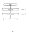
- the blood analyzing method of the blood analyzer 100 is described below.
- the blood analyzing method of the blood analyzer 100 includes a measurement step S1 and a data processing step S2.
- the sample preparing part 10 prepares a measurement sample by mixing a blood sample, a fluorescent dye for staining nucleic acid, and a hemolytic agent to hemolyze red blood cells. Specifically, a blood sample contained in a sample container is dispensed. The dispensed sample is then mixed with fluorescent dye and hemolytic agent in a mixing container to obtain a measurement sample.
- the measurement sample is then irradiated by light from the light source 21.
- the measurement sample is supplied together with the sheath fluid so as to flow through the flow cell 23.
- Light from the light source 21 is irradiated on the measurement sample flowing through the flow cell 23.
- the forward scattered light receiver 22a receives the forward scattered light, and outputs forward scattered light signals corresponding to the intensity of the forward scattered light.
- the side scattered light receiver 22b receives the side scattered light, and outputs side scattered light signals corresponding to the intensity of the side scattered light.
- the fluorescent light receiver 22c receives the fluorescent light, and outputs fluorescent light signals corresponding to the intensity of the fluorescent light.
- the processing part 30 counts the blood cells based on the obtained signals.
- the reactive B lymphocytes are detected based on at least one of the fluorescent light signals and forward scattered light signals.
- the reactive B lymphocytes are counted based on the detection results of the reactive B lymphocytes. At least the number of reactive B lymphocytes of the count result is output.
- the white blood cells are discriminated as reactive lymphocytes and blood cells other than reactive lymphocytes based on the fluorescent light signals and side scattered light signals.
- the reactive B lymphocytes are also detected among the reactive lymphocytes based on at least one of the fluorescent light signals and forward scattered light signals in the data processing step S2.
- the reactive B lymphocytes also may be detected among the discriminated lymphocytes after discriminating the white blood cells as lymphocytes and blood cells other than lymphocytes.
- white blood cells also may be discriminated at one time as reactive B lymphocytes, lymphocytes other than reactive B lymphocytes, and white blood cells other than lymphocytes based on the fluorescent light signals, side scattered light signals, and forward scattered light signals.
- the data processing step S2 may include, for example, a signal receiving step S21 in which the processing part 30 receives the fluorescent light signals, forward scattered light signals, and side scattered light signals from the receiving part 22, as shown in FIG. 3 .
- the processing part 30 also may store the forward scattered light intensity, side scattered light intensity, and fluorescent light intensity corresponding to each particle in the obtained measurement sample as data associated with each particle in a memory.
- the processing part 30 next analyzes the data of the fluorescent light signals, forward scattered light signals, and side scattered light signals stored in memory, using an analysis program which is pre-stored in the memory.
- the processing part 30 first discriminates the white blood cells in the measurement sample into subclasses of lymphocytes, monocytes, neutrophils, basophils, and eosinophils and the like based on the fluorescent light signals and side scattered light signals. Then, the discriminated lymphocytes are further discriminated as normal lymphocytes, reactive B lymphocytes, and reactive T lymphocytes, and respectively counted based on at least one of the fluorescent light signals and forward scattered light signals.
- the processing part 30 stores the detection and count results obtained in the detection and counting step S22.
- the processing part 30 displays the detection and counting results obtained in the detection and counting step S22 on the display part 40.
- the present inventors as a result of performing comparison experiments with clinical trials, have found that the measured fluorescent light intensity and the forward scattered light intensity are different in reactive B lymphocytes, reactive T lymphocytes, and in normal lymphocytes.
- the fluorescent light signals and forward scattered light signals obtained from reactive T lymphocytes have stronger intensities than the fluorescent light signals and forward scattered light signals obtained from normal lymphocytes.
- the fluorescent light signals and forward scattered light signals obtained form reactive B lymphocytes also have stronger intensities than the fluorescent light signals and forward scattered light signals obtained from normal lymphocytes and reactive T lymphocytes.
- the present inventors conceived that reactive B lymphocytes and normal lymphocytes can be discriminated and the reactive B lymphocytes can be counted based on at least one of the fluorescent light intensity and forward scattered light intensity.
- reactive T lymphocytes and normal lymphocytes can be discriminated and the reactive T lymphocytes can be counted based on at least one of the fluorescent light intensity and forward scattered light intensity.
- reactive B lymphocytes are counted based on the side scattered light intensity and at least one of the fluorescent light intensity and forward scattered light intensity. It is further preferable that reactive B lymphocytes are counted based on the side scattered light intensity, fluorescent light intensity, and forward scattered light intensity.
- a first example of the reactive lymphocyte detection and counting method is described using the scattergram of FIG. 4 .
- White blood cells in the measurement sample first are discriminated into subclasses of lymphocytes, monocytes, neutrophils, basophils, and eosinophils and the like based on the intensities of the fluorescent light signals and side scattered light signals.
- the discriminated lymphocytes are further discriminated as normal lymphocytes, reactive B lymphocytes, and reactive T lymphocytes based on the fluorescent light intensity and side scattered light intensity, which are the parameters used as the axes in constructing the scattergram shown in FIG. 4 .
- Each lymphocyte can be specifically discriminated by the specific method disclosed in Japanese Laid-Open Patent Application No. H5-149863 as a method for discriminating lymphocytes as normal lymphocytes, reactive B lymphocytes, and reactive T lymphocytes.
- fixed region first are set for the respective lymphocytes of normal lymphocytes, reactive B lymphocytes, and reactive T lymphocytes.
- the fixed region is set so that the degree of membership in a cluster becomes 1 relative to the fixed region of each lymphocyte within the fixed region.
- the position of the initial center of gravity of the distribution is determined for each cluster based on the particle distribution within the cluster.
- the position of the initial center of gravity is set as the center of gravity of each cluster, and the distance to the initial center of gravity is calculated for particles outside each fixed region.
- the degree of membership of each cluster is determined based on the calculated distance.
- Each particle is assigned to a cluster according to the degree of membership in the cluster, and the center of gravity position is redetermined for each cluster.
- Whether the difference between the previous center of gravity position and the current center of gravity position is less than a standard value is then determined.
- a new center of gravity position is determined until the difference between the previous center of gravity position and the current center of gravity position is less than the standard value, and the number of particles belonging to each particle cluster is calculated when the difference between the previous center of gravity position and the current center of gravity position becomes less than the standard value.
- the normal lymphocytes, reactive B lymphocytes, and reactive T lymphocytes can be specified by the above process.
- the number of reactive T lymphocytes and the number of reactive B lymphocytes can be combined to provide the total number of reactive lymphocytes. a large number of lymphocytes appear in the region in which the fluorescent light intensity is high in the sample collected from a subject with an infectious disease shown in FIG. 4 , whereas lymphocytes scarcely appear in the region in which the fluorescent light intensity is high in the normal sample shown in FIG. 5 .
- a method for specifying by the strength of the fluorescent light signal may also be used as a separate method of specifying each lymphocyte. Specifically, lymphocytes which have a fluorescent light intensity below a first threshold value are detected and counted as normal lymphocytes, lymphocytes which have a fluorescent light intensity between the first threshold value and a second threshold value that is higher than the first threshold value are detected and counted as reactive T lymphocytes, are detected and counted as normal lymphocytes, and lymphocytes which have a fluorescent light intensity above the second threshold value are detected and counted as reactive B lymphocytes,
- each type of lymphocyte is detected using the fluorescent light intensity.
- lymphocytes also may be discriminated based on the forward scattered light intensity as an axial parameter used to configure the scattergram shown in FIG. 6 .
- the forward scattered light intensity is high similar to the previously described fluorescent light intensity in the sample collected from a subject with an infectious disease shown in FIG. 6 .
- discrimination using the fluorescent light intensity is preferable due to the higher resolution when discriminating lymphocytes as reactive B lymphocytes and reactive T lymphocytes.
- each type of lymphocyte may be detected using at least only fluorescent light intensity or only forward scattered light intensity when discriminating lymphocytes as reactive B lymphocytes and reactive T lymphocytes. Lymphocytes also may be discriminated based on fluorescent light intensity and side scattered light intensity, or forward scattered light intensity and side scattered light intensity rather than using only fluorescent light intensity and side scattered light intensity. Each type of lymphocyte also can be detected based on fluorescent light intensity, forward scattered light intensity, and side scattered light intensity.
- the present invention supplies a program for realizing the above described blood analyzing method to a computer or blood analyzer through a network or various types of storage media, and executes the program by the computer or blood analyzer reading the program.
- the number of reactive T lymphocytes counted by the method of the present invention, and the number of CD8-positive T lymphocytes counted using fluorescently labeled CD8 + antibody were obtained from 48 samples provided by different subjects. A strong positive correlation was obtained when a correlation coefficient of 0.9336 was determined for the reactive T lymphocytes counted by the method of the present invention and the number of CD8-positive T lymphocytes counted using fluorescently labeled CD8 + antibody.
- the proportion of the number of atypical lymphocytes relative to the number of lymphocytes was determined by observing cells using a microscope for the 48 samples obtained from different subjects mentioned above. A strong positive correlation was obtained when a correlation coefficient of 0.8968 was determined for the proportion of the number of reactive T lymphocytes relative to the number of lymphocytes counted by the method of the present invention and the proportion of the number of abnormal cells relative to the number of lymphocytes determined by the microscopic examination.
- the number of reactive T lymphocytes counted by the method of the present invention is understood to be the number of abnormal cells, that is, T lymphocytes, and is strongly presumed to be the number of reactive T lymphocytes.
- the proportion of the number of plasma cells relative to the number of lymphocytes was determined by observing cells using a microscope for 33 samples obtained from different subjects. A strong positive correlation was obtained when a correlation coefficient of 0.6108 was determined for the proportion of the number of reactive B lymphocytes relative to the number of lymphocytes counted by the method of the present invention and the proportion of the number of plasma cells relative to the number of lymphocytes determined by the microscopic examination.
- the reactive B lymphocytes counted by the method of the present invention are strongly presumed to be plasma cells, that is, reactive B lymphocytes.
Abstract
Description
- The invention relates to a blood analyzer, blood analyzing method, and blood analyzing program.
- White blood cells such as lymphocytes, monocytes, basophils, eosinophils and neutrophils are present in normal peripheral blood. Abnormal white blood cells which are not present in normal peripheral blood appear in diseases such as viral infections or hematopoietic tumors. Abnormal white blood cells include, for example, reactive lymphocytes which have been activated by antigen stimulation.
- For example, Japanese laid-open patent No.
2012-233754 - Atypical lymphocytes include reactive B lymphocytes and reactive T lymphocytes. However, although the method disclosed in
Patent Document 1 can discriminate atypical lymphocytes, this method cannot discriminate and count reactive B lymphocytes alone. A blood analyzer and blood analyzing method capable of discriminating and counting reactive B lymphocytes is therefore desirable. - The blood analyzer of the present invention includes a sample preparing part configured to prepare a measurement sample by mixing a blood sample, a fluorescent dye for staining nucleic acid, and a hemolytic agent to hemolyze red blood cells; a light source configured to irradiate light on the measurement sample; a light receiving part configured to receive fluorescent light, forward scattered light, and side scattered light given off by the measurement sample irradiated by light, and output fluorescent light signals corresponding to the intensity of the fluorescent light, forward scattered light signals corresponding to the intensity of the forward scattered light, and side scattered light signals corresponding to the intensity of the side scattered light; and a processing part configured to discriminate and count the reactive B lymphocytes based on at least one of the fluorescent light signal and forward scattered light signal.
- The blood analyzing method of the present invention includes a step of obtaining fluorescent light signals corresponding to the intensity of the fluorescent light, forward scattered light signals corresponding to the intensity of the forward scattered light, and side scattered light signals corresponding to the intensity of side scattered light given off when the measurement sample is irradiated by light; and a step of discriminating and counting the reactive B lymphocytes based on at least one of the fluorescent light signal and forward scattered light signal.
- The blood analyzing program of the present invention executes, on a computer, a step of obtaining fluorescent light signals corresponding to the intensity of the fluorescent light, forward scattered light signals corresponding to the intensity of the forward scattered light, and side scattered light signals corresponding to the intensity of side scattered light given off when a measurement sample is irradiated by light; and a step of discriminating and counting the reactive B lymphocytes based on at least one of the fluorescent light signal and forward scattered light signal.
- The present invention provides a blood analyzer, blood analyzing method, and blood analyzing program capable of counting reactive B lymphocytes.
-
-
FIG. 1 is a brief block diagram of the blood analyzer of an embodiment of the invention. -
FIG. 2 is a flow chart of the blood analyzing method of an embodiment of the invention; -
FIG. 3 is a flow chart of the data processing step of the blood analyzing method of an embodiment of the invention; -
FIG. 4 is an example of a scattergram in which the axes are the side scattered light intensity and the fluorescent light intensity of a sample collected from a subject with an infectious disease; -
FIG. 5 is an example of a scattergram in which the axes are the side scattered light intensity and the fluorescent light intensity of a normal sample; -
FIG. 6 is an example of a scattergram in which the axes are the side scattered light intensity and the forward scattered light intensity of a sample collected from a subject with an infectious disease; and -
FIG. 7 is an example of a scattergram in which the axes are the side scattered light intensity and the forward scattered light intensity of a normal sample. - A preferred embodiment of the present invention is described in the following examples.
-
FIG. 1 is a brief block diagram of theblood analyzer 100 of the embodiment. Theblood analyzer 100 is a device for analyzing information related to various blood cells contained in blood using a flow cytometric method. - The
blood analyzer 100 is provided with asample preparing part 10. Thesample preparing part 10 is the part that prepares measurement sample to be used in analysis. Thesample preparing part 10 prepares a measurement sample by mixing a blood sample (whole blood), and various reagents such as hemolytic agent and fluorescent dye. - Hemolytic agent is a reagent for lysing red blood cells. Therefore, red blood cells are hemolyzed in the measurement sample containing hemolytic agent.
- Hemolytic agent preferably contains, for example, a surfactant. Cationic surfactant is an example of a preferred surfactant. Cationic surfactant has strong lysing power. Cationic surfactant is used to hemolyze red blood cells, and to injure the cell membranes of normal white blood cells and abnormal mononuclear cells. Therefore, normal white blood cells and abnormal mononuclear cells are easily stained by fluorescent dye. Examples of preferred cationic surfactant include quaternary ammonium salt type surfactant and pyridinium salt type surfactant. Specific examples of preferred cationic surfactant are surfactants having 9 to 30 carbon total carbon atoms represented by, for example, structural formulae (I) and (II).
- Formula (I): http://dbboysysmex.co.jp/DBBOY/2014-06004588/1/0/9842.php Formula (I)

- Formula (II): http://dbboy.sysmex.co.jp/DBBOY/2014-06004589/1/0/19139.php (In formulae (I) and (II), R1 is an alkyl group or alkenyl group having 6 to 18 carbon atoms. Formula (II)

- R2 and R3 are an alkyl or alkenyl group having 1 to 4 carbon atoms. R4 is an alkyl group having 1 to 4 carbon atoms, an alkenyl group or a benzyl group. X is a halogen atom.)
- An alkyl group or alkenyl group having 6, 8, 10, 12, and 14 carbon atoms is preferable, while a straight-chain alkyl group is particularly preferred as R1. Specific examples preferred as R1 include octyl, decyl and dodecyl groups.
- A methyl group, ethyl group and propyl group are particularly preferred as R2 and R3.
- A methyl group, ethyl group and propyl group are preferred as R4.
- Hemolytic agent, for example, may include surfactants other than cationic surfactant such as nonionic surfactants in addition to the cationic surfactant. Hemolytic agent also may include, for example, organic acids and buffer agents in addition to surfactant.
- Fluorescent dye is an agent to stain nucleic acid. Red blood cells which do not have nucleic acid are therefore not stained by the fluorescent dye. Cells which have a nucleus such as nucleated red blood cells and white blood cells which have nucleic acid are stained by the fluorescent dye.
- Examples of fluorescent dyes include propidium iodide, ethidium bromide, ethidium - acridine heterodimer, ethidium diazide, ethidium homodimer-1, ethidium homodimer-2, ethidium monoazide, trimethylene bis [[3 - [[4 - [[(3-methyl-benzothiazole-3-ium) -2-yl] methylene] -1,4-dihydro-quinoline] -1-yl] propyl] dimethyl aminium]-tetraiodide (TOTO-1), 4 - [(3-methyl-benzothiazole -2 (3H) - ylidene) methylcarbamoyl] -1- [3- (trimethyl amino niobium) propyl] quinolinium-diiodide (TO-PRO-1), N, N, N', N'- tetramethyl -N, N'- bis [3-[4- [3-[(3-methyl-benzothiazole-3-ium)-2-yl] -2-propenylidene] -1,4-dihydro-quinolin-1-yl] propyl]-1,3-propanediaminium tetraiodide (TOTO-3), or 2- [3-[[1-[3-(trimethyl Ami niobium) propyl] -1,4-dihydro-quinoline] -4-ylidene] -1-propenyl] -3-methyl-benzothiazole-3-ium-diiodide (TO-PRO-3).
- The measurement sample prepared in the sample preparing part is supplied to a
measuring part 20. Themeasuring part 20 has thelight source 21, thelight receiving part 22, and aflow cell 23. The measurement sample is fed into theflow cell 23. A sheath fluid is fed together with the measurement sample to theflow cell 23. The measurement sample together with the sheath fluid flows through theflow cell 23. - The
light source 21 irradiates light on the measurement sample within theflow cell 23. Thelight source 21 is configured by, for example, a semiconductor laser light source or the like. The wavelength of the might emitted by thelight source 21 may be, for example, 633 nm. - Fluorescent light, side scattered light, and forward scattered light are produced when the light from the
light source 21 irradiates the measurement sample. The forward scattered light is received by a forward scatteredlight receiver 22a. The forward scatteredlight receiver 22a outputs a forward scattered light signal which corresponds to the intensity of the forward scattered light to theprocessing part 30. - The
processing part 30 is configured by a central processing unit (CPU) of a personal computer installed externally. Note that theprocessing part 30 is not limited to this configuration and also may be the central processing unit of a computer provided within theblood analyzer 100. - The side scattered light is received by a side scattered
light receiver 22b. The side scatteredlight receiver 22b outputs a side scattered light signal which corresponds to the intensity of the side scattered light to theprocessing part 30. - The fluorescent light is received by a fluorescent
light receiver 22c. The fluorescentlight receiver 22c outputs a fluorescent light signal which corresponds to the intensity of the fluorescent light to theprocessing part 30. - The
light receiving part 22 is configured by the forward scatteredlight receiver 22a, side scatteredlight receiver 22b, and fluorescentlight receiver 22c. Note that thelight receiving part 22 may be configured by, for example, photodiodes or the like. - The
processing part 30 calculates information related to various blood cells and the like contained in blood as a measurement sample based on the input fluorescent light signal, side scattered light signal, and forward scattered light signal. - The
processing part 30 detects the reactive B lymphocytes based on at least one of the fluorescent light signal and forward scattered light signal. Theprocessing part 30 counts the reactive B lymphocytes based on the detection result. Theprocessing part 30 outputs the number of reactive B lymphocytes as a count result. - More specifically, the
processing part 30 discriminates white blood cells as reactive lymphocytes and white blood cells other than reactive lymphocytes based on the fluorescent light signals and side scattered light signals. Theprocessing part 30 also discriminates the reactive B lymphocytes among the reactive lymphocytes based on at least one of the fluorescent light signals and forward scattered light signals. Note that theprocessing part 30 also may detect the reactive B lymphocytes among the lymphocytes after discriminating the white blood cells as lymphocytes and blood cells other than lymphocytes. - The
processing part 30 also may discriminate white blood cells as reactive B lymphocytes, lymphocytes other than reactive B lymphocytes, and white blood cells other than lymphocytes based on the fluorescent light signals, side scattered light signals, and forward scattered light signals. White blood cells other than lymphocytes include, for example, monocytes, neutrophils, eosinophils, basophils and the like. - A
display part 40 is connected to theprocessing part 30. Thedisplay part 40 shows the count results output from theprocessing part 30. Thedisplay part 40 at least shows the number of reactive B lymphocytes. Thedisplay part 40 shows, for example, at least one type of information selected from a group including the reactive B lymphocyte count, the reactive T lymphocyte count, the ratio of the reactive B lymphocyte count and reactive T lymphocyte count, the ratio of the lymphocyte count and the reactive lymphocyte count, and the ratio of the white blood cell count and the reactive lymphocyte count. - The blood analyzing method of the
blood analyzer 100 is described below. - As shown in
FIG. 2 , the blood analyzing method of theblood analyzer 100 includes a measurement step S1 and a data processing step S2. - In the measurement step S1 of the
blood analyzer 100, thesample preparing part 10 prepares a measurement sample by mixing a blood sample, a fluorescent dye for staining nucleic acid, and a hemolytic agent to hemolyze red blood cells. Specifically, a blood sample contained in a sample container is dispensed. The dispensed sample is then mixed with fluorescent dye and hemolytic agent in a mixing container to obtain a measurement sample. - The measurement sample is then irradiated by light from the
light source 21. Specifically, the measurement sample is supplied together with the sheath fluid so as to flow through theflow cell 23. Light from thelight source 21 is irradiated on the measurement sample flowing through theflow cell 23. When the light impinges the measurement sample, fluorescent light, side scattered light, and forward scattered light are produced. The forward scatteredlight receiver 22a receives the forward scattered light, and outputs forward scattered light signals corresponding to the intensity of the forward scattered light. The side scatteredlight receiver 22b receives the side scattered light, and outputs side scattered light signals corresponding to the intensity of the side scattered light. The fluorescentlight receiver 22c receives the fluorescent light, and outputs fluorescent light signals corresponding to the intensity of the fluorescent light. - Then, in the data processing step S2, the
processing part 30 counts the blood cells based on the obtained signals. - In the data processing step S2, at least the reactive B lymphocytes are detected based on at least one of the fluorescent light signals and forward scattered light signals. The reactive B lymphocytes are counted based on the detection results of the reactive B lymphocytes. At least the number of reactive B lymphocytes of the count result is output.
- More specifically, in the data processing step S2, the white blood cells are discriminated as reactive lymphocytes and blood cells other than reactive lymphocytes based on the fluorescent light signals and side scattered light signals. The reactive B lymphocytes are also detected among the reactive lymphocytes based on at least one of the fluorescent light signals and forward scattered light signals in the data processing step S2.
- In the data processing step S2, the reactive B lymphocytes also may be detected among the discriminated lymphocytes after discriminating the white blood cells as lymphocytes and blood cells other than lymphocytes. In the data processing step S2, white blood cells also may be discriminated at one time as reactive B lymphocytes, lymphocytes other than reactive B lymphocytes, and white blood cells other than lymphocytes based on the fluorescent light signals, side scattered light signals, and forward scattered light signals.
- More specifically, the data processing step S2 may include, for example, a signal receiving step S21 in which the
processing part 30 receives the fluorescent light signals, forward scattered light signals, and side scattered light signals from the receivingpart 22, as shown inFIG. 3 . Theprocessing part 30 also may store the forward scattered light intensity, side scattered light intensity, and fluorescent light intensity corresponding to each particle in the obtained measurement sample as data associated with each particle in a memory. - In a detection and counting step S22, the
processing part 30 next analyzes the data of the fluorescent light signals, forward scattered light signals, and side scattered light signals stored in memory, using an analysis program which is pre-stored in the memory. - The
processing part 30 first discriminates the white blood cells in the measurement sample into subclasses of lymphocytes, monocytes, neutrophils, basophils, and eosinophils and the like based on the fluorescent light signals and side scattered light signals. Then, the discriminated lymphocytes are further discriminated as normal lymphocytes, reactive B lymphocytes, and reactive T lymphocytes, and respectively counted based on at least one of the fluorescent light signals and forward scattered light signals. - Then, in the results storing step S23, the
processing part 30 stores the detection and count results obtained in the detection and counting step S22. In the display step S24, theprocessing part 30 displays the detection and counting results obtained in the detection and counting step S22 on thedisplay part 40. - The present inventors, as a result of performing comparison experiments with clinical trials, have found that the measured fluorescent light intensity and the forward scattered light intensity are different in reactive B lymphocytes, reactive T lymphocytes, and in normal lymphocytes. Specifically, the fluorescent light signals and forward scattered light signals obtained from reactive T lymphocytes have stronger intensities than the fluorescent light signals and forward scattered light signals obtained from normal lymphocytes. The fluorescent light signals and forward scattered light signals obtained form reactive B lymphocytes also have stronger intensities than the fluorescent light signals and forward scattered light signals obtained from normal lymphocytes and reactive T lymphocytes. As a result, the present inventors conceived that reactive B lymphocytes and normal lymphocytes can be discriminated and the reactive B lymphocytes can be counted based on at least one of the fluorescent light intensity and forward scattered light intensity. Similarly, reactive T lymphocytes and normal lymphocytes can be discriminated and the reactive T lymphocytes can be counted based on at least one of the fluorescent light intensity and forward scattered light intensity.
- From the perspective of greater accuracy in counting reactive B lymphocytes and reactive T lymphocytes, it is preferable that reactive B lymphocytes are counted based on the side scattered light intensity and at least one of the fluorescent light intensity and forward scattered light intensity. It is further preferable that reactive B lymphocytes are counted based on the side scattered light intensity, fluorescent light intensity, and forward scattered light intensity.
- Examples of the method of counting reactive lymphocytes are described in more detail below.
- A first example of the reactive lymphocyte detection and counting method is described using the scattergram of
FIG. 4 . - White blood cells in the measurement sample first are discriminated into subclasses of lymphocytes, monocytes, neutrophils, basophils, and eosinophils and the like based on the intensities of the fluorescent light signals and side scattered light signals.
- In the first example, the discriminated lymphocytes are further discriminated as normal lymphocytes, reactive B lymphocytes, and reactive T lymphocytes based on the fluorescent light intensity and side scattered light intensity, which are the parameters used as the axes in constructing the scattergram shown in
FIG. 4 . - Each lymphocyte can be specifically discriminated by the specific method disclosed in Japanese Laid-Open Patent Application No.
H5-149863 - Specifically, fixed region first are set for the respective lymphocytes of normal lymphocytes, reactive B lymphocytes, and reactive T lymphocytes. The fixed region is set so that the degree of membership in a cluster becomes 1 relative to the fixed region of each lymphocyte within the fixed region. Next, the position of the initial center of gravity of the distribution is determined for each cluster based on the particle distribution within the cluster. Next, the position of the initial center of gravity is set as the center of gravity of each cluster, and the distance to the initial center of gravity is calculated for particles outside each fixed region. The degree of membership of each cluster is determined based on the calculated distance. Each particle is assigned to a cluster according to the degree of membership in the cluster, and the center of gravity position is redetermined for each cluster. Whether the difference between the previous center of gravity position and the current center of gravity position is less than a standard value is then determined. A new center of gravity position is determined until the difference between the previous center of gravity position and the current center of gravity position is less than the standard value, and the number of particles belonging to each particle cluster is calculated when the difference between the previous center of gravity position and the current center of gravity position becomes less than the standard value. The normal lymphocytes, reactive B lymphocytes, and reactive T lymphocytes can be specified by the above process. The number of reactive T lymphocytes and the number of reactive B lymphocytes can be combined to provide the total number of reactive lymphocytes.
a large number of lymphocytes appear in the region in which the fluorescent light intensity is high in the sample collected from a subject with an infectious disease shown inFIG. 4 , whereas lymphocytes scarcely appear in the region in which the fluorescent light intensity is high in the normal sample shown inFIG. 5 . - A method for specifying by the strength of the fluorescent light signal may also be used as a separate method of specifying each lymphocyte. Specifically, lymphocytes which have a fluorescent light intensity below a first threshold value are detected and counted as normal lymphocytes, lymphocytes which have a fluorescent light intensity between the first threshold value and a second threshold value that is higher than the first threshold value are detected and counted as reactive T lymphocytes, are detected and counted as normal lymphocytes, and lymphocytes which have a fluorescent light intensity above the second threshold value are detected and counted as reactive B lymphocytes,
- In the above example, each type of lymphocyte is detected using the fluorescent light intensity. However, the present invention is not limited to this example. For example, lymphocytes also may be discriminated based on the forward scattered light intensity as an axial parameter used to configure the scattergram shown in
FIG. 6 . Compared to the normal sample shown inFIG. 7 , it is understood that lymphocytes appear in the region in which the forward scattered light intensity is high similar to the previously described fluorescent light intensity in the sample collected from a subject with an infectious disease shown inFIG. 6 . As can be understood fromFIGS. 4 and6 , discrimination using the fluorescent light intensity is preferable due to the higher resolution when discriminating lymphocytes as reactive B lymphocytes and reactive T lymphocytes. - As mentioned above, each type of lymphocyte may be detected using at least only fluorescent light intensity or only forward scattered light intensity when discriminating lymphocytes as reactive B lymphocytes and reactive T lymphocytes. Lymphocytes also may be discriminated based on fluorescent light intensity and side scattered light intensity, or forward scattered light intensity and side scattered light intensity rather than using only fluorescent light intensity and side scattered light intensity. Each type of lymphocyte also can be detected based on fluorescent light intensity, forward scattered light intensity, and side scattered light intensity.
- Note that the present invention supplies a program for realizing the above described blood analyzing method to a computer or blood analyzer through a network or various types of storage media, and executes the program by the computer or blood analyzer reading the program.
- Count results obtained using the
blood analyzer 100 are described through experiments comparing with conventional methods. - The number of reactive T lymphocytes counted by the method of the present invention, and the number of CD8-positive T lymphocytes counted using fluorescently labeled CD8 + antibody were obtained from 48 samples provided by different subjects. A strong positive correlation was obtained when a correlation coefficient of 0.9336 was determined for the reactive T lymphocytes counted by the method of the present invention and the number of CD8-positive T lymphocytes counted using fluorescently labeled CD8 + antibody.
- The proportion of the number of atypical lymphocytes relative to the number of lymphocytes was determined by observing cells using a microscope for the 48 samples obtained from different subjects mentioned above. A strong positive correlation was obtained when a correlation coefficient of 0.8968 was determined for the proportion of the number of reactive T lymphocytes relative to the number of lymphocytes counted by the method of the present invention and the proportion of the number of abnormal cells relative to the number of lymphocytes determined by the microscopic examination.
- The number of reactive T lymphocytes counted by the method of the present invention is understood to be the number of abnormal cells, that is, T lymphocytes, and is strongly presumed to be the number of reactive T lymphocytes.
- The proportion of the number of plasma cells relative to the number of lymphocytes was determined by observing cells using a microscope for 33 samples obtained from different subjects. A strong positive correlation was obtained when a correlation coefficient of 0.6108 was determined for the proportion of the number of reactive B lymphocytes relative to the number of lymphocytes counted by the method of the present invention and the proportion of the number of plasma cells relative to the number of lymphocytes determined by the microscopic examination.
- From the above, the reactive B lymphocytes counted by the method of the present invention are strongly presumed to be plasma cells, that is, reactive B lymphocytes.
Claims (15)
- A blood analyzer comprising:a sample preparing part configured to prepare a measurement sample by mixing a blood sample, a fluorescent dye for staining nucleic acid, and a hemolytic agent to hemolyze red blood cells;a light source configured to irradiate light on the measurement sample;a light receiving part configured to receive fluorescent light, forward scattered light, and side scattered light given off by the measurement sample irradiated by light, and output fluorescent light signals corresponding to the intensity of the fluorescent light, forward scattered light signals corresponding to the intensity of the forward scattered light, and side scattered light signals corresponding to the intensity of the side scattered light; anda processing part configured to discriminate and count the reactive B lymphocytes based on at least one of the fluorescent light signals and forward scattered light signals.
- The blood analyzer of claim 1, wherein the processing part discriminates lymphocytes from blood cells in the measurement sample and discriminates reactive B lymphocytes from the discriminated lymphocytes based on the fluorescent light signals and the side scattered light signals.
- The blood analyzer of claim 1 or claim 2, wherein the processing part discriminates and counts the reactive B lymphocytes based on the side scattered light and at least one of the fluorescent light signals, and forward scattered light signals.
- The blood analyzer of claim 1 or claim 2, wherein the processing part discriminates and counts the reactive B lymphocytes based on the side scattered light signals and the fluorescent light signals.
- The blood analyzer of any one among claims 1 through 4, wherein the processing part discriminates and counts the reactive T lymphocytes based on at least one of the fluorescent light signals and forward scattered light signals.
- The blood analyzer of any one among claims 1 through 5, further comprising:a display part configured to display the results output from the processing part, wherein the processing part shows the count results on the display part.
- The blood analyzer of claim 6, wherein the display part shows at least one type of information selected from a group including the reactive B lymphocyte count, the reactive T lymphocyte count, the ratio of the reactive B lymphocyte count and reactive T lymphocyte count, the ratio of the lymphocyte count and the reactive lymphocyte count, and the ratio of the white blood cell count and the reactive lymphocyte count.
- A blood analyzing method comprising:a step of obtaining fluorescent light signals corresponding to the intensity of the fluorescent light, forward scattered light signals corresponding to the intensity of the forward scattered light, and side scattered light signals corresponding to the intensity of side scattered light given off when the measurement sample is irradiated by light; anda step of discriminating and counting the reactive B lymphocytes based on at least one of the fluorescent light signals and forward scattered light signals.
- The blood analyzing method of claim 8, wherein lymphocytes are discriminated from blood cells in the measurement sample and reactive B lymphocytes are discriminated from the discriminated lymphocytes based on the fluorescent light signals and the side scattered light signals in the step of discriminating and counting the reactive B lymphocytes.
- The blood analyzing method of claim 8 or 9, wherein reactive B lymphocytes are discriminated based on the side scattered light, and at least one of the fluorescent light signals and forward scattered light signals in the step of discriminating and counting the reactive B lymphocytes.
- The blood analyzing method of any one among claims 9 or 10, further comprising: a step of discriminating and counting the reactive T lymphocytes based on at least one of the fluorescent light signals and forward scattered light signals.
- A blood analyzing program configured to execute, on a computer, comprising:a step of obtaining fluorescent light signals corresponding to the intensity of the fluorescent light, forward scattered light signals corresponding to the intensity of the forward scattered light, and side scattered light signals corresponding to the intensity of side scattered light given off when a measurement sample is irradiated by light; anda step of discriminating and counting the reactive B lymphocytes based on at least one of the fluorescent light signals and forward scattered light signals.
- The blood analyzing program of claim 12, wherein lymphocytes are discriminated from blood cells in the measurement sample and reactive B lymphocytes are discriminated from the discriminated lymphocytes based on the fluorescent light signals and the side scattered light signals in the step of discriminating and counting the reactive B lymphocytes.
- The blood analyzing program of claim 12 or 13, wherein reactive B lymphocytes are discriminated based on the side scattered light, and at least one of the fluorescent light signals and forward scattered light signals in the step of discriminating and counting the reactive B lymphocytes.
- The blood analyzing program of any one among claims 12 through 14, further comprising:a step of discriminating and counting the reactive T lymphocytes based on at least one of the fluorescent light signals and forward scattered light signals.
Applications Claiming Priority (1)
| Application Number | Priority Date | Filing Date | Title |
|---|---|---|---|
| JP2014126606A JP6461494B2 (en) | 2014-06-19 | 2014-06-19 | Blood analyzer, blood analysis method and blood analysis program |
Publications (2)
| Publication Number | Publication Date |
|---|---|
| EP2957889A1 true EP2957889A1 (en) | 2015-12-23 |
| EP2957889B1 EP2957889B1 (en) | 2019-01-30 |
Family
ID=53397939
Family Applications (1)
| Application Number | Title | Priority Date | Filing Date |
|---|---|---|---|
| EP15172056.2A Active EP2957889B1 (en) | 2014-06-19 | 2015-06-15 | Blood analyzer, blood analyzing method, and blood analyzing program |
Country Status (4)
| Country | Link |
|---|---|
| US (1) | US10578603B2 (en) |
| EP (1) | EP2957889B1 (en) |
| JP (1) | JP6461494B2 (en) |
| CN (1) | CN105277519B (en) |
Families Citing this family (7)
| Publication number | Priority date | Publication date | Assignee | Title |
|---|---|---|---|---|
| CN105928907A (en) * | 2016-05-17 | 2016-09-07 | 深圳普门科技有限公司 | Erythrocyte-osmotic-fragility measuring method based on scattering turbidimetric technology |
| CN107655865B (en) * | 2016-07-25 | 2021-10-19 | 希森美康株式会社 | Blood analysis device and blood analysis method |
| CN107219195B (en) * | 2017-05-23 | 2019-07-23 | 山东中医药大学附属医院 | A kind of blood leucocyte detection device and method |
| JP6903494B2 (en) | 2017-06-09 | 2021-07-14 | シスメックス株式会社 | Particle analysis methods for identifying infectious diseases |
| EP4006527A4 (en) * | 2019-07-25 | 2023-02-15 | Seoul Viosys Co., Ltd | Light irradiation apparatus |
| CN111504869B (en) * | 2020-05-15 | 2021-06-08 | 中国计量科学研究院 | Flow type aggregate impurity analyzer |
| WO2023125939A1 (en) * | 2021-12-31 | 2023-07-06 | 深圳迈瑞生物医疗电子股份有限公司 | Hematology analyzer, method for indicating infection status, and use of infection flag parameter |
Citations (7)
| Publication number | Priority date | Publication date | Assignee | Title |
|---|---|---|---|---|
| JPH05149863A (en) | 1991-11-27 | 1993-06-15 | Toa Medical Electronics Co Ltd | Particle counting method |
| US20050069959A1 (en) * | 2003-09-26 | 2005-03-31 | Ayumu Yoshida | Method and apparatus for classifying B-lymphocytes |
| US20090105963A1 (en) * | 2006-05-13 | 2009-04-23 | Jesper Laursen | Methods for Flow Cytometry Analyses of Un-Lysed Cells from Biological Fluids |
| US20120115159A1 (en) * | 2009-07-03 | 2012-05-10 | Sysmex Corporation | Blood analyzer and blood analyzing method |
| JP2012233754A (en) | 2011-04-28 | 2012-11-29 | Sysmex Corp | Method for classifying/counting leukocyte, leukocyte classification reagent kit, and leukocyte classification reagent |
| US20140051071A1 (en) * | 2011-04-28 | 2014-02-20 | Sysmex Corporation | Blood analyzer, blood analysis method, and computer program product |
| US20140147837A1 (en) * | 2012-11-26 | 2014-05-29 | Sysmex Corporation | Method for analyzing blood cells and blood cell analyzer |
Family Cites Families (3)
| Publication number | Priority date | Publication date | Assignee | Title |
|---|---|---|---|---|
| JP3345135B2 (en) * | 1992-11-19 | 2002-11-18 | シスメックス株式会社 | Blood analysis method |
| US7625712B2 (en) * | 2004-05-21 | 2009-12-01 | Beckman Coulter, Inc. | Method for a fully automated monoclonal antibody-based extended differential |
| EP2520926B1 (en) * | 2011-05-05 | 2022-06-15 | Sysmex Corporation | Blood analyzer, blood analysis method, and computer program product |
-
2014
- 2014-06-19 JP JP2014126606A patent/JP6461494B2/en active Active
-
2015
- 2015-06-10 CN CN201510315343.1A patent/CN105277519B/en active Active
- 2015-06-15 EP EP15172056.2A patent/EP2957889B1/en active Active
- 2015-06-16 US US14/740,815 patent/US10578603B2/en active Active
Patent Citations (7)
| Publication number | Priority date | Publication date | Assignee | Title |
|---|---|---|---|---|
| JPH05149863A (en) | 1991-11-27 | 1993-06-15 | Toa Medical Electronics Co Ltd | Particle counting method |
| US20050069959A1 (en) * | 2003-09-26 | 2005-03-31 | Ayumu Yoshida | Method and apparatus for classifying B-lymphocytes |
| US20090105963A1 (en) * | 2006-05-13 | 2009-04-23 | Jesper Laursen | Methods for Flow Cytometry Analyses of Un-Lysed Cells from Biological Fluids |
| US20120115159A1 (en) * | 2009-07-03 | 2012-05-10 | Sysmex Corporation | Blood analyzer and blood analyzing method |
| JP2012233754A (en) | 2011-04-28 | 2012-11-29 | Sysmex Corp | Method for classifying/counting leukocyte, leukocyte classification reagent kit, and leukocyte classification reagent |
| US20140051071A1 (en) * | 2011-04-28 | 2014-02-20 | Sysmex Corporation | Blood analyzer, blood analysis method, and computer program product |
| US20140147837A1 (en) * | 2012-11-26 | 2014-05-29 | Sysmex Corporation | Method for analyzing blood cells and blood cell analyzer |
Also Published As
| Publication number | Publication date |
|---|---|
| CN105277519B (en) | 2018-06-19 |
| US10578603B2 (en) | 2020-03-03 |
| EP2957889B1 (en) | 2019-01-30 |
| US20150369793A1 (en) | 2015-12-24 |
| CN105277519A (en) | 2016-01-27 |
| JP2016004030A (en) | 2016-01-12 |
| JP6461494B2 (en) | 2019-01-30 |
Similar Documents
| Publication | Publication Date | Title |
|---|---|---|
| EP2957889B1 (en) | Blood analyzer, blood analyzing method, and blood analyzing program | |
| CN103492875B (en) | Blood analysis device and blood analysis method | |
| EP3141885B1 (en) | Blood analyzer | |
| EP3073265B1 (en) | Blood analyzer and blood analysis method | |
| US9453790B2 (en) | Blood analyzer, blood analyzing method, and computer program product | |
| EP2450706B1 (en) | Blood analyzer and blood analyzing method | |
| US20140147837A1 (en) | Method for analyzing blood cells and blood cell analyzer | |
| US9797824B2 (en) | Method for hematology analysis | |
| CN106483109B (en) | Blood analysis device and blood analysis method | |
| CN111656161B (en) | Sample analyzer abnormality alarming method, system and storage medium | |
| US20210041344A1 (en) | Blood analysis system, blood analyzer, blood analysis method and storage medium | |
| JP7291337B2 (en) | Bone marrow fluid analysis method, sample analyzer and computer program | |
| US20210033593A1 (en) | Methods and systems for determining platelet concentration | |
| US20230296591A1 (en) | Sample analysis method, sample analyzer, and computer-readable storage medium | |
| Rico et al. | Flow-cytometry-based protocols for human blood/marrow immunophenotyping with minimal sample perturbation | |
| CN114450589A (en) | Method for analyzing red blood cells in blood sample and blood analysis system | |
| EP3136080B1 (en) | Urine sample analyzer and urine sample analyzing method | |
| JP2020521950A (en) | Method and system for sample analysis | |
| JPH04326061A (en) | Blood analyser |
Legal Events
| Date | Code | Title | Description |
|---|---|---|---|
| PUAI | Public reference made under article 153(3) epc to a published international application that has entered the european phase |
Free format text: ORIGINAL CODE: 0009012 |
|
| AK | Designated contracting states |
Kind code of ref document: A1 Designated state(s): AL AT BE BG CH CY CZ DE DK EE ES FI FR GB GR HR HU IE IS IT LI LT LU LV MC MK MT NL NO PL PT RO RS SE SI SK SM TR |
|
| AX | Request for extension of the european patent |
Extension state: BA ME |
|
| 17P | Request for examination filed |
Effective date: 20160324 |
|
| RBV | Designated contracting states (corrected) |
Designated state(s): AL AT BE BG CH CY CZ DE DK EE ES FI FR GB GR HR HU IE IS IT LI LT LU LV MC MK MT NL NO PL PT RO RS SE SI SK SM TR |
|
| STAA | Information on the status of an ep patent application or granted ep patent |
Free format text: STATUS: EXAMINATION IS IN PROGRESS |
|
| 17Q | First examination report despatched |
Effective date: 20170314 |
|
| GRAP | Despatch of communication of intention to grant a patent |
Free format text: ORIGINAL CODE: EPIDOSNIGR1 |
|
| STAA | Information on the status of an ep patent application or granted ep patent |
Free format text: STATUS: GRANT OF PATENT IS INTENDED |
|
| INTG | Intention to grant announced |
Effective date: 20180904 |
|
| GRAS | Grant fee paid |
Free format text: ORIGINAL CODE: EPIDOSNIGR3 |
|
| GRAA | (expected) grant |
Free format text: ORIGINAL CODE: 0009210 |
|
| STAA | Information on the status of an ep patent application or granted ep patent |
Free format text: STATUS: THE PATENT HAS BEEN GRANTED |
|
| AK | Designated contracting states |
Kind code of ref document: B1 Designated state(s): AL AT BE BG CH CY CZ DE DK EE ES FI FR GB GR HR HU IE IS IT LI LT LU LV MC MK MT NL NO PL PT RO RS SE SI SK SM TR |
|
| REG | Reference to a national code |
Ref country code: GB Ref legal event code: FG4D |
|
| REG | Reference to a national code |
Ref country code: CH Ref legal event code: EP |
|
| REG | Reference to a national code |
Ref country code: AT Ref legal event code: REF Ref document number: 1093681 Country of ref document: AT Kind code of ref document: T Effective date: 20190215 |
|
| REG | Reference to a national code |
Ref country code: IE Ref legal event code: FG4D |
|
| REG | Reference to a national code |
Ref country code: DE Ref legal event code: R096 Ref document number: 602015023887 Country of ref document: DE |
|
| REG | Reference to a national code |
Ref country code: LT Ref legal event code: MG4D |
|
| REG | Reference to a national code |
Ref country code: NL Ref legal event code: MP Effective date: 20190130 |
|
| PG25 | Lapsed in a contracting state [announced via postgrant information from national office to epo] |
Ref country code: ES Free format text: LAPSE BECAUSE OF FAILURE TO SUBMIT A TRANSLATION OF THE DESCRIPTION OR TO PAY THE FEE WITHIN THE PRESCRIBED TIME-LIMIT Effective date: 20190130 Ref country code: NL Free format text: LAPSE BECAUSE OF FAILURE TO SUBMIT A TRANSLATION OF THE DESCRIPTION OR TO PAY THE FEE WITHIN THE PRESCRIBED TIME-LIMIT Effective date: 20190130 Ref country code: LT Free format text: LAPSE BECAUSE OF FAILURE TO SUBMIT A TRANSLATION OF THE DESCRIPTION OR TO PAY THE FEE WITHIN THE PRESCRIBED TIME-LIMIT Effective date: 20190130 Ref country code: PL Free format text: LAPSE BECAUSE OF FAILURE TO SUBMIT A TRANSLATION OF THE DESCRIPTION OR TO PAY THE FEE WITHIN THE PRESCRIBED TIME-LIMIT Effective date: 20190130 Ref country code: NO Free format text: LAPSE BECAUSE OF FAILURE TO SUBMIT A TRANSLATION OF THE DESCRIPTION OR TO PAY THE FEE WITHIN THE PRESCRIBED TIME-LIMIT Effective date: 20190430 Ref country code: PT Free format text: LAPSE BECAUSE OF FAILURE TO SUBMIT A TRANSLATION OF THE DESCRIPTION OR TO PAY THE FEE WITHIN THE PRESCRIBED TIME-LIMIT Effective date: 20190530 Ref country code: FI Free format text: LAPSE BECAUSE OF FAILURE TO SUBMIT A TRANSLATION OF THE DESCRIPTION OR TO PAY THE FEE WITHIN THE PRESCRIBED TIME-LIMIT Effective date: 20190130 Ref country code: SE Free format text: LAPSE BECAUSE OF FAILURE TO SUBMIT A TRANSLATION OF THE DESCRIPTION OR TO PAY THE FEE WITHIN THE PRESCRIBED TIME-LIMIT Effective date: 20190130 |
|
| REG | Reference to a national code |
Ref country code: AT Ref legal event code: MK05 Ref document number: 1093681 Country of ref document: AT Kind code of ref document: T Effective date: 20190130 |
|
| PG25 | Lapsed in a contracting state [announced via postgrant information from national office to epo] |
Ref country code: RS Free format text: LAPSE BECAUSE OF FAILURE TO SUBMIT A TRANSLATION OF THE DESCRIPTION OR TO PAY THE FEE WITHIN THE PRESCRIBED TIME-LIMIT Effective date: 20190130 Ref country code: HR Free format text: LAPSE BECAUSE OF FAILURE TO SUBMIT A TRANSLATION OF THE DESCRIPTION OR TO PAY THE FEE WITHIN THE PRESCRIBED TIME-LIMIT Effective date: 20190130 Ref country code: BG Free format text: LAPSE BECAUSE OF FAILURE TO SUBMIT A TRANSLATION OF THE DESCRIPTION OR TO PAY THE FEE WITHIN THE PRESCRIBED TIME-LIMIT Effective date: 20190430 Ref country code: LV Free format text: LAPSE BECAUSE OF FAILURE TO SUBMIT A TRANSLATION OF THE DESCRIPTION OR TO PAY THE FEE WITHIN THE PRESCRIBED TIME-LIMIT Effective date: 20190130 Ref country code: IS Free format text: LAPSE BECAUSE OF FAILURE TO SUBMIT A TRANSLATION OF THE DESCRIPTION OR TO PAY THE FEE WITHIN THE PRESCRIBED TIME-LIMIT Effective date: 20190530 Ref country code: GR Free format text: LAPSE BECAUSE OF FAILURE TO SUBMIT A TRANSLATION OF THE DESCRIPTION OR TO PAY THE FEE WITHIN THE PRESCRIBED TIME-LIMIT Effective date: 20190501 |
|
| PG25 | Lapsed in a contracting state [announced via postgrant information from national office to epo] |
Ref country code: SK Free format text: LAPSE BECAUSE OF FAILURE TO SUBMIT A TRANSLATION OF THE DESCRIPTION OR TO PAY THE FEE WITHIN THE PRESCRIBED TIME-LIMIT Effective date: 20190130 Ref country code: RO Free format text: LAPSE BECAUSE OF FAILURE TO SUBMIT A TRANSLATION OF THE DESCRIPTION OR TO PAY THE FEE WITHIN THE PRESCRIBED TIME-LIMIT Effective date: 20190130 Ref country code: CZ Free format text: LAPSE BECAUSE OF FAILURE TO SUBMIT A TRANSLATION OF THE DESCRIPTION OR TO PAY THE FEE WITHIN THE PRESCRIBED TIME-LIMIT Effective date: 20190130 Ref country code: AL Free format text: LAPSE BECAUSE OF FAILURE TO SUBMIT A TRANSLATION OF THE DESCRIPTION OR TO PAY THE FEE WITHIN THE PRESCRIBED TIME-LIMIT Effective date: 20190130 Ref country code: DK Free format text: LAPSE BECAUSE OF FAILURE TO SUBMIT A TRANSLATION OF THE DESCRIPTION OR TO PAY THE FEE WITHIN THE PRESCRIBED TIME-LIMIT Effective date: 20190130 Ref country code: EE Free format text: LAPSE BECAUSE OF FAILURE TO SUBMIT A TRANSLATION OF THE DESCRIPTION OR TO PAY THE FEE WITHIN THE PRESCRIBED TIME-LIMIT Effective date: 20190130 Ref country code: IT Free format text: LAPSE BECAUSE OF FAILURE TO SUBMIT A TRANSLATION OF THE DESCRIPTION OR TO PAY THE FEE WITHIN THE PRESCRIBED TIME-LIMIT Effective date: 20190130 |
|
| REG | Reference to a national code |
Ref country code: DE Ref legal event code: R097 Ref document number: 602015023887 Country of ref document: DE |
|
| PG25 | Lapsed in a contracting state [announced via postgrant information from national office to epo] |
Ref country code: SM Free format text: LAPSE BECAUSE OF FAILURE TO SUBMIT A TRANSLATION OF THE DESCRIPTION OR TO PAY THE FEE WITHIN THE PRESCRIBED TIME-LIMIT Effective date: 20190130 |
|
| PLBE | No opposition filed within time limit |
Free format text: ORIGINAL CODE: 0009261 |
|
| STAA | Information on the status of an ep patent application or granted ep patent |
Free format text: STATUS: NO OPPOSITION FILED WITHIN TIME LIMIT |
|
| PG25 | Lapsed in a contracting state [announced via postgrant information from national office to epo] |
Ref country code: AT Free format text: LAPSE BECAUSE OF FAILURE TO SUBMIT A TRANSLATION OF THE DESCRIPTION OR TO PAY THE FEE WITHIN THE PRESCRIBED TIME-LIMIT Effective date: 20190130 |
|
| 26N | No opposition filed |
Effective date: 20191031 |
|
| PG25 | Lapsed in a contracting state [announced via postgrant information from national office to epo] |
Ref country code: MC Free format text: LAPSE BECAUSE OF FAILURE TO SUBMIT A TRANSLATION OF THE DESCRIPTION OR TO PAY THE FEE WITHIN THE PRESCRIBED TIME-LIMIT Effective date: 20190130 |
|
| REG | Reference to a national code |
Ref country code: CH Ref legal event code: PL |
|
| GBPC | Gb: european patent ceased through non-payment of renewal fee |
Effective date: 20190615 |
|
| PG25 | Lapsed in a contracting state [announced via postgrant information from national office to epo] |
Ref country code: SI Free format text: LAPSE BECAUSE OF FAILURE TO SUBMIT A TRANSLATION OF THE DESCRIPTION OR TO PAY THE FEE WITHIN THE PRESCRIBED TIME-LIMIT Effective date: 20190130 |
|
| REG | Reference to a national code |
Ref country code: BE Ref legal event code: MM Effective date: 20190630 |
|
| PG25 | Lapsed in a contracting state [announced via postgrant information from national office to epo] |
Ref country code: TR Free format text: LAPSE BECAUSE OF FAILURE TO SUBMIT A TRANSLATION OF THE DESCRIPTION OR TO PAY THE FEE WITHIN THE PRESCRIBED TIME-LIMIT Effective date: 20190130 |
|
| PG25 | Lapsed in a contracting state [announced via postgrant information from national office to epo] |
Ref country code: IE Free format text: LAPSE BECAUSE OF NON-PAYMENT OF DUE FEES Effective date: 20190615 Ref country code: GB Free format text: LAPSE BECAUSE OF NON-PAYMENT OF DUE FEES Effective date: 20190615 |
|
| PG25 | Lapsed in a contracting state [announced via postgrant information from national office to epo] |
Ref country code: LI Free format text: LAPSE BECAUSE OF NON-PAYMENT OF DUE FEES Effective date: 20190630 Ref country code: CH Free format text: LAPSE BECAUSE OF NON-PAYMENT OF DUE FEES Effective date: 20190630 Ref country code: BE Free format text: LAPSE BECAUSE OF NON-PAYMENT OF DUE FEES Effective date: 20190630 Ref country code: LU Free format text: LAPSE BECAUSE OF NON-PAYMENT OF DUE FEES Effective date: 20190615 |
|
| PG25 | Lapsed in a contracting state [announced via postgrant information from national office to epo] |
Ref country code: FR Free format text: LAPSE BECAUSE OF NON-PAYMENT OF DUE FEES Effective date: 20190630 |
|
| PG25 | Lapsed in a contracting state [announced via postgrant information from national office to epo] |
Ref country code: CY Free format text: LAPSE BECAUSE OF FAILURE TO SUBMIT A TRANSLATION OF THE DESCRIPTION OR TO PAY THE FEE WITHIN THE PRESCRIBED TIME-LIMIT Effective date: 20190130 |
|
| PG25 | Lapsed in a contracting state [announced via postgrant information from national office to epo] |
Ref country code: HU Free format text: LAPSE BECAUSE OF FAILURE TO SUBMIT A TRANSLATION OF THE DESCRIPTION OR TO PAY THE FEE WITHIN THE PRESCRIBED TIME-LIMIT; INVALID AB INITIO Effective date: 20150615 Ref country code: MT Free format text: LAPSE BECAUSE OF FAILURE TO SUBMIT A TRANSLATION OF THE DESCRIPTION OR TO PAY THE FEE WITHIN THE PRESCRIBED TIME-LIMIT Effective date: 20190130 |
|
| PG25 | Lapsed in a contracting state [announced via postgrant information from national office to epo] |
Ref country code: MK Free format text: LAPSE BECAUSE OF FAILURE TO SUBMIT A TRANSLATION OF THE DESCRIPTION OR TO PAY THE FEE WITHIN THE PRESCRIBED TIME-LIMIT Effective date: 20190130 |
|
| PGFP | Annual fee paid to national office [announced via postgrant information from national office to epo] |
Ref country code: DE Payment date: 20230502 Year of fee payment: 9 |