EP2952900A1 - Diagnostic composition and its use in the determination of coagulation characteristics of a test liquid - Google Patents
Diagnostic composition and its use in the determination of coagulation characteristics of a test liquid Download PDFInfo
- Publication number
- EP2952900A1 EP2952900A1 EP15177115.1A EP15177115A EP2952900A1 EP 2952900 A1 EP2952900 A1 EP 2952900A1 EP 15177115 A EP15177115 A EP 15177115A EP 2952900 A1 EP2952900 A1 EP 2952900A1
- Authority
- EP
- European Patent Office
- Prior art keywords
- container
- diagnostic composition
- glass
- mixture
- blood
- Prior art date
- Legal status (The legal status is an assumption and is not a legal conclusion. Google has not performed a legal analysis and makes no representation as to the accuracy of the status listed.)
- Withdrawn
Links
Images



Classifications
-
- G—PHYSICS
- G01—MEASURING; TESTING
- G01N—INVESTIGATING OR ANALYSING MATERIALS BY DETERMINING THEIR CHEMICAL OR PHYSICAL PROPERTIES
- G01N33/00—Investigating or analysing materials by specific methods not covered by groups G01N1/00 - G01N31/00
- G01N33/48—Biological material, e.g. blood, urine; Haemocytometers
- G01N33/50—Chemical analysis of biological material, e.g. blood, urine; Testing involving biospecific ligand binding methods; Immunological testing
- G01N33/86—Chemical analysis of biological material, e.g. blood, urine; Testing involving biospecific ligand binding methods; Immunological testing involving blood coagulating time or factors, or their receptors
-
- G—PHYSICS
- G01—MEASURING; TESTING
- G01N—INVESTIGATING OR ANALYSING MATERIALS BY DETERMINING THEIR CHEMICAL OR PHYSICAL PROPERTIES
- G01N2333/00—Assays involving biological materials from specific organisms or of a specific nature
- G01N2333/435—Assays involving biological materials from specific organisms or of a specific nature from animals; from humans
- G01N2333/745—Assays involving non-enzymic blood coagulation factors
Definitions
- the present invention is directed to diagnostic compositions for use in the viscoelastic analysis of a test liquid, and to a container comprising same.
- the present invention further is directed to a method of performing a viscoelastic analysis on a test liquid, and to the use of the diagnostic composition in such a method.
- the coagulation of blood is a complex process during which blood forms solid clots. It is an important part of hemostasis (the cessation of blood loss from a damaged vessel) whereby a damaged blood vessel wall is covered by a blood clot to stop hemorrhage and aid repair of the damaged vessel. Disorders in coagulation can lead to increased hemorrhage and/or thrombosis and embolism.
- coagulation is initiated within 20 seconds after an injury occurs to the blood vessel damaging the endothelial cells. Platelets immediately form a haemostatic plug at the site of injury. This process is called primary haemostasis. Secondary haemostasis follows if plasma components called coagulation factors respond in a complex cascade to form fibrin strands which strengthen the platelet plug.
- the coagulation cascade of secondary hemostasis has two pathways, the Contact Activation pathway (formerly known as the Intrinsic Pathway) and the Tissue Factor pathway (formerly known as the Extrinsic pathway) that lead to fibrin formation. It was previously thought that the coagulation cascade consisted of two pathways of equal importance joined to a common pathway. It is now known that the primary pathway for the initiation of blood coagulation is the Tissue Factor pathway. The pathways are a series of reactions, in which a zymogen of a serine protease and its glycoprotein co-factor are activated to become active components that then catalyze the next reaction in the cascade. Coagulation factors are generally indicated by Roman numerals from I - XIII, with a lowercase 'a' appended to indicate the activated form.
- Fibrinolysis is the process where the fibrin clot is broken down.
- Tissue plasminogen activator (tPA) and urokinase are the agents that convert plasminogen to active plasmin, thus allowing fibrinolysis to occur.
- An accurate measurement of the ability of a patient's blood to coagulate in a timely and effective fashion is crucial to certain surgical and medical procedures. Rapid and accurate detection of abnormal coagulations is also of particular importance with respect to appropriate treatment to be given to patients suffering from clotting disorders. Often the condition of such patients makes it necessary to administer blood components, anti-coagulants, certain fibrinolytic agents, anti-platelet agents, or compounds inducing the reverse effects of said agents. In these cases, the treatment dose can be adapted to the extent of a clotting disorder previously determined.
- Measurements of blood clotting are provided by various devices, for example as disclosed in ( US 5,777,215 ), ( US 6,537,819 ), or ( US 5,777,215 ). These devices measure the mechanical properties of the clot throughout its structural development. These systems are summarized under the term “viscoelastic methods", as they continuously detect viscoelastic properties of the blood clot while its formation and lysis.
- a viscoelastic measurement provides information about several distinct parameters, for example the time between coagulation activation and clot initiation (clotting time CT), the dynamics of clot formation (clot formation time CFT), the firmness of the clot (amplitudes A5-A30 and maximum clot firmness MCF), or the extent of fibrinolysis (maximum lysis ML).
- a number of references describe instruments for measuring blood clotting characteristics based upon mechanical movements. These instruments monitor the elastic properties of blood as it is induced to clot under a low shear environment, i.e. in static blood volumes. The patterns of change in shear elasticity enable the determination of the kinetics of clot formation, as well as the strength and stability of the formed clot.
- the strength and stability of the clot provide information about the ability of the clot to perform the "work of hemostasis" (i.e., stop or prevent abnormal bleeding) and about the adequacy of blood platelet-fibrin interaction.
- the kinetics of clot formation mainly provides information about the functionality of coagulation factors. Analysis of all of this information provides results which are useful to predict bleeding, to monitor and manage thrombosis, or to monitor fibrinolysis.
- the reagents used in viscoelastic analysis consist of an initial activator (e.g., an activator of either the intrinsic or the extrinsic pathway) and optionally one or more inhibitors (e.g., fibrinolysis inhibitors, heparin inhibitors, platelet inhibitors) and/or one or more further specific factor of the coagulation cascade.
- an initial activator e.g., an activator of either the intrinsic or the extrinsic pathway
- inhibitors e.g., fibrinolysis inhibitors, heparin inhibitors, platelet inhibitors
- further specific factor of the coagulation cascade e.g., fibrinolysis inhibitors, heparin inhibitors, platelet inhibitors
- these reagents can be used either alone or in combination: For example, a measurement with only intrinsic activator in the sample can be combined with a measurement with intrinsic activator and a sufficient amount of heparin inhibitor (e.g., heparinase) in the sample to detect the presence of heparin in the test liquid; a combination of extrinsic activator and platelet inhibitor (e.g., Cytochalasin D) in the sample is applied to determine the activity of fibrinogen without platelet contribution in the test liquid.
- heparin inhibitor e.g., heparinase
- extrinsic activator and platelet inhibitor e.g., Cytochalasin D
- reagent systems on the market, which are based on a variety of reagents. Some of them are liquid, and have to be pipetted into the cup (e.g. CaCl 2 solution), some are dried into the test cup (such as heparinase) and some are provided in small vials, in a quantity intended for one test.
- a characteristic of these reagents is that still each reagent is typically provided alone, and therefore several steps are required at least for tests requiring more than one active reagent.
- Another possible strategy to simplify the handling of the reagents is to combine the different reagents necessary for one test in liquid phase in their working concentration.
- the main problem here is the interaction of the different substances while staying together for a longer period.
- Some components negatively affect the stability of each other when staying together in the liquid phase at higher concentrations; for example, CaCl 2 disturbs the stability of Tissue Factor reagent in liquid phase over the time.
- disadvantages of this solution include the need for a very precise pipetting process, as the separate reagent chambers are very small and also the problem that the reagent drops might still mix before the freeze-drying by vibrations present on the reagent filling line. Another problem is the possible air-drying of the small reagent drops during the processing under room conditions before the lyophilisation process starts. Again the problem of automatically handling the small plastic and thus very light using standard reagent-filling lines is present.
- a diagnostic composition which allows for a safe, reproducible and easy to use procedure for different tests in viscoelastic systems. It is a further object of the present invention to provide a diagnostic composition which is specifically adapted to one single analysis of a blood sample and has a superior reagent stability regarding prior art compositions. It is a still further object of the present invention to provide a diagnostic method which provides reliable and reproducible results, is easy to handle and which provides a standardized system for the determination of the coagulation characteristics of a blood sample.
- the tests may be performed as follows: a defined volume of a sample (e.g., whole blood, blood plasma etc.) is added directly into a container 1 containing the diagnostic composition. After dissolving of the composition in the blood sample, the resulting mixture is pipetted from the container 1 into the measuring cup 2 of a measuring apparatus 4. The cup 2 is then put into a position such that the pin 3 is immersed into the liquid in the test cup (cp. Fig. 2 ).
- a defined volume of a sample e.g., whole blood, blood plasma etc.
- the diagnostic composition (i.e. reagent mixture) of the invention may be provided in a larger volume (and thus in a lower concentration) prior to the step of lyophilization than it is present in the final measuring step.
- the final concentration in the measuring step (obtained by resolving the diagnostic composition in the test sample) then will be higher than the initial concentration present before the lyophilization step.
- the present invention provides a diagnostic composition for use in the viscoelastic analysis of a test liquid, comprising the following constituents:
- the calcium salt preferably is CaCl 2 .
- the diagnostic composition or reaction mixture of the present invention comprises constituents, which are per se known in the art.
- One difference to the prior art approaches is, however, that the mixture of these constituents is provided in an essentially dry form.
- Essentially dry in the context of the invention means a state, wherein the mixture is essentially free from any liquid or moisture, in particular being depleted of water. Water or any other liquid, however, may be present as residue in the mixture, but only to an extent, which does not negatively influence the stability of the overall composition. In particular, it has to be excluded that an interaction occurs between the different constituents, which negatively affects the stability. A remaining amount of liquid, preferably water, in the composition of up to 10 % by weight should be acceptable.
- the amount sufficient for performing one single viscoelastic analysis of a specified test liquid, for example a blood sample, is that amount of all constituents in mixture, which provides the required concentration of the reagents in the final analysis of the coagulation characteristics of the blood sample, i.e. in the cup 2 of a measuring apparatus 4. Therefore, it is not necessary to further portion the diagnostic composition before or after dissolving it in a liquid.
- the activator of coagulation as mentioned above preferably is an intrinsic and/or extrinsic activator.
- the extrinsic activator of coagulation in turn preferably is the Tissue Factor (TF) and is more preferably selected from lipidated TF or rTF.
- TF Tissue Factor
- the intrinsic activator of coagulation is preferably selected from celite, ellagic acid, sulfatit, kaolin, silica, RNA, or mixtures thereof.
- the diagnostic composition of the present invention may optionally comprise a calcium salt such as CaCl 2 , wherein CaCl 2 is preferably present in an amount of about 1-100 ⁇ mol/ml of test liquid.
- CaCl 2 is preferably present in an amount of about 1-100 ⁇ mol/ml of test liquid.
- the amount of CaCl 2 must be sufficient to ensure recalcification of the test liquid, in particular of the blood sample, if the sample was decalcified before. It turned out that the amount of from 3-30 ⁇ mol/ml is optimal to achieve this requirement.
- the exact volume of the test liquid to be collected from the patient has to be known as well as the amount of decalcifying reagent employed.
- the diagnostic composition of the present invention optionally contains one ore more inhibitors, being selected, for example, from one or more of a platelet inhibitor, fibrinolysis inhibitor, or heparin inhibitor.
- the platelet inhibitor may be a cyto-skeletton inhibitor or a GPIIb/IIIa antagonist.
- the fibrinolysis inhibitor can be selected, for example, from aprotinine, tranexamic acid, or eaca; the heparin inhibitor might be selected, for example, from heparinase, protamine or protamine-related peptides; and the coagulation factor can be selected, for example, from one or more coagulation factors or activated coagulation factors preferably FXa or FVa or activated protein C or FVIIa.
- this is only a preferred selection and further inhibitors can be used if required.
- the dry mixture is a lyophilized mixture, more preferably a mixture produced by co-lyophilization of a mixture of the liquid reagents in one single lyophilizytion process.
- the diagnostic composition is produced by filling liquid components - in the required quantities to reach the intended composition - into a suitable container 1 (for example by a pipetting machine) and drying this composition in the container 1 under application of low pressure environment (about 1000 - 0.2 mbar) at suitable temperature (about +30°C to -70°C).
- the container 1 then may be closed or sealed by a close cover or the like (e.g., a lid 5) in order to avoid a loss of reagents, or an invasion of contaminants, water etc.
- the diagnostic composition may also contain one or more stabilizers, wherein the stabilizer preferably is albumin or gelatine.
- the diagnostic composition may also contain one or more phospholipids, wherein the phospholipids may be a composition of different phospholipids like phosphatidyserine, phosphatidylethanolamine and phosphatidylethanolcholine.
- the phospholipids may be a composition of different phospholipids like phosphatidyserine, phosphatidylethanolamine and phosphatidylethanolcholine.
- mixtures of phospholipids extracted from rabbit brain may be used.
- the present invention provides a container 1, comprising the diagnostic composition as defined above.
- the container 1 preferably takes the form of a vial or a cuvette.
- the container 1 is formed from a material (e.g., plastic or glass) which is not corroded or otherwise affected by the reagents to be filled in or the test liquid to be filled in.
- a material e.g., plastic or glass
- the container 1 may have cylindrical shape, but its shape does not necessarily have to be cylindrical.
- the container 1 may have a form which reduces its inner lateral profile from the upper opening to the bottom, as for example a conically shaped form as indicated in Fig. 4 or at least a partially conical form. This provides a better handling of the usually small amounts of liquid reagent mixture: In a flat bottom container, for example in a common vial with similar diameter, the used amount of liquid would hardly cover the whole bottom and might dry in an uncontrolled manner before the lyophilization process starts.
- FIG. 4A The cross-section of a basically axially symmetric container 1 of a preferred embodiment is shown in Fig. 4A .
- the present invention is not restricted thereto, and also U-shaped, rectangular shaped or the like forms may be used.
- the container 1 may be closed or sealed by a lid 5 or the like in order to avoid a loss of reagents, or an invasion of contaminants, water etc.
- the container 1 is designed in a way that it can be directly used as the measuring cup 2 of the viscoelastic measuring apparatus 4.
- a viscolelastic analysis can be performed such that a respective container 1 is provided, the test liquid is added into the container 1, and the measurement is performed directly in the container 1.
- only the blood dispension into the container has to be performed as a liquid transfer step, which can be realized by using a manual pipette, an automated pipette, an automated dispenser or any other liquid transfer equipment.
- the container 1 might be designed by combination of two materials, e.g., glass and plastic or glass and a surface covering. As indicated in Figure 4B , this combination can be realized by providing a glass container with a plastic insert forming the portion where the liquid reagent is filled in before lyophilization and where the test liquid is added to before the viscoelastic measurement.
- the part of the container made from glass does not necessarily have to clasp the entire underside of the plastic part but might be constructed according to Figure 4C .
- Figure 4B and C a further embodiment of the invention can be seen.
- the container 1 (for example a cuvette) may be incorporated in a larger structure, for example a glass article, which provides some technical advantages: at first, thus set-up may technically facilitate the step of lyphilisation, and secondly, may provide a holding for the container.
- possible coagulation activation in the test liquid by the glass surface is excluded, while the superior sealing properties of the glass material when compared to plastic material are still used.
- the similar effect of suppressing possible coagulation activation in the test liquid by the glass surface can be realized by covering the glass surface (or at least the inner portion of the glass surface) with a layer of one or more substances that are not able to activate coagulation if they are in contact with blood or blood components.
- a prior art measuring cup 2 according to US 2004/0071604 is illustrated in Fig. 3 :
- the main difference of the present invention when compared to prior art in US 2004/0071604 is provided by the co-lyphylization process of a mixture of reagents in the cup.
- This makes the division of the vessel into two or more regions - separated by bars - unnecessary, thus avoiding the resulting complications for the production process by filling in at least two different liquids into two different portions of the vessel.
- the present invention is directed to a method of performing a viscoelastic analysis on a test liquid, preferably a blood sample, comprising the steps of:
- the test liquid preferably is a blood sample, preferably is a mammalian, more preferably a sample of human blood or blood components (e.g., whole blood or blood plasma).
- a blood sample preferably is a mammalian, more preferably a sample of human blood or blood components (e.g., whole blood or blood plasma).
- Step c) of the method of the present invention preferably takes about 1-60, more preferably 2-10 sec and most preferred is about 5 sec.
- the mixture of the diagnostic composition and the blood sample should be quickly transferred to the measuring cup 2 of the measuring apparatus 4. This is done in step d) by manually or automatically pipetting the mixture from the container 1 and by transferring it thereby to the apparatus 4, i.e. to the measuring cup 2 of the apparatus 4.
- step d) may be omitted.
- the apparatus 4 preferably is a device suited for viscoelastic measurements, for example devices disclosed in ( US 5,777,215 ), ( US 6,537,819 ), or ( US 5,777,215 ).
- FIG. 2 One example of that apparatus 4 is shown in Fig. 2 :
- the method of the present invention comprises such a viscoelastic analysis of a blood sample in order to determine its coagulation characteristics, wherein such a viscoelastic analysis in the broadest sense is the measurement of a relative movement of a cuvette containing a blood sample relative to a punch.
- the analysis preferably comprises the determination of the clotting time, the clot formation time, and the firmness of the clot over time including fibrinolytic effects.
- the present invention is directed to the use of a diagnostic composition or a container 1 as defined above in a method for analyzing the viscoelastic behaviour of a test liquid, preferably a blood sample.
Landscapes
- Health & Medical Sciences (AREA)
- Life Sciences & Earth Sciences (AREA)
- Hematology (AREA)
- Engineering & Computer Science (AREA)
- Molecular Biology (AREA)
- Biomedical Technology (AREA)
- Chemical & Material Sciences (AREA)
- Immunology (AREA)
- Urology & Nephrology (AREA)
- Biotechnology (AREA)
- Microbiology (AREA)
- Cell Biology (AREA)
- Food Science & Technology (AREA)
- Medicinal Chemistry (AREA)
- Physics & Mathematics (AREA)
- Analytical Chemistry (AREA)
- Biochemistry (AREA)
- General Health & Medical Sciences (AREA)
- General Physics & Mathematics (AREA)
- Pathology (AREA)
- Investigating Or Analysing Biological Materials (AREA)
Abstract
Description
- The present invention is directed to diagnostic compositions for use in the viscoelastic analysis of a test liquid, and to a container comprising same. The present invention further is directed to a method of performing a viscoelastic analysis on a test liquid, and to the use of the diagnostic composition in such a method.
- The coagulation of blood is a complex process during which blood forms solid clots. It is an important part of hemostasis (the cessation of blood loss from a damaged vessel) whereby a damaged blood vessel wall is covered by a blood clot to stop hemorrhage and aid repair of the damaged vessel. Disorders in coagulation can lead to increased hemorrhage and/or thrombosis and embolism.
- In a normal individual, coagulation is initiated within 20 seconds after an injury occurs to the blood vessel damaging the endothelial cells. Platelets immediately form a haemostatic plug at the site of injury. This process is called primary haemostasis. Secondary haemostasis follows if plasma components called coagulation factors respond in a complex cascade to form fibrin strands which strengthen the platelet plug.
- The coagulation cascade of secondary hemostasis has two pathways, the Contact Activation pathway (formerly known as the Intrinsic Pathway) and the Tissue Factor pathway (formerly known as the Extrinsic pathway) that lead to fibrin formation. It was previously thought that the coagulation cascade consisted of two pathways of equal importance joined to a common pathway. It is now known that the primary pathway for the initiation of blood coagulation is the Tissue Factor pathway. The pathways are a series of reactions, in which a zymogen of a serine protease and its glycoprotein co-factor are activated to become active components that then catalyze the next reaction in the cascade. Coagulation factors are generally indicated by Roman numerals from I - XIII, with a lowercase 'a' appended to indicate the activated form.
- Fibrinolysis is the process where the fibrin clot is broken down. Tissue plasminogen activator (tPA) and urokinase are the agents that convert plasminogen to active plasmin, thus allowing fibrinolysis to occur.
- The detection of normal or decreased functionality of these coagulation components is important in order to assess patients' hemostasis disorders.
- Several methods of measuring the coagulation characteristics of blood are known. Some such devices attempt to simulate the natural flow of blood in the veins and arteries of a living subject, while other measurement techniques are performed in static blood volumes.
- An accurate measurement of the ability of a patient's blood to coagulate in a timely and effective fashion is crucial to certain surgical and medical procedures. Rapid and accurate detection of abnormal coagulations is also of particular importance with respect to appropriate treatment to be given to patients suffering from clotting disorders. Often the condition of such patients makes it necessary to administer blood components, anti-coagulants, certain fibrinolytic agents, anti-platelet agents, or compounds inducing the reverse effects of said agents. In these cases, the treatment dose can be adapted to the extent of a clotting disorder previously determined.
- Measurements of blood clotting are provided by various devices, for example as disclosed in (
US 5,777,215 ), (US 6,537,819 ), or (US 5,777,215 ). These devices measure the mechanical properties of the clot throughout its structural development. These systems are summarized under the term "viscoelastic methods", as they continuously detect viscoelastic properties of the blood clot while its formation and lysis. - A viscoelastic measurement provides information about several distinct parameters, for example the time between coagulation activation and clot initiation (clotting time CT), the dynamics of clot formation (clot formation time CFT), the firmness of the clot (amplitudes A5-A30 and maximum clot firmness MCF), or the extent of fibrinolysis (maximum lysis ML).
- A number of references describe instruments for measuring blood clotting characteristics based upon mechanical movements. These instruments monitor the elastic properties of blood as it is induced to clot under a low shear environment, i.e. in static blood volumes. The patterns of change in shear elasticity enable the determination of the kinetics of clot formation, as well as the strength and stability of the formed clot. The strength and stability of the clot provide information about the ability of the clot to perform the "work of hemostasis" (i.e., stop or prevent abnormal bleeding) and about the adequacy of blood platelet-fibrin interaction. The kinetics of clot formation mainly provides information about the functionality of coagulation factors. Analysis of all of this information provides results which are useful to predict bleeding, to monitor and manage thrombosis, or to monitor fibrinolysis.
- However, as the clotting process consists of various interlinked components, the use of specific activators and inhibitors is further applied in order to detect hemostasis disorders more specifically.
- Accordingly, the reagents used in viscoelastic analysis consist of an initial activator (e.g., an activator of either the intrinsic or the extrinsic pathway) and optionally one or more inhibitors (e.g., fibrinolysis inhibitors, heparin inhibitors, platelet inhibitors) and/or one or more further specific factor of the coagulation cascade.
- Optionally further components may be added:
- Calcium (CaCl2): The calcium is added for recalcification of the sample. Blood samples can be prevented from clotting by several different anticoagulatory substances like heparin, EDTA, citrate. Typically functional tests are done with blood anticoagulated with citrate. Citrate moderately complexes calcium of the blood sample. Calcium is necessary for the coagulation process, it is involved in complex formation and is co-factor for most of the coagulation factors (e.g., FI, FII, FV, FVII, FVIII, FIX, FX, FXI, FXIII, TF). Therefore, recalcification of the sample is necessary to ensure correct coagulation in the sample, if the sample was citrated during blood withdrawal (by using a blood tube containing citrate).
- Phospholipids: Several complexes in the coagulation cascade are phospholipid-dependent and, therefore, additional phospholipids might be added.
- Stabilizers: For the stabilization of the reagents between the time of production and the analysis (e.g. albumin, gelatine)
- Depending on the diagnostic aim, these reagents can be used either alone or in combination: For example, a measurement with only intrinsic activator in the sample can be combined with a measurement with intrinsic activator and a sufficient amount of heparin inhibitor (e.g., heparinase) in the sample to detect the presence of heparin in the test liquid; a combination of extrinsic activator and platelet inhibitor (e.g., Cytochalasin D) in the sample is applied to determine the activity of fibrinogen without platelet contribution in the test liquid.
- There is a reagent concept for viscoelastic measurements in the literature (ReoPro-modified TEG: Wenker et al.: Thrombelastography, The Internet Journal of Anesthesiology, 2000, ); Ruttmann et al.: Hemodilution Enhanced Coagulation Is Not Due to Platelet Clumping, Anesthesiology 2004; 101: A150; Recombiplastin- and ReoPro-modified TEG:http://www.transfusionguidelines.org.uk/ docs/pdfs/bbt_app-use_teg-sop-example.pdf; TF- and Trasylol-modified TEG: Tanaka et al.: Evaluation of a novel kallikrein inhibitor on hemostatic activation in vitro, Thrombosis Research, Volume 113, ) that is based on the combination of commercially available activator reagents intended for other tests, such as the prothrombin time activator Innovin or Recombiplastin®, combined with customer-made CaCl2 solution and drugs, such as ReoPro® (abciximab) and Trasylol® (aprotinin). This leads to a low standardization, many pipetting steps and many sources of error.
- There is a reagent system for viscoelastic measurements marketed by Pentapharm, which is based on standardized reagents, most of which are provided to the customer in a liquid form, which are pipetted by the user into the test cup using standardized operating procedures. This standardizes the application; however it still requires several pipetting steps for the analysis. For example, to perform a FIBTEM test together with an EXTEM control test, the pipetting of blood, CaCl2 solution, extrinsic activator and a platelet inhibitor may result in the performance of a total of 8 pipetting steps (including three times changing of the tip during one test procedure) and the need for 3 different reagents that have to be handled by the user. This provides a requirement for training consumes time and is a potential source of error.
- There are other reagent systems on the market, which are based on a variety of reagents. Some of them are liquid, and have to be pipetted into the cup (e.g. CaCl2 solution), some are dried into the test cup (such as heparinase) and some are provided in small vials, in a quantity intended for one test. A characteristic of these reagents is that still each reagent is typically provided alone, and therefore several steps are required at least for tests requiring more than one active reagent.
- Accordingly, some efforts have been done in the past to provide a simpler reagent system for viscoelastic measurements of blood or blood components:
- drying them directly into the cup which takes the volume of the sample during measurement
- compound stable liquid combinations of the reagents in the working concentration
- One shortcoming of the strategy of drying active reagents directly into the test cup is that these are typically made of plastic and are light, which makes the filling of these cups in automated reagent dispensing lines more difficult. This makes manual filling steps necessary or the development of specialized equipment, which are both costly.
- Another shortcoming of this strategy is that the active components or stabilizers may interfere with the adhesion strength of the blood clot on the cup surfaces, which is required to perform correct measurements. For example, there has been shown diminished clot adhesion after incubation of albumin solution into the cup (Albumin is a typical stabilizer used to stabilize all kinds of proteins in reagents):

- Another possible strategy to simplify the handling of the reagents is to combine the different reagents necessary for one test in liquid phase in their working concentration. The main problem here is the interaction of the different substances while staying together for a longer period. Some components negatively affect the stability of each other when staying together in the liquid phase at higher concentrations; for example, CaCl2 disturbs the stability of Tissue Factor reagent in liquid phase over the time.
- Moreover, if these combined reagents should be provided in an amount sufficient for exactly one test, another problem would arise: In this case, the very small portion of liquid reagent might stick to parts of the reagent container or the cap and might thus not mix sufficiently with the test liquid when the analysis is performed.
- One strategy proposed to solve the named aspects was disclosed by
Kolde et al. in US patent application 20040071604 . In this application, a cup system for viscoelastic analyses is presented, in which the lower end of the cup is divided in several reagent chambers. This allows to place the reagents independently into the different chambers, without mixing them and then to freeze-dry the reagents. - However, disadvantages of this solution include the need for a very precise pipetting process, as the separate reagent chambers are very small and also the problem that the reagent drops might still mix before the freeze-drying by vibrations present on the reagent filling line. Another problem is the possible air-drying of the small reagent drops during the processing under room conditions before the lyophilisation process starts. Again the problem of automatically handling the small plastic and thus very light using standard reagent-filling lines is present.
- The most coagulation test methods in routine lab use just measure the time from adding an activator to the sample until the first initial formation of a fibrin clot can be detected (= clotting time). They stop at this point and no further measurement is done. This has the implications, that in these methods a firm adhesion of the blood to the surfaces of the measurement cell is not necessary. Accordingly, the variety of analyzers and reagents available for the assessment of clotting times with such methods does not have to handle the unique problems connected with viscoelastic measurements.
- Therefore, it is an object of the present invention to provide a diagnostic composition, which allows for a safe, reproducible and easy to use procedure for different tests in viscoelastic systems. It is a further object of the present invention to provide a diagnostic composition which is specifically adapted to one single analysis of a blood sample and has a superior reagent stability regarding prior art compositions. It is a still further object of the present invention to provide a diagnostic method which provides reliable and reproducible results, is easy to handle and which provides a standardized system for the determination of the coagulation characteristics of a blood sample.
- These objects are attained by the diagnostic composition of
claim 1, the container of claim 15, the diagnostic method of claim 18 and the use of claim 26. - Preferred embodiments are set forth in the dependent claims.
- By using the diagnostic composition of the invention, the tests may be performed as follows: a defined volume of a sample (e.g., whole blood, blood plasma etc.) is added directly into a
container 1 containing the diagnostic composition. After dissolving of the composition in the blood sample, the resulting mixture is pipetted from thecontainer 1 into the measuringcup 2 of a measuringapparatus 4. Thecup 2 is then put into a position such that thepin 3 is immersed into the liquid in the test cup (cp.Fig. 2 ). - Therefore, the user needs only 4 pipetting steps for each test to perform (in the prior art liquid system up to 8 steps, see above) and no change of the pipette tip is necessary.
- Thus, it is clear that the present system for the determination of coagulation characteristics of a blood sample can be handled easier, thereby making the likelihood of errors smaller, which can be due to an imprecise line of action by a (potentially less experienced) operator.
- Therefrom, further advantages may arise as, for example, a higher reproducibility of the results to be achieved, and thus, a higher degree of standardization.
- Furthermore, one advantage of the present approach is that the diagnostic composition (i.e. reagent mixture) of the invention may be provided in a larger volume (and thus in a lower concentration) prior to the step of lyophilization than it is present in the final measuring step. In more detail, it is advantageous to initially provide a low concentration of the reagent mixture in order to avoid an early reaction of the reagents. The final concentration in the measuring step (obtained by resolving the diagnostic composition in the test sample) then will be higher than the initial concentration present before the lyophilization step.
- Further features and advantages of the present invention will be evident from a description of embodiments with reference to the figures.
- In the figures:
-
Figure 1 is an exemplary diagram showing a typical thromboelastometric measurement; -
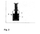
Figure 2 is showing a measuringapparatus 4 for thromboelastometric analysis; -
Figure 3 is an illustration of ameasuring cup 2 of a measuringapparatus 4 of the prior art; -
Figure 4A , B, C are schematic cross-sectional views of three preferred embodiments of acontainer 1 of the present invention. - In a first aspect, the present invention provides a diagnostic composition for use in the viscoelastic analysis of a test liquid, comprising the following constituents:
- a) at least one activator of coagulation;
- b) optionally a calcium salt in an amount sufficient to ensure recalcification of the test liquid, and
- c) optionally one or more inhibitors and/or other coagulation components or factors; characterized in that
the composition is present as an essentially dry mixture of all constituents and in an amount sufficient for performing one single viscoelastic analysis of a specified test liquid. - The calcium salt preferably is CaCl2.
- The diagnostic composition or reaction mixture of the present invention comprises constituents, which are per se known in the art. One difference to the prior art approaches is, however, that the mixture of these constituents is provided in an essentially dry form.
- "Essentially" dry in the context of the invention means a state, wherein the mixture is essentially free from any liquid or moisture, in particular being depleted of water. Water or any other liquid, however, may be present as residue in the mixture, but only to an extent, which does not negatively influence the stability of the overall composition. In particular, it has to be excluded that an interaction occurs between the different constituents, which negatively affects the stability. A remaining amount of liquid, preferably water, in the composition of up to 10 % by weight should be acceptable.
- The amount sufficient for performing one single viscoelastic analysis of a specified test liquid, for example a blood sample, is that amount of all constituents in mixture, which provides the required concentration of the reagents in the final analysis of the coagulation characteristics of the blood sample, i.e. in the
cup 2 of a measuringapparatus 4. Therefore, it is not necessary to further portion the diagnostic composition before or after dissolving it in a liquid. - Further, this is achieved by dissolving the substances in the test liquid (blood sample etc.) itself, not by dissolving the constituents in an amount of liquid diluent leading to the final working concentration.
- The activator of coagulation as mentioned above preferably is an intrinsic and/or extrinsic activator.
- The extrinsic activator of coagulation in turn preferably is the Tissue Factor (TF) and is more preferably selected from lipidated TF or rTF.
- The intrinsic activator of coagulation is preferably selected from celite, ellagic acid, sulfatit, kaolin, silica, RNA, or mixtures thereof.
- As a second feature, the diagnostic composition of the present invention may optionally comprise a calcium salt such as CaCl2, wherein CaCl2 is preferably present in an amount of about 1-100 µmol/ml of test liquid. As mentioned above, the amount of CaCl2 must be sufficient to ensure recalcification of the test liquid, in particular of the blood sample, if the sample was decalcified before. It turned out that the amount of from 3-30 µmol/ml is optimal to achieve this requirement. In order to determine the required amount of CaCl2 to be contained in the diagnostic composition even more precisely, the exact volume of the test liquid to be collected from the patient has to be known as well as the amount of decalcifying reagent employed.
- The diagnostic composition of the present invention optionally contains one ore more inhibitors, being selected, for example, from one or more of a platelet inhibitor, fibrinolysis inhibitor, or heparin inhibitor.
- Those inhibitors may be used and combined depending on the precise diagnostic demands, for example, the platelet inhibitor may be a cyto-skeletton inhibitor or a GPIIb/IIIa antagonist. The like, the fibrinolysis inhibitor can be selected, for example, from aprotinine, tranexamic acid, or eaca; the heparin inhibitor might be selected, for example, from heparinase, protamine or protamine-related peptides; and the coagulation factor can be selected, for example, from one or more coagulation factors or activated coagulation factors preferably FXa or FVa or activated protein C or FVIIa. However, it is noted that this is only a preferred selection and further inhibitors can be used if required.
- In a preferred embodiment, the dry mixture is a lyophilized mixture, more preferably a mixture produced by co-lyophilization of a mixture of the liquid reagents in one single lyophilizytion process. More precisely, in this preferred embodiment the diagnostic composition is produced by filling liquid components - in the required quantities to reach the intended composition - into a suitable container 1 (for example by a pipetting machine) and drying this composition in the
container 1 under application of low pressure environment (about 1000 - 0.2 mbar) at suitable temperature (about +30°C to -70°C). Thecontainer 1 then may be closed or sealed by a close cover or the like (e.g., a lid 5) in order to avoid a loss of reagents, or an invasion of contaminants, water etc. - In a preferred embodiment, the diagnostic composition may also contain one or more stabilizers, wherein the stabilizer preferably is albumin or gelatine.
- In a preferred embodiment, the diagnostic composition may also contain one or more phospholipids, wherein the phospholipids may be a composition of different phospholipids like phosphatidyserine, phosphatidylethanolamine and phosphatidylethanolcholine. For example, mixtures of phospholipids extracted from rabbit brain may be used.
- The present diagnostic composition may have the following constitution in preferred embodiments:
- extrinsic activation: Combination of extrinsic activator and stabilizer and, optionally, CaCl2
- intrinsic activation: Combination of intrinsic activator and stabilizer and, optionally, CaCl2
- extrinsic activation insensitive for heparin:
- Combination of extrinsic activator, heparin inhibitor and stabilizer and, optionally, CaCl2
- intrinsic activation insensitive for heparin:
- Combination of intrinsic activator, heparin inhibitor and stabilizer and, optionally, CaCl2
- extrinsic activation without platelet activation:
- Combination of extrinsic activator, platelet inhibitor and stabilizer and, optionally, CaCl2
- extrinsic activation without platelet activation, insensitive for heparin: Combination of extrinsic activator, platelet inhibitor, heparin inhibitor and stabilizer and, optionally, CaCl2
- intrinsic activation without platelet activation:
- Combination of intrinsic activator, platelet inhibitor and stabilizer and, optionally, CaCl2
- intrinsic activation without platelet activation, insensitive for heparin: Combination of intrinsic activator, platelet inhibitor, heparin inhibitor and stabilizer and, optionally, CaCl2
- extrinsic activation with inhibition of fibrinolysis:
- Combination of extrinsic activator, fibrinolysis inhibitor and stabilizer and, optionally, CaCl2
- extrinsic activation with inhibition of fibrinolysis, insensitive for heparin: Combination of extrinsic activator, fibrinolysis inhibitor, heparin inhibitor and stabilizer and, optionally, CaCl2
- intrinsic activation with inhibition of fibrinolysis:
- Combination of intrinsic activator, fibrinolysis inhibitor and stabilizer and, optionally, CaCl2
- intrinsic activation with inhibition of fibrinolysis, insensitive for heparin: Combination of intrinsic activator, fibrinolysis inhibitor, heparin inhibitor and stabilizer and, optionally, CaCl2
- extrinsic activation with additional coagulation factor: Combination of extrinsic activator, one additional coagulation factor and stabilizer and, optionally, CaCl2
- extrinsic activation with additional coagulation factor, insensitive for heparin: Combination of extrinsic activator, one additional coagulation factor, heparin inhibitor and stabilizer and, optionally, CaCl2
- intrinsic activation with additional coagulation factor: Combination of intrinsic activator, one additional coagulation factor and stabilizer and, optionally, CaCl2
- intrinsic activation with additional coagulation factor, insensitive for heparin: Combination of intrinsic activator, one additional coagulation factor, heparin inhibitor and stabilizer and, optionally, CaCl2
- In a second aspect, the present invention provides a
container 1, comprising the diagnostic composition as defined above. Thecontainer 1 preferably takes the form of a vial or a cuvette. - The
container 1 is formed from a material (e.g., plastic or glass) which is not corroded or otherwise affected by the reagents to be filled in or the test liquid to be filled in. - The
container 1 may have cylindrical shape, but its shape does not necessarily have to be cylindrical. Thecontainer 1 may have a form which reduces its inner lateral profile from the upper opening to the bottom, as for example a conically shaped form as indicated inFig. 4 or at least a partially conical form. This provides a better handling of the usually small amounts of liquid reagent mixture: In a flat bottom container, for example in a common vial with similar diameter, the used amount of liquid would hardly cover the whole bottom and might dry in an uncontrolled manner before the lyophilization process starts. Using flat bottom vials with a smaller diameter would reduce this problem, but such vials might then have an opening diameter which is too narrow to be handled with standard pipetting equipment used in connection with diagnostic devices like, for example, the ROTEM thromboelastometer. (Regarding the design of the ROTEM system and how to use it, it is referred to the publication ofUS 5,777,215 , incorporated herein by reference.) - Furthermore, such small vials might become harder to manage for common automated processing systems and might thus increase the production costs.
- The cross-section of a basically axially
symmetric container 1 of a preferred embodiment is shown inFig. 4A . However, it should be explicitly noted that the present invention is not restricted thereto, and also U-shaped, rectangular shaped or the like forms may be used. - As mentioned above, the
container 1 may be closed or sealed by alid 5 or the like in order to avoid a loss of reagents, or an invasion of contaminants, water etc. - In a further embodiment, the
container 1 is designed in a way that it can be directly used as the measuringcup 2 of theviscoelastic measuring apparatus 4. In other words, a viscolelastic analysis can be performed such that arespective container 1 is provided, the test liquid is added into thecontainer 1, and the measurement is performed directly in thecontainer 1. In this case, only the blood dispension into the container has to be performed as a liquid transfer step, which can be realized by using a manual pipette, an automated pipette, an automated dispenser or any other liquid transfer equipment. - In this embodiment, the
container 1 might be designed by combination of two materials, e.g., glass and plastic or glass and a surface covering. As indicated inFigure 4B , this combination can be realized by providing a glass container with a plastic insert forming the portion where the liquid reagent is filled in before lyophilization and where the test liquid is added to before the viscoelastic measurement. In this context the part of the container made from glass does not necessarily have to clasp the entire underside of the plastic part but might be constructed according toFigure 4C . InFigure 4B and C , a further embodiment of the invention can be seen. The container 1 (for example a cuvette) may be incorporated in a larger structure, for example a glass article, which provides some technical advantages: at first, thus set-up may technically facilitate the step of lyphilisation, and secondly, may provide a holding for the container. - In these embodiments, possible coagulation activation in the test liquid by the glass surface is excluded, while the superior sealing properties of the glass material when compared to plastic material are still used. The similar effect of suppressing possible coagulation activation in the test liquid by the glass surface can be realized by covering the glass surface (or at least the inner portion of the glass surface) with a layer of one or more substances that are not able to activate coagulation if they are in contact with blood or blood components.
-
- A
container 1 is provided, which serves as reagent support and measuring vessel (i.e. can be regarded as a measuring cup 2) for analysis using various analytical processes, and has a region which is divided into at least two chambers (6a, 6b, 6c) by one or more bars extending from the container wall or the container base, wherein the chambers are arranged so that liquid or solid reagents may be introduced therein without them being able to be mixed by diffusion or running into one another. Thecontainer 1 is used for drying or freeze-drying by with completely or partly filled chambers and serves at the same time as a measuring vessel after re-dissolving the dried material by adding water, reagent solution, or the sample present in aqueous phase. - Hence, the main difference of the present invention when compared to prior art in
US 2004/0071604 is provided by the co-lyphylization process of a mixture of reagents in the cup. This makes the division of the vessel into two or more regions - separated by bars - unnecessary, thus avoiding the resulting complications for the production process by filling in at least two different liquids into two different portions of the vessel. - In a third aspect, the present invention is directed to a method of performing a viscoelastic analysis on a test liquid, preferably a blood sample, comprising the steps of:
- a) obtaining a test liquid;
- b) providing a
container 1 as defined above; - c) adding the test liquid into said
container 1, thereby dissolving the diagnostic composition contained therein; - d) transferring the mixture of said test liquid and said diagnostic composition into a
cup 2 and put it into anapparatus 4 suitable for performing a viscolelastic analysis, or
putting thecontainer 1 into anapparatus 4 suitable for performing a viscolelastic analysis; and - e) performing the viscoelastic analysis of said mixture.
- As already mentioned above, the test liquid preferably is a blood sample, preferably is a mammalian, more preferably a sample of human blood or blood components (e.g., whole blood or blood plasma).
- Step c) of the method of the present invention preferably takes about 1-60, more preferably 2-10 sec and most preferred is about 5 sec. Following that time, the mixture of the diagnostic composition and the blood sample should be quickly transferred to the measuring
cup 2 of the measuringapparatus 4. This is done in step d) by manually or automatically pipetting the mixture from thecontainer 1 and by transferring it thereby to theapparatus 4, i.e. to the measuringcup 2 of theapparatus 4. - As an alternative, if the
container 1 of the present invention is the measuringcup 2, the measurement is performed directly in thecontainer 1. In this case, step d) may be omitted. - The
apparatus 4 preferably is a device suited for viscoelastic measurements, for example devices disclosed in (US 5,777,215 ), (US 6,537,819 ), or (US 5,777,215 ). - One example of that
apparatus 4 is shown inFig. 2 : - After the formation of the clot between cup 2 (cuvette) and
pin 3, the clot itself is stretched by the movement of thepin 3 relative to thecup 2. The detection of the characteristic parameters of the clot is based on the mechanical coupling ofcup 2 andpin 3 by the clot. This is only possible if the clot adheres on the surfaces of bothcup 2 andpin 3. So, a firm adhesion on the surfaces of bothcup 2 andpin 3 is essentially required for the viscoelastic analysis. - The method of the present invention comprises such a viscoelastic analysis of a blood sample in order to determine its coagulation characteristics, wherein such a viscoelastic analysis in the broadest sense is the measurement of a relative movement of a cuvette containing a blood sample relative to a punch. The analysis preferably comprises the determination of the clotting time, the clot formation time, and the firmness of the clot over time including fibrinolytic effects.
- In practice, the following steps may be performed:
- 1. a defined volume of a sample (e.g., whole blood, plasma) is added directly into a vial containing the lyophilized reagent composition; the measurement should start at a time close to the moment of adding the sample
- 2. after dissolving of the reagent mixture in the sample (5 sec.) the reagent-sample mixture is pipetted from the reagent vial into the measuring cup 2 (not necessary if the vial functions itself as measuring cup 2)
- 3. the
cup 2 is then put into a position such that thepin 3 is immersed into the liquid contained in the test cup, the measurement continues until stopped by the user - Therefore, the user needs not more than 4 pipetting steps in total for each test to perform (compared to the up to 8 steps required for a liquid reagent system) and no change of pipette tip is necessary when preparing one test. This clearly indicates the direct benefit of the present invention for the person who is performing such tests.
- In a further aspect, the present invention is directed to the use of a diagnostic composition or a
container 1 as defined above in a method for analyzing the viscoelastic behaviour of a test liquid, preferably a blood sample. - Although the present invention has been described in accordance with preferred embodiments, it is obvious for a person skilled in the art that modifications are possible in all embodiments.
-
- 1
- container
- 2
- measuring cup
- 3
- pin
- 4
- measuring apparatus
- 5
- lid
- 6 a,b,c
- reagent chambers
Claims (14)
- A diagnostic composition for use in the viscoelastic analysis of a blood sample, comprising the following constituents:a) at least one activator of coagulation;b) a calcium salt, in an amount of 1-100 µmol/ml of the test liquid, sufficient to ensure recalcification of the test liquid; and,c) optionally one or more inhibitors wherein said one or more inhibitors are selected from the group consisting of a platelet inhibitor, a fibrinolysis inhibitor, and a heparin inhibitor; andd) optionally one or more coagulation factors or activated coagulation factors, wherein said one or more coagulation factors or activated coagulation factors are selected from the group consisting of FI, FII, Fill, FIV, FV, FVI, FVII, FVIII, FIX, FX, FXI, FXII, FXIII, Fla, Fila, Filla, FIVa, FVa, FVla, FVlla, FVllla, FIXa, FXa, FXIa, FXIIa, FXllla, and activated protein C;characterized in that the composition is present as an essentially dry mixture of all constituents and in an amount sufficient for performing one single viscoelastic analysis of a blood sample and the composition is provided in a container made of either glass or glass and plastic or glass and a surface covering.
- A diagnostic composition of claim 1, wherein the dry mixture is a lyophilized mixture.
- A diagnostic composition according to claim 1 or 2, wherein the activated coagulation factors are selected from FXa, FVa, activated protein C or FVIIa.
- A diagnostic composition according to any one of claims 1 to 3, wherein the at least one activator of coagulation is an extrinsic activator.
- A diagnostic composition according to any one of claims 1 to 4 used for viscoelastic analysis in a container, wherein the diagnostic composition is dissolved with the blood in a container made of either glass or glass and plastic or glass and a surface covering.
- A container suitable to conceiving a defined volume of a blood sample, said container comprising the diagnostic composition according to any one of claims 1 to 5, wherein the container is made from either glass or glass and plastic or glass and a surface covering.
- A container according to claim 6, wherein the container takes the form of a vial or a cuvette.
- A container according to claim 6 or 7, wherein the container has the form which reduces its inner lateral profile from the upper opening to the bottom.
- A container according to any one of claims 6 to 8 for use in a viscoelastic analysis on a blood sample, said analysis comprising the steps of:a) obtaining the blood;b) providing the container;c) adding the blood into said container, thereby dissolving the diagnostic composition according to any of claims 1 to 5 contained therein;d) optionally transferring the mixture of said blood and said diagnostic composition into an apparatus suitable for performing a viscolelastic analysis; or putting the container into an apparatus suitable for performing a viscolelastic analysis; ande) performing the viscoelastic analysis of said mixture;wherein the container is made from either glass or glass and plastic or glass and a surface covering.
- A method of performing a viscoelastic analysis on a blood sample comprising the steps of:a) providing a container according to any one of claims 6 to 9;b) adding the blood sample into said container, thereby dissolving the diagnostic composition according to any one of claims 1 to 5 contained therein;c) optionally transferring the mixture of said blood sample and said diagnostic composition into an apparatus suitable for performing a viscoelastic analysis; or putting the container into an apparatus suitable for performing a viscoelastic analysis; andd) performing the viscoelastic analysis of said mixture.
- The method according to claim 10, wherein step b) takes 1-60 sec.
- The method according to claim 10 or 11, wherein the mixture is transferred in step c) by manually or automatically pipetting the mixture from the container and by transferring it thereby to the apparatus.
- The method according to any one of claims 10 to 12, wherein the mixture is transferred to the measuring cup of the apparatus.
- The method according to any one of claims 10 to 13, wherein the apparatus is a thromboelastometer of a thrombelastograph.
Applications Claiming Priority (2)
| Application Number | Priority Date | Filing Date | Title |
|---|---|---|---|
| GBGB0701821.1A GB0701821D0 (en) | 2007-02-01 | 2007-02-01 | Diagnostic composition and its use in the determination of coagulation characteristics of a test liquid |
| EP08702342.0A EP2111556B1 (en) | 2007-02-01 | 2008-01-31 | Container comprising a diagnostic composition and its use in the determination of coagulation characteristics of a test liquid |
Related Parent Applications (2)
| Application Number | Title | Priority Date | Filing Date |
|---|---|---|---|
| EP08702342.0A Division-Into EP2111556B1 (en) | 2007-02-01 | 2008-01-31 | Container comprising a diagnostic composition and its use in the determination of coagulation characteristics of a test liquid |
| EP08702342.0A Division EP2111556B1 (en) | 2007-02-01 | 2008-01-31 | Container comprising a diagnostic composition and its use in the determination of coagulation characteristics of a test liquid |
Publications (1)
| Publication Number | Publication Date |
|---|---|
| EP2952900A1 true EP2952900A1 (en) | 2015-12-09 |
Family
ID=37891022
Family Applications (2)
| Application Number | Title | Priority Date | Filing Date |
|---|---|---|---|
| EP08702342.0A Not-in-force EP2111556B1 (en) | 2007-02-01 | 2008-01-31 | Container comprising a diagnostic composition and its use in the determination of coagulation characteristics of a test liquid |
| EP15177115.1A Withdrawn EP2952900A1 (en) | 2007-02-01 | 2008-01-31 | Diagnostic composition and its use in the determination of coagulation characteristics of a test liquid |
Family Applications Before (1)
| Application Number | Title | Priority Date | Filing Date |
|---|---|---|---|
| EP08702342.0A Not-in-force EP2111556B1 (en) | 2007-02-01 | 2008-01-31 | Container comprising a diagnostic composition and its use in the determination of coagulation characteristics of a test liquid |
Country Status (6)
| Country | Link |
|---|---|
| US (1) | US20100190193A1 (en) |
| EP (2) | EP2111556B1 (en) |
| JP (1) | JP2010518371A (en) |
| CN (1) | CN101578521A (en) |
| GB (1) | GB0701821D0 (en) |
| WO (1) | WO2008093216A1 (en) |
Families Citing this family (39)
| Publication number | Priority date | Publication date | Assignee | Title |
|---|---|---|---|---|
| PL2202517T3 (en) | 2008-12-23 | 2013-01-31 | Casyso Ag C A | A cartridge device for a measuring system for measuring viscoelastic characteristics of a sample liquid,a corresponding measuring system, and a corresponding method |
| US8448499B2 (en) | 2008-12-23 | 2013-05-28 | C A Casyso Ag | Cartridge device for a measuring system for measuring viscoelastic characteristics of a sample liquid, a corresponding measuring system, and a corresponding method |
| US8383045B2 (en) | 2009-01-16 | 2013-02-26 | C A Casyso Ag | Measuring unit for measuring characteristics of a sample liquid, in particular viscoelastic characteristics of a blood sample |
| US8673267B2 (en) | 2009-03-02 | 2014-03-18 | Massachusetts Institute Of Technology | Methods and products for in vivo enzyme profiling |
| EP2371284A1 (en) | 2010-03-24 | 2011-10-05 | C A Casyso AG | Method and apparatus for determining at least one evaluation parameter of a blood sample |
| US20130102015A1 (en) * | 2010-03-30 | 2013-04-25 | C A Casyso Ag | Composition for the determination of coagulation characteristics of a test liquid |
| DK2555704T3 (en) | 2010-04-08 | 2019-08-05 | Hemosonics Llc | VIEW OF HEMOSTATIC PARAMETERS |
| WO2013105987A2 (en) | 2011-02-15 | 2013-07-18 | Hemosonics, Llc | Characterization of blood hemostasis and oxygen transport parameters |
| BR112013020675B1 (en) | 2011-02-15 | 2022-01-25 | Hemosonics, Llc | Devices and method for hemostasis assessment |
| JP6245990B2 (en) | 2011-03-15 | 2017-12-13 | マサチューセッツ インスティテュート オブ テクノロジー | Multiplex detection with isotopically encoded reporters |
| WO2012159021A2 (en) | 2011-05-19 | 2012-11-22 | Hemosonics, Llc | Portable hemostasis analyzer |
| JP6847660B2 (en) | 2013-06-07 | 2021-03-24 | マサチューセッツ インスティテュート オブ テクノロジー | Affinity-based detection of synthetic biomarkers encoding ligands |
| EP2884284A1 (en) * | 2013-12-12 | 2015-06-17 | C A Casyso AG | Dry diagnostic reagent |
| JP6526717B2 (en) | 2014-05-05 | 2019-06-05 | ヘモネティクス・コーポレーションHaemonetics Corporation | Methods and reagents for detecting fibrinolysis and hyperfibrinolysis |
| US10288630B2 (en) | 2014-09-29 | 2019-05-14 | C A Casyso Gmbh | Blood testing system and method |
| US10816559B2 (en) | 2014-09-29 | 2020-10-27 | Ca Casyso Ag | Blood testing system and method |
| US9897618B2 (en) | 2014-09-29 | 2018-02-20 | C A Casyso Gmbh | Blood testing system |
| US10539579B2 (en) | 2014-09-29 | 2020-01-21 | C A Casyso Gmbh | Blood testing system and method |
| US10175225B2 (en) | 2014-09-29 | 2019-01-08 | C A Casyso Ag | Blood testing system and method |
| CN107250375A (en) * | 2014-11-06 | 2017-10-13 | 科罗拉多州立大学董事会 | In the presence of thrombolytic agent new morbid state is identified using viscoelasticity analysis |
| USD777343S1 (en) | 2015-05-28 | 2017-01-24 | C A Casyso Ag | Body fluid cartridge device |
| US10295554B2 (en) | 2015-06-29 | 2019-05-21 | C A Casyso Gmbh | Blood testing system and method |
| EP3317422B1 (en) * | 2015-07-01 | 2019-08-28 | Leyser Lab GmbH | Diagnostic kit for viscoelastic analysis and its uses |
| CN105424945B (en) * | 2015-11-20 | 2018-05-18 | 重庆贝羿生物科技有限公司 | A kind of coagulation activation detection reagent and preparation method thereof and purposes |
| US20190101526A1 (en) | 2016-03-30 | 2019-04-04 | Sony Corporation | Blood state analysis apparatus, blood state analysis system, blood state analysis method, and program |
| EP3440013A4 (en) | 2016-04-08 | 2021-03-17 | Massachusetts Institute of Technology | METHODS FOR SPECIFICALLY PROFILING PROTEASE ACTIVITY AT LYMPHATIC GANGLIONS |
| EP3799958B1 (en) * | 2016-04-15 | 2023-11-15 | enicor GmbH | Pipette tip and uses and methods thereof |
| WO2017193070A1 (en) | 2016-05-05 | 2017-11-09 | Massachusetts Institute Of Technology | Methods and uses for remotely triggered protease activity measurements |
| US10473674B2 (en) | 2016-08-31 | 2019-11-12 | C A Casyso Gmbh | Controlled blood delivery to mixing chamber of a blood testing cartridge |
| CA3059358A1 (en) | 2017-04-07 | 2018-10-11 | Massachusetts Institute Of Technology | Methods to spatially profile protease activity in tissue and sections |
| JP7432500B2 (en) | 2017-04-20 | 2024-02-16 | ヘモソニックス エル・エル・シー | Disposable system for analysis of hemostatic function |
| US10843185B2 (en) | 2017-07-12 | 2020-11-24 | Ca Casyso Gmbh | Autoplatelet cartridge device |
| CN107561257A (en) * | 2017-08-16 | 2018-01-09 | 北京乐普医疗科技有限责任公司 | A kind of detection method for assessing heparin class curative effect of medication and residual condition |
| CN108279313B (en) * | 2017-12-29 | 2021-03-02 | 广州万孚生物技术股份有限公司 | Reagent and test cup for rapidly detecting blood viscoelasticity |
| WO2019173332A1 (en) | 2018-03-05 | 2019-09-12 | Massachusetts Institute Of Technology | Inhalable nanosensors with volatile reporters and uses thereof |
| US12173349B2 (en) | 2018-09-25 | 2024-12-24 | Massachusetts Institute Of Technology | Lung protease nanosensors and uses thereof |
| US11835522B2 (en) | 2019-01-17 | 2023-12-05 | Massachusetts Institute Of Technology | Sensors for detecting and imaging of cancer metastasis |
| CN109884327A (en) * | 2019-03-27 | 2019-06-14 | 深圳优迪生物技术有限公司 | The detection of functional fiber albumen activator and its application |
| US20230243856A1 (en) * | 2022-01-31 | 2023-08-03 | Instrumentation Laboratory Company | Identifying direct oral factor xa inhibitors |
Citations (4)
| Publication number | Priority date | Publication date | Assignee | Title |
|---|---|---|---|---|
| US5504011A (en) * | 1994-10-21 | 1996-04-02 | International Technidyne Corporation | Portable test apparatus and associated method of performing a blood coagulation test |
| WO1998049563A1 (en) * | 1997-04-30 | 1998-11-05 | Medtronic, Inc. | Heparin-independent, high sensitivity platelet function evaluation technique |
| WO2002079375A1 (en) * | 2001-03-30 | 2002-10-10 | Coagulation Diagnostics, Incorporated | Rapid assessment of coagulation activity in whole blood |
| EP1367135A1 (en) * | 2002-05-29 | 2003-12-03 | Pentapharm AG | Improved method for the assessment of thrombin formation in blood or plasma |
Family Cites Families (14)
| Publication number | Priority date | Publication date | Assignee | Title |
|---|---|---|---|---|
| US4529614A (en) * | 1981-12-02 | 1985-07-16 | Becton, Dickinson And Company | One step anticoagulant coating |
| US5223227A (en) * | 1989-06-19 | 1993-06-29 | Haemoscope Corporation | Disposable pin and cup with reuseable stem and collar for blood coagulation analyzer |
| JP2954239B2 (en) * | 1989-08-07 | 1999-09-27 | 清水製薬株式会社 | Simple blood coagulation test equipment for whole blood |
| JPH0638797A (en) * | 1992-07-23 | 1994-02-15 | Tokuyama Soda Co Ltd | Blood coagulation time measurement dry reagent |
| ES2131543T3 (en) * | 1993-06-30 | 1999-08-01 | Stiftung Fur Diagnostische For | MEASURING THE TIME OF ACTIVATED PARTIAL THROMBOPLASTINE (APTT) IN A SINGLE-STAGE REACTION. |
| JP2857043B2 (en) * | 1993-11-16 | 1999-02-10 | 株式会社トクヤマ | Blood clotting time measurement dry reagent |
| JPH0961433A (en) * | 1995-08-23 | 1997-03-07 | Tokuyama Corp | Dry reagent for blood coagulation time measurement |
| DE69832628T2 (en) * | 1997-04-23 | 2006-09-07 | Instrumentation Laboratory S.P.A. | RECOMBINANT RABBIT FACTOR BASED PROTHROMBIN TIME REAGENT |
| US6225126B1 (en) * | 1999-02-22 | 2001-05-01 | Haemoscope Corporation | Method and apparatus for measuring hemostasis |
| KR20020042622A (en) * | 1999-07-23 | 2002-06-05 | 코애귤레이션 다이어그노스틱스, 인크. | Method for measuring coagulant factor activity in whole blood |
| EP1234614A1 (en) * | 2001-02-27 | 2002-08-28 | Pentapharm Gmbh | Metering vessel subdivided by ribs for receiving reagents, its fabrication and use |
| CA2525940A1 (en) * | 2003-05-16 | 2004-11-25 | National Institute For Biological Standards And Control (Nibsc) | Mixture |
| GB0418474D0 (en) * | 2004-08-12 | 2004-09-22 | Pentapharm Ltd | Device with novel and improved surface properties |
| JP4921359B2 (en) * | 2005-04-01 | 2012-04-25 | キッセイ薬品工業株式会社 | Sensitive hematology assays and reagents |
-
2007
- 2007-02-01 GB GBGB0701821.1A patent/GB0701821D0/en not_active Ceased
-
2008
- 2008-01-31 JP JP2009547777A patent/JP2010518371A/en active Pending
- 2008-01-31 WO PCT/IB2008/000208 patent/WO2008093216A1/en not_active Ceased
- 2008-01-31 CN CNA2008800016445A patent/CN101578521A/en active Pending
- 2008-01-31 EP EP08702342.0A patent/EP2111556B1/en not_active Not-in-force
- 2008-01-31 US US12/522,364 patent/US20100190193A1/en not_active Abandoned
- 2008-01-31 EP EP15177115.1A patent/EP2952900A1/en not_active Withdrawn
Patent Citations (4)
| Publication number | Priority date | Publication date | Assignee | Title |
|---|---|---|---|---|
| US5504011A (en) * | 1994-10-21 | 1996-04-02 | International Technidyne Corporation | Portable test apparatus and associated method of performing a blood coagulation test |
| WO1998049563A1 (en) * | 1997-04-30 | 1998-11-05 | Medtronic, Inc. | Heparin-independent, high sensitivity platelet function evaluation technique |
| WO2002079375A1 (en) * | 2001-03-30 | 2002-10-10 | Coagulation Diagnostics, Incorporated | Rapid assessment of coagulation activity in whole blood |
| EP1367135A1 (en) * | 2002-05-29 | 2003-12-03 | Pentapharm AG | Improved method for the assessment of thrombin formation in blood or plasma |
Also Published As
| Publication number | Publication date |
|---|---|
| US20100190193A1 (en) | 2010-07-29 |
| EP2111556B1 (en) | 2015-09-02 |
| CN101578521A (en) | 2009-11-11 |
| JP2010518371A (en) | 2010-05-27 |
| GB0701821D0 (en) | 2007-03-14 |
| EP2111556A1 (en) | 2009-10-28 |
| WO2008093216A1 (en) | 2008-08-07 |
Similar Documents
| Publication | Publication Date | Title |
|---|---|---|
| EP2111556B1 (en) | Container comprising a diagnostic composition and its use in the determination of coagulation characteristics of a test liquid | |
| EP3002591B1 (en) | Composition for the determination of coagulation characteristics of a test liquid | |
| EP3140650B1 (en) | Methodologies and reagents for detecting fibrinolysis and hyperfibrinolysis | |
| US6902904B2 (en) | Coagulation assay reagents containing lanthanides | |
| US12084710B2 (en) | Diagnostic kit for viscoelastic analysis and uses and methods thereof | |
| US20250065321A1 (en) | Pipette tip and uses and methods thereof |
Legal Events
| Date | Code | Title | Description |
|---|---|---|---|
| PUAI | Public reference made under article 153(3) epc to a published international application that has entered the european phase |
Free format text: ORIGINAL CODE: 0009012 |
|
| AC | Divisional application: reference to earlier application |
Ref document number: 2111556 Country of ref document: EP Kind code of ref document: P |
|
| AK | Designated contracting states |
Kind code of ref document: A1 Designated state(s): AT BE BG CH CY CZ DE DK EE ES FI FR GB GR HR HU IE IS IT LI LT LU LV MC MT NL NO PL PT RO SE SI SK TR |
|
| 17P | Request for examination filed |
Effective date: 20160608 |
|
| RBV | Designated contracting states (corrected) |
Designated state(s): AT BE BG CH CY CZ DE DK EE ES FI FR GB GR HR HU IE IS IT LI LT LU LV MC MT NL NO PL PT RO SE SI SK TR |
|
| STAA | Information on the status of an ep patent application or granted ep patent |
Free format text: STATUS: EXAMINATION IS IN PROGRESS |
|
| 17Q | First examination report despatched |
Effective date: 20170331 |
|
| STAA | Information on the status of an ep patent application or granted ep patent |
Free format text: STATUS: THE APPLICATION IS DEEMED TO BE WITHDRAWN |
|
| 18D | Application deemed to be withdrawn |
Effective date: 20220802 |