EP2952211A1 - Contrast medium, and preparation method and preparation kit therefor - Google Patents
Contrast medium, and preparation method and preparation kit therefor Download PDFInfo
- Publication number
- EP2952211A1 EP2952211A1 EP14745511.7A EP14745511A EP2952211A1 EP 2952211 A1 EP2952211 A1 EP 2952211A1 EP 14745511 A EP14745511 A EP 14745511A EP 2952211 A1 EP2952211 A1 EP 2952211A1
- Authority
- EP
- European Patent Office
- Prior art keywords
- contrast medium
- molecular probe
- solution
- probe
- labeled
- Prior art date
- Legal status (The legal status is an assumption and is not a legal conclusion. Google has not performed a legal analysis and makes no representation as to the accuracy of the status listed.)
- Withdrawn
Links
Images






Classifications
-
- A—HUMAN NECESSITIES
- A61—MEDICAL OR VETERINARY SCIENCE; HYGIENE
- A61K—PREPARATIONS FOR MEDICAL, DENTAL OR TOILETRY PURPOSES
- A61K49/00—Preparations for testing in vivo
- A61K49/22—Echographic preparations; Ultrasonic imaging preparations
- A61K49/222—Echographic preparations; Ultrasonic imaging preparations characterised by a special physical form, e.g. emulsions, liposomes
- A61K49/227—Liposomes, lipoprotein vesicles, e.g. LDL or HDL lipoproteins, micelles, e.g. phospholipidic or polymeric
-
- A—HUMAN NECESSITIES
- A61—MEDICAL OR VETERINARY SCIENCE; HYGIENE
- A61K—PREPARATIONS FOR MEDICAL, DENTAL OR TOILETRY PURPOSES
- A61K49/00—Preparations for testing in vivo
- A61K49/22—Echographic preparations; Ultrasonic imaging preparations
-
- A—HUMAN NECESSITIES
- A61—MEDICAL OR VETERINARY SCIENCE; HYGIENE
- A61K—PREPARATIONS FOR MEDICAL, DENTAL OR TOILETRY PURPOSES
- A61K49/00—Preparations for testing in vivo
- A61K49/22—Echographic preparations; Ultrasonic imaging preparations
- A61K49/222—Echographic preparations; Ultrasonic imaging preparations characterised by a special physical form, e.g. emulsions, liposomes
- A61K49/223—Microbubbles, hollow microspheres, free gas bubbles, gas microspheres
Definitions
- the present invention relates to a contrast medium, and a method and kit for preparing the contrast medium.
- Diagnostic imaging modality such as X-ray CT, MRI and ultrasonic diagnostic equipment has long been used as an essential tool at medical front. In these tools, imaging is made based on difference in in-vivo CT values, spin relaxation time and acoustic impedance. Since difference in these physical properties exclusively reflects a structure (figure) of a living body, the modality is called "form imaging".
- molecular imaging In contrast, imaging of a site of the same structural tissue but different in function is called “functional imaging”. Particularly in the functional imaging, visualizing the present states of molecules, such as proteins, constituting a living organism, is often called “molecular imaging”. The molecular imaging is expected to be used in understanding life phenomena such as development and differentiation and applied to diagnosing and treating diseases. Because of this, the molecular imaging is a research field currently most receiving attention.
- a "molecular probe” is frequently used, which is a substance having structural selectivity to molecules constituting a living organism.
- a detectable structure by any physical means is added and the distribution of the molecular probe in a body is visualized. Attempt to use such molecular imaging in early diagnosis has been accelerated in recent years.
- Ultrasonic diagnostic equipment has the following features that other modality tools do not provide: (1) real time analysis can be excellently made, (2) because of small size, use in operation rooms is rarely restricted and (3) not only used as a diagnosis tool but also used as a treatment tool. For the reason, the ultrasonic diagnostic equipment is further expected as a diagnosis-therapy integrated tool that can be used other than large hospitals.
- a site-specific oil-in-water emulsion using a liquid microbubble-precursor is conventionally known as an ultrasound contrast medium (for example, see Patent Literature 1).
- the microbubble-precursor is a chemical substance that is easily gasified and can be stably present in a liquid state in the emulsion.
- a contrast medium which produces micro air bubbles when ultrasound energy is applied thereto and has a size sufficient to travel from a blood vessel to a tissue, is proposed.
- the microbubble precursor is administered into a living body in the form of nano-size liquid drops; however, liquid drops cause phase change by ultrasound irradiation to generate microbubbles.
- the microbubble precursor is, i.e., a phase-change ultrasound contrast medium. Owing to the ability of forming micro air bubbles by application of ultrasound energy, the contrast medium works in a site-specific manner.
- tissue selectivity can be provided.
- the ultrasound contrast medium formed of a microbubble precursor disclosed in Patent Literature 1 has a disadvantage in that the presence of the contrast medium in and out a cell cannot be determined; in particular, whether it is incorporated in a cell or not cannot be determined.
- An object of the invention is to provide a contrast medium whose uptake within a cell can be easily determined, a method and kit for preparing the contrast medium.
- the present invention will provide the following means.
- a contrast medium comprising at least one targeting molecular probe and at least one optical probe on a surface of a substrate having an ultrasound imaging function.
- the contrast medium may have a first molecular probe for conjugation, an optical probe labeled with a second molecular probe and a third molecular probe for targeting capable of binding to a cell or a tissue in a disease-specific manner.
- the third molecular probe may be labeled with the first molecular probe.
- the second molecular probe and the third molecular probe may be contained in a molar ratio of 1:1.
- the substrate may be a capsule-type contrast medium comprising a contrast substance having an imaging function included in an outer coat formed of lipid membrane.
- the contrast substance may be a water insoluble substance vaporizable by ultrasound irradiation.
- the contrast substance may be a gaseous material.
- a method for preparing the contrast medium comprising: a first step of producing a first solution containing the substrate of which the surface is labeled with the first molecular probe; a second step of producing a second solution containing the second molecular probe labeled with the optical probe; a third step of producing a third solution containing the third molecular probe labeled with the first molecular probe; a fourth step of producing a fourth solution by mixing the second solution produced in the second step and the third solution produced in the third step; a fifth step of mixing and reacting the first solution produced in the first step with the solution produced in the fourth step with the first solution produced in the first step; and a sixth step of centrifuging the solution mixture produced in the fifth step.
- the second molecular probe contained in the second solution and the third molecular probe contained in the third solution to be mixed in the fourth step may be present in a molar ratio of 1:1.
- a 10-fold or more dilution of the first solution produced in the first step may be mixed.
- a kit for preparing the contrast medium comprising: a plurality of storage containers separately storing a first solution containing the substrate of which a surface is labeled with the first molecular probe, a second solution containing the second molecular probe labeled with the optical probe, and a third solution containing the third molecular probe labeled with the first molecular probe; and openable and closable channels connected with the storage containers.
- an effect of easily determining whether a contrast medium is incorporated within a cell can be exerted.
- Contrast medium 1 according to a first embodiment of the present invention and a method for preparing the contrast medium will be described below with reference to the drawings.
- the contrast medium 1 has an ultrasound imageable substrate 2, at least one molecular probe (3 to 5) and at least one optical probe 6, which modify a surface of the substrate 2, as shown in Fig. 1 .
- the substrate 2 is constituted of a liposome, which contains perfluoro hexane therein as an inclusive and has polyethylene glycol in the surface.
- Perfluoro hexane is a water insoluble substance, which is phase changeable into a gaseous material by irradiation with ultrasound at an intensity of a predetermined threshold or more. In other words, perfluoro hexane produces an ultrasound imaging function by changing a phase into gaseous material.
- the molecular probes 3 to 5 include a first molecular probe 3 for conjugation to be attached to the surface of the substrate 2 as a label, a second molecular probe 4 to be attached to an optical probe 6 as a label and a third molecular probe 5 for targeting to be attached to the first molecular probe 3.
- biotin As the first molecular probe 3, for example, biotin is used.
- the second molecular probe 4 for example, streptavidin is used.
- CCA1 antibody As the third molecular probe 5, for example, CCA1 antibody is used.
- Alexa Fluor (AF) 647 is used as the optical probe 6.
- the method for preparing the contrast medium 1 of the embodiment includes a first step S1 of producing a first solution containing the substrate 2, the surface of which is labeled with a first molecular probe 3; a second step S2 of producing a second solution containing the second molecular probe 4 labeled with the optical probe 6; a third step S3 of producing a third solution containing the third molecular probe 5 labeled with the first molecular probe 3; a fourth step S4 of producing a fourth solution by mixing the second solution produced in the second step and the third solution produced in the third step; a fifth step S5 of mixing and reacting the first solution produced in the first step S1 with the fourth solution produced in the fourth step S4; and a sixth step S6 of centrifuging the solution mixture produced in the fifth step S5.
- a first solution containing biotinylated nano liquid drops (biotin 3 is attached to the surface of the substrate 2 as a label) is produced.
- a second solution containing streptavidin 4 labeled with AF647 6 is produced.
- a third solution containing biotinylated CCA1 antibody prepared by labelling CCA1 antibody 5 with biotin 3 is produced.
- the fourth solution is produced by mixing the second solution (for example, 0.4 ⁇ m) and the third solution (for example, 0.4 ⁇ m) and allowing the resultant solution to stand still for 15 minutes for conjugation.
- a dilution (for example, 20-fold) of the first solution (for example, 50 ⁇ L) is mixed with the fourth solution (for example, 50 ⁇ L) for a reaction, for example, at 4°C for 30 minutes. In this manner, the surface of the substrate 2 of a contrast medium is simultaneously modified with CCA1 antibody and AF647 6.
- the solution mixture produced in the fifth step S5 is centrifuged for example, at 3000G for 5 minutes.
- the contrast medium 1 according to the embodiment is produced.
- a free composite not attached to the contrast medium 1 is separated and removed from the contrast medium.
- the concentration of the second solution can be roughly calculated, for example, by an absorption method. Furthermore, the concentration of the third solution can be accurately measured, for example, by use of an absorption method and Bradford method.
- the specific binding property of the contrast medium 1 according to the embodiment to large intestine cancer cells cultured in a Petri dish (DLD-1) was checked.
- the supernatant centrifuged in the sixth step S6 was removed and the precipitate was resuspended in PBS (for example, 100 ⁇ L).
- PBS for example, 100 ⁇ L
- the resultant solution was sprinkled on the cells and allowed to stand still for 30 minutes. The surface of the cells was washed with the culture solution.
- the specific binding property was checked by a confocal fluorescence microscope and a flow-cytometer.
- Fig. 5 (a) shows the relationship between amount of fluorescence and the number of cells due to uptake, in which (0) intact, (1) phase changeable nano liquid drops (PCND) alone, (2) no CCA1 antibody and (3) the contrast medium 1 of the embodiment; and Fig. 5 (b) shows the mean fluorescence of each of the cases of graph (a).
- Fig. 5 (c) shows the relationship between amount of fluorescence and the number of cells due to uptake, in which (0) intact, (2) no CCA1 antibody and (3) the contrast medium 1 of the embodiment, when phase changeable nano liquid drops containing a different component from the case (a) is used; and Fig. 5 (d) shows the mean fluorescence of each of the cases of graph (c).
- the contrast medium 1 according to the embodiment has a high accumulation level in the cell since it is labeled with CCA1 antibody serving as the third molecular probe 5. Since the contrast medium 1 according to the embodiment can be formed in the nano order, the contrast medium is bound to the cell surface if the culture time is short, whereas, the contrast medium is incorporated in the cell if the culture time is long. This phenomenon is called internalization. As a result, different functions can be shown as images with the passage of time from administration to a living body. Furthermore, the effect does not vary depending upon the component contained therein.
- the contrast medium 1 according to the embodiment contains water insoluble perfluoro hexane therein as an inclusion.
- the perfluoro hexane is a substance, which causes phase change into a gaseous material by irradiation with ultrasound at an intensity of a predetermined threshold or more.
- the contrast medium 1 according to the embodiment labeled with an antibody and a pigment was subjected to functional evaluation as an ultrasound contrast medium, more specifically, evaluation of phase change from liquid to gas. The results will be described with reference to Fig. 6 and Fig. 7 .
- Fig. 6 shows the experiment system for ultrasound irradiation and imaging.
- reference symbol 7 denotes a water vessel
- reference symbol 8 a focused ultrasound transducer
- reference symbol 9 an ultrasound probe for diagnosis
- reference symbol 10 an amplifier
- reference symbol 11 a wave generator
- reference symbol 12 ultrasonic diagnostic equipment
- reference symbol A denotes a sample and reference symbol B degassed water.
- the water vessel 7 is filled with degassed water B of 37°C.
- a phantom (sample A) of a contrast medium according to the embodiment, which is contained in 8% acrylamide gel such that perfluoro pentane is contained in a concentration of 0.02 mg/mL, is placed in the water vessel 7.
- Pulse ultrasound of 3 MHz (duty ratio: 0.01, 1000 cycle) is applied to the phantom by use of the focused ultrasound transducer 8 for one second.
- the ultrasound transducer 8 is driven by the wave generator 11 and the amplifier 10. During ultrasound irradiation by the ultrasound transducer 8, an ultrasound image of phantom A is obtained by an ultrasound probe 12 for diagnosis connected to the ultrasonic diagnostic equipment 12.
- Figs. 7 (a) and (b) show ultrasound images of a tumor before and after irradiation with ultrasound at at least an ultrasound intensity required for changing brightness of an ultrasound image (threshold: corresponding to ultrasound intensity required for generating micron-size air bubbles for diagnosis) (see the arrow in Fig. 7 (b) ). Since ultrasound for diagnosis herein has previously set to be lower than the threshold at which a phase change occurs, an echo signal obtained from the ultrasound for diagnosis is scattered wave derived from bubbles already generated.
- a contract medium is prepared in accordance with the procedure as shown in Fig. 8 (a) , which is a different procedure from that of the preparation method of the embodiment, the contrast medium is agglomerated as shown in Figs. 8 (b) and (c) , with the result that it becomes difficult for the contrast medium to specifically bind to a target molecule.
- the procedure shown in Fig. 8 (a) after the first step S1 and the second step S2 mentioned above, the first solution and the second solution are mixed, and the resultant solution is mixed with a solution containing a biotinylated CCA1 antibody which is prepared by labeling a CCA1 antibody with biotin.
- Fig. 8 (b) is a microscope photograph of cells to which a solution containing biotinylated phase changeable nano liquid drops of a contrast medium modified with streptavidin labeled with AF647 is applied; whereas Fig. 8 (c) is a microscope photograph of cells to which a solution, which is prepared by adding a solution modified with a biotinylated CCA1 antibody to the solution of Fig. 8 (b) , is applied.
- a contrast medium is also agglomerated, as shown in Fig. 9 (a) , with the result that it becomes difficult for the contrast medium to specifically bind to a target molecule.
- the contrast medium 1 and the preparation method for the contrast medium 1 according to the embodiment has an advantage in that the contrast medium is allowed to be specifically bound only to a target molecule of a living body (cell) by means of the third molecular probe 5 without agglomerating particles of the contrast medium 1; and also has another advantage in that imaging of the state (function) of a living body, which varies with time after administration of the contrast medium, can be made by light and ultrasound.
- the fluorescence imaging by the contrast medium 1 according to the embodiment it is possible to previously understand behavior of the contrast medium 1 according to the embodiment in the cell such as timing of internalization. It is also possible to perform ultrasound irradiation at an optimal time while observing fluorescence in real time.
- the contrast medium has sensitivity to not only ultrasound but also light, which is excellent in real-time imaging and resolution, and having a size sufficient to travel into tissues through blood vessel by labeling the ultrasound imageable substrate 2 with the molecular probe 3 and the optical probe 6. Owing to this, the contrast medium has an advantage of early diagnosing a disease at a cellular level.
- perfluoro hexane which is a water insoluble substance vaporizable by ultrasound irradiation
- contrast medium 1 perfluoro hexane
- other water insoluble substances may be used, or alternatively, water insoluble substances different in boiling point may be used in combination.
- contrast substances sensitive to other modality means may be contained in the contrast medium in order to use other modality means except ultrasound and light, in combination.
- the molecular probes 3 to 5 and the optical probe 6 are not limited to those used in the embodiment.
- a nano-size bubble liposome which is prepared by using a solution containing perfluoro butane as an inclusion in place of perfluoro hexane and lipid membrane formed of polyethylene glycol as an outer coat, may be employed.
- the contrast medium 1 thus prepared When the contrast medium 1 thus prepared is injected to a mouse tail, the contrast medium can be selectively accumulated only to a tumor tissue, as shown in Figs. 10 (a) and (b) due to the EPR effect due to the nano size and active binding by an antibody. Owing to this, ultrasound imaging can be advantageously made without vaporizing the inclusion.
- contrast medium 1 examples may include air and gaseous materials such as argon, perfluoro butane and perfluoro propane.
- the material for the outer coat is not limited to the lipid membrane formed of polyethylene glycol and lipid materials having other hydrophilic groups may be used. Furthermore, if information on internalization is not required, a step of labeling the contrast medium with an optical dye may be skipped.
- the contrast medium according to the embodiment has tissue selectivity to a cancer cell and an internalization ability to enter into the cell, the contrast medium can serve as a delivery means for any cancer cell.
- the structure of the delivery means may be the same as that of the contrast medium according to the embodiment.
- the delivery means may be, for example, a substrate binding to a targeting molecular probe via a conjugate molecular probe having optical activity.
- the substrate may contain a desired substance having an anti-cancer function.
- a substance, which is activated only upon application of therapeutic-purpose ultrasound may be contained.
- the delivery means thus constituted may be used, for example, as diagnostic/therapeutic agent.
- the contrast medium according to the embodiment employs a mixture of two types of gaseous materials as an inclusion, more specifically, a gas mixture of perfluoro hexane and perfluoro pentane contained in a ratio of 1:1.
- the size of the contrast medium according to the embodiment thus constituted is compared to those of the contrast mediums obtained by other preparation methods as shown in Table 1.
- Table 1 shows the results of two experimental results. ⁇ Table 1 ⁇ (1) Measurement results (pixels) Average ( ⁇ m) Standard deviation ( ⁇ m) First solution 13 12 13 12 13 11 11 12 11 13 0.756 0.055 Single addition of SA 14 11 25 59 24 113 110 27 39 11 2.706 2.420 Embodiment 10 12 12 13 13 13 14 13 14 19 11 0.819 0.152 (2) First solution 14 12 12 15 12 13 16 11 13 11 0.806 0.104 Single addition of SA 23 61 79 12 14 13 14 62 85 68 2.694 1.899 Embodiment 14 11 11 15 12 12 13 10 14 15 0.794 0.110
- the size of the contrast medium considerably increases and agglomerated; however, if the contrast medium is prepared by the preparation method according to the embodiment, the medium can be labeled with an antibody while maintaining a nano-size.
- the mixing ratio of the water insoluble substances is not limited to those of the embodiment. The type of inclusions and combination and mixing ratio thereof, can be appropriately changed depending upon the vaporization conditions (size of air bubbles, retention time of vapor phase, frequency responsiveness and ultrasound intensity threshold).
- the contrast medium produced by the preparation method according to the embodiment even if a gaseous material serving as an inclusion is changed, causes no agglomeration of particles of the contrast medium and specifically bind only to a target molecule of a living body (cell) by means of the third molecular probe.
- imaging of the state (function) of a living body, which varies with time after administration of the contrast medium, can be made.
- the contrast medium was added to culture dishes of HCT116 cell and AGS cell and allowed to stand still, for example, one hour, and the surface are washed with the culture medium. For example, three hours later, cell-specific uptake was observed by a confocal laser microscope.
- the cells were cultured for 12 hours, washed with PBS, removed from the culture dishes with trypsin/EDTA and subjected to flow-cytometric analysis.
- the contrast medium according to the embodiment was also accumulated in HCT116 cell and travels into the cell; however, the contrast medium was not accumulated in AGS cell, whose expression level of CCA1 is reported to be low.
- the contrast medium according to the embodiment can specifically bind to the antigen corresponding to the antibody employed as the label and advantageously distinguish the cell from other cells.
- the contrast medium 1 can be produced by a preparation kit 21 shown in Fig. 14 .
- the preparation kit 21 has storage containers 22, 23 and 24 for aseptically storing first to third solutions, respectively, and channels 25 and 26 connecting between these storage containers 22 to 24.
- the channels 25, 26 are equipped respectively with valves 27 and 28, which are operated to open and close the channels 25 and 26.
- the second solution and the third solution are mixed by opening the valve 28 of the second channel 26 to produce the fourth solution, and then the first solution and the fourth solution are mixed by opening the valve 27 of the first channel 25.
- the contrast medium 1 according to the embodiment can be aseptically produced by setting the whole preparation kit in a centrifuge. Note that a safety apparatus may be provided such that the valve 28 and valve 27 are opened only in this order.
Landscapes
- Health & Medical Sciences (AREA)
- Life Sciences & Earth Sciences (AREA)
- Acoustics & Sound (AREA)
- Nuclear Medicine, Radiotherapy & Molecular Imaging (AREA)
- Radiology & Medical Imaging (AREA)
- Epidemiology (AREA)
- Physics & Mathematics (AREA)
- Animal Behavior & Ethology (AREA)
- General Health & Medical Sciences (AREA)
- Public Health (AREA)
- Veterinary Medicine (AREA)
- Medicines Containing Antibodies Or Antigens For Use As Internal Diagnostic Agents (AREA)
- Ultra Sonic Daignosis Equipment (AREA)
- Medicinal Preparation (AREA)
Abstract
Description
- The present invention relates to a contrast medium, and a method and kit for preparing the contrast medium.
- Diagnostic imaging modality such as X-ray CT, MRI and ultrasonic diagnostic equipment has long been used as an essential tool at medical front. In these tools, imaging is made based on difference in in-vivo CT values, spin relaxation time and acoustic impedance. Since difference in these physical properties exclusively reflects a structure (figure) of a living body, the modality is called "form imaging".
- In contrast, imaging of a site of the same structural tissue but different in function is called "functional imaging". Particularly in the functional imaging, visualizing the present states of molecules, such as proteins, constituting a living organism, is often called "molecular imaging". The molecular imaging is expected to be used in understanding life phenomena such as development and differentiation and applied to diagnosing and treating diseases. Because of this, the molecular imaging is a research field currently most receiving attention.
- In the molecular imaging, a "molecular probe" is frequently used, which is a substance having structural selectivity to molecules constituting a living organism. In this case, to the molecular probe, a detectable structure by any physical means is added and the distribution of the molecular probe in a body is visualized. Attempt to use such molecular imaging in early diagnosis has been accelerated in recent years.
- In an attempt to use modality such as MRI and ultrasonic diagnostic equipment, which has been previously used in form-imaging, in the molecular imaging, research and development are conducted. Ultrasonic diagnostic equipment has the following features that other modality tools do not provide: (1) real time analysis can be excellently made, (2) because of small size, use in operation rooms is rarely restricted and (3) not only used as a diagnosis tool but also used as a treatment tool. For the reason, the ultrasonic diagnostic equipment is further expected as a diagnosis-therapy integrated tool that can be used other than large hospitals.
- To realize molecular imaging for "early diagnosis for a disease", a site-specific oil-in-water emulsion using a liquid microbubble-precursor is conventionally known as an ultrasound contrast medium (for example, see Patent Literature 1).
- The microbubble-precursor is a chemical substance that is easily gasified and can be stably present in a liquid state in the emulsion. A contrast medium, which produces micro air bubbles when ultrasound energy is applied thereto and has a size sufficient to travel from a blood vessel to a tissue, is proposed.
- The microbubble precursor is administered into a living body in the form of nano-size liquid drops; however, liquid drops cause phase change by ultrasound irradiation to generate microbubbles. The microbubble precursor is, i.e., a phase-change ultrasound contrast medium. Owing to the ability of forming micro air bubbles by application of ultrasound energy, the contrast medium works in a site-specific manner. In addition, if a molecular probe such as antibody and ligand which selectively binds to a molecule constituting a living body is added, tissue selectivity can be provided.
- {PTL 1}
Publication of Japanese Patent No.3016592 - However, the ultrasound contrast medium formed of a microbubble precursor disclosed in
Patent Literature 1 has a disadvantage in that the presence of the contrast medium in and out a cell cannot be determined; in particular, whether it is incorporated in a cell or not cannot be determined. - The present invention was made in consideration of the aforementioned circumstances. An object of the invention is to provide a contrast medium whose uptake within a cell can be easily determined, a method and kit for preparing the contrast medium.
- To attain the above object, the present invention will provide the following means.
- According to one aspect of the present invention, there is provided a contrast medium comprising at least one targeting molecular probe and at least one optical probe on a surface of a substrate having an ultrasound imaging function.
- In the aspect, the contrast medium may have a first molecular probe for conjugation, an optical probe labeled with a second molecular probe and a third molecular probe for targeting capable of binding to a cell or a tissue in a disease-specific manner.
- In the aspect, the third molecular probe may be labeled with the first molecular probe.
- In the aspect, the second molecular probe and the third molecular probe may be contained in a molar ratio of 1:1.
- In the aspect, the substrate may be a capsule-type contrast medium comprising a contrast substance having an imaging function included in an outer coat formed of lipid membrane.
- In the aspect, the contrast substance may be a water insoluble substance vaporizable by ultrasound irradiation.
- In the aspect, the contrast substance may be a gaseous material.
- According to another aspect of the present invention, there is provided a method for preparing the contrast medium, comprising: a first step of producing a first solution containing the substrate of which the surface is labeled with the first molecular probe; a second step of producing a second solution containing the second molecular probe labeled with the optical probe; a third step of producing a third solution containing the third molecular probe labeled with the first molecular probe; a fourth step of producing a fourth solution by mixing the second solution produced in the second step and the third solution produced in the third step; a fifth step of mixing and reacting the first solution produced in the first step with the solution produced in the fourth step with the first solution produced in the first step; and a sixth step of centrifuging the solution mixture produced in the fifth step.
- In the aspect, the second molecular probe contained in the second solution and the third molecular probe contained in the third solution to be mixed in the fourth step may be present in a molar ratio of 1:1.
- In the aspect, in the fifth step, a 10-fold or more dilution of the first solution produced in the first step may be mixed.
- According to another aspect of the present invention, there is provided a kit for preparing the contrast medium as mentioned above, comprising: a plurality of storage containers separately storing a first solution containing the substrate of which a surface is labeled with the first molecular probe, a second solution containing the second molecular probe labeled with the optical probe, and a third solution containing the third molecular probe labeled with the first molecular probe; and openable and closable channels connected with the storage containers.
- According to the present invention, an effect of easily determining whether a contrast medium is incorporated within a cell can be exerted.
-
- {
Fig. 1 }
The figure is a view schematically showing the molecular structure of a contrast medium according to a first embodiment of the present invention. - {
Fig. 2 }
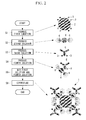
The figure shows a flowchart of a method for preparing the contrast medium ofFig. 1 . - {
Fig. 3 }
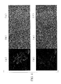
The figure shows respectively (a) a confocal fluorescence microscope image, (b) a bright-field image and (c) a superimposed image all showing specific binding ability of the contrast medium ofFig. 1 to cancer cells highly expressing CCA1 (30 minutes after the contrast medium is sprinkled on the cells before culture); and (d) a confocal fluorescence microscope image, (e) a bright-field image and (f) a superimposed image all showing a contrast medium of Comparative Example having no CCA1 antibody. - {
Fig. 4 }
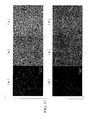
The figure shows respectively (a) a confocal fluorescence microscope image, (b) a bright-field image and (c) a superimposed image all showing specific uptake of the contrast medium ofFig. 1 by cancer cells highly expressing CCA1 (six hours after culture); and (d) a confocal fluorescence microscope image, (e) a bright-field image and (f) a superimposed image all showing a contrast medium of Comparative Example having no CCA1 antibody. - {
Fig. 5 }
The figure shows flow-cytometric analysis results showing specific uptake of the contrast medium ofFig. 1 by cancer cells (six hours after culture): (a) relationship between amount of fluorescence and the number of cells due to uptake of phase changeable nano liquid drops by cancer cells highly expressing CCA1, (b) the mean fluorescence of each of the cases in graph (a), (c) relationship between amount of fluorescence and the number of cells due to uptake of phase changeable nano liquid drops containing a different component and (d) the mean fluorescence of each of the cases in graph (c). - {
Fig. 6 }
The figure shows the experiment system for irradiation of a tissue to which the contrast medium ofFig. 1 is administered with ultrasound and imaging. - {
Fig. 7 }
The figure shows ultrasound images (a) before irradiation and (b) after irradiation with ultrasound at an intensity required for vaporization. - {
Fig. 8 }
The figure shows respectively confocal microscope analysis results of the contrast medium of Comparative Example produced in accordance with different order of steps from that of the preparation method shown inFig. 2: (a) flowchart of the preparation method, (b) confocal microscope analysis results of a contrast medium having no CCA1 antibody and produced by skipping the second step of the method (a), (c) confocal microscope analysis results of a contrast medium having CCA1 antibody and produced by the method including the second step. - {
Fig. 9 }
The figure shows respectively (a) scattering of an undiluted case and (b) scattering of 20-fold dilution in the fifth step of the flowchart ofFig. 2 . - {
Fig. 10 }
The figure shows ultrasound images of a modified contrast medium ofFig. 1 (a) before culture and (b) after culture, which shows specific binding ability to cancer cells. - {
Fig. 11 }
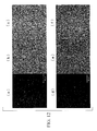
The figure shows (a) a confocal fluorescence microscope image, (b) a bright-field image and (c) a superimposed image all showing specific uptake of the contrast medium ofFig. 1 by HCT116 cells (three hours after culture); and (d) a confocal fluorescence microscope image, (e) a bright-field image and (f) a superimposed image all showing a contrast medium of Comparative Example having no CCA1 antibody. - {
Fig. 12 }
The figure shows (a) a confocal fluorescence microscope image, (b) a bright-field image and (c) a superimposed image all showing specific uptake of the contrast medium ofFig. 1 by AGS cells (three hours after culture); and (d) a confocal fluorescence microscope image, (e) a bright-field image and (f) a superimposed image all showing a contrast medium of Comparative Example having no CCA1 antibody. - {
Fig. 13 }
The figure shows flow-cytometric analysis results showing specific uptake of the contrast medium ofFig. 1 by HCT116 cells and AGS cells (twelve hours after culture), (a) relationship between amount of fluorescence and the number of cells due to uptake of phase changeable nano liquid drop by HCT116 cell, (b) (a) the mean fluorescence of each of the cases of graph (a), (c) relationship between amount of fluorescence and the number of cells due to uptake of phase changeable nano liquid drops by AGS cells and (d) the mean fluorescence of each of the cases of graph (c). - {
Fig. 14 }
The figure schematically shows a kit for preparing a contrast medium according to an embodiment of the present invention. -
Contrast medium 1 according to a first embodiment of the present invention and a method for preparing the contrast medium will be described below with reference to the drawings. - The
contrast medium 1 according to the embodiment has anultrasound imageable substrate 2, at least one molecular probe (3 to 5) and at least oneoptical probe 6, which modify a surface of thesubstrate 2, as shown inFig. 1 . - The
substrate 2 is constituted of a liposome, which contains perfluoro hexane therein as an inclusive and has polyethylene glycol in the surface. Perfluoro hexane is a water insoluble substance, which is phase changeable into a gaseous material by irradiation with ultrasound at an intensity of a predetermined threshold or more. In other words, perfluoro hexane produces an ultrasound imaging function by changing a phase into gaseous material. - The
molecular probes 3 to 5 include a firstmolecular probe 3 for conjugation to be attached to the surface of thesubstrate 2 as a label, a secondmolecular probe 4 to be attached to anoptical probe 6 as a label and a thirdmolecular probe 5 for targeting to be attached to the firstmolecular probe 3. - As the first
molecular probe 3, for example, biotin is used. - As the second
molecular probe 4, for example, streptavidin is used. - As the third
molecular probe 5, for example, CCA1 antibody is used. - As the
optical probe 6, for example, Alexa Fluor (AF) 647 is used. - A method for preparing the
contrast medium 1 according to the embodiment having the constitution as mentioned above will be described below. - The method for preparing the
contrast medium 1 of the embodiment, as shown inFig. 2 , includes a first step S1 of producing a first solution containing thesubstrate 2, the surface of which is labeled with a firstmolecular probe 3; a second step S2 of producing a second solution containing the secondmolecular probe 4 labeled with theoptical probe 6; a third step S3 of producing a third solution containing the thirdmolecular probe 5 labeled with the firstmolecular probe 3; a fourth step S4 of producing a fourth solution by mixing the second solution produced in the second step and the third solution produced in the third step; a fifth step S5 of mixing and reacting the first solution produced in the first step S1 with the fourth solution produced in the fourth step S4; and a sixth step S6 of centrifuging the solution mixture produced in the fifth step S5. - To describe more specifically, in the first step S1, a first solution containing biotinylated nano liquid drops (
biotin 3 is attached to the surface of thesubstrate 2 as a label) is produced. In the second step S2, a secondsolution containing streptavidin 4 labeled withAF647 6 is produced. In the third step S3, a third solution containing biotinylated CCA1 antibody prepared by labellingCCA1 antibody 5 withbiotin 3 is produced. - In the fourth step S4, the fourth solution is produced by mixing the second solution (for example, 0.4 µm) and the third solution (for example, 0.4 µm) and allowing the resultant solution to stand still for 15 minutes for conjugation. In the fifth step S5, a dilution (for example, 20-fold) of the first solution (for example, 50 µL) is mixed with the fourth solution (for example, 50 µL) for a reaction, for example, at 4°C for 30 minutes. In this manner, the surface of the
substrate 2 of a contrast medium is simultaneously modified with CCA1 antibody andAF647 6. - In the sixth step S6, the solution mixture produced in the fifth step S5 is centrifuged for example, at 3000G for 5 minutes. In this manner, the
contrast medium 1 according to the embodiment is produced. In this step, a free composite not attached to thecontrast medium 1 is separated and removed from the contrast medium. - Herein, it is important to carry out the third step S3 prior to the fourth step S4; mix the second solution and the third solution in a molar ratio of 1:1 in the fourth step S4; and mix a 10-fold or more dilution of the first solution with the fourth solution in the fifth step S5.
- The concentration of the second solution can be roughly calculated, for example, by an absorption method. Furthermore, the concentration of the third solution can be accurately measured, for example, by use of an absorption method and Bradford method.
- The action of the
contrast medium 1 thus produced according to the embodiment will be described below. - The specific binding property of the
contrast medium 1 according to the embodiment to large intestine cancer cells cultured in a Petri dish (DLD-1) was checked. - Herein, the supernatant centrifuged in the sixth step S6 was removed and the precipitate was resuspended in PBS (for example, 100 µL). The resultant solution was sprinkled on the cells and allowed to stand still for 30 minutes. The surface of the cells was washed with the culture solution.
- The specific binding property was checked by a confocal fluorescence microscope and a flow-cytometer.
- Confocal fluorescence microscope images are separately shown in
Fig. 3 (a), (d) andFig. 4 (a), (d) and microscopic images in the bright-field are shown inFig. 3 (b), (e) andFig. 4 (b), (e) . The superimposed images of these are separately shown inFig. 3 (c), (f) andFig. 4 (c), (f) . Furthermore, flow-cytometric analysis results are shown inFigs. 5 (a) to (d) . -
Fig. 5 (a) shows the relationship between amount of fluorescence and the number of cells due to uptake, in which (0) intact, (1) phase changeable nano liquid drops (PCND) alone, (2) no CCA1 antibody and (3) thecontrast medium 1 of the embodiment; andFig. 5 (b) shows the mean fluorescence of each of the cases of graph (a). -
Fig. 5 (c) shows the relationship between amount of fluorescence and the number of cells due to uptake, in which (0) intact, (2) no CCA1 antibody and (3) thecontrast medium 1 of the embodiment, when phase changeable nano liquid drops containing a different component from the case (a) is used; andFig. 5 (d) shows the mean fluorescence of each of the cases of graph (c). - According to these figures, it is demonstrated that the
contrast medium 1 according to the embodiment has a high accumulation level in the cell since it is labeled with CCA1 antibody serving as the thirdmolecular probe 5. Since thecontrast medium 1 according to the embodiment can be formed in the nano order, the contrast medium is bound to the cell surface if the culture time is short, whereas, the contrast medium is incorporated in the cell if the culture time is long. This phenomenon is called internalization. As a result, different functions can be shown as images with the passage of time from administration to a living body. Furthermore, the effect does not vary depending upon the component contained therein. - Furthermore, the
contrast medium 1 according to the embodiment contains water insoluble perfluoro hexane therein as an inclusion. The perfluoro hexane is a substance, which causes phase change into a gaseous material by irradiation with ultrasound at an intensity of a predetermined threshold or more. Thecontrast medium 1 according to the embodiment labeled with an antibody and a pigment was subjected to functional evaluation as an ultrasound contrast medium, more specifically, evaluation of phase change from liquid to gas. The results will be described with reference toFig. 6 and Fig. 7 . -
Fig. 6 shows the experiment system for ultrasound irradiation and imaging. In the figure, reference symbol 7 denotes a water vessel, reference symbol 8 a focused ultrasound transducer,reference symbol 9 an ultrasound probe for diagnosis,reference symbol 10 an amplifier, reference symbol 11 a wave generator, andreference symbol 12 ultrasonic diagnostic equipment. Furthermore, reference symbol A denotes a sample and reference symbol B degassed water. Now, the procedure for the experiment will be described below. - First, the water vessel 7 is filled with degassed water B of 37°C.
- Then, a phantom (sample A) of a contrast medium according to the embodiment, which is contained in 8% acrylamide gel such that perfluoro pentane is contained in a concentration of 0.02 mg/mL, is placed in the water vessel 7. Pulse ultrasound of 3 MHz (duty ratio: 0.01, 1000 cycle) is applied to the phantom by use of the focused ultrasound transducer 8 for one second.
- The ultrasound transducer 8 is driven by the
wave generator 11 and theamplifier 10. During ultrasound irradiation by the ultrasound transducer 8, an ultrasound image of phantom A is obtained by anultrasound probe 12 for diagnosis connected to the ultrasonicdiagnostic equipment 12. -
Figs. 7 (a) and (b) show ultrasound images of a tumor before and after irradiation with ultrasound at at least an ultrasound intensity required for changing brightness of an ultrasound image (threshold: corresponding to ultrasound intensity required for generating micron-size air bubbles for diagnosis) (see the arrow inFig. 7 (b) ). Since ultrasound for diagnosis herein has previously set to be lower than the threshold at which a phase change occurs, an echo signal obtained from the ultrasound for diagnosis is scattered wave derived from bubbles already generated. - Note that even if the ultrasound irradiation condition from the focused ultrasound transducer 8 is changed to 9 MHz, the same results as in
Fig. 7 were successfully obtained by optimizing the intensity, the number of cycles and application time. From the results shown herein, the effect of thecontrast medium 1 containing a water insoluble substance causing phase change from liquid to gas like the embodiment as an inclusive, is clearly demonstrated based on brightness change in the ultrasound image for diagnosis. - Note that the same effect as in the embodiment was obtained in the case of a contrast medium using a low-boiling point water-insoluble substance in combination with a high-boiling point water-insoluble substance.
- As described above, it was confirmed that even if an
optical probe 6 and amolecular probe 5 such as CCA1 antibody are mounted on the surface of thesubstrate 2, vaporization can be made. - If a contract medium is prepared in accordance with the procedure as shown in
Fig. 8 (a) , which is a different procedure from that of the preparation method of the embodiment, the contrast medium is agglomerated as shown inFigs. 8 (b) and (c) , with the result that it becomes difficult for the contrast medium to specifically bind to a target molecule. In the procedure shown inFig. 8 (a) , after the first step S1 and the second step S2 mentioned above, the first solution and the second solution are mixed, and the resultant solution is mixed with a solution containing a biotinylated CCA1 antibody which is prepared by labeling a CCA1 antibody with biotin. -
Fig. 8 (b) is a microscope photograph of cells to which a solution containing biotinylated phase changeable nano liquid drops of a contrast medium modified with streptavidin labeled with AF647 is applied; whereasFig. 8 (c) is a microscope photograph of cells to which a solution, which is prepared by adding a solution modified with a biotinylated CCA1 antibody to the solution ofFig. 8 (b) , is applied. - In the fifth step S5, if the first solution produced in the first step S1 is directly used without being diluted, a contrast medium is also agglomerated, as shown in
Fig. 9 (a) , with the result that it becomes difficult for the contrast medium to specifically bind to a target molecule. - As described above, the
contrast medium 1 and the preparation method for thecontrast medium 1 according to the embodiment has an advantage in that the contrast medium is allowed to be specifically bound only to a target molecule of a living body (cell) by means of the thirdmolecular probe 5 without agglomerating particles of thecontrast medium 1; and also has another advantage in that imaging of the state (function) of a living body, which varies with time after administration of the contrast medium, can be made by light and ultrasound. - As a result, using the fluorescence imaging by the
contrast medium 1 according to the embodiment, it is possible to previously understand behavior of thecontrast medium 1 according to the embodiment in the cell such as timing of internalization. It is also possible to perform ultrasound irradiation at an optimal time while observing fluorescence in real time. - To be more specific, it is possible to provide a contrast medium having sensitivity to not only ultrasound but also light, which is excellent in real-time imaging and resolution, and having a size sufficient to travel into tissues through blood vessel by labeling the
ultrasound imageable substrate 2 with themolecular probe 3 and theoptical probe 6. Owing to this, the contrast medium has an advantage of early diagnosing a disease at a cellular level. - It is also possible to appropriately set the timing for vaporizing the contrast medium by ultrasound irradiation. More specifically, imaging of various functions can be made also by ultrasound irradiation if the
contrast medium 1 according to the embodiment is used. - In the embodiment, perfluoro hexane, which is a water insoluble substance vaporizable by ultrasound irradiation, is used as an inclusion in the
contrast medium 1; however, other water insoluble substances may be used, or alternatively, water insoluble substances different in boiling point may be used in combination. Besides this, contrast substances sensitive to other modality means may be contained in the contrast medium in order to use other modality means except ultrasound and light, in combination. - The
molecular probes 3 to 5 and theoptical probe 6 are not limited to those used in the embodiment. - In the embodiment, a nano-size bubble liposome, which is prepared by using a solution containing perfluoro butane as an inclusion in place of perfluoro hexane and lipid membrane formed of polyethylene glycol as an outer coat, may be employed.
- When the
contrast medium 1 thus prepared is injected to a mouse tail, the contrast medium can be selectively accumulated only to a tumor tissue, as shown inFigs. 10 (a) and (b) due to the EPR effect due to the nano size and active binding by an antibody. Owing to this, ultrasound imaging can be advantageously made without vaporizing the inclusion. - Examples of the inclusion of the
contrast medium 1 may include air and gaseous materials such as argon, perfluoro butane and perfluoro propane. The material for the outer coat is not limited to the lipid membrane formed of polyethylene glycol and lipid materials having other hydrophilic groups may be used. Furthermore, if information on internalization is not required, a step of labeling the contrast medium with an optical dye may be skipped. - Furthermore, since the contrast medium according to the embodiment has tissue selectivity to a cancer cell and an internalization ability to enter into the cell, the contrast medium can serve as a delivery means for any cancer cell. The structure of the delivery means may be the same as that of the contrast medium according to the embodiment. The delivery means may be, for example, a substrate binding to a targeting molecular probe via a conjugate molecular probe having optical activity.
- The substrate may contain a desired substance having an anti-cancer function. Alternatively, a substance, which is activated only upon application of therapeutic-purpose ultrasound, may be contained. The delivery means thus constituted may be used, for example, as diagnostic/therapeutic agent.
- Now, the method for preparing a contrast medium according to a second embodiment of the present invention will be described below with reference to the drawings.
- The contrast medium according to the embodiment employs a mixture of two types of gaseous materials as an inclusion, more specifically, a gas mixture of perfluoro hexane and perfluoro pentane contained in a ratio of 1:1.
- The size of the contrast medium according to the embodiment thus constituted is compared to those of the contrast mediums obtained by other preparation methods as shown in Table 1. Table 1 shows the results of two experimental results.
{Table 1} (1) Measurement results (pixels) Average (µm) Standard deviation (µm) First solution 13 12 13 12 13 11 11 12 11 13 0.756 0.055 Single addition of SA 14 11 25 59 24 113 110 27 39 11 2.706 2.420 Embodiment 10 12 12 13 13 14 13 14 19 11 0.819 0.152 (2) First solution 14 12 12 15 12 13 16 11 13 11 0.806 0.104 Single addition of SA 23 61 79 12 14 13 14 62 85 68 2.694 1.899 Embodiment 14 11 11 15 12 12 13 10 14 15 0.794 0.110 - If streptavidin (SA) is singly added to the first solution produced in the first step, the size of the contrast medium considerably increases and agglomerated; however, if the contrast medium is prepared by the preparation method according to the embodiment, the medium can be labeled with an antibody while maintaining a nano-size. The mixing ratio of the water insoluble substances is not limited to those of the embodiment. The type of inclusions and combination and mixing ratio thereof, can be appropriately changed depending upon the vaporization conditions (size of air bubbles, retention time of vapor phase, frequency responsiveness and ultrasound intensity threshold).
- As described above, the contrast medium produced by the preparation method according to the embodiment, even if a gaseous material serving as an inclusion is changed, causes no agglomeration of particles of the contrast medium and specifically bind only to a target molecule of a living body (cell) by means of the third molecular probe. In addition, imaging of the state (function) of a living body, which varies with time after administration of the contrast medium, can be made.
- Herein, specific binding property to HCT116 cell and AGS cell in place of DLD1 cell was checked, without changing CCA1 antibody serving as the third
molecular probe 5. - To describe it more specifically, the contrast medium was added to culture dishes of HCT116 cell and AGS cell and allowed to stand still, for example, one hour, and the surface are washed with the culture medium. For example, three hours later, cell-specific uptake was observed by a confocal laser microscope. The cells were cultured for 12 hours, washed with PBS, removed from the culture dishes with trypsin/EDTA and subjected to flow-cytometric analysis.
- Accumulation results are shown in
Figs. 11 (a) to (f) andFigs. 12 (a) to (f) .Figs. 11 (a) to (c) andFigs. 12 (a) to (c) show HCT116 cell-specific uptake; whereasFigs. 11 (d) to (f) andFigs. 12 (d) to (f) show AGS cell-specific uptake. The flow-cytometric results are shown inFigs. 13 (a) to (d) . - The contrast medium according to the embodiment was also accumulated in HCT116 cell and travels into the cell; however, the contrast medium was not accumulated in AGS cell, whose expression level of CCA1 is reported to be low.
- As described above, the contrast medium according to the embodiment can specifically bind to the antigen corresponding to the antibody employed as the label and advantageously distinguish the cell from other cells.
- In the present invention, the
contrast medium 1 can be produced by apreparation kit 21 shown inFig. 14 . - The
preparation kit 21 hasstorage containers channels storage containers 22 to 24. Thechannels valves channels - The second solution and the third solution are mixed by opening the
valve 28 of thesecond channel 26 to produce the fourth solution, and then the first solution and the fourth solution are mixed by opening thevalve 27 of thefirst channel 25. Thecontrast medium 1 according to the embodiment can be aseptically produced by setting the whole preparation kit in a centrifuge. Note that a safety apparatus may be provided such that thevalve 28 andvalve 27 are opened only in this order. -
- 1
- Contrast medium
- 2
- Substrate
- 3
- First molecular probe
- 4
- Second molecular probe
- 5
- Third molecular probe
- 6
- Optical probe
- 21
- Preparation kit
- 22 to 24
- Storage containers
- 25, 26
- Channels
Claims (11)
- A contrast medium comprising at least one molecular probe and at least one optical probe on a surface of a substrate having an ultrasound imaging function.
- The contrast medium according to Claim 1, comprising a first molecular probe, an optical probe labeled with a second molecular probe and a third molecular probe capable of binding to a cell or a tissue in a disease-specific manner.
- The contrast medium according to Claim 2, wherein the third molecular probe is labeled with the first molecular probe.
- The contrast medium according to Claim 2 or Claim 3, wherein the second molecular probe and the third molecular probe are contained in a molar ratio of 1:1.
- The contrast medium according to any one of Claims 1 to 4, wherein the substrate is a capsule-type contrast medium comprising a contrast substance having an imaging function included in an outer coat formed of lipid membrane.
- The contrast medium according to Claim 5, wherein the contrast substance is a water insoluble substance vaporizable by ultrasound irradiation.
- The contrast medium according to Claim 5, wherein the contrast substance is a gaseous material.
- A method for preparing the contrast medium according to Claim 2, comprising:a first step of producing a first solution containing the substrate of which the surface is labeled with the first molecular probe;a second step of producing a second solution containing the second molecular probe labeled with the optical probe;a third step of producing a third solution containing the third molecular probe labeled with the first molecular probe;a fourth step of producing a fourth solution by mixing the second solution produced in the second step and the third solution produced in the third step;a fifth step of mixing and reacting the first solution produced in the first step with the fourth solution produced in the fourth step; anda sixth step of centrifuging the solution mixture produced in the fifth step.
- The preparation method according to Claim 8, wherein the second molecular probe contained in the second solution and the third molecular probe contained in the third solution to be mixed in the fourth step are present in a molar ratio of 1:1.
- The preparation method according to Claim 8 or 9, wherein in the fifth step, a 10-fold or more dilution of the first solution produced in the first step is mixed.
- A kit for preparing the contrast medium according to Claim 2, comprising: a plurality of storage containers separately storing a first solution containing the substrate of which a surface is labeled with the first molecular probe, a second solution containing the second molecular probe labeled with the optical probe, and a third solution containing the third molecular probe labeled with the first molecular probe; and openable and closable channels connected with the storage containers.
Applications Claiming Priority (2)
| Application Number | Priority Date | Filing Date | Title |
|---|---|---|---|
| JP2013016610A JP6278551B2 (en) | 2013-01-31 | 2013-01-31 | Contrast agent, production method and production kit |
| PCT/JP2014/051311 WO2014119455A1 (en) | 2013-01-31 | 2014-01-22 | Contrast medium, and preparation method and preparation kit therefor |
Publications (2)
| Publication Number | Publication Date |
|---|---|
| EP2952211A1 true EP2952211A1 (en) | 2015-12-09 |
| EP2952211A4 EP2952211A4 (en) | 2016-10-19 |
Family
ID=51262169
Family Applications (1)
| Application Number | Title | Priority Date | Filing Date |
|---|---|---|---|
| EP14745511.7A Withdrawn EP2952211A4 (en) | 2013-01-31 | 2014-01-22 | CONTRAST PRODUCT, ITS PREPARATION METHOD AND ITS PREPARATION KIT |
Country Status (5)
| Country | Link |
|---|---|
| US (1) | US20150328343A1 (en) |
| EP (1) | EP2952211A4 (en) |
| JP (1) | JP6278551B2 (en) |
| CN (1) | CN105120904A (en) |
| WO (1) | WO2014119455A1 (en) |
Family Cites Families (9)
| Publication number | Priority date | Publication date | Assignee | Title |
|---|---|---|---|---|
| US4444879A (en) * | 1981-01-29 | 1984-04-24 | Science Research Center, Inc. | Immunoassay with article having support film and immunological counterpart of analyte |
| JPS59189254A (en) | 1983-04-08 | 1984-10-26 | 岩谷産業株式会社 | Cryogenic thermal damper |
| US20050019266A1 (en) * | 1997-05-06 | 2005-01-27 | Unger Evan C. | Novel targeted compositions for diagnostic and therapeutic use |
| AU2003251858A1 (en) * | 2002-07-11 | 2004-02-02 | Targeson, Llc | Microbubble compositions, and methods for preparing and using same |
| AU2003252136A1 (en) * | 2002-07-24 | 2004-02-09 | Ptc Therapeutics, Inc. | METHODS FOR IDENTIFYING SMALL MOLEDULES THAT MODULATE PREMATURE TRANSLATION TERMINATION AND NONSENSE MEDIATED mRNA DECAY |
| AU2008242757A1 (en) * | 2007-04-20 | 2008-10-30 | Oregon Health & Science University | Ultrasound imaging with targeted microbubbles |
| WO2009073193A2 (en) * | 2007-12-03 | 2009-06-11 | The Johns Hopkins University | Methods of synthesis and use of chemospheres |
| CN101980667B (en) * | 2008-04-04 | 2012-08-15 | 株式会社日立制作所 | Ultrasonic wave irradiation device |
| US20100298709A1 (en) * | 2009-04-17 | 2010-11-25 | Visualsonics Inc. | Method for nonlinear imaging of ultrasound contrast agents at high frequencies |
-
2013
- 2013-01-31 JP JP2013016610A patent/JP6278551B2/en not_active Expired - Fee Related
-
2014
- 2014-01-22 CN CN201480006172.8A patent/CN105120904A/en active Pending
- 2014-01-22 EP EP14745511.7A patent/EP2952211A4/en not_active Withdrawn
- 2014-01-22 WO PCT/JP2014/051311 patent/WO2014119455A1/en not_active Ceased
-
2015
- 2015-07-29 US US14/812,456 patent/US20150328343A1/en not_active Abandoned
Also Published As
| Publication number | Publication date |
|---|---|
| JP2014148470A (en) | 2014-08-21 |
| CN105120904A (en) | 2015-12-02 |
| US20150328343A1 (en) | 2015-11-19 |
| EP2952211A4 (en) | 2016-10-19 |
| WO2014119455A1 (en) | 2014-08-07 |
| JP6278551B2 (en) | 2018-02-14 |
Similar Documents
| Publication | Publication Date | Title |
|---|---|---|
| Azimizonuzi et al. | A state-of-the-art review of the recent advances of theranostic liposome hybrid nanoparticles in cancer treatment and diagnosis | |
| Sanchez-Cano et al. | X-ray-based techniques to study the nano–bio interface | |
| Wu et al. | Magnetic particle spectroscopy: a short review of applications using magnetic nanoparticles | |
| Cheng et al. | Construction and validation of nano gold tripods for molecular imaging of living subjects | |
| Galanzha et al. | In vivo magnetic enrichment and multiplex photoacoustic detection of circulating tumour cells | |
| Kawasaki et al. | Nanotechnology, nanomedicine, and the development of new, effective therapies for cancer | |
| Maysinger | Nanoparticles and cells: good companions and doomed partnerships | |
| Teraphongphom et al. | Nanoparticle loaded polymeric microbubbles as contrast agents for multimodal imaging | |
| Wang et al. | Photoacoustic/ultrasound dual-modality contrast agent and its application to thermotherapy | |
| Xia et al. | In vivo coinstantaneous identification of hepatocellular carcinoma circulating tumor cells by dual-targeting magnetic-fluorescent nanobeads | |
| Vats et al. | Role of nanotechnology in theranostics and personalized medicines | |
| Xu et al. | Synthesis, characterization, and in vitro evaluation of targeted gold nanoshelled poly (d, l-lactide-co-glycolide) nanoparticles carrying anti p53 antibody as a theranostic agent for ultrasound contrast imaging and photothermal therapy | |
| Li et al. | Ultrasound and nanomedicine for cancer-targeted drug delivery: screening, cellular mechanisms and therapeutic opportunities | |
| Umadevi et al. | Current trends and advances in nanoplatforms-based imaging for cancer diagnosis | |
| Zhu et al. | Incorporation of a rhodamine B conjugated polymer for nanoparticle trafficking both in vitro and in vivo | |
| Beaune et al. | Location of magnetic and fluorescent nanoparticles encapsulated inside giant liposomes | |
| US20130052131A1 (en) | Nanoparticles, methods of making nanoparticles, and methods of use | |
| Kumar et al. | Noninvasive detection and thermal ablation therapy of endometriosis using silica‐coated gold nanorods | |
| Iradukunda et al. | Glutathione-conjugated fluorometric ratiometric NIR-silicon nanoparticles and Its applications for in vitro and in vivo imaging | |
| US20150328343A1 (en) | Contrast medium, method and kit for preparing same | |
| Wang et al. | Preparation and in vitro study of stromal cell-derived factor 1-targeted Fe3O4/poly (lactic-co-glycolic acid)/perfluorohexane nanoparticles | |
| Chu et al. | Mechanical properties of sub-microbubbles with a nanoparticle-decorated polymer shell | |
| Basuthakur et al. | Green-synthesized nanoparticles for fluorescence bioimaging and diagnostic applications | |
| Nagahara et al. | Nanofunctional materials in cancer research: challenges, novel methods, and emerging applications | |
| Farahbakhsh et al. | An insight into fluorescence and magnetic resonance bioimaging using a multifunctional polyethyleneimine-passivated gadocarbon dots nanoconstruct assembled with AS1411 |
Legal Events
| Date | Code | Title | Description |
|---|---|---|---|
| PUAI | Public reference made under article 153(3) epc to a published international application that has entered the european phase |
Free format text: ORIGINAL CODE: 0009012 |
|
| 17P | Request for examination filed |
Effective date: 20150814 |
|
| AK | Designated contracting states |
Kind code of ref document: A1 Designated state(s): AL AT BE BG CH CY CZ DE DK EE ES FI FR GB GR HR HU IE IS IT LI LT LU LV MC MK MT NL NO PL PT RO RS SE SI SK SM TR |
|
| AX | Request for extension of the european patent |
Extension state: BA ME |
|
| DAX | Request for extension of the european patent (deleted) | ||
| RAP1 | Party data changed (applicant data changed or rights of an application transferred) |
Owner name: OLYMPUS CORPORATION Owner name: THE UNIVERSITY OF TOKYO |
|
| RIN1 | Information on inventor provided before grant (corrected) |
Inventor name: MURAKAMI, MIYUKI Inventor name: NAGAMUNE, TERUYUKI Inventor name: YAMAGUCHI, SATOSHI Inventor name: SHIBASAKI, YOSHIKAZU Inventor name: IIJIMA, MARIKO |
|
| A4 | Supplementary search report drawn up and despatched |
Effective date: 20160915 |
|
| RIC1 | Information provided on ipc code assigned before grant |
Ipc: A61K 49/22 20060101ALI20160909BHEP Ipc: A61K 49/00 20060101AFI20160909BHEP |
|
| STAA | Information on the status of an ep patent application or granted ep patent |
Free format text: STATUS: EXAMINATION IS IN PROGRESS |
|
| 17Q | First examination report despatched |
Effective date: 20180608 |
|
| STAA | Information on the status of an ep patent application or granted ep patent |
Free format text: STATUS: THE APPLICATION IS DEEMED TO BE WITHDRAWN |
|
| 18D | Application deemed to be withdrawn |
Effective date: 20180821 |