EP2949348A1 - Adhesion preventing material - Google Patents
Adhesion preventing material Download PDFInfo
- Publication number
- EP2949348A1 EP2949348A1 EP14743521.8A EP14743521A EP2949348A1 EP 2949348 A1 EP2949348 A1 EP 2949348A1 EP 14743521 A EP14743521 A EP 14743521A EP 2949348 A1 EP2949348 A1 EP 2949348A1
- Authority
- EP
- European Patent Office
- Prior art keywords
- adhesion
- liver
- cell sheet
- cells
- preventing material
- Prior art date
- Legal status (The legal status is an assumption and is not a legal conclusion. Google has not performed a legal analysis and makes no representation as to the accuracy of the status listed.)
- Granted
Links
- 239000000463 material Substances 0.000 title claims abstract description 46
- 210000005033 mesothelial cell Anatomy 0.000 claims abstract description 80
- 210000004027 cell Anatomy 0.000 claims abstract description 77
- 238000000034 method Methods 0.000 claims abstract description 35
- 230000001737 promoting effect Effects 0.000 claims abstract description 6
- 210000004185 liver Anatomy 0.000 claims description 90
- 210000001519 tissue Anatomy 0.000 claims description 21
- 230000008929 regeneration Effects 0.000 claims description 20
- 238000011069 regeneration method Methods 0.000 claims description 20
- 210000000056 organ Anatomy 0.000 claims description 19
- 238000012753 partial hepatectomy Methods 0.000 claims description 17
- 230000002265 prevention Effects 0.000 claims description 17
- 210000003245 peritoneal mesothelial cell Anatomy 0.000 claims description 16
- 206010034650 Peritoneal adhesions Diseases 0.000 claims description 10
- 230000001605 fetal effect Effects 0.000 claims description 9
- 210000000577 adipose tissue Anatomy 0.000 claims description 8
- 210000000918 epididymis Anatomy 0.000 claims description 7
- 201000010063 epididymitis Diseases 0.000 claims description 7
- 238000011808 rodent model Methods 0.000 claims description 6
- 230000033667 organ regeneration Effects 0.000 abstract 1
- 238000002054 transplantation Methods 0.000 description 30
- 230000002980 postoperative effect Effects 0.000 description 19
- 239000005090 green fluorescent protein Substances 0.000 description 15
- 238000010172 mouse model Methods 0.000 description 15
- 108010043121 Green Fluorescent Proteins Proteins 0.000 description 14
- 102000004144 Green Fluorescent Proteins Human genes 0.000 description 14
- 238000012752 Hepatectomy Methods 0.000 description 13
- 241000699666 Mus <mouse, genus> Species 0.000 description 13
- 210000003494 hepatocyte Anatomy 0.000 description 10
- 238000011746 C57BL/6J (JAX™ mouse strain) Methods 0.000 description 8
- 108010071390 Serum Albumin Proteins 0.000 description 8
- 102000007562 Serum Albumin Human genes 0.000 description 8
- 238000011156 evaluation Methods 0.000 description 7
- BPYKTIZUTYGOLE-IFADSCNNSA-N Bilirubin Chemical compound N1C(=O)C(C)=C(C=C)\C1=C\C1=C(C)C(CCC(O)=O)=C(CC2=C(C(C)=C(\C=C/3C(=C(C=C)C(=O)N\3)C)N2)CCC(O)=O)N1 BPYKTIZUTYGOLE-IFADSCNNSA-N 0.000 description 6
- KPKZJLCSROULON-QKGLWVMZSA-N Phalloidin Chemical compound N1C(=O)[C@@H]([C@@H](O)C)NC(=O)[C@H](C)NC(=O)[C@H](C[C@@](C)(O)CO)NC(=O)[C@H](C2)NC(=O)[C@H](C)NC(=O)[C@@H]3C[C@H](O)CN3C(=O)[C@@H]1CSC1=C2C2=CC=CC=C2N1 KPKZJLCSROULON-QKGLWVMZSA-N 0.000 description 6
- 230000002440 hepatic effect Effects 0.000 description 6
- 238000002350 laparotomy Methods 0.000 description 6
- 206010019663 Hepatic failure Diseases 0.000 description 5
- 206010060932 Postoperative adhesion Diseases 0.000 description 5
- 230000000694 effects Effects 0.000 description 5
- 210000002950 fibroblast Anatomy 0.000 description 5
- 230000006870 function Effects 0.000 description 5
- 208000007903 liver failure Diseases 0.000 description 5
- 231100000835 liver failure Toxicity 0.000 description 5
- 238000001356 surgical procedure Methods 0.000 description 5
- 229920000208 temperature-responsive polymer Polymers 0.000 description 5
- 206010067125 Liver injury Diseases 0.000 description 4
- 230000002950 deficient Effects 0.000 description 4
- 231100000234 hepatic damage Toxicity 0.000 description 4
- 230000008818 liver damage Effects 0.000 description 4
- UCSJYZPVAKXKNQ-HZYVHMACSA-N streptomycin Chemical compound CN[C@H]1[C@H](O)[C@@H](O)[C@H](CO)O[C@H]1O[C@@H]1[C@](C=O)(O)[C@H](C)O[C@H]1O[C@@H]1[C@@H](NC(N)=N)[C@H](O)[C@@H](NC(N)=N)[C@H](O)[C@H]1O UCSJYZPVAKXKNQ-HZYVHMACSA-N 0.000 description 4
- PRDFBSVERLRRMY-UHFFFAOYSA-N 2'-(4-ethoxyphenyl)-5-(4-methylpiperazin-1-yl)-2,5'-bibenzimidazole Chemical compound C1=CC(OCC)=CC=C1C1=NC2=CC=C(C=3NC4=CC(=CC=C4N=3)N3CCN(C)CC3)C=C2N1 PRDFBSVERLRRMY-UHFFFAOYSA-N 0.000 description 3
- NHBKXEKEPDILRR-UHFFFAOYSA-N 2,3-bis(butanoylsulfanyl)propyl butanoate Chemical compound CCCC(=O)OCC(SC(=O)CCC)CSC(=O)CCC NHBKXEKEPDILRR-UHFFFAOYSA-N 0.000 description 3
- 102100036475 Alanine aminotransferase 1 Human genes 0.000 description 3
- 108010082126 Alanine transaminase Proteins 0.000 description 3
- 206010003445 Ascites Diseases 0.000 description 3
- 108010003415 Aspartate Aminotransferases Proteins 0.000 description 3
- 102000004625 Aspartate Aminotransferases Human genes 0.000 description 3
- 108091003079 Bovine Serum Albumin Proteins 0.000 description 3
- 241000699670 Mus sp. Species 0.000 description 3
- 229930040373 Paraformaldehyde Natural products 0.000 description 3
- 108010009711 Phalloidine Proteins 0.000 description 3
- 108010094028 Prothrombin Proteins 0.000 description 3
- 102100027378 Prothrombin Human genes 0.000 description 3
- 230000003187 abdominal effect Effects 0.000 description 3
- 230000006866 deterioration Effects 0.000 description 3
- 239000012091 fetal bovine serum Substances 0.000 description 3
- 238000007912 intraperitoneal administration Methods 0.000 description 3
- 238000002955 isolation Methods 0.000 description 3
- 201000007270 liver cancer Diseases 0.000 description 3
- 229920002866 paraformaldehyde Polymers 0.000 description 3
- 238000002360 preparation method Methods 0.000 description 3
- 230000002062 proliferating effect Effects 0.000 description 3
- 229940039716 prothrombin Drugs 0.000 description 3
- 238000011084 recovery Methods 0.000 description 3
- 239000000523 sample Substances 0.000 description 3
- 210000002966 serum Anatomy 0.000 description 3
- 239000002356 single layer Substances 0.000 description 3
- 238000012360 testing method Methods 0.000 description 3
- IGAZHQIYONOHQN-UHFFFAOYSA-N Alexa Fluor 555 Chemical compound C=12C=CC(=N)C(S(O)(=O)=O)=C2OC2=C(S(O)(=O)=O)C(N)=CC=C2C=1C1=CC=C(C(O)=O)C=C1C(O)=O IGAZHQIYONOHQN-UHFFFAOYSA-N 0.000 description 2
- 208000014644 Brain disease Diseases 0.000 description 2
- 206010009944 Colon cancer Diseases 0.000 description 2
- KCXVZYZYPLLWCC-UHFFFAOYSA-N EDTA Chemical compound OC(=O)CN(CC(O)=O)CCN(CC(O)=O)CC(O)=O KCXVZYZYPLLWCC-UHFFFAOYSA-N 0.000 description 2
- 208000032274 Encephalopathy Diseases 0.000 description 2
- 102000010834 Extracellular Matrix Proteins Human genes 0.000 description 2
- 108010037362 Extracellular Matrix Proteins Proteins 0.000 description 2
- 108090000379 Fibroblast growth factor 2 Proteins 0.000 description 2
- 102000003974 Fibroblast growth factor 2 Human genes 0.000 description 2
- 102000003745 Hepatocyte Growth Factor Human genes 0.000 description 2
- 108090000100 Hepatocyte Growth Factor Proteins 0.000 description 2
- ZDXPYRJPNDTMRX-VKHMYHEASA-N L-glutamine Chemical compound OC(=O)[C@@H](N)CCC(N)=O ZDXPYRJPNDTMRX-VKHMYHEASA-N 0.000 description 2
- 108090000630 Oncostatin M Proteins 0.000 description 2
- 229930182555 Penicillin Natural products 0.000 description 2
- JGSARLDLIJGVTE-MBNYWOFBSA-N Penicillin G Chemical compound N([C@H]1[C@H]2SC([C@@H](N2C1=O)C(O)=O)(C)C)C(=O)CC1=CC=CC=C1 JGSARLDLIJGVTE-MBNYWOFBSA-N 0.000 description 2
- 102000004142 Trypsin Human genes 0.000 description 2
- 108090000631 Trypsin Proteins 0.000 description 2
- 210000001015 abdomen Anatomy 0.000 description 2
- 210000000683 abdominal cavity Anatomy 0.000 description 2
- 238000011316 allogeneic transplantation Methods 0.000 description 2
- 230000004663 cell proliferation Effects 0.000 description 2
- 239000006285 cell suspension Substances 0.000 description 2
- 230000008859 change Effects 0.000 description 2
- 239000003153 chemical reaction reagent Substances 0.000 description 2
- 210000002744 extracellular matrix Anatomy 0.000 description 2
- ZDXPYRJPNDTMRX-UHFFFAOYSA-N glutamine Natural products OC(=O)C(N)CCC(N)=O ZDXPYRJPNDTMRX-UHFFFAOYSA-N 0.000 description 2
- 229960002743 glutamine Drugs 0.000 description 2
- JYGXADMDTFJGBT-VWUMJDOOSA-N hydrocortisone Chemical compound O=C1CC[C@]2(C)[C@H]3[C@@H](O)C[C@](C)([C@@](CC4)(O)C(=O)CO)[C@@H]4[C@@H]3CCC2=C1 JYGXADMDTFJGBT-VWUMJDOOSA-N 0.000 description 2
- 208000008384 ileus Diseases 0.000 description 2
- 230000006872 improvement Effects 0.000 description 2
- 208000015181 infectious disease Diseases 0.000 description 2
- 230000002401 inhibitory effect Effects 0.000 description 2
- 210000000936 intestine Anatomy 0.000 description 2
- 208000014018 liver neoplasm Diseases 0.000 description 2
- 238000005259 measurement Methods 0.000 description 2
- 239000002609 medium Substances 0.000 description 2
- 208000008338 non-alcoholic fatty liver disease Diseases 0.000 description 2
- 206010053219 non-alcoholic steatohepatitis Diseases 0.000 description 2
- 229940049954 penicillin Drugs 0.000 description 2
- 210000003516 pericardium Anatomy 0.000 description 2
- 210000004303 peritoneum Anatomy 0.000 description 2
- 238000003757 reverse transcription PCR Methods 0.000 description 2
- 238000000926 separation method Methods 0.000 description 2
- 229960005322 streptomycin Drugs 0.000 description 2
- 239000012588 trypsin Substances 0.000 description 2
- 230000029663 wound healing Effects 0.000 description 2
- FWMNVWWHGCHHJJ-SKKKGAJSSA-N 4-amino-1-[(2r)-6-amino-2-[[(2r)-2-[[(2r)-2-[[(2r)-2-amino-3-phenylpropanoyl]amino]-3-phenylpropanoyl]amino]-4-methylpentanoyl]amino]hexanoyl]piperidine-4-carboxylic acid Chemical compound C([C@H](C(=O)N[C@H](CC(C)C)C(=O)N[C@H](CCCCN)C(=O)N1CCC(N)(CC1)C(O)=O)NC(=O)[C@H](N)CC=1C=CC=CC=1)C1=CC=CC=C1 FWMNVWWHGCHHJJ-SKKKGAJSSA-N 0.000 description 1
- 206010060921 Abdominal abscess Diseases 0.000 description 1
- 206010000087 Abdominal pain upper Diseases 0.000 description 1
- 102000007469 Actins Human genes 0.000 description 1
- 108010085238 Actins Proteins 0.000 description 1
- 239000012103 Alexa Fluor 488 Substances 0.000 description 1
- 206010050456 Anastomotic leak Diseases 0.000 description 1
- 238000011725 BALB/c mouse Methods 0.000 description 1
- 241000283707 Capra Species 0.000 description 1
- 241000725101 Clea Species 0.000 description 1
- 102000012422 Collagen Type I Human genes 0.000 description 1
- 108010022452 Collagen Type I Proteins 0.000 description 1
- 102000004266 Collagen Type IV Human genes 0.000 description 1
- 108010042086 Collagen Type IV Proteins 0.000 description 1
- 208000001333 Colorectal Neoplasms Diseases 0.000 description 1
- 108090000368 Fibroblast growth factor 8 Proteins 0.000 description 1
- 206010018910 Haemolysis Diseases 0.000 description 1
- 208000032843 Hemorrhage Diseases 0.000 description 1
- 241000282412 Homo Species 0.000 description 1
- 206010027476 Metastases Diseases 0.000 description 1
- 241001465754 Metazoa Species 0.000 description 1
- 101100215374 Mus musculus Actb gene Proteins 0.000 description 1
- 101000756628 Mus musculus Actin, cytoplasmic 1 Proteins 0.000 description 1
- 101100230981 Mus musculus Hgf gene Proteins 0.000 description 1
- 101001076414 Mus musculus Interleukin-6 Proteins 0.000 description 1
- 101100130579 Mus musculus Mdk gene Proteins 0.000 description 1
- 101100352771 Mus musculus Podxl gene Proteins 0.000 description 1
- 102000004140 Oncostatin M Human genes 0.000 description 1
- 241000283973 Oryctolagus cuniculus Species 0.000 description 1
- 208000000450 Pelvic Pain Diseases 0.000 description 1
- 239000012980 RPMI-1640 medium Substances 0.000 description 1
- 241000283984 Rodentia Species 0.000 description 1
- 108010090804 Streptavidin Proteins 0.000 description 1
- 238000000692 Student's t-test Methods 0.000 description 1
- 108090000340 Transaminases Proteins 0.000 description 1
- 102000003929 Transaminases Human genes 0.000 description 1
- 238000012084 abdominal surgery Methods 0.000 description 1
- 210000003815 abdominal wall Anatomy 0.000 description 1
- 238000010171 animal model Methods 0.000 description 1
- 238000003556 assay Methods 0.000 description 1
- 230000008901 benefit Effects 0.000 description 1
- 210000000941 bile Anatomy 0.000 description 1
- 239000012620 biological material Substances 0.000 description 1
- 229960002685 biotin Drugs 0.000 description 1
- 239000011616 biotin Substances 0.000 description 1
- 230000000903 blocking effect Effects 0.000 description 1
- 210000004369 blood Anatomy 0.000 description 1
- 239000008280 blood Substances 0.000 description 1
- 238000004364 calculation method Methods 0.000 description 1
- 239000002775 capsule Substances 0.000 description 1
- 238000004113 cell culture Methods 0.000 description 1
- 230000001413 cellular effect Effects 0.000 description 1
- 239000003795 chemical substances by application Substances 0.000 description 1
- 230000001684 chronic effect Effects 0.000 description 1
- 210000001072 colon Anatomy 0.000 description 1
- 208000029742 colonic neoplasm Diseases 0.000 description 1
- 239000002299 complementary DNA Substances 0.000 description 1
- 238000007796 conventional method Methods 0.000 description 1
- 210000004748 cultured cell Anatomy 0.000 description 1
- 238000012258 culturing Methods 0.000 description 1
- 238000009109 curative therapy Methods 0.000 description 1
- 230000006378 damage Effects 0.000 description 1
- 210000002257 embryonic structure Anatomy 0.000 description 1
- 238000005516 engineering process Methods 0.000 description 1
- DNJIEGIFACGWOD-UHFFFAOYSA-N ethyl mercaptane Natural products CCS DNJIEGIFACGWOD-UHFFFAOYSA-N 0.000 description 1
- 210000003754 fetus Anatomy 0.000 description 1
- 238000002695 general anesthesia Methods 0.000 description 1
- 239000011521 glass Substances 0.000 description 1
- 239000003102 growth factor Substances 0.000 description 1
- 239000001963 growth medium Substances 0.000 description 1
- 210000002216 heart Anatomy 0.000 description 1
- 230000008588 hemolysis Effects 0.000 description 1
- 230000023597 hemostasis Effects 0.000 description 1
- 210000002989 hepatic vein Anatomy 0.000 description 1
- 238000010842 high-capacity cDNA reverse transcription kit Methods 0.000 description 1
- 229960000890 hydrocortisone Drugs 0.000 description 1
- 230000002209 hydrophobic effect Effects 0.000 description 1
- 238000000338 in vitro Methods 0.000 description 1
- 230000036512 infertility Effects 0.000 description 1
- 208000000509 infertility Diseases 0.000 description 1
- 208000021267 infertility disease Diseases 0.000 description 1
- 238000011835 investigation Methods 0.000 description 1
- 238000002357 laparoscopic surgery Methods 0.000 description 1
- 210000002429 large intestine Anatomy 0.000 description 1
- 239000010410 layer Substances 0.000 description 1
- 210000003041 ligament Anatomy 0.000 description 1
- 230000003908 liver function Effects 0.000 description 1
- 210000005228 liver tissue Anatomy 0.000 description 1
- 210000004072 lung Anatomy 0.000 description 1
- 238000013227 male C57BL/6J mice Methods 0.000 description 1
- 210000000713 mesentery Anatomy 0.000 description 1
- 238000010197 meta-analysis Methods 0.000 description 1
- 230000009401 metastasis Effects 0.000 description 1
- 230000001394 metastastic effect Effects 0.000 description 1
- 206010061289 metastatic neoplasm Diseases 0.000 description 1
- 239000008188 pellet Substances 0.000 description 1
- 239000002831 pharmacologic agent Substances 0.000 description 1
- 230000036470 plasma concentration Effects 0.000 description 1
- 102000005162 pleiotrophin Human genes 0.000 description 1
- 210000004224 pleura Anatomy 0.000 description 1
- 229920000642 polymer Polymers 0.000 description 1
- 239000002861 polymer material Substances 0.000 description 1
- 210000003240 portal vein Anatomy 0.000 description 1
- 239000002243 precursor Substances 0.000 description 1
- 230000008569 process Effects 0.000 description 1
- 230000035755 proliferation Effects 0.000 description 1
- 238000011002 quantification Methods 0.000 description 1
- 210000000664 rectum Anatomy 0.000 description 1
- 230000009467 reduction Effects 0.000 description 1
- 230000008672 reprogramming Effects 0.000 description 1
- 238000002271 resection Methods 0.000 description 1
- 210000001626 skin fibroblast Anatomy 0.000 description 1
- 238000010186 staining Methods 0.000 description 1
- 210000000130 stem cell Anatomy 0.000 description 1
- 239000000126 substance Substances 0.000 description 1
- 239000002344 surface layer Substances 0.000 description 1
- 230000004083 survival effect Effects 0.000 description 1
- 210000001550 testis Anatomy 0.000 description 1
- 210000000115 thoracic cavity Anatomy 0.000 description 1
- VBEQCZHXXJYVRD-GACYYNSASA-N uroanthelone Chemical compound C([C@@H](C(=O)N[C@H](C(=O)N[C@@H](CS)C(=O)N[C@@H](CC(N)=O)C(=O)N[C@@H](CS)C(=O)N[C@H](C(=O)N[C@@H]([C@@H](C)CC)C(=O)NCC(=O)N[C@@H](CC=1C=CC(O)=CC=1)C(=O)N[C@@H](CO)C(=O)NCC(=O)N[C@@H](CC(O)=O)C(=O)N[C@@H](CCCNC(N)=N)C(=O)N[C@@H](CS)C(=O)N[C@@H](CCC(N)=O)C(=O)N[C@@H]([C@@H](C)O)C(=O)N[C@@H](CCCNC(N)=N)C(=O)N[C@@H](CC(O)=O)C(=O)N[C@@H](CC(C)C)C(=O)N[C@@H](CCCNC(N)=N)C(=O)N[C@@H](CC=1C2=CC=CC=C2NC=1)C(=O)N[C@@H](CC=1C2=CC=CC=C2NC=1)C(=O)N[C@@H](CCC(O)=O)C(=O)N[C@@H](CC(C)C)C(=O)N[C@@H](CCCNC(N)=N)C(O)=O)C(C)C)[C@@H](C)O)NC(=O)[C@H](CO)NC(=O)[C@H](CC(O)=O)NC(=O)[C@H](CC(C)C)NC(=O)[C@H](CO)NC(=O)[C@H](CCC(O)=O)NC(=O)[C@@H](NC(=O)[C@H](CC=1NC=NC=1)NC(=O)[C@H](CCSC)NC(=O)[C@H](CS)NC(=O)[C@@H](NC(=O)CNC(=O)CNC(=O)[C@H](CC(N)=O)NC(=O)[C@H](CC(C)C)NC(=O)[C@H](CS)NC(=O)[C@H](CC=1C=CC(O)=CC=1)NC(=O)CNC(=O)[C@H](CC(O)=O)NC(=O)[C@H](CC=1C=CC(O)=CC=1)NC(=O)[C@H](CO)NC(=O)[C@H](CO)NC(=O)[C@H]1N(CCC1)C(=O)[C@H](CS)NC(=O)CNC(=O)[C@H]1N(CCC1)C(=O)[C@H](CC=1C=CC(O)=CC=1)NC(=O)[C@H](CO)NC(=O)[C@@H](N)CC(N)=O)C(C)C)[C@@H](C)CC)C1=CC=C(O)C=C1 VBEQCZHXXJYVRD-GACYYNSASA-N 0.000 description 1
- 210000002620 vena cava superior Anatomy 0.000 description 1
- 238000005406 washing Methods 0.000 description 1
- DGVVWUTYPXICAM-UHFFFAOYSA-N β‐Mercaptoethanol Chemical compound OCCS DGVVWUTYPXICAM-UHFFFAOYSA-N 0.000 description 1
Images




Classifications
-
- A—HUMAN NECESSITIES
- A61—MEDICAL OR VETERINARY SCIENCE; HYGIENE
- A61L—METHODS OR APPARATUS FOR STERILISING MATERIALS OR OBJECTS IN GENERAL; DISINFECTION, STERILISATION OR DEODORISATION OF AIR; CHEMICAL ASPECTS OF BANDAGES, DRESSINGS, ABSORBENT PADS OR SURGICAL ARTICLES; MATERIALS FOR BANDAGES, DRESSINGS, ABSORBENT PADS OR SURGICAL ARTICLES
- A61L31/00—Materials for other surgical articles, e.g. stents, stent-grafts, shunts, surgical drapes, guide wires, materials for adhesion prevention, occluding devices, surgical gloves, tissue fixation devices
- A61L31/005—Ingredients of undetermined constitution or reaction products thereof
-
- A—HUMAN NECESSITIES
- A61—MEDICAL OR VETERINARY SCIENCE; HYGIENE
- A61K—PREPARATIONS FOR MEDICAL, DENTAL OR TOILETRY PURPOSES
- A61K35/00—Medicinal preparations containing materials or reaction products thereof with undetermined constitution
- A61K35/12—Materials from mammals; Compositions comprising non-specified tissues or cells; Compositions comprising non-embryonic stem cells; Genetically modified cells
- A61K35/37—Digestive system
- A61K35/407—Liver; Hepatocytes
-
- A—HUMAN NECESSITIES
- A61—MEDICAL OR VETERINARY SCIENCE; HYGIENE
- A61L—METHODS OR APPARATUS FOR STERILISING MATERIALS OR OBJECTS IN GENERAL; DISINFECTION, STERILISATION OR DEODORISATION OF AIR; CHEMICAL ASPECTS OF BANDAGES, DRESSINGS, ABSORBENT PADS OR SURGICAL ARTICLES; MATERIALS FOR BANDAGES, DRESSINGS, ABSORBENT PADS OR SURGICAL ARTICLES
- A61L27/00—Materials for grafts or prostheses or for coating grafts or prostheses
- A61L27/36—Materials for grafts or prostheses or for coating grafts or prostheses containing ingredients of undetermined constitution or reaction products thereof, e.g. transplant tissue, natural bone, extracellular matrix
- A61L27/3604—Materials for grafts or prostheses or for coating grafts or prostheses containing ingredients of undetermined constitution or reaction products thereof, e.g. transplant tissue, natural bone, extracellular matrix characterised by the human or animal origin of the biological material, e.g. hair, fascia, fish scales, silk, shellac, pericardium, pleura, renal tissue, amniotic membrane, parenchymal tissue, fetal tissue, muscle tissue, fat tissue, enamel
-
- A—HUMAN NECESSITIES
- A61—MEDICAL OR VETERINARY SCIENCE; HYGIENE
- A61L—METHODS OR APPARATUS FOR STERILISING MATERIALS OR OBJECTS IN GENERAL; DISINFECTION, STERILISATION OR DEODORISATION OF AIR; CHEMICAL ASPECTS OF BANDAGES, DRESSINGS, ABSORBENT PADS OR SURGICAL ARTICLES; MATERIALS FOR BANDAGES, DRESSINGS, ABSORBENT PADS OR SURGICAL ARTICLES
- A61L27/00—Materials for grafts or prostheses or for coating grafts or prostheses
- A61L27/36—Materials for grafts or prostheses or for coating grafts or prostheses containing ingredients of undetermined constitution or reaction products thereof, e.g. transplant tissue, natural bone, extracellular matrix
- A61L27/3641—Materials for grafts or prostheses or for coating grafts or prostheses containing ingredients of undetermined constitution or reaction products thereof, e.g. transplant tissue, natural bone, extracellular matrix characterised by the site of application in the body
-
- A—HUMAN NECESSITIES
- A61—MEDICAL OR VETERINARY SCIENCE; HYGIENE
- A61L—METHODS OR APPARATUS FOR STERILISING MATERIALS OR OBJECTS IN GENERAL; DISINFECTION, STERILISATION OR DEODORISATION OF AIR; CHEMICAL ASPECTS OF BANDAGES, DRESSINGS, ABSORBENT PADS OR SURGICAL ARTICLES; MATERIALS FOR BANDAGES, DRESSINGS, ABSORBENT PADS OR SURGICAL ARTICLES
- A61L27/00—Materials for grafts or prostheses or for coating grafts or prostheses
- A61L27/50—Materials characterised by their function or physical properties, e.g. injectable or lubricating compositions, shape-memory materials, surface modified materials
-
- A—HUMAN NECESSITIES
- A61—MEDICAL OR VETERINARY SCIENCE; HYGIENE
- A61L—METHODS OR APPARATUS FOR STERILISING MATERIALS OR OBJECTS IN GENERAL; DISINFECTION, STERILISATION OR DEODORISATION OF AIR; CHEMICAL ASPECTS OF BANDAGES, DRESSINGS, ABSORBENT PADS OR SURGICAL ARTICLES; MATERIALS FOR BANDAGES, DRESSINGS, ABSORBENT PADS OR SURGICAL ARTICLES
- A61L31/00—Materials for other surgical articles, e.g. stents, stent-grafts, shunts, surgical drapes, guide wires, materials for adhesion prevention, occluding devices, surgical gloves, tissue fixation devices
- A61L31/14—Materials characterised by their function or physical properties, e.g. injectable or lubricating compositions, shape-memory materials, surface modified materials
-
- A—HUMAN NECESSITIES
- A61—MEDICAL OR VETERINARY SCIENCE; HYGIENE
- A61P—SPECIFIC THERAPEUTIC ACTIVITY OF CHEMICAL COMPOUNDS OR MEDICINAL PREPARATIONS
- A61P1/00—Drugs for disorders of the alimentary tract or the digestive system
- A61P1/16—Drugs for disorders of the alimentary tract or the digestive system for liver or gallbladder disorders, e.g. hepatoprotective agents, cholagogues, litholytics
-
- A—HUMAN NECESSITIES
- A61—MEDICAL OR VETERINARY SCIENCE; HYGIENE
- A61P—SPECIFIC THERAPEUTIC ACTIVITY OF CHEMICAL COMPOUNDS OR MEDICINAL PREPARATIONS
- A61P41/00—Drugs used in surgical methods, e.g. surgery adjuvants for preventing adhesion or for vitreum substitution
-
- A—HUMAN NECESSITIES
- A61—MEDICAL OR VETERINARY SCIENCE; HYGIENE
- A61P—SPECIFIC THERAPEUTIC ACTIVITY OF CHEMICAL COMPOUNDS OR MEDICINAL PREPARATIONS
- A61P43/00—Drugs for specific purposes, not provided for in groups A61P1/00-A61P41/00
-
- A—HUMAN NECESSITIES
- A61—MEDICAL OR VETERINARY SCIENCE; HYGIENE
- A61L—METHODS OR APPARATUS FOR STERILISING MATERIALS OR OBJECTS IN GENERAL; DISINFECTION, STERILISATION OR DEODORISATION OF AIR; CHEMICAL ASPECTS OF BANDAGES, DRESSINGS, ABSORBENT PADS OR SURGICAL ARTICLES; MATERIALS FOR BANDAGES, DRESSINGS, ABSORBENT PADS OR SURGICAL ARTICLES
- A61L2430/00—Materials or treatment for tissue regeneration
- A61L2430/28—Materials or treatment for tissue regeneration for liver reconstruction
Definitions
- the present invention relates to an adhesion preventing material consisting of a mesothelial cell sheet, and the like.
- NASH non-alcoholic steatohepatitis
- colon cancer a malignant steatohepatitis
- the number of patients suffering from primary/metastatic liver cancer is expected to increase in future.
- repeated hepatectomy As a treatment method for liver cancer which has a high recurrence rate, "repeated hepatectomy" has been accepted as one of the most effective methods which can extend the survival term or from which radical cure can be expected and it has been performed widely.
- the repeated hepatectomy however has a problem of postoperative adhesion. When postoperative adhesion occurs, adhesiotomy becomes necessary before hepatectomy is performed again. This increases the risk of prolongation of operation time, increase in blood loss, and increase in the rate of complications such as infections.
- Adhesion is a phenomenon inevitably associated with abdominal surgeries and it occurs irrespective of age, sex, or race. It occurs not only in the liver but also in various places and may cause ileus, chronic pelvic pain, stomachache, sterility, or the like.
- Non-patent Document 1 One of the currently most common methods for treating postoperative adhesion is placing an absorptive adhesion preventing material made of a polymer material. It is reported that placing an absorptive adhesion preventing material reduces the extent of adhesion (Non-patent Document 1). However, it has been elucidated from the results of meta-analysis in recent years that the above method does not decrease the frequency of ileus and increases the frequency of intra-abdominal abscess and anastomotic leaks (Non-patent Documents 2 and 3). Further, it has been pointed out that using an absorptive adhesion preventing agent may increase the possibility of inhibiting wound healing or, when it is used for the liver, may increase the risk of postoperative bile leakage.
- An object of the present invention is to provide an adhesion preventing material capable of preventing adhesion safely and efficiently, and the like. Means for Solving the Problem
- the present inventors have proceeded with an extensive investigation in order to overcome the above-mentioned problems. As a result, it has been found that adhesion can be prevented safely by using a cell sheet prepared using mesothelial cells as an adhesion preventing material.
- an adhesion preventing material prepared using liver mesothelial cells when applied to a resected surface of the liver that has been subjected to partial hepatectomy, prevents adhesion and at the same time, promotes liver regeneration.
- the present invention relates to:
- the adhesion preventing material of the present invention is comprised of mesothelial cells which are biomaterials so that it can be applied to the organ or tissue safely and can prevent adhesion without inhibiting wound healing.
- liver mesothelial cells can prevent adhesion and at the same time promote liver regeneration after partial hepatectomy so that it may suppress postoperative liver failure and increase the frequency of repeated liver transplantation.
- the adhesion preventing material of the present invention can function even when it is prepared using cells derived from an individual other than the recipient so that it is useful for clinical application.
- the adhesion preventing material of the present invention is consisting of a cell sheet containing mesothelial cells.
- adheresion preventing material means a cell sheet to be transplanted, for preventing "adhesion” which is a phenomenon that originally separated organs or tissues bond to each other, onto the organs, tissues, or the like which may cause adhesion.
- preventing means not only preventing adhesion completely but also significantly suppressing adhesion.
- cell sheet means a sheet containing cells as a main component. Although it is not particularly limited insofar as it can be attached to the organ or tissue, it may be a monolayer cell sheet, a sheet comprised of two or more layers of cells, or a sheet comprised of cells obtained by three-dimensional culture.
- the cell sheet is typically a monolayer cell sheet.
- the cell sheet may contain an extracellular matrix.
- mesothelial cells as used herein means monolayer cells that cover the uppermost surface of the mesothelium.
- the mesothelium is a pattern tissue that covers the surface of body cavities such as thoracic cavity, pericardium, and abdominal cavity.
- the mesothelial cells may be isolated from, for example, the surface layer of the peritoneum, mesentery, tunica vaginalis testis, pleura, or pericardium, or the tissues or organs such as liver, heart, lung, and large intestine. It may also be obtained by differentiating stem cells (for example, iPS cells or ES cells) or by direct reprogramming of fibroblasts.
- mesothelial cells from the tissues or organs may be derived from individuals of any stage from fetus to adult. For example, they can be obtained from individuals from fetal to infant stages, that is, individuals from a fetal stage to a stage of a child less than about six years old.
- Cells derived from individuals from fetal to infant stages abundantly contain growth factors such as hepatocyte growth factor (HGF) and epidermal growth factor (EGF) so that they are useful not only for adhesion prevention but also for regeneration promotion of the organs or tissues.
- HGF hepatocyte growth factor
- EGF epidermal growth factor
- regeneration promotion of the organs or tissues means restoration, to its original state, of the entirety or portion of the constitution or function which the organs or tissues originally have but is damaged by surgery or adhesion.
- the mesothelial cells may be derived from the same individual as the recipient to be transplanted with the adhesion preventing material or may be derived from an individual different from the recipient.
- the "adhesion preventing material” may contain a support necessary for cell culture or for maintaining the shape of the cell sheet. In another embodiment, the "adhesion preventing material” may be substantially consisting only of the cell sheet. The adhesion preventing material substantially consisting only of the cell sheet has an advantage that it easily achieves a function of promoting regeneration of the organs or tissues, because it can reproduce a state closer to nature in the living body.
- the "cell sheet comprising mesothelial cells” as used herein may contain other cells insofar as it contains mesothelial cells.
- the proportion of mesothelial cells in the total number of cells contained in the cell sheet may be, for example, 50% or more, 60% or more, 70% or more, 80% or more, 90% or more, 95% or more, 98% or more, or 99% or more.
- the mesothelial cells may contain mesothelial precursor cells which will differentiate into mesothelial cells.
- the mesothelial cells are liver mesothelial cells.
- the liver mesothelial cells are mesothelial cells that cover the surface of the liver.
- the adhesion preventing material consisting of a cell sheet comprising the liver mesothelial cells can be used for adhesion prevention and/or regeneration promotion of any organ or tissue. For example, it is useful for adhesion prevention of a resected surface after partial hepatectomy.
- the intraperitoneal adipose tissue or the like is easy to attach to the resected surface after partial hepatectomy and cause adhesion.
- the adhesion preventing material consisting of a cell sheet comprising liver mesothelial cells can significantly prevent adhesion of the resected surface after partial hepatectomy and further, promote liver regeneration, thereby preventing deterioration in thepatic functional reserve.
- resected surface means a surface which appears after resection or separation of a portion of the organ or tissue.
- hepatic functional reserve means the function of the liver itself. Hepatic functional reserve can be evaluated by a known method. For example, liver damage classification or Child-Pugh classification is frequently used for the evaluation.
- hepatic functional reserve is evaluated by measuring ascites, serum bilirubin, serum albumin, ICG R15, and prothrombin activity, while in the Child-Pugh, it is evaluated by measuring encephalopathy, ascites, serum bilirubin, serum albumin, and prothrombin activity.
- promoting liver regeneration means achieving at least one of the following: improvement in at least one of the measurement values of ascites, serum bilirubin, serum albumin, ICG R15, prothrombin activity, and encephalopathy, improvement in evaluation by the liver damage classification or Child-Pugh classification, increase in the size of the liver, increase in the weight of the liver, promotion of proliferation of hepatocytes, and increase in the size of hepatocytes.
- the adhesion preventing material of the present invention contains an extracellular matrix therein, it does not need suturing and it can be engrafted onto the surface or resected surface of an organ or tissue only by placing it thereto.
- the adhesion preventing material of the present invention can be used for adhesion prevention of any organs or tissues where adhesion could occur.
- an adhesion preventing material prepared using liver mesothelial cells can also be used for adhesion prevention of intestines or peritoneum.
- An adhesion preventing material prepared using peritoneal mesothelial cells can also be used for adhesion prevention of the liver resected surface.
- the adhesion preventing material of the present invention can be prepared by a known method for preparing a cultured cell sheet or a method similar thereto.
- a cell sheet can be obtained by culturing mesothelial cells in a culture dish coated with a temperature responsive polymer, changing the temperature when the mesothelial cells form a sheet, and collecting the resulting sheet from the culture dish.
- the temperature responsive polymer include polymers which become hydrophobic from hydrophilic by a temperature change.
- a commercially available culture dish coated in advance with a temperature responsive polymer may be used. By using the culture dish coated with a temperature responsive polymer, an adhesion preventing material that does not contain a support but is substantially consisting only of a mesothelial cell sheet can be obtained.
- the adhesion preventing method according to the present invention includes a step of placing, during surgery, the above-mentioned cell sheet comprising mesothelial cells on the surface or resected surface of the tissue or organ where adhesion may occur.
- the mesothelial cell sheet can engraft onto the tissue or organ only by placing it thereon without suturing and prevent adhesion, the mesothelial cell sheet may be sutured.
- the method of promoting liver regeneration according to the present invention includes a step of placing a cell sheet comprising liver mesothelial cells on a resected surface after partial hepatectomy.
- This method can prevent adhesion, which could become a problem during repeated hepatectomy and at the same time promote recovery of hepatic functional reserve so that it becomes possible to prevent postoperative liver failure and increase the frequency of hepatectomy.
- the method of preparing a partial hepatectomy adhesion rodent model according to the present invention includes a step of tying and cutting the median lobe and a step of cutting the middle part of the left lobe.
- the liver of the rodent is separated into a left lobe, a median lobe, a right lobe, and a caudate lobe and each lobe is in cluster form.
- the entirety of each of the lobes is therefore resected from the conventional mouse hepatectomy model so that an adhesion model having a liver resected surface cannot be obtained.
- the liver resected surface can be formed in the left lobe so that a rodent model having adhesion thereon can be obtained.
- the method of preparing a partial hepatectomy adhesion rodent model according to the present invention may further include a step of placing the epididymis adipose tissue in the vicinity of the resected surface of the left lobe. Placement of the adipose tissue in the vicinity of the resected surface promotes attachment of the adipose tissue thereto, facilitating the occurrence of adhesion.
- a partial hepatectomy adhesion rodent model according to the present invention has a resected surface in the left lobe of the liver.
- FIG. 1 schematically shows an adhesion preventing method in Examples.
- GFP positive liver mesothelial cells were isolated from a recombinant mouse expressing green fluorescent protein (GFP) using a cell sorter and cultured on a temperature responsive culture dish.
- a cell sheet obtained by changing the temperature was collected from the culture dish and transplanted onto the liver resected surface. Whether the sheet was attached to the liver resected surface or not was confirmed by detecting GFP. Details of the materials and methods will be explained below.
- GFP green fluorescent protein
- Wild type male C57BL/6J mice (10 weeks of age) were purchased from Nihon CLEA.
- Recombinant C57BL/6J mice expressing green fluorescent protein (GFP) were provided by Dr. Okabe of Osaka University.
- the age of the mouse embryos was determined by days after appearance of the vaginal plug (noon of the day when the vaginal plug appeared was considered as E0.5).
- mice Under general anesthesia, wild type C57BL/6J mice were each placed in a spine position. The mouse was incised along a median line 2 cm above from Xyphoid inferiorly while lifting up the peritoneal so as not to damage the intraperitoneal organ. After laparotomy, the falciform ligament was separated until it reached the superior vena cava. First, the pedicle of the median lobe was tied with 4-0 silk and cut off to remove the median lobe. With an electric scalpel, the hepatic capsule around the left lobe was cauterized and the left lobe was resected at the middle thereof. During excision, the left portal veins and the left hepatic veins were ligated en bloc and separated.
- the epididymis adipose tissue was lifted up to the upper part of the abdomen and placed on the resected surface of the left lobe. At the time of abdominal closure, the epididymis adipose tissue was sutured with the abdominal wall by a single stitch in order to prevent misalignment.
- mice were sacrificed 10 days or 30 days after the surgery.
- An adhesion percentage of each mouse was determined by dividing the adhesion length by the length of the liver resected surface.
- Fetal liver mesothelial cells were isolated from E12.5 GFP recombinant mice according to the method of Onitsuka, et al. ( Onitsuka, I. et al., Gastroenterology. 138, 1525-1535(2010 )). Described briefly, the fetal liver tissue was minced, isolated using Liver Digest Medium (Life Technologies), and hemolyzed in a hypotonic buffer. The cells were blocked with an anti-FcR antibody, co-stained with Anti-Mouse Podocalyxin-Biotin (MBL), and then incubated with APC-conjugated streptavidin (Invitrogen).
- the sample was sorted using a cell sorter "Moflo XDP" (Beckman Coulter).
- the sorted sample was cultured in vitro in a culture dish coated with type IV collagen while using MEM ⁇ medium (containing 10% fetal bovine serum (FBS), 50 nmol/L mercaptoethanol, penicillin, streptomycin, glutamine, 10 ng/mL basic fibroblast growth factor (bFGF), 10 ng/mL Oncostatin M (product of OSM), and insulin-transferrin-selenium-X) in an incubator of 37°C and 5% CO 2 .
- FBS fetal bovine serum
- bFGF basic fibroblast growth factor
- Oncostatin M product of OSM
- insulin-transferrin-selenium-X insulin-transferrin-selenium-X
- the adult fibroblasts were collected from GFP recombinant mouse skin tissue.
- the skin tissue was minced and treated with 0.05% trypsin and 0.5 mM EDTA at 37°C for 20 minutes.
- the cell suspension thus obtained was centrifuged and resuspended in a culture medium.
- peritoneal mesothelial cells were resuspended in RPMI1640 (20% FBS, penicillin, streptomycin, glutamine, and hydrocortisone (0.4 ⁇ g/mL)) and seeded in a culture dish coated with type I collagen.
- the primary culture cells were each transferred to an UpCell (trade mark) plate (Cellseed Inc.) and cultured.
- the plate was placed at room temperature, the cells in the sheet form were collected from the culture dish, and a cell sheet was obtained.
- the cell sheet was transplanted onto the liver resected surface of the mouse rightly after PPHx, followed by abdominal closure.
- the cell sheet engrafted only by placing on the liver resected surface.
- the surgery was monitored while detecting a GFP signal by 130S-GFP (product of Science eye).
- AST aspartate aminotransferase
- ALT alanine aminotransferase
- Frozen sections (8 ⁇ m) of the left lobe were prepared using Microm HM505E cryostat (Microm International GmbH) and mounted on APS-coated glass slides (Matsunami Glass Industry). The sections were fixed with PBS containing 4% paraformaldehyde (PFA) and stained with hematoxylin-eosin (HE).
- PFA paraformaldehyde
- the cell size and the percentage of proliferating cells were determined using IN Cell Analyzer 2000 (GE Healthcare) in accordance with the method of Miyaoka, et al. ( Miyaoka Y. et al., Curr Biol. 22, 1166-1175(2012 )). Described briefly, the cell size was determined by measuring, as the outline of hepatocytes, phalloidin-stained actin surrounding the periphery of the nuclei stained with Hoechst 33342. The percentage of proliferating cells was determined by detecting and counting Ki67 positive cells while making use of staining with Hoechst 33342 and phalloidin.
- PPHx mouse model was prepared using a BALB/c mouse in a manner similar to that employed above in item 3.
- Fetal liver mesothelial cells were obtained from a GFP recombinant C57BL/6J mouse as described above in 4.
- a cell sheet was prepared as described above in item 7 and was transplanted into the PPHx mouse model.
- a peritoneal mesothelial cell-deficient site was prepared by detaching peritoneal mesothelial cells from the C57BL/6J mouse and a peritoneal adhesion model was established.
- the liver mesothelial cell sheet prepared using the liver mesothelial cells derived from the GFP recombinant C57BL/6J mouse was transplanted onto the peritoneal mesothelial cell-deficient site.
- the abdomen was closed without transplanting the cell sheet.
- a liver resected surface was prepared by the above-described method and thereby a mouse model having adhesion was prepared.
- the process of the liver damage after hepatectomy was similar to that of postoperative course in humans. It has therefore been confirmed that the mouse model is an animal model that reflects the human hepatectomy more (data not shown).
- FIG. 2 On the left side of FIG. 2 , two photographs are shown: one is the PPHx mouse model to which the liver mesothelial cell sheet has been autotransplanted and the other one is a mouse to which nothing has been transplanted, each on postoperative day 10. As shown in the photographs, adhesion can be observed from the entire liver resected surface of the non-transplanted mouse, while almost no adhesion can be observed from the liver resected surface of the mouse transplanted with the liver mesothelial cell sheet. Similar results were observed even on postoperative day 30.
- liver mesothelial cell sheet, the peritoneal mesothelial cell sheet, and the fibroblast cell sheet were each autotransplanted onto the liver resected surface of the PPHx mouse model and on postoperative day 10, the adhesion percentage was calculated.
- Adhesion was suppressed largely in the liver mesothelial cell sheet transplantation group and the peritoneal mesothelial cell sheet transplantation group and their adhesion percentages were about 20% and about 40%, respectively.
- the adhesion percentage was almost 100% similar to that in the non-transplantation group.
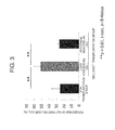
- FIG. 3 shows the evaluation results of the postoperative liver regeneration in the liver mesothelial cell transplantation group, the peritoneal mesothelial cell transplantation group, and non-transplantation group, with the percentage of Ki67 positive hepatocytes as an indicator.
- the percentage of Ki67 positive hepatocytes was almost twice as much as that of the non-transplantation group, revealing that the cell proliferation was promoted.
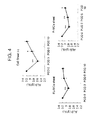
- FIG. 4 shows the evaluation results of postoperative liver regeneration in the liver mesothelial cell transplantation group, the peritoneal mesothelial cell transplantation group, and non-transplantation group, with the serum albumin as an indicator. It has been confirmed that in the liver mesothelial cell sheet transplantation group, postoperative recovery of the serum albumin was promoted. Early recovery of liver function is very desirable since it is known to contribute largely to the avoidance of postoperative liver failure.
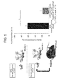
- FIG. 5 shows the results of an adhesion percentage measured by transplanting the cell sheet prepared using the liver mesothelial cells derived from a C57BL/6J mouse onto the BALB/c PPHx mouse model. Similar to the adhesion percentage of the autotransplanted sheet, a drastic reduction in adhesion percentage was observed in the liver mesothelial cell sheet transplantation group.
- FIG. 6 shows the evaluation results of postoperative liver regeneration with the percentage of Ki67 positive hepatocytes as an indicator. Similar to the liver regeneration caused by autotransplantation, the percentage of the Ki67 positive hepatocytes in the liver mesothelial cell sheet transplantation group was almost twice as much as that in the non-transplantation group, revealing the promotion of cell proliferation.
- FIG. 7 shows the observation results of the peritoneal adhesion model that transplanted with the liver mesothelial cell sheet and subjected to laparotomy 10 days after transplantation.
- FIG. 8 shows the observation results of the model subjected to laparotomy after 10 days without transplanting the liver mesothelial cell sheet.
- intestines in the abdominal cavity adhered firmly to the peritoneal wall-deficient site, while in the liver mesothelial cell transplantation group, adhesion was markedly suppressed.
- the GFP signal derived from the liver mesothelial cell sheet was observed at the peritoneal wall-deficient site, suggesting that the liver mesothelial cells engrafted and prevented intraperitoneal adhesion.
Landscapes
- Health & Medical Sciences (AREA)
- Life Sciences & Earth Sciences (AREA)
- Chemical & Material Sciences (AREA)
- Animal Behavior & Ethology (AREA)
- Veterinary Medicine (AREA)
- Public Health (AREA)
- General Health & Medical Sciences (AREA)
- Epidemiology (AREA)
- Medicinal Chemistry (AREA)
- Engineering & Computer Science (AREA)
- Chemical Kinetics & Catalysis (AREA)
- Biomedical Technology (AREA)
- Pharmacology & Pharmacy (AREA)
- Vascular Medicine (AREA)
- Surgery (AREA)
- Heart & Thoracic Surgery (AREA)
- Dermatology (AREA)
- Oral & Maxillofacial Surgery (AREA)
- Transplantation (AREA)
- Zoology (AREA)
- Bioinformatics & Cheminformatics (AREA)
- General Chemical & Material Sciences (AREA)
- Nuclear Medicine, Radiotherapy & Molecular Imaging (AREA)
- Organic Chemistry (AREA)
- Botany (AREA)
- Physiology (AREA)
- Biotechnology (AREA)
- Cell Biology (AREA)
- Developmental Biology & Embryology (AREA)
- Immunology (AREA)
- Virology (AREA)
- Nutrition Science (AREA)
- Molecular Biology (AREA)
- Urology & Nephrology (AREA)
- Gastroenterology & Hepatology (AREA)
- Materials For Medical Uses (AREA)
- Micro-Organisms Or Cultivation Processes Thereof (AREA)
- Medicines Containing Material From Animals Or Micro-Organisms (AREA)
- Environmental Sciences (AREA)
- Animal Husbandry (AREA)
Abstract
Description
- The present invention relates to an adhesion preventing material consisting of a mesothelial cell sheet, and the like.
- In the context of an increase in the number of patients suffering from non-alcoholic steatohepatitis (NASH) or colon cancer patients due to westernization of lifestyle, the number of patients suffering from primary/metastatic liver cancer is expected to increase in future.
- As a treatment method for liver cancer which has a high recurrence rate, "repeated hepatectomy" has been accepted as one of the most effective methods which can extend the survival term or from which radical cure can be expected and it has been performed widely. The repeated hepatectomy however has a problem of postoperative adhesion. When postoperative adhesion occurs, adhesiotomy becomes necessary before hepatectomy is performed again. This increases the risk of prolongation of operation time, increase in blood loss, and increase in the rate of complications such as infections.
- Adhesion is a phenomenon inevitably associated with abdominal surgeries and it occurs irrespective of age, sex, or race. It occurs not only in the liver but also in various places and may cause ileus, chronic pelvic pain, stomachache, sterility, or the like.
- For postoperative adhesion which is said to occur at a rate as high as from 93 to 100% after surgical operations (including not so serious cases), various treatment or prevention methods have so far been proposed. Examples of them include separation of adhesion by reoperation through laparotomy or laparoscopic surgery. However, the former one places an excessive burden on patients, while the latter one requires operators to have advanced skill. In addition, they may cause adhesion again due to the reoperation.
- A method of reducing postoperative adhesion by administration of pharmacological agents has been attempted, but effect of it is hardly recognized at present.
- One of the currently most common methods for treating postoperative adhesion is placing an absorptive adhesion preventing material made of a polymer material. It is reported that placing an absorptive adhesion preventing material reduces the extent of adhesion (Non-patent Document 1). However, it has been elucidated from the results of meta-analysis in recent years that the above method does not decrease the frequency of ileus and increases the frequency of intra-abdominal abscess and anastomotic leaks (Non-patent Documents 2 and 3). Further, it has been pointed out that using an absorptive adhesion preventing agent may increase the possibility of inhibiting wound healing or, when it is used for the liver, may increase the risk of postoperative bile leakage.
- In repeated hepatectomy, there occurs a problem of deterioration in functional reserve of the remnant liver in addition to the problem of adhesion. The deterioration in functional reserve may lead to liver failure so that it inevitably limits the frequency or range of repeated hepatectomy.
-
- Non-Patent Document 1: Fazio, VW. et al., Dis Colon Rectum, 49, 1-11, 2006
- Non-Patent Document 2: Tang, CL. et al., Ann Surg., 243, 449-455, 2006
- Non-Patent Document 3: Zeng, Q. et al., World J Surg., 31, 2125-2131, 2007
- An object of the present invention is to provide an adhesion preventing material capable of preventing adhesion safely and efficiently, and the like. Means for Solving the Problem
- The present inventors have proceeded with an extensive investigation in order to overcome the above-mentioned problems. As a result, it has been found that adhesion can be prevented safely by using a cell sheet prepared using mesothelial cells as an adhesion preventing material.
- It has also been found that an adhesion preventing material prepared using liver mesothelial cells, when applied to a resected surface of the liver that has been subjected to partial hepatectomy, prevents adhesion and at the same time, promotes liver regeneration.
- In addition, it has been confirmed that even a mesothelial cell sheet prepared using cells derived from an individual other than a recipient can function, leading to the completion of the present invention.
- The present invention relates to:
- [1] an adhesion preventing material comprised of a cell sheet containing mesothelial cells;
- [2] the adhesion preventing material as described above in [1], wherein the mesothelial cells are cells derived from the same individual as the subject of the adhesion prevention;
- [3] the adhesion preventing material as described above in [1], wherein the mesothelial cells are cells derived from an individual different from the subject of the adhesion prevention;
- [4] the adhesion preventing material as described above in any one of [1] to [3], wherein the mesothelial cells are cells derived from an individual from fetal to infant stages;
- [5] the adhesion preventing material as described above in any one of [1] to [4], wherein the mesothelial cells are liver mesothelial cells;
- [6] the adhesion preventing material as described above in [5], which is to be used for adhesion prevention of a resected surface after partial hepatectomy or prevention of peritoneal adhesion;
- [7] the adhesion preventing material as described above in any one of [1] to [4], wherein the mesothelial cells are peritoneal mesothelial cells;
- [8] the adhesion preventing material as described above in [7], which is used for prevention of peritoneal adhesion;
- [9] the adhesion preventing material as described above in any one of [1] to [8], which does not contain a support but contains only the cell sheet,
- [10] a method of preventing adhesion, comprising placing a cell sheet containing mesothelial cells on the surface or resected surface of a tissue or an organ;
- [11] a method of promoting liver regeneration, including placing a cell sheet containing liver mesothelial cells on a resected surface after partial hepatectomy;
- [12] a method of preparing a partial hepatectomy adhesion rodent model, including:
- tying and cutting the median lobe ; and
- cutting the middle part of the left lobe; and
- [13] the method described above in [12], further including a step of placing the epididymis adipose tissue in the vicinity of the resected surface of the left lobe.
- The adhesion preventing material of the present invention is comprised of mesothelial cells which are biomaterials so that it can be applied to the organ or tissue safely and can prevent adhesion without inhibiting wound healing.
- It can also prevent adhesion of a resected surface after partial hepatectomy, though conventional methods cannot. A cell sheet using liver mesothelial cells can prevent adhesion and at the same time promote liver regeneration after partial hepatectomy so that it may suppress postoperative liver failure and increase the frequency of repeated liver transplantation.
- The adhesion preventing material of the present invention can function even when it is prepared using cells derived from an individual other than the recipient so that it is useful for clinical application.
-
- [
FIG. 1] FIG. 1 schematically shows an adhesion preventing method using an adhesion preventing material according to the present invention. - [
FIG. 2 ] The left panels ofFIG. 2 show photographs of a PPHx mouse model onto which a liver mesothelial cell sheet was autotransplanted and the other model onto which no sheet was transplanted, each onpostoperative day 10. The right panel ofFIG.2 shows the results of autotransplanting a liver mesothelial cell sheet, a peritoneal mesothelial cell sheet, and a fibroblast cell sheet onto the liver resected surface of PPHx mouse models, respectively, and calculating an adhesion percentage onpostoperative day 10. - [
FIG. 3] FIG. 3 shows the results of evaluating postoperative liver regeneration in the liver mesothelial cell transplantation group, the peritoneal mesothelial cell transplantation group, and the non-transplantation group with a percentage of Ki67 positive hepatocytes as an indicator. - [
FIG. 4] FIG. 4 shows the results of evaluating postoperative liver regeneration in the liver mesothelial cell transplantation group, the peritoneal mesothelial cell transplantation group, and the non-transplantation group with the serum albumin as an indicator. - [
FIG. 5] FIG. 5 shows the results of transplanting a cell sheet using the liver mesothelial cells derived from a C57BL/6J mouse onto a BALB/c PPHx mouse model and determining an adhesion percentage. - [
FIG. 6] FIG. 6 shows the results of transplanting a cell sheet using the liver mesothelial cells derived from a C57BL/6J mouse onto a BALB/c PPHx mouse model and evaluating the postoperative liver regeneration with a percentage of Ki67 positive hepatocytes as an indicator. - [
FIG. 7] FIG. 7 shows the observation results of a peritoneal adhesion mouse model which was transplanted with the cell sheet using the liver mesothelial cells and was then subjected tolaparotomy 10 days after transplantation. - [
FIG. 8] FIG. 8 shows the observation results of a peritoneal adhesion mouse model which was subjected to abdominal closure without transplanting a cell sheet and after 10 days, was subjected to laparotomy. - In one embodiment, the adhesion preventing material of the present invention is consisting of a cell sheet containing mesothelial cells.
- The term "adhesion preventing material" as used herein means a cell sheet to be transplanted, for preventing "adhesion" which is a phenomenon that originally separated organs or tissues bond to each other, onto the organs, tissues, or the like which may cause adhesion.
- The term "preventing" as used herein means not only preventing adhesion completely but also significantly suppressing adhesion.
- The term "cell sheet" as used herein means a sheet containing cells as a main component. Although it is not particularly limited insofar as it can be attached to the organ or tissue, it may be a monolayer cell sheet, a sheet comprised of two or more layers of cells, or a sheet comprised of cells obtained by three-dimensional culture. The cell sheet is typically a monolayer cell sheet. The cell sheet may contain an extracellular matrix.
- The term "mesothelial cells" as used herein means monolayer cells that cover the uppermost surface of the mesothelium. The mesothelium is a pattern tissue that covers the surface of body cavities such as thoracic cavity, pericardium, and abdominal cavity. The mesothelial cells may be isolated from, for example, the surface layer of the peritoneum, mesentery, tunica vaginalis testis, pleura, or pericardium, or the tissues or organs such as liver, heart, lung, and large intestine. It may also be obtained by differentiating stem cells (for example, iPS cells or ES cells) or by direct reprogramming of fibroblasts.
- In isolating mesothelial cells from the tissues or organs, they may be derived from individuals of any stage from fetus to adult. For example, they can be obtained from individuals from fetal to infant stages, that is, individuals from a fetal stage to a stage of a child less than about six years old. Cells derived from individuals from fetal to infant stages abundantly contain growth factors such as hepatocyte growth factor (HGF) and epidermal growth factor (EGF) so that they are useful not only for adhesion prevention but also for regeneration promotion of the organs or tissues. The term "regeneration promotion of the organs or tissues" as used herein means restoration, to its original state, of the entirety or portion of the constitution or function which the organs or tissues originally have but is damaged by surgery or adhesion.
- In addition, the mesothelial cells may be derived from the same individual as the recipient to be transplanted with the adhesion preventing material or may be derived from an individual different from the recipient.
- In one embodiment, the "adhesion preventing material" may contain a support necessary for cell culture or for maintaining the shape of the cell sheet. In another embodiment, the "adhesion preventing material" may be substantially consisting only of the cell sheet. The adhesion preventing material substantially consisting only of the cell sheet has an advantage that it easily achieves a function of promoting regeneration of the organs or tissues, because it can reproduce a state closer to nature in the living body.
- The "cell sheet comprising mesothelial cells" as used herein may contain other cells insofar as it contains mesothelial cells. The proportion of mesothelial cells in the total number of cells contained in the cell sheet may be, for example, 50% or more, 60% or more, 70% or more, 80% or more, 90% or more, 95% or more, 98% or more, or 99% or more. The mesothelial cells may contain mesothelial precursor cells which will differentiate into mesothelial cells.
- In one embodiment, the mesothelial cells are liver mesothelial cells. The liver mesothelial cells are mesothelial cells that cover the surface of the liver. The adhesion preventing material consisting of a cell sheet comprising the liver mesothelial cells can be used for adhesion prevention and/or regeneration promotion of any organ or tissue. For example, it is useful for adhesion prevention of a resected surface after partial hepatectomy. The intraperitoneal adipose tissue or the like is easy to attach to the resected surface after partial hepatectomy and cause adhesion.
- For the treatment of primary liver cancer or colorectal cancer liver metastasis, an active attempt to perform "repeated hepatectomy" to prevent recurrence in the remnant liver after hepatectomy is only one curative treatment from which radical cure or prolongation of life can be expected. When adhesion occurs in such a case, it must be separated during second or third surgery, which increases the risk of massive hemorrhage or infection. In addition, the adhesion deteriorates hepatic functional reserve so that it limits the frequency of hepatectomy and at the same time, it is likely to cause postoperative liver failure.
- The adhesion preventing material consisting of a cell sheet comprising liver mesothelial cells can significantly prevent adhesion of the resected surface after partial hepatectomy and further, promote liver regeneration, thereby preventing deterioration in thepatic functional reserve.
- The term "resected surface" as used herein means a surface which appears after resection or separation of a portion of the organ or tissue.
- The term "hepatic functional reserve" as used herein means the function of the liver itself. Hepatic functional reserve can be evaluated by a known method. For example, liver damage classification or Child-Pugh classification is frequently used for the evaluation.
- In the liver damage classification, hepatic functional reserve is evaluated by measuring ascites, serum bilirubin, serum albumin, ICG R15, and prothrombin activity, while in the Child-Pugh, it is evaluated by measuring encephalopathy, ascites, serum bilirubin, serum albumin, and prothrombin activity.
- The term "promoting liver regeneration" as used herein means achieving at least one of the following: improvement in at least one of the measurement values of ascites, serum bilirubin, serum albumin, ICG R15, prothrombin activity, and encephalopathy, improvement in evaluation by the liver damage classification or Child-Pugh classification, increase in the size of the liver, increase in the weight of the liver, promotion of proliferation of hepatocytes, and increase in the size of hepatocytes.
- When the adhesion preventing material of the present invention contains an extracellular matrix therein, it does not need suturing and it can be engrafted onto the surface or resected surface of an organ or tissue only by placing it thereto.
- The adhesion preventing material of the present invention can be used for adhesion prevention of any organs or tissues where adhesion could occur. For example, an adhesion preventing material prepared using liver mesothelial cells can also be used for adhesion prevention of intestines or peritoneum. An adhesion preventing material prepared using peritoneal mesothelial cells can also be used for adhesion prevention of the liver resected surface.
- The adhesion preventing material of the present invention can be prepared by a known method for preparing a cultured cell sheet or a method similar thereto.
- As one example, a cell sheet can be obtained by culturing mesothelial cells in a culture dish coated with a temperature responsive polymer, changing the temperature when the mesothelial cells form a sheet, and collecting the resulting sheet from the culture dish. Examples of the temperature responsive polymer include polymers which become hydrophobic from hydrophilic by a temperature change. As the temperature responsive polymer, known ones and those similar thereto can be used. A commercially available culture dish coated in advance with a temperature responsive polymer may be used. By using the culture dish coated with a temperature responsive polymer, an adhesion preventing material that does not contain a support but is substantially consisting only of a mesothelial cell sheet can be obtained.
- In one embodiment, the adhesion preventing method according to the present invention includes a step of placing, during surgery, the above-mentioned cell sheet comprising mesothelial cells on the surface or resected surface of the tissue or organ where adhesion may occur. Although the mesothelial cell sheet can engraft onto the tissue or organ only by placing it thereon without suturing and prevent adhesion, the mesothelial cell sheet may be sutured.
- In one embodiment, the method of promoting liver regeneration according to the present invention includes a step of placing a cell sheet comprising liver mesothelial cells on a resected surface after partial hepatectomy. This method can prevent adhesion, which could become a problem during repeated hepatectomy and at the same time promote recovery of hepatic functional reserve so that it becomes possible to prevent postoperative liver failure and increase the frequency of hepatectomy.
- In one embodiment, the method of preparing a partial hepatectomy adhesion rodent model according to the present invention includes a step of tying and cutting the median lobe and a step of cutting the middle part of the left lobe.
- The liver of the rodent, different from the human liver, is separated into a left lobe, a median lobe, a right lobe, and a caudate lobe and each lobe is in cluster form. The entirety of each of the lobes is therefore resected from the conventional mouse hepatectomy model so that an adhesion model having a liver resected surface cannot be obtained.
- According to the method of the present invention, on the other hand, the liver resected surface can be formed in the left lobe so that a rodent model having adhesion thereon can be obtained.
- The method of preparing a partial hepatectomy adhesion rodent model according to the present invention may further include a step of placing the epididymis adipose tissue in the vicinity of the resected surface of the left lobe. Placement of the adipose tissue in the vicinity of the resected surface promotes attachment of the adipose tissue thereto, facilitating the occurrence of adhesion.
- A partial hepatectomy adhesion rodent model according to the present invention has a resected surface in the left lobe of the liver.
- Disclosure of all the patent documents and non-patent documents cited herein is incorporated herein by reference in its entirety.
- The present invention will hereinafter be described specifically based on Examples, but the present invention is not limited to or by them. The present invention can be changed in various embodiments by those skilled in the art without departing from the significance of the present invention and such a change is embraced within the scope of the present invention.
-
FIG. 1 schematically shows an adhesion preventing method in Examples. First, GFP positive liver mesothelial cells were isolated from a recombinant mouse expressing green fluorescent protein (GFP) using a cell sorter and cultured on a temperature responsive culture dish. A cell sheet obtained by changing the temperature was collected from the culture dish and transplanted onto the liver resected surface. Whether the sheet was attached to the liver resected surface or not was confirmed by detecting GFP. Details of the materials and methods will be explained below. - Wild type male C57BL/6J mice (10 weeks of age) were purchased from Nihon CLEA. Recombinant C57BL/6J mice expressing green fluorescent protein (GFP) were provided by Dr. Okabe of Osaka University. The age of the mouse embryos was determined by days after appearance of the vaginal plug (noon of the day when the vaginal plug appeared was considered as E0.5).
- Under general anesthesia, wild type C57BL/6J mice were each placed in a spine position. The mouse was incised along a median line 2 cm above from Xyphoid inferiorly while lifting up the peritoneal so as not to damage the intraperitoneal organ. After laparotomy, the falciform ligament was separated until it reached the superior vena cava. First, the pedicle of the median lobe was tied with 4-0 silk and cut off to remove the median lobe. With an electric scalpel, the hepatic capsule around the left lobe was cauterized and the left lobe was resected at the middle thereof. During excision, the left portal veins and the left hepatic veins were ligated en bloc and separated.
- After confirming hemostasis, the epididymis adipose tissue was lifted up to the upper part of the abdomen and placed on the resected surface of the left lobe. At the time of abdominal closure, the epididymis adipose tissue was sutured with the abdominal wall by a single stitch in order to prevent misalignment.
- The mice were sacrificed 10 days or 30 days after the surgery. An adhesion percentage of each mouse was determined by dividing the adhesion length by the length of the liver resected surface.
- Fetal liver mesothelial cells were isolated from E12.5 GFP recombinant mice according to the method of Onitsuka, et al. (Onitsuka, I. et al., Gastroenterology. 138, 1525-1535(2010)). Described briefly, the fetal liver tissue was minced, isolated using Liver Digest Medium (Life Technologies), and hemolyzed in a hypotonic buffer. The cells were blocked with an anti-FcR antibody, co-stained with Anti-Mouse Podocalyxin-Biotin (MBL), and then incubated with APC-conjugated streptavidin (Invitrogen). The sample was sorted using a cell sorter "Moflo XDP" (Beckman Coulter). The sorted sample was cultured in vitro in a culture dish coated with type IV collagen while using MEMα medium (containing 10% fetal bovine serum (FBS), 50 nmol/L mercaptoethanol, penicillin, streptomycin, glutamine, 10 ng/mL basic fibroblast growth factor (bFGF), 10 ng/mL Oncostatin M (product of OSM), and insulin-transferrin-selenium-X) in an incubator of 37°C and 5% CO2.
- The adult fibroblasts were collected from GFP recombinant mouse skin tissue. The skin tissue was minced and treated with 0.05% trypsin and 0.5 mM EDTA at 37°C for 20 minutes. The cell suspension thus obtained was centrifuged and resuspended in a culture medium.
- Adult peritoneal mesothelial cells were collected from the epididymis fat of GFP recombinant mice in accordance with the method of Loureiro, et al. (Loureiro, J. et al., Nephrol Dial Transplant. 25, 1098-1108(2010)). The epididymis fat was treated at 37°C for 30 minutes with 0.05% trypsin and 0.5 mM EDTA. The cell suspension was centrifuged at 1500 rpm for 5 minutes and the resulting pellets were treated on ice for 5 minutes in a hemolysis buffer. After washing several times with PBS, the resulting peritoneal mesothelial cells were resuspended in RPMI1640 (20% FBS, penicillin, streptomycin, glutamine, and hydrocortisone (0.4 µg/mL)) and seeded in a culture dish coated with type I collagen.
- The primary culture cells were each transferred to an UpCell (trade mark) plate (Cellseed Inc.) and cultured. The plate was placed at room temperature, the cells in the sheet form were collected from the culture dish, and a cell sheet was obtained.
- The cell sheet was transplanted onto the liver resected surface of the mouse rightly after PPHx, followed by abdominal closure. The cell sheet engrafted only by placing on the liver resected surface. The surgery was monitored while detecting a GFP signal by 130S-GFP (product of Science eye).
- The plasma concentration of each of aspartate aminotransferase (AST) and alanine aminotransferase (ALT) was measured using a commercially available kit ("Transaminase C2-Test Wako", Wako Pure Chemical Industries).
- Measurement of the serum albumin concentration was entrusted to Oriental Yeast Co., Ltd.
- Frozen sections (8 µm) of the left lobe were prepared using Microm HM505E cryostat (Microm International GmbH) and mounted on APS-coated glass slides (Matsunami Glass Industry). The sections were fixed with PBS containing 4% paraformaldehyde (PFA) and stained with hematoxylin-eosin (HE).
- From each of the cell sheets, total RNA was extracted using a Trizol reagent (Invitrogen) and cDNA was synthesized using High Capacity cDNA Reverse Transcription Kit (Takara Bio Inc.). Quantitative RT-PCR was performed using LightCycler 480 (product of Roche) and the following primers.
- Mouse PCLP1:
TCCTTGTTGCTGCCCTCT (SEQ ID NO: 1)
CTCTGTGAGCCGTTGCTG (SEQ ID NO: 2) - Mouse HGF:
CACCCCTTGGGAGTATTGTG (SEQ ID NO: 3)
GGGACATCAGTCTCATTCACAG (SEQ ID NO: 4) - Mouse IL6:
GCTACCAAACTGGATATAATCAGGA (SEQ ID NO: 5)
CCAGGTAGCTATGGTACTCCAGAA (SEQ ID NO: 6) - Mouse Mdk:
CGCACTGGTAAAACCGAACT (SEQ ID NO: 7)
GAAGAAGCCTCGGTGCTG (SEQ ID NO: 8) - Mouse Ptn:
AGGACCTCTGCAAGCCAAA (SEQ ID NO: 9)
CACAGCTGCCAAGATGAAAAT (SEQ ID NO: 10) - Mouse β actin:
CTAAGGCCAACCGTGAAAAG (SEQ ID NO: 11)
ACCAGAGGCATACAGGGACA (SEQ ID NO: 12) - After immobilization with 4% PFA-containing PBS, sections (8 mm) of the left lobe including the liver resected surface were blocked with a blocking reagent at room temperature for one hour and the resulting sections were allowed to stand overnight at 4°C in an antibody solution. 1:300 diluted rabbit anti-mouse Ki67 antibodies (product of Leica biosystems) were used as the primary antibodies, while Alexa Fluor 555 goat anti-rabbit IgGs (H+L) (product of Invitrogen) were used as the secondary antibodies. Nuclei were counterstained with Hoechst 33342 (Sigma-Aldrich). The cellular outlines were stained with Alexa Fluor 488 phalloidin (Invitrogen).
- The cell size and the percentage of proliferating cells were determined using IN Cell Analyzer 2000 (GE Healthcare) in accordance with the method of Miyaoka, et al. (Miyaoka Y. et al., Curr Biol. 22, 1166-1175(2012)). Described briefly, the cell size was determined by measuring, as the outline of hepatocytes, phalloidin-stained actin surrounding the periphery of the nuclei stained with Hoechst 33342. The percentage of proliferating cells was determined by detecting and counting Ki67 positive cells while making use of staining with Hoechst 33342 and phalloidin.
- Four photographs were taken for each mouse. Each photograph included from 500 to 700 hepatocytes, meaning that from 2000 to 2800 cells/mouse were analyzed in total.
- PPHx mouse model was prepared using a BALB/c mouse in a manner similar to that employed above in
item 3. - Fetal liver mesothelial cells were obtained from a GFP recombinant C57BL/6J mouse as described above in 4. A cell sheet was prepared as described above in item 7 and was transplanted into the PPHx mouse model.
- A peritoneal mesothelial cell-deficient site was prepared by detaching peritoneal mesothelial cells from the C57BL/6J mouse and a peritoneal adhesion model was established. As described above in item 7, the liver mesothelial cell sheet prepared using the liver mesothelial cells derived from the GFP recombinant C57BL/6J mouse was transplanted onto the peritoneal mesothelial cell-deficient site. In Control group, the abdomen was closed without transplanting the cell sheet.
- On
postoperative day 10, the adhesion was observed and the GFP signal of the peritoneal deficient-site was detected. - The results are presented as means ± standard deviations. For the statistics, Student t-Test was used. A p value < 0.05 was considered to indicate statistical significance.
- A liver resected surface was prepared by the above-described method and thereby a mouse model having adhesion was prepared. The process of the liver damage after hepatectomy was similar to that of postoperative course in humans. It has therefore been confirmed that the mouse model is an animal model that reflects the human hepatectomy more (data not shown).
- On the left side of
FIG. 2 , two photographs are shown: one is the PPHx mouse model to which the liver mesothelial cell sheet has been autotransplanted and the other one is a mouse to which nothing has been transplanted, each onpostoperative day 10. As shown in the photographs, adhesion can be observed from the entire liver resected surface of the non-transplanted mouse, while almost no adhesion can be observed from the liver resected surface of the mouse transplanted with the liver mesothelial cell sheet. Similar results were observed even onpostoperative day 30. - On the right side of
FIG. 2 , calculation results of an adhesion percentage are shown. Described specifically, the liver mesothelial cell sheet, the peritoneal mesothelial cell sheet, and the fibroblast cell sheet were each autotransplanted onto the liver resected surface of the PPHx mouse model and onpostoperative day 10, the adhesion percentage was calculated. - Adhesion was suppressed largely in the liver mesothelial cell sheet transplantation group and the peritoneal mesothelial cell sheet transplantation group and their adhesion percentages were about 20% and about 40%, respectively. In the fibroblast cell sheet transplantation group, on the other hand, the adhesion percentage was almost 100% similar to that in the non-transplantation group.
-
FIG. 3 shows the evaluation results of the postoperative liver regeneration in the liver mesothelial cell transplantation group, the peritoneal mesothelial cell transplantation group, and non-transplantation group, with the percentage of Ki67 positive hepatocytes as an indicator. In the liver mesothelial cell sheet transplantation group, the percentage of Ki67 positive hepatocytes was almost twice as much as that of the non-transplantation group, revealing that the cell proliferation was promoted. -
FIG. 4 shows the evaluation results of postoperative liver regeneration in the liver mesothelial cell transplantation group, the peritoneal mesothelial cell transplantation group, and non-transplantation group, with the serum albumin as an indicator. It has been confirmed that in the liver mesothelial cell sheet transplantation group, postoperative recovery of the serum albumin was promoted. Early recovery of liver function is very desirable since it is known to contribute largely to the avoidance of postoperative liver failure. -
FIG. 5 shows the results of an adhesion percentage measured by transplanting the cell sheet prepared using the liver mesothelial cells derived from a C57BL/6J mouse onto the BALB/c PPHx mouse model. Similar to the adhesion percentage of the autotransplanted sheet, a drastic reduction in adhesion percentage was observed in the liver mesothelial cell sheet transplantation group. -
FIG. 6 shows the evaluation results of postoperative liver regeneration with the percentage of Ki67 positive hepatocytes as an indicator. Similar to the liver regeneration caused by autotransplantation, the percentage of the Ki67 positive hepatocytes in the liver mesothelial cell sheet transplantation group was almost twice as much as that in the non-transplantation group, revealing the promotion of cell proliferation. -
FIG. 7 shows the observation results of the peritoneal adhesion model that transplanted with the liver mesothelial cell sheet and subjected tolaparotomy 10 days after transplantation.FIG. 8 shows the observation results of the model subjected to laparotomy after 10 days without transplanting the liver mesothelial cell sheet. In the non-transplantation group, intestines in the abdominal cavity adhered firmly to the peritoneal wall-deficient site, while in the liver mesothelial cell transplantation group, adhesion was markedly suppressed. The GFP signal derived from the liver mesothelial cell sheet was observed at the peritoneal wall-deficient site, suggesting that the liver mesothelial cells engrafted and prevented intraperitoneal adhesion. -
- SEQ ID NOS: 1 and 2 represent primers used for mouse PCLP1.
- SEQ ID NOS: 3 and 4 represent primers used for mouse HGF.
- SEQ ID NOS: 5 and 6 represent primers used for mouse IL6.
- SEQ ID NOS: 7 and 8 represent primers used for mouse Mdk.
- SEQ ID NOS: 9 and 10 represent primers used for mouse Ptn.
- SEQ ID NOS: 11 and 12 represent primers used for mouse β actin.


Claims (13)
- An adhesion preventing material consisting of a cell sheet comprising mesothelial cells.
- The adhesion preventing material according to Claim 1, wherein the mesothelial cells are cells derived from the same individual as the subject of the adhesion prevention.
- The adhesion preventing material according to Claim 1, wherein the mesothelial cells are cells derived from an individual different from the subject of the adhesion prevention.
- The adhesion preventing material according to any one of Claims 1 to 3, wherein the mesothelial cells are cells derived from an individual from fetal to infant stages.
- The adhesion preventing material according to any one of Claims 1 to 4, wherein the mesothelial cells are liver mesothelial cells.
- The adhesion preventing material according to Claim 5, which is used for adhesion prevention of a resected surface after partial hepatectomy or prevention of peritoneal adhesion.
- The adhesion preventing material according to any of Claims 1 to 4, wherein the mesothelial cells are peritoneal mesothelial cells.
- The adhesion preventing material according to Claim 7, which is used for prevention of peritoneal adhesion;
- The adhesion preventing material according to any one of Claims 1 to 8, which does not comprise a support but consists only of the cell sheet.
- A method of preventing adhesion, comprising placing a cell sheet containing mesothelial cells on the surface or resected surface of a tissue or an organ.
- A method of promoting liver regeneration, comprising placing a cell sheet containing liver mesothelial cells on a resected surface after partial hepatectomy.
- A method of preparing a partial hepatectomy adhesion rodent model, comprising:tying and cutting a median lobe; andcutting a middle part of a left lobe.
- The method according to Claim 12, further comprising:placing an epididymis adipose tissue in the vicinity of a resected surface of the left lobe.
Applications Claiming Priority (2)
| Application Number | Priority Date | Filing Date | Title |
|---|---|---|---|
| US201361755163P | 2013-01-22 | 2013-01-22 | |
| PCT/JP2014/051280 WO2014115776A1 (en) | 2013-01-22 | 2014-01-22 | Adhesion preventing material |
Publications (3)
| Publication Number | Publication Date |
|---|---|
| EP2949348A1 true EP2949348A1 (en) | 2015-12-02 |
| EP2949348A4 EP2949348A4 (en) | 2016-11-02 |
| EP2949348B1 EP2949348B1 (en) | 2017-11-15 |
Family
ID=51227567
Family Applications (1)
| Application Number | Title | Priority Date | Filing Date |
|---|---|---|---|
| EP14743521.8A Not-in-force EP2949348B1 (en) | 2013-01-22 | 2014-01-22 | Adhesion preventing material |
Country Status (5)
| Country | Link |
|---|---|
| US (2) | US20160058917A1 (en) |
| EP (1) | EP2949348B1 (en) |
| JP (1) | JP6168666B2 (en) |
| KR (1) | KR20150109425A (en) |
| WO (1) | WO2014115776A1 (en) |
Cited By (1)
| Publication number | Priority date | Publication date | Assignee | Title |
|---|---|---|---|---|
| CN108795867A (en) * | 2018-06-05 | 2018-11-13 | 华东理工大学 | The method for shifting external threedimensional model for building colon cancer cell peritonaeum |
Families Citing this family (5)
| Publication number | Priority date | Publication date | Assignee | Title |
|---|---|---|---|---|
| JP5979571B2 (en) * | 2014-05-23 | 2016-08-24 | 国立大学法人 東京大学 | Method for producing mesothelial cells and cell sheet for adhesion prevention |
| WO2016031824A1 (en) * | 2014-08-28 | 2016-03-03 | 国立大学法人弘前大学 | Artificial peritoneal tissue and method for producing same |
| JPWO2016114355A1 (en) * | 2015-01-15 | 2017-04-27 | 国立大学法人 東京大学 | Anti-adhesion composition |
| WO2022181776A1 (en) * | 2021-02-25 | 2022-09-01 | 学校法人関西医科大学 | Method for inducing differentiation of peritoneal mesothelium cells from pluripotent stem cells |
| CN113637631B (en) * | 2021-08-09 | 2023-06-16 | 南方医科大学南方医院 | Extraction and culture method of rat peritoneal mesothelial cells |
Family Cites Families (4)
| Publication number | Priority date | Publication date | Assignee | Title |
|---|---|---|---|---|
| US6987024B1 (en) * | 2000-04-10 | 2006-01-17 | Raven Biotechnologies, Inc. | Human ovarian mesothelial cells and methods of isolation and uses thereof |
| JP2005000608A (en) * | 2003-06-11 | 2005-01-06 | Mitsuo Okano | Cultured cell sheet having high take capability and method for manufacturing and using the same |
| JPWO2005047496A1 (en) * | 2003-11-14 | 2007-05-31 | 学校法人慶應義塾 | Cell culture method, cell three-dimensional culture method, three-dimensional tissue, artificial organ, and tissue transplantation method |
| US9045734B2 (en) * | 2012-07-31 | 2015-06-02 | The Board Of Trustees Of The Leland Stanford Junior University | Isolation and characterization of progenitor cells from mesothelium |
-
2014
- 2014-01-22 US US14/762,189 patent/US20160058917A1/en not_active Abandoned
- 2014-01-22 JP JP2014558597A patent/JP6168666B2/en not_active Expired - Fee Related
- 2014-01-22 EP EP14743521.8A patent/EP2949348B1/en not_active Not-in-force
- 2014-01-22 KR KR1020157022387A patent/KR20150109425A/en not_active Ceased
- 2014-01-22 WO PCT/JP2014/051280 patent/WO2014115776A1/en not_active Ceased
-
2017
- 2017-09-20 US US15/710,507 patent/US20180008749A1/en not_active Abandoned
Cited By (1)
| Publication number | Priority date | Publication date | Assignee | Title |
|---|---|---|---|---|
| CN108795867A (en) * | 2018-06-05 | 2018-11-13 | 华东理工大学 | The method for shifting external threedimensional model for building colon cancer cell peritonaeum |
Also Published As
| Publication number | Publication date |
|---|---|
| US20180008749A1 (en) | 2018-01-11 |
| EP2949348A4 (en) | 2016-11-02 |
| JP6168666B2 (en) | 2017-07-26 |
| JPWO2014115776A1 (en) | 2017-01-26 |
| US20160058917A1 (en) | 2016-03-03 |
| KR20150109425A (en) | 2015-10-01 |
| EP2949348B1 (en) | 2017-11-15 |
| WO2014115776A1 (en) | 2014-07-31 |
Similar Documents
| Publication | Publication Date | Title |
|---|---|---|
| US20180008749A1 (en) | Adhesion Preventing Material | |
| Loforese et al. | Impaired liver regeneration in aged mice can be rescued by silencing Hippo core kinases MST1 and MST2 | |
| Kasagi et al. | Fibrostenotic eosinophilic esophagitis might reflect epithelial lysyl oxidase induction by fibroblast-derived TNF-α | |
| Cheng et al. | Long non-coding RNA Meg3 deficiency impairs glucose homeostasis and insulin signaling by inducing cellular senescence of hepatic endothelium in obesity | |
| Xu et al. | AZGP1 suppresses epithelial-to-mesenchymal transition and hepatic carcinogenesis by blocking TGFβ1-ERK2 pathways | |
| Feng et al. | Autophagy resists EMT process to maintain retinal pigment epithelium homeostasis | |
| Inagaki et al. | Use of mouse liver mesothelial cells to prevent postoperative adhesion and promote liver regeneration after hepatectomy | |
| CN110892061A (en) | Methods of expanding bile duct cells | |
| Maruya et al. | Autologous adipose-derived stem cell sheets enhance the strength of intestinal anastomosis | |
| CN107427535A (en) | Blood clot through modification | |
| Adas et al. | Treatment of ischemic colonic anastomoses with systemic transplanted bone marrow derived mesenchymal stem cells. | |
| Inagaki et al. | Cell‐based therapy for preventing postoperative adhesion and promoting regeneration after hepatectomy | |
| Wang et al. | α‐Catenin promotes dermal fibroblasts proliferation and migration during wound healing via FAK/YAP activation | |
| Santerre et al. | Isolation efficiency of collagenase and EDTA for the culture of corneal endothelial cells | |
| CN103969452B (en) | The classification diagnosis kit of BAY 43-9006 personalized treatment liver cancer | |
| KR20230157969A (en) | How to expand cholangiocytes | |
| CN113041242A (en) | Application of dexmedetomidine in promoting liver regeneration | |
| Gomez-de-Antonio et al. | Stem cells and bronchial stump healing | |
| Wang et al. | miR-27a-5p inhibits acute rejection of liver transplantation in rats by inducing M2 polarization of Kupffer cells through the PI3K/Akt pathway | |
| JP2016030755A (en) | Hepatocyte growth promoter containing mesothelial cells | |
| EP3147354A1 (en) | Method for producing mesothelial cell, and cell sheet for concrescence prevention | |
| CN105807063A (en) | Use of CD63 in preparation of kit for liver disease diagnosis or drug for preventing or treating liver diseases | |
| Baligar et al. | Experimental liver fibrosis and intrasplenic transplantation of CD45+ bone marrow cells | |
| JP4730967B2 (en) | Method for screening vascular smooth muscle cell growth inhibitor and growth promoter | |
| Guo et al. | METTL3‐Mediated m6A Modification Enhances the Function of Adipose‐Derived Stem Cells Under Hypoxic Conditions Thereby Improving Fat Graft Retention |
Legal Events
| Date | Code | Title | Description |
|---|---|---|---|
| PUAI | Public reference made under article 153(3) epc to a published international application that has entered the european phase |
Free format text: ORIGINAL CODE: 0009012 |
|
| 17P | Request for examination filed |
Effective date: 20150820 |
|
| AK | Designated contracting states |
Kind code of ref document: A1 Designated state(s): AL AT BE BG CH CY CZ DE DK EE ES FI FR GB GR HR HU IE IS IT LI LT LU LV MC MK MT NL NO PL PT RO RS SE SI SK SM TR |
|
| AX | Request for extension of the european patent |
Extension state: BA ME |
|
| DAX | Request for extension of the european patent (deleted) | ||
| RIC1 | Information provided on ipc code assigned before grant |
Ipc: A61K 35/407 20150101ALI20160617BHEP Ipc: A01K 67/027 20060101ALI20160617BHEP Ipc: A61L 15/16 20060101ALI20160617BHEP Ipc: A61L 27/00 20060101AFI20160617BHEP |
|
| A4 | Supplementary search report drawn up and despatched |
Effective date: 20161006 |
|
| RIC1 | Information provided on ipc code assigned before grant |
Ipc: A61K 35/407 20150101ALI20160929BHEP Ipc: A61L 27/00 20060101AFI20160929BHEP Ipc: A61L 15/16 20060101ALI20160929BHEP Ipc: A01K 67/027 20060101ALI20160929BHEP |
|
| GRAP | Despatch of communication of intention to grant a patent |
Free format text: ORIGINAL CODE: EPIDOSNIGR1 |
|
| INTG | Intention to grant announced |
Effective date: 20170601 |
|
| GRAS | Grant fee paid |
Free format text: ORIGINAL CODE: EPIDOSNIGR3 |
|
| GRAA | (expected) grant |
Free format text: ORIGINAL CODE: 0009210 |
|
| AK | Designated contracting states |
Kind code of ref document: B1 Designated state(s): AL AT BE BG CH CY CZ DE DK EE ES FI FR GB GR HR HU IE IS IT LI LT LU LV MC MK MT NL NO PL PT RO RS SE SI SK SM TR |
|
| REG | Reference to a national code |
Ref country code: CH Ref legal event code: EP Ref country code: GB Ref legal event code: FG4D Ref country code: AT Ref legal event code: REF Ref document number: 945641 Country of ref document: AT Kind code of ref document: T Effective date: 20171115 |
|
| REG | Reference to a national code |
Ref country code: IE Ref legal event code: FG4D |
|
| REG | Reference to a national code |
Ref country code: DE Ref legal event code: R096 Ref document number: 602014017253 Country of ref document: DE |
|
| REG | Reference to a national code |
Ref country code: FR Ref legal event code: PLFP Year of fee payment: 5 |
|
| REG | Reference to a national code |
Ref country code: NL Ref legal event code: MP Effective date: 20171115 |
|
| REG | Reference to a national code |
Ref country code: LT Ref legal event code: MG4D |
|
| REG | Reference to a national code |
Ref country code: AT Ref legal event code: MK05 Ref document number: 945641 Country of ref document: AT Kind code of ref document: T Effective date: 20171115 |
|
| PG25 | Lapsed in a contracting state [announced via postgrant information from national office to epo] |
Ref country code: LT Free format text: LAPSE BECAUSE OF FAILURE TO SUBMIT A TRANSLATION OF THE DESCRIPTION OR TO PAY THE FEE WITHIN THE PRESCRIBED TIME-LIMIT Effective date: 20171115 Ref country code: ES Free format text: LAPSE BECAUSE OF FAILURE TO SUBMIT A TRANSLATION OF THE DESCRIPTION OR TO PAY THE FEE WITHIN THE PRESCRIBED TIME-LIMIT Effective date: 20171115 Ref country code: SE Free format text: LAPSE BECAUSE OF FAILURE TO SUBMIT A TRANSLATION OF THE DESCRIPTION OR TO PAY THE FEE WITHIN THE PRESCRIBED TIME-LIMIT Effective date: 20171115 Ref country code: NO Free format text: LAPSE BECAUSE OF FAILURE TO SUBMIT A TRANSLATION OF THE DESCRIPTION OR TO PAY THE FEE WITHIN THE PRESCRIBED TIME-LIMIT Effective date: 20180215 Ref country code: NL Free format text: LAPSE BECAUSE OF FAILURE TO SUBMIT A TRANSLATION OF THE DESCRIPTION OR TO PAY THE FEE WITHIN THE PRESCRIBED TIME-LIMIT Effective date: 20171115 Ref country code: FI Free format text: LAPSE BECAUSE OF FAILURE TO SUBMIT A TRANSLATION OF THE DESCRIPTION OR TO PAY THE FEE WITHIN THE PRESCRIBED TIME-LIMIT Effective date: 20171115 |
|
| PG25 | Lapsed in a contracting state [announced via postgrant information from national office to epo] |
Ref country code: GR Free format text: LAPSE BECAUSE OF FAILURE TO SUBMIT A TRANSLATION OF THE DESCRIPTION OR TO PAY THE FEE WITHIN THE PRESCRIBED TIME-LIMIT Effective date: 20180216 Ref country code: HR Free format text: LAPSE BECAUSE OF FAILURE TO SUBMIT A TRANSLATION OF THE DESCRIPTION OR TO PAY THE FEE WITHIN THE PRESCRIBED TIME-LIMIT Effective date: 20171115 Ref country code: BG Free format text: LAPSE BECAUSE OF FAILURE TO SUBMIT A TRANSLATION OF THE DESCRIPTION OR TO PAY THE FEE WITHIN THE PRESCRIBED TIME-LIMIT Effective date: 20180215 Ref country code: LV Free format text: LAPSE BECAUSE OF FAILURE TO SUBMIT A TRANSLATION OF THE DESCRIPTION OR TO PAY THE FEE WITHIN THE PRESCRIBED TIME-LIMIT Effective date: 20171115 Ref country code: AT Free format text: LAPSE BECAUSE OF FAILURE TO SUBMIT A TRANSLATION OF THE DESCRIPTION OR TO PAY THE FEE WITHIN THE PRESCRIBED TIME-LIMIT Effective date: 20171115 Ref country code: RS Free format text: LAPSE BECAUSE OF FAILURE TO SUBMIT A TRANSLATION OF THE DESCRIPTION OR TO PAY THE FEE WITHIN THE PRESCRIBED TIME-LIMIT Effective date: 20171115 |
|
| PG25 | Lapsed in a contracting state [announced via postgrant information from national office to epo] |
Ref country code: EE Free format text: LAPSE BECAUSE OF FAILURE TO SUBMIT A TRANSLATION OF THE DESCRIPTION OR TO PAY THE FEE WITHIN THE PRESCRIBED TIME-LIMIT Effective date: 20171115 Ref country code: CY Free format text: LAPSE BECAUSE OF FAILURE TO SUBMIT A TRANSLATION OF THE DESCRIPTION OR TO PAY THE FEE WITHIN THE PRESCRIBED TIME-LIMIT Effective date: 20171115 Ref country code: SK Free format text: LAPSE BECAUSE OF FAILURE TO SUBMIT A TRANSLATION OF THE DESCRIPTION OR TO PAY THE FEE WITHIN THE PRESCRIBED TIME-LIMIT Effective date: 20171115 Ref country code: DK Free format text: LAPSE BECAUSE OF FAILURE TO SUBMIT A TRANSLATION OF THE DESCRIPTION OR TO PAY THE FEE WITHIN THE PRESCRIBED TIME-LIMIT Effective date: 20171115 Ref country code: CZ Free format text: LAPSE BECAUSE OF FAILURE TO SUBMIT A TRANSLATION OF THE DESCRIPTION OR TO PAY THE FEE WITHIN THE PRESCRIBED TIME-LIMIT Effective date: 20171115 |
|
| REG | Reference to a national code |
Ref country code: DE Ref legal event code: R097 Ref document number: 602014017253 Country of ref document: DE |
|
| PG25 | Lapsed in a contracting state [announced via postgrant information from national office to epo] |
Ref country code: PL Free format text: LAPSE BECAUSE OF FAILURE TO SUBMIT A TRANSLATION OF THE DESCRIPTION OR TO PAY THE FEE WITHIN THE PRESCRIBED TIME-LIMIT Effective date: 20171115 Ref country code: SM Free format text: LAPSE BECAUSE OF FAILURE TO SUBMIT A TRANSLATION OF THE DESCRIPTION OR TO PAY THE FEE WITHIN THE PRESCRIBED TIME-LIMIT Effective date: 20171115 Ref country code: IT Free format text: LAPSE BECAUSE OF FAILURE TO SUBMIT A TRANSLATION OF THE DESCRIPTION OR TO PAY THE FEE WITHIN THE PRESCRIBED TIME-LIMIT Effective date: 20171115 Ref country code: RO Free format text: LAPSE BECAUSE OF FAILURE TO SUBMIT A TRANSLATION OF THE DESCRIPTION OR TO PAY THE FEE WITHIN THE PRESCRIBED TIME-LIMIT Effective date: 20171115 |
|
| REG | Reference to a national code |
Ref country code: CH Ref legal event code: PL |
|
| PLBE | No opposition filed within time limit |
Free format text: ORIGINAL CODE: 0009261 |
|
| STAA | Information on the status of an ep patent application or granted ep patent |
Free format text: STATUS: NO OPPOSITION FILED WITHIN TIME LIMIT |
|
| 26N | No opposition filed |
Effective date: 20180817 |
|
| PG25 | Lapsed in a contracting state [announced via postgrant information from national office to epo] |
Ref country code: LU Free format text: LAPSE BECAUSE OF NON-PAYMENT OF DUE FEES Effective date: 20180122 |
|
| REG | Reference to a national code |
Ref country code: IE Ref legal event code: MM4A |
|
| PG25 | Lapsed in a contracting state [announced via postgrant information from national office to epo] |
Ref country code: SI Free format text: LAPSE BECAUSE OF FAILURE TO SUBMIT A TRANSLATION OF THE DESCRIPTION OR TO PAY THE FEE WITHIN THE PRESCRIBED TIME-LIMIT Effective date: 20171115 Ref country code: CH Free format text: LAPSE BECAUSE OF NON-PAYMENT OF DUE FEES Effective date: 20180131 Ref country code: LI Free format text: LAPSE BECAUSE OF NON-PAYMENT OF DUE FEES Effective date: 20180131 |
|
| PG25 | Lapsed in a contracting state [announced via postgrant information from national office to epo] |
Ref country code: IE Free format text: LAPSE BECAUSE OF NON-PAYMENT OF DUE FEES Effective date: 20180122 |
|
| PGFP | Annual fee paid to national office [announced via postgrant information from national office to epo] |
Ref country code: GB Payment date: 20190117 Year of fee payment: 6 Ref country code: DE Payment date: 20190327 Year of fee payment: 6 Ref country code: FR Payment date: 20190328 Year of fee payment: 6 |
|
| PGFP | Annual fee paid to national office [announced via postgrant information from national office to epo] |
Ref country code: BE Payment date: 20190228 Year of fee payment: 6 |
|
| PG25 | Lapsed in a contracting state [announced via postgrant information from national office to epo] |
Ref country code: MC Free format text: LAPSE BECAUSE OF FAILURE TO SUBMIT A TRANSLATION OF THE DESCRIPTION OR TO PAY THE FEE WITHIN THE PRESCRIBED TIME-LIMIT Effective date: 20171115 |
|
| PG25 | Lapsed in a contracting state [announced via postgrant information from national office to epo] |
Ref country code: MT Free format text: LAPSE BECAUSE OF NON-PAYMENT OF DUE FEES Effective date: 20180122 |
|
| PG25 | Lapsed in a contracting state [announced via postgrant information from national office to epo] |
Ref country code: TR Free format text: LAPSE BECAUSE OF FAILURE TO SUBMIT A TRANSLATION OF THE DESCRIPTION OR TO PAY THE FEE WITHIN THE PRESCRIBED TIME-LIMIT Effective date: 20171115 |
|
| PG25 | Lapsed in a contracting state [announced via postgrant information from national office to epo] |
Ref country code: PT Free format text: LAPSE BECAUSE OF FAILURE TO SUBMIT A TRANSLATION OF THE DESCRIPTION OR TO PAY THE FEE WITHIN THE PRESCRIBED TIME-LIMIT Effective date: 20171115 |
|
| PG25 | Lapsed in a contracting state [announced via postgrant information from national office to epo] |
Ref country code: HU Free format text: LAPSE BECAUSE OF FAILURE TO SUBMIT A TRANSLATION OF THE DESCRIPTION OR TO PAY THE FEE WITHIN THE PRESCRIBED TIME-LIMIT; INVALID AB INITIO Effective date: 20140122 Ref country code: MK Free format text: LAPSE BECAUSE OF NON-PAYMENT OF DUE FEES Effective date: 20171115 |
|
| PG25 | Lapsed in a contracting state [announced via postgrant information from national office to epo] |
Ref country code: AL Free format text: LAPSE BECAUSE OF FAILURE TO SUBMIT A TRANSLATION OF THE DESCRIPTION OR TO PAY THE FEE WITHIN THE PRESCRIBED TIME-LIMIT Effective date: 20171115 Ref country code: IS Free format text: LAPSE BECAUSE OF FAILURE TO SUBMIT A TRANSLATION OF THE DESCRIPTION OR TO PAY THE FEE WITHIN THE PRESCRIBED TIME-LIMIT Effective date: 20180315 |
|
| REG | Reference to a national code |
Ref country code: DE Ref legal event code: R119 Ref document number: 602014017253 Country of ref document: DE |
|
| GBPC | Gb: european patent ceased through non-payment of renewal fee |
Effective date: 20200122 |
|
| REG | Reference to a national code |
Ref country code: BE Ref legal event code: MM Effective date: 20200131 |
|
| PG25 | Lapsed in a contracting state [announced via postgrant information from national office to epo] |
Ref country code: FR Free format text: LAPSE BECAUSE OF NON-PAYMENT OF DUE FEES Effective date: 20200131 Ref country code: GB Free format text: LAPSE BECAUSE OF NON-PAYMENT OF DUE FEES Effective date: 20200122 Ref country code: DE Free format text: LAPSE BECAUSE OF NON-PAYMENT OF DUE FEES Effective date: 20200801 |
|
| PG25 | Lapsed in a contracting state [announced via postgrant information from national office to epo] |
Ref country code: BE Free format text: LAPSE BECAUSE OF NON-PAYMENT OF DUE FEES Effective date: 20200131 |