EP2944255A1 - Automatic selected human portion identification and adjustment device for medical treatment equipment - Google Patents
Automatic selected human portion identification and adjustment device for medical treatment equipment Download PDFInfo
- Publication number
- EP2944255A1 EP2944255A1 EP14179445.3A EP14179445A EP2944255A1 EP 2944255 A1 EP2944255 A1 EP 2944255A1 EP 14179445 A EP14179445 A EP 14179445A EP 2944255 A1 EP2944255 A1 EP 2944255A1
- Authority
- EP
- European Patent Office
- Prior art keywords
- ray detector
- human body
- position adjustment
- identification
- ray
- Prior art date
- Legal status (The legal status is an assumption and is not a legal conclusion. Google has not performed a legal analysis and makes no representation as to the accuracy of the status listed.)
- Withdrawn
Links
Images
Classifications
-
- A—HUMAN NECESSITIES
- A61—MEDICAL OR VETERINARY SCIENCE; HYGIENE
- A61B—DIAGNOSIS; SURGERY; IDENTIFICATION
- A61B6/00—Apparatus for radiation diagnosis, e.g. combined with radiation therapy equipment
- A61B6/46—Apparatus for radiation diagnosis, e.g. combined with radiation therapy equipment with special arrangements for interfacing with the operator or the patient
- A61B6/467—Apparatus for radiation diagnosis, e.g. combined with radiation therapy equipment with special arrangements for interfacing with the operator or the patient characterised by special input means
- A61B6/469—Apparatus for radiation diagnosis, e.g. combined with radiation therapy equipment with special arrangements for interfacing with the operator or the patient characterised by special input means for selecting a region of interest [ROI]
-
- A—HUMAN NECESSITIES
- A61—MEDICAL OR VETERINARY SCIENCE; HYGIENE
- A61B—DIAGNOSIS; SURGERY; IDENTIFICATION
- A61B6/00—Apparatus for radiation diagnosis, e.g. combined with radiation therapy equipment
- A61B6/06—Diaphragms
-
- A—HUMAN NECESSITIES
- A61—MEDICAL OR VETERINARY SCIENCE; HYGIENE
- A61B—DIAGNOSIS; SURGERY; IDENTIFICATION
- A61B6/00—Apparatus for radiation diagnosis, e.g. combined with radiation therapy equipment
- A61B6/42—Apparatus for radiation diagnosis, e.g. combined with radiation therapy equipment with arrangements for detecting radiation specially adapted for radiation diagnosis
-
- A—HUMAN NECESSITIES
- A61—MEDICAL OR VETERINARY SCIENCE; HYGIENE
- A61B—DIAGNOSIS; SURGERY; IDENTIFICATION
- A61B6/00—Apparatus for radiation diagnosis, e.g. combined with radiation therapy equipment
- A61B6/44—Constructional features of apparatus for radiation diagnosis
- A61B6/4429—Constructional features of apparatus for radiation diagnosis related to the mounting of source units and detector units
- A61B6/4452—Constructional features of apparatus for radiation diagnosis related to the mounting of source units and detector units the source unit and the detector unit being able to move relative to each other
-
- A—HUMAN NECESSITIES
- A61—MEDICAL OR VETERINARY SCIENCE; HYGIENE
- A61B—DIAGNOSIS; SURGERY; IDENTIFICATION
- A61B6/00—Apparatus for radiation diagnosis, e.g. combined with radiation therapy equipment
- A61B6/52—Devices using data or image processing specially adapted for radiation diagnosis
- A61B6/5205—Devices using data or image processing specially adapted for radiation diagnosis involving processing of raw data to produce diagnostic data
-
- A—HUMAN NECESSITIES
- A61—MEDICAL OR VETERINARY SCIENCE; HYGIENE
- A61B—DIAGNOSIS; SURGERY; IDENTIFICATION
- A61B6/00—Apparatus for radiation diagnosis, e.g. combined with radiation therapy equipment
- A61B6/54—Control of apparatus or devices for radiation diagnosis
- A61B6/542—Control of apparatus or devices for radiation diagnosis involving control of exposure
- A61B6/544—Control of apparatus or devices for radiation diagnosis involving control of exposure dependent on patient size
-
- A—HUMAN NECESSITIES
- A61—MEDICAL OR VETERINARY SCIENCE; HYGIENE
- A61B—DIAGNOSIS; SURGERY; IDENTIFICATION
- A61B6/00—Apparatus for radiation diagnosis, e.g. combined with radiation therapy equipment
- A61B6/54—Control of apparatus or devices for radiation diagnosis
- A61B6/545—Control of apparatus or devices for radiation diagnosis involving automatic set-up of acquisition parameters
-
- A—HUMAN NECESSITIES
- A61—MEDICAL OR VETERINARY SCIENCE; HYGIENE
- A61B—DIAGNOSIS; SURGERY; IDENTIFICATION
- A61B6/00—Apparatus for radiation diagnosis, e.g. combined with radiation therapy equipment
- A61B6/54—Control of apparatus or devices for radiation diagnosis
- A61B6/547—Control of apparatus or devices for radiation diagnosis involving tracking of position of the device or parts of the device
-
- A—HUMAN NECESSITIES
- A61—MEDICAL OR VETERINARY SCIENCE; HYGIENE
- A61B—DIAGNOSIS; SURGERY; IDENTIFICATION
- A61B5/00—Measuring for diagnostic purposes; Identification of persons
- A61B5/103—Detecting, measuring or recording devices for testing the shape, pattern, colour, size or movement of the body or parts thereof, for diagnostic purposes
- A61B5/107—Measuring physical dimensions, e.g. size of the entire body or parts thereof
- A61B5/1079—Measuring physical dimensions, e.g. size of the entire body or parts thereof using optical or photographic means
Definitions
- the present invention relates to a control device of medical treatment equipment, and in particular to an automatic selected human portion identification and adjustment device combined with medical treatment equipment to control the operation of an X-ray detector of the medical treatment equipment for adjusting the X-ray detector to aligned with a predetermined selected body portion of a target human body.
- the X-ray imaging generally uses and adjusts shielding plates to determine a radiation area.
- a radiographer needs to manually or semi-automatically adjust the shielding plate through upward/downward and left/rightward movement in order to select a desired portion and area of the portion to be irradiated.
- the X-ray detector is commonly designed as a fixed device.
- the X-ray detector is held by a hand and manually placed at the portion of human body that is to be irradiated.
- a radiographer or medical personnel has to help a patient to make a correct pose and position, or a person other than the radiographer may be need to hold the X-ray detector with hands.
- the radiographer then proceeds to a collimator to adjust the shielding plate. This causes an issue of poor operability.
- a radiographer or medical personnel makes the adjustment, deviations and inconsistencies of the accuracy of adjustment, the size of the radiation area, and the radiation zone may readily occur due to difference in respect of personal experience, expertise, habit, and personal emotional issues.
- an object of the present invention is to provide an automatic selected human portion identification and adjustment device for medical treatment equipment, in which an image characteristic identification unit receives an instant image pixel depth signal captured by a depth camera and carries out image characteristic comparison between the instant image pixel depth signal and a predetermined image pixel depth signal of a predetermined human body portion for identification and in response thereto, generates an X-ray detector position signal to allow an X-ray detector position adjustment mechanism to drive an X-ray detector of the medical treatment equipment to move along a guide mechanism thereby adjusting the X-ray detector to align with a predetermined human body portion of a target human body.
- an automatic X-ray detector adjustment device is combined with medical treatment equipment, in which an automatic selected human portion identification and adjustment device is included in the medical treatment equipment.
- the automatic selected human portion identification and adjustment device includes a depth camera, an image characteristic identification unit, and an X-ray detector position adjustment mechanism.
- the image characteristic identification unit performs image characteristic comparison between an instant image pixel depth signal with a predetermined image pixel depth signal for identification and generates, in response thereto, an X-ray detector position adjustment signal.
- the X-ray detector position adjustment mechanism drives the X-ray detector to move in order to adjust the X-ray detector to correspond to the predetermined human body portion of a target human body.
- the automatic selected human portion identification and adjustment device for medical treatment equipment further comprises an image capture device to acquire at least one instant image of the target human body.
- the automatic selected human portion identification and adjustment device for medical treatment equipment comprises a vertical carrying frame and the guide mechanism is provided on the vertical carrying frame.
- the X-ray detector may be alternatively mounted to a horizontal carrying frame.
- the guide mechanism comprises: a first-direction guide mechanism, which is mounted to the horizontal carrying frame in a first direction, and a second-direction guide mechanism, which is mounted to the horizontal carrying frame in a second direction.
- the X-ray detector position adjustment mechanism comprises: a first-direction driving unit, which drives the X-ray detector to move along the first-direction guide mechanism, and a second-direction driving unit, which drives the X-ray detector to move along the second-direction guide mechanism.
- the X-ray detector position adjustment mechanism may further comprise a rotation driving unit, which drives the X-ray detector to rotate about a rotation axial direction.
- the automatic selected human portion identification and adjustment device for medical treatment equipment may further comprise a display device.
- the efficacy is that with the above technical solution of the present invention, medical personnel, when operating an X-ray detector of an X-ray medical treatment equipment, may use the automatic X-ray detector adjustment device of the present invention to automatically detect a predetermined human body portion of a target human body, whereby errors caused by lacking of experience or poor habit of a radiographer can be reduced and the inconvenience and time required for manual adjustment of a X-ray detector by a radiographer may be greatly reduced. In addition, the difficult that a patient needs to change pose thereof to correspond to the X-ray detector can be eliminated. Further, the automatic selected human portion identification and adjustment device of the present invention is operable with a collimator control device to adjust a collimator for adjustment toward a selected human body portion.
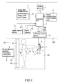
- medical treatment equipment 100 is arranged to include an X-ray source generator 1, a collimator 11, and an X-ray detector 2.
- the X-ray source generator 1 is coupled to the collimator 11.
- the X-ray detector 2 is arranged at a position that is spaced from the X-ray source generator 1 by a predetermined distance.
- the X-ray source generator 1 emits an X-ray beam 15, which, after passing through shielding plates 12 and a filter 13 arranged in the collimator 11, is projected to a target human body 3 and is detected by the X-ray detector 2.
- the collimator 11 is coupled to a collimator control device 14, whereby the collimator control device 14 controls the shielding plate 12 arranged in the collimator 11 for adjustment of a selected human body portion 31 of the target human body 3.
- an automatic selected human portion identification and adjustment device 200 is combined with and coupled to the medical treatment equipment 100 to control the operation of the X-ray detector 2 of the medical treatment equipment 100 in order to execute identification of the selected human body portion and make adjustment toward a predetermined human body portion 31 of the target human body 3.
- the automatic selected human portion identification and adjustment device 200 comprises a processor unit 4, a depth camera 5, an image capture device 51, an image data storage device 6, a display device 7, an image characteristic identification unit 8, and an X-ray detector position adjustment mechanism 9.
- the depth camera 5 is set at a position corresponding to the target human body 3 in order to acquire at least one instant image pixel depth signal S1 of the target human body 3.
- the depth camera 5 is connected to the processor unit 4 to transmit the acquired instant image pixel depth signal S1 to the processor unit 4.
- an image capture device 51 may be provided and connected to the processor unit 4 in order to acquire at least one instant image S11 of the target human body 3 and transmit the instant image S11 to the processor unit 4.
- the image data storage device 6 is connected to the processor unit 4.
- the image data storage device 6 stores therein at least one predetermined image pixel depth signal 61 of the predetermined human body portion 31.
- the display device 7 is connected to the processor unit 4 to display the instant image pixel depth signal S1 of the target human body 3 acquired the depth camera 5 and the instant image S11 of the target human body 3 acquired by the image capture device 51.
- the image characteristic identification unit 8 is connected to the processor unit 4 for comparison and identification of characteristics.
- the process unit 4 compares the instant image pixel depth signal S1 acquired the depth camera 5 and the predetermined image pixel depth signal 61 of the predetermined human body portion 31 of the image data storage device 6 for identification.
- the processor unit 4 According to the result of comparison, if the instant image pixel depth signal S1 and the predetermined image pixel depth signal 61 of the predetermined human body portion 31 are different, the processor unit 4 generates, according to the difference between the instant image pixel depth signal S1 and the predetermined image pixel depth signal 61, at least one X-ray detector position adjustment signal S2 that is transmitted to the X-ray detector position adjustment mechanism 9.
- the X-ray detector position adjustment mechanism 9 after receiving the X-ray detector position adjustment signal S2, generates a movement driving signal S3 for driving the X-ray detector 2 to move thereby adjusting the X-ray detector 2 to correspond to the predetermined human body portion 31 of the target human body 3.
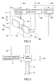
- the collimator control device 14 comprises a vertical direction drive motor 14a, a horizontal direction drive motor 14b, and a rotation drive motor 14c.
- the processor unit 4 is capable of generating a vertical direction driving signal S41 to control the vertical direction drive motor 14a, a horizontal direction driving signal S42 to control the horizontal direction drive motor 14b, and a rotation driving signal S43 to control the rotation drive motor 14c respectively.
- the vertical direction drive motor 14a functions to drive a pair of first-direction arranged shielding plates 12a, 12b of the collimator 11 to move in a vertical direction M1.
- the horizontal direction drive motor 14b functions to drive a pair of horizontal-direction arranged shielding plates 12c, 12d of the collimator 11 to move in a horizontal direction M2.
- the rotation drive motor 14c functions to drive the collimator 11 to rotate about a rotation axial direction M3.
- the X-ray detector 2 is mounted to a vertical carrying frame 21 and the vertical carrying frame 21 is provided with a guide mechanism 22.
- the X-ray detector position adjustment mechanism receives the X-ray detector adjustment signal S2 and generates the movement driving signal S3 to drive the X-ray detector 2, enabling the X-ray detector 2 to move in the vertical direction M1 along the guide mechanism 22.
- the guide mechanism 22 is achieved with a conventional guide rail or guide channel.
- the X-ray detector position adjustment mechanism 9 may comprise a motor, a hydraulic cylinder, a pneumatic cylinder, or the like.
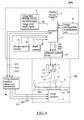
- Figure 4 is a schematic view showing a circuit system according to a second embodiment of the present invention.
- the constituent components of the instant embodiment are substantially identical to those of the first embodiment and identical components are designated with the same reference numerals for consistency.
- the medical treatment equipment 100 similarly comprises an X-ray source generator 1, a collimator 11, and an X-ray detector 2.
- the collimator 11 and the X-ray detector 2 are arranged in a vertical direction and thus, the target human body 3 is allowed to lie horizontally on a treatment bed.
- the automatic selected human portion identification and adjustment device 200a it is possible to control the operation of the X-ray detector 2 of the medical treatment equipment 100 in a similar way so as to achieve adjustment toward the predetermined human body portion 31 of the target human body 3.
- the automatic selected human portion identification and adjustment device 200a of the instant embodiment similarly comprises a processor unit 4, a depth camera 5, an image capture device 51, an image data storage device 6, a display device 7, an image characteristic identification unit 8, and an X-ray detector position adjustment mechanism 9a.
- the processor unit 4 In making a comparison, if the instant image pixel depth signal S1 and the predetermined image pixel depth signal 61 of the predetermined human body portion 31 are different, the processor unit 4 generates, according to the difference between the instant image pixel depth signal S1 and the predetermined image pixel depth signal 61 of the predetermined human body portion, a first-direction X-ray detector position adjustment signal S21, a second-direction X-ray detector position adjustment signal S22, and a rotation axial direction X-ray detector position adjustment signal S23 to the X-ray detector position adjustment mechanism 9a.
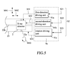
- a horizontal carrying frame 23 is provided with a first-direction guide mechanism 22a and a second-direction guide mechanism 22b.
- the first-direction guide mechanism 22a is mounted to the horizontal carrying frame 23 in a first direction M41 and the second-direction guide mechanism 22b is mounted to the horizontal carrying frame 23 in a second direction M42.
- the X-ray detector position adjustment mechanism 9a receives a first-direction X-ray detector position adjustment signal S21, a second-direction X-ray detector position adjustment signal S22, and a rotation axial direction X-ray detector position adjustment signal S23 and performs the following operations:
Abstract
An automatic selected human portion identification and adjustment device (200, 200a) for medical treatment equipment (100) is disclosed, in which an automatic selected human portion identification and adjustment device (200, 200a) is included in the medical treatment equipment (100). The automatic selected human portion identification and adjustment device (200, 200a) includes a depth camera (5), an image characteristic identification unit (8), and an X-ray detector (2) position adjustment mechanism. The image characteristic identification unit (8) performs image characteristic comparison between an instant image pixel depth signal (S1) with a predetermined image pixel depth signal (61) for identification and generates, in response thereto, an X-ray detector position adjustment signal (S2). Then, the X-ray detector (2) position adjustment mechanism drives the X-ray detector (2) to move in order to adjust the X-ray detector (2) to correspond to the predetermined human body portion of a target human body (3).
Description
- The present invention relates to a control device of medical treatment equipment, and in particular to an automatic selected human portion identification and adjustment device combined with medical treatment equipment to control the operation of an X-ray detector of the medical treatment equipment for adjusting the X-ray detector to aligned with a predetermined selected body portion of a target human body.
- Currently, regular medical examination often applies X-ray imaging. The X-ray imaging generally uses and adjusts shielding plates to determine a radiation area. In a conventional design, a radiographer needs to manually or semi-automatically adjust the shielding plate through upward/downward and left/rightward movement in order to select a desired portion and area of the portion to be irradiated. The X-ray detector is commonly designed as a fixed device. In an alternative design, the X-ray detector is held by a hand and manually placed at the portion of human body that is to be irradiated.
- In the conventional designs of the X-ray detector, a radiographer or medical personnel has to help a patient to make a correct pose and position, or a person other than the radiographer may be need to hold the X-ray detector with hands. The radiographer then proceeds to a collimator to adjust the shielding plate. This causes an issue of poor operability. Further, during the operation that a radiographer or medical personnel makes the adjustment, deviations and inconsistencies of the accuracy of adjustment, the size of the radiation area, and the radiation zone may readily occur due to difference in respect of personal experience, expertise, habit, and personal emotional issues.
- When an X-ray photograph is incorrect in respect of the radiation range or radiation area, incorrect reading of visual inspection by a treating operator may occur and incorrect diagnosis may also result. Further, the chance that a patient has to take the examination once more due to the error of the operation may often be thus greatly increased. It is thus a challenge to the industry to overcome the above-discussed problems and issues.
- Thus, to overcome the above-discussed problems and issues, an object of the present invention is to provide an automatic selected human portion identification and adjustment device for medical treatment equipment, in which an image characteristic identification unit receives an instant image pixel depth signal captured by a depth camera and carries out image characteristic comparison between the instant image pixel depth signal and a predetermined image pixel depth signal of a predetermined human body portion for identification and in response thereto, generates an X-ray detector position signal to allow an X-ray detector position adjustment mechanism to drive an X-ray detector of the medical treatment equipment to move along a guide mechanism thereby adjusting the X-ray detector to align with a predetermined human body portion of a target human body.
- The technical solution that the present invention adopts to achieve the above object is that an automatic X-ray detector adjustment device is combined with medical treatment equipment, in which an automatic selected human portion identification and adjustment device is included in the medical treatment equipment. The automatic selected human portion identification and adjustment device includes a depth camera, an image characteristic identification unit, and an X-ray detector position adjustment mechanism. The image characteristic identification unit performs image characteristic comparison between an instant image pixel depth signal with a predetermined image pixel depth signal for identification and generates, in response thereto, an X-ray detector position adjustment signal. Then, the X-ray detector position adjustment mechanism drives the X-ray detector to move in order to adjust the X-ray detector to correspond to the predetermined human body portion of a target human body.
- The automatic selected human portion identification and adjustment device for medical treatment equipment further comprises an image capture device to acquire at least one instant image of the target human body.
- The automatic selected human portion identification and adjustment device for medical treatment equipment comprises a vertical carrying frame and the guide mechanism is provided on the vertical carrying frame.
- The X-ray detector may be alternatively mounted to a horizontal carrying frame. The guide mechanism comprises: a first-direction guide mechanism, which is mounted to the horizontal carrying frame in a first direction, and a second-direction guide mechanism, which is mounted to the horizontal carrying frame in a second direction. The X-ray detector position adjustment mechanism comprises: a first-direction driving unit, which drives the X-ray detector to move along the first-direction guide mechanism, and a second-direction driving unit, which drives the X-ray detector to move along the second-direction guide mechanism.
- The X-ray detector position adjustment mechanism may further comprise a rotation driving unit, which drives the X-ray detector to rotate about a rotation axial direction.
- The automatic selected human portion identification and adjustment device for medical treatment equipment may further comprise a display device.
- The efficacy is that with the above technical solution of the present invention, medical personnel, when operating an X-ray detector of an X-ray medical treatment equipment, may use the automatic X-ray detector adjustment device of the present invention to automatically detect a predetermined human body portion of a target human body, whereby errors caused by lacking of experience or poor habit of a radiographer can be reduced and the inconvenience and time required for manual adjustment of a X-ray detector by a radiographer may be greatly reduced. In addition, the difficult that a patient needs to change pose thereof to correspond to the X-ray detector can be eliminated. Further, the automatic selected human portion identification and adjustment device of the present invention is operable with a collimator control device to adjust a collimator for adjustment toward a selected human body portion.
- The present invention will be apparent to those skilled in the art by reading the following description of preferred embodiments of the present invention, with reference to the attached drawings, in which:
-
Figure 1 is a schematic view of a circuit system of a first embodiment according to the present invention; -
Figure 2 is a schematic view showing a detailed circuit diagram of a collimator control device and a collimator; -
Figure 3 is a detailed schematic view showing the coupling relationship between an X-ray detector position adjustment mechanism and an X-ray detector ofFigure 1 ; -
Figure 4 is a schematic view of a circuit system of a second embodiment according to the present invention; and -
Figure 5 is a detailed schematic view showing the coupling relationship between an X-ray detector position adjustment mechanism and an X-ray detector ofFigure 4 . - With reference to the drawings and in particular to
Figure 1 , a first embodiment is illustrated, in whichmedical treatment equipment 100 is arranged to include anX-ray source generator 1, acollimator 11, and anX-ray detector 2. TheX-ray source generator 1 is coupled to thecollimator 11. TheX-ray detector 2 is arranged at a position that is spaced from theX-ray source generator 1 by a predetermined distance. TheX-ray source generator 1 emits anX-ray beam 15, which, after passing throughshielding plates 12 and afilter 13 arranged in thecollimator 11, is projected to a targethuman body 3 and is detected by theX-ray detector 2. - The
collimator 11 is coupled to acollimator control device 14, whereby thecollimator control device 14 controls theshielding plate 12 arranged in thecollimator 11 for adjustment of a selectedhuman body portion 31 of the targethuman body 3. - In the present invention, an automatic selected human portion identification and
adjustment device 200 is combined with and coupled to themedical treatment equipment 100 to control the operation of theX-ray detector 2 of themedical treatment equipment 100 in order to execute identification of the selected human body portion and make adjustment toward a predeterminedhuman body portion 31 of the targethuman body 3. - The automatic selected human portion identification and
adjustment device 200 according to the present invention comprises aprocessor unit 4, adepth camera 5, animage capture device 51, an imagedata storage device 6, adisplay device 7, an imagecharacteristic identification unit 8, and an X-ray detectorposition adjustment mechanism 9. - The
depth camera 5 is set at a position corresponding to the targethuman body 3 in order to acquire at least one instant image pixel depth signal S1 of the targethuman body 3. Thedepth camera 5 is connected to theprocessor unit 4 to transmit the acquired instant image pixel depth signal S1 to theprocessor unit 4. - In addition to the
depth camera 5, animage capture device 51 may be provided and connected to theprocessor unit 4 in order to acquire at least one instant image S11 of the targethuman body 3 and transmit the instant image S11 to theprocessor unit 4. - The image
data storage device 6 is connected to theprocessor unit 4. The imagedata storage device 6 stores therein at least one predetermined imagepixel depth signal 61 of the predeterminedhuman body portion 31. - The
display device 7 is connected to theprocessor unit 4 to display the instant image pixel depth signal S1 of the targethuman body 3 acquired thedepth camera 5 and the instant image S11 of the targethuman body 3 acquired by theimage capture device 51. - The image
characteristic identification unit 8 is connected to theprocessor unit 4 for comparison and identification of characteristics. Theprocess unit 4 compares the instant image pixel depth signal S1 acquired thedepth camera 5 and the predetermined imagepixel depth signal 61 of the predeterminedhuman body portion 31 of the imagedata storage device 6 for identification. - According to the result of comparison, if the instant image pixel depth signal S1 and the predetermined image
pixel depth signal 61 of the predeterminedhuman body portion 31 are different, theprocessor unit 4 generates, according to the difference between the instant image pixel depth signal S1 and the predetermined imagepixel depth signal 61, at least one X-ray detector position adjustment signal S2 that is transmitted to the X-ray detectorposition adjustment mechanism 9. The X-ray detectorposition adjustment mechanism 9, after receiving the X-ray detector position adjustment signal S2, generates a movement driving signal S3 for driving theX-ray detector 2 to move thereby adjusting theX-ray detector 2 to correspond to the predeterminedhuman body portion 31 of the targethuman body 3. - As shown in
Figure 2 , in an embodiment of the present invention, thecollimator control device 14 comprises a verticaldirection drive motor 14a, a horizontaldirection drive motor 14b, and arotation drive motor 14c. Theprocessor unit 4 is capable of generating a vertical direction driving signal S41 to control the verticaldirection drive motor 14a, a horizontal direction driving signal S42 to control the horizontaldirection drive motor 14b, and a rotation driving signal S43 to control therotation drive motor 14c respectively. The vertical direction drivemotor 14a functions to drive a pair of first-direction arrangedshielding plates collimator 11 to move in a vertical direction M1. The horizontal direction drivemotor 14b functions to drive a pair of horizontal-direction arrangedshielding plates collimator 11 to move in a horizontal direction M2. The rotation drivemotor 14c functions to drive thecollimator 11 to rotate about a rotation axial direction M3. With the above-discussed control, when applied in combination with X-ray detectorposition adjustment mechanism 9, the purpose of controlling thecollimator 11 to adjust theX-ray beam 15 to align with the predeterminedhuman body portion 31 of the targethuman body 3 can be achieved. - As shown in
Figure 3 , which shows further details of the coupling relationship between the X-ray detector position adjustment mechanism and the X-ray detector ofFigure 1 , theX-ray detector 2 is mounted to avertical carrying frame 21 and thevertical carrying frame 21 is provided with aguide mechanism 22. The X-ray detector position adjustment mechanism receives the X-ray detector adjustment signal S2 and generates the movement driving signal S3 to drive theX-ray detector 2, enabling theX-ray detector 2 to move in the vertical direction M1 along theguide mechanism 22. In an embodiment, theguide mechanism 22 is achieved with a conventional guide rail or guide channel. The X-ray detectorposition adjustment mechanism 9 may comprise a motor, a hydraulic cylinder, a pneumatic cylinder, or the like. -
Figure 4 is a schematic view showing a circuit system according to a second embodiment of the present invention. The constituent components of the instant embodiment are substantially identical to those of the first embodiment and identical components are designated with the same reference numerals for consistency. In the instant embodiment, themedical treatment equipment 100 similarly comprises anX-ray source generator 1, acollimator 11, and anX-ray detector 2. However, thecollimator 11 and theX-ray detector 2 are arranged in a vertical direction and thus, the targethuman body 3 is allowed to lie horizontally on a treatment bed. - With the automatic selected human portion identification and
adjustment device 200a according to the present invention, it is possible to control the operation of theX-ray detector 2 of themedical treatment equipment 100 in a similar way so as to achieve adjustment toward the predeterminedhuman body portion 31 of the targethuman body 3. - The automatic selected human portion identification and
adjustment device 200a of the instant embodiment similarly comprises aprocessor unit 4, adepth camera 5, animage capture device 51, an imagedata storage device 6, adisplay device 7, an imagecharacteristic identification unit 8, and an X-ray detectorposition adjustment mechanism 9a. - In making a comparison, if the instant image pixel depth signal S1 and the predetermined image
pixel depth signal 61 of the predeterminedhuman body portion 31 are different, theprocessor unit 4 generates, according to the difference between the instant image pixel depth signal S1 and the predetermined imagepixel depth signal 61 of the predetermined human body portion, a first-direction X-ray detector position adjustment signal S21, a second-direction X-ray detector position adjustment signal S22, and a rotation axial direction X-ray detector position adjustment signal S23 to the X-ray detectorposition adjustment mechanism 9a. - As shown in
Figure 5 , which shows further details of the coupling relationship between the X-ray detector position adjustment mechanism and the X-ray detector ofFigure 4 , as shown in the drawing, ahorizontal carrying frame 23 is provided with a first-direction guide mechanism 22a and a second-direction guide mechanism 22b. The first-direction guide mechanism 22a is mounted to thehorizontal carrying frame 23 in a first direction M41 and the second-direction guide mechanism 22b is mounted to thehorizontal carrying frame 23 in a second direction M42. - The X-ray detector
position adjustment mechanism 9a receives a first-direction X-ray detector position adjustment signal S21, a second-direction X-ray detector position adjustment signal S22, and a rotation axial direction X-ray detector position adjustment signal S23 and performs the following operations: - (a) generating a first-direction driving signal S31 to activate a first-
direction driving unit 91a for driving theX-ray detector 2 to move in the first direction M1 along the first-direction guide mechanism 22a; - (b) generating a second-direction driving signal S32 to activate a second-
direction driving unit 91b for driving theX-ray detector 2 to move in the second direction M2 along the second-direction guide mechanism 22b; and - (c) generating a rotation axial direction driving signal S32 to activate a
rotation driving unit 91c for driving thehorizontal carrying frame 23 to rotate about a rotation axial direction M43. - Although the present invention has been described with reference to the preferred embodiments thereof, it is apparent to those skilled in the art that a variety of modifications and changes may be made without departing from the scope of the present invention which is intended to be defined by the appended claims.
Claims (6)
- An automatic selected human portion identification and adjustment device (200, 200a) for a medical treatment equipment (100), in which the medical treatment equipment (100) includes an X-ray source generator (1), a collimator (11), an X-ray detector (2), and a collimator control device (14), wherein the X-ray source generator (1) projects an X-ray beam (15) to a target human body (3) through the collimator (11) and the X-ray detector (2) detects the X-ray beam (15), automatic characterized in that the selected human portion identification and adjustment device (200, 200a) comprises:a processor unit (4);an image data storage device (6), which is connected to the processor unit (4) and stores therein at least one predetermined image pixel depth signal (61) of a predetermined human body portion (31) of the target human (3);a depth camera (5), which is connected to the processor unit (4) and set at a position corresponding to the target human body (3) to acquire at least one instant image pixel depth signal (S1) of the target human body (3);an image characteristic identification unit (8), which is connected to the processor unit (4) and receives the instant image pixel depth signal (S1) acquired by the depth camera (5) and compares the instant image pixel depth signal (S1) with the predetermined image pixel depth signal (61) for identification in order to generate an X-ray detector position adjustment signal (S2); andan X-ray detector position adjustment mechanism (9, 9a), which is connected to the processor unit (4) and coupled to the X-ray detector (2) to adjust a relative position of the X-ray detector (2) with respect to the X-ray source generator (1),wherein the processor unit (4) receives the X-ray detector position adjustment signal (S2) and then transmits the X-ray detector position adjustment signal (S2) to the X-ray detector position adjustment mechanism (9, 9a), whereby the X-ray detector position adjustment mechanism (9, 9a) drives the X-ray detector (2) to move along a guide mechanism (22) so as to adjust the X-ray detector (2) to correspond to the predetermined human body portion (31) of the target human body (3).
- The automatic selected human portion identification and adjustment device (200, 200a) as claimed in Claim 1 further comprising an image capture device (51) to acquire at least one instant image (S11) of the target human body (3).
- The automatic selected human portion identification and adjustment device (200, 200a) as claimed in Claim 1, wherein the X-ray detector (2) is mounted to a vertical carrying frame (21), the guide mechanism (22) being provided on the vertical carrying frame (21), the X-ray detector position adjustment mechanism (9) driving the X-ray detector (2) to move in a vertical direction (M1) along the guide mechanism (22).
- The automatic selected human portion identification and adjustment device (200, 200a) as claimed in Claim 1, wherein
the X-ray detector (2) is mounted to a horizontal carrying frame (23); and
the guide mechanism (22) comprises:a first-direction guide mechanism (22a), which is mounted to the horizontal carrying frame (23) in a first direction (M41); anda second-direction guide mechanism (22b), which is mounted to the horizontal carrying frame (23) in a second direction (M42); andthe X-ray detector position adjustment mechanism (9a) comprises:a first-direction driving unit (91a), which drives the X-ray detector (2) to move along the first-direction guide mechanism (22a); and
a second-direction driving unit (91b), which drives the X-ray detector (2) to move along the second-direction guide mechanism (22b). - The automatic selected human portion identification and adjustment device (200, 200a) as claimed in Claim 4, wherein the X-ray detector position adjustment mechanism (9a) further comprises:a rotation driving unit (91c), which drives the X-ray detector (2) to rotate about a rotation axial direction (M43).
- The automatic selected human portion identification and adjustment device (200, 200a) as claimed in Claim 1 further comprising a display device (7).
Applications Claiming Priority (1)
| Application Number | Priority Date | Filing Date | Title |
|---|---|---|---|
| TW103116934A TWI535421B (en) | 2014-05-14 | 2014-05-14 | Automatic identification and adjustment of the selected parts of the medical equipment |
Publications (1)
| Publication Number | Publication Date |
|---|---|
| EP2944255A1 true EP2944255A1 (en) | 2015-11-18 |
Family
ID=51263258
Family Applications (1)
| Application Number | Title | Priority Date | Filing Date |
|---|---|---|---|
| EP14179445.3A Withdrawn EP2944255A1 (en) | 2014-05-14 | 2014-08-01 | Automatic selected human portion identification and adjustment device for medical treatment equipment |
Country Status (4)
| Country | Link |
|---|---|
| US (1) | US9462985B2 (en) |
| EP (1) | EP2944255A1 (en) |
| CN (1) | CN105078483A (en) |
| TW (1) | TWI535421B (en) |
Cited By (1)
| Publication number | Priority date | Publication date | Assignee | Title |
|---|---|---|---|---|
| CN110507338A (en) * | 2019-08-30 | 2019-11-29 | 东软医疗系统股份有限公司 | Localization method, device, equipment and Digital X-ray Radiotive system |
Families Citing this family (16)
| Publication number | Priority date | Publication date | Assignee | Title |
|---|---|---|---|---|
| US9566040B2 (en) * | 2014-05-14 | 2017-02-14 | Swissray Asia Healthcare Co., Ltd. | Automatic collimator adjustment device with depth camera and method for medical treatment equipment |
| EP2954843A1 (en) * | 2014-06-13 | 2015-12-16 | Agfa Healthcare | Method and system for configuring an x-ray imaging system |
| CN106456100A (en) * | 2014-06-30 | 2017-02-22 | 爱克发医疗保健公司 | Method and system for configuring an X-ray imaging system |
| US9649084B2 (en) * | 2014-07-21 | 2017-05-16 | Samsung Electronics Co., Ltd. | X-ray imaging apparatus and method for creating X-ray image |
| KR102340197B1 (en) * | 2015-02-03 | 2021-12-16 | 삼성전자주식회사 | X ray apparatus and method of oprating the same |
| EP3320842B1 (en) * | 2015-07-07 | 2020-02-12 | Fujifilm Corporation | Radiography apparatus, method for controlling radiography apparatus and program |
| US10556129B2 (en) * | 2015-10-02 | 2020-02-11 | Varian Medical Systems, Inc. | Systems and methods for treating a skin condition using radiation |
| JP6735109B2 (en) * | 2016-02-05 | 2020-08-05 | キヤノンメディカルシステムズ株式会社 | X-ray imaging system |
| ITUA20162102A1 (en) * | 2016-03-30 | 2017-09-30 | Cefla S C | BEAM RESTRICTION DEVICE FOR RADIOGRAPHIC EQUIPMENT |
| DE102016205176A1 (en) * | 2016-03-30 | 2017-10-05 | Siemens Healthcare Gmbh | Apparatus and method for creating an X-ray panoramic image |
| CN105769124B (en) * | 2016-03-31 | 2019-06-25 | 联想(北京)有限公司 | Method for sensing and sensing device |
| JP6849966B2 (en) * | 2016-11-21 | 2021-03-31 | 東芝エネルギーシステムズ株式会社 | Medical image processing equipment, medical image processing methods, medical image processing programs, motion tracking equipment and radiation therapy systems |
| EP3387997B1 (en) * | 2017-04-13 | 2020-02-26 | Siemens Healthcare GmbH | Medical imaging device and method controlling one or more parameters of a medical imaging device |
| CN107334487A (en) * | 2017-08-11 | 2017-11-10 | 上海联影医疗科技有限公司 | A kind of medical image system and its scan method |
| CN112770673A (en) * | 2018-09-27 | 2021-05-07 | 富士胶片株式会社 | Radiographic apparatus |
| CN114699097B (en) * | 2022-06-06 | 2022-09-27 | 江苏康众数字医疗科技股份有限公司 | Radiographic imaging system and radiographic imaging method |
Citations (6)
| Publication number | Priority date | Publication date | Assignee | Title |
|---|---|---|---|---|
| DE102007017794B3 (en) * | 2007-04-16 | 2008-12-04 | Siemens Ag | Method for positioning of displaceable patient couch in medical diagnose unit, involves taking consequence of graphic data sets of patient couch with patient present at it by camera |
| US20140016750A1 (en) * | 2012-07-12 | 2014-01-16 | Samsung Electronics Co., Ltd. | X-ray imaging apparatus and method for controlling x-ray imaging apparatus |
| WO2014033614A1 (en) * | 2012-08-27 | 2014-03-06 | Koninklijke Philips N.V. | Patient-specific and automatic x-ray system adjustment based on optical 3d scene detection and interpretation |
| WO2014202720A1 (en) * | 2013-06-19 | 2014-12-24 | Koninklijke Philips N.V. | Calibration of imagers with dynamic beam shapers |
| WO2014210190A2 (en) * | 2013-06-25 | 2014-12-31 | Varian Medical Systems, Inc. | Systems and methods for detecting a possible collision between an object and a patient in a medical procedure |
| WO2014208969A1 (en) * | 2013-06-26 | 2014-12-31 | Samsung Electronics Co., Ltd. | Method and apparatus for providing information related to location of target object on medical apparatus |
Family Cites Families (33)
| Publication number | Priority date | Publication date | Assignee | Title |
|---|---|---|---|---|
| JP3619027B2 (en) * | 1998-09-30 | 2005-02-09 | キヤノン株式会社 | X-ray imaging apparatus, X-ray imaging system, X-ray image processing method, and computer-readable recording medium |
| US6463121B1 (en) * | 1999-10-13 | 2002-10-08 | General Electric Company | Interactive x-ray position and exposure control using image data as reference information |
| DE10234465A1 (en) * | 2002-07-29 | 2004-02-12 | Siemens Ag | X-ray sectional-imaging method in which the height of a sectional image is set using a camera arrangement for imaging the patient from the side so that a reference marking can be made on a reference image |
| DE10244609A1 (en) * | 2002-09-25 | 2004-04-15 | Siemens Ag | Radiation image recording device |
| US6944265B2 (en) * | 2002-11-25 | 2005-09-13 | Ge Medical Systems Global Technology Company, Llc | Image pasting using geometry measurement and a flat-panel detector |
| US6935779B2 (en) * | 2002-11-29 | 2005-08-30 | Ge Medical Systems Global Technology Company, Llc | Method and apparatus for aligning an X-ray source and detector at various source to image distances |
| DE10322142A1 (en) * | 2003-05-16 | 2004-12-09 | Siemens Ag | Radioscopy instrument for medical diagnostic imaging, has laser distance meter for measuring distance between source and patient, and has computer for automatic exposure calculation |
| DE10327294A1 (en) * | 2003-06-17 | 2005-01-20 | Siemens Ag | Method for image processing of X-ray images and associated image processing unit |
| DE10345509A1 (en) * | 2003-09-30 | 2005-05-04 | Siemens Ag | Visualized image optimization of an X-ray image |
| DE102004015540B4 (en) * | 2004-03-30 | 2006-12-28 | Siemens Ag | Radiation image recording device |
| CN1984606B (en) * | 2004-07-13 | 2011-08-10 | 皇家飞利浦电子股份有限公司 | X-ray equipment and method for controlling it |
| US7522701B2 (en) * | 2005-12-20 | 2009-04-21 | General Electric Company | System and method for image composition using position sensors |
| JP2008125981A (en) * | 2006-11-24 | 2008-06-05 | Shimadzu Corp | Universal photography system |
| CN101278840B (en) * | 2007-04-02 | 2012-02-22 | Ge医疗系统环球技术有限公司 | X-ray photographic equipment |
| CN101491444B (en) * | 2008-01-25 | 2012-12-12 | Ge医疗系统环球技术有限公司 | X-ray detection station and X-ray imaging device |
| JP2010017376A (en) * | 2008-07-11 | 2010-01-28 | Fujifilm Corp | Radiographic imaging apparatus |
| JP5523025B2 (en) * | 2008-09-16 | 2014-06-18 | 富士フイルム株式会社 | Installation error detection method and image correction method for imaging surface of radiation image detector |
| JP5137252B2 (en) * | 2008-09-24 | 2013-02-06 | 富士フイルム株式会社 | Radiation imaging equipment |
| JP2010075245A (en) * | 2008-09-24 | 2010-04-08 | Fujifilm Corp | Radiographic imaging apparatus |
| JP2010094209A (en) * | 2008-10-15 | 2010-04-30 | Fujifilm Corp | Radiation imaging apparatus |
| JP2010221003A (en) * | 2009-02-26 | 2010-10-07 | Fujifilm Corp | Radiation imaging apparatus |
| JP5324282B2 (en) * | 2009-03-27 | 2013-10-23 | 富士フイルム株式会社 | Radiography equipment |
| JP5333580B2 (en) * | 2009-04-07 | 2013-11-06 | 株式会社島津製作所 | X-ray equipment |
| JP5657224B2 (en) * | 2009-08-31 | 2015-01-21 | 富士フイルム株式会社 | Method and apparatus for determining degree of installation error of imaging surface of radiation image detector |
| JP5438493B2 (en) * | 2009-12-22 | 2014-03-12 | 富士フイルム株式会社 | Radiation imaging system and auxiliary device thereof |
| CN103249364B (en) * | 2010-11-26 | 2015-05-13 | 株式会社岛津制作所 | X-ray examination apparatus |
| JP5579636B2 (en) * | 2011-02-07 | 2014-08-27 | 富士フイルム株式会社 | Radiographic imaging apparatus and radiographic imaging method |
| US8899832B2 (en) * | 2011-10-20 | 2014-12-02 | Fujifilm Medical Systems U.S.A., Inc. | Mobile patient positioning shield for multiple exposure imaging exams |
| US8767913B2 (en) * | 2011-12-06 | 2014-07-01 | Shimadzu Corporation | X-ray radiography device |
| KR101431781B1 (en) * | 2012-06-20 | 2014-08-20 | 삼성전자주식회사 | X-ray image apparatus and control method for the same |
| US8908832B2 (en) * | 2013-03-11 | 2014-12-09 | Shimadzu Corporation | Radiographic apparatus and method for the same |
| TWI542327B (en) * | 2014-05-14 | 2016-07-21 | Automatic control device and method for exposure of X - ray machine with depth camera | |
| US9566040B2 (en) * | 2014-05-14 | 2017-02-14 | Swissray Asia Healthcare Co., Ltd. | Automatic collimator adjustment device with depth camera and method for medical treatment equipment |
-
2014
- 2014-05-14 TW TW103116934A patent/TWI535421B/en not_active IP Right Cessation
- 2014-07-24 US US14/339,525 patent/US9462985B2/en active Active
- 2014-08-01 EP EP14179445.3A patent/EP2944255A1/en not_active Withdrawn
- 2014-08-08 CN CN201410387936.4A patent/CN105078483A/en active Pending
Patent Citations (6)
| Publication number | Priority date | Publication date | Assignee | Title |
|---|---|---|---|---|
| DE102007017794B3 (en) * | 2007-04-16 | 2008-12-04 | Siemens Ag | Method for positioning of displaceable patient couch in medical diagnose unit, involves taking consequence of graphic data sets of patient couch with patient present at it by camera |
| US20140016750A1 (en) * | 2012-07-12 | 2014-01-16 | Samsung Electronics Co., Ltd. | X-ray imaging apparatus and method for controlling x-ray imaging apparatus |
| WO2014033614A1 (en) * | 2012-08-27 | 2014-03-06 | Koninklijke Philips N.V. | Patient-specific and automatic x-ray system adjustment based on optical 3d scene detection and interpretation |
| WO2014202720A1 (en) * | 2013-06-19 | 2014-12-24 | Koninklijke Philips N.V. | Calibration of imagers with dynamic beam shapers |
| WO2014210190A2 (en) * | 2013-06-25 | 2014-12-31 | Varian Medical Systems, Inc. | Systems and methods for detecting a possible collision between an object and a patient in a medical procedure |
| WO2014208969A1 (en) * | 2013-06-26 | 2014-12-31 | Samsung Electronics Co., Ltd. | Method and apparatus for providing information related to location of target object on medical apparatus |
Cited By (2)
| Publication number | Priority date | Publication date | Assignee | Title |
|---|---|---|---|---|
| CN110507338A (en) * | 2019-08-30 | 2019-11-29 | 东软医疗系统股份有限公司 | Localization method, device, equipment and Digital X-ray Radiotive system |
| CN110507338B (en) * | 2019-08-30 | 2022-12-27 | 东软医疗系统股份有限公司 | Positioning method, device and equipment and digital X-ray photography system |
Also Published As
| Publication number | Publication date |
|---|---|
| TW201542171A (en) | 2015-11-16 |
| CN105078483A (en) | 2015-11-25 |
| US20150327832A1 (en) | 2015-11-19 |
| TWI535421B (en) | 2016-06-01 |
| US9462985B2 (en) | 2016-10-11 |
Similar Documents
| Publication | Publication Date | Title |
|---|---|---|
| US9462985B2 (en) | Automatic selected human portion identification and adjustment device for medical treatment equipment | |
| US9566040B2 (en) | Automatic collimator adjustment device with depth camera and method for medical treatment equipment | |
| US10076293B2 (en) | Rapid frame-rate wireless imaging system | |
| DE102014219667B3 (en) | Method for selecting a recording area and system for selecting a recording area | |
| EP2740405B1 (en) | X-ray imaging apparatus and method for controlling the same | |
| JP2019107397A (en) | Medical apparatus, control method and program of medical apparatus | |
| JP6446348B2 (en) | Radiation tomography apparatus and program | |
| US10722200B2 (en) | Apparatus and methods for a projection display device on X-ray imaging devices | |
| US11864937B2 (en) | Imaging systems and methods | |
| WO2020036225A1 (en) | Radiographic imaging apparatus | |
| US9720512B2 (en) | Device and method for the gesture-controlled setting of setting variables on an X-ray source | |
| US20210169438A1 (en) | Imaging systems and methods | |
| JP7342990B2 (en) | X-ray imaging device | |
| US20180206810A1 (en) | X-ray imaging apparatus and control method thereof | |
| US20190261931A1 (en) | Video patient tracking for medical imaging guidance | |
| JP2015202222A5 (en) | ||
| US20160073998A1 (en) | X-ray diagnostic apparatus | |
| US20230414185A1 (en) | Imaging systems and methods | |
| KR20140141249A (en) | X-ray imaging system and method for controlling detecting area of the same | |
| US10702224B1 (en) | Positioning guidance system For X-ray exams | |
| WO2023183854A1 (en) | Improved imaging systems and methods | |
| US20150265179A1 (en) | Assisting Navigation of a Medical Insert Inside a Patient | |
| TW201542175A (en) | Diagnosis-treatment apparatus collimator region selection control device combined with depth camera | |
| TW201542173A (en) | Medical apparatus collimator auto-adjustment device combined with depth camera and method thereof | |
| EP3103395B1 (en) | Imaging device for x-ray image |
Legal Events
| Date | Code | Title | Description |
|---|---|---|---|
| PUAI | Public reference made under article 153(3) epc to a published international application that has entered the european phase |
Free format text: ORIGINAL CODE: 0009012 |
|
| AK | Designated contracting states |
Kind code of ref document: A1 Designated state(s): AL AT BE BG CH CY CZ DE DK EE ES FI FR GB GR HR HU IE IS IT LI LT LU LV MC MK MT NL NO PL PT RO RS SE SI SK SM TR |
|
| AX | Request for extension of the european patent |
Extension state: BA ME |
|
| STAA | Information on the status of an ep patent application or granted ep patent |
Free format text: STATUS: THE APPLICATION IS DEEMED TO BE WITHDRAWN |
|
| 18D | Application deemed to be withdrawn |
Effective date: 20160519 |