EP2941192B1 - Verfahren und vorrichtung zur quantifizierung der eigenschaften eines mit einem kontrastmittel behandelten objekts - Google Patents
Verfahren und vorrichtung zur quantifizierung der eigenschaften eines mit einem kontrastmittel behandelten objekts Download PDFInfo
- Publication number
- EP2941192B1 EP2941192B1 EP13812043.1A EP13812043A EP2941192B1 EP 2941192 B1 EP2941192 B1 EP 2941192B1 EP 13812043 A EP13812043 A EP 13812043A EP 2941192 B1 EP2941192 B1 EP 2941192B1
- Authority
- EP
- European Patent Office
- Prior art keywords
- contrast agent
- concentration
- microbubbles
- imaging
- retained
- Prior art date
- Legal status (The legal status is an assumption and is not a legal conclusion. Google has not performed a legal analysis and makes no representation as to the accuracy of the status listed.)
- Active
Links
Images


Classifications
-
- A—HUMAN NECESSITIES
- A61—MEDICAL OR VETERINARY SCIENCE; HYGIENE
- A61K—PREPARATIONS FOR MEDICAL, DENTAL OR TOILETRY PURPOSES
- A61K49/00—Preparations for testing in vivo
- A61K49/22—Echographic preparations; Ultrasound imaging preparations ; Optoacoustic imaging preparations
- A61K49/222—Echographic preparations; Ultrasound imaging preparations ; Optoacoustic imaging preparations characterised by a special physical form, e.g. emulsions, liposomes
- A61K49/227—Liposomes, lipoprotein vesicles, e.g. LDL or HDL lipoproteins, micelles, e.g. phospholipidic or polymeric
-
- G—PHYSICS
- G06—COMPUTING OR CALCULATING; COUNTING
- G06T—IMAGE DATA PROCESSING OR GENERATION, IN GENERAL
- G06T7/00—Image analysis
- G06T7/0002—Inspection of images, e.g. flaw detection
- G06T7/0012—Biomedical image inspection
-
- A—HUMAN NECESSITIES
- A61—MEDICAL OR VETERINARY SCIENCE; HYGIENE
- A61B—DIAGNOSIS; SURGERY; IDENTIFICATION
- A61B6/00—Apparatus or devices for radiation diagnosis; Apparatus or devices for radiation diagnosis combined with radiation therapy equipment
- A61B6/48—Diagnostic techniques
- A61B6/481—Diagnostic techniques involving the use of contrast agents
-
- A—HUMAN NECESSITIES
- A61—MEDICAL OR VETERINARY SCIENCE; HYGIENE
- A61B—DIAGNOSIS; SURGERY; IDENTIFICATION
- A61B6/00—Apparatus or devices for radiation diagnosis; Apparatus or devices for radiation diagnosis combined with radiation therapy equipment
- A61B6/48—Diagnostic techniques
- A61B6/486—Diagnostic techniques involving generating temporal series of image data
-
- A—HUMAN NECESSITIES
- A61—MEDICAL OR VETERINARY SCIENCE; HYGIENE
- A61B—DIAGNOSIS; SURGERY; IDENTIFICATION
- A61B6/00—Apparatus or devices for radiation diagnosis; Apparatus or devices for radiation diagnosis combined with radiation therapy equipment
- A61B6/52—Devices using data or image processing specially adapted for radiation diagnosis
- A61B6/5211—Devices using data or image processing specially adapted for radiation diagnosis involving processing of medical diagnostic data
- A61B6/5217—Devices using data or image processing specially adapted for radiation diagnosis involving processing of medical diagnostic data extracting a diagnostic or physiological parameter from medical diagnostic data
-
- A—HUMAN NECESSITIES
- A61—MEDICAL OR VETERINARY SCIENCE; HYGIENE
- A61B—DIAGNOSIS; SURGERY; IDENTIFICATION
- A61B8/00—Diagnosis using ultrasonic, sonic or infrasonic waves
- A61B8/06—Measuring blood flow
-
- A—HUMAN NECESSITIES
- A61—MEDICAL OR VETERINARY SCIENCE; HYGIENE
- A61B—DIAGNOSIS; SURGERY; IDENTIFICATION
- A61B8/00—Diagnosis using ultrasonic, sonic or infrasonic waves
- A61B8/48—Diagnostic techniques
- A61B8/481—Diagnostic techniques involving the use of contrast agents, e.g. microbubbles introduced into the bloodstream
-
- A—HUMAN NECESSITIES
- A61—MEDICAL OR VETERINARY SCIENCE; HYGIENE
- A61B—DIAGNOSIS; SURGERY; IDENTIFICATION
- A61B8/00—Diagnosis using ultrasonic, sonic or infrasonic waves
- A61B8/52—Devices using data or image processing specially adapted for diagnosis using ultrasonic, sonic or infrasonic waves
- A61B8/5215—Devices using data or image processing specially adapted for diagnosis using ultrasonic, sonic or infrasonic waves involving processing of medical diagnostic data
- A61B8/5223—Devices using data or image processing specially adapted for diagnosis using ultrasonic, sonic or infrasonic waves involving processing of medical diagnostic data for extracting a diagnostic or physiological parameter from medical diagnostic data
-
- A—HUMAN NECESSITIES
- A61—MEDICAL OR VETERINARY SCIENCE; HYGIENE
- A61K—PREPARATIONS FOR MEDICAL, DENTAL OR TOILETRY PURPOSES
- A61K49/00—Preparations for testing in vivo
- A61K49/001—Preparation for luminescence or biological staining
- A61K49/0013—Luminescence
- A61K49/0017—Fluorescence in vivo
- A61K49/0019—Fluorescence in vivo characterised by the fluorescent group, e.g. oligomeric, polymeric or dendritic molecules
- A61K49/0021—Fluorescence in vivo characterised by the fluorescent group, e.g. oligomeric, polymeric or dendritic molecules the fluorescent group being a small organic molecule
- A61K49/0032—Methine dyes, e.g. cyanine dyes
- A61K49/0034—Indocyanine green, i.e. ICG, cardiogreen
-
- A—HUMAN NECESSITIES
- A61—MEDICAL OR VETERINARY SCIENCE; HYGIENE
- A61K—PREPARATIONS FOR MEDICAL, DENTAL OR TOILETRY PURPOSES
- A61K49/00—Preparations for testing in vivo
- A61K49/001—Preparation for luminescence or biological staining
- A61K49/0013—Luminescence
- A61K49/0017—Fluorescence in vivo
- A61K49/005—Fluorescence in vivo characterised by the carrier molecule carrying the fluorescent agent
- A61K49/0058—Antibodies
-
- A—HUMAN NECESSITIES
- A61—MEDICAL OR VETERINARY SCIENCE; HYGIENE
- A61K—PREPARATIONS FOR MEDICAL, DENTAL OR TOILETRY PURPOSES
- A61K49/00—Preparations for testing in vivo
- A61K49/001—Preparation for luminescence or biological staining
- A61K49/0063—Preparation for luminescence or biological staining characterised by a special physical or galenical form, e.g. emulsions, microspheres
- A61K49/0069—Preparation for luminescence or biological staining characterised by a special physical or galenical form, e.g. emulsions, microspheres the agent being in a particular physical galenical form
- A61K49/0076—Preparation for luminescence or biological staining characterised by a special physical or galenical form, e.g. emulsions, microspheres the agent being in a particular physical galenical form dispersion, suspension, e.g. particles in a liquid, colloid, emulsion
- A61K49/0082—Preparation for luminescence or biological staining characterised by a special physical or galenical form, e.g. emulsions, microspheres the agent being in a particular physical galenical form dispersion, suspension, e.g. particles in a liquid, colloid, emulsion micelle, e.g. phospholipidic micelle and polymeric micelle
-
- A—HUMAN NECESSITIES
- A61—MEDICAL OR VETERINARY SCIENCE; HYGIENE
- A61K—PREPARATIONS FOR MEDICAL, DENTAL OR TOILETRY PURPOSES
- A61K49/00—Preparations for testing in vivo
- A61K49/06—Nuclear magnetic resonance [NMR] contrast preparations; Magnetic resonance imaging [MRI] contrast preparations
- A61K49/08—Nuclear magnetic resonance [NMR] contrast preparations; Magnetic resonance imaging [MRI] contrast preparations characterised by the carrier
- A61K49/10—Organic compounds
- A61K49/14—Peptides, e.g. proteins
- A61K49/16—Antibodies; Immunoglobulins; Fragments thereof
-
- A—HUMAN NECESSITIES
- A61—MEDICAL OR VETERINARY SCIENCE; HYGIENE
- A61K—PREPARATIONS FOR MEDICAL, DENTAL OR TOILETRY PURPOSES
- A61K49/00—Preparations for testing in vivo
- A61K49/06—Nuclear magnetic resonance [NMR] contrast preparations; Magnetic resonance imaging [MRI] contrast preparations
- A61K49/18—Nuclear magnetic resonance [NMR] contrast preparations; Magnetic resonance imaging [MRI] contrast preparations characterised by a special physical form, e.g. emulsions, microcapsules, liposomes
- A61K49/1806—Suspensions, emulsions, colloids, dispersions
- A61K49/1809—Micelles, e.g. phospholipidic or polymeric micelles
-
- A—HUMAN NECESSITIES
- A61—MEDICAL OR VETERINARY SCIENCE; HYGIENE
- A61K—PREPARATIONS FOR MEDICAL, DENTAL OR TOILETRY PURPOSES
- A61K49/00—Preparations for testing in vivo
- A61K49/22—Echographic preparations; Ultrasound imaging preparations ; Optoacoustic imaging preparations
- A61K49/221—Echographic preparations; Ultrasound imaging preparations ; Optoacoustic imaging preparations characterised by the targeting agent or modifying agent linked to the acoustically-active agent
-
- A—HUMAN NECESSITIES
- A61—MEDICAL OR VETERINARY SCIENCE; HYGIENE
- A61K—PREPARATIONS FOR MEDICAL, DENTAL OR TOILETRY PURPOSES
- A61K49/00—Preparations for testing in vivo
- A61K49/22—Echographic preparations; Ultrasound imaging preparations ; Optoacoustic imaging preparations
- A61K49/222—Echographic preparations; Ultrasound imaging preparations ; Optoacoustic imaging preparations characterised by a special physical form, e.g. emulsions, liposomes
- A61K49/223—Microbubbles, hollow microspheres, free gas bubbles, gas microspheres
-
- G—PHYSICS
- G01—MEASURING; TESTING
- G01R—MEASURING ELECTRIC VARIABLES; MEASURING MAGNETIC VARIABLES
- G01R33/00—Arrangements or instruments for measuring magnetic variables
- G01R33/20—Arrangements or instruments for measuring magnetic variables involving magnetic resonance
- G01R33/44—Arrangements or instruments for measuring magnetic variables involving magnetic resonance using nuclear magnetic resonance [NMR]
- G01R33/48—NMR imaging systems
- G01R33/54—Signal processing systems, e.g. using pulse sequences ; Generation or control of pulse sequences; Operator console
- G01R33/56—Image enhancement or correction, e.g. subtraction or averaging techniques, e.g. improvement of signal-to-noise ratio and resolution
- G01R33/5601—Image enhancement or correction, e.g. subtraction or averaging techniques, e.g. improvement of signal-to-noise ratio and resolution involving use of a contrast agent for contrast manipulation, e.g. a paramagnetic, super-paramagnetic, ferromagnetic or hyperpolarised contrast agent
-
- G—PHYSICS
- G06—COMPUTING OR CALCULATING; COUNTING
- G06T—IMAGE DATA PROCESSING OR GENERATION, IN GENERAL
- G06T7/00—Image analysis
- G06T7/0002—Inspection of images, e.g. flaw detection
- G06T7/0012—Biomedical image inspection
- G06T7/0014—Biomedical image inspection using an image reference approach
- G06T7/0016—Biomedical image inspection using an image reference approach involving temporal comparison
-
- G—PHYSICS
- G16—INFORMATION AND COMMUNICATION TECHNOLOGY [ICT] SPECIALLY ADAPTED FOR SPECIFIC APPLICATION FIELDS
- G16H—HEALTHCARE INFORMATICS, i.e. INFORMATION AND COMMUNICATION TECHNOLOGY [ICT] SPECIALLY ADAPTED FOR THE HANDLING OR PROCESSING OF MEDICAL OR HEALTHCARE DATA
- G16H50/00—ICT specially adapted for medical diagnosis, medical simulation or medical data mining; ICT specially adapted for detecting, monitoring or modelling epidemics or pandemics
- G16H50/30—ICT specially adapted for medical diagnosis, medical simulation or medical data mining; ICT specially adapted for detecting, monitoring or modelling epidemics or pandemics for calculating health indices; for individual health risk assessment
-
- A—HUMAN NECESSITIES
- A61—MEDICAL OR VETERINARY SCIENCE; HYGIENE
- A61B—DIAGNOSIS; SURGERY; IDENTIFICATION
- A61B2576/00—Medical imaging apparatus involving image processing or analysis
- A61B2576/02—Medical imaging apparatus involving image processing or analysis specially adapted for a particular organ or body part
- A61B2576/023—Medical imaging apparatus involving image processing or analysis specially adapted for a particular organ or body part for the heart
-
- A—HUMAN NECESSITIES
- A61—MEDICAL OR VETERINARY SCIENCE; HYGIENE
- A61B—DIAGNOSIS; SURGERY; IDENTIFICATION
- A61B5/00—Measuring for diagnostic purposes; Identification of persons
- A61B5/02—Detecting, measuring or recording for evaluating the cardiovascular system, e.g. pulse, heart rate, blood pressure or blood flow
- A61B5/02028—Determining haemodynamic parameters not otherwise provided for, e.g. cardiac contractility or left ventricular ejection fraction
-
- A—HUMAN NECESSITIES
- A61—MEDICAL OR VETERINARY SCIENCE; HYGIENE
- A61B—DIAGNOSIS; SURGERY; IDENTIFICATION
- A61B5/00—Measuring for diagnostic purposes; Identification of persons
- A61B5/05—Detecting, measuring or recording for diagnosis by means of electric currents or magnetic fields; Measuring using microwaves or radio waves
- A61B5/055—Detecting, measuring or recording for diagnosis by means of electric currents or magnetic fields; Measuring using microwaves or radio waves involving electronic [EMR] or nuclear [NMR] magnetic resonance, e.g. magnetic resonance imaging
-
- A—HUMAN NECESSITIES
- A61—MEDICAL OR VETERINARY SCIENCE; HYGIENE
- A61B—DIAGNOSIS; SURGERY; IDENTIFICATION
- A61B8/00—Diagnosis using ultrasonic, sonic or infrasonic waves
- A61B8/46—Ultrasonic, sonic or infrasonic diagnostic devices with special arrangements for interfacing with the operator or the patient
- A61B8/467—Ultrasonic, sonic or infrasonic diagnostic devices with special arrangements for interfacing with the operator or the patient characterised by special input means
- A61B8/469—Ultrasonic, sonic or infrasonic diagnostic devices with special arrangements for interfacing with the operator or the patient characterised by special input means for selection of a region of interest
-
- A—HUMAN NECESSITIES
- A61—MEDICAL OR VETERINARY SCIENCE; HYGIENE
- A61B—DIAGNOSIS; SURGERY; IDENTIFICATION
- A61B8/00—Diagnosis using ultrasonic, sonic or infrasonic waves
- A61B8/52—Devices using data or image processing specially adapted for diagnosis using ultrasonic, sonic or infrasonic waves
- A61B8/5215—Devices using data or image processing specially adapted for diagnosis using ultrasonic, sonic or infrasonic waves involving processing of medical diagnostic data
- A61B8/5238—Devices using data or image processing specially adapted for diagnosis using ultrasonic, sonic or infrasonic waves involving processing of medical diagnostic data for combining image data of patient, e.g. merging several images from different acquisition modes into one image
- A61B8/5246—Devices using data or image processing specially adapted for diagnosis using ultrasonic, sonic or infrasonic waves involving processing of medical diagnostic data for combining image data of patient, e.g. merging several images from different acquisition modes into one image combining images from the same or different imaging techniques, e.g. color Doppler and B-mode
-
- G—PHYSICS
- G06—COMPUTING OR CALCULATING; COUNTING
- G06T—IMAGE DATA PROCESSING OR GENERATION, IN GENERAL
- G06T2207/00—Indexing scheme for image analysis or image enhancement
- G06T2207/10—Image acquisition modality
- G06T2207/10072—Tomographic images
- G06T2207/10088—Magnetic resonance imaging [MRI]
-
- G—PHYSICS
- G06—COMPUTING OR CALCULATING; COUNTING
- G06T—IMAGE DATA PROCESSING OR GENERATION, IN GENERAL
- G06T2207/00—Indexing scheme for image analysis or image enhancement
- G06T2207/10—Image acquisition modality
- G06T2207/10132—Ultrasound image
-
- G—PHYSICS
- G06—COMPUTING OR CALCULATING; COUNTING
- G06T—IMAGE DATA PROCESSING OR GENERATION, IN GENERAL
- G06T2207/00—Indexing scheme for image analysis or image enhancement
- G06T2207/30—Subject of image; Context of image processing
- G06T2207/30004—Biomedical image processing
- G06T2207/30101—Blood vessel; Artery; Vein; Vascular
- G06T2207/30104—Vascular flow; Blood flow; Perfusion
-
- G—PHYSICS
- G16—INFORMATION AND COMMUNICATION TECHNOLOGY [ICT] SPECIALLY ADAPTED FOR SPECIFIC APPLICATION FIELDS
- G16H—HEALTHCARE INFORMATICS, i.e. INFORMATION AND COMMUNICATION TECHNOLOGY [ICT] SPECIALLY ADAPTED FOR THE HANDLING OR PROCESSING OF MEDICAL OR HEALTHCARE DATA
- G16H30/00—ICT specially adapted for the handling or processing of medical images
- G16H30/20—ICT specially adapted for the handling or processing of medical images for handling medical images, e.g. DICOM, HL7 or PACS
-
- G—PHYSICS
- G16—INFORMATION AND COMMUNICATION TECHNOLOGY [ICT] SPECIALLY ADAPTED FOR SPECIFIC APPLICATION FIELDS
- G16H—HEALTHCARE INFORMATICS, i.e. INFORMATION AND COMMUNICATION TECHNOLOGY [ICT] SPECIALLY ADAPTED FOR THE HANDLING OR PROCESSING OF MEDICAL OR HEALTHCARE DATA
- G16H30/00—ICT specially adapted for the handling or processing of medical images
- G16H30/40—ICT specially adapted for the handling or processing of medical images for processing medical images, e.g. editing
-
- Y—GENERAL TAGGING OF NEW TECHNOLOGICAL DEVELOPMENTS; GENERAL TAGGING OF CROSS-SECTIONAL TECHNOLOGIES SPANNING OVER SEVERAL SECTIONS OF THE IPC; TECHNICAL SUBJECTS COVERED BY FORMER USPC CROSS-REFERENCE ART COLLECTIONS [XRACs] AND DIGESTS
- Y02—TECHNOLOGIES OR APPLICATIONS FOR MITIGATION OR ADAPTATION AGAINST CLIMATE CHANGE
- Y02A—TECHNOLOGIES FOR ADAPTATION TO CLIMATE CHANGE
- Y02A90/00—Technologies having an indirect contribution to adaptation to climate change
- Y02A90/10—Information and communication technologies [ICT] supporting adaptation to climate change, e.g. for weather forecasting or climate simulation
Definitions
- a method of quantifying the characteristics of an object treated with a contrast agent is disclosed. More specifically, but not exclusively, such quantifying is disclosed where the contrast agent includes targeting microparticles which have been introduced into a subject.
- Imaging refers to detection using an imaging device, examples include but are not limited to, ultrasound or ultrasonic (US) imaging, magnetic resonance imaging (MRI), scintigraphy, single photon emission computed tomography (SPECT), positron emission tomography (PET), computed tomography (CT), X-ray imaging/fluoroscopy, fluorescence imaging, bioluminescence imaging, microscopy, optical methods, or multi-modal variants thereof.
- US ultrasound or ultrasonic
- MRI magnetic resonance imaging
- SPECT single photon emission computed tomography
- PET positron emission tomography
- CT computed tomography
- X-ray imaging/fluoroscopy fluorescence imaging, bioluminescence imaging, microscopy, optical methods, or multi-modal variants thereof.
- Suitable contrast agents for (contrast enhanced) imaging depends on the nature of the imaging modality proposed, and vice versa.
- gas-containing microparticles such as microbubbles may be used as contrast agents in US imaging
- microparticles containing radionuclides e.g., technetium-99m, thallium-201, iodine-123, iodine-131, gallium-67, indium-111, fluorine-18, carbon-11, nitrogen-13, oxygen-15, rubidium-82
- microparticles containing paramagnetic, superparamagnetic or ultrasuperparamagnetic materials e.g., gadolinium (Gd), iron oxide, iron, platinum, manganese
- microparticles containing radio-opaque materials e.g., iodine, barium, metal
- CT or X-ray imaging/fluoroscopy e.g., X-ray imaging/fluoroscopy
- imaging signal refers to the received signal in imaging, that is identified to represent that of a contrast agent or contrast agent plus another element (e.g., tissue or blood).
- the received signal includes, but is not limited to, the raw, radiofrequency or front data, data before/after coding or processing, image pixel data (or image), or the number/density/concentration of microparticles observed visually under microscopy.
- signal intensity refers to the intensity or strength of the imaging signal, it may be used synonymously with similar terms such as (but not limited to), the signal amplitude, signal strength, signal power (eg signal voltage squared, signal audio loudness), signal decibel (dB), signal videointensity, signal videodensity (e.g., pixel intensity on an image in grey or other colour scale), or the number/density/concentration of microparticles observed visually under microscopy.
- the image videodensity e.g., pixel intensity
- Contrast agents which comprise molecular binding elements can be used in appropriate imaging modality/modalities for molecular imaging, for the detection of molecules of interest (target molecules).
- US molecular imaging can be achieved using targeting microbubbles as contrast agents.
- Microbubbles are formed of a shell encapsulating a gas.
- the shell can be made of a lipid, protein or polymer.
- Microbubbles oscillate within an acoustic field producing signals appearing as bright spots on an US picture, thereby effecting US contrast enhancement.
- the microbubbles are sufficiently small to flow without obstruction through small blood vessels, rather like the way in which red blood cells flow.
- Targeting microbubbles have shells containing molecular binding elements, which bind to molecules of interest one wishes to detect.
- targeting microbubbles when targeting microbubbles are introduced into the bloodstream, they circulate with the blood and attach and accumulate on and around the molecules of interest, detectable using US imaging.
- Non-targeting microbubbles can also be imaged using US
- the molecule of interest may be, but is not limited to, a molecule, protein, receptor, particle or cell (including that present on artificial/implanted materials, e.g., metal, polymer or drug on a coronary stent, prosthetic heart valve or closure device).
- the molecule of interest may be present on the surface of cells.
- the molecule of interest may exist de-novo or may be introduced artificially into a subject or system.
- “Targeted microbubble contrast enhanced ultrasonography” is a name given to such a technique whereby targeting microbubbles are introduced into a subject or system, and the microbubbles are imaged using an US device.
- a suitable device includes, but are not limited to, the Siemens Acuson Sequoia 512 ultrasound system (using, for example, its contrast pulse sequencing (CPS) imaging mode), Phillips HDI5000 ultrasound system (using, for example, pulse inversion imaging mode), Phillips Sonos 5500 ultrasound system (using, for example, power modulation imaging mode), or VisualSonics Vevo 770 (using for example linear imaging mode) or Vevo 2100 (using, for example, non-linear imaging mode).
- Retained microbubbles are microbubbles retained or accumulated in a tissue or system (for example a flow chamber system) due to adherence to the molecule(s) of interest. Retained microbubbles may also be retained in a tissue or system due to other mechanisms including, but not limited to, non-specific adherence or cellular-uptake. Free microbubbles are microbubbles that circulate freely in a tissue or system. Both retained and free microbubbles decrease in number over time owing to their elimination.
- Targeted MCU has been used to determine the concentration of a molecule of interest (a target molecule), by measuring the retained microbubble signal intensity after a certain time following the introduction of the microbubbles such that the free microbubbles in the body or system have decreased through elimination to a relatively low level (for example when the signal caused by free microbubbles has become low, insignificant, minimal or undetectable).
- a relatively low level for example when the signal caused by free microbubbles has become low, insignificant, minimal or undetectable.
- this method of imaging signal analysis lacks sensitivity and has a low degree of quantification, as evidenced by it being poor at detecting low concentrations as well as small changes in the target molecule concentration. Furthermore, it is prone to inaccuracies, inconsistencies and wide variations.
- the iDQC signal is used to monitor dynamic changes in the lamellar structure as temperature-sensitive liposomes released their contents.
- the method may have one or more advantages, including but not limited to: higher sensitivity, accuracy and degree of quantification for the molecular targets; more robust and reproducible, higher dose of contrast agents (e.g., targeting microbubbles in targeted MCU) can be used, which can be administered as a bolus (continuous infusion may also be used); other useful physical properties can be obtained simultaneously, such as the retained or free contrast agent half-life; tissue fractional vascular volume may also be obtained.
- the method may quantify a wider range of target molecule concentrations as well as detecting smaller changes in them.
- examples of the disclosure have a number of advantages, including a higher degree of quantification of molecular targets.
- embodiments of the present invention provide a more robust method which increases the degree of quantification of molecular imaging, such as in US molecular imaging.
- the plurality of imaging signals may be captured once the concentration of contrast agent is less than the point where attenuation and/or saturation of the signal intensity is significant.
- the processing of the imaging signals allows larger numbers of microbubbles to be used without imaging signal saturation and/or attenuation causing the problems set out above.
- the bi-exponential function allows separation of the retained microbubble signal from that of the freely circulating ones. Images can be processed after the event, meaning that post processing can be used remotely.
- a plurality of ultrasound images may be captured in step (a) once the concentration of microbubbles is less than the point where saturation and/or attenuation of the signal intensity occurs or becomes significant.
- the biexponential equation may further comprise an additional component(s), such as (but not limited to) a constant(s), a scaling factor(s), a factor(s) or an exponential term(s).
- an additional exponential term may be used in the latter; while a constant may be used for one or both of the former two.
- Contrast agents may be administered as a bolus or infusion.
- One aspect of the invention may allow high contrast dose (to allow saturation of contrast-to-target molecule binding for accurate/reproducible quantification of target molecule expression level/concentration) causing signal saturation and/or attenuation to be used.
- this may be given over upto a few seconds.
- the contrast agent may be administered as a continuous infusion over a sufficient period of time (eg several minutes) to allow saturation of contrast agent-to-target molecule binding or contrast agent retention. The infusion is then stopped. Contrast agent signal intensities will then start to decrease, a plurality of imaging signals may then be captures and processed in the same way as described.
- US scanners to image organs within a body non-invasively is well known. It is also known to enhance the images that are obtained by introducing targeting microbubbles intravenously ( iv ), the microbubbles being contrast agents which are visible to US imaging apparatus, the shell of the bubble being designed to adhere to molecules of interest (target molecules) in the tissue or organ to be imaged in a process called “targeted microbubble contrast enhanced ultrasonography” (targeted MCU).
- targeting microbubbles intravenously iv
- the microbubbles being contrast agents which are visible to US imaging apparatus
- the shell of the bubble being designed to adhere to molecules of interest (target molecules) in the tissue or organ to be imaged in a process called “targeted microbubble contrast enhanced ultrasonography” (targeted MCU).
- targeting microbubbles are used as a contrast agent and US imaging is used for their detection.
- Targeting microbubbles are introduced into the body to adhere to target molecules, and images of a region of interest are collected spaced over a period of time.
- a point in the region of interest (ROI) can be selected, the pixel on each of the US images corresponding to that point can be identified, and the bubble US signal intensity of those pixels are measured and curve fitted to a bi-exponential function.
- the bi-exponential function comprises two exponential terms, one of which is related to the elimination of retained targeting microbubbles which have adhered to target molecules, and the other being related to the elimination of free targeting microbubbles which have not adhered and circulate freely.
- This process is repeated for a number of other points in the region of interest from the same images in order to obtain the various characteristics for a number of points within the region of interest.
- the values of those various characteristics can then be averaged in order to obtain a very accurate quantification of the characteristics for that region of interest.
- one characteristic of importance might be the concentration of retained microbubbles because this quantifies the concentration or expression level of target molecules in the region of interest.
- the concentration of target molecules in the region of interest might be indicative, for example, of the presence or extent of a disease.
- Another characteristic might be the half-life of the free and/or retained microbubbles. These can be determined in the same way, by identifying the curve to which the measured signal intensity of a point or region of interest shown in the images best fits.
- imaging signals may be obtained from US imaging after about 2 to 6 min post bolus administration of the microbubbles into the body. Curve fitting the signal intensities after this point has the effect that the exponential function for the retained microbubbles are extrapolated back to the initial (maximum) concentration A r . This also applies for the free microbubbles. In this way, the disadvantages of the prior art are overcome.
- an inflammatory response was stimulated to cause the heart to express E-selection (Esel) to which the microbubbles adhere.
- Esel E-selection
- Wild type mice were first injected with lipopolysaccharide (LPS) to induce systemic inflammation.
- LPS lipopolysaccharide
- the heart's inflammatory response was to express Esel (an endothelial adhesion molecule expressed on activated endothelium during inflammation).
- Microbubbles were introduced into the cardiovascular system which adhered to Esel molecules (target molecules), and the accumulation of bubbles attached to Esel targets in the heart allowed the heart to be quantitatively analysed (e.g., for Esel concentration or expression level indicative of the degree of endothelial activation or inflammation) from ultrasound (US) images, using one aspect of the invention.
- Esel molecules target molecules
- US ultrasound
- MES-1 monoclonal antibody a rat IgG2a,k against mouse Esel, and its F(ab') 2 fragment (MES-1 F(ab') 2 ) was provided by Dr D Brown (UCB Celltech, UK).
- Reduced MES-1 F(ab') 2 containing 2 thiol groups per F(ab') 2 , were prepared by tris(2-carboxyethyl)phosphine hydrochloride (TCEP) reduction.
- Rat IgG2a,k isotype negative control mAb (BD Biosciences).
- Biotinylated rabbit mAb against rat IgG2a (secondary antibody) (Vector Laboratories).
- C 3 F 8 Maleimide-functionalised lipid-shelled octafluoropropane (C 3 F 8 ) microbubbles were prepared by sonication of a gas-saturated aqueous suspension of 1,2-distearoyl-sn-glycero-3-phosphocholine (DSPC; Avanti Polar Lipids, AL), 1,2-distearoyl-sn-glycero-3-phosphoethanolamine-N-(maleimide(polyethylene glycol)-2000) (DSPE-PEG2000-Mal; Avanti Polar Lipids), mono-stearate poly(ethylene)glycol (PEG40 stearate; Sigma-Aldrich), and fluorescent dye 1,1'-dioctadecyl-3,3,3',3'-tetramethylindocarbocyanine perchlorate (DiI; Molecular Probes) at 75:9:14:2 molar ratio, in the presence of C 3 F 8 .
- TCEP reduced MES-1 F(ab') 2 molecules per bubble were incubated for 30min at 4°C, near neutral pH, under C 3 F 8 atmosphere with constant gentle agitation; the reaction was terminated by adding excess N-Ethylmaleimide (Sigma-Aldrich) to quench any unreacted thiol.
- Microbubbles were washed with cold degassed normal saline using multiple cycles of centrifugation flotation under C 3 F 8 atmosphere at 4°C before and after microbubble conjugation, to remove unincorporated components and particle fragments.
- Freshly prepared Esel targeting microbubbles were immediately divided into 20-50 ⁇ l aliquots, capped and sealed with parafilm (American National Can), then snap frozen in liquid nitrogen and stored at -80°C until use.
- the concentration and size of subsequently thawed Esel targeting microbubbles were, respectively, 1-3 ⁇ 10 9 bubbles/ml and diameter 2.2(mean) ⁇ 0.2(SEM) ⁇ m (98.6% or 100% of the bubbles were under 6 or 10 ⁇ m in diameter, respectively).
- the Esel targeting microbubbles were sufficiently echogenic, stable, lacked non-specific binding, and produced no immediate adverse effects in vivo.
- Wild-type mice adult male C57Bl6/Jax (Charles River, UK).
- Esel knock-out mice adult male Esel homozygote KO on C57B16 background, bred locally from mice donated by Dr K Norman and Prof P Hellewell (University of Sheffield, UK). All the animal work was carried out under Project Licences and Personal Licences granted by the Home Office under the Animals (Scientific Procedures) Act 1986; ethical approval was additionally obtained from the local Ethical Review Panel.
- LPS Lipopolysaccharide
- WT and Esel KO mice were pre-treated with 50 ⁇ g LPS from E Coli 0111:B4 (Sigma-Aldrich), made up to 200 ⁇ l volume in normal saline, by intraperitoneal (ip ) injection to induce systemic inflammation.
- LPS from E Coli 0111:B4
- ip intraperitoneal
- Systemic administration of LPS by ip injection produces systemic inflammation, which includes induction of Esel expression in multiple organs including the heart and kidneys.
- Immunohistochemistry was performed on acetone-fixed cryosections of freshly harvested hearts of WT (with/without LPS pre-treatment) and Esel KO (pre-treated with LPS) mice. After blocking non-specific binding sites with 100 ⁇ l of 1:1000 rabbit serum (Sigma-Aldrich) for 1 hour (h) at room temperature (rt), sections were incubated for 1h at rt with 100 ⁇ l of 0.01mg/ml primary antibody: MES-1 (for Esel), MEC13.3 (for PECAM-1, endothelial marker) or isotype negative control. Each section was then incubated with 100 ⁇ l of 0.005mg/ml biotinylated secondary antibody for 60min at rt.
- MES-1 for Esel
- MEC13.3 for PECAM-1, endothelial marker
- RT-qPCR Reverse Transcriptase - Real Time Quantitative Polymerase Chain Reaction
- the primer sequences were: Esel forward primer 5'-CTCATTGCTCTACTTGTTGATG-3', Esel reverse primer 5'-GCATTTGTGTTCCTGATTG-3', HPRT-I forward primer 5'-ATTAGCGATGATGAACCAG-3', HPRT-I reverse primer 5'-AGTCTTTCAGTCCTGTCCAT-3'.
- the threshold cycle ( Ct ) was determined from the amplification plot using the iCycler TM iQ Optical System Software Version 3.0a (Bio-Rad).
- RNA from the mouse heart was typically ⁇ 1 ⁇ g pure RNA per 1mg tissue, kept at concentrations over ⁇ 1mg/ml in molecular grade (RNase-free) H 2 O (Sigma-Aldrich).
- the RT reaction mixture for first-strand cDNA synthesis consisted of 1 ⁇ g total RNA, 2 ⁇ l 10x buffer RT, 2 ⁇ l deoxyribonucleotide triphosphate (dNTP) mix (5mM each 2'-deoxyadenosine 5'-triphosphate (dATP), 2'-deoxycytidine 5'-triphosphate (dCTP), 2'-deoxyguanosine 5'-triphosphate (dGTP), 2'-deoxythymidine 5'-triphosphate (dTTP), 1 ⁇ (4 units) Omniscript reverse transcriptase, 2 ⁇ l (1 ⁇ g) oligo(dT) 12-18 primer (Invitrogen) and molecular grade H 2 O made up to a total reaction volume of 20 ⁇ l, incubated for 1h at 37°C.
- dNTP deoxyribonucleotide triphosphate
- qPCR was carried out in a 25 ⁇ l-reaction volume in each well of a 96-well 0.2ml thin-wall PCR plate (Bio-Rad) covered with an Optical Quality Sealing Tape (Bio-Rad).
- the qPCR reaction mixture consisted of 5 ⁇ l cDNA template (1:50 water dilution of the finished RT reaction), 0.5 ⁇ l (10 ⁇ M) each of the forward and reverse primer for the respective gene (see text for primer sequence; the primers were custom ordered from Invitrogen), 6.5 ⁇ l molecular grade H 2 O and 12.5 ⁇ l iQ TM SYBR ® Green Supermix (Bio-Rad).
- the qPCR cycling condition was: initial 3min denaturing step at 95°C (Well Factor analysis in first 90s); then 40 cycles of 15s at 95°C, 1min at 56°C; melt-curve analysis in 0.5°C steps (1min denaturation at 95°C, 1min reset at 56°C, then 80 cycles of 10s at 60°C with 0.5°C increment for each cycle); final cooling step at 4°C.
- Esel and HPRT-I were amplified on the same plate for each animal; no-template negative control using molecular grade H 2 O in place of cDNA template for both primer pairs were included in all plates. For data analysis, wells with abnormal amplification plot or melt-curve were excluded.
- WT and Esel KO mice were all pre-treated with LPS, tail vein cannulated and anaesthetised with xylazine/ketamine mixture as described above. The chest, abdomen and pelvis were then shaved and the animal placed supine. ECG electrode pads (Ambu ® Blue Sensor P, Ambu) were applied to the paws and connected to the US machine (Acuson Sequoia ® 512 US system, Siemens, CA) equipped with ⁇ Small Animal ECG Filter'. A layer of warm gel (Gel for ultrasonic & electrical transmission, Henleys Medical) was coupled between the skin and US transducer (15L8-s linear array transducer, foot print 26mm, Siemens).
- baseline parasternal short axis (PSA) view at the papillary muscle level, parasternal long axis (PLA) and apical 4-chamber (A4C) views of the heart with and without ⁇ regional expansion selection' (RES; giving magnified images with enhanced resolution) were recorded as 3s-digital clips. Thereafter, imaging was maintained in the PSA view with the transducer fixed in position using a free standing clamp.
- PSA parasternal short axis
- PLA parasternal long axis
- A4C apical 4-chamber
- a stopwatch was then started and a high dose of 1 ⁇ 10 8 Esel targeting bubbles (in 100 ⁇ l volume made up with normal saline) injected at 10s via the tail vein catheter as a rapid iv bolus over 1-2s, followed by a 100 ⁇ l-normal saline flush over 1-2s at 20s.
- Continuous US insonation was applied without pausing from time 0-1min 23s on the stop-watch, then paused, then resumed only for 3s each time for digital image acquisition.
- 3s-digital clips (RES activated) of the heart containing several consecutive cardiac cycles were recorded at 10s and 13s, then at 10s intervals from 20s-1min 20s, then at 1min intervals from 2min20s-10min20s, then at 2min intervals from 12min20s-30min20s, then at 5min intervals until 60min20s on the stopwatch (image acquisition was stopped earlier if particle contrast enhancement in the left ventricular (LV) cavity (central blood pool) was no longer visible).
- Unmagnified (non-RES) images of the thorax containing the heart in the PSA view and surrounding tissues were recorded at ⁇ 5min intervals. Other views of the heart (PLA and A4C views) were acquired at the end.
- 7MHz (P7MHz, spatial resolution ⁇ 0.4mm) CPS imaging at MI 0.22 was also acquired at baseline and the end of the 14MHz imaging study.
- the transmit power was first reduced from -9dB to -19dB before reducing the US frequency, to avoid an increase in MI (up to ⁇ 0.7) causing inadvertent particle destruction.
- All animals received only one dose of bubbles to avoid carry-over effect from previous bubble dosing (e.g., blocking of Esel binding sites by previously administered Esel targeting bubbles).
- animals were sacrificed and tissues immediately harvested for frozen section immunohistochemistry and qRT-PCR as described above.
- Videodensitometric method was used to measure particle signal intensity off-line, using the YABCO ⁇ software (LLC Charlottesville, Virginia). End-diastolic image frames of the heart in the PSA view ( ⁇ CPS-contrast only' images) were selected and aligned, those that could not be aligned (e.g., due to large movement artefact) were excluded. Regions of interest (ROIs) were placed on the mid-anterior wall of the myocardium (M) and adjacent region in the LV cavity (C), as shown in Figure 5a . These regions were chosen because they were consistently least or minimally affected by US attenuation in all animals.
- Linearised VI ( I ) was expressed in arbitrary acoustic units (AU). I of several end-diastolic image frames within the 3s-recording period at each time point were averaged, then corrected for background noise by subtracting away average I of the baseline images (images before particle administration) in the respective animals.
- TICs of the myocardium (tissue) and LV cavity (central blood pool) were constructed by plotting background-subtracted I of the myocardium and LV cavity, respectively, against time post bubble administration.
- the LPS Time for US molecular imaging was taken as the duration between the time of LPS pre-treatment and administration of the targeting bubbles.
- the mean heart rate (HR) for each animal was calculated from all HRs recorded at different time points during cardiac imaging.
- the Esel cell-surface protein concentration was predictable from its mRNA concentration in the heart, in this LPS mouse model.
- the relationship between the concentration of mRNA and cell-surface protein was approximately linear, allowing direct use of the mRNA concentration as a surrogate quantifier for the cell-surface protein concentration (the latter being the actual target of the targeting microbubbles).
- the videodensitometric method is used to process the US images. Above, it is described how a number of three seconds clips of images are acquired and recorded, and when they are processed, the corresponding images from each recorded clip are compared pixel by pixel or by region of interest so that signal intensities at those points of interest can be compared. Since the number of microbubbles reduces over time, the intensity of a point of interest will reduce over time. Different points of interest may behave differently with respect to the presence/absence/concentration of molecular target, flow and microparticle elimination characteristics, artifact (e.g., alignment/motion/machine/attenuation artifacts, which may/may not be intermittent).
- artifact e.g., alignment/motion/machine/attenuation artifacts, which may/may not be intermittent.
- the intensity detected in the images can be curve fitted so as to identify which bi-exponential curve fits best for each point. Once the curve is known, it is possible to obtain the parameters for the point of interest. A r indicates the maximum retained bubble concentration at the point of interest in the elimination phase. This process can be repeated for multiple points of interest across a region of interest. For each point of interest, pixels from each of a series of images are compared and then curve fitted. Once a number of points have been curve fitted, an average can be taken to obtain an accurate quantification of the values of the parameters for that region of interest.
- the averaging can be done before curve fitting takes place, averaging the intensities of the pixels in a particular image corresponding to a region of interest with a plurality of points of interest, and then comparing average intensity values for each image. The average values can then be curve fitted instead.
- a f e - ⁇ f t represents the elimination of free microbubbles
- a r e - ⁇ r t represents the elimination of retained microbubbles.
- the bi-exponential function predicts the elimination of free and retained microbubbles and allows the calculation of the concentration of retained and/or free microbubbles at any point.
- concentration of retained or free microbubbles for a series of points in a region of interest have been calculated, it is also possible to create an image to show the variation of these concentrations across that region of interest. Further, any of the other characteristics which can be derived from the curve could also be formed into an image, if desired. An image showing the variation of the half-life of the retained microbubbles within the region of interest might be formed, for example.
- microbubbles can be administered and US imaging used, as described above, to take multiple images of a tissue and nearby blood pool (e.g, heart cavity or blood vessel). Points of interest in the tissue and nearby blood pool can be analysed, as described above, and the intensities curve fitted to the bi-exponential equation.
- the exponential term of the bi-exponential function relating to retained microbubbles will be zero or approach zero. The characteristics of the free microbubbles in the blood pool is determined from its respective exponential term.
- the characteristics of free microbubbles within the tissue and free microbubbles within the blood pool can be compared to obtain useful information.
- the ratio of tissue A f or tissue A UC f with that of the blood pool as the denominator allows one to obtain the fractional vascular volume of the tissue.
- an important clinical characteristic can be obtained using this method.
- this method uniquely allows determination of the tissue fractional vascular volume using large bolus dose of microbubbles without imaging signal saturation and/or attenuation causing problems described above (works best when retained microbubble concentrations are minimal/low/moderate), in contrast to prior-art methods which require small (ie non-attenuating/saturating) microbubble dose to be used.
- unexpected deviation of tissue A f , AUC f or ⁇ f from that of the blood pool may alert one to particular disease states such as circulatory obstruction; unexpected relationships amongst tissue ⁇ f , blood pool ⁇ f and tissue ⁇ r may alert one to anomalies in the US setting, bubbles or host.
- the quantified characteristics can be obtained for non-targeting contrast agents or targeting contrast agents (the latter administered to a subject where the target molecule is known to be absent).
- the contrast agent (depending on its nature) may be retained in a tissue/system due to non-specific mechanisms, such as (but not limited to) non-specific binding, non-specific cellular uptake, mechanical obstruction, or flow stasis.
- the exponential term previously representing retained (targeted) contrast agent (as described above) can be used to represent non-specifically retained contrast agent in the analysis.
- a r represents the degree of non-specific contrast agent retention in the tissue/system, which may be used to determine the dosage of contrast agent bearing therapeutic material being delivered to the tissue/system by non-specific retention.
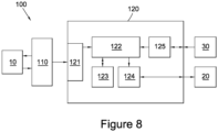
- Figure 8 illustrates a system 100 used for implementing the method for quantifying the characteristics of an object using imaging as described herein.
- the system 100 comprises an imaging system 110 and a data processing system 120.
- the imaging system 110 which may be a system such as an ultrasonic imaging system or an MRI system, is arranged to image an object 10.
- the imaging system 110 then sends captured image data to the data processing system 120.
- the data processing system 120 receives the captured image data at communications unit 121, which sends any data received from external systems to processor 122.
- the processor 122 then stores the received image data in memory 123.
- the processor 122 is then able to perform the various processes and analysis discussed throughout this document.
- the results of the processing performed by the processor 122 are then sent to a graphics card 124 for display on a monitor 20.
- the data processing system 120 also comprises a user interface module 125 arranged to receive instructions for controlling the operation of the data processing system 120 by a user via a user interface 30.
- system 100 is described as comprising a number of distinct components it will be appreciated that all of the components, or a subset of the components, shown in Figure 8 may be integrated within a single unit. Furthermore, it will be appreciated that aspects of the system 100 may also be distributed components. For example, the processing may be performed by a distributed network of data processing systems. In addition, the processing may be carried out in a cloud computing environment.
- the various methods described above may also be implemented by a computer program.
- the computer program may be stored in memory 123 and executable by processor 122.
- the computer program may include computer code arranged to instruct a computer, such as the data processing system 120, to perform the functions of one or more of the various methods described above.
- the computer program and/or the code for performing such methods may be provided to an apparatus, such as a computer, on a computer readable medium or computer program product.
- the computer readable medium could be, for example, an electronic, magnetic, optical, electromagnetic, infrared, or semiconductor system, or a propagation medium for data transmission, for example for downloading the code over the Internet.
- the computer readable medium could take the form of a physical computer readable medium such as semiconductor or solid state memory, magnetic tape, a removable computer diskette, a random access memory (RAM), a read-only memory (ROM), a rigid magnetic disc, and an optical disk, such as a CD-ROM, CD-R/W or DVD.
- a physical computer readable medium such as semiconductor or solid state memory, magnetic tape, a removable computer diskette, a random access memory (RAM), a read-only memory (ROM), a rigid magnetic disc, and an optical disk, such as a CD-ROM, CD-R/W or DVD.
Landscapes
- Health & Medical Sciences (AREA)
- Life Sciences & Earth Sciences (AREA)
- Engineering & Computer Science (AREA)
- General Health & Medical Sciences (AREA)
- Public Health (AREA)
- Veterinary Medicine (AREA)
- Animal Behavior & Ethology (AREA)
- Physics & Mathematics (AREA)
- Nuclear Medicine, Radiotherapy & Molecular Imaging (AREA)
- Radiology & Medical Imaging (AREA)
- Medical Informatics (AREA)
- Biomedical Technology (AREA)
- Epidemiology (AREA)
- Pathology (AREA)
- Surgery (AREA)
- Molecular Biology (AREA)
- Heart & Thoracic Surgery (AREA)
- Biophysics (AREA)
- High Energy & Nuclear Physics (AREA)
- Computer Vision & Pattern Recognition (AREA)
- Optics & Photonics (AREA)
- Acoustics & Sound (AREA)
- Chemical & Material Sciences (AREA)
- General Physics & Mathematics (AREA)
- Physiology (AREA)
- Immunology (AREA)
- Dispersion Chemistry (AREA)
- Hematology (AREA)
- Quality & Reliability (AREA)
- Theoretical Computer Science (AREA)
- Condensed Matter Physics & Semiconductors (AREA)
- Signal Processing (AREA)
- Medicinal Chemistry (AREA)
- Primary Health Care (AREA)
- Databases & Information Systems (AREA)
- Data Mining & Analysis (AREA)
- Ultra Sonic Daignosis Equipment (AREA)
- Magnetic Resonance Imaging Apparatus (AREA)
- Cardiology (AREA)
- Medicines Containing Antibodies Or Antigens For Use As Internal Diagnostic Agents (AREA)
Claims (21)
- Verfahren, das auf einem Datenverarbeitungssystem (120) ausgeführt wird, um die Merkmale eines Objekts (10) unter Anwendung von Ultraschallbildgebung zu quantifizieren, wobei das Objekt mit Mikrobläschen behandelt wird, die als Kontrastmittel dienen, das in den Körper des Objekts eingebracht wurde, umfassend:(a) Empfangen einer Vielzahl von Abbildungssignalen des Objekts (10), die über einen Zeitraum aufgenommen wurden, von einem Abbildungssystem (110), wobei die Abbildungssignale nach etwa 2 bis 6 Minuten nach Verabreichung der Mikrobläschen in den Körper gewonnen werden,(b) Messen der Signalintensität in jedem der Vielzahl von Bildsignalen, die einem Punkt auf dem Objekt (10) entsprechen, im Datenverarbeitungssystem (120),(c) im Datenverarbeitungssystem (120) eine Kurvenanpassung der gemessenen Signalintensität des Punktes auf dem Objekt (10) an eine bi-exponentielle Funktion, die einen ersten und einen zweiten Exponentialterm umfasst, wobei der erste Exponentialterm die Abnahme der Konzentration des freien Kontrastmittels mit der Zeit darstellt und der zweite Exponentialterm der Abnahme der Konzentration des auf dem Objekt (10) gebundenen Kontrastmittels mit der Zeit darstellt, wobei die Kurvenanpassung eine Extrapolation zurück zur anfänglichen (maximalen) Konzentration des gebundenen Kontrastmittels in der Eliminierungsphase des Kontrastmittels umfasst.
- Verfahren nach Anspruch 1, ferner umfassend den Schritt: Erzeugen eines Bildes, das das quantifizierte Merkmal für mindestens einen Teil des Objekts (10) enthält.
- Verfahren nach Anspruch 1 oder Anspruch 2, ferner umfassend das Erfassen der Vielzahl von Bildsignalen des Objekts (10) über einen Zeitraum hinweg.
- Verfahren nach Anspruch 3, wobei die Vielzahl von Bildsignalen erfasst wird, sobald die Kontrastmittelkonzentration unter dem Punkt liegt, an dem die Abschwächung und/oder die Sättigung der Signalintensität signifikant ist.
- Verfahren nach einem der vorhergehenden Ansprüche, ferner umfassend die Schritte:(d) Wiederholung der Schritte (b) & (c) für Punkte in einem interessierenden Bereich des Objekts (10), und(e) Mittelwertbildung des quantifizierten Merkmals.
- Verfahren nach einem der vorhergehenden Ansprüche, ferner umfassend die Schritte:(d) Wiederholung von Schritt (b) für Punkte in einem interessierenden Bereich des Objekts (10),(e) Mittelwertbildung der Signalintensitäten von Punkten über den interessierenden Bereich zu bestimmten Zeiten und(f) Ausführen von Schritt (c) anhand der mittleren Intensitäten.
- Verfahren nach einem der vorhergehenden Ansprüche, wobei die Vielzahl der über eine Zeitspanne aufgenommenen Bildsignale eine Vielzahl von Bildern ist.
- Verfahren nach einem der vorangehenden Ansprüche, wobei die in jedem der Vielzahl von Bildsignalen gemessene Signalintensität die Intensität eines Pixels jedes der Bilder ist, das dem Punkt auf dem Objekt (10) entspricht.
- Verfahren nach einem der vorhergehenden Ansprüche, wobei zur Verarbeitung der Bilder ein videodensitometrisches Verfahren verwendet wird.
- Verfahren nach einem der vorangehenden Ansprüche, wobei die bi-exponentielle Funktion umfasst:

- Verfahren nach einem der vorhergehenden Ansprüche, wobei die Biexponentialgleichung ferner eine oder mehrere Komponenten umfasst, die aus einer Liste, umfassend Konstante, Skalierungsfaktor, Faktor, Exponentialterm, ausgewählt sind.
- Verfahren nach einem der vorangehenden Ansprüche, ferner umfassend das Quantifizieren des Merkmals der Halbwertszeit des gebundenen Kontrastmittels im Datenverarbeitungssystem (120).
- Verfahren nach einem der vorangehenden Ansprüche, ferner umfassend das Quantifizieren des Merkmals der Fläche unter der Kurve des gebundenen Kontrastmittels im Datenverarbeitungssystem (120).
- Verfahren nach einem der vorangehenden Ansprüche, ferner umfassend das Quantifizieren des Merkmals der Eliminierungsgeschwindigkeitskonstante λf des freien Kontrastmittels im Datenverarbeitungssystem (120).
- Verfahren nach einem der vorangehenden Ansprüche, ferner umfassend das Quantifizieren des Merkmals der Halbwertszeit des freien Kontrastmittels im Datenverarbeitungssystem (120).
- Verfahren nach einem der vorangehenden Ansprüche, ferner umfassend das Quantifizieren des Merkmals der maximalen Konzentration Af des freien Kontrastmittels im Datenverarbeitungssystem (120).
- Verfahren nach einem der vorangehenden Ansprüche, ferner umfassend das Quantifizieren des Merkmals der Konzentration Afe -λft des freien Kontrastmittels in dem Datenverarbeitungssystem (120).
- Verfahren nach einem der vorangehenden Ansprüche, ferner umfassend das Quantifizieren des Merkmals der Fläche unter der Kurve des freien Kontrastmittels im Datenverarbeitungssystem (120).
- Verfahren nach einem der vorangehenden Ansprüche, ferner umfassend das Quantifizieren des vaskulären Volumenanteils eines Gewebes im Datenverarbeitungssystem (120), indem man das Verhältnis von Af oder AUCf von zwei interessierenden Bereichen nimmt, wobei der Nenner derjenige für den interessierenden Bereich ist, der in einem Blutpoolbereich liegt.
- Verfahren nach einem der vorhergehenden Ansprüche, wobei das Kontrastmittel eine Vielzahl von Mikrobläschen mit oder ohne molekulare Bindungselemente ist.
- Vorrichtung (100) zur Quantifizierung der Merkmale eines Objekts mittels Ultraschallabbildung, wobei das Objekt mit Mikrobläschen behandelt wird, die als Kontrastmittel dienen, das in einen Körper des Objekts eingeführt wurde, wobei die Vorrichtung ein Datenverarbeitungssystem (120) umfasst, das zur Durchführung des Verfahrens nach einem der Ansprüche 1 bis 20 eingerichtet ist.
Applications Claiming Priority (2)
| Application Number | Priority Date | Filing Date | Title |
|---|---|---|---|
| GB1223328.4A GB2509168B (en) | 2012-12-21 | 2012-12-21 | Method of quantifying the characteristics of an object treated with a contrast agent |
| PCT/GB2013/053405 WO2014096863A1 (en) | 2012-12-21 | 2013-12-20 | Method for Quantifying the Characteristics of an Object treated with a Contrast Agent |
Publications (3)
| Publication Number | Publication Date |
|---|---|
| EP2941192A1 EP2941192A1 (de) | 2015-11-11 |
| EP2941192C0 EP2941192C0 (de) | 2024-10-30 |
| EP2941192B1 true EP2941192B1 (de) | 2024-10-30 |
Family
ID=47682539
Family Applications (1)
| Application Number | Title | Priority Date | Filing Date |
|---|---|---|---|
| EP13812043.1A Active EP2941192B1 (de) | 2012-12-21 | 2013-12-20 | Verfahren und vorrichtung zur quantifizierung der eigenschaften eines mit einem kontrastmittel behandelten objekts |
Country Status (7)
| Country | Link |
|---|---|
| US (1) | US9629935B2 (de) |
| EP (1) | EP2941192B1 (de) |
| JP (1) | JP6430958B2 (de) |
| KR (1) | KR102241374B1 (de) |
| CN (1) | CN105073004B (de) |
| GB (1) | GB2509168B (de) |
| WO (1) | WO2014096863A1 (de) |
Families Citing this family (10)
| Publication number | Priority date | Publication date | Assignee | Title |
|---|---|---|---|---|
| US9949722B2 (en) * | 2013-12-03 | 2018-04-24 | University Of Virginia Patent Foundation | System and method for binding dynamics of targeted microbubbles |
| US9526468B2 (en) * | 2014-09-09 | 2016-12-27 | General Electric Company | Multiple frame acquisition for exposure control in X-ray medical imagers |
| US10433817B2 (en) * | 2015-12-10 | 2019-10-08 | Bracco Suisse S.A. | Detection of immobilized contrast agent with dynamic thresholding |
| CN109313926B (zh) * | 2016-05-27 | 2023-06-09 | 生命技术公司 | 用于生物数据的图形用户界面的方法和系统 |
| CN111902074B (zh) * | 2018-03-13 | 2024-08-23 | 博信生物科技股份有限公司 | 用于灵敏分子分析的组合物和方法 |
| US20190370956A1 (en) * | 2018-05-30 | 2019-12-05 | General Electric Company | Contrast imaging system and method |
| CN109350098B (zh) * | 2018-08-27 | 2021-02-26 | 苏州瑞派宁科技有限公司 | 信号的拟合方式的确定方法、重建方法和装置 |
| KR102382199B1 (ko) * | 2019-06-27 | 2022-04-05 | 고려대학교 산학협력단 | 인공 기준 지표를 활용한 자기 공명 영상의 처리 장치 및 방법 |
| KR102392778B1 (ko) * | 2020-01-02 | 2022-05-04 | 한국원자력의학원 | 영상 기반의 치료용 방사성의약품 치료선량 결정 시스템 및 방법 |
| TWI741773B (zh) * | 2020-09-02 | 2021-10-01 | 中國醫藥大學 | 超音波影像之判讀方法及其系統 |
Family Cites Families (16)
| Publication number | Priority date | Publication date | Assignee | Title |
|---|---|---|---|---|
| US5024230A (en) * | 1988-11-23 | 1991-06-18 | Picker International, Inc. | Dual flow/lambda display for xenon enhanced tomography |
| US8021303B2 (en) * | 2003-06-12 | 2011-09-20 | Bracco Research Sa | System for extracting morphological information through a perfusion assessment process |
| US20050048539A1 (en) * | 2003-06-13 | 2005-03-03 | The General Hospital Corporation | Methods to monitor molecule conformation and molecule/molecule proximity |
| WO2006055498A2 (en) * | 2004-11-15 | 2006-05-26 | Uab Research Foundation | Methods and systems of analyzing clinical parameters and methods of producing visual images |
| US20060149479A1 (en) * | 2004-12-30 | 2006-07-06 | Art, Advanced Research Technologies Inc. | Method for improving fluorescence image contrast |
| US20070047786A1 (en) * | 2005-08-25 | 2007-03-01 | Lenovo (Singapore) Pte. Ltd. | System and method for creating robust training data from MRI images |
| JP2008220868A (ja) * | 2007-03-15 | 2008-09-25 | Ge Medical Systems Global Technology Co Llc | 造影剤および磁気共鳴イメージング装置 |
| JP5186155B2 (ja) * | 2007-08-22 | 2013-04-17 | 日立アロカメディカル株式会社 | 超音波診断装置 |
| EP2234543B1 (de) * | 2007-12-28 | 2016-11-02 | Bracco Suisse SA | Quantifizierungsanalyse eines immobilisierten kontrastmittels bei medizinischen bildgebungsanwendungen |
| EP2128613A1 (de) * | 2008-05-28 | 2009-12-02 | Institut Pasteur | Verfahren zum Screening von Verbindungen auf ihre Fähigkeit zur Erhöhung der Rigidität roter, mit einem Einzellerparasit der Gattung Plasmodium infizierter Blutkörperchen und Anwendung |
| DE102008059788B4 (de) * | 2008-12-01 | 2018-03-08 | Olympus Soft Imaging Solutions Gmbh | Analyse und Klassifizierung insbesondere biologischer oder biochemischer Objekte auf Basis von Zeitreihen-Bildern, anwendbar bei der zytometrischen Time-Lapse-Zellanalyse in der bildbasierten Zytometrie |
| CN101756713A (zh) * | 2009-09-09 | 2010-06-30 | 西安交通大学 | 超声造影成像、灌注参量估计和灌注参量功能成像及其集成方法 |
| WO2011075476A1 (en) * | 2009-12-14 | 2011-06-23 | Arizona Board Of Regents, A Body Corporate Of The State Of Arizona, Acting For And On Behalf Of Arizona State University | Methods and compositions relating to reporter gels for use in mri techniques |
| US8718747B2 (en) * | 2010-04-16 | 2014-05-06 | Oslo Universitetssykehus Hf | Estimating and correcting for contrast agent extravasation in tissue perfusion imaging |
| NO20101638A1 (no) * | 2010-11-22 | 2012-05-23 | Sunnmore Mr Klinikk As | Fremgangsmate ved ex vivo distingvering mellom maligne og benigne tumorer ved anvendelse av kontrastmiddelbasert MR-skanning |
| CN102551803A (zh) * | 2011-12-31 | 2012-07-11 | 重庆安碧捷生物科技有限公司 | 超声造影录像分析方法及分析系统 |
-
2012
- 2012-12-21 GB GB1223328.4A patent/GB2509168B/en active Active
-
2013
- 2013-12-20 US US14/654,709 patent/US9629935B2/en active Active
- 2013-12-20 WO PCT/GB2013/053405 patent/WO2014096863A1/en not_active Ceased
- 2013-12-20 CN CN201380072851.0A patent/CN105073004B/zh active Active
- 2013-12-20 EP EP13812043.1A patent/EP2941192B1/de active Active
- 2013-12-20 JP JP2015548774A patent/JP6430958B2/ja active Active
- 2013-12-20 KR KR1020157019733A patent/KR102241374B1/ko active Active
Also Published As
| Publication number | Publication date |
|---|---|
| US20160199033A1 (en) | 2016-07-14 |
| GB2509168B (en) | 2020-04-01 |
| GB201223328D0 (en) | 2013-02-06 |
| CN105073004B (zh) | 2018-10-09 |
| EP2941192A1 (de) | 2015-11-11 |
| GB2509168A (en) | 2014-06-25 |
| KR20150122127A (ko) | 2015-10-30 |
| KR102241374B1 (ko) | 2021-04-16 |
| EP2941192C0 (de) | 2024-10-30 |
| US9629935B2 (en) | 2017-04-25 |
| WO2014096863A1 (en) | 2014-06-26 |
| JP2016505320A (ja) | 2016-02-25 |
| CN105073004A (zh) | 2015-11-18 |
| JP6430958B2 (ja) | 2018-11-28 |
Similar Documents
| Publication | Publication Date | Title |
|---|---|---|
| EP2941192B1 (de) | Verfahren und vorrichtung zur quantifizierung der eigenschaften eines mit einem kontrastmittel behandelten objekts | |
| Gutberlet et al. | Myocardial viability assessment in patients with highly impaired left ventricular function: comparison of delayed enhancement, dobutamine stress MRI, end-diastolic wall thickness, and TI201-SPECT with functional recovery after revascularization | |
| Wei et al. | Interactions between microbubbles and ultrasound: in vitro and in vivo observations | |
| Blomley et al. | Stimulated acoustic emission to image a late liver and spleen-specific phase of Levovist® in normal volunteers and patients with and without liver disease | |
| US9072492B2 (en) | Quantification analysis of immobilized contrast agent in medical imaging applications | |
| Schneider et al. | Bench-to-bedside review: Contrast enhanced ultrasonography-a promising technique to assess renal perfusion in the ICU | |
| US20070232909A1 (en) | Ultrasonic Characterization of Internal Body Conditions Using Information Theoretic Signal Receivers | |
| US20230270413A1 (en) | Composition and methods for sensitive molecular analysis | |
| Rissanen et al. | High-resolution ultrasound perfusion imaging of therapeutic angiogenesis | |
| Lindsey et al. | Assessment of molecular acoustic angiography for combined microvascular and molecular imaging in preclinical tumor models | |
| Nakamura et al. | Quantitative contrast‐enhanced ultrasonography of canine spleen | |
| Hudson et al. | Improved flow measurement using microbubble contrast agents and disruption-replenishment: clinical application to tumour monitoring | |
| Quartuccio et al. | Contrast-enhanced ultrasound evaluation of testicular interstitial cell tumours in conscious non-sedated dogs. | |
| Kogan et al. | Microbubbles in imaging: applications beyond ultrasound | |
| Allen et al. | Harmonic imaging: echocardiographic enhanced contrast intensity and duration | |
| Li et al. | Grey-scale contrast enhancement in rabbit liver with SonoVue™ at different doses | |
| Su et al. | Real-time myocardial contrast echocardiography in rat: infusion versus bolus administration | |
| Morishita et al. | Contrast-enhanced ultrasonography of the hepatic vein in normal dogs | |
| Agati et al. | Non-invasive assessment of myocardial perfusion by intravenous contrast echocardiography: state of the art | |
| Senior | Imagify™(perflubutane polymer microspheres) injectable suspension for the assessment of coronary artery disease | |
| Herbst et al. | Enhanced visualization of intratumoral microbubbles using singular value thresholding combined with normalized singular spectrum area | |
| Sosnovik et al. | Transmural variation of myocardial attenuation and its potential effect on contrast-mediated estimates of regional myocardial perfusion | |
| Sridharan et al. | Perfusion estimation using 3D subharmonic imaging: An in vivo study | |
| Ay et al. | Heterogeneity of contrast effect during intermittent second harmonic myocardial contrast echocardiography in healthy patients | |
| Lin et al. | Novel contrast agent Visphere™ is feasible for contrast‐enhanced ultrasonography in dogs |
Legal Events
| Date | Code | Title | Description |
|---|---|---|---|
| PUAI | Public reference made under article 153(3) epc to a published international application that has entered the european phase |
Free format text: ORIGINAL CODE: 0009012 |
|
| 17P | Request for examination filed |
Effective date: 20150721 |
|
| AK | Designated contracting states |
Kind code of ref document: A1 Designated state(s): AL AT BE BG CH CY CZ DE DK EE ES FI FR GB GR HR HU IE IS IT LI LT LU LV MC MK MT NL NO PL PT RO RS SE SI SK SM TR |
|
| AX | Request for extension of the european patent |
Extension state: BA ME |
|
| RAP1 | Party data changed (applicant data changed or rights of an application transferred) |
Owner name: YEH, JAMES SHUE-MIN |
|
| RAP1 | Party data changed (applicant data changed or rights of an application transferred) |
Owner name: YEH, JAMES SHUE-MIN |
|
| DAX | Request for extension of the european patent (deleted) | ||
| STAA | Information on the status of an ep patent application or granted ep patent |
Free format text: STATUS: EXAMINATION IS IN PROGRESS |
|
| 17Q | First examination report despatched |
Effective date: 20190318 |
|
| RIC1 | Information provided on ipc code assigned before grant |
Ipc: G16H 50/30 20180101ALI20230428BHEP Ipc: A61K 49/22 20060101ALI20230428BHEP Ipc: A61K 49/18 20060101ALI20230428BHEP Ipc: A61K 49/16 20060101ALI20230428BHEP Ipc: A61B 5/02 20060101ALI20230428BHEP Ipc: A61K 49/00 20060101ALI20230428BHEP Ipc: A61B 8/00 20060101ALI20230428BHEP Ipc: G06T 7/00 20060101ALI20230428BHEP Ipc: A61B 5/055 20060101ALI20230428BHEP Ipc: A61B 5/00 20060101ALI20230428BHEP Ipc: A61B 8/08 20060101ALI20230428BHEP Ipc: A61B 6/00 20060101AFI20230428BHEP |
|
| GRAP | Despatch of communication of intention to grant a patent |
Free format text: ORIGINAL CODE: EPIDOSNIGR1 |
|
| STAA | Information on the status of an ep patent application or granted ep patent |
Free format text: STATUS: GRANT OF PATENT IS INTENDED |
|
| INTG | Intention to grant announced |
Effective date: 20230720 |
|
| GRAS | Grant fee paid |
Free format text: ORIGINAL CODE: EPIDOSNIGR3 |
|
| GRAJ | Information related to disapproval of communication of intention to grant by the applicant or resumption of examination proceedings by the epo deleted |
Free format text: ORIGINAL CODE: EPIDOSDIGR1 |
|
| GRAL | Information related to payment of fee for publishing/printing deleted |
Free format text: ORIGINAL CODE: EPIDOSDIGR3 |
|
| STAA | Information on the status of an ep patent application or granted ep patent |
Free format text: STATUS: EXAMINATION IS IN PROGRESS |
|
| RBV | Designated contracting states (corrected) |
Designated state(s): AL AT BE BG CH CY CZ DE DK EE ES FI FR GR HR HU IE IS IT LI LT LU LV MC MK MT NL NO PL PT RO RS SE SI SK SM TR |
|
| INTC | Intention to grant announced (deleted) | ||
| RAP3 | Party data changed (applicant data changed or rights of an application transferred) |
Owner name: YEH, JAMES SHUE-MIN |
|
| GRAP | Despatch of communication of intention to grant a patent |
Free format text: ORIGINAL CODE: EPIDOSNIGR1 |
|
| STAA | Information on the status of an ep patent application or granted ep patent |
Free format text: STATUS: GRANT OF PATENT IS INTENDED |
|
| INTG | Intention to grant announced |
Effective date: 20240109 |
|
| GRAJ | Information related to disapproval of communication of intention to grant by the applicant or resumption of examination proceedings by the epo deleted |
Free format text: ORIGINAL CODE: EPIDOSDIGR1 |
|
| GRAL | Information related to payment of fee for publishing/printing deleted |
Free format text: ORIGINAL CODE: EPIDOSDIGR3 |
|
| STAA | Information on the status of an ep patent application or granted ep patent |
Free format text: STATUS: EXAMINATION IS IN PROGRESS |
|
| INTC | Intention to grant announced (deleted) | ||
| GRAP | Despatch of communication of intention to grant a patent |
Free format text: ORIGINAL CODE: EPIDOSNIGR1 |
|
| STAA | Information on the status of an ep patent application or granted ep patent |
Free format text: STATUS: GRANT OF PATENT IS INTENDED |
|
| INTG | Intention to grant announced |
Effective date: 20240618 |
|
| GRAS | Grant fee paid |
Free format text: ORIGINAL CODE: EPIDOSNIGR3 |
|
| GRAA | (expected) grant |
Free format text: ORIGINAL CODE: 0009210 |
|
| STAA | Information on the status of an ep patent application or granted ep patent |
Free format text: STATUS: THE PATENT HAS BEEN GRANTED |
|
| AK | Designated contracting states |
Kind code of ref document: B1 Designated state(s): AL AT BE BG CH CY CZ DE DK EE ES FI FR GR HR HU IE IS IT LI LT LU LV MC MK MT NL NO PL PT RO RS SE SI SK SM TR |
|
| REG | Reference to a national code |
Ref country code: CH Ref legal event code: EP |
|
| REG | Reference to a national code |
Ref country code: DE Ref legal event code: R096 Ref document number: 602013086238 Country of ref document: DE |
|
| REG | Reference to a national code |
Ref country code: IE Ref legal event code: FG4D |
|
| U01 | Request for unitary effect filed |
Effective date: 20241101 |
|
| U07 | Unitary effect registered |
Designated state(s): AT BE BG DE DK EE FI FR IT LT LU LV MT NL PT RO SE SI Effective date: 20241113 |
|
| RAP4 | Party data changed (patent owner data changed or rights of a patent transferred) |
Owner name: YEH, JAMES SHUE-MIN |
|
| U1H | Name or address of the proprietor changed after the registration of the unitary effect |
Owner name: YEH, JAMES SHUE-MIN; GB |
|
| U20 | Renewal fee for the european patent with unitary effect paid |
Year of fee payment: 12 Effective date: 20241201 |
|
| PGFP | Annual fee paid to national office [announced via postgrant information from national office to epo] |
Ref country code: IE Payment date: 20241112 Year of fee payment: 12 |
|
| PG25 | Lapsed in a contracting state [announced via postgrant information from national office to epo] |
Ref country code: IS Free format text: LAPSE BECAUSE OF FAILURE TO SUBMIT A TRANSLATION OF THE DESCRIPTION OR TO PAY THE FEE WITHIN THE PRESCRIBED TIME-LIMIT Effective date: 20250228 Ref country code: HR Free format text: LAPSE BECAUSE OF FAILURE TO SUBMIT A TRANSLATION OF THE DESCRIPTION OR TO PAY THE FEE WITHIN THE PRESCRIBED TIME-LIMIT Effective date: 20241030 |
|
| PG25 | Lapsed in a contracting state [announced via postgrant information from national office to epo] |
Ref country code: ES Free format text: LAPSE BECAUSE OF FAILURE TO SUBMIT A TRANSLATION OF THE DESCRIPTION OR TO PAY THE FEE WITHIN THE PRESCRIBED TIME-LIMIT Effective date: 20241030 |
|
| PG25 | Lapsed in a contracting state [announced via postgrant information from national office to epo] |
Ref country code: NO Free format text: LAPSE BECAUSE OF FAILURE TO SUBMIT A TRANSLATION OF THE DESCRIPTION OR TO PAY THE FEE WITHIN THE PRESCRIBED TIME-LIMIT Effective date: 20250130 |
|
| PG25 | Lapsed in a contracting state [announced via postgrant information from national office to epo] |
Ref country code: GR Free format text: LAPSE BECAUSE OF FAILURE TO SUBMIT A TRANSLATION OF THE DESCRIPTION OR TO PAY THE FEE WITHIN THE PRESCRIBED TIME-LIMIT Effective date: 20250131 |
|
| PGFP | Annual fee paid to national office [announced via postgrant information from national office to epo] |
Ref country code: CH Payment date: 20250101 Year of fee payment: 12 |
|
| PG25 | Lapsed in a contracting state [announced via postgrant information from national office to epo] |
Ref country code: PL Free format text: LAPSE BECAUSE OF FAILURE TO SUBMIT A TRANSLATION OF THE DESCRIPTION OR TO PAY THE FEE WITHIN THE PRESCRIBED TIME-LIMIT Effective date: 20241030 |
|
| PG25 | Lapsed in a contracting state [announced via postgrant information from national office to epo] |
Ref country code: RS Free format text: LAPSE BECAUSE OF FAILURE TO SUBMIT A TRANSLATION OF THE DESCRIPTION OR TO PAY THE FEE WITHIN THE PRESCRIBED TIME-LIMIT Effective date: 20250130 |
|
| PG25 | Lapsed in a contracting state [announced via postgrant information from national office to epo] |
Ref country code: SM Free format text: LAPSE BECAUSE OF FAILURE TO SUBMIT A TRANSLATION OF THE DESCRIPTION OR TO PAY THE FEE WITHIN THE PRESCRIBED TIME-LIMIT Effective date: 20241030 |
|
| PG25 | Lapsed in a contracting state [announced via postgrant information from national office to epo] |
Ref country code: MC Free format text: LAPSE BECAUSE OF FAILURE TO SUBMIT A TRANSLATION OF THE DESCRIPTION OR TO PAY THE FEE WITHIN THE PRESCRIBED TIME-LIMIT Effective date: 20241030 |
|
| PG25 | Lapsed in a contracting state [announced via postgrant information from national office to epo] |
Ref country code: SK Free format text: LAPSE BECAUSE OF FAILURE TO SUBMIT A TRANSLATION OF THE DESCRIPTION OR TO PAY THE FEE WITHIN THE PRESCRIBED TIME-LIMIT Effective date: 20241030 |
|
| PG25 | Lapsed in a contracting state [announced via postgrant information from national office to epo] |
Ref country code: CZ Free format text: LAPSE BECAUSE OF FAILURE TO SUBMIT A TRANSLATION OF THE DESCRIPTION OR TO PAY THE FEE WITHIN THE PRESCRIBED TIME-LIMIT Effective date: 20241030 |
|
| PLBE | No opposition filed within time limit |
Free format text: ORIGINAL CODE: 0009261 |
|
| STAA | Information on the status of an ep patent application or granted ep patent |
Free format text: STATUS: NO OPPOSITION FILED WITHIN TIME LIMIT |
|
| 26N | No opposition filed |
Effective date: 20250731 |