EP2939593A1 - Vorrichtung und System zur Bestimmung der physiologischen Parameter des Sternumknochens - Google Patents
Vorrichtung und System zur Bestimmung der physiologischen Parameter des Sternumknochens Download PDFInfo
- Publication number
- EP2939593A1 EP2939593A1 EP14166934.1A EP14166934A EP2939593A1 EP 2939593 A1 EP2939593 A1 EP 2939593A1 EP 14166934 A EP14166934 A EP 14166934A EP 2939593 A1 EP2939593 A1 EP 2939593A1
- Authority
- EP
- European Patent Office
- Prior art keywords
- sensor
- blood flow
- light source
- sternum
- septum
- Prior art date
- Legal status (The legal status is an assumption and is not a legal conclusion. Google has not performed a legal analysis and makes no representation as to the accuracy of the status listed.)
- Withdrawn
Links
- 210000001562 sternum Anatomy 0.000 title claims description 50
- 210000000988 bone and bone Anatomy 0.000 claims abstract description 46
- 210000000115 thoracic cavity Anatomy 0.000 claims abstract description 42
- 230000017531 blood circulation Effects 0.000 claims abstract description 39
- 230000002792 vascular Effects 0.000 claims abstract description 13
- 238000005259 measurement Methods 0.000 claims abstract description 10
- 238000004458 analytical method Methods 0.000 claims abstract description 5
- 238000000034 method Methods 0.000 claims description 42
- 230000000241 respiratory effect Effects 0.000 claims description 42
- QVGXLLKOCUKJST-UHFFFAOYSA-N atomic oxygen Chemical compound [O] QVGXLLKOCUKJST-UHFFFAOYSA-N 0.000 claims description 22
- 229910052760 oxygen Inorganic materials 0.000 claims description 22
- 239000001301 oxygen Substances 0.000 claims description 22
- 210000004369 blood Anatomy 0.000 claims description 16
- 239000008280 blood Substances 0.000 claims description 16
- 230000036387 respiratory rate Effects 0.000 claims description 10
- 238000012544 monitoring process Methods 0.000 claims description 8
- 210000001349 mammary artery Anatomy 0.000 claims description 6
- 210000003462 vein Anatomy 0.000 claims description 6
- 108010054147 Hemoglobins Proteins 0.000 claims description 5
- 102000001554 Hemoglobins Human genes 0.000 claims description 5
- 230000008321 arterial blood flow Effects 0.000 claims description 4
- 230000014759 maintenance of location Effects 0.000 claims description 4
- 239000000853 adhesive Substances 0.000 claims description 3
- 230000001070 adhesive effect Effects 0.000 claims description 3
- 210000003484 anatomy Anatomy 0.000 claims description 3
- 238000011156 evaluation Methods 0.000 claims description 3
- 230000003434 inspiratory effect Effects 0.000 claims description 3
- 238000012546 transfer Methods 0.000 claims description 2
- 238000013186 photoplethysmography Methods 0.000 description 14
- 230000029058 respiratory gaseous exchange Effects 0.000 description 10
- 210000001519 tissue Anatomy 0.000 description 8
- 210000000038 chest Anatomy 0.000 description 7
- 210000002465 tibial artery Anatomy 0.000 description 6
- 230000036772 blood pressure Effects 0.000 description 5
- 230000000694 effects Effects 0.000 description 5
- INGWEZCOABYORO-UHFFFAOYSA-N 2-(furan-2-yl)-7-methyl-1h-1,8-naphthyridin-4-one Chemical compound N=1C2=NC(C)=CC=C2C(O)=CC=1C1=CC=CO1 INGWEZCOABYORO-UHFFFAOYSA-N 0.000 description 4
- 208000006545 Chronic Obstructive Pulmonary Disease Diseases 0.000 description 4
- 108010064719 Oxyhemoglobins Proteins 0.000 description 4
- 210000003489 abdominal muscle Anatomy 0.000 description 4
- 108010002255 deoxyhemoglobin Proteins 0.000 description 4
- 210000004872 soft tissue Anatomy 0.000 description 4
- 206010006322 Breath holding Diseases 0.000 description 3
- 210000003423 ankle Anatomy 0.000 description 3
- 210000004204 blood vessel Anatomy 0.000 description 3
- 230000000747 cardiac effect Effects 0.000 description 3
- 238000004891 communication Methods 0.000 description 3
- 210000000876 intercostal muscle Anatomy 0.000 description 3
- 230000002503 metabolic effect Effects 0.000 description 3
- 210000002345 respiratory system Anatomy 0.000 description 3
- 230000035488 systolic blood pressure Effects 0.000 description 3
- 206010048908 Seasonal allergy Diseases 0.000 description 2
- 238000010521 absorption reaction Methods 0.000 description 2
- -1 around 660nm Chemical compound 0.000 description 2
- 208000006673 asthma Diseases 0.000 description 2
- 210000001185 bone marrow Anatomy 0.000 description 2
- 238000001514 detection method Methods 0.000 description 2
- 238000003745 diagnosis Methods 0.000 description 2
- 201000010099 disease Diseases 0.000 description 2
- 208000037265 diseases, disorders, signs and symptoms Diseases 0.000 description 2
- 238000005516 engineering process Methods 0.000 description 2
- 210000003743 erythrocyte Anatomy 0.000 description 2
- 238000001914 filtration Methods 0.000 description 2
- 230000002631 hypothermal effect Effects 0.000 description 2
- 238000004519 manufacturing process Methods 0.000 description 2
- 239000000463 material Substances 0.000 description 2
- 230000003287 optical effect Effects 0.000 description 2
- 230000000541 pulsatile effect Effects 0.000 description 2
- 201000002859 sleep apnea Diseases 0.000 description 2
- 238000013125 spirometry Methods 0.000 description 2
- 239000000126 substance Substances 0.000 description 2
- 230000001360 synchronised effect Effects 0.000 description 2
- 238000012360 testing method Methods 0.000 description 2
- 210000000707 wrist Anatomy 0.000 description 2
- 208000035285 Allergic Seasonal Rhinitis Diseases 0.000 description 1
- 206010001742 Allergy to animal Diseases 0.000 description 1
- 208000019901 Anxiety disease Diseases 0.000 description 1
- 208000006096 Attention Deficit Disorder with Hyperactivity Diseases 0.000 description 1
- 208000036864 Attention deficit/hyperactivity disease Diseases 0.000 description 1
- 206010006102 Bradypnoea Diseases 0.000 description 1
- 208000001640 Fibromyalgia Diseases 0.000 description 1
- 206010020843 Hyperthermia Diseases 0.000 description 1
- 101150004367 Il4i1 gene Proteins 0.000 description 1
- 208000008930 Low Back Pain Diseases 0.000 description 1
- 201000002481 Myositis Diseases 0.000 description 1
- 208000002193 Pain Diseases 0.000 description 1
- 208000025747 Rheumatic disease Diseases 0.000 description 1
- 201000002661 Spondylitis Diseases 0.000 description 1
- NIXOWILDQLNWCW-UHFFFAOYSA-N acrylic acid group Chemical group C(C=C)(=O)O NIXOWILDQLNWCW-UHFFFAOYSA-N 0.000 description 1
- 239000002390 adhesive tape Substances 0.000 description 1
- 230000003321 amplification Effects 0.000 description 1
- 210000001367 artery Anatomy 0.000 description 1
- 206010003246 arthritis Diseases 0.000 description 1
- 230000003143 atherosclerotic effect Effects 0.000 description 1
- 238000010009 beating Methods 0.000 description 1
- 230000033228 biological regulation Effects 0.000 description 1
- 230000005540 biological transmission Effects 0.000 description 1
- 210000000601 blood cell Anatomy 0.000 description 1
- 238000009530 blood pressure measurement Methods 0.000 description 1
- 230000036471 bradycardia Effects 0.000 description 1
- 208000006218 bradycardia Diseases 0.000 description 1
- 208000024336 bradypnea Diseases 0.000 description 1
- 238000004364 calculation method Methods 0.000 description 1
- 210000000748 cardiovascular system Anatomy 0.000 description 1
- 210000004027 cell Anatomy 0.000 description 1
- 239000004020 conductor Substances 0.000 description 1
- 230000007812 deficiency Effects 0.000 description 1
- 230000006870 function Effects 0.000 description 1
- 239000003292 glue Substances 0.000 description 1
- 230000004217 heart function Effects 0.000 description 1
- 230000036031 hyperthermia Effects 0.000 description 1
- 208000000122 hyperventilation Diseases 0.000 description 1
- 230000000870 hyperventilation Effects 0.000 description 1
- 208000014674 injury Diseases 0.000 description 1
- 230000003601 intercostal effect Effects 0.000 description 1
- 235000015110 jellies Nutrition 0.000 description 1
- 239000008274 jelly Substances 0.000 description 1
- 210000004072 lung Anatomy 0.000 description 1
- 238000013508 migration Methods 0.000 description 1
- 230000005012 migration Effects 0.000 description 1
- 210000003205 muscle Anatomy 0.000 description 1
- 238000003199 nucleic acid amplification method Methods 0.000 description 1
- 239000002304 perfume Substances 0.000 description 1
- 210000005259 peripheral blood Anatomy 0.000 description 1
- 239000011886 peripheral blood Substances 0.000 description 1
- 230000002093 peripheral effect Effects 0.000 description 1
- 230000035479 physiological effects, processes and functions Effects 0.000 description 1
- 201000004338 pollen allergy Diseases 0.000 description 1
- 229920001296 polysiloxane Polymers 0.000 description 1
- 238000011160 research Methods 0.000 description 1
- 230000036391 respiratory frequency Effects 0.000 description 1
- 230000004202 respiratory function Effects 0.000 description 1
- 230000035939 shock Effects 0.000 description 1
- 239000007779 soft material Substances 0.000 description 1
- 239000007921 spray Substances 0.000 description 1
- 210000003270 subclavian artery Anatomy 0.000 description 1
- 201000000596 systemic lupus erythematosus Diseases 0.000 description 1
- 208000008203 tachypnea Diseases 0.000 description 1
- 206010043089 tachypnoea Diseases 0.000 description 1
- 239000004753 textile Substances 0.000 description 1
- 230000008733 trauma Effects 0.000 description 1
- 230000002747 voluntary effect Effects 0.000 description 1
Images


Classifications
-
- A—HUMAN NECESSITIES
- A61—MEDICAL OR VETERINARY SCIENCE; HYGIENE
- A61B—DIAGNOSIS; SURGERY; IDENTIFICATION
- A61B5/00—Measuring for diagnostic purposes; Identification of persons
- A61B5/02—Detecting, measuring or recording pulse, heart rate, blood pressure or blood flow; Combined pulse/heart-rate/blood pressure determination; Evaluating a cardiovascular condition not otherwise provided for, e.g. using combinations of techniques provided for in this group with electrocardiography or electroauscultation; Heart catheters for measuring blood pressure
- A61B5/026—Measuring blood flow
- A61B5/0261—Measuring blood flow using optical means, e.g. infrared light
-
- A—HUMAN NECESSITIES
- A61—MEDICAL OR VETERINARY SCIENCE; HYGIENE
- A61B—DIAGNOSIS; SURGERY; IDENTIFICATION
- A61B5/00—Measuring for diagnostic purposes; Identification of persons
- A61B5/02—Detecting, measuring or recording pulse, heart rate, blood pressure or blood flow; Combined pulse/heart-rate/blood pressure determination; Evaluating a cardiovascular condition not otherwise provided for, e.g. using combinations of techniques provided for in this group with electrocardiography or electroauscultation; Heart catheters for measuring blood pressure
- A61B5/0205—Simultaneously evaluating both cardiovascular conditions and different types of body conditions, e.g. heart and respiratory condition
- A61B5/02055—Simultaneously evaluating both cardiovascular condition and temperature
-
- A—HUMAN NECESSITIES
- A61—MEDICAL OR VETERINARY SCIENCE; HYGIENE
- A61B—DIAGNOSIS; SURGERY; IDENTIFICATION
- A61B5/00—Measuring for diagnostic purposes; Identification of persons
- A61B5/02—Detecting, measuring or recording pulse, heart rate, blood pressure or blood flow; Combined pulse/heart-rate/blood pressure determination; Evaluating a cardiovascular condition not otherwise provided for, e.g. using combinations of techniques provided for in this group with electrocardiography or electroauscultation; Heart catheters for measuring blood pressure
- A61B5/026—Measuring blood flow
- A61B5/0295—Measuring blood flow using plethysmography, i.e. measuring the variations in the volume of a body part as modified by the circulation of blood therethrough, e.g. impedance plethysmography
-
- A—HUMAN NECESSITIES
- A61—MEDICAL OR VETERINARY SCIENCE; HYGIENE
- A61B—DIAGNOSIS; SURGERY; IDENTIFICATION
- A61B5/00—Measuring for diagnostic purposes; Identification of persons
- A61B5/145—Measuring characteristics of blood in vivo, e.g. gas concentration, pH value; Measuring characteristics of body fluids or tissues, e.g. interstitial fluid, cerebral tissue
- A61B5/1455—Measuring characteristics of blood in vivo, e.g. gas concentration, pH value; Measuring characteristics of body fluids or tissues, e.g. interstitial fluid, cerebral tissue using optical sensors, e.g. spectral photometrical oximeters
-
- A—HUMAN NECESSITIES
- A61—MEDICAL OR VETERINARY SCIENCE; HYGIENE
- A61B—DIAGNOSIS; SURGERY; IDENTIFICATION
- A61B5/00—Measuring for diagnostic purposes; Identification of persons
- A61B5/68—Arrangements of detecting, measuring or recording means, e.g. sensors, in relation to patient
- A61B5/6801—Arrangements of detecting, measuring or recording means, e.g. sensors, in relation to patient specially adapted to be attached to or worn on the body surface
- A61B5/6813—Specially adapted to be attached to a specific body part
- A61B5/6823—Trunk, e.g., chest, back, abdomen, hip
-
- A—HUMAN NECESSITIES
- A61—MEDICAL OR VETERINARY SCIENCE; HYGIENE
- A61B—DIAGNOSIS; SURGERY; IDENTIFICATION
- A61B5/00—Measuring for diagnostic purposes; Identification of persons
- A61B5/68—Arrangements of detecting, measuring or recording means, e.g. sensors, in relation to patient
- A61B5/6801—Arrangements of detecting, measuring or recording means, e.g. sensors, in relation to patient specially adapted to be attached to or worn on the body surface
- A61B5/683—Means for maintaining contact with the body
- A61B5/6832—Means for maintaining contact with the body using adhesives
- A61B5/6833—Adhesive patches
-
- A—HUMAN NECESSITIES
- A61—MEDICAL OR VETERINARY SCIENCE; HYGIENE
- A61B—DIAGNOSIS; SURGERY; IDENTIFICATION
- A61B2562/00—Details of sensors; Constructional details of sensor housings or probes; Accessories for sensors
- A61B2562/02—Details of sensors specially adapted for in-vivo measurements
- A61B2562/0233—Special features of optical sensors or probes classified in A61B5/00
- A61B2562/0238—Optical sensor arrangements for performing transmission measurements on body tissue
-
- A—HUMAN NECESSITIES
- A61—MEDICAL OR VETERINARY SCIENCE; HYGIENE
- A61B—DIAGNOSIS; SURGERY; IDENTIFICATION
- A61B5/00—Measuring for diagnostic purposes; Identification of persons
- A61B5/02—Detecting, measuring or recording pulse, heart rate, blood pressure or blood flow; Combined pulse/heart-rate/blood pressure determination; Evaluating a cardiovascular condition not otherwise provided for, e.g. using combinations of techniques provided for in this group with electrocardiography or electroauscultation; Heart catheters for measuring blood pressure
- A61B5/024—Detecting, measuring or recording pulse rate or heart rate
- A61B5/02416—Detecting, measuring or recording pulse rate or heart rate using photoplethysmograph signals, e.g. generated by infrared radiation
- A61B5/02427—Details of sensor
- A61B5/02433—Details of sensor for infrared radiation
-
- A—HUMAN NECESSITIES
- A61—MEDICAL OR VETERINARY SCIENCE; HYGIENE
- A61B—DIAGNOSIS; SURGERY; IDENTIFICATION
- A61B5/00—Measuring for diagnostic purposes; Identification of persons
- A61B5/08—Detecting, measuring or recording devices for evaluating the respiratory organs
- A61B5/0816—Measuring devices for examining respiratory frequency
Definitions
- WO 2007/097702 discloses a method for the generation, detection and evaluation of a photoplethysmographic (PPG) signal to monitor blood characteristics in blood vessels of limited flexibility, such as in vascular compartments and vessels of deep tissues. Underlying the invention are the effects that the orientation and axial migration of red blood cells have on the absorption, scattering and reflection of light (photons) of near infrared and of blue-green wavelengths.
- PPG photoplethysmographic
- near-infrared wavelength light source(s) and blue-green-wavelength light source(s) are spaced at particular distances from a photodetector(s).
- This method allows continuous, non invasive monitoring of blood characteristics and changes in these characteristics over time.
- Data obtained by this method include blood pressure, blood flow, pulsatile blood volume and red blood cell velocity in blood vessels which are rigid or which have a limited flexibility, such as the vascular tissue of bone or in atherosclerotic or stiff vessels.
- the new instrumentation is an optical system which may be used for determining at least 10 different respiratory parameters and at least two cardiovasculatory parameters from only one sensor applied close to bone tissue, such as the sternum.
- the disclosed system may especially be used to monitor respiratory related multiparameters from thorax and therefore provide a completely new tool for diagnosis, treatment and follow up of patients with dysfunctional respiration having COPD, asthma, fibro myalgia, sleep apnea, pain in neck- shoulder region, lower back pain.
- the system may be used in emergency applications for simultaneous recording of 3 vital signs; heart rate, respiratory rate and oxygen saturation at the accident place, in connection with heart lung resuscitation (HLR), during transport in ambulance, helicopter or boat or in the emergency department.
- HLR heart lung resuscitation
- shock and hypothermia there are different phases of respiratory rate (both tachypnea and bradypnea) as well as of heart rate (takycardia and bradycardia).
- the non-invasive and wireless system may improve the quality of life for the individual by online monitoring of multiparameters and by optimizing diagnosis, treatment decisions and rehabilitation modalities.
- the light source comprising at least one near-infra-red lights source and/or at least one red light source and/or a green light source.
- the different light sources may be used to measure different parameters.
- the red light source may be used to measure variation in oxyhemoglobin and/or deoxyhemoglobin with reference to a near infrared light source in the range 800nm to 810nm.
- a near infrared light source with a wavelength in the range 900nm to 950nm may be used to measure variation in oxyhemoglobin and/or deoxyhemoglobin with reference to a near infrared light source in the range 800nm to 810nm.
- Green light may be used to measuring the hemoglobin content with reference to a near infrared light source in the range 800nm to 810nm.
- the wavelength of the lights sources may be in the range of 400 nm to 1200nm, such as the red light source may have a wavelength between 640nm to 680nm, the near-infra-red light source may have a wavelength between 800nm to 810nm and/or between 900nm to 950nm.
- the green light source may have a wavelength between 540nm to 590.
- the disclosed system also comprises a retention element adapted to hold the sensor at a skin site adjacent the thoracic bone or septum of the subject.
- the disclosed system is wirelessly connected to transfer the recorded information to a receiving unit.
- the vascular blood flow and/or volume is arterial blood flow and/or volume.
- This configuration is used to cover vessels emerging from left and right internal thoracic arteries and its corresponding collecting veins. This configuration will improve the signal to noise of the measured data.
- the analysing unit is configured to detect oxygen saturation based on a quotient between the red light and the near infrared light and/or between two near infrared lights with different wavelengths.
- the analysing unit may also be configured to detect haemoglobin content based on a quotient between the green light and the near infrared light which is used as a reference.
- the disclosed method includes assessing blood flow in rigid vessels or in vessels of limited flexibility.
- the analysing and evaluation is assessed from the blood flow supply anatomy of the sternum, wherein the blood flow to sternum varies synchronously with the blood flow supply to other parts of sternum.
- the vascular blood flow and/or volume is an arterial blood flow and/or volume.
- the following description focuses on an example of the present disclosure applicable to a device and method for determining physiological parameters from measured or monitored blood flow characteristic related to thoracic bone and in particular to a location apposition to sternum.
- the disclosure is not limited to this application but may be applied to many other locations viable for assessing, monitoring and/or measuring blood flow characteristics in rigid vessels or in vessels of limited flexibility.
- FIG. 1 is illustrating a schematic overview of blood flow around the thorax and in the sternum bone 1.
- thoracic bone and in particular sternum bone, has a special function related to the vessel architecture and physiology around the thorax.
- the internal thoracic artery 11, which branches off from the subclavian artery 10, and the corresponding collecting veins has branches to both the intercostal muscles 12 and to the sternum 13.
- the intercostal muscles 12 assist in respiration.
- the branches to the abdominal muscles assist in respiration.
- the blood flow to the sternum may easily be modulated by; a) the pressure changes in the thoracic cavity synchronously with respiration and b) blood circulation related to the activity of the thoracal and abdominal muscle compartments, especially during forced respiratory activity.
- the inventors have found that measurement of blood flow in the highly perfused sternum bone using a sensor 15 positioned apposition thoracic bone or sternum 1 enable monitoring of both respiratory and cardiac activity of several features and vital parameters.
- a sensor 15 directly on the sternum also makes it possible to follow the overall respiratory pattern without disturbing the respiratory tract (as for a spirometer which introduces a respiratory resistance being inappropriate for patients with COPD, asthma etc.).
- the placement of a sensor 15 directly on the sternum also means that the measured blood flow variations corresponding to the heart activity is of more central origin due to the relatively short distance between the heart and the position of the sensor.
- Figure 1 illustrates one example of the position of a sensor 15 on the sternum using only one unit including both a control unit and the sensor. Other positions of the sensor 15 on the ribs and a thoracic bone may also be plausible.
- FIG. 2 is illustrating a schematic example of a sensor system 2 in accordance with the disclosure.
- the sensor system 2 is a medical system for determining at least one physiological parameter based on a blood flow, such as vascular blood flow, and/or volume characteristics related to a thoracic bone or around the thorax and in the sternum bone of a subject.
- the sensor system 2 comprises a sensor 22 for skin apposition.
- the sensor 22 comprises at least one light source 21 for illuminating of the thoracic bone or septum and at least one detector 23 for detecting and recording reflected light from the thoracic bone or septum.
- the light sources 21 may preferably be at least one near-infra-red light source and/or at least one red light source and/or at least one light source between violent to green.
- An example of a light source that is due to its small size is suitable to be used in the sensor 22 is LEDs, such as near-infra-red LEDs or Red LEDs or violent to green LEDs.
- Wavelengths in the range of 400 nm to 1200nm may be used to measure blood flow variations in the sternum bone.
- the wavelength used depends on the parameters to be measure. For example, a light source with a wavelength around 800nm to 810 nm may be used as a reference, since variation in oxygen level in the blood may not be detected using this wavelength range.
- the oxygen content may be determined by a quotient between total(DC) light reflection of two wavelengths, for example near infrared in the range 800nm to 810nm and red light in the range 640nm to 680nm. Additionally and/or alternatively, for some examples, the oxygen content may be determined by a quotient between total(DC) light reflection of two wavelengths in the range 900nm to 950nm and 800nm to 810nm. These quotients have empirically been found to correspond to oxygen saturation in blood samples whereas oxygen saturation was measured at a chemical lab using standard lab methods. Due to total light reflection and the highly perfused sternum bone with steady metabolic need the determined oxygen saturation is assumed to be of arterial origin, which is denoted as SaO 2 .
- hemoglobin it has been discovered that using a light source having a wavelength in the violent to green range, such as between 400nm to 590 nm, such as a green light source with a wavelength between 540nm to 590nm, such as about 560nm, it is possible to detect a light reflection signal from the sternum bone. Normally, no reflected light will be detected from tissue in this wavelength range as the absorption is very high.
- the hemoglobin content may be determined by a quotient between total(DC) light reflection of two wavelengths, for example near infrared in the range 800nm to 810nm and green light in the range 540nm to 590nm.
- the senor 21 may be a photoplethysmography (PPG) type of sensor.
- PPG photoplethysmography
- the sensor system 2 further comprises a control unit 20 connected to the sensor 22 and configured to control operation of the sensor system 2.
- the sensor 22 and the control unit may be connected by wire or wireless using, for example Bluetooth.
- the control unit 20 either includes an analysing unit 25 or is connected to an analysing unit 25.
- the analysing unit 25 is configured to analyse measured data from the detected and recorded light to obtain at least one physiological parameter.
- control unit 20 includes or is connected to a memory unit 24 configured to store measured data from the detected and recorded light obtainable from the sensor 22 when operating the sensor system 2.
- sensor system 2 further comprises a unit for plotting a respiratory power curve based on the measured reflected light.
- the sensor 22 may, in some examples, be attached to the skin using a special interface set-up/connection between the sensor 22 and the skin.
- This interface may be double adhesive tape, adhesive spray, skin glue, jelly, soft material engorging the sensor 22 and/or negative pressure.
- the system may be combined with a second system developed to measure the blood pressure from soft tissue.
- the second system may provide the blood pressure measurement from the ankle of the arm wrist.
- a sensor system for measuring systolic blood pressure is disclosed in WO 2006/049571 .
- the disclosed method and means for measuring the systolic blood pressure is hereby incorporated in its entirety.
- the incorporated disclosure relates to the disclosed flexible pad for measuring systolic ankle blood pressure.
- the pad comprises two or more pairs of light emitting diodes and photo detectors. The pairs being disposed in parallel and the detectors being adapted for detecting light emitted by the respective diode into tissue and reflected from there.
- the pad further includes conductor means for providing power to the light emitting diodes from a power source and means selected from conducting means and wireless means for putting the detectors in communication with electronic equipment for detector signal analysis.
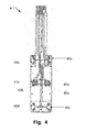
- Figure 3 illustrates two examples of the sensor system positioned at thoracic bone or sternum. In the illustrated examples the sensor system is positioned at the sternum.
- the sensor systems comprise a control unit and a sensor configured to use wireless technology to communicate with a receiving unit for transferring of recorded data to and/or from the control unit.
- the receiving unit may be a computer, reading plate, mobile phone, or other communication platforms for transferring information of the subject to a hospital, a medical practitioner or to a nurse.
- the senor 31 is positioned at a position on the thoracic bone, preferably at sternum.
- the control unit 30 may be positioned in a breast-pocket of a shirt, T-shirt or other clothes or at a trousers belt or at a skin surface close to the subject.
- the sensor 31 is separately attached to the skin.
- the sensor used may be in some examples be a photoplethysmography (PPG) type of sensor.
- PPG photoplethysmography
- the sensor 31 and the control unit 30 may be connected using a wire.
- the sensor 31 and the control unit may be connected using wireless technology.
- both the sensor system of example A and example B may be equipped with a display on the control unit.
- the display may be configured to present data, such as vital parameters, directly on the control unit.
- the double lining configuration is preferred to cover vessels emerging from left and right internal thoracic arteries (see Fig1 ) and its corresponding collecting veins.
- the length of the sensor may correlate to the volume of blood covered during measurement. Both these factors may enable a big blood volume to be recorded which enhances the strength of the PPG signal and therefore signal to noise ratio (SNR).
- the arrangement of the lights sources and the photodetectors of the sensor should preferably cover the length and from left to right of the thoracic bone or septum, thereby provide a large measure area covering as many of the blood vessels, in and around the thoracic bone or septum as possible.
- LEDs and photodetectors are possible, such as a configuration including at least one near-infra-red LED, such as a LED having a wavelength between 800nm to 810nm,in combination with at least one red LED, such as a LED having a wavelength between 640 to 680nm.
- near-infra-red LED such as a LED having a wavelength between 800nm to 810nm
- red LED such as a LED having a wavelength between 640 to 680nm.
- the configuration mainly depends on the anatomy of the location of the thoracic bone and the vital parameters to be measured.
- the printed card of the sensor 4 is casted in hard or partly flexible silicone, acrylic or other material moulded with a special geometry to be adapted to the anatomical appearance over the sternum, such as the anatomical appearance of both males and females.
- Figure 5 is illustrating schematic overview of an exemplary method 5.
- the method is for non-invasively determining at least one physiological parameter based on a blood flow, such as vascular blood flow, and/or volume characteristics from measuring on a thoracic bone or around the thorax and in the sternum bone of a subject.
- the method comprising providing 50 adapted to be arranged at a skin site of the thoracic bone or septum of a subject.
- the sensor includes at least one light source and at least one detector.
- One example of a suitable sensor is a sensor in accordance with the present disclosure.
- the method further comprises transmitting 21 light from the at least one light source to the thoracic bone or septum. Detecting 52 part of the light reflected back from the bone using the detector. Recording 53 measured data related to the detected light and analyzing 54 the measured data from the detected and recorded light to obtain at least one physiological parameter.
- Respiratory curve or pattern may be observed.
- oxygen saturation SaO 2 (%)may be determined according to the technique described above.
- the oxygen content is determined by a quotient between total(DC) light reflection of two wavelengths, for example 804 and 660nm and/or 940nm and 804nm. This quotient has empirically been found to correspond to oxygen saturation in blood samples whereas oxygen saturation was measured at a chemical lab using standard lab methods. Due to total light reflection and the highly perfused sternum bone with steady metabolic need the determined oxygen saturation is assumed to be of arterial origin, which is denoted as SaO 2 .
- More respiratory parameters may be extracted from the RP curve when the subject/patient performs e.g. forced inspiration and expiration during 1s, FEV 1 .
- Respiratory Rate (breaths / min) and Respiratory curve or pattern it has not been possible until now to monitor any of the other parameters using PPG on soft tissue because it is only on the thoracic bone or sternum bone that the respiratory signal is evident with enough high S/N (signal to noise ratio) due to the special anatomical vessel architecture, vessel branching and high blood delivery to the thorax compartments. This is partly due to the production of cells in the red bone marrow in the sternum and also the need of blood to the intercostal and abdominal muscles engaged in the respiratory activity and the vicinity to the heart and bigger vessels. Further parameters are the curve shape of the heart beat which is visible in the measured curves due to the short distance between the heart and the sternum.
- Figure 7 illustrates simultaneous recordings using the herein disclosed system and method 110 (lower curve) and a spirometer 100 (reflects air volumes) in a healthy subject during rest, maximal inspiration (upward deflection) and expiration (downward reflection) and during breath holding.
- the Respiratory Power Curve 110 follows the spirometry curve 100 at all phases and the respiratory rates also coincide.
- Figure 8 illustrates simultaneous recordings using the herein disclosed system and method 120(upper curve) and a capnograph 130 (lower curve) on a healthy subject in rest and during hyperventilation (increased respiratory rate).
- the Respiratory Power Curve 120 is obtained by recordings from the sternum on a healthy subject.
- the recording of the capnograph 130 is obtained from the airways and reflects variations in CO 2 concentration during inspiration and expiration and as seen there is a high resemblance in respiratory rate comparing the methods.
- FIG. 9 illustrates a Respiratory Power Curve 140 recording using the herein disclosed system and method on a female patient with COPD during a 6 minutes walking test.
- the Respiratory Power Curve shows deep sighs in order to be able to perform the walking procedure. This particularly demonstrates the possibility to follow and reflect the respiratory pattern on patients with different dysfunctional breathing when utilizing the herein disclosed system and method.
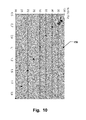
- Fig 10 illustrates the quotient 150 between total(DC) light reflection of two wavelengths at 804 and 660nm determined from a recording with the herein disclosed system and method and plotted against oxygen saturation measured by a pulse oximeter (Allen J 2007 Photoplethysmography and its application in clinical physiological measurement. Physiol. Meas. 28:R1-R39) during breath holding on one free diver (lying position).
- the example demonstrates the ability of the disclosed system and method to follow variations in oxygen saturation.
- Figure 11 is illustrating examples of systems 201 in accordance with the disclosure and positions at an animal 200 apposition to a thoracic bone or sternum 203.
- the system 201 is fastened means 202.
- the fastening means 202 could be as straps, such as ratchet straps or elastic straps.
- the sensor of the system may be positioned in the fur or the animal 200 may be locally shaved to obtain a spot where the sensor of the system 201 may be positioned directly on to the skin of the animal 200.
Landscapes
- Health & Medical Sciences (AREA)
- Life Sciences & Earth Sciences (AREA)
- Physics & Mathematics (AREA)
- General Health & Medical Sciences (AREA)
- Veterinary Medicine (AREA)
- Engineering & Computer Science (AREA)
- Biomedical Technology (AREA)
- Heart & Thoracic Surgery (AREA)
- Medical Informatics (AREA)
- Molecular Biology (AREA)
- Surgery (AREA)
- Animal Behavior & Ethology (AREA)
- Biophysics (AREA)
- Public Health (AREA)
- Pathology (AREA)
- Cardiology (AREA)
- Physiology (AREA)
- Hematology (AREA)
- Spectroscopy & Molecular Physics (AREA)
- Optics & Photonics (AREA)
- Pulmonology (AREA)
- Measurement Of The Respiration, Hearing Ability, Form, And Blood Characteristics Of Living Organisms (AREA)
- Measuring Pulse, Heart Rate, Blood Pressure Or Blood Flow (AREA)
- Measuring And Recording Apparatus For Diagnosis (AREA)
Priority Applications (8)
| Application Number | Priority Date | Filing Date | Title |
|---|---|---|---|
| EP14166934.1A EP2939593A1 (de) | 2014-05-02 | 2014-05-02 | Vorrichtung und System zur Bestimmung der physiologischen Parameter des Sternumknochens |
| JP2017508769A JP6726171B2 (ja) | 2014-05-02 | 2015-05-04 | 胸骨から生理パラメータを測定する方法 |
| EP15721206.9A EP3136956B1 (de) | 2014-05-02 | 2015-05-04 | Verfahren zur bestimmung der physiologischen parameter des sternumknochens |
| PCT/EP2015/059732 WO2015166110A1 (en) | 2014-05-02 | 2015-05-04 | Device and system for determining physiological parameters from the sternum bone |
| DK15721206.9T DK3136956T3 (da) | 2014-05-02 | 2015-05-04 | Fremgangsmåde til at bestemme fysiologiske parametre fra sternumknoglen |
| ES15721206T ES2802152T3 (es) | 2014-05-02 | 2015-05-04 | Método para determinar parámetros fisiológicos del hueso del esternón |
| US15/308,526 US10863909B2 (en) | 2014-05-02 | 2015-05-04 | Device and system for determining physiological parameters from the sternum bone |
| CN201580022187.8A CN106255450B (zh) | 2014-05-02 | 2015-05-04 | 用于从胸骨骨骼确定生理参数的设备和系统 |
Applications Claiming Priority (1)
| Application Number | Priority Date | Filing Date | Title |
|---|---|---|---|
| EP14166934.1A EP2939593A1 (de) | 2014-05-02 | 2014-05-02 | Vorrichtung und System zur Bestimmung der physiologischen Parameter des Sternumknochens |
Publications (1)
| Publication Number | Publication Date |
|---|---|
| EP2939593A1 true EP2939593A1 (de) | 2015-11-04 |
Family
ID=50639314
Family Applications (2)
| Application Number | Title | Priority Date | Filing Date |
|---|---|---|---|
| EP14166934.1A Withdrawn EP2939593A1 (de) | 2014-05-02 | 2014-05-02 | Vorrichtung und System zur Bestimmung der physiologischen Parameter des Sternumknochens |
| EP15721206.9A Active EP3136956B1 (de) | 2014-05-02 | 2015-05-04 | Verfahren zur bestimmung der physiologischen parameter des sternumknochens |
Family Applications After (1)
| Application Number | Title | Priority Date | Filing Date |
|---|---|---|---|
| EP15721206.9A Active EP3136956B1 (de) | 2014-05-02 | 2015-05-04 | Verfahren zur bestimmung der physiologischen parameter des sternumknochens |
Country Status (7)
| Country | Link |
|---|---|
| US (1) | US10863909B2 (de) |
| EP (2) | EP2939593A1 (de) |
| JP (1) | JP6726171B2 (de) |
| CN (1) | CN106255450B (de) |
| DK (1) | DK3136956T3 (de) |
| ES (1) | ES2802152T3 (de) |
| WO (1) | WO2015166110A1 (de) |
Cited By (1)
| Publication number | Priority date | Publication date | Assignee | Title |
|---|---|---|---|---|
| ITUA20161519A1 (it) * | 2016-03-10 | 2017-09-10 | Michele Gallamini | Dispositivo di monitoraggio di parametri funzionali dell’organismo |
Families Citing this family (2)
| Publication number | Priority date | Publication date | Assignee | Title |
|---|---|---|---|---|
| JP6818294B2 (ja) * | 2016-09-05 | 2021-01-20 | 学校法人 埼玉医科大学 | がん患者における薬剤の治療効果の評価方法、治療効果の評価データの作成方法、治療効果の評価装置及び治療効果の評価プログラム |
| CN112990169B (zh) * | 2021-05-20 | 2021-08-24 | 天津美腾科技股份有限公司 | 煤岩界面的识别方法、割煤轨迹的确定方法及装置 |
Citations (6)
| Publication number | Priority date | Publication date | Assignee | Title |
|---|---|---|---|---|
| US20040225207A1 (en) * | 2003-05-09 | 2004-11-11 | Sang-Kon Bae | Ear type apparatus for measuring a bio signal and measuring method therefor |
| WO2006049571A1 (en) | 2004-11-04 | 2006-05-11 | Lindberg Lars-Goeran | Method and means for measuring systolic blood pressure in the ankle |
| WO2007097702A1 (en) | 2006-02-21 | 2007-08-30 | Lindberg Lars-Goeran | Non-invasive monitoring of blood flow in deep tissue |
| US20130060098A1 (en) * | 2009-12-23 | 2013-03-07 | Delta, Dansk Elektronik, Lys Og Akustik | Monitoring device |
| US20130261415A1 (en) * | 2012-03-30 | 2013-10-03 | General Electric Company | System and methods for physiological monitoring |
| US20130267854A1 (en) * | 2012-04-09 | 2013-10-10 | Jami Johnson | Optical Monitoring and Computing Devices and Methods of Use |
Family Cites Families (10)
| Publication number | Priority date | Publication date | Assignee | Title |
|---|---|---|---|---|
| US5421329A (en) * | 1994-04-01 | 1995-06-06 | Nellcor, Inc. | Pulse oximeter sensor optimized for low saturation |
| WO2001017420A1 (en) * | 1999-09-08 | 2001-03-15 | Optoq Ab | Method and apparatus for detecting blood characteristics including hemoglobin |
| JP4460414B2 (ja) * | 2004-10-06 | 2010-05-12 | 日本電信電話株式会社 | 血圧計 |
| US8055321B2 (en) * | 2005-03-14 | 2011-11-08 | Peter Bernreuter | Tissue oximetry apparatus and method |
| JP2007105329A (ja) * | 2005-10-14 | 2007-04-26 | Hitachi Ltd | 血糖値測定装置及び代謝量測定装置 |
| JP2007225392A (ja) * | 2006-02-22 | 2007-09-06 | Spectratech Inc | 光干渉装置 |
| JP4957354B2 (ja) * | 2007-04-23 | 2012-06-20 | 株式会社デンソー | 生体状態検出装置 |
| CN101214146A (zh) * | 2008-01-11 | 2008-07-09 | 西北工业大学 | 生物组织血液微循环参数检测系统 |
| CA2775467C (en) * | 2009-09-14 | 2018-03-27 | Sleep Methods, Inc. | System and method for anticipating the onset of an obstructive sleep apnea event |
| DE102011017064A1 (de) * | 2011-04-14 | 2012-10-18 | Ingo Flore | Diagnostische Messvorrichtung mit integriertem Spektrometer |
-
2014
- 2014-05-02 EP EP14166934.1A patent/EP2939593A1/de not_active Withdrawn
-
2015
- 2015-05-04 EP EP15721206.9A patent/EP3136956B1/de active Active
- 2015-05-04 DK DK15721206.9T patent/DK3136956T3/da active
- 2015-05-04 ES ES15721206T patent/ES2802152T3/es active Active
- 2015-05-04 CN CN201580022187.8A patent/CN106255450B/zh active Active
- 2015-05-04 JP JP2017508769A patent/JP6726171B2/ja active Active
- 2015-05-04 WO PCT/EP2015/059732 patent/WO2015166110A1/en active Application Filing
- 2015-05-04 US US15/308,526 patent/US10863909B2/en active Active
Patent Citations (6)
| Publication number | Priority date | Publication date | Assignee | Title |
|---|---|---|---|---|
| US20040225207A1 (en) * | 2003-05-09 | 2004-11-11 | Sang-Kon Bae | Ear type apparatus for measuring a bio signal and measuring method therefor |
| WO2006049571A1 (en) | 2004-11-04 | 2006-05-11 | Lindberg Lars-Goeran | Method and means for measuring systolic blood pressure in the ankle |
| WO2007097702A1 (en) | 2006-02-21 | 2007-08-30 | Lindberg Lars-Goeran | Non-invasive monitoring of blood flow in deep tissue |
| US20130060098A1 (en) * | 2009-12-23 | 2013-03-07 | Delta, Dansk Elektronik, Lys Og Akustik | Monitoring device |
| US20130261415A1 (en) * | 2012-03-30 | 2013-10-03 | General Electric Company | System and methods for physiological monitoring |
| US20130267854A1 (en) * | 2012-04-09 | 2013-10-10 | Jami Johnson | Optical Monitoring and Computing Devices and Methods of Use |
Non-Patent Citations (1)
| Title |
|---|
| SCHREINER C ET AL: "Blood oxygen level measurement with a chest-based Pulse Oximetry prototype system", COMPUTERS IN CARDIOLOGY, 2010, IEEE, 26 September 2010 (2010-09-26), pages 537 - 540, XP032008342, ISBN: 978-1-4244-7318-2 * |
Cited By (1)
| Publication number | Priority date | Publication date | Assignee | Title |
|---|---|---|---|---|
| ITUA20161519A1 (it) * | 2016-03-10 | 2017-09-10 | Michele Gallamini | Dispositivo di monitoraggio di parametri funzionali dell’organismo |
Also Published As
| Publication number | Publication date |
|---|---|
| EP3136956A1 (de) | 2017-03-08 |
| US20170055852A1 (en) | 2017-03-02 |
| EP3136956B1 (de) | 2020-04-22 |
| JP2017514649A (ja) | 2017-06-08 |
| DK3136956T3 (da) | 2020-07-06 |
| CN106255450B (zh) | 2020-09-18 |
| CN106255450A (zh) | 2016-12-21 |
| JP6726171B2 (ja) | 2020-07-22 |
| US10863909B2 (en) | 2020-12-15 |
| WO2015166110A1 (en) | 2015-11-05 |
| ES2802152T3 (es) | 2021-01-15 |
Similar Documents
| Publication | Publication Date | Title |
|---|---|---|
| cheol Jeong et al. | Wearable devices for precision medicine and health state monitoring | |
| US10588528B2 (en) | Handheld physiological sensor | |
| JP5645655B2 (ja) | 血中酸素飽和度の非侵襲的測定 | |
| US10368772B2 (en) | Handheld physiological sensor | |
| US11071479B2 (en) | Handheld physiological sensor | |
| JP2006231012A (ja) | 酸素運搬の循環時間測定方法および装置 | |
| CN109640793B (zh) | 用于确定呼吸类型的传感器系统和方法 | |
| US9901302B2 (en) | Physiological monitoring system featuring floormat and wired handheld sensor | |
| US11950892B2 (en) | Handheld physiological sensor | |
| EP3136956B1 (de) | Verfahren zur bestimmung der physiologischen parameter des sternumknochens | |
| US9877684B2 (en) | Physiological monitoring system featuring floormat and wired handheld sensor | |
| US20170188845A1 (en) | Physiological monitoring system featuring floormat and wired handheld sensor | |
| US20170188859A1 (en) | Handheld physiological sensor | |
| US20170188966A1 (en) | Physiological monitoring system featuring floormat and wired handheld sensor | |
| US20160095556A1 (en) | Graphical technique for detecting congestive heart failure | |
| US9918678B2 (en) | Physiological monitoring system featuring floormat and wired handheld sensor | |
| US20170188873A1 (en) | Handheld physiological sensor | |
| Viciano-Tudela et al. | Using Pulse Oximetry Measurements for Disease Monitoring Sandra Sendra Universitat Politecnica de Valencia, Spain | |
| US20170188843A1 (en) | Handheld physiological sensor | |
| US20170188829A1 (en) | Handheld physiological sensor |
Legal Events
| Date | Code | Title | Description |
|---|---|---|---|
| PUAI | Public reference made under article 153(3) epc to a published international application that has entered the european phase |
Free format text: ORIGINAL CODE: 0009012 |
|
| AK | Designated contracting states |
Kind code of ref document: A1 Designated state(s): AL AT BE BG CH CY CZ DE DK EE ES FI FR GB GR HR HU IE IS IT LI LT LU LV MC MK MT NL NO PL PT RO RS SE SI SK SM TR |
|
| AX | Request for extension of the european patent |
Extension state: BA ME |
|
| STAA | Information on the status of an ep patent application or granted ep patent |
Free format text: STATUS: THE APPLICATION IS DEEMED TO BE WITHDRAWN |
|
| 18D | Application deemed to be withdrawn |
Effective date: 20160505 |