EP2929855B1 - Dental scanning device - Google Patents
Dental scanning device Download PDFInfo
- Publication number
- EP2929855B1 EP2929855B1 EP12889074.6A EP12889074A EP2929855B1 EP 2929855 B1 EP2929855 B1 EP 2929855B1 EP 12889074 A EP12889074 A EP 12889074A EP 2929855 B1 EP2929855 B1 EP 2929855B1
- Authority
- EP
- European Patent Office
- Prior art keywords
- dental
- patient
- scanning device
- bite
- mouth
- Prior art date
- Legal status (The legal status is an assumption and is not a legal conclusion. Google has not performed a legal analysis and makes no representation as to the accuracy of the status listed.)
- Active
Links
Images


Classifications
-
- A—HUMAN NECESSITIES
- A61—MEDICAL OR VETERINARY SCIENCE; HYGIENE
- A61B—DIAGNOSIS; SURGERY; IDENTIFICATION
- A61B1/00—Instruments for performing medical examinations of the interior of cavities or tubes of the body by visual or photographical inspection, e.g. endoscopes; Illuminating arrangements therefor
- A61B1/00163—Optical arrangements
- A61B1/00172—Optical arrangements with means for scanning
-
- A—HUMAN NECESSITIES
- A61—MEDICAL OR VETERINARY SCIENCE; HYGIENE
- A61B—DIAGNOSIS; SURGERY; IDENTIFICATION
- A61B1/00—Instruments for performing medical examinations of the interior of cavities or tubes of the body by visual or photographical inspection, e.g. endoscopes; Illuminating arrangements therefor
- A61B1/24—Instruments for performing medical examinations of the interior of cavities or tubes of the body by visual or photographical inspection, e.g. endoscopes; Illuminating arrangements therefor for the mouth, i.e. stomatoscopes, e.g. with tongue depressors; Instruments for opening or keeping open the mouth
- A61B1/247—Instruments for performing medical examinations of the interior of cavities or tubes of the body by visual or photographical inspection, e.g. endoscopes; Illuminating arrangements therefor for the mouth, i.e. stomatoscopes, e.g. with tongue depressors; Instruments for opening or keeping open the mouth with means for viewing areas outside the direct line of sight, e.g. dentists' mirrors
-
- A—HUMAN NECESSITIES
- A61—MEDICAL OR VETERINARY SCIENCE; HYGIENE
- A61C—DENTISTRY; APPARATUS OR METHODS FOR ORAL OR DENTAL HYGIENE
- A61C13/00—Dental prostheses; Making same
- A61C13/0003—Making bridge-work, inlays, implants or the like
- A61C13/0006—Production methods
- A61C13/0019—Production methods using three dimensional printing
-
- A—HUMAN NECESSITIES
- A61—MEDICAL OR VETERINARY SCIENCE; HYGIENE
- A61C—DENTISTRY; APPARATUS OR METHODS FOR ORAL OR DENTAL HYGIENE
- A61C9/00—Impression cups, i.e. impression trays; Impression methods
- A61C9/004—Means or methods for taking digitized impressions
- A61C9/0046—Data acquisition means or methods
- A61C9/0053—Optical means or methods, e.g. scanning the teeth by a laser or light beam
-
- G—PHYSICS
- G16—INFORMATION AND COMMUNICATION TECHNOLOGY [ICT] SPECIALLY ADAPTED FOR SPECIFIC APPLICATION FIELDS
- G16H—HEALTHCARE INFORMATICS, i.e. INFORMATION AND COMMUNICATION TECHNOLOGY [ICT] SPECIALLY ADAPTED FOR THE HANDLING OR PROCESSING OF MEDICAL OR HEALTHCARE DATA
- G16H20/00—ICT specially adapted for therapies or health-improving plans, e.g. for handling prescriptions, for steering therapy or for monitoring patient compliance
- G16H20/40—ICT specially adapted for therapies or health-improving plans, e.g. for handling prescriptions, for steering therapy or for monitoring patient compliance relating to mechanical, radiation or invasive therapies, e.g. surgery, laser therapy, dialysis or acupuncture
-
- Y—GENERAL TAGGING OF NEW TECHNOLOGICAL DEVELOPMENTS; GENERAL TAGGING OF CROSS-SECTIONAL TECHNOLOGIES SPANNING OVER SEVERAL SECTIONS OF THE IPC; TECHNICAL SUBJECTS COVERED BY FORMER USPC CROSS-REFERENCE ART COLLECTIONS [XRACs] AND DIGESTS
- Y10—TECHNICAL SUBJECTS COVERED BY FORMER USPC
- Y10T—TECHNICAL SUBJECTS COVERED BY FORMER US CLASSIFICATION
- Y10T29/00—Metal working
- Y10T29/49—Method of mechanical manufacture
- Y10T29/49567—Dental appliance making
Definitions
- the present invention relates to a dental scanning device to be applied both in the field of stomatology in general, and in dental prosthetics manufacturing, and to a method for performing a dental scan by means of said dental scanning device.
- the invention relates to an intraoral dental scanning device making it possible to obtain a 3D image of the teeth and gums on a corresponding viewing device.
- No manual handling whatsoever by an operator is necessary, as the present device is fully automated; it is inserted into a patient's oral cavity, where the patient holds the device by exerting a force or pressure when biting it, the patient himself thereby constituting the fixed-reference system for the scan.
- 3D images of the teeth and gums for diagnostic and therapeutic purposes have traditionally been obtained using replicas or models from alginate-impressed molds. Such replicas make it possible to obtain a negative image of the gums and teeth, which must subsequently be cast and scanned.
- these conventional techniques pose problems in terms of both patient discomfort, and their lack of reliability and accuracy, making the process slow and costly.
- Modemular intra-oral imaging system video camera for instance, essentially provides a hand-held video camera for capturing images from inside a patient's mouth, wherein the camera comprises a housing inside which there is a handle portion and a viewing portion, as well as a sensor device optically aligned along a longitudinal axis through said housing, and whose purpose is converting to data images taken by the camera.
- 3D dental scanner relates to a method and to a system for imaging a dental structure from the inside of the oral cavity, through the motion of at least one image capturer set on an fixed-reference-system coupled arm, external to the mouth, to generate a 3D model of the structure, based on the images captured.
- Document ES2324658 (T3 ), "Sistema digitalizador por laser para reasonablyations dentales” [Laser digitizing system for dental applications] relates to a laser digitizer that comprises a light source with collimation optics configured to generate a collimated light beam; a scanner optically coupled with the light source and configured to scan the collimated beam towards the object to be optically represented in a predetermined pattern; an imaging instrument with an optical axis at an angle ⁇ to the scanner, configured to detect a reflection of the pattern from the object, and generate the data representing the surface of the object, based on the reflection of the pattern; and a processor coupled to the scanner, and the imaging system, configured to render a data-based 3D image of the object; characterized in that the scanner is configured to scan the collimated beam all along at least two axes in the predetermined pattern, the pattern comprising a set of curvilinear segments.
- document WO 91/03980 A1 discloses a dental scanning device having a bite body and a scanner body housing a mobile scan head arranged to perform a sweeping motion in two directions.
- the object of this invention is a dental scanning device without the flaws of the previously known systems from the state of the art, and which makes it possible to obtain 3D images with respect to a fixed reference system that is the patient's own mouth.
- This enables a high level of accuracy in forming images, as the present device moves along with the patient; i.e. the reference of the position of the teeth, for instance, with respect to the device of the invention, is preserved at all moments, and remains fixed throughout the scanning process.
- the dental scanning device of the invention as defined in claim 1 is made up of two bodies; one of them houses the scanner as such, as well as a mobile scan head, and the other one is to be held by the patient biting, and is to correspondingly house the mobile scan head.
- the scan head Upon inserting the portion of the scan head housed in the bite body -and therefore held by the patient's mouth-the scan head performs a sweeping motion in at least two directions: one, following the longitudinal axis of the scan head, and the other one perpendicular to it, in both senses of the direction, such that scanning is the result of a combination of both movements: lateral and depth-wise, with respect to the dental arcade.
- the body to be held by the patient's mouth is made of an easy-to-disinfect/sterilize material.
- the aforementioned bodies can be coupled to one another, and may be disassembled and joined again; in this case, either the patient's piece intended to be held by the patient's mouth is made of an easy-to-disinfect/sterilize material, or constitutes an element that is disposable after use.
- the scanner body (1) and the bite body (2) are easily coupled to one another through corresponding means arranged on their the coupling ends.
- the dental scanning device of the invention is made up of two bodies.
- the first one, the scanner body (1) houses a mobile scan head (3) made up of a mobile longitudinal element, ending in the scan head (4) as such.
- This mobile element (3, 4) is partially kept inside the scanner body (1).
- the scan head (4) projects from the body (1) for the purpose of being housed in a second body or bite body (2) by way of a case.
- the bite body (2) therefore constitutes a case to house the scan head (4).
- the bite body (2) is inserted into the patient's mouth and is held in place when the patient bites it, such that the scan head (4) is inserted into the mouth to scan the corresponding dental arcade.
- the shape of the bite body (2) adapts to the mouth to make it easier to hold.
- This bite body (2) essentially constitutes a protective case for the scan head (4), and can be bitten by the user. This is why the bite body (2) is made of a suitable transparent material.
- the scan head performs a sweeping motion in at least two directions, one following the longitudinal axis of the longitudinal element (3), and the other one perpendicular to it, in both senses of the direction, such that scanning is the result of a combination of both movements: lateral and depth-wise, with respect to the dental arcade, as shown in figure 3 .
- the scan head (4) of the device of the invention includes detection sensors, laser sensors or similar, as well as cameras to capture images derived from the tooth-by-tooth sweep of the dental arcade and the gums. These are obtained automatically, and, as a result of the design with a fixed and constant reference system, can be used to generate an accurate 3D image.
Description
- The present invention relates to a dental scanning device to be applied both in the field of stomatology in general, and in dental prosthetics manufacturing, and to a method for performing a dental scan by means of said dental scanning device.
- More specifically, the invention relates to an intraoral dental scanning device making it possible to obtain a 3D image of the teeth and gums on a corresponding viewing device. No manual handling whatsoever by an operator is necessary, as the present device is fully automated; it is inserted into a patient's oral cavity, where the patient holds the device by exerting a force or pressure when biting it, the patient himself thereby constituting the fixed-reference system for the scan.
- 3D images of the teeth and gums for diagnostic and therapeutic purposes have traditionally been obtained using replicas or models from alginate-impressed molds. Such replicas make it possible to obtain a negative image of the gums and teeth, which must subsequently be cast and scanned. However, these conventional techniques pose problems in terms of both patient discomfort, and their lack of reliability and accuracy, making the process slow and costly.
- In the state of the art there are several known devices that try to solve the problems posed by such conventional techniques; e.g. panoramic dental X rays or computerized dental tomographies.
- In this respect, document
EP 0825837 , "Modular intra-oral imaging system video camera", for instance, essentially provides a hand-held video camera for capturing images from inside a patient's mouth, wherein the camera comprises a housing inside which there is a handle portion and a viewing portion, as well as a sensor device optically aligned along a longitudinal axis through said housing, and whose purpose is converting to data images taken by the camera. - Document
US 2006154198 , "3D dental scanner relates to a method and to a system for imaging a dental structure from the inside of the oral cavity, through the motion of at least one image capturer set on an fixed-reference-system coupled arm, external to the mouth, to generate a 3D model of the structure, based on the images captured. - Document
ES2 383 220 - Document
ES2324658 (T3 - Lastly, document
WO 91/03980 A1 - These devices and systems from the state of the art have several drawbacks. First, they are essentially devices where, in some cases, a technician must manually operate them with a toothbrush-like motion, with the inaccuracies that this sort of manual operation might entail. Likewise, they are based on photographs taken by the various devices, wherein a software program usually interprets and interpolates these photographs to produce a final 3D image. Thus, besides the time needed to obtain the final image, these known systems depend on the operator's skill at handling the camera and inserting it in hard-to-reach parts of the mouth. Other known devices do not depend so much on manual handling by the operator, but have an external fixed coordinate system that is independent of the patient; therefore, the final image is exclusively based on images captured with no extra projection. Thus, they are not very accurate devices, as the reference system changes with the slightest movement of the patient.
- Therefore, the object of this invention is a dental scanning device without the flaws of the previously known systems from the state of the art, and which makes it possible to obtain 3D images with respect to a fixed reference system that is the patient's own mouth. This enables a high level of accuracy in forming images, as the present device moves along with the patient; i.e. the reference of the position of the teeth, for instance, with respect to the device of the invention, is preserved at all moments, and remains fixed throughout the scanning process.
- To do so, the dental scanning device of the invention as defined in claim 1 is made up of two bodies; one of them houses the scanner as such, as well as a mobile scan head, and the other one is to be held by the patient biting, and is to correspondingly house the mobile scan head. Upon inserting the portion of the scan head housed in the bite body -and therefore held by the patient's mouth-the scan head performs a sweeping motion in at least two directions: one, following the longitudinal axis of the scan head, and the other one perpendicular to it, in both senses of the direction, such that scanning is the result of a combination of both movements: lateral and depth-wise, with respect to the dental arcade. This is why the body to be held by the patient's mouth is made of an easy-to-disinfect/sterilize material.
- According to the invention, the aforementioned bodies can be coupled to one another, and may be disassembled and joined again; in this case, either the patient's piece intended to be held by the patient's mouth is made of an easy-to-disinfect/sterilize material, or constitutes an element that is disposable after use. In this embodiment according to the invention, the scanner body (1) and the bite body (2) are easily coupled to one another through corresponding means arranged on their the coupling ends.
- What follows is a description of the invention, based on one of the preferred embodiments thereof, and with reference to the attached figures, wherein:
- Figure 1
- is a plan view of the scanning device according to one embodiment of the invention.
- Figure 2
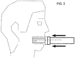
- is a side view of the scanning device in
figure 1 , showing how it is inserted into the patient's mouth. - Figure 3
- is a diagram showing the scan directions of the device in
figure 1 . - As shown in
figure 1 , the dental scanning device of the invention is made up of two bodies. The first one, the scanner body (1), houses a mobile scan head (3) made up of a mobile longitudinal element, ending in the scan head (4) as such. This mobile element (3, 4) is partially kept inside the scanner body (1). On the other hand, the scan head (4) projects from the body (1) for the purpose of being housed in a second body or bite body (2) by way of a case. The bite body (2) therefore constitutes a case to house the scan head (4). - If the abovementioned bodies can be coupled, and may be separated and joined again, the foregoing is equally applicable when both bodies (1 & 2) have been previously coupled to one another.
- As indicated, the bite body (2) is inserted into the patient's mouth and is held in place when the patient bites it, such that the scan head (4) is inserted into the mouth to scan the corresponding dental arcade.
- Likewise, the shape of the bite body (2) adapts to the mouth to make it easier to hold. This bite body (2) essentially constitutes a protective case for the scan head (4), and can be bitten by the user. This is why the bite body (2) is made of a suitable transparent material.
- Once the part of the scan head (4) that is housed in the bite body (2) has been inserted, and is thereby secured in the patient's mouth, as shown in
figure 2 , the scan head performs a sweeping motion in at least two directions, one following the longitudinal axis of the longitudinal element (3), and the other one perpendicular to it, in both senses of the direction, such that scanning is the result of a combination of both movements: lateral and depth-wise, with respect to the dental arcade, as shown infigure 3 . - For intraoral scanning, the scan head (4) of the device of the invention includes detection sensors, laser sensors or similar, as well as cameras to capture images derived from the tooth-by-tooth sweep of the dental arcade and the gums. These are obtained automatically, and, as a result of the design with a fixed and constant reference system, can be used to generate an accurate 3D image.
Claims (8)
- A dental scanning device made up of two bodies (1, 2) wherein the first body, also termed scanner body (1) houses a mobile scan head (3) consisting of a mobile longitudinal element ending in the scan head (4) as such, and wherein the second body, also termed bite body (2) constitutes a case to house the scan head (4), as well as the element for the patient to secure and place the device by biting it,
wherein the scan head (4) is adapted to perform a sweeping motion in at least two directions: one following the longitudinal axis of the longitudinal element (3), and the other one perpendicular to it, in both senses of the direction, such that scanning is the result of a combination of both movements: lateral and depth-wise, with respect to the dental arcade, characterised in that the bodies (1, 2) are separable and couple to one another through corresponding means arranged on respective coupling ends of the first body (1) and of the second body (2). - The dental scanning device according to claim 1, characterized in that the mobile scan head (3,4) is partially kept inside the scanner body (1).
- The dental scanning device according to claim 1, characterized in that the scan head (4) projects from the body (1) for the purpose of being housed in the second body or bite body (2) by way of a case.
- The dental scanning device according to claim 1, characterized in that the bite body (2) is manufactured with a transparent material.
- The dental scanning device according to claim 1, characterized in that the bite body (2) is disposable after use.
- The dental scanning device according to one of the preceding claims, characterized in that the sweeping motion is done with respect to a fixed reference system that is the patient's own mouth, wherein the second body or bite body (2) is held by the patient's mouth by exerting pressure when biting it.
- A method for performing a dental scan by means of the dental scanning device according to one of the claims 1 to 6, comprising the steps of:- inserting the bite body (2) of the dental scanning device into the patient's mouth,- holding the bite body (2) in place, when the patient bites the bite body (2),- inserting the scan head (4) of the dental scanning device into the patient's mouth, particularly into the bite body (2),- scanning a corresponding dental arcade, wherein a sweeping motion is performed in at least two directions: one following the longitudinal axis of the longitudinal element, and the other one perpendicular to it, in both senses of the direction, such that scanning is the result of a combination of both movements: lateral and depth-wise, with respect to the dental arcade,wherein the scanner body (1) and the bite body (2) are coupled to each other.
- The method according to claim 7, wherein the sweeping motion is done with respect to a fixed reference system that is the patient's own mouth, and wherein the second body or bite body (2) is held by the patient's mouth by exerting pressure when biting it.
Priority Applications (1)
| Application Number | Priority Date | Filing Date | Title |
|---|---|---|---|
| PL12889074T PL2929855T3 (en) | 2012-11-28 | 2012-11-28 | Dental scanning device |
Applications Claiming Priority (2)
| Application Number | Priority Date | Filing Date | Title |
|---|---|---|---|
| PCT/ES2012/070834 WO2014083211A1 (en) | 2012-11-28 | 2012-11-28 | Dental scanning device |
| ES201231853A ES2472640B1 (en) | 2012-11-28 | 2012-11-28 | DENTAL SCANNING DEVICE |
Publications (3)
| Publication Number | Publication Date |
|---|---|
| EP2929855A1 EP2929855A1 (en) | 2015-10-14 |
| EP2929855A4 EP2929855A4 (en) | 2016-07-27 |
| EP2929855B1 true EP2929855B1 (en) | 2021-04-07 |
Family
ID=50827211
Family Applications (1)
| Application Number | Title | Priority Date | Filing Date |
|---|---|---|---|
| EP12889074.6A Active EP2929855B1 (en) | 2012-11-28 | 2012-11-28 | Dental scanning device |
Country Status (13)
| Country | Link |
|---|---|
| US (3) | US8989567B1 (en) |
| EP (1) | EP2929855B1 (en) |
| JP (1) | JP2016501592A (en) |
| KR (1) | KR20150089030A (en) |
| CN (1) | CN104853692A (en) |
| AU (1) | AU2012395512A1 (en) |
| BR (1) | BR112015012332A2 (en) |
| CA (1) | CA2893035C (en) |
| ES (2) | ES2472640B1 (en) |
| IL (1) | IL239000A0 (en) |
| PL (1) | PL2929855T3 (en) |
| SG (1) | SG11201504207SA (en) |
| WO (1) | WO2014083211A1 (en) |
Families Citing this family (23)
| Publication number | Priority date | Publication date | Assignee | Title |
|---|---|---|---|---|
| JP2016501592A (en) * | 2012-11-28 | 2016-01-21 | アポロ オーラル スキャナー, エルエルシー | Dental scanner device |
| EP3145436B1 (en) * | 2014-05-23 | 2019-10-02 | Apollo Oral Scanner, Llc | Novel dental scanner device and system and methods of use |
| FR3027205B1 (en) * | 2014-10-20 | 2020-07-17 | Modjaw | METHOD AND SYSTEM FOR MODELING THE MANDIBULAR KINEMATICS OF A PATIENT |
| WO2016176556A1 (en) | 2015-04-29 | 2016-11-03 | University Of Maryland, Baltimore | Apparatus and method for recording digital images and presenting 3d models of a body lumen |
| CN113440087A (en) * | 2015-05-19 | 2021-09-28 | 泰拓卡尔有限公司 | System and method for imaging a body orifice of a patient |
| KR101851660B1 (en) * | 2015-05-20 | 2018-05-25 | 주식회사 바텍 | mouthpiece type scanner for oral cavity |
| EP3410919B1 (en) * | 2016-02-01 | 2023-12-06 | Martin, Marco | Dental imager and method for recording photographic impressions |
| EP3496591A1 (en) | 2016-08-10 | 2019-06-19 | Carestream Dental Technology Topco Limited | Automatic intraoral 3d scanner with low coherence ranging |
| CN110621259B (en) * | 2017-03-09 | 2021-11-02 | 马辛宾科夫斯基N-实验室 | Intraoral scanning device, method of operating such a device and scanner system |
| WO2018183514A2 (en) * | 2017-03-28 | 2018-10-04 | Scientific Intake Limited Co. | Systems including removable oral devices |
| PL3668706T3 (en) * | 2017-08-16 | 2021-11-29 | Gabaja Limited | System and method of manufacturing a mouth piece |
| US11612469B2 (en) * | 2018-04-05 | 2023-03-28 | Tech Xika Ptt, S.L. | Device for measuring dental parameters |
| US10499802B1 (en) * | 2018-08-31 | 2019-12-10 | Kaohsiung Medical University | Mouth-opening device custom-made through 3-dimensional printing |
| FR3087644B1 (en) * | 2018-10-30 | 2022-06-10 | Dental Monitoring | ARTICULATED DENTAL PHOTO TAKING KIT |
| AU2020254822A1 (en) * | 2019-04-05 | 2021-10-07 | Align Technology, Inc. | Intraoral scanner sleeve authentication and identification |
| GB201913469D0 (en) * | 2019-09-18 | 2019-10-30 | Univ Leeds Innovations Ltd | Three-dimensional dental scanning system and method of scanning |
| US20210177266A1 (en) * | 2019-12-17 | 2021-06-17 | Clayton Adams Teufel | Intraoral scanning with raw depth data |
| US20210196152A1 (en) * | 2019-12-31 | 2021-07-01 | Align Technology, Inc. | Gesture control using an intraoral scanner |
| CN111513680B (en) * | 2020-04-30 | 2021-08-03 | 四川大学 | Intraoral scanner |
| US11633108B2 (en) | 2020-10-15 | 2023-04-25 | Sean M. Langton | Trans-illuminative intraoral diagnostic lighting system and method of using |
| US11382727B1 (en) * | 2021-05-19 | 2022-07-12 | Thamer Marghalani | Three-dimensional oral imaging system and method |
| EP4181062A1 (en) * | 2021-11-11 | 2023-05-17 | DENTSPLY SIRONA Inc. | Computer-assisted medical procedures |
| CN115281866B (en) * | 2022-08-15 | 2023-11-17 | 北京美立刻医疗器械有限公司 | Tooth model acquisition auxiliary device, acquisition method, system, medium and equipment |
Family Cites Families (17)
| Publication number | Priority date | Publication date | Assignee | Title |
|---|---|---|---|---|
| US3382781A (en) * | 1965-02-10 | 1968-05-14 | William L. Hamilton | Camera |
| US4575805A (en) * | 1980-12-24 | 1986-03-11 | Moermann Werner H | Method and apparatus for the fabrication of custom-shaped implants |
| DE3932151A1 (en) * | 1989-09-22 | 1991-04-04 | Peter Rohleder | DEVICE FOR SCANNING DETECTION OF AN INTERIOR |
| US5702249A (en) | 1995-05-19 | 1997-12-30 | Cooper; David H. | Modular intra-oral imaging system video camera |
| US6592371B2 (en) * | 2000-10-25 | 2003-07-15 | Duane Durbin | Method and system for imaging and modeling a three dimensional structure |
| JP4478318B2 (en) * | 2000-10-25 | 2010-06-09 | 株式会社オレンジハウス | Dental row image reader |
| US6386867B1 (en) * | 2000-11-30 | 2002-05-14 | Duane Milford Durbin | Method and system for imaging and modeling dental structures |
| EP1252859A3 (en) * | 2001-04-27 | 2003-12-17 | Firma Ivoclar Vivadent AG | Dental camera with mouthpiece |
| US6821116B2 (en) * | 2001-09-12 | 2004-11-23 | Ivoclar Vivadent, Inc. | System for scanning oral environment |
| JP2004033465A (en) * | 2002-07-03 | 2004-02-05 | Mitsutoyo Corp | Tooth row image reading apparatus |
| US7142312B2 (en) | 2002-12-31 | 2006-11-28 | D4D Technologies, Llc | Laser digitizer system for dental applications |
| US7004754B2 (en) * | 2003-07-23 | 2006-02-28 | Orametrix, Inc. | Automatic crown and gingiva detection from three-dimensional virtual model of teeth |
| US7494338B2 (en) | 2005-01-11 | 2009-02-24 | Duane Durbin | 3D dental scanner |
| FR2883719B1 (en) | 2005-04-01 | 2007-06-01 | Atmel Grenoble Soc Par Actions | INTRAORAL DENTAL IMAGE SENSOR AND RADIOLOGICAL SYSTEM USING THE SENSOR |
| US8105233B2 (en) * | 2007-10-24 | 2012-01-31 | Tarek Ahmed Nabil Abou El Kheir | Endoscopic system and method for therapeutic applications and obtaining 3-dimensional human vision simulated imaging with real dynamic convergence |
| US8998609B2 (en) * | 2012-02-11 | 2015-04-07 | The Board Of Trustees Of The Leland Stanford Jr. University | Techniques for standardized imaging of oral cavity |
| JP2016501592A (en) * | 2012-11-28 | 2016-01-21 | アポロ オーラル スキャナー, エルエルシー | Dental scanner device |
-
2012
- 2012-11-28 JP JP2015544510A patent/JP2016501592A/en active Pending
- 2012-11-28 KR KR1020157015674A patent/KR20150089030A/en not_active Application Discontinuation
- 2012-11-28 ES ES201231853A patent/ES2472640B1/en active Active
- 2012-11-28 EP EP12889074.6A patent/EP2929855B1/en active Active
- 2012-11-28 SG SG11201504207SA patent/SG11201504207SA/en unknown
- 2012-11-28 BR BR112015012332A patent/BR112015012332A2/en not_active IP Right Cessation
- 2012-11-28 WO PCT/ES2012/070834 patent/WO2014083211A1/en active Application Filing
- 2012-11-28 CN CN201280077303.2A patent/CN104853692A/en active Pending
- 2012-11-28 PL PL12889074T patent/PL2929855T3/en unknown
- 2012-11-28 CA CA2893035A patent/CA2893035C/en active Active
- 2012-11-28 AU AU2012395512A patent/AU2012395512A1/en not_active Abandoned
- 2012-11-28 ES ES12889074T patent/ES2878141T3/en active Active
-
2014
- 2014-05-23 US US14/286,650 patent/US8989567B1/en active Active
-
2015
- 2015-03-23 US US14/666,229 patent/US9301672B2/en active Active
- 2015-05-25 IL IL239000A patent/IL239000A0/en unknown
- 2015-05-27 US US14/723,258 patent/US10226164B2/en active Active
Non-Patent Citations (1)
| Title |
|---|
| None * |
Also Published As
| Publication number | Publication date |
|---|---|
| EP2929855A4 (en) | 2016-07-27 |
| CA2893035C (en) | 2019-11-19 |
| JP2016501592A (en) | 2016-01-21 |
| WO2014083211A1 (en) | 2014-06-05 |
| PL2929855T3 (en) | 2021-11-08 |
| KR20150089030A (en) | 2015-08-04 |
| ES2472640B1 (en) | 2015-04-08 |
| ES2472640A1 (en) | 2014-07-01 |
| US8989567B1 (en) | 2015-03-24 |
| US10226164B2 (en) | 2019-03-12 |
| CA2893035A1 (en) | 2014-06-05 |
| ES2878141T3 (en) | 2021-11-18 |
| IL239000A0 (en) | 2015-07-30 |
| BR112015012332A2 (en) | 2017-07-11 |
| US9301672B2 (en) | 2016-04-05 |
| US20150250379A1 (en) | 2015-09-10 |
| EP2929855A1 (en) | 2015-10-14 |
| US20150238287A1 (en) | 2015-08-27 |
| CN104853692A (en) | 2015-08-19 |
| AU2012395512A1 (en) | 2015-07-02 |
| SG11201504207SA (en) | 2015-07-30 |
Similar Documents
| Publication | Publication Date | Title |
|---|---|---|
| EP2929855B1 (en) | Dental scanning device | |
| KR101977181B1 (en) | Dental intraoral scanner system | |
| AU2022202018B2 (en) | Dental imager and method for recording photographic impressions | |
| KR102050547B1 (en) | Device and method for subgingival measurement | |
| CA2950090C (en) | Novel dental scanner device and system and methods of use | |
| US20120288819A1 (en) | Dental imaging system with orientation detector | |
| JP5891080B2 (en) | Jaw movement simulation method, jaw movement simulation apparatus, and jaw movement simulation system | |
| JP6774365B2 (en) | Tip member that can be attached to and detached from the image pickup device and the housing of the image pickup device. | |
| NZ743965B2 (en) | Dental imager and method for recording photographic impressions |
Legal Events
| Date | Code | Title | Description |
|---|---|---|---|
| PUAI | Public reference made under article 153(3) epc to a published international application that has entered the european phase |
Free format text: ORIGINAL CODE: 0009012 |
|
| 17P | Request for examination filed |
Effective date: 20150626 |
|
| AK | Designated contracting states |
Kind code of ref document: A1 Designated state(s): AL AT BE BG CH CY CZ DE DK EE ES FI FR GB GR HR HU IE IS IT LI LT LU LV MC MK MT NL NO PL PT RO RS SE SI SK SM TR |
|
| AX | Request for extension of the european patent |
Extension state: BA ME |
|
| DAX | Request for extension of the european patent (deleted) | ||
| RIN1 | Information on inventor provided before grant (corrected) |
Inventor name: FERNANDEZ PULIDO, ALFONSO Inventor name: DE PABLOS GARCIA, DAVID |
|
| RA4 | Supplementary search report drawn up and despatched (corrected) |
Effective date: 20160629 |
|
| RIC1 | Information provided on ipc code assigned before grant |
Ipc: A61C 9/00 20060101AFI20160623BHEP Ipc: A61C 19/00 20060101ALI20160623BHEP Ipc: A61B 1/247 20060101ALI20160623BHEP Ipc: A61C 13/00 20060101ALI20160623BHEP Ipc: A61B 1/24 20060101ALI20160623BHEP Ipc: A61B 1/00 20060101ALI20160623BHEP |
|
| STAA | Information on the status of an ep patent application or granted ep patent |
Free format text: STATUS: EXAMINATION IS IN PROGRESS |
|
| 17Q | First examination report despatched |
Effective date: 20190606 |
|
| GRAP | Despatch of communication of intention to grant a patent |
Free format text: ORIGINAL CODE: EPIDOSNIGR1 |
|
| STAA | Information on the status of an ep patent application or granted ep patent |
Free format text: STATUS: GRANT OF PATENT IS INTENDED |
|
| INTG | Intention to grant announced |
Effective date: 20201030 |
|
| GRAS | Grant fee paid |
Free format text: ORIGINAL CODE: EPIDOSNIGR3 |
|
| GRAA | (expected) grant |
Free format text: ORIGINAL CODE: 0009210 |
|
| STAA | Information on the status of an ep patent application or granted ep patent |
Free format text: STATUS: THE PATENT HAS BEEN GRANTED |
|
| AK | Designated contracting states |
Kind code of ref document: B1 Designated state(s): AL AT BE BG CH CY CZ DE DK EE ES FI FR GB GR HR HU IE IS IT LI LT LU LV MC MK MT NL NO PL PT RO RS SE SI SK SM TR |
|
| REG | Reference to a national code |
Ref country code: GB Ref legal event code: FG4D |
|
| REG | Reference to a national code |
Ref country code: AT Ref legal event code: REF Ref document number: 1378677 Country of ref document: AT Kind code of ref document: T Effective date: 20210415 Ref country code: CH Ref legal event code: EP |
|
| REG | Reference to a national code |
Ref country code: DE Ref legal event code: R096 Ref document number: 602012075165 Country of ref document: DE |
|
| REG | Reference to a national code |
Ref country code: IE Ref legal event code: FG4D |
|
| REG | Reference to a national code |
Ref country code: LT Ref legal event code: MG9D |
|
| REG | Reference to a national code |
Ref country code: NL Ref legal event code: MP Effective date: 20210407 Ref country code: AT Ref legal event code: MK05 Ref document number: 1378677 Country of ref document: AT Kind code of ref document: T Effective date: 20210407 |
|
| PG25 | Lapsed in a contracting state [announced via postgrant information from national office to epo] |
Ref country code: BG Free format text: LAPSE BECAUSE OF FAILURE TO SUBMIT A TRANSLATION OF THE DESCRIPTION OR TO PAY THE FEE WITHIN THE PRESCRIBED TIME-LIMIT Effective date: 20210707 Ref country code: AT Free format text: LAPSE BECAUSE OF FAILURE TO SUBMIT A TRANSLATION OF THE DESCRIPTION OR TO PAY THE FEE WITHIN THE PRESCRIBED TIME-LIMIT Effective date: 20210407 Ref country code: HR Free format text: LAPSE BECAUSE OF FAILURE TO SUBMIT A TRANSLATION OF THE DESCRIPTION OR TO PAY THE FEE WITHIN THE PRESCRIBED TIME-LIMIT Effective date: 20210407 Ref country code: LT Free format text: LAPSE BECAUSE OF FAILURE TO SUBMIT A TRANSLATION OF THE DESCRIPTION OR TO PAY THE FEE WITHIN THE PRESCRIBED TIME-LIMIT Effective date: 20210407 Ref country code: NL Free format text: LAPSE BECAUSE OF FAILURE TO SUBMIT A TRANSLATION OF THE DESCRIPTION OR TO PAY THE FEE WITHIN THE PRESCRIBED TIME-LIMIT Effective date: 20210407 Ref country code: FI Free format text: LAPSE BECAUSE OF FAILURE TO SUBMIT A TRANSLATION OF THE DESCRIPTION OR TO PAY THE FEE WITHIN THE PRESCRIBED TIME-LIMIT Effective date: 20210407 |
|
| REG | Reference to a national code |
Ref country code: ES Ref legal event code: FG2A Ref document number: 2878141 Country of ref document: ES Kind code of ref document: T3 Effective date: 20211118 |
|
| PG25 | Lapsed in a contracting state [announced via postgrant information from national office to epo] |
Ref country code: RS Free format text: LAPSE BECAUSE OF FAILURE TO SUBMIT A TRANSLATION OF THE DESCRIPTION OR TO PAY THE FEE WITHIN THE PRESCRIBED TIME-LIMIT Effective date: 20210407 Ref country code: SE Free format text: LAPSE BECAUSE OF FAILURE TO SUBMIT A TRANSLATION OF THE DESCRIPTION OR TO PAY THE FEE WITHIN THE PRESCRIBED TIME-LIMIT Effective date: 20210407 Ref country code: GR Free format text: LAPSE BECAUSE OF FAILURE TO SUBMIT A TRANSLATION OF THE DESCRIPTION OR TO PAY THE FEE WITHIN THE PRESCRIBED TIME-LIMIT Effective date: 20210708 Ref country code: LV Free format text: LAPSE BECAUSE OF FAILURE TO SUBMIT A TRANSLATION OF THE DESCRIPTION OR TO PAY THE FEE WITHIN THE PRESCRIBED TIME-LIMIT Effective date: 20210407 Ref country code: IS Free format text: LAPSE BECAUSE OF FAILURE TO SUBMIT A TRANSLATION OF THE DESCRIPTION OR TO PAY THE FEE WITHIN THE PRESCRIBED TIME-LIMIT Effective date: 20210807 Ref country code: PT Free format text: LAPSE BECAUSE OF FAILURE TO SUBMIT A TRANSLATION OF THE DESCRIPTION OR TO PAY THE FEE WITHIN THE PRESCRIBED TIME-LIMIT Effective date: 20210809 Ref country code: NO Free format text: LAPSE BECAUSE OF FAILURE TO SUBMIT A TRANSLATION OF THE DESCRIPTION OR TO PAY THE FEE WITHIN THE PRESCRIBED TIME-LIMIT Effective date: 20210707 |
|
| REG | Reference to a national code |
Ref country code: DE Ref legal event code: R097 Ref document number: 602012075165 Country of ref document: DE |
|
| PG25 | Lapsed in a contracting state [announced via postgrant information from national office to epo] |
Ref country code: RO Free format text: LAPSE BECAUSE OF FAILURE TO SUBMIT A TRANSLATION OF THE DESCRIPTION OR TO PAY THE FEE WITHIN THE PRESCRIBED TIME-LIMIT Effective date: 20210407 Ref country code: DK Free format text: LAPSE BECAUSE OF FAILURE TO SUBMIT A TRANSLATION OF THE DESCRIPTION OR TO PAY THE FEE WITHIN THE PRESCRIBED TIME-LIMIT Effective date: 20210407 Ref country code: CZ Free format text: LAPSE BECAUSE OF FAILURE TO SUBMIT A TRANSLATION OF THE DESCRIPTION OR TO PAY THE FEE WITHIN THE PRESCRIBED TIME-LIMIT Effective date: 20210407 Ref country code: EE Free format text: LAPSE BECAUSE OF FAILURE TO SUBMIT A TRANSLATION OF THE DESCRIPTION OR TO PAY THE FEE WITHIN THE PRESCRIBED TIME-LIMIT Effective date: 20210407 Ref country code: SM Free format text: LAPSE BECAUSE OF FAILURE TO SUBMIT A TRANSLATION OF THE DESCRIPTION OR TO PAY THE FEE WITHIN THE PRESCRIBED TIME-LIMIT Effective date: 20210407 Ref country code: SK Free format text: LAPSE BECAUSE OF FAILURE TO SUBMIT A TRANSLATION OF THE DESCRIPTION OR TO PAY THE FEE WITHIN THE PRESCRIBED TIME-LIMIT Effective date: 20210407 |
|
| PLBE | No opposition filed within time limit |
Free format text: ORIGINAL CODE: 0009261 |
|
| STAA | Information on the status of an ep patent application or granted ep patent |
Free format text: STATUS: NO OPPOSITION FILED WITHIN TIME LIMIT |
|
| 26N | No opposition filed |
Effective date: 20220110 |
|
| REG | Reference to a national code |
Ref country code: DE Ref legal event code: R081 Ref document number: 602012075165 Country of ref document: DE Owner name: CARNOJAAL, S.L., ES Free format text: FORMER OWNER: APOLLO ORAL SCANNER, LLC, MIAMI, FL, US |
|
| REG | Reference to a national code |
Ref country code: GB Ref legal event code: 732E Free format text: REGISTERED BETWEEN 20220421 AND 20220427 |
|
| PG25 | Lapsed in a contracting state [announced via postgrant information from national office to epo] |
Ref country code: IS Free format text: LAPSE BECAUSE OF FAILURE TO SUBMIT A TRANSLATION OF THE DESCRIPTION OR TO PAY THE FEE WITHIN THE PRESCRIBED TIME-LIMIT Effective date: 20210807 Ref country code: AL Free format text: LAPSE BECAUSE OF FAILURE TO SUBMIT A TRANSLATION OF THE DESCRIPTION OR TO PAY THE FEE WITHIN THE PRESCRIBED TIME-LIMIT Effective date: 20210407 |
|
| PG25 | Lapsed in a contracting state [announced via postgrant information from national office to epo] |
Ref country code: MC Free format text: LAPSE BECAUSE OF FAILURE TO SUBMIT A TRANSLATION OF THE DESCRIPTION OR TO PAY THE FEE WITHIN THE PRESCRIBED TIME-LIMIT Effective date: 20210407 |
|
| REG | Reference to a national code |
Ref country code: CH Ref legal event code: PL |
|
| PG25 | Lapsed in a contracting state [announced via postgrant information from national office to epo] |
Ref country code: LU Free format text: LAPSE BECAUSE OF NON-PAYMENT OF DUE FEES Effective date: 20211128 Ref country code: BE Free format text: LAPSE BECAUSE OF NON-PAYMENT OF DUE FEES Effective date: 20211130 |
|
| REG | Reference to a national code |
Ref country code: BE Ref legal event code: MM Effective date: 20211130 |
|
| PG25 | Lapsed in a contracting state [announced via postgrant information from national office to epo] |
Ref country code: IE Free format text: LAPSE BECAUSE OF NON-PAYMENT OF DUE FEES Effective date: 20211128 |
|
| REG | Reference to a national code |
Ref country code: ES Ref legal event code: PC2A Owner name: CARNOJAAL, S.L. Effective date: 20221111 |
|
| PGFP | Annual fee paid to national office [announced via postgrant information from national office to epo] |
Ref country code: IT Payment date: 20221111 Year of fee payment: 11 Ref country code: GB Payment date: 20221117 Year of fee payment: 11 Ref country code: FR Payment date: 20221110 Year of fee payment: 11 Ref country code: ES Payment date: 20221201 Year of fee payment: 11 Ref country code: DE Payment date: 20221110 Year of fee payment: 11 |
|
| PGFP | Annual fee paid to national office [announced via postgrant information from national office to epo] |
Ref country code: PL Payment date: 20221117 Year of fee payment: 11 |
|
| PG25 | Lapsed in a contracting state [announced via postgrant information from national office to epo] |
Ref country code: HU Free format text: LAPSE BECAUSE OF FAILURE TO SUBMIT A TRANSLATION OF THE DESCRIPTION OR TO PAY THE FEE WITHIN THE PRESCRIBED TIME-LIMIT; INVALID AB INITIO Effective date: 20121128 Ref country code: CY Free format text: LAPSE BECAUSE OF FAILURE TO SUBMIT A TRANSLATION OF THE DESCRIPTION OR TO PAY THE FEE WITHIN THE PRESCRIBED TIME-LIMIT Effective date: 20210407 |
|
| PG25 | Lapsed in a contracting state [announced via postgrant information from national office to epo] |
Ref country code: LI Free format text: LAPSE BECAUSE OF NON-PAYMENT OF DUE FEES Effective date: 20220701 Ref country code: CH Free format text: LAPSE BECAUSE OF NON-PAYMENT OF DUE FEES Effective date: 20220701 |