EP2925890B1 - Accelerated prediction of cancer progression and response to treatment - Google Patents
Accelerated prediction of cancer progression and response to treatment Download PDFInfo
- Publication number
- EP2925890B1 EP2925890B1 EP13859257.1A EP13859257A EP2925890B1 EP 2925890 B1 EP2925890 B1 EP 2925890B1 EP 13859257 A EP13859257 A EP 13859257A EP 2925890 B1 EP2925890 B1 EP 2925890B1
- Authority
- EP
- European Patent Office
- Prior art keywords
- tumor cells
- circulating tumor
- embryo
- zebrafish
- cells
- Prior art date
- Legal status (The legal status is an assumption and is not a legal conclusion. Google has not performed a legal analysis and makes no representation as to the accuracy of the status listed.)
- Active
Links
Images



Classifications
-
- G—PHYSICS
- G01—MEASURING; TESTING
- G01N—INVESTIGATING OR ANALYSING MATERIALS BY DETERMINING THEIR CHEMICAL OR PHYSICAL PROPERTIES
- G01N33/00—Investigating or analysing materials by specific methods not covered by groups G01N1/00 - G01N31/00
- G01N33/48—Biological material, e.g. blood, urine; Haemocytometers
- G01N33/50—Chemical analysis of biological material, e.g. blood, urine; Testing involving biospecific ligand binding methods; Immunological testing
- G01N33/5005—Chemical analysis of biological material, e.g. blood, urine; Testing involving biospecific ligand binding methods; Immunological testing involving human or animal cells
- G01N33/5008—Chemical analysis of biological material, e.g. blood, urine; Testing involving biospecific ligand binding methods; Immunological testing involving human or animal cells for testing or evaluating the effect of chemical or biological compounds, e.g. drugs, cosmetics
- G01N33/5011—Chemical analysis of biological material, e.g. blood, urine; Testing involving biospecific ligand binding methods; Immunological testing involving human or animal cells for testing or evaluating the effect of chemical or biological compounds, e.g. drugs, cosmetics for testing antineoplastic activity
-
- C—CHEMISTRY; METALLURGY
- C12—BIOCHEMISTRY; BEER; SPIRITS; WINE; VINEGAR; MICROBIOLOGY; ENZYMOLOGY; MUTATION OR GENETIC ENGINEERING
- C12N—MICROORGANISMS OR ENZYMES; COMPOSITIONS THEREOF; PROPAGATING, PRESERVING, OR MAINTAINING MICROORGANISMS; MUTATION OR GENETIC ENGINEERING; CULTURE MEDIA
- C12N5/00—Undifferentiated human, animal or plant cells, e.g. cell lines; Tissues; Cultivation or maintenance thereof; Culture media therefor
- C12N5/06—Animal cells or tissues; Human cells or tissues
- C12N5/0602—Vertebrate cells
- C12N5/0693—Tumour cells; Cancer cells
-
- C—CHEMISTRY; METALLURGY
- C12—BIOCHEMISTRY; BEER; SPIRITS; WINE; VINEGAR; MICROBIOLOGY; ENZYMOLOGY; MUTATION OR GENETIC ENGINEERING
- C12N—MICROORGANISMS OR ENZYMES; COMPOSITIONS THEREOF; PROPAGATING, PRESERVING, OR MAINTAINING MICROORGANISMS; MUTATION OR GENETIC ENGINEERING; CULTURE MEDIA
- C12N15/00—Mutation or genetic engineering; DNA or RNA concerning genetic engineering, vectors, e.g. plasmids, or their isolation, preparation or purification; Use of hosts therefor
- C12N15/02—Preparation of hybrid cells by fusion of two or more cells, e.g. protoplast fusion
-
- C—CHEMISTRY; METALLURGY
- C12—BIOCHEMISTRY; BEER; SPIRITS; WINE; VINEGAR; MICROBIOLOGY; ENZYMOLOGY; MUTATION OR GENETIC ENGINEERING
- C12Q—MEASURING OR TESTING PROCESSES INVOLVING ENZYMES, NUCLEIC ACIDS OR MICROORGANISMS; COMPOSITIONS OR TEST PAPERS THEREFOR; PROCESSES OF PREPARING SUCH COMPOSITIONS; CONDITION-RESPONSIVE CONTROL IN MICROBIOLOGICAL OR ENZYMOLOGICAL PROCESSES
- C12Q1/00—Measuring or testing processes involving enzymes, nucleic acids or microorganisms; Compositions therefor; Processes of preparing such compositions
- C12Q1/68—Measuring or testing processes involving enzymes, nucleic acids or microorganisms; Compositions therefor; Processes of preparing such compositions involving nucleic acids
- C12Q1/6876—Nucleic acid products used in the analysis of nucleic acids, e.g. primers or probes
- C12Q1/6883—Nucleic acid products used in the analysis of nucleic acids, e.g. primers or probes for diseases caused by alterations of genetic material
- C12Q1/6886—Nucleic acid products used in the analysis of nucleic acids, e.g. primers or probes for diseases caused by alterations of genetic material for cancer
-
- G—PHYSICS
- G01—MEASURING; TESTING
- G01N—INVESTIGATING OR ANALYSING MATERIALS BY DETERMINING THEIR CHEMICAL OR PHYSICAL PROPERTIES
- G01N33/00—Investigating or analysing materials by specific methods not covered by groups G01N1/00 - G01N31/00
- G01N33/48—Biological material, e.g. blood, urine; Haemocytometers
- G01N33/50—Chemical analysis of biological material, e.g. blood, urine; Testing involving biospecific ligand binding methods; Immunological testing
- G01N33/5005—Chemical analysis of biological material, e.g. blood, urine; Testing involving biospecific ligand binding methods; Immunological testing involving human or animal cells
- G01N33/5008—Chemical analysis of biological material, e.g. blood, urine; Testing involving biospecific ligand binding methods; Immunological testing involving human or animal cells for testing or evaluating the effect of chemical or biological compounds, e.g. drugs, cosmetics
- G01N33/5082—Supracellular entities, e.g. tissue, organisms
- G01N33/5088—Supracellular entities, e.g. tissue, organisms of vertebrates
-
- C—CHEMISTRY; METALLURGY
- C12—BIOCHEMISTRY; BEER; SPIRITS; WINE; VINEGAR; MICROBIOLOGY; ENZYMOLOGY; MUTATION OR GENETIC ENGINEERING
- C12Q—MEASURING OR TESTING PROCESSES INVOLVING ENZYMES, NUCLEIC ACIDS OR MICROORGANISMS; COMPOSITIONS OR TEST PAPERS THEREFOR; PROCESSES OF PREPARING SUCH COMPOSITIONS; CONDITION-RESPONSIVE CONTROL IN MICROBIOLOGICAL OR ENZYMOLOGICAL PROCESSES
- C12Q2600/00—Oligonucleotides characterized by their use
- C12Q2600/106—Pharmacogenomics, i.e. genetic variability in individual responses to drugs and drug metabolism
-
- C—CHEMISTRY; METALLURGY
- C12—BIOCHEMISTRY; BEER; SPIRITS; WINE; VINEGAR; MICROBIOLOGY; ENZYMOLOGY; MUTATION OR GENETIC ENGINEERING
- C12Q—MEASURING OR TESTING PROCESSES INVOLVING ENZYMES, NUCLEIC ACIDS OR MICROORGANISMS; COMPOSITIONS OR TEST PAPERS THEREFOR; PROCESSES OF PREPARING SUCH COMPOSITIONS; CONDITION-RESPONSIVE CONTROL IN MICROBIOLOGICAL OR ENZYMOLOGICAL PROCESSES
- C12Q2600/00—Oligonucleotides characterized by their use
- C12Q2600/112—Disease subtyping, staging or classification
-
- C—CHEMISTRY; METALLURGY
- C12—BIOCHEMISTRY; BEER; SPIRITS; WINE; VINEGAR; MICROBIOLOGY; ENZYMOLOGY; MUTATION OR GENETIC ENGINEERING
- C12Q—MEASURING OR TESTING PROCESSES INVOLVING ENZYMES, NUCLEIC ACIDS OR MICROORGANISMS; COMPOSITIONS OR TEST PAPERS THEREFOR; PROCESSES OF PREPARING SUCH COMPOSITIONS; CONDITION-RESPONSIVE CONTROL IN MICROBIOLOGICAL OR ENZYMOLOGICAL PROCESSES
- C12Q2600/00—Oligonucleotides characterized by their use
- C12Q2600/118—Prognosis of disease development
-
- C—CHEMISTRY; METALLURGY
- C12—BIOCHEMISTRY; BEER; SPIRITS; WINE; VINEGAR; MICROBIOLOGY; ENZYMOLOGY; MUTATION OR GENETIC ENGINEERING
- C12Q—MEASURING OR TESTING PROCESSES INVOLVING ENZYMES, NUCLEIC ACIDS OR MICROORGANISMS; COMPOSITIONS OR TEST PAPERS THEREFOR; PROCESSES OF PREPARING SUCH COMPOSITIONS; CONDITION-RESPONSIVE CONTROL IN MICROBIOLOGICAL OR ENZYMOLOGICAL PROCESSES
- C12Q2600/00—Oligonucleotides characterized by their use
- C12Q2600/154—Methylation markers
-
- C—CHEMISTRY; METALLURGY
- C12—BIOCHEMISTRY; BEER; SPIRITS; WINE; VINEGAR; MICROBIOLOGY; ENZYMOLOGY; MUTATION OR GENETIC ENGINEERING
- C12Q—MEASURING OR TESTING PROCESSES INVOLVING ENZYMES, NUCLEIC ACIDS OR MICROORGANISMS; COMPOSITIONS OR TEST PAPERS THEREFOR; PROCESSES OF PREPARING SUCH COMPOSITIONS; CONDITION-RESPONSIVE CONTROL IN MICROBIOLOGICAL OR ENZYMOLOGICAL PROCESSES
- C12Q2600/00—Oligonucleotides characterized by their use
- C12Q2600/156—Polymorphic or mutational markers
-
- C—CHEMISTRY; METALLURGY
- C12—BIOCHEMISTRY; BEER; SPIRITS; WINE; VINEGAR; MICROBIOLOGY; ENZYMOLOGY; MUTATION OR GENETIC ENGINEERING
- C12Q—MEASURING OR TESTING PROCESSES INVOLVING ENZYMES, NUCLEIC ACIDS OR MICROORGANISMS; COMPOSITIONS OR TEST PAPERS THEREFOR; PROCESSES OF PREPARING SUCH COMPOSITIONS; CONDITION-RESPONSIVE CONTROL IN MICROBIOLOGICAL OR ENZYMOLOGICAL PROCESSES
- C12Q2600/00—Oligonucleotides characterized by their use
- C12Q2600/158—Expression markers
-
- G—PHYSICS
- G01—MEASURING; TESTING
- G01N—INVESTIGATING OR ANALYSING MATERIALS BY DETERMINING THEIR CHEMICAL OR PHYSICAL PROPERTIES
- G01N2333/00—Assays involving biological materials from specific organisms or of a specific nature
- G01N2333/435—Assays involving biological materials from specific organisms or of a specific nature from animals; from humans
- G01N2333/43504—Assays involving biological materials from specific organisms or of a specific nature from animals; from humans from invertebrates
- G01N2333/43526—Assays involving biological materials from specific organisms or of a specific nature from animals; from humans from invertebrates from worms
-
- G—PHYSICS
- G01—MEASURING; TESTING
- G01N—INVESTIGATING OR ANALYSING MATERIALS BY DETERMINING THEIR CHEMICAL OR PHYSICAL PROPERTIES
- G01N2800/00—Detection or diagnosis of diseases
- G01N2800/70—Mechanisms involved in disease identification
- G01N2800/7023—(Hyper)proliferation
- G01N2800/7028—Cancer
Definitions
- the present invention relates to a method of establishing tumors in zebrafish, by injecting human tumor cells. It also relates to the use of the established human tumors in zebrafish for characterizing tumor cells, testing drugs and for individualized medicine.
- Proper regulation of signal transduction in cells is responsible for a variety of biological functions including normal cellular replication, growth, cell physiology and cell death. Any perturbations to normal signal transduction in cells can result in various disease states of the body and often disease states are a result of the involvement of more than one cell type and overall body physiology states. Specifically, in the case of cancer, this situation is especially intricate as there is involvement of many underlying inflammatory states of the human body. Diverse inflammatory conditions such as obesity, allergy, arthritis, and diabetes all play a huge role in how cancer progresses and how treatment may work. Therefore, creation of in vivo models that mimic complicated conditions such as cancer require animal models that have an active immune system. Without an active immune system, the dynamic cellular heterogeneity observed in cancer cannot be completely replicated.
- epithelial-based cancers such as cancers of the breast, prostrate, lung, colon and pancreas
- the need to focus therapy towards such metastasized tumors is of paramount importance.
- Invasive with distant metastasized stage IV carcinomas present a very low survival rate (seer.cancer.gov).
- CTC circulating tumor cells
- Urology 65:713-8 Cohen SJ, Punt CJ, Iannotti N, Saidman BH, Sabbath KD, Gabrail NY, Picus J, Morse MA, Mitchell E, Miller MC, Doyle GV, Tissing H, Terstappen LW, Meropol NJ. (2009); Prognostic significance of circulating tumor cells in patients with metastatic colorectal cancer.
- Ann Oncol. 20:1223-9 Krebs MG, Sloane R, Priest L, Lancashire L, Hou JM, Greystoke A, Ward TH, Ferraldeschi R, Hughes A, Clack G, Ranson M, Dive C, Blackhall FH. (2011); Evaluation and prognostic significance of circulating tumor cells in patients with non-small-cell lung cancer. J Clin Oncol. 29:1556-63 ).
- Molecular and genomic profiling of cancer cells has become important because it can provide targeted therapy for an individual's particular cancer.
- profiling of the primary tumor will not represent the molecular changes that have occurred in the metastatic CTC.
- What is required for the targeted treatment of metastatic secondary tumors is a way to profile the CTCs.
- there are very few CTCs in a patient's blood so it is very difficult to isolate and characterize the cells.
- isolating the few CTC in a patient's blood has limited applications, unless the cells can be propagated and examined.
- Growing the CTC in tissue culture may be possible, but in vitro culture does not fully represent the cell characteristics, in particular their ability to invade normal tissues and form three-dimensional tumors, and to recruit growth factors and blood vessels.
- zebrafish Danio rerio, a popular fresh water aquarium fish
- zebrafish has been extremely popular in the study of embryogenesis, cardiovascular research, neuronal development and retinal regeneration but recently it has been established as a great model for almost every kind of cancer as well ( Stoletov and Klemke (2008) "Catch of the day: zebrafish as a human cancer model. Oncogene 27:4509-4520 )".
- Zebrafish are responsive to carcinogenic chemicals and form neoplasms very similar to that seen in humans ( Beckwith et al (2000) "Ethylnitrosourea induces neoplasia in zebrafish (Danio rerio). Lab Invest. 80(3):379-385 ). It is also a fantastic model for cancer genetics ( Stern and Zon (2003). “Cancer genetics and drug discovery in the zebrafish.” Nature Rev. Cancer 3: 533-539 ).
- Manipulations in zebrafish are performed at various stages of its growth, but 48 hours post fertilization (hpf) is frequently used and is one of the high priority stages for manipulations.
- the time and manpower required for the processing of many zebrafish embryos during large scale genetic, drug screening and toxicity studies, and cancer cell assays can often be the limiting factor for most laboratories.
- multi-well microinjection systems for 48 hpf zebrafish embryos primarily because of their elongated and odd shape.
- Konantz et al. use xenotransplantation of human cancer cells into zebra fish embryos to monitor treatment strategies ( Konantz et al. (2012) "Zebrafish xenografts as a tool for in vivo studies on human cancer”. Ann. N. Y. Acad. Sci, 1266:124-137 ). Pruvot et al. test leukemic cell xenografts in zebrafish embryo for drug efficacy ( Pruvot et al. (2011) Leukemic cell xenograft in zebrafish embryo for investigating drug efficacy". Haematologica 96:612-616 ).
- Automated multi-well microinjection systems are well known in the field of cell biology wherein they are primarily used in intranuclear or intracytoplasmic injection of materials such as DNA, RNAi, proteins, or even other cells such as sperm. Automated systems enable a large number of microinjections with reproducible consistency and accuracy that is often hard to achieve manually.
- PCT Patent Application No. PCT/ES2005/000255 filed May 12, 2005 by Alfonso Gutier-Rez Adan et al for "Supplementation for Embryo and/or Cell Manipulation” which discloses a system for increasing the quality and safety of embryo and cell manipulation media by supplementing the manipulation media with compounds, such as synthetic hyaluronan, phospholipids or unsaturated fatty acids that are obtained from soybeans, to reduce adhesiveness and increase viscosity while retaining the fluidity of the medium, to assist in micromanipulations including microinjection of cells into embryos during preimplantation stage.
- compounds such as synthetic hyaluronan, phospholipids or unsaturated fatty acids that are obtained from soybeans
- U.S. Patent Application No. 11/224,364 filed Sep. 12, 2005 by Leandro Christmann for "Microinjection Assembly and Methods for Microinjecting and Reimplanting Avian Eggs" which discloses a microinjection assembly including a microscope, a microinjection system comprising a micromanipulator, a micropipette and a piezo-electric oscillator, and an obliquely angled macro monitoring unit, which allows microinjecting the germinal disk of an avian egg.
- PCT Patent Application No. PCT/US2006/0006868 (or US2009/0211023) filed Feb. 27, 2006 by Daniel G. O'Connell for "Cell Tray” which discloses a multiwell cell tray that enables automated processing and simultaneous monitoring and analyzing of a large matrix of cells, biological fluids, chemicals and/or solid samples.
- the aims of the present invention were to overcome the technical problems of profiling and characterizing primary tumor cells and CTC in an animal model, and to provide a system that would enable efficient manipulation and injection of zebrafish embryos.
- a method is provided to establish tumors from circulating tumor cells (CTCs) from a primary tumor in a zebrafish embryo the method comprising:
- a method for testing a drug for its effect on circulating tumor cells from a primary tumor comprising:
- Variants of the multi-well microinjection system aspect include the following:
- the apparatus can be used with the holding frame and the groove plates in place, without the removable insert, thereby allowing the embryos to rest in the groove of the groove plate but to be accessible by a micropipette for microinjection at variable angles.
- a removable injection cover plate which has rubber lined apertures over each conical well which is formed by the groove plate, may be placed onto the groove plate and may be used to guide injection of the 48 hour post-fertilization zebrafish embryos.
- This disclosure also provides an option of controlling the robotic arm through vision recognition of embryo structures.
- the software can be designed to enable detection of the success of injection if liquids or labeled cells are fluorescent. Automation for the selection of the sites of injection and the protocols of injection can also be changed by simple software updates. Automation through a visual recognition system will also enable using smaller number of embryos and empty wells.
- the apparatus described herein can also be used with manual microinjectors available commercially. Even for manual manipulations, this equipment will reduce labor by reducing extra handling and labeling of tubes. Since embryos are not removed from their wells, the chances of mixing and mislabeling of embryos, as well as inducing stress into the embryos, is greatly reduced.
- This disclosure not only provides ease of handling large number of embryos for microinjecting liquids and cells, but it also enables proper positioning and injection of tumor pieces into the embryo in a high throughput fashion.
- the multi-well microinjection system described herein is economical and can be manufactured in a "one time use” fashion.
- the multi-well microinjection system (not part of the invention) and the method of use described herein greatly simplifies handling of a large number of embryos and improves injection accuracies and consistencies over a large number of experiments. Since all wells in a single module are connected, all embryos get the same treatment. Unequal loss of media volume due to drying, or unequal adding per well, does not cause any variation from well to well. Embryos of the same treatment group are exposed to such changes all equally.
- This multi-well microinjection system is for use in a 96 well format, but this system can be modified for 6 well, 12 well or 24 well plate formats as well.
- This multi-well microinjection system is for use with 48 hour post-fertilization zebrafish embryos, but can also be appropriated for manipulation of 24 to 72 hour post-fertilization zebrafish embryos, for manipulation of embryos from other species of fish, e.g. Medaka, and for embryos from xenopus, rodent, dog, and other laboratory animals.
- a rapid assay method that can predict a patient's tumor physiology (such as growth, invasive ability, metastatic organ-homing, etc.) and response to various anti-cancer treatments is required.
- the present invention provides methods for the prediction of cancer progression and response to treatment.
- the method may use an advanced "Cancer Progression and Response Matrix".
- Cancer Progression and Response Matrix may be used to facilitate the design of individualized and targeted therapies based on predictable tumor progression and responses to treatment.
- Subject or "Patient” or “Individual” typically include humans but can also include other animals including but not limited to rodents, canines, felines, equines, ovines, bovines, porcines and primates.
- Tumor includes a mass of cells found in or on the body of a subject that have some form of physiological, histological, molecular and or structural abnormality.
- Cancer includes any member of a class of diseases that have abnormal cells which grow in an uncontrolled fashion. This includes all neoplastic conditions and all cancers whether characterized benign, invasive, localized, pre-metastatic, metastatic, post-metastatic, soft tissue or solid, including any stage or grade.
- Body typically includes morphology, physiology, anatomy, behavior, origin, and distribution.
- “Pathophysiology” all typically mean the disordered physiological processes associated with a condition.
- cancer is a set of diseases that are driven by progressive genetic abnormalities that include chromosomal abnormalities, genetic mutations and epigenetic alterations.
- epigenetic alteration which are functionally relevant modifications to the genome that does not involve a change in the nucleotide sequence, play a significant role in regulating the overall biology of cancer cells. Epigenetic alterations have been observed due to environmental exposures.
- Biopsy refers to the process of removing cells or tissue samples for diagnostic or prognostic evaluation. Any known biopsy technique can be applied to the methods and compositions of present invention. Representative biopsy techniques include but are not limited to excisional, incisional, needle, and surgical biopsies. The choice of the biopsy technique used depends on tissue type to be evaluated and the location, size and type of the tumor.
- “Invasion” refers to encroachment or intrusion. Particularly, invasive tumor cells are cells that are able to invade into surrounding tissues. Not all tumor cells have the ability to invade. "Metastasis” is the development of secondary malignant growths (“Metastatic tumors”) at a distance from a primary site of cancer. It is the spread of cancer cells from one organ or part of the body to another non-adjacent organ or part. Cancer cells first move into the circulatory system (intravasation) followed by positioning into a secondary site to create secondary tumors (extravasation).
- Circulating tumor cells are tumor cells that have undergone intravasation and are found in the circulation. Circulating extratumoral cells include, but are not limited to, circulating tumor cells, disseminated cancer cells, and cancer stem cells. Circulating tumor cells can be otentially obtained from any accessible biological fluid such as whole blood, sputum, bronchial lavage, urine, nipple aspirate, lymph, saliva, needle aspirate, etc.
- Organ-homing involves seeding of circulating tumor cells into organs of metastasis. Primary tumors tend to metastasize to specific distant "target" organs. For example, lung cancer tends to frequently metastasize to the brain. The process or organ selection is not a random process although the physiology behind organ-homing is not well understood.
- Signal transduction occurs when an extracellular signaling molecule activates a cell surface receptor ("Signaling molecule” or “Signal transducer”). In turn, this receptor alters intracellular molecules creating a response, which typically include ordered sequences of biochemical reactions"
- Molecular genetic tumor markers have been identified based on the biological characterization of tumors, such as tumor development, growth, invasion and metastasis.
- Some examples include, but are not limited to, oncogenes (K-ras, erbB-1 (EGFR), erbB-2 (HER-2/neu), bcl-2, c-/N-/L-myc, c-kit), tumor suppressor genes (p53, RB, p16, p27, FHIT, RASSF1A), telomerase, invasion and metastasis markers (MMP, VEGF, COX-2), cell adhesion factors (E-Cadherin, beta-catherin), epithelial markers (cytokeratin, CEA), apoptosis markers (caspase-3, cleaved PARP), single nucleotide polymorphism (SNP), and anticancer drug susceptibility markers (MRP, LRP, MDR, beta-tubulin, ERCC
- Differential activation/deactivation of signaling pathways as well as changes in invasiveness and/or organ-homing of cells, in presence of anticancer drugs can aid in the selection of a suitable cancer therapy regimen at the proper dose for each patient.
- There could be a multitude of related application including prediction of how well chemotherapy is progressing for a given patient.
- “Chemicals” represents broadly all chemical compounds or substances that have been obtained crude, or have been purified from natural (available in nature through botanical or artificial sources (such as synthesized artificially in a laboratory).
- “Synthesized or naturally occurring chemicals and biologicals” include, but are not limited to, medicinal or therapeutic substances, non-medicinal substances, occurring in nature, artificially created, preparations made from living organisms (plant, animal, etc.), or extracted from non-living animal souces or minerals. These can include chemotherapeutic drugs, pharmaceutical formulations, Natural Health Products, powders, tea and extracts, serums, vaccines, antigens, antitoxins, etc.
- Immunomodulation is the adjustment of the immune responses, as in immunopotentiation (activation of the immune system), immunosuppression (suppression of the immune system), or induction of immunologic tolerance. Specifically, there is a complex dynamism between immune cells and malignant cells in the tumor microenvironment, which has there is in fact significant prognostic relevance as the immune system has both tumor promoting and inhibiting roles. Tumor infiltrating immune cells, and the chronic inflammation at the tumor site play a significant role in the growth, procession, invasion and metastatic disease. Immunomodulation can therefore impact greatly the progression of the disease.
- immunomodulation therefore represents the adjustment of immune responses of the tumor infiltrating immune cells that came with the patient tumor cell mass, regulating the regulators of the immune systems (interleukins and interferons) and regulating the host immune system, specifically the zebrafish immune cells.
- Zebrafish eggs were collected and incubated for 48h at 36degC in E3 medium (5 mM NaCl. 0.17 mM KCl. 0.33 mM CaCl2. 0.33 mM MgSO4. 0.1% methylene blue).
- E3 medium 5 mM NaCl. 0.17 mM KCl. 0.33 mM CaCl2. 0.33 mM MgSO4. 0.1% methylene blue.
- the embryos were anesthetized with tricaine and decorionated using Dumont #5 forceps.
- MDA-MB-231 cells (metastatic breast cancer cells) were grown in D-MEM (high glucose), 10% fetal bovine serum (FBS), 0.1 mM MEM NonEssential Amino Acids (NEAA), 2 mM L-glutamine, 1% Pen-Strep and labelled using CM-DiI (Vibrant, Lifetech, 4 ng/ul final concentration, incubated 4 min at 37°C followed by 15 min at 4°C). 50 cells were injected into the yolk of one 48hpf tricaine anesthetised zebrafish embryo. Images were taken 24h post injection.
- the isolated CTCs were localized at the site of injection but were also visible throughout the tail of the zebrafish embryo and were capable of forming metastatic patterns in the zebrafish embryo.
- CTCs were collected from 20ml blood (EDTA-Ca as anti-coagulant) from a Stage 4 lung cancer patient who has metastasis in the brain and one control healthy individual.
- CTCs were collected by sequential positive (anti-EpCam BerP4 antibody, AbCaM) and negative (anti-CD45, AbCam) selections using antibody coated magnetic beads (Dynabeads, Lifetech) according to manufacturer's instructions. Two-capture-wash-release were performed for each step. The yield was about 110 cells from the metastasis patient but no cells were detectable from the healthy donor.
- the CTCs obtained were stained with DiO (Vibrant, Lifetech, 200 mM final concentration) for 20min at 37degC.
- Tumor tissues from late stage lung cancer patient that had shown metastasis to the brain was minced and incubated in Liberase DL (Roche) as per manufacturer's instructions.
- Lung cells were passed through a 70 micrometer cell strainer and resuspended in 2 ml RPMI 1640 before counting. Cell viability was confirmed by trypan blue exclusion.
- Cells are labeled with fluorescent tracking PKH-67 (Sigma) dye following the manufacturer's instructions and resuspended in PBS containing 25mM glucose. 100 cells are injected into the yolk sac using NanojectII micromanipulator device. A group of embryos are injected with PBS+glucose only as control.
- embryos are then incubated in TE water containing antibiotic/antimycotic solution and let to recover overnight in an incubator at 35degC. After 24h of incubation post tumor transplantation, embryos are imaged under a fluorescent microscope to ensure the presence of tumor cells in the yolk sac. Drugs/Treatments are added at various concentrations and the plate with embryos are incubated at 35 °C for an additional 3 days. Embryos were anesthetized with tricaine and re-imaged under a fluorescent microscope. Drugs used in this experiment were Paclitaxel alone or in combination with Carboplatin.
- Drug response was measured through expressions of 18 genes (BCL2, BCL-X, BCL-B, BFL-1, BCL-W, MCL1, CDC2, CYCLIN-D, CYCLIN-A1, BAX, BAK, BOK, BID, BIM, BAD, BMF, NOXA, PUMA), nine (9) for survival (growth and cell cycle) and nine (9) for death (apoptosis).
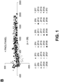
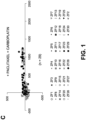
- FIG. 1 Tumor coordinates graphically represented ( FIG. 1 ) show very high reproducibility of the invasion and metastasis patterns in the presence or absence of drug treatment. There is differential response of the invasive tumors in comparison to non-invasive tumor cells in presence of drugs as measured through Invasion Index, Migration Index as well as Homing Index. There is also a very clear difference in survival (measured by cell cycle and growth) and death (measured by apoptosis) of non-invasive and invasive cells.
- the multi-well plate assembly component 10 includes a holding frame 12 including a base plate 28 supporting a plurality of embryo handling wells 24.
- the assembly 10 is made in a 96 well plate format and complies with international standards, although other standards may be used. This set-up can therefore be used with all standard microtiter plate readers and can be manipulated in all suitable liquid handlers.
- the multi-well plate assembly component 10 includes a lid 16, which is preferably provided with labels to mark the positions of wells of the multi-well plate assembly component 10, that offers safety, isolation, and prevents liquid in wells from drying.
- the holding frame 12 In this aspect, eight separable, removable modules 18 (seen in detail in FIG 3 ) are mounted in the holding frame 12. Every one of the eight separable, removable modules 18 has a groove plate 20 and a removable insert 22 that is mounted on the groove plate 20. As seen in FIG 4 , the groove plate 20 includes a plurality of the aforementioned embryo handling wells 24 and a lateral liquid handling well 26.
- Each embryo handling well 24 preferably has a cylindrical upper section 30 and a conical lower section 32.
- the lateral liquid handling well 26 is preferably completely cylindrical.
- the lateral liquid handling well 26 and the embryo handling wells 24 are interconnected at their outlet ends by a transverse drain channel 34.
- the removable insert 22 abuts the holding frame 12 at its outer edge and abuts the outer edges of the embryo handling wells 24 at its lower edge.
- the removable insert 22 can be removed for better manipulation of the embryo.
- the mounting of the removable insert 22 does not need to be airtight as there is the above-described intercommunication between each embryo handling well 24.
- the base plate 28 should preferably be transparent and UV penetrable.
- the removable insert 22 may be colored.
- FIGS 2 to 4 there are 11 embryo handling wells 24 (W1-11) for housing embryos and one lateral liquid handling well 26.
- lateral liquid handling well 26 and the embryo handling wells 24 are interconnected at their outlet ends by a transverse drain channel 34. Therefore, any change in liquid level in one well (e.g. well W1) will result in compensation through other wells (e.g. wells W2 to W11). This will prevent uneven drying of wells and all wells will have the same liquid level. Therefore, all liquid handling, changing of media, etc. can be done by a robotic liquid handler in the liquid handling well 26, thereby substantially preventing handling, damage or stress to the embryos.
- the embryo handling wells 24 have a conical bottom 32 where the larva of the zebrafish can be placed. As will be seen later in FIGS 6A and 6B , given the shape of the zebrafish larva, once anesthetized, they will fall into the well conical lower portion 32 of embryo handling well 24 with the yolk on top.
- a cover plate 36 can be positioned over the groove plate 20 in the place of the removable insert 22. As will be seen in FIG 7 , this cover plate 36 can act as the guide for the injection of the tumor cells along with pro-angiogenic factors into the embryo.
- FIG 5 The rectangle area within the broken lines in FIG 4 is shown in enlarged form in FIG 5 .
- FIG 6A is a horizontal transverse cross-section of a 48 hpf zebrafish embryo
- Fig 6B is a vertical transverse cross-section of a 48 hpf zebrafish embryo.
- a micropipette unit 40 may have a replaceable micropipette, 42, preferably of glass.
- the 48 hpf zebrafish embryo 48 is disposed in the conical lower section 32 of the embryo handling well 24, with its dorsal side 50 within the lower narrower end of the conical section 32 and with its yolk 52 in the upper wider end of the conical section 32.
- the unit so provided is protected by the cover plate 36.
- the micropipette unit 40 is positioned to inject the tumor cells along with pro-angiogenic factors, preferably growth factor angiopoietin into the yolk 52 through the aperture in the cover plate 38.
- the micropipette unit 40 having the replaceable micropipette 42 is controlled by a robotic arm 54.
- the liquid solution of the tumor cells along with a pro-angiogenic factor, preferably growth factor angiopoietin, is conducted through the robotic arm 52 via conduit 56.
- the robotic injector arm 54 can be rotated at any angle by means of control arm 58. The rotation is shown schematically by arrows 60.
- FIG 9 is a simplified replication of FIG 7 showing the use of the cover plate 36 as a guide for the injection of the tumor along with a pro-angiogenic factor, preferably growth factor angiopoietin, into the yolk 52.
- a pro-angiogenic factor preferably growth factor angiopoietin
- manipulations can be done under a microscope 62 without the removable insert in place. This allows manipulation of the embryo at any angle and injection can be performed to any part of the embryo body 50, 52.
- the micropipette unit 40 can be rotated from a vertical position shown in solid lines to a tilted position shown in broken lines so that injection can be performed to any part of the embryo body 50, 52.
- the micropipette unit 40 can be rotated from a tilted position shown in solid lines to a vertical position shown in broken lines so that injection can be performed to any part of the embryo body 50, 52.
- FIG 8B also shows that the embryo body 50, 52 can also be rotated.
- Embryos are dechorionated at 48 hpf and moved to wells using a glass pipette. If desired, embryos can be treated with pro-angiogenic factor, preferably growth factor angiopoietin, to increase the likelihood and efficiency of tumor cell uptake. Media is removed partially through well 36 and tricaine is added to anesthetize the embryos. Tricaine solution can be added to each well 24 as well to speed up the process. The embryos undergo anesthesia and fall to the lower conical bottoms 32 of the embryo handling wells 24 of the groove plates 20. Given the conical shape 32 at the bottom of the embryo handling wells 24, and the yolk 52 being lighter than the rest of the body 50, larvae fall with yolk 52 facing upwardly.
- pro-angiogenic factor preferably growth factor angiopoietin
- injection cover plate 36 can be positioned to guide the tumor cell along with the pro-angiogenic factor, preferably growth factor angiopoitin.
- Robotic arm 54 fitted with the glass micropipette 40 is used to inject the tumor cells along with the pro-angiogenic factor, preferably growth factor angiopoietin, into the embryo yolk 50.
- the yolk sac seals itself rapidly.
- the injection cover plate 36 is removed and the removable insert 22 is positioned to create the wells 24.
- Pipetting out tricaine solution through well 26 can change the fluids in the wells 24, and fresh media is added again through well 26.
- the wells 24 for each row of 11 embryos will therefore be filled, and each embryo will revive from anesthesia. Once they revive, they are free to swim around in their own wells and not mix with neighbouring embryos. This allows keeping track of individual embryos.
- the entire assembled unit with the lid 16 on and with swimming zebrafish larvae inside, can be stacked one above another and stored in an incubator as for other microtiter plates.
- the groove plate 20 is transparent, the larvae can be observed under UV in real time without needing to handle the larvae. If needed, larvae can be anesthetized for observations as mentioned earlier without handling them. Not only tumor growth can be measured using software, but also swimming behavior can be observed in real time. Such observations may alternatively be done manually or by using detection software.
- This step is generally performed in Eppendorf tubes because the mixing is not good in most 96 well plates even on a shaker. By pipetting up and down in well 26 alone, all 11 embryos can be rocked and shaken on a single module. Similarly, using a programmed liquid handler, all such processes for the entire plate can be optimized.
- fluorescence as a measure of tumor mass can be calculated directly using a UV plate reader. This same equipment can be used for other injections, such as DNA, RNA, morpholinos as well.
Landscapes
- Health & Medical Sciences (AREA)
- Life Sciences & Earth Sciences (AREA)
- Engineering & Computer Science (AREA)
- Chemical & Material Sciences (AREA)
- Biomedical Technology (AREA)
- Immunology (AREA)
- Molecular Biology (AREA)
- Bioinformatics & Cheminformatics (AREA)
- Biotechnology (AREA)
- Organic Chemistry (AREA)
- Genetics & Genomics (AREA)
- Wood Science & Technology (AREA)
- Zoology (AREA)
- Cell Biology (AREA)
- General Health & Medical Sciences (AREA)
- Biochemistry (AREA)
- Microbiology (AREA)
- Hematology (AREA)
- Urology & Nephrology (AREA)
- Analytical Chemistry (AREA)
- Pathology (AREA)
- Physics & Mathematics (AREA)
- Proteomics, Peptides & Aminoacids (AREA)
- General Engineering & Computer Science (AREA)
- Tropical Medicine & Parasitology (AREA)
- Toxicology (AREA)
- Food Science & Technology (AREA)
- Medicinal Chemistry (AREA)
- General Physics & Mathematics (AREA)
- Biophysics (AREA)
- Oncology (AREA)
- Hospice & Palliative Care (AREA)
- Plant Pathology (AREA)
- Measuring Or Testing Involving Enzymes Or Micro-Organisms (AREA)
- Apparatus Associated With Microorganisms And Enzymes (AREA)
- Micro-Organisms Or Cultivation Processes Thereof (AREA)
- Investigating Or Analysing Biological Materials (AREA)
Description
- The present invention relates to a method of establishing tumors in zebrafish, by injecting human tumor cells. It also relates to the use of the established human tumors in zebrafish for characterizing tumor cells, testing drugs and for individualized medicine.
- Proper regulation of signal transduction in cells is responsible for a variety of biological functions including normal cellular replication, growth, cell physiology and cell death. Any perturbations to normal signal transduction in cells can result in various disease states of the body and often disease states are a result of the involvement of more than one cell type and overall body physiology states. Specifically, in the case of cancer, this situation is especially intricate as there is involvement of many underlying inflammatory states of the human body. Diverse inflammatory conditions such as obesity, allergy, arthritis, and diabetes all play a huge role in how cancer progresses and how treatment may work. Therefore, creation of in vivo models that mimic complicated conditions such as cancer require animal models that have an active immune system. Without an active immune system, the dynamic cellular heterogeneity observed in cancer cannot be completely replicated. Furthermore, for clinical usefulness of such animal models of cancer, especially for prediction of the biology of each individual's cancer, organ invasion and cancer cell metastasis to other parts of the body, there should be a way of mimicking an individual patient's cancer in a very short time (before the start of chemotherapy) and predict cancer cell response to treatment.
- For epithelial-based cancers, such as cancers of the breast, prostrate, lung, colon and pancreas, the need to focus therapy towards such metastasized tumors is of paramount importance. Invasive with distant metastasized stage IV carcinomas present a very low survival rate (seer.cancer.gov).
- Metastatic cancer involves the detachment of aggressive malignant cells from the primary tumor into the bloodstream and/or lymphatic channels. Such circulating tumor cells (CTC) manage to reach distant organs where they develop secondary metastasis. Concordantly, the presence of these CTCs is associated with a poor prognosis (Balic M, Williams A, Lin H, Datar R, Cote RJ. (2012). Circulating Tumor Cells: From Bench to Bedside. Annu Rev Med. 2012 Oct 18.).
- The treatment of patients with metastatic disease continues to be largely dependent on the information we obtain from the primary tumor in spite of frequent discordance between the biomarkers observed on primary tumors versus those observed on secondary tumors (Naoki Niikura, Jun Liu, Naoki Hayashi, Elizabeth A. Mittendorf, Yun Gong, Shana L. Palla, Yutaka Tokuda, Ana M. Gonzalez-Angulo, Gabriel N. Hortobagyi and Naoto T. Ueno (2011); Loss of Human Epidermal Growth Factor Receptor 2 (HER2) Expression in Metastatic Sites of HER2-Overexpressing Primary Breast Tumors. J Clin Oncol, 30:593-599; Dupont Jensen J, Laenkholm AV, Knoop A, Ewertz M, Bandaru R, Liu W, Hackl W, Barrett JC, Gardner H. (2011); PIK3CA mutations may be discordant between primary and corresponding metastatic disease in breast cancer. Clin Cancer Res. 17:667-77). As the circumstantial originators of secondary tumors and metastasis, understanding the biology of secondary tumors will add new perspectives in the individualized treatment of advanced cancer patients. In support of our hypothesis, the prognostic significance of CTCs has been demonstrated for several types of cancers (Cristofanilli M, Budd GT, Ellis MJ, Stopeck A, Matera J, Miller MC, Reuben JM, Doyle GV, Allard WJ, Terstappen LW, Hayes DF. (2004); Circulating tumor cells, disease progression, and survival in metastatic breast cancer. N Engl J Med. 351:781-91; Moreno JG, Miller MC, Gross S, Allard WJ, Gomella LG, Terstappen LW. (2005); Circulating tumor cells predict survival in patients with metastatic prostate cancer. Urology 65:713-8; Cohen SJ, Punt CJ, Iannotti N, Saidman BH, Sabbath KD, Gabrail NY, Picus J, Morse MA, Mitchell E, Miller MC, Doyle GV, Tissing H, Terstappen LW, Meropol NJ. (2009); Prognostic significance of circulating tumor cells in patients with metastatic colorectal cancer. Ann Oncol. 20:1223-9; Krebs MG, Sloane R, Priest L, Lancashire L, Hou JM, Greystoke A, Ward TH, Ferraldeschi R, Hughes A, Clack G, Ranson M, Dive C, Blackhall FH. (2011); Evaluation and prognostic significance of circulating tumor cells in patients with non-small-cell lung cancer. J Clin Oncol. 29:1556-63).
- Molecular and genomic profiling of cancer cells has become the new trend in targeted therapy and oncology research. However, the relevance of molecular heterogeneity of the cancer cells and their constantly changing nature, the relevance of molecular signatures of the primary tumor as well as the CTCs is limited (Powell AA, Talasaz AH, Zhang H, Coram MA, Reddy A, et al. (2012) Single Cell Profiling of Circulating Tumor Cells: Transcriptional Heterogeneity and Diversity from Breast Cancer Cell Lines. PLoS ONE 7: e33788.).
- Molecular and genomic profiling of cancer cells has become important because it can provide targeted therapy for an individual's particular cancer. However, profiling of the primary tumor will not represent the molecular changes that have occurred in the metastatic CTC. What is required for the targeted treatment of metastatic secondary tumors is a way to profile the CTCs. However there are very few CTCs in a patient's blood so it is very difficult to isolate and characterize the cells. Furthermore, isolating the few CTC in a patient's blood has limited applications, unless the cells can be propagated and examined. Growing the CTC in tissue culture may be possible, but in vitro culture does not fully represent the cell characteristics, in particular their ability to invade normal tissues and form three-dimensional tumors, and to recruit growth factors and blood vessels.
- The zebrafish, Danio rerio, a popular fresh water aquarium fish, is an important model organism and is being increasingly used in scientific research (Lieschke and Currie (2007) "Animal models of human disease: zebrafish swims into view." Nature Reviews Genetics 8:353-367). In medicine, zebrafish has been extremely popular in the study of embryogenesis, cardiovascular research, neuronal development and retinal regeneration but recently it has been established as a great model for almost every kind of cancer as well (Stoletov and Klemke (2008) "Catch of the day: zebrafish as a human cancer model. Oncogene 27:4509-4520)".
- Zebrafish are responsive to carcinogenic chemicals and form neoplasms very similar to that seen in humans (Beckwith et al (2000) "Ethylnitrosourea induces neoplasia in zebrafish (Danio rerio). Lab Invest. 80(3):379-385). It is also a fantastic model for cancer genetics (Stern and Zon (2003). "Cancer genetics and drug discovery in the zebrafish." Nature Rev. Cancer 3: 533-539). The ease of genetic manipulations in zebrafish has aided its role in being an excellent model for understanding angiogenesis, apoptosis and metastasis (Serbedija et al (1999) "Zebrafish angiogenesis: a novel model for drug screening."; Angiogenesis 3:353-359; Parng et al (2002) "Zebrafish: a preclinical model for drug screening."; Assay Dev. Technol. 1:41-48; Marques et al (2009) "Metastatic behavior of primary human tumours in a zebrafish xenotransplantation model." BMC Cancer 9:128).
- Manipulations in zebrafish are performed at various stages of its growth, but 48 hours post fertilization (hpf) is frequently used and is one of the high priority stages for manipulations. The time and manpower required for the processing of many zebrafish embryos during large scale genetic, drug screening and toxicity studies, and cancer cell assays can often be the limiting factor for most laboratories. However, there are presently no commercially available multi-well microinjection systems for 48 hpf zebrafish embryos, primarily because of their elongated and odd shape.
- Konantz et al. use xenotransplantation of human cancer cells into zebra fish embryos to monitor treatment strategies (Konantz et al. (2012) "Zebrafish xenografts as a tool for in vivo studies on human cancer". Ann. N. Y. Acad. Sci, 1266:124-137). Pruvot et al. test leukemic cell xenografts in zebrafish embryo for drug efficacy (Pruvot et al. (2011) Leukemic cell xenograft in zebrafish embryo for investigating drug efficacy". Haematologica 96:612-616).
- Automated multi-well microinjection systems are well known in the field of cell biology wherein they are primarily used in intranuclear or intracytoplasmic injection of materials such as DNA, RNAi, proteins, or even other cells such as sperm. Automated systems enable a large number of microinjections with reproducible consistency and accuracy that is often hard to achieve manually.
- Therefore what is needed to profile and characterize primary tumor cells and CTC is a method to establish and grow the tumor cells in vivo in an animal model. This could allow drug testing on the tumor cells and could provide targeted therapy to the tumor cells in the patient. Furthermore, what is needed in the art is a system that would enable efficient manipulation and injection of 48 hpf zebrafish embryos, for genetic, toxicity, drug, and cancer studies.
-
U.S. Patent Application No. 10/923,253 (orUS2005/0112030A1) filed Aug 20, 2004 by Stephanie E. Gaus for "Meshwell Plates" which discloses a multiwell plate, such as a 96-well plate, with the bottom tip removed and replaced with a mesh with openings, to allow fast draining of solutions and to prevent "wicking" of solution between wells. The "Meshwell Plate" is stated to be intended to be particularly useful for assaying zebrafish embryos. - PCT Patent Application No.
PCT/ES2005/000255 US2008/0268419) filed May 12, 2005 by Alfonso Gutier-Rez Adan et al for "Supplementation for Embryo and/or Cell Manipulation" which discloses a system for increasing the quality and safety of embryo and cell manipulation media by supplementing the manipulation media with compounds, such as synthetic hyaluronan, phospholipids or unsaturated fatty acids that are obtained from soybeans, to reduce adhesiveness and increase viscosity while retaining the fluidity of the medium, to assist in micromanipulations including microinjection of cells into embryos during preimplantation stage. -
U.S. Patent Application No. 11/224,364 (orUS2006/0010510A1) filed Sep. 12, 2005 by Leandro Christmann for "Microinjection Assembly and Methods for Microinjecting and Reimplanting Avian Eggs" which discloses a microinjection assembly including a microscope, a microinjection system comprising a micromanipulator, a micropipette and a piezo-electric oscillator, and an obliquely angled macro monitoring unit, which allows microinjecting the germinal disk of an avian egg. - PCT Patent Application No.
PCT/US2006/0006868 (orUS2009/0211023) filed Feb. 27, 2006 by Daniel G. O'Connell for "Cell Tray" which discloses a multiwell cell tray that enables automated processing and simultaneous monitoring and analyzing of a large matrix of cells, biological fluids, chemicals and/or solid samples. -
U.K. Patent Application No. 1004629 GB2479521) filed March 19, 2012 by Jan De Sonneville -
US Patent No 7,339,090 patented Mar 4, 2008 by L. Christmann for "Microinjection Device and Method of Use" which discloses microinjection devices including a needle and a viewing instrument wherein the viewing instrument provides magnified viewing of an object to an operator from an angle other than right angle. -
WO 0065137 2000-11-02 by M. Palacios-Boyce -
WO 2058847 2002-08-01by M. Paranjape et al -
- 1. seer.cancer.gov
- 2. Balic M, Williams A, Lin H, Datar R, Cote RJ. (2012). Circulating Tumor Cells: From Bench to Bedside. Annu Rev Med. 2012 . [Epub ahead of print]
- 3. Naoki Niikura, Jun Liu, Naoki Hayashi, Elizabeth A. Mittendorf, Yun Gong, Shana L. Palla, Yutaka Tokuda, Ana M. Gonzalez-Angulo, Gabriel N. Hortobagyi and Naoto T. Ueno (2011). Loss of Human Epidermal Growth Factor Receptor 2 (HER2) Expression in Metastatic Sites of HER2-Overexpressing Primary Breast Tumors. J Clin Oncol, 30:593-599.
- 4. Dupont Jensen J, Laenkholm AV, Knoop A, Ewertz M, Bandaru R, Liu W, Hackl W, Barrett JC, Gardner H. (2011). PIK3CA mutations may be discordant between primary and corresponding metastatic disease in breast cancer. Clin Cancer Res. 17:667-77.
- 5. Cristofanilli M, Budd GT, Ellis MJ, Stopeck A, Matera J, Miller MC, Reuben JM, Doyle GV, Allard WJ, Terstappen LW, Hayes DF. (2004). Circulating tumor cells, disease progression, and survival in metastatic breast cancer. N Engl J Med. 351:781-91.
- 6. Moreno JG, Miller MC, Gross S, Allard WJ, Gomella LG, Terstappen LW. (2005). Circulating tumor cells predict survival in patients with metastatic prostate cancer. Urology 65:713-8.
- 7. Cohen SJ, Punt CJ, Iannotti N, Saidman BH, Sabbath KD, Gabrail NY, Picus J, Morse MA, Mitchell E, Miller MC, Doyle GV, Tissing H, Terstappen LW, Meropol NJ. (2009). Prognostic significance of circulating tumor cells in patients with metastatic colorectal cancer. Ann Oncol. 20:1223-9.
- 8. Krebs MG, Sloane R, Priest L, Lancashire L, Hou JM, Greystoke A, Ward TH, Ferraldeschi R, Hughes A, Clack G, Ranson M, Dive C, Blackhall FH. (2011). Evaluation and prognostic significance of circulating tumor cells in patients with non-small-cell lung cancer. J Clin Oncol. 29:1556-63.
- 9. Powell AA, Talasaz AH, Zhang H, Coram MA, Reddy A, et al. (2012) Single Cell Profiling of Circulating Tumor Cells: Transcriptional Heterogeneity and Diversity from Breast Cancer Cell Lines. PLoS ONE 7: e33788.
- 10. Konantz et al. (2012) Zebrafish xenografts as a tool for in vivo studies on human cancer. Ann. N. Y. Acad. Sci, 1266:124-137).
- 11. Pruvot et al. (2011) Leukemic cell xenograft in zebrafish embryo for investigating drug efficacy. Haematologica 96:612-616).
- The aims of the present invention were to overcome the technical problems of profiling and characterizing primary tumor cells and CTC in an animal model, and to provide a system that would enable efficient manipulation and injection of zebrafish embryos.
- The applicant has discovered that these technical problems are solved by way of methods that include creating viable tumors from xeno-transplanted human CTCs into zebrafish, studying the metastatic potential of the injected CTCs, predicting the organ preference of isolated CTCs, and assessing the response to therapeutic agents.
- In one aspect of the invention, a method is provided to establish tumors from circulating tumor cells (CTCs) from a primary tumor in a zebrafish embryo the method comprising:
- a. Isolating said circulating tumor cells from previously obtained biological fluid biopsies selected from whole blood, sputum, bronchial lavage, urine, nipple aspirate, lymph, and saliva;
- b. Labeling the circulating tumor cells with a cell tracking dye, preferably a fluorescent dye;
- c. Injecting the labeled circulating tumor cells into a 24 to 48 hours post fertilization (hpf) zebrafish embryo yolk; and
- d. Incubating the embryo for 24 hours or more to establish tumors in the zebrafish embryo.
- In a further aspect of the invention, a method is provided for testing a drug for its effect on circulating tumor cells from a primary tumor, comprising:
- a. microinjecting circulating tumor cells from a primary tumor into a 24 to 72 hour post-fertilization zebrafish embryo yolk, wherein the CTCs are isolated from previously obtained biological fluid biopsies selected from whole blood, sputum, bronchial lavage, urine, nipple aspirate, lymph, and saliva, and wherein the CTCs are labeled with a cell tracking dye, preferably a fluorescent dye;
- b. allowing the labeled circulating tumor cells to grow within the zebrafish embryo for a predetermined time to establish tumors in the zebrafish embryo;
- c. incubating in system water or microinjecting the zebrafish embryo with a drug being tested for its effect on the circulating tumor cells; and
- d. monitoring the effect of the drug on the circulating tumor cells by measuring the circulating tumor cells in the 24-72 hpf embryo after the pre-determined time.
- Further embodiments are detailed in the appended dependent claims.
- Variants of the multi-well microinjection system aspect (not part of the invention) include the following:
- the plurality of embryo holding wells are each interconnected at their open conical bottom to the bottom of the well module at an outer edge of the groove plate;
- the automation of the microinjection is performed using a robotic arm controlled micropipette holder;
- the automation of the microinjection is performed using a micropipette unit which is controlled by a robotic arm;
- the micropipette injection system is structured and arranged to be rotatably positionable, thereby to inject the embryo at variable angles and/or heights;
- the position and and/or angle of the robotic arm is adjustable, either manually or according to a commercially-available software-controlled interface, or through the development of a specific automatic injection system which is specifically designed for microinjection into zebrafish embryos;
- the robotic arm is controlled through human vision recognition of embryo structures or through vision recognition of embryo structures by means of fluorescence labelling of the cells, or by means of software which is programmed to enable detection of the success of injection of liquids,
- the software being either commercially-available software-controlled interface, or through the development of a specific automatic injection system which is specifically designed for microinjection into zebrafish embryos;
- automation for the selection of the sites of injection and the protocols of injection is changed by software updates; and
- the automatic microinjection system is controlled by commercially-available microinjector injection system, or through the development of a specific automatic injection system which is specifically designed for microinjection into zebrafish embryos.
- The apparatus can be used with the holding frame and the groove plates in place, without the removable insert, thereby allowing the embryos to rest in the groove of the groove plate but to be accessible by a micropipette for microinjection at variable angles. In this configuration, a removable injection cover plate which has rubber lined apertures over each conical well which is formed by the groove plate, may be placed onto the groove plate and may be used to guide injection of the 48 hour post-fertilization zebrafish embryos.
- This disclosure also provides an option of controlling the robotic arm through vision recognition of embryo structures. In such cases, the software can be designed to enable detection of the success of injection if liquids or labeled cells are fluorescent. Automation for the selection of the sites of injection and the protocols of injection can also be changed by simple software updates. Automation through a visual recognition system will also enable using smaller number of embryos and empty wells.
- The apparatus described herein can also be used with manual microinjectors available commercially. Even for manual manipulations, this equipment will reduce labor by reducing extra handling and labeling of tubes. Since embryos are not removed from their wells, the chances of mixing and mislabeling of embryos, as well as inducing stress into the embryos, is greatly reduced.
- Changing liquid in the wells when there is a live embryo in it cannot be performed using a robotic arm. However, the ability to change media in one well where there is no living embryo inside, makes the use of a robotic arm feasible. Also, manually changing liquid in wells with a living embryo can be very stressful for the embryo. By this method, gradual change of solution from the well module at an outer edge of the groove plate, which has a cylindrical shape can reduce unnecessary stress on the embryos.
- This disclosure not only provides ease of handling large number of embryos for microinjecting liquids and cells, but it also enables proper positioning and injection of tumor pieces into the embryo in a high throughput fashion.
- The multi-well microinjection system described herein, is economical and can be manufactured in a "one time use" fashion.
- The multi-well microinjection system (not part of the invention) and the method of use described herein greatly simplifies handling of a large number of embryos and improves injection accuracies and consistencies over a large number of experiments. Since all wells in a single module are connected, all embryos get the same treatment. Unequal loss of media volume due to drying, or unequal adding per well, does not cause any variation from well to well. Embryos of the same treatment group are exposed to such changes all equally.
- This multi-well microinjection system is for use in a 96 well format, but this system can be modified for 6 well, 12 well or 24 well plate formats as well.
- This multi-well microinjection system is for use with 48 hour post-fertilization zebrafish embryos, but can also be appropriated for manipulation of 24 to 72 hour post-fertilization zebrafish embryos, for manipulation of embryos from other species of fish, e.g. Medaka, and for embryos from xenopus, rodent, dog, and other laboratory animals.
- In the accompanying drawings:
-
FIG. 1 : Differential response of invasive and non-invasive primary lung tumor cells to drugs. -
FIG. 1A : Tumor coordinates of 30 xenografts captured through imaging are represented graphically. -
FIG. 1B : Tumor coordinates of 28 xenografts treated with Paclitaxel. -
FIG. 1C : Tumor coordinates of 29 xenografts treated with Paclitaxel and Carboplatin. (-D = No drug control, +P = Treated with Paclitaxel, C+P = Treated with Carboplatin and Paclitaxel) -
FIG. 1D : Calculated Migration Index of untreated and treated xenografts. (-D = No drug control, +P = Treated with Paclitaxel, C+P = Treated with Carboplatin and Paclitaxel) -
FIG. 1E : Calculated Invasion Index of untreated and treated xenografts. (-D = No drug control, +P = Treated with Paclitaxel, C+P = Treated with Carboplatin and Paclitaxel) -
FIG. 1F : Brain metastatic tumors were observed in the xenografts, recapitulating the organ-homing observed in the patient. -
FIG. 1G : Drug response of the brain metastasis tumors in xenografts. (-D = No drug control, +P = Treated with Paclitaxel, C+P = Treated with Carboplatin and Paclitaxel. -
FIG. 1H : Drug response of invasive and non-invasive cells in xenografts treated with Paclitaxel. Drug response was measured through expressions of MGMTs, 9 for survival and 9 for death. -
FIG. 1I : Drug response of invasive and non-invasive cells in xenografts treated with Paclitaxel and Carboplatin. -
FIG. 2 is a top plan view of the multi-well plate assembly component of one aspect of this invention; -
FIG. 3 is a top plan view of one of the removable modules of the embodiment ofFig 2 ; -
FIG. 4 is a cross section showing the embryos handling wells, with a groove plate sitting within the holding frame of the embodiment ofFig 2 ; -
FIG. 5 is enlarged cross sections of the embryo handling wells and the removable insert of the embodiment ofFig 2 ; -
FIG. 6A is a horizontal cross-section view of a 48 hpf zebrafish embryo, andFIG 6B is a transverse cross-section view of a 48 hpf zebrafish embryo. -
FIG. 7 is a schematic side view of the arrangement of a 48 hpf zebrafish embryo in the groove plate with an injection cover plate and a micropipette for microinjection of the embodiment ofFig 2 ; -
FIG. 8 is a schematic side view of a rotatable micropipette for microinjection of the embodiment ofFig 2 ; -
FIG. 9 is a schematic side view of a 48 hpf zebrafish embryo in a groove plate with an injection cover plate as a guide for the micropipette for microinjection of a tumor into a 48 hpf zebrafish embryo according to an embodiment of this invention; -
FIG 10 is a schematic side view of a 48 hpf zebrafish embryo in a groove plate showing the rotatable angle of the micropipette for microinjection while the embryo is in the groove plate according to an embodiment of this invention;
and -
FIG 11A and FIG 11B are schematic side views of a 48 hpf zebrafish embryo in a groove plate showing the flexibility for rotation of the micropipette while still allowing access to the embryo in the groove plate for microinjection thereinto according to an embodiment of this invention. - Molecular and genomic profiling of cancer cells has become the new trend in targeted therapy and oncology research. However, the relevance of molecular heterogeneity of the cancer cells and their constantly changing dynamic nature, the relevance of molecular signatures of the primary tumor as well invaded or metastasized tumor cells is limited. In this scenario, defined by limited efficacy of current chemotherapies to metastatic cancers, and the limited application of genomic profiling of cancer cells, we explored the possibility of creating representative and biologically relevant live 3D tumors out of tumor tissues (e.g., surgically removed primary tumor, biopsy, CTCs, etc.) to obtain clinically relevant physiological information about invasion and metastasis.
- For a successful individualized and targeted approach to cancer treatment, a rapid assay method that can predict a patient's tumor physiology (such as growth, invasive ability, metastatic organ-homing, etc.) and response to various anti-cancer treatments is required.
- An individualized and targeted treatment approach is however further complicated by the dynamic nature of all cancers. As a result every primary, invaded or metastasized tumor is made up of heterogeneous population of cells. Therefore a process of separating/ fractionating the cancer cell pool into various physiological or molecular categories is important.
- The present invention provides methods for the prediction of cancer progression and response to treatment. The method may use an advanced "Cancer Progression and Response Matrix". Thus certain aspects may be used to facilitate the design of individualized and targeted therapies based on predictable tumor progression and responses to treatment.
- As used herein, the following terminologies have meanings ascribed to them unless specified: "Subject" or "Patient" or "Individual" typically include humans but can also include other animals including but not limited to rodents, canines, felines, equines, ovines, bovines, porcines and primates.
- "Tumor" includes a mass of cells found in or on the body of a subject that have some form of physiological, histological, molecular and or structural abnormality.
- "Cancer" includes any member of a class of diseases that have abnormal cells which grow in an uncontrolled fashion. This includes all neoplastic conditions and all cancers whether characterized benign, invasive, localized, pre-metastatic, metastatic, post-metastatic, soft tissue or solid, including any stage or grade.
- "Biology" or "Physiology" typically includes morphology, physiology, anatomy, behavior, origin, and distribution.
- "Pathophysiology" all typically mean the disordered physiological processes associated with a condition. Particularly, cancer is a set of diseases that are driven by progressive genetic abnormalities that include chromosomal abnormalities, genetic mutations and epigenetic alterations. Particularly epigenetic alteration, which are functionally relevant modifications to the genome that does not involve a change in the nucleotide sequence, play a significant role in regulating the overall biology of cancer cells. Epigenetic alterations have been observed due to environmental exposures.
- "Biopsy" refers to the process of removing cells or tissue samples for diagnostic or prognostic evaluation. Any known biopsy technique can be applied to the methods and compositions of present invention. Representative biopsy techniques include but are not limited to excisional, incisional, needle, and surgical biopsies. The choice of the biopsy technique used depends on tissue type to be evaluated and the location, size and type of the tumor.
- "Invasion" refers to encroachment or intrusion. Particularly, invasive tumor cells are cells that are able to invade into surrounding tissues. Not all tumor cells have the ability to invade. "Metastasis" is the development of secondary malignant growths ("Metastatic tumors") at a distance from a primary site of cancer. It is the spread of cancer cells from one organ or part of the body to another non-adjacent organ or part. Cancer cells first move into the circulatory system (intravasation) followed by positioning into a secondary site to create secondary tumors (extravasation).
- "Circulating tumor cells" or "CTC" are tumor cells that have undergone intravasation and are found in the circulation. Circulating extratumoral cells include, but are not limited to, circulating tumor cells, disseminated cancer cells, and cancer stem cells. Circulating tumor cells can be otentially obtained from any accessible biological fluid such as whole blood, sputum, bronchial lavage, urine, nipple aspirate, lymph, saliva, needle aspirate, etc.
- "Organ-homing" involves seeding of circulating tumor cells into organs of metastasis. Primary tumors tend to metastasize to specific distant "target" organs. For example, lung cancer tends to frequently metastasize to the brain. The process or organ selection is not a random process although the physiology behind organ-homing is not well understood.
- "Signal transduction" occurs when an extracellular signaling molecule activates a cell surface receptor ("Signaling molecule" or "Signal transducer"). In turn, this receptor alters intracellular molecules creating a response, which typically include ordered sequences of biochemical reactions"
- "Molecular genetic tumor markers" or "MGTMs" have been identified based on the biological characterization of tumors, such as tumor development, growth, invasion and metastasis. Some examples include, but are not limited to, oncogenes (K-ras, erbB-1 (EGFR), erbB-2 (HER-2/neu), bcl-2, c-/N-/L-myc, c-kit), tumor suppressor genes (p53, RB, p16, p27, FHIT, RASSF1A), telomerase, invasion and metastasis markers (MMP, VEGF, COX-2), cell adhesion factors (E-Cadherin, beta-catherin), epithelial markers (cytokeratin, CEA), apoptosis markers (caspase-3, cleaved PARP), single nucleotide polymorphism (SNP), and anticancer drug susceptibility markers (MRP, LRP, MDR, beta-tubulin, ERCC1). Differential activation/deactivation of signaling pathways as well as changes in invasiveness and/or organ-homing of cells, in presence of anticancer drugs can aid in the selection of a suitable cancer therapy regimen at the proper dose for each patient. There could be a multitude of related application including prediction of how well chemotherapy is progressing for a given patient.
- "Chemicals" represents broadly all chemical compounds or substances that have been obtained crude, or have been purified from natural (available in nature through botanical or artificial sources (such as synthesized artificially in a laboratory).
- "Synthesized or naturally occurring chemicals and biologicals" include, but are not limited to, medicinal or therapeutic substances, non-medicinal substances, occurring in nature, artificially created, preparations made from living organisms (plant, animal, etc.), or extracted from non-living animal souces or minerals. These can include chemotherapeutic drugs, pharmaceutical formulations, Natural Health Products, powders, tea and extracts, serums, vaccines, antigens, antitoxins, etc.
- "Immunomodulation" is the adjustment of the immune responses, as in immunopotentiation (activation of the immune system), immunosuppression (suppression of the immune system), or induction of immunologic tolerance. Specifically, there is a complex dynamism between immune cells and malignant cells in the tumor microenvironment, which has there is in fact significant prognostic relevance as the immune system has both tumor promoting and inhibiting roles. Tumor infiltrating immune cells, and the chronic inflammation at the tumor site play a significant role in the growth, procession, invasion and metastatic disease. Immunomodulation can therefore impact greatly the progression of the disease. In the context of the current invention, immunomodulation therefore represents the adjustment of immune responses of the tumor infiltrating immune cells that came with the patient tumor cell mass, regulating the regulators of the immune systems (interleukins and interferons) and regulating the host immune system, specifically the zebrafish immune cells.
- Zebrafish eggs were collected and incubated for 48h at 36degC in E3 medium (5 mM NaCl. 0.17 mM KCl. 0.33 mM CaCl2. 0.33 mM MgSO4. 0.1% methylene blue). The embryos were anesthetized with tricaine and decorionated using Dumont #5 forceps.
- MDA-MB-231 cells (metastatic breast cancer cells) were grown in D-MEM (high glucose), 10% fetal bovine serum (FBS), 0.1 mM MEM NonEssential Amino Acids (NEAA), 2 mM L-glutamine, 1% Pen-Strep and labelled using CM-DiI (Vibrant, Lifetech, 4 ng/ul final concentration, incubated 4 min at 37°C followed by 15 min at 4°C). 50 cells were injected into the yolk of one 48hpf tricaine anesthetised zebrafish embryo. Images were taken 24h post injection.
- RESULTS: After injection, the isolated CTCs were localized at the site of injection but were also visible throughout the tail of the zebrafish embryo and were capable of forming metastatic patterns in the zebrafish embryo.
- CTCs were collected from 20ml blood (EDTA-Ca as anti-coagulant) from a Stage 4 lung cancer patient who has metastasis in the brain and one control healthy individual. CTCs were collected by sequential positive (anti-EpCam BerP4 antibody, AbCaM) and negative (anti-CD45, AbCam) selections using antibody coated magnetic beads (Dynabeads, Lifetech) according to manufacturer's instructions. Two-capture-wash-release were performed for each step. The yield was about 110 cells from the metastasis patient but no cells were detectable from the healthy donor. The CTCs obtained were stained with DiO (Vibrant, Lifetech, 200 mM final concentration) for 20min at 37degC. Total of 100 stained CTC cells were injected into the yolk of one 48hpf tricaine anesthetised zebrafish embryo. Images were obtained 24h post injection. RESULTS: Isolated CTCs were capable of forming tumors and formed metastases in the brain tissues of the zebrafish larvae.
- Tumor tissues from late stage lung cancer patient that had shown metastasis to the brain was minced and incubated in Liberase DL (Roche) as per manufacturer's instructions. Lung cells were passed through a 70 micrometer cell strainer and resuspended in 2 ml RPMI 1640 before counting. Cell viability was confirmed by trypan blue exclusion. Cells are labeled with fluorescent tracking PKH-67 (Sigma) dye following the manufacturer's instructions and resuspended in PBS containing 25mM glucose. 100 cells are injected into the yolk sac using NanojectII micromanipulator device. A group of embryos are injected with PBS+glucose only as control. The embryos are then incubated in TE water containing antibiotic/antimycotic solution and let to recover overnight in an incubator at 35degC. After 24h of incubation post tumor transplantation, embryos are imaged under a fluorescent microscope to ensure the presence of tumor cells in the yolk sac. Drugs/Treatments are added at various concentrations and the plate with embryos are incubated at 35 °C for an additional 3 days. Embryos were anesthetized with tricaine and re-imaged under a fluorescent microscope. Drugs used in this experiment were Paclitaxel alone or in combination with Carboplatin. Drug response was measured through expressions of 18 genes (BCL2, BCL-X, BCL-B, BFL-1, BCL-W, MCL1, CDC2, CYCLIN-D, CYCLIN-A1, BAX, BAK, BOK, BID, BIM, BAD, BMF, NOXA, PUMA), nine (9) for survival (growth and cell cycle) and nine (9) for death (apoptosis).
- RESULTS: Tumor coordinates graphically represented (
FIG. 1 ) show very high reproducibility of the invasion and metastasis patterns in the presence or absence of drug treatment. There is differential response of the invasive tumors in comparison to non-invasive tumor cells in presence of drugs as measured through Invasion Index, Migration Index as well as Homing Index. There is also a very clear difference in survival (measured by cell cycle and growth) and death (measured by apoptosis) of non-invasive and invasive cells. - As seen in the
FIGS 2 to 5 , the multi-wellplate assembly component 10 includes a holdingframe 12 including abase plate 28 supporting a plurality ofembryo handling wells 24. In this aspect, theassembly 10 is made in a 96 well plate format and complies with international standards, although other standards may be used. This set-up can therefore be used with all standard microtiter plate readers and can be manipulated in all suitable liquid handlers. The multi-wellplate assembly component 10 includes alid 16, which is preferably provided with labels to mark the positions of wells of the multi-wellplate assembly component 10, that offers safety, isolation, and prevents liquid in wells from drying. - In this aspect, eight separable, removable modules 18 (seen in detail in
FIG 3 ) are mounted in the holdingframe 12. Every one of the eight separable,removable modules 18 has agroove plate 20 and aremovable insert 22 that is mounted on thegroove plate 20. As seen inFIG 4 , thegroove plate 20 includes a plurality of the aforementionedembryo handling wells 24 and a lateral liquid handling well 26. - Each embryo handling well 24 preferably has a cylindrical
upper section 30 and a conicallower section 32. The lateral liquid handling well 26 is preferably completely cylindrical. The lateral liquid handling well 26 and theembryo handling wells 24 are interconnected at their outlet ends by atransverse drain channel 34. Theremovable insert 22 abuts the holdingframe 12 at its outer edge and abuts the outer edges of theembryo handling wells 24 at its lower edge. Theremovable insert 22 can be removed for better manipulation of the embryo. The mounting of theremovable insert 22 does not need to be airtight as there is the above-described intercommunication between each embryo handling well 24. Thebase plate 28 should preferably be transparent and UV penetrable. Theremovable insert 22 may be colored. - In this aspect as seen in
FIGS 2 to 4 , there are 11 embryo handling wells 24 (W1-11) for housing embryos and one lateral liquid handling well 26. As previously described lateral liquid handling well 26 and theembryo handling wells 24 are interconnected at their outlet ends by atransverse drain channel 34. Therefore, any change in liquid level in one well (e.g. well W1) will result in compensation through other wells (e.g. wells W2 to W11). This will prevent uneven drying of wells and all wells will have the same liquid level. Therefore, all liquid handling, changing of media, etc. can be done by a robotic liquid handler in the liquid handling well 26, thereby substantially preventing handling, damage or stress to the embryos. - All manipulations are done on the
groove plate 20. As previously described, theembryo handling wells 24 have a conical bottom 32 where the larva of the zebrafish can be placed. As will be seen later inFIGS 6A and 6B , given the shape of the zebrafish larva, once anesthetized, they will fall into the well conicallower portion 32 of embryo handling well 24 with the yolk on top. As will be seen inFIG. 7 , acover plate 36 can be positioned over thegroove plate 20 in the place of theremovable insert 22. As will be seen inFIG 7 , thiscover plate 36 can act as the guide for the injection of the tumor cells along with pro-angiogenic factors into the embryo. - The rectangle area within the broken lines in
FIG 4 is shown in enlarged form inFIG 5 . -
FIG 6A is a horizontal transverse cross-section of a 48 hpf zebrafish embryo, andFig 6B is a vertical transverse cross-section of a 48 hpf zebrafish embryo. - As seen in
FIG 7 , amicropipette unit 40 may have a replaceable micropipette, 42, preferably of glass. The 48hpf zebrafish embryo 48 is disposed in the conicallower section 32 of the embryo handling well 24, with itsdorsal side 50 within the lower narrower end of theconical section 32 and with itsyolk 52 in the upper wider end of theconical section 32. The unit so provided is protected by thecover plate 36. Themicropipette unit 40 is positioned to inject the tumor cells along with pro-angiogenic factors, preferably growth factor angiopoietin into theyolk 52 through the aperture in thecover plate 38. - As seen in
FIG 8 , themicropipette unit 40 having thereplaceable micropipette 42 is controlled by arobotic arm 54. The liquid solution of the tumor cells along with a pro-angiogenic factor, preferably growth factor angiopoietin, is conducted through therobotic arm 52 viaconduit 56. Therobotic injector arm 54 can be rotated at any angle by means ofcontrol arm 58. The rotation is shown schematically byarrows 60. -
FIG 9 is a simplified replication ofFIG 7 showing the use of thecover plate 36 as a guide for the injection of the tumor along with a pro-angiogenic factor, preferably growth factor angiopoietin, into theyolk 52. - As seen in
FIG 10 , manipulations can be done under amicroscope 62 without the removable insert in place. This allows manipulation of the embryo at any angle and injection can be performed to any part of theembryo body - As seen in
FIG 11A , themicropipette unit 40 can be rotated from a vertical position shown in solid lines to a tilted position shown in broken lines so that injection can be performed to any part of theembryo body - As seen in
FIG 11B , themicropipette unit 40 can be rotated from a tilted position shown in solid lines to a vertical position shown in broken lines so that injection can be performed to any part of theembryo body FIG 8B also shows that theembryo body - Embryos are dechorionated at 48 hpf and moved to wells using a glass pipette. If desired, embryos can be treated with pro-angiogenic factor, preferably growth factor angiopoietin, to increase the likelihood and efficiency of tumor cell uptake. Media is removed partially through well 36 and tricaine is added to anesthetize the embryos. Tricaine solution can be added to each well 24 as well to speed up the process. The embryos undergo anesthesia and fall to the lower
conical bottoms 32 of theembryo handling wells 24 of thegroove plates 20. Given theconical shape 32 at the bottom of theembryo handling wells 24, and theyolk 52 being lighter than the rest of thebody 50, larvae fall withyolk 52 facing upwardly. If required,injection cover plate 36 can be positioned to guide the tumor cell along with the pro-angiogenic factor, preferably growth factor angiopoitin.Robotic arm 54 fitted with theglass micropipette 40 is used to inject the tumor cells along with the pro-angiogenic factor, preferably growth factor angiopoietin, into theembryo yolk 50. The yolk sac seals itself rapidly. - Once injections are complete, the
injection cover plate 36 is removed and theremovable insert 22 is positioned to create thewells 24. - Pipetting out tricaine solution through well 26 can change the fluids in the
wells 24, and fresh media is added again through well 26. Thewells 24 for each row of 11 embryos will therefore be filled, and each embryo will revive from anesthesia. Once they revive, they are free to swim around in their own wells and not mix with neighbouring embryos. This allows keeping track of individual embryos. The entire assembled unit with thelid 16 on and with swimming zebrafish larvae inside, can be stacked one above another and stored in an incubator as for other microtiter plates. - Since, preferably, the
groove plate 20 is transparent, the larvae can be observed under UV in real time without needing to handle the larvae. If needed, larvae can be anesthetized for observations as mentioned earlier without handling them. Not only tumor growth can be measured using software, but also swimming behavior can be observed in real time. Such observations may alternatively be done manually or by using detection software. - After carrying out the above described example experiments, if the larvae need to be euthanized and stained, all handling of the larvae and changing of liquids can be done in this plate. One of the most important steps in whole embryo staining is rocking and shaking of embryos in solution for proper mixing.
- This step is generally performed in Eppendorf tubes because the mixing is not good in most 96 well plates even on a shaker. By pipetting up and down in well 26 alone, all 11 embryos can be rocked and shaken on a single module. Similarly, using a programmed liquid handler, all such processes for the entire plate can be optimized.
- Once all staining is done, fluorescence as a measure of tumor mass can be calculated directly using a UV plate reader. This same equipment can be used for other injections, such as DNA, RNA, morpholinos as well.
Claims (16)
- A method to establish tumors from circulating tumor cells (CTCs) from a primary tumor in a zebrafish embryo the method comprising:a. Isolating said circulating tumor cells from previously obtained biological fluid biopsies selected from whole blood, sputum, bronchial lavage, urine, nipple aspirate, lymph, and saliva;b. Labeling the circulating tumor cells with a cell tracking dye, preferably a fluorescent dye;c. Injecting the labeled circulating tumor cells into a 24 to 48 hours post fertilization (hpf) zebrafish embryo yolk; andd. Incubating the embryo for 24 hours or more to establish tumors in the zebrafish embryo.
- The method of claim 1, further comprising measuring the labeled circulating tumor cells position after incubating the embryo to determine whether said circulating tumor cells (i) invade the embryo body through the yolk sac; or (ii) remain in the yolk sac and behave closely to non-invasive circulating tumor cells (CTCs), to predict the likelihood of the CTCs to invade or metastasize.
- The method of claim 2, further comprising using said circulating tumor cells' position to indicate circulating tumor cells' metastatic potential, wherein if the circulating tumor cells invade the embryo body, the circulating tumor cells have metastatic potential and the propensity of the CTCs to invade and metastasize is high, to identify cancer patients with a higher probability of disease relapse.
- The method of claim 2, further comprising quantitating said circulating tumor cells in the embryo yolk sac and the embryo body upon incubation of the embryo at 35°C for 24h or more, by:a. Capturing automated fluorescent images of the circulating tumor cells under a fluorescent microscope;b. using image analysis software to measure a Width and a Length of the circulating tumor cells' foci, the intensity of the signal on each foci and the position of the spot in the image, and calculating a Volume of the circulating tumor cells' foci as ½(Width)(Length)2;c. measuring an Invasive Index of the circulating tumor cells' foci as Invasive Index (II) = 1/n Σ(number of circulating tumor cells foci in the embryo at T hours / Total number of circulating tumor cells injected in the embryo), where n is the number of embryos considered in the experiment, and T is the incubation time, and the greater the II, the higher the propensity of the circulating tumor cells to invade; andd. measuring a Migration Index (MI) of the circulating tumor cells, calculated as Migration Index (MI) = 1/n Σ(CD at T hours / Total number of circulating tumor cells' foci at time T hours), where CD = Cumulative distance traveled by circulating tumor cells, n is the number of embryos considered in the experiment, and T is the incubation time, where the higher the value of the MI, the more aggressively invasive are the circulating tumor cells.
- The method of claim 4, further comprising determining whether any one of: the Volume of said circulating tumor cells' foci; the Invasive Index; or the Migration Index are different in the presence versus the absence of a chemical to measure the response to a chemical by the circulating tumor cells.
- The method of claim 4, wherein the embryo incubation is in the presence or the absence of a chemical to measure the response to a chemical by said circulating tumor cells.
- The method of claim 6, further comprising measuring an effect of the chemical on the injected circulating tumor cells by:a. Digesting the embryo containing the circulating tumor cells in a protease solution after incubation;b. Dispersing the embryo and the circulating tumor cells with pipetting to dissociate to a single cell suspension;c. Fixing and counting the viable circulating tumor cells in the single cell suspension under a fluorescent microscope;d. Calculating a ratio of the counted viable circulating tumor cells to the injected circulating tumor cells;e. Comparing the ratio for incubation of the embryo in the presence of the chemical to the ratio for incubation of the embryo in the absence of the chemical; andf. Using the comparison to determine whether the chemical affects the circulating tumor cells;to measure the number of surviving circulating tumor cells after incubation with or without synthesized or naturally occurring chemicals.
- The method of claim 6, further comprising measuring and comparing the patterns of invasiveness of said circulating tumor cells in the presence or absence of the chemical and comparing whether circulating tumor cell invasiveness is different in the presence of the chemical, to predict drug efficacy against tumor cell invasiveness.
- The method of claim 6, further comprising assessing changes in the injected circulating tumor cells' DNA by:a. enzymatically digesting the embryo and said circulating tumor cells after incubation;b. isolating the digested embryo and circulating tumor cells' DNA;c. PCR amplifying one or more genes from the digested embryo and circulating tumor cells' DNA using PCR primers designed for human genetic sequences;d. Sequencing the amplified one or more genes to identify genetic mutations; ande. Bisulfite sequencing the digested embryo and circulating tumor cells' DNA to locate epigenetic modifications.
- The method of claim 6, further comprising analyzing said injected circulating tumor cells' gene expression by:a. Enzymatically digesting the embryo and the circulating tumor cells after incubation;b. Isolating the digested embryo and the circulating tumor cells' RNA;c. Performing a Quantitative Real-Time PCR analysis of gene expression using two or more primers designed for human genetic sequences.
- The method of claim 6, further comprising analyzing said circulating tumor cells' protein expression by one or more of:a. Fixing the whole embryo using a chemical fixative and using immunohistochemistry with one or more human protein antibodies to visualize the circulating tumor cells' protein expression;b. Visualizing the circulating tumor cells' protein expression using immuno-histochemistry on histological section slides of the embryo after the circulating tumor cell injection and incubation;c. Visualizing the circulating tumor cells' protein expression using ELISA (enzyme-linked immunosorption assay); andd. Visualizing the circulating tumor cells' protein expression using Western blot.
- The method of claim 6, further comprising image analysis of fluorescently labeled vascularization to predict the preferred organ for homing of said circulating tumor cells.
- The method of claim 12, further comprising measuring an organ homing pattern change of said circulating tumor cells in the absence versus the presence of a drug to predict a drug effect on the organ homing preference of the circulating tumor cells.
- A method for testing a drug for its effect on circulating tumor cells from a primary tumor, comprising:a. microinjecting circulating tumor cells from a primary tumor into a 24 to 72 hour post-fertilization zebrafish embryo yolk, wherein the CTCs are isolated from previously obtained biological fluid biopsies selected from whole blood, sputum, bronchial lavage, urine, nipple aspirate, lymph, and saliva, and wherein the CTCs are labeled with a cell tracking dye, preferably a fluorescent dye;b. allowing the labeled circulating tumor cells to grow within the zebrafish embryo for a predetermined time to establish tumors in the zebrafish embryo;c. incubating in system water or microinjecting the zebrafish embryo with a drug being tested for its effect on the circulating tumor cells; andd. monitoring the effect of the drug on the circulating tumor cells by measuring the circulating tumor cells in the 24-72 hpf embryo after the pre-determined time.
- The method of any one of claims 1-14, further comprising the addition of a pro-angiogenic factor, preferably angiopoietin, into water containing the zebrafish embryo either before, during or after the microinjection of said circulating tumor cells to test drugs for their effect on circulating tumor cells in 24 to 72 hour post fertilization zebrafish embryos.
- The method of claim 15, wherein the pro-angiogenic factor is microinjected into the embryo.
Applications Claiming Priority (2)
| Application Number | Priority Date | Filing Date | Title |
|---|---|---|---|
| US201261732375P | 2012-12-02 | 2012-12-02 | |
| PCT/IB2013/060580 WO2014083555A1 (en) | 2012-12-02 | 2013-12-02 | Accelerated prediction of cancer progression and response to treatment |
Publications (4)
| Publication Number | Publication Date |
|---|---|
| EP2925890A1 EP2925890A1 (en) | 2015-10-07 |
| EP2925890A4 EP2925890A4 (en) | 2016-06-01 |
| EP2925890B1 true EP2925890B1 (en) | 2024-05-15 |
| EP2925890C0 EP2925890C0 (en) | 2024-05-15 |
Family
ID=50827252
Family Applications (1)
| Application Number | Title | Priority Date | Filing Date |
|---|---|---|---|
| EP13859257.1A Active EP2925890B1 (en) | 2012-12-02 | 2013-12-02 | Accelerated prediction of cancer progression and response to treatment |
Country Status (7)
| Country | Link |
|---|---|
| US (1) | US10760060B2 (en) |
| EP (1) | EP2925890B1 (en) |
| JP (1) | JP6430392B2 (en) |
| CN (1) | CN105121657A (en) |
| CA (1) | CA2893549C (en) |
| SG (1) | SG11201504300VA (en) |
| WO (1) | WO2014083555A1 (en) |
Families Citing this family (11)
| Publication number | Priority date | Publication date | Assignee | Title |
|---|---|---|---|---|
| EP3218713A1 (en) * | 2014-11-14 | 2017-09-20 | The U.S.A. as represented by the Secretary, Department of Health and Human Services | Apparatus, kit and method for manipulating small model organisms |
| WO2016200985A1 (en) * | 2015-06-08 | 2016-12-15 | Trustees Of Tufts College | Imaging system to characterize dynamic changes in cell and particle characteristics |
| US10453551B2 (en) | 2016-06-08 | 2019-10-22 | X Development Llc | Simulating living cell in silico |
| US20190225923A1 (en) * | 2016-09-30 | 2019-07-25 | Nanyang Technological University | Apparatus for embryo biopsy |
| CA3044030A1 (en) * | 2016-11-17 | 2018-05-24 | Nantbio, Inc. | Validation of inferred anticancer pathways |
| GB201710955D0 (en) | 2017-07-07 | 2017-08-23 | Insphero Ag | Microtissue compartment device |
| KR101965299B1 (en) * | 2017-11-14 | 2019-04-03 | 주식회사 수젠텍 | Assembly type blot strip device |
| CN110402853B (en) * | 2018-04-28 | 2022-01-07 | 义慧科技(深圳)有限公司 | Detection model for tumor killing ability of immune cells and construction method and application thereof |
| CN108841869B (en) * | 2018-05-31 | 2022-05-06 | 上海交通大学医学院附属瑞金医院 | A method for constructing a zebrafish NK/TCL tumor model and its application |
| CN112094890A (en) * | 2019-06-17 | 2020-12-18 | 南京尧顺禹生物科技有限公司 | Method for quantitatively evaluating proliferation of tumor cells transplanted into zebra fish embryo |
| US11992297B2 (en) * | 2020-02-03 | 2024-05-28 | Korea Research Institute Of Standards And Science | Monitoring method of heart rate for toxicity test using high-speed digital holography |
Family Cites Families (11)
| Publication number | Priority date | Publication date | Assignee | Title |
|---|---|---|---|---|
| ES2286908T3 (en) * | 1998-12-01 | 2007-12-01 | Phylonix Pharmaceuticals Inc. | METHODS TO ENTER HETEROLOGICAL CELLS IN FISH. |
| WO2000065137A1 (en) | 1999-04-23 | 2000-11-02 | Palacios Boyce Monica | Microelectromechanical devices useful for manipulating cells or embryos, kits thereof, methods of making same, and methods of use thereof |
| WO2002058847A2 (en) | 2000-11-28 | 2002-08-01 | Georgetown University | Cell transformation using a single chip silicon microfabricated array incorporating integrated micro-piercing injectors |
| US7339090B2 (en) | 2001-02-13 | 2008-03-04 | Avigenics, Inc. | Microinjection devices and methods of use |
| US20020116732A1 (en) | 2001-02-13 | 2002-08-22 | Leandro Christmann | Microinjection assembly and methods for microinjecting and reimplanting avian eggs |
| US7190449B2 (en) | 2002-10-28 | 2007-03-13 | Nanopoint, Inc. | Cell tray |
| US20050112030A1 (en) | 2003-08-21 | 2005-05-26 | Gaus Stephanie E. | Meshwell plates |
| US20080077999A1 (en) * | 2004-03-23 | 2008-03-27 | Eisai R&D Management Co., Ltd. | Using Nonhuman Animal Model, Method of Measuring Transcription Activity Method of Measuring Cell Quantity and Method of Measuring Tumor Volume |
| ES2259566B1 (en) | 2005-03-29 | 2007-08-01 | Instituto Nacional De Investigacion Y Tecnologia Agraria Y Alimentaria-Inia | SUPPLEMENTATION FOR EMBRYO HANDLING AND / OR CELLULAR MEDIA. |
| EP2329817A1 (en) * | 2009-09-04 | 2011-06-08 | Ernst-Moritz-Arndt-Universität Greifswald | Retinoic acid receptor antagonists, miR-10a inhibitors and inhibitors of HOXB1 or HOXB3 repressors for treating pancreatic cancer |
| GB2479521A (en) | 2010-03-19 | 2011-10-19 | Univ Leiden | Array microinjection apparatus and methods for single cells or embryos |
-
2013
- 2013-12-02 CN CN201380070892.6A patent/CN105121657A/en active Pending
- 2013-12-02 SG SG11201504300VA patent/SG11201504300VA/en unknown
- 2013-12-02 WO PCT/IB2013/060580 patent/WO2014083555A1/en not_active Ceased
- 2013-12-02 JP JP2015544609A patent/JP6430392B2/en active Active
- 2013-12-02 CA CA2893549A patent/CA2893549C/en active Active
- 2013-12-02 US US14/649,196 patent/US10760060B2/en active Active
- 2013-12-02 EP EP13859257.1A patent/EP2925890B1/en active Active
Non-Patent Citations (1)
| Title |
|---|
| SIYANG ZHENG ET AL: "3D microfilter device for viable circulating tumor cell (CTC) enrichment from blood", BIOMEDICAL MICRODEVICES, KLUWER ACADEMIC PUBLISHERS, BO, vol. 13, no. 1, 27 October 2010 (2010-10-27), pages 203 - 213, XP019877255, ISSN: 1572-8781, DOI: 10.1007/S10544-010-9485-3 * |
Also Published As
| Publication number | Publication date |
|---|---|
| CA2893549C (en) | 2023-05-09 |
| WO2014083555A1 (en) | 2014-06-05 |
| US10760060B2 (en) | 2020-09-01 |
| JP2016501021A (en) | 2016-01-18 |
| EP2925890A1 (en) | 2015-10-07 |
| CA2893549A1 (en) | 2014-06-05 |
| SG11201504300VA (en) | 2015-07-30 |
| CN105121657A (en) | 2015-12-02 |
| EP2925890C0 (en) | 2024-05-15 |
| EP2925890A4 (en) | 2016-06-01 |
| JP6430392B2 (en) | 2018-11-28 |
| US20150315546A1 (en) | 2015-11-05 |
Similar Documents
| Publication | Publication Date | Title |
|---|---|---|
| CA2893549C (en) | Accelerated prediction of cancer progression and response to treatment | |
| Lin et al. | Identification of novel anti-liver cancer small molecules with better therapeutic index than sorafenib via zebrafish drug screening platform | |
| Hill et al. | Embryonic zebrafish xenograft assay of human cancer metastasis | |
| US20190376013A1 (en) | Microfluidic devices, systems, and methods for investigating three-dimensional cellular structures | |
| Haney et al. | Drug screening of primary patient derived tumor xenografts in zebrafish | |
| JP2015507470A (en) | Drug screening method and use thereof | |
| CN109906382A (en) | Biomarkers, disease-associated genes exploration methods and kidney cancer markers | |
| US20170336391A1 (en) | Tumor cell isolation/purification process and methods for use thereof | |
| CN104919315B (en) | The screening system of individuation anticancer agent | |
| Chen et al. | In vivo pharmacology models for cancer target research | |
| Antunes et al. | Analysis of tissue chimerism in nude mouse brain and abdominal xenograft models of human glioblastoma multiforme: what does it tell us about the models and about glioblastoma biology and therapy? | |
| CN109415702A (en) | Cell culture | |
| Costa et al. | Zebrafish Patient‐Derived Avatars from Digestive Cancers for Anti‐cancer Therapy Screening | |
| Kwak et al. | Isolation and characterization of tumorspheres from a recurrent pineoblastoma patient: Feasibility of a patient-derived xenograft | |
| Driscoll et al. | Evaluation of in vivo toxicity of biological nanoparticles | |
| Mi et al. | Identification of the metastasis potential and its associated genes in melanoma multinucleated giant cells using the PHA-ECM830 fusion method | |
| CN107430115A (en) | For operating utensil, external member and the method for small scale mode biology | |
| WO2016154459A1 (en) | High-throughput drug and genetic assays for cellular transformation | |
| van der Ent et al. | Automation of technology for cancer research | |
| Shimada et al. | Zebrafish-based systems pharmacology of cancer metastasis | |
| Li et al. | Establishment of embryonic zebrafish xenograft assays to investigate TGF-β family signaling in human breast cancer progression | |
| CN118949058A (en) | A HMGB1-targeted degradation small molecule HMGB1-PROTAC and its derivatives and applications | |
| Ishii et al. | Involvement of epithelial-mesenchymal transition in adenoid cystic carcinoma metastasis | |
| Chen et al. | Brain metastasis organoids: A systematic review of their methods and clinical application | |
| CN120958119A (en) | Methods for delivering components to microorgan spheres |
Legal Events
| Date | Code | Title | Description |
|---|---|---|---|
| PUAI | Public reference made under article 153(3) epc to a published international application that has entered the european phase |
Free format text: ORIGINAL CODE: 0009012 |
|
| 17P | Request for examination filed |
Effective date: 20150624 |
|
| AK | Designated contracting states |
Kind code of ref document: A1 Designated state(s): AL AT BE BG CH CY CZ DE DK EE ES FI FR GB GR HR HU IE IS IT LI LT LU LV MC MK MT NL NO PL PT RO RS SE SI SK SM TR |
|
| AX | Request for extension of the european patent |
Extension state: BA ME |
|
| DAX | Request for extension of the european patent (deleted) | ||
| RA4 | Supplementary search report drawn up and despatched (corrected) |
Effective date: 20160504 |
|
| RIC1 | Information provided on ipc code assigned before grant |
Ipc: G01N 33/50 20060101ALI20160428BHEP Ipc: C12N 5/09 20100101ALI20160428BHEP Ipc: C12N 15/02 20060101ALI20160428BHEP Ipc: C12Q 1/68 20060101AFI20160428BHEP |
|
| STAA | Information on the status of an ep patent application or granted ep patent |
Free format text: STATUS: EXAMINATION IS IN PROGRESS |
|
| 17Q | First examination report despatched |
Effective date: 20171103 |
|
| REG | Reference to a national code |
Ref country code: DE Ref legal event code: R079 Free format text: PREVIOUS MAIN CLASS: C12Q0001680000 Ipc: C12Q0001688600 Ref document number: 602013085716 Country of ref document: DE |
|
| GRAP | Despatch of communication of intention to grant a patent |
Free format text: ORIGINAL CODE: EPIDOSNIGR1 |
|
| STAA | Information on the status of an ep patent application or granted ep patent |
Free format text: STATUS: GRANT OF PATENT IS INTENDED |
|
| RIC1 | Information provided on ipc code assigned before grant |
Ipc: G01N 33/50 20060101ALI20231129BHEP Ipc: C12Q 1/6886 20180101AFI20231129BHEP |
|
| INTG | Intention to grant announced |
Effective date: 20231215 |
|
| GRAS | Grant fee paid |
Free format text: ORIGINAL CODE: EPIDOSNIGR3 |
|
| GRAA | (expected) grant |
Free format text: ORIGINAL CODE: 0009210 |
|
| STAA | Information on the status of an ep patent application or granted ep patent |
Free format text: STATUS: THE PATENT HAS BEEN GRANTED |
|
| AK | Designated contracting states |
Kind code of ref document: B1 Designated state(s): AL AT BE BG CH CY CZ DE DK EE ES FI FR GB GR HR HU IE IS IT LI LT LU LV MC MK MT NL NO PL PT RO RS SE SI SK SM TR |
|
| REG | Reference to a national code |
Ref country code: CH Ref legal event code: EP Ref country code: GB Ref legal event code: FG4D |
|
| REG | Reference to a national code |
Ref country code: DE Ref legal event code: R096 Ref document number: 602013085716 Country of ref document: DE |
|
| REG | Reference to a national code |
Ref country code: IE Ref legal event code: FG4D |
|
| U01 | Request for unitary effect filed |
Effective date: 20240613 |
|
| U07 | Unitary effect registered |
Designated state(s): AT BE BG DE DK EE FI FR IT LT LU LV MT NL PT SE SI Effective date: 20240621 |
|
| PG25 | Lapsed in a contracting state [announced via postgrant information from national office to epo] |
Ref country code: IS Free format text: LAPSE BECAUSE OF FAILURE TO SUBMIT A TRANSLATION OF THE DESCRIPTION OR TO PAY THE FEE WITHIN THE PRESCRIBED TIME-LIMIT Effective date: 20240915 |
|
| PG25 | Lapsed in a contracting state [announced via postgrant information from national office to epo] |
Ref country code: HR Free format text: LAPSE BECAUSE OF FAILURE TO SUBMIT A TRANSLATION OF THE DESCRIPTION OR TO PAY THE FEE WITHIN THE PRESCRIBED TIME-LIMIT Effective date: 20240515 |
|
| PG25 | Lapsed in a contracting state [announced via postgrant information from national office to epo] |
Ref country code: GR Free format text: LAPSE BECAUSE OF FAILURE TO SUBMIT A TRANSLATION OF THE DESCRIPTION OR TO PAY THE FEE WITHIN THE PRESCRIBED TIME-LIMIT Effective date: 20240816 |
|
| PG25 | Lapsed in a contracting state [announced via postgrant information from national office to epo] |
Ref country code: ES Free format text: LAPSE BECAUSE OF FAILURE TO SUBMIT A TRANSLATION OF THE DESCRIPTION OR TO PAY THE FEE WITHIN THE PRESCRIBED TIME-LIMIT Effective date: 20240515 |
|
| PG25 | Lapsed in a contracting state [announced via postgrant information from national office to epo] |
Ref country code: PL Free format text: LAPSE BECAUSE OF FAILURE TO SUBMIT A TRANSLATION OF THE DESCRIPTION OR TO PAY THE FEE WITHIN THE PRESCRIBED TIME-LIMIT Effective date: 20240515 |
|
| PG25 | Lapsed in a contracting state [announced via postgrant information from national office to epo] |
Ref country code: PL Free format text: LAPSE BECAUSE OF FAILURE TO SUBMIT A TRANSLATION OF THE DESCRIPTION OR TO PAY THE FEE WITHIN THE PRESCRIBED TIME-LIMIT Effective date: 20240515 Ref country code: NO Free format text: LAPSE BECAUSE OF FAILURE TO SUBMIT A TRANSLATION OF THE DESCRIPTION OR TO PAY THE FEE WITHIN THE PRESCRIBED TIME-LIMIT Effective date: 20240815 Ref country code: IS Free format text: LAPSE BECAUSE OF FAILURE TO SUBMIT A TRANSLATION OF THE DESCRIPTION OR TO PAY THE FEE WITHIN THE PRESCRIBED TIME-LIMIT Effective date: 20240915 Ref country code: HR Free format text: LAPSE BECAUSE OF FAILURE TO SUBMIT A TRANSLATION OF THE DESCRIPTION OR TO PAY THE FEE WITHIN THE PRESCRIBED TIME-LIMIT Effective date: 20240515 Ref country code: GR Free format text: LAPSE BECAUSE OF FAILURE TO SUBMIT A TRANSLATION OF THE DESCRIPTION OR TO PAY THE FEE WITHIN THE PRESCRIBED TIME-LIMIT Effective date: 20240816 Ref country code: ES Free format text: LAPSE BECAUSE OF FAILURE TO SUBMIT A TRANSLATION OF THE DESCRIPTION OR TO PAY THE FEE WITHIN THE PRESCRIBED TIME-LIMIT Effective date: 20240515 Ref country code: RS Free format text: LAPSE BECAUSE OF FAILURE TO SUBMIT A TRANSLATION OF THE DESCRIPTION OR TO PAY THE FEE WITHIN THE PRESCRIBED TIME-LIMIT Effective date: 20240815 |
|
| PG25 | Lapsed in a contracting state [announced via postgrant information from national office to epo] |
Ref country code: CZ Free format text: LAPSE BECAUSE OF FAILURE TO SUBMIT A TRANSLATION OF THE DESCRIPTION OR TO PAY THE FEE WITHIN THE PRESCRIBED TIME-LIMIT Effective date: 20240515 |
|
| PG25 | Lapsed in a contracting state [announced via postgrant information from national office to epo] |
Ref country code: RO Free format text: LAPSE BECAUSE OF FAILURE TO SUBMIT A TRANSLATION OF THE DESCRIPTION OR TO PAY THE FEE WITHIN THE PRESCRIBED TIME-LIMIT Effective date: 20240515 Ref country code: SK Free format text: LAPSE BECAUSE OF FAILURE TO SUBMIT A TRANSLATION OF THE DESCRIPTION OR TO PAY THE FEE WITHIN THE PRESCRIBED TIME-LIMIT Effective date: 20240515 |
|
| PG25 | Lapsed in a contracting state [announced via postgrant information from national office to epo] |
Ref country code: SM Free format text: LAPSE BECAUSE OF FAILURE TO SUBMIT A TRANSLATION OF THE DESCRIPTION OR TO PAY THE FEE WITHIN THE PRESCRIBED TIME-LIMIT Effective date: 20240515 |
|
| PG25 | Lapsed in a contracting state [announced via postgrant information from national office to epo] |
Ref country code: SM Free format text: LAPSE BECAUSE OF FAILURE TO SUBMIT A TRANSLATION OF THE DESCRIPTION OR TO PAY THE FEE WITHIN THE PRESCRIBED TIME-LIMIT Effective date: 20240515 Ref country code: SK Free format text: LAPSE BECAUSE OF FAILURE TO SUBMIT A TRANSLATION OF THE DESCRIPTION OR TO PAY THE FEE WITHIN THE PRESCRIBED TIME-LIMIT Effective date: 20240515 Ref country code: RO Free format text: LAPSE BECAUSE OF FAILURE TO SUBMIT A TRANSLATION OF THE DESCRIPTION OR TO PAY THE FEE WITHIN THE PRESCRIBED TIME-LIMIT Effective date: 20240515 Ref country code: CZ Free format text: LAPSE BECAUSE OF FAILURE TO SUBMIT A TRANSLATION OF THE DESCRIPTION OR TO PAY THE FEE WITHIN THE PRESCRIBED TIME-LIMIT Effective date: 20240515 |
|
| REG | Reference to a national code |
Ref country code: DE Ref legal event code: R097 Ref document number: 602013085716 Country of ref document: DE |
|
| PLBE | No opposition filed within time limit |
Free format text: ORIGINAL CODE: 0009261 |
|
| STAA | Information on the status of an ep patent application or granted ep patent |
Free format text: STATUS: NO OPPOSITION FILED WITHIN TIME LIMIT |
|
| 26N | No opposition filed |
Effective date: 20250218 |
|
| PG25 | Lapsed in a contracting state [announced via postgrant information from national office to epo] |
Ref country code: MC Free format text: LAPSE BECAUSE OF FAILURE TO SUBMIT A TRANSLATION OF THE DESCRIPTION OR TO PAY THE FEE WITHIN THE PRESCRIBED TIME-LIMIT Effective date: 20240515 |
|
| REG | Reference to a national code |
Ref country code: CH Ref legal event code: PL |
|
| U90 | Renewal fees not paid: noting of loss of rights |
Free format text: RENEWAL FEE NOT PAID FOR YEAR 12 Effective date: 20250711 |
|
| GBPC | Gb: european patent ceased through non-payment of renewal fee |
Effective date: 20241202 |
|
| PG25 | Lapsed in a contracting state [announced via postgrant information from national office to epo] |
Ref country code: GB Free format text: LAPSE BECAUSE OF NON-PAYMENT OF DUE FEES Effective date: 20241202 |
|
| PG25 | Lapsed in a contracting state [announced via postgrant information from national office to epo] |
Ref country code: CH Free format text: LAPSE BECAUSE OF NON-PAYMENT OF DUE FEES Effective date: 20241231 |
|
| PG25 | Lapsed in a contracting state [announced via postgrant information from national office to epo] |
Ref country code: IE Free format text: LAPSE BECAUSE OF NON-PAYMENT OF DUE FEES Effective date: 20241202 |
|
| U93 | Unitary patent lapsed |
Free format text: RENEWAL FEE NOT PAID Effective date: 20241231 |
|
| PG25 | Lapsed in a contracting state [announced via postgrant information from national office to epo] |
Ref country code: CY Free format text: LAPSE BECAUSE OF FAILURE TO SUBMIT A TRANSLATION OF THE DESCRIPTION OR TO PAY THE FEE WITHIN THE PRESCRIBED TIME-LIMIT; INVALID AB INITIO Effective date: 20131202 |