-
Human T-cell lymphotropic virus (HTLV) type I (HTLV-I) was the first pathogenic retrovirus to be discovered in man in 1980. It is the causative agent of T-cell leukemia and/or lymphoma and of HTLV-associated myelopathy, a severe demyelinating condition that eventually leads to tropical spastic paraparesis. The cumulative lifetime risk of developing these fatal and incurable diseases amounts to - 5 % in asymptomatic carriers of HTLV-I. HTLV-I infects primarily CD4-positive T-cells. It is also called the adult T-cell lymphoma virus type 1. HTLV-II shares approximately 70% genomic homology (translating into 80-95 % structural similarity at the protein level) with HTLV-I. The pathogenic potential of HTLV-II is not yet completely elucidated but HTLV-II is regarded as a risk marker for blood transfusion since it is mainly found in intravenous drug users world-wide (Vandamme et al., Evolutionary strategies of human T-cell lymphotropic virus type II, Gene 261 (2000) 171-180). Both viruses are spread globally, but the prevalence of HTLV-I is highest in hot spot regions in Southern Japan (Kyushu and Shikoku), Sub-Saharan Africa, the Caribbean (Jamaica and Haiti) and South America.
-
The major transmission modes of HTLV-I/II are through sexual contact, blood transfusion, sharing injection needles and mother to child transmission through breast-feeding. The seroconversion period after HTLV infection is long when compared to other infectious diseases. The window period, i.e. the time frame after infection within which no antibodies against the virus can be detected may range from several months to years.
-
Blood donor screening for HTLV was introduced first in Japan in the mid-1980s, in the United States and Canada in 1988/1989, in France in 1991 and in several European and South American countries after 1991. So far no gold standard has emerged for the diagnosis of HTLV infection. Several immunoassays based on recombinant and/or synthetic peptide antigens have been introduced in the past years.
-
Commercially available immunoassays for detecting anti-HTLV-antibodies often use polypeptides derived from the envelope of the virus (gp46 surface protein and gp21 transmembrane protein) or from the gag-encoded p24 capsid protein.
-
Due to the long seroconversion time it is important to detect even very small amounts of antibodies once they appear at an early stage after infection. Therefore, the development of appropriate antigens for a highly sensitive immunoassay is mandatory. As a matter of course, it is desirable to close the diagnostic gap between infection and detection, in order to prevent inadvertent spread and propagation of the virus.
-
It has been known for some time that, upon HTLV-infection, antibodies to the gag proteins appear early in seroconversion. In particular, the gag-encoded capsid antigen p24 is a preferred early target of the humoral immune response (Manns et al., Blood (1991) 77: 896-905). Hitherto, peptidic and recombinant variants of the p24 capsid protein have been used as antigens in immunoassays. By means of these antigens, anti-p24 immunoglobulins of the G-type have been detected with high accuracy and satisfying sensitivity so far. p24 capsid antigens of this kind are, however, not able to bind and detect immunoglobulins of the M type. Since IgM molecules usually appear before IgG molecules during seroconversion, we reasoned that it should be worthwhile to modify recombinant p24 capsid antigen in a way that it is recognized and bound by IgM. In brief, we wondered whether it was possible to improve the sensitivity of anti-p24 immunoglobulin detection by tailoring and engineeringthe p24 capsid antigen. In particular, we were seeking to design a p24 variant which was able to interact with and detect IgM molecules.
-
The problem underlying the invention therefore is the development of an immunoassay for detecting antibodies against HTLV-I and HTLV-II that overcomes the limited seroconversion sensitivity of the so far available immunoassays.
-
The problem is solved by the current invention as specified in the claims.
Summary of the invention:
-
The invention concerns soluble HTLV p24 antigens that are fused to chaperones and their use in diagnostic applications such as immunoassays for detecting antibodies against HTLV-I or HTLV-II in an isolated biological sample. In particular, the invention relates to a soluble HTLV-I or HTLV-II p24 antigen comprising a fragment of at least 25 amino acids of SEQ ID NOs. 1 or 4 (HTLV-I) or SEQ ID NOs. 5 or 8 (HTLV-II), wherein the HTLV p24 antigen is fused with a chaperone. Moreover, the invention covers recombinant DNA molecules encoding these HTLV-I and -II fusion antigens as well as their recombinant production using expression vectors and host cells transformed with such expression vectors. In addition, the invention focusses on compositions of several of these HTLV p24 antigens and on an immunoassay method for detection of HTLV antibodies using the antigens of the invention. Also the use of HTLV p24 antigens in an in vitro diagnostic assay as well as a reagent kit for detection of anti-HTLV-antibodies comprising said HTLV p24 antigens is encompassed.
Legend to the disclosed amino acid sequences:
-
- SEQ ID NO. 1: p24/HTLV-I (146-344)// P10274, 199 amino acid residues
Shows the HTLV-I p24 sequence as retrieved from SwissProt database ID P10274 (146-344 Gag-Pro polyprotein from Human T-cell leukemia virus 1, strain Japan ATK-1 subtype A). The numbering refers to the immature polyprotein precursor (sequence of aa 1-130 refers to matrix protein p 19). Note that the N-terminal 15 amino acid residues from aa 131-145 (proline rich sequence) have been omitted.
- SEQ ID NO. 2: p24 NTD (146-260)/HTLV-I, 115 amino acids
Shows the N-terminal domain of HTLV-I p24 from amino acid 146-260 (for numbering of amino acid positions see also SEQ ID NO. 1). Note that one position is marked as X (underlined) which means that the cysteine residue of the natural sequence may be replaced by an alanine or serine (X = C, A or S).
- SEQ ID NO. 3: p24 CTD (261-344)/HTLV-I, 84 amino acid residues
Shows the C-terminal domain of HTLV-I p24 from amino acid residues 261-344 (for numbering of amino acid positions see also SEQ ID NO. 1). Note that two positions are marked as X (underlined) which means that the cysteine residues of the natural sequence may be replaced by alanine or serine (X = C, A or S).
- SEQ ID NO. 4: p24 (146-344)/HTLV-I, 199 amino acid residues
Shows the HTLV-I p24 sequence similar to SEQ ID NO. 1 with regard to length and position. However, three amino acid positions show an X (underlined) which means that in these positions the naturally occurring cysteines (positions no. 193, 311 and 332 numbered according to the precursor polypeptide sequence) may be substituted by alanine or serine (X = C, A or S).
- SEQ ID NO. 5: p24/HTLV-II (152-350)//P03353, 199 amino acid residues
Shows the HTLV-II p24 sequence as retrieved from SwissProt database ID P03353 (152-3350 Gag-Pro polyprotein from Human T-cell leukemia virus 2). The numbering refers to the immature polyprotein precursor (sequence of aa 1-136 refers to matrix protein p19). Note that the N-terminal 15 amino acids from aa 137-151 (proline rich sequence) have been omitted.
- SEQ ID NO. 6: p24 NTD (152-266)/HTLV-II, 115 amino acid residues
Shows the N-terminal domain of HTLV-II p24 from amino acid 152-266 (for numbering of amino acid positions see also SEQ ID NO. 5). Note that one position is marked as X (underlined) which means that the cysteine residue of the natural sequence may be replaced by an alanine or serine (X = C, A or S).
- SEQ ID NO. 7: p24 CTD (267-350)/HTLV-II, 84 amino acid residues
Shows the C-terminal domain of HTLV-II p24 from amino acid 267-350 (for numbering of amino acid positions see also SEQ ID NO. 5). Note that three positions are marked as X (underlined) which means that the cysteine residues of the natural sequence may be replaced by alanine or serine (X = C, A or S).
PSWAAILQGL EEPY X AFVER LNVALDNGLP EGTPKEPILR SLAYSNANKE X QKILQARGH TNSPLGEMLR T X QAWTPKDK TKVL
- SEQ ID NO. 8: p24 (152-350)/HTLV-II, 199 amino acid residues
Shows the HTLV-II p24 sequence similar to SEQ ID NO. 5 with regard to length and position. Four amino acid positions show an X (underlined) which means that in these positions the naturally occurring cysteines (positions no. 199, 281, 317 and 338 numbered according to the precursor polypeptide sequence) can be substituted by alanine or serine (X = C, A or S).
The following amino acid sequences (SEQ ID NOs. 9-16 and 18-24) show fusion sequences of HTLV-I or HTLV-II p24 (complete or partial) sequences as used in the examples section. The two letters Ec in the protein designations for EcSlyD, EcFkpA and EcSkp indicate the protein sequence origin from Escherichia coli. Each protein bears a hexa-histidine tag at its C-terminal end which is used to facilitate protein purification and refolding.
- SEQ ID NO. 9: EcSlyD-EcSlyD-p24(146-344)/HTLV-I
- SEQ ID NO. 10: EcFkpA-p24(146-344)/HTLV-I
- SEQ ID NO. 11: EcSlyD-EcSlyD-p24/CTD(258-344)/HTLV-I
- SEQ ID NO. 12: EcFkpA-p24/CTD(258-344)/HTLV-I
- SEQ ID NO. 13: EcSkp-p24/CTD(258-344)/HTLV-I
- SEQ ID NO. 14: EcSlyD-EcSlyD-p24/NTD(146-260)/HTLV-I
- SEQ ID NO. 15: EcFkpA-p24/NTD(146-260)/HTLV-I
- SEQ ID NO. 16: EcSkp-p24/NTD(146-260)/HTLV-I
- SEQ ID NO. 17: glycine-rich linker between fused polypeptides (see example 1)
GGGSGGGSGG GSGGGSGGGS GGG
- SEQ ID NO. 18: EcSlyD-EcSlyD-p24(152-350)/HTLV-II
- SEQ ID NO. 19: EcFkpA-p24(152-350)/HTLV-II
- SEQ ID NO. 20: EcSkp-p24(152-350)/HTLV-II
- SEQ ID NO. 21: EcFkpA-p24/CTD(267-350)/HTLV-II
- SEQ ID NO. 22: EcSkp-p24/CTD(267-350)/HTLV-II
- SEQ ID NO. 23: EcFkpA-p24/NTD(152-266)/HTLV-II
- SEQ ID NO. 24: EcSkp-p24/NTD(152-266)/HTLV-II
- SEQ ID NO. 25: gp21/HTLV-1 (339-446)// P14075, 108 amino acid residues
Shows amino acid residues no. 339-446 of envelope glycoprotein gp21 (derived from the env polyprotein precursor) according to SwissProt entry ID P14075. The complete polyprotein precursor comprises: surface protein (=glycoprotein 46, gp46) and transmembrane protein (=glycoprotein 21 gp21) of human T-cell leukemia virus I (isolate Caribbean HS-35 subtype A). Note that three residues are marked as X (underlined) which means that the cysteine residue of the natural sequence may be replaced by an alanine or serine (X = C, A or S).
HTLV gp21 may also be advantageously applied as a solubility-enhanced chaperone fusion polypeptide as shown for example in SEQ ID NOs. 26 and 27
- SEQ ID NO. 26: EcSlyD-gp21(339-446)/HTLV-1
- SEQ ID NO. 27: EcSlpA-gp21(339-446)/HTLV-1
Legend to the Figures:
-
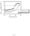
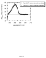
- Fig. 1 shows the near UV CD spectrum of Skp-p24/CTD (267-350), SEQ ID NO. 22.
- Fig. 2 shows the melting curve of Skp-p24/CTD (SEQ ID NO. 22).Thermally induced unfolding and refolding is monitored by near UV CD spectroscopy at 277 nm.
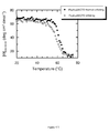
- Fig. 3 shows the near UV CD spectrum of FkpA-p24/CTD (267-350), SEQ ID NO. 21.
- Fig. 4 shows that the near UV CD signal of the native FkpA-p24/CTD molecule is fully restored after a thermally induced unfolding/refolding cycle.
Detailed description of the invention
-
HTLV p24 is a crucial antigen for the detection of anti-HTLV antibodies. The p24 capsid protein has been known in the art for a long time and has been used in immunoassays for detection of anti-HTLV antibodies (Manns et al., Blood (1991) 77: 896-905). Immunoassays for the detection of both IgG and IgM molecules require a set of antigens that are recognized and bound not only by IgG molecules but also by IgM molecules. IgM molecules typically occur in the early phase of seroconversion upon infection with HTLV. The binding of the polyvalent IgM molecules is critically dependent on a high antigen epitope density. Thus, it is imperative that antigens designed for the specific detection of IgM molecules possess and display such a high epitope density.
-
A conventional way to generate IgM detection modules with high epitope density would be to polymerize monomeric antigens by means of chemical crosslinking. There is a wealth of homobifunctional and heterobifunctional crosslinkers that may be used with great advantage and that are well known in the art. Yet, there are some severe drawbacks in the chemically induced polymerization of antigens for use as specifiers in serological assays. For instance, the insertion of crosslinker moieties into antigens may compromise antigenicity by interfering with the native-like conformation or by masking crucial epitopes. Furthermore, the introduction of non-natural tertiary contacts may interfere with the reversibility of protein folding/unfolding, and it may, additionally, be the source of interference problems which have to be overcome by anti-interference strategies in the immunoassay mixture.
-
A more recent technique of generating IgM detection modules is to fuse the antigen of interest to an oligomeric chaperone, thereby conveying high epitope density to the antigen. The advantage of this technology lies in its high reproducibility and in the triple function of the oligomeric chaperone fusion partner: firstly, the chaperone enhances the expression rate of the fusion polypeptide in the host cell, secondly, the chaperone facilitates the refolding process of the target antigen and enhances its overall solubility and, thirdly, it assembles the target antigen reproducibly into an ordered oligomeric structure.
-
European patent application publication no.
EP1982993A2 discloses a method and tools for early detection of true primary infections by pathogens such as human cytomegalovirus using antigens that are fused to oligomeric chaperones. However, this publication is silent with regard to detection of HTLV infection.
-
Our initial attempts with the full-length version of HTLV p24 had revealed that this protein exhibits high solubility when fused to EcSlyD-EcSlyD or EcFkpA as a chaperone. Its solubility was, however, limited when p24 was fused to the trimeric Skp chaperone. It is self-evident that solubility of all the compounds is a critical feature for heterogeneous immunoassay applications. Aggregation processes of proteinaceous ingredients in immunoassays usually result in both a loss of signal (due to the loss of epitopes) and a loss of specificity (due to unspecific binding of labeled antigen aggregate to the solid phase). We observed that full length p24 from HTLV - when fused to the oligomeric chaperone EcSkp - shows a tendency to aggregate in physiological buffer at ambient temperature. Thus, the full-length p24 variant was somewhat precluded from simple and straightforward applications in a sensitive IgM immunoassay.
-
Instead of focusing on the full-length version of p24, we now tried to design truncated, yet conformationally folded fragments of p24. In other words, we sought to use protein domains instead of the full-length p24 protein as the base for antigen development. A protein domain is an autonomously folding entity within a protein structure, that is, a protein domain is not dependent on other parts or regions of the protein in its folding. To date, many natural protein domains have been elucidated, ranging in size from ∼ 40 amino acid residues (WW domain) to more than 300 amino acid residues. It has also been demonstrated that very small, yet stable protein domains may be designed from the scratch: artificial polypeptide sequences with fragment lengths from 23-28 amino acid sequences have been shown to fold cooperatively and to possess the characteristic features of protein domains (Struthers, M. D. et al., Design of a monomeric 23-residue polypeptide with defined tertiary structure, Science (1996) 271 (5247) 342-345; Dahiyat, B. I. & Mayo, S.L., De novo protein design: fully automated sequence selection, Science (1997) 278 (5335) 82-87; Dahiyat, B.I. et al., De novo protein design: towards fully automated protein design, J. Mol. Biol. (1997) 273 (4) 789-796). From theoretical considerations and experimental evidence it is assumed that the minimal length requirement for a protein domain is around 25 amino acid residues (Porter L. L. & Rose, G. D., A thermodynamic definition of protein domains, PNAS (2012) 109 (24), 9420-9425).
-
In the Journal of Molecular Biology (1999) Aug. 13; 291(2):491-505, Khorasanizadeh et al. present the NMR structure of the capsid protein p24 and reveal the domain topology of this protein. According to this work, p24 from HTLV-I is largely helical and consists of two well-separated domains, i.e. p24 comprises two well-defined autonomous folding units. The N-terminal domain (NTD) harbors the helices 1-7, whereas the C-terminal domain (CTD) comprises the helices 8-12. We wondered whether it was feasible to express the two domains individually in E. coli, and whether we would be able to obtain oligomeric chaperone polypeptide fusions in a soluble and antigenic form.
-
Khorasanizadeh et al. are silent with regard to antigenic properties (e.g., B-cell epitopes) of p24 and any diagnostic applications of the NMR-characterized HTLV capsid protein. It has been unpredictable from the mere three-dimensional solution structure of the p24 capsid antigen whether its antigenicity resides mainly in the N-terminal domain (NTD) or in the C-terminal domain (CTD) or whether its B-cell epitopes are evenly spread throughout the molecule.
-
Surprisingly, we were able to express the isolated HTLV p24 domains NTD and CTD in fusion with chaperone modules such as SlyD, FkpA and Skp. As can be seen in the Examples section, all of these constructs could be purified to homogeneity, they were well soluble and we were able to assess them for their antigenicity with anti-HTLV positive human sera in an automated immunoassay analyzer. The results were quite clear-cut: antigenicity was pretty high for both domains and was even slightly higher for the C-Domain (CTD). Strikingly, NTD could be identified as precarious with respect to the blank values, which were significantly increased when compared to the CTD. CTD exhibited excellent signal dynamics in that it generated high signals with positive sera and very low signals with negative sera. This is surprising, since the CTD is presumed to harbor a natural dimerization motif needed for p24 capsid assembly. By virtue of its natural oligomerization behavior, we had reasoned that the CTD would exhibit an aggregation tendency that is significantly higher than the aggregation tendency of the NTD.
-
When we assessed p24 CTD and NTD with rabbit anti-HTLV seroconversion sera (there are no commercially available human HTLV seroconversion panels, so we had to recur to an artificial rabbit model), we found that the use of chaperone-induced oligomeric p24 variants on both sides of a DAGS assay tremendously enhances the sensitivity of the immunoassay. Seroconversion samples are recognized much better with oligomeric p24 variants than with monomeric p24 variants.
-
In brief, the C-domains of p24 from HTLV-I and HTLV-II were identified as p24 fragments with high antigenicity and high solubility. When fused to chaperones such as SlyD, FkpA or Skp, p24 CTD remains soluble, stable and is well-suited for the detection of IgM molecules which typically occur in the early phase of seroconversion upon infection with HTLV. Therefore, in particular the oligomeric FkpA and Skp fusion variants of p24 CTD may serve to enhance the sensitivity of HTLV-immunoassays.
-
We have developed variants of the capsid protein p24 from HTLV that are more soluble and significantly less aggregation-prone than the full-length p24 molecule. Solubility and stability are improved at the expense of antigenicity - nevertheless, the newly developed p24 variants hold promise as antigens in HTLV immunoassays, since they are abundantly overexpressed in E. coli, are easily purified and refolded via immobilized metal chelate chromatography (IMAC), exhibit satisfying stability properties and may be used to reliably detect anti-HTLV antibodies in human sera (presumably in combination with the ectodomain of gp21, another immunodominant protein from HTLV). It is of paramount importance that, e.g., the FkpA-p24/CTD and Skp-p24/CTD fusion proteins form natural oligomers with epitope densities that are sufficient to detect IgM molecules. Since we aimed at developing an immunoassay for total immunoglobulin detection (i.e. detection of IgG and IgM), the oligomeric species FkpA-p24/CTD and Skp-p24/CTD may be used advantageously as specifiers on both sides of a DAGS format (e.g. FkpA-p24/CTD-biotin and Skp-p24/CTD-ruthenium). Preliminary data suggest that the use of oligomeric p24 variants ensures an excellent seroconversion sensitivity which is unmatched by competitor assays.
-
The invention therefore concerns soluble HTLV p24 antigens that are fused to chaperones and their use in diagnostic applications such as immunoassays for detecting antibodies against HTLV-I or HTLV-II in an isolated biological sample. The term "HTLV" means "human T-cell lymphotropic virus". Unless specifically marked as HTLV-I or HTLV-II the term HTLV refers to both virus types.
-
As to the length of the HTLV p24 antigen, any HTLV p24 antigen fragment can be fused to a chaperone as long as it keeps its ability to bind to anti-HTLV antibodies in an isolated biological sample. This property can be checked for example by an immunoassay - the procedures of which are well-known in the art - wherein said p24 antigen is used as a specific binding partner in a pre-characterized biological sample that contains anti-p24 antibodies. As long as the immunoassay yields a positive signal the HTLV p24 antigen is suitable in the sense of the invention.
-
Preferably, the p24 antigen present in the fusion polypeptide comprises a fragment of at least 25 amino acids of SEQ ID NOs. 4 (HTLV-I p24 complete sequence) or SEQ ID NO. 8 (HTLV-II p24 complete sequence), wherein the HTLV p24 antigen fragment is fused with a chaperone. Further preferred is a HTLV p24 fragment length of at least 30, 40, 50, 60, 70 or 80 amino acid residues of SEQ ID NOs. 4 or 8. A fragment length of at least 80 to 110 or at least 80 to 115 amino acid residues is most preferred. In the sense of the invention, also the complete sequences of p24 can be applied in a fusion protein.
-
In a further preferred mode, the antigen comprises only a certain domain of the complete HTLV p24 antigen such as the N-terminal domain (NTD) or the C-terminal domain (CTD). Preferably, the antigen comprises the N-terminal domain of SEQ ID NO. 2 or the C-terminal domain of SEQ ID NO. 3 of HTLV-I p24. For the HTLV-II antigen, the fusion antigen preferably comprises the N-terminal domain of SEQ ID NO. 6 or the C-terminal domain of SEQ ID. NO. 7. In a further preferred mode, if the N-terminal domain is part of the antigen the C-terminal domain is missing and vice versa.
-
Particularly preferred is a soluble HTLV p24 antigen fused to a chaperone selected from the group consisting of SEQ ID NOs. 9 to 16 and 18 to 24.
-
The term HTLV p24 antigen encompasses also variants. HTLV p24 variants may easily be created by a person skilled in the art by conservative or homologous substitutions of the disclosed amino acid sequences (such as e.g. substitutions of a cysteine by alanine or serine). The term "variants" in this context also relates to a protein or a protein fragment (i.e. a polypeptide or peptide) substantially similar to said protein. For example, modifications such as C- or N-terminal truncations by 1 to 10 amino acids are within the scope of the claimed HTLV p24 antigens. In particular, a variant may be an isoform which shows amino acid exchanges, deletions or insertions compared to the amino acid sequence of the most prevalent protein isoform. In one embodiment, such a substantially similar protein has a sequence similarity to the most prevalent isoform of the protein of at least 80%, in another embodiment at least 85% or at least 90%, in yet another embodiment at least 95%. The term "variant" also relates to a post-translationally modifed protein such as a glycosylated or phosphorylated protein. According to the invention a variant classifies as a HTLV p24 antigen variant as long as the immunoreactivity in an in vitro diagnostic immunoassay is maintained, i.e. the variant is still able to bind and detect anti-HTLV p24 antibodies present in an isolated sample. A "variant" is also a protein or antigen which has been modified for example by covalent or non-covalent attachment of a label or carrier moiety to the protein or antigen. Possible labels are radioactive, fluorescent, chemiluminescent, electrochemiluminescent, enzymes or others e.g. like digoxigenin or biotin. These labels are known to a person skilled in the art.
-
The HTLV p24 antigens of the current invention are soluble, stable and immunoreactive, i.e. they are suitable as antigens for use in an immunological assay. This means that the antigens according to the invention are soluble under physiological buffer conditions, for example in a phosphate buffer system at ambient temperature without addition of detergents. The antigens are also capable of binding to or being recognized and bound by antibodies specific for HTLV p24, like e.g. anti- p24 antibodies present in an isolated sample such as human sera.
-
The HTLV p24 antigens according to the invention are fused to a chaperone. The term "fusion protein", "fusion polypeptide" or "fusion antigen" or simply "antigen" as used in the present invention refers to a protein comprising at least one protein part corresponding to a HTLV p24 polypeptide and at least one protein part derived from a chaperone that serves the role of a fusion partner.
-
Chaperones, which are known as classical folding helpers, are proteins that assist the folding and maintenance of the structural integrity of other proteins. Examples of folding helpers are described in detail in
WO 03/000877 . According to the invention chaperones of the peptidyl prolyl isomerase class such as chaperones of the FKBP family can be used for fusion to the HTLV p24 antigen variants. Examples of FKBP chaperones suitable as fusion partners are FkpA, SlyD and SlpA. A further chaperone suitable as a fusion partner for HTLV p24 is Skp, a trimeric chaperone from the periplasm of
E.coli, not belonging to the FKBP family. It is not always necessary to use the complete sequence of a chaperone. Functional fragments of chaperones (so-called binding-competent modules) which still possess the required abilities and functions may also be used (cf.
WO 98/13496 ).
-
According to a further embodiment of the invention at least one or at least two modules of an FKBP chaperone such as e.g. E. coli SlyD, SlpA or FkpA are used as fusion moieties for expression of the HTLV p24 antigens. The chaperone Skp may be used as a fusion partner as well. The fusion of two FKBP-chaperone domains results in improved solubility of the resulting fusion polypeptide. The fusion moieties may be located at the N-terminus or at the C-terminus or at both ends (sandwich-like) of the HTLV p24 antigen.
-
The HTLV p24 antigens according to the invention can be generated and prepared by means of recombinant DNA techniques. Another aspect of the invention therefore is a recombinant DNA molecule encoding a HTLV p24 antigen and variants thereof as defined further above.
-
The term "recombinant DNA molecule" refers to a molecule which is made by the combination of two otherwise separated segments of DNA sequence accomplished by the artificial manipulation of isolated segments of polynucleotides by genetic engineering techniques or by chemical synthesis. In doing so one may join together polynucleotide segments of desired functions to generate a desired combination of functions. Recombinant DNA techniques for expression of proteins in prokaryotic or lower or higher eukaryotic host cells are well known in the art. They have been described e.g. by Sambrook et al., (1989, Molecular Cloning: A Laboratory Manual)
-
The recombinant DNA molecules according to the invention may also contain sequences encoding linker peptides of 10 to 100 amino acid residues in between the HTLV p24 antigen and the fusion moieties and also between several fusion moieties. Such a linker sequence may for example harbor a proteolytic cleavage site.
-
A further aspect of the invention is an expression vector comprising operably linked a recombinant DNA molecule according to the present invention, i.e., a recombinant DNA molecule encoding an HTLV p24 antigen and a peptidyl prolyl isomerase chaperone, such as an FKBP-chaperone, wherein the FKBP-chaperone is selected from FkpA, SlyD and SlpA. In an alternative embodiment the recombinant DNA molecule encodes a fusion protein comprising an HTLV p24 antigen and Skp. The expression vector comprising a recombinant DNA according to the present invention may be used to express the HTLV p24 antigen in a cell free translation system or may be used to transform a host cell for expression of the HTLV p24 antigen according to methods well known in the art. Another aspect of the invention therefore relates to a host cell transformed with an expression vector according to the present invention. In one embodiment of the current invention the recombinant HTLV p24 antigens are produced in E. coli cells.
-
An additional aspect is a method for producing a soluble, stable and immunoreactive HTLV p24 antigen that is produced as a fusion protein containing the HTLV p24 antigen and a chaperone. Preferably, a chaperone such as Skp or a peptidyl prolyl isomerase class chaperone like an FKBP chaperone is used. In a further embodiment of the invention said FKBP chaperone is selected from the group consisting of SlyD, FkpA and SlpA.
-
This method comprises the steps of
- a) culturing host cells transformed with the above-described expression vector containing a gene encoding an HTLV p24 antigen
- b) expression of the gene encoding said HTLV p24 antigen
- c) purification of said HTLV p24 antigen.
-
Optionally, as an additional step d), functional solubilization needs to be carried out so that the HTLV p24 antigen is brought into a soluble and immunoreactive conformation by means of refolding techniques known in the art.
-
An additional aspect of the present invention concerns a method for the detection of anti-HTLV antibodies in an isolated human sample wherein an HTLV p24 antigen according to the invention is used as a binding partner for the antibodies. The invention thus covers a method for the detection of antibodies specific for HTLV in an isolated sample, said method comprising
- a) forming an immunoreaction admixture by admixing a body fluid sample with an HTLV p24 antigen according to the invention
- b) maintaining said immunoreaction admixture for a time period sufficient for allowing antibodies against said HTLV p24 antigen present in the body fluid sample to immunoreact with said HTLV p24 antigen to form an immunoreaction product; and
- c) detecting the presence and/or the concentration of any of said immunoreaction product.
-
In a further aspect said method is suitable for detecting HTLV antibodies of the IgG and the IgM subclass.
-
Immunoassays for detection of antibodies are well known in the art, and so are methods for carrying out such assays and practical applications and procedures. The HTLV p24 antigens according to the invention can be used to improve assays for the detection of anti-HTLV antibodies independently of the labels used and independently of the mode of detection (e.g., radioisotope assay, enzyme immunoassay, electrochemiluminescence assay, etc.) or the assay principle (e.g., test strip assay, sandwich assay, indirect test concept or homogenous assay, etc.). All biological liquids known to the expert can be used as isolated samples for the detection of anti-HTLV antibodies. The samples usually used are bodily liquids like whole blood, blood sera, blood plasma, urine or saliva.
-
A further embodiment of the invention is an immunoassay for detecting anti-HTLV antibodies in an isolated sample performed according to the so-called double antigen sandwich concept (DAGS). Sometimes this assay concept is also termed double antigen bridge concept, because the two antigens are bridged by an antibody analyte. In such an assay the ability of an antibody to bind at least two different molecules of a given antigen with its two (IgG, IgE), four (IgA) or ten (IgM) paratopes is required and utilized.
-
In more detail, an immunoassay for the determination of anti-HTLV antibodies according to the double antigen bridge format is carried out by incubating a sample containing the anti-HTLV antibodies with two different HTLV p24 antigens, i.e. a first ("solid phase") HTLV p24 antigen and a second HTLV p24 ("detection") antigen, wherein each of the said antigens binds specifically to said anti-HTLV antibodies. The first antigen can be bound directly or indirectly to a solid phase and usually carries an effector group which is part of a bioaffine binding pair like, e.g., biotin and avidin. For example, if the first antigen is conjugated to biotin the solid phase is coated with either avidin or streptavidin. The second antigen carries a label. Thus an immunoreaction admixture is formed comprising the first antigen, the sample antibody and the second antigen. A solid phase to which the first antigen can be bound is added either before the addition of the sample to said antigens or after the immunoreaction admixture is formed. This immunoreaction admixture is maintained for a time period sufficient for allowing anti-HTLV antibodies against said HTLV p24 antigens in the body fluid sample to immunoreact with said HTLV p24 antigens to form an immunoreaction product. Next step is a separation step wherein the liquid phase is separated from the solid phase. Finally, the presence of any of said immunoreaction product is detected in the solid or liquid phase or both.
-
In said DAGS immunoassay the basic structures of the "solid phase antigen" and the "detection antigen" are essentially the same. It is also possible to use, in a double antigen bridge assay, similar but different HTLV p24 antigens, which are immunologically cross-reactive. The essential requirement for performing such assays is that the relevant epitope or the relevant epitopes are present on both antigens. According to the invention it is possible to use different fusion moieties for each HTLV p24 antigen (e.g. SlyD fused to HTLV p24 on the solid phase side and FkpA p24 fused to HTLV p24 on the detection side) as such variations significantly alleviate the problem of non-specific binding and thus mitigate the risk of false-positive results.
-
Preferably, in said DAGS immunoassay an asymmetric format is applied, combining an HTLV p24 fused to FkpA and an HTLV p24 antigen fused to Skp. More preferably, the HTLV p24 fused to FkpA is used on the solid phase side and the HTLV p24 fused to Skp is applied on the detection side. Most preferably, the HTLV p24 FkpA fusion protein carries a biotin moiety for attachment to a solid phase that has been coated with streptavidin or avidin and the HTLV p24 fusion protein carries an electrochemiluminescent label such as ruthenium complexes.
-
A further embodiment of the present invention is therefore an immunoassay according to the double antigen bridge concept wherein a first HTLV p24 antigen according to the present invention, and a second HTLV p24 antigen according to the present invention are used.
-
The present invention further relates to the use of at least one antigen of HTLV p24 in a diagnostic test for the detection of anti-HTLV antibodies.
-
An additional subj ect matter of the invention is a reagent kit for the detection of antibodies against HTLV, containing, in addition to the usual test additives for immunoassays, at least one antigen of the HTLV p24 antigens according to the invention suitable for specifically binding to HTLV antibodies to be determined and possibly carrying a label as well as other usual additives if necessary.
-
In particular the reagent kit contains an HTLV p24 antigen according to any of SEQ ID NOs. 9 to 16 and 18 to 24.
-
In addition, the reagent kits defined above contain controls and standard solutions as well as reagents in one or more solutions with the common additives, buffers, salts, detergents etc. as used by the average man skilled in the art along with instructions for use.
-
Another embodiment is a composition of HTLV antigens comprising an HTLV gag antigen (p24) according to the current invention and an HTLV env antigen, preferably gp21. Preferred is a composition comprising the C-terminal domains of HTLV p24, particularly preferred is a composition comprising an HTLV-I p24 antigen according to SEQ ID NO. 3 and/or an HTLV-II p24 antigen according to SEQ ID NO. 7 and HTLV gp21. For example, in said composition an HTLV gp21 sequence comprising any of SEQ ID NOs. 25, 26 or 27 can be present. For application in an immunoassay according to the DAGS format the composition comprises each HTLV antigen in two forms, i.e. in a form that enables the antigen to be attached to a solid phase (e.g. a biotinylated antigen that can bind to a surface coated with streptavidin) and in a labeled form that enables detection of the immunocomplex between HTLV antibodies present in the sample and the applied HTLV antigens.
-
The invention also concerns the use of a HTLV p24 antigen according to the invention in an in vitro diagnostic test for the detection of anti-HTLV antibodies.
-
The invention is further illustrated by the Examples.
Example 1
Cloning and purification of p24 capsid fusion polypeptides
Cloning of expression cassettes
-
On the basis of the pET24a expression plasmid of Novagen (Madison, WI, USA), expression cassettes encoding p24 fusion proteins from HTLV-I and HTLV-II were obtained essentially as described (Scholz, C. et al., J. Mol. Biol. (2005) 345, 1229-1241). The sequences of the p24 antigens from HTLV-I and HTLV-II were retrieved from the SwissProt database (SwissProt ID P10274 and P03353, respectively). A synthetic gene encoding p24 capsid antigen aa 146-344 (numbering refers to the Gag-Pro polyprotein precursor) from HTLV-I (lacking the proline-rich 15 amino acids at the N-terminus of the mature capsid protein) with a glycine-rich linker region fused in frame to the N-terminus was purchased from Medigenomix (Martinsried, Germany). The cysteine residues of p24 at positions 193, 311 and 332 were changed to alanine residues in order to prevent unwanted side-effects such as oxidation or intermolecular disulfide bridging. BamHI and XhoI restriction sites were at the 5' and the 3' ends of the p24-coding region, respectively. A further synthetic gene encoding two EcSlyD units (residues 1-165 of SwissProt accession no. POA9K9) connected via a glycine-rich linker region and encompassing part of a further linker region at the C-terminus were likewise purchased from Medigenomix. NdeI and BamHI restriction sites were at the 5' and 3' ends of this cassette, respectively. The genes and the restriction sites were designed to enable the in frame fusion of the chaperone part EcSlyD-EcSlyD and the p24 antigen part by simple ligation. In order to avoid inadvertent recombination processes and to increase the genetic stability of the expression cassette in the E. coli host, the nucleotide sequences encoding the EcSlyD units were degenerated as were the nucleotide sequences encoding the extended linker regions. i.e., different codon combinations were used to encode identical amino acid sequences.
-
The pET24a vector was digested with NdeI and XhoI and the cassette comprising tandem-SlyD fused in frame to HTLV-I p24 (146-344) was inserted. Expression cassettes comprising Pasteurella multocida SlyD (1-156, SwissProt ID Q9CKP2) E. coli Skp (21-161, SwissProt ID P0AEU7) or E. coli FkpA (26-270, SwissProt ID P45523) were constructed accordingly, as well as expression cassettes comprising p24 and p24 fragments from HTLV-II (SwissProt ID P03353). As with p24 from HTLV-I, the genuine cysteine residues of p24 from HTLV-II at positions 199, 281, 317 and 338 (again, numbering refers to the precursor Gag-Pro polyprotein) were changed to alanine residues in order to prevent unwanted side-effects such as oxidation or intermolecular disulfide bridging. All recombinant fusion polypeptide variants contained a C-terminal hexahistidine tag to facilitate Ni-NTA-assisted purification and refolding. QuikChange (Stratagene, La Jolla, CA, USA) and standard PCR techniques were used to generate point mutations, deletion, insertion and extension variants or restriction sites in the respective expression cassettes.
-
The drawing below shows a scheme of the N-terminally truncated HTLV-I p24 antigen 146-344 bearing two SlyD chaperone units fused in frame to its N-terminal end. To denote the
E. coli origin of the SlyD fusion partner, the depicted fusion polypeptide has been named
EcSlyD-
EcSlyD-p24 (146-344).
-
The insert of the resulting plasmid was sequenced and found to encode the desired fusion protein. The complete amino acid sequences of the p24 fusion polypeptides from HTLV-I and HTLV-II are shown in SEQ ID NOs. 9 to 16 and 18 to 24. The amino acid sequence of the linker L is shown is SEQ ID NO. 17.
-
Purification of fusion proteins comprising p24 and p24 variants from HTLV-I and HTLV-II
-
All p24 fusion protein variants were purified by using virtually identical protocols. E. coli BL21 (DE3) cells harboring the particular pET24a expression plasmid were grown at 37°C in LB medium plus kanamycin (30 µg/ml) to an OD600 of 1.5, and cytosolic overexpression was induced by adding 1 mM isopropyl-ß-D-thiogalactoside. Three hours after induction, cells were harvested by centrifugation (20 min at 5000 g), frozen and stored at -20°C. For cell lysis, the frozen pellet was resuspended in chilled 50 mM sodium phosphate pH 8.0, 7.0 M GdmCl, 5 mM imidazole and the suspension was stirred for 2 h on ice to complete cell lysis. After centrifugation and filtration (0.45 µm/0.2 µm), the crude lysate was applied onto a Ni-NTA column equilibrated with the lysis buffer including 5.0 mM TCEP. The subsequent washing step was tailored for the respective target protein and ranged from 5 to 15 mM imidazole (in 50 mM sodium phosphate pH 8.0, 7.0 M GdmCl, 5.0 mM TCEP). At least 10-15 volumes of the washing buffer were applied. Then, the GdmCl solution was replaced by 50 mM potassium phosphate pH 8.0, 100 mM KCl, 10 mM imidazole, 5.0 mM TCEP to induce conformational refolding of the matrix-bound protein. In order to avoid reactivation of copurifying proteases, a protease inhibitor cocktail (Complete® EDTA-free, Roche) was included in the refolding buffer. A total of 15-20 column volumes of refolding buffer were applied in an overnight reaction. Then, both TCEP and the Complete® EDTA-free inhibitor cocktail were removed by washing with 3-5 column volumes 50 mM potassium phosphate pH 8.0, 100 mM KCl, 10 mM imidazole. Subsequently, the imidazole concentration - still in 50 mM potassium phosphate pH 8.0, 100 mM KCl - was raised to 20 - 80 mM (depending on the respective target protein) in order to remove unspecifically bound protein contaminants. The native protein was then eluted by 500 mM imidazole in the same buffer. Protein-containing fractions were assessed for purity by Tricine-SDS-PAGE and pooled. Finally, the proteins were subjected to size-exclusion-chromatography (Superdex HiLoad, Amersham Pharmacia) and the protein-containing fractions were pooled and concentrated to 10-20 mg/ml in an Amicon cell (YM10).
-
After the coupled purification and refolding protocol, protein yields of roughly 10-30 mg could be obtained from 1 g of E. coli wet cells, depending on the respective target protein.
Example 2
Spectroscopic measurements
-
Protein concentration measurements were performed with an Uvikon XL double-beam spectrophotometer. The molar extinction coefficients (ε
280) were determined by using the procedure described by
Pace (1995), Protein Sci. 4, 2411-2423. The molar extinction coefficients (ε
M280) used for the distinct fusion polypeptides are specified in table 1.
Table 1: Protein parameters of the p24 fusion polypeptide variants generated and used in this study. All parameters are referring to the respective protein monomers. | fusion protein | length of target protein (aa residues) | molecular weight of fusion polypeptide (Da) | pI | εM280 M-1 cm-1 | Abs 0.1% (= 1 mg/ml) |
| p24 variants HTLV-I | | | | | |
| EcSlyD-EcSlyD-p24 | 146-344 | 61762 | 5.0 | 35870 | 0.581 |
| EcFkpA-p24 | 146-344 | 50840 | 6.8 | 39880 | 0.784 |
| EcSkp-p24 | 146-344 | 40306 | 9.1 | 25440 | 0.631 |
| EcSlyD-EcSlyD-p24/CTD | 258-344 | 49311 | 4.9 | 25900 | 0.525 |
| EcFkpA-p24/CTD | 258-344 | 38389 | 7.1 | 29910 | 0.779 |
| EcSkp-p24/CTD | 258-344 | 27855 | 9.3 | 15470 | 0.555 |
| EcSlyD-EcSlyD-p24/NTD | 146-260 | 52486 | 4.8 | 21890 | 0.417 |
| EcFkpA-p24/NTD | 146-260 | 41565 | 6.5 | 25900 | 0.623 |
| EcSkp-p24/NTD | 146-260 | 31031 | 9.0 | 11460 | 0.369 |
| P24 variants HTLV-II | | | | | |
| EcSlyD-EcSlyD-p24 | 152-350 | 61868 | 5.0 | 35870 | 0.580 |
| EcFkpA-p24 | 152-350 | 50946 | 7.2 | 39880 | 0.783 |
| EcSkp-p24 | 152-350 | 40412 | 9.2 | 25440 | 0.630 |
| EcFkpA-p24/CTD | 267-350 | 38120 | 7.1 | 29910 | 0.785 |
| EcSkp-p24/CTD | 267-350 | 27586 | 9.3 | 15470 | 0.561 |
| EcFkpA-p24/NTD | 152-266 | 41739 | 6.7 | 25900 | 0.621 |
| EcSkp-p24/CTD | 152-266 | 31205 | 9.2 | 11460 | 0.367 |
-
The amino acid sequences of the fusion polypeptide variants are shown in SEQ ID NOs. 9 to 16 and 18 to 24.
Example 3
Coupling of biotin and ruthenium moieties to the fusion proteins
-
The lysine ε-amino groups of the fusion polypeptides were modified at protein concentrations of 10-30 mg/ml with N-hydroxy-succinimide activated biotin and ruthenium label molecules, respectively. The label/protein ratio varied from 2:1 to 5:1 (mol:mol), depending on the respective fusion protein. The reaction buffer was 150 mM potassium phosphate pH 8.0, 100 mM KCl, 0.5 mM EDTA. The reaction was carried out at room temperature for 15 min and stopped by adding buffered L-lysine to a final concentration of 10 mM. To avoid hydrolytic inactivation of the labels, the respective stock solutions were prepared in dried DMSO (seccosolv quality, Merck, Germany). DMSO concentrations up to 25% in the reaction buffer were well tolerated by all fusion proteins studied. After the coupling reaction, unreacted free label was removed by passing the crude protein conjugate over a gel filtration column (Superdex 200 HiLoad).
Example 4
-
Immunological reactivity (i.e., antigenicity) of different p24 capsid antigen variants in a HTLV immunoassay
-
The immunological reactivity (antigenicity) of the polypeptide fusion variants of HTLV p24 capsid antigen was assessed in automated Elecsys® 2010 and cobas e 411 analyzers (Roche Diagnostics GmbH). Elecsys® is a registered trademark of the Roche group. Measurements were carried out in the double antigen sandwich format.
-
Signal detection in Elecsys® 2010 and cobas e 411 is based on electrochemiluminescence. The biotin-conjugate (i.e. the capture-antigen) is immobilized on the surface of a streptavidin coated magnetic bead whereas the detection-antigen bears a complexed Ruthenium cation (switching between the redox states 2+ and 3+) as the signaling moiety. In the presence of a specific immunoglobulin analyte, the chromogenic ruthenium complex is bridged to the solid phase and emits light at 620 nm after excitation at a platinum electrode. The signal output is in arbitrary light units.
-
The recombinant p24 capsid antigen fusion polypeptides were assessed in a double antigen sandwich (DAGS) immunoassay format. To this end, recombinant HTLV-I capsid antigen p24 was used as a biotin and a ruthenium conjugate, respectively, to detect anti-p24 antibodies in human sera.
-
p24 is one of the immunodominant antigens of HTLV, and soluble variants of p24 - as disclosed in this patent application - are invaluable tools for the detection of HTLV infections. In all measurements, chemically polymerized and unlabeled EcSlyD-EcSlyD, EcFkpA and EcSkp were implemented in large excess (∼ 10 µg/ml) in the reaction buffer as anti-interference substances to avoid immunological cross reactions via the chaperone fusion units.
-
In particular, three p24 variants from HTLV-I were scrutinized in this study, namely full length p24 (146-344, numbering refers to Gag-Pro polyprotein precursor, see SEQ ID NOs. 1 and 5), p24 N-terminal domain (NTD, 146-260) and p24 C-terminal domain (CTD, 261-344). In order to detect anti-p24 IgG molecules, EcSlyD-EcSlyD-p24-biotin and EcSlyD-EcSlyD-p24-ruthenium were used in R1 (reagent buffer 1) and R2 (reagent buffer 2), respectively. In order to detect both anti-p24 IgM and IgG molecules, EcFkpA-p24-biotin and EcSkp-p24-ruthenium were used in R1 (reagent buffer 1) and R2 (reagent buffer 2), respectively. The concentrations of the antigen conjugates in R1 and R2, respectively, were 100 ng/ml each. N-terminally truncated mature p24 (146-344) was used as an EcSlyD-EcSlyD fusion polypeptide on the biotin-side and as an EcFkpA fusion polypeptide on the ruthenium side.
-
Unfortunately, human HTLV seroconversion panels - which are an indispensable tool for the development of improved in vitro diagnostic assays - are not available commercially. In order to assess the antigenic properties of the different p24 variants in the very early phase of HTLV infection, we had to recur to rabbit sera serving as a seroconversion model. To this end, New Zealand White Rabbits were immunized with purified and inactivated HTLV-I and -II viral lysates (purchased from Zeptometrix, New York, USA) and complete Freund's adjuvant to induce an immune response (2 immunizations, 1 week interval). We are aware that the pattern of the humoral immune response upon true HTLV infections in man might slightly differ from an immune response triggered by virus lysate vaccination of rabbits. Yet, the artificially induced rabbit seroconversion is the best mimic that was available to us.
-
In a first experiment, monomeric p24 CTD (p24, 261-344) was assessed with anti-HTLV-negative human sera in the aforementioned DAGS immunoassay setup in order to get an idea of the background signal. The unavoidable system-inherent signal is around 500 counts. Low background signals are indicative of high solubility and generally benign physicochemical properties of the respective ruthenium conjugates. From table 2 we can infer that the physicochemical properties of monomeric p24 CTD are excellent (column 1). This holds true for oligomeric p24 CTD as well (column 2): FkpA-p24(261-344)-biotin and Skp-p24(261-344)-ruthenium, when used as an antigen pair in the DAGS format, lead to a signal background of ∼ 1100 counts with negative human sera, which clearly points to good solubility properties. However, it becomes evident at first glance that the monomeric and the oligomeric form of p24 CTD strongly differ in their capability to detect anti-HTLV-antibodies (and notably IgM molecules) in seroconversion panels as shown in Table 2. Having a closer look at seroconversion K5645, we find that monomeric p24 CTD barely detects day 18 as positive (1558 counts), whereas the use of oligomeric p24 CTD already reveals day 14 as clearly positive (8232 counts) and leads to a signal as high as 50118 counts at day 18. We see the same picture with seroconversion panels K5646, K5647 and K5648: the oligomeric p24 CTD variant produces higher signals at earlier times and thus warrants an excellent sensitivity in the early detection of anti-p24 antibodies in seroconversions. In principle, the situation is similar with the N-terminal domain (NTD) of p24, which encompasses the amino acid residues 146-260 (numbering refers to Gag-Pro polyprotein precursor). As with the CTD, the oligomeric form of p24 NTD is better suited to detect antibodies that appear in the early phase of seroconversion (i.e., immunoglobulins of the M-type), which is exemplified in particular with the seroconversion panels K5647 and K5648 (Table 2, columns 3 and 4). However, the background signals of the oligomeric NTD p24 are significantly increased when compared to CTD p24. In addition, the antigenicity of the C-terminal domain of p24 seems to outdistance the antigenicity of the N-terminal domain. In conclusion, the oligomeric C-terminal domain of p24 possesses outstanding physicochemical and superior antigenic properties making it an attractive candidate for HTLV serology. It is clearly superior to full-length p24 (146-344, numbering of Gag-pro polyprotein precursor) in terms of sensitivity in early IgM detection. Since the Skp fusion polypeptide of full length p24 (146-344) was not available since it significantly tended to aggregate, we were confined to SlyD-SlyD and FkpA fusion polypeptides of the p24 full-length version. When monomeric SlyD-SlyD-p24 (146-344) is used on the biotin side and oligomeric FkpA-p24 (146-344) is used on the ruthenium side of the DAGS format, the results are quite clear-cut: full-length p24 yields excellent signals in the late phases of the seroconversion panels, but it completely fails in early detection (table 2, column 5). Both oligomeric CTD p24 and NTD p24 are superior to the monomeric full-length variant, providing good evidence that sensitive early detection is mainly dependent on the epitope density of the p24 fragments used. It does not seem to be mandatory to offer the complete p24 sequence as a whole to get an excellent seroconversion sensitivity. Rather, epitopes in the N and C-domain of p24 are sufficient to warrant a sensitive and reliable detection of IgM molecules in the early phase of HTLV infection - provided that these epitopes are offered in an oligomeric form. By virtue of its superior solubility (as reflected in the low background signals) and its outstanding antigenicity, the C-terminal domain of p24 from HTLV-I holds promise as an invaluable ingredient in a HTLV immunoassay. This was somewhat unexpected: since the C-domain of the p24 capsid antigen is presumably involved in p24 oligomerization (
Khorasanizadeh et al., J. Mol. Biol. (1999) 291, 491-505), we reasoned that the isolated C-domain could possibly tend to aggregation, at least it should be more difficult to handle than the N-domain. Moreover, our expectation was that the p24 C-domain which is largely hidden in the mature capsid particles would probably harbor less immunodominant epitopes than the well-accessible N-domain. To our surprise, the converse is true. Indeed, the N-domain of p24 is also well-suited as an antigen for HTLV-immunoassay, albeit it seems inferior to the C-terminal domain in terms of solubility and antigenicity. Table 2 shows the results for p24 variants from HTLV-I. We found virtually identical results for the corresponding p24 variants from HTLV-II. This is in line with our expectations since the amino acid sequences of p24 from HTLV-I and HTLV-II share 84 % identity and 93 % homology. The corresponding sequences for p24 from HTLV-II were 152-266 (N-domain, NTD), 267-350 (C-domain, CTD) and 152-350 (mature full-length p24), see also SEQ ID NOs. 5-8.
Table 2: superior immunoreactivity of oligomeric p24 variants in early HTLV infections (increased sensitivity in rabbit seroconversion panels). | p24 variant (fragment length) | mono CTD (261-344) | oligo CTD (261-344) | mono NTD (146-260) | oligo NTD (146-260) | Full-length p24 (146-344) |
| fusion partner R1 (Bi) | SlyD-SlyD | FkpA | SlyD-SlyD | FkpA | SlyD-SlyD |
| fusion partner R2 (Ru) | SlyD-SlyD | Skp | SlyD-SlyD | Skp | FkpA |
| conc. (ng/ml) | 100 | 100 | 100 | 100 | 300 |
| counts in Elecsys analyzer (cobas e 411) | | | | | |
| anti-HTLV-negative sera | | | | | |
| 0701.1201.01 | 599 | 976 | 667 | 2846 | 1677 |
| 0701.1202.01 | 611 | 1116 | 724 | 4331 | 1981 |
| 0701.1203.01 | 592 | 1148 | 717 | 4860 | 1933 |
| seroconversion panels (day of bleeding) | | | | | |
| K5645 (day 0) | 725 | 1037 | 790 | 2330 | 1608 |
| K5645 (day 10) | 612 | 1196 | 758 | 2549 | 1613 |
| K5645 (day 14) | 642 | 8232 | 729 | 2690 | 2910 |
| K5645 (day 18) | 1558 | 50118 | 906 | 3071 | 15191 |
| | | | | | |
| K5646 (day 0) | 592 | 1045 | 728 | 2359 | 1580 |
| K5646 (day 11) | 636 | 1396 | 770 | 2779 | 1665 |
| K5646 (day 15) | 1425 | 13090 | 740 | 3084 | 1715 |
| K5646 (day 19) | 14080 | 106376 | 4342 | 6321 | 8832 |
| K5646 (day 23) | 109285 | 160403 | 33361 | 15881 | 76212 |
| | | | | | |
| K5647 (day 0) | 814 | 917 | 799 | 2295 | 1445 |
| K5647 (day 12) | 2620 | 95130 | 1100 | 19920 | 3606 |
| K5647 (day 16) | 159796 | 61639 | 19774 | 88453 | 339050 |
| K5647 (day 20) | 187997 | 63193 | 62381 | 99227 | 623586 |
| | | | | | |
| K5648 (day 0) | 572 | 848 | 737 | 2113 | 1467 |
| K5648 (day 10) | 803 | 1003 | 871 | 2562 | 1512 |
| K5648 (day 14) | 2575 | 122993 | 972 | 4324 | 2733 |
| K5648 (day 18) | 10107 | 181988 | 4401 | 21689 | 10892 |
| K5648 (day 22) | 58656 | 352195 | 7844 | 16692 | 48125 |
Example 5
Combinations of oligomeric chaperone carrier proteins in an asymmetric double antigen sandwich format.
-
The immunoassay was essentially performed as described in Example 4. Oligomeric chaperones such as FkpA and Skp may be used advantageously as fusion partners in order to achieve a functional oligomerization of their respective client antigens. Fusion of FkpA or Skp to target proteins (i.e., their client or guest antigens) may yield well-defined oligomeric fusion polypeptides which are suited for the detection of IgM molecules in immunoassays. Here, we addressed the question whether there is a particularly preferred combination of Skp-X and FkpA-X fusion polypeptides when used in a DAGS (double antigen sandwich) format. (Note: "X" generally refers to any target protein or antigen.) In other words, we addressed the question whether it is advisable to use FkpA-X fusion polypeptides on the (capture) biotin side rather than on the (signaling) ruthenium side. Conversely, we wondered whether it is advisable to use Skp-X fusion polypeptides on the (signaling) ruthenium side rather than on the (capture) biotin side.
-
In a first experiment, biotin and ruthenium conjugates of purified recombinant EcSkp and EcFkpA were prepared as described in example 3. That is, the purified chaperones (i. e., the naked chaperones without any fused target sequences) were biotinylated by means of an N-hydroxy-succinimide-activated biotin label. As well, ruthenylated chaperones (i.e., the naked chaperones without any fused target sequences) were produced by means of an N-hydroxy-succinimide-activated ruthenium label.
-
Then, a symmetric DAGS was performed with
EcSkp on both the biotin and the ruthenium side at varying concentrations. As a sample, a pool of anti-HTLV negative human sera was used, and measurements were carried out in duplicate. It is obvious from the data in table 3, that the background signal is fairly high when using the very same oligomeric chaperone (here:
EcSkp) on both the biotin and the ruthenium side of a DAGS format, even at very low concentrations. The background signal strongly increases with the conjugate concentration in a dose-dependent fashion. Thus, a symmetric DAGS format does not seem to be a viable option when using an oligomeric fusion partner such as the trimeric chaperone Skp from
E. coli. Similar results have been found for the dimeric chaperone FkpA.
Table 3: Use of the very same oligomeric chaperone on both sides of a DAGS immunoassay (oligomeric carrier protein in symmetric DAGS format) | Experiment | V1 | V2 | V3 | V4 | V5 | V6 |
| | | | | | | |
| R1 basis buffer | R1 |
| Skp-Bi conc. [ng/ml] | 10 | 20 | 50 | 100 | 250 | 500 |
| R2 basis buffer | R2 |
| Skp-Ru conc. [ng/ml] | 10 | 20 | 50 | 100 | 250 | 500 |
| sample | Signal (counts) | Signal (counts) | Signal (counts) | Signal (counts) | Signal (counts) | Signal (counts) |
| Control 1 (Pool of anti-HTLV-negative human sera) | 3969 | 6301 | 12582 | 17521 | 21471 | 21498 |
| | 3921 | 6170 | 13187 | 17610 | 21467 | 22056 |
-
However, when we combined
EcSkp and
EcFkpA in an asymmetric DAGS format, the picture turned out completely different (see table 4 below). Irrespective of the combination of the chaperones, the background signals were substantially reduced when we used different chaperones on the capture and the signaling side. For instance, the background signal in the symmetric (
i.e., Skp-Bi/Skp-Ru) DAGS format was around 21,000 counts at a conjugate concentration of 250 ng/ml each. At the very same conjugate concentration, the background signal in the asymmetric DAGS format is dramatically reduced to 2,700 counts (Skp-Bi/FkpA-Ru) and to 860 counts (FkpA-Bi/Skp-Ru), respectively. It is self-evident at first glance that there is, indeed, a preferred combination of FkpA and Skp in a DAGS immunoassay: it is advisable to use FkpA as a biotin conjugate and Skp as a ruthenium conjugate in a DAGS immunoassay, and it is reasonable to conclude that the same holds true for FkpA-X and Skp-X fusion polypeptides.
Table 4: Use of different oligomeric chaperones on both sides of a DAGS immunoassay (oligomeric carrier proteins in asymmetric DAGS format) | R1 basis buffer | R1 |
| R1 | Skp-Bi | Skp-Bi | FkpA-Bi | FkpA-Bi |
| conc, [ng/ml] | 10 | 250 | 10 | 250 |
| R2 basis buffer | R2 |
| R2 | FkpA-Ru | FkpA-Ru | Skp-Ru | Skp-Ru |
| conc, [ng/ml] | 10 | 250 | 10 | 250 |
| sample | Signal (counts) | Signal (counts) | Signal (counts) | Signal (counts) |
| Control 1 (Pool of anti-HTLV negative human sera) | 567 | 2668 | 447 | 854 |
| | 561 | 2764 | 439 | 864 |
Example 6
CD-detected thermally induced unfolding of Skp-p24/CTD (267-350) and FkpA-p24/CTD (267-350)
-
Near-UV CD spectra were recorded with a Jasco-720 spectropolarimeter with a thermostatted cell holder and converted to mean residue ellipticity. The buffer was 150 mM potassium phosphate pH 8.0, 100 mM KCl, 0.5 mM EDTA. The pathlength was 0.2 cm, the protein concentration was 218 µM (referring to Skp-p24 monomer) or 147.5 µM (referring to FkpA-p24 monomer). The measuring range was 250 - 330 nm, the band width was 1.0 nm, the scanning speed was 20 nm/min at a resolution of 0.5 nm and the response was 1 s. In order to improve the signal-to-noise ratio, spectra were measured nine times and averaged.
-
Circular dichroism spectroscopy (CD) is the method of choice to assess both the secondary and the tertiary structure of proteins. Ellipticity in the aromatic region (250-330 nm) reports on tertiary contacts within a protein (i.e., the globular structure of a regularly folded protein) and is considered as the fingerprint region of a native-like fold (conformation).
-
Near UV CD spectra of Skp-p24/CTD(267-350) and FkpA-p24/CTD(267-350), SEQ ID NO. 22 and 21, respectively, were monitored to address the question whether the fusion proteins adopt an ordered conformation after the matrix-coupled refolding procedure which is the crucial step in the purification process. The answer is quite clear-cut: the near UV CD signals of both Skp-p24/CTD (see Figure 1) and FkpA-p24/CTD (see Figure 3) unequivocally report an orderly tertiary structure of the respective fusion polypeptide. Obviously, the aromatic residues of Skp-p24/CTD and FkpA-p24/CTD are embedded in the lipophilic protein core and thus experience asymmetric surroundings which strongly points to a native-like conformation of the carrier and target protein component within the respective fusion construct. The near UV CD spectrum of Skp-p24/CTD exhibits a negative signal with maxima at 282 and 277 nm (Figure 1). The near UV CD spectrum of FkpA-p24/CTD exhibits a positive signal with a maximum at 280 nm (Figure 3).
-
In order to address the question whether the thermally induced unfolding of Skp-p24/CTD and FkpA-p24/CTD is reversible, melting curves were monitored in the near UV region at detection wavelengths of 277 and 280 nm, respectively. The temperature range was 20-75 °C, the band width was 2.0 nm, the temperature slope was 1°C/min and the response was 2 s.
-
The thermally-induced unfolding was monitored at 277 and 280 nm, corresponding to the maximal signal amplitudes for Skp-p24/CTD and FkpA-p24/CTD, respectively. Upon heating, the non-covalent contacts which stabilize the native conformation of the fusion polypeptide molecules become loose and finally break down. For Skp-p24/CTD, this thermally induced unfolding (as monitored at 277 nm) is reflected in an increase in the CD signal as shown in Figure 2. Skp-p24/CTD obviously retains its native-like fold and its trimeric structure up to 55°C. The onset of unfolding is between 55°C and 60°C. At 70°C, the molecule is completely unfolded, as judged by the melting curve in Figure 2. Strikingly, the CD signal is restored when the protein solution is chilled down to 20°C (Figures 1, 2). Yet, the hysteresis of the refolding curve is pronounced and probably points to different pathways of unfolding and refolding. It is astounding that the thermally induced unfolding of a complex trimeric fusion protein such as Skp-p24/CTD is - at least partially - a reversible process. It would have been expected that Skp-p24/CTD, after thermally induced unfolding and dissociation into the monomeric subunits, would aggregate very quickly and quantitatively at an elevated temperature such as 75°C. Yet, we find that Skp-p24/CTD is obviously able to readopt its native-like conformation when the protein solution is chilled to 20°C. Indeed, the near UV CD spectra monitored before and after the thermally induced unfolding virtually superimpose (see Figure 1). In conclusion, Skp-p24/CTD possesses robust folding properties which are outstanding for a molecule with this degree of complexity and which are highly desired for an antigen that is used in an immunoassay. We found very similar results for FkpA-p24/CTD: just like Skp-p24/CTD, FkpA-p24/CTD exhibits a marked CD signal in the near UV region (250-330 nm, signal maximum at 280 nm), pointing to a well-ordered conformation after the matrix-coupled refolding process (Figure 3). The CD signal of FkpA-p24/CTD strongly decreases when the molecule unfolds and loses its tertiary structure (Figure 4). By means of thermal transitions we observed that FkpA-p24/CTD indeed retains its native-like conformation at temperatures up to 55°C. The onset of unfolding - as monitored by near UV CD spectroscopy at 280 nm - is around 60°C, and at 70°C FkpA-p24/CTD is fully unfolded. It is remarkable that the CD signal of the native FkpA-p24/CTD molecule is fully restored after a thermal unfolding/refolding cycle (Figure 4). As illustrated in Figure 3, the CD spectra of FkpA-p24/CTD before and after the unfolding/refolding cycle superimpose almost perfectly.
-
In conclusion, Skp-p24/CTD and FkpA-p24/CTD possess very robust folding properties which are outstanding for molecules with this degree of complexity and which are highly desirable for fusion polypeptides that serve as antigenic ingredients,
i.e., specifiers in an immunoassay.