EP2912059B1 - Anti-campylobacter jejuni antibodies and uses therefor - Google Patents
Anti-campylobacter jejuni antibodies and uses therefor Download PDFInfo
- Publication number
- EP2912059B1 EP2912059B1 EP13849857.1A EP13849857A EP2912059B1 EP 2912059 B1 EP2912059 B1 EP 2912059B1 EP 13849857 A EP13849857 A EP 13849857A EP 2912059 B1 EP2912059 B1 EP 2912059B1
- Authority
- EP
- European Patent Office
- Prior art keywords
- fragment
- jejuni
- isolated
- antibody
- purified antibody
- Prior art date
- Legal status (The legal status is an assumption and is not a legal conclusion. Google has not performed a legal analysis and makes no representation as to the accuracy of the status listed.)
- Active
Links
- 241000589875 Campylobacter jejuni Species 0.000 claims description 141
- 239000012634 fragment Substances 0.000 claims description 123
- 241001465754 Metazoa Species 0.000 claims description 61
- 241000287828 Gallus gallus Species 0.000 claims description 56
- 210000003495 flagella Anatomy 0.000 claims description 55
- 230000027455 binding Effects 0.000 claims description 54
- 239000000427 antigen Substances 0.000 claims description 44
- 108091007433 antigens Proteins 0.000 claims description 44
- 102000036639 antigens Human genes 0.000 claims description 44
- 238000000034 method Methods 0.000 claims description 44
- 230000004899 motility Effects 0.000 claims description 42
- 108010047041 Complementarity Determining Regions Proteins 0.000 claims description 32
- 108010040721 Flagellin Proteins 0.000 claims description 18
- 239000013598 vector Substances 0.000 claims description 17
- 239000003242 anti bacterial agent Substances 0.000 claims description 12
- 230000003115 biocidal effect Effects 0.000 claims description 11
- 108010003723 Single-Domain Antibodies Proteins 0.000 claims description 10
- 208000015181 infectious disease Diseases 0.000 claims description 10
- 150000007523 nucleic acids Chemical class 0.000 claims description 8
- 108010062877 Bacteriocins Proteins 0.000 claims description 7
- 150000001875 compounds Chemical class 0.000 claims description 7
- 239000006041 probiotic Substances 0.000 claims description 7
- 235000018291 probiotics Nutrition 0.000 claims description 7
- 244000144972 livestock Species 0.000 claims description 6
- 230000000529 probiotic effect Effects 0.000 claims description 6
- 108091028043 Nucleic acid sequence Proteins 0.000 claims description 5
- 240000004808 Saccharomyces cerevisiae Species 0.000 claims description 5
- 108020004707 nucleic acids Proteins 0.000 claims description 4
- 102000039446 nucleic acids Human genes 0.000 claims description 4
- 241000283690 Bos taurus Species 0.000 claims description 3
- 241001494479 Pecora Species 0.000 claims description 3
- 235000013330 chicken meat Nutrition 0.000 description 54
- 102000035195 Peptidases Human genes 0.000 description 45
- 108091005804 Peptidases Proteins 0.000 description 45
- 239000004365 Protease Substances 0.000 description 45
- 241000589876 Campylobacter Species 0.000 description 42
- 241000282842 Lama glama Species 0.000 description 40
- 239000002953 phosphate buffered saline Substances 0.000 description 38
- 210000004027 cell Anatomy 0.000 description 29
- 210000001035 gastrointestinal tract Anatomy 0.000 description 27
- 235000001014 amino acid Nutrition 0.000 description 26
- 230000001580 bacterial effect Effects 0.000 description 26
- 238000002965 ELISA Methods 0.000 description 25
- 108090000623 proteins and genes Proteins 0.000 description 24
- FAPWRFPIFSIZLT-UHFFFAOYSA-M Sodium chloride Chemical compound [Na+].[Cl-] FAPWRFPIFSIZLT-UHFFFAOYSA-M 0.000 description 23
- 229940024606 amino acid Drugs 0.000 description 23
- 150000001413 amino acids Chemical class 0.000 description 23
- 238000004091 panning Methods 0.000 description 23
- 238000003556 assay Methods 0.000 description 22
- 102000004169 proteins and genes Human genes 0.000 description 22
- 238000011282 treatment Methods 0.000 description 22
- 230000000670 limiting effect Effects 0.000 description 20
- 235000018102 proteins Nutrition 0.000 description 20
- 241000894006 Bacteria Species 0.000 description 19
- 108020004414 DNA Proteins 0.000 description 19
- 108090000284 Pepsin A Proteins 0.000 description 19
- 102000057297 Pepsin A Human genes 0.000 description 19
- 230000029087 digestion Effects 0.000 description 19
- 235000013305 food Nutrition 0.000 description 19
- 241000282414 Homo sapiens Species 0.000 description 17
- 108090000631 Trypsin Proteins 0.000 description 17
- 102000004142 Trypsin Human genes 0.000 description 17
- 229940111202 pepsin Drugs 0.000 description 17
- 239000000872 buffer Substances 0.000 description 16
- 229960001322 trypsin Drugs 0.000 description 16
- 239000012588 trypsin Substances 0.000 description 16
- 108090000317 Chymotrypsin Proteins 0.000 description 15
- 229960002376 chymotrypsin Drugs 0.000 description 15
- 230000000694 effects Effects 0.000 description 15
- 238000000746 purification Methods 0.000 description 15
- 238000002198 surface plasmon resonance spectroscopy Methods 0.000 description 15
- 102000008394 Immunoglobulin Fragments Human genes 0.000 description 14
- 210000004534 cecum Anatomy 0.000 description 14
- 108090000765 processed proteins & peptides Proteins 0.000 description 14
- 108010021625 Immunoglobulin Fragments Proteins 0.000 description 13
- 238000002474 experimental method Methods 0.000 description 13
- 238000002415 sodium dodecyl sulfate polyacrylamide gel electrophoresis Methods 0.000 description 13
- XLYOFNOQVPJJNP-UHFFFAOYSA-N water Substances O XLYOFNOQVPJJNP-UHFFFAOYSA-N 0.000 description 13
- 241000589877 Campylobacter coli Species 0.000 description 12
- 238000013459 approach Methods 0.000 description 12
- 230000012010 growth Effects 0.000 description 12
- 238000011534 incubation Methods 0.000 description 12
- 239000000178 monomer Substances 0.000 description 11
- 239000011780 sodium chloride Substances 0.000 description 11
- 238000004458 analytical method Methods 0.000 description 10
- 238000006243 chemical reaction Methods 0.000 description 10
- 238000010276 construction Methods 0.000 description 10
- 238000001514 detection method Methods 0.000 description 10
- MHMNJMPURVTYEJ-UHFFFAOYSA-N fluorescein-5-isothiocyanate Chemical compound O1C(=O)C2=CC(N=C=S)=CC=C2C21C1=CC=C(O)C=C1OC1=CC(O)=CC=C21 MHMNJMPURVTYEJ-UHFFFAOYSA-N 0.000 description 10
- 238000002347 injection Methods 0.000 description 10
- 239000007924 injection Substances 0.000 description 10
- 238000004519 manufacturing process Methods 0.000 description 10
- 230000035772 mutation Effects 0.000 description 10
- 244000144977 poultry Species 0.000 description 10
- 235000013594 poultry meat Nutrition 0.000 description 10
- 210000002966 serum Anatomy 0.000 description 10
- JKMHFZQWWAIEOD-UHFFFAOYSA-N 2-[4-(2-hydroxyethyl)piperazin-1-yl]ethanesulfonic acid Chemical compound OCC[NH+]1CCN(CCS([O-])(=O)=O)CC1 JKMHFZQWWAIEOD-UHFFFAOYSA-N 0.000 description 9
- 239000007995 HEPES buffer Substances 0.000 description 9
- 230000001965 increasing effect Effects 0.000 description 9
- 230000002829 reductive effect Effects 0.000 description 9
- YBJHBAHKTGYVGT-ZKWXMUAHSA-N (+)-Biotin Chemical compound N1C(=O)N[C@@H]2[C@H](CCCCC(=O)O)SC[C@@H]21 YBJHBAHKTGYVGT-ZKWXMUAHSA-N 0.000 description 8
- 238000011109 contamination Methods 0.000 description 8
- 230000009260 cross reactivity Effects 0.000 description 8
- 239000000203 mixture Substances 0.000 description 8
- 238000001542 size-exclusion chromatography Methods 0.000 description 8
- 235000002198 Annona diversifolia Nutrition 0.000 description 7
- 102000004190 Enzymes Human genes 0.000 description 7
- 108090000790 Enzymes Proteins 0.000 description 7
- 241000588724 Escherichia coli Species 0.000 description 7
- 241000282412 Homo Species 0.000 description 7
- 241000283973 Oryctolagus cuniculus Species 0.000 description 7
- 229940088710 antibiotic agent Drugs 0.000 description 7
- 239000003153 chemical reaction reagent Substances 0.000 description 7
- UQLDLKMNUJERMK-UHFFFAOYSA-L di(octadecanoyloxy)lead Chemical compound [Pb+2].CCCCCCCCCCCCCCCCCC([O-])=O.CCCCCCCCCCCCCCCCCC([O-])=O UQLDLKMNUJERMK-UHFFFAOYSA-L 0.000 description 7
- 229940088598 enzyme Drugs 0.000 description 7
- 239000012530 fluid Substances 0.000 description 7
- 239000000499 gel Substances 0.000 description 7
- 239000012528 membrane Substances 0.000 description 7
- 230000001717 pathogenic effect Effects 0.000 description 7
- 229920001184 polypeptide Polymers 0.000 description 7
- 102000004196 processed proteins & peptides Human genes 0.000 description 7
- 230000009467 reduction Effects 0.000 description 7
- QKNYBSVHEMOAJP-UHFFFAOYSA-N 2-amino-2-(hydroxymethyl)propane-1,3-diol;hydron;chloride Chemical compound Cl.OCC(N)(CO)CO QKNYBSVHEMOAJP-UHFFFAOYSA-N 0.000 description 6
- PEDCQBHIVMGVHV-UHFFFAOYSA-N Glycerine Chemical compound OCC(O)CO PEDCQBHIVMGVHV-UHFFFAOYSA-N 0.000 description 6
- HEMHJVSKTPXQMS-UHFFFAOYSA-M Sodium hydroxide Chemical compound [OH-].[Na+] HEMHJVSKTPXQMS-UHFFFAOYSA-M 0.000 description 6
- ZMANZCXQSJIPKH-UHFFFAOYSA-N Triethylamine Chemical compound CCN(CC)CC ZMANZCXQSJIPKH-UHFFFAOYSA-N 0.000 description 6
- 230000037396 body weight Effects 0.000 description 6
- 108020001507 fusion proteins Proteins 0.000 description 6
- 102000037865 fusion proteins Human genes 0.000 description 6
- 230000002496 gastric effect Effects 0.000 description 6
- RAXXELZNTBOGNW-UHFFFAOYSA-N imidazole Natural products C1=CNC=N1 RAXXELZNTBOGNW-UHFFFAOYSA-N 0.000 description 6
- 244000052769 pathogen Species 0.000 description 6
- 239000008188 pellet Substances 0.000 description 6
- 238000002360 preparation method Methods 0.000 description 6
- 230000002441 reversible effect Effects 0.000 description 6
- 229920001817 Agar Polymers 0.000 description 5
- 206010051226 Campylobacter infection Diseases 0.000 description 5
- BWGNESOTFCXPMA-UHFFFAOYSA-N Dihydrogen disulfide Chemical compound SS BWGNESOTFCXPMA-UHFFFAOYSA-N 0.000 description 5
- KDXKERNSBIXSRK-UHFFFAOYSA-N Lysine Natural products NCCCCC(N)C(O)=O KDXKERNSBIXSRK-UHFFFAOYSA-N 0.000 description 5
- 229920001213 Polysorbate 20 Polymers 0.000 description 5
- 241000293869 Salmonella enterica subsp. enterica serovar Typhimurium Species 0.000 description 5
- 239000012505 Superdex™ Substances 0.000 description 5
- 230000002378 acidificating effect Effects 0.000 description 5
- 239000008272 agar Substances 0.000 description 5
- 229960002685 biotin Drugs 0.000 description 5
- 239000011616 biotin Substances 0.000 description 5
- 239000005018 casein Substances 0.000 description 5
- 238000005119 centrifugation Methods 0.000 description 5
- 239000003795 chemical substances by application Substances 0.000 description 5
- 230000002860 competitive effect Effects 0.000 description 5
- 239000013604 expression vector Substances 0.000 description 5
- 230000003993 interaction Effects 0.000 description 5
- 230000000968 intestinal effect Effects 0.000 description 5
- 239000002609 medium Substances 0.000 description 5
- 230000004048 modification Effects 0.000 description 5
- 238000012986 modification Methods 0.000 description 5
- 239000002773 nucleotide Substances 0.000 description 5
- 125000003729 nucleotide group Chemical group 0.000 description 5
- 238000002823 phage display Methods 0.000 description 5
- 239000000256 polyoxyethylene sorbitan monolaurate Substances 0.000 description 5
- 235000010486 polyoxyethylene sorbitan monolaurate Nutrition 0.000 description 5
- 239000000047 product Substances 0.000 description 5
- 239000000126 substance Substances 0.000 description 5
- 238000006467 substitution reaction Methods 0.000 description 5
- 239000006228 supernatant Substances 0.000 description 5
- 229960005486 vaccine Drugs 0.000 description 5
- 238000005406 washing Methods 0.000 description 5
- 108091032973 (ribonucleotides)n+m Proteins 0.000 description 4
- CURLTUGMZLYLDI-UHFFFAOYSA-N Carbon dioxide Chemical compound O=C=O CURLTUGMZLYLDI-UHFFFAOYSA-N 0.000 description 4
- KCXVZYZYPLLWCC-UHFFFAOYSA-N EDTA Chemical compound OC(=O)CN(CC(O)=O)CCN(CC(O)=O)CC(O)=O KCXVZYZYPLLWCC-UHFFFAOYSA-N 0.000 description 4
- WSFSSNUMVMOOMR-UHFFFAOYSA-N Formaldehyde Chemical compound O=C WSFSSNUMVMOOMR-UHFFFAOYSA-N 0.000 description 4
- WHUUTDBJXJRKMK-VKHMYHEASA-N L-glutamic acid Chemical compound OC(=O)[C@@H](N)CCC(O)=O WHUUTDBJXJRKMK-VKHMYHEASA-N 0.000 description 4
- NBIIXXVUZAFLBC-UHFFFAOYSA-N Phosphoric acid Chemical compound OP(O)(O)=O NBIIXXVUZAFLBC-UHFFFAOYSA-N 0.000 description 4
- 239000004098 Tetracycline Substances 0.000 description 4
- KZSNJWFQEVHDMF-UHFFFAOYSA-N Valine Natural products CC(C)C(N)C(O)=O KZSNJWFQEVHDMF-UHFFFAOYSA-N 0.000 description 4
- 238000007792 addition Methods 0.000 description 4
- 239000000654 additive Substances 0.000 description 4
- 230000000996 additive effect Effects 0.000 description 4
- 230000002776 aggregation Effects 0.000 description 4
- 238000004220 aggregation Methods 0.000 description 4
- 125000003275 alpha amino acid group Chemical group 0.000 description 4
- 235000020958 biotin Nutrition 0.000 description 4
- 238000010367 cloning Methods 0.000 description 4
- 238000005516 engineering process Methods 0.000 description 4
- 108010048367 enhanced green fluorescent protein Proteins 0.000 description 4
- 238000000799 fluorescence microscopy Methods 0.000 description 4
- 210000003405 ileum Anatomy 0.000 description 4
- 238000001597 immobilized metal affinity chromatography Methods 0.000 description 4
- 230000028993 immune response Effects 0.000 description 4
- 238000000338 in vitro Methods 0.000 description 4
- 239000000463 material Substances 0.000 description 4
- 239000013642 negative control Substances 0.000 description 4
- 230000007935 neutral effect Effects 0.000 description 4
- 239000002245 particle Substances 0.000 description 4
- 239000000546 pharmaceutical excipient Substances 0.000 description 4
- 239000013641 positive control Substances 0.000 description 4
- 238000012545 processing Methods 0.000 description 4
- 230000009870 specific binding Effects 0.000 description 4
- 230000002195 synergetic effect Effects 0.000 description 4
- 238000012360 testing method Methods 0.000 description 4
- 229960002180 tetracycline Drugs 0.000 description 4
- 229930101283 tetracycline Natural products 0.000 description 4
- 235000019364 tetracycline Nutrition 0.000 description 4
- 150000003522 tetracyclines Chemical class 0.000 description 4
- 238000002560 therapeutic procedure Methods 0.000 description 4
- 208000035143 Bacterial infection Diseases 0.000 description 3
- 108091035707 Consensus sequence Proteins 0.000 description 3
- DHMQDGOQFOQNFH-UHFFFAOYSA-N Glycine Chemical compound NCC(O)=O DHMQDGOQFOQNFH-UHFFFAOYSA-N 0.000 description 3
- 108060003951 Immunoglobulin Proteins 0.000 description 3
- 208000022559 Inflammatory bowel disease Diseases 0.000 description 3
- AGPKZVBTJJNPAG-WHFBIAKZSA-N L-isoleucine Chemical compound CC[C@H](C)[C@H](N)C(O)=O AGPKZVBTJJNPAG-WHFBIAKZSA-N 0.000 description 3
- 206010049567 Miller Fisher syndrome Diseases 0.000 description 3
- 241000699670 Mus sp. Species 0.000 description 3
- 238000002835 absorbance Methods 0.000 description 3
- 239000002671 adjuvant Substances 0.000 description 3
- 230000004520 agglutination Effects 0.000 description 3
- 125000000539 amino acid group Chemical group 0.000 description 3
- 208000022362 bacterial infectious disease Diseases 0.000 description 3
- 230000000903 blocking effect Effects 0.000 description 3
- 210000004369 blood Anatomy 0.000 description 3
- 239000008280 blood Substances 0.000 description 3
- 201000004927 campylobacteriosis Diseases 0.000 description 3
- BECPQYXYKAMYBN-UHFFFAOYSA-N casein, tech. Chemical compound NCCCCC(C(O)=O)N=C(O)C(CC(O)=O)N=C(O)C(CCC(O)=N)N=C(O)C(CC(C)C)N=C(O)C(CCC(O)=O)N=C(O)C(CC(O)=O)N=C(O)C(CCC(O)=O)N=C(O)C(C(C)O)N=C(O)C(CCC(O)=N)N=C(O)C(CCC(O)=N)N=C(O)C(CCC(O)=N)N=C(O)C(CCC(O)=O)N=C(O)C(CCC(O)=O)N=C(O)C(COP(O)(O)=O)N=C(O)C(CCC(O)=N)N=C(O)C(N)CC1=CC=CC=C1 BECPQYXYKAMYBN-UHFFFAOYSA-N 0.000 description 3
- 235000021240 caseins Nutrition 0.000 description 3
- 238000010586 diagram Methods 0.000 description 3
- 239000003814 drug Substances 0.000 description 3
- 210000001198 duodenum Anatomy 0.000 description 3
- 230000007717 exclusion Effects 0.000 description 3
- 230000002550 fecal effect Effects 0.000 description 3
- 244000144992 flock Species 0.000 description 3
- 238000009472 formulation Methods 0.000 description 3
- 238000005194 fractionation Methods 0.000 description 3
- 230000004927 fusion Effects 0.000 description 3
- 239000008103 glucose Substances 0.000 description 3
- 230000002209 hydrophobic effect Effects 0.000 description 3
- 230000003053 immunization Effects 0.000 description 3
- 238000002649 immunization Methods 0.000 description 3
- 102000018358 immunoglobulin Human genes 0.000 description 3
- 230000005764 inhibitory process Effects 0.000 description 3
- 210000001630 jejunum Anatomy 0.000 description 3
- 235000013372 meat Nutrition 0.000 description 3
- 238000001543 one-way ANOVA Methods 0.000 description 3
- 235000020030 perry Nutrition 0.000 description 3
- 238000007747 plating Methods 0.000 description 3
- 238000002708 random mutagenesis Methods 0.000 description 3
- 108091008146 restriction endonucleases Proteins 0.000 description 3
- 230000035945 sensitivity Effects 0.000 description 3
- 238000013207 serial dilution Methods 0.000 description 3
- 239000007787 solid Substances 0.000 description 3
- 241000894007 species Species 0.000 description 3
- 239000000725 suspension Substances 0.000 description 3
- 238000002255 vaccination Methods 0.000 description 3
- 238000001262 western blot Methods 0.000 description 3
- NFGXHKASABOEEW-UHFFFAOYSA-N 1-methylethyl 11-methoxy-3,7,11-trimethyl-2,4-dodecadienoate Chemical compound COC(C)(C)CCCC(C)CC=CC(C)=CC(=O)OC(C)C NFGXHKASABOEEW-UHFFFAOYSA-N 0.000 description 2
- UAIUNKRWKOVEES-UHFFFAOYSA-N 3,3',5,5'-tetramethylbenzidine Chemical compound CC1=C(N)C(C)=CC(C=2C=C(C)C(N)=C(C)C=2)=C1 UAIUNKRWKOVEES-UHFFFAOYSA-N 0.000 description 2
- 231100000710 AB5 toxin Toxicity 0.000 description 2
- 206010003267 Arthritis reactive Diseases 0.000 description 2
- 241000271566 Aves Species 0.000 description 2
- 208000034309 Bacterial disease carrier Diseases 0.000 description 2
- 241000193163 Clostridioides difficile Species 0.000 description 2
- 208000035473 Communicable disease Diseases 0.000 description 2
- 238000012286 ELISA Assay Methods 0.000 description 2
- ULGZDMOVFRHVEP-RWJQBGPGSA-N Erythromycin Chemical compound O([C@@H]1[C@@H](C)C(=O)O[C@@H]([C@@]([C@H](O)[C@@H](C)C(=O)[C@H](C)C[C@@](C)(O)[C@H](O[C@H]2[C@@H]([C@H](C[C@@H](C)O2)N(C)C)O)[C@H]1C)(C)O)CC)[C@H]1C[C@@](C)(OC)[C@@H](O)[C@H](C)O1 ULGZDMOVFRHVEP-RWJQBGPGSA-N 0.000 description 2
- 241000724791 Filamentous phage Species 0.000 description 2
- 208000035895 Guillain-Barré syndrome Diseases 0.000 description 2
- HVLSXIKZNLPZJJ-TXZCQADKSA-N HA peptide Chemical compound C([C@@H](C(=O)N[C@@H](CC(O)=O)C(=O)N[C@@H](C(C)C)C(=O)N1[C@@H](CCC1)C(=O)N[C@@H](CC(O)=O)C(=O)N[C@@H](CC=1C=CC(O)=CC=1)C(=O)N[C@@H](C)C(O)=O)NC(=O)[C@H]1N(CCC1)C(=O)[C@@H](N)CC=1C=CC(O)=CC=1)C1=CC=C(O)C=C1 HVLSXIKZNLPZJJ-TXZCQADKSA-N 0.000 description 2
- UQSXHKLRYXJYBZ-UHFFFAOYSA-N Iron oxide Chemical compound [Fe]=O UQSXHKLRYXJYBZ-UHFFFAOYSA-N 0.000 description 2
- DCXYFEDJOCDNAF-REOHCLBHSA-N L-asparagine Chemical compound OC(=O)[C@@H](N)CC(N)=O DCXYFEDJOCDNAF-REOHCLBHSA-N 0.000 description 2
- CKLJMWTZIZZHCS-REOHCLBHSA-N L-aspartic acid Chemical compound OC(=O)[C@@H](N)CC(O)=O CKLJMWTZIZZHCS-REOHCLBHSA-N 0.000 description 2
- ROHFNLRQFUQHCH-YFKPBYRVSA-N L-leucine Chemical compound CC(C)C[C@H](N)C(O)=O ROHFNLRQFUQHCH-YFKPBYRVSA-N 0.000 description 2
- COLNVLDHVKWLRT-QMMMGPOBSA-N L-phenylalanine Chemical compound OC(=O)[C@@H](N)CC1=CC=CC=C1 COLNVLDHVKWLRT-QMMMGPOBSA-N 0.000 description 2
- OUYCCCASQSFEME-QMMMGPOBSA-N L-tyrosine Chemical compound OC(=O)[C@@H](N)CC1=CC=C(O)C=C1 OUYCCCASQSFEME-QMMMGPOBSA-N 0.000 description 2
- 239000006142 Luria-Bertani Agar Substances 0.000 description 2
- TWRXJAOTZQYOKJ-UHFFFAOYSA-L Magnesium chloride Chemical compound [Mg+2].[Cl-].[Cl-] TWRXJAOTZQYOKJ-UHFFFAOYSA-L 0.000 description 2
- 241000699666 Mus <mouse, genus> Species 0.000 description 2
- 239000000020 Nitrocellulose Substances 0.000 description 2
- 229940124158 Protease/peptidase inhibitor Drugs 0.000 description 2
- 108091036333 Rapid DNA Proteins 0.000 description 2
- 241000607142 Salmonella Species 0.000 description 2
- 108010017898 Shiga Toxins Proteins 0.000 description 2
- 238000000692 Student's t-test Methods 0.000 description 2
- 238000011481 absorbance measurement Methods 0.000 description 2
- 230000009824 affinity maturation Effects 0.000 description 2
- 230000004075 alteration Effects 0.000 description 2
- 229960000723 ampicillin Drugs 0.000 description 2
- AVKUERGKIZMTKX-NJBDSQKTSA-N ampicillin Chemical compound C1([C@@H](N)C(=O)N[C@H]2[C@H]3SC([C@@H](N3C2=O)C(O)=O)(C)C)=CC=CC=C1 AVKUERGKIZMTKX-NJBDSQKTSA-N 0.000 description 2
- 230000000845 anti-microbial effect Effects 0.000 description 2
- 230000008901 benefit Effects 0.000 description 2
- 239000011230 binding agent Substances 0.000 description 2
- 230000008033 biological extinction Effects 0.000 description 2
- 238000013378 biophysical characterization Methods 0.000 description 2
- 230000015572 biosynthetic process Effects 0.000 description 2
- 210000001124 body fluid Anatomy 0.000 description 2
- 230000015556 catabolic process Effects 0.000 description 2
- 238000004113 cell culture Methods 0.000 description 2
- MYSWGUAQZAJSOK-UHFFFAOYSA-N ciprofloxacin Chemical compound C12=CC(N3CCNCC3)=C(F)C=C2C(=O)C(C(=O)O)=CN1C1CC1 MYSWGUAQZAJSOK-UHFFFAOYSA-N 0.000 description 2
- 238000000576 coating method Methods 0.000 description 2
- 235000018417 cysteine Nutrition 0.000 description 2
- XUJNEKJLAYXESH-UHFFFAOYSA-N cysteine Natural products SCC(N)C(O)=O XUJNEKJLAYXESH-UHFFFAOYSA-N 0.000 description 2
- 238000006731 degradation reaction Methods 0.000 description 2
- 238000010217 densitometric analysis Methods 0.000 description 2
- 238000011161 development Methods 0.000 description 2
- 230000018109 developmental process Effects 0.000 description 2
- 238000000502 dialysis Methods 0.000 description 2
- 238000010790 dilution Methods 0.000 description 2
- 239000012895 dilution Substances 0.000 description 2
- 230000003467 diminishing effect Effects 0.000 description 2
- 239000012149 elution buffer Substances 0.000 description 2
- 230000002708 enhancing effect Effects 0.000 description 2
- 239000000284 extract Substances 0.000 description 2
- 238000002073 fluorescence micrograph Methods 0.000 description 2
- 230000002068 genetic effect Effects 0.000 description 2
- 239000011521 glass Substances 0.000 description 2
- 235000011187 glycerol Nutrition 0.000 description 2
- HNDVDQJCIGZPNO-UHFFFAOYSA-N histidine Natural products OC(=O)C(N)CC1=CN=CN1 HNDVDQJCIGZPNO-UHFFFAOYSA-N 0.000 description 2
- 230000002779 inactivation Effects 0.000 description 2
- 230000006698 induction Effects 0.000 description 2
- 230000002401 inhibitory effect Effects 0.000 description 2
- 238000011081 inoculation Methods 0.000 description 2
- 210000002011 intestinal secretion Anatomy 0.000 description 2
- 238000002955 isolation Methods 0.000 description 2
- 238000002372 labelling Methods 0.000 description 2
- JVTAAEKCZFNVCJ-UHFFFAOYSA-N lactic acid Chemical compound CC(O)C(O)=O JVTAAEKCZFNVCJ-UHFFFAOYSA-N 0.000 description 2
- 230000004807 localization Effects 0.000 description 2
- 230000007774 longterm Effects 0.000 description 2
- 210000004962 mammalian cell Anatomy 0.000 description 2
- 238000005259 measurement Methods 0.000 description 2
- 239000006151 minimal media Substances 0.000 description 2
- 229920001220 nitrocellulos Polymers 0.000 description 2
- WWZKQHOCKIZLMA-UHFFFAOYSA-N octanoic acid Chemical compound CCCCCCCC(O)=O WWZKQHOCKIZLMA-UHFFFAOYSA-N 0.000 description 2
- 239000000137 peptide hydrolase inhibitor Substances 0.000 description 2
- YBYRMVIVWMBXKQ-UHFFFAOYSA-N phenylmethanesulfonyl fluoride Chemical compound FS(=O)(=O)CC1=CC=CC=C1 YBYRMVIVWMBXKQ-UHFFFAOYSA-N 0.000 description 2
- 239000013612 plasmid Substances 0.000 description 2
- 230000008569 process Effects 0.000 description 2
- 230000001681 protective effect Effects 0.000 description 2
- ZJOSXOOPEBJBMC-LJRWBPDUSA-N pseudaminic acid Chemical compound CC(=O)N[C@@H]([C@@H](O)C)[C@@H]1O[C@](O)(C(O)=O)C[C@H](O)[C@@H]1NC(C)=O ZJOSXOOPEBJBMC-LJRWBPDUSA-N 0.000 description 2
- 208000002574 reactive arthritis Diseases 0.000 description 2
- 230000004044 response Effects 0.000 description 2
- 238000012552 review Methods 0.000 description 2
- 239000012146 running buffer Substances 0.000 description 2
- 238000012216 screening Methods 0.000 description 2
- 238000012163 sequencing technique Methods 0.000 description 2
- 238000003307 slaughter Methods 0.000 description 2
- 239000000243 solution Substances 0.000 description 2
- 239000002904 solvent Substances 0.000 description 2
- 238000007619 statistical method Methods 0.000 description 2
- 239000000758 substrate Substances 0.000 description 2
- 241001515965 unidentified phage Species 0.000 description 2
- 101100295756 Acinetobacter baumannii (strain ATCC 19606 / DSM 30007 / JCM 6841 / CCUG 19606 / CIP 70.34 / NBRC 109757 / NCIMB 12457 / NCTC 12156 / 81) omp38 gene Proteins 0.000 description 1
- 239000004475 Arginine Substances 0.000 description 1
- DCXYFEDJOCDNAF-UHFFFAOYSA-N Asparagine Natural products OC(=O)C(N)CC(N)=O DCXYFEDJOCDNAF-UHFFFAOYSA-N 0.000 description 1
- 108090001008 Avidin Proteins 0.000 description 1
- 101100281119 Brachyspira hyodysenteriae flaA1 gene Proteins 0.000 description 1
- UXVMQQNJUSDDNG-UHFFFAOYSA-L Calcium chloride Chemical compound [Cl-].[Cl-].[Ca+2] UXVMQQNJUSDDNG-UHFFFAOYSA-L 0.000 description 1
- 241000282836 Camelus dromedarius Species 0.000 description 1
- 241000589986 Campylobacter lari Species 0.000 description 1
- 241000283707 Capra Species 0.000 description 1
- 239000005635 Caprylic acid (CAS 124-07-2) Substances 0.000 description 1
- 108090000565 Capsid Proteins Proteins 0.000 description 1
- 101710132601 Capsid protein Proteins 0.000 description 1
- 241000251730 Chondrichthyes Species 0.000 description 1
- 101710094648 Coat protein Proteins 0.000 description 1
- 108020004705 Codon Proteins 0.000 description 1
- -1 Cys23 Chemical class 0.000 description 1
- 102000007260 Deoxyribonuclease I Human genes 0.000 description 1
- 108010008532 Deoxyribonuclease I Proteins 0.000 description 1
- 241000196324 Embryophyta Species 0.000 description 1
- 101710151190 Flagellin A Proteins 0.000 description 1
- 208000005577 Gastroenteritis Diseases 0.000 description 1
- 206010061166 Gastroenteritis bacterial Diseases 0.000 description 1
- 239000004471 Glycine Substances 0.000 description 1
- 102100021181 Golgi phosphoprotein 3 Human genes 0.000 description 1
- 101001050288 Homo sapiens Transcription factor Jun Proteins 0.000 description 1
- 206010022678 Intestinal infections Diseases 0.000 description 1
- QNAYBMKLOCPYGJ-REOHCLBHSA-N L-alanine Chemical compound C[C@H](N)C(O)=O QNAYBMKLOCPYGJ-REOHCLBHSA-N 0.000 description 1
- FFEARJCKVFRZRR-BYPYZUCNSA-N L-methionine Chemical compound CSCC[C@H](N)C(O)=O FFEARJCKVFRZRR-BYPYZUCNSA-N 0.000 description 1
- 125000002435 L-phenylalanyl group Chemical group O=C([*])[C@](N([H])[H])([H])C([H])([H])C1=C([H])C([H])=C([H])C([H])=C1[H] 0.000 description 1
- QIVBCDIJIAJPQS-VIFPVBQESA-N L-tryptophane Chemical compound C1=CC=C2C(C[C@H](N)C(O)=O)=CNC2=C1 QIVBCDIJIAJPQS-VIFPVBQESA-N 0.000 description 1
- KZSNJWFQEVHDMF-BYPYZUCNSA-N L-valine Chemical compound CC(C)[C@H](N)C(O)=O KZSNJWFQEVHDMF-BYPYZUCNSA-N 0.000 description 1
- ROHFNLRQFUQHCH-UHFFFAOYSA-N Leucine Natural products CC(C)CC(N)C(O)=O ROHFNLRQFUQHCH-UHFFFAOYSA-N 0.000 description 1
- 241000186781 Listeria Species 0.000 description 1
- 239000004472 Lysine Substances 0.000 description 1
- 101710125418 Major capsid protein Proteins 0.000 description 1
- 102000016943 Muramidase Human genes 0.000 description 1
- 108010014251 Muramidase Proteins 0.000 description 1
- 101710135898 Myc proto-oncogene protein Proteins 0.000 description 1
- 102100038895 Myc proto-oncogene protein Human genes 0.000 description 1
- 108010062010 N-Acetylmuramoyl-L-alanine Amidase Proteins 0.000 description 1
- MVTQIFVKRXBCHS-SMMNFGSLSA-N N-[(3S,6S,12R,15S,16R,19S,22S)-3-benzyl-12-ethyl-4,16-dimethyl-2,5,11,14,18,21,24-heptaoxo-19-phenyl-17-oxa-1,4,10,13,20-pentazatricyclo[20.4.0.06,10]hexacosan-15-yl]-3-hydroxypyridine-2-carboxamide (10R,11R,12E,17E,19E,21S)-21-hydroxy-11,19-dimethyl-10-propan-2-yl-9,26-dioxa-3,15,28-triazatricyclo[23.2.1.03,7]octacosa-1(27),6,12,17,19,25(28)-hexaene-2,8,14,23-tetrone Chemical compound CC(C)[C@H]1OC(=O)C2=CCCN2C(=O)c2coc(CC(=O)C[C@H](O)\C=C(/C)\C=C\CNC(=O)\C=C\[C@H]1C)n2.CC[C@H]1NC(=O)[C@@H](NC(=O)c2ncccc2O)[C@@H](C)OC(=O)[C@@H](NC(=O)[C@@H]2CC(=O)CCN2C(=O)[C@H](Cc2ccccc2)N(C)C(=O)[C@@H]2CCCN2C1=O)c1ccccc1 MVTQIFVKRXBCHS-SMMNFGSLSA-N 0.000 description 1
- 229930193140 Neomycin Natural products 0.000 description 1
- 101710141454 Nucleoprotein Proteins 0.000 description 1
- 108091034117 Oligonucleotide Proteins 0.000 description 1
- 101710160107 Outer membrane protein A Proteins 0.000 description 1
- 238000009004 PCR Kit Methods 0.000 description 1
- 229910019142 PO4 Inorganic materials 0.000 description 1
- 206010033799 Paralysis Diseases 0.000 description 1
- 102000003992 Peroxidases Human genes 0.000 description 1
- 208000000474 Poliomyelitis Diseases 0.000 description 1
- 101710083689 Probable capsid protein Proteins 0.000 description 1
- ONIBWKKTOPOVIA-UHFFFAOYSA-N Proline Natural products OC(=O)C1CCCN1 ONIBWKKTOPOVIA-UHFFFAOYSA-N 0.000 description 1
- 108010076504 Protein Sorting Signals Proteins 0.000 description 1
- 101100120228 Pseudomonas aeruginosa fliC gene Proteins 0.000 description 1
- 238000012181 QIAquick gel extraction kit Methods 0.000 description 1
- MTCFGRXMJLQNBG-UHFFFAOYSA-N Serine Natural products OCC(N)C(O)=O MTCFGRXMJLQNBG-UHFFFAOYSA-N 0.000 description 1
- 241000607768 Shigella Species 0.000 description 1
- 108010090804 Streptavidin Proteins 0.000 description 1
- 229930006000 Sucrose Natural products 0.000 description 1
- CZMRCDWAGMRECN-UGDNZRGBSA-N Sucrose Chemical compound O[C@H]1[C@H](O)[C@@H](CO)O[C@@]1(CO)O[C@@H]1[C@H](O)[C@@H](O)[C@H](O)[C@@H](CO)O1 CZMRCDWAGMRECN-UGDNZRGBSA-N 0.000 description 1
- 108020005038 Terminator Codon Proteins 0.000 description 1
- AYFVYJQAPQTCCC-UHFFFAOYSA-N Threonine Natural products CC(O)C(N)C(O)=O AYFVYJQAPQTCCC-UHFFFAOYSA-N 0.000 description 1
- 239000004473 Threonine Substances 0.000 description 1
- 101710120037 Toxin CcdB Proteins 0.000 description 1
- 102100023132 Transcription factor Jun Human genes 0.000 description 1
- 101710150448 Transcriptional regulator Myc Proteins 0.000 description 1
- 239000007983 Tris buffer Substances 0.000 description 1
- QIVBCDIJIAJPQS-UHFFFAOYSA-N Tryptophan Natural products C1=CC=C2C(CC(N)C(O)=O)=CNC2=C1 QIVBCDIJIAJPQS-UHFFFAOYSA-N 0.000 description 1
- 208000036142 Viral infection Diseases 0.000 description 1
- 239000004188 Virginiamycin Substances 0.000 description 1
- 108010080702 Virginiamycin Proteins 0.000 description 1
- JLCPHMBAVCMARE-UHFFFAOYSA-N [3-[[3-[[3-[[3-[[3-[[3-[[3-[[3-[[3-[[3-[[3-[[5-(2-amino-6-oxo-1H-purin-9-yl)-3-[[3-[[3-[[3-[[3-[[3-[[5-(2-amino-6-oxo-1H-purin-9-yl)-3-[[5-(2-amino-6-oxo-1H-purin-9-yl)-3-hydroxyoxolan-2-yl]methoxy-hydroxyphosphoryl]oxyoxolan-2-yl]methoxy-hydroxyphosphoryl]oxy-5-(5-methyl-2,4-dioxopyrimidin-1-yl)oxolan-2-yl]methoxy-hydroxyphosphoryl]oxy-5-(6-aminopurin-9-yl)oxolan-2-yl]methoxy-hydroxyphosphoryl]oxy-5-(6-aminopurin-9-yl)oxolan-2-yl]methoxy-hydroxyphosphoryl]oxy-5-(6-aminopurin-9-yl)oxolan-2-yl]methoxy-hydroxyphosphoryl]oxy-5-(6-aminopurin-9-yl)oxolan-2-yl]methoxy-hydroxyphosphoryl]oxyoxolan-2-yl]methoxy-hydroxyphosphoryl]oxy-5-(5-methyl-2,4-dioxopyrimidin-1-yl)oxolan-2-yl]methoxy-hydroxyphosphoryl]oxy-5-(4-amino-2-oxopyrimidin-1-yl)oxolan-2-yl]methoxy-hydroxyphosphoryl]oxy-5-(5-methyl-2,4-dioxopyrimidin-1-yl)oxolan-2-yl]methoxy-hydroxyphosphoryl]oxy-5-(5-methyl-2,4-dioxopyrimidin-1-yl)oxolan-2-yl]methoxy-hydroxyphosphoryl]oxy-5-(6-aminopurin-9-yl)oxolan-2-yl]methoxy-hydroxyphosphoryl]oxy-5-(6-aminopurin-9-yl)oxolan-2-yl]methoxy-hydroxyphosphoryl]oxy-5-(4-amino-2-oxopyrimidin-1-yl)oxolan-2-yl]methoxy-hydroxyphosphoryl]oxy-5-(4-amino-2-oxopyrimidin-1-yl)oxolan-2-yl]methoxy-hydroxyphosphoryl]oxy-5-(4-amino-2-oxopyrimidin-1-yl)oxolan-2-yl]methoxy-hydroxyphosphoryl]oxy-5-(6-aminopurin-9-yl)oxolan-2-yl]methoxy-hydroxyphosphoryl]oxy-5-(4-amino-2-oxopyrimidin-1-yl)oxolan-2-yl]methyl [5-(6-aminopurin-9-yl)-2-(hydroxymethyl)oxolan-3-yl] hydrogen phosphate Polymers Cc1cn(C2CC(OP(O)(=O)OCC3OC(CC3OP(O)(=O)OCC3OC(CC3O)n3cnc4c3nc(N)[nH]c4=O)n3cnc4c3nc(N)[nH]c4=O)C(COP(O)(=O)OC3CC(OC3COP(O)(=O)OC3CC(OC3COP(O)(=O)OC3CC(OC3COP(O)(=O)OC3CC(OC3COP(O)(=O)OC3CC(OC3COP(O)(=O)OC3CC(OC3COP(O)(=O)OC3CC(OC3COP(O)(=O)OC3CC(OC3COP(O)(=O)OC3CC(OC3COP(O)(=O)OC3CC(OC3COP(O)(=O)OC3CC(OC3COP(O)(=O)OC3CC(OC3COP(O)(=O)OC3CC(OC3COP(O)(=O)OC3CC(OC3COP(O)(=O)OC3CC(OC3COP(O)(=O)OC3CC(OC3COP(O)(=O)OC3CC(OC3CO)n3cnc4c(N)ncnc34)n3ccc(N)nc3=O)n3cnc4c(N)ncnc34)n3ccc(N)nc3=O)n3ccc(N)nc3=O)n3ccc(N)nc3=O)n3cnc4c(N)ncnc34)n3cnc4c(N)ncnc34)n3cc(C)c(=O)[nH]c3=O)n3cc(C)c(=O)[nH]c3=O)n3ccc(N)nc3=O)n3cc(C)c(=O)[nH]c3=O)n3cnc4c3nc(N)[nH]c4=O)n3cnc4c(N)ncnc34)n3cnc4c(N)ncnc34)n3cnc4c(N)ncnc34)n3cnc4c(N)ncnc34)O2)c(=O)[nH]c1=O JLCPHMBAVCMARE-UHFFFAOYSA-N 0.000 description 1
- 230000009471 action Effects 0.000 description 1
- 238000001042 affinity chromatography Methods 0.000 description 1
- 239000007801 affinity label Substances 0.000 description 1
- 230000004931 aggregating effect Effects 0.000 description 1
- 235000004279 alanine Nutrition 0.000 description 1
- 229910000147 aluminium phosphate Inorganic materials 0.000 description 1
- 238000000540 analysis of variance Methods 0.000 description 1
- 239000003674 animal food additive Substances 0.000 description 1
- 230000000844 anti-bacterial effect Effects 0.000 description 1
- 230000000692 anti-sense effect Effects 0.000 description 1
- 230000008262 antibiotic resistance mechanism Effects 0.000 description 1
- 230000000890 antigenic effect Effects 0.000 description 1
- 230000027645 antigenic variation Effects 0.000 description 1
- 101150042295 arfA gene Proteins 0.000 description 1
- ODKSFYDXXFIFQN-UHFFFAOYSA-N arginine Natural products OC(=O)C(N)CCCNC(N)=N ODKSFYDXXFIFQN-UHFFFAOYSA-N 0.000 description 1
- 229960001230 asparagine Drugs 0.000 description 1
- 235000009582 asparagine Nutrition 0.000 description 1
- 229940009098 aspartate Drugs 0.000 description 1
- 230000002368 bacteriocinic effect Effects 0.000 description 1
- 230000004888 barrier function Effects 0.000 description 1
- 230000009286 beneficial effect Effects 0.000 description 1
- 230000000975 bioactive effect Effects 0.000 description 1
- 238000007413 biotinylation Methods 0.000 description 1
- 230000000740 bleeding effect Effects 0.000 description 1
- 238000009395 breeding Methods 0.000 description 1
- 230000001488 breeding effect Effects 0.000 description 1
- 239000001110 calcium chloride Substances 0.000 description 1
- 229910001628 calcium chloride Inorganic materials 0.000 description 1
- 244000309466 calf Species 0.000 description 1
- 235000011089 carbon dioxide Nutrition 0.000 description 1
- 239000013592 cell lysate Substances 0.000 description 1
- 230000008859 change Effects 0.000 description 1
- 229940043292 chymotrypsin / trypsin Drugs 0.000 description 1
- 229960003405 ciprofloxacin Drugs 0.000 description 1
- 238000000978 circular dichroism spectroscopy Methods 0.000 description 1
- 210000003555 cloaca Anatomy 0.000 description 1
- 238000011260 co-administration Methods 0.000 description 1
- 230000008045 co-localization Effects 0.000 description 1
- 239000011248 coating agent Substances 0.000 description 1
- 239000002299 complementary DNA Substances 0.000 description 1
- 230000021615 conjugation Effects 0.000 description 1
- 239000013078 crystal Substances 0.000 description 1
- 239000012228 culture supernatant Substances 0.000 description 1
- 125000000151 cysteine group Chemical group N[C@@H](CS)C(=O)* 0.000 description 1
- 230000007423 decrease Effects 0.000 description 1
- 230000001934 delay Effects 0.000 description 1
- 238000012217 deletion Methods 0.000 description 1
- 230000037430 deletion Effects 0.000 description 1
- 230000036425 denaturation Effects 0.000 description 1
- 238000004925 denaturation Methods 0.000 description 1
- 238000000326 densiometry Methods 0.000 description 1
- 230000001419 dependent effect Effects 0.000 description 1
- 238000009795 derivation Methods 0.000 description 1
- 230000001066 destructive effect Effects 0.000 description 1
- 239000003599 detergent Substances 0.000 description 1
- 238000002059 diagnostic imaging Methods 0.000 description 1
- 230000000741 diarrhetic effect Effects 0.000 description 1
- 239000000539 dimer Substances 0.000 description 1
- 238000006471 dimerization reaction Methods 0.000 description 1
- LOKCTEFSRHRXRJ-UHFFFAOYSA-I dipotassium trisodium dihydrogen phosphate hydrogen phosphate dichloride Chemical compound P(=O)(O)(O)[O-].[K+].P(=O)(O)([O-])[O-].[Na+].[Na+].[Cl-].[K+].[Cl-].[Na+] LOKCTEFSRHRXRJ-UHFFFAOYSA-I 0.000 description 1
- 201000010099 disease Diseases 0.000 description 1
- 208000037265 diseases, disorders, signs and symptoms Diseases 0.000 description 1
- BFMYDTVEBKDAKJ-UHFFFAOYSA-L disodium;(2',7'-dibromo-3',6'-dioxido-3-oxospiro[2-benzofuran-1,9'-xanthene]-4'-yl)mercury;hydrate Chemical compound O.[Na+].[Na+].O1C(=O)C2=CC=CC=C2C21C1=CC(Br)=C([O-])C([Hg])=C1OC1=C2C=C(Br)C([O-])=C1 BFMYDTVEBKDAKJ-UHFFFAOYSA-L 0.000 description 1
- 239000002552 dosage form Substances 0.000 description 1
- 239000003651 drinking water Substances 0.000 description 1
- 235000020188 drinking water Nutrition 0.000 description 1
- 238000010828 elution Methods 0.000 description 1
- 230000008029 eradication Effects 0.000 description 1
- 229960003276 erythromycin Drugs 0.000 description 1
- 238000011156 evaluation Methods 0.000 description 1
- 230000001747 exhibiting effect Effects 0.000 description 1
- 210000003608 fece Anatomy 0.000 description 1
- 101150071682 flaA gene Proteins 0.000 description 1
- 229940124307 fluoroquinolone Drugs 0.000 description 1
- 244000078673 foodborn pathogen Species 0.000 description 1
- UIWYJDYFSGRHKR-UHFFFAOYSA-N gadolinium atom Chemical compound [Gd] UIWYJDYFSGRHKR-UHFFFAOYSA-N 0.000 description 1
- 229910001938 gadolinium oxide Inorganic materials 0.000 description 1
- 229940075613 gadolinium oxide Drugs 0.000 description 1
- 238000003304 gavage Methods 0.000 description 1
- 238000010353 genetic engineering Methods 0.000 description 1
- 229930195712 glutamate Natural products 0.000 description 1
- ZDXPYRJPNDTMRX-UHFFFAOYSA-N glutamine Natural products OC(=O)C(N)CCC(N)=O ZDXPYRJPNDTMRX-UHFFFAOYSA-N 0.000 description 1
- 229960000789 guanidine hydrochloride Drugs 0.000 description 1
- PJJJBBJSCAKJQF-UHFFFAOYSA-N guanidinium chloride Chemical compound [Cl-].NC(N)=[NH2+] PJJJBBJSCAKJQF-UHFFFAOYSA-N 0.000 description 1
- 238000003306 harvesting Methods 0.000 description 1
- 230000036541 health Effects 0.000 description 1
- 125000001165 hydrophobic group Chemical group 0.000 description 1
- 230000000521 hyperimmunizing effect Effects 0.000 description 1
- 238000003384 imaging method Methods 0.000 description 1
- 230000008076 immune mechanism Effects 0.000 description 1
- 230000008105 immune reaction Effects 0.000 description 1
- 210000004201 immune sera Anatomy 0.000 description 1
- 229940042743 immune sera Drugs 0.000 description 1
- 210000000987 immune system Anatomy 0.000 description 1
- 230000002163 immunogen Effects 0.000 description 1
- 230000005847 immunogenicity Effects 0.000 description 1
- 238000003364 immunohistochemistry Methods 0.000 description 1
- 238000012744 immunostaining Methods 0.000 description 1
- 238000009169 immunotherapy Methods 0.000 description 1
- 230000006872 improvement Effects 0.000 description 1
- 238000001727 in vivo Methods 0.000 description 1
- 239000002054 inoculum Substances 0.000 description 1
- 238000007689 inspection Methods 0.000 description 1
- 230000010354 integration Effects 0.000 description 1
- 238000009830 intercalation Methods 0.000 description 1
- 230000002452 interceptive effect Effects 0.000 description 1
- 229960000310 isoleucine Drugs 0.000 description 1
- AGPKZVBTJJNPAG-UHFFFAOYSA-N isoleucine Natural products CCC(C)C(N)C(O)=O AGPKZVBTJJNPAG-UHFFFAOYSA-N 0.000 description 1
- 229960000318 kanamycin Drugs 0.000 description 1
- 229930027917 kanamycin Natural products 0.000 description 1
- SBUJHOSQTJFQJX-NOAMYHISSA-N kanamycin Chemical compound O[C@@H]1[C@@H](O)[C@H](O)[C@@H](CN)O[C@@H]1O[C@H]1[C@H](O)[C@@H](O[C@@H]2[C@@H]([C@@H](N)[C@H](O)[C@@H](CO)O2)O)[C@H](N)C[C@@H]1N SBUJHOSQTJFQJX-NOAMYHISSA-N 0.000 description 1
- 229930182823 kanamycin A Natural products 0.000 description 1
- 239000004310 lactic acid Substances 0.000 description 1
- 235000014655 lactic acid Nutrition 0.000 description 1
- 210000002429 large intestine Anatomy 0.000 description 1
- 239000007788 liquid Substances 0.000 description 1
- 210000004698 lymphocyte Anatomy 0.000 description 1
- 239000012139 lysis buffer Substances 0.000 description 1
- 229960000274 lysozyme Drugs 0.000 description 1
- 239000004325 lysozyme Substances 0.000 description 1
- 235000010335 lysozyme Nutrition 0.000 description 1
- 239000003120 macrolide antibiotic agent Substances 0.000 description 1
- 229940041033 macrolides Drugs 0.000 description 1
- 229910001629 magnesium chloride Inorganic materials 0.000 description 1
- 230000014759 maintenance of location Effects 0.000 description 1
- 238000013507 mapping Methods 0.000 description 1
- 230000008774 maternal effect Effects 0.000 description 1
- 150000004667 medium chain fatty acids Chemical class 0.000 description 1
- 238000002844 melting Methods 0.000 description 1
- 230000008018 melting Effects 0.000 description 1
- 229930182817 methionine Natural products 0.000 description 1
- 244000005700 microbiome Species 0.000 description 1
- 230000003278 mimic effect Effects 0.000 description 1
- 238000002156 mixing Methods 0.000 description 1
- 150000002759 monoacylglycerols Chemical class 0.000 description 1
- 239000012120 mounting media Substances 0.000 description 1
- 239000002105 nanoparticle Substances 0.000 description 1
- 239000002071 nanotube Substances 0.000 description 1
- 239000002070 nanowire Substances 0.000 description 1
- 229960004927 neomycin Drugs 0.000 description 1
- 238000007857 nested PCR Methods 0.000 description 1
- 238000006386 neutralization reaction Methods 0.000 description 1
- 230000009871 nonspecific binding Effects 0.000 description 1
- 229960002446 octanoic acid Drugs 0.000 description 1
- 101150087557 omcB gene Proteins 0.000 description 1
- 101150115693 ompA gene Proteins 0.000 description 1
- 238000012634 optical imaging Methods 0.000 description 1
- 150000007524 organic acids Chemical class 0.000 description 1
- 235000005985 organic acids Nutrition 0.000 description 1
- 230000001769 paralizing effect Effects 0.000 description 1
- 230000005298 paramagnetic effect Effects 0.000 description 1
- 230000035515 penetration Effects 0.000 description 1
- 108040007629 peroxidase activity proteins Proteins 0.000 description 1
- COLNVLDHVKWLRT-UHFFFAOYSA-N phenylalanine Natural products OC(=O)C(N)CC1=CC=CC=C1 COLNVLDHVKWLRT-UHFFFAOYSA-N 0.000 description 1
- NBIIXXVUZAFLBC-UHFFFAOYSA-K phosphate Chemical compound [O-]P([O-])([O-])=O NBIIXXVUZAFLBC-UHFFFAOYSA-K 0.000 description 1
- 239000010452 phosphate Substances 0.000 description 1
- 230000000704 physical effect Effects 0.000 description 1
- 229920002401 polyacrylamide Polymers 0.000 description 1
- 229920000136 polysorbate Polymers 0.000 description 1
- 230000002516 postimmunization Effects 0.000 description 1
- 235000013613 poultry product Nutrition 0.000 description 1
- 239000000843 powder Substances 0.000 description 1
- 230000035755 proliferation Effects 0.000 description 1
- 230000002035 prolonged effect Effects 0.000 description 1
- 238000000159 protein binding assay Methods 0.000 description 1
- 108020001580 protein domains Proteins 0.000 description 1
- 230000017854 proteolysis Effects 0.000 description 1
- 230000002797 proteolythic effect Effects 0.000 description 1
- 101150118013 pseB gene Proteins 0.000 description 1
- 230000005180 public health Effects 0.000 description 1
- 239000002096 quantum dot Substances 0.000 description 1
- 238000003259 recombinant expression Methods 0.000 description 1
- 238000010188 recombinant method Methods 0.000 description 1
- 230000008929 regeneration Effects 0.000 description 1
- 238000011069 regeneration method Methods 0.000 description 1
- 238000011160 research Methods 0.000 description 1
- 230000008261 resistance mechanism Effects 0.000 description 1
- 230000000717 retained effect Effects 0.000 description 1
- 238000003118 sandwich ELISA Methods 0.000 description 1
- 238000013341 scale-up Methods 0.000 description 1
- 238000001338 self-assembly Methods 0.000 description 1
- 238000000926 separation method Methods 0.000 description 1
- 239000002002 slurry Substances 0.000 description 1
- 150000003384 small molecules Chemical class 0.000 description 1
- 238000005063 solubilization Methods 0.000 description 1
- 230000007928 solubilization Effects 0.000 description 1
- 230000006641 stabilisation Effects 0.000 description 1
- 238000011105 stabilization Methods 0.000 description 1
- 230000000087 stabilizing effect Effects 0.000 description 1
- 239000005720 sucrose Substances 0.000 description 1
- 239000004094 surface-active agent Substances 0.000 description 1
- 230000009885 systemic effect Effects 0.000 description 1
- 230000008685 targeting Effects 0.000 description 1
- 238000004448 titration Methods 0.000 description 1
- 231100000419 toxicity Toxicity 0.000 description 1
- 230000001988 toxicity Effects 0.000 description 1
- 239000003053 toxin Substances 0.000 description 1
- 231100000765 toxin Toxicity 0.000 description 1
- 108700012359 toxins Proteins 0.000 description 1
- 230000007704 transition Effects 0.000 description 1
- 239000013638 trimer Substances 0.000 description 1
- LENZDBCJOHFCAS-UHFFFAOYSA-N tris Chemical compound OCC(N)(CO)CO LENZDBCJOHFCAS-UHFFFAOYSA-N 0.000 description 1
- 239000003656 tris buffered saline Substances 0.000 description 1
- OUYCCCASQSFEME-UHFFFAOYSA-N tyrosine Natural products OC(=O)C(N)CC1=CC=C(O)C=C1 OUYCCCASQSFEME-UHFFFAOYSA-N 0.000 description 1
- 238000005199 ultracentrifugation Methods 0.000 description 1
- 239000004474 valine Substances 0.000 description 1
- 239000000273 veterinary drug Substances 0.000 description 1
- 230000009385 viral infection Effects 0.000 description 1
- 229960003842 virginiamycin Drugs 0.000 description 1
- 235000019373 virginiamycin Nutrition 0.000 description 1
- 230000007923 virulence factor Effects 0.000 description 1
- 239000000304 virulence factor Substances 0.000 description 1
- 238000005303 weighing Methods 0.000 description 1
- 239000012130 whole-cell lysate Substances 0.000 description 1
Images


Classifications
-
- C—CHEMISTRY; METALLURGY
- C07—ORGANIC CHEMISTRY
- C07K—PEPTIDES
- C07K16/00—Immunoglobulins [IGs], e.g. monoclonal or polyclonal antibodies
- C07K16/12—Immunoglobulins [IGs], e.g. monoclonal or polyclonal antibodies against material from bacteria
- C07K16/1203—Immunoglobulins [IGs], e.g. monoclonal or polyclonal antibodies against material from bacteria from Gram-negative bacteria
- C07K16/121—Immunoglobulins [IGs], e.g. monoclonal or polyclonal antibodies against material from bacteria from Gram-negative bacteria from Helicobacter (Campylobacter) (G)
-
- A—HUMAN NECESSITIES
- A61—MEDICAL OR VETERINARY SCIENCE; HYGIENE
- A61K—PREPARATIONS FOR MEDICAL, DENTAL OR TOILETRY PURPOSES
- A61K39/00—Medicinal preparations containing antigens or antibodies
- A61K39/395—Antibodies; Immunoglobulins; Immune serum, e.g. antilymphocytic serum
- A61K39/40—Antibodies; Immunoglobulins; Immune serum, e.g. antilymphocytic serum bacterial
-
- A—HUMAN NECESSITIES
- A61—MEDICAL OR VETERINARY SCIENCE; HYGIENE
- A61K—PREPARATIONS FOR MEDICAL, DENTAL OR TOILETRY PURPOSES
- A61K45/00—Medicinal preparations containing active ingredients not provided for in groups A61K31/00 - A61K41/00
- A61K45/06—Mixtures of active ingredients without chemical characterisation, e.g. antiphlogistics and cardiaca
-
- A—HUMAN NECESSITIES
- A61—MEDICAL OR VETERINARY SCIENCE; HYGIENE
- A61P—SPECIFIC THERAPEUTIC ACTIVITY OF CHEMICAL COMPOUNDS OR MEDICINAL PREPARATIONS
- A61P31/00—Antiinfectives, i.e. antibiotics, antiseptics, chemotherapeutics
- A61P31/04—Antibacterial agents
-
- G—PHYSICS
- G01—MEASURING; TESTING
- G01N—INVESTIGATING OR ANALYSING MATERIALS BY DETERMINING THEIR CHEMICAL OR PHYSICAL PROPERTIES
- G01N33/00—Investigating or analysing materials by specific methods not covered by groups G01N1/00 - G01N31/00
- G01N33/48—Biological material, e.g. blood, urine; Haemocytometers
- G01N33/50—Chemical analysis of biological material, e.g. blood, urine; Testing involving biospecific ligand binding methods; Immunological testing
- G01N33/53—Immunoassay; Biospecific binding assay; Materials therefor
- G01N33/569—Immunoassay; Biospecific binding assay; Materials therefor for microorganisms, e.g. protozoa, bacteria, viruses
- G01N33/56911—Bacteria
- G01N33/56922—Campylobacter
-
- A—HUMAN NECESSITIES
- A61—MEDICAL OR VETERINARY SCIENCE; HYGIENE
- A61K—PREPARATIONS FOR MEDICAL, DENTAL OR TOILETRY PURPOSES
- A61K39/00—Medicinal preparations containing antigens or antibodies
- A61K2039/505—Medicinal preparations containing antigens or antibodies comprising antibodies
-
- C—CHEMISTRY; METALLURGY
- C07—ORGANIC CHEMISTRY
- C07K—PEPTIDES
- C07K2317/00—Immunoglobulins specific features
- C07K2317/20—Immunoglobulins specific features characterized by taxonomic origin
- C07K2317/22—Immunoglobulins specific features characterized by taxonomic origin from camelids, e.g. camel, llama or dromedary
-
- C—CHEMISTRY; METALLURGY
- C07—ORGANIC CHEMISTRY
- C07K—PEPTIDES
- C07K2317/00—Immunoglobulins specific features
- C07K2317/30—Immunoglobulins specific features characterized by aspects of specificity or valency
- C07K2317/35—Valency
-
- C—CHEMISTRY; METALLURGY
- C07—ORGANIC CHEMISTRY
- C07K—PEPTIDES
- C07K2317/00—Immunoglobulins specific features
- C07K2317/50—Immunoglobulins specific features characterized by immunoglobulin fragments
- C07K2317/56—Immunoglobulins specific features characterized by immunoglobulin fragments variable (Fv) region, i.e. VH and/or VL
- C07K2317/565—Complementarity determining region [CDR]
-
- C—CHEMISTRY; METALLURGY
- C07—ORGANIC CHEMISTRY
- C07K—PEPTIDES
- C07K2317/00—Immunoglobulins specific features
- C07K2317/50—Immunoglobulins specific features characterized by immunoglobulin fragments
- C07K2317/56—Immunoglobulins specific features characterized by immunoglobulin fragments variable (Fv) region, i.e. VH and/or VL
- C07K2317/569—Single domain, e.g. dAb, sdAb, VHH, VNAR or nanobody®
-
- C—CHEMISTRY; METALLURGY
- C07—ORGANIC CHEMISTRY
- C07K—PEPTIDES
- C07K2317/00—Immunoglobulins specific features
- C07K2317/90—Immunoglobulins specific features characterized by (pharmaco)kinetic aspects or by stability of the immunoglobulin
- C07K2317/92—Affinity (KD), association rate (Ka), dissociation rate (Kd) or EC50 value
-
- G—PHYSICS
- G01—MEASURING; TESTING
- G01N—INVESTIGATING OR ANALYSING MATERIALS BY DETERMINING THEIR CHEMICAL OR PHYSICAL PROPERTIES
- G01N2333/00—Assays involving biological materials from specific organisms or of a specific nature
- G01N2333/195—Assays involving biological materials from specific organisms or of a specific nature from bacteria
- G01N2333/205—Assays involving biological materials from specific organisms or of a specific nature from bacteria from Campylobacter (G)
-
- G—PHYSICS
- G01—MEASURING; TESTING
- G01N—INVESTIGATING OR ANALYSING MATERIALS BY DETERMINING THEIR CHEMICAL OR PHYSICAL PROPERTIES
- G01N2469/00—Immunoassays for the detection of microorganisms
- G01N2469/10—Detection of antigens from microorganism in sample from host
-
- Y—GENERAL TAGGING OF NEW TECHNOLOGICAL DEVELOPMENTS; GENERAL TAGGING OF CROSS-SECTIONAL TECHNOLOGIES SPANNING OVER SEVERAL SECTIONS OF THE IPC; TECHNICAL SUBJECTS COVERED BY FORMER USPC CROSS-REFERENCE ART COLLECTIONS [XRACs] AND DIGESTS
- Y02—TECHNOLOGIES OR APPLICATIONS FOR MITIGATION OR ADAPTATION AGAINST CLIMATE CHANGE
- Y02A—TECHNOLOGIES FOR ADAPTATION TO CLIMATE CHANGE
- Y02A50/00—TECHNOLOGIES FOR ADAPTATION TO CLIMATE CHANGE in human health protection, e.g. against extreme weather
- Y02A50/30—Against vector-borne diseases, e.g. mosquito-borne, fly-borne, tick-borne or waterborne diseases whose impact is exacerbated by climate change
Definitions
- the present disclosure relates generally to antibodies, fragments thereof, and to derivatives and applications of such proteins.
- the antibodies and fragments described are directed against flagellar proteins of C. jejuni.
- Campylobacter genus encompasses a large number of morphologically diverse groups (spiral, curved or rod shaped) of bacteria with over 35 species and subspecies reported, 20 of which are found to be either pathogenic to humans causing enteric and extraintestinal illnesses or to colonize a diverse number of sites in humans. (Man, 2011).
- Campylobacter jejuni a Gram negative spiral bacterium
- Campylobacter jejuni a Gram negative spiral bacterium
- Campylobacter jejuni a Gram negative spiral bacterium
- jejuni have been attributed to the production of organic acids, bacteriocin or bacteriocin-like substances (Santini et al., 2010; Messaoudi et al., 2011). Many of these approaches have not been widely adopted in the field because of issues such as efficacy, safety, toxicity, scale-up of production and purification, and the development of campylobacter resistance.
- Antibiotics such as fluoroquinolone and macrolides have been approved for the treatment of Campylobacter spp. in both poultry and humans.
- their prolonged use in human and animal health has led to a rapid increase of resistant campylobacter strains in many countries around the world and their use is no longer recommended in animal feed stocks (Smith et al., 2010; Luangtongkum et al., 2009; Alfredson et al., 2007; Silva et al., 2011).
- Medium chain fatty acids e.g., caprylic acid
- monoacyl glycerols are alternatives to antibiotics and have been used as feed and water additives to control or eliminate the campylobacter loads in chickens (de los Santos et al., 2009; Hermans et al., 2010, Molatova et al., 2010). Nonetheless, data related to the numbers and prevalence of Campylobacter upon treatment with chemical compounds is inconsistent and no clear conclusion could be made on their effectiveness (The EFSA Journal, 2011).
- Antibiotic therapy including the use of virginiamycin, erythromycin, neomycin or ciprofloxacin to reduce or eliminate the source of infection in poultry or to treat human infection is a useful tool.
- a growing concern regarding wide-spread use of antibiotic treatment in animal production is the development of resistant Campylobacter strains and the fact that antibiotic-resistant Campylobacter from chickens might cause antibiotic-resistant infections in humans.
- Vaccination against a large number of infectious diseases is widely used in commercially reared chickens (Clark et al., 2012). Vaccination of poultry for protection against Campylobacter spp. colonization has also been extensively studied. However, identification of a cross-reactive vaccinal target capable of eliciting a rapid and strong immune response over a short period of time (3 - 4 weeks) coupled with the need for novel adjuvants are some of the challenges to be overcome (de Zoete et al., 2007; Layton et al., 2011; Zeng et al., 2010; Clark et al., 2012). Consequently, no commercial vaccine against C. jejuni is currently available.
- a competitive exclusion (CE) approach is based on the establishment of a protective enteric flora using defined or undefined microorganisms from the guts of healthy chickens to prevent campylobacter from occupying its specific niche, especially the cecum (Zhang et al., 2007; Chen, 2001; Stern, 2001). Difficulties in applying the CE approach include a lack of standardization in identifying the complex species in CE products as well as limited and variable success rates in reducing campylobacter infections (Lin, 2009; EFSA Journal 2011 ).
- Antibodies were originally recognized as effective antimicrobial reagents by Behring and Kitasato in the early 1890s (Behring & Kitasato, 1890; Casadevall et al., 2004) and since then, serum therapy became an effective strategy to combat many infectious diseases.
- the presence of specific antibodies in the serum or intestinal secretions has been associated with resistance of rabbits (Burr et al.,1988; Pavlovskis et al., 1991; Rollwagen et al., 1993) and mice (Dolby & Newell 1986; Rollwagen et al., 1993) to colonization by C. jejuni. In young chickens (less than 2 - 3 weeks old), the presence of maternal antibodies against Campylobacter spp.
- U.S. Patent No. 8,173,130 (Salzman et al. ), U.S. Patent Publication No. 2009/0208506 (Rachamim et al. ), and U.S. Patent Publication No. 2010/0239583 (Murthy et al. ) describe monoclonal antibodies to flagellin from various Gram-negative bacteria including Campylobacter, which can be used to deter bacterial infection, as well as treat or prevent diseases including inflammatory bowel disease. These antibodies share common disadvantages of such molecules including difficulty in engineering, difficulty in and cost of production, and slow tissue penetration when used in vivo. Additionally, mAb and fragments thereof (for example, scFv and Fab) are very sensitive to GI tract proteases, which is disadvantageous when oral administration is desired.
- Campylobacter at source would reduce the risk of human exposure to the pathogen and would have a significant impact on food safety and public health.
- a safer food supply permits a supplier to avoid costly operational shut-downs and product recalls. Reducing environmental exposure, improving biosecurity, competitive exclusion, vaccination, host genetics selection, and antimicrobial or antibiotic strategies including bacteriophage therapy and bacteriocin treatment are beneficial, but there is still a need for improved strategies to reduce C. jejuni in the food supply.
- innovative approaches to the challenges presented by Campylobacter jejuni would be of benefit to the public.
- the present disclosure relates generally to antibodies, fragments thereof, and to derivatives and applications of such proteins.
- the antibodies and fragments described are directed against flagellar proteins of C. jejuni.
- Isolated or purified antibodies or fragments thereof are as described herein, together with modifications thereof, including multimeric forms, such as pentabodies.
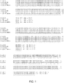
- the affinity specificity of the antibodies or fragments thereof for C. jejuni flagella is illustrated.
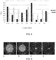
- the antibodies show efficacy in reducing C. jejuni colonization levels in chickens, for example when orally administered.
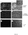
- the antibodies disclosed display specific binding to the bacterial flagella.
- the antibodies and multimers described herein reduce C. jejuni motility. Further variants are described with advantageous biophysical properties. Through specific panning efforts and disulfide-bond engineering strategies, antibodies are described which display good thermal stability and protease tolerance or resistance. A hyper-stabilized antibody or fragment thereof is also described, having superior thermal stability and resistance to the major gastrointestinal (GI) proteases.
- GI major gastrointestinal
- the present disclosure provides an isolated or purified antibody or fragment thereof specifically binding to C. jejuni flagella, comprising
- the isolated or purified antibody or fragment thereof as described herein may specifically bind to flagellin; more specifically, the isolated or purified antibody or fragment thereof may specifically bind to the Fla A component of flagellin.
- the isolated or purified antibody or fragment thereof as described above is a single-domain antibody (sdAb).
- the sdAb may be of camelid origin.
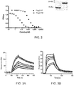
- the isolated or purified antibody or fragment thereof may be provided in a multivalent display.
- the isolated or purified antibody or fragment thereof may be expressed as a fusion protein with the verotoxin B subunit.
- the fusion protein may assemble into a pentabody.
- the multimer may comprise one or more than one fusion protein selected from: or a having at least 90% identity thereto.
- the present disclosure also provides a nucleic acid sequence encoding the isolated or purified antibody or fragment thereof described herein.
- a vector comprising the nucleic acid molecule just described is also provided.
- the isolated or purified antibody or fragment thereof of the present disclosure may be linked to a detectable label.
- the present disclosure further provides a method of reducing presence of C. jejuni in an animal or an animal environment.
- the method comprises administering to the animal the isolated or purified antibody or fragment thereof of the present disclosure.
- the isolated or purified antibody or fragment thereof may be administered orally.
- the isolated or purified antibody or fragment thereof may be comprised in a yeast expression system.
- an antibiotic, bacteriocin, or other plant- or animal-derived compound effective against C. jejuni may additionally be administered to the animal; alternatively, a competing microbe, optionally co-expressed or co-contained in a probiotic system, may additionally be administered to the animal.
- the present disclosure also provides a method of reducing introduction of C. jejuni into an animal environment.
- the isolated or purified antibody or fragment thereof is administered to an inductee animal, priorto introducing the inductee animal into the animal environment.
- the present disclosure further provides a C. jejuni vaccine or formulation comprising the isolated or purified antibody or fragment thereof of as described herein and an excipient.
- the vaccine may be for oral delivery.
- a method of treating a C. jejuni infected subject is also provided; the subject is treated by administering the isolated or purified antibody or fragment thereof as described herein.
- the method may also comprise administering an antibiotic effective against C. jejuni.
- the subject may be a livestock animal selected from the group consisting of a chicken, cow, or sheep; alternatively, the subject may be a human.
- the present disclosure further provides a use of the isolated or purified antibody or fragment thereof described herein for treating or for preparing a medicament for treating a C. jejuni infection in a subject in need thereof.
- a method of detecting C. jejuni in a sample is also provided.
- the sample is contacted with the isolated or purified antibody or fragment thereof described herein, then the presence of bound antibody is detected.
- the sample may comprise a bodily fluid or fecal material; alternatively, the sample may comprise a food product or a surface swab from a food product.
- the present disclosure also provides a kit for detecting C. jejuni in a sample.
- the kit may comprise the isolated or purified antibody or fragment thereof of the present disclosure and instructions for use in detecting C. jejuni.
- a detection reagent for detecting C. jejuni in a sample is also provided that comprises the isolated or purified antibody or fragment thereof described herein and a suitable carrier.
- the present disclosure relates to antibodies and fragments thereof that specifically bind to C. jejuni.
- the antibodies and fragments thereof described herein are useful in controlling or diminishing C. jejuni prevalence in the food chain. Methods involving administration of C. jejuni -specific single-domain antibodies to animals are described, which reduce C. jejuni levels.
- the purified antibody or fragment thereof exhibits specific binding to C. jejuni flagella.
- the anti- C . jejuni functionality may be determined in terms of binding to flagellar proteins of C. jejuni, reducing motility of C. jejuni, or reducing colonization or incidence of infection of C. jejuni. Evaluation of binding and/or motility is well within the capabilities of a skilled artisan using techniques described herein.
- the purified antibody or fragment thereof binds specifically to flagellin.
- the purified antibody or fragment thereof binds specifically to the Fla A component of flagellin.
- antibody also referred to in the art as “immunoglobulin” (Ig), used herein refers to a protein constructed from paired heavy and light polypeptide chains; various Ig isotypes exist, including IgA, IgD, IgE, IgG, and IgM.
- Ig immunoglobulin
- each chain fold into a number of distinct globular domains joined by more linear polypeptide sequences.
- V L variable
- C L constant
- C H constant
- C H constant domain
- Fv antigen binding region
- the light and heavy chain variable regions are responsible for binding the target antigen and can therefore show significant sequence diversity between antibodies.
- the constant regions show less sequence diversity, and are responsible for binding a number of natural proteins to elicit important biochemical events.
- the variable region of an antibody contains the antigen binding determinants of the molecule, and thus determines the specificity of an antibody for its target antigen.
- the majority of sequence variability occurs in six hypervariable regions, three each per variable heavy (V H ) and light (V L ) chain; the hypervariable regions combine to form the antigen-binding site, and contribute to binding and recognition of an antigenic determinant.
- the specificity and affinity of an antibody for its antigen is determined by the structure of the hypervariable regions, as well as their size, shape and chemistry of the surface they present to the antigen.
- Various schemes exist for identification of the regions of hypervariability the two most common being those of Kabat and of Chothia and Lesk.
- Kabat et al (1991a; 1991b) define the "complementarity-determining regions” (CDR) based on sequence variability at the antigen-binding regions of the V H and V L domains.
- CDR complementarity-determining regions
- Chothia and Lesk (1987) define the "hypervariable loops" (H or L) based on the location of the structural loop regions in the V H and V L domains.
- CDR and hypervariable loop regions that are adjacent or overlapping
- those of skill in the antibody art often utilize the terms "CDR” and "hypervariable loop” interchangeably, and they may be so used herein.
- the regions forming the antigen-binding site are presently referred to herein as CDR L1, CDR L2, CDR L3, CDR H1, CDR H2, CDR H3 in the case of antibodies comprising a V H and a V L domain; or as CDR1, CDR2, CDR3 in the case of the antigen-binding regions of either a heavy chain or a light chain.
- the CDR/loops are referred to herein according to the IMGT numbering system (Lefranc, M.-P.
- the region outside of the CDR is referred to as the framework region (FR).
- the FR provides structural integrity to the variable domain and ensures retention of the immunoglobulin fold. This characteristic structure of antibodies provides a stable scaffold upon which substantial antigen-binding diversity can be explored by the immune system to obtain specificity for a broad array of antigens (Padlan et al, 1994).
- an "antibody fragment” as referred to herein may include any suitable antigen-binding antibody fragment known in the art.
- the antibody fragment may be obtained by manipulation of a naturally-occurring antibody, or may be obtained using recombinant methods.
- an antibody fragment may include, but is not limited to Fv, single-chain Fv (scFv; a molecule consisting V L and V H connected with a peptide linker), Fab, Fab', F(ab') 2 , single domain antibody (sdAb), and multivalent presentations of these.
- the antibody fragment may be a single domain antibody (sdAb) derived from naturally-occurring sources.
- Heavy chain antibodies of camelid origin (Hamers-Casterman et al, 1993) lack light chains and thus their antigen binding sites consist of one domain, termed V H H.
- sdAb have also been observed in shark and are termed VNARs (Greenberg et al.,1995; Nuttall et al, 2003); other sdAb may be engineered based on human heavy or light chain sequences (Jespers et al, 2004; To et al, 2005).
- sdAb includes those directly isolated from V L , V H , V H H or V NAR reservoir of any origin through phage display or other display technologies and those generated through further modification of such sdAb by humanization, affinity maturation, stabilization, solubilization (e.g., camelization), or other methods of antibody engineering. Also encompassed by embodiments described herein are homologues, derivatives, or fragments that retain the antigen-binding function and specificity of the sdAb.
- SdAb are excellent building blocks for novel antibody molecules due to their high thermostability, high detergent resistance, relatively high resistance to proteases (Dumoulin et al, 2002) and high production yield (Arbabi-Ghahroudi et al, 1997); they can also be engineered to have very high affinity by isolation from an immune library (Li et al, 2009) or by in vitro affinity maturation (Davies & Riechmann, 1996).
- antibody fragments are preferable to whole antibodies (e.g., IgG) due to lower production cost in prokaryotic systems and ease of genetic manipulation.
- VHH have been shown to be extremely stable when cloned and expressed as monomers using recombinant expression systems (Arbabi-Ghahroudi et al., 1997; Muyldermans 2001).
- a sdAb comprises a single immunoglobulin domain that retains the immuglobulin fold; most notably, only three CDR form the antigen-binding site. However, not all CDR may be required for binding the antigen. For example, and without wishing to be limiting, one, two, or three of the CDR may contribute to binding and recognition of the antigen by the sdAb of the present disclosure.
- the CDR of the sdAb are referred to herein as CDR1, CDR2, and CDR3, and are based on IMGT numbering system (Lefranc, M.-P. et al., 2003).
- the antibody or fragment thereof may be a sdAb.
- the sdAb may be of camelid origin, and thus may be based on camelid framework regions; alternatively, the CDR may be grafted onto the framework regions of other antibody domains, for example but not limited to VNAR, human V H or human V l framework regions.
- the CDR described above may be grafted onto the framework regions of other types of antibody fragments (Fv, scFv, Fab).
- the present embodiment further encompasses an antibody fragment that is "humanized” using any suitable method know in the art, for example, but not limited to CDR grafting and veneering.
- Humanization of an antibody or antibody fragment comprises replacing an amino acid in the sequence with its human counterpart, as found in the human consensus sequence, without loss of antigen-binding ability or specificity; this approach reduces immunogenicity of the antibody or fragment thereof when introduced into human subjects.
- one or more than one of the heavy chain CDR defined herein may be fused or grafted to a human variable region (V H , or V L ), or to other human antibody fragment framework regions (Fv, scFv, Fab).
- V H , or V L human variable region
- Fv, scFv, Fab human antibody fragment framework regions
- the conformation of said one or more than one hypervariable loop is preserved, and the affinity and specificity of the sdAb for its target (i.e., flagella) is also preserved.
- CDR grafting is known in the art and is described in at least the following: US Patent No. 6180370 , US Patent No. 5693761 , US Patent No. 6054297 , US Patent No. 5859205 , and European Patent No. 626390 .
- Veneering also referred to in the art as "variable region resurfacing", involves humanizing solvent-exposed positions of the antibody or fragment; thus, buried non-humanized residues, which may be important for CDR conformation, are preserved while the potential for immunological reaction against solvent-exposed regions is minimized.
- Veneering is known in the art and is described in at least the following: US Patent No. 5869619 , US Patent No. 5766886 , US Patent No. 5821123 , and European Patent No. 519596 . Persons of skill in the art would be amply familiar with methods of preparing such humanized antibody fragments.
- the isolated or purified antibody or fragment thereof of the present disclosure may comprise a CDR1 of sequence GLTFRNFHMA (SEQ ID NO:1), a CDR2 of sequence ISWSRDRQ (SEQ ID NO:2), and a CDR3 of sequence AARTASASGDWYKGSYQY (SEQ ID NO:3).
- the antibody or fragment thereof may be a sdAb.
- the sdAb may be of camelid origin, and thus may be based on camelid framework region.
- the isolated or purified antibody or fragment thereof may comprise the sequence:
- a substantially identical sequence may comprise one or more conservative amino acid mutations. It is known in the art that one or more conservative amino acid mutations to a reference sequence may yield a mutant peptide with no substantial change in physiological, chemical, or functional properties compared to the reference sequence; in such a case, the reference and mutant sequences would be considered "substantially identical" polypeptides.
- Conservative amino acid mutation may include addition, deletion, or substitution of an amino acid; in one non-limiting example, the conservative amino acid mutation is a conservative amino acid substitution.
- a conservative amino acid substitution is defined herein as the substitution of an amino acid residue for another amino acid residue with similar chemical properties (e.g. size, charge, or polarity).
- a conservative amino acid substitution may substitute a basic, neutral, hydrophobic, or acidic amino acid for another of the same group.
- basic amino acid it is meant hydrophilic amino acids having a side chain pK value of greater than 7, which are typically positively charged at physiological pH.
- Basic amino acids include histidine (His or H), arginine (Arg or R), and lysine (Lys or K).
- neutral amino acid also "polar amino acid”
- Polar amino acids include serine (Ser or S), threonine (Thr or T), cysteine (Cys or C), tyrosine (Tyr or Y), asparagine (Asn or N), and glutamine (Gln or Q).
- hydrophobic amino acid also “non-polar amino acid” is meant to include amino acids exhibiting a hydrophobicity of greater than zero according to the normalized consensus hydrophobicity scale of Eisenberg (1984).
- Hydrophobic amino acids include proline (Pro or P), isoleucine (lie or I), phenylalanine (Phe or F), valine (Val or V), leucine (Leu or L), tryptophan (Trp or W), methionine (Met or M), alanine (Ala or A), and glycine (Gly or G).
- “Acidic amino acid” refers to hydrophilic amino acids having a side chain pK value of less than 7, which are typically negatively charged at physiological pH. Acidic amino acids include glutamate (Glu or E), and aspartate (Asp or D).
- Sequence identity is used to evaluate the similarity of two sequences; it is determined by calculating the percent of residues that are the same when the two sequences are aligned for maximum correspondence between residue positions. Any known method may be used to calculate sequence identity; for example, computer software is available to calculate sequence identity. Without wishingto be limiting, sequence identity can be calculated by software such as NCBI BLAST2 service maintained by the Swiss Institute of Bioinformatics (and as found at ca.expasy.org/tools/blast/), BLAST-P, Blast-N, or FASTA-N, or any other appropriate software that is known in the art.
- the substantially identical sequences may be at least 90% identical; in another example, the substantially identical sequences may be at least 90, 91, 92, 93, 94, 95, 96, 97, 98, 99, or 100% identical at the amino acid level to sequences described herein.
- alignment of FlagV1M, FlagV1F23M, FlagV1MDSB, and FlagV1F23MDSB leads to sequence identities of between 96.8% and 98.4%.
- the substantially identical sequences retain the activity and specificity of the reference sequence.
- certain amino acid residues of an antibody, particularly within the framework regions may be mutated without affecting the antigen-binding and other functional properties of the antibody.
- the antibody or fragment thereof may also comprise additional sequences to aid in expression, detection, or purification of a recombinant antibody or fragment thereof.
- the antibody or fragment thereof may comprise a targeting or signal sequence (for example, but not limited to ompA), a detection tag (for example, but not limited to c-Myc), a purification tag (for example, but not limited to a histidine purification tag, His 5 or His 6 ), or any combination thereof.
- the antibody or fragment thereof may also be in a multivalent display.
- Multimerization may be achieved by any suitable method of known in the art. For example, and without wishing to be limiting in any manner, multimerization may be achieved using self-assembly molecules (Zhang et al, 2004; Merritt & Hol, 1995), as described in WO2003/046560 .
- the described method produces pentabodies by expressing a fusion protein comprising the antibody or fragment thereof and the pentamerization domain of the B-subunit of an AB 5 toxin family ( Nielson et al, 2000; WO2003/046560 ); for example, and without wishing to be limiting, the sequence of the pentamerization domain may be TPDCVTGKVEYTKYNDEDTFTVKVGDKELFTNRANLQSLLLSAQITGMTVTIKTNACHNGGGFSEVIFR (SEQ ID NO:17).
- the pentamerization domain assembles into a pentamer, through which a multivalent display of the antibody or fragment thereof is formed.
- each subunit in the pentamer comprises the fusion protein described above.
- pentamerization of the antibody or fragment thereof in no way alters its antigen binding or recognition (ie., the antibody or fragment thereof binds the same antigen, in the same manner, with the same specificity and affinity).
- the resulting pentabodies are compact, have high avidity (functional affinity), and are stable antigen-binding molecules (Zhang et al., 2004).
- These pentavalent antibodies are also capable of enhancing agglutination when bound to antigens (Zhang et al., 2004), thereby increasing their efficacy.
- Each subunit of the multimers described herein may be the same or different.
- the multimerization domain may be linked to the antibody or antibody fragment using a linker; such a linker should be of sufficient length and appropriate composition to provide flexible attachment of the two molecules, but should not hamper the antigen-binding properties of the antibody.
- the linker may be the linker GPGGGSGGGGS (SEQ ID NO:18).
- a multimer may comprise the sequence:
- the antibody or fragment thereof may be presented as a dimer, a trimer, or any other suitable oligomer. This may be achieved by methods known in the art, for example direct linking connection (Nielsen et al, 2000), c-jun/Fos interaction (de Kruif et al, 1996), "Knob into holes” interaction (Ridgway et al, 1996).
- a multimer may also be formed using the multimerization domains described by Zhu et al.
- antibody form is a fusion of the antibody or fragment thereof, as described herein, with a coiled-coil peptide resulting in a multimeric molecule (Zhu et al., 2010).
- Another method known in the art for multimerization is to dimerize the antibody or fragment thereof using a Fc domain.
- a Fc gene in inserted into an expression vector; the nucleotide sequence of the antibody or fragment thereof can be amplified and inserted into the vector such that the C-terminus of the antibody or fragment thereof is linked to the hinge region of the Fc without addition of extra residues.
- the resulting vector can be transfected to a mammalian cell line and the fusion protein may be recombinantly expressed, then purified by affinity chromatography (for example, on a protein A column).
- affinity chromatography for example, on a protein A column.
- subunits within the multimers may comprise the same or different antibodies or fragments thereof, as described herein.
- nucleic acid sequences encoding the antibody or fragment thereof as described herein. Given the degeneracy of the genetic code, a number of nucleotide sequences would have the effect of encoding the polypeptide, as would be readily understood by a skilled artisan.
- the nucleic acid sequence may be codon-optimized.
- Vectors comprising the nucleic acids as just described, are encompassed.
- a host cell comprising the nucleotide encoding any one of the antibodies or fragment thereof described herein would also be readily recognized by the skilled artisan.
- a phage comprising the antibody or fragment thereof described herein and/or display as fusion to one of the viral coat protein (for example, but not limited to g3p of filamentous phages), and/or comprising the nucleotide encoding the antibody or fragment thereof is also encompassed.
- the antibody or fragment thereof described herein may be labeled with detectable label.
- the label may be rendered detectable or may in itself be detectable, so that the presence of binding to C. jejuni can be observed.
- the detectable label may be a radioisotope, a paramagnetic label (for example gadolinium or iron oxide), a fluorophore, a fluorophore, a fluorescent agent (for example, FITC or Enhanced Green Fluorescent Protein (EGFP)), Near Infra-Red (NIR; for example Cy5.5, Alexa680, Dylight680, or Dylight800) fluorochrome or dye, an echogenic microbubble, an affinity label (for example biotin, avidin, etc), fused to a detectable protein-based molecule, nucleotide, quantum dot, nanoparticle, nanowire, or nanotube or any other suitable agent that may be detected by imaging methods.
- the antibody or fragment thereof may be linked to the detectable agent using any method known in the art (recombinant technology, chemical conjugation, etc.); optionally a linker may be used as needed.
- the step of detecting may be accomplished by any suitable method known in the art, for example, but not limited to optical imaging, immunohistochemistry or molecular diagnostic imaging, ELISA, or other suitable method.
- the antibody or fragment thereof may be linked to a fluorescent agent such as FITC, or may genetically be fused to the Enhanced Green Fluorescent Protein (EGFP).
- FITC fluorescent agent
- EGFP Enhanced Green Fluorescent Protein
- reducing the presence of C. jejuni in an animal or an animal environment are described herein.
- reducing the presence may comprise reducing contamination on the surface of the animal, or within the gastrointestinal tract of an animal.
- the method described herein could be used for reducing the presence of C. jejuni.
- the environment of an animal can relate to the animal's immediate surroundings, such as the walls or floors of a cage or facility, the feeding or watering apparatuses within an animal compound, the bedding materials found in an animal compound, or simply the fecal material present external to the animal within the animal's confines.
- Administering to an animal the antibody or fragment thereof described herein can be for the purpose of reducing the presence of C.
- Administration to the animal may be by any suitable method known in the art.
- the antibody or fragment thereof described herein may be administered orally.
- Oral delivery permits the antibody or fragment thereof to be delivered within the water or food supply to an animal, and is less noticeable or stressful to an animal than an injection. Gavage is also an acceptable oral route when highly accurate delivery of an oral dosing regime is desirable. Other routes of administration can also be considered, such as via a systemic, or rectal delivery route.
- the antibody or fragment thereof may be included in the animal's food supply.
- the antibody or fragment thereof may be provided in a yeast expression system that is included in the animal's food supply.
- the antibody or fragment thereof may be displayed on yeast coat protein(s) or expressed internally or externally by the yeast.
- Co-administration of another substance that is effective against C. jejuni is also a possible strategy for reducing C. jejuni in an animal environment.
- administering to the animal an antibiotic either at the same time or at an adjacent time to the delivery of the antibody or fragment thereof can have an additive effect or may have a synergistic effect.
- the result of which is a reduced likelihood of C. jejuni contamination, but also a reduced usage of antibiotic.
- a bacteriocin effective against C. jejuni can also be provided to the animal with the antibody or fragment thereof for an additive or synergistic effect.
- any other plant- or animal-derived compound such as a small molecule, peptide, or protein that has effect against C.
- jejuni may be used together with the antibody or fragment thereof described herein.
- a competitive microbe may also be provided to the animal concurrently with the antibody or fragment thereof in order to achieve an additive or a synergistic effect.
- the competitive microbe may be used together with the antibody or fragment thereof described herein as part of a probiotic system. Within such a probiotic system, the antibody or fragment thereof may be co-administered with the competitive microbe, or may be delivered sequentially. Expression of the antibody or fragment thereof described herein within a probiotic system may also be undertaken.
- Scaffold engineering of portions of the antibody or fragment thereof outside of the CDR regions can confer additional protease resistance, as well as thermal and low pH resistance.
- the form of delivery may also be altered with coatings or excipients that provide a protective effect against gut enzymes, thermal or low pH effects, and in this way, the sequence of the antibody or fragment thereof itself need not be modified, but rather the formulation prepared for oral delivery may itself be optimized for the species of subject to which the antibody or fragment thereof is to be delivered.
- the dosage form may be of any type acceptable for peptide delivery to animals. Coated forms and slow release forms could be used if desirable. Liquid, powder, crystal, gel, semi-solid, or tablet forms can be used.
- the animal to which the antibody or fragment thereof may be delivered may be a bird, such as a broiler chicken or laying hen.
- Other types of livestock animals such as cows, sheep, etc. may also benefit from the peptide if C. jejuni is present in the animal's gut or surrounding environment.
- livestock applications are not limited to poultry.
- a typical animal environment may be a barn or farm, such as a poultry farm.
- a method is provided that prevents introduction of a new contaminated animal or "inductee" animal into the environment, such as a barn.
- the antibody or fragment thereof administering to an inductee prior to introducing the inductee animal into the animal environment, such as a barn or farm.
- the animal could be cleared of the likelihood of contamination prior to taking up residence with the other animals who may have already received treatment.
- An anti- C . jejuni vaccine is described herein which comprises the antibody or fragment thereof described herein, together with an excipient.
- the vaccine may be formulated for oral delivery, as described above.
- a method of treating a C. jejuni infected subject comprising administering to the subject the antibody or fragment thereof described above.
- an antibiotic effective against C. jejuni can be co-administered to the subject.
- the subject may be livestock, such as a chicken, the method is also applicable to human subjects.
- a formulation for such a use in treating C. jejuni infection comprises the antibody or fragment thereof together with an excipient.
- the antibody or fragment thereof may be used in the preparation of a medicament to treat C. jejuni infection in a subject in need thereof.
- the antibody or fragment thereof described herein is also useful for methods of detecting C. jejuni in a sample.
- a sample is contacted with the antibody or fragment thereof, with or without the presence of a label, and subsequently, the presence of bound antibody or fragment thereof is detected using any acceptable means.
- the sample may comprise a bodily fluid or fecal material for those instances where detection is used to determine contamination or infection of an individual.
- the sample which is contacted by the antibody or fragment thereof may be a food product, a food container, food processing equipment, or a surface swab from a food product, container or processing equipment.
- a kit for conducting such a method, which would include the antibody or fragment thereof itself, together with instructions for use in detecting C. jejuni.
- reagents to be used in such a detection kit may be included for the user's convenience.
- the antibody or fragment thereof itself may be the main component of detection reagent that is to be used for detecting C. jejuni in a sample.
- a detection reagent would also a suitable carrier, such as a buffer.
- the present inventors have isolated FlagV1M and FlagV6M, flagellin-binding single domain antibodies (sdAb) isolated from a hyperimmunized llama phage display library by panning against flagella proteins.
- FlagV1F23M was isolated from a library in which an error-prone PCR approach was used to introduce random mutations in FlagV1M DNA; panning was performed under protease treatment conditions. The equivalent mutations were also conferred to FlagV6M, resulting in FlagV6F23M.
- Pentavalency was conferred to the FlagV1M, FlagV1F23M, and FlagV6M antibodies by fusion of the sdAb with a protein domain derived from the verotoxin B homopentamer, producing proteins FlagV1P, FlagV1F23P, and FlagV6P.
- the resulting antibody or fragment thereof which may be referred to as "pentabodies", are compact, showed high avidity, and are stable antigen-binding molecules.
- These pentavalent VHH were capable of enhancing agglutination of C. jejuni and C. coli when bound to antigens.
- Single domain antibodies are, in general, significantly more resistant to proteases than the conventional antibody fragments.
- FlagV1M, FlagV1F23M, FlagV6M, and FlagV6F23M were also modified to possess increased tolerance to gut enzymes, thus increasing efficacy of oral delivery.
- Typical gut enzymes which may have a destructive effect on a polypeptide include pepsin, trypsin and chymotrypsin.
- resistance to these enzymes is advantageous, as the peptide would have more exposure time to bind with ambient C. jejuni within the intestinal tract.
- the modification to FlagV1M, FlagV1F23M, FlagV6M, and FlagV6F23M was made by introducing a second disulfide bridge between residues 54 and 78, producing sdAb FlagV1MDSB, FlagV1F23MDSB, FlagV6MDSB, and FlagV6F23MDSB, respectively.
- Sites for modification in preparing the disulfide bridge modified antibodies were selected based on optimal alteration to V H H thermostability and proteolytic stability when cysteine residues are introduced at specific sites.
- V H H and pentabodies counterparts were specific for flagellin, a C . jejuni cell surface antigen, as demonstrated by fluorescence microscopy. Furthermore, the inventors demonstrated that the pentabodies developed against flagellin bind to flaA protein, which plays a role in invading mammalian cells. SPR demonstrated that the V H H had low nanomolar affinity to the target. The V H H were also capable of preventing/disrupting Campylobacter growth and motility, as demonstrated in a motility assay. The V H H also significantly reduced the levels of C. jejuni colonization in chickens when orally administered. Additionally, studies of the combinatorial therapy of antibodies and antibiotics in the control of Campylobacter illustrate that the administration of the V H H and pentabodies can lower the required dose of antibiotics by up to 35-fold.
- Flagella were prepared for use as the antigen in subsequent examples.
- C. jejuni (strain 81-176) flagella were isolated as described previously (Power et al., 2003). Briefly, to prepare flagella, C. jejuni was cultured overnight and cells were scraped off into Muller-Hinton broth and incubated for overnight. Cells were then harvested by centrifugation and re-suspended in 100 mL of tris-buffered saline solution. Flagella were sheared from the cells using a Waring blender on ice. Cell debris was pelleted by centrifugation and the supernatant was transferred to an ultracentrifugation tube. Flagella were pelleted by centrifugation for 1 hour at 45,000 rpm. Further purification was done by re-suspension in 2% SDS and centrifugation of the samples. Pellets were re-suspended in 200 - 500 ⁇ L of dH 2 O.
- a male llama Lama glama
- C . jejuni flagella Example 1
- Seven injections were performed in total and, for each injection, 100 ⁇ g of antigen in a total volume of 0.5 ml was mixed with an equal volume of either complete (day 1) or incomplete (days 21, 35, 49, 63) Freund's adjuvant (Sigma).
- the last two injections (days 76 and 90) were performed with 100 ⁇ g of antigen without adjuvant.
- Preimmune blood (15 - 20 ml) was collected before the first injection and on days 21, 49, 76 and 90.
- the specific immune responses were analyzed by ELISA using total preimmune and immune sera.
- Llama serum from day 90 was fractionated according to Hamers-Casterman et al. (1993). Protein G and A columns (GE Healthcare) were used for serum fractionation according to the manufacturer's instructions and separated fractions were adjusted to pH 6 with 1 M Tris/HCl, pH 8.8, and dialyzed against pre-chilled PBS at 4°C overnight. Individual heavy fractions (G1, A1 and A2) and G2 (conventional IgG) were analyzed for specific binding to flagella by ELISA. Briefly, microtitre plates (MaxisorpTM plates) (Nalge Nunc International, Rochester, NY) were coated overnight at 4°C with 5 ⁇ g/ml of flagella antigen (Example 1) in PBS.
- Wells were rinsed and blocked with 200 ⁇ l of 1% casein. Different dilutions of purified IgG fractions (G1, G2, A1 and A2) were added and incubated at room temperature for 1.5 h. Wells were washed with PBST (0.05% v/v Tween-20), and incubated with goat anti-llama IgG (H+L) (1:1,000 in PBS) (Bethyl Laboratories, Montgomery, TX) followed by Swine-anti-goat-HRP (1:3,000 in PBS) (Cedarlane, Burlington, ON, Canada). Signal was detected by adding 100 ⁇ l/well TMB peroxidase substrate (Kirkegaard and Perry Laboratories, Gaithersburg, MD, USA). Reactions were stopped by adding 100 1M phosphoric acid and A 450 was measured using a Bio-Rad ELISA plate reader.
- a hyper-immunized llama VHH library was constructed based on RNA isolated from the serum collected in Example 2.
- Phage display library was constructed as described previously (Arbabi Ghahroudi et al., 2009). In brief, total RNA was isolated from approximately 1 X 10 7 lymphocytes collected on day 90 post-immunization using QIAamp RNA blood mini kit (Qiagen, Mississauga, Ontario, Canada). First-strand cDNA was synthesized with oligo(dT) primer using 5 ⁇ g total RNA as template according to manufacturer's recommendations (GE Healthcare) (Arbabi Ghahroudi et al., 2009).
- Variable and part of the constant domains DNAs were amplified using oligonucleotides MJ1-3 (sense) and two CH2 domain antisense primers CH2 and CH2b3 (for primer sequences see Arbabi Ghahroudi et al., 2009) and heavy chain fragments (550-650 bp in length) were gel-purified using QIA quick gel extraction kit (Qiagene).
- the variable regions of heavy chain antibodies (IgG2 and IgG3) were re-amplified in a second PCR reaction using MJ7 and MJ8 primers (for primer sequences see Arbabi Ghahroudi et al., 2009).
- the amplified PCR products were purified with a QIAquick PCR purification kit (Qiagene), digested with Sfil (New England BioLabs, Pickering, Ontario, Canada), and re-purified using the same kit. Twelve micrograms of digested V H H fragments were ligated with 40 ⁇ g (3:1 molar ratio, respectively) Sfi-digested pMED1 phagemid vector (Arbabi Ghahroudi et al., 2009) using LigaFast Rapid DNA ligation system and its protocol (Promega, Madison, WI), transformed into commercial electrocompetent TG1 E .
- coli cells (Stratagene, La Jolla, CA) as described previously (Arbabi Ghahroudi et al., 2009) and a library size of 5 X 10 7 transformants was obtained.
- the V H H fragments from 30 colonies were PCR-amplified and sequenced to analyze the complexity of the library; all clones had inserts of expected sizes and were different from each other at their CDR regions as determined by sequencing of their encoding V H H fragments.
- the library was grown for 3-4 hours at 37 °C, 250 rpm in 2 XYT/Amp-Glucose (2% w/v) medium.
- the bacterial cells were pelleted, resuspended in the same medium and stored as glycerol stock at -80°C as described previously (Arbabi Ghahroudi et al., 2009).
- the infected cell pellets were resuspended in 200 ml of2XYT/Amp with 50 ⁇ g/ml kanamycin and incubated overnight at 37°C and 250 rpm.
- the phage particles in culture supernatant were PEG-precipitated as described previously (Arbabi-Ghahroudi et al., 2009) and the phage pellets were resuspended in 2 ml of sterile PBS and the phage titration was determined.
- 96-well MaxisorpTM plate was coated with 30 ⁇ g of flagella antigen overnight at 4°C.
- the wells were rinsed with PBS and blocked with PBS/1% (w/v) casein for 2 h at 37°C. Approximately 10 12 rescued phage particles were added to the blocked wells and incubated for 2 hours at 37°C.
- the wells were washed 5X with PBST (0.1% v/v Tween-20) and 5X with PBS.
- the bound phages were eluted with 0.1 M triethylamine, neutralized with 1M Tris-HCL, PH 7.4 and incubated with exponentially growing TG1 cells. After 30 min incubation at 37°C, the cells were superinfected with M13KO7 for additional 15 min and grown in 2XYT-Amp-Kan overnight at 37 °C.
- Panning was continued for three more rounds following the same conditions except that antigen concentration was reduced to 20,15, and 10 ⁇ g/well and washing was increased 7, 10 and 12X with PBS-T and PBS for the second, third and fourth rounds of panning, respectively.
- 48 randomly picked colonies were grown and subjected to phage ELISA screening as described previously (Arbabi Ghahroudi et al., 2009) except that 5 ⁇ g/ml of Flagella were coated onto microtiter plates.
- Positive clones include FlagV1M and FlagV6M (SEQ ID NO:8 and SEQ ID NO:13; Figure 1 ), which are further studied herein.
- V H H against flagella identified in Example 3 were PCR amplified from the pMED1 phagemid vector with BbsI1-V H H forward primer and BamHI-V H H reverse primer (Table 1).
- the PCR fragments were digested with the Bbsl and BamHI restriction enzymes and ligated into the similarly digested pSJF2H expression vector (Arbabi-Ghahroudi et al., 2009).
- all plasmids were transformed into electrocompetent TG1 E . coli and selected on LB agar plates + ampicillin. Colonies were screened by colony PCR for inserts and the DNA sequenced.
- V H H antibodies were expressed using the 5-day minimal media method (Arbabi-Ghahroudi et al., 2009). After induction of protein expression, cell cultures were harvested at 6,000 rpm x 30 min (4°C), the supernatant decanted, and the periplasmic contents extracted from the cell pellet. Briefly, the pellet of monomeric V H H was resuspended in 20 ml of ice cold TES (0.2 M Tris-HCl pH 8.0, 20% (w/v) sucrose, 0.5 mM EDTA) and incubated on ice for 30 min.
- TES 0.2 M Tris-HCl pH 8.0, 20% (w/v) sucrose, 0.5 mM EDTA
- Fractionation was performed on an ⁇ KTA FPLC purification system (GE Healthcare) with 10 mM HEPES, 500 mM NaCl, pH 7.0 as the starting buffer and 10 mM HEPES, 500 mM NaCl, 500 mM imidazole, pH 7.0 as the elution buffer.
- Table 1 shows the primers used in the construction of monomeric and pentameric V H H clones, as described in Examples 4 and 5.
- Table 1 Primers used in the construction of monomeric and pentameric V H H clones Name Sequence 5' ⁇ 3' BbsI1-V H H forward primer TATGAAGACACCAGGCCCAGGTAAAGCTGGAGGAGTCT (SEQ ID NO:23) BamHI-V H H reverse primer TTGTTCGGATCCTGAGGAGACGGTGACCTG (SEQ ID NO:24) Apal-V H H reverse primer ATTATTATGGGCCCTGAGGAGACGGTGACCTGGGTC (SEQ ID NO:25)
- Pentamers of the monomeric V H H were prepared by fusing the V H H to the pentamerization domain of the B-subunit of an AB 5 toxin family. Once expressed, the subunits self-assemble with pentamers.
- FlagV1P and FlagV6P (SEQ ID NO:19 and SEQ ID NO:21; Figure 1 ) were prepared.
- V H H against flagella identified in Example 3 were PCR amplified from the pMED1 phagemid vector with BbsI1-V H H forward primer and Apal-V H H reverse primer (Table 1).
- the PCR fragments were digested with the BbSI and Apal restriction enzymes and ligated into the similarly digested pVT2 expression vector (Arbabi-Ghahroudi et al., 2009).
- all plasmids were transformed into electrocompetent TG1 E . coli and selected on LB agar plates + ampicillin. Colonies were screened by colony PCR for inserts and the DNA sequenced.
- Pentameric antibodies were expressed using the 5-day minimal media method (Arbabi-Ghahroudi et al., 2009). After induction of protein expression, cell cultures were harvested at 6,000 rpm x 30 min (4°C), the supernatant decanted, and the periplasmic contents extracted from the cell pellet. The cells were re-suspended in 100 ml of ice-cold lysis buffer (50 mM Tris-HCl, pH 8.0, 25 mM NaCl,) kept on ice at - 20°C overnight or frozen on dry ice for 1 h.
- ice-cold lysis buffer 50 mM Tris-HCl, pH 8.0, 25 mM NaCl,
- the resulting supernatant containing V H H was dialysed overnight into immobilized metal-affinity chromatography (IMAC) buffer A (10 mM HEPES pH 7.0, 500 mM NaCl) and purified as described (Arbabi-Ghahroudi et al., 2009). Purification of antibodies was done using HiTrapTM Chelating HP columns (GE Healthcare) according to the instruction. Fractionation was performed on an ⁇ KTA FPLC purification system (GE Healthcare) with 10 mM HEPES, 500 mM NaCl, pH 7.0 as the starting buffer and 10 mM HEPES, 500 mM NaCl, 500 mM imidazole, pH 7.0 as the elution buffer.
- IMAC immobilized metal-affinity chromatography
- Purified protein fractions were pooled and dialyzed against PBS. Eluted fractions were analyzed by SDS-PAGE and Western blotting before being dialysed into PBS.
- V H H concentrations were determined by absorbance measurements at 280 nm using theoretical MW and extinction coefficients calculated with the ExPASy ProtParam Tool (http://expasy.org/tools/protparam.html) according to Pace et al., 1995.
- the yield of the purified pentamers ranged from 10-50 mg/l bacterial culture.
- the FlagV1M, FlagV6M, FlagV1P, and FlagV6P antibodies expressed and purified in Examples 4 and 5 were characterized.
- anti-flagellin V H H were biotinylated by mixing Pierce EZ-Link Sulfo-NHS-LC-LC-biotin (GE Healthcare) with approximately 10 folds molar excess in 10 mM phosphate, 150 mM NaCl, pH 7.0 for 30 min at room temperature followed by dialysis against the same buffer. Analyses were performed with a Biacore 3000 instrument (GE Healthcare). All measurements were carried out at 25 °C in 10 mM HEPES, pH 7.4, containing 150 mM NaCl, 3 mM EDTA and 0.005% surfactant P20 (GE Healthcare).
- Antibody binding assays were performed as described in Example 2, except that after washing the plate with PBST and blocking with PBS-casein (1%), a 5 ⁇ g/ml solution of FlagV1M, FlagV1P, or FlagV6P was added to the respective wells and incubated for 1 hour at 37 °C. Wells were washed with PBST (0.05% v/v Tween-20) and rabbit anti-His6 IgG conjugated to HRP (1:5000 in PBS) (Bethyl Laboratories) and incubated for 1 hour at room temperature. Binding was detected with TMB substrate (Kirkegaard and Perry Laboratories) and the reaction was stopped with 1M H 3 PO 4 and A 450 was measured using an ELISA plate reader as described above.
- FlagV1M showed strong binding activity by ELISA. As shown, 50% maximum binding was achieved at 0.2 ⁇ g/ml (15.6 nM) for FlagV1M, while pentamer FlagV1P achieved 50% maximum binding at 0.005 ⁇ g/ml (40 pM) - an almost 400X increase in functional affinity.
- the approximate affinity of FlagV1M obtained by ELISA (in the range of 20-30 nM) is in agreement with values obtained by SPR.
- Flagella were prepared from Fla A and Fla B mutant strains of C . jejuni along with the whole cell lysate of wild type strain of 81-176 as described above. The flagella preparations were separated on 12.5% SDS-polyacrylamide gels under reducing conditions and transferred to nitrocellulose membrane. Membranes were blocked with 3% (w/v) BSA in PBS and reacted with FlagVlP pentabody for 1 hour at room temperature. After washing five times with PBST, membranes were incubated with either mouse anti-verotoxin followed by goat-anti-mouse AP conjugate or anti-His AP conjugates (diluted 1:5,000 in blocking buffer) (Abcam, Cambridge, MA). Finally, the membranes were washed four times and incubated with AP substrate (Bio-Rad) for 10 min. The AP reaction was stopped by rinsing the membranes with distilled H 2 O and air dried.
- FlagV1P bound the Fla A component of flagellin. Weak cross-reactivity was observed with Fla B, likely due to high DNA sequence identity (95%) between the Fla A and Fla B proteins (Wassenaar et al. 1991).
- FlagV1M and FlagV6M bound the same epitope.
- SPR co-injection experiments were conducted as described above. For both antibodies, 60-100 ⁇ l of each V H H, at a concentration 50x its K D value, were injected over 600-700 RUs of immobilized flagella at 30 ⁇ l/min. Results are shown in Figure 5 .
- FlagV1M and FlagV6M appeared to bind distinct, non-overlapping epitopes since the signal approximately doubled with the second injection. This further suggests that a combination of both FlagV1 and FlagV6 monomer or pentamer might provide a higher efficiency of binding to the antigen, i.e. flagella and therefore, potentially better efficacy of antibodies in reducing the rate of campylobacter colonization.
- FlagV1P and FlagV6P were labeled at a concentration of 2 mg/ml with FITC using the FITC-labeling kit from Invitrogen according the manufacturer's instruction.
- the labeled pentabodies were dialysed against PBS several times to remove unincorporated FITC. Wild-type C. jejuni 81176 and mutant cj1293 were fixed in log phase growth with 3% formalin overnight. The cells were washed with PBS to remove formalin, and then 10 ⁇ l was air dried onto glass coverslips at ⁇ 1x10 8 /ml. Non-specific binding was blocked with 50 ⁇ l 5% milk-PBS for 1 hour at room temperature.
- the cells were incubated for 1 hour at room temperature in 50 ⁇ l FITC-labeled FlagVlP diluted in PBS to 80 ⁇ g/ml.
- the cells were washed with PBS/0.1% Tween and then mounted onto glass slides with Vectashield-DAPI (Vector Laboratories, Burlington, Canada) mounting medium.
- the slides were examined with a Zeiss Axiovert 200M microscope (Zeiss, Toronto, Canada). Experiment was done in duplicate, on three independent occasions, with at least three fields of view on each coverslip imaged.
- Results are shown in Figure 6 .
- the pentabodies specifically label C. jejuni, strain 81176, while no specific binding to C. jejuni (strain 1293) or C. difficile bacteria was detected. Binding of FlagVlP was demonstrated through fluorescence microscopy by which the antibody could be seen binding to the flagellar filaments located at the poles of C. jejuni 81-176 cells. Flagellins of C .
- jejuni 81-176 have been shown to be glycosylated with pseudaminic acid (Thibault et al., 2001) and inactivation of the gene pseB, which is involved in the biosynthesis of pseudaminic acid, renders cells unable to assemble flagellar filaments (Schoenhofen et al., 2006; Goon et al., 2003). As observed in Figure 6 (panels e and f) disruption of this gene led to an inability of the FlagV1P to bind to the C . jejuni cells, confirming this binding is specific to the flagellar filaments.
- Cross-reactivity ELISA was used to determine the cross-reactivity of the purified anti-flagella pentamers (FlagV1P, FlagV6P) with nine different strains of C. jejuni (strains 81-176, P1, 11168, P4, P19, P36, P2, P3, and P64). All strains used were human clinical isolate except C . jejuni strain P2, which is a strain isolated from calf.
- Flagella were prepared from the different strains and used for coating of the microtiter plates in ELISA assays, as described in Example 2, except that wells were coated with 10 ⁇ g of Campylobacter flagella proteins from various strains and binding was detected using FlagVlP or FlagV6P pentabodies. Absorbance values indicate an average of two independent experiments.
- FlagV1M DNA was used as the initial template and amplified in a 50 ⁇ l reaction using a random mutagenesis PCR kit (GeneMorph II Random Mutagenesis kit, Stratagene) and primers MJ7BACK and MJFOR 11 (Table 2) for 30 cycles (95°C 30 s, 55°C 30, 72°C 60 s) followed by a 10 min extension at 72°C.
- a random mutagenesis PCR kit GeneMorph II Random Mutagenesis kit, Stratagene
- primers MJ7BACK and MJFOR 11 Table 2
- PCR products (about 500 bp in length) were purified with a QIAquick PCR purification kit (Qiagen, Mississauga, Ontario, Canada), digested with Sfil for 6 h at 50°C (New England BioLabs, Pickering, Ontario, Canada) and re-purified using the same kit. 200 ⁇ g pMED6 vector was also digested overnight at 50°C followed by a 2 h Pstl/Xhol as described previously. The digested vector was purified with QIAquick PCR purification kit (Qiagene) and the DNA was eluted in sterile distilled H 2 O (Arbabi Ghahroudi et al., 2009).
- Qiagene Qiagene
- V H H fragments from 30 colonies were PCR-amplified and sequenced, demonstrating the presence of point mutation within the V H H amino acid sequence (data not shown).
- the library was grown for 3 - 4 h at 37°C, 250 rpm in 2 XYT/Amp-Glucose (2% w/v) medium.
- the bacterial cells were pelleted, re-suspended in the same medium and stored as glycerol stock at -80°C as described previously (Arbabi Ghahroudi et al., 2009).
- Table 2 shows the primers used in the construction of error prone PCR library, subcloning, and disulfide bond mutants. Methods utilizing these primer are described in Examples 7 and 10.
- Table 2 Primers used in construction of error prone PCR library, subcloning, and disulfide bond mutants Name Sequence 5' ⁇ 3' Purpose MJ7BACK CAT GTG CAT GGC CTA GAC TCG CGG CCC AGC CGG CCA TGG CC (SEQ ID NO:26)
- EP-PCR MJFOR 11 CAT GTG TAG ATT CTG CCT GGC CGG CCT GGC C (SEQ ID NO:27)
- protease-treated phage aliquots were mixed and used for panning. A total of four rounds of panning were performed against flagella as described previously (Arbabi Ghahroudi et al., 2009). Briefly, wells of a 96-well MaxisorpTM plate (Nunc) were coated with 15 ⁇ g of flagella or PBS (as a blank) overnight at 4°C. The wells were rinsed with PBS and blocked with PBS/1% (w/v) casein for 2 h at 37°C. Mixed protease-treated phage particles (100 ⁇ l contain approximately 10 11 pfu) were added to the blocked wells and incubated for 2 h at 37°C.
- the wells were washed 6 X with PBST (0.1% (v/v) Tween-20) and 6 X with PBS.
- the bound phages were eluted with 0.1 M triethylamine, neutralized with 1 M Tris-HCl, pH 7.4 and incubated with exponentially growing TG1 E . coli cells. After 30 min incubation at 37°C, the cells were superinfected with M13KO7 for additional 15 min and grown in 2XYT-Amp-Kan overnight at 37°C.
- Panning was continued for three more rounds following the same conditions except that antigen concentration was reduced to 12.5, 10, and 10 ⁇ g/well and washing was increased 7, 10 and 12 X with PBS-T and PBS for the second, third and fourth rounds of panning, respectively.
- 24 randomly picked colonies were subjected to colony PCR and the PCR fragments were sequenced. A total of 9 anti-flagella V H H with 1 - 4 point mutations were identified.
- the mutant clones along with the parental V1 clone were grown and subjected to phage ELISA screening as described previously (Arbabi Ghahroudi et al., 2009) except that 5 ⁇ g/ml of flagella were coated onto microtiter plates.
- the phage antibodies were pre-incubated with either chicken GI tract fluids harboring various proteases or with the major GI proteases, namely, pepsin, trypsin and chymotrypsin ( Figure 15B ).
- the filamentous phages (f1, fd, and M13) are known to be resistant to most GI tact fluid proteases and phage display, therefore, is an ideal display platform to select resistant V H H single domain antibodies.
- 9 different V H H were isolated; however, less than half of these clones turned out to be positive in phage ELISA assay ( Figure 15C ).
- FlagV1F23M had a comparable ELISA signal and expression level to the parental V1 clone; other clones either turned out to be weak binders or had poor expression.
- the sequencing data showed that the only difference between FlagV1M V H H and FlagV1F23M is located in two residues (Lys ⁇ Gln at position 3, Glu ⁇ Val at position 5, IMGT numbering system) in framework 1.
- the FlagV1F23 clone was then expressed as a monomer and pentamer (SEQ ID NO:9 and SEQ ID NO:20; Figure 1 ).
- the same mutations were applied to the FlagV6 antibody (using appropriate primers), yielding FlagV6F23M and FlagV6F23P (SEQ ID NO:14 and SEQ ID NO:22; Figure 1 ).
- FlagV1F23M, FlagV1F23P, FlagV6F23M, and FlagV6F23P were expressed and purified as described in Examples 4 and 5, as appropriate.
- the motility assay was performed as described previously (Kalmokoff et al., 2006). Antibodies, at a final concentration of 0.25-1 ⁇ g/ ⁇ l, were incubated with C. jejuni (strain 81-176) or C . coli (5x10 7 CFUs) at RT for 30 minutes. The mixtures were plated in the center of a petri dish containing Muller-Hinton agar (0.4%) and incubated at 37°C under microaerophilic conditions (5% O 2 , 10% CO 2 , and 85% N2). Bacterial motility was determined by measuring the diameter of the circle produced by the growing bacteria at 24, 48, and 72 hrs after plating the bacteria. The cross-reactivity of the antibodies with S .
- enterica serovar typhimurium was also tested using the methods as just described. To test whether the combination of antibodies with antibiotics can further disrupt the motility, different concentration of tetracycline (0-64 ⁇ g/ml) were added to the culture plates in the presence of 1 ⁇ g/ ⁇ l of FlagV1P, with the remainder of the assay performed as just described.
- Table 3 illustrates C . jejuni 81-176 and 11168 motility on plates after incubation with FlagV1 and FlagV6 monomers and pentamers. The diameter of the circles representing the spread of bacteria from the inoculum site was measured. Asterisk indicates statistical significance of FlagV1 and FlagV6 antibody treatments vs the control unrelated pentabody. Table 3 C.
- FIG. 10 The effect of a combination of FlagVlP and antibiotics on the motility of C. jejuni (strain 81-176) is shown in Figure 10 .
- the top row illustrates bacterial growth when treated with the control buffer while the bottom row represents the bacteria treated with FlagV1P pentabody at a concentration of 1 ⁇ g/ ⁇ l. Plates contained an increasing concentration of tetracycline: 0 ⁇ g/ml (A), 4 ⁇ g/ml (B), 16 ⁇ g/ml (C) and 64 ⁇ g/ml (D). Pictures were taken after 24 h of incubation. Addition of FlagV1P pentabody enhances the effect of tetracycline on Campylobacter motility by approximately 35-fold.
- Results of motility assays on pentabodies FlagV1P-treated Campylobacter coli and Salmonella typhimurium are shown in Figures 11A and B , respectively.
- the bacterial growth of C . coli strain VC167 (A) and S . enterica serovar typhimurium (B) were measured at various time points.
- Antibodies were used at a concentration of 1 ⁇ g/ul. None of the pentabodies appears to affect motility of Salmonella.
- Table 4 provides the results of motility assays showing the cross-reactivity of the pentabodies FlagVlP and FlagV6P with C . coli VC167. Significant reduction in motility of C . coli was noticed with FlagV6P pentabody. The values were subjected to the Student's t -test for statistical analysis. * p value ⁇ 0.05; ** p value ⁇ 0.005 Table 4 Motility assays showing cross-reactivity of pentabodies FlagV1P and FlagV6P with C.
- Flagella of C. jejuni is a virulence factor that mediates the bacterial colonization in the cecum of chicken GI tract. Therefore interruption of flagella motility through bindingto antibodies is proposed to interfere with the bacterial motility and proliferation in the gut.
- C. jejuni colonization and treatment of leghorn chicks Inoculates for chick colonization experiments were prepared by harvesting C. jejuni 81-176 bacteria grown for 18 h in a phosphate buffered saline solution. Bacterial cells were diluted in PBS and maintained on ice until immediately before use. The viable cell count was determined by plating serial dilutions onto Karmali agar (Bacto). One-day old specific pathogen free (SPF) leghorn chicks (mixed sex) were obtained from the hatchery at the Canadian Food Inspection Agency, Ottawa, Canada.
- SPF pathogen free
- Figures 12A and 12B show the effect of oral administration of FlagVlP or FlagV1F23P on the pathogen levels in Campylobacter jejuni-colonized chickens.
- Bacterial burdens in the individual ceca show significant reductions in the pentabody-treated chickens ( Figures 12A and 12B ).
- the negative control group showed no detectable C. jejuni in the ceca of uninfected chickens.
- gavaged FlagVlP The effect of gavaged FlagVlP on chicken body weight was investigated by weighing the chickens one day and four days after challenging with Campylobacter alone or when pentabodies were administered orally. PBS was used as control. The average body weight ⁇ standard deviation of the values obtained from 28 replicates is shown in Figure 13 for each group; no significant difference was found between the groups.
- Pentamer localization in Chicken GI Tract by Sandwich ELISA Chickens were gavaged with 1 mg of FlagV1P pentabodies according to the schedule described for colonization and treatment (see above section). Wells of a Maxisorp 96-microtiter plate (Nunc) were coated with mouse monoclonal anti-verotoxin antibodies (10 ⁇ g/ml) overnight at 4°C. After blocking with PBS-casein (1%), collected intestinal fluids from cecum, ileum, jejunum and duodenum were added to the wells in 2-fold serial dilutions (1/2 -1/2048) and the plate was incubated at 37 °C for 1 hour.
- FIG. 14 shows the detection of anti-C. jejuni FlagVlP in different parts of chicken intestinal track. As shown, on average, there is a relatively high concentration of FlagV1P in cecum. It is noteworthy to mention that the principal site of Campylobacter jejuni colonization is the cecum, large intestine and cloaca (Beery et al., 1988; Carrillo et al., 2005). This data suggest a co-localization effect of pentabodies in GI tract and site of campylobacter colonization.
- VHH as described above were constructed to include an additional disulfide bridge engineering, which may further increase tolerance to enzymes found in the chicken gastrointestinal tract.
- FlagV1F23MDSB was created by amplifying FlagV1F23M with F23-DSB-for/V1-DSB-rev and V1-DSB-for/BamHI-V H H primer sets (Table 2). All were cloned into the pSJF2H expression vector and transformed into TG1 E. coli as described in Example 3.
- FlagV1MDSB and FlagV1F23MDSB (SEQ ID NO:30 and SEQ ID NO:31, respectively; Figure 1 ) were expressed and purified as described in Example 4.
- FlagV6M and FlagV6F23M An additional disulfide bond was also introduced into the core of FlagV6M and FlagV6F23M, using methods as described above.
- the resulting constructs were dubbed FlagV6MDSB and FlagV6F23MDSB (SEQ ID NO:32 and SEQ ID NO:33, respectively; Figure 1 ).
- V H H yields ranged from 9.5 mg/l (V1-DSB) to 21.0 mg/l (Table 2).
- Size-exclusion chromatography and surface-plasmon resonance (SPR) analyses of disulfide bond mutants were performed prior to SPR analysis using a Superdex 75 column and ⁇ 50 ⁇ M of V H H (FlagVIM, FlagV1MDSB, FlagV1F23M, or FlagV1F23MDSB), as described previously ( Hussack, PLoS ONE, 2011 ).
- V H H FrVIM, FlagV1MDSB, FlagV1F23M, or FlagV1F23MDSB
- flagellin from C. jejuni strain 81-176 was biotinylated using EZ-Link Sulfo-NHS-Biotinylation kit (Thermo Scientific).
- flagellin A 500 mL of flagellin A was prepared (Example 1) and used at a concentration of 1mg/ml. Biotin was used at 20x molar concentration and according to the instruction provided by the manufacturer. Unincorporated biotin was removed by dialysis against 1000x volume of PBS for a total of 8 times.
- the running buffer for all SPR experiments was HBS-EP (10 mM HEPES, 150 mM NaCl, 3 mM EDTA, pH 7.4, 0.005% P20; GE Healthcare).
- V H H eluted from the SEC column were injected over the immobilized flagella at concentrations ranging from 25-400 nM, using multiple injections of increasing concentrations.
- the surface was stripped after each V H H using regeneration buffers (8 M guanidine hydrochloride and 1 M NaOH) and a fresh biotinylated flagella surface prepared as described above.
- Affinities (K D s) were calculated using the BiaEvaluation 4.1 software updated with the single-cycle kinetics package.
- Wild-type V H H comprise a canonical disulfide bond at positions 23/104 (IMGT numbering).
- IMGT numbering The introduction of an additional, non-canonical disulfide bond at positions 54/78 (IMGT numbering) into FlagV1M and FlagV1F23M variant gave disulfide mutants FlagV1MDSB and FlagV1F23MDSB. Soluble bacterial expression of the disulfide mutants yielded up to 23 mg/L V H H.
- the affinity of the V H H was determined by SPR using single-cycle kinetics. The sensorgrams and affinities of FlagV1M, FlagV1MDSB, FlagV1F23M, or FlagV1F23MDSB are shown in Figure 16B and reported in Table 2.
- V H H s fit a 1:1 binding model and retained high affinity binding to C . jejuni flagella, with K D ranging from 23.9 nm (FlagV1M) to 17.3 nM (FlagV1F23MDSB).
- SEC chromatograms are shown ( Figure 16C ) confirming all V H H s were monomeric (Table 2). The elution volumes (V e ) are noted on each chromatogram.
- V H H Thermal unfolding analysis of V H H.
- the thermal unfolding mid-point temperature ( T m ) of all V H H was determined using circular dichroism spectroscopy at pH 7.3 and pH 2.0 using a Jasco J-815 spectrophotometer (Jasco, Easton, MD) by following V H H unfolding at 215 nm, essentially as described ( Hussack, PLoS ONE, 2011 ), except that data points were obtained every 0.2°C.
- V H H unfolding was measured using protein concentrations of 50 ⁇ g/mL, except in the case of the V1 V H H where 10 ⁇ g/ml was used for T m determination at pH 7.3 because of aggregation at high temperatures using 50 ⁇ g/ml.
- FlagV1M aggregated at temperatures above 68°C ( Figure 17 , inset) when tested at50 ⁇ g/ml; however, when the concentration was reduced to 10 ⁇ g/ml, no aggregation was detectable at higher temperatures and a single-phase unfolding curve was obtained.
- T m of V H H were determined at pH 2.0.
- VHH were incubated for at least 2 h at pH 2.0. As is evident from the unfolding plots in Figure 17 , V1 was completely unfolded at the starting temperature (25°C) at pH 2.0 and a T m could not be determined.
- FlagV1MDSB was folded at 25°C, pH 2.0 and a T m of 42.41 ⁇ 0.11°C was determined, illustrating the significant impact the second disulfide bond has on FlagV1MDSB stability.
- the FlagV1F23M was partially unfolded at 25°C, pH 2.0 and a T m of 30.51 ⁇ 0.26°C was determined, an improvement from FlagV1M. This result underscores the success of the protease-panning strategy in selecting for more stable binders.
- FlagV1M, FlagV1MDSB, FlagV1F23M, and FlagV1F23MDSB V H H were subjected to in vitro protease digestion assays with the major GI proteases pepsin (Sigma), trypsin (Roche), and chymotrypsin (Roche).
- V H H digestions were performed exactly as described ( Hussack, PLoS ONE, 2011 ) and analyzed by SDS-PAGE thereafter. Briefly, V H H were digested with 100 ⁇ g/mL of pepsin for 60 min at 37°C, pH 2.0; control V H H was incubated in the absence of pepsin.
- V H H were incubated with 10 ⁇ g/mL of protease for 60 min at 37°C, pH 7.3; control V H H were incubated in the absence of protease.
- V H H (50 ⁇ g) were first digested with pepsin (37°C, pH 2.0, 10 ⁇ g/ml final) for 15 or 30 min, followed by digestion with trypsin + chymotrypsin (37°C, pH 7.4, 10 ⁇ g/ml final for each) for 15 or 30 min, in a total volume of 50 ⁇ l.
- pepsin 37°C, pH 2.0, 10 ⁇ g/ml final
- trypsin + chymotrypsin 37°C, pH 7.4, 10 ⁇ g/ml final for each
- protease inhibitor cocktail (Sigma) was added to stop the reaction. Sequentially digested V H H were compared to non-treated controls by SDS-PAGE.
- C. jejuni motility assay on protease-digested V H H. Motility assay was performed as described previously (Kalmokoff et al, 2006).
- Antibodies at a final concentration of 1 ⁇ g/ul, were incubated with C. jejuni (5x10 4 CFUs) at RT for 30 minutes. The mixtures were plated in the center of a petri dish containing Muller-Hinton agar (0.4%) and incubated at 37°C under microaerophilic conditions (5% O 2 , 10% CO 2 , and 85% N2).
- Bacterial motility was determined by measuring the diameter of the circle produced by the growing bacteria at 24 hrs after plating the bacteria.
- the V H H were used in motility assays to determine if the sequentially-digested V H H were still functional in reducing C. jejuni motility (as depicted in Figure 19C and Figure 20 ).
- the V H H untreated with proteases remained effective in inhibiting the growth and spread of the bacteria on the plate after incubation for 24 hrs.
- the protease-treated FlagV1M and FlagV1F23M antibodies lost activity almost entirely after both 15 min and 30 min incubation conditions.
- the growth-inhibiting activity of FlagV1MDSB and FlagV1F23MDSB mutants was almost unaffected by the protease treatment, further confirming the protease-treated V H H remain active and functional in the disulfide bond mutants.
- Table 5 shows biophysical properties of C. jejuni flagella-specific V H H. Rate and affinity constants, or "on” and “off” rates (k a or k on and k d or k off ), and resistance to gut enzymes pepsin, trypsin, and chymotrypsin are included.
- n/a proteins denatured at pH 2.0, 25°C.
- V H H antibodies are emerging as novel tools against bacterial and viral infections.
- the small size of these polypeptides permits binding to epitopes that are inaccessible to conventional antibodies.
- Unique physical properties, such as resistance to proteolysis, denaturation and aggregation also allow applications in oral delivery therapeutics for human or livestock.
- phage display technology was used to construct and screen a library of V H H antibodies against C. jejuni flagella. It was demonstrated that the V H H and the pentameric versions thereof were able to bind to the antigen, and were effective in lowering C. jejuni loads in chickens when administered orally.
- FlagV1M bound and disrupted the growth of Campylobacter jejuni on a plate assay, but was ineffective on a closely related strain of Campylobacter ( C . coli ). Both FlagV1M and FlagVlP were effective in disrupting the growth of bacteria in the motility assay at the concentrations used. Without wishing to be bound by theory, the mode of action could be through agglutination and/or intercalating of the V H H into a protein cavity causing disruption of the bacterial motility.
- the pentameric version of the flagellin-specific V H H was constructed and used in the chicken studies on the assumption that the bacterial cells would become agglutinated by the V H H domains of the pentabodies, and this would impair the ability of C. jejuni to colonize the chicken gastrointestinal tract.
- the motility of Campylobacter is required for colonization of the viscous intestinal mucous, and the flagellin protein is the immunodominant antigen on the cell surface.
- the protease-resistant and multimeric forms of the antibody or fragment thereof described above will advantageously reduce the colonization of Campylobacter in the chicken, for example, in the cecum.
- the technology described provides a new tool for controlling Campylobacter contamination.
Description
- The present disclosure relates generally to antibodies, fragments thereof, and to derivatives and applications of such proteins. The antibodies and fragments described are directed against flagellar proteins of C. jejuni.
- The Campylobacter genus encompasses a large number of morphologically diverse groups (spiral, curved or rod shaped) of bacteria with over 35 species and subspecies reported, 20 of which are found to be either pathogenic to humans causing enteric and extraintestinal illnesses or to colonize a diverse number of sites in humans. (Man, 2011).
- Campylobacter jejuni, a Gram negative spiral bacterium, is currently one of the most prevalent food-borne pathogens and a leading cause of bacterial gastroenteritis in humans worldwide. In North America, campylobacteriosis outnumbers the reported cases of illnesses caused by Salmonella, Shigella, Listeria and E. coli combined (Stern & Robach, 2003; Newell et al., 2003; Blaser, 1997). Despite relatively mild diarrheal illness, Campylobacter infection has been associated with severe long-term complications, including: Guillain-Barré Syndrome (GBS), the leading cause of paralysis since the eradication of polio; the non-paralytic variant Miller Fisher syndrome (Humphery et al. 2007; Hariharan et al. 2004; Blaser et al. 1986); reactive arthritis (ReA); and inflammatory bowel disease (IBD) (Rautelin et al. 2000; Gellynck et al. 2008).
- It is estimated that between 50 - 80% of human campylobacteriosis cases can be attributed to chicken consumption, and therefore broiler chicken meat is considered the primary vector for transmitting the pathogen to humans (EFSA Journal 2011; Hermans et al. 2011). Control and reduction of Campylobacter levels in poultry, and chickens in particular, could improve poultry product safety and decrease the incidence of Campylobacter-induced gastroenteritis. Thus, long term complications in humans could be reduced, resulting in saving of tens of billions dollars in hospitalization and other associated costs around the world (EFSA report, 2011; Nyachuba, 2010; Buzby et al. 1997).
- Current intervention strategies against C. jejuni and other less frequent strains (e.g., C. coli and C. lari) are applied at various stages of production, including during poultry breeding (primary interventions), during meat production and/or during meat processing. The most accepted strategy is to prevent Campylobacter spp. from entering the flock by installing hygiene barriers and fly screens, the use of high quality water, reduction of slaughter age and discontinued thinning (Newell & Wagenaar, 2000; Wagenaar et al., 2006, 2008; Lin 2009; EFSA report, 2011). However, the susceptibility of chickens to infection by C. jejuni and its ubiquity in the environment have negatively impacted the success of these biosecurity approaches, highlighting the need for alternative approaches by which the bacterial infection can be controlled or eliminated. A reduction in cecal campylobacter levels of 0.5 - 5 log10 CFU/g has been reported for administration of bacteriophages to chickens as a feed-additive or veterinary drug (El-Shibiny et al., 2009; Carrillo et al., 2005; Wagenaar et al., 2005).
- More effectively, treatment with bacteriocins added to poultry drinking water completely eliminated the pathogen in 90% of cases or reduced its levels by 106 fold or more (Svetoch et al., 2010). Other biological reagents such as probiotics (Santini et al., 2010; Willis et al., 2008) and plant bioactive compounds (Castillo et al., 2011; Kureckci et al., 2012) have also been used as food or water additives and shown to significantly reduce the campylobacter loads in chicken feces. The bactericidal effects of probiotic strains such as lactic acid bacteria (LAB) against C. jejuni have been attributed to the production of organic acids, bacteriocin or bacteriocin-like substances (Santini et al., 2010; Messaoudi et al., 2011). Many of these approaches have not been widely adopted in the field because of issues such as efficacy, safety, toxicity, scale-up of production and purification, and the development of campylobacter resistance.
- Antibiotics such as fluoroquinolone and macrolides have been approved for the treatment of Campylobacter spp. in both poultry and humans. However, their prolonged use in human and animal health has led to a rapid increase of resistant campylobacter strains in many countries around the world and their use is no longer recommended in animal feed stocks (Smith et al., 2010; Luangtongkum et al., 2009; Alfredson et al., 2007; Silva et al., 2011). Medium chain fatty acids (e.g., caprylic acid) and monoacyl glycerols are alternatives to antibiotics and have been used as feed and water additives to control or eliminate the campylobacter loads in chickens (de los Santos et al., 2009; Hermans et al., 2010, Molatova et al., 2010). Nonetheless, data related to the numbers and prevalence of Campylobacter upon treatment with chemical compounds is inconsistent and no clear conclusion could be made on their effectiveness (The EFSA Journal, 2011). Antibiotic therapy including the use of virginiamycin, erythromycin, neomycin or ciprofloxacin to reduce or eliminate the source of infection in poultry or to treat human infection is a useful tool. However, a growing concern regarding wide-spread use of antibiotic treatment in animal production is the development of resistant Campylobacter strains and the fact that antibiotic-resistant Campylobacter from chickens might cause antibiotic-resistant infections in humans.
- Vaccination against a large number of infectious diseases is widely used in commercially reared chickens (Clark et al., 2012). Vaccination of poultry for protection against Campylobacter spp. colonization has also been extensively studied. However, identification of a cross-reactive vaccinal target capable of eliciting a rapid and strong immune response over a short period of time (3 - 4 weeks) coupled with the need for novel adjuvants are some of the challenges to be overcome (de Zoete et al., 2007; Layton et al., 2011; Zeng et al., 2010; Clark et al., 2012). Consequently, no commercial vaccine against C. jejuni is currently available.
- A competitive exclusion (CE) approach, first described by Nurmi and Rantala (1973), is based on the establishment of a protective enteric flora using defined or undefined microorganisms from the guts of healthy chickens to prevent campylobacter from occupying its specific niche, especially the cecum (Zhang et al., 2007; Chen, 2001; Stern, 2001). Difficulties in applying the CE approach include a lack of standardization in identifying the complex species in CE products as well as limited and variable success rates in reducing campylobacter infections (Lin, 2009; EFSA Journal 2011).
- Lastly, it has been suggested that the susceptibility or resistance of chickens to Campylobacter spp. is dependent on the hosts' genetic system and involves both non-immune and immune mechanisms (Kaiser et al., 2009). Therefore, selective breedingwould be a method of choice to expand the genetically inherited resistant chicken lines. In this regard, a 10 -100 fold difference in Campylobacter spp. colonization was observed between four inbred chicken lines and the inherited resistance pattern was consistent with single autosomal dominant locus (Boyd et al., 2005). Establishment of resistant chicken lines while preserving meat or egg production and quality, is, however, a time-consuming process with unpredictable results. To date, none of the above-mentioned experimental interventions has been modelled or applied at the field level and, therefore, none has been successfully commercialized.
- Antibodies were originally recognized as effective antimicrobial reagents by Behring and Kitasato in the early 1890s (Behring & Kitasato, 1890; Casadevall et al., 2004) and since then, serum therapy became an effective strategy to combat many infectious diseases. The presence of specific antibodies in the serum or intestinal secretions has been associated with resistance of rabbits (Burr et al.,1988; Pavlovskis et al., 1991; Rollwagen et al., 1993) and mice (Dolby & Newell 1986; Rollwagen et al., 1993) to colonization by C. jejuni. In young chickens (less than 2 - 3 weeks old), the presence of maternal antibodies against Campylobacter spp. delays the onset of colonization and reduces the rate of horizontal spread of C. jejuni in the flock (Sahin et al., 2003), suggesting that passive immunotherapy using anti-Campylobacter spp. antibodies could be an attractive approach for interfering with bacterial colonization in chickens. Indeed, passive immunization with anti-flagella monoclonal antibodies has already been shown to reduce C. jejuni colonization in mice (Ueki et al.. 1987). Similarly, the use of hyperimmune anti-C. jejuni rabbit serum or anti-C. jejuni antibodies appear to be effective in diminishing C. jejuni colonization in chickens (Stern et al.,1990). Consistent with this, others have shown that poultry abattoir workers who have high titers of Campylobacter spp.-specific IgGs circulating in their blood rarely acquire campylobacteriosis (Cawthraw et al., 2000). Despite all these observations, antibodies as agents for reducing Campylobacter loads have not gained market attention largely due to the high cost of manufacturing, sensitivity of conventional antibodies to GI tract proteases, lack of effective GI tract delivery systems, and high antigenic variation among Campylobacter spp., which requires multiple antibody preparations to target different strains of Campylobacter.
-
U.S. Patent No. 8,173,130 (Salzman et al. ),U.S. Patent Publication No. 2009/0208506 (Rachamim et al. ), andU.S. Patent Publication No. 2010/0239583 (Murthy et al. ), describe monoclonal antibodies to flagellin from various Gram-negative bacteria including Campylobacter, which can be used to deter bacterial infection, as well as treat or prevent diseases including inflammatory bowel disease. These antibodies share common disadvantages of such molecules including difficulty in engineering, difficulty in and cost of production, and slow tissue penetration when used in vivo. Additionally, mAb and fragments thereof (for example, scFv and Fab) are very sensitive to GI tract proteases, which is disadvantageous when oral administration is desired. - The presence of specific antibodies in the serum or intestinal secretions of rabbits and mice has been associated with a resistance to gastrointestinal tract colonization by C. jejuni. Studies in chickens also suggest that active immunization can reduce the level of intestinal infection by C. jejuni, but the window of time to obtain a sufficient immune response prior to the early slaughter of chickens, as well as cost and feasibility, make this approach impractical.
- Control of Campylobacter at source, particularly within poultry farms, would reduce the risk of human exposure to the pathogen and would have a significant impact on food safety and public health. Advantageously, a safer food supply permits a supplier to avoid costly operational shut-downs and product recalls. Reducing environmental exposure, improving biosecurity, competitive exclusion, vaccination, host genetics selection, and antimicrobial or antibiotic strategies including bacteriophage therapy and bacteriocin treatment are beneficial, but there is still a need for improved strategies to reduce C. jejuni in the food supply. Innovative approaches to the challenges presented by Campylobacter jejuni would be of benefit to the public.
- Therefore, there remains a need in the art for a cost-effective method of reducing C. jejuni in the food supply; there also remains a need in the art for antibodies that have high affinity but can overcome the shortcomings of IgGs and their variants.
- The present disclosure relates generally to antibodies, fragments thereof, and to derivatives and applications of such proteins. The antibodies and fragments described are directed against flagellar proteins of C. jejuni.
- Isolated or purified antibodies or fragments thereof are as described herein, together with modifications thereof, including multimeric forms, such as pentabodies. The affinity specificity of the antibodies or fragments thereof for C. jejuni flagella is illustrated. The antibodies show efficacy in reducing C. jejuni colonization levels in chickens, for example when orally administered. The antibodies disclosed display specific binding to the bacterial flagella. The antibodies and multimers described herein reduce C. jejuni motility. Further variants are described with advantageous biophysical properties. Through specific panning efforts and disulfide-bond engineering strategies, antibodies are described which display good thermal stability and protease tolerance or resistance. A hyper-stabilized antibody or fragment thereof is also described, having superior thermal stability and resistance to the major gastrointestinal (GI) proteases.
- Thus, the present disclosure provides an isolated or purified antibody or fragment thereof specifically binding to C. jejuni flagella, comprising
- a complementarity determining region (CDR) 1 comprising the sequence GLTFRNFHMA (SEQ ID NO:1);
- a CDR2 comprising the sequence ISWSRDRQ (SEQ ID NO:2); and
- a CDR3 comprising the sequence AARTASASGDWYKGSYQY (SEQ ID NO:3).
- The isolated or purified antibody or fragment thereof as described above may comprise a CDR1 of sequence GLTFRNFHMA (SEQ ID NO:1), a CDR2 of sequence ISWSRDRQ (SEQ ID NO:2), and a CDR3 of sequence AARTASASGDWYKGSYQY (SEQ ID NO:3). More specifically, the isolated or purified antibody or fragment thereof may comprise the sequence:where X1=K or Q; X2= E or V; X3=A or C; X4=I or C; or a sequence having at least 90% identity thereto. In a specific, non-limiting example, the isolated or purified antibody or fragment thereof may comprise the sequence:


 or a having at least 90% identity thereto.
- The isolated or purified antibody or fragment thereof as described herein may specifically bind to flagellin; more specifically, the isolated or purified antibody or fragment thereof may specifically bind to the Fla A component of flagellin.
- The isolated or purified antibody or fragment thereof as described above is a single-domain antibody (sdAb). The sdAb may be of camelid origin.
- The isolated or purified antibody or fragment thereof may be provided in a multivalent display. For example, the isolated or purified antibody or fragment thereof may be expressed as a fusion protein with the verotoxin B subunit. The fusion protein may assemble into a pentabody. In a specific, non-limiting example, the multimer may comprise one or more than one fusion protein selected from:


 or a having at least 90% identity thereto.
- The present disclosure also provides a nucleic acid sequence encoding the isolated or purified antibody or fragment thereof described herein. A vector comprising the nucleic acid molecule just described is also provided.
- The isolated or purified antibody or fragment thereof of the present disclosure may be linked to a detectable label.
- The present disclosure further provides a method of reducing presence of C. jejuni in an animal or an animal environment. The method comprises administering to the animal the isolated or purified antibody or fragment thereof of the present disclosure. The isolated or purified antibody or fragment thereof may be administered orally. The isolated or purified antibody or fragment thereof may be comprised in a yeast expression system. In the method as described, an antibiotic, bacteriocin, or other plant- or animal-derived compound effective against C. jejuni may additionally be administered to the animal; alternatively, a competing microbe, optionally co-expressed or co-contained in a probiotic system, may additionally be administered to the animal.
- The present disclosure also provides a method of reducing introduction of C. jejuni into an animal environment. The isolated or purified antibody or fragment thereof is administered to an inductee animal, priorto introducing the inductee animal into the animal environment.
- The present disclosure further provides a C. jejuni vaccine or formulation comprising the isolated or purified antibody or fragment thereof of as described herein and an excipient. The vaccine may be for oral delivery.
- A method of treating a C. jejuni infected subject is also provided; the subject is treated by administering the isolated or purified antibody or fragment thereof as described herein. Optionally, the method may also comprise administering an antibiotic effective against C. jejuni. In the method as just described, the subject may be a livestock animal selected from the group consisting of a chicken, cow, or sheep; alternatively, the subject may be a human.
- The present disclosure further provides a use of the isolated or purified antibody or fragment thereof described herein for treating or for preparing a medicament for treating a C. jejuni infection in a subject in need thereof.
- A method of detecting C. jejuni in a sample is also provided. The sample is contacted with the isolated or purified antibody or fragment thereof described herein, then the presence of bound antibody is detected. The sample may comprise a bodily fluid or fecal material; alternatively, the sample may comprise a food product or a surface swab from a food product.
- The present disclosure also provides a kit for detecting C. jejuni in a sample. The kit may comprise the isolated or purified antibody or fragment thereof of the present disclosure and instructions for use in detecting C. jejuni. A detection reagent for detecting C. jejuni in a sample is also provided that comprises the isolated or purified antibody or fragment thereof described herein and a suitable carrier.
- Other aspects and features of the present disclosure will become apparent to those ordinarily skilled in the art upon review of the following description of specific embodiments in conjunction with the accompanying figures.
- Embodiments of the present disclosure will now be described, by way of example only, with reference to the attached Figures.
-
FIGURE 1 shows sequences of VHH monomers and pentamers prepared herein. -
FIGURE 2 shows an ELISA analysis of binding of the monomeric and pentameric VHH to a C. jejuni surface antigen, as well as an image of coomassie-stained purified monomer and pentamer antibodies. Various concentrations (ranging from 10 to 0.0001 µg/ml) of monomeric (FlagV1M) and pentameric (FlagV1P) form of Flagella-specific antibodies (VHH) were used in ELISA. Absorbance data were normalized for the actual number of VHH molecules in monomeric and pentameric formats. Solid squares represent pentabodies VHH and open squares show momoneric VHH. A representative image of the coomassie-stained purified monomeric and pentameric VHH is shown above the ELISA analysis. -
FIGURES 3A, 3B and3C show SPR analysis of the binding of monomeric VHH FlagV1M and FlagV6M to C.jejuni flagella The sensorgram shows binding of 28, 42, 56, 70, 140 and 190 nM FlagV1M (FIGURE 3A ) or FlagV6M (FIGURE 3B ) to 700 RUs of biotinylated flagellin captured on an SA sensorchip. The open circles are the data points and the curved lines are the fitting of the data to a 1:1 interaction model.FIGURE 3C is a SPR sensorgram for pentameric FlaV1P showing increase in functional affinity (avidity); this confirms the ELISA results shown inFigure 2 . -
FIGURE 4 illustrates the detection of the FlaA component of flagellin by the FlagVlP pentabody in a Western blot. Flagella prepared from Fla A mutant (lane1), Fla B mutant (lane 2) and wild type 81-176 (lane 3) strains of C. jejuni, were blotted onto nitrocellulose membrane, and were reacted with FlagV1P. The flagellin component was detected by anti-His AP conjugates (lane 1-3). -
FIGURE 5 is a sensorgram showing results of SPR co-injection experiments for FlagV1M and FlagV6M. Each VHH was injected over immobilized flagella. FlagV1M and FlagV6M appeared to bind distinct, non-overlapping epitopes since the signal approximately doubled with the second injection. -
FIGURE 6 shows photographs showing fluorescently labeled FlagVlP hybridized with either C. jejuni (strain 81176) (panel a,b), or C. difficile bacteria (panel c, d) and C. jejuni (strain 1293) (panel e,f) as controls. C. jejuni strain (a, b) were specifically detected by FITC-labeled pentamer and shown in white in the image (a, b) while control strains are nor stained by the same antibodies (shown as dark or grey background; c, d, e,and f). -
FIGURE 7 is a collection of fluorescence microscopy images showing FlagVlP (row A) and FlagV6P (row B) binding to various C. jejuni strain 81-176, 81-176 flaA-flaB-, or C. jejunistrain 11168 flagella. Row C is fluorescence microscopy images showing rabbit polyclonal anti-81-176 flagella antibodies binding to each of the three strains. Representative field of view are shown for all images at the same magnification, as indicated by the 5 µm bar. The binding of FITC-labeled pentabodies appeared as white in the images of row A (left panel); row B (left and right panels); and row C (left and right panels). FlagVlP did not label C. jejuni strain 1168 (row A, right panel); both FlagVlP and FlagV6P ded not label 81-176 flaA-flab- mutants (rows A, B, and C, middle panels). -
FIGURE 8 is a bar graph showing ELISA results of binding of Flag1V1P and FlagV6P to flagella isolates from 9 different strains of C. jejuni. Absorbance values indicate an average of two independent experiments. -
FIGURE 9 illustrates the results of a motility assay of C. jejuni in the absence or presence of FlagV1P. Pictures were taken after 53 hrs of incubation. Panel A: C. jejuni bacteria incubated with buffer as control; panel B: bacteria incubated with an unrelated pentabody; panel C: bacteria incubated with FlagV1M; and panel D: bacteria incubated with FlagV1P. -
FIGURE 10 depicts the results of a motility assay of C. jejuni when the bacteria were treated with a combination of pentabodies and antibiotics. The top row illustrates bacterial growth when treated with the control buffer while the bottom row represents the bacteria treated with FlagVlP (at a concentration of 1µg/ml). Increasing concentrations of tetracycline were used: 0 µg/ml (column A), 4 µg/ml (column B), 16 µg/ml (column C), and 64 µg/ml (column D). Pictures were taken after 24 h of incubation. -
FIGURES 11A and11B show the results of motility assays, illustrating the cross-reactivity of the FlagV6P to C. coli but not Salmonella typhimurium. The bacterial growth of C. coli strain VC167 (FIGURE 11A ) and S. enterica serovar typhimurium (FIGURE 11B ) were measured at different time points. Antibodies were used at a concentration of 1 µg/µl. -
FIGURES 12A and12B illustrate the effect of oral administration of FlagVlP and FlagV1F23P on the levels of Campylobacter jejuni colonization of chickens inoculated with 108 C. jejuni cells. After challenge with C. jejuni, chickens received FlagVlP (labeled FlagV1P2R5) or FlagV1F23P (labeled F23P). An uninfected, negative control group was also included.FIGURE 12A shows a scatterplot of the bacterial burdens in the individual ceca, with the mean cecal bacterial burden denoted by a horizontal line.FIGURE 12B shows the bacterial burdens (mean + SEM) in the ceca of chickens treated with PBS (open bars), FlagVlP (closed bars), or FlagV1F23P (hatched bars). *** p <0.001, One-way ANOVA, followed by Bonferroni multiple comparison test. -
FIGURE 13 shows the effect of administration of FlagVlP (1mg) on chicken body weight. Chicken were weighed one day and four days after challenging with C. jejuni alone or challenge followed by pentabody administration. PBS was used as control and body weights (in grams) were measured at day one and day four. The average body weight ± standard deviation of the values obtained from 28 replicates is shown for each group. No significant difference was found between the groups. -
FIGURE 14 is a bar graph showing results of the detection of FlagV1P in different parts of chicken intestinal tract. Chicken were gavaged with the FlagV1P and intestinal fluids were collected from cecum, ileum, jejunum, and duodenum. 2-fold serial dilutions were prepared and subjected to ELISA. Results in the graph show that pentabodies were detected mostly in cecum and Ileum fluid extracts. -
FIGURES 15A ,15B, and 15C illustrate methodology for isolation of protease-resistant VHH FlagV1M variants.FIGURE 15A shows the multi-cloning site of the phagemid vector pMED6. DNA encoding the His6/HA tag was removed from the sequence between the '3 Sfil site (bold and italics) and the amber stop codon (underlined).FIGURE 15B shows the work-flow diagram highlighting the construction of the FlagV1M VHH error-prone PCR library, protease treatment of phages and panning scheme.FIGURE 15C is a bar graph showing phage ELISA results for 9 VHH isolated from panning of protease-treated phages from the V1 error-prone PCR library. Asterisks denote the clones that showed comparable signals to FlagV1M. -
FIGURES 16A, 16B and16C provide biophysical characterization of FlagV1-derived antibodies. V1= FlagV1M; F23= FlagV1F23M; V1-DSB= FlagV1MDSB; F23-DSB= FlagV1F23MDSB.FIGURE 16A is a non-reducing SDS-PAGE gel showing separation of various VHH; the soluble bacterial expression yielded up to 23 mg/LVHH. All ran at their expected molecular masses.FIGURE 16B shows single-cycle kinetic SPR sensorgrams of the various VHH. The VHH retain high affinity binding to immobilized C. jejuni flagella and fit a 1:1 binding model. The KD for each VHH is shown on the sensorgrams.FIGURE 16C showsSuperdex 75™ size-exclusion chromatography profiles of the various VHH, demonstrating that all are non-aggregating monomers, with all samples approaching 100% monomer peaks. -
FIGURE 17 shows thermal unfolding curves of FlagV1M and FlagV1F23M VHH and their disulfide-bond variants at neutral pH (7.3) and acidic pH (2.0). Mid-point unfolding temperatures (melting temperature, Tm ) were calculated. At pH 2.0, V1 was fully denatured and FlagV1F23M partially denatured at the starting temperature of 25°C. FlagV1MDSB was partially denatured at pH 2.0 while FlagV1F23MDSB was fully folded, demonstrating the additive effects of the FlagV1F23M and extra disulfide bond on VHH thermal stability. Open circles represent replicate 1; solid circles represent replicate 2. V1= FlagV1M; F23= FlagV1F23M; V1-DSB= FlagV1MDSB; F23-DSB= FlagV1F23MDSB -
FIGURE 18 shows the protease susceptibility of FlagV1M and FlagV1F23M VHH and their disulfide-bond variants to the major gastrointestinal proteases. VHH were digested with pepsin, trypsin, and chymotrypsin; in control experiments, VHH were incubated in the absence of the enzymes. Digested VHH and controls were separated by SDS-PAGE. The bar graph summarizes the protease resistance profiles generated by densitometric analysis of SDS-PAGE gels. A total of 3 independent digestions were performed for each VHH and each protease. Error bars represent mean protease resistance ± SEM. V1= FlagV1M; F23= FlagV1F23M; V1-DSB= FlagV1MDSB; F23-DSB= FlagV1F23MDSB -
FIGURES 19A, 19B , and19C show a reduction of C. jejuni motility by VHH exposed to multiple proteases.FIGURE 19A is a schematic diagram illustrating the sequential digest of VHH with pepsin followed by trypsin and chymotrypsin.FIGURE 19B is a reducing SDS-PAGE gel analysis of sequential digests.FIGURE 19C is a bar graph summary of motility assays. The bars represent the mean diameter of C. jejuni on plates treated with buffer control, VHH, or protease-digested VHH from 3 independent experiments; the error bars represent the SEM. The dashed line at5 mm represents the starting diameter of C. jejuni on the plates. Statistical analysis was performed by one-way ANOVA followed by Dunnett's multiple comparison test, all relative to the control with no VHH (** p < 0.01). FlagV1M and FlagV1F23M VHH -
FIGURE 20 depicts the result of a representative C. jejuni motility assay. C. jejuni motility is reduced in the presence of functional VHH. C. jejuni strain 81-176 was applied to plates with control buffer or VHH. The diameter of the bacterial growth was measured 24 h after plate inoculation. (i) control C. jejuni after 24 h incubation with control buffer (no VHH) containing proteases, (ii) legend for images, (iii) FlagV1M control, 15 and 30 min digests, (iv) FlagV1MDSB control, 15 and 30 min digests, (V) FlagV1F23M control, 15 and 30 min digests, and (vi) FlagV1F23MDSB control, 15 and 30 min digests. - Generally, the present disclosure relates to antibodies and fragments thereof that specifically bind to C. jejuni. The antibodies and fragments thereof described herein are useful in controlling or diminishing C. jejuni prevalence in the food chain. Methods involving administration of C. jejuni-specific single-domain antibodies to animals are described, which reduce C. jejuni levels.
- An isolated or purified antibody or fragment thereof specifically binding to C. jejuni flagella, comprising
- a complementarity determining region (CDR) 1 comprising the sequence GLTFRNFHMA (SEQ ID NO:1);
- a CDR2 comprising the sequence ISWSRDRQ (SEQ ID NO:2); and
- a CDR3 comprising the sequence AARTASASGDWYKGSYQY (SEQ ID NO:3).
- The purified antibody or fragment thereof exhibits specific binding to C. jejuni flagella. The anti-C. jejuni functionality may be determined in terms of binding to flagellar proteins of C. jejuni, reducing motility of C. jejuni, or reducing colonization or incidence of infection of C. jejuni. Evaluation of binding and/or motility is well within the capabilities of a skilled artisan using techniques described herein. In one non-limitingexample, the purified antibody or fragment thereof binds specifically to flagellin. In a further non-limiting example, the purified antibody or fragment thereof binds specifically to the Fla A component of flagellin.
- The term "antibody", also referred to in the art as "immunoglobulin" (Ig), used herein refers to a protein constructed from paired heavy and light polypeptide chains; various Ig isotypes exist, including IgA, IgD, IgE, IgG, and IgM. When an antibody is correctly folded, each chain folds into a number of distinct globular domains joined by more linear polypeptide sequences. For example, the immmunoglobulin light chain folds into a variable (VL) and a constant (CL) domain, while the heavy chain folds into a variable (VH) and three constant (CH, CH2, CH3) domains. Interaction of the heavy and light chain variable domains (VH and VL) results in the formation of an antigen binding region (Fv). Each domain has a well-established structure familiar to those of skill in the art.
- The light and heavy chain variable regions are responsible for binding the target antigen and can therefore show significant sequence diversity between antibodies. The constant regions show less sequence diversity, and are responsible for binding a number of natural proteins to elicit important biochemical events. The variable region of an antibody contains the antigen binding determinants of the molecule, and thus determines the specificity of an antibody for its target antigen. The majority of sequence variability occurs in six hypervariable regions, three each per variable heavy (VH) and light (VL) chain; the hypervariable regions combine to form the antigen-binding site, and contribute to binding and recognition of an antigenic determinant. The specificity and affinity of an antibody for its antigen is determined by the structure of the hypervariable regions, as well as their size, shape and chemistry of the surface they present to the antigen. Various schemes exist for identification of the regions of hypervariability, the two most common being those of Kabat and of Chothia and Lesk. Kabat et al (1991a; 1991b) define the "complementarity-determining regions" (CDR) based on sequence variability at the antigen-binding regions of the VH and VL domains. Chothia and Lesk (1987) define the "hypervariable loops" (H or L) based on the location of the structural loop regions in the VH and VL domains. As these individual schemes define CDR and hypervariable loop regions that are adjacent or overlapping, those of skill in the antibody art often utilize the terms "CDR" and "hypervariable loop" interchangeably, and they may be so used herein. For this reason, the regions forming the antigen-binding site are presently referred to herein as CDR L1, CDR L2, CDR L3, CDR H1, CDR H2, CDR H3 in the case of antibodies comprising a VH and a VL domain; or as CDR1, CDR2, CDR3 in the case of the antigen-binding regions of either a heavy chain or a light chain. The CDR/loops are referred to herein according to the IMGT numbering system (Lefranc, M.-P. et al., 2003), which was developed to facilitate comparison of variable domains. In this system, conserved amino acids (such as Cys23, Trp41, Cys104, Phe/Trp118, and a hydrophobic residue at position 89) always have the same position. Additionally, a standardized delimitation of the framework regions (FR1:
positions 1 to 26; FR2: 39 to 55; FR3: 66 to 104; and FR4:118 to 129) and of the CDR (CDR1: 27 to 38, CDR2: 56 to 65; and CDR3: 105 to 117) is provided. - The region outside of the CDR is referred to as the framework region (FR). The FR provides structural integrity to the variable domain and ensures retention of the immunoglobulin fold. This characteristic structure of antibodies provides a stable scaffold upon which substantial antigen-binding diversity can be explored by the immune system to obtain specificity for a broad array of antigens (Padlan et al, 1994).
- An "antibody fragment" as referred to herein may include any suitable antigen-binding antibody fragment known in the art. The antibody fragment may be obtained by manipulation of a naturally-occurring antibody, or may be obtained using recombinant methods. For example, an antibody fragment may include, but is not limited to Fv, single-chain Fv (scFv; a molecule consisting VL and VH connected with a peptide linker), Fab, Fab', F(ab')2, single domain antibody (sdAb), and multivalent presentations of these.
- In a non-limiting example, the antibody fragment may be a single domain antibody (sdAb) derived from naturally-occurring sources. Heavy chain antibodies of camelid origin (Hamers-Casterman et al, 1993) lack light chains and thus their antigen binding sites consist of one domain, termed VHH. sdAb have also been observed in shark and are termed VNARs (Greenberg et al.,1995; Nuttall et al, 2003); other sdAb may be engineered based on human heavy or light chain sequences (Jespers et al, 2004; To et al, 2005). As used herein, "sdAb" includes those directly isolated from VL, VH, VHH or VNAR reservoir of any origin through phage display or other display technologies and those generated through further modification of such sdAb by humanization, affinity maturation, stabilization, solubilization (e.g., camelization), or other methods of antibody engineering. Also encompassed by embodiments described herein are homologues, derivatives, or fragments that retain the antigen-binding function and specificity of the sdAb.
- SdAb are excellent building blocks for novel antibody molecules due to their high thermostability, high detergent resistance, relatively high resistance to proteases (Dumoulin et al, 2002) and high production yield (Arbabi-Ghahroudi et al, 1997); they can also be engineered to have very high affinity by isolation from an immune library (Li et al, 2009) or by in vitro affinity maturation (Davies & Riechmann, 1996).
- For applications such as toxin neutralization and/or target inactivation, antibody fragments (particularly sdAb) are preferable to whole antibodies (e.g., IgG) due to lower production cost in prokaryotic systems and ease of genetic manipulation. Additionally, VHH, have been shown to be extremely stable when cloned and expressed as monomers using recombinant expression systems (Arbabi-Ghahroudi et al., 1997; Muyldermans 2001).
- A person of skill in the art would be well-acquainted with the structure of a single-domain antibody. A sdAb comprises a single immunoglobulin domain that retains the immuglobulin fold; most notably, only three CDR form the antigen-binding site. However, not all CDR may be required for binding the antigen. For example, and without wishing to be limiting, one, two, or three of the CDR may contribute to binding and recognition of the antigen by the sdAb of the present disclosure. The CDR of the sdAb are referred to herein as CDR1, CDR2, and CDR3, and are based on IMGT numbering system (Lefranc, M.-P. et al., 2003).
- As previously stated, the antibody or fragment thereof may be a sdAb. The sdAb may be of camelid origin, and thus may be based on camelid framework regions; alternatively, the CDR may be grafted onto the framework regions of other antibody domains, for example but not limited to VNAR, human VH or human Vl framework regions. In yet another alternative, the CDR described above may be grafted onto the framework regions of other types of antibody fragments (Fv, scFv, Fab). The present embodiment further encompasses an antibody fragment that is "humanized" using any suitable method know in the art, for example, but not limited to CDR grafting and veneering. Humanization of an antibody or antibody fragment comprises replacing an amino acid in the sequence with its human counterpart, as found in the human consensus sequence, without loss of antigen-binding ability or specificity; this approach reduces immunogenicity of the antibody or fragment thereof when introduced into human subjects. In the process of CDR grafting, one or more than one of the heavy chain CDR defined herein may be fused or grafted to a human variable region (VH, or VL), or to other human antibody fragment framework regions (Fv, scFv, Fab). In such a case, the conformation of said one or more than one hypervariable loop is preserved, and the affinity and specificity of the sdAb for its target (i.e., flagella) is also preserved. CDR grafting is known in the art and is described in at least the following:
US Patent No. 6180370 ,US Patent No. 5693761 ,US Patent No. 6054297 ,US Patent No. 5859205 , and European Patent No.626390 US Patent No. 5869619 ,US Patent No. 5766886 ,US Patent No. 5821123 , and European Patent No.519596 - The isolated or purified antibody or fragment thereof of the present disclosure may comprise a CDR1 of sequence GLTFRNFHMA (SEQ ID NO:1), a CDR2 of sequence ISWSRDRQ (SEQ ID NO:2), and a CDR3 of sequence AARTASASGDWYKGSYQY (SEQ ID NO:3). The antibody or fragment thereof may be a sdAb. The sdAb may be of camelid origin, and thus may be based on camelid framework region. In a more specific example, the isolated or purified antibody or fragment thereof may comprise the sequence:where X1=K or Q; X2= E or V; X3=A or C; X4=I or C; or a sequence having at least 90% identity thereto. In a further, non-limiting example, the isolated or purified antibody or fragment thereof may comprise the sequence:

- QVKLEESGGGLVQAGGSRRLSCATSGLTFRNFHMAWFRQVAGKEREWAAISWSRDRQYYPDPVKGR FTITRDNAKNTVYLQMNSLKPEDTAVYYCAARTASASGDWYKGSYQYWGQGTQVTVSS (SEQ ID NO:8), also referred to herein as FlagV1M;
- QVQLVESGGGLVQAGGSRRLSCATSGLTFRNFHMAWFRQVAGKEREWAAISWSRDRQYYPDPVKGR FTITRDNAKNTVYLQMNSLKPEDTAVYYCAARTASASGDWYKGSYQYWGQGTQVTVSS (SEQ ID NO:9), also referred to herein as FlagV1F23M;
- QVKLEESGGGLVQAGGSRRLSCATSGLTFRNFHMAWFRQVAGKEREWCAISWSRDRQYYPDPVKGR FTCTRDNAKNTVYLQMNSLKPEDTAVYYCAARTASASGDWYKGSYQYWGQGTQVTVSS (SEQ ID NO:10), also referred to herein as FlagV1MDSB;
- QVQLVESGGGLVQAGGSRRLSCATSGLTFRNFHMAWFRQVAGKEREWCAISWSRDRQYYPDPVKGR FTCTRDNAKNTVYLQMNSLKPEDTAVYYCAARTASASGDWYKGSYQYWGQGTQVTVSS (SEQ ID NO:11), also referred to herein as FlagV1F23MDSB;
- A substantially identical sequence may comprise one or more conservative amino acid mutations. It is known in the art that one or more conservative amino acid mutations to a reference sequence may yield a mutant peptide with no substantial change in physiological, chemical, or functional properties compared to the reference sequence; in such a case, the reference and mutant sequences would be considered "substantially identical" polypeptides. Conservative amino acid mutation may include addition, deletion, or substitution of an amino acid; in one non-limiting example, the conservative amino acid mutation is a conservative amino acid substitution. A conservative amino acid substitution is defined herein as the substitution of an amino acid residue for another amino acid residue with similar chemical properties (e.g. size, charge, or polarity).
- A conservative amino acid substitution may substitute a basic, neutral, hydrophobic, or acidic amino acid for another of the same group. By the term "basic amino acid" it is meant hydrophilic amino acids having a side chain pK value of greater than 7, which are typically positively charged at physiological pH. Basic amino acids include histidine (His or H), arginine (Arg or R), and lysine (Lys or K). By the term "neutral amino acid" (also "polar amino acid"), it is meant hydrophilic amino acids having a side chain that is uncharged at physiological pH, but which has at least one bond in which the pair of electrons shared in common by two atoms is held more closely by one of the atoms. Polar amino acids include serine (Ser or S), threonine (Thr or T), cysteine (Cys or C), tyrosine (Tyr or Y), asparagine (Asn or N), and glutamine (Gln or Q). The term "hydrophobic amino acid" (also "non-polar amino acid") is meant to include amino acids exhibiting a hydrophobicity of greater than zero according to the normalized consensus hydrophobicity scale of Eisenberg (1984). Hydrophobic amino acids include proline (Pro or P), isoleucine (lie or I), phenylalanine (Phe or F), valine (Val or V), leucine (Leu or L), tryptophan (Trp or W), methionine (Met or M), alanine (Ala or A), and glycine (Gly or G). "Acidic amino acid" refers to hydrophilic amino acids having a side chain pK value of less than 7, which are typically negatively charged at physiological pH. Acidic amino acids include glutamate (Glu or E), and aspartate (Asp or D).
- Sequence identity is used to evaluate the similarity of two sequences; it is determined by calculating the percent of residues that are the same when the two sequences are aligned for maximum correspondence between residue positions. Any known method may be used to calculate sequence identity; for example, computer software is available to calculate sequence identity. Without wishingto be limiting, sequence identity can be calculated by software such as NCBI BLAST2 service maintained by the Swiss Institute of Bioinformatics (and as found at ca.expasy.org/tools/blast/), BLAST-P, Blast-N, or FASTA-N, or any other appropriate software that is known in the art.
- The substantially identical sequences may be at least 90% identical; in another example, the substantially identical sequences may be at least 90, 91, 92, 93, 94, 95, 96, 97, 98, 99, or 100% identical at the amino acid level to sequences described herein. For example, and without wishing to be limiting in any manner, alignment of FlagV1M, FlagV1F23M, FlagV1MDSB, and FlagV1F23MDSB leads to sequence identities of between 96.8% and 98.4%. Importantly, the substantially identical sequences retain the activity and specificity of the reference sequence. As would be known to one of skill in the art, certain amino acid residues of an antibody, particularly within the framework regions may be mutated without affecting the antigen-binding and other functional properties of the antibody.
- The antibody or fragment thereof may also comprise additional sequences to aid in expression, detection, or purification of a recombinant antibody or fragment thereof. For example, and without wishing to be limiting, the antibody or fragment thereof may comprise a targeting or signal sequence (for example, but not limited to ompA), a detection tag (for example, but not limited to c-Myc), a purification tag (for example, but not limited to a histidine purification tag, His5 or His6), or any combination thereof.
- The antibody or fragment thereof may also be in a multivalent display. Multimerization may be achieved by any suitable method of known in the art. For example, and without wishing to be limiting in any manner, multimerization may be achieved using self-assembly molecules (Zhang et al, 2004; Merritt & Hol, 1995), as described in
WO2003/046560 . The described method produces pentabodies by expressing a fusion protein comprising the antibody or fragment thereof and the pentamerization domain of the B-subunit of an AB5 toxin family (Nielson et al, 2000; WO2003/046560 ); for example, and without wishing to be limiting, the sequence of the pentamerization domain may be
TPDCVTGKVEYTKYNDEDTFTVKVGDKELFTNRANLQSLLLSAQITGMTVTIKTNACHNGGGFSEVIFR (SEQ ID NO:17).
The pentamerization domain assembles into a pentamer, through which a multivalent display of the antibody or fragment thereof is formed. As would be recognized by the skilled person, each subunit in the pentamer comprises the fusion protein described above. As would also be readily recognized by the person of skill in the art, pentamerization of the antibody or fragment thereof in no way alters its antigen binding or recognition (ie., the antibody or fragment thereof binds the same antigen, in the same manner, with the same specificity and affinity).The resulting pentabodies are compact, have high avidity (functional affinity), and are stable antigen-binding molecules (Zhang et al., 2004). These pentavalent antibodies are also capable of enhancing agglutination when bound to antigens (Zhang et al., 2004), thereby increasing their efficacy. - Each subunit of the multimers described herein may be the same or different. Additionally, the multimerization domain may be linked to the antibody or antibody fragment using a linker; such a linker should be of sufficient length and appropriate composition to provide flexible attachment of the two molecules, but should not hamper the antigen-binding properties of the antibody. In one non-limiting example, the linker may be the linker GPGGGSGGGGS (SEQ ID NO:18).
- In one specific, non-limiting example, a multimer may comprise the sequence:
- QVKLEESGGGLVQAGGSRRLSCATSGLTFRNFHMAWFRQVAGKEREWAAISWSRDRQYYPDPVKGR FTITRDNAKNTVYLQMNSLKPEDTAVYYCAARTASASGDWYKGSYQYWGQGTQVTVSSGPGGGSGG GGSTPDCVTGKVEYTKYNDEDTFTVKVGDKELFTNRANLQSLLLSAQITGMTVTIKTNACHNGGGFSE VIFR (SEQ ID NO:19), also referred to herein as FlagV1P;
- QVQLVESGGGLVQAGGSRRLSCATSGLTFRNFHMAWFRQVAGKEREWAAISWSRDRQYYPDPVKGR FTITRDNAKNTVYLQMNSLKPEDTAVYYCAARTASASGDWYKGSYQYWGQGTQVTVSSGPGGGSGG GGSTPDCVTGKVEYTKYNDEDTFTVKVGDKELFTNRANLQSLLLSAQITGMTVTIKTNACHNGGGFSE VIFR (SEQ ID NO:20), also referred to herein as FlagV1F23P;
- QVKLEESGGGLVQAGGSRRLSCATSGLTFRNFHMAWFRQVAGKEREWCAISWSRDRQYYPDPVKGR FTCTRDNAKNTVYLQMNSLKPEDTAVYYCAARTASASGDWYKGSYQYWGQGTQVTVSSGPGGGSGG GGSTPDCVTGKVEYTKYNDEDTFTVKVGDKELFTNRANLQSLLLSAQITGMTVTIKTNACHNGGGFSE VIFR (SEQ ID NO:34), also referred to herein as FlagV1PDSB;
- QVQLVESGGGLVQAGGSRRLSCATSGLTFRNFHMAWFRQVAGKEREWCAISWSRDRQYYPDPVKGR FTCTRDNAKNTVYLQMNSLKPEDTAVYYCAARTASASGDWYKGSYQYWGQGTQVTVSSGPGGGSGG GGSTPDCVTGKVEYTKYNDEDTFTVKVGDKELFTNRANLQSLLLSAQITGMTVTIKTNACHNGGGFSE VIFR (SEQ ID NO:35), also referred to herein as FlagV1F23PDSB;
- Other forms of multivatent display are also encompassed. For example, and without wishing to be limiting, the antibody or fragment thereof may be presented as a dimer, a trimer, or any other suitable oligomer. This may be achieved by methods known in the art, for example direct linking connection (Nielsen et al, 2000), c-jun/Fos interaction (de Kruif et al, 1996), "Knob into holes" interaction (Ridgway et al, 1996). A multimer may also be formed using the multimerization domains described by Zhu et al. (2010); this form, referred to herein as a "combody" form, is a fusion of the antibody or fragment thereof, as described herein, with a coiled-coil peptide resulting in a multimeric molecule (Zhu et al., 2010).
- Another method known in the art for multimerization is to dimerize the antibody or fragment thereof using a Fc domain. In this approach, a Fc gene in inserted into an expression vector; the nucleotide sequence of the antibody or fragment thereof can be amplified and inserted into the vector such that the C-terminus of the antibody or fragment thereof is linked to the hinge region of the Fc without addition of extra residues. The resulting vector can be transfected to a mammalian cell line and the fusion protein may be recombinantly expressed, then purified by affinity chromatography (for example, on a protein A column). One non-limiting example of such a method of multimerization is described by Bell et al (2010) and Iqbal et al (in press). Techniques for implementing such dimerization would be known to those of skill in the art.
- In the multimers as described above, subunits within the multimers may comprise the same or different antibodies or fragments thereof, as described herein.
- Also encompassed are nucleic acid sequences encoding the antibody or fragment thereof as described herein. Given the degeneracy of the genetic code, a number of nucleotide sequences would have the effect of encoding the polypeptide, as would be readily understood by a skilled artisan. The nucleic acid sequence may be codon-optimized. Vectors comprising the nucleic acids as just described, are encompassed.
- A host cell comprising the nucleotide encoding any one of the antibodies or fragment thereof described herein would also be readily recognized by the skilled artisan.
- A phage comprising the antibody or fragment thereof described herein and/or display as fusion to one of the viral coat protein (for example, but not limited to g3p of filamentous phages), and/or comprising the nucleotide encoding the antibody or fragment thereof is also encompassed.
- The antibody or fragment thereof described herein may be labeled with detectable label. The label may be rendered detectable or may in itself be detectable, so that the presence of binding to C. jejuni can be observed. The detectable label may be a radioisotope, a paramagnetic label (for example gadolinium or iron oxide), a fluorophore, a fluorophore, a fluorescent agent (for example, FITC or Enhanced Green Fluorescent Protein (EGFP)), Near Infra-Red (NIR; for example Cy5.5, Alexa680, Dylight680, or Dylight800) fluorochrome or dye, an echogenic microbubble, an affinity label (for example biotin, avidin, etc), fused to a detectable protein-based molecule, nucleotide, quantum dot, nanoparticle, nanowire, or nanotube or any other suitable agent that may be detected by imaging methods. The antibody or fragment thereof may be linked to the detectable agent using any method known in the art (recombinant technology, chemical conjugation, etc.); optionally a linker may be used as needed. The step of detecting may be accomplished by any suitable method known in the art, for example, but not limited to optical imaging, immunohistochemistry or molecular diagnostic imaging, ELISA, or other suitable method. In a specific, non-limiting example, the antibody or fragment thereof may be linked to a fluorescent agent such as FITC, or may genetically be fused to the Enhanced Green Fluorescent Protein (EGFP).
- Methods of reducing the presence of C. jejuni in an animal or an animal environment are described herein. Within an individual animal, reducing the presence may comprise reducing contamination on the surface of the animal, or within the gastrointestinal tract of an animal. Should an animal be systemically infected, the method described herein could be used for reducing the presence of C. jejuni. The environment of an animal can relate to the animal's immediate surroundings, such as the walls or floors of a cage or facility, the feeding or watering apparatuses within an animal compound, the bedding materials found in an animal compound, or simply the fecal material present external to the animal within the animal's confines. Administering to an animal the antibody or fragment thereof described herein can be for the purpose of reducing the presence of C. jejuni within the animal to which the antibody or fragment thereof is administered, or an offspring of such an animal, or within the flock, cage or barn in which the animal lives. Reducing C. jejuni within the animal's gastrointestinal tract is one way to reduce contamination within the animal's environment, leading to a safer food supply chain, with lower incidence of contamination.
- Administration to the animal may be by any suitable method known in the art. Advantageously, the antibody or fragment thereof described herein may be administered orally. Oral delivery permits the antibody or fragment thereof to be delivered within the water or food supply to an animal, and is less noticeable or stressful to an animal than an injection. Gavage is also an acceptable oral route when highly accurate delivery of an oral dosing regime is desirable. Other routes of administration can also be considered, such as via a systemic, or rectal delivery route. For example, and without wishing to be limiting in any manner, the antibody or fragment thereof may be included in the animal's food supply. In one non-limiting example, the antibody or fragment thereof may be provided in a yeast expression system that is included in the animal's food supply. In a non-limiting example, the antibody or fragment thereof may be displayed on yeast coat protein(s) or expressed internally or externally by the yeast.
- Co-administration of another substance that is effective against C. jejuni is also a possible strategy for reducing C. jejuni in an animal environment. For example, administering to the animal an antibiotic either at the same time or at an adjacent time to the delivery of the antibody or fragment thereof can have an additive effect or may have a synergistic effect. The result of which is a reduced likelihood of C. jejuni contamination, but also a reduced usage of antibiotic. A bacteriocin effective against C. jejuni can also be provided to the animal with the antibody or fragment thereof for an additive or synergistic effect. In addition to, or as an alternative to bacteriocin, any other plant- or animal-derived compound, such as a small molecule, peptide, or protein that has effect against C. jejuni may be used together with the antibody or fragment thereof described herein. A competitive microbe may also be provided to the animal concurrently with the antibody or fragment thereof in order to achieve an additive or a synergistic effect. The competitive microbe may be used together with the antibody or fragment thereof described herein as part of a probiotic system. Within such a probiotic system, the antibody or fragment thereof may be co-administered with the competitive microbe, or may be delivered sequentially. Expression of the antibody or fragment thereof described herein within a probiotic system may also be undertaken.
- Scaffold engineering of portions of the antibody or fragment thereof outside of the CDR regions can confer additional protease resistance, as well as thermal and low pH resistance. The form of delivery may also be altered with coatings or excipients that provide a protective effect against gut enzymes, thermal or low pH effects, and in this way, the sequence of the antibody or fragment thereof itself need not be modified, but rather the formulation prepared for oral delivery may itself be optimized for the species of subject to which the antibody or fragment thereof is to be delivered.
- The dosage form may be of any type acceptable for peptide delivery to animals. Coated forms and slow release forms could be used if desirable. Liquid, powder, crystal, gel, semi-solid, or tablet forms can be used.
- The animal to which the antibody or fragment thereof may be delivered may be a bird, such as a broiler chicken or laying hen. Other types of livestock animals, such as cows, sheep, etc. may also benefit from the peptide if C. jejuni is present in the animal's gut or surrounding environment. Thus, livestock applications are not limited to poultry. A typical animal environment may be a barn or farm, such as a poultry farm. In order to avoid contamination of an animal environment that is substantially free of C. jejuni, a method is provided that prevents introduction of a new contaminated animal or "inductee" animal into the environment, such as a barn. In such a method, the antibody or fragment thereof administering to an inductee prior to introducing the inductee animal into the animal environment, such as a barn or farm. In this way, the animal could be cleared of the likelihood of contamination prior to taking up residence with the other animals who may have already received treatment.
- An anti-C. jejuni vaccine is described herein which comprises the antibody or fragment thereof described herein, together with an excipient. The vaccine may be formulated for oral delivery, as described above.
- A method of treating a C. jejuni infected subject is also described, comprising administering to the subject the antibody or fragment thereof described above. Optionally, an antibiotic effective against C. jejuni can be co-administered to the subject. While the subject may be livestock, such as a chicken, the method is also applicable to human subjects.
- A formulation for such a use in treating C. jejuni infection comprises the antibody or fragment thereof together with an excipient. Thus, the antibody or fragment thereof may be used in the preparation of a medicament to treat C. jejuni infection in a subject in need thereof.
- The antibody or fragment thereof described herein is also useful for methods of detecting C. jejuni in a sample. In such a method, a sample is contacted with the antibody or fragment thereof, with or without the presence of a label, and subsequently, the presence of bound antibody or fragment thereof is detected using any acceptable means. The sample may comprise a bodily fluid or fecal material for those instances where detection is used to determine contamination or infection of an individual. In embodiments where the presence of C. jejuni is to be assessed in a food product, or in a food processing environment, the sample which is contacted by the antibody or fragment thereof may be a food product, a food container, food processing equipment, or a surface swab from a food product, container or processing equipment.
- A kit is provided for conducting such a method, which would include the antibody or fragment thereof itself, together with instructions for use in detecting C. jejuni. Optionally, reagents to be used in such a detection kit may be included for the user's convenience.
- The antibody or fragment thereof itself may be the main component of detection reagent that is to be used for detecting C. jejuni in a sample. Such a detection reagent would also a suitable carrier, such as a buffer.
- The present inventors have isolated FlagV1M and FlagV6M, flagellin-binding single domain antibodies (sdAb) isolated from a hyperimmunized llama phage display library by panning against flagella proteins. FlagV1F23M was isolated from a library in which an error-prone PCR approach was used to introduce random mutations in FlagV1M DNA; panning was performed under protease treatment conditions. The equivalent mutations were also conferred to FlagV6M, resulting in FlagV6F23M. Pentavalency was conferred to the FlagV1M, FlagV1F23M, and FlagV6M antibodies by fusion of the sdAb with a protein domain derived from the verotoxin B homopentamer, producing proteins FlagV1P, FlagV1F23P, and FlagV6P. The resulting antibody or fragment thereof, which may be referred to as "pentabodies", are compact, showed high avidity, and are stable antigen-binding molecules. These pentavalent VHH were capable of enhancing agglutination of C. jejuni and C. coli when bound to antigens.
- Single domain antibodies are, in general, significantly more resistant to proteases than the conventional antibody fragments. However, FlagV1M, FlagV1F23M, FlagV6M, and FlagV6F23M were also modified to possess increased tolerance to gut enzymes, thus increasing efficacy of oral delivery. Typical gut enzymes which may have a destructive effect on a polypeptide include pepsin, trypsin and chymotrypsin. Thus, resistance to these enzymes is advantageous, as the peptide would have more exposure time to bind with ambient C. jejuni within the intestinal tract. The modification to FlagV1M, FlagV1F23M, FlagV6M, and FlagV6F23M was made by introducing a second disulfide bridge between
residues 54 and 78, producing sdAb FlagV1MDSB, FlagV1F23MDSB, FlagV6MDSB, and FlagV6F23MDSB, respectively. Sites for modification in preparing the disulfide bridge modified antibodies were selected based on optimal alteration to VHH thermostability and proteolytic stability when cysteine residues are introduced at specific sites. - The VHH and pentabodies counterparts were specific for flagellin, a C. jejuni cell surface antigen, as demonstrated by fluorescence microscopy. Furthermore, the inventors demonstrated that the pentabodies developed against flagellin bind to flaA protein, which plays a role in invading mammalian cells. SPR demonstrated that the VHH had low nanomolar affinity to the target. The VHH were also capable of preventing/disrupting Campylobacter growth and motility, as demonstrated in a motility assay. The VHH also significantly reduced the levels of C. jejuni colonization in chickens when orally administered. Additionally, studies of the combinatorial therapy of antibodies and antibiotics in the control of Campylobacter illustrate that the administration of the VHH and pentabodies can lower the required dose of antibiotics by up to 35-fold.
- The present invention will be further illustrated in the following examples. However, it is to be understood that these examples are for illustrative purposes only and should not be used to limit the scope of the present invention in any manner.
- Flagella were prepared for use as the antigen in subsequent examples.
- C. jejuni (strain 81-176) flagella were isolated as described previously (Power et al., 2003). Briefly, to prepare flagella, C. jejuni was cultured overnight and cells were scraped off into Muller-Hinton broth and incubated for overnight. Cells were then harvested by centrifugation and re-suspended in 100 mL of tris-buffered saline solution. Flagella were sheared from the cells using a Waring blender on ice. Cell debris was pelleted by centrifugation and the supernatant was transferred to an ultracentrifugation tube. Flagella were pelleted by centrifugation for 1 hour at 45,000 rpm. Further purification was done by re-suspension in 2% SDS and centrifugation of the samples. Pellets were re-suspended in 200 - 500 µL of dH2O.
- To isolate VHH that target the C. jejuni flagella, a llama was immunized with the flagella antigen obtained in Example 1.
- A male llama (Lama glama) was immunized subcutaneously with C. jejuni flagella (Example 1). Seven injections were performed in total and, for each injection, 100 µg of antigen in a total volume of 0.5 ml was mixed with an equal volume of either complete (day 1) or incomplete (
days days day 90 was fractionated according to Hamers-Casterman et al. (1993). Protein G and A columns (GE Healthcare) were used for serum fractionation according to the manufacturer's instructions and separated fractions were adjusted topH 6 with 1 M Tris/HCl, pH 8.8, and dialyzed against pre-chilled PBS at 4°C overnight. Individual heavy fractions (G1, A1 and A2) and G2 (conventional IgG) were analyzed for specific binding to flagella by ELISA. Briefly, microtitre plates (Maxisorp™ plates) (Nalge Nunc International, Rochester, NY) were coated overnight at 4°C with 5 µg/ml of flagella antigen (Example 1) in PBS. Wells were rinsed and blocked with 200 µl of 1% casein. Different dilutions of purified IgG fractions (G1, G2, A1 and A2) were added and incubated at room temperature for 1.5 h. Wells were washed with PBST (0.05% v/v Tween-20), and incubated with goat anti-llama IgG (H+L) (1:1,000 in PBS) (Bethyl Laboratories, Montgomery, TX) followed by Swine-anti-goat-HRP (1:3,000 in PBS) (Cedarlane, Burlington, ON, Canada). Signal was detected by adding 100 µl/well TMB peroxidase substrate (Kirkegaard and Perry Laboratories, Gaithersburg, MD, USA). Reactions were stopped by adding 100 1M phosphoric acid and A450 was measured using a Bio-Rad ELISA plate reader. - SDS-PAGE confirmed the purity of the llama serum fractions (G1, G2, A1 and A2; data not shown). ELISA of the fractions showed strong immune responses in heavy chain as well as conventional fractions against flagella antigens when compared with pre-immune bleeding (data not shown). These results are also comparable with the ELISA results obtained from total serum (data not shown).
- A hyper-immunized llama VHH library was constructed based on RNA isolated from the serum collected in Example 2.
- Phage display library was constructed as described previously (Arbabi Ghahroudi et al., 2009). In brief, total RNA was isolated from approximately 1
X 107 lymphocytes collected onday 90 post-immunization using QIAamp RNA blood mini kit (Qiagen, Mississauga, Ontario, Canada). First-strand cDNA was synthesized with oligo(dT) primer using 5 µg total RNA as template according to manufacturer's recommendations (GE Healthcare) (Arbabi Ghahroudi et al., 2009). Variable and part of the constant domains DNAs were amplified using oligonucleotides MJ1-3 (sense) and two CH2 domain antisense primers CH2 and CH2b3 (for primer sequences see Arbabi Ghahroudi et al., 2009) and heavy chain fragments (550-650 bp in length) were gel-purified using QIA quick gel extraction kit (Qiagene). The variable regions of heavy chain antibodies (IgG2 and IgG3) were re-amplified in a second PCR reaction using MJ7 and MJ8 primers (for primer sequences see Arbabi Ghahroudi et al., 2009). The amplified PCR products were purified with a QIAquick PCR purification kit (Qiagene), digested with Sfil (New England BioLabs, Pickering, Ontario, Canada), and re-purified using the same kit. Twelve micrograms of digested VHH fragments were ligated with 40 µg (3:1 molar ratio, respectively) Sfi-digested pMED1 phagemid vector (Arbabi Ghahroudi et al., 2009) using LigaFast Rapid DNA ligation system and its protocol (Promega, Madison, WI), transformed into commercial electrocompetent TG1 E. coli cells (Stratagene, La Jolla, CA) as described previously (Arbabi Ghahroudi et al., 2009) and a library size of 5 X 10⁷ transformants was obtained. The VHH fragments from 30 colonies were PCR-amplified and sequenced to analyze the complexity of the library; all clones had inserts of expected sizes and were different from each other at their CDR regions as determined by sequencing of their encoding VHH fragments. The library was grown for 3-4 hours at 37 °C, 250 rpm in 2 XYT/Amp-Glucose (2% w/v) medium. The bacterial cells were pelleted, resuspended in the same medium and stored as glycerol stock at -80°C as described previously (Arbabi Ghahroudi et al., 2009). - Panning experiments was essentially performed as described previously (Arbabi Ghahroudi et al., 1997 and 2009). Panning was performed for a total of four rounds against the flagella antigens. Two milliliters of the library stock was grown in for 1-2 hours at 37°C, 250 rpm in 2 X YT/Amp-Glucose (2% w/v) medium (A600 = 0.4-0.5), infected with M13KO7 helperphage (New England Biolobas) for 1 hour at 37 °C. After centrifugation of the culture at 4°C, the infected cell pellets were resuspended in 200 ml of2XYT/Amp with 50 µg/ml kanamycin and incubated overnight at 37°C and 250 rpm. The phage particles in culture supernatant were PEG-precipitated as described previously (Arbabi-Ghahroudi et al., 2009) and the phage pellets were resuspended in 2 ml of sterile PBS and the phage titration was determined. 96-well Maxisorp™ plate was coated with 30 µg of flagella antigen overnight at 4°C. The wells were rinsed with PBS and blocked with PBS/1% (w/v) casein for 2 h at 37°C. Approximately 1012 rescued phage particles were added to the blocked wells and incubated for 2 hours at 37°C. The wells were washed 5X with PBST (0.1% v/v Tween-20) and 5X with PBS. The bound phages were eluted with 0.1 M triethylamine, neutralized with 1M Tris-HCL, PH 7.4 and incubated with exponentially growing TG1 cells. After 30 min incubation at 37°C, the cells were superinfected with M13KO7 for additional 15 min and grown in 2XYT-Amp-Kan overnight at 37 °C. Panning was continued for three more rounds following the same conditions except that antigen concentration was reduced to 20,15, and 10 µg/well and washing was increased 7, 10 and 12X with PBS-T and PBS for the second, third and fourth rounds of panning, respectively. Afterfour rounds of panning, 48 randomly picked colonies were grown and subjected to phage ELISA screening as described previously (Arbabi Ghahroudi et al., 2009) except that 5 µg/ml of Flagella were coated onto microtiter plates. Positive clones include FlagV1M and FlagV6M (SEQ ID NO:8 and SEQ ID NO:13;
Figure 1 ), which are further studied herein. - VHH against flagella identified in Example 3 were PCR amplified from the pMED1 phagemid vector with BbsI1-VHH forward primer and BamHI-VHH reverse primer (Table 1). The PCR fragments were digested with the Bbsl and BamHI restriction enzymes and ligated into the similarly digested pSJF2H expression vector (Arbabi-Ghahroudi et al., 2009). Upon ligation, all plasmids were transformed into electrocompetent TG1 E. coli and selected on LB agar plates + ampicillin. Colonies were screened by colony PCR for inserts and the DNA sequenced.
- VHH antibodies were expressed using the 5-day minimal media method (Arbabi-Ghahroudi et al., 2009). After induction of protein expression, cell cultures were harvested at 6,000 rpm x 30 min (4°C), the supernatant decanted, and the periplasmic contents extracted from the cell pellet. Briefly, the pellet of monomeric VHH was resuspended in 20 ml of ice cold TES (0.2 M Tris-HCl pH 8.0, 20% (w/v) sucrose, 0.5 mM EDTA) and incubated on ice for 30 min. Next, 30 ml of ice-cold 1/8 TES (diluted in dH2O) was added, incubated an additional 30 min on ice, and the slurry centrifuged at 12,000 rpm for 30 min (4°C). The resulting supernatant containing VHH was dialysed overnight into immobilized metal-affinity chromatography (IMAC) buffer A (10 mM HEPES pH 7.0,500 mM NaCl) and purified as described (Arbabi-Ghahroudi et al., 2009). Purification of antibodies was done using HiTrap™ Chelating HP columns (GE Healthcare) according to the instruction. Fractionation was performed on an ÄKTA FPLC purification system (GE Healthcare) with 10 mM HEPES, 500 mM NaCl, pH 7.0 as the starting buffer and 10 mM HEPES, 500 mM NaCl, 500 mM imidazole, pH 7.0 as the elution buffer.
- Purified protein fractions were pooled and dialyzed against PBS. Eluted fractions were analyzed by SDS-PAGE and Western blotting before being dialysed into PBS. VHH concentrations were determined by absorbance measurements at 280 nm using theoretical MW and extinction coefficients calculated with the ExPASy ProtParam Tool (http://expasy.org/tools/protparam.html) according to Pace et al., 1995. The yield of the purified monomeric FlagV1M and FlagV6M VHH ranged from 10 to 80 mg/l bacterial culture (
Figure 2 ). - Table 1, below, shows the primers used in the construction of monomeric and pentameric VHH clones, as described in Examples 4 and 5.
Table 1 Primers used in the construction of monomeric and pentameric VHH clones Name Sequence 5'→3' BbsI1-VHH forward primer TATGAAGACACCAGGCCCAGGTAAAGCTGGAGGAGTCT (SEQ ID NO:23) BamHI-VHH reverse primer TTGTTCGGATCCTGAGGAGACGGTGACCTG (SEQ ID NO:24) Apal-VHH reverse primer ATTATTATGGGCCCTGAGGAGACGGTGACCTGGGTC (SEQ ID NO:25) - Pentamers of the monomeric VHH were prepared by fusing the VHH to the pentamerization domain of the B-subunit of an AB5 toxin family. Once expressed, the subunits self-assemble with pentamers. FlagV1P and FlagV6P (SEQ ID NO:19 and SEQ ID NO:21;
Figure 1 ) were prepared. - VHH against flagella identified in Example 3 were PCR amplified from the pMED1 phagemid vector with BbsI1-VHH forward primer and Apal-VHH reverse primer (Table 1). The PCR fragments were digested with the BbSI and Apal restriction enzymes and ligated into the similarly digested pVT2 expression vector (Arbabi-Ghahroudi et al., 2009). Upon ligation, all plasmids were transformed into electrocompetent TG1 E. coli and selected on LB agar plates + ampicillin. Colonies were screened by colony PCR for inserts and the DNA sequenced.
- Pentameric antibodies were expressed using the 5-day minimal media method (Arbabi-Ghahroudi et al., 2009). After induction of protein expression, cell cultures were harvested at 6,000 rpm x 30 min (4°C), the supernatant decanted, and the periplasmic contents extracted from the cell pellet. The cells were re-suspended in 100 ml of ice-cold lysis buffer (50 mM Tris-HCl, pH 8.0, 25 mM NaCl,) kept on ice at - 20°C overnight or frozen on dry ice for 1 h. One ml of 100 mM PMSF and 200 µl of 1M DTT were added to the frozen suspensions which were then thawed at room temperature with occasional shaking. Cells were lysed by adding 3 ml of freshly prepared lysozyme (final concentration = 150 µg/ml). The suspensions were incubated at room temperature for 30 - 50 min with occasional shaking until they became viscous at which
time 200 µl - 300 µl of DNase I (Sigma) (15 units/µl in 1 M MgCl2) were added followed by an additional 15 min at room temperature. Cell lysates were centrifuged and filtered through 0.22 µm membrane filters. The resulting supernatant containing VHH was dialysed overnight into immobilized metal-affinity chromatography (IMAC) buffer A (10 mM HEPES pH 7.0, 500 mM NaCl) and purified as described (Arbabi-Ghahroudi et al., 2009). Purification of antibodies was done using HiTrap™ Chelating HP columns (GE Healthcare) according to the instruction. Fractionation was performed on an ÄKTA FPLC purification system (GE Healthcare) with 10 mM HEPES, 500 mM NaCl, pH 7.0 as the starting buffer and 10 mM HEPES, 500 mM NaCl, 500 mM imidazole, pH 7.0 as the elution buffer. - Purified protein fractions were pooled and dialyzed against PBS. Eluted fractions were analyzed by SDS-PAGE and Western blotting before being dialysed into PBS. VHH concentrations were determined by absorbance measurements at 280 nm using theoretical MW and extinction coefficients calculated with the ExPASy ProtParam Tool (http://expasy.org/tools/protparam.html) according to Pace et al., 1995. The yield of the purified pentamers ranged from 10-50 mg/l bacterial culture.
- The FlagV1M, FlagV6M, FlagV1P, and FlagV6P antibodies expressed and purified in Examples 4 and 5 were characterized.
- Surface Plasmon Resonance. Monomeric and pentameric FlagV1 and FlagV6 (Example 4) were passed through size exclusion columns,
Superdex 75 and Superdex 200 (GE Healthcare), respectively, in 10 mM HEPES, pH 7.4, containing 150 mM NaCl, 3 mM EDTA. Monomeric VHH fractions were collected and protein concentrations determined by measuring A280 measurements. 0.8 mg/ml of anti-flagellin VHH were biotinylated by mixing Pierce EZ-Link Sulfo-NHS-LC-LC-biotin (GE Healthcare) with approximately 10 folds molar excess in 10 mM phosphate, 150 mM NaCl, pH 7.0 for 30 min at room temperature followed by dialysis against the same buffer. Analyses were performed with a Biacore 3000 instrument (GE Healthcare). All measurements were carried out at 25 °C in 10 mM HEPES, pH 7.4, containing 150 mM NaCl, 3 mM EDTA and 0.005% surfactant P20 (GE Healthcare). Approximately 700-900 RUs of biotinylated flagellin were captured on SA sensor chip (GE Healthcare) at a flow rate of 5 µl/min. Various concentrations of the antibodies were injected over Flagellin-SA surfaces using an SA surface as a reference at a flow rate of 40 µl/min. Surfaces were regenerated by washing with running buffer. Data were analyzed with BIAevaluation 4.1 software. - As flagella were sensitive to the acidic conditions used for immobilization on CM% sensor chips, the biotinylated antigen was captured on streptavidin. SPR results are shown in
Figure 3A-3C . All data sets showed reasonably good fitting to a 1:1 interaction model, allowing for the derivation of rate constants and affinities (shown in Table 2, below) which were in the range of in the range of 20-30 nM.Figure 3C shows FlagV1P to have a very slow off rate compared to FlagV1M (Figure 3A ), showing an increase in functional affinity. - Antibody binding assays. ELISAs were performed as described in Example 2, except that after washing the plate with PBST and blocking with PBS-casein (1%), a 5 µg/ml solution of FlagV1M, FlagV1P, or FlagV6P was added to the respective wells and incubated for 1 hour at 37 °C. Wells were washed with PBST (0.05% v/v Tween-20) and rabbit anti-His6 IgG conjugated to HRP (1:5000 in PBS) (Bethyl Laboratories) and incubated for 1 hour at room temperature. Binding was detected with TMB substrate (Kirkegaard and Perry Laboratories) and the reaction was stopped with 1M H3PO4 and A450 was measured using an ELISA plate reader as described above.
- ELISA results are shown in
Figure 2 . FlagV1M showed strong binding activity by ELISA. As shown, 50% maximum binding was achieved at 0.2 µg/ml (15.6 nM) for FlagV1M, while pentamer FlagV1P achieved 50% maximum binding at 0.005 µg/ml (40 pM) - an almost 400X increase in functional affinity. The approximate affinity of FlagV1M obtained by ELISA (in the range of 20-30 nM) is in agreement with values obtained by SPR. - Epitope mapping. Flagella were prepared from Fla A and Fla B mutant strains of C. jejuni along with the whole cell lysate of wild type strain of 81-176 as described above. The flagella preparations were separated on 12.5% SDS-polyacrylamide gels under reducing conditions and transferred to nitrocellulose membrane. Membranes were blocked with 3% (w/v) BSA in PBS and reacted with FlagVlP pentabody for 1 hour at room temperature. After washing five times with PBST, membranes were incubated with either mouse anti-verotoxin followed by goat-anti-mouse AP conjugate or anti-His AP conjugates (diluted 1:5,000 in blocking buffer) (Abcam, Cambridge, MA). Finally, the membranes were washed four times and incubated with AP substrate (Bio-Rad) for 10 min. The AP reaction was stopped by rinsing the membranes with distilled H2O and air dried.
- Results are shown in
Figure 4 . FlagV1P bound the Fla A component of flagellin. Weak cross-reactivity was observed with Fla B, likely due to high DNA sequence identity (95%) between the Fla A and Fla B proteins (Wassenaar et al. 1991). - To determine whether FlagV1M and FlagV6M bound the same epitope, SPR co-injection experiments were conducted as described above. For both antibodies, 60-100 µl of each VHH, at a concentration 50x its KD value, were injected over 600-700 RUs of immobilized flagella at 30 µl/min. Results are shown in
Figure 5 . FlagV1M and FlagV6M appeared to bind distinct, non-overlapping epitopes since the signal approximately doubled with the second injection. This further suggests that a combination of both FlagV1 and FlagV6 monomer or pentamer might provide a higher efficiency of binding to the antigen, i.e. flagella and therefore, potentially better efficacy of antibodies in reducing the rate of campylobacter colonization. - FITC labelling of pentabodies and Fluorescence microscopy. FlagV1P and FlagV6P were labeled at a concentration of 2 mg/ml with FITC using the FITC-labeling kit from Invitrogen according the manufacturer's instruction. The labeled pentabodies were dialysed against PBS several times to remove unincorporated FITC. Wild-
type C. jejuni 81176 and mutant cj1293 were fixed in log phase growth with 3% formalin overnight. The cells were washed with PBS to remove formalin, and then 10 µl was air dried onto glass coverslips at ∼1x10⁸/ml. Non-specific binding was blocked with 50µl 5% milk-PBS for 1 hour at room temperature. The cells were incubated for 1 hour at room temperature in 50 µl FITC-labeled FlagVlP diluted in PBS to 80µg/ml. The cells were washed with PBS/0.1% Tween and then mounted onto glass slides with Vectashield-DAPI (Vector Laboratories, Burlington, Canada) mounting medium. The slides were examined with a Zeiss Axiovert 200M microscope (Zeiss, Toronto, Canada). Experiment was done in duplicate, on three independent occasions, with at least three fields of view on each coverslip imaged. - Results are shown in
Figure 6 . The pentabodies specifically label C. jejuni,strain 81176, while no specific binding to C. jejuni (strain 1293) or C. difficile bacteria was detected. Binding of FlagVlP was demonstrated through fluorescence microscopy by which the antibody could be seen binding to the flagellar filaments located at the poles of C. jejuni 81-176 cells. Flagellins of C. jejuni 81-176 have been shown to be glycosylated with pseudaminic acid (Thibault et al., 2001) and inactivation of the gene pseB, which is involved in the biosynthesis of pseudaminic acid, renders cells unable to assemble flagellar filaments (Schoenhofen et al., 2006; Goon et al., 2003). As observed inFigure 6 (panels e and f) disruption of this gene led to an inability of the FlagV1P to bind to the C. jejuni cells, confirming this binding is specific to the flagellar filaments. - In another experiment, fluorescence microscopy was performed essentially as described above except that C. jejuni strain 81-176, 81-176 flaA-flaB-, or C. jejuni
strain 11168 was incubated with FITC-labeled FlagVlP or Flag V6P. Immunostaining with FITC-labeled rabbit anti-flagella polyclonal was used as positive control. Results inFigure 7 show that FlagV1P binds to C. jejuni flagella of strain 81-176 while FlagV6P binds to the flagella of both 81-176 and 11168 strains. This demonstrates that FlagV6P antibody might interact with a wide range of flagella variants of C. jejuni strains. - Cross-reactivity ELISA. ELISA was used to determine the cross-reactivity of the purified anti-flagella pentamers (FlagV1P, FlagV6P) with nine different strains of C. jejuni (strains 81-176, P1, 11168, P4, P19, P36, P2, P3, and P64). All strains used were human clinical isolate except C. jejuni strain P2, which is a strain isolated from calf. Flagella were prepared from the different strains and used for coating of the microtiter plates in ELISA assays, as described in Example 2, except that wells were coated with 10 µg of Campylobacter flagella proteins from various strains and binding was detected using FlagVlP or FlagV6P pentabodies. Absorbance values indicate an average of two independent experiments.
- Results are shown in
Figure 8 . ELISA data showed that FlagVlP interacted strongly with 81-176 (the immunogen strain) and five other strains to different degrees, but did not bind strongly to 11168, P2, and P3 strains under the condition tested. These data suggest that co-application of both antibodies may provide a more effective product for preventing C. jejuni species and sub-species colonization. - Altered antibodies were developed through random mutagenesis to have increased tolerance to gut enzymes, which allows them to resist the harsh environment of the chicken gastrointestinal tract. To do so, an error-prone library was constructed based on the FlagV1M VHH. The work-flow diagram highlighting the construction of the VHH V1 error-prone PCR library, protease treatment of phages and panning scheme is shown in
Figure 15B . - Construction of the mutant V1 library by error-prone PCR. Before construction of the V1 error-prone PCR library, the protease-sensitive His6/HA tags were removed from the pMED1 vector. The new vector was named pMED6 (
Figure 15A ) and contained an amber "tag"stop codon 4 nucleotides downstream of the 5' Sfil restriction enzyme site where the previous His6/HA tag was located. For error-prone PCR, 10 ng of FlagV1M DNA was used as the initial template and amplified in a 50 µl reaction using a random mutagenesis PCR kit (GeneMorph II Random Mutagenesis kit, Stratagene) and primers MJ7BACK and MJFOR 11 (Table 2) for 30 cycles (95°C 30 s, 55°C 30, 72°C 60 s) followed by a 10 min extension at 72°C. The PCR products (about 500 bp in length) were purified with a QIAquick PCR purification kit (Qiagen, Mississauga, Ontario, Canada), digested with Sfil for 6 h at 50°C (New England BioLabs, Pickering, Ontario, Canada) and re-purified using the same kit. 200 µg pMED6 vector was also digested overnight at 50°C followed by a 2 h Pstl/Xhol as described previously. The digested vector was purified with QIAquick PCR purification kit (Qiagene) and the DNA was eluted in sterile distilled H2O (Arbabi Ghahroudi et al., 2009). Forty-five micrograms of digested VHH fragments were ligated with 150 µg (3:1 molar ratio, respectively) Sfil-digested pMED6 phagemid vector (Arbabi Ghahroudi et al., 2009) using LigaFast Rapid DNA ligation system and its protocol (Promega, Madison, WI), transformed into commercial electrocompetent TG1 E. coli cells (Stratagene, La Jolla, CA) as described previously (Arbabi Ghahroudi et al., 2009) and a library size of approximately 2 X 10⁹ transformants was obtained. The VHH fragments from 30 colonies were PCR-amplified and sequenced, demonstrating the presence of point mutation within the VHH amino acid sequence (data not shown). The library was grown for 3 - 4 h at 37°C, 250 rpm in 2 XYT/Amp-Glucose (2% w/v) medium. The bacterial cells were pelleted, re-suspended in the same medium and stored as glycerol stock at -80°C as described previously (Arbabi Ghahroudi et al., 2009). - Table 2, below shows the primers used in the construction of error prone PCR library, subcloning, and disulfide bond mutants. Methods utilizing these primer are described in Examples 7 and 10.
Table 2 Primers used in construction of error prone PCR library, subcloning, and disulfide bond mutants Name Sequence 5'→3' Purpose MJ7BACK CAT GTG CAT GGC CTA GAC TCG CGG CCC AGC CGG CCA TGG CC (SEQ ID NO:26) EP- PCR MJFOR 11 CAT GTG TAG ATT CTG CCT GGC CGG CCT GGC C (SEQ ID NO:27) EP-PCR BbsI1-VHH TAT GAA GAC ACC AGG CCC AGG TAA AGC TGG AGG AGT CT (SEQ ID NO:23) Subcloning BamHI-VHH TTG TTC GGA TCC TGA GGA GAC GGT GAC CTG (SEQ ID NO:24) Subcloning V1-DSB-for 
DSB cloning V1-DSB-rev 
DSB cloning - Protease-panning of the V1 error-prone PCR library. Panning experiments was essentially performed as described previously (Arbabi Ghahroudi et al., 1997; Arbabi Ghahroudi et al., 2009) except that rescued and amplified phages from the initial library and each round of panning were pre-treated with chicken GI tract fluid as well as pepsin, chymotrypsin and trypsin proteases. Three phage aliquots (125 µl each; 1 x 10¹² phage particles) were prepared in 1 mM Tris-HCl, pH 7.8 buffer. To the first phage aliquot, 12.5 µl GI tract chicken protease extract (10 X dilution) was added and incubated for 2 h at 37°C. The second phage aliquot was incubated with equimolar mixture of chymotrypsin/trypsin (Roche) (R1: 2.5 µM of each protease, R2: 7.5 µM, and R3-4: 10 µM) in 1 mM HCl plus 20 mM CaCl2 and incubated for 15 min (R1), 45 min (R2), and 60 min (R3-4). Similarly, different concentrations of pepsin (Roche) was prepared in PBS (R1: 2.5 µM, R2: 7.5 µM, R3-4 10 µM) and 1/10 volume of 100 mM HCl pH 2.0 to the 125 µl phage aliquot. The protease reaction was stopped either by adding 12.5 µl of protease inhibitor cocktail (Roche) for chicken protease and trypsin/chymotrysine or by adding ½ volume of 1 M Tris-HCl, pH 7.5 for pepsin.
- The protease-treated phage aliquots were mixed and used for panning. A total of four rounds of panning were performed against flagella as described previously (Arbabi Ghahroudi et al., 2009). Briefly, wells of a 96-well Maxisorp™ plate (Nunc) were coated with 15 µg of flagella or PBS (as a blank) overnight at 4°C. The wells were rinsed with PBS and blocked with PBS/1% (w/v) casein for 2 h at 37°C. Mixed protease-treated phage particles (100 µl contain approximately 1011 pfu) were added to the blocked wells and incubated for 2 h at 37°C. The wells were washed 6 X with PBST (0.1% (v/v) Tween-20) and 6 X with PBS. The bound phages were eluted with 0.1 M triethylamine, neutralized with 1 M Tris-HCl, pH 7.4 and incubated with exponentially growing TG1 E. coli cells. After 30 min incubation at 37°C, the cells were superinfected with M13KO7 for additional 15 min and grown in 2XYT-Amp-Kan overnight at 37°C. Panning was continued for three more rounds following the same conditions except that antigen concentration was reduced to 12.5, 10, and 10 µg/well and washing was increased 7, 10 and 12 X with PBS-T and PBS for the second, third and fourth rounds of panning, respectively. After four rounds of panning, 24 randomly picked colonies were subjected to colony PCR and the PCR fragments were sequenced. A total of 9 anti-flagella VHH with 1 - 4 point mutations were identified. The mutant clones along with the parental V1 clone were grown and subjected to phage ELISA screening as described previously (Arbabi Ghahroudi et al., 2009) except that 5 µg/ml of flagella were coated onto microtiter plates.
- In order to select phage antibodies resisting the chicken GI tract environment during panning, the phage antibodies were pre-incubated with either chicken GI tract fluids harboring various proteases or with the major GI proteases, namely, pepsin, trypsin and chymotrypsin (
Figure 15B ). The filamentous phages (f1, fd, and M13) are known to be resistant to most GI tact fluid proteases and phage display, therefore, is an ideal display platform to select resistant VHH single domain antibodies. After panning, 9 different VHH were isolated; however, less than half of these clones turned out to be positive in phage ELISA assay (Figure 15C ). Following sub-cloning of the positive VHH in monomeric and pentameric expression vectors (pSJF2 and pVT2, respectively), only clone FlagV1F23M had a comparable ELISA signal and expression level to the parental V1 clone; other clones either turned out to be weak binders or had poor expression. The sequencing data showed that the only difference between FlagV1M VHH and FlagV1F23M is located in two residues (Lys→Gln atposition 3, Glu→Val atposition 5, IMGT numbering system) inframework 1. The FlagV1F23 clone was then expressed as a monomer and pentamer (SEQ ID NO:9 and SEQ ID NO:20;Figure 1 ). The same mutations were applied to the FlagV6 antibody (using appropriate primers), yielding FlagV6F23M and FlagV6F23P (SEQ ID NO:14 and SEQ ID NO:22;Figure 1 ). - Expression and purification of soluble and pentameric VHH. FlagV1F23M, FlagV1F23P, FlagV6F23M, and FlagV6F23P were expressed and purified as described in Examples 4 and 5, as appropriate.
- Inhibition of Campylobacter growth and motility by VHH and VHH pentabodies was studied using standard plate assay.
- The motility assay was performed as described previously (Kalmokoff et al., 2006). Antibodies, at a final concentration of 0.25-1 µg/µl, were incubated with C. jejuni (strain 81-176) or C. coli (5x10⁷ CFUs) at RT for 30 minutes. The mixtures were plated in the center of a petri dish containing Muller-Hinton agar (0.4%) and incubated at 37°C under microaerophilic conditions (5% O2, 10% CO2, and 85% N2). Bacterial motility was determined by measuring the diameter of the circle produced by the growing bacteria at 24, 48, and 72 hrs after plating the bacteria. The cross-reactivity of the antibodies with S. enterica serovar typhimurium was also tested using the methods as just described. To test whether the combination of antibodies with antibiotics can further disrupt the motility, different concentration of tetracycline (0-64 µg/ml) were added to the culture plates in the presence of 1 µg/µl of FlagV1P, with the remainder of the assay performed as just described.
- Results are shown in
Figure 9 and Table 3. Campylobacter strain 81-176 co-incubated with FlagV1M, FlagV1P, FlagV6P, or a combination of FlagV1P and FlagV6P showed a marked reduction in the bacterial motility. Another commonly used strain of Campylobacter,strain 11168 was also examined and demonstrated motility inhibition on plate assay with FlagV6P. Both FlagV1M and FlagVlP remained functional even after 48 hrs of incubation. - Table 3, below, illustrates C. jejuni 81-176 and 11168 motility on plates after incubation with FlagV1 and FlagV6 monomers and pentamers. The diameter of the circles representing the spread of bacteria from the inoculum site was measured. Asterisk indicates statistical significance of FlagV1 and FlagV6 antibody treatments vs the control unrelated pentabody.
Table 3 C. jejuni motility after incubation with FlagV1 and FlagV6 monomers and pentamers Strain 81-176 Strain 1168 Treatment diameter (mm) ± SD-24h diameter (mm) ± SD - 48 h diameter (mm) ± SD - 24 h PBS 26.6 ± 2.25 82 ± 3.3 19.5.3 ± 1.5 Unrelated pentabody 24 ± 2.7 67.3 ± 4.5 20.2 ± 2.22 FlagV1M 8.6 ± 1.25* 19.5 ± 0.5* 18 ± 0.83 FlagV1P 8.8 ± 0.76* 45.16 ± 5* 16.8 ± 1.35 FlagV6P 8.75 ± 0.35* 45.5 ± 14.08 12 ± 0.95* FlagV1P+ FlagV6P 9 ± 1.32* 28 ± 3.04* 13.2 ± 0.66* - The effect of a combination of FlagVlP and antibiotics on the motility of C. jejuni (strain 81-176) is shown in
Figure 10 . The top row illustrates bacterial growth when treated with the control buffer while the bottom row represents the bacteria treated with FlagV1P pentabody at a concentration of 1 µg/µl. Plates contained an increasing concentration of tetracycline: 0 µg/ml (A), 4 µg/ml (B), 16 µg/ml (C) and 64 µg/ml (D). Pictures were taken after 24 h of incubation. Addition of FlagV1P pentabody enhances the effect of tetracycline on Campylobacter motility by approximately 35-fold. - Results of motility assays on pentabodies FlagV1P-treated Campylobacter coli and Salmonella typhimurium are shown in
Figures 11A andB , respectively. The bacterial growth of C. coli strain VC167 (A) and S. enterica serovar typhimurium (B) were measured at various time points. Antibodies were used at a concentration of 1 µg/ul. None of the pentabodies appears to affect motility of Salmonella. Results of cross-reactivity of the pentabodies FlagV1P and FlagV6P with C. coli VC167are also shown in Table B. - Table 4, below, provides the results of motility assays showing the cross-reactivity of the pentabodies FlagVlP and FlagV6P with C. coli VC167. Significant reduction in motility of C. coli was noticed with FlagV6P pentabody. The values were subjected to the Student's t-test for statistical analysis. * p value <0.05; ** p value <0.005
Table 4 Motility assays showing cross-reactivity of pentabodies FlagV1P and FlagV6P with C. coli VC167 Treatment diameter (mm) ± SD- 24 h diameter (mm) ± SD- 48 h diameter (mm) ± SD- 72 h PBS 12.5 ± 0.5 25.3 ± 1.52 43.6 ± 4.5 FlagV1P 10.6 ± 0.6* 22 ± 3 42.3 ± 4.1 FlagV6P 5.6 ± 0.66** 11.5 ± 1.32** 25.6 ± 3.05** - Inhibition of C. jejuni colonization in the chicken gut using anti-flagella VHH monomer or pentamer is investigated as an alternative approach to prevent campylobacter outbreaks. Flagella of C. jejuni is a virulence factor that mediates the bacterial colonization in the cecum of chicken GI tract. Therefore interruption of flagella motility through bindingto antibodies is proposed to interfere with the bacterial motility and proliferation in the gut.
- C. jejuni colonization and treatment of leghorn chicks. Inoculates for chick colonization experiments were prepared by harvesting C. jejuni 81-176 bacteria grown for 18 h in a phosphate buffered saline solution. Bacterial cells were diluted in PBS and maintained on ice until immediately before use. The viable cell count was determined by plating serial dilutions onto Karmali agar (Bacto). One-day old specific pathogen free (SPF) leghorn chicks (mixed sex) were obtained from the hatchery at the Canadian Food Inspection Agency, Ottawa, Canada. They were randomly assigned into negative control, positive control, and treatment groups, weighed, ID tagged and housed in animal containment units and provided with feed and water ad libitum. The units were housed in an environmentally controlled
level 2 bio-containment room. Onarrival 10% of the birds were randomly tested for colonization by C. jejuni. On day two, positive control and treatment groups were orally challenged with 300 µl C. jejuni 81-176 108 cfu/ml in PBS. Positive control groups received 300 µl PBS and treatment groups (n=28/group, 14 chickens in each of 2 containment units) received 300 µl of FlagVlP or FlagV1F23P at 1 h, 24h and 48 h after the challenge. An uninfected, negative control group was also included (n=15). - At 1h or 4h, or 48 h after antibody treatment, birds were euthanized by cervical dislocation according to the approved guidelines of the Canadian Council for Animal Care. Ceca were aseptically collected for qualitative as well as quantitative assessment of colonization. Cecal contents were serially plated onto Karmali agar (Oxoid) and C. jejuni counts were done after incubation for 2 days at 37°C under microaerophilic conditions. The chicken body weight was also measured on
day -
Figures 12A and12B show the effect of oral administration of FlagVlP or FlagV1F23P on the pathogen levels in Campylobacter jejuni-colonized chickens. At 1 h, 24h and 48 h after challenge with C. jejuni, chickens received 300 µl pentameric wild type FlagVlP or mutant FlagV1F23P. Bacterial burdens in the individual ceca show significant reductions in the pentabody-treated chickens (Figures 12A and12B ). The negative control group showed no detectable C. jejuni in the ceca of uninfected chickens. - The effect of gavaged FlagVlP on chicken body weightwas investigated by weighing the chickens one day and four days after challenging with Campylobacter alone or when pentabodies were administered orally. PBS was used as control. The average body weight ± standard deviation of the values obtained from 28 replicates is shown in
Figure 13 for each group; no significant difference was found between the groups. - Pentamer localization in Chicken GI Tract by Sandwich ELISA. Chickens were gavaged with 1 mg of FlagV1P pentabodies according to the schedule described for colonization and treatment (see above section). Wells of a Maxisorp 96-microtiter plate (Nunc) were coated with mouse monoclonal anti-verotoxin antibodies (10 µg/ml) overnight at 4°C. After blocking with PBS-casein (1%), collected intestinal fluids from cecum, ileum, jejunum and duodenum were added to the wells in 2-fold serial dilutions (1/2 -1/2048) and the plate was incubated at 37 °C for 1 hour. Wells were then washed with PBST (0.05% v/v Tween-20); rabbit anti-His6 IgG conjugated to HRP (1:5000 in PBS) were added (100µl/well) (Bethyl Laboratories) and incubated for 1 hour at room temperature. Binding was detected with TMB substrate (Kirkegaard and Perry Laboratories) and the reaction was stopped with 1M H3PO4. A450 was measured using an ELISA plate reader as described in Example 2. Data are presented as means ± SEM for each group, unless otherwise specified. Differences in tissue bacterial burdens were assessed by Student's t test or one-way analysis of variance (ANOVA) followed by Bonferroni's post-hoc multiple comparison tests, when appropriate. Differences were considered significant when p<0.05.
- Intestinal fluids were collected from cecum, ileum, jejunum and duodenum to examine for the presence of gavaged pentabodies.
Figure 14 shows the detection of anti-C. jejuni FlagVlP in different parts of chicken intestinal track. As shown, on average, there is a relatively high concentration of FlagV1P in cecum. It is noteworthy to mention that the principal site of Campylobacter jejuni colonization is the cecum, large intestine and cloaca (Beery et al., 1988; Carrillo et al., 2005). This data suggest a co-localization effect of pentabodies in GI tract and site of campylobacter colonization. - VHH as described above were constructed to include an additional disulfide bridge engineering, which may further increase tolerance to enzymes found in the chicken gastrointestinal tract.
- Construction of disulfide-bond mutant VHH. An additional disulfide bond was introduced into the core of FlagV1M and FlagV1F23M by mutating residues at
positions 54 and 104 to cysteine using splice-overlap extension PCR, essentially as described elsewhere (Hussack, 2011). To create FlagV1MDSB, which contains 2 disulfide bonds, FlagV1M was amplified using Bbsl1-VHH/V1-DSB-rev and V1-DSB-for/BamHI-VHH primer sets (Table 2). Likewise, FlagV1F23MDSB was created by amplifying FlagV1F23M with F23-DSB-for/V1-DSB-rev and V1-DSB-for/BamHI-VHH primer sets (Table 2). All were cloned into the pSJF2H expression vector and transformed into TG1 E. coli as described in Example 3. FlagV1MDSB and FlagV1F23MDSB (SEQ ID NO:30 and SEQ ID NO:31, respectively;Figure 1 ) were expressed and purified as described in Example 4. - An additional disulfide bond was also introduced into the core of FlagV6M and FlagV6F23M, using methods as described above. The resulting constructs were dubbed FlagV6MDSB and FlagV6F23MDSB (SEQ ID NO:32 and SEQ ID NO:33, respectively;
Figure 1 ). - Analysis of the disulfide mutants by SDS-PAGE showed that FlagV1MDSB and FlagV1F23MDSB ran at their expected molecular weights and there were no signs of high-order multimers on the non-reducing gel (
Figure 16A ). VHH yields ranged from 9.5 mg/l (V1-DSB) to 21.0 mg/l (Table 2). - Size-exclusion chromatography and surface-plasmon resonance (SPR) analyses of disulfide bond mutants. Size-exclusion chromatography (SEC) was performed prior to SPR analysis using a
Superdex 75 column and ∼50 µM of VHH (FlagVIM, FlagV1MDSB, FlagV1F23M, or FlagV1F23MDSB), as described previously (Hussack, PLoS ONE, 2011). Prior to SPR analysis on a Biacore 3000 (GE Healthcare), flagellin from C. jejuni strain 81-176 was biotinylated using EZ-Link Sulfo-NHS-Biotinylation kit (Thermo Scientific). 500 mL of flagellin A was prepared (Example 1) and used at a concentration of 1mg/ml. Biotin was used at 20x molar concentration and according to the instruction provided by the manufacturer. Unincorporated biotin was removed by dialysis against 1000x volume of PBS for a total of 8 times. The running buffer for all SPR experiments was HBS-EP (10 mM HEPES, 150 mM NaCl, 3 mM EDTA, pH 7.4, 0.005% P20; GE Healthcare). For binding analysis, 2000 response units (RUs) of biotinylated flagella was immobilized onto a CAP sensor chip (GE Healthcare) previously loaded with the biotin CAPture reagent (50 µg/ml in HBS-EP buffer; GE Healthcare). VHH eluted from the SEC column were injected over the immobilized flagella at concentrations ranging from 25-400 nM, using multiple injections of increasing concentrations. The surface was stripped after each VHH using regeneration buffers (8 M guanidine hydrochloride and 1 M NaOH) and a fresh biotinylated flagella surface prepared as described above. Affinities (KDs) were calculated using the BiaEvaluation 4.1 software updated with the single-cycle kinetics package. - Wild-type VHH comprise a canonical disulfide bond at positions 23/104 (IMGT numbering). The introduction of an additional, non-canonical disulfide bond at
positions 54/78 (IMGT numbering) into FlagV1M and FlagV1F23M variant gave disulfide mutants FlagV1MDSB and FlagV1F23MDSB. Soluble bacterial expression of the disulfide mutants yielded up to 23 mg/L VHH. The affinity of the VHH was determined by SPR using single-cycle kinetics. The sensorgrams and affinities of FlagV1M, FlagV1MDSB, FlagV1F23M, or FlagV1F23MDSB are shown inFigure 16B and reported in Table 2. All of the VHH s fit a 1:1 binding model and retained high affinity binding to C. jejuni flagella, with KD ranging from 23.9 nm (FlagV1M) to 17.3 nM (FlagV1F23MDSB). SEC chromatograms are shown (Figure 16C ) confirming all VHH s were monomeric (Table 2). The elution volumes (Ve) are noted on each chromatogram. - Thermal unfolding analysis of VHH. The thermal unfolding mid-point temperature (Tm ) of all VHH was determined using circular dichroism spectroscopy at pH 7.3 and pH 2.0 using a Jasco J-815 spectrophotometer (Jasco, Easton, MD) by following VHH unfolding at 215 nm, essentially as described (Hussack, PLoS ONE, 2011), except that data points were obtained every 0.2°C. VHH unfolding was measured using protein concentrations of 50 µg/mL, except in the case of the V1 VHH where 10 µg/ml was used for Tm determination at pH 7.3 because of aggregation at high temperatures using 50 µg/ml.
- Unfolding curves with a single-phase transition were obtained for all VHH at pH 7.3. At this pH, the Tm of disulfide mutant VHH was higher than VHH with a single disulfide bond. For example, the Tm of FlagV1MDSB and FlagV1F23MDSB were 79.10 ± 0.11°C and 80.19 ± 0.12°C, respectively, compared to 61.65 ± 0.38°C and 72.33 ± 0.15°C for FlagV1M and FlagV1F23M, respectively (
Figure 17 ; Table 2). It should be noted that FlagV1M aggregated at temperatures above 68°C (Figure 17 , inset) when tested at50 µg/ml; however, when the concentration was reduced to 10 µg/ml, no aggregation was detectable at higher temperatures and a single-phase unfolding curve was obtained. Next, the Tm of VHH were determined at pH 2.0. Before performing unfolding experiments, VHH were incubated for at least 2 h at pH 2.0. As is evident from the unfolding plots inFigure 17 , V1 was completely unfolded at the starting temperature (25°C) at pH 2.0 and a Tm could not be determined. In contrast, FlagV1MDSB was folded at 25°C, pH 2.0 and a Tm of 42.41 ± 0.11°C was determined, illustrating the significant impact the second disulfide bond has on FlagV1MDSB stability. The FlagV1F23M was partially unfolded at 25°C, pH 2.0 and a Tm of 30.51 ± 0.26°C was determined, an improvement from FlagV1M. This result underscores the success of the protease-panning strategy in selecting for more stable binders. Similar to neutral pH, the clone with the highest Tm at pH 2.0 was F23-DSB, with a Tm of 44.55 ± 0.03°C, suggesting the effects of the stabilizing mutations in FlagV1F23M and the effects of the second disulfide bond were partially synergistic in creating the hyper-stabilized FlagV1F23MDSB domain. - In vitro protease digestions of VHH. FlagV1M, FlagV1MDSB, FlagV1F23M, and FlagV1F23MDSB VHH were subjected to in vitro protease digestion assays with the major GI proteases pepsin (Sigma), trypsin (Roche), and chymotrypsin (Roche). VHH digestions were performed exactly as described (Hussack, PLoS ONE, 2011) and analyzed by SDS-PAGE thereafter. Briefly, VHH were digested with 100 µg/mL of pepsin for 60 min at 37°C, pH 2.0; control VHH was incubated in the absence of pepsin. For trypsin and chymotrypsin digestions, VHH were incubated with 10 µg/mL of protease for 60 min at 37°C, pH 7.3; control VHH were incubated in the absence of protease. Digested VHH and controls were separated by SDS-PAGE and densitometric analysis of the SDS-PAGE gels was performed. A total of 3 independent protease digestions were performed on each VHH.
- Results are shown in
Figure 18 . Both FlagVlM and FlagV1F23M were susceptible to pepsin degradation, with 22.3 ± 8.1% and 6.8 ± 3.6% of VHH remaining intact after 60 min, respectively. The disulfide-engineered variants were considerably more resistant to pepsin, with FlagV1MDSB showing complete resistance (100.5 ± 6.7%) at the concentration of pepsin tested (100 µg/ml). FlagV1F23MDSB was also very resistant to pepsin, with 96.9 ± 15.8% of the VHH remaining intact after 60 min. When digested with trypsin for 60 min, F23 was completely resistant (101.1 ± 4.7%) followed by V1 (84.4 ± 1.8%), FlagV1F23MDSB (49.1 ± 15.4%) and FlagV1MDSB (41.3 ± 2.7%). It is evident that the addition of the disulfide bond reduces trypsin resistance for both clones here. When digested with chymotrypsin for60 min, FlagV1F23MDSB displayed the highest resistance (90.9 ± 4.1%) followed by FlagV1F23M (85.4 ± 3.3%), FlagVlM (52.9 ± 6.5%), and FlagV1MDSB (49.5 ± 6.7%). Therefore, the impact of the second disulfide bond does not increase chymotrypsin sensitivity but does increase trypsin sensitivity. Collectively, these data illustrates that the FlagV1F23M variant isolated from protease-panning has been selected for very high trypsin and chymotrypsin resistance or tolerance. Improved resistance to pepsin, trypsin and chymotrypsin degradation are desirable features for polypeptides that may be provided for oral administration. - In addition to individual protease digestions, a sequential digestion reaction was performed (see
Figure 19A ) to mimic sequential protease digestion in the GI tract. VHH (50 µg) were first digested with pepsin (37°C, pH 2.0, 10 µg/ml final) for 15 or 30 min, followed by digestion with trypsin + chymotrypsin (37°C, pH 7.4, 10 µg/ml final for each) for 15 or 30 min, in a total volume of 50 µl. After the pepsin digestion, the pH of the reaction was neutralized with 1 M NaOH. After the trypsin + chymotrypsin digestion, protease inhibitor cocktail (Sigma) was added to stop the reaction. Sequentially digested VHH were compared to non-treated controls by SDS-PAGE. - The effect of the sequential digestions and non-treated controls by SDS-PAGE is shown in
Figure 19B . Near complete digestion of FlagV1M is evident at 15 and 30 min relative to control FlagV1M (compare "V1" to "V1(15)" and "V1(30)"). FlagV1F23M was more resistant than FlagV1M, with a strong band present after the sequential 15 min digest (compare "F23" to "F23(15)"). After 30 min, near-complete digestion of FlagV1F23M was found. Conversely, both disulfide-bond variants were strongly resistant to the sequential protease digestions, even after 30 min of treatment (compare "V1-DSB" to "V1-DSB(30)" and "F23-DSB" to "F23-DSB(30)"). Densitometry analysis of the bands revealed 74.5% of FlagV1F23MDSB VHH intact after the sequential 30 min digest compared to 47.6% of FlagV1MDSB VHH intact after the same treatment. - C. jejuni motility assay on protease-digested VHH. Motility assay was performed as described previously (Kalmokoff et al, 2006). Antibodies, at a final concentration of 1 µg/ul, were incubated with C. jejuni (5x10⁴ CFUs) at RT for 30 minutes. The mixtures were plated in the center of a petri dish containing Muller-Hinton agar (0.4%) and incubated at 37°C under microaerophilic conditions (5% O2, 10% CO2, and 85% N2). Bacterial motility was determined by measuring the diameter of the circle produced by the growing bacteria at 24 hrs after plating the bacteria.
- C.jejuni motility assay on protease-digested VHH mutants. VHH were exposed to a sequential protease digestion scheme as just described in the section above. The motility assay was then performed as described previously (Kalmokoff et al., 2006). Antibodies, at a final concentration of 1 µg/µl, were incubated with C. jejuni (strain 81-176) (5x10⁴ CFU) at RT for 30 minutes. The mixtures were plated in the center of a petri dish containing Muller-Hinton agar (0.4%) and incubated at 37°C under microaerophilic conditions (5% O2, 10% CO2, and 85% N2). Bacterial motility was determined by measuring the diameter of the circle produced by the growing bacteria at 24hrs after plate inoculation.
- The VHH were used in motility assays to determine if the sequentially-digested VHH were still functional in reducing C. jejuni motility (as depicted in
Figure 19C and Figure 20 ). The VHH untreated with proteases remained effective in inhibiting the growth and spread of the bacteria on the plate after incubation for 24 hrs. The protease-treated FlagV1M and FlagV1F23M antibodies lost activity almost entirely after both 15 min and 30 min incubation conditions. In contrast, the growth-inhibiting activity of FlagV1MDSB and FlagV1F23MDSB mutants was almost unaffected by the protease treatment, further confirming the protease-treated VHH remain active and functional in the disulfide bond mutants. - Table 5, below, shows biophysical properties of C. jejuni flagella-specific VHH. Rate and affinity constants, or "on" and "off" rates (ka or kon and kd or koff), and resistance to gut enzymes pepsin, trypsin, and chymotrypsin are included. a Determined by
Superdex 75 size-exclusion chromatography peak area integration; b 10 µg/ml used due to aggregation of V1 at 50 µg/ml; c Digestions performed using 100 µg/ml protease; d Digestions performed using 10 µg/ml protease; n/a: proteins denatured at pH 2.0, 25°C.Table 5 Biophysical properties of C. jejuni flagella-specific VHH V1M V1-DSB F23 F23-DSB V6M Yield (mg/l) 17.0 9.5 21.0 16.5 20 SEC (% monomer)a 96.88 99.34 99.83 99.88 99.10 ka (M-1 s-1) 2.36 x 10⁵ 1.53 x 10⁵ 2.29 x 10⁵ 1.65 x 10⁵ 1 x 10⁶ kd (s-1) 5.63 x 10⁻³ 2.80 x 10⁻³ 4.26 x 10⁻³ 2.86 x 10⁻³ 3 x 10⁻² KD (nM) 23.9 18.2 18.6 17.3 25.0 Tm pH 7.3 (°C) 61.65 ± 0.38b 79.10 ± 0.11 72.33 ± 0.15 80.19 ± 0.12 nd Tm pH 2.0 (°C) n/a 42.41 ± 0.11 30.51 ± 0.26 44.55 ± 0.03 nd Pepsin resistance (%)c 22.3 ± 8.1 100.5 ± 6.7 6.8 ± 3.6 96.9 ± 15.8 nd Trypsin resistance (%)d 84.4 ± 1.8 41.3 ± 2.7 101.1 ± 4.7 49.1 ± 15.4 nd Chymotrypsin resistance (%)d 52.9 ± 6.5 49.5 ± 6.7 85.4 ± 3.3 90.9 ± 4.1 nd - It is clear that single-domain VHH antibodies are emerging as novel tools against bacterial and viral infections. The small size of these polypeptides permits binding to epitopes that are inaccessible to conventional antibodies. Unique physical properties, such as resistance to proteolysis, denaturation and aggregation also allow applications in oral delivery therapeutics for human or livestock.
- As described herein, phage display technology was used to construct and screen a library of VHH antibodies against C. jejuni flagella. It was demonstrated that the VHH and the pentameric versions thereof were able to bind to the antigen, and were effective in lowering C. jejuni loads in chickens when administered orally.
- In vitro studies indicated that FlagV1M bound and disrupted the growth of Campylobacter jejuni on a plate assay, but was ineffective on a closely related strain of Campylobacter (C. coli). Both FlagV1M and FlagVlP were effective in disrupting the growth of bacteria in the motility assay at the concentrations used. Without wishing to be bound by theory, the mode of action could be through agglutination and/or intercalating of the VHH into a protein cavity causing disruption of the bacterial motility.
- To determine the efficacy of the antibody in reducing campylobacter colonization, the pentameric version of the flagellin-specific VHH was constructed and used in the chicken studies on the assumption that the bacterial cells would become agglutinated by the VHH domains of the pentabodies, and this would impair the ability of C. jejuni to colonize the chicken gastrointestinal tract. The motility of Campylobacter is required for colonization of the viscous intestinal mucous, and the flagellin protein is the immunodominant antigen on the cell surface.
- The protease-resistant and multimeric forms of the antibody or fragment thereof described above will advantageously reduce the colonization of Campylobacter in the chicken, for example, in the cecum. The technology described provides a new tool for controlling Campylobacter contamination.
- In the preceding description, for purposes of explanation, numerous details are set forth in order to provide a thorough understanding of the embodiments. However, it will be apparent to one skilled in the art that these specific details are not required.
- The above-described embodiments are intended to be examples only. Alterations, modifications and variations of the particular embodiments described are encompassed herein, as would be evident to those of skill in the art.
- All patents, patent applications and publications referred to herein and throughout the application are hereby incorporated by reference.
- Alfredson D.A., and KorolikV. 2007. Antibiotic resistance and resistance mechanisms in Campylobacter jejuni and Campylobacter coli-Review. FEMS Microbiol Lett. 277(2):123-32.
- Arbabi Ghahroudi, M. Desmyter A, Wyns L, Hamers R., and Muyldermans S (1997) Selection and identification of single domain antibody fragments from camel heavy-chain antibodies, FEBS Lett 414,521-526.
- Arbabi-Ghahroudi, M., Tanha, J., and Mackenzie, R. 2009. Isolation of monoclonal antibody fragments from phage display libraries. Methods Mol.Biol. 502:341-64.
- Beery J.T., Hugdahl M.B., and Doyle M.P. 1988. Colonization of gastrointestinal tracts of chicks by Campylobacter jejuni. Appl Environ Microbiol. 54(10):2365-70.
- Behring, E. A. and Kitasato, S. 1890. Ueber das zustandekommen der diptherie-immunitat und der tetanus-immunität bei thieren. Deutch. Med. Woch. 49,1113-1114.
- Bell, A., Wang, Z.J., Arbabi-Ghahroudi, M., Chang, T.A., Durocher, Y., Trojahn, U., Baardsnes, J., Jaramillo, M.L., Li, S., Baral, T.N., O'Connor-McCourt, M., Mackenzie, R. and Zhang, J. Differential tumor-targeting abilities of three single-domain antibody formats. Cancer Lett. 289, 81-90 (2010).
- Blaser MJ, Perez GP, Smith PF, Patton C, Tenover FC, Lastovica AJ, Wang WI. 1986. Extraintestinal Campylobacter jejuni and Campylobacter coli infections: host factors and strain characteristics. J. Infect. Dis. 153:552-559.
- Blaser MJ. , 1997. Epidemiologic and clinical features of Campylobacter jejuni infections. J. Infect. Dis. suppl. 2: S103-105.
- Boyd Y., Herbert E.G., Marston K.L., Jones M.A. and Barrow P.A., 2005. Host genes affect intestinal colonisation of newly hatched chickens by Campylobacter jejuni. Immunogenetics, 57, 248-53.
- Burr DH, Caldwell MB, Bourgeois AL, Morgan HR, Wistar R Jr, Walker RI. 1988. Mucosal and systemic immunity to Campylobacter jejuni in rabbits after gastric inoculation. Infect Immun. 1988: 56(1):99-105.
- Buzby JC, Allos BM, Roberts T. 1997. The economic burden of Campylobacter-associated Guillain-Barré syndrome. J Infect Dis.176 Suppl 2:S192-7.
- Carrillo C, Atterbury RJ, el-ShibinyA, Connerton PL, Dillon E, Scott A, Connerton IF. 2005. Bacteriophage therapy to reduce Campylobacter jejuni colonization of broiler chickens. Appl Environ Microbiol. 71(11):6554-63.
- Carrillo, C. L.,Atterbury, R. J., ElShibiny, A., Connerton, P. L. Scott, A., and Connerton, I. F. (2005). Bacteriophage therapy to reduce Campylobacter jejuni colonization of broiler chickens. Appl. Environ. Microbiol. 71, 6554-6563.
- Casadevall A., Dadachova E., and Pirofski L.A. 2004. Passive antibody therapy for infectious diseases. Nat Rev Microbiol-Review. 2004 Sep;2(9):695-703.
- Castillo S.L., Heredia N., Contreras J.F.,and Garcia S. 2011. Extracts of edible and medicinal plants in inhibition of growth, adherence, and cytotoxin production of Campylobacter jejuni and Campylobactercoli. J Food Sci. 76(6): M421-6.
- Cawthraw S.A., Lind L., Kaijser B., and Newell D.G. 2000. Antibodies, directed towards Campylobacter jejuni antigens, in sera from poultry abattoir workers. Clin Exp Immunol. 122(1):55-60.
- Chothia C, Lesk AM. Canonical structures for the hypervariable regions of immunoglobulins. J Mol Biol. 1987;196(4):901-17.
- Clark J.D., Oakes R.D., Redhead K., Crouch C.F., Francis M.J., Tomley F.M., and Blake D.P. 2012. Eimeria species parasites as novel vaccine delivery vectors: anti-Campylobacter jejuni protective immunity induced by Eimeria tenella-delivered CjaA. Vaccine. 30(16):2683-8.
- Davies J., and L. Riechmann, Affinity improvement of single antibody VH domains: residues in all three hypervariable regions affect antigen binding. Immunotechnology 2 (1996) 169-179.
- De Kruif, J. and Logtenberg, T. Leucine zipper dimerized bivalent and bispecific scFv antibodies from a semi-synthetic antibody phage display library. J. Biol. Chem. 271, 7630-7634 (1996).
- de los Santos F., Donoghue A.M., Venkitanarayanan K., Metcalf J.H., Reyes-Herrera I., Dirain M.L.,AguiarV.F., Blore P.J., and Donoghue D.J. 2009. The natural feed additive caprylic acid decreases Campylobacter jejuni colonization in market-aged broiler chickens. Poult Sci. 88(1):61-4.
- de Zoete M.R., van Putten J.P. and Wagenaar J.A., 2007. Vaccination of chickens against Campylobacter. Vaccine, 25, 5548-57.
- Dolby JM, Newell DG. 1986. The protection of infant mice from colonization with Campylobacter jejuni by vaccination of the dams. J Hyg (Lond). 1986: 96(2):143-51.
- Eisenberg, D.; E. Schwarz; M. Komaromy & R. Wall (1984) Analysis of membrane and surface protein sequences with the hydrophobic moment plot. J Mol Biol, 179, 125-142
- El-Shibiny A, Scott A, Timms A, Metawea Y, Connerton P, Connerton I. 2009. Application of a group II Campylobacter bacteriophage to reduce strains of Campylobacter jejuni and Campylobacter coli colonizing broiler chickens. J Food Prot. 72(4):733-40.
- European Food Safety Authority (EFSA). Scientific Opinion on Campylobacter in broiler meat production: controloptions and performance objectives and/or targets at different stages of the food chain. 2011. EFSA Journal. 9(4) 2105:1-141
- Gellynck X, Messens W, Halet D, Grijspeerdt K, Hartnett E, Viaene J. 2008. Economics of reducing Campylobacter at different levels within the Belgian poultry meat chain.J Food Prot. 71(3):479-85.
- Goon S., Kelly J.F., Logan S.M., Ewing C.P., and Guerry P. 2003. Pseudaminic acid, the major modification on Campylobacter flagellin, is synthesized via the Cj1293 gene. Mol Microbiol. 50(2):659-71.
- Greenberg A.S., Avila D., Hughes M., Hughes A., McKinney E.C.,and Flajnik M.F. 1995. A new antigen receptor gene family that undergoes rearrangement and extensive somatic diversification in sharks. Nature. 374(6518):168-73.
- Hamers-Casterman, C. Atarhouch T, Muyldermans S, Robinson G, Hamers C, Songa EB, Bendahman N, Hamers R. (1993) Naturally occurring antibodies devoid of light chains, Nature 363, 446-448.
- Hariharan H, Murphy G, Kempt I. 2004. Campylobacter jejuni: Public health hazards and potential control methods in poultry: a review. Vet. Med. - Czech, 49: 441-446.
- Hermans D, Pasmans F, Messens W, Martel A, Van Immerseel F, Rasschaert G, Heyndrickx M, Van Deun K, Haesebrouck F. 2011. Poultry as a host for the zoonotic pathogen Campylobacter jejuni. Review Vector Borne Zoonotic Dis. 2012 Feb;12(2):89-98.
- Hermans D., Martel A., Van Deun K., Verlinden M., Van Immerseel F., Garmyn A., Messens W., Heyndrickx M., Haesebrouck F., and Pasmans F. 2010. Intestinal mucus protects Campylobacter jejuni in the ceca of colonized broiler chickens against the bactericidal effects of medium-chain fatty acids. Poult Sci. 89(6):1144-55.
- Jespers, L., Schon, O., Famm, K. and Winter, G. Aggregation-resistant domain antibodies selected on phage by heat denaturation. Nat. Biotechnol. 22, 1161-1165 (2004).
- Kabat EA, Wu TT. Identical V region amino acid sequences and segments of sequences in antibodies of different specificities. Relative contributions of VH and VL genes, minigenes, and complementarity-determining regions to binding of antibody-combining sites. J Immunol. 1991a;147:1709-19.
- Kabat EA, Wu, T. T, Perry, H. M, Gottesman, K.S. and Koeler, C. Sequences of proteins of immunological interest. Publication 1991b:91-3242.
- Kaiser P., Howell M.M.J., Fife M., Sadeyen J.R., Salmon N., Rothwell L., Young J., Poh T.Y., Stevens M., Smith J., Burt D., Swaggerty C. and Kogut M., 2009. Towards the selection of chickens resistant to Salmonella and Campylobacter infections. Bull Mem Acad R Med Belg,164,17-25; discussion 25-6.
- Kalmokoff et al., J. Bacteriol 2006; 188(12):4312-4320.
- Layton S.L., Morgan M.J., Cole K., Kwon Y.M., Donoghue D.J., Hargis B.M., and Pumford N.R. 2011. Evaluation of Salmonella-vectored Campylobacter peptide epitopes for reduction of Campylobacter jejuni in broiler chickens. Clin Vaccine Immunol. 2011 Mar;18(3):449-54.
- Lefranc, M.-P., Pommié, C., Ruiz, M., Giudicelli, V., Foulquier, E., Truong, L., Thouvenin-Contet, V. and Lefranc, G. "IMGT unique numbering for immunoglobulin and T cell receptor variable domains and Ig superfamily V-like domains". Dev. Comp. Immunol., 27, 55-77 (2003)
- Lin J. 2009. Novel approaches for Campylobacter control in poultry-review. Foodborne Pathog Dis. 6(7):755-65.
- Luangtongkum T., Jeon B., Han J., Plummer P., Logue C.M. and Zhang Q. 2009. Antibiotic resistance in Campylobacter: emergence, transmission and persistence-Review. Future Microbiol. 4(2):189-200.
- Man SM. 2011 The clinical importance of emerging Campylobacter species. Nat Rev Gastroenterol Hepatol. 25;8(12):669-85.
- Merritt, E.A. and Hol, W.G. AB5 toxins. Current Opinion in ).
- Messaoudi S., Kergourlay G., Rossero A., Ferchichi M., Prévost H., Drider D., Manai M., and Dousset X. 2011. Identification of lactobacilli residing in chicken ceca with antagonism against Campylobacter. Int Microbiol. 14(2):103-10.
- Muyldermans, S. (2001) Single domain camel antibodies: current status, J Biotechnol 74, 277-302.
- Newell D.G., and Wagenaar J.A. 2000. Poultry infections and their control at the farm level. In: Campylobacter, 2nd edition. Navhamkin I. and Blaser M.J. (eds.). Washington, DC: American Society for Microbiology, 200, pp. 497-509.
- Newell DG, Fearnley C. 2003. Sources of Campylobacter colonization in broiler chickens. Appl Environ Microbiol. 69(8):4343-51.
- Nielsen, U.B., Adams, G.P., Weiner, L.M. and Marks, J.D. Targeting of bivalent anti-ErbB2 diabody antibody fragments to tumor cells is independent of the intrinsic antibody affinity. ).
- Nurmi E., and Rantala M. 1973. New aspects of Salmonella infection in broiler production. Nature. 241(5386):210-1
- Nuttall, S.D., Krishnan, U.V., Doughty, L., Pearson, K., Ryan, M.T., Hoogenraad, N.J., Hattarki, M., Carmichael, J.A., Irving, R.A. and Hudson, P.J. Isolation and characterization of an IgNAR variable domain specific for the human mitochondrial translocase receptor Tom70. Eur. J. Biochem. 270, 3543-3554 (2003).
- Nyachuba DG. 2010 Foodborne illness: is it on the rise? Nutr Rev. 68(5):257-69.
- Pace CN, Vajdos F, Fee L, Grimsley G, and Gray T. 1995. How to measure and predict the molar absorption coefficient of a protein. Protein Sci. (11):2411-2423.
- Padlan, E.A. Anatomy of the antibody molecule. ).
- Pavlovskis OR, Rollins DM, Haberberger RL Jr, Green AE, Habash L, Strocko S, Walker RI. 1991. Significance of flagella in colonization resistance of rabbits immunized with Campylobacter spp. Infect Immun. 59(7):2259-64.
- Power et al., J. Bacteriol (2003) 176:3303-3313.
- Ridgway JBB, Presta LG, Carter P. "Knobs-into-holes" engineering of antibody CH3 domains for heavy chain heterodimerization. Prot Eng 1996; 9:617-621
- Rollwagen FM, Pacheco ND, Clements JD, Pavlovskis O, Rollins DM, Walker RI. Killed Campylobacter elicits immune response and protection when administered with an oral adjuvant. Vaccine 1993. 11(13):1316-20.
- Sahin O, Luo N, Huang S, Zhang Q. 2003. Effect of Campylobacter-specific maternal antibodies on Campylobacter jejuni colonization in young chickens. Appl Environ Microbiol. 69(9): 5372-9.
- Santini C., Baffoni L., Gaggia F., Granata M., Gasbarri R., Di Gioia D., and Biavati B. 2010. Characterization of probiotic strains: an application as feed additives in poultry against Campylobacter jejuni. Int J Food Microbiol. 141 Suppl 1:S98-108.
- Silva J, Leite D, Fernandes M, Mena C, Gibbs PA, Teixeira P. 2011. Campylobacter spp. as a Foodborne Pathogen: A Review. Front Microbiol. 2:200:1-12.
- Smith J.L., and Fratamico P.M. 2010. Fluoroquinolone resistance in campylobacter-review. J Food Prot. 2010 Jun;73(6):1141-52.
- Stern N.J., Meinersmann R.J., and Dickerson H.W. 1990. Influence of antibody treatment of Campylobacter jejuni on the dose required to colonize chicks. Avian Dis. 34(3):595-601.
- Stern N.J.,and Robach M.C.. 2003. Enumeration of Campylobacter spp. in broiler feces and in corresponding processed carcasses. J Food Prot. 66(9):1557-63.
- Svetoch E.A. and Stern N.J. (2010) Bacteriocins to control Campylobacter spp. in poultry-A review. Poult Sci. 89(8):1763-8. Review.
- Thibault P., Logan S.M., Kelly J.F., Brisson J.R., Ewing C.P., Trust T.J., and Guerry P. 2001. Identification of the carbohydrate moieties and glycosylation motifs in Campylobacter jejuni flagellin. J Biol Chem. 276(37):34862-70.
- To, R., Hirama, T., Arbabi-Ghahroudi, M., MacKenzie, R., Wang, P., Xu, P., Ni, F. and Tanha, J. Isolation of monomeric human VHs by a phage selection. J. Biol. Chem. 280, 41395-41403 (2005).
- Ueki Y, Umeda A, Fujimoto S, Mitsuyama M, Amako K. (1987) Protection against Campylobacter jejuni infection in suckling mice by anti-flagellar antibody. Microbiol Immunol 31: 1161-1171.
- Wagenaar J. A., Jacob-Reitsma W., Hoshagen, M., and Newell D.G. 2008. Poultry colonization with campylobacter and its control at the primary productionlevel. In: Campylobacter, 3rd edition. Navhamkin I., Szymanski C.M. and Blaser M.J. (eds.). Washington, DC: American Society for Microbiology, 2008, pp. 667-678.
- Wagenaar J. A., Mevius D.J.,and Havelaar A.H. 2006. Campylobacter in primary animal production and control strategies to reduce the burden of human campylobacteriosis. Rev Sci Tech. 25(2):581-94. Review
- Wagenaar J.A., Van Bergen M.A., Mueller M.A., WassenaarT.M. and Carlton R.M. 2005. Phage therapyreduces Campylobacter jejuni colonization in broilers. Vet Microbiol, 109, 275-83.
- Wassenaar, T. M., Bleumink-Pluym, N. M. C. & Van der Zeijst, B. A. M. (1991). Inactivation of Campylobacter jejuni flagellin genes by homologous recombination demonstrates that PaA but not PaB is required for invasion. .
- Willis W.L., and Reid L. 2008. Investigating the effects of dietary probiotic feeding regimens on broiler chicken production and Campylobacter jejuni presence. Poult Sci. 87(4):606-11.
- ZengX., Xu F., and Lin J. 2010. Development and Evaluation of CmeCSubunitVaccine against Campylobacter jejuni. J Vaccines Vaccin. 1(3): 1-21.
- Zhang G., Ma L., and Doyle M.P. 2007. Potential competitive exclusion bacteria from poultry inhibitory to Campylobacter jejuni and Salmonella. J Food Prot. 70(4):867-73.
- Zhang J, Li Q, Nguyen TD, Tremblay TL, Stone E, To R, Kelly J, Roger MacKenzie C. 2004. A pentavalent single-domain antibody approach to tumor antigen discovery and the development of novel proteomics reagents. J Mol Biol. 341(1):161-9.
- Zhu et al., Immunology and Cell Biology (2010) 88:667-675.
-
US 8,173,130 -
US 2009/0208506 -
US 2010/0239583 -
WO2003/046560 -
US 6180370 -
US 5693761 -
US 6054297 -
US 5859205 -
EP 626390 -
US 5869619 -
US 5766886 -
US 5821123 -
EP 519596 -
- <110> National Research Council of Canada
- <120> ANTI-CAMPYLOBACTER JEJUNI ANTIBODIES AND USES THEREFOR
- <130> PAT 7269W-90
- <150>
US 61/718,062
<151> 2012-10-24 - <160> 42
- <170> PatentIn version 3.5
- <210> 1
<211> 10
<212> PRT
<213> Lama glama - <220>
<221> MISC_FEATURE
<222> (1)..(10)
<223> FlagV1 CDR1 - <400> 1

- <210> 2
<211> 8
<212> PRT
<213> Lama glama - <220>
<221> MISC_FEATURE
<222> (1)..(8)
<223> FlagV1 CDR2 - <400> 2

- <210> 3
<211> 18
<212> PRT
<213> Lama glama - <220>
<221> MISC_FEATURE
<222> (1)..(18)
<223> FlagV1 CDR3 - <400> 3

- <210> 4
<211> 10
<212> PRT
<213> Lama glama - <220>
<221> MISC_FEATURE
<222> (1)..(10)
<223> FlagV6 CDR1 - <400> 4

- <210> 5
<211> 7
<212> PRT
<213> Lama glama - <220>
<221> MISC_FEATURE
<222> (1)..(7)
<223> FlagV6 CDR2 - <400> 5

- <210> 6
<211> 22
<212> PRT
<213> Lama glama - <220>
<221> MISC_FEATURE
<222> (1)..(22)
<223> FlagV6 CDR3 - <400> 6

- <210> 7
<211> 125
<212> PRT
<213> Lama glama - <220>
<221> MISC_FEATURE
<222> (1)..(125)
<223> FlagV1 consensus sequence - <220>
<221> MISC_FEATURE
<222> (3)..(3)
<223> Xaa is K (Lys) or Q (Gln) - <220>
<221> MISC_FEATURE
<222> (5)..(5)
<223> Xaa is E (Glu) or V (Val) - <220>
<221> MISC_FEATURE
<222> (49).. (49)
<223> Xaa is A (Ala) or C (Cys) - <220>
<221> MISC_FEATURE
<222> (70)..(70)
<223> Xaa is I (Ile) or C (Cys) - <400> 7

- <210> 8
<211> 125
<212> PRT
<213> Lama glama - <220>
<221> MISC_FEATURE
<222> (1)..(125)
<223> FlagV1M - <400> 8

- <210> 9
<211> 125
<212> PRT
<213> Lama glama - <220>
<221> MISC_FEATURE
<222> (1)..(125)
<223> FlagV1F23M - <400> 9

- <210> 10
<211> 125
<212> PRT
<213> Lama glama - <220>
<221> MISC_FEATURE
<222> (1)..(125)
<223> FlagV1MDSB - <400> 10

- <210> 11
<211> 125
<212> PRT
<213> Lama glama - <220>
<221> MISC_FEATURE
<222> (1)..(125)
<223> FlagV1F23MDSB - <400> 11

- <210> 12
<211> 128
<212> PRT
<213> Lama glama - <220>
<221> MISC_FEATURE
<222> (1)..(128)
<223> FlagV6 consensus sequence - <220>
<221> MISC_FEATURE
<222> (3)..(3)
<223> Xaa is K (Lys) or Q (Gln) - <220>
<221> MISC_FEATURE
<222> (5)..(5)
<223> Xaa is E (Glu) or V (Val) - <220>
<221> MISC_FEATURE
<222> (49)..(49)
<223> Xaa is A (Ala) or C (Cys) - <220>
<221> MISC_FEATURE
<222> (69)..(69)
<223> Xaa is I (Ile) or C (Cys) - <400> 12


- <210> 13
<211> 128
<212> PRT
<213> Lama glama - <220>
<221> MISC_FEATURE
<222> (1)..(128)
<223> FlagV6M - <400> 13

- <210> 14
<211> 128
<212> PRT
<213> Lama glama - <220>
<221> MISC_FEATURE
<222> (1)..(128)
<223> FlagV6F23M - <400> 14

- <210> 15
<211> 128
<212> PRT
<213> Lama glama - <220>
<221> MISC_FEATURE
<222> (1)..(128)
<223> FlagV6MDSB - <400> 15

- <210> 16
<211> 128
<212> PRT
<213> Lama glama - <220>
<221> MISC_FEATURE
<222> (1)..(128)
<223> FlagV6F23MDSB - <400> 16

- <210> 17
<211> 69
<212> PRT
<213> Artificial Sequence - <220>
<223> Pentamerization domain - <400> 17

- <210> 18
<211> 11
<212> PRT
<213> Artificial Sequence - <220>
<223> Linker - <400> 18

- <210> 19
<211> 205
<212> PRT
<213> Lama glama - <220>
<221> MISC_FEATURE
<222> (1)..(205)
<223> FlagVlP - <400> 19


- <210> 20
<211> 205
<212> PRT
<213> Lama glama - <220>
<221> MISC_FEATURE
<222> (1)..(205)
<223> FlagV1F23P - <400> 20

- <210> 21
<211> 208
<212> PRT
<213> Lama glama - <220>
<221> MISC_FEATURE
<222> (1)..(208)
<223> FlagV6P - <400> 21


- <210> 22
<211> 208
<212> PRT
<213> Lama glama - <220>
<221> MISC_FEATURE
<222> (1)..(208)
<223> FlagV6F23P - <400> 22

- <210> 23
<211> 38
<212> DNA
<213> Artificial Sequence - <220>
<223> BbsI1-VHH forward primer - <400> 23
tatgaagaca ccaggcccag gtaaagctgg aggagtct 38 - <210> 24
<211> 30
<212> DNA
<213> Artificial Sequence - <220>
<223> BamHI-VHH reverse primer - <400> 24
ttgttcggat cctgaggaga cggtgacctg 30 - <210> 25
<211> 36
<212> DNA
<213> Artificial Sequence - <220>
<223> ApaI-VHH reverse primer - <400> 25
attattatgg gccctgagga gacggtgacc tgggtc 36 - <210> 26
<211> 41
<212> DNA
<213> Artificial Sequence - <220>
<223> MJ7BACK primer - <400> 26
catgtgcatg gcctagactc gcggcccagc cggccatggc c 41 - <210> 27
<211> 31
<212> DNA
<213> Artificial Sequence - <220>
<223> MJFOR 11 primer - <400> 27
catgtgtaga ttctgcctgg ccggcctggcc 31 - <210> 28
<211> 52
<212> DNA
<213> Artificial Sequence - <220>
<223> V1-DSB-for primer - <400> 28
tagacagtat tatccagatc ccgtgaaggg ccgattcacc tgcaccagag ac 52 - <210> 29
<211> 44
<212> DNA
<213> Artificial Sequence - <220>
<223> V1-DSB-rev primer - <400> 29
ggataatact gtctatctct actccaggaa atagcgcaca ctac 44 - <210> 30
<211> 125
<212> PRT
<213> Lama glama - <220>
<221> MISC_FEATURE
<222> (1)..(125)
<223> FlagV1MDSB - <400> 30

- <210> 31
<211> 125
<212> PRT
<213> Lama glama - <220>
<221> MISC_FEATURE
<222> (1)..(125)
<223> FlagV1F23MDSB - <400> 31


- <210> 32
<211> 128
<212> PRT
<213> Lama glama - <220>
<221> MISC_FEATURE
<222> (1)..(128)
<223> FlagV6MDSB - <400> 32

- <210> 33
<211> 128
<212> PRT
<213> Lama glama - <220>
<221> MISC_FEATURE
<222> (1)..(128)
<223> FlagV6MF23DSB - <400> 33


- <210> 34
<211> 205
<212> PRT
<213> Lama glama - <220>
<221> MISC_FEATURE
<222> (1)..(205)
<223> FlagV1PDSB - <400> 34

- <210> 35
<211> 205
<212> PRT
<213> Lama glama - <220>
<221> MISC_FEATURE
<222> (1)..(205)
<223> FlagV1F23PDSB - <400> 35


- <210> 36
<211> 208
<212> PRT
<213> Lama glama - <220>
<221> MISC_FEATURE
<222> (1)..(208)
<223> FlagV6PDSB - <400> 36

- <210> 37
<211> 208
<212> PRT
<213> Lama glama - <220>
<221> MISC_FEATURE
<222> (1)..(208)
<223> FlagV6F23PDSB - <400> 37

- <210> 38
<211> 24
<212> DNA
<213> Artificial Sequence - <220>
<223> Portion of the pMED6 vector - <400> 38
ggccaggccg gccaggcata gact 24 - <210> 39
<211> 375
<212> DNA
<213> Lama glama - <220>
<221> misc_feature
<222> (1)..(375)
<223> FlagV1M - <400> 39


- <210> 40
<211> 375
<212> DNA
<213> Lama glama - <220>
<221> misc_feature
<222> (1)..(375)
<223> FlagV1F23M - <400> 40

- <210> 41
<211> 375
<212> DNA
<213> Lama glama - <220>
<221> misc_feature
<222> (1)..(375)
<223> FlagV1MDSB - <400> 41

- <210> 42
<211> 375
<212> DNA
<213> Lama glama - <220>
<221> misc_feature
<222> (1)..(375)
<223> FlagV1F23MDSB - <400> 42


Claims (13)
- An isolated or purified single domain antibody or sdAb or antigen-binding fragment thereof specifically binding to C. jejuni flagella, comprising
a complementarity determining region (CDR) 1 comprising the sequence GLTFRNFHMA (SEQ ID NO:1);
a CDR2 comprising the sequence ISWSRDRQ (SEQ ID NO:2); and
a CDR3 comprising the sequence AARTASASGDWYKGSYQY (SEQ ID NO:3),
wherein the isolated or purified antibody or fragment thereof specifically binds to the FlaA component of flagellin and is capable of reducing motility of C. jejuni. - The isolated or purified antibody or fragment thereof of claim 1, comprising a sequence selected from the group consisting of:
QVX1LX2ESGGGLVQAGGSRRLSCATSGLTFRNFHMAWFRQVAGKEREVVX3AISWSRDRQYYPDPVKGRFTX4 TRDNAKNTVYLQMNSLKPEDTAVYYCAARTASASGDWYKGSYQYWGQGTQVTVSS (SEQ ID NO:7), where X1=K or Q; X2= E or V; X3=A or C; X4=I or C; or a sequence having at least 90% identity thereto;

 a sequence having at least 90% identity thereto.
- The isolated or purified antibody or fragment thereof of any one of claims 1 or 2, wherein the antibody or fragment thereof is in a multivalent display.
- A nucleic acid molecule comprising a nucleic acid sequence encoding the isolated or purified antibody or fragment thereof of any one of claims 1 to 3.
- A vector comprising the nucleic acid molecule of claim 4.
- The isolated or purified antibody or fragment thereof of any one of claims 1 to 3 for use in reducing the presence of C. jejuni in an animal or an animal environment.
- The isolated or purified antibody or fragment thereof for the use according to claim 6, wherein the isolated or purified antibody or fragment thereof is orally administered.
- The isolated or purified antibody or fragment thereof for the use according to claims 6 or 7, wherein the isolated or purified antibody or fragment thereof is expressed from a yeast expression system.
- The isolated or purified antibody or fragment thereof of any one of claims 1 to 3 or for the use according to claims 6 to 8, wherein the isolated or purified antibody or fragment thereof is co-administered with an antibiotic, bacteriocin, or other plant- or animal-derived compound effective
- The isolated or purified antibody or fragment thereof of any one of claims 1 to 3 or for the use according to claims 6 to 9, wherein the isolated or purified antibody or fragment thereof is co-expressed with or co-contained in a probiotic system.
- The isolated or purified antibody or fragment thereof of any one of claims 1 to 3 for use in treating C. jejuni infection in a subject in need thereof.
- The isolated or purified antibody or fragment thereof for the use according to claim 11, wherein the subject is a livestock animal selected from the group consisting of chicken, cow, and sheep.
- A method of detecting C. jejuni in a sample, comprising contacting the sample with the isolated or purified antibody or fragment thereof of any one of claims 1 to 3, and detecting the presence of said bound isolated or purified antibody or fragment thereof.
Applications Claiming Priority (2)
| Application Number | Priority Date | Filing Date | Title |
|---|---|---|---|
| US201261718062P | 2012-10-24 | 2012-10-24 | |
| PCT/CA2013/050806 WO2014063253A1 (en) | 2012-10-24 | 2013-10-24 | Anti-campylobacter jejuni antibodies and uses therefor |
Publications (3)
| Publication Number | Publication Date |
|---|---|
| EP2912059A1 EP2912059A1 (en) | 2015-09-02 |
| EP2912059A4 EP2912059A4 (en) | 2016-09-28 |
| EP2912059B1 true EP2912059B1 (en) | 2019-08-14 |
Family
ID=50543821
Family Applications (1)
| Application Number | Title | Priority Date | Filing Date |
|---|---|---|---|
| EP13849857.1A Active EP2912059B1 (en) | 2012-10-24 | 2013-10-24 | Anti-campylobacter jejuni antibodies and uses therefor |
Country Status (12)
| Country | Link |
|---|---|
| US (2) | US9926363B2 (en) |
| EP (1) | EP2912059B1 (en) |
| JP (1) | JP2015535224A (en) |
| KR (1) | KR20150073211A (en) |
| CN (1) | CN104870472A (en) |
| AU (1) | AU2013334451A1 (en) |
| BR (1) | BR112015009306A2 (en) |
| CA (1) | CA2889398C (en) |
| IN (1) | IN2015DN04087A (en) |
| MX (1) | MX2015005304A (en) |
| PH (1) | PH12015500929A1 (en) |
| WO (1) | WO2014063253A1 (en) |
Families Citing this family (5)
| Publication number | Priority date | Publication date | Assignee | Title |
|---|---|---|---|---|
| WO2017199094A1 (en) | 2016-05-20 | 2017-11-23 | Novobind Livestock Therapeutics Inc. | Antibodies against microorganisms and uses thereof |
| SG11202013193XA (en) * | 2018-07-17 | 2021-02-25 | Humabs Biomed Sa | Antibodies against campylobacter species |
| CN110743009B (en) * | 2019-09-30 | 2023-08-01 | 广州医科大学附属第一医院 | Application of flagellin antibody in preparation of medicine for preventing and treating urinary calculus and kidney injury |
| WO2023044158A1 (en) * | 2021-09-20 | 2023-03-23 | Biomedit, Llc | Multivalent anti-campylobacter antibodies and vaccine |
| WO2023112965A1 (en) * | 2021-12-15 | 2023-06-22 | 学校法人帝京平成大学 | Vhh antibody |
Family Cites Families (12)
| Publication number | Priority date | Publication date | Assignee | Title |
|---|---|---|---|---|
| US5530101A (en) | 1988-12-28 | 1996-06-25 | Protein Design Labs, Inc. | Humanized immunoglobulins |
| GB8928874D0 (en) | 1989-12-21 | 1990-02-28 | Celltech Ltd | Humanised antibodies |
| US5859205A (en) | 1989-12-21 | 1999-01-12 | Celltech Limited | Humanised antibodies |
| EP0519596B1 (en) | 1991-05-17 | 2005-02-23 | Merck & Co. Inc. | A method for reducing the immunogenicity of antibody variable domains |
| WO1994004679A1 (en) | 1991-06-14 | 1994-03-03 | Genentech, Inc. | Method for making humanized antibodies |
| US5869619A (en) | 1991-12-13 | 1999-02-09 | Xoma Corporation | Modified antibody variable domains |
| CA2103887C (en) | 1991-12-13 | 2005-08-30 | Gary M. Studnicka | Methods and materials for preparation of modified antibody variable domains and therapeutic uses thereof |
| AU3458299A (en) * | 1998-03-31 | 1999-10-18 | United States Of America, As Represented By The Secretary Of Agriculture, The | Monoclonal antibodies against (campylobacter jejuni) and (campylobacter coli) outer membrane antigens |
| WO2003046560A2 (en) | 2001-11-30 | 2003-06-05 | National Research Council Of Canada | Self-assembly molecules |
| US8263078B2 (en) | 2007-09-05 | 2012-09-11 | Inotek Pharmaceuticals Corporation | Antibodies against flagellin and uses thereof |
| US20100239583A1 (en) | 2009-03-04 | 2010-09-23 | Inotek Pharmaceuticals Corporation | Antibodies against flagellin and uses thereof |
| WO2012100343A1 (en) * | 2011-01-28 | 2012-08-02 | National Research Council Of Canada | Engineering of immunoglobulin domains |
-
2013
- 2013-10-24 IN IN4087DEN2015 patent/IN2015DN04087A/en unknown
- 2013-10-24 EP EP13849857.1A patent/EP2912059B1/en active Active
- 2013-10-24 US US14/438,412 patent/US9926363B2/en active Active
- 2013-10-24 CN CN201380064695.3A patent/CN104870472A/en active Pending
- 2013-10-24 JP JP2015538223A patent/JP2015535224A/en active Pending
- 2013-10-24 CA CA2889398A patent/CA2889398C/en active Active
- 2013-10-24 AU AU2013334451A patent/AU2013334451A1/en not_active Abandoned
- 2013-10-24 BR BR112015009306A patent/BR112015009306A2/en not_active IP Right Cessation
- 2013-10-24 KR KR1020157013633A patent/KR20150073211A/en not_active Application Discontinuation
- 2013-10-24 WO PCT/CA2013/050806 patent/WO2014063253A1/en active Application Filing
- 2013-10-24 MX MX2015005304A patent/MX2015005304A/en unknown
-
2015
- 2015-04-24 PH PH12015500929A patent/PH12015500929A1/en unknown
-
2017
- 2017-12-20 US US15/848,225 patent/US10259864B2/en active Active
Non-Patent Citations (1)
| Title |
|---|
| None * |
Also Published As
| Publication number | Publication date |
|---|---|
| JP2015535224A (en) | 2015-12-10 |
| KR20150073211A (en) | 2015-06-30 |
| EP2912059A4 (en) | 2016-09-28 |
| IN2015DN04087A (en) | 2015-10-09 |
| MX2015005304A (en) | 2016-04-04 |
| PH12015500929A1 (en) | 2015-06-29 |
| CN104870472A (en) | 2015-08-26 |
| US10259864B2 (en) | 2019-04-16 |
| BR112015009306A2 (en) | 2017-08-22 |
| EP2912059A1 (en) | 2015-09-02 |
| AU2013334451A1 (en) | 2015-06-04 |
| WO2014063253A1 (en) | 2014-05-01 |
| CA2889398A1 (en) | 2014-05-01 |
| CA2889398C (en) | 2023-08-01 |
| US20150307597A1 (en) | 2015-10-29 |
| US20180142000A1 (en) | 2018-05-24 |
| US9926363B2 (en) | 2018-03-27 |
Similar Documents
| Publication | Publication Date | Title |
|---|---|---|
| US10259864B2 (en) | Anti-campylobacter jejuni antibodies and uses therefor | |
| US9771416B2 (en) | Clostridium difficile-specific antibodies and uses thereof | |
| Riazi et al. | Pentavalent single-domain antibodies reduce Campylobacter jejuni motility and colonization in chickens | |
| JP2019505220A (en) | Antibody molecule-drug conjugates that specifically bind to lipopolysaccharide and uses thereof | |
| EP2968508B1 (en) | Antibodies against clostridium difficile toxins and methods of using the same | |
| CN110431150A (en) | Antibody molecule-drug conjugates and application thereof | |
| US20200040067A1 (en) | Anti-salmonella antibodies and uses thereof | |
| CN101014621A (en) | Treatment of bacterial infections | |
| ARBABI-GHAHROUDI et al. | Patent 2889398 Summary | |
| US9873733B2 (en) | Anti-staphylococcal antibodies | |
| KR102180731B1 (en) | Antibodies for neutralizing anthrax toxin and pharmaceutical composition comprising the same | |
| Riazi et al. | Pentavalent Single Domain Antibodies Reduce Campylobacter jejuni Motility and | |
| US9273124B2 (en) | Monoclonal antibodies that react with the capsule of Bacillus anthracis | |
| Hussack | Single-domain Antibody Inhibitors of Clostridium difficile Toxins | |
| Kandalaft | Isolation and characterization of anti-SLP single domain antibodies for the therapy of C. difficile infection |
Legal Events
| Date | Code | Title | Description |
|---|---|---|---|
| PUAI | Public reference made under article 153(3) epc to a published international application that has entered the european phase |
Free format text: ORIGINAL CODE: 0009012 |
|
| 17P | Request for examination filed |
Effective date: 20150526 |
|
| AK | Designated contracting states |
Kind code of ref document: A1 Designated state(s): AL AT BE BG CH CY CZ DE DK EE ES FI FR GB GR HR HU IE IS IT LI LT LU LV MC MK MT NL NO PL PT RO RS SE SI SK SM TR |
|
| AX | Request for extension of the european patent |
Extension state: BA ME |
|
| DAX | Request for extension of the european patent (deleted) | ||
| RIC1 | Information provided on ipc code assigned before grant |
Ipc: A61K 39/40 20060101ALI20160419BHEP Ipc: G01N 33/569 20060101ALI20160419BHEP Ipc: C07K 16/12 20060101AFI20160419BHEP Ipc: C12N 15/13 20060101ALI20160419BHEP Ipc: A61P 31/04 20060101ALI20160419BHEP Ipc: C07K 19/00 20060101ALI20160419BHEP Ipc: A61K 47/48 20060101ALI20160419BHEP |
|
| RA4 | Supplementary search report drawn up and despatched (corrected) |
Effective date: 20160829 |
|
| RIC1 | Information provided on ipc code assigned before grant |
Ipc: A61P 31/04 20060101ALI20160823BHEP Ipc: C07K 19/00 20060101ALI20160823BHEP Ipc: C12N 15/13 20060101ALI20160823BHEP Ipc: A61K 39/40 20060101ALI20160823BHEP Ipc: A61K 47/48 20060101ALI20160823BHEP Ipc: G01N 33/569 20060101ALI20160823BHEP Ipc: C07K 16/12 20060101AFI20160823BHEP |
|
| STAA | Information on the status of an ep patent application or granted ep patent |
Free format text: STATUS: EXAMINATION IS IN PROGRESS |
|
| 17Q | First examination report despatched |
Effective date: 20180315 |
|
| RIC1 | Information provided on ipc code assigned before grant |
Ipc: C07K 19/00 20060101ALI20181207BHEP Ipc: C07K 16/12 20060101AFI20181207BHEP Ipc: A61K 39/40 20060101ALI20181207BHEP Ipc: A61P 31/04 20060101ALI20181207BHEP Ipc: C12N 15/13 20060101ALI20181207BHEP Ipc: G01N 33/569 20060101ALI20181207BHEP |
|
| GRAP | Despatch of communication of intention to grant a patent |
Free format text: ORIGINAL CODE: EPIDOSNIGR1 |
|
| STAA | Information on the status of an ep patent application or granted ep patent |
Free format text: STATUS: GRANT OF PATENT IS INTENDED |
|
| INTG | Intention to grant announced |
Effective date: 20190220 |
|
| GRAS | Grant fee paid |
Free format text: ORIGINAL CODE: EPIDOSNIGR3 |
|
| GRAA | (expected) grant |
Free format text: ORIGINAL CODE: 0009210 |
|
| STAA | Information on the status of an ep patent application or granted ep patent |
Free format text: STATUS: THE PATENT HAS BEEN GRANTED |
|
| AK | Designated contracting states |
Kind code of ref document: B1 Designated state(s): AL AT BE BG CH CY CZ DE DK EE ES FI FR GB GR HR HU IE IS IT LI LT LU LV MC MK MT NL NO PL PT RO RS SE SI SK SM TR |
|
| REG | Reference to a national code |
Ref country code: GB Ref legal event code: FG4D |
|
| REG | Reference to a national code |
Ref country code: CH Ref legal event code: EP Ref country code: AT Ref legal event code: REF Ref document number: 1166906 Country of ref document: AT Kind code of ref document: T Effective date: 20190815 |
|
| REG | Reference to a national code |
Ref country code: DE Ref legal event code: R096 Ref document number: 602013059285 Country of ref document: DE |
|
| REG | Reference to a national code |
Ref country code: IE Ref legal event code: FG4D |
|
| REG | Reference to a national code |
Ref country code: NL Ref legal event code: FP |
|
| REG | Reference to a national code |
Ref country code: LT Ref legal event code: MG4D |
|
| PG25 | Lapsed in a contracting state [announced via postgrant information from national office to epo] |
Ref country code: LT Free format text: LAPSE BECAUSE OF FAILURE TO SUBMIT A TRANSLATION OF THE DESCRIPTION OR TO PAY THE FEE WITHIN THE PRESCRIBED TIME-LIMIT Effective date: 20190814 Ref country code: SE Free format text: LAPSE BECAUSE OF FAILURE TO SUBMIT A TRANSLATION OF THE DESCRIPTION OR TO PAY THE FEE WITHIN THE PRESCRIBED TIME-LIMIT Effective date: 20190814 Ref country code: BG Free format text: LAPSE BECAUSE OF FAILURE TO SUBMIT A TRANSLATION OF THE DESCRIPTION OR TO PAY THE FEE WITHIN THE PRESCRIBED TIME-LIMIT Effective date: 20191114 Ref country code: HR Free format text: LAPSE BECAUSE OF FAILURE TO SUBMIT A TRANSLATION OF THE DESCRIPTION OR TO PAY THE FEE WITHIN THE PRESCRIBED TIME-LIMIT Effective date: 20190814 Ref country code: PT Free format text: LAPSE BECAUSE OF FAILURE TO SUBMIT A TRANSLATION OF THE DESCRIPTION OR TO PAY THE FEE WITHIN THE PRESCRIBED TIME-LIMIT Effective date: 20191216 Ref country code: FI Free format text: LAPSE BECAUSE OF FAILURE TO SUBMIT A TRANSLATION OF THE DESCRIPTION OR TO PAY THE FEE WITHIN THE PRESCRIBED TIME-LIMIT Effective date: 20190814 Ref country code: NO Free format text: LAPSE BECAUSE OF FAILURE TO SUBMIT A TRANSLATION OF THE DESCRIPTION OR TO PAY THE FEE WITHIN THE PRESCRIBED TIME-LIMIT Effective date: 20191114 |
|
| REG | Reference to a national code |
Ref country code: AT Ref legal event code: MK05 Ref document number: 1166906 Country of ref document: AT Kind code of ref document: T Effective date: 20190814 |
|
| PG25 | Lapsed in a contracting state [announced via postgrant information from national office to epo] |
Ref country code: RS Free format text: LAPSE BECAUSE OF FAILURE TO SUBMIT A TRANSLATION OF THE DESCRIPTION OR TO PAY THE FEE WITHIN THE PRESCRIBED TIME-LIMIT Effective date: 20190814 Ref country code: IS Free format text: LAPSE BECAUSE OF FAILURE TO SUBMIT A TRANSLATION OF THE DESCRIPTION OR TO PAY THE FEE WITHIN THE PRESCRIBED TIME-LIMIT Effective date: 20191214 Ref country code: ES Free format text: LAPSE BECAUSE OF FAILURE TO SUBMIT A TRANSLATION OF THE DESCRIPTION OR TO PAY THE FEE WITHIN THE PRESCRIBED TIME-LIMIT Effective date: 20190814 Ref country code: LV Free format text: LAPSE BECAUSE OF FAILURE TO SUBMIT A TRANSLATION OF THE DESCRIPTION OR TO PAY THE FEE WITHIN THE PRESCRIBED TIME-LIMIT Effective date: 20190814 Ref country code: GR Free format text: LAPSE BECAUSE OF FAILURE TO SUBMIT A TRANSLATION OF THE DESCRIPTION OR TO PAY THE FEE WITHIN THE PRESCRIBED TIME-LIMIT Effective date: 20191115 Ref country code: AL Free format text: LAPSE BECAUSE OF FAILURE TO SUBMIT A TRANSLATION OF THE DESCRIPTION OR TO PAY THE FEE WITHIN THE PRESCRIBED TIME-LIMIT Effective date: 20190814 |
|
| PG25 | Lapsed in a contracting state [announced via postgrant information from national office to epo] |
Ref country code: TR Free format text: LAPSE BECAUSE OF FAILURE TO SUBMIT A TRANSLATION OF THE DESCRIPTION OR TO PAY THE FEE WITHIN THE PRESCRIBED TIME-LIMIT Effective date: 20190814 |
|
| PG25 | Lapsed in a contracting state [announced via postgrant information from national office to epo] |
Ref country code: DK Free format text: LAPSE BECAUSE OF FAILURE TO SUBMIT A TRANSLATION OF THE DESCRIPTION OR TO PAY THE FEE WITHIN THE PRESCRIBED TIME-LIMIT Effective date: 20190814 Ref country code: AT Free format text: LAPSE BECAUSE OF FAILURE TO SUBMIT A TRANSLATION OF THE DESCRIPTION OR TO PAY THE FEE WITHIN THE PRESCRIBED TIME-LIMIT Effective date: 20190814 Ref country code: PL Free format text: LAPSE BECAUSE OF FAILURE TO SUBMIT A TRANSLATION OF THE DESCRIPTION OR TO PAY THE FEE WITHIN THE PRESCRIBED TIME-LIMIT Effective date: 20190814 Ref country code: EE Free format text: LAPSE BECAUSE OF FAILURE TO SUBMIT A TRANSLATION OF THE DESCRIPTION OR TO PAY THE FEE WITHIN THE PRESCRIBED TIME-LIMIT Effective date: 20190814 Ref country code: RO Free format text: LAPSE BECAUSE OF FAILURE TO SUBMIT A TRANSLATION OF THE DESCRIPTION OR TO PAY THE FEE WITHIN THE PRESCRIBED TIME-LIMIT Effective date: 20190814 Ref country code: IT Free format text: LAPSE BECAUSE OF FAILURE TO SUBMIT A TRANSLATION OF THE DESCRIPTION OR TO PAY THE FEE WITHIN THE PRESCRIBED TIME-LIMIT Effective date: 20190814 |
|
| PG25 | Lapsed in a contracting state [announced via postgrant information from national office to epo] |
Ref country code: SM Free format text: LAPSE BECAUSE OF FAILURE TO SUBMIT A TRANSLATION OF THE DESCRIPTION OR TO PAY THE FEE WITHIN THE PRESCRIBED TIME-LIMIT Effective date: 20190814 Ref country code: SK Free format text: LAPSE BECAUSE OF FAILURE TO SUBMIT A TRANSLATION OF THE DESCRIPTION OR TO PAY THE FEE WITHIN THE PRESCRIBED TIME-LIMIT Effective date: 20190814 Ref country code: IS Free format text: LAPSE BECAUSE OF FAILURE TO SUBMIT A TRANSLATION OF THE DESCRIPTION OR TO PAY THE FEE WITHIN THE PRESCRIBED TIME-LIMIT Effective date: 20200224 Ref country code: MC Free format text: LAPSE BECAUSE OF FAILURE TO SUBMIT A TRANSLATION OF THE DESCRIPTION OR TO PAY THE FEE WITHIN THE PRESCRIBED TIME-LIMIT Effective date: 20190814 Ref country code: CZ Free format text: LAPSE BECAUSE OF FAILURE TO SUBMIT A TRANSLATION OF THE DESCRIPTION OR TO PAY THE FEE WITHIN THE PRESCRIBED TIME-LIMIT Effective date: 20190814 |
|
| REG | Reference to a national code |
Ref country code: CH Ref legal event code: PL |
|
| REG | Reference to a national code |
Ref country code: DE Ref legal event code: R097 Ref document number: 602013059285 Country of ref document: DE |
|
| PLBE | No opposition filed within time limit |
Free format text: ORIGINAL CODE: 0009261 |
|
| STAA | Information on the status of an ep patent application or granted ep patent |
Free format text: STATUS: NO OPPOSITION FILED WITHIN TIME LIMIT |
|
| PG2D | Information on lapse in contracting state deleted |
Ref country code: IS |
|
| PG25 | Lapsed in a contracting state [announced via postgrant information from national office to epo] |
Ref country code: LU Free format text: LAPSE BECAUSE OF NON-PAYMENT OF DUE FEES Effective date: 20191024 Ref country code: LI Free format text: LAPSE BECAUSE OF NON-PAYMENT OF DUE FEES Effective date: 20191031 Ref country code: CH Free format text: LAPSE BECAUSE OF NON-PAYMENT OF DUE FEES Effective date: 20191031 |
|
| 26N | No opposition filed |
Effective date: 20200603 |
|
| REG | Reference to a national code |
Ref country code: BE Ref legal event code: MM Effective date: 20191031 |
|
| PG25 | Lapsed in a contracting state [announced via postgrant information from national office to epo] |
Ref country code: BE Free format text: LAPSE BECAUSE OF NON-PAYMENT OF DUE FEES Effective date: 20191031 Ref country code: SI Free format text: LAPSE BECAUSE OF FAILURE TO SUBMIT A TRANSLATION OF THE DESCRIPTION OR TO PAY THE FEE WITHIN THE PRESCRIBED TIME-LIMIT Effective date: 20190814 |
|
| PG25 | Lapsed in a contracting state [announced via postgrant information from national office to epo] |
Ref country code: IE Free format text: LAPSE BECAUSE OF NON-PAYMENT OF DUE FEES Effective date: 20191024 |
|
| PG25 | Lapsed in a contracting state [announced via postgrant information from national office to epo] |
Ref country code: CY Free format text: LAPSE BECAUSE OF FAILURE TO SUBMIT A TRANSLATION OF THE DESCRIPTION OR TO PAY THE FEE WITHIN THE PRESCRIBED TIME-LIMIT Effective date: 20190814 |
|
| PG25 | Lapsed in a contracting state [announced via postgrant information from national office to epo] |
Ref country code: HU Free format text: LAPSE BECAUSE OF FAILURE TO SUBMIT A TRANSLATION OF THE DESCRIPTION OR TO PAY THE FEE WITHIN THE PRESCRIBED TIME-LIMIT; INVALID AB INITIO Effective date: 20131024 Ref country code: MT Free format text: LAPSE BECAUSE OF FAILURE TO SUBMIT A TRANSLATION OF THE DESCRIPTION OR TO PAY THE FEE WITHIN THE PRESCRIBED TIME-LIMIT Effective date: 20190814 |
|
| PG25 | Lapsed in a contracting state [announced via postgrant information from national office to epo] |
Ref country code: MK Free format text: LAPSE BECAUSE OF FAILURE TO SUBMIT A TRANSLATION OF THE DESCRIPTION OR TO PAY THE FEE WITHIN THE PRESCRIBED TIME-LIMIT Effective date: 20190814 |
|
| PGFP | Annual fee paid to national office [announced via postgrant information from national office to epo] |
Ref country code: NL Payment date: 20231026 Year of fee payment: 11 |
|
| PGFP | Annual fee paid to national office [announced via postgrant information from national office to epo] |
Ref country code: GB Payment date: 20231024 Year of fee payment: 11 |
|
| PGFP | Annual fee paid to national office [announced via postgrant information from national office to epo] |
Ref country code: FR Payment date: 20231026 Year of fee payment: 11 Ref country code: DE Payment date: 20231012 Year of fee payment: 11 |